Kidney News - April 2021 - American Society of Nephrology
48
Inside Glomerular diseases From ANCA-associated vasculitis to treating adult podocytopathy, our special section covers all the bases Policy Update Navigating changes in the timeline for the Kidney Care Choices Model Fellows First Teasing out the effects of pandemic- related stressors on fellows News Flash Malaria and FSGS: Is there a connection? April 2021 | Vol. 13, Number 4 > Continued on page 9 O ne approach to lessening racial inequity in ac- cess to kidney transplants could be to allow Black patients onto the transplant waiting list at a higher level of estimated glomerular filtra- tion rate (eGFR) than is currently needed to qualify, accord- ing to a study published in JASN. Patients are normally eligible to be added to the kidney transplant waiting list when their eGFR drops to 20 mL/ min per 1.73 m 2 , but the study authors estimated that reg- istering Black people on the waitlist “as early as an eGFR of 24–25 mL/min per 1.73 m 2 might improve racial equity in accruable wait time prior to [end stage kidney disease (ESKD)] onset.” e study is part of the movement that gained great mo- mentum last year to evaluate racial disparities in kidney care and in particular, the use of a race factor in most GFR esti- mating equations. As part of this effort, ASN partnered with the National Kidney Foundation (NKF) to form a Task Force on Reassessing the Inclusion of Race in Diagnosing Kidney Diseases that is expected to issue recommendations imminently. e study, “Racial Disparities in Eligibility for Preemp- tive Waitlisting for Kidney Transplantation and Modifica- tion of eGFR resholds to Equalize Waitlist Time,” used data from the Chronic Renal Insufficiency Cohort (CRIC) Study, a multi-center observational cohort in the United Adding Black Patients to Transplant Lists Earlier Could Ease Racial Disparities, Researchers Find Nephrology Certification Exam Pass Rates Bounce Back … Somewhat T he percentage of test-takers who passed the nephrology certification exam on their first try increased by 6% in 2020, after dropping to an all-time low in 2019. However, the 2020 pass rate of 80% is still below the 83% rate of 2018 and is the second-lowest rate ever. e large drop in 2019 set off a dismayed discussion on Twitter and other venues about what was going wrong, and the reaction to 2020’s improvement was much more restrained. “I am relieved we are not seeing a continued drop in the pass rate, but nephrology still has the lowest pass rate of any internal medicine specialty,” Scott Gilbert, MD, of Tufts Medical Center and chair of the ASN Workforce and Train- ing Committee, said in an email to Kidney News. “We need to continue to make nephrology an attractive field for the best candidates, and then improve our training experience to allow candidates to succeed on the boards.” “Interpreting this year-to-year is less useful than moni- toring the trends over time,” Gilbert said, and the long-term trends point toward a challenging time for the field of neph- rology. e 80% pass rate is a 10-point drop from 2016, and there have been instances in the past when scores have bumped up before continuing to decline. Long-term decline In 2014, the pass rate dropped 7 percentage points to 80%, only to bounce back to 89% in 2015 and 90% in 2016. But that recovery was followed by another decline to 83% in both 2017 and 2018, then 74% in 2019. e trend lines point to a long-term decline: in the five-year period from 2006 to 2010, the pass rates averaged 94%; from 2011 to 2015, they averaged 86%; and from 2016 to 2020, they av- eraged 82% (and for the past three years, 79%). For the 16 subspecialties included in the American Board of Internal Medicine (ABIM) certification tests in 2020, the average pass rate was 92%. Nephrology’s pass rate is a full 5 percentage points below the rate of the next-lowest specialty (hospice and palliative medicine), and all the other subspe- cialties had pass rates of 89% or higher. Matthew A. Sparks, MD, assistant professor of medicine at Duke University, associate director of its fellowship pro- gram, and a member of the ABIM nephrology board, said he doesn’t see a big difference between 2019’s 74% rate and 2020’s 80% pass rate: “I am happy it is not lower, but I am not really happy with 80%. Every program director has to look at this and figure out what should they do to improve and start taking steps to figure this out. is needs to be tackled on multiple fronts, and that [extends to] fellowship By Eric Seaborg Continued on page 9 >
-
Upload
khangminh22 -
Category
Documents
-
view
2 -
download
0
Transcript of Kidney News - April 2021 - American Society of Nephrology

InsideGlomerular diseases From ANCA-associated vasculitis to treating adult podocytopathy, our special section covers all the bases
Policy Update Navigating changes in the timeline for the Kidney Care Choices Model
Fellows FirstTeasing out the effects of pandemic-related stressors on fellows
News FlashMalaria and FSGS: Is there a connection?
April 2021 | Vol. 13, Number 4
>Continued on page 9
One approach to lessening racial inequity in ac-cess to kidney transplants could be to allow Black patients onto the transplant waiting list at a higher level of estimated glomerular filtra-
tion rate (eGFR) than is currently needed to qualify, accord-ing to a study published in JASN.
Patients are normally eligible to be added to the kidney transplant waiting list when their eGFR drops to 20 mL/min per 1.73 m2, but the study authors estimated that reg-istering Black people on the waitlist “as early as an eGFR of 24–25 mL/min per 1.73 m2 might improve racial equity in accruable wait time prior to [end stage kidney disease (ESKD)] onset.”
The study is part of the movement that gained great mo-
mentum last year to evaluate racial disparities in kidney care and in particular, the use of a race factor in most GFR esti-mating equations. As part of this effort, ASN partnered with the National Kidney Foundation (NKF) to form a Task Force on Reassessing the Inclusion of Race in Diagnosing Kidney Diseases that is expected to issue recommendations imminently.
The study, “Racial Disparities in Eligibility for Preemp-tive Waitlisting for Kidney Transplantation and Modifica-tion of eGFR Thresholds to Equalize Waitlist Time,” used data from the Chronic Renal Insufficiency Cohort (CRIC) Study, a multi-center observational cohort in the United
Adding Black Patients to Transplant Lists Earlier Could Ease Racial Disparities, Researchers Find
Nephrology Certification Exam Pass Rates Bounce Back … Somewhat
T he percentage of test-takers who passed the nephrology certification exam on their first try increased by 6% in 2020, after dropping to an all-time low in 2019. However, the 2020 pass
rate of 80% is still below the 83% rate of 2018 and is the second-lowest rate ever.
The large drop in 2019 set off a dismayed discussion on Twitter and other venues about what was going wrong, and the reaction to 2020’s improvement was much more restrained.
“I am relieved we are not seeing a continued drop in the pass rate, but nephrology still has the lowest pass rate of any internal medicine specialty,” Scott Gilbert, MD, of Tufts Medical Center and chair of the ASN Workforce and Train-ing Committee, said in an email to Kidney News. “We need to continue to make nephrology an attractive field for the best candidates, and then improve our training experience to allow candidates to succeed on the boards.”
“Interpreting this year-to-year is less useful than moni-toring the trends over time,” Gilbert said, and the long-term trends point toward a challenging time for the field of neph-rology. The 80% pass rate is a 10-point drop from 2016, and there have been instances in the past when scores have bumped up before continuing to decline.
Long-term declineIn 2014, the pass rate dropped 7 percentage points to 80%, only to bounce back to 89% in 2015 and 90% in 2016.
But that recovery was followed by another decline to 83% in both 2017 and 2018, then 74% in 2019. The trend lines point to a long-term decline: in the five-year period from 2006 to 2010, the pass rates averaged 94%; from 2011 to 2015, they averaged 86%; and from 2016 to 2020, they av-eraged 82% (and for the past three years, 79%).
For the 16 subspecialties included in the American Board of Internal Medicine (ABIM) certification tests in 2020, the average pass rate was 92%. Nephrology’s pass rate is a full 5 percentage points below the rate of the next-lowest specialty (hospice and palliative medicine), and all the other subspe-cialties had pass rates of 89% or higher.
Matthew A. Sparks, MD, assistant professor of medicine at Duke University, associate director of its fellowship pro-gram, and a member of the ABIM nephrology board, said he doesn’t see a big difference between 2019’s 74% rate and 2020’s 80% pass rate: “I am happy it is not lower, but I am not really happy with 80%. Every program director has to look at this and figure out what should they do to improve and start taking steps to figure this out. This needs to be tackled on multiple fronts, and that [extends to] fellowship
By Eric Seaborg
Continued on page 9 >

0
10
20
30
40
50
60
70
80
90
100
BENLYSTA IV + ST (n=223) Placebo + ST (n=223)
43%32%Re
spon
der
s ach
ievi
ng
a r
enal r
esp
on
se (
%)
Renal Response Week 104(Primary endpoint)
P=0.0311
Greater odds of achieving response1,2
55%OR=1.55
95% CI: 1.04, 2.32
Signifi cantly more patients on BENLYSTA achieved renal response vs standard therapy (ST) alone at Week 104*†
* In a Phase III double-blind multicenter study, 448 adult patients with active lupus nephritis were randomized to BENLYSTA + ST or placebo + ST as induction and maintenance therapy. BENLYSTA 10 mg/kg or placebo was administered by intravenous (IV) infusion over 1 hour on Days 0, 14, and 28, and at 4-week intervals thereafter through Week 104. Renal response (Primary Effi cacy Renal Response) at Week 104 was defi ned as eGFR ≥60 mL/min/1.73m2 or no worse than 20% below pre-fl are value, uPCR ≤0.7, and not a treatment failure. Treatment failures were defi ned as patients who received prohibited medications. To be considered a responder, patients had to meet all 3 components.
† ST was defi ned as mycophenolate mofetil + high-dose steroids for induction, followed by mycophenolate mofetil + low-dose steroids for maintenance; OR cyclophosphamide + high-dose steroids for induction, followed by azathioprine + low-dose steroids for maintenance.
References: 1. Data on File, GSK. 2. Furie R, Rovin BH, Houssiau F, et al. Two-year, randomized, controlled trial of belimumab in lupus nephritis. N Engl J Med. 2020;383:1117-1128.
See more results at DiscoverBENLYSTAHCP.com
THE FIRST FDA-APPROVED TREATMENT FOR LUPUS NEPHRITIS
BENLYSTA is indicated for patients aged ≥5 with active, autoantibody-positive systemic lupus erythematosus (SLE) receiving standard therapy and patients aged ≥18 with active lupus nephritis receiving standard therapy. The subcutaneous (SC) formulation is approved for patients aged ≥18. BENLYSTA is not recommended in patients with severe active central nervous system lupus or in combination with other biologics.
IMPORTANT SAFETY INFORMATION
Please see Brief Summary of full Prescribing Information for BENLYSTA on the following pages.
Trademarks are owned by or licensed to the GSK group of companies. ©2020 GSK or licensor. BELJRNA200007 December 2020. Produced in USA.
CONTRAINDICATION Previous anaphylaxis with BENLYSTA.
WARNINGS AND PRECAUTIONS Serious Infections: Serious and sometimes fatal infections have been reported in patients receiving immunosuppressive agents, including BENLYSTA. The incidence of serious infections was similar in patients receiving BENLYSTA versus placebo, whereas fatal infections occurred more frequently with BENLYSTA. The most frequent serious infections in adults treated with BENLYSTA IV included pneumonia, urinary tract infection, cellulitis, and bronchitis. Use caution in patients with severe or chronic infections, and consider interrupting therapy in patients with a new infection. Progressive Multifocal Leukoencephalopathy (PML): Cases of JC virus-associated PML resulting in neurological defi cits, including fatal cases, have been reported in patients with SLE receiving immunosuppressants, including BENLYSTA. If PML is confi rmed, consider stopping immunosuppressant therapy, including BENLYSTA. Hypersensitivity Reactions (Including Anaphylaxis): Acute hypersensitivity reactions, including anaphylaxis (eg, hypotension, angioedema, urticaria or other rash, pruritus, and dyspnea) and death, have been reported, including in patients who have previously tolerated BENLYSTA. Generally, reactions occurred within hours of the infusion but may occur later. Non-acute hypersensitivity reactions (eg, rash, nausea, fatigue, myalgia, headache, and facial edema) typically occurred up to a week after infusion. Patients with a history of multiple drug allergies or signifi cant hypersensitivity may be at increased risk. With BENLYSTA SC, systemic hypersensitivity reactions were similar to those in IV trials.
Healthcare providers (HCPs) should monitor patients during and after IV administration and be prepared to manage anaphylaxis; discontinue immediately in the event of a serious reaction. Premedication may mitigate or mask a hypersensitivity response. Advise patients about hypersensitivity symptoms and instruct them to seek immediate medical care if a reaction occurs. Infusion Reactions: Serious infusion reactions (eg, bradycardia, myalgia, headache, rash, urticaria, and hypotension) were reported in adults. HCPs should monitor patients and manage reactions if they occur. Premedication may mitigate or mask a reaction. If an infusion reaction develops, slow or interrupt the infusion. Depression and Suicidality: In adult trials, psychiatric events reported more frequently with BENLYSTA IV related primarily to depression-related events, insomnia, and anxiety; serious psychiatric events included serious depression and suicidality, including 2 completed suicides. No serious depression-related events or suicides were reported in the BENLYSTA SC trial. Before adding BENLYSTA, assess patients’ risk of depression and suicide and monitor them during treatment. Instruct patients/caregivers to contact their HCP if they experience new/worsening depression, suicidal thoughts, or other mood changes. Malignancy: The impact of BENLYSTA on the development of malignancies is unknown; its mechanism of action could increase the risk for malignancies. Immunization: Live vaccines should not be given for 30 days before or concurrently with BENLYSTA as clinical safety has not been established. Use With Biologic Therapies: BENLYSTA has not been studied and is not recommended in combination with other biologic therapies, including B-cell targeted therapies.
IMPORTANT SAFETY INFORMATION (cont’d)
ADVERSE REACTIONS The most common serious adverse reactions in adult SLE clinical trials were serious infections, BENLYSTA IV 6.0% (placebo 5.2%), some of which were fatal infections, BENLYSTA IV 0.3% (placebo 0.1%). Adverse reactions occurring in ≥3% of adults and ≥1% more than placebo: nausea 15% (12%); diarrhea 12% (9%); pyrexia 10% (8%); nasopharyngitis 9% (7%); bronchitis 9% (5%); insomnia 7% (5%); pain in extremity 6% (4%); depression 5% (4%); migraine 5% (4%); pharyngitis 5% (3%); cystitis 4% (3%); leukopenia 4% (2%); viral gastroenteritis 3% (1%). In adult patients with active lupus nephritis, serious infections occurred in 14% of patients receiving BENLYSTA IV (placebo 17%), some of which were fatal infections, BENLYSTA 0.9% (placebo 0.9%). Adverse reactions occurring in ≥3% of adults and ≥1% more than placebo were consistent with the known safety profi le of BENLYSTA IV in SLE patients. Adverse reactions in pediatric patients aged ≥5 years receiving BENLYSTA IV were consistent with those observed in adults. The safety profi le observed for BENLYSTA SC in adults was consistent with the known safety profi le of BENLYSTA IV with the exception of local injection site reactions.
USE IN SPECIFIC POPULATIONS Pregnancy: There are insuffi cient data in pregnant women to establish whether there is drug-associated risk for major birth defects or miscarriage. After a risk/benefi t assessment, if prevention is warranted, women of childbearing potential should use contraception during treatment and for ≥4 months after the fi nal treatment. Pregnancy Registry: HCPs are encouraged to register patients and pregnant women are encouraged to enroll themselves by calling 1-877-681-6296. Lactation: No information is available on the presence of belimumab in human milk, the effects on the breastfed infant, or the effects on milk production. Consider developmental and health benefi ts of breastfeeding with the mother’s clinical need for BENLYSTA and any potential adverse effects on the breastfed child or from the underlying maternal condition. Pediatric Use: The safety and effectiveness have not been established for BENLYSTA IV in SLE patients <5 years of age, and in active LN patients <18 years of age, and for BENLYSTA SC in SLE and LN patients <18 years of age.

(continued on next page)
BRIEF SUMMARY BENLYSTA (belimumab) for injection, for intravenous use. BENLYSTA (belimumab) injection, for subcutaneous use.
The following is a brief summary only; see full prescribing information for complete product information.
1 INDICATIONS AND USAGE
BENLYSTA (belimumab) is indicated for the treatment of:
• patients aged 5 years and older with active, autoantibody-positive systemic lupus erythematosus (SLE) who are receiving standard therapy, and
• adult patients with active lupus nephritis who are receiving standard therapy.
Limitations of Use The efficacy of BENLYSTA has not been evaluated in patients with severe active central nervous system lupus. BENLYSTA has not been studied in combination with other biologics.
Use of BENLYSTA is not recommended in these situations.
4 CONTRAINDICATIONS
BENLYSTA is contraindicated in patients who have had anaphylaxis with belimumab.
5 WARNINGS AND PRECAUTIONS
5.1 Serious Infections: Serious and sometimes fatal infections have been reported in patients receiving immunosuppressive agents, including BENLYSTA. Overall, the incidence of serious infections in controlled trials was similar in patients receiving BENLYSTA compared with placebo, whereas fatal infections occurred more frequently in patients receiving BENLYSTA.
In controlled trials of BENLYSTA administered intravenously in adults with SLE, the incidence of serious infections was 6.0% in patients receiving BENLYSTA compared with 5.2% in patients receiving placebo. The most frequent serious infections included pneumonia, urinary tract infections, cellulitis, and bronchitis. Fatal infections occurred in 0.3% of patients receiving BENLYSTA and in 0.1% of patients receiving placebo [see Adverse Reactions (6.1)].
In a controlled trial of active lupus nephritis, adults received BENLYSTA administered intravenously plus standard therapy or placebo plus standard therapy. Serious infections occurred in 14% of patients receiving BENLYSTA and in 17% of patients receiving placebo. Fatal infections occurred in 0.9% (2/224) of patients receiving BENLYSTA and in 0.9% (2/224) of patients receiving placebo [see Adverse Reactions (6.1)].
In a postmarketing safety trial of BENLYSTA administered intravenously to adults with SLE, the incidence of serious infections was 3.7% in patients receiving BENLYSTA compared with 4.1% in patients receiving placebo. Fatal infections occurred in 0.45% of patients receiving BENLYSTA and 0.15% of patients receiving placebo [see Adverse Reactions (6.1)].
In a controlled trial of BENLYSTA administered subcutaneously in adults with SLE, the incidence of serious infections was 4.1% in patients receiving BENLYSTA and 5.4% in patients receiving placebo. Fatal infections occurred in 0.5% of patients receiving BENLYSTA and in none of the patients receiving placebo [see Adverse Reactions (6.2)].
Consider the risk and benefit before initiating treatment with BENLYSTA in patients with severe or chronic infections. Consider interrupting therapy with BENLYSTA in patients who develop a new infection while receiving it and monitor these patients closely.
Progressive Multifocal Leukoencephalopathy (PML): Cases of JC virus-associated PML resulting in neurological deficits, including fatal cases, have been reported in patients with SLE receiving immunosuppressants, including BENLYSTA. Risk factors for PML include treatment with immunosuppressant therapies and impairment of immune function. Consider the diagnosis of PML in any patient presenting with new-onset or deteriorating neurological signs and symptoms and consult with a neurologist or other appropriate specialist as clinically indicated. In patients with confirmed PML, consider stopping immunosuppressant therapy, including BENLYSTA.
5.2 Hypersensitivity Reactions, including Anaphylaxis: Acute hypersensitivity reactions, including anaphylaxis and death, have been reported in association with BENLYSTA. These events generally occurred within hours of the infusion; however, they may occur later. Non-acute hypersensitivity reactions including rash, nausea, fatigue, myalgia, headache, and facial edema, have been reported and typically occurred up to a week following the most recent infusion. Hypersensitivity, including serious reactions, has occurred in patients who have previously tolerated infusions of BENLYSTA. Limited data suggest that patients with a history of multiple drug allergies or significant hypersensitivity may be at increased risk.
In the controlled clinical trials of BENLYSTA administered intravenously in adults with SLE, hypersensitivity reactions (occurring on the same day of infusion) were reported in 13% (191/1,458) of patients receiving BENLYSTA and 11% (76/675) of patients receiving placebo. Anaphylaxis was observed in 0.6% (9/1,458) of patients receiving BENLYSTA and 0.4% (3/675) of patients receiving placebo. Manifestations included hypotension, angioedema, urticaria or other rash, pruritus, and dyspnea. Due to overlap in signs and symptoms, it was not possible to distinguish between hypersensitivity reactions and infusion reactions in all cases [see Warnings and Precautions (5.3)]. Some patients (13%) received premedication, which may have mitigated or masked a hypersensitivity response; however, there is insufficient evidence to determine whether premedication diminishes the frequency or severity of hypersensitivity reactions.
In a controlled trial of BENLYSTA administered subcutaneously in adults with SLE, systemic hypersensitivity reactions were similar to those observed in the intravenous clinical trials.
BENLYSTA for intravenous use should be administered by healthcare providers prepared to manage anaphylaxis. In the event of a serious reaction, discontinue BENLYSTA immediately and administer appropriate medical therapy. Monitor patients during infusion and for an appropriate period of time after intravenous administration of BENLYSTA. Consider administering premedication as prophylaxis prior to intravenous dosing [see Dosage and Administration (2.1) of full prescribing information].
Inform patients receiving BENLYSTA of the signs and symptoms of hypersensitivity reactions and instruct them to seek immediate medical care should a reaction occur.
5.3 Infusion Reactions: In the controlled clinical trials of BENLYSTA administered intravenously in adults with SLE, adverse events associated with the infusion (occurring on the same day of the infusion) were reported in 17% (251/1,458) of patients receiving BENLYSTA and 15% (99/675) of patients receiving placebo. Serious infusion reactions (excluding hypersensitivity reactions) were reported in 0.5% of patients receiving BENLYSTA and 0.4% of patients receiving placebo and included bradycardia, myalgia, headache, rash, urticaria, and hypotension. The most common infusion reactions (≥3% of patients receiving BENLYSTA) were headache, nausea, and skin reactions. Due to overlap in signs and symptoms, it was not possible to distinguish between hypersensitivity reactions and infusion reactions in all cases [see Warnings and Precautions (5.2)]. Some patients (13%) received premedication, which may have mitigated or masked an infusion reaction; however, there is insufficient evidence to determine whether premedication diminishes the frequency or severity of infusion reactions. Consider administering premedication as prophylaxis prior to intravenous dosing [see Dosage and Administration (2.1) of full prescribing information, Adverse Reactions (6.1)].
BENLYSTA for intravenous use should be administered by healthcare providers prepared to manage infusion reactions. The infusion rate may be slowed or interrupted if the patient develops an infusion reaction. Healthcare providers should be aware of the risk of hypersensitivity reactions, which may present as infusion reactions, and monitor patients closely [see Warnings and Precautions (5.2)].
5.4 Depression and Suicidality: In controlled clinical trials of BENLYSTA administered intravenously in adults with SLE, psychiatric events were reported more frequently in patients treated with BENLYSTA (16%) than with placebo (12%) and were related primarily to depression-related events, insomnia, and anxiety. Serious psychiatric events and serious depression were reported in 0.8% and 0.4% of patients receiving BENLYSTA and 0.4% and 0.1% of patients receiving placebo, respectively. Two suicides (0.1%) were reported in patients receiving BENLYSTA (one with 10 mg/kg and one with 1 mg/kg) [see Adverse Reactions (6.1].
In a postmarketing trial of BENLYSTA administered intravenously in adults with SLE, serious psychiatric events and serious depression were reported in 1.0% and 0.3% of patients receiving BENLYSTA, and 0.3% and <0.1% of patients receiving placebo, respectively. The overall incidence of suicidal ideation or behavior or self-injury without suicidal intent was 0.7% of patients receiving BENLYSTA and 0.2% of patients receiving placebo. No suicide was reported in either group [see Adverse Reactions (6.1)].
The intravenous trials above did not exclude patients with a history of psychiatric disorders.
In a controlled trial of BENLYSTA administered subcutaneously in adults with SLE, which excluded patients with a history of psychiatric disorders, psychiatric events were reported less frequently in patients receiving BENLYSTA (6%) compared with those receiving placebo (11%). There were no serious depression-related events or suicides reported in either group [see Adverse Reactions (6.2)].
Assess the risk of depression and suicide considering the patient’s medical history and current psychiatric status before treatment with BENLYSTA and continue to monitor patients during treatment. Instruct patients receiving BENLYSTA (and caregivers, if applicable) to contact their healthcare provider if they experience new or worsening depression, suicidal thoughts or behavior, or other mood changes. Consider the risk and benefit of continued treatment with BENLYSTA for patients who develop such symptoms.
5.5 Malignancy: The impact of treatment with BENLYSTA on the development of malignancies is not known. In the controlled clinical trials of BENLYSTA administered intravenously in adults with SLE, malignancies (including non-melanoma skin cancers) were reported in 0.4% of patients receiving BENLYSTA and 0.4% of patients receiving placebo. In the intravenous controlled clinical trials, malignancies, excluding non-melanoma skin cancers, were observed in 0.2% (3/1,458) and 0.3% (2/675) of patients receiving BENLYSTA and placebo, respectively. In the controlled clinical trial of BENLYSTA administered subcutaneously in adults with SLE (N=836), the data were similar. The mechanism of action of BENLYSTA could increase the risk for the development of malignancies.
5.6 Immunization: Live vaccines should not be given for 30 days before or concurrently with BENLYSTA as clinical safety has not been established. No data are available on the secondary transmission of infection from persons receiving live vaccines to patients receiving BENLYSTA or the effect of BENLYSTA on new immunizations. Because of its mechanism of action, BENLYSTA may interfere with the response to immunizations.
5.7 Concomitant Use with Other Biologic Therapies: BENLYSTA has not been studied in combination with other biologic therapies, including B-cell targeted therapies. Therefore, use of BENLYSTA is not recommended in combination with biologic therapies.
6 ADVERSE REACTIONS
The following have been observed with BENLYSTA and are discussed in detail in the Warnings and Precautions section:
• Serious Infections [see Warnings and Precautions (5.1)]
• Hypersensitivity Reactions, including Anaphylaxis [see Warnings and Precautions (5.2)]
• Infusion Reactions [see Warnings and Precautions (5.3)]
• Depression and Suicidality [see Warnings and Precautions (5.4)]
• Malignancy [see Warnings and Precautions (5.5)]
Because clinical trials are conducted under widely varying conditions, adverse reaction rates observed in the clinical trials of a drug cannot be directly compared with rates in the clinical trials of another drug and may not reflect the rates observed in practice.
6.1 Clinical Trials Experience with Intravenous Administration Adults: The data described in Table 1 reflect exposure to BENLYSTA administered intravenously plus standard therapy compared with placebo plus standard therapy in 2,133 adult patients with SLE in 3 controlled trials (Trials 1, 2, and 3). Patients received BENLYSTA plus standard therapy at doses of 1 mg/kg (n=673), 4 mg/kg (n=111; Trial 1 only), or 10 mg/kg (n=674), or placebo plus standard therapy (n=675) intravenously over a 1-hour period on Days 0, 14, 28, and then every 28 days. In 2 of the trials (Trial 1 and Trial 3), treatment was given for 48 weeks, while in the other trial (Trial 2) treatment was given for 72 weeks [see Clinical Studies (14.1 in full prescribing information)]. Because there was no apparent dose-related increase in the majority of adverse events observed with BENLYSTA, the safety data summarized below are presented for the 3 intravenous doses pooled, unless otherwise indicated; the adverse reaction table displays the results for the recommended intravenous dose of 10 mg/kg compared with placebo.
The population had a mean age of 39 years (range: 18 to 75): 94% were female, and 52% were White. In these trials, 93% of patients treated with BENLYSTA plus standard therapy reported an adverse event compared with 92% treated with placebo plus standard therapy.
The most common serious adverse events were serious infections (6.0% and 5.2% in the groups receiving BENLYSTA and placebo plus standard therapy, respectively), some of which were fatal [see Warnings and Precautions (5.1)].
The most commonly reported adverse events, occurring in ≥5% of patients in clinical trials, were nausea, diarrhea, pyrexia, nasopharyngitis, bronchitis, insomnia, pain in extremity, depression, migraine, and pharyngitis.
The proportion of patients who discontinued treatment due to any adverse reaction during the controlled clinical trials was 6.2% for patients receiving BENLYSTA plus standard therapy and 7.1% for patients receiving placebo plus standard therapy. The most common adverse reactions resulting in discontinuation of treatment (≥1% of patients receiving BENLYSTA or placebo) were infusion reactions (1.6% BENLYSTA and 0.9% placebo), lupus nephritis (0.7% BENLYSTA and 1.2% placebo), and infections (0.7% BENLYSTA and 1.0% placebo).
Adverse reactions, regardless of causality, occurring in at least 3% of patients with SLE who received BENLYSTA 10 mg/kg plus standard therapy and at an incidence at least 1% greater than that observed with placebo plus standard therapy in 3 controlled trials (Trials 1, 2, and 3) were: nausea 15% and 12%; diarrhea 12% and 9%; pyrexia 10% and 8%; nasopharyngitis 9% and 7%; bronchitis 9% and 5%; insomnia 7% and 5%; pain in extremity 6% and 4%; depression 5% and 4%; migraine 5% and 4%; pharyngitis 5% and 3%; cystitis 4% and 3%; leukopenia 4% and 2%; viral gastroenteritis 3% and 1%.
Infections: In the controlled clinical trials of BENLYSTA administered intravenously in adults with SLE, the overall incidence of infections was 71% in patients receiving BENLYSTA compared with 67% in patients receiving placebo. The most frequent infections (>5% of patients receiving BENLYSTA) were upper respiratory tract infection, urinary tract infection, nasopharyngitis, sinusitis, bronchitis, and influenza. Infections leading to discontinuation of treatment occurred in 0.7% of patients receiving BENLYSTA and 1.0% of patients receiving placebo. Serious infections occurred in 6.0% of patients receiving BENLYSTA and in 5.2% of patients receiving placebo. The most frequent serious infections included pneumonia, urinary tract infection, cellulitis, and bronchitis. Fatal infections occurred in 0.3% (4/1,458) of patients receiving BENLYSTA and in 0.1% (1/675) of patients receiving placebo.
In a randomized, double-blind, placebo-controlled, 104-week trial of active lupus nephritis in adults receiving BENLYSTA administered intravenously (N=448), the overall incidence of infections was 82% in patients receiving BENLYSTA compared with 76% in patients receiving placebo. Serious infections occurred in 14% of patients receiving BENLYSTA and in 17% of patients receiving placebo. Fatal infections occurred in 0.9% (2/224) of patients receiving BENLYSTA and in 0.9% (2/224) of patients receiving placebo.
In a randomized, double-blind, placebo-controlled, 52-week, postmarketing safety trial of BENLYSTA administered intravenously in adults with SLE (N=4,003), the incidence of serious infections was 3.7% in patients receiving BENLYSTA compared with 4.1% in patients receiving placebo. Serious infections leading to discontinuation of treatment occurred in 1.0% of patients receiving BENLYSTA and in 0.9% of patients receiving placebo. Fatal infections occurred in 0.45% (9/2,002) of patients receiving BENLYSTA and in 0.15% (3/2,001) of patients receiving placebo, where the incidence of all-cause mortality was 0.50% (10/2,002) in patients receiving BENLYSTA and 0.40% (8/2,001) in patients receiving placebo.
Depression and Suicidality: In controlled clinical trials of BENLYSTA administered intravenously in adults with SLE (N=2,133), psychiatric events were reported more frequently with BENLYSTA (16%) than with placebo (12%), primarily related to depression-related events (6.3% BENLYSTA; 4.7% placebo), insomnia (6.0% BENLYSTA; 5.3% placebo), and anxiety (3.9% BENLYSTA; 2.8% placebo). Serious psychiatric events were reported in 0.8% (12/1,458) of patients receiving BENLYSTA and 0.4% (3/675) of patients receiving placebo. Serious depression was reported in 0.4% (6/1,458) of patients receiving BENLYSTA and 0.1% (1/675) of patients receiving placebo. Two suicides (0.1%) were reported in patients receiving BENLYSTA (one with 10 mg/kg and one with 1 mg/kg).
In a randomized, double-blind, placebo-controlled, 52-week, postmarketing safety trial of BENLYSTA administered intravenously in adults with SLE (N=4,003), serious psychiatric events were reported in 1.0% (20/2,002) of patients receiving BENLYSTA and 0.3% (6/2,001) of patients receiving placebo. Serious depression was reported in 0.3% (7/2,002) of patients receiving BENLYSTA and in <0.1% (1/2,001) receiving placebo. The overall incidence of serious suicidal ideation or behavior or self-injury without suicidal intent was 0.7% (15/2,002) of patients receiving BENLYSTA and 0.2% (5/2,001) of patients receiving placebo. On the Columbia-Suicide Severity Rating Scale (C-SSRS), 2.4% (48/1,974) of patients receiving BENLYSTA reported suicidal ideation or behavior compared with 2.0% (39/1,988) of patients receiving placebo. No suicide was reported in either group.
The intravenous trials above did not exclude patients with a history of psychiatric disorders.
Black/African-American Patients: The safety of BENLYSTA 10 mg/kg administered intravenously plus standard therapy (n=331) compared with placebo plus standard therapy (n=165) in Black patients with SLE (Trial 4) was consistent with the known safety profile of BENLYSTA administered intravenously plus standard therapy in the overall population [see Clinical Studies (14.1) of full prescribing information].

Lupus Nephritis: The safety of BENLYSTA 10 mg/kg administered intravenously plus standard therapy (n=224) compared with placebo plus standard therapy (n=224) was evaluated in adults with lupus nephritis for up to 104 weeks (Trial 5) [see Clinical Studies (14.2) of full prescribing information]. The adverse reactions observed were consistent with the known safety profile of BENLYSTA administered intravenously plus standard therapy in patients with SLE. Cases of myelosuppression, including febrile neutropenia, leukopenia, and pancytopenia, were observed in subjects who received induction therapy with cyclophosphamide followed by maintenance therapy with azathioprine, or mycophenolate.
Pediatric Patients: The safety of BENLYSTA administered intravenously plus standard therapy (n=53) compared with placebo plus standard therapy (n=40) was evaluated in 93 pediatric patients with SLE (Trial 6). The adverse reactions observed were consistent with those observed in adults [see Clinical Studies (14.3) of full prescribing information].
6.2 Clinical Trials Experience with Subcutaneous Administration in Adults: The data described below reflect exposure to BENLYSTA administered subcutaneously plus standard therapy compared with placebo plus standard therapy in 836 patients with SLE in a controlled trial (Trial 7). In addition to standard therapy, patients received BENLYSTA 200 mg (n=556) or placebo (n=280) (2:1 randomization) once weekly for up to 52 weeks [see Clinical Studies (14.4) of full prescribing information].
The overall population had a mean age of 39 years (range: 18 to 77), 94% were female, and 60% were White. In the trial, 81% of patients treated with BENLYSTA plus standard therapy reported an adverse event compared with 84% treated with placebo plus standard therapy. The proportion of patients who discontinued treatment due to any adverse reaction during the controlled clinical trial was 7.2% of patients receiving BENLYSTA plus standard therapy and 8.9% of patients receiving placebo plus standard therapy.
The safety profile observed for BENLYSTA administered subcutaneously plus standard therapy was consistent with the known safety profile of BENLYSTA administered intravenously plus standard therapy, with the exception of local injection site reactions.
InfectionsIn a controlled trial of BENLYSTA administered subcutaneously in adults with SLE (N=836), the overall incidence of infections was 55% in patients receiving BENLYSTA compared with 57% in patients receiving placebo (serious infections: 4.1% with BENLYSTA and 5.4% with placebo). The most commonly reported infections with BENLYSTA administered subcutaneously were similar to those reported with BENLYSTA administered intravenously. Fatal infections occurred in 0.5% (3/556) of patients receiving BENLYSTA and in no patients receiving placebo (0/280).
Depression and SuicidalityIn a controlled trial of BENLYSTA administered subcutaneously in adults with SLE (N=836), which excluded patients with a history of psychiatric disorders, psychiatric events were reported in 6% of patients receiving BENLYSTA and 11% of patients receiving placebo. Depression-related events were reported in 2.7% (15/556) of patients receiving BENLYSTA and 3.6% (10/280) of patients receiving placebo. Serious psychiatric events were reported in 0.2% (1/556) of patients receiving BENLYSTA and in no patients receiving placebo. There were no serious depression-related events or suicides reported in either group. On the C-SSRS, 1.3% (7/554) of patients receiving BENLYSTA reported suicidal ideation or behavior compared with 0.7% (2/277) of patients receiving placebo.
Injection Site ReactionsIn a controlled clinical trial of BENLYSTA administered subcutaneously in adults with SLE (N=836), the frequency of injection site reactions was 6.1% (34/556) for patients receiving BENLYSTA plus standard therapy and 2.5% (7/280) for patients receiving placebo plus standard therapy. These injection site reactions (most commonly pain, erythema, hematoma, pruritus, and induration) were mild to moderate in severity. The majority (94%) did not necessitate discontinuation of treatment.
6.3 Postmarketing Experience: The following adverse reactions have been identified during postapproval use of BENLYSTA. Because these reactions are reported voluntarily from a population of uncertain size, it is not always possible to reliably estimate their frequency or establish a causal relationship to drug exposure.
• Fatal anaphylaxis [see Warnings and Precautions (5.2)].
6.4 Immunogenicity: As with all therapeutic proteins, there is potential for immunogenicity. In Trials 2 and 3 (intravenous dosing in adults with SLE), anti-belimumab antibodies were detected in 4 of 563 (0.7%) patients receiving BENLYSTA 10 mg/kg and in 27 of 559 (4.8%) patients receiving BENLYSTA 1 mg/kg. The reported frequency for the group receiving 10 mg/kg may underestimate the actual frequency due to lower assay sensitivity in the presence of high drug concentrations. Neutralizing
antibodies were detected in 3 patients receiving BENLYSTA 1 mg/kg. Three patients with anti-belimumab antibodies experienced mild infusion reactions of nausea, erythematous rash, pruritus, eyelid edema, headache, and dyspnea; none of the reactions was life-threatening. In Trial 4 (intravenous dosing in adult Black patients), anti-belimumab antibodies were detected in 2 of 321 (0.6%) patients receiving BENLYSTA 10 mg/kg during the 52-week, placebo-controlled period. In Trial 5 (intravenous dosing in adults with lupus nephritis), there was no formation of anti-belimumab antibodies in 224 patients receiving BENLYSTA 10 mg/kg plus standard therapy during the 104-week, placebo-controlled period. In Trial 6 (intravenous dosing in pediatric patients with SLE), there was no formation of anti-belimumab antibodies in 53 patients receiving BENLYSTA 10 mg/kg plus standard therapy during the 52-week placebo-controlled period. In Trial 7 (subcutaneous dosing in adults with SLE), there was no formation of anti-belimumab antibodies in 556 patients receiving BENLYSTA 200 mg during the 52-week placebo-controlled period.
The clinical relevance of the presence of anti-belimumab antibodies is not known.
The data reflect the percentage of patients whose test results were positive for antibodies to belimumab in specific assays.
7 DRUG INTERACTIONS
Formal drug interaction studies have not been performed with BENLYSTA. In clinical trials, BENLYSTA was administered concomitantly with other drugs, including corticosteroids, antimalarials, immunomodulatory and immunosuppressive agents (including azathioprine, cyclophosphamide, methotrexate, and mycophenolate), angiotensin pathway antihypertensives, HMG-CoA reductase inhibitors (statins), and/or non-steroidal anti-inflammatory drugs (NSAIDs) without evidence of a clinically meaningful effect of these concomitant medications on belimumab pharmacokinetics. The effect of belimumab on the pharmacokinetics of other drugs has not been evaluated [see Clinical Pharmacology (12.3) of full prescribing information].
8 USE IN SPECIFIC POPULATIONS
8.1 Pregnancy
Pregnancy Exposure Registry: There is a pregnancy exposure registry that monitors pregnancy outcomes in women exposed to BENLYSTA during pregnancy. Healthcare professionals are encouraged to register patients by calling 1-877-681-6296.
Risk Summary: Available data on use of BENLYSTA in pregnant women, from observational studies, published case reports, and postmarketing surveillance, are insufficient to determine whether there is a drug-associated risk for major birth defects or miscarriage. There are risks to the mother and fetus associated with SLE (see Clinical Considerations). Monoclonal antibodies, such as belimumab, are actively transported across the placenta during the third trimester of pregnancy and may affect immune response in the in utero-exposed infant (see Clinical Considerations). In an animal combined embryo-fetal and pre- and post-natal development study with monkeys that received belimumab by intravenous administration, there was no evidence of fetal harm with exposures approximately 9 times (based on intravenous administration) and 20 times (based on subcutaneous administration) the exposure at the maximum recommended human dose (MRHD). Belimumab-related findings in monkey fetuses and/or infants included reductions of B-cell counts, reductions in the density of lymphoid tissue B-lymphocytes in the spleen and lymph nodes, and altered IgG and IgM titers. The no-adverse-effect-level (NOAEL) was not identified for these findings; however, they were reversible within 3 to 12 months after the drug was discontinued (see Data). Based on animal data and the mechanism of action of belimumab, the immune system in infants of treated mothers may be adversely affected. It is unknown, based on available data, whether immune effects, if identified, are reversible [see Clinical Pharmacology (12.1) of full prescribing information].
The estimated background risk of major birth defects and miscarriage for the indicated population is unknown. All pregnancies have a background risk of birth defect, loss, or other adverse outcomes. In the U.S. general population, the background risk of major birth defects and miscarriage in clinically recognized pregnancies is 2% to 4% and 15% to 20%, respectively.
Clinical ConsiderationsDisease-Associated Maternal and/or Embryo/Fetal Risk: Pregnant women with SLE are at increased risk of adverse pregnancy outcomes, including worsening of the underlying disease, premature birth, miscarriage, and intrauterine growth restriction. Maternal lupus nephritis increases the risk of hypertension and preeclampsia/eclampsia. Passage of maternal autoantibodies across the placenta may result in adverse neonatal outcomes, including neonatal lupus and congenital heart block.
Fetal/Neonatal Adverse Reactions: Monoclonal antibodies are increasingly transported across the placenta as pregnancy progresses, with the largest amount transferred during the third trimester. Risks and benefits should be considered prior to administering live or live-attenuated vaccines to infants exposed to BENLYSTA in utero. Monitor an infant of a treated mother for B-cell reduction and other immune dysfunction [see Warnings and Precautions (5.6) and Animal Data (in 8.1) of full prescribing information].
Data [see Data (in 8.1) of full prescribing information].
8.2 Lactation
Risk Summary: No information is available on the presence of belimumab in human milk, the effects of the drug on the breastfed infant, or the effects of the drug on milk production. The developmental and health benefits of breastfeeding should be considered along with the mother’s clinical need for BENLYSTA, and any potential adverse effects on the breastfed child from BENLYSTA or from the underlying maternal condition.
8.3 Females and Males of Reproductive Potential
Contraception: Following an assessment of benefit versus risk, if prevention of pregnancy is warranted, females of reproductive potential should use effective contraception during treatment and for at least 4 months after the final treatment.
8.4 Pediatric Use: Intravenous administration of BENLYSTA in patients with SLE is indicated in children aged 5 years and older. Determination of efficacy in pediatric patients was based on pharmacokinetic (PK) and efficacy results from a pediatric SLE study (Trial 6), as well as PK exposure and extrapolation of the established efficacy of BENLYSTA plus standard therapy from the Phase 3 intravenous studies in adults with SLE. A randomized, double-blind, placebo-controlled, PK, efficacy, and safety study (Trial 6) to evaluate intravenously administered BENLYSTA 10 mg/kg plus standard therapy compared with placebo plus standard therapy over 52 weeks was conducted in 93 pediatric patients with SLE. The proportion of pediatric patients achieving an SRI-4 response was higher in patients receiving BENLYSTA plus standard therapy compared with placebo plus standard therapy. Pediatric patients receiving BENLYSTA plus standard therapy also had a lower risk of experiencing a severe flare compared with the placebo plus standard therapy [see Clinical Studies (14.3) of full prescribing information].
The adverse event profile in pediatric patients was consistent with the overall population in the Phase 3 studies in adults [see Adverse Reactions (6.1)].
Pharmacokinetics were evaluated in a total of 53 pediatric patients and were consistent with the adult population [see Clinical Pharmacology (12.3) of full prescribing information]. The safety and effectiveness of BENLYSTA have not been established in pediatric patients younger than 5 years of age.
The safety and effectiveness of intravenous administration of BENLYSTA have not been established in pediatric patients with active lupus nephritis younger than 18 years of age.
The safety and effectiveness of subcutaneous administration of BENLYSTA have not been established in pediatric patients younger than 18 years of age.
8.5 Geriatric Use: Clinical studies of BENLYSTA did not include sufficient numbers of subjects aged 65 or older to determine whether they respond differently from younger subjects. Use with caution in elderly patients.
8.6 Renal Impairment: No dosage adjustment is recommended in patients with renal impairment.
8.7 Hepatic Impairment: No dosage adjustment is recommended in patients with hepatic impairment.
8.8 Racial Groups: In Trial 2 and Trial 3 (intravenous dosing), SLE Responder Index-4 (SRI-4) response rates were lower for Black patients receiving BENLYSTA plus standard therapy relative to Black patients receiving placebo plus standard therapy [see Clinical Studies (14.1) of full prescribing information].
In Trial 4 (intravenous dosing), a 2:1 randomized, placebo-controlled trial in Black patients, SLE Responder Index (SRI-S2K) response rates were higher for Black patients receiving BENLYSTA plus standard therapy (49%) relative to Black patients receiving placebo plus standard therapy (42%). However, the treatment difference was not statistically significant [see Clinical Studies (14.1) of full prescribing information].
In Trial 7 (subcutaneous dosing), SRI-4 response was 45% (26/58) in Black patients receiving BENLYSTA plus standard therapy compared with 39% (13/33) in Black patients receiving placebo plus standard therapy [see Clinical Studies (14.4) of full prescribing information].
The safety profile of BENLYSTA in Black patients was consistent with the known safety profile of BENLYSTA administered in the overall population [see Adverse Reactions (6.1)].
10 OVERDOSAGEThere is limited experience with overdosage of belimumab. Adverse reactions reported in association with cases of overdose have been consistent with those expected for belimumab.
13 NONCLINICAL TOXICOLOGY13.1 Carcinogenesis, Mutagenesis, Impairment of Fertility: Long-term animal studies have not been performed to evaluate the carcinogenic potential of belimumab. Effects on male and female fertility have not been directly evaluated in animal studies.
17 PATIENT COUNSELING INFORMATIONAdvise the patient to read the FDA-approved patient labeling (Medication Guide and Instructions for Use) of full prescribing information.Serious Infections: Advise patients that BENLYSTA may decrease their ability to fight infections, and that serious infections, including some fatal ones, occurred in patients receiving BENLYSTA in clinical trials. Ask patients if they have a history of chronic infections and if they are currently on any therapy for an infection [see Warnings and Precautions (5.1)]. Instruct patients to tell their healthcare provider if they develop signs or symptoms of an infection.Progressive Multifocal Leukoencephalopathy: Advise patients to contact their healthcare professional if they experience new or worsening neurological symptoms such as memory loss, confusion, dizziness or loss of balance, difficulty talking or walking, or vision problems [see Warnings and Precautions (5.1)].Hypersensitivity Reactions/Anaphylaxis and Infusion Reactions: Educate patients on the signs and symptoms of hypersensitivity reactions and infusion reactions. Instruct patients to immediately tell their healthcare provider if they experience symptoms of an allergic reaction during or after the administration of BENLYSTA. Inform patients to tell their healthcare provider about possible delayed reactions that may include a combination of symptoms such as rash, nausea, fatigue, muscle aches, headache, and/or facial swelling that may occur after administration of BENLYSTA [see Warnings and Precautions (5.2, 5.3)].Depression and Suicidality: Instruct patients to contact their healthcare provider if they experience new or worsening depression, suicidal thoughts, or other mood changes [see Warnings and Precautions (5.4)].Immunizations: Inform patients that they should not receive live vaccines while taking BENLYSTA. Response to vaccinations could be impaired by BENLYSTA [see Warnings and Precautions (5.6)].Pregnancy Registry: Inform patients that there is a pregnancy registry to monitor fetal outcomes of pregnant women exposed to BENLYSTA [see Use in Specific Populations (8.1)].Pregnancy: Inform female patients of reproductive potential that BENLYSTA may impact the immune system in infants of treated mothers and to inform their prescriber of a known or suspected pregnancy [see Use in Specific Populations (8.1)].
Trademarks are owned by or licensed to the GSK group of companies. Manufactured by Human Genome Sciences, Inc. (a subsidiary of GlaxoSmithKline) Rockville, Maryland 20850 U.S. License No. 1820
Marketed by
GlaxoSmithKline Research Triangle Park, NC 27709 ©2020 GSK group of companies or its licensor. BNL:15BRSTrademarks are owned by or licensed to the GSK group of companies. ©2020 GSK or licensor. BELJRNA200007 December 2020 Produced in USA.

ASN gratefully acknowledges the Society’s Diamond and Platinum Corporate Supporters for their
contributions in 2020.
DIAMOND LEVEL
PLATINUM LEVEL
CORPORATE SUPPORTERS
20
20
Otsuka Group of Companies
KidneyNewsEDITORIAL STAFFEditor-in-Chief: Kenar D. Jhaveri, MD, FASN, Donald and Barbara Zucker School of Medicine at Hofstra/Northwell, Hempstead, NYExecutive Editor: Dawn McCoyDesign: Lisa Cain
EDITORIAL BOARDRay Bignall, MD, The Ohio State College of Medicine, Columbia, OHSamira Farouk, MD, FASN, Icahn School of Medicine at Mt. Sinai, NYKatie Kwon, MD, FASN, Lake Michigan Nephrology, St. Joseph, MIHajeong Lee, MD, PhD, Seoul National University Hospital, South KoreaEdgar V. Lerma, MD, FASN, University of Illinois, Chicago/Associates in Nephrology SC, Chicago, ILEugene Lin, MD, FASN, University of Southern California – Los Angeles, CAJia H. Ng, MD, Donald and Barbara Zucker School of Medicine at Hofstra/Northwell, Hempstead, NYMaria Jose Soler Romeo, MD, PhD, University Hospital, Vall d’Hebron, Barcelona, SpainMatthew Sparks, MD, FASN, Duke University, Durham, NCMayuri Trivedi, MBBS, DM, Lokmanya Tilak Municipal General Hospital General Hospital,
Mumbai, IndiaAnitha Vijayan, MD, FASN, Washington University in St. Louis, MOFellows First: Sam Kant, MD, Johns Hopkins University School of Medicine; Matthew R. Sinclair, MD,
Duke University; Tiffany Truong, DO, University of Southern California, Los Angeles
ADVERTISING SALESThe Walchli Tauber Group2225 Old Emmorton Road, Suite 201, Bel Air, MD 21015443-252-0571 Mobile 214-704-4628 Phone [email protected]
CLASSIFIED ADVERTISING443-512-8899 *106 [email protected]
ASN COUNCILPresident: Susan E. Quaggin, MD, FASNPresident-Elect: Barbara T. Murphy, MB BAO BCh, FRCPIPast President: Anupam Agarwal, MD, FASNSecretary: Michelle A. Josephson, MD, FASNTreasurer: Keisha L. Gibson, MD, MPH, FASNCouncilors: David H. Ellison, MD, FASN, Crystal A. Gadegbeku, MD, FASNPrabir Roy-Chaudhury MD, PhD, FASN, Patrick H. Nachman, MD, FASN
Executive Vice President: Tod IbrahimSenior Director of Publishing: Robert Henkel
ASN Kidney News is published by the American Society of Nephrology 1401 H Street, NW, Suite 900, Washington, DC 20005. Phone: 202-640-4660
www.asn-online.orgASN Kidney News is the authoritative source for analysis of trends in medicine, industry, and policy affecting all practitioners in nephrology. The statements and opinions expressed in ASN Kidney News are solely those of the authors and not of the American Society of Nephrology (ASN) or the editorial policy of the editors. The appearance of advertisements in ASN Kidney News is not a warranty, endorsement, or approval of the products or services advertised or of their effectiveness, quality, or safety. The American Society of Nephrology disclaims responsibility for any injury to persons or property resulting from any ideas or products referred to in the articles or advertisements. It is the policy of Kidney News to publish relevant disclosures of authors.
The American Society of Nephrology is organized and operated exclusively for scientific and educational purposes, including enhancing the field of nephrology by advancing the scientific knowledge and clinical practice of that discipline through stimulation of basic and clinical investigation, providing access to new knowledge through the publication of journals and the holding of scientific meetings, advocating for the development of national health policies to improve the quality of care for renal patients, cooperating with other national and international societies and organizations involved in the field of nephrology, and using other means as directed by the Council of the Society.
Postmaster: Please send address changes to ASN Kidney News, c/o Customer Service, American Society of Nephrology 1401 H Street, NW, Suite 900, Washington, DC 20005.
Publications mail agreement No. 40624074. Return undeliverable Canadian addresses to PO Box 503, RPO West Beaver Creek, Richmond Hill ON L4B 4R6
ASN Kidney News (ISSN print 1943-8044 and online 1943-8052) is an official publication of the American Society of Nephrology, 1401 H Street, NW, Suite 900, Washington DC 20005, and is published monthly 11 times a year except November. Periodicals postage paid at Washington, DC and at additional mailing offices. Subscription rates: $12 per year. To order, please email [email protected]. Subscription prices subject to change. Annual ASN membership dues include $12 for ASN Kidney News subscription.
Copyright © 2021 All rights reserved
«WINNER OF 3 DESIGN AWARDS «
COVID-19 Category

Nephrology Certification Exam Continued from page 1
April 2021 | ASN Kidney News | 9
programs, to educational leaders, to the ABIM board, [and] to the exam-writing committee. All need to really come to-gether to tackle this issue. I think any fellow in the United States should be capable of passing this test on the first at-tempt.”
Why the drop?Various explanations for the drop in test performance have been put forward, with the most commonly cited contribu-tors being a drop in the quality of the fellows who form the test-taking pool, a mismatch between the test material and the clinical experience of fellows, and a failure of fellowship programs to prepare trainees for the exam.
The concerns about the quality of the candidates com-ing into nephrology stem from the difficulty fellowship programs have had in recent years in attracting candidates. In 2010, the initial pass rate was 98%, and in that period, nephrology programs had their pick of candidates, with 1.5 candidates applying for each available position. But the sub-specialty dropped in popularity, and in recent years there have been fewer applicants than positions available. Neph-rology is not the first choice of a significant number of fel-lows matched into it through the National Resident Match-ing Program, even as the number of programs has grown.
Evidence that nephrology may not be attracting the strongest candidates comes from the internal medicine cer-tification exam. A decade ago, candidates who completed the nephrology certification exam had among the highest scores on the internal medicine certification exam com-pared with other subspecialties. Candidates who took the 2019 nephrology exam had the lowest scores on the internal medicine exam compared to other subspecialties, according to Bradley Brossman, PhD, vice president of psychometrics at ABIM. ABIM declined to give updated figures for the most recent test.
Relevance of the testAnother area of concern relates to the question of how ac-curately the test reflects the needs and realities of the fellows who take it, as the test creators try to balance the inevitable tension of including routine and rare conditions.
“I thought that they tested both the common diseases that we see in everyday practice, as well as rare diseases and
things that we do not see commonly. It is important to test both,” according to Manasi Bapat, MD, who passed her exam in 2018 and then joined a large private practice, the East Bay Nephrology Medical Group in California. “Even though these rare diseases are not something we see every day, we need to know them so that we are prepared to diag-nose them when we finally encounter them in practice. So I think it was pretty fair.”
In contrast, Yusra R. Cheema, MD, who passed the test in 2014 and is now director of the fellowship program at Northwestern University, says the exam overemphasizes esoteric subjects in a way that often makes it necessary to teach to the test: “There are things they like to test because they make good test questions. When doing review sessions with the fellows, I find myself saying, ‘I know in real life we would do A, B, and C, but in this test we are going to have to choose one best answer.’ That is kind of a non-real-world scenario” that fellows need to be aware of to pass the test.
Bapat and Cheema both stressed that they personally benefited from fellowship programs that had the resources to provide time for study sessions devoted to test preparation.
Anyone who fails the exam can take it again, and the ultimate pass rate for certification remained at 97% across all disciplines, including nephrology, according to the most recent numbers ABIM released. Bapat said that this ultimate pass rate supports her conjecture that some candidates may fail due to inadequate preparation. “The second time around, these folks may be putting in more time and succeeding. I think the burden of adequate preparation falls more on the individual physician appearing for the exam, but programs may be able to bolster this with dedicated time and board review sessions. These efforts from training programs are of paramount importance from my personal experience,” she said.
Fellowship programs on the edge?Whether or not prep sessions are a key to success, the pass rates have led some to wonder whether some fellowship pro-grams are falling short in providing adequate preparation. As far back as 2014, in an editorial in the American Journal of Kidney Diseases, Christina Yuan, MD, of Walter Reed Na-tional Military Medical Center and two co-authors calcu-lated that the general pass rates indicated that many neph-rology training programs may not be meeting the minimum pass-rate threshold required by the Accreditation Council for Graduate Medical Education (ACGME) to remain ac-credited. Since that editorial, the pass rates have declined, making this worry “even more” likely, Yuan said in an email to Kidney News.
If some programs have near-to-perfect pass rates—as
two of the programs contacted for this article confirmed—then other programs must be below average. If the five-year nephrology average pass rate is 82% overall, and ACGME requires an 80% pass rate to accredit a fellowship program, there must be programs that are skating on the edge. But neither ABIM nor ACGME will make this information public.
“The American Board of Internal Medicine publicizes pass rates for internal medicine residency programs,” said ASN Executive Vice President Tod Ibrahim. “To date, how-ever, ABIM has refused to do the same with any fellowship training programs, including nephrology. As such, the com-munity and, more important, the applicants have no way to compare programs related to how their graduates perform on initial certification. I recognize that this issue is challeng-ing, but the fact that we cannot even have the discussion is disappointing.”
Responding to the challengeThe nephrology community has recognized and responded to these challenges in recent years, raising hopes that 2020’s uptick is not an aberration but a harbinger of the trend re-versing. For example, the ABIM Nephrology Committee, partly in reaction to criticism that it was dominated by older academic nephrologists, has become younger and more di-verse in recent years.
For its part, ASN has increased its educational support, Ibrahim said: “ASN provides the in-training examination for nephrology fellows, the Kidney Self-Assessment Program (KSAP), the Nephrology Self-Assessment Program (neph-SAP), and the board review course and update to help sup-port everyone, including nephrology fellows, who is prepar-ing for the ABIM exam.”
Only time will tell whether the increase represents a trend or was the result of a statistical tick or other confounder. Nephrology was part of an overall trend—pass rates im-proved in nine of the 16 subspecialties.
Could the greater success have been an effect of the most consequential factor of the year, the pandemic? Did being homebound lead to more study time? Bapat said that was unlikely because fellows “were probably busier because the hospitals were packed, and we had so many dialysis pa-tients.” Sparks noted that “you have to be in the right frame of mind to retain information,” and the many stresses from many directions would make study more difficult.
“I’ve oftentimes been accused of being an optimist,” Cheema said. But at least the scores have moved back to a positive direction, and the recent nephrology match was “more successful than we have had in many years. So I am hopeful about the direction in which nephrology is going.”
States that enrolled participants between 2003 and 2008 and has followed them annually at in-person visits ever since. CRIC participants self-report their race. The authors used the 2012 Chronic Kidney Disease Epidemiology Collabo-ration (CKD-EPI) creatinine-based eGFR equation to rate kidney function to construct a study cohort of patients eli-gible for waitlist registration. They then analyzed the effects of three different estimating equations on a study cohort of patients eligible for the waitlist. Two of the equations include a race factor (the 2012 CKD-EPI creatinine-based equation and a 2012 eGFR creatinine- and cystatin-C-based equa-tion) and one that does not include a race adjustment (the 2012 CKD-EPI cystatin-C-based equation).
In the case of each equation, they found that Black people experienced a 31%–35% shorter time than Whites between reaching the 20 mL/min eGFR that made them eligible for the waiting list and the onset of ESKD and initiation of di-alysis. They then calculated the level of kidney function at which Black patients would need to be added to the waitlist to equalize the potential wait times among Black and White
patients using the different equations. “Regardless of which equation we used to estimate kid-
ney function, Black patients had less potential time available for waitlist registration than White patients,” according to lead author Elaine Ku, MD, MAS, director of the neph-rology transition clinic at the University of California San Francisco. “Our results suggest that it may not solely be the race term itself in the existing GFR estimating equations that leads to racial disparities in access to the kidney transplant waitlist. We found that Black individuals have faster progres-sion of their kidney disease than White individuals between the time when they would meet eligibility criteria for wait-list registration and onset of need for dialysis, which may contribute to racial disparities in preemptive waitlist access. We found that the use of a higher kidney function to allow for earlier eligibility for waitlisting in Blacks could theoreti-cally reduce the racial disparity in time spent in the advanced stages of chronic kidney disease.”
Gabriel M. Danovitch, MD, chair of nephrology and renal transplantation at the David Geffen School of Medi-cine at UCLA, questioned the wisdom of changing eligibil-ity criteria in this way: “I’m not comfortable with the idea that certain ethnic groups, by virtue of what is typically their self-identification as part of a group, would automatically
be given some advantage in getting on the transplant list. It could easily be gamed by virtue of self-identification [or] by transplant staff. I think we should be more concerned with the basic issues, [which are] getting the best care for people who need it and doing our best to understand why African Americans are at increased risk of having kidney disease and addressing it.”
Mallika L. Mendu, MD, MBA, assistant professor at Harvard Medical School and a member of the NKF-ASN Task Force, said she agreed with the study’s “conclusion that the Black race modifier included in the CKD-EPI equation is not sufficient to explain disparities between Black and White patients in terms of transplantation. We need invest-ment in strategies to address disparities across kidney disease care delivery for vulnerable patient populations, particularly Black, LatinX, and Native American patients. I’m hopeful that the current discussion about the importance of health equity among patients with kidney disease will move us in that direction.”
All three experts said they await the task force’s recommen-dations. “I think our data are informative for the NKF-ASN Task Force regarding the role of race and GFR estimation as it relates to transplant care, though we would emphasize that our study was done in a theoretical context,” Ku said.
Transplant Lists Continued from page 1

11517112 Neph Ad (KING-size comp) 2021 M2FRDate:Client:Product:Client Code:WF Issue #Releasing as:Final Size:Finishing:Gutter:Colors:
Producer:AD:AE:QC:Production:Digital Artist:FR Spellcheck:
3-16-2021 9:06 AMHORIZON THERAPEUTICSHORIZON -KRYSTEXXAP-KRY-017978434994PDFx1A21.5”w x 14.5”hMagazine0.625” each side4C
Meg KirkJamie GardnerLir DimansteinNAEddie ColonstudioNA
Job info
Team
Special Instructions
Helvetica Neue LT Std (45 Light, 75 Bold, 46 Light Italic), Arial Narrow (Regular)
Fonts Images
Inks
PREPARED BY
Additional Information
Additional Comments for Sizing
Please provide a high resolution PDF/X-1a to the below for release to pub:Lir Dimanstein - [email protected] Datar - Ketaki.
Safety: 9.5”w x 13”h
Trim: 10.75”w x 14.5”h Cyan, Magenta, Yellow, Black
KXX_Gradient_Fragment_4C.ai (397.27%; 103KB), KXX_Logo_Pos_4C.ai (48.16%; 81KB), Horizon_Logo_4C_M01.eps (15.91%; 1.8MB), HORI_A061025_4C.tif (CMYK; 1234 ppi; 24.31%; 64.2MB), HORI_A061024_4C.tif (CMYK; 1234 ppi; 24.31%; 63.7MB), Shape Explore_V6b_ma-genta_RE08252017_UV.ai (66.73%; 6.6MB)
Scale: 1” = 1”
BleedTrim/FlatLive/Safety
11” w x 15” h 11” w x 15” h10.75” w x 14.5” h 10.75” w x 14.5” h9.5” w x 13” h 9.5” w x 13” h
Path: PrePress:Horizon:Krystexxa:11517112:P-KRY-01797_Neph_Ad_KING_Size_Comp_M2FR.indd
PDFX1A _
KRYSTEXXA and the HORIZON logo are trademarks owned by or licensed to Horizon. © 2021 Horizon Therapeutics plc P-KRY-01797 03/21
References: 1. KRYSTEXXA (pegloticase) [prescribing information] Horizon. 2. McDonagh EM, et al. Pharmacogenet Genomics. 2014;24:464-476. 3. Terkeltaub R, et al. Arthritis Res Ther. 2006;8(suppl 1):S4.
Please see Brief Summary of Prescribing Information, including Boxed Warning, for KRYSTEXXA on the following page.
Only 10% of uric acid � ltered through the kidney is excreted3
vs Nearly all of allantoin � ltered through the kidney is excreted2,3
Artist’s renditions.
INDICATION AND IMPORTANT SAFETY INFORMATION
INDICATIONS AND USAGE
KRYSTEXXA® (pegloticase) is indicated for the treatment of chronic gout in adult patients who have failed to normalize serum uric acid and whose signs and symptoms are inadequately controlled with xanthine oxidase inhibitors at the maximum medically appropriate dose or for whom these drugs are contraindicated.
Important Limitations of Use: KRYSTEXXA is not recommended for the treatment of asymptomatic hyperuricemia.
IMPORTANT SAFETY INFORMATION
WARNING: ANAPHYLAXIS AND INFUSION REACTIONS
Anaphylaxis and infusion reactions have been reported to occur during and after administration of KRYSTEXXA. Anaphylaxis may occur with any infusion, including a � rst infusion, and generally manifests within 2 hours of the infusion. However, delayed-type hypersensitivity reactions have also been reported. KRYSTEXXA should be administered in healthcare settings and by healthcare providers prepared to manage anaphylaxis and infusion reactions. Patients should be premedicated with antihistamines and corticosteroids. Patients should be closely monitored for an appropriate period of time for anaphylaxis after administration of KRYSTEXXA. Monitor serum uric acid levels prior to infusions and consider discontinuing treatment if levels increase to above 6 mg/dL, particularly when 2 consecutive levels above 6 mg/dL are observed.
The risk of anaphylaxis and infusion reactions is higher in patients who have lost therapeutic response.
Concomitant use of KRYSTEXXA and oral urate-lowering agents may blunt the rise of sUA levels. Patients should discontinue oral urate-lowering agents and not institute therapy with oral urate-lowering agents while taking KRYSTEXXA.
In the event of anaphylaxis or infusion reaction, the infusion should be slowed, or stopped and restarted at a slower rate.
Inform patients of the symptoms and signs of anaphylaxis, and instruct them to seek immediate medical care should anaphylaxis occur after discharge from the healthcare setting.
CONTRAINDICATIONS: G6PD DEFICIENCY ASSOCIATED HEMOLYSIS AND METHEMOGLOBINEMIA
Screen patients for G6PD de� ciency prior to starting KRYSTEXXA. Hemolysis and methemoglobinemia have been reported with KRYSTEXXA in patients with G6PD de� ciency. Do not administer KRYSTEXXA to these patients.
GOUT FLARES
An increase in gout � ares is frequently observed upon initiation of anti-hyperuricemic therapy, including treatment with KRYSTEXXA. If a gout � are occurs during treatment, KRYSTEXXA need not be discontinued. Gout � are prophylaxis with a non-steroidal anti-in� ammatory drug (NSAID) or colchicine is recommended starting at least 1 week before initiation of KRYSTEXXA therapy and lasting at least 6 months, unless medically contraindicated or not tolerated.
CONGESTIVE HEART FAILURE
KRYSTEXXA has not been studied in patients with congestive heart failure, but some patients in the clinical trials experienced exacerbation. Exercise caution when using KRYSTEXXA in patients who have congestive heart failure and monitor patients closely following infusion.
ADVERSE REACTIONS
The most commonly reported adverse reactions in clinical trials with KRYSTEXXA are gout � ares, infusion reactions, nausea, contusion or ecchymosis, nasopharyngitis, constipation, chest pain, anaphylaxis and vomiting.
RENAL EXCRETION OF ALLANTOIN IS UP TO 10 TIMES MORE EFFICIENT THAN EXCRETION OF URIC ACID2
KRYSTEXXA (PEGLOTICASE) IS A RECOMBINANT URICASE ENZYME THAT CONVERTS URATE INTO ALLANTOIN1
TO LEARN MORE, VISIT KRYSTEXXAHCP.COM
S:20.25"
S:13"
T:21.5"
T:14.5"
B:21.75"
B:15"
F:10.75"
FS:9.5"
F:10.75"
FS:9.5"

11517112 Neph Ad (KING-size comp) 2021 M2FRDate:Client:Product:Client Code:WF Issue #Releasing as:Final Size:Finishing:Gutter:Colors:
Producer:AD:AE:QC:Production:Digital Artist:FR Spellcheck:
3-16-2021 9:06 AMHORIZON THERAPEUTICSHORIZON -KRYSTEXXAP-KRY-017978434994PDFx1A21.5”w x 14.5”hMagazine0.625” each side4C
Meg KirkJamie GardnerLir DimansteinNAEddie ColonstudioNA
Job info
Team
Special Instructions
Helvetica Neue LT Std (45 Light, 75 Bold, 46 Light Italic), Arial Narrow (Regular)
Fonts Images
Inks
PREPARED BY
Additional Information
Additional Comments for Sizing
Please provide a high resolution PDF/X-1a to the below for release to pub:Lir Dimanstein - [email protected] Datar - Ketaki.
Safety: 9.5”w x 13”h
Trim: 10.75”w x 14.5”h Cyan, Magenta, Yellow, Black
KXX_Gradient_Fragment_4C.ai (397.27%; 103KB), KXX_Logo_Pos_4C.ai (48.16%; 81KB), Horizon_Logo_4C_M01.eps (15.91%; 1.8MB), HORI_A061025_4C.tif (CMYK; 1234 ppi; 24.31%; 64.2MB), HORI_A061024_4C.tif (CMYK; 1234 ppi; 24.31%; 63.7MB), Shape Explore_V6b_ma-genta_RE08252017_UV.ai (66.73%; 6.6MB)
Scale: 1” = 1”
BleedTrim/FlatLive/Safety
11” w x 15” h 11” w x 15” h10.75” w x 14.5” h 10.75” w x 14.5” h9.5” w x 13” h 9.5” w x 13” h
Path: PrePress:Horizon:Krystexxa:11517112:P-KRY-01797_Neph_Ad_KING_Size_Comp_M2FR.indd
PDFX1A _
KRYSTEXXA and the HORIZON logo are trademarks owned by or licensed to Horizon. © 2021 Horizon Therapeutics plc P-KRY-01797 03/21
References: 1. KRYSTEXXA (pegloticase) [prescribing information] Horizon. 2. McDonagh EM, et al. Pharmacogenet Genomics. 2014;24:464-476. 3. Terkeltaub R, et al. Arthritis Res Ther. 2006;8(suppl 1):S4.
Please see Brief Summary of Prescribing Information, including Boxed Warning, for KRYSTEXXA on the following page.
Only 10% of uric acid � ltered through the kidney is excreted3
vs Nearly all of allantoin � ltered through the kidney is excreted2,3
Artist’s renditions.
INDICATION AND IMPORTANT SAFETY INFORMATION
INDICATIONS AND USAGE
KRYSTEXXA® (pegloticase) is indicated for the treatment of chronic gout in adult patients who have failed to normalize serum uric acid and whose signs and symptoms are inadequately controlled with xanthine oxidase inhibitors at the maximum medically appropriate dose or for whom these drugs are contraindicated.
Important Limitations of Use: KRYSTEXXA is not recommended for the treatment of asymptomatic hyperuricemia.
IMPORTANT SAFETY INFORMATION
WARNING: ANAPHYLAXIS AND INFUSION REACTIONS
Anaphylaxis and infusion reactions have been reported to occur during and after administration of KRYSTEXXA. Anaphylaxis may occur with any infusion, including a � rst infusion, and generally manifests within 2 hours of the infusion. However, delayed-type hypersensitivity reactions have also been reported. KRYSTEXXA should be administered in healthcare settings and by healthcare providers prepared to manage anaphylaxis and infusion reactions. Patients should be premedicated with antihistamines and corticosteroids. Patients should be closely monitored for an appropriate period of time for anaphylaxis after administration of KRYSTEXXA. Monitor serum uric acid levels prior to infusions and consider discontinuing treatment if levels increase to above 6 mg/dL, particularly when 2 consecutive levels above 6 mg/dL are observed.
The risk of anaphylaxis and infusion reactions is higher in patients who have lost therapeutic response.
Concomitant use of KRYSTEXXA and oral urate-lowering agents may blunt the rise of sUA levels. Patients should discontinue oral urate-lowering agents and not institute therapy with oral urate-lowering agents while taking KRYSTEXXA.
In the event of anaphylaxis or infusion reaction, the infusion should be slowed, or stopped and restarted at a slower rate.
Inform patients of the symptoms and signs of anaphylaxis, and instruct them to seek immediate medical care should anaphylaxis occur after discharge from the healthcare setting.
CONTRAINDICATIONS: G6PD DEFICIENCY ASSOCIATED HEMOLYSIS AND METHEMOGLOBINEMIA
Screen patients for G6PD de� ciency prior to starting KRYSTEXXA. Hemolysis and methemoglobinemia have been reported with KRYSTEXXA in patients with G6PD de� ciency. Do not administer KRYSTEXXA to these patients.
GOUT FLARES
An increase in gout � ares is frequently observed upon initiation of anti-hyperuricemic therapy, including treatment with KRYSTEXXA. If a gout � are occurs during treatment, KRYSTEXXA need not be discontinued. Gout � are prophylaxis with a non-steroidal anti-in� ammatory drug (NSAID) or colchicine is recommended starting at least 1 week before initiation of KRYSTEXXA therapy and lasting at least 6 months, unless medically contraindicated or not tolerated.
CONGESTIVE HEART FAILURE
KRYSTEXXA has not been studied in patients with congestive heart failure, but some patients in the clinical trials experienced exacerbation. Exercise caution when using KRYSTEXXA in patients who have congestive heart failure and monitor patients closely following infusion.
ADVERSE REACTIONS
The most commonly reported adverse reactions in clinical trials with KRYSTEXXA are gout � ares, infusion reactions, nausea, contusion or ecchymosis, nasopharyngitis, constipation, chest pain, anaphylaxis and vomiting.
RENAL EXCRETION OF ALLANTOIN IS UP TO 10 TIMES MORE EFFICIENT THAN EXCRETION OF URIC ACID2
KRYSTEXXA (PEGLOTICASE) IS A RECOMBINANT URICASE ENZYME THAT CONVERTS URATE INTO ALLANTOIN1
TO LEARN MORE, VISIT KRYSTEXXAHCP.COM
S:20.25"S:13"
T:21.5"T:14.5"
B:21.75"B:15"
F:10.75"
FS:9.5"
F:10.75"
FS:9.5"

Table 1. Adverse Reactions Occurring in 5% or More of Patients Treated with KRYSTEXXA Compared to Placebo
a If the same subject in a given group had more than one occurrence in the same preferred term event category, the subject was counted only once.
b Most did not occur on the day of infusion and could be related to other factors (e.g., concomitant medications relevant to contusion or ecchymosis, insulin dependent diabetes mellitus).
Immunogenicity Anti-pegloticase antibodies developed in 92% of patients treated with KRYSTEXXA every 2 weeks, and 28% for placebo. Anti-PEG antibodies were also detected in 42% of patients treated with KRYSTEXXA. High anti-pegloticase antibody titer was associated with a failure to maintain pegloticase-induced normalization of uric acid. The impact of anti-PEG antibodies on patients’ responses to other PEG-containing therapeutics is unknown.
There was a higher incidence of infusion reactions in patients with high anti-pegloticase antibody titer: 53% (16 of 30) in the KRYSTEXXA every 2 weeks group compared to 6% in patients who had undetectable or low antibody titers.
As with all therapeutic proteins, there is a potential for immunogenicity. The observed incidence of antibody positivity in an assay is highly dependent on several factors including assay sensitivity and specificity and assay methodology, sample handling, timing of sample collection, concomitant medications, and underlying disease. For these reasons, the comparison of the incidence of antibodies to pegloticase with the incidence of antibodies to other products may be misleading.
Postmarketing Experience General disorders and administration site conditions: asthenia, malaise, peripheral swelling have been identified during postapproval use of KRYSTEXXA. Because these reactions are reported voluntarily from a population of uncertain size, it is not always possible to reliably estimate their frequency or establish a causal relationship.
USE IN SPECIFIC POPULATIONSPregnancy Risk SummaryThere are no adequate and well-controlled studies of KRYSTEXXA in pregnant women.Based on animal reproduction studies, no structural abnormalities were observed when pegloticase was administered by subcutaneous injection to pregnant rats and rabbits during the period of organogenesis at doses up to 50 and 75 times, respectively, the maximum recommended human dose (MRHD). Decreases in mean fetal and pup body weights were observed at approximately 50 and 75 times the MRHD, respectively
All pregnancies have a background risk of birth defect, loss or other adverse outcomes. In the US general population, the estimated background risk of major birth defects and miscarriage in clinical recognized pregnancies is 2% to 4% and 15% to 20%, respectively.
Data Animal Data In 2 separate embryo-fetal developmental studies, pregnant rats and rabbits received pegloticase during the period of organogenesis at doses up to approximately 50 and 75 times the maximum recommended human dose (MRHD), respectively (on a mg/m2 basis at maternal doses up to 40 and 30 mg/kg twice weekly, in rats and rabbits, respectively). No evidence of structural abnormalities was observed in rats or rabbits. However, decreases in mean fetal and pup body weights were observed at approximately 50 and 75 times the MRHD in rats and rabbits, respectively (on a mg/m2 basis at maternal doses up to 40 and 30 mg/kg every other day, in rats and rabbits, respectively). No effects on mean fetal body weights were observed at approximately 10 and 25 times the MRHD in rats and rabbits, respectively (on a mg/m2 basis at maternal doses up to 10 mg/kg twice weekly in both species).
Lactation Risk Summary It is not known whether this drug is excreted in human milk. Therefore, KRYSTEXXA should not be used when breastfeeding unless the clear benefit to the mother can overcome the unknown risk to the newborn/infant.
Pediatric Use The safety and effectiveness of KRYSTEXXA in pediatric patients less than 18 years of age have not been established.
Geriatric Use Of the total number of patients treated with KRYSTEXXA 8 mg every 2 weeks in the controlled studies, 34% (29 of 85) were 65 years of age and older and 12% (10 of 85) were 75 years of age and older. No overall differences in safety or effectiveness were observed between older and younger patients, but greater sensitivity of some older individuals cannot be ruled out. No dose adjustment is needed for patients 65 years of age and older.
Renal Impairment No dose adjustment is required for patients with renal impairment. A total of 32% (27 of 85) of patients treated with KRYSTEXXA 8 mg every 2 weeks had a creatinine clearance of ≤62.5 mL/min. No overall differences in efficacy were observed.
OVERDOSAGE No reports of overdosage with KRYSTEXXA have been reported. The maximum dose that has been administered as a single intravenous dose is 12 mg as uricase protein. Patients suspected of receiving an overdose should be monitored, and general supportive measures should be initiated as no specific antidote has been identified.
PATIENT COUNSELING INFORMATION Advise the patient to read the FDA-approved patient labeling (Medication Guide).
General Information Provide and instruct patients to read the accompanying Medication Guide before starting treatment and before each subsequent treatment.
Anaphylaxis and Infusion Reactions • Anaphylaxis and infusion reactions can occur at any
infusion while on therapy. Counsel patients on the importance of adhering to any prescribed medications to help prevent or lessen the severity of these reactions.
• Educate patients on the signs and symptoms of anaphylaxis, including wheezing, peri-oral or lingual edema, hemodynamic instability, and rash or urticaria.
• Educate patients on the most common signs and symptoms of an infusion reaction, including urticaria (skin rash), erythema (redness of the skin), dyspnea (difficulty breathing), flushing, chest discomfort, chest pain, and rash.
• Advise patients to seek medical care immediately if they experience any symptoms of an allergic reaction during or at any time after the infusion of KRYSTEXXA.
• Advise patients to discontinue any oral urate-lowering agents before starting on KRYSTEXXA and not to take any oral urate-lowering agents while on KRYSTEXXA.
Glucose-6-phosphate dehydrogenase (G6PD) Deficiency Inform patients not to take KRYSTEXXA if they have a condition known as G6PD deficiency. Explain to patients that G6PD deficiency is more frequently found in individuals of African, Mediterranean, or Southern Asian ancestry and that they may be tested to determine if they have G6PD deficiency, unless already known.
Gout Flares Explain to patients that gout flares may initially increase when starting treatment with KRYSTEXXA, and that medications to help reduce flares may need to be taken regularly for the first few months after KRYSTEXXA is. Advise patients that they should not stop KRYSTEXXA therapy if they have a flare.
Manufactured by: Horizon Therapeutics Ireland DAC Dublin, Ireland US License Number 2022
Distributed by: Horizon Therapeutics USA, Inc. Lake Forest, IL 60045
KRYSTEXXA and the HORIZON logo are trademarks owned by or licensed by Horizon.© 2020 Horizon Therapeutics plc L-KRY-00017 10/20
Adverse Reaction (Preferred Term)
KRYSTEXXA8 mg every 2 weeks (N=85)
Na (%)
Placebo(N=43)N (%)
Gout flare 65 (77%) 35 (81%)
Infusion reaction 22 (26%) 2 (5%)
Nausea 10 (12%) 1 (2%)
Contusionb or Ecchymosisb
9 (11%) 2 (5%)
Nasopharyngitis 6 (7%) 1 (2%)
Constipation 5 (6%) 2 (5%)
Chest Pain 5 (6%) 1 (2%)
Anaphylaxis 4 (5%) 0 (0%)
Vomiting 4 (5%) 1 (2%)
(pegloticase injection), for intravenous infusion
Brief Summary - Please see the KRYSTEXXA package insert for Full Prescribing Information.
WARNING: ANAPHYLAXIS and INFUSION REACTIONS; G6PD DEFICIENCY ASSOCIATED HEMOLYSIS and METHEMOGLOBINEMIA • Anaphylaxis and infusion reactions have been
reported to occur during and after administration of KRYSTEXXA.
• Anaphylaxis may occur with any infusion, including a first infusion, and generally manifests within 2 hours of the infusion. However, delayed-type hypersensitivity reactions have also been reported.
• KRYSTEXXA should be administered in healthcare settings and by healthcare providers prepared to manage anaphylaxis and infusion reactions.
• Patients should be pre-medicated with antihistamines and corticosteroids.
• Patients should be closely monitored for an appropriate period of time for anaphylaxis after administration of KRYSTEXXA.
• Monitor serum uric acid levels prior to infusions and consider discontinuing treatment if levels increase to above 6 mg/dL, particularly when 2 consecutive levels above 6 mg/dL are observed.
• Screen patients at risk for G6PD deficiency prior to starting KRYSTEXXA. Hemolysis and methemoglobinemia have been reported with KRYSTEXXA in patients with G6PD deficiency. Do not administer KRYSTEXXA to patients with G6PD deficiency.
INDICATIONS AND USAGEKRYSTEXXA® (pegloticase) is a PEGylated uric acid specific enzyme indicated for the treatment of chronic gout in adult patients refractory to conventional therapy.
Gout refractory to conventional therapy occurs in patients who have failed to normalize serum uric acid and whose signs and symptoms are inadequately controlled with xanthine oxidase inhibitors at the maximum medically appropriate dose or for whom these drugs are contraindicated.
Important Limitations of Use:KRYSTEXXA is not recommended for the treatment of asymptomatic hyperuricemia.
CONTRAINDICATIONSGlucose-6-phosphate dehydrogenase (G6PD) deficiency
WARNINGS AND PRECAUTIONSAnaphylaxis During pre-marketing clinical trials, anaphylaxis was reported with a frequency of 6.5% (8/123) of patients treated with KRYSTEXXA every 2 weeks and 4.8% (6/126) for the every 4-week dosing regimen. There were no cases of anaphylaxis in patients receiving placebo. Anaphylaxis generally occurred within 2 hours after treatment. Diagnostic criteria of anaphylaxis were skin or mucosal tissue involvement, and, either airway compromise, and/or reduced blood pressure with or without associated symptoms, and a temporal relationship to KRYSTEXXA or placebo injection with no other identifiable cause. Manifestations included wheezing, peri-oral or lingual edema, or hemodynamic instability, with or without rash or urticaria. Cases occurred in patients being pre-treated with one or more doses of an oral antihistamine, an intravenous corticosteroid and/or acetaminophen. This pre-treatment may have blunted or obscured symptoms or signs of anaphylaxis and therefore the reported frequency may be an underestimate.
KRYSTEXXA should be administered in a healthcare setting by healthcare providers prepared to manage anaphylaxis. Patients should be pre-treated with antihistamines and corticosteroids. Anaphylaxis may occur with any infusion, including a first infusion, and generally manifests within 2 hours of the infusion. However, delayed type hypersensitivity reactions have also been reported. Patients should be closely monitored for an appropriate period of time for anaphylaxis after administration of KRYSTEXXA. Patients should be informed of the symptoms and signs of anaphylaxis and instructed to seek immediate medical care should anaphylaxis occur after discharge from the healthcare setting.
The risk of anaphylaxis is higher in patients whose uric acid level increases to above 6 mg/dL, particularly when 2 consecutive levels above 6 mg/dL are observed. Monitor serum uric acid levels prior to infusions and consider discontinuing treatment if levels increase to above 6 mg/dL. Because of the possibility that concomitant use of oral urate-lowering therapy and KRYSTEXXA may potentially blunt the rise of serum uric acid levels, it is recommended that before starting KRYSTEXXA patients discontinue oral urate-lowering medications and not institute therapy with oral urate-lowering agents while taking KRYSTEXXA.
Infusion ReactionsDuring pre-marketing controlled clinical trials, infusion reactions were reported in 26% of patients treated with KRYSTEXXA 8 mg every 2 weeks, and 41% of patients treated with KRYSTEXXA 8 mg every 4 weeks, compared to 5% of patients treated with placebo. These infusion reactions occurred in patients being pre-treated with an oral antihistamine, intravenous corticosteroid and/or acetaminophen. This pre-treatment may have blunted or obscured symptoms or signs of infusion reactions and therefore the reported frequency may be an underestimate.
Manifestations of these reactions included urticaria (frequency of 10.6%), dyspnea (frequency of 7.1%), chest discomfort (frequency of 9.5%), chest pain (frequency of 9.5%), erythema (frequency of 9.5%), and pruritus (frequency of 9.5%). These manifestations overlap with the symptoms of anaphylaxis, but in a given patient did not occur together to satisfy the clinical criteria for diagnosing anaphylaxis. Infusion reactions are thought to result from release of various mediators, such as cytokines. Infusion reactions occurred at any time during a course of treatment with approximately 3% occurring with the first infusion, and approximately 91% occurred during the time of infusion.
KRYSTEXXA should be administered in a healthcare setting by healthcare providers prepared to manage infusion reactions. Patients should be pre-treated with antihistamines and corticosteroids. KRYSTEXXA should be infused slowly over no less than 120 minutes. In the event of an infusion reaction, the infusion should be slowed, or stopped and restarted at a slower rate.
The risk of infusion reaction is higher in patients whose uric acid level increases to above 6 mg/dL, particularly when 2 consecutive levels above 6 mg/dL are observed. Monitor serum uric acid levels prior to infusions and consider discontinuing treatment if levels increase to above 6 mg/dL. Because of the possibility that concomitant use of oral urate-lowering therapy and KRYSTEXXA may potentially blunt the rise of serum uric acid levels, it is recommended that before starting KRYSTEXXA patients discontinue oral urate-lowering medications and not institute therapy with oral urate-lowering agents while taking KRYSTEXXA.
G6PD Deficiency Associated Hemolysis and Methemoglobinemia Life threatening hemolytic reactions and methemoglobinemia have been reported with KRYSTEXXA in patients with glucose-6-phosphate dehydrogenase (G6PD) deficiency. Because of the risk of hemolysis and methemoglobinemia, do not administer KRYSTEXXA to patients with G6PD deficiency [see Contraindications]. Screen patients at risk for G6PD deficiency prior to starting KRYSTEXXA. For example, patients of African, Mediterranean (including Southern European and Middle Eastern), and Southern Asian ancestry are at increased risk for G6PD deficiency.
Gout FlaresDuring the controlled treatment period with KRYSTEXXA or placebo, the frequencies of gout flares were high in all treatment groups, but more so with KRYSTEXXA treatment during the first 3 months of treatment, and decreased in the subsequent 3 months of treatment. The percentages of patients with any flare for the first 3 months were 74%, 81%, and 51%, for KRYSTEXXA 8 mg every 2 weeks, KRYSTEXXA 8 mg every 4 weeks, and placebo, respectively. The percentages of patients with any flare for the subsequent 3 months were 41%, 57%, and 67%, for KRYSTEXXA 8 mg every 2 weeks, KRYSTEXXA 8 mg every 4 weeks, and placebo, respectively. Patients received gout flare prophylaxis with colchicine and/or nonsteroidal anti-inflammatory drugs (NSAIDs) starting at least one week before receiving KRYSTEXXA.
Gout flares may occur after initiation of KRYSTEXXA. An increase in gout flares is frequently observed upon initiation of anti-hyperuricemic therapy, due to changing serum uric acid levels resulting in mobilization of urate from tissue deposits. Gout flare prophylaxis with a non-steroidal anti-inflammatory drug (NSAID) or colchicine is recommended starting at least 1 week before initiation of KRYSTEXXA therapy and lasting at least 6 months, unless medically contraindicated or not tolerated. KRYSTEXXA does not need to be discontinued because of a gout flare. The gout flare should be managed concurrently as appropriate for the individual patient.
Congestive Heart Failure KRYSTEXXA has not been formally studied in patients with congestive heart failure, but some patients in the clinical trials experienced exacerbation. Two cases of congestive heart failure exacerbation occurred during the trials in patients receiving treatment with KRYSTEXXA 8 mg every 2 weeks. No cases were reported in placebo-treated patients. Four subjects had exacerbations of pre-existing congestive heart failure while receiving KRYSTEXXA 8 mg every 2 weeks during the open-label extension study. Exercise caution when using KRYSTEXXA in patients who have congestive heart failure and monitor patients closely following infusion.
Re-treatment with KRYSTEXXA No controlled trial data are available on the safety and efficacy of re-treatment with KRYSTEXXA after stopping treatment for longer than 4 weeks. Due to the immunogenicity of KRYSTEXXA, patients receiving re-treatment may be at increased risk of anaphylaxis and infusion reactions. Therefore, patients receiving re-treatment after a drug-free interval should be monitored carefully.
ADVERSE REACTIONSThe following serious adverse reactions are discussed in greater detail in other sections of the label: • Anaphylaxis [see Warnings and Precautions] • Infusion Reactions [see Warnings and Precautions] • G6PD Deficiency Associated Hemolysis and
Methemoglobinemia [see Warnings and Precautions] • Gout Flares [see Warnings and Precautions] • Congestive Heart Failure [see Warnings and Precautions]
Clinical Trials ExperienceThe data described below reflect exposure to KRYSTEXXA in patients with chronic gout refractory to conventional therapy in two replicate randomized, placebo-controlled, double-blind 6-month clinical trials: 85 patients were treated with KRYSTEXXA 8 mg every 2 weeks; 84 patients were treated with KRYSTEXXA 8 mg every 4 weeks; and 43 patients were treated with placebo.
Because clinical studies are conducted under widely varying and controlled conditions, adverse reaction rates observed in clinical studies of a drug cannot be directly compared to rates in the clinical studies of another drug, and may not predict the rates observed in a broader patient population in clinical practice.
The most common adverse reactions that occurred in ≥5% of patients treated with KRYSTEXXA 8 mg every 2 weeks are provided in Table 1.
S:20.25"
S:13"
T:21.5"
T:14.5"
B:21.75"
B:15"
F:10.75"
FS:9.5"
F:10.75"
FS:9.5"

Table 1. Adverse Reactions Occurring in 5% or More of Patients Treated with KRYSTEXXA Compared to Placebo
a If the same subject in a given group had more than one occurrence in the same preferred term event category, the subject was counted only once.
b Most did not occur on the day of infusion and could be related to other factors (e.g., concomitant medications relevant to contusion or ecchymosis, insulin dependent diabetes mellitus).
Immunogenicity Anti-pegloticase antibodies developed in 92% of patients treated with KRYSTEXXA every 2 weeks, and 28% for placebo. Anti-PEG antibodies were also detected in 42% of patients treated with KRYSTEXXA. High anti-pegloticase antibody titer was associated with a failure to maintain pegloticase-induced normalization of uric acid. The impact of anti-PEG antibodies on patients’ responses to other PEG-containing therapeutics is unknown.
There was a higher incidence of infusion reactions in patients with high anti-pegloticase antibody titer: 53% (16 of 30) in the KRYSTEXXA every 2 weeks group compared to 6% in patients who had undetectable or low antibody titers.
As with all therapeutic proteins, there is a potential for immunogenicity. The observed incidence of antibody positivity in an assay is highly dependent on several factors including assay sensitivity and specificity and assay methodology, sample handling, timing of sample collection, concomitant medications, and underlying disease. For these reasons, the comparison of the incidence of antibodies to pegloticase with the incidence of antibodies to other products may be misleading.
Postmarketing Experience General disorders and administration site conditions: asthenia, malaise, peripheral swelling have been identified during postapproval use of KRYSTEXXA. Because these reactions are reported voluntarily from a population of uncertain size, it is not always possible to reliably estimate their frequency or establish a causal relationship.
USE IN SPECIFIC POPULATIONSPregnancy Risk SummaryThere are no adequate and well-controlled studies of KRYSTEXXA in pregnant women.Based on animal reproduction studies, no structural abnormalities were observed when pegloticase was administered by subcutaneous injection to pregnant rats and rabbits during the period of organogenesis at doses up to 50 and 75 times, respectively, the maximum recommended human dose (MRHD). Decreases in mean fetal and pup body weights were observed at approximately 50 and 75 times the MRHD, respectively
All pregnancies have a background risk of birth defect, loss or other adverse outcomes. In the US general population, the estimated background risk of major birth defects and miscarriage in clinical recognized pregnancies is 2% to 4% and 15% to 20%, respectively.
Data Animal Data In 2 separate embryo-fetal developmental studies, pregnant rats and rabbits received pegloticase during the period of organogenesis at doses up to approximately 50 and 75 times the maximum recommended human dose (MRHD), respectively (on a mg/m2 basis at maternal doses up to 40 and 30 mg/kg twice weekly, in rats and rabbits, respectively). No evidence of structural abnormalities was observed in rats or rabbits. However, decreases in mean fetal and pup body weights were observed at approximately 50 and 75 times the MRHD in rats and rabbits, respectively (on a mg/m2 basis at maternal doses up to 40 and 30 mg/kg every other day, in rats and rabbits, respectively). No effects on mean fetal body weights were observed at approximately 10 and 25 times the MRHD in rats and rabbits, respectively (on a mg/m2 basis at maternal doses up to 10 mg/kg twice weekly in both species).
Lactation Risk Summary It is not known whether this drug is excreted in human milk. Therefore, KRYSTEXXA should not be used when breastfeeding unless the clear benefit to the mother can overcome the unknown risk to the newborn/infant.
Pediatric Use The safety and effectiveness of KRYSTEXXA in pediatric patients less than 18 years of age have not been established.
Geriatric Use Of the total number of patients treated with KRYSTEXXA 8 mg every 2 weeks in the controlled studies, 34% (29 of 85) were 65 years of age and older and 12% (10 of 85) were 75 years of age and older. No overall differences in safety or effectiveness were observed between older and younger patients, but greater sensitivity of some older individuals cannot be ruled out. No dose adjustment is needed for patients 65 years of age and older.
Renal Impairment No dose adjustment is required for patients with renal impairment. A total of 32% (27 of 85) of patients treated with KRYSTEXXA 8 mg every 2 weeks had a creatinine clearance of ≤62.5 mL/min. No overall differences in efficacy were observed.
OVERDOSAGE No reports of overdosage with KRYSTEXXA have been reported. The maximum dose that has been administered as a single intravenous dose is 12 mg as uricase protein. Patients suspected of receiving an overdose should be monitored, and general supportive measures should be initiated as no specific antidote has been identified.
PATIENT COUNSELING INFORMATION Advise the patient to read the FDA-approved patient labeling (Medication Guide).
General Information Provide and instruct patients to read the accompanying Medication Guide before starting treatment and before each subsequent treatment.
Anaphylaxis and Infusion Reactions • Anaphylaxis and infusion reactions can occur at any
infusion while on therapy. Counsel patients on the importance of adhering to any prescribed medications to help prevent or lessen the severity of these reactions.
• Educate patients on the signs and symptoms of anaphylaxis, including wheezing, peri-oral or lingual edema, hemodynamic instability, and rash or urticaria.
• Educate patients on the most common signs and symptoms of an infusion reaction, including urticaria (skin rash), erythema (redness of the skin), dyspnea (difficulty breathing), flushing, chest discomfort, chest pain, and rash.
• Advise patients to seek medical care immediately if they experience any symptoms of an allergic reaction during or at any time after the infusion of KRYSTEXXA.
• Advise patients to discontinue any oral urate-lowering agents before starting on KRYSTEXXA and not to take any oral urate-lowering agents while on KRYSTEXXA.
Glucose-6-phosphate dehydrogenase (G6PD) Deficiency Inform patients not to take KRYSTEXXA if they have a condition known as G6PD deficiency. Explain to patients that G6PD deficiency is more frequently found in individuals of African, Mediterranean, or Southern Asian ancestry and that they may be tested to determine if they have G6PD deficiency, unless already known.
Gout Flares Explain to patients that gout flares may initially increase when starting treatment with KRYSTEXXA, and that medications to help reduce flares may need to be taken regularly for the first few months after KRYSTEXXA is. Advise patients that they should not stop KRYSTEXXA therapy if they have a flare.
Manufactured by: Horizon Therapeutics Ireland DAC Dublin, Ireland US License Number 2022
Distributed by: Horizon Therapeutics USA, Inc. Lake Forest, IL 60045
KRYSTEXXA and the HORIZON logo are trademarks owned by or licensed by Horizon.© 2020 Horizon Therapeutics plc L-KRY-00017 10/20
Adverse Reaction (Preferred Term)
KRYSTEXXA8 mg every 2 weeks (N=85)
Na (%)
Placebo(N=43)N (%)
Gout flare 65 (77%) 35 (81%)
Infusion reaction 22 (26%) 2 (5%)
Nausea 10 (12%) 1 (2%)
Contusionb or Ecchymosisb
9 (11%) 2 (5%)
Nasopharyngitis 6 (7%) 1 (2%)
Constipation 5 (6%) 2 (5%)
Chest Pain 5 (6%) 1 (2%)
Anaphylaxis 4 (5%) 0 (0%)
Vomiting 4 (5%) 1 (2%)
(pegloticase injection), for intravenous infusion
Brief Summary - Please see the KRYSTEXXA package insert for Full Prescribing Information.
WARNING: ANAPHYLAXIS and INFUSION REACTIONS; G6PD DEFICIENCY ASSOCIATED HEMOLYSIS and METHEMOGLOBINEMIA • Anaphylaxis and infusion reactions have been
reported to occur during and after administration of KRYSTEXXA.
• Anaphylaxis may occur with any infusion, including a first infusion, and generally manifests within 2 hours of the infusion. However, delayed-type hypersensitivity reactions have also been reported.
• KRYSTEXXA should be administered in healthcare settings and by healthcare providers prepared to manage anaphylaxis and infusion reactions.
• Patients should be pre-medicated with antihistamines and corticosteroids.
• Patients should be closely monitored for an appropriate period of time for anaphylaxis after administration of KRYSTEXXA.
• Monitor serum uric acid levels prior to infusions and consider discontinuing treatment if levels increase to above 6 mg/dL, particularly when 2 consecutive levels above 6 mg/dL are observed.
• Screen patients at risk for G6PD deficiency prior to starting KRYSTEXXA. Hemolysis and methemoglobinemia have been reported with KRYSTEXXA in patients with G6PD deficiency. Do not administer KRYSTEXXA to patients with G6PD deficiency.
INDICATIONS AND USAGEKRYSTEXXA® (pegloticase) is a PEGylated uric acid specific enzyme indicated for the treatment of chronic gout in adult patients refractory to conventional therapy.
Gout refractory to conventional therapy occurs in patients who have failed to normalize serum uric acid and whose signs and symptoms are inadequately controlled with xanthine oxidase inhibitors at the maximum medically appropriate dose or for whom these drugs are contraindicated.
Important Limitations of Use:KRYSTEXXA is not recommended for the treatment of asymptomatic hyperuricemia.
CONTRAINDICATIONSGlucose-6-phosphate dehydrogenase (G6PD) deficiency
WARNINGS AND PRECAUTIONSAnaphylaxis During pre-marketing clinical trials, anaphylaxis was reported with a frequency of 6.5% (8/123) of patients treated with KRYSTEXXA every 2 weeks and 4.8% (6/126) for the every 4-week dosing regimen. There were no cases of anaphylaxis in patients receiving placebo. Anaphylaxis generally occurred within 2 hours after treatment. Diagnostic criteria of anaphylaxis were skin or mucosal tissue involvement, and, either airway compromise, and/or reduced blood pressure with or without associated symptoms, and a temporal relationship to KRYSTEXXA or placebo injection with no other identifiable cause. Manifestations included wheezing, peri-oral or lingual edema, or hemodynamic instability, with or without rash or urticaria. Cases occurred in patients being pre-treated with one or more doses of an oral antihistamine, an intravenous corticosteroid and/or acetaminophen. This pre-treatment may have blunted or obscured symptoms or signs of anaphylaxis and therefore the reported frequency may be an underestimate.
KRYSTEXXA should be administered in a healthcare setting by healthcare providers prepared to manage anaphylaxis. Patients should be pre-treated with antihistamines and corticosteroids. Anaphylaxis may occur with any infusion, including a first infusion, and generally manifests within 2 hours of the infusion. However, delayed type hypersensitivity reactions have also been reported. Patients should be closely monitored for an appropriate period of time for anaphylaxis after administration of KRYSTEXXA. Patients should be informed of the symptoms and signs of anaphylaxis and instructed to seek immediate medical care should anaphylaxis occur after discharge from the healthcare setting.
The risk of anaphylaxis is higher in patients whose uric acid level increases to above 6 mg/dL, particularly when 2 consecutive levels above 6 mg/dL are observed. Monitor serum uric acid levels prior to infusions and consider discontinuing treatment if levels increase to above 6 mg/dL. Because of the possibility that concomitant use of oral urate-lowering therapy and KRYSTEXXA may potentially blunt the rise of serum uric acid levels, it is recommended that before starting KRYSTEXXA patients discontinue oral urate-lowering medications and not institute therapy with oral urate-lowering agents while taking KRYSTEXXA.
Infusion ReactionsDuring pre-marketing controlled clinical trials, infusion reactions were reported in 26% of patients treated with KRYSTEXXA 8 mg every 2 weeks, and 41% of patients treated with KRYSTEXXA 8 mg every 4 weeks, compared to 5% of patients treated with placebo. These infusion reactions occurred in patients being pre-treated with an oral antihistamine, intravenous corticosteroid and/or acetaminophen. This pre-treatment may have blunted or obscured symptoms or signs of infusion reactions and therefore the reported frequency may be an underestimate.
Manifestations of these reactions included urticaria (frequency of 10.6%), dyspnea (frequency of 7.1%), chest discomfort (frequency of 9.5%), chest pain (frequency of 9.5%), erythema (frequency of 9.5%), and pruritus (frequency of 9.5%). These manifestations overlap with the symptoms of anaphylaxis, but in a given patient did not occur together to satisfy the clinical criteria for diagnosing anaphylaxis. Infusion reactions are thought to result from release of various mediators, such as cytokines. Infusion reactions occurred at any time during a course of treatment with approximately 3% occurring with the first infusion, and approximately 91% occurred during the time of infusion.
KRYSTEXXA should be administered in a healthcare setting by healthcare providers prepared to manage infusion reactions. Patients should be pre-treated with antihistamines and corticosteroids. KRYSTEXXA should be infused slowly over no less than 120 minutes. In the event of an infusion reaction, the infusion should be slowed, or stopped and restarted at a slower rate.
The risk of infusion reaction is higher in patients whose uric acid level increases to above 6 mg/dL, particularly when 2 consecutive levels above 6 mg/dL are observed. Monitor serum uric acid levels prior to infusions and consider discontinuing treatment if levels increase to above 6 mg/dL. Because of the possibility that concomitant use of oral urate-lowering therapy and KRYSTEXXA may potentially blunt the rise of serum uric acid levels, it is recommended that before starting KRYSTEXXA patients discontinue oral urate-lowering medications and not institute therapy with oral urate-lowering agents while taking KRYSTEXXA.
G6PD Deficiency Associated Hemolysis and Methemoglobinemia Life threatening hemolytic reactions and methemoglobinemia have been reported with KRYSTEXXA in patients with glucose-6-phosphate dehydrogenase (G6PD) deficiency. Because of the risk of hemolysis and methemoglobinemia, do not administer KRYSTEXXA to patients with G6PD deficiency [see Contraindications]. Screen patients at risk for G6PD deficiency prior to starting KRYSTEXXA. For example, patients of African, Mediterranean (including Southern European and Middle Eastern), and Southern Asian ancestry are at increased risk for G6PD deficiency.
Gout FlaresDuring the controlled treatment period with KRYSTEXXA or placebo, the frequencies of gout flares were high in all treatment groups, but more so with KRYSTEXXA treatment during the first 3 months of treatment, and decreased in the subsequent 3 months of treatment. The percentages of patients with any flare for the first 3 months were 74%, 81%, and 51%, for KRYSTEXXA 8 mg every 2 weeks, KRYSTEXXA 8 mg every 4 weeks, and placebo, respectively. The percentages of patients with any flare for the subsequent 3 months were 41%, 57%, and 67%, for KRYSTEXXA 8 mg every 2 weeks, KRYSTEXXA 8 mg every 4 weeks, and placebo, respectively. Patients received gout flare prophylaxis with colchicine and/or nonsteroidal anti-inflammatory drugs (NSAIDs) starting at least one week before receiving KRYSTEXXA.
Gout flares may occur after initiation of KRYSTEXXA. An increase in gout flares is frequently observed upon initiation of anti-hyperuricemic therapy, due to changing serum uric acid levels resulting in mobilization of urate from tissue deposits. Gout flare prophylaxis with a non-steroidal anti-inflammatory drug (NSAID) or colchicine is recommended starting at least 1 week before initiation of KRYSTEXXA therapy and lasting at least 6 months, unless medically contraindicated or not tolerated. KRYSTEXXA does not need to be discontinued because of a gout flare. The gout flare should be managed concurrently as appropriate for the individual patient.
Congestive Heart Failure KRYSTEXXA has not been formally studied in patients with congestive heart failure, but some patients in the clinical trials experienced exacerbation. Two cases of congestive heart failure exacerbation occurred during the trials in patients receiving treatment with KRYSTEXXA 8 mg every 2 weeks. No cases were reported in placebo-treated patients. Four subjects had exacerbations of pre-existing congestive heart failure while receiving KRYSTEXXA 8 mg every 2 weeks during the open-label extension study. Exercise caution when using KRYSTEXXA in patients who have congestive heart failure and monitor patients closely following infusion.
Re-treatment with KRYSTEXXA No controlled trial data are available on the safety and efficacy of re-treatment with KRYSTEXXA after stopping treatment for longer than 4 weeks. Due to the immunogenicity of KRYSTEXXA, patients receiving re-treatment may be at increased risk of anaphylaxis and infusion reactions. Therefore, patients receiving re-treatment after a drug-free interval should be monitored carefully.
ADVERSE REACTIONSThe following serious adverse reactions are discussed in greater detail in other sections of the label: • Anaphylaxis [see Warnings and Precautions] • Infusion Reactions [see Warnings and Precautions] • G6PD Deficiency Associated Hemolysis and
Methemoglobinemia [see Warnings and Precautions] • Gout Flares [see Warnings and Precautions] • Congestive Heart Failure [see Warnings and Precautions]
Clinical Trials ExperienceThe data described below reflect exposure to KRYSTEXXA in patients with chronic gout refractory to conventional therapy in two replicate randomized, placebo-controlled, double-blind 6-month clinical trials: 85 patients were treated with KRYSTEXXA 8 mg every 2 weeks; 84 patients were treated with KRYSTEXXA 8 mg every 4 weeks; and 43 patients were treated with placebo.
Because clinical studies are conducted under widely varying and controlled conditions, adverse reaction rates observed in clinical studies of a drug cannot be directly compared to rates in the clinical studies of another drug, and may not predict the rates observed in a broader patient population in clinical practice.
The most common adverse reactions that occurred in ≥5% of patients treated with KRYSTEXXA 8 mg every 2 weeks are provided in Table 1.
S:20.25"
S:13"T:21.5"
T:14.5"B:21.75"
B:15"
F:10.75"
FS:9.5"
F:10.75"
FS:9.5"

Alport syndrome (AS) is more prevalent than you may think.
In fact, AS is the second most common cause of inherited kidney failure affecting 30,000 — 60,000 men and women, boys and girls in the United States.1,2
Persistent Hematuria Underlying Inflammation Reduced GFR Family History of CKD or AS
LOOK BENEATH THE SURFACE
In the identification of Alport syndrome
HIGHlightAS
AS often goes undetected, especially in females and those with non sex-linked inheritance patterns.3,4 Recognize the cardinal signs and symptoms to1,5,6:
GFR=glomerular filtration rate; CKD=chronic kidney disease. © 2020 Reata Pharmaceuticals, Inc. All Rights Reserved. US-NNP-2000031 12/20
Early and accurate diagnosis followed by appropriate intervention could decelerate or prevent kidney failure. Genetic testing offers powerful precision medicine.5,7
Learn more at Alportsyndrome.com/infoREFERENCES: 1. Savige J. Alport syndrome: its effects on the glomerular filtration barrier and implications for future treatment. J Physiol. 2014;592(14):4013-4023. 2. Alport syndrome diagnosis. Alport Syndrome Foundation. Accessed September 29, 2019. https://www.alportsyndrome.org/what-is-alport-syndrome. 3. Liapis H, Jain S. The interface of genetics with pathology in Alport nephritis. J Am Soc Nephrol. 2013;24(12):1925-1927. 4. Savige J, Colville D, Rheault M, et al. Alport syndrome in women and girls. Clin J Am Soc Nephrol. 2016;11(9):1713-1720. 5. Savige J, Gregory M, Gross O, Kashtan C, Ding J, Flinter F. Expert guidelines for the management of Alport syndrome and thin basement membrane nephrology. J Am Soc Nephrol. 2013;24(3):364-375. 6. Arora P. Chronic kidney disease. Medscape. Accessed September 16, 2020. https://emedicine.medscape.com/article/238798-overview. 7. Kashtan CE. Alport syndrome: achieving early diagnosis and treatment. Am J Kidney Dis. 2020; doi:10.1053/j.ajkd.2020.03.026.
™
Scan the QR code for a deep dive on Alport syndrome
3284_Journal_Ad_KidneyNews_16p25x10p875_FIN.indd All Pages3284_Journal_Ad_KidneyNews_16p25x10p875_FIN.indd All Pages 12/9/20 1:53 PM12/9/20 1:53 PM
Policy Update
New Timeline for Start of Kidney Care Choices ModelParticipants will not have to report in MIPS
By David White
Kidney health care has been con-strained for decades by silos of care: chronic kidney disease
(CKD), kidney failure and dialysis, and kidney transplant. ASN and its members have long advocated for a change in pay-ment policy and care delivery approaches to disrupt a system that traditionally placed most all the financial incentives on kidney failure treatment. “The current Medicare End-Stage Renal Disease benefit program has long focused on dialysis at the expense of going upstream to slow CKD progres-sion and focusing on pre-emptive trans-plantation,” said Susan E. Quaggin, MD, FASN, ASN President.
That was until now. The Kidney Care Choices (KCC) model, often referred to as the voluntary model, is designed to upend those dynamics. A Centers for Medicare & Medicaid Services (CMS) statement pro-claimed, “KCC is designed to help health care providers reduce the cost and improve the quality of care for patients with late-stage chronic kidney disease and ESRD [end-stage renal disease]. This model also aims to delay the need for dialysis and en-courage kidney transplantation.”
The KCC model had an open-ap-plication period in late 2019 and early 2020, resulting in reportedly hundreds of applications (the exact number was not publicly disclosed) to participate in the model, which is created and overseen by the Center for Medicare and Medicaid In-novation (CMMI, created by Congress in 2010 through passage of the Affordable Care Act [ACA]). The performance pe-riod was originally set to begin January 1, 2021. COVID-19 changed the timeline of the program twice now. In 2020, the pe-riod was pushed back to April 1, 2021, and now that date has been moved to January 1, 2022.
“While we were disappointed by the de-lay, this process has been building for over 10 years now, and we have to move toward the goals involved: more upstream kidney health care and more transplant,” Quag-gin said. “Also, we have made it clear to CMMI that the top priority following this change must be to make sure nephrolo-gists who were planning to be in an AAPM (Advanced Alternative Payment Model) and not reporting in MIPS (Merit-based Incentive Payment System) are taken care of and not placed in financial jeopardy or given an unexpected reporting burden. For now, CMMI says they will be able to file a hardship exemption for reporting in MIPS for 2021.”
Since the beginning of 2021—as the
start date of April 1, 2021, approached—many ASN members who had planned on being KCC participants began expressing serious concerns about new requirements being incorporated into the voluntary model. ASN, therefore, requested that CMMI review several issues of concern to members. With the extension of the imple-mentation period through the end of 2021, ASN intends to push CMMI to address the following issues raised by members: 1. Withholding 30% of payments to pre-
vent clawbacks for CMS overpayment.
Participants are concerned that the with-hold will severely affect cash flow for all practices, particularly small ones, and in many cases preclude participation.
2. Removing the dialysis facility fee in the model has raised concern that the move will negatively impact the ability of some groups to participate in the model, thereby limiting the scope of kidney pa-tient participation, which is key to the model’s success.
3. Compensating with a transplant bo-nus is an excellent incentive to increase
transplantation and may help make up for these two cash flow issues in the longer term. However, because it is paid over 3 years, it cannot overcome the im-mediate cash flow challenges that these two issues create in the short term.
4. Overcoming challenges of administering the patient activation measure (PAM) and where the input of that data will oc-cur.
5. Discussing the payment levels of the CKD quarterly capitated payment (QCP).

Alport syndrome (AS) is more prevalent than you may think.
In fact, AS is the second most common cause of inherited kidney failure affecting 30,000 — 60,000 men and women, boys and girls in the United States.1,2
Persistent Hematuria Underlying Inflammation Reduced GFR Family History of CKD or AS
LOOK BENEATH THE SURFACE
In the identification of Alport syndrome
HIGHlightAS
AS often goes undetected, especially in females and those with non sex-linked inheritance patterns.3,4 Recognize the cardinal signs and symptoms to1,5,6:
GFR=glomerular filtration rate; CKD=chronic kidney disease. © 2020 Reata Pharmaceuticals, Inc. All Rights Reserved. US-NNP-2000031 12/20
Early and accurate diagnosis followed by appropriate intervention could decelerate or prevent kidney failure. Genetic testing offers powerful precision medicine.5,7
Learn more at Alportsyndrome.com/infoREFERENCES: 1. Savige J. Alport syndrome: its effects on the glomerular filtration barrier and implications for future treatment. J Physiol. 2014;592(14):4013-4023. 2. Alport syndrome diagnosis. Alport Syndrome Foundation. Accessed September 29, 2019. https://www.alportsyndrome.org/what-is-alport-syndrome. 3. Liapis H, Jain S. The interface of genetics with pathology in Alport nephritis. J Am Soc Nephrol. 2013;24(12):1925-1927. 4. Savige J, Colville D, Rheault M, et al. Alport syndrome in women and girls. Clin J Am Soc Nephrol. 2016;11(9):1713-1720. 5. Savige J, Gregory M, Gross O, Kashtan C, Ding J, Flinter F. Expert guidelines for the management of Alport syndrome and thin basement membrane nephrology. J Am Soc Nephrol. 2013;24(3):364-375. 6. Arora P. Chronic kidney disease. Medscape. Accessed September 16, 2020. https://emedicine.medscape.com/article/238798-overview. 7. Kashtan CE. Alport syndrome: achieving early diagnosis and treatment. Am J Kidney Dis. 2020; doi:10.1053/j.ajkd.2020.03.026.
™
Scan the QR code for a deep dive on Alport syndrome
3284_Journal_Ad_KidneyNews_16p25x10p875_FIN.indd All Pages3284_Journal_Ad_KidneyNews_16p25x10p875_FIN.indd All Pages 12/9/20 1:53 PM12/9/20 1:53 PM
Policy Update April 2021 | ASN Kidney News | 15
On Wednesday, April 14, advocates from the American Association of Kidney Patients (AAKP) and ASN
will meet with their members of Congress during the 9th Annual Kidney Health Ad-vocacy Day and call for passage of the Living Donor Protection Act of 2021.
A longstanding advocacy priority of ASN and the broader kidney health community, the Living Donor Protection Act guarantees that living donors have access to life, dis-ability, and long-term care insurance with full coverage and without higher premiums and codifies that the Family and Medical
ASN, AAKP to Advocate for Living Donor Protections during Kidney Health Advocacy DayBy Zachary Kribs
Leave Act protects the employment of liv-ing donors after taking time off to donate an organ.
Currently, as many as one in four liv-ing donors reports significant difficulty in obtaining life, disability, and long-term care insurance, and fear of a loss of employ-ment after donating an organ is commonly expressed by living donors. The removal of these barriers to living donation is a critical first step to increasing the number of organs available for transplantation.
Furthermore, the removal of these bar-riers will also increase equity in transplanta-tion. Black Americans are 50% less likely to receive a kidney from a living donor than White Americans, and research has consist-ently pointed to barriers to donation, such as insurability and job security, as factors leading to this disparity.
“Every day, I see firsthand the differ-ence donated kidneys make in the lives of my patients,” said Roslyn B. Mannon, MD, FASN, ASN Policy and Advocacy Commit-tee Chair. “Yet, currently, living donors face too many barriers to provide this gift of life at a time when donating a kidney is more important than ever: 12 Americans die eve-ry day while waiting for a kidney transplant. I applaud the sponsors of the Living Donor Protection Act for ensuring that the ability of living donors to obtain insurance and re-tain employment is no longer an obstacle to organ donation.”
The time is right to make this important change. In 2020, Congress passed the Com-prehensive Immunosuppressive Drug Cov-erage for Kidney Transplant Patients Act, another longstanding kidney health policy priority, demonstrating bipartisan support within Congress for increasing patient ac-cess to transplantation. During the same time period, the Living Donor Protection Act gained 100 co-sponsors for legislation in the House and 26 co-sponsors in the Senate, clearing an unofficial threshold for demon-strating broad bipartisan support and open-ing new doors for its passage.
Advocates from AAKP and ASN will build on this momentum during Kidney Health Advocacy Day, highlighting the im-portance of kidney transplants for patient health, the need to increase the number of kidney transplants from living donors to re-duce the organ shortage, and the imperative to increase equity in kidney health. “ASN is committed to increasing the number of kidneys available for transplant and increas-ing equity in the US transplant system,” said Susan E. Quaggin, MD, FASN, ASN Presi-dent. “The Living Donor Protection Act is a critical first step to achieve these goals.”
The Living Donor Protection Act is led in the House by Reps. Jerry Nadler (D-NY) and Jaime Herrera Beutler (R-WA) and in the Senate by sponsors and Sens. Kirsten Gillibrand (D-NY) and Tom Cot-ton (R-AR). The legislation has broad sup-port from the kidney health and transplant community.
ASN sees the KCC models as vitally important steps to improving kidney care but strongly advocates for the above issues to be addressed before late fall 2021. Any program adjustments aside, COVID-19 delays remain a concern for CMMI, pa-tients, and practices.
“COVID-related delays have become common and annoying, I agree,” Quag-gin commented, “but we are pursuing big changes for patients, and we have to keep our eyes on the prize.”

Stay Up to Date. Listen at www.asn-online.org/podcast
ASN PODCASTS
The Easiest Way to Stay Up to Date
ASN Podcasts connect everything in one place:
• Interviews with leading nephrology experts
• Discussions on cutting-edge advances in
research care
• Latest articles from ASN publications
Play episodes directly from your computer
or download and listen on the go using your
favorite mobile device.
With ASN podcasts, learning is just a few clicks away.

© 2018 Amgen Inc. All rights reserved. Not for Reproduction. USA-416-80027 04-18
Visit ParsabivHCP.com for more information.
Not an actual Parsabiv™ vial. The displayed vial is for illustrative purposes only.
Only one calcimimetic lowers and maintains key sHPT lab values with IV administration you control1
IndicationParsabiv™ (etelcalcetide) is indicated for the treatment of secondary hyperparathyroidism (HPT) in adult patients with chronic kidney disease (CKD) on hemodialysis.
Limitations of Use: Parsabiv™ has not been studied in adult patients with parathyroid carcinoma, primary hyperparathyroidism, or with CKD who are not on hemodialysis and is not recommended for use in these populations.
Important Safety InformationContraindication: Parsabiv™ is contraindicated in patients with known hypersensitivity to etelcalcetide or any of its excipients. Hypersensitivity reactions, including pruritic rash, urticaria, and face edema, have occurred.Hypocalcemia: Parsabiv™ lowers serum calcium and can lead to hypocalcemia, sometimes severe. Signifi cant lowering of serum calcium can cause QT interval prolongation and ventricular arrhythmia. Patients with conditions that predispose to QT interval prolongation and ventricular arrhythmia may be at increased risk for QT interval prolongation and ventricular arrhythmias if they develop hypocalcemia due to Parsabiv™. Closely monitor corrected serum calcium and QT interval in patients at risk on Parsabiv™.Signifi cant reductions in corrected serum calcium may lower the threshold for seizures. Patients with a history of seizure disorder may be at increased risk for seizures if they develop hypocalcemia due to Parsabiv™. Monitor corrected serum calcium in patients with seizure disorders on Parsabiv™.Concurrent administration of Parsabiv™ with another oral calcimimetic could result in severe, life-threatening hypocalcemia. Patients switching from cinacalcet to Parsabiv™ should discontinue cinacalcet for at least 7 days prior to initiating Parsabiv™. Closely monitor corrected serum calcium in patients receiving Parsabiv™ and concomitant therapies known to lower serum calcium.
Measure corrected serum calcium prior to initiation of Parsabiv™. Do not initiate in patients if the corrected serum calcium is less than the lower limit of normal. Monitor corrected serum calcium within 1 week after initiation or dose adjustment and every 4 weeks during treatment with Parsabiv™. Measure PTH 4 weeks after initiation or dose adjustment of Parsabiv™. Once the maintenance dose has been established, measure PTH per clinical practice.Worsening Heart Failure: In Parsabiv™ clinical studies, cases of hypotension, congestive heart failure, and decreased myocardial performance have been reported. Closely monitor patients treated with Parsabiv™ for worsening signs and symptoms of heart failure. Upper Gastrointestinal Bleeding: In clinical studies, 2 patients treated with Parsabiv™ in 1253 patient years of exposure had upper gastrointestinal (GI) bleeding at the time of death. The exact cause of GI bleeding in these patients is unknown and there were too few cases to determine whether these cases were related to Parsabiv™. Patients with risk factors for upper GI bleeding, such as known gastritis, esophagitis, ulcers or severe vomiting, may be at increased risk for GI bleeding with Parsabiv™. Monitor patients for worsening of common Parsabiv™ GI adverse reactions and for signs and symptoms of GI bleeding and ulcerations during Parsabiv™ therapy. Adynamic Bone: Adynamic bone may develop if PTH levels are chronically suppressed. Adverse Reactions: In clinical trials of patients with secondary HPT comparing Parsabiv™ to placebo, the most common adverse reactions were blood calcium decreased (64% vs. 10%), muscle spasms (12% vs. 7%), diarrhea (11% vs. 9%), nausea (11% vs. 6%), vomiting (9% vs. 5%), headache (8% vs. 6%), hypocalcemia (7% vs. 0.2%), and paresthesia (6% vs. 1%).
Please see Brief Summary of full Prescribing Information on adjacent page.
IV = intravenous; sHPT = secondary hyperparathyroidism; PTH = parathyroid hormone; P = phosphate; cCa = corrected calcium.Reference: 1. Parsabiv™ (etelcalcetide) prescribing information, Amgen.

BRIEF SUMMARY OF PRESCRIBING INFORMATION
Please see package insert for full Prescribing Information.
INDICATIONS AND USAGE
PARSABIV is indicated for the treatment of secondary hyperparathyroidism (HPT) in adult patients with chronic kidney disease (CKD) on hemodialysis.
Limitations of Use:
PARSABIV has not been studied in adult patients with parathyroid carcinoma, primary hyperparathyroidism, or with chronic kidney disease who are not on hemodialysis and is not recommended for use in these populations.
CONTRAINDICATIONS
Hypersensitivity
PARSABIV is contraindicated in patients with known hypersensitivity to etelcalcetide or any of its excipients. Hypersensitivity reactions, including pruritic rash, urticaria, and face edema, have occurred with PARSABIV [see Adverse Reactions (6.1) in PARSABIV full prescribing information].
WARNINGS AND PRECAUTIONS
Hypocalcemia
PARSABIV lowers serum calcium [see Adverse Reactions (6.1) in PARSABIV full prescribing information] and can lead to hypocalcemia, sometimes severe. Significant lowering of serum calcium can cause paresthesias, myalgias, muscle spasms, seizures, QT interval prolongation, and ventricular arrhythmia.
QT Interval Prolongation and Ventricular Arrhythmia
In the combined placebo-controlled studies, more patients treated with PARSABIV experienced a maximum increase from baseline of greater than 60 msec in the QTcF interval (0% placebo versus 1.2% PARSABIV). In these studies, the incidence of a maximum post-baseline predialysis QTcF > 500 msec in the placebo and PARSABIV groups was 1.9% and 4.8%, respectively [see Adverse Reactions (6.1) in PARSABIV full prescribing information]. Patients with congenital long QT syndrome, history of QT interval prolongation, family history of long QT syndrome or sudden cardiac death, and other conditions that predispose to QT interval prolongation and ventricular arrhythmia may be at increased risk for QT interval prolongation and ventricular arrhythmias if they develop hypocalcemia due to PARSABIV. Closely monitor corrected serum calcium and QT interval in patients at risk receiving PARSABIV.
Seizures
Significant reductions in corrected serum calcium may lower the threshold for seizures. Patients with a history of seizure disorder may be at increased risk for seizures if they develop hypocalcemia due to PARSABIV. Monitor corrected serum calcium in patients with seizure disorders receiving PARSABIV.
Concurrent administration of PARSABIV with another oral calcium-sensing receptor agonist could result in severe, life-threatening hypocalcemia. Patients switching from cinacalcet to PARSABIV should discontinue cinacalcet for at least 7 days prior to initiating PARSABIV [see Dosage and Administration (2.4) in PARSABIV full prescribing information]. Closely monitor corrected serum calcium in patients receiving PARSABIV and concomitant therapies known to lower serum calcium.
Measure corrected serum calcium prior to initiation of PARSABIV. Do not initiate in patients if the corrected serum calcium is less than the lower limit of normal. Monitor corrected serum calcium within 1 week after initiation or dose adjustment and every 4 weeks during treatment with PARSABIV [see Dosage and Administration (2.2) in PARSABIV full prescribing information]. Educate patients on the symptoms of hypocalcemia, and advise them to contact a healthcare provider if they occur.
If corrected serum calcium falls below the lower limit of normal or symptoms of hypocalcemia develop, start or increase calcium supplementation (including calcium, calcium-containing phosphate binders, and/or vitamin D sterols or increases in dialysate calcium concentration). PARSABIV dose reduction or discontinuation of PARSABIV may be necessary [see Dosage and Administration (2.2) in PARSABIV full prescribing information].
Worsening Heart Failure
In clinical studies with PARSABIV, cases of hypotension, congestive heart failure, and decreased myocardial performance have been reported. In clinical studies, heart failure requiring hospitalization occurred in 2% of PARSABIV-treated patients and 1% of placebo-treated patients. Reductions in corrected serum calcium may be associated with congestive heart failure, however, a causal relationship to PARSABIV could not be completely excluded. Closely monitor patients treated with PARSABIV for worsening signs and symptoms of heart failure.
Upper Gastrointestinal Bleeding
In clinical studies, two patients treated with PARSABIV in 1253 patient-years of exposure had upper gastrointestinal (GI) bleeding noted at the time of death while no patient in the control groups in 384 patient-years of exposure had upper GI bleeding noted at the time of death. The exact cause of GI bleeding in these patients is unknown, and there were too few cases to determine whether these cases were related to PARSABIV.
Patients with risk factors for upper GI bleeding (such as known gastritis, esophagitis, ulcers, or severe vomiting) may be at increased risk for GI bleeding while receiving PARSABIV treatment. Monitor patients for worsening of common GI adverse reactions of nausea and vomiting associated with PARSABIV [see Adverse Reactions (6.1) in PARSABIV full prescribing information] and for signs and symptoms of GI bleeding and ulcerations during PARSABIV therapy. Promptly evaluate and treat any suspected GI bleeding.
Adynamic Bone
Adynamic bone may develop if PTH levels are chronically suppressed. If PTH levels decrease below the recommended target range, the dose of vitamin D sterols and/or PARSABIV should be reduced or therapy discontinued. After discontinuation, resume therapy at a lower dose to maintain PTH levels in the target range [see Dosage and Administration (2.1) in PARSABIV full prescribing information].
ADVERSE REACTIONS
The following adverse reactions are discussed in greater detail in other sections of the labeling:
• Hypocalcemia [see Warnings and Precautions (5.1) in PARSABIV full prescribing information]
• Worsening Heart Failure [see Warnings and Precautions (5.2) in PARSABIV full prescribing information]
• Upper Gastrointestinal Bleeding [see Warnings and Precautions (5.3) in PARSABIV full prescribing information]
• Adynamic Bone [see Warnings and Precautions (5.4) in PARSABIV full prescribing information]
Clinical Trials Experience
Because clinical trials are conducted under widely varying conditions, adverse reaction rates observed in the clinical trials of a drug cannot be directly compared with rates in the clinical trials of another drug and may not reflect the rates observed in clinical practice.
The data in Table 2 are derived from two placebo-controlled clinical studies in patients with chronic kidney disease and secondary hyperparathyroidism on hemodialysis. The data reflect exposure of 503 patients to PARSABIV with a mean duration of exposure to PARSABIV of 23.6 weeks. The mean age of patients was approximately 58 years, and 60% of the patients were male. Of the total patients, 67% were Caucasian, 28% were Black or African American, 2.6% were Asian, 1.2% were Native Hawaiian or Other Pacific Islander, and 1.6% were categorized as Other.
Table 2 shows common adverse reactions associated with the use of PARSABIV in the pool of placebo-controlled studies. These adverse reactions occurred more commonly on PARSABIV than on placebo and were reported in at least 5% of patients treated with PARSABIV.
Table 2: Adverse Reactions Reported in ≥ 5% of PARSABIV-Treated Patients
Adverse Reaction* Placebo (N = 513)
PARSABIV (N = 503)
Blood calcium decreaseda 10% 64%
Muscle spasms 7% 12%
Diarrhea 9% 11%
Nausea 6% 11%
Vomiting 5% 9%
Headache 6% 8%
Hypocalcemiab 0.2% 7%
Paresthesiac 1% 6%
* Included adverse reactions reported with at least 1% greater incidence in the PARSABIV group compared to the placebo group
a Asymptomatic reductions in calcium below 7.5 mg/dL or clinically significant asymptomatic reductions in corrected serum calcium between 7.5 and < 8.3 mg/dL (that required medical management)
b Symptomatic reductions in corrected serum calcium < 8.3 mg/dL c Paresthesia includes preferred terms of paresthesia and hypoesthesia

Other adverse reactions associated with the use of PARSABIV but reported in < 5% of patients in the PARSABIV group in the two placebo-controlled clinical studies were:
• Hyperkalemia: 3% and 4% for placebo and PARSABIV, respectively.
• Hospitalization for Heart Failure: 1% and 2% for placebo and PARSABIV, respectively.
• Myalgia: 0.2% and 2% for placebo and PARSABIV, respectively.
• Hypophosphatemia: 0.2% and 1% for placebo and PARSABIV, respectively.
Description of Selected Adverse Reactions
Hypocalcemia
In the combined placebo-controlled studies, a higher proportion of patients on PARSABIV developed at least one corrected serum calcium value below 7.0 mg/dL (7.6% PARSABIV, 3.1% placebo), below 7.5 mg/dL (27% PARSABIV, 5.5% placebo), and below 8.3 mg/dL (79% PARSABIV, 19% placebo). In the combined placebo-controlled studies, 1% of patients in the PARSABIV group and 0% of patients in the placebo group discontinued treatment due to an adverse reaction attributed to a low corrected serum calcium.
Hypophosphatemia
In the combined placebo-controlled studies, 18% of patients treated with PARSABIV and 8.2% of patients treated with placebo had at least one measured phosphorus level below the lower normal limit (i.e., 2.2 mg/dL).
QTc Interval Prolongation Secondary to Hypocalcemia
In the combined placebo-controlled studies, more patients treated with PARSABIV experienced a maximum increase from baseline of greater than 60 msec in the QTcF interval (0% placebo versus 1.2% PARSABIV). The patient incidence of maximum post-baseline predialysis QTcF > 500 msec in the placebo and PARSABIV groups was 1.9% and 4.8%, respectively.
Hypersensitivity
In the combined placebo-controlled studies, the subject incidence of adverse reactions potentially related to hypersensitivity was 4.4% in the PARSABIV group and 3.7% in the placebo group. Hypersensitivity reactions in the PARSABIV group were pruritic rash, urticaria, and face edema.
Immunogenicity
As with all peptide therapeutics, there is potential for immunogenicity. The detection of anti-drug binding antibody formation is highly dependent on the sensitivity and specificity of the assay. Additionally, the observed incidence of antibody positivity in an assay may be influenced by several factors, including assay methodology, sample handling, timing of sample collection, concomitant medications, and underlying disease. For these reasons, comparison of the incidence of antibodies to etelcalcetide with the incidence of antibodies to other products may be misleading.
In clinical studies, 7.1% (71 out of 995) of patients with secondary hyperparathyroidism treated with PARSABIV for up to 6 months tested positive for binding anti-etelcalcetide antibodies. Fifty-seven out of 71 had pre-existing anti-etelcalcetide antibodies.
No evidence of altered pharmacokinetic profile, clinical response, or safety profile was associated with pre-existing or developing anti-etelcalcetide antibodies. If formation of anti-etelcalcetide binding antibodies with a clinically significant effect is suspected, contact Amgen at 1-800-77-AMGEN (1-800-772-6436) to discuss antibody testing.
USE IN SPECIFIC POPULATIONS
Pregnancy
Risk Summary
There are no available data on the use of PARSABIV in pregnant women. In animal reproduction studies, effects were seen at doses associated with maternal toxicity that included hypocalcemia. In a pre- and post-natal study in rats administered etelcalcetide during organogenesis through delivery and weaning, there was a slight increase in perinatal pup mortality, delay in parturition, and transient effects on pup growth at exposures 1.8 times the human exposure for the clinical dose of 15 mg three times per week. There was no effect on sexual maturation, neurobehavioral, or reproductive function in the rat offspring. In embryo-fetal studies, when rats and rabbits were administered etelcalcetide during organogenesis, reduced fetal growth was observed at exposures 2.7 and 7 times exposures for the clinical dose, respectively.
The estimated background risk of major birth defects and miscarriage for the indicated population is unknown. In the U.S. general population, the estimated background risk of major birth defects and miscarriage in clinically recognized pregnancies is 2-4% and 15-20%, respectively.
Data
Animal Data
There were no effects on embryo-fetal development in Sprague-Dawley rats when etelcalcetide was dosed at 0.75, 1.5, and 3 mg/kg/day by the intravenous route during organogenesis (pre-mating to gestation day 17) at exposures up to 1.8 times human exposures at the clinical dose of 15 mg three times per week based on AUC. No effects on embryo-fetal development were observed in New Zealand White rabbits at doses of etelcalcetide of 0.375, 0.75, and 1.5 mg/kg by the intravenous route (gestation day 7 to 19), representing up to 4.3 times human exposures based on AUC. In separate studies at higher doses of 4.5 mg/kg in rats (gestation days 6 to 17) and 2.25 mg/kg in rabbits (gestation days 7 to 20), representing 2.7 and 7 fold clinical exposures, respectively, there was reduced fetal growth associated with maternal toxicities of hypocalcemia, tremoring, and reductions in body weight and food consumption.
In a pre- and post-natal development study in Sprague-Dawley rats administered etelcalcetide at 0.75, 1.5, and 3 mg/kg/day by the intravenous route (gestation day 7 to lactation day 20), there was a slight increase in perinatal pup mortality, delay in parturition, and transient reductions in post-natal growth at 3 mg/kg/day (representing 1.8-fold human exposures at the clinical dose of 15 mg three times per week based on AUC), associated with maternal toxicities of hypocalcemia, tremoring, and reductions in body weight and food consumption. There were no effects on sexual maturation, neurobehavioral, or reproductive function at up to 3 mg/kg/day, representing exposures up to 1.8-fold human exposure based on AUC.
Lactation
Risk Summary
There are no data regarding the presence of PARSABIV in human milk or effects on the breastfed infant or on milk production. Studies in rats showed [14C]-etelcalcetide was present in the milk at concentrations similar to plasma. Because of the potential for PARSABIV to cause adverse effects in breastfed infants including hypocalcemia, advise women that use of PARSABIV is not recommended while breastfeeding.
Data
Presence in milk was assessed following a single intravenous dose of [14C]- etelcalcetide in lactating rats at maternal exposures similar to the exposure at the human clinical dose of 15 mg three times per week. [14C]-etelcalcetide-derived radioactivity was present in milk at levels similar to plasma.
Pediatric Use
The safety and efficacy of PARSABIV have not been established in pediatric patients.
Geriatric Use
Of the 503 patients in placebo-controlled studies who received PARSABIV, 177 patients (35.2%) were ≥ 65 years old and 72 patients (14%) were ≥ 75 years old.
No clinically significant differences in safety or efficacy were observed between patients ≥ 65 years and younger patients (≥ 18 and < 65 years old). No differences in plasma concentrations of etelcalcetide were observed between patients ≥ 65 years and younger patients (≥ 18 and < 65 years old).
OVERDOSAGE
There is no clinical experience with PARSABIV overdosage. Overdosage of PARSABIV may lead to hypocalcemia with or without clinical symptoms and may require treatment. Although PARSABIV is cleared by dialysis, hemodialysis has not been studied as a treatment for PARSABIV overdosage. In the event of overdosage, corrected serum calcium should be checked and patients should be monitored for symptoms of hypocalcemia, and appropriate measures should be taken [see Warnings and Precautions (5.1) in PARSABIV full prescribing information].
PARSABIV™ (etelcalcetide)
Manufactured for:KAI Pharmaceuticals, Inc., a wholly owned subsidiary of Amgen, Inc. One Amgen Center DriveThousand Oaks, California 91320-1799
Patent: http://pat.amgen.com/Parsabiv/
© 2017 Amgen, Inc. All rights reserved.

20 | ASN Kidney News | April 2021
ATTRACTING OSTEOPATHIC MEDICAL STUDENTS INTO NEPHROLOGY
By Laura Maursetter, Laura McCann, Riley Hoffman, and Keisha Gibson
Osteopathic medicine has a tradition of training primary care doctors (1). Although the mission statements of many osteopathic institutions are explicit about this charge, students enter training with a variety of ca-reer goals, not all focused on primary care (2). Nephrology is one field seeing a rise in osteopathically trained learners (Figure 1). Noting this
trend, members of the ASN Council have been asked to develop an action plan to foster further support for osteopathic nephrology careers among medical students. Focus groups of osteopathic physicians were developed to share common and unique experiences to strategize ways to make osteopathic learners feel welcomed into nephrology careers.
What you need to know about osteopathic medical trainingOsteopathic training was established in 1892, with 10% of currently practicing phy-sicians being osteopathically trained. Because of significant expansion, 25% of current medical students in the United States are receiving osteopathic training across 58 cam-puses. Many are located in smaller communities, and most are private institutions not affiliated with a major university.
The holistic approach used in patient care is also seen in applicant selection. Osteo-pathic learners are slightly older (26 vs. 24 years old), more likely to have had prior work experiences, and more non-science degrees (3). Classes are very similar to allopathic train-ing in the first two years of osteopathic medical school, with additional training provided in osteopathic manipulative medicine, which focuses on alignment of the musculoskeletal system to enhance healing. This provides very early patient contact to learners.
The osteopathic assessment is administered by the National Board of Osteopathic Medical Examiners and is called the COMLEX (Comprehensive Osteopathic Medical Licensing Examination of the United States). It aligns with each step examination of the USMLE (United States Medical Licensing Examination). Whereas large comparative research between the COMLEX and USMLE exams is lacking, a study was conducted with osteopathic students taking both exams showing correlation. From this, comparative equations were developed (4). Even with the added cost and anxiety, 60% of osteopathic learners elect to take both tests (5). Many program directors across a variety of fields recommend this practice, although this is not an ACGME (Accreditation Council for Graduate Medical Education) requirement for residency applications (6) (Figure 2).
The residency match system is set up to use research as a tool to gain opportunities. Regardless of degree, students with research experience are 1.4 times more likely to match. Allopathic medical students were 2.27 times more likely to have research accomplish-
ments (abstracts, publications, or presentations) than their osteopathic counterparts (7). This same trend continues post-graduation, as fewer Doctors of Osteopathic Medicine (DOs) are represented in academic medicine, serve as senior authors, obtain research grants, or serve on editorial boards of major journals (0.15% likely to be DOs) (8, 9). Research accomplishments have not been a focus of many of the osteopathic training in-stitutions; therefore, DOs lack the infrastructure to gain research experience. Ultimately, this disadvantages osteopathic students in fields using research to measure success.
Matching with a nephrology careerHistorically, there were separate residency match systems used for osteopathic and al-lopathic students. However, in March 2020, students from both systems applied togeth-er, using one common match and into one residency system that represents all types of training. Now, more than ever, residency program directors must look at factors to determine the best candidate, including shared experiences or opportunities available to both groups. As many in the field of nephrology search for ways to attract career interest, having an acceptance of osteopathic training can aid in encouraging more osteopathic students to consider a nephrology career.
ASN Council members’ idea to consider ways to connect with osteopathic learners is novel among medicine subspecialties. The effort to connect with the common goal of attracting great physicians no matter the training path is an excellent example of the inclusivity ASN strives to achieve. Through discussions with DOs, both inside and out of nephrology, the following plan was developed around themes in education, mentorship, and leadership but will need the entire ASN community to accomplish it (Figure 3).
Education Improve knowledge of osteopathic training by decreasing bias and chang-ing strategies of reviewing learners for the skills needed to be successful physicians. ASN will build its relationship with organizations such as the American College of Osteopathic Internists (ACOI) to discuss avenues of collaboration, such as developing training for nephrology fellowship programs to gain a greater understanding of similarities and dif-ferences between osteopathic and allopathic tracks. This will suggest consideration of the evaluation process to limit selection bias. Osteopathic-designated Continuing Medical Education (CME) credit will be considered for ASN events. Last, as an ASN community, we will gather a list of nephrology rotation opportunities (academic or community based) for interested students, who may lack other opportunities, to gain experience that could influence a career choice.
Mentorship Utilize the ASN network to showcase the field of nephrology. We will make or enhance connections to the 58 osteopathic medical schools in order to promote the programs that ASN already offers to students—Kidney TREKS (Tutored Research and Education for Kidney Scholars) and STARS (Students and Residents). We hope these connections will spur development of programs, such as nephrology interest groups, noon discussion, or medical school lectures, to meet the needs of each institution. Additionally, there is a significant amount of research potential in osteopathic students that has yet to be accessed. By utilizing the ASN research mentorship connection, innovative relation-ships and projects can be developed to enhance scholarships for students.
Leadership It is easier for students to envision fulfilling nephrology careers when there are successful leaders who share similar backgrounds. Highlighting leaders on social media for various types of career accomplishments is one important way to start. It will be essential to incorporate osteopathic physicians into various ASN committees and for these physicians to be represented on editorial boards.
ASN is an innovative leader in the medical community by being a pioneer in an effort to broaden the appeal of nephrology to osteopathic students who have tradi-tionally been limited in representation. With the significant rise in the proportion of
Figure 2. Highlighting the similarities and differences between osteopathic and allopathic training
Figure 1. Trends in matched nephrology fellowship by training designation
ASN Workforce Data 2020 (https://asndataanalytics.github.io/AY-2020-Nephrology-Match/).

April 2021 | ASN Kidney News | 21
osteopathic medical students, it is important to encourage career development to attract excellent nephrologists for the future.
Laura Maursetter, DO, FASN, is associate professor in the Division of Nephrology, Depart-ment of Medicine, University of Wisconsin School of Medicine and Public Health, Madison. Laura McCann is Senior Director of Leadership Development and Culture Change with the American Society of Nephrology. Riley Hoffman is a Workforce and Training Associate with
the American Society of Nephrology. Keisha Gibson, MD, MPH, FASN, is an ASN Councilor and Associate Professor of Medicine and Pediatrics at the University of North Carolina, Chapel Hill.
Dr. Maursetter is co-chair of the ASN Board Review Course and Update. There are no honoraria for this project. The other authors report no conflicts of interest.
References1. Stefani KM, et al. Choosing primary care: Factors influencing graduating osteo-
pathic medical students. J Am Osteopath Assoc 2020; 120:380−387. doi: 10.7556/jaoa.2020.060
2. Cummings M. Osteopathic students’ graduate medical education aspirations versus realities: The relationship of osteopathic medicine and primary care. Acad Med 2016; 91:36−41. doi: 10.1097/ACM.0000000000000892
3. Kuther T. Understanding Allopathic Versus Osteopathic Medicine (ThoughtCo), Aug. 27, 2020. thoughtco.com/difference-between-allopathic-and-osteopathic-medi-cine-1686320
4. Slocum PC, Louder JS. How to predict USMLE scores from COMLEX-USA scores: A guide for directors of ACGME-accredited residency programs. J Am Osteopath Assoc 2006; 106:568−569. https://jaoa.org/article.aspx?articleid=2093358&resultClick=1
5. Ahmed H, Carmody JB. Double jeopardy: The USMLE for osteopathic medical stu-dents. Acad Med 2020; 95:666. doi: 10.1097/ACM.0000000000003180
6. Weizberg M, et al. Should osteopathic students applying to allopathic emergency medicine programs take the USMLE exam? West J Emerg Med 2014; 15:101−106. doi: 10.5811/westjem.2013.8.16169
7. Matthews CN, et al. Evaluating the influence of research on match success for osteo-pathic and allopathic applicants to residency programs. J Am Osteopath Assoc 2019; 119:588−596. doi: 10.7556/jaoa.2019.102
8. Antony M, et al. Difference in R01 grant funding among osteopathic and allopathic emergency physicians over the last decade. West J Emerg Med 2017; 18:621−623. doi: 10.5811/westjem.2017.1.32964
9. Ashurst JV, Galuska M. Osteopathic physicians on the editorial boards of major medical journals over the past 30 years. J Am Osteopath Assoc 2016; 116:92−95. doi: 10.7556/jaoa.2016.021
Figure 3. Enhancing osteopathic nephrology careers
A summary of the action plan to enhance osteopathic learners to join nephrology.
Dedicated to finding a cure.

ASN members receive:
Unparalleled networking opportunities in both live and online programs.
Access to ASN Communities, an online forum dedicated to kidney professionals.
Complimentary access to ASN’s journals, JASN, CJASN, Kidney News, and Kidney360.
Discounted registration to the world’s premiere nephrology meeting, Kidney Week.
And much more!
Learn more at www.ASN-online.org/membership.
Help Us Create a World Without Kidney Diseases
Join the world’s largest network of nephrology professionals.

GLOMERULAR DISEASES
In 1914, Volhard and Fahr (1) described the first-ever classification of glo-merular diseases. It was simple: inflammatory, degenerative, or related to ar-teriosclerosis (Table 1). Since then, diseases of the glomerulus have always held a special place of interest for nephrologists. On closer inspection, the science and knowledge of the glomerulus have revealed much of the beauty and complexity of this structure. From the 1900s to 2021, we have come a
long way with advances in genetics in the discovery of APOL1 polymorphisms associ-ated with focal segmental glomerulosclerosis (FSGS), multiple monogenic causes of FSGS and other primary kidney diseases, autoantibodies directed toward the phos-pholipase A2 receptor and others in membranous nephropathy, newer classifications of membranoproliferative glomerulonephritis (MPGN), novel treatments for lupus nephritis and antineutrophil cytoplasmic antibody (ANCA) vasculitis, defining C3 glomerulonephritis (C3GN), and better understanding of paraprotein-mediated glo-merular diseases, and the list goes on and on. In this issue of Kidney News, we highlight some common glomerular diseases and their updated pathophysiology and treatment strategies, but we also highlight some rarer disease states and novel associations with infections and autoimmune diseases.
Here is a countdown of the top 10 reasons why we think nephrologists love the glomerulus:10 Rapidly progressive glomerular nephrologists (RPGNs)
are the immunologists among nephrologists. 9 The epithelial cell of the glomerulus is called the podo-
cyte. Its name is derived from the foot-like projections (pedicels). Maybe glomerular specialists should be called podocytologists.
8 Electron microscopy pictures make great Zoom back-grounds; welcome to the “art” of nephrology.
7 The colorful histopathology slides make us and the nephropathologists smile.6 You don’t have to worry about the “math” of nephrology; the calculations you do
for acid–base and electrolyte disorders don’t haunt you in glomerular diseases.5 It is ever evolving with newer molecules for treatment, newer diagnostic approach-
es, and newer disease definitions. (Remember how we changed the MPGN clas-sification and welcomed C3GN?)
4 We stay grounded when we deal with them—old is gold! (A glomerular nephrolo-gist is nothing without the simple humble and oldest tool in nephrology: a urinary examination.)
3 Many times, the glomerular disorder can be successfully treated and remission achieved.
2 Novel treatment options are rapidly emerging to treat various glomerular disor-ders. By 2030, we may also be using therapeutic agents with names that are hard to pronounce (e.g., namemelongxxxyyzzyAB).
1 Dealing with a glomerular nephrologist is like solving a puzzle: the history, the urine picture, the serology, making a mental differential diagnosis list. Then the curtains rise with the climax of the kidney biopsy, and then the treatment options, and then the response.... It makes us feel like a detective!
Figure above created using BioRender (biorender.com)
Reference1. Volhard F, Fahr T. Die Brightsche Nierenkrankheit. Klinik, Pathologie und Atlas
(Springer-Verlag: Berlin), 1914.
Why Do Nephrologists
LOVE THE GLOMERULUS?By Kenar D. Jhaveri and Mayuri Trivedi
Table 1. “Pathogenetic system of Bright’s renal diseases,” as discussed by Volhard and Fahr (1) in the original mono-graph in 1914
Degenerative diseases (nephrosis and of known etiology)
Inflammatory diseases (nephritis)
Arteriosclerotic diseases (sclerosis)
1. Acute course, chronic course, final stage (without rise in blood pressure)
2. Sub-variety: necrotizing nephrosis
1. Acute stage and chronic stage without and chronic stage with renal impairment (All three stages can run with and without nephrotic component.)
2. Focal nephritis without rise in blood pressure, acute and chronic stage, septic interstitial focal nephritis, embolic focal nephritis
1. Simple benign hypertension, pure sclerosis of renal vessels
2. The combination form: malignant genuine contracted kidney, sclerosis + nephritis
ASN members receive:
Unparalleled networking opportunities in both live and online programs.
Access to ASN Communities, an online forum dedicated to kidney professionals.
Complimentary access to ASN’s journals, JASN, CJASN, Kidney News, and Kidney360.
Discounted registration to the world’s premiere nephrology meeting, Kidney Week.
And much more!
Learn more at www.ASN-online.org/membership.
Help Us Create a World Without Kidney Diseases
Join the world’s largest network of nephrology professionals.

GLOMERULAR DISEASES
Kidney involvement is a major complication of anti-neutrophil cytoplasmic antibody(ANCA)-associated vasculitis (AAV), mani-festing clinically as rapid decline in glomerular
filtration rate and histologically by Pauci-immune crescentic (Figure 1) and necrotizing (Figure 2) glomerulonephritis.
Over the last two decades, a number of randomized con-trolled trials have been conducted in AAV through collabo-ration of international experts in vasculitis. These trials have provided solid evidence for effective immunosuppressive therapy for remission induction and maintenance of remis-sion. The focus has now turned to mitigating treatment-re-lated adverse effects, along with treatment and prevention of disease relapse. Trials involving AAV were among the most fervently discussed topics in 2020. These trials not only looked to refine longstanding practices in the management of AAV but also added to our understanding of the disease.
Steroid avoidance on the anvilSteroid avoidance has been a major goal in medicine and specifically in the management of AAV. Inhibition of the complement cascade is an attractive alternative to steroids for a variety of autoimmune diseases. In AAV, C5a and the C5a receptor (C5aR) have been implicated in pathogenesis by
their effect on neutrophils and vascular endothelial cells (1, 2). Avacopan, an oral C5aR antagonist, may be the answer in this quest for steroid avoidance in AAV. A phase 2 trial (CLEAR) demonstrated that this agent was efficacious in re-placing high-dose steroids in patients with newly diagnosed or relapsing AAV treated with cyclophosphamide or rituxi-mab (3). Most recently, the ADVOCATE (A Phase 3 Clini-cal Trial of CCX168 [Avacopan] in Patients with ANCA-Associated Vasculitis) trial (n = 331) compared oral avacopan (n = 166) to oral prednisone (n = 165) in patients with newly diagnosed or relapsing granulomatosis with polyangiitis or microscopic polyangiitis after induction with either rituxi-mab or cyclophosphamide (4). Patients in both groups had similar demographic and clinical characteristics—most im-portantly, similar organ involvement and induction regimen. The following were pertinent findings of this pivotal study:
1. Avacopan treatment resulted in remission in patients with AAV receiving rituximab or cyclophosphamide/azathioprine and was noninferior to prednisone at week 26. However, it was superior to prednisone in sustained remission at week 52 (primary outcome).
2. A significant reduction in steroid-related adverse effects was observed in the avacopan arm in comparison to
prednisone arms, with accompanied acceptable safety profile of avacopan. (Adverse effects included abnormal liver function tests, serious infections, and worsening of vasculitis.)
To PLEX or not to PLEXMedicine has always advanced when the status quo has been questioned. One such trial is PEXIVAS (Plasma Exchange and Glucocorticoids for Treatment of Anti-Neutrophil Cy-toplasm Antibody [ANCA]-Associated Vasculitis) (5). This was the largest AAV trial to date (n = 704): a 2-by-2 fac-torial, randomized by design, evaluated the use of plasma exchange (PLEX) compared with no PLEX and standard versus reduced dose oral glucocorticoids in patients with severe disease. Cyclophosphamide (84%) was the predomi-nant induction therapy with 16% receiving rituximab. (All patients received methylprednisone as part of an induction regimen.) The study design ensured even distribution of myeloperoxidase (MPO)- and proteinase-3 (PR3)-positive patients, along with those with severe kidney and pulmo-nary disease in the treatment groups.
One of the pivotal findings of the trial was that a reduced dose steroid regimen was associated with reduced risk of se-rious infections at 1 year and was noninferior to the standard dose regimen. This is especially pertinent since infections are the leading cause of mortality in the early phases of treat-ment and continue to be a major contributor to morbidity and mortality in the long term (6, 7). It has already been established that steroids are associated with increased infec-tious risk and progressive organ damage in patients with AAV (8−11).
PLEX was not associated with a significant difference in the primary composite outcome of death from any cause or end-stage kidney disease (ESKD) or the secondary outcomes of sustained remission, serious adverse events, or serious in-fections at 1 year, including in patients with severe kidney or pulmonary disease. There are important caveats associated with these findings that warrant further discussion.
• Kidney biopsy was not an entry criterion for the study. Therefore, we cannot truly assess acuity of disease to ac-tually ascertain who would benefit from PLEX.
• A subgroup analysis (n = 191) of nonsevere (n = 130; haz-ard ratio [HR] 0.64, confidence interval [CI] 0.33−1.24) and severe (n = 61; HR 0.64, CI 0.28−1.64) pulmonary hemorrhage, defined by oxygen saturation <85% while breathing ambient air or requiring mechanical ventila-tion, trended toward a possible benefit from PLEX, albe-it not statistically significant. This was likely because rela-tively few patients with severe pulmonary hemorrhage were enrolled (<10% of total trial participants), a popula-tion that has been traditionally treated with PLEX.
Although this trial shows that PLEX may not be indi-cated in most patients with mild to moderate AAV disease (devoid of pulmonary hemorrhage and/or creatinine >5.6 mg/dL requiring dialysis), the jury is still out about its use in patients with severe kidney disease and/or diffuse alveolar hemorrhage.
The rituximab maintenance conundrumDisease relapse remains a significant challenge in AAV, oc-curring in over 50% of patients within 5 years, with the majority suffering treatment-related toxicity (12−14). The MAINRITSAN (Efficacy Study of Two Treatments in the Remission of Vasculitis) trial indicated that, following re-
ANCA-Associated Vasculitis Circa 2020−2021 The March of Advancement ContinuesBy Sam Kant and Duvuru Geetha
Figure 1. Cellular crescents
Figure 2. Glomerular fibrinoid necrosis at Jones methenamine silver (JMS) staining
Extracapillary proliferation causes glomerular tuft deflation with disappearance of normal glomerular structure and occlusion of capillary lumina. This is better shown by Jones methenamine silver (JMS) staining (A) and periodic acid−Schiff (PAS) staining (B). (Image courtesy Paride Fenaroli)
Silver staining hallmarks glomerular basement membranes (GBMs) and allows recognition of rupture (arrows) of GBM (A) and Bowman capsule (B), whereas fibrinoid necrosis appears as eosinophilic material. A small (A) and a larger (B) cellular crescent (stars) are also noted, as a result of membrane rupture. (Image courtesy Paride Fenaroli)

GLOMERULAR DISEASES April 2021 | ASN Kidney News | 25
>Continued on page 26
mission induction with cyclophosphamide, rituximab was superior to azathioprine for relapse prevention (15). Howev-er, this was followed by an increase in relapse risk after rituxi-mab withdrawal, with a mean time to relapse of 2 years after the rituximab dose. MAINRITSAN 2 (Comparison Study of Two Rituximab Regimens in the Remission of ANCA-Associated Vasculitis) answered the question of frequency of rituximab dosing by demonstrating that relapse rates were similar for tailored and scheduled rituximab, with fewer in-fusions in the tailored group (16). In 2020, MAINRITSAN 3 (Comparison between a Long-Term and a Conventional Maintenance Treatment with Rituximab) showed that ex-tending rituximab maintenance therapy by another 2 years was associated with reduced relapse risk compared to stand-ard maintenance therapy (17).
The appropriate maintenance regimen in patients with relapsing disease was provided with further clarity in the same year. The RITAZAREM (Rituximab Vasculitis Maintenance Study) trial recruited patients with relapsed AAV whose re-mission was re-induced with rituximab and glucocorticoids. Patients were then randomized in a 1:1 ratio to receive either rituximab (1000 mg every 4 months for 5 doses) or azathio-prine (2 mg/kg/day) as maintenance therapy.
The authors recently published results of the induction-phase findings from the trial, demonstrating treatment with rituximab and glucocorticoids achieved a remission rate of 90% by the fourth month (18). The initial results of the maintenance phase (rituximab vs. azathioprine) were report-ed at the American College of Rheumatology and European Renal Association conferences. Rituximab was superior to azathioprine for preventing disease relapse in patients with AAV with a prior history of relapse. Twenty months after randomization, 13% of patients in the rituximab group had experienced a relapse compared to 38% of patients in the azathioprine group (19, 20). This trial has added more nu-ance to the care of patients with relapsing disease, which may represent a separate phenotype of disease.
ConclusionA collaborative effort by nephrology and rheumatology has resulted in significant strides in the understanding of pathogenesis of disease and improvement in outcomes by continual innovation in management strategies. The next frontier lies in stratification of patient factors that might influence treatment response and evaluation of the use of biomarkers and predictors of relapse, allowing for more tailored treatment protocols with minimal side effects without compromising efficacy to improve outcomes in AAV.
Sam Kant, MD, and Duvuru Geetha, MBBS, MD, are affiliated with the Division of Nephrology, Department of Medicine, Johns Hopkins University of School of Medicine, Baltimore, MD.
Sam Kant reports no disclosures. Duvuru Geetha has the following disclosures: site principal investigator for the AD-VOCATE trial and consultant to ChemoCentryx (manu-facturer of Avacopan) and Aurinia Pharmaceuticals Inc.
References1. Furuta S, Jayne DRW. Antineutrophil cytoplasm anti-
body-associated vasculitis: Recent developments. Kid-ney Int 2013; 84:244−249. doi: 10.1038/ki.2013.24
2. Halbwachs L, Lesavre P. Endothelium-neutrophil interactions in ANCA-associated diseases. J Am Soc Nephrol 2012; 23:1449−1461. doi: 10.1681/ASN.2012020119
3. Jayne DRW, et al. Randomized trial of C5a receptor inhibitor avacopan in ANCA-associated vasculitis. J Am Soc Nephrol 2017; 28:2756−2767. doi: 10.1681/ASN.2016111179
4. Jayne DRW, et al. Avacopan for the treatment of ANCA-associated vasculitis. N Engl J Med 2021; 384:599−609. doi: 10.1056/NEJMoa2023386
5. Walsh M, et al. Plasma exchange and glucocorticoids in severe ANCA-associated vasculitis. N Engl J Med 2020; 382:622−631. doi: 10.1056/NEJMoa1803537
6. Kronbichler A, et al. Frequency, risk factors and prophylaxis of infection in ANCA‐associated vasculi-tis. Eur J Clin Invest 2015; 45:346−368. doi: 10.1111/eci.12410
7. Flossmann O, et al. Long-term patient survival in ANCA-associated vasculitis. Ann Rheum Dis 2011; 70:488−494. doi: 10.1136/ard.2010.137778
8. Robson J, et al. Glucocorticoid treatment and damage in the anti-neutrophil cytoplasm antibody-associated vasculitides: Long-term data from the European Vascu-litis Study Group trials. Rheumatology (Oxford) 2015; 54:471−481. doi: 10.1093/rheumatology/keu366
9. McGregor JAG, et al. Glucocorticoids and relapse and infection rates in anti-neutrophil cytoplas-mic antibody disease. Clin J Am Soc Nephrol 2012; 7:240−247. doi: 10.2215/CJN.05610611
10. Goupil R, et al. Lymphopenia and treatment-related infectious complications in ANCA-associated vascu-litis. Clin J Am Soc Nephrol 2013; 8:416−423. doi: 10.2215/CJN.07300712
11. Charlier C, et al. Risk factors for major infections in
Wegener granulomatosis: Analysis of 113 patients. Ann Rheum Dis 2009; 68:658−663. doi: 10.1136/ard.2008.088302
12. Smith RM, et al. Rituximab as therapy to induce remis-sion after relapse in ANCA-associated vasculitis. Ann Rheum Dis 2020; 79:1243−1249. doi: 10.1136/ann-rheumdis-2019-216863
13. Jayne D, et al. A randomized trial of maintenance thera-py for vasculitis associated with antineutrophil cytoplas-mic autoantibodies. N Engl J Med 2003; 349:36−44. doi: 10.1056/NEJMoa020286
14. de Groot K, et al. Pulse versus daily oral cyclophos-phamide for induction of remission in antineutrophil cytoplasmic antibody-associated vasculitis: A rand-omized trial. Ann Intern Med 2009; 150:670−680. doi: 10.7326/0003-4819-150-10-200905190-00004
15. Guillevin L, et al. Rituximab versus azathioprine for maintenance in ANCA-associated vasculitis. N Engl J Med 2014; 371:1771−1780. doi: 10.1056/NEJ-Moa1404231
16. Charles P, et al. Comparison of individually tailored versus fixed-schedule rituximab regimen to maintain ANCA-associated vasculitis remission: Results of a mul-ticentre, randomised controlled, phase III trial (MAIN-RITSAN2). Ann Rheum Dis 2018; 77:1143−1149. doi: 10.1136/annrheumdis-2017-212878
17. Charles P, et al. Long-term rituximab use to maintain remission of antineutrophil cytoplasmic antibody-as-sociated vasculitis: A randomized trial. Ann Intern Med 2020; 173:179−187. doi: 10.7326/M19-3827
18. Smith RM, et al. Rituximab as therapy to induce remis-sion after relapse in ANCA-associated vasculitis. Ann Rheum Dis 2020; 79:1243−1249. doi: 10.1136/ann-rheumdis-2019-216863
19. Smith R, et al. A randomized, controlled trial of rituxi-mab versus azathioprine after induction of remission with rituximab for patients with ANCA-associated vasculitis and relapsing disease. Nephrol Dial Transplant 2020; 35 (Suppl 3):LB004. doi: 10.1093/ndt/gfaa146.LB004
20. Smith R, et al. A randomized, controlled trial of rituxi-mab versus azathioprine after induction of remission with rituximab for patients with ANCA-associated vas-culitis and relapsing disease [abstract]. Arthritis Rheuma-tol 2019; 71 (Suppl 10):806. https://acrabstracts.org/abstract/a-randomized-controlled-trial-of-rituximab-versus-azathioprine-after-induction-of-remission-with-rituximab-for-patients-with-anca-associated-vasculitis-and-relapsing-disease/
Treatment Updates in Alport Syndrome By Mairead Pfaff and Christine B. Sethna
Alport syndrome is an inherited kidney disease characterized by abnormalities in the glomeru-lar basement membrane and is associated with hearing loss, ocular anomalies, and risk for pro-
gressive loss of kidney function. Alport syndrome accounts for 3% of children with chronic kidney disease (CKD) and 0.2% of adults with kidney failure in the United States (1). The exact prevalence of Alport syndrome is unknown, but
it is believed to be approximately 1 to 9 per 100,000 peo-ple (1). Alport syndrome is phenotypically heterogeneous and results from various patterns of genetic inheritance of mutations in type IV collagen genes (COL4A3, COL4A4, and COL4A5). The most common form is an X-linked mutation in COL4A5, which accounts for 80% of Alport syndrome. Inheritance may also be autosomal recessive and autosomal dominant. More rarely, Alport syndrome can be caused by de novo mutations in the collagen IV genes.
Although variable, the natural course of Alport syn-drome progresses from hematuria to albuminuria, followed by proteinuria, glomerular and tubulointerstitial fibrosis, decline in estimated glomerular filtration rate (eGFR), and kidney failure. Current treatment recommendations for Al-port syndrome focus on slowing this progression of kidney disease (Table 1).
The current standard of care for patients with Alport syndrome includes the use of angiotensin-converting en-zyme (ACE) inhibitors and angiotensin receptor blockers (ARBs). The recent Efficacy and Safety Study to Delay Renal Failure in Children with Alport Syndrome (Early PRO-TECT) trial demonstrated that early treatment with the ACE inhibitor ramipril reduced the albuminuria slope
and delayed the decline in eGFR in children with Alport syndrome (2). In recent guidelines developed by Kashtan and Gross (3), genetic testing is recommended in suspected Alport syndrome patients with clinical or pedigree data sug-gesting a diagnosis of Alport syndrome to help guide treat-ment. In male X-linked and all patients with autosomal-re-cessive Alport syndrome, progression to CKD is more likely, and it is suggested that ACE inhibitor/ARB treatment begin at the time of diagnosis, unless diagnosis is before the ages of 12−24 months. Female X-linked and all autosomal-domi-nant patients are less likely to develop CKD; therefore, it is suggested that treatment with ACE inhibitors/ARBs should begin at the onset of microalbuminuria (3).
Several novel therapeutic agents for the treatment of Al-port syndrome are currently being investigated. The Phase 2/3 Trial of the Efficacy and Safety of Bardoxolone Methyl in Patients with Alport Syndrome (CARDINAL) is a re-cently completed clinical trial that compared the efficacy and safety of bardoxolone methyl to placebo in patients with Alport syndrome and CKD. Bardoxolone methyl is an oral agent that activates transcription factor Nrf2 and inhib-

its nuclear factor-κB, thereby inducing anti-inflammatory molecular pathways, restoring mitochondrial function, and reducing oxidative stress. In a recent press release, Reata Pharmaceuticals announced positive results for the primary outcome, achieving a statistically significant im-provement in eGFR of 7.7 mL/min/1.73 m2 from base-line after 2 years in Alport syndrome patients with CKD treated with bardoxolone methyl compared to placebo (4). Additionally, Reata reported the results of the long-term extension trial, Extended Access Program for Bardoxolo-ne Methyl in Patients with CKD (EAGLE), which also showed favorable outcomes with improvement in eGFR in 14 patients after 3 years of treatment (4). Bardoxolone was reported to be well tolerated, with muscle spasms and elevated aminotransferases observed as the most common adverse events. These data have not yet been peer reviewed or published. The company announced that it will be seek-ing US Food and Drug Administration (FDA) approval.
Additionally, the use of microRNA (miRNA)-based treatments has been of interest after clinical evidence of increased levels of miRNA-21 was determined to contrib-ute to kidney fibrosis in Alport syndrome (5, 6). A phase
2 randomized, double blind, placebo-controlled study of lademirsen, an anti-miRNA-21 given by subcutaneous injection, is currently underway. The study, sponsored by Sanofi, has a target enrollment of 45 patients, and results are expected to be available in 2023 (7).
Atrasentan in Patients with Proteinuric Glomeru-lar Diseases (AFFINITY) is a phase 2 open-label basket trial of atrasentan, an oral selective endothelin A recep-tor blocker agent. AFFINITY is set to begin recruitment in the first half of 2021. Chinook Therapeutics plans to recruit 80 participants with Alport syndrome, along with other proteinuric kidney diseases (8).
Overall, there have been many new developments in the diagnosis and treatment of Alport syndrome and promising clinical trials are underway. With these poten-tial treatment options becoming available in the future, it is even more important that early diagnosis of Alport syndrome aided by genetic testing becomes more widely available and affordable. More research must be done to corroborate the guidelines regarding treatment paths for specific Alport syndrome genotypes. Last, with the recent increase in research for Alport syndrome, ongoing and up-coming trials should consider opinions of key stakehold-ers, including clinicians and patients, when planning clini-cal trials (9).
Mairead Pfaff is a research intern in the Division of Pedi-
atric Nephrology, Cohen Children's Medical Center, New Hyde Park, NY, while an active undergraduate student at the University of Notre Dame, IN. Christine B. Sethna, MD, is with the Division of Pediatric Nephrology, Cohen Children's Medical Center, New Hyde Park, NY.
The authors report no conflicts of interest for any of the medications discussed in the article.
References1. National Organization for Rare Disorders (NORD).
Alport Syndrome. Published 2020. Accessed February 2, 2021. https://rarediseases.org/rare-diseases/alport-syndrome/
2. Gross O, et al. A multicenter, randomized, placebo-controlled, double-blind phase 3 trial with open-arm comparison indicates safety and efficacy of nephro-protective therapy with ramipril in children with Al-port’s syndrome. Kidney Int 2020; 97:1275−1286. doi: 10.1016/j.kint.2019.12.015
3. Kashtan CE, Gross O. Clinical practice recommenda-tions for the diagnosis and management of Alport syn-drome in children, adolescents, and young adults—an update for 2020. Pediatr Nephrol 2021; 36:711−719. doi: 10.1007/s00467-020-04819-6.
4. Reata Pharmaceuticals. Reata Announces Positive Re-sults from Year 2 of the Pivotal Phase 3 CARDINAL
GLOMERULAR DISEASES
Alport SyndromeContinued from page 25
Summary of drugs currently used to treat Alport syndromeDrug class Indication for treatment Method of delivery Mechanism of action Possible side effects
Angiotensin Converting Enzyme (ACE) Inhibitors
XLAS males—at time of diagnosis XLAS females—microalbuminuriaARAS—at time of diagnosis ADAS—albuminuria
Oral Inhibits ACE and prevents formation of angiotensin II; allows relaxation of blood vessels, decreases blood pressure, and decreases sodium levels in the blood
Dizziness, dry cough, angioedema, hyperkalemia, elevated creatinine
Angiotensin Receptor Blockers
Patients with persistent proteinuria after taking ACE inhibitors or patients who did not tolerate ACE inhibitors due to side effects
Oral Blocks aldosterone from binding receptor, increasing excretion of water and sodium, retaining more potassium, decreasing blood pressure
Dizziness, angioedema, hyperkalemia, elevated creatinine
Summary of drugs currently being studied to treat Alport syndromeDrug/Company Stage of development Method of delivery Mechanism of action Primary outcome
Bardoxolone Methyl (RTA 402) Reata Pharmaceuticals
CARDINAL trial: phase 3 trial of 157 Alport syndrome (AS) patients randomized to bardoxolone or placebo for 100 weeks was completed in October 2020.
EAGLE trial: long-term extension trial of 14 AS patients treated with bardoxolone for 3 years
Oral bardoxolone methyl capsules 5−30 mg QD
Activates the pathway of transcription factor Nrf2 and inhibits nuclear factor-κB pathway. Together, these effects decrease kidney inflammatory responses and prevent fibrosis.
Compared to baseline, eGFR increased 7.7 mL/min/1.73 m2 (p = 0.0005) at 100 weeks.
eGFR increased 11.5, 13.3, and 11 mL/min/1.73 m2 at years 1, 2, and 3, respectively.
Lademirsen (RG-012, SAR339375)
Genzyme, a Sanofi Company
A phase 2, randomized, double-blind, placebo-controlled study has a target enrollment of 45 participants. Study is actively recruiting.
Weekly subcutaneous injection of the anti-microRNA-21 drug
MicroRNA-21 reduces P42/P44 MAPK pathway activation and therefore reduces renal fibrosis and inflammation.
Adverse events
Annualized change in eGFR from baseline to 48 weeks
Atrasentan (CHK-01, Atrasentan Hydrochloride, ABT-627)
Chinook Therapeutics
AFFINITY study: phase 2, open-label basket study to evaluate the efficacy and safety of atrasentan; set to begin recruitment in the first half 2021.
Oral atrasentan 0.75 mg tablets QD
Inhibitor of endothelin-A receptor blocks the effect of endothelin-1 (ET-1); decreases the effects of ET-1 theorized to prevent progression to primary glomerular disease and reduce vasoconstriction
Change in urinary protein-to-creatinine ratio from baseline to week 12
Table 1. Drugs to treat Alport syndrome

GLOMERULAR DISEASES April 2021 | ASN Kidney News | 27
Study of Bardoxolone Methyl in Patients with Alport Syndrome. November 9, 2020. https://www.reataphar-ma.com/wp-content/uploads/2020/11/20201109_RETA_PR_CARDINAL_year_2.pdf
5. Chen W, et al. Establishment of microRNA, transcript and protein regulatory networks in Alport syndrome induced pluripotent stem cells. Mol Med Rep 2019; 19:238−250. doi: 10.3892/mmr.2018.9672
6. Chung AC, et al. MicroRNA and nephropathy: emerging concepts. Int J Nephrol Renovasc Dis 2013; 6:169−179. doi: 10.2147/IJNRD.S37885
7. Study of Lademirsen (SAR339375) in Patients with Alport Syndrome (HERA). ClinicalTrials.gov: NCT02855268. Accessed February 2, 2021. https://clin-icaltrials.gov/ct2/show/NCT02855268?cond=alport syndrome&draw=1&rank=3
8. Atrasentan in Patients with Proteinuric Glomeru-lar Diseases (AFFINITY). ClinicalTrials.gov: NCT04573920. Accessed February 2, 2021. https://clinicaltrials.gov/ct2/show/NCT04573920?cond=alport&draw=3&rank=17
9. Weinstock BA, et al. Clinical trial recommendations for potential Alport syndrome therapies. Kidney Int 2020; 97:1109−1116. doi: 10.1016/j.kint.2020.02.029
Drugs cause approximately 20% of community- and hospital-acquired episodes of acute kidney failure (1–3). Among older adults, the inci-dence of drug-induced nephrotoxicity may be
as high as 66% (4). Drug-induced nephrotoxicity may ac-count for 20% of acute kidney injury (AKI), including both acute and chronic kidney disease. Prospective cohort studies of AKI have documented the frequency of drug-induced nephrotoxicity to be approximately 14%−26% in intensive care unit cohorts (5–7).
A growing body of literature highlights the potential for drugs to induce not only AKI but also glomerular dis-eases, termed drug-induced glomerular diseases. Patients with glomerular involvement generally present with one of five clinical syndromes: recurrent macroscopic hematuria, microscopic hematuria associated with proteinuria, heavy proteinuria or nephritic/nephrotic syndrome, rapidly pro-gressive glomerulonephritis (RPGN), or chronic glomeru-lonephritis (GN). Strict monitoring of kidney function, urine and blood abnormalities, and blood pressure must be performed in patients undergoing therapy with potentially toxic drugs. It is critical to recognize these conditions early, because in many patients, there is improvement after re-moving the offending medication (8). In certain scenarios, removal of the offending agent plus an immunosuppressive strategy has been employed. However, the effectiveness of immunosuppressive therapy in this context has not been determined. From a diagnostic and therapeutic standpoint, it is sometimes difficult to ascribe a drug as being directly causative versus unmasking a preexisiting syndrome.
Drug-induced glomerular diseases can also be classified into two categories: direct cellular toxicity and immune-mediated injury (Table 1).
Direct glomerular cell injury involving the visceral epithelial (or podocytes), endothelial, and mesangial cellsPodocyte injury: Drug-induced podocytopathies can mani-fest as nephrotic syndrome, nephrotic range proteinuria, with or without AKI. The spectrum of pathologic findings has consisted of minimal change disease (MCD) and focal segmental glomerulosclerosis (FSGS). This includes both FSGS, not otherwise specified, and collapsing glomeru-lopathy. Multiple therapeutic agents have been associ-ated with these lesions (Table 1), including interferon, bisphosphonates, lithium, nonsteroidal anti-inflammatory drugs (NSAIDs; e.g., indomethacin, celecoxib), and andro-genic anabolic steroids (8).
Endothelial cell injury: Thrombotic microangiopathy (TMA) is characterized by mechanical microangiopathic hemolytic anemia, thrombocytopenia, and end organ in-jury. Pathologic findings include endothelial swelling and necrosis, glomerular and vascular thrombosis, mesangi-olysis, glomerular basement membrane duplication with
cellular interposition, mucoid intimal edema, and fibrin deposition (8). Drugs are an important acquired cause of TMA (Table 1) and include anti-angiogenesis drugs, chemotherapy, interferon, quinine, calcineurin inhibitors, and thienopyridines (9). It is of interest that drug-induced TMA may be immune mediated (ADAMTS-13 or anti-platelet antibodies induction), a consequence of direct tox-icity of the offending drug to endothelial cells and more recently, inhibition of the vascular endothelial growth fac-tor pathway to involve injury to kidney podocytes (10). Al-though the majority of patients lack complement genetic variants, the response of drug-induced TMA to eculizumab may provide indirect evidence of complement activation in some cases (11).
Mesangial cell/area injury: Smoking-associated nodular glomerulosclerosis is a lesion related to heavy cigarette smoking (12), and smoking cessation seems to reduce the likelihood of progression to end stage kidney disease (13). Although usually idiopathic, the immunoglobulin A (IgA) antibody is occasionally induced by drugs (e.g., vancomycin, carbamazepine, ceftriaxone, and cyclosporine), malignancies, infections, and other causes (14).
Immune-mediated injury from drug-induced autoimmunity Drug-induced autoimmunity is an idiosyncratic (type B) reaction, which is generally unpredictable and unrelated to the mechanism of action of the drug, unlike the type A reac-tion, which is drug dependent and dose related (15). Drug-induced autoimmunity is a rare phenomenon, occurring in <1% of patients exposed to a drug, leading to manifesta-tions of lupus or vasculitis; and kidney involvement—even rarer—occurs in about 5% of patients with drug-induced autoimmunity (15). Most of the disorders improve upon stopping the medication. In patients where major organ in-jury is present, immunosuppression may be needed to quell the inflammation and prevent permanent damage (16). The mechanism of glomerular injury is thought to be from the activation of the adaptive immune system by the offend-ing drug or its metabolite. There is not a classic syndrome ascribed to any one particular drug class (15).
Membranous nephropathy is the other form of drug-induced autoimmunity. Drugs used to treat rheumatoid arthritis, rarely used now including penicillamine and gold salts, were associated with membranous nephropathy. Cur-rently, drug-induced membranous nephropathy is rare and has been reported with organic mercurials in skin-lightening creams, the newer rheumatoid arthritis drug adalimumab, and NSAIDs including celecoxib (17), gefitinib (18), and nivolumab (19). Interestingly, NSAID-associated membra-nous nephropathy accounted for 10% of patients with early membranous nephropathy (20).
Drug-induced glomerular diseases should be part of the
differential diagnosis in patients presenting with glomeru-lar syndrome. Recognition of a drug-induced etiology and rapid withdrawal of the offending agent are essential to op-timize the chances of recovery of kidney function. Steroids, eculizumab, and/or pheresis may not work in most of these cases. Clinicians must be aware of this clinical presentation in order to individualize patient management.
Hassan Izzedine, MD, PhD, is with the Department of Neph-rology, Peupliers Private Hospital, Ramsay Générale de San-té, Paris, France. Jia Hwei Ng, MD, MSCE, is an Assistant Professor of Medicine, Division of Nephrology, Donald and Barbara Zucker School of Medicine at Hofstra/Northwell and Northwell Health Physician, Hempstead, NY.
The authors report no conflicts of interest.
References1. Kaufman J, et al. Community-acquired acute renal fail-
ure. Am J Kidney Dis 1991; 17:191–198. doi: 10.1016/s0272-6386(12)81128-0
2. Nash K, et al. Hospital-acquired renal insufficiency. Am J Kidney Dis 2002; 39:930–936. doi: 10.1053/ajkd.2002.32766
3. Bellomo R. The epidemiology of acute renal failure: 1975 versus 2005. Curr Opin Crit Care 2006; 12:557–560. doi: 10.1097/01.ccx.0000247443.86628.68
4. Kohli HS, et al. Treatment-related acute renal failure in the elderly: a hospital-based prospective study. Neph-rol Dial Transplant 2000; 15:212–217. doi: 10.1093/ndt/15.2.212
5. Mehta RL, et al. Spectrum of acute renal failure in the intensive care unit: The PICARD experience. Kid-ney Int 2004; 66:1613–1621. doi: 10.1111/j.1523-1755.2004.00927.x
6. Uchino S, et al. Acute renal failure in critically ill pa-tients: A multinational, multicenter study. JAMA 2005; 294:813–818. doi: 10.1001/jama.294.7.813
7. Hoste EAJ, et al. Epidemiology of acute kidney injury in critically ill patients: The multinational AKI-EPI study. Intensive Care Med 2015; 41:1411–1423. doi: 10.1007/s00134-015-3934-7
8. Radhakrishnan J, Perazella MA. Drug-induced glomer-ular disease: Attention required! Clin J Am Soc Nephrol 2015; 10:1287–1290. doi: 10.2215/CJN.01010115
9. Markowitz GS, et al. Drug-induced glomerular dis-ease: Direct cellular injury. Clin J Am Soc Nephrol 2015; 7:1292–1300. doi: 10.2215/CJN.00860115
10. Izzedine H, Perazella MA. Thrombotic microangiopa-thy, cancer, and cancer drugs. Am J Kidney Dis 2015; 66:857−868. doi: 10.1053/j.ajkd.2015.02.340
11. Palma LMP, et al. Complement in secondary throm-botic microangiopathy. Kidney Int Rep 2021; 6:11−23. doi: 10.1016/j.ekir.2020.10.009
Keep in Mind the Spectrum of Drug-Induced Glomerular Diseases By Hassan Izzedine and Jia Hwei Ng
>Continued on page 28

GLOMERULAR DISEASES
Category of kidney injury Clinical-pathologic diagnosis Mechanisms of kidney injury Drug examples
Vasculopathy TMA Direct endothelial injury Gemcitabine, mitomycin C, calcineurin inhibitors, sirolimus, everolimus, IFN, pentostatin, vincristine, opioids, proteasome inhibitors, palbociclib, valproic acid, IVIg
VSMC dysfunction (VEGF defi-ciency)
Anti-angiogenesis drugs
Antibody mediated Quinine, IFN, thienopyridines, oxaliplatin, quetiapine, gemcitabine, muromonab-CD3, penicillin, sulfisoxazole, trielina
Vasculitis Immune-mediated injury Anti-thyroid drugs, biological agents, antibiotics, anti-tuberculosis drugs, DMARDs, psychoactive agents, immune checkpoint inhibitors (ipilimumab, pembrolizumab, nivolumab, tremelimumab)
Glomerulopathy Miscellaneous (allopurinol, atorvastatin, cocaine/levamisole, denosumab, hydralazine, isotretinoin, phenytoin, febuxostat, foscarnet, methamphetamine, sofosbuvir in kidney transplant recipient, protease inhibitors)
Podocytes Minimal change Direct cellular injury Pamidronate, IFN, TKIs, lithium, NSAIDs, quinolones, rifampicin, ipilimumab, nivolumab, pembrolizumab
FSGS (including collapsing form) TKIs, sirolimus, pamidronate, anabolic steroids, lithium, IFN, nivolumab
Endothelial cells TMA (See above.) (See above.)
ANCA-associated vasculitis Necrotizing crescentic GN Immune-mediated injury (See above.)
Endocapillary lesions Class III/IV/V LN
High risk TNF-α inhibitors, procainamide, hydralazine
Moderate risk Quinidine
Low risk TNF-α inhibitors, carbamazepine, PTU, methyldopa, captopril, acebutolol, chlorpromazine, isoniazid, minocycline, ipilimumab, nivolumab
Others Pembrolizumab, nivolumab
Subepithelial space Membranous nephropathy Subepithelial IC deposits Gold therapy, penicillamine and bucillamine, captopril, NSAIDs, gefitinib, nivolumab, chlopropamide, aprotinin, adalimumab, celecoxib, lithium, mercaptopropionyl
Mesangial space IgA nephropathy IgA deposit Vancomycin, carbamazepine, ceftriaxone, metronidazole, cyclosporine, acetaminophen, amiodarone, furosemide, nivolumab, ipilimumab, pembrolizumab
Nodular glomerulosclerosis AGEs formation and accumulation Cigarette smoking
Table 1. Drug-induced glomerular diseases
Abbreviations: TMA, thrombotic microangiopathy; FSGS, focal segmental glomerulosclerosis; VSMC, vascular smooth muscle cell; VEGF, vascular endothelial growth fac-tor; ANCA, anti-neutrophil cytoplasmic autoantibody; GN, glomerulonephritis. IC, immune complex; IFN, interferon; NSAIDs, non-steroidal anti-inflammatory drugs; TKIs, tyrosine kinase inhibitors; PTU, propylthiouracil; LN, lupus nephritis; TNF-α, tumor necrosis factor α; IVIg, intravenous immunoglobulin; DMARDs, disease-modifying anti-rheumatic drugs; AGEs, advanced glycation end products.
12. Nasr SH, D’Agati VD. Nodular glomerulosclerosis in the nondiabetic smoker. J Am Soc Nephrol 2007; 18:2032–2036. doi: 10.1681/ASN.2006121328
13. Markowitz GS, et al. Idiopathic nodular glomerulo-sclerosis is a distinct clinicopathologic entity linked to hypertension and smoking. Hum Pathol 2002; 33:826–
835. doi: 10.1053/hupa.2002.12618914. Kim JS, et al. Concurrent drug-induced linear immuno-
globulin A dermatosis and immunoglobulin A nephrop-athy. Ann Dermatol 2015; 27:315−318. doi: 10.5021/ad.2015.27.3.315
15. Hogan JJ, et al. Drug-induced glomerular disease: Im-mune-mediated injury. Clin J Am Soc Nephrol 2015; 10:1300–1310. doi: 10.2215/CJN.01910215
16. Xiao X, Chang C. Diagnosis and classification of drug-induced autoimmunity (DIA). J Autoimmun 2014; 48–49:66–72. doi: 10.1016/j.jaut.2014.01.005
17. Izzedine H, et al. Drug-induced glomerulopa-thies. Expert Opin Drug Saf 2006; 5:95–106. doi:
10.1517/14740338.5.1.9518. Kaneko T, et al. A case of gefitinib-associated membra-
nous nephropathy in treatment for pulmonary adeno-carcinoma. CEN Case Rep 2015; 4:31−37. doi: 10.1007/s13730-014-0135-0
19. Lin JS, et al. Immune checkpoint inhibitor associated reactivation of primary membranous nephropathy re-sponsive to rituximab. J Immunother Cancer 2020; 8:e001287. doi: 10.1136/jitc-2020-001287
20. Radford MG Jr., et al. Reversible membranous ne-phropathy associated with the use of nonsteroidal anti-inflammatory drugs. JAMA 1996; 276:466–469. doi: 10.1001/jama.1996.03540060042033
Spectrum of Drug-Induced Glomerular DiseasesContinued from page 27
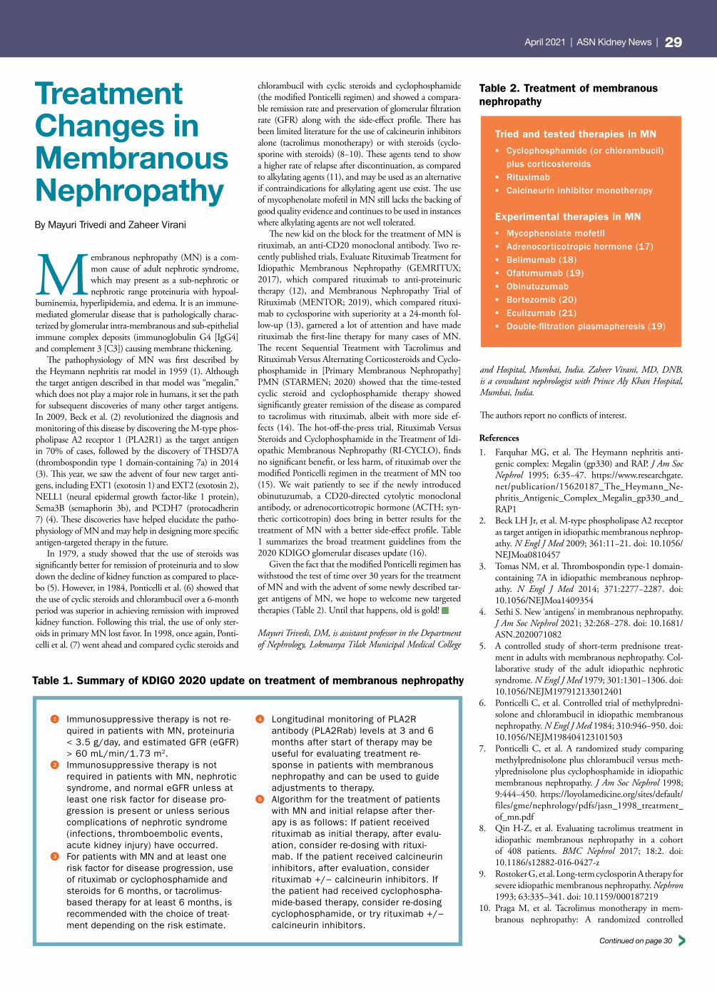
GLOMERULAR DISEASES April 2021 | ASN Kidney News | 29
Treatment Changes in Membranous NephropathyBy Mayuri Trivedi and Zaheer Virani
Membranous nephropathy (MN) is a com-mon cause of adult nephrotic syndrome, which may present as a sub-nephrotic or nephrotic range proteinuria with hypoal-
buminemia, hyperlipidemia, and edema. It is an immune-mediated glomerular disease that is pathologically charac-terized by glomerular intra-membranous and sub-epithelial immune complex deposits (immunoglobulin G4 [IgG4] and complement 3 [C3]) causing membrane thickening.
The pathophysiology of MN was first described by the Heymann nephritis rat model in 1959 (1). Although the target antigen described in that model was “megalin,” which does not play a major role in humans, it set the path for subsequent discoveries of many other target antigens. In 2009, Beck et al. (2) revolutionized the diagnosis and monitoring of this disease by discovering the M-type phos-pholipase A2 receptor 1 (PLA2R1) as the target antigen in 70% of cases, followed by the discovery of THSD7A (thrombospondin type 1 domain-containing 7a) in 2014 (3). This year, we saw the advent of four new target anti-gens, including EXT1 (exotosin 1) and EXT2 (exotosin 2), NELL1 (neural epidermal growth factor-like 1 protein), Sema3B (semaphorin 3b), and PCDH7 (protocadherin 7) (4). These discoveries have helped elucidate the patho-physiology of MN and may help in designing more specific antigen-targeted therapy in the future.
In 1979, a study showed that the use of steroids was significantly better for remission of proteinuria and to slow down the decline of kidney function as compared to place-bo (5). However, in 1984, Ponticelli et al. (6) showed that the use of cyclic steroids and chlorambucil over a 6-month period was superior in achieving remission with improved kidney function. Following this trial, the use of only ster-oids in primary MN lost favor. In 1998, once again, Ponti-celli et al. (7) went ahead and compared cyclic steroids and
chlorambucil with cyclic steroids and cyclophosphamide (the modified Ponticelli regimen) and showed a compara-ble remission rate and preservation of glomerular filtration rate (GFR) along with the side-effect profile. There has been limited literature for the use of calcineurin inhibitors alone (tacrolimus monotherapy) or with steroids (cyclo-sporine with steroids) (8−10). These agents tend to show a higher rate of relapse after discontinuation, as compared to alkylating agents (11), and may be used as an alternative if contraindications for alkylating agent use exist. The use of mycophenolate mofetil in MN still lacks the backing of good quality evidence and continues to be used in instances where alkylating agents are not well tolerated.
The new kid on the block for the treatment of MN is rituximab, an anti-CD20 monoclonal antibody. Two re-cently published trials, Evaluate Rituximab Treatment for Idiopathic Membranous Nephropathy (GEMRITUX; 2017), which compared rituximab to anti-proteinuric therapy (12), and Membranous Nephropathy Trial of Rituximab (MENTOR; 2019), which compared rituxi-mab to cyclosporine with superiority at a 24-month fol-low-up (13), garnered a lot of attention and have made rituximab the first-line therapy for many cases of MN. The recent Sequential Treatment with Tacrolimus and Rituximab Versus Alternating Corticosteroids and Cyclo-phosphamide in [Primary Membranous Nephropathy] PMN (STARMEN; 2020) showed that the time-tested cyclic steroid and cyclophosphamide therapy showed significantly greater remission of the disease as compared to tacrolimus with rituximab, albeit with more side ef-fects (14). The hot-off-the-press trial, Rituximab Versus Steroids and Cyclophosphamide in the Treatment of Idi-opathic Membranous Nephropathy (RI-CYCLO), finds no significant benefit, or less harm, of rituximab over the modified Ponticelli regimen in the treatment of MN too (15). We wait patiently to see if the newly introduced obinutuzumab, a CD20-directed cytolytic monoclonal antibody, or adrenocorticotropic hormone (ACTH; syn-thetic corticotropin) does bring in better results for the treatment of MN with a better side-effect profile. Table 1 summarizes the broad treatment guidelines from the 2020 KDIGO glomerular diseases update (16).
Given the fact that the modified Ponticelli regimen has withstood the test of time over 30 years for the treatment of MN and with the advent of some newly described tar-get antigens of MN, we hope to welcome new targeted therapies (Table 2). Until that happens, old is gold!
Mayuri Trivedi, DM, is assistant professor in the Department of Nephrology, Lokmanya Tilak Municipal Medical College
and Hospital, Mumbai, India. Zaheer Virani, MD, DNB, is a consultant nephrologist with Prince Aly Khan Hospital, Mumbai, India.
The authors report no conflicts of interest.
References1. Farquhar MG, et al. The Heymann nephritis anti-
genic complex: Megalin (gp330) and RAP. J Am Soc Nephrol 1995; 6:35−47. https://www.researchgate.net/publication/15620187_The_Heymann_Ne-phritis_Antigenic_Complex_Megalin_gp330_and_RAP1
2. Beck LH Jr, et al. M-type phospholipase A2 receptor as target antigen in idiopathic membranous nephrop-athy. N Engl J Med 2009; 361:11−21. doi: 10.1056/NEJMoa0810457
3. Tomas NM, et al. Thrombospondin type-1 domain-containing 7A in idiopathic membranous nephrop-athy. N Engl J Med 2014; 371:2277−2287. doi: 10.1056/NEJMoa1409354
4. Sethi S. New ‘antigens’ in membranous nephropathy. J Am Soc Nephrol 2021; 32:268−278. doi: 10.1681/ASN.2020071082
5. A controlled study of short-term prednisone treat-ment in adults with membranous nephropathy. Col-laborative study of the adult idiopathic nephrotic syndrome. N Engl J Med 1979; 301:1301−1306. doi: 10.1056/NEJM197912133012401
6. Ponticelli C, et al. Controlled trial of methylpredni-solone and chlorambucil in idiopathic membranous nephropathy. N Engl J Med 1984; 310:946−950. doi: 10.1056/NEJM198404123101503
7. Ponticelli C, et al. A randomized study comparing methylprednisolone plus chlorambucil versus meth-ylprednisolone plus cyclophosphamide in idiopathic membranous nephropathy. J Am Soc Nephrol 1998; 9:444−450. https://loyolamedicine.org/sites/default/files/gme/nephrology/pdfs/jasn_1998_treatment_of_mn.pdf
8. Qin H-Z, et al. Evaluating tacrolimus treatment in idiopathic membranous nephropathy in a cohort of 408 patients. BMC Nephrol 2017; 18:2. doi: 10.1186/s12882-016-0427-z
9. Rostoker G, et al. Long-term cyclosporin A therapy for severe idiopathic membranous nephropathy. Nephron 1993; 63:335–341. doi: 10.1159/000187219
10. Praga M, et al. Tacrolimus monotherapy in mem-branous nephropathy: A randomized controlled
Table 2. Treatment of membranous nephropathy
Tried and tested therapies in MN
• Cyclophosphamide (or chlorambucil) plus corticosteroids
• Rituximab• Calcineurin inhibitor monotherapy
Experimental therapies in MN
• Mycophenolate mofetil• Adrenocorticotropic hormone (17)• Belimumab (18)• Ofatumumab (19)• Obinutuzumab • Bortezomib (20)• Eculizumab (21)• Double-filtration plasmapheresis (19)
>Continued on page 30
Table 1. Summary of KDIGO 2020 update on treatment of membranous nephropathy
1 Immunosuppressive therapy is not re-quired in patients with MN, proteinuria < 3.5 g/day, and estimated GFR (eGFR) > 60 mL/min/1.73 m2.
2 Immunosuppressive therapy is not required in patients with MN, nephrotic syndrome, and normal eGFR unless at least one risk factor for disease pro-gression is present or unless serious complications of nephrotic syndrome (infections, thromboembolic events, acute kidney injury) have occurred.
3 For patients with MN and at least one risk factor for disease progression, use of rituximab or cyclophosphamide and steroids for 6 months, or tacrolimus-based therapy for at least 6 months, is recommended with the choice of treat-ment depending on the risk estimate.
4 Longitudinal monitoring of PLA2R antibody (PLA2Rab) levels at 3 and 6 months after start of therapy may be useful for evaluating treatment re-sponse in patients with membranous nephropathy and can be used to guide adjustments to therapy.
5 Algorithm for the treatment of patients with MN and initial relapse after ther-apy is as follows: If patient received rituximab as initial therapy, after evalu-ation, consider re-dosing with rituxi-mab. If the patient received calcineurin inhibitors, after evaluation, consider rituximab +/− calcineurin inhibitors. If the patient had received cyclophospha-mide-based therapy, consider re-dosing cyclophosphamide, or try rituximab +/− calcineurin inhibitors.

GLOMERULAR DISEASES
trial. Kidney Int 2007; 71:924–930. doi: 10.1038/sj.ki.5002215
11. Ramachandran R, et al. Two-year follow-up study of membranous nephropathy treated with tacrolimus and corticosteroids versus cyclical corticosteroids and cyclophosphamide. Kidney Int Rep 2017; 2:610−616. doi: 10.1016/j.ekir.2017.02.004
12. Dahan K, et al. Rituximab for severe membranous ne-phropathy: A 6-month trial with extended follow-up. J Am Soc Nephrol 2017; 28:348−358. doi: 10.1681/ASN.2016040449
13. Fervenza FC, et al. Rituximab or cyclosporine in the treatment of membranous nephropathy. N Engl J Med 2019; 381:36−46. doi: 10.1056/NEJMoa1814427
14. Fernández-Juárez G, et al. The STARMEN trial indi-
cates that alternating treatment with corticosteroids and cyclophosphamide is superior to sequential treat-ment with tacrolimus and rituximab in primary mem-branous nephropathy. Kidney Int [published online ahead of print November 7, 2020]. doi: 10.1016/j.kint.2020.10.014; https://www.sciencedirect.com/science/article/pii/S0085253820312515
15. Scolari F, et al. Rituximab or cyclophosphamide in the treatment of membranous nephropathy: The RI-CYCLO randomized trial. J Am Soc Nephrol [published online ahead of print March 2021]. doi: https://doi.org/10.1681/ASN.2020071091; https://jasn.asnjournals.org/content/early/2021/02/27/ASN.2020071091
16. KDIGO Clinical Practice Guideline on Glomeru-lar Diseases (Kidney Disease, Improving Global Outcomes [KDIGO]). June 2020. Accessed Feb-ruary 25, 2020. https://kdigo.org/wp-content/up-loads/2017/02/KDIGO-GN-GL-Public-Review-Draft_1-June-2020.pdf
17. van de Logt A-E, et al. Synthetic ACTH in high risk patients with idiopathic membranous nephropathy: A prospective, open label cohort study. PLoS One 2015;
10:e0142033. doi: 10.1371/journal.pone.014203318. Barrett C, et al. Effect of belimumab on proteinuria
and anti-phospholipase A2 receptor autoantibody in primary membranous nephropathy. Nephrol Dial Transplant 2020; 35:599–606. doi: 10.1093/ndt/gfz086
19. Podestà MA, et al. Accelerating the depletion of cir-culating anti-phospholipase A2 receptor antibodies in patients with severe membranous nephropathy: Pre-liminary findings with double filtration plasmaphere-sis and ofatumumab. Nephron 2020; 144:30−35. doi: 10.1159/000501858
20. Bontscho J, et al. Myeloperoxidase-specific plasma cell depletion by bortezomib protects from anti-neutro-phil cytoplasmic autoantibodies-induced glomerulo-nephritis. J Am Soc Nephrol 2011; 22:336–348. doi: 10.1681/ASN.2010010034
21. Appel G, et al. Eculizumab (C5 complement inhibi-tor) in the treatment of idiopathic membranous ne-phropathy. J Am Soc Nephrol 2002; 13:668A. https://www.researchgate.net/publication/312463048_Ecu-lizumab_C5_complement_inhibitor_in_the_treat-ment_of_idiopathic_membranous_nephropathy
Treatment Changes in Membranous NephropathyContinued from page 29
Fibrillary glomerulonephritis and immunotactoid glomerulonephritis represent two of the kidney diseases characterized by organized fibrillar depos-its. In 1977, the first case of fibrillary glomeru-
lonephritis was described in a patient with nephrotic syn-drome whose kidney biopsy showed amyloid-like deposits that did not stain with Congo red (1). The term “fibrillary glomerulonephritis,” however, did not appear in the lit-erature until 1987 (2). Immunotactoid glomerulonephritis was first used to describe the kidney biopsy of a patient with nephrotic syndrome in 1980 (3). For years, whether im-munotactoid and fibrillary glomerulonephritis denoted two separate entities or different presentations of a single disease was hotly debated (4, 5). This was due mainly to the fact that they were being differentiated by the size and character-istics of the fibrils. The fibrils in fibrillary glomerulonephri-tis are solid (as opposed to hollow), randomly arranged, and typically measure 9 to 26 nm in diameter (6−8). In contrast, the fibrils in immunotactoid glomerulonephritis are micro-tubules with a hollow center ranging from 14 to 90 nm in diameter, typically arranged in parallel patterns (7, 9). Al-though the distinction seems obvious (Table 1), in practice, accurately measuring the size of the fibrils or identification of the hollow center in the microtubules on electron micros-copy is often challenging (10). Moreover, the size overlap of the fibrils/microtubules further adds to the confusion. The debate was finally settled when DnaJ homolog subfamily B member 9 (DNAJB9) was discovered to be involved in the pathogenesis of fibrillary glomerulonephritis but not in immunotactoid glomerulonephritis (11). Now, fibrillary glomerulonephritis is defined by the presence of DNAJB9.
Histologically, the two entities share many similar fea-tures, but there are some subtle differences (Figure 1). On light microscopy, the most common histologic pattern in fibrillary glomerulonephritis is the mesangial proliferative pattern (71%), followed by the membranoproliferative pattern (8, 12). In comparison, endocapillary proliferative (35%) and membranoproliferative (29%) are the most common patterns in immunotactoid glomerulonephritis (9). The membranous pattern and crescents have also been described in both entities. Immunoglobulin (Ig)G is the dominant deposit on immunofluorescence in both fibrillary
Immunotactoid and Fibrillary Glomerular Diseases—Zebras or Not So Anymore? By Nelson Leung and Mariam P. Alexander
Figure 1. Immunotactoid and fibrillary glomerulonephritis characteristics
Fibrillary glomerulonephritis is characterized by expansion of the mesangial matrix with mild hypercellularity (A). The expanded matrix stains periodic acid−Schiff (PAS) positive (B) and silver negative (C). The DNAJB9 immuno-histochemical stain is positive in fibrillary glomerulonephritis, with typical extracellular staining, as noted in the glomerulus (D). DNAJB9 may also be noted along tubular basement membranes and peritubular capillaries (E). Unlike as in the amyloid, the deposits are typically Congo red negative (F). Immunofluorescence studies demon-strate smudgy to pseudo-linear staining of glomerular mesangium and capillary walls with IgG (G), and ultrastruc-tural studies show randomly oriented, non-branching fibrils, measuring between 15 nm and 30 nm (H). Amyloid fibrils are typically fine, randomly oriented, non-branching, measuring between 9 and 26 nm in diameter (I).

GLOMERULAR DISEASES April 2021 | ASN Kidney News | 31
and immunotactoid glomerulonephritis (6, 8, 9). IgA and IgM can also be found. C1q (in >90%) and C3 (in >60%) are commonly found in both fibrillary and immunotactoid glomerulonephritis. On electron microscopy, the depos-its are typically located in the mesangium and the lamina densa of the glomerular basement membranes in fibrillary glomerulonephritis (8). Mesangial deposits also dominate in immunotactoid glomerulonephritis, but infiltration of the lamina densa is limited (9). Tubular basement membrane deposits are rarely seen in fibrillary glomerulonephritis but not in immunotactoid glomerulonephritis.
Two variants of fibrillary glomerulonephritis have re-cently been described. First is a congophilic variant of fibril-lary glomerulonephritis, which is found in up to 24% in one series (13). It is important to note that in these patients, congophilic deposits are not found outside of the kidney. Proteomics studies by mass spectrometry found that the spectral counts of apolipoprotein E and serum amyloid P component (SAP) were higher in the congophilic fibrillary glomerulonephritis cases, whereas the apolipoprotein A-IV spectral counts were similar to the non-congophilic fibrillary glomerulonephritis. However, none of the spectral counts of the three chaperone proteins in congophilic fibrillary glo-merulonephritis were as high as those in amyloid glomeru-lopathy. More recently, an Ig-negative DNAJB9-positive fibrillary glomerulonephritis was identified (14). So far, the prognosis of Ig-negative DNAJB9-positive fibrillary glo-merulonephritis does not appear to be different than IgG-positive DNAJB9-positive fibrillary glomerulonephritis.
Due to their different pathogenesis, it is not surprising that the medical conditions associated with each disease are also different. Fibrillary glomerulonephritis is associated with solid cancers, lymphoproliferative disorders, myelo-proliferative disorders, vasculitis, and hepatitis or cirrhosis. Autoimmune diseases, including inflammatory bowel dis-ease, are quite common, whereas monoclonal gammopathy is extremely rare (8, 12). In fact, one study found that only 0.7% of DNAJB9-positive fibrillary glomerulonephritis cases were associated with a monoclonal gammopathy (15). On the other hand, two-thirds of a recent combined series from the Mayo Clinic and Columbia University, involving 73 immunotactoid glomerulonephritis cases, had light-chain restriction demonstrated by immunofluorescence on the kidney biopsy (9). In this series, 82% of the patients with monoclonal deposits and 26% of the patients with polyclonal deposits had a hematologic condition. Lympho-ma (53%) was the most common hematologic condition, followed by monoclonal gammopathy of renal significance (MGRS; 22%) and multiple myeloma (8%) in the patients
with monoclonal deposits (16). Of the patients with lym-phoma, 86% had a chronic lymphocytic leukemia/small lymphocytic lymphoma (CLL/SLL) clone. The high per-centage of CLL/SLL clones has also been reported by oth-ers (6). The different etiologies may explain the difference in recurrence rate after kidney transplantation. Recurrence was reported in 21% of DNAJB9-positive fibrillary glomerulo-nephritis patients who underwent a kidney transplant after a median of 10.2 years (17). In comparison, 60% of patients with immunotactoid glomerulonephritis experienced recur-rence within 10 months of kidney transplantation, which is similar to other MGRS-related diseases (9, 18).
So, are fibrillary and immunotactoid glomerulonephritis still considered zebras? It is estimated that there are between 185,000 and 285,000 zebras in the world vs. 58,372,106 horses. Zebras, therefore, represent 0.3%−0.5% of the horse population. Studies estimate that fibrillary glomerulonephri-tis comprises about 1% of the native kidney biopsies, which is more common than zebras in relationship to horses (7, 12). Immunotactoid glomerulonephritis, on the other hand, makes up only 0.04% of the native kidney biopsies; not only is it a zebra, but it is a Grevy’s zebra, rarest of the zebras (9).
Nelson Leung is with the Divisions of Nephrology and Hyper-tension and Hematology at the Mayo Clinic, Rochester, MN. Mariam P. Alexander is with the Department of Anatomic Pa-thology at the Mayo Clinic, Rochester, MN.
Dr. Leung is on an advisory board for AbbVie and Omeros, with all payments going to Mayo. He has two study grants from Omeros and one from Al-nylam. Dr. Alexander has no conflicts of interest.
References1. Rosenmann E, Eliakim M. Nephrotic syndrome asso-
ciated with amyloid-like glomerular deposits. Nephron 1977; 18:301−308. doi: 10.1159/000180846
2. Alpers CE, et al. Fibrillary glomerulonephritis: An entity with unusual immunofluorescence features. Kidney Int 1987; 31:781−789. doi: 10.1038/ki.1987.66
3. Schwartz MM, Lewis EJ. The quarterly case: Nephrotic syndrome in a middle-aged man. Ultrastruct Pathol 1980; 1:575−582. doi: 10.3109/01913128009140563
4. Korbet SM, et al. The fibrillary glomerulopathies. Am J Kidney Dis 1994; 23:751−765. doi: 10.1016/s0272-6386(12)70290-1
5. Alpers CE. Fibrillary glomerulonephritis and immu-notactoid glomerulopathy: Two entities, not one. Am J Kidney Dis 1993; 22:448−451. doi: 10.1016/s0272-
6386(12)70151-86. Bridoux F, et al. Fibrillary glomerulonephritis and
immunotactoid (microtubular) glomerulopathy are associated with distinct immunologic features. Kidney Int 2002; 62:1764−1775. doi: 10.1046/j.1523-1755.2002.00628.x
7. Fogo A, et al. Morphologic and clinical features of fi-brillary glomerulonephritis versus immunotactoid glo-merulopathy. Am J Kidney Dis 1993; 22:367−377. doi: 10.1016/s0272-6386(12)70138-5
8. Nasr SH, et al. Fibrillary glomerulonephritis: A report of 66 cases from a single institution. Clin J Am Soc Nephrol 2011; 6:775−784. doi: 10.2215/CJN.08300910
9. Nasr SH, et al. Immunotactoid glomerulopathy is a rare entity with monoclonal and polyclonal variants. Kidney Int 2021; 99:410−420. doi: 10.1016/j.kint.2020.07.037
10. Alpers CE, Kowalewska J. Fibrillary glomerulonephritis and immunotactoid glomerulopathy. J Am Soc Nephrol 2008; 19:34−37. doi: 10.1681/ASN.2007070757
11. Nasr SH, et al. DNAJB9 is a specific immunohistochem-ical marker for fibrillary glomerulonephritis. Kidney Int Rep 2018; 3:56−64. doi: 10.1016/j.ekir.2017.07.017
12. Rosenstock JL, Markowitz GS. Fibrillary glomerulone-phritis: An update. Kidney Int Rep 2019; 4:917−922. doi: 10.1016/j.ekir.2019.04.013
13. Alexander MP, et al. Congophilic fibrillary glomerulone-phritis: A case series. Am J Kidney Dis 2018; 72:325−336. doi: 10.1053/j.ajkd.2018.03.017
14. Said SM, et al. Immunoglobulin-negative DNAJB9-as-sociated fibrillary glomerulonephritis: A report of 9 cas-es. Am J Kidney Dis 2021; 77:454−458. doi: 10.1053/j.ajkd.2020.04.015
15. Said SM, et al. DNAJB9-positive monotypic fibrillary glomerulonephritis is not associated with monoclonal gammopathy in the vast majority of patients. Kidney Int 2020; 98:498−504. doi: 10.1016/j.kint.2020.02.025
16. Leung N, et al. Monoclonal gammopathy of renal signif-icance: When MGUS is no longer undetermined or in-significant. Blood 2012; 120:4292−4295. doi: 10.1182/blood-2012-07-445304
17. El Ters M, et al. Recurrence of DNAJB9-positive fibril-lary glomerulonephritis after kidney transplantation: A case series. Am J Kidney Dis 2020; 76:500−510. doi: 10.1053/j.ajkd.2020.01.018
18. Said SM, et al. Proliferative glomerulonephritis with monoclonal immunoglobulin G deposits is associ-ated with high rate of early recurrence in the allo-graft. Kidney Int 2018; 94:159−169. doi: 10.1016/j.kint.2018.01.028
Table 1. Fibrillary and immunotactoid glomerulonephritis distinctions
Fibrillary glomerulonephritis Immunotactoid glomerulonephritis
Histology Mesangial proliferative, membranoprolifera-tive, membranous pattern
Endocapillary proliferative, membranoproliferative, membranous pattern
Crescents Occasionally Occasionally
Fibril characteristics 9−26 nm, solid, randomly arranged 14−90 nm, hollow (microtubule), parallel arrays
Defining characteristics DNAJB9+ DNAJB9−
Congo red staining Typically non-congophilic, but a congophilic variant exists
Non-congophilic
Associated conditions Hepatitis, autoimmune diseases, inflamma-tory bowel disease, malignancies
Monoclonal gammopathy, lymphoma, chronic lymphocytic leukemia
MGRS related Extremely rare Common
Recur after kidney transplant ~20% >60%
Prevalence Rare Extremely rare

GLOMERULAR DISEASES
Bone marrow
‘Mucosal-type’ poorlygalactosylated IgA1
Mesangial IgA1deposition
Circulation
IgA1-immunecomplex formation
Cross-reactiveantimicrobial antibodies
IgA1-specific IgGand IgA antibodies
Autoantibodies toIgA1 hinge region
Mucosalimmune system
Gutmicrobiome
IgA+ B cells
21
3
4
6
5
Figure 1. Links between the gut and the kidney in IgA nephropathy
IgA Nephropathy—Should We Target the Gut?By Chee Kay Cheung and Jonathan Barratt
Immunoglobulin A nephropathy (IgAN) is the most common form of primary glomerular disease world-wide. Despite being initially described over 50 years ago by Dr. Jean Berger, there remains no disease-spe-
cific treatment. Its underlying pathogenesis is a dysregu-lation of the IgA immune system, which is characterized by elevated circulating levels of polymeric IgA1 that lack terminal galactose residues within the hinge region (termed “poorly galactosylated IgA1”) and the presence of IgA1-specific IgG and IgA antibodies (Figure 1). This leads to the formation of IgA-containing immune complexes that deposit within the glomerular mesangium, triggering me-sangial cell proliferation, complement activation, inflam-mation, and subsequent damage (1). A number of trials have tested immunosuppressive strategies commonly em-ployed in other immune-mediated glomerulonephritides, including cyclophosphamide, rituximab, mycophenolate, and azathioprine, but there is no clear evidence to support the efficacy of any of these agents in IgAN. Standard of care for the management of IgAN is currently focused on blood pressure control, weight loss, reduction of dietary sodium intake, smoking cessation, and use of renin-angiotensin system blockers. Current KDIGO (Kidney Disease: Im-proving Global Outcomes) guidelines suggest the addition
of corticosteroids only in cases where proteinuria persists despite the above measures, but the risk-benefit profile of their use has been brought into question by two recent ran-domized controlled trials: STOP-IgAN (Supportive Versus Immunosuppressive Therapy for the Treatment of Progres-sive IgA Nephropathy) (2) and TESTING (Therapeutic Evaluation of Steroids in IgA Nephropathy Global) (3).
Ever since its first description, there has been significant interest in a potential link between IgAN and the mucosal immune system due to the fact that approximately 30% of patients with IgAN experience episodes of visible hematu-ria coincident with upper respiratory tract or gastrointes-tinal infections (4). In addition, there are well-established associations between IgAN and other gastrointestinal dis-eases, including inflammatory bowel disease and celiac disease (5, 6). The exact mechanisms that link mucosal im-mune system activation and IgAN have been the focus of intense study, and over the past decade, a mucosal origin for IgAN has become more firmly established.
Human IgA exists as two isoforms, IgA1 and IgA2, which in turn can exist as monomers or polymers. Poly-meric IgA1, and in particular, poorly galactosylated poly-meric IgA1, is mainly produced at respiratory and gut mucosal surfaces in the mucosa-associated lymphoid tissue (MALT) (7). Here, IgA plays an important role in the host defense against microbial invasion. The gut-associated lym-phoid tissue (GALT) produces the most IgA of all MALT sites, and this is concentrated in specialized collections of lymphoid follicles called the Peyer’s patches, which are pre-dominantly located in the distal ileum.
Not only is “mucosal-type” poorly galactosylated poly-meric IgA1 elevated in the circulation in IgAN, but it is also a major component of mesangial IgA deposits (8). In addition, secretory IgA, which is mucosal IgA and bound to the 70-kDa secretory component from its passage across the mucosal epithelial cell layer, is also elevated in the cir-culation in IgAN and can be detected within mesangial IgA deposits (9). Collectively, these observations suggest
that mucosal-type IgA is misdirected into the circulation in IgAN either directly from the MALT or from systemically located mucosal plasma cells that have mis-homed during normal lymphoid trafficking (10).
A number of other lines of evidence support an impor-tant link between the gut and kidney in IgAN. McCarthy et al. (11) developed a transgenic mouse that overexpresses the B cell survival cytokine BAFF (B cell activating fac-tor). The mouse developed a hyper-IgA syndrome, driven by IgA production in the lamina propria of the gut, and an IgAN-like kidney phenotype. Raising these mice in a germ-free environment and therefore avoiding coloniza-tion of the gut by commensal flora prevented development of the kidney phenotype, until gut microbiota were intro-duced (11). Chemouny et al. (12) confirmed the impor-tance of the interaction between the gut microbiome and the MALT by treating their humanized transgenic IgAN mouse model with antibiotics to deplete the gut microbi-ota and showing a significant reduction in mesangial IgA1 deposition. Genome-wide association studies have identi-fied multiple risk alleles for IgAN that are also directly as-sociated with synthesis of IgA within the gut, inflammatory bowel disease, integrity of the intestinal epithelial barrier, and response to mucosal pathogens (13). In keeping with this, a recent epidemiological study demonstrated that patients with IgAN are more likely to develop inflamma-tory bowel disease, and those who do have an increased risk of progression to end-stage kidney disease (5). There is also emerging evidence from cross-sectional studies that the composition of the gut microbiome may be altered in patients with progressive IgAN (14).
In the search for targeted therapies in IgAN, it is there-fore logical that work has focused on GALT-directed treat-ments. A targeted release formulation of budesonide (Ne-fecon) has been developed to deliver the active drug to the distal ileum, targeting the Peyer’s patches of the GALT. The phase 2b NEFIGAN (The Effect of Nefecon® in Patients with Primary IgA Nephropathy at Risk of Developing End-stage Renal Disease) trial demonstrated that treatment with 16 mg Nefecon over 9 months significantly reduced proteinuria levels and stabilized kidney function compared to the placebo group where the estimated glomerular fil-tration rate (eGFR) fell by 4.7 mL/min/1.73 m2 over the same time period (15). The phase 3 NefIgArd (Efficacy and Safety of Nefecon in Patients with Primary IgA [Immu-noglobulin A] Nephropathy) trial is comparing 9 months’ treatment with 16 mg Nefecon vs. placebo in 360 patients with IgAN, with follow-up at 2 years (ClinicalTrials.gov: NCT03643965). This trial has closed to recruitment and recently reported 9-month outcomes in the first 199 pa-tients, confirming that Nefecon treatment results in signifi-cant proteinuria reduction and less deterioration of eGFR than placebo. The elucidation of the mechanisms by which Nefecon modulates gut mucosal IgA production may shed additional light on the overall pathogenesis of IgAN.
Alternative strategies to target the GALT and mucosal IgA synthesis in IgAN have been proposed. Although there are interesting reports from mouse models of the impact of dietary modification, to date, there is no clear evidence that changes, such as a gluten-free diet, have a beneficial effect in IgAN (6). The influence of the gut microbiome on mucosal IgA production and ways in which this could be manipulated, for example, with probiotics, are areas of growing interest. The B cell survival cytokines BAFF and APRIL (a proliferation-inducing ligand) play key roles in IgA class-switch recombination and IgA synthesis in the GALT, and inhibition of BAFF and/or APRIL is the sub-ject of ongoing phase 2 clinical trials in IgAN (16).
A better understanding of the links between the gut and the kidney in IgAN, including the composition of the gut microbiome, how the microbiota interact with the GALT to determine the mucosal IgA response, and factors involved in promoting the production of poorly galacto-sylated polymeric IgA1 and its passage into the circulation, will hopefully allow the development of additional targeted treatment strategies, with the aim of providing options to
(1) Dysregulation within the gut-associated lymphoid tissue (GALT), which may be potentiated by dynamic interac-tions with the gut microbiome, ultimately results in excess amounts of "mucosal-type" IgA1 entering the circula-tion through mis-homing of mucosal IgA1-producing B cells to systemic sites, including the bone marrow (2), and/or (3) direct passage of mucosal IgA1 from the GALT into the systemic circulation. The increase in circulat-ing mucosal-type poorly galactosylated IgA1 (4) results in IgA immune complex formation due to self-aggregation of polymeric IgA1 molecules (5). Immune complex formation can be amplified by IgA1-specific IgG and IgA antibodies, which may be cross-reactive antimicrobial antibodies and/or true autoantibodies. Circulating IgA1 immune complexes subsequently deposit in the mesangium where they trigger a mesangioproliferative glomerulo-nephritis in susceptible individuals (6).

GLOMERULAR DISEASES April 2021 | ASN Kidney News | 33
Table 1. Antigens associated with membranous nephropathy
Membranous nephropathyMembranous nephropathy (MN) is a common cause of nephrotic syndrome, attributed to approximately 25% of adult patients with nephrotic-range proteinuria or ne-phrotic syndrome (1). This number is significantly less in children with nephrotic syndrome. The clinical course of MN is insidious, with variable degrees of proteinu-ria, hypoalbuminemia, and hyperlipidemia that can lead to significant edema and in extreme cases, a rapid loss of kidney function, anasarca, acute kidney injury, and thromboembolic events. The clinical presentation is the resulting immune complex formation on the epithelia side of the glomerular basement membrane (GBM).
MN is classified as primary, based on the presence of autoantigens; secondary, based on the association with systemic autoimmune disease, malignancy, medications, or infections; or alloimmune, based on humoral respons-es between host and donor antigens. Historically, diag-
nosis of MN was largely clinical, with support from a kidney biopsy showing non-proliferative glomeruli with thickened capillary loops, immunoglobulin G (IgG) and variable C3 positivity on immunofluorescence, and electron microscopy with subepithelial immune com-plexes in the GBM (Figure 1) (2). Within the last 12 years, there has been the discovery of numerous antigens that not only aid in the diagnosis of MN but that can also be used to gauge prognosis and guide therapy.
The first antigen: PLA2RAlthough the idea that an autoantigen was responsible for the pathophysiology of MN was first demonstrated in 1959, it was not until 2009 that the first antigen, the M-type phospholipase A2 receptor-1 (PLA2R), was identified (3). PLA2R, a 180-kDa transmembrane glycoprotein, is implicated in approximately 80% of all primary membranous cases and 55% of all MN. Since
the discovery of PLA2R, there is now a commercially available antibody that has aided in the diagnosis of MN through both tissue-specific antigen staining and through a serum assay. Additionally, serum PLA2R lev-els have provided a means to identify aggressive disease, monitor treatment, and predict relapse.
The second antigen: THSD7AThrombospondin type 1 domain-containing 7A (THS-D7A) is a 250-kDa protein that is expressed on the podocyte. The antigenicity of THSD7A was brought to light in 2014 (4). Antibodies against THSD7A are prevalent in up to 10% of all patients with primary MN. In patients with THSD7A-positive MN, there was an upwards of 20% incidence of malignancy within 3 months of diagnosis, suggesting an antigen association to a diagnosis that was formerly thought to be a second-ary cause of MN.
% MN, percentage of primary and secondary membranous nephropathy; SLE, systemic lupus erythematosus; MCTD, mixed connective tissue disease.
treat this disease at its various stages in order to prevent its progression.
Chee Kay Cheung, MBChB, PhD, is a Consultant Nephrolo-gist and Honorary Senior Lecturer at the University Hospitals of Leicester NHS Trust and University of Leicester, UK. Jonathan Barratt, PhD, FRCP, is the Mayer Professor of Renal Medicine and Honorary Consultant Nephrologist at the University of Leicester and University Hospitals of Leicester NHS Trust, UK.
Disclosures: Dr. Cheung has received research funding from GlaxoSmithKline. Professor Barratt is Chair of the Study Steering Committee for the NefIgArd trial.
References 1. Suzuki H, et al. The pathophysiology of IgA nephrop-
athy. J Am Soc Nephrol 2011; 22:1795–1803. doi: 10.1681/ASN.2011050464
2. Rauen T, et al. Intensive supportive care plus immuno-suppression in IgA nephropathy. N Engl J Med 2015; 373:2225–2236. doi: 10.1056/NEJMoa1415463
3. Lv J, et al. Effect of oral methylprednisolone on clini-cal outcomes in patients with IgA nephropathy: The TESTING randomized clinical trial. JAMA 2017; 318:432–442. doi: 10.1001/jama.2017.9362
4. Tumlin JA, et al. Idiopathic IgA nephropathy: Patho-
genesis, histopathology, and therapeutic options. Clin J Am Soc Nephrol 2007; 2:1054–1061. doi: 10.2215/CJN.04351206
5. Rehnberg J, et al. Inflammatory bowel disease is more common in patients with IgA nephropathy and pre-dicts progression of ESKD: A Swedish population-based cohort study. J Am Soc Nephrol 2021; 32:411–423. doi: 10.1681/ASN.2020060848
6. Cheung CK, Barratt J. Gluten and IgA nephropathy: You are what you eat?. Kidney Int 2015; 88:215–218. doi: 10.1038/ki.2015.149
7. Barratt J, et al. Exaggerated systemic antibody re-sponse to mucosal Helicobacter pylori infection in IgA nephropathy. Am J Kidney Dis 1999; 33:1049–1057. doi: 10.1016/S0272-6386(99)70141-1
8. Hiki Y, et al. Mass spectrometry proves under-O-glycosylation of glomerular IgA1 in IgA nephropathy. Kidney Int 2001; 59:1077–1085. doi: 10.1046/j.1523-1755.2001.0590031077.x
9. Oortwijn BD, et al. Demonstration of secretory IgA in kidneys of patients with IgA nephropathy. Nephrol Dial Transplant 2007; 22:3191–3195. doi: 10.1093/ndt/gfm346
10. Barratt J, et al. Immune complex formation in IgA nephropathy: A case of the ‘right’ antibodies in the ‘wrong’ place at the ‘wrong’ time? Nephrol Dial Trans-
plant 2009; 24:3620–3623. doi: 10.1093/ndt/gfp44111. McCarthy DD, et al. Mice overexpressing BAFF de-
velop a commensal flora-dependent, IgA-associated nephropathy [published correction appears in J Clin Invest 2012; 122:778]. J Clin Invest 2011; 121:3991–4002. doi: 10.1172/JCI45563
12. Chemouny JM, et al. Modulation of the microbiota by oral antibiotics treats immunoglobulin A nephrop-athy in humanized mice. Nephrol Dial Transplant 2019; 34:1135–1144. doi: 10.1093/ndt/gfy323
13. Kiryluk K, et al. Discovery of new risk loci for IgA nephropathy implicates genes involved in immu-nity against intestinal pathogens. Nat Genet 2014; 46:1187–1196. doi: 10.1038/ng.3118
14. De Angelis M, et al. Microbiota and metabolome associated with immunoglobulin A nephropathy (IgAN). PLoS One 2014; 9:e99006. doi: 10.1371/journal.pone.0099006
15. Fellström BC, et al. Targeted-release budesonide ver-sus placebo in patients with IgA nephropathy (NE-FIGAN): A double-blind, randomised, placebo-con-trolled phase 2b trial. Lancet 2017; 389:2117–2127. doi: 10.1016/S0140-6736(17)30550-0
16. Selvaskandan H, et al. New strategies and perspectives on managing IgA nephropathy. Clin Exp Nephrol 2019; 23:577−588. doi: 10.1007/s10157-019-01700-1
>Continued on page 34
Antigen % MN Mean age (yr) Disease association Serum antibody
PLA2R 55 56 None Yes
THSD7A 3 50 Malignancy Yes
NELL-1 2.5 63 Malignancy Yes
Sema3B 3 7 (ped), 36 (adult) Family history Yes
PCDH7 4 61 Possible malignancy Yes
EXT1/EXT2 10 36 SLE, MCTD No
NCAM1 5 34 SLE Yes
HTRA1 Unknown Unknown Unknown Yes
Newer Antigens and Membranous NephropathyBy Randy L. Luciano

GLOMERULAR DISEASES
Newer antigensThe technique of laser microdissection of glomeruli from patients with antigen-negative primary MN, followed by mass spectroscopy, has led to the identification of newer antigens (5).
Neural epidermal growth factor-like 1 (NELL-1) is a potential podocyte-associated protein that has been implicated as an antigen for MN (6). This 90-kDa se-creted protein was identified in approximately 16% of all unidentified MN cases, representing 2.5% of all MN cases. In addition, there has been an association with ma-lignancy in upwards of 33% of patients with MN with detectable antibodies to NELL-1.
Semaphorin-3B (Sema3B) is an 83-kDa secreted pro-tein that has been detected in podocytes (7). Autoanti-bodies to this antigen were found on immunoblots under reducing conditions, suggesting a difficult-to-identify antigen epitope. Autoantibodies to Sema3B were found in a greater proportion of infants, children, and young adults, some of whom had a known family history. MN from Sema3B autoantibodies accounts for <3% of all MN, but in the pediatric population, this increases to 15% of MN patients.
Protocadherin 7 (PCDH7) is a 116-kDa transmem-
brane protein, most likely functioning in cell signaling. Autoantibodies to PCDH7 are seen in older patients (mean age of 61) with no apparent disease or malignancy association (8). Biopsy samples of patients with PCDH7 autoantibodies demonstrated trace to no complement activation, a finding that is much different than patients with the other forms of antigen-mediated MN. Inter-estingly, in patients with PCDH7-associated MN, there was a high percentage of patients who developed spon-taneous remission. The presence of PCDH7 may be a marker for disease severity and progression and can have potential use to guide the use or non-use of therapeutic agents in the future; however, more studies are necessary to elucidate this role.
Exostosin 1/Exostosin 2 (EXT1/EXT2) protein com-plexes have been identified in patients with MN second-ary to autoimmune disease, such as systemic lupus ery-thematosus (SLE) and mixed connective tissue disease (9). EXT1/EXT2-associated MN is more commonly seen in younger patients, with a higher percentage of fe-male patients. However, circulating antibodies to EXT1/EXT2 have not been identified, making the antigenicity of this protein complex unclear at this time.
Potential antigensRecently, two additional proteins have been identified as potential antigens for MN. Neural cell adhesion mol-ecule 1 (NCAM1) was found to be an antigen in MN, colocalizing to immune complexes in tissue from patients with MN and also present as an autoantibody in serum (10). The prevalence was 6.6% in patients with SLE class V and 2% in patients with primary MN.
Another potential antigen that was recently identified in three patients with primary MN is high-temperature recombinant protein A1 (HTRA1) (11). Anti-HTRA1 antibodies colocalize to the capillary loops with IgG4. Additional studies in large cohorts will be necessary to establish a prevalence of this potential antigen.
Redefining membranous nephropathyIn little over a decade, MN has evolved from a disease that was divided into primary versus secondary disease associations into a syndrome that can be defined through specific antigens (Table 1). Although the identification of antigens apart from PLA2R is not part of routine clini-cal practice yet, one can envision in the next decade the clinical availability of a panel of MN antigen-specific antibodies. These antibodies can be used in conjunction with or separately from a kidney biopsy to diagnose MN, understand prognosis, guide treatment strategies, and predict relapse in MN.
Randy L. Luciano, MD, PhD, is Associate Professor of Med-icine and Associate Program Director, Nephrology Fellow-ship, at Yale University School of Medicine in New Haven, CT.
The author reports no conflict of interest.
References1. Debiec H, Ronco P. Immunopathogenesis of mem-
branous nephropathy: An update. Semin Immuno-pathol 2014; 36:381−397. doi: 10.1007/s00281-014-0423-y
2. Fogo AB, et al. AJKD Atlas of Renal Pathology: Membranous nephropathy. Am J Kidney Dis 2015; 66:e15−e17. doi: 10.1053/j.ajkd.2015.07.006
3. Beck LH Jr, et al. M-type phospholipase A2 receptor as target antigen in idiopathic membranous nephrop-athy. N Engl J Med 2009; 361:11−21. doi: 10.1056/NEJMoa0810457
4. Tomas NM, et al. Thrombospondin type-1 domain-containing 7A in idiopathic membranous nephrop-athy. N Engl J Med 2014; 371:2277−2287. doi: 10.1056/NEJMoa1409354
5. Sethi S. New ‘antigens’ in membranous nephropathy. J Am Soc Nephrol 2021; 32:268−278. doi: 10.1681/ASN.2020071082
6. Sethi S, et al. Neural epidermal growth factor-like 1 protein (NELL-1) associated membranous nephrop-athy. Kidney Int 2020; 97:163−174. doi: 10.1016/j.kint.2019.09.014
7. Sethi S, et al. Semaphorin 3B-associated membranous nephropathy is a distinct type of disease predomi-nantly present in pediatric patients. Kidney Int 2020; 98:1253−1264. doi: 10.1016/j.kint.2020.05.030
8. Sethi S, et al. Protocadherin-7 associated membra-nous nephropathy. ASN Kidney Week 2020 Reim-agined. 2020; FR-OR36. https://www.asn-online.org/education/kidneyweek/2020/program-abstract.aspx?controlId=3445270
9. Sethi S, et al. Exostosin 1/exostosin 2-associated membranous nephropathy. J Am Soc Nephrol 2019; 30:1123−1136. doi: 10.1681/ASN.2018080852
10. Caza T, et al. Neural cell adhesion molecule 1 is a novel autoantigen in membranous lupus nephritis. Kidney Int [published online ahead of print Oc-tober 9, 2020]. doi: 10.1016/j.kint.2020.09.016; https://www.kidney-international.org/article/S0085-2538(20)31180-7/fulltext
11. Al-Rabadi L, et al. High temperature recombinant protein A1 (HTRA1): A novel antigen in membra-nous nephropathy. ASN Kidney Week 2020 Reim-agined. 2020; FR-OR35. https://www.asn-online.org/education/kidneyweek/2020/program-abstract.aspx?controlId=3447205
Newer Antigens and Membranous NephropathyContinued from page 33
Figure 1. Representative biopsy of 77-year-old man with primary membranous nephropathy (MN)
(Top left) Hematoxylin and eosin (H&E)-stained section showing a glomerulus with thickened capillary loops. (Top right) Immunofluorescence with IgG showing glomerular basement membrane staining. (Bottom left) Immunofluorescence with C3 showing glomerular basement membrane staining. (Bottom right) Electron microscopy showing glomerular basement membrane with subepithelial deposits.

GLOMERULAR DISEASES April 2021 | ASN Kidney News | 35
Fellows First
Minimal change disease (MCD) is one of the major causes of idiopathic nephrotic syn-drome, accounting for up to 70%−90% of cases in children and approximately 15% of
cases in adults (1). The characteristic appearance of MCD on a kidney biopsy is normal glomeruli on light microsco-py with diffuse effacement of the epithelial foot processes on electron microscopy. The pathogenesis of MCD is not fully elucidated, but systemic T cell dysfunction produc-ing increased levels of a glomerular permeability factor has been implicated (2, 3). Although the pathogenesis remains uncertain, similar to focal segmental glomerulosclerosis, a circulating factor that damages the glomerular capillary wall has been postulated, resulting in proteinuria and foot process fusion (1).
Glucocorticoid therapy has been the mainstay of therapy for MCD for decades. This management strat-egy in children has been informed by several large pro-spective randomized clinical trials (RCTs) in addition to observational studies (4). Over 90% of children respond with complete remission to initial steroid therapy (5). The recommendation for glucocorticoid therapy in adults has been informed mostly by observational studies (4), as RCT data are lacking, with the majority of informa-tion coming from a single RCT published in 1970. This trial compared low-dose prednisone (<30 mg/day) with no specific therapy among 31 adults. More than 75% of treated patients had remission of proteinuria to less than 1 g/day within 6 months (6). Subsequently, several retro-spective observational studies have demonstrated a high but variable response rate (67%−100%) in adult patients treated with higher doses (e.g., 60 mg/day or 1 mg/kg/day) (7−9). Kidney Disease: Improving Global Outcomes (KDIGO) currently recommends glucocorticoid therapy as treatment for the initial episode of adult MCD, while acknowledging that there is only low-quality evidence available and that this recommendation is based largely on extrapolation from trial data in children in addition to small observational studies in adults (10).
Steroids, however, have a significant adverse side effect profile, including Cushingoid features (11), weight gain (12), hypertension (13), gastrointestinal bleeding (14), os-teoporosis (15), diabetes (16), and increased infection risk (17). This is particularly concerning because a prolonged course of steroid treatment is often required in MCD, and relapse rates in adults can be high (8). Therefore, there has been increasing interest in steroid-sparing or minimizing regimens. Steroid-sparing regimens have already been in-vestigated for other glomerulonephritides, including anti-neutrophil cytoplasmic antibody (ANCA) vasculitis (18) and membranous nephropathy (19), with encouraging results to date.
One large investigation into a steroid-sparing regimen for MCD is the Tacrolimus Versus Prednisolone for the Treatment of Minimal Change Disease (MinTac) trial, a multi-center, open-label RCT based in the United King-dom, in which 52 adult patients with MCD were rand-omized to treatment with either oral tacrolimus at 0.05 mg/kg twice daily for 12 weeks (then tapered over a further 8 weeks) or prednisolone at 1 mg/kg daily up to 60 mg daily for 16 weeks. The primary objective was to demon-strate the non-inferiority of tacrolimus compared to pred-nisolone for inducing remission in MCD, in addition to showing that relapse rates were similar, and adverse events
were less common. Although there was no statistically significant difference in the primary outcome (complete remission at 8 weeks) between groups (68% for tacrolimus vs. 84% for prednisolone; p = 0.32), the a priori definition of non-inferiority was not met in either the per-protocol or the intention-to-treat analysis. Relapse rates (73% for tacrolimus vs. 74% for prednisolone; p = 0.99) and safe-ty profile were found to be similar between groups (20). This was the first study to investigate the use of tacrolimus monotherapy to treat MCD, and although the sample size was small, and further research is required, the results do suggest that tacrolimus may be an effective alternative treatment to steroids for MCD in adult patients.
More recently, another randomized controlled trial compared combined tacrolimus and low-dose steroid treatment with the standard high-dose steroid protocol in adult patients (21). In this open-label, non-inferiority study, 144 adults with MCD were randomized to receive either 0.05 mg/kg twice-daily tacrolimus plus once-daily 0.5 mg/kg prednisolone or once-daily 1 mg/kg predniso-lone alone for up to 8 weeks or until achieving complete remission. The steroid dose was then tapered to a mainte-nance dose of 5−7.5 mg/day in both groups, 2 weeks after
complete remission, until 24 weeks after study-drug initia-tion. The primary end point, defined as complete remis-sion within 8 weeks (urine protein:creatinine ratio <0.2 g/g), was achieved in 79.1% of those receiving tacrolimus and low-dose steroid compared to 76.8% receiving high-dose steroid, confirming non-inferiority of this treatment protocol. Of note, the relapse rate was also much lower in the combined tacrolimus/low-dose steroid protocol compared to the high-dose steroid-alone group (5.7% vs. 22.6%, respectively; p = 0.01) with no major safety dif-ferences observed (21). Studies investigating steroid mini-mization regimens for MCD are summarized in Table 1. Use of rituximab has already been shown to facilitate such regimens in other glomerulonephritides (18), and there is some emerging evidence from case series to suggest that it also could have a future role in steroid-sparing treatment strategies for MCD (22, 23).
Tacrolimus, with or without low-dose steroids, there-fore appears to be an effective alternative to high-dose steroids in MCD, particularly in patients at high risk of adverse effects from steroids, such as those with diabetes, obesity, osteoporosis, or mood disorders. However, further research is needed to establish long-term safety data, as well as the best protocol for its use.
Dearbhla Kelly, MB, BCh, BAO, MSc, MRCPI, is a renal specialist registrar with Beaumont Hospital, Dublin, Ireland.
The author has no conflicts of interest.
References1. Vivarelli M, et al. Minimal change disease. Clin Am
J Soc Nephrol 2017; 12:332−345. doi: 10.2215/CJN.05000516
2. Shalhoub RJ. Pathogenesis of lipoid nephrosis: A disorder of T-cell function. Lancet 1974; 2:556−560. doi: 10.1016/s0140-6736(74)91880-7
3. Maas RJ, et al. Permeability factors in idiopathic nephrotic syndrome: Historical perspectives and les-sons for the future. Nephrol Dial Transplant 2014; 29:2207−2216. doi: 10.1093/ndt/gfu355
4. Hogan J, Radhakrishnan J. The treatment of mini-mal change disease in adults. J Am Soc Nephrol 2013; 24:702−711. doi: 10.1681/ASN.2012070734
5. Tarshish P, et al. Prognostic significance of the early course of minimal change nephrotic syndrome: Re-port of the International Study of Kidney Disease in Children. J Am Soc Nephrol 1997; 8:769−776. htt-ps://jasn.asnjournals.org/content/jnephrol/8/5/769.full.pdf?with-ds=yes
6. Black DA, et al. Controlled trial of prednisone in adult patients with the nephrotic syndrome. Br Med J 1970; 3:421−426. doi: 10.1136/bmj.3.5720.421
7. Nakayama M, et al. Steroid responsiveness and fre-quency of relapse in adult-onset minimal change nephrotic syndrome. Am J Kidney Dis 2002; 39:503−512. doi: 10.1053/ajkd.2002.31400
8. Waldman M, et al. Adult minimal-change disease: Clinical characteristics, treatment, and outcomes. Clin J Am Soc Nephrol 2007; 2:445−453. doi: 10.2215/CJN.03531006
9. Tse K-C, et al. Idiopathic minimal change nephrotic syndrome in older adults: Steroid responsiveness and pattern of relapses. Nephrol Dial Transplant 2003; 18:1316−1320. doi: 10.1093/ndt/gfg134
10. Chapter 5: Minimal-change disease in adults. Kid-ney Int Suppl 2012; 2:177−180. doi:10.1038/ki-sup.2012.1
11. Boumpas DT, et al. Glucocorticoid therapy for immune-mediated diseases: Basic and clinical cor-relates. Ann Intern Med 1993; 119:1198−1208. doi: 10.7326/0003-4819-119-12-199312150-00007
12. Huscher D, et al. Dose-related patterns of glucocor-ticoid-induced side effects. Ann Rheum Dis 2009; 68:1119−1124. doi: 10.1136/ard.2008.092163
13. Whitworth JA. Mechanisms of glucocorticoid-in-duced hypertension. Kidney Int 1987; 31:1213−1224. doi: 10.1038/ki.1987.131
14. Gabriel SE, et al. Risk for serious gastrointestinal complications related to use of nonsteroidal anti-in-
Minimization in Minimal Change Disease: Maximal Change in Practice?By Dearbhla Kelly
>Continued on page 36
The pathogenesis of MCD is not fully elucidated, but systemic T cell dysfunction producing increased levels of a glomerular permeability factor has been implicated.

GLOMERULAR DISEASES
flammatory drugs. A meta-analysis. Ann Intern Med 1991; 115:787−796. doi: 10.7326/0003-4819-115-10-787
15. van Staa TP, et al. The epidemiology of corticosteroid-induced osteoporosis: A meta-analysis. Osteoporosis Int 2002; 13:777−787. doi: 10.1007/s001980200108
16. Gurwitz JH, et al. Glucocorticoids and the risk for initiation of hypoglycemic therapy. Arch In-tern Med 1994; 154:97−101. doi: 10.1001/archinte.1994.00420010131015
17. Dixon WG, et al. Immediate and delayed impact of oral glucocorticoid therapy on risk of serious in-fection in older patients with rheumatoid arthri-tis: A nested case-control analysis. Ann Rheum Dis 2012; 71:1128−1133. doi: 10.1136/annrheum-dis-2011-200702
18. Pepper RJ, et al. A novel glucocorticoid-free main-tenance regimen for anti-neutrophil cytoplasm an-tibody-associated vasculitis. Rheumatology (Oxford) 2019; 58:260−268. doi: 10.1093/rheumatology/key288
19. Fervenza FC, et al. Rituximab or cyclosporine in the treatment of membranous nephropathy. N Engl J Med 2019; 381:36−46. doi: 10.1056/NEJMoa1814427
20. Medjeral-Thomas NR, et al. Randomized, controlled trial of tacrolimus and prednisolone monotherapy for adults with de novo minimal change disease: A multicenter, randomized, controlled trial. Clin J Am Soc Nephrol 2020; 15:209−218. doi: 10.2215/CJN.06180519
21. Chin HJ, et al. Comparison of the efficacy and safe-ty of tacrolimus and low-dose corticosteroid with high-dose corticosteroid for minimal change ne-phrotic syndrome in adults. J Am Soc Nephrol 2021; 32:199−210. doi: 10.1681/ASN.2019050546
22. Ainley L, et al. Treatment of concurrent minimal change disease and Epstein Barr virus-driven post-transplant lymphoproliferative disorder with rituxi-mab following hematopoietic stem cell transplanta-tion. Kidney Int Rep 2021; 6:224−227. doi: 10.1016/j.ekir.2020.10.012
23. Fenoglio R, et al. Rituximab as a front-line therapy for adult-onset minimal change disease with nephrot-ic syndrome. Oncotarget 2018; 9:28799−28804. doi: 10.18632/oncotarget.25612.
24. Kim YC, et al. Complete remission induced by tac-rolimus and low-dose prednisolone in adult minimal change nephrotic syndrome: A pilot study. Kidney Res Clin Pract 2012; 31:112−117. doi: 10.1016/j.krcp.2012.04.321
25. Li X, et al. Tacrolimus monotherapy after intrave-nous methylprednisolone in adults with minimal change nephrotic syndrome. J Am Soc Nephrol 2017; 28:1286−1295. doi: 10.1681/ASN.2016030342
Steroid Minimization Continued from page 35
CR, complete remission; PR, partial remission; RCT, randomized controlled trial; UPCR, urine protein:creatinine ratio.
Study Design Number of participants
Treatment protocol
Primary outcome
Results Relapse rates Safety profile
Kim et al. (24) Pilot study(single center)
14 Tacrolimus 0.05 mg/kg twice daily and prednisolone 0.5 mg/kg/day (up to 40 mg/day) until remis-sion for 16 weeks
Cumulative percentage of CR (defined as UPCR < 0.2 g protein/g creatinine) during 16 weeks
CR was achieved by 13/14 (92.9%) patients within 8 weeks.
Three of 14 (21.4%) patients had relapsed at 31 weeks, 36 weeks, and 40 weeks after treat-ment.
Three cases re-ported abdominal pain, diarrhea, or new-onset diabe-tes mellitus.
Li et al. (25) Prospective RCT (8 centers in China)
119 Short-term intra-venous methyl-prednisolone (0.8 mg/kg per day for 10 days) followed by a conventional tapering oral prednisone regi-men vs. short-term intravenous methylpredni-solone followed by tacrolimus (0.05 mg/kg/day) monotherapy for 36 weeks
Cumulative num-bers of patients who experienced CR (decrease in proteinuria to ≤0.3 g/day) or PR (decrease in proteinuria to <3.5 g/day but >0.3 g/day)
Remission oc-curred in 51 of 53 (96.2%; all CR) glucocor-ticoid-treated patients and 55 of 56 (98.2%; 52 CR and three PR) tacrolimus-treat-ed patients (p = 0.61 for remis-sion; p = 0.68 for CR).
Relapse occurred in 49.0% and 45.5% of the glucocorticoid- and tacrolimus-treated patients, respectively (p = 0.71).
128 adverse events in the glu-cocorticoid group vs. 81 in the tacrolimus group; seven adverse events in the glu-cocorticoid group and two adverse events in the tacrolimus group were serious.
Medjeral-Thomas et al. (20)
Prospective, open-label RCT (6 centers)
50 Tacrolimus at 0.05 mg/kg twice daily (for 12 weeks, then tapered over a further 8 weeks) or prednisolone at 1 mg/kg daily up to 60 mg daily (for 16 weeks)
CR of nephrotic syndrome (UPCR < 50 mg/mmol) after 8 weeks of therapy
No significant differences in CR rates at 8 weeks (21 out of 25 [84%] for pred-nisolone and 17 out of 25 [68%] for tacrolimus co-horts; p = 0.32)
No significant difference in relapse rates (17/23 [73.9%] for predniso-lone and 16/22 [72.7%] for tac-rolimus cohorts)
18/25 patients experienced adverse events in the predniso-lone cohort, and 20/27 did in the tacrolimus cohort (p = 0.99). There were four serious adverse events that required admission in the prednisolone and three in the tac-rolimus cohorts (p = 0.99).
Chin et al. (21) Prospective, open-label RCT (15 centers)
144 0.05 mg/kg twice-daily tacroli-mus plus once-daily 0.5 mg/kg prednisolone vs. once-daily 1 mg/kg prednisolone alone for up to 8 weeks or until achieving CR
CR within 8 weeks (UPCR < 0.2 g/g)
CR within 8 weeks occurred in 53/67 patients (79.1%) receiving tacrolimus and low-dose ster-oid and 53/69 patients (76.8%) receiving high-dose steroid.
Significantly fewer patients relapsed on maintenance tacrolimus plus tapered ster-oid vs. tapered steroid alone (5.7% vs. 22.6%, respectively; p = 0.01).
49/67 (73.1%) in the combined tacrolimus and low-dose steroid group and 47/69 (68.1%) in the high-dose steroid group experi-enced adverse events (p = 0.52).
Table 1. Studies investigating steroid-minimization treatment strategies for MCD

GLOMERULAR DISEASES April 2021 | ASN Kidney News | 37
Glomerular Diseases Associated with COVID-19 By Purva Sharma and Vanesa Bijol
As the coronavirus infectious disease 2019 (COVID-19) pandemic unleashed through the world, we found that patients infected with se-vere acute respiratory syndrome coronavirus-2
(SARS-CoV-2) had a wide range of direct and indirect ef-fects culminating in a variety of end-organ injuries. Acute kidney injury (AKI) was found in as many as 80% of pa-tients admitted to the intensive care unit with severe illness.
The most common histopathologic equivalent of clini-cal AKI was acute tubular injury or necrosis in biopsy and autopsy studies in these patients. However, COVID-19 can affect all compartments of the kidney parenchyma, in-cluding the glomerulus, interstitium, and vasculature. The tubulointerstitial injury is non-inflammatory and with-out viral cytopathic effect; however, a number of reports claimed direct viral infection of tubules or podocytes on electron microscopy, which was quickly disputed by oth-ers (1−4). Some authors have reported the positive find-ings by immunohistochemistry, immunofluorescence, or in situ hybridization (1, 5), whereas others have interpreted their results as negative using the same techniques (6–9). All of this, together with the fact that viremia or viruria are rarely detected clinically, suggests that indirect mechanisms related to cytokine release are of utmost importance for the mechanisms of AKI, as well as the injury to other tissues, organs, and systems including the coagulation and com-plement pathways. Hemodynamic factors and medication-induced kidney injury also remain important causative fac-tors for AKI in COVID-19.
A variety of glomerular diseases have been reported in patients with COVID-19, some triggered or exacerbated by COVID-19 or likely independent of it (Figure 1). Col-lapsing glomerulopathy (CG) has a special predilection for patients with African ancestry who are homozygous for high-risk APOL1 genotypes (10). In a recent review (11), 32 cases of CG were reported, out of which 21 had APOL1 polymorphisms. Ninety-six percent of these pa-tients were of African or African-American heritage. Nasr et al. (12) reported eight cases of CG, all of whom were Black. The mechanism is thought to be activation of the host interferon-chemokine pathway leading to upregulation of the APOL1 variant gene and disruption of podocyte au-tophagy, rather than direct infection of the glomerular cells (13). This association of CG with COVID-19 has bought itself a new name, COVAN, or COVID-19-associated ne-phropathy (14).
The treatment of COVID-19-associated CG is con-troversial. Treatment of the underlying disease as is done in other infections associated with CG is likely of benefit, although data on treatment outcomes in these patients are lacking. Steroids have been used in CG with persistent nephrotic-range proteinuria, but without evidence-based guidelines and consideration of the potential for harm in COVID-19 sepsis, their widespread use cannot be en-dorsed. Given that an inflammatory cascade and cytokine storm play a major role, there may be some benefit with the interleukin 6 (IL-6) receptor blockade, although this remains entirely speculative. A correlation with other po-docytopathies, including minimal change disease and focal
segmental glomerulosclerosis (FSGS), is less clear but likely involves T cell activation and cytokine release (15).
There have been case reports of antineutrophil cytoplas-mic antibody (ANCA)-associated vasculitis (AAV) in pa-tients with SARS-CoV-2 infection. The role of neutrophil extracellular traps (NETs) in the pathogenesis of AAV has been studied in the past, so new insights into the role of NETs in pathogenesis of COVID-19 naturally raise the question of association of AAV and COVID-19, although this remains to be proven (16). Anti-glomerular basement membrane (GBM) disease, immunoglobulin A (IgA) vasculitis, and membranous nephropathy (either phospholipase A2 recep-tor [PLA2R] positive or negative) have been reported as well. A reported case of crescentic transformation of pre-existing lupus nephritis emphasizes the potential importance of an inflammatory milieu in patients with COVID-19, leading to worsening of their preexisting disease (9).
Thrombotic microangiopathies, through complement-induced coagulopathy and other indirect mechanisms of endothelial cell injury, have been reported, often with se-vere clinical course, requiring dialysis, and with high mor-tality (17−19).
Of tubulointerstitial diseases, apart from acute tubular injury, with or without necrosis, there have been reports of myoglobin cast nephropathy due to rhabdomyolysis (6, 9) and occasional reports of medication-induced kidney in-jury (drug-induced acute interstitial nephritis [AIN] and kidney oxalosis in mega doses of vitamin C) (20).
Overall, as the field of glomerular diseases grows, we need to carry forward the lessons learned from COVID-19, most importantly, how patients should be managed, weighing the risks and benefits of immunosuppression and reducing environmental exposure including incorporation of telemedicine (21). These will be important considera-tions in the future in this high-risk population to reduce glomerular disease burden and relapse.
Purva Sharma is with the Division of Kidney Diseases and Hypertension and the Glomerular Center at Northwell Health, Hempstead, NY. Vanesa Bijol is with the Glomerular Center at Northwell Health and the Department of Pathology, Donald and Barbara Zucker School of Medicine at Hofstra/Northwell, Northwell Health, Hempstead, NY.
The authors report no conflicts of interest to disclose.
References1. Su H, et al. Renal histopathological analysis of 26
postmortem findings of patients with COVID-19 in China. Kidney Int 2020; 98:219–227. doi: 10.1016/j.kint.2020.04.003
2. Farkash EA, et al. Ultrastructural evidence for di-rect renal infection with SARS-CoV-2. J Am Soc Nephrol 2020; 31:1683−1687. doi: 10.1681/ASN.2020040432
3. Calomeni E, et al. Multivesicular bodies mimick-ing SARS-CoV-2 in patients without COVID-19. Kidney Int 2020; 98:233–234. doi: 10.1016/j.kint.2020.05.003
4. Smith KD, et al. Am I a coronavirus? Kidney Int 2020; 98:506–507. doi: 10.1016/j.kint.2020.05.021
5. Puelles VG, et al. Multiorgan and renal tropism of SARS-CoV-2. N Engl J Med 2020; 383:590−592. doi: 10.1056/NEJMc2011400
6. Sharma P, et al. COVID-19-associated kidney in-jury: A case series of kidney biopsy findings. J Am Soc Nephrol 2020; 31:1948–1958. doi: 10.1681/ASN.2020050699
7. Golmai P, et al. Histopathologic and ultrastruc-tural findings in postmortem kidney biopsy mate-rial in 12 patients with AKI and COVID-19. J Am Soc Nephrol 2020; 31:1944–1947. doi: 10.1681/ASN.2020050683
8. Santoriello D, et al. Postmortem kidney pathol-ogy findings in patients with COVID-19. J Am Soc Nephrol 2020; 31:2158–2167. doi: 10.1681/ASN.2020050744
9. Kudose S, et al. Kidney biopsy findings in patients with COVID-19. J Am Soc Nephrol 2020; 31:1959–1968. doi: 10.1681/ASN.2020060802
10. Wu H, et al. AKI and collapsing glomerulopathy associated with COVID-19 and APOL1 high-risk genotype. J Am Soc Nephrol 2020; 31:1688–1695. doi: 10.1681/ASN.2020050558
11. Sharma P, et al. Pathology of COVID 19 associated acute kidney injury. Clin Kidney J [published online ahead of print January 24, 2021]. https://academic.oup.com/ckj/advance-article/doi/10.1093/ckj/sfab003/6118199
Figure created by Purva Sharma and Kenar Jhaveri using BioRender (biorender.com)
Figure 1. Glomerular diseases in COVID-19
22 AAPPOOLL11 RRiisskk VVaarriiaannttss
Collapsing Glomerulopathy
Other Podocytopathies
ANCA vasculitis
Thrombotic Microangiopathy
Others- Membranous Nephropathy, Lupus nephritis, Membranoproliferative GN
GGlloommeerruullaarr DDiisseeaasseess iinn CCOOVVIIDD--1199
>Continued on page 38
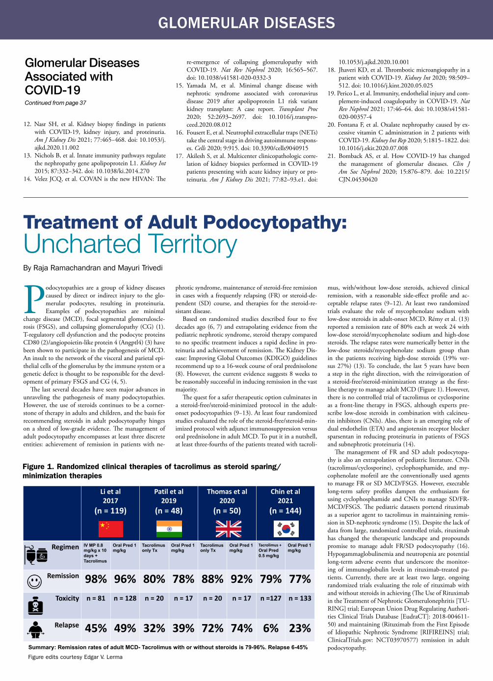
GLOMERULAR DISEASES
12. Nasr SH, et al. Kidney biopsy findings in patients with COVID-19, kidney injury, and proteinuria. Am J Kidney Dis 2021; 77:465−468. doi: 10.1053/j.ajkd.2020.11.002
13. Nichols B, et al. Innate immunity pathways regulate the nephropathy gene apolipoprotein L1. Kidney Int 2015; 87:332−342. doi: 10.1038/ki.2014.270
14. Velez JCQ, et al. COVAN is the new HIVAN: The
re-emergence of collapsing glomerulopathy with COVID-19. Nat Rev Nephrol 2020; 16:565–567. doi: 10.1038/s41581-020-0332-3
15. Yamada M, et al. Minimal change disease with nephrotic syndrome associated with coronavirus disease 2019 after apolipoprotein L1 risk variant kidney transplant: A case report. Transplant Proc 2020; 52:2693–2697. doi: 10.1016/j.transpro-ceed.2020.08.012
16. Fousert E, et al. Neutrophil extracellular traps (NETs) take the central stage in driving autoimmune respons-es. Cells 2020; 9:915. doi: 10.3390/cells9040915
17. Akilesh S, et al. Multicenter clinicopathologic corre-lation of kidney biopsies performed in COVID-19 patients presenting with acute kidney injury or pro-teinuria. Am J Kidney Dis 2021; 77:82–93.e1. doi:
10.1053/j.ajkd.2020.10.00118. Jhaveri KD, et al. Thrombotic microangiopathy in a
patient with COVID-19. Kidney Int 2020; 98:509–512. doi: 10.1016/j.kint.2020.05.025
19. Perico L, et al. Immunity, endothelial injury and com-plement-induced coagulopathy in COVID-19. Nat Rev Nephrol 2021; 17:46–64. doi: 10.1038/s41581-020-00357-4
20. Fontana F, et al. Oxalate nephropathy caused by ex-cessive vitamin C administration in 2 patients with COVID-19. Kidney Int Rep 2020; 5:1815−1822. doi: 10.1016/j.ekir.2020.07.008
21. Bomback AS, et al. How COVID-19 has changed the management of glomerular diseases. Clin J Am Soc Nephrol 2020; 15:876–879. doi: 10.2215/CJN.04530420
Glomerular Diseases Associated with COVID-19 Continued from page 37
Podocytopathies are a group of kidney diseases caused by direct or indirect injury to the glo-merular podocytes, resulting in proteinuria. Examples of podocytopathies are minimal
change disease (MCD), focal segmental glomeruloscle-rosis (FSGS), and collapsing glomerulopathy (CG) (1). T-regulatory cell dysfunction and the podocyte proteins CD80 (2)/angiopoietin-like protein 4 (Angptl4) (3) have been shown to participate in the pathogenesis of MCD. An insult to the network of the visceral and parietal epi-thelial cells of the glomerulus by the immune system or a genetic defect is thought to be responsible for the devel-opment of primary FSGS and CG (4, 5).
The last several decades have seen major advances in unraveling the pathogenesis of many podocytopathies. However, the use of steroids continues to be a corner-stone of therapy in adults and children, and the basis for recommending steroids in adult podocytopathy hinges on a shred of low-grade evidence. The management of adult podocytopathy encompasses at least three discrete entities: achievement of remission in patients with ne-
phrotic syndrome, maintenance of steroid-free remission in cases with a frequently relapsing (FR) or steroid-de-pendent (SD) course, and therapies for the steroid-re-sistant disease.
Based on randomized studies described four to five decades ago (6, 7) and extrapolating evidence from the pediatric nephrotic syndrome, steroid therapy compared to no specific treatment induces a rapid decline in pro-teinuria and achievement of remission. The Kidney Dis-ease: Improving Global Outcomes (KDIGO) guidelines recommend up to a 16-week course of oral prednisolone (8). However, the current evidence suggests 8 weeks to be reasonably successful in inducing remission in the vast majority.
The quest for a safer therapeutic option culminates in a steroid-free/steroid-minimized protocol in the adult-onset podocytopathies (9−13). At least four randomized studies evaluated the role of the steroid-free/steroid-min-imized protocol with adjunct immunosuppression versus oral prednisolone in adult MCD. To put it in a nutshell, at least three-fourths of the patients treated with tacroli-
mus, with/without low-dose steroids, achieved clinical remission, with a reasonable side-effect profile and ac-ceptable relapse rates (9−12). At least two randomized trials evaluate the role of mycophenolate sodium with low-dose steroids in adult-onset MCD. Rémy et al. (13) reported a remission rate of 80% each at week 24 with low-dose steroid/mycophenolate sodium and high-dose steroids. The relapse rates were numerically better in the low-dose steroids/mycophenolate sodium group than in the patients receiving high-dose steroids (19% ver-sus 27%) (13). To conclude, the last 5 years have been a step in the right direction, with the reinvigoration of a steroid-free/steroid-minimization strategy as the first-line therapy to manage adult MCD (Figure 1). However, there is no controlled trial of tacrolimus or cyclosporine as a front-line therapy in FSGS, although experts pre-scribe low-dose steroids in combination with calcineu-rin inhibitors (CNIs). Also, there is an emerging role of dual endothelin (ETA) and angiotensin receptor blocker sparsentan in reducing proteinuria in patients of FSGS and subnephrotic proteinuria (14).
The management of FR and SD adult podocytopa-thy is also an extrapolation of pediatric literature. CNIs (tacrolimus/cyclosporine), cyclophosphamide, and my-cophenolate mofetil are the conventionally used agents to manage FR or SD MCD/FSGS. However, execrable long-term safety profiles dampen the enthusiasm for using cyclophosphamide and CNIs to manage SD/FR-MCD/FSGS. The pediatric datasets portend rituximab as a superior agent to tacrolimus in maintaining remis-sion in SD-nephrotic syndrome (15). Despite the lack of data from large, randomized controlled trials, rituximab has changed the therapeutic landscape and propounds promise to manage adult FR/SD podocytopathy (16). Hypogammaglobulinemia and neutropenia are potential long-term adverse events that underscore the monitor-ing of immunoglobulin levels in rituximab-treated pa-tients. Currently, there are at least two large, ongoing randomized trials evaluating the role of rituximab with and without steroids in achieving (The Use of Rituximab in the Treatment of Nephrotic Glomerulonephritis [TU-RING] trial; European Union Drug Regulating Authori-ties Clinical Trials Database [EudraCT]: 2018-004611-50) and maintaining (Rituximab from the First Episode of Idiopathic Nephrotic Syndrome [RIFIREINS] trial; ClinicalTrials.gov: NCT03970577) remission in adult podocytopathy.
Treatment of Adult Podocytopathy: Uncharted TerritoryBy Raja Ramachandran and Mayuri Trivedi
Li et al2017
(n = 119)
Patil et al2019
(n = 48)
Thomas et al 2020
(n = 50)
Chin et al2021
(n = 144)
Regimen IV MP 0.8 mg/kg x 10 days +Tacrolimus
Oral Pred 1 mg/kg
Tacrolimusonly Tx
Oral Pred 1 mg/kg
Tacrolimusonly Tx
Oral Pred 1 mg/kg
Tacrolimus +Oral Pred0.5 mg/kg
Oral Pred 1 mg/kg
Remission 98% 96% 80% 78% 88% 92% 79% 77%Toxicity n = 81 n = 128 n = 20 n = 17 n = 20 n = 17 n =127 n = 133
Relapse 45% 49% 32% 39% 72% 74% 6% 23%Summary: Remission rates of adult MCD- Tacrolimus with or without steroids is 79-96%. Relapse 6-45%
Figure 1. Randomized clinical therapies of tacrolimus as steroid sparing/ minimization therapies
Figure edits courtesy Edgar V. Lerma

GLOMERULAR DISEASES April 2021 | ASN Kidney News | 39
C3 Glomerulopathy: Update on Pathogenesis and TreatmentBy Shikha Wadhwani and Samir V. Parikh
The management of steroid-resistant FSGS is con-troversial. Traditionally, tacrolimus or cyclosporine are the first-line therapy to manage adult steroid-resistant FSGS. With the sustained evolution of genetic testing, up to 60% of the patients with steroid-resistant FSGS have genetic mutations in podocyte and non-podocyte genes (5). Data from the pediatric population suggest that over three-fourths of the patients with non-genet-ic-associated, steroid-resistant FSGS respond favora-bly to cyclosporine therapy (17). With the unceasing expansion of the library of genes incriminated in the development of steroid-resistant FSGS, experts rec-ommend genetic testing in all steroid-resistant FSGS, hence procrastinating ineffective treatment (18). As per the current consensus, all steroid-resistant FSGS pa-tients need to be started on an angiotensin-converting enzyme inhibitor or angiotensin receptor blocker and lipid lowering with statin therapy. Both the disease and its treatment affect the bone and cardiovascular system adversely. Hence, future research needs to solicit safe-guards for bone and cardiovascular health in patients with steroid-resistant FSGS.
To conclude, the management of adult podocytopa-thy graduates from steroids (only) to steroid-minimized or steroid-free therapies, primarily involving tacroli-mus/mycophenolate sodium and rituximab, thus offer-ing a new perspective to the management of SD or FR podocytopathy. Genetic testing is the key to manage steroid-resistant FSGS, at least before subjecting it to toxic second-line agents. Admittedly, adult nephrolo-gists worldwide with renewed interest in glomerular diseases need to collaborate and examine a steroid-free or steroid-minimized protocol for managing MCD/FSGS. Understandably, the TURING and RIFIREINS trials are steps in the right direction.
Raja Ramachandran is Assistant Professor with the De-partment of Nephrology, Post-Graduate Institute of Medi-cal Education and Research, Chandigarh, India. May-uri Trivedi is Assistant Professor with the Department of Nephrology, Lokmanya Tilak Medical General Hospital, Mumbai, India.
References1. Barisoni L, et al. A proposed taxonomy for the podo-
cytopathies: A reassessment of the primary nephrotic diseases. Clin J Am Soc Nephrol 2007; 2:529−542. doi: 10.2215/CJN.04121206
2. Garin EH, et al. Urinary CD80 is elevated in minimal change disease but not in focal segmental glomerulo-sclerosis. Kidney Int 2010; 78:296−302. doi: 10.1038/ki.2010.143
3. Clement LC, et al. Podocyte-secreted angiopoietin-like-4 mediates proteinuria in glucocorticoid-sensitive nephrotic syndrome. Nat Med 2011; 17:117−122. doi: 10.1038/nm.2261
4. Kopp JB, et al. Clinical features and histology of apolipo-protein L1-associated nephropathy in the FSGS Clini-cal Trial. J Am Soc Nephrol 2015; 26:1443−1448. doi: 10.1681/ASN.2013111242
5. Landini S, et al. Reverse phenotyping after whole-exome sequencing in steroid-resistant nephrotic syndrome. Clin J Am Soc Nephrol 2020; 15:89−100. doi: 10.2215/CJN.06060519
6. Black DA, et al. Controlled trial of prednisone in adult patients with the nephrotic syndrome. Br Med J 1970; 3:421−426. doi: 10.1136/bmj.3.5720.421
7. Coggins CH. Adult minimal change nephropathy: Ex-perience of the collaborative study of glomerular disease. Trans Am Clin Climatol Assoc 1986; 97:18−26. PMID: 3915841
8. Kidney Disease: Improving Global Outcomes (KDI-GO) Glomerulonephritis Work Group. KDIGO clini-cal practice guideline for glomerulonephritis. Kidney Int Suppl 2012; 2:139−274. https://www.sciencedirect.com/journal/kidney-international-supplements/vol/2/issue/2
9. Li X, et al. Tacrolimus monotherapy after intrave-nous methylprednisolone in adults with minimal change nephrotic syndrome. J Am Soc Nephrol 2017; 28:1286−1295. doi: 10.1681/ASN.2016030342
10. Patil MR, et al. Tacrolimus as the first-line agent in adult-onset minimal change disease: A randomized controlled study. Saudi J Kidney Dis Transpl 2019; 30:129−137. https://www.sjkdt.org/article.asp?issn=1319-2442;year=
2019;volume=30;issue=1;spage=129;epage=137;aulast=Patil
11. Medjeral-Thomas NR, et al. Randomized, con-trolled trial of tacrolimus and prednisolone mono-therapy for adults with de novo minimal change disease: A multicenter, randomized, controlled tri-al. Clin J Am Soc Nephrol 2020; 15:209−218. doi: 10.2215/CJN.06180519
12. Chin HJ, et al. Comparison of the efficacy and safe-ty of tacrolimus and low-dose corticosteroid with high-dose corticosteroid for minimal change ne-phrotic syndrome in adults. J Am Soc Nephrol 2021; 32:199−210. doi: 10.1681/ASN.2019050546
13. Rémy P, et al. An open-label randomized controlled trial of low-dose corticosteroid plus enteric-coated mycophenolate sodium versus standard corticoster-oid treatment for minimal change nephrotic syn-drome in adults (MSN Study). Kidney Int 2018; 94:1217−1226. doi: 10.1016/j.kint.2018.07.021
14. Trachtman H, et al. DUET: A phase 2 study evaluat-ing the efficacy and safety of sparsentan in patients with FSGS. J Am Soc Nephrol 2018; 29:2745−2754. doi: 10.1681/ASN.2018010091
15. Basu B, et al. Efficacy of rituximab vs tacrolimus in pediatric corticosteroid-dependent nephrotic syndrome: A randomized clinical trial. JAMA Pedi-atr 2018; 172:757−764. doi: 10.1001/jamapediat-rics.2018.1323
16. Gauckler P, et al. Rituximab in adult minimal change disease and focal segmental glomeruloscle-rosis—what is known and what is still unknown? Autoimmun Rev 2020; 19:102671. doi: 10.1016/j.autrev.2020.102671
17. Büscher AK, et al. Rapid response to cyclosporin A and favorable renal outcome in nongenetic versus genetic steroid-resistant nephrotic syndrome. Clin J Am Soc Nephrol 2016; 11:245−253. doi: 10.2215/CJN.07370715
18. De Vriese AS, et al. Differentiating primary, genetic, and secondary FSGS in adults: A clinicopathologic approach. J Am Soc Nephrol 2018; 29:759−774. doi: 10.1681/ASN.2017090958
C3 glomerulonephritis (C3GN) and dense de-posit disease (DDD), collectively known as C3 glomerulopathy (C3G), are rare glomerular diseases presenting with microscopic hema-
turia, proteinuria, and often, abnormal kidney function. Low serum C3 is present in 70%−80% of patients with DDD and 50% with C3GN (1). Effective therapies are lacking, and prognosis is poor (2). Disease recurrence after kidney transplantation is common and leads to graft loss in 30%−40% of affected patients (3, 4).
Pathogenesis of C3GC3G is characterized by dysregulation of the alternative complement pathway and defined by C3-dominant stain-ing on immunofluorescence (IF) of a kidney biopsy. DDD is differentiated from C3GN histologically: the former has characteristic ribbon-like, electron-dense, intramembra-nous deposits on electron microscopy, whereas the latter has mesangial, subendothelial, and rarely, subepithelial deposits (5). Despite the histological differences, the clinical presen-tation, outcomes, and alternative complement pathway ab-normalities are similar between C3GN and DDD.
Figure 1 provides an overview of the alternative com-
plement pathway in healthy and disease states. Briefly, in physiologic states, the alternative complement pathway maintains low-level activation through spontaneous hy-drolysis of C3 to C3b (“tick over”) and controlled genera-tion of C3 convertase (C3bBb). The C3 convertase am-plifies the alternative complement pathway by producing more C3b through C3 cleavage and drives C5 convertase (C3bBbC3b) generation. C5 convertase cleaves C5 to form the anaphylatoxin C5a and C5b—the latter forming the membrane attack complex (MAC), C5b-9, which induces cell lysis (5). In healthy states, fluid phase (factor H and fac-tor I) and cell surface (factor H, membrane cofactor protein [MCP], decay-accelerating factor [DAF], and complement receptor 1 [CR1]) regulators of complement activity keep the alternative complement pathway under tight control. Genetic or acquired defects of these complement regulators or activators are responsible for alternative pathway dysregu-lation in C3G (Table 1). Accordingly, a complete comple-ment workup is recommended for all patients. The most common defect in C3G is an acquired C3 nephritic factor (C3Nef), a C3 convertase-stabilizing immunoglobulin G (IgG) autoantibody that dramatically increases its half-life and hence perpetuates alternative pathway dysregulation (5,
6). Genetic variants are identified in up to 25% of C3G cases; however, the functional significance of these variants is often unclear (1, 3).
Management in C3GThere are no approved therapies for C3G, and current treat-ment regimens are based on retrospective case series and expert opinion. Blockade of the renin-angiotensin system is recommended for all patients with proteinuria. Corticoster-oids and non-specific immunosuppressive agents are often used but have shown variable success. Perhaps the best avail-able evidence for treatment of C3G comes from two in-dependent cohort studies (combined n=132), which dem-onstrated efficacy of corticosteroids plus mycophenolate mofetil (MMF) as compared to steroids alone, other im-munosuppressive therapies, or supportive care (7, 8). These studies, however, are limited by their retrospective, uncon-trolled design and heterogeneity in both treatment duration and steroid dosing. Notably, MMF showed minimal re-sponse in another cohort (n = 78), possibly due to a greater number of patients with genetic variants (9). Nonetheless,
>Continued on page 40
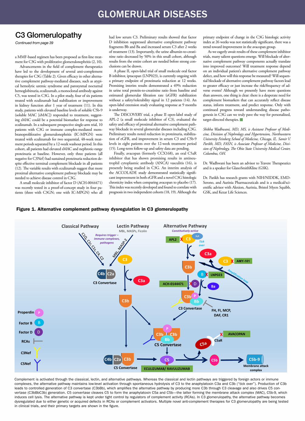
GLOMERULAR DISEASES
a MMF-based regimen has been proposed as first-line treat-ment for C3G with proliferative glomerulonephritis (2, 10).
Advancements in the field of complement therapeutics have led to the development of several anti-complement therapies for C3G (Table 2). Given efficacy in other alterna-tive complement pathway-mediated diseases, such as atypi-cal hemolytic uremic syndrome and paroxysmal nocturnal hemoglobinuria, eculizumab, a monoclonal antibody against C5, was tested in C3G. In a pilot study, four of six patients treated with eculizumab had stabilization or improvement in kidney function after 1 year of treatment (11). In this study, patients with elevated baseline levels of soluble C5b-9 (soluble MAC [sMAC]) responded to treatment, suggest-ing sMAC could be a potential biomarker for response to eculizumab. In a subsequent prospective single-arm trial, 10 patients with C3G or immune complex-mediated mem-branoproliferative glomerulonephritis (IC-MPGN) were treated with eculizumab for two sequential, 48-week treat-ment periods separated by a 12-week washout period. In this cohort, all patients had elevated sMAC and nephrotic-range proteinuria at baseline. However, only three patients (all negative for C3Nef) had sustained proteinuria reduction de-spite effective terminal complement blockade in all patients (12). The variable results with eculizumab suggest that more proximal alternative complement pathway blockade may be needed to achieve disease control in C3G.
A small molecule inhibitor of factor D (ACH-0044471) was recently tested in a proof-of-concept study in four pa-tients (three with C3GN; one with IC-MPGN) who all
had low serum C3. Preliminary results showed that factor D inhibition suppressed alternative complement pathway fragments Bb and Ba and increased serum C3 after 2 weeks of treatment (13). Importantly, the urine albumin-to-creati-nine ratio decreased by 50% in this small cohort, although results from the entire cohort are needed before strong con-clusions can be drawn.
A phase II, open-label trial of small molecule oral factor B inhibitor, iptacopan (LNP023), is currently ongoing with a primary endpoint of proteinuria reduction at 12 weeks. Promising interim results demonstrated a 49% reduction in urine total protein-to-creatinine ratio from baseline and estimated glomerular filtration rate (eGFR) stabilization without a safety/tolerability signal in 12 patients (14). An open-label extension study evaluating response at 9 months is underway.
The DISCOVERY trial, a phase II open-label study of APL-2 (a small molecule inhibitor of C3), evaluated the safety and efficacy of proximal alternative complement path-way blockade in several glomerular diseases including C3G. Preliminary results noted reduction in proteinuria, stabiliza-tion of eGFR, and improvement in serum C3 and C5b-9 levels in eight patients over the 12-week treatment period (15). Long-term follow-up and safety data are pending.
Finally, avacopan (formerly CCX168), an oral C5aR inhibitor that has shown promising results in antineu-trophil cytoplasmic antibody (ANCA) vasculitis (16), is presently being studied in C3G. An interim analysis of the ACCOLADE study demonstrated statistically signifi-cant improvement in both eGFR and a novel C3G histologic chronicity index when comparing avacopan to placebo (17). This index was recently developed and found to correlate with prognosis in two independent cohorts (18, 19). Although the
primary endpoint of change in the C3G histologic activity index at 26 weeks was not statistically significant, there was a trend toward improvement in the avacopan group.
As we eagerly await results of these complement inhibitor trials, many salient questions emerge. Will blockade of alter-native complement pathway components actually translate into improved outcomes? Will treatment response depend on an individual patient’s alternative complement pathway defect, and how will this response be measured? Will sequen-tial blockade of alternative complement pathway factors lead to greater efficacy or just increase the risk/frequency of ad-verse events? Although we presently have more questions than answers, one thing is clear: there is a desperate need for complement biomarkers that can accurately reflect disease status, inform treatment, and predict response. Only with continued progress toward understanding disease patho-genesis in C3G can we truly pave the way for personalized, target-directed therapies.
Shikha Wadhwani, MD, MS, is Assistant Professor of Medi-cine, Division of Nephrology and Hypertension, Northwestern University Feinberg School of Medicine, Chicago, IL. Samir V. Parikh, MD, FASN, is Associate Professor of Medicine, Divi-sion of Nephrology, The Ohio State University Medical Center, Columbus, OH.
Dr. Wadhwani has been an advisor to Travere Therapeutics and is a speaker for GlaxoSmithKline (GSK).
Dr. Parikh has research grants with NIH/NIDDK, EMD-Serono, and Aurinia Pharmaceuticals and is a medical/sci-entific advisor with Alexion, Aurinia, Bristol Myers Squibb, GSK, and Kezar Life Sciences.
C3 GlomerulopathyContinued from page 39
Amplification loop
Tick over
ECULIZUMAB/ RAVULIZUMAB
AVACOPAN
ACH-0144471
LNP023
AMY-101
APL2
D
B
BaC3a
C5 Convertase
C5
C5a
Membrane attack complex
Bb
C3Nef
C5Nef X
C5aR
Classical Pathway Lectin PathwayMBL, MASPs, Ficolin
Requires trigger –immune complexes,
pathogens
C3
C2aC4b
C3 Convertase
C5 Convertase
C3
C3 Convertase
C3
C2aC4b
P
Bb
X
X
X
X
X
Alternative PathwayConstitutively active
RCAs
C5b-9
C3a
C5b
DFactor D
Factor B
FH, FI, MCP, DAF, CR1
P
Properdin P
C1Q, C1r, C1s, C4, C2 MBL
, MASP
-2
H20
B
Figure 1. Alternative complement pathway dysregulation in C3 glomerulopathy
Complement is activated through the classical, lectin, and alternative pathways. Whereas the classical and lectin pathways are triggered by foreign actors or immune complexes, the alternative pathway maintains low-level activation through spontaneous hydrolysis of C3 to the anaphylatoxin C3a and C3b (“tick over”). Production of C3b leads to controlled generation of C3 convertase (C3bBb), which amplifies the alternative pathway by producing more C3b through C3 cleavage and also drives C5 con-vertase (C3bBbC3b) generation. C5 convertase cleaves C5 to form the anaphylatoxin C5a and C5b—the latter forming the membrane attack complex (MAC), C5b-9, which induces cell lysis. The alternative pathway is kept under tight control by regulators of complement activity (RCAs). In C3 glomerulopathy, the alternative pathway becomes dysregulated due to either genetic or acquired defects in RCAs or complement activators. Multiple novel anti-complement therapies for C3 glomerulopathy are being tested in clinical trials, and their primary targets are shown in the figure.

GLOMERULAR DISEASES April 2021 | ASN Kidney News | 41
References1. Iatropoulos P, et al. Complement gene variants deter-
mine the risk of immunoglobulin-associated MPGN and C3 glomerulopathy and predict long-term re-nal outcome. Mol Immunol 2016; 71:131−142. doi: 10.1016/j.molimm.2016.01.010
2. Smith RJH, et al. C3 glomerulopathy—understanding a rare complement-driven renal disease. Nat Rev Nephrol 2019; 15:129−143. doi: 10.1038/s41581-018-0107-2
3. Servais A, et al. Acquired and genetic complement abnormalities play a critical role in dense deposit dis-ease and other C3 glomerulopathies. Kidney Int 2012; 82:454−464. doi: 10.1038/ki.2012.63
4. Angelo JR, et al. Allograft failure in kidney transplant recipients with membranoproliferative glomerulo-nephritis. Am J Kidney Dis 2011; 57:291−299. doi: 10.1053/j.ajkd.2010.09.021
5. Smith RJ, et al. New approaches to the treatment of dense deposit disease. J Am Soc Nephrol 2007; 18:2447−2456. doi: 10.1681/ASN.2007030356
6. Fakhouri F, et al. Practical management of C3 glomeru-lopathy and Ig-mediated MPGN: Facts and uncertain-ties. Kidney Int 2020; 98:1135−1148. doi: 10.1016/j.kint.2020.05.053
7. Rabasco C, et al. Effectiveness of mycophenolate mofetil in C3 glomerulonephritis. Kidney Int 2015; 88:1153−1160. doi: 10.1038/ki.2015.227
8. Avasare RS, et al. Mycophenolate mofetil in combina-tion with steroids for treatment of C3 glomerulopathy:
A case series. Clin J Am Soc Nephrol 2018; 13:406−413. doi: 10.2215/CJN.09080817
9. Ravindran A, et al. C3 glomerulopathy: Ten years’ experience at Mayo Clinic. Mayo Clin Proc 2018; 93:991−1008. doi: 10.1016/j.mayocp.2018.05.019
10. Floege J, et al. Chapter 8: Complement-associated glo-merulonephritis. KDIGO Clinical Practice Guidelines on Glomerular Diseases. Public draft review. June 2020. htt-ps://kdigo.org/wp-content/uploads/2017/02/KDIGO-GN-GL-Public-Review-Draft_1-June-2020.pdf
11. Bomback AS, et al. Eculizumab for dense deposit dis-ease and C3 glomerulonephritis. Clin J Am Soc Nephrol 2012; 7:748−756. doi: 10.2215/CJN.12901211
12. Ruggenenti P, et al. C5 convertase blockade in mem-branoproliferative glomerulonephritis: A single-arm clinical trial. Am J Kidney Dis 2019; 74:224−238. doi: 10.1053/j.ajkd.2018.12.046
13. Galvan MD, et al. Evaluation of urine complement biomarker in C3G following complement alternative pathway inhibition with ACH-4471 (SA-PO424). ASN Kidney Week 2018. https://www.asn-online.org/education/kidneyweek/2018/program-abstract.aspx?controlId=3021605
14. Wong EKS, et al. LNP023: A novel oral complement alternative pathway factor B inhibitor safely and ef-fectively reduces proteinuria in C3 glomerulopathy (SU-OR39). ASN Kidney Week 2020. https://www.asn-online.org/education/kidneyweek/2020/program-abstract.aspx?controlId=3442221
15. Dixon BP, et al. C3 inhibition with APL-2 targets the underlying disease process of C3G complement hy-peractivity and improves proteinuria (FR-PO906). ASN Kidney Week 2019. https://www.asn-online.org/education/kidneyweek/2019/program-abstract.aspx?controlId=3231183
16. Jayne DRW, et al. Avacopan for the treatment of ANCA-associated vasculitis. N Engl J Med 2021; 384:599−609. doi: 10.1056/NEJMoa2023386
17. ChemoCentryx Press Release: ChemoCentryx and VFMCRP Provide Topline Results from ACCOLADE Trial of Avacopan in C3 Glomerulopathy Including Im-proved Estimated Glomerular Filtration Rate (eGFR). December 21, 2020. https://ir.chemocentryx.com/news-releases/news-release-details/chemocentryx-and-vfmcrp-provide-topline-results-accolade-trial
18. Bomback AS, et al. C3 glomerulonephritis and dense deposit disease share a similar disease course in a large United States cohort of patients with C3 glomeru-lopathy. Kidney Int 2018; 93:977–985. doi: 10.1016/j.kint.2017.10.022
19. Caravaca-Fontán F, et al. Validation of a histologic scoring index for C3 glomerulopathy. Am J Kidney Dis [published online ahead of print December 22, 2020]. doi: 10.1053/j.ajkd.2020.11.011; https://www.ajkd.org/article/S0272-6386(20)31160-4/fulltext
20. Martín B, Smith RJH. C3 glomerulopathy. In GeneRe-views (University of Seattle, WA) 2007 (updated 2018). https://www.ncbi.nlm.nih.gov/books/NBK1425/
Table 1. Genetic and acquired complement defects in C3 glomerulopathy
Table 2. Clinical trials of complement-directed therapies
Genetic variant Frequency (ref. 20) Acquired defect Frequency
C3 11% C3 nephritic factor 80% DDD; 50% C3GN
Complement factor H 12% C5 nephritic factor 50% of C3G
CFHR1, -1/5, -3/1, or -5 Rare C4 nephritic factor Rare
Complement factor B 1% Anti-factor H autoantibody 4%−12% of C3G
Complement factor I 5% Anti-C3B autoantibody 2%−3% of C3G
Drug Target Sponsor Treatment population
Trial phase
Clinical trial # Status
Eculizumab C5 Alexion C3GN or IC-MPGN 2 NCT02093533 Completed
Avacopan (CCX168)
C5aR ChemoCentryx C3G, native or post-transplant
2 NCT03301467 Recruitment completed; study ongoing
ACH-0144471 Complement Factor D
Alexion C3G or IC-MPGN 2 NCT03124368 Completed
ACH-0144471 Complement Factor D
Alexion C3G or IC-MPGN 2 NCT03459443 Recruitment completed; study ongoing
ACH-0144471 Complement Factor D
Alexion C3G 2 NCT03369236 Recruitment completed; study ongoing
Iptacopan (LNP023)
Complement Factor B
Novartis C3G and recurrent C3G in transplant
2 NCT03832114, NCT03955445
Recruiting
APL-2 C3 Apellis C3G, IgAN, LN (class III, IV, or V), primary MN
2 NCT03453619 Recruitment completed; study ongoing
AMY-101 C3 Amyndas Healthy males 1 NCT03316521 Completed
Narsoplimab (OMS721)
MASP-2 Omeros C3G, IgAN, LN, MN 2 NCT02682407 Recruiting
CFHR, complement factor H-related protein.
C5aR, complement component 5a receptor; IgAN, immunoglobulin A nephropathy; LN, lupus nephritis; MN, membranous nephropathy; MASP-2, mannose-binding lectin serine protease 2.

42 | ASN Kidney News | April 2021 Findings
Malaria and FSGS—Is There a Connection?Highlights of a recent study in CJASN, “Malaria, Collapsing Glomerulopathy, and Focal and Segmental Glomerulosclerosis”
By Vincent Audard and Anissa Moktefi
M alaria, a potentially life-threatening disease, is the most prevalent en-demic infectious disease worldwide, affecting millions of people in tropi-
cal areas. In European and Western countries, ma-laria is acquired during travel to areas in which the disease is endemic. Kidney involvement, including acute kidney injury, is seen in up to 60% of patients with severe malaria and is frequently observed with Plasmodium falciparum and Plasmodium malariae. However, the modern era has seen the spectrum of glomerular damage associated with malaria infection widened.
In a retrospective study performed in France (1), we identified 23 patients (22 due to P. falciparum in-fection and all but one patient of African ancestry) with biopsy-proven glomerular disease occurring af-ter acute malaria (kidney biopsy performed during the three months following confirmation of Plasmo-dium infection).
All patients (12 men and 11 women, mean age 47 years) presented with acute kidney injury (requiring kidney replacement therapy in 12 cases) at the time of kidney biopsy. The kidney pathology findings in-cluded focal segmental glomerulosclerosis (FSGS) in 21 cases and minimal change disease in two patients. Collapsing glomerulopathy (CG) was the most com-mon pathology finding (Figure 1A). CG was ob-served in 18 patients (including nine with HIV in-fection) and was associated with the presence of two high-risk APOL1 variants in all seven patients tested for APOL1 polymorphism. Immunohistochemistry with an antibody targeting P. falciparum histidine-rich protein-2 (HRP-2), to search for the presence of P. falciparum in the kidney tissues, revealed the presence of the parasite in the lumina of the tubules (Figure 1B) of all patients tested but its absence from the glomeruli (Figure 1C). At the end of follow-up, eight patients required kidney replacement therapy. Overall, these data suggest that, in patients of Af-rican ancestry, imported Plasmodium infection pro-motes CG in particular. In this setting, malaria may act as a “second hit” in patients with genetic (high-risk APOL1 genotype) or viral infection-associated susceptibility factors.
In addition to the recent findings showing that severe acute respiratory syndrome coronavirus 2 (SARS-CoV-2)-associated CG predominantly affects individuals of African ancestry who have high-risk
APOL1 alleles (2), our study emphasizes the role of infectious agents as triggers of CG in patients with genetic susceptibility. The accurate pathophysiologi-cal processes of these infectious agents (malaria and SARS-CoV-2) in the development of CG remain to be clarified.
Vincent Audard, MD, PhD, is with the Assistance Publique des Hôpitaux de Paris (AP-HP), Hôpitaux Universitaires Henri Mondor, Service de Néphrologie et Transplantation, Centre de Référence Maladie Rare “Syndrome Néphrotique Idiopathique,” Fédération Hospitalo-Universitaire “Inno-vative Therapy for Immune Disorders,” and the Univ Paris Est Créteil, Institut National de la Santé et de la Recherche Médicale (INSERM) U955, Institut Mondor de Recherche Biomédicale (IMRB), Equipe 21, Créteil, France. Anissa Moktefi, MD, PhD, is with the Univ Paris Est Créteil, In-stitut National de la Santé et de la Recherche Médicale (IN-SERM) U955, Institut Mondor de Recherche Biomédicale (IMRB), Equipe 21, and the Assistance Publique des Hôpi-taux de Paris (AP-HP), Hôpitaux Universitaires Henri Mondor, Département de Pathologie, Créteil, France.
Disclosures: Dr. Audard reports receiving personal fees from Addmedica outside of the submitted work. Dr. Moktefi has nothing to disclose.
References1. Amoura A, et al. Malaria, collapsing glomerulopathy,
and focal and segmental glomerulosclerosis. Clin J Am Soc Nephrol 2020; 15:964−972. doi: 10.2215/CJN.00590120
2. Ng JH, et al. Pathophysiology and pathology of acute kidney injury in patients with COVID-19. Adv Chronic Kidney Dis 2020; 27:365−376. doi: 10.1053/j.ackd.2020.09.003
NEWS FLASH
Figure 1. Collapsing glomerulopathy with hypertrophic and hyperplastic podocytes containing protein resorp-tion droplets (A; periodic acid–Schiff staining; ×400). Immunohistochemistry with a monoclonal antibody against Plasmodium falciparum histidine-rich protein-2 showed the presence of the P. falciparum antigen in the lumen of a tubule (B; x400) but was negative on glomeruli (C; x400).
Want to learn even more about how changes in health care policy, the kidney workforce, and new research will affect you?
Check out Kidney News Online at www.kidneynews.org

Nova Biomedical’s Educational Webinar Series Presents:
The role of ionized magnesium is of growing interest in internal medicine, especially in cardiovascular diseases and in patients with diabetes mellitus. Since ionized magnesium (iMg) is the only physiologically active component of serum magnesium, total serum magnesium (tMg) is not always an accurate indicator of a patient’s magnesium status. This presentation will describe recent studies showing statistically significant decreased iMg in patients with various diseases, e.g., heart failure, arteriosclerosis, lipid disorders, hypertension, heart rhythm disorders, and diabetes mellitus. Other situations also can cause a decrease in iMg, such as chemotherapy, emotional stress, and vigorous exercise. Lowered magnesium concentrations should be identified and corrected expeditiously to avoid vascular damage, arrhythmias, inflammation, and other sequelae of hypomagnesemia. The presentation will describe why measurement of ionized magnesium, not total, is a better tool to manage magnesium status correctly.Learning Objectives:
• Role of magnesium in disease states including heart failure, heart rhythm disorders, diabetes, hypertension, gastrointestinal and renal losses• Why ionized magnesium is a better tool to manage magnesium status
Presenter Prof. Dr. Klaus Kisters, MD Med Clinic I, St. Anna Hospital, ESH Excellence Centre, Herne, Germany
novabiomedical.com
Several aspects of COVID-19 disease mimic metabolic events shown to occur during latent subclinical magnesium deficiency. Most notably, hypomagnesemia is a known pro-inflammatory state, and can predispose to cytokine storm, a factor in severe COVID-19 cases. A summary of experimental findings and knowledge of the biochemical role magnesium may play in the pathogenesis of COVID-19, particularly in severe cases, is presented. Frequent monitoring of ionized magnesium status with subsequent repletion, when appropriate, may be an effective strategy to influence disease contraction and progression.Learning Objectives:
• Ionized magnesium in pro-inflammatory states• Ionized magnesium management and repletion in COVID-19 severe illness
Presenter Taylor Wallace, PhD Think Healthy Group, Washington, DC, USA; Department of Nutrition and Food Studies, George Mason University, Fairfax, Virginia, USA; Center for Magnesium Education & Research, Pahoa, Hawaii, USA
Webinar Dates:Thursday, May 6, 1:00 PM Eastern Time Thursday, May 20, 1:00 PM Eastern Time
Register Now at: novabiomedical.com/iMg-CV-Nutrition-kn
Educational CreditsThis program offers 1 hour of P.A.C.E. continuing education credits.
This program has been approved by the American Association of Critical-Care Nurses (AACN), for 1.00 CERPs, Synergy CERP Category A, File Number 23619. Approval refers to recognition of continuing education only and does not imply AACN approval or endorsement of the content of this educational activity, or the products mentioned.
Clinical Importance of Ionized Magnesium in Cardiovascular Disease, Diabetes Mellitus, Gastrointestinal and Renal Losses
Combating COVID-19 and Building Immune Resilience: A Potential Role for Magnesium Nutrition
43 | ASN Kidney News | March 2021 Findings April 2021 | ASN Kidney News | 43
Recovery of kidney function is common for COVID-19 patients with acute kidney injury requiring kidney re-placement therapy (AKI-KRT), according to a report in Kidney International.
Researchers at a large German tertiary care center re-port their experience with 74 hospitalized patients who developed AKI-KRT as a complication of COVID-19 between March and June 2020. The patients’ median age was 65 years and three-fourths were men. All patients were in the ICU when AKI-KRT developed. Nearly all were on mechanical ventilation, and 39.2% were receiv-ing extracorporeal membrane oxygenation.
Outcomes were assessed in October 2020—a median of 151 days after the start of KRT. In 37 patients who sur-vived to discharge, median duration of KRT was 27 days. At follow-up, 62.2% of patients had complete recovery of kidney function while 91.9% had partial recovery. Just 3 patients (8.1% of survivors) were still KRT dependent.
From the start of the COVID-19 pandemic, AKI-KRT has been recognized as a common complication. This series of critically ill COVID-19 patients with AKI-KRT suggests that most survivors will have recovery of kidney function at follow-up in the months after dis-charge. Recovery can occur even after prolonged periods of KRT. “This information may be of value for patients with COVID-19 and their clinicians when it comes to deciding about the initiation or discontinuation of KRT,” the researchers write [Stockmann H, et al. High rates of long-term renal recovery in survivors of COVID-19-asso-ciated acute kidney injury requiring kidney replacement therapy. Kidney Int 2021; 99:1021−1022. doi: 10.1016/j.kint.2021.01.005.
Electronic health record alerts have only a modest impact on care processes for acute kidney injury (AKI), and no impact on important disease outcomes—with a possible increase in adverse outcomes in some settings, accord-ing to conclusions from a randomized trial in the British Medical Journal.
The double-blind, multi-center trial was carried out at six hospitals, including four teaching hospitals, in a New England university-affiliated health system. The interven-tion was a “pop-up” alert in the electronic health record of patients meeting KDIGO (Kidney Disease Improving Global Outcomes) criteria for AKI.
At intervention hospitals, the alert was triggered when-ever the chart was opened by a provider with authority to change or enter new orders—including physicians, train-ees, nurse practitioners, and physician’s assistants. The alert prompted providers to enter AKI onto a patient’s problem list and included a link to a standard AKI order set. At usual-care control hospitals, the system generated “silent” alerts that were not visible to providers but were
Most COVID-19 Patients with AKI Regain Kidney Function
Electronic Alerts for AKI Show Little Benefit, and Possible Harms
tracked by the researchers.A primary composite outcome of AKI progression, dialysis
initiation, or death within 14 days was compared for patients at intervention and control hospitals. Secondary outcomes in-cluded the frequency of various care practices for AKI and the effects of the alerts at each study hospital.
The analysis included 6030 patients admitted over 22 months. There was no significant difference in rates of the primary outcome at intervention versus usual care hospitals: 21.3% and 20.9%, respectively.
At the two non-teaching hospitals, accounting for 13% of patients, the risk of the primary outcome was higher in the alert group: relative risk 1.49. The difference appeared to be mainly driven by deaths: 15.6% in the alert group versus 8.6% in the usual-care group.
Rates of kidney consultations were similar between the groups. Some small increases in process measures in the alert group were observed, including orders for intravenous fluids
and urinalysis.It is often assumed that increased recognition of AKI in
hospitalized patients will lead to improvements in care and thus in clinical outcomes, the authors noted. Thus, many health systems have introduced electronic alerts for AKI, de-spite limited evidence of their impact on patient outcomes.
The new trial shows no improvement in clinical outcomes in AKI patients at hospitals with electronic health record alerts and limited effects on care processes. The study also provides evidence of possible harms associated with AKI alerts in some settings, which remains unexplained.
“This study argues against the implementation of infor-mational alerts for acute kidney injury and for a reconsidera-tion of the alerts currently used,” the authors state [Wilson FP, et al. Electronic health record alerts for acute kidney injury: Multicenter, randomized clinical trial. BMJ 2021; 372:m4786. doi: 10.1136/bmj.m4786; https://www.bmj.com/content/372/bmj.m4786].

44 | ASN Kidney News | April 2021
Fellows First
Nephrology Fellow Mental Health and Well-Being during the COVID-19 Pandemic By Matthew R. Sinclair
While the coronavirus in-fectious disease 2019 (COVID-19) pandemic has significantly affected
all health care workers, graduate medi-cal education (GME) trainees have been especially impacted. The personal and professional obstacles faced during GME training are unique compared to those that attending physicians face, as trainees are concerned not only about protecting their patients and loved ones, but they also have additional stressors related to attaining the skills necessary for independent practice, advancing their research, ensuring edu-cation goals are attained, securing a job, and passing their board certification exam, among others. A recent national survey assessing the impact of the pandemic on cardiovascular fellows found that 69% of respondents were concerned about fulfill-ing their training requirements (1). While a recent study done in the UK showed high burnout rates among nephrology trainees during the pandemic (2), little is known about how the various stressors related to training during the pandemic have specif-ically burdened North American nephrol-
ogy fellows, a group that already suffers from significant burnout (3). Two recent initiatives spearheaded by the American Society of Nephrology (ASN) sought to answer some of the questions surrounding the mental health, professional develop-ment, and overall well-being of nephrology fellows.
One of these initiatives took place on January 13, 2021, when ASN hosted a 90-minute, virtual COVID-19 roundta-ble unlike any it had held previously. This roundtable was unique in that it was creat-ed for and led by fellows in adult nephrol-ogy. The panelists and audience members consisted entirely of nephrology fellows from the United States and Canada. Two guest faculty members from New York, Dr. Daniel Cukor and Dr. Jia Ng, experts in the fields of GME behavioral health and COVID-19 response, respectively, also helped facilitate the roundtable. The forum was organized to discuss the COVID-19 pandemic from fellows’ perspectives in an environment conducive to facilitating con-structive dialogue. Difficult topics were dis-cussed, personal testimonies were shared, and institutional policies compared.
The second initiative was a survey sent out in August 2020 to 1005 current and recently graduated adult and pediatric ne-phrology fellows from the United States, which received 425 responses (42% re-sponse rate) (Figure 1). Themes of the sur-vey focused on the effect of the COVID-19 pandemic on trainees’ experiences and well-being. Pivert et al. (4) reported on the results of this survey in JASN.
In the first segment of the roundtable, titled “The COVID-19 Experience,” the mental health of trainees was highlighted. The initial discussion focused on emotions felt early in the pandemic, of which fear was ubiquitous, especially in the hotspots. Several themes emerged, such as fear of get-ting infected, fear of infecting a loved one, and the fear of not being able to get back into the country as an international med-ical graduate. The personal stories shared were heart-wrenching. Another common theme was sadness. Having to see patients sick and dying with COVID-19 day in and day out was a lot to bear. However, hav-ing to do this while also witnessing ram-pant anti-Asian sentiment, racial injustice against Black individuals, and communi-
ties of color disproportionately burdened by the pandemic weighed heavily on many of the trainees.
Feelings of loneliness were also quite common, with some fellows preferring to be at the hospital, just so they did not have to deal with the isolation that came with being at home. Importantly, the majority of fellows stated that their programs pro-vided resources and outreach to improve fellow well-being. Despite these good in-tentions from the fellowship programs, some of the negative downstream effects of these emotions were captured in fellow responses on the survey (4), with 42% of respondents indicating that the pandemic had a negative effect on their overall quality of life (QOL) and 33% stating it negatively affected their work-life balance (Figure 1). Additionally, residents who completed the survey filled out a tool called the Resident Well-Being Index (RWBI), which is meant to correlate with QOL, fatigue, meaning in work, and burnout, among other things, with a score of 5 or greater (on a 0−7 scale) consistent with distress (5). Surprisingly, only 15% of respondents actually met this distress threshold, a much lower number than expected, demonstrating the resilience of fellows during the crisis (Figure 1). The authors theorize that this unexpected find-ing could have been related to the sense of unity and purpose that nephrology fellows felt being on the front lines. However, an-other possibility may be related to some themes that came up during the second half of the roundtable discussion.
During the final 45 minutes of the roundtable, titled “The Division Experi-ence,” the focus shifted from trainee mental health to how individual institutions man-aged fellow safety and education. It became clear after hearing multiple testimonies that many institutions diminished or eliminated the need for fellows to perform daily phys-ical exams on patients with COVID-19, in an effort to both limit spread and protect trainees. This was consistent with the sur-vey results (4), which showed that 64% of fellows did not have any in-person consults on inpatients with COVID-19 (Figure 1). Of the fellows who did perform consults on these patients, 27% reported manag-ing them via telehealth, 29% by in-person visits by faculty without fellows, with an-other 9% taking a different approach. The fellows at the roundtable made it clear that they strongly appreciated the efforts taken by their programs to protect them. Addi-tionally, one of the most difficult aspects of the pandemic—the lack of resources in hotspots like New York—turned out to be an invaluable learning experience for many trainees. With limited resources to perform kidney replacement therapy (KRT) given
Figure 1. The COVID-19 pandemic and US nephrology fellow training and well-being
Reference: Pivert et al. (4).

April 2021 | ASN Kidney News | 45
the large number of patients who required it, nephrology divisions had to become creative. This included atypical uses of mo-dalities such as emergent peritoneal dialy-sis and sharing continuous KRT (CKRT) machines. Even dialysate supplies ran short, requiring the repurposing of perito-neal dialysate to be used during CKRT.
And what about learning outside of the hospital? While most fellows had scarcely, if ever, used remote learning prior to the pandemic, 76% of survey respondents said that at the onset of the pandemic, all con-ferences shifted to online only. With these massive changes in nephrology education, it would not have been surprising if train-ees felt negatively impacted. However, the majority of survey respondents indicated that despite all of these changes, they did not feel that their professional develop-ment was affected significantly. In fact, >80% of fellows and graduates felt their training programs had successfully main-tained their education throughout the pandemic, with only 24% of respondents expressing concerns about sufficient case variety and clinical experiences. Nearly 90% of respondents felt prepared for unsu-pervised practice (Figure 1). These findings were noticeably more favorable than what was seen among UK nephrology trainees, in which >75% of respondents noted they had reduced access to specialty clinics, transplantation, and procedures (2).
Now more than 1 year since the be-ginning of the COVID-19 pandemic and with the emergency-use authorization of three COVID-19 vaccines creating a met-aphorical light at the end of the tunnel, it is easier to look at some of the positive aspects these ASN initiatives revealed. Nephrology fellows, despite dealing with significant stressors during the COVID-19 pandemic, have largely remained optimis-tic. They have taken a terrible situation and somehow managed to find a silver lining. Nephrology fellowship programs in gen-eral showed they cared about the well-be-ing of their trainees, and this was clearly appreciated given the survey responses and roundtable testimonials. However, we must remember that trainees are a het-erogeneous group, and although the ma-jority of fellows may have weathered the pandemic better than expected, we cannot ignore the individuals who are not all right, many of whom may be suffering in silence. Furthermore, although the survey had a 42% response rate, that means we still do not have any idea of the feelings of more than half of the fellows in the country. Nephrology fellowship programs need to continue to provide mental health resourc-es and outreach to their trainees. Moreo-ver, we as fellows need to continue to have the difficult conversations surrounding our own struggles and well-being. By normal-izing this type of discourse now, we can ensure that openness, honesty, and discuss-ing shared experiences are the legacy that we leave to the next generation of trainees, long after the end of the pandemic.
REGISTER FOR
AN ORCID iD
TODAYASN journals encourage every author to register and use an ORCID iD for:
Distinction: Distinguish your research from that of others with a similar name by associating your work to a unique iD that you verify.
Attribution: Your research activities, such as publications and grants, will be correctly attributed to you when you use your ORCID iD.
Discoverability: Increase discoverability of your work by compiling your research activities into one central location. The ORCID iD icon will appear next to your name in the article PDF. Readers can click on the icon to view your profile on ORCID.org and discover your other work.
Collaboration: ORCID maintains an ongoing record of your affiliations, grants, publications, and peer review activity, which can be valuable to collaborators, funders, institutions, and employers.
Accessibility: Use your ORCid ID as your login when submitting manuscripts to ASN or when logging into other systems like Crossref and Europe PubMed.
Learn more at www.orcid.org or email [email protected]
for assistance.
Matthew R. Sinclair, MD, is a nephrology fellow affiliated with the Division of Neph-rology, Department of Medicine, Duke University School of Medicine and Duke Clinical Research Institute, Durham, NC.
The author was the moderator of the ASN COVID-19 Fellows Only Roundtable, a main focus of the article.
References1. Rao P, et al. The impact of the COV-
ID-19 pandemic on cardiovascular fellows-in-training: A national survey.
J Am Coll Cardiol 2020; 76:871−875. doi: 10.1016/j.jacc.2020.06.027
2. Beckwith H, et al. Impact of the COVID-19 pandemic on training, morale and well-being amongst the UK renal workforce. KI Rep [pub-lished online ahead of print March 15, 2021]. doi: https://doi.org/10.1016/j.ekir.2021.03.004
3. Agrawal V, et al. Burnout and emo-tional well-being among nephrology fellows: A national online survey. J Am Soc Nephrol 2020; 31:675−685. doi: 10.1681/ASN.2019070715
4. Pivert KA, et al. Impact of the COV-ID-19 pandemic on nephrology fel-low training and well-being in the United States: A national survey. J Am Soc Neprhol [published online ahead of print March 3, 2021]. doi: 10.1681/ASN.2020111636; https://jasn.asnjournals.org/content/ear-ly/2021/03/03/ASN.2020111636
5. Dyrbye LN, et al. Ability of the phy-sician well-being index to identify residents in distress. J Grad Med Educ 2014; 6:78−84. doi: 10.4300/JGME-D-13-00117.1

Whether you’re actively seeking new employment, or seeing what opportunities are currently available, ASN provides you the most comprehensive list of positions in kidney medicine and research.
Access the newest jobs available, filter positions to best fit your needs, and create job alerts so you never miss a matching job opportunity.
careers.asn-online.org
Find your perfect fit.
Spend less time searching and more time connecting with potential employers using the ASN Career Center.

April 2021 | ASN Kidney News | 47
Are you a fellow and
have a tip or idea you’d like to share with your fellow peers and
the broader kidney
community?
Send your idea to the ASN Kidney
News Fellows First column at
Index to Advertisers
JASN is currently looking for our third class
of Editorial Fellows.
In this exciting program, early career scientists, trainees, and/or junior faculty members will get hands-on experience with the editors of JASN, the number one Nephrology journal.
Fellows participate in all aspects of the editorial process, including manuscript review, editorial policy development, and much more.
Interested applicants should send their applications to [email protected] by April 30.Learn more at http://bit.ly/JASNEditorialFellowship
The Editorial Fellows will be recognized on the JASN masthead page and website and provided a yearly stipend of $1000 and free ASN membership.
Learn from the Best Editors in Nephrology, Become a JASN Editorial Fellow
JASN is currently looking for our third class
of Editorial Fellows.
In this exciting program, early career scientists, trainees, and/or junior faculty members will get hands-on experience with the editors of JASN, the number one Nephrology journal.
Fellows participate in all aspects of the editorial process, including manuscript review, editorial policy development, and much more.
Interested applicants should send their applications to [email protected] by April 30.Learn more at http://bit.ly/JASNEditorialFellowship
The Editorial Fellows will be recognized on the JASN masthead page and website and provided a yearly stipend of $1000 and free ASN membership.
Learn from the Best Editors in Nephrology, Become a JASN Editorial FellowJASN is currently looking for our third class
of Editorial Fellows.
In this exciting program, early career scientists, trainees, and/or junior faculty members will get hands-on experience with the editors of JASN, the number one Nephrology journal.
Fellows participate in all aspects of the editorial process, including manuscript review, editorial policy development, and much more.
Interested applicants should send their applications to [email protected] by April 30.Learn more at http://bit.ly/JASNEditorialFellowship
The Editorial Fellows will be recognized on the JASN masthead page and website and provided a yearly stipend of $1000 and free ASN membership.
Learn from the Best Editors in Nephrology, Become a JASN Editorial Fellow
Amgen . . . . . . . . . . . . . . . . . . . . . . . . . . . . Pages 17-19
Ardelyx . . . . . . . . . . . . . . . . . . . . . . . . . . . . . Back Cover
GlaxoSmithKline . . . . . . . . . . . . . . . . . . . . . . Pages 2-7
Horizon Therapeutics . . . . . . . . . . . . . . . . . Pages 10-13
IGA Nephropathy . . . . . . . . . . . . . . . . . . . . . . . .Page 21
Nova Biomedical . . . . . . . . . . . . . . . . . . . . . . . .Page 43
Reata . . . . . . . . . . . . . . . . . . . . . . . . . . . . . Pages 14-15

Consistently achieving target phosphorus levels with phosphate binders is a diffi cult load for anyone to carry.
Phosphate binders were the best we could do for hyperphosphatemia management with the science we had.1
But with a deeper mechanistic understanding of phosphate absorption and the role of the paracellular pathway, Ardelyx is developing a new approach to advance patient care.
Look deeper into the science at ParacellularPathway.com
1. Gutekunst L. An update on phosphate binders: a dietitian’s perspective. J Ren Nutr. 2016;26(4):209-218.
© Ardelyx, Inc. 2020. All rights reserved.
In the treatment of hyperphosphatemia in patients with chronic kidney disease (CKD) on dialysis,
S:9.5"S:13.25"
T:10"T:14"
B:11"B:15"