
Solute Induced Jittery Motion of Self-Propelled Droplets - arXiv
Upload
independentCategory
view
1download
0
Juvenile Cataract-Associated Mutation of Solute CarrierSLC16A12 Impairs Trafficking of the Protein to thePlasma Membrane
John J. Castorino,1 Shannon M. Gallagher-Colombo,1 Alex V. Levin,2 Paul G. FitzGerald,3
Jessica Polishook,1 Barbara Kloeckener-Gruissem,4,5 Eric Ostertag,6,7,8 and Nancy J. Philp1
PURPOSE. SLC16A12 encodes an orphan member of the mono-carboxylate transporter family, MCT12. A nonsense mutationin SLC16A12 (c.643C�T; p.Q215X) causes juvenile cataractwith a dominant inheritance pattern. In the present study, invitro and in vivo experimental models were used to gaininsight into how the SLC16A12 (c.643C�T) mutation leads tocataract formation.
METHODS. MCT12 peptide antibodies were generated and usedto examine the expression of MCT12 in the lens using immuno-confocal microscopy. To determine whether loss of Slc16a12resulted in cataract formation, a Slc16a12 hypomorphic ratgenerated by transposon insertional mutagenesis was charac-terized using RT-PCR, slit lamp microscopy and histologicmethods. Exogenous expression of MCT12 and MCT12:214�, amimic of the mutant allele, were used to assess protein expres-sion and trafficking.
RESULTS. MCT12 protein was detected in the lens epitheliumand secondary fiber cells at postnatal day 1. In the Slc16a12TKO
rat, complete loss of MCT12 did not result in any detectableocular phenotype. Exogenous expression of MCT12-GFP andMCT12:214�-GFP revealed that the full-length protein wastrafficked to the plasma membrane (PM), whereas the trun-cated protein was retained in the endoplasmic reticulum (ER).When both MCT12 and MCT12:214� were coexpressed, tomimic the heterozygous patient genotype, the truncated pro-tein was retained in the ER whereas full-length MCT12 wastrafficked to the PM. Furthermore, MCT12 was identified as
another MCT isoform that requires CD147 for trafficking to thecell surface.
CONCLUSIONS. These data support a model whereby theSLC16A12 (c.643C�T) mutation causes juvenile cataractby a defect in protein trafficking rather than byhaploinsufficiency. (Invest Ophthalmol Vis Sci. 2011;52:6774–6784) DOI:10.1167/iovs.10-6579
Recently, it was reported that a mutation in the SLC16A12gene that encodes the solute transporter MCT12 was
linked to autosomal dominant juvenile lens cataract.1 Patientswith the c.643C�T; p.Q215X mutation presented with cortico-nuclear cataract, microcornea, and renal glucosuria.1
SLC16A12 is an orphan member of the monocarboxylate trans-porter (MCT) gene family (SLC16). Fourteen members in theSLC16A gene family have been identified, and the differentMCT isoforms vary in their substrate specificity and tissuedistribution. MCT1 to MCT4 transport lactate and other mono-carboxylic acids,2,3 MCT8 transports thyroid hormone,4 MCT9has been reported to function as a carnitine or urate trans-porter,5,6 and MCT10 transports aromatic amino acids.7,8 Thenatural substrate specificities of the other members of the MCTfamily, including MCT12, have not yet been determined.MCT12 shares the greatest amino acid sequence identity withMCT4 (31%) and MCT3 (30%), but some residues critical forlactate transport are not conserved.2,9 Therefore, it is notpossible to predict the substrate specificity of MCT12 basedsolely on sequence homology.
MCTs, like other solute transporters, have twelve mem-brane spanning domains and the amino and carboxy terminiare cytoplasmic. With the exception of the intracellular loopbetween the sixth and seventh transmembrane domains, theintracellular and extracellular loops are relatively short. MCT1to MCT4 are functional heterodimers and require an accessoryprotein for their maturation and trafficking from the endoplas-mic reticulum (ER) to the plasma membrane.10–13 CD147 is theaccessory protein for MCT1, MCT3, and MCT4,10,12 whereasembigin is the accessory protein for MCT2.14 It is not yetknown whether other members of the SLC16 family, includingMCT12, require CD147 for trafficking to the plasma mem-brane.
Based on the predicted secondary structure of MCT12, thec.643C�T; p.Q215X mutation in SLC16A12 would be ex-pected to encode a protein with only the first six transmem-brane domains given that Q215 is found in the large intracel-lular loop between the sixth and seventh transmembranedomains.1 Based on the predicted tertiary structure of otherMCTs, the mutant protein would not produce a functionaltransporter, and folding and trafficking of the mutant proteinwould likely be impaired. In patients carrying the mutantSLC16A12 gene, cataract formation could be caused by haplo-insufficiency. Alternatively, misfolding of the mutant protein
From the 1Department of Pathology, Anatomy, and Cell Biology,Jefferson Medical College, Thomas Jefferson University, Philadelphia,Pennsylvania; 2Department of Ophthalmology and Ocular Genetics,Wills Eye Institute, Philadelphia, Pennsylvania; 3Department of CellBiology and Human Anatomy, School of Medicine, University of Cali-fornia, Davis, California; 4Institute of Medical Molecular Genetics, Uni-versity of Zurich, Zurich, Switzerland; 5Department of Biology, ETHZ,Zurich, Switzerland; 6Transposagen Biopharmaceuticals, Inc., Lexing-ton, Kentucky; 7Department of Microbiology, Immunology, and Mo-lecular Genetics, University of Kentucky, Lexington, Kentucky; and8Department of Pathology and Laboratory Medicine, University of Ken-tucky Chandler Hospital, Lexington, Kentucky.
Supported by National Institutes of Health Grants EY012042 (NJP)and EY08747 (PGF).
Submitted for publication September 15, 2010; revised February9, June 13, and July 7, 2011; accepted July 11, 2011.
Disclosure: J.J. Castorino, None; S.M. Gallagher-Colombo,None; A.V. Levin, None; P.G. FitzGerald, None; J. Polishook, None;B. Kloeckener-Gruissem, None; E. Ostertag, None; N.J. Philp,None
Corresponding author: Nancy J. Philp, Department of Pathology,Anatomy, and Cell Biology, Jefferson Medical College, Thomas Jeffer-son University, 1020 Locust Street, Philadelphia, PA 19107;[email protected].
Lens
Investigative Ophthalmology & Visual Science, August 2011, Vol. 52, No. 96774 Copyright 2011 The Association for Research in Vision and Ophthalmology, Inc.
could cause cataract formation, as has been reported for cata-ract-associated mutations in connexins and crystallins.15
In the present study, we used in vitro and in vivo experimentalmodels to gain insight into how the SLC16A12(c.643C�T) mutationmay cause cataracts by testing two possible mechanisms, haploinsuf-ficiency and protein misfolding. Exogenous expression of full-lengthand truncated MCT12 proteins in HEK-293 cells revealed that thefull-length MCT12 was trafficked to the plasma membranewhile the truncated MCT12 was retained in the ER. Further-more, in the Slc16a12TKO rat, complete loss of MCT12 did notresult in any detectable ocular phenotype. From these studies,we propose that the dominant form of cataract observed inpatients harboring the SLC16A12 mutation most likely resultsfrom a defect in folding and trafficking of the mutant proteinrather than from haploinsufficiency.
METHODS
Chemicals
All reagents were purchased from Sigma Chemical Co. (St. Louis, MO)unless otherwise stated.
Animals
Mice (C57BL/6) were obtained from Charles Rivers Laboratories (Wil-mington, MA). Fischer (F344) rat strains used in these studies wereprovided by Transposagen Biopharmaceuticals, Inc. (Lexington, KY).All animals were maintained on a 12-hour light/12-hour dark cycle. Theanimals were killed during the light period of the cycle. All animalprocedures were performed in compliance with National Institutes ofHealth guidelines as approved by the Institutional Animal Care and UseCommittee of Thomas Jefferson University. Procedures are in accor-dance with the ARVO Statement for the Use of Animals in Ophthalmicand Vision Research.
Genetic modification of Rattus norvegicus gene solute carrierfamily 16, member 12 (Slc16a12) was carried out by DNA transposoninsertional mutagenesis using a gene trap, as described previously.16
The DNA transposon-modified allele was designated Slc16a12Tn(sb-T2/Bart3)2.298Mcwi. The Rat Genome Database mutant strain symbolfor the genetically modified rat was designated F344-Slc16a12Tn(sb-T2/Bart3)2.298Mcwi. The insertion site of the transposon is intronic, be-tween the first and second coding exons of the Slc16a12 gene located onchromosome 1 in the Fischer (F344) rat strain. The genomic DNA flanking the5� end of the insertion is TACGTCCTCATGTATTAACTTTGGTCAA-TATGGAATTAAGATAAGATTGGCAGTGAGACAAATTCAGGCCTGGATAG-TTAGGGCTGGCTTTCTAG. Additional information about this rat model canbe found at the Transposagen Biopharmaceuticals, Inc. Web site (www.trans-posagen.com).
Genotyping
Genomic DNA was prepared from ear clippings and analyzed by PCR.PCR conditions were as follows: 94°C, 7 minutes; (94°C, 30 seconds;58°C, 30 seconds; 72°C, 45 seconds) � 40 cycles; 72°C, 7 minutes.Wild-type (1031 bp) and knockout (594 bp) amplicons were generatedwith the primer sets shown in Supplementary Table S1, http://www.iovs.org/lookup/suppl/doi:10.1167/iovs.10-6579/-/DCSupplemental.PCR products were separated on 1.5% agarose gels. Rats with onlythe wild-type locus, one copy of the transposon and two copies ofthe transposon are referred to as Slc16a12WT, Slc16a12het, andSlc16a12TKO, respectively.
RT-PCR
Total RNA was extracted from cells using purification kits (RNeasyMini; Qiagen, Valencia, CA) according to the manufacturer’s protocol.cDNA was synthesized from 300 ng total tissue RNA or 2 �g cell-extracted RNA by reverse transcriptase (SuperScript III; Invitrogen,Carlsbad, CA) with oligo dT primers. PCR was performed with 1 �L
cDNA with the primers and conditions described in SupplementaryTable S2, http://www.iovs.org/lookup/suppl/doi:10.1167/iovs.10-6579/-/DCSupplemental, and Fisher Taq polymerase. Expression of Slc16a12in rat tissues and human cell lines was determined by RT-PCR. PCRproducts were separated on 1.5% agarose gels.
Generation of MCT12 Expression Vectors
Full-length MCT12 was amplified from cDNA prepared from humanfetal retinal pigment epithelial cells with the forward primer gcgaactc-gagccaccatggcaaaagtaaatagagctc (which includes an XhoI site) and thereverse primer gcgaagaattcctgtgaggctgtagccagg (which includes anEcoRI site). MCT12:214� was amplified with the same forward primerbut with the reverse primer gcgaagaattccagttctacacacatggttctgc (whichalso includes an EcoRI site). PCR products were digested with XhoIand EcoRI and were cloned into these sites of the pEGFP-N1 orpmCherry-C1 vectors (Clontech, Mountain View, CA). All constructswere confirmed to be free of mutations by sequencing at the ThomasJefferson University Nucleic Acid Facility.
Cell Culture and Transfection
HEK-293 and HLE-B3 cells (generously provided by Dwight Stambo-lian) were cultured in Dulbecco’s modified Eagle’s medium containing1% penicillin/streptomycin, 2 mM glutamine, and 5% fetal bovineserum in a humidified atmosphere of 5% CO2 at 37°C. Cells weretransfected with vectors expressing MCT12-GFP, MCT12:214�-GFP,mCherry-MCT12, or mCherry-MCT12:214� using transfection reagent(Fugene-6; Roche, Indianapolis, IN) in accordance with the manufac-turer’s instructions. All tissue culture vessels were poly lysine–coatedbefore plating. When required, stable cell lines were generated byselection with aminoglycoside antibiotic (0.8 mg/mL G418; Mediatech,Manassas, VA). Non-clonally derived stable cell lines expressingMCT12-GFP are referred to as HEK-293:MCT12-GFP cells. We wereunable to derive stable lines of cells expressing MCT12:214�.
Antibodies
Anti–peptide antibodies were raised in rabbits against the syntheticoligopeptide SDPKLQLWTNGSVAYSVARELDQ corresponding to theamino acids 478 to 500 within the C-terminal cytoplasmic tail of humanMCT12 (Yenzym, Inc., San Francisco, CA). Antibody specificity wasconfirmed with immunostaining of cells transiently transfected withMCT12-GFP, which showed that only cells expressing the fusion werelabeled with the antibody (see Fig. 2B). Anti–peptide antibodies toMCT1 were generated as previously described.17 Additional antibodiesused in these studies were anti–human CD147 (for immunoprecipita-tion; Fitzgerald Industries International, Acton, MA), anti–humanCD147 (for Western blot, N19; Santa Cruz Biotechnology, Santa Cruz,CA), anti–GRP78 (for immunofluorescence; Sigma), anti–�-actin andanti–GFP (anti–rabbit for immunoprecipitation and anti–goat for West-ern blot; Rockland Immunochemicals, Inc., Gilbertsville, PA). AlexaFluor and horseradish peroxidase (HRP)–conjugated secondary anti-bodies were purchased from Invitrogen and Thermo Fisher Scientific,respectively.
Immunofluorescence
Cells grown on poly lysine–coated two-well chamber slides were fixedin 4% paraformaldehyde (Electron Microscopy Sciences, Hatfield, PA)in PBS for 5 minutes at room temperature, followed by 25 minutes onice. Cells were washed in PBS, permeabilized with methanol for 3minutes at �20°C, blocked using 5% BSA in PBS with 0.1% Tween 20(PBST), and incubated with primary antibody overnight at 4°C. Thenext day, cells were washed with PBST and incubated in secondaryantibody for 30 minutes, washed, and mounted with mounting me-dium (Gelvatol; Sigma). Antibodies were used at the following dilu-tions: MCT12 (rabbit, 1:200) donkey anti–rabbit IgG-Alexa-Fluor 555(rabbit, 1:500). Images were processed with fluorescent protein imag-ing software (LSM 510; Zeiss, Thornwood, NY) and image-editing
IOVS, August 2011, Vol. 52, No. 9 MCT12 Truncation Leads to Intracellular Accumulation 6775

software (Photoshop 7.0; Adobe, San Jose, CA). Adjustments weremade to brightness and contrast only.
For tissue sections, animals were euthanatized by overdose ofanesthetic followed by cardiac perfusion with 4% paraformaldehyde inPBS. Tissues were removed, washed in PBS, and cryoprotected by with30% sucrose/PBS before they were embedded in OCT compound(Tissue-Tek; Sakura Finetek, Torrance, CA). Cryosections (8–10 �m)were cut and collected on glass slides (Superfrost; Fisher Scientific,Inc.). Sections were rehydrated and then blocked and labeled withantibodies as described.
Immunoprecipitation
Cells were washed twice with PBS and lysed with ice-cold CHAPS lysisbuffer (25 mM HEPES buffer, pH 7.4, 150 mM NaCl, 5 mM MgCl2, 1%CHAPS detergent) containing protease inhibitors for 30 minutes. Thelysate was clarified by centrifugation at 14,000g for 30 minutes. Analiquot of the supernatant was removed for protein analysis, and 1 mgsupernatant was incubated with 1 �L anti–GFP, 2 �L anti–MCT12, or1 �L anti–CD147 antibody overnight with end-over rotation at 4°C.The following day, 20 �L immobilized protein A/G plus beads (Immu-noPure; Pierce, Rockford, IL) were added, and the samples wereincubated for 4 hours with end-over rotation at 4°C. Cell lysates werealso incubated with beads only (no antibody added) as a control.The beads were washed three times with lysis buffer. Bound proteinswere eluted from the beads with 2� lithium dodecyl sulfate (LDS)sample buffer, and proteins were analyzed by SDS-PAGE and immuno-blotting. Immunoprecipitation experiments were performed at leastthree times for each condition. Similar results were obtained in allexperiments.
Biotinylation of Cell Surface Proteins
Cells were washed with PBS and then incubated with 0.5 mg/mLbiotinylation reagent (EZ Link Sulfo-NHS-LC-Biotin; Pierce) in PBS for30 minutes at room temperature. After 30 minutes, the reaction wasquenched by washing the plates three times with PBS containing 100mM glycine. Cells were extracted in ice-cold Triton lysis buffer (25 mMHEPES buffer, pH 7.4, 150 mM NaCl, 5 mM MgCl2, 1% Triton X-100, 60mM n-octyl-glucopyranoside) containing protease inhibitors. An ali-quot of the cleared lysates was removed for protein determination, andanother aliquot was diluted 1:1 with 2� LDS sample buffer (Invitro-gen) for analysis of biotinylated proteins by SDS-PAGE. One milligramof soluble protein was incubated overnight with rotating at 4°C withstreptavidin-agarose beads (Thermo Scientific) at 4°C with end-overrotation. Beads were washed three times with lysis buffer, and precip-itated proteins were eluted with 50 �L 2� LDS sample buffer.
siRNA-Mediated Knockdown
HEK-293 cells stably expressing MCT12-GFP were transfected withCD147-specific or control siRNA characterized previously using anelectroporator (Neon; Invitrogen).13 Briefly, cells were trypsinized,washed twice in PBS, and resuspended in resuspension buffer (Invit-rogen) to achieve 3 � 105 cells and 0.5 �L 10�M siRNA per 10 �Ltransfection. Cells were pulsed twice at 1200 V for 20 ms each; 60-mmdishes received two sets of electroporated cells each, whereas two-well chamber slides received one set of electroporated cells.
Immunoblot Analysis
Detergent-soluble lysates were diluted in 2� LDS sample buffer (Invit-rogen), and protein samples were resolved on 4% to 12% Bis-Tris gels(NuPAGE; Invitrogen) and transferred to membranes (Immobilon-P;Millipore, Bedford, MA). Membranes were incubated for 1 hour atroom temperature in blocking buffer (20 mM Tris, 137 mM NaCl, pH7.5, 5% dry skim milk), followed by 1-hour incubation with primaryantibodies and 30-minute incubation with HRP-conjugated secondaryantibodies diluted 1:5000. Blots were probed with the following anti-bodies: MCT1 (1:1000), CD147 (1:1000), and GFP (goat, 1:1000).
�-Actin (mouse, 1:50,000) was used as a loading control. Reactivebands were visualized with enhanced chemiluminescence Westernblotting detection reagents (Pierce) and x-ray film. Films were scanned(Perfection 3200; Epson, Long Beach, CA), and densitometry measure-ments were performed on resultant tiff images (AlphaEaseFC soft-ware and the Spot Denso tool, version 4.0.0; Alpha Innotech, SanLeandro, CA).
Imaging of Rat Eyes
Both eyes from 2- and 4-month-old rats were dilated by applying 1 to 2drops of 1% tropicamide solution to each eye approximately 15 min-utes before examination using a portable slit lamp biomicroscope.Observations of lens clarity and any other anterior segment findings ineach eye were manually recorded. In addition, the eyes of the animalswere imaged using a camera (Lumix; Panasonic, Newark, NJ) to displaygross morphology of the eye in addition to clarity of the lens. Imageswere processed with image editing software (Photoshop 7.0; Adobe).To further determine lens clarity, lenses were removed from eyes of 7-to 10-week-old rats and placed on 300 mesh square copper grids(Electron Microscopy Sciences). Lenses were imaged using a micro-scope (Eclipse E800; Nikon, Tokyo, Japan) under the 4� objective.Images were obtained using an digital camera (Optronics, Wake Forest,NC) and were processed using image analysis software (BioQuant[Bioquant Image Analysis Corp., Nashville, TN] and Photoshop 7.0[Adobe]).
Histology
After slit lamp examination, 8- to 10-week-old Slc16a12WT, Slc16a12het
and Slc16a12TKO rats were euthanatized, and eyes were removed andfixed for histologic analysis. Briefly, each animal received a lethal doseof barbiturate and was then perfused with 50 to 100 mL of a 2.5%paraformaldehyde/0.25% glutaraldehyde solution. After perfusion, eyeswere removed and placed in 4% paraformaldehyde for 24 hours. Eyeswere prepared for paraffin sectioning either by the Histology CoreFacility in the Kimmel Cancer Center at Thomas Jefferson University(Philadelphia, PA) or by Am Histolabs, Inc. (Gaithersburg, MD). Thesefacilities also sectioned the eyes and stained them with hematoxylinand eosin. Sections were examined on a microscope (Eclipse E800;Nikon). Images were obtained with a digital camera (Optronics) andprocessed using image analysis software (BioQuant [Bioquant ImageAnalysis Corp.] and Photoshop 7.0 [Adobe]). Adjustments were madeto brightness and contrast only.
Scanning Electron Microscopy
Eyes were enucleated, and lenses were removed to 0.1 M cacodylatebuffer, pH 7.5, with 2% glutaraldehyde and 2.5% formaldehyde at roomtemperature. After 1 hour, lenses were bisected and then allowed tocontinue fixation for 48 hours. Tissues were dehydrated through ace-tone and critical point dried. Lenses were then split into quarters alongthe anterior-posterior axis, yielding a freshly fractured surface thatexposed a complete lens radius. Specimens were sputter coated beforeexamination with a scanning electron microscope (XL 30; Philips,Andover, MA).
RESULTS
MCT12 Is a Heteromeric Transporter andRequires CD147 for Maturation and Trafficking tothe Plasma Membrane
MCT12 is an orphan member of the SLC16A family, and,though tissue distribution of the mRNA has been reported,nothing is known about the expression, trafficking, or functionof MCT12 protein. We generated a MCT12-GFP fusion con-struct and stably expressed it in HEK-293 cells to determine thesubcellular distribution of MCT12. Confocal imaging of thecells revealed that MCT12-GFP was predominantly detected in
6776 Castorino et al. IOVS, August 2011, Vol. 52, No. 9
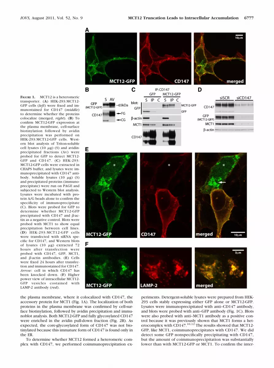
the plasma membrane, where it colocalized with CD147, theaccessory protein for MCT1 (Fig. 1A). The localization of bothproteins in the plasma membrane was confirmed by cell-sur-face biotinylation, followed by avidin precipitation and immu-noblot analysis. Both MCT12-GFP and fully glycosylated CD147were enriched in the avidin pull-down fraction (Fig. 2B). Asexpected, the core-glycosylated form of CD147 was not bio-tinylated because this immature form of CD147 is found only inthe ER.
To determine whether MCT12 formed a heteromeric com-plex with CD147, we performed coimmunoprecipitation ex-
periments. Detergent-soluble lysates were prepared from HEK-293 cells stably expressing either GFP alone or MCT12-GFP.Lysates were immunoprecipitated with anti–CD147 antibody,and blots were probed with anti–GFP antibody (Fig. 1C). Blotswere also probed with anti–MCT1 antibody as a positive con-trol because it was previously shown that MCT1 forms a het-erocomplex with CD147.10,12 The results showed that MCT12-GFP, like MCT1, coimmunoprecipitates with CD147. We diddetect some GFP nonspecifically precipitating with CD147,but the amount of coimmunoprecipitation was substantiallylower than with MCT12-GFP or MCT1. To confirm the inter-
FIGURE 1. MCT12 is a heteromerictransporter. (A) HEK-293:MCT12-GFP cells (left) were fixed and im-munostained for CD147 (middle)to determine whether the proteinscolocalize (merged, right). (B) Toconfirm MCT12-GFP expression atthe plasma membrane, cell-surfacebiotinylation followed by avidinprecipitation was performed onHEK-293:MCT12-GFP cells. West-ern blot analysis of Triton-solublecell lysates (10 �g) (S) and avidin-precipitated fractions (Av) wereprobed for GFP to detect MCT12-GFP and CD147. (C) HEK-293:MCT12-GFP cells were extracted inCHAPS buffer, and lysates were im-munoprecipitated with CD147 anti-body. Soluble lysates (10 �g) (S)and precipitated proteins (immuno-precipitate) were run on PAGE andsubjected to Western blot analysis.Lysates were incubated with pro-tein A/G beads alone to confirm thespecificity of immunoprecipitate(C). Blots were probed for GFP todetermine whether MCT12-GFPprecipitated with CD147 and �-ac-tin as a negative control. Blots wereprobed with MCT1 to show equalprecipitation between cell lines.(D) HEK-293:MCT12-GFP cellswere transfected with siRNA spe-cific for CD147, and Western blotsof lysates (10 �g) extracted 72hours after transfection wereprobed with CD147, GFP, MCT1,and �-actin antibodies. (E) Cellswere fixed 24 hours after transfec-tion and immunostained for CD147.Arrow: cell in which CD147 hasbeen knocked down. (F) Higherpower view of intracellular MCT12-GFP vesicles costained withLAMP-2 antibody (red).
IOVS, August 2011, Vol. 52, No. 9 MCT12 Truncation Leads to Intracellular Accumulation 6777

FIGURE 2. Expression of MCT12 inthe lens. (A) cDNA was preparedfrom RNA extracted from P1, P7, and3-month-old (adult) lenses, and PCRwas performed for Slc16a12 and�-tubulin as a loading control. (B)Antipeptide antibodies were gener-ated against two regions of the C-ter-minal cytoplasmic tail. The sequenceshown as peptide 1 was used forthese studies. Antibodies raised topeptide 2 did not specifically labelMCT12 for immunostaining or West-ern blotting. (C) HEK-293 cells tran-siently transfected with vectors ex-pressing MCT12-GFP were stainedwith anti–peptide MCT12 antibody(red) to confirm antibody specificity.Untransfected cells (asterisks) werenot labeled by the antibody. (D) Fro-zen tissue sections of P1 mouselenses were stained with the antipep-tide antibody (red). Differential inter-ference contrast (DIC) and phalloi-din (green) show tissue structure.Blocking the antibody with the im-munogenic peptide before staining(D, lower) prevented all staining.(E) Enlargement of the boxed re-gion in D.
6778 Castorino et al. IOVS, August 2011, Vol. 52, No. 9

action, the reciprocal coimmunoprecipitate was performedwith anti–GFP antibody, followed by Western blot analysiswith anti–CD147 antibody. CD147 was confirmed to coim-munoprecipitate with MCT12-GFP (Supplementary Fig. S1A,http://www.iovs.org/lookup/suppl/doi:10.1167/iovs.10-6579/-/DCSupplemental).
To assess whether MCT12 required CD147 for trafficking tothe plasma membrane, CD147 was knocked down in HEK-293:MCT12-GFP cells using siRNA. HEK-293:MCT12-GFP cellstransfected with either scrambled or CD147-specific siRNAwere harvested 72 hours after transfection, and levels ofCD147 and MCT12-GFP protein were determined by immuno-blot analysis. Silencing of CD147 reduced the levels of MCT12-GFP compared with scrambled siRNA-treated cells (Fig. 1D). Asexpected, the level of expression of MCT1 was also reduced bysilencing CD147, as previously reported.12,18 Confocal immu-nofluorescence analysis showed that 24 hours after silencingCD147 in cells in which GFP fluorescence could still be de-tected, MCT12-GFP was localized to intracellular vesicles (Fig.1E, arrow). These vesicles colabeled with anti–LAMP-2 anti-body, indicating that in the absence of CD147, MCT12-GFP wasdegraded by the lysosomal pathway (Fig. 1F).
MCT12 Is Expressed in the Lens
To begin to understand how mutations in SLC16A12 couldlead to juvenile cataract, we examined the developmentalexpression of the Slc16a12 transcript in postnatal lenses ofmice. RT-PCR revealed that Slc16a12 mRNA was expressed inthe lens at postnatal day (P)1 and P7 at higher levels thanobserved in the adult lens (Fig. 2A).
To examine the spatial distribution of MCT12 protein in thelens, we generated an anti–MCT12 peptide antibody to a highlyconserved domain of the C-terminal cytoplasmic tail of MCT12(Fig. 2B). To validate the antibody specificity, HEK-293 cellsexpressing MCT12-GFP fusion protein were fixed and labeledwith the anti–MCT12 antibody. Immunofluorescence confocalmicroscopy showed that the anti–MCT12 antibody labeledonly cells expressing the MCT12-GFP fusion protein but notuntransfected cells (Fig. 2C).
Next we labeled frozen sections of eyes from P1 mousepups with the anti–MCT12 antibody. MCT12 was detected in
the basolateral membrane of the lens epithelium (Fig. 2D),with strongest staining observed at the equatorial epithelium(Supplementary Fig. S1B, http://www.iovs.org/lookup/suppl/doi:10.1167/iovs.10-6579/-/DCSupplemental). Additionally, weobserved staining in the differentiating secondary fiber cells ofP1 mouse lenses (Figs. 2D, 2E).
Generation of Slc16a12TKO Rats
One potential explanation for dominant cataract formation inpatients with the SLC16A12 exon 6:c643C�T mutation couldbe haploinsufficiency. To address whether this could be theunderlying cause for juvenile cataract found in patients, weobtained the Slc16a12TKO rat, a functional knockout generatedby transposon insertional mutagenesis (Fig. 3A). Heterozygoteswere crossed to obtain Slc16a12WT, Slc16a12het andSlc16a12TKO animals, and genotypes of the rats were deter-mined by PCR using primers specific for the wild-type andtransposon-inserted loci (Fig. 3B). Rat offspring were born toexpected Mendelian genotype ratios, and no obvious pheno-typic changes were observed in the transgenic animals eventhough Slc16a12 mRNA was detected in a number of oculartissues (Supplementary Fig. S1C, http://www.iovs.org/lookup/suppl/doi:10.1167/iovs.10-6579/-/DCSupplemental) and or-gans (Supplementary Fig. S1D, http://www.iovs.org/lookup/suppl/doi:10.1167/iovs.10-6579/-/DCSupplemental) in wild-type rats.
With a focus on cataract formation in these animals, wespecifically examined the expression of Slc16a12 mRNA ex-tracted from whole lenses of adult Slc16a12WT andSlc16a12TKO animals. To determine that the gene trap wasfunctional, we performed a number of different PCR reactionson cDNA prepared from rat lens mRNA to determine whetherSlc16a12 mRNA was expressed in Slc16a12TKO rats (Fig. 3C).Using forward and reverse primers within exon 2 confirmedthat the Slc16a12 locus was transcribed in both theSlc16a12WT and Slc16a12TKO rats (Fig. 3D). PCR reactionsfrom cDNA prepared without reverse transcriptase generatedno products. When primers from exon 2 (forward) and exon 7(reverse) were used, a product was detected only in wild-typeanimals, demonstrating the transposon did indeed disrupt nor-mal Slc16a12 mRNA expression and confirming that the
FIGURE 3. Generation of Slc16a12TKO
rats. (A) Schematic of the transposoninsertion is presented. Primer set A/Bamplifies the wild-type gene, whereasprimer set A/T amplifies the knockoutfragment of the gene (primer se-quences provided in Table S1). Notethat the human SLC16A12 locus hasan additional upstream exon thatdoes not seem to be present in themouse or rat genes. (B) Totalgenomic DNA was analyzed by PCRto determine genotypes of animalsused for these studies. Primers A/Byielded a 1031-bp band if the normalSlc16a12 gene was present (indica-tive of wild-type). Primers A/Tyielded a 594-bp band if only thetransposon was present in theSlc16a12 gene (indicative of the knockout phenotype). Heterozygous animals (het) showed both the 1031-bp and 594-bp bands, whereas thewild-type (WT) showed only the 1031-bp band and the knockout (TKO) showed only the 594-bp band. M, size marker. (C) Primers were designedto assess the mRNA generated from Slc16a12WT and Slc16a12TKO animals. (D) cDNA was generated from total DNase-treated RNA extracted fromSlc16a12WT and Slc16a12TKO whole lenses. PCR was performed within rat Slc16a12 exon 2 to show the locus was transcribed in both genotypes.PCR was performed from exon 2 to exon 7 to show that normal Slc16a12 mRNA was processed only in Slc16a12WT animals and from exon 2 tothe gene trap to show that only the mutant mRNA was generated in Slc16a12TKO animals. �-Tubulin was amplified as a loading control. No PCRproducts were produced in samples in which the cDNA reaction did not contain reverse transcriptase (RT). (E) PCR was performed for all membersof the Slc16a family (primer sequences provided in Table S2) on cDNA prepared from microdissected lens epithelium (E) or fiber (F) cells, but onlythree members in addition to Slc16a12 were detected. Representative reactions from three different rats are shown.
IOVS, August 2011, Vol. 52, No. 9 MCT12 Truncation Leads to Intracellular Accumulation 6779
Slc16a12TKO animals were effectively null. No additional prod-ucts were amplified in the PCR reactions from animals harbor-ing the transposon that would be indicative of aberrant splic-ing. PCR using primers from exon 2 and the gene trapamplified a product using only RNA prepared fromSlc16a12TKO animals, which indicated that the mutated mRNAwas expressed. Amplification of �-actin mRNA was used as aloading control in these experiments. Together, these dataindicate that the first coding exon is transcribed and that thefirst 36 amino acids of the MCT12 might be translated, butadditional expression of MCT12 protein from the locus inSlc16a12TKO rats is unlikely.
Of the 13 additional members of the Slc16a gene family,only the mRNA for Slc16a1, Slc16a3, and Slc16a10 was de-tected in the lenses of the Slc16a12WT and Slc16a12TKO rats(Fig. 3E). The amounts of Slc16a1 (MCT1) and Slc16a3 (MCT4)seemed comparable between wild-type and knockout siblings.However, we detected the Slc16a10 (MCT10) PCR product inthe lens epithelium isolated from Slc16a12TKO rats but not inage-matched, wild-type siblings.
Slc16a12TKO Rats Do Not Develop Cataracts
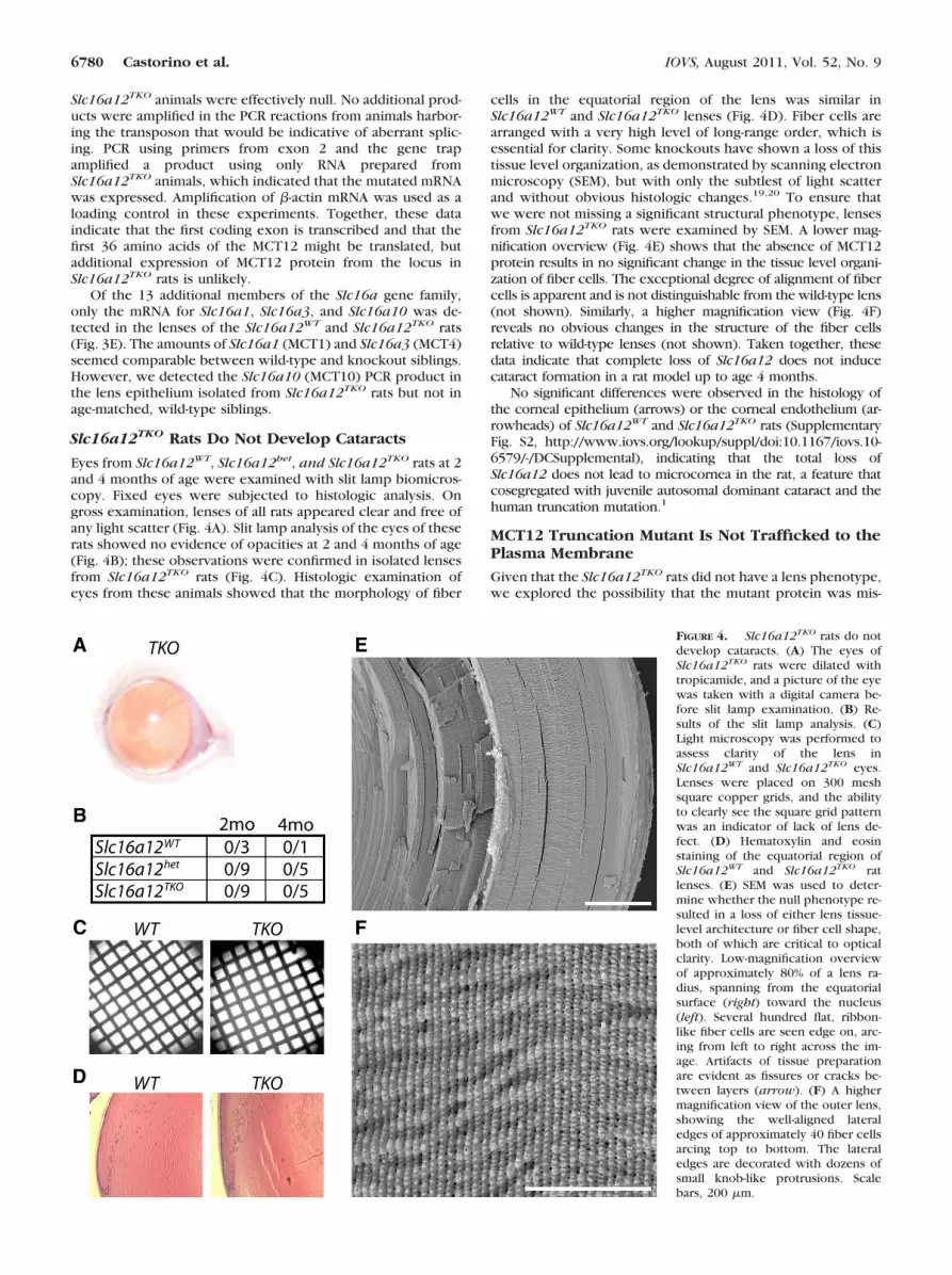
Eyes from Slc16a12WT, Slc16a12het, and Slc16a12TKO rats at 2and 4 months of age were examined with slit lamp biomicros-copy. Fixed eyes were subjected to histologic analysis. Ongross examination, lenses of all rats appeared clear and free ofany light scatter (Fig. 4A). Slit lamp analysis of the eyes of theserats showed no evidence of opacities at 2 and 4 months of age(Fig. 4B); these observations were confirmed in isolated lensesfrom Slc16a12TKO rats (Fig. 4C). Histologic examination ofeyes from these animals showed that the morphology of fiber
cells in the equatorial region of the lens was similar inSlc16a12WT and Slc16a12TKO lenses (Fig. 4D). Fiber cells arearranged with a very high level of long-range order, which isessential for clarity. Some knockouts have shown a loss of thistissue level organization, as demonstrated by scanning electronmicroscopy (SEM), but with only the subtlest of light scatterand without obvious histologic changes.19,20 To ensure thatwe were not missing a significant structural phenotype, lensesfrom Slc16a12TKO rats were examined by SEM. A lower mag-nification overview (Fig. 4E) shows that the absence of MCT12protein results in no significant change in the tissue level organi-zation of fiber cells. The exceptional degree of alignment of fibercells is apparent and is not distinguishable from the wild-type lens(not shown). Similarly, a higher magnification view (Fig. 4F)reveals no obvious changes in the structure of the fiber cellsrelative to wild-type lenses (not shown). Taken together, thesedata indicate that complete loss of Slc16a12 does not inducecataract formation in a rat model up to age 4 months.
No significant differences were observed in the histology ofthe corneal epithelium (arrows) or the corneal endothelium (ar-rowheads) of Slc16a12WT and Slc16a12TKO rats (SupplementaryFig. S2, http://www.iovs.org/lookup/suppl/doi:10.1167/iovs.10-6579/-/DCSupplemental), indicating that the total loss ofSlc16a12 does not lead to microcornea in the rat, a feature thatcosegregated with juvenile autosomal dominant cataract and thehuman truncation mutation.1
MCT12 Truncation Mutant Is Not Trafficked to thePlasma Membrane
Given that the Slc16a12TKO rats did not have a lens phenotype,we explored the possibility that the mutant protein was mis-
FIGURE 4. Slc16a12TKO rats do notdevelop cataracts. (A) The eyes ofSlc16a12TKO rats were dilated withtropicamide, and a picture of the eyewas taken with a digital camera be-fore slit lamp examination. (B) Re-sults of the slit lamp analysis. (C)Light microscopy was performed toassess clarity of the lens inSlc16a12WT and Slc16a12TKO eyes.Lenses were placed on 300 meshsquare copper grids, and the abilityto clearly see the square grid patternwas an indicator of lack of lens de-fect. (D) Hematoxylin and eosinstaining of the equatorial region ofSlc16a12WT and Slc16a12TKO ratlenses. (E) SEM was used to deter-mine whether the null phenotype re-sulted in a loss of either lens tissue-level architecture or fiber cell shape,both of which are critical to opticalclarity. Low-magnification overviewof approximately 80% of a lens ra-dius, spanning from the equatorialsurface (right) toward the nucleus(left). Several hundred flat, ribbon-like fiber cells are seen edge on, arc-ing from left to right across the im-age. Artifacts of tissue preparationare evident as fissures or cracks be-tween layers (arrow). (F) A highermagnification view of the outer lens,showing the well-aligned lateraledges of approximately 40 fiber cellsarcing top to bottom. The lateraledges are decorated with dozens ofsmall knob-like protrusions. Scalebars, 200 �m.
6780 Castorino et al. IOVS, August 2011, Vol. 52, No. 9

folded and not trafficked to the plasma membrane. To investi-gate this possibility, we generated expression vectors of trun-cated MCT12 designed to mimic expression of theMCT12Q215X mutant (MCT12:214�). MCT12:214� was ex-pressed as either GFP or mCherry fusion protein in HEK-293cells. As shown (Figs. 1A, 2B), full-length MCT12-GFP wasprimarily localized to the plasma membrane, as indicated by GFPlabeling at the cell-cell borders, filopodia, and lamellipodia. Incontrast, when we expressed MCT12:214�-GFP in HEK-293 cells,the fusion protein was retained in the ER, as demonstrated by itscolocalization with the ER marker glucose-regulated protein, 78kDa (GRP78, BIP) (Fig. 5A). The full-length MCT12 fusion did notcolocalize with GRP78. A similar distribution of MCT12-GFP andMCT12:214�-GFP was observed in the HLE-B3 lens epithelial cellline, similar to that in HEK-293 cells (Supplementary Figs. S3A,S3B, http://www.iovs.org/lookup/suppl/doi:10.1167/iovs.10-6579/-/DCSupplemental), indicating that the ER retention ofMCT12:214�-GFP is not cell-type specific.
Cell surface biotinylation followed by avidin precipita-tion was performed to further assess the trafficking ofMCT12:214�-GFP to the plasma membrane. Blots of celllysates and avidin precipitates showed that MCT12-GFP wasenriched in the avidin-precipitated fraction (Fig. 5B),whereas MCT12:214�-GFP was barely detected in this frac-tion. The small amount that could be detected in the avidin-precipitated fraction could be attributed to a very smallamount of MCT12:214�-GFP reaching the surface or to in-complete inactivation of the biotinylation reaction. Regard-less, the ratio of precipitated to soluble MCT12-GFP wasorders of magnitude greater than that of MCT12:214�-GFP.As a control, the blots were reprobed with CD147 antibody,which showed that the fully glycosylated form of CD147 wasenriched to a similar degree in the biotinylated fractions of bothcell lines. This indicates that the difference in the biotinylation ofMCT12-GFP compared with MCT12:214�-GFP was not caused byvariation in biotinylation in the different cell lines. The differencein expression levels between MCT12-GFP and MCT12:214�-GFPcould not be accounted for by differential detergent solubility ofthe fusion proteins because both proteins were distributed to asimilar degree between the detergent-soluble and the detergent-insoluble fractions (Supplementary Fig. S3B, http://www.iovs.org/lookup/suppl/doi:10.1167/iovs.10-6579/-/DCSupplemental).Furthermore, mRNA levels expressed from the transfected vec-tors were roughly equivalent (Fig. 5C).
Differences in the levels of MCT12-GFP and MCT12:214�-GFP could be attributed to degradation of the misfoldedprotein. To test this possibility, we treated cells with lacta-cystin, a proteasome inhibitor. Lactacystin treatment in-creased MCT12:214�-GFP levels approximately 5.6-fold,whereas MCT12-GFP levels increased by only 0.5-fold (Figs. 6A,6B). This difference was statistically significant (P � 0.004).Degradation of MCT12:214� by the proteasome pathwayindicates that the truncated protein is a target of the ER-associated degradation pathway and therefore is likely mis-folded.
To determine whether trafficking of full-length MCT12 tothe plasma membrane could be disrupted by coexpressionwith MCT12:214�, as would be the case if MCT12Q215X
behaved as a dominant-negative, stable HEK-293:MCT12-GFP, cells were transiently transfected with mCherry-MCT12:214�. In cells expressing both full-length and trun-cated proteins, MCT12-GFP was detected at the plasmamembrane but mCherry-MCT12:214� remained intracellular(Fig. 6C), indicating that expression and trafficking of thefull-length protein were not disrupted by the truncatedprotein.
ER Retention of Truncated MCT12 Is NotMCT12 Specific
To determine whether the retention of truncated MCT12:214�in the ER was specific for MCT12 or a phenomenon of trun-cated MCTs in general, we generated a truncation of MCT4within the large intracellular loop between transmembranedomains 6 and 7 and expressed it as a GFP fusion (MCT4:207�-GFP) in HEK-293 cells. Although normal MCT4 is trafficked tothe plasma membrane21 (Fig. 7A), MCT4:207�-GFP was re-tained in the ER (Fig. 7B). This finding suggests that premature
FIGURE 5. MCT12:214� is retained in the ER. (A) HEK-293 cellstransiently transfected with vectors expressing either MCT12:214�-GFP (upper) or MCT12-GFP (lower) were immunostained for GRP78(red). Scale bars, 10 �m. (B) HEK-293 cells transiently transfected withvectors expressing either MCT12-GFP or MCT12:214�-GFP were cell-surface biotinylated 48 hours after transfection. Biotinylated proteinswere precipitated from cell lysates with streptavidin-agarose beads.Soluble (S) and avidin-precipitated (Av) fractions were run by SDS-PAGE and subjected to Western blot analysis, and the blots wereprobed for GFP and CD147. (C) To determine whether differences inprotein levels between MCT12-GFP and MCT12:214�-GFP were due todifferences in mRNA levels, mRNA was extracted from HEK-293 cellstransiently transfected with either MCT12-GFP or MCT12:214�-GFP 48hours after transfection, and cDNA was prepared. PCR was performedfor GFP to unify amplification from both constructs and �-actin as acontrol.
IOVS, August 2011, Vol. 52, No. 9 MCT12 Truncation Leads to Intracellular Accumulation 6781

termination of any MCT is likely to cause ER retention becauseof protein misfolding.
DISCUSSION
SLC16A12 was identified by Halestrap and Visser2 throughsearching the human genomic and EST databases; however,nothing is known about the properties or function of theprotein. Recently, it was discovered that patients with a non-sense mutation in SLC16A12 develop juvenile cataract with adominant inheritance pattern.1 In this study, we investigatedpotential mechanisms by which the mutation in exon 6(c.643C�T) of SLC16A12 could lead to cataract formation. Wefocused on two possibilities: haploinsufficiency caused by re-duced levels of the normal transporter molecules or a directeffect of the truncated protein. The evidence presented heresuggests that MCT12Q215X is retained in the ER and that proteinmisfolding and trafficking defects, rather than haploinsuffi-ciency, are the underlying cause of cataract formation.
MCTs have been localized to plasma and mitochondrialmembranes; therefore, in the present study, we used cell linesexpressing an MCT12-GFP fusion protein to assess the subcel-lular distribution of the protein.2,22 In both HEK-293 andHLE-B3 cells, MCT12 was trafficked to the plasma membrane.Moreover, we demonstrated for the first time that maturationand trafficking of MCT12 to the plasma membrane required theaccessory protein CD147. These studies reveal that MCT12,like MCT1, MCT3, and MCT4, requires assembly with CD147for stability and cell surface expression. MCT12 was detectedin the plasma membrane of lens epithelial cells and in differ-
entiating and secondary fiber cells as early as P1 in the mouse.The temporal and spatial expression of MCT12 in lens epithe-lium and secondary fiber cells was consistent with corticonu-clear cataract observed in patients with SLC16A12(c.643C�T)mutations.
Mutations in facilitated solute transporters can cause dis-ease because of alterations in expression, trafficking, or func-tional activity. To determine the fate of the MCT12Q215X pro-tein produced from the SLC16A12 cataract-associatedmutation, we expressed a mimic (MCT12:214�-GFP) in HEK-293 cells. The truncated MCT12, composed only of the first sixtransmembrane domains, was retained in the ER (Fig. 5) andwas degraded by the proteasome pathway, indicative of mis-folding (Fig. 6). Although the crystal structures of MCT familymembers have not been determined, molecular modelingbased on the structure of the Escherichia coli homolog lactosepermease (LacY) transporter has been performed.23,24 Modelsof the tertiary structures of MCT1, MCT8, and LacY predictinteractions of the first and second halves of MCT by closeassociations between transmembrane domains 5–8 and2–11.2,9,25 The transmembrane domains of MCTs are well con-served; thus, a similar arrangement likely exists for MCT12.Therefore, any mutation that would cause such an early pre-mature termination of an MCT would likely lead to misfolding.Indeed, performing a similar truncation of MCT4 also led toretention of the resultant protein in the ER (Fig. 7). Expressionof the truncated protein does not affect expression of thewild-type (Fig. 6C); therefore, MCT12Q215X does not functionas dominant-negative. There is considerable overlap of trans-port properties among different gene families; hence, it israre that haploinsufficiency of a solute transporter results ina phenotype.26 Haploinsufficiency of the connexins Gja3and Gja8 does not result in cataract formation in micebecause the heterozygotes lack a detectable phenotype. Toaddress the possibility that cataract formation in patientswith the SLC16A12 c.643C�T mutation resulted from hap-loinsufficiency, we examined the eyes of the Slc16a12TKO
rat. The lack of cataract phenotype in either the heterozy-gote or the homozygote rat would not immediately supporta model of haploinsufficiency. Further, there was no evi-dence of cataract formation in Bsg-deficient (CD147) mice27
(Judith Ochrieter, personal communication, 2011). It is ex-
FIGURE 7. Truncated MCT4 behaves like MCT12:214�. To determinewhether defective trafficking of MCT12:214� was specific to MCT12,we generated a similar truncation of MCT4, MCT4:207�-GFP. Cellswere fixed 48 hours after transfection and were stained for GRP78(red). MCT4-GFP (A) is present predominantly at the cell surface,whereas MCT4:207�-GFP (B) is predominantly retained in the ER.Scale bars, 10 �m.
FIGURE 6. MCT12:214� is degraded by proteasomes and is not dom-inant-negative. Forty hours after transfection with vectors expressingeither MCT12-GFP or MCT12:214�-GFP, HEK-293 cells were treatedwith 10 �M lactacystin for 8.5 hours. Note: MCT12-GFP and MCT12:214�-GFP are actually of different molecular weights, as shown inFigure 5B. Western blot analysis (A) was quantitated (B), and therelative levels of MCT12-GFP and MCT12:214�-GFP were determinedin control and lactacystin-treated cells as normalized to �-actin signal.Error bars are � SD (n � 3). (C) To determine whether MCT12:214�disrupts full-length MCT12 localization, HEK-293:MCT12-GFP cellswere transiently transfected with mCherry-MCT12:214�. Cells werefixed 48 hours after transfection and imaged by confocal microscopy.Illustrations specify fusion protein and type of tag. Scale bar, 10 �m.
6782 Castorino et al. IOVS, August 2011, Vol. 52, No. 9

pected that these mice would have a loss of MCT1, MCT4,and MCT12 activity in the lens because the accessory pro-tein is required for maturation of the transporters.12
The genetic robustness of the Slc16a12TKO rat may beattributed to genetic buffering or functional complementationor redundancy. The observed increased expression ofSlc16a10 mRNA in Slc16a12TKO lenses may support this ex-planation if both transporters have similar substrate specifici-ties. We know that MCT10 transports aromatic amino acids28
and thyroid hormone,4 but the transported substrate of MCT12is not yet known. This information would shed light on thepossibility that functional redundancy may be responsible forthe lack of a phenotype. Because antibodies that recognize ratMCT10 are not available, confirmation about whether the ob-served increase in Slc16a10 transcripts in the lenses ofSlc16a12TKO rats also leads to an increase in functional MCT10protein awaits experimental testing. Furthermore, the geneticbackground of the rat strain used in these studies may haveinherent resistance to cataract formation; alternatively, extrin-sic environmental factors could also play a role in cataracto-genesis in humans to which our animal model was not ex-posed.29
Knocking out genes often does not result in a phenotypewhereas knocking in the mutant gene does because of a neg-ative gain of function of the mutant protein. For example, aknockout model of connexin 50 has a slowly developing cat-aract, whereas the mutation mimicking human cataract-associ-ated phenotype displays a much more severe cataract.30,31
Additionally, mutations in SLC16A2 that affect trafficking ofMCT8 to the plasma membrane result in a more severe phe-notype than those that affect transport activity. Similar to ourfindings with Slc16a12TKO rats, Slc16a2 knockout mice showno signs of the neurologic abnormalities observed in pa-tients.32,33 Based on the cell-based assays with MCT12:214�-GFP and the lack of phenotypes in the Slc16a12TKO rats andBsg knockout mice, we would expect that a knock-in of themutated allele would be the only mechanism to accuratelyassess the development of the SLC16A12(c.643C�T)–associ-ated cataract.
MCT12 was detected in the equatorial lens epithelial cells,which differentiate into lens fiber cells. The transition from anepithelial cell to a mature fiber cell necessitates a large increasein protein and lipid synthesis and a degradation of intracellularorganelles. These cells already undergo an unfolded proteinresponse, and it is likely that cells in this region of the lenswould be hypersensitive to additional stress caused by a mu-tated, misfolded protein.34 Expression of the MCT12Q215X mu-tant protein in the equatorial region of the lens could result inexcessive ER stress, which is known to cause cataract.35–37 Thejuvenile corticonuclear cataract observed in patients withSLC16A12(c.643C�T) mutations would be expected if thetemporal and spatial expression of MCT12 observed in themouse lens is recapitulated in the human lens.
Overall, our findings suggest that cataract formation inpatients with the c.643C�T mutation in SLC16A12 is due toa defect in protein folding and trafficking causing ER stressin the differentiating and secondary lens fiber cells. Thetransporter knockout model provided a valuable tool toassess the contribution of individual transporters to lenstransparency. Future studies with humanized rat models willprovide further insight into how specific mutations lead tocataract formation, thereby providing valuable models fordetermining whether juvenile cataracts arising from defectsin protein trafficking can be treated pharmacologicallyrather than surgically.
References
1. Kloeckener-Gruissem B, Vandekerckhove K, Nurnberg G, et al.Mutation of solute carrier SLC16A12 associates with a syndromecombining juvenile cataract with microcornea and renal glucos-uria. Am J Human Genet. 2008;82:772–779.
2. Halestrap AP, Meredith D. The SLC16 gene family from monocar-boxylate transporters (MCTs) to aromatic amino acid transportersand beyond. Pflugers Archiv Eur J Physiol. 2004;447:619–628.
3. Grollman EF, Philp NJ, McPhie P, Ward RD, Sauer B. Determinationof transport kinetics of chick MCT3 monocarboxylate transporterfrom retinal pigment epithelium by expression in genetically mod-ified yeast. Biochemistry. 2000;39:9351–9357.
4. Friesema EC, Jansen J, Jachtenberg JW, Visser WE, Kester MH, Visser TJ.Effective cellular uptake and efflux of thyroid hormone by human mono-carboxylate transporter 10. Mol Endocrinol. 2008;22:1357–1369.
5. Illig T, Gieger C, Zhai G, et al. A genome-wide perspective of geneticvariation in human metabolism. Nat Genet. 2010;42:137–141.
6. Kolz M, Johnson T, Sanna S, et al. Meta-analysis of 28,141 individ-uals identifies common variants within five new loci that influenceuric acid concentrations. PLoS Genet. 2009;5:e1000504.
7. Ramadan T, Camargo SM, Herzog B, Bordin M, Pos KM, Verrey F.Recycling of aromatic amino acids via TAT1 allows efflux of neu-tral amino acids via LAT2–4F2hc exchanger. Pflugers Arch. 2007;454:507–516.
8. Ramadan T, Camargo SM, Summa V, et al. Basolateral aromaticamino acid transporter TAT1 (Slc16a10) functions as an effluxpathway. J Cell Physiol. 2006;206:771–779.
9. Wilson MC, Meredith D, Bunnun C, Sessions RB, Halestrap AP.Studies on the DIDS-binding site of monocarboxylate transporter 1suggest a homology model of the open conformation and a plau-sible translocation cycle. J Biol Chem. 2009;284:20011–20021.
10. Kirk P, Wilson MC, Heddle C, Brown MH, Barclay AN, HalestrapAP. CD147 is tightly associated with lactate transporters MCT1 andMCT4 and facilitates their cell surface expression. EMBO J. 2000;19:3896–3904.
11. Wilson MC, Meredith D, Halestrap AP. Fluorescence resonance en-ergy transfer studies on the interaction between the lactate trans-porter MCT1 and CD147 provide information on the topology andstoichiometry of the complex in situ. J Biol Chem. 2002;277:3666–3672.
12. Philp NJ, Ochrietor JD, Rudoy C, Muramatsu T, Linser PJ. Loss ofMCT1, MCT3, and MCT4 expression in the retinal pigment epithe-lium and neural retina of the 5A11/basigin-null mouse. InvestOphthalmol Vis Sci. 2003;44:1305–1311.
13. Gallagher SM, Castorino JJ, Wang D, Philp NJ. Monocarboxylatetransporter 4 regulates maturation and trafficking of CD147 to theplasma membrane in the metastatic breast cancer cell line MDA-MB-231. Cancer Res. 2007;67:4182–4189.
14. Wilson MC, Meredith D, Fox JE, Manoharan C, Davies AJ, HalestrapAP. Basigin (CD147) is the target for organomercurial inhibition ofmonocarboxylate transporter isoforms 1 and 4: the ancillary pro-tein for the insensitive MCT2 is EMBIGIN (gp70). J Biol Chem.2005;280:27213–27221.
15. Hejtmancik JF. Congenital cataracts and their molecular genetics.Semin Cell Dev Biol. 2008;19:134–149.
16. Lu B, Geurts AM, Poirier C, et al. Generation of rat mutants usinga coat color-tagged Sleeping Beauty transposon system. MammGenome. 2007;18:338–346.
17. Philp NJ, Wang D, Yoon H, Hjelmeland LM. Polarized expression ofmonocarboxylate transporters in human retinal pigment epitheliumand ARPE-19 cells. Invest Ophthalmol Vis Sci. 2003;44:1716–1721.
18. Deora AA, Philp N, Hu J, Bok D, Rodriguez-Boulan E. Mechanismsregulating tissue-specific polarity of monocarboxylate transportersand their chaperone CD147 in kidney and retinal epithelia. ProcNatl Acad Sci U S A. 2005;102:16245–16250.
19. Alizadeh A, Clark JI, Seeberger T, et al. Targeted genomic deletionof the lens-specific intermediate filament protein CP49. InvestOphthalmol Vis Sci. 2002;43:3722–3727.
20. Yoon KH, Blankenship T, Shibata B, FitzGerald PG. Resisting theeffects of aging: a function for the fiber cell beaded filament. InvestOphthalmol Vis Sci. 2008;49:1030–1036.
IOVS, August 2011, Vol. 52, No. 9 MCT12 Truncation Leads to Intracellular Accumulation 6783
21. Castorino JJ, Deborde S, Deora A, et al. Basolateral sortingsignals regulating tissue-specific polarity of heteromeric mono-carboxylate transporters in epithelia. Traffic. 2011;12:483– 498.
22. Brooks GA, Hashimoto T. Investigation of the lactate shuttle inskeletal muscle mitochondria. J Physiol. 2007;584:705–706; au-thor reply 707–708.
23. Abramson J, Smirnova I, Kasho V, Verner G, Kaback HR, Iwata S.Structure and mechanism of the lactose permease of Escherichiacoli. Science. 2003;301:610–615.
24. Galic S, Schneider HP, Broer A, Deitmer JW, Broer S. The loopbetween helix 4 and helix 5 in the monocarboxylate transporterMCT1 is important for substrate selection and protein stability.Biochem J. 2003;376:413–422.
25. van der Deure WM, Peeters RP, Visser TJ. Molecular aspects ofthyroid hormone transporters, including MCT8, MCT10, andOATPs, and the effects of genetic variation in these transporters. JMol Endocrinol. 2010;44:1–11.
26. DeGorter MK, Kim RB. Use of transgenic and knockout mousemodels to assess solute carrier transporter function. Clin Pharma-col Ther. 2011;89:612–616.
27. Naruhashi K, Kadomatsu K, Igakura T, et al. Abnormalities ofsensory and memory functions in mice lacking Bsg gene. BiochemBiophys Res Commun. 1997;236:733–737.
28. Kim DK, Kanai Y, Chairoungdua A, Matsuo H, Cha SH, Endou H.Expression cloning of a Na-independent aromatic amino acidtransporter with structural similarity to H/monocarboxylatetransporters. J Biol Chem. 2001;276:17221–17228.
29. Barbaric I, Miller G, Dear TN. Appearances can be deceiving:phenotypes of knockout mice. Brief Funct Genomic Proteomic.2007;6:91–103.
30. Xia CH, Cheng C, Huang Q, et al. Absence of alpha3 (Cx46) andalpha8 (Cx50) connexins leads to cataracts by affecting lens innerfiber cells. Exp Eye Res. 2006;83:688–696.
31. DeRosa AM, Mese G, Li L, et al. The cataract causing Cx50–S50Pmutant inhibits Cx43 and intercellular communication in the lensepithelium. Exp Cell Res. 2009;315:1063–1075.
32. Trajkovic M, Visser TJ, Mittag J, et al. Abnormal thyroid hormonemetabolism in mice lacking the monocarboxylate transporter 8.J Clin Invest. 2007;117:627–635.
33. Di Cosmo C, Liao XH, Dumitrescu AM, Philp NJ, Weiss RE, RefetoffS. Mice deficient in MCT8 reveal a mechanism regulating thyroidhormone secretion. J Clin Invest. 2010;120:3377–3388.
34. Firtina Z, Duncan MK. Unfolded Protein Response (UPR) is activatedduring normal lens development. Gene Expr Patterns. 2011;11:135–143.
35. Watson GW, Andley UP. Activation of the unfolded protein re-sponse by a cataract-associated alphaA-crystallin mutation.Biochem Biophys Res Commun. 2010;401:192–196.
36. Mulhern ML, Madson CJ, Danford A, Ikesugi K, Kador PF, Shinohara T.The unfolded protein response in lens epithelial cells from galactosemicrat lenses. Invest Ophthalmol Vis Sci. 2006;47:3951–3959.
37. Firtina Z, Danysh BP, Bai X, Gould DB, Kobayashi T, Duncan MK.Abnormal expression of collagen IV in lens activates unfolded proteinresponse resulting in cataract. J Biol Chem. 2009;284:35872–35884.
6784 Castorino et al. IOVS, August 2011, Vol. 52, No. 9
Copyright © 2022 FDOKUMEN