Journal of Nanobiotechnology
14
Endes et al. J Nanobiotechnol (2016) 14:78 DOI 10.1186/s12951-016-0230-9 REVIEW A critical review of the current knowledge regarding the biological impact of nanocellulose C. Endes 1,2† , S. Camarero‑Espinosa 1,2† , S. Mueller 1 , E. J. Foster 1,3 , A. Petri‑Fink 1 , B. Rothen‑Rutishauser 1 , C. Weder 1 and M. J. D. Clift 1,4* Abstract Several forms of nanocellulose, notably cellulose nanocrystals and nanofibrillated cellulose, exhibit attractive property matrices and are potentially useful for a large number of industrial applications. These include the paper and card‑ board industry, use as reinforcing filler in polymer composites, basis for low‑density foams, additive in adhesives and paints, as well as a wide variety of food, hygiene, cosmetic, and medical products. Although the commercial exploita‑ tion of nanocellulose has already commenced, little is known as to the potential biological impact of nanocellulose, particularly in its raw form. This review provides a comprehensive and critical review of the current state of knowledge of nanocellulose in this format. Overall, the data seems to suggest that when investigated under realistic doses and exposure scenarios, nanocellulose has a limited associated toxic potential, albeit certain forms of nanocellulose can be associated with more hazardous biological behavior due to their specific physical characteristics. Keywords: Nanocellulose, Cellulose nanocrystals, Human health, Risk, Exposure, Hazard, Nanofibers, Nano‑object‑cell interactions, Nanotoxicology © The Author(s) 2016. This article is distributed under the terms of the Creative Commons Attribution 4.0 International License (http://creativecommons.org/licenses/by/4.0/), which permits unrestricted use, distribution, and reproduction in any medium, provided you give appropriate credit to the original author(s) and the source, provide a link to the Creative Commons license, and indicate if changes were made. The Creative Commons Public Domain Dedication waiver (http://creativecommons.org/ publicdomain/zero/1.0/) applies to the data made available in this article, unless otherwise stated. Background Since the emergence of nanotechnology as a field in its own right, a continuously increasing number of new nanomaterials have been developed, which are poten- tially useful for applications that range from healthcare products to high-performance engineering materials [1– 3]. Several forms of nanocellulose, in their raw format, have been demonstrated to exhibit attractive property matrices and are potentially useful for the paper indus- try, as a reinforcing filler in polymer composites, basis for low-density foams, in packaging materials, additive in colloidal systems such as adhesives and paints, zero- calorie filler/thickener/stabilizer in a wide variety of food products, and in hygiene, cosmetic, and medical prod- ucts [4, 5]. Although (microcrystalline) cellulose has long been used in healthcare products such as wound healing tissue and dialysis membranes, as well as a food addi- tive, little is known as to the potential adverse biologi- cal impact of its nanoscale variants, whose commercial exploitation only begun in the last few years [6, 7]. Cellulose, the most abundant polymer in the world, is found in plant cell-walls, certain sea creatures, e.g. tunicates, and algae, e.g. Valonia. It is also produced by several bacteria such as Acetobacter xylinum [8–11]. Cel- lulose is a carbohydrate, whose repeat unit is constituted by two anhydroglucose units that are linked by a β-1,4 glycosidic bond. Cellulose chains assemble via complex inter- and intramolecular H-bonding into crystalline structures [12, 13]. Crystalline sheets pack in a paral- lel fashion, building up filiform structures that can be isolated from the native material as cellulose nanocrys- tals (CNCs), which are also referred to as nanocrystal- line cellulose (NCC) or cellulose nanowhiskers (CNWs). ese rod-shaped, high-aspect-ratio nanoparticles (HARN; aspect ratio = length/diameter ≥ 3 [14]) exhibit a diameter of 5–40 nm and a length that can vary from Open Access Journal of Nanobiotechnology *Correspondence: [email protected] † C. Endes, S. Camarero‑Espinosa contributed equally as first author 4 In Vitro Toxicology Group, Swansea University Medical School, Singleton Park Campus, Swansea SA2 8PP, Wales, UK Full list of author information is available at the end of the article
-
Upload
khangminh22 -
Category
Documents
-
view
2 -
download
0
Transcript of Journal of Nanobiotechnology
Endes et al. J Nanobiotechnol (2016) 14:78 DOI 10.1186/s12951-016-0230-9
REVIEW
A critical review of the current knowledge regarding the biological impact of nanocelluloseC. Endes1,2†, S. Camarero‑Espinosa1,2†, S. Mueller1, E. J. Foster1,3, A. Petri‑Fink1, B. Rothen‑Rutishauser1, C. Weder1 and M. J. D. Clift1,4*
Abstract
Several forms of nanocellulose, notably cellulose nanocrystals and nanofibrillated cellulose, exhibit attractive property matrices and are potentially useful for a large number of industrial applications. These include the paper and card‑board industry, use as reinforcing filler in polymer composites, basis for low‑density foams, additive in adhesives and paints, as well as a wide variety of food, hygiene, cosmetic, and medical products. Although the commercial exploita‑tion of nanocellulose has already commenced, little is known as to the potential biological impact of nanocellulose, particularly in its raw form. This review provides a comprehensive and critical review of the current state of knowledge of nanocellulose in this format. Overall, the data seems to suggest that when investigated under realistic doses and exposure scenarios, nanocellulose has a limited associated toxic potential, albeit certain forms of nanocellulose can be associated with more hazardous biological behavior due to their specific physical characteristics.
Keywords: Nanocellulose, Cellulose nanocrystals, Human health, Risk, Exposure, Hazard, Nanofibers, Nano‑object‑cell interactions, Nanotoxicology
© The Author(s) 2016. This article is distributed under the terms of the Creative Commons Attribution 4.0 International License (http://creativecommons.org/licenses/by/4.0/), which permits unrestricted use, distribution, and reproduction in any medium, provided you give appropriate credit to the original author(s) and the source, provide a link to the Creative Commons license, and indicate if changes were made. The Creative Commons Public Domain Dedication waiver (http://creativecommons.org/publicdomain/zero/1.0/) applies to the data made available in this article, unless otherwise stated.
BackgroundSince the emergence of nanotechnology as a field in its own right, a continuously increasing number of new nanomaterials have been developed, which are poten-tially useful for applications that range from healthcare products to high-performance engineering materials [1–3]. Several forms of nanocellulose, in their raw format, have been demonstrated to exhibit attractive property matrices and are potentially useful for the paper indus-try, as a reinforcing filler in polymer composites, basis for low-density foams, in packaging materials, additive in colloidal systems such as adhesives and paints, zero-calorie filler/thickener/stabilizer in a wide variety of food products, and in hygiene, cosmetic, and medical prod-ucts [4, 5]. Although (microcrystalline) cellulose has long
been used in healthcare products such as wound healing tissue and dialysis membranes, as well as a food addi-tive, little is known as to the potential adverse biologi-cal impact of its nanoscale variants, whose commercial exploitation only begun in the last few years [6, 7].
Cellulose, the most abundant polymer in the world, is found in plant cell-walls, certain sea creatures, e.g. tunicates, and algae, e.g. Valonia. It is also produced by several bacteria such as Acetobacter xylinum [8–11]. Cel-lulose is a carbohydrate, whose repeat unit is constituted by two anhydroglucose units that are linked by a β-1,4 glycosidic bond. Cellulose chains assemble via complex inter- and intramolecular H-bonding into crystalline structures [12, 13]. Crystalline sheets pack in a paral-lel fashion, building up filiform structures that can be isolated from the native material as cellulose nanocrys-tals (CNCs), which are also referred to as nanocrystal-line cellulose (NCC) or cellulose nanowhiskers (CNWs). These rod-shaped, high-aspect-ratio nanoparticles (HARN; aspect ratio = length/diameter ≥ 3 [14]) exhibit a diameter of 5–40 nm and a length that can vary from
Open Access
Journal of Nanobiotechnology
*Correspondence: [email protected] †C. Endes, S. Camarero‑Espinosa contributed equally as first author
4 In Vitro Toxicology Group, Swansea University Medical School, Singleton Park Campus, Swansea SA2 8PP, Wales, UKFull list of author information is available at the end of the article
Page 2 of 14Endes et al. J Nanobiotechnol (2016) 14:78
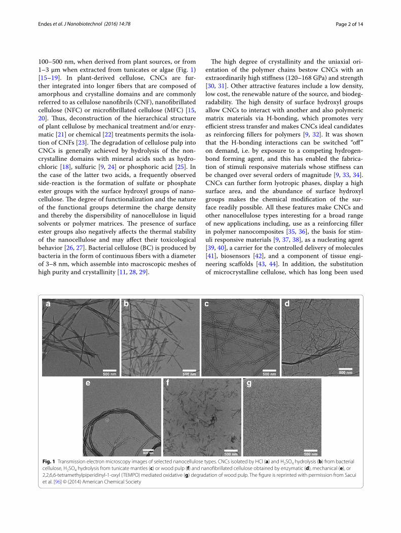
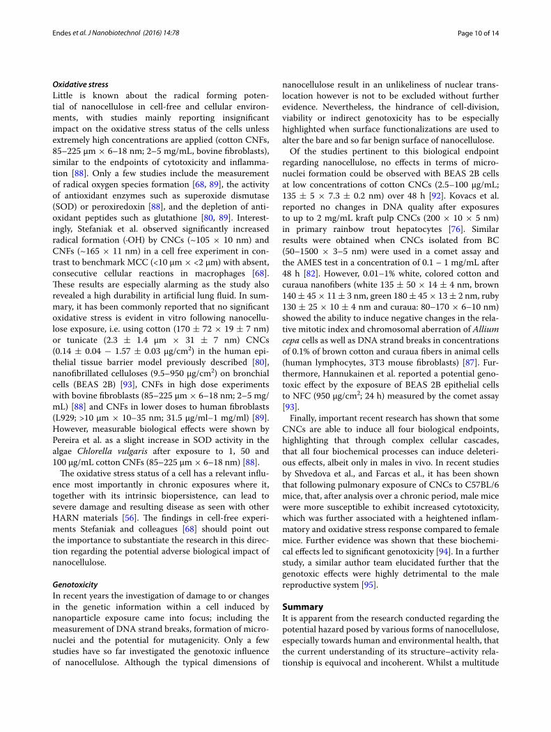
100–500 nm, when derived from plant sources, or from 1–3 µm when extracted from tunicates or algae (Fig. 1) [15–19]. In plant-derived cellulose, CNCs are fur-ther integrated into longer fibers that are composed of amorphous and crystalline domains and are commonly referred to as cellulose nanofibrils (CNF), nanofibrillated cellulose (NFC) or microfibrillated cellulose (MFC) [15, 20]. Thus, deconstruction of the hierarchical structure of plant cellulose by mechanical treatment and/or enzy-matic [21] or chemical [22] treatments permits the isola-tion of CNFs [23]. The degradation of cellulose pulp into CNCs is generally achieved by hydrolysis of the non-crystalline domains with mineral acids such as hydro-chloric [18], sulfuric [9, 24] or phosphoric acid [25]. In the case of the latter two acids, a frequently observed side-reaction is the formation of sulfate or phosphate ester groups with the surface hydroxyl groups of nano-cellulose. The degree of functionalization and the nature of the functional groups determine the charge density and thereby the dispersibility of nanocellulose in liquid solvents or polymer matrices. The presence of surface ester groups also negatively affects the thermal stability of the nanocellulose and may affect their toxicological behavior [26, 27]. Bacterial cellulose (BC) is produced by bacteria in the form of continuous fibers with a diameter of 3–8 nm, which assemble into macroscopic meshes of high purity and crystallinity [11, 28, 29].
The high degree of crystallinity and the uniaxial ori-entation of the polymer chains bestow CNCs with an extraordinarily high stiffness (120–168 GPa) and strength [30, 31]. Other attractive features include a low density, low cost, the renewable nature of the source, and biodeg-radability. The high density of surface hydroxyl groups allow CNCs to interact with another and also polymeric matrix materials via H-bonding, which promotes very efficient stress transfer and makes CNCs ideal candidates as reinforcing fillers for polymers [9, 32]. It was shown that the H-bonding interactions can be switched “off” on demand, i.e. by exposure to a competing hydrogen-bond forming agent, and this has enabled the fabrica-tion of stimuli responsive materials whose stiffness can be changed over several orders of magnitude [9, 33, 34]. CNCs can further form lyotropic phases, display a high surface area, and the abundance of surface hydroxyl groups makes the chemical modification of the sur-face readily possible. All these features make CNCs and other nanocellulose types interesting for a broad range of new applications including, use as a reinforcing filler in polymer nanocomposites [35, 36], the basis for stim-uli responsive materials [9, 37, 38], as a nucleating agent [39, 40], a carrier for the controlled delivery of molecules [41], biosensors [42], and a component of tissue engi-neering scaffolds [43, 44]. In addition, the substitution of microcrystalline cellulose, which has long been used
Fig. 1 Transmission electron microscopy images of selected nanocellulose types. CNCs isolated by HCl (a) and H2SO4 hydrolysis (b) from bacterial cellulose, H2SO4 hydrolysis from tunicate mantles (c) or wood pulp (f) and nanofibrillated cellulose obtained by enzymatic (d), mechanical (e), or 2,2,6,6‑tetramethylpiperidinyl‑1‑oxyl (TEMPO) mediated oxidative (g) degradation of wood pulp. The figure is reprinted with permission from Sacui et al. [96] © (2014) American Chemical Society
Page 3 of 14Endes et al. J Nanobiotechnol (2016) 14:78
as rheology modifier in food products and cosmetic for-mulations, and as an excipient in tablets, with nanocellu-lose types can be envisioned to bring significant benefits beyond those described above.
The commercial production of CNCs and NFC has recently been launched and a gross world product of $600 billion is expected by 2020 [45]. For example, based on the technology developed by FPInovations and under the supervision of Domtar (Domtar Coorporation, Montreal, Canada), CelluForce© built a semi-commercial facility in 2010 with a capacity to produce 1000 kg CNCs per day [46, 47], whilst Innventia© reported a production of 100 kg CNFs per day in 2011 [48]. Several other entities have in the meantime installed production facilities for CNFs and CNCs that expand these initial capacities. The man-ufacturing of final products such as coatings, packaging materials, composite materials, aerogels for insulation or water filtration containing different types of nano-cellulose has already commenced [49, 50]. Given these developments, the potential human health risks associ-ated with exposure to these nanomaterials, especially in the form of respirable nanofibers as either a final prod-uct (e.g. in food and health care products), after extrac-tion from a more complex material (e.g. after aging and degradation of a polymer nanocomposite or mechanical treatment of the latter), or at production or processing facilities (e.g. occupational exposure) must be under-stood [51, 52]. This is considered for all main portals of entry to the human body, including the skin, gastrointes-tinal tract, systemic circulation, and arguably, the most important, the lung [53]. The latter is considered the pri-mary route of exposure to humans for any nanoparticle released into the environment (including, and especially, an occupational scenario) [54].
Since the first findings regarding the adverse bio-logical impact of HARN, and their potential association with lung diseases were identified [55], special attention is being paid to the toxicology of engineered nanofib-ers [56]. The most prominently known fact surrounding fibers, is that exposure to asbestos fibers was associated with the development of epidemic lung disease states such as fibrosis, asbestosis, lung cancer, mesothelioma and pleural plaques [57]. Further studies on the toxicol-ogy of synthetic vitreous fibers (SVF), which are a group of inorganic materials containing aluminum or calcium silicates, led to the development of the fiber pathogenic-ity paradigm [58–60]. The fiber paradigm states that the length of a fiber is a key parameter that impacts the ability of a macrophage to phagocytize it; this results in frustrated phagocytosis [58], subsequent stimulation of inflammatory factors leading towards potential fibrosis or carcinogenic effects if the fiber is too long. However, the length is not the unique parameter involved in the
toxicology of fibers; indeed the biopersistence of a fiber has been specifically identified as the key factor govern-ing the biological response following (chronic) exposure [58, 61].
The fiber paradigm therefore highlights the impor-tance of the form, shape and biological interaction of a substance when brought into contact with mammalian cells/tissue(s). Based on this understanding, and with the development of a disease commonly referred to as ‘brown lung’, observed in workers of the cotton industry exposed to cotton dust [62–64], several studies investigated the possible health risks associated with cellulosic materials. Tatrai et al. [65] administrated a single dose intratrache-ally (15 mg) of either cellulose powder, pine wood dust or a fiber-free extract from the same wood dust and observed after one month following exposure, granu-lomatous inflammation, fibrosis and alveobronchiolitis in vivo. The authors also observed in microscopic stud-ies the presence of birefringent fibrous structures in the cytoplasm of formed multinucleated giant cells. However, these effects were not observed in fiber-free samples. In addition, other parameters such as the biopersistence of cellulose have been evaluated in several studies in vivo [66, 67] and in vitro [68]. Davis [67] reported in a 28-day inhalation study with rats the formation of alveolitis and granulomata. By contrast, a further in vivo study con-ducted by Warheit et al. [66]. that involved a 2-week inhalation period, no significant pulmonary effects were detected 3 months post exposure following exposure to microcellulose. Nevertheless, the authors reported the extremely limited rate of clearance of the fibers from the lungs of the animals which, as mentioned before, is an important parameter in fiber toxicology. Muhle et al. [69] also conducted an in vivo study and reported, after one year of exposure, a higher durability of cellulose fibers in the lung of rats (2 mg dose intratracheally) than chryso-tile, a common form of asbestos. The biopersistence of cellulose nanofibers was also assessed in vitro using arti-ficial lung airway lining fluid and macrophage phagolyso-somal fluid, further supporting the durability of cellulosic fibers in a biological environment [68]. In light of these findings, and in further consideration of the differences between bulk and nanoscale materials, there is an imper-ative need to understand the potential hazard posed by nanocellulose, due to its nanoscale (1–100 nm) dimen-sions [53]. As a result, a number of studies have recently been conducted to shed light on this aspect. The objec-tive of the present review is to summarize and critically discuss this recent work, and elucidate which key indica-tors can be utilized in the future in order to safely apply nanocelluose in different industries. It is important to note, that the discussion centered around this review is based upon the raw form of nanocellulose, and not that
Page 4 of 14Endes et al. J Nanobiotechnol (2016) 14:78
already applied in e.g. a polymer matrix. For a compre-hensive review on applied forms of nanocellulose, please refer to [5].
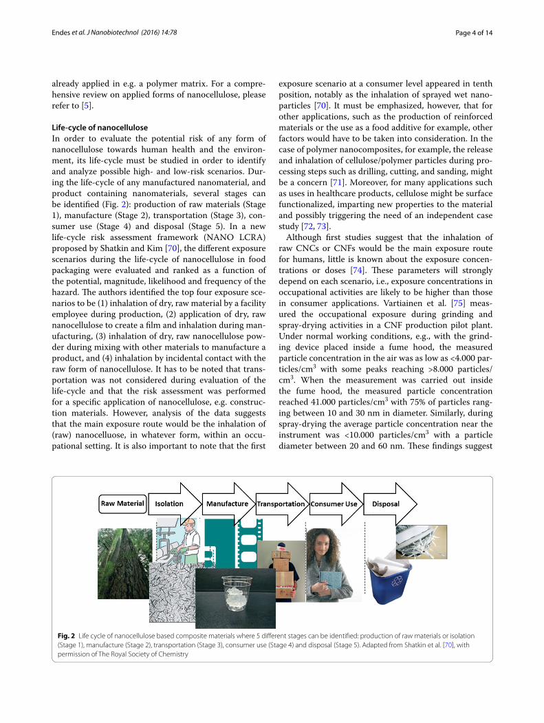
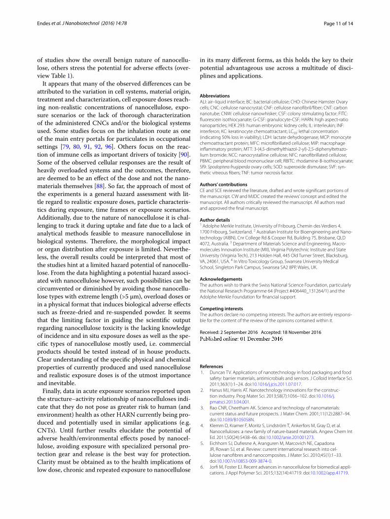
Life‑cycle of nanocelluloseIn order to evaluate the potential risk of any form of nanocellulose towards human health and the environ-ment, its life-cycle must be studied in order to identify and analyze possible high- and low-risk scenarios. Dur-ing the life-cycle of any manufactured nanomaterial, and product containing nanomaterials, several stages can be identified (Fig. 2): production of raw materials (Stage 1), manufacture (Stage 2), transportation (Stage 3), con-sumer use (Stage 4) and disposal (Stage 5). In a new life-cycle risk assessment framework (NANO LCRA) proposed by Shatkin and Kim [70], the different exposure scenarios during the life-cycle of nanocellulose in food packaging were evaluated and ranked as a function of the potential, magnitude, likelihood and frequency of the hazard. The authors identified the top four exposure sce-narios to be (1) inhalation of dry, raw material by a facility employee during production, (2) application of dry, raw nanocellulose to create a film and inhalation during man-ufacturing, (3) inhalation of dry, raw nanocellulose pow-der during mixing with other materials to manufacture a product, and (4) inhalation by incidental contact with the raw form of nanocellulose. It has to be noted that trans-portation was not considered during evaluation of the life-cycle and that the risk assessment was performed for a specific application of nanocellulose, e.g. construc-tion materials. However, analysis of the data suggests that the main exposure route would be the inhalation of (raw) nanocelluose, in whatever form, within an occu-pational setting. It is also important to note that the first
exposure scenario at a consumer level appeared in tenth position, notably as the inhalation of sprayed wet nano-particles [70]. It must be emphasized, however, that for other applications, such as the production of reinforced materials or the use as a food additive for example, other factors would have to be taken into consideration. In the case of polymer nanocomposites, for example, the release and inhalation of cellulose/polymer particles during pro-cessing steps such as drilling, cutting, and sanding, might be a concern [71]. Moreover, for many applications such as uses in healthcare products, cellulose might be surface functionalized, imparting new properties to the material and possibly triggering the need of an independent case study [72, 73].
Although first studies suggest that the inhalation of raw CNCs or CNFs would be the main exposure route for humans, little is known about the exposure concen-trations or doses [74]. These parameters will strongly depend on each scenario, i.e., exposure concentrations in occupational activities are likely to be higher than those in consumer applications. Vartiainen et al. [75] meas-ured the occupational exposure during grinding and spray-drying activities in a CNF production pilot plant. Under normal working conditions, e.g., with the grind-ing device placed inside a fume hood, the measured particle concentration in the air was as low as <4.000 par-ticles/cm3 with some peaks reaching >8.000 particles/cm3. When the measurement was carried out inside the fume hood, the measured particle concentration reached 41.000 particles/cm3 with 75% of particles rang-ing between 10 and 30 nm in diameter. Similarly, during spray-drying the average particle concentration near the instrument was <10.000 particles/cm3 with a particle diameter between 20 and 60 nm. These findings suggest
Fig. 2 Life cycle of nanocellulose based composite materials where 5 different stages can be identified: production of raw materials or isolation (Stage 1), manufacture (Stage 2), transportation (Stage 3), consumer use (Stage 4) and disposal (Stage 5). Adapted from Shatkin et al. [70], with permission of The Royal Society of Chemistry
Page 5 of 14Endes et al. J Nanobiotechnol (2016) 14:78
that humans can be readily exposed to nanocellulose in a variety of occupational settings at heightened concen-trations. Nonetheless, understanding of the impact of chronic, repeated exposure to these airborne concentra-tions to human health however, remains, at best, limited.
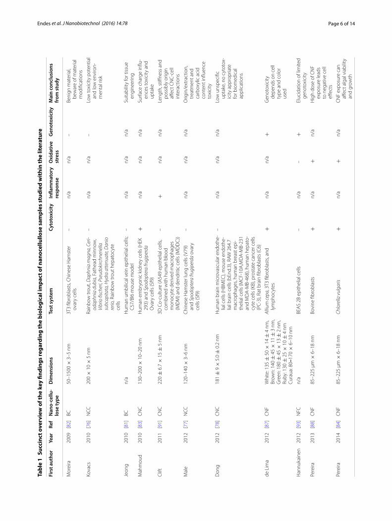
Biological impact of nanocelluloseSince human exposure, and to a lesser extent based on the current understanding, environmental exposure, to nanocellulose has been shown to be of a significant increase to normal airborne particle concentrations [75], and further to the concerns surrounding the poten-tial hazard associated with HARN and nanomaterials in general [58], understanding of the structure–activity relationship of nanocellulose is vital. The purpose of the remainder of this review therefore, is to provide a criti-cal overview of research directed towards exploring the biological impact and potential hazard of nanocellu-lose. An overview of key studies is provided in Table 1. In Table 1, together with the physical characteristics of the nanocellulose investigated, a description of the test system utilized, as well as the results of tests designed to assess cytotoxicity, (pro-)inflammatory response follow-ing nanocellulose exposure, the oxidative stress status of the biological system studied, as well as the potential for nanocellulose to elicit genotoxicity. Throughout the par-ticle and fiber toxicology field, these endpoints are rec-ognized as the most important drivers of nanomaterial toxicity [54]. For convenience, Table 1 provides a brief summary of the overall conclusions from each of these studies, although it is acknowledged that in some cases the entries may be overly simplified. It is important to further highlight that the biological systems highlighted through the main text and in Table 1 cover both in vitro, in vivo and ecosystem orientated models. This is a con-sidered approach to convey the current understanding of the biological impact of raw nanocellulose, and its vary-ing forms (which also change study-by-study) in terms of the biological response measured.
CytotoxicityOne of the first important studies regarding the eco-toxicological impact of cellulose nanocrystals derived from ‘kraft pulp’ (CNC dimensions: 200 × 10 × 5 nm) was published by Kovacs et al. in 2010 [76]. The authors presented results from a realistic exposure scenario, i.e., suspension experiments with relevant dose ranges (0.03–10 g/L), that were based on the potential effluent in the vicinity of a CNC production site. The study included aquatic organisms from all trophic levels from bacteria, algae, crustacean, cnidarian to fish and investigated acute lethality (LC50 = the lethal concentration that reduces the biological system population to 50% viability),
reproduction, growth, morphology, embryo development and cytotoxicity. Taking all results into consideration, the authors summarized the outcome as “non-concerning”.
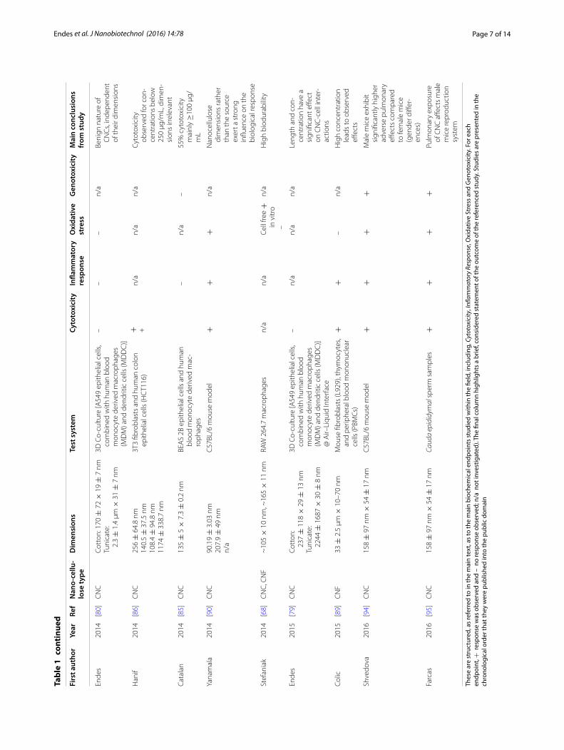
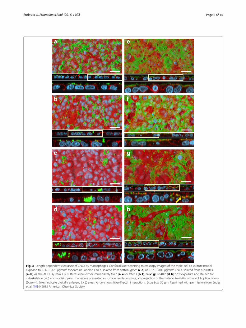
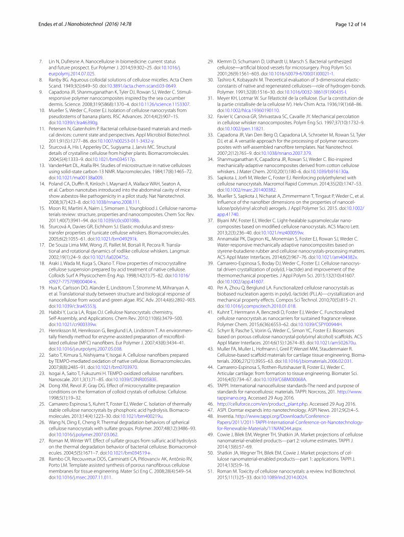
Further to this, several studies on cellulose-human interactions confirmed the limited toxic potential of nanocellulose in terms of cytotoxicity in various experi-mental systems [77, 78]. A sophisticated triple-cell co-culture model of the human epithelial tissue bar-rier (formulated of a layer of epithelial cells, compli-mented by human blood monocyte derived macrophages and dendritic cells on the apical and basolateral sides respectively) was used in a study that showed no sig-nificant cytotoxicity of two different CNC types iso-lated from cotton (170 ± 72 × 19 ± 7 nm) and tunicates (2.3 ± 1.4 µm × 31 ± 7 nm) that were deposited onto the cells in realistic doses (0.14 ± 0.04, 0.81 ± 0.03 and 1.57 ± 0.03 µg/cm2) from aerosolized water-based sus-pensions [79, 80]. However, clearance, albeit based upon a dose, time and CNC-dependent manner, of deposited CNCs by macrophages was observed when cells were exposed to both of these types CNCs, with a lower effi-ciency associated with the tunicate CNCs (Fig. 3) [79]. Jeong and co-workers used bacterial cellulose (BC; no dimensions given [81]) in in vitro experiments with human umbilical vein endothelial cells (HUVECs) [81]. Neither of their experiments measuring cytotoxicity via the MTT assay, observing the morphology with light microscopy or assessing apoptosis/necrosis (Annexin V/Propidium Iodide staining) and cell-cycle via flow cytometry, showed significant altered outcomes after 24 or 48 h towards the exposure to high BC concentra-tions (0.1–1 mg/mL) compared to the negative con-trol. Furthermore, in vivo exposure of 0.5–5 mg/mL BC administered via intraperitoneal injection to C57/Bl6 male mice showed no adverse effects after 7 days in comparison to sham exposures. Similar results with BC (50–1500 × 3–5 nm) were obtained by Moreira et al. [82] who could not detect significant changes in morphol-ogy or proliferation rates of mouse fibroblasts (3T3) and Chinese hamster ovary cells (CHO) in exposures ranging from 0.1–1 mg/mL.
However, there are also studies that have shown cyto-toxic effects upon exposure to nanocellulose. Mahmoud and co-workers investigated uptake and membrane integrity in human embryonic kidney cells (HEK 293) and Sf9 insect cells and found that exposure to 0.1 mg/mL of negatively charged CNCs (ζ-potential −46.4 mV), which had been isolated from enzyme treated flax fib-ers (130–200 × 10–20 nm) and labeled with FITC (fluorescein isothiocyanate), led to membrane rupture under physiological pH in contrast to exposure to posi-tively charged, RBITC-labeled (rhodamine B isothiocy-anate) CNCs (ζ-potential 8.7 mV) [83]. Similar cytotoxic
Page 6 of 14Endes et al. J Nanobiotechnol (2016) 14:78
Tabl
e 1
Succ
inct
ove
rvie
w o
f the
key
find
ings
rega
rdin
g th
e bi
olog
ical
impa
ct o
f nan
ocel
lullo
se s
ampl
es s
tudi
ed w
ithi
n th
e lit
erat
ure
Firs
t aut
hor
Year
Ref
Nan
o‑ce
llu‑
lose
type
Dim
ensi
ons
Test
sys
tem
Cyto
toxi
city
Infla
mm
ator
y re
spon
seO
xida
tive
stre
ssG
enot
oxic
ity
Mai
n co
nclu
sion
s fr
om s
tudy
Mor
eira
2009
[82]
BC50
–150
0 ×
3–5
nm
3T3
fibro
blas
ts, C
hine
se H
amst
er
ovar
y ce
lls–
n/a
n/a
–Be
nign
mat
eria
l, be
war
e of
mat
eria
l m
odifi
catio
ns
Kova
cs20
10[7
6]N
CC20
0 ×
10
× 5
nm
Rain
bow
trou
t, D
aphn
ia m
agna
, Cer
i-od
aphn
ia d
ubia
, Fat
head
min
now
, Vi
brio
fisc
heri,
Pse
udok
irchn
erie
lla
subc
apita
ta, H
ydra
att
enua
te, D
anio
re
rio, R
ainb
ow tr
out h
epat
ocyt
e ce
lls
–n/
an/
a–
Low
toxi
city
pot
entia
l an
d lo
w e
nviro
n‑m
enta
l ris
k
Jeon
g20
10[8
1]BC
n/a
Hum
an u
mbi
lical
vei
n ep
ithel
ial c
ells
; C
57/B
l6 m
ouse
mod
el–
n/a
n/a
n/a
Suita
bilit
y fo
r tis
sue
engi
neer
ing
Mah
mou
d20
10[8
3]C
NC
130–
200
× 1
0–20
nm
Hum
an e
mbr
yoni
c ki
dney
cel
ls (H
EK
293)
and
Spo
dopt
era
frugi
perd
a O
vary
cel
ls (S
f9)
+n/
an/
an/
aSu
rfac
e ch
arge
influ
‑en
ces
toxi
city
and
up
take
Clif
t20
11[9
1]C
NC
220
± 6
.7 ×
15
± 5
nm
3D C
o‑cu
lture
(A54
9 ep
ithel
ial c
ells
, co
mbi
ned
with
hum
an b
lood
m
onoc
yte
deriv
ed m
acro
phag
es
(MD
M) a
nd d
endr
itic
cells
(MD
DC
))
–+
n/a
n/a
Leng
th, s
tiffne
ss a
nd
poss
ibly
orig
in
affec
t CN
C‑c
ell
inte
ract
ions
Mal
e20
12[7
7]N
CC12
0–14
0 ×
3–6
nm
Chi
nese
Ham
ster
lung
cel
ls (V
79)
and
Spod
opte
ra fr
ugip
erda
ova
ry
cells
(Sf9
)
–n/
an/
an/
aO
rigin
/ext
ract
ion,
tr
eatm
ent a
nd
carb
oxyl
ic a
cid
cont
ent i
nflue
nce
toxi
city
Don
g20
12[7
8]C
NC
181
± 9
× 5
.0 ±
0.2
nm
Hum
an b
rain
mic
rova
scul
ar e
ndot
he‑
lial c
ells
(HBM
EC),
mou
se e
ndot
he‑
lial b
rain
cel
ls (b
End.
3), R
AW 2
64.7
m
acro
phag
es, h
uman
bre
ast e
pi‑
thel
ial c
ells
(MC
F‑10
A,M
DA
‑MB‑
231
and
MD
A‑M
B‑46
8), h
uman
hep
ato‑
cyte
cel
ls (K
B), p
rost
ate
canc
er c
ells
(P
C‑3
), Ra
t bra
in fi
brob
last
s (C
6)
–n/
an/
an/
aLo
w u
nspe
cific
up
take
, no
cyto
tox‑
icity
app
ropr
iate
fo
r bio
med
ical
ap
plic
atio
ns
de L
ima
2012
[87]
CN
FW
hite
: 135
± 5
0 ×
14
± 4
nm
,Br
own:
140
± 4
5 ×
11
± 3
nm
,G
reen
: 180
± 4
5 ×
13
± 2
nm
,Ru
by: 1
30 ±
25
× 1
0 ±
4 n
mCu
raua
: 80–
170
× 6
–10
nm
Alliu
m c
epa,
3T3
fibr
obla
sts,
and
lym
phoc
ytes
+n/
an/
a+
Gen
otox
icity
de
pend
s on
cel
l ty
pe a
nd c
olor
us
ed
Han
nuka
inen
2012
[93]
NFC
n/a
BEA
S 2B
epi
thel
ial c
ells
–n/
a–
+El
ucid
atio
n of
lim
ited
geno
toxi
city
Pere
ira20
13[8
8]C
NF
85–2
25 µ
m ×
6–1
8 nm
Bovi
ne fi
brob
last
s+
n/a
+n/
aH
igh
dose
of C
NF
expo
sure
lead
s to
neg
ativ
e ce
ll eff
ects
Pere
ira20
14[8
4]C
NF
85–2
25 µ
m ×
6–1
8 nm
Chlo
rella
vul
garis
+n/
a+
n/a
CN
F ex
posu
re c
an
affec
t alg
al v
iabi
lity
and
grow
th
Page 7 of 14Endes et al. J Nanobiotechnol (2016) 14:78
Thes
e ar
e st
ruct
ured
, as
refe
rred
to in
the
mai
n te
xt, a
s to
the
mai
n bi
oche
mic
al e
ndpo
ints
stu
died
with
in th
e fie
ld, i
nclu
ding
, Cyt
otox
icity
, Infl
amm
ator
y Re
spon
se, O
xida
tive
Stre
ss a
nd G
enot
oxic
ity. F
or e
ach
endp
oint
, + r
espo
nse
was
obs
erve
d an
d –
no
resp
onse
obs
erve
d; n
/a n
ot in
vest
igat
ed).
The
final
col
umn
high
light
s a
brie
f, co
nsid
ered
sta
tem
ent o
f the
out
com
e of
the
refe
renc
ed s
tudy
. Stu
dies
are
pre
sent
ed in
the
chro
nolo
gica
l ord
er th
at th
ey w
ere
publ
ishe
d in
to th
e pu
blic
dom
ain
Tabl
e 1
cont
inue
d
Firs
t aut
hor
Year
Ref
Nan
o‑ce
llu‑
lose
type
Dim
ensi
ons
Test
sys
tem
Cyto
toxi
city
Infla
mm
ator
y re
spon
seO
xida
tive
stre
ssG
enot
oxic
ity
Mai
n co
nclu
sion
s fr
om s
tudy
Ende
s20
14[8
0]C
NC
Cott
on: 1
70 ±
72
× 1
9 ±
7 n
mTu
nica
te:
2.3
± 1
.4 µ
m ×
31
± 7
nm
3D C
o‑cu
lture
[A54
9 ep
ithel
ial c
ells
, co
mbi
ned
with
hum
an b
lood
m
onoc
yte
deriv
ed m
acro
phag
es
(MD
M) a
nd d
endr
itic
cells
(MD
DC
)]
––
–n/
aBe
nign
nat
ure
of
CN
Cs,
inde
pend
ent
of th
eir d
imen
sion
s
Han
if20
14[8
6]C
NC
256
± 6
4.8
nm14
0.5
± 3
7.5
nm10
8.4
± 9
4.8
nm11
74 ±
338
.7 n
m
3T3
fibro
blas
ts a
nd h
uman
col
on
epith
elia
l cel
ls (H
CT1
16)
+ +n/
an/
an/
aCy
toto
xici
ty
obse
rved
for c
on‑
cent
ratio
ns b
elow
25
0 µg
/mL,
dim
en‑
sion
s irr
elev
ant
Cata
lan
2014
[85]
CN
C13
5 ±
5 ×
7.3
± 0
.2 n
mBE
AS
2B e
pith
elia
l cel
ls a
nd h
uman
bl
ood
mon
ocyt
e de
rived
mac
‑ro
phag
es
–n/
a–
55%
cyt
otox
icity
m
ainl
y ≥
100
µg/
mL
Yana
mal
a20
14[9
0]C
NC
90.1
9 ±
3.0
3 nm
207.
9 ±
49
nmn/
a
C57
BL/6
mou
se m
odel
++
+n/
aN
anoc
ellu
lose
di
men
sion
s ra
ther
th
an th
e so
urce
ex
ert a
str
ong
influ
ence
on
the
biol
ogic
al re
spon
se
Stef
ania
k20
14[6
8]C
NC
, CN
F~
105
× 1
0 nm
, ~16
5 ×
11
nmRA
W 2
64.7
mac
roph
ages
n/a
n/a
Cell
free
+
in v
itro
–
n/a
Hig
h bi
odur
abili
ty
Ende
s20
15[7
9]C
NC
Cott
on:
237
± 1
18 ×
29
± 1
3 nm
Tuni
cate
: 22
44 ±
168
7 ×
30
± 8
nm
3D C
o‑cu
lture
[A54
9 ep
ithel
ial c
ells
, co
mbi
ned
with
hum
an b
lood
m
onoc
yte
deriv
ed m
acro
phag
es
(MD
M) a
nd d
endr
itic
cells
(MD
DC
)] @
Air–
Liqu
id In
terf
ace
–n/
an/
an/
aLe
ngth
and
con
‑ce
ntra
tion
have
a
sign
ifica
nt e
ffect
on
CN
C‑c
ell i
nter
‑ac
tions
Colic
2015
[89]
CN
F33
± 2
.5 µ
m ×
10–
70 n
mM
ouse
fibr
obla
sts
(L92
9), t
hym
ocyt
es,
and
perip
hera
l blo
od m
onon
ucle
ar
cells
(PBM
Cs)
++
–n/
aH
igh
conc
entr
atio
n le
ads
to o
bser
ved
effec
ts
Shve
dova
2016
[94]
CN
C15
8 ±
97
nm ×
54
± 1
7 nm
C57
BL/6
mou
se m
odel
++
++
Mal
e m
ice
exhi
bit
sign
ifica
ntly
hig
her
adve
rse
pulm
onar
y eff
ects
com
pare
d to
fem
ale
mic
e (g
ende
r diff
er‑
ence
s)
Farc
as20
16[9
5]C
NC
158
± 9
7 nm
× 5
4 ±
17
nmCa
uda
epid
idym
al s
perm
sam
ples
++
++
Pulm
onar
y ex
posu
re
of C
NC
affe
cts
mal
e m
ice
repr
oduc
tion
syst
em
Page 8 of 14Endes et al. J Nanobiotechnol (2016) 14:78
Fig. 3 Length dependent clearance of CNCs by macrophages. Confocal laser scanning microscopy images of the triple‑cell co‑culture model exposed to 0.56 ± 0.25 μg/cm2 rhodamine‑labeled CNCs isolated from cotton (green a–d) or 0.67 ± 0.09 μg/cm2 CNCs isolated from tunicates (e–h) via the ALICE system. Co‑cultures were either immediately fixed (a, e) or after 1 (b, f), 24 (c, g), or 48 h (d, h) post exposure and stained for cytoskeleton (red) and nuclei (cyan). Images are presented as surface rendering (top), xz‑projection of the z‑stacks (middle), or twofold optical zoom (bottom). Boxes indicate digitally enlarged (×2) areas. Arrow shows fiber‑F‑actin interactions. Scale bars 30 μm. Reprinted with permission from Endes et al. [79] © 2015 American Chemical Society

Page 9 of 14Endes et al. J Nanobiotechnol (2016) 14:78
reactions were also reported using typical CNCs in exposures to algae [84] or bronchial cells (BEAS 2B) [85]. However, in both studies extremely high nano-cellulose concentrations in respect to mammalian cell culture (0.25–5 mg/mL) were used [86–88]. Of note in this regard is the study by Colic and co-authors [89], who showed that only the exposure to extremely high concentrations of long, entangled cellulose nanofibrils (33 ± 2.5 µm × 10–10 nm; 0.25–1 mg/mL), the highest one covering the L929 monolayers almost completely, lead to impaired metabolic activity and reduced cell pro-liferation [89]. Furthermore in vivo, Yanamala measured elevated cytotoxicity (as determined by an increase in the activity of the enzyme lactate dehydrogenase) after the aspiration of wood pulp derived CNCs in mice (50, 100 and 200 μg/mouse), detecting similar strong reactions in the context of cytotoxicity compared to asbestos aspira-tion (50 μg/mouse) [90].
Overall, the incidence of benign results in terms of cytotoxicity, viability and impact upon mammalian cell morphology seems to be prevalent in the current liter-ature upon the risk of nanocellulose. Despite this, the existence of adverse effects observed following nano-cellulose exposure has to be taken into consideration when evaluating the total hazard posed by this mate-rial. Summarizing, single, low doses administration of nanocelluloses hint at the non-hazardous nature of nanocellulose, yet lack a degree of realism when con-sidering human exposure. The importance of relevant exposure systems (cell type), dose, nanocellulose type/treatment/origin together with a clear material char-acterization is especially highlighted by the seemingly directly opposing results obtained by Mahmoud and co-authors (0.1 mg/mL FITC-labeled CNCs elicit cyto-toxicity in human embryonic kidney cells (HEK 293) ovary cells (Sf9)) [83] vs. Dong et al. (0.01–0.05 mg/mL FITC-labeled CNCs induce no measurable cytotox-icity in a wide range of barrier and immune cell types in vitro) [78].
InflammationOne of the key aspects of the nanoparticle-cell inter-action is the potential for nanoparticles to elucidate a (pro-)inflammatory response from the cellular system being studied. In a realistic in vitro model of the human epithelial tissue barrier, it has been demonstrated that the exposure to CNCs does not induce a significant amount of (pro-)inflammatory mediators tumor necro-sis factor-α (TNF-α) and interleukin-8 (IL-8), in contrast to asbestos fibers [91, 80]. The latter study [80] involved CNCs isolated from cotton (170 ± 72 × 19 ± 7 nm) and tunicates (2.3 ± 1.4 µm × 31 ± 7 nm) that were applied via nebulizing aqueous suspensions at a concentration
range from 0.14 ± 0.04 to 1.57 ± 0.03 µg/cm2 by an air–liquid exposure approach. These results are underpinned by a study of Catalan et al., who exposed monocyte derived macrophage monocultures to 30–300 µg/mL cotton CNCs (135 ± 5 × 7.3 ± 0.2 nm) with no detec-tion of TNF-α and IL-1β in comparison to microcrystal-line cellulose (CNC aggregates that were micron-sized) [92]. Interestingly, Colic and co-workers showed an anti-inflammatory influence of cellulose nanofibril expo-sures on PBMCs (peripheral blood mononuclear cells) in vitro, as measured by downregulation of IL-2, IFN-γ (interferon-γ) and IL-17, of, which was only observed at considered high doses (0.25–1 mg/mL) [89]. How-ever, Clift et al. (220 ± 6.7 × 15 ± 5 nm) [91], who used the same 3D triple-cell co-culture model of the human epithelial tissue barrier highlighted above and applied CNCs via aqueous suspensions, showed an increase in IL-8 response when exposed to 30 µg/mL cotton CNCs. An extensive screening study by Yanamala and col-leagues that explored the administration of CNCs after different processing steps (wood pulp CNCs applied as isolated in suspension and kept in suspension vs. iso-lated and freeze dried to powder before re-suspension) found that both preparations of CNCs have the poten-tial to induce inflammatory effects following pharyngeal aspiration in mice [90]. The authors detected signifi-cantly elevated pulmonary influxes of total cells, espe-cially PBMCs compared to negative controls and mice exposed to asbestos. Increased expression of cytokines (IL-1α, IL-1β, IL-5, IL-6, IL-12 p40, G-CSF, GM-CSF, KC, MCP-1, MIP-1α, MIP-1β, and TNF-α) involved in acute inflammatory reactions compared to the con-trol could be detected. Interestingly, depending on the pre-treatment from which the CNCs were applied, either a T-helper cell subtype 1 (Th1) mediated immune response (freeze dried before resuspension) or the induction of a Th2 associated response (only suspension) could be observed.
Despite the data discussed the above paragraph (Table 1), there remains a prominent lack of coherent data to substantially, and specifically evaluate the poten-tial of nanocellulose to pose a relevant hazard towards human health via an inflammatory immune response. Nevertheless, the existing studies point out that the phys-ico-chemical characteristics, especially the aggregation status, of CNCs can have a (direct) detrimental impact towards elucidating a (pro-)inflammatory response [90]. Moreover, overload exposures often mask the underlying specific mechanisms of toxicity and can only point at a general direction of potential hazard. In terms of inflam-mation, especially the chronic or repeated low dose expo-sure as the most realistic scenario for human exposure must be focused upon in future research.
Page 10 of 14Endes et al. J Nanobiotechnol (2016) 14:78
Oxidative stressLittle is known about the radical forming poten-tial of nanocellulose in cell-free and cellular environ-ments, with studies mainly reporting insignificant impact on the oxidative stress status of the cells unless extremely high concentrations are applied (cotton CNFs, 85–225 µm × 6–18 nm; 2–5 mg/mL, bovine fibroblasts), similar to the endpoints of cytotoxicity and inflamma-tion [88]. Only a few studies include the measurement of radical oxygen species formation [68, 89], the activity of antioxidant enzymes such as superoxide dismutase (SOD) or peroxiredoxin [88], and the depletion of anti-oxidant peptides such as glutathione [80, 89]. Interest-ingly, Stefaniak et al. observed significantly increased radical formation (∙OH) by CNCs (~105 × 10 nm) and CNFs (~165 × 11 nm) in a cell free experiment in con-trast to benchmark MCC (<10 µm × <2 µm) with absent, consecutive cellular reactions in macrophages [68]. These results are especially alarming as the study also revealed a high durability in artificial lung fluid. In sum-mary, it has been commonly reported that no significant oxidative stress is evident in vitro following nanocellu-lose exposure, i.e. using cotton (170 ± 72 × 19 ± 7 nm) or tunicate (2.3 ± 1.4 µm × 31 ± 7 nm) CNCs (0.14 ± 0.04 − 1.57 ± 0.03 µg/cm2) in the human epi-thelial tissue barrier model previously described [80], nanofibrillated celluloses (9.5–950 µg/cm2) on bronchial cells (BEAS 2B) [93], CNFs in high dose experiments with bovine fibroblasts (85–225 µm × 6–18 nm; 2–5 mg/mL) [88] and CNFs in lower doses to human fibroblasts (L929; >10 µm × 10–35 nm; 31.5 µg/ml–1 mg/ml) [89]. However, measurable biological effects were shown by Pereira et al. as a slight increase in SOD activity in the algae Chlorella vulgaris after exposure to 1, 50 and 100 μg/mL cotton CNFs (85–225 μm × 6–18 nm) [88].
The oxidative stress status of a cell has a relevant influ-ence most importantly in chronic exposures where it, together with its intrinsic biopersistence, can lead to severe damage and resulting disease as seen with other HARN materials [56]. The findings in cell-free experi-ments Stefaniak and colleagues [68] should point out the importance to substantiate the research in this direc-tion regarding the potential adverse biological impact of nanocellulose.
GenotoxicityIn recent years the investigation of damage to or changes in the genetic information within a cell induced by nanoparticle exposure came into focus; including the measurement of DNA strand breaks, formation of micro-nuclei and the potential for mutagenicity. Only a few studies have so far investigated the genotoxic influence of nanocellulose. Although the typical dimensions of
nanocellulose result in an unlikeliness of nuclear trans-location however is not to be excluded without further evidence. Nevertheless, the hindrance of cell-division, viability or indirect genotoxicity has to be especially highlighted when surface functionalizations are used to alter the bare and so far benign surface of nanocellulose.
Of the studies pertinent to this biological endpoint regarding nanocellulose, no effects in terms of micro-nuclei formation could be observed with BEAS 2B cells at low concentrations of cotton CNCs (2.5–100 μg/mL; 135 ± 5 × 7.3 ± 0.2 nm) over 48 h [92]. Kovacs et al. reported no changes in DNA quality after exposures to up to 2 mg/mL kraft pulp CNCs (200 × 10 × 5 nm) in primary rainbow trout hepatocytes [76]. Similar results were obtained when CNCs isolated from BC (50–1500 × 3–5 nm) were used in a comet assay and the AMES test in a concentration of 0.1 – 1 mg/mL after 48 h [82]. However, 0.01–1% white, colored cotton and curaua nanofibers (white 135 ± 50 × 14 ± 4 nm, brown 140 ± 45 × 11 ± 3 nm, green 180 ± 45 × 13 ± 2 nm, ruby 130 ± 25 × 10 ± 4 nm and curaua: 80–170 × 6–10 nm) showed the ability to induce negative changes in the rela-tive mitotic index and chromosomal aberration of Allium cepa cells as well as DNA strand breaks in concentrations of 0.1% of brown cotton and curaua fibers in animal cells (human lymphocytes, 3T3 mouse fibroblasts) [87]. Fur-thermore, Hannukainen et al. reported a potential geno-toxic effect by the exposure of BEAS 2B epithelial cells to NFC (950 μg/cm2; 24 h) measured by the comet assay [93].
Finally, important recent research has shown that some CNCs are able to induce all four biological endpoints, highlighting that through complex cellular cascades, that all four biochemical processes can induce deleteri-ous effects, albeit only in males in vivo. In recent studies by Shvedova et al., and Farcas et al., it has been shown that following pulmonary exposure of CNCs to C57BL/6 mice, that, after analysis over a chronic period, male mice were more susceptible to exhibit increased cytotoxicity, which was further associated with a heightened inflam-matory and oxidative stress response compared to female mice. Further evidence was shown that these biochemi-cal effects led to significant genotoxicity [94]. In a further study, a similar author team elucidated further that the genotoxic effects were highly detrimental to the male reproductive system [95].
SummaryIt is apparent from the research conducted regarding the potential hazard posed by various forms of nanocellulose, especially towards human and environmental health, that the current understanding of its structure–activity rela-tionship is equivocal and incoherent. Whilst a multitude
Page 11 of 14Endes et al. J Nanobiotechnol (2016) 14:78
of studies show the overall benign nature of nanocellu-lose, others stress the potential for adverse effects (over-view Table 1).
It appears that many of the observed differences can be attributed to the variation in cell systems, material origin, treatment and characterization, cell exposure doses reach-ing non-realistic concentrations of nanocellulose, expo-sure scenarios or the lack of thorough characterization of the administered CNCs and/or the biological systems used. Some studies focus on the inhalation route as one of the main entry portals for particulates in occupational settings [79, 80, 91, 92, 96]. Others focus on the reac-tion of immune cells as important drivers of toxicity [90]. Some of the observed cellular responses are the result of heavily overloaded systems and the outcomes, therefore, are deemed to be an effect of the dose and not the nano-materials themselves [88]. So far, the approach of most of the experiments is a general hazard assessment with lit-tle regard to realistic exposure doses, particle characteris-tics during exposure, time frames or exposure scenarios. Additionally, due to the nature of nanocellulose it is chal-lenging to track it during uptake and fate due to a lack of analytical methods feasible to measure nanocellulose in biological systems. Therefore, the morphological impact or organ distribution after exposure is limited. Neverthe-less, the overall results could be interpreted that most of the studies hint at a limited hazard potential of nanocellu-lose. From the data highlighting a potential hazard associ-ated with nanocellulose however, such possibilities can be circumvented or diminished by avoiding those nanocellu-lose types with extreme length (>5 µm), overload doses or in a physical format that induces biological adverse effects such as freeze-dried and re-suspended powder. It seems that the limiting factor in guiding the scientific output regarding nanocellulose toxicity is the lacking knowledge of incidence and in situ exposure doses as well as the spe-cific types of nanocellulose mostly used, i.e. commercial products should be tested instead of in house products. Clear understanding of the specific physical and chemical properties of currently produced and used nanocellulose and realistic exposure doses is of the utmost importance and inevitable.
Finally, data in acute exposure scenarios reported upon the structure–activity relationship of nanocelluloses indi-cate that they do not pose as greater risk to human (and environment) health as other HARN currently being pro-duced and potentially used in similar applications (e.g. CNTs). Until further results elucidate the potential of adverse health/environmental effects posed by nanocel-lulose, avoiding exposure with specialized personal pro-tection gear and release is the best way for protection. Clarity must be obtained as to the health implications of low dose, chronic and repeated exposure to nanocellulose
in its many different forms, as this holds the key to their potential advantageous use across a multitude of disci-plines and applications.
AbbreviationsALI: air–liquid interface; BC: bacterial cellulose; CHO: Chinese Hamster Ovary cells; CNC: cellulose nanocrystal; CNF: cellulose nanofibril/fiber; CNT: carbon nanotube; CNW: cellulose nanowhisker; CSF: colony stimulating factor; FITC: fluorescein isothiocyanate; G‑CSF: granulocyte‑CSF; HARN: high aspect‑ratio nanoparticles; HEK 293: human embryonic kidney cells; IL: interleukin; INF: interferon; KC: keratinocyte chemoattractant; LC50: lethal concentration (indicating 50% loss in viability); LDH: lactate dehydrogenase; MCP: monocyte chemoattractant protein; MFC: microfibrillated cellulose; MIP: macrophage inflammatory protein; MTT: 3‑(4,5‑dimethylthiazol‑2‑yl)‑2,5‑diphenyltetrazo‑lium bromide; NCC: nanocrystalline cellulose; NFC: nanofibrillated cellulose; PBMC: peripheral blood mononuclear cell; RBITC: rhodamine‑B‑isothiocyanate; Sf9: Spodoptera frugiperda ovary cells; SOD: superoxide dismutase; SVF: syn‑thetic vitreous fibers; TNF: tumor necrosis factor.
Authors’ contributionsCE and SCE reviewed the literature, drafted and wrote significant portions of the manuscript. CW and MJDC created the reviews’ concept and edited the manuscript. All authors critically reviewed the manuscript. All authors read and approved the final manuscript.
Author details1 Adolphe Merkle Institute, University of Fribourg, Chemin des Verdiers 4, 1700 Fribourg, Switzerland. 2 Australian Institute for Bioengineering and Nano‑technology (AIBN), Cnr College Rd & Cooper Rd, Building 75, Brisbane, QLD 4072, Australia. 3 Department of Materials Science and Engineering, Macro‑molecules Innovation Institute (MII), Virginia Polytechnic Institute and State University (Virginia Tech), 213 Holden Hall, 445 Old Turner Street, Blacksburg, VA, 24061, USA. 4 In Vitro Toxicology Group, Swansea University Medical School, Singleton Park Campus, Swansea SA2 8PP, Wales, UK.
AcknowledgementsThe authors wish to thank the Swiss National Science Foundation, particularly the National Research Programme 64 (Project #406440_131264/1) and the Adolphe Merkle Foundation for financial support.
Competing interestsThe authors declare no competing interests. The authors are entirely responsi‑ble for the content of the review of the opinions contained within it.
Received: 2 September 2016 Accepted: 18 November 2016
References 1. Duncan TV. Applications of nanotechnology in food packaging and food
safety: barrier materials, antimicrobials and sensors. J Colloid Interface Sci. 2011;363(1):1–24. doi:10.1016/j.jcis.2011.07.017.
2. Hanus MJ, Harris AT. Nanotechnology innovations for the construc‑tion industry. Prog Mater Sci. 2013;58(7):1056–102. doi:10.1016/j.pmatsci.2013.04.001.
3. Rao CNR, Cheetham AK. Science and technology of nanomaterials: current status and future prospects. J Mater Chem. 2001;11(12):2887–94. doi:10.1039/B105058N.
4. Klemm D, Kramer F, Moritz S, Lindström T, Ankerfors M, Gray D, et al. Nanocelluloses: a new family of nature‑based materials. Angew Chem Int Ed. 2011;50(24):5438–66. doi:10.1002/anie.201001273.
5. Eichhorn SJ, Dufresne A, Aranguren M, Marcovich NE, Capadona JR, Rowan SJ, et al. Review: current international research into cel‑lulose nanofibres and nanocomposites. J Mater Sci. 2010;45(1):1–33. doi:10.1007/s10853‑009‑3874‑0.
6. Jorfi M, Foster EJ. Recent advances in nanocellulose for biomedical appli‑cations. J Appl Polymer Sci. 2015;132(14):41719. doi:10.1002/app.41719.
Page 12 of 14Endes et al. J Nanobiotechnol (2016) 14:78
7. Lin N, Dufresne A. Nanocellulose in biomedicine: current status and future prospect. Eur Polymer J. 2014;59:302–25. doi:10.1016/j.eurpolymj.2014.07.025.
8. Ranby BG. Aqueous colloidal solutions of cellulose micelles. Acta Chem Scand. 1949;3(5):649–50. doi:10.3891/acta.chem.scand.03‑0649.
9. Capadona JR, Shanmuganathan K, Tyler DJ, Rowan SJ, Weder C. Stimuli‑responsive polymer nanocomposites inspired by the sea cucumber dermis. Science. 2008;319(5868):1370–4. doi:10.1126/science.1153307.
10. Mueller S, Weder C, Foster EJ. Isolation of cellulose nanocrystals from pseudostems of banana plants. RSC Advances. 2014;4(2):907–15. doi:10.1039/c3ra46390g.
11. Petersen N, Gatenholm P. Bacterial cellulose‑based materials and medi‑cal devices: current state and perspectives. Appl Microbiol Biotechnol. 2011;91(5):1277–86. doi:10.1007/s00253‑011‑3432‑y.
12. Šturcová A, His I, Apperley DC, Sugiyama J, Jarvis MC. Structural details of crystalline cellulose from higher plants. Biomacromolecules. 2004;5(4):1333–9. doi:10.1021/bm034517p.
13. VanderHart DL, Atalla RH. Studies of microstructure in native celluloses using solid‑state carbon‑13 NMR. Macromolecules. 1984;17(8):1465–72. doi:10.1021/ma00138a009.
14. Poland CA, Duffin R, Kinloch I, Maynard A, Wallace WAH, Seaton A, et al. Carbon nanotubes introduced into the abdominal cavity of mice show asbestos‑like pathogenicity in a pilot study. Nat Nanotechnol. 2008;3(7):423–8. doi:10.1038/nnano.2008.111.
15. Moon RJ, Martini A, Nairn J, Simonsen J, Youngblood J. Cellulose nanoma‑terials review: structure, properties and nanocomposites. Chem Soc Rev. 2011;40(7):3941–94. doi:10.1039/c0cs00108b.
16. Šturcová A, Davies GR, Eichhorn SJ. Elastic modulus and stress‑transfer properties of tunicate cellulose whiskers. Biomacromolecules. 2005;6(2):1055–61. doi:10.1021/bm049291k.
17. De Souza Lima MM, Wong JT, Paillet M, Borsali R, Pecora R. Transla‑tional and rotational dynamics of rodlike cellulose whiskers. Langmuir. 2002;19(1):24–9. doi:10.1021/la020475z.
18. Araki J, Wada M, Kuga S, Okano T. Flow properties of microcrystalline cellulose suspension prepared by acid treatment of native cellulose. Colloids Surf A Physicochem Eng Asp. 1998;142(1):75–82. doi:10.1016/s0927‑7757(98)00404‑x.
19. Hua K, Carlsson DO, Alander E, Lindstrom T, Stromme M, Mihranyan A, et al. Translational study between structure and biological response of nanocellulose from wood and green algae. RSC Adv. 2014;4(6):2892–903. doi:10.1039/c3ra45553j.
20. Habibi Y, Lucia LA, Rojas OJ. Cellulose Nanocrystals: chemistry, Self‑Assembly, and Applications. Chem Rev. 2010;110(6):3479–500. doi:10.1021/cr900339w.
21. Henriksson M, Henriksson G, Berglund LA, Lindstrom T. An environmen‑tally friendly method for enzyme‑assisted preparation of microfibril‑lated cellulose (MFC) nanofibers. Eur Polymer J. 2007;43(8):3434–41. doi:10.1016/j.eurpolymj.2007.05.038.
22. Saito T, Kimura S, Nishiyama Y, Isogai A. Cellulose nanofibers prepared by TEMPO‑mediated oxidation of native cellulose. Biomacromolecules. 2007;8(8):2485–91. doi:10.1021/bm0703970.
23. Isogai A, Saito T, Fukuzumi H. TEMPO‑oxidized cellulose nanofibers. Nanoscale. 2011;3(1):71–85. doi:10.1039/C0NR00583E.
24. Dong XM, Revol JF, Gray DG. Effect of microcrystallite preparation conditions on the formation of colloid crystals of cellulose. Cellulose. 1998;5(1):19–32.
25. Camarero Espinosa S, Kuhnt T, Foster EJ, Weder C. Isolation of thermally stable cellulose nanocrystals by phosphoric acid hydrolysis. Biomacro‑molecules. 2013;14(4):1223–30. doi:10.1021/bm400219u.
26. Wang N, Ding E, Cheng R. Thermal degradation behaviors of spherical cellulose nanocrystals with sulfate groups. Polymer. 2007;48(12):3486–93. doi:10.1016/j.polymer.2007.03.062.
27. Roman M, Winter WT. Effect of sulfate groups from sulfuric acid hydrolysis on the thermal degradation behavior of bacterial cellulose. Biomacromol‑ecules. 2004;5(5):1671–7. doi:10.1021/bm034519+.
28. Rambo CR, Recouvreux DOS, Carminatti CA, Pitlovanciv AK, Antônio RV, Porto LM. Template assisted synthesis of porous nanofibrous cellulose membranes for tissue engineering. Mater Sci Eng C. 2008;28(4):549–54. doi:10.1016/j.msec.2007.11.011.
29. Klemm D, Schumann D, Udhardt U, Marsch S. Bacterial synthesized cellulose—artificial blood vessels for microsurgery. Prog Polym Sci. 2001;26(9):1561–603. doi:10.1016/s0079‑6700(01)00021‑1.
30. Tashiro K, Kobayashi M. Theoretical evaluation of 3‑dimensional elastic‑constants of native and regenerated celluloses—role of hydrogen‑bonds. Polymer. 1991;32(8):1516–30. doi:10.1016/0032‑3861(91)90435‑l.
31. Meyer KH, Lotmar W. Sur l’élasticité de la cellulose. (Sur la constitution de la partie cristallisée de la cellulose IV). Helv Chim Acta. 1936;19(1):68–86. doi:10.1002/hlca.19360190110.
32. Favier V, Canova GR, Shrivastava SC, Cavaille JY. Mechanical percolation in cellulose whisker nanocomposites. Polym Eng Sci. 1997;37(10):1732–9. doi:10.1002/pen.11821.
33. Capadona JR, Van Den Berg O, Capadona LA, Schroeter M, Rowan SJ, Tyler DJ, et al. A versatile approach for the processing of polymer nanocom‑posites with self‑assembled nanofibre templates. Nat Nanotechnol. 2007;2(12):765–9. doi:10.1038/nnano.2007.379.
34. Shanmuganathan K, Capadona JR, Rowan SJ, Weder C. Bio‑inspired mechanically‑adaptive nanocomposites derived from cotton cellulose whiskers. J Mater Chem. 2010;20(1):180–6. doi:10.1039/b916130a.
35. Sapkota J, Jorfi M, Weder C, Foster EJ. Reinforcing poly(ethylene) with cellulose nanocrystals. Macromol Rapid Commun. 2014;35(20):1747–53. doi:10.1002/marc.201400382.
36. Mueller S, Sapkota J, Nicharat A, Zimmermann T, Tingaut P, Weder C, et al. Influence of the nanofiber dimensions on the properties of nanocel‑lulose/poly(vinyl alcohol) aerogels. J Appl Polymer Sci. 2015. doi:10.1002/app.41740.
37. Biyani MV, Foster EJ, Weder C. Light‑healable supramolecular nano‑composites based on modified cellulose nanocrystals. ACS Macro Lett. 2013;2(3):236–40. doi:10.1021/mz400059w.
38. Annamalai PK, Dagnon KL, Monemian S, Foster EJ, Rowan SJ, Weder C. Water‑responsive mechanically adaptive nanocomposites based on styrene‑butadiene rubber and cellulose nanocrystals‑processing matters. ACS Appl Mater Interfaces. 2014;6(2):967–76. doi:10.1021/am404382x.
39. Camarero‑Espinosa S, Boday DJ, Weder C, Foster EJ. Cellulose nanocrys‑tal driven crystallization of poly(d, l‑lactide) and improvement of the thermomechanical properties. J Appl Polym Sci. 2015;132(10):41607. doi:10.1002/app.41607.
40. Pei A, Zhou Q, Berglund LA. Functionalized cellulose nanocrystals as biobased nucleation agents in poly(L‑lactide) (PLLA)—crystallization and mechanical property effects. Compos Sci Technol. 2010;70(5):815–21. doi:10.1016/j.compscitech.2010.01.018.
41. Kuhnt T, Herrmann A, Benczedi D, Foster EJ, Weder C. Functionalized cellulose nanocrystals as nanocarriers for sustained fragrance release. Polymer Chem. 2015;6(36):6553–62. doi:10.1039/C5PY00944H.
42. Schyrr B, Pasche S, Voirin G, Weder C, Simon YC, Foster EJ. Biosensors based on porous cellulose nanocrystal‑poly(vinyl alcohol) scaffolds. ACS Appl Mater Interfaces. 2014;6(15):12674–83. doi:10.1021/am502670u.
43. Muller FA, Muller L, Hofmann I, Greil P, Wenzel MM, Staudenmaier R. Cellulose‑based scaffold materials for cartilage tissue engineering. Bioma‑terials. 2006;27(21):3955–63. doi:10.1016/j.biomaterials.2006.02.031.
44. Camarero‑Espinosa S, Rothen‑Rutishauser B, Foster EJ, Weder C. Articular cartilage: from formation to tissue engineering. Biomater Sci. 2016;4(5):734–67. doi:10.1039/C6BM00068A.
45. TAPPI. International nanocellulose standards‑The need and purpose of standards for nanocellulosic materials. TAPPI: Norcross, 201. http://www.tappinano.org. Accessed 29 Aug 2016.
46. http://celluforce.com/en/product_plant.php. Accessed 29 Aug 2016. 47. ASPI. Domtar expands into nanotechnology. ASPI News. 2012;9(2):4–5. 48. Inventia. http://www.tappi.org/Downloads/Conference‑
Papers/2011/2011‑TAPPI‑International‑Conference‑on‑Nanotechnology‑for‑Renewable‑Materials/11NANO44.aspx.
49. Cowie J, Bilek EM, Wegner TH, Shatkin JA. Market projections of cellulose nanomaterial‑enabled products—part 2: volume estimates. TAPPI J. 2014;13(6):57–69.
50. Shatkin JA, Wegner TH, Bilek EM, Cowie J. Market projections of cel‑lulose nanomaterial‑enabled products—part 1: applications. TAPPI J. 2014;13(5):9–16.
51. Roman M. Toxicity of cellulose nanocrystals: a review. Ind Biotechnol. 2015;11(1):25–33. doi:10.1089/ind.2014.0024.
Page 13 of 14Endes et al. J Nanobiotechnol (2016) 14:78
52. Warheit DB, Reed KL, Webb TR. Man‑made respirable‑sized organic fibers: what do we know about their toxicological profiles? Ind Health. 2001;39(2):119–25. doi:10.2486/indhealth.39.119.
53. Camarero‑Espinosa S, Endes C, Mueller S, Petri‑Fink A, Rothen‑Rutishauser B, Weder C, et al. Elucidating the potential biological impact of cellulose nanocrystals. Fibers. 2016;4(3):21.
54. Stone V, Miller MR, Clift MJD, Elder A, Mills NL, Moller P, et al. Nanomateri‑als vs ambient ultrafine particles: an opportunity to exchange toxicology knowledge. Environ Health Perspect. 2016. doi:10.1289/EHP424.
55. Poland CA, Duffin R, Kinloch I, Maynard A, Wallace WAH, Seaton A, et al. Carbon nanotubes introduced into the abdominal cavity of mice show asbestos‑like pathogenicity in a pilot study. Nat Nano. 2008;3(7):423–8.
56. Donaldson K, Murphy FA, Duffin R, Poland CA. Asbestos, carbon nano‑tubes and the pleural mesothelium: a review of the hypothesis regarding the role of long fibre retention in the parietal pleura, inflammation and mesothelioma. Particle Fibre Toxicol. 2010;7:5.
57. Becklake MR. Asbestos‑related diseases of lung and other organs—their epidemiology and implications for clinical practice. Am Rev Respir Dis. 1976;114(1):187–227.
58. Donaldson K, Tran CL. An introduction to the short‑term toxicology of respirable industrial fibres. Mutation Res Fundam Mol Mech Mutagen. 2004;553(1–2):5–9. doi:10.1016/j.mrfmmm.2004.06.011.
59. Bernstein DM. Synthetic vitreous fibers: a review toxicology, epi‑demiology and regulations. Crit Rev Toxicol. 2007;37(10):839–86. doi:10.1080/10408440701524592.
60. Wick P, Clift MJD, Rösslein M, Rothen‑Rutishauser B. A brief summary of carbon nanotubes science and technology: a health and safety perspec‑tive. ChemSusChem. 2011;4(7):905–11. doi:10.1002/cssc.201100161.
61. Donaldson K, Brown RC, Brown GM. New perspectives on basic mecha‑nisms in lung disease. 5. Respirable industrial fibres: mechanisms of pathogenicity. Thorax. 1993;48(4):390–5. doi:10.1136/thx.48.4.390.
62. Furness G, Maitland HB. Studies on cotton dust in relation to byssinosis. 1. Bacteria and fungi in cotton dust. Br J Ind Med. 1952;9(2):138–45.
63. Niven RM, Pickering CAC. Byssinosis: a review. Thorax. 1996;51(6):632–7. 64. Pickering CAC, Niven R. Byssinosis and other cotton‑related diseases.
Hunter’s diseases of occupations, 10th Edn. CRC Press, Taylor and Francis Group, Boca Raton. 2010.
65. Tatrai E, Adamis Z, Bohm U, Meretey K, Ungvary G. Role of cellulose in wood dust‑induced fibrosing alveo‑bronchiolitis in rat. J Appl Toxicol. 1995;15(1):45–8.
66. Warheit DB, Snajdr SI, Harstsky MA, Frame SR. Advances in the preven‑tion of occupational respiratory diseases. International Congress Series, Amsterdam: Elsevier Science; 1998. p. 579–89.
67. Davis JMG. The need for standardized testing procedures for all products capable of liberating respirable fibers—the example of materials based on cellulose. Br J Ind Med. 1993;50(2):187–90.
68. Stefaniak AB, Seehra MS, Fix NR, Leonard SS. Lung biodurability and free radical production of cellulose nanomaterials. Inhal Toxicol. 2014;26(12):733–49. doi:10.3109/08958378.2014.948650.
69. Muhle H, Ernst H, Bellmann B. Investigation of the durability of cellulose fibres in rat lungs. Ann Occup Hyg. 1997;41(Supplement 1):184–8.
70. Shatkin JA, Kim B. Cellulose nanomaterials: life cycle risk assessment, and environmental health and safety roadmap. Environ Sci Nano. 2015;5(2):477‑99.
71. Köhler AR, Som C, Helland A, Gottschalk F. Studying the potential release of carbon nanotubes throughout the application life cycle. J Clean Prod. 2008;16(8–9):927–37. doi:10.1016/j.jclepro.2007.04.007.
72. Albanese A, Tang PS, Chan WCW. The Effect of nanoparticle size, shape, and surface chemistry on biological systems. In: Yarmush ML, editor. Annual review of biomedical engineering, vol 14. Annual Review of Biomedical Engineering; 2012. p. 1–16.
73. Clift MJD, Rothen‑Rutishauser B, Brown DM, Duffin R, Donaldson K, Proudfoot L, et al. The impact of different nanoparticle surface chemistry and size on uptake and toxicity in a murine macrophage cell line. Toxicol Appl Pharmacol. 2008;232(3):418–27. doi:10.1016/j.taap.2008.06.009.
74. Donaldson K, Schinwald A, Murphy F, Cho W‑S, Duffin R, Lang T, et al. The biologically effective dose in inhalation nanotoxicology. Acc Chem Res. 2013;46(3):723–32. doi:10.1021/ar300092y.
75. Vartiainen J, Pohler T, Sirola K, Pylkkanen L, Alenius H, Hokkinen J, et al. Health and environmental safety aspects of friction grinding and spray drying of microfibrillated cellulose. Cellulose. 2011;18(3):775–86. doi:10.1007/s10570‑011‑9501‑7.
76. Kovacs T, Naish V, O’Connor B, Blaise C, Gagne F, Hall L, et al. An ecotoxico‑logical characterization of nanocrystalline cellulose (NCC). Nanotoxicol‑ogy. 2010;4(3):255–70. doi:10.3109/17435391003628713.
77. Male KB, Leung ACW, Montes J, Kamen A, Luong JHT. Probing inhibitory effects of nanocrystalline cellulose: inhibition versus surface charge. Nanoscale. 2012;4(4):1373–9. doi:10.1039/c2nr11886f.
78. Dong S, Hirani AA, Colacino KR, Lee YW, Roman M. Cytotoxicity and cellular uptake of cellulose nanocrystals. Nano LIFE. 2012;02(03):1241006. doi:10.1142/S1793984412410061.
79. Endes C, Mueller S, Kinnear C, Vanhecke D, Foster EJ, Petri‑Fink A, et al. Fate of cellulose nanocrystal aerosols deposited on the lung cell surface in vitro. Biomacromolecules. 2015;16(4):1267–75. doi:10.1021/acs.biomac.5b00055.
80. Endes C, Schmid O, Kinnear C, Mueller S, Camarero‑Espinosa S, Vanhecke D, et al. An in vitro testing strategy towards mimicking the inhalation of high aspect ratio nanoparticles. Particle Fibre Toxicol. 2014;11(1):40.
81. Jeong SI, Lee SE, Yang H, Jin YH, Park CS, Park YS. Toxicologic evaluation of bacterial synthesized cellulose in endothelial cells and animals. Mol Cell Toxicol. 2010;6(4):373–80. doi:10.1007/s13273‑010‑0049‑7.
82. Moreira S, Silva NB, Almeida‑Lima J, Oliveira Rocha HA, Batistuzzo Medeiros SR, Alves C Jr, et al. BC nanofibres: in vitro study of genotoxic‑ity and cell proliferation. Toxicol Lett. 2009;189(3):235–41. doi:10.1016/j.toxlet.2009.06.849.
83. Mahmoud KA, Mena JA, Male KB, Hrapovic S, Kamen A, Luong JHT. Effect of surface charge on the cellular uptake and cytotoxicity of fluorescent labeled cellulose nanocrystals. ACS Appl Mater Interfaces. 2010;2(10):2924–32. doi:10.1021/am1006222.
84. Pereira M, Mouton L, Yepremian C, Coute A, Lo J, Marconcini J, et al. Ecotoxicological effects of carbon nanotubes and cellulose nanofibers in Chlorella vulgaris. J Nanobiotechnol. 2014;12(1):15.
85. Catalan J, Ilves M, Jarventaus H, Hannukainen KS, Kontturi E, Vanhala E, et al. Genotoxic and immunotoxic effects of cellulose nanocrystals in vitro. Environ Mol Mutagen. 2014;56(2):171‑82
86. Hanif Z, Ahmed FR, Shin SW, Kim Y‑K, Um SH. Size‑ and dose‑dependent toxicity of cellulose nanocrystals (CNC) on human fibroblasts and colon adenocarcinoma. Colloids Surf B Biointerfaces. 2014;119:162–5. doi:10.1016/j.colsurfb.2014.04.018.
87. de Lima R, Feitosa LO, Maruyama CR, Barga MA, Yamawaki PC, Vieira IJ, et al. Evaluation of the genotoxicity of cellulose nanofibers. Int J Nanomed. 2012;7:3555–65. doi:10.2147/ijn.s30596.
88. Pereira MM, Raposo NRB, Brayner R, Teixeira EM, Oliveira V, Quintao CCR, et al. Cytotoxicity and expression of genes involved in the cellular stress response and apoptosis in mammalian fibroblast exposed to cotton cel‑lulose nanofibers. Nanotechnology. 2013;24(7):075103.
89. Colic M, Mihajlovic D, Mathew A, Naseri N, Kokol V. Cytocompat‑ibility and immunomodulatory properties of wood based nanofi‑brillated cellulose. Cellulose. 2015;22(1):763–78. doi:10.1007/s10570‑014‑0524‑8.
90. Yanamala N, Farcas MT, Hatfield MK, Kisin ER, Kagan VE, Geraci CL, et al. In vivo evaluation of the pulmonary toxicity of cellulose nanocrystals: a renewable and sustainable nanomaterial of the future. ACS Sustain Chem Eng. 2014;2(7):1691–8. doi:10.1021/sc500153k.
91. Clift MJD, Foster EJ, Vanhecke D, Studer D, Wick P, Gehr P, et al. Investigat‑ing the interaction of cellulose nanofibers derived from cotton with a sophisticated 3D human lung cell coculture. Biomacromolecules. 2011;12(10):3666–73. doi:10.1021/bm200865j.
92. Catalan J, Ilves M, Jarventaus H, Hannukainen K‑S, Kontturi E, Vanhala E, et al. Genotoxic and immunotoxic effects of cellulose nanocrys‑tals in vitro. Environ Mol Mutagen. 2015;56(2):171–82. doi:10.1002/em.21913.
93 Hannukainen KS, Suhonen S, Savolainen K, Norppa H. Genotoxicity of nanofibrillated cellulose in vitro as measured by enzyme comet assay. Toxicol Lett. 2012;211:S71. doi:10.1016/j.toxlet.2012.03.276.
Page 14 of 14Endes et al. J Nanobiotechnol (2016) 14:78
• We accept pre-submission inquiries
• Our selector tool helps you to find the most relevant journal
• We provide round the clock customer support
• Convenient online submission
• Thorough peer review
• Inclusion in PubMed and all major indexing services
• Maximum visibility for your research
Submit your manuscript atwww.biomedcentral.com/submit
Submit your next manuscript to BioMed Central and we will help you at every step:
94 Shvedova AA, Kisin ER, Yanamala N, Farcas MT, Menas AL, Williams A, Fournier PM, Reynolds JS, Gutkin DW, Star A, Reiner RS, Halappanavar S, Kagan VE. Gender differences in murine pulmonary responses elicited by cellulose nanocrystals. Part Fibre Toxicol. 2016;13(1):28. doi:10.1186/s12989‑016‑0140‑x.
95 Farcas MT, Kisin ER, Menas AL, Gutkin DW, Star A, Reiner RS, Yanamala N, Savolainen K, Shvedova AA. Pulmonary exposure to cellulose nanocrys‑tals caused deleterious effects to reproductive system in male mice. J Toxicol Environ Health A. 2016;24:1–14.
96 Sacui IA, Nieuwendaal RC, Burnett DJ, Stranick SJ, Jorfi M, Weder C, et al. Comparison of the properties of cellulose nanocrystals and cellulose nanofibrils isolated from bacteria, tunicate, and wood processed using acid, enzymatic, mechanical, and oxidative methods. ACS Appl Mater Interfaces. 2014;6(9):6127–38. doi:10.1021/am500359f.