Widespread Occurrence of Ranavirus in Pond-Breeding Amphibian Populations
Upload
independentCategory
view
1download
0
Isolation, Structure, and Biological Activity of Phaeofungin, a CyclicLipodepsipeptide from a Phaeosphaeria sp. Using the Genome-WideCandida albicans Fitness TestSheo B. Singh,*,† John Ondeyka,† Guy Harris,† Kithsiri Herath,† Deborah Zink,† Francisca Vicente,‡,§
Gerald Bills,‡,§ Javier Collado,‡,§ Gonzalo Platas,‡,§ Antonio Gonzalez del Val,‡,§ Jesus Martin,‡,§
Fernando Reyes,§ Hao Wang,⊥,∥ Jennifer Nielsen Kahn,⊥ Stefan Galuska,⊥ Robert Giacobbe,⊥
George Abruzzo,⊥ Terry Roemer,⊥,∥ and Deming Xu∥,▽
†Department of Medicinal Chemistry, Merck Research Laboratories, PO Box 2000, Rahway, New Jersey 07065, United States‡Centro de Investigacion Basica (CIBE), Merck Sharp & Dohme de Espana, S. A., Josefa Valcarcel 38, E-28027, Madrid, Spain§Fundacion MEDINA, Centro de Excelencia en Investigacion de Medicamentos Innovadores en Andalucía, Avenida Conocimiento 3,Parque Tecnologico Ciencias de la Salud, 18100 Armilla, Granada, Spain⊥Department of Infectious Diseases, Merck Research Laboratories, PO Box 2000, Rahway, New Jersey 07065, United States∥Center of Fungal Genetics, Merck Frosst Canada Ltd., Montreal, Quebec, H2X3Y8, Canada
*S Supporting Information
ABSTRACT: Phaeofungin (1), a new cyclic depsipeptide isolated from Phaeosphaeria sp., was discovered by application ofreverse genetics technology, using the Candida albicans fitness test (CaFT). Phaeofungin is comprised of seven amino acids and aβ,γ-dihydroxy-γ-methylhexadecanoic acid arranged in a 25-membered cyclic depsipeptide. Five of the amino acids were assignedwith D-configurations. The structure was elucidated by 2D-NMR and HRMS-MS analysis of the natural product and itshydrolyzed linear peptide. The absolute configuration of the amino acids was determined by Marfey’s method by complete andpartial hydrolysis of 1. The CaFT profile of the phaeofungin-containing extract overlapped with that of phomafungin (3), anotherstructurally different cyclic lipodepsipeptide isolated from a Phoma sp. using the same approach. Comparative biologicalcharacterization further demonstrated that these two fungal lipodepsipeptides are functionally distinct. While phomafungin waspotentiated by cyclosporin A (an inhibitor of the calcineurin pathway), phaeofungin was synergized with aureobasidin A (2) (aninhibitor of the sphingolipid biosynthesis) and to some extent caspofungin (an inhibitor of glucan synthase). Furthermore,phaeofungin caused ATP release in wild-type C. albicans strains but phomafungin did not. It showed modest antifungal activityagainst C. albicans (MIC 16−32 μg/mL) and better activity against Aspergillus fumigatus (MIC 8−16 μg/mL) and Trichophytonmentagrophytes (MIC 4 μg/mL). The linear peptide was inactive, suggesting that the macrocyclic depsipeptide ring is essential fortarget engagement and antifungal activity.
Candida albicans and Aspergillus fumigatus are two key fungalpathogens responsible for the majority of life-threateningsystemic fungal infections. These infections are particularlyproblematic to the patient populations that are immunocom-promised.1 Currently, three classes of drugs are used in theclinic for treatment of systemic fungal infections. These areamphotericin B,2 azoles (e.g., fluconazole),3 and echinocandins(e.g., caspofungin, micafungin, and anidulafungin).4 While thesetreatment options are generally acceptable, they are notlimitless. In addition, fungal strains resistant to these treatmentsare emerging, though fortunately with lower frequency.
Therefore, new treatment options are needed to augment orreplace these agents.Natural products continue to be one of the best sources of
antifungal agents, and two of the three classes of clinically usedantifungal agents are natural product derived. However, thenatural product-based drug discovery process comes with
Special Issue: Special Issue in Honor of Lester A. Mitscher
Received: October 9, 2012Published: December 24, 2012
Article
pubs.acs.org/jnp
© 2012 American Chemical Society andAmerican Society of Pharmacognosy 334 dx.doi.org/10.1021/np300704s | J. Nat. Prod. 2013, 76, 334−345

limitations, as compared to synthetic compounds, ranging fromthe high demand for resources, the specialized expertise neededin building and maintaining high-quality libraries of naturalproducts, and the selection of diverse microbial strains capableof producing natural products, to the incompatibility of naturalproduct extract libraries with the “blitz screen” mentality ofhigh-throughput screens. These limitations had been furtherexacerbated by the ever-increasing rediscovery of knowncompounds. Collectively, they have exhausted the conventional“grind-and-find” approach leading to abandonment of naturalproduct efforts by most of the big pharmaceutical companies.The key to increasing the probability of success in identifyingnovel scaffolds from natural products is threefold: (1) Thediversity of the source microbes must be improved to increasethe probability of new natural product discovery; (2) prior tocommitting chemical fractionation, those extracts (of fermenta-tion broths) containing known compounds must be effectivelyrecognized and removed; and (3) extracts containing novelbiological activities (and most likely novel structures) must bepositively identified and prioritized.To address these challenges, efficient and robust natural
product dereplication by chemical and biological means isneeded. While large-scale chemical profiling can be readilyachieved, true dereplication is resource-consuming and oftenstrategically prohibitive if a structure is to be specificallyassociated with a specific biological activity. Dereplication ofcompounds using conventional single-target-based biochemicalassays is relatively straightforward. However, parallel multitarget(protein or whole-cell based) screenings are not amenable tothe challenge, since biological dereplication requires compre-hensive insights of mechanisms of action of active compoundsthat may have not been characterized. Chemical genomicstrategies could provide a solution to biological dereplication.In fact, our effort in applying the chemogenomic approach tonatural products (see Roemer et al.5 for a review) was first andforemost augmented by replenishing the natural product librarywith biodiversity. We have expanded our efforts to isolatemicroorganisms from different geography and habitats. Thisgeographic diversity was coupled with improved high-throughput fermentation methods aimed to increase functionalchemical diversity.6
The chemogenomic approach, known as the “fitness test”,was first developed using baker’s yeast and adapted in theprimary human fungal pathogen, C. albicans.7,8 The assay relieson a phenomenon known as chemically induced haploinsuffi-ciency, where deletion of one allele of a given gene in a diploidfungus renders the mutant hypersensitive to a compound thatinhibits the corresponding protein target at sublethalconcentrations. In fact, when a collection of heterozygousdeletion strains covering the entire or a significant portion of afungal genome was tested against a panel of antifungalcompounds with known modes of action (MOAs), hyper-variable growth (hypersensitivity or resistance, hence the termfitness test) was restricted to only a small set of strains thatcorrespond to aspects of their respective MOAs, including thetargets. In almost all cases, the concise fitness test profiles arereflective of the MOAs of compounds tested.7,8 As all theheterozygotes were barcoded at the deleted allele, only a smallvolume of pooled strains for each assay is needed to determinethe relative abundance (i.e., fitness) of each strain in responseto chemical perturbation, by DNA microarray methodology.By applying the C. albicans fitness test (CaFT), it was
possible to mechanistically annotate natural products in the
form of crude extracts. With a compendium of known naturalproducts (pure), the CaFT profile of any extract in questioncould be readily compared. If it matched the profile of a knowncompound, the extract was immediately analyzed by LC-MS toconfirm the presence/absence of such a compound. In othercases, a CaFT profile might reflect the biological MOA of aknown natural product that is not present in the compendium.This could be readily remedied in the CaFT and the LC-MSanalyses using the compound in question. The CaFT profilingof natural products had been a self-reiterating process that alsogrouped extracts by their biological activities. Once a novelCaFT profile was identified, chemical resources could then beallocated for mechanistically guided fractionation of therepresentative extract.5 This approach has been provenproductive in identifying novel compounds from naturalproducts with distinctive biological activities.5,9−13
In this report, we present the identification, isolation,structural elucidation, and characterization of a cyclic lip-odepsipeptide, phaeofungin (1, Figure 1), from an extract of afungus, Phaeosphaeria sp. (F-167,953). The original extract wasidentified for its whole-cell activity against wild-type C. albicansand A. fumigatus, two medically important human fungal
Figure 1. Structures of phaeofungin (1) and two other cyclicdepsipeptides, aureobasidin A (2) and phomafungin (3).
Journal of Natural Products Article
dx.doi.org/10.1021/np300704s | J. Nat. Prod. 2013, 76, 334−345335

pathogens. When tested in the CaFT, it generated a profile thatcontained hypersensitivities of fungal strains corresponding tothe sphingolipid biosynthesis pathway. However, the CaFT andLC-MS analyses failed to detect the presence of knowninhibitors of the pathway (e.g., myriocin,14 rustmicin,15−17
minimoidin,16 aureobasidins (e.g., aureobasidin A, 2),18
australifungin,16 and syringomycins19). Nonetheless, theCaFT profile overlapped with that of another set of extracts,which were subsequently identified to contain a cycliclipodepsipeptide, phomafungin (3),11 indicating certain struc-
Figure 2. CaFT profiles of aureobasidin A, myriocin, rustmicin, phaeofungin, and the fermentation extracts of F-167,953. The relative behavior ofeach heterozygous deletion strain elicited by the antifungal compounds is expressed by the normalized z-scores, with positive values indicatinghypersensitivity, and negative values resistance. To compare the CaFT profiles, strains with absolute values of z-scores no less than 3 in at least threeindependent experiments were selected. Their z-scores were grouped by hierarchical clustering using the centroid linkage method, the results ofwhich are displayed in TreeView, with the scale of the heat map shown on the top-right corner, together with experimental numbers andexperimental conditions. The heterozygous deletion strains are indicated by the orf19 designations of the corresponding genes, with C. albicans genenames and S. cerevisiae orthologs/homologues (if any) (as appeared in the Candida Genome Database, www.candidagenome.org). Highlighted byred arrows are those involved in the sphingolipid biosynthesis.
Journal of Natural Products Article
dx.doi.org/10.1021/np300704s | J. Nat. Prod. 2013, 76, 334−345336

tural similarity. We proceeded to isolate and characterize theactive compound produced by F-167,953. Consistent with theprediction of the CaFT results, this compound turned out to beanother cyclic lipodepsipeptide, termed phaeofungin (1).Despite biological signature overlap of the two cyclicdepsipeptides, our results showed that phaeofungin (1) and
phomafungin (3) are structurally and biologically distinct. This
discovery demonstrates the power of the CaFT, which detected
and differentiated natural products in crude extracts with
distinct or overlapping (often partially) activities with distinct
structural and mechanistic attributes.
Figure 3. CaFT profiles of phaeofungin (1) and its source extract from F-167,953, and phomafungin (3) and its source extract from F-224,939, andother extracts identified to contain known inhibitors of the fungal glucan synthase (GS) inhibitors. The CaFT results were analyzed and displayedusing the same parameters described in Figure 2. Highlighted are genes that are involved in the calcineurin pathway (black), sphingolipid (yellow),potential efflux (white), and glucan synthesis (red). Strains of double heterozygous deletion of FKS1 together with another gene implicated in theglucan synthesis/cell wall are indicated by a red bracket, with blue arrows indicating three other deletion strains (RHO1+/− SMI1+/−, RHO1+/−
FKS3+/−, and RHO1+/− CHS3+/−). These three strains, together with RHO1+/− and two independently constructed GSK1+/− strains, were notsignificantly hypersensitive to F-167,953 or phaeofungin. The scale of the heat map is the same as in Figure 2.
Journal of Natural Products Article
dx.doi.org/10.1021/np300704s | J. Nat. Prod. 2013, 76, 334−345337
■ RESULTS AND DISCUSSION
Identification of a Phaeofungin-Containing Extract inthe CaFT. Extracts of fermentation broths of microbial strainswith whole-cell antifungal activities against the wild-type C.albicans were tested in the CaFT assay. A fungal extract fromstrain F-167,953 produced a complex profile that was distinctfrom any known natural products tested in the CaFT. Severalhypersensitive heterozygous deletion strains were identifiedthat corresponded to genes involved in the sphingolipidbiosynthesis, including AUR1/orf19.1945 (the target of the
antifungal cyclic depsipeptide aureobasidin A), LCB1/orf19.6438, and MIT1/orf19.4077 (Figure 2). However,aureobasidin A (2) and other inhibitors of the sphingolipidbiosynthesis yielded CaFT profiles that shared no additionalsimilarity (Figure 2). Nevertheless, when compared with othernatural products from our collection, the CaFT profile of F-167,953 overlapped with that of another class of fungal extractsthat were subsequently identified to contain the lipodepsipep-tide phomafungin (3)11 (Figure 3). More intriguingly, F-167,953 induced specific hypersensitivity of a group of double
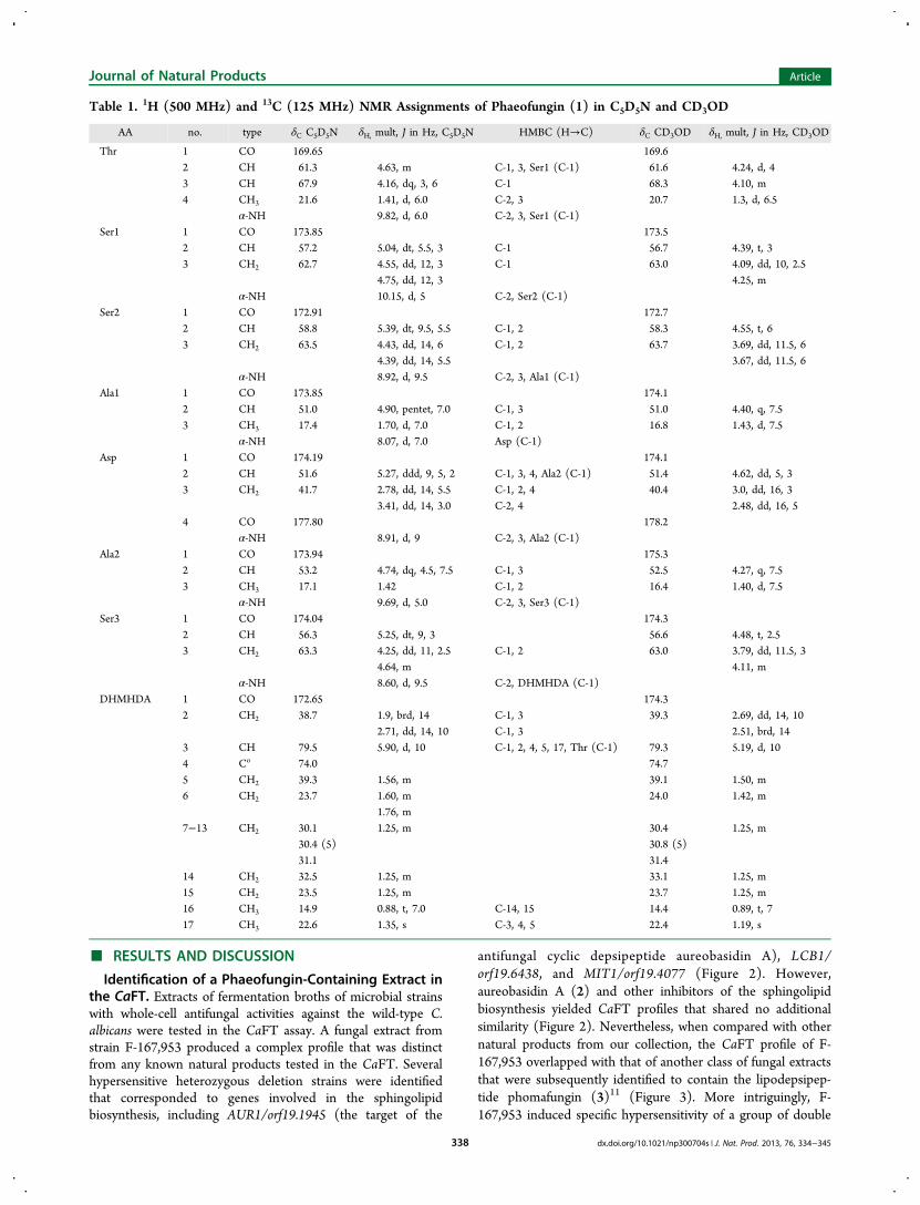
Table 1. 1H (500 MHz) and 13C (125 MHz) NMR Assignments of Phaeofungin (1) in C5D5N and CD3OD
AA no. type δC C5D5N δH, mult, J in Hz, C5D5N HMBC (H→C) δC CD3OD δH, mult, J in Hz, CD3OD
Thr 1 CO 169.65 169.62 CH 61.3 4.63, m C-1, 3, Ser1 (C-1) 61.6 4.24, d, 43 CH 67.9 4.16, dq, 3, 6 C-1 68.3 4.10, m4 CH3 21.6 1.41, d, 6.0 C-2, 3 20.7 1.3, d, 6.5
α-NH 9.82, d, 6.0 C-2, 3, Ser1 (C-1)Ser1 1 CO 173.85 173.5
2 CH 57.2 5.04, dt, 5.5, 3 C-1 56.7 4.39, t, 33 CH2 62.7 4.55, dd, 12, 3 C-1 63.0 4.09, dd, 10, 2.5
4.75, dd, 12, 3 4.25, mα-NH 10.15, d, 5 C-2, Ser2 (C-1)
Ser2 1 CO 172.91 172.72 CH 58.8 5.39, dt, 9.5, 5.5 C-1, 2 58.3 4.55, t, 63 CH2 63.5 4.43, dd, 14, 6 C-1, 2 63.7 3.69, dd, 11.5, 6
4.39, dd, 14, 5.5 3.67, dd, 11.5, 6α-NH 8.92, d, 9.5 C-2, 3, Ala1 (C-1)
Ala1 1 CO 173.85 174.12 CH 51.0 4.90, pentet, 7.0 C-1, 3 51.0 4.40, q, 7.53 CH3 17.4 1.70, d, 7.0 C-1, 2 16.8 1.43, d, 7.5
α-NH 8.07, d, 7.0 Asp (C-1)Asp 1 CO 174.19 174.1
2 CH 51.6 5.27, ddd, 9, 5, 2 C-1, 3, 4, Ala2 (C-1) 51.4 4.62, dd, 5, 33 CH2 41.7 2.78, dd, 14, 5.5 C-1, 2, 4 40.4 3.0, dd, 16, 3
3.41, dd, 14, 3.0 C-2, 4 2.48, dd, 16, 54 CO 177.80 178.2
α-NH 8.91, d, 9 C-2, 3, Ala2 (C-1)Ala2 1 CO 173.94 175.3
2 CH 53.2 4.74, dq, 4.5, 7.5 C-1, 3 52.5 4.27, q, 7.53 CH3 17.1 1.42 C-1, 2 16.4 1.40, d, 7.5
α-NH 9.69, d, 5.0 C-2, 3, Ser3 (C-1)Ser3 1 CO 174.04 174.3
2 CH 56.3 5.25, dt, 9, 3 56.6 4.48, t, 2.53 CH2 63.3 4.25, dd, 11, 2.5 C-1, 2 63.0 3.79, dd, 11.5, 3
4.64, m 4.11, mα-NH 8.60, d, 9.5 C-2, DHMHDA (C-1)
DHMHDA 1 CO 172.65 174.32 CH2 38.7 1.9, brd, 14 C-1, 3 39.3 2.69, dd, 14, 10
2.71, dd, 14, 10 C-1, 3 2.51, brd, 143 CH 79.5 5.90, d, 10 C-1, 2, 4, 5, 17, Thr (C-1) 79.3 5.19, d, 104 Co 74.0 74.75 CH2 39.3 1.56, m 39.1 1.50, m6 CH2 23.7 1.60, m 24.0 1.42, m
1.76, m7−13 CH2 30.1 1.25, m 30.4 1.25, m
30.4 (5) 30.8 (5)31.1 31.4
14 CH2 32.5 1.25, m 33.1 1.25, m15 CH2 23.5 1.25, m 23.7 1.25, m16 CH3 14.9 0.88, t, 7.0 C-14, 15 14.4 0.89, t, 717 CH3 22.6 1.35, s C-3, 4, 5 22.4 1.19, s
Journal of Natural Products Article
dx.doi.org/10.1021/np300704s | J. Nat. Prod. 2013, 76, 334−345338

heterozygous deletion strains, each of which containedheterozygous deletions of two genes that are functionallyimplicated in the fungal glucan synthesis and cell wallbiosynthesis.5 The hypersensitivity of these strains is highlyreflective of the presence of fungal glucan synthase inhibitors inthe extracts5 (Figure 3). Although the complex profiles of theseextracts did not directly suggest a discernible MOA, thedifference between F-167,953 and the phomafungin-containingextracts indicated that their biological activities shared certainsimilarities but were not completely identical. Prior to anychemical fractionation effort, we predicted that the correspond-ing natural products present in these extracts were structurallyand functionally different. These extracts were thus selected fordetailed chemical purification and characterization of naturalproduct(s) responsible for biological activities.Producing Organisms. The fungal strain F-167,953 was
isolated from a Sedum sp., a flowering plant, collected in theprovince of Albacete, Spain. Similarity searches with the 28Sand ITS rDNA sequences indicated a relatively high sequencesimilarity with some species of Phaeosphaeria, Ophiosphaerella,and various anamorph genera associated with the familyPhaeosphaeriaceae. Alignments of its 28S rDNA sequenceclearly placed this fungus within the Phaeosphaeriaceae. Thenearest match in the ITS region of the rDNA was an anamorphspecies, Chaetosphaeronema hispidulum (CBS 216.75), of thePhaeosphaeriaceae, with a similarity of 94.5%. A second strain,F-262,327 (from the province of Cuenca), was identifiedsubsequently to produce phaeofungin (1). It yielded a nearlyidentical 28S rDNA sequence. The 28S rDNA sequences ofthese two strains were resampled from recent phylogeneticstudies on fungi of the Phaeosphaeriaceae20−22 and aligned.Neighbor joining analysis of the alignment (Figure S1,Supporting Information) placed F-167,953 and F-262,327 inthe Phaeosphariaceae among a group of anamorph speciesincluding Plenodomus fuscomaculans and Chaetosphaeronemacoonsii.Prior to the CaFT analysis, these extracts of fungus F-
167,953 were observed to perturb the cell wall biosynthesis in adistinct cell-based assay designed to detect inhibitors of the C.albicans cell wall function/glucan synthesis (data not shown), abioactivity reminiscent of what was observed in the CaFT(Figure 3). The strain was revived from the freezer and grownin an eight-medium nutritional array to generate 1 mL extractsfor evaluation of antifungal activity.6 The fungus producedactive C. albicans activity in five of the eight media tested, andthe medium yielding the most potent extract was scaled up toone liter.Isolation and Structural Elucidation of Phaeofungin
(1). The fermentation broth of Phaeosphaeria sp. F-167,953 wasextracted with an equal volume of acetone. The extract waschromatographed on an Amberchrome column and eluted witha step gradient of aqueous MeOH. The fractions from eachchromatographic step were tested for their antifungal activityagainst the wild-type C. albicans. Those fractions containing theantifungal activity were finally purified by reversed-phaseHPLC, and the resulting active fractions were lyophilized toafford 1 (638 mg, 638 mg/L) as an amorphous, colorlesspowder. The purified material was tested in the CaFT, and ityielded a profile indistinguishable from the original one(Figures 2 and 3), suggesting that the purified materialrepresents the biological activity detected in the CaFT.HRESIFTMS analysis of 1 provided a molecular formula of
C40H69N7O16. It displayed end absorption in the UV spectrum
and absorption bands for hydroxy and various amide/estercarbonyls in the IR spectrum. The 13C NMR spectrum alongwith the DEPT spectrum in C5D5N and CD3OD exhibited thepresence of nine carboxyl carbonyls, nine methines (seven α-amino acid methines and two oxymethines), three oxy-methylenes, an oxygenated quaternary, and four methyls, andthe remaining carbons were aliphatic methylenes, typical ofthose present in a fatty chain. The 1H NMR spectrum inC5D5N (Table 1) revealed seven doublets in the downfieldregion of the spectrum, which were exchanged in the 1H NMRspectrum in CD3OD (Table 1), suggesting that they areexchangeable NH groups. This indicated that compound 1 ispeptidic, consisting of at least seven α-amino acids. The COSY,TOCSY, and HSQC spectroscopic analysis of 1 indicated thepresence of the following moieties: three serines (Ser), athreonine (Thr), two alanines (Ala), an aspartic acid (Asp), anda β,γ-dihydroxy-γ-methylhexadecanoic acid (DHMHDA). Thestructure of the latter unit was confirmed by HMBCcorrelations of H3-17 to C-3, C-4, and C-5 and H-3 to C-4,C-5, and C-17. Basic hydrolysis of 1 produced the acylatedheptapeptide 4 (MW 921), suggesting the presence of a lactonering in 1. The mass spectrometric analysis of 4 showed afragment ion at m/z 638 due to the loss of the fatty acid unit.The HRMS-MS analysis of the fragment ion at m/z 638produced b series fragment ions at m/z 519, 432, 345, 274, and159, suggesting a Ser-Ala-Asp-Ala-Ser-Ser-Thr-OH moiety(Figure 4). Whether Ser or Ala was present at the end of the
N-terminus could not be differentiated from these fragmenta-tions due to the lack of fragmentation between these two aminoacids. The amino acid sequence was corroborated by theHMBC correlations of α-CH and NH protons to thecorresponding adjacent carbonyls and α-carbons in C5D5N(Table 1). The unambiguous assignment of the C-1 ofDHMHDA unit was secured from the HMBC correlations ofH-2 and H-3, which were critical for the final sequencedetermination. The C-1 signal of the DHMHDA unit showedthe HMBC correlation to the Ser3-NH, confirming the amidebond between Ser3 and DHMHDA, thus the linear sequence.The H-3 resonance of the DHMHDA unit displayed a strongHMBC correlation to the C-1 of the threonine, confirming themacro-lactone ring in the depsipeptide structure of 1.The 1H NMR chemical shifts, including shifts of carbon-
bound protons in C5D5N, were highly concentration depend-
Figure 4. HRESIMS-MS fragmentation of the acyclic acylatedheptapeptide 4.
Journal of Natural Products Article
dx.doi.org/10.1021/np300704s | J. Nat. Prod. 2013, 76, 334−345339

ent. The same concentration-dependent phenomenon was notobserved in CD3OD. This phenomenon was likely due to thepresence of a large number of hydroxylated amino acids thatinteracted with C5D5N. NOESY correlations of phaeofungin inC5D5N further corroborated the sequence and the assignmentof the structure. The Ser2-NH showed NOESY correlations toNH groups of both the adjacent amino acids Ser1 and Ala1,suggesting that they are proximal to each other. Similarcorrelations were observed with the NH groups of other aminoacids, confirming the amino acid sequence of the depsipeptide.Exhaustive acid hydrolysis of phaeofungin (1) with 6 N HCl
at 110 °C overnight followed by derivatization with Marfey’sreagent (1-fluoro-2,4-dinitrophenyl-5-L-valine, L-FDVA)23 andcomparative quantitative analysis by LC-MS with correspond-ing authentic standards confirmed the presence of 2 × D-Ser, aL-Ser, a D-allo-Thr, a D-Ala, an L-Ala, and a D-aspartic acid(Asp). Partial acid hydrolysis with 0.5 N HCl at 100 °C for 30min followed by a reversed-phase HPLC purification led to theisolation of two major peptide fragments, 5 and 6 (Figure 5).
The structures of these fragments were elucidated byHRESIMS-MS analysis (Figure S2, Supporting Information).Similar exhaustive acid hydrolysis of 5 and 6 followed byanalogous reaction with Marfey’s reagent (L-FDVA)23 andsimilar comparative quantitative analysis confirmed thepresence of L-Ala, 2 × D-Ser and D-allo-Thr; and 2 × D-Serand L-allo-Thr from 5 and 6, respectively. On the basis of thecombination of all the data presented above, a cyclo-(DHMHDA-L-Ser-D-Ala-D-Asp-L-Ala-D-Ser-D-Ser-D-allo-Thr)depsipeptide structure 1 was established for the natural product
isolated from the extract F-167,953, named phaeofungin. Theconfigurations of the two stereocenters of the oxygenatedcarbons of the fatty acid remain unassigned.
Characterization of Biological Activity of Phaeofungin(1). The antifungal potency and the spectrum of phaeofungin(1) were determined in an in vitro growth assay. The data aresummarized in Table 2. Phaeofungin inhibited C. albicans withan MIC of 16−32 μg/mL. It showed similar activity against C.lusitaniae (MIC 32 μg/mL) and poorer activities against otherCandida species (Table 2). It exhibited slightly better activitiesagainst the filamentous fungi A. fumigatus (MIC 8−16 μg/mL)and the dermatophytic fungi T. mentagrophytes (MIC 4 μg/mL). The activities of both C. albicans and A. fumigatus werenegatively impacted by the presence of 50% mouse and 50%human sera. Compound 1 did not show any activity againstStaphylococcus aureus (tested at the highest concentration of 32μg/mL), suggesting potential for relative specificity for thefungal growth inhibition. The linear peptide 4 showed noactivity against any fungal strains (MIC > 32 μg/mL),indicating the importance of the macrocyclic depsipeptide forthe antifungal activity. Phaeofungin exhibited dose-dependenttoxicity in DBA/2N mice when dosed (5% aqueous DMSO)intraperitoneally (ip) at over 12.5 mg/kg including lethality.The compound showed no adverse effects at 6.25 mg/kg.When the isolated phaeofungin (1) was tested in the CaFT,
it yielded a profile that preserved characteristic features of theoriginal extract (Figures 2 and 3), suggesting that compound 1corresponded to the biological activity observed in the CaFT ofthe extract. After structure elucidation, it became clear that bothphaeofungin and phomafungin (3)11 are cyclic lipodepsipep-tides. However, as predicted by the CaFT profiles, they arestructurally different from each other in both the lipid and thecyclic peptide moieties: phaeofungin is cyclo-[DHMHDA-L-Ser-D-Ala-D-Asp-L-Ala-D-Ser-D-Ser-D-allo-Thr], and phomafun-gin is cyclo-[HMHDA-L-Ala-homoser-L-Glu-Gly-L-Ser-D-Asn-D-allo-Thr-homoser]. A close analysis of the CaFT profilesrevealed several functional aspects of the activity that promptedus to characterize the biological activities of both lip-odepsipeptides.It was noted that heterozygous deletions of genes involved in
the sphingolipid biosynthesis are hypersensitive to phomafun-gin (3) and phaeofungin (1) in the CaFT (Figure 3). However,a quantitative difference was noted in the relative hyper-sensitivity. Most notably, AUR1+/− displayed pronounced
Figure 5. Fragments (5 and 6) from the partial acid hydrolysis ofphaeofungin (1).
Table 2. Antifungal Activity (MIC, μg/mL) and Spectrum of Phaeofungin (1)a
organism strain phaeofungin (1) phomafungin (3)b caspofungin
Candida albicans MY1055 32 (16) 4 0.5C. albicans +50% mouse serum MY1055 >32 >32 0.25C. glabrata MY1381 >32 8 (4) 0.25C. parapsilosis ATCC22019 >32 (32) 4 0.5C. lusitaniae MY1396 32 4 <0.03C. krusei ATCC6258 >32 (32) 4 1C. tropicalis MY1012 >32 >32 NTAspergillus fumigatus MF5668 16 (8) 4 32A. fumigatus +50% human serum MF5668 >32 >32 32Trichophyton mentagrophytes MF7004 4 2 (1) 32Staphylococcus aureus MB2865 >32 >32 >32
aThe number in parentheses is MIC80 (prominent inhibition of growth, also referred to as MEC for Aspergillus). The time of incubation for all MICmeasurements was 24 h except for A. fumigatus, which were incubated for 48 h, and T. mentagrophytes was incubated for 96 h. bMIC data ofphomafungin (3) are taken from ref 11.
Journal of Natural Products Article
dx.doi.org/10.1021/np300704s | J. Nat. Prod. 2013, 76, 334−345340

hypersensitivity to phaeofungin, and RTA1+/− was mostsensitive to phaeofungin (Figure 3). Since AUR1 correspondsto the target of aureobasidin A (2), we tested how theantifungal activity of 2 was affected by either of thelipodepsipeptides. Our results demonstrated that phaeofunginand aureobasidin A are synergistic; that is, a combination ofboth compounds at sublethal concentrations resulted inmaximal inhibition of growth of the wide-type C. albicansstrain (Figure 6A), whereas less synergy was observed betweenphomafungin and aureobasidin A (Figure 6B).
In a cell-based screen for inhibitors of the fungal glucansynthase, the phaeofungin-containing extract from F-167,953was shown to perturb cell wall biogenesis in C. albicans (datanot shown). Consistent with this early observation, both theextract and purified phaeofungin induced hypersensitivity oftwo independently constructed GSK1+/− strains and a group ofdouble heterozygous deletion strains that have been implicatedin the action of inhibitors of the fungal glucan synthase (Figure3). However, the rest of the profiles shared no similarity: theRHO1+/− and three additional double heterozygous deletionstrains were only hypersensitive to the known glucan synthaseinhibitors (Figure 3). When tested in combination, lowconcentrations of phaeofungin (with no inhibitory effect)potentiated the activity of caspofungin (Figure 7A). Undersimilar conditions, no potentiation was observed by phoma-fungin (Figure 7B).Two heterozygous deletion strains for genes involved in the
calcineurin pathway, CMD1/orf19.4413 and CRZ1/orf19.7359,displayed specific hypersensitivity to phomafungin, but not tophaeofungin (Figure 3). We tested an inhibitor of thecalcineurin pathway, cyclosporin A, in combination with eitherlipodepsipeptide. Cyclosporin A, not active against C. albicans,potentiated the potency of phomafungin (Figure 8B) but notphaeofungin (Figure 8A), consistent with the CaFT results.Despite these functional differences, the CaFT profiles of
both phomafungin (3) and phaeofungin (1) contained therelative resistance of a heterozygous deletion strain of DIP5/orf19.2942 (Figure 3). The corresponding gene encodes adicarboxylic amino acid permease. The relative resistance of the
heterozygous deletion strain could indicate a potential role ofthe putative permease in the uptake of both lipodepsipeptides.If so, a loss-of-function mutation of DIP5 should render themutant strain resistant to both phomafungin and phaeofungin.
Figure 6. Interactions of the antifungal activities of aureobasidin A(ABA) with phaeofungin (A) and phomafungin (B) against the wild-type C. albicans strain used for the construction of heterozygousdeletions strains. Sublethal concentrations of either cyclic lip-odepsipeptides (as indicated) were mixed with aureobasidin A in arange of concentrations (100 to 0.4 ng/mL). The antifungal activitiesof mixtures were measured after 24 h incubation from the OD600,which was then normalized by the growth of untreated culture.
Figure 7. Interaction of the antifungal activities of caspofungin withphaeofungin (1) (A) and phomafungin (B) in the wild-type C. albicansstrain. Sublethal concentrations of either cyclic lipodepsipeptide weremixed with caspofungin in a range of concentrations (100 to 1.6 ng/mL). The antifungal activities of mixtures were measured after a 24 hincubation from the OD600, which was then normalized by the growthof untreated culture.
Figure 8. Interactions of cyclosporin A (CaA) with phaeofungin (1)(A) and phomafungin (3) (B) in the wild-type C. albicans strain.Three concentration of cyclosporine A (as indicated) were mixed with1 and 3 in a range of concentrations (10 to 0.16 μg/mL). Theantifungal activities of mixtures were measured after a 24 h incubationby OD600, which was then normalized by the growth of untreatedculture.
Journal of Natural Products Article
dx.doi.org/10.1021/np300704s | J. Nat. Prod. 2013, 76, 334−345341

This was indeed observed in the fungal strain in which thepromoter of DIP5 is replaced with a regulatable tet promoterunder the repressing conditions (data not shown).Both cyclic lipodepsipeptides (1 and 3) were then tested for
their effects on release of ATP from the cytoplasm in two wild-type C. albicans strains. With the reference compoundamphotericin B, no ATP release was observed even at aconcentration greater than the MIC (Figure 9D). Only minimalATP release was observed with caspofungin at concentrationsgreater than the MIC, and this was particularly so in strain2323, the starting C. albicans strain used for strain construction(Figure 9C). While phomafungin (3) did not cause ATPrelease in either strain (Figure 9B), phaeofungin (1) resulted inthe release of ATP in both strains. Most importantly, themaximal release correlated with the MIC (Figure 9A).Phaeofungin (1) and phomafungin (3) are distantly related
to a group of cyclic lipodepsipeptides, including syringomycinE,24 syringostatin,25 syringotoxin,25 pseudomycin A,26 andcormycin A,27 all of which are produced by plant-associatedPseudomonas spp.11 However, these cyclic lipodepsipeptidesdiffer substantially from phaeofungin and phomafungin in boththe macrocyclic ring size and composition of constituent aminoacids. Both phaeofungin and phomafungin are comprised of aβ-hydroxyhexadecanoic (C16) fatty acid chain, which forms anintegral part of the macrocycle, in contrast to the syringomycinseries of peptides, which possess a shorter chain β-hydroxy fattyacid and terminate with the acylation of the amino group of aserine residue, of which the hydroxy group forms a macrocycle.In addition, phaeofungin is comprised of only seven aminoacids and forms a 25-membered ring, and phomafungin iscomprised of only eight amino acids, forming a 28-memberedmacrocyclic ring. None of the amino acids in phaeofungin and
phomafungin are basic amino acids. The syringomycins familyof peptides consists of at least nine amino acids (several basic),forming a 28-membered macrocyclic ring. The Pseudomonaslipodepsipeptides (e.g., syringomycin E) are important virulentfactors for the phytopathogenic bacteria and are thought toexert their effects by forming ion channels on the plasmamembrane, leading to cytolysis.28,29 Pseudomonas spp. alsoproduce large cyclic lipodepsipeptides known as the syringo-peptins, comprised of 22 or 25 amino acids with anoctadepsipeptide ring structure and a 3-hydroxy fatty acylchain. They have been shown to target the yeast plasmamembrane by a lipid-dependent channel-forming mechanism ofaction.30 The antifungal activity of syringomycin E wasfunctionally linked to the sphingolipid pathway in S. cerevisiaethat is sensitive to Ca2+ fluctuation.24,31 Our results also suggesta functional connection between the two fungal lipodepsipep-tides and the sphingolipid and Ca2+ homeostasis. However, aspredicted by the CaFT profiles (Figure 3), phomafungin andphaeofungin are different in their biological activities: theformer is potentiated by cyclosporin A (Figure 8), while thelatter is potentiated by aureobasidin A (Figure 6). Furthermore,phaeofungin potentiated caspofungin at sublethal concentra-tions (Figure 7), consistent with the preferential hyper-sensitivity of the double heterozygous deletion strains to onlyphaeofungin in the CaFT (Figure 3). Phaeofungin, but notphomafungin, was found to cause ATP release in two strains ofC. albicans (Figure 9).Despite these functional differences, the exact mechanisms of
action of both fungal lipodepsipeptides remains elusive.However, we speculate that phaeofungin (1) and phomafungin(3) target the plasma membrane and that their activities aredifferently affected (both qualitatively and quantitatively) by
Figure 9. ATP release assay with phaeofungin (1) (A), phomafungin (3) (B), caspofungin (C), and amphotericin B (D). The ATP release (filledsymbols) and the whole-cell antifungal (open symbols) activities were tested in C. albicans strains 2323 (green symbols) and SC5314 (red symbols).The solid blue line indicates the basal level of ATP release in the assay, the green dashed line the total of ATP release in strain 2323, and the reddashed line the total ATP release in strain SC5314.
Journal of Natural Products Article
dx.doi.org/10.1021/np300704s | J. Nat. Prod. 2013, 76, 334−345342

changes in sphingolipid content, Ca2+ concentrations, and cellwall integrity. In the case of 1, its interaction with the plasmamembrane could lead to release of ATP and cytolysis.Cyclic lipodepsipeptides are chemically and biologically
diverse secondary metabolites produced by many organisms.Their biological properties have been exploited successfully inantimicrobial therapeutics, e.g., echinocandins (caspofungin,micafungin, and anidulafungin, inhibitors of cell wall syn-thesis)32 and daptomycin (targeting bacterial plasma mem-brane).33 Using a chemogenomic assay, the CaFT, we identifiedtwo fungal lipodepsipeptides, phaeofungin (1) and phomafun-gin (3). The CaFT profiles of these original extracts indicatedthat these two active compounds are structurally andbiologically related, but not identical. This prediction wasconfirmed upon isolation and characterization of bothcompounds and subsequent biological analysis. It provides anexample in which the application of a chemogenomic approachcould be used to discern biological activities, hence thestructures of the active compounds, in a crude extract. Thisserves as a powerful tool for the discovery of novel naturalproduct antifungal agents particularly when combined withinteresting chemical diversity.
■ EXPERIMENTAL SECTIONGeneal Experimental Procedures. All reagents were obtained
from Sigma-Aldrich and were used without further purification. TheUV spectrum was recorded on a Perkin-Elmer Lambda 35 UV/visspectrometer. Optical rotations were obtained on a Perkin-Elmer 241polarimeter; IR spectral data were obtained on a Perkin-ElmerSpectrum One spectrometer. The NMR spectra were obtained on aVarian Inova 500 or 600 MHz spectrometer operating at 500 or 600MHz for 1H and 125 or 150 MHz for 13C nuclei. The chemical shiftswere referenced to residual C5D5N (δH 8.74 ppm and δC 150.4 ppm)and CD3OD (δH 3.30 ppm and δC 49.00 ppm). Data were collecteduniformly at 25 °C in 3 mm NMR tubes. A Nalorac 3 mm H{CN}indirect Z-gradient probe was used for all samples. Varian standardpulse sequences were used for all data collection. The 2D TOCSY datawere collected with a 4900 Hz spin-lock field held for 80 ms, using theflopsy16 mixing scheme. The 1D TOCSY data were collected with a6200 Hz spin-lock field using the MLEV17 mixing scheme. Mixingtimes were arrayed: 20, 40, 60, 80, and 90 ms. Peak selection wasachieved using an IBURP waveform with a 50 Hz bandwidth. 1Hhomonuclear correlation data were obtained with the Varian gCOSYor gDQF-COSY pulse sequences. ROESY data were collected using a4500 Hz spin-lock field applied for 200 ms. Single- and multiple-bondheteronuclear connectivity data were observed using the gHSQC andgHMBC pulse sequences, respectively. The gHMBC data werecollected using a mixing time optimized for a 7 Hz heteronuclearcoupling constant. High-resolution mass spectra were obtained on aThermo Finnigan LTQ-FT using electrospray ionization using aFinnigan Ion Max source with source fragmentation on and equal to18 V.Fungal Material. The fungal strain (F-167,953, E-000504901) was
isolated from living stems and leaves of a Sedum sp. (Crassulaceae)collected in Motilleja, Albacete, Spain (Figure S1, SupportingInformation). A second strain (F-262,327, E-000531145) producingphaeofungin (1) (Figure S1, Supporting Information) was isolatedfrom the stems and leaves of Teucrium sp. (Lamiaceae) collected atSerrania de Cuenca, Cuenca, Spain. Strains were maintained as frozenmycelia in 10% glycerol at −80 °C.DNA Sequencing and Characterization of Fungal Strains.
Total genomic DNA was extracted from mycelia grown on YM agar.The rDNA region containing the partial sequence of 28S rDNAincluding the D1 and D2 variable domains was amplified with theprimers NL1 and NL4. Sequences of 28S rDNA were used to generatea neighbor-joining tree (Figure S1, Supporting Information) thatdemonstrated a close relationship between the two cyclic lip-
odepsipeptide-producing strains and showed that they belong to thePhaeosphaeriaceae. To achieve further phylogenetic resolution, thesame genomic DNA samples were used to amplify and sequence theintertranscribed spacer regions and 5.8S gene of the rDNA (ITS) withprimers 18S-3 and Nl1r.
PCR reactions were performed following standard procedures (5min at 93 °C followed by 40 cycles of 30 s at 93 °C, 30 s at 53 °C, and2 min at 72 °C) with Taq DNA polymerase (Q-bioGene), followingthe procedures recommended by the manufacturer. The amplificationproducts (0.10 μg/mL) were sequenced using Bigdye Terminatorsversion 1.1 (Perkin-Elmer, Norwalk, CT, USA), following themanufacturer’s recommendations. For all the amplification products,each strand was sequenced with the same primers used for the initialamplification. Partial sequences were assembled using Genestudiosoftware (Genestudio, Inc.), and consensus sequences were alignedwith Clustal X. Neighbor-joining analyses (Figure S1, SupportingInformation) were used to approximate the phylogenetic relationshipsamong strains.
Fermentations in Microarrays and Scale up for Isolation.Our screening strategy relied on high-throughput generation of 1 mLscale extracts of organisms grown under varied fermentationparameters followed by assay for antibiosis caused by cell-penetrablemolecules using bioassays with C. albicans and S. aureus. Organism-and-medium combinations yielding extracts with a minimum potencyand activity spectrum were scaled up to provide larger fermentationssuitable for profiling in the CaFT and further processing for an extractlibrary and for chemical fractionation, if needed. The strategy andprotocols for fermentation of fungi on nutritional microarrays havebeen described previously.6 Each week, 160 to 240 fungal strains wereselected for fermentation. These fungi were grown 2 to 3 weeks in 60mm Petri dishes containing YM agar (Fluka or Difco malt extract 10 g,Difco yeast extract 2 g, agar 20 g, distilled H2O 1000 mL). Three tofour mycelial disks were cut from each 60 mm plate. Mycelia diskswere crushed in the bottom of tubes (25 × 150 mm) containing 8 mLof SMYA medium (Difco neopeptone 10 g, maltose 40 g, Difco yeastextract 10 g, agar 4 g, distilled H2O 1000 mL) and two cover glasses(22 mm2). Tubes were agitated on an orbital shaker (200 rev min−1, 5cm throw), and rotation of the cover glasses continually shearedhyphae and mycelial disk fragments to produce homogeneous hyphalsuspensions. Tubes were agitated 4 to 6 days at 22 °C. Hyphalsuspensions from these tubes were transferred to master inoculumplates. Master plates of fungal inoculum were used to inoculate eight-media nutritional arrays in a 10-column × eight-row pattern.6
Nutritional arrays were grown statically for 21 days at 22 °C.The detection of antifungal activity from strain F-167,953 originated
from a 1 mL fermentation in the medium GLUS [glucose 20 g; yeastextract (Becton Dickinson) 1 g; monosodium glutamate 14.5 g; KCl0.5 g; K2HPO4 0.25 g; CaCl2·2H2O 0.20 g; MgSO4·7H2O 0.02 g;ZnSO4·7H2O 0.01 mg; CuSO4·5H2O 0.005 mg; distilled H2O 10 000mL]. This fermentation was scaled up to 1 L by growing F-167,953 in500 mL flasks with 150 mL of liquid GLUS agitated at 220 rpm, 22 °Cfor 22 days. The liquid fermentations were extracted with an equalvolume of acetone and pooled. A 4 mL aliquot was frozen and testedfor CaFT, and the remaining extract was used for the isolation of activemetabolites.
Candida albicans Fitness Test. The assay was reportedelsewhere.8 Its application to biological dereplication of naturalproducts has been published.5,8−13
Extraction and Isolation of Phaeofungin (1). One liter of thefermentation broth was extracted with 1 L of acetone and filtered, andthe filtrate was concentrated under reduced pressure to remove mostof the acetone. The 500 mL of the mainly aqueous solution remainingafter concentration was charged to a 60 cubic centimeterAmberchrome column. The column was eluted by a 100 min lineargradient of 0 to 100% aqueous MeOH at 10 mL/min, and 5 mLfractions were collected every 0.5 min. The antifungal activity eluted infractions 13−19, which were concentrated under reduced pressure andlyophilized to yield 1.3 g of a partially enriched fraction. This fractionwas dissolved in 20 mL of MeOH and chromatographed on a 1 LSephadex LH20 column, eluted with 100% MeOH at 10 mL/min.
Journal of Natural Products Article
dx.doi.org/10.1021/np300704s | J. Nat. Prod. 2013, 76, 334−345343

Fractions (20 mL each) were collected every 2 min. The activity elutedin fractions 38−58. These fractions were combined and concentratedto give 1.0 g of solid. This solid material was dissolved in 7 mL ofMeOH. One milliliter of this material was purified by preparative RPHPLC (Zorbax Rx C8 21.2 × 250 mm) using a 40 min gradient of 10−95% aqueous acetonitrile (0.1% TFA) at 12 mL/min. The activityeluted at 20−26 min. The chromatography was repeated seven times,and fractions from each run were pooled and lyophilized to afford 638mg (638 mg/L) of phaeofungin (1) as a colorless powder: [α]23D +6.8(c 0.45, MeOH); UV (MeOH) λmax end absorption; IR (ZnSe) νmax3294, 2923, 2853, 1643, 1517, 1378, 1255, 1068 cm−1; for 1H and 13CNMR see Table 1; ESIMS m/z 904 [M + H]+, 886 [M − H2O + H]+,868 [M − 2H2O + H]+; HRESIFTMS m/z 904.4865 (calcd forC40H69N7O16 + H, 904.4879), 868.4688 (calcd for C40H65N7O14 + H,868.4668).Basic Hydrolysis of Phaeofungin (1). Phaeofungin (1) (20 mg)
was suspended in water (5 mL), treated with 0.1 NaOH (2 mL), andstirred at room temperature for 90 min. The reaction mixture wasdirectly charged on a 5 mL Amberchrome column and washedthoroughly with water, and the linear peptide was eluted withmethanol, which was concentrated under reduced pressure andrechromatographed by reversed-phase HPLC using similar conditionsto those used for the purification of 1. Lyophilization of fractions gavelinear peptide 4 as a colorless, amorphous powder: 1H NMR (500MHz, C5D5N) δ 9.51 (1H, d, J = 6 Hz), 9.37 (1H, d, J = 7.5 Hz), 9.31(1H, d, J = 8 Hz), 9.04 (1H, d, J = 8 Hz), 8.87 (1H, d, J = 6.5 Hz),8.86 (1H, d, J = 6 Hz), 5.43 (1H, brd, J = 5.5 Hz), 5.34 (1H, brt, J = 6Hz), 5.32 (1H, t, J = 5.5 Hz), 5.26 (1H, dd, J = 11, 6 Hz), 5.20 (1H, t,J = 6 Hz), 4.95 (1H, pentet, J = 7 Hz), 4.79 (1H, q, J = 7 Hz), 4.70(1H, pentet, J = 6 Hz), 4.52 (1H, dd, J = 6, 5.5 Hz), 4.50 (1H, m),4.44 (1H, dd, J = 11, 5.5 Hz), 4.36 (1H, dd, J = 11, 6 Hz), 4.33 (1H,dd, J = 10.5, 5 Hz), 3.49 (1H, dd, J = 16.5, 5.5 Hz), 3.43 (1H, dd, J =16.5, 7 Hz), 3.06 (1H, brd, 14 Hz), 2.99 (1H, dd, J = 14, 10 Hz), 1.90(1H, dt, J = 5, 13 Hz), 1.79 (1H, dt, J = 4.5, 13 Hz), 1.67 (3H, d, J = 7Hz), 1.63 (3H, d, J = 7 Hz), 1.58 (2H, m), 1.42 (3H, s), 1.28−124(18H, m), 0.87 (3H, t, J = 7 Hz); HRESIFTMS m/z 922.4979 (calcdfor C40H71N7O17 + H, 922.4985); HRESIFTMS-MS (m/z) 638.2632(calcd for C23H40N7O14, 638.2630), 519.2072 (calcd for C19H31N6O11,519.2050), 480.1960 (calcd for C17H30N5O11, 480.1940), 432.1740(calcd for C16H26N5O9, 432.1730), 345.1421 (calcd for C13H21N4O7,345.1400), 274.1040 (calcd for C10H16N3O6, 274.1030), 159.0770(calcd for C6H11N2O3, 159.0760).Acid Hydrolysis of Phaeofungin (1). Phaeofungin (0.5 mg) was
dissolved in 0.5 mL of 6 N HCl in a Reacti-Vial and heated at 110 °Covernight. The reaction was cooled to room temperature, and volatilematerial was removed under a stream of nitrogen. To the completelydried material was added 200 μL of a solution of 1% 1-fluoro-2,4-dinitrophenyl-5-L-valine amide (L-FDVA) in acetone. To this solutionwas added 40 μL of an aqueous solution of 1.0 M sodium bicarbonate.This mixture in a Reacti-Vial was heated in a Reacti-Therm heatingmodule for 1 h. The solution was allowed to cool to roomtemperature, and 20 μL of 2 M HCl was added. The solution wasdegassed and analyzed by reversed-phase HPLC (Zorbax SB-C8, 2.1 ×30 mm, 3.5 μm, elution with a 35 min linear gradient of 5−20% ofsolvent A: 90:10 H2O/CH3CN containing 1.3 mM TFA and 1.3 mMammonium formate, and solvent B: 90:10 CH3CN/H2O containing1.3 mM TFA and 1.3 mM ammonium formate, at a flow rate of 0.3mL/min) and compared with the authentic samples of the Marfey’sderivatives of the corresponding authentic amino acids. Retentiontimes of authentic standards were as follows: L-Ser (7.0 min), D-Ser(9.0 min), L-Ala (12.5 min), D-Ala (23.4 min), L-Thr (7.6 min), D-Thr(17.2 min), L-allo-Thr (8.3 min), D-allo-Thr (12.2 min), L-Asp (7.9min), D-Asp (11.0 min).Partial Acid Hydrolysis of Phaeofungin (1). Phaeofungin (1) (5
mg) was dissolved in 0.2 mL of 0.5 N HCl in a Reacti-Vial and heatedat 100 °C for 30 min. The reaction mixture was allowed to cool atroom temperature, concentrated under a stream of nitrogen, andchromatographed by reversed-phase HPLC [Zorbax RX-C8, 9.4 × 250mm, 5 μm, elution with a 32 min linear gradient of 5−100% of solventA (H2O with 0.1% TFA) and solvent B (CH3CN with 0.1% TFA) at a
flow rate of 3.6 mL/min]. Fractions eluting at 23.5, 24.5, and 25.0 minwere lyophilized to give pure phaeofungin, 5, and 6. The structures ofcompounds 5 and 6 were elucidated by LCMS-MS analysis (Figure S2,Supporting Information). 5: HRESIMS m/z 631.3922 (calcd forC30H54N4O10 + H, 631.3913), 560.3538 (M − Ala), 473.3224 (M −Ala-Ser), 386.2897 (M − Ala-Ser-Ser). 6: HRESIMS m/z 560.3516(calcd for C27H49N3O9 + H, 560.3542), 473.3219 (M − Ser),386.2878 (M − Ser).
Compounds 5 and 6 were separately hydrolyzed exhaustively,similar to the exhaustive hydrolysis of phaeofungin (1). Thehydrolyzed mixture of amino acids was derivatized by Marfey’sreagent in an identical manner and was analyzed in the similar mannerby reversed-phase HPLC. Compound 5 yielded 2 × D-Ser (tR 9.0 min),L-Ala (tR 12.5 min), and D-allo-Thr (tR 12.2 min), and 6 afforded 2 ×D-Ser (tR 9.0 min) and D-allo-Thr (tR 12.2 min).
Antifungal Assay. The whole-cell antifungal activity assay wasdescribed elsewhere.13 The MIC (minimum inhibitory concentration)against each of the strains was determined as previously described.34
Cells were inoculated at 105 colony-forming units/mL followed byincubation at 37 °C with a 2-fold serial dilution of compound in thegrowth medium for 37 °C for 20 h. MIC is defined as the lowestconcentration of an antifungal inhibiting visible growth.
ATP Release Assay. A Promega BacTiter Glo assay was usedaccording to the manufacturer’s instructions to measure the release ofATP from the fungal cells in the presence of various antifungalcompounds.
■ ASSOCIATED CONTENT*S Supporting Information1H and 13C NMR, neighbor joining analysis of the 28S rDNA,and photos of the phaeofungin-producing strains. This materialis available free of charge via the Internet at http://pubs.acs.org.
■ AUTHOR INFORMATIONCorresponding Author*Tel: 1 (732) 594-3222. Fax: 1 (732) 594-3007. E.mail: [email protected] Address▽WuXi AppTec Co. Ltd., Shanghai, People's Republic ofChina.NotesThe authors declare no competing financial interest.
■ ACKNOWLEDGMENTSWe would like to thank M. Arrocho, K. Calati, and K. Furgesonfor the preliminary isolation and assay support.
■ DEDICATIONDedicated to Dr. Lester A. Mitscher, of the University ofKansas, for his pioneering work on the discovery of bioactivenatural products and their derivatives.
■ REFERENCES(1) Hof, H. Eur. J. Clin. Microbiol. Infect. Dis. 2008, 27, 327−334.(2) Gallis, H. A.; Drew, R. H.; Pickard, W. W. Rev. Infect. Dis. 1990,12, 308−329.(3) Zonios, D. I.; Bennett, J. E. Semin. Respir. Crit. Care Med. 2008,29, 198−210.(4) Georgopapadakou, N. H. Expert. Opin. Investig. Drugs 2001, 10,269−280.(5) Roemer, T.; Xu, D.; Singh, S. B.; Parish, C. A.; Harris, G.; Wang,H.; Davies, J. E.; Bills, G. F. Chem. Biol. 2011, 18, 148−164.(6) Bills, G. F.; Platas, G.; Fillola, A.; Jimenez, M. R.; Collado, J.;Vicente, F.; Martin, J.; Gonzalez, A.; Bur-Zimmermann, J.; Tormo, J.R.; Pelaez, F. J. Appl. Microbiol. 2008, 104, 1644−1658.
Journal of Natural Products Article
dx.doi.org/10.1021/np300704s | J. Nat. Prod. 2013, 76, 334−345344

(7) Giaever, G.; Flaherty, P.; Kumm, J.; Proctor, M.; Nislow, C.;Jaramillo, D. F.; Chu, A. M.; Jordan, M. I.; Arkin, A. P.; Davis, R. W.Proc. Natl. Acad. Sci. U. S. A. 2004, 101, 793−798.(8) Xu, D.; Jiang, B.; Ketela, T.; Lemieux, S.; Veillette, K.; Martel, N.;Davison, J.; Sillaots, S.; Trosok, S.; Bachewich, C.; Bussey, H.;Youngman, P.; Roemer, T. PLoS Pathog. 2007, 3, e92.(9) Jiang, B.; Xu, D.; Allocco, J.; Parish, C.; Davison, J.; Veillette, K.;Sillaots, S.; Hu, W.; Rodriguez-Suarez, R.; Trosok, S.; Zhang, L.; Li, Y.;Rahkhoodaee, F.; Ransom, T.; Martel, N.; Wang, H.; Gauvin, D.;Wiltsie, J.; Wisniewski, D.; Salowe, S.; Kahn, J. N.; Hsu, M. J.;Giacobbe, R.; Abruzzo, G.; Flattery, A.; Gill, C.; Youngman, P.;Wilson, K.; Bills, G.; Platas, G.; Pelaez, F.; Diez, M. T.; Kauffman, S.;Becker, J.; Harris, G.; Liberator, P.; Roemer, T. Chem. Biol. 2008, 15,363−374.(10) Parish, C. A.; Smith, S. K.; Calati, K.; Zink, D.; Wilson, K.;Roemer, T.; Jiang, B.; Xu, D.; Bills, G.; Platas, G.; Pelaez, F.; Diez, M.T.; Tsou, N.; McKeown, A. E.; Ball, R. G.; Powles, M. A.; Yeung, L.;Liberator, P.; Harris, G. J. Am. Chem. Soc. 2008, 130, 7060−7066.(11) Herath, K.; Harris, G.; Jayasuriya, H.; Zink, D.; Smith, S.;Vicente, F.; Bills, G.; Collado, J.; Gonzalez, A.; Jiang, B.; Kahn, J. N.;Galuska, S.; Giacobbe, R.; Abruzzo, G.; Hickey, E.; Liberator, P.; Xu,D.; Roemer, T.; Singh, S. B. Bioorg. Med. Chem. 2009, 17, 1361−1369.(12) Ondeyka, J.; Harris, G.; Zink, D.; Basilio, A.; Vicente, F.; Bills,G.; Platas, G.; Collado, J.; Gonzalez, A.; de la Cruz, M.; Martin, J.;Kahn, J. N.; Galuska, S.; Giacobbe, R.; Abruzzo, G.; Hickey, E.;Liberator, P.; Jiang, B.; Xu, D. M.; Roemer, T.; Singh, S. B. J. Nat. Prod.2009, 72, 136−141.(13) Xu, D.; Ondeyka, J.; Harris, G. H.; Zink, D.; Kahn, J. N.; Wang,H.; Bills, G.; Platas, G.; Wang, W.; Szewczak, A. A.; Liberator, P.;Roemer, T.; Singh, S. B. J. Nat. Prod. 2011, 74, 1721−1730.(14) Miyake, Y.; Kozutsumi, Y.; Nakamura, S.; Fujita, T.; Kawasaki,T. Biochem. Biophys. Res. Commun. 1995, 211, 396−403.(15) Takatsu, T.; Nakayama, H.; Shimazu, A.; Furihata, K.; Ikeda, K.;Seto, H.; Otake, N. J. Antibiot. 1985, 38, 1806−1809.(16) Mandala, S. M.; Harris, G. H. In Methods in Enzymology;Sphingolipid Metabolism and Cell Signaling, Part A; Academic Press:San Diego, CA, 2000; Vol. 311, pp 335−348.(17) Mandala, S. M.; Thornton, R. A.; Milligan, J.; Rosenbach, M.;Garcia-Calvo, M.; Bull, H. G.; Harris, G.; Abruzzo, G. K.; Flattery, A.M.; Gill, C. J.; Bartizal, K.; Dreikorn, S.; Kurtz, M. B. J. Biol. Chem.1998, 273, 14942−14949.(18) Vicente, M. F.; Basilio, A.; Cabello, A.; Pelaez, F. Clin. Microbiol.Infect. 2003, 9, 15−32.(19) Grilley, M. M.; Stock, S. D.; Dickson, R. C.; Lester, R. L.;Takemoto, J. Y. J. Biol. Chem. 1998, 273, 11062−11068.(20) de Gruyter, J.; Woudenberg, J. H. C.; Aveskamp, M. M.;Verkley, G. J. M.; Groenewald, J. Z.; Crous, P. W.Mycologia 2010, 102,1066−1081.(21) Zhang, Y.; Schoch, C. L.; Fournier, J.; Crous, P. W.; de Gruyter,J.; Woudenberg, J. H. C.; Hirayama, K.; Tanaka, K.; Pointing, S. B.;Spatafora, J. W.; Hyde, K. D. Stud. Mycol. 2009, 85−102.(22) Zhang, Y.; Crous, P. W.; Schoch, C. L.; Hyde, K. D. FungalDiversity 2012, 53, 1−221.(23) Marfey, P. Carlsberg Res. Commun. 1984, 49, 591−596.(24) Grilley, M. M.; Stock, S. D.; Dickson, R. C.; Lester, R. L.;Takemoto, J. Y. J. Biol. Chem. 1998, 273, 11062−11068.(25) Fukuchi, N.; Isogai, A.; Nakayama, J.; Takayama, S.; Yamashita,S.; Suyama, S.; Y., T. J.; Suzuki, A. J. Chem. Soc., Perkin Trans. 1 1992,1149−1157.(26) Di Giorgio, D.; Camoni, L.; Marchiafava, C.; Ballio, A.Phytochemistry 1997, 45, 1385−1391.(27) Scaloni, A.; Dalla Serra, M.; Amodeo, P.; Mannina, L.; Vitale, R.M.; Segre, A. L.; Cruciani, O.; Lodovichetti, F.; Greco, M. L.; Fiore, A.;Gallo, M.; D’Ambrosio, C.; Coraiola, M.; Menestrina, G.; Graniti, A.;Fogliano, V. Biochem. J. 2004, 384, 25−36.(28) Szabo, Z.; Budai, M.; Blasko, K.; Grof, P. Biochim. Biophys. Acta2004, 1660, 118−130.(29) Raaijmakers, J. M.; de Bruijn, I.; de Kock, M. J. Mol. Plant-Microbe Interact. 2006, 19, 699−710.
(30) Bensaci, M. F.; Gurnev, P. A.; Bezrukov, S. M.; Takemoto, J. Y.Front. Microbiol. 2011, 2, 216.(31) Stock, S. D.; Hama, H.; Radding, J. A.; Young, D. A.; Takemoto,J. Y. Antimicrob. Agents Chemother. 2000, 44, 1174−1180.(32) Cappelletty, D.; Eiselstein-McKitrick, K. Pharmacotherapy 2007,27, 369−388.(33) Baltz, R. H.; Miao, V.; Wrigley, S. K. Nat. Prod. Rep. 2005, 22,717−741.(34) Bartizal, K.; Scott, T.; Abruzzo, G.; Gill, C.; Pacholok, C.; Lynch,L.; Kropp, H. Antimicrob. Agents Chemother. 1995, 39, 1070−1076.
Journal of Natural Products Article
dx.doi.org/10.1021/np300704s | J. Nat. Prod. 2013, 76, 334−345345
Copyright © 2022 FDOKUMEN