Structure, conformation and biological activity of a novel lipodepsipeptide from Pseudomonas...
12
Biochem. J. (2004) 384, 25–36 (Printed in Great Britain) 25 Structure, conformation and biological activity of a novel lipodepsipeptide from Pseudomonas corrugata : cormycin A 1 Andrea SCALONI* 2 , Mauro DALLA SERRA† 2 , Pietro AMODEO‡ 2 , Luisa MANNINA§ 2 , Rosa Maria VITALE, Anna Laura SEGRE¶, Oscar CRUCIANI¶, Francesca LODOVICHETTI¶, Maria Luigia GRECO**, Alberto FIORE††, Monica GALLO††, Chiara D’AMBROSIO*, Manuela CORAIOLA†, Gianfranco MENESTRINA†, Antonio GRANITI** and Vincenzo FOGLIANO†† 3 *Proteomics and Mass Spectrometry Laboratory, I.S.P.A.A.M., National Research Council, 80147 Naples, Italy, †ITC and Institute of Biophysics, National Research Council, 38050 Povo (Trento), Italy, ‡Istituto di Chimica Biomolecolare, Consiglio Nazionale delle Ricerche, 80078 Pozzuoli (Naples), Italy, §Dipartimento di Scienze e Tecnologie Agroalimentari, Ambientali e Microbiologiche, Universit` a di Molise, 86100 Campobasso, Italy, Istituto di Biostrutture e Bioimmagini, Consiglio Nazionale delle Ricerche, 80134 Napoli, Italy, ¶Institute of Chemical Methodologies, National Research Council, 00016 Monterotondo Stazione (Rome), Italy, **Dipartimento di Biologia e Patologia Vegetale, Universit` a di Bari, 70126 Bari, Italy, and ††Dipartimento di Scienza degli Alimenti, Universit` a di Napoli “Federico II”, Parco Gussone, Edificio 84, 80055 Portici (Naples), Italy Cationic lipodepsipeptides from Pseudomonas spp. have been characterized for their structural and antimicrobial properties. In the present study, the structure of a novel lipodepsipeptide, cormycin A, produced in culture by the tomato pathogen Pseudo- monas corrugata was elucidated by combined protein chemistry, mass spectrometry and two-dimensional NMR procedures. Its peptide moiety corresponds to L-Ser-D-Orn-L-Asn-D-Hse-L-His- L-aThr-Z-Dhb-L-Asp(3-OH)-L-Thr(4-Cl) [where Orn represents ornithine, Hse is homoserine, aThr is allo-threonine, Z-Dhb is 2,3- dehydro-2-aminobutanoic acid, Asp(3-OH) is 3-hydroxyaspartic acid and Thr(4-Cl) is 4-chlorothreonine], with the terminal car- boxy group closing a macrocyclic ring with the hydroxy group of the N-terminal serine residue. This is, in turn, N-acylated by 3,4-dihydroxy-esadecanoate. In aqueous solution, cormycin A showed a rather compact structure, being derived from an inward orientation of some amino acid side chains and from the ‘hairpin- bent’ conformation of the lipid, due to inter-residue interactions involving its terminal part. Cormycin was significantly more ac- tive than the other lipodepsipeptides from Pseudomonas spp., as demonstrated by phytotoxicity and antibiosis assays, as well as by red-blood-cell lysis. Differences in biological activity were puta- tively ascribed to its weak positive net charge at neutral pH. Planar lipid membrane experiments showed step-like current transitions, suggesting that cormycin is able to form pores. This ability was strongly influenced by the phospholipid composition of the mem- brane and, in particular, by the presence of sterols. All of these findings suggest that cormycin derivatives could find promising applications, either as antifungal compounds for topical use or as post-harvest biocontrol agents. Key words: antimicrobial peptide, cormycin, lipid bilayer, lipo- depsipeptide, membrane permeabilization, phytotoxin, Pseudo- monas corrugata. INTRODUCTION Human and plant pathogens are increasingly resistant to current control strategies. Much effort has been spent in finding novel compounds with antimicrobial properties. Among these mol- ecules, different polypeptides from various organisms have been studied for their important pharmaceutical and agricultural ap- plications. LDPs (lipodepsipeptides) from Pseudomonas spp. are among the most characterized bacterial peptide derivatives [1]; in particular, those from the plant pathogen Pseudomonas syringae van Hall have been studied either as antifungal derivatives for pharmacological use or as post-harvest biocontrol agents [2–4]. P. corrugata Robert et Scarlett is a Gram-negative bacterium that has been isolated all over the world from bulk soil and the plant rhizosphere, as well as from several plant species. It is the causal agent of some necrotic diseases of cultivated plants, such as pith necrosis of tomato and pepper [5]; infected plants may wilt and die. The type of damage caused to plants, particularly loss of turgor of parenchymatic tissues, cell collapsing and necrosis, suggests that toxins produced by the pathogen are partially respon- sible for disease symptoms. P. corrugata is also a strong anta- gonist of several bacteria [6]. Several species of plant pathogenic Pseudomonas, particularly P. syringae, produce, in culture, phytotoxic and antimicrobial cationic LDPs belonging to two main groups. The first one is formed by nonapeptide lactones acylated by a long chain of 3-hydroxy fatty acids. This group includes the SRs (syringo- mycins) [7,8], the STs (syringotoxins) [9], the SSs (syringostatins) [10] and the PSs (pseudomycins) [11]. The second group consists of lipopeptides formed by a long and highly hydrophobic peptide chain, containing a polar lactonized penta- or octa-peptide moiety at the C-terminus. It includes the tolaasins [12], the SPs (syringo- peptins) [13] and the FPs (fuscopeptins) [14]. Both classes present a cyclic structure rich in basic amino acids that resembles the characteristic disulphide-bridged hepta-, octa- and nona-peptide ring occurring in a series of antimicrobial peptides recently described from amphibian skin [15]. The strong activity of these Pseudomonas metabolites against plants, animals and micro- organisms is due to their membrane-disrupting ability, as demon- strated by their effect on some membrane functions in higher Abbreviations used: CM-A, cormycin A; HMQC, heteronuclear multiple quantum correlation; LDP, lipodepsipeptide; LUVs, large unilamellar vesicles; MALDI–TOF, matrix-assisted laser desorption ionization–time of flight; NOE, nuclear Overhauser effect; PC, phosphatidylcholine; PSD, post-source decay; RBC, red-blood cell; C 50 , peptide concentration causing 50 % RBC lysis; PS, pseudomycin; SM, sphingomyelin; SR, syringomycin; SS, syringostatin; ST, syringotoxin; TPPI, time proportional phase increment. 1 Dedicated to the memory of our friend, Gianfranco Menestrina. 2 These authors have contributed equally to this work. 3 To whom correspondence should be addressed (email [email protected]). c 2004 Biochemical Society
-
Upload
independent -
Category
Documents
-
view
1 -
download
0
Transcript of Structure, conformation and biological activity of a novel lipodepsipeptide from Pseudomonas...

Biochem. J. (2004) 384, 25–36 (Printed in Great Britain) 25
Structure, conformation and biological activity of a novel lipodepsipeptidefrom Pseudomonas corrugata: cormycin A1
Andrea SCALONI*2, Mauro DALLA SERRA†2, Pietro AMODEO‡2, Luisa MANNINA§2, Rosa Maria VITALE‖, Anna Laura SEGRE¶,Oscar CRUCIANI¶, Francesca LODOVICHETTI¶, Maria Luigia GRECO**, Alberto FIORE††, Monica GALLO††,Chiara D’AMBROSIO*, Manuela CORAIOLA†, Gianfranco MENESTRINA†, Antonio GRANITI** and Vincenzo FOGLIANO††3
*Proteomics and Mass Spectrometry Laboratory, I.S.P.A.A.M., National Research Council, 80147 Naples, Italy, †ITC and Institute of Biophysics, National Research Council, 38050 Povo(Trento), Italy, ‡Istituto di Chimica Biomolecolare, Consiglio Nazionale delle Ricerche, 80078 Pozzuoli (Naples), Italy, §Dipartimento di Scienze e Tecnologie Agroalimentari,Ambientali e Microbiologiche, Universita di Molise, 86100 Campobasso, Italy, ‖Istituto di Biostrutture e Bioimmagini, Consiglio Nazionale delle Ricerche, 80134 Napoli, Italy,¶Institute of Chemical Methodologies, National Research Council, 00016 Monterotondo Stazione (Rome), Italy, **Dipartimento di Biologia e Patologia Vegetale, Universita di Bari,70126 Bari, Italy, and ††Dipartimento di Scienza degli Alimenti, Universita di Napoli “Federico II”, Parco Gussone, Edificio 84, 80055 Portici (Naples), Italy
Cationic lipodepsipeptides from Pseudomonas spp. have beencharacterized for their structural and antimicrobial properties.In the present study, the structure of a novel lipodepsipeptide,cormycin A, produced in culture by the tomato pathogen Pseudo-monas corrugata was elucidated by combined protein chemistry,mass spectrometry and two-dimensional NMR procedures. Itspeptide moiety corresponds to L-Ser-D-Orn-L-Asn-D-Hse-L-His-L-aThr-Z-Dhb-L-Asp(3-OH)-L-Thr(4-Cl) [where Orn representsornithine, Hse is homoserine, aThr is allo-threonine, Z-Dhb is 2,3-dehydro-2-aminobutanoic acid, Asp(3-OH) is 3-hydroxyasparticacid and Thr(4-Cl) is 4-chlorothreonine], with the terminal car-boxy group closing a macrocyclic ring with the hydroxy groupof the N-terminal serine residue. This is, in turn, N-acylated by3,4-dihydroxy-esadecanoate. In aqueous solution, cormycin Ashowed a rather compact structure, being derived from an inwardorientation of some amino acid side chains and from the ‘hairpin-bent’ conformation of the lipid, due to inter-residue interactions
involving its terminal part. Cormycin was significantly more ac-tive than the other lipodepsipeptides from Pseudomonas spp., asdemonstrated by phytotoxicity and antibiosis assays, as well as byred-blood-cell lysis. Differences in biological activity were puta-tively ascribed to its weak positive net charge at neutral pH. Planarlipid membrane experiments showed step-like current transitions,suggesting that cormycin is able to form pores. This ability wasstrongly influenced by the phospholipid composition of the mem-brane and, in particular, by the presence of sterols. All of thesefindings suggest that cormycin derivatives could find promisingapplications, either as antifungal compounds for topical use or aspost-harvest biocontrol agents.
Key words: antimicrobial peptide, cormycin, lipid bilayer, lipo-depsipeptide, membrane permeabilization, phytotoxin, Pseudo-monas corrugata.
INTRODUCTION
Human and plant pathogens are increasingly resistant to currentcontrol strategies. Much effort has been spent in finding novelcompounds with antimicrobial properties. Among these mol-ecules, different polypeptides from various organisms have beenstudied for their important pharmaceutical and agricultural ap-plications. LDPs (lipodepsipeptides) from Pseudomonas spp. areamong the most characterized bacterial peptide derivatives [1]; inparticular, those from the plant pathogen Pseudomonas syringaevan Hall have been studied either as antifungal derivatives forpharmacological use or as post-harvest biocontrol agents [2–4].P. corrugata Robert et Scarlett is a Gram-negative bacterium thathas been isolated all over the world from bulk soil and the plantrhizosphere, as well as from several plant species. It is the causalagent of some necrotic diseases of cultivated plants, such as pithnecrosis of tomato and pepper [5]; infected plants may wilt anddie. The type of damage caused to plants, particularly loss ofturgor of parenchymatic tissues, cell collapsing and necrosis,suggests that toxins produced by the pathogen are partially respon-
sible for disease symptoms. P. corrugata is also a strong anta-gonist of several bacteria [6].
Several species of plant pathogenic Pseudomonas, particularlyP. syringae, produce, in culture, phytotoxic and antimicrobialcationic LDPs belonging to two main groups. The first one isformed by nonapeptide lactones acylated by a long chain of3-hydroxy fatty acids. This group includes the SRs (syringo-mycins) [7,8], the STs (syringotoxins) [9], the SSs (syringostatins)[10] and the PSs (pseudomycins) [11]. The second group consistsof lipopeptides formed by a long and highly hydrophobic peptidechain, containing a polar lactonized penta- or octa-peptide moietyat the C-terminus. It includes the tolaasins [12], the SPs (syringo-peptins) [13] and the FPs (fuscopeptins) [14]. Both classes presenta cyclic structure rich in basic amino acids that resembles thecharacteristic disulphide-bridged hepta-, octa- and nona-peptidering occurring in a series of antimicrobial peptides recentlydescribed from amphibian skin [15]. The strong activity of thesePseudomonas metabolites against plants, animals and micro-organisms is due to their membrane-disrupting ability, as demon-strated by their effect on some membrane functions in higher
Abbreviations used: CM-A, cormycin A; HMQC, heteronuclear multiple quantum correlation; LDP, lipodepsipeptide; LUVs, large unilamellar vesicles;MALDI–TOF, matrix-assisted laser desorption ionization–time of flight; NOE, nuclear Overhauser effect; PC, phosphatidylcholine; PSD, post-source decay;RBC, red-blood cell; C50, peptide concentration causing 50 % RBC lysis; PS, pseudomycin; SM, sphingomyelin; SR, syringomycin; SS, syringostatin; ST,syringotoxin; TPPI, time proportional phase increment.
1 Dedicated to the memory of our friend, Gianfranco Menestrina.2 These authors have contributed equally to this work.3 To whom correspondence should be addressed (email [email protected]).
c© 2004 Biochemical Society

26 A. Scaloni and others
plants [16]. In fact, biophysical studies have demonstrated thatLDPs form transmembrane ion channels, and consequentlyincrease cell membrane permeability [17–21].
Recently, it was demonstrated that a P. corrugata strain fromtomato (NCPPB 2445) produces two isoforms of LDPs consistingof 22 amino acid residues; these long-chain lipodepsipeptideswere called corpeptins (CPA and CPB; [22]). Interestingly, nononapeptides were found in the culture filtrate of this strain. Thisfinding was very unexpected, because Pseudomonas strains us-ually produce both groups of LDPs. To verify the possibility thatnonapeptides could also be present in the P. corrugata filtrates,culture media of several wild-type bacterium strains were pro-duced and screened. Their chromatographic analysis revealeda strain-dependent production of nonapeptides; this findingprompted us to characterize these novel compounds. In the presentstudy, we describe the isolation, structural elucidation, definitionof structure in solution, and biological activity of a member ofthis family of lipodepsinonapeptides.
MATERIALS AND METHODS
Chemicals
Ergosterol, stigmasterol, soya-bean asolectin (a negativelycharged mixture of 24 % PC (phosphatidylcholine), 39 % phos-phatidylethanolamine, 19 % phosphatidylserine, 6 % phospha-tidic acid, 1 % phosphatidylinositol and 11 % other lipids), cal-cein, Sephadex G50 and α-cyano-4-hydroxycinnamic acid werefrom Sigma (St Louis, MO, U.S.A.). Prionex (porcine hydro-lysed collagen with an average mass of 20 kDa) was fromPentapharm (Basel, Switzerland). Egg PC, egg SM (sphingomy-elin), cholesterol, egg phosphatidic acid, 1,2-diphytanoyl-sn-glycerophosphocholine and liver phosphatidylinositol were fromAvanti Polar Lipids (Alabaster, AL, U.S.A.); all lipids wereused without further purifications. Triton X-100 was from Merck(Darmstadt, Germany). 2H2O, H2O and CF3C2H2O2H (deuteratedtrifluoroethanol) for NMR experiments were purchased fromFluka and ICN Biochemicals respectively.
Bacterial strain and culture conditions
P. corrugata strain IPVCT 10.3, isolated from tomatoes in Italyand belonging to the genomic group I [23], was used throughoutthe present study. Colonies were maintained on nutrient agarcontaining 2 % (v/v) glycerol, and stored at 4 ◦C.
For toxin production, bacteria were grown in 1-litre Roux bot-tles containing 200 ml of 10 mM potassium phosphate, pH 6.2,1 % (w/v) mannitol, 0.4 % (w/v) L-α-amino-β-imidazole-pro-pionic acid, 0.01 % (w/v) CaCl2, 0.02 % (w/v) MgSO4 and0.002 % (w/v) FeSO4 [24]. Bacterial inoculum was prepared fromcultures grown on King B medium at 25 ◦C for 24 h. Bacteria werewashed from medium with sterile distilled water, and the resultingsuspension was adjusted to an optical density of 0.3 [2 × 10⁸ c.f.u.(colony-forming units)/ml], estimated spectrophotometrically ata wavelength of 600 nm. Each bottle was inoculated with 400 µlof bacterial suspension and incubated in the dark at 25 ◦C for5 days.
Peptide extraction and purification procedure
After incubation, cultures were acidified at pH 2 by 1 M HCl ad-dition; then, an equal volume of cold acetone was added and theprecipitate was stored overnight at 4 ◦C. Cell debris was removedby centrifugation (7000 g for 15 min at 4 ◦C). Toxin extractionand purification were carried out as described previously [8].Extracts were fractionated by reversed-phase HPLC on a Aqua-
pore RP300 column (220 mm × 4.6 mm, 7 µm internal diameter;Applied Biosystems, Framingham, MA, U.S.A.) using a model200LC pump (PerkinElmer, Fremont, CA, U.S.A.) and amodel SP-10AVvp UV-VIS detector (Shimadzu, Japan). Singlepeaks were collected manually, and the freeze-dried material wasused for further characterization.
Chemical procedures
Lactone bond hydrolysis was selectively obtained following in-cubation with 0.1 M NH4HCO3 at 37 ◦C overnight. Partial acidhydrolysis was performed in 120 mM HCl for 30, 180 and480 min at 110 ◦C. Mass spectrometry (MS) analysis of peptidepartial hydrolysates was performed directly on hydrolysis productmixtures without any purification procedure. Extensive peptidehydrolysis was obtained following incubation with 6 M HCl at110 ◦C for 24 h in vacuo. Amino acid analyses were performedwith a 4151 Alpha Plus automatic analyser (Amersham Bio-sciences). Amino acid chirality was determined on CM-A (cor-mycin A) hydrolysates following derivative formation with 1-fluoro-2,4-dinitrophenyl-5-L-alanine amide and chromatographicseparation of the resulting derivatives, as described previously[25]. Parallel analysis of SR-E and ST was performed as a control.
MS
LDPs were identified by LC-MS using an API-100 single quad-rupole mass spectrometer (PerkinElmer Sciex Instruments,Montreal, PQ, Canada) equipped with an atmospheric pressureionization source. Separation was performed using an AquaporeRP300 column (220 mm × 2.1 mm, 7 µm internal diameter; Ap-plied Biosystems) eluted at a flow rate of 0.2 ml/min. The eluatewas monitored at 220 nm and 0.05 ml/min split into the ionsource, measuring the total ion current. A probe voltage of 4.7 kVand a declustering potential of 40 V were used. Acquisition wasmade in positive polarity, using a dwell time of 1 ms and a stepsize of 0.5 m/z. Each scan was acquired from 500–2500 m/z. Theinstrument was calibrated with the ions of ammonium adducts ofpoly(propylene glycol). LC-MS data were processed with Bio-MultiView software (PerkinElmer Sciex).
Samples were also analysed by MALDI–TOF (matrix-assistedlaser-desorption ionization–time of flight) MS using a Voyager-DE PRO mass spectrometer (Applied Biosystems). Peptides wereloaded on to the instrument target using the dried droplet techniqueand α-cyano-4-hydroxycinnamic acid as matrix. Spectra were ac-quired either by reflectron or linear mode with delayed extraction.PSD (post-source decay) fragment ion spectra were acquired afterisolation of the appropriate precursor using timed ion selection.Fragment ions were refocused on to the detector by stepping thevoltage applied to the reflectron in the following ratios: 1.000(precursor ion segment), 0.960, 0.750, 0.563, 0.422, 0.316, 0.237,0.178, 0.133, 0.100, 0.075, 0.056 and 0.042 (fragment segments).Individual segments were superimposed by using the Data Ex-plorer 5.1 software (Applied Biosystems). All precursor ion seg-ments were acquired at low laser power (variable attenuator =1950) for less than 200 laser pulses to avoid saturation of thedetector. The laser power was increased by 200 units for all re-maining segments of the PSD acquisitions. Typically, 300 laserpulses were acquired for each fragment-ion segment. The PSDdata were acquired with an Acquiris digitizer at a digitization rateof 500 MHz.
NMR experiments
Samples for NMR study were prepared by dissolving approx.1 mg of freeze-dried peptide both in 700 µl of 2H2O (for
c© 2004 Biochemical Society

Structure and biological activity of cormycin A 27
preliminary observations) and in CF3C2H2O2H/H2O (4:1 v/v),without any pH correction. NMR spectra were run on a BrukerAMX600 instrument operating at 600.13 MHz with a z-gradientselection. Experiments were performed either at 300 K or atlower temperatures; in fact, at 282 K it was possible to obtainspectra with a good resolution, avoiding both precipitation pro-cesses and concentration effects due to peptide aggregation.
1H-NMR experiments were performed using either a suitablepre-saturation of the water signal or a gradient water suppressionsequence. 1H–1H TOCSY and NOESY experiments were per-formed in the phase-sensitive mode with the TPPI (time pro-portional phase increment) method using a gradient watersuppression. The HMQC (heteronuclear multiple quantum cor-relation) experiment was performed using the echo/anti-echodetection method with a soft pre-saturation to suppress the watersignal. All the two-dimensional experiments have been acquiredwith a time domain of 1024 data points in the F2 dimension, 512data points in the F1 dimension and a recycle delay of 1 s. 1H–1HTOCSY was acquired with a spin-lock duration of 80 ms;1H–1H NOESY was acquired with a mixing time of 200 ms; theHMQC was acquired using a coupling constant 1JC−H of 150 Hz.The number of scans was optimized to obtain a satisfactorysignal/noise ratio.
Structural determination, refinement and analysis from NMR data
NMR data from NOESY spectra performed on the sample inCF3C2H2O2H/H2O (4:1, v/v) at 282 K were translated into inter-protonic distance restraints by classifying peak intensities intothree classes: ‘strong’ (s), ‘medium’ (m) and ‘weak’ (w), corres-ponding to 3.0, 4.0 and 5.0 Å distance upper-limit values res-pectively (1 Å ≡ 0.1 nm). Pseudoatoms were used to representnon-stereospecifically assigned protons.
Restraints were processed with CYANA 1.0.6 [26], setting both‘swap’ and ‘expand’ parameters equal to zero to reproduce thebehaviour of DYANA 1.5 [26], by applying pseudoatom cor-rection and the GRIDSEARCH module (excluding the ‘distancemodify’ option to prevent excessive increase of restraint dis-tance values involving pseudoatoms). These restraints were usedin SA (simulated annealing) calculations with CYANA 1.0.6,using default values for all program variables not explicitlydiscussed here.
Restraints (143 in total) were initially classified as either ‘un-ambiguous’ (a single possible assignment allowed, no noise or sol-vent interference) or ‘ambiguous’ (28 restraints; multiple possibleassignments and/or location in noisy regions, close proximity tosolvent signals).
Starting with ‘unambiguous’ restraints only, an iterative stru-cture determination was performed based on 200 cycles of simul-ated annealing, which produced a structure set in which the best20 conformations in terms of target function were used for atentative assignment of the remaining ambiguous restraints. Thenew assignments were retained only if the resulting structures ex-hibited better convergence and target function values than the pre-vious set. The whole cycle was iterated until no new assignmentsor target function improvements were obtained. The resulting best20 structures of the last iteration underwent final refinement andanalyses.
These structures were energy-minimized, using the SANDERmodule of the AMBER 6.0 package [27], down to an energy gradi-ent norm value of < 10−4 kcal · mol−1 · Å−1 (1 cal ≈ 4.184 J). ThePARM99 set of force-field parameters [28,29], with a distance-dependent dielectric constant ε = rij and an 8 Å cut-off for non-bonded distances, were used. Atomic point charges of non-codi-fied residues for the AMBER force-field were calculated using a
standard procedure, based on the best fit of ab initio electronicdistribution {Hartree-Fock level, 6-31G* basis set, GAMESS(General Atomic and Molecular Electronic Structure System)program [30] with the RESP (restrained electrostatic potential)procedure [31,32]}. Parameters for the other force-field termswere derived by analogy with already parameterized residuesand atom types, except for torsional parameters involving � andψ dihedral angles of Z-Dhb (2,3-dehydro-2-aminobutanoic acid),obtained by minimizing the difference between ab initio andAMBER (�,ψ) potential energy hypersurfaces (P. Amodeo,unpublished work).
Final analyses and figure drawing were performed with theMolMol 2K.2 program [33].
Antimicrobial and phytotoxic assays
Antimicrobial activity of culture filtrates and preparations fromeach purification step was tested against cultures of Bacillusmegaterium de Bary ITM 100 and Rhodotorula pilimanae Hedricket Burke A.T.C.C. 26423 by PDA plate assay [34]. Activity ofpreparations and pure compounds was expressed in LDP unit/µg.In particular, a unit of activity was the amount of toxin in a 10 µldroplet which completely inhibited growth of B. megaterium inthe area of application of the droplet. Phytotoxic activity wasestimated by injection on leaves of 1-month-old tobacco (Nico-tiana tabacum, L. cv). White Burley plants were grown in agreenhouse at 23 ◦C [35]. Appearance of necrotic lesions wasobserved at 48 h after injections.
RBC (red-blood cell) haemolysis
Haemolytic activity of LDPs on human, rabbit and sheep erythro-cytes was determined by measuring turbidity at 650 nm with a96-well kinetic microplate reader UVMax (Molecular Devices,Sunnyvale, CA, U.S.A.), as described previously [19]. RBCs wereprepared from fresh blood by washing in 0.85 % (w/v) NaCl.Anticoagulant solutions used were heparin, sodium-EDTA andsodium citrate respectively. Typically, 5–10 µl of peptide solutionwas added to the first microplate well and then serially diluted2-fold into a buffer containing 120 mM NaCl, 10 mM Mes and1 mM EDTA, pH 6.0. Subsequently, 100 µl of RBCs in the samebuffer (1.2 × 10⁷ cells/ml) was added to each well. The initialapparent absorbance (A) at 650 nm was approx. 0.1. The percent-age of haemolysis was calculated as (Ai − Af )/(Ai − Aw) × 100,where Ai and Af are the absorbance values at the beginning andthe end of the reaction respectively, whereas Aw is the absorbancevalue obtained by hypotonic lysis with pure water. Haemolyticactivity was reported as 1/C50 value, where C50 is the peptideconcentration causing 50 % RBC lysis.
Calcein release assay of membrane-permeabilizing activity
LUVs (large unilamellar vesicles), loaded with a self-quenchingconcentration of calcein (80 mM), were used to check membrane-permeabilizing activity of peptides, as described previously [36].LUVs were prepared by extrusion through two stacked poly-carbonate filters with 100 nm pores. Prepared vesicles weremonodisperse with a diameter of approx. 100 nm, as measuredby quasi-elastic light scattering [37]. External non-encapsulatedcalcein was removed by spinning the LUVs suspension througha mini-column packed with Sephadex G50 medium. The lipidmixtures reported elsewhere in the paper are given on a molarbasis. The permeabilizing assay was performed using a kineticmicroplate fluorimeter reader (Fluostar, SLT, Groeding, Austria),as reported previously [38]. Aliquots of washed LUVs were in-troduced into each well to a final lipid concentration of 6–8 µMin 200 µl of 10 mM Mes, pH 6.0, containing 100 mM NaCl and
c© 2004 Biochemical Society

28 A. Scaloni and others
1 mM EDTA. Interferometric filters (485 nm excitation wave-length; 538 nm emission wavelength) were used. The percentagepermeabilization (R%) was then calculated as (Ffin − Fin)/(Fmax − Fin) × 100, where Fin and Ffin represent the initial and finalvalue of fluorescence before and after peptide addition respect-ively, and Fmax is the maximum calcein release, which wasobtained by adding 1 mM Triton X-100. Unspecific binding ofprotein and liposomes to plastic microplate walls was stronglyreduced by pre-treating the microplate with a 0.1 mg/ml Prionexsolution. In all experiments, spontaneous calcein leakage was sosmall as to be neglected.
Data were analysed further using a statistical model introducedby Parente et al. [39], improved by Rapaport et al. [40], and imple-mented in our laboratory using a software package [19,41]. Thisapproach allowed an estimation of the main parameters governingthe permeabilization process, i.e. the partition coefficient K1
of toxin monomers into the liposomes, the aggregation process ofmembrane-inserted monomers (K2) and the number of monomers(M) necessary for the formation of an active pore. The reportedvalues were calculated as follows: a set of curves was generated byfreely varying each of the three parameters in a wide range, curveswith a χ 2 (chi-square distribution) value between the minimal(χ 2
min) and χ 2min + (χ 2
min/10) were selected, and their parameterswere used to calculate the average +− S.E.
Determination of the critical micellar concentration
Peptide critical micellar concentration was determined as de-scribed previously [19], at room temperature by measuring thecontact angle between a 1 µl drop of water containing CM-Aand a polypropylene surface. Measurement was obtained semi-automatically from the picture of each drop by a home-developedfitting routine. Measured angles were reported in a graph as afunction of CM-A concentration. The obtained critical micellarconcentration value corresponds to the peptide concentrationwhere a discontinuity in that graph occurred.
Pore-forming activity on planar lipid bilayers
Ion-channel-forming ability of CM-A was studied on planarlipid membranes made of 30 % (mol/mol) cholesterol in 1,2-diphytanoyl-sn-glycerophosphocholine. Peptide was added onone side only (called the ‘cis’ side) to stable pre-formed bilayersbathed in 10 mM Mes/100 mM KCl, pH 6.0. The trans sidewas held at virtual ground. Currents were recorded by a patchclamp amplifier (Axopatch 200, Axon Instruments); a DigiData1200 A/D converter (Axon Instruments) connected to a personalcomputer was used for data acquisition. Current traces werefiltered at 100 Hz and computer-acquired using the Axoscope8 software (Axon Instruments). Measurements were performed atroom temperature.
RESULTS
Purification of lipodepsinonapeptides from P. corrugata
Chromatographic analysis of culture filtrates from selected P.corrugata strains showed the typical pattern already observed inP. syringae extracts, characterized by the presence of two groupsof compounds with different polarity [13]. In particular,P. corrugata strain IPVCT 10.3 showed the profile reported inFigure 1 (top panel). Peaks eluting at 43.37 and 44.41 min cor-responded to corpeptin A and B (M-H+ ion at m/z 2095.3 and2121.2 respectively). On the other hand, the compound elutingat 17.85 min presented an M-H+ ion at m/z 1274.0–1276.0(Figure 1, bottom panel). Although this value was in the range
Figure 1 Representative chromatogram of P. corrugata extracts and massspectrum CM-A
Top panel reports the total ion current trace; bottom panel shows the mass spectrum of thecompound eluted at 17.85 min.
of that observed for other Pseudomonas lipodepsinonapeptides, itdid not match any of the known ones. This finding suggests thatP. corrugata strain IPVCT 10.3 produced a new metabolite, whichwe named ‘cormycin A’ (CM-A). In some culture filtrates, peakscorresponding to corpeptins were not detected, thus suggestingthat production of long-chain LDPs by P. corrugata strains wasdependent on strain variability and growth conditions, eventhough genetic potential for production of both toxin groups waspresent. This would explain the lack of nonapeptides in the cul-ture filtrate studied by Emanuele et al. [22]. It is likely that LDPproduction by P. corrugata strains has a regulation different fromthat already reported in P. syringae strains. In fact, wild-typeP. syringae always produces both toxins, although mutants de-fective in the production of one of the two type of toxins wereobtained.
Structural analysis of CM-A
The complete structure of CM-A was elucidated by combined che-mical degradation, MS and two-dimensional NMR procedures.Quantitative amino acid determination showed the occurrence ofone mol each of serine, Orn (ornithine), Asx (Asn or Asp), Hse(homoserine), histidine, aThr (allo-threonine), Asp(3-OH) (3-hy-droxyserine) and Thr(4-Cl) (4-chlorothreonine). Furthermore,two-dimensional NMR investigation reached the same con-clusion, and identified a Dhb (2,3-dehydro-2-aminobutanoic acid)residue and the 3,4-dihydroxyexadecanoyl fatty acid. All aminoacid residues, with the exception of Orn and Hse, presented anL-absolute configuration, as determined by derivative formationwith 1-fluoro-2,4-dinitrophenyl-5-L-alanine amide. The presenceof a chlorine atom/molecule was confirmed by the typical M-H+
ion doublet already detected in other LDPs from P. syringaestrains. The possibility that CM-A was a lipodepsinonapeptidewas supported by the occurrence in the molecule of nine aminoacid residues and a long dihydroxyacyl chain, as well as fromthe difference of 18 mass units between the calculated sum of
c© 2004 Biochemical Society

Structure and biological activity of cormycin A 29
Figure 2 MALDI–TOF post-source decay mass spectrum of the linearpeptide with M-H+ ion at m/z 1274.1 obtained from CM-A following alkalinetreatment
Measured mass values and assigned ions are indicated. In addition to b- and y-type ions,the spectrum showed the occurrence of b*-type ions generated after the initial cleavage of thepeptide bond between serine and Orn residues. FA, fatty acid.
the different moieties and the measured molecular mass. More-over, this hypothesis was reinforced from the observed additionof one mole of water (M-H+ ion at m/z 1292.0–1294.0 as adoublet) by treatment with ammonium bicarbonate, followed bysubstitution of chloride with a hydroxy group (M-H+ ion at m/z1274.1 as a singlet). Tandem MS analysis of this latter speciesproduced a fragmentation pattern from which the partial se-quence Ser-Orn-Asn-Hse-His-aThr-Dhb-Asp(3-OH)-Thr(4-OH)was deduced (Figure 2). This finding was integrated with dataderived by direct MALDI–TOF-MS analysis of partial acid hydro-lysates that allowed us to confirm the structure proposed for CM-A(Table 1). In these hydrolysates, an acid-catalysed conversionof the side-chain-amide moiety (Asn) to carboxylate (Asp) wasobserved.
The structural analysis performed by two-dimensional high-field NMR spectroscopy allowed the complete assignment of theresonances of each amino acid residue (Supplementary Table 1S,http://www.BiochemJ.org/bj/384/bj3840025add/htm). Resultswere consistent with MS data: the presence of hydroxylic groupsin positions β and γ of the fatty chain was clearly demonstrated
by the TOCSY connectivities and by the HMQC experiments.The length of the fatty acid moiety was determined by the integ-ration of the fatty acid chain resonances with respect to the aminoacid resonances. The closure of the lactone ring between thecarboxy group of Thr(4-Cl) and the hydroxy moiety of serine wasclearly indicated by the diagnostic 1H and 13C chemical-shiftvalues. The NOESY spectrum showed the cross-peaks due to di-polar connectivities. In particular, the CαHi/NH(i + 1) cross-peaks (Figure 3 and Supplementary Table 2S, http://www.BiochemJ.org/bj/384/bj3840025add/htm) allowed us to deter-mine the peptide sequence, which was in agreement with the datareported above. In addition, the Z-configuration of the doublebond in the Dhb residue was assigned on the basis of the NOE(nuclear Overhauser effect) cross-peaks between the CHβ-Dhbiand NH(i + 1).
Structure of CM-A in solution
The refined structure set, obtained with the restraints summarizedin Figure 3 (for a complete restraint list, see the NOE peaktable in Supplementary Table 2S, http://www.BiochemJ.org/bj/384/bj3840025add/htm) exhibited a very satisfying spatial con-vergence, with root-mean-square deviation values of 0.36, 0.39and 0.96 Å for backbone atoms, backbone atoms plus the twoheavy atoms of the L-Ser1 side chain involved in peptide-lactonering closure, and all heavy atoms, excluding lipidic side chain,respectively, and a small number of violations, mostly under0.2 Å (Figure 3). Dihedral angle analysis (Table 2) and visualinspection of the final bundle (Figure 4) both showed that, inaddition to the backbone, a strict convergence (angular orderparameter values >0.85) was also obtained for side chains ofL-Ser1 (involved in the lactone ring closure), D-Orn2 (χ 1), D-Hse4,L-His5, L-aThr6 and L-Asp(3-OH)8 (χ 1). In particular, L-His5 sidechain exhibited a very limited conformational flexibility, withthe imidazole ring pointing inwards with respect to the peptidering. This rigidity can be related mainly to several mediumand long-range NOE correlations, involving His5 with Z-Dhb7,Asp(3-OH)8 and Thr(4-Cl)9 [see the NOE table (SupplementaryTable 2S, http://www.BiochemJ.org/bj/384/bj3840025add/htm)].This orientation was in part stabilized by hydrogen bond inter-actions of the side-chain -NH group with amide carbonyl groupsfrom both L-His5 and L-Asp(3-OH)8. Another noteworthy sidechain orientation was that of D-Orn2, whose terminal amino groupwas involved in hydrogen-bond interactions, mainly with D-Hse4
side chain, but also with the polar part of the lipid. This amino acidwas shielded from solvent by the hairpin-folded lipid hydrophobic
Table 1 Structure of CM-A and some products generated by its partial acid hydrolysis
Peptide M-H+
CH3-(CH2)11-CHOH-CHOH-CH2-CO-L-Ser-D-Orn-L-Asn-D-Hse-L-His-L-aThr-Z-Dhb-L-Asp(3-OH)-L-Thr(4-Cl) 1274.0–1276.0
CH3-(CH2)11-CHOH-CHOH-CH2-CO-Ser-Orn-Asp-Hse-His-aThr-Dhb-Asp(3-OH)-Thr(4-OH)-OH 1275.0CH3-(CH2)11-CHOH-CHOH-CH2-CO-Ser-Orn-Asp-Hse-His-aThr-Dhb-Asp(3-OH)-OH 1158.1CH3-(CH2)11-CHOH-CHOH-CH2-CO-Ser-Orn-Asp-Hse-His-aThr-OH 944.0CH3-(CH2)11-CHOH-CHOH-CH2-CO-Ser-Orn-Asp-Hse-His-OH 843.2CH3-(CH2)11-CHOH-CHOH-CH2-CO-Ser-Orn-Asp-Hse-OH 706.1CH3-(CH2)11-CHOH-CHOH-CH2-CO-Ser-Orn-Asp-Hse> 687.9CH3-(CH2)11-CHOH-CHOH-CH2-CO-Ser-Orn-Asp-OH 604.8CH3-(CH2)11-CHOH-CHOH-CH2-CO-Ser-Orn-OH 490.1CH3-(CH2)11-CHOH-CHOH-CH2-CO-Ser-OH 376.0
Orn-Asp-Hse-His-aThr-Dhb-Asp(3-OH)-Thr(4-OH)-OH 918.0Orn-Asp-Hse-His-aThr-Dhb-Asp(3-OH)-OH 801.2Orn-Asp-Hse-His-aThr-OH 587.1
c© 2004 Biochemical Society

30 A. Scaloni and others
Figure 3 Statistics of NOEsy-derived interprotonic contacts for CM-A
Assigned cross-peaks used in structural determination are classified in terms of distance and topology. The numbers of violations, occurring at least in five energy-minimized structures (< 0.2 Aand > 0.2 A respectively) are reported in parentheses. Average energy (in kcal · mol−1), root-mean-square deviations for backbone and heavy atoms (excluding fatty-acid side chains) (in A) andaverage value of the distance violation sum (in A) for the best 20 refined structures are also shown, alongside their corresponding S.D.s.
Table 2 Dihedral angle statistics of best 20 structures in solution of CM-A
For each angle, average values are reported in degrees. Angular order parameter (S) values are also shown in parentheses. Bold values correspond to angles associated with the peptide-lactonering backbone. χ values in the fatty acid (FA) side chain are omitted. The last atom involved in χ 2 dihedral definition for Ser1 is C(10). ψ and ω′ angles in L-Thr(4-Cl)9 are defined asN(10)-Cα (10)-C(10)-Oγ (2) and Cα (10)-C(10)-Oγ (2)-Cβ (2) respectively.
ω � χ 1 χ 2 Others ψ
FA − 28 (0.58)L-Ser1 179 (0.99) 22 (0.48) −51 (0.99) 140 (0.81) ω’: 177 (0.99) −36 (0.95)D-Orn2 −177 (0.99) 151 (0.98) − 65 (0.99) 86 (0.80) χ 3: 108 (0.49) −32 (0.99)L-Asn3 172 (1.00) −56 (0.99) − 53 (0.72) 15 (0.75) −29 (0.96)D-Hse4 177 (1.00) 88 (0.94) − 163 (0.99) − 179 (0.99) χ 3: − 177 (0.97) 37 (0.99)L-His5 −176 (1.00) −120 (0.99) 48 (1.00) 45 (1.00) 92 (0.99)L-aThr6 178 (1.00) −57 (1.00) − 63 (1.00) χ 2
1: − 179 (1.00) χ 22: − 47 (0.91) 94 (0.99)
Z-Dhb7 173 (1.00) 133 (1.00) 86 (1.00) −14 (1.00)L-Asp(3-OH)8 177 (1.00) −118 (0.99) − 162 (0.97) χ 2
1: − 106 (0.80) χ 22: − 30 (0.35) 160 (1.00)
L-Thr(4-Cl)9 −171 (1.00) −49 (0.89) 99 (0.36) χ 21: − 135 (0.04) χ 2
2: − 23 (0.67) 120 (0.95)
chain. This feature provided a reasonable explanation for thedetectability of NMR signals from side chain amino protons,which are usually not observable in peptides, because of theirfast exchange with solvent protons, and for the relatively lowvalue (−2.8 ppb · K−1) of the temperature coefficient calculatedfor these protons in the 282–300 K temperature range, usuallyassociated with a moderate solvent interaction only in the highertemperature region of the range.
The overall backbone conformation of the peptide, resultingfrom the presence of alternating stereochemistry at the Cα atom,
a lactone ring, a conformationally restrained unusual residue (Z-Dhb7), and a large lipid chain, exhibits mostly non-canonicaldihedral angle values, with no typical secondary structure element,and a mixture of positively and negatively valued � angles.However, a slightly distorted type II β-turn centred on L-aThr6-Z-Dhb7 is stably observed, whereas L-Asp(3-OH)8 shows asubstantially extended conformation in all the refined structures(Table 2).
This conformation is stabilized, in addition to the interactionsdescribed above, by a network of hydrogen bonds of high- to
c© 2004 Biochemical Society
Structure and biological activity of cormycin A 31
Figure 4 Bundle of the best 20 structures of CM-A in solution
A stereoscopic diagram showing the best-fit ring superposition (calculated on backbone atomsplus heavy atoms of the Ser1 side chain) is shown. Hydrogen atoms are omitted.
medium-stability, including both backbone–backbone and back-bone–side chain interactions. In particular, strong stabilizing hy-drogen bonds are observed between D-Hse4 and NH, and fattyacid and CO groups (and, to a lesser extent, D-Orn2), as wellas between L-Asp(3-OH)8 and NH, and L-His5 and CO groups(this latter locking the aforementioned β-turn). The hydrogen-bond pattern, combined with a solvent-accessibility analysis, alsoexplains the temperature coefficient values observed for amideprotons in the 282–300 K temperature range. In fact, a very lowvalue (0.5 ppb · K−1), corresponding to virtually no interactionwith the solvent, is observed for the D-Hse4 HN proton, whichis both involved in hydrogen bonding and totally inaccessible tothe solvent in all the final structures, whereas low to intermediatevalues (−3.2 and −3.0 ppb · K−1) are exhibited by L-Asp(3-OH)8
HN and L-His5 HN protons respectively. Although L-Asp(3-OH)8 HN is strongly involved in the β-turn hydrogen bond, ithas a very small, average accessibility (2.0% of the maximumaccessibility), with a maximum value of 5.3% in the wholestructure set, which could account for a limited solvent interaction.On the other hand, L-His5 HN is not involved in hydrogen-bond interactions, but is totally shielded from the solvent in thestructure set. However, the lack of local rigidity resulting fromthe absence of strong polar interactions is associated with largerlocal fluctuations in molecular dynamics simulations (resultsnot shown), and this could explain the limited solvent interactionapparently indicated by the temperature coefficient value. Allother amide protons exhibited no hydrogen-bond interactionsand/or higher solvent accessibilities, and this feature correlateswell with their temperature coefficients, spanning a range from−5.0 to −11.2 ppb · K−1, usually associated with a strong inter-action with solvent. Only L-Asn3 exhibited a positive value (5.8)of the coefficient, which is usually interpreted as being associatedwith conformational equilibrium at a higher temperature.
The representative structure of the final bundle had a rather com-pact shape, resulting from an inward orientation of some sidechains (D-Orn2, D-Hse4 and L-His5), and from the ‘hairpin-bent’conformation of the lipid due to inter-residue interactions, cor-responding to observed NOE effects of its terminal part withD-Orn2 and D-Hse4 (see Supplementary Table 2S, http://www.BiochemJ.org/bj/384/bj3840025add/htm). This arrangement re–sults in an ‘L’-shaped, almost orthogonal (89◦) orientation both ofthe average planes of the lactone ring and of the bent fatty acid sidechain. An analysis of residue distribution on the molecular surfaceshowed the presence of highly distinctive regions of polarity: aconcave hydrophobic surface, including the lipid and Z-Dhb7 side
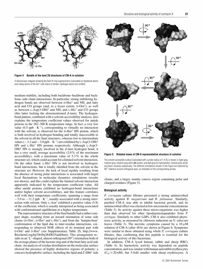
Figure 5 Rotated views of CM-A representative structure in solution
The solvent-accessible surface (calculated with a probe radius of 1.4 A) is shown in light grey,medium grey, medium grey with a � symbol, and dark grey for hydrophobic, neutral polar, acidicand basic residues respectively. The different orientations shown in the Figure are obtained by90◦ rotations around orthogonal axes, as indicated on the corresponding arrows.
chains, and a larger, mainly convex region containing polar andcharged residues (Figure 5).
Biological activity
P. corrugata culture filtrates presented a strong antimicrobialactivity against B. megaterium and R. pilimanae. Similarly,purified CM-A was able to inhibit bacterial growth, and itsantimicrobial effect was elicited at low micromolar concentrations(Table 3). Its activity against these micro-organisms was higherthan that observed for other lipodepsinonapeptides from P.syringae. Similarly to other LDPs, CM-A also exhibited phyto-toxic activity, as measured by chlorosis induction on N. tabacumleaves (Table 3). The necrotic symptoms caused by a 10 µMsolution of CM-A (after 48 h) are shown in Figure 6. Symptomswere similar to those obtained using whole P. corrugata culturefiltrates, thus confirming that this peptide is essential for thebiological activity of this bacterium.
In addition, CM-A lysed human, rabbit and sheep RBCs(Table 4). Its haemolytic activity was dependent on peptideconcentration, and was similar with human and rabbit erythrocytes(C50 ≈ 20 nM), but 5-fold smaller with sheep erythrocytes. A
c© 2004 Biochemical Society

32 A. Scaloni and others
Table 3 Antimicrobial activity of CM-A against B. megaterium and Rh.pilimanae and phytotoxic activity on N. tabacum
Measurements were performed in comparison with SR-E and ST. MIC, minimal inhibitoryconcentration.
Specific activity (units/µg) MIC (µM) Time of chlorosisB. megaterium R. pilimanae B. megaterium R. pilimanae induction (h)
CM-A 3.0 8.0 3 3 24SR-E 1.3 5.0 5 5 48ST 1.0 2.5 9 9 72
Figure 6 Toxicity of P. corrugata and purified CM-A to host plant leaves
Symptoms shown by tobacco leaves 48 h after infection. Left of the central leaf vein, P. corrugatainfection; right, pure CM-A (10 µM) injection.
similar trend was observed for the other lipodepsinonapeptidesfrom P. syringae that were tested. In addition, comparativemeasurements showed that CM-A was more active than SR-Eand ST (Table 4) [19,42].
CM-A was also able to interact with and permeabilize artificialmembranes. The leakage of calcein was dependent on peptideconcentration and liposome lipid composition (Figure 7 andTable 4). CM-A was far more active when a sterol was presentas a component of the liposome membranes (Table 4). Amongall sterols tested, cholesterol (mainly present in animal cell mem-branes) provided the highest sensitivity, followed by ergosterol (amajor component of fungal plasma membranes) and stigmasterol(the main sterol component of higher plant membranes). Thispreference for sterol-containing membranes has been alreadyreported for SR-E [19] and ST (Table 4), although animal cells arenot the natural targets of these LDPs. The presence of negativelycharged lipids (i.e. phosphatidic acid and phosphatidylinositol)did not improve, but rather hampered, CM-A activity. Moreover,SM made LUVs less prone to CM-A permeabilization. Theobserved reduced activity on SM-containing liposomes correlateswell with the lower haemolytic activity measured on sheep RBCs.In fact, sheep erythrocytes present a higher content of SM and areduced percentage of PC with respect to the other RBCs tested,
Table 4 Haemolytic activity of CM-A and its permeabilizing activity onvesicles with different lipid composition
Measurements were performed in comparison with syringomycin E and syringotoxin. Lipidmixtures are reported on a molar basis. n.d., not determined; Chol, cholesterol; Erg, ergosterol;Sti, stigmasterol.
1/C50* (106 M−1)
CM-A SR-E ST
Target cellsHuman RBC 50 +− 8 (11)† 2.0 +− 0.2 (9) 0.3 +− 0.1 (4)Rabbit RBC 40 (1) 0.8 +− 0.3 (2) 0.2 (1)Sheep RBC 10 +− 1 (3) 0.4 +− 0.1 (3) 0.07 +− 0.02 (3)
Target liposomesPC < 0.05 (1) 0.14 +− 0.02 (3) < 0.05 (1)PC:PA (1:1) < 0.05 (1) < 0.1 (1) < 0.05 (1)PC:PI (1:1) < 0.04 (1) < 0.1 (1) < 0.05 (1)
Asolectin 0.06 +− 0.01 (3) 0.03 (2) < 0.01 (3)PC:SM (1:1) 0.03 (1) 1.1 +− 0.1 (3) < 0.05 (1)PC:Chol (1:1) 1.7 +− 0.8 (2) 20 +− 8 (2) 0.2 (1)PC:Erg (1:1) 0.59 +− 0.03 (2) 2.5 +− 0.6 (3) 0.06 (1)PC:Sti (1:1) 0.21 +− 0.01 (2) 0.04 +− 0.02 (2) 0.02 (2)PC:Chol:SM (1:0.7:0.3) 1 (1) 3.3 +− 1.1 (2) n.d.PC:PA:Chol:SM 2.7 (1) 2.6 (1) 0.04 (1)
(0.9:0.1:0.7:0.3)PC:PA:Chol:SM 1.3 (1) 2.6 (1) 0.04 (1)
(0.1:0.9:0.7:0.3)
* Activity is expressed as 1/C50, where C50 is the micromolar concentration of LDP causing50 % of erythrocyte haemolysis or of calcein release.
† The error shown corresponds to S.D.; in parentheses the number of repetitions is shown.
Figure 7 Dose-dependence of CM-A-permeabilizing activity on liposomeswith different lipid composition
Calcein-loaded liposomes were exposed to different peptide concentrations, and the percentageof calcein released after 45 min is reported. The scale on the top x-axis shows the peptide-to-lipid molar ratio. Lines through the points are best-fit plots, fitted according to the statisticalmodel described in the text. Best values of fitting parameters were as follows: �, PC/choles-terol: K 1 = 5.8 +− 0.7 × 10² M−1, M = 7 +− 1, K 2 = 320 +− 30 × 10−3; �, PC/ergosterol:K 1 = 5.9 +− 0.7 × 10² M−1, M = 6 +− 1, K 2 = 80 +− 10 × 10−3; �, PC/stigmasterol: K 1 =5.5 +− 0.9 × 10² M−1, M = 7 +− 1, K 2 = 38 +− 9 × 10⁻³; �, asolectin: K 1 = 5.1 +− 0.7 ×10² M−1, M = 7 +− 1, K 2 = 13 +− 2 × 10⁻³; �, PC/SM: K 1 = 4.4 +− 0.8 × 10² M−1; M =7 +− 1; K 2 = 4 +− 1 × 10⁻³; �, PC: K 1 = 4.2 +− 1.0 × 10² M−1; M = 6 +− 1; K 2 = 2.5 +−0.7 × 10⁻³.
c© 2004 Biochemical Society

Structure and biological activity of cormycin A 33
Figure 8 Determination of the critical micellar concentration of CM-A
Drops (1 µl volume) containing CM-A at the reported concentrations in pure water were gentlydeposited on a flat polypropylene surface, and the contact angles were measured. Points reportedare averages +− S.D. for at least three measurements. Critical micellar concentration is indicatedby the arrow, and corresponds to the sharp bend in the curve. Insets show pictures of the dropsat the highest (lower-right corner) and lowest (upper-left corner) CM-A concentrations.
whereas the content of the other phospholipids and cholesterol issimilar [43].
In general, the Parente-Rapaport model, based on a pore-forming mechanism, described our data well, at least for peptideconcentrations lower than 10 µM (Figure 7). According to thismodel, the difference in activity was related to a different degreeof reversibility of surface aggregation of the adsorbed monomers(K2). In fact, CM-A bound to PC/cholesterol LUVs showed a lowdegree of reversibility (K2 = 0.3); on the contrary, increased levelsof reversibility were observed for PC/ergosterol, PC/stigmasteroland other-composition vesicles. In all cases, the partition coef-ficient (K1) was almost constant. Similarly, the number of mono-mers constituting the active channel was always in the range of7 +− 1. All the parameters calculated for the different LUVs areshown in the legend to Figure 7.
To better understand the strongest interaction of CM-A withRBC, the critical micellar concentration of this peptide was me-asured. Normally, this parameter is inversely related to membrane-permeabilizing properties. Our experiments demonstrated thatCM-A presents a critical micellar concentration of 138 µM(Figure 8), a value approx. 10 times lower than that measuredfor SR-E [19]. Accordingly, CM-A is more hydrophobic than SR-E [17,19]; this result is consistent with the strongest propensity ofCM-A to partition into membranes. On the other hand, the abilityof CM-A to self-associate in solution into micelles could delaythe process of membrane penetration at concentrations above thecritical micellar concentration, providing a possible explanationof the saturating permeabilization effect observed in Figure 7.
The pore-forming mechanism of action for CM-A was wellsupported by planar lipid-bilayer experiments. Addition of CM-Ato a solution bathing a planar lipid bilayer made of 7:3 (mol/mol)1,2-diphytanoyl-sn-glycerophosphocholine/cholesterol increased
Figure 9 Formation of ion channels by CM-A in planar lipid bilayer
Addition of a 11 µM CM-A solution to the buffer bathing the membrane was able to causediscrete increases of the ionic current through the membrane. Each step-like current increasecorresponded to the opening of a single channel with an average conductance of 3.9 +− 0.6 pS.The closed state (C) and the different open levels (Oi) are marked with dashed lines. The iparameter corresponds to the number of single channels simultaneously open; its value rangedfrom 1–5. On the right, the occupation histogram of each level is shown. The applied voltagewas − 140 mV; the buffer used was 100 mM KCl/10 mM Mes, pH 6.0. Membrane compo-sition was 7:3 (mol/mol) 1,2-diphytanoyl-sn-glycerophosphocholine/cholesterol.
the membrane conductance, causing step-like current transitions(Figure 9). This behaviour is peculiar to the formation of struc-tured conducting pathways across the membrane (i.e. pores),rather than being due to an unspecific detergent-like activity. Fromthe representative trace reported in Figure 9, a single channel con-ductance of 3.9 +− 0.6 pS was measured.
DISCUSSION
The growing problem of microbial resistance to conventionalantibiotics and the need for new compounds with antibiotic pro-perties has stimulated interest in the development of antimicrobialand antifungal peptides as human therapeutics. The main hurdlesimpeding the use of antimicrobial peptides in systemic therapyinclude the high doses, close to toxic ones, at which they areeffective in animal models of infection. Nevertheless, a seriesof antimicrobial peptides are now under pharmaceutical develop-ment [44,45]. In this scenario, Pseudomonas LDPs have shownpromising features based on their effective antimicrobial andantifungal properties [46,47]. They can be potentially employed inmycoses and systemic fungi infections caused by Candida ssp.,Aspergillus fumigatus and Fusarium spp., which are emergingas problems for immunocompromised hosts [2]. In fact, theusefulness of azole-based drugs (e.g. fluconazole) against fungalinfections is limited by the lack of a broad spectrum of activity, orby severe toxicity phenomena. Moreover, LDPs have also showna potential use as biocontrol agents in agriculture [4]. In fact,Pseudomonas-based formulates are marketed in the U.S.A. forcitrus post-harvest treatment, and the association between LDPsand cell-wall-degrading enzymes has proved to be very effectiveagainst several plant pathogens [48].
In the present study, we have reported the structural andbiological properties of a new LDP, namely CM-A. CM-A sharesseveral structural features with other lipodepsinonapeptides fromP. syringae spp. (Figure 10). They all present a similar hydroxy-fatty acyl moiety at the N-terminus, a similar nonapeptide cyclicstructure closed by a lactone bond between conserved residues,three common peculiar amino acids, i.e. Z-Dhb, L-Asp(3-OH),and L-Thr(4-Cl), and two residues with a D-absolute configurationat not-fixed positions in the polypeptide chain. Main differencesbetween CM-A and the other P. syringae LDPs were ascribedto the nature of the amino acids occurring at positions 2–6,as well as to the molecular net charge. In fact, all P. syringae
c© 2004 Biochemical Society

34 A. Scaloni and others
Figure 10 Structural differences among PSs, SSs, ST, SRs and CM-A
The general structure of Pseudomonas lipodepsinonapeptides is reported on the top. Peptide net charge was calculated at pH 7.0. Orn, ornithine; Hse, homoserine; aThr, allo-threonine; Dab,2,4-diamino-butanoic acid; Dhb, 2,3-dehydro-2-amino-butanoic acid; Asp(3-OH), 3-hydroxyaspartic acid; Thr(4-Cl), 4-chlorothreonine. Charged residues are shown in bold.
lipodepsinonapeptides have a positive net charge as a result ofthe presence of two or three positively charged residues in theamino acid region 2–5 (Figure 10), whereas only a negativelycharged residue [Asp(3-OH)] is present in the conserved regionof amino acids 6–8. On the other hand, CM-A presents only asingle positively charged residue at neutral pH in these positions.In fact, the imidazole of its histidine residue has a measured pKa
value of 6.0, and an ionization strongly influenced by interactionswith proximal amino acids.
A detailed comparative analysis of the three-dimensionalstructure of CM-A with respect to those already published forST [9] and pseudomycin A [11] was not possible. In fact, allstructural analyses were performed under different experimentalconditions. However, although influences due to solvent effectshave to be considered, CM-A has a cyclic structure, withsome amino acids shielded from solvent by its hairpin-foldedlipid hydrophobic chain, similarly to what has already beenreported previously for ST, but different from that reportedfor pseudomycin A. Preliminary modelling studies on CM-Asuggest that this lipodepsipeptide should form oligomeric channelstructures containing six to eight monomers (results not shown),in agreement with biological data derived from lipid bilayerpermeabilization measurements (Figure 7).
Based on mechanism of action of antimicrobial cationic pep-tides from multicellular organisms towards bacterial membranes[44,45], two main structural features have been proposed tostrongly affect the biological activity of Pseudomonas spp. LDPs,i.e. the occurrence of basic residues and the presence of specifichydrophobic moieties [3,46]. It has been suggested that positivecharges should facilitate interaction with negatively charged phos-pholipids, allowing LDP to insert itself into membrane bilayersas a result of its amphipathic nature [47]. Although these featuresshould strongly promote initial peptide-membrane targeting, otherimportant and unknown physicochemical phenomena have to beconsidered to definitively understand the mechanism of action ofthese molecules. In fact, our comparative data on antimicrobial,phytotoxic and haemolytic properties of Pseudomonas LDPs(Tables 3 and 4), in contrast with the general structural criteriareported above, demonstrated that the peptide homologue withthe most reduced positive net charge (i.e. CM-A) presents thehighest biological activity. Moreover, measurements on artificialvesicles of different lipid composition demonstrated for all theseLDPs (Table 4) that permeabilization was not improved by the
presence of negatively charged phospholipids, hypothetical tar-gets of basic amino acids. On the other hand, LUV permeabiliz-ation was strongly increased by sterols (Table 4). These findingsare in perfect agreement with previous data on P. syringae lipo-depsinonapeptides, showing by a genetic approach that ergosterol,the main component of the fungal plasma membrane, and cho-lesterol or stigmasterol, are implicated in their mechanism ofaction [49]. On the other hand, this observation strongly contrastswith the decreased activity of antimicrobial cationic peptides frommulticellular organisms in sterol-containing membranes [50],again suggesting that modified models of interaction, with respectto those adopted for ribosomally synthesized peptides from multi-cellular organisms, have to be proposed to fully explain the activityof Pseudomonas LDPs towards fungal plasma membranes. Theseconsiderations correlate well with recent publications on otherbacterial peptides [47,50].
In conclusion, although not as active as other well-known phar-maceutical derivatives, Pseudomonas LDPs show potential as leadcompounds for the development of effective antifungal agents.They are fungicidal against important human pathogens, solublein aqueous solution, and they seem to have a unique mechanismof action. The structural and biological properties of CM-Areported in the present study provide new insights into previousinformation on structure–function relationships regarding thesemolecules. Promising new studies are now in progress to modifyspecific LDP chemical functionalities, in an attempt to enhancetheir antifungal activity and reduce their toxicity.
We thank Teuzin Wangdu and Claudio Della Volpe for their kind technical assistanceand scientific discussion in critical micellar concentration experiments. This work wasfinancially supported by grants from the Italian National Research Council, Istituto Trentinodi Cultura and Provincia Autonoma di Trento (PAT, project AGRIBIO) and PRIN 2002. Thebacterial strain IPVCT 10.3 was kindly supplied by Dr. G. Cirvilleri (University of Catania,Italy).
REFERENCES
1 Bender, C. L., Alarcon-Chaidez, F. and Gross, D. C. (1999) Pseudomonas syringaephytotoxins: mode of action, regulation and biosynthesis by peptide and polyketidesynthetases. Microbiol. Mol. Biol. Rev. 63, 266–292
2 De Lucca, A. J., Jacks, T. J., Takemoto, J. Y., Vinjard, B., Peter, J., Navarro, E. and Walsh,T. J. (1999) Fungal lethality, binding, and cytotoxicity of syringomycin-E.Antimicrob. Agents Chemother. 43, 371–373
c© 2004 Biochemical Society

Structure and biological activity of cormycin A 35
3 Zhang, Y.-Z., Sun, X., Zeckner, D. J., Sachs, R. K., Current, W. L., Jidda, J., Rodriguez, M.and Chen, S. H. (2001) Syntheses and antifungal activities of novel 3-amido bearingpseudomycin analogues. Bioorg. Med. Chem. Lett. 11, 903–907
4 Bull, C. T., Wadsworth, M. L., Sorensen, K. N., Takemoto, J. Y., Austin, R. K. andSmilanick, J. L. (1998) Syringomycin E produced by biological control agent controlsgreen mold on citrus. Biol. Control 12, 89–95
5 Bradbury, J. F. (1987) Pseudomonas corrugata. In: CMI Descriptions of PathogenicFungi and Bacteria, No, 893. Commonwealth Mycological Institute, CAB International,Kew, U.K.
6 Chun, W. and Leary, J. V. (1989) Novel toxin produced by Pseudomonas corrugata, thecausal agent of tomato pith necrosis. In Phytotoxins and Plant Pathogenesis (Graniti, A.,Durbin, R. D. and Ballio, A., eds.), pp. 93–116, Springer-Verlag, Berlin
7 Segre, A. L., Bachmann, R. C., Ballio, A., Bossa, F., Grgurina, I., Iacobellis, N. S.,Marino, G., Pucci, P., Simmaco, M. and Takemoto, J. Y. (1989) The structure ofsyringomycin A1, E and G. FEBS Lett. 255, 27–31
8 Ballio, A., Barra, D., Bossa, F., DeVay, J. E., Grgurina, I., Iacobellis, N. S., Marino, G.,Pucci, P., Simmaco, M. and Surico, G. (1988) Multiple forms of syringomycin.Physiol. Mol. Plant Pathol. 33, 493–496
9 Ballio, A., Bossa, F., Collina, A., Gallo, M., Iacobellis, N. S., Paci, M., Pucci, P.,Scaloni, A., Segre, A. L. and Simmaco, M. (1990) Structure of syringotoxin, a bioactivemetabolite of Pseudomonas syringae pv. syringae. FEBS Lett. 269, 377–380
10 Fukuchi, N., Isogai, A., Nakayama, S. and Suzuki, A. (1990) Structure of syringostatins Aand B, novel phytotoxins produced by Pseudomonas syringae pv. syringae isolated fromlilac blights. Tetrahedron Lett. 31, 695–698
11 Coiro, V. M., Segre, A. L., Di Nola, A., Paci, M., Grottesi, A., Veglia, G. and Ballio, A.(1998) Solution conformation of the Pseudomonas syringae MSU 16H phytotoxiclipodepsipeptide Pseudomycin A determined by computer simulations using distancegeometry and molecular dynamics from NMR data. Eur. J. Biochem. 257, 449–456
12 Nutkins, J. C., Mortishire-Smith, R. J., Packman, L. C., Brodey, C. L., Rainey, P. B.,Johnston, K. and Williams, D. H. (1991) Structure determination of tolaasin, anextracellular lipodepsipeptide produced by mushroom pathogen. Pseudomonas tolaasii.J. Am. Chem. Soc. 113, 2621–2627
13 Ballio, A., Barra, D., Bossa, F., Collina, A., Grgurina, I., Marino, G., Moneti, G.,Paci, M., Pucci, P., Segre, A. L. and Simmaco, M. (1991) Syringopeptins, newphytotoxic lipodepsipeptides from Pseudomonas syringae pv. syringae. FEBS Lett. 291,109–112
14 Ballio, A., Bossa, F., Camoni, L., Di Giorgio, D., Flamand, M. C., Marcite, H., Nitti, G.,Pucci, P. and Scaloni, A. (1996) Structure of fuscopeptins, phytotoxic metabolites fromPseudomonas fuscovaginae. FEBS Lett. 381, 213–216
15 Rinaldi, A. C. (2002) Antimicrobial peptides from amphibian skin: an expanding scenario.Curr. Opin. Chem. Biol. 6, 799–804
16 Che, F. S., Kasamo, K., Fukuchi, N., Isogai, A. and Suzuki, A. (1992) Bacterial phytotoxin,syringomycin, syringostatin and syringotoxin, exert their effect on the plasma membraneH+-ATPase partly by a detergent action and partly by inhibition of the enzyme.Physiol. Plant. 86, 518–524
17 Hutchinson, M. L., Tester, M. A. and Gross, D. C. (1995) Role of biosurfactant and ionchannel-forming activities of syringomycin in transmembrane ion flux: a model for themechanism of action in the plant-pathogen interaction. Mol. Plant Microbe Interact. 8,610–620
18 Fegin, A. M., Takemoto, J. Y., Wangspa, R., Teeter, J. H. and Brand, J. G. (1996)Properties of voltage-gated ion channel formed by syringomycin E in planar lipid bilayers.J. Membr. Biol. 149, 41–47
19 Dalla Serra, M., Fagiuoli, G., Nordera, P., Bernhart, I., Della Volpe, C., Di Giorgio, D.,Ballio, A. and Menestrina, G. (1999) The interaction of lipodepsipeptide toxins fromPseudomonas syringae pv. syringae with biological and model membranes:a comparison of syringotoxin, syringomycin and syringopeptins. Mol. PlantMicrobe Interact. 12, 391–400
20 Dalla Serra, M., Bernhart, I., Nordera, P., Di Giorgio, D., Ballio, A. and Menestrina, G.(1999) Conductive properties and gating of channels formed by syringopeptin 25A, abioactive lipodepsipeptide from Pseudomonas syringae pv. syringae in planar lipidmembrane. Mol. Plant Microbe Interact. 12, 401–409
21 Agner, G., Kaulin, I. A., Gurnev, P. A., Szabo, T., Schagina, L. V., Takemoto, J. Y. andBlasko, K. (2000) Membrane-permeabilizing activities of cyclic lipodepsipeptides,syringopeptin 22A and syringomycin E from Pseudomonas syringae pv. syringae inhuman red blood cells and in bilayer lipid membranes. Bioelectrochemistry 52, 161–167
22 Emanuele, M. C., Scaloni, A., Lavermicocca, P., Iacobellis, N. S., Camoni, L.,Di Giorgio, D., Pucci, P., Paci, M., Segre, A. L. and Ballio, A. (1998) Corpeptins, newbioactive lipodepsipeptide from culture of Pseudomonas corrugata. FEBS Lett. 433,317–320
23 Catara, V., Arnold, D., Cirvilleri, G. and Vivian, A. (2000) Specific oligonucleotide primersfor the rapid identification and detection of the agent of tomato pith necrosis,Pseudomonas corrugata, by PCR amplification: evidence for two distinct genomicgroups. Eur. J. Plant Pathol. 106, 753–762
24 Surico, G., Lavermicocca, P. and Iacobellis, N. S. (1988) Produzione di siringomicina esiringotossina in colture di Pseudomonas syringae pv. syringae. Phytopathol. Med. 27,163–168
25 Scaloni, A., Simmaco, M. and Bossa, F. (2003) D-amino acid analysis.Methods Mol. Biol. 211, 169–180
26 Guntert, P., Mumenthaler, C. and Wuthrich, K. (1997) Torsion angle dynamics for NMRstructure calculation with the new program DYANA. J. Mol. Biol. 273, 283–298
27 Case, D. A., Pearlman, D. A., Caldwell, J. W., Cheatham, III, T. E., Ross, W. S.,Simmerling, C. L., Darden, T. A., Merz, K. M., Stanton, R. V., Cheng, A. L. et al. (1999)AMBER 6, University of California, San Francisco
28 Wang, J., Cieplak, P. and Kollman, P. A. (2000) How well does a restrained electrostaticpotential (RESP) model perform in calculating conformational energies of organic andbiological molecules? J. Comput. Chem. 21, 1049–1074
29 Cornell, W. D., Cieplak, P., Bayly, C. I., Gould, I. R., Merz, Jr, K. M., Ferguson, D. M.,Spellmeyer, D. C., Fox, T., Caldwell, J. W. and Kollman, P. A. (1995) A second generationforce field for the simulation of proteins, nucleic acids, and organic molecules.J. Am. Chem. Soc. 117, 5179–5197
30 Schmidt, M. W., Baldridge, K. K., Boatz, J. A., Elbert, S. T., Gordon, M. S., Jensen, J. J.,Koseki, S., Matsunaga, N., Nguyen, K. A., Su, S., Windus, T. L., Dupuis, M. andMontgomery, J. A. (1993) The general atomic and molecular electronic structure system.J. Comput. Chem. 14, 1347–1363
31 Bayly, C. I., Cieplak, P., Cornell, W. D. and Kollman, P. A. (1993) A well-behavedelectrostatic potential based method using charge restraints for determining atom-centered charges: the RESP model. J. Phys. Chem. 97, 10269–10280
32 Cornell, W. D., Cieplak, P., Bayly, C. I. and Kollman, P. A. (1993) Application of RESPcharges to calculate conformational energies, hydrogen bond energies and free energiesof solvation. J. Am. Chem. Soc. 115, 9620–9625
33 Koradi, R., Billeter, M. and Wuthrich, K. (1996) MOLMOL: a program for display andanalysis of macromolecular structures. J. Mol. Graphics 14, 51–55
34 Lavermicocca, P., Iacobellis, N. S., Simmaco, M. and Graniti, A. (1997) Biologicalproperties and spectrum of activity of Pseudomonas syringae pv. syringae toxins.Physiol. Mol. Plant Pathol. 50, 129–140
35 Iacobellis, N. S., Lavermicocca, P., Grgurina, I., Simmaco, M. and Ballio, A. (1992)Phytotoxic properties of Pseudomonas syringae pv. syringae toxins. Physiol. Mol.Plant Pathol. 40, 107–116
36 Kayalar, C. and Duzgunes, N. (1986) Membrane action of colicin E1: detection by therelease of carboxyfluorescein and calcein from liposomes. Biochim. Biophys. Acta 860,51–56
37 Anderluh, G., Dalla Serra, M., Viero, G., Guella, G., Macek, P. and Menestrina, G. (2003)Pore formation by equinatoxin II, a eukaryotic protein toxin, occurs by induction ofnonlamellar lipid structures. J. Biol. Chem. 278, 45216–45223
38 Tejuca, M., Dalla Serra, M., Ferreras, M., Lanio, M. E. and Menestrina, G. (1996)Mechanism of membrane permeabilization by sticholysin I, a cytolysin isolated from thevenom of the sea anemone Stichodactyla helianthus. Biochemistry 35, 14947–14957
39 Parente, R. A., Nir, S. and Szoka, F. C. (1990) Mechanism of leakage of phospholipidvesicle contents induced by the peptide GALA. Biochemistry 29, 8720–8728
40 Rapaport, D., Peled, R., Nir, S. and Shai, Y. (1996) Reversible surface aggregation in poreformation by pardaxin. Biophys. J. 70, 2502–2512
41 Anzlovar, S., Dalla Serra, M., Dermastia, M. and Menestrina, G. (1998) Membranepermeabilizing activity of linusitin from flax seed. Mol. Plant Microbe Interact. 11,610–617
42 Dalla Serra, M., Menestrina, G., Carpaneto, A., Gambale, F., Fogliano, V. and Ballio, A.(2003) Molecular mechanism of action of syringopeptins, antifungal peptides fromPseudomonas syringae pv. syringae. In Pore-Forming Peptides and Protein Toxins, vol. 5(Menestrina, G., Dalla Serra, M. and Lazarovici, P., eds.; Cellular and MolecularMechanisms of Toxin Action, Lazarovici, P., series ed.), pp. 272–295, Taylor & FrancisGroup, London
43 Chapman, D. (1968) Biological Membranes, Physical Fact and Function. Academic Press,London, UK
44 Zasloff, M. (2002) Antimicrobial peptides of multicellular organisms. Nature (London)415, 389–395
45 Hancock, R. E. and Lehrer, R. (1998) Cationic peptides: a new source of antibiotics.Trends Biotechnol. 16, 82–88
c© 2004 Biochemical Society
36 A. Scaloni and others
46 Sorensen, K. N., Kim, K. H. and Takemoto, J. Y. (1996) In vitro antifungal andfungicidal activities and erythrocyte toxicities of cyclic lipodepsinonapeptidesproduced by Pseudomonas syringae pv. syringae. Antimicrob. Agent Chemother. 40,2710–2713
47 Zhang, L., Dhillon, P., Yan, H., Farmer, S. and Hancock, R. E. W. (2000) Interactions ofbacterial cationic peptide antibiotics with outer and cytoplasmic membranes ofPseudomonas aeruginosa. Antimicrob. Agent Chemother. 44, 3317–3321
48 Fogliano, V., Ballio, A., Gallo, M., Woo, S., Scala, F. and Lorito, M. (2002) Pseudomonaslipodepsipeptides and fungal cell wall degrading enzymes are synergistic in the inhibitionof fungal growth. Mol. Plant Microbe Interact. 15, 323–330
49 Wangspa, R. and Takemoto, J. Y. (1998) Role of ergosterol in growth inhibition ofSaccharomyces cerevisiae by syringomycin E. FEMS Microbiol. Lett. 167, 215–220
50 Matsuzaki, K. (1999) Why and how are peptide–lipid interactions utilized for self-defense? Magainins and tachyplesins as archetypes. Biochim. Biophys. Acta 1462, 1–10
Received 15 March 2004/14 June 2004; accepted 15 June 2004Published as BJ Immediate Publication 15 June 2004, DOI 10.1042/BJ20040422
c© 2004 Biochemical Society