Isolation of GUS marker lines for genes expressed in Arabidopsis endosperm, embryo and maternal...
12
DOI: 10.1093/jxb/erg031 RESEARCH PAPER Isolation of GUS marker lines for genes expressed in Arabidopsis endosperm, embryo and maternal tissues Biljana Stangeland 1,5 , Zhian Salehian 1 , Reidunn Aalen 2 , Abul Mandal 3 and Odd-Arne Olsen 1,4 1 Plant Molecular Biology Laboratory, Department of Chemistry and Biotechnology, Agricultural University of Norway, PO Box 5040, N-1432 Aas, Norway 2 Division of Molecular Biology, University of Oslo, Oslo, Norway 3 University of Sko ¨vde, Sweden Received 19 June 2002; Accepted 11 September 2002 Abstract In order to identify marker lines expressing GUS in various endosperm compartments and at different developmental stages, a collection of Arabidopsis thaliana (L.) Heynh. promoter trap lines were screened. The screen identified 16 lines displaying GUS-reporter gene expression in the endosperm, embryo and other seed organs. The distinctive pat- terns of GUS expression in these lines provide molecular markers for most cell compartments in the endosperm of Arabidopsis seeds at all devel- opmental stages, and represent a valuable research tool for characterizing present and future Arabidopsis seed mutants. GUS expression pat- terns of these 16 lines are presented here. One line showed chalazal endosperm-specific GUS activity at the heart stage of embryo development. In six lines embryo-specific GUS activity was detected. Six lines exhibited GUS activity predom- inantly in the endosperm and embryo while two lines showed strong GUS activity in all seed organs. In one line GUS activity was detected in integuments and syncytial endosperm, while the GUS activity at the cotyledonary stage of the embryo was seed coat-specific. In addition, two funiculus markers and two silique markers expressed in the abscission zone and the guard cells are also presented. Key words: Arabidopsis, clearing, embryo, endosperm, GUS, seed, promoter, trapping. Introduction Phenotypic analysis of mutant plants, derived either from forward or reverse genetics approaches, is one of the richest sources for information on gene function. However, the task of unravelling the function of a gene is not always straightforward. In particular, seed lethal mutants in Arabidopsis can be challenging, and the stage of develop- mental arrest can be difficult to determine. Examples illustrating this include mutants affected in cytokinesis (Assaad et al., 2001; Lauber et al., 1997; Lukowitz et al., 1996; Strompen et al., 2002) and early endosperm development (Liu and Meinke, 1998; Mayer et al., 1999; Tzafrir et al., 2002). In such studies, molecular markers for cell types and tissues within the embryo and endosperm are very useful; the difference in marker expression between mutant and wild-type plants being able to detect differ- ences that could otherwise have remained undetected. An example that illustrates the usefulness of such molecular markers is the analysis of pilz mutants (Mayer et al., 1999). In this study, expression patterns of several cell-cycle- dependent GUS reporters were followed during the development of pilz mutant embryos. Other examples of marker line studies include examinations of eight emb mutants, where the seed-specific promoters ABI3, 2S1 and Em1 were used to drive the GUS reporter gene (gusA) in a study of the effects of morphogenesis on storage proteins and Lea gene expression (Devic et al., 1996). Several laboratories have reported the identification of molecular marker lines generated by promoter trapping, including the lines POLARIS (embryonic and seedling root tip), EXORDIUM (cotyledons and shoot and root 4 Present address: Pioneer Hi-Bred International, A DuPont Company, Iowa 7300 NW 62nd Avenue, Johnston, IA 50131–1004, USA. 5 To whom correspondence should be addressed. Fax: +47 64 94 77 20. E-mail: [email protected] Journal of Experimental Botany, Vol. 54, No. 381, ª Society for Experimental Biology 2003; all rights reserved Journal of Experimental Botany, Vol. 54, No. 381, pp. 279–290, January 2003 by guest on September 25, 2013 http://jxb.oxfordjournals.org/ Downloaded from by guest on September 25, 2013 http://jxb.oxfordjournals.org/ Downloaded from by guest on September 25, 2013 http://jxb.oxfordjournals.org/ Downloaded from by guest on September 25, 2013 http://jxb.oxfordjournals.org/ Downloaded from by guest on September 25, 2013 http://jxb.oxfordjournals.org/ Downloaded from by guest on September 25, 2013 http://jxb.oxfordjournals.org/ Downloaded from by guest on September 25, 2013 http://jxb.oxfordjournals.org/ Downloaded from by guest on September 25, 2013 http://jxb.oxfordjournals.org/ Downloaded from by guest on September 25, 2013 http://jxb.oxfordjournals.org/ Downloaded from by guest on September 25, 2013 http://jxb.oxfordjournals.org/ Downloaded from by guest on September 25, 2013 http://jxb.oxfordjournals.org/ Downloaded from by guest on September 25, 2013 http://jxb.oxfordjournals.org/ Downloaded from
Transcript of Isolation of GUS marker lines for genes expressed in Arabidopsis endosperm, embryo and maternal...

DOI: 10.1093/jxb/erg031
RESEARCH PAPER
Isolation of GUS marker lines for genes expressed inArabidopsis endosperm, embryo and maternal tissues
Biljana Stangeland1,5, Zhian Salehian1, Reidunn Aalen2, Abul Mandal3 and Odd-Arne Olsen1,4
1Plant Molecular Biology Laboratory, Department of Chemistry and Biotechnology, Agricultural University ofNorway, PO Box 5040, N-1432 Aas, Norway2 Division of Molecular Biology, University of Oslo, Oslo, Norway3 University of SkoÈvde, Sweden
Received 19 June 2002; Accepted 11 September 2002
Abstract
In order to identify marker lines expressing GUS in
various endosperm compartments and at different
developmental stages, a collection of Arabidopsis
thaliana (L.) Heynh. promoter trap lines were
screened. The screen identi®ed 16 lines displaying
GUS-reporter gene expression in the endosperm,
embryo and other seed organs. The distinctive pat-
terns of GUS expression in these lines provide
molecular markers for most cell compartments in
the endosperm of Arabidopsis seeds at all devel-
opmental stages, and represent a valuable research
tool for characterizing present and future
Arabidopsis seed mutants. GUS expression pat-
terns of these 16 lines are presented here. One
line showed chalazal endosperm-speci®c GUS
activity at the heart stage of embryo development.
In six lines embryo-speci®c GUS activity was
detected. Six lines exhibited GUS activity predom-
inantly in the endosperm and embryo while two
lines showed strong GUS activity in all seed
organs. In one line GUS activity was detected in
integuments and syncytial endosperm, while the
GUS activity at the cotyledonary stage of the
embryo was seed coat-speci®c. In addition, two
funiculus markers and two silique markers
expressed in the abscission zone and the guard
cells are also presented.
Key words: Arabidopsis, clearing, embryo, endosperm, GUS,
seed, promoter, trapping.
Introduction
Phenotypic analysis of mutant plants, derived either fromforward or reverse genetics approaches, is one of therichest sources for information on gene function. However,the task of unravelling the function of a gene is not alwaysstraightforward. In particular, seed lethal mutants inArabidopsis can be challenging, and the stage of develop-mental arrest can be dif®cult to determine. Examplesillustrating this include mutants affected in cytokinesis(Assaad et al., 2001; Lauber et al., 1997; Lukowitz et al.,1996; Strompen et al., 2002) and early endospermdevelopment (Liu and Meinke, 1998; Mayer et al., 1999;Tzafrir et al., 2002). In such studies, molecular markers forcell types and tissues within the embryo and endosperm arevery useful; the difference in marker expression betweenmutant and wild-type plants being able to detect differ-ences that could otherwise have remained undetected. Anexample that illustrates the usefulness of such molecularmarkers is the analysis of pilz mutants (Mayer et al., 1999).In this study, expression patterns of several cell-cycle-dependent GUS reporters were followed during thedevelopment of pilz mutant embryos. Other examples ofmarker line studies include examinations of eight embmutants, where the seed-speci®c promoters ABI3, 2S1 andEm1 were used to drive the GUS reporter gene (gusA) in astudy of the effects of morphogenesis on storage proteinsand Lea gene expression (Devic et al., 1996).
Several laboratories have reported the identi®cation ofmolecular marker lines generated by promoter trapping,including the lines POLARIS (embryonic and seedlingroot tip), EXORDIUM (cotyledons and shoot and root
4 Present address: Pioneer Hi-Bred International, A DuPont Company, Iowa 7300 NW 62nd Avenue, Johnston, IA 50131±1004, USA.5 To whom correspondence should be addressed. Fax: +47 64 94 77 20. E-mail: [email protected]
Journal of Experimental Botany, Vol. 54, No. 381, ã Society for Experimental Biology 2003; all rights reserved
Journal of Experimental Botany, Vol. 54, No. 381, pp. 279±290, January 2003
by guest on September 25, 2013
http://jxb.oxfordjournals.org/D
ownloaded from
by guest on Septem
ber 25, 2013http://jxb.oxfordjournals.org/
Dow
nloaded from
by guest on September 25, 2013
http://jxb.oxfordjournals.org/D
ownloaded from
by guest on Septem
ber 25, 2013http://jxb.oxfordjournals.org/
Dow
nloaded from
by guest on September 25, 2013
http://jxb.oxfordjournals.org/D
ownloaded from
by guest on Septem
ber 25, 2013http://jxb.oxfordjournals.org/
Dow
nloaded from
by guest on September 25, 2013
http://jxb.oxfordjournals.org/D
ownloaded from
by guest on Septem
ber 25, 2013http://jxb.oxfordjournals.org/
Dow
nloaded from
by guest on September 25, 2013
http://jxb.oxfordjournals.org/D
ownloaded from
by guest on Septem
ber 25, 2013http://jxb.oxfordjournals.org/
Dow
nloaded from
by guest on September 25, 2013
http://jxb.oxfordjournals.org/D
ownloaded from
by guest on Septem
ber 25, 2013http://jxb.oxfordjournals.org/
Dow
nloaded from

apices) and COLUMELLA (root cap), which were used inthe characterization of gnom and hydra mutants (Toppingand Lindsey, 1997). In these studies, insertional mutagen-esis and promoter trapping were combined in order todissect polar development in Arabidopsis embryos andseedlings. Characterization of the topless mutation, caus-ing shoot to root transformation during embryogenesis,was carried out by crossing the mutants to the enhancertrap lines J1092 and LENNY (root speci®c reporter), andthe auxin-responsive reporter DR5 made by GUS promoterfusion (Long et al., 2001; JA Long, personal communica-tion). This analysis showed that the LENNY reporter wasexpressed at both poles in tpl embryos while DR5 was mis-expressed.
An alternative approach to the use of marker lines is insitu hybridization, in which the patterns of expression ofknown genes are studied. In this way, an analysis of ®ve newMADS-box genes from Arabidopsis, termed AGL (aga-mous like), revealed that AGL18 and AGL16 are speci®c-ally expressed in the endosperm and in guard cells andtrichomes, respectively (Alvarez-Buylla et al., 2000). Inaddition, several recent reports utilize this technique in theanalysis of embryo and endosperm mutants of the pilz group(Mayer et al., 1999), knolle (Volker et al., 2001) andprolifera (Holding and Springer, 2002). Although highlyef®cient, this technique is time-consuming and requiresknowledge of the gene product. In addition, in theArabidopsis endosperm, identi®cation of stage and com-partment-speci®c developmental markers through differ-ential screening is hampered by the small size of theArabidopsis seed. In particular, the minute size and fragilityof the syncytial endosperm do not allow dissection.
To date, few attempts have been made systematically toidentify marker lines with GUS preferred pattern ofexpression in the Arabidopsis endosperm, although severalendosperm marker lines have been derived from promoterand enhancer trapping experiments designed to detectGUS activity in embryos (Topping et al., 1994) and ovulesin early seed development (Vielle-Calzada et al., 2000).For example, two lines, 17G2 and 19C1, were labelled asendosperm-speci®c in the promoter trap screening formolecular markers of embryogenesis. Using enhancertrapping, three endosperm-speci®c genes were mentioned,but their patterns of GUS expression have not beenpublished (Vielle-Calzada et al., 2000). Studies ofArabidopsis mutants that exhibit autonomous endospermdevelopment in the absence of fertilization medea/®s1(fertilization independent seed), ®s/®s2 and ®e (fertiliza-tion independent endosperm)/®s3 have also led to theidenti®cation of endosperm-speci®c markers (Chaudhuryet al., 1997; Grossniklaus et al., 1998; Luo et al. 2000;Ohad et al., 1996). The endosperm-speci®c GFP reporterKS117 isolated by enhancer trapping showed expressionpatterns similar to FIS1/MEA and FIS2 (Sùrensen et al.,2001). This marker line features enhancer trapping of the
FORMIN gene that most likely mediates actin polymeriza-tion (MB Sùrensen, personal communication).
The experiments described in the present paper wereinitiated to identify additional molecular marker lines thatcomplemented existing marker lines. In particular, themarkers were useful for the studies of Arabidopsisendosperm (Brown et al., 1999). To achieve this goal, acollection of Arabidopsis promoter trap lines was system-atically screened (Mandal et al., 1995) using an improvedclearing method for GUS staining of the seed compart-ments (Stangeland and Salehian, 2002). The frequency ofGUS positive lines obtained in this experiment accordswell with that reported by Topping et al. (1994), 13% and16%, respectively, although the two experiments may notbe directly comparable due to the use of different clearingtechniques.
Materials and methods
Plant material and growth conditions
In order to isolate molecular markers for endosperm cells and tissuesin Arabidopsis, a collection of T-DNA promoter trap lines wasscreened. These transgenic lines contained the pMHA2 vector,harbouring a promoterless GUS reporter gene at the right T-DNAborder (Mandal et al., 1995). The 5¢ end of the gusA gene was placedapproximately 40 bp from the right end of the T-DNA (Mandal et al.,1995). The plant transformation vector contained a pnos-nptII plantselectable marker gene. T-DNA lines were generated in the C24ecotype of Arabidopsis thaliana by root transformation. Progenyseeds were selected on MS medium (Murashige and Skoog, 1962)containing 50 mg l±1 kanamycin. Plants were transferred to soil andgrown at 2363 °C and 70% humidity, under 40 W cool white¯uorescent lights (16/8 h light/dark). Between six and nine plantsfrom each family were assayed for GUS activity. Up to sixsubsequent generations were regularly screened for GUS activity inseeds.
GUS assay and image processing
GUS assay (Jefferson et al., 1987) was performed on dissected, non-®xed Arabidopsis siliques at different developmental stages.Harvested siliques were slit twice longitudinally prior to the GUSassay and clearing (Stangeland and Salehian, 2002).
Results
The microscopy screen of 309 independent lines from thepMHA2 promoter trap collection (Mandal et al., 1995) wascarried out on ¯owers, siliques and different vegetativeorgans that had been stained for GUS expression. Siliqueswere harvested from 0±10 DAP (days after pollination)and 16 lines were identi®ed showing stable Mendelianinheritance of the GUS activity in seeds through severalgenerations. GUS expression patterns in each of these linesbehaved as single Mendelian traits and are presented inTable 1. Among these lines, one line showed staining onlyin the endosperm at a speci®c developmental stage(GNOCCHI1). Six lines showed embryo-speci®c activity(LINGUINE1-6), six in the endosperm, embryo and seed
280 Stangeland et al.

attachment point (SENAPE1-6), one in the endosperm,integuments and seed coat (PESCE1), and two in all seedorgans (STINCO1, 2). In one line, GUS activity wasobserved in hydathodes of leaves, embryos and endosperm(SENAPE5). Of these 16 lines, 80% had detectable GUSactivity in siliques and 60% in ¯owers. In addition, GUSactivity in vegetative organs, especially, roots was notuncommon (Table 1).
Additionally, four silique markers were identi®edincluding an abscission zone marker (BASIL), a guardcell marker (PESTO) and two lines that expressed gusA inthe funiculus (FUSILLI).
Endosperm expressed GUS reporters
The endosperm preferred GUS reporter line namedGNOCCHI1 (GUS in nodule, chalazal chamber andinteguments) showed speci®c staining of the chalazal
endosperm and the adjacent integument tissue. StrongGUS activity was detected in the upper part of the siliques,bearing seeds at the globular stage of embryo develop-ment. At this stage, GUS staining was detected in thechalazal endosperm and in the embryo. At the heart stageof embryo development, GUS activity was detectedexclusively in the chalazal endosperm (Fig. 1A). Inaddition to consistent staining in the chalazal chamber(ChC), staining of the surrounding tissues varied and wasabsent or distributed in a gradient-like fashion (Fig. 1A), ascon®rmed by studies of hundreds of seeds of this type. Thisgave the impression that the primary site of GUS activitywas in the chalazal cyst and the nodule, and that theadditional staining in the surrounding tissues might becaused by diffusion of the CIBr-indigo stain. Staining ofthe heart stage embryo was detected only in intensivelystained seeds, suggesting that diffusion or uptake of CIBr-indigo produced in the ChC could be the reason for embryostaining at this stage. However, this is very dif®cult toverify. Whether or not gusA is truly expressed in theembryo at the globular stage of development, theGNOCCHI1 marker is speci®c for chalazal endosperm atthe heart stage of embryo development. At later develop-mental stages (torpedo-walking stick) GUS activity couldnot be detected in seeds and siliques. Weak GUS activitywas detected in the ®niculus at the heart stage of embryodevelopment.
The lines PESCE1 and STINCO2 (see below) showedespecially strong GUS activity in the endosperm nodule(Fig. 2A), as well as activity in the whole seed. In a fractionof the seeds of the PESCE1 line, GUS activity wasdetected exclusively in the nodule, micropylar endospermand integuments (Fig. 2A, B).
Reporter lines with GUS activity in embryos
The embryo marker lines identi®ed in this study weretermed LINGUINE (line with GUS in embryo). Within thecollection of six LINGUINE lines, GUS activity waspresent either only in the embryos (LINGUINE1), or alsoin trace amounts in the endosperm at an early develop-mental stage (LINGUINE2, LINGUINE6), or at a latedevelopmental stage (LINGUINE3, LINGUINE5)(Table 1; Figs 1, 2).
In LINGUINE1, visible GUS expression was detected atthe heart stage of embryo development. Staining wasstrongest in the basal embryo proper, corresponding to thecells that are developing into the hypocotyl and the root tip(Fig. 1B, C). At later developmental stages, GUS stainingwas still present in the same area of the torpedo stageembryo (Fig. 1B), but became more evenly distributed inthe hypocotyl at the cotyledonary stage of embryodevelopment (Fig. 1C). GUS activity was not detected inthe endosperm.
LINGUINE2 showed GUS activity from the heart stageonwards. Some weak and diffuse activity was detected in
Table 1. Summary of GUS expression patterns of marker linesin Arabidopsis reproductive and vegetative organs
Seed organs are indicated as endosperm, embryo, integuments, seedcoat and seed attachment point. Parts of the siliques are indicated asfuniculus, silique wall, silique vascular tissue (SVT) and theabscission zone of the silique (AZ). GUS speci®c activities in stomata(st) and aleurone layer (al) are indicated.
Isolation of GUS marker lines 281
the endosperm at earlier stages. GUS activity was alsodetected in the hypocotyls and cotyledons of the walkingstick stage embryos (Fig. 1D).
In LINGUINE3 (Fig. 1E, F), GUS activity was detect-able in the central area of the embryo proper at the globular
and heart stage. From late heart to the torpedo stage ofembryo development, GUS activity could be detected inthe whole embryo, with the strongest activity in the basalpart of the embryo proper and weakest in the surroundingendosperm (Fig. 1E). GUS activity could also be detected
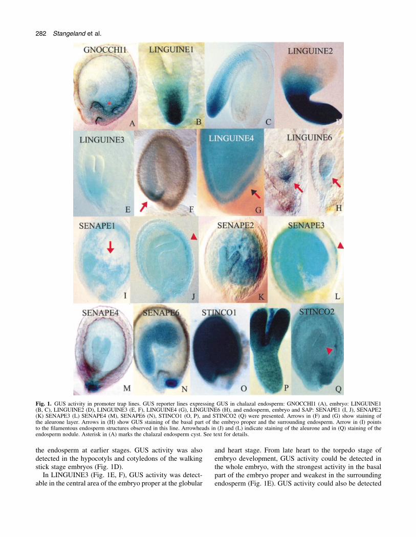
Fig. 1. GUS activity in promoter trap lines. GUS reporter lines expressing GUS in chalazal endosperm: GNOCCHI1 (A), embryo: LINGUINE1(B, C), LINGUINE2 (D), LINGUINE3 (E, F), LINGUINE4 (G), LINGUINE6 (H), and endosperm, embryo and SAP: SENAPE1 (I, J), SENAPE2(K) SENAPE3 (L) SENAPE4 (M), SENAPE6 (N), STINCO1 (O, P), and STINCO2 (Q) were presented. Arrows in (F) and (G) show staining ofthe aleurone layer. Arrows in (H) show GUS staining of the basal part of the embryo proper and the surrounding endosperm. Arrow in (I) pointsto the ®lamentous endosperm structures observed in this line. Arrowheads in (J) and (L) indicate staining of the aleurone and in (Q) staining of theendosperm nodule. Asterisk in (A) marks the chalazal endosperm cyst. See text for details.
282 Stangeland et al.

in the few endosperm cells adjacent to the cotyledons (notshown). At the late torpedo stage, GUS activity could bedetected in the embryo, suspensor, and the thin epidermallayer underneath the embryo corresponding to the aleuronelayer (Fig. 1F).
In LINGUINE4, embryo and aleurone GUS expressionpatterns at the later stages were similar to LINGUINE3(Fig. 1G). The GUS marker was also expressed in siliquesand petals (Fig. 2M). Line LINGUINE5 showed GUSactivity in the embryo, funiculus and siliques. Dissected
Fig. 2. GUS activity in promoter trap lines (continued). GUS staining in the marker line PESCE1 expressing GUS in integuments and endospermat early stages (A, B), and exclusively in seed coat at later stages (C, E) is presented. In markers FUSILLI1 (F) and FUSILLI2 (G) GUS activity isdetected in funiculus and silique. SENAPE5 is expressed in leaf hydathodes (H±J) and seeds (L). LINGUINE4 (K) is expressed in seeds (Fig. 1G)and in petals (M). Markers PESTO (L, M) and BASIL (N) are expressed predominantly in pedicel stomata (N, O) and in the abscission zone ofthe ¯oral organs (P) respectively. Arrows in (A) and (B) indicate GUS staining of the endosperm nodule. Arrows in (D) and (E) point at the GUSstained columella, central area of the seed coat cells. Asterisk in (C) indicates late stage embryo that is not GUS stained.
Isolation of GUS marker lines 283

embryos from this line resembled embryos presented inFig 1P, showing uniform GUS staining of the embryo. InLINGUINE6, very weak activity was present in the basalpart of the embryo from the globular to the walking stickstage of development (Fig. 1H), whereas strong activitywas present in the abscission zone of the siliques (Fig. 2K).Very weak activity was localized in the endosperm tissuesurrounding the base of the embryo proper (Fig. 1H).
Other seed GUS reporters
In several of the lines studied, GUS activity was localizedpredominantly in the endosperm and embryo, but was alsodetectable in maternal tissues, including the seed attach-ment point and the integuments. This group of markers wastermed SENAPE (staining in endosperm, attachment pointand embryo). In SENAPE1, GUS activity was strongest inthe endosperm and embryo, though weak activity in theinteguments at later developmental stages could also beobserved (Fig. 1I, J). In this line, GUS activity wasdetected shortly after fertilization in the central cell, in theendosperm, as well as in the embryo at the globular stage.At the heart stage of embryo development, ®lament-likepatterns could be observed in the central endospermchamber (Fig. 1I). Detailed analysis showed that thesepatterns represent intensively stained endosperm nuclei.Occasionally, GUS activity in the embryo was masked byblue staining in the endosperm, and was dif®cult toresolve. Later in development, GUS activity was detect-able in torpedo stage embryos and the aleurone layer(Fig. 1J). In SENAPE1, GUS activity was also present inpetals and in transport tissues and the abscission zone ofthe silique.
In SENAPE2, GUS activity could be detected in siliques(not shown) and seeds (Fig. 1K). At the globular and heartstages of embryo development, GUS activity was pre-dominantly localized to the peripheral endosperm withsome activity in the embryos. At the late heart/earlytorpedo stage, GUS activity could be detected both inembryos and in the endosperm (Fig. 1K), staining beingespecially strong in the chalazal chamber (Fig. 1K). At thewalking stick and cotyledonary stages of embryo devel-opment, staining of the embryo was especially strong in theprotoderm, the shoot apical meristem (SAM), and theembryonic root (not shown). In vegetative organs, none oronly weak GUS activity was detected (not shown).
In the marker line SENAPE3, intensive GUS stainingwas detected in siliques and seeds (Fig. 1L). At the heartstages of embryo development, very weak GUS activitywas detected in the embryo. At the torpedo and walkingstick stages, embryo staining became intense (Fig. 1L). Atthis stage, GUS activity could be detected in the cellularendosperm and the epidermal layer corresponding to thealeurone layer (Fig. 1L, arrowhead). In integuments, GUSactivity was either very weak or absent. Intense GUSactivity was also present in the cotyledonary embryo,
seedlings and ¯ower. In SENAPE4, GUS activity waspresent in the endosperm, embryo and seed attachmentpoints (Fig. 1M).
In seeds of the line SENAPE5, GUS activity wasdetected in the embryo, endosperm, funiculus, andhydathodes (Fig. 2H±J, L), and also in the area betweenleaf veins (Fig. 2J). While the GUS staining of thehydathodes (Fig. 2H, I) was strong and stable, GUSactivity of the embryo and endosperm showed variableintensity (Fig. 2L). In SENAPE6, GUS activity wasstrongest in the embryo, endosperm and seed attachmentpoints (Fig. 1N).
In other lines, including STINCO (staining intense andconstitutive) 1 and 2, GUS activity was ubiquitous. InSTINCO1 the GUS reporter was strongly expressed in allvegetative and reproductive tissues (Fig. 1O, P). Indissected embryos the strongest GUS activity was detectedin cotyledons (Fig. 1P). GUS activity in STINCO2 wasalso intense and ubiquitous (Fig. 1Q). Particularly strongactivity was detected in the endosperm nodule (Fig. 1Q,arrowhead), thus resembling marker PESCE1 (Fig. 2A).Later in seed development, GUS activity was detectedpredominantly in embryos.
Intense GUS staining, resembling the STINCO lines,was also found in PESCE1 (preferentially expressed inseed coat and endosperm). However, at later developmen-tal stages this marker was seed-coat-speci®c (Fig. 2C±E),in contrast to the STINCO lines. GUS staining could bedetected in the integuments immediately after fertilization.At all later stages, strong GUS activity was detected in theinteguments and seed coat (Fig. 2A±E). At the globularand heart stages of embryo development, GUS activitycould also be detected in the endosperm, but was absent orweak in embryos (Fig. 1A±C). The strong GUS staining ofthe chalazal endosperm nodule was especially noticeable(Fig. 1A, B, arrows). Stained endosperm nodules wereattached to the chalazal integument wall, but their localposition varied. A fraction of seeds showed GUS activityonly in the nodule, micropylar chamber and the chalazalproliferating tissue (Fig. 2B). It is thought that the GUSexpression patterns in this line could, at least partially, becaused by the diffusion of the CIBr-indigo stain from thechalazal endosperm to the adjacent seed and maternaltissues. At later stages, GUS staining was localized to theseed coat, precisely to the cytoplasm and cell walls of themucilage cells (Fig. 2D, E, arrows). The morphology ofthis cell layer has been described in detail (Western et al.,2000; Windsor et al., 2000). Based on the informationfrom these reports it was concluded that the GUS stain islocalized to the central elevation, columella, harbouringthe cytoplasm of these cells (Fig. 2D, E). Dissectedembryos did not show GUS staining (Fig. 2C, asterisk).GUS activity in siliques was detected at the stages earlierthan the heart-torpedo stage of embryo development. Atlater stages GUS activity was detected only in seeds.
284 Stangeland et al.

Silique GUS reporter lines
In the lines FUSILLI 1 and 2 (funiculus and siliques line),GUS activity was detected in the funiculus, SAP andvascular tissue of the silique (Fig. 2F, G). In BASIL (basisof the silique), GUS activity was detected only in the basalpart of the siliques that corresponds to the zone ofabscission (Fig. 2P). In PESTO (pedicel stomata), GUSactivity was localized to the guard cells (Fig. 2N, O),silique vascular tissue, auxiliary meristems, and thehypocotyls of seedlings. Staining of guard cells wasespecially strong in pedicels and stems. In leaves, eithernone or only weak staining of stomata, often in the vicinityof vascular tissue, was observed.
Several marker lines show imprinting but none of thelines showed visible phenotypes
Even though weak endosperm defects were occasionallyobserved in the original isolates of the lines describedabove, mutant seed phenotypes were not identi®ed. Bycounting seeds from 6±9 plants per line; 5±10 siliques (upto 500 seeds) per plant, frequencies of non-pollinated andaborted seeds were estimated to be similar in transgeniclines and the wild-type plants growing in the same growthchamber. between 0.5% and 1% aborted seeds wereregistered in all lines. The number of non-pollinated ovulesper silique varied a lot, but was, on average, about a fewper cent. All marker lines showed stable patterns ofinheritance of the GUS expression pattern over severalgenerations. Neither lethality, nor a de®ciency of homo-zygous genotypes was observed.
In order to characterize inheritance patterns of mol-ecular markers further, reciprocal crosses were also carriedout involving the marker lines and wild-type plants(Table 2). In this analysis, wild-type ¯owers were pollin-ated with pollen from the marker lines SENAPE1,SENAPE2, GNOCCHI1, and PESCE1. Genetic crosseswere performed by pollinating 15±20 wild-type ¯owers pertransgenic line. Directly after the genetic cross 2±3 siliques
were harvested every day and seeds were tested for GUSactivity (20±30 seeds per line daily). seeds were analysedfrom the very early stages up to and including thecotyledonary stage of embryo development (7 DAP, daysafter pollination). In total, up to 100±200 seeds per markerline (1±7 DAP) were tested for GUS activity. In parallel, aGUS test was performed on self-pollinated seeds using thesame assay chemicals. As shown in Table 2, GUS activitywas never detected in the out-cross progeny, indicatingthat GUS expression was either extremely low or com-pletely absent. These data suggest that these genes mightbe imprinted.
Discussion
Endosperm-speci®c markers
During early endosperm development, three basic com-partments of the Arabidopsis ovule can be distinguished,namely the micropylar chamber (MC), the central chamber(CC) and the chalazal chamber (ChC) (Boisnard-Loriget al., 2001; Brown et al., 1999) (Fig. 3). The micropylarendosperm that surrounds the embryo is the compartmentwhere cellularization of the free nuclear endosperm starts(Brown et al., 1999; Otegui and Staehelin, 2000; VanLammeren et al., 1996). The central endosperm chamber,also termed the peripheral endosperm, represents the mainbody of the endosperm harbouring the majority of theendosperm nuclei distributed as a thin layer around thecentral vacuole (Fig. 3). The chalazal endosperm cyst(CEC) remains free nuclear long after the rest of theendosperm became cellular (Brown et al., 1999; Nguyenet al., 2000).
In addition to this compartmentalization, the main bodyof cellular endosperm consists of two cell types, aperipheral layer of cells that appears to be similar to thealeurone layer of cereal endosperm (Brown et al., 1999;Kuang et al., 1996), and a central mass of cells thattemporarily accumulates starch grains (Eastmond et al.,
Table 2. Several GUS reporter genes seem to be imprinted when introduced as a pollen parent
GUS activity was performed on seeds from self-pollinated progeny of marker lines SENAPE1, SENAPE2, GNOCCHI1 and PESCE1 and geneticcrosses where the wild-type ¯owers were pollinated with the pollen from these marker lines.
Genetic cross Stage of embryo development
Early stages Globular Heart Torpedo Walking stick Cotyledonary
GUS activitySENAPE13SENAPE1 Yes Yes Yes Yes Yes Yes/noWT3SENAPE1 No No No No No NoSENAPE23SENAPE2 No Yes Yes Yes Yes YesWT3SENAPE2 No No No No No NoGNOCCHI13GNOCCHI1 No Yes Yes No No NoWT3GNOCCHI1 No No No No No NoPESCE13PESCE1 Yes Yes Yes Yes Yes YesWT3PESCE1 No No No No No No
Isolation of GUS marker lines 285

2002; Vinkenoog et al., 2000). Unlike grass grains, wherethe endosperm is persistent, the Arabidopsis endosperm isconsumed before seed maturity (Berger, 1999; Lopes andLarkins, 1993; Olsen et al., 1999; Olsen, 2001).
The present work was motivated by the low availabilityof Arabidopsis endosperm GUS-marker lines displayingGUS expression at different development stages as well asendosperm compartments. An overview of the authors'own and previously published endosperm markers ispresented in Fig. 3. GUS fusions to the FIS1/MEA andFIS/FIS2 promoters represent speci®c markers of earlynuclear endosperm development (Luo et al., 2000) (Fig. 3).GUS activity from these constructs can already be detectedin the polar nuclei, the central cell nucleus of unpollinatedovules and in the syncytial endosperm (Luo et al., 2000).After cellularization, activity ceases in the micropylar andperipheral endosperm, being restricted to the chalazalchamber. The GFP reporter of the enhancer trap lineKS117 is expressed in the chalazal cyst at the heart stage ofembryo development, but not in the endosperm nodule(Sùrensen et al., 2001). Thus, the GNOCCHI1 linedescribed here represents a supplement to this marker
line, displaying GUS expression exclusively in the ChC atthe heart stage of embryo development. GFP activity ofKS117 precedes cellularization and marks out the forma-tion of an anterior±posterior (A±P) polar axis duringendosperm development (Sùrensen et al., 2001). Similarpatterns were observed with GUS:FIS1/MEA andGUS:FIS2 fusions. In contrast, the GNOCCHI1 representsa marker that is speci®c to the chalazal endosperm.
Many known endosperm mutants are defective in thechalazal endosperm, including several titan/pilz mutants,in which CEC is either absent or enlarged (Liu et al., 2002;Liu and Meinke, 1998; Mayer et al., 1999; McElver et al.,2000; Tzafrir et al., 2002). Confocal studies of ®s1/mea,®s2, and ®s3/®e mutants revealed that CEC is usuallyenlarged and can also be detached from the maternal tissue(Sùrensen et al., 2001). Further analysis of chalazalendosperm in these and other mutants using GNOCCHI1will hopefully help to unravel the function of thisinteresting region of the Arabidopsis endosperm.
The endosperm nodule, another specialization of thechalazal endosperm, has not been extensively studied.Nodule-speci®c expression has been shown for the AGL18
Fig. 3. Outline of endosperm development in Arabidopsis and pattern of expression of this and previously published endosperm marker lines.Graphic presentations are based on longitudinal sections of ovules (Brown et al., 1999). Stages of endosperm development are indicated as in (A)to (F). Stage (A) shows four-nucleate endosperm. In marker lines endosperm nuclei are indicated in blue and the central vacuole (CV) in grey.Intensities and nuances of the blue colour are corresponding to the histological GUS staining. Stage (B) represents an early peripheral (syncytial)stage of endosperm development. The syncytial endosperm consists of a thin layer of nuclei. Early marker of the endosperm development isFIS2:GUS fusion that shows endosperm-speci®c expression at the four nucleate (A) and early peripheral stage (B). Endosperm marker KS117 isexpressed from the early peripheral stage (Sùrensen et al., 2001). Stage (C) represents a mid-syncytial stage of endosperm development. A thinlayer of syncytial endosperm surrounds the central vacuole in the 2-cell stage of embryo development. At this stage of endosperm developmentthree compartments (indicated by two red lines) can be clearly identi®ed: micropylar chamber (MC), central chamber (CC) and the chalazalchamber (ChC). Endosperm markers SENAPE1-2 are expressed in the endosperm and embryo at this stage. Stage (D) represents the late syncytialstage of endosperm development. In the ovule with late globular/transition stage of embryo, syncytial endosperm of the MC surrounds theembryo. Endosperm- and integument-speci®c GUS reporter PESCE1 (D) and the chalazal GUS reporter GNOCCHI expressed in the chalazalendosperm at late syncytial (not shown) stage are markers of this stage. White circle indicates strong staining of the endosperm nodule. Stages (E)and (E¢) mark endosperm cellularization corresponding to the heart (E) and torpedo (E¢) stages of embryo. Chalazal endosperm remains syncytial.At the cellularization stage marker GNOCCHI1 is expressed (E). SENAPE1 marker (E¢) is expressed in the endosperm and the presumptivealeurone layer. Stage (F) marks maturation. In Arabidopsis, the persistent epidermal layer, corresponding to the aleurone layer (arrow), is the onlypart of the endosperm that can be found in the ovule at the cotyledonary stage of embryo development. Marker LINGUINE3 (F) is expressed inthe embryo and aleurone at this late developmental stage.
286 Stangeland et al.

gene (Alvarez-Buylla et al., 2000). AGL18 promoterfusions to gusA and GFP would be expected to create acompartment-speci®c marker for the endosperm nodule. Inthree transgenic lines from this collection, GUS activitywas strong in the nodule. These lines include GNOCCHI1(Figs 1A, 3E), PESCE1 (Figs 2A, B, 3D) and STINCO2(Fig. 1Q). Of these, the latter two showed strong noduleexpression, as well as expression in integuments and in theremainder of the endosperm. In some seeds of the linePESCE1, GUS activity was detected almost exclusively innodules, chalazal proliferating tissue and the micropylarendosperm, indicating that these might be the sources ofthe GUS. It remains to be determined if the eventualspreading is caused by the diffusion of the dye or thesecretion of the gusA product from the nodule to other seedorgans. Performing numerous GUS assays in Arabidopsisseeds, it was observed that the diffusion could be a slightproblem in some cases. Alternatively, endosperm could besecreting proteins into adjacent maternal tissue as alreadyreported in maize (Serna et al., 2001). In order to con®rmthese results, in situ hybridization and immunostaining ofthe GUS product could be used.
Several of the markers lines have GUS activity in theMC and CC endosperm compartments. Of these markers,SENAPE2 represents a CC-speci®c endosperm marker,being expressed only in the central chamber at the globularstage of embryo development (Fig. 1K). This promotertrap line is highly endosperm- and embryo-speci®c, withnone or only very weak GUS activity in vegetative organs.As mentioned above, a fraction of the seeds of the PESCE1line, in addition to endosperm nodule, also shows expres-sion in the micropylar endosperm (Fig. 2B). Embryomarker LINGUINE6 also exhibited GUS activity in thelower part of the embryo proper and the surrounding MC(Fig. 1H). It would be interesting to cross these markers tosome of the endosperm mutants titan/pilz, that eithercontain reduced number of the endosperm nuclei in the CCor show delayed or abolished cellularization (Liu et al.,2002; Liu and Meinke, 1998; Mayer et al., 1999; McElveret al., 2000; Tzafrir et al., 2002).
At the torpedo stage of embryo development (Fig. 3E¢),the main body of the endosperm is cellular. Later on, a thinepidermal layer corresponding to the aleurone layer andthe small portion of the free nuclear chalazal endosperm,represents the remainder of the endosperm at this stage(Fig. 3F). Marker LINGUINE3 is expressed in the embryoand the adjacent aleurone (Fig. 3F, arrow). Two additionalendosperm markers, SENAPE1 (Fig. 3E¢) and SENAPE3were expressed at later stages of development in thealeurone layer. Both lines also showed GUS activity inembryos. In cereals, the aleurone layer and the embryo areboth involved in the synthesis and accumulation of lipids,desiccation tolerance and dormancy. Studies in barleyshowed that several genes are expressed both in embryosand in the aleurone layer (Aalen et al., 1994). The same
pattern could be applied for Arabidopsis assuming that thethin epidermal layer is an orthologue of aleurone layer incereals. In fact, the Arabidopsis homologue AtPer1 of thealeurone and embryo expressed barley Per1 gene isexpressed in this layer as well as in the embryo inArabidopsis seeds (HaslekaÊs et al., 1998). Markers for theArabidopsis aleurone layer will be used in the laboratoriesto probe for similarities with the cereal aleurone layer, andwould be particularly valuable in characterization of theendosperm of Arabidopsis dek1 mutants. In maize,homozygous dek1 endosperm lacks aleurone layer, theDEK1 gene encoding a member of the calpain gene super-family (Lid et al., 2002). One of the previously identi®edmarker lines 17G2, reported to be endosperm-speci®c(Topping et al., 1994), did not display an endosperm-speci®c pattern of GUS expression in this work. In additionto the endosperm, this marker also expressed GUS in theinteguments (data not shown), thus resembling some ofthis study's SENAPE markers.
The expression of four of the endosperm GUS reporterswas abolished when introduced paternally. In the light ofthe current debate addressing the contribution of thematernal versus paternal genomes in genetic crosses(Vielle-Calzada et al., 2000, 2001; Weijers et al., 2001),silencing of the pollen-introduced gusA in vivo fusions isan important issue. It has been documented that the zygotictranslation of numerous paternal GUS reporters created byenhancer trapping has been delayed (Vielle-Calzada et al.,2000). It was not possible to detect any GUS activity 1±6DAP, indicating that these markers are imprinted.However, a case has been reported where the GUSpromoter fusion showed imprinting, while the paternalwild-type allele was transcribed (Springer et al., 2000).Whether the putative endogenous genes trapped in theselines show the same pattern of expression, as the promoterfusions must await further analysis.
It is concluded that the trapping experiment identi®edseveral additional endosperm markers, two of them beingcompartment-speci®c in a stage-speci®c manner, namelychalazal marker GNOCCHI1 and the CC markerSENAPE2. Marker LINGUINE6 was speci®cally ex-pressed in MC and the lower part of the embryo proper(Fig. 1). Together, previously identi®ed markersFIS2:GUS and KS117 and the markers identi®ed by thisstudy cover all stages of endosperm development. In orderto achieve an even more speci®c MC reporter that isideally not expressed in the embryo, further promoter orenhancer trapping lines should be analysed.
Embryo- and seed-coat-speci®c markers
Known embryo markers mostly include GUS reportersgenerated either by in vivo or in vitro promoter fusions.Two embryo-speci®c GUS reporters POLARIS/AtEM101and EXORDIUM/AtEM201 were generated as in vivofusions by promoter trapping (Topping et al., 1994).
Isolation of GUS marker lines 287

Further embryo markers identi®ed in gene and enhancertrapping screens were GT148/PROLIFERA (Springeret al., 1995) and J1092 and LENNY (JA Long, personalcommunication), respectively. Embryo marker lines cre-ated in vitro include gusA gene fusions to the promoters ofthe following genes: histone4, 2S1 (albumin storageprotein), Em1 (desiccation programme), and ABI3(Devic et al., 1996). These embryo GUS reporters areactive from preglobular to the late cotyledonary stage ofembryo (H4 and ABI3), during the mid-embryogenesis(2S1) or late in embryogenesis (Em1) (Devic et al., 1996).Known expression patterns of genes AtLTP1 andAtCYCB1 were utilized in construction of two additionalembryo and endosperm GUS reporters (Baroux et al.,2001).
Six embryo expressed markers were identi®ed in thisscreen. Most of these markers were quite speci®c showingonly weak and localized endosperm activity. In all linesGUS was predominantly expressed in the lower segmentsof the embryo proper (LINGUINE1-3 and LINGUINE6)or in the whole embryo (LINGUINE4-5). These markerscover all developmental stages, and will be useful infurther investigation of embryo and endosperm mutants. Inaddition, PESCE1 could be used in analysis of seed coatmutants (Pen®eld et al., 2001; Western et al., 2001).
The four silique markers identi®ed in this screen could beused in versatile developmental studies. Mutation in theMADS box gene SEEDSTICK/AGL11, results in malfunc-tioning seed abscission and funiculus development(Pinyopich et al., 2001). Further characterization of thisand other related mutants requires funiculus-speci®c mark-ers, for example, the two funiculus speci®c markers(FUSILLI1-2) identi®ed in this screen. According topresent knowledge, no other funiculus-speci®c GUS report-ers have been reported so far. The GUS reporter BASIL wasalso identi®ed showing strong and speci®c GUS activity inthe abscission zone. This marker can be used in the analysisof mutants that show delay in the abscission of the ¯oralorgans (M Butenko, S Patterson, R. Aalen, personalcommunication). Guard cells-speci®c GUS expressionwas detected in one of this study's lines (PESTO). GUSactivity was strongest in pedicels and siliques. A similarGUS marker was recently described (Husebye et al., 2002).In this report gusA was fused to the myrosinase promoterresulting in the stomata-speci®c expression. Unpublisheddata show that the T-DNA present in the line PESTO wasnot inserted in the known myrosinase genes.
These results indicate that promoter trapping is anexcellent method for the isolation of seed markers, becausethese insertions did not cause the death of homozygousgenotypes or gametophytes. Identi®cation of non-mutantseed markers is considered to be advantageous especiallyconcerning propagation and analysis of known mutantswhere double mutant phenotypes are neither desirable noreasy to work with.
Acknowledgements
Grants to B Stangeland and Z Salehian have been provided by theStrategic University Program funded by the Research Council ofNorway (project number 129525/420).
References
Aalen RB, Opsahl-Ferstad HG, Linnestad C, Olsen OA. 1994.Transcripts encoding an oleosin and a dormancy-related proteinare present in both the aleurone layer and the embryo ofdeveloping barley (Hordeum vulgare L.) seeds. The Plant Journal5, 385±396.
Alvarez-Buylla ER, Liljegren SJ, Pelaz S, Gold SE, BurgeffC, Ditta GS, Vergara-Silva F, Yanofsky MF. 2000. MADS-box gene evolution beyond ¯owers: expression in pollen,endosperm, guard cells, roots, and trichomes. The PlantJournal 24, 457±466.
Assaad FF, Huet Y, Mayer U, Jurgens G. 2001. The cytokinesisgene KEULE encodes a Sec1 protein that binds the syntaxinKNOLLE. Journal of Cell Biology 152, 531±543.
Baroux C, Blanvillain R, Moore IR, Gallois P. 2001.Transactivation of BARNASE under the AtLTP1 promoteraffects the basal pole of the embryo and shoot development ofthe adult plant in Arabidopsis. The Plant Journal 28, 503±515.
Berger F. 1999. Endosperm development. Current Opinion inPlant Biology 2, 28±32.
Boisnard-Lorig C, Colon-Carmona A, Bauch M, Hodge S,Doerner P, Bancharel E, Dumas C, Haseloff J, Berger F. 2001.Dynamic analyses of the expression of the HISTONE::YFPfusion protein in Arabidopsis show that syncytial endosperm isdivided in mitotic domains. The Plant Cell 13, 495±509.
Brown RC, Lemmon BE, Nguyen H, Olsen OA. 1999.Development of endosperm in Arabidopsis thaliana. SexualPlant Reproduction 12, 32±42.
Chaudhury AM, Ming L, Miller C, Craig S, Dennis ES, PeacockWJ. 1997. Fertilization-independent seed development inArabidopsis thaliana. Proceedings of the National Academy ofSciences, USA 94, 4223±4228.
Devic M, Albert S, Delseny M. 1996. Induction and expression ofseed-speci®c promoters in Arabidopsis embryo-defectivemutants. The Plant Journal 9, 205±215.
Eastmond PJ, Hooks MA, Williams D, Lange P, Bechtold N,Sarrobert C, Nussaume L, Graham IA. 2002. Trehalose-6-phosphate synthase 1, which catalyses the ®rst step in trehalosesynthesis, is essential for Arabidopsis embryo maturation. ThePlant Journal 29, 225±235.
Grossniklaus U, Vielle-Calzada J-P, Hoeppner MA, GaglianoWB. 1998. Maternal control of embryogenesis by MEDEA, apolycomb group gene in Arabidopsis. Science 280, 446±450.
HaslekaÊs C, Stacy RAP, Nygaard V, CuliaÂnÄez-MaciaÁ, F, AalenRB. 1998. The expression of a peroxiredoxin antioxidant gene inArabidopsis thaliana is seed-speci®c and related to dormancy.Plant Molecular Biology 36, 833±845.
Holding DR, Springer PS. 2002. The Arabidopsis genePROLIFERA is required for proper cytokinesis during seeddevelopment. Planta 214, 373±382.
Husebye H, Chadchawan S, Winge P, Thangstad OP, BonesAM. 2002. Guard cell- and phloem idioblast-speci®c expressionof thioglucoside glucohydrolase 1 (myrosinase) in Arabidopsis.Plant Physiology 128, 1180±1188.
Jefferson RA, Kavanagh TA, Bevan MW. 1987. GUS fusions: b-glucuronidase as a sensitive and versatile gene fusion marker inhigher plants. EMBO Journal 6, 3901±3907.
Kuang A, Xiao Y, Musgrave ME. 1996. Cytochemical localisation
288 Stangeland et al.

of reserves during seed development in Arabidopsis thalianaunder space¯ight conditions. Annals of Botany 78, 343±351.
Lauber MH, Waizenegger I, Steinmann T, Schwarz H, MayerU, Hwang I, Lukowitz W, Jurgens G. 1997. The ArabidopsisKNOLLE protein is a cytokinesis-speci®c syntaxin. Journal ofCell Biology 139, 1485±1493.
Lid SE, Gruis D, Jung R, Lorentzen JA, Ananiev E, ChamberlinM, Niu X, Meeley R, Nichols S, Olsen OA. 2002. The defectivekernel 1 (dek1) gene required for aleurone cell development inthe endosperm of maize grains encodes a membrane protein ofthe calpain gene superfamily. Proceedings of the NationalAcademy of Sciences, USA 99, 5460±5465.
Liu C, McElver J, Tzafrir I, Joosen R, Wittich P, Patton D, VanLammeren AA, Meinke D. 2002. Condensin and cohesinknockouts in Arabidopsis exhibit a titan seed phenotype. ThePlant Journal 29, 405±415.
Liu CM, Meinke DW. 1998. The titan mutants of Arabidopsis aredisrupted in mitosis and cell cycle control during seeddevelopment. The Plant Journal 16, 21±31.
Long J, Woody S, Barton MK, Meyerowitz E. 2001. A gain-of-function mutation at the TOPLESS locus causes shoot toroot transformations during embryogenesis. 12th InternationalConference on Arabidopsis Research, 23±27 June. Madison,63.
Lopes MA, Larkins BA. 1993. Endosperm origin, development,and function. The Plant Cell 5, 1383±1399.
Lukowitz W, Mayer U, Jurgens G. 1996. Cytokinesis in theArabidopsis embryo involves the syntaxin-related KNOLLE geneproduct. Cell 84, 61±71.
Luo M, Bilodeau P, Dennis ES, Peacock WJ, Chaudhury A.2000. Expression and parent-of-origin effects for FIS2, MEA, andFIE in the endosperm and embryo of developing Arabidopsisseeds. Proceedings of the National Academy of Sciences, USA 97,10637±10624.
Mandal A, Sandgren M, HolmstroÈm K-O, Gallois P, Palva ET.1995. Identi®cation of Arabidopsis thaliana sequences responsiveto low temperature and abscisic acid by T-DNA tagging andin vivo gene fusion. Plant Molecular Biology Reporter 13,243±254.
Mayer U, Herzog U, Berger F, Inze D, Jurgens G. 1999.Mutations in the pilz group genes disrupt the microtubulecytoskeleton and uncouple cell cycle progression from celldivision in Arabidopsis embryo and endosperm. EuropeanJournal of Cell Biology 78, 100±108.
McElver J, Patton D, Rumbaugh M, Liu C, Yang LJ, Meinke D.2000. The TITAN5 gene of Arabidopsis encodes a protein relatedto the ADP ribosylation factor family of GTP binding proteins.The Plant Cell 12, 1379±1392.
Murashige T, Skoog T. 1962. A revised medium for rapid growthand bioassays with tobacco tissue culture. Physiologia Plantarum15, 473±497.
Nguyen H, Brown RC, Lemmon BE. 2000. The specializedchalazal endosperm in Arabidopsis thaliana and Lepidiumvirginicum (Brassicaceae). Protoplasma 212, 99±110.
Ohad N, Margossian L, Hsu YC, Williams C, Repetti P, FischerRL. 1996. A mutation that allows endosperm developmentwithout fertilization. Proceedings of the National Academy ofSciences, USA 93, 5319±5324.
Olsen OA. 2001. Endosperm development: cellularization and cellfate speci®cation. Annual Reviews of Plant Physiology and PlantMolecular Biology 52, 233±267.
Olsen OA, Linnestad C, Nichols S. 1999. Developmental biologyof the cereal endosperm. Trends in Plant Science 4, 253±257.
Otegui M, Staehelin A. 2000. Syncytial-type cell plates: a novelkind of cell plate involved in endosperm cellularization ofArabidopsis. The Plant Cell 12, 933±947.
Pen®eld S, Meissner RC, Shoue DA, Carpita NC, Bevan MW.2001. MYB61 is required for mucilage deposition and extrusionin the Arabidopsis seed coat. The Plant Cell 13, 2777±2791.
Pinyopich A, Ditta GS, Janofsky F. 2001. Roles of SEEDSTICKMADS-box gene during ovule and seed development. 12thInternational Conference on Arabidopsis Research, 23±27 June.Madison, 54.
Serna A, Maitz M, O`Connell T, et al. 2001. Maize endospermsecretes a novel antifungal protein into adjacent maternal tissue.The Plant Journal 25, 687±698.
Springer PS, Holding DR, Groover A, Yordan C, MartienssenRA. 2000. The essential Mcm7 protein PROLIFERA is localizedto the nucleus of dividing cells during the G(1) phase and isrequired maternally for early Arabidopsis development.Development 127, 1815±1822.
Springer PS, McCombie WR, Sundaresan V, Martienssen RA.1995. Gene trap tagging of PROLIFERA, an essencial MCM2-3-5-like gene in Arabidopsis. Science 268, 877±880.
Stangeland B, Salehian Z. 2002. An improved clearing method forGUS staining in Arabidopsis endosperm. Plant MolecularBiology Reporter 20, 107±114.
Strompen G, El Kasmi F, Richter S, Lukowitz W, Assaad FF,Jurgens G, Mayer U. 2002. The Arabidopsis HINKEL geneencodes a kinesin-related protein involved in cytokinesis and isexpressed in a cell cycle-dependent manner. Current Biology 12,153±158.
Sùrensen MB, Chaudhury AM, Robert H, Bancharel E, BergerF. 2001. Polycomb group genes control pattern formation in plantseed. Current Biology 11, 277±281.
Topping JF, Agyeman F, Henricot B, Lindsey K. 1994.Identi®cation of molecular markers of embryogenesis inArabidopsis thaliana by promoter trapping. The Plant Journal5, 895±903.
Topping JF, Lindsey K. 1997. Promoter trap markers differentiatestructural and positional components of polar development inArabidopsis. The Plant Cell 9, 1713±1725.
Tzafrir I, McElver JA, Liu CM, Yang LJ, Wu JQ, Martinez A,Patton DA, Meinke DW. 2002. Diversity of TITAN functions inArabidopsis seed development. Plant Physiology 128, 38±51.
Van Lammeren AAM, Kieft H, Ma F, Veenendaal WLHV. 1996.Light microscopical study of endosperm formation in Brassicanapus. Acta Societatis Botanicorum Poloniae 65, 265±272.
Vielle-Calzada J-P, Baskar R, Grossniklaus U. 2000. Delayedactivation of the paternal genome during seed development.Nature 404, 91±94.
Vielle-Calzada J-P, Baskar R, Grossniklaus U. 2001.Communication arising: Vielle-Calzada et al. reply to Weijerset al., 2001. Nature 414, 710.
Vinkenoog R, Spielman M, Adams S, Fischer RL, DickinsonHG, Scott RJ. 2000. Hypomethylation promotes autonomousendosperm development and rescues postfertilization lethality in®e mutants. The Plant Cell 12, 2271±2282.
Volker A, Stierhof YD, Jurgens G. 2001. Cell cycle-independentexpression of the Arabidopsis cytokinesis-speci®c syntaxinKNOLLE results in mistargeting to the plasma membrane andis not suf®cient for cytokinesis. Journal of Cell Sciences 114,3001±3012.
Weijers D, Geldner N, Offringa R, Jurgens G. 2001. Earlypaternal gene activity in Arabidopsis. Nature 414, 709±710.
Western TL, Burn J, Tan WL, Skinner DJ, Martin-McCaffrey L, Moffatt BA, Haughn GW. 2001. Isolationand characterization of mutants in seed coat mucilagesecretory cell development in Arabidopsis. Plant Physiology127, 998±1011.
Western TL, Skinner DJ, Haughn GW. 2000. Differentiation of
Isolation of GUS marker lines 289

mucilage secretory cells of the Arabidopsis seed coat. PlantPhysiology 122, 345±355.
Windsor JB, Symonds VV, Mendenhall J, Lloyd AM. 2000.
Arabidopsis seed coat development: morphologicaldifferentiation of the outer integument. The Plant Journal 22,483±493.
290 Stangeland et al.