Characterization of the wheat endosperm transfer cell-specific protein TaPR60
18
Characterization of the wheat endosperm transfer cell-specific protein TaPR60 Nataliya Kovalchuk Jessica Smith Margaret Pallotta Rohan Singh Ainur Ismagul Serik Eliby Natalia Bazanova Andrew S. Milligan Maria Hrmova Peter Langridge Sergiy Lopato Received: 8 January 2009 / Accepted: 25 May 2009 / Published online: 10 June 2009 Ó Springer Science+Business Media B.V. 2009 Abstract The TaPR60 gene from bread wheat encodes a small cysteine-rich protein with a hydrophobic signal pep- tide, predicted to direct the TaPR60 protein to a secretory pathway. It was demonstrated by heterologous expression of recombinant TaPR60 protein that the signal peptide is recognized and cleaved in yeast cells. The full-length gene including promoter sequence of a TaPR60 orthologue was cloned from a BAC library of Triticum durum. A tran- scriptional promoter-GUS fusion was stably transformed into wheat, barley and rice. The strongest GUS expression in wheat and barley was found in the endosperm transfer cells, while in rice the promoter was active inside the starchy endosperm during the early stages of grain filling. The TaPR60 gene was also used as bait in a yeast two- hybrid screen. Five proteins were identified in the screen, and for some of these prey proteins, the interaction was confirmed by co-immunoprecipitation. The signal peptide binding proteins, TaUbiL1 and TaUbiL2, are homologues of animal proteins, which belong to proteolytic complexes, and therefore may be responsible for TaPR60 processing or degradation of the signal peptide. Other proteins that interact with TaPR60 may have a function in TaPR60 secretion or regulation of this process. Examination of a three dimensional model of TaPR60 suggested that this protein could be involved in binding of lipidic molecules. Keywords Grain development Endosperm Transfer cells LTP Processing Proteins–protein interaction Abbreviations ETC Endosperm transfer cells BETL Basal endosperm transfer layers BAP2 Basal layer type antifungal protein DAP Days after pollination dbEST GenBank expressed sequence tag database GUS b-Glucuronidase BAC Bacterial artificial chromosome Y2H Yeast two-hybrid Y3H yeast three-hybrid BD Binding domain AD Activation domain UBL Ubiquitin like domain UBA Ubiquitin associated domain APP Amyloid precursor protein BCP Blue copper protein Introduction After cellularization, the endosperm of cereals comprises four main tissues: endosperm transfer cells, embryo The nucleotide sequences have been deposited in the GenBank database under GenBankAccession Numbers: FJ459807 (TdPR60), FJ459808 (TaUbiL1), FJ459809 (TaUbiL2), FJ459810 (TaBCP), FJ459811 (TaPlp-P), FJ459812 (TaHVA22L). Electronic supplementary material The online version of this article (doi:10.1007/s11103-009-9510-1) contains supplementary material, which is available to authorized users. N. Kovalchuk J. Smith M. Pallotta A. Ismagul S. Eliby N. Bazanova A. S. Milligan M. Hrmova P. Langridge S. Lopato (&) Australian Centre for Plant Functional Genomics, PMB 1, Glen Osmond, SA 5064, Australia e-mail: [email protected] R. Singh School of Agriculture, Food and Wine, The University of Adelaide, Waite Campus, Glen Osmond, SA 5064, Australia 123 Plant Mol Biol (2009) 71:81–98 DOI 10.1007/s11103-009-9510-1
-
Upload
independent -
Category
Documents
-
view
2 -
download
0
Transcript of Characterization of the wheat endosperm transfer cell-specific protein TaPR60
Characterization of the wheat endosperm transfer cell-specificprotein TaPR60
Nataliya Kovalchuk Æ Jessica Smith Æ Margaret Pallotta Æ Rohan Singh ÆAinur Ismagul Æ Serik Eliby Æ Natalia Bazanova Æ Andrew S. Milligan ÆMaria Hrmova Æ Peter Langridge Æ Sergiy Lopato
Received: 8 January 2009 / Accepted: 25 May 2009 / Published online: 10 June 2009
� Springer Science+Business Media B.V. 2009
Abstract The TaPR60 gene from bread wheat encodes a
small cysteine-rich protein with a hydrophobic signal pep-
tide, predicted to direct the TaPR60 protein to a secretory
pathway. It was demonstrated by heterologous expression
of recombinant TaPR60 protein that the signal peptide is
recognized and cleaved in yeast cells. The full-length gene
including promoter sequence of a TaPR60 orthologue was
cloned from a BAC library of Triticum durum. A tran-
scriptional promoter-GUS fusion was stably transformed
into wheat, barley and rice. The strongest GUS expression
in wheat and barley was found in the endosperm transfer
cells, while in rice the promoter was active inside the
starchy endosperm during the early stages of grain filling.
The TaPR60 gene was also used as bait in a yeast two-
hybrid screen. Five proteins were identified in the screen,
and for some of these prey proteins, the interaction was
confirmed by co-immunoprecipitation. The signal peptide
binding proteins, TaUbiL1 and TaUbiL2, are homologues
of animal proteins, which belong to proteolytic complexes,
and therefore may be responsible for TaPR60 processing or
degradation of the signal peptide. Other proteins that
interact with TaPR60 may have a function in TaPR60
secretion or regulation of this process. Examination of a
three dimensional model of TaPR60 suggested that this
protein could be involved in binding of lipidic molecules.
Keywords Grain development � Endosperm �Transfer cells � LTP � Processing �Proteins–protein interaction
Abbreviations
ETC Endosperm transfer cells
BETL Basal endosperm transfer layers
BAP2 Basal layer type antifungal protein
DAP Days after pollination
dbEST GenBank expressed sequence tag database
GUS b-Glucuronidase
BAC Bacterial artificial chromosome
Y2H Yeast two-hybrid
Y3H yeast three-hybrid
BD Binding domain
AD Activation domain
UBL Ubiquitin like domain
UBA Ubiquitin associated domain
APP Amyloid precursor protein
BCP Blue copper protein
Introduction
After cellularization, the endosperm of cereals comprises
four main tissues: endosperm transfer cells, embryo
The nucleotide sequences have been deposited in the GenBank
database under GenBankAccession Numbers: FJ459807 (TdPR60),
FJ459808 (TaUbiL1), FJ459809 (TaUbiL2), FJ459810 (TaBCP),
FJ459811 (TaPlp-P), FJ459812 (TaHVA22L).
Electronic supplementary material The online version of thisarticle (doi:10.1007/s11103-009-9510-1) contains supplementarymaterial, which is available to authorized users.
N. Kovalchuk � J. Smith � M. Pallotta � A. Ismagul � S. Eliby �N. Bazanova � A. S. Milligan � M. Hrmova � P. Langridge �S. Lopato (&)
Australian Centre for Plant Functional Genomics, PMB 1,
Glen Osmond, SA 5064, Australia
e-mail: [email protected]
R. Singh
School of Agriculture, Food and Wine, The University
of Adelaide, Waite Campus, Glen Osmond, SA 5064, Australia
123
Plant Mol Biol (2009) 71:81–98
DOI 10.1007/s11103-009-9510-1
surrounding region, starchy endosperm, and aleurone
(Becraft 2001a; Olsen 2004a, b). Endosperm transfer cells
(ETC) are characterized by the presence of cell wall in-
growths, which increase the surface of the cellular mem-
brane up to 22-fold and make ETC very efficient in the
uptake of nutrients from adjacent maternal vascular tissue
to the endosperm (Wang et al. 1994; Becraft 2001b; Olsen
2004a).
Several groups of genes have been found to be expres-
sed only or mainly in ETC (Thompson et al. 2001; Olsen
2004a), but functions have been assigned to just a few.
Among such genes is ZmTCRR-1 (Muniz et al. 2006). The
product of this maize gene is a small type-A response
regulator protein which belongs to the two-component
signal transduction system. ZmTCRR-1 mRNA transcripts
were detected only in ETC, but the protein was detected in
the conductive tissue deep inside the endosperm and is
probably involved in intracellular signal transmission
(Muniz et al. 2006).
Another ETC-specific gene with well characterized
function is ZmMRP-1. It belongs to the subfamily of
R1MYB transcription factors with a DNA binding domain
containing a single MYB repeat. It can activate some, but
not all, tested ETC-specific promoters. It has been shown to
activate promoters of BETL-1 and BETL-2 (Gomez et al.
2002) as well as the promoters of MEG1 (Gutierrez-Marcos
et al. 2004) and ZmTCRR-1 (Muniz et al. 2006). The spe-
cific cis-element, -TATCTCTATCTC-, recognized by this
transcription factor, was recently identified in the promoter
of BETL-1 (Barrero et al. 2006).
Many ETC-specific genes encode low molecular weight
cysteine-rich proteins with N-terminal hydrophobic signal
peptides. However, there are little data in the literature
about the functions of these proteins, the mechanism of
proteolytic processing and the localization of the processed
peptides. Four types of such proteins were found in maize
basal endosperm transfer layers (BETL). BETL-1 and
BETL-3 showed sequence homology to defensin-like pro-
teins; BETL-2 had no homologous sequences; and BETL-4
had some homology to the Bowman-Birk family of a-
amylase/trypsin inhibitors (Hueros et al. 1995, 1999).
Members of these protein families have been shown to
inhibit the growth of fungi and bacteria (Broekaert et al.
1997). Defensins can also alter the permeability of fungal
plasma membranes and hence may act as regulators of
transport through the plasma membrane (Thompson et al.
2001). Proteolytic processing and secretion into adjacent
maternal tissue of BETL2 protein, which was later renamed
basal layer type antifungal protein 2 (BAP2), was demon-
strated by immunolocalization and immunodetection in
different grain tissues (Serna et al. 2001). A gene with
sequence similarity to BETL3, OsPR9a, was recently
identified in rice. However, its expression in rice is not
restricted to endosperm transfer cells, but was also detected
in some flower tissues (Li et al. 2008). Expression of BETL
proteins is greatly reduced in the maize reduced grain
filling1 (rgf1) mutant, which also showed decreased uptake
of sugars in endosperm cells at 5–10 days after pollination
(DAP) (Maitz et al. 2000).
Maternally expressed gene1 (meg1) encodes a further
small protein which bears structural similarity to defensins.
The meg1 is expressed exclusively in the basal transfer
region of maize endosperm and shows a parent-of-origin
expression pattern during early stages and biallelic
expression at later stages of endosperm development
(Gutierrez-Marcos et al. 2004). The product of this gene is
glycosylated and localized to the labyrinthine ingrowths of
the walls of transfer cells.
Another class of ETC genes, encoding low molecular
weight cysteine-rich proteins, was identified in the barley
transfer cell domain of the endosperm coenocyte. The gene
was designated Endosperm 1 (END1) (Doan et al. 1996).The
expression activity of the barley END1 gene and its ortho-
logue from wheat were studied using in situ hybridization
(Doan et al. 1996; Drea et al. 2005). In both barley and wheat
END1 is expressed in the coenocyte above the nucellar
projection during the free-nuclear division stage. After
cellularization, END1 transcripts accumulate mainly in the
ventral endosperm over the nucellar projection, but from 8
DAP a low level of expression can also be detected in the
modified aleurone and the neighboring starchy endosperm
(Doan et al. 1996). The function of END1 remains unknown.
Expression directed by the OsPR602 promoter of the
rice homologue of the barley END1 gene, was studied in
rice and barley. In rice the promoter of OsPR602 was
active in ETC and above the ETC in parts of the starchy
endosperm. However, GUS expression was also detected in
the maternal vascular tissue adjacent to ETC and vascular
tissue of the lemma and palea (Li et al. 2008). Surprisingly,
in barley the promoter of OsPR602 was activated only in
ETC and adjacent layers of starchy endosperm and the
pattern of temporal and spatial GUS expression perfectly
correlated with the expression of the END1 gene from
barley (Doan et al. 1996).
In this study, we cloned and characterized expression of
an ETC-specific gene from a tetraploid wheat, Triticum
durum. The gene, designated TdPR60, is an orthologue of
TaPR60 from hexaploid wheat (Li et al. 2008) and END1
from barley (Doan et al. 1996). The spatial and temporal
activities of the TdPR60 promoter were analyzed by stable
transformation of wheat, barley and rice with promoter-
GUS reporter constructs. In an attempt to understand the
function of the gene through the identification of proteins
interacting with TaPR60, TaPR60 was used as bait in a
yeast two-hybrid screen of a cDNA library prepared from
developing wheat grain. Five proteins were found to
82 Plant Mol Biol (2009) 71:81–98
123
interact either with the hydrophobic signal peptide or the
N-terminal part of the mature TaPR60 protein. The possi-
ble roles of the identified proteins in the proteolytic pro-
cessing and secretion of TaPR60 are discussed.
Materials and methods
Gene cloning and plasmid construction
The full length cDNA sequence of TaPR60 was used to
probe a BAC library prepared from the genomic DNA of
Triticum durum cv. Langdon (Cenci et al. 2003) using
Southern blot hybridization as described elsewhere (Sam-
brook et al. 1989). Plasmid DNA from two BAC clones,
which strongly hybridized with the probe, was isolated
using a Large Construct Kit (QIAGEN). The T. durum
homologue of TaPR60 was amplified by PCR using BAC
DNA as template and primers derived from the ends of the
coding region of TaPR60 cDNA. The TdPR60 promoter
sequence was first identified on the BAC clone by several
consecutive sequencing reactions. As a result of ‘walking’
along the DNA, about 2,500 bp of sequence upstream from
the TdPR60 translation start codon was obtained. This
sequence was subsequently used to design forward and
reverse primers for the isolation of the promoter fragment.
A 2,147 bp fragment of promoter with a full-length 50-untranslated region of TdPR60 was amplified by PCR using
AccuPrimeTM Pfx DNA Polymerase (Invitrogen) from
DNA of BAC clone W60-1 (#227 N8) as a template. This
was cloned into the pENTR/D-TOPO vector (Invitrogen),
the cloned insert verified by sequencing and then subcloned
into the pMDC164 plant transformation vector (Curtis and
Grossniklaus 2003) using recombination cloning. The
resulting construct was designated pTdPR60. Selectable
marker genes conferred hygromycin resistance in plants
and kanamycin resistance in bacteria. The resulting binary
vector was introduced into Agrobacterium tumefaciens
AGL1 strain by electroporation.
Plasmids for the mapping of protein binding sites were
prepared in the following way: PCR amplicons of respective
deletions of the coding region were generated using primers
with introduced restriction sites and cDNA of TaPR60 as a
template and subsequently cloned in the EcoR1 and BamH1
restriction sites of the polylinker of the vector pGBKT7.
Recombinant clones were confirmed by restriction analysis
and verified by sequencing using the T7 primer.
Plant transformation and analysis
The construct pTdPR60 was transformed into rice and
barley using Agrobacterium-mediated transformation and
the method developed by (Tingay et al. 1997) and modified
by (Matthews et al. 2001). Rice (Oryza sativa L. ssp.
Japonica cv. Nipponbare) and barley (Hordeum vulgare
L. cv. Golden Promise) were used as donor plants. Wheat
(Triticum aestivum L. cv. Bobwhite) was transformed using
biolistic bombardment according to the following protocol.
Immature seeds of wheat were surface-sterilized by
immersing in 70% ethanol for 2 min, followed by incu-
bation in 1% sodium hypochlorite solution with shaking at
125 rpm for 20 min and finally by three washes in sterile
distilled water. Immature embryos (1.0–1.5 mm in length,
semitransparent) were isolated aseptically and were placed,
with the scutellum side up, on solid culture medium.
Embryos developing compact nodular calli were selected
using a stereomicroscope and used for bombardment 7–
21 days after isolation. The cultures were kept in the dark
at 25�C on solid MS (Duchefa, M0222) (Murashige and
Skoog 1962) with 30 g/l sucrose and 2 mg/l 2,4-D (MS2).
A 4,915 bp long fragment, containing the TdPR60 pro-
moter, GUS gene and Nos terminator, was excised from the
pTdPR60 construct using PmeI and BsaXI, gel purified and
co-transformed together with the pUbi-hpt-35S ter cassette
[3,676 bp PmeI/SmaI fragment of the plasmid pMDC32
(Curtis and Grossniklaus 2003)] into wheat using micro-
projectile bombardment. A DNA-gold coating was pre-
pared according to the protocol of Sanford et al. (1993).
Microprojectile bombardment was performed using the
Biolistic PDS-1000/He Particle Delivery System (Bio-
Rad). Before bombardment, immature embryos were pre-
treated for 4 h on MS2 medium supplemented with 100 g/l
sucrose. Embryos (50/plate) were then placed in the centre
of a plate to form a circle with a diameter of 10 mm.
Bombardment conditions were 900 or 1,100 psi, with a
15 mm distance from the macrocarrier launch point to the
stopping screen and a 60 mm distance from the stopping
screen to target tissue. The distance between the rupture
disk and the launch point of the macrocarrier was 12 mm.
Sixteen hours after bombardment, the calli were transferred
to MS2 medium and grown in the dark for 1 week. Two
days after bombardment the treated calli were transferred
to MS selection medium supplemented with 2 mg/l 2,4-D
and 150 mg/l hygromycin B. After 3–6 selections (4–
6 months) greening callus tissues were subcultured on MS
regeneration medium supplemented with 1 mg/l kinetin
and 5–10 mg/l zeatin. Regenerating plantlets were then
transferred to jars with half-strength hormone-free MS
medium supplemented with 50 mg/l hygromycin B. The
fully developed plantlets were acclimated for 7–10 days at
room temperature in a liquid medium containing fourfold
diluted MS salts. Plants with strong roots were then
transplanted into soil and grown under greenhouse condi-
tions to maturity. Transgene integration was confirmed
either by PCR using GUS specific primers or by Southern
blot hybridization. In the last case genomic DNA from
Plant Mol Biol (2009) 71:81–98 83
123
transgenic lines was digested with XhoI and probed with
the coding sequence of the hygromycin phosphotransferase
selectable marker gene.
Histochemical and histological GUS assays
Histochemical and histological GUS assays were per-
formed as described in (Li et al. 2008) using T0, T1 and T2
(only for barley) transgenic plants and T1, T2 and T3 seeds.
Northern blot hybridization and quantitative PCR
analysis
Northern blot hybridization was performed as described
elsewhere (Sambrook et al. 1989). The same filter mem-
brane was hybridized consecutively with a full length
cDNA of TaPR60 and 30 UTRs of each cDNA cloned in the
yeast two-hybrid screen. Q-PCR analysis was performed
using primers from coding and 30 untranslated regions of
TaPR60. cDNAs prepared from wheat (cv. Chinese Spring)
grain at different days after pollination were used as a
template. Grain of each stage was collected from the
middle of several spikes from each of 3–4 plants and
combined together; each data point is the mean of four
replicate PCR reactions. The Q-PCR procedure was
described in (Burton et al. 2004). TaPR60 mRNA copy
number was normalized against GAPDH mRNA copy
number.
Protein expression and co-immuno-precipitation
The coding region of TaPR60 with an N-terminal c-Myc
epitope was subcloned into the pYES2.1 expressional
vector using the pYES2.1 TOPO TA Expression Kit
(Invitrogen). The recombinant TaPR60 protein that was
expressed in the yeast (Saccharomyces cerevisiae) strain
EGY contained an N-terminal c-Myc epitope as well as a
C-terminal V5 epitope and 6His-tag. Yeast cells were
collected at 0, 4, 8 and 24 h after induction of protein
synthesis. Total yeast protein was extracted and analyzed
by 15% PAGE–SDS-electrophoresis and detected on
Western blots with antibodies (Clontech) against either
c-Myc or 6His epitopes.
For co-immunoprecipitation experiments, the bait con-
struct of TaPR60 and prey constructs of TaPR60 (negative
control), TaUbiL1, TaBCP, TaClp-P, TaPI and TaHVA22L
were linearized with either of BamHI, SacI or PstI and used
as templates for in vitro protein expression in the TNT
Coupled Wheat Germ Extract System Kit (Promega). All
prey proteins were co-expressed together with the bait
TaPR60 and contained N-terminal HA epitopes, while the
bait protein contained a c-Myc epitope. Co-immunopre-
cipitation was performed as described in the manual for the
MATCHMAKER Co-IP Kit (Clontech), with the following
modifications: 50 ll, rather than 10 ll, of in vitro transla-
tion mix was used for the immunoprecipitation; c-Myc
antibodies directly coupled to agarose beads (Anti-c-Myc
Agarose Conjugate, SIGMA) were used instead of soluble
antibodies and protein A-agarose; and detection of in vitro
translated proteins on the Western blot membranes was
performed using immunodetection with respective anti-
bodies instead of radioactive protein labeling and
detection.
Molecular modeling of the TaPR60 protein
Three-dimensional (3D) molecular models of a putative
wheat lipid binding protein (LBP) without signal peptide
sequence were constructed using the Modeller 9v2 program
(Sali and Blundell 1993). To identify the most suitable
template for TaPR60, searches were performed through the
Structure Prediction Meta-Server (Ginalski et al. 2003),
MetaPP server (Rost et al. 2004), SeqAlert (Bioinformatics
and Biological Computing, Weizmann Institute of Science,
Israel), Protein Data Bank (PDB) (Berman et al. 2000) and
3D-PSSM Server (Kelley et al. 2000). The highest scoring
templates suggested by these servers were lipid binding
protein (LBP) from Prunus persica (PDP accession number
2alg, chain A; called hereafter 2alg:A) that also contained
two lipid molecules, and a non-specific lipid transfer pro-
tein from wheat (PDP accession number 1n89). The 2alg:A
containing 92 amino acid residues was retrieved from the
PDB (Berman et al. 2000). The secondary structure pre-
dictions of the TaPR60 protein were performed with SAM
T06 (Karplus et al. 1998) and with 3D-PSSM (Kelley et al.
2000). The positions of secondary structure elements and
hydrophobic clusters were manually examined with
hydrophobic cluster analysis (Callebaut et al. 1997). The
malign3 module of Modeller 9v2 was used to align 2alg:A
(91 residues) with TaPR60 (84 residues) and also to cal-
culate the positional sequence identity between the two
sequences. The sequence alignment generated by Malign3
was able to detect positions of eight paired cysteines that
formed a basis for molecular modeling. The aligned 2alg:A
and TaPR60 sequences were further used as input param-
eters to build 3D models on a Linux Red Hat workstation,
running a Fedora Linux Core 4 operating system. The final
3D molecular model of TaPR60 was selected from 80
models that showed the lowest value of the Modeller
objective function. The stereochemical quality and overall
G-factors of the 2alg:A structure and TaPR60 model were
calculated with PROCHECK (Ramachandran et al. 1963;
Laskowski et al. 1993). Z-score values for combined
energy profiles were evaluated by Prosa2003 (Sippl 1993);
the plots were smoothed using a window size of 20 amino
acid residues. Spatial superposition of the 2alg:A structure
84 Plant Mol Biol (2009) 71:81–98
123
and TaPR60 model was performed in the DeepView
molecular browser (Guex and Peitsch 1997), using a
fragment alternate fit routine. The distributions of hydro-
phobic and polar areas on the surface of TaPR60 were
calculated with GRASP (Nicholls et al. 1991). The
molecular graphics were generated with the PyMol (http://
www.pymol.org/) and GRASP software packages. The
molecular model of TaPR60 including its signal peptide
was built with I-TASSER (Zhang 2008).
Yeast two-hybrid screening and yeast three-hybrid
assay
The full-length coding region of the TaPR60 cDNA was
amplified using primers incorporating EcoRI and BamHI
restriction. The PCR product was cloned into the EcoRI-
BamHI sites of the pGBKT7 vector (Clontech). The
resulting construct was used as a bait plasmid to screen a
MATCHMAKER cDNA library prepared from the whole
grain of Triticum aestivum L. cv. Chinese Spring at 0–6
DAP. Yeast transformation and screening were performed
as described in the Yeast MATCHMAKER System 3
manual. Putative positive clones were verified by co-
transformation with the bait construct and empty bait
plasmid into yeast. The size of inserts was determined
using PCR with primers derived from vector sequences
flanking the insert. Clones containing the same size of
inserts were analyzed by HaeIII digestion, combined in
groups, and inserts for several representatives from each
group were sequenced. Full length cDNAs encoding Ta-
UbiL1 and TaUbiL2 were isolated using the SMARTTM
RACE cDNA Amplification Kit (Clontech) and total RNA
from wheat grain at 0–6 DAP.
The yeast three-hybrid system was used for the investi-
gation of competition of two proteins for the same binding
site on the third protein. The pBridge vector (Clontech)
contains two distinct multiple cloning sites to allow con-
stitutive expression of fusion BD-protein and a strong
inducible expression of the second protein. The coding
regions of TaPR60 were subcloned into the EcoRI-BamHI
sites of pBridge as a fusion with the BD of the yeast GAL4;
TaHVA22 was cloned into NotI-BglII sites of the same
vector under the MET25 promoter. TaUbiL1, TaBCP and
TaClp-P were expressed as activation domain fusions from
the pGADT7 vector. The pGADT7 and pBridge vectors
were used as negative controls. After yeast transformation
with appropriate variants of constructs the yeast colonies
were first grown on -Leu, -Trp media (selection for plas-
mids) and then transferred by replica plating to media
lacking either -Leu, -Trp, -His (selection for BD-protein—
AD-protein interactions) or -Leu, -Trp, -His, -Met (selec-
tion for protein–protein interactions plus induction of pro-
tein-competitor). The effect of the third protein competitor
was indicated by expression of nutritional marker (absence
of yeast growth on the -Leu, -Trp, -His, -Met media in the
case were protein–protein interactions were disrupted).
Results
Cloning of the TdPR60 gene
The cDNA of TaPR60 was isolated from a cDNA library
prepared from the liquid part of the syncytial endosperm of
Triticum aestivum at 3–6 DAP. A single cDNA of TaPR60
was identified among approximately 200 cDNAs randomly
selected for sequencing and grain-specific expression of the
gene was demonstrated by quantitative (Q)-PCR (Li et al.
2008). The full length cDNA of TaPR60 (Acc.No.
Eu264062) was used as a probe to screen a bacterial arti-
ficial chromosome (BAC) library prepared from genomic
DNA of Triticum durum cv. Langdon (Cenci et al. 2003).
Two BAC clones were selected for further analysis on the
basis of the strength of the hybridization signals. BAC
DNA was isolated and used as a template for PCR with
primers derived from the beginning and the end of the
coding region of TaPR60. Only one from the two isolated
BAC clones (#227 N8) gave the predicted PCR product.
Sequencing of the PCR product revealed that the cloned
insert is part of the genomic clone of the TaPR60 ortho-
logue from T. durum. The cloned gene was designated
TdPR60. The coding region of TdPR60 was interrupted by
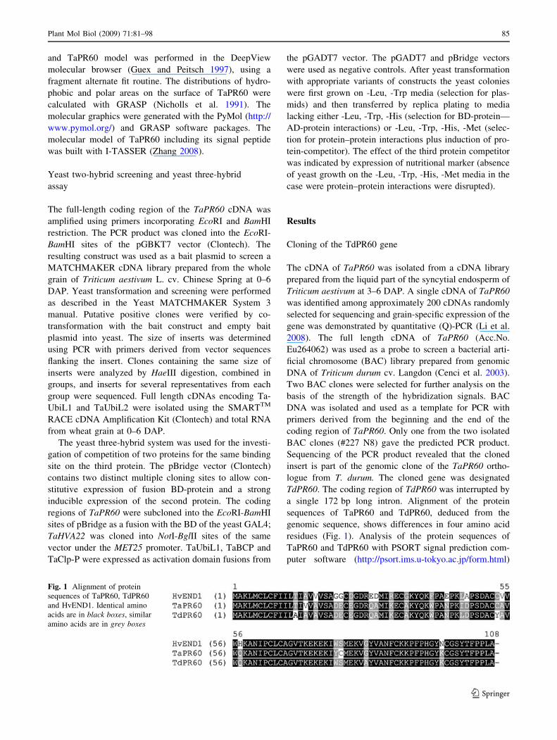
a single 172 bp long intron. Alignment of the protein
sequences of TaPR60 and TdPR60, deduced from the
genomic sequence, shows differences in four amino acid
residues (Fig. 1). Analysis of the protein sequences of
TaPR60 and TdPR60 with PSORT signal prediction com-
puter software (http://psort.ims.u-tokyo.ac.jp/form.html)
Fig. 1 Alignment of protein
sequences of TaPR60, TdPR60
and HvEND1. Identical amino
acids are in black boxes, similar
amino acids are in grey boxes
Plant Mol Biol (2009) 71:81–98 85
123
indicated the presence of an N-terminal hydrophobic signal
peptide of twenty amino acid residues in length, implying
secretion of both TaPR60 and TdPR60 to either the vacuole
or apoplast. The predicted cleavage site was situated
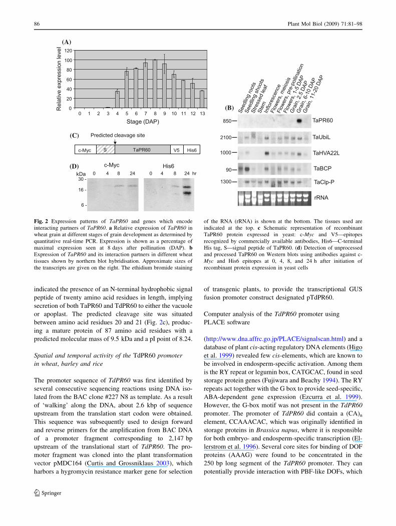
between amino acid residues 20 and 21 (Fig. 2c), produc-
ing a mature protein of 87 amino acid residues with a
predicted molecular mass of 9.5 kDa and a pI point of 8.24.
Spatial and temporal activity of the TdPR60 promoter
in wheat, barley and rice
The promoter sequence of TdPR60 was first identified by
several consecutive sequencing reactions using DNA iso-
lated from the BAC clone #227 N8 as template. As a result
of ‘walking’ along the DNA, about 2.6 kbp of sequence
upstream from the translation start codon were obtained.
This sequence was subsequently used to design forward
and reverse primers for the amplification from BAC DNA
of a promoter fragment corresponding to 2,147 bp
upstream of the translational start of TdPR60. The pro-
moter fragment was cloned into the plant transformation
vector pMDC164 (Curtis and Grossniklaus 2003), which
harbors a hygromycin resistance marker gene for selection
of transgenic plants, to provide the transcriptional GUS
fusion promoter construct designated pTdPR60.
Computer analysis of the TdPR60 promoter using
PLACE software
(http://www.dna.affrc.go.jp/PLACE/signalscan.html) and a
database of plant cis-acting regulatory DNA elements (Higo
et al. 1999) revealed few cis-elements, which are known to
be involved in endosperm-specific activation. Among them
is the RY repeat or legumin box, CATGCAC, found in seed
storage protein genes (Fujiwara and Beachy 1994). The RY
repeats act together with the G box to provide seed-specific,
ABA-dependent gene expression (Ezcurra et al. 1999).
However, the G-box motif was not present in the TdPR60
promoter. The promoter of TdPR60 did contain a (CA)n
element, CCAAACAC, which was originally identified in
storage proteins in Brassica napus, where it is responsible
for both embryo- and endosperm-specific transcription (El-
lerstrom et al. 1996). Several core sites for binding of DOF
proteins (AAAG) were found to be concentrated in the
250 bp long segment of the TdPR60 promoter. They can
potentially provide interaction with PBF-like DOFs, which
(A)
(B)
(D)
(C)
Fig. 2 Expression patterns of TaPR60 and genes which encode
interacting partners of TaPR60. a Relative expression of TaPR60 in
wheat grain at different stages of grain development as determined by
quantitative real-time PCR. Expression is shown as a percentage of
maximal expression seen at 8 days after pollination (DAP). bExpression of TaPR60 and its interaction partners in different wheat
tissues shown by northern blot hybridisation. Approximate sizes of
the transcripts are given on the right. The ethidium bromide staining
of the RNA (rRNA) is shown at the bottom. The tissues used are
indicated at the top. c Schematic representation of recombinant
TaPR60 protein expressed in yeast: c-Myc and V5—epitopes
recognized by commercially available antibodies, His6—C-terminal
His tag, S—signal peptide of TaPR60. (d) Detection of unprocessed
and processed TaPR60 on Western blots using antibodies against c-
Myc and His6 epitopes at 0, 4, 8, and 24 h after initiation of
recombinant protein expression in yeast cells
86 Plant Mol Biol (2009) 71:81–98
123
were demonstrated to bind to the prolamin box and specif-
ically activate promoters in the endosperm (Diaz et al.
2005). However, a complete prolamin box was not identi-
fied. The (TATCTC) repeats, which specifically interact
with ZmMRP-1 transcription factor and are responsible for
ETC-specific promoter activation in maize (Barrero et al.
2006), were not identified in the TdPR60 promoter. Mapping
of promoter segments responsible for the specific activation
in ETC using biolistic bombardment was unsuccessful
because of the extremely low frequency of successful tran-
sient transformation events observed in endosperm tissue.
No activity of the promoter was observed after biolistic
bombardment of isolated embryos at different stages of
development, although bombardment with a positive control
construct (pUbi-GUS) was successful (data not shown).
For stable transformation experiments, the pTdPR60
construct was transformed into the Agrobacterium tum-
efaciens strain AGL1 and the presence of plasmid in
selected colonies was confirmed by PCR using specific
primers. Transformed Agrobacterium was subsequently
used to introduce constructs into rice and barley. A
4,915 bp long fragment, containing the TdPR60 promoter,
GUS gene and Nos terminator, was excised from the
pTdPR60 construct and co-transformed together with a
plant selectable marker cassette (Ubi-Hpt-Nos) into wheat
using microprojectile bombardment. The integration of
promoter-GUS fusions in transgenic plants was confirmed
by PCR using primers derived from promoter and GUS
sequences. Southern blot hybridization was also performed
on some transgenic wheat and barley lines, providing data
showing the number of inserted copies of the selectable
marker hpt gene. The number of inserts in transgenic lines
of barley varied from one (Line 2) to two (Line 3) and three
(Lines 4–6). In wheat, the number of inserts varied from
two to about 16 or more. The number of inserts in trans-
genic rice plants was not examined.
Six T0 wheat lines were selected using the GUS staining
assay, from which three were selected for further analysis.
Ten T1 progeny for each of the three lines were analyzed.
Among the 30 progeny, there were two plants with very
strong, two plants with relatively strong, and 13 plants with
weak transgene expression; and 13 plants exhibited no
GUS expression. All positive lines demonstrated the same
pattern of GUS expression.
Eighteen T0 barley lines were selected, from which two
were used for the analysis of T1 plants. From nine T1 plants,
one had strong, two plants had weak and six plants had no
transgene expression. All T0 and T1 plants demonstrated the
same pattern of GUS expression. The differences in
expression levels between different transgenic lines
showed no correlation with the number of inserts.
Twenty-four T0 lines of transgenic rice were analyzed
for GUS activity. Eight lines demonstrated strong promoter
activity and the same pattern of gene expression. The T1
generation was analyzed for two lines (six plants for each).
All positive plants had the same patterns of transgene
expression as T0 plants. Wild type plants and/or plants
transformed with a vector containing only the selectable
marker cassette were used as negative controls. No dif-
ferences were found between wild type plants and plants
transformed with the control vector. The strength of the
TdPR60 promoter in barley and rice remained the same in
the T0 and T1 generations. In contrast, promoter strength in
wheat significantly decreased in the T1 generation relative
to the corresponding T0 parents.
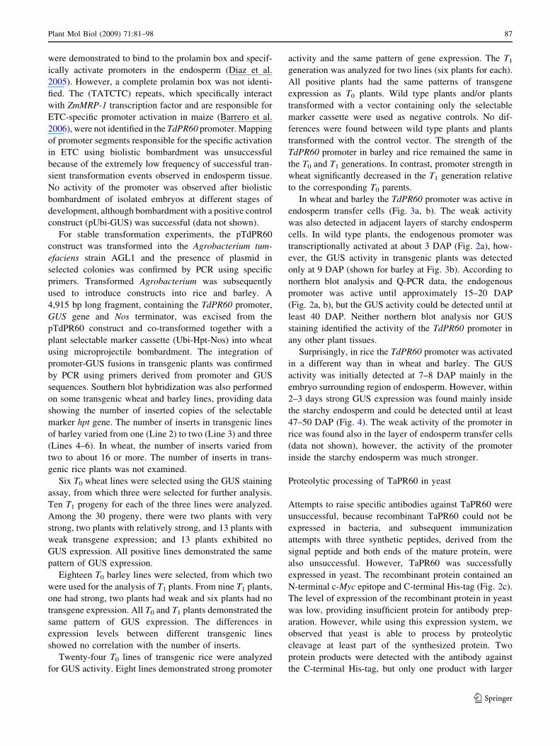
In wheat and barley the TdPR60 promoter was active in
endosperm transfer cells (Fig. 3a, b). The weak activity
was also detected in adjacent layers of starchy endosperm
cells. In wild type plants, the endogenous promoter was
transcriptionally activated at about 3 DAP (Fig. 2a), how-
ever, the GUS activity in transgenic plants was detected
only at 9 DAP (shown for barley at Fig. 3b). According to
northern blot analysis and Q-PCR data, the endogenous
promoter was active until approximately 15–20 DAP
(Fig. 2a, b), but the GUS activity could be detected until at
least 40 DAP. Neither northern blot analysis nor GUS
staining identified the activity of the TdPR60 promoter in
any other plant tissues.
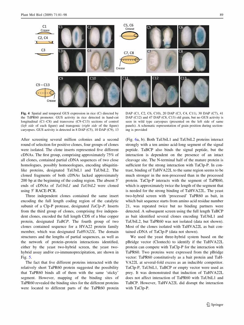
Surprisingly, in rice the TdPR60 promoter was activated
in a different way than in wheat and barley. The GUS
activity was initially detected at 7–8 DAP mainly in the
embryo surrounding region of endosperm. However, within
2–3 days strong GUS expression was found mainly inside
the starchy endosperm and could be detected until at least
47–50 DAP (Fig. 4). The weak activity of the promoter in
rice was found also in the layer of endosperm transfer cells
(data not shown), however, the activity of the promoter
inside the starchy endosperm was much stronger.
Proteolytic processing of TaPR60 in yeast
Attempts to raise specific antibodies against TaPR60 were
unsuccessful, because recombinant TaPR60 could not be
expressed in bacteria, and subsequent immunization
attempts with three synthetic peptides, derived from the
signal peptide and both ends of the mature protein, were
also unsuccessful. However, TaPR60 was successfully
expressed in yeast. The recombinant protein contained an
N-terminal c-Myc epitope and C-terminal His-tag (Fig. 2c).
The level of expression of the recombinant protein in yeast
was low, providing insufficient protein for antibody prep-
aration. However, while using this expression system, we
observed that yeast is able to process by proteolytic
cleavage at least part of the synthesized protein. Two
protein products were detected with the antibody against
the C-terminal His-tag, but only one product with larger
Plant Mol Biol (2009) 71:81–98 87
123
molecular weight was recognized by the antibody against
the N-terminal c-Myc epitope (Fig. 2). The size of the
detected proteins perfectly matched the predicted sizes of
the full length recombinant protein (18.1 kDa) and the
mature form of the protein (13.5 kDa). About 20–30% of
synthesized protein molecules remained uncleaved 24 h
after the induction of protein synthesis (Fig. 2d). The sig-
nal peptide was not detected. Successful expression of
recombinant TaPR60 in yeast and the presence of a sig-
nificant amount of uncleaved protein encouraged us to use
TaPR60 as bait in a yeast two-hybrid screen.
Isolation of interaction partners of TaPR60
A yeast two-hybrid cDNA library prepared from immature
wheat grain collected at 0–6 DAP was used for the screen.
Fig. 3 Spatial and temporal GUS expression in wheat (a) and barley
(b) directed by the TdPR60 promoter. GUS activity in wheat (a) and
barley (b) detected in hand-cut longitudinal (A1, A3, A5, A6, B1, B3–
B7) and transverse sections (A2, A4, and B2) of transgenic caryopsis,
at 9 DAP (B1), 13 DAP (B3, B6), 15 DAP (A5, A6, B4), 17 DAP
(B7), 24 DAP (A1–A4), and 34 DAP (B2, B5). No GUS activity is
seen in wild type caryopses in the panels A1–A4, B4, B6 and B7.
Grain was from T0 transgenic lines (A1–A4, B1–B5) and T1 progeny
(A5, A6, B6, B7). Histochemical GUS assay counterstained with
safranin in: 10 lm thick transverse sections of transgenic wheat (A7,
A8) and barley (B9) caryopsis, and longitudinal sections of transgenic
wheat (A9, A10) and barley caryopsis (B8) at 15 DAP (A9, A10), 16
DAP (B8), 31 DAP (A7, A8) and 34 DAP (B9)
88 Plant Mol Biol (2009) 71:81–98
123
After screening several million colonies and a second
round of selection for positive clones, four groups of clones
were isolated. The clone inserts represented five different
cDNAs. The first group, comprising approximately 75% of
all clones, contained partial cDNA sequences of two close
homologues, possibly homoeologues, encoding ubiquitin-
like proteins, designated TaUbiL1 and TaUbiL2. The
cloned fragments of both cDNAs lacked approximately
200 bp at the beginning of the coding region. The absent 50
ends of cDNAs of TaUbiL1 and TaUbiL2 were cloned
using 50 RACE-PCR.
Three independent clones contained the same insert
encoding the full length coding region of the catalytic
subunit of a Clp-P protease, designated TaClp-P. Inserts
from the third group of clones, comprising five indepen-
dent clones, encoded the full length CDS of a blue copper
protein, designated TaBCP. The fourth group of two
clones contained sequence for a HVA22 protein family
member, which was designated TaHVA22L. The domain
structures and the lengths of partial sequences, as well as
the network of protein–protein interactions identified,
either by the yeast two-hybrid screen, the yeast two-
hybrid assay and/or co-immunoprecipitation, are shown in
Fig. 5.
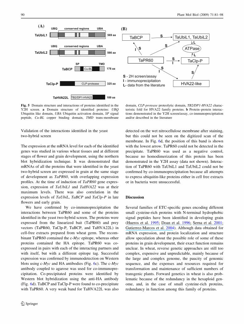
The fact that five different proteins interacted with the
relatively short TaPR60 protein suggested the possibility
that TaPR60 binds all of them with the same ‘sticky’
segment. However, mapping of the binding sites of
TaPR60 revealed the binding sites for the different proteins
were located to different parts of the TaPR60 protein
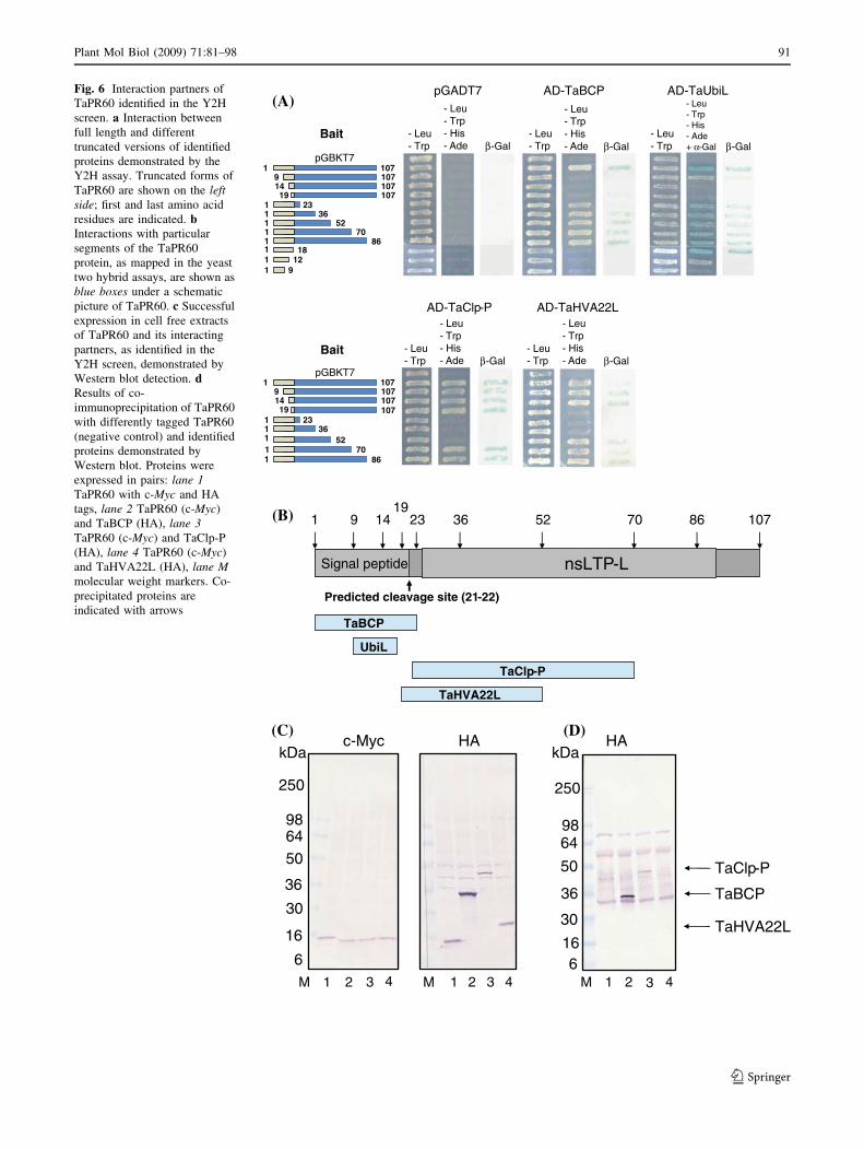
(Fig. 6a, b). Both TaUbiL1 and TaUbiL2 proteins interact
strongly with a ten amino acid-long segment of the signal
peptide. TaBCP also binds the signal peptide, but the
interaction is dependent on the presence of an intact
cleavage site. The N-terminal half of the mature protein is
sufficient for the strong interaction with TaClp-P. In con-
trast, binding of TaHVA22L to the same region seems to be
much stronger in the non-processed than in the processed
protein. TaClp-P interacts with the segment of TaPR60,
which is approximately twice the length of the segment that
is needed for the strong binding of TaHVA22L. The yeast
two-hybrid screen with ‘processed’ TaPR60 as bait, in
which bait sequence starts from amino acid residue number
21, was repeated twice but no binding partners were
detected. A subsequent screen using the full length TaBCP
as bait identified several clones encoding TaUbiL1 and
TaUbiL2, but TaPR60 was not isolated (data not shown).
Most of the clones isolated with TaHVA22L as bait con-
tained cDNA of TaClp-P (data not shown).
We used the yeast three-hybrid system based on the
pBridge vector (Clontech) to identify if the TaHVA22L
protein can compete with TaClp-P for the interaction with
TaPR60. Two proteins were expressed from the pBridge
vector: TaPR60 constitutively as a bait protein and TaH-
VA22L at several-fold excess as an inducible competitor.
TaClp-P, TaUbiL1, TaBCP or empty vector were used as
prey. It was demonstrated that induction of TaHVA22L
does not affect interaction of TaPR60 with TaUbiL1 and
TaBCP. However, TaHVA22L did disrupt the interaction
with TaClp-P.
Fig. 4 Spatial and temporal GUS expression in rice (C) directed by
the TdPR60 promoter. GUS activity in rice detected in hand-cut
longitudinal (C1–C8) and transverse (C9–C13) sections of control
(left side of each figure) and transgenic (right side of the figure)
caryopses. GUS activity is detected in 8 DAP (C5), 10 DAP (C9), 13
DAP (C1, C2, C6, C10), 20 DAP (C3, C4, C11), 30 DAP (C7), 41
DAP (C12) and 47 DAP (C8, C13) old grain, but no GUS activity is
seen in wild type caryopses (presented on the left side of same
panels). A schematic representation of grain position during section-
ing is provided
Plant Mol Biol (2009) 71:81–98 89
123
Validation of the interactions identified in the yeast
two-hybrid screen
The expression at the mRNA level for each of the identified
genes was studied in various wheat tissues and at different
stages of flower and grain development, using the northern
blot hybridization technique. It was demonstrated that
mRNAs of all the proteins that were identified in the yeast
two-hybrid screen are expressed in grain at the same stage
of development as TaPR60, with overlapping expression
profiles. At the time of induction of TaPR60 gene expres-
sion, expression of TaUbiL1 and TaHVA22 was at their
maximum levels. There was also correlation in the
expression levels of TaUbiL, TaBCP and TaClp-P in late
flowers and early grain.
We have confirmed by co-immunoprecipitation the
interactions between TaPR60 and some of the proteins
identified in the yeast two-hybrid screen. The proteins were
expressed from the linearized bait (TaPR60) and prey
vectors (TaPR60, TaClp-P, TaBCP, and TaHVA22L) in
cell-free extracts prepared from wheat germ. The recom-
binant TaPR60 contained the c-Myc epitope, whereas other
proteins contained the HA epitope. TaPR60 was co-
expressed in pairs with each of the interacting partners and
with itself, but with a different epitope tag. Successful
expression was confirmed by immunodetection on Western
blots using c-Myc and HA antibodies (Fig. 6c). The c-Myc
antibody coupled to agarose was used for co-immunopre-
cipitation. Co-precipitated proteins were identified by
Western blot hybridization using the anti-HA antibody
(Fig. 6d). TaBCP and TaClp-P were found to co-precipitate
with TaPR60. A very weak band for TaHVA22L was also
detected on the wet nitrocellulose membrane after staining,
but this could not be seen on the digitized scan of the
membrane. In Fig. 6d, the position of this band is shown
with the lowest arrow. TaPR60 could not be detected in the
precipitate. TaPR60 was used as a negative control,
because no homodimerization of this protein has been
demonstrated in the Y2H assay (data not shown). Interac-
tion of TaPR60 with TaUbiL1 and TaUbiL2 could not be
confirmed by co-immunoprecipitation because all attempts
to express ubiquitin-like proteins either in cell free extracts
or in bacteria were unsuccessful.
Discussion
Several families of ETC-specific genes encoding different
small cysteine-rich proteins with N-terminal hydrophobic
signal peptides have been identified in developing grain
(Hueros et al. 1995; Doan et al. 1996; Serna et al. 2001;
Gutierrez-Marcos et al. 2004). Although data obtained for
mRNA expression, and protein localization and structure
allow speculation about the possible role of some of these
proteins in grain development, their exact function remains
unclear. In wheat, reverse genetic approaches are still too
complex, expensive and unpredictable, mainly because of
the large and complex genome, the paucity of genomic
sequence, and the expenses and resources required for
transformation and maintenance of sufficient numbers of
transgenic plants. Forward genetics in wheat is also prob-
lematic because of the redundancy in the hexaploid gen-
ome, and, in the case of small cysteine-rich proteins,
redundancy in function among this family of proteins.
(A)(B)
Fig. 5 Domain structure and interactions of proteins identified in the
Y2H screen. a Domain structure of identified proteins: UBQUbiquitin like domain, UBA Ubiquitin activation domain, SP signal
peptide, Cu-BL copper binding domain, TMD trans-membrane
domain, CLP-protease proteolytic domain, TB2/DP1-HVA22 charac-
teristic fold for HVA22 family proteins. b Protein–protein interac-
tions demonstrated in the Y2H screen/assay, co-immunoprecipitation
and/or described in the literature
90 Plant Mol Biol (2009) 71:81–98
123
(A)pGADT7 AD-TaBCP AD-TaUbiL
- Leu- Trp
- Leu- Trp- His - Ade β-Gal
AD-TaClp-P AD-TaHVA22L
Bait
pGBKT7
pGBKT7
Bait
(B)
Signal peptide
TaHVA22L
TaClp-P
869 1419
10723 36 52 701
TaBCP
UbiL
Predicted cleavage site (21-22)
nsLTP-L
(C)
TaBCP
TaClp-P
1 2 3 4M
c-Myc HA HA
250
986450
36
30
16
6
250
9864
50
36
30
166
1 2 3 4M 1 2 3 4M
kDakDa(D)
TaHVA22L
1
11111111
9
9
1419
107107107107
2336
52
8670
1218
1
11111
91419
107107107107
2336
52
8670
- Leu- Trp
- Leu- Trp- His - Ade β-Gal
- Leu- Trp
- Leu- Trp- His - Ade+ α-Gal β-Gal
- Leu- Trp
- Leu- Trp- His - Ade β-Gal
- Leu- Trp
- Leu- Trp- His - Ade β-Gal
Fig. 6 Interaction partners of
TaPR60 identified in the Y2H
screen. a Interaction between
full length and different
truncated versions of identified
proteins demonstrated by the
Y2H assay. Truncated forms of
TaPR60 are shown on the leftside; first and last amino acid
residues are indicated. bInteractions with particular
segments of the TaPR60
protein, as mapped in the yeast
two hybrid assays, are shown as
blue boxes under a schematic
picture of TaPR60. c Successful
expression in cell free extracts
of TaPR60 and its interacting
partners, as identified in the
Y2H screen, demonstrated by
Western blot detection. dResults of co-
immunoprecipitation of TaPR60
with differently tagged TaPR60
(negative control) and identified
proteins demonstrated by
Western blot. Proteins were
expressed in pairs: lane 1TaPR60 with c-Myc and HA
tags, lane 2 TaPR60 (c-Myc)
and TaBCP (HA), lane 3TaPR60 (c-Myc) and TaClp-P
(HA), lane 4 TaPR60 (c-Myc)
and TaHVA22L (HA), lane Mmolecular weight markers. Co-
precipitated proteins are
indicated with arrows
Plant Mol Biol (2009) 71:81–98 91
123
Besides these genetic approaches, one of the first steps
to elucidate the function of genes is to study the temporal
and spatial patterns of gene expression. It was demon-
strated earlier by quantitative RT-PCR that TaPR60 is an
endosperm-specific gene (Li et al. 2008). Analysis of the
promoter of the T. durum orthologue of TaPR60 provided
additional and more precise data on expression of the
END1-like genes. The anticipated utility of the promoter in
biotechnological applications prompted analysis of the
promoter in three agriculturally important plants: wheat,
barley and rice. The spatial and temporal activities of the
promoter in wheat and barley were nearly identical. In
wheat and barley the promoter was active in endosperm
transfer cells and adjacent layers of starchy endosperm. In
barley, the spatial and temporal activity of the TdPR60
promoter was practically indistinguishable from the activ-
ity of the promoter of the rice gene OsPR602 (Li et al.
2008). Surprisingly, in rice the TdPR60 promoter was
activated 1–2 days earlier and was detected mainly inside
of the starchy endosperm (Fig. 4), in contrast to its activity
in wheat and barley. Unlike the OsPR602 promoter in rice
(Li et al. 2008), no activity of the wheat promoter was
identified in rice flowers or any other tissues. This suggests
at least partial incompatibility of transcription factors and
cis-elements responsible for ETC-specific activation of
wheat and rice promoters. Use of strongly diverged or
different cis-elements in rice and wheat is supported by the
observation that identical or near-identical cis-elements in
the TaPR60 and OsPR602 promoters could not be identi-
fied. The expression pattern of TdPR60 suggests involve-
ment of the gene product either in signal transduction or
nutrient transfer into the endosperm. Although transcripts
of TaPR60 were detected in endosperm as early as 3 DAP
(Fig. 2a), at the beginning of coenocyte cellularization,
GUS activity was not observed before 9 DAP, when cell-
ularization of the endosperm was near completion. This
result implies that the gene product is not involved in the
endosperm cellularization process. The difference between
timing of mRNA and protein expression can be explained
by possible translational regulation provided by the 50UTR
sequence, which was included in the promoter construct.
However, we have no experimental evidence for a role of
the 50UTR.
Molecular modeling is another complementary
approach, which can assist in predicting and better under-
standing the function of proteins. This has been demon-
strated in our current structural proteomics era, where 3D
structures or molecular models are becoming integral
components of multidisciplinary biological research. An
example of usefulness of molecular modeling has recently
been demonstrated (Rodrigues et al. 2008). A molecular
model of the wheat TaPR60 protein was constructed using
as a template a putative Prunus persica LBP (Protein
DataBank accession number 2alg:A; Pasquato et al. 2006)
that was identified by several prediction servers listed in
the Materials and Methods section. The LBP protein rep-
resents a canonical all alpha protein categorized in the
SCOP protein classification system. More precisely, these
structures belong to the family of ‘Plant Lipid Transfer and
Hydrophobic Proteins’ (Andreeva et al. 2008). Other
hydrophobic proteins are also classified in this protein
group, such as a soybean hydrophobic protein (Baud et al.
1993). The positional sequence identity and similarity
between the 2alg:A and TaPR60 sequences were 17 and
31%, respectively. This rather low sequence identity but
significant similarity emphasized complexity of modeling
(Sanchez and Sali 1998; John and Sali 2003; Sippl and
Wiederstein 2008). However, the analysis of sequences via
hydrophobic cluster analysis (HCA) (Callebaut et al. 1997)
indicated that the positions of eight cysteine residues were
highly conserved in both sequences (Fig. 7a; marked by
arrowheads). This observation suggested that the cysteine
residues in TaPR60 could also be paired, and the associated
parts of the sequences could form a-helical secondary
structural elements (Fig. 7a; marked by lines).
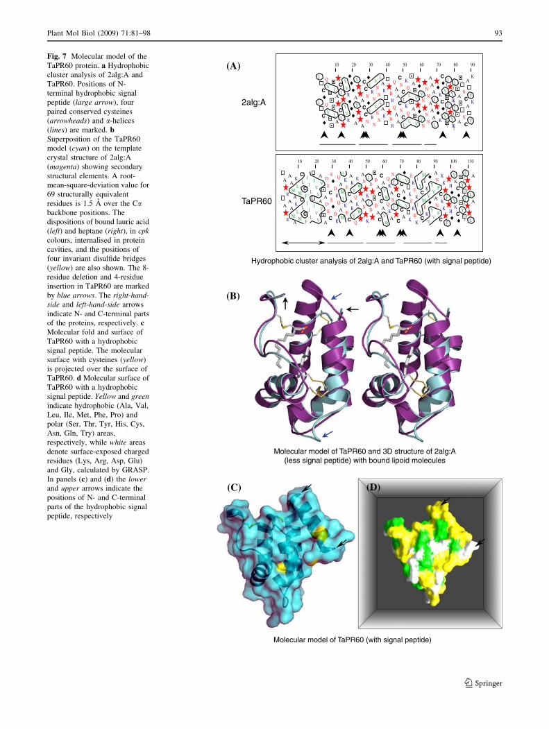
The 3D models of TaPR60 with and without signal
peptide were generated (Fig. 7) and their stereochemical
parameters (Ramachandran et al. 1963; Laskowski et al.
1993) and combined energy profiles (Sippl 1993) were
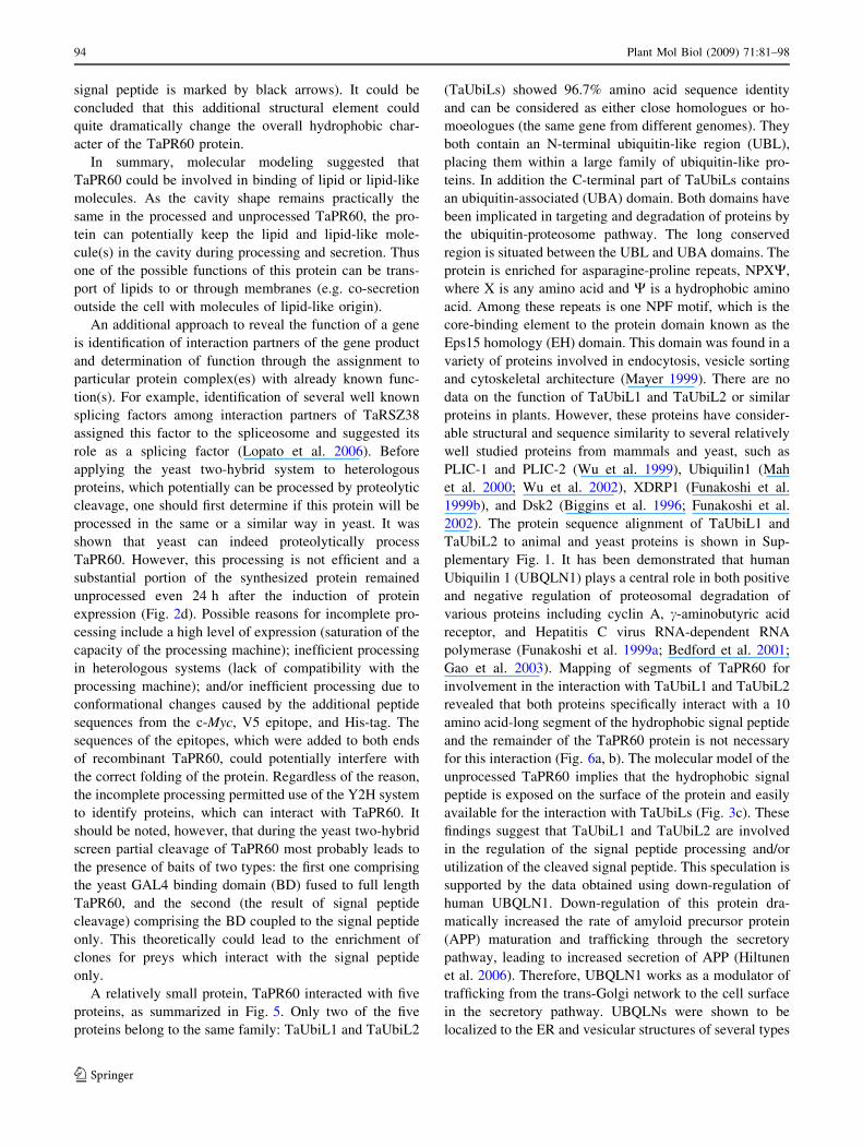
found to be acceptable (data not shown). Both proteins
(Fig. 7) contained a series of highly conserved a-helices
folded in all alpha protein class according to SCOP protein
classification (Andreeva et al. 2008). The major structural
differences between the TaPR60 model and its template
stemmed from one-eight-residue deletion and one-four-
residue insertion in TaPR60 (Fig. 7b, marked by blue
arrows). Two lipoid molecules (lauric acid and heptane)
were also modeled in the central hydrophobic cavity of
TaPR60 (contained in 2alg:A) that was formed in the
central region of the helical bundle (Fig. 7b); the volume of
this cavity was approximately 1,240 A3, compared to the
volume of the cavity of 2alg:A, which equaled 1,205 A3.
The side chains of predominantly hydrophobic and aro-
matic residues (Val, Ala, Phe, Trp) lining up the cavity
were rotated out to the sides of the protein fold, so a rather
large hydrophobic cavity could be formed that could
internalize possibly several, and at least two lipid or lipid-
like molecules. Three amino acid residues (Trp36, Cys45,
Lys52) were making contacts of less than 3 A with the two
lipid molecules through hydrophobic interactions.
Molecular modeling of a full length sequence of
TaPR60, including its hydrophobic signal peptide, was
performed with the I-TASSER (Zhang 2008). The model of
TaPR60 indicated that the signal peptide comprising 20
amino acid residues (Fig. 7a) formed an additional a-helix
that had a significant hydrophobic surface (Fig. 7c, d; the
92 Plant Mol Biol (2009) 71:81–98
123
Molecular model of TaPR60 and 3D structure of 2alg:A(less signal peptide) with bound lipoid molecules
(A)
(B)
2alg:A
10 20 30 40 50 60 70 80 90 100 110
A
R
AA
MA
K
LM
C
LC
F
IIL
IVV
A
V
A
DEC
E
DR
QA
M
IKEC
AK
YQ
K
W
AN
K
ID
DAC
CA
VW
QKA
N
I
C
LCA
V
K
EKEK
IYC
MEK
V
YV
AN
FC
KK
F
H
YKC
Y
F
L
A
A
R
AA
MA
K
LM
C
LC
F
IIL
IVV
A
V
A
DEC
E
DR
QA
M
IKEC
AK
YQ
K
W
AN
K
ID
DAC
CA
VW
QKA
N
I
C
LCA
V
K
EKEK
IYC
MEK
V
YV
AN
FC
KK
F
H
YKC
Y
F
L
A
10 20 30 40 50 60 70 80 90
I
C
Q
V LA
C
I
YVR A
V
A
CCN
IRN
VNN
LAR D
R
QAA
CNC
L
KQ
L
A
VVN
NN
AAA
LK
C
V
I
YK
I
ANC
A
V
K
I
C
Q
V LA
C
I
YVR A
V
A
CCN
IRN
VNN
LAR D
R
QAA
CNC
L
KQ
L
A
VVN
NN
AAA
LK
C
V
I
YK
I
ANC
A
V
K
Hydrophobic cluster analysis of 2alg:A and TaPR60 (with signal peptide)
Molecular model of TaPR60 (with signal peptide)
(C) (D)
TaPR60
Fig. 7 Molecular model of the
TaPR60 protein. a Hydrophobic
cluster analysis of 2alg:A and
TaPR60. Positions of N-
terminal hydrophobic signal
peptide (large arrow), four
paired conserved cysteines
(arrowheads) and a-helices
(lines) are marked. bSuperposition of the TaPR60
model (cyan) on the template
crystal structure of 2alg:A
(magenta) showing secondary
structural elements. A root-
mean-square-deviation value for
69 structurally equivalent
residues is 1.5 A over the Cabackbone positions. The
dispositions of bound lauric acid
(left) and heptane (right), in cpkcolours, internalised in protein
cavities, and the positions of
four invariant disulfide bridges
(yellow) are also shown. The 8-
residue deletion and 4-residue
insertion in TaPR60 are marked
by blue arrows. The right-hand-side and left-hand-side arrows
indicate N- and C-terminal parts
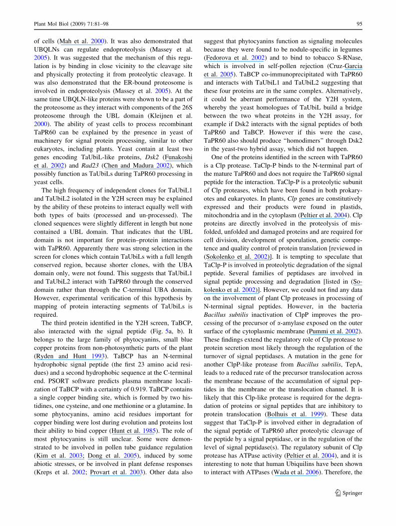
of the proteins, respectively. cMolecular fold and surface of
TaPR60 with a hydrophobic
signal peptide. The molecular
surface with cysteines (yellow)
is projected over the surface of
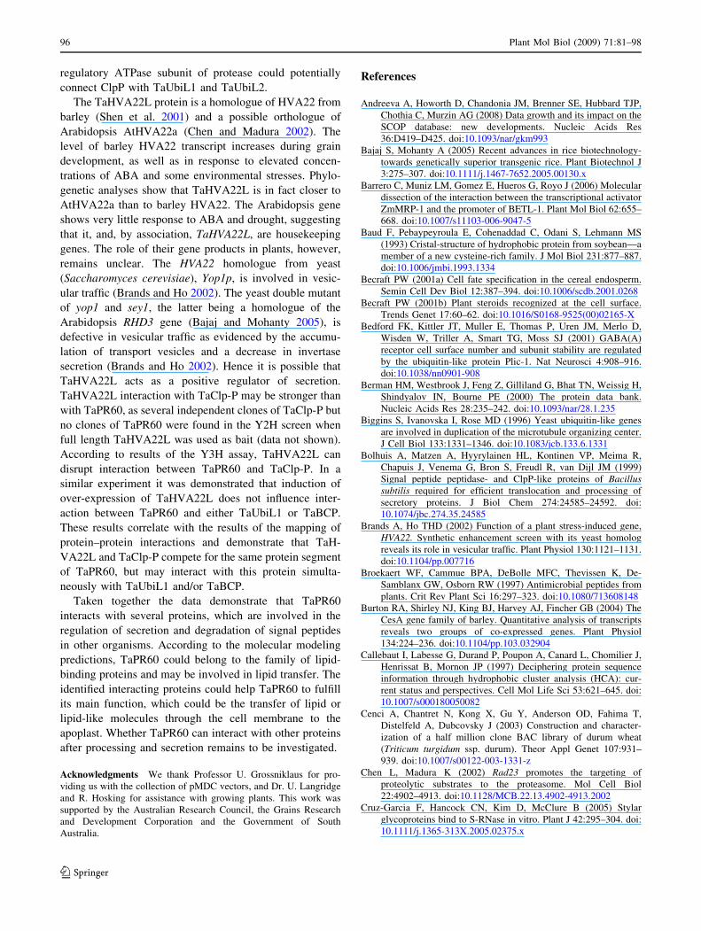
TaPR60. d Molecular surface of
TaPR60 with a hydrophobic
signal peptide. Yellow and greenindicate hydrophobic (Ala, Val,
Leu, Ile, Met, Phe, Pro) and
polar (Ser, Thr, Tyr, His, Cys,
Asn, Gln, Try) areas,
respectively, while white areas
denote surface-exposed charged
residues (Lys, Arg, Asp, Glu)
and Gly, calculated by GRASP.
In panels (c) and (d) the lowerand upper arrows indicate the
positions of N- and C-terminal
parts of the hydrophobic signal
peptide, respectively
Plant Mol Biol (2009) 71:81–98 93
123
signal peptide is marked by black arrows). It could be
concluded that this additional structural element could
quite dramatically change the overall hydrophobic char-
acter of the TaPR60 protein.
In summary, molecular modeling suggested that
TaPR60 could be involved in binding of lipid or lipid-like
molecules. As the cavity shape remains practically the
same in the processed and unprocessed TaPR60, the pro-
tein can potentially keep the lipid and lipid-like mole-
cule(s) in the cavity during processing and secretion. Thus
one of the possible functions of this protein can be trans-
port of lipids to or through membranes (e.g. co-secretion
outside the cell with molecules of lipid-like origin).
An additional approach to reveal the function of a gene
is identification of interaction partners of the gene product
and determination of function through the assignment to
particular protein complex(es) with already known func-
tion(s). For example, identification of several well known
splicing factors among interaction partners of TaRSZ38
assigned this factor to the spliceosome and suggested its
role as a splicing factor (Lopato et al. 2006). Before
applying the yeast two-hybrid system to heterologous
proteins, which potentially can be processed by proteolytic
cleavage, one should first determine if this protein will be
processed in the same or a similar way in yeast. It was
shown that yeast can indeed proteolytically process
TaPR60. However, this processing is not efficient and a
substantial portion of the synthesized protein remained
unprocessed even 24 h after the induction of protein
expression (Fig. 2d). Possible reasons for incomplete pro-
cessing include a high level of expression (saturation of the
capacity of the processing machine); inefficient processing
in heterologous systems (lack of compatibility with the
processing machine); and/or inefficient processing due to
conformational changes caused by the additional peptide
sequences from the c-Myc, V5 epitope, and His-tag. The
sequences of the epitopes, which were added to both ends
of recombinant TaPR60, could potentially interfere with
the correct folding of the protein. Regardless of the reason,
the incomplete processing permitted use of the Y2H system
to identify proteins, which can interact with TaPR60. It
should be noted, however, that during the yeast two-hybrid
screen partial cleavage of TaPR60 most probably leads to
the presence of baits of two types: the first one comprising
the yeast GAL4 binding domain (BD) fused to full length
TaPR60, and the second (the result of signal peptide
cleavage) comprising the BD coupled to the signal peptide
only. This theoretically could lead to the enrichment of
clones for preys which interact with the signal peptide
only.
A relatively small protein, TaPR60 interacted with five
proteins, as summarized in Fig. 5. Only two of the five
proteins belong to the same family: TaUbiL1 and TaUbiL2
(TaUbiLs) showed 96.7% amino acid sequence identity
and can be considered as either close homologues or ho-
moeologues (the same gene from different genomes). They
both contain an N-terminal ubiquitin-like region (UBL),
placing them within a large family of ubiquitin-like pro-
teins. In addition the C-terminal part of TaUbiLs contains
an ubiquitin-associated (UBA) domain. Both domains have
been implicated in targeting and degradation of proteins by
the ubiquitin-proteosome pathway. The long conserved
region is situated between the UBL and UBA domains. The
protein is enriched for asparagine-proline repeats, NPXW,
where X is any amino acid and W is a hydrophobic amino
acid. Among these repeats is one NPF motif, which is the
core-binding element to the protein domain known as the
Eps15 homology (EH) domain. This domain was found in a
variety of proteins involved in endocytosis, vesicle sorting
and cytoskeletal architecture (Mayer 1999). There are no
data on the function of TaUbiL1 and TaUbiL2 or similar
proteins in plants. However, these proteins have consider-
able structural and sequence similarity to several relatively
well studied proteins from mammals and yeast, such as
PLIC-1 and PLIC-2 (Wu et al. 1999), Ubiquilin1 (Mah
et al. 2000; Wu et al. 2002), XDRP1 (Funakoshi et al.
1999b), and Dsk2 (Biggins et al. 1996; Funakoshi et al.
2002). The protein sequence alignment of TaUbiL1 and
TaUbiL2 to animal and yeast proteins is shown in Sup-
plementary Fig. 1. It has been demonstrated that human
Ubiquilin 1 (UBQLN1) plays a central role in both positive
and negative regulation of proteosomal degradation of
various proteins including cyclin A, c-aminobutyric acid
receptor, and Hepatitis C virus RNA-dependent RNA
polymerase (Funakoshi et al. 1999a; Bedford et al. 2001;
Gao et al. 2003). Mapping of segments of TaPR60 for
involvement in the interaction with TaUbiL1 and TaUbiL2
revealed that both proteins specifically interact with a 10
amino acid-long segment of the hydrophobic signal peptide
and the remainder of the TaPR60 protein is not necessary
for this interaction (Fig. 6a, b). The molecular model of the
unprocessed TaPR60 implies that the hydrophobic signal
peptide is exposed on the surface of the protein and easily
available for the interaction with TaUbiLs (Fig. 3c). These
findings suggest that TaUbiL1 and TaUbiL2 are involved
in the regulation of the signal peptide processing and/or
utilization of the cleaved signal peptide. This speculation is
supported by the data obtained using down-regulation of
human UBQLN1. Down-regulation of this protein dra-
matically increased the rate of amyloid precursor protein
(APP) maturation and trafficking through the secretory
pathway, leading to increased secretion of APP (Hiltunen
et al. 2006). Therefore, UBQLN1 works as a modulator of
trafficking from the trans-Golgi network to the cell surface
in the secretory pathway. UBQLNs were shown to be
localized to the ER and vesicular structures of several types
94 Plant Mol Biol (2009) 71:81–98
123
of cells (Mah et al. 2000). It was also demonstrated that
UBQLNs can regulate endoproteolysis (Massey et al.
2005). It was suggested that the mechanism of this regu-
lation is by binding in close vicinity to the cleavage site
and physically protecting it from proteolytic cleavage. It
was also demonstrated that the ER-bound proteosome is
involved in endoproteolysis (Massey et al. 2005). At the
same time UBQLN-like proteins were shown to be a part of
the proteosome as they interact with components of the 26S
proteosome through the UBL domain (Kleijnen et al.
2000). The ability of yeast cells to process recombinant
TaPR60 can be explained by the presence in yeast of
machinery for signal protein processing, similar to other
eukaryotes, including plants. Yeast contain at least two
genes encoding TaUbiL-like proteins, Dsk2 (Funakoshi
et al. 2002) and Rad23 (Chen and Madura 2002), which
possibly function as TaUbiLs during TaPR60 processing in
yeast cells.
The high frequency of independent clones for TaUbiL1
and TaUbiL2 isolated in the Y2H screen may be explained
by the ability of these proteins to interact equally well with
both types of baits (processed and un-processed). The
cloned sequences were slightly different in length but none
contained a UBL domain. That indicates that the UBL
domain is not important for protein–protein interactions
with TaPR60. Apparently there was strong selection in the
screen for clones which contain TaUbiLs with a full length
conserved region, because shorter clones, with the UBA
domain only, were not found. This suggests that TaUbiL1
and TaUbiL2 interact with TaPR60 through the conserved
domain rather than through the C-terminal UBA domain.
However, experimental verification of this hypothesis by
mapping of protein interacting segments of TaUbiLs is
required.
The third protein identified in the Y2H screen, TaBCP,
also interacted with the signal peptide (Fig. 5a, b). It
belongs to the large family of phytocyanins, small blue
copper proteins from non-photosynthetic parts of the plant
(Ryden and Hunt 1993). TaBCP has an N-terminal
hydrophobic signal peptide (the first 23 amino acid resi-
dues) and a second hydrophobic sequence at the C-terminal
end. PSORT software predicts plasma membrane locali-
zation of TaBCP with a certainty of 0.919. TaBCP contains
a single copper binding site, which is formed by two his-
tidines, one cysteine, and one methionine or a glutamine. In
some phytocyanins, amino acid residues important for
copper binding were lost during evolution and proteins lost
their ability to bind copper (Hunt et al. 1985). The role of
most phytocyanins is still unclear. Some were demon-
strated to be involved in pollen tube guidance regulation
(Kim et al. 2003; Dong et al. 2005), induced by some
abiotic stresses, or be involved in plant defense responses
(Kreps et al. 2002; Provart et al. 2003). Other data also
suggest that phytocyanins function as signaling molecules
because they were found to be nodule-specific in legumes
(Fedorova et al. 2002) and to bind to tobacco S-RNase,
which is involved in self-pollen rejection (Cruz-Garcia
et al. 2005). TaBCP co-immunoprecipitated with TaPR60
and interacts with TaUbiL1 and TaUbiL2 suggesting that
these four proteins are in the same complex. Alternatively,
it could be aberrant performance of the Y2H system,
whereby the yeast homologues of TaUbiL build a bridge
between the two wheat proteins in the Y2H assay, for
example if Dsk2 interacts with the signal peptides of both
TaPR60 and TaBCP. However if this were the case,
TaPR60 also should produce ‘‘homodimers’’ through Dsk2
in the yeast-two hybrid assay, which did not happen.
One of the proteins identified in the screen with TaPR60
is a Clp protease. TaClp-P binds to the N-terminal part of
the mature TaPR60 and does not require the TaPR60 signal
peptide for the interaction. TaClp-P is a proteolytic subunit
of Clp proteases, which have been found in both prokary-
otes and eukaryotes. In plants, Clp genes are constitutively
expressed and their products were found in plastids,
mitochondria and in the cytoplasm (Peltier et al. 2004). Clp
proteins are directly involved in the proteolysis of mis-
folded, unfolded and damaged proteins and are required for
cell division, development of sporulation, genetic compe-
tence and quality control of protein translation [reviewed in
(Sokolenko et al. 2002)]. It is tempting to speculate that
TaClp-P is involved in proteolytic degradation of the signal
peptide. Several families of peptidases are involved in
signal peptide processing and degradation [listed in (So-
kolenko et al. 2002)]. However, we could not find any data
on the involvement of plant Clp proteases in processing of
N-terminal signal peptides. However, in the bacteria
Bacillus subtilis inactivation of ClpP improves the pro-
cessing of the precursor of a-amylase exposed on the outer
surface of the cytoplasmic membrane (Pummi et al. 2002).
These findings extend the regulatory role of Clp protease to
protein secretion most likely through the regulation of the
turnover of signal peptidases. A mutation in the gene for
another ClpP-like protease from Bacillus subtilis, TepA,
leads to a reduced rate of the precursor translocation across
the membrane because of the accumulation of signal pep-
tides in the membrane or the translocation channel. It is
likely that this Clp-like protease is required for the degra-
dation of proteins or signal peptides that are inhibitory to
protein translocation (Bolhuis et al. 1999). These data
suggest that TaClp-P is involved either in degradation of
the signal peptide of TaPR60 after proteolytic cleavage of
the peptide by a signal peptidase, or in the regulation of the
level of signal peptidase(s). The regulatory subunit of Clp
protease has ATPase activity (Peltier et al. 2004), and it is
interesting to note that human Ubiquilins have been shown
to interact with ATPases (Wada et al. 2006). Therefore, the
Plant Mol Biol (2009) 71:81–98 95
123
regulatory ATPase subunit of protease could potentially
connect ClpP with TaUbiL1 and TaUbiL2.
The TaHVA22L protein is a homologue of HVA22 from
barley (Shen et al. 2001) and a possible orthologue of
Arabidopsis AtHVA22a (Chen and Madura 2002). The
level of barley HVA22 transcript increases during grain
development, as well as in response to elevated concen-
trations of ABA and some environmental stresses. Phylo-
genetic analyses show that TaHVA22L is in fact closer to
AtHVA22a than to barley HVA22. The Arabidopsis gene
shows very little response to ABA and drought, suggesting
that it, and, by association, TaHVA22L, are housekeeping
genes. The role of their gene products in plants, however,
remains unclear. The HVA22 homologue from yeast
(Saccharomyces cerevisiae), Yop1p, is involved in vesic-
ular traffic (Brands and Ho 2002). The yeast double mutant
of yop1 and sey1, the latter being a homologue of the
Arabidopsis RHD3 gene (Bajaj and Mohanty 2005), is
defective in vesicular traffic as evidenced by the accumu-
lation of transport vesicles and a decrease in invertase
secretion (Brands and Ho 2002). Hence it is possible that
TaHVA22L acts as a positive regulator of secretion.
TaHVA22L interaction with TaClp-P may be stronger than
with TaPR60, as several independent clones of TaClp-P but
no clones of TaPR60 were found in the Y2H screen when
full length TaHVA22L was used as bait (data not shown).
According to results of the Y3H assay, TaHVA22L can
disrupt interaction between TaPR60 and TaClp-P. In a
similar experiment it was demonstrated that induction of
over-expression of TaHVA22L does not influence inter-
action between TaPR60 and either TaUbiL1 or TaBCP.
These results correlate with the results of the mapping of
protein–protein interactions and demonstrate that TaH-
VA22L and TaClp-P compete for the same protein segment
of TaPR60, but may interact with this protein simulta-
neously with TaUbiL1 and/or TaBCP.
Taken together the data demonstrate that TaPR60
interacts with several proteins, which are involved in the
regulation of secretion and degradation of signal peptides
in other organisms. According to the molecular modeling
predictions, TaPR60 could belong to the family of lipid-
binding proteins and may be involved in lipid transfer. The
identified interacting proteins could help TaPR60 to fulfill
its main function, which could be the transfer of lipid or
lipid-like molecules through the cell membrane to the
apoplast. Whether TaPR60 can interact with other proteins
after processing and secretion remains to be investigated.
Acknowledgments We thank Professor U. Grossniklaus for pro-
viding us with the collection of pMDC vectors, and Dr. U. Langridge
and R. Hosking for assistance with growing plants. This work was
supported by the Australian Research Council, the Grains Research
and Development Corporation and the Government of South
Australia.
References
Andreeva A, Howorth D, Chandonia JM, Brenner SE, Hubbard TJP,
Chothia C, Murzin AG (2008) Data growth and its impact on the
SCOP database: new developments. Nucleic Acids Res
36:D419–D425. doi:10.1093/nar/gkm993
Bajaj S, Mohanty A (2005) Recent advances in rice biotechnology-
towards genetically superior transgenic rice. Plant Biotechnol J
3:275–307. doi:10.1111/j.1467-7652.2005.00130.x
Barrero C, Muniz LM, Gomez E, Hueros G, Royo J (2006) Molecular
dissection of the interaction between the transcriptional activator
ZmMRP-1 and the promoter of BETL-1. Plant Mol Biol 62:655–
668. doi:10.1007/s11103-006-9047-5
Baud F, Pebaypeyroula E, Cohenaddad C, Odani S, Lehmann MS
(1993) Cristal-structure of hydrophobic protein from soybean—a
member of a new cysteine-rich family. J Mol Biol 231:877–887.
doi:10.1006/jmbi.1993.1334
Becraft PW (2001a) Cell fate specification in the cereal endosperm.
Semin Cell Dev Biol 12:387–394. doi:10.1006/scdb.2001.0268
Becraft PW (2001b) Plant steroids recognized at the cell surface.
Trends Genet 17:60–62. doi:10.1016/S0168-9525(00)02165-X
Bedford FK, Kittler JT, Muller E, Thomas P, Uren JM, Merlo D,
Wisden W, Triller A, Smart TG, Moss SJ (2001) GABA(A)
receptor cell surface number and subunit stability are regulated
by the ubiquitin-like protein Plic-1. Nat Neurosci 4:908–916.
doi:10.1038/nn0901-908
Berman HM, Westbrook J, Feng Z, Gilliland G, Bhat TN, Weissig H,
Shindyalov IN, Bourne PE (2000) The protein data bank.
Nucleic Acids Res 28:235–242. doi:10.1093/nar/28.1.235
Biggins S, Ivanovska I, Rose MD (1996) Yeast ubiquitin-like genes
are involved in duplication of the microtubule organizing center.
J Cell Biol 133:1331–1346. doi:10.1083/jcb.133.6.1331
Bolhuis A, Matzen A, Hyyrylainen HL, Kontinen VP, Meima R,
Chapuis J, Venema G, Bron S, Freudl R, van Dijl JM (1999)
Signal peptide peptidase- and ClpP-like proteins of Bacillussubtilis required for efficient translocation and processing of
secretory proteins. J Biol Chem 274:24585–24592. doi:
10.1074/jbc.274.35.24585
Brands A, Ho THD (2002) Function of a plant stress-induced gene,
HVA22. Synthetic enhancement screen with its yeast homolog
reveals its role in vesicular traffic. Plant Physiol 130:1121–1131.
doi:10.1104/pp.007716
Broekaert WF, Cammue BPA, DeBolle MFC, Thevissen K, De-
Samblanx GW, Osborn RW (1997) Antimicrobial peptides from
plants. Crit Rev Plant Sci 16:297–323. doi:10.1080/713608148
Burton RA, Shirley NJ, King BJ, Harvey AJ, Fincher GB (2004) The
CesA gene family of barley. Quantitative analysis of transcripts
reveals two groups of co-expressed genes. Plant Physiol
134:224–236. doi:10.1104/pp.103.032904
Callebaut I, Labesse G, Durand P, Poupon A, Canard L, Chomilier J,
Henrissat B, Mornon JP (1997) Deciphering protein sequence
information through hydrophobic cluster analysis (HCA): cur-
rent status and perspectives. Cell Mol Life Sci 53:621–645. doi:
10.1007/s000180050082
Cenci A, Chantret N, Kong X, Gu Y, Anderson OD, Fahima T,
Distelfeld A, Dubcovsky J (2003) Construction and character-
ization of a half million clone BAC library of durum wheat
(Triticum turgidum ssp. durum). Theor Appl Genet 107:931–
939. doi:10.1007/s00122-003-1331-z
Chen L, Madura K (2002) Rad23 promotes the targeting of
proteolytic substrates to the proteasome. Mol Cell Biol
22:4902–4913. doi:10.1128/MCB.22.13.4902-4913.2002
Cruz-Garcia F, Hancock CN, Kim D, McClure B (2005) Stylar
glycoproteins bind to S-RNase in vitro. Plant J 42:295–304. doi:
10.1111/j.1365-313X.2005.02375.x
96 Plant Mol Biol (2009) 71:81–98
123
Curtis MD, Grossniklaus U (2003) A gateway cloning vector set for
high-throughput functional analysis of genes in planta. Plant
Physiol 133:462–469. doi:10.1104/pp.103.027979
Diaz I, Martinez M, Isabel-LaMoneda I, Rubio-Somoza I, Carbonero
P (2005) The DOF protein, SAD, interacts with GAMYB in
plant nuclei and activates transcription of endosperm-specific
genes during barley seed development. Plant J 42:652–662. doi:
10.1111/j.1365-313X.2005.02402.x
Doan DNP, Linnestad C, Olsen OA (1996) Isolation of molecular
markers from the barley endosperm coenocyte and the sur-
rounding nucellus cell layers. Plant Mol Biol 31:877–886. doi:
10.1007/BF00019474
Dong J, Kim ST, Lord EM (2005) Plantacyanin plays a role in
reproduction in Arabidopsis. Plant Physiol 138:778–789. doi:
10.1104/pp.105.063388
Drea S, Leader DJ, Arnold BC, Shaw P, Dolan L, Doonan JH (2005)
Systematic spatial analysis of gene expression during wheat
caryopsis development. Plant Cell 17:2172–2185. doi:10.1105/
tpc.105.034058
Ellerstrom M, Stalberg K, Ezcurra I, Rask L (1996) Functional
dissection of a napin gene promoter: identification of promoter
elements required for embryo and endosperm-specific transcrip-
tion. Plant Mol Biol 32:1019–1027. doi:10.1007/BF00041385
Ezcurra I, Ellerstrom M, Wycliffe P, Stalberg K, Rask L (1999)
Interaction between composite elements in the napA promoter:
both the B-box ABA-responsive complex and the RY/G complex
are necessary for seed-specific expression. Plant Mol Biol
40:699–709. doi:10.1023/A:1006206124512
Fedorova M, van de Mortel J, Matsumoto PA, Cho J, Town CD,
VandenBosch KA, Gantt JS, Vance CP (2002) Genome-wide
identification of nodule-specific transcripts in the model legume
Medicago truncatula. Plant Physiol 130:519–537. doi:10.1104/
pp.006833
Fujiwara T, Beachy RN (1994) Tissue specific and temporal
regulation of beta-conglycinin gene—roles of the RY repeat
and other cis-acting elements. Plant Mol Biol 24:261–272. doi:
10.1007/BF00020166
Funakoshi A, Fukamizu Y, Miyasaka K (1999a) Mechanism of
cholecystokinin-A-receptor antagonist on human pancreatic
exocrine secretion—localization of CCK-A receptor in the
human duodenum. Digestion 60:75–80. doi:10.1159/000051459
Funakoshi M, Geley S, Hunt T, Nishimoto T, Kobayashi H (1999b)
Identification of XDRP1; a Xenopus protein related to yeast
Dsk2p binds to the N-terminus of cyclin A and inhibits its
degradation. EMBO J 18:5009–5018. doi:10.1093/emboj/18.18.
5009
Funakoshi M, Sasaki T, Nishimoto T, Kobayashi H (2002) Budding
yeast Dsk2p is a polyubiquitin-binding protein that can interact
with the proteasome. Proc Natl Acad Sci USA 99:745–750. doi:
10.1073/pnas.012585199
Gao L, Tu H, Shi ST, Lee KJ, Asanaka M, Hwang SB, Lai MMC (2003)
Interaction with a ubiquitin-like protein enhances the ubiquitina-
tion and degradation of hepatitis C virus RNA-dependent
RNA polymerase. J Virol 77:4149–4159. doi:10.1128/JVI.77.7.
4149-4159.2003
Ginalski K, Elofsson A, Fischer D, Rychlewski L (2003) 3D-Jury: a
simple approach to improve protein structure predictions. Bio-
informatics 19:1015–1018. doi:10.1093/bioinformatics/btg124
Gomez E, Royo J, Guo Y, Thompson R, Hueros G (2002) Establishment
of cereal endosperm expression domains: identification and
properties of a maize transfer cell-specific transcription factor,
ZmMRP-1. Plant Cell 14:599–610. doi:10.1105/tpc.010365
Guex N, Peitsch MC (1997) Swiss-model and the Swiss-PdbViewer:
an environment for comparative protein modeling. Electropho-
resis 18:2714–2723. doi:10.1002/elps.1150181505
Gutierrez-Marcos JF, Costa LM, Biderre-Petit C, Khbaya B, O’Sul-
livan DM, Wormald M, Perez P, Dickinson HG (2004)
Maternally expressed gene1 is a novel maize endosperm transfer
cell-specific gene with a maternal parent-of-origin pattern of
expression. Plant Cell 16:1288–1301. doi:10.1105/tpc.019778
Higo K, Ugawa Y, Iwamoto M, Korenaga T (1999) Plant cis-acting
regulatory DNA elements (PLACE) database: 1999. Nucleic
Acids Res 27:297–300. doi:10.1093/nar/27.1.297
Hiltunen M, Lu A, Thomas AV, Romano DM, Kim M, Jones PB, Xie
Z, Kounnas MZ, Wagner SL, Berezovska O, Hyman BT, Tesco
G, Bertram L, Tanzi RE (2006) Ubiquilin 1 modulates amyloid
precursor protein trafficking and a beta secretion. J Biol Chem
281:32240–32253. doi:10.1074/jbc.M603106200
Hueros G, Varotto S, Salamini F, Thompson RD (1995) Molecular
characterization of Bet1, a gene expressed in the endosperm
transfer cells of maize. Plant Cell 7:747–757
Hueros C, Gomez E, Cheikh N, Edwards J, Weldon M, Salamini F,
Thompson RD (1999) Identification of a promoter sequence
from the BETL1 gene cluster able to confer transfer-cell-specific
expression in transgenic maize. Plant Physiol 121:1143–1152.
doi:10.1104/pp.121.4.1143
Hunt LT, George DG, Yeh LSL (1985) Ragweed allergen RA-3—
relationship to some type-1 copper-binding proteins. J Mol Evol
21:126–132. doi:10.1007/BF02100086
John B, Sali A (2003) Comparative protein structure modeling by
iterative alignment, model building and model assessment.
Nucleic Acids Res 31:3982–3992. doi:10.1093/nar/gkg460
Karplus K, Barrett C, Hughey R (1998) Hidden Markov models for
detecting remote protein homologies. Bioinformatics 14:846–
856. doi:10.1093/bioinformatics/14.10.846
Kelley LA, MacCallum RM, Sternberg MJE (2000) Enhanced
genome annotation using structural profiles in the program 3D-
PSSM. J Mol Biol 299:499–520. doi:10.1006/jmbi.2000.3741
Kim S, Mollet JC, Dong J, Zhang KL, Park SY, Lord EM (2003)
Chemocyanin, a small basic protein from the lily stigma, induces
pollen tube chemotropism. Proc Natl Acad Sci USA 100:16125–
16130. doi:10.1073/pnas.2533800100
Kleijnen MF, Shih AH, Zhou PB, Kumar S, Soccio RE, Kedersha NL,
Gill G, Howley PM (2000) The hPLIC proteins may provide a
link between the ubiquitination machinery and the proteasome.
Mol Cell 6:409–419. doi:10.1016/S1097-2765(00)00040-X
Kreps JA, Wu YJ, Chang HS, Zhu T, Wang X, Harper JF (2002)
Transcriptome changes for Arabidopsis in response to salt,
osmotic, and cold stress. Plant Physiol 130:2129–2141. doi:
10.1104/pp.008532
Laskowski RA, Macarthur MW, Moss DS, Thornton JM (1993)
Procheck—a program to check the stereochemical quality of
protein structures. J Appl Cryst 26:283–291. doi:10.1107/S0021
889892009944
Li M, Singh R, Bazanova N, Milligan AS, Shirley N, Langridge P,
Lopato S (2008) Spatial and temporal expression of endosperm
transfer cell-specific promoters in transgenic rice and barley. Plant
Biotechnol J 6:465–476. doi:10.1111/j.1467-7652.2008.00333.x
Lopato S, Borisjuk L, Milligan AS, Shirley N, Bazanova N, Parsley
K, Langridge P (2006) Systematic identification of factors
involved in post-transcriptional processes in wheat grain. Plant
Mol Biol 62:637–653. doi:10.1007/s11103-006-9046-6
Mah AL, Perry G, Smith MA, Monteiro MJ (2000) Identification of
ubiquilin, a novel presenilin interactor that increases presenilin
protein accumulation. J Cell Biol 151:847–862. doi:10.1083/jcb.
151.4.847
Maitz M, Santandrea G, Zhang ZY, Lal S, Hannah LC, Salamini F,
Thompson RD (2000) rgf1, a mutation reducing grain filling in
maize through effects on basal endosperm and pedicel develop-
ment. Plant J 23:29–42. doi:10.1046/j.1365-313x.2000.00747.x
Plant Mol Biol (2009) 71:81–98 97
123
Massey LK, Mah AL, Monteiro MJ (2005) Ubiquilin regulates
presenilin endoproteolysis and modulates gamma-secretase
components, Pen-2 and nicastrin. Biochem J 391:513–525. doi:
10.1042/BJ20050491
Matthews PR, Wang MB, Waterhouse PM, Thornton S, Fieg SJ,
Gubler F, Jacobsen JV (2001) Marker gene elimination from
transgenic barley, using co-transformation with adjacent ‘twin T-
DNAs’ on a standard Agrobacterium transformation vector. Mol
Breed 7:195–202. doi:10.1023/A:1011333321893
Mayer BJ (1999) Endocytosis: EH domains lend a hand. Curr Biol
9:R70–R73. doi:10.1016/S0960-9822(99)80014-1
Muniz LM, Royo J, Gomez E, Barrero C, Bergareche D, Hueros G
(2006) The maize transfer cell-specific type-A response regulator
ZmTCRR-1 appears to be involved in intercellular signalling.
Plant J 48:17–27. doi:10.1111/j.1365-313X.2006.02848.x
Murashige T, Skoog F (1962) A revised medium for rapid growth and
bio assays with tobacco tissue cultures. Physiol Plant 15:473–
481. doi:10.1111/j.1399-3054.1962.tb08052.x
Nicholls A, Sharp KA, Honig B (1991) Protein folding and
association—insides from the interfacial and thermodynamic
properties of hydrocarbons. Proteins 11:281–296. doi:10.1002/
prot.340110407
Olsen OA (2004a) Dynamics of maize aleurone cell formation: the
‘‘surface-’’rule. Maydica 49:37–40
Olsen OA (2004b) Nuclear endosperm development in cereals and
Arabidopsis thaliana. Plant Cell 16:S214–S227. doi:10.1105/tpc.
017111
Pasquato N, Berni R, Folli C, Folloni S, Cianci M, Pantano S,
Helliwell JR, Zanotti G (2006) Crystal structure of peach Pru p
3, the prototypic member of the family of plant non-specific lipid
transfer protein pan-allergens. J Mol Biol 356:684–694. doi:
10.1016/j.jmb.2005.11.063
Peltier JB, Ripoll DR, Friso G, Rudella A, Cai Y, Ytterberg J,
Giacomelli L, Pillardy J, van Wijk KJ (2004) Clp protease
complexes from photosynthetic and non-photosynthetic plastids
and mitochondria of plants, their predicted three-dimensional
structures, and functional implications. J Biol Chem 279:4768–
4781. doi:10.1074/jbc.M309212200
Provart NJ, Gil P, Chen WQ, Han B, Chang HS, Wang X, Zhu T
(2003) Gene expression phenotypes of Arabidopsis associated
with sensitivity to low temperatures. Plant Physiol 132:893–906.
doi:10.1104/pp.103.021261
Pummi T, Leskela S, Wahlstrom E, Gerth U, Tjalsma H, Hecker M,
Sarvas M, Kontinen VP (2002) ClpXP protease regulates the
signal peptide cleavage of secretory preproteins in Bacillussubtilis with a mechanism distinct from that of the Ecs ABC
transporter. J Bacteriol 184:1010–1018. doi:10.1128/jb.184.4.
1010-1018.2002
Ramachandran GN, Ramakrishnan C, Sasisekharan V (1963) Stereo-
chemistry of polypeptide chain configurations. J Mol Biol 7:95–
99. doi:10.1016/S0022-2836(63)80023-6
Rodrigues JCM, Tucker MR, Johnson SD, Hrmova M, Koltunow
AGM (2008) Sexual and apomictic (asexual) seed formation in
Hieracium requires the plant Polycomb-group gene fertilization
independent endosperm (FIE). Plant Cell 20:2372–2386. doi:
10.1105/tpc.108.059287
Rost B, Yachdav G, Liu JF (2004) The predictprotein server. Nucleic
Acids Res 32:W321–W326. doi:10.1093/nar/gkh377
Ryden LG, Hunt LT (1993) Evolution of protein complexity—the
blue copper-containing oxidases and related proteins. J Mol Evol
36:41–66. doi:10.1007/BF02407305
Sali A, Blundell TL (1993) Comperative protein modeling by
satisfaction of spatial restrains. J Mol Biol 234:779–815. doi:
10.1006/jmbi.1993.1626
Sambrook J, Fritsch EF, Maniatis T (1989) Molecular cloning. A
laboratory manual, vol 1, 2 and 3, 2nd edn. Cold Spring Harbor
Laboratory Press, New York
Sanchez R, Sali A (1998) Large-scale protein structure modeling of
the Saccharomyces cerevisiae genome. Proc Natl Acad Sci USA
95:13597–13602. doi:10.1073/pnas.95.23.13597
Sanford JC, Smith FD, Russell JA (1993) Optimizing the biolistic
process for different biological applications. Methods Enzymol
217:483–509. doi:10.1016/0076-6879(93)17086-K
Serna A, Maitz M, O’Connell T, Santandrea G, Thevissen K, Tienens
K, Hueros G, Faleri C, Cai G, Lottspeich F, Thompson RD
(2001) Maize endosperm secretes a novel antifungal protein into
adjacent maternal tissue. Plant J 25:687–698. doi:10.1046/
j.1365-313x.2001.01004.x
Shen QX, Chen CN, Brands A, Pan SM, Ho THD (2001) The stress-
and abscisic acid-induced barley gene HVA22: developmental
regulation and homologues in diverse organisms. Plant Mol Biol
45:327–340. doi:10.1023/A:1006460231978
Sippl MJ (1993) Recognition of errors in 3-dimentional structures of
proteins. Proteins 17:355–362. doi:10.1002/prot.340170404
Sippl MJ, Wiederstein M (2008) A note on difficult structure
alignment problems. Bioinformatics 24:426–427. doi:10.1093/
bioinformatics/btm622
Sokolenko A, Pojidaeva E, Zinchenko V, Panichkin V, Glaser VM,
Herrmann RG, Shestakov SV (2002) The gene complement for
proteolysis in the Cyanobacterium synechocystis sp. PCC 6803
and Arabidopsis thaliana chloroplasts. Curr Genet 41:291–310.
doi:10.1007/s00294-002-0309-8
Thompson RD, Hueros G, Becker HA, Maitz M (2001) Development
and functions of seed transfer cells. Plant Sci 160:775–783. doi:
10.1016/S0168-9452(01)00345-4
Tingay S, McElroy D, Kalla R, Fieg S, Wang MB, Thornton S,
Brettell R (1997) Agrobacterium tumefaciens-mediated barley
transformation. Plant J 11:1369–1376. doi:10.1046/j.1365-313X.
1997.11061369.x
Wada S, Hamada M, Satoh N (2006) A genomewide analysis of genes
for the heat shock protein 70 chaperone system in the ascidian
Ciona intestinalis. Cell Stress Chaperones 11:23–33. doi:
10.1379/CSC-137R.1
Wang HL, Offler CE, Patrick JW, Ugalde TD (1994) The cellular
pathway of photosynthate transfer in the developing wheat-grain.
1. Delineation of a potential transfer pathway using fluorescent
dyes. Plant Cell Environ 17:257–266. doi:10.1111/j.1365-3040.
1994.tb00291.x
Wu AL, Wang J, Zheleznyak A, Brown EJ (1999) Ubiquitin-related
proteins regulate interaction of vimentin intermediate filaments
with the plasma membrane. Mol Cell 4:619–625. doi:
10.1016/S1097-2765(00)80212-9
Wu SL, Mikhailov A, Kallo-Hosein H, Hara K, Yonezawa K, Avruch
J (2002) Characterization of ubiquilin 1, an mTOR-interacting
protein. Biochim Biophys Acta-Mol Cell Res 1542:41–56
Zhang Y (2008) I-TASSER server for protein 3D structure prediction.
BMC Bioinform 9:40–47. doi:10.1186/1471-2105-9-40
98 Plant Mol Biol (2009) 71:81–98
123