Isolation and Identification of a Rumen Lactobacillus Bacteria ...
11
microorganisms Article Isolation and Identification of a Rumen Lactobacillus Bacteria and Its Degradation Potential of Gossypol in Cottonseed Meal during Solid-State Fermentation Wei-Kang Wang , Wen-Juan Li, Qi-Chao Wu, Yan-Lu Wang, Sheng-Li Li and Hong-Jian Yang * Citation: Wang, W.-K.; Li, W.-J.; Wu, Q.-C.; Wang, Y.-L.; Li, S.-L.; Yang, H.-J. Isolation and Identification of a Rumen Lactobacillus Bacteria and Its Degradation Potential of Gossypol in Cottonseed Meal during Solid-State Fermentation. Microorganisms 2021, 9, 2200. https://doi.org/10.3390/ microorganisms9112200 Academic Editors: Yimin Cai, Zaenal Bachruddin and Jianguo Zhang Received: 13 September 2021 Accepted: 19 October 2021 Published: 21 October 2021 Publisher’s Note: MDPI stays neutral with regard to jurisdictional claims in published maps and institutional affil- iations. Copyright: © 2021 by the authors. Licensee MDPI, Basel, Switzerland. This article is an open access article distributed under the terms and conditions of the Creative Commons Attribution (CC BY) license (https:// creativecommons.org/licenses/by/ 4.0/). State Key Laboratory of Animal Nutrition, College of Animal Science and Technology, China Agricultural University, Beijing 100193, China; [email protected] (W.-K.W.); [email protected] (W.-J.L.); [email protected] (Q.-C.W.); [email protected] (Y.-L.W.); [email protected] (S.-L.L.) * Correspondence: [email protected] Abstract: Cottonseed meal (CSM) is an important protein feed source for dairy cows. Its inclusion in ruminant diets is limited due to the presence of the highly toxic gossypol though rumen mi- croorganisms are believed to be capable of gossypol degrading and transforming. The objective of the present study was to isolate the gossypol-degrading bacteria from the rumen contents and to assess its potential for gossypol degradation in vitro. A strain named Lactobacillus agilis WWK129 was anaerobically isolated from dairy cows after mixed rumen microorganisms were grown on a substrate with gossypol as the sole carbon source. Furthermore, the strain was applied at 5% inoculum concentration in vitro to continuously ferment CSM at 39 ◦ C for five days, and it presented gossypol degradability as high as 83%. Meanwhile, the CSM contents of crude protein, essential amino acids increased significantly along with the increase of lactic acid yield (p < 0.01). Compared with the original CSM, the fermented CSM contents of neutral detergent fiber and acid detergent fiber was remarkably decreased after the anaerobic fermentation (p < 0.01). In brief, the Lactobacillus strain isolated from the rumen is not only of great importance for gossypol biodegradation of CSM, but it could also be used to further explore the role of rumen microorganisms in gossypol degradation by the ruminants. Keywords: gossypol; rumen; solid-state fermentation; cottonseed meal 1. Introduction Gossypol (C 30 H 30 O 8 ), a polyphenolic compound (Figure 1) produced by the cotton plant (Gossypium sp.), is one of the anti-nutritional factors to limit the application of cotton by-products in animal production [1,2]. For a long time, excessive intake of gossypol has been found to cause growth depression [3], reproductive performances decline, anemia, as well as other internal organ abnormalities in animals [4,5]. Adult ruminants were believed more tolerant to gossypol than monogastric animals as well as young ruminants [6]. Such tolerance was believed to be associated with the characteristics of gossypol binding to soluble proteins and gossypol partially degrading ability of rumen microbes [7,8]. As for cottonseed meal (CSM), an important protein feedstuff, three methods have been commonly used in animal feed processing for many years to reduce gossypol’s toxicity, including i) mechanical processing [9], ii) chemical treatment [10,11], and iii) mi- crobial fermentation [12]. Among these methods, microbial fermentation was considered the most promising method for gossypol detoxification as well as a nutritional improve- ment compared with the other methods with subsiding effects, especially for CSM [13,14]. Numerous fungi were reported to play an important role in CSM fermentation for gossy- pol degradation, including Candida tropicalis, Saccharomyces cerevisiae, Aspergillus oryzae, Aspergillus terreus, and Aspergillus niger [15,16]. Although these fungi were found capable of gossypol degradation, some fungi metabolites were toxic to animals, e.g., citrinin secreted Microorganisms 2021, 9, 2200. https://doi.org/10.3390/microorganisms9112200 https://www.mdpi.com/journal/microorganisms
-
Upload
khangminh22 -
Category
Documents
-
view
2 -
download
0
Transcript of Isolation and Identification of a Rumen Lactobacillus Bacteria ...
microorganisms
Article
Isolation and Identification of a Rumen Lactobacillus Bacteriaand Its Degradation Potential of Gossypol in Cottonseed Mealduring Solid-State Fermentation
Wei-Kang Wang , Wen-Juan Li, Qi-Chao Wu, Yan-Lu Wang, Sheng-Li Li and Hong-Jian Yang *
�����������������
Citation: Wang, W.-K.; Li, W.-J.; Wu,
Q.-C.; Wang, Y.-L.; Li, S.-L.; Yang, H.-J.
Isolation and Identification of a
Rumen Lactobacillus Bacteria and Its
Degradation Potential of Gossypol in
Cottonseed Meal during Solid-State
Fermentation. Microorganisms 2021, 9,
2200. https://doi.org/10.3390/
microorganisms9112200
Academic Editors: Yimin Cai, Zaenal
Bachruddin and Jianguo Zhang
Received: 13 September 2021
Accepted: 19 October 2021
Published: 21 October 2021
Publisher’s Note: MDPI stays neutral
with regard to jurisdictional claims in
published maps and institutional affil-
iations.
Copyright: © 2021 by the authors.
Licensee MDPI, Basel, Switzerland.
This article is an open access article
distributed under the terms and
conditions of the Creative Commons
Attribution (CC BY) license (https://
creativecommons.org/licenses/by/
4.0/).
State Key Laboratory of Animal Nutrition, College of Animal Science and Technology, China AgriculturalUniversity, Beijing 100193, China; [email protected] (W.-K.W.); [email protected] (W.-J.L.);[email protected] (Q.-C.W.); [email protected] (Y.-L.W.); [email protected] (S.-L.L.)* Correspondence: [email protected]
Abstract: Cottonseed meal (CSM) is an important protein feed source for dairy cows. Its inclusionin ruminant diets is limited due to the presence of the highly toxic gossypol though rumen mi-croorganisms are believed to be capable of gossypol degrading and transforming. The objective ofthe present study was to isolate the gossypol-degrading bacteria from the rumen contents and toassess its potential for gossypol degradation in vitro. A strain named Lactobacillus agilis WWK129was anaerobically isolated from dairy cows after mixed rumen microorganisms were grown ona substrate with gossypol as the sole carbon source. Furthermore, the strain was applied at 5%inoculum concentration in vitro to continuously ferment CSM at 39 ◦C for five days, and it presentedgossypol degradability as high as 83%. Meanwhile, the CSM contents of crude protein, essentialamino acids increased significantly along with the increase of lactic acid yield (p < 0.01). Comparedwith the original CSM, the fermented CSM contents of neutral detergent fiber and acid detergent fiberwas remarkably decreased after the anaerobic fermentation (p < 0.01). In brief, the Lactobacillus strainisolated from the rumen is not only of great importance for gossypol biodegradation of CSM, but itcould also be used to further explore the role of rumen microorganisms in gossypol degradation bythe ruminants.
Keywords: gossypol; rumen; solid-state fermentation; cottonseed meal
1. Introduction
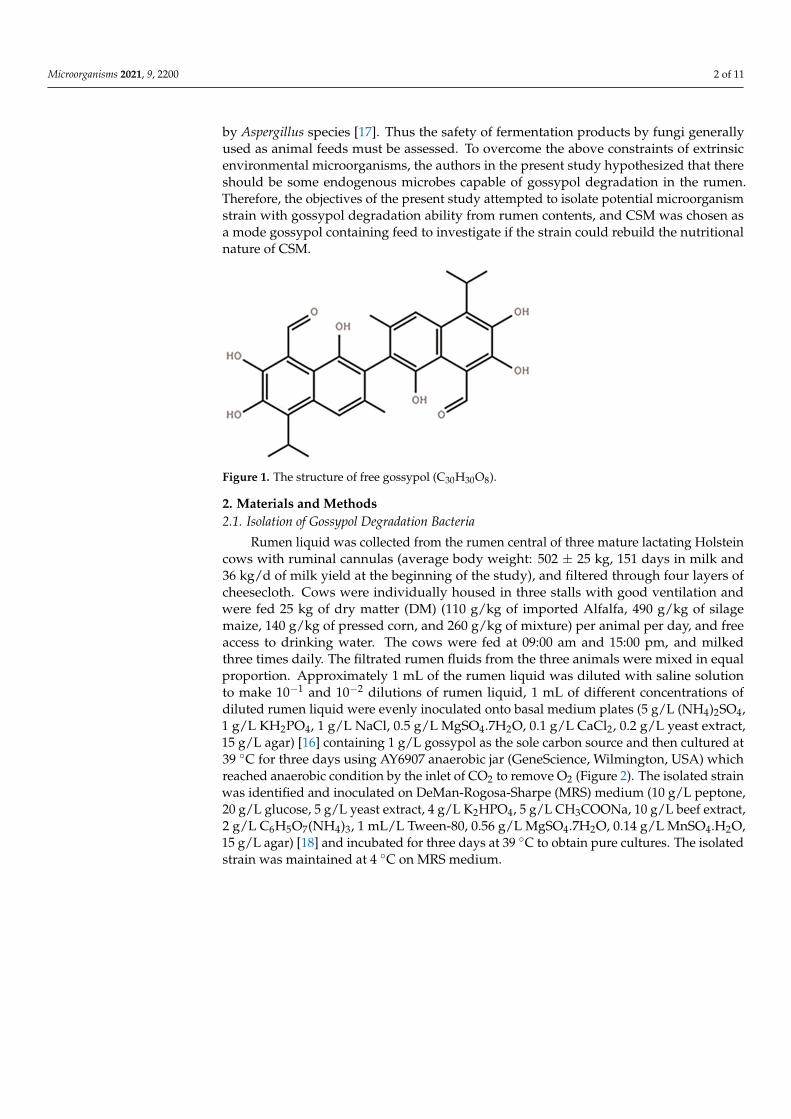
Gossypol (C30H30O8), a polyphenolic compound (Figure 1) produced by the cottonplant (Gossypium sp.), is one of the anti-nutritional factors to limit the application of cottonby-products in animal production [1,2]. For a long time, excessive intake of gossypol hasbeen found to cause growth depression [3], reproductive performances decline, anemia, aswell as other internal organ abnormalities in animals [4,5]. Adult ruminants were believedmore tolerant to gossypol than monogastric animals as well as young ruminants [6]. Suchtolerance was believed to be associated with the characteristics of gossypol binding tosoluble proteins and gossypol partially degrading ability of rumen microbes [7,8].
As for cottonseed meal (CSM), an important protein feedstuff, three methods havebeen commonly used in animal feed processing for many years to reduce gossypol’stoxicity, including i) mechanical processing [9], ii) chemical treatment [10,11], and iii) mi-crobial fermentation [12]. Among these methods, microbial fermentation was consideredthe most promising method for gossypol detoxification as well as a nutritional improve-ment compared with the other methods with subsiding effects, especially for CSM [13,14].Numerous fungi were reported to play an important role in CSM fermentation for gossy-pol degradation, including Candida tropicalis, Saccharomyces cerevisiae, Aspergillus oryzae,Aspergillus terreus, and Aspergillus niger [15,16]. Although these fungi were found capable ofgossypol degradation, some fungi metabolites were toxic to animals, e.g., citrinin secreted
Microorganisms 2021, 9, 2200. https://doi.org/10.3390/microorganisms9112200 https://www.mdpi.com/journal/microorganisms
Microorganisms 2021, 9, 2200 2 of 11
by Aspergillus species [17]. Thus the safety of fermentation products by fungi generallyused as animal feeds must be assessed. To overcome the above constraints of extrinsicenvironmental microorganisms, the authors in the present study hypothesized that thereshould be some endogenous microbes capable of gossypol degradation in the rumen.Therefore, the objectives of the present study attempted to isolate potential microorganismstrain with gossypol degradation ability from rumen contents, and CSM was chosen asa mode gossypol containing feed to investigate if the strain could rebuild the nutritionalnature of CSM.
Microorganisms 2021, 9, x FOR PEER REVIEW 2 of 12
Figure 1. The structure of free gossypol (C30H30O8).
As for cottonseed meal (CSM), an important protein feedstuff, three methods have been com-monly used in animal feed processing for many years to reduce gossypol’s toxicity, including i) mechanical processing [9], ii) chemical treatment [10,11], and iii) microbial fermentation [12]. Among these methods, microbial fermentation was considered the most promising method for gossypol detoxification as well as a nutritional improvement compared with the other methods with subsiding effects, especially for CSM [13,14]. Numerous fungi were reported to play an im-portant role in CSM fermentation for gossypol degradation, including Candida tropicalis, Saccharo-myces cerevisiae, Aspergillus oryzae, Aspergillus terreus, and Aspergillus niger [15,16]. Although these fungi were found capable of gossypol degradation, some fungi metabolites were toxic to animals, e.g., citrinin secreted by Aspergillus species [17]. Thus the safety of fermentation products by fungi generally used as animal feeds must be assessed. To overcome the above constraints of extrinsic environmental microorganisms, the authors in the present study hypothesized that there should be some endogenous microbes capable of gossypol degradation in the rumen. Therefore, the objec-tives of the present study attempted to isolate potential microorganism strain with gossypol deg-radation ability from rumen contents, and CSM was chosen as a mode gossypol containing feed to investigate if the strain could rebuild the nutritional nature of CSM.
2. Materials and Methods 2.1. Isolation of Gossypol Degradation Bacteria
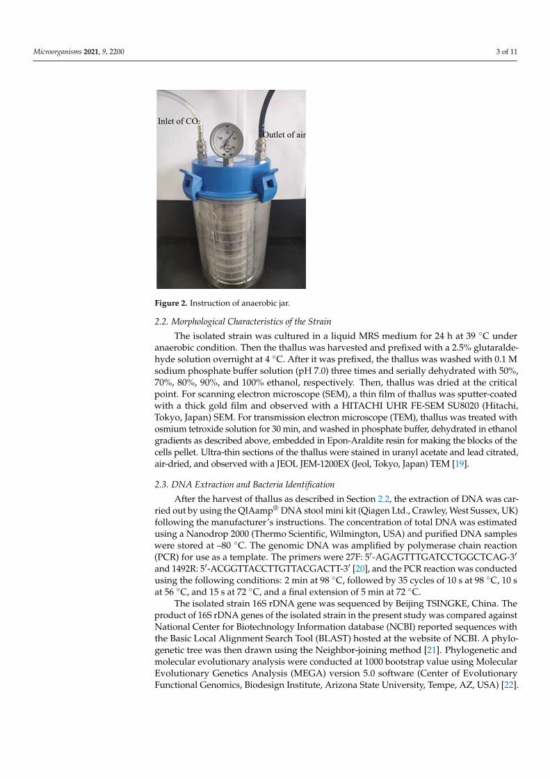
Rumen liquid was collected from the rumen central of three mature lactating Hol-stein cows with ruminal cannulas (average body weight: 502 ± 25 kg, 151 days in milk and 36 kg/d of milk yield at the beginning of the study), and filtered through four layers of cheesecloth. Cows were individually housed in three stalls with good ventilation and were fed 25 kg of dry matter (DM) (110 g/kg of imported Alfalfa, 490 g/kg of silage maize, 140 g/kg of pressed corn, and 260 g/kg of mixture) per animal per day, and free access to drinking water. The cows were fed at 09:00 am and 15:00 pm, and milked three times daily. The filtrated rumen fluids from the three animals were mixed in equal proportion. Ap-proximately 1 mL of the rumen liquid was diluted with saline solution to make 10−1 and 10−2 dilutions of rumen liquid, 1 mL of different concentrations of diluted rumen liquid were evenly inoculated onto basal medium plates (5 g/L (NH4)2SO4, 1 g/L KH2PO4, 1 g/L NaCl, 0.5 g/L MgSO4.7H2O, 0.1 g/L CaCl2, 0.2 g/L yeast extract, 15 g/L agar) [16] containing 1 g/L gossypol as the sole carbon source and then cultured at 39 °C for three days using AY6907 anaerobic jar (GeneScience, Wilmington, USA) which reached anaerobic condi-tion by the inlet of CO2 to remove O2 (Figure 2). The isolated strain was identified and inoculated on DeMan-Rogosa-Sharpe (MRS) medium (10 g/L peptone, 20 g/L glucose, 5 g/L yeast extract, 4 g/L K2HPO4, 5 g/L CH3COONa, 10 g/L beef extract, 2 g/L C6H5O7(NH4)3, 1 mL/L Tween-80, 0.56 g/L MgSO4.7H2O, 0.14 g/L MnSO4.H2O, 15 g/L agar) [18] and incu-bated for three days at 39 °C to obtain pure cultures. The isolated strain was maintained at 4 °C on MRS medium.
Figure 1. The structure of free gossypol (C30H30O8).
2. Materials and Methods2.1. Isolation of Gossypol Degradation Bacteria
Rumen liquid was collected from the rumen central of three mature lactating Holsteincows with ruminal cannulas (average body weight: 502 ± 25 kg, 151 days in milk and36 kg/d of milk yield at the beginning of the study), and filtered through four layers ofcheesecloth. Cows were individually housed in three stalls with good ventilation andwere fed 25 kg of dry matter (DM) (110 g/kg of imported Alfalfa, 490 g/kg of silagemaize, 140 g/kg of pressed corn, and 260 g/kg of mixture) per animal per day, and freeaccess to drinking water. The cows were fed at 09:00 am and 15:00 pm, and milkedthree times daily. The filtrated rumen fluids from the three animals were mixed in equalproportion. Approximately 1 mL of the rumen liquid was diluted with saline solutionto make 10−1 and 10−2 dilutions of rumen liquid, 1 mL of different concentrations ofdiluted rumen liquid were evenly inoculated onto basal medium plates (5 g/L (NH4)2SO4,1 g/L KH2PO4, 1 g/L NaCl, 0.5 g/L MgSO4.7H2O, 0.1 g/L CaCl2, 0.2 g/L yeast extract,15 g/L agar) [16] containing 1 g/L gossypol as the sole carbon source and then cultured at39 ◦C for three days using AY6907 anaerobic jar (GeneScience, Wilmington, USA) whichreached anaerobic condition by the inlet of CO2 to remove O2 (Figure 2). The isolated strainwas identified and inoculated on DeMan-Rogosa-Sharpe (MRS) medium (10 g/L peptone,20 g/L glucose, 5 g/L yeast extract, 4 g/L K2HPO4, 5 g/L CH3COONa, 10 g/L beef extract,2 g/L C6H5O7(NH4)3, 1 mL/L Tween-80, 0.56 g/L MgSO4.7H2O, 0.14 g/L MnSO4.H2O,15 g/L agar) [18] and incubated for three days at 39 ◦C to obtain pure cultures. The isolatedstrain was maintained at 4 ◦C on MRS medium.
Microorganisms 2021, 9, 2200 3 of 11Microorganisms 2021, 9, x FOR PEER REVIEW 3 of 12
Figure 2. Instruction of anaerobic jar.
2.2. Morphological Characteristics of the Strain The isolated strain was cultured in a liquid MRS medium for 24 h at 39 °C under
anaerobic condition. Then the thallus was harvested and prefixed with a 2.5% glutaralde-hyde solution overnight at 4 °C. After it was prefixed, the thallus was washed with 0.1 M sodium phosphate buffer solution (pH 7.0) three times and serially dehydrated with 50%, 70%, 80%, 90%, and 100% ethanol, respectively. Then, thallus was dried at the critical point. For scanning electron microscope (SEM), a thin film of thallus was sputter-coated with a thick gold film and observed with a HITACHI UHR FE-SEM SU8020 (Hitachi, To-kyo, Japan) SEM. For transmission electron microscope (TEM), thallus was treated with osmium tetroxide solution for 30 min, and washed in phosphate buffer, dehydrated in ethanol gradients as described above, embedded in Epon-Araldite resin for making the blocks of the cells pellet. Ultra-thin sections of the thallus were stained in uranyl acetate and lead citrated, air-dried, and observed with a JEOL JEM-1200EX (Jeol, Tokyo, Japan) TEM [19].
2.3. DNA Extraction and Bacteria Identification After the harvest of thallus as described in Section 2.2, the extraction of DNA was
carried out by using the QIAamp® DNA stool mini kit (Qiagen Ltd., Crawley, West Sus-sex, UK) following the manufacturer’s instructions. The concentration of total DNA was estimated using a Nanodrop 2000 (Thermo Scientific, Wilmington, USA) and purified DNA samples were stored at –80 °C. The genomic DNA was amplified by polymerase chain reaction (PCR) for use as a template. The primers were 27F: 5′-AGAGTTT-GATCCTGGCTCAG-3′ and 1492R: 5′-ACGGTTACCTTGTTACGACTT-3′ [20], and the PCR reaction was conducted using the following conditions: 2 min at 98 °C, followed by 35 cycles of 10 s at 98 °C, 10 s at 56 °C, and 15 s at 72 °C, and a final extension of 5 min at 72 °C.
The isolated strain 16S rDNA gene was sequenced by Beijing TSINGKE, China. The product of 16S rDNA genes of the isolated strain in the present study was compared against National Center for Biotechnology Information database (NCBI) reported se-quences with the Basic Local Alignment Search Tool (BLAST) hosted at the website of NCBI. A phylogenetic tree was then drawn using the Neighbor-joining method [21]. Phy-logenetic and molecular evolutionary analysis were conducted at 1000 bootstrap value using Molecular Evolutionary Genetics Analysis (MEGA) version 5.0 software (Center of
Figure 2. Instruction of anaerobic jar.
2.2. Morphological Characteristics of the Strain
The isolated strain was cultured in a liquid MRS medium for 24 h at 39 ◦C underanaerobic condition. Then the thallus was harvested and prefixed with a 2.5% glutaralde-hyde solution overnight at 4 ◦C. After it was prefixed, the thallus was washed with 0.1 Msodium phosphate buffer solution (pH 7.0) three times and serially dehydrated with 50%,70%, 80%, 90%, and 100% ethanol, respectively. Then, thallus was dried at the criticalpoint. For scanning electron microscope (SEM), a thin film of thallus was sputter-coatedwith a thick gold film and observed with a HITACHI UHR FE-SEM SU8020 (Hitachi,Tokyo, Japan) SEM. For transmission electron microscope (TEM), thallus was treated withosmium tetroxide solution for 30 min, and washed in phosphate buffer, dehydrated in ethanolgradients as described above, embedded in Epon-Araldite resin for making the blocks of thecells pellet. Ultra-thin sections of the thallus were stained in uranyl acetate and lead citrated,air-dried, and observed with a JEOL JEM-1200EX (Jeol, Tokyo, Japan) TEM [19].
2.3. DNA Extraction and Bacteria Identification
After the harvest of thallus as described in Section 2.2, the extraction of DNA was car-ried out by using the QIAamp® DNA stool mini kit (Qiagen Ltd., Crawley, West Sussex, UK)following the manufacturer’s instructions. The concentration of total DNA was estimatedusing a Nanodrop 2000 (Thermo Scientific, Wilmington, USA) and purified DNA sampleswere stored at –80 ◦C. The genomic DNA was amplified by polymerase chain reaction(PCR) for use as a template. The primers were 27F: 5′-AGAGTTTGATCCTGGCTCAG-3′
and 1492R: 5′-ACGGTTACCTTGTTACGACTT-3′ [20], and the PCR reaction was conductedusing the following conditions: 2 min at 98 ◦C, followed by 35 cycles of 10 s at 98 ◦C, 10 sat 56 ◦C, and 15 s at 72 ◦C, and a final extension of 5 min at 72 ◦C.
The isolated strain 16S rDNA gene was sequenced by Beijing TSINGKE, China. Theproduct of 16S rDNA genes of the isolated strain in the present study was compared againstNational Center for Biotechnology Information database (NCBI) reported sequences withthe Basic Local Alignment Search Tool (BLAST) hosted at the website of NCBI. A phylo-genetic tree was then drawn using the Neighbor-joining method [21]. Phylogenetic andmolecular evolutionary analysis were conducted at 1000 bootstrap value using MolecularEvolutionary Genetics Analysis (MEGA) version 5.0 software (Center of EvolutionaryFunctional Genomics, Biodesign Institute, Arizona State University, Tempe, AZ, USA) [22].
Microorganisms 2021, 9, 2200 4 of 11
2.4. Growth Study of Isolated Bacteria in Different Carbon Source
Growth experiments were conducted with different carbon sources. After the isolatedstrain was anaerobically cultured in MRS medium for 24 h as described in Section 2.2, 1 mLcultures were inoculated onto 500 mL liquid MRS medium which used glucose or gossypol(1 g/L) as carbon resource, respectively. Then, the inoculated MRS mediums were mixedwell and divided 2 mL into each tube. All of these tubes were anaerobically incubated at39 ◦C, bacteria growth of 3 tubes was determined by optical density at 600 nm every 1 husing a spectrophotometer.
2.5. Solid-State Fermentation and Chemical Analysis
A representative CSM sample that contained 500 mg/g gossypol was dried at 65 ◦Cfor 24 h in a forced-air oven and ground to pass through a 0.25-mm screen. The nutritivecomposition of CSM sample included 164 g/kg of neutral detergent fiber (NDF), 115 g/kgof acid detergent fiber (ADF) and 500 g/kg of crude protein (CP) in DM. Then the CSMsample was autoclave sterilized at 121 ◦C for 25 min. The isolated strain was culturedin liquid MRS medium for 24 h at 39 ◦C under anaerobic condition, and 1.5 mL culturedmedium was diluted to 15 mL by liquid MRS medium. For each fermentation period,a 15 g CSM sample was inoculated with 15 mL of the diluted cultured medium in each10 cm culture dish, resulting in 5% inoculum level and 50% moisture in dry matter (DM).All of these culture dishes (5 incubation time × 3 replicates) were put in a anaerobic jar,purged with CO2 to obtain anaerobic condition, and anaerobically incubated at 39 ◦C for1, 2, 3, 4, and 5 days, respectively. Meanwhile, to prevent the confusion of the effect ofmedium contents on fermentation results, a 15 g CSM sample was inoculated with 15 mLliquid MRS medium without isolated strain in three replicates and incubated at the sameconditions as above for five days.
After incubation was completed, CSM samples were dried in an oven at 65 ◦C for 48 hfor later analysis. Crude proteinwas determined according to AOAC methods [23], aminoacids were assayed using a Hitachi L-8800 instrument (Hitachi, Tokyo, Japan). Neutraldetergent fiber and acid detergent fiber were determined following the method of VanSoest et al. [24]. Lactic acid was determined by High Performance Liquid Chromatography(HPLC) according to the methods of Cira et al. [25]. Gossypol content was determined asthe methods of Wang et al. [26]: 1.5 g sample in 15 mL acetone was ultrasound 30 min at40 ◦C, centrifuged and collected the supernatant, repeat the above processes three times,the extraction was combined and filtrated with 0.45 µm microporous, rotary evaporated,dissolved by acetonitrile −0.2% phosphoric acid solution and fixed capacity to 2.5 mL. Thecontent of gossypol was quantified by HPLC with a Wufeng analytical instrument (WufengCo., Ltd., Shanghai, China). The analytical column was a symmetry reversed-phase C18column (250 × 4.6 mm, 5 µm, pH 2–8, Waters, Milford, MA, USA). The mobile phasewas 85:15 (v/v) acetonitrile −0.2% phosphoric acid solution at a flow rate of 1 mL/min.Injections were 20 µL, and the gossypol was detected at 235 nm.
2.6. Statistical Analysis
Statistical analyses were completed using the general linear model procedure ofSAS [27]. Standard errors (S.E.M) of means, adjusted by the Tukey method, were estimatedby the least square means procedure of SAS [27]. Significance was declared at p < 0.05unless otherwise noted.
3. Results3.1. Morphological Characteristics
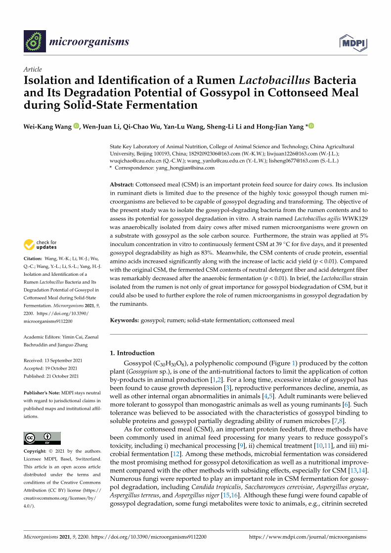
A single strain was isolated from the rumen liquid on basal medium plates containing 1 g/Lgossypol as the sole carbon source after three days of anaerobic fermentation (Figure 3A), and49.25% of gossypol in the medium disappeared. The appearance of colonies on the basalmedium plates was rough and dirty white with wrinkly surfaces and irregular shapes.
Microorganisms 2021, 9, 2200 5 of 11
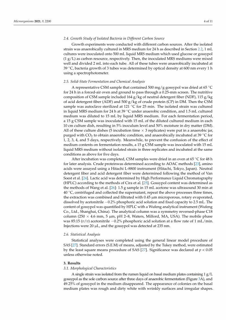
The bacteria were positive by Gram-staining (Figure 3B) and confirmed visible rod-shapedbacteria elements by both SEM and TEM observations (Figure 3C,D).
Microorganisms 2021, 9, x FOR PEER REVIEW 5 of 12
3. Results 3.1. Morphological Characteristics
A single strain was isolated from the rumen liquid on basal medium plates contain-ing 1 g/L gossypol as the sole carbon source after three days of anaerobic fermentation (Figure 3A), and 49.25% of gossypol in the medium disappeared. The appearance of col-onies on the basal medium plates was rough and dirty white with wrinkly surfaces and irregular shapes. The bacteria were positive by Gram-staining (Figure 3B) and confirmed visible rod-shaped bacteria elements by both SEM and TEM observations (Figure 3C,D).
Figure 3. Morphological identification of the L. agilis WWK129 strain. (A) The colony characteristics of this strain; (B) the microstructure characteristics of this strain at 1000 magnification; (C) the scan-ning electron microscopical observation of this strain; (D) the transmission electron microscopical observation of this strain.
3.2. 16S rDNA Homology The genomic DNA of the isolated strain was successfully extracted and amplified
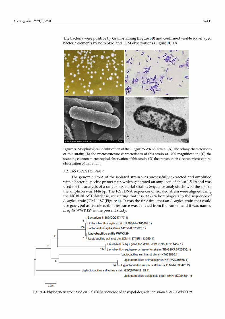
with a bacteria-specific primer pair, which generated an amplicon of about 1.5 kb and was used for the analysis of a range of bacterial strains. Sequence analysis showed the size of the amplicon was 1446 bp. The 16S rDNA sequences of isolated strain were aligned using the NCBI-BLAST database, indicating that it is 99.72% homologous to the sequence of L. agilis strain JCM 1187 (Figure 4). It was the first time that an L. agilis strain that could use gossypol as its sole carbon resource was isolated from the rumen, and it was named L. agilis WWK129 in the present study.
Figure 3. Morphological identification of the L. agilis WWK129 strain. (A) The colony characteristicsof this strain; (B) the microstructure characteristics of this strain at 1000 magnification; (C) thescanning electron microscopical observation of this strain; (D) the transmission electron microscopicalobservation of this strain.
3.2. 16S rDNA Homology
The genomic DNA of the isolated strain was successfully extracted and amplifiedwith a bacteria-specific primer pair, which generated an amplicon of about 1.5 kb and wasused for the analysis of a range of bacterial strains. Sequence analysis showed the size ofthe amplicon was 1446 bp. The 16S rDNA sequences of isolated strain were aligned usingthe NCBI-BLAST database, indicating that it is 99.72% homologous to the sequence ofL. agilis strain JCM 1187 (Figure 4). It was the first time that an L. agilis strain that coulduse gossypol as its sole carbon resource was isolated from the rumen, and it was namedL. agilis WWK129 in the present study.
Microorganisms 2021, 9, x FOR PEER REVIEW 6 of 12
Figure 4. Phylogenetic tree based on 16S rDNA sequence of gossypol-degradation strain L. agilis WWK129.
3.3. Growth Study There is no obvious difference in bacteria density in different carbon sources in the
lag phase. Bacteria in different mediums both exponentially increased after 6 h of incuba-tion, but the maintenance time of the log phase in gossypol was significantly shorter than it in glucose. There was an obvious decline phase of bacteria in glucose but not in gossy-pol. The final bacteria density of L. agilis WWK129 in glucose was significantly greater than it in gossypol (Figure 5).
Figure 5. Growth curve of L. agilis WWK129 in carbon source of gossypol and glucose. OD600, op-tical density of incubated medium at 600 nm.
3.4. Gossypol Disappearance and Nutrient Shifts of Fermented CSM Different culture periods were chosen to evaluate the effect of solid-state fermenta-
tion by L. agilis WWK129 strain on the nutritive value of CSM. Compared with the control, the gossypol content of fermented CSM decreased obviously, gossypol degradability in-creased significantly along with the prolongation of fermentation and it reached over 80% after five days of anaerobic fermentation (Figure 6). The NDF and ADF contents of CSM decreased significantly after fermentation (p < 0.01), while CP and lactic acid content were significantly higher than the control (p < 0.05) (Figure 6 and Table 1). It was evident that
Figure 4. Phylogenetic tree based on 16S rDNA sequence of gossypol-degradation strain L. agilis WWK129.

Microorganisms 2021, 9, 2200 6 of 11
3.3. Growth Study
There is no obvious difference in bacteria density in different carbon sources in the lagphase. Bacteria in different mediums both exponentially increased after 6 h of incubation,but the maintenance time of the log phase in gossypol was significantly shorter than it inglucose. There was an obvious decline phase of bacteria in glucose but not in gossypol.The final bacteria density of L. agilis WWK129 in glucose was significantly greater than it ingossypol (Figure 5).
Microorganisms 2021, 9, x FOR PEER REVIEW 6 of 12
Figure 4. Phylogenetic tree based on 16S rDNA sequence of gossypol-degradation strain L. agilis WWK129.
3.3. Growth Study There is no obvious difference in bacteria density in different carbon sources in the
lag phase. Bacteria in different mediums both exponentially increased after 6 h of incuba-tion, but the maintenance time of the log phase in gossypol was significantly shorter than it in glucose. There was an obvious decline phase of bacteria in glucose but not in gossy-pol. The final bacteria density of L. agilis WWK129 in glucose was significantly greater than it in gossypol (Figure 5).
Figure 5. Growth curve of L. agilis WWK129 in carbon source of gossypol and glucose. OD600, op-tical density of incubated medium at 600 nm.
3.4. Gossypol Disappearance and Nutrient Shifts of Fermented CSM Different culture periods were chosen to evaluate the effect of solid-state fermenta-
tion by L. agilis WWK129 strain on the nutritive value of CSM. Compared with the control, the gossypol content of fermented CSM decreased obviously, gossypol degradability in-creased significantly along with the prolongation of fermentation and it reached over 80% after five days of anaerobic fermentation (Figure 6). The NDF and ADF contents of CSM decreased significantly after fermentation (p < 0.01), while CP and lactic acid content were significantly higher than the control (p < 0.05) (Figure 6 and Table 1). It was evident that
Figure 5. Growth curve of L. agilis WWK129 in carbon source of gossypol and glucose. OD600,optical density of incubated medium at 600 nm.
3.4. Gossypol Disappearance and Nutrient Shifts of Fermented CSM
Different culture periods were chosen to evaluate the effect of solid-state fermentationby L. agilis WWK129 strain on the nutritive value of CSM. Compared with the control, thegossypol content of fermented CSM decreased obviously, gossypol degradability increasedsignificantly along with the prolongation of fermentation and it reached over 80% after fivedays of anaerobic fermentation (Figure 6). The NDF and ADF contents of CSM decreasedsignificantly after fermentation (p < 0.01), while CP and lactic acid content were significantlyhigher than the control (p < 0.05) (Figure 6 and Table 1). It was evident that most aminoacids of CSM improved markedly by fermentation of L. agilis WWK129, whereas Asparticacid, Serine, Glutamic acid, Proline, and Arginine decreased. Among essential aminoacids, levels of Phenylalanine increased the most, while among non-essential amino acids,Alanine increased the most.
Microorganisms 2021, 9, x FOR PEER REVIEW 7 of 12
most amino acids of CSM improved markedly by fermentation of L. agilis WWK129, whereas Aspartic acid, Serine, Glutamic acid, Proline, and Arginine decreased. Among essential amino acids, levels of Phenylalanine increased the most, while among non-es-sential amino acids, Alanine increased the most.
Figure 6. Gossypol content and lactic acid yield of cottonseed meal fermented by L. agilis WWK129.
Table 1. Nutritive value of cottonseed meal fermented by L. agilis WWK129 (%, DM).
Item 1 Fermented CSM at Different
Incubation Time S.E.M p Value
Con 2 1 Day 2 Day 3 Day 4 Day 5 Day Time L Q NDF 16.3 a 15.3 ab 14.9 ab 14.6 ab 13.6 bc 12.7 c 0.46 <0.01 0.45 0.04 ADF 11.3 a 6.8 b 6.5 c 6.4 cd 6.3 de 6.1 e 0.05 <0.01 <0.01 <0.01 CP 57.6 b 60.2 a 60.7 a 60.9 a 61.3 a 61.7 a 0.41 0.03 0.81 <0.01
Aspartic acid
4.28 a 4.24 ab 4.24 ab 4.20 bc 4.17 d 4.17 d 0.011 <0.01 0.29 <0.01
Threonine 1.29 c 1.36 c 1.36 c 1.44 b 1.51 a 1.55 a 0.020 <0.01 <0.01 0.01 Serine 1.77 a 1.64 b 1.65 b 1.65 b 1.65 b 1.67 b 0.028 0.10 0.42 0.20
Glutamic acid 11.36 a 10.74 ab 10.52 b 10.42 b 10.32 b 9.94 b 0.222 0.04 0.50 0.01
Proline 1.46 a 1.28 b 1.27 b 1.19 b 1.07 c 0.99 c 0.029 <0.01 0.11 <0.01 Glycine 1.87 1.90 1.91 1.92 1.93 1.99 0.034 0.36 0.31 0.13 Alanine 1.76 c 1.78 c 1.80 c 1.87 b 1.98 a 1.98 a 0.021 <0.01 <0.01 0.10 Cysteine 0.74 b 0.74 b 0.76 ab 0.78 ab 0.79 a 0.79 a 0.016 0.09 0.07 0.09 Valine 2.11 d 2.18 cd 2.35 bc 2.40 ab 2.48 ab 2.54 a 0.050 <0.01 0.01 <0.01
Methionine 0.56 c 0.60 c 0.61 c 0.68 b 0.74 a 0.75 a 0.016 <0.01 <0.01 0.01 Isoleucine 1.53 d 1.53 d 1.58 cd 1.64 bc 1.67 ab 1.73 a 0.019 <0.01 0.01 <0.01 Leucine 2.71 d 2.79 c 2.80 c 2.85 b 2.87 ab 2.91 a 0.033 <0.01 <0.01 <0.01 Tyrosine 1.52 c 1.53 bc 1.56 abc 1.58 ab 1.60 a 1.61 a 0.015 0.02 0.06 0.05
Phenylalanine 2.53 d 2.75 c 2.81 bc 2.90 b 3.21 a 3.21 a 0.037 <0.01 <0.01 <0.01
Lysine 1.90 b 1.91 b 1.93 b 2.18 a 2.28 a 2.31 a 0.044 <0.01 <0.01 0.04 Histidine 1.27 1.25 1.27 1.28 1.27 1.27 0.016 0.59 0.19 0.35 Arginine 5.66 a 5.53 ab 5.44 ab 5.47 ab 5.21 bc 5.09 c 0.089 0.03 0.40 0.12
Figure 6. Gossypol content and lactic acid yield of cottonseed meal fermented by L. agilis WWK129.

Microorganisms 2021, 9, 2200 7 of 11
Table 1. Nutritive value of cottonseed meal fermented by L. agilis WWK129 (%, DM).
Item 1Fermented CSM at Different Incubation Time
S.E.Mp Value
Con 2 1 Day 2 Day 3 Day 4 Day 5 Day Time L Q
NDF 16.3 a 15.3 ab 14.9 ab 14.6 ab 13.6 bc 12.7 c 0.46 <0.01 0.45 0.04ADF 11.3 a 6.8 b 6.5 c 6.4 cd 6.3 de 6.1 e 0.05 <0.01 <0.01 <0.01CP 57.6 b 60.2 a 60.7 a 60.9 a 61.3 a 61.7 a 0.41 0.03 0.81 <0.01
Aspartic acid 4.28 a 4.24 ab 4.24 ab 4.20 bc 4.17 d 4.17 d 0.011 <0.01 0.29 <0.01Threonine 1.29 c 1.36 c 1.36 c 1.44 b 1.51 a 1.55 a 0.020 <0.01 <0.01 0.01
Serine 1.77 a 1.64 b 1.65 b 1.65 b 1.65 b 1.67 b 0.028 0.10 0.42 0.20Glutamic acid 11.36 a 10.74 ab 10.52 b 10.42 b 10.32 b 9.94 b 0.222 0.04 0.50 0.01
Proline 1.46 a 1.28 b 1.27 b 1.19 b 1.07 c 0.99 c 0.029 <0.01 0.11 <0.01Glycine 1.87 1.90 1.91 1.92 1.93 1.99 0.034 0.36 0.31 0.13Alanine 1.76 c 1.78 c 1.80 c 1.87 b 1.98 a 1.98 a 0.021 <0.01 <0.01 0.10Cysteine 0.74 b 0.74 b 0.76 ab 0.78 ab 0.79 a 0.79 a 0.016 0.09 0.07 0.09
Valine 2.11 d 2.18 cd 2.35 bc 2.40 ab 2.48 ab 2.54 a 0.050 <0.01 0.01 <0.01Methionine 0.56 c 0.60 c 0.61 c 0.68 b 0.74 a 0.75 a 0.016 <0.01 <0.01 0.01Isoleucine 1.53 d 1.53 d 1.58 cd 1.64 bc 1.67 ab 1.73 a 0.019 <0.01 0.01 <0.01Leucine 2.71 d 2.79 c 2.80 c 2.85 b 2.87 ab 2.91 a 0.033 <0.01 <0.01 <0.01Tyrosine 1.52 c 1.53 bc 1.56 abc 1.58 ab 1.60 a 1.61 a 0.015 0.02 0.06 0.05
Phenylalanine 2.53 d 2.75 c 2.81 bc 2.90 b 3.21 a 3.21 a 0.037 <0.01 <0.01 <0.01Lysine 1.90 b 1.91 b 1.93 b 2.18 a 2.28 a 2.31 a 0.044 <0.01 <0.01 0.04
Histidine 1.27 1.25 1.27 1.28 1.27 1.27 0.016 0.59 0.19 0.35Arginine 5.66 a 5.53 ab 5.44 ab 5.47 ab 5.21 bc 5.09 c 0.089 0.03 0.40 0.12a,b,c,d,e Values in a line within the same class without a common superscript are significantly different (p < 0.05). 1 ADF, acid detergent fiber;CP, crude protein; NDF, neutral detergent fiber; L, linear; Q, quadratic. 2 Con, cottonseed meal inoculated with MRS medium without theisolated strain to prevent the confusion of the effect of medium contents on fermentation results.
4. Discussion4.1. Identification of L. agilis WWK129
Many times, rumen microorganisms have been demonstrated to be capable of gossy-pol biodegradation ability. Chen et al. [28] and Zhang et al. [29] had isolated Bacillus strainsfrom the rumen with high activity of gossypol degradation successively. In the presentstudy, the strain named L. agilis WWK129 which could use gossypol as its sole carbonresource was isolated from the rumen of dairy cows and identified according to morpholog-ical and molecular methods. Our previous study had revealed that there was a promotingeffect of gossypol on the activity of Firmicutes bacteria [30], and the isolated Lactobacillusbelongs to Firmicutes, which was consistent with the result of previous research. AlthoughL. casei and L. plantarum were found to degrade gossypol in previous studies [31,32], therewas no report so far about gossypol biodegradation by Lactobacillus strain in the rumen,and this is the first time that a Lactobacillus strain could degrade gossypol isolated from therumen in the present study.
Lactobacillus species are common constituents of gastrointestinal tracts [33], they havebeen used as direct-fed probiotics [34] and as silage inoculation [35]. The beneficial effectsof Lactobacillus on the production performance of dairy cows included increase in milkyield, improvement of feed efficiency, and increase in daily weight gain [36]. In ruminants,Lactobacillus bacteria are generally only prevalent in young animals before the rumen hasproperly developed. To date, Lactobacillus bacteria commonly found in the rumen of adultdairy cows included L. ruminis, L. vitulinus, L. acidophilus, L. casei, L. fermentum, L. plantarum,L. buchneri, L. brevis, L. cellobiosus, L. helveticus, and L. salivarius [37]. L. agilis is firstly isolatedfrom municipal sewage in 1981, and it is commonly known as motile Lactobacillus speciesfrom sewage and chicken guts [38,39], it was demonstrated that L. agilis played a key rolein the degradation of sinigrin, cholesterol, and glucosinolates [40,41]. There was limitedknowledge about its function in ruminants, Zhu et al. [42] had isolated several L. agilisfrom the feces of dairy calves, and found that these bacteria readily degraded long chaininulin. To date, this is the first time found that L. agilis presented the ability of gossypol
Microorganisms 2021, 9, 2200 8 of 11
degradation, the successful isolation of L. agilis from the rumen would be helpful for abetter understanding of its detoxification mechanism of gossypol in the future.
The method of isolating bacteria from the rumen by screening medium is commonlyused for research on the function of rumen bacteria. In the present study, L. agilis WWK129statin was cultivated in a basal medium using gossypol as the only carbon source, andabout 50% of gossypol in basal medium plates disappeared after three days of anaerobicfermentation, which demonstrated that gossypol could provide the carbon source for itsgrowth by biodegradation rather than transformation. The exponential phase of growthcurve is a stage in which microbial cells are divides at the maximal rate and the natureof the medium is being used as quickly as possible. According to the growth curve ofL. agilis WWK129 in different carbon resources, the maintained time and slope of expo-nential phase in gossypol was smaller than glucose, which noted that L. agilis WWK129grow faster in glucose instead of gossypol. It was assumed that a variety of environmentalconditions cause the death of microorganisms, such as the depletion of essential nutrientsin the decline phase. The optical density of the medium containing glucose decreasedsignificantly after 16 h of incubation, this was mainly due to the decrease of nutrientcomposition of medium and the increase of cell metabolites which inhibited the growth ofincubated bacteria. There was no decline phase in medium contained gossypol after 24 hof incubation and the final density of L. agilis WWK129 in stationary phase was greater inmedium contained glucose, which revealed that the biodegradation and transformationrate of gossypol was relatively slow and difficult, and the utilization efficiency of gossypolwas lower than glucose.
4.2. Effect of L. agilis WWK129 Fermentation on the Nutritional Value of CSM
In the present study, fiber content of control group inoculated with MRS mediumwithout L agilis WWK129 were consistent with the original CSM sample, while CP con-tent was significant higher than original state, this could be due to the peptone in MRSmedium which improved the CP level of control group. According to the results ofChen et al. [28] and Zhang et al. [29], Bacillus strains isolated from the rumen appliedin optimum fermentation conditions, the gossypol content in solid-state fermented CSMdecreased by 80%, relative to the control. In the current study, it was found that over than80% gossypol in CSM could be degraded by L. agilis WWK129 in solid-state fermentationafter five days, which was relatively greater than previous research, it could be due to thedifferent fermentation conditions, CSM variety, and gossypol biodegradation ability ofstains. The disappearance of gossypol could be due to the biodegradation of microbialenzymes secreted by L. agilis WWK129 and gossypol, its metabolites could also be used byL. agilis WWK129 as a carbon resource as well. Wu [43] investigated the effect of solid-statefermentation by three types of L. acidophilus on the nutritional value of CSM and foundthat lactic acid and essential amino acids content of fermented CSM were greater than thecontrol, which was consistent with the results of the present study. The improvement of CPand amino acids content in fermented CSM was mainly due to the growth of microflora,as well as the increase of cellular protein and microbial enzymes secreted by L. agilisWWK129, which could promote the nutritive value and utilization efficiency of proteinin CSM. The fiber content of fermented CSM was significantly lower than that of CSMwithout inoculum, it was noted that L. agilis WWK129 could be helpful for the degradationof fiber, and the reduction of fiber could provide carbon resources for the growth of L. agilisWWK129. Lactic acid is a key end-product of Lactobacillus fermentation, and lactic acidcontent persistently increased along with the incubation reflected that the fermentationconditions in the present study were suitable for the growth of L. agilis WWK129.
Solid-state microbial fermentation is an important method used for gossypol detoxifica-tion in the cotton by-products process, and Candida tropicalis and Lactobacillus plantarum werecommonly used in the fermentation of CSM feed sources in previous research. Zhang et al. [44]found that gossypol degradability in CSM fermented by C. tropicalis ZD-3 reached 94.6%after 48 h, CP content increased by about 11% compared with the control, total amino
Microorganisms 2021, 9, 2200 9 of 11
acids, and essential amino acids content of fermented CSM increased by 27% and 29%,respectively. Khalaf and Meleigy [15] investigated the effect of fungi on gossypol content ofCSM by solid-state fermentation and found that C. tropicalis could degrade 86.18% gossypolafter 72 h of incubation at 30 ◦C. Hong [45] found that gossypol degradability in CSM with5% inoculum of L. plantarum reached 40% after 60 h solid-state fermentation at 30 ◦C. Thereis no report about feed resource fermented by L. agilis in previous, the results of the currentstudy have verified the feasibility of L. agilis used in solid-state fermentation of CSM andprovided more bacteria selection for the microbial fermentation of cotton by-products inthe future.
Lactobacillus strains were approved for use as probiotics for both humans and an-imals [46,47], and the colonization of Lactobacillus strains in the gut epithelium couldreduce the risk of pathogenic bacterial infection by the production of lactic acid and othermetabolites [48]. Lactic acid in fermented CSM feed sources can increase acidity, inhibit thegrowth of spoilage bacteria and prevent the mildew of feed. In addition, all of this researchdemonstrated that the fermentation of Lactobacillus strain can significantly increase thecontent of essential amino acids and improve aminol acids balance. Consequently, the useof L. agilis WWK129 in CSM fermentation can not only reduce the toxicity of gossypol andpromote nutritive value, but also be conducive to the health of animals.
5. Conclusions
Collectively, the present study noted that L. agilis WWK129, firstly isolated from therumen contents, presented the great potential for gossypol biodegradation. Gossypoldisappearance rate in cottonseed meal reached up to 80% after 5 days of anaerobic fermen-tation by the isolated strain, meanwhile, neutral detergent fiber and acid detergent fibercontent decreased by about 4% and 5%, respectively, crude protein content increased byabout 4% and most essential amino acids content improved significantly, suggesting thatthe L. agilis WWK129 has promising application prospect in gossypol detoxification andmicrobial fermentation of cottonseed meal, and it could provide a basis for further analysisof gossypol biodegradation in the rumen.
Author Contributions: Conceptualization, W.-K.W. and Y.-L.W.; methodology, W.-K.W.; software,W.-J.L.; validation, Q.-C.W.; formal analysis, W.-K.W.; investigation, Y.-L.W.; resources, W.-J.L. andQ.-C.W.; data curation, W.-K.W.; writing—original draft preparation, W.-K.W.; writing—reviewand editing, S.-L.L. and H.-J.Y.; visualization, H.-J.Y.; supervision, H.-J.Y.; project administration,H.-J.Y.; funding acquisition, S.-L.L. All authors have read and agreed to the published version ofthe manuscript.
Funding: This research was funded by the Key Research and Development Project of Ningxia HuiAutonomous Region, grant number 2018BBF33006.
Institutional Review Board Statement: The study was conducted according to the guidelines of theDeclaration of Helsinki, and approved by the Institutional Review Board of Institutional Animal CareCommittee of China Agricultural University (protocol code CAU20171014-1 and date of approval 25September 2020).
Data Availability Statement: Data from the study are available in NCBI-GenBank under accessionnumber MZ 779227.
Conflicts of Interest: The authors declare no conflict of interest.
References1. Santos, J.E.P.; Villasenor, M.; Depeters, E.J.; Robinson, P.H.; Baldwin, B.C., Jr. Type of cottonseed and level of gossypol in diets of
lactating dairy cows: Effects on lactation performance and plasma gossypol. J. Dairy Sci. 2002, 85, 1491–1501. [CrossRef]2. Krempl, C.; Heidel-Fischer, H.M.; Jiménez-Alemán, G.H.; Reichelt, M.; Menezes, R.C.; Boland, W.; Vogel, H.; Heckel, D.G.; Joußen, N.
Gossypol toxicity and detoxification in Helicoverpa armigera and Heliothis virescens. Insect Biochem. Mol. Biol. 2016, 78, 69–77.[CrossRef] [PubMed]
3. Robinson, P.H.; Getachew, G.; De Peters, E.J.; Calhoun, M.C. Influence of variety and storage for up to 22 days on nutrientcomposition and gossypol level of Pima cottonseed (Gossypium spp.). Anim. Feed. Sci. Technol. 2001, 91, 149–156. [CrossRef]
Microorganisms 2021, 9, 2200 10 of 11
4. Velasquez-Pereira, J.; Arechiga, C.F.; McDowell, L.R.; Hansen, P.J.; Chenoweth, P.J.; Calhoun, M.C.; Risco, C.A.; Batra, T.R.;Williams, S.N.; Wilkinson, N.S. Effects of gossypol from cottonseed meal and dietary vitamin E on the reproductive characteristicsof superovulated beef heifers. J. Anim. Sci. 2002, 80, 2485–2492. [CrossRef] [PubMed]
5. Santos, J.E.P.; Villaseñor, M.; Robinson, P.H.; Depeters, E.J.; Holmberg, C.A. Type of Cottonseed and Level of Gossypol in Diets ofLactating Dairy Cows: Plasma Gossypol, Health, and Reproductive Performance. J. Dairy Sci. 2003, 86, 892–905. [CrossRef]
6. Wang, X.; Howell, C.P.; Chen, F.; Yin, J.J.; Jiang, Y.M. Gossypol—A polyphenolic compound from cotton plant. Adv. Food Nutr.Res. 2009, 58, 215–263.
7. Reiser, R.; Fu, H.C. The Mechanism of Gossypol Detoxification by Ruminant Animals. J. Nutr. 1962, 76, 215–218. [CrossRef][PubMed]
8. Feng, Y.; Wang, Y. The nutrition value and metabolism of cottonseed in ruminants. Feed. Res. 2011, 4, 13–16.9. Rahma, E.H.; Rao, M.S.N. Gossypol Removal and Functional Properties of Protein Produced by Extraction of Glanded Cottonseed
with Different Solvents. J. Food Sci. 1984, 49, 1057–1060. [CrossRef]10. Tabatabai, F.; Golian, A.; Salarmoeini, M. Determination and detoxification methods of cottonseed meal gossypol for broiler
chicken rations. Agric. Sci. Technol. 2002, 16, 3–15.11. Nagalakshmi, D.; Sastry, V.R.B.; Pawde, A. Rumen fermentation patterns and nutrient digestion in lambs fed cottonseed meal
supplemental diets. Anim. Feed. Sci. Technol. 2003, 103, 1–14. [CrossRef]12. Wu, X.Y.; Chen, J.X. The utilization of microbes to break down FG in cottonseed meal. Sci. Agric. Sin. 1989, 22, 82–86.13. Weng, X.-Y.; Sun, J.-Y. Biodegradation of free gossypol by a new strain of Candida tropicalis under solid state fermentation:
Effects of fermentation parameters. Process. Biochem. 2006, 41, 1663–1668. [CrossRef]14. Weng, X.-Y.; Sun, J.-Y. Kinetics of biodegradation of free gossypol by Candida tropicalis in solid-state fermentation. Biochem. Eng.
J. 2006, 32, 226–232. [CrossRef]15. Khalaf, M.A.; Meleigy, S.A. Reduction of free gossypol levels in cottonseed meal by microbial treatment. Int. J. Agric. Biol. 2008,
10, 185–190.16. Yang, X.; Sun, J.-Y.; Guo, J.-L.; Weng, X.-Y. Identification and proteomic analysis of a novel gossypol-degrading fungal strain.
J. Sci. Food Agric. 2011, 92, 943–951. [CrossRef] [PubMed]17. DeRuiter, J.; Jacyno, J.M.; Davis, R.A.; Cutler, H.G. Studies on Aldose Reductase Inhibitors from Fungi. I. Citrinin and Related
Benzopyran Derivatives. J. Enzym. Inhib. 1992, 6, 201–210. [CrossRef]18. De Mann, J.C.; Rogosa, M.; Sharpe, M.E. A medium for cultivation of Lactobacill. J. Appl. Microb. 1969, 23, 130–135.19. Perfumo, A.; Elsaesser, A.; Littmann, S.; Foster, R.A.; Kuypers, M.M.; Cockell, C.S.; Kminek, G. Epifluorescence, SEM, TEM and
nanoSIMS image analysis of the cold phenotype of Clostridium psychrophilum at subzero temperatures. FEMS Microbiol. Ecol.2014, 90, 869–882. [CrossRef]
20. Zhang, Y.; Luan, H.; Wei, Z.; Hao, Z.; Xi, R.; Liao, X. Exploiting of honey-associated Bacillus strains as plant-growth promotingbacteria for enhancing barley growth in rare earth tailings. Ann. Microbiol. 2015, 66, 559–568. [CrossRef]
21. Saitou, N.; Nei, M. The neighbor-joining method: A new method for reconstructing phylogenetic trees. Mol. Biol. Evol. 1987, 4,406–425. [CrossRef] [PubMed]
22. Tamura, K.; Dudley, J.; Nei, M.; Kumar, S. Mega 4: Molecular evolutionary gnentics analysis (MEGA) software version 4.0. Mol.Biol. Evol. 2007, 24, 1596–1599. [CrossRef] [PubMed]
23. AOAC. Official Methods of Analysis of AOAC International, 18th ed.; Association of Official Analytical Chemists International:Gaithersburg, MD, USA, 2005.
24. Van Soest, P.J.; Robertson, J.B.; Lewis, B.A. Methods for dietary fiber, neutral fiber, and nonstarch polysaccharides in relation toanimal nutrition. J. Dairy Sci. 1991, 74, 3583–3597. [CrossRef]
25. Cira, L.A.; Huerta, S.; Hall, G.M.; Shirai, K. Pilot scale lactic acid fermentation of shrimp wastes for chitin recovery. Process.Biochem. 2002, 37, 1359–1366. [CrossRef]
26. Wang, W.-K.; Wang, Y.-L.; Li, W.-J.; Wu, Q.-C.; Li, S.-L.; Yang, H.-J. Gossypol Exhibited Higher Detrimental Effect on RuminalFermentation Characteristics of Low-Forage in Comparison with High-Forage Mixed Feeds. Toxics 2021, 9, 51. [CrossRef]
27. SAS. Statistical Analytical System (SAS) Users Guides: Statistics; Version 8.2; Statistica Analysis Institute: Cary, NC, USA, 1999.28. Chen, L.; Zh, Y.; Ch, X.; Ch, M.; Meng, X.; Cai, H. Gossypol Degradation Strain Coming from Ruminant Rumens and Application
Thereof. CN 104328063A, 4 February 2015.29. Zhang, Y.; Zhang, Z.; Dai, L.; Liu, Y.; Cheng, M.; Chen, L. Isolation and characterization of a novel gossypol-degrading
bac-teriabacillus subtilis strain rumen bacillus subtilis. Asian Austral. J. Anim. 2018, 31, 63–70. [CrossRef] [PubMed]30. Wang, W.-K.; Wang, Y.-L.; Li, W.-J.; Wu, Q.-C.; Yang, K.-L.; Li, S.-L.; Yang, H.-J. In situ rumen degradation characteristics and
bacterial colonization of whole cottonseed, cottonseed hull and cottonseed meal with different gossypol content. AMB Express2021, 11, 91. [CrossRef] [PubMed]
31. Tang, H.W.; Wu, Y.F.; Yao, X.H.; Wang, X. Method for Removing Free Gossypol from Cotton Dregs by Biological FermentationMethod. CN102138632B, 24 July 2013.
32. Chen, W.; Tian, F.W.; Zhai, Q.X.; Sun, Y.Y.; Zhao, J.X.; Wang, G.; Zhang, Q.X.; Liu, X.M.; Zhang, B.X.; Zhang, H. LactobacillusPlantarum Capable of Degrading Gossypol and Application of Lactobacillus Plantarum. CN105255793A, 20 January 2016.
33. Heeney, D.D.; Gareau, M.G.; Marco, M.L. Intestinal Lactobacillus in health and disease, a driver or just along for the ride? Curr.Opin. Biotechnol. 2018, 49, 140–147. [CrossRef] [PubMed]
Microorganisms 2021, 9, 2200 11 of 11
34. Lebeer, S.; Bron, P.A.; Marco, M.L.; Pijkeren, J.P.V.; Motherway, M.O.C.; Hill, C.; Pot, B.; Roos, S.; Klaenhammer, T. Identificationof probiotic effector molecules: Present state and future perspectives. Curr. Opin. Biotechnol. 2018, 49, 217–223. [CrossRef]
35. Muck, R.E.; Nadeau, E.M.G.; McAllister, T.A.; Contreras-Govea, F.E.; Santos, M.C.; Kung, L., Jr. Silage review: Recent advancesand future uses of silage additives. J. Dairy Sci. 2018, 101, 3980–4000. [CrossRef]
36. Doyle, N.; Mbandlwa, P.; Kelly, W.J.; Attwood, G.; Li, Y.; Ross, R.P.; Stanton, C.; Leahy, S. Use of Lactic Acid Bacteria to Re-duceMethane Production in Ruminants, a Critical Review. Front. Microbiol. 2019, 10, 2207. [CrossRef]
37. Feng, Y.L. Ruminant Nutrition; Science Press: Beijing, China, 2004; pp. 10–11.38. Stephenson, D.P.; Moore, R.J.; Allison, G.E. Transformation of, and Heterologous Protein Expression in, Lactobacillus agilis and
Lactobacillus vaginalis Isolates from the Chicken Gastrointestinal Tract. Appl. Environ. Microbiol. 2011, 77, 220–228. [CrossRef]39. Kajikawa, A.; Midorikawa, E.; Masuda, K.; Kondo, K.; Irisawa, T.; Igimi, S.; Okada, S. Characterization of flagellins isolated from
a highly motile strain of Lactobacillus agilis. BMC Microbiol. 2016, 16, 1–8. [CrossRef]40. Palop, M.L.; Smiths, J.P.; Brink, B.T. Degradation of sinigrin by Lactobacillus agilis strain R16. Int. J. Food Microbiol. 1995, 26,
219–229. [CrossRef]41. Luangin, V. Influence of Human Gut Microbiota on the Metabolic Fate of Glucosinolates; Imperial College London: London, UK, 2013.42. Zhu, Y.; Liu, J.; Lopez, J.M.; Mills, D.A. Inulin Fermentation by Lactobacilli and Bifidobacteria from Dairy Calves. Appl. Environ.
Microbiol. 2020, 87, e01738-20. [CrossRef] [PubMed]43. Wu, Y.Y. A Study on Screening and Breeding of High-Yield Lactic Acid Bacteria and the Nutritional Characteristics of Fermented Cottonseed
Meal; Shihezi University: Shihezi, China, 2013.44. Zhang, W.-J.; Xu, Z.-R.; Zhao, S.-H.; Sun, J.-Y.; Yang, X. Development of a microbial fermentation process for detoxification of
gossypol in cottonseed meal. Anim. Feed. Sci. Technol. 2007, 135, 176–186. [CrossRef]45. Hong, Z. Microbial Solid Fermentation to Reduce Free Gossypol Content in the Cottonseed Meal; Wuhan Polytechnic University: Wuhan,
China, 2016.46. Peran, L.; Sierra, S.; Comalada, M.; Lara-Villoslada, F.; Bailón, E.; Nieto, A.; Concha, Á.; Olivares, M.; Zarzuelo, A.; Xaus, J.; et al.
A comparative study of the preventative effects exerted by two probiotics, Lactobacillus reuteri and Lactobacillus fermentum, inthe trinitrobenzene sulfonic acid model of rat colitis. Br. J. Nutr. 2007, 97, 96–103. [CrossRef]
47. Slizewska, K.; Chlebicz-Wójcik, A.; Nowak, A. Probiotic Properties of New Lactobacillus Strains Intended to Be Used as FeedAdditives for Monogastric Animals. Probiotics Antimicrob. Proteins 2021, 13, 146–162. [CrossRef]
48. Slover, C.M.; Danziger, L. Lactobacillus: A Review. Clin. Microbiol. Newsl. 2008, 30, 23–27. [CrossRef]