The Extensive Reading Foundation , s Guide to Extensive Reading
Upload
independentCategory
view
3download
0
Downloaded from www.microbiologyresearch.org by
IP: 23.22.24.125
On: Sat, 13 Feb 2016 10:30:56
The complete genomes of Lactobacillusplantarum and Lactobacillus johnsoniireveal extensive differences in chromosomeorganization and gene content
Jos Boekhorst,1 Roland J. Siezen,1,2,4 Marie-Camille Zwahlen,3
David Vilanova,3 Raymond D. Pridmore,3 Annick Mercenier,3
Michiel Kleerebezem,2,4 Willem M. de Vos,2 Harald Brussow3
and Frank Desiere3
Correspondence
Jos Boekhorst
1Center for Molecular and Biomolecular Informatics, 6525ED, Nijmegen, The Netherlands
2Wageningen Centre for Food Sciences, 6700 AN Wageningen, The Netherlands
3Nestle Research Center, Nestec SA, 1000 Lausanne 26, Switzerland
4NIZO food research, 6710 BA Ede, The Netherlands
Received 9 June 2004
Revised 23 July 2004
Accepted 29 July 2004
The first comprehensive comparative analysis of lactobacilli was done by comparing the
genomes of Lactobacillus plantarum (3?3 Mb) and Lactobacillus johnsonii (2?0 Mb). L. johnsonii
is predominantly found in the gastrointestinal tract, while L. plantarum is also found on plants
and plant-derived material, and is used in a variety of industrial fermentations. The L. plantarum
and L. johnsonii chromosomes have only 28 regions with conservation of gene order, totalling
about 0?75 Mb; these regions are not co-linear, indicating major chromosomal rearrangements.
Metabolic reconstruction indicates many differences between L. johnsonii and L. plantarum:
numerous enzymes involved in sugar metabolism and in biosynthesis of amino acids, nucleotides,
fatty acids and cofactors are lacking in L. johnsonii. Major differences were seen in the number
and types of putative extracellular proteins, which are of interest because of their possible role
in host–microbe interactions. The differences between L. plantarum and L. johnsonii, both in
genome organization and gene content, are exceptionally large for two bacteria of the same
genus, emphasizing the difficulty in taxonomic classification of lactobacilli.
INTRODUCTION
Lactobacilli belong to the lactic acid bacteria (LAB) and aremembers of the low-GC content Gram-positive bacteria.
Many are used in starter cultures for food and feedfermentations, and several species are frequently encoun-tered in the human gastrointestinal tract (Vaughan et al.,2002). Some strains of LAB are marketed as probiotics,which are claimed to positively affect human and/or animalhealth (Braun-Fahrlander et al., 2002; Link-Amster et al.,1994). However, not much is known about the mechan-isms by which these LAB affect the host.
Recently, the genomes of two members of the genusLactobacillus have been completely sequenced: Lactobacillusplantarum WCFS1 (Kleerebezem et al., 2003) and Lacto-bacillus johnsonii NCC533 (Pridmore et al., 2004). L.johnsonii NCC533, isolated from human faeces, has beenextensively studied for its probiotic activities, includingimmunomodulation (Haller et al., 2000a, 2000b) andinteraction with the human host (Ibnou-Zekri et al.,2003). L. plantarum WCFS1 was isolated from humansaliva. L. plantarum is a versatile bacterium that is found ina variety of ecological niches, ranging from vegetable andplant fermentations to the human gastrointestinal tract.
Abbreviations: BDI, base deviation index; CDS, coding sequence; COG,Clusters of Orthologous Group; KEGG, Kyoto Encyclopedia of Genesand Genomes; LAB, lactic acid bacteria; PTS, phosphotransferasesystem.
Details of the size and location of conserved gene clusters in L.plantarum and L. johnsonii may be found in Supplementary Table S1;the number of proteins of L. plantarum and L. johnsonii for all COGclasses in Supplementary Table S2; a KEGG comparison of majordifferences between L. plantarum and L. johnsonii in SupplementaryTable S3; L. johnsonii and L. plantarum API 50 test results in Supple-mentary Table S4; the redundancy of enzymes involved in pyruvatemetabolism in Supplementary Table S5; gene clusters encoding func-tionally related proteins present in L. plantarum but not in L. johnsoniiand vice versa in Supplementary Table S6; lists of proteins unique toeither L. plantarum or L. johnsonii in Supplementary Tables S7–S12;lists of proteins involved in the biosynthesis of polysaccharides,bacteriocins and prophages in Supplementary Table S13 with theonline version of this paper at http://mic.sgmjournals.org.
0002-7392 G 2004 SGM Printed in Great Britain 3601
Microbiology (2004), 150, 3601–3611 DOI 10.1099/mic.0.27392-0

Downloaded from www.microbiologyresearch.org by
IP: 23.22.24.125
On: Sat, 13 Feb 2016 10:30:56
This flexibility of L. plantarum is reflected by its relativelylarge genome size, a large number of proteins involved inregulation and transport functions, and a high metabolicpotential (Kleerebezem et al., 2003).
In order to expand our understanding of the molecularevolution, diversity, function and adaptation of lactobacillito specific environments, we have performed a whole-genome comparison of L. plantarum and L. johnsonii. Inaddition, we compared the proteins of these two organismsto the draft sequences of other LAB genomes (Klaenhammeret al., 2002). We provide a first comprehensive view ofdifferences on the genome level in lactobacilli, and evidencefor large genetic diversity in this genus. We identify featuresunderlying the large difference in genome size and genecontent in lactobacilli, and provide a first insight into theset of genes and functions which could be specific for lacticacid bacteria. This knowledge provides numerous leads fortargeted experimental verification of unique or commonphysiological properties.
METHODS
Genome sequences. Complete genome sequences of L. plantarumWCFS1 (Kleerebezem et al., 2003) accession number AL935263, L.johnsonii NCC533 (Pridmore et al., 2004) accession numberAE017198, Bacillus subtilis 128, Enterococcus faecalis V583, Listeriamonocytogenes EGDe and Lactococcus lactis IL1403 were obtainedfrom GenBank Entrez Genomes (http://www.ncbi.nlm.nih.gov/genomes/MICROBES/Complete.html). The gene ID numbers usedin the text to refer to specific L. plantarum and L. johnsonii genesare the same as those used in the original papers (Kleerebezem et al.,2003; Pridmore et al., 2004). Genome comparison with unfinishedLAB genome sequences utilized sequence data of Lactobacillus brevisATCC367, Lactobacillus casei ATCC334, Lactobacillus delbrueckiiATCCBAA-365, Lactobacillus gasseri ATCC-33323, Leuconostocmesenteroides ATCC-8293, Oenococcus oeni PSU-1 and Pediococcuspentosaceus from the ERGO database (http://ergo.integratedgenomics.com/ERGO/), originally produced by the US Department of EnergyJoint Genome Institute (http://www.jgi.doe.gov). For comparativepurposes, all species of the genus Lactobacillus, Lactococcus andLeuconostoc, as well as the bacteria Streptococcus thermophilus,Oenococcus oeni, Bifidobacterium longum and Pediococcus pentosaceus,are considered to be LAB (Klaenhammer et al., 2002). Fig. 1 showsthe 16S rRNA tree of the relevant organisms.
The coding sequences (CDSs) of the L. johnsonii genome have beenidentified using FrameD (Schiex et al., 2003), while the CDSs of theL. plantarum genome have been identified using Glimmer (Delcheret al., 1999), which could lead to some erroneous comparison ofthe CDSs. However, for both organisms the positions of CDSs onthe genome have been manually adjusted based on the presence ofa plausible ribosome-binding site and on BLAST alignments withhomologues (Kleerebezem et al., 2003; Pridmore et al., 2004), reducingthe impact of this difference in CDS identification. Moreover, forboth organisms the minimal size of a CDS was set at 30 codons.
Genome comparisons. Orthologous relationships were detectedby a previously described method (Snel et al., 2002) using the Smith& Waterman sequence comparison algorithm (Smith & Waterman,1981) against the NCBI Clusters of Orthologous Group (COG)database (Tatusov et al., 2001). The functional classification pro-vided by the COG database was used for the functional comparisonof L. plantarum and L. johnsonii on a genome-wide scale.
Homology relationships were established using BLASTP (Altschul et al.,1990) and Smith & Waterman sequence comparison. Homologueswere detected with a threshold of 1E210; a gene was consideredorganism specific when it had no Smith & Waterman hits at all, oronly hits with an e-score higher than 1E210 to proteins of otherorganisms in the non-redundant proteins databases (SWISS-PROT,TrREMBL and TrEMBL updates) (Boeckmann et al., 2003) or theLAB genomes taken from the ERGO database. Proteins wereconsidered LAB-specific when they did not have a Smith &Waterman hit with an e-score lower than 1E210 in a search againstSWISS-PROT, TrREMBL, TrEMBL updates and the LAB sequencestaken from the ERGO database.
Whole genomes were compared at the nucleotide level using theDotter software (Sonnhammer & Durbin, 1995) with default values. Abidirectional best-hit approach was used to identify genome synteny atthe protein level. The results of this analysis were visualized using theArtemis Comparison Tool (http://www.sanger.ac.uk/Software/ACT/).
Transporter classification was preformed according to the TC-DBscheme (Busch & Saier, 2002). All proteins were searched againstthe TC-DB Database Release 1.5.1 using BLASTP with a threshold of10E24, followed by manual curation: false positive hits were removedmanually when clear evidence suggested that they were not related totransport function.
Signal peptides were predicted using SignalP (Nielsen et al., 1997).
Base deviation analysis of genes was performed by calculating achi-squared index based on the expected and observed frequency foreach nucleotide (Tettelin et al., 2001).
Synchronizing annotation. The two genomes compared in thisstudy were initially analysed using different ontologies and annota-tions (Kleerebezem et al., 2003; Pridmore et al., 2004). To facilitatefunctional comparison of L. plantarum and L. johnsonii, the annota-tion of proteins found to be homologous, but having differentannotations in the two genomes, was manually verified andcorrected where necessary. This resulted in an improved annotationof both genomes, in particular for the functional class ‘regulation’and for the assignment of EC numbers, and made automated detec-tion of functional differences possible.
Fig. 1. 16S rRNA-based phylogenetic tree (unrooted).Sequences were extracted from the European rRNA database(Wuyts et al., 2004) and aligned using CLUSTAL W (Thompsonet al., 1994). The tree was visualized using TreeView (Page,1996).
3602 Microbiology 150
J. Boekhorst and others

Downloaded from www.microbiologyresearch.org by
IP: 23.22.24.125
On: Sat, 13 Feb 2016 10:30:56
Reconstruction of metabolic pathways. EC numbers wereextracted from the genome annotations and manually curated. Theywere then automatically mapped onto the Kyoto Encyclopedia ofGenes and Genomes (KEGG) metabolic pathways (Kanehisa et al.,2002) for visualization and identification of differences in metabo-lism between L. plantarum and L. johnsonii. In cases of predictedmissing key enzymes in one of the two organisms, a further effortwas made to identify homologous candidate enzymes by extensivemanual searches with BLASTP and HMMER (Eddy, 1996; Sonnhammeret al., 1998).
Sugar utilization. API 50 analysis of sugar utilization wasperformed using the supplier’s protocol (BioMerieux Benelux).Additional sugar fermentation profiles were obtained from theliterature (Fujisawa et al., 1992; Kleerebezem et al., 2003).
RESULTS AND DISCUSSION
General genome features
The main features of the genomes of L. plantarumWCFS1 and L. johnsonii NCC533 are shown in Table 1.L. plantarum WCFS1 has a genome of over 3?3 Mb, whichis exceptionally large for a Lactobacillus species, since thegenome size of lactobacilli is generally between 1?8 and2?5 Mb (Klaenhammer et al., 2002). At the DNA level,L. johnsonii and L. plantarum are very divergent. A DNAdot plot comparison of the two genomes (data not shown)shows a low overall sequence similarity. Much closer DNAhomology has been observed between L. johnsonii andother members of the Lactobacillus acidophilus family(Pridmore et al., 2004).
Homologous proteins
Evolutionary distances can be measured by the comparisonof gene repertoires (Tamames, 2001). Closely related speciesshare a large proportion of genes; in contrast, distantlyrelated species should have lost a significant fraction of thegenes inherited from their last common ancestor, resultingin a low proportion of shared genes. An overview of thepercentage of homologues shared between the variousgenomes is given in Table 2. Of all the proteins encoded
by the L. johnsonii genome, 83% have a homologue inL. gasseri, 70% have a homologue in L. plantarum, 62%have a homologue in E. faecalis and 58% have a homologuein L. monocytogenes. In contrast, when the L. plantarumgenome is used as query, the large difference in genomesize between L. plantarum and L. johnsonii leads to L.plantarum sharing more homologues with the largergenomes of E. faecalis (58%), L. monocytogenes (57%)and B. subtilis (52%), than with L. johnsonii (51%).
Genome synteny
On an evolutionary time scale, protein sequences are moreconserved than DNA sequences. It is possible to detectgene clusters encoding homologous proteins in relatedorganisms even where low-level DNA conservation makessequence alignment very difficult. These syntenic regionscan provide insight into functions of the proteins com-prising them: for example, genes already described inone organism might be annotated correctly in a secondorganism based on synteny. This principle has been used inthe prediction of gene function by several methods, suchas Rosetta Stone (Marcotte et al., 1999) and the conservedgene neighbours method (Dandekar et al., 1998; Overbeeket al., 1999). The selective advantage of physical proximityof genes for co-regulation makes some gene clusters lessprone to breakup than others, thus extending the range ofevolutionary distance over which sequence conservation isdetectable.
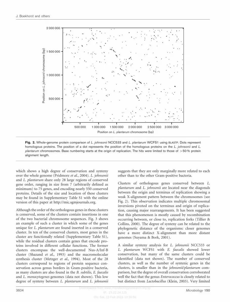
A dot plot comparison at the protein level of the genomesof L. plantarum and L. johnsonii (Fig. 2) shows no large-scale conservation of gene order, but only conservation ofgenes in clusters, confirming the relatively large phylo-genetic distance between L. plantarum and L. johnsonii.The lack of large-scale gene order conservation betweenL. plantarum and L. johnsonii is in strong contrast to thewhole chromosome alignment of L. johnsonii and L. gasseri,
Table 1. Genome features of L. plantarum WCFS1 and L.johnsonii NCC533
Genome feature Strain
L. plantarum
WCFS1
L. johnsonii
NCC533
Length (bp) 3 308 274 1 992 676
Coding density (%) 84?1 89?3
G+C content (%) 45?6 34?9
Predicted CDSs 3 009 1 821
tRNAs 62 79
rRNA operons 5 6
Phage genes 159 (2 prophages,
2 remnants)
54 (2 prophages,
1 remnant)
IS elements 15 14
Table 2. Homologous proteins in genomes of Gram-positivebacteria
The numbers indicate the percentage of the proteins in the query
genome with a homologue (BLAST hit with e-score higher than
1E210) in the other genome. Amino acid sequences of the species
in the header row are used as database, the sequences of the
species in the column as query.
Species Species
1 2 3 4 5 6 7
1 B. subtilis 2 43 29 30 40 37 49
2 E. faecalis 51 2 40 41 53 52 56
3 L. gasseri 53 63 2 85 69 59 58
4 L. johnsonii 54 63 83 2 70 60 58
5 L. plantarum 52 58 49 51 2 54 57
6 L. lactis 52 60 44 46 59 2 58
7 L. monocytogenes 65 60 42 44 58 53 2
http://mic.sgmjournals.org 3603
Comparative genomics of lactobacilli
Downloaded from www.microbiologyresearch.org by
IP: 23.22.24.125
On: Sat, 13 Feb 2016 10:30:56
which shows a high degree of conservation and syntenyover the whole genome (Pridmore et al., 2004). L. johnsoniiand L. plantarum share only 28 large regions of conservedgene order, ranging in size from 7 (arbitrarily defined asminimum) to 75 genes, and encoding nearly 550 conservedproteins. Details of the size and location of these clustersmay be found in Supplementary Table S1 with the onlineversion of this paper at http://mic.sgmjournals.org.
Although the order of the orthologous genes in these clustersis conserved, some of the clusters contain insertions in oneof the two bacterial chromosome sequences. Fig. 3 showsan example of such a cluster in which some of the genesunique for L. plantarum are found inserted in a conservedcluster. In ten of the conserved clusters, most genes in thecluster are functionally related (Supplementary Table S1),while the residual clusters contain genes that encode pro-teins involved in different cellular functions. The formerclusters encompass the well-documented Nus-A/Inf-Bcluster (Shazand et al., 1993) and the macromolecularsynthesis cluster (Metzger et al., 1994). Most of the 28clusters correspond to regions of protein sequence con-servation across genus borders in Gram-positive bacteria,as many clusters are also found in the B. subtilis, E. faecalisand L. monocytogenes genomes (data not shown). This lowdegree of synteny between L. plantarum and L. johnsonii
suggests that they are only marginally more related to eachother than to the other Gram-positive bacteria.
Clusters of orthologous genes conserved between L.plantarum and L. johnsonii are located near the diagonalsbetween the origin and terminus of replication showing aweak X-alignment pattern between the chromosomes (seeFig. 2). This observation indicates multiple chromosomalinversions pivoted on the terminus and origin of replica-tion, causing major rearrangements. It has been suggestedthat this phenomenon is mostly caused by recombinationoccurring between, or close to, replication forks (Tillier &Collins, 2000). The degree of synteny can be related to thephylogenetic distance of the organisms: closer genomeshave a more distinct X-alignment than more distantgenomes (Suyama & Bork, 2001).
A similar synteny analysis for L. johnsonii NCC533 orL. plantarum WCFS1 with E. faecalis showed lowerconservation, but many of the same clusters could beidentified (data not shown). The number of conservedclusters, as well as the number of syntenic genes in theclusters, is smaller than in the johnsonii/plantarum com-parison, but the degree of overall conservation corroboratedwell the fact that the genus Enterococcus is closely related tobut distinct from Lactobacillus (Klein, 2003). Very limited
Pos
ition
on
L. jo
hnso
nii c
hrom
osom
e (b
p)
1 000 000
1 500 000
2 000 000
500 000
500 000 1 000 000 1 500 000 2 000 000 2 500 000 3 000 000
Position on L. plantarum chromosome (bp)
Fig. 2. Whole-genome protein comparison of L. johnsonii NCC533 and L. plantarum WCFS1 using BLASTP. Dots representhomologous proteins. The position of a dot represents the position of the homologous proteins on the L. johnsonii and L.
plantarum chromosomes. Base numbering starts at the origin of replication. The hits were limited to those of >50% proteinalignment length.
3604 Microbiology 150
J. Boekhorst and others

Downloaded from www.microbiologyresearch.org by
IP: 23.22.24.125
On: Sat, 13 Feb 2016 10:30:56
synteny could be detected with L. lactis or streptococci(data not shown).
Phylogenetic trees based on 16S RNA (Fig. 1) or highlyconserved genes support the relatively large phylogeneticdistance between L. plantarum and L. johnsonii suggestedby the protein dot plot. An unrooted tree based on the atpDgene (part of the highly conserved ATP synthase cluster)shows L. johnsonii to be closely related to L. gasseri, but alsoshows a relatively large distance between L. johnsonii andL. plantarum (Siezen et al., 2004), in agreement with Fig. 1.The phylogenetic distance between L. plantarum and L.johnsonii is in fact similar to the distance between L.plantarum and E. faecalis. These findings re-emphasize thedifficulties in establishing the taxonomy of lactobacilli, andshow that the current classification of the Lactobacillusgenus, based on morphology and lactic acid production, isnot always supported by phylogenetic relationships basedon sequence homology and genome synteny.
Functional comparison of proteomes
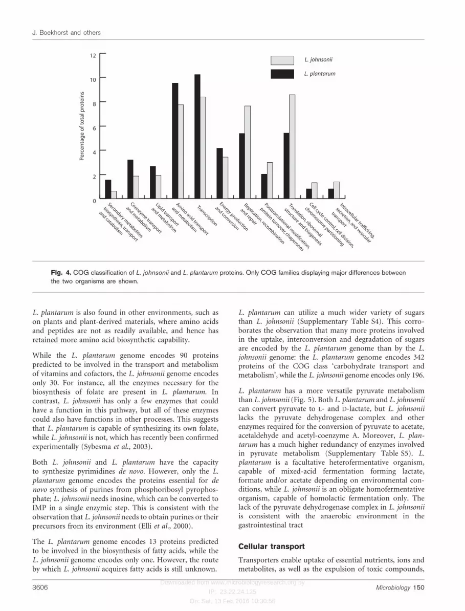
The percentage of the total number of proteins of L.plantarum and L. johnsonii belonging to selected COGfunctional classes is shown in Fig. 4. Only classes displayinglarge differences between the two organisms are shown;Supplementary Table S2 shows the number of proteins ofL. plantarum and L. johnsonii for all COG classes. Thisoverview gives an indication of the differences in focus onmetabolism and other cellular functions of these bacteria.Compared to L. johnsonii, L. plantarum has a relativelyhigh number of proteins for carbohydrate, amino acid andlipid metabolism. Due to its smaller genome size, L.johnsonii has a higher percentage of genes involved in‘core functions’ such as replication and translation.
Metabolic pathways
Metabolic reconstruction can provide insights into thedifferences and similarities in the metabolic potential ofL. plantarum and L. johnsonii, which can be helpful inboth explaining observed physiological differences betweenthe two species and in the design of experimental studiesto investigate genotype–phenotype relationships.
The mapping of enzymic functions on the metabolicpathways provided by the KEGG database resulted in theidentification of a set of enzymes required for knownbiochemical pathways. The main differences between L.johnsonii and L. plantarum are listed in SupplementaryTable S3. The classes and metabolic pathways that displaystriking differences between the two organisms will bedescribed in some detail below.
The L. plantarum genome encodes 268 proteins predictedto be involved in the metabolism and transport of aminoacids, while the L. johnsonii genomes encodes only 125.L. plantarum encodes the enzymes required for the bio-synthesis of all amino acids, with the exception of leucine,isoleucine and valine. In contrast, L. johnsonii is predictedto be incapable of synthesizing most, if not all, of the 20standard amino acids. This reflects the environmentalniches in which the bacteria live. L. johnsonii typically isfound only in the gut, although recent reports (Guan le et al.,2003; Meroth et al., 2003) suggest that L. johnsonii mightalso occur in other nutrient-rich environments, where itcan take up amino acids and peptides from its environ-ment. To this end, L. johnsonii has an extracellular,cell-bound proteinase to liberate these peptides fromproteinaceous substrates, and more intracellular peptidasesfor degradation of imported peptides than L. plantarum(Kleerebezem et al., 2003; Pridmore et al., 2004). In contrast,
Fig. 3. Example of a gene cluster conserved between L. johnsonii and L. plantarum. The two horizontal bars at the toprepresent the two strands of the L. johnsonii genome; the two at the bottom represent the two strands of the L. plantarum
genome. Horizontal arrows represent CDSs. Red CDSs have an orthologue in the other genome, whereas blue CDSs do not.The vertical bars connect orthologous genes. Almost all of the genes present in the L. johnsonii gene cluster are also presentin L. plantarum, but many of the genes in L. plantarum are not present in L. johnsonii. The genes unique to the L. plantarum
cluster include genes encoding four cell-envelope proteins, three proteins of the pyruvate dehydrogenase complex, and aputative L-lactate dehydrogenase.
http://mic.sgmjournals.org 3605
Comparative genomics of lactobacilli
Downloaded from www.microbiologyresearch.org by
IP: 23.22.24.125
On: Sat, 13 Feb 2016 10:30:56
L. plantarum is also found in other environments, such ason plants and plant-derived materials, where amino acidsand peptides are not as readily available, and hence hasretained more amino acid biosynthetic capability.
While the L. plantarum genome encodes 90 proteinspredicted to be involved in the transport and metabolismof vitamins and cofactors, the L. johnsonii genome encodesonly 30. For instance, all the enzymes necessary for thebiosynthesis of folate are present in L. plantarum. Incontrast, L. johnsonii has only a few enzymes that couldhave a function in this pathway, but all of these enzymescould also have functions in other processes. This suggeststhat L. plantarum is capable of synthesizing its own folate,while L. johnsonii is not, which has recently been confirmedexperimentally (Sybesma et al., 2003).
Both L. johnsonii and L. plantarum have the capacityto synthesize pyrimidines de novo. However, only the L.plantarum genome encodes the proteins essential for denovo synthesis of purines from phosphoribosyl pyrophos-phate; L. johnsonii needs inosine, which can be converted toIMP in a single enzymic step. This is consistent with theobservation that L. johnsonii needs to obtain purines or theirprecursors from its environment (Elli et al., 2000).
The L. plantarum genome encodes 13 proteins predictedto be involved in the biosynthesis of fatty acids, while theL. johnsonii genome encodes only one. However, the routeby which L. johnsonii acquires fatty acids is still unknown.
L. plantarum can utilize a much wider variety of sugarsthan L. johnsonii (Supplementary Table S4). This corro-borates the observation that many more proteins involvedin the uptake, interconversion and degradation of sugarsare encoded by the L. plantarum genome than by the L.johnsonii genome: the L. plantarum genome encodes 342proteins of the COG class ‘carbohydrate transport andmetabolism’, while the L. johnsonii genome encodes only 196.
L. plantarum has a more versatile pyruvate metabolismthan L. johnsonii (Fig. 5). Both L. plantarum and L. johnsoniican convert pyruvate to L- and D-lactate, but L. johnsoniilacks the pyruvate dehydrogenase complex and otherenzymes required for the conversion of pyruvate to acetate,acetaldehyde and acetyl-coenzyme A. Moreover, L. plan-tarum has a much higher redundancy of enzymes involvedin pyruvate metabolism (Supplementary Table S5). L.plantarum is a facultative heterofermentative organism,capable of mixed-acid fermentation forming lactate,formate and/or acetate depending on environmental con-ditions, while L. johnsonii is an obligate homofermentativeorganism, capable of homolactic fermentation only. Thelack of the pyruvate dehydrogenase complex in L. johnsoniiis consistent with the anaerobic environment in thegastrointestinal tract
Cellular transport
Transporters enable uptake of essential nutrients, ions andmetabolites, as well as the expulsion of toxic compounds,
0
2
4
6
8
10
12L. johnsonii
L. plantarum
Intracellular trafficking,
secretion, and vesicular
transport
Cell cycle control, cell division,
chromosom
e partitioning
Translation, ribosomal
structure and biogenesis
Posttranslational modification,
protein turnover, chaperones
Replication, recombination
and repair
Energy production
and conversion
Transcription
Amino acid transport
and metabolism
Lipid transport
and metabolism
Coenzyme transport
and metabolism
Secondary metabolites
biosynthesis, transport
and catabolism
Perc
enta
ge
of t
ota
l pro
tein
s
Fig. 4. COG classification of L. johnsonii and L. plantarum proteins. Only COG families displaying major differences betweenthe two organisms are shown.
3606 Microbiology 150
J. Boekhorst and others

Downloaded from www.microbiologyresearch.org by
IP: 23.22.24.125
On: Sat, 13 Feb 2016 10:30:56
cell-envelope macromolecules and the end products ofcellular metabolism. Putative transporters have been identi-fied in L. johnsonii and L. plantarum by comparison withthe Transport Classification DataBase (TC-DB; Busch &Saier, 2002) (Table 3). L. johnsonii contains 286 genesassociated with various transport systems, accounting formore than 15% of its total CDSs, which is proportionallyslightly more than L. plantarum WCSF1 (473 proteins,
13%). Both numbers compare well with other organismsof similar genome size living in nutrient-rich environments,such as the cheese starter Lactococcus lactis (11% trans-porters; Bolotin et al., 2001) and the oral pathogenStreptococcus mutans (15%; Ajdic et al., 2002).
The increased transport potential of L. plantarum isprimarily due to an increased redundancy of transport
Fig. 5. Pyruvate metabolism in L. johnsonii and L. plantarum. The figure is based on the pyruvate metabolism pathway fromthe KEGG database (Kanehisa et al., 2002). Open circles represent metabolites; the square boxes represent enzymes withtheir EC numbers. The colour of a box indicates the presence of the gene encoding that enzyme in L. plantarum (blue), inL. johnsonii (yellow) or in both (green). Not shown are enzymes and pathways that (i) are cytochrome dependent, (ii) do notoccur in bacteria, and (iii) have no known genes.
Table 3. Summary of transporters encoded in the genomes of L. plantarum and L. johnsonii
Transporter class Number in:
L. plantarum L. johnsonii
Channel proteins 22 10
Electrochemical potential-driven transporters 142 71
Primary active transporters* 195 148
Sugar-transporting phosphotransferase systems (PTS) 57 44
Transmembrane electron carriers 3 0
Accessory factors involved in transport 20 2
Incompletely characterized transport systems 34 11
Total 473 286
*Includes 147 and 105 ABC transporter proteins, respectively.
http://mic.sgmjournals.org 3607
Comparative genomics of lactobacilli

Downloaded from www.microbiologyresearch.org by
IP: 23.22.24.125
On: Sat, 13 Feb 2016 10:30:56
proteins. For instance, L. plantarum encodes six glycerol-uptake facilitator proteins, compared to a single protein inL. johnsonii. This observation suggests the importance ofglycerol uptake in L. plantarum.
The most notable electrochemical potential-driven trans-porters in L. johnsonii are two conjugated bile salt–protonsymporters (LJ0057 and LJ0058), which have been found tobe unique proteins of the Lactobacillus acidophilus groupof organisms (Pridmore et al., 2004). Striking differencesare found in the number of multidrug/oligosaccharidyl-lipid/polysaccharide flippase superfamily, the auxin-effluxcarrier family and the drug/metabolite transporter super-family of transporters in L. plantarum. The L. plantarumgenome encodes 5, 4 and 11 proteins belonging to thesefamilies, respectively, whereas the L. johnsonii genomeencodes only one protein of each family. Primary activetransporters (mainly ABC transporters: 147 and 105 pro-teins in L. plantarum and L. johnsonii, respectively) representthe largest group of transporters in both lactobacilli. InL. johnsonii and L. plantarum, 16 and 25 complete PEP-dependent, phosphoryl transfer-driven group translocators(phosphotransferase system, PTS) systems were identified,respectively, including multiple systems for the uptake ofglucose, mannose, fructose, and b-glucosides, and singlesystems for cellobiose, sucrose, and galactitol.
Extracellular proteins
Extracellular proteins are considered to be important forinteraction of bacteria with their environment, for examplein adhesion and communication. This makes them ofspecial interest in the case of lactobacilli, because they maybe involved in host–microbe and microbe–microbe inter-actions, such as in the gastrointestinal tract or on plantmaterials. Putative extracellular proteins of L. plantarumand L. johnsonii were identified by the presence of a Sec-pathway-dependent signal peptide. Both proteins that aresecreted into the environment and proteins that becomeattached to the cell surface fall into this category. The latterwere identified by searching for cell-anchoring domains,such as the N-terminal lipoprotein motif for anchoring tothe cell membrane (Sutcliffe & Russell, 1995) and theC-terminal LPxTG motif for anchoring to peptidoglycan(Navarre & Schneewind, 1999). The L. plantarum andL. johnsonii genomes are predicted to encode 211(Kleerebezem et al., 2003) and 117 putative extracellularproteins, respectively. Nearly 90% of these proteins inboth species are predicted to contain at least one type ofcell-wall anchoring domain.
A comparison of the putative extracellular proteins encodedin both genomes is summarized in Table 4. The set ofextracellular proteins of known function is very similar in
Table 4. Comparison of putative extracellular proteins encoded in the genomes of L. plantarumand L. johnsonii
Functional Class Number in:
L. plantarum L. johnsonii
ABC transporters, substrate-binding domain 30 20
Regulators* 5 5
Enzymes, known functions
Proteases 14 8
Transpeptidases 4 4
Cell-wall hydrolases 12 5
Other 5 6
Miscellaneous, known functions 8 4
Unknown function, present in both genomes
Homologues, singles 19 20
Homologues, familiesD
CSH1 family 8 5
CSH2 family 5 1
WY-domain family 4 2
Unknown function, present in single genome 97 37
Total 211 117
*Membrane-anchored proteins with extracellular transcriptional attenuator domain (Pfam PF03816).
DCSH1, cell-surface hydrolase family 1: contains active site triad Ser, Asp and His residues and consensus
GxSHG typical of hydrolases (e.g. lipase, esterase). CSH2, cell-surface hydrolase family 2: contains active site
triad Ser, Asp and His residues and consensus GxSMG typical of hydrolases (e.g. lipase, esterase). WY, C-
terminal cell-surface binding domain (Jankovic et al., 2003).
3608 Microbiology 150
J. Boekhorst and others
Downloaded from www.microbiologyresearch.org by
IP: 23.22.24.125
On: Sat, 13 Feb 2016 10:30:56
both lactobacilli, although L. plantarum has more para-logues for several of these known functions. However, themajority (55–65%) of putative extracellular proteins are ofunknown function (Table 4). Some of these are presentin both lactobacilli, either as single copies of orthologues,or as multiple copies (paralogues) belonging to differentfamilies. Two families of putative cell-surface hydrolases(CSH-1 and CSH-2) are detected which have sequencecharacteristics of lipases or esterases (Anthonsen et al.,1995; Wong & Schotz, 2002). It is striking to note that themajority of extracellular proteins with unknown functionare not shared by L. plantarum and L. johnsonii, but onlyoccur in one of the two bacteria.
The C-terminal LPxTG motif for covalent binding topeptidoglycan is present in 25 and 14 extracellular proteinsof L. plantarum and L. johnsonii, respectively. Generally,these are large, multi-domain, repeat-containing proteins(Kleerebezem et al., 2003; Pridmore et al., 2004). Again,there is very little homology between the LPxTG proteins ofL. plantarum and those of L. johnsonii, other than in thepeptidoglycan attachment motif.
Regulators
Regulatory proteins play an important role in the adapta-tion of an organism to different environments. The L.plantarum genome is predicted to encode 264 regulators(9?4% of all proteins), while the L. johnsonii genome hasonly 114 putative regulators (6%), as summarized inSupplementary Table S6. This agrees with the generalobservation that large genomes have a relatively highnumber of proteins involved in transcription and regula-tion (Konstantinidis & Tiedje, 2004; van Nimwegen, 2003).
Besides the difference in genome size, the different lifestylesof L. plantarum and L. johnsonii also contribute to thisdifference. The number of proteins predicted to be involved
in the regulation of sugar and energy metabolism isespecially high in L. plantarum. This is in agreement withthe differences found in sugar metabolism in the twoorganisms: L. plantarum can utilize a much wider varietyof sugars than L. johnsonii (Supplementary Table S4). L.plantarum with its free-living lifestyle needs to be capableof dealing withmany different environmental circumstances(Boneca et al., 2003), and apparently has both the metabo-lic capacity and the regulatory machinery to deal withadaptation to different niches, while L. johnsonii does notneed a complex regulatory apparatus because of therelatively stable environment in the gastrointestinal tract.
LAB-specific and unique genes
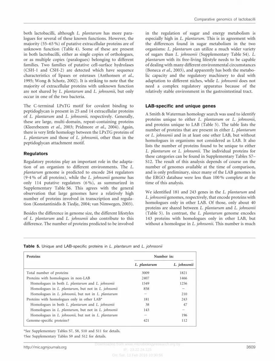
A Smith &Waterman homology search was used to identifyproteins unique to either L. plantarum or L. johnsonii,and proteins unique to LAB (Table 5). The table lists thenumber of proteins that are present in either L. plantarumor L. johnsonii and in at least one other LAB, but withouthomologues in organisms not considered as LAB. It alsolists the number of proteins found to be unique to eitherL. plantarum or L. johnsonii. The individual proteins forthese categories can be found in Supplementary Tables S7–S12. The result of this analysis depends of course on thenumber of genomes available at the time of comparison,and is only preliminary, since many of the LAB genomes inthe ERGO database were less than 100% complete at thetime of this analysis.
We identified 181 and 243 genes in the L. plantarum andL. johnsonii genomes, respectively, that encode proteins withhomologues only in other LAB. Of those, only about 40proteins are shared between L. plantarum and L. johnsonii(Table 5). In contrast, the L. plantarum genome encodes143 proteins with homologues only in other LAB, butwithout a homologue in L. johnsonii. This number is much
Table 5. Unique and LAB-specific proteins in L. plantarum and L. johnsonii
Proteins Number in:
L. plantarum L. johnsonii
Total number of proteins 3009 1821
Proteins with homologues in non-LAB 2407 1466
Homologues in both L. plantarum and L. johnsonii 1549 1256
Homologues in L. plantarum, but not in L. johnsonii 858 2
Homologues in L. johnsonii, but not in L. plantarum 2 210
Proteins with homologues only in other LAB* 181 243
Homologues in both L. plantarum and L. johnsonii 38 47
Homologues in L. plantarum, but not in L. johnsonii 143 2
Homologues in L. johnsonii, but not in L. plantarum 2 196
Genome-specific proteinsD 421 112
*See Supplementary Tables S7, S8, S10 and S11 for details.
DSee Supplementary Tables S9 and S12 for details.
http://mic.sgmjournals.org 3609
Comparative genomics of lactobacilli
Downloaded from www.microbiologyresearch.org by
IP: 23.22.24.125
On: Sat, 13 Feb 2016 10:30:56
lower than the 196 LAB-specific proteins encoded byL. johnsonii without homologues in L. plantarum, especiallyconsidering the relatively large size of the L. plantarumgenome compared to the L. johnsonii genome. This differ-ence is caused by the close relatedness of the L. johnsoniiand L. gasseri genomes; these two organisms share thesame niche and have a very similar genetic make-up andgenome organization (Pridmore et al., 2004). Moreover,this also explains the relatively low number of unique genesin L. johnsonii.
Many of the proteins present in L. plantarum but absent inL. johnsonii, or vice versa, are grouped in clusters on thegenome. A large number of these clustered unique genesencode functionally related proteins, such as those involvedin the biosynthesis of polysaccharides, bacteriocins andprophages (Supplementary Table S13). In L. plantarum,such clusters frequently have a high base-deviation index(BDI), suggesting horizontal transfer (Kleerebezem et al.,2003). In L. johnsonii however, only the polysaccharidebiosynthesis cluster (LJ1027–1047) has a high BDI.
Most of the proteins predicted to be LAB specific areof unknown function (Supplementary Tables S7–S12). Theidentification of structural features, such as signal peptides,transmembrane helices and cell-wall anchors, and con-served domains/motifs in these proteins, such as thoseinvolved in the binding of ATP, DNA and carbohydrates,could be used to predict their function and to identifypotentially interesting targets for future research. In thisway, the preliminary analysis of LAB-specific genes des-cribed here can serve as a starting point for a morecomprehensive study of LAB-specific proteins and geneclusters, once the complete genome sequences of manyLAB species become available (Klaenhammer et al., 2002).
Concluding remarks
The ability of L. plantarum to survive in many differentenvironments is reflected by the much more elaboratemetabolic, regulatory and transport machinery compared tothat of L. johnsonii. The differences between L. plantarumand L. johnsonii, both in genome organization and in genecontent, are exceptionally large for two bacteria of thesame genus (Suyama & Bork, 2001). Similar differenceshave been reported only in streptococci (Tettelin et al.,2002). This low degree of synteny between L. plantarum andL. johnsonii suggests that they are only marginally morerelated to each other than to other Gram-positive bacteria.These findings emphasize the difficulty in taxonomicclassification of lactobacilli.
Overall, the genome-wide comparison of two completeLactobacillus genomes has provided unique informationon the relatedness and differences between the two species.This has led to insight into the genomic adaptation toecological niches of L. plantarum and L. johnsonii, andprovides leads for targeted experimental studies.
ACKNOWLEDGEMENTS
This work was supported by a grant from the Netherlands Organi-
zation for Scientific Research, (NWO-BMI project 050.50.206). We
thank Douwe Molenaar for the calculation of BDI, Quinta
Helmer for the analysis of extracellular proteins and John Janssen
for the identification of conserved gene clusters. We thank
Dr T. Klaenhammer (North Carolina State University) for access
to the incomplete genome of Lactobacillus gasseri and Dr C. Tran
and Dr M. Saier (University of California in San Diego) for making
TC-DB available.
REFERENCES
Ajdic, D., McShan, W. M., McLaughlin, R. E. & 16 other authors
(2002). Genome sequence of Streptococcus mutans UA159, a cario-
genic dental pathogen. Proc Natl Acad Sci U S A 99, 14434–14439.
Altschul, S. F., Gish, W., Miller, W., Myers, E. W. & Lipman, D. J.
(1990). Basic local alignment search tool. J Mol Biol 215, 403–410.
Anthonsen, H. W., Baptista, A., Drablos, F., Martel, P., Petersen,
S. B., Sebastiao, M. & Vaz, L. (1995). Lipases and esterases: a review
of their sequences, structure and evolution. Biotechnol Annu Rev 1,
315–371.
Boeckmann, B., Bairoch, A., Apweiler, R. & 9 other authors
(2003). The SWISS-PROT protein knowledgebase and its supple-
ment TrEMBL in 2003. Nucleic Acids Res 31, 365–370.
Bolotin, A., Wincker, P., Mauger, S., Jaillon, O., Malarme, K.,
Weissenbach, J., Ehrlich, S. D. & Sorokin, A. (2001). The complete
genome sequence of the lactic acid bacterium Lactococcus lactis ssp.
lactis IL1403. Genome Res 11, 731–753.
Boneca, I. G., de Reuse, H., Epinat, J. C., Pupin, M., Labigne, A. &
Moszer, I. (2003). A revised annotation and comparative analysis of
Helicobacter pylori genomes. Nucleic Acids Res 31, 1704–1714.
Braun-Fahrlander, C., Riedler, J., Herz, U. & 12 other authors
(2002). Environmental exposure to endotoxin and its relation to
asthma in school-age children. N Engl J Med 347, 869–877.
Busch, W. & Saier, M. H., Jr (2002). The transporter classification
(TC) system, 2002. Crit Rev Biochem Mol Biol 37, 287–337.
Dandekar, T., Snel, B., Huynen, M. & Bork, P. (1998). Conservation
of gene order: a fingerprint of proteins that physically interact.
Trends Biochem Sci 23, 324–328.
Delcher, A. L., Harmon, D., Kasif, S., White, O. & Salzberg, S. L.
(1999). Improved microbial gene identification with GLIMMER.
Nucleic Acids Res 27, 4636–4641.
Eddy, S. R. (1996). Hidden Markov models. Curr Opin Struct Biol 6,
361–365.
Elli, M., Zink, R., Rytz, A., Reniero, R. & Morelli, L. (2000). Iron
requirement of Lactobacillus spp. in completely chemically defined
growth media. J Appl Microbiol 88, 695–703.
Fujisawa, T., Benno, Y., Yaeshima, T. & Mitsuoka, T. (1992).
Taxonomic study of the Lactobacillus acidophilus group, with
recognition of Lactobacillus gallinarum sp. nov. and Lactobacillus
johnsonii sp. nov. and synonymy of Lactobacillus acidophilus group
A3 (Johnson et al., 1980) with the type strain of Lactobacillus
amylovorus (Nakamura 1981). Int J Syst Bacteriol 42, 487–491.
Guan le, L., Hagen, K. E., Tannock, G. W., Korver, D. R., Fasenko,
G. M. & Allison, G. E. (2003). Detection and identification of Lacto-
bacillus species in crops of broilers of different ages by using PCR-
denaturing gradient gel electrophoresis and amplified ribosomal
DNA restriction analysis. Appl Environ Microbiol 69, 6750–6757.
3610 Microbiology 150
J. Boekhorst and others
Downloaded from www.microbiologyresearch.org by
IP: 23.22.24.125
On: Sat, 13 Feb 2016 10:30:56
Haller, D., Blum, S., Bode, C., Hammes, W. P. & Schiffrin, E. J.(2000a). Activation of human peripheral blood mononuclear cells bynonpathogenic bacteria in vitro: evidence of NK cells as primarytargets. Infect Immun 68, 752–759.
Haller, D., Bode, C., Hammes, W. P., Pfeifer, A. M., Schiffrin, E. J.& Blum, S. (2000b). Non-pathogenic bacteria elicit a differentialcytokine response by intestinal epithelial cell/leucocyte co-cultures.Gut 47, 79–87.
Ibnou-Zekri, N., Blum, S., Schiffrin, E. J. & von der Weid, T.(2003). Divergent patterns of colonization and immune responseelicited from two intestinal Lactobacillus strains that display similarproperties in vitro. Infect Immun 71, 428–436.
Jankovic, I., Ventura, M., Meylan, V., Rouvet, M., Elli, M. & Zink, R.(2003). Contribution of aggregation-promoting factor to mainte-nance of cell shape in Lactobacillus gasseri 4B2. J Bacteriol 185,3288–3296.
Kanehisa, M., Goto, S., Kawashima, S. & Nakaya, A. (2002). TheKEGG databases at GenomeNet. Nucleic Acids Res 30, 42–46.
Klaenhammer, T., Altermann, E., Arigoni, F. & 33 other authors(2002). Discovering lactic acid bacteria by genomics. Antonie VanLeeuwenhoek 82, 29–58.
Kleerebezem, M., Boekhorst, J., van Kranenburg, R. & 17 otherauthors (2003). Complete genome sequence of Lactobacillusplantarum WCFS1. Proc Natl Acad Sci U S A 100, 1990–1995.
Klein, G. (2003). Taxonomy, ecology and antibiotic resistance ofenterococci from food and the gastro-intestinal tract. Int J FoodMicrobiol 88, 123–131.
Konstantinidis, K. T. & Tiedje, J. M. (2004). Trends between genecontent and genome size in prokaryotic species with larger genomes.Proc Natl Acad Sci U S A 101, 3160–3165.
Link-Amster, H., Rochat, F., Saudan, K. Y., Mignot, O. &Aeschlimann, J. M. (1994). Modulation of a specific humoralimmune response and changes in intestinal flora mediated throughfermented milk intake. FEMS Immunol Med Microbiol 10, 55–63.
Marcotte, E. M., Pellegrini, M., Ng, H. L., Rice, D. W., Yeates, T. O.& Eisenberg, D. (1999). Detecting protein function and protein–protein interactions from genome sequences. Science 285, 751–753.
Meroth, C. B., Walter, J., Hertel, C., Brandt, M. J. & Hammes,W. P. (2003). Monitoring the bacterial population dynamics insourdough fermentation processes by using PCR-denaturing gradientgel electrophoresis. Appl Environ Microbiol 69, 475–482.
Metzger, R., Brown, D. P., Grealish, P., Staver, M. J., Versalovic, J.,Lupski, J. R. & Katz, L. (1994). Characterization of the macro-molecular synthesis (MMS) operon from Listeria monocytogenes.Gene 151, 161–166.
Navarre, W. W. & Schneewind, O. (1999). Surface proteins ofGram-positive bacteria and mechanisms of their targeting to the cellwall envelope. Microbiol Mol Biol Rev 63, 174–229.
Nielsen, H., Engelbrecht, J., Brunak, S. & von Heijne, G. (1997).A neural network method for identification of prokaryotic andeukaryotic signal peptides and prediction of their cleavage sites. IntJ Neural Syst 8, 581–599.
Overbeek, R., Fonstein, M., D’Souza, M., Pusch, G. D. & Maltsev, N.(1999). The use of gene clusters to infer functional coupling. ProcNatl Acad Sci U S A 96, 2896–2901.
Page, R. D. (1996). TreeView: an application to display phylogenetictrees on personal computers. Comput Appl Biosci 12, 357–358.
Pridmore, D., Berger, B., Desiere, F. & 12 other authors (2004). Thegenome sequence of the probiotic intestinal bacterium Lactobacillusjohnsonii NCC 533. Proc Natl Acad Sci U S A 101, 2512–2517.
Schiex, T., Gouzy, J., Moisan, A. & de Oliveira, Y. (2003). FrameD: a
flexible program for quality check and gene prediction in prokaryotic
genomes and noisy matured eukaryotic sequences. Nucleic Acids Res
31, 3738–3741.
Shazand, K., Tucker, J., Grunberg-Manago, M., Rabinowitz, J. C. &Leighton, T. (1993). Similar organization of the nusA-infB operon
in Bacillus subtilis and Escherichia coli. J Bacteriol 175, 2880–2887.
Siezen, R. J., Van Enckevort, F. H., Kleerebezem, M. & Teusink, B.(2004). Genome data mining of lactic acid bacteria: the impact of
bioinformatics. Curr Opin Biotechnol 15, 105–115.
Smith, T. F. & Waterman, M. S. (1981). Identification of common
molecular subsequences. J Mol Biol 147, 195–197.
Snel, B., Bork, P. & Huynen, M. A. (2002). The identification of
functional modules from the genomic association of genes. Proc Natl
Acad Sci U S A 99, 5890–5895.
Sonnhammer, E. L. & Durbin, R. (1995). A dot-matrix program with
dynamic threshold control suited for genomic DNA and protein
sequence analysis. Gene 167, GC1–GC10.
Sonnhammer, E. L., Eddy, S. R., Birney, E., Bateman, A. & Durbin, R.(1998). Pfam: multiple sequence alignments and HMM-profiles of
protein domains. Nucleic Acids Res 26, 320–322.
Sutcliffe, I. C. & Russell, R. R. (1995). Lipoproteins of Gram-
positive bacteria. J Bacteriol 177, 1123–1128.
Suyama, M. & Bork, P. (2001). Evolution of prokaryotic gene order:
genome rearrangements in closely related species. Trends Genet 17,
10–13.
Sybesma, W., Starrenburg, M., Tijsseling, L., Hoefnagel, M. H.& Hugenholtz, J. (2003). Effects of cultivation conditions on
folate production by lactic acid bacteria. Appl Environ Microbiol 69,
4542–4548.
Tamames, J. (2001). Evolution of gene order conservation in
prokaryotes. Genome Biol 2, research0020.1–0020.11.
Tatusov, R. L., Natale, D. A., Garkavtsev, I. V. & 7 other authors(2001). The COG database: new developments in phylogenetic
classification of proteins from complete genomes. Nucleic Acids Res
29, 22–28.
Tettelin, H., Nelson, K. E., Paulsen, I. T. & 36 other authors (2001).Complete genome sequence of a virulent isolate of Streptococcus
pneumoniae. Science 293, 498–506.
Tettelin, H., Masignani, V., Cieslewicz, M. J. & 40 other authors(2002). Complete genome sequence and comparative genomic
analysis of an emerging human pathogen, serotype V Streptococcus
agalactiae. Proc Natl Acad Sci U S A 99, 12391–12396.
Thompson, J. D., Higgins, D. G. & Gibson, T. J. (1994). CLUSTAL W:
improving the sensitivity of progressive multiple sequence alignment
through sequence weighting, position-specific gap penalties and
weight matrix choice. Nucleic Acids Res 22, 4673–4680.
Tillier, E. R. & Collins, R. A. (2000). Genome rearrangement by
replication-directed translocation. Nat Genet 26, 195–197.
van Nimwegen, E. (2003). Scaling laws in the functional content of
genomes. Trends Genet 19, 479–484.
Vaughan, E. E., de Vries, M. C., Zoetendal, E. G., Ben-Amor, K.,Akkermans, A. D. & de Vos, W. M. (2002). The intestinal LABs.
Antonie Van Leeuwenhoek 82, 341–352.
Wong, H. & Schotz, M. C. (2002). The lipase gene family. J Lipid Res
43, 993–999.
Wuyts, J., Perriere, G. & Van De Peer, Y. (2004). The European
ribosomal RNA database. Nucleic Acids Res 32, Database issue
D101–D103.
http://mic.sgmjournals.org 3611
Comparative genomics of lactobacilli
Copyright © 2022 FDOKUMEN