Ion-Controlled Conformational Dynamics in the Outward-Open Transition from an Occluded State of LeuT
11
Ion-Controlled Conformational Dynamics in the Outward-Open Transition from an Occluded State of LeuT Chunfeng Zhao, †‡6 Sebastian Stolzenberg, §jj6 Luis Gracia, §{ Harel Weinstein, §{ Sergei Noskov, †‡ * and Lei Shi §{ * † Institute for Biocomplexity and Informatics and ‡ Department of Biological Sciences, University of Calgary, Calgary, Alberta, Canada; § Department of Physiology and Biophysics and { HRH Prince Alwaleed Bin Talal Bin Abdulaziz Alsaud Institute for Computational Biomedicine, Weill Cornell Medical College, Cornell University, New York, New York; and jj Department of Physics, Cornell University, Ithaca, New York ABSTRACT Neurotransmitter:sodium symporter (NSS) proteins are secondary Na þ -driven active transporters that terminate neurotransmission by substrate uptake. Despite the availability of high-resolution crystal structures of a bacterial homolog of NSSs—Leucine Transporter (LeuT)—and extensive computational and experimental structure-function studies, unanswered questions remain regarding the transport mechanisms. We used microsecond atomistic molecular-dynamics (MD) simulations and free-energy computations to reveal ion-controlled conformational dynamics of LeuT in relation to binding affinity and selec- tivity of the more extracellularly positioned Na þ binding site (Na1 site). In the course of MD simulations starting from the occluded state with bound Na þ , but in the absence of substrate, we find a spontaneous transition of the extracellular vestibule of LeuT into an outward-open conformation. The outward opening is enhanced by the absence of Na1 and modulated by the protonation state of the Na1-associated Glu-290. Consistently, the Na þ affinity for the Na1 site is inversely correlated with the extent of outward-open character and is lower than in the occluded state with bound substrate; however, the Na1 site retains its selectivity for Na þ over K þ in such conformational transitions. To the best of our knowledge, our findings shed new light on the Na þ -driven transport cycle and on the symmetry in structural rearrangements for outward- and inward-open transitions. INTRODUCTION Neurotransmitter:sodium symporters (NSSs), a family of secondary active transporters, terminate neurotransmission by Na þ -driven uptake of the neurotransmitter from the synaptic cleft (1). In the transport cycle of NSSs, the trans- porter traverses outward-open, occluded, and inward-open conformational states in an alternating-access manner (2), and the energy stored in the Na þ gradient across the membrane is used in the translocation of substrate against its concentration gradient. Therefore, both the binding and dissociation of Na þ are critical for the conformational tran- sitions of the transporter. Although the exact molecular mechanism of Na þ -induced conformational dynamics is not known, Na þ binding has been proposed to precede substrate binding and lead to a conformational transition of the transporter toward a more outward-open conforma- tion that facilitates the entrance of the substrate (3,4), which suggests that Na þ would bind favorably and selectively even in the absence of substrate. Two Na þ ions are bound in the crystal structures of LeuT, a prokaryotic NSS homolog (5,6), at sites termed the Na1 site and Na2 site. The exact sequence of Na þ binding in the Na1 and Na2 sites is not clear, but the release of Na þ from the Na2 site was found to play a critical role in the tran- sition toward an inward-open conformation, making it likely that Na þ binding in the Na2 site is important for establishing and maintaining the outward-open conformation (3,7,8). Indeed, both the location of the Na2 site and the role of binding and dissociation of Na2 in conformational transi- tions related to function (3,8) seem to be mechanistic features that are shared among transporter families with LeuT-like structural fold (9,10), even in the absence of sequence homology across the families. In contrast, the role that Na þ binding in the Na1 site plays in the conforma- tional transitions in any of these transporters is less clear. In LeuT, the Na1 site is formed by residues Ala-22 and Asn-27 of transmembrane (TM)1, Thr-254 of TM6, and Asn-286 of TM7. The Na1-bound ion is seen in the crystal structures to be in direct contact with the carboxylate group of either the cotransported substrate Leu or the inhibitor Trp bound in the primary binding site (S1 site) (5,6). In addi- tion, Na1 is in close proximity to Glu-290 of TM7, a residue that was found to undergo a protonation/deprotonation cycle during the transport cycle (11). Na þ binding in the Na1 site was proposed to be essential for the stabilization of the unwound segments of TM1 and TM6, leading to the forma- tion of the high-affinity substrate-binding pocket (6). More- over, results from previous computational studies suggest that the presence of Na1 in the occluded state of the trans- porter contributes significantly to the favorable binding free energies of the bound ligands, and vice versa (12). This mutual stabilization is obviously due to the direct ionic interaction between Na1 and carboxylate group of cotrans- ported substrate, but the observation that the interaction with substrate is critical for Na1 affinity must be considered Submitted March 21, 2012, and accepted for publication July 30, 2012. 6 Chunfeng Zhao and Sebastian Stolzenberg contributed equally to this work. *Correspondence: [email protected] or [email protected] Editor: Jose Faraldo-Gomez. Ó 2012 by the Biophysical Society 0006-3495/12/09/0878/11 $2.00 http://dx.doi.org/10.1016/j.bpj.2012.07.044 878 Biophysical Journal Volume 103 September 2012 878–888
-
Upload
sacklerinstitute -
Category
Documents
-
view
1 -
download
0
Transcript of Ion-Controlled Conformational Dynamics in the Outward-Open Transition from an Occluded State of LeuT

878 Biophysical Journal Volume 103 September 2012 878–888
Ion-Controlled Conformational Dynamics in the Outward-Open Transitionfrom an Occluded State of LeuT
Chunfeng Zhao,†‡6 Sebastian Stolzenberg,§jj6 Luis Gracia,§{ Harel Weinstein,§{ Sergei Noskov,†‡*and Lei Shi§{*†Institute for Biocomplexity and Informatics and ‡Department of Biological Sciences, University of Calgary, Calgary, Alberta, Canada;§Department of Physiology and Biophysics and {HRHPrince Alwaleed Bin Talal Bin Abdulaziz Alsaud Institute for Computational Biomedicine,Weill Cornell Medical College, Cornell University, New York, New York; and jjDepartment of Physics, Cornell University, Ithaca, New York
ABSTRACT Neurotransmitter:sodium symporter (NSS) proteins are secondary Naþ-driven active transporters that terminateneurotransmission by substrate uptake. Despite the availability of high-resolution crystal structures of a bacterial homolog ofNSSs—Leucine Transporter (LeuT)—and extensive computational and experimental structure-function studies, unansweredquestions remain regarding the transport mechanisms. We used microsecond atomistic molecular-dynamics (MD) simulationsand free-energy computations to reveal ion-controlled conformational dynamics of LeuT in relation to binding affinity and selec-tivity of the more extracellularly positioned Naþ binding site (Na1 site). In the course of MD simulations starting from the occludedstate with bound Naþ, but in the absence of substrate, we find a spontaneous transition of the extracellular vestibule of LeuT intoan outward-open conformation. The outward opening is enhanced by the absence of Na1 and modulated by the protonationstate of the Na1-associated Glu-290. Consistently, the Naþ affinity for the Na1 site is inversely correlated with the extent ofoutward-open character and is lower than in the occluded state with bound substrate; however, the Na1 site retains its selectivityfor Naþ over Kþ in such conformational transitions. To the best of our knowledge, our findings shed new light on the Naþ-driventransport cycle and on the symmetry in structural rearrangements for outward- and inward-open transitions.
INTRODUCTION
Neurotransmitter:sodium symporters (NSSs), a family ofsecondary active transporters, terminate neurotransmissionby Naþ-driven uptake of the neurotransmitter from thesynaptic cleft (1). In the transport cycle of NSSs, the trans-porter traverses outward-open, occluded, and inward-openconformational states in an alternating-access manner (2),and the energy stored in the Naþ gradient across themembrane is used in the translocation of substrate againstits concentration gradient. Therefore, both the binding anddissociation of Naþ are critical for the conformational tran-sitions of the transporter. Although the exact molecularmechanism of Naþ-induced conformational dynamics isnot known, Naþ binding has been proposed to precedesubstrate binding and lead to a conformational transitionof the transporter toward a more outward-open conforma-tion that facilitates the entrance of the substrate (3,4), whichsuggests that Naþwould bind favorably and selectively evenin the absence of substrate.
Two Naþ ions are bound in the crystal structures of LeuT,a prokaryotic NSS homolog (5,6), at sites termed the Na1site and Na2 site. The exact sequence of Naþ binding inthe Na1 and Na2 sites is not clear, but the release of Naþ
from the Na2 site was found to play a critical role in the tran-sition toward an inward-open conformation, making it likely
Submitted March 21, 2012, and accepted for publication July 30, 2012.6Chunfeng Zhao and Sebastian Stolzenberg contributed equally to this
work.
*Correspondence: [email protected] or [email protected]
Editor: Jose Faraldo-Gomez.
� 2012 by the Biophysical Society
0006-3495/12/09/0878/11 $2.00
that Naþ binding in the Na2 site is important for establishingand maintaining the outward-open conformation (3,7,8).Indeed, both the location of the Na2 site and the roleof binding and dissociation of Na2 in conformational transi-tions related to function (3,8) seem to be mechanisticfeatures that are shared among transporter families withLeuT-like structural fold (9,10), even in the absence ofsequence homology across the families. In contrast, therole that Naþ binding in the Na1 site plays in the conforma-tional transitions in any of these transporters is less clear.
In LeuT, the Na1 site is formed by residues Ala-22 andAsn-27 of transmembrane (TM)1, Thr-254 of TM6, andAsn-286 of TM7. The Na1-bound ion is seen in the crystalstructures to be in direct contact with the carboxylate groupof either the cotransported substrate Leu or the inhibitorTrp bound in the primary binding site (S1 site) (5,6). In addi-tion, Na1 is in close proximity to Glu-290 of TM7, a residuethat was found to undergo a protonation/deprotonation cycleduring the transport cycle (11). Naþ binding in the Na1 sitewas proposed to be essential for the stabilization of theunwound segments of TM1 and TM6, leading to the forma-tion of the high-affinity substrate-binding pocket (6). More-over, results from previous computational studies suggestthat the presence of Na1 in the occluded state of the trans-porter contributes significantly to the favorable bindingfree energies of the bound ligands, and vice versa (12).This mutual stabilization is obviously due to the direct ionicinteraction between Na1 and carboxylate group of cotrans-ported substrate, but the observation that the interactionwith substrate is critical for Na1 affinity must be considered
http://dx.doi.org/10.1016/j.bpj.2012.07.044

Dynamic Control of LeuT by Naþ Binding 879
in view of the above-mentioned favorable Naþ binding inthe absence of substrate. Therefore, to achieve a mechanisticunderstanding of the transport, it is necessary to evaluate thesequence of Naþ and substrate binding in the Na1 and S1sites, and to learn how Na1 affinity and binding relates tothe presence of bound substrate.
In this study, we addressed some of these questions usingthe occluded and outward-open structures of LeuT as refer-ence states of the transporter. Conformational rearrange-ments were monitored from molecular-dynamics (MD)simulations of Naþ-bound LeuT in the absence of substrate.This Naþ-only configuration was revealed computationallyto correspond to a converged outward-open conformationsuitable for substrate binding. The impact that the confor-mational transition from occluded to outward-open has onNa1 binding affinity and selectivity was evaluated withthe use of molecular mechanics/Poisson-Boltzmann surfacearea (MM/PBSA) and free-energy perturbation (FEP)/MDcalculations. The dynamics and energetic results regardingthe transition yielded structure-based mechanistic details,and suggest the existence of a quasi-stable binding locationof Naþ near the Na1 site.
MATERIALS AND METHODS
MD simulations
Based on our established simulation protocols and the molecular system, we
carried out a simulation of LeuT (PDB: 2A65) in the absence of substrate but
in the presence of Naþ bound in Na1 and Na2, using NAMD (13) as
described previously (3,14). Note that the S1 site is separated from the water
phase only by the Phe-253 side chain, the fluctuation of which enables quick
entry of water molecules into the cavity in the absence of substrate, without
disruption of the overall integrity of the cavity. Briefly, all-atom simulations
of LeuT immersed in explicit 1-palmitoyl-2-oleoyl-sn-glycero-3-phospho-
choline lipid bilayer (POPC) were carried out with the CHARMM27-
CMAP force field (15). In the isothermal-isobaric (NPT) ensemble, constant
temperature (310 K) was maintained with Langevin dynamics, and 1 atm
constant pressure was achieved with the hybrid Nose-Hoover Langevin
piston method (16) applied to an anisotropic flexible periodic cell, with
orthogonal pressure components computed independently. Particle mesh
Ewald method was used to evaluate long-range electrostatic effects. A
time step of 1 fs was used for the first 30 ns, and was then increased to
2 fs for the rest of the simulation. Two independent trajectories were
collected. See the Supporting Material for the FEP/MD methods used in
computation of ion selectivity and in alchemical transformation.
Conformational analysis
We studied the conformational changes involved in the transition between
LeuT’s occluded and open extracellular-facing conformations on two
960-ns MD trajectories (MD1 and MD2). To align these trajectories, we
used RMSDTT (see below).
We computed the angle between two helices (HA) using the PyMOL
script AngleBetweenHelices (17). In this work, we considered the follow-
ing helices of the LeuT structure: TM1a (residues 10–22), TM1b (residues
27–38), TM2a (residues 41–56), TM2b (residues 57–70), IL2 (residues 76–
85), TM3a (residues 88–103), TM3b (residues 104–118), TM4 (residues
166–183), TM5 (residues 191–213), EL3a (residues 223–231), EL3b (resi-
dues 234–240), TM6a (residues 241–252), TM6b (residues 260–267), TM7
(residues 276–306), EL4a (residues 308–318), EL4b (residues 320–333),
TM8 (residues 337–369), TM9 (residues 375–395), TM10a (residues
399–410), TM10b (residues 411–424), TM11a (residues 447–469),
TM11b (residues 470–477), and TM12 (residues 483–513). We evaluated
the Pearson correlation on smoothed data with an averaging window of
6 ns using the R program (18).
A principal component analysis (PCA) was performed with the use of
Gromacs 4.0.5 on the Ca atoms of residues 5–511. The relative content
of a particular PCA mode PCi to the dynamics of the underlying trajectory
is defined as kihðli=P3N
j¼1ljÞ � 100%, where li is the eigenvalue of mode
PCi. The conformational difference vector between the outward-open and
occluded conformations of an MD trajectory is defined as DR!hR!
o � R!
c,
where R!
o and R!
c are the 3N-dimensional position vectors of all the
Ca atoms (N) of the most outward-open and occluded conformations,
respectively. These two extreme conformations are determined by the
number of water molecules in the extracellular vestibule (EV) along the
entire simulation trajectory, with the outward-open and occluded con-
formations having the maximum and minimum numbers of water, respec-
tively. We arbitrarily define a water molecule to be in the EV if its
oxygen atom is within 26 A of the Cb atom of Phe-259 but not within
5 A of lipid atoms, and its z-coordinate is larger but no more than 23 A
above that of the same reference atom (the z-axis is perpendicular to the
membrane and points toward the extracellular side). The normalized
overlap ci between PCi and this conformational difference vector is
cihð��PCi��!
$DR!��=ð��PCi
��!��$��DR!��ÞÞ � 100%, where PCi
��!is the 3N-dimensional
vector of the PCi coordinates. The trajectory overlap gi with the mode
PCi as a function of simulation time t is giðtÞhPC�!
i$dR!ðtÞ, where
dR!ðtÞhR
!ðtÞ � R!
avg is the 3N-dimensional vector difference between
the trajectory’s instantaneous conformation R!ðtÞ of all Ca atoms and its
trajectory average R!
avg.
Iterative root mean-square deviation fitting
We used an iterative procedure similar to that proposed by Damm and Carl-
son (19) to obtain a biased fitting toward the common rigid regions among
all the frames in a trajectory. This superposition method relies solely on the
trajectory data, without using any external information about the regions of
interest. This is achieved by a succession of weighted fittings, during which
the weights of the residues in the rigid regions are increased.
For an iteration i, a pairwise weighted fitting of all the selected frames (n)
in the trajectory is computed, resulting in n(n � 1)/2 fits. The by-residue
root mean-square deviation (RMSD) for residue j is then calculated as
RMSDj ¼ffiffiffiffiffiffiffiffiffiffiffiffiffiffiffiffiffiffiffiffiffiffiffiffiffiffiffiffiffiffiffiffiffiffiffiffiffiffiffiffiffiffiffiffiffiffiPðdjp;q Þ2=ðnðn� 1Þ=2Þ
q, where djp;q is the distance between the
residue j in fitted frames p and q. RMSDj is transformed into the new weight
for residue j, wj , using an exponential function wj ¼ e�f ½RMSDj�RMSDmin �,where f is a scaling factor and RMSDmin is the minimum of all by-residue
RMSDs in iteration i. The new weights for all residues are then fed into
the next iteration (i þ 1). The first iteration has uniform weights (i.e.,
wi¼1;j ¼ 1:0) for all residues. For subsequent iterations (i > 1), the weights
vary from residue to residue but are constant for all frames within a iteration.
The iterative procedure is considered to be converged if the vector of
weights, w.
i, which combines all the residue weights, deviates from w.
i�1
by less than a predefined Euclidean distance Dw. The iterative RMSD
(iRMSD) is defined as the RMSD of two frames after weighted fitting
with the converged w..
For each MD trajectory in this study, we first determined the w.
using 800
equally distributed frames, i.e., taking a frame every 1.2 ns. The conver-
gence criterion was chosen to be Dw ¼ 0:01, and was reached for both
MD1 and MD2 after five iterations. We then used w.
to superimpose the
frames of the trajectory taken every 0.24 ns (4000 frames in total; because
the number of pairwise RMSD fits grows quadratically with the number of
frames, we have to use a relatively smaller number of frames to determine
the w.
first). As shown in Fig. S1, the iterative fitting results in higher root
Biophysical Journal 103(5) 878–888

880 Zhao et al.
mean-square fluctuations (RMSFs) for the more-flexible loop regions and
lower RMSFs for the more-stable TM regions than the nonbiased scheme.
Thus, the iterative fitting is able to detect the residue flexibility along
a trajectory in an automatic way without any prior knowledge.
The iterative fitting was implemented in the RMSDTT plug-in (20) for
Visual Molecular Dynamics (VMD) (21), which uses a modified version
to calculate by-residue RMSDs efficiently. The modified version of the
plug-in is available from the authors.
Computation of binding enthalpies in the Na1 site
We evaluated the enthalpy of ion binding to site Na1 using an MM/PBSA
approach (22) that was previously employed in studies of ion/substrate
binding to the human serotonin transporter (23). In this approach, short-
ranged van der Waals contributions to the binding free energy were
computed with the MM force field for the interaction between the Naþ
and the protein, using the INTE module in the CHARMM program (24).
The electrostatic contribution (desolvation and complexation) to the
binding free energy was obtained by solving the PB equations for each of
the three states (ion only, protein only, and the ion/protein complex) using
the PBEQ module in the CHARMM program, version c35b1. The PB equa-
tion was solved by using the focusing method with initial evaluation of the
electrostatic potential on a coarse grid with spacing of 1.0 A and subsequent
computation with a fine grid with spacing of 0.5 A. A set of CHARMMC27
charges were used together with atomic radii optimized for continuum elec-
trostatics computations (25).
To evaluate the role of water molecules in the stabilization of bound
cation, we performed two sets of simulations with implicit and explicit
accounts for first-shell solvent molecules. For the data plotted as Protein,
the receptor is the LeuT protein, including the Naþ bound in the Na2
site. For the data plotted as Proteinþ8water, the receptor additionally
includes eight explicit water molecules that are the closest to the bound
Naþ in the Na1 site. The selection of the eight explicit water molecules
was updated for each frame. A dielectric constant (ε) of 2 was assigned
to the receptor, and the membrane environment was modeled as a low
dielectric continuum (ε ¼ 2) with thickness of 40 A. The choice of dielec-
tric constant would obviously have an impact on the computations of the
absolute binding free energies. However, in this work we aim to evaluate
the time evolution of the binding energies rather than their absolute values.
MM/PBSA is most often used to obtain the solvation or binding free
energy by averaging the computed values from an entire equilibrium trajec-
tory. In this study, however, because the conformation of the Na1 binding
site change over the timescales of our microsecond MD simulations, we
report the running values (their time averages) of the binding enthalpies
calculated with MM/PBSA. In this manner, we capture the fluctuations in
the ion binding affinity associated with the conformational changes of the
LeuT protein.
Computation of the potential of mean force forNaD binding in the Na1 site
We carried out potential of mean force (PMF) calculations for Naþ binding
to the Na1 site using the CHARMM program (24) for constructs including
the substrate-bound occluded LeuT conformation, the 2A65-like frame F1,
and the 3F3A-like frame F8 (see text). The reaction coordinate is the value
of the Cartesian coordinate z that is perpendicular to the lipid bilayer and
pointing toward the extracellular side. Harmonic biasing potentials with
a force constant of 10 kcal/(mol$A2) were applied to 109 windows using
the MMFP module in CHARMM. The window size was 0.25 A starting
from �2.5 A below the Na1 site to 24.5 A above it (extracellular side).
For each window, 1 ns of MD simulation was carried out using a timestep
of 2 fs. The first 500 ps of the trajectory for each window were used to seed
the initial configuration (moving the Na1 ion to the constrained position)
and equilibration. The seeding procedure placed the ion in a wide range
Biophysical Journal 103(5) 878–888
of positions within the EV and also enabled sufficient sampling along the
reaction coordinate (Fig. S4). The weighted histogram method (WHAM)
was then used to obtain the PMFs from the data of the last 500 ps simula-
tions for each window (26). We computed the mean and standard error
of the PMFs by blocking the data into three blocks. The reaction coor-
dinates for the occluded state were shifted along the X axis so that the posi-
tion of the center of Na1 site aligned with the 2A65-like frame F1. Each of
the PMFs provides a relative free-energy profile for the system considered,
with a long tail corresponding to an ion dynamics in the bulk phase.
The bulk-like regions of the computed PMFs for F1, F8, and occluded
LeuT display the following standard deviations (SDs): 50.6 kcal/
mol, 50.1 kcal/mol, and 51.0 kcal/mol, respectively. In the absence of
constraints acting orthogonal to the ion escape path, the computation of
absolute binding constants (Kd) for an ion is an undetermined problem
(27–29), but the computed PMFs allow us to assess qualitatively the loca-
tions of quasi-stable sites along the escape pathway as well as their stability
relative to the bulk region.
RESULTS
Transition toward an outward-open conformationof LeuT
It has been suggested that the binding of Naþ would inducean outward-open conformation, thereby increasing access tothe substrate binding site from the extracellular end (3,4,8).To determine the properties of such a Naþ-stabilized openconformation, and to characterize the transition betweenthe occluded and outward-open conformations, we carriedout MD simulations starting from the occluded conforma-tion (PDB: 2A65; red cartoon in Fig. 1 A) in the presenceof Naþ in both Na1 and Na2, but without substrate boundin the S1 site. Overall, the conformational changes in twoindependent MD runs of 960 ns each (termed MD1 andMD2) indicate a transition from the starting occluded stateto an outward-open conformation similar to that of theinhibitor-stabilized structure (PDB: 3F3A; blue cartoon inFig. 1 A). This transition is characterized by a coordinatedrearrangement surrounding the extracellular segments ofTM6 and TM1 (TM6a and TM1b), especially by a prominentoutward tilting of TM6a.
To analyze in detail the global conformational changes,we developed a new scheme of iterative fitting for theRMSD calculations (iRMSD) that can detect and attributea relative weight according to the rigidity/flexibility ofeach residue position without prior specifications (seeMaterials and Methods). From the iRMSDs of the Ca atoms(Ca-iRMSD) with respect to the occluded and outward-opencrystal structures (red and blue plots, respectively, in Fig. 1B and Fig. S2 A), we observe similar trends of structural re-arrangements in both MD1 and MD2, which bring theprotein conformation closer to the 3F3A structure whiledeviating from the 2A65 structure. The rearrangements areaccompanied by the occupation of the EV by an increasingnumber of water molecules (Fig. 1 C and Fig. S2 B).
To identify the intrinsic dynamic characteristics duringthe conformational transition, we performed a PCA on theMD1 and MD2 trajectories, and found that the first principal

FIGURE 1 Analysis of the conformational transition in the Naþ-onlytrajectory. (A) Superposition of the crystal structures of LeuT in the
occluded (PDB: 2A65, red) and outward-open (PDB: 3F3A, blue) states.
Major conformational rearrangements are indicated by arrows. (B) Time-
dependent evolution of Ca-iRMSDs in the MD1 trajectory using both
2A65 (red) and 3F3A (blue) structures as references. (C) Time-dependent
evolution of the number of water molecules in the EValong the MD1 trajec-
tory. See Materials and Methods for the criteria used to determine that
a water molecule is in the EV. The curves in panels B and C are smoothened
over a 6-ns averaging window. (D) The Ca-iRMSD2A65 is plotted against
Ca-iRMSD3F3A. Each point is color-coded by g1(t), the trajectory overlap
with the PC1 (see Materials and Methods).
Dynamic Control of LeuT by Naþ Binding 881
component (PC1) captures approximately k1 ¼ 26% and21% of the dynamics content, respectively (Table S1, MovieS1, and Movie S2). Of note, when Ca-iRMSD2A65 is plottedagainst Ca-iRMSD3F3A (Fig. 1 D and Fig. S2 C) and eachdata point is color-coded by the time-dependent PC1 trajec-tory overlap g1(t) (see Materials and Methods), the trajec-tory is separated into an occluded (upper-left) and an open(lower-right) cluster. Such a separation is more obvious inMD1, in which the transition takes >150–200 ns and issignificantly slower than that observed in MD2 (Fig. 1 Band Fig. S2 A). Therefore, in the following analysis we focuson MD1, which provides more information on the transition.
In MD1, g1(t) has a large direct correlation with Ca-iRMSD2A65 and an inverse correlation with Ca-iRMSD3F3A,with Pearson correlation coefficients of 0.93 and �0.80,respectively (Fig. S3). This is in contrast to the weakercorrelation between the two Ca-iRMSDs (correlation co-efficient: �0.65). Thus, we chose the trajectory overlapg1(t) as the parameter to quantify the transition betweenthe occluded and outward-open conformations, rather thanthe individual RMSD values, which are nondirectional.
Analysis of the global conformational rearrangementsobserved in the simulations can identify the specific struc-tural elements that are the major contributors to the con-formational transition. We identified such elements bycomputing along the entire trajectory the angles betweenhelix axes of TM helix pairs (HAs), and the distancesbetween their centers of mass (HDs). These measureswere subjected to a correlation analysis with the transitionparameter g1(t), and the top 40 HAs and HDs most corre-lated with PC1 were mapped on the structure (Fig. 2).Several segments near TM1b and TM6a emerge as the struc-tural regions most prominently involved in the opening tran-sition (Fig. 2 D, Table S2). Specifically, the changes withinthe TM6a-EL3-TM2a-TM1b region cause an increase in thedistances of these structural elements from TM3, TM8, andTM10, whereas their distances from TM11a are decreased(Fig. 2 D). The overall trend of the transition is thus charac-terized by these movements in the TM6a-EL3-TM2a-TM1bregion as they distance themselves from the relativelyimmobile group of TM3, TM8, and TM10 and enlarge theEV; the inward tilt of TM11 partially fills in the enlargedEV (Fig. 2 A).
We find that the large-scale transition described above istriggered by the rearrangements of the interaction networknear the Na1 and S1 sites, in response to the absence ofthe S1 substrate. Comparing the two frames with the lowestand highest values of g1(t), which represent respectively themost occluded and most outward-open frames alongthe PC1 (Fig. 2 B and C), we observe a rearrangement ofthe aromatic cluster formed by residues Tyr-107 and Tyr-108 of TM3, and Phe-252 and Phe-253 of TM6, on top ofthe S1 site as we described previously (14). Thus, thesubstrate in the S1 site restrains the Phe-253 side chainin the trans rotamer through a hydrophobic-aromatic
Biophysical Journal 103(5) 878–888
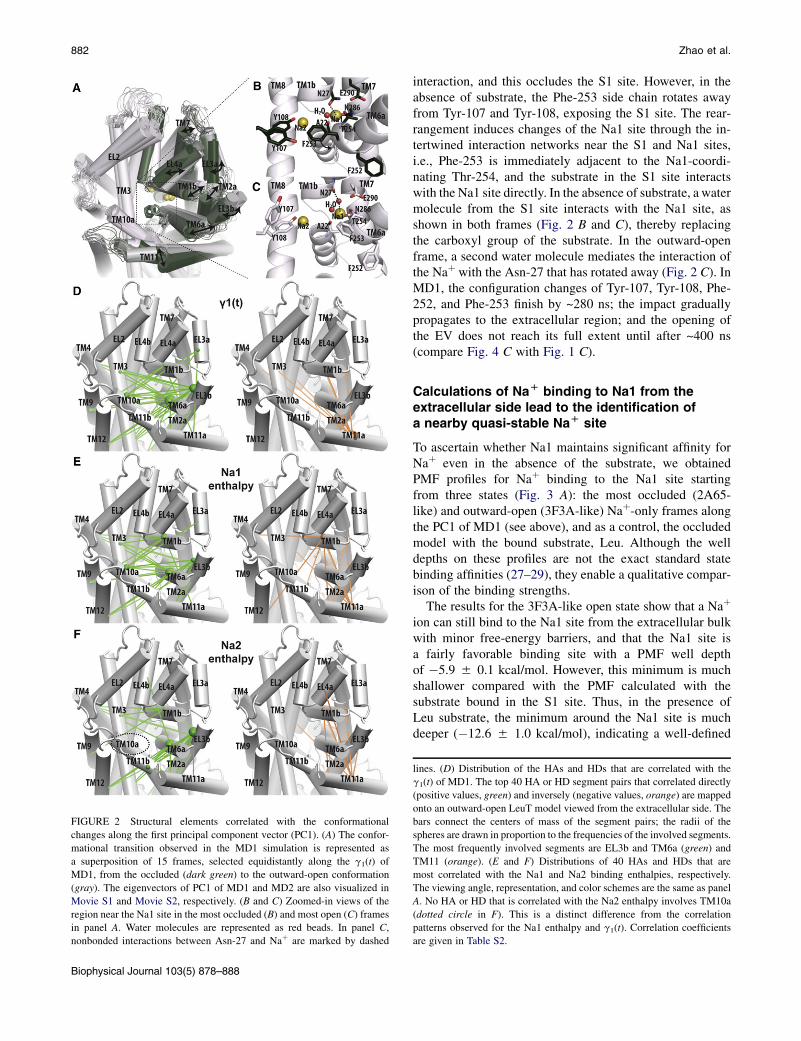
FIGURE 2 Structural elements correlated with the conformational
changes along the first principal component vector (PC1). (A) The confor-
mational transition observed in the MD1 simulation is represented as
a superposition of 15 frames, selected equidistantly along the g1(t) of
MD1, from the occluded (dark green) to the outward-open conformation
(gray). The eigenvectors of PC1 of MD1 and MD2 are also visualized in
Movie S1 and Movie S2, respectively. (B and C) Zoomed-in views of the
region near the Na1 site in the most occluded (B) and most open (C) frames
in panel A. Water molecules are represented as red beads. In panel C,
nonbonded interactions between Asn-27 and Naþ are marked by dashed
Biophysical Journal 103(5) 878–888
882 Zhao et al.
interaction, and this occludes the S1 site. However, in theabsence of substrate, the Phe-253 side chain rotates awayfrom Tyr-107 and Tyr-108, exposing the S1 site. The rear-rangement induces changes of the Na1 site through the in-tertwined interaction networks near the S1 and Na1 sites,i.e., Phe-253 is immediately adjacent to the Na1-coordi-nating Thr-254, and the substrate in the S1 site interactswith the Na1 site directly. In the absence of substrate, a watermolecule from the S1 site interacts with the Na1 site, asshown in both frames (Fig. 2 B and C), thereby replacingthe carboxyl group of the substrate. In the outward-openframe, a second water molecule mediates the interaction ofthe Naþ with the Asn-27 that has rotated away (Fig. 2 C). InMD1, the configuration changes of Tyr-107, Tyr-108, Phe-252, and Phe-253 finish by ~280 ns; the impact graduallypropagates to the extracellular region; and the opening ofthe EV does not reach its full extent until after ~400 ns(compare Fig. 4 C with Fig. 1 C).
Calculations of NaD binding to Na1 from theextracellular side lead to the identification ofa nearby quasi-stable NaD site
To ascertain whether Na1 maintains significant affinity forNaþ even in the absence of the substrate, we obtainedPMF profiles for Naþ binding to the Na1 site startingfrom three states (Fig. 3 A): the most occluded (2A65-like) and outward-open (3F3A-like) Naþ-only frames alongthe PC1 of MD1 (see above), and as a control, the occludedmodel with the bound substrate, Leu. Although the welldepths on these profiles are not the exact standard statebinding affinities (27–29), they enable a qualitative compar-ison of the binding strengths.
The results for the 3F3A-like open state show that a Naþ
ion can still bind to the Na1 site from the extracellular bulkwith minor free-energy barriers, and that the Na1 site isa fairly favorable binding site with a PMF well depthof �5.9 5 0.1 kcal/mol. However, this minimum is muchshallower compared with the PMF calculated with thesubstrate bound in the S1 site. Thus, in the presence ofLeu substrate, the minimum around the Na1 site is muchdeeper (�12.6 5 1.0 kcal/mol), indicating a well-defined
lines. (D) Distribution of the HAs and HDs that are correlated with the
g1(t) of MD1. The top 40 HA or HD segment pairs that correlated directly
(positive values, green) and inversely (negative values, orange) are mapped
onto an outward-open LeuT model viewed from the extracellular side. The
bars connect the centers of mass of the segment pairs; the radii of the
spheres are drawn in proportion to the frequencies of the involved segments.
The most frequently involved segments are EL3b and TM6a (green) and
TM11 (orange). (E and F) Distributions of 40 HAs and HDs that are
most correlated with the Na1 and Na2 binding enthalpies, respectively.
The viewing angle, representation, and color schemes are the same as panel
A. No HA or HD that is correlated with the Na2 enthalpy involves TM10a
(dotted circle in F). This is a distinct difference from the correlation
patterns observed for the Na1 enthalpy and g1(t). Correlation coefficients
are given in Table S2.

FIGURE 3 Naþ binding and selectivity in the Na1 site. (A) PMF profiles
for Naþ moving from the Na1 site to the extracellular side. The profiles are
shown for the occluded state with bound substrate (red), Naþ-only 2A65-
like (blue), and 3F3A-like (magenta) frames. (B) Naþ/Kþ and Naþ/Liþ
selectivity. The selectivities for the ion in the Na1 site of the occluded state
of LeuTwith substrate and both ions bound (occluded (Occ.) data are from
Caplan et al. (7)) and eight frames (F1–F8) selected from the MD1 trajec-
tory (see text). By definition, positive values of DDGNaþ/X
þ (where Xþ
represents Kþ or Liþ) indicate that the conformation is selective for Naþ
over Xþ. Thus the blue region is Naþ selective, the gray region corresponds
to an ambiguous selectivity between Naþ and ion Xþ, and the green regioncorresponds to Xþ selective sites. The SDs of ion selectivity are plotted as
error bars.
Dynamic Control of LeuT by Naþ Binding 883
ion binding site. The 2A65-like Naþ-only frame hasa minimum of �7.3 5 0.6 kcal/mol, which is ~1.4 50.6 kcal/mol deeper than the 3F3A-like Naþ-only frameand still 5.3 5 1.2 kcal/mol shallower than the occludedstate with the bound Leu substrate. On the other hand, foran extracellular Naþ binding event to the Na1 site, the2A65-like Naþ-only frame shows a barrier of ~2.4 kcal/mol, which is similar to that in the occluded state(~2.6 kcal/mol). This reflects the similarity between theconformation of the transporter in the 2A65-like Naþ-only frame and that in the occluded state with boundsubstrate Leu.
We note that the PMF region between �2 and þ5 Aaround the minima are relatively flat (with a free-energychange of <3 kcal/mol) for both Naþ-only frames, which
indicates a loose binding site and suggests the possibilitythat a quasi-stable binding location for Naþ may existnear the Na1 site. Indeed, in the trajectories of the umbrellasampling simulations (Fig. S4), the quasi-stable bindinglocation near Na1 site and Glu-290 (defined as the Na10
site) is very frequently visited by the Naþ ion. We exploredthis further in a set of 30 MD runs, from each of three pointswith high Na1 enthalpy and/or longer Na1–Na2 distance:606.96, 878.52, and 960 ns of MD1 (see below). In eachset of 10 MD runs, each run was started with a differentrandom number seed. Of interest, in one of the trajectoriesrestarted from 878.52 ns, the Naþ bound in Na1 is shiftedupward and remains stable for at least 60 ns in a location~2 A more extracellular than the Na1 site (Na10 site). Inthis location, the Naþ ion is in direct interaction with thenegatively charged Glu-290 while it also coordinates withthe side chain of Thr-254.
NaD/KD and NaD/LiD ion selectivity of Na1in the transition
To evaluate the conformational dependence of the Na1binding selectivity along the transition, we performed ionselectivity computations using eight representative framesof the MD1 trajectory (F1–F8) as the starting configurations.These frames were selected to have equidistant and ascend-ing values of g1(t) so as to cover the range along the entiretrajectory. Thus, F1 and F8 (shown in Fig. 2, B and C) arethe most occluded and outward-open frames along thePC1, respectively, as mentioned above.
Fig. 3 B and Table S3 show that the selectivity of Naþ
over Kþ in five of the eight frames (F2, F4, F5, F7, andF8) is comparable to that observed in the occluded LeuTstructure with bound substrate (3.4 kcal/mol), but it isslightly reduced in F1, F3, and F6. Overall, the Na1 site inall of the selected frames remains robustly selective forNaþ over Kþ, showing that the Naþ preference of the Na1site is preserved even in the absence of the negativelycharged carboxylate group of a substrate. For the Naþ
over Liþ selectivity, however, most of the eight frames indi-cate ambiguous selectivity at best, in similarity to the case ofoccluded LeuT structure with bound substrate (7). Takentogether, the results indicate that the absence of substratedoes not have a major impact on the selectivity of Naþ
over Kþ or Liþ in the Na1 site.
Enthalpies of NaD binding to the Na1 sitemoderately correlate with the extent ofcompletion of the transition toward the outward-open conformation
To characterize the changes in Na1 and Na2 affinities forNaþ along the transition, we applied two schemes for calcu-lating binding enthalpy. In the first scheme, denoted asProtein, all of the water molecules were treated implicitly.
Biophysical Journal 103(5) 878–888
884 Zhao et al.
In the second scheme, termed Proteinþ8water, eight watermolecules closest to the bound Naþwere included explicitlyand all other water molecules were treated implicitly.Although in some time spans the calculated enthalpy valuesare slightly lower for Proteinþ8water than for Protein, thetwo time series show very similar trends (Fig. 4 A). There-fore, we applied only the Protein scheme for the calculationof Na2 enthalpy.
Overall, along the entire MD1 trajectory, the Na1 bindingenthalpy is largely in the negative range (Fig. 4 A). This isconsistent with the results from the PMF calculations(Fig. 3 A) showing that even in the absence of the substrate,Naþ still has significant affinity for the Na1 site. In compar-ison, the Na2 enthalpy is entirely in the negative range and
FIGURE 4 Evolution of the Na1 and Na2 binding enthalpies and the
associated changes of structural elements in MD1. (A) Naþ binding
enthalpies computed using MM/PBSA for the Na1 and Na2 sites, with
a snapshot taken every 0.24 ns along the entire MD1 trajectory. Protein
and Proteinþ8water (see text) are plotted as blue and red lines, respectively.
(B) Evolution of the distances between Na1 and the indicated elements,
including Na2, all of the Na1 coordinating residues, and Glu-290. (C)
Evolution of the c1 rotamers of the aromatic cluster at the extracellular
gate of the S1 substrate binding site (Tyr-107, Tyr-108, Phe-252, and
Phe-253 (14)). (D) Evolution of the number of waters within the indicated
distances of Na1. The plots shown in all panels are moving averages of 6 ns.
The zoomed-in view of panel B for the peak periods near 610 and 875 ns are
shown in Fig. S5.
Biophysical Journal 103(5) 878–888
fluctuates less than the Na1 enthalpy. Of note, at ~878 ns,there is a strong positive spike in the Na1 binding enthalpyvalues (DH ¼ 4.3 5 3.1 kcal/mol) for a duration of ~10 ns.Similarly to a smaller one around 610 ns, this spike is asso-ciated with a temporary dissociation of Naþ from its coordi-nating residues, except for Thr-254 (Fig. 4 B and C andFig. S5). The resulting upward movement of the ion pro-duces a direct interaction with Glu-290 (Fig. 4 D).
The coordination of Asn-27 to Na1 is mostly indirect,through water molecules, and thus seems to be the weakestamong all Na1 coordinating residues (Fig. 4 B). The Asn-27side-chain rotation correlates well with the number of watermolecules coordinating the Naþ in Na1, and also with theg3(t) of MD1. Of interest, in comparison with PC1, PC3shows limited tilting of TM6a and TM1b, and TM7 andEL4a have significant rearrangements that are not obviousin PC1. These rearrangements are likely due to the weak-ened association between TM1b and TM7, as a result ofthe rotation of Asn-27 side chain away from Na1 (Fig. S3and Movie S3). In general, an increase in partial hydrationof Na1 has a disrupting effect on Naþ stability (compareFig. 4 A and 4 D), but configurational changes in the secondlayer of residues surrounding the Na1 site can lower theenthalpy values and thus increase the affinity (e.g., amongthe residues in the aromatic cluster, the rotamer of Phe252is moderately correlated with the Na1 binding enthalpy,with a Pearson coefficient of �0.61; indeed, the rotationof its c1 rotamer back to a gauche position, as in theoccluded state, is likely responsible for the decrease ofenthalpy values after ~870 ns (compare Fig. 4 A and 4 C).To establish the relation between the Na1 and Na2
binding affinities and the conformational changes criticalto the transition, we evaluated the correlation between thebinding enthalpies and the conformational observables onboth the global scale (g1(t)) and the local scale (HAs,HDs, and distances to Na1). We found g1(t) to be moder-ately correlated with Na1 enthalpy values, but significantlyless correlated with Na2 enthalpy values (Pearson coeffi-cients: 0.65 and 0.43, respectively). Similar differenceswere found for the local observables (Table S2 andFig. S3). When mapped onto the structure, the 40 mostcorrelated HAs/HDs for Na1 enthalpy (Fig. 2 E) are distrib-uted in a pattern similar to those identified in the PC1-correlation analysis (Fig. 2 D), but the similarity is lessobvious for those obtained for Na2 enthalpy (Fig. 2 F). Ofinterest, the extracellular segment of TM10, TM10a, whichis located at the center of the EV, does not appear in any ofthe top 40 Na2-enthalpy-correlated HAs and HDs (dottedcircle in Fig. 2 F). In contrast, five HA/HDs involvingTM10a are highly correlated with both Na1 enthalpy andg1(t) (Table S2). Thus, the conformational changes thatplay a leading role in the opening transition in LeuT’s EVare more associated with changes in the Naþ binding affinityfor the Na1 site than for the Na2 site (note that as a higherenthalpy value corresponds to lower affinity, the direct

Dynamic Control of LeuT by Naþ Binding 885
correlation between Na1 enthalpy values and g1(t) suggestsan inverse correlation between Na1 affinity and g1(t); seeDiscussion).
FIGURE 5 Impact of Naþ binding and Glu-290 protonation on the extent
of the outward-open character of LeuT conformations. (A) The extents of
outward-open character of various configurations are reflected by the
normalized distributions of the number of water molecules in the EV.
Compared with the occluded state with S1-bound substrate (black, based
on a 230 ns MD trajectory described previously (3,32)), the Naþ-onlyconfiguration (green, 60–420 and 720–960 ns periods of MD1 for the left
and right panels, respectively) has ~30 more waters in the EV, and the
absence of Na1 (red, 480 ns) further opens the EV slightly. The removal
of the charge of Glu-290 (purple, 360 ns) renders the EV less outward-
open. Consistent trends are observed from the no-Na1 simulations started
from both 60 (left) and 720 (right) ns points of MD1 (see text). The multiple
peaks of the green curve in the left panel indicate that the MD1 is in
a conformational transition in the 60–420 ns period. The distribution plots
were generated from the water count data using a Gaussian kernel density
estimate (density function in R (18)). (B) The roles of Naþ binding and Glu-
290 protonation in the control of conformational dynamics of the transport
cycle. The configurations are in the same color code as in panel A.
Role of Na1 in the transition toward an outward-open conformation
The results described in the previous sections indicate thetendency of the transporter molecule to transition towardthe outward-open conformation in the absence of substrate,and with Naþ bound at both Na1 and Na2 sites. To furtherdissect the impact of Naþ binding in the Na1 site on theconformation adopted by the transporter, we carried outfour MD simulations in the absence of Naþ in Na1 (termedno-Na1). Starting from the 60 and 720 ns points of MD1(corresponding to the relatively occluded and outward-open states in the Naþ-only configuration; see Fig. 1), theno-Na1 configuration was simulated with either deproto-nated (480 ns each from the two points) or protonated(360 ns each) Glu-290. We chose to consider the Glu-290protonation state in our Naþ-only simulations because theNa1 is always in close proximity to the negatively chargedGlu-290, and the absence of Na1 was found to resultin the protonation of Glu-290 in the inward-open conforma-tion (11).
To evaluate the impact of various substrate, ion, andcharge combinations on the global conformation from theresults of these MD simulations, we measured the extentof outward-open character by the number of water mole-cules in the EV (i.e., more water corresponds to a moreoutward-open conformation). By using similar lengths ofMD trajectories to calculate and compare the normalizeddistribution of the number of waters, we found that in theabsence of Na1, the transporter protein is slightly moreoutward-open than in the Naþ-only configuration withNaþ bound at both Na1 and Na2 sites (Fig. 5 A). Theabsence of Na1 results in more outward tilting of TM7and EL4a, reminiscent of the PC3 motion of MD1 (seeabove, Movie S3), which is correlated with the extent ofhydration near the Na1 site (Fig. S3). However, with thenegative charge of Glu-290 eliminated by protonation, thetransporter protein becomes less outward-open thanthe form with deprotonated Glu-290. This is likely due tothe dehydration effect of the protonated Glu-290 on theNa1 site vicinity in the absence of bound Naþ (Fig. S6).We observed consistent trends from both starting points,but note that the impact of Glu-290 protonation was moreobvious when the simulation was started from a moreoccluded state (Fig. 5 A).
DISCUSSION
The Naþ-only MD simulations initiated from the crystallo-graphically determined occluded state of LeuT produceda detailed mechanistic picture of global conformational re-
arrangement of the transporter molecule. They show thatthe opening of the EV is achieved in both trajectories(MD1 and MD2) by a series of structural reconfigurationsnear the substrate and Na1 sites, which then trigger a seriesof discrete but coordinated rearrangements within theTM6a-EL3-TM2a-TM1b cluster, resulting in a distancingfrom TM3, TM8, and TM10. The LeuT conformation result-ing from these changes exhibits the main characteristics ofthe outward-open form of the transporter (4,14) and issimilar to that stabilized by an inhibitor Trp (PDB: 3F3A).This leads to the conclusion that Naþ binding alone, whichis shown by this study to be possible with high selectivityand significant affinity in the absence of substrate, biasesthe transporter toward an outward-open conformation thatis ready for the binding of substrate.
While this work was being prepared, the crystal structureof the LeuT mutant Y108A (PDB: 3TT1) was released (30).This structure was determined in the absence of substrate,but with bound Naþ in both the Na1 and Na2 sites. It isvery gratifying to report that the outward-open conforma-tion in the crystal structure is very similar to the Naþ-only
Biophysical Journal 103(5) 878–888

886 Zhao et al.
conformation identified computationally and describedhere, and partially as well in an earlier documentation ofthe effect of Naþ (14). Thus, in contrast to the 3F3Aoutward-open structure, the 3TT1 structure in the absenceof any bound ligand shows the side chain of Phe-253 inTM6 to be rotated away from Tyr-107 and Tyr-108/Phe-108 of TM3, thus exposing the S1 site, just as observed inour simulations. However, the MD simulations describedhere provide in addition a dynamic picture of the transitionto an outward-open conformation that exhibits some subtledifferences from both the 3F3A and 3TT1 structures: Inthose two crystal structures, TM1b tilts away from thecenter of the TM bundle in parallel with TM6a, whereasin the simulations, although TM1b can tilt as far as observedin the crystal structures, TM6a is more mobile than TM1band is able to move farther away from the center of theTM bundle (Fig. 2 A). Using the F8 frame as a representativesnapshot (its all-Ca RMSD values with respect to the 3F3A,3TT1, and 2A65 structures are 1.65 A, 1.56 A, and 1.94 A,respectively), a superposition on the Ca atoms of TM3 andTM8 shows that the Ca atom of Gly-242 at the extracellulartip of TM6a is positioned 3.4 A more outward in the F8frame than in the 3F3A structure. Of interest, in the Naþ-only 3TT1 structure, this outward tilt is also larger than inthe 3F3A structure, with a value of 1.3 A (as a comparisonreference, the difference in the position of Gly-242 betweenthe 3F3A structure and the occluded 2A65 structure is2.9 A). Thus, the parallel outward tilted orientations ofTM1b and TM6a revealed by the 3F3A and 3TT1 structuresrepresent a stable configuration, but the detailed simulationsreveal a dynamic relationship between these two core TMsthat is not evident from the structures alone. We find thatTM6a can move away from TM1b and the TM bundle inthe conformational transition. In addition, the rearrange-ments of TM1a-TM1b and TM6a-TM6b are among thestructural elements that exhibit the best correlation withthe changes in the Na1 and Na2 enthalpies, respectively(Table S2). This suggests that the TM1 and TM6 conforma-tions are tuned by the Naþ binding to sites positioned in theunwound regions of these TMs, arguing against rigid-bodymovements for TM1 and TM6 (31). However, symmetricalfeatures as proposed by Forrest and Rudnick (31) areevident in the conformational transition, with the rearrange-ment of TM6a away from the bundle observed here resem-bling the movement of its symmetric counterpart TM1arevealed from simulation and single-molecule fluorescenceresonance energy transfer measurements (3,32), and the pre-dicted outward-tilt of TM1a is indeed observed in therecently published inward-facing structure (30).
That Naþ binding to the Na1 site is energetically favor-able and selective (over Kþ), even in the absence of S1substrate, is shown by our results from the PMF calcula-tions, as well as the time series of binding enthalpies alongthe transition, and the free-energy simulations. These resultssuggest the possibility that Naþ binds in the Na1 site in the
Biophysical Journal 103(5) 878–888
absence of substrate, albeit with lower affinity than inthe presence of substrate. However, the ion selectivity ofthe Na1 site calculated along the transition betweenoccluded and outward-open states indicates differences inthis binding ability when Naþ is compared with Kþ orLiþ. Thus, for Naþ/Kþ, the selectivity for Naþ is maintainedalong representative frames of the whole transition, whichis an important finding in view of the considerable con-centration of Kþ in the physiological environment. Theconservation of Naþ/Kþ selectivities upon removal of theion-coordinating Leu substrate is intriguing. Previousstudies (33,34) suggested that the removal of a high-fieldcoordinating ligand, such as the charged carboxyl groupsubstrate, from an ion binding site might lead to a reductionin the selectivities for Naþ over Kþ. Instead, the selectivityfor Naþ over Kþ in the Na1 site for many of the studiedframes from the Naþ-only trajectory is only slightlydecreased. An ion coordination analysis of the FEP/MDtrajectories collected for the alchemical transition ofNaþ 4 Kþ bound in the Na1 site suggests that the carboxylgroup of Glu-290 has a strong impact on the coordination ofthe bound ion and may contribute to the selectivity.
In contrast, we find for Naþ/Liþ that the selectivity israther ambiguous. This is not entirely unexpected, as Caplanet al. (7) showed that the Naþ/Liþ selectivity is mostlyachieved by the coupling effect between the Na1 and Na2sites. Therefore, we conclude that the Naþ over Kþ andLiþ selectivities of the Na1 site in the absence of thesubstrate remains largely the same as in the presence ofthe substrate.
We found that the conformational transition to outward-open is inversely correlated with the changes in Na1 bindingaffinity but only weakly correlated with Na2 affinity. Thus,it is likely that the Naþ bound at the Na2 site plays a criticalrole in maintaining the overall outward-open conformationin the absence of the substrate, while the presence of Na1modulates the extent of outward-open character, with theabsence of Na1 making the conformation slightly moreoutward-open (Fig. 5 A). Consistently, the release of Na2(but not Na1) has been shown to be required for the confor-mational transition to inward-open (3,8). It is tempting tospeculate that binding and release of Naþ in the Na1 andNa2 sites may play symmetrical roles in the outward- andinward-open transition. However, the locations of the Na1and Na2 sites are not entirely symmetrical in the pseudo-inverted repeats, which may explain the differences in theircoupling to the directional translocation of substrate.
The distinct roles that Na1 and Na2 play in the conforma-tional transitions relevant to LeuT function are further un-derscored by the connection between Naþ binding in theNa1 site and the protonation state of Glu-290. In a previousstudy (11), we found that the absence of Na1 resulted in theprotonation of Glu-290 in the inward-open conformation.Indeed, we find here that the removal of both the Na1-boundNaþ and the negative charge at Glu-290 renders the
Dynamic Control of LeuT by Naþ Binding 887
conformation less outward-open, especially when the simu-lation is started from a relatively more occluded state. Thisis consistent with the role of neutral (protonated) Glu-290 informing the inward-open conformation. In addition, becauseGlu-290 is located along the potential entry route of Naþ
toward the Na1 site (Fig. S4), the negatively charged Glu-290 is likely responsible for the broadened attraction tothe Na1 site (i.e., the formation of a quasi-stable bindinglocation near the Na1 site) and for providing part of thedriving force for Na1 binding.
Our current mechanistic understanding of the sequence of(de)protonation and Naþ binding is summarized in Fig. 5 B.Starting from the apo inward-open state, Naþ binds at theNa2 site first and steers the transporter toward an outward-open conformation. The subsequent deprotonation of Glu-290 further opens up the EV and attracts Naþ binding atthe Na1 site. The entry of the S1 substrate is facilitated bythe presence of Na1, which forms a direct stabilizing inter-action with the charged substrate. The combined impact ofNaþ binding in the Na1 site and substrate binding inthe S1 site induces the transporter to transition toward anoccluded state. The Na2-only states (purple and red inFig. 5 B), however, are likely only transient states in thephysiological condition.
SUPPORTING MATERIAL
Methods, sixfigures, three tables, threemovies, and references are available at
http://www.biophysj.org/biophysj/supplemental/S0006-3495(12)00857-0.
We thank Matthias Quick for helpful discussion.
This work was supported by the National Institutes of Health (grants
DA023694 to L.S., and DA012408 and U54GM087519 to H.W.) and the
National Sciences and Engineering Research Council (Discovery Grant
RGPIN-315019 to S.Y.N.). S.Y.N. is an Alberta Innovates Technology
Futures New Faculty, Canadian Institute for Health Research New Investi-
gator, and an Alberta Innovates Health Solutions Scholar. C.F.Z. is sup-
ported by a postdoctoral scholarship from Alberta Innovates Health
Solutions. Computations were performed on Jaguar at Oak Ridge National
Laboratory (BIP-014), Ranger at the Texas Advanced Computing Center
(TG-MCB090022), the Cofrin Center for Biomedical Information of the
Institute for Computational Biomedicine at Weill Cornell Medical College,
the West-Grid/Compute Canada facilities, and the local TNK cluster sup-
ported by the Canadian Foundation for Innovation.
REFERENCES
1. Sonders, M. S., M. Quick, and J. A. Javitch. 2005. How did the neuro-transmitter cross the bilayer? A closer view. Curr. Opin. Neurobiol.15:296–304.
2. Jardetzky, O. 1966. Simple allosteric model for membrane pumps.Nature. 211:969–970.
3. Shi, L., M. Quick, ., J. A. Javitch. 2008. The mechanism of a neuro-transmitter:sodium symporter—inward release of Naþ and substrate istriggered by substrate in a second binding site. Mol. Cell. 30:667–677.
4. Quick, M., H. Yano, ., J. A. Javitch. 2006. State-dependent confor-mations of the translocation pathway in the tyrosine transporter Tyt1,a novel neurotransmitter:sodium symporter from Fusobacterium nucle-atum. J. Biol. Chem. 281:26444–26454.
5. Singh, S. K., C. L. Piscitelli,., E. Gouaux. 2008. A competitive inhib-itor traps LeuT in an open-to-out conformation. Science. 322:1655–1661.
6. Yamashita, A., S. K. Singh, ., E. Gouaux. 2005. Crystal structure ofa bacterial homologue of Naþ/Cl-dependent neurotransmitter trans-porters. Nature. 437:215–223.
7. Caplan, D. A., J. O. Subbotina, and S. Y. Noskov. 2008. Molecularmechanism of ion-ion and ion-substrate coupling in the Naþ-dependentleucine transporter LeuT. Biophys. J. 95:4613–4621.
8. Zhao, C., and S. Y. Noskov. 2011. The role of local hydration andhydrogen-bonding dynamics in ion and solute release from ion-coupledsecondary transporters. Biochemistry. 50:1848–1856.
9. Abramson, J., and E. M. Wright. 2009. Structure and function ofNa(þ)-symporters with inverted repeats. Curr. Opin. Struct. Biol.19:425–432.
10. Shi, L., and H. Weinstein. 2010. Conformational rearrangements to theintracellular open states of the LeuT and ApcT transporters are modu-lated by common mechanisms. Biophys. J. 99:L103–L105.
11. Zhao, Y., M. Quick,., J. A. Javitch. 2010. Substrate-dependent protonantiport in neurotransmitter:sodium symporters. Nat. Chem. Biol.6:109–116.
12. Noskov, S. Y., and B. Roux. 2008. Control of ion selectivity in LeuT:two Naþ binding sites with two different mechanisms. J. Mol. Biol.377:804–818.
13. Phillips, J. C., R. Braun, ., K. Schulten. 2005. Scalable moleculardynamics with NAMD. J. Comput. Chem. 26:1781–1802.
14. Claxton, D. P., M. Quick, ., H. S. McHaourab. 2010. Ion/substrate-dependent conformational dynamics of a bacterial homolog of neuro-transmitter:sodium symporters. Nat. Struct. Mol. Biol. 17:822–829.
15. MacKerell, Jr., A. D., M. Feig, and C. L. Brooks, 3rd. 2004. Improvedtreatment of the protein backbone in empirical force fields. J. Am.Chem. Soc. 126:698–699.
16. Feller, S. E., Y. Zhang, ., B. R. Brooks. 1995. Constant pressuremolecular dynamics simulation: the Langevin piston method.J. Chem. Phys. 103:4613–4621.
17. Holder, T. 2012. AngleBetweenHelices. http://www.pymolwiki.org/index.php/AngleBetweenHelices.
18. R Development Core Team. 2011. R: A Language and Environment forStatistical Computing. http://www.R-project.org.
19. Damm, K. L., and H. A. Carlson. 2006. Gaussian-weighted RMSDsuperposition of proteins: a structural comparison for flexible proteinsand predicted protein structures. Biophys. J. 90:4558–4573.
20. Gracia, L. G. 2012. RMSDTT: RMSD Trajectory Tool. http://physiology.med.cornell.edu/faculty/hweinstein/vmdplugins/rmsdtt/.
21. Humphrey, W., A. Dalke, and K. Schulten. 1996. VMD: visual molec-ular dynamics. J. Mol. Graph. 14:33–38, 27–38.
22. Srinivasan, J., T. E. Cheatham, ., D. A. Case. 1998. Continuumsolvent studies of the stability of DNA, RNA, and phosphoramidateDNA helices. J. Am. Chem. Soc. 120:9401–9409.
23. Henry, L. K., H. Iwamoto,., R. D. Blakely. 2011. A conserved aspar-agine residue in transmembrane segment 1 (TM1) of serotonin trans-porter dictates chloride-coupled neurotransmitter transport. J. Biol.Chem. 286:30823–30836.
24. Brooks, B. R., C. L. Brooks, 3rd, ., M. Karplus. 2009. CHARMM:the biomolecular simulation program. J. Comput. Chem. 30:1545–1614.
25. Nina, M., W. Im, and B. Roux. 1999. Optimized atomic radii for proteincontinuum electrostatics solvation forces. Biophys. Chem. 78:89–96.
26. Grossfield, A. 2012. WHAM: the weighted histogram analysis method,version 2.0.6. http://membrane.urmc.rochester.edu/content/wham.
27. Roux, B., O. S. Andersen, and T. W. Allen. 2008. Comment on‘‘Free energy simulations of single and double ion occupancy in gram-icidin A’’ [J. Chem. Phys. 126, 105103 (2007)]. J. Chem. Phys.128:227101, author reply 227102.
Biophysical Journal 103(5) 878–888

888 Zhao et al.
28. Allen, T. W., O. S. Andersen, and B. Roux. 2006. Moleculardynamics—potential of mean force calculations as a tool for under-standing ion permeation and selectivity in narrow channels. Biophys.Chem. 124:251–267.
29. Doudou, S., N. A. Burton, and R. H. Henchman. 2009. Standard freeenergy of binding from a one-dimensional potential of mean force.J. Chem. Theory Comput. 5:909–918.
30. Krishnamurthy, H., and E. Gouaux. 2012. X-ray structures of LeuT insubstrate-free outward-open and apo inward-open states. Nature.481:469–474.
Biophysical Journal 103(5) 878–888
31. Forrest, L. R., and G. Rudnick. 2009. The rocking bundle: a mechanismfor ion-coupled solute flux by symmetrical transporters. Physiology(Bethesda). 24:377–386.
32. Zhao, Y., D. Terry, ., J. A. Javitch. 2010. Single-molecule dynamicsof gating in a neurotransmitter transporter homologue. Nature. 465:188–193.
33. Yu, H., S. Y. Noskov, and B. Roux. 2009. Hydration number, topologicalcontrol, and ion selectivity. J. Phys. Chem. B. 113:8725–8730.
34. Noskov, S. Y., S. Berneche, and B. Roux. 2004. Control of ion selec-tivity in potassium channels by electrostatic and dynamic propertiesof carbonyl ligands. Nature. 431:830–834.