Thermal-induced conformational changes in the product release area drive the enzymatic activity of...
6
Thermal-induced conformational changes in the product release area drive the enzymatic activity of xylanases 10B: Crystal structure, conformational stability and functional characterization of the xylanase 10B from Thermotoga petrophila RKU-1 Camila Ramos Santos a , Andreia Navarro Meza a , Zaira Bruna Hoffmam b , Junio Cota Silva b , Thabata Maria Alvarez b , Roberto Ruller b , Guilherme Menegon Giesel c , Hugo Verli c , Fabio Marcio Squina b , Rolf Alexander Prade d , Mario Tyago Murakami a,⇑ a Laboratório Nacional de Biociências (LNBio), Centro Nacional de Pesquisa em Energia e Materiais, Campinas, SP, Brazil b Laboratório Nacional de Ciência e Tecnologia do Bioetanol (CTBE), Centro Nacional de Pesquisa em Energia e Materiais, Campinas, SP, Brazil c Centro de Biotecnologia, Universidade Federal do Rio Grande do Sul, Porto Alegre, RS, Brazil d Department of Microbiology and Molecular Genetics, Oklahoma State University, Stillwater, OK, USA article info Article history: Received 27 October 2010 Available online 9 November 2010 Keywords: Xylanase Glycoside hydrolase family 10 Crystal structure Thermostability Thermotoga petrophila RKU-1 abstract Endo-xylanases play a key role in the depolymerization of xylan and recently, they have attracted much attention owing to their potential applications on biofuels and paper industries. In this work, we have investigated the molecular basis for the action mode of xylanases 10B at high temperatures using biochemical, biophysical and crystallographic methods. The crystal structure of xylanase 10B from hyper- thermophilic bacterium Thermotoga petrophila RKU-1 (TpXyl10B) has been solved in the native state and in complex with xylobiose. The complex crystal structure showed a classical binding mode shared among other xylanases, which encompasses the 1 and 2 subsites. Interestingly, TpXyl10B displayed a temper- ature-dependent action mode producing xylobiose and xylotriose at 20 °C, and exclusively xylobiose at 90 °C as assessed by capillary zone electrophoresis. Moreover, circular dichroism spectroscopy suggested a coupling effect of temperature-induced structural changes with this particular enzymatic behavior. Molecular dynamics simulations supported the CD analysis suggesting that an open conformational state adopted by the catalytic loop (Trp297-Lys326) provokes significant modifications in the product release area (+1,+2 and +3 subsites), which drives the enzymatic activity to the specific release of xylobiose at high temperatures. Ó 2010 Elsevier Inc. All rights reserved. 1. Introduction Xylanases (b-1,4-xylan xylanohydrolase, E.C. 3.2.1.8) are responsible for breaking down xylan, the major hemicellulosic component of plant cell walls, into short xylooligosaccharides by a general acid–base mechanism involving two glutamic acid resi- dues [1,2]. Typically, these enzymes can be classified into glycoside hydrolase (GH) families 10 and 11 based on amino-acid sequence similarities [3]. However, xylanases belonging to families 5, 8 and 43 have also been identified [4,5]. The biotechnological appli- cations of xylanases include food, feed, textile and paper industries [6], and recently they have received much attention owing to their use in degradation of lignocellulosic biomass for biofuels produc- tion [7,8]. In this context, enzymes exhibiting high enzymatic efficiency combined with hyper thermal and chemical tolerance are extremely desired. Although countless extremophilic enzymes have been characterized, the molecular basis for their mode of action at high temperatures is still poorly understood. Several structures of family 10 xylanases have been solved, from meso- philic to hyperthermophilic organisms enabling an in-depth com- parative study in terms of thermophilicity, thermostability and action mechanism. Thus, in order to gain insights into the structural determinants of their mode of action at high temperatures we have performed a functional and structural characterization of a hyperthermostable xylanase 10B from the bacterium Thermotoga petrophila RKU-1 (TpXyl10B). Our studies addressed important 0006-291X/$ - see front matter Ó 2010 Elsevier Inc. All rights reserved. doi:10.1016/j.bbrc.2010.11.010 Abbreviations: TpXyl10B, xylanase 10B from Thermotoga petrophila RKU-1; TmXyl10B, xylanase 10B from Thermotoga maritima MSB8; GH, glycoside hydrolase; CD, circular dichroism; CZE, capillary zone electrophoresis; U, unit; MD, molecular dynamics; XTAL, X-ray crystallography. ⇑ Corresponding author. Address: Laboratório Nacional de Biociências (LNBio), Centro Nacional de Pesquisa em Energia e Materiais, Rua Giuseppe Maximo Scolfaro, 10000, Campinas, 13083-970 SP, Brazil. Fax: +55 19 3512 1004. E-mail address: [email protected] (M.T. Murakami). Biochemical and Biophysical Research Communications 403 (2010) 214–219 Contents lists available at ScienceDirect Biochemical and Biophysical Research Communications journal homepage: www.elsevier.com/locate/ybbrc
Transcript of Thermal-induced conformational changes in the product release area drive the enzymatic activity of...

Biochemical and Biophysical Research Communications 403 (2010) 214–219
Contents lists available at ScienceDirect
Biochemical and Biophysical Research Communications
journal homepage: www.elsevier .com/locate /ybbrc
Thermal-induced conformational changes in the product release area drive theenzymatic activity of xylanases 10B: Crystal structure, conformational stabilityand functional characterization of the xylanase 10B from Thermotoga petrophilaRKU-1
Camila Ramos Santos a, Andreia Navarro Meza a, Zaira Bruna Hoffmam b, Junio Cota Silva b,Thabata Maria Alvarez b, Roberto Ruller b, Guilherme Menegon Giesel c, Hugo Verli c, Fabio Marcio Squina b,Rolf Alexander Prade d, Mario Tyago Murakami a,⇑a Laboratório Nacional de Biociências (LNBio), Centro Nacional de Pesquisa em Energia e Materiais, Campinas, SP, Brazilb Laboratório Nacional de Ciência e Tecnologia do Bioetanol (CTBE), Centro Nacional de Pesquisa em Energia e Materiais, Campinas, SP, Brazilc Centro de Biotecnologia, Universidade Federal do Rio Grande do Sul, Porto Alegre, RS, Brazild Department of Microbiology and Molecular Genetics, Oklahoma State University, Stillwater, OK, USA
a r t i c l e i n f o
Article history:Received 27 October 2010Available online 9 November 2010
Keywords:XylanaseGlycoside hydrolase family 10Crystal structureThermostabilityThermotoga petrophila RKU-1
0006-291X/$ - see front matter � 2010 Elsevier Inc. Adoi:10.1016/j.bbrc.2010.11.010
Abbreviations: TpXyl10B, xylanase 10B from ThTmXyl10B, xylanase 10B from Thermotoga maritima MCD, circular dichroism; CZE, capillary zone electrophodynamics; XTAL, X-ray crystallography.⇑ Corresponding author. Address: Laboratório Nac
Centro Nacional de Pesquisa em Energia e MaterScolfaro, 10000, Campinas, 13083-970 SP, Brazil. Fax:
E-mail address: [email protected] (M.
a b s t r a c t
Endo-xylanases play a key role in the depolymerization of xylan and recently, they have attracted muchattention owing to their potential applications on biofuels and paper industries. In this work, we haveinvestigated the molecular basis for the action mode of xylanases 10B at high temperatures usingbiochemical, biophysical and crystallographic methods. The crystal structure of xylanase 10B from hyper-thermophilic bacterium Thermotoga petrophila RKU-1 (TpXyl10B) has been solved in the native state andin complex with xylobiose. The complex crystal structure showed a classical binding mode shared amongother xylanases, which encompasses the �1 and �2 subsites. Interestingly, TpXyl10B displayed a temper-ature-dependent action mode producing xylobiose and xylotriose at 20 �C, and exclusively xylobiose at90 �C as assessed by capillary zone electrophoresis. Moreover, circular dichroism spectroscopy suggesteda coupling effect of temperature-induced structural changes with this particular enzymatic behavior.Molecular dynamics simulations supported the CD analysis suggesting that an open conformational stateadopted by the catalytic loop (Trp297-Lys326) provokes significant modifications in the product releasearea (+1,+2 and +3 subsites), which drives the enzymatic activity to the specific release of xylobiose athigh temperatures.
� 2010 Elsevier Inc. All rights reserved.
1. Introduction
Xylanases (b-1,4-xylan xylanohydrolase, E.C. 3.2.1.8) areresponsible for breaking down xylan, the major hemicellulosiccomponent of plant cell walls, into short xylooligosaccharides bya general acid–base mechanism involving two glutamic acid resi-dues [1,2]. Typically, these enzymes can be classified into glycosidehydrolase (GH) families 10 and 11 based on amino-acid sequencesimilarities [3]. However, xylanases belonging to families 5, 8
ll rights reserved.
ermotoga petrophila RKU-1;SB8; GH, glycoside hydrolase;resis; U, unit; MD, molecular
ional de Biociências (LNBio),iais, Rua Giuseppe Maximo+55 19 3512 1004.
T. Murakami).
and 43 have also been identified [4,5]. The biotechnological appli-cations of xylanases include food, feed, textile and paper industries[6], and recently they have received much attention owing to theiruse in degradation of lignocellulosic biomass for biofuels produc-tion [7,8]. In this context, enzymes exhibiting high enzymaticefficiency combined with hyper thermal and chemical toleranceare extremely desired. Although countless extremophilic enzymeshave been characterized, the molecular basis for their mode ofaction at high temperatures is still poorly understood. Severalstructures of family 10 xylanases have been solved, from meso-philic to hyperthermophilic organisms enabling an in-depth com-parative study in terms of thermophilicity, thermostability andaction mechanism. Thus, in order to gain insights into the structuraldeterminants of their mode of action at high temperatures we haveperformed a functional and structural characterization of ahyperthermostable xylanase 10B from the bacterium Thermotogapetrophila RKU-1 (TpXyl10B). Our studies addressed important
C.R. Santos et al. / Biochemical and Biophysical Research Communications 403 (2010) 214–219 215
issues in the structure–function relationship of xylanases 10B as themodulation of their action mode at high temperatures by conforma-tional changes in the product release area.
2. Material and methods
2.1. Cloning, protein expression and purification
The TpXyl10B coding gene was amplified by PCR using theT. petrophila RKU-1 genomic DNA as template and the followingoligonucleotides: 50-CATATGCAAAATGTATCTCTGAGAGAGCTC-30
and 5-GGATCCTCATTCTATCTTTTTCTCCAGCAC-30. The sequencewas cloned into pGEM�-T Easy vector system (Promega) and wasfurther subcloned into pET28a vector (Novagen) using NdeI andBamHI restriction sites. The protein expression was performed inBL21(DE3)SlyD cells at 37 �C for 4 h after induction with 0.5 mMisopropyl b-D-1-thiogalactopyranoside. Harvested cells wereresuspended in lysis buffer (20 mM sodium phosphate pH 7.4,500 mM NaCl and 5 mM imidazole) and disrupted by lysozymetreatment (80 lg/mL, 1 h, on ice) followed by sonication (VibracellVCX 500, Sonics & Materials, Inc.). The cell extract was clarified bycentrifugation at 20,000g for 30 min at 4 �C and applied into a 5 mLHiTrap™ Chelating HP column (GE Healthcare) with a flow rate of1 mL/min. The elution was carried out using an imidazole gradientfrom 5 to 500 mM in 20 column volumes. Fractions containingTpXyl10B were pooled, concentrated and applied into a Superdex75 16/60 column (GE Healthcare) with a flow rate of 0.5 mL/min.All chromatographic steps were performed using an ÄKTA FPLCsystem (GE Healthcare). The purified TpXyl10B was then dialyzedagainst 20 mM phosphate buffer (pH 7.2) and concentrated byultrafiltration. Sample purity was assessed by SDS–PAGE underreducing conditions. Protein concentration was determined byabsorbance at 280 nm.
2.2. Xylanase activity assay
Xylanase activity was assayed using birchwood xylan (Sigma,USA) as substrate. Liberated reducing sugars were quantified by adinitrosalicylic method [9]. One unit (U) of enzyme activity wasdefined as the amount of enzyme required to release 1 lmol ofreducing sugar per minute. Specific activity was expressed as IU/mg protein.
2.3. Response surface methodology
The variables analyzed here were the pH along with tempera-ture. Central points for 12 experiments in total were consideredfor optimization of these variables. Details regarding the statisticalapproaches for these experiments can be found in Myers andMontgomery [10]. The regression and graphical analysis of the datawere performed using the software StatisticaTM version 8.0,considering a p-value of 0.05. The significance of the regressioncoefficients was given by Student’s t-test. The second order modelequation was determined by Fisher’s test and the multiple coeffi-cient of determination (R2) gave the variance explained by themodel. All xylanase activity assays were carried out in triplicateusing 10 lL of enzyme sample (650 ng), 40 lL of 50 mM acetateor phosphate buffer and 50 lL of birchwood xylan (stock solution1%(W/V) in water).
2.4. Capillary zone electrophoresis of oligosaccharides
The 1,4-b-D-xylohexaose (Megazyme) was derivatized with8-aminopyreno-1,3,6-trisulfonic acid (APTS) by reductive amina-tion [11]. Capillary zone electrophoresis (CZE) of oligosaccharides
was performed on a P/ACE MQD instrument (Beckman Coulter)equipped with laser-induced fluorescence detection. A fused-silicacapillary (TSP050375, Polymicro Technologies) of internal diame-ter of 50 lm and total length of 31 cm was used as separation col-umn for oligosaccharides. Electrophoresis conditions were 15 kV/70–100 lA at a controlled temperature of 20 �C. Oligomers labeledwith APTS were excited at 488 nm and emission was collectedthrough a 520 nm band pass filter.
2.5. Circular dichroism spectroscopy
Far-UV circular dichroism (CD) spectra were recorded on a JascoJ-810 spectropolarimeter (Jasco International Co.). CD measure-ments were carried out in a 1 mm quartz cuvette using the wave-length range of 190–260 nm. The protein concentration was set to3.2 lM in 10 mM sodium phosphate buffer, pH 6.7. All sampleswere centrifuged at 10,000g for 10 min prior to analysis. In orderto study the effect of temperature on TpXyl10B structure, the sam-ple was heated from 20 to 95 �C and CD spectra at different tem-peratures were obtained. The data collection parameters were setto scan rate of 50 nm/min, response time of 4 s, sensitivity of100 mdeg, accumulation of 6, heating rate of 1 �C/min and delaytime for spectrum collection of 60 s. The reversibility of the tem-perature effect was checked by cooling the sample to 20 �C usingthe same parameters above. Baseline subtraction, smoothing anddata normalization were carried out using the graphical softwareORIGIN (http://www.originlab.com/). The CD data are shown asmean residue ellipticity units (deg cm2 dmol�1 residue�1).
2.6. Crystal structure determination
Crystallization was performed by vapor diffusion method usinga HoneyBee 963 robot (Genomic Solutions). Sitting drops were pre-pared by mixing 0.5 lL of Xyl10B at 8 mg/mL with an equal volumeof mother liquor and were equilibrated against 80 lL of the solu-tion at 18 �C. Small crystals grew from a solution containing100 mM sodium acetate pH 4.5, 30%(W/V) PEG8000 and 200 mMlithium sulfate. The crystals were optimized by varying the pH orprecipitant concentration in small steps. Best crystals grew in5 days from the solution containing 100 mM sodium acetate pH4.3, 14%(W/V) PEG8000, 200 mM lithium sulfate and 5%(V/V) glyc-erol. The TpXyl10B/xylobiose crystals were obtained by the co-crystallization method. The protein was incubated with 5 mM ofboth 1,4-b-D-xylohexaose (Megazyme) and xylose (Megazyme)and then used in crystallization experiments as previously de-scribed. Other substrates such as xylobiose, xylotriose, xylotetra-ose and xylopentaose were not tried for co-crystallizationexperiments. For X-ray data collection, crystals were taken fromthe drop and directly flash-cooled without adding any cryoprotec-tant in a 100 K nitrogen gas stream. X-ray diffraction data were col-lected at the W01B-MX2 beamline (Brazilian Synchrotron LightLaboratory, Campinas, Brazil). The wavelength used was 1.4586 Åand the intensities were recorded in a Mar Mosaic 225 mm CCDdetector. A total of 360 images were collected with an oscillationangle of 1� for the native crystal and 175 images were collectedwith an oscillation of 2� for the TpXyl10B/xylobiose crystal. Datawere indexed, integrated and scaled using the HKL2000 package[12]. The native structure of TpXyl10B was determined by molec-ular replacement method using the program MOLREP [13] andthe atomic coordinates of TmXyl10B (PDB entry code: 1VBU,[14]) as the search model. The crystallographic structure wasrefined by interspersed cycles of manual model building basedon inspection of the 2Fo–Fc and Fo–Fc maps using the programCOOT [15] and restrained refinement with the program REFMAC5[16]. Water molecules were manually added at the final stages.

216 C.R. Santos et al. / Biochemical and Biophysical Research Communications 403 (2010) 214–219
Stereochemistry of the models was analyzed with the programPROCHECK [17].
2.7. Molecular dynamics simulations
The molecular dynamics (MD) simulations were performedwith the TpXyl10B crystal structure using GROMACS suite andGROMOS96 43a1 force field [18]. The MD protocol was based onprevious studies [19] evaluating the TpXyl10B structure at 20 �C(293 K) and 90 �C (363 K) for 20 ns. The systems were solvated intriclinic boxes using periodic boundary conditions and SPC watermodel [20]. Counter ions were added to neutralize the system.The Lincs method was applied to constrain covalent bond lengths,allowing an integration step of 2 fs after an initial energy minimi-zation step using the steepest descents algorithm. Electrostaticinteractions were calculated with Particle-Mesh Ewald [21]. Tem-perature and pressure were kept constant by separately couplingprotein, ions, and solvent to external temperature and pressurebaths [22] with coupling constants of s = 0.1 and 0.5 ps, respec-tively. The systems were slowly heated from 50 K to desired tem-perature (293 or 363 K) in steps of 5 ps, each one increasing thereference temperature by 50 K. The analyses were performed overthe entire MD trajectories (details are given in SupplementaryFig. S1).
Fig. 1. Capillary zone electrophoresis of APTS-labeled oligosaccharides releasedthrough TpXyl10B enzymatic hydrolysis at 20 �C (A) or 90 �C (B). (C) Thermalstability of TpXyl10B as assessed by Far-UV CD experiments.
3. Results and discussion
3.1. Xylanase activity optimization
After the screening of variables by previous univariate experi-ments, a central composite design with two variables [23] and fourcenter points was performed. The design matrix and the responseresults for xylanase activity are provided in supplementary TableS2. A model of two variable described by the equation IU/mgprotein = 175.74 + 49.27X1 � 57.72X1
2 � 17.11X22 was used. The
model was validated by variance analysis (ANOVA) indicating thatthe model was significant at high confidence level (95%), withR2 = 0.88 [23,24]. The xylanase activity response was straight asexpected, varying linearly with the pH and quadratically with thepH and temperature (Supplementary Fig. S3). The best conditionsof enzyme activity were reached in the pH and temperature rangeof 6.2–7.0 and 79–90 �C, respectively. The best conditions obtainedby the model (pH 6.5 and 88.2 �C) is in full agreement with thosevalues obtained by univariate experiments (pH 6.5 and 90 �C)and the predicted xylanase activity (188.1 IU/mg protein) variedonly 2% in respect to the experimental value (192.2 IU/mg protein)that is explained by the lack of fitness of the model in the peripheryof the curve. The values found for TpXyl10B are similar to thoseshown for TmXyl10B, whose optimum pH and temperature are6.1 and 90 �C, respectively [25]. In fact, these xylanases have greatpotential to be explored in industrial processes due to high ther-mostability and thermophilicity.
3.2. TpXyl10B produces exclusively xylobiose from xylohexaose at theoptimum temperature
The product of enzymatic hydrolysis of xylohexaose byTpXyl10B at 20 or 90 �C were analyzed by CZE, indicating a tem-perature-dependent pattern of cleavage (Fig. 1A and B). Interest-ingly, at 20 �C both xylobiose and xylotriose were released byaction of TpXyl10B (Fig. 1A), whereas at 90 �C only xylobiose wasdetected (Fig. 1B). The preference for xylobiose release was also re-ported for TmXyl10B [26], in an experiment carried out at 90 �C,similar to our results.
3.3. Secondary structure changes of TpXyl10B during heating
The Far-UV CD spectrum of Xyl10B at 20 �C showed a character-istic curve of a/b proteins with a maximum at 195 nm and aminimum at 222 nm (Fig. 1C). During heating from 20 to 95 �C,the CD spectrum presented significant alterations in its profileand amplitude at temperatures around 90 �C (Fig. 1C) indicatingthe occurrence of changes in the secondary structure contents ofTpXyl10B. The optimum temperature for enzymatic activity is90 �C suggesting that those structural changes would be associatedwith the TpXyl10B function. CD experiments were also performedin the presence of xylohexaose, which resulted in very similar CDspectral profiles like those obtained for the protein in the absenceof substrate (results not shown).
3.4. Structure and substrate-binding sites
Co-crystallization of TpXyl10B with xylohexaose resulted inxylobiose bound at the active site, since xylohexaose has beenhydrolyzed by the action of TpXyl10B. TpXyl10B was also co-crys-tallized in the presence of xylose; however, data analysis clearlyindicated the presence of xylobiose bound to the active site insteadof xylose. It could be explained by contamination of xylose withother xylan degradation products or due to a putative transglyco-sylation activity. Zhengqiang et al. [25] reported the formation ofxylobiose after reaction of p-nitrophenyl-b-D-xylopyranoside byTmXyl10B and it was attributed to a transglycosylation reaction.
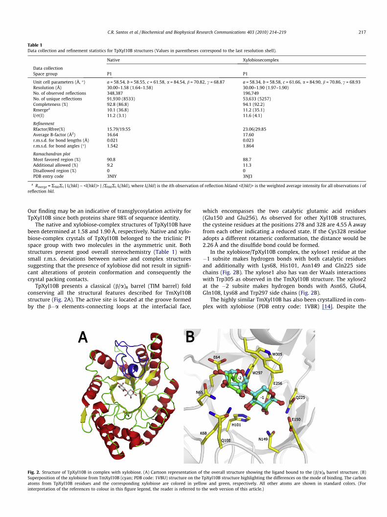
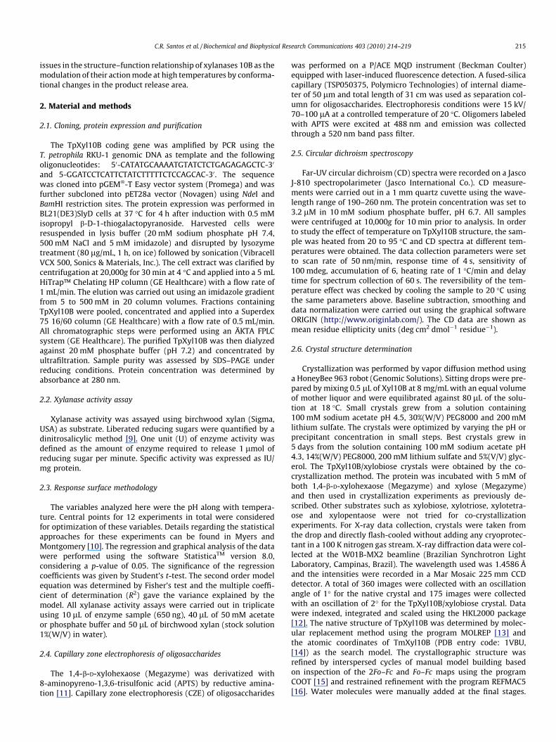
Table 1Data collection and refinement statistics for TpXyl10B structures (Values in parentheses correspond to the last resolution shell).
Native Xylobiosecomplex
Data collectionSpace group P1 P1
Unit cell parameters (Å, �) a = 58.54, b = 58.55, c = 61.58, a = 84.54, b = 70.82, c = 68.87 a = 58.34, b = 58.58, c = 61.66, a = 84.90, b = 70.86, c = 68.93Resolution (Å) 30.00–1.58 (1.64–1.58) 30.00–1.90 (1.97–1.90)No. of observed reflections 348,387 196,749No. of unique reflections 91,930 (8533) 53,633 (5257)Completeness (%) 92.8 (86.8) 94.1 (92.2)Rmergea 10.1 (36.8) 11.2 (35.1)I/r(I) 11.2 (3.1) 11.6 (4.1)
RefinementRfactor/Rfree(%) 15.79/19.55 23.06/29.85Average B-factor (Å2) 16.64 17.60r.m.s.d. for bond lengths (Å) 0.021 0.023r.m.s.d. for bond angles (�) 1.542 1.864
Ramachandran plotMost favored region (%) 90.8 88.7Additional allowed (%) 9.2 11.3Disallowed region (%) 0 0PDB entry code 3NIY 3NJ3
a Rmerge = RhklRi | Ii(hkl) – <I(hkl)> | /RhklRi Ii(hkl), where Ii(hkl) is the ith observation of reflection hkland <I(hkl)> is the weighted average intensity for all observations i ofreflection hkl.
C.R. Santos et al. / Biochemical and Biophysical Research Communications 403 (2010) 214–219 217
Our finding may be an indicative of transglycosylation activity forTpXyl10B since both proteins share 98% of sequence identity.
The native and xylobiose-complex structures of TpXyl10B havebeen determined at 1.58 and 1.90 Å, respectively. Native and xylo-biose-complex crystals of TpXyl10B belonged to the triclinic P1space group with two molecules in the asymmetric unit. Bothstructures present good overall stereochemistry (Table 1) withsmall r.m.s. deviations between native and complex structuressuggesting that the presence of xylobiose did not result in signifi-cant alterations of protein conformation and consequently thecrystal packing contacts.
TpXyl10B presents a classical (b/a)8 barrel (TIM barrel) foldconserving all the structural features described for TmXyl10Bstructure (Fig. 2A). The active site is located at the groove formedby the b�a elements-connecting loops at the interfacial face,
Fig. 2. Structure of TpXyl10B in complex with xylobiose. (A) Cartoon representation ofSuperposition of the xylobiose from TmXyl10B (cyan; PDB code: 1VBU) structure on theatoms from TpXyl10B residues and the corresponding xylobiose are colored in yellointerpretation of the references to colour in this figure legend, the reader is referred to
which encompasses the two catalytic glutamic acid residues(Glu150 and Glu256). As observed for other Xyl10B structures,the cysteine residues at the positions 278 and 328 are 4.55 Å awayfrom each other indicating a reduced state. If the Cys328 residueadopts a different rotameric conformation, the distance would be2.26 Å and the disulfide bond could be formed.
In the xylobiose/TpXyl10B complex, the xylose1 residue at the�1 subsite makes hydrogen bonds with both catalytic residuesand additionally with Lys68, His101, Asn149 and Gln225 sidechains (Fig. 2B). The xylose1 also has van der Waals interactionswith Trp305 as observed in the TmXyl10B structure. The xylose2at the �2 subsite makes hydrogen bonds with Asn65, Glu64,Gln108, Lys68 and Trp297 side chains (Fig. 2B).
The highly similar TmXyl10B has also been crystallized in com-plex with xylobiose (PDB entry code: 1VBR) [14]. Despite the
the overall structure showing the ligand bound to the (b/a)8 barrel structure. (B)TpXyl10B structure highlighting the differences on the mode of binding. The carbonw and green, respectively. All other atoms are shown in standard colors. (For
the web version of this article.)

Fig. 3. Structural changes in the interfacial loops at 90 �C modulate enzymatic specificity of TpXyl10B. (A) The structure of TpXyl10B from molecular dynamics simulations at90 �C was superposed on the native crystal structure and the main differences are colored in blue (crystal) and red (simulation). Molecular surface representations of thecrystal structure (B) and the final frame from MD simulation at 90 �C (C). The P. simplicissimum xylopentaose complex structure (PDB entry code: 1B3Z) was superposed onthe TpXyl10B structure and the xylopentaose is shown in the TpXyl10B active site to delineate the �3 to +2 subsites. (For interpretation of the references to colour in thisfigure legend, the reader is referred to the web version of this article.)
218 C.R. Santos et al. / Biochemical and Biophysical Research Communications 403 (2010) 214–219
strong conservation of sequence between TpXyl10B and TmXyl10B(five divergent residues, located at the protein surface), the xylobi-ose at the active site is in a different conformation for these twoxylanases (Fig. 2B). The xylose2 from TpXyl10B is rotated about180� with respect to the corresponding xylose in the TmXyl10Bstructure. In other xylanases structures with longer substrates likexylotriose (PDB entry code: 2W5F; [27]) and xylotetraose (PDB en-try code: 1US2; [28]), the xylose subunit at the �2 subsite displaysa classical conformation as observed in our xylobiose-TpXyl10Bcomplex structure. Interestingly, the active-site residues contact-ing xylobiose are exactly in the same position when compared tothe xylobiose-TmXyl10B structure indicating that the unusual con-formation observed in the TmXyl10B structure may be due to amisinterpretation of electron density map.
3.5. Structural basis for the mode of action at the optimumtemperature
The enzymatic hydrolysis of xylan by endo-xylanases occurs be-tween xylose residues at the �1 and +1 subsites. The xylose num-bering follows sugar binding site nomenclature for glycosylhydrolases [29]. These sites are well known, as shown for xylan-ases 10B from Clostridium thermocellum, Cellvibrio japonicus andPenicillium simplicissimum [27,28,30]. The �1, �2, �3 subsites werecalled ‘‘substrate recognition area’’ and +1,+2,+3 subsites werecalled ‘‘product release area’’ by Schmidt [30]. The substrate recog-nition area contains residues making abundant interactions withthe ligand, whereas the product release area residues present few-er interactions with the ligand. It has been proposed that the neg-ative subsites determine the minimum length of the substrate forcatalysis indicating that xylotetraose is the shortest hydrolyzablesubstrate for xylanases 10B [30]. Herein, we propose a key roleof the positive subsites in determining the product cleavage pat-tern of xylanases 10B.
As observed by CZE analysis, TpXyl10B releases exclusivelyxylobiose at temperatures close to 90 �C (Fig. 1B) and it is corre-lated with significant structural changes under the same condi-tions as assessed by CD spectroscopy (Fig. 1C). In order to shedlight on the molecular basis of the temperature influence on the ac-tion mode of TpXyl10B, molecular dynamics simulations were car-ried out at 20 and 90 �C.
The final frame of TpXyl10B structure from MD simulations at90 �C presented only significant differences around the product
release area when compared to the crystal structure, withoutdisturbing the overall fold and structure (Fig. 3). At 90 �C, the cat-alytic loop comprising the residues 297–326 was displaced faraway from its original conformation, resulting in a drastic increaseof accessible solvent area (ASA) of the active-site pocket and in theloss of the +3 subsite. Contrary to the fact that the increase of ASAaround to the active site could accommodate larger substrates, theloss of the +3 subsite does not permit the binding of additional xy-lose subunits at the product release area. Additionally, the dis-placement of the catalytic loop has also disturbed the adjacent+2 subsite contributing to specific release of xylobiose at the opti-mum temperature.
In conclusion, our findings from CD, CZE, XTAL and MD studiessuggest that thermal-induced conformational changes in the prod-uct release area play a role in the modulation of the action mode ofthermostable xylanases 10B.
Acknowledgments
This research was supported by grants from Fundação deAmparo à Pesquisa do Estado de São Paulo (FAPESP, 08/58037-9)to FMS and Conselho Nacional de Desenvolvimento Científico eTecnológico (CNPq, 478059/2009-4 and 472174/2007-0) to MTMand HV, respectively. RAP has funding from the Department of En-ergy, awards 06103-OKL and ZDJ-7-77608-01.
Appendix A. Supplementary data
Supplementary data associated with this article can be found, inthe online version, at doi:10.1016/j.bbrc.2010.11.010.
References
[1] J.D. McCarter, S.G. Withers, Mechanisms of enzymatic glycoside hydrolysis,Curr. Opin. Struct. Biol. 4 (1994) 885–892.
[2] G. Davies, B. Henrissat, Structures and mechanisms of glycosyl hydrolases,Structure 3 (1995) 853–859.
[3] B. Henrissat, G. Davies, Structural and sequence-based classification ofglycoside hydrolases, Curr. Opin. Struct. Biol. 7 (1997) 637–644.
[4] B. Henrissat, A. Bairoch, New families in the classification of glycosylhydrolases based on amino acid sequence similarities, Biochem. J. 293(1993) 781–788.
[5] B.L. Cantarel, P.M. Coutinho, C. Rancurel, T. Bernard, V. Lombard, B. Henrissat,The carbohydrate-active enzymes database (CAZy): an expert resource forglycogenomics, Nucleic Acids Res. 37 (2009) D233–D238.
[6] N. Kulkarni, A. Shendye, M. Rao, Molecular and biotechnological aspects ofxylanases, FEMS Microbiol. Rev. 23 (1999) 411–456.
C.R. Santos et al. / Biochemical and Biophysical Research Communications 403 (2010) 214–219 219
[7] J. Sheehan, M. Himmel, Enzymes, energy, and the environment: a strategicperspective on the U.S. Department of energy’s research and developmentactivities for bioethanol, Biotechnol. Prog. 15 (1999) 817–827.
[8] J. Zaldivar, J. Nielsen, L. Olsson, Fuel ethanol production from lignocellulose: achallenge for metabolic engineering and process integration, Appl. Microbiol.Biotechnol. 56 (2001) 17–34.
[9] G. Miller, Use of dinitrosalicylic acid reagent for determination of reducingsugar, Anal. Chem. 31 (1959) 426–428.
[10] R.H. Myers, D.C. Montgomery, Response surface methodology: process andproduct optimization using designed experiments, John Wiley & Sons, NewYork, 2002, pp. 1–13.
[11] F.T. Chen, R.A. Evangelista, Analysis of mono- and oligosaccharide isomersderivatized with 9-aminopyrene-1, 4, 6-trisulfonate by capillary electropho-resis with laser-induced fluorescence, Anal. Biochem. 230 (1995) 273–280.
[12] Z. Otwinowski, W. Minor, Processing of X-ray diffraction data collected inoscillation mode, Methods Enzymol. 276 (1997) 307–326.
[13] A. Vagin, A. Teplyakov, MOLREP: an automated program for molecularreplacement, J. Appl. Crystallogr. 30 (1997) 1022–1025.
[14] Ihsanawati, T. Kumasaka, T. Kaneko, C. Morokuma, R. Yatsunami, T. Sato,S. Nakamura, N. Tanaka, Structural basis of the substrate subsite and the highlythermal stability of xylanase 10B from Thermotoga maritima MSB8, Proteins 61(2005) 999–1009.
[15] P. Emsley, K. Cowtan, Coot: model-building tools for molecular graphics, ActaCrystallogr. D 60 (2004) 2126–2132.
[16] G.N. Murshudov, A.A. Vagin, E.J. Dodson, Refinement of macromolecularstructures by the maximum-likelihood method, Acta Crystallogr. D 53 (1997)240–255.
[17] R.A. Laskowski, M.W. MacArthur, D.S. Moss, J.M. Thornton, PROCHECK - aprogram to check the stereochemical quality of protein structures, J. Appl.Crystallogr. 26 (1993) 283–291.
[18] D. van der Spoel, E. Lindahl, B. Hess, G. Groenhof, A.E. Mark, H.J.C. Berendsen,GROMACS: fast, flexible and free, J. Comput. Chem. 26 (2005) 1701–1718.
[19] B.L. de Groot, H. Grubmüller, Water permeation across biological membranes:mechanism and dynamics of aquaporin-1 and GlpF, Science 294 (2001) 2353–2357.
[20] H.J.C. Berendsen, J.P.M. Postma, W. van Gunsteren, J. Hermans, Interactionmodels for water in relation to protein hydration, in: B. Pullman (Ed.),
Intermolecular Forces, Dordrecht Reidel Publishing Company, TheNetherlands, 1981, pp. 331–342.
[21] T. Darden, D. York, L. Pedersen, Particle mesh Ewald – an N log(N) method forEwald sums in large systems, J. Chem. Phys. 98 (1993) 10089–10092.
[22] H.J.C. Berendsen, J.P.M. Postma, A. DiNola, J.R. Haak, Molecular dynamics withcoupling to an external bath, J. Chem. Phys. 81 (1984) 3684–3690.
[23] F. Francis, A. Sabu, K.M. Nampoothiri, S. Ramachandram, S. Ghosh, G. Szakacs,A. Pandey, Use of response surface methodology for optimizing processparameters for the production of a-amylase by Aspergillus oryzae, Biochem.Eng. J. 15 (2003) 107–115.
[24] F.F.C. Barros, J.L. Bicas, J.C. Silva, A.P. Dionisio, G.M. Pastore, Use of the responsesurface methodology to optimize the pre-treatment of cassava wastewateremployed as substrates for the production biosurfactants, in: J. Krause, O.Fleischer (Eds.), Industrial Fermentation: Food Processes, Nutrient Sources andProduction Strategies, Nova Science Publisher, Hauppauge, New York, 2010,pp. 327–341.
[25] J. Zhengqiang, A. Kobayashi, M.M. Ahsan, L. Lite, M. Kitaoka, K. Hayashi,Characterization of a thermostable family 10 endo-xylanase (XynB) fromThermotoga maritima that cleaves p-nitrophenyl-beta-D-xyloside, J. Biosci.Bioeng. 92 (2001) 423–428.
[26] Z.Q. Jiang, W. Deng, Y.P. Zhu, L.T. Li, Y.J. Sheng, K. Hayashi, The recombinantxylanase B of Thermotoga maritima is highly xylan specific and producesexclusively xylobiose from xylans, a unique character for industrialapplications, J. Mol. Catal. B Enzym. 27 (2004) 207–213.
[27] S. Najmudin, B.A. Pinheiro, J.A. Prates, H.J. Gilbert, M.J. Romão, C.M. Fontes,Putting an N-terminal end to the Clostridium thermocellum xylanase Xyn10Bstory: crystal structure of the CBM22–1-GH10 modules complexed withxylohexaose, J. Struct. Biol. 172 (2010) 353–362.
[28] G. Pell, L. Szabo, S.J. Charnock, H. Xie, T.M. Gloster, G.J. Davies, H.J. Gilbert,Structural and biochemical analysis of Cellvibrio japonicus xylanase 10C: howvariation in substrate-binding cleft influences the catalytic profile of familyGH-10 xylanases, J. Biol. Chem. 279 (2004) 11777–11788.
[29] G.J. Davies, K.S. Wilson, B. Henrissat, Nomenclature for sugar-binding subsitesin glycosyl hydrolases, Biochem. J. 321 (1997) 557–559.
[30] A. Schmidt, G.M. Gübitz, C. Kratky, Xylan binding subsite mapping in thexylanase from Penicillium simplicissimum using xylooligosaccharides as cryo-protectant, Biochemistry 38 (1999) 2403–2412.