Stability, folding dynamics, and long-range conformational ...
10
Stability, folding dynamics, and long-range conformational transition of the synaptic t-SNARE complex Xinming Zhang a , Aleksander A. Rebane a,b,c,d , Lu Ma a , Feng Li (李峰) a,d , Junyi Jiao a,b , Hong Qu a , Frederic Pincet a,d,e , James E. Rothman a,d , and Yongli Zhang a,1 a Department of Cell Biology, Yale School of Medicine, New Haven, CT 06520; b Integrated Graduate Program in Physical and Engineering Biology, Yale University, New Haven, CT 06511; c Department of Physics, Yale University, New Haven, CT 06511; d Nanobiology Institute, Yale University, West Haven, CT 06477; and e Laboratoire de Physique Statistique, École Normale Superieure, L’université de recherche Paris Sciences et Lettres, CNRS UMR 8550, Sorbonne Universités, Université Pierre-et-Marie-Curie University of Paris 06, Université Paris Diderot, 75005 Paris, France Edited by Axel T. Brunger, Stanford University, Stanford, CA, and approved November 9, 2016 (received for review April 8, 2016) Synaptic soluble N-ethylmaleimide–sensitive factor attachment protein receptors (SNAREs) couple their stepwise folding to fusion of synaptic vesicles with plasma membranes. In this process, three SNAREs assemble into a stable four-helix bundle. Arguably, the first and rate-limiting step of SNARE assembly is the formation of an ac- tivated binary target (t)-SNARE complex on the target plasma mem- brane, which then zippers with the vesicle (v)-SNARE on the vesicle to drive membrane fusion. However, the t-SNARE complex readily misfolds, and its structure, stability, and dynamics are elusive. Using single-molecule force spectroscopy, we modeled the synaptic t-SNARE complex as a parallel three-helix bundle with a small frayed C terminus. The helical bundle sequentially folded in an N-terminal domain (NTD) and a C-terminal domain (CTD) separated by a central ionic layer, with total unfolding energy of ∼17 k B T, where k B is the Boltzmann constant and T is 300 K. Peptide binding to the CTD activated the t-SNARE complex to initiate NTD zippering with the v-SNARE, a mechanism likely shared by the mammalian uncoordinated-18-1 protein (Munc18- 1). The NTD zippering then dramatically stabilized the CTD, facilitating further SNARE zippering. The subtle bidirectional t-SNARE conforma- tional switch was mediated by the ionic layer. Thus, the t-SNARE com- plex acted as a switch to enable fast and controlled SNARE zippering required for synaptic vesicle fusion and neurotransmission. t-SNARE complex | SNARE four-helix bundle | SNARE assembly | membrane fusion | optical tweezers S ynaptic soluble N-ethylmaleimide–sensitive factor attachment protein receptors (SNAREs) mediate fast and calcium-triggered fusion of synaptic vesicles to presynaptic plasma membranes re- quired for neurotransmission (1). They consist of VAMP2 (vesicle associated membrane protein 2, also called synaptobrevin 2) an- chored on vesicles (v-SNARE) and syntaxin and SNAP-25 (synap- tosome associated protein 25) located on target plasma membranes (t-SNAREs) (2). These SNAREs contain characteristic SNARE motifs of ∼60 amino acids (3) (Fig. 1A). Syntaxin and SNAP-25 can form a 1:1 t-SNARE complex (4–6). During membrane fusion, the t- and v-SNAREs join to form an extraordinarily stable four-helix bundle (3, 7–10). In the core of the bundle are 15 layers of hy- drophobic amino acids and a central ionic layer containing three glutamines and one arginine. Whereas the zippering energy and kinetics between t- and v-SNAREs have recently been measured (8, 9), the structure, stability, and dynamics of the t-SNARE complex have not been well understood. The structure and dynamics of the t-SNARE complex are crucial for SNARE assembly and membrane fusion. Formation of the t-SNARE complex is likely an obligate intermediate before SNARE zippering (6, 11–14). A preformed t-SNARE complex docks the vesicles to plasma membranes (15) and boosts the speed, strength, and accuracy of SNARE zippering (5, 9, 16). Furthermore, the t-SNARE complex is an important target for proteins that regulate SNARE zippering and membrane fusion, such as Munc18-1, synaptotagmins, and complexin (8, 17–19). Finally, the t-SNARE complex shows intriguing dynamics in reconstituted membrane fusion. Peptides corresponding to the VAMP2 N-terminal domain (NTD; called Vn peptides or Vn) or C-terminal domain (CTD; called Vc) are often used to facilitate membrane fusion (6, 10, 20, 21). Tightly bound to the t-SNARE complex, they attenuate SNARE zippering (8, 22), yet surprisingly enhance the rate of membrane fusion (6, 10). The underlying molecular mechanisms are not fully understood, which calls for an improved understanding of the structure and dynamics of the t-SNARE complex. Studying t-SNARE folding is challenging using ensemble-based experimental approaches, because the t-SNARE complex readily misfolds (6, 21, 23) and is highly dynamic (4). Syntaxin and SNAP-25 can efficiently form a stable parallel four-helix bundle containing two syntaxin molecules and one SNAP-25 molecule (the 2:1 complex), which inhibits SNARE zippering and membrane fusion (5, 6). In addition, it is reported that the t-SNARE complex folds into at least two alternative conformations in which either SNARE motif in SNAP-25 partially or completely dissociates from syntaxin (4). Interestingly, yeast t-SNARE homologs Sso1 and Sec9 do not misfold. Fiebig et al. (24) found that Sso1 in the t-SNARE com- plex is N-terminally structured but C-terminally disordered. Using optical tweezers, Ma et al. (8) and Gao et al. (9) observed that synaptic t-SNARE complexes unfold cooperatively at a high force Significance Intracellular membrane fusion is mediated by coupled folding and assembly of three or four soluble N-ethylmaleimide–sensitive factor attachment protein receptor (SNARE) proteins into a four- helix bundle. A rate-limiting step is the formation of a partial complex containing three helixes called the target (t)-SNARE complex on the target plasma membrane. The t-SNARE complex then serves as a template to guide stepwise zippering of the fourth helix, a process that is further regulated by other proteins. The synaptic t-SNARE complex readily misfolds. Consequently, its conformation, stability, and dynamics have not been well un- derstood. Using optical tweezers and theoretical modeling, we elucidated the folding intermediates and kinetics of the t-SNARE complex and discovered a long-range conformational switch of t-SNAREs during SNARE zippering, which is essential for regulated SNARE assembly during synaptic vesicle fusion. Author contributions: X.Z., J.E.R., and Y.Z. designed research; X.Z., L.M., J.J., and H.Q. performed research; F.L. contributed new reagents/analytic tools; X.Z., A.A.R., L.M., F.L., J.J., H.Q., F.P., J.E.R., and Y.Z. analyzed data; and X.Z. and Y.Z. wrote the paper. The authors declare no conflict of interest. This article is a PNAS Direct Submission. 1 To whom correspondence should be addressed. Email: [email protected]. This article contains supporting information online at www.pnas.org/lookup/suppl/doi:10. 1073/pnas.1605748113/-/DCSupplemental. www.pnas.org/cgi/doi/10.1073/pnas.1605748113 PNAS | Published online November 28, 2016 | E8031–E8040 BIOPHYSICS AND COMPUTATIONAL BIOLOGY PNAS PLUS
-
Upload
khangminh22 -
Category
Documents
-
view
0 -
download
0
Transcript of Stability, folding dynamics, and long-range conformational ...

Stability, folding dynamics, and long-rangeconformational transition of the synaptict-SNARE complexXinming Zhanga, Aleksander A. Rebanea,b,c,d, Lu Maa, Feng Li (李峰)a,d, Junyi Jiaoa,b, Hong Qua, Frederic Pinceta,d,e,James E. Rothmana,d, and Yongli Zhanga,1
aDepartment of Cell Biology, Yale School of Medicine, New Haven, CT 06520; bIntegrated Graduate Program in Physical and Engineering Biology, YaleUniversity, New Haven, CT 06511; cDepartment of Physics, Yale University, New Haven, CT 06511; dNanobiology Institute, Yale University, West Haven,CT 06477; and eLaboratoire de Physique Statistique, École Normale Superieure, L’université de recherche Paris Sciences et Lettres, CNRS UMR 8550,Sorbonne Universités, Université Pierre-et-Marie-Curie University of Paris 06, Université Paris Diderot, 75005 Paris, France
Edited by Axel T. Brunger, Stanford University, Stanford, CA, and approved November 9, 2016 (received for review April 8, 2016)
Synaptic soluble N-ethylmaleimide–sensitive factor attachmentprotein receptors (SNAREs) couple their stepwise folding to fusionof synaptic vesicles with plasma membranes. In this process, threeSNAREs assemble into a stable four-helix bundle. Arguably, the firstand rate-limiting step of SNARE assembly is the formation of an ac-tivated binary target (t)-SNARE complex on the target plasma mem-brane, which then zippers with the vesicle (v)-SNARE on the vesicleto drive membrane fusion. However, the t-SNARE complex readilymisfolds, and its structure, stability, and dynamics are elusive. Usingsingle-molecule force spectroscopy, wemodeled the synaptic t-SNAREcomplex as a parallel three-helix bundlewith a small frayed C terminus.The helical bundle sequentially folded in an N-terminal domain (NTD)and a C-terminal domain (CTD) separated by a central ionic layer, withtotal unfolding energy of∼17 kBT, where kB is the Boltzmann constantand T is 300 K. Peptide binding to the CTD activated the t-SNAREcomplex to initiate NTD zippering with the v-SNARE, a mechanismlikely shared by themammalian uncoordinated-18-1 protein (Munc18-1). The NTD zippering then dramatically stabilized the CTD, facilitatingfurther SNARE zippering. The subtle bidirectional t-SNARE conforma-tional switch was mediated by the ionic layer. Thus, the t-SNARE com-plex acted as a switch to enable fast and controlled SNARE zipperingrequired for synaptic vesicle fusion and neurotransmission.
t-SNARE complex | SNARE four-helix bundle | SNARE assembly |membrane fusion | optical tweezers
Synaptic soluble N-ethylmaleimide–sensitive factor attachmentprotein receptors (SNAREs) mediate fast and calcium-triggered
fusion of synaptic vesicles to presynaptic plasma membranes re-quired for neurotransmission (1). They consist of VAMP2 (vesicleassociated membrane protein 2, also called synaptobrevin 2) an-chored on vesicles (v-SNARE) and syntaxin and SNAP-25 (synap-tosome associated protein 25) located on target plasma membranes(t-SNAREs) (2). These SNAREs contain characteristic SNAREmotifs of ∼60 amino acids (3) (Fig. 1A). Syntaxin and SNAP-25 canform a 1:1 t-SNARE complex (4–6). During membrane fusion, thet- and v-SNAREs join to form an extraordinarily stable four-helixbundle (3, 7–10). In the core of the bundle are 15 layers of hy-drophobic amino acids and a central ionic layer containing threeglutamines and one arginine. Whereas the zippering energy andkinetics between t- and v-SNAREs have recently been measured(8, 9), the structure, stability, and dynamics of the t-SNAREcomplex have not been well understood.The structure and dynamics of the t-SNARE complex are crucial
for SNARE assembly and membrane fusion. Formation of thet-SNARE complex is likely an obligate intermediate before SNAREzippering (6, 11–14). A preformed t-SNARE complex docks thevesicles to plasma membranes (15) and boosts the speed, strength,and accuracy of SNARE zippering (5, 9, 16). Furthermore, thet-SNARE complex is an important target for proteins that regulateSNARE zippering and membrane fusion, such as Munc18-1,
synaptotagmins, and complexin (8, 17–19). Finally, the t-SNAREcomplex shows intriguing dynamics in reconstituted membranefusion. Peptides corresponding to the VAMP2 N-terminal domain(NTD; called Vn peptides or Vn) or C-terminal domain (CTD;called Vc) are often used to facilitate membrane fusion (6, 10, 20,21). Tightly bound to the t-SNARE complex, they attenuateSNARE zippering (8, 22), yet surprisingly enhance the rate ofmembrane fusion (6, 10). The underlying molecular mechanismsare not fully understood, which calls for an improved understandingof the structure and dynamics of the t-SNARE complex.Studying t-SNARE folding is challenging using ensemble-based
experimental approaches, because the t-SNARE complex readilymisfolds (6, 21, 23) and is highly dynamic (4). Syntaxin and SNAP-25can efficiently form a stable parallel four-helix bundle containingtwo syntaxin molecules and one SNAP-25 molecule (the 2:1complex), which inhibits SNARE zippering and membrane fusion(5, 6). In addition, it is reported that the t-SNARE complex foldsinto at least two alternative conformations in which either SNAREmotif in SNAP-25 partially or completely dissociates from syntaxin(4). Interestingly, yeast t-SNARE homologs Sso1 and Sec9 do notmisfold. Fiebig et al. (24) found that Sso1 in the t-SNARE com-plex is N-terminally structured but C-terminally disordered. Usingoptical tweezers, Ma et al. (8) and Gao et al. (9) observed thatsynaptic t-SNARE complexes unfold cooperatively at a high force
Significance
Intracellular membrane fusion is mediated by coupled foldingand assembly of three or four soluble N-ethylmaleimide–sensitivefactor attachment protein receptor (SNARE) proteins into a four-helix bundle. A rate-limiting step is the formation of a partialcomplex containing three helixes called the target (t)-SNAREcomplex on the target plasma membrane. The t-SNARE complexthen serves as a template to guide stepwise zippering of thefourth helix, a process that is further regulated by other proteins.The synaptic t-SNARE complex readily misfolds. Consequently, itsconformation, stability, and dynamics have not been well un-derstood. Using optical tweezers and theoretical modeling, weelucidated the folding intermediates and kinetics of the t-SNAREcomplex and discovered a long-range conformational switch oft-SNAREs during SNARE zippering, which is essential for regulatedSNARE assembly during synaptic vesicle fusion.
Author contributions: X.Z., J.E.R., and Y.Z. designed research; X.Z., L.M., J.J., and H.Q. performedresearch; F.L. contributed new reagents/analytic tools; X.Z., A.A.R., L.M., F.L., J.J., H.Q., F.P., J.E.R.,and Y.Z. analyzed data; and X.Z. and Y.Z. wrote the paper.
The authors declare no conflict of interest.
This article is a PNAS Direct Submission.1To whom correspondence should be addressed. Email: [email protected].
This article contains supporting information online at www.pnas.org/lookup/suppl/doi:10.1073/pnas.1605748113/-/DCSupplemental.
www.pnas.org/cgi/doi/10.1073/pnas.1605748113 PNAS | Published online November 28, 2016 | E8031–E8040
BIOPH
YSICSAND
COMPU
TATIONALBIOLO
GY
PNASPL
US

when pulled from both ends of syntaxin, indicating a largely struc-tured syntaxin in a stable t-SNARE complex. However, the detailedconformation of the t-SNARE complex, especially SNAP-25, andits stability and dynamics are not clear.In this work, we measured the conformation, stability, and dy-
namics of a single synaptic t-SNARE complex using optical tweezers.Our single-molecule method prevented the t-SNARE complex frommisfolding, thus allowing us to focus on the 1:1 complex. We foundthat the t-SNARE complex folded in two steps and had a frayedt-SNARE C terminus (Tc). Binding of Vn stabilized the CTD,whereas binding of Vc stabilized the NTD, structured the Tc, andpromoted initial ternary SNARE zippering, potentially accountingfor the positive effect of both peptides on membrane fusion.
ResultsT-SNARE Constructs and Experimental Setup. To measure the con-formation and stability of the cytoplasmic t-SNARE complex, wefirst pulled a single t-SNARE complex at the C termini of syntaxinand the first SNARE motif in SNAP-25 (SN1) (Fig. 1 A and B).Their N termini were cross-linked by a disulfide bond formed be-tween two cysteine residues. We chose the N-terminal cross-linkingsite such that it facilitated refolding of the t-SNARE complex but
minimally altered its structure. We tested three cross-linking sitesby substituting the corresponding amino acids with cysteine anddesignated the SNARE constructs as −8C, −12C, and −16C (Fig.1A). To prevent t-SNARE misfolding and ensure correct cross-linking, we first formed the ternary SNARE complex and thenremoved the VAMP2 molecule by unfolding the ternary complex.The complex was attached on one end to a streptavidin-coatedpolystyrene bead through a biotinylated Avi-tag and on the otherend to an antidigoxigenin antibody-coated polystyrene beadthrough a 2,260-bp DNA handle (9) (Fig. 1B). The beads weretrapped in two optical traps formed by focused laser beams. Bymoving one optical trap relative to the other, we controlled theforce applied on the SNARE complex and measured the end-to-end extension of the SNARE/DNA tether in response to theforce. We recorded the force and extension at 10 kHz and usedthem to derive t-SNARE folding and stability.
Syntaxin and SN1 Are Largely Structured and Fold Reversibly.We firstpulled a single ternary SNARE construct −8C to a force of ∼22 pN,leading to a representative force-extension curve (FEC) shown inFig. 1C. Below ∼15 pN, the extension increased monotonically withforce, mainly due to stretching of the semiflexible DNA handle.
B-8C
SN2SNAP-25
SN1Syntaxin (Syx)
DNA handle
Extension
DigoxigeninBiotin
-8
-1
+4
s--s
NRD
SyxSNAP-25VAMP2
1 23
-7-1
4
-7-1
+4
D-7-1
+4
0
5
10
15
20
25
50 nm
Extension
Forc
e (p
N)
1 12 3
4
23
41 2 3 4 1
23
4
C -8C -12C
A-7 -6 -5 -4 -3 -2 -1 0 1 2 3 4 5 6 7 8
NTD CTD
Syx
SN1SN2
F
FDEREQMAISGGFIRRVTNDARENEMDENLEQVSGIIGNLRHMALDMGNEIDTQNRQIDRIMEKADSNKTRIDEANQRATKM
LESGNPAIFASGIIMDSSISKQALSEIETRHSEIIKLENSIRELHDMFMDMAMLVESQGEMIDRIEYNVEHAVDYVERAVSDTKKA
AEDAADMRNELEEMQRRADQLADESLESTRRMLQLVEESKDAGIRTLVMLDEQGEQLERIEEGMDQINKDMKEAEKNLTDL
-8-9-10-15 -14 -13 -12 -11-16
-8C-12C-16C
Tc
226219212205198191184177170 233 240 247 254
SN2C
Fig. 1. T-SNARE sequences, experimental setup, and derived folding states. (A) Amino acids of the synaptic syntaxin 1A (Syx) and SNAP-25B containingSNARE motifs. SNAP-25B consists of two SNARE motifs (SN1 and SN2) connected by a disordered linker (magenta line), with four intrinsic cysteines (marked bystars) mutated to serine. Amino acids of the hydrophobic and ionic layers in the SNARE motifs (numbered from −7 to +8) and their N-terminal extensions(numbered from −16 to −8) are highlighted in yellow. The syntaxin sequence is numbered in red. Four pairs of cross-linked amino acids are indicated by linesand labeled by their corresponding construct names and pulling sites (arrows). The t-SNARE complex contained three distinct folding domains: the NTD, theCTD, and the frayed Tc. (B) Experimental setup to pull a single t-SNARE complex (−8C) containing the NRD of syntaxin. The structure of the folded t-SNAREcomplex shown here was modeled based on the crystal structure of the SNARE ternary complex (3) and our single-molecule measurements. (C) FECs obtainedby pulling (black and green) and relaxing (cyan) the two SNARE constructs −8C and −12C. The pulling or relaxing direction is indicated by arrows colored thesame as the corresponding FECs. Blue arrows mark full disassembly of the ternary SNARE complex and accompanying dissociation of the VAMP2 molecule.Red lines are best fits of the corresponding FECs by the worm-like chain model. (D) Schematic of the SNARE transitions among four states, including the fullyassembled ternary SNARE state 1, the folded t-SNARE state 2, the partially folded t-SNARE state 3, and the fully unfolded t-SNARE state 4.
E8032 | www.pnas.org/cgi/doi/10.1073/pnas.1605748113 Zhang et al.
As a result, the FEC could be fit by a worm-like chain model (25)(Fig. 1C, red curve). Above 17 pN, first fast and then slow extensionflickering (Fig. 1C, regions marked by green and magenta paral-lelograms, respectively) appeared successively as force increased,indicating reversible folding and unfolding transitions of a SNAREC-terminal region and middle region, respectively. At ∼20 pN, anabrupt extension jump (Fig. 1C, indicated by a blue arrow) repre-sented irreversible unfolding of the remaining N-terminal region.Pulling the molecule to above 20 pN did not cause any additionalunfolding, which demonstrated that the SNARE complex had beenfully unfolded (Fig. 1C, state 4). The above interpretations onSNARE transitions were confirmed by the similarities and differ-ences in the FECs obtained by pulling the other two SNARE con-structs −12C and −16C (Fig. 1C and Fig. S1).Upon relaxation, the SNARE complex in all three constructs
remained unfolded at a force above ∼7 pN, but refolded below thisforce (Fig. 1C and Fig. S1). The folding process was reversible via atransient intermediate state (Fig. 1 C and D, state 3). Interestingly,the fully refolded SNARE complex (in state 2) had an extensiongreater than the fully assembled ternary SNARE complex (in state2), suggesting a partially folded t-SNARE complex. Pulling thet-SNAREs again revealed an FEC that overlapped the relaxationFEC (Fig. 1C, compare the green and cyan FECs for −12C). Theseobservations indicate that the VAMP2 molecule dissociated fromthe t-SNAREs upon disassembly of the ternary complex. To con-firm this interpretation, we added 10 μM VAMP2 into the solutionafter a single ternary SNARE complex had been disassembled andfound that VAMP2 restored assembly of the ternary SNAREcomplex (Fig. S2). Thus, disassembly of the ternary SNARE com-plex led to dissociation of the VAMP2 molecule (Fig. 1D, states1–4) and generated an unfolded t-SNARE complex that partiallyrefolded at a low force via an intermediate state 3. In addition, theintermediate state 3 appeared to be partially zippered (Fig. 1D),because shifting the N-terminal cross-linking site away from theSNARE motifs (from −8C to −12C to −16C) changed the exten-
sion of the intermediate state relative to the extension of the un-folded t-SNARE state, but not of the folded t-SNARE state (Fig.1C and Fig. S1). Finally, we found that the N-terminal regulatorydomain (NRD) of syntaxin (Fig. 1B) did not significantly affectt-SNARE folding, because the t-SNARE complex without theNRD showed an FEC identical to the FEC of the complex with theNRD (Fig. S3). This finding indicates that the NRD did notstrongly interact with the SNARE motifs, consistent with our pre-vious observation (8).
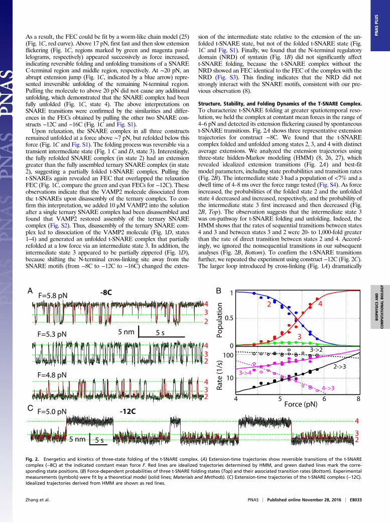
Structure, Stability, and Folding Dynamics of the T-SNARE Complex.To characterize t-SNARE folding at greater spatiotemporal reso-lution, we held the complex at constant mean forces in the range of4–6 pN and detected its extension flickering caused by spontaneoust-SNARE transitions. Fig. 2A shows three representative extensiontrajectories for construct −8C. We found that the t-SNAREcomplex folded and unfolded among states 2, 3, and 4 with distinctaverage extensions. We analyzed the extension trajectories usingthree-state hidden-Markov modeling (HMM) (8, 26, 27), whichrevealed idealized extension transitions (Fig. 2A) and best-fitmodel parameters, including state probabilities and transition rates(Fig. 2B). The intermediate state 3 had a population of <7% and adwell time of 4–8 ms over the force range tested (Fig. S4). As forceincreased, the probabilities of the folded state 2 and the unfoldedstate 4 decreased and increased, respectively, and the probability ofthe intermediate state 3 first increased and then decreased (Fig.2B, Top). The observation suggests that the intermediate state 3was on-pathway for t-SNARE folding and unfolding. Indeed, theHMM shows that the rates of sequential transitions between states4 and 3 and between states 3 and 2 were 20- to 1,000-fold greaterthan the rate of direct transition between states 2 and 4. Accord-ingly, we ignored the nonsequential transitions in our subsequentanalyses (Fig. 2B, Bottom). To confirm the t-SNARE transitionsfurther, we repeated the experiment using construct −12C (Fig. 2C).The larger loop introduced by cross-linking (Fig. 1A) dramatically
234
234
234
A -8C
5 nm 5 s
F=5.8 pN
F=5.3 pN
F=4.8 pN
23
4
5 nm 5 s
F=5.0 pN -12CC
B
0
0.5
1
Popu
latio
n
4 5 6
10
100
Force (pN)
Rate
(1/s
)
2 4
3
3->2
2->3
4->3
3->4
8
Fig. 2. Energetics and kinetics of three-state folding of the t-SNARE complex. (A) Extension-time trajectories show reversible transitions of the t-SNAREcomplex (−8C) at the indicated constant mean force F. Red lines are idealized trajectories determined by HMM, and green dashed lines mark the corre-sponding state positions. (B) Force-dependent probabilities of three t-SNARE folding states (Top) and their associated transition rates (Bottom). Experimentalmeasurements (symbols) were fit by a theoretical model (solid lines; Materials and Methods). (C) Extension-time trajectories of the t-SNARE complex (−12C).Idealized trajectories derived from HMM are shown as red lines.
Zhang et al. PNAS | Published online November 28, 2016 | E8033
BIOPH
YSICSAND
COMPU
TATIONALBIOLO
GY
PNASPL
US

slowed down the transition between states 3 and 4 (Fig. 2C and Fig.S4), as is observed in many other systems (28). Consequently, theintermediate state 3 was better resolved due to its greater lifetime(Fig. 2C). These findings confirm that the transition between states3 and 4 was caused by the t-SNARENTD (Fig. 1D). In contrast, thetransition between states 3 and 2 was barely affected by the changein the cross-linking site, which corroborates the partially zipperedintermediate state 3.To derive the conformations and folding energies of the par-
tially zippered state and the folded state, we simultaneously fit themeasured state populations, transition rates, forces, and extensionchanges using a theoretical model (26). The model treated theconformations and energies of different folding states at zero forceas fitting parameters and accounted for all of the experimentalmeasurements under tension (Fig. 2B). We assumed that the threeSNARE motifs synchronously zippered from the −7 layer towardthe +8 layer, using the t-SNARE structure in the ternary complexas a template (3) (Materials and Methods). This assumption wastested by a series of experiments to be described below. Based onthis inferred folding pathway, the positions of the partially zip-pered state 3 and the folded state 2 were mainly determined bytheir extensions relative to the extension of the unfolded state 4.The model fitting showed that the folded t-SNARE complex waslargely a three-helix bundle with frayed C termini for both syntaxinand SN1. The boundary between the ordered and disordered re-gions lay approximately between the +4 and +5 layers (Fig. 1Aand Table 1). In the partially zippered state 3, the boundary wasshifted to approximately −1 layer. Thus, the t-SNARE complexfolded in two steps, first in the NTD (from −7 layer to −1 layer)and then in the CTD (from 0 layer to +4 layer). The model fittingalso revealed unfolding energies of 5 kBT (Boltzmann constanttimes temperature) for the NTD and 7 kBT for the CTD. A smallbarrier of 4 (±2) kBT for CTD folding suggests a lifetime range of7–400 μs for the intermediate state 3 at zero force. We derived asimple theory to relate the unimolecular NTD folding detected byus to bimolecular association between syntaxin and SNAP-25(Supporting Information, Fig. S5, and Table S1). The theoryyielded a binding energy of 17 kBT or a dissociation constant of41 nM and an apparent binding rate constant of 1.0 × 10⁴ M−1·s−1
between syntaxin and SNAP-25 (Table 1 and Table S1). Thebinding affinity and rate are consistent with previous measure-ments of 16 nM and 0.6 × 10⁴ M−1·s−1, respectively (5, 6). Ourstructural model for t-SNARE folding was confirmed by effects ofsingle alanine substitutions in syntaxin, one at the ionic layer in thefolded region (Syx Q226A) and the other at the +5 layer in thedisordered region (Syx V244A). As was predicted by the model,the former dramatically destabilized the t-SNARE complex andthe latter barely changed t-SNARE folding (Table 1 and Fig. S6).This finding also shows that the ionic layer plays an important rolein stabilizing the t-SNARE complex. Finally, our model was fur-ther verified by pulling the t-SNARE complex from the N and Ctermini of syntaxin (Fig. S7) and the results below.
Three SNARE Motifs Fold Synchronously. It was unclear what rolethe C-terminal SNARE motif of SNAP-25 (SN2) played in thet-SNARE folding. To examine the impact of SN2, we split SNAP-25 in construct −12C into SN1 and SN2 and designated the qua-ternary SNARE construct as SN1C (Fig. 3A). Full disassembly ofthe complex led to dissociation of both VAMP2 and SN2, gener-ating a syntaxin-SN1 conjugate. Relaxing the conjugate down toaround zero force, we did not observe any folding event (Fig. 3A,cyan FEC). This finding demonstrated that SN2 was essential fort-SNARE folding and that syntaxin and SN1 could not form anystable structure. The t-SNARE structure derived by us contrastswith the previous t-SNARE structures in which SN2 can partiallyor completely dissociate (4, 29). Note that syntaxin and SN1 canassociate into a four-helix bundle with two copies of each (16, 30),which cannot form under our experimental conditions. To examinethe SN2 conformation in the t-SNARE complex further, we madea t-SNARE construct designated as SN2C, in which the N terminusof SN2 was cross-linked to syntaxin at the −12 layer (Figs. 1A and3A). We now pulled the t-SNARE complex from the C termini ofSN2 and syntaxin and obtained representative FECs shown in Fig.3A. After unfolding the ternary SNARE complex (Fig. 3A, redarrow), we relaxed the remaining t-SNAREs and saw their co-operative folding at ∼3 pN (Fig. 3A, cyan arrow). The foldedt-SNARE complex (in state 2) again had an extension greater thanthe corresponding ternary complex, confirming a frayed SN2 in Tc(Fig. 3B). Similar to −12C, further pulling the refolded SN2Ccaused a reversible transition between states 2 and 3 in the forcerange of 4–6 pN (Fig. 3A, green FEC). We then held the t-SNAREcomplex at constant mean forces and detected its force-dependentthree-state transitions (Fig. 3C). Like −12C, the construct that wecreated exhibited a slow NTD transition and a fast CTD transition.Detailed analysis (Fig. 3D) showed that the conformations andunfolding energies of the t-SNARE complex derived from pullingSN2 are close to the corresponding measurements obtained frompulling SN1 (Table 1). These comparisons revealed that the threeSNARE motifs in the t-SNARE complex zippered synchronouslyin two steps, first in NTD and then in CTD, and were all frayed inTc (Fig. 1D). Compared with the half-zippered or highly dynamict-SNARE structures previously reported (4, 10), the t-SNAREstructure deduced by us is significantly more ordered and stable.
Vc Binding Stabilizes the Frayed Tc. To examine effects of Vcpeptides on t-SNARE folding and ternary SNARE zippering, wetested four Vc peptides that start at different positions in theVAMP2 sequence but end at the same amino acid 96 (Fig. 4A).These peptides are designated by “Vc-” followed by their startingamino acid numbers. We first pulled the t-SNARE constructs−8C and SN2C in the presence of 10 μM Vc-61 (10). Afterunfolding the ternary SNARE complexes (Fig. 4B, black FECs),we first refolded the t-SNARE complexes at a low force (Fig. 4B,gray FECs) and then added Vc-61 into the solution to allowVc-61 to bind to the t-SNARE complexes (Fig. 4B, black arrows).
Table 1. Domains and energies associated with t-SNARE folding
SNARE construct
CTD NTD
Total dissociationenergy, kBTPosition, aa
Unfoldingenergy, kBT Position, aa
Unfoldingenergy, kBT
−8C 243 (2) 7 (4) 222 (1) 5 (1) 17 (4)Syx Q226A NA NA 233 (5) 6 (3) 11 (3)Syx V244A 243 (5) 7 (4) 223 (3) 5 (2) 17 (4)SN2C 243 (7) 6 (3) 222 (4) 6 (1) 18 (3)
The C-terminal border of the CTD or NTD is shown by the number of the corresponding amino acid (aa) in syntaxin (Fig. 1A). Thetotal dissociation energy in the last column was calculated as the sum of the CTD energy, the NTD energy, and the correction for thelatter due to N-terminal cross-linking (Supporting Information and Table S1). Shown in parentheses is the SD. The CTD of Syx Q226A islargely disordered (Fig. S6), and thus not accessed (NA).
E8034 | www.pnas.org/cgi/doi/10.1073/pnas.1605748113 Zhang et al.

In subsequent pulling, the Vc-bound t-SNARE complexes showedextensions identical to the ternary SNARE complex (Fig. 4B,compare red FECs with black FECs), indicating that Vc bindinginduced Tc folding as in the ternary complex (Fig. 4C, state 5).The Vc-bound t-SNARE complex completely unfolded at ∼10 pN(Fig. 4B, red arrows), which suggests that Vc significantly en-hanced the mechanical stability of the t-SNARE complex.To observe the Vc-induced disorder-to-helix transition in Tc
further, we held the t-SNARE complex at a constant mean force inthe presence of 0.5 μM Vc-61. For both constructs −8C and SN2C,we first observed reversible three-state transitions characteristic ofthe free t-SNARE complex (Fig. 4D, black regions). Then, thetransitions stopped at a low extension, consistent with the Vc-boundt-SNARE state (Fig. 4D, cyan regions). The t-SNARE complexremained in the Vc-bound state for more than 20 min, corrobo-rating a strong association between Vc and the t-SNARE complex.The Vc-bound state 5 in both −8C and SN2C had an extension thatwas 2–4 nm lower than the extension of the folded t-SNAREcomplex in state 2, with an average of 2.6 (±0.4) nm (Fig. 4 D andE). The extension change is consistent with folding of the whole Tc,which extends our previous observation on the Vc-induced foldingin the frayed syntaxin C terminus (8).
Vn Binding Stabilizes the CTD, but Not Tc. Li et al. (10) recentlydemonstrated that the t-SNARE complex prebound by Vn alsogreatly promotes SNARE-mediated membrane fusion. To pin-point its underlying mechanism, we investigated the effect of Vnon t-SNARE folding (Fig. 4A). In the presence of 10 μM Vn, thet-SNARE complex initially showed the same extension as thefolded t-SNARE state 2 at a low force (Fig. 5A, compare cyan andgray FECs), indicating that Vn bound to the t-SNARE complexbut did not induce Tc folding (Fig. 5B, state 6). However, unlikethe free t-SNARE complex, the Vn-bound t-SNARE complexremained in the folded state to a high force, typically around13 pN. Then, the complex abruptly and completely unfolded (Figs.5A, cyan arrows and 5B, states 6 to 4). The unfolding force of theVn-bound t-SNARE complex approximately followed a Gaussiandistribution (Fig. 5C, Top). The average unfolding force of 13.4(±1.6) pN was significantly higher than the average equilibriumunfolding force of the t-SNARE complex alone, or ∼5.4 pN (Fig.2B). These observations indicate that Vn greatly stabilized thet-SNARE CTD. To confirm this finding, we examined Vn bindingat a constant mean force. For both −8C and SN2C, Vn bindingtrapped the t-SNARE complex in a low extension state (Fig. 5D,cyan regions). A comparison of the extension probability density
D
BA
C
Fig. 3. Three t-SNARE helices fold synchronously. (A) FECs obtained by first pulling (black) and then relaxing (cyan) the t-SNARE constructs SN1C and SN2C.Further pulling SN2C led to the FEC shown in green. FEC regions were fit by the worm-like chain model (red lines), revealing different SNARE folding states (rednumbers). (B) Schematic of the states and their transitions for SN2C. (C) Extension-time trajectories of SN2C at constant mean forces. The idealized extensiontransitions (red lines) were determined by three-state HMM, and the average state extensions are marked by green dashed lines. (D) Force-dependent proba-bilities (Top) and transition rates (Bottom) associated with the different folding states of the t-SNARE complex SN2C. Results of model fitting are shown in solidlines. Error bars indicate SDs.
Zhang et al. PNAS | Published online November 28, 2016 | E8035
BIOPH
YSICSAND
COMPU
TATIONALBIOLO
GY
PNASPL
US

distributions of the Vn-bound and -unbound states showed thatthe Vn-bound t-SNARE state 6 had an extension identical to thefolded t-SNARE state 2 (Fig. 5E). The finding confirms that Vnstabilized CTD, but not Tc (Fig. 5B). Moreover, lengthening theVn peptide to the +3 layer led to the same conclusion (Fig. S8),indicating a common role of Vn peptides in specifically stabilizingthe CTD. Finally, the Vn-induced CTD stabilization is furthersupported by our experiments in the presence of both Vn and Vcpeptides (Fig. 5 A–C). Interestingly, Tc unfolding was enough todissociate Vc (Fig. 5B, states 7 to 6). As a result, the distribution ofthe force to dissociate Vc did not significantly depend on Vn (Fig.5C, compare Middle and Bottom). Therefore, a structured Tc isrequired for SNARE CTD zippering. In conclusion, our resultssuggest that Vn binding significantly stabilized the CTD, but didnot induce CTD folding, in contrast to a recent derivation (10).
Effect of T-SNARE Conformational Switch on Ternary SNARE Zippering.We have recently shown that the t- and v-SNAREs zipper stepwisein three distinct domains, the NTD, the middle domain (MD), andthe CTD (8) (Fig. 4A), in a manner similar to stepwise t-SNAREfolding reported here. In particular, the NTDs of both the ternarySNARE complex and the t-SNARE complex correspond to thesame hydrophobic layers from −7 to −1. Our above Vn-bindingexperiment suggests that as VAMP2 zippers to MD, the t-SNARECTD is stabilized and forms a rigid template for the v-SNARE tozipper, thereby promoting the speed and energy of SNARE zip-pering. This observation partly explains why Vn peptides enhancemembrane fusion (10). However, it remains unclear how Vcpeptides stimulate membrane fusion (6, 20), given their role inattenuating v-SNARE zippering (8). To pinpoint the effect of Vcpeptides on SNARE zippering further, we repeated our SNAREzippering assay (8) in the presence of four Vc peptides with
0
4
8
12
16
20
Extension
Forc
e (p
N)
40 nm
VcVc
5 nm 5 s
B C
DE
-8C SN2C
-8C + Vc-61
SN2C + Vc-61
SyntaxinSNAP-25Vc
5 4-7
+4
2-7
+4
3-7-1
-8C
5
1
41
54
432 5
5
4
32
-8C + Vc-61
SN2C + Vc-61
A
Tc
4
43
2
25
5
-4 4 120
0.2
0.40
0.2
0.4
Relative Extension (nm)
Prob
. den
sity
(1/n
m)F=5.7 pN
F=5.0 pN
80
Layer #:Seq. #: 32 35 39 42 46 49 53 56 60 63 67 70 74 77 81 84
-7 -6 -5 -4 -3 -2 -1 0 1 2 3 4 5 6 7 8
A.A.: LQQTQAQVDEVVDIMRVNVDKVLERDQKLSELDDRADALQAGASQFETSAAKLKRKYWWKNLKMM
Domain:NTD MD CTD
96
Vc-49Vc-53
Vc-57Vc-61
LD
Vn (26-60)
Fig. 4. Vc peptides induce Tc folding. (A) VAMP2, Vc, and Vn sequences and ternary SNARE zippering domains, including theMD and linker domain (LD). (B) FECsof the t-SNARE complexes −8C and SN2C in the absence and presence of Vc. We first pulled to disassemble a ternary SNARE complex (black), and then relaxed thet-SNARE complex (gray), added Vc (black arrows), and finally unfolded the Vc-bound t-SNARE complex (red FECs and arrows). (C) Schematic model of Vc-inducedTc folding in −8C. (D) Extension-time trajectories of the t-SNARE complexes −8C (Top) and SN2C (Bottom) at the indicated forces in the presence 0.5 μM Vc. TheVc-bound regions are highlighted in cyan. (E) Probability density distributions of the extensions in C (symbols with corresponding colors) and their best fits by oneGaussian function or a sum of three Gaussian functions (lines). For the latter, individual Gaussian functions were plotted in red dashed lines.
E8036 | www.pnas.org/cgi/doi/10.1073/pnas.1605748113 Zhang et al.

different lengths (Figs. 4A and 6A). Here, a ternary SNARE com-plex was cross-linked between syntaxin and VAMP2 near their −6layers and pulled from their C termini in the presence of 50 μM Vcpeptides (Fig. 6B). The FECs showed the folding states andpathways of the SNARE complex alone as previously reported(Fig. 6A and Fig. S9A, black and gray curves, and Fig. 6B, statesii–v) (8). However, the FECs also contained new features from theVc-bound SNARE complexes (Fig. 6A and Fig. S9A, red curves).Vc binding occurred in the force range of the overlapping CTD,MD, and NTD transitions (Fig. 6A, green dots), which suggeststhat Vc bound to the t-SNARE complex after VAMP2 was par-tially or completely unzipped or destabilized by force (Fig. 6B). Thebound Vc was generally displaced at a low force, as was manifestedby an extension drop during relaxation (Fig. 6A, green arrows, andFig. 6B, from state vi or vii to state ii). The Vc displacing force wasstochastic and dependent on the length of the Vc peptide, with asmaller average displacing force for a longer Vc peptide (Fig. 6A).
Vc binding dramatically changed the energetics of ternarySNARE zippering. Because Vc binding blocked CTD and MDfolding, the CTD and MD transitions were inhibited and only thetwo-state NTD transition remained (Fig. 6C and Fig. S9B). Vcbinding changed the NTD stability in a length-dependent man-ner, as is indicated by changes in the equilibrium between thefolded and the unfolded NTD states and their equilibrium forces(Table 2). For example, at a constant mean force of 17.8 pN (Fig.6C, black region in trace b), the SNARE complex frequentlyunzipped. However, upon Vc-57 binding, the complex primarilyresided in the folded NTD state Fig. 6C, red region in trace b).The equilibrium change was also demonstrated by the change inthe extension probability density distribution (Fig. 6D, compareblack and red curves). As a result, frequent NTD transition wasonly seen at a higher force near its equilibrium force of ∼20 pN(Fig. 6 C and D). In addition, Vc-57 binding did not alter theaverage extension change accompanying the NTD transition
4
8
12
16
20
50 nm
Extension
Forc
e(pN
)A B Syntaxin SNAP25 Vc
SN1
SN2
Vn-8C SN2C
-8C+ Vn + Vc-61
C D E
+ Vn
0
-8C + Vn
SN2C + Vn
5 nm
5 s
4
32
6
7
7
1
6
4 4
46
4
32 6
6
7
0
0.2Vc-61
0
0.4Vc-61
Prob
abili
ty p
er b
in
5 10 15
0
0.4
Force (pN)
Vn
+Vn
-8C
0.2
0.4
0
0
0.2
0.4
0.6
Relative extension (nm)
Prob
. den
sity
0 4 8 12-4
4
432
6
6 -8C + Vn
SN2C + Vn
-7-1
+4
-7-1
+4
-7
+4
-7-1
F=5.3 pN
F=5.2 pN
Fig. 5. Vn peptide stabilizes the CTD, but not Tc. (A) FECs obtained by pulling the t-SNARE complexes −8C and SN2C in the ternary SNARE complexes (black)in the presence of Vn (cyan) or in the presence of both Vn and Vc (red). Events of Vn dissociation, Vc dissociation, and t-SNARE refolding are indicated by cyan,red, and gray arrows, respectively. (B) Schematic model illustrates the states and transitions of the t-SNARE complex −8C in the presence of both Vn and Vc.(C) Histogram distribution of the unfolding force of the t-SNARE complex bound by Vn (Top), both Vn and Vc (Middle), or Vc only (Bottom). In the presence ofboth Vn and Vc, the unfolding force is associated with the first unfolding event corresponding to Vc dissociation. (D) Extension-time trajectories of thet-SNARE complexes −8C and SN2C at constant mean forces F in 0.5 μM Vn. The Vn-bound states are highlighted in cyan. (E) Probability density distributions ofthe extension regions in black and cyan in C (symbols) and their best fits by Gaussian functions (lines).
Zhang et al. PNAS | Published online November 28, 2016 | E8037
BIOPH
YSICSAND
COMPU
TATIONALBIOLO
GY
PNASPL
US

(Fig. 6D and Table 2), ruling out any large structural change inthe NTD induced by Vc-57. These observations indicated thatVc-57 binding significantly stabilized the NTD by inducing a subtlelong-range conformational change, likely helix packing, in thet-SNARE complex. In contrast, Vc-53 and Vc-49 destabilized theNTD transition (Table 2), because both peptides partially blockedNTD folding (Fig. 4A) and decreased the extension changes of
NTD transitions (Fig. 6D and Table 2). Based on extensive mea-surements of force-dependent NTD transitions, we derived NTDunfolding energies in the presence of four Vc peptides (Table 2).Whereas Vc-61 only slightly stabilized the NTD, Vc-57 increasedthe NTD unfolding energy by 5 (±2) kBT, significantly stabilizingthe NTD. This comparison suggests that the ionic layer mediated theVc-induced t-SNARE conformational switch that stabilized the
Extension0
5
10
15
20
25Fo
rce
(pN
)
730 735 740 745 750 755 760Extension (nm)
Prob
. den
sity
50 nm
0.2 s20 nm
a. Vc-61, F=16.3 pN
b. Vc-57, F=17.8 pN
c. Vc-57, F=20 pN
d. Vc-53, F=16.2 pN
e. Vc-49, F=16.0 pN
a. Vc-61
b. Vc-57
c. Vc-57
d. Vc-53
e. Vc-49
0.2/nm
Vc-61 Vc-57 Vc-53 Vc-49ii
-12
-6
4
viviii
-1
8
2v
viiivi
-2
8vii
Vc-53Vc-49Vc-61
Vc-57
C
BA
iiiiiv
vi
viiivi
ii
iiiDv
v
ii
ii
ii
vi
vi
vi viii
vii viii
viii
viii
Fig. 6. Vc peptides enhance SNARE NTD association. (A) FECs obtained by pulling (black) and relaxing (gray) single ternary SNARE complexes in the presence ofdifferent Vc peptides. The Vc-bound SNARE states are shown in red as in C and D, with the NTD transitions marked by green dashed parallelograms. Vc peptidesbound to SNARE complexes at green points and were displaced at points near green arrows. As a rare event, Vc-53 dissociated from the SNARE complex at a highforce (marked by cyan arrows), followed by t-SNARE unfolding (blue arrow). The time-dependent force and extension corresponding to the FECs with Vc-57 areshown in Fig. S9A. (B) Diagram of different states and transitions involved in SNARE zippering and Vc binding, including the activated t-SNARE state viii.(C) Extension-time trajectories showing Vc binding at the indicated constant mean forces. Green dashed lines indicate the positions of different states shown inB. Extended views of two trajectories here are shown in Fig. S9B. (D) Probability density distributions of the extensions shown in C corresponding to theVc-unbound states (black) and the Vc-bound states (red).
Table 2. Properties of the SNARE NTD folding in the absence (−) and presence of Vc peptides
Vc peptideEquilibriumforce, pN Extension, nm
Unfoldingenergy, kBT Relative folding rate
— 17.2 (0.5) 6.7 (0.4) 24 (2) 1Vc-61 18.1 (0.8) 6.7 (0.2) 25 (1) 6.0 (0.1)Vc-57 20.2 (0.3) 6.7 (0.2) 29 (1) 5.8 (0.1)Vc-53 18.0 (0.9) 5.2 (0.2) 19 (1) 4.1 (0.1)Vc-49 15.0 (0.7) 4.3 (0.3) 12 (1) 1.4 (0.2)
E8038 | www.pnas.org/cgi/doi/10.1073/pnas.1605748113 Zhang et al.

NTD. In contrast, Vc-53 and Vc-49 destabilized the NTD pro-gressively, because both peptides impeded NTD zippering.Vc peptides also enhanced the rate of NTD folding in a length-
dependent manner (Fig. 6C, Fig. S9B, and Table 2). The NTD ofthe native SNARE complex slowly assembles but readily disas-sembles upon vesicle undocking (8, 9), limiting the overall rate ofSNARE assembly and membrane fusion. Munc18-1 and otherregulatory proteins enhance NTD assembly to initiate SNAREzippering (17, 31, 32). Vc-57 significantly increased the rate andstability of NTD assembly, suggesting that this peptide efficientlyactivated the t-SNARE complex to initiate SNARE zippering.Other Vc peptides are predicted to promote SNARE zippering ina descending efficiency order of Vc-61, Vc-53, and Vc-49, con-sistent with their order of potency to activate membrane fusion(20). Vc-49 has been used widely to facilitate SNARE-mediatedfusion (6, 20). Our results suggest that Vc-49 significantly desta-bilized NTD and only slightly enhanced the rate of NTD zipper-ing. However, Vc-49 binds to the t-SNARE complex with thehighest affinity among the four Vc peptides and may additionallypromote SNARE zippering and membrane fusion by stabilizingthe t-SNARE complex in the 1:1 complex (6). Alternatively, Vcpeptides inhibit SNARE misassembly, such as formation of anti-parallel SNARE bundles, thereby indirectly promoting functionalSNARE assembly and membrane fusion (19). Note that the twomechanisms of Vc-enhanced SNARE assembly are not necessarilyexclusive: The increased rate or energy of NTD zippering de-creases the yield of SNARE misassembly due to kinetic or ther-modynamic partitioning of the two processes. We expect that theN-terminal cross-linking in our SNARE constructs did not changethe relative stability and rate of NTD assembly measured by us(Table 2 and Supporting Information). Our results demonstratethat Vc peptides not only enhanced the rate of NTD zippering(33) but also could stabilize the NTD in a length-dependentmanner. Interestingly, Munc18-1 stabilized Tc and NTD zipperingin a manner similar to Vc-57 (8), suggesting a commonmechanismto promote initial SNARE zippering and membrane fusion di-rectly or indirectly by regulating the t-SNARE conformation.
DiscussionUsing optical tweezers, we measured the folding intermediates,energies, and kinetics of the synaptic t-SNARE complex and pro-bed its long-range conformational change during SNARE zipper-ing using Vn and Vc peptides. We derived a structural model forthe t-SNARE complex in which the three SNARE motifs formed athree-helix bundle from −7 to +4 layers and were disordered from+5 to +8 layers. Our structural derivation assumed a particulart-SNARE folding pathway (Materials and Methods) and a homo-geneous worm-like chain model for the polypeptide. We verifiedthe derived structures by measurements on the t-SNARE com-plexes that were not cross-linked, cross-linked at four N-terminalsites, pulled from three different sites, mutated in syntaxin, split inSNAP-25, or bound by Vn and Vc. In contrast to other t-SNAREmodels (4, 10), the t-SNARE structure derived by us contains afully ordered binding site (from −4 to +3 layers) for synaptotagmin(18) and a largely ordered binding site for complexin (19, 34).T-SNARE folding was robust under our experimental conditions.Thus, our results revealed a significantly more structured and stablet-SNARE complex than previous derivations and corroborated thebidirectional t-SNARE conformational change crucial for fast andregulated SNARE zippering (4, 8, 10, 24, 33). However, we did nottest t-SNAREmisfolding into the 2:1 complex, which is expected tobe the primary t-SNARE misfolding pathway. In addition,t-SNARE stability and dynamics may be altered by membranes(4, 35), which were absent in our experiments.We propose a model to describe t-SNARE folding during
membrane fusion (Fig. 7). First, the t-SNARE NTD slowly asso-ciates, forming the partially assembled t-SNARE complex (Fig. 7,states I–II). Subsequently, this complex spontaneously and reversibly
folds into the full t-SNARE complex (Fig. 7, state III). Syn-aptotagmin and Munc18-1 then bind to the t-SNARE complex,docking the vesicle to the plasma membrane (14, 15, 18) (Fig. 7,state IV). Munc18-1 stabilizes the t-SNARE complex, which isrequired for efficient docking (15). Furthermore, Munc18-1 inducesTc folding and the NTD conformational change, activating thet-SNARE complex to initiate SNARE zippering (8, 32, 33). Notethat Munc18-1 also binds to SNAREs in other modes that playcrucial roles in SNARE assembly (8, 14, 31). Binding of v-SNARENTD forms a half-zippered trans-SNARE complex, a process thatis assisted by synaptotagmins, complexin, and other proteins (1, 19,31) (Fig. 7, state V). The v-SNARE binding also stabilizes thet-SNARE CTD in the force-bearing trans-SNARE complex, which,in turn, stabilizes associations of regulatory proteins to the trans-SNARE complex. Finally, calcium triggers further zippering ofv-SNARE along the stabilized t-SNARE template, leading to fastassembly of the SNARE four-helix bundle and subsequent mem-brane fusion (Fig. 7, state VI).
Materials and MethodsSNARE Proteins. The syntaxin construct comprised the cytoplasmic domain ofrat syntaxin 1A (residues 1–265, with mutation C145S), a spacer sequence(GGSGNGGSGS), and a C-terminal Avi-tag (GLNDIFEAQKIEWHE). The genescorresponding to the syntaxin protein and mouse VAMP2 (residues 28–94) werecloned into the pET-SUMO vector (Thermo Fisher), whereas the SNAP-25B genewas inserted into the pET-28a vector. All proteins were expressed in BL21 (DE3)cells and purified using nickel nitrilotriacetic acid beads. The syntaxin proteinwas biotinylated in vitro using biotin ligase enzyme (BirA) as previously de-scribed (8, 9). The N-terminal His-tag and SUMO protein were cleaved from thepurified syntaxin and VAMP2 proteins. Syntaxin, SNAP-25, and VAMP2 weremixed in a molar ratio of 1:1:2 in Hepes buffer containing 10 mM imidazole and2 mM Tris(2-carboxyethyl)phosphine. Ternary SNARE complexes were formedby incubating the mixture at 4 °C overnight and then purified using theN-terminal His-tag on SNAP-25.
High-Resolution Dual-Trap Optical Tweezers. The optical tweezers were home-built as described (8). Briefly, a 1,064-nm laser beamwas expanded, collimated,and split into two orthogonally polarized beams. The beams were focused by awater-immersion objective with a numerical aperture of 1.2 (Olympus) to formtwo optical traps. Displacements of the trapped beads were detected by back-focal plane interferometry. Optical tweezers were remotely operated througha computer interface written in LabVIEW (National Instruments).
Single-Molecule Protein Folding Experiment. The purified SNARE complexeswere cross-linkedwith the DNA handle as described before (9). An aliquot of thecross-linked protein/DNA conjugate was incubated with 1 μL of antidigoxigenin-coated polystyrene beads 2.17 μm in diameter (Spherotech), diluted to 1 mL of
I II III
SyntaxinSN1SN2
SNAP-25VAMP2
Ca
V VI
-1
2+
Munc18
+8
+4
Synaptotagamin
IV
Fig. 7. Model of t-SNARE folding and conformational changes in SNAREzippering and membrane fusion. The schematic states involved (not drawnto scale) are the monomeric t-SNAREs (I), the partially assembled t-SNAREcomplex (II), the folded t-SNARE complex (III), the activated t-SNARE complex(IV), the partially zippered trans-SNARE complex (V), and the zipperedSNARE complex (VI).
Zhang et al. PNAS | Published online November 28, 2016 | E8039
BIOPH
YSICSAND
COMPU
TATIONALBIOLO
GY
PNASPL
US
PBS, and injected into the top channel of a microfluidic chamber. Streptavidin-coated polystyrene beads 1.86 μm in diameter were injected into the bottomchannel. Both top and bottom channels were connected to a central channel bycapillary tubes, where both kinds of beads were trapped. A single SNAREcomplex was tethered between two beads by bringing them close. Data wererecorded at 20 kHz, mean-filtered to 10 kHz, and stored on a hard disk. Thesingle-molecule experiment was conducted in PBS at 23 (±1) °C. An oxygenscavenging system was added to prevent potential protein photodamage byoptical traps.
Data Analysis. Our methods are described in detail elsewhere (9, 26, 27).Briefly, the extension trajectories were analyzed by two- or three-state HMM,which yielded the probability, extension, force, lifetime, and transition ratesfor each state (27). To relate the experimental measurements to the confor-mations and energy (or the energy landscape) of the t-SNARE complex at zeroforce, we constructed a structural model for t-SNARE folding (26). In thismodel, three SNARE motifs were assumed to zipper synchronously layer bylayer from the −7 layer toward the +8 layer, which established a t-SNAREfolding pathway as a function of the reaction coordinate, with the contour
length of the unfolded polypeptide stretched by optical tweezers. We chosethe contour lengths and folding energies of the partially zippered and foldedt-SNARE complexes as fitting parameters, which allowed us to calculate thetotal extension of the SNARE/DNA tether and the total energy of the tetherand beads in optical traps. The extension and energy of the unfolded poly-peptide, as well as the DNA handle, were calculated using the Marko–Siggiaequation (25). The extension of the folded portion was derived from thet-SNARE structure in the ternary SNARE complex. From the calculated totalenergies for all states, we further evaluated the probability of each state basedon the Boltzmann distribution and transition rates based on the Kramersequation. Finally, we fit the calculated state extensions, forces, probabilities,and transition rates to the corresponding experimental measurements usingnonlinear least-squares fitting, which revealed the conformations and energiesof different t-SNARE folding states as best-fit parameters.
ACKNOWLEDGMENTS. We thank Axel Brunger and Erdem Karatekin fordiscussion and Tong Shu for help. This work was supported by the NIHGrants GM093341 (to Y. Z.), GM071458 (to J.E.R.), and T32GM007223, andby the Raymond and Beverly Sackler Institute at Yale.
1. Südhof TC, Rothman JE (2009) Membrane fusion: Grappling with SNARE and SMproteins. Science 323(5913):474–477.
2. Söllner T, et al. (1993) SNAP receptors implicated in vesicle targeting and fusion.Nature 362(6418):318–324.
3. Sutton RB, Fasshauer D, Jahn R, Brunger AT (1998) Crystal structure of a SNAREcomplex involved in synaptic exocytosis at 2.4 A resolution. Nature 395(6700):347–353.
4. Weninger K, Bowen ME, Choi UB, Chu S, Brunger AT (2008) Accessory proteins sta-bilize the acceptor complex for synaptobrevin, the 1:1 syntaxin/SNAP-25 complex.Structure 16(2):308–320.
5. Fasshauer D, Margittai M (2004) A transient N-terminal interaction of SNAP-25 andsyntaxin nucleates SNARE assembly. J Biol Chem 279(9):7613–7621.
6. Pobbati AV, Stein A, Fasshauer D (2006) N- to C-terminal SNARE complex assemblypromotes rapid membrane fusion. Science 313(5787):673–676.
7. Walter AM, Wiederhold K, Bruns D, Fasshauer D, Sørensen JB (2010) SynaptobrevinN-terminally bound to syntaxin-SNAP-25 defines the primed vesicle state in regulatedexocytosis. J Cell Biol 188(3):401–413.
8. Ma L, et al. (2015) Munc18-1-regulated stage-wise SNARE assembly underlying syn-aptic exocytosis. eLife 4:e09580.
9. Gao Y, et al. (2012) Single reconstituted neuronal SNARE complexes zipper in threedistinct stages. Science 337(6100):1340–1343.
10. Li F, et al. (2014) A half-zippered SNARE complex represents a functional intermediatein membrane fusion. J Am Chem Soc 136(9):3456–3464.
11. Weber T, et al. (1998) SNAREpins: Minimal machinery for membrane fusion. Cell 92(6):759–772.
12. Pertsinidis A, et al. (2013) Ultrahigh-resolution imaging reveals formation of neuronalSNARE/Munc18 complexes in situ. Proc Natl Acad Sci USA 110(30):E2812–E2820.
13. Knowles MK, et al. (2010) Single secretory granules of live cells recruit syntaxin-1 andsynaptosomal associated protein 25 (SNAP-25) in large copy numbers. Proc Natl AcadSci USA 107(48):20810–20815.
14. Dawidowski D, Cafiso DS (2016) Munc18-1 and the Syntaxin-1 N terminus regulateopen-closed states in a t-SNARE complex. Structure 24(3):392–400.
15. de Wit H, et al. (2009) Synaptotagmin-1 docks secretory vesicles to syntaxin-1/SNAP-25acceptor complexes. Cell 138(5):935–946.
16. Fasshauer D, Eliason WK, Brünger AT, Jahn R (1998) Identification of a minimal coreof the synaptic SNARE complex sufficient for reversible assembly and disassembly.Biochemistry 37(29):10354–10362.
17. Shen J, Tareste DC, Paumet F, Rothman JE, Melia TJ (2007) Selective activation ofcognate SNAREpins by Sec1/Munc18 proteins. Cell 128(1):183–195.
18. Zhou Q, et al. (2015) Architecture of the synaptotagmin-SNARE machinery for neu-ronal exocytosis. Nature 525(7567):62–67.
19. Choi UB, Zhao M, Zhang Y, Lai Y, Brunger AT (2016) Complexin induces a confor-mational change at the membrane-proximal C-terminal end of the SNARE complex.eLife 5:e16886.
20. Melia TJ, et al. (2002) Regulation of membrane fusion by the membrane-proximal coilof the t-SNARE during zippering of SNAREpins. J Cell Biol 158(5):929–940.
21. Kreutzberger AJB, Liang B, Kiessling V, Tamm LK (2016) Assembly and comparison ofplasma membrane SNARE acceptor complexes. Biophys J 110(10):2147–2150.
22. Hernandez JM, et al. (2012) Membrane fusion intermediates via directional and fullassembly of the SNARE complex. Science 336(6088):1581–1584.
23. Liu W, Stout RF, Jr, Parpura V (2012) Ternary SNARE complexes in parallel versus anti-parallel orientation: Examination of their disassembly using single-molecule forcespectroscopy. Cell Calcium 52(3-4):241–249.
24. Fiebig KM, Rice LM, Pollock E, Brunger AT (1999) Folding intermediates of SNAREcomplex assembly. Nat Struct Biol 6(2):117–123.
25. Marko JF, Siggia ED (1995) Stretching DNA. Macromolecules 28:8759–8770.26. Rebane AA, Ma L, Zhang Y (2016) Structure-based derivation of protein folding in-
termediates and energies from optical tweezers. Biophys J 110(2):441–454.27. Zhang YL, Jiao J, Rebane AA (2016) Hidden Markov modeling with detailed balance
and its application to single protein folding. Biophys J 111(10):2110–2124.28. Woodside MT, et al. (2006) Nanomechanical measurements of the sequence-dependent
folding landscapes of single nucleic acid hairpins. Proc Natl Acad Sci USA 103(16):6190–6195.
29. An SJ, Almers W (2004) Tracking SNARE complex formation in live endocrine cells.Science 306(5698):1042–1046.
30. Misura KMS, Gonzalez LC, Jr, May AP, Scheller RH, Weis WI (2001) Crystal structureand biophysical properties of a complex between the N-terminal SNARE region ofSNAP25 and syntaxin 1a. J Biol Chem 276(44):41301–41309.
31. Baker RW, et al. (2015) A direct role for the Sec1/Munc18-family protein Vps33 as atemplate for SNARE assembly. Science 349(6252):1111–1114.
32. Munch AS, et al. (2016) Extension of Helix 12 in Munc18-1 induces vesicle priming.J Neurosci 36(26):6881–6891.
33. Li F, Tiwari N, Rothman JE, Pincet F (2016) Kinetic barriers to SNAREpin assembly inthe regulation of membrane docking/priming and fusion. Proc Natl Acad Sci USA113(38):10536–10541.
34. Kümmel D, et al. (2011) Complexin cross-links prefusion SNAREs into a zigzag array.Nat Struct Mol Biol 18(8):927–933.
35. Su Z, Ishitsuka Y, Ha T, Shin YK (2008) The SNARE complex from yeast is partiallyunstructured on the membrane. Structure 16(7):1138–1146.
36. Moran LB, Schneider JP, Kentsis A, Reddy GA, Sosnick TR (1999) Transition stateheterogeneity in GCN4 coiled coil folding studied by using multisite mutations andcrosslinking. Proc Natl Acad Sci USA 96(19):10699–10704.
37. Rognoni L, Stigler J, Pelz B, Ylänne J, Rief M (2012) Dynamic force sensing of filaminrevealed in single-molecule experiments. Proc Natl Acad Sci USA 109(48):19679–19684.
38. Gao Y, Sirinakis G, Zhang Y (2011) Highly anisotropic stability and folding kinetics of asingle coiled coil protein under mechanical tension. J Am Chem Soc 133(32):12749–12757.
39. Plischke M, Bergersen B (2006) Equilibrium Statistical Physics (World Scientific Pub-lishing Co., Singapore), 3rd Ed.
40. Kiema T, et al. (2006) The molecular basis of filamin binding to integrins and com-petition with talin. Mol Cell 21(3):337–347.
E8040 | www.pnas.org/cgi/doi/10.1073/pnas.1605748113 Zhang et al.