Conformational Polymorphism. The Influence of Crystal Structure on Molecular Conformation
9
TOURNAL J OF THE AMERICAN CHEMICAL SOCIETY Registered in U.S Patent Office. 0 Copyright. 1978, by the American Chemical Society VOLUME 100, NUMBER 3 FEBRUARY 1, 1978 Conformational Polymorphism. The Influence of Crystal Structure on Molecular Conformation J. Bernstein*Ia and A. T. Hagler*Ib Contribution from the Department of Chemistry, Ben-Curion University of the Negeu, Beer-Sheua, Israel, and the Department of Chemical Physics, Weizmann Institute of Science, Rehouot, Israel. Received June 2, 1977 Abstract: A methodology for the study of the influence of crystal forces on molecular conformation has been developed and ap- plied. As part of this study the phenomenon of conformationalpolymorphism, in which a molecule adopts significantly differ- ent conformations in different crystal polymorphs, is elucidated. A combination of quantitative analysis of the molecular pack- ing in different space groups along with ab initio molecular orbital calculations is used to analyze these phenomena. The model system p-(N-chlorobenzy1idene)-p-chloroaniline (a Schiff base) was chosen for study. Lattice energy calculations involving the minimization of the energy of the triclinic and orthorhombic crystal forms of this molecule were carried out in order to ex- plain the stability of the former lattice in which the unstable planar conformation of the molecule obtains. Three different po- tential functions were employed (6-9, 6- 12, and 6-exp), in order to avoid “potential-dependent” conclusions. All potentials yielded lower energies (more negative), for the triclinic lattice, in agreement with the experimental observation. The conforma- tional energy of the isolated molecule was studied by molecular orbital methods using both minimal and split valance basis sets. The energy differences obtained from these calculations are in good agreement with the differences in lattice energies obtained from the crystal calculations. A “partitioning” of the lattice energy into “partial atomic energies” was performed in order to carry out a detailed analysis of the packing differences between the polymorphs. The introduction of this partitioning proved to be a powerful probe for analyses of the energetics of different crystal packing modes. The approach employed, including packing analysis and crystal energetic studies to conformational polymorphs, yields much information as to the nature of the crystal forces in the different polymorphs and promises to be a useful tool in the investigation of the role of these forces in in- fluencing molecular conformation. Introduction The influence of “crystal forces” on molecular conformation is often cited when unusual or unexpected geometrical features are found in crystal structure investigations. The precise nature of these forces has remained somewhat enigmatic, however, and it is the aim of this paper to present an example of a viable and promising approach to the elucidation and understanding of these interactions. Although x-ray crystal structure investigations provide precise molecular geometrical information, structure deter- minations on flexible molecules carry the understood (but often unheeded) caveat that the solid state conformatioh and related molecular properties are not a priori identical with those in solution. This problem has been addressed with increasing frequency in the recent literature2w5and a variety of spectro- scopic techniques and other physical methods have been em- ployed to investigate and compare solution and solid state conformations. The problem is of crucial importance in bio- logical compounds, where activity is intimately related to conformation.6-8 X-ray crystallography is clearly the most powerful method for determining the conformations of the molecules; however, again the conformation of the molecules in the crystal may not correspond to the biologically relevant 0002-7863/78/1500-0673$01 .OO/O conformations in solution or in the biological milieu. Bovey9 has recently addressed the question directly in a comparison of solution and crystal conformations of cyclic peptides. Other studies on peptides in which this problem has been studied have been carried out by Kopple,Io and Brown and Teller,” who studied cyclo-(L-Ala-L-Pro-D-Phe)z, and by Goodman et al. on thyrotropin releasing factor and model ~ o m p o u n d s , ~ * ~ ’ ~ and on a set of N-methylated cyclic dipeptides.14 While these studies provide information on similarities or differences in molecular conformation in various media, they have not, in general, yielded information about the magnitudes or directional properties of the forces in the solid which stabilize a particular molecular conformation. Kitaigorod~kii’~ has suggested four approaches to evaluating the role of the crystalline field on molecular conformation: (1) comparison of the structure of gaseous and crystalline mole- cules; (2) comparison of geometries of crystallographically independent molecules in the same crystal; (3) analysis of the structure of a molecule whose crystallographic symmetry is lower than its free molecule symmetry; (4) comparison of the conformation of molecules in different polymorphic modifi- cations. In this study, we have chosen the last approach, which we term conformational polymorphism.‘ 0 1978 American Chemical Society 673
Transcript of Conformational Polymorphism. The Influence of Crystal Structure on Molecular Conformation

T O U R N A L J
O F T H E A M E R I C A N C H E M I C A L S O C I E T Y Registered in U.S Patent Office. 0 Copyright. 1978, by the American Chemical Society
VOLUME 100, NUMBER 3 FEBRUARY 1, 1978
Conformational Polymorphism. The Influence of Crystal Structure on Molecular Conformation
J. Bernstein*Ia and A. T. Hagler*Ib Contribution from the Department of Chemistry, Ben-Curion University of the Negeu, Beer-Sheua, Israel, and the Department of Chemical Physics, Weizmann Institute of Science, Rehouot, Israel. Received June 2, 1977
Abstract: A methodology for the study of the influence of crystal forces on molecular conformation has been developed and ap- plied. As part of this study the phenomenon of conformationalpolymorphism, in which a molecule adopts significantly differ- ent conformations in different crystal polymorphs, is elucidated. A combination of quantitative analysis of the molecular pack- ing in different space groups along with ab initio molecular orbital calculations is used to analyze these phenomena. The model system p-(N-chlorobenzy1idene)-p-chloroaniline (a Schiff base) was chosen for study. Lattice energy calculations involving the minimization of the energy of the triclinic and orthorhombic crystal forms of this molecule were carried out in order to ex- plain the stability of the former lattice in which the unstable planar conformation of the molecule obtains. Three different po- tential functions were employed (6-9, 6- 12, and 6-exp), in order to avoid “potential-dependent” conclusions. All potentials yielded lower energies (more negative), for the triclinic lattice, in agreement with the experimental observation. The conforma- tional energy of the isolated molecule was studied by molecular orbital methods using both minimal and split valance basis sets. The energy differences obtained from these calculations are in good agreement with the differences in lattice energies obtained from the crystal calculations. A “partitioning” of the lattice energy into “partial atomic energies” was performed in order to carry out a detailed analysis of the packing differences between the polymorphs. The introduction of this partitioning proved to be a powerful probe for analyses of the energetics of different crystal packing modes. The approach employed, including packing analysis and crystal energetic studies to conformational polymorphs, yields much information as to the nature of the crystal forces in the different polymorphs and promises to be a useful tool in the investigation of the role of these forces in in- fluencing molecular conformation.
Introduction
The influence of “crystal forces” on molecular conformation is often cited when unusual or unexpected geometrical features are found in crystal structure investigations. The precise nature of these forces has remained somewhat enigmatic, however, and it is the aim of this paper to present an example of a viable and promising approach to the elucidation and understanding of these interactions.
Although x-ray crystal structure investigations provide precise molecular geometrical information, structure deter- minations on flexible molecules carry the understood (but often unheeded) caveat that the solid state conformatioh and related molecular properties are not a priori identical with those in solution. This problem has been addressed with increasing frequency in the recent literature2w5 and a variety of spectro- scopic techniques and other physical methods have been em- ployed to investigate and compare solution and solid state conformations. The problem is of crucial importance in bio- logical compounds, where activity is intimately related to conformation.6-8 X-ray crystallography is clearly the most powerful method for determining the conformations of the molecules; however, again the conformation of the molecules in the crystal may not correspond to the biologically relevant
0002-7863/78/1500-0673$01 .OO/O
conformations in solution or in the biological milieu. Bovey9 has recently addressed the question directly in a comparison of solution and crystal conformations of cyclic peptides. Other studies on peptides in which this problem has been studied have been carried out by Kopple,Io and Brown and Teller,” who studied cyclo-(L-Ala-L-Pro-D-Phe)z, and by Goodman et al. on thyrotropin releasing factor and model ~ o m p o u n d s , ~ * ~ ’ ~ and on a set of N-methylated cyclic dipeptides.14
While these studies provide information on similarities or differences in molecular conformation in various media, they have not, in general, yielded information about the magnitudes or directional properties of the forces in the solid which stabilize a particular molecular conformation.
Kitaigorod~kii’~ has suggested four approaches to evaluating the role of the crystalline field on molecular conformation: (1) comparison of the structure of gaseous and crystalline mole- cules; (2) comparison of geometries of crystallographically independent molecules in the same crystal; ( 3 ) analysis of the structure of a molecule whose crystallographic symmetry is lower than its free molecule symmetry; (4) comparison of the conformation of molecules in different polymorphic modifi- cations.
In this study, we have chosen the last approach, which we term conformational polymorphism.‘
0 1978 American Chemical Society 673
674 Journal of the American Chemical Society / 100:3 / February I , 1978
a d
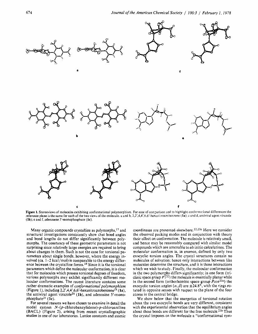
C t Figure 1. Stereoviews of molecules exhibiting conformational polymorphism. For ease of comparison and to highlight conformational differences the reference plane is the same for each of the two views of the molecule. a and b, 2,2’,4,4’,6,6’-hexanitroazobenzene ( la) ; c and d, antiviral agent virazole (lb); e and f, adenosene 5’-monophosphate (IC).
Many organic compounds crystallize as polymorphs,17 and structural investigations consistently show that bond angles and bond lengths do not differ significantly between poly- morphs. The constancy of these geometric parameters is not surprising since relatively large energies are required to bring about changes in them. Such is not the case for torsional pa- rameters about single bonds, however, where the energy in- volved (ca. 1-2 kcal/mol) is comparable to the energy differ- ence between the crystalline forms.I8 Since it is the torsional parameters which define the molecular conformation, it is clear that for molecules which possess torsional degrees of freedom, various polymorphs may exhibit significantly different mo- lecular conformations. The recent literature contains some rather dramatic examples of conformational polymorphism (Figure l ) , including 2,2‘,4,4‘,6,6‘-hexanitroa~obenzene~~ (la), the antiviral agent virazole20 ( lb) , and adenosine 5’-mono- phospha te2l (1 c).
For several reasons we have chosen to examine in detail the model system N-@-ch1orobenzylidene)-p-chloroaniline (BACL) (Figure 2), arising from recent crystallographic studies in one of our laboratories. Lattice constants and atomic
coordinates are presented e l ~ e w h e r e . * ~ , * ~ ~ Here we consider the observed packing modes and in conjunction with theory their effect on conformation. The molecule is relatively small, and hence may be reasonably compared with similar model compounds which are amenable to ab initio calculations. The molecular conformation is, in essence, defined by only two exocyclic torsion angles. The crystal structures contain no molecules of solvation; hence only interactions between like molecules determine the structure, and it is those interactions which we wish to study. Finally, the molecular conformation in the two polymorphs differs significantly; in one form (tri- clinic space group P122) the molecule is essentially planar while in the second form (orthorhombic space group P ~ c n * ~ ~ ) the exocyclic torsion angles (a , 0) are f24.8’, with the rings ro- tated in opposite senses with respect to the plane of the four atoms in the central bridge.
We show below that the energetics of torsional rotation about the two exocyclic bonds are very different, consistent with the experimental observation that the equilibrium angles about these bonds are different for the free molecule.23b Thus the crystal imposes on the molecule a “conformational sym-

Bernstein. Hagler / Conformational Polymorphism 675
Table I. Nonbonded Parameters for N-(p-Chlorobenzy1idene)-p-chloroaniline
Atom 6-12‘ 6-9‘ Exponentiald type” r* € r* t A B a
C 4.35 0.039 3.62 0.184 449.7 71 500 3.60 x 4.14 0.08 1 3.82 0.1725 449.7 71 500 3.60 Clb 3.866 0.244 3.866 0.244 1430 220 700 3.621 H 2.75 0.038 3.54 0.0025 40.2 2870 3.74 HX 2.75 0.019 3.54 0.001 25 40.2 2870 3.74
a See Experimental Section for definition of X and HX. Taken from ref 32. See ref 30. Potential has form -Ar6 + B exp(-ar). Except for chlorine, these parameters taken from ref 3 1 .
metry” which the isolated molecule does not possess. In this sense, this system also presents the opposite of the third ap- proach proposed by Kitaigorodskii in that the conformational symmetry of the molecule is higher in the crystal than in the free molecule.
The investigation which follows, then, includes an a b initio molecular orbital study of the energetics of changes in mo- lecular conformation and a study of crystal lattice forces to determine the relative energetics of the two crystal forms. The dependence of lattice energies on potential function is thor- oughly tested by employing a variety of functions. A parti- tioning of the total lattice energy is carried out to obtain indi- vidual “partial atomic” contributions, ei, to the overall energy. These results are used in conjunction with the x-ray analyses to analyze the influence of the crystal symmetry and packing in the conformation of the BACL molecule.
Methods Quantum Mechanical Calculations. The difference in mo-
lecular energy due to rotations about the N-phenyl and CH-phenyl bonds (a , 0) by the amounts observed in the two crystal structures were calculated using a b initio molecular orbital theory. Since the molecule is too large to be accom- modated by existing programs we chose model compounds 2 and 3 to calculate the difference in molecular energy between
2 3
the molecular conformations found in the two crystal modifi- cations. The energy as a function of torsion angle about N- phenyl and CH-phenyl bonds in 2 and 3 was calculated using both a minimal Gaussian basis set (STO-3G)24 and an ex- tended basis set (4-3 1G).25 The former calculations were carried out with the “Gaussian-70’’ program26 while the latter employed the “Gaussian-1 00” p r ~ g r a m . ~ ’ Bond angles and bond lengths were taken from ref 28 except for C-H, which were assigned values of 1.08 A, and C-Me and N-Me, which were given values of 1.53 and 1.47 A, r e s p e ~ t i v e l y . ~ ~
Lattice Energy Calculations. The techniques employed have been described p r e v i o ~ s l y . ~ ~ The lattice energy for each of the two forms was calculated with “6-1 2”, “6-9”, and William’s “exponential” potential functions which were derived pre- viously and shown to account well for a variety of crystal structures and sublimation e n e r g i e ~ . ~ ~ , ~ ’ They are given in Table I. W e have carried out the calculations with several functional forms in order to avoid conclusions which might be “artifacts” of a given set of potenital functions. The parameters for the chlorine atoms32 were not derived as a part of the s t ~ d i e s ~ ~ , ~ ~ referred to above. In addition to the use of three functional forms a further test of the sensitivity of the results to the values of these chlorine parameters was made. The sensitivity of the minimized energy and crystal structure to the parameters was tested by carrying out the minimizations with variations in r* and E of f10% (a possible total of four addi-
C126-Cl9
u22 nx6 ~-006)c7-C~~-006l
/ \ ti23
(00361 ti12 HI3
(00721 (00721
Figure 2. Numbering of atoms and assignment of charges (in fraction of an electron) in N-@-chlorobenzy1idene)-p-chloroaniline (BACL). Charges are given for only half the molecule which is related to the second half by crystallographic symmetry, an inversion center in the triclinic case and a twofold axis in the orthorhombic one. The meaning of X and HX is given in text (Experimental Section). a and flare the exocyclic torsion angles which define the molecular conformation.
tional calculations for each function). Charges on atoms for calculating the electrostatic contribution to the total energy (Figure 2) were estimated on the basis of charges obtained from a b initio calculations (STO-3G basis set) on 2 (for both planar and nonplanar conformations), 4 and 5. They were kept
4 5
unchanged for all the lattice energy calculation^.^^^^^^ Both crystal structures are disordered about crystallographic
symmetry elements: the triclinic (planar) about a center of symmetry,22 and the orthorhombic (nonplanar) about a two- fold a x i ~ , ~ ~ a although the free molecule contains neither ele- ment of symmetry. The crystallographic consequence of the disorder is that over the crystal space the central bridge group is a statistical average of
/c /N=G and
H \ c = N /
C‘ C‘ H Hence in the notation of Table I and Figure 2, “X” and “HX” represent respectively an average between C and N and “half of a hydrogen”. The crystallographic asymmetric unit in both structures is “half’ of the disordered molecule; the triclinic structure contains one molecule per cell, the orthorhombic four molecules per cell.
Results Quantum Mechanical Calculations. Results of the ab initio
calculations for the two model compounds are given in Figure 3. Additional details and discussion will be given elsewhere,34b but the points relevant to this study are presented here.
For the exocyclic angle cy the minimal basis set (STO-3G) and extended basis set (4-31G) energy is lowered by nearly the same amount as a result of a rotation by 24.8’. For fl this is not the case, the minimal basis set showing a virtual insensitivity

6 7 6
Table 11. Comparison of Experimental and Minimized Crystal Structures of BACL
Journal of the American Chemical Society / 100:3 / February I , 1978
6-12 6-9 Exponential Exptl Calcd A. Calcd A. Calcd Aa '
a , A b
Y, deg P
Volume. A3
C
Y
a , A b
VoIume/moIecuIe, A3
Molecular
AX, A AY A2
Volume change -0.1 orth-tric, A3
% change based
C
reorientation b
Triclinic Form 5.986 6.221 0.235 3.933 4.099 0.166
12.342 12.032 -0.310 87.38 86.27 -1.11 78.40 78.66 0.26 89.53 88.00 -1.53
284.3 300.1 15.8
Orthorhombic Form 24.503 23.692 -0.81 1
6.334 6.853 0.519 7.326 7.574 0.248
284.2 307.4 23.2
I1 I11 IV I1
-0.514 -0.548 -0.085 -0.209 -0.696 -0.864 -0.168 0.089 -0.206 0.084 -0.122 0.356
7.3
-2.4
5.901 3.603
11.893 87.95 79.96 93.04
248.4
24.620 6.174 7.244
262.4
I11
-0.202 0.101 0.085
14.0
-5.3
-0.085 6.096 -0.330 3.883 -0.449 12.229
0.57 90.26 1.56 79.26 3.51 92.23
-35.9 284.1
0.117 23.866 -0.160 6.828 -0.082 7.518
-21.8 292.8
IV I1 111
0.006 -0.417 -0.495 0.011 -0.657 -0.814 0.271 0.060 -0.012
8.7
-3.0
0.1 10 -0.050 -0.113
2.88 0.86 2.70
-0.2
-0.637 0.494 0.192 8.6
IV
-0.078 -0.156
0.073
on orth
A is difference between calculated and experimental values. The rotational components of the molecular reorientation resulting from the minimization are all zero within the precision of the calculations. The entries under column headings 11,111, IV are the translational com- ponents of the molecular reorientation in the minimized structure with respect to the experimental structure for the molecules related to the reference molecule, I, by symmetry as described in caption to Figure 5.
I .o
0.0
c al - z
a"
\ 0 x
- - -1.0
-2.0
I 1 . 1 I I I
. ..... . . . ... . . ... . . .. . . .. .. . . . ... . . .. . .. \
IO 20 30 40 5 0 60 Exocyclic Torsion Angle(Degrees)
Figure 3. Summary of ab initio calculations on 2 and 3. The ordinate represents energy differences between the planar conformation and non- planar ones.
to the rotation while the energy increases with the extended basis set, by about two-thirds of the energy decrease in a. In both cases, the net change from twist about both exocyclic angles is in favor of the nonplanar conformation found in the orthorhombic structure (extended -0.5 kcal/mol; minimal -1.5 kcal/mol); these results are in qualitative agreement with the conformation found in the crystal structure of benzyli- deneaniline (a = 5 5 O , p = With a much simpler model based on a combination of HMO r-electron energies and H.-H
Table 111. Summary of Lattice Energy Calculations
Crystal energy, kcal/mol potential employed
6-12 6-9 ExDonential
Triclinic Initial E,,, -21.15 -41.38 -23.34
E n b -22.14 -42.38 -24.34 Eeiec 1 .oo 1 .oo 1 .oo
Final E,,, -22.73 -45.71 -23.99 E n b -23.28 -46.64 -24.83 Eeiec 0.55 0.93 0.86
Orthorhombic (per Molecule) Initial E,,, -19.34 -39.81 -21.53
E n b -20.66 -41.13 -22.85 Eel,, 1.32 1.32 1.32
Final EtOt -21.68 -42.73 -22.34 E n b -22.41 -44.24 -23.27 Eeiec 0.73 1.51 0.93
AE( tric-orth) -1.05 -2.98 - 1.65
nonbonded potentials, Burgi and dun it^^^ obtained a figure of about 1.5 kcal/mol for the preference of this conformation over the planar one.
Lattice Energy Calculations. The results of the lattice energy calculations a re summarized in Tables I1 and 111. W e make the following observations concerning these tables:
(1) The volume/molecule in the initial (experimental) cells for the two structures is identical, a somewhat surprising result, since both the molecular geometry and the packing modes differ significantly in the two forms (Figures 4 and 5).
(2) For all the minimized structures the triclinic volume/ molecule is lower than the orthorhombic. The percent differ- ence in volume is of the expected order of magnitude36a and the variations for the three potential functions are consistent with the energy difference obtained between the two dimorphs. The largest volume change comes from the 6-9 potential, which

Bernstein, Hagler Conformational Polymorphism 677
Figure 4. Stereoview of the triclinic structure as viewed on the best plane of the molecule. The origin molecule is that with the lower elevation in the pair at the upper right hand corner. The molecule above the origin molecule is obtained by a transition along b; the upper molecule in the pair in the lower right hand corner is related to the origin molecule by a transition along a . The remaining axis is c .
Figure 5. Stereoview of the orthorhombic structure as viewed along the c crystallographic axis. a is horizontal and b is vertical. The origin molecule ( I ) is the full molecule with the higher elevation (closer to viewer) in the left hand portion of the cell. Molecule 11 is obtained via the screw axis parallel to a and is the uppermost one on the right-hand side. Molecule 111 (below the origin molecule) is obtained via the c glide perpendicular to a ; molecule IV is related to the origin molecule through a center of symmetry and is below molecule 11. The remaining two full molecules on the right-hand side are related by translations to molecules I1 and IV.
also gives the largest energy difference between the two forms.
(3) For the orthorhombic form unit cell angles were per- mitted to vary but did not do so, Le., the space group symmetry was retained by the calculation. This must be the case due to vanishing derivatives with respect to symmetry elements. In light of the disorder present in this structure, the symmetry aspects of the minimization deserve some comment. Recall that in the orthorhombic case the molecule is disordered about a twofold axis leading to crystallographic equivalence between the two chemically inequivalent “halves” of the molecule. The twofold axis relates one-half of the disordered molecule to the other results from the intersection of the two c-glides at x =
and y = 3/4 and is parallel to the c axis (Figure 6a).36b If we orient the initial (not disordered) benzylideneaniline molecules as in Figure 6b, then the c glide perpendicular to the b axis is maintained but that perpendicular to the a axis is lost. The reverse is true for the initial orientation shown in Figure 6c, which is the one we employed here in our test for conservation of symmetry. The overall crystallographic symmetry depends not only on this relationship within the pair but also on the relationship between the pair of molecules as shown in Figure 6c (say I and I11 in Figure 5) and the remaining pair in the cell (I1 and IV in Figure 5). If the n glide is retained then the space group remains orthorhombic P2lnc but if the center of sym- metry is retained then the resulting structure corresponds to monoclinic space group P21/c with a the unique crystallo- graphic axis. Again the latter case was that chosen for our minimization of a structure without disorder. Since the space group symmetry a t the start of the minimization is monoclinic there is no longer a symmetry restriction on a, the angle be- tween b and c , and this angle (but not the other two) may vary during the course of the minimization; this is precisely what occurred. (4) The lattice energy minimizations (Table 111) all yield
a result favoring the triclinic structure over the orthorhombic one, and by an energy difference which is compatible with the a b initio molecular orbital calculations. The relationship be- tween these calculations is discussed more fully below. The quantitative agreement between the molecular and crystal calculations is especially good for the 6- 12 and exponential functions but is less so for the 6-9 potential. The discrepancy
c-glide L to V’ /
6 c - 6b - 60 - Figure 6. Relationship between glide planes and molecular orientation in the orthorhombic structure. The solid line represents the molecule of higher elevation with respect to the c axis. 6a shows the presence of the twofold axis which arises from the intersection of the two glide planes. 6b and 6c show the molecular orientations required by a single glide plane perpendicular to the b axis and perpendicular to the a axis, respective- ly.
between the absolute energies for the 6-9 potential on the one hand and the exponential and 6- 12 on the other is larger than has been observed previously30a and is also commented on below.
(5) In addition to calculating the total minimized crystal energy we have partitioned the total energy into individual atomic contributions (Table IV). This is done by assigning half of each of the pairwise interactions to each of the atoms in- volved. The variation in these “partial energies” between the polymorphs is a direct reflection of the differences in packing interactions which characterize the two structures. This vari- ation is most readily examined by looking a t the partial ener- gies in the two forms. The relative role of each atom’s contri- bution to the overall energy is remarkably insensitive to the potential functions used and is in fact identical for the 6-12 and exp potentials for both structures. Once again the magnitude of the interactions for the 6-9 potential is different from the other two.
(6) The order of the relative contributions of the partial atomic energy to the total energy is the same for both the tri- clinic and orthorhombic forms. This is an indication that the environments of the atoms in the two crystal forms do not differ drastically in terms of energetics.
(7) W e extracted as well the significant repulsive interato- mic interactions in the minimized crystal structures in order to aid in examining the packing effects in detail. The largest

618 Journal of the American Chemical Society / 100:3 / February I , 1978
Table IV. Partition of the Minimized Crvstal Energv into Individual Atomic Contributions (e;)o
Aei (tric-orth) Orthorhombic Triclinic Atom 6-12 6-9 Exo 6-12 6-9 Exo 6-12 6-9 EXD
c1 x 2 HX5 c 7 C8 c 9 c 1 0 c1 I H12 H I 3 H14 H15 C116
-0.15 (10) -1.91 (2)
0.04 (1 1) -1.18 (5) -0.98 (6) -0.84 (7) - 1.36 (4) -1.52 (3) -0.40 (9) -0.62 (8)
0.17 (12) 0.43 (13)
-2.45 (1)
-1.53 (8) -3.71 (1)
-3.00 ( 5 ) -2.58 (6) -2.57 (7) -3.25 (4) -3.43 (2)
0.36 (1 1)
0.04 ( I O )
0.76 (1 2) 1.22 (13)
-0.43 (9)
-3.28 (3)
-0.21 ( I O ) -1.87 (2)
-1.39 (5) -1.14 (6) -1.09 (7) - 1.60 (4) -1.74 (3) -0.32 (9) -0.50 (9)
0.03 (1 1)
0.36 ( 1 2) 0.58 (1 3)
-2.38 (1)
-0.24 (10) -2.04 (2)
-1.13 ( 5 ) - 1 .OO (6) -0.95 (7) -1.47 (4)
-0.49 (9) -0.68 (8)
0.01 (1 1)
-1.47 (3)
0.20 (12) 0.39 (1 3)
-2.52 ( I )
1.87 (8) -3.81 (1)
0.36 (1 1) -2.99 (5) -2.70 (6) -2.70 (7)
-3.48 (3)
-0.39 (9)
-3.49 (2)
-0.20 (10)
0.82 (1 2) 1.04 (13)
-3.46 (4)
-0.32 (10) -2.01 (2)
0.00 (1 1) -1.36 (5) -1.21 (6)
-1.73 (4) -1.77 (3) -0.49 (9) -0.63 (8)
-1.10 (7)
0.37 (12) 0.54 (1 3)
-2.41 (1)
-0.09 -0.13 -0.03
0.05 -0.02 -0.1 1 -0.1 1
0.05 -0.09 -0.06
0.03 -0.05 -0.07
-0.34 -0.10
0.00 0.01
-0.12 -0.13 -0.24 -0.05 -0.24 -0.04 -0.06 -0.18 -0.18
-0.1 1 -0.14 -0.03
0.03 -0.07 -0.09 -0.13 -0.03 -0.17 -0.13
0.01 -0.04 -0.03
Entries are in kcal/mol. The number in parentheses gives the ranking of the atomic contribution in increasing energy. Owing to symmetry only half of the atoms in the molecule are given. Entries in “Atom” column are compatible with numbering in Figure 2.
Table V. Specific Atom-Atom Interactions
6-12 potential 6-9 potential Exponential potential
Trans- Trans- Trans- Interaction type Interaction type Interaction type
Rank Atom-atomU lation Force Atom-atom lation Force Atom-atom lation Force
Orthorhombic 1 H15-I H22-I1 010 1.03 H15-I H22-I1 010 4.37 H15-I H22-I 010 1.26 2 H13-I C116-IV 010 0.70 H13-I C116-IV 010 1.63 H13-I C116-IV 010 0.98 3 C8-I C126-I1 001 0.61 H13-I C126-I1 000 0.92 C8-I C126-I1 001 0.97 4 ClO-I C19-111 011 0.56 C8-I H23-I1 011 0.89
6 H14-I HX6-I 010 0.50 X2-I X3-I11 000 0.71 5 H15-I C17-111 011 0.52 C9-I C18-I11 011 0.72
- Triclinic - - 1 (2116 C126 - - 2 H13 C126 - 3 c11 H22 - 4 H13 C126 - 1 1 1 0.52 H13 C126 -
211 0.69 H13 C126 - 101 1.52 H13 C126 101 0.95 101 0.67 C116 (2126 - 211 1.44 H15 H22 - 110 0.75 110 0.52 H15 H22 - 110 1.35 H13 C126 1 1 1 0.70
111 1.14 5 c11 H22 - 100 0.46 HX5 H23 - 110 1.07 6 C10 H22 110 0.44 H I 5 H22 100 0.74
Numbers following atom identifier refer to symmetry operations as follows: I, x , y , z ; 11, ‘/2 + x , 1 - y , ‘/z - z ; 111, !/2 - x , y , -% + z ; IV, 1 - x , 1 - y , 1 - z.
of these specific interactions for each of the potentials used are given in Table V, In the orthorhombic case the interaction H15-I-H22-1 (010) (the Roman numeral following the atom indicates the symmetry operation relating this molecule to the asymmetric unit ( x , y , 2 ) ; see legend to Figure 5) is by far the strongest for all three potential functions and the second strongest interaction is common for the three as well. For a particular potential, lower ranked interactions differ only slightly in magnitude and hence it is not surprising to find a lack of total direct correspondence among them for the three potentials. The correspondence of strong interactions is di- minished somewhat for the triclinic case. The C1-Cl interac- tion does not appear among the top three in the exponential potential but the H13-Cl26 interaction is a t or near the top for all three. Furthermore the distinction between the strongest interaction and those below it in the list is not as sharp as for the orthorhombic case.
(8) It is seen that the minimized crystal energy does not change significantly with changes of f 1 0 % in the chlorine parameters, and (see Table VI) that the energy difference between polymorphs is virtually insensitive to such changes. The reason for this insensitivity between the polymorphs is seen in terms of the difference in partial atomic energies as discussed below.
Discussion
Overall Crystal Structures. Both crystal structures (Figures 4 and 5 ) exhibit a region of a strong concentration of chlorine atoms as indicated by the broken line (a plane in three di- mensions). In both cases the repulsive interactions across this plane a re among the strongest observed (Table V). They are generally of the type C1-H but Cl-Cl is also among the second strongest for the triclinic case. (Such an interaction would have been expected on the basis of the short Cl-Cl(3.42 A) distance found in the crystal structure analysis.) Therefore, both structures may be described as being composed of blocks of molecules between two neighboring parallel planes. The tri- clinic crystal may be thought of as being generated by trans- lation of these blocks in the c direction (Figure 4). In the or- thorhombic structure the distance between the planes is one- half of the axial length (Figure 5 ) and the contents of one block may be generated from its neighbor through the inversion centers which lie in the planes a t x = 0, 1/2.37
These repulsive interactions emphasize the compressive effect of the overall lattice forces. The atoms in actual contact are “pushed” into the repulsive side of the interatomic poten- tial. I t is because of this compressive effect that contact dis- tances cannot be taken as the minimum in the interatomic

Bernstein, Hagler / Conformational Polymorphism 679
Table VI. Summary of Results of Lattice Energy Minimizations for Variations of Chlorine Parameters in Potential Functions (kcal/mol)
Normala l . l r 0 . 9 ~ 1.16 0 . 9 ~
6-12 Potential Triclinic
E . initial . . -21.15 -21.41 -21.20 -22.38 -22.46 Efinal -22.73 -23.17 -22.13 -23.19 -22.21
E , ini t ia l , . -19.34 -18.81 -19.85 Efinal -21.68 -22.09 -22.14 AE(tric-orth) - 1.05 - 1.08 -1.05
Orthorhombic
6-9 Potential
E. initial , . -41.38 -43.31 -43.41 -42.98 -41.71 Efinal -45.71 -45.68 -45.22 -46.40 -45.05
E. initial . . -39.81 -40.88 -40.49 Erinal -42.73 -42.55 -43.43 AE(tric-orth) -2.98 -3.13 -2.97
a See Table I.
Triclinic
Orthorhombic
Not performed in the interest of saving computer time.
potential (van der Waals radii).30b,36 Within the blocks the molecules are stacked in a direction approximately perpen- dicular to the bridge atoms ( C l , X2, X3, C4 in the notation of Figure 2). In the triclinic case the stacking direction is b. In the orthorhombic structure the stacking is due to a c glide rather than a simple translation. Within the blocks the stacks are related by an a translation in the triclinic structure and a b translation in the orthorhombic one. The interactions across the planes serve to stabilize these blocks with respect to each other, as evidenced by the fact that, except for the 6-9 potential, the chlorine atoms make the largest single contribution to the stabilization energy of approximately 2.5 kcal/mol (Table IV).
Analysis of Individual Contributions. Our principal interest in this study was to isolate, if possible, the stabilizing influences in the triclinic crystal with respect to the orthorhombic one with the aim of exploring the mode by which the more highly en- ergetic molecular conformation is stabilized in the triclinic form. The lower lattice energy of the triclinic form relative to the orthorhombic is required by the fact that this polymorphic form is observed. The intramolecular energy of the molecule is lower in the nonplanar conformation, as demonstrated both by the ab initio calculations and experiment. Therefore, if the orthorhombic lattice, which contains the more stable molecular form, were more stable as well, it is unlikely that the triclinic form would be observed. The favorable lattice energy of the triclinic form brings the molecule into its unfavorable, planar molecular conformation, and it is this effect that we study here. In light of the above discussion it is gratifying that the potential functions all yield the triclinic structure as the more stable lattice, by an energy consistent with the intramolecular energy differences. Analysis of the individual contributions to the overall crystal energy is illustrative in this regard. For reasons given below we confine the quantitative aspects of the following remarks to the result for the 6-12 and 6-exp functions, although qualitatively those for the 6-9 are in agreement with the former two. We have shown that regardless of the potential function the lattice energy minimizations yield a preferentially lower energy for the triclinic form. The differences in individual atomic contributions to the minimized crystal energies for all three potentials are given in Table IV. They contain the in- formation necessary for this analysis.
W e note first that no single atom makes an outstanding contribution to stabilizing the triclinic structure over the or- thorhombic one. On the contrary, the partial atomic energies
show the mode of stabilization to be nonspecific in that nearly all atoms make a small stabilizing (Aei(tric-orth) < 0) con- tribution. This result is somewhat surprising since even a cursory examination of the two crystal structures reveals some rather striking differences in the spatial arrangement of the molecules. It might have been expected a priori that the dif- ferences in individual atomic contributions for a small number of atoms might yield the difference between the two forms; yet this is clearly not the case in this system. W e note that, al- though we are dealing here with small differences in compar- atively large numbers, both the signs and relative magnitudes of the energy differences between the three potentials are re- markably consistent. Only the C11 entry for the 6-12 potential and the H13 entry for the 6-9 potential are exceptions to this statement. Thus it would appear that these energies indeed reflect differences in the geometry of packing.
Some details of the energy differences listed in Table IV should be noted. Crystallographic disorder about the bridging atoms is observed in all 4,4’-dihalo-substituted benzyli- deneanilines and we previously postulated23a that the forces acting a t the extremities of the molecules (i.e., involving the halogens) play a significant role in determining the packing of these structures. The results obtained here are consistent with this postulate; namely, the chlorine contribution to the total energy is the largest single one in both crystal forms. The chlorine does not, however, make a major contribution in stabilizing the triclinic structure over the orthorhombic one (Aei -0.07, -0.03 kcal/mol for the 6-12 and exp potentials, respectively). Even in the best case (6-9 potential) its contri- bution is only the fifth largest contributor to the difference in energy.
Only the overall environments of two atoms C7 and H14 are less favorable in the triclinic structure, tending to “destabilize” it with respect to the orthorhombic one (Aei(tric-orth) > 0), and even then only to a small degree. H I 4 makes a positive (unfavorable) contribution to the lattice energy of both forms, while C7 is one of the stronger contributors to the negative lattice energy.
In spite of the small magnitudes involved, again there is a good deal of consistency in the relative roles of the atoms in stabilizing the triclinic structure. Atoms C1, X2, C9, C10, H12, and H13 are consistently among the highest contributors to the stabilization energy. The atoms on the bridge C1, X1, H X 5 provide -0.25, -0.28 kcal/mol stabilization for the triclinic structure, while C9, C1 give -0.1 8, -0.1 3 kcal/mol. The remaining atoms (4 C’s and H’s) provide -0.20, -0.53 kcal/mol. In terms of regions in the molecule, it may be seen that both the bridge region (between the two phenyl groups) and the phenyl groups pack more favorably in the triclinic structure. The contribution to the stabilization energy of the bridge region is -0.5 kcal/mol, while the phenyl groups pro- vide the remainder (Tables 111, IV). As noted above the chlo- rine atoms do not contribute significantly to the difference in energy between the two structures, the latter despite the fact that the chlorine atoms make the largest single contribution to the lattice energy, and the fact that the intermolecular spatial relation of the chlorine atoms is very different in the two structures. In the triclinic structure the chlorine atoms are related by translation both within the blocks and between them. In the orthorhombic structure the chlorine atoms are related by a glide plane within the stacks and by a center across the blocks (Figures 4 and 5).
Lattice Calculations and Crystallographic Rigid Body Analysis. It is not unreasonable to ask the question “to what extent can one rotate the phenyl rings in the triclinic structure out of planarity before a significant change is observed in the minimized energy?” Or, to put it another way, how strongly are these rotations restricted by the interactions described above? W e have carried out such calculations, rotating the

680 Journal of the American Chemical Society / 100:3 / February 1, 1978
rings in opposite senses by 5, 7.5, and 10’ for the 6-12 poten- tia1.38 The minimized total energy obtained is -22.55, -22.20, and -22.25 kcal/mol. The calculations behave normally for the first two but for the 10’ rotations there are large changes in cell constants (up to 20’ in angles) and the electrostatic contribution to the total energy becomes negative. We are thus inclined to believe that a 7.5’ rotation is the maximum allowed while still maintaniing the triclinic structure. It is noteworthy that the rigid body analyses of the thermal parameters obtained from the crystal structure determination22 indicated a large root mean square amplitude of the phenyl ring libration of 8.9’ about the X-C (phenyl) axis. We noted at the time that this value probably was not due to a true molecular librational motion but to small static rotations of the phenyl rings aver- aged over the crystal space which would lead to the anisotropic temperature factors compatible with such an apparent libra- tional motion. Such a good correspondence between the rigid body analysis and the minimizations of distorted molecules may perhaps be fortuitous but is nevertheless gratifyingly compatible with the present analysis.
Comparison of Potential Functions. Two of the main criteria for the applicability of a potential function to crystal lattice minimization are (1) the fit of the calculated crystal structure to the experimental one and (2) the comparison of the energy obtained with the experimental sublimation energy. No ex- perimental sublimation energy is available for either form of BACL but it is possible to make a reasonable estimate of its expected value. The sublimation energy of benzyldeneaniline (20.5 k ~ a l / m o l ) ~ ~ lies between that for stilbene (23.7 kcal/ mol)40 and azobenzene (17.9 k c a l / m ~ l ) . ~ ’ To obtain an esti- mate of the contribution of the addition of two chlorine atoms on the energy we note that their presence in the 4,4‘ positions of diphenyl raises the value of the sublimation energy from 19.5 kcal/mo140 to 24.8 k c a l l m 0 1 . ~ ~ Thus, we expect a value of approximately 25 kcal/mol for the sublimation energy of BACL. This value is quite compatible with the results obtained for the 6- 12 and 6-exp potentials.
The 6-9 potential yields a significantly larger difference between the two dimorphs than the other potentials and thus is one of the first cases among those which have been studied30 where, in terms of absolute energies, there is a large and sig- nificant difference between the 6-9 and 6- 12 potentials. This result may provide an additional useful observable for the further refinement of these potential functions.
Some additional points emerging from these calculations are worthy of comment. In terms of differences in energy be- tween crystalline polymorphs we have shown that the results are virtually independent of the potential and parameters chosen, provided that a reasonable potential function is chosen. Despite the discrepancy in the absolute value of the energy and the exclusive use of the 6-9 potential would not have altered the major conclusions of this work, as it was seen that the rel- ative energies of interest in this study were consistent among all three potentials used.
The difference in minimized crystal energies obtained for the two forms is in excellent agreement with the ab initio cal- culations. The quantity obtained is compatible with Kitaigo- rodskii’s estimate for the expected energy difference between polymorphs, and is also consistent with a number of recent calculation^^^*^^ which almost invariably give energies on the order of 1 kcal/mol between crystal modifications.
Summary We have presented a detailed study of the crystal packing
modes of the two conformational polymorphs of dichloro- benzylideneaniline with the aim of elucidating the effect of crystal forces on molecular conformation. Both the total binding energy and the stabilization energy of the triclinic form
over the orthorhombic form were analyzed in terms of the in- dividual atomic contributions, “partial atomic energies”, to the lattice energies. These results were shown to be in agree- ment with both ab initio and experimental results which show that the planar conformation of the molecule observed in the triclinic crystal is the higher energy geometry.
Three of the important points which emerged from the study follow:
(1) The relative contribution of the atoms, as indicated by the relative “partial atomic energies”, is the same for both crystal forms.
(2) The chlorine atom, which constitutes the single largest contribution to the total lattice energy of both crystal forms, has essentially the same energetic environment in the two forms, and does not contribute significantly to the difference .in lattice energies of the two forms.
(3) No one atom or group dominates the contribution to the stabilization of the triclinic form, but rather the phenyl rings and bridge region of the molecule contribute roughly equally to this stabilization. It was noted that, although energy dif- ferences between the two crystal forms were relatively small (-1-2 kcal), as compared to the total energies and even ac- curacy in a single energy, they were nevertheless significant. This is a result of the fact that the errors which arise in appli- cation of a given potential are mainly systematic and thus differences between them which reflected differences in the geometry of packing are significant. This was verified both by the difference in energy between the two forms obtained from three different potential functions and more strikingly by the constancy in the relative contributions of the “partial atomic energies” in the three potentials.
At this early stage in the development of this technique for the study of the influence of crystal forces on molecular con- formation, it is not clear whether the three phenomena ob- served above are characteristic of conformational polymor- phism or indigenous to this particular system. We are exam- ining this question in the dimethyl analogue of the compounds studied here which is trimorphic45 and has the added feature that in at least one of the forms the molecule is not disordered, but rather exhibits a conformation in the crystal approximating that of the lowest energy for the free molecule. Of particular interest with respect to these crystal forms is the question as to why the analogous chlorine molecule does not pack in the ordered structure which corresponds to the lowest molecular energy.46
Acknowledgment. We are grateful to Ms. Ruth Sharon for her help in the preparation.of the lattice energy program used in these calculations. It is a pleasure to acknowledge the as- sistance of Ms. Pnina Dauber in many of the computations and of Ehud Goldstein in the preparation of the stereoplots.
References and Notes ( 1 ) (a) Ben-Gurion University of the Negev; (b) Weizmann Institute of
Science. (2) (a) S. R. Byrn, C. W. Graber, and S. L. Midland, J. Org. Chem., 41, 2283
(1976), and ref 1-5 cited therein; (b) C. J. Eckhardt and J. Bernstein. J. Am. Chem. Soc., 94, 3247 (1972).
of American Crvstallooraohic Association. Aua 1976. Abstract No. 01. (3) H. L. Ammon, P. H. Mazzochi, L. Lin, and E. Colicelli, Northwestern Meeting
(4) R. M. Tal and J: B. F.k . Engberts, J. Chem. soc., Perkin Trans. 2, 483 (1976).
(5) J. Caillet, P. Claverie, and B. Pullman, Acta Crystallogr., Sect. B, 32, 2740 (1976).
(6) H. A. Scheraga and C. Anfinsen, Adw. Protein Chem., 29, 205 (1975). (7) A. T. Hagler and S. Lifson in “The Proteins”, 3rd ed, H. Neurath and R. L.
Hill, Ed., Academic Press, New York, N.Y.. in press. (8) C. M. Deber, V. Madison, and E. R. Blout, Acc. Chem. Res., 9, 106
(1976). (9) F. A. Bovey, “Peptides, Polypeptides and Proteins”, Proceedings of the
Rehovot Symposium, Wiley-lnterscience, New York, N.Y., 1974, pp 248-265.
(10) K. P. Kopple. T. J. Schamper, and A. Go, J. Am. Chem. Soc., 96, 2597 (1 974).
Singh / Partial Molar Volumes of S o m e Aqueous Electrolytes 68 1
J. N. Brown and R. G. Teller, J. Am. Chem. SOC., 98, 7565 (1976). B. Donzel, J. Rivier, and M. Goodman, Biopolymers, 13, 3621 (1974). E. Benedetti, A. Christensen, C. Gilon, W. Fuller, and M. Goodman, Bio- polymers, 15, 2523 (1976). E. Benedetti, R. E. Marsh, and M. Goodman, J. Am. Chem. SOC., 98,6676 (1976). A. I. Kitaigorodskii, Adv. Struct. Res. Diffr. Methods, 3, 173 (1970). P. Corradini, Chim. lnd. (Milan), 55, 122 (1973); N. C. Panagiotopoulis, G. A. Jeffrey, S. J. LaPlaca, and W. C. Hamilton, Acta Crystallogr., Sect. B,
See, for instance, L. Deffet, "Repertoire des Composes Organiques Po- iymorphes", Editions Desoer, Liege, 1942. Reference 15, p 225. E. J. Graeber and B. Morosin, Acta Crystallogr., Sect. B, 32, 310 (1976). P. Prusiner and M. Sundaralingam, Acta Crystallogr., Sect. 8, 32, 419 (1976). S. Needle, W. Kuhlbrandt, and Achari, Acta Crystallogr., Sect B, 32, 1850 (1976); J. Kraut and L. H. Jensen, Acta Crystallogr., 16, 79 (1963). J. Bernstein and G. M. J. Schmidt, J. Chem. SOC., Perkin Trans. 2, 951 (1972). (a) J. Bernstein and I. Izak, J. Chem. SOC., Perkin Trans. 2, 429 (1976); (b) T. Bally, E. Haselbach, S. Lanijiova, F. Marscher, and M. Rossi, Helv. Chim. Acta, 59, 486 (1976). and references cited therein. W. J. Hehre, R. F. Stewart, and J. A. Pople, J. Chem. Phys., 51, 2657 (1969). R. Ditchfield, W. J. Hehre, and J. A. Pople, J. Chem. Phys., 54, 724 (1971). W. J. Hehre, W. A. Lanthan, R. Ditchfield. N. W. Newton, and J. A. Pople, Quantum Chemistry Program Exchange, indiana University, Bloomington, Ind., Program No. 236. Extension of GAUSS 70 (ref 26) by J. Baudet and G. N. H. Port. H. B. Burgi and J. D. Dunitz, Helv. Chim. Acta, 52, 1747 (1970). 0. Kennard et al., Ed.. "Molecular Structures and Dimensions", N. V. A. Oosthock's Uitgevers Mij, Utrecht, 1972, pp S2-S3. (a) A. T. Hagler, L. Leiserowitz, and M. Tuval, J. Am. Chem. Soc., 98,4600 (1976); (b) A. T. Hagler and S. Lifson. ibid., 96, 5327 (1974); (c) Acta Crystallogr., Sect. 6, 30, 1336 (1974); (d) A. T. Hagler, E. Huler, and S. Lifson, J. Am. Chem. SOC., 96, 5319 (1974). D. E. Williams, Acta Crystailogr., Sect. A, 30, 71 (1974). E. Giglio, Nature (London), 222, 339 (1969).
30, 1421 (1974).
(33) The use of partial charges obtained from population analysis for confor- mational calculations or lattice energy calculations has recently been questioned.348 We do it here cognizant of these drawbacks. For the case under consideration here, the electrostatic contribution is small and the results are not sensitive to the exact charges used.
(34) (a) A. T. Hagler and A. Lapiccirella, Biopolymers, 15, 1167 (1976), and references cited therein; (b) J. Bernstein and A. T. Hagier, to be pub- lished.
(35) H. 8. Burgi and J. D. Dunitz, Helv. Chim. Acta, 54, 1255 (1971). (36) (a) A. I. Kitaigorodskii, "Molecular Crystals and Molecules", Academic
Press, New York, N.Y., 1973, pp 71-74, 186-190. (b) A glide operation is composed of a reflection through a plane perpendicular to a crystallo- graphic axis followed by a translation parallel to that plane, usually one-half the distance of a second unit cell vector. Thus a c glide perpendicular to b involves first a reflection through a plane perpendicular to the b axis followed by half a translation along c. A twofold screw axis is composed of a rotation of 180' about an axis parallel to the specified crystallographic axis and a translation of one-half the axial length along the same axis.
(37) "International Tables for X-Ray Crystallography". Vol. I, Kynoch Press, Birmingham, England, 1952, p 144.
(38) Rotation of the rings in opposite sense is expected to be preferred over rotation in the same sense since the opposite mode is that found in the orthorhombic structure. In this calculation, as in others for the triclinic form, all molecules are translationally equivalent, i.e., all molecules have been distorted from planarity to the same extent.
(39) G. E. Coates and L. E. Sutton, J. Chem. Soc., 1187 (1948). (40) E. Morawetz, J. Chem. Thermodyn., 4, 455 (1972). (41) N. F. H. Bright, T. Varson, and T. A. Dyson, Research (London), 3, 185
(42) N. K. Smith, G. Gorin, W. D. Good, and J. P. McCullough, J. Phys. Chem.,
(43) A. T. Hagler and L. Leiserowitz. in preparation. (44) C. P. Brock and J. A. Ibers, Acta Crystallogr., Sect. A, 31, 38 (1976). (45) J. Bernstein, I. Bar, and A. Christensen, Acta Crystallogr., Sect. 6, 32, 1609
(1976); I. Bar and J. Bernstein, Acta Crystallogr., Sect. B, 33, 1738 (1977). I. Bar and J. Bernstein, unpublished results.
(46) Preliminary results on this system further support the results of this study. The lattice energy of this hypothetical ordered form is higher than either of the two observed structures in all three potentials. Again the differences in energy as obtained from the three potential forms are consistent as is the ordering of the partial atomic energies.
(1950).
68, 940 (1964).
Partial Molar Volumes of Some Aqueous Electrolytes and a Transition Model
Prem P. Singh Contribution f r o m the Department of Chemistry, Punjab Agricultural Uniuersity, Ludhiana, India. Received January 12, 1977
Abstract: An expression has been derived for partial molar volumes and also for apparent molar volumes 4 ~ , from the free-en- ergy equation based on a model which takes explicit accounts of interionic effects and effects due to thermal jostling. The ex- pression suggests that for moderately concentrated solutions 4~ varies linearly with C1/3 and that the cube root behavior of 4~ gives way to C1/2 dependence as C - 0. Excellent quantitative agreement is obtained by adopting a value of 60.66 X b a r 1 for d(ln c)/dP of water a t 25 "C and 1 atm.
Introduction of aqueous
strong electrolytes reveals that this model may be a reasonable description for moderate to concentrated solutions while in the limit of infinite dilution the ion-cloud concept of Debye must prevail. A more reasonable formalism appears to be one based on Lietzke, Stoughton, and Fuoss' ob~erva t ions .~ W e have recently proposed a model4 (henceforth called the transition model) which assumes that ions in solutions (dilute as well as concentrated) have a certain degree of mobility and that it is the time average population density of the ions that corre- sponds to either the Debye or the "lattice" model. The fun- damental equation expressing the mean ionic molal activity coefficient is4
Examination of the "lattice" or "cell"
0002-7863/78/1500-0681$01.00/0
- log (1 + umM/1000)
+ ( I - e - ' 5 m ) ( - ~ V ~ + BC + 6 ) - log (1 + umM/1000) (1)
where A = [Ar'Z+Z-N/2.303uRTt] a ~ ~ ~ ( N / 1 0 0 0 ) ~ / ~ , v = U+ + u-, A" is the cell model analogue of the Madelung constant, t is the bulk dielectric constant, a , p converts a particular
0 1978 American Chemical Society