Window Prophylaxis [and other stuff involving tuberculosis ...
Iodide Excess Induces Apoptosis in Thyroid Cellsthrough a p53-Independent Mechanism InvolvingOxidative Stress*
MARIO VITALE, TIZIANA DI MATOLA, FRANCESCA D’ASCOLI,SALVATORE SALZANO, FAUSTO BOGAZZI, GIANFRANCO FENZI, ENIO MARTINO,AND GUIDO ROSSI
Dipartimento di Biologia e Patologia Cellulare e Molecolare (M.V., T.D., G.R.), Universita Federico II,Naples 80131, Italy; Dipartimento di Endocrinologia ed Oncologia Molecolare e Clinica (F.D., GF.F.),Universita Federico II, Naples, Italy; Centro di Endocrinologia ed Oncologia Sperimentale “G.Salvatore” (S.S., G.R.), C.N.R.; Dipartimento di Endocrinologia (F.B., E.M.), Universita di Pisa, 56100Pisa, Italy
ABSTRACTThyroid toxicity of iodide excess has been demonstrated in animals
fed with an iodide-rich diet; in vitro iodide is cytotoxic, inhibits cellgrowth, and induces morphological changes in thyroid cells of somespecies. In this study, we investigated the effect of iodide excess in animmortalized thyroid cell line (TAD-2) in primary cultures of humanthyroid cells and in cells of nonthyroid origin. Iodide displayed adose-dependent cytotoxicity in both TAD-2 and primary thyroid cells,although at different concentrations, whereas it had no effect on cellsof nonthyroid origin. Thyroid cells treated with iodide excess under-went apoptosis, as evidenced by morphological changes, plasma mem-brane phosphatidylserine exposure, and DNA fragmentation. Apo-ptosis was unaffected by protein synthesis inhibition, whereas
inhibition of peroxidase enzymatic activity by propylthiouracil com-pletely blocked iodide cytotoxicity. During KI treatment, reactiveoxygen species were produced, and lipid peroxide levels increasedmarkedly. Inhibition of endogenous p53 activity did not affect thesensitivity of TAD-2 cells to iodide, and Western blot analysis dem-onstrated that p53, Bcl-2, Bcl-XL, and Bax protein expression did notchange when cells were treated with iodide. These data indicate thatexcess molecular iodide, generated by oxidation of ionic iodine byendogenous peroxidases, induces apoptosis in thyroid cells through amechanism involving generation of free radicals. This type of apo-ptosis is p53 independent, does not require protein synthesis, and isnot induced by modulation of Bcl-2, Bcl-XL, or Bax protein expression.(Endocrinology 141: 598–605, 2000)
IN ADDITION to its role as a substrate for thyroid hormonebiosynthesis, iodide participates in a number of clinically
important interactions with the thyroid. Acute administra-tion of large doses of iodide determines a biphasic responseof the thyroid: an increase, followed by a decrease, in theyield of organic iodide and thyroid hormones. The mecha-nism of the relative blockade of organic iodide yield, knownas the Wolff-Chaikoff effect (1), is, in part, unknown and, inpart, caused by the biochemical effects of large concentra-tions of the reactive form of iodide generated by oxidativemechanisms. Another important effect of iodine on the thy-roid is its ability to diminish the hypervascularity and hy-perplasia that characterize the diffuse goiter of Graves’ dis-ease. The molecular mechanisms of this phenomenon,widely used to facilitate surgical therapy of this disorder, areuncertain, but it has been hypothesized that iodine couldbind to organic compounds and interfere with the metabolicprocesses necessary for the maintenance of hyperplasia andmay be responsible for the involuting effect of iodine excess(2).
The toxicity of iodide excess has been demonstrated, bothin animals and in cell systems. Involution of the thyroidgland has been described in rats fed with an iodide-rich diet(3, 4). In vitro, iodide inhibits thyroid cell growth and inducesmorphological changes in porcine thyroid cells (5). Someeffects of iodide seem to be species specific. A cytotoxic effectof iodide has been documented in rat FRTL-5 cells but not inprimary dog thyrocytes (6). The iodide-induced cytotoxiceffect on rat thyrocytes included necrotic and apoptotic fea-tures, indicating the involvement of a controlled process ofcell death. Apoptosis (or programmed cell death) is an activeprocess of cell self destruction requiring the activation of agenetic program, leading to changes in morphology, DNAfragmentation, and protein cross-linking (7, 8). The apoptoticpathways are activated by physiological stimuli such as en-vironmental signals, cytokines (9, 10), and growth factors;they can also be induced by pathological stimuli, radiation,and anticancer drugs (11–14). Excess iodide intake is neverobtained in physiological conditions; however, therapeuticuse of iodide-rich compounds, such as amiodarone and io-dinated radiographic contrast agents, can lead to release ofa large amount of iodide. Amiodarone, a potent antiarhyth-mic drug containing two iodine atoms per molecule, mayinduce either hypo- or hyperthyroidism (15–17). Amioda-rone-induced hypothyroidism in rats is associated with spe-cific ultrastructural features of necrosis and apoptosis of thethyroid gland and cytokine production (18–20). However,
Received June 7, 1999.Address all correspondence and requests for reprints to: Mario Vitale,
Dipartimento di Biologia e Patologia Cellulare e Molecolare, Via S.Pansini, 5 Napoli, 80131, Italy. E-mail: [email protected].
* This work has been supported, in part, by funding from Ministerodell’Universita e della Ricerca Scientifica (to G.R. and G.F.) and byMURST-C.N.R., Biothecnology Program L. 95/95 (to G.F.).
0013-7227/00/$03.00/0 Vol. 141, No. 2Endocrinology Printed in U.S.A.Copyright © 2000 by The Endocrine Society
598
the role of iodine vs. the direct drug cytotoxicity in the patho-genesis of amiodarone-induced hypothyroidism is not yetfully solved. Therefore, it was of interest to investigatewhether iodide itself displays cytotoxic effects on humanthyroid cells and whether its cytotoxicity represents an ap-optotic phenomenon. In the present study, we employedprimary cultures of human thyroid cells and the immortal-ized thyroid cell line TAD-2, which proved to be a goodmodel for studying apoptosis in the thyroid (9).
Materials and MethodsCells and chemicals
Cell cultures from normal thyroids were prepared, as previouslydescribed, by collagenase digestion (21) and cultured in a 5% CO2atmosphere at 37 C, in F-12 medium supplemented with 10% FCS, with1 mU/ml bovine tyreotropin (Sigma, St. Louis, MO). The TAD-2 cell line,obtained by Simian virus 40 infection of human fetal thyroid cells, wasa kind gift of Dr. T. F. Davies, Mount Sinai Hospital (New York, NY).TAD-2, human dermal fibroblasts (PG1), osteosarcoma cells (SaOS), andendometrial carcinoma cells (HeLa) were cultured in a 5% CO2 atmo-sphere at 37 C, in DMEM supplemented with 10% FCS. Medium waschanged every 3–4 days. Cells were detached by 0.5 mm EDTA incalcium- and magnesium-free PBS with 0.05% trypsin. TADp53cG cellmutants were generated by transfecting pLTRp53cG containing the tem-perature-sensitive dominant-negative p53 gene mutated at codon 135(gift of Dr. A. Levin, Princeton University, Princeton, NJ), as described(10). KCl, KI, cycloexhimide, and o-phenylenediamine were purchasedfrom Sigma. A 10-mm stock solution of 6-propyl-2-thiouracil (PTU)(Sigma) was prepared at basic pH, buffered at pH 7.5 by HCl.
DNA electrophoresis
Cells collected by centrifugation were washed in PBS, lysed in 300 ml0.5% Triton-X100, 5 mm Tris-buffer pH 7.4, 20 mm EDTA for 20 min at4 C and centrifuged at 13.000 rpm for 30 min. Centrifugation-resistantlow molecular weight DNA was extracted with phenol/chloroform,precipitated with ethanol and incubated with 0.5 mg/ml deoxyribonu-clease-free ribonuclease A for 30 min at 37 C. DNA with loading bufferwas electrophoresed in 1% agarose, 1 mg/ml bromide at 50 V in 45 mmTris-borate and visualized by UV.
Cell death measurements
Annexin V assay for determination of apoptosis/necrosis ratio wasperformed as follows: cells were washed twice with cold PBS; resus-pended in 10 mm HEPES (pH 7.4), 140 mm NaCl, and 2.5 mm CaCl2; andincubated for 15 min at room temperature with Annexin V-fluoresceinconjugated (PharMingen, San Diego, CA) and 5 mg/ml propidiumiodide. Cells were analyzed within 1 h, by flow cytometry, using aFACScan (Becton Dickinson and Co., Mountain View, CA).
Estimation of cell death, by flow cytometry, was performed as fol-lows: floating cells and adherent cells obtained by trypsin/EDTA werecollected, washed in cold PBS, and fixed in 70% cold ethanol for 30 min.Ethanol was removed by PBS wash, and cells were incubated in PBS, 50mg/ml propidium iodide, 10 mg/ml deoxyribonuclease-free ribonucle-ase A overnight at 4 C. Cells were then analyzed by flow cytometry. Thepercent of dead cells was calculated by dividing the number of cellsdisplaying red fluorescence lower than G0-G1 diploid peak by the totalnumber of collected cells times 100.
Antibodies and Western-blot analysis
Mouse monoclonal antibodies to p53 were purchased from Trans-duction Laboratories, Inc. (Lexington, KY); mouse monoclonal antibodyto Bcl-2 and rabbit polyclonal antibodies to Bcl-X and Bax were fromSanta Cruz Biotechnology, Inc., Santa Cruz, CA. Cells were washed incold PBS and lysed for 10 min at 4 C with 1 ml of lysis buffer [50 mmTris (pH 7.4), 0.5% NP40, 0.01% SDS] containing protease inhibitors.Lysates from adherent cells, collected by scraping and from floatingcells, were centrifuged at 12,000 3 g for 15 min at 4 C. The protein
concentration in cell lysates was determined by Protein Assay (Bio-RadLaboratories, Inc. Richmond, CA), and 50 mg of total protein from eachsample were boiled for 5 min in Laemmli sample buffer (125 mm TrispH 6.8, 5% glycerol, 2% SDS, 1% b-mercaptoethanol, and 0.006% bro-mophenol blue). Proteins were separated by SDS-PAGE and transferredonto nitrocellulose membrane (Hybond-ECL Nitrocellulose, AmershamPharmacia Biotech, Rainham, UK). Acrylamide concentration was 12%for p53 and Bcl-XL, 15% for Bcl-2 and Bax. Membranes were blocked by5% nonfat dry milk, 1% ovalbumin, 5% FCS, and 7.5% glycine; and afterthree washes, the membranes were incubated for 1 h at 4 C with 0.5mg/ml of mouse monoclonal or rabbit polyclonal primary antibodies inPBS. After three washes, filters were incubated for 1 h at 4 C withhorseradish peroxidase-conjugated antimouse or antirabbit secondaryantibodies (Bio-Rad Laboratories, Inc.) diluted 1:2000 in PBS, Tween-20.After a final wash, protein bands were detected by an enhanced chemi-luminescence system (Amersham Pharmacia Biotech).
Fluorescent measurement of intracellular reactive oxygenspecies (ROS)
TAD-2 cells were collected by mild trypsinization; washed in PBS;and resuspended in PBS, 10 mm 5,6-carboxy-29,79-dichlorofluoresceindiacetate (DCFH-DA, Molecular Probes, Inc., Eugene, OR), 5 mg/mlpropidium iodide at 37 C; and kept in DCFH-DA thereafter. DCFH-DAis a compound taken up by the cells and trapped in a nonfluorescentdeacylated form (DCFH). DCFH is oxidized by ROS to a fluorescentform (22). After 1 h of incubation, cells were analyzed by FACScan withexcitation at 495 nm and emission at 525 nm wavelength. Cells leakingDCFH because they were no longer intact were stained by the non-membrane-permeable dye propidium iodide and excluded.
Lipid peroxidation measurement
The level of lipid peroxide under KI treatment was determined bymeasuring the production of thiobarbituric acid reactive substances(TBARS), according to the method of Esterbauer and Cheeseman (23).The cells were harvested, lysed by sonication, and incubated with 0.5%thiobarbituric acid for 30 min at 80 C. Basal levels of TBARS generatedby liporeroxides were measured fluorimetrically (excitation at 530 nm,emission at 550 nm). Malondialdehyde bisdimethylacetal was used asstandard. TBARS levels were normalized for cell protein content.
ResultsPotassium iodide displays cytotoxicity restricted tothyroid cells
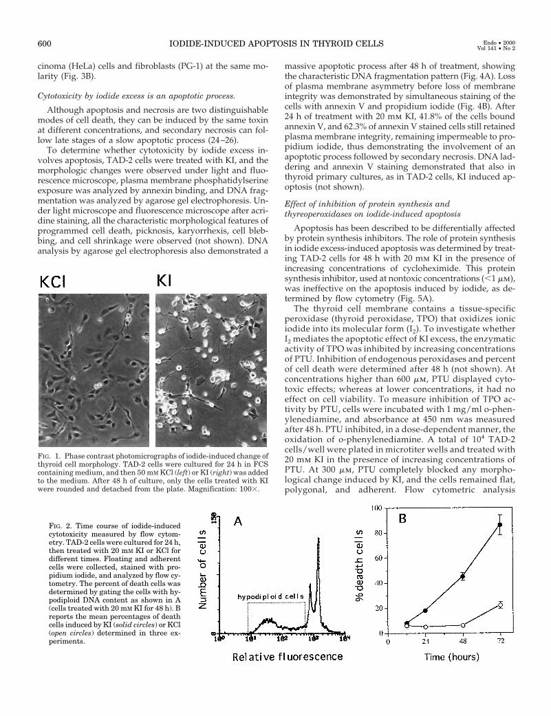
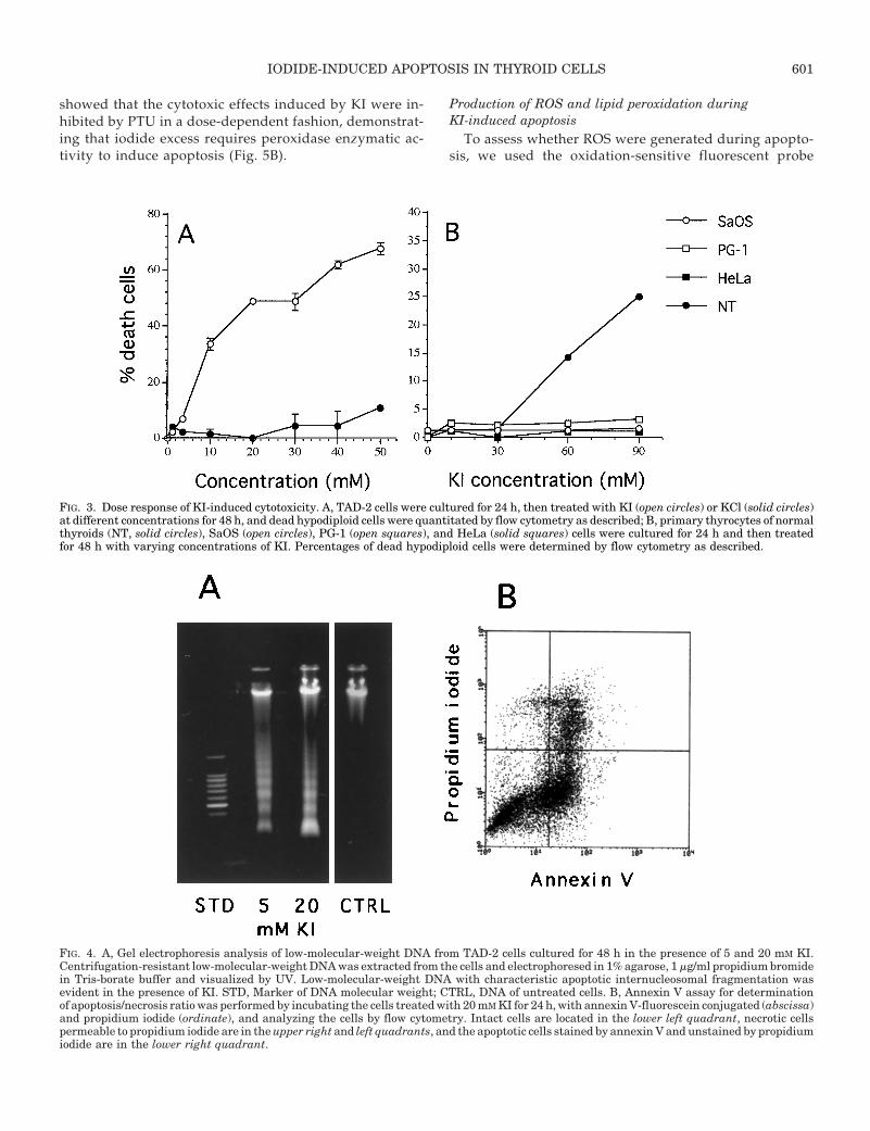
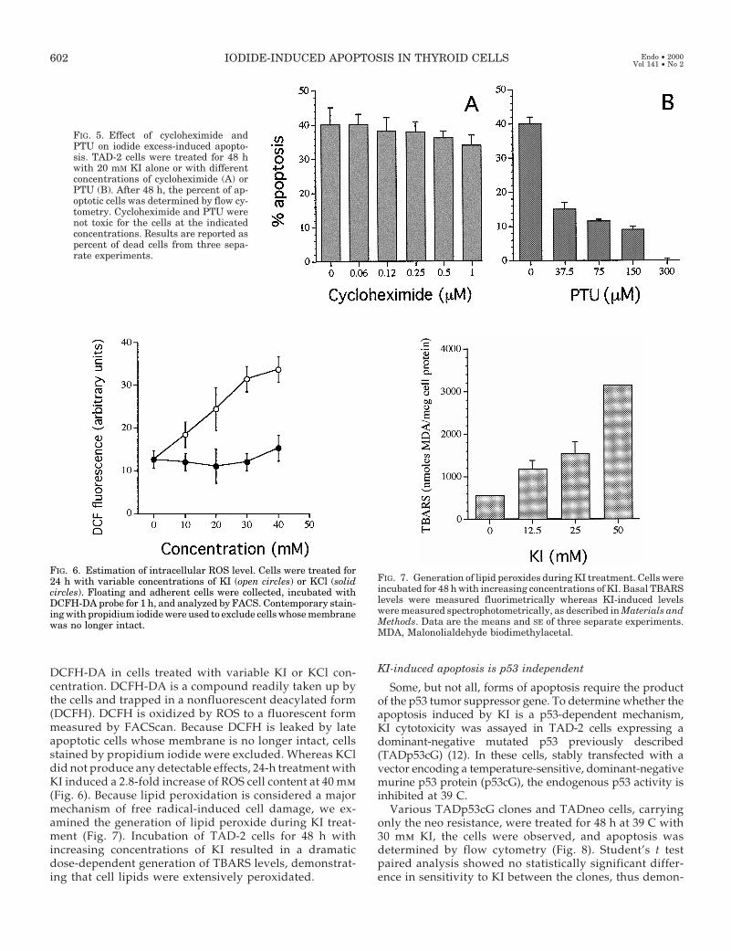
TAD-2 cells, primary human thyroid cells, and cells ofnonthyroid origin were treated with varying concentrationsof KI and KCl for 48 h. Whereas KCl at concentrations as highas 50 mm did not produce any detectable effects, treatmentwith KI induced a dramatic change in the morphology ofthyroid TAD-2 cells, from flat-adherent to round-detached(Fig. 1). The number of dead cells with hypodiploid DNAcontent was determined by flow cytometric analysis of float-ing and adherent cells fixed and permeabilized by ethanoland stained with propidium iodide as previously shown (12).Because both apoptosis and late necrosis determine a de-crease of cellular content of DNA, cytotoxicity was deter-mined by measuring the percent of hypodiploid cells asshown in Fig. 2A. The cytotoxic effect of KI was time de-pendent (Fig. 2B), affecting 50% of the cells at 20 mm con-centration by 48 h, and dose-dependent, whereas no effectwas observed in the cells treated with KCl (Fig. 3A). Potas-sium iodide also displayed a dose-dependent cytotoxicity,although at higher concentrations, in primary cultures ofnormal thyroid cells; whereas no effect was observed in cellsof nonthyroid origin such as osteosarcoma (SaOS) and car-
IODIDE-INDUCED APOPTOSIS IN THYROID CELLS 599
cinoma (HeLa) cells and fibroblasts (PG-1) at the same mo-larity (Fig. 3B).
Cytotoxicity by iodide excess is an apoptotic process.
Although apoptosis and necrosis are two distinguishablemodes of cell death, they can be induced by the same toxinat different concentrations, and secondary necrosis can fol-low late stages of a slow apoptotic process (24–26).
To determine whether cytotoxicity by iodide excess in-volves apoptosis, TAD-2 cells were treated with KI, and themorphologic changes were observed under light and fluo-rescence microscope, plasma membrane phosphatidylserineexposure was analyzed by annexin binding, and DNA frag-mentation was analyzed by agarose gel electrophoresis. Un-der light microscope and fluorescence microscope after acri-dine staining, all the characteristic morphological features ofprogrammed cell death, picknosis, karyorrhexis, cell bleb-bing, and cell shrinkage were observed (not shown). DNAanalysis by agarose gel electrophoresis also demonstrated a
massive apoptotic process after 48 h of treatment, showingthe characteristic DNA fragmentation pattern (Fig. 4A). Lossof plasma membrane asymmetry before loss of membraneintegrity was demonstrated by simultaneous staining of thecells with annexin V and propidium iodide (Fig. 4B). After24 h of treatment with 20 mm KI, 41.8% of the cells boundannexin V, and 62.3% of annexin V stained cells still retainedplasma membrane integrity, remaining impermeable to pro-pidium iodide, thus demonstrating the involvement of anapoptotic process followed by secondary necrosis. DNA lad-dering and annexin V staining demonstrated that also inthyroid primary cultures, as in TAD-2 cells, KI induced ap-optosis (not shown).
Effect of inhibition of protein synthesis andthyreoperoxidases on iodide-induced apoptosis
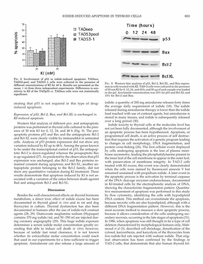
Apoptosis has been described to be differentially affectedby protein synthesis inhibitors. The role of protein synthesisin iodide excess-induced apoptosis was determined by treat-ing TAD-2 cells for 48 h with 20 mm KI in the presence ofincreasing concentrations of cycloheximide. This proteinsynthesis inhibitor, used at nontoxic concentrations (,1 mm),was ineffective on the apoptosis induced by iodide, as de-termined by flow cytometry (Fig. 5A).
The thyroid cell membrane contains a tissue-specificperoxidase (thyroid peroxidase, TPO) that oxidizes ioniciodide into its molecular form (I2). To investigate whetherI2 mediates the apoptotic effect of KI excess, the enzymaticactivity of TPO was inhibited by increasing concentrationsof PTU. Inhibition of endogenous peroxidases and percentof cell death were determined after 48 h (not shown). Atconcentrations higher than 600 mm, PTU displayed cyto-toxic effects; whereas at lower concentrations, it had noeffect on cell viability. To measure inhibition of TPO ac-tivity by PTU, cells were incubated with 1 mg/ml o-phen-ylenediamine, and absorbance at 450 nm was measuredafter 48 h. PTU inhibited, in a dose-dependent manner, theoxidation of o-phenylenediamine. A total of 104 TAD-2cells/well were plated in microtiter wells and treated with20 mm KI in the presence of increasing concentrations ofPTU. At 300 mm, PTU completely blocked any morpho-logical change induced by KI, and the cells remained flat,polygonal, and adherent. Flow cytometric analysis
FIG. 1. Phase contrast photomicrographs of iodide-induced change ofthyroid cell morphology. TAD-2 cells were cultured for 24 h in FCScontaining medium, and then 50 mM KCl (left) or KI (right) was addedto the medium. After 48 h of culture, only the cells treated with KIwere rounded and detached from the plate. Magnification: 1003.
FIG. 2. Time course of iodide-inducedcytotoxicity measured by flow cytom-etry. TAD-2 cells were cultured for 24 h,then treated with 20 mM KI or KCl fordifferent times. Floating and adherentcells were collected, stained with pro-pidium iodide, and analyzed by flow cy-tometry. The percent of death cells wasdetermined by gating the cells with hy-podiploid DNA content as shown in A(cells treated with 20 mM KI for 48 h). Breports the mean percentages of deathcells induced by KI (solid circles) or KCl(open circles) determined in three ex-periments.
600 IODIDE-INDUCED APOPTOSIS IN THYROID CELLS Endo • 2000Vol 141 • No 2
showed that the cytotoxic effects induced by KI were in-hibited by PTU in a dose-dependent fashion, demonstrat-ing that iodide excess requires peroxidase enzymatic ac-tivity to induce apoptosis (Fig. 5B).
Production of ROS and lipid peroxidation duringKI-induced apoptosis
To assess whether ROS were generated during apopto-sis, we used the oxidation-sensitive fluorescent probe
FIG. 3. Dose response of KI-induced cytotoxicity. A, TAD-2 cells were cultured for 24 h, then treated with KI (open circles) or KCl (solid circles)at different concentrations for 48 h, and dead hypodiploid cells were quantitated by flow cytometry as described; B, primary thyrocytes of normalthyroids (NT, solid circles), SaOS (open circles), PG-1 (open squares), and HeLa (solid squares) cells were cultured for 24 h and then treatedfor 48 h with varying concentrations of KI. Percentages of dead hypodiploid cells were determined by flow cytometry as described.
FIG. 4. A, Gel electrophoresis analysis of low-molecular-weight DNA from TAD-2 cells cultured for 48 h in the presence of 5 and 20 mM KI.Centrifugation-resistant low-molecular-weight DNA was extracted from the cells and electrophoresed in 1% agarose, 1 mg/ml propidium bromidein Tris-borate buffer and visualized by UV. Low-molecular-weight DNA with characteristic apoptotic internucleosomal fragmentation wasevident in the presence of KI. STD, Marker of DNA molecular weight; CTRL, DNA of untreated cells. B, Annexin V assay for determinationof apoptosis/necrosis ratio was performed by incubating the cells treated with 20 mM KI for 24 h, with annexin V-fluorescein conjugated (abscissa)and propidium iodide (ordinate), and analyzing the cells by flow cytometry. Intact cells are located in the lower left quadrant, necrotic cellspermeable to propidium iodide are in the upper right and left quadrants, and the apoptotic cells stained by annexin V and unstained by propidiumiodide are in the lower right quadrant.
IODIDE-INDUCED APOPTOSIS IN THYROID CELLS 601
DCFH-DA in cells treated with variable KI or KCl con-centration. DCFH-DA is a compound readily taken up bythe cells and trapped in a nonfluorescent deacylated form(DCFH). DCFH is oxidized by ROS to a fluorescent formmeasured by FACScan. Because DCFH is leaked by lateapoptotic cells whose membrane is no longer intact, cellsstained by propidium iodide were excluded. Whereas KCldid not produce any detectable effects, 24-h treatment withKI induced a 2.8-fold increase of ROS cell content at 40 mm(Fig. 6). Because lipid peroxidation is considered a majormechanism of free radical-induced cell damage, we ex-amined the generation of lipid peroxide during KI treat-ment (Fig. 7). Incubation of TAD-2 cells for 48 h withincreasing concentrations of KI resulted in a dramaticdose-dependent generation of TBARS levels, demonstrat-ing that cell lipids were extensively peroxidated.
KI-induced apoptosis is p53 independent
Some, but not all, forms of apoptosis require the productof the p53 tumor suppressor gene. To determine whether theapoptosis induced by KI is a p53-dependent mechanism,KI cytotoxicity was assayed in TAD-2 cells expressing adominant-negative mutated p53 previously described(TADp53cG) (12). In these cells, stably transfected with avector encoding a temperature-sensitive, dominant-negativemurine p53 protein (p53cG), the endogenous p53 activity isinhibited at 39 C.
Various TADp53cG clones and TADneo cells, carryingonly the neo resistance, were treated for 48 h at 39 C with30 mm KI, the cells were observed, and apoptosis wasdetermined by flow cytometry (Fig. 8). Student’s t testpaired analysis showed no statistically significant differ-ence in sensitivity to KI between the clones, thus demon-
FIG. 5. Effect of cycloheximide andPTU on iodide excess-induced apopto-sis. TAD-2 cells were treated for 48 hwith 20 mM KI alone or with differentconcentrations of cycloheximide (A) orPTU (B). After 48 h, the percent of ap-optotic cells was determined by flow cy-tometry. Cycloheximide and PTU werenot toxic for the cells at the indicatedconcentrations. Results are reported aspercent of dead cells from three sepa-rate experiments.
FIG. 6. Estimation of intracellular ROS level. Cells were treated for24 h with variable concentrations of KI (open circles) or KCl (solidcircles). Floating and adherent cells were collected, incubated withDCFH-DA probe for 1 h, and analyzed by FACS. Contemporary stain-ing with propidium iodide were used to exclude cells whose membranewas no longer intact.
FIG. 7. Generation of lipid peroxides during KI treatment. Cells wereincubated for 48 h with increasing concentrations of KI. Basal TBARSlevels were measured fluorimetrically whereas KI-induced levelswere measured spectrophotometrically, as described in Materials andMethods. Data are the means and SE of three separate experiments.MDA, Malonolialdehyde biodimethylacetal.
602 IODIDE-INDUCED APOPTOSIS IN THYROID CELLS Endo • 2000Vol 141 • No 2
strating that p53 is not required in this type of drug-induced apoptosis.
Expression of p53, Bcl-2, Bax, and Bcl-XL is unchanged inKI-induced apoptosis
Western blot analysis of different pro- and antiapoptoticproteins was performed in thyroid cells cultured in the pres-ence of 30 mm KI for 0, 12, 24, and 48 h (Fig. 9). The pro-apoptotic proteins p53 and Bax and the antiapoptotic Bcl-2and Bcl-XL were clearly visible by immunoblot in untreatedcells. Analysis of p53 protein expression did not show anyvariation induced by KI up to 48 h. Among the genes knownto be under the transcriptional control of p53, the antiapop-totic Bcl-2 is down-regulated whereas the proapoptotic Baxis up-regulated (27). As predicted by the observation that p53expression was unchanged, also Bcl-2 and Bax proteins re-mained constant during apoptosis; and Bcl-XL, another an-tiapoptotic protein belonging to the Bcl-2 family, did notshow any quantitative variation during KI treatment. Theseresults demonstrate that apoptosis induced by KI is not as-sociated with a variation of the ratios between death agonistBad and antagonists Bcl-2 and Bcl-XL.
Discussion
Besides the well-characterized effects on thyroid hormonemetabolism, a direct toxic effect of iodide excess has beendocumented in thyroid gland in vivo and in rat and dogthyrocytes in culture. Thyroid dysfunction has also beendocumented in humans after the use of iodide-rich contrastagents (28, 29). Diatrozoate meglumine sodium (Hypaque)contains 370 mg iodide/ml, and 50–350 ml are injected dur-ing coronary angiography (29). This huge iodide exposuredetermines a serum iodide molar concentration greatly ex-ceeding that able to induce cell death in vitro; however,because of iodide fast renal clearance, it is not knownwhether its extracellular molar concentration could reachthat used in our experiments for a time sufficient to triggerapoptosis. Amiodarone can also release a large amount of
iodide: a quantity of 200 mg amiodarone releases forty timesthe average daily requirement of iodide (18). The iodidereleased during amiodarone therapy is lower than the iodideload reached with use of contrast agents, but amiodarone isstored in many tissues, and iodide is subsequently releasedover a long period (30).
Iodide toxicity to thyroid cells at the molecular level hasnot yet been fully documented, although the involvement ofan apoptotic process has been hypothesized. Apoptosis, orprogrammed cell death, is an active process of self destruc-tion that requires the activation of a genetic program leadingto changes in cell morphology, DNA fragmentation, andprotein cross-linking (26). The first cellular event displayedby cells undergoing apoptosis is the loss of plasma mem-brane asymmetry, leading the phosphatidylserine present inthe inner leaf of the cell membrane to appear in the outer leaf,with preservation of membrane integrity. In TAD-2 cellstreated with KI excess, this event was clearly demonstratedwhen the cells were stained by fluorescent annexin V butremained unstained with propidium iodide. A later event inthe apoptotic process is the activation by terminal caspasesof the DNA cleavage enzymes endonucleases, documentedin KI-treated cells by the electrophoretic analysis of DNA,showing the characteristic fragmentation pattern. Quantita-tive measurement of apoptosis was performed in this studyby flow cytometry, identifying the cells with hypodiploidDNA content. This method can overestimate the apoptosis,because necrotic cells are also hypodiploid, although with adifferent DNA fragmentation pattern. However, this is themost accurate method to measure a slow apoptotic process,because it allows consideration of the cells undergoing sec-ondary necrosis, occurring in the late stages of apoptosis (31).In 1986, when apoptosis was still thought to be a type of celldeletion characterized by morphological features only, Mah-moud et al (3). described cell shrinkage, densification of thecytosol, karyorrhexis, and karyolysis of the thyrocytes fromlow-iodide-fed rats injected with sodium iodide. That orig-inal observation has been confirmed by the findings inTAD-2 cells, that demonstrate that also human thyroid fol-
FIG. 8. Involvement of p53 in iodide-induced apoptosis. TADneo,TAD53-pool, and TAD53–1 cells were cultured in the presence ofdifferent concentrations of KI for 48 h. Results are presented as themean 6 SD from three independent experiments. Differences in sen-sitivity to KI of the TADp53 vs. TADneo cells were not statisticallysignificant.
FIG. 9. Western blot analysis of p53, Bcl-2, Bcl-XL, and Bax expres-sion in cells treated with KI. TAD-2 cells were cultured in the presenceof 30 mM KI for 0, 12, 24, and 48 h; and 50 mg of each sample was loadedin the gel. Acrylamide concentration was 12% for p53 and Bcl-XL and15% for Bcl-2 and Bax.
IODIDE-INDUCED APOPTOSIS IN THYROID CELLS 603
licular cells react to an excess of iodide activating a cellsuicide program. Similar sensitivity to KI excess was alsoshown by thyroid primary cultures, whereas cells of non-thyroid origin were resistant, indicating that iodide cytotox-icity is tissue specific.
Some, but not all, kinds of apoptosis require protein syn-thesis; and programmed cell death can even be acceleratedby protein synthesis inhibitors (32, 33). Whereas inhibitors ofprotein prenylation in TAD-2 cells induce a type of apoptosisfully inhibited by cycloheximide (12), iodide toxicity provedto be completely resistant to this drug, demonstrating it to bea process independent of protein synthesis. Thus, the entireprocess, from the involvement of the iodide metabolism trig-gering apoptosis to the executioner pathway leading to celldeath, must involve proteins already present in the cell. Thethyroid specificity and the ability of the TPO inhibitor PTUto fully block the toxicity of iodide strongly suggest that ioniciodide is not directly toxic for the follicular cell, whereas itsmolecular form I2, produced by TPO oxidation, mediates theapoptotic effect of KI excess. I2 is a highly reactive molecule,able to react with proteins, lipids, and nucleic acids to formiodocompounds. Different types of iodolipids are producedwhen iodide binds to membrane lipids, and this could de-termine the loss of cell and mitochondrial membrane integ-rity with generation of ROS and peroxidation of lipids (34).Although the molecular mechanisms behind apoptosis areonly partially understood, some molecular effectors havebeen identified. The apoptotic pathways initiate at the cellsurface, from membrane receptors such as Fas/APO1 andTNFR-1 and are executed by a class of cysteine proteases, thecaspases, representing the distal effector components of theapoptotic machinery. Apoptosis has been described to bedifferentially affected by the transcription regulatory activityof the oncosuppressor p53, depending on the cell system andthe apoptotic stimulus. Some types of apoptosis do not re-quire macromolecular synthesis, and entry into the apoptosispathway does not involve p53 transcriptional activity (35,12). This type of apoptosis involves the activation of pro-teases of the caspase cascade already present in the cell. Thetoxic effect demonstrated by iodide excess in the TADp53 cellmutants at 39 C, a temperature at which endogenous p53 wasinactivated by dominant-negative p53cG, indicated that thistumor suppressor gene is not involved. This, together withthe observation that macromolecular synthesis inhibition didnot induce resistance, were confirmed by Western blot anal-ysis that demonstrated a constant level of protein expressionof p53, Bcl-2, Bcl-Xl, and Bax throughout the apoptotic pro-cess. The overall ratio of death agonists to antagonists, whichconstitutes the critical intracellular checkpoint of apoptosis,is not regulated only by the synthesis of the Bcl-2 familyproteins such as Bcl-2, Bcl-Xl, and Bax. The Bcl-2 family hasfurther expanded to include antiapoptotic and proapoptoticproteins whose activity is regulated at posttranscriptionallevel (36). One of these proteins, Bad, does not require neo-synthesis to regulate apoptosis, because it heterodimerizeswith Bcl-Xl only when it is nonphosphorylated (37). Thus,factors other than those investigated can be altered by KIexcess at posttranscriptional level and trigger apoptosis. Sev-eral studies have shown that exposure to ionizing radiationor anticancer drugs inducing DNA damage, evokes a variety
of cellular responses, including p53 accumulation in the celland its translocation into the nucleus (38–41). The observa-tion that the level of expression of p53 is unchanged duringKI-induced apoptosis suggests that DNA damage is not aprimary event evoked by KI. A number of recent findingshave contributed to the development of models for the in-volvement of mitochondria in apoptotic execution (26, 42).Loss in mitochondrial membrane barrier function involvingopening of mitochondrial megachannels, cytocrome C re-lease, and ROS production, is a major controlling mechanismin some apoptotic process (43–45). Whichever iodiocom-pounds are generated by I2, produced by the TPO-mediatedtransformation of ionic iodide, mitochondria seem to be apossible target. Future studies will have to explore the natureof the toxic iodocompound(s) and whether mitochondrialdamage occurs during this type of apoptosis.
Acknowledgment
We thank Dr. A. Levine for the plasmid pLTRp53cG.
References
1. Wolff J, Chaikoff IL 1948 Plasma inorganic iodide as a homeostatic regulatorof thyroid function. J Biol Chem 174:555–564
2. Pisarev MA, Itoiz ME 1972 Action of KI on stimulated thyroid protein syn-thesis. Endocrinology 90:1409–1412
3. Mahmoud I, Colin I, Many MC, Denef JF 1986 Direct toxic effect of iodidein excess on iodine-deficient thyroid glands: epithelial necrosis and in-flammation associated with lipofuscin accumulation. Exp Mol Pathol44:259 –271
4. Belshaw BE, Becker DV 1973 Necrosis of follicular cells and discharge ofthyroidal iodine induced by administering iodide to iodine-deficient dogs.J Clin Endocrinol Metab 36:466–474
5. Takasu N, Handa Y, Kawaoi A, Shimizu Y, Yamada T 1985 Effects of iodideon thyroid follicle structure and electrophysiological potentials of culturedthyroid cells. Endocrinology 117:71–76
6. Golstein J, Dumont JE 1996 Cytotoxic effects of iodide on thyroid cells:difference between rat thyroid FRTL-5 cell and primary dog thyrocyte re-sponsiveness. J Endocrinol Invest 19:119–126
7. Cohen JJ 1993 Apoptosis. Immunol Today 14:126–1308. Ellis RE, Yuan JY, Horvitz HR 1991 Mechanisms and functions of cell death.
Annu Rev Cell Biol 7:663–6989. Vitale M, Di Matola T, Fenzi G, Illario M, Rossi G 1998 Fibronectin is required
to prevent thyroid cell apoptosis through an integrin-mediated adhesionmechanism. J Clin Endocrinol Metab 83:3673–3680
10. Yamaguchi Y, Suda T, Ohta S, Tominaga K, Miura Y, Kasahara T 1991Analysis of the survival of mature human eosinophils: interleukin-5 preventsapoptosis in mature human eosinophils. Blood 78:2542–2547
11. Duke RC, Cohen JJ 1986 IL-2 addiction: withdrawal of growth factor activatesa suicide program in dependent T cells. Lymphokine Res 5:289–299
12. Vitale M, Di Matola T, Rossi G, Laezza C, Fenzi G, Bifulco M 1999 Prenyl-transferase inhibitors induce apoptosis in proliferating thyroid cells througha p53-independent CrmA-sensitive, and caspase-3-like protease-dependentmechanism. Endocrinology 140:698–704
13. Giordano C, Stassi G, De Maria R, Todaro M, Richiusa P, Papoff G,Ruberti G, Bagnasco M, Testi R, Galluzzo A 1997 Potential involvementof Fas and its ligand in the pathogenesis of Hashimoto’s thyroiditis. Science275:960 –963
14. Barry MA, Behnke CA, Eastman A 1990 Activation of programmed cell death(apoptosis) by cisplatin, other anticancer drugs, toxins and hyperthermia.Biochem Pharmacol 40:2353–2362
15. Martino E, Safran M, Aghini-Lombardi F, Rajatanavin R, Lenziari M, Fay M,Pacchiarotti A, Aronin N, Macchia E, Haffajee C, Odoguardi E, Love J, BigalliA, Baschieri L, Pinchera A, Braverman LE 1984 Environmental iodine intakeand thyroid dysfunction during chronic amiodarone therapy. Ann Intern Med101:28–34
16. Unger J, Lambert M, Jonckheer MH, Denayer P 1993 Amiodarone and thethyroid: pharmacological, toxic and therapeutic effects. J Intern Med233:435–443
17. Harjai KJ, Licata AA 1997 Effects of amiodarone on thyroid function. AnnIntern Med 126:63–73
18. Chiovato L, Martino E, Tonacchera M, Santini F, Lapi P, Mammoli C, Braver-man LE, Pinchera A 1994 Studies on the in vivo cytotoxic effect of amiodarone.Endocrinology 134:2277–2282
604 IODIDE-INDUCED APOPTOSIS IN THYROID CELLS Endo • 2000Vol 141 • No 2
19. Pitsiavas V, Smerdely P, Li M, Boyages SC 1997 Amiodarone induces adifferent pattern of ultrastructural change in the thyroid to iodide excess alonein both the BB/W rat and Wistar rat. Eur J Endocrinol 137:89–98
20. Bartalena L, Grasso L, Brogioni L, Aghini-Lombardi F, Braverman LE, Mar-tino E 1994 Serum interleukin-6 in amiodarone-induced thyrotoxicosis. J ClinEndocrinol Metab 78:423–427
21. Vitale M, Illario M, Di Matola T, Casamassima A, Fenzi GF, Rossi G 1997Integrin binding to immobilized collagen and fibronectin stimulates the pro-liferation of human thyroid cells in culture. Endocrinology 138:1642–1648
22. Epling CL, Stites DP, McHugh TM, Chong HO, Blackwood LL, Wara DW1992 Neutrophil function screening in patients with chronic granulomatousdisease by a flow cytometric method. Cytometry 13:615–620
23. Esterbauer H, Cheeseman KH 1990 Determination of aldehydic lipid peroxi-dation products: malonaldehyde and 4-hydroxynonenal. Methods Enzymol186:407–421
24. Kroemer G 1995 The pharmacology of T cell apoptosis. Adv Immunol.58:211–296
25. Wertz IE, Hanley MR 1996 Diverse molecular provocation of programmed celldeath. Trends Biochem Sci 21:359–364
26. Wyllie AH, Morris RG, Smith AL, Dunlop D 1984 Chromatin cleavage inapoptosis: association with condensed chromatin morphology and depen-dence on macromolecular synthesis. J Pathol 142:67–77
27. Oren M, Prives C 1996 p53: upstream, downstream and off stream. Review ofThe 8th p53 Workshop (Dundee, July 5–9, 1996). Biochim Biophys Acta1288:R13–R19
28. Fradkin JE, Wolff J 1983 Iodide-induced thyrotoxicosis. Medicine 62:1–2029. Roti E, Braverman LE 1997 Iodine-induced thyroid disease. In: Braverman LE
(ed) Diseases of the Thyroid. Humana Press Inc, Totowa, pp 369–38330. Bellen JC, Penglis S, Tsopelas C 1995 Radiolabeling and biodistribution of
amiodarone and desethylamiodarone. Nucl Med Biol 22:953–95531. Darzynkiewicz Z, Li X, Gong J 1994 Assays of cell viability: discrimination of
cells dying by apoptosis. Methods Cell Biol 41:15–9732. Borner MM, Myers CE, Sartor O, Sei Y, Toko T, Trepel JB, Schneider E 1995
Drug-induced apoptosis is not necessarily dependent on macromolecular syn-thesis or proliferation in the p53-negative human prostate cancer cell line PC-3.Cancer Res 55:2122–2128
33. Muzio M, Chinnaiyan AM, Kischkel FC, O’Rourke K, Shevchenko A, Ni J,Scaffidi C, Bretz JD, Zhang M, Gentz R, Mann M, Krammer PH, Peter ME,Dixit VM 1996 FLICE, a novel FADD-homologous ICE/CED-3-like protease,is recruited to the CD95 (Fas/APO-1) death-inducing signaling complex. Cell85:817–827
34. Pereira A, Braekman JC, Dumont JE, Boeynaems JM 1990 Identification of amajor iodolipid from the horse thyroid gland as 2-iodohexadecanal. J BiolChem 265:17018-
35. Nagata S, Golstein P 1995 The Fas death factor. Science 267:1449–145636. Chao DT, Korsmeyer SJ 1998 BCL-2 family: regulators of cell death. Annu Rev
Immunol 16:395–41937. Zha J, Harada H, Yang E, Jockel J, Korsmeyer SJ 1996 Serine phosphorylation
of death agonist BAD in response to survival factor results in binding to 14–3-3not BCL-X. Cell 87:619–628
38. Yonish-Rouach E, Resnitzky D, Lotem J, Sachs L, Kimchi A, Oren M 1991Wild-type p53 induces apoptosis of myeloid leukaemic cells that is inhibitedby interleukin-6. Nature 352:345–357
39. Ryan JJ, Danish R, Gottlieb CA, Clarke MF 1993 Cell cycle analysis ofp53-induced cell death in murine erythroleukemia cells. Mol Cell Biol13:711–719
40. Yang T, Namba H, Hara T, Takmura N, Nagayama Y, Fukata S, Ishikawa N,Kuma K, Ito K, Yamashita S 1997 p53 induced by ionizing radiation mediatesDNA end-jointing activity, but not apoptosis of thyroid cells. Oncogene14:1511–1519
41. Wilson MR 1998 Apoptosis: unmasking the executioner. Cell Death Differ5:646–652
42. Kroemer G, Dallaporta B, Resche-Rigon M 1998 The mitochondrial death/liferegulator in apoptosis and necrosis. Annu Rev Physiol 60:619–642
43. Hirsch T, Marzo I, Kroemer G 1997 Role of the mitochondrial permeabilitytransition pore in apoptosis. Biosci Rep 17:67–76
44. Backway KL, McCulloch EA, Chow S, Hedley DW 1997 Relationships be-tween the mitochondrial permeability transition and oxidative stress duringara-C toxicity. Cancer Res 57:2446–2451
45. Marchetti P, Decaudin D, Macho A, Zamzami N, Hirsch T, Susin SA,Kroemer G 1997 Redox regulation of apoptosis: impact of thiol oxidationstatus on mitochondrial function. Eur J Immunol 27:289–296
IODIDE-INDUCED APOPTOSIS IN THYROID CELLS 605
Copyright © 2022 FDOKUMEN