Reliability of a method to measure novelty-seeking in nonhuman primates
Upload
sorbonne-frCategory
view
0download
0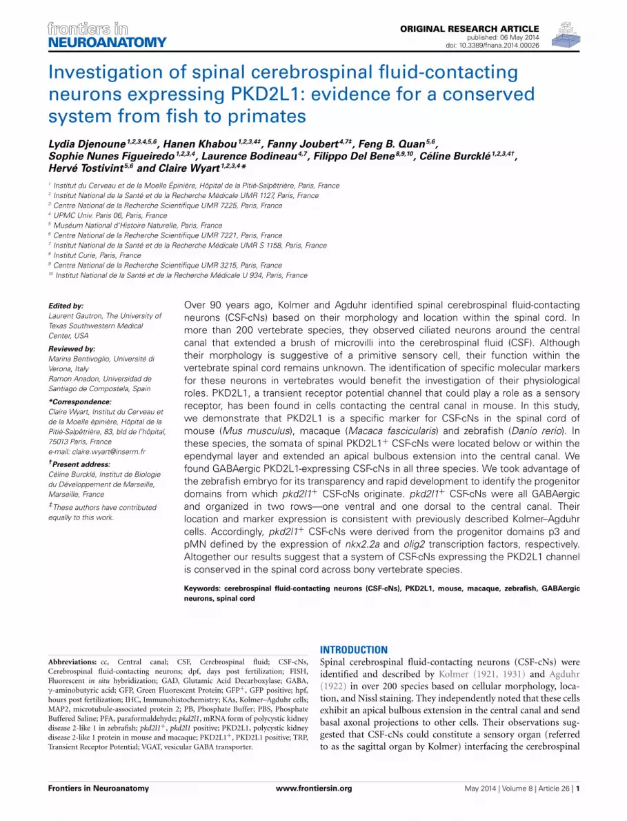
ORIGINAL RESEARCH ARTICLEpublished: 06 May 2014
doi: 10.3389/fnana.2014.00026
Investigation of spinal cerebrospinal fluid-contactingneurons expressing PKD2L1: evidence for a conservedsystem from fish to primatesLydia Djenoune1,2,3,4,5,6, Hanen Khabou1,2,3,4‡, Fanny Joubert4,7‡, Feng B. Quan5,6,
Sophie Nunes Figueiredo1,2,3,4, Laurence Bodineau4,7, Filippo Del Bene8,9,10, Céline Burcklé1,2,3,4†,
Hervé Tostivint5,6 and Claire Wyart1,2,3,4*
1 Institut du Cerveau et de la Moelle Épinière, Hôpital de la Pitié-Salpêtrière, Paris, France2 Institut National de la Santé et de la Recherche Médicale UMR 1127, Paris, France3 Centre National de la Recherche Scientifique UMR 7225, Paris, France4 UPMC Univ. Paris 06, Paris, France5 Muséum National d’Histoire Naturelle, Paris, France6 Centre National de la Recherche Scientifique UMR 7221, Paris, France7 Institut National de la Santé et de la Recherche Médicale UMR S 1158, Paris, France8 Institut Curie, Paris, France9 Centre National de la Recherche Scientifique UMR 3215, Paris, France10 Institut National de la Santé et de la Recherche Médicale U 934, Paris, France
Edited by:
Laurent Gautron, The University ofTexas Southwestern MedicalCenter, USA
Reviewed by:
Marina Bentivoglio, Université diVerona, ItalyRamon Anadon, Universidad deSantiago de Compostela, Spain
*Correspondence:
Claire Wyart, Institut du Cerveau etde la Moelle épinière, Hôpital de laPitié-Salpêtrière, 83, bld de l’hôpital,75013 Paris, Francee-mail: [email protected]†Present address:
Céline Burcklé, Institut de Biologiedu Développement de Marseille,Marseille, France‡These authors have contributedequally to this work.
Over 90 years ago, Kolmer and Agduhr identified spinal cerebrospinal fluid-contactingneurons (CSF-cNs) based on their morphology and location within the spinal cord. Inmore than 200 vertebrate species, they observed ciliated neurons around the centralcanal that extended a brush of microvilli into the cerebrospinal fluid (CSF). Althoughtheir morphology is suggestive of a primitive sensory cell, their function within thevertebrate spinal cord remains unknown. The identification of specific molecular markersfor these neurons in vertebrates would benefit the investigation of their physiologicalroles. PKD2L1, a transient receptor potential channel that could play a role as a sensoryreceptor, has been found in cells contacting the central canal in mouse. In this study,we demonstrate that PKD2L1 is a specific marker for CSF-cNs in the spinal cord ofmouse (Mus musculus), macaque (Macaca fascicularis) and zebrafish (Danio rerio). Inthese species, the somata of spinal PKD2L1+ CSF-cNs were located below or within theependymal layer and extended an apical bulbous extension into the central canal. Wefound GABAergic PKD2L1-expressing CSF-cNs in all three species. We took advantage ofthe zebrafish embryo for its transparency and rapid development to identify the progenitordomains from which pkd2l1+ CSF-cNs originate. pkd2l1+ CSF-cNs were all GABAergicand organized in two rows—one ventral and one dorsal to the central canal. Theirlocation and marker expression is consistent with previously described Kolmer–Agduhrcells. Accordingly, pkd2l1+ CSF-cNs were derived from the progenitor domains p3 andpMN defined by the expression of nkx2.2a and olig2 transcription factors, respectively.Altogether our results suggest that a system of CSF-cNs expressing the PKD2L1 channelis conserved in the spinal cord across bony vertebrate species.
Keywords: cerebrospinal fluid-contacting neurons (CSF-cNs), PKD2L1, mouse, macaque, zebrafish, GABAergic
neurons, spinal cord
Abbreviations: cc, Central canal; CSF, Cerebrospinal fluid; CSF-cNs,Cerebrospinal fluid-contacting neurons; dpf, days post fertilization; FISH,Fluorescent in situ hybridization; GAD, Glutamic Acid Decarboxylase; GABA,γ-aminobutyric acid; GFP, Green Fluorescent Protein; GFP+, GFP positive; hpf,hours post fertilization; IHC, Immunohistochemistry; KAs, Kolmer–Agduhr cells;MAP2, microtubule-associated protein 2; PB, Phosphate Buffer; PBS, PhosphateBuffered Saline; PFA, paraformaldehyde; pkd2l1, mRNA form of polycystic kidneydisease 2-like 1 in zebrafish; pkd2l1+, pkd2l1 positive; PKD2L1, polycystic kidneydisease 2-like 1 protein in mouse and macaque; PKD2L1+, PKD2L1 positive; TRP,Transient Receptor Potential; VGAT, vesicular GABA transporter.
INTRODUCTIONSpinal cerebrospinal fluid-contacting neurons (CSF-cNs) wereidentified and described by Kolmer (1921, 1931) and Agduhr(1922) in over 200 species based on cellular morphology, loca-tion, and Nissl staining. They independently noted that these cellsexhibit an apical bulbous extension in the central canal and sendbasal axonal projections to other cells. Their observations sug-gested that CSF-cNs could constitute a sensory organ (referredto as the sagittal organ by Kolmer) interfacing the cerebrospinal
Frontiers in Neuroanatomy www.frontiersin.org May 2014 | Volume 8 | Article 26 | 1
NEUROANATOMY
Djenoune et al. Conservation of spinal CSF-cNs
fluid (CSF) with the nervous system at the level of thespinal cord.
Since the discovery of CSF-cNs, electron microscopy stud-ies have shown that these cells exhibit a sensory tuft, previouslyreferred to as a “brush border” (Dale et al., 1987b), a “centralbody” (Vigh and Vigh-Teichmann, 1973) or a “bud” (Stoeckelet al., 2003). The apical bulbous extension projects from theperikarya of CSF-cNs toward the central canal and ends with aterminal bud in contact with its lumen. This apical extensionis characterized by the dendritic marker microtubule-associatedprotein 2 (MAP2) (Orts-Del’Immagine et al., 2014).
Immunohistochemistry (IHC) studies showed evidence forGABAergic CSF-cNs in many species such as rat (Barber et al.,1982; Stoeckel et al., 2003), turtle (Reali et al., 2011), Africanclawed frog (Dale et al., 1987a; Binor and Heathcote, 2001),zebrafish (Bernhardt et al., 1992; Martin et al., 1998; Higashijimaet al., 2004a,b; Wyart et al., 2009; Yang et al., 2010), eel and trout(Roberts et al., 1995), dogfish (Sueiro et al., 2004), and lampreys(Brodin et al., 1990; Christenson et al., 1991a,b; Melendez-Ferroet al., 2003; Robertson et al., 2007; Rodicio et al., 2008; Villar-Cervino et al., 2008). In African clawed frog and zebrafish, theseGABAergic CSF-cNs were named Kolmer–Agduhr cells (KAs)to distinguish them from ciliated ependymal cells (Dale et al.,1987a; Bernhardt et al., 1992). In these species CSF-cNs projectan ascending axon ventrally in the spinal cord (Dale et al., 1987a;Bernhardt et al., 1992; Higashijima et al., 2004a; Wyart et al.,2009).
Despite the anatomical studies of CSF-cNs and their impli-cation in modulating locomotion (Wyart et al., 2009), theirphysiological role in vertebrates remains unknown. One obstacleto answering this question is the lack of a specific genetic markerto identify these cells. Recently the calcium-permeable polycys-tic kidney disease 2-like 1 (PKD2L1) channel (Huang et al., 2006;Ishimaru et al., 2006), first identified in kidney, retina, and heart(Basora et al., 2002) was found to be expressed in cells contactingthe CSF at the level of the brainstem and spinal cord in mouse(Huang et al., 2006; Orts-Del’Immagine et al., 2012). This chan-nel belongs to the family of Transient Receptor Potential (TRP)channels. These are known to be chemo-, thermo- or mechano-sensitive (Delmas, 2004; Delmas et al., 2004). Expression of thischannel and location of these cells at the interface with the CSF areconsistent with the hypothesis that CSF-cNs have a proprioceptivesensory function.
In this study, we investigated whether PKD2L1 was a spe-cific marker for CSF-cNs across vertebrates, by examining mouse,macaque and zebrafish. In mouse and macaque PKD2L1 wasenriched in the sensory tuft of CSF-cNs in cervical, thoracic andlumbar spinal cord. The soma of PDK2L1+ cells was locatedin the ependymal layer or just underneath. In both species,a significant proportion of PKD2L1+ CSF-cNs was distinctlyidentified as GABAergic. We took advantage of the zebrafishmodel organism to thoroughly characterize the properties andlineage of pkd2l1+ cells in the spinal cord. In this model, allpkd2l1+ cells contacting the central canal were confirmed asGABAergic neurons. Reciprocally, all KAs, defined by their loca-tion and their GABAergic phenotype expressed pkd2l1. Usingspecific transgenic lines, we confirmed that these cells derive
from two progenitor domains. The dorsal pkd2l1+ KAs originatedfrom pMN and were labeled by the Enhanced Green FluorescentProtein (EGFP) in the Tg(olig2:EGFP) while the ventral pkd2l1+KAs originated from p3 and were labeled by Tg(nkx2.2a:mEGFP).Altogether our data show that PKD2L1 is a specific markerof CSF-cNs shared between multiple vertebrate species. Wefound evidence for PKD2L1-expressing GABAergic CSF-cNs inall examined species. Our results on the developmental ori-gin of CSF-cNs in zebrafish open new paths of investigation inmammals.
MATERIALS AND METHODSMOUSEExperimental animalsWild type (WT) mice (Mus musculus, OF1 strain) were kepton a 12 h light/dark cycle with free access to food and water.Experiments were performed at embryonic (E), postnatal (P) andadult stages: four WT adult mice, one WT newborn, and threeWT embryos were used in the present study. One adult maleand two E16.5 GAD67-GFP knock-in embryos (Tamamaki et al.,2003), resulting of the insertion of the cDNA encoding EGFP intothe locus encoding GAD67, have been used. After mating, the dayof detection of the vaginal plug was considered as embryonic dayE0.5. Pregnant females were sacrificed by decapitation at E14.5,E16.5, or E18.5. Uterine horns were removed from the motherand embryos were excised from their individual bag, soaked incold 0.1 M Phosphate Buffer (PB) for 10 min and fixed by immer-sion in cold 2% paraformaldehyde (PFA) solution in 0.1 M PBfor 3 h at 4◦C. Newborn mice (P1) were deeply anesthetized byan exposure to low temperature and the spinal cord was imme-diately removed and fixed with 2% PFA for 4 h. Adult mice weredeeply anesthetized with intraperitoneal injection of Nembutal®(150 mg·kg−1) and transcardially perfused sequentially with NaCl0.9% (20 ml) and PFA (2%, 30 ml for WT adults and 4%, 30 mlfor the GAD67-GFP mouse). After fixation, spinal cords were dis-sected out and post-fixed for 24 h in the fixative solution (2%PFA for WT mice and 4% for the GAD67-GFP mouse) at 4◦C.Following fixation (and post-fixation if appropriate), tissues werecryo-protected for 24–48 h in 30% sucrose in 0.1 M PB at 4◦Cand then stored at -20◦C for later use. Experiments were per-formed in accordance with the European Communities Council(EEC) Directive of 22 September 2010 (2010/63/EU) and Frenchlaw (87/848).
ImmunohistochemistryStandard immunofluorescence procedures were used to localizeantigens of interest (Voituron et al., 2011). At the cervical level,10 μm-thick coronal sections of the spinal cord were obtainedusing a cryostat (Leica CM 3050S), mounted on silanized slidesand stored at −20◦C for later IHC using a rabbit anti-Polycystin-L (anti-human PKD2L1, 1:500 dilution; Millipore, Billerica, MA,USA). This PKD2L1 antibody was raised against the syntheticpeptide from the N-terminal of human Polycystin-L. For PKD2L1IHC at E14.5 and E16.5, we used an Antigen Unmasking Solution(Vector Laboratories). Briefly, the sections were incubated in 1%Bovine serum albumin (BSA) and 0.1% Triton X-100 for 20 minat room temperature (RT). After rinsing with 0.1 M phosphate
Frontiers in Neuroanatomy www.frontiersin.org May 2014 | Volume 8 | Article 26 | 2
Djenoune et al. Conservation of spinal CSF-cNs
buffered saline (PBS), the sections were incubated for 30 min atRT with DAPI (1.25 μg/ml; Life Technologies) and with Alexa-conjugated anti-rabbit secondary antibodies (1:500 dilution; LifeTechnologies). For GFP and PKD2L1 double IHC, 30 μm-thickcoronal floating sections obtained using a cryostat (Leica CM1850S) were immunostained by co-incubating two primary anti-bodies, a chicken anti-GFP (1:1000 dilution; Abcam, Cambridge,UK) with the rabbit anti-PKD2L1 (1:500 dilution) overnight at4◦C, and then the specific secondary antibodies Alexa Fluor 488donkey anti-chicken IgG (1:500 dilution, Life Technologies) andAlexa 555-conjugated goat anti rabbit for 2 h at RT. In all IHCexperiments, control sections were processed in parallel by omit-ting primary antibodies. Finally, sections were washed with PBSbefore being mounted onto glass slides with fluorescent mountingmedium (AquaPolyMount, Biovalley, Marne La Vallée, France).Sections were observed under an Olympus FV1000 confocalmicroscope.
Quantification of cellsOn 10 μm-thick sections, we performed Z-stacks with a step sizeof 1–1.5 μm in a region of interest (ROI) centered on the centralcanal (approximately 150 × 220 μm width, n = 13 sections forE16.5 GAD67-GFP and 115 × 140 μm width, n = 7 sections foradult GAD67-GFP sections). For each analyzed section, cells wereidentified by the DAPI staining in order to avoid counting multi-ple times the same cell. Within the ROI, all PKD2L1+ cells werecounted manually and probed for GFP in the GAD67-GFP line.
MACAQUEExperimental animalsTissue from two adult macaques (Macaca fascicularis, a 25-yearold female and an 8-year old male) was used for this study.These animals were sacrificed for other purposes and spinal cordtissue samples were subsequently collected and devoted to thepresent study. Spinal cord collection was performed after thesacrifice of animals, in strict accordance with the recommenda-tions of the Weatherall Report regarding good animal practice.All surgical procedures and experimental protocols were car-ried out in strict accordance with the National Institutes ofHealth guidelines (2013) and the recommendations of the EEC(2010/63/EU).
Tissue extraction and preparationMacaques were deeply anesthetized with intramuscular injec-tion of ketamine and xylazine at 50 and 5 mg/kg, respectively.Following the anesthesia, each animal was exsanguinated andperfused with 0.9% NaCl before their tissues were fixed byan intracardial perfusion containing 4% PFA in 0.1 M PBS.Spinal cord was extracted and meninges were removed usingfine forceps. Tissue was embedded in 5% agarose and sec-tioned at 50 μm using a vibratome (Leica VT1000S). Additionally,frozen tissue blocks were obtained by passing tissue througha series of cryoprotection solutions (20, 30% sucrose inPBS) and freezing in isopentane; frozen sections were cut at20 μm (HM 650V Microtome, Thermo Scientific). All sec-tions were stored in 0.4% Sodium Azide in 0.1 M PBS at 4◦Cuntil IHC.
ImmunohistochemistryFree-floating sections from cervical, thoracic, and lumbar spinalcord tissue were used for IHC. Sections were first incubated in ablocking solution composed of 1% BSA, 0.5% Triton X-100 and2% normal goat serum in PBS to reduce non-specific labeling.Sections were then incubated in primary antibodies overnightat 4◦C. After multiple washes, sections were incubated in sec-ondary antibodies for 4 h at 4◦C. Primary antibodies used wererabbit anti-Polycystin-L (anti-human PKD2L1, 1:1,000 dilution;Millipore, Billerica, MA, USA), guinea pig anti-vesicular GABATransporter (anti-VGAT, 1:500; Synaptic Systems, Germany) andrabbit anti-GAD65/67 (1:500; Abcam, MA, USA). Secondaryantibodies were Alexa Fluor 488- or Alexa Fluor 568-conjugatedanti-rabbit (1:500; Life Technologies) and Alexa Fluor 488-anti-guinea pig (1:500; Life Technologies). The same blocking solutionas previously was used to dilute the antibodies. PBS was used forwashing sections between each step. Sections were mounted onglass slides, cover slipped with Prolong® Gold Antifade Reagent(Life Technologies) mounting medium and then imaged on aLeica SP2 AOBS AOTF inverted confocal laser scanning micro-scope. Single optical slice images of individual labeled cells weretaken using a 63× oil objective.
ZEBRAFISHExperimental animalsAll zebrafish (Danio rerio) lines were maintained and raised ona 14/10 h light cycle and water was regulated at 28.5◦C, con-ductivity at 500 μS and pH at 7.4 (Westerfield, 2000). WT ABand Tüpfel long fin (TL) embryos were used for whole mountin situ hybridization (ISH). Tg(nkx2.2a:mEGFP) (Ng et al., 2005;Kirby et al., 2006) and Tg(olig2:EGFP) (Shin et al., 2003) trans-genic lines were kindly provided by Prof. Bruce Appel, Universityof Colorado, Denver, USA. Embryos were dechorionated andstaged according to number of somites as described (Kimmelet al., 1995): the 30-somite stage corresponds to Prim-5 or 24 hpost fertilization when raised at 28.5◦C. Adult fish were anes-thetized in 0.02% MS 222 (Sandoz, Levallois-Perret, France)and killed by decapitation. All procedures were approved by theInstitutional Ethics Committee at the Institut du Cerveau et dela Moelle épinière (ICM), Paris, France, the Ethical CommitteeCharles Darwin and received subsequent approval from the EEC(2010/63/EU).
Generation of the pkd2l1 probeTo generate the pkd2l1 ISH probe, the coding fragmentfor pkd2l1 was amplified from zebrafish embryo totalcDNA using the following primers (5′ to 3′): pkd2l1_For:TAGTGGTGATACTGCTTGCTGTGGTGG (from the end ofthe exon 6 of the pkd2l1 gene and the beginning of exon 7)and pkd2l1_Rev: TGGTTCCACACTGTTCTCGAGGTCACG(from the end of exon 13). The PCR fragment was clonedinto the pCRII-TOPO vector (Life Technologies, Carlsbad, CA,USA). The resulting plasmid was linearized with NotI. Thegad67 plasmid, kindly provided by Dr. Uwe Strähle, KarlsruheInstitute of Technology, Germany, was linearized with NcoI.Digoxigenin (DIG)- and fluorescein (Fluo)-labeled probes weresynthesized using SP6 RNA polymerase with the RNA Labeling
Frontiers in Neuroanatomy www.frontiersin.org May 2014 | Volume 8 | Article 26 | 3

Djenoune et al. Conservation of spinal CSF-cNs
Kit (Roche Applied Science, Basel, Switzerland) to generate bothpkd2l1 and gad67 antisense probes. To generate pkd2l1 senseprobes, the plasmid was linearized with KpnI and transcriptionwas carried out using T7 RNA polymerase. All probes werepurified using the mini Quick Spin RNA Column (Roche, Basel,Switzerland).
In situ hybridizationWhole-mount ISH were performed as previously described(Parmentier et al., 2011; Alunni et al., 2013) on embryos or dis-sected adult spinal cords fixed in 4% PFA in PBS overnight at 4◦C.To reveal pkd2l1 expression, probes were detected with anti-DIGor anti-Fluo antibodies conjugated to alkaline phosphatase fol-lowed by a chromogenic reaction using a solution of NBT/BCIPas substrate (Roche Diagnostics, France). To quantify cell den-sity or ascertain transcript colocalization, probes were detectedby the antibodies conjugated to horseradish peroxidase and wererevealed by Tyramide Signal Amplification using Tyramide-FITCor Tyramide-TAMRA as substrates. The specificity of the pkd2l1probe was verified using a pkd2l1 sense probe as negative control(data not shown). Double fluorescent in situ hybridization (FISH)for pkd2l1 and gad67 were performed on adult zebrafish spinalcords and coupled with respectively DIG and Fluo.
Immunohistochemistry and sectioningThe following primary antibodies were used for IHC: rabbitanti-GABA (1:2000, Sigma-Aldrich, St. Louis, MO, USA), rab-bit anti-GAD65/67 and chicken anti-GFP (both used at 1:500dilution, Abcam, Cambridge, UK). Immunostaining specificitywas established by omitting the primary specific antibodies, noimmunoreactive signal was observed. Agarose sections were col-lected as described above. In the zebrafish embryo, we performedeither 50 μm-thick sagittal (for anti-GAD65/67 IHC) or trans-verse (for anti-GFP IHC) sections. In the adult, we performed50 μm-thick sagittal and frontal sections of previously FISHstained adult spinal cords.
Combination of pkd2l1 FISH with immunohistochemistrypkd2l1 FISH was performed before IHC against GFP orGAD65/67: embryos were washed and immunostained withthe chicken anti-GFP antibody or the rabbit anti-GAD65/67antibody overnight at 4◦C, and then incubated with thecorresponding Alexa conjugated secondary antibodies IgG(1:500, Life Technologies) combined with DAPI (2.5 μg/ml, LifeTechnologies).
MicroscopyWhole-mount embryos stained by NBT/BCIP were mountedin 80% glycerol. Sections of adult spinal cord were mountedin a solution of Mowiol. Embryos were imaged using a NikonAZ100M macroscope and a Leica DM5000 B Upright micro-scope. Adult spinal cord sections were imaged using a NikonAZ100M macroscope. To quantify pkd2l1+ and GABA+ cells,stained embryos were imaged using the Fixed Stage microscopeAxioExaminer Z1 equipped with a 20× water immersion objec-tive and a Yokogawa CSU-X1 spinning disk unit (n = 13 embryosfor pkd2l1 and n = 10 for GABA). To quantify the overlapof GFP with pkd2l1 FISH in the Tg(nkx2.2a:mEGFP) and the
Tg(olig2:EGFP) transgenic embryos, 50 μm-thick sections wereanalyzed on an Olympus FV1000 confocal microscope equippedwith a 40× water immersion objective using 405, 473, and 543 nmlaser lines. Images were processed using Fiji (Schindelin et al.,2012) and Adobe Illustrator (Adobe Systems, Mountain View,CA, USA) software.
Quantification of cellsTo quantify the total number of pkd2l1+ cells revealed by FISHper embryo, Z-stacks of the entire embryos from somite 1 to 30were acquired. Total numbers of cells were counted per somitelabeled on both sides of the midline. The boundaries for eachsomite were established with transmitted light. The three sub-types of pkd2l1 expressing cells were defined according to theirposition relative to the central canal. We distinguished: (i) therow of cells that was ventral and in proximity to the central canal,(ii) the row that was dorsal and in proximity to the central canaland (iii) the sparse cells that were dorsal and distant from thecentral canal. The proportion of pkd2l1+ cells double-labeled forGAD65/67 in 30-somite (Prim-5) embryos was quantified basedon 50 μm-thick sagittal sections performed after the FISH andbefore GAD65/67 IHC. The proportion of pkd2l1+ cells double-labeled for GFP in Tg(nkx2.2a:mEGFP) and Tg(olig2:EGFP) in30-somite embryos was quantified based on 50 μm-thick trans-verse sections performed after the FISH and IHC. The totalnumber of cells is given as the mean ± Standard Error of the Mean(SEM). The same method was applied for counting of GABA+KAs. There are four GABAergic interneuron types in the zebrafishembryo (Bernhardt et al., 1992; Higashijima et al., 2004a); KAswere identified as the only GABAergic ascending neurons witha soma located just below (KA”) or above (KA’) the centralcanal.
RESULTSPKD2L1 IS EXPRESSED IN CSF-cNs FROM EMBRYONIC STAGES TOADULTHOOD IN THE MOUSE SPINAL CORDPKD2L1 was originally identified in CSF-cNs at postnatal stagesP1–P4 in the mouse spinal cord (Huang et al., 2006). We inves-tigated PKD2L1 expression in the spinal cord at the embryonic,postnatal and adult stages. PKD2L1 expression could not bedetected before E14.5 in the spinal cord at the cervical, thoracicand lumbar levels. At E14.5, few PKD2L1+ cells could be iden-tified in the cervical spinal cord (Figure 1A). Starting at E16.5,PKD2L1+ cells localized around the central canal exhibited thetypical morphology of spinal CSF-cNs (Figures 1B–E, arrows). AtE18.5, the soma of PKD2L1+ cells was usually located under thelayer of ependymal cells (91 out of 102, while 11 out of 102 werelocated within the ependyma, Table 1). PKD2L1+ cells sent anapical bulbous extension toward the central canal ending in thelumen with a bud enriched in PKD2L1 (Figures 1B–E, arrows).In the adult, the soma of PKD2L1+ CSF-cNs was located in lam-ina X and always under the ependyma (95 out of 95 PKD2L1+cells) (Figure 1E, Table 1). We also observed PKD2L1+ cells withsimilar morphology localized away from the central canal, thoughthese cells lacked a visible apical extension to the central canal(Figures 1A–C, arrowheads). Our data identify PKD2L1 as ageneral marker of CSF-cNs.
Frontiers in Neuroanatomy www.frontiersin.org May 2014 | Volume 8 | Article 26 | 4

Djenoune et al. Conservation of spinal CSF-cNs
FIGURE 1 | PKD2L1 expression in CSF-cNs is present from
embryonic stage E14.5 to adulthood in mouse. (A–E)
Immunostaining for PKD2L1 at E14.5 (A), E16.5 (B), E18.5 (C), P1(D), and in the adult (E) performed on coronal sections of the mousecervical spinal cord. At E14.5, few PKD2L1+ cells are detected(arrowhead). From E16.5 (B) to adult (E), multiple PKD2L1+ CSF-cNs
(arrows) surround the central canal where they project an apicalbulbous dendritic extension contacting its lumen. Note that PKD2L1+cells exhibiting a similar morphology but not distinctly contacting thecentral canal (arrowheads) can also be observed (A–C). Dorsal is up.White dashed line delineates the central canal. DAPI staining appearsin blue. Scale bars = 20 μm.
Table 1 | Distribution of PKD2L1+ cells based on the location of their
soma relative to the ependyma in the mouse spinal cord.
Stage Total number of Number of PKD2L1+ Number of PKD2L1+
PKD2L1+ cells cells located in the cells located in the
ependymal layer subependymal layer
E16.5 84 8 76
E18.5 102 11 91
P1 184 6 178
Adult 95 0 95
PKD2L1+ cells located within the ependyma represent only a small number
(<11%) of the total population of PKD2L1+ cells counted at embryonic and new-
born stages. In the adult mouse spinal cord, the somata of all PKD2L1+ cells are
located subependymally.
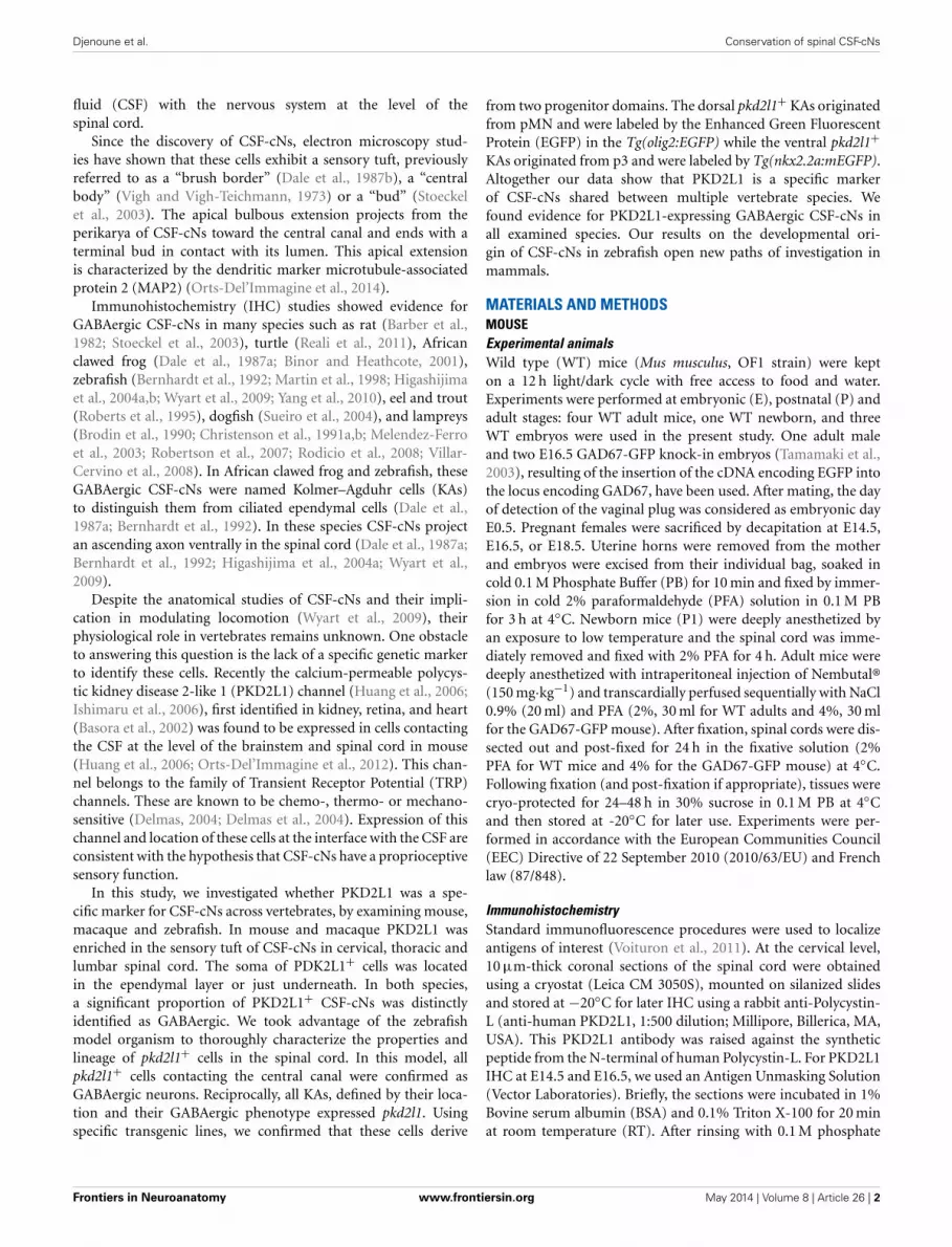
PKD2L1 IS PRESENT IN GABAergic CSF-cNs IN THE MOUSE SPINALCORDAs mentioned before, cells contacting the CSF had been describedas GABAergic in many species, but the neurotransmitter pheno-type has not been thoroughly established yet in mouse. We testedwhether CSF-cNs were GABAergic in the embryonic and adultmouse spinal cord (Figure 2). We took advantage of the well-characterized GAD67-GFP knock-in mouse where the enhancedGFP was targeted to the locus encoding GAD67 using homolo-gous recombination (Tamamaki et al., 2003). In these animals,we performed IHC for GFP to amplify the endogenous GFPsignals (Figure 2). In GAD67-GFP knock-in embryos, doubleIHC for GFP and PKD2L1 on coronal sections of E16.5 micespinal cords (Figure 2A) revealed that most of PKD2L1+ cells
were GFP+ (arrows) indicating their GABAergic nature (n = 2embryos, 107 out of 162 PKD2L1+ cells). In one adult, weobserved the same findings and found 47 out of 73 cells labeled forboth PKD2L1 and GFP (Figure 2B, arrows). Note that both in theembryos and in the adult, we observed PKD2L1+cells that werenot distinctly GFP+ (Figures 2A,B, double arrowhead). Notably,we never observed GFP+ CSF-cNs that were not PKD2L1+.Altogether we show evidence for PKD2L1+ CSF-cNs that areGABAergic in the mouse spinal cord.
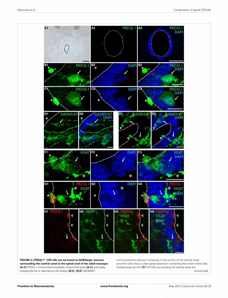
PKD2L1+ GABAergic CSF-cNs SURROUND THE CENTRAL CANAL INTHE ADULT MACAQUE SPINAL CORDWe next asked whether PKD2L1 labels CSF-cNs in the spinal cordof primates (Figure 3). In transverse sections from cervical, tho-racic and lumbar spinal cord, we observed the distribution ofPKD2L1 immunofluorescence around the central canal in lam-ina X (Figures 3A–C). PKD2L1+ cells were localized around thecentral canal and exhibited the typical morphology of spinal CSF-cNs with an apical extension toward the central canal endingwith a bud in the lumen. PKD2L1 was enriched in the terminalbud of cerebrospinal fluid-contacting cells (Figures 3A–C) andfaintly expressed (Figures 3B,C) or absent (Figure 3G) in the cellsoma and in the rest of the apical extensions. PKD2L1+CSF-cNsclosely surrounded the central canal; they had a round nucleusand were located under (Figures 3B–E) or within the ependyma(Figures 3F,G). In order to test whether PKD2L1+ CSF-cNs wereGABAergic, we used IHC for the enzymes GAD65/67 and theVGAT transporter. Positive immunostaining for GAD65/67 andVGAT was found in cells surrounding the central canal at thelevel of their apical bulbous extension, soma and putative axon
Frontiers in Neuroanatomy www.frontiersin.org May 2014 | Volume 8 | Article 26 | 5
Djenoune et al. Conservation of spinal CSF-cNs
FIGURE 2 | Some PKD2L1+ cells surrounding the central canal of
cervical spinal cord of adult mouse are immunoreactive for GFP in the
knock-in GAD67-GFP mouse. (A) 10 μm-thick coronal sections ofGAD67-GFP E16.5 mouse spinal cord immunostained for GFP (A1,A3,A4)
and PKD2L1 (A2–A4). Most PKD2L1+ cells are GFP+ (arrows) but not all
(double arrowheads). (B) 30 μm-thick coronal sections of a GAD67-GFP adultmouse spinal cord immunostained for GFP (B1,B3,B4) and PKD2L1 (B2–B4).While many PKD2L1+cells are distinctly GFP+ (arrows), some cells are not(double arrowheads). Dorsal is up. White dashed line delineates the centralcanal. DAPI staining appears in blue. Scale bars = 25 μm (A), 15 μm (B).
(Figures 3D–F, arrowheads, arrows, and asterisks, respectively)but buds were labeled by GABAergic markers less frequentlythan they were by PKD2L1. Double immunofluorescence forPKD2L1 and VGAT showed evidence for CSF-cNs double-labeledat the level of the soma and the intraluminal bud (Figure 3G).Nonetheless VGAT could not be detected in all PKD2L1+buds (Figure 3H, double arrowheads). Cells double-labeled forPKD2L1 and VGAT were observed at the cervical, thoracic andlumbar level in the spinal cord. Taken together, our data show forthe first time the presence of immunoreactive CSF-cNs contain-ing PKD2L1 and GABAergic markers (GAD65/67 or VGAT) inthe macaque lamina X. These findings demonstrate that a pop-ulation of GABAergic CSF-cNs expressing PKD2L1 is present aswell in the spinal cord of adult primates. Our results also supportthe idea that PKD2L1 is a specific marker of CSF-cNs in primatesand labels more of these cells than GABAergic markers do in ourexperimental conditions.
THE EXPRESSION OF pkd2l1 mRNA IN ZEBRAFISH EMBRYO ENABLESAN EXTENSIVE QUANTIFICATION AND LINEAGE ANALYSIS OF CSF-cNsThe developmental origin of CSF-cNs has not been established yetin mammals. To tackle the question of the developmental origin
of pkd2l1+ cells, we took advantage of the zebrafish embryo for itstransparency and the rapidity of development of the spinal cord.
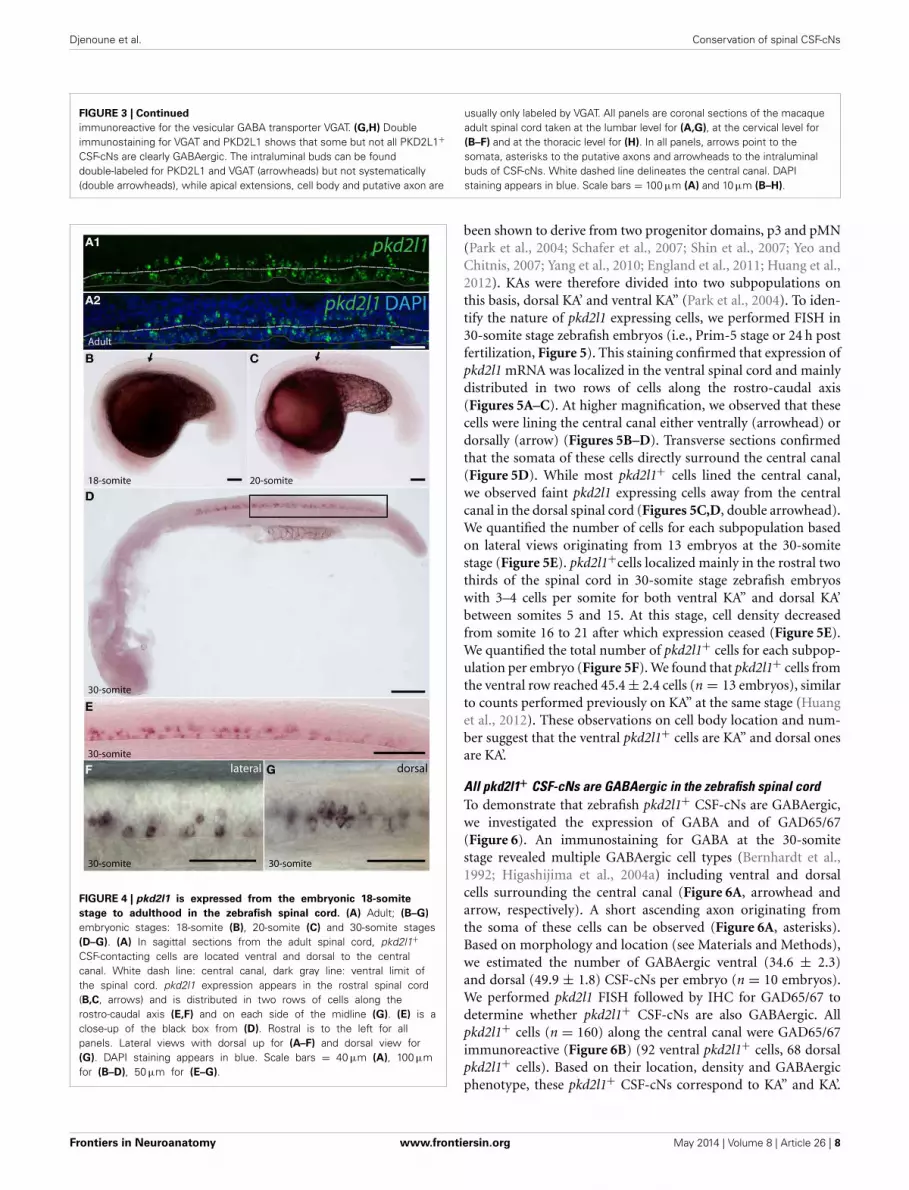
pkd2l1 is expressed in CSF-cNs in zebrafishTo assess whether pkd2l1 was expressed in CSF-cNs in zebrafish,we performed ISH using the antisense probe for pkd2l1(Figure 4). We observed the expression of pkd2l1 in CSF-cNsalong the central canal in sagittal sections of the adult spinal cord(Figure 4A). pkd2l1+ cells contacting the CSF were located ven-trally and dorsally to the central canal (Figures 4A1,A2). In thezebrafish embryo, we investigated the 10-, 14-, 18-, 20-, and 30-somite stages (n > 6 embryos per condition). A weak signal inthe rostral spinal cord (black arrow) could be detected at the18-somite stage (Figure 4B). Gradually, the staining appearedrostro-caudally within the spinal cord (Figures 4C,D). Cells withdense labeling were located in two parallel rows along the ven-tral margin of the spinal cord (Figures 4E,F). Dorsal views of thespinal cord showed that these cells were located on either side ofthe midline (Figure 4G). At the larval stages, we observed thatpkd2l1+ CSF-cNs covered the entire length of the spinal cordand the organization in two parallel rows was not distinct any-more (data not shown). In zebrafish, CSF-cNs (named KAs) have
Frontiers in Neuroanatomy www.frontiersin.org May 2014 | Volume 8 | Article 26 | 6
Djenoune et al. Conservation of spinal CSF-cNs
FIGURE 3 | PKD2L1+ CSF-cNs can be found as GABAergic neurons
surrounding the central canal in the spinal cord of the adult macaque.
(A–C) PKD2L1 immunostaining labels intraluminal buds (A–C) and rarelysubependymal or ependymal cell bodies (B,C). (D,E) GAD65/67
immunoreactive cells are numerous in the vicinity of the central canal:proximal cells show a clear apical extension contacting the lumen while cellslocated away do not. (F) CSF-cNs surrounding the central canal are
(Continued)
Frontiers in Neuroanatomy www.frontiersin.org May 2014 | Volume 8 | Article 26 | 7
Djenoune et al. Conservation of spinal CSF-cNs
FIGURE 3 | Continued
immunoreactive for the vesicular GABA transporter VGAT. (G,H) Doubleimmunostaining for VGAT and PKD2L1 shows that some but not all PKD2L1+CSF-cNs are clearly GABAergic. The intraluminal buds can be founddouble-labeled for PKD2L1 and VGAT (arrowheads) but not systematically(double arrowheads), while apical extensions, cell body and putative axon are
usually only labeled by VGAT. All panels are coronal sections of the macaqueadult spinal cord taken at the lumbar level for (A,G), at the cervical level for(B–F) and at the thoracic level for (H). In all panels, arrows point to thesomata, asterisks to the putative axons and arrowheads to the intraluminalbuds of CSF-cNs. White dashed line delineates the central canal. DAPIstaining appears in blue. Scale bars = 100 μm (A) and 10 μm (B–H).
FIGURE 4 | pkd2l1 is expressed from the embryonic 18-somite
stage to adulthood in the zebrafish spinal cord. (A) Adult; (B–G)
embryonic stages: 18-somite (B), 20-somite (C) and 30-somite stages(D–G). (A) In sagittal sections from the adult spinal cord, pkd2l1+CSF-contacting cells are located ventral and dorsal to the centralcanal. White dash line: central canal, dark gray line: ventral limit ofthe spinal cord. pkd2l1 expression appears in the rostral spinal cord(B,C, arrows) and is distributed in two rows of cells along therostro-caudal axis (E,F) and on each side of the midline (G). (E) is aclose-up of the black box from (D). Rostral is to the left for allpanels. Lateral views with dorsal up for (A–F) and dorsal view for(G). DAPI staining appears in blue. Scale bars = 40 μm (A), 100 μmfor (B–D), 50 μm for (E–G).
been shown to derive from two progenitor domains, p3 and pMN(Park et al., 2004; Schafer et al., 2007; Shin et al., 2007; Yeo andChitnis, 2007; Yang et al., 2010; England et al., 2011; Huang et al.,2012). KAs were therefore divided into two subpopulations onthis basis, dorsal KA’ and ventral KA” (Park et al., 2004). To iden-tify the nature of pkd2l1 expressing cells, we performed FISH in30-somite stage zebrafish embryos (i.e., Prim-5 stage or 24 h postfertilization, Figure 5). This staining confirmed that expression ofpkd2l1 mRNA was localized in the ventral spinal cord and mainlydistributed in two rows of cells along the rostro-caudal axis(Figures 5A–C). At higher magnification, we observed that thesecells were lining the central canal either ventrally (arrowhead) ordorsally (arrow) (Figures 5B–D). Transverse sections confirmedthat the somata of these cells directly surround the central canal(Figure 5D). While most pkd2l1+ cells lined the central canal,we observed faint pkd2l1 expressing cells away from the centralcanal in the dorsal spinal cord (Figures 5C,D, double arrowhead).We quantified the number of cells for each subpopulation basedon lateral views originating from 13 embryos at the 30-somitestage (Figure 5E). pkd2l1+cells localized mainly in the rostral twothirds of the spinal cord in 30-somite stage zebrafish embryoswith 3–4 cells per somite for both ventral KA” and dorsal KA’between somites 5 and 15. At this stage, cell density decreasedfrom somite 16 to 21 after which expression ceased (Figure 5E).We quantified the total number of pkd2l1+ cells for each subpop-ulation per embryo (Figure 5F). We found that pkd2l1+ cells fromthe ventral row reached 45.4 ± 2.4 cells (n = 13 embryos), similarto counts performed previously on KA” at the same stage (Huanget al., 2012). These observations on cell body location and num-ber suggest that the ventral pkd2l1+ cells are KA” and dorsal onesare KA’.
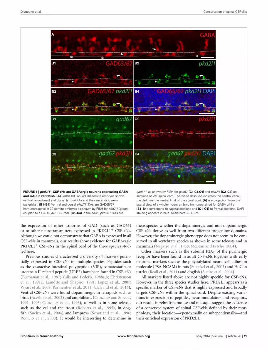
All pkd2l1+ CSF-cNs are GABAergic in the zebrafish spinal cordTo demonstrate that zebrafish pkd2l1+ CSF-cNs are GABAergic,we investigated the expression of GABA and of GAD65/67(Figure 6). An immunostaining for GABA at the 30-somitestage revealed multiple GABAergic cell types (Bernhardt et al.,1992; Higashijima et al., 2004a) including ventral and dorsalcells surrounding the central canal (Figure 6A, arrowhead andarrow, respectively). A short ascending axon originating fromthe soma of these cells can be observed (Figure 6A, asterisks).Based on morphology and location (see Materials and Methods),we estimated the number of GABAergic ventral (34.6 ± 2.3)and dorsal (49.9 ± 1.8) CSF-cNs per embryo (n = 10 embryos).We performed pkd2l1 FISH followed by IHC for GAD65/67 todetermine whether pkd2l1+ CSF-cNs are also GABAergic. Allpkd2l1+ cells (n = 160) along the central canal were GAD65/67immunoreactive (Figure 6B) (92 ventral pkd2l1+ cells, 68 dorsalpkd2l1+ cells). Based on their location, density and GABAergicphenotype, these pkd2l1+ CSF-cNs correspond to KA” and KA’.
Frontiers in Neuroanatomy www.frontiersin.org May 2014 | Volume 8 | Article 26 | 8
Djenoune et al. Conservation of spinal CSF-cNs
To test whether zebrafish CSF-cNs persist as GABAergic neu-rons until adulthood, we performed pkd2l1 and gad67 doubleFISH on whole adult spinal cord indicating that pkd2l1+ CSF-cNsexpress gad67 throughout development (Figure 6C). Contraryto what we observed in mouse and macaque, these resultsdemonstrate that in zebrafish all pkd2l1+ CSF-cNs are clearlyGABAergic.
pkd2l1+CSF-cNs originate from the p3 and pMN progenitordomains in zebrafishTo confirm the developmental origins of pkd2l1+ cells, we tookadvantage of the zebrafish model in which pkd2l1 is expressedin the 30-somite stage embryo when progenitor domains arewell-defined. Previous studies have shown that dorsal KA’ cellsare derived from the pMN domain and express the transcrip-tion factor olig2 (Park et al., 2004). Ventral KA” cells origi-nate from the p3 progenitor domain (Schafer et al., 2007) andexpress the transcription factor nkx2.2a (Yang et al., 2010; Huanget al., 2012). We therefore performed pkd2l1 FISH combinedwith an anti-GFP immunostaining in the Tg(nkx2.2a:mEGFP)and the Tg(olig2:EGFP) transgenic lines (Shin et al., 2003; Nget al., 2005) (Figures 7, 8). In the Tg(nkx2.2a:mEGFP) transgenicembryos, the p3 domain was labeled by GFP staining as expected(Figure 7A). Ventral pkd2l1+cells contacting the central canalwere labeled for GFP while dorsal cells were not (Figure 7B).In the Tg(olig2:EGFP) transgenic embryos (Figure 8), the pro-genitor domain pMN was labeled by GFP (Figure 8A). In theseanimals, only dorsal pkd2l1+ cells contacting the central canalwere GFP+ (Figure 8B). Transverse sections allowed to estimatethe number of pkd2l1+ cells contacting the central canal thatwere positive for GFP in the Tg(nkx2.2a:mEGFP) and in theTg(olig2:EGFP) transgenic embryos (Table 2). While only ven-tral pkd2l1+cells expressed GFP in the Tg(nkx2.2a:mEGFP) line(38 out of 38 cells), dorsal cells solely expressed GFP in theTg(olig2:EGFP) line (93 out of 93 cells) (Table 2). Our data indi-cate that pkd2l1+cells lining the central canal in zebrafish areKAs originating from p3 (for KA”) and from pMN (for KA’)depending on their dorso-ventral location relative to the centralcanal.
DISCUSSIONPKD2L1 APPEARS AS A SPECIFIC MARKER OF SPINAL CSF-cNs INBONY VERTEBRATE SPECIESPKD2L1+ neurons projecting their apical bulbous dendriticextension into the central canal of the spinal cord had beendescribed only in mouse at the P1–P4 stages (Huang et al.,2006) and in the adult (Orts-Del’Immagine et al., 2012, 2014).We hypothesized that the channel PKD2L1 could be a specificmarker of spinal CSF-cNs shared among vertebrates. We demon-strate here that the TRP channel PKD2L1 is a marker of CSF-cNsin three vertebrate species: Mus musculus, Macaca fascicularis,and Danio rerio. For the first time, we show the expression ofthis channel in primate and zebrafish CSF-cNs. In the adultmacaque spinal cord, CSF-cNs express PKD2L1 at the cervical,thoracic and lumbar levels. In the zebrafish embryo, pkd2l1+CSF-cNs originally differentiate in two rows along rostro-caudalgradient.
Taken together, our results demonstrate the shared expres-sion of PKD2L1 in spinal CSF-cNs across bony vertebrate species.GABA has been previously reported to label some CSF-cNs(Barber et al., 1982; Dale et al., 1987a,b; Brodin et al., 1990;Christenson et al., 1991a,b; Bernhardt et al., 1992; Martin et al.,1998; Binor and Heathcote, 2001; Stoeckel et al., 2003; Robertsonet al., 2007; Rodicio et al., 2008; Villar-Cervino et al., 2008; Realiet al., 2011). In mouse and macaque, PKD2L1 appeared alwayshighly enriched in the intraluminal buds of CSF-cNs. On the con-trary, GABAergic markers (GAD65/67, VGAT) rarely labeled inthese species the soma or the apical bulbous extension of CSF-cNs. Notably we never observed CSF-cNs labeled by GABAergicmarkers and not by PKD2L1. In mammals, PKD2L1 seems there-fore to label more CSF-contacting cells than GABAergic mark-ers do. Nonetheless, with the approach we developed here, wecannot assess the existence of CSF-cNs that would not expressPKD2L1.
EXPRESSION OF PKD2L1 IN CSF-cNs OF THE EMBRYONIC SPINALCORDIn mouse and zebrafish, we detected the expression of the chan-nel at early embryonic stages of development (E14.5 stage forthe mouse and 18-somite stage for the zebrafish). In mouse,PKD2L1 expression could be found in few spinal cells contact-ing the central canal at E14.5 but became more evenly expressedfrom E16.5 to adulthood. This observation is consistent witha recent report showing that CSF-cNs emerged at E14 in therat spinal cord (Kutna et al., 2013). It suggests that PKD2L1expression may start soon after the cell differentiates into aCSF-cN.
We demonstrate here that within the spinal cord of thezebrafish embryo pkd2l1 mRNA is enriched in CSF-cNs that aremainly arranged in two rows, one ventral and one dorsal to thecentral canal. Previous studies have shown that KA cells can besubdivided into two populations of CSF-cNs: the ventral KA” andthe dorsal KA’ (Park et al., 2004; Schafer et al., 2007; Shin et al.,2007; Yeo and Chitnis, 2007; Yang et al., 2010; England et al., 2011;Huang et al., 2012). We showed that pkd2l1 can be detected inboth KA” and KA’.
In the three species studied here, we observed PKD2L1+perikarya away from the central canal and for which we couldnot observe an apical bulbous extension reaching the lumen. Inthe zebrafish embryonic spinal cord, pkd2l1 expression was weakin cells that were localized dorsally and away from the centralcanal. These cells were less than 8% (7.6% ± 2.3) of the pkd2l1+cells in our FISH experiments (Figure 5). Some of them expressedGFP in the Tg(olig2:EGFP) line (Table 2) suggesting that theycould be either CSF-cNs originating from pMN, motoneuronsor ventral longitudinal descending neurons (VeLDs) (Bernhardtet al., 1990; Park et al., 2004; Warp et al., 2012). A more exten-sive characterization would be necessary to identify the nature ofthese marginal dorsal cells. In cross sections of mouse, macaqueand zebrafish spinal cords, those distant cells did not distinctlycontact the central canal. However, as shown in rats (Lu et al.,2008), they could extend a long apical bulbous extension reach-ing the central canal that would be difficult to capture in thinsections.
Frontiers in Neuroanatomy www.frontiersin.org May 2014 | Volume 8 | Article 26 | 9

Djenoune et al. Conservation of spinal CSF-cNs
FIGURE 5 | pkd2l1 is expressed in cells contacting the CSF and
surrounding the central canal in the spinal cord of the zebrafish
embryo. (A–D) FISH of pkd2l1 mRNA at the 30-somite stage. Lateralviews for (A–C) and transverse section for (D). Two subpopulations ofpkd2l1 bright expressing cells surround the central canal ventrally(arrowhead) and dorsally (arrow). Note the existence of dorsal pkd2l1weak expressing cells away from the central canal (double arrowhead).The white dashed line, dark gray solid line and the solid white lines
indicate respectively the central canal, the ventral limit of the spinal cord,and the somite boundaries. DAPI staining appears in blue. Scale bars =50 μm for (A), 30 μm for (B,C), and 20 μm for (D). (E) Mean number ofpkd2l1+ cells per somite along the rostro-caudal axis at the 30-somitestage. (F) Total cell counts per embryo (n = 13 embryos). We counted45.4 ± 2.4 ventral pkd2l1+ cells, 47.1 ± 3.4 dorsal pkd2l1+ cells and7.6 ± 2.3 dorsal away from the central canal pkd2l1+ cells per embryo.(E,F) Mean values are given ± s.e.m.
ON THE DEVELOPMENTAL ORIGINS OF pkd2l1 EXPRESSING CSF-cNsAt the embryonic stage, the neural tube is subdivided into molec-ularly defined neural progenitor domains generating distinctneuronal subtypes in vertebrates (Ericson et al., 1997; Briscoeet al., 1999; Jessell, 2000; Novitch et al., 2001; Goulding, 2009).In zebrafish, CSF-cNs referred to as KA cells derive from thetwo most ventral domains of the spinal cord; the ventral to thecentral canal p3 domain labeled by nkx2.2a and the more dor-sal pMN marked by olig2 (Park et al., 2004; Schafer et al., 2007;Shin et al., 2007; Yeo and Chitnis, 2007; Yang et al., 2010; Englandet al., 2011; Huang et al., 2012). By analyzing transverse sectionsof stable transgenic lines where GFP reports the expression ofthese transcription factors, we found that pkd2l1+ CSF-cNs werederived from the nkx2.2a and from the olig2 expression domains.This observation confirms the double developmental origin ofCSF-cNs in zebrafish.
In the embryonic mouse spinal cord, the progenitor domainsp3 and pMN form well-defined bands labeled by NKX2.2 andOLIG2, respectively, between E9.5 and E12.5 (Briscoe et al., 1999;Jessell, 2000; Novitch et al., 2001). At these stages, PKD2L1 isnot yet expressed (data not shown). Therefore, by IHC for thesetranscription factors and PKD2L1, we could not test whetherPKD2L1+CSF-cNs originate as well from p3 and pMN (datanot shown). To reveal the developmental origin of these cells inmouse, lineage tracing based on the use of inducible transgeniclines for p3 and pMN markers will be necessary. Previous resultsrelying on a tamoxifen-inducible Cre-recombinase inserted into
the Olig2 locus indicate that a subpopulation of cells originatingfrom pMN between E9.5 and E14.5 are located at the ependymalborder circling the central canal (Srinivas et al., 2001; Masahiraet al., 2006). This observation suggests that some CSF-cNs couldoriginate from pMN in mouse, although a thorough investiga-tion would be necessary to address this question in this modelorganism.
GABAergic CSF-cNs EXPRESS PKD2L1 IN THE SPINAL CORDGABAergic CSF-cNs have been described in the spinal cord ofvarious vertebrate species (Barber et al., 1982; Dale et al., 1987a,b;Brodin et al., 1990; Christenson et al., 1991a,b; Bernhardt et al.,1992; Martin et al., 1998; Binor and Heathcote, 2001; Stoeckelet al., 2003; Robertson et al., 2007; Rodicio et al., 2008; Villar-Cervino et al., 2008; Reali et al., 2011). Here we used differentGABAergic markers, GABA itself, the synthesis enzyme GAD orthe GABA transporter VGAT, to test whether PKD2L1+ CSF-cNswere GABAergic. In zebrafish, we demonstrate that all pkd2l1+cells are GABAergic in spinal CSF-cNs. In mouse and macaque,we could not demonstrate a complete co-localization of PKD2L1with GABAergic markers (GAD67-GFP or VGAT) in CSF-cNs.In macaque, a minority of PKD2L1+ intraluminal buds wasclearly double-labeled with VGAT. In mouse, the observation ofGABAergic PKD2L1+ CSF-cNs was confirmed in the GAD67-GFP transgenic line by the co-expression of PKD2L1 and GFPin only some cells. The lack of systematic colocalization betweenPKD2L1 and GABAergic markers in CSF-cNs could be due to
Frontiers in Neuroanatomy www.frontiersin.org May 2014 | Volume 8 | Article 26 | 10
Djenoune et al. Conservation of spinal CSF-cNs
FIGURE 6 | pkd2l1+ CSF-cNs are GABAergic neurons expressing GABA
and GAD in zebrafish. (A) GABA IHC on WT 30-somite embryos showsventral (arrowhead) and dorsal (arrow) KAs and their ascending axon(asterisks). (B1–B4) Ventral and dorsal pkd2l1+KAs are GAD65/67immunoreactive in 30-somite embryos as shown by FISH for pkd2l1 (green)coupled to a GAD65/67 IHC (red). (C1–C4) In the adult, pkd2l1+ KAs are
gad67+ as shown by FISH for gad67 (C1,C3,C4) and pkd2l1 (C2–C4) onsections of WT spinal cord. The white dash line indicates the central canal,the dark line the ventral limit of the spinal cord. (A) is a projection from thelateral view of a whole-mount embryo immunostained for GABA while(B1–B4) correspond to sagittal sections and (C1–C4) to frontal sections. DAPIstaining appears in blue. Scale bars = 30 μm.
the expression of other isoforms of GAD (such as GAD65)or to other neurotransmitters expressed in PKD2L1+ CSF-cNs.Although we could not demonstrate that GABA is expressed in allCSF-cNs in mammals, our results show evidence for GABAergicPKD2L1+ CSF-cNs in the spinal cord of the three species stud-ied here.
Previous studies characterized a diversity of markers poten-tially expressed in CSF-cNs in multiple species. Peptides suchas the vasoactive intestinal polypeptide (VIP), somatostatin orurotensin II-related peptide (URP2) have been found in CSF-cNs(Buchanan et al., 1987; Yulis and Lederis, 1988a,b; Christensonet al., 1991a; Lamotte and Shapiro, 1991; Lopez et al., 2007;Wyart et al., 2009; Parmentier et al., 2011; Jalalvand et al., 2014).Ventral CSF-cNs were found dopaminergic in tetrapods such asbirds (Acerbo et al., 2003) and amphibians (Gonzalez and Smeets,1991, 1993; Gonzalez et al., 1993), as well as in some teleostssuch as the eel and the trout (Roberts et al., 1995), in dog-fish (Sueiro et al., 2004) and lampreys (Schotland et al., 1996;Rodicio et al., 2008). It would be interesting to determine in
these species whether the dopaminergic and non-dopaminergicCSF-cNs derive as well from two different progenitor domains.However, the dopaminergic phenotype does not seem to be con-served in all vertebrate species as shown in some teleosts and inmammals (Nagatsu et al., 1988; McLean and Fetcho, 2004).
Other markers such as the subunit P2X2 of the purinergicreceptor have been found in adult CSF-cNs together with earlyneuronal markers such as the polysialylated neural cell adhesionmolecule (PSA-NCAM) in rats (Stoeckel et al., 2003) and HuC inturtles (Reali et al., 2011) and dogfish (Sueiro et al., 2004).
All markers listed above are not highly specific for CSF-cNs.However, in the three species studies here, PKD2L1 appears as aspecific marker of CSF-cNs that is highly expressed and broadlytargets CSF-cNs within the spinal cord. Despite existing varia-tions in expression of peptides, neuromodulators and receptors,our results in zebrafish, mouse and macaque suggest the existenceof a conserved system of spinal CSF-cNs defined by their mor-phology, their location—ependymally or subependymally—andtheir enriched expression of PKD2L1.
Frontiers in Neuroanatomy www.frontiersin.org May 2014 | Volume 8 | Article 26 | 11

Djenoune et al. Conservation of spinal CSF-cNs
FIGURE 7 | Ventral pkd2l1+ cells express nkx2.2a and derive from the p3
progenitor domain in the zebrafish embryo. (A,B) pkd2l1 FISH inTg(nkx2.2a: mEGFP) embryos immunostained for GFP at the 30-somitestage. IHC for GFP reveals the p3 domain. (A) Lateral view showing that themost ventral pkd2l1+ cells are GFP+. (B) A typical transverse section shows
a ventral pkd2l1+ cell contacting the central canal and expressing GFP(arrowhead) while a dorsal pkd2l1+ cell contacting the central canal does notexpress GFP (arrow) in the Tg(nkx2.2a: mEGFP) transgenic embryo. Whitedashed line delineates the central canal, dark line the ventral limit of thespinal cord. DAPI staining appears in blue. Scale bars = 20 μm.
FIGURE 8 | Dorsal pkd2l1+ CSF-cNs express olig2 and derive from the
pMN progenitor domain in the embryonic spinal cord of zebrafish. (A,B)
pkd2l1 FISH in Tg(olig2:EGFP) embryos immunostained for GFP at the30-somite stage. IHC for GFP reveals the pMN domain. (A) Lateral viewshowing dorsal pkd2l1+ cells express GFP. (B) Transverse sections showing
dorsal pkd2l1+ cells contacting the central canal express GFP (arrow) while aventral pkd2l1+ cell contacting the central canal (arrowhead) does notexpress GFP in the Tg(olig2:EGFP) transgenic line. White dashed linedelineates the central canal, dark line the ventral limit of the spinal cord. DAPIstaining appears in blue. Scale bars = 20 μm.
Frontiers in Neuroanatomy www.frontiersin.org May 2014 | Volume 8 | Article 26 | 12
Djenoune et al. Conservation of spinal CSF-cNs
Table 2 | Distribution of ventral and dorsal pkd2l1+ cells expressing
GFP in Tg(nkx2.2a:mEGFP) and Tg(olig2:EGFP) zebrafish transgenic
lines at the 30-somite stage.
Number of cells %
COLOCALIZATION OF GFP AND pkd2l1 IN Tg(nkx2.2a:mEGFP)
Total number of pkd2l1+ counted cells 76
Ventral to CC pkd2l1+ GFP+/ventral to the CCpkd2l1+
38/38 100
Dorsal to CC pkd2l1+ GFP+/dorsal to the CCpkd2l1+
0/32 0
Dorsal and away from the CC pkd2l1+ GFP+/dorsal and away from the CC pkd2l1+
0/6 0
COLOCALIZATION OF GFP AND pkd2l1 IN Tg(olig2:EGFP)
Total number of pkd2l1+ counted cells 149
Ventral to CC pkd2l1+ GFP+/ ventral to the CCpkd2l1+
0/52 0
Dorsal to CC pkd2l1+ GFP+/ dorsal to the CCpkd2l1+
93/93 100
Dorsal and away from the CC pkd2l1+ GFP+/dorsal and away from the CC pkd2l1+
2/4 50
All ventral pkd2l1+ cells are GFP+ in Tg(nkx2.2a:mEGFP) but none of the dorsal
ones. Reciprocally all the dorsal pkd2l1+ cells are GFP+ in the Tg(olig2:EGFP)
but none of the ventral ones.
PKD2L1 has been involved in multiple functions from sourtaste (Huang et al., 2006; Ishimaru et al., 2006; Inada et al., 2008;Ishii et al., 2009; Shimizu et al., 2009; Chang et al., 2010; Shimizuet al., 2011; Horio et al., 2011) to primary cilium signaling(Decaen et al., 2013; Delling et al., 2013). The peculiar locationof CSF-cNs in contact with the CSF strongly suggests a role forPKD2L1 as a sensor of CSF composition, pH and/or osmolar-ity (Huang et al., 2006; Orts-Del’Immagine et al., 2012) sincethe channel is activated upon acidification (Ishimaru et al., 2006;Inada et al., 2008; Orts-Del’Immagine et al., 2012), alkalinization(Shimizu et al., 2011; Orts-Del’Immagine et al., 2012) or hypo-osmotic variations (Shimizu et al., 2009; Orts-Del’Immagineet al., 2012).
The role(s) of CSF-cNs in the vertebrate spinal cord is (are)poorly understood. They could be proprioceptors sensitive tothe CSF composition (Huang et al., 2006; Orts-Del’Immagineet al., 2012) and modulating locomotion (Wyart et al., 2009),or enabling the differentiation of progenitors in the ependymalneurogenic niche via GABA release (Reali et al., 2011). The inves-tigation of PKD2L1 functions in CSF-cNs across multiple speciesshould reveal whether its physiological role in the spinal cord isconserved in vertebrates.
AUTHOR CONTRIBUTIONSLydia Djenoune and Claire Wyart conceived, designed, and super-vised all experiments. Lydia Djenoune performed the experi-ments on zebrafish embryos helped by Céline Burcklé at theearly stages of the project. Hanen Khabou performed all exper-iments on the macaque spinal cord. Fanny Joubert and LaurenceBodineau performed experiments on the mouse spinal cord.Feng B. Quan performed FISH experiments on the zebrafishadult spinal cord. Sophie Nunes Figueiredo performed GABAimmunohistochemistry on zebrafish embryos. Filippo Del Bene
designed the pkd2l1 in situ probe. Lydia Djenoune analyzed thedata under supervision of Claire Wyart and Hervé Tostivint. LydiaDjenoune and Claire Wyart wrote the manuscript. All authorsdiscussed the results and implications and commented on themanuscript.
ACKNOWLEDGMENTSWe thank Prof. Bruce Appel, University of Colorado in Denver,USA for sharing the Tg(nkx2.2a:mEGFP) and Tg(olig2:EGFP) fishtransgenic lines and Prof. Yuchio Yanagawa, Gunma UniversityGraduate School of Medicine in Maebashi, Japan for sharing theGAD67-GFP knock-in mice used in this study. We thank Dr. UweSträhle, Karlsruhe Institute of Technology, Germany for sharingthe gad67 plasmid. We thank Jean Simonnet, Dr. DesdemonaFricker, and Dr. Richard Miles for help with the GAD67-GFPknock-in mouse. We thank Dr. Vanessa Ribes for helpful advicesregarding experiments on the embryonic mouse spinal cord.We thank Dr. Carlos Parras, Sowmya Sekizar, Dr. MarianaGraciarena, Melissa Fauveau, and Dr. Brahim Nait-Oumesmarwho gave valuable insights for IHC in the mouse embryonicspinal cord. We thank Dr. Pierre Pouget and Prof. Marie Vidailhetfor providing access to the tissue of adult macaque spinal cord andVirgile Brochard and Dominique Tandé for precious advices. Wethank Prof. Herwig Baier, Max Planck Institute of Neurobiologyin Martinsried, Germany, the team of the “Plateforme d’ImagerieCellulaire Pitié Salpêtrière” and Prof. Thomas Similowski fortheir support. We thank Urs Lucas Böhm, Kevin Fidelin, JennaSternberg, Dr. Pierre-Luc Bardet, Dr. Andrew E. Prendergast,Dr. Jean Paul Rio, and Dr. Kristen Severi for critical readingof the manuscript. This work received financial support fromthe Institut du Cerveau et de la Moelle épinière (ICM withthe French program “Investissements d’avenir” ANR-10-IAIHU-06), the network Ecole des Neurosciences de Paris (ENP), theFondation Bettencourt Schueller (FBS), Mr Pierre Belle, the Cityof Paris Emergence program, the Atip/Avenir junior programfrom Institut National de la Santé et de la Recherche Médicaleand Centre National de la Recherche Scientifique, the Fyssenfoundation, the International Reintegration Grant from MarieCurie Actions Framework Program 6, and the European ResearchCouncil (ERC) starter grant “OptoLoco.”
REFERENCESAcerbo, M. J., Hellmann, B., and Gunturkun, O. (2003). Catecholaminergic and
dopamine-containing neurons in the spinal cord of pigeons: an immuno-histochemical study. J. Chem. Neuroanat. 25, 19–27. doi: 10.1016/S0891-0618(02)00072-8
Agduhr, E. (1922). Über ein Zentrales Sinnesorgan (?) bei den Vertebraten. Z. Anat.Entwicklungs. 66, 223–360. doi: 10.1007/BF02593586
Alunni, A., Krecsmarik, M., Bosco, A., Galant, S., Pan, L., Moens, C. B., et al.(2013). Notch3 signaling gates cell cycle entry and limits neural stem cell ampli-fication in the adult pallium. Development 140, 3335–3347. doi: 10.1242/dev.095018
Barber, R. P., Vaughn, J. E., and Roberts, E. (1982). The cytoarchitectureof GABAergic neurons in rat spinal cord. Brain Res. 238, 305–328. doi:10.1016/0006-8993(82)90107-X
Basora, N., Nomura, H., Berger, U. V., Stayner, C., Guo, L., Shen, X., et al. (2002).Tissue and cellular localization of a novel polycystic kidney disease-like geneproduct, polycystin-L. J. Am. Soc. Nephrol. 13, 293–301.
Bernhardt, R. R., Chitnis, A. B., Lindamer, L., and Kuwada, J. Y. (1990).Identification of spinal neurons in the embryonic and larval zebrafish. J. Comp.Neurol. 302, 603–616. doi: 10.1002/cne.903020315
Frontiers in Neuroanatomy www.frontiersin.org May 2014 | Volume 8 | Article 26 | 13

Djenoune et al. Conservation of spinal CSF-cNs
Bernhardt, R. R., Patel, C. K., Wilson, S. W., and Kuwada, J. Y. (1992). Axonaltrajectories and distribution of GABAergic spinal neurons in wildtype andmutant zebrafish lacking floor plate cells. J. Comp. Neurol. 326, 263–272. doi:10.1002/cne.903260208
Binor, E., and Heathcote, R. D. (2001). Development of GABA-immunoreactiveneuron patterning in the spinal cord. J. Comp. Neurol. 438, 1–11. doi:10.1002/cne.1298
Briscoe, J., Sussel, L., Serup, P., Hartigan-O’Connor, D., Jessell, T. M., Rubenstein,J. L., et al. (1999). Homeobox gene Nkx2.2 and specification of neuronal iden-tity by graded Sonic hedgehog signalling. Nature 398, 622–627. doi: 10.1038/19315
Brodin, L., Dale, N., Christenson, J., Storm-Mathisen, J., Hokfelt, T., and Grillner, S.(1990). Three types of GABA-immunoreactive cells in the lamprey spinal cord.Brain Res. 508, 172–175. doi: 10.1016/0006-8993(90)91134-3
Buchanan, J. T., Brodin, L., Hokfelt, T., Van Dongen, P. A., and Grillner, S. (1987).Survey of neuropeptide-like immunoreactivity in the lamprey spinal cord. BrainRes. 408, 299–302. doi: 10.1016/0006-8993(87)90392-1
Chang, R. B., Waters, H., and Liman, E. R. (2010). A proton current drives actionpotentials in genetically identified sour taste cells. Proc. Natl. Acad. Sci. U.S.A.107, 22320–22325. doi: 10.1073/pnas.1013664107
Christenson, J., Alford, S., Grillner, S., and Hokfelt, T. (1991a). Co-localizedGABA and somatostatin use different ionic mechanisms to hyperpolarizetarget neurons in the lamprey spinal cord. Neurosci. Lett. 134, 93–97. doi:10.1016/0304-3940(91)90516-V
Christenson, J., Bongianni, F., Grillner, S., and Hokfelt, T. (1991b). PutativeGABAergic input to axons of spinal interneurons and primary sensory neu-rons in the lamprey spinal cord as shown by intracellular Lucifer yellow andGABA immunohistochemistry. Brain Res. 538, 313–318. doi: 10.1016/0006-8993(91)90446-3
Dale, N., Roberts, A., Ottersen, O. P., and Storm-Mathisen, J. (1987a). The devel-opment of a population of spinal cord neurons and their axonal projectionsrevealed by GABA immunocytochemistry in frog embryos. Proc. R. Soc. Lond. BBiol. Sci. 232, 205–215. doi: 10.1098/rspb.1987.0069
Dale, N., Roberts, A., Ottersen, O. P., and Storm-Mathisen, J. (1987b). Themorphology and distribution of ‘Kolmer-Agduhr cells,’ a class of cerebrospinal-fluid-contacting neurons revealed in the frog embryo spinal cord by GABAimmunocytochemistry. Proc. R. Soc. Lond. B Biol. Sci. 232, 193–203. doi:10.1098/rspb.1987.0068
Decaen, P. G., Delling, M., Vien, T. N., and Clapham, D. E. (2013). Direct recordingand molecular identification of the calcium channel of primary cilia. Nature504, 315–318. doi: 10.1038/nature12832
Delling, M., Decaen, P. G., Doerner, J. F., Febvay, S., and Clapham, D. E. (2013).Primary cilia are specialized calcium signalling organelles. Nature 504, 311–314.doi: 10.1038/nature12833
Delmas, P. (2004). Polycystins: from mechanosensation to gene regulation. Cell 118,145–148. doi: 10.1016/j.cell.2004.07.007
Delmas, P., Padilla, F., Osorio, N., Coste, B., Raoux, M., and Crest, M. (2004).Polycystins, calcium signaling, and human diseases. Biochem. Biophys. Res.Commun. 322, 1374–1383. doi: 10.1016/j.bbrc.2004.08.044
England, S., Batista, M. F., Mich, J. K., Chen, J. K., and Lewis, K. E. (2011).Roles of Hedgehog pathway components and retinoic acid signalling in spec-ifying zebrafish ventral spinal cord neurons. Development 138, 5121–5134. doi:10.1242/dev.066159
Ericson, J., Rashbass, P., Schedl, A., Brenner-Morton, S., Kawakami, A., VanHeyningen, V., et al. (1997). Pax6 controls progenitor cell identity and neuronalfate in response to graded Shh signaling. Cell 90, 169–180. doi: 10.1016/S0092-8674(00)80323-2
Gonzalez, A., and Smeets, W. J. (1991). Comparative analysis of dopamine andtyrosine hydroxylase immunoreactivities in the brain of two amphibians, theanuran Rana ridibunda and the urodele Pleurodeles waltlii. J. Comp. Neurol. 303,457–477. doi: 10.1002/cne.903030311
Gonzalez, A., and Smeets, W. J. (1993). Noradrenaline in the brain of the SouthAfrican clawed frog Xenopus laevis: a study with antibodies against nora-drenaline and dopamine-beta-hydroxylase. J. Comp. Neurol. 331, 363–374. doi:10.1002/cne.903310306
Gonzalez, A., Tuinhof, R., and Smeets, W. J. (1993). Distribution of tyro-sine hydroxylase and dopamine immunoreactivities in the brain of theSouth African clawed frog Xenopus laevis. Anat. Embryol. 187, 193–201. doi:10.1007/BF00171750
Goulding, M. (2009). Circuits controlling vertebrate locomotion: moving in a newdirection. Nat. Rev. Neurosci. 10, 507–518. doi: 10.1038/nrn2608
Higashijima, S., Mandel, G., and Fetcho, J. R. (2004a). Distribution of prospec-tive glutamatergic, glycinergic, and GABAergic neurons in embryonic and larvalzebrafish. J. Comp. Neurol. 480, 1–18. doi: 10.1002/cne.20278
Higashijima, S., Schaefer, M., and Fetcho, J. R. (2004b). Neurotransmitter proper-ties of spinal interneurons in embryonic and larval zebrafish. J. Comp. Neurol.480, 19–37. doi: 10.1002/cne.20279
Horio, N., Yoshida, R., Yasumatsu, K., Yanagawa, Y., Ishimaru, Y., Matsunami, H.,et al. (2011). Sour taste responses in mice lacking PKD channels. PLoS ONE6:e20007. doi: 10.1371/journal.pone.0020007
Huang, A. L., Chen, X., Hoon, M. A., Chandrashekar, J., Guo, W., Trankner, D.,et al. (2006). The cells and logic for mammalian sour taste detection. Nature442, 934–938. doi: 10.1038/nature05084
Huang, P., Xiong, F., Megason, S. G., and Schier, A. F. (2012). Attenuation of Notchand Hedgehog signaling is required for fate specification in the spinal cord. PLoSGenet. 8:e1002762. doi: 10.1371/journal.pgen.1002762
Inada, H., Kawabata, F., Ishimaru, Y., Fushiki, T., Matsunami, H., and Tominaga,M. (2008). Off-response property of an acid-activated cation channelcomplex PKD1L3-PKD2L1. EMBO Rep. 9, 690–697. doi: 10.1038/embor.2008.89
Ishii, S., Misaka, T., Kishi, M., Kaga, T., Ishimaru, Y., and Abe, K. (2009). Acetic acidactivates PKD1L3-PKD2L1 channel–a candidate sour taste receptor. Biochem.Biophys. Res. Commun. 385, 346–350. doi: 10.1016/j.bbrc.2009.05.069
Ishimaru, Y., Inada, H., Kubota, M., Zhuang, H., Tominaga, M., and Matsunami,H. (2006). Transient receptor potential family members PKD1L3 and PKD2L1form a candidate sour taste receptor. Proc. Natl. Acad. Sci. U.S.A. 103,12569–12574. doi: 10.1073/pnas.0602702103
Jalalvand, E., Robertson, B., Wallen, P., Hill, R. H., and Grillner, S. (2014). Laterallyprojecting cerebrospinal fluid-contacting cells in the lamprey spinal cord are oftwo distinct types. J. Comp. Neurol. 522, 1753–1768. doi: 10.1002/cne.23542
Jessell, T. M. (2000). Neuronal specification in the spinal cord: inductive signals andtranscriptional codes. Nat. Rev. Genet. 1, 20–29. doi: 10.1038/35049541
Kimmel, C. B., Ballard, W. W., Kimmel, S. R., Ullmann, B., and Schilling, T.F. (1995). Stages of embryonic development of the zebrafish. Dev. Dyn. 203,253–310. doi: 10.1002/aja.1002030302
Kirby, B. B., Takada, N., Latimer, A. J., Shin, J., Carney, T. J., Kelsh, R. N., et al.(2006). In vivo time-lapse imaging shows dynamic oligodendrocyte progeni-tor behavior during zebrafish development. Nat. Neurosci. 9, 1506–1511. doi:10.1038/nn1803
Kolmer, W. (1921). Das “Sagittalorgan” der Wirbeltiere. Z. Anat. Entwicklungs. 60,652–717. doi: 10.1007/BF02593657
Kolmer, W. (1931). Über das Sagittalorgan, ein Zentrales Sinnesorgan derWirbeltiere, Insbesondere Beim Affen. Z. Zellforsch. Mik. Ana. 13, 236–248. doi:10.1007/BF00406356
Kutna, V., Sevc, J., Gombalova, Z., Matiasova, A., and Daxnerova, Z. (2013).Enigmatic cerebrospinal fluid-contacting neurons arise even after the termina-tion of neurogenesis in the rat spinal cord during embryonic development andretain their immature-like characteristics until adulthood. Acta Histochem. 116,278–285. doi: 10.1016/j.acthis.2013.08.004
Lamotte, C. C., and Shapiro, C. M. (1991). Ultrastructural localization of sub-stance P, met-enkephalin, and somatostatin immunoreactivity in lamina X ofthe primate spinal cord. J. Comp. Neurol. 306, 290–306. doi: 10.1002/cne.903060206
Lopez, J. M., Moreno, N., Morona, R., Munoz, M., Dominguez, L., and Gonzalez,A. (2007). Distribution of somatostatin-like immunoreactivity in the brain ofthe caecilian Dermophis mexicanus (Amphibia: Gymnophiona): comparativeaspects in amphibians. J. Comp. Neurol. 501, 413–430. doi: 10.1002/cne.21244
Lu, X., Geng, X., Zhang, L., and Zeng, Y. (2008). The methodology for labeling thedistal cerebrospinal fluid-contacting neurons in rats. J. Neurosci. Methods 168,98–103. doi: 10.1016/j.jneumeth.2007.09.033
Martin, S. C., Heinrich, G., and Sandell, J. H. (1998). Sequence andexpression of glutamic acid decarboxylase isoforms in the developingzebrafish. J. Comp. Neurol. 396, 253–266. doi: 10.1002/(SICI)1096-9861(19980629)396:2<253::AID-CNE9>3.0.CO;2-#
Masahira, N., Takebayashi, H., Ono, K., Watanabe, K., Ding, L., Furusho, M., et al.(2006). Olig2-positive progenitors in the embryonic spinal cord give rise notonly to motoneurons and oligodendrocytes, but also to a subset of astrocytesand ependymal cells. Dev. Biol. 293, 358–369. doi: 10.1016/j.ydbio.2006.02.029
Frontiers in Neuroanatomy www.frontiersin.org May 2014 | Volume 8 | Article 26 | 14
Djenoune et al. Conservation of spinal CSF-cNs
McLean, D. L., and Fetcho, J. R. (2004). Ontogeny and innervation patterns ofdopaminergic, noradrenergic, and serotonergic neurons in larval zebrafish.J. Comp. Neurol. 480, 38–56. doi: 10.1002/cne.20280
Melendez-Ferro, M., Perez-Costas, E., Villar-Cheda, B., Rodriguez-Munoz, R.,Anadon, R., and Rodicio, M. C. (2003). Ontogeny of gamma-aminobutyricacid-immunoreactive neurons in the rhombencephalon and spinal cord of thesea lamprey. J. Comp. Neurol. 464, 17–35. doi: 10.1002/cne.10773
Nagatsu, I., Sakai, M., Yoshida, M., and Nagatsu, T. (1988). Aromatic L-amino aciddecarboxylase-immunoreactive neurons in and around the cerebrospinal fluid-contacting neurons of the central canal do not contain dopamine or serotoninin the mouse and rat spinal cord. Brain Res. 475, 91–102. doi: 10.1016/0006-8993(88)90202-8
Ng, A. N., De Jong-Curtain, T. A., Mawdsley, D. J., White, S. J., Shin, J.,Appel, B., et al. (2005). Formation of the digestive system in zebrafish:III. Intestinal epithelium morphogenesis. Dev. Biol. 286, 114–135. doi:10.1016/j.ydbio.2005.07.013
Novitch, B. G., Chen, A. I., and Jessell, T. M. (2001). Coordinate regulation of motorneuron subtype identity and pan-neuronal properties by the bHLH repressorOlig2. Neuron 31, 773–789. doi: 10.1016/S0896-6273(01)00407-X
Orts-Del’Immagine, A., Kastner, A., Tillement, V., Tardivel, C., Trouslard, J., andWanaverbecq, N. (2014). Morphology, distribution and phenotype of poly-cystin kidney disease 2-like 1-positive cerebrospinal fluid contacting neuronsin the brainstem of adult mice. PLoS ONE 9:e87748. doi: 10.1371/jour-nal.pone.0087748
Orts-Del’Immagine, A., Wanaverbecq, N., Tardivel, C., Tillement, V., Dallaporta,M., and Trouslard, J. (2012). Properties of subependymal cerebrospinal fluidcontacting neurones in the dorsal vagal complex of the mouse brainstem.J. Physiol. 590, 3719–3741. doi: 10.1113/jphysiol.2012.227959
Park, H. C., Shin, J., and Appel, B. (2004). Spatial and temporal regulation of ventralspinal cord precursor specification by Hedgehog signaling. Development 131,5959–5969. doi: 10.1242/dev.01456
Parmentier, C., Hameury, E., Dubessy, C., Quan, F. B., Habert, D., Calas, A., et al.(2011). Occurrence of two distinct urotensin II-related peptides in zebrafishprovides new insight into the evolutionary history of the urotensin II genefamily. Endocrinology 152, 2330–2341. doi: 10.1210/en.2010-1500
Reali, C., Fernandez, A., Radmilovich, M., Trujillo-Cenoz, O., and Russo, R. E.(2011). GABAergic signalling in a neurogenic niche of the turtle spinal cord.J. Physiol. 589, 5633–5647. doi: 10.1113/jphysiol.2011.214312
Roberts, B. L., Maslam, S., Scholten, G., and Smit, W. (1995). Dopaminergicand GABAergic cerebrospinal fluid-contacting neurons along the central canalof the spinal cord of the eel and trout. J. Comp. Neurol. 354, 423–437. doi:10.1002/cne.903540310
Robertson, B., Auclair, F., Menard, A., Grillner, S., and Dubuc, R. (2007). GABA dis-tribution in lamprey is phylogenetically conserved. J. Comp. Neurol. 503, 47–63.doi: 10.1002/cne.21348
Rodicio, M. C., Villar-Cervino, V., Barreiro-Iglesias, A., and Anadon, R. (2008).Colocalization of dopamine and GABA in spinal cord neurones in the sealamprey. Brain Res. Bull. 76, 45–49. doi: 10.1016/j.brainresbull.2007.10.062
Schafer, M., Kinzel, D., and Winkler, C. (2007). Discontinuous organization andspecification of the lateral floor plate in zebrafish. Dev. Biol. 301, 117–129. doi:10.1016/j.ydbio.2006.09.018
Schindelin, J., Arganda-Carreras, I., Frise, E., Kaynig, V., Longair, M., Pietzsch, T.,et al. (2012). Fiji: an open-source platform for biological-image analysis. Nat.Methods 9, 676–682. doi: 10.1038/nmeth.2019
Schotland, J. L., Shupliakov, O., Grillner, S., and Brodin, L. (1996). Synapticand nonsynaptic monoaminergic neuron systems in the lamprey spinal cord.J. Comp. Neurol. 372, 229–244. doi: 10.1002/(SICI)1096-9861(19960819)372:2<229::AID-CNE6>3.0.CO;2-5
Shimizu, T., Higuchi, T., Fujii, T., Nilius, B., and Sakai, H. (2011). Bimodal effectof alkalization on the polycystin transient receptor potential channel, PKD2L1.Pflugers Arch. 461, 507–513. doi: 10.1007/s00424-011-0934-5
Shimizu, T., Janssens, A., Voets, T., and Nilius, B. (2009). Regulation of the murineTRPP3 channel by voltage, pH, and changes in cell volume. Pflugers Arch. 457,795–807. doi: 10.1007/s00424-008-0558-6
Shin, J., Park, H. C., Topczewska, J. M., Mawdsley, D. J., and Appel, B. (2003).Neural cell fate analysis in zebrafish using olig2 BAC transgenics. Methods CellSci. 25, 7–14. doi: 10.1023/B:MICS.0000006847.09037.3a
Shin, J., Poling, J., Park, H. C., and Appel, B. (2007). Notch signaling regulatesneural precursor allocation and binary neuronal fate decisions in zebrafish.Development 134, 191–120. doi: 10.1242/dev.001602
Srinivas, S., Watanabe, T., Lin, C. S., William, C. M., Tanabe, Y., Jessell, T. M., et al.(2001). Cre reporter strains produced by targeted insertion of EYFP and ECFPinto the ROSA26 locus. BMC Dev. Biol. 1:4. doi: 10.1186/1471-213X-1-4
Stoeckel, M. E., Uhl-Bronner, S., Hugel, S., Veinante, P., Klein, M. J., Mutterer,J., et al. (2003). Cerebrospinal fluid-contacting neurons in the rat spinalcord, a gamma-aminobutyric acidergic system expressing the P2X2 subunit ofpurinergic receptors, PSA-NCAM, and GAP-43 immunoreactivities: light andelectron microscopic study. J. Comp. Neurol. 457, 159–174. doi: 10.1002/cne.10565
Sueiro, C., Carrera, I., Molist, P., Rodriguez-Moldes, I., and Anadon, R. (2004).Distribution and development of glutamic acid decarboxylase immunoreac-tivity in the spinal cord of the dogfish Scyliorhinus canicula (elasmobranchs).J. Comp. Neurol. 478, 189–206. doi: 10.1002/cne.20285
Tamamaki, N., Yanagawa, Y., Tomioka, R., Miyazaki, J., Obata, K., and Kaneko, T.(2003). Green fluorescent protein expression and colocalization with calretinin,parvalbumin, and somatostatin in the GAD67-GFP knock-in mouse. J. Comp.Neurol. 467, 60–79. doi: 10.1002/cne.10905
Vigh, B., and Vigh-Teichmann, I. (1973). Comparative ultrastructure of thecerebrospinal fluid-contacting neurons. Int. Rev. Cytol. 35, 189–251. doi:10.1016/S0074-7696(08)60355-1
Villar-Cervino, V., Holstein, G. R., Martinelli, G. P., Anadon, R., and Rodicio, M.C. (2008). Glycine-immunoreactive neurons in the developing spinal cord ofthe sea lamprey: comparison with the gamma-aminobutyric acidergic system.J. Comp. Neurol. 508, 112–130. doi: 10.1002/cne.21661
Voituron, N., Frugiere, A., Mc Kay, L. C., Romero-Granados, R., Dominguez-Del-Toro, E., Saadani-Makki, F., et al. (2011). The kreisler mutation leads tothe loss of intrinsically hypoxia-activated spots in the region of the retro-trapezoid nucleus/parafacial respiratory group. Neuroscience 194, 95–111. doi:10.1016/j.neuroscience.2011.07.062
Warp, E., Agarwal, G., Wyart, C., Friedmann, D., Oldfield, C. S., Conner, A., et al.(2012). Emergence of patterned activity in the developing zebrafish spinal cord.Curr. Biol. 22, 93–102. doi: 10.1016/j.cub.2011.12.002
Westerfield, M. (2000). The Zebrafish Book. A Guide for the Laboratory Use ofZebrafish (Danio rerio). Eugene, OR: University of Oregon Press.
Wyart, C., Del Bene, F., Warp, E., Scott, E. K., Trauner, D., Baier, H., et al. (2009).Optogenetic dissection of a behavioural module in the vertebrate spinal cord.Nature 461, 407–410. doi: 10.1038/nature08323
Yang, L., Rastegar, S., and Strahle, U. (2010). Regulatory interactions spec-ifying Kolmer-Agduhr interneurons. Development 137, 2713–2722. doi:10.1242/dev.048470
Yeo, S. Y., and Chitnis, A. B. (2007). Jagged-mediated Notch signaling main-tains proliferating neural progenitors and regulates cell diversity in theventral spinal cord. Proc. Natl. Acad. Sci. U.S.A. 104, 5913–5918. doi:10.1073/pnas.0607062104
Yulis, C. R., and Lederis, K. (1988a). Occurrence of an anterior spinal, cere-brospinal fluid-contacting, urotensin II neuronal system in various fishspecies. Gen. Comp. Endocrinol. 70, 301–311. doi: 10.1016/0016-6480(88)90150-5
Yulis, C. R., and Lederis, K. (1988b). Relationship between urotensin II- andsomatostatin-immunoreactive spinal cord neurons of Catostomus commersoniand Oncorhynchus kisutch (Teleostei). Cell Tissue Res. 254, 539–542. doi:10.1007/BF00226503
Conflict of Interest Statement: The authors declare that the research was con-ducted in the absence of any commercial or financial relationships that could beconstrued as a potential conflict of interest.
Received: 05 January 2014; accepted: 10 April 2014; published online: 06 May 2014.Citation: Djenoune L, Khabou H, Joubert F, Quan FB, Nunes Figueiredo S, BodineauL, Del Bene F, Burcklé C, Tostivint H and Wyart C (2014) Investigation of spinalcerebrospinal fluid-contacting neurons expressing PKD2L1: evidence for a conservedsystem from fish to primates. Front. Neuroanat. 8:26. doi: 10.3389/fnana.2014.00026This article was submitted to the journal Frontiers in Neuroanatomy.Copyright © 2014 Djenoune, Khabou, Joubert, Quan, Nunes Figueiredo, Bodineau,Del Bene, Burcklé, Tostivint and Wyart. This is an open-access article distributed underthe terms of the Creative Commons Attribution License (CC BY). The use, distributionor reproduction in other forums is permitted, provided the original author(s) or licen-sor are credited and that the original publication in this journal is cited, in accordancewith accepted academic practice. No use, distribution or reproduction is permittedwhich does not comply with these terms.
Frontiers in Neuroanatomy www.frontiersin.org May 2014 | Volume 8 | Article 26 | 15
Copyright © 2022 FDOKUMEN