Intracellular antioxidant enzymes are not globally upregulated during hibernation in the major...
8
Intracellular antioxidant enzymes are not globally upregulated during hibernation in the major oxidative tissues of the 13-lined ground squirrel Spermophilus tridecemlineatus Melissa M. Page a , Craig W. Peters a , James F. Staples b , Jeffrey A. Stuart a, ⁎ a Department of Biological Sciences, Brock University, 500 Glenridge Ave., St. Catharines, Ontario, Canada L2S 3A1 b Department of Biology, University of Western Ontario, London, Ontario, Canada abstract article info Article history: Received 15 July 2008 Received in revised form 5 September 2008 Accepted 5 September 2008 Available online 9 October 2008 Keywords: ROS Oxidative stress resistance Antioxidant enzymes Catalase MnSOD CuZnSOD Heart Brain Hibernation Hibernating mammals exhibit oxidative stress resistance in brain, liver and other tissues. In many animals, cellular oxidative stress resistance is associated with enhanced expression of intracellular antioxidant enzymes. Intracellular antioxidant capacity may be upregulated during hibernation to protect against oxidative damage associated with the ischemia-reperfusion that occurs during transitions between torpor and arousal. We tested the hypothesis that the 13-lined ground squirrel (Spermophilus tridecemlineatus), upregulates intracellular antioxidant enzymes in major oxidative tissues during hibernation. The two major intracellular isoforms of superoxide dismutase (MnSOD and CuZnSOD), which catalyze the first step in superoxide detoxification, were quantified in heart, brain and liver tissue using immunodetection and an in- gel activity assay. However, no differences in SOD protein expression or activity were found between active and hibernating squirrels. Measurements of glutathione peroxidase and glutathione reductase, which catalyze hydrogen peroxide removal, were not broadly upregulated during hibernation. The activity of catalase, which catalyzes an alternative hydrogen peroxide detoxification pathway, was higher in heart and brain of torpid squirrels, but lower in liver. Taken together, these data do not support the hypothesis that hibernation is associated with enhanced oxidative stress resistance due to an upregulation of intracellular antioxidant enzymes in the major oxidative tissues. © 2008 Elsevier Inc. All rights reserved. 1. Introduction Cellular reactive oxygen species (ROS) metabolism has been the focus of intense interest, due in part to the demonstration that overexpression of antioxidant enzymes confers resistance to oxidative stress, disease (reviewed in Solaini and Harris, 2005; Blomgren and Hagberg, 2006) and, in some instances, increased lifespan (eg. Phillips et al., 2000). Amongst the intracellular antioxidant enzymes, the mitochondrial (MnSOD) and cytosolic (CuZnSOD) superoxide dismu- tase isoforms are perhaps most strongly linked to oxidative stress and disease resistance and to extended lifespan. For example, MnSOD is upregulated in various tissues of the long-lived type 5 adenylyl cyclase knockout mice (Yan et al., 2007), in long-lived Caenorhabditis elegans mutants (Honda and Honda, 2002), and by the polyphenol resveratrol (Robb et al., 2008a,b), which promotes extended longevity in some animals (see Baur and Sinclair, 2006 for review). Interestingly, superoxide dismutase activities are also upregulated in a number of species as they enter a metabolically depressed dormant state, such as the hypometabolic C. elegans dauer larva (reviewed in Honda and Honda, 2002), and estivating terrestrial snails (Hermes-Lima and Storey, 1995). Whether enhanced antioxidant capacity is a general strategy practiced by animals entering a dormant state remains to be determined, but recent studies have demonstrated enhanced oxidative stress resistance in brain and liver tissue of hibernators (Lindell et al., 2005; Dave et al., 2006; Christian et al., 2008). Also, one of the major intracellular signaling pathways mediating formation of the stress resistant and long-lived dauer larva in C. elegans is regulated similarly in hibernating 13-lined ground squirrels (Spermophilus tridecemlineatus). Akt, which phosphorylates FOXO and thus inhibits its transcriptional activity, is repressed in brain during hibernation (Cai et al., 2004). This suggests that, during hibernation, FOXO would actively stimulate transcription of its target genes, which include those encoding antioxidant enzymes. Therefore, it is important to establish whether intracellular antioxidant enzymes are indeed upregulated in hibernating mammals and play a role in conferring oxidative stress resistance. Several authors (Hermes-Lima, 2002; Carey et al., 2003) have suggested that hibernators and estivators upregulate intracellular antioxidant enzymes to protect against ischemia-reperfusion (I-R) injury during torpor-arousal transitions. Even relatively brief periods of I-R can be fatal to cells in highly oxidative tissues such as heart and Comparative Biochemistry and Physiology, Part A 152 (2009) 115–122 ⁎ Corresponding author. Tel.: +1 905 688 5550x4814; fax: +1 905 688 1855. E-mail address: [email protected] (J.A. Stuart). 1095-6433/$ – see front matter © 2008 Elsevier Inc. All rights reserved. doi:10.1016/j.cbpa.2008.09.032 Contents lists available at ScienceDirect Comparative Biochemistry and Physiology, Part A journal homepage: www.elsevier.com/locate/cbpa
Transcript of Intracellular antioxidant enzymes are not globally upregulated during hibernation in the major...

Comparative Biochemistry and Physiology, Part A 152 (2009) 115–122
Contents lists available at ScienceDirect
Comparative Biochemistry and Physiology, Part A
j ourna l homepage: www.e lsev ie r.com/ locate /cbpa
Intracellular antioxidant enzymes are not globally upregulated duringhibernation in the major oxidative tissues of the 13-lined ground squirrelSpermophilus tridecemlineatus
Melissa M. Page a, Craig W. Peters a, James F. Staples b, Jeffrey A. Stuart a,⁎a Department of Biological Sciences, Brock University, 500 Glenridge Ave., St. Catharines, Ontario, Canada L2S 3A1b Department of Biology, University of Western Ontario, London, Ontario, Canada
⁎ Corresponding author. Tel.: +1 905 688 5550x4814;E-mail address: [email protected] (J.A. Stuart).
1095-6433/$ – see front matter © 2008 Elsevier Inc. Aldoi:10.1016/j.cbpa.2008.09.032
a b s t r a c t
a r t i c l e i n f oArticle history:
Hibernating mammals exhi Received 15 July 2008Received in revised form 5 September 2008Accepted 5 September 2008Available online 9 October 2008Keywords:ROSOxidative stress resistanceAntioxidant enzymesCatalaseMnSODCuZnSODHeartBrainHibernation
bit oxidative stress resistance in brain, liver and other tissues. In many animals,cellular oxidative stress resistance is associated with enhanced expression of intracellular antioxidantenzymes. Intracellular antioxidant capacity may be upregulated during hibernation to protect againstoxidative damage associated with the ischemia-reperfusion that occurs during transitions between torporand arousal. We tested the hypothesis that the 13-lined ground squirrel (Spermophilus tridecemlineatus),upregulates intracellular antioxidant enzymes in major oxidative tissues during hibernation. The two majorintracellular isoforms of superoxide dismutase (MnSOD and CuZnSOD), which catalyze the first step insuperoxide detoxification, were quantified in heart, brain and liver tissue using immunodetection and an in-gel activity assay. However, no differences in SOD protein expression or activity were found between activeand hibernating squirrels. Measurements of glutathione peroxidase and glutathione reductase, whichcatalyze hydrogen peroxide removal, were not broadly upregulated during hibernation. The activity ofcatalase, which catalyzes an alternative hydrogen peroxide detoxification pathway, was higher in heart andbrain of torpid squirrels, but lower in liver. Taken together, these data do not support the hypothesis thathibernation is associated with enhanced oxidative stress resistance due to an upregulation of intracellularantioxidant enzymes in the major oxidative tissues.
© 2008 Elsevier Inc. All rights reserved.
1. Introduction
Cellular reactive oxygen species (ROS) metabolism has been thefocus of intense interest, due in part to the demonstration thatoverexpression of antioxidant enzymes confers resistance to oxidativestress, disease (reviewed in Solaini and Harris, 2005; Blomgren andHagberg, 2006) and, in some instances, increased lifespan (eg. Phillipset al., 2000). Amongst the intracellular antioxidant enzymes, themitochondrial (MnSOD) and cytosolic (CuZnSOD) superoxide dismu-tase isoforms are perhaps most strongly linked to oxidative stress anddisease resistance and to extended lifespan. For example, MnSOD isupregulated in various tissues of the long-lived type 5 adenylyl cyclaseknockout mice (Yan et al., 2007), in long-lived Caenorhabditis elegansmutants (Honda and Honda, 2002), and by the polyphenol resveratrol(Robb et al., 2008a,b), which promotes extended longevity in someanimals (see Baur and Sinclair, 2006 for review).
Interestingly, superoxide dismutase activities are also upregulatedin a number of species as they enter a metabolically depresseddormant state, such as the hypometabolic C. elegans dauer larva
fax: +1 905 688 1855.
l rights reserved.
(reviewed in Honda and Honda, 2002), and estivating terrestrial snails(Hermes-Lima and Storey, 1995). Whether enhanced antioxidantcapacity is a general strategy practiced by animals entering a dormantstate remains to be determined, but recent studies have demonstratedenhanced oxidative stress resistance in brain and liver tissue ofhibernators (Lindell et al., 2005; Dave et al., 2006; Christian et al.,2008). Also, one of the major intracellular signaling pathwaysmediating formation of the stress resistant and long-lived dauerlarva in C. elegans is regulated similarly in hibernating 13-lined groundsquirrels (Spermophilus tridecemlineatus). Akt, which phosphorylatesFOXO and thus inhibits its transcriptional activity, is repressed in brainduring hibernation (Cai et al., 2004). This suggests that, duringhibernation, FOXO would actively stimulate transcription of its targetgenes, which include those encoding antioxidant enzymes. Therefore,it is important to establish whether intracellular antioxidant enzymesare indeed upregulated in hibernating mammals and play a role inconferring oxidative stress resistance.
Several authors (Hermes-Lima, 2002; Carey et al., 2003) havesuggested that hibernators and estivators upregulate intracellularantioxidant enzymes to protect against ischemia-reperfusion (I-R)injury during torpor-arousal transitions. Even relatively brief periodsof I-R can be fatal to cells in highly oxidative tissues such as heart and

116 M.M. Page et al. / Comparative Biochemistry and Physiology, Part A 152 (2009) 115–122
brain characterized by rapid ATP turnover (see Solaini and Harris,2005; Blomgren and Hagberg, 2006 for reviews). Transgenic over-expression of MnSOD, which catalyzes the first step in superoxideradical detoxification in mitochondria, can protect murine heart (Chenet al., 1998) and brain (Murakami et al., 1998; Kim et al., 2002) from I-Rinjury. In contrast, MnSOD deficiency increases vulnerability of bothtissues to oxidant-mediated death (Kim et al., 2002; Asimakis et al.,2002). This protective effect may be mediated by the ability of MnSODto prevent mitochondrial permeability transition and cell death undera variety of stressful conditions (Silva et al., 2005; Solaini and Harris,2005;Warner et al., 2004). Therefore, hibernators such as the 13-linedground squirrel might upregulate intracellular antioxidant enzymes toprotect highly oxidative tissues from I-R injury during repeated boutsof torpor and arousal. Other antioxidant enzymes also can beprotective in this context. CuZnSOD could effectively dismutatesuperoxide radicals produced in the cytosol by, for example, xanthineoxidase, which is activated in rodents during I-R (Warner et al., 2004).Similarly, transgenic overexpression of other antioxidant enzymessuch as glutathione peroxidase and catalase confer resistance to I-Rdamage in murine brain tissue and might play a similar role duringhibernation (Warner et al., 2004).
Several authors have investigated antioxidant enzyme activities inhibernators (reviewed in Carey et al., 2003). However, these studieshave generally not focused on brain and heart tissue, though these aremost vulnerable to I-R injury in other mammals, or they havequantified SOD activities directly in crude tissue homogenates usingspectrophotometry. Spectrophotometric assays of SOD isoforms areparticularly problematic, due to the presence of multiple sources ofpotential interference (Beyer and Fridovich, 1987; Spitz and Oberley,1989). To address this problem, assays that employ native gelelectrophoresis to separate SOD isoforms from interfering moleculesprior to quantification have been developed (Beauchamp andFridovich, 1971). In addition, quantification by Western blot is useful,as neither SOD isoform is known to be physiologically regulated byphosphorylation or by other post-translational modification, so thatthere is good agreement between the enzyme level and activity. Also,for measurement of MnSOD, it is critical to account for the fact thatthis isoform is localized exclusively in mitochondria, so that anychange in mitochondrial abundance in cells will affect cellular MnSODlevels/activities without changing the enzyme activity per unitmitochondrion. For this reason, it is necessary to establish mitochon-drial abundance, or volume density, to correctly interpret thesignificance of altered MnSOD protein levels and/or activities incrude tissue homogenates.
Here, we have used both immunodetection and an in-gel activityassay method to measure the levels and activities of MnSOD andCuZnSOD in heart, brain and liver tissues of active and torpid 13-linedground squirrels. In addition, we have measured the activities ofenzymes involved in the second step of superoxide radical detoxifica-tion, the conversion of hydrogen peroxide (H2O2) towater. Using theseapproaches we tested the hypothesis that hibernation is associatedwith an upregulation of intracellular antioxidant enzymes in themajor oxidative tissues.
2. Materials and methods
2.1. Materials
Chemicals were purchased from Sigma-Aldrich (Oakville, ON,Canada; including Fluka and Caledon) and Bioshop (Burlington, ON,Canada). BioRad protein dye was obtained from BioRad Laboratories,(Hercules, CA, USA). Prestained broad range protein marker wasobtained from BioLabs, (New England, MA, USA). MemCode reversibleprotein stain kit was purchased from Pierce Biotechnology, (Rockland,IL, USA). Antibodies to human CuZnSOD and human MnSOD werepurchased from StressGen Biotechnologies (Assay Designs, Ann Arbor,
MI, USA). Anti-actin antibody (C-11) was purchased from Santa CruzBiotechnology. Infrared dye-conjugated secondary antibodies torabbit and goat were purchased from Rockland Immunochemicals,(Gilbertsville, PA, USA).
2.2. Animals
13-lined ground squirrels were wild-caught as apparently youngadults during late spring at the University of Manitoba's field researchstation in CarmenMB, Canada (49°30′N, 98°01W). Although the age ofthe animals could not be determined, trapping at this time of yeartypically yields recently weaned young of the year (Vaughan et al.,2006). After transfer to the University of Western Ontario (London,ON, Canada) animal husbandry followed themethods of Muleme et al.(2006). All animals underwent a full hibernation season in the lab.Monitoring of hibernation and sampling followed the proceduredescribed by Muleme et al. (2006). Hibernating squirrels weresampled towards the end of the first lab hibernation season, 3–4 days into a hibernation bout when both body temperature (~5 °C)andmetabolic rate (measured as in Muleme et al., 2006) were low andconstant. Torpid squirrels had not eaten for several weeks (hibernat-ing 13-lined ground squirrels do not eat, even during periodicspontaneous arousals) and weighed between 131 g and 154 g at thetime of sampling. ‘Active,’ non-hibernating squirrels, underwent ahibernation season in the lab but were sacrificed in mid-June whenthey weighed between 168 g and 239 g, and were actively gainingweight, but were fasted for 18–24 h prior to sampling. Active animalswere euthanized by Euthanyl overdose (270 mg/mL, 0.2 mL/100 g)whereas hibernating animals were euthanized by cervical dislocationto prevent inducing arousal. Brain, heart and liver tissue were rapidlyexcised, flash-frozen in liquid nitrogen, and stored at −80 °C.
2.3. Tissue homogenization
Tissues from active and torpid 13-lined ground squirrels werehomogenized in Buffer A (10 mM KH2PO4 (pH 7.4), 20 mM EDTA,30 mM KCl, 0.1% Triton X-100, 10% glycerol) to measure SOD activity(in-gel assay) and protein level (immunodetection). For all othermeasurements of enzyme activities, tissues were homogenized inBuffer B (20mMHEPES (pH7.5), 2 mM EDTA, 2 mMEGTA,100mMKCl,0.5% Triton X-100, 5% glycerol). All homogenization was performed at4 °C with a PowerGen 125 homogenizer (Fisher Scientific, Ottawa, ON,Canada) on full speed for 10 s for 3 cycles. Samples were centrifugedfor 10min at 500 g (4 °C). Protein concentrations of supernatants weremeasured using the Bradford technique with a BioRad protein kit.Homogenates were stored at −80 °C.
2.4. Western blots
Equal amounts of protein extract (20 µg) were separated by SDS-PAGE and electro-transferred to a PVDF membrane. Membranes werestained with Memcode reversible protein stain to ensure eventransfer. Membranes were incubated in blocking solution (5% skimmilk in 1X PBS) for 45 min. Following blocking, incubations withCuZnSOD (1:5000), MnSOD (1:8333) (StressGen, Victoria, BC, Canada)or actin (1:833) antibodies were completed overnight in blockingbuffer at 4 °C. The membranes were visualized using the Odysseyinfrared imaging system from LI-COR Biosciences (NE, USA). Proteinlevel quantificationwas done using Odyssey imaging system software,version 1.0.
2.5. In-gel superoxide dismutase activity assay
SOD isoform activities were assayed as in Robb et al. (2008a,b).Briefly, equal amounts of protein were subjected to native poly-acrylamide gel electrophoresis at 17.5 mA for 2.5 h at 4 °C. Gels were

Fig. 1. Immunodetection of MnSOD and CuZnSOD squirrel tissues. (A) Recognition of the 22 kDa subunit of MnSOD and ~16 kDa subunit of CuZnSOD in mouse and squirrel braintissue. (B) Representative Western blot showing MnSOD, CuZnSOD and actin levels in brain tissue of active and torpid squirrels. 20 µg of protein was loaded in each lane.
117M.M. Page et al. / Comparative Biochemistry and Physiology, Part A 152 (2009) 115–122
incubated in a solution of 1.23 mM nitro blue tetrazolium. After a briefrinse with deionized water, gels were stained with a solutioncontaining a superoxide generating system: 28 mM TEMED and4.6×10−2 mM riboflavin in potassium phosphate buffer, pH 7.8. Gelswere washed and illuminated under bright fluorescent light until thebackground had developed to a uniform blue-violet colour and bandswere clearly visible. Bands represent zones of SOD activity in whichnitro blue tetrazolium was not reduced by superoxide due to itsdismutation. An in-gel standard curve was constructed using adilution series of pure bovine liver CuZnSOD (Sigma), and theactivities of samples were calculated by interpolation. To distinguishbetween CuZnSOD and MnSOD, the former was inhibited by inclusionof 5 mM KCN in the staining solution. Analysis of SOD activity inindividual protein bands was done by scanning the gels using an
Fig. 2. MnSOD and CuZnSOD protein levels in squirrel tissues are unaltered during hibernasquirrel tissues; (C) MnSOD levels per unit mitochondrion (standardized to citrate sybars=hibernating. All values are means±SEM of duplicate measurements made on 5–6 anim
Image Scanner (Amersham Pharmacia Biotech), and quantifying bandintensity using Bio-Rad's Quantity One® software.
2.6. Other enzyme assays
All enzyme assays were performed at 30 °C, using a Varian Cary100 Bio UV-Visible Spectrophotometer. For all assays, a backgroundchange in absorbance is subtracted from the change in absorbancefollowing the initiation of the reaction. Conditions for the citratesynthase (CS) activity assay were: 0.5 mM 5,5′−dithiobis(2-nitroben-zoic) acid, 0.1 mM acetyl-CoenzymeA, 0.05% Triton X-100, 50 mM Tris(pH 8.0). A background change in absorbance was measured beforethe addition of 0.5 mM oxaloacetate initiated the reaction and thechange in absorbance was measured at 412 nm. Glutathione
tion. (A) MnSOD protein levels in heart, brain and liver; (B) Citrate synthase activity innthase activity); (D) CuZnSOD levels in squirrel tissues. Open bars=active; filledals per experimental group. ‘⁎’=significantly different (pb0.05).

Fig. 3. MnSOD and CuZnSOD activities are unaltered during hibernation. (A) Representative gel showing identification of MnSOD and CuZnSOD bands and inhibition of CuZnSODactivity by cyanide; (B) Representative gel showing SOD standard (bovine heart CuZnSOD) and squirrel liver SOD bands (A, active; H, hibernating); (C) MnSOD activity in tissues ofactive and torpid squirrels; (D) MnSOD/CS (correction of MnSOD activity for citrate synthase activity, a proxy of cellular mitochondrial content); (E) CuZnSOD activities in tissues ofactive and torpid squirrels. In all bar graphs, values are means±SEM of duplicate measurements on 5–6 individuals per experimental group.
118 M.M. Page et al. / Comparative Biochemistry and Physiology, Part A 152 (2009) 115–122
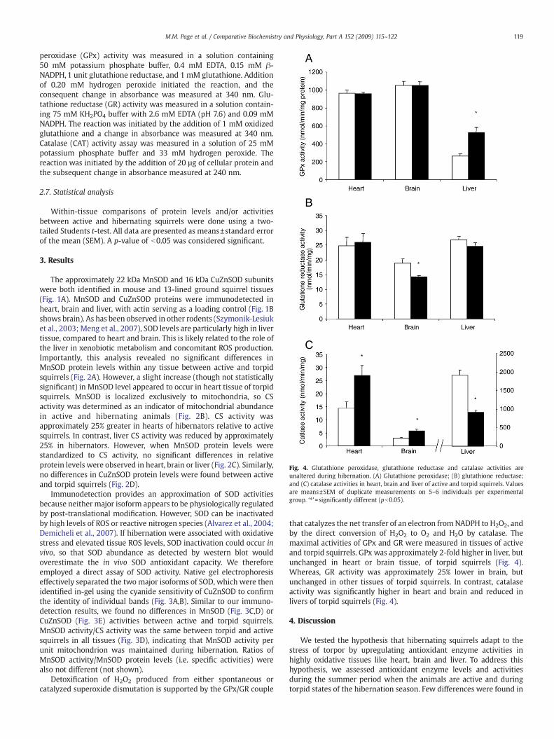
Fig. 4. Glutathione peroxidase, glutathione reductase and catalase activities areunaltered during hibernation. (A) Glutathione peroxidase; (B) glutathione reductase;and (C) catalase activities in heart, brain and liver of active and torpid squirrels. Valuesare means±SEM of duplicate measurements on 5–6 individuals per experimentalgroup. ‘⁎’=significantly different (pb0.05).
119M.M. Page et al. / Comparative Biochemistry and Physiology, Part A 152 (2009) 115–122
peroxidase (GPx) activity was measured in a solution containing50 mM potassium phosphate buffer, 0.4 mM EDTA, 0.15 mM β-NADPH, 1 unit glutathione reductase, and 1 mM glutathione. Additionof 0.20 mM hydrogen peroxide initiated the reaction, and theconsequent change in absorbance was measured at 340 nm. Glu-tathione reductase (GR) activity was measured in a solution contain-ing 75 mM KH2PO4 buffer with 2.6 mM EDTA (pH 7.6) and 0.09 mMNADPH. The reaction was initiated by the addition of 1 mM oxidizedglutathione and a change in absorbance was measured at 340 nm.Catalase (CAT) activity assay was measured in a solution of 25 mMpotassium phosphate buffer and 33 mM hydrogen peroxide. Thereaction was initiated by the addition of 20 µg of cellular protein andthe subsequent change in absorbance measured at 240 nm.
2.7. Statistical analysis
Within-tissue comparisons of protein levels and/or activitiesbetween active and hibernating squirrels were done using a two-tailed Students t-test. All data are presented as means±standard errorof the mean (SEM). A p-value of b0.05 was considered significant.
3. Results
The approximately 22 kDa MnSOD and 16 kDa CuZnSOD subunitswere both identified in mouse and 13-lined ground squirrel tissues(Fig. 1A). MnSOD and CuZnSOD proteins were immunodetected inheart, brain and liver, with actin serving as a loading control (Fig. 1Bshows brain). As has been observed in other rodents (Szymonik-Lesiuket al., 2003; Meng et al., 2007), SOD levels are particularly high in livertissue, compared to heart and brain. This is likely related to the role ofthe liver in xenobiotic metabolism and concomitant ROS production.Importantly, this analysis revealed no significant differences inMnSOD protein levels within any tissue between active and torpidsquirrels (Fig. 2A). However, a slight increase (though not statisticallysignificant) in MnSOD level appeared to occur in heart tissue of torpidsquirrels. MnSOD is localized exclusively to mitochondria, so CSactivity was determined as an indicator of mitochondrial abundancein active and hibernating animals (Fig. 2B). CS activity wasapproximately 25% greater in hearts of hibernators relative to activesquirrels. In contrast, liver CS activity was reduced by approximately25% in hibernators. However, when MnSOD protein levels werestandardized to CS activity, no significant differences in relativeprotein levels were observed in heart, brain or liver (Fig. 2C). Similarly,no differences in CuZnSOD protein levels were found between activeand torpid squirrels (Fig. 2D).
Immunodetection provides an approximation of SOD activitiesbecause neither major isoform appears to be physiologically regulatedby post-translational modification. However, SOD can be inactivatedby high levels of ROS or reactive nitrogen species (Alvarez et al., 2004;Demicheli et al., 2007). If hibernation were associated with oxidativestress and elevated tissue ROS levels, SOD inactivation could occur invivo, so that SOD abundance as detected by western blot wouldoverestimate the in vivo SOD antioxidant capacity. We thereforeemployed a direct assay of SOD activity. Native gel electrophoresiseffectively separated the two major isoforms of SOD, which were thenidentified in-gel using the cyanide sensitivity of CuZnSOD to confirmthe identity of individual bands (Fig. 3A,B). Similar to our immuno-detection results, we found no differences in MnSOD (Fig. 3C,D) orCuZnSOD (Fig. 3E) activities between active and torpid squirrels.MnSOD activity/CS activity was the same between torpid and activesquirrels in all tissues (Fig. 3D), indicating that MnSOD activity perunit mitochondrion was maintained during hibernation. Ratios ofMnSOD activity/MnSOD protein levels (i.e. specific activities) werealso not different (not shown).
Detoxification of H2O2 produced from either spontaneous orcatalyzed superoxide dismutation is supported by the GPx/GR couple
that catalyzes the net transfer of an electron fromNADPH to H2O2, andby the direct conversion of H2O2 to O2 and H2O by catalase. Themaximal activities of GPx and GR were measured in tissues of activeand torpid squirrels. GPx was approximately 2-fold higher in liver, butunchanged in heart or brain tissue, of torpid squirrels (Fig. 4).Whereas, GR activity was approximately 25% lower in brain, butunchanged in other tissues of torpid squirrels. In contrast, catalaseactivity was significantly higher in heart and brain and reduced inlivers of torpid squirrels (Fig. 4).
4. Discussion
We tested the hypothesis that hibernating squirrels adapt to thestress of torpor by upregulating antioxidant enzyme activities inhighly oxidative tissues like heart, brain and liver. To address thishypothesis, we assessed antioxidant enzyme levels and activitiesduring the summer period when the animals are active and duringtorpid states of the hibernation season. Few differences were found in

120 M.M. Page et al. / Comparative Biochemistry and Physiology, Part A 152 (2009) 115–122
the antioxidant enzymes measured between these two states,suggesting that no adaptive upregulation of antioxidant enzymes issustained in the 13-lined ground squirrel during hibernation. Theseresults contrast with previous reports of hibernation-induced effectson SOD activities in the ground squirrel Citellus citellus (Petrovic et al.,1986; Buzadzic et al., 1997). Petrovic et al. (1986) reported increasedCuZnSOD activity in brain and brown adipose tissue (BAT), and lowerMnSOD activity in liver and brain during hibernation. In contrast,Buzadzic et al. (1997) reported slightly lower total SOD activity inbrain of hibernating versus active C. citellus sampled in spring.Recently, Morin et al. (2008) reported that CuZnSOD levels in heartincrease by up to 50% during entry into hibernation, but by three daysin torpor have fallen to the levels equal to those in active 13-linedground squirrels. During arousal from this first period of torpor,CuZnSOD levels are reduced still further, to levels below those ofactive squirrels (Morin et al., 2008). The disparity between resultsfrom these different studies may thus be related to when during thehibernation season tissues are sampled. However, our data, fromtorpid animals that had been hibernating for several months, indicatethat, if upregulation of CuZnSOD or other antioxidant enzymes occursearly in the hibernation season, it is not maintained. Thus, squirrelsarousing from a full season of hibernation, during which I-R stresswould be expected to be high, would not have enhanced antioxidativecapacities in the major oxidative tissues.
It is important to note that both quantitative immunodetection andactivity assays reported here provided essentially identical results. Asneither SOD isoform is thought to be physiologically regulated post-translationally, a good agreement between the two methods ofquantification is expected. In addition, standardizing to CS activityallows the more meaningful value of MnSOD level/activity per unitmitochondrion to be obtained. The marginal increase in heart MnSODactivity observed in hibernators (Fig. 2) was thus apparently due to aslightly higher mitochondrial content (CS activity), and not anincrease in MnSOD per unit mitochondrion. Increased MnSODactivities reported previously in, for example, C. citellus BAT (Buzadzicet al., 1990) may have reflected the proliferation of mitochondria inthis thermogenic tissue during cold exposure (Milner et al., 1986;Barger et al., 2006).
Various reports have suggested that intracellular antioxidantactivities might increase during the period of arousal from torpor(reviewed in Carey et al., 2003). Though this appears to be the case forblood-borne small molecule antioxidants, which may be importedinto cells (see below), it is unlikely to include the intracellularantioxidant enzymes measured here. MnSOD and CuZnSOD areprimarily transcriptionally regulated, and increasing enzyme copynumber during arousal would be impractical. Profound changes inmetabolic rate occur within the first h of arousal in 13-lined groundsquirrels (Carey et al., 2003; Muleme et al., 2006), when bodytemperature is low and protein synthesis in brain (Frerichs et al., 1998)and liver (Van Breukelen and Martin, 2001) is severely suppressed.Therefore, it is unlikely that a rapid and transient upregulation ofenzyme during this period of stress would be a practicable strategy. Inrats, transcriptional upregulation of MnSOD in response to oxidativestress is not observed until six hours (Clerch et al., 1996) or more (Hoet al., 1996) post-stress. In addition hypoxia, which may occur duringarousal (Ma et al., 2005), is actually associated with a down-regulationof MnSOD levels in rodent cells (Russell et al., 1995). It therefore seemslikely that any burst of mitochondrial ROS production during arousalfrom torpor would already have occurred by the time sufficientMnSOD or CuZnSOD could be produced to counter it.
Although we found no evidence of enhanced intracellular SODactivities in torpid squirrels, it is known that various antioxidantsaccumulate in plasma during hibernation and/or arousal. For example,extracellular SOD (ecSOD) (Okamoto et al., 2006; Akita et al., 2007)and catalase (Ohta et al., 2006) accumulate in plasma of thehibernating Syrian hamster (Mesocricetus auratus). Also, both the 13-
lined ground squirrel (Drew et al., 1999) and the Arctic ground squirrel(S. parryii) (Drew et al., 1999; Tøien et al., 2001) accumulate ascorbatein plasma during hibernation, to levels several-fold above those inactive animals. Osborne and Hashimoto, (2007) showed in Syrianhamsters that ascorbate in brain extracellular fluid is oxidized duringarousal from torpor. Thus, increases both in oxidative stress andresistance to this stress appear to occur in the extracellularcompartment during arousal. This may be as, or more, importantthan intracellular oxidative stress, and it is important to make thedistinction between intracellular and extracellular oxidative stress.However, it is also possible that some extracellular small-moleculeantioxidants move rapidly into cells during arousal. For example, oneroute of ascorbate import is via glucose transporters (Liang et al.,2001). A rapid increase in glucose oxidation occurs during arousal(reviewed in Carey et al., 2003), suggesting increased transporterabundance within cell membranes, which might facilitate a rapidimport of ascorbate to combat oxidative stress within cells. Thus, smallmolecule and/or extracellular antioxidants may play an important roleduring the torpor-arousal transition, and these may provide oxidativestress resistance in the absence of increases in SODs or otherintracellular antioxidant enzymes.
Only relatively subtle changes in GPx and GR were observed intorpid squirrels, and these were confined to liver and brain,respectively. The almost 2-fold increase in liver GPx activity measuredhere was similar to that reported in C. citellus (Buzadzic et al., 1990).However, this was a tissue-specific phenomenon, with no similarincrease in brain or heart tissue, suggesting no role for GPx incountering oxidative stress in brain tissue.
Also similar to Buzadzic et al. (1990), we observed a significantdecrease in liver catalase activity during hibernation. Liver catalaseactivity also decreases during hibernation in the sub-desert rodentJerboa (Jaculus orientalis) compared to summer active individuals(Kabine et al., 2003). This could be an effect of fasting, as liver catalaseactivity decreases with fasting in laboratory rats and pigs (Slauteret al., 1984; Cheon et al., 2005). In contrast to the observations for livertissue, catalase activity was higher in both brain and heart tissue oftorpid squirrels. Peroxisomes are an important site of lipid oxidation(Wanders, 2004), and torpid ground squirrels are highly reliant on themetabolism of lipid fuels (Carey et al., 2003). Fasting in mice inducesperoxisome proliferator-activated receptor gamma coactivator-1(PGC-1α) in heart tissue (Lehman et al., 2000), suggesting an increasein peroxisomal number also occurs. Thus, it is difficult to interpret thechanges in catalase activity associated with hibernation in 13-linedground squirrels exclusively as pre-adaptations to potential oxidativestress, as peroxisomal proliferation in brain and heart tissue related toaltered energy metabolism may have occurred during hibernation.
Thus, taken together, our results do not support a major role foreither intracellular isoform of SOD, nor for GPx or GR in augmentingoxidative stress resistance of heart or brain tissue during hibernationin 13-lined ground squirrels. A role for catalase is possible; however, asmentioned above interpretation of the catalase results is impeded bythe possible role of peroxisomal proliferation associated with lipidmetabolism in hibernation. Additionally, other antioxidant enzymesor small molecule antioxidants may play more important roles. Forexample, a putative thioredoxin peroxidase has been identified to beupregulated during hibernation in heart tissue of 13-lined groundsquirrels (Morin and Storey, 2007) and bats (Myotis lucifugus) (Eddyet al., 2005).
The central thesis underlying the expectation that upregulation oftissue antioxidant enzymes occurs during hibernation has been thatthe profound shifts in tissue perfusion andmetabolic activity betweentorpor and arousal would create transiently hypoxic or anoxicconditions that might enhance mitochondrial ROS production duringboth the hypoxic event and following reperfusion. In this context, it isinteresting that liver (Lindell et al., 2005), intestinal (Fleck and Carey,2005; Kurtz et al., 2006) and brain (Dave et al., 2006) tissue all show

121M.M. Page et al. / Comparative Biochemistry and Physiology, Part A 152 (2009) 115–122
enhanced resistance to I-R injury during hibernation in squirrels.Similarly, Ma et al. (2005) demonstrate an absence of oxidativedamage in brain tissue of arousing S. parryii, though the level ofmalondialdehyde, a marker of oxidative lipid damage, is elevated two-fold in liver mitochondria isolated from hibernating 13-lined groundsquirrels compared to summer active controls (Gerson et al., 2008).Our data indicate for liver, brain and heart that a global upregulation ofintracellular antioxidant enzymes does not mediate this phenotype. Itwill be interesting to identify what other possible adaptive mechan-isms confer tissue stress resistance during hibernation.
Given that the levels/activities of intracellular antioxidants duringhibernation do not appear to change, one alternative explanation forthe apparent absence of oxidative stress and damage during thetorpor-arousal transition is that the I-R associated ROS productionthat occurs under these conditions in non-hibernatorsmay simply beavoided in hibernators, either by avoiding profound tissue hypoxia orby tolerating it without initiation of aberrant ROS production. In S.parryii, the torpid state is associated with both high arterial PO2 andlow hypoxia inducible factor-1α (HIF-1α) expression (Ma et al.,2005). Plasma hypoxemia occurs during arousal from torpor andtissue HIF-1α levels increase during this period, but only to levelsseen in active animals. Thus, evidence supporting the idea thatprofound tissue hypoxia occurs during torpor and/or arousal islimited. Even if transient hypoxia did occur in tissues of hibernators,it may not elicit the same burst of mitochondrial ROS that occurs innon-hibernators (Duranteau et al., 1998; Chandel et al., 1998). Inothermammals, mitochondrial superoxide radical production duringhypoxia appears to be mediated primarily by respiratory complex III(Guzy and Schumacker, 2006). Perhaps differences in complex IIIstructure and/or function in hibernators allow them to avoid this.However in daily torpor, a phenotype similar to hibernation, fluxthrough complex III is not significantly reduced in liver mitochon-drial isolated from torpid dwarf hamsters (Phodopus sungorus)compared with active, normothermic controls (Brown et al., 2007).Studies of effects of hypoxia on cultured squirrel cells may provideinsight into whether they respond similarly to those of rats to atransient hypoxia mimicking I-R.
Acknowledgements
This work was supported by an Early Researcher Award (JAS) andNatural Sciences and Engineering Research Council (NSERC) DiscoveryGrants awarded to JAS and JFS. MMP is supported by an OntarioGraduate Scholarship (OGS). CWP was supported by an NSERCUndergraduate Student Research Award. The assistance of AlvinIverson and the staff of the Carman Research Station was invaluable.We would also like to thank Alex Gerson, Jamie Drooker and JasonBrown for help in animal husbandry and tissue collection.
References
Akita, K., Hanaya, T., Arai, S., Ohta, T., Okamoto, I., Fukuda, S., 2007. Purification,identification, characterization, and cDNA cloning of a high molecular weightextracellular superoxide dismutase of hamster that transiently increases in plasmaduring arousal from hibernation. Comp. Biochem. Physiol. A. 146, 223–232.
Alvarez, B., Demicheli, V., Durán, R., Trujillo, M., Cerveñansky, C., Freeman, B.A., Radi, R.,2004. Inactivation of human CuZn superoxide dismutase by peroxynitrite andformation of histidinyl radical. Free Radic. Biol. Med. 37, 813–822.
Asimakis, G.K., Lick, S., Patterson, C., 2002. Postischemic recovery of contractile functionis impaired in SOD2(+/−) but not SOD1(+/−) mouse heart. Circulation.105, 981–986.
Barger, J.L., Barnes, B.M., Boyer, B.B., 2006. Regulation of UCP1 and UCP3 in arctic groundsquirrels and relationwith mitochondrial proton leak. J. Appl. Physiol. 101, 339–347.
Baur, J.A., Sinclair, D.A., 2006. Therapeutic potential of resveratrol: the in vivo evidence.Nat. Rev. Drug Discov. 5, 493–506.
Beauchamp, C., Fridovich, I., 1971. Superoxide dismutase: improved assays and an assayapplicable to acrylamide gels. Anal. Biochem. 44, 276–287.
Beyer Jr., W.F., Fridovich, I., 1987. Assaying for superoxide dismutase activity: some largeconsequences of minor changes in conditions. Anal. Biochem. 161, 559–566.
Blomgren, K., Hagberg, H., 2006. Free radicals, mitochondria, and hypoxia-ischemia inthe developing brain. Free Radic. Biol. Med. 40, 388–397.
Brown, J.C.L., Gerson, A.R., Staples, J.F., 2007. Mitochondrial metabolism during dailytorpor in the dwarf Siberian hamster: role of active regulated changes and passivethermal effects. Am. J. Physiol. 293, R1833–R1845.
Buzadzic, B., Spasic, M., Saicic, Z.S., Radojicic, R., Petrovic, V.M., Halliwell, B., 1990.Antioxidant defenses in the ground squirrel Citellus citellus. 2. The effect ofhibernation. Free Radic. Biol. Med. 9, 407–413.
Buzadzic, B., Blagojevic, D., Korac, B., Saicic, Z.S., Spasic, M.B., Petrovic, V.M., 1997.Seasonal variation in the antioxidant defense system of the brain of the groundsquirrel (Citellus citellus) and response to low temperature compared with rat.Comp. Biochem. Physiol. 117C, 141–149.
Cai, D., McCarron, R.M., Yu, E.Z., Li, Y., Hallenbeck, J., 2004. Akt phosphorylation andkinase activity are down-regulated during hibernation in the 13-lined groundsquirrel. Brain Res. 1014, 14–21.
Carey, H.V., Andrews, M.T., Martin, S.L., 2003. Mammalian hibernation: cellular andmolecular responses to depressed metabolism and low temperature. Physiol Rev.83, 1153–1181.
Chandel, N.S., Maltepe, E., Goldwasser, E., Mathieu, C.E., Simon, M.C., Schumacker, P.T.,1998. Mitochondrial reactive oxygen species trigger hypoxia-induced transcription.Proc. Natl. Acad. Sci. USA. 95, 11715–11720.
Chen, Z., Siu, B., Ho, Y.S., Vincent, R., Chua, C.C., Hamdy, R.C., Chua, B.H., 1998.Overexpression of MnSOD protects against myocardial ischemia/reperfusion injuryin transgenic mice. J. Mol. Cell Cardiol. 30, 2281–2289.
Cheon, Y., Nara, T.Y., Band, M.R., Beever, J.E., Wallig, M.A., Nakamura, M.T., 2005.Induction of overlapping genes by fasting and a peroxisome proliferator in pigs:evidence of functional PPARα in nonproliferating species. Am. J. Physiol. Regul.Integr. Comp. Physiol. 288, R1525–R1535.
Christian, S.L., Ross, A.P., Zhao, H.W., Kristenson, H.J., Zhan, X., Rasley, B.T., Bickler, P.E.,Drew, K.L., 2008. Arctic ground squirrel (Spermophilus parryii) hippocampalneurons tolerate prolonged oxygen-glucose deprivation and maintain baselineERK1/2 and JNK activation despite drastic ATP loss. J. Cereb. Blood Flow Metab. 28,1307–1319.
Clerch, B.L., Wright, A., Chung, D.J., Massaro, D., 1996. Early divergent lung antioxidantenzyme expression in response to lipopolysaccharide. Am. J. Physiol. 271, L949–L954.
Dave, K.R., Prado, R., Raval, A.P., Drew, K.L., Perez-Pinzon, M.A., 2006. The Arctic groundsquirrel brain is resistant to injury from cardiac arrest during euthermia. Stroke. 37,1261–1265.
Demicheli, V., Quijano, C., Alvarez, B., Radi, R., 2007. Inactivation and nitration of humansuperoxide dismutase (SOD) by fluxes of nitric oxide and superoxide. Free Radic.Biol. Med. 42, 1359–1368.
Drew, K.L., Osborne, P.G., Frerichs, K.U., Hu, Y., Koren, R.E., Hallenbeck, J.M., Rice, M.E.,1999. Ascorbate and glutathione regulation in hibernating ground squirrels. BrainRes. 851, 1–8.
Duranteau, J., Chandel, N.S., Kulisz, A., Shao, Z., Schumacker, P.T., 1998. Intracellularsignaling by reactive oxygen species during hypoxia in cardiomyocytes. J. Biol.Chem. 273, 11619–11624.
Eddy, S.F., McNally, J.D., Storey, K.B., 2005. Up-regulation of a thioredoxin peroxidase-like protein, proliferation-associated gene, in hibernating bats. Arch. Biochem.Biophys. 435, 103–111.
Fleck, C.C., Carey, H.V., 2005. Modulation of apoptotic pathways in intestinal mucosaduring hibernation. Am. J. Physiol. Regul. Integr. Comp. Physiol. 289, R586–R595.
Frerichs, K.U., Smith, C.B., Brenner, M., DeGracia, D.J., Krause, G.S., Marrone, L., Dever, T.E.,Hallenbeck, J.M., 1998. Suppression of protein synthesis in brain during hibernationinvolves inhibition of protein initiation and elongation. Proc. Natl. Acad. Sci. USA 95,14511–14516.
Gerson, A.R., Brown, J.C.L., Thomas, R., Bernards, M.A., Staples, J.F., 2008. Effects ofdietary polyunsaturated fatty acids on mitochondrial metabolism in mammalianhibernation. J. Exp. Biol. 211, 2689–2699.
Guzy, R.D., Schumacker, P.T., 2006. Oxygen sensing by mitochondria at complex III: theparadox of increased reactive oxygen species during hypoxia. Exp. Physiol. 91,807–819.
Hermes-Lima, M., Storey, K.B., 1995. Antioxidant defenses andmetabolic depression in apulmonate land snail. Am. J. Physiol. 268, R1386–R1393.
Hermes-Lima, M., Zenteno-Savin, T., 2002. Animal response to drastic changes inoxygen availability and physiological oxidative stress. Comp. Biochem. Physiol. C.133, 537–556.
Ho, Y.-S., Dey, M.S., Crapo, J.D., 1996. Antioxidant enzyme expression in rat lungs duringhyperoxia. Am. J. Physiol. 270, L810–L818.
Honda, Y., Honda, S., 2002. Oxidative stress and life span determination in the nematodeCaenorhabditis elegans. Ann. NY Acad. Sci. 959, 466–474.
Kabine, M., Clémencet, M.-C., Bride, J., El Kebbaj, M.S., Latruffe, N., Cherkaoui-Malki, M.,2003. Changes of peroxisomal fatty acid metabolism during cold acclimatization inhibernating jerboa (Jaculus orientalis). Biochimie 85, 707–714.
Kim, G.W., Kondo, T., Noshita, N., Chan, P.H., 2002. Manganese superoxide dismutasedeficiency exacerbates cerebral infarction after focal cerebral ischemia/reperfusionin mice: implications of the production and role of superoxide radicals. Stroke. 33,809–815.
Kurtz, C.C., Lindell, S.L., Mangino, M.J., Carey, H.V., 2006. Hibernation confers resistanceto intestinal ischemia-reperfusion injury. Am. J. Physiol. 291, G895–G901.
Lehman, J.J., Barger, P.M., Kovacs, A., Saffitz, J.E., Medeiros, D.M., Kelly, D.P., 2000.Peroxisome proliferator–activated receptor γ coactivator-1 promotes cardiacmitochondrial biogenesis. J. Clin. Invest. 106, 847–856.
Liang, W.-J., Johnson, D., Jarvis, S.M., 2001. Vitamin C transport systems of mammaliancells. Mol. Memb. Biol. 18, 87–95.
Lindell, S.L., Klahn, S.L., Piazza, T.M., Mangino, M.J., Torrealba, J.R., Southard, J.H., Carey,H.V., 2005. Natural resistance to liver cold ischemia-reperfusion injury associatedwith the hibernation phenotype. Am. J. Physiol. 288, G473–G480.

122 M.M. Page et al. / Comparative Biochemistry and Physiology, Part A 152 (2009) 115–122
Ma, L.Y., Zhu, X., Rivera, P.M., Tøien, Ø., Barnes, B.M., LaManna, J.C., Smith,M.A., Drew, K.L.,2005. Absence of cellular stress in brain after hypoxia induced by arousal fromhibernation in Arctic ground squirrels. Am. J. Physiol. 289, R1297–R1306.
Meng, Q., Wong, Y.T., Chen, J., Ruan, R., 2007. Age-related changes in mitochondrialfunction and antioxidative enzyme activity in fischer 344 rats. Mech. Ageing. Dev.128, 286–292.
Milner, R.E., Wang, L.C.H., Trayhurn, P., 1986. Brown fat thermogenesis duringhibernation and arousal in Richardson's ground squirrel. Am. J. Physiol. 256,R42–R48.
Morin Jr., P., Storey, K.B., 2007. Antioxidant defense in hibernation: cloning andexpression of peroxiredoxins from hibernating ground squirrels, Spermophilustridecemlineatus. Arch. Biochem. Biophys. 461, 59–65.
Morin Jr., P., Ni, Z., McMullen, D.C., Storey, K.B., 2008. Expression of Nrf2 and itsdownstream gene targets in hibernating 13-lined ground squirrels, Spermophilustridecemlineatus. Mol. Cell Biochem. 312, 121–129.
Muleme, M., Walpole, A.C., Staples, J.F., 2006. Mitochondrial metabolism in hibernation:metabolic suppression, temperature effects, and substrate preferences. Phyiol.Biochem. Zool. 79, 474–483.
Murakami, K., Kondo, T., Kawasr, M., Li, Y., Sato, S., Chen, S.F., Chan, P.H., 1998.Mitochondrial susceptibility to oxidative stress exacerbates cerebral infarction thatfollows permanent focal cerebral ischemia in mutant mice with manganesesuperoxide dismutase deficiency. J. Neurosci. 18, 205–213.
Ohta, H., Okamoto, I., Hanaya, T., Arai, S., Ohta, T., Fukuda, S., 2006. Enhancedantioxidant defense due to extracellular catalase activity in Syrian hamster duringarousal from hibernation. Comp. Biochem. Physiol. C 143, 484–491.
Okamoto, I., Kayano, T., Hanaya, T., Arai, S., Ikeda, M., Kurimoto, M., 2006. Up-regulationof an extracellular superoxide dismutase-like activity in hibernating hamsterssubjected to oxidative stress in mid- to late arousal from torpor. Comp. Biochem.Physiol. C 144, 47–56.
Osborne, P.G., Hashimoto, M., 2007. Brain ECF antioxidant interactions in hamstersduring arousal from hibernation. Behav. Brain Res. 178, 115–122.
Petrovic, V.M., Milii, B., Spasic, M., Sai, Z., 1986. Copper-zinc containing and manganesecontaining superoxide dismutase in the ground squirrel Citellus citellus— the effectof hibernation. Free Rad. Res. 1, 339–346.
Phillips, J.P., Parkes, T.L., Hilliker, A.J., 2000. Targeted neuronal gene expression andlongevity in Drosophila. Exp. Gerontol. 35, 1157–1164.
Robb, E.L., Page, M.M., Wiens, B.E., Stuart, J.A., 2008a. Molecular mechanisms ofoxidative stress resistance induced by resveratrol: specific and progressiveinduction of MnSOD. Biochem. Biophys. Res. Comm. 367, 406–412.
Robb, E.L., Winkelmolen, L., Visanji, N., Brotchie, J., Stuart, J.A., 2008b. Dietaryresveratrol administration increases MnSOD expression and activity in mousebrain. Biochem. Biophys. Res. Commun. 372, 254–259.
Russell, W.J., Ho, Y.-S., Parish, G., Jackson, R.M., 1995. Effects of hypoxia on MnSODexpression in mouse lung. Am. J. Physiol. 269, L221–L226.
Silva, J.P., Shabalina, I.G., Dufour, E., Petrovic, N., Backlund, E.C., Hultenby, K., Wibom, R.,Nedergaard, J., Cannon, B., Larsson, N.G., 2005. SOD2 overexpression: enhancedmitochondrial tolerancebut absence of effectonUCP2 activity. EMBO J. 24, 4061–4070.
Slauter, R.W., Yamazak, R.K., 1984. Glucagon and fasting do not activate peroxisomalfatty acid β-oxidation in rat liver. Arch. Biochem. Biophys. 233, 197–202.
Solaini, G., Harris, D.A., 2005. Biochemical dysfunction in heart mitochondria exposed toischaemia and reperfusion. Biochem. J. 390, 377–394.
Spitz, D.R., Oberley, L.W., 1989. An assay for superoxide dismutase activity inmammalian tissue homogenates. Anal. Biochem. 179, 8–18.
Szymonik-Lesiuk, S., Czechowska, G., Stryjecka-Zimmer, M., Slomka, M., Madro, A.,Celinski, K., Wielosz, M., 2003. Catalase, superoxide dismutase, and glutathioneperoxidase activities in various rat tissues after carbon tetrachloride intoxication.J Hepatobilliary. Pancreat. Surg. 10, 309–315.
Tøien, Ø., Drew, K.L., Chao, M.L., Rice, M.F., 2001. Ascorbate dynamics and oxygenconsumption during arousal from hibernation in Arctic ground squirrels. Am. J.Physiol. 281, R572–R583.
Van Breukelen, F., Martin, S., 2001. Translational initiation is uncoupled from elongationat 18 °C during mammalian hibernation. Am. J. Physiol. 281, R1379.
Vaughn, D.K., Gruber, A.R., Michalski, M.L., Seidling, J., Schlink, S., 2006. Capture, care,and captive breeding of 13-lined ground squirrels, Spermophilus tridecemlineatus.Lab. Animal. 35, 33–40.
Wanders, R.J.A., 2004. Peroxisomes, lipid metabolism, and peroxisomal disorders. Mol.Genet. Met. 83, 16–27.
Warner, D.S., Sheng, H., Batinic-Haberle, I., 2004. Oxidants, antioxidants and theischemic brain. J. Exp. Biol. 207, 3221–3231.
Yan, L., Vatner, D.E., O, Connor, J.P., Ivessa, A., Ge, H., Chen, W., Hirotani, S., Ishikawa, Y.,Sadoshima, J., Vatner, S.F., 2007. Type 5 adenylyl cyclase disruption increaseslongevity and protects against stress. Cell 130, 247–258.