Death in the octopus' garden: fatal blue-lined octopus envenomations of adult green sea turtles
7
Mar Biol DOI 10.1007/s00227-011-1846-9 123 ORIGINAL PAPER Death in the octopus’ garden: fatal blue-lined octopus envenomations of adult green sea turtles Kathy A. Townsend · Jens Altvater · Michael C. Thomas · Qamar A. Schuyler · GeoVrey W. Nette Received: 27 July 2011 / Accepted: 18 November 2011 © The Author(s) 2011. This article is published with open access at Springerlink.com Abstract The blue-lined octopus Hapalochlaena fasciata contains the powerful neuromuscular blocker tetrodotoxin (TTX), which causes muscle weakness and respiratory fail- ure. H. fasciata is regarded as one of the most venomous marine animals in the world, and multiple human fatalities have been attributed to the octopus. To date, there have been no recorded incidents of an envenomation of a wild animal. Here, we present a newly developed, multi-stage tandem mass spectrometry technique that provides unequivocal evidence for two cases of envenomation of two »110 kg herbivorous green sea turtles by two tiny cryptic blue-lined octopuses (»4 cm body length). These cases of accidental ingestion provide evidence for the Wrst time of the antipredator eVect of TTX and highlight a previ- ously unconsidered threat to turtles grazing within seagrass beds. Introduction Twenty thousand sea turtles are estimated to reside in Mor- eton Bay, oV Brisbane in Queensland, Australia (Limpus et al. 1994). Moreton Bay’s expansive seagrass beds are an important feeding ground for the endangered green turtle (Chelonia mydas) (Limpus et al. 1994; Brand-Gardner et al. 1999; Arthur et al. 2008). The seagrass beds also provide important habitat for many marine creatures, including juve- nile Wsh, crustaceans, molluscs, and cephalopods (Weng 1990; Davie 1998). One of the species of cephalopods shel- tering within these seagrass beds is the venomous blue-lined octopus, Hapalochlaena fasciata (Norman and Reid 2000). This visually cryptic animal hides among tide pools, blend- ing in with its surroundings via pigmented chromatophore organs. When threatened, the animal Xashes bright blue rings and lines, which act as a warning to potential predators (Tranter and Augustin 1973; Mathger et al. 2008). The saliva of H. fasciata contains tetrodotoxin (TTX), a powerful neurotoxin that it uses to incapacitate prey such as large Wsh and crustaceans. It has been hypothesized that TTX is also used for protection from predators, although to date there has been no empirical evidence to support this assumption (Tranter and Augustin 1973; Yotsu-Yamashita et al. 2007). TTX is a potent neuromuscular blocker that causes muscle weakness and respiratory failure and it is known as one of the most toxic venoms to mammals. TTX has been shown to have a wholly lethal dose (LD) of 5.8 g/kg via intramuscular injection (Xu et al. 2003), and it is estimated that a tiny 25 g octopus possesses enough venom to fatally paralyze ten 75 kg humans (Narahashi et al. 1967; Sutherland 1983; Williamson 1987). While there are high levels of TTX in many of the tissues of H. fasciata, including their arms and mantel (see Yotsu- Yamashita et al. 2007; Williams and Caldwell 2009; Communicated by R. Lewison. Electronic supplementary material The online version of this article (doi:10.1007/s00227-011-1846-9) contains supplementary material, which is available to authorized users. K. A. Townsend (&) · Q. A. Schuyler School of Biological Sciences, The University of Queensland, Moreton Bay Research Station, P.O. Box 138, Dunwich, QLD 4183, Australia e-mail: [email protected] J. Altvater · M. C. Thomas · G. W. Nette Independent Marine Biochemistry Research, Moreton Bay Research Station, P.O. Box 138, Dunwich, QLD 4183, Australia
-
Upload
independent -
Category
Documents
-
view
0 -
download
0
Transcript of Death in the octopus' garden: fatal blue-lined octopus envenomations of adult green sea turtles

Mar Biol
DOI 10.1007/s00227-011-1846-9ORIGINAL PAPER
Death in the octopus’ garden: fatal blue-lined octopus envenomations of adult green sea turtles
Kathy A. Townsend · Jens Altvater · Michael C. Thomas · Qamar A. Schuyler · GeoVrey W. Nette
Received: 27 July 2011 / Accepted: 18 November 2011© The Author(s) 2011. This article is published with open access at Springerlink.com
Abstract The blue-lined octopus Hapalochlaena fasciatacontains the powerful neuromuscular blocker tetrodotoxin(TTX), which causes muscle weakness and respiratory fail-ure. H. fasciata is regarded as one of the most venomousmarine animals in the world, and multiple human fatalitieshave been attributed to the octopus. To date, there havebeen no recorded incidents of an envenomation of a wildanimal. Here, we present a newly developed, multi-stagetandem mass spectrometry technique that providesunequivocal evidence for two cases of envenomation oftwo »110 kg herbivorous green sea turtles by two tinycryptic blue-lined octopuses (»4 cm body length). Thesecases of accidental ingestion provide evidence for the Wrsttime of the antipredator eVect of TTX and highlight a previ-ously unconsidered threat to turtles grazing within seagrassbeds.
Introduction
Twenty thousand sea turtles are estimated to reside in Mor-eton Bay, oV Brisbane in Queensland, Australia (Limpuset al. 1994). Moreton Bay’s expansive seagrass beds are animportant feeding ground for the endangered green turtle(Chelonia mydas) (Limpus et al. 1994; Brand-Gardner et al.1999; Arthur et al. 2008). The seagrass beds also provideimportant habitat for many marine creatures, including juve-nile Wsh, crustaceans, molluscs, and cephalopods (Weng1990; Davie 1998). One of the species of cephalopods shel-tering within these seagrass beds is the venomous blue-linedoctopus, Hapalochlaena fasciata (Norman and Reid 2000).This visually cryptic animal hides among tide pools, blend-ing in with its surroundings via pigmented chromatophoreorgans. When threatened, the animal Xashes bright bluerings and lines, which act as a warning to potential predators(Tranter and Augustin 1973; Mathger et al. 2008).
The saliva of H. fasciata contains tetrodotoxin (TTX), apowerful neurotoxin that it uses to incapacitate prey such aslarge Wsh and crustaceans. It has been hypothesized thatTTX is also used for protection from predators, although todate there has been no empirical evidence to support thisassumption (Tranter and Augustin 1973; Yotsu-Yamashitaet al. 2007). TTX is a potent neuromuscular blocker thatcauses muscle weakness and respiratory failure and it isknown as one of the most toxic venoms to mammals. TTXhas been shown to have a wholly lethal dose (LD) of5.8 �g/kg via intramuscular injection (Xu et al. 2003), andit is estimated that a tiny 25 g octopus possesses enoughvenom to fatally paralyze ten 75 kg humans (Narahashiet al. 1967; Sutherland 1983; Williamson 1987). Whilethere are high levels of TTX in many of the tissues ofH. fasciata, including their arms and mantel (see Yotsu-Yamashita et al. 2007; Williams and Caldwell 2009;
Communicated by R. Lewison.
Electronic supplementary material The online version of this article (doi:10.1007/s00227-011-1846-9) contains supplementary material, which is available to authorized users.
K. A. Townsend (&) · Q. A. SchuylerSchool of Biological Sciences, The University of Queensland, Moreton Bay Research Station, P.O. Box 138, Dunwich, QLD 4183, Australiae-mail: [email protected]
J. Altvater · M. C. Thomas · G. W. NetteIndependent Marine Biochemistry Research, Moreton Bay Research Station, P.O. Box 138, Dunwich, QLD 4183, Australia
123
Mar Biol
Williams et al. 2011), there is currently no empirical evi-dence that the octopuses can exude TTX from these tissues.Instead, envenomation is thought to occur through subcuta-neous injection via a bite from its small, parrot-like beak(Edmonds 1969; Yotsu-Yamashita et al. 2007). Many casestudies have described Hapalochlaena sp. envenomationsin human beings (Edmonds 1969; Walker 1983; Williamson
1987; Edmonds 1989; Hodgson 1997; Cavazzoni et al.2008), but to date, there have been no recorded incidents ofan envenomation of a wild marine animal. This studypresents the Wrst morphological and biochemical evidenceof mortality caused by the envenomation of two fullygrown, sexually mature green turtles (Chelonia mydas)by two blue-lined octopuses (Hapalochlaena fasciata).
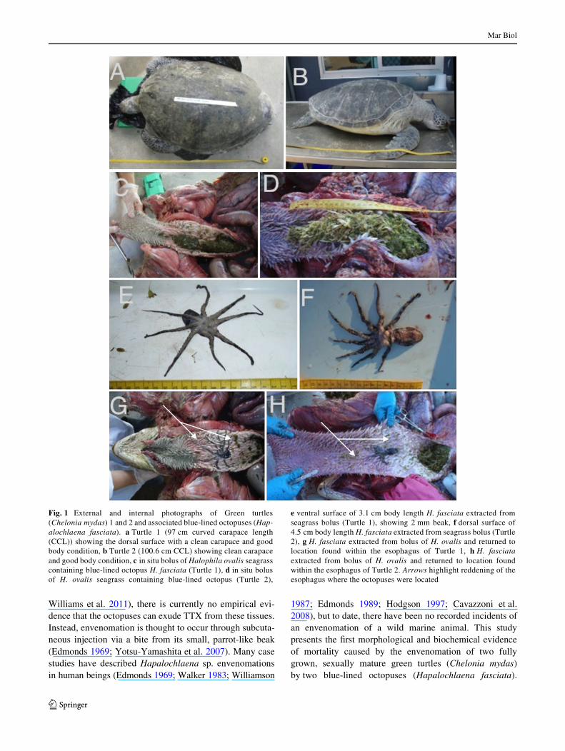
Fig. 1 External and internal photographs of Green turtles(Chelonia mydas) 1 and 2 and associated blue-lined octopuses (Hap-alochlaena fasciata). a Turtle 1 (97 cm curved carapace length(CCL)) showing the dorsal surface with a clean carapace and goodbody condition, b Turtle 2 (100.6 cm CCL) showing clean carapaceand good body condition, c in situ bolus of Halophila ovalis seagrasscontaining blue-lined octopus H. fasciata (Turtle 1), d in situ bolusof H. ovalis seagrass containing blue-lined octopus (Turtle 2),
e ventral surface of 3.1 cm body length H. fasciata extracted fromseagrass bolus (Turtle 1), showing 2 mm beak, f dorsal surface of4.5 cm body length H. fasciata extracted from seagrass bolus (Turtle2), g H. fasciata extracted from bolus of H. ovalis and returned tolocation found within the esophagus of Turtle 1, h H. fasciataextracted from bolus of H. ovalis and returned to location foundwithin the esophagus of Turtle 2. Arrows highlight reddening of theesophagus where the octopuses were located
123

Mar Biol
Complimentary to this report is the development of a newmethodology utilizing multi-stage tandem mass spectrome-try, which has allowed unequivocal determination of thepresence of the TTX toxin within the samples tested.
Methods and materials
As part of a larger study investigating turtle mortalitywithin Moreton Bay, two large green turtles (Cheloniamydas) were found dead; Turtle 1 on a sand bank(S27o21�50� E153o25�6�) on October 11, 2008, and Turtle 2on North Stradbroke Island (S27o24�13� E153o26�12�) onSeptember 15, 2010, a distance of 4.7 km apart (and nearly2 years). The animals were brought to Moreton BayResearch Station and necropsied via gross anatomicalinvestigation. External and internal observations wererecorded and photographed.
During the necropsy, samples were collected of the foodbolus, liver, kidney, and muscle of the C. mydas specimens 1and 2, and the enXamed tissue associated with the octopuswithin the esophagus in Turtle 2. Samples were stored frozenat ¡20°C. The entire H. fasciata organism from eachC. mydas was removed from the esophagus and maintained in90% ethanol (Turtle 1) or placed in ¡80°C freezer (Turtle 2).
The salivary glands of the H. fasciata specimens weresubsequently extracted and all samples listed above (plusthe liver from a third turtle that did not die from TTX) wereanalyzed for the presence of TTX using a unique multiple-stage mass spectrometry technique developed speciWcallyfor this study. This powerful technique utilizing HPLC–MSwith hydrophilic interaction chromatography was chosenabove single-stage mass spectrometry (SSMS). DuringSSMS, many other substances may elute oV the column atthe same retention time as TTX, making a negative controlnecessary (Matsumura 1995, 2001; Williams et al. 2004,2011). To highlight the strength of this technique, weincluded a negative control, although it was unnecessary as
the technique provides unequivocal identiWcation of TTX.While a summary of the procedure is below, a full descrip-tion of the technique can be found in Online Resource 1.
Preliminary experiments on the identiWcation of TTXcentered on the detection of the expected [M + H]+ ion atm/z 320 by LC–MS and monitoring the m/z 320–302 tran-sition by LC–MS/MS. Both methods lead to the detectionof a range of isobaric ions complicating the identiWcation ofTTX. The m/z 320–302 transition results from the neutralloss of water from the TTX [M + H] + ion. The neutral lossof water is a common fragmentation process for many ionsand is therefore not speciWc to TTX.
The fragmentation of the TTX [M + H]+ ion was studiedusing MSn by ion trap mass spectrometry. MS2 and MS3
resulted primarily in the neutral loss of additional waterfrom the precursor ions. Using MS4, however, abundantfragment ions were observed at m/z 162, 200, and 254which were characteristic of the TTX [M + H]+ ion. The m/z200 fragment ion in the MS4 spectrum was used in the iden-tiWcation of TTX since it was the most abundant of thesethree fragment ions.
Results
The two large dead green sea turtles (Chelonia mydas)appeared outwardly healthy, with no life threatening exter-nal injuries (Fig. 1a, b). Internal observations of both turtlesrevealed a large, healthy muscle mass (Table 1) and anextensive fat layer throughout the entire body, indicatingdeath was sudden. In each case, the circulatory and urogen-ital systems presented normal. Water was found within thebronchi, and bright red, frothy oxygenated blood observedin both lungs. On investigation of the gastrointestinal tract(GI), large quantities of seagrass (Halophila ovalis) werefound within the esophagus and stomach of each animal(Fig. 1c, d), while the rest of the GI was Wlled with progres-sively digested food through to the rectum.
Table 1 Morphological measurements of the two sexually mature female green turtles (Chelonia mydas) and blue-lined octopus(Hapalochlaena fasciata) involved in this study
Mass estimates are based on the mass-size function for female green turtles published by Limpus et al. (1994)
Linear regression used log wt (kg) = 2.9297(log CCL (cm)) ¡ 3.7929 (r2 = 0.986, DF = 1174, p < 0.0005) from Limpus et al. (1994)
Turtle I (KAT110920080086) Turtle II (QAS150920100007)
Minimum over-curve carapace length (CCL) (cm) 97.0 100.6
Head width (cm) 11.3 12.97
Tail length (cm) 20.8 21.0
Plastron length (cm) 76.6 82.0
Pectoral muscle depth (cm) R = 2.2, L = 1.4 R = 2.3, L = 2.2
Calculated mass (kg)* 106.6 118.6
Body length of blue-lined octopus (cm) 3.1 4.5
Body plus arm length of blue-lined octopus (cm) 10.9 12.2
123

Mar Biol
Both turtles exhibited the same symptoms. A closerinspection of the contents of the esophagus revealed a blue-lined octopus (Hapalochlaena fasciata) encased within theseagrass bolus in each of the two turtles (Fig. 1e, f). Mor-phometric measurements of the turtles and the octopusesare outlined in Table 1. The mouthparts of both blue-linedoctopuses were approximately 2 mm in diameter (Fig. 1e).The esophageal tissue directly surrounding the octopus wasred and inXamed in both cases (Fig. 1g, h), suggesting thatthese were the sites of envenomation, although no obviousbite marks could be found among the keratinized papillae.
Through comparison to the TTX standard (Fig. 2a), MS4
fragment ions were observed at m/z 162, 200, and 254 inthe salivary glands of the octopus (Fig. 2b) and tissues ofboth turtles (Fig. 2c, d), which were characteristic of theTTX [M + H]+ ion (Fig. 2 insert).
MS4 m/z 200.2 fragmentation chromatogram for theTTX standard indicated that the distinctive peak was foundbetween 8 and 12 min (9.7 min) (Fig. 3a). For ease of dis-play, we narrowed the remaining chromatograms to thisregion (Fig. 3b). Note also the diVerent scales on the y-axisfor each sub-Wgure, which is a reXection of the relativeabundance of the TTX in each sample. Distinctive peakswere found in the turtle muscle, esophagus, and kidney(Fig. 3c–e), and in the salivary gland of the octopus(Fig. 3f). All of these had a signal to noise ratios (SN) ofabove 3. However, TTX was below detectable levels in thefood bolus and the liver tissues of the Turtle 2 and 3(Fig. 3g–i). While peaks appear to be present around the 9.8and 9.6 min mark of Fig. 3g, h, respectively, these were atsuch low concentrations, and missing distinctive ion signa-tures that they are considered to be background noise. Thiswas further conWrmed by the analysis of the SN, for thesetissues were at not detectable levels. A summary of positiveTTX detections from both case studies can be found inTable 2.
Discussion
Green turtles (C. mydas) are herbivorous, feeding mainlyon seagrass, macroalgae, and mangrove fruits (Brand-Gardneret al. 1999; Arthur et al. 2008) while supplementing theirdiet with gelatinous organisms (Arthur et al. 2007). Thespecies feeds while submerged and can breath hold for upto 3 h while foraging, before returning to the surface tobreathe (Lutz and Bentley 1985; Arthur et al. 2007). Of thesix species of seagrass found within the sheltered waters ofMoreton Bay, Halophila ovalis is the preferred food sourcefor C. mydas due to its nutritional qualities (Brand-Gardneret al. 1999). Submerged meadows of H. ovalis are verycommon on the sand banks surrounding the area where theturtles in this study were found (Zharikov et al. 2005; Phinn
et al. 2008). Large quantities of H. ovalis throughout thegastrointestinal tracts of the sea turtles indicated that theanimals had had long-term access to a high-quality foodsource and were feeding within the sheltered waters ofMoreton Bay prior to their death. As the octopuses in bothinstances were completely encased within a seagrass bolus(Fig. 1c, d), it is proposed that the ingestion was accidental,with the turtles consuming the visually cryptic animals dur-ing the grazing process.
While there was a distinct reddening of the esophagus inthe area in which the octopuses were located, (Fig. 1g, h), no
Fig. 2 MS4 fragment spectrum of (from top to bottom) a TTX Stan-dard and TTX molecule structure (insert top right); b Blue-lined octo-pus salivary gland; c Turtle 2 esophagus and d Turtle 1 muscle tissuesshowing peaks consistent with the presence of TTX
123

Mar Biol
Fig. 3 a Extracted ion chromatogram (m/z = 200.2) of TTX standardacross the full 20 min run. The remaining chromatograms are highlightedbetween 8 and 12 min, b TTX standard, c muscle (Turtle 2), d esophagus(Turtle 2), e kidney (Turtle 2), f octopus salivary gland (found in Turtle 2esophagus), g food bolus (Turtle 2), h liver (Turtle 2), and i negative con-
trol (liver from Turtle 3) and their associated signal to noise ratios (SN).SN values less than 3 are considered to be noise and indicate that the TTXis below detectable levels, while n/a indicates undetectable levels. Notealso the diVerent scales on the y-axis and NL values for each sub Wgure,which are a reXection of the relative abundance of the TTX in each sample
A
B
G
F
C
D
E
H
I
8 9 10 11 12Time (min)
0
2000
4000
6000
8000
10000
12000
Inte
nsity
9.2 NL:1.36E4Blue-linedOctopus SalivaryGland
8 9 10 11 12
Time (min)
0
2
4
6
8
10
Inte
nsity
9.99.8
10.3 11.710.6
NL:1.07E1Turtle 2Muscle
8 9 10 11 12
Time (min)
0.00
0.05
0.10
0.15
Inte
nsity
9.8
11.0
10.39.58.5
NL:1.88E-1Turtle 2Food Bolus
8 9 10 11 12
Time (min)
0
1
2
3
4
5
Inte
nsity
9.3
9.7 10.2 10.8
NL:5.97Turtle 2Oesophagus
8 9 10 11 12
Time (min)
0.00
0.20
0.40
0.60
0.80
Inte
nsity
9.6
11.110.7
9.9
NL:8.17E-1Turtle 2Liver
8 9 10 11 12
Time (min)
0
2
4
6
8
Inte
nsity
10.0
10.6
NL:8.40Turtle 2Kidney
8 9 10 11 12
Time (min)
0
0.2
0.4
0.6
0.8
1.0
Inte
nsity
NL:0Turtle 3Livernegative Control
8 9 10 11 12Time (min)
0
50
100
150
200
Inte
nsity
9.7
NL:2.21E2TTX Standard
0 2 4 6 8 10 12 14 16 18Time (min)
0
50
100
150
200
Inte
nsity
9.7 NL:2.21E2TTX Standard
123

Mar Biol
distinct bite marks could be found. Other studies haveshown that the bite leaves little mechanical or pharmacolog-ical skin trace locally, although reddening and swelling ofthe envenomation site is common (Edmonds 1969, 1989;Williamson 1987; Cavazzoni et al. 2008). The lack of dis-tinct puncture marks may also be an indication that the octo-pus may not have bitten the inner tissue of the esophagus;instead, toxins may have been leached from the dermis ofH. fasciata, as proposed by Yotsu-Yamashita et al. (2007).However, while TTX has been detected in high levels inmultiple regions of the octopus body (Yotsu-Yamashitaet al. 2007; Williams and Caldwell 2009; Williams et al.2011), it has been shown that the consumption of tissue isWfty times less toxic relative to intraperitoneal injection (Xuet al. 2003). The lack of detectable TTX in the seagrassbolus in which the octopus was fully encased further indi-cates that the TTX detected in the turtle tissues came fromthe octopus via active envenomation, not exudation.
There is no anti-venom currently available for TTX(Anonomous 2003, Xu et al. 2003); however, human casestudies have shown that patients that are ventilated mechan-ically often recover (Edmonds 1969; Walker 1983; Wil-liamson 1987). There are two proposed mechanisms for theremoval of TTX from the body. Unlike protein-based neu-rotoxins, it is thought that TTX is not broken down and isexcreted unchanged by vertebrates (Kao 1966). However,others have hypothesized that the toxin is broken down inthe body, presumably via the liver—although there is cur-rently no empirical evidence that this is the case (Edmonds1969; Walker 1983; Williamson 1987). This hypothesizedrole of the liver in degrading TTX is supported by the lackof detectable levels of the toxin within both liver samplesfrom this study (Table 2).
TTX disrupts sodium channels causing nerve conduc-tion failure, particularly in the somatic motor nerves (Nara-hash et al. 1966, 1967). Human case studies have shownthat Xaccid muscle paralysis occurs soon after bite, with thepatients remaining conscious even after paralysis hasoccurred. Dose-dependent respiratory weakness or failureoccurs spontaneously some time later (Williamson 1987;Edmonds 1989; Cavazzoni et al. 2008). Necropsy evidence
from the cases in this study indicates that similar symptomsmay have aVected the turtles. Saltwater drowning is diag-nosed in reptiles by a bloody froth in the airways, whileXuid, sometimes together with plant and other material, islikely to be present in the upper alimentary tract (Cooper2008). Water in the bronchi and bright red, frothy oxygen-ated blood were found within the lungs of both turtles, indi-cating that the animals were in the water when the biteoccurred, and subsequently drowned. The large amount offresh seagrass in the esophagus and stomach suggests thatthey were most probably feeding. The subsequent Xaccidparalysis likely disabled their ability to swim and lift theirheads out of the water to breathe, causing the animals totake water into the lungs.
This is the Wrst evidence of the antipredator function ofTTX. While omnivorous green turtle hatchlings have beenobserved attempting to feed on small, non-venomous octo-puses, Octopus bocki (Caldwell 2005), we propose thatthese are two cases of healthy, adult herbivorous green seaturtles (C. mydas) accidentally consuming cryptic blue-lined octopuses (H. fasciata) while feeding on the seagrassmeadows in which the octopus shelters. These accidentalingestions resulted in lethal TTX envenomations by theblue-lined octopuses, delivered actively through bitingthe esophageal tissue. The TTX subsequently immobilizedthe turtle, preventing it from raising its head above thewater to breathe, Wlling its lungs with water and resulting ina toxin-induced drowning. As these two animals weresourced from 100 necropsied turtles, death by blue-linedoctopus envenomation may be a previously unconsideredthreat to grazing within seagrass beds.
Acknowledgments This project was supported by the GoldringEmerging Marine Scientist Fellowship, Earthwatch Institute of Austra-lia, Underwater World Mooloolaba, Quandamooka Land and SeaCouncil, The Goodman Foundation, and Moreton Bay Marine ParkAuthority. Thanks also go to T. Gallagher, Z. Allen, C. Mueller,E. Lewis, D. Burns and to the staV at Moreton Bay Research Station.
Open Access This article is distributed under the terms of the Crea-tive Commons Attribution Noncommercial License which permits anynoncommercial use, distribution, and reproduction in any medium,provided the original author(s) and source are credited.
Table 2 Presence and absence of TTX from samples sourced from stranded green sea turtles and associated blue-lined octopus
(–) TTX undetectable, (+) TTX detected, # not analyzed, n.a. not applicable
Animal Tissue sampled
Liver Muscle Kidney Esophagus Salivary gland Food bolus
Turtle I – + + # n.a. –
Turtle II – + + + n.a. #
Blue-lined octopus I # # # # + n.a.
Blue-lined octopus II # # # # + n.a.
123

Mar Biol
References
Anonomous (2003) Australian Venom Research Unit—blue-ringedoctopus. The University of Melbourne, Melbourne. http://www.avru.org/health/health_bluering.html. Accessed 9 Nov 2011
Arthur KE, O’Neil JM, Limpus CJ, Abernathy K, Marshall G (2007)Using animal-borne imaging to assess green turtle (Cheloniamydas) foraging ecology in Moreton Bay, Australia. MarineTechnol Soc J 41:9–13
Arthur KE, Boyle MC, Limpus CJ (2008) Ontogenetic changes in dietand habitat use in green sea turtle (Chelonia mydas) life history.Marine Ecol Prog Ser 362:303–311
Brand-Gardner SJ, Lanyon JM, Limpus CJ (1999) Diet selection byimmature green turtles, Chelonia mydas, in subtropical MoretonBay, south-east Queensland. Aust J Zool 47:181–191
Caldwell RL (2005) An observation of inking behavior protectingadult Octopus bocki from predation by green turtle (Chelonia my-das) hatchlings. Pac Sci 59:69–72
Cavazzoni E, Lister B, Sargent P, Schibler A (2008) Blue-ringed octo-pus (Hapalochlaena sp.) envenomation of a 4-year-old boy:a case report. Clin Toxicol 46:760–761
Cooper JE (2008) Methods in herpetological forensic work—post-mortem techniques. Appl Herpetol 5:351–370
Davie P (1998) Wild guide to moreton bay. Queensland Museum, Bris-bane, p 408
Edmonds C (1969) A non-fatal case of blue-ringed octopus bite. MedJ Aust 2:601
Edmonds C (1989) Case report blue-ringed octopus bite DangerousMarine Creatures. Reed Books, Frenchs Forest, pp 100–101
Hodgson WC (1997) Pharmacological action of Australian animalvenoms. Clin Exp Pharmacol Physiol 24:10–17
Kao CY (1966) Tetrodotoxin, saxitoxin, and their signiWcance in thestudy of excitation phenomena. Pharmacol Rev 18:997–1049
Limpus CJ, Couper PJ, Read MA (1994) The green turtle, Cheloniamydas, in Queensland: population structure in a warm temperatefeeding area. Mem Qld Museum 35:139–154
Lutz PL, Bentley TB (1985) Respiratory physiology of diving in thesea turtle. Copeia 3:671–679
Mathger LM, Denton EJ, Marshall NJ, Hanlon RT (2008) Mechanismsand behavioural functions of structural coloration in cephalopodsconference on Iridescence—more than meets the eye, Tempe,AZ, pp S149–S163
Matsumura K (1995) Re-examination of tetrodotoxin production bybacteria. Appl Environ Microbiol 61:3468–3470
Matsumura K (2001) No ability to produce tetrodotoxin in bacteria.Appl Environ Microbiol 67:2393–2394
Narahash T, Moore JW, Poston RN (1966) SpeciWc action of tetrodo-toxin on nerves. Science 154:425
Narahashi T, Moore JW, Poston RN (1967) Tetrodotoxin derivatives:chemical structure and blockage of nerve membrane conductance.Science 156:976–979
Norman M, Reid A (2000) A guide to squid, cuttleWsh and octopusesof Australia. CSIRO Publishing, Moorabbin
Phinn S, Roelfsema C, Dekker A, Brando V, Anstee J (2008) Mappingseagrass species, cover and biomass in shallow waters: An assess-ment of satellite multi-spectral and airborne hyper-spectral imag-ing systems in Moreton Bay (Australia). Remote Sens Environ112:3413–3425
Sutherland SK (1983) Australian animal toxins: the creature, their tox-ins and the care of the poisoned patient. Oxford University Press,Melbourne
Tranter DJ, Augustin O (1973) Observations on life-history of blue-ringed octopus Hapalochlaena-maculosa. Mar Biol 18:115–128
Walker DG (1983) Survival after sever envenomation by the blue-ringedoctopus (Hapalochlaena maculosa). Med J Aust 2:663–665
Weng HT (1990) Fish in Shallow Areas in Moreton Bay, Queenslandand factors aVecting their distribution. Estuar Coast Shelf Sci30:569–578
Williams BL, Caldwell RL (2009) Intra-organismal distribution oftetrodotoxin in two species of blue-ringed octopuses (Hap-alochlaena fasciata and H. lunulata). Toxicon 54:345–353
Williams BL, Brodie ED Jr, Brodie ED III (2004) Chemical ecology ofa resistant predator and its toxic prey: distribution of tetrodotoxinin the common garter snake after ingestion of the rough-skinnednewt. J Chem Ecol 30:1901–1919
Williams BL, Lovenburg V, HuVard CL, Caldwell RL (2011) Chemi-cal defense in pelagic octopus paralarvae: Tetrodotoxin alonedoes not protect individual paralarvae of the greater blue-ringedoctopus (Hapalochlaena lunulata) from common reef predators.Chemoecology 21:131–141
Williamson JAH (1987) The blue-ringed octopus bite and envenoma-tion syndrome. Clin Dermatol 5:127–133
Xu Q, Huang K, Gao L, Zhang H, Rong K (2003) Toxicity of tetrodotoxintowards mice and rabbits. Wei sheng yan jiu. J Hyg Res 32:371–374
Yotsu-Yamashita M, Mebs D, Flachsenberger W (2007) Distributionof tetrodotoxin in the body of the blue-ringed octopus (Hap-alochlaena maculosa). Toxicon 49:410–412
Zharikov Y, Skilleter GA, Loneragan NR, Taranto T, Cameron BE(2005) Mapping and characterising subtropical estuarine land-scapes using aerial photography and GIS for potential applicationin wildlife conservation and management. Biol Conserv 125:87–100
123