The interplay between non-verbal and verbal interaction in ...
Upload
independentCategory
view
3download
0
doi:10.1182/blood-2007-09-110569Prepublished online February 12, 2008;2008 111: 4500-4510
Saulius Sumanas, Gustavo Gomez, Yan Zhao, Changwon Park, Kyunghee Choi and Shuo Lin and myeloid cell formationInterplay among Etsrp/ER71, Scl, and Alk8 signaling controls endothelial
http://bloodjournal.hematologylibrary.org/content/111/9/4500.full.htmlUpdated information and services can be found at:
(3205 articles)Hematopoiesis and Stem Cells �Articles on similar topics can be found in the following Blood collections
http://bloodjournal.hematologylibrary.org/site/misc/rights.xhtml#repub_requestsInformation about reproducing this article in parts or in its entirety may be found online at:
http://bloodjournal.hematologylibrary.org/site/misc/rights.xhtml#reprintsInformation about ordering reprints may be found online at:
http://bloodjournal.hematologylibrary.org/site/subscriptions/index.xhtmlInformation about subscriptions and ASH membership may be found online at:
Copyright 2011 by The American Society of Hematology; all rights reserved.Washington DC 20036.by the American Society of Hematology, 2021 L St, NW, Suite 900, Blood (print ISSN 0006-4971, online ISSN 1528-0020), is published weekly
For personal use only. by guest on February 17, 2014. bloodjournal.hematologylibrary.orgFrom For personal use only. by guest on February 17, 2014. bloodjournal.hematologylibrary.orgFrom
HEMATOPOIESIS AND STEM CELLS
Interplay among Etsrp/ER71, Scl, and Alk8 signaling controls endothelial andmyeloid cell formationSaulius Sumanas,1,2 Gustavo Gomez,1 Yan Zhao,1 Changwon Park,3 Kyunghee Choi,3,4 and Shuo Lin1
1Department of Molecular, Cell and Developmental Biology, University of California, Los Angeles; 2Division of Developmental Biology, Cincinnati Children’sHospital Medical Center, OH; and 3Department of Pathology and Immunology and 4Molecular Cell Biology Program, Washington University School of Medicine,St Louis, MO
Vascular endothelial and myeloid cellshave been proposed to originate from acommon precursor cell, the hemangio-blast. The mechanism of endothelial andmyeloid cell specification and differentia-tion is poorly understood. We have previ-ously described the endothelial-specificzebrafish Ets1-related protein (Etsrp),which was both necessary and sufficientto initiate vasculogenesis in the zebrafishembryos. Here we identify human Etv2/
ER71 and mouse ER71 proteins as func-tional orthologs of Etsrp. Overexpressionof mouse ER71 and Etsrp caused strongexpansion of hemangioblast and vascu-lar endothelial lineages in a zebrafishembryo. In addition, we show that etsrp isalso required for the formation of myeloidbut not erythroid cells. In the absence ofetsrp function, the number of granulo-cytes and macrophages is greatly re-duced. Etsrp overexpression causes
expansion of both myeloid and vascularendothelial lineages. Analysis of mosaicembryos indicates that etsrp functionscell autonomously in inducing myeloidlineage. We further demonstrate that thechoice of endothelial versus myeloid fatedepends on a combinatorial effect ofetsrp, scl, and alk8 genes. (Blood. 2008;111:4500-4510)
© 2008 by The American Society of Hematology
Introduction
Hematopoiesis, the differentiation of hematopoietic stem cells intovarious blood cell lineages, and vasculogenesis, the differentiation ofendothelial cell progenitors into vascular endothelial cells, are closelyrelated in many different organisms. Endothelial and hematopoietic cellsemerge in the embryo in close proximity and time, suggesting thepossibility of a common progenitor, the hemangioblast.1 In the extraem-bryonic visceral yolk sac of a mouse embryo, vascular plexus and bloodislands are closely associated with each other and share expression ofmultiple genes such as flk1, flt1, tie1, tie2, and others.2 Differentiatingembryonic stem cells contain blast colony-forming cells which candifferentiate into both hematopoietic and endothelial cells in vitro.3 Inthe zebrafish, which recently emerged as an excellent model system forthe studies of vertebrate hematopoiesis and vasculogenesis/angiogen-esis, both primitive hematopoietic and endothelial progenitors originatewithin lateral plate mesoderm, later forming an intermediate cell massregion, equivalent to the mouse blood islands.4,5 The zebrafish clochemutants are deficient in both vascular endothelial and hematopoieticlineages.6 Zebrafish vascular endothelial and hematopoietic cell progeni-tors also share expression of common markers such as a basichelix-loop-helix transcription factor scl/tal17,8 and an ETS domaintranscription factor fli1.9 Overexpression of scl can induce both hemato-poietic and endothelial markers7,8,10 whereas knockdown of scl results indefective hematopoietic and endothelial development.11,12 Single-celllabeling in a zebrafish gastrula embryo demonstrated that individualcells can give rise to both erythroid and endothelial cells,13 providing adefinitive evidence for the existence of hemangioblasts in vivo.
In the zebrafish, the anterior lateral plate mesoderm gives rise tothe progenitors of myeloid cells and head vessels while theposterior lateral plate mesoderm gives rise to the primitive
erythroid cells, axial, and intersegmental blood vessels.14 Tworecent studies have demonstrated that the interplay between anearly myeloid-specific transcription factor pu.1/spi1 and anerythroid-specific gata1 determines the choice between myeloidand erythroid fates in the progenitor cells.15,16 Pu.1 is firstexpressed in both myeloid and erythroid cell progenitors, and laterits erythroid-specific expression is repressed by gata1, ensuringthat pu.1 expression remains restricted to the myeloid cells withinthe anterior lateral mesoderm. Scl signaling is critical for myeloiddevelopment, as scl-deficient embryos lack pu.1-expressing cells.12
BMP receptor Alk8 signaling functions in parallel to scl, alsoproviding an instructive input for myelopoiesis.17 However, themechanism of transition from the putative hemangioblast to themyeloid and endothelial lineages is still unknown. Although arecent study demonstrated the existence of the common erythroid-endothelial progenitor cells,13 myeloid-endothelial progenitor cellshave not been analyzed in this study. Therefore, the definitiveevidence for the existence of a common myeloid-endothelialhemangioblast progenitor cell is still missing.
We have recently described a novel ets1-related protein, etsrp.18
Etsrp expression is localized to the presumptive vascular progeni-tor cells from the early somitogenesis stages. Knockdown of etsrpresults in the complete lack of circulation. Angioblasts in etsrp-deficient embryos do not differentiate, fail to migrate towardmidline, and do not express other vascular markers. Scl expressionis strongly down-regulated in the anterior lateral mesoderm inetsrp-morpholino (MO)–injected embryos (morphants) while itsposterior erythroid-specific expression is not affected. Consistentwith this, erythroid lineage is not affected in etsrp morphants.
Submitted September 5, 2007; accepted February 4, 2008. Prepublished online asBlood First Edition paper, February 12, 2008; DOI 10.1182/blood-2007-09-110569.
The online version of this article contains a data supplement.
The publication costs of this article were defrayed in part by page chargepayment. Therefore, and solely to indicate this fact, this article is herebymarked ‘‘advertisement’’ in accordance with 18 USC section 1734.
© 2008 by The American Society of Hematology
4500 BLOOD, 1 MAY 2008 � VOLUME 111, NUMBER 9
For personal use only. by guest on February 17, 2014. bloodjournal.hematologylibrary.orgFrom
Overexpression of etsrp RNA is sufficient to induce expression ofendothelial and hemangioblast markers, including scl, in manydifferent regions of an embryo. These data show that etsrp is bothnecessary and sufficient for the initiation of vasculogenesis in azebrafish embryo. Although this study clearly established etsrp as acritical regulator of zebrafish vasculogenesis, it was not clearwhether etsrp function is conserved in other vertebrates, includingmammals.
In the current study, we demonstrate by the phylogeny analysisthat zebrafish etsrp is related to the mouse ER71 and humanetv2/ER71 genes. We show that mouse ER71 is functionally relatedto Etsrp by performing overexpression of both proteins in zebrafishembryos. We also further investigate etsrp function within theputative hemangioblast cells. We show that etsrp is both necessaryand sufficient to initiate the myeloid cell formation, in addition tothe endothelial lineage. We demonstrate that etsrp function isspecific to the anterior but not the posterior hemangioblasts, thusproviding the first distinction between the 2 pools. We also showthat as the endothelial and myeloid lineages separate, etsrp isexcluded from the myeloid lineage, and remains restricted to theendothelial cell precursors. Finally, we show that etsrp-expressingcells can give rise to both myeloid and endothelial lineages,supporting the existence of a common myeloid-endothelial cellprogenitor, the hemangioblast.
Methods
Microinjection of MOs
In all experiments, unless otherwise noted, 8 ng to 10 ng etsrp-specific MO1and MO2 mixture (4-5 ng each) was injected at the 1- to 2-cell stage.18
Typically, 2 independent experiments were performed, with at least 15 to 20embryos per experiment analyzed. Penetrance of observed phenotypes wasgreater than 90%, unless otherwise noted. Other morpholinos used: 1.2 ngalk8 MO: ACAACTCCTCAAGTGACTCTCAGCG (Open Biosystems,Huntsville, AL)19; scl MO: GCTCGGATTTCAGTTTTTCCATCAT (OpenBiosystems)18; 8 ng pu.1 MO: GATATACTGATACTCCATTGGTGGT15
(kind gift of J.P. Kanki, Dana-Farber Cancer Institute, Boston, MA); 7 nggata1-ATG MO: CTGCAAGTGTAGTATTGAAGATGTC16 (kindly do-nated by L. Zon, Harvard Medical School, Boston, MA).
Overexpression and epistasis experiments
etsrp mRNA (75-150 pg) was injected into the zebrafish embryos at the 1-to 8-cell stages.18 Approximately 100 pg scl mRNA8 and 10 pg CA-alk8mRNA19(kindly donated by M. Hammerschmidt, Max Planck Institute forImmunobiology, Freiburg, Germany) was used in the overexpression andepistasis experiments. etsrp DNA microinjection construct was made bysubcloning full-length etsrp cDNA into the HindIII site of pXeX.20 Circularetsrp-XeX plasmid (25-75 pg) was injected into the blastomere at the 1-cellstage. hER71 coding sequence was cloned into pCMV-SPORT6 vector(Open Biosystems). hER71 DNA (75-100 pg) was injected into theflk1-GFP zebrafish embryos at the 1-cell stage. mER71 coding sequence hasbeen subcloned into pcDNA3.1/V5-His vector (Invitrogen, Carlsbad, CA).mER71 DNA (75 pg) was injected in overexpression experiments. ForhEts1 RNA injections, hEts1 ORF (kindly provided by P. Oettgen, BethIsrael Deaconess Medical Center, Boston, MA) has been subcloned into theSpeI site of pT3TS,21 subsequently linearized with BamHI and transcribedwith T3 mMessage Machine kit (Ambion, Austin, TX). For hEts1 DNAinjections, 200 pg hEts1 cDNA in pSG5neo vector (Stratagene, La Jolla,CA; kindly provided by R. Forough, Texas A&M University, CollegeStation, TX) was injected.
In situ hybridization
In situ hybridization was performed as described.22 For the 2-colorconventional in situ hybridization, the same protocol was followed exceptthat embryos were incubated with 2 probes, etsrp-labeled with fluorescein-UTP (Roche, Indianapolis, IN), and the other cDNA labeled with digoxygenin-UTP (Roche). Following the first color (blue) development, embryos werefixed in the 4% paraformaldehyde/phosphate-buffered saline (PBS) solu-tion for 1 hour, washed in PBS with 2% bovine serum albumin and 2%Tween 20 (PBT),22 incubated with anti–fluorescein antibody conjugatedwith alkaline phosphatase (Roche) overnight at 4°C, washed in PBT, anddeveloped using 5-Bromo-4-chloro-3-indolyl �-D-galactopyranoside (X-gal; Sigma-Aldrich, St Louis, MO) and iodonitrotetrazolium chloride (INT;Sigma-Aldrich) as the substrate for red color. To detect fluorescein-dextranand pu.1 localization, a fluorescent 2-color in situ protocol was followed (J.Schoeneback, B. Keegan, and D. Yelon, unpublished). Briefly, fixedembryos were hybridized with DIG-labeled pu.1 probe at 65°C, washed in0.2� saline-sodium citrate (SSC), blocked in 1� blocking reagent (Roche),incubated with 1:500 anti-DIG POD (Roche), washed in PBT, incubatedwith DNP-tyramide at 1:50 (Perkin Elmer, Waltham, MA), washed in PBT,blocked in 1� blocking reagent, incubated with anti-DNP POD at 1:500(Perkin Elmer), washed in PBT, incubated with Cy3-tyramide at 1:25(Perkin Elmer), sequentially washed in PBT, 1% H2O2, PBT, 0.1 M pH 2.2glycine, PBT, blocked in 1� blocking reagent, incubated with anti-FITCPOD at 1:1000 (Roche), washed in PBT, incubated with FITC-tyramide(Perkin Elmer), washed in PBT, and imaged as described below in “Imageprocessing and analysis.” The following probes were used: etsrp23; pu.124;gata125; flk126; scl8; mpx27 (made by subcloning mpx cDNA into the pCR4vector; Invitrogen; kindly provided by J. Larson, University of Minnesota,Minneapolis/St Paul); lcp128 (made by subcloning lcp1 cDNA into thepCR4 vector; kindly provided by J. Larson).
Transplantation
Donor embryos (wt or flk1-GFP homozygous) were injected with a mixtureof etsrp RNA (100 pg) and fluorescein isothiocyanate-dextran (2 ng;molecular weight [Mw] 2 MDa; Sigma-Aldrich) or tetramethyl rhodamineisothiocyanate-dextran (2 ng; Mw 2 MDa; Sigma-Aldrich) into theblastomere at the 1-cell stage. Recipient embryos (wt or flk1-GFP) wereinjected with 7.5 ng to 8 ng etsrp MO1 and MO2 1:1 mixture at the 1- to2-cell stage. Embryos were transferred to Danieau solution and manuallydechorionated prior to transplantation. Ten to 50 cells were transplanted atthe sphere to 30% epiboly stages by using capillary needles and adjustingbalance pressure of PLI-100 microinjector (Harvard Apparatus, Holliston,MA) to move cells up and down the needle. Embryos were fixed at the 8- to10-somite stages and analyzed for pu.1 expression and the presence forfluorescein. Two-color conventional or fluorescent in situ hybridizationprotocol was followed to detect DIG-labeled pu.1 and fluorescein (onlypu.1 probe was used).
Image processing and analysis
Tetramethylrhodamine isothiocyanate (TRITC)–labeled live flk1-GFP em-bryos or FITC-labeled pu.1-Cy3 hybridized embryos were imaged using amonochrome CCD camera (Hamamatsu C4742-95) on a compound micro-scope (Axioplan; Zeiss, Thornwood, NY) equipped with rhodamine andgreen fluorescent protein (GFP) filter sets. To image stained embryos afterin situ, they were either positioned in 1� PBS on the agarose-coated dishes,or they were manually deyolked and mounted in 50% glycerol (2-color insitu) or araldite (single-color). Images were taken using a color digital CCDcamera (Axiocam; Zeiss) and a dissecting (Stemi SV11; Zeiss) or acompound microscope (Axioskop 2; Zeiss) with �5, �10, or �20objective. Typically, frames in different focus planes were manuallycombined using Adobe Photoshop to yield the maximum clarity image.During image analysis of transplanted embryos, color levels, hue, andsaturation were adjusted for the whole image. Color of the transplanted cellswas analyzed using the eyedropper tool in Photoshop, averaged from 7 to10 samplings, and normalized for the intensity in red channel. Thefollowing color ratios were calculated for the cells 1 to 5 (Figure 6H)
ETSRP REGULATES ENDOTHELIAL AND MYELOID LINEAGES 4501BLOOD, 1 MAY 2008 � VOLUME 111, NUMBER 9
For personal use only. by guest on February 17, 2014. bloodjournal.hematologylibrary.orgFrom
channels red/green/blue: (cell 1) 1 : 1.02 � 0.03 : 1.28 � 0.12; (cell 2)1 : 0.74 � 0.05 : 0.56 � .05; (cell 3) 1 : 0.54 � 0.10 : 0.59 � 0.11; (cell 4)1 : 0.77 � 0.05 : 0.93 � 0.09; (cell 5) 1 : 0.89 � 0.04 : 1.03 � 0.05. Theratios were used to select representative colors shown in the rectangles inFigure 6H.
Results
mER71 and hEtv2/ER71 proteins are functional orthologs ofzebrafish Etsrp
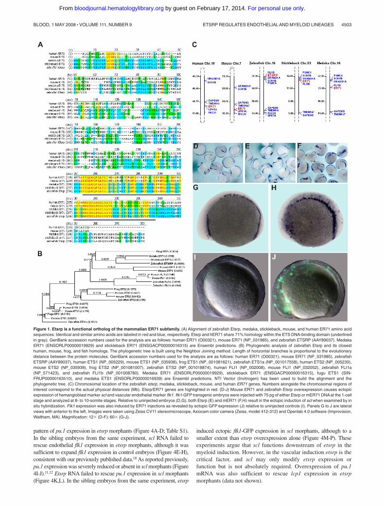
The previous study suggested that zebrafish Etsrp may be evolution-ary related to mammalian Ets1 proteins.18 However, the homologybetween Etsrp and Ets1 proteins is limited to the ETS DNA bindingdomain. Other domains such as Transactivation and Pointeddomains, present in other Ets1 family members, are not recogniz-able within the Etsrp sequence. To identify a potential Etsrportholog in other vertebrates, we performed phylogenetic analysisof the sequences and protein structure of different Ets familymembers. ER71 proteins from different vertebrates, includingmedaka (Oryzias latipes), stickleback (Gasterosteus aculeatus),frog (Xenopus laevis), mouse, and humans appeared as potentialorthologs of Etsrp (Figure 1). Mouse ER71 was originally isolatedas a testis-specific factor 1,29,30 but its embryonic expression hasnot been previously investigated. Etsrp sequence is 57% and 61%,respectively, identical to its medaka and stickleback homologs,while it displays a lower identity of 26% (37% similarity) to thehuman ER71 protein and 26% identity (36% similarity) to themouse ER71 (Figure 1A). Etsrp and hER71 proteins are 71%similar within the ETS domain region, whereas homology is muchlower within the rest of the sequence. Interestingly, similar to Etsrp,other vertebrate ER71 proteins have no recognizable structuraldomains outside the ETS domain. Recent functional studiesdetermined a potent transactivation domain present within themouse ER71 N-terminal region.30 Syntenic analysis shows thatvertebrate etsrp/ER71 chromosomal regions are highly conserved(Figure 1C).
To test if mammalian ER71 and zebrafish Etsrp proteins arefunctionally conserved, we injected DNA encoding mouse andhuman ER71 into early zebrafish embryos. Overexpression of bothmER71 and hER71 resulted in the strong expansion of hemangio-blast marker scl and vascular endothelial flk1-GFP expression,similar to the effect of etsrp expression (Figure 1D-J; data notshown). On the other hand, overexpression of related human Ets1DNA and RNA caused no apparent effect on early development andno increase in flk1-GFP expression (data not shown). These resultsargue that ER71 proteins, similar to Etsrp, can initiate vasculogen-esis in the zebrafish embryos, therefore Etsrp and ER71 but notEts1 proteins are functional orthologs.
Etsrp is necessary and sufficient for the formation ofmyeloid cells
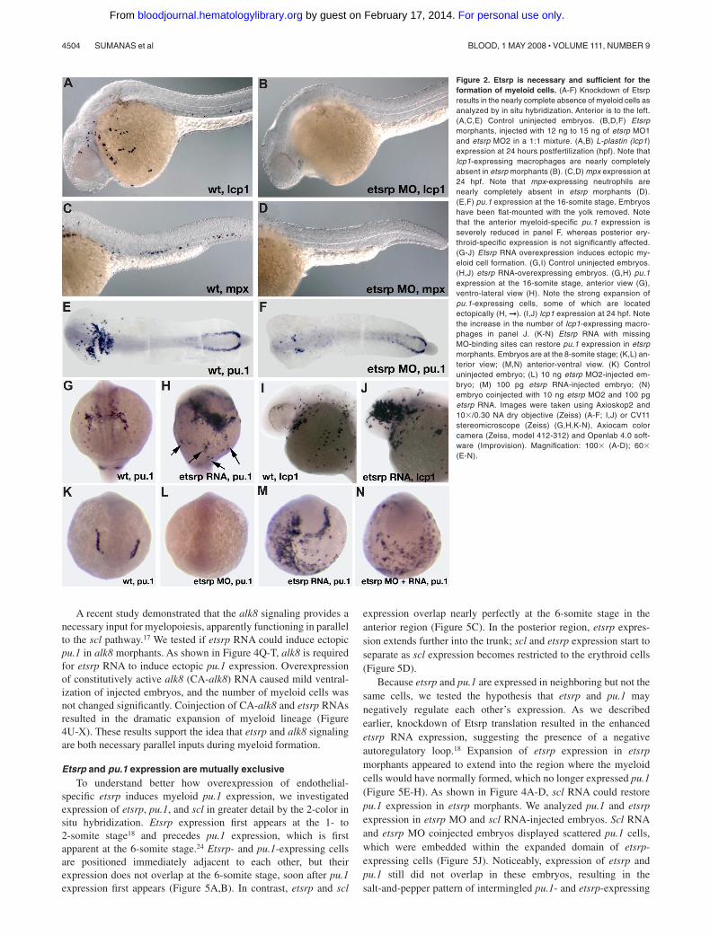
In the previous study, we showed that scl expression in the anteriorlateral mesoderm is nearly completely missing in etsrp mor-phants.18 Scl knockdown has been previously shown to affect themyeloid lineage.12 Therefore, we investigated if myeloid cells wereaffected in etsrp morphants. L-plastin (lcp1) expressing macro-phages28 and neutrophils31 and myeloid peroxidase (mpx) express-ing heterophilic granulocytes27,32 were nearly completely absent inetsrp morphants (Figure 2A-D). Expression of an early myeloidmarker pu.124 was strongly down-regulated in the anterior lateral
plate mesoderm, whereas the posterior erythroid-specific pu.1expression was not significantly affected (Figure 2E,F). Theseresults show that etsrp function is required for the formation ofmyeloid lineage, in addition to its previously demonstrated require-ment for endothelial lineage.18 Control 5-base mismatch morpho-lino caused no noticeable defects in embryonic development,including myeloid, hemangioblast, and endothelial marker expres-sion,18 confirming the specificity of the observed phenotypes(Figure S1, available on the Blood website; see the SupplementalMaterials link at the top of the online article; data not shown).
We have previously shown that overexpression of etsrp RNA wassufficient to induce strong ectopic scl expression.18 Etsrp RNA alsoinduced ectopic pu.1 expression (Figure 2G,H) and caused an increasein the number of lcp1-expressing macrophages and neutrophils (Figure2I,J). Interestingly, while the total of 67% of etsrp-overexpressingembryos displayed ectopic pu.1, 60% of the injected embryos also hadreduced or completely absent endogenous pu.1 expression; 51% dis-played both effects at the same time (Table 1). Etsrp RNA that lackedetsrp-MO binding sites induced ectopic pu.1 expression in etsrpmorphants, demonstrating that ectopic pu.1 cells are not merelymislocalized endogenous pu.1 cells (Figure 2K-N, Table 1). In the sameexperiment, etsrp RNA also caused ectopic expression of an endothelialmarker flk1 (vegfr2)26 in etsrp morphants, consistent with our previousresults (data not shown; Sumanas and Lin18). Etsrp overexpression cantherefore induce cells to initiate myeloid or endothelial development.Endogenous pu.1 may become down-regulated in etsrp-overexpressingembryos, as those cells undertake endothelial fate.
Etsrp affects both hematopoiesis and vasculogenesis in theanterior but not the posterior region
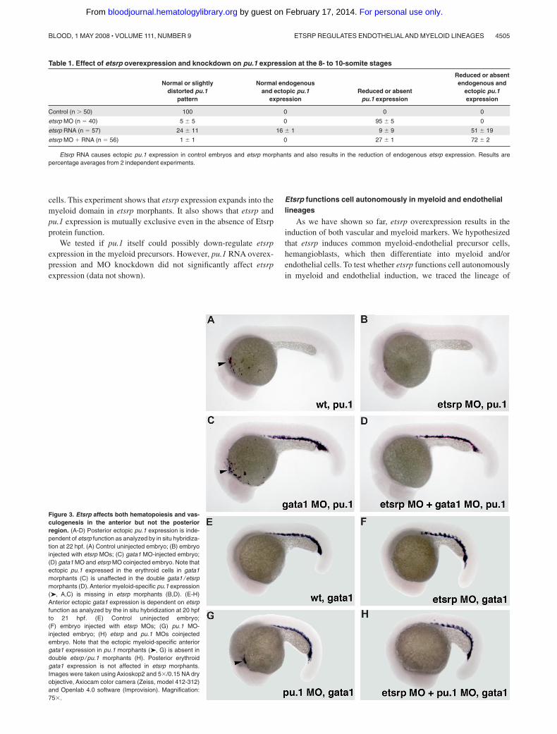
We have previously shown that etsrp knockdown and overexpres-sion did not significantly affect formation of erythroid cells,including erythroid-specific expression of gata118 (Figure S2). Weinvestigated whether etsrp function is limited to the myeloid cellswithin anterior lateral plate mesoderm. In wild-type embryos, pu.1is expressed in both erythroid and myeloid progenitors by the10-somite stage, and its erythroid expression disappears duringlater somitogenesis stages.24 Knockdown of gata1 has beenreported to cause erythroid cells to undertake myeloid fate resultingin the persistence of posterior pu.1 expression15,16 (Figure 3C).Coinjection of etsrp and gata1 MOs resulted in the loss of pu.1expression in the anterior but not the posterior region (Figure 3D),supporting etsrp requirement for the formation of anterior but notposterior myelo-erythroid progenitors. Knockdown of pu.1 hasbeen reported to result in the myeloid cells undertaking erythroidfate and expressing gata1 in the anterior region15 (Figure 3G).These anterior gata1-expressing cells were absent in the etsrp andpu.1 double morphants (Figure 3H), consistent with etsrp affectingmyelo-erythroid progenitors in the anterior region.
Scl functions downstream of etsrp in the myeloid but notendothelial lineage
Scl has been previously implicated in myelopoiesis as well asvasculogenesis.11,12 We studied the interaction of etsrp and scl inmyeloid and vascular development. We have previously shown thatetsrp overexpression induces strong ectopic scl expression.18 Scloverexpression also enhanced etsrp expression, which was limitedmostly to the somitic mesoderm (Figure S3; Figure 5G,I). Sclknockdown had no apparent effect on etsrp expression whereasetsrp morphants had strongly reduced scl expression in the anteriorand trunk domains.18 Addition of scl RNA restored the endogenous
4502 SUMANAS et al BLOOD, 1 MAY 2008 � VOLUME 111, NUMBER 9
For personal use only. by guest on February 17, 2014. bloodjournal.hematologylibrary.orgFrom
pattern of pu.1 expression in etsrp morphants (Figure 4A-D; Table S1).In the sibling embryos from the same experiment, scl RNA failed torescue endothelial flk1 expression in etsrp morphants, although it wassufficient to expand flk1 expression in control embryos (Figure 4E-H),consistent with our previously published data.18 As reported previously,pu.1 expression was severely reduced or absent in scl morphants (Figure4I-J).11,12 Etsrp RNA failed to rescue pu.1 expression in scl morphants(Figure 4K,L). In the sibling embryos from the same experiment, etsrp
induced ectopic flk1-GFP expression in scl morphants, although to asmaller extent than etsrp overexpression alone (Figure 4M-P). Theseexperiments argue that scl functions downstream of etsrp in themyeloid induction. However, in the vascular induction etsrp is thecritical factor, and scl may only modify etsrp expression orfunction but is not absolutely required. Overexpression of pu.1mRNA was also sufficient to rescue lcp1 expression in etsrpmorphants (data not shown).
Figure 1. Etsrp is a functional ortholog of the mammalian ER71 subfamily. (A) Alignment of zebrafish Etsrp, medaka, stickleback, mouse, and human ER71 amino acidsequences. Identical and similar amino acids are labeled in red and blue, respectively. Etsrp and hER71 share 71% homology within the ETS DNA-binding domain (underlinedin gray). GenBank accession numbers used for the analysis are as follows: human ER71 (O00321), mouse ER71 (NP_031985), and zebrafish ETSRP (AAY89037). MedakaER71 (ENSORLP00000019929) and stickleback ER71 (ENSGACP00000016315) are Ensembl predictions. (B) Phylogenetic analysis of zebrafish Etsrp and its closesthuman, mouse, frog, and fish homologs. The phylogenetic tree is built using the Neighbor Joining method. Length of horizontal branches is proportional to the evolutionarydistance between the protein molecules. GenBank accession numbers used for the analysis are as follows: human ER71 (O00321), mouse ER71 (NP_031985), zebrafishETSRP (AAY89037), human ETS1 (NP_005229), mouse ETS1 (NP_035938), frog ETS1 (NP_001081621), zebrafish ETS1a (NP_001017558), human ETS2 (NP_005230),mouse ETS2 (NP_035939), frog ETS2 (NP_001081007), zebrafish ETS2 (NP_001018874), human FLI1 (NP_002008), mouse FLI1 (NP_032052), zebrafish FLI1a(NP_571423), and zebrafish FLI1b (NP_001008780). Medaka ER71 (ENSORLP00000019929), stickleback ER71 (ENSGACP00000016315), fugu ETS1 (SIN-FRUP00000163510), and medaka ETS1 (ENSORLP00000016939) are Ensembl predictions. NTI Vector (Invitrogen) has been used to build the alignment and thephylogenetic tree. (C) Chromosomal location of the zebrafish etsrp, medaka, stickleback, mouse, and human ER71 genes. Numbers alongside the chromosomal regions ofinterest correspond to the actual physical distances (Mb). Etsrp/ER71 genes are highlighted in red. (D-J) Mouse ER71 and zebrafish Etsrp overexpression causes ectopicexpression of hemangioblast marker scl and vascular endothelial marker flk1. flk1-GFP transgenic embryos were injected with 75 pg of either Etsrp or mER71 DNA at the 1-cellstage and analyzed at 8- to 10-somite stages. Relative to uninjected embryos (D,G), both Etsrp (E) and mER71 (F,H) result in the ectopic induction of scl when examined by insitu hybridization. Flk1 expression was also induced by ER71 injections as revealed by ectopic GFP expression (J) relative to uninjected controls (I). Panels G to J are lateralviews with anterior to the left. Images were taken using Zeiss CV11 stereomicroscope, Axiocam color camera (Zeiss, model 412-312) and Openlab 4.0 software (Improvision,Waltham, MA). Magnification: 12� (D-F); 60� (G-J).
ETSRP REGULATES ENDOTHELIAL AND MYELOID LINEAGES 4503BLOOD, 1 MAY 2008 � VOLUME 111, NUMBER 9
For personal use only. by guest on February 17, 2014. bloodjournal.hematologylibrary.orgFrom
A recent study demonstrated that the alk8 signaling provides anecessary input for myelopoiesis, apparently functioning in parallelto the scl pathway.17 We tested if etsrp RNA could induce ectopicpu.1 in alk8 morphants. As shown in Figure 4Q-T, alk8 is requiredfor etsrp RNA to induce ectopic pu.1 expression. Overexpressionof constitutively active alk8 (CA-alk8) RNA caused mild ventral-ization of injected embryos, and the number of myeloid cells wasnot changed significantly. Coinjection of CA-alk8 and etsrp RNAsresulted in the dramatic expansion of myeloid lineage (Figure4U-X). These results support the idea that etsrp and alk8 signalingare both necessary parallel inputs during myeloid formation.
Etsrp and pu.1 expression are mutually exclusiveTo understand better how overexpression of endothelial-
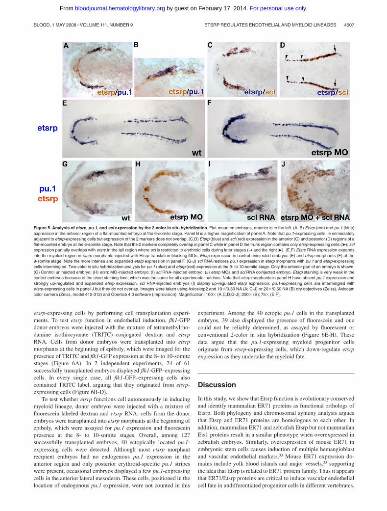
specific etsrp induces myeloid pu.1 expression, we investigatedexpression of etsrp, pu.1, and scl in greater detail by the 2-color insitu hybridization. Etsrp expression first appears at the 1- to2-somite stage18 and precedes pu.1 expression, which is firstapparent at the 6-somite stage.24 Etsrp- and pu.1-expressing cellsare positioned immediately adjacent to each other, but theirexpression does not overlap at the 6-somite stage, soon after pu.1expression first appears (Figure 5A,B). In contrast, etsrp and scl
expression overlap nearly perfectly at the 6-somite stage in theanterior region (Figure 5C). In the posterior region, etsrp expres-sion extends further into the trunk; scl and etsrp expression start toseparate as scl expression becomes restricted to the erythroid cells(Figure 5D).
Because etsrp and pu.1 are expressed in neighboring but not thesame cells, we tested the hypothesis that etsrp and pu.1 maynegatively regulate each other’s expression. As we describedearlier, knockdown of Etsrp translation resulted in the enhancedetsrp RNA expression, suggesting the presence of a negativeautoregulatory loop.18 Expansion of etsrp expression in etsrpmorphants appeared to extend into the region where the myeloidcells would have normally formed, which no longer expressed pu.1(Figure 5E-H). As shown in Figure 4A-D, scl RNA could restorepu.1 expression in etsrp morphants. We analyzed pu.1 and etsrpexpression in etsrp MO and scl RNA-injected embryos. Scl RNAand etsrp MO coinjected embryos displayed scattered pu.1 cells,which were embedded within the expanded domain of etsrp-expressing cells (Figure 5J). Noticeably, expression of etsrp andpu.1 still did not overlap in these embryos, resulting in thesalt-and-pepper pattern of intermingled pu.1- and etsrp-expressing
Figure 2. Etsrp is necessary and sufficient for theformation of myeloid cells. (A-F) Knockdown of Etsrpresults in the nearly complete absence of myeloid cells asanalyzed by in situ hybridization. Anterior is to the left.(A,C,E) Control uninjected embryos. (B,D,F) Etsrpmorphants, injected with 12 ng to 15 ng of etsrp MO1and etsrp MO2 in a 1:1 mixture. (A,B) L-plastin (lcp1)expression at 24 hours postfertilization (hpf). Note thatlcp1-expressing macrophages are nearly completelyabsent in etsrp morphants (B). (C,D) mpx expression at24 hpf. Note that mpx-expressing neutrophils arenearly completely absent in etsrp morphants (D).(E,F) pu.1 expression at the 16-somite stage. Embryoshave been flat-mounted with the yolk removed. Notethat the anterior myeloid-specific pu.1 expression isseverely reduced in panel F, whereas posterior ery-throid-specific expression is not significantly affected.(G-J) Etsrp RNA overexpression induces ectopic my-eloid cell formation. (G,I) Control uninjected embryos.(H,J) etsrp RNA-overexpressing embryos. (G,H) pu.1expression at the 16-somite stage, anterior view (G),ventro-lateral view (H). Note the strong expansion ofpu.1-expressing cells, some of which are locatedectopically (H, ➞ ). (I,J) lcp1 expression at 24 hpf. Notethe increase in the number of lcp1-expressing macro-phages in panel J. (K-N) Etsrp RNA with missingMO-binding sites can restore pu.1 expression in etsrpmorphants. Embryos are at the 8-somite stage; (K,L) an-terior view; (M,N) anterior-ventral view. (K) Controluninjected embryo; (L) 10 ng etsrp MO2-injected em-bryo; (M) 100 pg etsrp RNA-injected embryo; (N)embryo coinjected with 10 ng etsrp MO2 and 100 pgetsrp RNA. Images were taken using Axioskop2 and10�/0.30 NA dry objective (Zeiss) (A-F; I,J) or CV11stereomicroscope (Zeiss) (G,H,K-N), Axiocam colorcamera (Zeiss, model 412-312) and Openlab 4.0 soft-ware (Improvision). Magnification: 100� (A-D); 60�(E-N).
4504 SUMANAS et al BLOOD, 1 MAY 2008 � VOLUME 111, NUMBER 9
For personal use only. by guest on February 17, 2014. bloodjournal.hematologylibrary.orgFrom
cells. This experiment shows that etsrp expression expands into themyeloid domain in etsrp morphants. It also shows that etsrp andpu.1 expression is mutually exclusive even in the absence of Etsrpprotein function.
We tested if pu.1 itself could possibly down-regulate etsrpexpression in the myeloid precursors. However, pu.1 RNA overex-pression and MO knockdown did not significantly affect etsrpexpression (data not shown).
Etsrp functions cell autonomously in myeloid and endotheliallineages
As we have shown so far, etsrp overexpression results in theinduction of both vascular and myeloid markers. We hypothesizedthat etsrp induces common myeloid-endothelial precursor cells,hemangioblasts, which then differentiate into myeloid and/orendothelial cells. To test whether etsrp functions cell autonomouslyin myeloid and endothelial induction, we traced the lineage of
Figure 3. Etsrp affects both hematopoiesis and vas-culogenesis in the anterior but not the posteriorregion. (A-D) Posterior ectopic pu.1 expression is inde-pendent of etsrp function as analyzed by in situ hybridiza-tion at 22 hpf. (A) Control uninjected embryo; (B) embryoinjected with etsrp MOs; (C) gata1 MO-injected embryo;(D) gata1 MO and etsrp MO coinjected embryo. Note thatectopic pu.1 expressed in the erythroid cells in gata1morphants (C) is unaffected in the double gata1 / etsrpmorphants (D). Anterior myeloid-specific pu.1 expression(➤, A,C) is missing in etsrp morphants (B,D). (E-H)Anterior ectopic gata1 expression is dependent on etsrpfunction as analyzed by the in situ hybridization at 20 hpfto 21 hpf. (E) Control uninjected embryo;(F) embryo injected with etsrp MOs; (G) pu.1 MO-injected embryo; (H) etsrp and pu.1 MOs coinjectedembryo. Note that the ectopic myeloid-specific anteriorgata1 expression in pu.1 morphants (➤, G) is absent indouble etsrp / pu.1 morphants (H). Posterior erythroidgata1 expression is not affected in etsrp morphants.Images were taken using Axioskop2 and 5�/0.15 NA dryobjective, Axiocam color camera (Zeiss, model 412-312)and Openlab 4.0 software (Improvision). Magnification:75�.
Table 1. Effect of etsrp overexpression and knockdown on pu.1 expression at the 8- to 10-somite stages
Normal or slightlydistorted pu.1
pattern
Normal endogenousand ectopic pu.1
expressionReduced or absent
pu.1 expression
Reduced or absentendogenous and
ectopic pu.1expression
Control (n � 50) 100 0 0 0
etsrp MO (n � 40) 5 � 5 0 95 � 5 0
etsrp RNA (n � 57) 24 � 11 16 � 1 9 � 9 51 � 19
etsrp MO � RNA (n � 56) 1 � 1 0 27 � 1 72 � 2
Etsrp RNA causes ectopic pu.1 expression in control embryos and etsrp morphants and also results in the reduction of endogenous etsrp expression. Results arepercentage averages from 2 independent experiments.
ETSRP REGULATES ENDOTHELIAL AND MYELOID LINEAGES 4505BLOOD, 1 MAY 2008 � VOLUME 111, NUMBER 9
For personal use only. by guest on February 17, 2014. bloodjournal.hematologylibrary.orgFrom
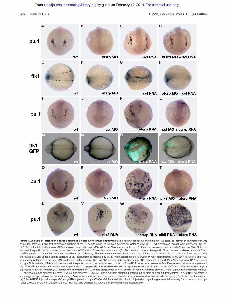
Figure 4. Analysis of interaction between etsrp and scl and alk8 signaling pathways. (A-H) scl RNA can rescue myeloid but not vascular cell formation in etsrp morphantsas evident from pu.1 and flk1 expression analysis at the 10-somite stage. (A-D) pu.1 expression, anterior view; (E-H) flk1 expression, dorsal view, anterior to the left.(A,E) Control uninjected embryos; (B,F) embryos injected with etsrp MOs; (C,G) scl RNA-injected embryos; (D,H) embryos coinjected with etsrp MOs and scl RNA. Note thatthe myeloid-specific pu.1 expression is restored in etsrp MO and scl RNA coinjected embryos (D). Also note that the vascular-specific flk1 expression is absent in etsrp MO andscl RNA coinjected embryos in the same experiment (H). (I-P) etsrp RNA can rescue vascular but not myeloid cell formation in scl morphants as evident from pu.1 and flk1expression analysis at the 8-somite stage. (I-L) pu.1 expression as analyzed by in situ hybridization, anterior view; (M-P) GFP fluorescence in flk1-GFP transgenic embryos,dorsal view, anterior is to the left. (I,M) Control uninjected embryo; (J,N) scl MO-injected embryo; (K,O) etsrp RNA-injected embryo; (L,P) scl MO and etsrp RNA coinjectedembryo. Note that etsrp RNA fails to rescue myeloid-specific pu.1 expression in scl morphants (L). Etsrp RNA can restore vascular flk1-GFP expression in the same experiment(P). Flk1-GFP fluorescence in wild-type embryos and scl morphants (M,N) is much weaker and not apparent under the same exposure. (Q-T) etsrp RNA fails to rescue pu.1expression in alk8 morphants. pu.1 expression analyzed at the 10-somite stage, anterior view, except for panel S, which is anterior-ventral. (Q) Control uninjected embryo;(R) alk8 MO-injected embryo; (S) etsrp RNA-injected embryo; (T) alk8 MO and etsrp RNA coinjected embryo. (U-X) etsrp and constitutively active CA-alk8 RNA synergize ininducing pu.1 expression at the 14-somite stage. Anterior-ventral views except for panel X, which is the ventrolateral view, anterior is to the top. (U) Control uninjected embryo;(V) CA-alk8 RNA-injected embryo; (W) etsrp RNA-injected embryo; (X) CA-alk8 RNA and etsrp RNA coinjected embryo. Images were taken using CV11 stereomicroscope(Zeiss), Axiocam color camera (Zeiss, model 412-312) and Openlab 4.0 software (Improvision). Magnification: 60�.
4506 SUMANAS et al BLOOD, 1 MAY 2008 � VOLUME 111, NUMBER 9
For personal use only. by guest on February 17, 2014. bloodjournal.hematologylibrary.orgFrom
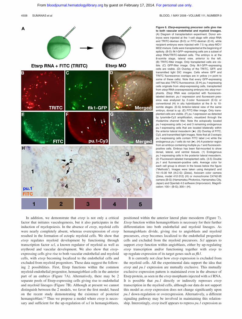
etsrp-expressing cells by performing cell transplantation experi-ments. To test etsrp function in endothelial induction, flk1-GFPdonor embryos were injected with the mixture of tetramethylrho-damine isothiocyanate (TRITC)–conjugated dextran and etsrpRNA. Cells from donor embryos were transplanted into etsrpmorphants at the beginning of epiboly, which were imaged for thepresence of TRITC and flk1-GFP expression at the 8- to 10-somitestages (Figure 6A). In 2 independent experiments, 24 of 61successfully transplanted embryos displayed flk1-GFP–expressingcells. In every single case, all flk1-GFP–expressing cells alsocontained TRITC label, arguing that they originated from etsrp-expressing cells (Figure 6B-D).
To test whether etsrp functions cell autonomously in inducingmyeloid lineage, donor embryos were injected with a mixture offluorescein-labeled dextran and etsrp RNA; cells from the donorembryos were transplanted into etsrp morphants at the beginning ofepiboly, which were assayed for pu.1 expression and fluoresceinpresence at the 8- to 10-somite stages. Overall, among 127successfully transplanted embryos, 40 ectopically located pu.1-expressing cells were detected. Although most etsrp morphantrecipient embryos had no endogenous pu.1 expression in theanterior region and only posterior erythroid-specific pu.1 stripeswere present, occasional embryos displayed a few pu.1-expressingcells in the anterior lateral mesoderm. These cells, positioned in thelocation of endogenous pu.1 expression, were not counted in this
experiment. Among the 40 ectopic pu.1 cells in the transplantedembryos, 39 also displayed the presence of fluorescein and onecould not be reliably determined, as assayed by fluorescent orconventional 2-color in situ hybridization (Figure 6E-H). Thesedata argue that the pu.1-expressing myeloid progenitor cellsoriginate from etsrp-expressing cells, which down-regulate etsrpexpression as they undertake the myeloid fate.
Discussion
In this study, we show that Etsrp function is evolutionary conservedand identify mammalian ER71 proteins as functional orthologs ofEtsrp. Both phylogeny and chromosomal synteny analysis arguesthat Etsrp and ER71 proteins are homologous to each other. Inaddition, mammalian ER71 and zebrafish Etsrp but not mammalianEts1 proteins result in a similar phenotype when overexpressed inzebrafish embryos. Similarly, overexpression of mouse ER71 inembryonic stem cells causes induction of multiple hemangioblastand vascular endothelial markers.33 Mouse ER71 expression do-mains include yolk blood islands and major vessels,33 supportingthe idea that Etsrp is related to ER71 protein family. Thus it appearsthat ER71/Etsrp proteins are critical to induce vascular endothelialcell fate in undifferentiated progenitor cells in different vertebrates.
Figure 5. Analysis of etsrp, pu.1, and scl expression by the 2-color in situ hybridization. Flat-mounted embryos, anterior is to the left. (A, B) Etsrp (red) and pu.1 (blue)expression in the anterior region of a flat-mounted embryo at the 6-somite stage. Panel B is a higher magnification of panel A. Note that pu.1-expressing cells lie immediatelyadjacent to etsrp-expressing cells but expression of the 2 markers does not overlap. (C,D) Etsrp (blue) and scl (red) expression in the anterior (C) and posterior (D) regions of aflat-mounted embryo at the 6-somite stage. Note that the 2 markers completely overlap in panel C while in panel D the trunk region contains only etsrp-expressing cells (➤); sclexpression partially overlaps with etsrp in the tail region where scl is restricted to erythroid cells during later stages (➞ and the right ➤). (E,F) Etsrp RNA expression expandsinto the myeloid region in etsrp morphants injected with Etsrp translation-blocking MOs. Etsrp expression in control uninjected embryos (E) and etsrp morphants (F) at the9-somite stage. Note the more intense and expanded etsrp expression in panel F. (G-J) scl RNA restores pu.1 expression in etsrp morphants with pu.1 and etsrp-expressingcells intermingled. Two-color in situ hybridization analysis for pu.1 (blue) and etsrp (red) expression at the 9- to 10-somite stage. Only the anterior part of an embryo is shown.(G) Control uninjected embryo; (H) etsrp MO-injected embryo; (I) scl RNA-injected embryo; (J) etsrp MOs and scl RNA coinjected embryo. Etsrp staining is very weak in thecontrol embryos because of the short staining time, which was the same for all experimental batches. Note that etsrp morphants in panel H have absent pu.1 expression andstrongly up-regulated and expanded etsrp expression. scl RNA-injected embryos (I) display up-regulated etsrp expression. pu.1-expressing cells are intermingled withetsrp-expressing cells in panel J but they do not overlap. Images were taken using Axioskop2 and 10�/0.30 NA (A; C-J) or 20�/0.50 NA (B) dry objectives (Zeiss), Axiocamcolor camera (Zeiss, model 412-312) and Openlab 4.0 software (Improvision). Magnification: 100� (A,C,D,G-J); 200� (B); 75� (E,F).
ETSRP REGULATES ENDOTHELIAL AND MYELOID LINEAGES 4507BLOOD, 1 MAY 2008 � VOLUME 111, NUMBER 9
For personal use only. by guest on February 17, 2014. bloodjournal.hematologylibrary.orgFrom
In addition, we demonstrate that etsrp is not only a criticalfactor that initiates vasculogenesis, but it also participates in theinduction of myelopoiesis. In the absence of etsrp, myeloid cellswere nearly completely absent, whereas overexpression of etsrpresulted in the formation of ectopic myeloid cells. We show thatetsrp regulates myeloid development by functioning throughtranscription factor scl, a known regulator of myeloid as well aserythroid and vascular development. We also show that etsrp-expressing cells give rise to both vascular endothelial and myeloidcells, with etsrp becoming localized to the endothelial cells andexcluded from myeloid progenitors. These data suggest the follow-ing 2 possibilities. First, Etsrp functions within the commonmyeloid-endothelial progenitor, hemangioblast cells in the anteriorpart of an embryo (Figure 7A). Alternatively, there may be 2separate pools of Etsrp-expressing cells giving rise to endothelialand myeloid lineages (Figure 7B). Although at present we cannotdistinguish between the 2 models, we favor the first model, basedon the recent study demonstrating the existence of posteriorhemangioblast.13 Thus we propose a model where etsrp is neces-sary and sufficient for the up-regulation of scl in hemangioblasts,
positioned within the anterior lateral plate mesoderm (Figure 7).Etsrp function within hemangioblasts is necessary for their furtherdifferentiation into both endothelial and myeloid lineages. Ashemangioblasts divide, giving rise to angioblasts and myeloidprecursors, etsrp becomes localized to the endothelial progenitorcells and excluded from the myeloid precursors. Scl appears tosupport etsrp function within angioblasts, either by up-regulatingetsrp transcription and/or functioning together with etsrp toup-regulate expression of its target genes such as flk1.
It is currently not clear how etsrp expression is excluded fromthe myeloid cells. All the experimental data support the idea thatetsrp and pu.1 expression are mutually exclusive. This mutuallyexclusive expression pattern is maintained even in the absence ofEtsrp protein, as seen in the etsrp morphants injected with scl RNA.It is possible that pu.1 directly or indirectly represses etsrptranscription in the myeloid cells, although our data do not supportthis model as etsrp expression does not change significantly uponpu.1 down-regulation or overexpression. Alternatively, a differentsignaling pathway may be involved in maintaining this relation-ship. Interestingly, etsrp itself appears to repress pu.1 expression as
Figure 6. Etsrp-expressing precursor cells give riseto both vascular endothelial and myeloid lineages.(A) Diagram of transplantation experiment. Donor em-bryos were injected at the 1-cell stage with etsrp RNAand TRITC-dextran (B-D) or FITC-dextran (E-H), whilerecipient embryos were injected with 7.5 ng etsrp MO1/MO2 mixture. Cells were transplanted at the beginning ofepiboly. (B-D) flk1-GFP–expressing cells are a subset ofetsrp RNA/TRITC-labeled cells. The embryo is at the8-somite stage, lateral view, anterior to the left.(B) TRITC-filter image. Only transplanted cells are vis-ible. (C) GFP-filter image. Only flk1-GFP–expressingcells are visible. (D) Overlay of the TRITC, GFP, andtransmitted light DIC images. Cells where GFP andTRITC fluorescence overlaps are in yellow (➞ point tosome of these cells). Note that every GFP-expressingcell has also TRITC fluorescence. (E-H) pu.1-expressingcells originate from etsrp-expressing cells, transplantedfrom etsrp RNA-overexpressing embryos into etsrp mor-phants. Etsrp RNA was coinjected with fluorescein-labeled dextran; pu.1 expression and fluorescein pres-ence was analyzed by 2-color fluorescent (E-G) orconventional (H) in situ hybridization at the 8- to 10-somite stages. (E-G) Anterior-lateral view of the sameembryo, dorsal is up. (E) FITC-filter image. Only trans-planted cells are visible. (F) pu.1 expression as detectedby tyramide-Cy3 amplification, visualized through therhodamine channel filter. Note the ectopically locatedpu.1-expressing cells (➞) and 3 remaining endogenouspu.1-expressing cells that are located bilaterally withinthe anterior lateral mesoderm (➤). (G) Overlay of FITC,Cy3, and transmitted light images. Note that all 3 ectopicpu.1-expressing cells contain FITC label (➞) while theendogenous pu.1 cells do not (➤). (H) A posterior regionfrom an embryo containing multiple pu.1 and fluorescein-positive cells. Embryo has been flat-mounted to showdorsal, lateral, and ventral tissues. (1) Endogenouspu.1-expressing cells in the posterior lateral mesoderm.(2) Fluorescein-labeled transplanted cells. (3-5) Doublepu.1 and fluorescein-positive cells. Average color foreach cell group is shown in the boxes below the figure(“Methods”). Images were taken using Axioplan2 and10�/0.30 NA (A;C-G) (Zeiss), Axiocam color camera(Zeiss, model 412-312) (H) or monochrome C4742-95camera (B-G) (Hamamatsu Photonics, Hamamatsu City,Japan) and Openlab 4.0 software (Improvision). Magnifi-cation: 100� (B-G); 300� (H).
4508 SUMANAS et al BLOOD, 1 MAY 2008 � VOLUME 111, NUMBER 9
For personal use only. by guest on February 17, 2014. bloodjournal.hematologylibrary.orgFrom

etsrp RNA-overexpressing embryos often have the endogenouspu.1 down-regulated or even totally absent. This may help toensure that pu.1 is never expressed in the endothelial cells.
It is also not known how cells make a choice between theendothelial and myeloid fates. Our results support the idea thatcontinued etsrp expression drives cells toward endothelial fate. Incontrast, the combination of etsrp and alk8 signaling17 combinedwith the subsequent etsrp down-regulation is sufficient to directmultiple cells toward myeloid fate. It is possible that additionalas-yet-undiscovered pathways participate in the cross-talk betweenendothelial and myeloid cells and influence this fate decision.
Until now, it has been generally accepted that the anterior andposterior hemangioblasts are equivalent in their potential. Forexample, the myeloid-forming anterior hemangioblasts can giverise to erythroid cells in the absence of pu.1, and the erythroid-forming posterior hemangioblasts can give rise to myeloid cells inthe absence of gata1.15,16 Etsrp is the first known gene tospecifically affect anterior hemangioblasts, while in the posteriorregion it functions in the angioblasts but does not affect hematopoi-etic cells. The most likely explanation is that in the anterior regionetsrp is expressed within the hemangioblast cells, whereas theposterior etsrp expression is limited to the angioblasts. It is possiblethat related Ets-domain proteins such as Ets134 may play a similarrole in the posterior hemangioblasts; it has been demonstrated thatetsrp, ets1, fli1, and fli1b genes can function somewhat redun-dantly.35 Interestingly, etsrp overexpression resulted in the expan-sion of hemangioblasts, which differentiate into endothelial andmyeloid, but not erythroid cells. Possible explanations are thatetsrp induces hemangioblasts in both regions but they behavedifferently in the posterior region, or etsrp induces hemangioblastsin the anterior but not the posterior region. Scl has been previouslyshown to be necessary for erythroid, myeloid, and to certain extent,endothelial development.11,12 Scl overexpression also induces ery-throid, myeloid, and endothelial lineages.7,8 Etsrp induces strongectopic scl expression while scl can feedback and induce etsrpexpression as well, although this induction is limited to the portionsof lateral, somitic and intermediate mesoderm. Apparently, etsrp-induced scl-expressing cells cannot differentiate into erythroidlineage, either because etsrp presence directs them toward endothelial
fate or additional genes up-regulated by etsrp ensure endothelial fate ofthese cells, even after etsrp expression is lost or down-regulated (whichhappens in a subset of etsrp-expressing cells).
Transplantation experiments show that etsrp-expressing cellscan give rise to both endothelial and myeloid lineages. A relativelylow number of transplanted cells (less than 1%) displayed pu.1expression. It is possible that not all etsrp-expressing cells have theability to differentiate into both lineages. There may be a certainbias in the transplantation experiments, (eg, the transplanted cellsdo not end up in the myeloid-competent region often enough). Wenoticed that the ectopic pu.1-expressing cells in the overexpressionand the transplantation experiments were often located in theventral and almost never in the dorsal side of an embryo, whereasflk1-expressing cells were located mostly at the dorsal side,possibly reflecting different myeloid and endothelial competenceswithin distinct regions of an embryo. Not all etsrp-expressing cellsdifferentiated into endothelial cells at the time of analysis, whichmay also reflect different competence to undertake endothelial fate,or simply a delay between the etsrp expression and flk1 induction.Although our experiments show that etsrp-expressing cells cangive rise to both lineages, thus supporting the hemangioblast idea,additional experiments are needed to convincingly demonstrate theexistence of a common endothelial-myeloid progenitor.
Acknowledgments
This research was supported by grants from the National Institutesof Health (R01 DK54508, T32 HL069766, and R25 CA098010).
Authorship
Contribution: S.S. designed and performed experiments, analyzeddata, and wrote the paper; G.G. and Y.Z. performed experimentsand analyzed data; C.P. and K.C. contributed reagents; S.L.designed experiments and analyzed data.
Conflict-of-interest disclosure: The authors declare no compet-ing financial interests.
Figure 7. Proposed models for etsrp function within the anterior lateral mesoderm. (A) Etsrp induces scl in the hemangioblast cells, which give rise to both vascularendothelial and myeloid precursors. As hemangioblasts divide, etsrp expression becomes restricted to endothelial cells where it is both necessary and sufficient forvasculogenesis. (B) Alternatively, there are 2 separate pools of etsrp-expressing endothelial and myeloid precursors. As the cells initiate myeloid marker expression, theydown-regulate etsrp expression.
ETSRP REGULATES ENDOTHELIAL AND MYELOID LINEAGES 4509BLOOD, 1 MAY 2008 � VOLUME 111, NUMBER 9
For personal use only. by guest on February 17, 2014. bloodjournal.hematologylibrary.orgFrom
Correspondence: Saulius Sumanas, Cincinnati Children’s HospitalMedical Center, Division of Developmental Biology, ML7007, 3333BurnetAve, Cincinnati, OH 45229; e-mail: [email protected];
or Shuo Lin, University of California, Los Angeles, Department ofMolecular, Cell and Developmental Biology, 621 C Young Dr South,Los Angeles, CA 90095; e-mail: [email protected].
References
1. Ema M, Rossant J. Cell fate decisions in earlyblood vessel formation. Trends Cardiovasc Med.2003;13:254-259.
2. Dumont DJ, Fong GH, Puri MC, Gradwohl G,
Alitalo K, Breitman ML. Vascularization of themouse embryo: a study of flk-1, tek, tie, andvascular endothelial growth factor expressionduring development. Dev Dyn. 1995;203:80-92.
3. Choi K, Kennedy M, Kazarov A, PapadimitriouJC, Keller G. A common precursor for hematopoi-etic and endothelial cells. Development. 1998;125:725-732.
4. Paw BH, Zon LI. Zebrafish: a genetic approach instudying hematopoiesis. Curr Opin Hematol.2000;7:79-84.
5. Weinstein BM. Plumbing the mysteries of vascu-lar development using the zebrafish. Semin CellDev Biol. 2002;13:515-522.
6. Stainier DY, Weinstein BM, Detrich HW, Zon LI3rd, Fishman MC. Cloche, an early acting ze-brafish gene, is required by both the endothelialand hematopoietic lineages. Development. 1995;121:3141-3150.
7. Gering M, Rodaway AR, Gottgens B, Patient RK,Green AR. The SCL gene specifies haemangio-blast development from early mesoderm. EmboJ. 1998;17:4029-4045.
8. Liao EC, Paw BH, Oates AC, Pratt SJ, Postleth-wait JH, Zon LI. SCL/Tal-1 transcription factoracts downstream of cloche to specify hematopoi-etic and vascular progenitors in zebrafish. GenesDev. 1998;12:621-626.
9. Brown LA, Rodaway AR, Schilling TF, et al. In-sights into early vasculogenesis revealed by ex-pression of the ETS-domain transcription factorFli-1 in wild-type and mutant zebrafish embryos.Mech Dev. 2000;90:237-252.
10. Gering M, Yamada Y, Rabbitts TH, Patient RK.Lmo2 and Scl/Tal1 convert non-axial mesoderminto haemangioblasts which differentiate into en-dothelial cells in the absence of Gata1. Develop-ment. 2003;130:6187-6199.
11. Dooley KA, Davidson AJ, Zon LI. Zebrafish sclfunctions independently in hematopoietic and en-dothelial development. Dev Biol. 2005;277:522-536.
12. Patterson LJ, Gering M, Patient R. Scl is requiredfor dorsal aorta as well as blood formation in ze-brafish embryos. Blood. 2005;105:3502-3511.
13. Vogeli KM, Jin SW, Martin GR, Stainier DY. Acommon progenitor for haematopoietic and endo-thelial lineages in the zebrafish gastrula. Nature.2006;443:337-339.
14. Hsu K, Kanki JP, Look AT. Zebrafish myelopoiesisand blood cell development. Curr Opin Hematol.2001;8:245-251.
15. Rhodes J, Hagen A, Hsu K, et al. Interplay of pu.1 and gata1 determines myelo-erythroid progeni-tor cell fate in zebrafish. Dev Cell. 2005;8:97-108.
16. Galloway JL, Wingert RA, Thisse C, Thisse B,Zon LI. Loss of gata1 but not gata2 convertserythropoiesis to myelopoiesis in zebrafish em-bryos. Dev Cell. 2005;8:109-116.
17. Hogan BM, Layton JE, Pyati UJ, et al. Specifica-tion of the primitive myeloid precursor pool re-quires signaling through Alk8 in zebrafish. CurrBiol. 2006;16:506-511.
18. Sumanas S, Lin S. Ets1-related protein is a keyregulator of vasculogenesis in zebrafish. PLoSBiol. 2006;4:e10.
19. Bauer H, Lele Z, Rauch GJ, Geisler R, Hammer-schmidt M. The type I serine/threonine kinasereceptor Alk8/Lost-a-fin is required for Bmp2b/7signal transduction during dorsoventral patterningof the zebrafish embryo. Development. 2001;128:849-858.
20. Johnson AD, Krieg PA. pXeX, a vector for effi-cient expression of cloned sequences in Xenopusembryos. Gene. 1994;147:223-226.
21. Hyatt TM, Ekker SC. Vectors and techniques forectopic gene expression in zebrafish. MethodsCell Biol. 1999;59:117-126.
22. Jowett T. Analysis of protein and gene expres-sion. Methods Cell Biol. 1999;59:63-85.
23. Sumanas S, Jorniak T, Lin S. Identification ofnovel vascular endothelial-specific genes by themicroarray analysis of the zebrafish cloche mu-tants. Blood. 2005;106:534-541.
24. Lieschke GJ, Oates AC, Paw BH, et al. ZebrafishSPI-1 (PU. 1) marks a site of myeloid develop-ment independent of primitive erythropoiesis: im-
plications for axial patterning. Dev Biol. 2002;246:274-295.
25. Detrich HW, Kieran MW 3rd, Chan FY, et al. In-traembryonic hematopoietic cell migration duringvertebrate development. Proc Natl Acad Sci U SA. 1995;92:10713-10717.
26. Thompson MA, Ransom DG, Pratt SJ, et al. Thecloche and spadetail genes differentially affecthematopoiesis and vasculogenesis. Dev Biol.1998;197:248-269.
27. Bennett CM, Kanki JP, Rhodes J, et al. Myelopoi-esis in the zebrafish, Danio rerio. Blood. 2001;98:643-651.
28. Herbomel P, Thisse B, Thisse C. Ontogeny andbehaviour of early macrophages in the zebrafishembryo. Development. 1999;126:3735-3745.
29. De Haro L, Janknecht R. Functional analysis ofthe transcription factor ER71 and its activation ofthe matrix metalloproteinase-1 promoter. NucleicAcids Res. 2002;30:2972-2979.
30. De Haro L, Janknecht R. Cloning of the murineER71 gene (Etsrp71) and initial characterizationof its promoter. Genomics. 2005;85:493-502.
31. Le Guyader D, Redd MJ, Colucci-Guyon E, et al.Origins and unconventional behavior of neutro-phils in developing zebrafish. Blood. 2008;111:132-141.
32. Lieschke GJ, Oates AC, Crowhurst MO, WardAC, Layton JE. Morphologic and functional char-acterization of granulocytes and macrophages inembryonic and adult zebrafish. Blood. 2001;98:3087-3096.
33. Dongjun L, Changwon P, Ho L, et al. ER71 in theacts downstream of BMP, Notch, and Wnt signal-ing in blood and vessel progenitor specification.Cell Stem Cell. 2008 (in press).
34. Zhu H, Traver D, Davidson AJ, et al. Regulationof the lmo2 promoter during hematopoietic andvascular development in zebrafish. Dev Biol.2005;281:256-269.
35. Pham VN, Lawson ND, Mugford JW, et al. Com-binatorial function of ETS transcription factors inthe developing vasculature. Dev Biol. 2007;303:772-783.
4510 SUMANAS et al BLOOD, 1 MAY 2008 � VOLUME 111, NUMBER 9
For personal use only. by guest on February 17, 2014. bloodjournal.hematologylibrary.orgFrom
Copyright © 2022 FDOKUMEN