Interleukin-34 Is a Functional Receptor for ! Phosphatase Receptor-type Protein-tyrosine Signal...
16
Richard Stanley Cheng, Solen Gokhan, Mark F. Mehler and E. Amy W. Hsu, Robert Halenbeck, Hui-Yong Edward Nieves, Lydia Tesfa, Haishan Lin, Sayan Nandi, Mario Cioce, Yee-Guide Yeung, Interleukin-34 Is a Functional Receptor for ! Phosphatase Receptor-type Protein-tyrosine Signal Transduction: doi: 10.1074/jbc.M112.442731 originally published online June 6, 2013 2013, 288:21972-21986. J. Biol. Chem. 10.1074/jbc.M112.442731 Access the most updated version of this article at doi: . JBC Affinity Sites Find articles, minireviews, Reflections and Classics on similar topics on the Alerts: When a correction for this article is posted • When this article is cited • to choose from all of JBC's e-mail alerts Click here Supplemental material: http://www.jbc.org/content/suppl/2013/06/06/M112.442731.DC1.html http://www.jbc.org/content/288/30/21972.full.html#ref-list-1 This article cites 80 references, 43 of which can be accessed free at
-
Upload
independent -
Category
Documents
-
view
0 -
download
0
Transcript of Interleukin-34 Is a Functional Receptor for ! Phosphatase Receptor-type Protein-tyrosine Signal...

Richard StanleyCheng, Solen Gokhan, Mark F. Mehler and E. Amy W. Hsu, Robert Halenbeck, Hui-YongEdward Nieves, Lydia Tesfa, Haishan Lin, Sayan Nandi, Mario Cioce, Yee-Guide Yeung, Interleukin-34
Is a Functional Receptor for!Phosphatase Receptor-type Protein-tyrosineSignal Transduction:
doi: 10.1074/jbc.M112.442731 originally published online June 6, 20132013, 288:21972-21986.J. Biol. Chem.
10.1074/jbc.M112.442731Access the most updated version of this article at doi:
.JBC Affinity SitesFind articles, minireviews, Reflections and Classics on similar topics on the
Alerts:
When a correction for this article is posted•
When this article is cited•
to choose from all of JBC's e-mail alertsClick here
Supplemental material:
http://www.jbc.org/content/suppl/2013/06/06/M112.442731.DC1.html
http://www.jbc.org/content/288/30/21972.full.html#ref-list-1
This article cites 80 references, 43 of which can be accessed free at
at Colum
bia University on N
ovember 15, 2013
http://ww
w.jbc.org/
Dow
nloaded from
at Colum
bia University on N
ovember 15, 2013
http://ww
w.jbc.org/
Dow
nloaded from
Receptor-type Protein-tyrosine Phosphatase ! Is a FunctionalReceptor for Interleukin-34*!S
Received for publication, December 6, 2012, and in revised form, May 21, 2013 Published, JBC Papers in Press, June 6, 2013, DOI 10.1074/jbc.M112.442731
Sayan Nandi‡1, Mario Cioce‡2, Yee-Guide Yeung‡, Edward Nieves‡, Lydia Tesfa‡, Haishan Lin§3, Amy W. Hsu§,Robert Halenbeck§, Hui-Yong Cheng¶, Solen Gokhan!, Mark F. Mehler!, and E. Richard Stanley‡4
From the Departments of ‡Developmental and Molecular Biology and ¶Biochemistry and the !Institute for Brain Disorders andNeural Regeneration, Departments of Neurology, Neuroscience, and Psychiatry and Behavioral Sciences, Albert Einstein College ofMedicine, Bronx, New York 10461 and §Five Prime Therapeutics, Inc., South San Francisco, California 94080
Background: IL-34 and the known IL-34 receptor, CSF-1R, are differentially expressed in mouse brain; thus, IL-34 maysignal via an additional receptor(s).Results: IL-34 binds to PTP-! on U251 human glioblastoma cells to stimulate intracellular signaling and responses.Conclusion: PTP-! is an IL-34 receptor.Significance: CSF-1R-independent actions of IL-34 via PTP-! should be considered in evaluating IL-34 roles in developmentand disease.
Interleukin-34 (IL-34) is highly expressed in brain. IL-34 sig-naling via its cognate receptor, colony-stimulating factor-1receptor (CSF-1R), is required for the development ofmicroglia.However, the differential expression of IL-34 and the CSF-1R inbrain suggests that IL-34may signal via an alternate receptor. ByIL-34 affinity chromatography of solubilizedmouse brainmem-brane followed by mass spectrometric analysis, we identifiedreceptor-type protein-tyrosine phosphatase ! (PTP-!), a cell sur-face chondroitin sulfate (CS) proteoglycan, as a novel IL-34 recep-tor. PTP-! is primarily expressed on neural progenitors and glialcells and is highly expressed in human glioblastomas. IL-34 selec-tivelyboundPTP-! inCSF-1R-deficientU251humanglioblastomacell lysates and inhibited the proliferation, clonogenicity, andmotility ofU251 cells in aPTP-!-dependentmanner. These effectswerecorrelatedwithan increase in tyrosinephosphorylationof thepreviously identified PTP-! downstream effectors focal adhesionkinase and paxillin. IL-34 binding to U251 cells was abrogated bychondroitinase ABC treatment, and CS competed with IL-34 forbinding to the extracellular domain of PTP-! and to the cells, indi-cating a dependence of binding on PTP-! CSmoieties. This studyidentifies an alternate receptor for IL-34 that may mediate itsaction on novel cellular targets.
The CSF-1R5 kinase (1, 2) plays a critical role in the regula-tion of macrophage and osteoclast production and function(3–6) as well as the development and regulation of other celltypes (7–11). The existence of an additional CSF-1R ligand wasproposed based on the greater severity of phenotype ofhomozygous CSF-1R-null mice compared with the phenotypeof homozygous CSF-1-null mutant mice (12). A second ligandfor the CSF-1R, interleukin-34 (IL-34), with no apparentsequence similarity to any other growth factor was subse-quently identified (13). Although IL-34 and CSF-1 compete forbinding to the CSF-1R and have similar CSF-1R-mediatedeffects, they exhibit significant tissue-specific and developmen-tal differences in their expression patterns (14). In addition,whereas CSF-1-deficient mice exhibit partial loss of microglia,CSF-1R-deficient mice have no microglia (15). This observa-tion together with the high expression of IL-34 in brain sug-gested an important role of IL-34 inmicroglial development. Inagreement with this, IL-34-deficient (IL-34!/!) mice wereshown to exhibit severe deficits in microglia (16, 17). Despitethe similarity of IL-34 and CSF-1 in their CSF-1R-mediatedeffects (14, 18), IL34 mRNA is expressed at a significantlyhigher level than either Csf1 or Csf1rmRNA in several regionsof the early postnatal and adult brain (14), IL-34 protein is oftenexpressed in regions where there is minimal expression of theCSF-1R or CSF-1-reporter proteins, and IL-34 is significantlymore active in suppressing neural progenitor cell proliferationand neuronal differentiation than CSF-1 (9). These observa-
* This work was supported, in whole or in part, by National Institutes of HealthGrants CA32551 and CA26504 (to E. R. S.), 5P30-CA13330 (a cancer centergrant to the Albert Einstein College of Medicine), NS071571 andP30HD071593 (to M. F. M. and S. G.), and 1S10RR019352 (a shared instru-mentation grant to the Einstein Laboratory for Macromolecular Analysisand Proteomics). This work was also supported by the F. M. Kirby, AlpernFamily, Mildred and Bernard H. Kayden, and Roslyn and Leslie GoldsteinFoundations (to M. F. M. and S. G.).
!S This article contains supplemental Table 1.1 Present address: Dept. of Neuroscience, College of Physicians and Surgeons,
Columbia University, 1051 Riverside Dr., New York, NY 10032.2 Present address: Dept. of Cardiothoracic Surgery, New York University Lan-
gone Medical Center, 462 First Ave., New York, NY 10016.3 Present address: Nuvik Therapeutics, Inc., 41 Woodford Dr., Moraga, CA
94566.4 To whom correspondence should be addressed: Dept. of Developmental
and Molecular Biology, Albert Einstein College of Medicine, 1300 MorrisPark Ave., Bronx, NY 10461. Tel.: 718-430-2344; Fax: 718-430-8567; E-mail:[email protected].
5 The abbreviations used are: CSF-1R, colony-stimulating factor-1 receptor;CS, chondroitin sulfate; HS, heparan sulfate; GAG, glycosaminoglycan;ECD, extracellular domains; ECM, extracellular matrix; FAK, focal adhesionkinase; KD, knockdown; PTN, pleiotrophin; PTP, protein-tyrosine phospha-tase; TN-R, tenascin-R; CSF, colony-stimulating factor; hPTN, human PTN;mIL-34, mouse IL-34; hIL-34, human IL-34; hCSF-1R, human CSF-1R; hCSF-1,human recombinant CSF-1; EDC/NHS, 1-ethyl-3(3-dimethylaminopropyl)-carbodiimide/N-hydroxysuccinimide; OG, N-octyl "-D-glucoside; SPR, sur-face plasmon resonance; NeuN, neuronal nuclei; GFAP, glial fibrillary acidicprotein; t50, time taken for 50% wound closure; GIT1/Cat-1, G protein-cou-pled receptor kinase interactor 1/Cool-associated, tyrosine-phosphory-lated 1; OB, olfactory bulb.
THE JOURNAL OF BIOLOGICAL CHEMISTRY VOL. 288, NO. 30, pp. 21972–21986, July 26, 2013© 2013 by The American Society for Biochemistry and Molecular Biology, Inc. Published in the U.S.A.
21972 JOURNAL OF BIOLOGICAL CHEMISTRY VOLUME 288 • NUMBER 30 • JULY 26, 2013
at Colum
bia University on N
ovember 15, 2013
http://ww
w.jbc.org/
Dow
nloaded from

tions suggested that IL-34 signals in a CSF-1R-independentmanner in brain.Receptor-type protein-tyrosine phosphatase ! (PTP-!) (19,
20), a cell surface receptor and a chondroitin sulfate (CS) pro-teoglycan, is highly abundant in the brain (21), primarilyexpressed on neural progenitors and glial cells (22–24), andbinds to and signals through the actions of multiple ligands(25), including the growth factor pleiotrophin (PTN) (26, 27),the cell surface protein contactin (28), and the extracellularmatrix (ECM) protein tenascin-R (TN-R) (29). The binding ofsome of these ligands involves the CS glycosaminoglycan(GAG)moiety of PTP-! (26, 30). Ligand binding to PTP-! leadsto increased tyrosine phosphorylation of downstream targets,including "-catenin, "-adducin, Src family kinases (SFK), focaladhesion kinase (FAK), paxillin, and extracellular signal-regu-lated kinase-1/2 (Erk-1/2) (31–38). PTP-! is up-regulated inmany human cancers, including glioblastomas, and regulatestheir proliferation and migration (39–41).In the present study, an unbiased proteomics approach iden-
tified PTP-! as an IL-34-interacting membrane protein inmouse brain. Using shRNA-mediated suppression of PTP-!expression in aCSF-1R-lessU251 human glioblastoma cell line,we demonstrate that IL-34 binds specifically to cell surfacePTP-! to initiate downstream signaling that leads to the inhibi-tion of cell proliferation, clonogenicity, and motility. We fur-ther show that IL-34-binding to PTP-! is dependent on thepresence of the CS GAGmoiety on PTP-!. The demonstrationof the existence of a novel IL-34 receptor increases the scope ofbiological effects of IL-34 in development, homeostasis, anddisease.
EXPERIMENTAL PROCEDURES
Reagents—Purified mouse IL-34 (mIL-34), human IL-34(hIL-34), and purified polyclonal rabbit anti-mIL-34 antibodieswere from Five Prime Therapeutics, Inc., and human PTN(hPTN) was from R&D Systems (Minneapolis, MN). Growthfactors were suspended in phosphate-buffered saline (PBS) asvehicle. mIL-34 and hIL-34 were biotinylated using a 10 molarexcess of EZ-Link Sulfo-NHS-LC-LC-Biotin (sulfosuccinimi-dyl-6-[biotinamido]-6-hexanamidohexanoate; Thermo Scien-tific) (15 min, 20 °C) following the manufacturer’s instructions.The rabbit anti-C-terminal CSF-1R peptide antibody (C-15) tothe mouse CSF-1R and human CSF-1R (hCSF-1R) used forWestern blotting and immunoprecipitation has beenreported previously (42). Other antibodies used for Westernblotting were directed against phosphotyrosine (pY-100) and"-catenin (Cell Signaling Technology); Tyr(P)118paxillin andTyr(P)397FAK (Invitrogen); hCSF-1R (2-4A5),"-adducin, FAK,and TN-R (Santa Cruz Biotechnology Inc.); paxillin and PTP-!(C-209) (BD Biosciences); PTP-! (3F8) (Developmental StudiesHybridoma Bank, University of Iowa); PTP-! (473-HD) (SantaCruz Biotechnology Inc.) (43); and EF1# (44). Bovine serumalbumin (BSA) was from Gemini. Puromycin dihydrochloride,trypan blue, crystal violet, DAPI, shark cartilage CS salts, Pro-teus vulgaris chondroitinase ABC, and phalloidin were fromSigma. Polybrene was from Santa Cruz Biotechnology, Inc.Neutravidin Ultralink beads were from Thermo Scientific.Streptavidin-conjugated allophycocyanin-Cy7 was from Bio-
legend. LIVE/DEAD" Fixable Dead Cell Stain kits were fromMolecular Probes. HTS FluoroBlokTM inserts and 24- and6-well tissue culture dishes were from BD Biosciences. Accu-tase was from Stem Cell Technologies (Vancouver, BritishColumbia, Canada). Human PTP-! and CSF-1R extracellulardomains (ECD) fused to immunoglobulin Fc domains (hPTP-!-ECD-Fc and hCSF-1R-ECD-Fc) were prepared as describedpreviously for the hCSF-1R-ECD-Fc (13). Human recombinantCSF-1 (hCSF-1) was a gift fromChiron Corp. (Emeryville, CA).EDC/NHS, HBS-P, and HBS-P" buffers were from GEHealthcare.Sample Preparation for LC-MS/MS Identification of the
Receptor—Subcellular fractionation was carried out to isolatethe membrane fraction from a pool of two postnatal day 7 andtwo postnatal day 60 mouse brains. Briefly, mouse brain tissuewas homogenized in homogenization buffer (65 mM Tris, 150mM sodium chloride, 1 mM EDTA, 10 $g/ml aprotinin, 10$g/ml leupeptin, and 1 mM benzamidine, pH 7.4), and thehomogenatewas centrifuged (1000# g, 3min, 4 °C). The super-natant was further centrifuged (100,000 # g, 30 min, 4 °C), andthe pellet was dissolved in 2%N-octyl"-D-glucoside (OG) priorto centrifugation (100,000 # g, 30 min, 4 °C). The supernatantcontaining 26 mg of the OG-solubilized membrane lysate wasfirst precleared by incubation with 60 $g of anti-CSF-1R pep-tide antibody (C-15) (4 °C, 16 h) and then incubated with 24 $gof mIL-34 non-covalently bound to 40 $g of immobilized poly-clonal rabbit anti-mIL-34 antibody (4 °C, 16 h). mIL-34#anti-mIL-34 antibody complex was serially washed using 0.1 M gly-cine HCl, pH 2.2 and 8 M urea and subsequently eluted with 1%SDS. The glycine HCl and urea washes did not result in disso-ciation of proteins frommIL-34#anti-mIL-34 antibody complexas determined by SDS-PAGE and LC-MS/MS.6 The denatured,reduced, and alkylated SDS eluate was further concentrated byultracentrifugation using 100-kDa-cutoff filters and subjectedto SDS removal, concentration, trypsinization, and detergentextraction with ethyl acetate as described elsewhere (45, 46)followed by LC-MS/MS.Nanoelectrospray LC-MS/MS Analyses and Protein Iden-
tification—Tryptic digests were loaded and separated using theUltiMate, FAMOS, Switchos nano-HPLC system (LCPackings,Dionex, Sunnyvale, CA) connected on line to an LTQ linear iontrap mass spectrometer (Thermo Fisher Scientific, Waltham,MA) and equippedwith a nanospray source. Themobile phasesconsisted of 5% acetonitrile, water, and 0.1% formic acid (A)and 80% acetonitrile, water, and 0.1% formic acid (B). Afterinjection (15 $l of sample) and loading onto a C18 trap column(0.3-mm inner diameter # 5 mm), the tryptic peptides wereseparated on a C18 analytical HPLC column (75-$m innerdiameter # 15 cm; PepMap, 3 $m, 100 Å; LC Packings,Dionex). The flow rate for loading and desalting was 15 $l/minfor 30 min, whereas the analytical separation was performed at250 nl/min. The gradient used was as follows: 2–55% B in 65min, held at 55% B for 10 min, increased to 95% B in 5 min, andthen held at 95% B for 5 min. The HPLC eluent was electros-prayed into the LTQ using the nanospray source. After an ini-
6 S. Nandi, Y. G. Yeung, and E. R. Stanley, unpublished observations.
Novel IL-34 Receptor
JULY 26, 2013 • VOLUME 288 • NUMBER 30 JOURNAL OF BIOLOGICAL CHEMISTRY 21973
at Colum
bia University on N
ovember 15, 2013
http://ww
w.jbc.org/
Dow
nloaded from

tial mass spectrometry (MS) survey scan (m/z 300–1800),MS/MS scans were obtained from the three most intense ionsusing a normalized collision energy of 35%. DTA files were gen-erated from the raw data files, merged, and searched against allspecies of the NCBInr database (July 2, 2010) using Mascot(version 2.3). The search parameters were: fixed modification,carboxymethyl Cys; variable modifications, Asn/Gln deamida-tion, oxidizedMet, pyro-Glu fromGln, and pyro-Glu fromGlu;two missed cleavages; peptide mass tolerance, $3.5 and $0.6Da for the product ions. Scaffold (version 3, Proteome SoftwareInc., Portland, OR) was used to validate MS/MS-based peptideand protein identifications. Peptide identifications wereaccepted if they could be established at greater than 95% prob-ability, and protein identifications were accepted at greaterthan 99% probability and with unique significant peptide hits(p% 0.05). Utilizing these criteria, nine proteins were identifiedexcluding trypsin and keratin (six of themweremembrane pro-teins listed in supplemental Table 1). The two proteins identi-fied with the highest protein score were TN-R and PTP-!. TheMascot protein score for TN-R (three matching protein acces-sion numbers: gi"148707401, gi"226958549, and gi"61216646;139 kDa) was 932 with 15% protein sequence coverage. Theprotein score for PTP-! (accession number gi"124486807) was555 with 5% coverage.Cell Lines, Cell Culture Conditions, and Cell Treatments—
The U251, SNB19, and U87MG human glioblastoma cell lineswere a gift from Dr. J. Segall, Albert Einstein College of Medi-cine, New York, NY. NIH-3T3-hCSF-1R cells (47) were a giftfrom Dr. Martine Roussel, St. Jude Children’s Research Hospi-tal, Memphis, TN. Mouse BAC1.2F5 macrophages (48) werecultured in 36 ng/ml hCSF-1 as described (49). U251 cells werecultured in DMEM-high glucose (Invitrogen) supplementedwith 10% FCS and passaged when confluent. Prior to stimula-tionwith hIL-34 or hPTN, cells were depleted of growth factorsby incubation in DMEM-high glucose supplemented with 0.2%BSA for 16h except where otherwise indicated. Following stim-ulation, cells were washed in ice-cold PBS and recovered byscraping and centrifugation except where otherwise indicated.For chondroitinase ABC treatment, serum-starved U251 cellswere incubated with 4.2 units/ml chondroitinase ABC (37 °C,1.5 h) andwashed extensively before processing for flow cytom-etry as described below. For treating membrane lysates, 0.3unit/ml chondroitinase ABC was used.Generation ofU251PTP-!Knockdown (KD)Cells—Lentiviral
particles (5 # 104 infectious units) carrying a pool of three dif-ferent PTP-! shRNA or scrambled shRNA plasmids (SantaCruz Biotechnology; Table 1) were used to infect 3 # 104 U251cells (50% confluent) in the presence of 5 $g/ml Polybrene in6-well dishes (37 °C, 16 h). Vector-containing cells wereselected using 5 $g/ml puromycin dihydrochloride, and theresistant colonies were further subcloned by a serial dilutionmethod in 96-well plates. The efficiency of knockdown wasestimated byWestern blottingwhole cell lysates from the puro-mycin-resistant clones with anti-PTP-! antibody.Immunoprecipitation and Western Blot Analysis—Mem-
brane fractions of mouse brain and of BAC1.2F5 macrophages,NIH-3T3-hCSF-1R cells, or U251 cells were solubilized inhomogenization buffer (65mMTris, 150mM sodium chloride, 1
mM EDTA, 10 $g/ml aprotinin, 10 $g/ml leupeptin, and 1 mMbenzamidine, pH 7.4) containing the appropriate concentra-tion ofOG (brainmembrane, 2%; cellmembrane, 1%) and incu-bated (4 °C, 16 h) with either immobilized purified polyclonalrabbit anti-mIL-34 antibody beads (preincubated with mIL-34), biotinylated mIL-34, biotinylated hIL-34 (a gift from FivePrime Therapeutics, Inc.), or anti-hCSF-1R antibody. Thebiotinylated IL-34 complexes were recovered by incubationwith neutravidin-agarose, and SDS eluates of IL-34 pulldownsand immunoprecipitates were analyzed by SDS-PAGE andWestern blotting. For co-immunoprecipitation experiments,mouse brainmembrane lysates were preincubatedwithmIL-34(4 °C, 16 h) prior to incubation with anti-PTP-! (3F8) antibody.For stimulation and/or immunoprecipitation experiments,serum-starved U251 cells were incubated with hPTN or hIL-34(120 ng/ml) at 37 °C, and Nonidet P-40 cell lysates (using 1%Nonidet P-40, 10 mM Tris-HCl, 50 mM NaCl, 30 mM Na4P2O7,50 mM NaF, 100 $M Na3VO4, 5 $M ZnCl2, 1 mM benzamidine,10 $g/ml leupeptin, and 10 $g/ml aprotinin, pH 7.2) were sub-jected to immunoprecipitation using antibodies to FAK andpaxillin.Flow Cytometry—For cell surface IL-34 binding, serum-
starved U251 cells were gently harvested with 2 mM EDTA inPBS, pH 7.4 and washed, and 1 # 106 cells were preincubatedwith a 10 molar excess of hIL-34 in Hanks’ balanced salt solu-tion (Invitrogen) in the presence of 1% BSA (4 °C, 1 h). Afterextensive washing, specific IL-34 binding was detected by incu-bating the cells with 2 $g/ml biotinylated hIL-34 (4 °C, 1 h) andsubsequently with 5 $g/ml streptavidin-conjugated allophyco-cyanin-Cy7. Flow cytometry was performed using FACS CantoII (BDBiosciences) (gating on viable cells). The FlowJo software(Treestar) was used for data analysis. For detection of hCSF-1Rexpression, 2 # 105 serum-starved U251 cells were incubatedwith 5 $g/ml rat anti-hCSF-1R monoclonal antibody (2-4A5)or control rat IgG1 (e-Biosciences) (4 °C, 45 min) and then sub-sequently incubated with 5 $g/ml FITC-conjugated F(ab&)2-anti-rabbit IgG (e-Biosciences) prior to flow cytometric analy-sis as described above. For the analysis of FLAG-tagged IL-34and CSF-1 binding to U251 cells by flow cytometry, the expres-sion, purification, and quantitation of the concentrations ofIL-34-FLAG and CSF-1-FLAG proteins in the medium of the
TABLE 1PTP-! shRNA sequences
Novel IL-34 Receptor
21974 JOURNAL OF BIOLOGICAL CHEMISTRY VOLUME 288 • NUMBER 30 • JULY 26, 2013
at Colum
bia University on N
ovember 15, 2013
http://ww
w.jbc.org/
Dow
nloaded from

transfected 293T cells as well as the detection of cell bindingwith biotin-labeled anti-FLAGM2 antibodywere carried out asdescribed (18).Cell Proliferation and Clonogenic Assays—U251 cells were
seeded at 25% confluence in DMEM-high glucose supple-mented with 10% FCS in 24-well tissue culture dishes. 24 hlater, cells were washed twice with PBS, and medium wasreplaced with DMEM-high glucose supplemented with 1% FCSand vehicle (PBS), hIL-34 (20 ng/ml), or hPTN (20 ng/ml). Cellproliferation was assessed by counting viable (trypan blue-ex-cluding) cells harvested at the indicated times. For the clono-genic assays, semiconfluent U251 cells were exposed to a 16-hpulse of hPTN (20 ng/ml), hIL-34 (20 ng/ml), or vehicle (PBS).Cells where then harvested by Accutase digestion, filteredthrough a 40-$m mesh to ensure single cellularity, and subse-quently seeded at 1000 cells/well into 6-well dishes in the pres-ence of 25% conditioned medium (from the 16-h pulse). Thenumber of colonies composed of'50 cellswas scored by crystalviolet staining 8 days later.Cell Migration Assays—For wound healing assays, serum-
starved (16 h)monolayer cultures of U251 cells were scratched,and the wound was allowed to heal in the continued absence ofserum and in the presence of either hPTN or hIL-34 (200ng/ml). For haptotactic migration assays, 105 serum-starvedU251 cells were assayed (37 °C, 4 h) in a 24-well Transwellchamber. Inserts were precoated with BSA (20 $g/ml; nogrowth factor), hPTN (5 $g/ml), or hIL-34 (10 $g/ml) for 2 h atroom temperature prior to the assay. For the randommigrationstudies (37 °C, 4 h), hPTN or hIL-34 (1 $g/ml) was added toboth sides of the Transwell chamber. Cells were scraped fromthe upper side of the chamber, and the lower side was stainedwith DAPI and phalloidin. Phalloidin-stained cells werecounted using a fluorescence microscope.Surface Plasmon Resonance (SPR)—SPR binding analyses
were performed at 25 °C onBiacore instruments. For binding ofhIL-34 and hCSF-1 to immobilized hPTP-!- and hCSF-1R-ECD-Fcs (Biacore T100), all flow cells of a CM4 sensor chipwere activated with EDC/NHS (7 min, 10 $l/min), and recom-binant Protein A (Pierce, 21184; 50 $g/ml in 10 mM sodiumacetate, pH 4.5) was applied (7 min, 10 $l/min). Followingimmobilization (%2500 resonance units of Protein A/flow cell),all flow cells were blocked with 1 M ethanolamine HCl, pH 8.5.hPTP-!-ECD-Fc (25$g/ml) andhCSF-1R-ECD-Fc (7$g/ml) inHBS-P were captured ((300 and (97 resonance units, respec-tively) on individual flow cells, and binding analyses were per-formed with different concentrations of recombinant hCSF-1or hIL-34 in HBS-P" or HBS-P" with 3 $g/ml CS (from sharkcartilage; Sigma, C4384). The Protein A surface was regener-ated with 10 mM glycine HCl, pH 1.5. For binding of hIL-34 tobrevican, different concentrations of hIL-34 in HBS-P" werepassed over brevican immobilized on the chip. Binding ofhuman TN-R to IL-34 and to hPTP-!-ECD-Fc (Biacore 3000)was carried out at 25 °C by immobilizing IL-34 (pH 7.0) andhPTP-!-ECD-Fc (pH 4.05) directly onto flow cells of a CM5sensor chip (%2800 resonance units/flow cell) and passing overdifferent concentrations of humanTN-R.Kd values were calcu-lated according to the steady state model using BIAevaluation
software (GE Healthcare). All sensorgrams were doublereferenced.Immunofluorescence Microscopy—Anesthetized mice were
transcardially perfused with ice-cold 0.25 mg/ml heparin inPBS (10 ml) followed by 4% paraformaldehyde (50 ml), andtheir brains were harvested, postfixed overnight in 4% parafor-maldehyde, equilibrated in 20% sucrose, flash frozen in a cryo-matrix resin, and stored at !80 °C prior to use. For immuno-fluorescence microscopy, specimens were cryosectioned(30-$m sections); immunostained with a mature neuronalmarker (NeuN, mIgG1, 1:100; Millipore), an adult neural stemcellmarker (GFAP,mouse IgG2b, 1:300;Millipore), IL-34 (poly-clonal rabbit IgG, 1:500; Five Prime Therapeutics, Inc.), PTP-!(3F8, mouse IgG1, 1:1250; Developmental Studies HybridomaBank), and TN-R (polyclonal rabbit IgG, 1:50; Santa Cruz Bio-technology); photographed either with an Olympus BX51microscope (Tokyo, Japan) coupled with a Sensicam digitalcamera (Cooke Corp., Romulus, MI) or with a Zeiss Duo V2laser confocal microscope. Images were subsequently pro-cessed with ImageJ and Photoshop CS5 software programs.Statistics—Student’s t test was used to assess the statistical
significance of the data sets.
RESULTS
mIL-34 Associates with Mouse Brain PTP-! and TN-R—Compared with the expression patterns of CSF-1 and the CSF-1R, IL-34 expression in postnatal mouse brain is spatially, tem-porally, and quantitatively distinct (9, 14). This prompted us tosearch for an additional IL-34 receptor(s) in detergent-solubi-lized postnatal mouse brain membranes. To identify novelIL-34-interacting proteins, mouse brain membrane lysatedepleted of the known IL-34 receptor, CSF-1R,was subjected toaffinity chromatography with mIL-34 non-covalently bound toan immobilized polyclonal rabbit anti-mIL-34 antibody (Fig.1A, lanes 1 and 2). Bound proteins were eluted with SDS andprocessed forMS. The two eluted proteins identifiedwith high-est certainty were PTP-!, a cell surface receptor PTP (Fig. 1B,upper panel), and its ECM ligand, TN-R (Fig. 1B, lower panel).SDS-PAGE and silver staining of the SDS eluates of the mIL-34#anti-mIL-34 antibody affinity purification or of IL-34-asso-ciated proteins prepared by the alternative approach of bindingto biotinylated mIL-34 and capturing the complexes withimmobilized neutravidin (Fig. 1A, lanes 3 and 4) contained adiffuse band of (400 kDa, another broad band at (225 kDa,and a (160/180-kDa species (Fig. 1A, lanes 1–4, upper panel).Due to alternative splicing, PTP-! exists in three isoforms:
one soluble and two membrane-spanning molecules (Fig. 1C).The (400-kDa band was confirmed by Western blotting tocover stainable bands corresponding to both the long (50) andphosphacan isoforms of PTP-! (51, 52) as well as multimericTN-R (53). The (225-kDa band co-migrated with a band thatstained with the 473-HD antibody (43), which sensitively stainsthe short PTP-! isoform and which also stained in the region ofthe(400-kDa band corresponding to the long receptor and thephosphacan isoforms. The 160/180-kDa proteins with theapparent molecular mass of the monomeric TN-R isoforms(53) co-migrated with the faster bands Western blotted withthe anti-TN-R antibody (Fig. 1A, lanes 1–4, lower panels) that
Novel IL-34 Receptor
JULY 26, 2013 • VOLUME 288 • NUMBER 30 JOURNAL OF BIOLOGICAL CHEMISTRY 21975
at Colum
bia University on N
ovember 15, 2013
http://ww
w.jbc.org/
Dow
nloaded from

FIGURE 1. Interaction of IL-34 with PTP-!. A, solubilized membrane fractions of mouse brain. OG-solubilized membrane fractions of mouse brain wereincubated (4 °C, 16 h) with either immobilized polyclonal rabbit anti-mIL-34 antibody beads that had been preincubated with mIL-34 (lanes 1 and 2) or withbiotinylated mIL-34 (lanes 3 and 4). The SDS eluates of the IL-34 immunoprecipitates or biotinylated IL-34 complexes (recovered with neutravidin beads) wereanalyzed by SDS-PAGE with silver (Ag) staining. The IL-34-associated proteins identified with 99% certainty by mass spectrometry were PTP-! and TN-R(supplemental Table 1). Western blots (WB) indicate the PTP-! (L, long isoform; P, phosphacan/soluble isoform; S, (225-kDa short isoform) and TN-R stainingbands. Multimeric (53) (slower migrating bands, (400 kDa) and alternatively spliced (faster migrating bands, 160/180 kDa) variants of TN-R were co-immu-noprecipitated or pulled down. B, LC-MS/MS peptide hits for PTP-! and TN-R. Identified peptides are highlighted. Upper panel, PTP-! protein sequence. Thecarbonic anhydrase homology domain (CA), the fibronectin type III repeat (F), and the dual phosphatase domains (PTP1 and PTP2) are boxed. The consensusGAG addition sites are underlined, and the transmembrane domain is italicized. Several N-linked glycosylation sites are not indicated. The peptide stretchmissing in the short isoform is bold. Lower panel, TN-R protein sequence. C, scheme depicting various PTP-! isoforms. Upper panel, the long isoform (active)containing a carbonic anhydrase domain (CA), a fibronectin type III repeat (F), a transmembrane domain (TM), protein-tyrosine phosphatase domains (PTP1 andPTP2), and three GAG addition sites. Middle, phosphacan, the secreted isoform lacking the transmembrane and PTP domains. Lower, the short isoform (active)missing 860 amino acids of the long isoform. 3F8 and C-209 antibodies recognize the extracellular and intracellular regions of PTP-!, respectively, and the473-HD antibody recognizes all three isoforms. 3F8 is directed against rat phosphacan and is not as effective in detecting mouse PTP-!. C-209 antibodyrecognizes the short isoform from mouse brain membrane lysates infrequently.7 D, co-immunoprecipitation of PTP-! with IL-34. The OG-solubilized mousebrain membrane fraction was incubated with mIL-34 (4 °C, 16 h) and immunoprecipitated with immobilized anti-PTP-! (3F8) or isotype control (mIgG1)antibody, and the immunoprecipitates were analyzed by gradient SDS-PAGE and Western blotting. L, long isoform; P, phosphacan; S, short isoform. E, SPRanalysis showing that hIL-34, but not hCSF-1, binds hPTP-!-ECD-Fc. Blank-subtracted sensorgrams for hIL-34 (left panel) and hCSF-1 (right panel) are shown. RU,resonance units.
Novel IL-34 Receptor
21976 JOURNAL OF BIOLOGICAL CHEMISTRY VOLUME 288 • NUMBER 30 • JULY 26, 2013
at Colum
bia University on N
ovember 15, 2013
http://ww
w.jbc.org/
Dow
nloaded from

correspond to the monomeric TN-R isoforms. To confirm thePTP-! binding results, we incubated mouse brain membranelysate withmIL-34 and performed a reciprocal co-immunopre-cipitation experiment utilizing 3F8, an antibody that recognizesboth the soluble and the long membrane-spanning isoforms ofrat PTP-! (54) (Fig. 1D), and Western blotting with anti-IL-34and anti-TN-R antibodies (Fig. 1D). These results show thatmIL-34 forms a complex with the larger membrane-spanningisoform of PTP-! and with TN-R. However, consistent withearlier reports that the TN-R binding to PTP-! is ligand-inde-pendent (29, 55), we also observed that TN-R binding to PTP-!was IL-34-independent (Fig. 1D).hIL-34, but Not hCSF-1, Binds to the Human PTP-! ECD in
Vitro—SPR analysis of hIL-34 binding to the full-length hPTP-!-ECD-Fc (Kd ( 10!7 M) and the hCSF-1R-ECD-Fc (Kd (10!12 M) revealed dose-dependent binding to both (Fig. 1E, leftpanel), whereas hCSF-1 only bound to the hCSF-1R-ECD-Fc(Kd ( 10!11 M) (Fig. 1E, right panel). Similar analyses of thebinding of IL-34 to two other proteoglycans identified by MS,human TN-R (Kd ( 10!6 M) and human brevican (Kd ) 3 #10!6 M), revealed lower affinity binding, whereas the interac-tion of TN-R with the hPTP-!-ECD-Fc was of higher affinity(Kd ( 10!8 M) but lower than reported previously (29, 55).hIL-34 Binds to Cell Surface PTP-! on U251 Glioblastoma
Cells—Becausemembrane-spanning PTP-!, rather than TN-R,is a known signal-transducing receptor for several ligands (25–29), we sought to determine whether PTP-! also functions as areceptor for IL-34. As PTP-! is up-regulated in human glioblas-tomas (39–41), we tested glioblastoma cell lines U251, SNB19,and U87MG for PTP-! expression. All the tested cell linesexpressed high levels of PTP-!,7 and we selected U251, whichdoes not express the CSF-1R (Fig. 2A), for hIL-34 binding stud-ies. Supporting our observations in mouse brain membrane(Fig. 1A), biotinylated hIL-34 primarily formed complexes withthe long, membrane-spanning (400-kDa PTP-! (51) and to alesser extent with the non-glycosaminoglycanylated 300-kDa(56) and the short 220-kDa (50) isoforms (Fig. 2B) in U251membrane lysates. However, we failed to observe TN-R in thebiotinylated hIL-34 pulldown fractions of U251 membranes,suggesting that IL-34 binds to PTP-! in a TN-R-independentmanner. Clones stably expressing PTP-! shRNA (KD cells)expressed lower levels of PTP-! protein than clones expressingscrambled shRNA (scrambled cells) (Fig. 2C, left panel). Scram-bled cells expressed higher levels of total soluble PTP-! thanuninfected cells (Fig. 2C, right panel), indicating that lentiviralinfection per se causes cellular PTP-! up-regulation. Consistentwith the dependence of IL-34 binding on PTP-! expression,flow cytometric studies demonstrated that the ability of bioti-nylated hIL-34 to bind to the cell surface of intact U251 cellswas reduced in PTP-! KD cells, particularly in KD2 cells (Fig.2D). Thus, these results show that hIL-34 binds to PTP-! at thesurface of intactU251 cells. The specificity and binding of IL-34to U251 cells was also investigated in binding experiments withFLAG-tagged human IL-34 and FLAG-tagged human CSF-1(18). At equivalent concentrations, IL-34-FLAG showed robust
binding, whereas CSF-1-FLAG failed to bind (Fig. 2E), andIL-34-FLAG exhibited dose-dependent binding, covering awide concentration range (0.1 pM–1 nM) (Fig. 2F).hIL-34 Inhibits U251 Proliferation, Clonogenicity, andMotil-
ity in a PTP-!-dependent Manner—PTP-! signaling is involvedin neuronal migration (57) and neuritogenesis (58) in mouseand in the in vitro and in vivo growth of human glioblastomas(40, 59, 60). As previous studies have shown that PTN inhibitsthe growth of glioblastomas, to determine the functional rele-vance of PTP-! receptor engagement by IL-34, we tested theeffects of IL-34 and PTN on the U251 glioblastoma cellsexpressing either PTP-! or scrambled (control) shRNA. Wefound that both IL-34 treatment and PTN treatment slightly,but significantly, reduced the growth of scrambled U251 cellsover a 96-h time period ((20% reduction in IL-34 versus vehi-cle-treated control cells; Fig. 3A, left panel), whereas the growthof the PTP-! KD U251 cell lines was not affected (Fig. 3A, rightpanel). We also examined the effects of IL-34 and PTN on thecolony-forming ability of infectedU251 cells. After a 16-h pulsewith IL-34 or PTN, cells were seeded at clonal density, and thecolonies formed at 8 days were stained and counted. IL-34 orPTN treatment strongly decreased the clonogenicity of scram-bled U251 cells (reductions of 68% for IL-34 and 53% for PTNversus vehicle control cells) without significantly affecting theclonogenicity of PTP-! KD cells (Fig. 3B, left and right panels).
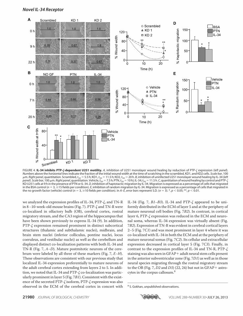
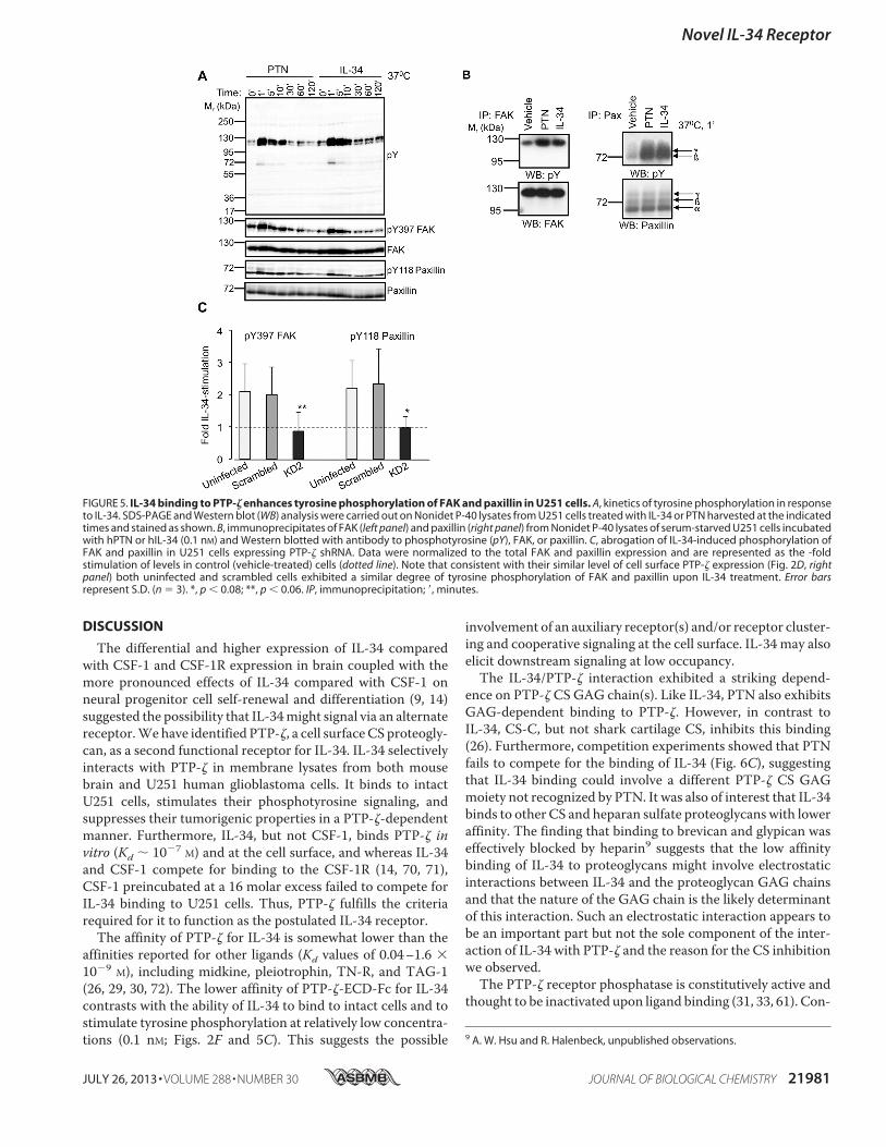
As PTNwas also shown previously to affect glioblastoma cellmigration (39–41), we compared the wound healing rates ofthe scrambled and PTP-! KD cells (Fig. 4A). In the absence ofadded ligand(s), KD clones exhibited a slower rate of woundhealing, indicating that the constitutively active PTP-! receptorfacilitates U251 migration (e.g. time taken for 50% wound clo-sure (t50) for KD2 cells was '20 h compared with 5.5 h forscrambled cells; Fig. 4A). Consistent with ligand-induced inac-tivation of the receptor (31, 33, 61), PTN (t50 ) 10 h) or IL-34(t50 ) 11.5 h) significantly inhibited wound healing in unin-fected cells (vehicle t50 ) 7.5 h) (Fig. 4B). Furthermore, neitherIL-34 nor PTN could suppress PTP-! KD cell wound healing(Fig. 4C), thereby indicating that suppression of healing byeither ligand is mediated through PTP-!. To determinewhether IL-34 and PTN suppress directed migration, we uti-lized a haptotaxis assay in which PTP-! ligands were shown tobemore effective in regulatingmigration than in a conventionalchemotaxis assay (62, 63). Both IL-34 and PTNwhen coated onthe bottom of the membrane suppressed migration of U251cells (Fig. 4D). To determine whether IL-34 and PTN alsoinhibit random migration, we examined migration of the cellsthrough membranes containing these growth factors on bothsides. Both IL-34 and PTN inhibited the random migration ofthe cells (Fig. 4E). Together, these results demonstrate thatIL-34 suppresses proliferation, clonogenicity, and motility ofU251 cells in vitro in a PTP-!-dependent manner.hIL-34 Enhances PTP-!-mediated Tyrosine Phosphorylation
of FAK and Paxillin in U251 Cells—To function as a receptorfor IL-34, IL-34 binding to cell surface PTP-! should triggerintracellular signaling. Consistent with the reduction of PTP-!phosphatase activity by ligand binding (31, 34, 61), PTP-!ligand binding has been shown previously to trigger intracellu-lar protein tyrosine phosphorylation (32, 33, 35, 36). Following7 S. Nandi and E. R. Stanley, unpublished observations.
Novel IL-34 Receptor
JULY 26, 2013 • VOLUME 288 • NUMBER 30 JOURNAL OF BIOLOGICAL CHEMISTRY 21977
at Colum
bia University on N
ovember 15, 2013
http://ww
w.jbc.org/
Dow
nloaded from

incubation of U251 cells with PTN or IL-34 for various times at37 °C, we observed a similar ligand-induced tyrosine phospho-rylation of proteins, including those with apparent molecularmasses of (190, (125, (120, (70, and (42 kDa, that peakedwithin the first 5 min of stimulation (Fig. 5A). PTP-! ligandshave been shown to increase the tyrosine phosphorylation ofFAK in lung and prostate carcinomas and endothelial cells (32,38, 41) and of paxillin in osteoblastic cells (64), and G protein-coupled receptor kinase interactor 1/Cool-associated, tyrosine-phosphorylated 1 (GIT1/Cat-1) has been shown to be a directPTP-! substrate (65).We showed that either PTN or IL-34 alsoincreased the tyrosine phosphorylation of FAK ((125 kDa),
paxillin ((70 kDa) (Fig. 5, A and B), and GIT1/Cat-17 in U251cells.In contrast, we failed to detect an increase in the tyrosine
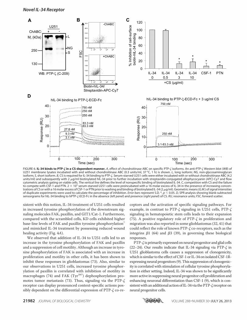
phosphorylation of the putative PTP-! substrates "-cateninand "-adducin (33, 35) in response to either ligand.7 IL-34-mediated activations of FAK and paxillin were abolished in thePTP-! KD2 cell (Fig. 5C), demonstrating that IL-34-inducedtyrosine phosphorylation of these proteins is mediated byPTP-!.hIL-34 Binds to PTP-! in a Chondroitin Sulfate-dependent
Manner—PTP-! is a proteoglycan receptor for several ligands(25–29). Furthermore, the PTP-!CS chains are known to affect
Novel IL-34 Receptor
21978 JOURNAL OF BIOLOGICAL CHEMISTRY VOLUME 288 • NUMBER 30 • JULY 26, 2013
at Colum
bia University on N
ovember 15, 2013
http://ww
w.jbc.org/
Dow
nloaded from

binding to some of these ligands (26, 30). We therefore testedthe requirement of CS for IL-34 binding. Consistent with thepreviously reported presence of CS on PTP-! (20, 66), treat-ment of solubilizedU251membraneswith chondroitinaseABCincreased the mobility of a significant fraction of the largePTP-! isoform (Fig. 6A). To determine the requirement of CSfor cell surface binding, intact U251 cells were incubated withenzymebuffer alone orwith chondroitinaseABC to remove cellsurface CS. Treatment with chondroitinase ABC reduced bind-ing of biotinylated IL-34 to the level seen in IL-34-competedcells (background levels) (Fig. 6B). Preincubation of U251 cellswith a 16molar excess of IL-34 blocked the subsequent bindingof biotinylated IL-34, whereas preincubation with a 16 molarexcess of CSF-1 or PTNwaswithout effect (Fig. 6C). Consistentwith the removal of binding sites by preincubation with chon-droitinase ABC, preincubation of IL-34with 3$g/ml shark car-tilage CS blocked IL-34 inhibition of biotinylated IL-34 binding
(Fig. 6C). Thus, the CS GAG moiety of PTP-! is involved inIL-34 binding. SPR analysis further confirmed the inhibition byCS (Fig. 6D).Comparative Expression Profiles of PTP-!, TN-R, and IL-34 in
Adult Brain—Previous studies have shown that PTP-! is pri-marily expressed in neural progenitors and glial cells (22–24) aswell as in a subset of cortical neurons (23, 26). The expression ofTN-R overlaps with PTP-! expression in rostral brain regions(67–69). IL-34 expression is primarily observed onmature neu-rons (9, 16, 17), including regions of the brain where PTP-! isexpressed (24). We have shown previously that IL-34 expres-sion profiles are distinct from those of its cognate receptor,CSF-1R, and also from those of CSF-1 and that it is preferen-tially increased in specific areas of early postnatal and adultforebrain, thereby suggesting the presence of an alternative sig-naling receptor (9). As PTP-! functions as a cell surface recep-tor for IL-34 (Figs. 2–5) and also interacts with TN-R (Fig. 1),
FIGURE 2. IL-34 binds cell surface PTP-! in U251 glioblastoma cells. A, U251 human glioblastoma cells lack the CSF-1R. Left panel, failure of hIL-34 to bind toCSF-1R on U251 cells. OG-solubilized membrane fractions of BAC1.2F5 (lanes 1 and 2) and U251 (lanes 3 and 4) cells were incubated overnight at 4 °C withbiotinylated mIL-34 (lanes 1 and 2) or biotinylated hIL-34 (lanes 3 and 4), and the biotinylated IL-34#neutravidin complexes were analyzed by SDS-PAGE andWestern blotting (WB) with an antibody that equally recognizes both mouse CSF-1R and hCSF-1R. Middle panel, absence of CSF-1R in U251 cells. OG-solubilizedmembrane fractions of U251 human glioblastoma (lane 1) and NIH-3T3-hCSF-1R (lanes 2– 4) cells were incubated overnight at 4 °C with anti-hCSF-1R antibody(lanes 1 and 2) or biotinylated hIL-34 (lanes 3 and 4), and the biotinylated IL-34#neutravidin complexes and CSF-1R immunoprecipitates were analyzed as above.Right panel, flow cytometric verification. 3T3-hCSF-1R or U251 cells (2 # 105) were incubated with 5 $g/ml rat monoclonal anti-hCSF-1R antibody or rat IgG1(isotype control) for 30 min at 4 °C, washed with PBS, and further incubated with 5 $g/ml anti-rat IgG1-conjugated FITC for 30 min at 4 °C. B, interaction of IL-34with PTP-! in OG-solubilized U251 cell membrane fractions. Membranes lysates were incubated with biotinylated hIL-34 (4 °C, 16 h), and the complexes werecaptured with neutravidin beads (4 °C, 6 h), eluted with SDS, and analyzed by SDS-PAGE and silver staining or by Western blotting (WB) with antibody to PTP-!.Arrowhead, nonspecific band; asterisk, PTP-! proteolytic product (52); L, long isoform; NG, non-glycosaminoglycan form; S, short isoform. C, reduced PTP-!expression in PTP-! KD U251 clones. Left panel, PTP-! and control (EF1#) Western blots of OG-solubilized whole cell lysates from cells expressing scrambled orPTP-! (KD1 and KD2) shRNAs. Right panel, quantitation of the combined intensities of the three bands (long isoform, non-glycosaminoglycan form, and shortisoform) from two independent experiments. Note that the scrambled cell line expressed a higher total PTP-! compared with uninfected U251 cells. D, flowcytometric analysis of hIL-34 binding to PTP-! KD U251 lines. Serum-starved control (scrambled) and KD (KD1 and KD2) cells were either untreated or incubatedwith 5 $g/ml biotinylated hIL-34 and then subsequently incubated with streptavidin-conjugated allophycocyanin (APC)-Cy7 prior to flow cytometric analysisgating on viable cells. Right panel, quantitation. Note that although scrambled cells expressed a higher level of total PTP-! cell surface expression was notsignificantly different from uninfected cells. Compared with the scrambled line, both KD1 and KD2 lines expressed lower levels of cell surface PTP-!, and KD2cells also had reduced cell surface PTP-! compared with uninfected cells. G.M., geometric means of signal intensities of duplicate experiments. E, FLAG-taggedIL-34, but not FLAG-tagged CSF-1, binds U251 cells. F, dose dependence of FLAG-tagged IL-34 binding to U251 cells. Control CM, 293T conditioned medium. InC and D, error bars represent S.D. (n ) 3). *, p % 0.07; **, p % 0.05; ***, p % 0.001. PE, phycoerythrin; FSC, forward scatter.
FIGURE 3. IL-34 inhibits growth and clonogenicity of U251 glioblastoma cells in a PTP-!-dependent manner. A, cell proliferation assays. Control (left panel)or PTP-! KD (right panel) cells were incubated with the indicated factors and times, and the number of viable cells was assessed by trypan blue exclusionstaining. B, clonogenic assay. Left panel, micrographs of colony forming assays of U251 cells incubated with vehicle or IL-34 as described under “ExperimentalProcedures.” Right panel, histograms showing average colony counts from triplicate experiments. In A and B, error bars represent S.D. (n ) 3). *, significantlydifferent from cells incubated with vehicle alone, p % 0.05.
Novel IL-34 Receptor
JULY 26, 2013 • VOLUME 288 • NUMBER 30 JOURNAL OF BIOLOGICAL CHEMISTRY 21979
at Colum
bia University on N
ovember 15, 2013
http://ww
w.jbc.org/
Dow
nloaded from
we analyzed the expression profiles of IL-34, PTP-!, and TN-Rin 8–10-week-old mouse brains (Fig. 7). PTP-! and TN-R wereco-localized in olfactory bulb (OB), cerebral cortex, rostralmigratory stream, and the CA3 region of the hippocampus thathave been shown previously to express IL-34 (9). In addition,PTP-! expression remained prominent in distinct subcorticalstructures (thalamic and subthalamic nuclei), midbrain, andbrain stem nuclei (inferior colliculus, pontine nuclei, locuscoeruleus, and vestibular nuclei) as well as the cerebellum anddisplayed distinct co-localization patterns with both IL-34 andTN-R (Fig. 7, A–D). Mature postmitotic neurons of the cere-brum were labeled by all three of these markers (Fig. 7, E–H).These observations are consistent with our previous study thatlocalized IL-34 expression preferentially to mature neurons ofthe adult cerebral cortex extending from layers 2 to 5. In addi-tion, we noted that IL-34 and PTP-! co-localization was partic-ularly prominent in layer 5 (Fig. 7B1). Consistent with the exist-ence of the secreted PTP-! isoform, PTP-! expression was alsoobserved in the ECM of the cerebral cortex in concert with
IL-34 (Fig. 7, B1–B3). IL-34 and PTP-! appeared to be uni-formly distributed in the ECMof layer 5 and at the periphery ofmature neuronal cell bodies (Fig. 7B2). In contrast, in corticallayer 6, PTP-! expression was reduced in the ECM and neuro-nal soma, whereas IL-34 expression was virtually absent (Fig.7B2). Expression of TN-Rwas evident in cerebral cortical layers2–5 (Fig. 7C1) and was most prominent in layer 4 where it wasco-localizedwith IL-34 in both the ECMand at the periphery ofmature neuronal somas (Fig. 7C2). Its cellular and extracellularexpression decreased in cortical layer 5 (Fig. 7C3). Finally, incontrast to the expression profiles of IL-34 and TN-R, PTP-!stainingwas also seen inGFAP" adult neural stemcells presentin the anterior subventricular zone (Fig. 7D1) as well as in thoseneural species migrating through the rostral migratory streamto the OB (Fig. 7,D2 andD3) (22, 24) but not in GFAP" astro-cytes in the corpus callosum.8
8 S. Gokhan, unpublished observations.
FIGURE 4. IL-34 inhibits PTP-!-dependent U251 motility. A, inhibition of U251 monolayer wound healing by reduction of PTP-! expression (left panel).Numbers above the horizontal lines indicate the fraction of the initial wound width at the time of scratching in the scrambled, KD1, and KD2 cells. Scale bar, 100$m. Right panel, quantitation. Scrambled, t50 ) 5.5 h; KD1, t50 ) 11.5 h; KD2, t50 ' 20 h. B, inhibition of uninfected U251 monolayer wound healing by IL-34 (leftpanel). Scale bar, 100 $m. Right panel, quantitation. Vehicle, t50 ) 7.5 h; PTN, t50 ) 10 h); IL-34; t50 ) 11.5 h. C, quantitation of wound healing by control and PTP-!KD U251 cells at 9 h in the presence of PTN or IL-34. D, inhibition of haptotactic migration by IL-34. Migration is expressed as a percentage of cells that migratedin the BSA control (n ) 3, %15 fields per condition). E, inhibition of random migration by IL-34. Migration is expressed as a percentage of cells that migrated inthe no growth factor (vehicle) control (n ) 3, %10 fields per condition). In A–E, error bars represent S.D. (n ) 3). *, p % 0.05; **, p % 0.01.
Novel IL-34 Receptor
21980 JOURNAL OF BIOLOGICAL CHEMISTRY VOLUME 288 • NUMBER 30 • JULY 26, 2013
at Colum
bia University on N
ovember 15, 2013
http://ww
w.jbc.org/
Dow
nloaded from
DISCUSSION
The differential and higher expression of IL-34 comparedwith CSF-1 and CSF-1R expression in brain coupled with themore pronounced effects of IL-34 compared with CSF-1 onneural progenitor cell self-renewal and differentiation (9, 14)suggested the possibility that IL-34might signal via an alternatereceptor.Wehave identified PTP-!, a cell surfaceCS proteogly-can, as a second functional receptor for IL-34. IL-34 selectivelyinteracts with PTP-! in membrane lysates from both mousebrain and U251 human glioblastoma cells. It binds to intactU251 cells, stimulates their phosphotyrosine signaling, andsuppresses their tumorigenic properties in a PTP-!-dependentmanner. Furthermore, IL-34, but not CSF-1, binds PTP-! invitro (Kd ( 10!7 M) and at the cell surface, and whereas IL-34and CSF-1 compete for binding to the CSF-1R (14, 70, 71),CSF-1 preincubated at a 16 molar excess failed to compete forIL-34 binding to U251 cells. Thus, PTP-! fulfills the criteriarequired for it to function as the postulated IL-34 receptor.
The affinity of PTP-! for IL-34 is somewhat lower than theaffinities reported for other ligands (Kd values of 0.04–1.6 #10!9 M), including midkine, pleiotrophin, TN-R, and TAG-1(26, 29, 30, 72). The lower affinity of PTP-!-ECD-Fc for IL-34contrasts with the ability of IL-34 to bind to intact cells and tostimulate tyrosine phosphorylation at relatively low concentra-tions (0.1 nM; Figs. 2F and 5C). This suggests the possible
involvement of an auxiliary receptor(s) and/or receptor cluster-ing and cooperative signaling at the cell surface. IL-34 may alsoelicit downstream signaling at low occupancy.The IL-34/PTP-! interaction exhibited a striking depend-
ence on PTP-! CS GAG chain(s). Like IL-34, PTN also exhibitsGAG-dependent binding to PTP-!. However, in contrast toIL-34, CS-C, but not shark cartilage CS, inhibits this binding(26). Furthermore, competition experiments showed that PTNfails to compete for the binding of IL-34 (Fig. 6C), suggestingthat IL-34 binding could involve a different PTP-! CS GAGmoiety not recognized by PTN. It was also of interest that IL-34binds to other CS and heparan sulfate proteoglycans with loweraffinity. The finding that binding to brevican and glypican waseffectively blocked by heparin9 suggests that the low affinitybinding of IL-34 to proteoglycans might involve electrostaticinteractions between IL-34 and the proteoglycan GAG chainsand that the nature of the GAG chain is the likely determinantof this interaction. Such an electrostatic interaction appears tobe an important part but not the sole component of the inter-action of IL-34 with PTP-! and the reason for the CS inhibitionwe observed.The PTP-! receptor phosphatase is constitutively active and
thought to be inactivated upon ligand binding (31, 33, 61). Con-
9 A. W. Hsu and R. Halenbeck, unpublished observations.
FIGURE 5. IL-34 binding to PTP-! enhances tyrosine phosphorylation of FAK and paxillin in U251 cells. A, kinetics of tyrosine phosphorylation in responseto IL-34. SDS-PAGE and Western blot (WB) analysis were carried out on Nonidet P-40 lysates from U251 cells treated with IL-34 or PTN harvested at the indicatedtimes and stained as shown. B, immunoprecipitates of FAK (left panel) and paxillin (right panel) from Nonidet P-40 lysates of serum-starved U251 cells incubatedwith hPTN or hIL-34 (0.1 nM) and Western blotted with antibody to phosphotyrosine (pY), FAK, or paxillin. C, abrogation of IL-34-induced phosphorylation ofFAK and paxillin in U251 cells expressing PTP-! shRNA. Data were normalized to the total FAK and paxillin expression and are represented as the -foldstimulation of levels in control (vehicle-treated) cells (dotted line). Note that consistent with their similar level of cell surface PTP-! expression (Fig. 2D, rightpanel) both uninfected and scrambled cells exhibited a similar degree of tyrosine phosphorylation of FAK and paxillin upon IL-34 treatment. Error barsrepresent S.D. (n ) 3). *, p % 0.08; **, p % 0.06. IP, immunoprecipitation; &, minutes.
Novel IL-34 Receptor
JULY 26, 2013 • VOLUME 288 • NUMBER 30 JOURNAL OF BIOLOGICAL CHEMISTRY 21981
at Colum
bia University on N
ovember 15, 2013
http://ww
w.jbc.org/
Dow
nloaded from
sistent with this notion, IL-34 treatment of U251 cells resultedin increased tyrosine phosphorylation of the downstream sig-naling molecules FAK, paxillin, and GIT1/Cat-1. Furthermore,compared with the scrambled cells, KD cells exhibited higherbase-line levels of FAK and paxillin tyrosine phosphorylation7and mimicked IL-34 treatment by possessing reduced woundhealing activity (Fig. 4A).We observed that addition of IL-34 to U251 cells led to an
increase in the tyrosine phosphorylation of FAK and paxillinand a suppression of cell motility. Although an increase in tyro-sine phosphorylation of FAK is associated with an increase inproliferation and motility in other cells, it has been shown toinhibit these responses in glioblastomas (73). Also, similar toour observations in U251 cells, increased tyrosine phosphor-ylation of paxillin is correlated with inhibition of motility inmacrophages (74) and FAK (Tyr397) dephosphorylation pro-motes tumor metastasis (73). Thus, signaling via the PTP-!receptor can display pronounced context-specific actions pos-sibly dependent on the differential expression of PTP-! co-re-
ceptors and the activation of specific signaling pathways. Forexample, in contrast to PTP-! signaling in U251 cells, PTP-!signaling in hematopoietic stem cells leads to their expansion(75). A positive regulatory role of PTP-! in proliferation andmigration was also reported in some glioblastomas (32, 41) thatcould reflect the role of known PTP-! co-receptors, such as theintegrins "1 (64) and "3 (39), in governing these biologicalresponses.PTP-! is primarily expressedonneuralprogenitorandglial cells
(22–24). Our results indicate that IL-34 signaling via PTP-! inU251 glioblastoma cells causes a suppression of clonogenicity,which is similar to the effect ofCSF-1or IL-34on isolatedCSF-1R-expressingneural progenitors (9). This suppressionof clonogenic-ity is correlated with stimulation of cellular tyrosine phosphoryla-tion in either setting. Indeed, IL-34 was shown to be significantlymore active in suppressingneural progenitor cell proliferation andenhancing neuronal differentiation than CSF-1 (9), which is con-sistentwith anadditional actionof IL-34via thePTP-! receptoronneural progenitor cells.
FIGURE 6. IL-34 binds to PTP-! in a CS-dependent manner. A, effect of chondroitinase ABC on specific PTP-! isoforms. An anti-PTP-! Western blot (WB) ofU251 membrane lysates incubated with and without chondroitinase ABC (0.3 units/ml, 37 °C, 1 h) is shown. L, long isoform; NG, non-glycosaminoglycanisoform; S, short isoform. B, CS is required for IL-34 binding to PTP-!. Serum-starved U251 cells were either incubated with or without chondroitinase ABC (4.2units/ml) and subsequently with 2 $g/ml biotinylated hIL-34 prior to further incubation with streptavidin-conjugated allophycocyanin (APC)-Cy7 and flowcytometric analysis gating on viable cells. The vertical line defines the level of nonspecific binding of biotinylated IL-34. C, competition with CS and the failureto compete with CSF-1 and PTN. 2 # 105 serum-starved U251 cells were preincubated with a 16 molar excess of IL-34 in the presence of increasing concen-trations of CS or with a 16 molar excess of CSF-1 or PTN prior to washing and binding of biotinylated IL-34 (2 $g/ml). Geometric means (G.M.) of signal intensitiesof duplicate experiments were used to calculate the percentage of inhibition. Error bars represent S.D. *, p % 0.05. D, SPR analysis showing blank-subtractedsensorgrams for hIL-34 binding to hPTP-!-ECD-Fc in the absence (left panel) and presence (right panel) of CS. RU, resonance units; FSC, forward scatter.
Novel IL-34 Receptor
21982 JOURNAL OF BIOLOGICAL CHEMISTRY VOLUME 288 • NUMBER 30 • JULY 26, 2013
at Colum
bia University on N
ovember 15, 2013
http://ww
w.jbc.org/
Dow
nloaded from

In addition, IL-34 suppressed U251 glioblastoma cell motil-ity. Relevant to this, we observed PTP-! expression on imma-ture cells migrating within the rostral migratory stream (Fig. 7,D2 and D3) at the periphery of which IL-34 (9) and TN-R (Fig.7C) (68) are expressed. It is possible that local regulation ofthese migrating progenitor cells via IL-34/PTP-!/TN-R signal-ingmay have structural and functional consequences related tothe integrity of the rostral migratory stream and olfaction.
Both PTP-! (23, 26) and IL-34 (9, 16) are expressed on corti-cal neurons. Our immunofluorescence localization studiesrevealed that IL-34, PTP-!, and TN-R were expressed through-out cortical layers 2–5. IL-34 was often co-localized with PTP-!and TN-R on the surface of mature neurons in cortical layers 5and 4, respectively. Thus, IL-34/TN-R/PTP-! signaling in thesemature, cortical neurons in an autocrine and/or a paracrinemanner may play a role in their maintenance and possibly in
FIGURE 7. Immunofluorescence localization of PTP-!, TN-R, and IL-34 in adult brain. A–D, photomontages made from serial sagittal sections of 8 –10-weekmouse brain showing the expression patterns of IL-34 (blue), PTP-! (green), and TN-R (red). IL-34 expression is observed within the OB and cerebral cortex withan increasing gradient of expression in caudal areas, including visual cortex (VCx) as well as the striatum (STR), thalamus (Th), inferior colliculus (IC), andcerebellum (Cb). PTP-! expression is present in all areas of the cerebrum and cerebellum where IL-34 expression is also observed. IL-34 is also present within thesubthalamic nucleus (STh), pontine nuclei (PN), locus ceruleus (LC), and vestibular nucleus (VN). TN-R expression appears to be present in more rostral areas ofthe cerebrum in which the highest expression levels are restricted to the rostral cerebral cortex, including the motor cortex (MCx; boxed) and OB. H, hippocam-pus. E–H, PTP-!, TN-R, and IL-34 are mostly observed in areas of the cerebrum in which mature NeuN-positive (green) neurons are present. Their expressionprofiles in the cerebellum are preferentially localized to the molecular and Purkinje cell layers (arrow). B1–B3, PTP-! and IL-34 are co-localized in cerebral corticallayers 2–5 with preferential expression in cortical layer 5 (arrow). PTP-! continues to be present within cortical layer 6 (arrowhead), whereas no IL-34 expressionis observed in this area. C1–C3, mature neurons within cerebral cortical layers 2–5 also express TN-R, and its co-localization with IL-34 is highest in cortical layer4 (arrow). TN-R expression is also observed in mature neurons of cortical layer 5 (arrowhead). D1, PTP-! (red) expression is also visible in the anterior subven-tricular zone (aSVZ), where it is co-localized with GFAP-expressing (green) adult neural stem cells. D2 and D3 (higher power image), GFAP-positive adult stemcells continue to express PTP-! as they undergo migration to the OB within the rostral migratory stream (RMS). GFAP-positive astrocytes present within thecorpus callosum (CC) do not express PTP-!.8 Scale bars, 1 mm (A–H), 50 $m (B1, C1, and D2), 40 $m (D1), and 25 $m (B2, B3, C2, C3, and D3).
Novel IL-34 Receptor
JULY 26, 2013 • VOLUME 288 • NUMBER 30 JOURNAL OF BIOLOGICAL CHEMISTRY 21983
at Colum
bia University on N
ovember 15, 2013
http://ww
w.jbc.org/
Dow
nloaded from

mediating higher cognitive functions. Interestingly, we alsoobserved co-localization of IL-34 with secreted PTP-! (Fig.7B2) and with TN-R (Fig. 7C2) in the cerebral cortical ECMconsistent with the existence of a stable IL-34#PTP-!#TN-Rcomplex (Fig. 1D). Such a stable association may serve to pro-vide high local concentrations of IL-34 to receptor-expressingcells. The identification of PTP-! as a novel receptor for IL-34necessitates a reevaluation of the possible role(s) of IL-34/PTP-! signaling in tissues inwhich both ligand and receptor areexpressed. Obviously the CNS is an important organ system tostudy because of the significant expression of both IL-34 andPTP-! in brain and because PTP-! has been implicated in sev-eral disease settings in the CNS. For example, it is expressed inremyelinating oligodendrocytes, and PTP-!-deficient mice dis-play a delayed recovery from demyelinating lesions in a modelof experimental autoimmune encephalomyelitis (76). Further-more, the soluble PTP-! isoform has been shown to be neces-sary for maturation of oligodendrocyte progenitors to differen-tiated myelin-secreting oligodendrocytes in vitro (77), andPTP-!-deficientmice exhibit increasedmyelin breakdown (78).In addition, the PTPRZ1 gene in humans is a schizophrenia-susceptibility gene (79), and PTP-! regulates tyrosine phos-phorylation of voltage-gated sodium channels in neurons (80).Given the multiplicity of ligands for PTP-!, the role of IL-34regulation in these settingswill need to be further defined. Also,our demonstration that IL-34 modulates tumorigenic proper-ties of the glioblastoma cell line U251 coupled with the fact thatPTP-! is expressed in neuroblastomas (19) and other tumors(38, 41) strongly suggests that analysis of the CSF-1R-depen-dent and -independent roles of IL-34 in tumorigenesis is ofgreat translational medicine relevance.
Acknowledgments—We thank Dr. Andreas Fiser, Tyler Roche, andDr. Michael Brenowitz of the Einstein College of Medicine for insightsrelevant to receptor/ligand interactions.We thankDr. Jeffery Segall ofthe Einstein College of Medicine for the U251 and U87MG humanglioblastoma lines; Dr. Shinya Suzu of Kumamoto University, Japan,for the IL-34-FLAG and CSF-1-FLAG constructs; Drs. AkihiroFujikawa and Masaharu Noda of the Division of Molecular Neuro-biology, National Institute for Basic Biology, Okazaki, Aichi, Japan foradvice and membrane preparations; and Dr. Violeta Chitu for criti-cally reading the manuscript. We also thank the staff of the FlowCytometry Facility, Laboratory for Macromolecular Analysis andProteomics, and the Protein Production section of the Macromolecu-lar TherapeuticsDevelopment Facility at the EinsteinCollege ofMed-icine and Drs. Cristina Caescu andMary Short for advice on plasmidDNA preparation for expression.
REFERENCES1. Sherr, C. J., Rettenmier, C. W., Sacca, R., Roussel, M. F., Look, A. T., and
Stanley, E. R. (1985) The c-fms proto-oncogene product is related to thereceptor for the mononuclear phagocyte growth factor, CSF-1. Cell 41,665–676
2. Yeung, Y. G., Jubinsky, P. T., Sengupta, A., Yeung, D. C., and Stanley, E. R.(1987) Purification of the colony-stimulating factor 1 receptor and dem-onstration of its tyrosine kinase activity. Proc. Natl. Acad. Sci. U.S.A. 84,1268–1271
3. Dai, X. M., Zong, X. H., Akhter, M. P., and Stanley, E. R. (2004) Osteoclastdeficiency results in disorganized matrix, reducedmineralization, and ab-normal osteoblast behavior in developing bone. J. Bone Miner. Res. 19,
1441–14514. Yoshida, H., Hayashi, S., Kunisada, T., Ogawa, M., Nishikawa, S., Oka-
mura, H., Sudo, T., Shultz, L. D., and Nishikawa, S. (1990) The murinemutation osteopetrosis is in the coding region of the macrophage colonystimulating factor gene. Nature 345, 442–444
5. Wiktor-Jedrzejczak, W., Bartocci, A., Ferrante, A. W., Jr., Ahmed-Ansari,A., Sell, K. W., Pollard, J. W., and Stanley, E. R. (1990) Total absence ofcolony-stimulating factor 1 in the macrophage-deficient osteopetrotic(op/op) mouse. Proc. Natl. Acad. Sci. U.S.A. 87, 4828–4832
6. Cecchini, M. G., Dominguez, M. G., Mocci, S., Wetterwald, A., Felix, R.,Fleisch, H., Chisholm, O., Hofstetter, W., Pollard, J. W., and Stanley, E. R.(1994) Role of colony stimulating factor-1 in the establishment and regu-lation of tissue macrophages during postnatal development of the mouse.Development 120, 1357–1372
7. Huynh, D., Dai, X. M., Nandi, S., Lightowler, S., Trivett, M., Chan, C. K.,Bertoncello, I., Ramsay, R. G., and Stanley, E. R. (2009) Colony stimulatingfactor-1 dependence of Paneth cell development in the mouse small in-testine. Gastroenterology 137, 136–144, 144.e1–3
8. Guleria, I., and Pollard, J.W. (2000) The trophoblast is a component of theinnate immune system during pregnancy. Nat. Med. 6, 589–593
9. Nandi, S., Gokhan, S., Dai, X. M., Wei, S., Enikolopov, G., Lin, H., Mehler,M. F., and Stanley, E. R. (2012) The CSF-1 receptor ligands IL-34 andCSF-1 exhibit distinct developmental brain expression patterns and reg-ulate neural progenitor cell maintenance and maturation. Dev. Biol. 367,100–113
10. Pixley, F. J., and Stanley, E. R. (2004) CSF-1 regulation of the wanderingmacrophage: complexity in action. Trends Cell Biol. 14, 628–638
11. Chitu, V., and Stanley, E. R. (2006) Colony-stimulating factor-1 in immu-nity and inflammation. Curr. Opin. Immunol. 18, 39–48
12. Dai, X.M., Ryan,G. R.,Hapel, A. J., Dominguez,M.G., Russell, R.G., Kapp,S., Sylvestre, V., and Stanley, E. R. (2002) Targeted disruption of themousecolony-stimulating factor 1 receptor gene results in osteopetrosis, mono-nuclear phagocyte deficiency, increased primitive progenitor cell frequen-cies, and reproductive defects. Blood 99, 111–120
13. Lin, H., Lee, E., Hestir, K., Leo, C., Huang, M., Bosch, E., Halenbeck, R.,Wu, G., Zhou, A., Behrens, D., Hollenbaugh, D., Linnemann, T., Qin, M.,Wong, J., Chu, K., Doberstein, S. K., and Williams, L. T. (2008) Discoveryof a cytokine and its receptor by functional screening of the extracellularproteome. Science 320, 807–811
14. Wei, S., Nandi, S., Chitu, V., Yeung, Y. G., Yu, W., Huang, M., Williams,L. T., Lin, H., and Stanley, E. R. (2010) Functional overlap but differentialexpression of CSF-1 and IL-34 in their CSF-1 receptor-mediated regula-tion of myeloid cells. J. Leukoc. Biol. 88, 495–505
15. Ginhoux, F., Greter, M., Leboeuf, M., Nandi, S., See, P., Gokhan, S.,Mehler, M. F., Conway, S. J., Ng, L. G., Stanley, E. R., Samokhvalov, I. M.,and Merad, M. (2010) Fate mapping analysis reveals that adult microgliaderive from primitive macrophages. Science 330, 841–845
16. Wang, Y., Szretter, K. J., Vermi, W., Gilfillan, S., Rossini, C., Cella, M.,Barrow, A. D., Diamond, M. S., and Colonna, M. (2012) IL-34 is a tissue-restricted ligand of CSF1R required for the development of Langerhanscells and microglia. Nat. Immunol. 13, 753–760
17. Greter, M., Lelios, I., Pelczar, P., Hoeffel, G., Price, J., Leboeuf, M., Kundig,T. M., Frei, K., Ginhoux, F., Merad, M., and Becher, B. (2012) Stroma-derived interleukin-34 controls the development and maintenance ofLangerhans cells and the maintenance of microglia. Immunity 37,1050–1060
18. Chihara, T., Suzu, S., Hassan, R., Chutiwitoonchai, N., Hiyoshi, M., Mo-toyoshi, K., Kimura, F., and Okada, S. (2010) IL-34 and M-CSF share thereceptor Fms but are not identical in biological activity and signal activa-tion. Cell Death Differ. 17, 1917–1927
19. Levy, J. B., Canoll, P. D., Silvennoinen,O., Barnea, G.,Morse, B., Honegger,A.M.,Huang, J. T., Cannizzaro, L. A., Park, S. H., andDruck, T. (1993) Thecloning of a receptor-type protein tyrosine phosphatase expressed in thecentral nervous system. J. Biol. Chem. 268, 10573–10581
20. Barnea, G., Grumet, M., Milev, P., Silvennoinen, O., Levy, J. B., Sap, J., andSchlessinger, J. (1994) Receptor tyrosine phosphatase" is expressed in theform of proteoglycan and binds to the extracellular matrix protein tenas-cin. J. Biol. Chem. 269, 14349–14352
Novel IL-34 Receptor
21984 JOURNAL OF BIOLOGICAL CHEMISTRY VOLUME 288 • NUMBER 30 • JULY 26, 2013
at Colum
bia University on N
ovember 15, 2013
http://ww
w.jbc.org/
Dow
nloaded from

21. Krueger, N. X., and Saito, H. (1992) A human transmembrane protein-tyrosine-phosphatase, PTP!, is expressed in brain and has an N-terminalreceptor domain homologous to carbonic anhydrases. Proc. Natl. Acad.Sci. U.S.A. 89, 7417–7421
22. von Holst, A., Sirko, S., and Faissner, A. (2006) The unique 473HD-Chon-droitinsulfate epitope is expressed by radial glia and involved in neuralprecursor cell proliferation. J. Neurosci. 26, 4082–4094
23. Shintani, T., Watanabe, E., Maeda, N., and Noda, M. (1998) Neurons aswell as astrocytes express proteoglycan-type protein tyrosine phosphatase!/RPTP": analysis of mice in which the PTP!/RPTP" gene was replacedwith the LacZ gene. Neurosci. Lett. 247, 135–138
24. Lafont, D., Adage, T., Greco, B., and Zaratin, P. (2009) A novel role forreceptor like protein tyrosine phosphatase ! in modulation of sensorimo-tor responses to noxious stimuli: evidences from knockout mice studies.Behav. Brain Res. 201, 29–40
25. Peles, E., Schlessinger, J., andGrumet,M. (1998)Multi-ligand interactionswith receptor-like protein tyrosine phosphatase ": implications for inter-cellular signaling. Trends Biochem. Sci. 23, 121–124
26. Maeda, N., Nishiwaki, T., Shintani, T., Hamanaka, H., and Noda, M.(1996) 6B4 proteoglycan/phosphacan, an extracellular variant of recep-tor-like protein-tyrosine phosphatase !/RPTP", binds pleiotrophin/hep-arin-binding growth-associated molecule (HB-GAM). J. Biol. Chem. 271,21446–21452
27. Li, Y. S., Milner, P. G., Chauhan, A. K., Watson, M. A., Hoffman, R. M.,Kodner, C. M., Milbrandt, J., and Deuel, T. F. (1990) Cloning and expres-sion of a developmentally regulated protein that induces mitogenic andneurite outgrowth activity. Science 250, 1690–1694
28. Peles, E., Nativ,M., Campbell, P. L., Sakurai, T.,Martinez, R., Lev, S., Clary,D. O., Schilling, J., Barnea, G., Plowman, G. D., Grumet, M., and Sch-lessinger, J. (1995) The carbonic anhydrase domain of receptor tyrosinephosphatase " is a functional ligand for the axonal cell recognition mole-cule contactin. Cell 82, 251–260
29. Milev, P., Chiba, A., Haring, M., Rauvala, H., Schachner, M., Ranscht, B.,Margolis, R. K., andMargolis, R. U. (1998) High affinity binding and over-lapping localization of neurocan and phosphacan/protein-tyrosine phos-phatase-!/" with tenascin-R, amphoterin, and the heparin-bindinggrowth-associated molecule. J. Biol. Chem. 273, 6998–7005
30. Maeda, N., Ichihara-Tanaka, K., Kimura, T., Kadomatsu, K., Muramatsu,T., and Noda, M. (1999) A receptor-like protein-tyrosine phosphatasePTP!/RPTP" binds a heparin-binding growth factor midkine. Involve-ment of arginine 78 of midkine in the high affinity binding to PTP!. J. Biol.Chem. 274, 12474–12479
31. Majeti, R., Bilwes, A. M., Noel, J. P., Hunter, T., and Weiss, A. (1998)Dimerization-induced inhibition of receptor protein tyrosine phospha-tase function through an inhibitory wedge. Science 279, 88–91
32. Polykratis, A., Katsoris, P., Courty, J., and Papadimitriou, E. (2005) Char-acterization of heparin affin regulatory peptide signaling in human endo-thelial cells. J. Biol. Chem. 280, 22454–22461
33. Meng, K., Rodriguez-Pena, A., Dimitrov, T., Chen, W., Yamin, M., Noda,M., and Deuel, T. F. (2000) Pleiotrophin signals increased tyrosine phos-phorylation of "-catenin through inactivation of the intrinsic catalyticactivity of the receptor-type protein tyrosine phosphatase "/!. Proc. Natl.Acad. Sci. U.S.A. 97, 2603–2608
34. Fukada, M., Fujikawa, A., Chow, J. P., Ikematsu, S., Sakuma, S., and Noda,M. (2006) Protein tyrosine phosphatase receptor type Z is inactivated byligand-induced oligomerization. FEBS Lett. 580, 4051–4056
35. Pariser, H., Perez-Pinera, P., Ezquerra, L., Herradon, G., and Deuel, T. F.(2005) Pleiotrophin stimulates tyrosine phosphorylation of "-adducinthrough inactivation of the transmembrane receptor protein tyrosinephosphatase "/!. Biochem. Biophys. Res. Commun. 335, 232–239
36. Pariser, H., Ezquerra, L., Herradon, G., Perez-Pinera, P., and Deuel, T. F.(2005) Fyn is a downstream target of the pleiotrophin/receptor proteintyrosine phosphatase "/!-signaling pathway: regulation of tyrosine phos-phorylation of Fyn by pleiotrophin. Biochem. Biophys. Res. Commun. 332,664–669
37. Fujikawa, A., Fukada,M.,Makioka, Y., Suzuki, R., Chow, J. P., Matsumoto,M., and Noda, M. (2011) Consensus substrate sequence for protein-ty-rosine phosphatase receptor type Z. J. Biol. Chem. 286, 37137–37146
38. Diamantopoulou, Z., Kitsou, P., Menashi, S., Courty, J., and Katsoris, P.(2012) Loss of receptor protein tyrosine phosphatase "/! (RPTP"/!) pro-motes prostate cancer metastasis. J. Biol. Chem. 287, 40339–40349
39. Mikelis, C., Sfaelou, E., Koutsioumpa, M., Kieffer, N., and Papadimitriou,E. (2009) Integrin #v"3 is a pleiotrophin receptor required for pleiotro-phin-induced endothelial cell migration through receptor protein tyro-sine phosphatase "/!. FASEB J. 23, 1459–1469
40. Muller, S., Kunkel, P., Lamszus, K., Ulbricht, U., Lorente, G. A., Nelson,A. M., von Schack, D., Chin, D. J., Lohr, S. C., Westphal, M., andMelcher,T. (2003) A role for receptor tyrosine phosphatase ! in glioma cell migra-tion. Oncogene 22, 6661–6668
41. Feng, Z. J., Gao, S. B.,Wu, Y., Xu, X. F., Hua, X., and Jin, G. H. (2010) Lungcancer cell migration is regulated via repressing growth factor PTN/RPTP"/! signaling by menin. Oncogene 29, 5416–5426
42. Yu, W., Chen, J., Xiong, Y., Pixley, F. J., Yeung, Y. G., and Stanley, E. R.(2012) Macrophage proliferation is regulated through CSF-1 receptor ty-rosines 544, 559, and 807. J. Biol. Chem. 287, 13694–13704
43. Garwood, J., Schnadelbach, O., Clement, A., Schutte, K., Bach, A., andFaissner, A. (1999) DSD-1-proteoglycan is the mouse homolog of phos-phacan and displays opposing effects on neurite outgrowth dependent onneuronal lineage. J. Neurosci. 19, 3888–3899
44. Edmonds, B. T.,Wyckoff, J., Yeung, Y. G.,Wang, Y., Stanley, E. R., Jones, J.,Segall, J., andCondeelis, J. (1996) Elongation factor-1# is an overexpressedactin binding protein in metastatic rat mammary adenocarcinoma. J. CellSci. 109, 2705–2714
45. Yeung, Y. G., Nieves, E., Angeletti, R. H., and Stanley, E. R. (2008) Removalof detergents from protein digests for mass spectrometry analysis. Anal.Biochem. 382, 135–137
46. Yeung, Y. G., and Stanley, E. R. (2003) Proteomic approaches to the anal-ysis of early events in colony-stimulating factor-1 signal transduction.Mol. Cell. Proteomics 2, 1143–1155
47. Roussel, M. F., Dull, T. J., Rettenmier, C. W., Ralph, P., Ullrich, A., andSherr, C. J. (1987) Transforming potential of the c-fms proto-oncogene(CSF-1 receptor). Nature 325, 549–552
48. Morgan, C., Pollard, J. W., and Stanley, E. R. (1987) Isolation and charac-terization of a cloned growth factor dependent macrophage cell line,BAC1.2F5. J. Cell. Physiol. 130, 420–427
49. Li, W., and Stanley, E. R. (1991) Role of dimerization and modification ofthe CSF-1 receptor in its activation and internalization during the CSF-1response. EMBO J. 10, 277–288
50. Nishiwaki, T., Maeda, N., and Noda, M. (1998) Characterization and de-velopmental regulation of proteoglycan-type protein tyrosine phospha-tase !/RPTP" isoforms. J. Biochem. 123, 458–467
51. Sakurai, T., Friedlander, D. R., and Grumet, M. (1996) Expression of poly-peptide variants of receptor-type protein tyrosine phosphatase ": the se-creted form, phosphacan, increases dramatically during embryonic devel-opment and modulates glial cell behavior in vitro. J. Neurosci. Res. 43,694–706
52. Chow, J. P., Fujikawa, A., Shimizu, H., Suzuki, R., and Noda, M. (2008)Metalloproteinase- and &-secretase-mediated cleavage of protein-tyro-sine phosphatase receptor type Z. J. Biol. Chem. 283, 30879–30889
53. Woodworth, A., Pesheva, P., Fiete, D., and Baenziger, J. U. (2004) Neuro-nal-specific synthesis and glycosylation of tenascin-R. J. Biol. Chem. 279,10413–10421
54. Maurel, P., Rauch, U., Flad, M., Margolis, R. K., andMargolis, R. U. (1994)Phosphacan, a chondroitin sulfate proteoglycan of brain that interactswith neurons and neural cell-adhesion molecules, is an extracellular var-iant of a receptor-type protein tyrosine phosphatase. Proc. Natl. Acad. Sci.U.S.A. 91, 2512–2516
55. Xiao, Z. C., Bartsch, U., Margolis, R. K., Rougon, G., Montag, D., andSchachner, M. (1997) Isolation of a tenascin-R binding protein frommouse brain membranes. A phosphacan-related chondroitin sulfate pro-teoglycan. J. Biol. Chem. 272, 32092–32101
56. Shitara, K., Yamada, H.,Watanabe, K., Shimonaka,M., and Yamaguchi, Y.(1994) Brain-specific receptor-type protein-tyrosine phosphatase RPTP"is a chondroitin sulfate proteoglycan in vivo. J. Biol. Chem. 269,20189–20193
57. Maeda, N., and Noda, M. (1998) Involvement of receptor-like protein
Novel IL-34 Receptor
JULY 26, 2013 • VOLUME 288 • NUMBER 30 JOURNAL OF BIOLOGICAL CHEMISTRY 21985
at Colum
bia University on N
ovember 15, 2013
http://ww
w.jbc.org/
Dow
nloaded from

tyrosine phosphatase !/RPTP" and its ligand pleiotrophin/heparin-bind-ing growth-associated molecule (HB-GAM) in neuronal migration. J. CellBiol. 142, 203–216
58. Fukazawa, N., Yokoyama, S., Eiraku, M., Kengaku, M., and Maeda, N.(2008) Receptor type protein tyrosine phosphatase !-pleiotrophin signal-ing controls endocytic trafficking of DNER that regulates neuritogenesis.Mol. Cell. Biol. 28, 4494–4506
59. Ulbricht, U., Eckerich, C., Fillbrandt, R., Westphal, M., and Lamszus, K.(2006) RNA interference targeting protein tyrosine phosphatase !/recep-tor-type protein tyrosine phosphatase " suppresses glioblastoma growthin vitro and in vivo. J. Neurochem. 98, 1497–1506
60. Foehr, E. D., Lorente, G., Kuo, J., Ram, R., Nikolich, K., andUrfer, R. (2006)Targeting of the receptor protein tyrosine phosphatase"with amonoclo-nal antibody delays tumor growth in a glioblastomamodel.Cancer Res. 66,2271–2278
61. Barr, A. J., Ugochukwu, E., Lee, W. H., King, O. N., Filippakopoulos, P.,Alfano, I., Savitsky, P., Burgess-Brown, N. A., Muller, S., and Knapp, S.(2009) Large-scale structural analysis of the classical human protein tyro-sine phosphatome. Cell 136, 352–363
62. Lu, K. V., Jong, K. A., Kim, G. Y., Singh, J., Dia, E. Q., Yoshimoto, K.,Wang,M. Y., Cloughesy, T. F., Nelson, S. F., andMischel, P. S. (2005) Differentialinduction of glioblastomamigration and growth by two forms of pleiotro-phin. J. Biol. Chem. 280, 26953–26964
63. Qi, M., Ikematsu, S., Maeda, N., Ichihara-Tanaka, K., Sakuma, S., Noda,M., Muramatsu, T., and Kadomatsu, K. (2001) Haptotactic migration in-duced by midkine. Involvement of protein-tyrosine phosphatase !. Mito-gen-activated protein kinase, and phosphatidylinositol 3-kinase. J. Biol.Chem. 276, 15868–15875
64. Muramatsu, H., Zou, P., Suzuki, H., Oda, Y., Chen, G. Y., Sakaguchi, N.,Sakuma, S., Maeda, N., Noda, M., Takada, Y., and Muramatsu, T. (2004)#4"1- and #6"1-integrins are functional receptors for midkine, a heparin-binding growth factor. J. Cell Sci. 117, 5405–5415
65. Kawachi, H., Fujikawa, A., Maeda, N., and Noda, M. (2001) IdentificationofGIT1/Cat-1 as a substratemolecule of protein tyrosine phosphatase !/"by the yeast substrate-trapping system. Proc. Natl. Acad. Sci. U.S.A. 98,6593–6598
66. Maeda, N., Hamanaka, H., Oohira, A., and Noda, M. (1995) Purification,characterization and developmental expression of a brain-specific chon-droitin sulfate proteoglycan, 6B4 proteoglycan/phosphacan.Neuroscience67, 23–35
67. El Ayachi, I., Fernandez, C., Baeza, N., De Paula, A. M., Pesheva, P., andFigarella-Branger, D. (2011) Spatiotemporal distribution of tenascin-R inthe developing human cerebral cortex parallels neuronal migration.J. Comp. Neurol. 519, 2379–2389
68. Saghatelyan, A., de Chevigny, A., Schachner, M., and Lledo, P. M. (2004)Tenascin-R mediates activity-dependent recruitment of neuroblasts inthe adult mouse forebrain. Nat. Neurosci. 7, 347–356
69. Woodworth, A., Fiete, D., and Baenziger, J. U. (2002) Spatial and temporal
regulation of tenascin-R glycosylation in the cerebellum. J. Biol. Chem.277, 50941–50947
70. Ma, X., Lin,W. Y., Chen, Y., Stawicki, S., Mukhyala, K.,Wu, Y., Martin, F.,Bazan, J. F., and Starovasnik, M. A. (2012) Structural basis for the dualrecognition of helical cytokines IL-34 and CSF-1 by CSF-1R. Structure 20,676–687
71. Liu, H., Leo, C., Chen, X., Wong, B. R., Williams, L. T., Lin, H., and He, X.(2012) The mechanism of shared but distinct CSF-1R signaling by thenon-homologous cytokines IL-34 and CSF-1. Biochim. Biophys. Acta1824, 938–945
72. Milev, P., Maurel, P., Haring, M., Margolis, R. K., and Margolis, R. U.(1996)TAG-1/axonin-1 is a high-affinity ligand of neurocan, phosphacan/protein-tyrosine phosphatase-!/", and N-CAM. J. Biol. Chem. 271,15716–15723
73. Zheng, Y., Xia, Y., Hawke, D., Halle, M., Tremblay, M. L., Gao, X., Zhou,X. Z., Aldape, K., Cobb, M. H., Xie, K., He, J., and Lu, Z. (2009) FAKphosphorylation by ERK primes ras-induced tyrosine dephosphorylationof FAK mediated by PIN1 and PTP-PEST.Mol. Cell 35, 11–25
74. Pixley, F. J., Lee, P. S., Condeelis, J. S., and Stanley, E. R. (2001) Proteintyrosine phosphatase ' regulates paxillin tyrosine phosphorylation andmediates colony-stimulating factor 1-induced morphological changes inmacrophages.Mol. Cell. Biol. 21, 1795–1809
75. Himburg, H. A., Muramoto, G. G., Daher, P., Meadows, S. K., Russell, J. L.,Doan, P., Chi, J. T., Salter, A. B., Lento, W. E., Reya, T., Chao, N. J., andChute, J. P. (2010) Pleiotrophin regulates the expansion and regenerationof hematopoietic stem cells. Nat. Med. 16, 475–482
76. Harroch, S., Furtado, G. C., Brueck, W., Rosenbluth, J., Lafaille, J., Chao,M., Buxbaum, J. D., and Schlessinger, J. (2002) A critical role for the pro-tein tyrosine phosphatase receptor type Z in functional recovery fromdemyelinating lesions. Nat. Genet. 32, 411–414
77. Lamprianou, S., Chatzopoulou, E., Thomas, J. L., Bouyain, S., andHarroch,S. (2011) A complex between contactin-1 and the protein tyrosine phos-phatase PTPRZ controls the development of oligodendrocyte precursorcells. Proc. Natl. Acad. Sci. U.S.A. 108, 17498–17503
78. Harroch, S., Palmeri, M., Rosenbluth, J., Custer, A., Okigaki, M., Shrager,P., Blum, M., Buxbaum, J. D., and Schlessinger, J. (2000) No obvious ab-normality in mice deficient in receptor protein tyrosine phosphatase ".Mol. Cell. Biol. 20, 7706–7715
79. Buxbaum, J. D., Georgieva, L., Young, J. J., Plescia, C., Kajiwara, Y., Jiang,Y., Moskvina, V., Norton, N., Peirce, T., Williams, H., Craddock, N. J.,Carroll, L., Corfas, G., Davis, K. L., Owen, M. J., Harroch, S., Sakurai, T.,and O’Donovan, M. C. (2008) Molecular dissection of NRG1-ERBB4 sig-naling implicates PTPRZ1 as a potential schizophrenia susceptibility gene.Mol. Psychiatry 13, 162–172
80. Ratcliffe, C. F., Qu, Y., McCormick, K. A., Tibbs, V. C., Dixon, J. E.,Scheuer, T., and Catterall, W. A. (2000) A sodium channel signaling com-plex: modulation by associated receptor protein tyrosine phosphatase ".Nat. Neurosci. 3, 437–444
Novel IL-34 Receptor
21986 JOURNAL OF BIOLOGICAL CHEMISTRY VOLUME 288 • NUMBER 30 • JULY 26, 2013
at Colum
bia University on N
ovember 15, 2013
http://ww
w.jbc.org/
Dow
nloaded from