A Novel Signal Transduction Cascade Involving Direct Physical Interaction of the Renin/Prorenin...
26
A Novel Signal Transduction Cascade Involving Direct Physical Interaction of the Renin/Prorenin Receptor With the Transcription Factor Promyelocytic Zinc Finger Protein Jan H. Schefe, Mario Menk, Jana Reinemund, Karin Effertz, Robin M. Hobbs, Pier Paolo Pandolfi, Patricia Ruiz, Thomas Unger,* Heiko Funke-Kaiser* Abstract—A human renin/prorenin receptor (RER) has recently been cloned. To gain insight into the molecular function of the RER, we studied its signal transduction mechanisms. Initially, we found a ubiquitous and intracellular expression pattern of the human RER. Consistently, we observed several transcriptional start sites and a high promoter activity of the human RER. We could identify the transcription factor promyelocytic zinc finger (PLZF) protein as a direct protein interaction partner of the C-terminal domain of the RER by yeast 2-hybrid screening and coimmunoprecipitation. Coimmunoprecipitation experiments also indicated homodimerization of the RER. On activation of the RER by renin, PLZF is translocated into the nucleus and represses transcription of the RER itself, thereby creating a very short negative feedback loop, but activates transcription of the p85 subunit of the phosphatidylinositol-3 kinase (PI3K-p85). Small interfering RNA against the RER abolished these effects. A PLZF cis-element in the RER promoter was identified by site-directed mutagenesis and electrophoretic mobility-shift assay. Renin stimulation caused a 6-fold recruitment of PLZF to this promoter region as shown by chromatin immunoprecipitation. Moreover, renin stimulation of rat H9c2 cardiomyoblasts induced an increase of cell number and a decrease of apoptosis. These effects were partly abolished by PI3K inhibition and completely abrogated by small interfering RNA against PLZF. Finally, experiments in PLZF knockout mice confirmed the role of PLZF as an upstream regulator of RER and PI3K-p85. Our data demonstrate the existence of a novel signal transduction pathway involving the ligand renin, RER, and the transcription factor PLZF, which is of physiological and putative pathophysiological relevance. (Circ Res. 2006;99:1355-1366.) Key Words: renin receptor PLZF ChIP signal transduction R enin and prorenin are classically thought of as (pro)en- zymes of the renin/angiotensin system (RAS), but recent evidence suggests that they also act as hormones because of their ability to bind cellular targets. 1 In 2002 a human renin/prorenin receptor (RER) has been cloned, which con- sists of 350 amino acids with a single transmembrane domain and specifically binds prorenin and renin. Interestingly, this receptor exerts a dual molecular function 2,3 (1) Binding of renin to its receptor increases the catalytic activity of renin approximately 4- to 5-fold. Furthermore, prorenin, which does not exhibit significant ability to generate angiotensin I in solution, gains enzymatic activity comparable to renin by binding to the RER, ie, the receptor is able to unmask the catalytic activity of prorenin. (2) The RER is also able to induce a signal transduction cascade on ligand binding. Binding of renin and also prorenin causes phosphorylation of the receptor and activation of the mitogen-activated protein kinases ERK 1 and 2 (extracellular signal-regulated kinases 1 and 2), whereas intracellular calcium or cAMP levels are not altered. Remarkably, even deglycosylated renin is able to induce extracellular signal-regulated 1/2 phosphorylation. 4 The mRNA of the receptor is highly expressed in brain, heart, and placenta with highest levels in the brain. In contrast, kidney, liver and pancreas show low mRNA expres- sion levels. 2,3 Via immunofluorescence, the receptor has been detected in mesangial and vascular smooth muscle cells of human heart and kidney. 2,3 In addition, mRNA and protein expression have been demonstrated in macrophages, T cells, and granulocytes. 4 The receptor is expressed on the cell surface in transfected mesangial cells, 3 but there are also indications of an (additional) intracellular receptor localization. 2,4 The cloned RER probably corresponds to the previously identified renin binding site on mesangial cells implicated in Original received July 17, 2006; revision received October 9, 2006; accepted October 25, 2006. From the Center for Cardiovascular Research/Institute of Pharmacology (J.H.S., M.M., J.R., T.U., H.F.-K.), Department of Anesthesiology and Intensive Care Medicine (M.M.), and Center for Cardiovascular Research (K.E., P.R.), Charite ´–Universita ¨tsmedizin Berlin, Germany; Max Planck Institute for Molecular Genetics (K.E., P.R.), Berlin, Germany; and Cancer Biology and Genetics Program (R.M.H., P.P.P.), Sloan-Kettering Institute, Memorial Sloan-Kettering Cancer Center, New York. *Both authors contributed equally to this work. Correspondence to Heiko Funke-Kaiser, Center for Cardiovascular Research (CCR)/Institute of Pharmacology, Charite ´–Universita ¨tsmedizin Berlin, Hessische Strasse 3-4, 10115 Berlin, Germany. E-mail [email protected] © 2006 American Heart Association, Inc. Circulation Research is available at http://circres.ahajournals.org DOI: 10.1161/01.RES.0000251700.00994.0d 1355 by guest on November 5, 2015 http://circres.ahajournals.org/ Downloaded from by guest on November 5, 2015 http://circres.ahajournals.org/ Downloaded from by guest on November 5, 2015 http://circres.ahajournals.org/ Downloaded from by guest on November 5, 2015 http://circres.ahajournals.org/ Downloaded from by guest on November 5, 2015 http://circres.ahajournals.org/ Downloaded from by guest on November 5, 2015 http://circres.ahajournals.org/ Downloaded from by guest on November 5, 2015 http://circres.ahajournals.org/ Downloaded from by guest on November 5, 2015 http://circres.ahajournals.org/ Downloaded from by guest on November 5, 2015 http://circres.ahajournals.org/ Downloaded from by guest on November 5, 2015 http://circres.ahajournals.org/ Downloaded from by guest on November 5, 2015 http://circres.ahajournals.org/ Downloaded from by guest on November 5, 2015 http://circres.ahajournals.org/ Downloaded from by guest on November 5, 2015 http://circres.ahajournals.org/ Downloaded from by guest on November 5, 2015 http://circres.ahajournals.org/ Downloaded from by guest on November 5, 2015 http://circres.ahajournals.org/ Downloaded from
-
Upload
independent -
Category
Documents
-
view
3 -
download
0
Transcript of A Novel Signal Transduction Cascade Involving Direct Physical Interaction of the Renin/Prorenin...

A Novel Signal Transduction Cascade Involving DirectPhysical Interaction of the Renin/Prorenin Receptor With
the Transcription Factor Promyelocytic Zinc Finger ProteinJan H. Schefe, Mario Menk, Jana Reinemund, Karin Effertz, Robin M. Hobbs, Pier Paolo Pandolfi,
Patricia Ruiz, Thomas Unger,* Heiko Funke-Kaiser*
Abstract—A human renin/prorenin receptor (RER) has recently been cloned. To gain insight into the molecular functionof the RER, we studied its signal transduction mechanisms. Initially, we found a ubiquitous and intracellular expressionpattern of the human RER. Consistently, we observed several transcriptional start sites and a high promoter activity ofthe human RER. We could identify the transcription factor promyelocytic zinc finger (PLZF) protein as a direct proteininteraction partner of the C-terminal domain of the RER by yeast 2-hybrid screening and coimmunoprecipitation.Coimmunoprecipitation experiments also indicated homodimerization of the RER. On activation of the RER by renin,PLZF is translocated into the nucleus and represses transcription of the RER itself, thereby creating a very short negativefeedback loop, but activates transcription of the p85� subunit of the phosphatidylinositol-3 kinase (PI3K-p85�). Smallinterfering RNA against the RER abolished these effects. A PLZF cis-element in the RER promoter was identified bysite-directed mutagenesis and electrophoretic mobility-shift assay. Renin stimulation caused a 6-fold recruitment ofPLZF to this promoter region as shown by chromatin immunoprecipitation. Moreover, renin stimulation of rat H9c2cardiomyoblasts induced an increase of cell number and a decrease of apoptosis. These effects were partly abolished byPI3K inhibition and completely abrogated by small interfering RNA against PLZF. Finally, experiments in PLZFknockout mice confirmed the role of PLZF as an upstream regulator of RER and PI3K-p85�. Our data demonstrate theexistence of a novel signal transduction pathway involving the ligand renin, RER, and the transcription factor PLZF,which is of physiological and putative pathophysiological relevance. (Circ Res. 2006;99:1355-1366.)
Key Words: renin receptor � PLZF � ChIP � signal transduction
Renin and prorenin are classically thought of as (pro)en-zymes of the renin/angiotensin system (RAS), but recent
evidence suggests that they also act as hormones because oftheir ability to bind cellular targets.1 In 2002 a humanrenin/prorenin receptor (RER) has been cloned, which con-sists of 350 amino acids with a single transmembrane domainand specifically binds prorenin and renin. Interestingly, thisreceptor exerts a dual molecular function2,3 (1) Binding ofrenin to its receptor increases the catalytic activity of reninapproximately 4- to 5-fold. Furthermore, prorenin, whichdoes not exhibit significant ability to generate angiotensin I insolution, gains enzymatic activity comparable to renin bybinding to the RER, ie, the receptor is able to unmask thecatalytic activity of prorenin. (2) The RER is also able toinduce a signal transduction cascade on ligand binding.Binding of renin and also prorenin causes phosphorylation ofthe receptor and activation of the mitogen-activated protein
kinases ERK 1 and 2 (extracellular signal-regulated kinases 1and 2), whereas intracellular calcium or cAMP levels are notaltered. Remarkably, even deglycosylated renin is able toinduce extracellular signal-regulated 1/2 phosphorylation.4
The mRNA of the receptor is highly expressed in brain,heart, and placenta with highest levels in the brain. Incontrast, kidney, liver and pancreas show low mRNA expres-sion levels.2,3 Via immunofluorescence, the receptor has beendetected in mesangial and vascular smooth muscle cells ofhuman heart and kidney.2,3 In addition, mRNA and proteinexpression have been demonstrated in macrophages, T cells,and granulocytes.4 The receptor is expressed on the cellsurface in transfected mesangial cells,3 but there are alsoindications of an (additional) intracellular receptorlocalization.2,4
The cloned RER probably corresponds to the previouslyidentified renin binding site on mesangial cells implicated in
Original received July 17, 2006; revision received October 9, 2006; accepted October 25, 2006.From the Center for Cardiovascular Research/Institute of Pharmacology (J.H.S., M.M., J.R., T.U., H.F.-K.), Department of Anesthesiology and
Intensive Care Medicine (M.M.), and Center for Cardiovascular Research (K.E., P.R.), Charite–Universitatsmedizin Berlin, Germany; Max PlanckInstitute for Molecular Genetics (K.E., P.R.), Berlin, Germany; and Cancer Biology and Genetics Program (R.M.H., P.P.P.), Sloan-Kettering Institute,Memorial Sloan-Kettering Cancer Center, New York.
*Both authors contributed equally to this work.Correspondence to Heiko Funke-Kaiser, Center for Cardiovascular Research (CCR)/Institute of Pharmacology, Charite–Universitatsmedizin Berlin,
Hessische Strasse 3-4, 10115 Berlin, Germany. E-mail [email protected]© 2006 American Heart Association, Inc.
Circulation Research is available at http://circres.ahajournals.org DOI: 10.1161/01.RES.0000251700.00994.0d
1355 by guest on November 5, 2015http://circres.ahajournals.org/Downloaded from by guest on November 5, 2015http://circres.ahajournals.org/Downloaded from by guest on November 5, 2015http://circres.ahajournals.org/Downloaded from by guest on November 5, 2015http://circres.ahajournals.org/Downloaded from by guest on November 5, 2015http://circres.ahajournals.org/Downloaded from by guest on November 5, 2015http://circres.ahajournals.org/Downloaded from by guest on November 5, 2015http://circres.ahajournals.org/Downloaded from by guest on November 5, 2015http://circres.ahajournals.org/Downloaded from by guest on November 5, 2015http://circres.ahajournals.org/Downloaded from by guest on November 5, 2015http://circres.ahajournals.org/Downloaded from by guest on November 5, 2015http://circres.ahajournals.org/Downloaded from by guest on November 5, 2015http://circres.ahajournals.org/Downloaded from by guest on November 5, 2015http://circres.ahajournals.org/Downloaded from by guest on November 5, 2015http://circres.ahajournals.org/Downloaded from by guest on November 5, 2015http://circres.ahajournals.org/Downloaded from
regulation of the plasminogen activator inhibitor-1 and hy-pertrophic effects.3,5 Very recently, it was shown that trans-genic overexpression of the RER in smooth muscle cellscauses a blood pressure elevation and an increase in heartrate.6
Concerning proteins encoded by the RER gene, it is of interestthat the C-terminal part of the RER is identical to the vacuolarproton-translocating ATPase (V-ATPase) membrane sector–associated protein M8-9 (APT6M8-9, also known asATP6AP2).7,8 V-ATPases, in general, exert several cellularfunctions such as neurotransmitter uptake and storage, endocy-tosis, and receptor recycling.9 Furthermore, RER and CAPER(homo sapiens endoplasmic reticulum [ER]-localized type Itransmembrane adaptor precursor) are identical transcripts (Gen-Bank accession no. AY038990). CAPER was identified in ayeast 2-hybrid screen using the ubiquitous tyrosine phosphatasePRL-1 (Phosphatase of Regenerating Liver-1) as bait (personalcommunication, R. Herbst, Palo Alto, LA). PRL-1 is involved inthe regulation of cellular proliferation and transformation andexhibits a cell cycle–dependent subcellular localization, beinglocalized to the ER in resting cells and to centrosomes and thespindle apparatus in mitotic cells.10
No direct protein interaction partner of the RER has beendescribed so far. Therefore, the aim of this study was toanalyze the signal transduction cascade of the RER based onprotein-protein and downstream protein-DNA interactions, aswell as on its subcellular localization. Furthermore, westudied the promoter architecture of the RER to mechanisti-cally explain its cellular expression pattern.
Materials and MethodsYeast Two-Hybrid ScreeningThe complete coding sequence of the human RER, based onGenBank accession no. GI:21325928, was cloned into the yeast2-hybrid bait-vector pBTM117c (kind gift from Erich Wanker,MDC, Berlin, Germany) expressing a LexA DNA-binding domainusing the sense primer 5�-CATGGCTGTGTTTGTCGTGCTCCT-3�and the antisense primer 5�-TCAATCCATTCGAATCTTCTGG-3�.This construct was cotransformed into L40 yeast with a GAL4activation domain fusion library of adult human heart cDNA inpACT2 vector (Clontech, Heidelberg, Germany). Yeast 2-hybridscreening was performed according to the cDNA library guidelinesof the manufacturer. Positive clones were identified and checkedafter retransformation on minimal medium lacking tryptophan,leucine, and histidine and an additional �-galactosidase assay.
All other methods are described in the online data supplement,available at http://circres.ahajournals.org.
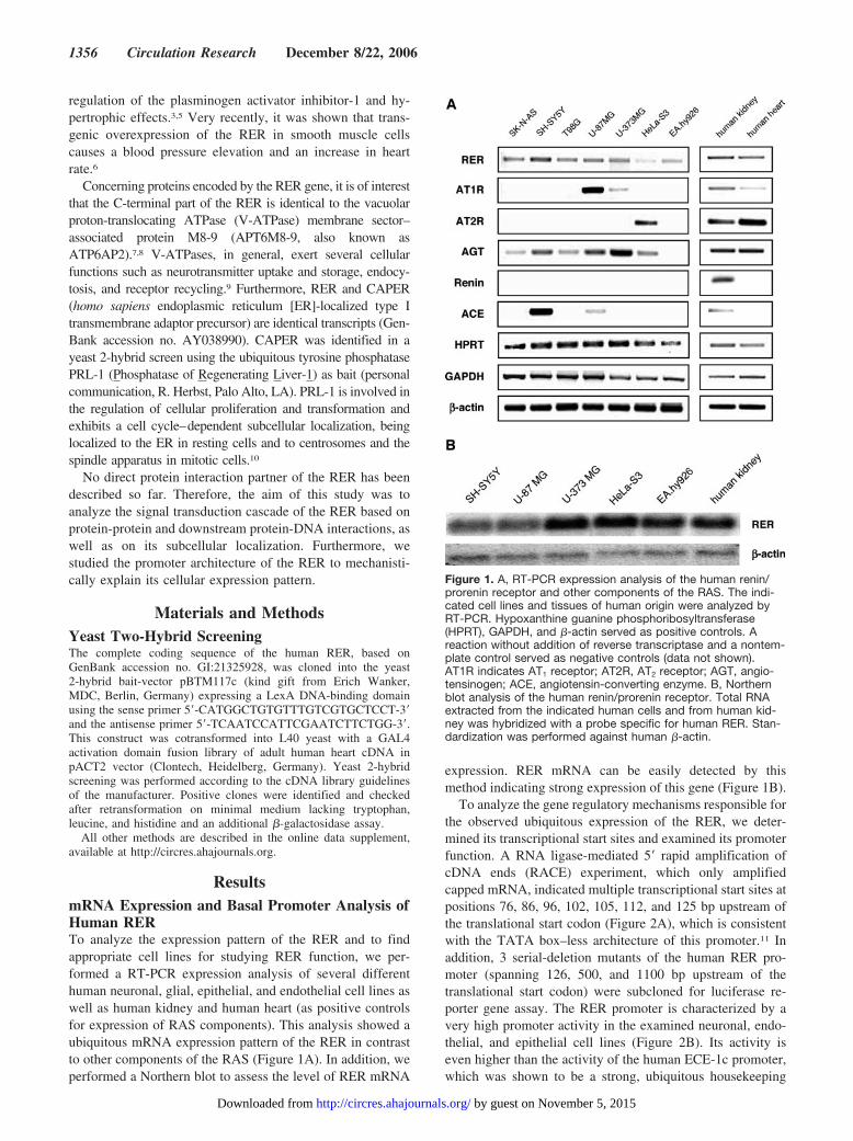
ResultsmRNA Expression and Basal Promoter Analysis ofHuman RERTo analyze the expression pattern of the RER and to findappropriate cell lines for studying RER function, we per-formed a RT-PCR expression analysis of several differenthuman neuronal, glial, epithelial, and endothelial cell lines aswell as human kidney and human heart (as positive controlsfor expression of RAS components). This analysis showed aubiquitous mRNA expression pattern of the RER in contrastto other components of the RAS (Figure 1A). In addition, weperformed a Northern blot to assess the level of RER mRNA
expression. RER mRNA can be easily detected by thismethod indicating strong expression of this gene (Figure 1B).
To analyze the gene regulatory mechanisms responsible forthe observed ubiquitous expression of the RER, we deter-mined its transcriptional start sites and examined its promoterfunction. A RNA ligase-mediated 5� rapid amplification ofcDNA ends (RACE) experiment, which only amplifiedcapped mRNA, indicated multiple transcriptional start sites atpositions 76, 86, 96, 102, 105, 112, and 125 bp upstream ofthe translational start codon (Figure 2A), which is consistentwith the TATA box–less architecture of this promoter.11 Inaddition, 3 serial-deletion mutants of the human RER pro-moter (spanning 126, 500, and 1100 bp upstream of thetranslational start codon) were subcloned for luciferase re-porter gene assay. The RER promoter is characterized by avery high promoter activity in the examined neuronal, endo-thelial, and epithelial cell lines (Figure 2B). Its activity iseven higher than the activity of the human ECE-1c promoter,which was shown to be a strong, ubiquitous housekeeping
Figure 1. A, RT-PCR expression analysis of the human renin/prorenin receptor and other components of the RAS. The indi-cated cell lines and tissues of human origin were analyzed byRT-PCR. Hypoxanthine guanine phosphoribosyltransferase(HPRT), GAPDH, and �-actin served as positive controls. Areaction without addition of reverse transcriptase and a nontem-plate control served as negative controls (data not shown).AT1R indicates AT1 receptor; AT2R, AT2 receptor; AGT, angio-tensinogen; ACE, angiotensin-converting enzyme. B, Northernblot analysis of the human renin/prorenin receptor. Total RNAextracted from the indicated human cells and from human kid-ney was hybridized with a probe specific for human RER. Stan-dardization was performed against human �-actin.
1356 Circulation Research December 8/22, 2006
by guest on November 5, 2015http://circres.ahajournals.org/Downloaded from

promoter (Figure 2B).12,13 Additionally, our results indicatethat 500 bp of the human RER promoter are sufficient to drivemaximal promoter activity in these cell types (Figure 2B).
Subcellular Localization of the RERBecause the subcellular localization of the RER may be a cluefor the understanding of its function, we addressed thisquestion using different methods. Initially, the localization ofthe RER within cellular membranes (ie, plasma membraneand/or membranes of organelles) was demonstrated by frac-tionated protein isolation followed by Western blotting ofHeLa-S3 cells (Figure 3A). The observed molecular mass ofapproximately 38 to 39 kDa in the membrane fraction isconsistent with the size of full-length RER described byNguyen et al,3 whereas the lower band of approximately 35 to36 kDa seen in cytosolic and membrane fractions (Figure 3A)likely corresponds to the �4-splice variant reported byRamser et al,8 in which the 96 bp-sized exon 4 is missing.
To further analyze the cellular membrane compartment inwhich the RER is localized, we performed a set of fluores-cence microscopic experiments. Immunocytology of c-myc-and FLAG-tagged RER in HeLa-S3 cells indicated a perinu-clear localization (Figure 3B). We next generated 3 differentenhanced green fluorescence protein (EGFP) fusion proteinsof the RER (each as N- and C-terminal fusion): (1) afull-length wild-type RER (RER full); (2) a full-length RERin which its atypical ER-retention motif KXXXX14,15 wasmutated to RXXXX (RER K/R mut); and (3) the V-ATPasesegment of the RER (RER ATPase). The full-length RERconstruct showed again a perinuclear localization and colo-calized with a marker of the ER (Figure 3C). Mutagenesis of
the ER retention motif resulted in a loss of perinuclear/ERlocalization (Figure 3C). Interestingly, the V-ATPase seg-ment of the RER showed a different localization patterncompared with the full-length RER, as it was localizedprimarily to the lysosomal compartment (Figure 3C). EGFPN-terminally fused to these constructs yielded similar results(data not shown).
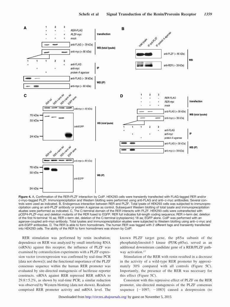
Protein-Protein Interaction Partners of the RERNo direct molecular interactions of the RER have beendescribed so far. Therefore, a major objective of our studywas to identify protein interaction partners of this ubiqui-tously expressed receptor to gain insight into its signaltransduction cascade. For this purpose, we performed a yeast2-hybrid screening using a human adult heart cDNA library(prey) and the full-length human RER (bait). The C-terminalthird of the transcription factor PLZF (PromyeLocytic ZincFinger protein) was identified as RER-interacting protein in 4clones, 3 of which were independent. To confirm thisRER-PLZF interaction, we performed a coimmunoprecipita-tion (CoIP) using transient transfections of full-length humanRER and full-length human PLZF. Figure 4A demonstratesthe ability of the RER to interact with PLZF in a system usingtagged proteins. This finding was further confirmed in anendogenous context (Figure 4B).
In an additional CoIP experiment using C- and N-terminaldeletion mutants of the RER, we identified the C-terminal,cytoplasmic region of the RER as the interaction domain forPLZF (Figure 4C).
Because dimerization is a common feature of severalreceptors, we investigated whether homodimerization may
Figure 2. A, Transcriptional start sites ofthe human renin/prorenin receptor. ARNA ligase-mediated 5� RACE was per-formed on SH-SY5Y cells, and theresulting PCR products were separatedin an agarose gel (left) and subcloned forsequencing (right). No. 1 indicates 100bp DNA ladder; 2, RACE reaction; 3,negative control. Transcriptional startsites are boxed and underlined; thetranslational start site “atg” is markedbold. The sequence shown correspondsto GenBank accession no. GI:37546587.B, Characterization of the human RERpromoter in different human cells. Serial-deletion mutants of the human RER pro-motor were subcloned into the pGL3-basic luciferase reporter vector. Numbersindicate base pairs upstream from thetranslational start site. Standardizationwas achieved by cotransfection withphRL-null vector, which encodes renillaluciferase. Relative luciferase activity(RLA) indicates promoter activity relativeto an insertless pGL3-basic vector. Thestrong housekeeping promoter of thehuman ECE-1c isoform (968 bpupstream of the translational start site)served as control (C).
Schefe et al Signal Transduction of the Renin/Prorenin Receptor 1357
by guest on November 5, 2015http://circres.ahajournals.org/Downloaded from

also be a characteristic of the RER. For this purpose, humanRER constructs with 2 different tags were transiently trans-fected. CoIP experiments indicated that the RER is able toform homodimers (Figure 4D).
Functional Analysis of the Renin-RER-PLZFSignal Transduction PathwayIn initial experiments involving PLZF overexpression inHEK293 cells (which endogenously express RER and PLZFmRNA [data not shown]), we observed that RER mRNA isreduced by renin stimulation (to 80.6�3.3%) and PLZFtransfection (to 72.6�5.2%), respectively (Figure 5A). Acombination of both repressed RER mRNA to 45.4�4.7%(Figure 5A). We could substantiate these findings by usingserial-deletion mutants of the human RER promoter as
described above. Only a promoter construct comprising theregion [�501; �1100] can be repressed by PLZF cotransfec-tion (Figure 5B).
Bioinformatic analysis of the RER promoter using MatInspector(http://www.genomatix.de)16 indicated the presence of a PLZFconsensus sequence (5�-AACTACAGTTTTCAC-3�) with a highcore and matrix similarity located in the region [�1097; �1083]relative to the translational start codon of the promoter.
To investigate functional downstream effects of the RER-PLZF interaction and to analyze whether these are influencedby stimulation of the RER with its ligand, renin, we per-formed a set of experiments in which the different compo-nents of the putative renin-RER-PLZF pathway and thepromoter of the human RER were experimentally modulatedin HEK293 cells.
Figure 3. A, Western blot analysis of the human renin/prorenin receptor. Cytosolic (C) (12 �g) and membrane (M) (9 �g) proteins ofHeLa-S3 cells were separated by SDS-PAGE, and Western blotting was performed using an antibody against human RER. B, Immuno-cytology of full-length RER. HeLa-S3 cells were transiently transfected with c-myc- and FLAG-tagged full-length RER, and subcellularlocalization was analyzed by immunofluorescence microscopy. Transfection of an insertless vector served as negative control. 4�,6-Diamidino-2-phenylindole (DAPI) was used as nuclear marker. C, Fluorescence microscopy of EGFP-RER fusion proteins. A full-lengthwild-type RER (RER full), a full-length RER, in which the atypical ER-retention motif had been mutated (RER K/R mut), and theV-ATPase segment of the RER (RER ATPase), each C-terminally fused to EGFP, were transiently transfected into HeLa-S3 cells. Mark-ers for the nuclear (DAPI), ER, and lysosomal compartment were used as indicated.
1358 Circulation Research December 8/22, 2006
by guest on November 5, 2015http://circres.ahajournals.org/Downloaded from
RER stimulation was performed by renin incubation;dependence on RER was analyzed by small interfering RNA(siRNA) against this receptor; the influence of PLZF wasexamined by cotransfection experiments with a PLZF expres-sion vector (overexpression was confirmed by real-time PCR[data not shown]); and the functional importance of the PLZFconsensus sequence within the human RER promoter wasevaluated by site-directed mutagenesis of luciferase reporterconstructs. siRNA against RER repressed RER mRNA to29.8�5.2%, as shown by real-time PCR; a similar reductionwas observed by Western blotting (data not shown). Readoutscomprised RER promoter activity and mRNA level. The
known PLZF target gene, the p85� subunit of thephosphatidylinositol-3 kinase (PI3K-p85�), served as anadditional downstream candidate gene of a RER/PLZF path-way activation.17
Stimulation of the RER with renin resulted in a decreasein the activity of a wild-type RER promoter by approxi-mately 30% compared with all controls (Figure 5C).Importantly, the presence of the RER was necessary forthis effect (Figure 5C).
Consistent with this repressive effect of PLZF on the RERpromoter, site-directed mutagenesis of the PLZF consensussequence [�1097; �1083] caused a derepression (to
Figure 4. A, Confirmation of the RER-PLZF interaction by CoIP. HEK293 cells were transiently transfected with FLAG-tagged RER and/orc-myc–tagged PLZF. Immunoprecipitation and Western blotting were performed using anti-FLAG and anti–c-myc antibodies. Several con-trols were used as indicated. B, Endogenous interaction between RER and PLZF. Total lysate of HEK293 cells was subjected to immunopre-cipitation using an anti-PLZF antibody or protein A agarose as control. Subsequent Western blotting of total lysate and immunoprecipitationeluates were performed as indicated. C, The C-terminal domain of the RER interacts with PLZF. HEK293 cells were cotransfected withpCEP4-PLZF-myc and deletion mutants of the RER fused to EGFP. RER full indicates full-length coding sequence; RER n-term del, deletionof the first N-terminal 16 aa; RER c-term del, deletion of the C-terminal (cytoplasmic) 18 aa; EGFP alone. CoIP was performed with anagarose-coupled anti–myc-antibody. Total lysates and immunoprecipitation eluates were subjected to Western blotting using anti–c-myc andanti-EGFP antibodies. D, The RER is able to form homodimers. The human RER was tagged with 2 different tags and transiently transfectedinto HEK293 cells. The ability of the RER to form homodimers was shown by CoIP.
Schefe et al Signal Transduction of the Renin/Prorenin Receptor 1359
by guest on November 5, 2015http://circres.ahajournals.org/Downloaded from

�150%) of promoter activity. The effect of renin stimulationwas also abolished by this mutagenesis (Figure 5C).
Similar but inverse effects were observed with respect tothe PI3K-p85� promoter, which is known to be positivelyregulated by PLZF17. RER stimulation with renin caused anincrease of 45% in PI3K-p85� promoter activity comparedwith control; this effect was abolished by downregulation ofthe RER using siRNA (Figure 5D).
To verify these findings, we analyzed the effect of RERstimulation on PI3K-p85� mRNA by real-time PCR analysis.Consistent with our promoter data, stimulation with reninincreased PI3K-p85� mRNA in systems with and withoutPLZF overexpression by 105.2�6.74% or 30.2�7.9% (rela-tive to vehicle control), respectively (Figure 5E). Again,downregulation of RER by siRNA abolished this inductionregardless the expression level of PLZF (Figure 5E).
Figure 5. A, Decrease of RER mRNA by renin stimulation and PLZF cotransfection. HEK293 cells with (pCEP4-PLZF) (�) or without(pCEP4 insertless) (�) PLZF overexpression were stimulated with renin (gray columns) or vehicle (white columns), and RER mRNA wasquantified by real-time PCR. mRNA expression level of the first column was set to 100%. B, PLZF is able to repress RER promoteractivity. SH-SY5Y and HEK293 cells were transfected with serial-deletion mutants of human RER promoter (numbers indicate promoterregion relative to the translation start site in base pairs). Cotransfection was performed with a PLZF expression vector (gray columns) oran insertless control vector (pCEP4) (white columns). RER promoter activity was determined using a luciferase reporter assay. RLA indi-cates relative luciferase activity. C, Repression of RER promoter activity by renin stimulation requires RER and a PLZF cis element.HEK293 cells overexpressing PLZF (by transient transfection of a pCEP4-PLZF expression vector) were stimulated with renin (gray col-umns) or vehicle (white columns), and relative luciferase activity (RLA) of a wild-type RER promoter reporter construct (WT) or a RERpromoter reporter construct with a mutation of the PLZF consensus sequence at position [�1097; �1083] (mut) was determined.�1110 indicates the length of the subcloned promoter relative to the ATG. The functional importance of the RER was examined byusing siRNA against RER (�) compared with a control siRNA (�). D, Activation of PI3K-p85� promoter activity by renin stimulationrequires RER. The activity of the PI3K-p85� promoter was analyzed in PLZF-overexpressing HEK293 cells in an experimental settinganalogous to Figure 5C, using siRNA against the RER or control siRNA. E, Increase of PI3K-p85� mRNA by renin stimulation requiresRER. HEK293 cells with (pCEP4-PLZF) or without (pCEP4 insertless) PLZF overexpression were stimulated with renin (gray columns) orvehicle (white columns), and PI3K-p85� mRNA (normalized to 18S rRNA) was determined by quantitative real-time PCR. The functionalimportance of the RER was analyzed by siRNA against RER (�) compared with a control siRNA (�). mRNA expression level of the firstcolumn was set to 100%.
1360 Circulation Research December 8/22, 2006
by guest on November 5, 2015http://circres.ahajournals.org/Downloaded from
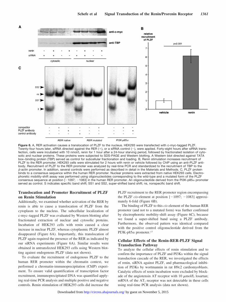
Translocation and Promoter Recruitment of PLZFon Renin StimulationAdditionally, we examined whether activation of the RER byrenin is able to cause a translocation of PLZF from thecytoplasm to the nucleus. The subcellular localization ofc-myc–tagged PLZF was evaluated by Western blotting afterfractionated extraction of nuclear and cytosolic proteins.Incubation of HEK293 cells with renin caused a clearincrease in nuclear PLZF, whereas cytoplasmic PLZF almostdisappeared (Figure 6A). Importantly, this translocation ofPLZF again required the presence of the RER as indicated byour siRNA experiments (Figure 6A). Similar results wereobtained in untransfected HEK293 cells using Western blot-ting against endogenous PLZF (data not shown).
To evaluate the recruitment of endogenous PLZF to thehuman RER promoter within the chromatin context, weperformed a chromatin-immunoprecipitation (ChIP) experi-ment. To ensure valid quantification of transcription factorrecruitment, immunoprecipitated DNA was quantified apply-ing real-time PCR analysis and multiple positive and negativecontrols. Renin stimulation of HEK293 cells did increase the
PLZF recruitment to the RER promoter region encompassingthe PLZF cis-element at position [�1097; �1083] approxi-mately 6-fold (Figure 6B).
The binding of PLZF to this cis element of the human RERpromoter (and not to a mutated form) was further confirmedby electrophoretic mobility-shift assay (Figure 6C), becausewe found a super-shifted band using a PLZF antibody.Furthermore, the observed pattern was identical comparedwith the positive control oligonucleotide derived from thePI3K-p85� promoter.17
Cellular Effects of the Renin-RER-PLZF SignalTransduction PathwayTo analyze the cellular effects of renin stimulation and toconfirm the importance of PLZF and PI3Ks within the signaltransduction cascade of the RER, we investigated the effectsof renin, siRNA against PLZF, and pharmacological inhibi-tion of PI3Ks by wortmannin in rat H9c2 cardiomyoblasts.Catalytic effects of renin incubation were excluded by block-ade of the angiotensin AT receptor with 10 �mol/L losartan;mRNA of the AT2 receptor was not detectable in these cellsusing real-time PCR analysis (data not shown).
Figure 6. A, RER activation causes a translocation of PLZF to the nucleus. HEK293 were transfected with c-myc-tagged PLZF.Twenty-four hours later, siRNA directed against the RER (�), or a siRNA control (�), were applied. Forty-eight hours after siRNA trans-fection, cells were incubated with 10 nmol/L renin for 1 hour after a 24-hour starving period, followed by fractionated isolation of cyto-solic and nuclear proteins. These proteins were subjected to SDS-PAGE and Western blotting. A Western blot directed against TATAbox–binding protein (TBP) served as control for subcellular fractionation and loading. B, Renin stimulation increases recruitment ofPLZF to the RER promoter. HEK293 cells were stimulated for 2 hours with renin or vehicle followed by ChIP using an anti-PLZF anti-body. Recruitment of PLZF to the RER promoter was analyzed by real-time PCR and standardized to the recruitment of TBP to the�-actin promoter. In addition, several controls were performed as described in detail in the Materials and Methods. C, PLZF proteinbinds to a consensus sequence within the human RER promoter. Nuclear proteins were extracted from native HEK293 cells. Electro-phoretic mobility-shift assay was performed using oligonucleotides corresponding to the wild-type and a mutated form of the PLZFconsensus sequence at position [�1097; �1083] in the human RER promoter. An oligonucleotide derived from the PI3K-p85� promoterserved as control. S indicates specific band shift; SS1 and SS2, super-shifted band shift; ns, nonspecific band shift.
Schefe et al Signal Transduction of the Renin/Prorenin Receptor 1361
by guest on November 5, 2015http://circres.ahajournals.org/Downloaded from

Renin stimulation increased the number of viable cells to133.9% compared with vehicle control. Preincubation withwortmannin partly abolished this effect, whereas siRNAagainst PLZF completely abrogated proproliferative effectsof renin stimulation (Figure 7A). Consistent results wereobserved regarding apoptosis using caspase 3 and 7 activityas readout. Renin stimulation decreased caspase activity to45.9% (in comparison to vehicle). Wortmannin partly andPLZF siRNA completely abolished this effect (Figure 7B).
To confirm the gene regulatory downstream effects of RERactivation in cardiomyoblasts, we also measured RER andPI3K-p85� mRNA in this experimental setting. RER mRNAdecreased to 53.7% under renin stimulation compared withvehicle control. siRNA against PLZF (which repressed PLZFmRNA to 15.23�9.8% as shown by real-time PCR) recon-
stituted RER mRNA to 86.5% (Figure 7C). Renin stimulationincreased PI3K-p85� mRNA to 224.2% compared withvehicle control. siRNA against PLZF abolished this effect(Figure 7D). Wortmannin did not affect RER or PI3K-p85�
mRNA levels, respectively (Figure 7C and 7D).
RER and PI3K-p85� mRNA Expression in PLZFKnockout MiceTo substantiate our findings concerning the transcriptionalregulation of RER and PI3K-p85� by PLZF in vivo, wequantified the respective transcript levels in PLZF knockout(PLZF�/�) and wild-type (WT) control mice, respectively.PLZF was not detectable in kidney and liver of WT andPLZF�/� mice. Within these organs, we did not observe anydifference in mRNA levels of RER and PI3K-p85�. In
Figure 7. A, The increase in cell number induced by renin is mediated by PLZF and, partly, PI3Ks. Rat H9c2 cardiomyoblasts weretransfected with siRNA against PLZF (�) or scrambled control siRNA (�). Forty-eight hours later, after 24 hours of serum starving, cellswere pretreated with 10 �mol/L losartan (AT1 receptor antagonist), and, additionally, 1 �mol/L wortmannin (PI3K inhibitor) (�) or vehicle(�). Thirty minutes later, cells were stimulated with 10 nmol/L renin (�) or vehicle (�). The number of viable cells was determined usingan ATP-dependent luciferase reaction. B, The decrease in apoptosis induced by renin is mediated PLZF and, partly, PI3Ks. Experimen-tal conditions were identical to A. The activity of caspase 3 and 7 was determined using a luciferase reaction and served as a measureof apoptosis. C, The decrease in RER mRNA expression induced by renin is mediated by PLZF. Experimental conditions were identicalto A. RER mRNA (normalized to 18S rRNA) was determined by quantitative real-time PCR. D, The increase in PI3K-p85� mRNAexpression induced by renin is mediated by PLZF. Experimental conditions were identical to A. PI3K-p85� mRNA (normalized to 18SrRNA) was determined by quantitative real-time PCR.
1362 Circulation Research December 8/22, 2006
by guest on November 5, 2015http://circres.ahajournals.org/Downloaded from
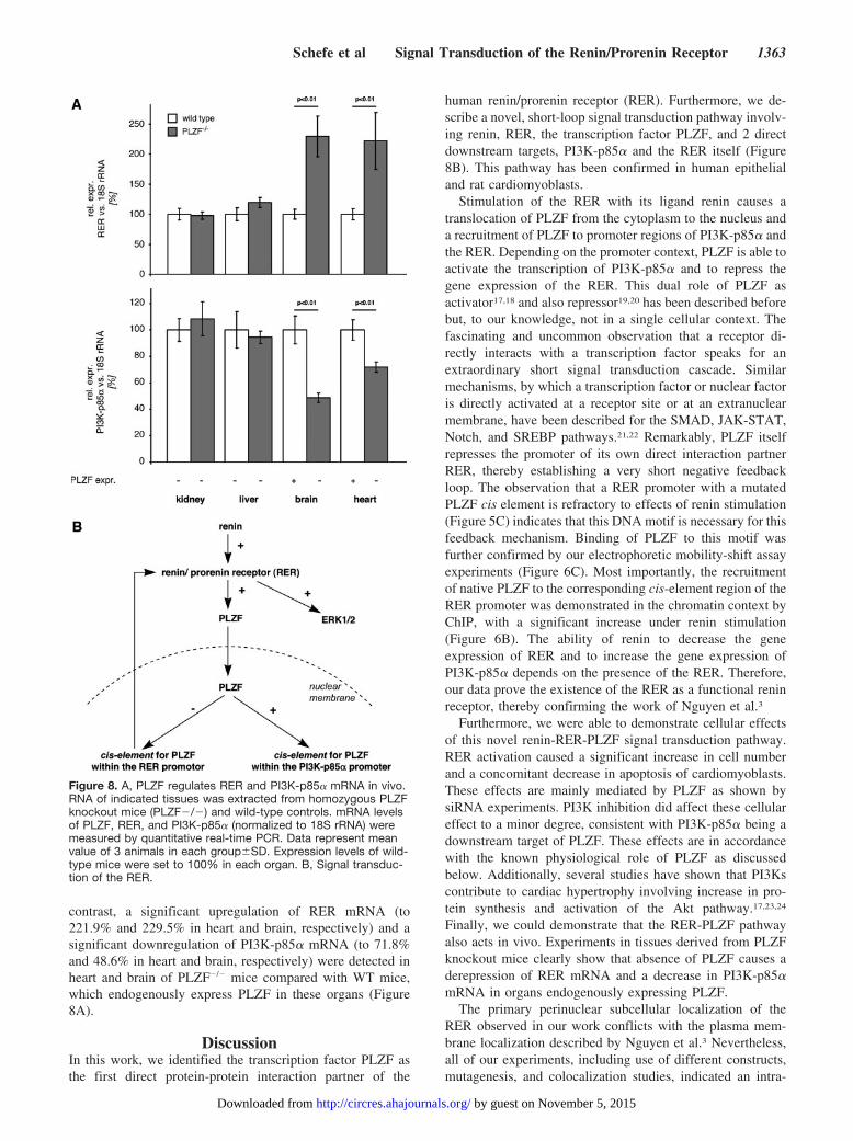
contrast, a significant upregulation of RER mRNA (to221.9% and 229.5% in heart and brain, respectively) and asignificant downregulation of PI3K-p85� mRNA (to 71.8%and 48.6% in heart and brain, respectively) were detected inheart and brain of PLZF�/� mice compared with WT mice,which endogenously express PLZF in these organs (Figure8A).
DiscussionIn this work, we identified the transcription factor PLZF asthe first direct protein-protein interaction partner of the
human renin/prorenin receptor (RER). Furthermore, we de-scribe a novel, short-loop signal transduction pathway involv-ing renin, RER, the transcription factor PLZF, and 2 directdownstream targets, PI3K-p85� and the RER itself (Figure8B). This pathway has been confirmed in human epithelialand rat cardiomyoblasts.
Stimulation of the RER with its ligand renin causes atranslocation of PLZF from the cytoplasm to the nucleus anda recruitment of PLZF to promoter regions of PI3K-p85� andthe RER. Depending on the promoter context, PLZF is able toactivate the transcription of PI3K-p85� and to repress thegene expression of the RER. This dual role of PLZF asactivator17,18 and also repressor19,20 has been described beforebut, to our knowledge, not in a single cellular context. Thefascinating and uncommon observation that a receptor di-rectly interacts with a transcription factor speaks for anextraordinary short signal transduction cascade. Similarmechanisms, by which a transcription factor or nuclear factoris directly activated at a receptor site or at an extranuclearmembrane, have been described for the SMAD, JAK-STAT,Notch, and SREBP pathways.21,22 Remarkably, PLZF itselfrepresses the promoter of its own direct interaction partnerRER, thereby establishing a very short negative feedbackloop. The observation that a RER promoter with a mutatedPLZF cis element is refractory to effects of renin stimulation(Figure 5C) indicates that this DNA motif is necessary for thisfeedback mechanism. Binding of PLZF to this motif wasfurther confirmed by our electrophoretic mobility-shift assayexperiments (Figure 6C). Most importantly, the recruitmentof native PLZF to the corresponding cis-element region of theRER promoter was demonstrated in the chromatin context byChIP, with a significant increase under renin stimulation(Figure 6B). The ability of renin to decrease the geneexpression of RER and to increase the gene expression ofPI3K-p85� depends on the presence of the RER. Therefore,our data prove the existence of the RER as a functional reninreceptor, thereby confirming the work of Nguyen et al.3
Furthermore, we were able to demonstrate cellular effectsof this novel renin-RER-PLZF signal transduction pathway.RER activation caused a significant increase in cell numberand a concomitant decrease in apoptosis of cardiomyoblasts.These effects are mainly mediated by PLZF as shown bysiRNA experiments. PI3K inhibition did affect these cellulareffect to a minor degree, consistent with PI3K-p85� being adownstream target of PLZF. These effects are in accordancewith the known physiological role of PLZF as discussedbelow. Additionally, several studies have shown that PI3Kscontribute to cardiac hypertrophy involving increase in pro-tein synthesis and activation of the Akt pathway.17,23,24
Finally, we could demonstrate that the RER-PLZF pathwayalso acts in vivo. Experiments in tissues derived from PLZFknockout mice clearly show that absence of PLZF causes aderepression of RER mRNA and a decrease in PI3K-p85�mRNA in organs endogenously expressing PLZF.
The primary perinuclear subcellular localization of theRER observed in our work conflicts with the plasma mem-brane localization described by Nguyen et al.3 Nevertheless,all of our experiments, including use of different constructs,mutagenesis, and colocalization studies, indicated an intra-
Figure 8. A, PLZF regulates RER and PI3K-p85� mRNA in vivo.RNA of indicated tissues was extracted from homozygous PLZFknockout mice (PLZF�/�) and wild-type controls. mRNA levelsof PLZF, RER, and PI3K-p85� (normalized to 18S rRNA) weremeasured by quantitative real-time PCR. Data represent meanvalue of 3 animals in each group�SD. Expression levels of wild-type mice were set to 100% in each organ. B, Signal transduc-tion of the RER.
Schefe et al Signal Transduction of the Renin/Prorenin Receptor 1363
by guest on November 5, 2015http://circres.ahajournals.org/Downloaded from
cellular localization of the RER. Furthermore, the totalcellular membrane lysates used for their kinetic studies maystill contain intracellular membrane proteins and, therefore,do not exclude an intracellular localization of the RER. Thepossibility that our results concerning the perinuclear local-ization are caused by an artifact related to tags or overexpres-sion is unlikely because we used several different C-terminaland also N-terminal tags. In addition, mutagenesis of theatypical C-terminal ER-retention signal strongly reduced theperinuclear localization of the RER. The fact that CAPER,which is identical to the RER as discussed above, can directlybind to PRL-1 (a protein observed in the ER in nonmitoticcells) also supports our results.
On the other hand, our experiments indicate that anextracellular signal (renin) can affect the signal transductionof the RER despite its mainly intracellular localization.Several mechanisms might account for this observation.Firstly, other renin-binding receptors, such as the mannose-6-phosphate receptor, could internalize renin and prorenin.25
Secondly, a nonsecreted (ie, intracellular) renin isoform,which could directly interact with an intracellular RER, hasbeen described containing an alternative first exon termed(albeit identical) “exon 1b”26,27 and “exon 1A,”28,29 respec-tively. Intracellular renin administration was reported toincrease inward calcium current in cardiomyocytes.30 Inaddition, transgenic overexpression of the nonsecreted, intra-cellular form of renin and angiotensinogen in the brain causesan increase in drinking volume and mean arterial pressure.31
Thirdly, it could be possible that very small amounts of theRER within the plasma membrane are sufficient for theinitiation of a RER signal transduction cascade. In thiscontext, the observed homodimerization of the RER mightalso be of importance, because dimerization can affect sub-cellular localization.32
In this study, we were able to demonstrate a ubiquitousexpression of the human RER, a high promoter activity indifferent cell types and multiple transcriptional start sites in aTATA box–less promoter. These features are consistent withhousekeeping properties of the RER gene,13 suggesting basalcellular functions of this protein. In this context, it is ofinterest that the C-terminal part of the RER is, as discussedabove, identical to the V-ATPase membrane sector-associated protein M8-9, because V-ATPase have functionsin almost every eukaryotic cell9. Nevertheless, APT6M8-9only constitutes 69 to 100 aa (GenBank accession no.GI:5031590)7 of the 350 aa3 of the full-length renin receptor.Therefore, the functions of the RER exceed those of a pureV-ATPase subunit, which is also supported by the data on thesignal transduction and the different subcellular localizationsof full-length RER and APT6M8-9 provided here. Themechanism by which the APT6M8-9 protein is generated,remains to be clarified. Alternative promoters and posttrans-lational cleavage by an unidentified protease are putativeexplanations.
The transcription factor PLZF contains multiple zinc-finger domains and is disrupted in patients with acutepromyelocytic leukemia (APL) caused by t(11;17)(q23;q21)chromosomal translocation.33,34 This APL subform is charac-terized by PLZF-RAR� (PLZF-retinoic acid receptor-�) fu-
sion proteins, which recruit histone deacetylase 1 (HDAC1)and do not respond to retinoic acid any more, explaining themissing response of these patients to retinoic acidtreatment.34–36
Wild-type PLZF can act as growth repressor and exertsproapoptotic functions during development.33,37 Concerningthe RAS, it seems important to note that PLZF was recentlydescribed as an adaptor protein of the AT2 receptor in theheart.17 This direct PLZF-AT2 receptor interaction was asso-ciated with stimulation of protein synthesis and putativecardiac hypertrophy.17
Data obtained from PLZF knockout mice indicate that thistranscription factor is involved in limb and axial skeletalpatterning,37 whereas the brain phenotype of these mice wasnot described by the authors. Consistent with this observa-tion, PLZF target genes include hox genes,37–39 besides thep85� subunit of PI3K17 and cyclin A2.20
Concerning the biological significance of the RER beyondits possible role in the RAS, it was recently shown by Ramseret al that a mutation in the renin receptor gene is a cause ofX-linked mental retardation (XLMR) and epilepsy (XMRE)syndrome in humans.8 Consistent with this observation arethe results of a zebrafish mutagenesis screen, in which amutation in the ATP6AP2 (which is identical to RER) genecaused a reduction in head size and necrosis of the centralnervous system.40 Remarkably, the human RER mutationobserved by Ramser et al altered neither binding affinity forrenin nor the RER-mediated augmentation of the catalyticefficiency of renin for angiotensinogen cleavage.8 In addition,the effect of this mutation (and also the effect of wild-typeRER stimulation by renin3) on mitogen-activated proteinkinase signaling are only modest,8 if non-significant. There-fore, it could be possible that the human RER mutation exertsits effect by an altered PLZF-mediated signal transduction.The complex, dynamic expression of PLZF in the developingcentral nervous system, and its likely role in hindbrain andalso forebrain segmentation supports this notion.41,42
Related to the role of the RER in central nervous systemdevelopment is the observation that RER mRNA can bedetected in human glioblastomas as well as in glioblastomacell lines and that renin inhibitors can reduce the cell numberin glioblastoma cell lines. This is probably caused by modu-lation of RER function, because this effect is independent ofAT1 and AT2 receptor activity.43
The clinical relevance of the RER is underlined by theinteresting observation that a decoy deca-peptide correspond-ing to the handle region of prorenin, which competitivelyinhibits prorenin binding to its receptor, attenuated thedevelopment and progression of cardiac fibrosis44 and alsoinhibited the development of diabetic nephropathy in ratmodels.45 Remarkably, a recent study demonstrated the abil-ity of RER activation to upregulate transforming growthfactor-�1 in mesangial cells independent of angiotensin IIactions.46
Knowledge regarding the signal transduction of the RER isof importance to evaluate the efficacy and safety of renininhibitors, such as aliskiren, which is currently in phase IIIclinical trials.47 As expected, renin inhibitors reduce plasmarenin activity (ie, enzyme activity with respect to angiotensin
1364 Circulation Research December 8/22, 2006
by guest on November 5, 2015http://circres.ahajournals.org/Downloaded from

I generation). Nevertheless, they increase total amount ofplasma renin protein, the RER ligand, dramatically (up to34-fold48). Therefore, it is crucial to examine whether renininhibitors change the intrinsic activity of renin with respect tothe RER. In this context, it is important to note that overac-tivation of the RER might be deleterious (eg, with respect toend-organ damage) considering the activation of mitogen-ac-tivated protein kinases and PLZF downstream of the RER aswell as the effects of the handle region decoy peptidementioned above. On the other hand, a blockade of the RERsignal transduction might be harmful, at least in pregnantwomen, because of the developmental importance of the RERand its direct interaction partner PLZF.
To conclude, our results demonstrate the existence of anovel signal transduction pathway downstream of the humanrenin/prorenin receptor, which involves direct binding of thetranscription factor PLZF to the receptor, its translocation tothe nucleus, and the positive and negative regulation of targetgenes. Based on the already described biomedical relevanceof the RER and PLZF, respectively, this pathway, connectingboth molecules, might be of importance in human physiologyand pathophysiology.
Sources of FundingThis project was supported by a Forschungsforderung of the Charite–Universitatsmedizin Berlin.
DisclosuresT.U. is member of the Aliskiren International Advisory Board ofNovartis Pharma AG (Basel, Suisse).
References1. Re RN. Intracellular renin and the nature of intracrine enzymes. Hyper-
tension. 2003;42:117–122.2. Nguyen G, Burckle CA, Sraer JD. Renin/prorenin-receptor biochemistry
and functional significance. Curr Hypertens Rep. 2004;6:129–132.3. Nguyen G, Delarue F, Burckle C, Bouzhir L, Giller T, Sraer JD. Pivotal
role of the renin/prorenin receptor in angiotensin II production andcellular responses to renin. J Clin Invest. 2002;109:1417–1427.
4. Mazak I, Wellner M, Shagdarsuren E, Dechend R, Schulze-Forster K,Nguyen G, Luft FC, Muller DN. Renin induces ERK 1/2 phosphorylationin U937 monocyte/macrophages independent of angiotensin II. The 57thAnnual Fall Conference and Scientific Sessions of the American Councilfor High Blood Pressure Research. September 23–26, 2003, Wash DC.Abstract.
5. Nguyen G, Delarue F, Berrou J, Rondeau E, Sraer JD. Specific receptorbinding of renin on human mesangial cells in culture increases plasmin-ogen activator inhibitor-1 antigen. Kidney Int. 1996;50:1897–1903.
6. Burckle CA, Jan Danser AH, Muller DN, Garrelds IM, Gasc JM, PopovaE, Plehm R, Peters J, Bader M, Nguyen G. Elevated blood pressure andheart rate in human renin receptor transgenic rats. Hypertension. 2006;47:552–556.
7. Ludwig J, Kerscher S, Brandt U, Pfeiffer K, Getlawi F, Apps DK,Schagger H. Identification and characterization of a novel 9.2-kDamembrane sector-associated protein of vacuolar proton-ATPase fromchromaffin granules. J Biol Chem. 1998;273:10939–10947.
8. Ramser J, Abidi FE, Burckle CA, Lenski C, Toriello H, Wen G, Lubs HA,Engert S, Stevenson RE, Meindl A, Schwartz CE, Nguyen G. A uniqueexonic splice enhancer mutation in a family with X-linked mental retar-dation and epilepsy points to a novel role of the renin receptor. Hum MolGenet. 2005;14:1019–1027.
9. Nelson N, Harvey WR. Vacuolar and plasma membrane proton-adeno-sinetriphosphatases. Physiol Rev. 1999;79:361–385.
10. Wang J, Kirby CE, Herbst R. The tyrosine phosphatase PRL-1 localizesto the endoplasmic reticulum and the mitotic spindle and is required fornormal mitosis. J Biol Chem. 2002;277:46659–46668.
11. Heinemeyer T, Chen X, Karas H, Kel AE, Kel OV, Liebich I, MeinhardtT, Reuter I, Schacherer F, Wingender E. Expanding the TRANSFACdatabase towards an expert system of regulatory molecular mechanisms.Nucleic Acids Res. 1999;27:318–322.
12. Funke-Kaiser H, Bolbrinker J, Theis S, Lemmer J, Richter CM, Paul M,Orzechowski HD. Characterization of the c-specific promoter of the geneencoding human endothelin-converting enzyme-1 (ECE-1). FEBS Lett.2000;466:310–316.
13. Funke-Kaiser H, Thomas A, Bremer J, Kovacevic SD, Scheuch K,Bolbrinker J, Theis S, Lemmer J, Zimmermann A, Zollmann FS,Herrmann SM, Paul M, Orzechowski HD. Regulation of the majorisoform of human endothelin-converting enzyme-1 by a strong house-keeping promoter modulated by polymorphic microsatellites.J Hypertens. 2003;21:2111–2124.
14. Vincent MJ, Martin AS, Compans RW. Function of the KKXX motif inendoplasmic reticulum retrieval of a transmembrane protein depends onthe length and structure of the cytoplasmic domain. J Biol Chem. 1998;273:950–956.
15. Gaynor EC, te Heesen S, Graham TR, Aebi M, Emr SD. Signal-mediatedretrieval of a membrane protein from the Golgi to the ER in yeast. J CellBiol. 1994;127:653–665.
16. Cartharius K, Frech K, Grote K, Klocke B, Haltmeier M, Klingenhoff A,Frisch M, Bayerlein M, Werner T. MatInspector and beyond: promoteranalysis based on transcription factor binding sites. Bioinformatics. 2005;21:2933–2942.
17. Senbonmatsu T, Saito T, Landon EJ, Watanabe O, Price E Jr, Roberts RL,Imboden H, Fitzgerald TG, Gaffney FA, Inagami T. A novel angiotensinII type 2 receptor signaling pathway: possible role in cardiac hypertrophy.EMBO J. 2003;22:6471–6482.
18. Labbaye C, Quaranta MT, Pagliuca A, Militi S, Licht JD, Testa U,Peschle C. PLZF induces megakaryocytic development, activates Tporeceptor expression and interacts with GATA1 protein. Oncogene. 2002;21:6669–6679.
19. Li JY, English MA, Ball HJ, Yeyati PL, Waxman S, Licht JD. Sequence-specific DNA binding and transcriptional regulation by the promyelocyticleukemia zinc finger protein. J Biol Chem. 1997;272:22447–22455.
20. Yeyati PL, Shaknovich R, Boterashvili S, Li J, Ball HJ, Waxman S,Nason-Burchenal K, Dmitrovsky E, Zelent A, Licht JD. Leukemia trans-location protein PLZF inhibits cell growth and expression of cyclin A.Oncogene. 1999;18:925–934.
21. Brivanlou AH, Darnell JE Jr. Signal transduction and the control of geneexpression. Science. 2002;295:813–818.
22. Emery JG, Ohlstein EH, Jaye M. Therapeutic modulation of transcriptionfactor activity. Trends Pharmacol Sci. 2001;22:233–240.
23. Hennessy BT, Smith DL, Ram PT, Lu Y, Mills GB. Exploiting thePI3K/AKT pathway for cancer drug discovery. Nat Rev Drug Discov.2005;4:988–1004.
24. Shioi T, Kang PM, Douglas PS, Hampe J, Yballe CM, Lawitts J, CantleyLC, Izumo S. The conserved phosphoinositide 3-kinase pathwaydetermines heart size in mice. EMBO J. 2000;19:2537–2548.
25. Danser AH, Deinum J. Renin, prorenin and the putative (pro)reninreceptor. Hypertension. 2005;46:1069–1076.
26. Lee-Kirsch MA, Gaudet F, Cardoso MC, Lindpaintner K. Distinct reninisoforms generated by tissue-specific transcription initiation and alter-native splicing. Circ Res. 1999;84:240–246.
27. Sinn PL, Sigmund CD. Identification of three human renin mRNAisoforms from alternative tissue-specific transcriptional initiation. PhysiolGenomics. 2000;3:25–31.
28. Clausmeyer S, Sturzebecher R, Peters J. An alternative transcript of therat renin gene can result in a truncated prorenin that is transported intoadrenal mitochondria. Circ Res. 1999;84:337–344.
29. Peters J, Clausmeyer S. Intracellular sorting of renin: cell type specificdifferences and their consequences. J Mol Cell Cardiol. 2002;34:1561–1568.
30. De Mello WC. Renin increments the inward calcium current in the failingheart. J Hypertens. 2006;24:1181–1186.
31. Lavoie JL, Liu X, Bianco RA, Beltz TG, Johnson AK, Sigmund CD.Evidence supporting a functional role for intracellular renin in the brain.Hypertension. 2006;47:461–466.
32. Muller L, Barret A, Etienne E, Meidan R, Valdenaire O, Corvol P,Tougard C. Heterodimerization of endothelin-converting enzyme-1isoforms regulates the subcellular distribution of this metalloprotease.J Biol Chem. 2003;278:545–555.
Schefe et al Signal Transduction of the Renin/Prorenin Receptor 1365
by guest on November 5, 2015http://circres.ahajournals.org/Downloaded from
33. Costoya JA, Pandolfi PP. The role of promyelocytic leukemia zinc fingerand promyelocytic leukemia in leukemogenesis and development. CurrOpin Hematol. 2001;8:212–217.
34. Minucci S, Pelicci PG. Histone deacetylase inhibitors and the promise ofepigenetic (and more) treatments for cancer. Nat Rev Cancer. 2006;6:38–51.
35. Grignani F, De Matteis S, Nervi C, Tomassoni L, Gelmetti V, Cioce M,Fanelli M, Ruthardt M, Ferrara FF, Zamir I, Seiser C, Grignani F, LazarMA, Minucci S, Pelicci PG. Fusion proteins of the retinoic acidreceptor-alpha recruit histone deacetylase in promyelocytic leukaemia.Nature. 1998;391:815–818.
36. Glaser KB, Staver MJ, Waring JF, Stender J, Ulrich RG, Davidsen SK.Gene expression profiling of multiple histone deacetylase (HDAC) in-hibitors: defining a common gene set produced by HDAC inhibition inT24 and MDA carcinoma cell lines. Mol Cancer Ther. 2003;2:151–163.
37. Barna M, Hawe N, Niswander L, Pandolfi PP. Plzf regulates limb andaxial skeletal patterning. Nat Genet. 2000;25:166–172.
38. Ivins S, Pemberton K, Guidez F, Howell L, Krumlauf R, Zelent A.Regulation of Hoxb2 by APL-associated PLZF protein. Oncogene. 2003;22:3685–3697.
39. Barna M, Merghoub T, Costoya JA, Ruggero D, Branford M, Bergia A,Samori B, Pandolfi PP. Plzf mediates transcriptional repression of HoxDgene expression through chromatin remodeling. Dev Cell. 2002;3:499–510.
40. Amsterdam A, Nissen RM, Sun Z, Swindell EC, Farrington S, HopkinsN. Identification of 315 genes essential for early zebrafish development.Proc Natl Acad Sci U S A. 2004;101:12792–12797.
41. Cook M, Gould A, Brand N, Davies J, Strutt P, Shaknovich R, Licht J,Waxman S, Chen Z, Gluecksohn-Waelsch S, Krumlauf R, Zelent A.
Expression of the zinc-finger gene PLZF at rhombomere boundaries inthe vertebrate hindbrain. Proc Natl Acad Sci U S A. 1995;92:2249–2253.
42. Avantaggiato V, Pandolfi PP, Ruthardt M, Hawe N, Acampora D, PelicciPG, Simeone A. Developmental analysis of murine PromyelocyteLeukemia Zinc Finger (PLZF) gene expression: implications for theneuromeric model of the forebrain organization. J Neurosci. 1995;15:4927–4942.
43. Juillerat-Jeanneret L, Celerier J, Chapuis Bernasconi C, Nguyen G, WostlW, Maerki HP, Janzer RC, Corvol P, Gasc JM. Renin and angioten-sinogen expression and functions in growth and apoptosis of humanglioblastoma. Br J Cancer. 2004;90:1059–1068.
44. Ichihara A, Kaneshiro Y, Takemitsu T, Suzuki F, Nakagawa T,Nishiyama A, Iwata H, Ishida Y, Inagami T, Saruta T, Hayashi M.“Receptor-associated prorenin system” contributes to hypertensiveend-organ damage. J Hypertens. 2005;23(suppl 2):S78:23(suppl 2):578.
45. Ichihara A, Hayashi M, Kaneshiro Y, Suzuki F, Nakagawa T, Tada Y,Koura Y, Nishiyama A, Okada H, Uddin MN, Nabi AH, Ishida Y,Inagami T, Saruta T. Inhibition of diabetic nephropathy by a decoypeptide corresponding to the “handle” region for nonproteolytic acti-vation of prorenin. J Clin Invest. 2004;114:1128–1135.
46. Huang Y, Wongamorntham S, Kasting J, McQuillan D, Owens RT, Yu L,Noble NA, Border W. Renin increases mesangial cell transforminggrowth factor-beta1 and matrix proteins through receptor-mediated, an-giotensin II-independent mechanisms. Kidney Int. 2006;69:105–113.
47. Hershey JC, Steiner B, Fischli W, Feuerstein G. Renin inhibitors: Anantihypertensive strategy on the verge of reality. Drug Discovery Today:Therapeutic Strategies. 2005;2:181–185.
48. Nussberger J, Wuerzner G, Jensen C, Brunner HR. Angiotensin II sup-pression in humans by the orally active renin inhibitor Aliskiren(SPP100): comparison with enalapril. Hypertension. 2002;39:e1–e8.
1366 Circulation Research December 8/22, 2006
by guest on November 5, 2015http://circres.ahajournals.org/Downloaded from
Online Supplement Schefe et al. Signal transduction of the renin/prorenin receptor
Materials and Methods
Cell culture and tissue samples
SH-SY5Y (human neuronal) and SK-N-AS (human neuronal) were cultured in RPMI 1640
supplemented with 10% fetal bovine serum (FBS), 100 U/ ml penicillin and 100 µg/ ml
streptomycin. HEK293 (human epithelial), HeLa-S3 (human epithelial), EA.hy926 (human
endothelial), T98G (human glial), U-87 MG (human glial), U-373 MG (human glial), and
H9c2 (rat cardiomyoblast) were cultured in DMEM with 4,5 g/ l glucose supplemented
with 10% FBS, 100 U/ ml penicillin and 100 µg/ ml streptomycin. All cell culture products
were obtained from PAN Biotech, Aidenbach, Germany. Cells were grown in a humidified
incubator at 5% CO2 and 37 °C.
RNA of human heart and tissue of human kidney were provided by the DHZB (Berlin) and
the Department of Urology (Charité – Universitätsmedizin Berlin), respectively.
The generation of the PLZF knockout mice was described previously1 (zitat). Indicated
organs of PLZF knockout mice and corresponding wild type control mice (C57BL6) were
removed and immediately subjected to RNAlater solution (Qiagen, Hilden, Germany).
Constructs and site-directed mutagenesis
The complete coding sequence (CDS) of the human RER, based on GenBank
GI:21325928, and human PLZF, based on GenBank GI:31543978, were subcloned into the
mammalian expression vector pCEP4 (Invitrogen, Karlsruhe, Germany) with different C-
terminal tags using the following primers: 5’-GCCACCATGGCTGTGTTTGTCGTGCT
(sense for pCEP4-RER-FLAG/-myc/-HA); 5’-
TCACTTGTCGTCATCGTCTTTGTAGTCATCCATTCGAATCTTCTGGT (antisense
for pCEP4-RER-FLAG); 5’-
Online Supplement Schefe et al. Signal transduction of the renin/prorenin receptor
2
TCACAGATCTTCTTCAGAAATAAGTTTTTGTTCATCCATTCGAATCTTCTGGT
(antisense for pCEP4-RER-myc); 5’-
TCAAGCGTAGTCTGGGACGTCGTATGGGTAATCCATTCGAATCTTCTGGT
(antisense for pCEP4-RER-HA); 5’- GCCACCATGGATCTGACAAAAATGGG (sense
for pCEP4-PLZF-myc); 5’-
TCACAGATCTTCTTCAGAAATAAGTTTTTGTTCCACATAGCACAGGTAGAGGT
(antisense for pCEP4-PLZF-myc).
Additionally, the CDS of the human RER was cloned into pEGFP-N1 and pEGFP-C3
(Clontech, Mountain View, CA, USA) to create N- or C-terminal EGFP fusion constructs
using the following primers: 5’- GGCACCATGGCTGTGTTTGT (sense for RER-full); 5’-
ATTCGAATCTTCTGGTTTG (antisense RER-full). Furthermore, two protein mutants of
the RER were generated: (1) The pEGFP-N1/C3-RER-ATPase construct, which comprises
only the vacuolar proton-translocating ATPase (V-ATPase) membrane sector-associated
protein M8-9 (APT6M8-9) part of the RER (C-terminal 69 aa), was subcloned using the
sense primer 5’- ATGGAGGCAAAACAAGCGAAGAACC and the antisense primer 5’-
ATTCGAATCTTCTGGTTTG; (2) The pEGFP-N1/C3-RER-K/R-mut construct, in which
the atypical C-terminal ER-retention signal (KXXXX) was replaced by RXXXX, was
subcloned using the sense primer 5´- GGCACCATGGCTGTGTTTGT and the antisense
primer 5´-ATCCATTCGAATCCTCTGGTTTG.
For mapping of interaction domains, N- and C-terminal deletion mutants of the RER were
subcloned into pEGFP-N1 (Clontech) using following primers: 5´-
ATGAACGAGTTTAGTATATTAAA (sense RER n-term del) and 5´-
AATCCATTCGAATCTTCTGG (antisense RER n-term del); 5´-
GGCACCATGGCTGTGTTTGT (sense RER c-term del) and 5´-
GATCCATGTTCCAAATATTGT (antisense RER c-term del).
Online Supplement Schefe et al. Signal transduction of the renin/prorenin receptor
3
The promoter of the human RER, based on GenBank GI:37546587, and serial deletion
mutants were subcloned into the luciferase reporter vector pGL3-basic (Promega,
Mannheim) using a common antisense primer located directly upstream of the translational
start site (5´- GGTGCCGCGGCGGCCGCAGCACTGC) and following sense primers: [-
1100;-1]: 5´-CTTAACTACAGTTTTCACTGGAACA; [-1100;-1]-mut(PLZF): 5’-
CTTTACATCTGTTTTCACTGGAA; [-500;-1]: 5´-
TCACAGCTGGCGTCCGTAGCCGGGC; and [-165;-1]: 5´-
GTGATTGGTGGAGAAAGCGGCAGCT. Numbers indicate positions relative to the start
codon.
A pGL3-basic plasmid encoding 968 bp of the promoter of human endothelin-converting
enzyme-1c (ECE-1c)2 was used as control.
RT-PCR
Total RNA was isolated using NucleoSpin RNA II (Macherey-Nagel, Düren, Germany)
according to the manufacturer’s protocol including a DNAase digest. cDNA synthesis was
performed using random hexamer primers and M-MLV reverse transcriptase (RNase H
minus; Promega, Mannheim, Germany); no template controls (NTCs) and reactions
without addition of reverse transcriptase (RT-) served as negative controls.
PCRs were carried out using the following human-specific primer pairs: RER: 5´-
ATTGGCCTATACCAGGAGAG (sense), 5´-TTCCCCATAACGCTTCCCAA
(antisense); angiotensin AT1 receptor (AT1R): 5´-CATATTTGTCATGATTCCTACTT
(sense), 5´-GCACAAACTGTAATATTGGTGT (antisense); angiotensin AT2 receptor
(AT2R): 5´-ACATCTTCAACCTCGCTGTG (sense), 5´-
CCATACACCAAACAAGGGGA (antisense); angiotensinogen: 5’-
CTGTGGATGAAAAGGCCCTA (sense), 5’- ATTGCCTGTAGCCTGTCAGC
Online Supplement Schefe et al. Signal transduction of the renin/prorenin receptor
4
(antisense); ACE: 5´-GCTGCAGCCCGGCAACTTTT (sense), 5´-
CGGTGGAGTAGATCCTGCTC (antisense); HPRT: 5’-
TGCTCGAGATGTGATGAAGG (sense), 5’-TCCCCTGTTGACTGGTCATT (antisense);
GAPDH: 5´-TGAAGGTCGGAGTCAACGGATTTGGT (sense); 5´-
CATGTGGGCCATGAGGTCCACCAC (antisense); β-actin: 5’-
TCCCTGGAGAAGAGCTACGA (sense), 5’- AGCACTGTGTTGGCGTACAG
(antisense).
Northern blotting
Northern blotting was performed as described previously3, 4. The probe against the human
RER and human β-actin (for standardization) corresponds to the PCR products described
above.
RNA ligase-mediated (RLM)- 5´-RACE
Transcriptional start sites of human RER were determined using the GeneRacer Kit
(Invitrogen, Karlsruhe, Germany), which only amplifies capped mRNA. Reverse
transcription was performed with random hexamer primers. Nested PCR utilized
HotStarTaq (Qiagen), 5´-CTCTCCTGGTATAGGCCAAT (antisense primer in first PCR)
and 5´-TCGGAAAACAACAGACCCTG (antisense primer in second PCR). Reaction
products were subcloned and sequenced.
Online Supplement Schefe et al. Signal transduction of the renin/prorenin receptor
5
Luciferase assays
The indicated cell types were seeded on day 1 in 12-well plates. 100 ng of indicated pGL3-
basic constructs (encoding firefly luciferase) and 20 ng of phRL-null plasmid (encoding
humanized renilla luciferase for standardization of promoter activity; Promega) per well
were transfected on day 2 at 60-80% confluency using GeneJuice (Merck Biosciences, Bad
Soden, Germany) according to the manufacturer’s protocol. Cells were harvested 48 hours
after transfection using Passive Lysis Buffer (Promega). Reporter activities were measured
in a Pharmingen Monolight 3010 luminometer (BD Biosciences, Erembodegem, Belgium)
using the Dual-Luciferase Reporter Assay System (Promega). Promoter activity is
expressed as relative luciferase activity (RLA)4. RLA data represent the mean ± standard
deviation of at least three single, parallel transfection experiments.
Subcellular protein extraction and Western blotting
Cytosolic and membrane proteins were isolated using the ProteoExtract Subcellular
Proteome Extraction Kit (Calbiochem, Darmstadt, Germany). A fractionated extraction of
cytosolic and nuclear proteins was performed as described previously4. Protein
concentrations were determined using the DC Protein Assay (Bio-Rad, München,
Germany). Cytosolic, membrane and nuclear fractions were controlled by Western blot
using antibodies against Akt (mouse monoclonal, 5G3; 1:1,000; Cell Signalling, Danvers,
MA, USA), angiotensin AT1 receptor (rabbit polyclonal, sc-579; 1:2,000; Santa Cruz
Biotechnology, Heidelberg, Germany) and TBP (rabbit polyclonal, sc-204; 1:1,000; Santa
Cruz Biotechnology), respectively.
For analysis of subcellular localization of RER, 9 µg (membrane) and 12 µg (cytosolic)
protein were loaded per lane, separated in a 10% SDS-PAGE and transferred to a
nitrocellulose membrane. Western blot analysis was performed with an antibody against
Online Supplement Schefe et al. Signal transduction of the renin/prorenin receptor
6
human RER (corresponding ATP6AP2; goat polyclonal, ab5959, Abcam, Cambridge, UK)
at a dilution of 0.5 µg/ ml. Horseradish peroxidase-conjugated anti-goat antibody (rabbit
polyclonal; 1:2,500; DAKO, Hamburg, Germany) was used as secondary antibody.
For PLZF translocation experiments HEK293 cells were seeded on day 1 in 6-well plates.
250 ng of pCEP4-PLZF-myc expression vector per well were transfected on day 2 at 40-
60% confluency using GeneJuice (Merck Biosciences, Bad Soden, Germany) according to
the manufacturer’s protocol. On day 3 the cells were transfected with 50 nM siRNA
against the RER [5’- GCUCCGUAAUCGCCUGUUU (sense strand)] or scrambled control
siRNA [5’- UUUACCGUCGCCUUGAGCU (sense strand)] (Eurogentec, Seraing,
Belgium) using jetSI-ENDO (Eurogentec) following the standard procedure. On day 5,
after 24 hours of starving in serum-free medium, cells were stimulated with 10 nM human
recombinant renin (mammalian expression; kind gift from Dominik N. Müller, MDC
Berlin) or vehicle (0.1% DMSO in PBS) for 2 hours. Western blot was performed with
anti-c-myc antibody (mouse monoclonal, 9B11; 1:2,000; Cell Signalling) and horseradish
peroxidase-conjugated anti-mouse antibody (rabbit polyclonal; 1:2,500; DAKO).
Chemoluminescence was detected with ECL (Amersham, München, Germany), and
subsequent exposition to Hyperfilm (Amersham).
Fluorescence microscopy
HeLa-S3 cells were seeded in 8-well chamber slides (BD Biosciences, Erembodegem,
Belgium) at day 1 and transfected the following day at 30-50% confluency with 50 ng of
the indicated plasmid using GeneJuice (Merck Biosciences) following the standard
protocol.
For immunofluorescene microscopy, 48 h after transfection cells were fixed and
permeabilized in methanol for 10 minutes at –20 °C. Blocking was performed in 1x TBS

Online Supplement Schefe et al. Signal transduction of the renin/prorenin receptor
7
with 0.1% Tween 20, 5% skimmed milk powder and 1% BSA (Sigma-Aldrich,
Taufkirchen, Germany) for 1 hour at room temperature. The primary antibodies anti-
FLAG-M2 (mouse monoclonal ; 1 µg/ ml; Sigma-Aldrich, Taufkirchen, Germany) and
anti-c-myc-tag (mouse monoclonal, 9B11; 1:2,000; Cell Signalling) were incubated
overnight in blocking buffer, followed by 1 hour of incubation at room temperature with
Cy3- or Cy5-conjugated anti-mouse antibody (rabbit polyclonal; 1:250; DAKO) in
blocking buffer. DAPI (Invitrogen, Karlsruhe, Germany) was used for nuclear staining
according to the manufacturer’s protocol.
For microscopy of the EGFP constructs, cells were stained with 1 µM ER-Tracker Red or
0.1 µM LysoTracker Red (both Invitrogen) in OPTIMEM (Gibco, Karlsruhe, Germany)
for 30 minutes at 37 °C 48 hours after transfection and subjected to imaging.
All images were obtained with a Leica DM-IRE2 (Leica, Wetzlar, Germany) fluorescence
microscope with a 63x lens.
Coimmunoprecipitation (coIP)
HEK293 cells were seeded on day 1 in 175 cm2 flasks. Transfection was performed on day
2 using polyethylenimine at 60-80% confluency. 20 µg of each indicated plasmid were
diluted in 500 µl PBS; 120 µl of a polyethylenimine solution (0.9 mg/ ml in ddH2O;
average MW 750,000; Sigma-Aldrich) were diluted in 500 µl 0.1 M NaCl. Both solutions
were vortexed and incubated separately for 10 min at room temperature. The solutions
were then mixed and again incubated for 10 min at room temperature before addition to the
serum- and antibiotics-free growth medium, which was replaced with complete growth
medium after 4 hours.
48 hours post-transfection the cells were washed twice with ice-cold PBS and lysed in ice-
cold lysis buffer containing 50 mM Tris-HCl (pH 7.4), 150 mM NaCl, 1 mM EDTA, 1%

Online Supplement Schefe et al. Signal transduction of the renin/prorenin receptor
8
Nonidet P40, 15% glycerol and Complete protease inhibitor cocktail (Roche, Mannheim,
Germany). Lysate containing 2 mg of total protein [determined by the BCA method (Bio-
Rad)] was treated with anti-FLAG-M2-agarose affinity beads, EZview Red anti-c-Myc
Affinity Gel or protein A agarose only (all Sigma-Aldrich), respectively, according to the
manufacturer’s recommendations. 25% of eluate were subjected to a 10% SDS-PAGE and
transferred to a nitrocellulose membrane. Western blot was performed with anti-FLAG-M2
antibody (mouse monoclonal; 0.5 µg/ ml; Sigma-Aldrich), anti-c-myc-tag antibody (mouse
monoclonal, 9B11; 1:2,000; Cell Signalling) and horseradish peroxidase-conjugated anti-
mouse antibody (rabbit polyclonal; 1:2,500; DAKO, Hamburg). Detection was performed
as described above.
For endogenous coIP total lysates were prepared as described above and subjected to
immunoprecipitation using an anti-PLZF antibody (rabbit polyclonal, sc-22839; Santa
Cruz Biotechnology) non-covalently bound to protein A agarose or protein A agarose only.
Western blot was performed as described above with anti-PLZF antibody (rabbit
polyclonal, sc-22839; Santa Cruz Biotechnology) and anti-RER antibody (goat polyclonal,
ab5959, Abcam, Cambridge, UK), respectively. For mapping of the interacting domain,
transfection (with the indicated plasmids) and lysis were performed as described above.
Immunoprecipitation was carried out using EZview Red anti-c-Myc Affinity Gel (Sigma).
Western blotting utilized anti-c-myc-tag (Cell Signalling) and anti-GFP antibodies (Santa
Cruz Biotechnology).
Luciferase assay and real-time RT-PCR under siRNA and renin stimulation conditions
HEK293 cells were seeded in 24-well plates and transfected with either 100 ng of pCEP4-
PLZF-myc expression vector, 50 ng of indicated pGL3-basic construct and 10 ng of phRL-
null plasmid (for luciferase assay), or 100 ng of pCEP4-PLZF-myc alone (for PCR

Online Supplement Schefe et al. Signal transduction of the renin/prorenin receptor
9
experiments) per well as described above. siRNA transfection, renin stimulation and
determination of reporter activity were performed as described above, except for an
exceeded renin stimulation period of 3 hours.
Quantitative real-time RT-PCR (qPCR)
RNA isolation and cDNA synthesis were performed as described above. qPCR was
performed applying a SYBR Green I reaction mix and run on a Stratagene Mx3000P
(Stratagene, La Jolla, CA, USA) using the following primers: human RER: 5’-
AAACAAGCGAAGAACCCAGC (sense), 5’-GGTGATAATCACAGCCAAGGC
(antisense); rat RER: 5’-CTTGCTGTGGGCAACCTATT (sense), 5’-
GGAACTGCATTCTCCAAAGG (antisense); mouse RER: 5’-
CCGCCTGTTTCAAGAAAACT (sense), 5’-TAGCACTTGCAGTTCGGAGA
(antisense); human PI3K-p85α: 5’-CGGATCTTGCAGAGCAGTTT (sense), 5’-
AGGTTGCTGGAGCTCTGTGT (antisense); rat PI3K-p85α: 5’-
CCTGGAAGCCATTGAGAAGA (sense), 5’-TCGAACACATCCAAGTCCAC
(antisense); mouse PI3K-p85α: 5’-CCAGCAGGATCAAGTTGTCA (sense), 5’-
TCCTGGGAAGTACGGGTGTA (antisense); human PLZF: 5’-
TAGGGTGCACACAGGTGAGA (sense), 5’-GTGCAGATGGTGCACTGGTA
(antisense); rat and mouse PLZF: 5’-GGAGCATCATGGATCTGACA (sense), 5’-
GATGACCACATCGCACAAAG (antisense); human, rat and mouse 18S rRNA: 5’-
CCGCAGCTAGGAATAATGGAATA (sense), 5’-TCTAGCGGCGCAATACGAAT
(antisense).
Data represent the mean expression level ± standard deviation (standardized to 18S rRNA
expression) calculated according to the ddCT method of at least three independent

Online Supplement Schefe et al. Signal transduction of the renin/prorenin receptor
10
measurements per cDNA (technical triplicates). Equality of PCR efficiencies has been
verified applying the program LinRegPCR and ANOVA testing5, 6.
Chromatin-immunoprecipitation (ChIP)
HEK293 cells cultured in 75 cm2 flasks were starved and stimulated with renin for 2 hours
as decribed above. Fixation at a confluence of about 90% was performed using 1%
formaldehyde in 1x PBS for 7 min at room temperature. Cells were then rinsed twice with
ice-cold 1x PBS, abraded in 1x PBS, and centrifuged for 5 min at 440 x g. Each pellet was
resuspended in 1 ml lysis buffer (1% SDS, 50 mM Tris-HCl, 1x Complete protease
inhibitor cocktail (Roche), 5 mM EDTA (final concentration); pH 8.1), followed by a 20
min incubation on ice. Sonification, immunoprecipitation and reversal of crosslink were
performed according to Bryant and Ptashne7, using 3 µg of following antibodies (all from
Santa Cruz Biotechnology): PLZF (sc-22839), TBP (sc-204) and MMP-2 (sc-10736).
Sonification itself was performed using the Sonoplus HD 2070/ UW 2070 sonifier with the
tip MS 72 (Bandelin Electronic, Berlin, Germany; output control (power %) = 100; time =
20 sec (once); constant duty cycle).
qPCR was performed as described above applying the following primers: human RER
promoter around the PLZF consensus sequence (RER-PLZF): 5´-
GCTCTGTGCCTCCTCTCTCA (sense), 5´-CCCAGCTGATGACCTTGAA (antisense);
human RER around the transcriptional start site (outside the PLZF consensus sequence;
RER-TSS): 5´-ACGTCCCTTCCGTAGGCCG (sense), 5´-
ACTGCGGGGCGACACGGAA (antisense); human β-actin promoter: 5´-
AATGCTGCACTGTGCGGCGA (sense), 5´-GGCGGATCGGCAAAGGCGA
(antisense); human intergenic region: 5´-AAATGAAGTGAGACCCCTCC (sense), 5´-
TCAAAGGACACACAGCACCT (antisense). Input (total) and genomic DNA served as

Online Supplement Schefe et al. Signal transduction of the renin/prorenin receptor
11
positive controls. DNA fragments immunoprecipitated with the anti-MMP-2 antibody, as
well as PCRs on an intergenic region and a no template control (NTC) served as negative
controls.
Calculation of transcription factor (PLZF) recruitment (to human RER promoter)
standardized to the recruitment of TBP to the TATA box region of the human beta-actin
promoter (X) was calculated according to
)]](,[)](),([[
)]](,[)](),([[
22
actinßPCRinputCtactinßPCRTBPIPCt
RERPCRinputCtRERPCRPLZFIPCt
X −−−−
−−
=
X was calculated separately for renin stimulation [X(renin)] and vehicle control
[X(vehicle)]. X(renin) divided by X(vehicle) served as a measure for relative PLZF
recruitment. Indicated standard deviation is based on three independent measurements on
each template.
Concerning the negative controls, following criteria were fulfilled (">" indicates a CT
difference of at least 3): (1) CT[IP(MMP-2), PCR(RER-PLZF)] > CT[IP(PLZF),
PCR(RER-PLZF)]; (2) CT[IP(MMP-2), PCR(β-actin)] > CT[IP(TBP), PCR(β-actin)]; (3)
CT[IP(PLZF), PCR(intergenic)] - CT[total, PCR(intergenic)] > CT[IP(PLZF), PCR(RER-
PLZF)] - CT[total, PCR(RER-PLZF)]; (4) CT[IP(TBP), PCR(intergenic)] - CT[total,
PCR(intergenic)] > CT[IP(TBP), PCR(β-actin)] - CT[total, PCR(β-actin)]; (5) CT[IP(PLZF),
PCR(RER-TSS)] - CT[total, PCR(RER-TSS)] > CT[IP(PLZF), PCR(RER-PLZF)] -
CT[total, PCR(RER-PLZF)]; (6) no template controls (NTCs) yielded no amplicons after
40 cycles.
Electromobility shift assay (EMSA)
EMSA experiments were performed as described previously8 using the oligonucleotides
5’- CTTAACTACAGTTTTCACTGG (RER native), 5’- CTTTACATCTGTTTTCACTGG

Online Supplement Schefe et al. Signal transduction of the renin/prorenin receptor
12
(RER mutated) and 5’-TTACATGTACTAGTGTTGTGG (PI3K-p85α); the binding
reaction was performed according to Senbonmatsu et al.9. For super-shift analysis, an anti-
PLZF antibody (rabbit polyclonal, sc-22839; Santa Cruz Biotechnology) was used; an anti-
MMP2 antibody (rabbit polyclonal, sc-10736; Santa Cruz Biotechnology) served as
control.
Cell viability and caspase 3/7 activity assay
H9c2 were seeded in 96-well plates on day 0 and transfected with 50 nM siRNA against
rat PLZF [(5’-CCAGCAAGAUGUUUGAGAU (sense strand)] or scrambled control
siRNA [5’-UCUCGCAGUGACUAUACAU (sense strand)] on day 1 as described above.
After 48 h and 24 h of incubation in serum-free starving medium, cells were pretreated
with 10 µM of losartan and, additionally, 1 µM of wortmannin or vehicle as indicated.
After 30 min cells were stimulated with 10 nM renin or vehicle. 24 h later the CellTiter-
Glo Luminescent Cell Viability Assay and Caspase-Glo 3/7 Assay (both Promega) have
been performed following the manufacturer’s standard protocol.
Statistical analysis
A two-tailed t-test has been applied and statistical significance was assumed at p < 0.05.

Online Supplement Schefe et al. Signal transduction of the renin/prorenin receptor
13
References
1 Barna M, Hawe N, Niswander L, Pandolfi PP. Plzf regulates limb and axial skeletal patterning. Nat Genet. 2000;25:166-172. 2 Funke-Kaiser H, Thomas A, Bremer J, Kovacevic SD, Scheuch K, Bolbrinker J, Theis S, Lemmer J, Zimmermann A, Zollmann FS, Herrmann SM, Paul M, Orzechowski HD. Regulation of the major isoform of human endothelin-converting enzyme-1 by a strong housekeeping promoter modulated by polymorphic microsatellites. J Hypertens. 2003;21:2111-2124. 3 Orzechowski HD, Gunther A, Menzel S, Zimmermann A, Funke-Kaiser H, Real R, Subkowski T, Zollmann FS, Paul M. Transcriptional mechanism of protein kinase C-induced isoform-specific expression of the gene for endothelin-converting enzyme-1 in human endothelial cells. Mol Pharmacol. 2001;60:1332-1342. 4 Funke-Kaiser H, Lemmer J, Langsdorff CV, Thomas A, Kovacevic SD, Strasdat M, Behrouzi T, Zollmann FS, Paul M, Orzechowski HD. Endothelin-converting enzyme-1 (ECE-1) is a downstream target of the homeobox transcription factor Nkx2-5. Faseb J. 2003;17:1487-1489. 5 Ramakers C, Ruijter JM, Deprez RH, Moorman AF. Assumption-free analysis of quantitative real-time polymerase chain reaction (PCR) data. Neurosci Lett. 2003;339:62-66. 6 Schefe JH, Lehmann KE, Buschmann IR, Unger T, Funke-Kaiser H. Quantitative real-time RT-PCR data analysis: Current concepts and the novel "Gene Expression´s CT Difference" formula. J Mol Med. 2006, Sept 14 [Epub ahead of print]. 7 Bryant GO, Ptashne M. Independent recruitment in vivo by Gal4 of two complexes required for transcription. Mol Cell. 2003;11:1301-1309. 8 Funke-Kaiser H, Reichenberger F, Kopke K, Herrmann SM, Pfeifer J, Orzechowski HD, Zidek W, Paul M, Brand E. Differential binding of transcription factor E2F-2 to the endothelin-converting enzyme-1b promoter affects blood pressure regulation. Hum Mol Genet. 2003;12:423-433. 9 Senbonmatsu T, Saito T, Landon EJ, Watanabe O, Price E, Jr., Roberts RL, Imboden H, Fitzgerald TG, Gaffney FA, Inagami T. A novel angiotensin II type 2 receptor signaling pathway: possible role in cardiac hypertrophy. Embo J. 2003;22:6471-6482.

Pandolfi, Patricia Ruiz, Thomas Unger and Heiko Funke-KaiserJan H. Schefe, Mario Menk, Jana Reinemund, Karin Effertz, Robin M. Hobbs, Pier Paolo
ProteinRenin/Prorenin Receptor With the Transcription Factor Promyelocytic Zinc Finger A Novel Signal Transduction Cascade Involving Direct Physical Interaction of the
Print ISSN: 0009-7330. Online ISSN: 1524-4571 Copyright © 2006 American Heart Association, Inc. All rights reserved.is published by the American Heart Association, 7272 Greenville Avenue, Dallas, TX 75231Circulation Research
doi: 10.1161/01.RES.0000251700.00994.0d2006;99:1355-1366; originally published online November 2, 2006;Circ Res.
http://circres.ahajournals.org/content/99/12/1355World Wide Web at:
The online version of this article, along with updated information and services, is located on the
http://circres.ahajournals.org/content/suppl/2006/11/03/01.RES.0000251700.00994.0d.DC1.htmlData Supplement (unedited) at:
http://circres.ahajournals.org//subscriptions/
is online at: Circulation Research Information about subscribing to Subscriptions:
http://www.lww.com/reprints Information about reprints can be found online at: Reprints:
document. Permissions and Rights Question and Answer about this process is available in the
located, click Request Permissions in the middle column of the Web page under Services. Further informationEditorial Office. Once the online version of the published article for which permission is being requested is
can be obtained via RightsLink, a service of the Copyright Clearance Center, not theCirculation Researchin Requests for permissions to reproduce figures, tables, or portions of articles originally publishedPermissions:
by guest on November 5, 2015http://circres.ahajournals.org/Downloaded from