Interfacial surface cbvncvbncharge and free accessibility to the PLA 2-active site-like region are...
10
Toxicon 49 (2007) 378–387 Interfacial surface charge and free accessibility to the PLA 2 -active site-like region are essential requirements for the activity of Lys49 PLA 2 homologues Ma´rio T. Murakami a , Magno M. Vic - oti a , Jose´ R.B. Abrego a , Marco R. Lourenzoni b , Ade´lia C.O. Cintra c , Emerson Z. Arruda d , Marcelo A. Tomaz d , Paulo A. Melo d , Raghuvir K. Arni a,e, a Department of Physics, IBILCE/UNESP, Cristova˜o Colombo 2265, Sa˜o Jose´do Rio Preto, SP 15054-000, Brazil b Associated Researchers, Sa˜o Carlos, SP, Brazil c Department of Toxicology, USP, Ribeira˜o Preto, SP, Brazil d Department of Basic and Clinical Pharmacology, ICB, CCS, UFRJ, Rio de Janeiro, RJ, Brazil e Center for Applied Toxinology, Butantan Institute, Sa˜o Paulo, SP, Brazil Received 14 August 2006; received in revised form 19 October 2006; accepted 23 October 2006 Available online 3 November 2006 Abstract Lys49 phospholipase A 2 homologues are highly myotoxic and cause extensive tissue damage but do not display hydrolytic activity towards natural phospholipids. The binding of heparin, heparin derivatives and polyanionic compounds such as suramin result in partial inhibition (up to 60%) of the myotoxic effects due to a change in the overall charge of the interfacial surface. In vivo experiments demonstrate that polyethylene glycol inhibits more than 90% of the myotoxic effects without exhibiting secondary toxic effects. The crystal structure of bothropstoxin-I complexed with polyethylene glycol reveals that this inhibition is due to steric hindrance of the access to the PLA 2 -active site-like region. These two inhibitory pathways indicate the roles of the overall surface charge and free accessibility to the PLA 2 -active site- like region in the functioning of Lys49 phospholipases A 2 homologues. Molecular dynamics simulations, small angle X-ray scattering and structural analysis indicate that the oligomeric states both in solution and in the crystalline states of Lys49 phospholipases A 2 are principally mediated by hydrophobic contacts formed between the interfacial surfaces. These results provide the framework for the potential application of both clinically approved drugs for the treatment of Viperidae snakebites. r 2006 Elsevier Ltd. All rights reserved. Keywords: Lys49 phospholipases A2 homologues; Myonecrosis; Suramin; Polyethylene glycol; Small angle scattering; Crystal structure and molecular dynamics ARTICLE IN PRESS www.elsevier.com/locate/toxicon 0041-0101/$ - see front matter r 2006 Elsevier Ltd. All rights reserved. doi:10.1016/j.toxicon.2006.10.011 Abbreviations: PLA 2 , phospholipase A 2 ; BthTX-I, Bothrops jararacussu bothropstoxin-I; ACL myotoxin, Agkistrodon contortrix laticinctus myotoxin; Basp-II, Bothrops asper myotoxin II; CK, creatine kinase; NSD, normalized spatial discrepancy; SAXS, small angle X-ray scattering; PEG-400, polyethylene glycol 400; MD, molecular dynamics; RMSD, root mean square deviations; IIP, intermolecular interaction potential; PISA, Protein, Interfaces, Surfaces and Assemblies. Corresponding author. Department of Physics, IBILCE/UNESP, Cristova˜o Colombo 2265, Sa˜o Jose´ do Rio Preto, SP 15054-000, Brazil. Tel.: +55 17 3321 2460; fax: +55 17 3221 2247. E-mail address: [email protected] (R.K. Arni).
-
Upload
independent -
Category
Documents
-
view
2 -
download
0
Transcript of Interfacial surface cbvncvbncharge and free accessibility to the PLA 2-active site-like region are...
ARTICLE IN PRESS
0041-0101/$ - se
doi:10.1016/j.to
Abbreviations
laticinctus myot
X-ray scattering
interaction pote�Correspondi
Brazil. Tel.: +5
E-mail addre
Toxicon 49 (2007) 378–387
www.elsevier.com/locate/toxicon
Interfacial surface charge and free accessibility to thePLA2-active site-like region are essential requirements for the
activity of Lys49 PLA2 homologues
Mario T. Murakamia, Magno M. Vic-otia, Jose R.B. Abregoa,Marco R. Lourenzonib, Adelia C.O. Cintrac, Emerson Z. Arrudad,
Marcelo A. Tomazd, Paulo A. Melod, Raghuvir K. Arnia,e,�
aDepartment of Physics, IBILCE/UNESP, Cristovao Colombo 2265, Sao Jose do Rio Preto, SP 15054-000, BrazilbAssociated Researchers, Sao Carlos, SP, Brazil
cDepartment of Toxicology, USP, Ribeirao Preto, SP, BrazildDepartment of Basic and Clinical Pharmacology, ICB, CCS, UFRJ, Rio de Janeiro, RJ, Brazil
eCenter for Applied Toxinology, Butantan Institute, Sao Paulo, SP, Brazil
Received 14 August 2006; received in revised form 19 October 2006; accepted 23 October 2006
Available online 3 November 2006
Abstract
Lys49 phospholipase A2 homologues are highly myotoxic and cause extensive tissue damage but do not display
hydrolytic activity towards natural phospholipids. The binding of heparin, heparin derivatives and polyanionic
compounds such as suramin result in partial inhibition (up to 60%) of the myotoxic effects due to a change in the overall
charge of the interfacial surface. In vivo experiments demonstrate that polyethylene glycol inhibits more than 90% of the
myotoxic effects without exhibiting secondary toxic effects. The crystal structure of bothropstoxin-I complexed with
polyethylene glycol reveals that this inhibition is due to steric hindrance of the access to the PLA2-active site-like region.
These two inhibitory pathways indicate the roles of the overall surface charge and free accessibility to the PLA2-active site-
like region in the functioning of Lys49 phospholipases A2 homologues. Molecular dynamics simulations, small angle X-ray
scattering and structural analysis indicate that the oligomeric states both in solution and in the crystalline states of Lys49
phospholipases A2 are principally mediated by hydrophobic contacts formed between the interfacial surfaces. These results
provide the framework for the potential application of both clinically approved drugs for the treatment of Viperidae
snakebites.
r 2006 Elsevier Ltd. All rights reserved.
Keywords: Lys49 phospholipases A2 homologues; Myonecrosis; Suramin; Polyethylene glycol; Small angle scattering; Crystal structure
and molecular dynamics
e front matter r 2006 Elsevier Ltd. All rights reserved.
xicon.2006.10.011
: PLA2, phospholipase A2; BthTX-I, Bothrops jararacussu bothropstoxin-I; ACL myotoxin, Agkistrodon contortrix
oxin; Basp-II, Bothrops asper myotoxin II; CK, creatine kinase; NSD, normalized spatial discrepancy; SAXS, small angle
; PEG-400, polyethylene glycol 400; MD, molecular dynamics; RMSD, root mean square deviations; IIP, intermolecular
ntial; PISA, Protein, Interfaces, Surfaces and Assemblies.
ng author. Department of Physics, IBILCE/UNESP, Cristovao Colombo 2265, Sao Jose do Rio Preto, SP 15054-000,
5 17 3321 2460; fax: +55 17 3221 2247.
ss: [email protected] (R.K. Arni).
ARTICLE IN PRESSM.T. Murakami et al. / Toxicon 49 (2007) 378–387 379
1. Introduction
Envenomation by Viperidae snakes results inacute myonecrosis that provokes permanent tissueloss resulting in disability, amputation and in somecases, death (Gutierrez and Lomonte, 1995). Myo-necrosis is mainly caused by the direct action ofcatalytically inactive Lys49 phospholipases A2
(PLA2) homologues upon the plasma membraneof muscle cells and by the indirect action ofmetalloproteinases and serine proteinases on thehaemostatic system (Gutierrez and Lomonte, 1995;Ownby et al., 1999). Since the widely used anti-venom serum therapy does not address myonecro-sis, the inclusion of a supplementary agent for thetreatment of Viperidae snakebites is therapeuticallyrelevant.
Heparin and related polyanions are able to inhibitmyonecrosis induced by snake venoms that containsmyotoxins such as Bothrops jararacussu bothrop-stoxin-I (BthTX-I) (Homsi-Branderburgo et al.,1988), Agkistrodon contortrix laticinctus myotoxin(ACL myotoxin) (Johnson and Ownby, 1993) andBothrops asper myotoxin II (Basp-II), both in vivoand in vitro (Lomonte and Gutierrez, 1989).Suramin, a polysulphonated naphthyl urea deriva-tive used clinically in the treatment of onchocercia-sis (Burch and Ashburn, 1951), Africantrypanosomiasis (Williamson and Desowitz, 1956)and several kinds of cancers (LaRocca et al., 1993;van Oosterom et al., 1991), is also a potentialinhibitor of myotoxins both in vivo and in vitro andrepresents an important therapeutic agent for thetreatment of Viperidae snakebites (Arruda et al.,2002; Murakami et al., 2005).
Catalytically inactive Lys49 PLA2s homologuescause membrane leakage in the absence of Ca2+
ions, without concomitant hydrolysis when testedagainst negatively charged liposomes (Gutierrez andLomonte, 1995; Ownby et al., 1999). A number ofstudies involving different techniques, such aschemical modification, sequence comparison ana-lyses, interaction with neutralizing molecules, syn-thetic peptide studies, and site-directed mutagenesis,have been used in an attempt to elucidate thestructural determinants for myotoxicity of Lys49PLA2s homologues (Ownby et al., 1999; Murakamiet al., 2005).
Our studies combining small angle X-ray scatter-ing, crystallography, molecular dynamics simula-tions, inhibition by suramin and polyethylene glycoland in vivo tests, reveal new features of the action
mechanism of myotoxins, suggesting a promisingsupplement of the current serum treatment by theinclusion of potent inhibitors that simultaneouslybind both in the putative catalytic and at theinterfacial recognition sites.
2. Materials and methods
2.1. Purification and biochemical characterization
Crude dessicated B. jararacussu venom wasobtained from a local serpentarium and BthTX-Iwas purified with minor modifications following thepublished protocol (Spencer et al., 1998). The purityof the samples was confirmed by SDS-PAGE(Laemmli, 1970) and the protein concentrationswere determined by the Bradford method (Brad-ford, 1976).
2.2. In vivo myotoxic assay
In vivo myotoxic assays were carried out aspreviously described in Murakami et al. (2005) withminor modifications. In accordance with eachprotocol, the quantity of toxin (50 mg/g) adminis-tered was adjusted taking into consideration theindividual weight of each animal and different ratiosof suramin and polyethylene glycol (1:0.25, 1:0.50,1:1, 1:2.5, 1:5.0; BthTX-I:inhibitor). Enzyme activ-ity was expressed as international Units per liter(U/L), where 1U is defined as the amount thatcatalyzes the transformation of 1 mmol of substrateat 25 1C.
2.3. Scattering data acquisition and analysis
Small angle X-ray scattering (SAXS) measure-ments were conducted at room temperature utilizingthe D11A-SAXS beamline at the Brazilian NationalSynchrotron Light Source where the wavelengthwas set to 1.488 A. A sample concentration of4–10mg/mL in a 20mM, pH 7.0 Tris–HCl bufferwas used and serial dilutions were prepared topermit the extrapolation of the SAXS curves to zeroconcentration. Data acquisitions were performed bytaking several 600 s frames of each sample. Datafitting was performed using the GNOM program(Semenyuk and Svergun, 1991) and the radius ofgyration (Rg) of the protein in solution wasdetermined from the lowest q values using theGuinier approximation (Guinier and Fournet,1955). The ab initio shape determination was
ARTICLE IN PRESSM.T. Murakami et al. / Toxicon 49 (2007) 378–387380
performed by the dummy atom model method,using the program DAMMIN (Svergun, 1999) andthe independent ab initio reconstructions wereaveraged with program DAMAVER. The shapereconstruction and the crystallographic atomiccoordinates were superimposed (SUPCOMB 2.0)(Kozin and Svergun, 2001). The superpositioningwas assessed by normalized spatial discrepancy(NSD or parameter d), which estimates the similar-ity between two different three-dimensional objects.NSD valueso1.5 indicate high correlation of thesurface complementarity and is typical for thecomparisons between an atomic model and thecorresponding DAMMIN model. The theoreticalSAXS pattern based on the atomic coordinates wascalculated using the program CRYSOL (Svergunet al., 1995).
2.4. Crystallization, data collection, structure
determination and refinement
Crystallization was performed by the hanging-drop vapour diffusion method and large singlecrystals (0.4mm in each dimension) were obtainedwhen a 2-mL protein droplet (protein concentration12mg/mL) was mixed with an equal volume ofreservoir solution consisting of 0.1M Hepes (pH7.5), 2.0M ammonium sulphate and 20% poly-ethylene glycol 400 (PEG-400). The crystals weretransferred to a cryo-protectant solution containing20% glycerol and were flash frozen, diffractionintensities were recorded utilizing a MAR-CCDdetector at the D03B/MX1 Beamline at the Brazi-lian National Synchrotron Light Source where thewavelength of the incident radiation was set to1.47 A. The diffraction intensities were integrated,scaled and merged using the HKL suite of programs(Otwinowski and Minor, 1997).
The crystal structure of the BthTX-I was solvedby molecular replacement techniques with theAMoRe package (Navaza, 1994) using the atomiccoordinates of the Basp-II (PDB code 1Y4L)(Murakami et al., 2005) as the search model.Positional and individual isotropic thermal factorrefinements were carried out using REFMAC5(Murshudov et al., 1997) as incorporated in theCCP4 suite. The 2Fo–Fc and Fo–Fc electron densitymaps were examined, the protein model wasmanually adjusted after each refinement cycle usingTURBO FRODO (Jones, 1985) and COOT (Emsleyand Cowtan, 2004) and the stereochemistry of thefinal model was analyzed using PROCHECK
(Laskowski et al., 1993). The atomic coordinatesand structure factors of BthTX-I have beendeposited with the Protein Data Bank (acessioncode 2H8I).
2.5. Molecular dynamics
Explicit solvent molecular dynamics (MD) simu-lations were performed using the GROMACS 3.2software package and the GROMOS-96 (43a1)force field (Lindahl et al., 2001). Two differentsystems were simulated, (i) the BthTX-I dimer and(ii) BthTX-I complexed with suramin. The topologyof suramin was obtained from the DundeePRODRG Server (http://davapc1.bioch.dundee.a-c.uk/cgi-bin/prodrg_beta). All MD simulationswere performed in an isothermal–isobaric ensemble,using the ‘‘leapfrog’’ algorithm (Hockney and Goel,1974) and the temperature and pressure werecontrolled using the Berendsen method (Berendsenet al., 1981). The long-range interactions weretreated using the particle-mesh Ewald sum method(Darden et al., 1993). The LINCS algorithm (Hesset al., 1997) was used to constrain protein covalentbonds involving hydrogen atoms, and the SETTLEalgorithm (Miyamoto and Kollman, 1992) was usedto constrain water molecules. The BthTX-I/suramincomplex simulations were performed in threephases. In the first phase (Phase I) a shortsimulation was performed with BthTX-I by impos-ing restraints on atomic positions during 100 ps inorder to equilibrate the system. In the second phase(Phase II), the atomic positions of BthTX-I atomswere not restrained in order to relax the proteinstructure, enabling the determination of the relaxedsolution structure. In the final phase (Phase III), thecomplete system was simulated without imposingpositional restraints. The root mean square devia-tions (RMSD) and intermolecular interaction po-tential (IIP) were monitored.
3. Results
3.1. Effect of suramin or/and polyethylene glycol on
myotoxicity in vivo
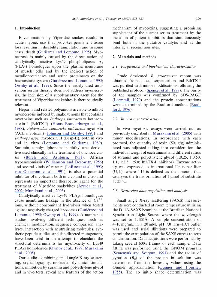
Intramuscular injections of BthTX-I (50 mg/g)increased plasma CK activity from 85741 to39507248U/L after 2 h (Figs. 1A and B). Pre-incubation of BthTX-I with suramin for 15minsignificantly inhibits the increase in plasma CKactivity in a dose-dependent fashion with maximum
ARTICLE IN PRESS
Fig. 1. In vivo dose-dependent effect of suramin (A) or PEG-400 (B) on CK release.
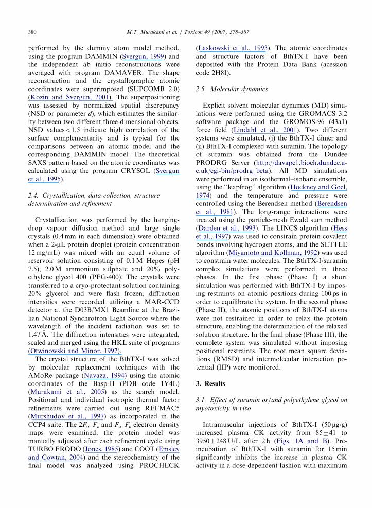
Fig. 2. In vivo effects of suramin and/or PEG-400 on the CK release.
M.T. Murakami et al. / Toxicon 49 (2007) 378–387 381
inhibition observed when the ratio is 1:1 (w/w;BthTX-I:suramin) (Fig. 2A). These values of plasmaCK activity are in agreement with previouslyreported values for other homologous Lys49 PLA2ssuch as ACL myotoxin (Melo et al., 1994) andBasp-II (Murakami et al., 2005). Experiments withPEG-400 were conducted under the same conditions(Fig. 2B). PEG-400 proved to be 30% more potentthan suramin resulting in approximately 92%inhibition of myotoxicity, whereas the maximuminhibition by suramin is 40% (Figs. 1A and B). Theeffect of the simultaneous inclusion of bothcompounds was tested in vivo (Fig. 2) and displayedenhanced inhibition, dramatically decreasing theCK activity to insignificant levels.
3.2. Crystal structure of BthTX-I complexed with
PEG-400
The final model consists of two molecules ofBthTX-I forming a homodimer, two PEG-400fragments and 86 solvent water molecules. Therefinement converged to a crystallographic residualof 22.5% (Rfree ¼ 27.6%) for 18,722 reflections inthe resolution range of 10.0–1.9 A. The electrondensity in the 2Fo–Fc Fourier maps was continuousand well defined for both monomers in theasymmetric unit, except for the C-terminii, whichare typically disordered as in other structures. Themodel displays good overall stereochemistry withRMSD values of 0.02 A and 2.11 for bond lengths
ARTICLE IN PRESSM.T. Murakami et al. / Toxicon 49 (2007) 378–387382
and bond angles, respectively and the average B-value for all atoms is 41.5 A2 (Table 1). TheRamachandran plot demonstrates that 88.5% ofthe dihedral angles are situated in the most favoredregions, while the remaining 10.5% of residues arein the additionally permitted regions. The moleculartopology of BthTX-I conserves all the main featuresof PLA2s containing an N-terminal a-helix (H1)(residue 2–12) and the two long anti-paralleldisulphide linked a-helices (H2 from residue 40 toresidue 55 and H3 from residue 86 to residue 103)with a mean distance of 10 A between the helicalaxes and two short helical turns (residues 19–22;SH4 and 108–110; SH5) (Arni and Ward, 1996).The b-wing region (residues 74–84) is structurallyconserved and a disulphide bridge preserves itsrelative orientation. The total dimeric interface area
Table 1
Data collection and refinement statistics
PDB code 2H8I
Data-collection statistics
Space group P 31 2 1
Unit-cell parameters (A) a ¼ b ¼ 56.02; c ¼ 127.57
Resolution range (A) 9.96–1.90
Unique reflections 18,722
Redundancy 10.5 (8.5)
Completeness (%) 99.3 (98.8)
I/s(I) 32.81 (4.33)
Rmerge (%)a 5.3 (41.0)
VM (A3Da�1) 2.02
Solvent content (%) 38.58
Refinement statistics
Rfactor (%)b 22.5
Rfree value (%)c 27.6
No. of amino acid residues 242
No. of solvent molecules 122
No. of polyethylene glycol molecules 2
Mean temperature factor (A2)d 41.5
R.m.s.d. bond lengths (A) 0.020
R.m.s.d. bond angles (1) 2.145
Ramachandran plot
Most favoured region (%) 88.5
Additionally allowed regions (%) 10.5
Generously allowed regions (%) 0.5
Disallowed regions (%) 0.5
Values in parentheses are for the highest resolution shell.aRmerge ¼ 100�SjI(h)�/I(h)Sj/SI(h), where I(h) is observed
intensity and /I(h)S is mean intensity of reflection h over all
measurements of I(h).bRfactor ¼ 100�SjFo�Fcj/S(Fo) the sums being taken over all
reflections with F/s(F)42 cutoff.cRfree ¼ Rfactor for 5% of the data, which were not included
during crystallographic refinement.dB-values are average B-values for all non-hydrogen atoms.
between the two molecules was calculated to beapproximately 3598 A2 occluding the whole hydro-phobic channel and the entrance to both the active-site pockets.
3.3. PEG-binding site
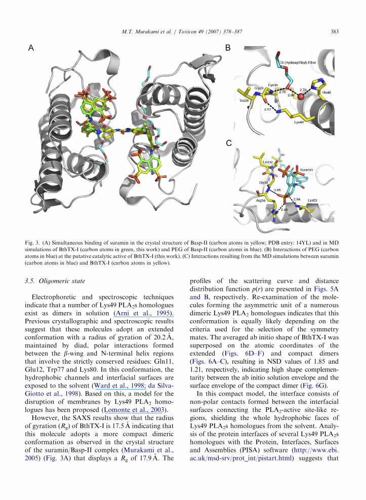
In both monomers, the electron density mapsindicated the presence of a PEG-400moleculebound in the hydrophobic channels that lead tothe PLA2-active site-like regions (Fig. 3A). ThePEG-400 molecules interact with the putativecalcium-binding loop and the residues consideredimportant for catalysis. The O1 atoms from the tailsof the PEG-400 fragments simultaneously interactwith the amide bonds between Cys29–Gly30 andHis48ND1 atom via a water molecule (Fig. 3B).Similar interactions were observed in the crystalstructure of piratoxin II, a Lys49 PLA2 fromBothrops pirajai, which contains a fatty acid boundat the entrance to the active-site cavity (Lee et al.,2001) and in the crystal structure of Basp-IIcomplexed with stearic acid (Watanabe et al.,2005). The crystal structure of Basp-II complexedwith suramin also contains a PEG molecule boundat the active-site cavity concomitantly with suramin(Murakami et al., 2005) bound at the interfacialrecognition face.
3.4. Suramin-binding site
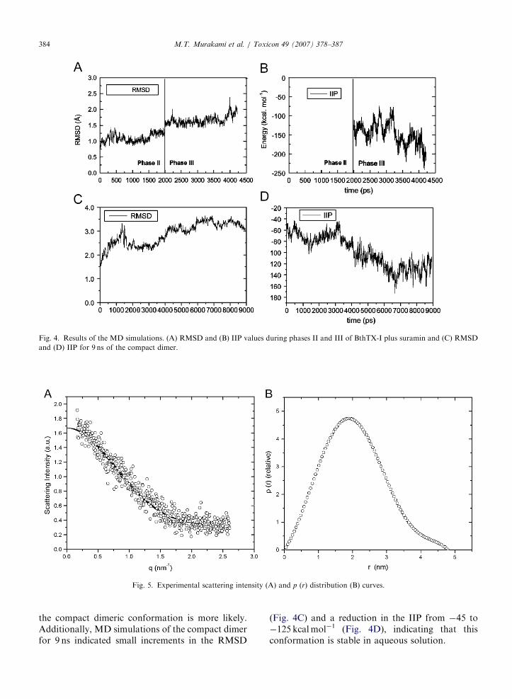
In the MD simulations of BthTX-I with suramin,the structural stability was monitored by theRMSDs of the protein atoms in phases II and III(Fig. 4A), which undergo small positional shifts(around 1.4 A) and a corresponding reductionin the IIP from �145 to �233 kcalmol�1
(Fig. 4B), indicating that the hydrated structure ofBthTX-I is similar to the crystallographic model.The resulting BthTX-I/suramin complex modelobtained from the MD simulations demonstrate asimilar mode of binding to that described forthe crystal structure of the Basp-II/suramin com-plex, where the residues Arg34 and Lys53 play keyroles in the interaction with the sulphonatednaphthyl rings (Fig. 3C). The central phenyl ringsembrace the putative calcium-binding loop and theshort helical turn (SH4) shields the whole hydro-phobic surface, inducing a drastic change in theoverall charge of the interfacial recognition face ofthe protein.
ARTICLE IN PRESS
Fig. 3. (A) Simultaneous binding of suramin in the crystal structure of Basp-II (carbon atoms in yellow; PDB entry: 14YL) and in MD
simulations of BthTX-I (carbon atoms in green, this work) and PEG of Basp-II (carbon atoms in blue). (B) Interactions of PEG (carbon
atoms in blue) at the putative catalytic active of BthTX-I (this work). (C) Interactions resulting from the MD simulations between suramin
(carbon atoms in blue) and BthTX-I (carbon atoms in yellow).
M.T. Murakami et al. / Toxicon 49 (2007) 378–387 383
3.5. Oligomeric state
Electrophoretic and spectroscopic techniquesindicate that a number of Lys49 PLA2s homologuesexist as dimers in solution (Arni et al., 1995).Previous crystallographic and spectroscopic resultssuggest that these molecules adopt an extendedconformation with a radius of gyration of 20.2 A,maintained by diad, polar interactions formedbetween the b-wing and N-terminal helix regionsthat involve the strictly conserved residues: Gln11,Glu12, Trp77 and Lys80. In this conformation, thehydrophobic channels and interfacial surfaces areexposed to the solvent (Ward et al., 1998; da Silva-Giotto et al., 1998). Based on this, a model for thedisruption of membranes by Lys49 PLA2 homo-logues has been proposed (Lomonte et al., 2003).
However, the SAXS results show that the radiusof gyration (Rg) of BthTX-I is 17.5 A indicating thatthis molecule adopts a more compact dimericconformation as observed in the crystal structureof the suramin/Basp-II complex (Murakami et al.,2005) (Fig. 3A) that displays a Rg of 17.9 A. The
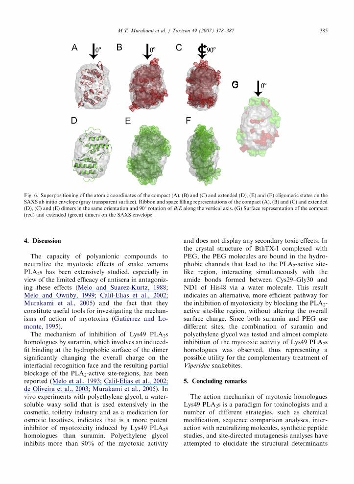
profiles of the scattering curve and distancedistribution function p(r) are presented in Figs. 5Aand B, respectively. Re-examination of the mole-cules forming the asymmetric unit of a numerousdimeric Lys49 PLA2 homologues indicates that thisconformation is equally likely depending on thecriteria used for the selection of the symmetrymates. The averaged ab initio shape of BthTX-I wassuperposed on the atomic coordinates of theextended (Figs. 6D–F) and compact dimers(Figs. 6A–C), resulting in NSD values of 1.85 and1.21, respectively, indicating high shape complemen-tarity between the ab initio solution envelope and thesurface envelope of the compact dimer (Fig. 6G).
In this compact model, the interface consists ofnon-polar contacts formed between the interfacialsurfaces connecting the PLA2-active site-like re-gions, shielding the whole hydrophobic faces ofLys49 PLA2s homologues from the solvent. Analy-sis of the protein interfaces of several Lys49 PLA2shomologues with the Protein, Interfaces, Surfacesand Assemblies (PISA) software (http://www.ebi.ac.uk/msd-srv/prot_int/pistart.html) suggests that
ARTICLE IN PRESS
Fig. 4. Results of the MD simulations. (A) RMSD and (B) IIP values during phases II and III of BthTX-I plus suramin and (C) RMSD
and (D) IIP for 9 ns of the compact dimer.
Fig. 5. Experimental scattering intensity (A) and p (r) distribution (B) curves.
M.T. Murakami et al. / Toxicon 49 (2007) 378–387384
the compact dimeric conformation is more likely.Additionally, MD simulations of the compact dimerfor 9 ns indicated small increments in the RMSD
(Fig. 4C) and a reduction in the IIP from �45 to�125 kcalmol�1 (Fig. 4D), indicating that thisconformation is stable in aqueous solution.
ARTICLE IN PRESS
Fig. 6. Superpositioning of the atomic coordinates of the compact (A), (B) and (C) and extended (D), (E) and (F) oligomeric states on the
SAXS ab initio envelope (gray transparent surface). Ribbon and space filling representations of the compact (A), (B) and (C) and extended
(D), (C) and (E) dimers in the same orientation and 901 rotation of B/E along the vertical axis. (G) Surface representation of the compact
(red) and extended (green) dimers on the SAXS envelope.
M.T. Murakami et al. / Toxicon 49 (2007) 378–387 385
4. Discussion
The capacity of polyanionic compounds toneutralize the myotoxic effects of snake venomsPLA2s has been extensively studied, especially inview of the limited efficacy of antisera in antagoniz-ing these effects (Melo and Suarez-Kurtz, 1988;Melo and Ownby, 1999; Calil-Elias et al., 2002;Murakami et al., 2005) and the fact that theyconstitute useful tools for investigating the mechan-isms of action of myotoxins (Gutierrez and Lo-monte, 1995).
The mechanism of inhibition of Lys49 PLA2shomologues by suramin, which involves an induced-fit binding at the hydrophobic surface of the dimersignificantly changing the overall charge on theinterfacial recognition face and the resulting partialblockage of the PLA2-active site-regions, has beenreported (Melo et al., 1993; Calil-Elias et al., 2002;de Oliveira et al., 2003; Murakami et al., 2005). Invivo experiments with polyethylene glycol, a water-soluble waxy solid that is used extensively in thecosmetic, toiletry industry and as a medication forosmotic laxatives, indicates that is a more potentinhibitor of myotoxicity induced by Lys49 PLA2shomologues than suramin. Polyethylene glycolinhibits more than 90% of the myotoxic activity
and does not display any secondary toxic effects. Inthe crystal structure of BthTX-I complexed withPEG, the PEG molecules are bound in the hydro-phobic channels that lead to the PLA2-active site-like region, interacting simultaneously with theamide bonds formed between Cys29–Gly30 andND1 of His48 via a water molecule. This resultindicates an alternative, more efficient pathway forthe inhibition of myotoxicity by blocking the PLA2-active site-like region, without altering the overallsurface charge. Since both suramin and PEG usedifferent sites, the combination of suramin andpolyethylene glycol was tested and almost completeinhibition of the myotoxic activity of Lys49 PLA2shomologues was observed, thus representing apossible utility for the complementary treatment ofViperidae snakebites.
5. Concluding remarks
The action mechanism of myotoxic homologuesLys49 PLA2s is a paradigm for toxinologists and anumber of different strategies, such as chemicalmodification, sequence comparison analyses, inter-action with neutralizing molecules, synthetic peptidestudies, and site-directed mutagenesis analyses haveattempted to elucidate the structural determinants
ARTICLE IN PRESSM.T. Murakami et al. / Toxicon 49 (2007) 378–387386
for myotoxicity of Lys49 PLA2s. Our resultsindicate that (i) the molecules that bind at thePLA2-active site-like region inhibit the myotoxicactivity of Lys49 PLA2; (ii) PEG derivativesrepresent attractive targets for drug design aimedat the treatment of Viperidae snakebites since theyare non-toxic; (iii) binding of suramin and heparinanalogues at the hydrophobic interfacial surfacealso inhibit myotoxic activity albeit at a lower leveland (iv) the inclusion of molecules that bind both atthe PLA2-active site-like region and at the hydro-phobic interfacial surface reduces myonecrosis toextremely low levels.
These biochemical, physiological and structuralfindings suggest a new promising therapeutic utilityfor the clinically approved drugs, suramin and PEG,which interact at two distinct sites, for the treatmentof myonecrosis induced by Viperidae snakebites.
Acknowledgements
Financial support by FAPESP, CEPID, SMOLB-Net, CNPq and CAPES/DAAD is gratefullyacknowledged. MTM is recipient of a FAPESPfellowship.
References
Arni, R.K., Ward, R.J., Gutierrez, J.M., Tulinsky, A., 1995.
Structure of a calcium-independent phospholipase-like myo-
toxic protein from Bothrops asper venom. Acta Crystallogr. D
51, 311–317.
Arni, R.K., Ward, R.J., 1996. Phospholipase A2—a structural
review. Toxicon 34, 827–841.
Arruda, E.Z., Silva, N.M., Moraes, R.A., Melo, P.A., 2002.
Effect of suramin on myotoxicity of some crotalid snake
venoms. Braz. J. Med. Biol. Res. 35, 723–726.
Berendsen, H.J.C., Postma, J.P.M., van Gunsteren, W.F., Her-
mans, J., 1981. Interactions models for water in relation to
protein hydration. In: Pullman, B. (Ed.), Intermolecular
Forces. Reidel Publishing Company, Dordrecht, pp. 331–342.
Bradford, M.M., 1976. A rapid and sensitive method for the
quantitation of microgram quantities of protein utilizing the
principle of protein dye binding. Anal. Biochem. 72, 248–254.
Burch, T.A., Ashburn, L.L., 1951. Experimental therapy of
onchocerciasis with suramin and hetrazan; results of a three-
year study. Am. J. Trop. Med. Hyg. 31, 617–623.
Calil-Elias, S., Martinez, A.M., Melo, P.A., 2002. Effect of
heparin and antivenom on skeletal muscle damage produced
by Bothrops jararacussu venom. Histol. Histopathol. 17,
463–470.
Darden, T., York, D., Pedersen, L., 1993. Particle mesh Ewald.
An N log(N) method for Ewald sumsin large systems.
J. Chem. Phys. 98, 10089–10092.
da Silva-Giotto, M.T., Garrat, R.C., Oliva, G., Mascarenhas,
Y.P., Giglio, J.R., Cintra, A.C.O., de Azevedo, W.F., Arni,
R.K., Ward, R.J., 1998. Crystallographic and spectroscopic
characterization of a molecular hinge: conformational
changes in bothropstoxin I, a dimeric Lys49-phospholipase
A2 homologue. Prot. Struct. Funct. Gen. 30, 442–454.
de Oliveira, M., Cavalcante, W.L., Arruda, E.Z., Melo, P.A.,
Dal-Pai Silva, M., Gallacci, M., 2003. Antagonism of
myotoxic and paralyzing activities of bothropstoxin-I by
suramin. Toxicon 42, 373–379.
Emsley, P., Cowtan, K., 2004. Coot: model-building tools
for molecular graphics. Acta Crystallogr. Sect. D 60,
2126–2132.
Guinier, A., Fournet, G., 1955. Small Angle Scattering of X-rays,
first ed. Wiley, New York, pp. 17–19.
Gutierrez, J.M., Lomonte, B., 1995. Phospholipase A2 myotoxins
from Bothrops snake venons. Toxicon 33, 1405–1424.
Hess, B., Becker, H., Berendsen, H.J., Fraaije, J.G.E.M., 1997.
Lincs: a linear constraint solver for molecular simulations.
J. Comput. Chem. 18, 1463–1472.
Hockney, R.W., Goel, S.P., 1974. Quiet high-resolution compu-
ter models of a plasma. J. Comput. Phys. 14, 148–158.
Homsi-Brandeburgo, M.I., Queiroz, L.S., Santo-Neto, H.,
Rodrigues-Simioni, L., Giglio, J.R., 1988. Fractionation of
Bothrops jararacussu snake venom: partial chemical charac-
terization and biological activity of bothropstoxin. Toxicon
26, 615–627.
Johnson, E.K., Ownby, C.L., 1993. Isolation of a myotoxin from
the venom of Agkistrodon contortrix laticinctus (broad-
banded copperhead) and pathogenesis of myonecrosis in-
duced by it in mice. Toxicon 31, 243–255.
Jones, T.A., 1985. Interactive computer graphics: FRODO.
Methods Enzymol. 115, 157–171.
Kozin, M.B., Svergun, D.I., 2001. Automated matching of high-
and low-resolution structural model. J. Appl. Crystallogr. 34,
33–41.
Laemmli, U.K., 1970. Cleavage of structural proteins during the
assembly of head of bacteriophage T4. Nature 227, 680–685.
LaRocca, R.V., Cooper, M.R., Uhrich, M., Danesi, R., Walther,
M.M., Linehan, W.M., Laskowski, R.A., Moss, D.S.,
Thornton, J.M., 1993. Main-chain bond lengths and bond
angles in protein structures. J. Mol. Biol. 231, 1049–1067.
Laskowski, R.A., Moss, D.S., Thornton, J.M., 1993. Main-chain
bond lengths and bond angles in protein structures. J. Mol.
Biol. 231, 1049–1067.
Lee, W.H., da Silva Giotto, M.T., Marangoni, S., Toyama,
M.H., Polikarpov, I., Garratt, R.C., 2001. Structural basis for
low catalytic activity in Lys49 phospholipases A2—a hypoth-
esis: the crystal structure of piratoxin II complexed to fatty
acid. Biochemistry 40, 28–36.
Lindahl, E., Hess, B., van der Spoe, D.L., 2001. Gromacs 3.0: a
package for molecular simulation and trajectory analysis.
J. Mol. Model. 7, 306–317.
Lomonte, B., Gutierrez, J.M., 1989. A new muscle damaging
toxin, myotoxin II, from the venom of the snake Bothrops
asper (terciopelo). Toxicon 27, 725–733.
Lomonte, B., Angulo, Y., Calderon, L., 2003. An overview of
lysine-49 phospholipase A2 myotoxins from crotalid snake
venoms and their structural determinants of myotoxic action.
Toxicon 42, 885–901.
Melo, P.A., Ownby, C.L., 1999. Ability of wedelolactone,
heparin and para-bromophenacyl bromide to antagonize the
myotoxic effects of two crotaline venoms and their PLA2
myotoxins. Toxicon 37, 199–215.
ARTICLE IN PRESSM.T. Murakami et al. / Toxicon 49 (2007) 378–387 387
Melo, P.A., Suarez-Kurtz, G., 1988. Release of creatine kinase from
skeletal muscle by Bothrops venoms: heparin potentiation of
inhibition by antivenin. Braz. J. Med. Biol. Res. 21, 548–558.
Melo, P.A., Homsi-Brandemburgo, M.I., Giglio, J.R., Ownby,
G., 1993. Antagonism of the myotoxic effects of Bothrops
jararacussu venom and bothropstoxin by polyanions. Toxicon
31, 285–291.
Melo, P.A., Nascimento, M.C., Mors, W.B., Ownby, G., 1994.
Inhibition of the myotoxic and hemorrhagic activities of
crotalid venoms by Eclipta prostrata (asteraceae) extracts and
constituents. Toxicon 32, 595–603.
Miyamoto, S., Kollman, P.A., 1992. SETTLE: an analytical
version of the SHAKE and RATTLE algorithm for rigid
water models. J. Comput. Chem. 13, 952–962.
Murakami, M.T., Arruda, E.Z., Melo, P.A., Martinez, A.B.,
Calil-Elias, S., Tomaz, M.A., Lomonte, B., Gutierrez, J.M.,
Arni, R.K., 2005. Inhibition of myotoxic activity of Bothrops
asper myotoxin II by the anti-trypanosomal drug suramin.
J. Mol. Biol. 350, 416–426.
Murshudov, G.N., Vagin, A.A., Dodson, E.J., 1997. Refinement
of macromolecular structures by the maximum-likelihood
method. Acta Crystallogr. Sect. D 53, 240–255.
Navaza, J., 1994. AMoRe: an automated package for molecular
replacement. Acta Crystallogr. A 50, 157–163.
Ownby, C.L., Selistre de Araujo, H.S., White, S.P., Fletcher, J.E.,
1999. Lysine 49 phospholipase A2 proteins. Toxicon 37, 441–445.
Otwinowski, Z., Minor, W., 1997. Processing of X-ray diffraction
data collected in oscillation mode. Methods Enzymol. 276,
307–326.
Semenyuk, A.V., Svergun, D.I., 1991. Gnom—a program
package for small-angle scattering data-processing. J. Appl.
Crystallogr. 24, 537–540.
Spencer, P.J., Aird, S.D., Boni-Mitake, M., Nascimento, N.,
Rogero, J.R., 1998. A single-step purification of bothrop-
stoxin-I. Braz. J. Med. Biol. Res. 31, 1125–1127.
Svergun, D.I., 1999. Restoring low resolution structure of
biological macromolecules from solution scattering using
simulated annealing. Biophys. J. 76, 2879–2886.
Svergun, D.I., Barberato, C., Koch, M.H., 1995. Crysol—a
program to evaluate X-ray solution scattering of biological
macromolecules from atomic coordinates. J. Appl. Crystal-
logr. 28, 768–773.
van Oosterom, A.T., ten Bokkel Huinink, W.W., van der Burg,
M.E., Vermorken, J.B., Willemse, P.H., Neijt, J.P., 1991.
Phase II clinical trial of doxifluridine in patients
with advanced ovarian cancer. Eur. J. Cancer 27,
747–749.
Ward, R.J., de Azevedo, W.F., Arni, R.K., 1998. At the interface:
crystal structures of phospholipases A2. Toxicon 36,
1623–1633.
Watanabe, L., Soares, A.M., Ward, R.J., Fontes, M.R., Arni,
R.K., 2005. Structural insights for fatty acid binding in a
Lys49-phospholipase A2: crystal structure of myotoxin II
from Bothrops moojeni complexed with stearic acid. Biochi-
mie 87, 161–167.
Williamson, J., Desowitz, R.S., 1956. Prophylacticactivity of
suramin complexes in animal trypanosomiasis. Nature 177,
1074–1075.