Delineation of the Structural Elements of Oriental Liver Fluke PLA 2 Isoforms for Potent Drug...
12
ORIGINAL ARTICLE Delineation of the Structural Elements of Oriental Liver Fluke PLA 2 Isoforms for Potent Drug Designing Gururao Hariprasad • Divya Kota • Sundararajan Baskar Singh • Alagiri Srinivasan • Souparno Adhikary Received: 9 April 2013 / Accepted: 24 August 2013 / Published online: 11 September 2013 Ó Association of Clinical Biochemists of India 2013 Abstract Clonorchis sinensis or the Chinese liver fluke is one of the most prevalent parasites affecting a major population in the oriental countries. The parasite lacks lipid generating mechanisms but is exposed to fatty acid rich bile in the liver. A secretory phospholipase A 2 , an enzyme that breaks down complex lipids, is important for the growth of the parasite. The enzyme is also implicated in the pathogenesis leading up to the hepatic fibrosis and its complications including cancer. The five isoforms of this particular enzyme from the parasite therefore qualify as potential drug targets. In this study, a detailed structural and ligand binding analysis of the isoforms has been done by modeling. The overall three dimensional structures of the isoforms are well conserved with three helices and a b- wing stabilized by four disulfide bonds. There are charac- teristic differences at the calcium binding loop, hydro- phobic channel and the C-terminal domain that can potentially be exploited for drug binding. But the most significant feature pertains to the catalytic site where the isoforms exhibit three variations of either a histidine- aspartate-tyrosine or histidine-glutamate-tyrosine or histi- dine-aspartate-phenylalanine. Molecular docking studies show that isoform specific residues and their conformations in the substrate binding hydrophobic channel make unique interactions with certain inhibitor molecules resulting in a perfect tight fit. The proposed ligand molecules have a predicted affinity in micro-molar to nano-molar range. Interestingly, few of the ligand binding interaction patterns is in accordance to the phylogenetic studies to thereby establish the usefulness of evolutionary mechanisms in aiding ligand design. The molecular diversity of the para- sitic PLA 2 described in this study provides a platform for personalized medicine in the therapeutics of clonorchiasis. Keywords Clonorchis sinensis Structural analysis Isoform specific potent drug designing Phospholipase A2 Introduction Clonorchiasis, caused by the parasite Clonorchis sinensis,a liver fluke, is one of the major parasitic zoonoses in the oriental population affecting about 35 million people globally [1, 2]. The parasite causes a series of pathological, manifestations in the liver ranging from inflammation, fibrosis and cholangiocarcinoma. Life cycle of the parasite begins with human infection following ingestion of fish that harbor encysted younger forms of parasite called as metacercaria. These metacercariae are then released in the small intestine and migrate up the bile duct where they will develop into adult flukes over the course of several weeks. There has been a lot of interest to understand molecular mechanisms of the parasite. In a very recent development this year, the entire genomic assembly and transcriptome analysis was done to understand more about the migration, parasitism and pathogenic behavior of C. sinensis at the molecular level [3]. Some of the important results of the study were the fact that genes encoding the proteins for the facilitation and metabolism of lipid were highly expressed in the parasite. One of the enzymes identified was a Electronic supplementary material The online version of this article (doi:10.1007/s12291-013-0377-1) contains supplementary material, which is available to authorized users. G. Hariprasad (&) D. Kota S. Baskar Singh A. Srinivasan S. Adhikary Department of Biophysics, All India Institute of Medical Sciences, Ansari Nagar, New Delhi 110029, India e-mail: [email protected] 123 Ind J Clin Biochem (Oct-Dec 2014) 29(4):430–441 DOI 10.1007/s12291-013-0377-1
Transcript of Delineation of the Structural Elements of Oriental Liver Fluke PLA 2 Isoforms for Potent Drug...
ORIGINAL ARTICLE
Delineation of the Structural Elements of Oriental Liver FlukePLA2 Isoforms for Potent Drug Designing
Gururao Hariprasad • Divya Kota •
Sundararajan Baskar Singh • Alagiri Srinivasan •
Souparno Adhikary
Received: 9 April 2013 / Accepted: 24 August 2013 / Published online: 11 September 2013
� Association of Clinical Biochemists of India 2013
Abstract Clonorchis sinensis or the Chinese liver fluke is
one of the most prevalent parasites affecting a major
population in the oriental countries. The parasite lacks lipid
generating mechanisms but is exposed to fatty acid rich
bile in the liver. A secretory phospholipase A2, an enzyme
that breaks down complex lipids, is important for the
growth of the parasite. The enzyme is also implicated in the
pathogenesis leading up to the hepatic fibrosis and its
complications including cancer. The five isoforms of this
particular enzyme from the parasite therefore qualify as
potential drug targets. In this study, a detailed structural
and ligand binding analysis of the isoforms has been done
by modeling. The overall three dimensional structures of
the isoforms are well conserved with three helices and a b-
wing stabilized by four disulfide bonds. There are charac-
teristic differences at the calcium binding loop, hydro-
phobic channel and the C-terminal domain that can
potentially be exploited for drug binding. But the most
significant feature pertains to the catalytic site where the
isoforms exhibit three variations of either a histidine-
aspartate-tyrosine or histidine-glutamate-tyrosine or histi-
dine-aspartate-phenylalanine. Molecular docking studies
show that isoform specific residues and their conformations
in the substrate binding hydrophobic channel make unique
interactions with certain inhibitor molecules resulting in a
perfect tight fit. The proposed ligand molecules have a
predicted affinity in micro-molar to nano-molar range.
Interestingly, few of the ligand binding interaction patterns
is in accordance to the phylogenetic studies to thereby
establish the usefulness of evolutionary mechanisms in
aiding ligand design. The molecular diversity of the para-
sitic PLA2 described in this study provides a platform for
personalized medicine in the therapeutics of clonorchiasis.
Keywords Clonorchis sinensis � Structural analysis �Isoform specific potent drug designing �Phospholipase A2
Introduction
Clonorchiasis, caused by the parasite Clonorchis sinensis, a
liver fluke, is one of the major parasitic zoonoses in the
oriental population affecting about 35 million people
globally [1, 2]. The parasite causes a series of pathological,
manifestations in the liver ranging from inflammation,
fibrosis and cholangiocarcinoma. Life cycle of the parasite
begins with human infection following ingestion of fish
that harbor encysted younger forms of parasite called as
metacercaria. These metacercariae are then released in the
small intestine and migrate up the bile duct where they will
develop into adult flukes over the course of several weeks.
There has been a lot of interest to understand molecular
mechanisms of the parasite. In a very recent development
this year, the entire genomic assembly and transcriptome
analysis was done to understand more about the migration,
parasitism and pathogenic behavior of C. sinensis at the
molecular level [3]. Some of the important results of the
study were the fact that genes encoding the proteins for the
facilitation and metabolism of lipid were highly expressed
in the parasite. One of the enzymes identified was a
Electronic supplementary material The online version of thisarticle (doi:10.1007/s12291-013-0377-1) contains supplementarymaterial, which is available to authorized users.
G. Hariprasad (&) � D. Kota � S. Baskar Singh � A. Srinivasan �S. Adhikary
Department of Biophysics, All India Institute of Medical Sciences,
Ansari Nagar, New Delhi 110029, India
e-mail: [email protected]
123
Ind J Clin Biochem (Oct-Dec 2014) 29(4):430–441
DOI 10.1007/s12291-013-0377-1

secretory phospholipase A2 (CsIIIPLA2) which breaks
down phospholipids into lysophospholipids and fatty acids
[4]. The importance of the enzyme lies in the fact that the
metacercaria that harbor in the hepatic tissue feed on the
bile that is rich in lipids. Interestingly, the genome of the
liver fluke revealed that there are genes missing for the
production of its own fatty acids. This makes the bile as the
only source of lipid for its growth [5]. The CsIIIPLA2 is
therefore an important energy source enzyme of the adult
parasite. Also, the enzyme has been shown to be crucial in
the development of parasite mediated hepatic fibrosis [6,
7]. It therefore makes this enzyme an attractive drug target.
There are a total of five isoforms of the enzyme that have
been isolated so far. The amino acid sequences of these
isoforms are available at the protein data bank. This makes
it feasible to delineate the structural details that determine
enzyme functionality, substrate specificity and drug bind-
ing. The rationale of structure based drug designing is an
ideal platform to develop potent therapeutics for clonor-
chiasis [8].
We have in this study, delineated the structural elements
of each of the five CsIIIPLA2 enzyme isoforms that are
necessary for drug binding and also propose isoform spe-
cific potent drug molecule candidates that can be taken up
for clinical use.
Methodology
Sequence Analysis
Protein sequences of C. sinensis secretory PLA2 (CsIII-
PLA2) isoforms were taken from National Centre for
Biotechnology Information. The sequence accession num-
bers of the five enzyme isoforms are are G7YK86,
G7YK85, G7Y6W9, C9V3K9 and G7YK83. The sequen-
ces were aligned using ClustalW [9] available at ExPASY.
Modeling Studies and Validation
Homology modeling of CsPLA2 isoforms were done based
on the crystal structure of bee venom PLA2 (PDB ID:
1POC) using SWISS-MODEL [10] software. The coordi-
nates obtained were visualized using program PyMOL
[11]. Analysis of conformational correctness and reliability
was carried out using PROCHECK [12]. Calcium coordi-
nate was taken from the crystal structure complex of 1POC
and fitted into the validated model. Hydrogen atoms were
added and the model was minimized in the presence of
calcium in Molegro Virtual Docker (MVD) [13]. These
models with the calcium ion were taken for structural
analysis and docking studies.
Molecular Docking
Eight small molecule PLA2 inhibitors: aspirin (2-(acetyl-
oxy)benzoic acid), diclofenac(2-[2,6-dichlorophenyl)amino]
benzeneaceticacid), indomethacin(2-[1-(4-chlorobenzoyl)-5-
methoxy-2-methylindol-3-yl]acetic acid), atropine {(1R,5S)-
8-methyl-8-azabicyclo[3.2.1]oct-3-yl(2r)-3-hydroxy-2-phe-
nylpropanoate}, anisic acid(4-methoxy benzoic acid), indole
2-carbamoylmethyl-5-propyl-octahydro-indol-7-yl)-acetic
acid), atenolol (2-(4-(2-hydroxy-3-(isopropylamino)pro-
poxy)phenyl)ethanamide) and LY311727 (3-[3-(2-amino-2-
oxoethyl)-1-benzyl-2-ethylindol-5-yl]oxypropyl phosphonic
acid) were chosen for binding studies with CsIIIPLA2 iso-
forms. The ligands were docked to each of the five CsIIIPLA2
isoforms using Molegro Virtual Docker (MVD) [13].
Ligand and Receptor Preparation
The coordinates for ligands aspirin, diclofenac, indometha-
cin, atropine, anisinic acid, indole, atenolol and LY311727
were obtained from their respective crystal structure com-
plexes with Protein Data Bank Id: 1OXR, 2B17, 3H1X,
1TH6, 2QUE, 1OXL, 2OTF and 1DCY. These molecules
and the validated CsIIIPLA2 isoform structures were pre-
pared by assigning any missing bonds, bond orders,
hybridization and charges.
Ligand Binding Site Prediction
The possible ligand binding cavity was predicted by MVD
by using the following settings: minimum cavity volume:
10 A, probe size: 1.40 A, maximum number of ray checks:
16, minimum number of ray hits: 12 and grid resolution:
0.80 A. Based on these parameters the best cavity was
chosen for docking with the ligands.
Molecular Docking
The docking calculations were done using MolDock SE
(simplex evolution) with MolDock Score [GRID] scoring
function.
Search Algorithm by MolDock Simplex Evolution
The simplex evolution parameters were set with 1500
maximum iterations, population size of 50 at 300 steps
with neighbor distance factor of 1. The energy threshold
value was set below 100 with 10 minimum torsions/
translations/rotations, 10 maximum positions and 10 quick
try values.
Ind J Clin Biochem (Oct-Dec 2014) 29(4):430–441 431
123

Pose Clustering and Generation
Pose Clustering was used to reduce the number of poses
found during the docking run and identify the best pose.
After docking, energy minimization was performed by a
short Nelder–Mead Simplex algorithm for minimization
of the translation, orientation and flexible dihedral
angles of the found poses. Based on a RMSD threshold
of 1.0, five poses were generated after 10 runs following
the one with the lowest energy was chosen as the best
pose.
Scoring Function by MolDock Score (GRID)
In the MolDock Score [GRID] a grid resolution of 0.3 A
was set to initiate the docking process. The ignore-distant-
atoms option was used to ignore atoms far away from the
binding site. The docking scoring function MolDock
Score used was an adaptation of the Differential Evolu-
tion (DE) algorithm [13]. The MolDock score is based on
a Piecewise-Linear Potential (PLP) introduced by Gehl-
haar et al. [14] and further extended in Generic Evolu-
tionary Method for molecular DOCKing (GEMDOCK)
[15]. In MolDock, the docking scoring function is
extended with a new term, taking hydrogen bond direc-
tionality into account. The MolDock score energy (Escore)
[13] is the summation of the internal energy of the ligand
(Eintra) and the ligand–protein interaction energy (Einter).
The Einter comprises of the PLP which is an approxima-
tion of the potential for hydrogen bonds, steric interac-
tions between atoms and electrostatic interactions
between charged atoms.
Escore ¼ Einter þ Eintra
However, in the MolDock Score [Grid] the hydrogen
bond directionality is not taken into account. The grid-
based scoring function facilitates speed-up by
precalculating potential-energy values on an evenly
spaced cubic grid. Therefore, the hydrogen bonding is
now determined solely on distance and hydrogen bonding
capabilities. The energy potential is evaluated by using tri-
linear interpolation between relevant grid points.
Prediction of Binding Affinities
The explicit hydrogens were created and their hydrogen
bonding patterns were determined. The candidates with the
best conformational and energetic results were taken as the
best fit. Interaction energies between the ligands and the
proteins were estimated as the MolDock score. The binding
affinities were calculated based on the MolDock Score
using the binding affinity module of MVD. Binding affinity
(Ki) is calculated by: Ebinding = -5.68 pKi.
Molecular Dynamics
The binding of the small molecules in the gas phase was
analyzed by molecular dynamics (MD) simulation in Ac-
celrys Discovery Studio (DS 2.5). The minimization of the
protein–ligand complex was carried out in ABNR algo-
rithm which incorporates the initial ‘Steepest descent’
minimization steps with convergent criteria of \0.1 kcal/
mol/A r.m.s gradient for 1,000 steps. The production run
was carried out for another 5 ns for canonical ensemble
(NVT) using Leapfrog Verlet dynamics integrator. In both
the stages (minimization and production) of simulation, a
time step of 1 fs was used and only the backbone of the
protein was kept rigid.
Validation of Docking Methodology
The docking methodology was validated on a crystal
structure complex of human group IIA PLA2 with
LY311727, one of the ligands used in the binding analysis
in this study. The molecule, LY311727 was extracted from
the complex (PDB Id: 1DCY), re-built, energy optimized
and the new coordinates were used as ligand input for
docking to the same protein. The conformation, total
interaction energies and the quality of interactions were
determined for the docked complex and compared with that
of the crystal structure complex.
Phylogenetic Analysis
The aligned sequence of the CsIIIPLA2 isoforms in Clu-
stalW format was taken. The sequences were trimmed to
the length of the isoform with the shortest primary struc-
ture. This data was submitted to the software MEGA 5.1
[16] for phylogenetic analysis by maximum likelihood and
maximum parsimony methods were carried out. The
respective trees were constructed after validation by boot
strapping.
Results and Discussion
Sequence Analysis
Sequences of amino acids corresponding to five CsIII-
PLA2s enzymes were aligned for sequence analysis
(Fig. 1). CsIIIPLA2 isoforms exist as monomers in the
native state unlike few other enzymes in the group III PLA2
family [17, 18]. The lengths of sequences and molecular
weights of the isoforms vary from one another. Isoform 3 is
the longest with 197 residues and isoform 4 is the shortest
with 115 residues. It is difficult to tell from the protein
annotations in the data bank whether the lengths of
432 Ind J Clin Biochem (Oct-Dec 2014) 29(4):430–441
123

sequences are primer dictated or not. The sequences will
therefore be considered as native forms in this study.
Alignment of the sequences shows conservation of eight
cysteines for a possible four disulfide bonds. There is a
high homology of 41 % among the sequences from
N-terminus up to the conserved phenylalanine at position
96 with no significant homology in C-terminal region. This
implies that functional motifs required for the catalytic
function are contained within the conserved stretch. Cata-
lytic histidine is conserved in all isoforms confirming his-
tidine dependent hydrolysis of the enzyme. Comparison
with bee venom PLA2 shows variations at positions of two
other residues that participate along side histidine in
catalysis. Isoform 2 has a glutamic acid instead of aspartic
acid at position 64 and isoforms 4 and 5 have phenylala-
nine instead of tyrosine at position 87. Calcium binding
motif is at N-terminal region of the enzymes with only
glycine residues conserved at position 11. While the ninth
position has either tryptophan, histidine or tyrosine, the
thirteenth position has either glycine, threonine or aspara-
gine. Calcium binding motif therefore shows a convergent
type of evolution through possible random mutations that
have been acquired by the isoforms with the same ancestor
with exposure to similar environments within the bile duct.
The fourth calcium binding residue, aspartate is conserved
along side catalytic His35 to form the classical ‘His-Asp
dyad’. Based on structural characterization of bee venom
PLA2 with a substrate binding analogue [17], the substrate
binding residues on CsIIIPLA2 isoforms were identified to
reveal interesting features. They are: (1) valine at position
83 and phenylalanine at position 67 are the only non-polar
residues that are identical; (2) non-polar residues are con-
served at the positions 4, 57, 87 and 91 and (3) cavity
residues at positions 12, 41, 43, 58, 60, 61, 86 and 93 are
not even semi-conserved. This highlights the fact that there
are physio-chemical differences in the channel for possible
substrate recognition function and specificity of isoforms.
This may partly explain the functional outcomes of
inflammation, hepatic fibrosis and cancer caused by
infection of the parasite, C. sinensis [5–7].
Model Building and Validation
Models of CsIII PLA2 were built based on crystal structure
of bee venom PLA2 using SWISS MODEL software
(Fig. 2). SWISS MODEL has been known to build the best
quality structures for this group of enzymes [18]. All five
isoform structures were analyzed for their conformational
correctness. Ramachandran plot using PROCHECK soft-
ware along with other structure validating parameters
judged the reliability of all models as shown in Table 1.
Based on results of Ramachandran plot, the total number of
residues in the model and the RMSD of the model from the
template main chain, isoform models built by SWISS
MODEL were found to be correct. These isoform models
were taken for structural analysis and subsequent docking
studies. A representative Ramachandran plot for CsIIIPLA2
isoform 1 has been shown in Fig. 3.
Overall Structure
Over all tertiary folds of all five CsIIIPLA2 isoforms
essentially comprise of three a-helices, a b-wing, a calcium
binding loop at N-terminus (Fig. 2). The entire structure is
stabilized by four disulfide bonds between first eight con-
served cysteines. There are variations pertaining to the
presence and length of C-terminal domain. While the fourth
isoform ends at end of the third helix, the first, second and
the fifth isoforms extend to different lengths taking up a loop
conformation. C-terminal region is the longest for the third
Fig. 1 Sequence homology studies of Clonorchis sinensis secretory
PLA2 isoforms. The conserved residues are shown in bold. The
calcium binding residues are shown in bright pink, cysteines are
shown in yellow, catalytic residues are shown in red and residues
lining the hydrophobic channel are shown in green, cyan, purple, blue
and orange in each of the five isoforms. ‘*’, ‘:’ and ‘.’ indicate
identical, conserved and semi-conserved residues respectively
Ind J Clin Biochem (Oct-Dec 2014) 29(4):430–441 433
123
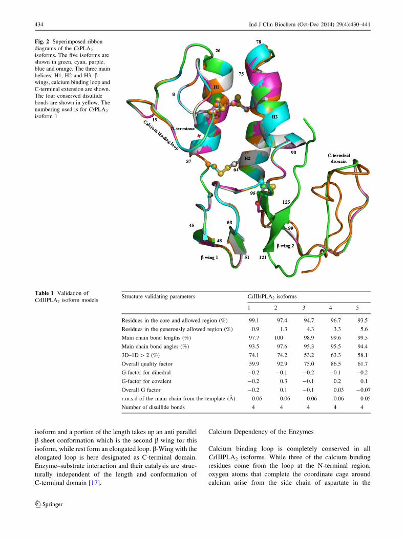
isoform and a portion of the length takes up an anti parallel
b-sheet conformation which is the second b-wing for this
isoform, while rest form an elongated loop. b-Wing with the
elongated loop is here designated as C-terminal domain.
Enzyme–substrate interaction and their catalysis are struc-
turally independent of the length and conformation of
C-terminal domain [17].
Calcium Dependency of the Enzymes
Calcium binding loop is completely conserved in all
CsIIIPLA2 isoforms. While three of the calcium binding
residues come from the loop at the N-terminal region,
oxygen atoms that complete the coordinate cage around
calcium arise from the side chain of aspartate in the
Fig. 2 Superimposed ribbon
diagrams of the CsPLA2
isoforms. The five isoforms are
shown in green, cyan, purple,
blue and orange. The three main
helices: H1, H2 and H3, b-
wings, calcium binding loop and
C-terminal extension are shown.
The four conserved disulfide
bonds are shown in yellow. The
numbering used is for CsPLA2
isoform 1
Table 1 Validation of
CsIIIPLA2 isoform modelsStructure validating parameters CsIIIsPLA2 isoforms
1 2 3 4 5
Residues in the core and allowed region (%) 99.1 97.4 94.7 96.7 93.5
Residues in the generously allowed region (%) 0.9 1.3 4.3 3.3 5.6
Main chain bond lengths (%) 97.7 100 98.9 99.6 99.5
Main chain bond angles (%) 93.5 97.6 95.3 95.5 94.4
3D–1D [ 2 (%) 74.1 74.2 53.2 63.3 58.1
Overall quality factor 59.9 92.9 75.0 86.5 61.7
G-factor for dihedral -0.2 -0.1 -0.2 -0.1 -0.2
G-factor for covalent -0.2 0.3 -0.1 0.2 0.1
Overall G factor -0.2 0.1 -0.1 0.03 -0.07
r.m.s.d of the main chain from the template (A) 0.06 0.06 0.06 0.06 0.05
Number of disulfide bonds 4 4 4 4 4
434 Ind J Clin Biochem (Oct-Dec 2014) 29(4):430–441
123

invariant position next to catalytic histidine (Fig. 4). A five
coordinate bonded calcium cage is therefore seen for all
isoforms establishing the structural basis for calcium
dependent nature of these enzymes. The backbone carbonyl
oxygen of glycine at position 11 is present in all isoforms.
However, there are variations in the remaining two residues
of the loop that participate in calcium binding. Carbonyl
oxygen of tryptophan in isoforms 1, histidine in isoforms 2
and 4, and tyrosine in isoforms 3 and 5, at ninth position
form coordinate bond with calcium. Likewise, carbonyl
oxygen of glycine and threonine, in isoforms 1 and 2, and
asparagine at thirteenth position in rest of the three iso-
forms participate in calcium binding. In addition to coor-
dinate bonds, calcium binding loop is stabilized by a
disulfide bond between Cys10 on the loop with Cys32 on
the adjacent first helix. Though calcium binding loop forms
the superior wall of the hydrophobic channel, the side
chains of calcium binding residues are oriented away from
the cavity. Therefore the only possible interactions of the
loop with substrate would be with Ca main chain atoms.
Conversely, there exists a high possibility of a similar
pattern of interactions from back bone atoms of the loop
with competitively binding ligand molecules at the cavity.
Catalytic Site
Phospholipase A2 enzyme hydrolyses the sn-2 ester bond
of phospholipids by catalytic histidine and a nucleophile
water molecule. The conformational state of histidine is
therefore important for hydrolysis of the phospholipids to
release arachidonic acid, a precursor for inflammatory
eicosanoids. Conformation of catalytic histidine at active
site is same in all five isoforms. The participation of this
residue makes the residue’s pKa the optimum pH of
enzyme [6]. Conformational orientation and stability of
histidine has been classically known to be through its
interactions with aspartate and tyrosine residues, which are
in the vicinity of catalytic site [17, 19]. In some of enzymes
there is more than one tyrosine involved in hydrogen
bonded interaction [20, 21]. These aspartates and tyrosines
arise from non-anologous positions of the enzyme. This
pattern of arrangement is seen in CsIIIPLA2 isoforms 1 and
3 (Fig. 5a) and is reminiscent of group X PLA2 [19]. In
isoform 2, there is a replacement of aspartate to a glutamate
at position 65. Though this is a classical conservative
mutation, the oxygen atoms of the carboxyl side chain are
oriented away from catalytic histidine and do not form any
hydrogen bonds with tyrosine (Fig. 5b). Inter-atomic dis-
tance between carboxyl oxygen of aspartate and hydroxyl
oxygen of tyrosine is 4 A. Conformation of glutamate is
stabilized by van der Waals interactions with His60,
Cys61, Cys62, Tyr86, Phe87 and Lys94. Orientation of
glutamate also rules out any possibility of a water mediated
hydrogen bond formation. This type of conformational
arrangement of residues at the active site is seen in PLA2
enzymes in venom of scorpion [22]. In isoforms 4 and 5,
there is a replacement of hydrophilic tyrosine to a
Fig. 3 A representative Ramachandran plot for the structure of
CsPLA2 isoform 4. The plot was built using NIH structural analysis
and verification software. 96.7 % of the residues in either the core
region or allowed region and remaining 3.3 % of the residues in the
generously allowed region with no residue in the disallowed region
Fig. 4 Calcium binding loop. The loop is shown in white and pink. The
calcium binding residues in the five isoforms are shown in green, cyan,
pink, white and orange. The calcium ion is shown as an asterix in pink
Ind J Clin Biochem (Oct-Dec 2014) 29(4):430–441 435
123
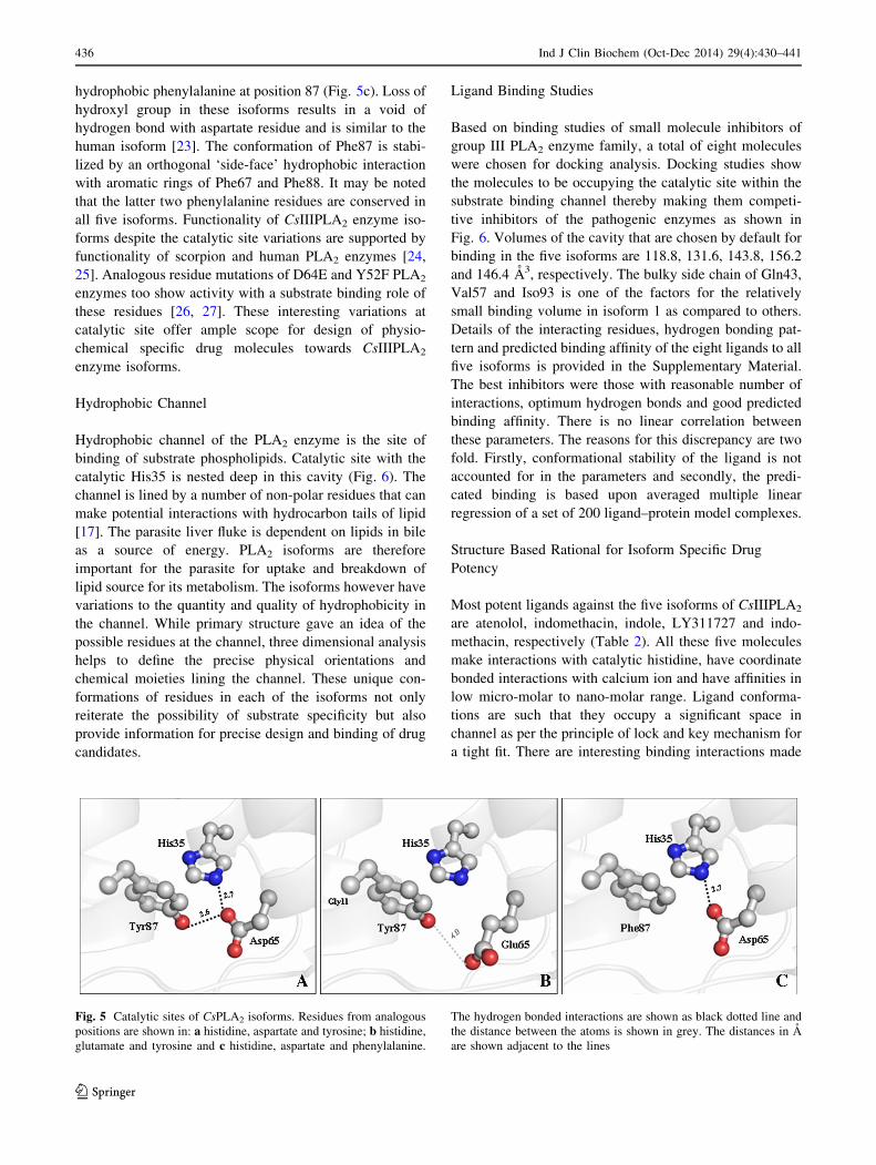
hydrophobic phenylalanine at position 87 (Fig. 5c). Loss of
hydroxyl group in these isoforms results in a void of
hydrogen bond with aspartate residue and is similar to the
human isoform [23]. The conformation of Phe87 is stabi-
lized by an orthogonal ‘side-face’ hydrophobic interaction
with aromatic rings of Phe67 and Phe88. It may be noted
that the latter two phenylalanine residues are conserved in
all five isoforms. Functionality of CsIIIPLA2 enzyme iso-
forms despite the catalytic site variations are supported by
functionality of scorpion and human PLA2 enzymes [24,
25]. Analogous residue mutations of D64E and Y52F PLA2
enzymes too show activity with a substrate binding role of
these residues [26, 27]. These interesting variations at
catalytic site offer ample scope for design of physio-
chemical specific drug molecules towards CsIIIPLA2
enzyme isoforms.
Hydrophobic Channel
Hydrophobic channel of the PLA2 enzyme is the site of
binding of substrate phospholipids. Catalytic site with the
catalytic His35 is nested deep in this cavity (Fig. 6). The
channel is lined by a number of non-polar residues that can
make potential interactions with hydrocarbon tails of lipid
[17]. The parasite liver fluke is dependent on lipids in bile
as a source of energy. PLA2 isoforms are therefore
important for the parasite for uptake and breakdown of
lipid source for its metabolism. The isoforms however have
variations to the quantity and quality of hydrophobicity in
the channel. While primary structure gave an idea of the
possible residues at the channel, three dimensional analysis
helps to define the precise physical orientations and
chemical moieties lining the channel. These unique con-
formations of residues in each of the isoforms not only
reiterate the possibility of substrate specificity but also
provide information for precise design and binding of drug
candidates.
Ligand Binding Studies
Based on binding studies of small molecule inhibitors of
group III PLA2 enzyme family, a total of eight molecules
were chosen for docking analysis. Docking studies show
the molecules to be occupying the catalytic site within the
substrate binding channel thereby making them competi-
tive inhibitors of the pathogenic enzymes as shown in
Fig. 6. Volumes of the cavity that are chosen by default for
binding in the five isoforms are 118.8, 131.6, 143.8, 156.2
and 146.4 A3, respectively. The bulky side chain of Gln43,
Val57 and Iso93 is one of the factors for the relatively
small binding volume in isoform 1 as compared to others.
Details of the interacting residues, hydrogen bonding pat-
tern and predicted binding affinity of the eight ligands to all
five isoforms is provided in the Supplementary Material.
The best inhibitors were those with reasonable number of
interactions, optimum hydrogen bonds and good predicted
binding affinity. There is no linear correlation between
these parameters. The reasons for this discrepancy are two
fold. Firstly, conformational stability of the ligand is not
accounted for in the parameters and secondly, the predi-
cated binding is based upon averaged multiple linear
regression of a set of 200 ligand–protein model complexes.
Structure Based Rational for Isoform Specific Drug
Potency
Most potent ligands against the five isoforms of CsIIIPLA2
are atenolol, indomethacin, indole, LY311727 and indo-
methacin, respectively (Table 2). All these five molecules
make interactions with catalytic histidine, have coordinate
bonded interactions with calcium ion and have affinities in
low micro-molar to nano-molar range. Ligand conforma-
tions are such that they occupy a significant space in
channel as per the principle of lock and key mechanism for
a tight fit. There are interesting binding interactions made
Fig. 5 Catalytic sites of CsPLA2 isoforms. Residues from analogous
positions are shown in: a histidine, aspartate and tyrosine; b histidine,
glutamate and tyrosine and c histidine, aspartate and phenylalanine.
The hydrogen bonded interactions are shown as black dotted line and
the distance between the atoms is shown in grey. The distances in A
are shown adjacent to the lines
436 Ind J Clin Biochem (Oct-Dec 2014) 29(4):430–441
123

by these ligands with the channel lining residues at the end
of molecular dynamics at the end of 5 ns. The key inter-
actions of these five ligands with their respective isoforms
are explained: (A) Atenolol binds at the catalytic site of
CsIIIPLA2 isoform 1 enzyme and makes hydrogen bonded
interactions with all three active site residues- His35,
Asp65 and Tyr87 (Figs. 6, 7a). The main chain nitrogen of
Gly11 makes a hydrogen bond with the oxygen of the
ligand. Thr58 makes a polar contact through its main chain
with the oxygen atom of the ligand acetamide. The ligand
makes coordinate bond with the calcium and an array of
hydrophobic interactions with Val83 (minimum interacting
distance of 4 A), Val57 (4 A) and Leu86 (3 A). The main
chain oxygen atom of Lys12 makes van der Waals contact
of 3.9 A with the ligand. It may be noted that residues
Lys12, Val57 and Leu86 are unique to isoform 1.
(B) Indomethacin is most potent in terms of occupying the
cavity volume as well as having chemical complementarity
to isoform 2. Carboxyl oxygen atom at one end of the
ligand makes two hydrogen bonds each with the residues
side chains of Asp36 (3.4 A) and main chain oxygen atom
of His9 (2.8 A). The chlorobenzoyl ring and the indole ring
of the ligand make a number of hydrophobic interactions
with Tyr92. The ligand also makes hydrophobic contacts
with Gly57, Pro58, Tyr59, Met60, Tyr87, Tyr92 and Gln94
(Fig. 7b). These residues along with His9 are unique to
isoform 2. (C) The natural molecule indole is the best fit for
the isoform 3 enzyme and makes a total of seven hydrogen
bonds, saturating all the polar moieties on the ligand
thereby stabilizing its binding conformation. Hydrogen
bonded interactions are with the residues Gly11, His35,
Asp36, Phe60, His61 and Tyr87 (Fig. 7c). The ligand also
makes important hydrophobic interactions with Phe60
(3.2 A) and van der Waals interaction with Tyr9 (3.6 A).
These two aromatic residues are unique to this isoform.
(D) The synthetic molecule LY311727 binds to isoform 4
enzyme at catalytic site and the carboxyl oxygens make
two hydrogen bonded interactions with Gly11 (3.4 A) and
His35 (2.6 A). The etheryl oxygen interacts by a hydrogen
bond with main chain nitrogen of Asn13 (Fig. 7d). The
ligand also makes hydrophobic contacts with the residues
Thr58 (3.4 A), Phe68 (3.9 A) and Phe87 (3.5 A) which are
unique to this isoform. (E) Indomethacin molecule at the
catalytic site of isoform 5 makes five hydrogen bonded
interactions. Two of these are from carboxyl oxygen with
Gly11 and His35, two are from carbonyl oxygen atom with
Thr58 and Asn13 and the last one is between methoxy
oxygen atom of the ligand and the side chain amino group
of Asn41. The ligand also makes hydrophobic interactions
with Tyr43 (3.9 A) and Val57 (3.8 A).
In summary, every ligand binds in a unique and a
definitive way making certain ligands more isoform
Fig. 6 A GRASP model of
CsPLA2 isoforms1 with the
ligand atenolol. The catalytic
site with its constituent residues
is shown in a mesh. The ligand,
atenolol is shown in white-blue-
red rendering. The interacting
residues are shown in green-
blue-red rendering and the
calcium ion is shown as a green
asterix. The hydrogen bonded
interactions are shown as dotted
lines
Ind J Clin Biochem (Oct-Dec 2014) 29(4):430–441 437
123
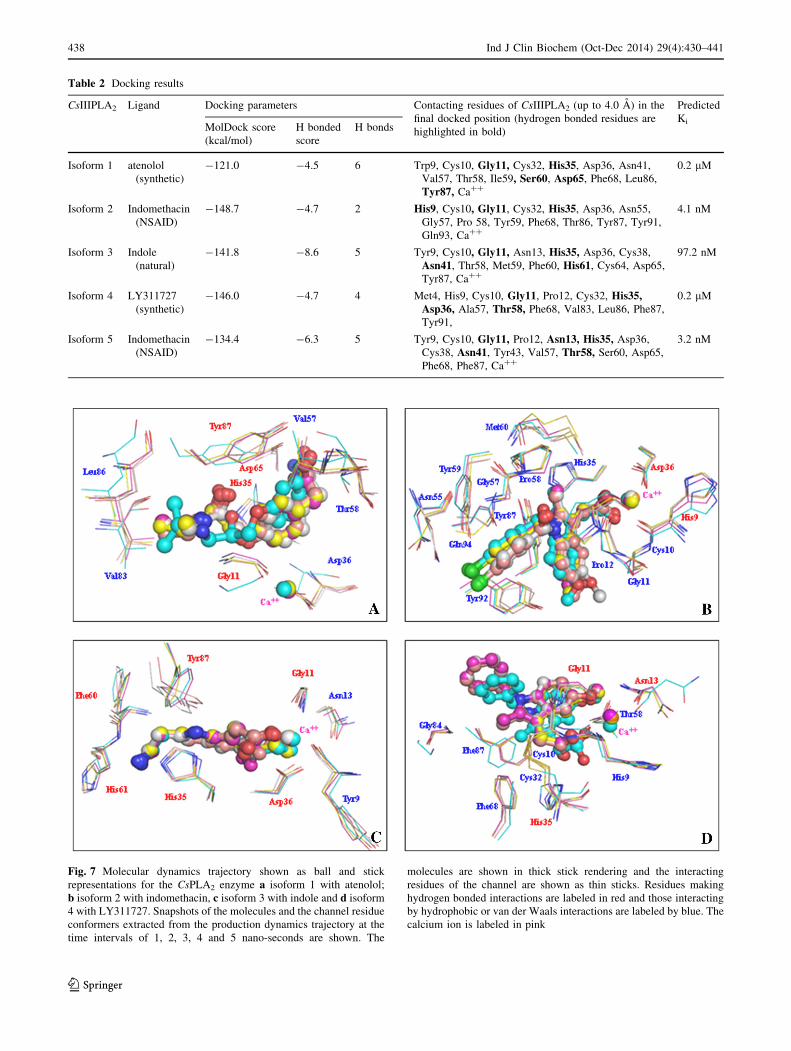
Table 2 Docking results
CsIIIPLA2 Ligand Docking parameters Contacting residues of CsIIIPLA2 (up to 4.0 A) in the
final docked position (hydrogen bonded residues are
highlighted in bold)
Predicted
KiMolDock score
(kcal/mol)
H bonded
score
H bonds
Isoform 1 atenolol
(synthetic)
-121.0 -4.5 6 Trp9, Cys10, Gly11, Cys32, His35, Asp36, Asn41,
Val57, Thr58, Ile59, Ser60, Asp65, Phe68, Leu86,
Tyr87, Ca??
0.2 lM
Isoform 2 Indomethacin
(NSAID)
-148.7 -4.7 2 His9, Cys10, Gly11, Cys32, His35, Asp36, Asn55,
Gly57, Pro 58, Tyr59, Phe68, Thr86, Tyr87, Tyr91,
Gln93, Ca??
4.1 nM
Isoform 3 Indole
(natural)
-141.8 -8.6 5 Tyr9, Cys10, Gly11, Asn13, His35, Asp36, Cys38,
Asn41, Thr58, Met59, Phe60, His61, Cys64, Asp65,
Tyr87, Ca??
97.2 nM
Isoform 4 LY311727
(synthetic)
-146.0 -4.7 4 Met4, His9, Cys10, Gly11, Pro12, Cys32, His35,Asp36, Ala57, Thr58, Phe68, Val83, Leu86, Phe87,
Tyr91,
0.2 lM
Isoform 5 Indomethacin
(NSAID)
-134.4 -6.3 5 Tyr9, Cys10, Gly11, Pro12, Asn13, His35, Asp36,
Cys38, Asn41, Tyr43, Val57, Thr58, Ser60, Asp65,
Phe68, Phe87, Ca??
3.2 nM
Fig. 7 Molecular dynamics trajectory shown as ball and stick
representations for the CsPLA2 enzyme a isoform 1 with atenolol;
b isoform 2 with indomethacin, c isoform 3 with indole and d isoform
4 with LY311727. Snapshots of the molecules and the channel residue
conformers extracted from the production dynamics trajectory at the
time intervals of 1, 2, 3, 4 and 5 nano-seconds are shown. The
molecules are shown in thick stick rendering and the interacting
residues of the channel are shown as thin sticks. Residues making
hydrogen bonded interactions are labeled in red and those interacting
by hydrophobic or van der Waals interactions are labeled by blue. The
calcium ion is labeled in pink
438 Ind J Clin Biochem (Oct-Dec 2014) 29(4):430–441
123

specific. It would therefore be useful to know the parasitic
PLA2 isoform by gene sequencing in patients with liver
fluke disease. This would be a meaningful exercise in the
direction of pharmacogenomics of clonorchiasis from a
clinical perspective.
Validation of Docking Methodology
As there is no crystal structure of a group III PLA2 with a
small molecule, the docking methodology was validated by
redocking LY311727 to the human group IIA PLA2 and
interaction parameters were compared to crystal structure
complex PDB ID: 1ILY. Total interaction energy of
docked complex is comparable to that of crystal structure
complex (Table 3). The r.m.s.d conformations of the two
complexes are less than 1 A (Fig. 8). Lastly, the docked
complex has same residue interactions as its crystal coun-
terpart. Hydrogen bonded interactions of Gly29 and His47
are common to both complexes. Ile9 and Phe98 are the
only two additional interacting residues that do not feature
in the crystal complex interactions. Predicted binding
affinity of 17.3 nM is comparable to experimentally
determined inhibitory constant of ligand LY311727 with
the same enzyme.
Evolutionary Perspectives for Isoform Specific Drug
Potency
Though sequence analysis helps to understand the extent of
identity of isoforms with any one of the sequences, phy-
logeny helps to evaluate inter sequence relationship
Table 3 Validation of the docking methodology
No. Structure Methodology
used
Total energy (kcal/mol) r.m.s.d of docked
complex with the
crystal structure
complex (A)
Contacting residues of the protein with the
ligand (residues making H bonded
interactions are shown in bold)Einter Eintra Escore
1 Human group IIA
PLA2 complex
with inhibitor
LY311727
Crystallization
(PDB ID:
1DCY)
-144.0 -1.3 -145.3 – Leu2, Phe5, His6, Ala17, Ala18, Tyr21,
Gly22, His27, Cys28, Gly29, Val30,
Cys44, His47, Asp48, Tyr51, Lys62, Ca??
2 Human group IIA
PLA2 complex
with inhibitor
LY311727
Docking
validation
(This study)
-138.5 -2.1 -140.6 0.8 Leu2, Phe5, His6, Ile9, Ala17, Ala18, Tyr21,
Gly22, His27, Cys28, Gly29, Val30,
Cys44, His47, Asp48, Tyr51, Lys62,
Phe98, Ca??
Fig. 8 Validation of the docking methodology. Crystal structure and
the docked conformations of the ligand LY311727 in the hydrophobic
channel of the human group IIA PLA2. The crystal structure complex
is shown in green while the docked structure complex is shown in
white
Fig. 9 Phylogenetic analysis. aPhylogeny tree by maximum
likelyhood method and bphylogeny tree by parsimony
method
Ind J Clin Biochem (Oct-Dec 2014) 29(4):430–441 439
123

amongst each other. Phylogenetic studies were done to
evaluate evolutionary proximity between the five CsIII-
PLA2 isoforms. As isoforms were of different lengths,
sequences were trimmed to a uniform length of hundred
residues. Sequences therefore essentially include calcium
binding motif, active site residues and hydrophobic channel
residues that play an important role in substrate identifi-
cation and binding. Both the maximum likelihood method
and the Maximum parsimony method show similar pattern
of trees (Fig. 9). The results show that isoform 3 and iso-
form 5 clustered together in a single clade, isoform 1 and
isoform 2 are closely related and isoform 4 is all by itself in
the two trees. It may be noted that ligand binding studies
pertain to the molecules that competitively bind at substrate
binding channel with an increased probability of making
interactions with the same substrate binding residues. It
therefore may be implied that understanding of the evolu-
tionary proximity amongst the CsPLA2 isoforms have a
direct bearing on ligand binding. From ligand binding
analysis carried out it is seen that there are consistent
binding patterns between: (1) first and second isoforms,
such as participation of Gly11 and His35 in hydrogen
bonded interactions with indomethacin; (2) third and fifth
isoforms, such as participation of Asn13 and Asn41 in
interactions with most potent ligands and (3) uniqueness of
the isoform 4 with participation of residues Met4, His35,
Asp36 and Leu86 in interactions with LY311727, and also
absence of a coordinate bond with the calcium. It is likely
that studies on a larger number of complexes will bring out
statistical significance of correlation between phylogenetic
studies and ligand binding fingerprints. Evolutionary rela-
tions of proteins therefore have scope to complement
structural analysis as a rational for isoform based drug
designing as in case of CsIIIPLA2.
Conclusion
CsIIIPLA2 isoforms are potential drug targets for infection
by parasite C. sinensis. All isoforms have a well conserved
three dimensional structure but have residue variations at
calcium binding site, catalytic site and at substrate binding
hydrophobic channel. Detailed structural analyses com-
bined with ligand binding studies help to understand fac-
tors that dictate isoform specific drug potency. Some of the
drug molecules proposed may either be further developed
or considered as a basis for personalized medicine for
hepatic fibrosis caused by the parasite.
Acknowledgments This research was supported by a Fast Track
project Grant to GH from the Department of Science and Technology,
Government of India.
References
1. Lun ZR, Gasser RB, Lai DH, Li AX, Zhu XQ, Yu XB, et al.
Clonorchiasis: a key foodborne zoonosis in China. Lancet Infect
Dis. 2005;5:31–41.
2. Li T, He S, Zhao H, Zhao G, Zhu XQ. Major trends in human
parasitic diseases in China. Trends Parasitol. 2010;26:264–70.
3. Huang Y, Chen W, Wang X, Liu H, Chen Y, Guo L, et al. The
carcinogenic liver fluke, Clonorchis sinensis: new assembly, re-
annotation and analysis of the genome and characterization of
tissue transcriptomes. PLoS One. 2013;8:e54732.
4. Murakami M, Sato H, Taketomi Y, Yamamoto K. Integrated
lipidomics in the secreted phospholipase A2 biology. Int J Mol
Sci. 2011;12:1474–95.
5. Wang X, Chen W, Huang Y, Sun J, Men J, Liu H, et al. The draft
genome of the carcinogenic human liver fluke Clonorchis sin-
ensis. Genome Biol. 2011;12:R107.
6. Hu F, Hu X, Ma C, Zhao J, Xu J, Yu X. Molecular character-
ization of a novel Clonorchis sinensis secretory phospholipase
A(2) and investigation of its potential contribution to hepatic
fibrosis. Mol Biochem Parasitol. 2009;167:127–34.
7. Zhang F, Liang P, Chen W, Wang X, Hu Y, Liang C, et al. Stage-
specific expression, immunolocalization of Clonorchis sinensis
lysophospholipase and its potential role in hepatic fibrosis.
Parasitol Res. 2013;12:737–49.
8. Hariprasad G, Kaur P, Srinivasan A, Singh TP, Kumar M.
Structural analysis of secretory phospholipase A2 from Clonor-
chis sinensis: therapeutic implications for hepatic fibrosis. J Mol
Model. 2012;18:3139–45.
9. Thompson JD, Gibson TJ, Higgins DG. Multiple sequence
alignment using ClustalW and ClustalX. Curr Protoc Bioinfor-
matics. 2002;2(2):3.
10. Arnold K, Bordoli L, Kopp J, Schwede T. The SWISS-MODEL
workspace: a web-based environment for protein structure
homology modeling. Bioinformatics. 2006;22:195–201.
11. De Lano WL. The PyMOL molecular graphics system. San
Carlos: DeLano Scientific; 2002.
12. Laskowski RA, MacArthur MW, Moss DS, Thornton JM. PRO-
CHECK: a program to check the stereochemical quality of pro-
tein structures. J Appl Cryst. 1993;26:283–91.
13. Thomsen R, Christensen MH. MolDock: a new technique for high-
accuracy molecular docking. J Med Chem. 2006;49:3315–21.
14. Gehlhaar DK, Verkhivker G, Rejto PA, Fogel DB, Fogel LJ,
Freer ST. Docking conformationally flexible small molecules into
a protein binding site through evolutionary programming. Pro-
ceedings of the fourth international conference on evolutionary
programming. 1995;615–627.
15. Yang JM, Chen CC. GEMDOCK: a generic evolutionary method
for molecular docking. Proteins. 2004;55:288–304.
16. Tamura K, Peterson D, Peterson N, Stecher G, Nei M, Kumar S.
MEGA5: molecular evolutionary genetics analysis using maxi-
mum likelihood, evolutionary distance, and maximum parsimony
methods. Mol Biol Evol. 2011;28:2731–9.
17. Scott DL, Otwinowski Z, Gelb MH, Sigler PB. Crystal structure
of bee-venom phospholipase A2 in a complex with a transition-
state analogue. Science. 1990;250:1563–6.
18. Hariprasad G, Baskar S, Das U, Ethayathulla AS, Kaur P, Singh
TP, et al. Cloning, sequence analysis and homology modeling of
a novel phospholipase A2 from Heterometrus fulvipes (Indian
black scorpion). DNA Seq. 2007;18:242–6.
19. Pan YH, Yu BZ, Singer AG, Ghomashchi F, Lameau G, Gelb
MH, et al. Crystal structure of human group 9 secreted phos-
pholipase A2 electrostatically neutral interfacial surface targets
zwitterionic membrane. J Biol Chem. 2002;277:29086–93.
440 Ind J Clin Biochem (Oct-Dec 2014) 29(4):430–441
123

20. Jabeen T, Singh N, Singh RK, Ethayathulla AS, Sharma S,
Srinivasan A, et al. Crystal structure of a novel phospholipase A2
from Naja naja sagittifera with a strong anti-coagulant activity.
Toxicon. 2005;46:865–75.
21. Jasti J, Paramasivam M, Srinivasan A, Singh TP. Structure of an
acidic phospholipase A2 from Indian saw-scaled viper (Echis
carinatus) at 2.6 A resolution reveals a novel intermolecular
interaction. Acta Crystallogr D. 2004;60:66–72.
22. Hariprasad G, Kumar M, Srinivasan A, Kaur P, Singh TP, Jithesh
O. Group III phospholipase A2 from the scorpion, Mesobuthus
tamulus: targeting and reversible inhibition by native peptides. Int
J Biol Macromol. 2011;48:423–31.
23. Hariprasad G, Kumar M, Kaur P, Singh TP, Kumar RP. Human
group III PLA2 as a drug target: structural analysis and inhibitor
binding studies. Int J Biol Macromol. 2010;47:496–501.
24. Hariprasad G, Saravanan K, Baskar S, Das U, Sharma S, Kaur P,
et al. Group III PLA2 from the scorpion, Mesobuthus tamulus:
cloning and recombinant expression in E. coli. Electron J Bio-
technol. 2009;12:3.
25. Valentin E, Ghomashchi F, Gelb MH, Lazdunski M, Lambeau G.
Novel human secreted phospholipase A(2) with homology to the
group III bee venom enzyme. J Biol Chem. 2000;275:7492–6.
26. Nicolas JP, Lin Y, Lambeau G, Ghomashchi F, Lazdunski M,
Gelb MH. Localization of structural elements of bee venom
phospholipase A2 involved in N-type receptor binding and
neurotoxicity. J Biol Chem. 1997;272:7173–81.
27. Dupureur CM, Yu BZ, Jain MK, Noel JP, Deng T, Li Y, et al.
Phospholipase A2 engineering Structural and functional roles of
highly conserved active site residues tyrosine-52 and tyrosine-73.
Biochemistry. 1992;31:6402–13.
Ind J Clin Biochem (Oct-Dec 2014) 29(4):430–441 441
123