In situ kinetic analysis of glyoxalase I and glyoxalase II in Saccharomyces cerevisiae
Upload
independentCategory
view
0download
0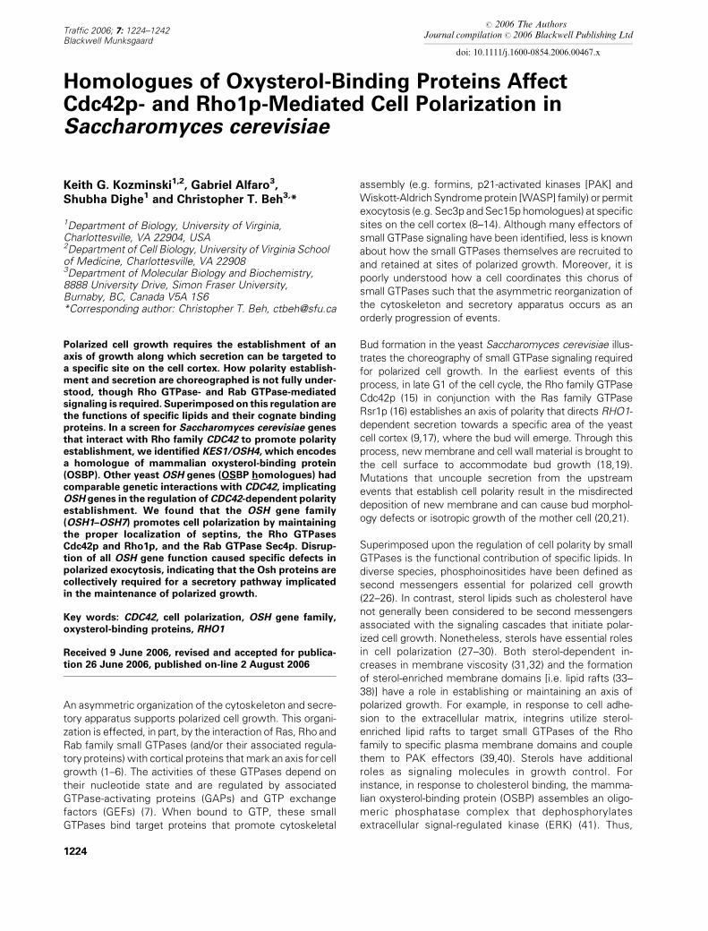
# 2006 The AuthorsJournal compilation # 2006 Blackwell Publishing Ltd
doi: 10.1111/j.1600-0854.2006.00467.x
Traffic 2006; 7: 1224–1242Blackwell Munksgaard
Homologues of Oxysterol-Binding Proteins AffectCdc42p- and Rho1p-Mediated Cell Polarization inSaccharomyces cerevisiae
Keith G. Kozminski1,2, Gabriel Alfaro3,
Shubha Dighe1 and Christopher T. Beh3,*
1Department of Biology, University of Virginia,Charlottesville, VA 22904, USA2Department of Cell Biology, University of Virginia Schoolof Medicine, Charlottesville, VA 229083Department of Molecular Biology and Biochemistry,8888 University Drive, Simon Fraser University,Burnaby, BC, Canada V5A 1S6*Corresponding author: Christopher T. Beh, [email protected]
Polarized cell growth requires the establishment of an
axis of growth along which secretion can be targeted to
a specific site on the cell cortex. How polarity establish-
ment and secretion are choreographed is not fully under-
stood, though Rho GTPase- and Rab GTPase-mediated
signaling is required. Superimposed on this regulation are
the functions of specific lipids and their cognate binding
proteins. In a screen for Saccharomyces cerevisiae genes
that interact with Rho family CDC42 to promote polarity
establishment, we identified KES1/OSH4, which encodes
a homologue of mammalian oxysterol-binding protein
(OSBP). Other yeast OSH genes (OSBP homologues) had
comparable genetic interactions with CDC42, implicating
OSH genes in the regulation of CDC42-dependent polarity
establishment. We found that the OSH gene family
(OSH1–OSH7) promotes cell polarization by maintaining
the proper localization of septins, the Rho GTPases
Cdc42p and Rho1p, and the Rab GTPase Sec4p. Disrup-
tion of all OSH gene function caused specific defects in
polarized exocytosis, indicating that the Osh proteins are
collectively required for a secretory pathway implicated
in the maintenance of polarized growth.
Key words: CDC42, cell polarization, OSH gene family,
oxysterol-binding proteins, RHO1
Received 9 June 2006, revised and accepted for publica-
tion 26 June 2006, published on-line 2 August 2006
An asymmetric organization of the cytoskeleton and secre-
tory apparatus supports polarized cell growth. This organi-
zation is effected, in part, by the interaction of Ras, Rho and
Rab family small GTPases (and/or their associated regula-
tory proteins) with cortical proteins thatmark an axis for cell
growth (1–6). The activities of these GTPases depend on
their nucleotide state and are regulated by associated
GTPase-activating proteins (GAPs) and GTP exchange
factors (GEFs) (7). When bound to GTP, these small
GTPases bind target proteins that promote cytoskeletal
assembly (e.g. formins, p21-activated kinases [PAK] and
Wiskott-Aldrich Syndromeprotein [WASP] family) or permit
exocytosis (e.g. Sec3p and Sec15p homologues) at specific
sites on the cell cortex (8–14). Although many effectors of
small GTPase signaling have been identified, less is known
about how the small GTPases themselves are recruited to
and retained at sites of polarized growth. Moreover, it is
poorly understood how a cell coordinates this chorus of
small GTPases such that the asymmetric reorganization of
the cytoskeleton and secretory apparatus occurs as an
orderly progression of events.
Bud formation in the yeast Saccharomyces cerevisiae illus-
trates the choreography of small GTPase signaling required
for polarized cell growth. In the earliest events of this
process, in late G1 of the cell cycle, the Rho family GTPase
Cdc42p (15) in conjunction with the Ras family GTPase
Rsr1p (16) establishes an axis of polarity that directs RHO1-
dependent secretion towards a specific area of the yeast
cell cortex (9,17), where the bud will emerge. Through this
process, newmembrane and cell wall material is brought to
the cell surface to accommodate bud growth (18,19).
Mutations that uncouple secretion from the upstream
events that establish cell polarity result in the misdirected
deposition of new membrane and can cause bud morphol-
ogy defects or isotropic growth of the mother cell (20,21).
Superimposed upon the regulation of cell polarity by small
GTPases is the functional contribution of specific lipids. In
diverse species, phosphoinositides have been defined as
second messengers essential for polarized cell growth
(22–26). In contrast, sterol lipids such as cholesterol have
not generally been considered to be second messengers
associated with the signaling cascades that initiate polar-
ized cell growth. Nonetheless, sterols have essential roles
in cell polarization (27–30). Both sterol-dependent in-
creases in membrane viscosity (31,32) and the formation
of sterol-enriched membrane domains [i.e. lipid rafts (33–
38)] have a role in establishing or maintaining an axis of
polarized growth. For example, in response to cell adhe-
sion to the extracellular matrix, integrins utilize sterol-
enriched lipid rafts to target small GTPases of the Rho
family to specific plasma membrane domains and couple
them to PAK effectors (39,40). Sterols have additional
roles as signaling molecules in growth control. For
instance, in response to cholesterol binding, the mamma-
lian oxysterol-binding protein (OSBP) assembles an oligo-
meric phosphatase complex that dephosphorylates
extracellular signal-regulated kinase (ERK) (41). Thus,
1224
sterols such as cholesterol restrict and control signaling
both by affecting the physical properties and composition
of membranes and by acting as activating ligands.
Oxysterol-binding proteins represent a large ubiquitous
family of eukaryotic lipid-binding proteins. They are soluble
intracellular receptors, some of which bind cholesterol and
oxysterols (42,43), the latter being potent feedback regu-
lators of cholesterol synthesis. It is unclear whether all
OSBP homologues bind cholesterol or oxysterols, but
most OSBP homologues appear to contain at least one
lipid-binding motif (44–46). Studies with budding yeast
have implicated OSBP homologues in the regulation of
secretory transport, nonvesicular lipid transport and mem-
brane dynamics (43,45,47–51). Saccharomyces cerevisiae
has seven genes that encode OSBP homologues, OSH1–
OSH7 (47,52,53). Each individual OSH gene in S. cerevisiae
is dispensable for growth, but deletion of all seven OSH
genes is lethal, indicating that these genes perform at least
one overlapping essential function (47). Depletion of all
Osh proteins from yeast cells results in an intracellular
accumulation of the cholesterol-like lipid ergosterol, which
otherwise resides in the plasma membrane (50). Individual
OSH genes are also implicated in specific transport pro-
cesses. For example, Osh1p is recruited to contact sites
between the nuclear envelope and the vacuole, where
components of the nuclear envelope are directly trans-
ferred to the vacuole (48,51,54). In contrast, Osh4p is
a negative regulator of Sec14p-dependent vesicle forma-
tion from the Golgi apparatus (45). Prima facie, these
trafficking events appear dissimilar but are controlled in
both cases by lipids interacting directly with an OSBP
(49,55). Although there is some overlap in the various
cellular locations occupied by each Osh protein (e.g. at the
plasma membrane), all have a distinct and dynamic pattern
of localization (48,49,56). In this regard, the vectoral trans-
location of Osh proteins in yeast between various mem-
brane compartments, or the canonical OSBP in mammalian
cells, appears to be triggered by binding specific lipids
(49,57). In addition to transport and membrane dynamics,
there is suggestive evidence that OSBPs affect cell cycle
regulation and development. In Caenorhabditis elegans,
for example, an OSBP homologue has been implicated
in transforming growth factor-b signaling (58), and a Dro-
sophila OSBP homologue expressed in fission yeast sup-
presses the mitotic checkpoint arrest caused by Wee1p
overexpression (59).
In budding yeast, we examined OSBP function in relation
to the role of small GTPases in polarized cell growth during
bud formation. We report that S. cerevisiae OSH genes
interact with CDC42 to promote cell polarity establishment
and, in a coordinated but distinct manner, with RHO1 to
affect polarized cell growth. After the bud site is estab-
lished, OSH genes are required for the proper localization
of small GTPases that regulate polarized secretion. Con-
sistent with this result, we also found that the OSH genes
were collectively required for a specific secretory pathway
associated with polarized exocytosis. Thus, the OSH
genes maintain cell polarization by their effect on vesicular
transport to sites of membrane growth.
Results
CDC42–OSH gene family interactions
To identify genes that interact with CDC42 during the
establishment of cell polarity in late G1, we screened for
multicopy dosage suppressors of cdc42-118D76A [(16); see
also Materials and Methods), a temperature-conditional
CDC42 allele specifically defective in polarity establish-
ment (60). At restrictive temperatures, cdc42-118 cells are
unable to establish an axis of polarized growth and arrest
as large, unbudded, multinucleated cells with a depolarized
actin cytoskeleton. Among the suppressors identified was
KES1/OSH4 (hereafter OSH4). At 368C, the temperature-
sensitive growth defect of cdc42-118 cells was rescued by
OSH4 whether expressed from its own promoter on
a high-copy vector (Figure 1A), or on low-copy CEN plas-
mid, or if expressed from the GAL10 promoter on multi-
copy plasmid (not shown). In comparison, the growth
defect of cdc42-118 cells at 368C was not suppressed in
transformants containing a vector control (Figure 1A).
These findings revealed an interaction between OSH4
and CDC42 and suggested that Osh4p is involved in
Cdc42p-dependent signaling during budding.
Several lines of evidence indicated that OSH4 interacts
specifically with CDC42 to affect cell polarization prior to
bud emergence. First, dosage suppression by OSH4 was
specific to CDC42. We tested whether multicopy OSH4
suppressed a temperature-sensitive allele of RHO1, which
encodes another Rho family small GTPase required for
polarized bud growth (61). Although OSH4 overexpression
did have an effect on RHO1 signaling, multicopy OSH4
failed to suppress the rhol-104ts growth defect at any
temperature tested (see below). This result indicated that
OSH4 does not suppress all mutations in Rho family
GTPases. Second, suppression by multicopy OSH4 was
observed only with specific CDC42 separation-of-function
alleles (Figure 1A). When multicopy OSH4 was trans-
formed into cdc42-129, which is not defective in establish-
ing an axis of polarized growth (60), or cdc42-123, which
disrupts a different aspect of polarized growth apart from
polarity establishment (60), neither allele was suppressed
compared with the vector control transformants (Figure 1
A). In fact, multicopyOSH4worsened the growth defect of
cdc42-129 cells. However, multicopy OSH4 weakly sup-
pressed the cdc42-101 temperature-sensitive growth
defect (Figure 1A). At 368C, cdc42-101 cells exhibit a polar-
ity establishment defect similar to that observed in cdc42-
118 cells at restrictive temperatures (60). Therefore, of all
the cdc42 alleles examined, multicopy OSH4 suppressed
only those alleles with a defined cell polarization defect.
OSH4 suppression of cdc42 polarization defects appears
to be CDC42 dependent; that is, it does not circumvent the
Traffic 2006; 7: 1224–1242 1225
OSH Genes Affect Polarized Growth
normal polarization of Cdc42p required for polarity estab-
lishment. In wild-type and cdc42-118 cells grown at 258C,Cdc42p localized to the presumptive bud site in unbudded
cells and to the apical cortex of small- and medium-budded
cells [data not shown; see also (60)]. In wild-type cells
shifted to 368C, the distribution of Cdc42p detected
by immunofluorescence microscopy was unchanged
(Figure 1B). However, when cdc42-118 cultures were
shifted to 368C, cell growth was arrested and unbudded
cells (many large) accumulated. In these cells, the distri-
bution of Cdc42p was diffuse or absent from the pre-
sumptive bud site (Figure 1B). If OSH4 suppressed cdc42
polarization defects in a Cdc42p-dependent manner, then
multicopyOSH4 should restore proper Cdc42p polarization
in unbudded cdc42-118 cells grown at the restrictive
temperature. This outcome was in fact observed (Figure 1
B). To analyze Cdc42p distribution in cdc42-118 and wild-
type cells that contained an OSH4 multicopy plasmid or
a vector control, log-phase cultures in minimal medium
were shifted from 25 to 368C for 9 h. Under these culture
conditions, >90% of cdc42-118 cells transformed with the
vector alone accumulated as unbudded cells. In contrast,
Cdc42p was properly polarized at the presumptive bud site
in a greater percentage of unbudded cdc42-118 cells that
contained multicopy OSH4 plasmid (Figure 1B and graph)
than with the vector control (Figure 1B and graph). More-
over, in cdc42-118 cells transformed with multicopy
OSH4, Cdc42p was properly polarized in approximately
the same percentage of unbudded cells as observed in
wild-type cells with the control vector (Figure 1B and
graph). These results indicated that multicopy OSH4
rescued the cdc42-118 polarization defect and that this
effect was due, at least in part, to the restoration of the
polarized localization of Cdc42p. Thus, suppression by
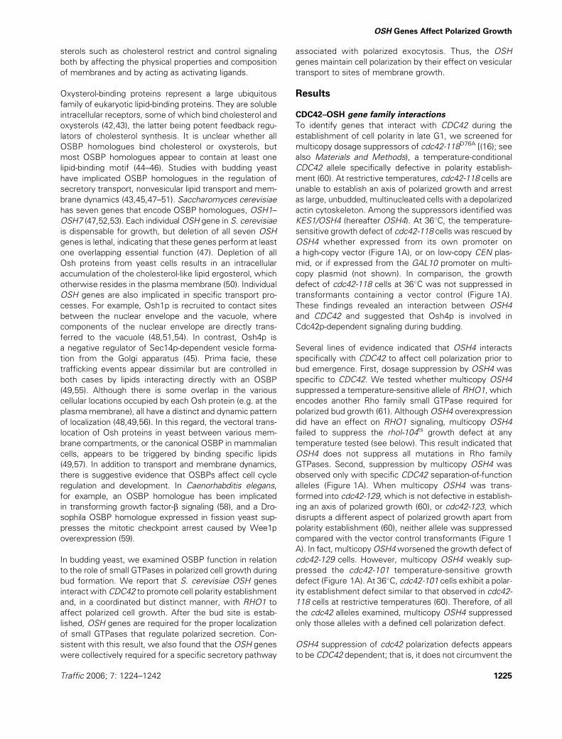
Figure 1: Suppression of cdc42ts growth defects by multicopyOSH genes. A)OSH4 suppressed the temperature-conditional growth
defect of cdc42ts cells defective in polarity establishment. Ten-fold serial dilutions (left to right) of cultures were spotted onto selective
minimal medium and growth was compared at 25 and 368C (5 days) for wild-type and mutant yeast transformed with a multicopy plasmid
containing OSH4 (pKK1821) or vector only (YEplac195). The strains transformed were CDC42 (DDY1300), cdc42-101 (DDY1304), cdc42-
118 (DDY1326), cdc42-123 (DDY1336) and cdc42-129 (DDY1344). B) Multicopy OSH4 promoted Cdc42p polarization in cdc42-118 and
wild-type strains. The strains shown are the same described in (A). After a shift from 258C, strains were incubated for 9 h at 368C inminimal
selective medium. Cdc42p was visualized by indirect immunofluorescence microscopy and nuclei were visualized with DAPI. Arrows
highlight examples of polarized Cdc42p, and asterisks identify multinucleated cells. Although some cultures contained cells distributed
throughout the cell cycle, unbudded cells are selectively shown because they were the focus of comparison. The scale bar is 10 mm.
Measurement of Cdc42p polarization in both unbudded mononucleated and multinucleated cells is presented in the graph. Cells were
scored as polarized when Cdc42p was visualized as a distinct spot at one pole of a cell (n > 200 unbudded cells scored for each strain).
C) AdditionalOSH family genes suppressed specific cdc42ts growth defects. Equivalent dilutions on selective synthetic medium compared
growth at 25 and 368C (5 days) of cdc42-101 (DDY1304), cdc42-118 (DDY1326) and cdc42-129 (DDY1344) strains transformed with
a multicopy (YEplac195) vector (left column) or a multicopy genomic library clone (pCB236-240, 242) containing the OSH family gene
indicated. A wild-type (WT) control strain (DDY1300) transformed with vector (YEplac195) is shown in the rightmost column. All strains in
each row were grown on the same plate.
1226 Traffic 2006; 7: 1224–1242
Kozminski et al.

OSH4 does not appear to be due to the activation of
a CDC42-independent pathway.
Three additional observations provided further evidence
that multicopy OSH4 promotes Cdc42p polarization. First,
multicopy OSH4 promoted Cdc42p polarization in multinu-
cleated cdc42-118 cells in which mutant Cdc42p is rarely
polarized. At 368C, we observed a sixfold increase in large,
multinucleated, unbudded cells with polarized Cdc42p in
cultures of cdc42-118 transformants with multicopy OSH4,
as compared with those with the vector control (Figure 1B,
graph). Polarized multinucleated cells accumulated in the
presence of multicopy OSH4, indicating that whereas
OSH4 suppressed defects in Cdc42p localization it did
not restore proper coupling of cell and nuclear division in all
cells. Second, at 368C, wild-type Cdc42p was detected at
the presumptive bud sitemore often in unbuddedwild-type
cells transformedwithmulticopyOSH4 than thosewith the
control vector (Figure 1B, graph). This observation indi-
cated that multicopy OSH4 augments wild-type Cdc42p
polarization as well as the mutant protein encoded by
cdc42-118. Third, in both mutant and wild-type cells at
368C, Cdc42p staining was more intense in unbudded cells
that contained multicopy OSH4 than in those containing
the vector control (Figure 1B). This observation strongly
suggested that more Cdc42p is present at the bud site
when OSH4 is present at multiple copies. Together, these
findings suggested that OSH4 helps establish Cdc42p
polarity or maintain its polarized localization at the pre-
sumptive bud site once polarity is established.
Because the OSH genes have overlapping functions (47),
we expanded our initial suppression analysis to include the
entire OSH gene family. Similar to OSH4, several of the
other OSH genes proved to be suppressors of cdc42
conditional growth defects. In multicopy plasmid con-
structs, OSH1, OSH2 or OSH6 expressed from their own
promoters (Figure 1C) suppressed cdc42-101 and cdc42-
118 temperature-conditional growth defects. OSH3 only
suppressed cdc42-118. MulticopyOSH5 andOSH7 did not
suppress the growth defects of either cdc42-101 or cdc42-
118 cells grown at 368C. In contrast, only multicopy OSH6
suppressed the temperature-sensitive growth defect of
cdc42-129 cells in addition to the other mutants tested
(Figure 1B). These results suggested that specific OSH
genes interact with CDC42 to promote the establishment
of cell polarity, but, as shown below, the OSH gene family
was collectively required for polarized cell growth.
Because of the complexity inherent in analyzing all seven
OSH genes (47), we limited our analyses to OSH2 and
OSH4 in many of the investigations described below.
These two genes were chosen because they strongly
suppressed cdc42ts alleles with polarized growth defects
(Figure 1). Because OSH2 and OSH4 are representative of
different OSH gene subfamilies and all OSH genes have
shared function(s) (47), the activities of these two OSH
genes are likely to approximate the others. As such, the
analysis of OSH2 and OSH4 expedited our analyses of
how the entire OSH gene family affects polarized cell
growth.
OSH genes maintain cell polarization
Because multicopy OSH genes suppressed cdc42 polari-
zation defects, we tested the converse to establish
whether the loss of OSH gene function resulted in the
loss of cell polarization. In this regard, we used the
organization of the actin cytoskeleton as a read-out for cell
polarization. In unbudded wild-type cells during late G1 of
the cell cycle, cortical actin patches are found predomi-
nantly at the presumptive bud site and actin cables run
approximately parallel to the mother-bud axis. In S and G2
phases, actin polarization persists but patches are pre-
dominantly found within the bud. Deletion of any single
OSH gene did not result in a loss of actin cytoskeleton
polarization, nor did the combined deletion of OSH genes
that suppress cdc42 cell polarity defects (i.e. osh1D–
osh4D osh6D) (data not shown).
To investigate whether a complete loss of OSH gene
function affects the polarization of the actin cytoskeleton,
we examined actin organization in a yeast strain lacking six
of the seven OSH genes, with the remaining OSH gene,
OSH2, under the regulatory control of a MET3 promoter
[oshD PMET3-OSH2 (oshD refers to the deletion of all OSH
genes other than those indicated)]. In the absence of
methionine in the culture medium, OSH2 is expressed in
oshD PMET3-OSH2 cells, permitting growth. In these cells,
actin patches are polarized to the bud site in unbudded
cells and towards the bud tip in small-budded cells
(Figure 2A,B) as observed in wild-type strains (not shown).
However, in the presence of added methionine, OSH2 ex-
pression is repressed, resulting in a gradual growth arrest
(47,50). After culturing in the presence of methionine,
oshD PMET3-OSH2 cells displayed distinct morphological
defects. Cells became larger and rounder, having wider
mother/bud necks and/or reiterative buds [Figure 2C–F
(50)]. Also observed was a decrease (�45%) in the number
of cells with a polarized actin cytoskeleton, as compared
with the same strain cultured in medium lacking methio-
nine (Figure 2, graph). Actin patch polarization was lost
in these methionine-repressed oshD PMET3-OSH2 cells
(Figure 2C–F) and actin cables were disorganized (Figure 2
C,E), no longer running parallel to the mother-bud axis
(compare with Figure 2A,B). The lower baseline of actin
polarization observed in unbudded cells versus budded
cells is normal and reflects the portion of the unbudded cell
population in early G1 when the bud site is not yet
established. These data indicated that OSH gene function
is required for proper cell polarization and contributes to
the polarized organization of the actin cytoskeleton.
Polarization of actin-assembly-promoting protein
complexes is unaffected in oshD osh4-1ts cells
Loss of cell polarization in the absence of OSH gene
function might reflect defects in either actin assembly or
Traffic 2006; 7: 1224–1242 1227
OSH Genes Affect Polarized Growth

in the localization of protein complex(es) required for the
spatial organization of the actin cytoskeleton. To address
the former possibility, we tested whether multicopy
OSH4 affected ACT1 mutations defective in actin assem-
bly. Multicopy OSH4 plasmids had no observable effect
on the growth of either a wild-type strain or several actin
assembly mutants [e.g. act1-157, act1-158, act1-159;
data not shown (62,63)]. This observation suggested that
OSH gene function does not directly affect actin assem-
bly, but it did not rule out the possibility that OSH genes
regulate the localization of proteins that spatially organize
actin.
To determine howOSH genes affect Cdc42p signaling and
cytoskeletal polarity, we examined in OSH mutants the
localization of green fluorescent protein (GFP) fusion
proteins that associate with polarity-promoting protein
complexes. In addition to using the oshD PMET3-OSH2
strain described above, we incorporated into our study
a temperature-sensitive OSH mutant strain, oshD osh4-1
(CBY926). This strain lacks all chromosomal copies of the
OSH genes but carries a plasmid containing the osh4-1ts
allele, which is rapidly inactivated at 378C (50). When
shifted to the restrictive temperature (378C), oshD osh4-1
cells arrest growth within the period of one cell cycle, but
the arrest is reversed if the strain is then returned to grow
at 238C (50). In this mutant, we analyzed the localization of
the polarisome, a 12S protein complex that contributes to
the spatial organization of the actin cytoskeleton (64–66).
Polarisome proteins facilitate the localization of the formin
Bni1p, which nucleates actin cable assembly at the bud
site and apical bud tip (21,67–71). The two yeast formins,
encoded by BNI1 and its paralogue BNR1, share some
functional overlap (72). Elimination of OSH function did not
significantly affect Bni1p localization to the bud (Figure 3)
or the Bnr1p localization to the bud neck (data not shown).
A plasmid expressing GFP-HA-Bni1p was transformed into
oshD osh4-1, oshD OSH4 and wild-type cells. After 4 h at
378C (one to two cell cycles), there was no detectable
difference in GFP-HA-Bni1p localization in any of these
strains (Figure 3). In all the strains examined, including the
wild-type control, GFP-HA-Bni1p was sometimes detected
at the opposite pole of the cell, consistent with previous
studies (6,73). Compared with wild type, GFP-Bnr1p was
also properly localized in oshD osh4-1 cells at 378C (data
not shown). These results were consistent with our finding
that neither OSH2 nor OSH4 on a multicopy plasmid sup-
pressed the temperature-sensitive growth defect of bnr1Dbni1-11ts or bnr1D bni1-12ts mutants [data not shown (69)].
GFP fusions of two other polarisome proteins, Bud6p and
Spa2p (66), were also examined in oshD osh4-1 and wild-
type cells at 378C. Both GFP-Spa2p and GFP-Bud6p
properly localized to the bud site and bud tip in these
mutant cells (Figures 3 and S1), albeit more GFP-Bud6p
aggregates were observed in theOSHmutant cells than in
wild type. These results indicated that OSH genes have
little, if any, effect on formin or polarisome localization.
We also monitored the localization of Arc15p, a subunit of
the Arp2/3 complex (74) that together with its associated
proteins has a direct role in nucleating the assembly of
cortical actin patches (75–77). As such, in wild-type cells,
Arc15p localization resembles that of actin patches
[described above (78)]. As shown in Figure 3, GFP-Arc15p
localization was unaffected by the inactivation of OSH
function. This finding suggested that the spatial distribu-
tion of the Arp2/3 complex as a whole was unaffected by
inactivation of the OSH genes. Together with the actin,
formin and polarisome data described above, these results
suggested that Osh proteins promote cell polarization by
a mechanism that is not directed towards the actin
cytoskeleton per se.
Figure 2: Osh-protein-depleted
cells exhibited a depolarization of
the actin cytoskeleton. Indirect
immunofluorescence micrographs of
oshD PMET3-OSH2 (JRY6326) cells cul-
tured in minimal medium at 258C for 9
h in the absence (A,B) or presence (C–
F) of methionine and probed with an
antibody against actin. Actin cables
(arrowheads) were present but more
difficult to visualize in Osh-depleted
cells (C–F) than in Osh-containing cells
(A,B). Scale bar is 5 mm. Measurement
of actin polarization in the aforemen-
tioned cells is summarized in the
graph. The actin cytoskeleton was
scored as polarized when cortical actin
patches were distributed toward one
pole of the cell. All cells counted had
a single nucleus as visualized with
DAPI (n > 300 cells scored for each
morphological class of each strain).
1228 Traffic 2006; 7: 1224–1242
Kozminski et al.
OSH genes maintain proper septin polarization and
septin ring assembly
Our observation that multicopy OSH4 rescued cdc42ts cell
polarity defects by restoring the polarized localization of
mutant Cdc42p (Figure 1B) strongly suggested that OSH
gene function is necessary prior to or during bud site
establishment. As an additional read-out for early cell
polarization events, we examined how OSH gene function
impacted septin localization. Although formins, the Arp2/3
complex and polarisome were properly polarized in oshD
osh4-1 cells at restrictive temperature, we found defects
in septin localization and assembly. The mitotic septin ring
complex [Cdc3p, Cdc10p, Cdc11p, Cdc12p and Shs1p/
Sep7p (79)] assembles at the bud site in a Cdc42p-
dependent (80,81) but actin-independent manner (82,83).
As shown in Figure 4, when oshD osh4-1 cells were
incubated at 238C, a ring of GFP-Cdc3p was observed at
the presumptive bud site and the mother-bud neck, in the
same manner as either wild-type or oshD OSH4 control
strains cultured under the same conditions at 23 or 378C.In contrast, GFP-Cdc3p was not fully polarized or properly
assembled in oshD osh4-1 cells grown at 378C for 4 h
(Figure 4). These defects were most apparent in oshDosh4-1 large-budded cells and less evident in unbudded
and small-budded cells (Figure S2), suggesting that OSHs
affect septin localization only at later times during bud
growth. In the majority of oshD osh4-1 cells with large
buds (67%) grown at 378C for 4 h, GFP-Cdc3p was not
polarized to the extent observed in control cells, and
randomly arranged GFP-Cdc3p dots accumulated on the
cell cortex. In addition, a significant number of oshD osh4-1
large-budded cells (76%) were observed with defects in
ring assembly. Similar ring defects were also observed in
oshD osh4-1 cells expressing either GFP-Cdc10p or GFP-
Cdc12p, or in OSH2-repressed oshD PMET3-OSH2 cells
expressing GFP-Cdc3p (data not shown). Thus, the OSH
genes appear unnecessary for the initial polarization and
assembly of septins/septin rings, but OSHs do appear
necessary for maintaining septin polarization and septin
ring integrity.
OSH genes affect Cdc42p localization
Polarisome assembly is dependent on the prior localization
of Cdc42p to the incipient bud site (68), but OSH mutants
had little effect on polarisome localization. These findings
suggested that Osh proteins maintain Cdc42p polarization
after the initial polarization of Cdc42p to the incipient bud
site. To test directly whether Osh function is necessary for
proper Cdc42p localization to the bud cortex, we examined
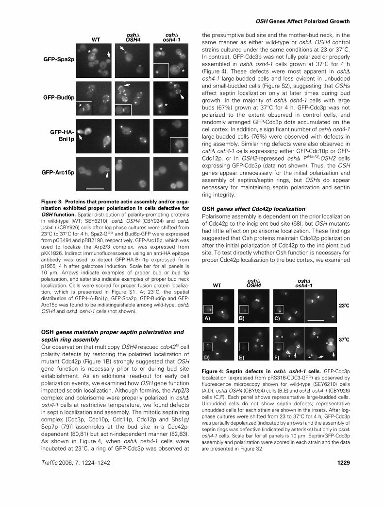
Figure 4: Septin defects in oshD osh4-1 cells. GFP-Cdc3p
localization (expressed from pRS316-CDC3-GFP) as observed by
fluorescence microscopy shown for wild-type (SEY6210) cells
(A,D), oshDOSH4 (CBY924) cells (B,E) and oshD osh4-1 (CBY926)
cells (C,F). Each panel shows representative large-budded cells.
Unbudded cells do not show septin defects; representative
unbudded cells for each strain are shown in the insets. After log-
phase cultures were shifted from 23 to 378C for 4 h, GFP-Cdc3p
was partially depolarized (indicated by arrows) and the assembly of
septin rings was defective (indicated by asterisks) but only in oshD
osh4-1 cells. Scale bar for all panels is 10 mm. Septin/GFP-Cdc3p
assembly and polarization were scored in each strain and the data
are presented in Figure S2.
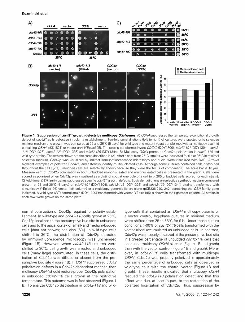
Figure 3: Proteins that promote actin assembly and/or orga-
nization exhibited proper polarization in cells defective for
OSH function. Spatial distribution of polarity-promoting proteins
in wild-type (WT; SEY6210), oshD OSH4 (CBY924) and oshD
osh4-1 (CBY926) cells after log-phase cultures were shifted from
238C to 378C for 4 h. Spa2-GFP and Bud6p-GFP were expressed
from pCB494 and pRB2190, respectively. GFP-Arc15p, which was
used to localize the Arp2/3 complex, was expressed from
pKK1826. Indirect immunofluorescence using an anti-HA epitope
antibody was used to detect GFP-HA-Bni1p expressed from
p1955, 4 h after galactose induction. Scale bar for all panels is
10 mm. Arrows indicate examples of proper bud or bud tip
polarization, and asterisks indicate examples of proper bud neck
localization. Cells were scored for proper fusion protein localiza-
tion, which is presented in Figure S1. At 238C, the spatial
distribution of GFP-HA-Bni1p, GFP-Spa2p, GFP-Bud6p and GFP-
Arc15p was found to be indistinguishable among wild-type, oshD
OSH4 and oshD osh4-1 cells (not shown).
Traffic 2006; 7: 1224–1242 1229
OSH Genes Affect Polarized Growth

Cdc42p polarization by indirect immunofluorescence
microscopy in wild-type, oshD OSH4 and oshD osh4-1ts
cells. Cdc42p was found in wild-type and oshD OSH4 con-
trol cells at one pole in unbudded cells and at the apical bud
cortex in small- and medium-budded cells, regardless of
temperature (Figure 5A). After shifting from 23 to 378C,however, Cdc42p localization was significantly perturbed
in oshD osh4-1ts cells (Figure 5A). When compared with
control cells, Cdc42p staining was detected in fewer
unbudded oshD osh4-1ts cells (Figure 5B). In unbudded
oshD osh4-1ts cells in which staining was evident, Cdc42p
was mislocalized and appeared as a thin crescent at the
cell cortex [Figure 5A (rightmost panel, top insert) and B].
The most apparent difference, however, was observed in
small- and medium-budded cells. Cdc42p was observed in
fewer small- and medium-budded oshD osh4-1ts cells than
in control strains (Figure 5A,B). When detected, Cdc42p
was oftenmislocalized to the side of the bud (Figure 5A,B).
In a few oshD osh4-1ts cells, Cdc42p was mislocalized at
the mother side of the bud neck (Figure 5A [rightmost
panel, bottom inset]). Loss or mislocalization of Cdc42p in
oshD osh4-1ts cells was not due to a decrease in the total
amount of Cdc42p. Mutant and control cells contained
equivalent amounts of Cdc42p whether grown at 23 or
378C (Figure 5C). In OSH-repressed oshD PMET3-OSH2
cells, Cdc42p was also expressed at normal levels and
a similar loss of Cdc42p localization was also observed (data
not shown). These results indicated that Osh proteins
promote Cdc42p polarization in both unbudded and budded
cells and suggested that Osh proteinsmanifest their effects
through the maintenance of Cdc42p polarization.
OSH genes affect polarized secretion; Bgl2p secretion
is blocked in oshD osh4-1ts cells
Because our data indicated a role for Osh proteins in
maintaining Cdc42p-dependent cell polarity, we investi-
gated whether the OSH genes affect polarized secretion.
Both Cdc42p and Rho1p are required (9,17,84) for the
polarized localization of proteins that comprise the exo-
cyst, a protein complex that spatially restricts secretory
vesicle docking to the bud site and to bud tips undergoing
polar growth (5). Consistent with a role for Osh proteins in
the regulation of polarized secretion, we found that several
OSH genes interacted with both CDC42 (Figure 1A) and
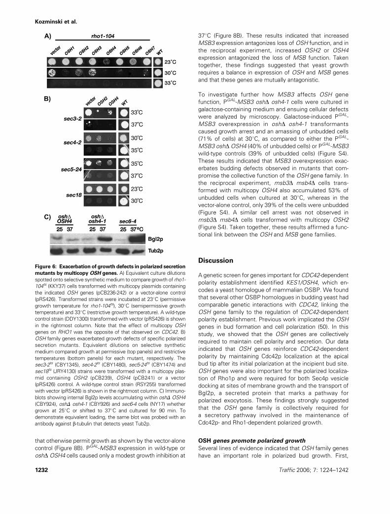
RHO1 (Figure 6A). Remarkably, OSH genes on multicopy
plasmids had the opposite effect on RHO1 as compared
with CDC42 mutants. When multicopy plasmids contain-
ing the OSH2 or OSH4 gene were introduced into rho1-
104ts strains, these transformants could only survive at
drastically reduced temperatures compared with vector-
alone control transformants (multicopy OSH1, OSH5,
OSH6 and OSH7 also exacerbated rho1-104ts defects;
Figure 6A). These findings indicated that the OSHs not
only affect Rho family GTPases in a gene-specific manner
but that Osh proteins might modulate opposing interac-
tions between Cdc42p and Rho1p.
The antagonistic effect of OSH gene overexpression was
not restricted to rho1-104 but was also observed with
other mutations that confer defects in polarized secretion.
Multicopy OSH2 orOSH4 exacerbated the growth defects
of the conditional sec3-2ts and sec5-24ts mutants, even at
temperatures that permit growth (Figure 6B). At restrictive
Figure 5: Cdc42p was mislocalized in
oshD osh4-1 cells at restrictive tem-
perature. A) Cdc42p was visualized by
indirect immunofluorescence micros-
copy in wild-type (WT; SEY6210), oshD
OSH4 (CBY924) and oshD osh4-1
(CBY926) cells after log-phase cultures
grown in minimal medium were shifted
from 23 to 378C for 4 h. Arrows indicate
examples of polarized Cdc42p that is
‘off-axis’; that is, not at the apical bud
tip but rather to the flank or base of the
bud. The asterisk indicates an example
of an unbudded cell in which Cdc42p is
otherwise polarized but distributed as
a crescent on the cell cortex rather than
a distinct spot. The scale bar is 5 mm.
B) Quantification of the distribution
patterns of Cdc42p in the cells shown
in (A). C) As shown by an anti-Cdc42p
immunoblot, Cdc42p levels remain con-
stant in wild-type (WT; SEY6210), oshD
OSH4 (CBY924) and oshD osh4-1
(CBY926) cells whether grown at 238Cor shifted to 378C for 4 h. To demon-
strate equivalent loading, the same blot
was probed with an antibody against
b-tubulin that detects yeast Tub2p.
1230 Traffic 2006; 7: 1224–1242
Kozminski et al.

temperatures, sec3-2ts and sec5-24ts mutations disrupt
the function of the exocyst complex, which is otherwise
the target for vesicle docking and fusion at the plasma
membrane (85,86). Vesicle targeting is mediated by the
Sec4p Rab GTPase, which is present on post-Golgi secre-
tory vesicles and is required for their docking with the
exocyst (8). Multicopy OSH2 and OSH4 also exacerbated
the defects of the sec4-2ts mutant (Figure 6B) at 308C,which is otherwise an acceptable temperature for the
growth of this mutant strain. Multicopy OSH2 or OSH4
had no effect on the growth of a sec18ts strain, which is
defective in SNARE complex disassembly (87) and is
blocked in multiple secretory transport pathways (88).
These findings suggested that OSH2 and OSH4 regulate
specific events during polarized secretion.
Although our results indicated a role forOSH2 andOSH4 in
polarized secretion, it was previously shown in OSH
mutant cells that the transport of proteins that mark
different secretory pathways was unaffected (45,50,51).
To reconcile these results, we assayed the secretory
transport of a protein specifically targeted to sites of
polarized growth. In yeast strains grown at 23 or 378C,we assayed the exocytosis of b-1,3-glucanase (Bgl2p),
which is normally secreted to the plasma membrane by
a population of late secretory vesicles distinct from those
which transport invertase (89). When shifted from 23 to
378C for 90 min, Bgl2p accumulated within oshD osh4-1
cells and was not transported to the plasma membrane.
This defect in Bgl2p exocytosis was comparable to that in
a sec6-4ts strain (Figure 6C), which is a well-characterized
late secretory mutant that also blocks the pathway for
Bgl2p transport (89). Bgl2p accumulation was not
observed in the control oshD OSH4 (Figure 6C) or wild-
type cells (data not shown) grown under the same
conditions. This result revealed a previously unknown
secretory defect in oshD osh4-1 cells and indicated that
the OSH genes are necessary for a specific pathway of
vesicular transport.
OSH genes are required for Rho1p and Sec4p
localization to the bud tip
To better understand how the OSH gene family regulates
polarized secretion, we tested whether OSH genes are
required for the localization of several proteins that regu-
late polarized secretion. We found that OSH genes are
necessary for the proper localization of Rho1p and GFP-
Sec4p but not GFP-Sec3p. Regardless of temperature, in
wild-type and oshD OSH4 control cells, Rho1p was
observed at one pole in unbudded cells and at the apical
bud tip in small- and medium-budded cells (Figure 7A). In
some cells, Rho1p staining also localized to the mitotic
spindle and spindle pole bodies, though independent
identification of these structures was not made. After
shifting from 23 to 378C for 4 h, Rho1p polarization was
perturbed in oshD osh4-1ts cells, whether budded or
unbudded (Figure 7A; also Figure S3). When compared
with the controls, Rho1p staining at the cortex was less
intense and more diffuse in the mutant cells. Similar
results were obtained with a GFP-Rho1p fusion protein
as well (data not shown). The decreased intensity of Rho1p
fluorescence in cells was not a result of lower Rho1p
levels. As detected by immunoblot, Rho1p levels were
equivalent in wild-type, oshD OSH4 and oshD osh4-1 cells
regardless of temperature (Figure 7B). Thus,OSH function
is necessary for proper Rho1p polarization in both budded
and unbudded cells.
To examine the localization of Sec3p and Sec4p by
fluorescence microscopy, GFP fusion proteins were ex-
pressed from plasmids transformed into wild-type, oshDOSH4 and oshD osh4-1 cells. When oshD osh4-1 cells
were incubated at 23 or 378C, GFP-Sec3p localization to
bud tips and bud necks was comparable to that seen in the
wild-type or oshD OSH4 controls (Figure 7A). GFP-Sec4p
localization, however, was aberrant in the large proportion
(46%) of oshD osh4-1 cells grown at 378C for 4 h. In wild-
type cells, GFP-Sec4p fluorescence was detected primar-
ily at the bud tip immediately adjacent to the plasma
membrane [(90); Figure 7A]. Although GFP-Sec4p was
observed in the bud in oshD osh4-1 cells, a dramatic
increase in fluorescence was detected, not at the bud
tip, but generally dispersed within the bud (Figure 7A). This
increase in fluorescence was consistent with an increase
in GFP-Sec4p levels detected by immunoblot in oshDosh4-1 cells at 378C (Figure 7C). These findings suggested
that secretory vesicles carrying GFP-Sec4p accumulated in
the bud in the absence of OSH function, which is consis-
tent with the observation by electron microscopy that
OSH inactivation causes vesicle accumulation in some
cells (50). This also implied that OSH genes promote
vesicle docking at the bud tip and affect the Sec4p-
dependent interaction between transport vesicles and
the exocyst. Thus, OSH genes impact polarized secretion
through Rho1p polarization and Sec4p-dependent vesicu-
lar transport.
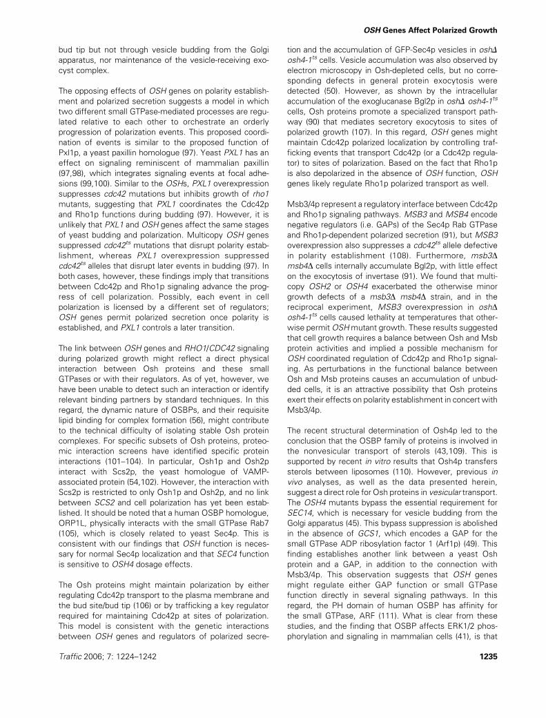
Perturbing the balance in dosage between
OSH genes and MSB genes inhibited growth
MSB3 and MSB4 encode negative regulators (GAPs) of
the Sec4p Rab GTPase (91). Consistent with our observa-
tion that the OSH genes antagonize sec4ts mutations and
polarized secretion, we found that multicopy OSH4 (and,
to a lesser degree, multicopy OSH2) exacerbated the
otherwise minor growth defects of amsb3Dmsb4D strain
(Figure 8A). Wild-type cells were unaffected by OSH2 or
OSH4 overexpression (Figure 8A), as were transformants
with either MSB3 or MSB4 deleted alone (data not
shown). The reciprocal experiment in which OSH mutants
were transformed with a high-expression PGAL-MSB3
plasmid also inhibited cell growth. On galactose-containing
medium, the induction of PGAL-MSB3 expression did not
rescue the temperature-sensitive growth defect of oshDosh4-1ts cells, rather it caused lethality at temperatures
Traffic 2006; 7: 1224–1242 1231
OSH Genes Affect Polarized Growth
that otherwise permit growth as shown by the vector-alone
control (Figure 8B). PGAL-MSB3 expression in wild-type or
oshD OSH4 cells caused only a modest growth inhibition at
378C (Figure 8B). These results indicated that increased
MSB3 expression antagonizes loss of OSH function, and in
the reciprocal experiment, increased OSH2 or OSH4
expression antagonized the loss of MSB function. Taken
together, these findings suggested that yeast growth
requires a balance in expression of OSH and MSB genes
and that these genes are mutually antagonistic.
To investigate further how MSB3 affects OSH gene
function, PGAL-MSB3 oshD osh4-1 cells were cultured in
galactose-containing medium and ensuing cellular defects
were analyzed by microscopy. Galactose-induced PGAL-
MSB3 overexpression in oshD osh4-1 transformants
caused growth arrest and an amassing of unbudded cells
(71% of cells) at 308C, as compared to either the PGAL-
MSB3 oshDOSH4 (40% of unbudded cells) or PGAL-MSB3
wild-type controls (39% of unbudded cells) (Figure S4).
These results indicated that MSB3 overexpression exac-
erbates budding defects observed in mutants that com-
promise the collective function of the OSH gene family. In
the reciprocal experiment, msb3D msb4D cells trans-
formed with multicopy OSH4 also accumulated 53% of
unbudded cells when cultured at 308C, whereas in the
vector-alone control, only 39% of the cells were unbudded
(Figure S4). A similar cell arrest was not observed in
msb3D msb4D cells transformed with multicopy OSH2
(Figure S4). Taken together, these results affirmed a func-
tional link between the OSH and MSB gene families.
Discussion
A genetic screen for genes important for CDC42-dependent
polarity establishment identified KES1/OSH4, which en-
codes a yeast homologue of mammalian OSBP. We found
that several other OSBP homologues in budding yeast had
comparable genetic interactions with CDC42, linking the
OSH gene family to the regulation of CDC42-dependent
polarity establishment. Previous work implicated the OSH
genes in bud formation and cell polarization (50). In this
study, we showed that the OSH genes are collectively
required to maintain cell polarity and secretion. Our data
indicated that OSH genes reinforce CDC42-dependent
polarity by maintaining Cdc42p localization at the apical
bud tip after its initial polarization at the incipient bud site.
OSH genes were also important for the polarized localiza-
tion of Rho1p and were required for both Sec4p vesicle
docking at sites of membrane growth and the transport of
Bgl2p, a secreted protein that marks a pathway for
polarized exocytosis. These findings strongly suggested
that the OSH gene family is collectively required for
a secretory pathway involved in the maintenance of
Cdc42p- and Rho1-dependent polarized growth.
OSH genes promote polarized growth
Several lines of evidence indicated that OSH family genes
have an important role in polarized bud growth. First,
Figure 6: Exacerbation of growth defects in polarized secretion
mutants by multicopy OSH genes. A) Equivalent culture dilutions
spotted onto selective syntheticmedium to compare growth of rho1-
104ts (KKY37) cells transformed with multicopy plasmids containing
the indicated OSH genes (pCB236-242) or a vector-alone control
(pRS426). Transformed strains were incubated at 238C (permissive
growth temperature for rho1-104ts), 308C (semipermissive growth
temperature) and 338C (restrictive growth temperature). A wild-type
control strain (DDY1300) transformedwith vector (pRS426) is shown
in the rightmost column. Note that the effect of multicopy OSH
genes on RHO1 was the opposite of that observed on CDC42. B)
OSH family genes exacerbated growth defects of specific polarized
secretion mutants. Equivalent dilutions on selective synthetic
medium compared growth at permissive (top panels) and restrictive
temperatures (bottom panels) for each mutant, respectively. The
sec3-2ts (CBY1345), sec4-2ts (CBY1480), sec5-24ts (CBY1474) and
sec18ts (JRY4130) strains were transformed with a multicopy plas-
mid containing OSH2 (pCB239), OSH4 (pCB241) or a vector
(pRS426) control. A wild-type control strain (RSY255) transformed
with vector (pRS426) is shown in the rightmost column. C) Immuno-
blots showing internal Bgl2p levels accumulating within oshD OSH4
(CBY924), oshD osh4-1 (CBY926) and sec6-4 cells (NY17) whether
grown at 258C or shifted to 378C and cultured for 90 min. To
demonstrate equivalent loading, the same blot was probed with an
antibody against b-tubulin that detects yeast Tub2p.
1232 Traffic 2006; 7: 1224–1242
Kozminski et al.

certain OSH genes are dosage suppressors of cdc42ts
polarity establishment defects. The mechanism of this
suppression involves, in part, Cdc42p localization at corti-
cal sites as increased OSH4 dosage partially restored
polarized localization to mutant forms of Cdc42p, and in
the absence of OSH function, Cdc42p was mislocalized.
Second, in the absence of OSH function, cytoplasmic
structures (i.e. cortical actin patches and septin rings)
necessary for proper bud growth were depolarized. Third,
GTPases required for polarized secretion (Cdc42p, Rho1p
and Sec4p) were mislocalized in the absence of OSH
function. However, the localization of other proteins
involved in actin and secretory polarization (e.g. formins,
Arp2/3 complex, polarisome and Sec3p) was unaffected by
OSH gene inactivation. The result that these proteins were
properly localized seems, at first glance, to be at odds with
the findings that Rho1p and Cdc42p are mislocalized upon
OSH inactivation. After all, the polarized localization of the
polarisome, Arp2/3 complex and Sec3p are dependent on
Rho1p and/or Cdc42p (9,68,92). In oshD osh4-1 cells at
378C, it is possible that an undetectable but sufficient
amount of Cdc42p and Rho1p remains properly polarized
at the bud site or bud tip to ensure the proper localization of
these proteins and proper budding. It is known in hypo-
morphic cdc42-1 cells, for example, that Cdc42p levels are
barely detectable by immunoblotting and immunofluores-
cence microscopy; yet, the cells appear morphologically
wild type at permissive temperatures (60), implying proper
localization of actin-organizing proteins. Last of all, the
polarized exocytosis of Bgl2p, but not other secreted
proteins (50), was defective in response to OSH inactiva-
tion. Thus, OSH function is necessary for specific events
required for cell polarization.
Our data indicated that many OSH family genes interact
with CDC42, butOSH2 andOSH4were the most effective
suppressors of cdc42ts cell polarity defects. Because
defects in cell polarization were only observed with the
loss of all OSH gene function, it appears that the OSH
genes share at least one common function necessary for
this process. Although the mechanism for this common
function is not yet known, it may involve the OSBP-related
domain (ORD) that is common to all Osh proteins and
OSBP homologues (46). Why some OSH genes were less
effective dosage suppressors compared with others might
relate to differences in localization of individual Osh
proteins. In response to lipid binding, the canonical OSBP
andmany of its homologues translocate betweenmembrane
Figure 7: Disruption of Rho1p and Sec4p localization in OSH mutants. A) After log-phase cultures were shifted from 23 to 378C for
4 h, indirect immunofluorescence microscopy revealed the spatial distribution of Rho1p, and fluorescence microscopy revealed the spatial
distribution of GFP-Sec4p (expressed from pRC2098) and GFP-Sec3p (expressed from pNB810) in oshD osh4-1 (CBY926), oshD OSH4
(CBY924) and wild-type cells (SEY6210). Arrows indicate examples of sites of Rho1p polarization. Note that because of the intensity of
GFP-Sec4p fluorescence in oshD osh4-1 cells, the corresponding image represents an exposure time that is one fourth of that shown for
the other transformed strains. The insert in the GFP-Sec4p oshD osh4-1 panel shows Sec4p mislocalization in a small-budded cell. Scale
bar for all panels is 10 mm. Among these strains at 238C, no significant differences in Rho1p, GFP-Sec4p or GFP-Sec3p localization were
apparent (not shown). Cells were scored for proper protein localization, which is presented in Figure S3. B) Immunoblots that show Rho1p
levels in wild-type (WT; SEY6210), oshD OSH4 (CBY924) and/or oshD osh4-1 (CBY926) cells whether grown at 238C or shifted to 378C for
4 h. To demonstrate equivalent loading, the same blot was probed with an antibody against b-tubulin (Tub2p). C) As compared to the wild-
type strain (SEY6210), GFP-Sec4p levels are markedly increased in oshD osh4-1 (CBY926) cells grown at 238C and shifted to 378C for 4 h,
which is consistent with the fluorescence microscopy results in (A). At 238C, GFP-Sec4p levels were equivalent in both strains tested. To
show equal loading, the same blot was probed with an anti-b-tubulin antibody that detects yeast Tub2p.
Traffic 2006; 7: 1224–1242 1233
OSH Genes Affect Polarized Growth

compartments (49,57). Thus, the transport dynamics of
individual Osh proteins might modulate their respective
ability to promote Cdc42p-dependent cell polarization.
Increased expression might result in ectopic expression,
providing more Osh protein to sites where it can better
facilitate Cdc42p function. Alternatively, increased dosage
of certain OSH genes might lead to a gain-of-function that
bypasses the requirement for CDC42 during cell polariza-
tion. This possibility, however, is unlikely because our data
indicated that OSH4 is not a bypass suppressor of cdc42ts
polarity defects and that suppression occurs even with
low-copy OSH expression. Thus, the OSH genes promote
cell polarization, not in place of CDC42 but in concert with
CDC42.
Could the observed depolarization of yeast cell in the
absence of OSH function be due to a general defect in
endocytosis? In short, the answer is no. It is true that
defects in endocytosis can lead to depolarization of the
cortical actin cytoskeleton (91), and Osh proteins promote
endocytosis by affecting membrane sterol levels (50).
However, this type of defect might account for depolar-
ization of the cortical actin cytoskeleton, but it is inconsis-
tent with septin mislocalization and assembly defects
when OSH function is absent. There is no evidence to
our knowledge that indicates that septin organization is
dependent on endocytosis. If a causal link exists between
OSHs/membrane sterols/endocytosis and cytoskeletal
polarity, then defects in endocytosis resulting from pertur-
bation in sterol distribution would also be predicted to
cause polarity defects. ERG2, which encodes a sterol
biosynthetic enzyme, was independently identified as the
endocytosis gene END11 (93,94). Similar toOSHmutants,
the deletion of ERG2/END11 affects membrane sterols
and blocks endocytosis (50). Unlike OSH mutants, how-
ever, defects in cell polarity are not apparent in erg2/
end11D cells (unpublished observation). This is likewise
true of arv1D cells, in which the normal cellular distribution
of sterols is defective (95) but cell polarization is not
(unpublished observation). These results suggest that
endocytic defects caused by perturbations in sterol traf-
ficking do not cause cellular depolarization per se. Thus,
the loss of OSH function does not affect polarized cell
growth through sterol-related defects in endocytosis.
Rather, consistent with our findings, the OSH gene family
directly affects cell polarization through its role in polarized
exocytosis.
OSH genes mediate opposing interactions between
CDC42 and RHO1 during polarized growth
In contrast to how the OSH genes maintain CDC42-
dependent cell polarization, OSH genes have the opposite
effect on RHO1 and other genes that mediate polarized
secretion. On multicopy plasmids, most of the OSH genes
exacerbated the growth defects of conditional mutants
that disrupt polarized secretion. When transformed with
multicopy OSH constructs, permissive temperatures for
growth were reduced for temperature-sensitive SEC3,
SEC4, SEC5 and RHO1 mutants. In the absence of OSH
function, however, Sec4p localization was disrupted and
undocked Sec4p-associated vesicles appeared to accumu-
late throughout the bud but not at the bud tip. This
mislocalization did not appear to result from a depolariza-
tion of Bni1p, Bud6p or Spa2p, which contribute to the
proper localization of Sec4p (96). Under the same con-
ditions, the localization of the exocyst component, Sec3p,
was unaffected. This observation suggested that OSH
genes facilitate vesicle docking at the bud tip but that OSH
genes have no direct impact on exocyst structure or
localization. OSH-dependent vesicle docking also appears
independent of the functional interaction between OSH4
and SEC14-dependent transport from the Golgi apparatus
(49). The deletion of OSH4, but none of the other OSH
genes, bypasses the essential requirement for Sec14p,
a phosphatidylcholine/phosphatidylinositol transfer protein
necessary for vesicle budding from the Golgi apparatus
(45). In this regard, the accumulation of Sec4p-associated
vesicles in the absence of OSH function represents a late
secretory event independent of Sec14p-mediated Golgi
vesicularization. Thus, the OSH gene family exerts its
effects on polarized secretion via vesicle delivery to the
Figure 8: Mutual antagonism of OSH and MSB function. A)
Ten-fold serial dilutions (left to right) of cultures were spotted onto
synthetic selective medium and grown at 308C (4 days) formsb3D
msb4D mutant yeast transformed with a multicopy plasmid
containing OSH2 (pCB239), OSH4 (pCB241) or the vector alone
(pRS426). Duplicate independent transformants are shown. Wild-
type, msb3D or msb4D congenic strains were unaffected by
multicopy OSH2 or OSH4 plasmids (not shown). B) Ten-fold serial
dilutions (left to right) of cultures were spotted onto synthetic
selective medium containing either 2% glucose or 2% galactose
and incubated at 30 or 378C. Growth of wild-type (SEY6210), oshD
osh4-1 (CBY926) and oshD OSH4 (CBY924) cells was compared
when transformed with a PGAL-MSB3 (pCB368) or vector control
plasmid (pKT10-GAL-HA).
1234 Traffic 2006; 7: 1224–1242
Kozminski et al.
bud tip but not through vesicle budding from the Golgi
apparatus, nor maintenance of the vesicle-receiving exo-
cyst complex.
The opposing effects of OSH genes on polarity establish-
ment and polarized secretion suggests a model in which
two different small GTPase-mediated processes are regu-
lated relative to each other to orchestrate an orderly
progression of polarization events. This proposed coordi-
nation of events is similar to the proposed function of
Pxl1p, a yeast paxillin homologue (97). Yeast PXL1 has an
effect on signaling reminiscent of mammalian paxillin
(97,98), which integrates signaling events at focal adhe-
sions (99,100). Similar to the OSHs, PXL1 overexpression
suppresses cdc42 mutations but inhibits growth of rho1
mutants, suggesting that PXL1 coordinates the Cdc42p
and Rho1p functions during budding (97). However, it is
unlikely that PXL1 and OSH genes affect the same stages
of yeast budding and polarization. Multicopy OSH genes
suppressed cdc42ts mutations that disrupt polarity estab-
lishment, whereas PXL1 overexpression suppressed
cdc42ts alleles that disrupt later events in budding (97). In
both cases, however, these findings imply that transitions
between Cdc42p and Rho1p signaling advance the prog-
ress of cell polarization. Possibly, each event in cell
polarization is licensed by a different set of regulators;
OSH genes permit polarized secretion once polarity is
established, and PXL1 controls a later transition.
The link between OSH genes and RHO1/CDC42 signaling
during polarized growth might reflect a direct physical
interaction between Osh proteins and these small
GTPases or with their regulators. As of yet, however, we
have been unable to detect such an interaction or identify
relevant binding partners by standard techniques. In this
regard, the dynamic nature of OSBPs, and their requisite
lipid binding for complex formation (56), might contribute
to the technical difficulty of isolating stable Osh protein
complexes. For specific subsets of Osh proteins, proteo-
mic interaction screens have identified specific protein
interactions (101–104). In particular, Osh1p and Osh2p
interact with Scs2p, the yeast homologue of VAMP-
associated protein (54,102). However, the interaction with
Scs2p is restricted to only Osh1p and Osh2p, and no link
between SCS2 and cell polarization has yet been estab-
lished. It should be noted that a human OSBP homologue,
ORP1L, physically interacts with the small GTPase Rab7
(105), which is closely related to yeast Sec4p. This is
consistent with our findings that OSH function is neces-
sary for normal Sec4p localization and that SEC4 function
is sensitive to OSH4 dosage effects.
The Osh proteins might maintain polarization by either
regulating Cdc42p transport to the plasma membrane and
the bud site/bud tip (106) or by trafficking a key regulator
required for maintaining Cdc42p at sites of polarization.
This model is consistent with the genetic interactions
between OSH genes and regulators of polarized secre-
tion and the accumulation of GFP-Sec4p vesicles in oshD
osh4-1ts cells. Vesicle accumulation was also observed by
electron microscopy in Osh-depleted cells, but no corre-
sponding defects in general protein exocytosis were
detected (50). However, as shown by the intracellular
accumulation of the exoglucanase Bgl2p in oshD osh4-1ts
cells, Osh proteins promote a specialized transport path-
way (90) that mediates secretory exocytosis to sites of
polarized growth (107). In this regard, OSH genes might
maintain Cdc42p polarized localization by controlling traf-
ficking events that transport Cdc42p (or a Cdc42p regula-
tor) to sites of polarization. Based on the fact that Rho1p
is also depolarized in the absence of OSH function, OSH
genes likely regulate Rho1p polarized transport as well.
Msb3/4p represent a regulatory interface between Cdc42p
and Rho1p signaling pathways. MSB3 and MSB4 encode
negative regulators (i.e. GAPs) of the Sec4p Rab GTPase
and Rho1p-dependent polarized secretion (91), but MSB3
overexpression also suppresses a cdc42ts allele defective
in polarity establishment (108). Furthermore, msb3Dmsb4D cells internally accumulate Bgl2p, with little effect
on the exocytosis of invertase (91). We found that multi-
copy OSH2 or OSH4 exacerbated the otherwise minor
growth defects of a msb3D msb4D strain, and in the
reciprocal experiment, MSB3 overexpression in oshDosh4-1ts cells caused lethality at temperatures that other-
wise permitOSHmutant growth. These results suggested
that cell growth requires a balance between Osh and Msb
protein activities and implied a possible mechanism for
OSH coordinated regulation of Cdc42p and Rho1p signal-
ing. As perturbations in the functional balance between
Osh and Msb proteins causes an accumulation of unbud-
ded cells, it is an attractive possibility that Osh proteins
exert their effects on polarity establishment in concert with
Msb3/4p.
The recent structural determination of Osh4p led to the
conclusion that the OSBP family of proteins is involved in
the nonvesicular transport of sterols (43,109). This is
supported by recent in vitro results that Osh4p transfers
sterols between liposomes (110). However, previous in
vivo analyses, as well as the data presented herein,
suggest a direct role for Osh proteins in vesicular transport.
The OSH4 mutants bypass the essential requirement for
SEC14, which is necessary for vesicle budding from the
Golgi apparatus (45). This bypass suppression is abolished
in the absence of GCS1, which encodes a GAP for the
small GTPase ADP ribosylation factor 1 (Arf1p) (49). This
finding establishes another link between a yeast Osh
protein and a GAP, in addition to the connection with
Msb3/4p. This observation suggests that OSH genes
might regulate either GAP function or small GTPase
function directly in several signaling pathways. In this
regard, the PH domain of human OSBP has affinity for
the small GTPase, ARF (111). What is clear from these
studies, and the finding that OSBP affects ERK1/2 phos-
phorylation and signaling in mammalian cells (41), is that
Traffic 2006; 7: 1224–1242 1235
OSH Genes Affect Polarized Growth
a general function of OSBP homologues is to integrate lipid
trafficking, cell signaling, and secretory transport.
Materials and Methods
Strains, microbial and genetic techniquesStrains and plasmids used in this study are listed in Tables 1 and 2,
respectively. Saccharomyces cerevisiae strains were cultured as described
in Kozminski et al. (16). All transformations were performed according to
Schiestl and Gietz (112). To select for the kanMX gene (113), yeast were
grown on rich medium containing 200 mg/mL G418 (GIBCO-Invitrogen Inc.,
Carlsbad, CA, USA). For repression of the MET3 promoter in yeast strain
JRY6326 (47), log-phase cultures in minimal medium were supplemented
with 100 mg/mL methionine. To induce expression of GAL10 promoter
fusions, 2% galactose (final concentration) was added to log-phase cultures
grown in minimal medium containing 2% raffinose.
Identification of KES1/OSH4 as a dosage suppressor
of cdc42-118The screen for cdc42-118 dosage suppressors is described in Kozminski
et al. (16). One YEp24 library plasmid identified in this screen, pKK1825,
contained a genomic fragment of chromosome XVI (base pairs 275701-
281371) that includes KES1/OSH4. To determine that OSH4 was indeed
the suppressor on pKK1825, isogenic cdc42 strains were transformed with
a 2-m plasmid with (pKK1821) or without (YEplac195) an OSH4 insert.
Transformants were streaked onto solid selective synthetic medium and
scored for growth after incubation at 25 and 368C for 5 days.
Plasmid constructsTo construct pKK1821, OSH4 was amplified from pKK1825 by polymerase
chain reaction (PCR) using primers (BamHI sites underlined) kes1-
forward (AAGCTCGGATCCGTTCTGTCTTGAGCTGTG) and kes1-reverse
(TGTCGAGGATCCCATATCCTTTCCTGTCACA) and cloned into the BamHI
site of YCplac33, forming pKK1062. The coding sequence of OSH4 in
pKK1062 was sequenced and found error free, relative to the Saccharo-
myces Genome Database (www.yeastgenome.org). An OSH4-containing
SphI–KpnI fragment from pKK1062 was then subcloned into the SphI and
KpnI sites of YEplac195, forming pKK1821. To construct pKK1826, ARC15-
GFP:HIS3MX6 was PCR amplified from DDY2752, using primers oKK212
(AGCTGCATGCCTCTGCTACTTGTTGTCTATG; SphI site underlined) and
oKK213 (TATAGGATCCGTTTATGCGTACTTGTTTTGTG). Digestion of the
PCR product with SphI and BglII produced an ARC15-GFP-containing
fragment that was cloned into the SphI and BamHI sites of YCplac33. To
construct pCB368, MSB3 was amplified from SEY6210, using primers
CBP242 (GGTACCATGCAGAACGATCAACAGAG; KpnI site underlined) and
CBP243 (CTCGAGGTTAGTCACCCTTGTCTTTTTTC; XhoI site underlined)
and cloned into pGEM-T-Easy (Promega, Madison, WI, USA). From the
resulting plasmid, digestion with KpnI and XhoI produced a 1.9-kb MSB3
fragment that was subcloned in-frame into the KpnI and XhoI sites of
pKT10-GAL-HA. To construct pCB494, SPA2-GFP was excised from
pRS416Spa2GFP with PvuI and subcloned into the PvuI sites of pRS426.
Constructs containing OSH genes on 2-m plasmids (pCB236-242) were
isolated from a YEp24 genomic library (114) in screens for genes that in high
copy suppressed the defects of oshD osh4-1 (CBY926) and/or oshD PMET3-
OSH2 under restrictive conditions. Genetic interactions described in this
article using the YEp24 OSH plasmids were confirmed with independently
isolated PGAL-OSH cDNAs and, in the case of OSH2 and OSH4, with
amplified sequences that do not contain flanking gene sequences.
Fluorescence microscopyIndirect immunofluorescence microscopy for Cdc42p and actin was
performed as described in Kozminski et al. (60) and for Rho1p as
described in Ayscough et al. (115), except that cells were cultured in
minimal medium, and sodium dodecyl sulfate (US Biological, Swamp-
scott, ME, USA) was used at 0.2% for Cdc42p and actin and at 0.3% for
Rho1p. Affinity-purified rabbit anti-yeast Cdc42p peptide antibody (60),
guinea-pig anti-yeast actin antibody (116) and affinity-purified rabbit anti-
yeast Rho1p peptide antibody were diluted 1:625, 1:500, 1:100 in PBS
containing 1 mg/mL BSA, respectively. Secondary antibodies (Jackson
ImmunoResearch Laboratories, West Grove, PA, USA) were diluted
1:100 in the same buffer. For visualization of epitope-tagged hemagglu-
tinin (HA)-Bni1p-GFP by indirect immunofluorescence, cells were fixed in
10% formalin for 30 min and incubated with a 1:1000 dilution of anti-HA
antibody (Covance Research Products, Denver, PA, USA) followed by
a 1:1000 dilution of an Alexa 488-conjugated anti-mouse secondary
antibody (Invitrogen – Molecular Probes). Fixed cells stained for immu-
nofluorescence, and cells expressing GFP fusions, were observed with
epifluorescence with either a Nikon E800 microscope equipped with
a �100/1.3 Plan-Neofluar objective or a Leica DMXRA2 microscope
equipped with a �100/1.40 Plan-Apo objective. Images were captured
with an Orca100ER or AG digital cameras (Hamamatsu Photonics,
Hamamatsu-City, Japan) and Openlab software (Improvision Inc., Lex-
ington, MA, USA). Unless otherwise noted, exposure times and contrast
enhancement were constant for a given series of images.
In experiments that only examined cell morphology and nuclear staining,
mid-log phase cells were pelleted and resuspended in 1 mL 3:1 (v/v)
methanol/acetic acid. After 30min at room temperature, cells were pelleted
and resuspended in 1 mL PBS. Following an additional wash with 1 mL PBS,
the final pellet was resuspended in mounting medium containing 1 mg/mL
4’,6-diamidino-2-phenylindole (DAPI; Accurate Chemicals and Scientific
Corp., Westbury, NY, USA) to stain nuclei.
ImmunoblotsImmunoblotting for Cdc42p and Rho1p was performed as described in
Kozminski et al. (60) and Ayscough et al. (115), respectively. Immunoblots
to detect GFP-Sec4p used a polyclonal anti-GFP antibody (Molecular Probes
Inc., Eugene, OR, USA) at a titre of 1:1000 with a 1:5000 titre of goat
horseradish peroxidase (HRP)-conjugated anti-rabbit immunoglobulin G
secondary antibody (Promega, Madison, WI, USA). Tubulin served as
a loading control and was detected with AA2, a mousemonoclonal antibody
raised against amino acids 412–430 of bovine brain b-tubulin (the kind gift of
Dr Tony Frankfurter, University of Virginia). AA2 was diluted to 130 ng/mL
with Tris-buffered saline (TBS) containing 0.1% Tween-20 (Fisher Scientific)
and incubated with blots overnight at room temperature.
Assay for Bgl2p secretionThe secretion of Bgl2p was assayed following the method of Harsay and
Schekman (manuscript in preparation). Yeast cells were grown overnight at
258C in YPD tomid-log phase (�0.3 OD600 units/mL). Each culture was then
split equally and 3.75 OD600 units were transferred to each of two new
flasks. One culture was incubated at 258C and the other culture was shifted
to 378C for 90 min. Cells from both cultures were then harvested by
centrifugation at 900� g for 5 min. Cell pellets were resuspended in 1 mL
ice-cold 10 mM NaN3, 10 mM KF solution, followed by a 10-min incubation
on ice. The suspension was transferred to microfuge tubes and microfuged
at 10 000� g for 1 min. Cell pellets were resuspended in fresh presphero-
plasting buffer (100mMTris–H2SO4, pH9.4; 50mMß-mercaptoethanol; 10mM
NaN3; 10 mM KF) and incubated on ice for 15 min. Cells were pelleted as
before, washed with 0.5 mL spheroplast buffer (50 mM KH2PO4–KOH, pH
7; 1.4 M sorbitol; 10 mM NaN3) and pelleted. After resuspension in 1 mL
spheroplast buffer containing 167 mg/mL zymolyase 100T (Seikagaku
Corporation, Tokyo, Japan), cells were incubated with gentle agitation for
30 min at 308C. Spheroplasts were then pelleted at 5000� g for 10 min
before their preparation for SDS–PAGE in 100 mL 2� SDS–PAGE sample
buffer. About 5 mL of each sample was loaded per lane on a 13% SDS–
PAGE gel. Detection of Bgl2p was made by immunoblotting, using a rabbit
polyclonal antibody against Bgl2p (the kind gift of Dr Randy Schekman,
1236 Traffic 2006; 7: 1224–1242
Kozminski et al.
University of California at Berkeley) that was preabsorbed against SEY2102,
a bgl2D strain (117), following the method of Roberts et al. (118).
Acknowledgments
Grateful acknowledgment to John Aitchison, Robert Arkowitz, Charles
Boone, Anthony Bretscher, Ruth Collins, David Drubin, Tony Frankfurter,
Wei Guo, Pekka Lappalainen, Peter Novick, Rick Rackubinski, Randy
Schekman, Kazuma Tanaka, Jeremy Thorner, and Akio Toh-e for plasmids,
yeast strains and/or antibodies. This work was funded by a Natural Science
and Engineering Research Council of Canada grant to C. T. B. and by joint
contributions from the Canadian Foundation for Innovation and the British
Columbia Knowledge and Development Fund. K. K. was supported by
a Basil O’Connor Starter Scholar Research Award (5-FY02-255) from the
March of Dimes Birth Defect Foundation. Special thanks to Nancy Hawkins,
Nick Harden and Dorothy Schafer for critical reading of this manuscript; we
especially thank Edina Harsay for advice on secretion assays and to Sharat
Gadde and Aki Kanauchi for technical assistance.
Supplementary Materials
Figure S1: Quantitative analysis of OSH-dependent polarization of
proteins involved in actin organization and/or assembly (complemen-
tary to Figure 3). Top) Polarized localization of GFP-HA-Bni1p, GFP-Spa2p
and GFP-Arc15p to either the presumptive bud site in unbudded cells, the
bud or bud tip in small-budded cells or the bud neck in large-budded cells
was determined in wild-type (SEY6210), oshD OSH4 (CBY924) and oshD
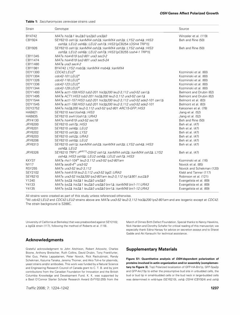
Table 1: Saccharomyces cerevisiae strains used
Strain Genotype Source
BY4742 MATa his3D1 leu2D0 lys2D0 ura3D0 Winzeler et al. (119)
CBY924 SEY6210 osh1D::kanMX4 osh2D::kanMX4 osh3D::LYS2 osh4D::HIS3
osh5D::LEU2 osh6D::LEU2 osh7D::HIS3 [pCB254 (OSH4 TRP1)]
Beh and Rine (50)
CBY926 SEY6210 osh1D::kanMX4 osh2D::kanMX4 osh3D::LYS2 osh4D::HIS3
osh5D::LEU2 osh6D::LEU2 osh7D::HIS3 [pCB255 (osh4-1 TRP1)]
Beh and Rine (50)
CBY1345 MATa his4-619 lys2-801 ura3 sec3-2
CBY1474 MATa his4-619 lys2-801 ura3 sec5-24
CBY1480 MATa ura3 sec4-2
CBY1981 BY4742 LYS2 msb3D::kanMX4 msb4D::kanMX4
DDY1300 CDC42:LEU2a Kozminski et al. (60)
DDY1304 cdc42-101:LEU2a Kozminski et al. (60)
DDY1326 cdc42-118:LEU2a Kozminski et al. (60)
DDY1336 cdc42-123:LEU2a Kozminski et al. (60)
DDY1344 cdc42-129:LEU2a Kozminski et al. (60)
DDY1493 MATa act1-159:HIS3 tub2-201 his3D200 leu2-3,112 ura3-52 can1D Belmont and Drubin (62)
DDY1495 MATa ACT1:HIS3 tub2-201 his3D200 leu2-3,112 ura3-52 can1D Belmont and Drubin (62)
DDY1544 MATa act1-157:HIS3 tub2-201 his3D200 leu2-3,112 ura3-52 ade2-101 can1D Belmont et al. (63)
DDY1545 MATa act1-158:HIS3 tub2-201 his3D200 leu2-3,112 ura3-52 ade2-101 Belmont et al. (63)
DDY2752 MATa his3D200 leu2-3,112 ura3-52 lys2-801 ARC15-GFP::HIS3 Kaksonen et al. (78)
HAB821 SEY6210 kes1/osh4D::HIS3 Jiang et al. (52)
HAB835 SEY6210 swh1/osh1D::URA3 Jiang et al. (52)
JRY4130 MATa his4-619 ura3-52 sec18 Beh and Rine (50)
JRY6200 SEY6210 osh7D::HIS3 Beh et al. (47)
JRY6201 SEY6210 osh6D::LEU2 Beh et al. (47)
JRY6202 SEY6210 osh3D::LYS2 Beh et al. (47)
JRY6203 SEY6210 osh2D::URA3 Beh et al. (47)
JRY6206 SEY6210 osh5D::LEU2 Beh et al. (47)
JRY6313 SEY6210 osh1D::kanMX4 osh2D::kanMX4 osh3D::LYS2 osh4D::HIS3
osh6D::LEU2
Beh et al. (47)
JRY6326 SEY6210 TRP1::PMET3-OSH2 osh1D::kanMX4 osh2D::kanMX4 osh3D::LYS2
osh4D::HIS3 osh5D::LEU2 osh6D::LEU2 osh7D::HIS3
Beh et al. (47)
KKY37 MATa rho1-104ts leu2-3,112 ura3-52 lys2-801am Kozminski et al. (16)
NY17 MATa sec6-4ts ura3-52 Novick et al. (85)
RSY255 MATa ura3-52 leu2-3,112 Novick and Schekman (120)
SEY2102 MATa his4-519 leu2-3,112 ura3-52 bgl2::URA3 Klebl and Tanner (117)
SEY6210 MATa ura3-52 his3D200 lys2-801am leu2-3,112 trp1D901 suc2D9 Robinson et al. (121)
Y1240 MATa lys2D his3D1 leu2D0 ura3D0 Evangelista et al. (69)
Y4133 MATa lys2D his3D1 leu2D0 ura3D0 bnr1D::kanMX6 bni1-11:URA3 Evangelista et al. (69)
Y4135 MATa lys2D his3D1 leu2D0 ura3D0 bnr1D::kanMX6 bni1-12:URA3 Evangelista et al. (69)
All strains were created as part of this study unless referenced otherwise.aAll cdc42:LEU2 and CDC42:LEU2 strains above are MATa ura3-52 leu2-3,112 his3D200 lys2-801am and are isogenic except at CDC42.
The strain background is S288C.
Traffic 2006; 7: 1224–1242 1237
OSH Genes Affect Polarized Growth
osh4-1 (CBY926) cells at 378C. The number of cells counted for each
strain ranged from 85 to 126 for the GFP-HA-Bni1p localization, from 62 to
132 for the GFP-Spa2p localization and from 220 to 429 for the GFP-Arc15p
localization. Bottom) Polarized localization of GFP-Bud6p in unbudded, small-
and large-budded cells at 378C (n> 72 for eachmorphological class counted).
Figure S2: Quantitative analysis of septin polarization and assembly
in OSH mutants (complementary to Figure 4). Left) Septin (GFP-Cdc3p)
polarization exclusively to the presumptive bud site was scored in unbud-
ded cells and to the bud neck in small- and large-budded cells in wild-type
(SEY6210), oshD OSH4 (CBY924) and oshD osh4-1 (CBY926) cells at 378C.
The appearance of cortical septin patches at locations other than those
observed in wild-type cells was defined as unpolarized. Right) Septin ring
assembly in wild-type, oshD OSH4 and oshD osh4-1 budded cells at 378C.
Defective septin rings included those with fragmented morphology,
asymmetric localization around the bud neck and/or irregular accumulations
of septins within the rings. Cells counted for both analyses ranged from 260
to 300.
Figure S3: Quantitative analysis of Rho1p, GFP-Sec3p and GFP-Sec4p
polarized localization in OSH mutants in wild-type (SEY6210), oshD
OSH4 (CBY925) and oshD osh4-1 (CBY926) cells (complementary to
Figure 7A). Rho1p polarized localization to the presumptive bud site in
unbudded cells, to sites of polarized growth in small- and medium-budded
cells and to the bud neck in large-budded cells after a shift from23 to 378C for
4 h (top right) or growth at 238C (top left). GFP-Sec3p (bottom left) and GFP-
Sec4p (bottom right) polarized localization to the presumptive bud site in
unbudded cells, to the bud tip in small-budded cells and to the bud neck in
large-budded cells, after a shift from 23 to 378C for 4 h. The number of cells
counted for each morphological class ranged from 200 to 308 for the Rho1p
localization, from 86 to 137 for the GFP-Sec3p localization and from 178 to
635 for the GFP-Sec4p. For GFP-Sec4p, a cell in which localization was ob-
served in the bud but not restricted to bud tip was not counted as polarized.
Figure S4: Cellular defects resulting from perturbations in OSH and
MSB gene dosage. A) Unbudded cells accumulated in oshD osh4-1
transformants expressing PGAL-MSB3. Differential-interference contrast
microscopy images (top panels) and corresponding DAPI-stained nuclei
(bottom panels) of wild-type (SEY6210), oshD osh4-1 (CBY924) and oshD
OSH4 (CBY926) cells transformed with the PGAL-MSB3 (pCB368) plasmid
after log-phase cultureswere transferred from 2% raffinose- to 2%galactose-
containing medium at 308C for 4 h. The corresponding percentages of
unbudded, small-budded and large-budded cells are presented in the left
graph. Cells counted for each strain in this analysis ranged from 348 to
755. B) Unbudded cell accumulation in msb3D msb4D cells transformed
with multicopy OSH4. Differential-interference contrast microscopy (top
panels) and corresponding DAPI staining (bottom panels) formsb3Dmsb4D
(CBY1981) log-phase cells transformed with either multicopy OSH2
(pCB239), OSH4 (pCB241) or the vector control (pRS426) after growth in
synthetic medium at 308C. Scale bar for all panels is 10 mm. The
corresponding percentages of budded and unbudded cells are presented
in the right graph. Cells counted for each strain ranged from 189 to 243. In
wild-type (BY4742) cells transformed with the multicopy OSH2, OSH4 or
the vector alone, no significant differences in the proportion of unbudded
and budded were observed (data not shown).
Supplemental materials are available as part of the online article at http://
www.blackwell-synergy.com
Table 2: Plasmids used
Plasmid Description Reference
p1955 PGAL-HA-BNI1-GFP CEN URA3 C. Boone (University of Toronto, Toronto, Canada)
pAGX1-BNR1 PACT1-BNR1-GFP CEN URA3 K. Tanaka (University of Hokkaido, Sapporo, Japan)
pCB236 OSH7 2m URA3
pCB237 OSH6 2m URA3
pCB238 OSH3 2m URA3
pCB239 OSH2 2m URA3
pCB240 OSH1 2m URA3
pCB241 OSH4 2m URA3
pCB242 OSH5 2m URA3
pCB254 OSH4 CEN TRP1 Beh and Rine (50)
pCB255 osh4-1ts CEN TRP1 Beh and Rine (50)
pCB368 PGAL-HA-MSB3 2m URA3
pCB494 SPA2-GFP 2m URA3
pGFP-RHO1 GFP-RHO1 CEN URA3 Marelli et al. (122)
pKK1062 OSH4 CEN URA3
pKK1821 OSH4 2m URA3
pKK1825 OSH4 2m URA3 Kozminski et al. (16)
pKK1826 ARC15-GFP CEN URA3
pKT10-GAL-HA PGAL-HA 2m URA3 Misu et al. (123)
pM-2 CDC10-GFP CEN URA3 Iwase and Toh-e (124)
pM-4 CDC12-GFP CEN URA3 Iwase and Toh-e (124)
pNB810 SEC3-GFP CEN URA3 Finger et al. (125)
pRB2190 PACT1-BUD6-GFP CEN URA3 Amberg et al. (64)
pRC2098 GFP-SEC4 CEN URA3 Calero et al. (126)
pRS316-CDC3-GFP CDC3-GFP CEN URA3 Caviston et al. (81)
pRS416Spa2GFP SPA2-GFP CEN URA3 Arkowitz and Lowe (127)
pRS426 2m URA3 Sikorski and Hieter (128)
YCplac33 CEN URA3 Gietz and Sugino (129)
YEplac195 2m URA3 Gietz and Sugino (129)
Unless otherwise referenced, all plasmids were created as part of this study.
1238 Traffic 2006; 7: 1224–1242
Kozminski et al.
References
1. Drubin DG, Nelson WJ. Origins of cell polarity. Cell 1996;84:335–344.
2. Gulli MP, Peter, M. Temporal and spatial regulation of Rho-type
guanine-nucleotide exchange factors: the yeast perspective. Genes
Dev 2001;15:365–379.
3. Etienne-Manneville S, Hall A. Rho GTPases in cell biology. Nature
2002;420:629–635.
4. Van Aelst L, Symons M. Role of Rho family GTPases in epithelial
morphogenesis. Genes Dev 2002;16:1032–1054.
5. Hsu SC, TerBush D, Abraham M, Guo W. The exocyst complex in
polarized exocytosis. Int Rev Cytol 2004;233:243–265.
6. Pruyne D, Legesse-Miller A, Gao L, Dong Y, Bretscher A. Mech-
anisms of polarized growth and organelle segregation in yeast. Annu
Rev Cell Dev Biol 2004;20:559–591.
7. Wennerberg K, Rossman KL, Der CJ. The Ras superfamily at a glance.
J Cell Sci 2005;118:843–846.
8. Guo W, Roth D, Walch-Solimena C, Novick P. The exocyst is an
effector for Sec4p, targeting secretory vesicles to sites of exocytosis.
EMBO J 1999;18:1071–1080.
9. Zhang X, Bi E, Novick P, Du L, Kozminski KG, Lipschutz JH, Guo W.
Cdc42 interacts with the exocyst and regulates polarized secretion.
J Biol Chem 2001;276:46745–46750.
10. Higgs HN, Pollard TD. Regulation of actin filament network formation
through Arp2/3 complex: activation by a diverse array of proteins.
Annu Rev Biochem 2001;70:649–676.
11. Dong Y, Pruyne D, Bretscher A. Formin-dependent actin assembly is
regulated by distinct modes of Rho signaling in yeast. J Cell Biol
2003;161:1081–1092.
12. Kadota J, Yamamoto T, Yoshiuchi S, Bi E, Tanaka K. Septin ring as-
sembly requires concerted action of polarisome components, a PAK
kinase Cla4p, and the actin cytoskeleton in Saccharomyces cerevi-
siae. Mol Biol Cell 2004;15:5329–5345.
13. Versele M, Thorner J. Septin collar formation in budding yeast
requires GTP binding and direct phosphorylation by the PAK, Cla4.
J Cell Biol 2004;164:701–715.
14. Hofmann C, Shepelev M, Chernoff J. The genetics of PAK. J Cell Sci
2004;117:4343–4354.
15. Adams AEM, Johnson DI, Longnecker RM, Sloat BF, Pringle JR.
CDC42 and CDC43, two additional genes involved in budding and the
establishment of cell polarity in the yeast Saccharomyces cerevisiae.
J Cell Biol 1990;111:131–142.
16. Kozminski KG, Beven L, Angerman E, Tong AH, Boone C, Park HO.
Interaction between a Ras and a Rho GTPase couples selection of
a growth site to the development of cell polarity in yeast. Mol Biol Cell
2003;14:4958–4970.
17. Guo W, Tamanoi F, Novick P. Spatial regulation of the exocyst
complex by Rho1 GTPase. Nat Cell Biol 2001;3:353–360.
18. Novick P, Schekman R. Export of major cell surface proteins
is blocked in yeast secretory mutants. J Cell Biol 1983;96:
541–547.
19. Ramirez RM, Ishida-Schick T, Krilowicz BL, Leish BA, Atkinson KD.
Plasma membrane expansion terminates in Saccharomyces cere-
visiae secretion-defective mutants while phospholipid synthesis con-
tinues. J Bacteriol 1983;154:1276–1283.
20. Mondesert G, Clarke DJ, Reed SI. Identification of genes controlling
growth polarity in the budding yeast Saccharomyces cerevisiae:
a possible role of N-glycosylation and involvement of the exocyst
complex. Genetics 1997;147:421–434.
21. Ozaki-Kuroda K, Yamamoto Y, Nohara H, Kinoshita M, Fujiwara T, Irie
K, Takai Y. Dynamic localization and function of Bni1p at the sites of
directed growth in Saccharomyces cerevisiae. Mol Cell Biol 2001;
21:827–839.
22. Merlot S, Firtel RA. Leading the way: directional sensing through
phosphatidylinositol 3-kinase and other signaling pathways. J Cell Sci
2003;116:3471–3478.
23. Desrivieres S, Cooke FT, Parker PJ, Hall MN. Mss4, a phosphatidyli-
nositol-4-phosphate 5-kinase required for organization of the actin
cytoskeleton in Saccharomyces cerevisiae. J Biol Chem 1998;273:
15787–15793.
24. Servant G, Weiner OD, Herzmark P, Balla T, Sedat JW, Bourne HR.
Polarization of chemoattractant receptor signaling during neutrophil
chemotaxis. Science 2000;287:1037–1040.
25. Weiner OD, Neilsen PO, Prestwich GD, Kirschner MW, Cantley LC,
Bourne HR. A PtdInsP(3)- and Rho GTPase-mediated positive feed-
back loop regulates neutrophil polarity. Nat Cell Biol 2002;4:509–513.
26. Wild AC, Yu JW, Lemmon MA, Blumer KJ. The p21-activated protein
kinase-related kinase Cla4 is a coincidence detector of signaling by
Cdc42 and phosphatidylinositol 4-phosphate. J Biol Chem 2004;279:
17101–17110.
27. Diederich W, Orso E, Drobnik W, Schmitz G. Apolipoprotein AI and
HDL(3) inhibit spreading of primary human monocytes through
a mechanism that involves cholesterol depletion and regulation of
CDC42. Atherosclerosis 2001;159:313–324.
28. Carland FM, Fujioka S, Takatsuto S, Yoshida S, Nelson T. The
identification of CVP1 reveals a role for sterols in vascular patterning.
Plant Cell 2002;14:2045–2058.
29. Pierini LM, Eddy RJ, Fuortes M, Seveau S, Casulo C, Maxfield FR.
Membrane lipid organization is critical for human neutrophil polariza-
tion. J Biol Chem 2003;278:10831–10841.
30. Willemsen V, Friml J, Grebe M, van den Toorn A, Palme K,
Scheres B. Cell polarity and PIN protein positioning in Arabidopsis
require sterol methyltransferase I function. Plant Cell 2003;15:
612–625.
31. Valdez-Taubas J, Pelham HR. Slow diffusion of proteins in the yeast
plasma membrane allows polarity to be maintained by endocytic
cycling. Curr Biol 2003;13:1636–1640.
32. Vasanji A, Ghosh PK, Graham LM, Eppell SJ, Fox PL. Polarization of
plasma membrane microviscosity during endothelial cell migration.
Dev Cell 2004;6:29–41.
33. Manes S, Mira E, Gomez-Mouton C, Lacalle RA, Keller P, Labrador JP,
Martinez AC. Membrane raft microdomains mediate front-rear polar-
ity in migrating cells. EMBO J 1999;18:6211–6220.
34. Gomez-Mouton C, Abad JL, Mira E, Lacalle RA, Gallardo E, Jimenez-
Baranda S, Illa I, Bernad A, Manes S, Martinez-A C. Segregation of
leading-edge and uropod components into specific lipid rafts during T
cell polarization. Proc Natl Acad Sci USA 2001;98:9642–9647.
35. Bagnat M, Simons K. Cell surface polarization during yeast mating.
Proc Natl Acad Sci USA 2002;99:14183–14188.
36. Wachtler V, Rajagopalan S, Balasubramanian MK. Sterol-rich plasma
membrane domains in the fission yeast Schizosaccharomyces
pombe. J Cell Sci 2003;116:867–874.
37. Martin SW, Konopka JB. Lipid raft polarization contributes to hyphal
growth in Candida albicans. Eukaryot Cell 2004;3:675–684.
38. Guan JL. Cell biology. Integrins, rafts, Rac, and Rho. Science 2004;
303:773–774.
39. del Pozo MA, Price LS, Alderson NB, Ren XD, Schwartz
MA. Adhesion to the extracellular matrix regulates the coupling of
the small GTPase Rac to its effector Pak. EMBO J 2000;19:
2008–2014.
40. Palazzo AF, Eng CH, Schlaepfer DD, Marcantonio EE, Gundersen GG.
Localized stabilization of microtubules by integrin- and FAK-facilitated
Rho signaling. Science 2004;303:836–839.
41. Wang PY, Weng J, Anderson RG. OSBP is a cholesterol-regulated
scaffolding protein in control of ERK1/2 activation. Science 2005;
307:1472–1476.
Traffic 2006; 7: 1224–1242 1239
OSH Genes Affect Polarized Growth
42. Kandutsch AA, Shown EP. Assay of oxysterol-binding protein in
a mouse fibroblast, cell-free system. Dissociation constant and other
properties of the system. J Biol Chem 1981;256:13068–13073.
43. Im YJ, Raychaudhuri S, Prinz WA, Hurley JH. Structural mechanism
for sterol sensing and transport by OSBP-related proteins. Nature
2005;437:154–158.
44. Taylor FR, Kandutsch AA. Oxysterol binding protein. Chem Phys
Lipids 1985;38:187–194.
45. Fang M, Kearns BG, Gedvilaite A, Kagiwada S, Kearns M, Fung MK,
Bankaitis VA. Kes1p shares homology with human oxysterol binding
protein and participates in a novel regulatory pathway for yeast Golgi-
derived transport vesicle biogenesis. EMBO J 1996;15:6447–6459.
46. Olkkonen VM, Levine TP. Oxysterol binding proteins: in more than
one place at one time? Biochem Cell Biol 2004;82:87–98.
47. Beh CT, Cool L, Phillips J, Rine J. Overlapping functions of the yeast
oxysterol-binding protein homologues. Genetics 2001;157:1117–1140.
48. Levine TP, Munro S. Dual targeting of Osh1p, a yeast homologue of
oxysterol-binding protein, to both the Golgi and the nucleus-vacuole
junction. Mol Biol Cell 2001;12:1633–1644.
49. Li X, Rivas MP, Fang M, Marchena J, Mehrotra B, Chaudhary A, Feng
L, Prestwich GD, Bankaitis VA. Analysis of oxysterol binding
protein homologue Kes1p function in regulation of Sec14p-dependent
protein transport from the yeast Golgi complex. J Cell Biol 2002;
157:63–77.
50. Beh CT, Rine J. A role for yeast oxysterol-binding protein homologs in
endocytosis and in the maintenance of intracellular sterol-lipid distri-
bution. J Cell Sci 2004;117:2983–2996.
51. Kvam E, Goldfarb DS. Nvj1p is the outer-nuclear-membrane receptor
for oxysterol-binding protein homolog Osh1p in Saccharomyces
cerevisiae. J Cell Sci 2004;117:4959–4968.
52. Jiang B, Brown JL, Sheraton J, Fortin N, Bussey H. A new family of
yeast genes implicated in ergosterol synthesis is related to the human
oxysterol binding protein. Yeast 1994;10:341–353.
53. Schmalix WA, Bandlow W. Swh1 from yeast encodes a candidate
nuclear factor containing ankyrin repeats and showing homology to
mammalian oxysterol-binding protein. Biochim Biophys Acta 1994;
1219:205–210.
54. Loewen CJ, Roy A, Levine TP. A conserved ER targeting motif in
three families of lipid binding proteins and in Opi1p binds VAP. EMBO
J 2003;22:2025–2035.
55. Wyles JP, McMaster CR, Ridgway ND. Vesicle-associated membrane
protein-associated protein-A (VAP-A) interacts with the oxysterol-
binding protein to modify export from the endoplasmic reticulum.
J Biol Chem 2002;277:29908–29918.
56. Wang P, Zhang Y, Li H, Chieu HK, Munn AL, Yang H. AAA ATPases
regulate membrane association of yeast oxysterol binding proteins
and sterol metabolism. EMBO J 2005;24:2989–2999.
57. Ridgway ND, Dawson PA, Ho YK, Brown MS, Goldstein JL. Trans-
location of oxysterol binding protein to Golgi apparatus triggered by
ligand binding. J Cell Biol 1992;116:307–319.
58. Sugawara K, Morita K, Ueno N, Shibuya H. Bip, a BRAM-interacting
protein involved in TGF-beta signalling, regulates body length in
Caenorhabditis elegans. Genes Cells 2001;6:599–606.
59. Alphey L, Jimenez J, Glover D. A Drosophila homologue of oxysterol
binding protein (OSBP) – implications for the role of OSBP. Biochim
Biophys Acta 1998;1395:159–164.
60. Kozminski KG, Chen AJ, Rodal AA, Drubin DG. Functions and
functional domains of the GTPase Cdc42p. Mol Biol Cell 2000;11:
339–354.
61. Yamochi W, Tanaka K, Nonaka H, Maeda A, Musha T, Takai Y. Growth
site localization of Rho1 small GTP-binding protein and its involvement
in bud formation in Saccharomyces cerevisiae. J Cell Biol 1994;125:
1077–1093.
62. Belmont LD, Drubin DG. The yeast V159N actin mutant reveals roles
for actin dynamics in vivo. J Cell Biol 1998;142:1289–1299.
63. Belmont LD, Patterson GM, Drubin DG. New actin mutants allow
further characterization of the nucleotide binding cleft and drug
binding sites. J Cell Sci 1999;112:1325–1336.
64. Amberg DC, Zahner JE, Mulholland JW, Pringle JR, Botstein D. Aip3p/
Bud6p, a yeast actin-interacting protein that is involved in morpho-
genesis and the selection of bipolar budding sites. Mol Biol Cell
1997;8:729–753.
65. Evangelista M, Blundell K, Longtine MS, Chow CJ, Adames N, Pringle
JR, Peter M, Boone C. Bni1p, a yeast formin linking Cdc42p and the
actin cytoskeleton during polarized morphogenesis. Science 1997;
276:118–122.
66. Sheu YJ, Santos B, Fortin N, Costigan C, Snyder M. Spa2p interacts
with cell polarity proteins and signaling components involved in yeast
cell morphogenesis. Mol Cell Biol 1998;18:4053–4069.
67. Fujiwara T, Tanaka K, Mino A, Kikyo M, Takahashi K, Shimizu K,
Takai Y. Rho1p-Bni1p-Spa2p interactions: implication in localization of
Bni1p at the bud site and regulation of the actin cytoskeleton in
Saccharomyces cerevisiae. Mol Biol Cell 1998;9:1221–1233.
68. Jaquenoud M, Peter M. Gic2p may link activated Cdc42p to compo-
nents involved in actin polarization, including Bni1p and Bud6p
(Aip3p). Mol Cell Biol 2000;20:6244–6258.
69. Evangelista M, Pruyne D, Amberg DC, Boone C, Bretscher A. Formins
direct Arp2/3-independent actin filament assembly to polarize cell
growth in yeast. Nat Cell Biol 2002;4:32–41.
70. Sagot I, Klee SK, Pellman D. Yeast formins regulate cell polarity
by controlling the assembly of actin cables. Nat Cell Biol 2002;4:
42–50.
71. Pruyne D, Gao L, Bi E, Bretscher A. Stable and dynamic axes of
polarity use distinct formin isoforms in budding yeast. Mol Biol Cell
2004;15:4971–4989.
72. Imamura H, Tanaka K, Hihara T, Umikawa M, Kamei T, Takahashi K,
Sasaki T, Takai Y. Bni1p and Bnr1p: downstream targets of the Rho
family small G-proteins which interact with profilin and regulate actin
cytoskeleton in Saccharomyces cerevisiae. EMBO J 1997;16:
2745–2755.
73. Matheos D, Metodiev M, Muller E, Stone D, Rose MD. Pheromo-
ne-induced polarization is dependent on the Fus3p MAPK acting
through the formin Bni1p. J Cell Biol 2004;165:99–109.
74. Winter D, Podtelejnikov AV, Mann M, Li R. The complex containing
actin-related proteins Arp2 and Arp3 is required for the motility and
integrity of yeast actin patches. Curr Biol 1997;7:519–529.
75. Welch MD, Mullins RD. Cellular control of actin nucleation. Annu Rev
Cell Dev Biol 2002;18:247–288.
76. Nicholson-Dykstra S, Higgs HN, Harris ES. Actin dynamics: growth
from dendritic branches. Curr Biol 2005;15:R346–357.
77. Rodal AA, Kozubowski L, Goode BL, Drubin DG, Hartwig JH. Actin
and septin ultrastructures at the budding yeast cell cortex. Mol Biol
Cell 2005;16:372–384.
78. Kaksonen M, Sun Y, Drubin DG. A pathway for association of
receptors, adaptors, and actin during endocytic internalization. Cell
2003;115:475–487.
79. Longtine MS, Bi E. Regulation of septin organization and function in
yeast. Trends Cell Biol 2003;13:403–409.
80. Gladfelter AS, Bose I, Zyla TR, Bardes ES, Lew DJ. Septin ring
assembly involves cycles of GTP loading and hydrolysis by Cdc42p.
J Cell Biol 2002;156:315–326.
81. Caviston JP, Longtine M, Pringle JR, Bi E. The role of Cdc42p
GTPase-activating proteins in assembly of the septin ring in yeast.
Mol Biol Cell 2003;14:4051–4066.
82. Ford SK, Pringle JR. Cellular morphogenesis in the Saccharomyces
cerevisiae cell cycle: localization of the CDC11 gene product and
1240 Traffic 2006; 7: 1224–1242
Kozminski et al.
the timing of events at the budding site. Dev Genet 1991;12:
281–292.
83. Ayscough KR, Stryker J, Pokala N, Sanders M, Crews P, Drubin
DG. High rates of actin filament turnover in budding yeast and roles
for actin in establishment and maintenance of cell polarity revealed
using the actin inhibitor Latrunculin-A. J Cell Biol 1997;137:399–
416.
84. Zajac A, Sun X, Zhang J, GuoW. Cyclical regulation of the exocyst and
cell polarity determinants for polarized cell growth. Mol Biol Cell
2005;16:1500–1512.
85. Novick P, Field C, Schekman R. Identification of 23 complementation
groups required for post-translational events in the yeast secretory
pathway. Cell 1980;21:205–215.
86. TerBush DR, Maurice T, Roth D, Novick P. The exocyst is a multi-
protein complex required for exocytosis in Saccharomyces cerevisiae.
EMBO J 1996;15:6483–6494.
87. Ungermann C, Nichols BJ, Pelham HR, Wickner W. A vacuolar
v-t-SNARE complex, the predominant form in vivo and on isolated
vacuoles, is disassembled and activated for docking and fusion. J Cell
Biol 1998;140:61–69.
88. Graham TR, Emr SD. Compartmental organization of Golgi-specific
protein modification and vacuolar protein sorting events defined in
a yeast sec18 (NSF) mutant. J Cell Biol 1991;114:207–218.
89. Harsay E, Bretscher A. Parallel secretory pathways to the cell surface
of yeast. J Cell Biol 1995;131:297–310.
90. Goud B, Salminen A, Walworth NC, Novick PJ. A GTP-binding protein
required for secretion rapidly associates with secretory vesicles and
the plasma membrane in yeast. Cell 1988;53:753–768.
91. Gao XD, Albert S, Tcheperegine SE, Burd CG, Gallwitz D, Bi E. The
GAP activity of Msb3p and Msb4p for the Rab GTPase Sec4p is
required for efficient exocytosis and actin organization. J Cell Biol
2003;162:635–646.
92. Lechler T, Jonsdottir GA, Klee SK, Pellman D, Li R. A two-tiered
mechanism by which Cdc42 controls the localization and activation of
an Arp2/3-activating motor complex in yeast. J Cell Biol 2001;155:
261–270.
93. Munn AL, Riezman H. Endocytosis is required for the growth of
vacuolar H(þ)-ATPase-defective yeast: identification of six new END
genes. J Cell Biol 1994;127:373–386.
94. Munn AL, Heese-Peck A, Stevenson BJ, Pichler H, Riezman H.
Specific sterols required for the internalization step of endocytosis
in yeast. Mol Biol Cell 1999;10:3943–3957.
95. Tinkelenberg AH, Liu Y, Alcantara F, Khan S, Guo Z, Bard M,
Sturley SL. Mutations in yeast ARV1 alter intracellular sterol distribu-
tion and are complemented by human ARV1. J Biol Chem 2000;275:
40667–40670.
96. Sheu YJ, Barral Y, Snyder M. Polarized growth controls cell shape and
bipolar bud site selection in Saccharomyces cerevisiae. Mol Cell Biol
2000;20:5235–5247.
97. Gao XD, Caviston JP, Tcheperegine SE, Bi E. Pxl1p, a paxillin-like
protein in Saccharomyces cerevisiae, may coordinate Cdc42p and
Rho1p functions during polarized growth. Mol Biol Cell 2004;15:
3977–3985.
98. Mackin NA, Sousou TJ, Erdman SE. The PXL1 gene of Saccharomy-
ces cerevisiae encodes a paxillin-like protein functioning in polarized
cell growth. Mol Biol Cell 2004;15:1904–1917.
99. Brown MC, Perrotta JA, Turner CE. Identification of LIM3 as the
principal determinant of paxillin focal adhesion localization and char-
acterization of a novel motif on paxillin directing vinculin and focal
adhesion kinase binding. J Cell Biol 1996;135:1109–1123.
100. Turner CE. Paxillin interactions. J Cell Sci 2000;113:4139–4140.
101. Uetz P, Giot L, Cagney G, Mansfield TA, Judson RS, Knight JR,
Lockshon D, Narayan V, Srinivasan M, Pochart P, Qureshi-Emili A,
Li Y, Godwin B, Conover D, Kalbfleisch T, et al. A comprehensive
analysis of protein-protein interactions in Saccharomyces cerevisiae.
Nature 2000;403:623–627.
102. Gavin AC, Bosche M, Krause R, Grandi P, Marzioch M, Bauer A,
Schultz J, Rick JM, Michon AM, Cruciat CM, Remor M, Hofert C,
Schelder M, Brajenovic M, Ruffner H, et al. Functional organization of
the yeast proteome by systematic analysis of protein complexes.
Nature 2002;415:141–147.
103. Ho Y, Gruhler A, Heilbut A, Bader GD, Moore L, Adams SL, Millar A,
Taylor P, Bennett K, Boutilier K, Yang L, Wolting C, Donaldson I,
Schandorff S, Shewnarane J, et al. Systematic identification of protein
complexes in Saccharomyces cerevisiae by mass spectrometry.
Nature 2002;415:180–183.
104. Hazbun TR, Malmstrom L, Anderson S, Graczyk BJ, Fox B, Riffle M,
Sundin BA, Aranda JD, McDonald WH, Chiu CH, Snydsman BE,
Bradley P, Muller EG, Fields S, Baker D, et al. Assigning function to
yeast proteins by integration of technologies. Mol Cell
2003;12:1353–1365.
105. Johansson M, Lehto M, Tanhuanpaa K, Cover TL, Olkkonen VM. The
oxysterol-binding protein homologue ORP1L interacts with Rab7 and
alters functional properties of late endocytic compartments. Mol Biol
Cell 2005;16:5480–5492.
106. Irazoqui JE, Howell AS, Theesfeld CL, Lew DJ. Opposing roles for
actin in Cdc42p polarization. Mol Biol Cell 2005;16:1296–1304.
107. Adamo JE, Moskow JJ, Gladfelter AS, Viterbo D, Lew DJ, Brennwald
PJ. Yeast Cdc42 functions at a late step in exocytosis, specifically
during polarized growth of the emerging bud. J Cell Biol 2001;155:
581–592.
108. Bi E, Chiavetta JB, Chen H, Chen GC, Chan CS, Pringle JR.
Identification of novel, evolutionarily conserved Cdc42p-interacting
proteins and of redundant pathways linking Cdc24p and Cdc42p to
actin polarization in yeast. Mol Biol Cell 2000;11:773–793.
109. Levine TP. A new way for sterols to walk on water. Mol Cell
2005;19:722–723.
110. Raychaudhuri S, Im YJ, Hurley JH, Prinz WA. Nonvesicular sterol
movement from plasma membrane to ER requires oxysterol-binding
protein-related proteins and phosphoinositides. J Cell Biol 2006;
173:107–119.
111. Godi A, Di Campli A, Konstantakopoulos A, Di Tullio G, Alessi DR,
Kular GS, Daniele T, Marra P, Lucocq JM, De Matteis MA. FAPPs
control Golgi-to-cell-surface membrane traffic by binding to ARF and
PtdIns(4)P. Nat Cell Biol 2004;6:393–404.
112. Schiestl RH, Gietz RD. High efficiency transformation of intact yeast
cells using single stranded nucleic acids as a carrier. Curr Genet
1989;16:339–346.
113. Wach A, Brachat A, Pohlmann R, Philippsen P. New heterologous
modules for classical or PCR-based gene disruptions in Saccharomy-
ces cerevisiae. Yeast 1994;10:1793–1808.
114. Carlson M, Botstein D. Two differentially regulated mRNAs with
different 50 ends encode secreted and intracellular forms of invertase.
Cell 1982;28:145–154.
115. Ayscough KR, Eby JJ, Lila T, Dewar H, Kozminski KG, Drubin DG.
Sla1p is a functionally modular component of the yeast cortical actin
cytoskeleton required for correct localization of both Rho1p-GTPase
and Sla2p, a protein with talin homology. Mol Biol Cell 1999;10:
1061–1075.
116. Palmgren S, Vartiainen M, Lappalainen P. Twinfilin, a molecular
mailman for actin monomers. J Cell Sci 2002;115:881–886.
117. Klebl F, Tanner W. Molecular cloning of a cell wall exo-beta-1,3-
glucanase from Saccharomyces cerevisiae. J Bacteriol 1989;171:
6259–6264.
118. Roberts CJ, Raymond CK, Yamashiro CT, Stevens TH. Methods for
studying the yeast vacuole. Methods Enzymol 1991;194:644–661.
Traffic 2006; 7: 1224–1242 1241
OSH Genes Affect Polarized Growth

119. Winzeler EA, Shoemaker DD, Astromoff A, Liang H, Anderson K,
Andre B, Bangham R, Benito R, Boeke JD, Bussey H, Chu AM,
Connelly C, Davis K, Dietrich F, Dow SW, et al. Functional character-
ization of the S. cerevisiae genome by gene deletion and parallel
analysis. Science 1999;285:901–906.
120. Novick P, Schekman R. Secretion and cell-surface growth are blocked
in a temperature-sensitive mutant of Saccharomyces cerevisiae. Proc
Natl Acad Sci USA 1979;76:1858–1862.
121. Robinson JS, Klionsky DJ, Banta LM, Emr SD. Protein sorting in
Saccharomyces cerevisiae: isolation of mutants defective in the
delivery and processing of multiple vacuolar hydrolases. Mol Cell Biol
1988;8:4936–4948.
122. Marelli M, Smith JJ, Jung S, Yi E, Nesvizhskii AI, Christmas RH,
Saleem RA, Tam YY, Fagarasanu A, Goodlett DR, Aebersold R,
Rachubinski RA, Aitchison JD. Quantitative mass spectrometry
reveals a role for the GTPase Rho1p in actin organization on the
peroxisome membrane. J Cell Biol 2004;167:1099–1112.
123. Misu K, Fujimura-Kamada K, Ueda T, Nakano A, Katoh H, Tanaka K.
Cdc50p, a conserved endosomal membrane protein, controls polar-
ized growth in Saccharomyces cerevisiae. Mol Biol Cell 2003;14:
730–747.
124. Iwase M, Toh-e A. Nis1 encoded by YNL078w: a new neck protein
of Saccharomyces cerevisiae. Genes Genet Syst 2001;76:335–
343.
125. Finger FP, Hughes TE, Novick P. Sec3p is a spatial landmark for
polarized secretion in budding yeast. Cell 1998;92:559–571.
126. Calero M, Chen CZ, Zhu W, Winand N, Havas KA, Gilbert PM,
Burd CG, Collins RN. Dual prenylation is required for Rab protein
localization and function. Mol Biol Cell 2003;14:1852–1867.
127. Arkowitz RA, Lowe N. A small conserved domain in the yeast Spa2p is
necessary and sufficient for its polarized localization. J Cell Biol
1997;138:17–36.
128. Sikorski RS, Hieter P. A system of shuttle vectors and yeast host
strains designed for efficient manipulation of DNA in Saccharomyces
cerevisiae. Genetics 1989;122:19–27.
129. Gietz RD, Sugino A. New yeast-Escherichia coli shuttle vectors
constructed with in vitro mutagenized yeast genes lacking six-base
pair restriction sites. Gene 1988;74:527–534.
1242 Traffic 2006; 7: 1224–1242
Kozminski et al.
Copyright © 2022 FDOKUMEN