Interfacial micromorphology and shear bond strength of single-bottle primer/adhesives
9
Dent Mater 13:316-324, September, 1997 Interfacial micromorphology and shear bond strength of single-bottle primer/adhesives Marcos ~ Vargas, Deborah S. Cobb, Gerald E. Denehy Department of Operative Dentistry, College of Dentistry, University of Iowa, Iowa City, Iowa, USA ABSTRACT Objectives. This study was conducted 1) to characterize through SEM analysis the resin-dentin interface produced by single-bottle primer/ adhesives and a three-component system [Scotchbond Multi-Purpose (3M Dental)] and 2) to evaluate the shear bond strength to dentin of these adhesive systems. Methods. Single-bottle primer/adhesives [Bond t (Jeneric/Pentron), Single Bond, (3M Dental Products); One Step (Bisco Inc.), OptiBond Solo (Kerr Corp.), Prime & Bond 2.1 (LD. Caulk-Dentsply), Syntac Single- Component (Ivoclar-Vivadent), Tenure Quik with Fluoride (Den-Mat)]were used according to manufacturers' instructions to bond resin composite to flat dentinal surfaces of extracted human third molars (n=15). All samples were thermocycled 300x. Twelve specimens per group were used to measure shear bond strength and three specimens were used to evaluate the interfacial morphology under SEM. A one-way ANOVA and Tukey's test were used to assess the results. Results. Mean shear bond strengths in MPa ± SD for the groups ranged from 22.27 ¢ 4.5 MPa for Single Bond to 7.6 + 3.9 MPa for Syntac Single- Component. The statistical analysis indicated that Single Bond produced significantly higher (p < 0.001) bond strengths than Syntac Single- Component, Prime & Bond 2.1, Bond I and Tenure Quik With Fluoride. Bond strengths for Syntac Single-Component were significantly lower than One-Step, OptiBond Solo, Scotchbond Multi-Purpose Plus and Single Bond. SEM examination clearly revealed the formation of a distinct hybrid layer for all adhesive systems; however, minor variations in ultrastructure existed among products. Significance. Some single-bottle primer/adhesives present in vitro bond strengths and hybrid layer formation similar to those found for the conventional three-component adhesive system tested. INTRODUCTION A goal and challenge in restorative dentistry is the achievement of consistent adhesion to tooth structure. Predictable adhesion to enamel has been possible since 1955 with the introduction of acid etching by Buonocore (1955). Unfortunately, adhesion to dentin has been an elusive goal, due to its inherent properties such as variable structure, high organic content and positive fluid flow (Pashley, 1990). Adhesion to dentin is primarily micromechanical, resulting from infiltration of resin into tooth structure (Nakabayashi, 1991). Most conventional adhesive systems use three steps to achieve this bond. First, an acid is used to remove the smear layer and partially decalcify the dentin surface to expose the organic portion of dentin consisting primarily of type I collagen (Ten Cate, 1994). An amphiphilic primer resin is then applied, which facilitates the third step, application of an adhesive resin that penetrates into the collagen network of the demineralized dentin. This creates a zone of resin- reinforced dentin called the "hybrid layer" or interdiffusion zone (Nakabayashi, 1982; Nakabayashi et al., 1982; Van Meerbeek et al., 1992; 1993a; 1993b). Several studies have characterized the ultrastructure of the resin-dentin interface using TEM, SEM and confocal microscopy (Inokoshi et al., 1990; Van Meerbeek et al., 1992; 1995a). In addition, the effects of different dentin treatments, including variations in application technique and surface wetness have been evaluated for conventional systems (Kanca, 1992b; Sano et al., 1994)o Recently, manufacturers have introduced single-bottle primer/adhesive systems. These agents combine the primer resin with the adhesive resin into a single component, thus simplifying the bonding procedure (Tay et al., 1996). While the convenience of these systems is desirable, research is needed to substantiate manufacturers' claims regarding the efficacy of these single-bottle, systems compared to conventional adhesives. It has not been well established if the new single-bottle primer/adhesive systems are able to adequately infiltrate the demineralized zone and form durable bonds to dentin. Studies evaluating the adhesion and interfacial characteristics ofthese new single-bottle primer/adhesives should provide insight into the micromorphology of the interface as well as the efficacy of these products. 316 Vargaset aL/SEM analysis and bond strength of single bottle adhesives
-
Upload
independent -
Category
Documents
-
view
0 -
download
0
Transcript of Interfacial micromorphology and shear bond strength of single-bottle primer/adhesives

Dent Mater 13:316-324, September, 1997
Interfacial micromorphology and shear bond strength of single-bottle primer/adhesives
Marcos ~ Vargas, Deborah S. Cobb, Gerald E. Denehy
Department of Operative Dentistry, College of Dentistry, University of Iowa, Iowa City, Iowa, USA
ABSTRACT Objectives. This study was conducted 1) to characterize through SEM analysis the resin-dentin interface produced by single-bottle primer/ adhesives and a three-component system [Scotchbond Multi-Purpose (3M Dental)] and 2) to evaluate the shear bond strength to dentin of these adhesive systems. Methods. Single-bottle primer/adhesives [Bond t (Jeneric/Pentron), Single Bond, (3M Dental Products); One Step (Bisco Inc.), OptiBond Solo (Kerr Corp.), Prime & Bond 2.1 (LD. Caulk-Dentsply), Syntac Single- Component (Ivoclar-Vivadent), Tenure Quik with Fluoride (Den-Mat)] were used according to manufacturers' instructions to bond resin composite to flat dentinal surfaces of extracted human third molars (n=15). All samples were thermocycled 300x. Twelve specimens per group were used to measure shear bond strength and three specimens were used to evaluate the interfacial morphology under SEM. A one-way ANOVA and Tukey's test were used to assess the results. Results. Mean shear bond strengths in MPa ± SD for the groups ranged from 22.27 ¢ 4.5 MPa for Single Bond to 7.6 + 3.9 MPa for Syntac Single- Component. The statistical analysis indicated that Single Bond produced significantly higher (p < 0.001) bond strengths than Syntac Single- Component, Prime & Bond 2.1, Bond I and Tenure Quik With Fluoride. Bond strengths for Syntac Single-Component were significantly lower than One-Step, OptiBond Solo, Scotchbond Multi-Purpose Plus and Single Bond. SEM examination clearly revealed the formation of a distinct hybrid layer for all adhesive systems; however, minor variations in ultrastructure existed among products. Significance. Some single-bottle primer/adhesives present in vitro bond strengths and hybrid layer formation similar to those found for the conventional three-component adhesive system tested.
INTRODUCTION
A goal and challenge in restorative dentistry is the achievement of consistent adhesion to tooth structure. Predictable adhesion to enamel has been possible since 1955 with the introduction of acid etching by Buonocore (1955). Unfortunately, adhesion to dentin has been an elusive goal, due to its inherent properties such as
variable structure, high organic content and positive fluid flow (Pashley, 1990).
Adhesion to dentin is primarily micromechanical, resulting from infiltration of resin into tooth structure (Nakabayashi, 1991). Most conventional adhesive systems use three steps to achieve this bond. First, an acid is used to remove the smear layer and partially decalcify the dentin surface to expose the organic portion of dentin consisting primarily of type I collagen (Ten Cate, 1994). An amphiphilic primer resin is then applied, which facilitates the third step, application of an adhesive resin tha t pene t ra tes into the collagen ne twork of the demineralized dentin. This creates a zone of resin- re inforced dent in called the "hybrid layer" or interdiffusion zone (Nakabayashi, 1982; Nakabayashi et al., 1982; Van Meerbeek et al., 1992; 1993a; 1993b).
Several studies have characterized the ultrastructure of the resin-dentin interface using TEM, SEM and confocal microscopy (Inokoshi et al., 1990; Van Meerbeek et al., 1992; 1995a). In addition, the effects of different dentin treatments, including variations in application technique and surface wetness have been evaluated for conventional systems (Kanca, 1992b; Sano et al., 1994)o
Recently, manufacturers have introduced single-bottle primer/adhesive systems. These agents combine the primer resin with the adhesive resin into a single component, thus simplifying the bonding procedure (Tay et al., 1996). While the convenience of these systems is desirable, research is needed to substantiate manufacturers' claims regarding the efficacy of these single-bottle, systems compared to conventional adhesives.
It has not been well established if the new single-bottle primer/adhesive systems are able to adequately infiltrate the demineralized zone and form durable bonds to dentin. Studies evalua t ing the adhesion and in ter facia l characteristics of these new single-bottle primer/adhesives should provide insight into the micromorphology of the interface as well as the efficacy of these products.
316 Vargas et aL/SEM analysis and bond strength of single bottle adhesives
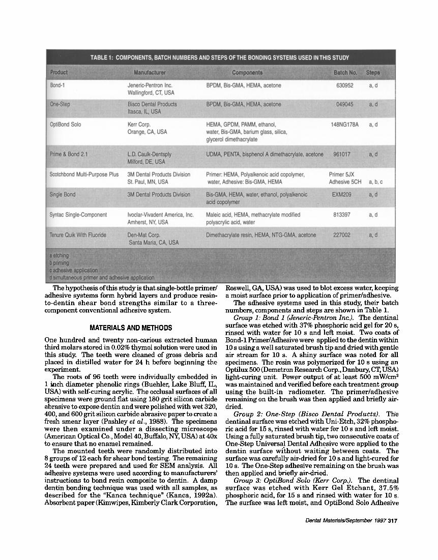
Bond-1 Jeneric-Pentron Inc. BPDM, Bis-GMA, HEMA, acetone 630952 a, d Wallingford, CT, USA
OptiBond Solo Kerr Corp. HEMA, GPDM, PAMM, ethanol, 148NG178A a, d Orange, CA, USA water, Bis-GMA, barium glass, silica,
glycerol dimethacrylate :.. . . . . . . . . . . . . . . . . . . . . . . . . . . . . . . . . . . . . . . . . . . . . . . . . . . . . . . . . . . . . . . . . . . . . .@
Scotchbond Multi-Purpose Plus 3M Dental Products Division Primer: HEMA, Polyalkenoic acid copolymer, Primer 5JX St. Paul, MN, USA water, Adhesive: Bis-GMA, HEMA Adhesive 5CH a, b, c
Syntac Single-Component Ivoclar-Vivadent America, Inc. Maleic acid, HEMA, methacrylate modified 813397 a, d Amherst, NY, USA polyacrylic acid, water
The hypothesis of this study is that single-bottle primer/ adhesive systems form hybrid layers and produce resin- to-dent in shea r bond s t r eng ths s imilar to a three- component conventional adhesive system.
MATERIALS AND METHODS
One hundred and twenty non-carious extracted human third molars stored in 0.02% thymol solution were used in this study. The teeth were cleaned of gross debris and placed in distilled water for 24 h before beginning the experiment.
The roots of 96 teeth were individually embedded in 1 i~ch diameter phenolic rings (Buehler, Lake Bluff, IL, USA) with serf-curing acrylic. The occlusal surfaces of all specimens were ground flat using 180 grit silicon carbide abrasive to expose dentin and were polished with wet 320, 400, and 600 grit silicon carbide abrasive paper to create a fresh smear layer (Pashley et al., 1988). The specimens were then examined under a dissect ing microscope (American Optical Co., Model 40, Buffalo, NY, USA) at 40x to ensure that no enamel remained.
The mounted teeth were randomly distributed into 8 groups of 12 each for shear bond testing. The remaining 24 teeth were prepared and used for SEM analysis. All adhesive systems were used according to manufacturers' instructions to bond resin composite to dentin. A damp dentin bonding technique was used with all samples, as described for the "Kanca technique" (Kanca, 1992a). Absorbent paper (Kimwipes, Kimberly Clark Corporation,
Roswell, GA, USA) was used to blot excess water, keeping a moist surface prior to application of primer/adhesive.
The adhesive systems used in this study, their batch numbers, components and steps are shown in Table 1.
Group 1: Bond 1 (Jeneric-Pentron Inc.). The dentinal surface was etched with 37% phosphoric acid gel for 20 s, rinsed with water for 10 s and left moist. Two coats of Bond-1 Primer/Adhesive were applied to the dentin within 10 s using a well saturated brush tip and dried with gentle air stream for 10 s. A shiny surface was noted for all specimens. The resin was polymerized for 10 s using an Optilux 500 (Demetron Research Corp., Danbury, CT, USA) light-curing unit. Power output of at least 500 mW/cm 2 was maintained and verified before each t reatment group using the built-in radiometer. The pr imer/adhesive remaining on the brush was then applied and briefly air- dried.
Group 2: One-Step (Bisco Dental ProductS). • T h e dentinal surface was etched with Uni-Etch, 32% phospho- ric acid for 15 s, rinsed with water for 10 s and left moist. Using a fully saturated brush tip, two consecutive coats of One-Step Universal Dental Adhesive were applied to the dent in surface wi thout waitir~g be tween coats. The surface was carefully air-dried for 10 s and light-cured for 10 s. The One-Step adhesive remaining on the brush was then applied and briefly air-dried.
Group 3: OptiBond Solo (Kerr Corp.). The dentinal sur face was e tched w i th Ker r Gel E tchan t , 37.5% phosphoric acid, for 15 s and rinsed with water for 10 s. The surface was left moist, and OptiBond Solo Adhesive
Dental Materials~September 1997 317
was applied with agitation for 20 s and light-cured for 30 s.
Group 4:Prime & Bond2.1 (L.D. Caulk-Dentsply). The dentinal surface was etched with Tooth Conditioner Gel, 34% phosphoric acid, for 15 s, rinsed with water for 10 s and left moist. Prime & Bond 2.1 Universal Light-cured Dental Adhesive System was applied for 20 s, keeping the surface wet, then gently air-dried for 5 s and light-cured for 10 s.
Group 5: Scotchbond Multi-Purpose (3M Dental Products Division). The dentinal surface was etched with 3M Scotchbond Etchant, 35% phosphoric acid, for 15 s, rinsed with water for 10 s and left moist. One coat of Scotchbond Mult i-Purpose Primer was applied to the dentinal surface, gently air-dried for 5 s, and Scotchbond Multi-Purpose Adhesive was applied and light-cured for 10 s.
Group 6: Single Bond (3M Dental Products Division). The dentinal surface was etched with 3M Scotchbond Etchant, 35% phosphoric acid, for 15 s, rinsed with water for 10 s and left moist. Using a fully saturated brush tip, two coats of Single Bond Adhesive were applied; the surface was gently air-dried for 5 s and light-cured for 10 s.
Group 7: Syntac Single-Component (Ivoclar-Vivadent America, Inc.). The dentinal surface was etched with Email Preparator GS, 37% phosphoric acid, for 15 s and rinsed wi th wa te r for 10 s, and left moist. Syntac Single- Component was applied with agitation for 10 s, then left undisturbed for 20 s. The surface was gently air-dried for 5 s and light-cured for 20 s. A second coat of adhesive was applied, gently dried for 10 s and light-cured for 20 s.
Group 8: Tenure Quik With Fluoride (Den-Mat Corp.). The dentinal surface was etched with Den-Mat medium viscosity etchant, 37% phosphoric acid, for 15 s and rinsed with water for 10 s and left moist. Three consecutive coats of Tenure Quik With Fluoride were applied to the surface, left undisturbed for 15 s, then gently air-dried for 10 s and l ight-cured for 15 s. The Tenure Quik with Fluoride adhesive remaining on the brush was applied and briefly air-dried.
A metal split mold (4.3 mm diameter, 2 mm high) was used as the matrix for standardized placement of one increment of Restorative Z-100, shade A-2 (3M Dental). The resin composite was then polymerized for 40 s, and the metal matrix was carefully removed.
Following the bonding procedure, specimens were stored in distilled water for 24 h at 37°C and thermocycled 300x between 5 ° and 55°C. Twelve specimens from each group were used to determine shear bond strength using a universa l tes t ing machine (Zwick Mater ia ls Testing machine, model #144560, Zwick of America, Inc., East Windsor, CT, USA). Spec imens were secured in a mounting jig and a straight-edge chisel attached to the crosshead was used to apply a shearing force at 5 mm per min until failure. Bond strengths were calculated using the Zwick computer software.
One-way analysis of variance (ANOVA) was used to detect any significant differences (p < 0.001) in bond strengths among the groups, and Tukey's HSD test was used as the analytic method for post hoc comparison of groups.
The r ema in ing 24 t e e t h were sec t ioned us ing a sectioning machine to separate the root from the coronal portion at the cemento-enamel junction. The coronal portion of each specimen was sectioned to obtain two dentin disks approximately I mm thick for a total of twenty- four pairs of dentin disks. For each adhesive system, three dentin disk pairs were bonded together according to the method described by Inokoshi et al. (1990). The bonding surfaces of each pair received the individual adhesive system according to the protocol used for shear bond strength. After conditioning and application of primer/ adhesive, t reated surfaces were bonded together with Clearfil Protect Liner (J. Morita USA Inc., Tustin, CA. USA), a low viscosity resin, according to the method of Van Meerbeek et al. (1992). The specimens were immersed in 2.5% glutaraldehyde in 0.1 M sodium cacodylate buffer at pH 7.4 and stored for 48 h at 4°C.
The dentin disk pairs were sectioned perpendicular to the bonded interface with a diamond saw under copious w a t e r l avage to ob t a in 4 l o n g i t u d i n a l sec t ions approximately 1 mm thick. Sections were then embedded in epoxy resin and metallurgically polished with 50 pro, 15 l~m, and 5 Inn grit sandpaper, followed by a 2-4 tim and 0.01 tim d i a m o n d p a s t e u s ing a po l i sh ing cloth. Specimens were rinsed between grits with distilled water and 50% ethanol. They were then dehydrated through a s c e n d i n g ace tone g rades , i m m e r s e d in 100% in Hexamethyldisizilane (HMDS) overnight and left under a vacuum hood until complete evaporation of the HMDS. All specimens were subjected to argon-ion-beam etching (DuoMill type 600, Gatan Inc., Warrendale, PA, USA) for app rox ima te ly 15 s. Ope ra t i ng condi t ions for the argon-ion-beam etching were: an accelerating voltage of 5 kV, an ion current density of 1 mA/cm 2 and the ion beam directed 35 ° to the polished surface. The specimens were then sputter-coated with Au-Pd and observed under a Hitachi S-4000 field-emission scanning electron microscope (Hitachi , Tokyo, Japan) a t d i f ferent magnif ica t ions ( Inokoshi et al., 1990; Van M e e r b e e k et al., 1992). Differences in surface micromorphology and thickness of the res in-dent in in ter face p roduced by the var ious adhes ive sys t ems were examined. Micrographs of representative interfaces for each adhesive system were taken.
RESULTS Mean shear bond strength and standard deviations are given in Table 2.
Mean shear bond s t rengths ranged from 22.27 + 4.5 MPa for Single Bond to 7.6 + 3.9 MPa for Syntac Single- Component . One-way ANOVA and Tukey 's Test for pairw~se multiple comparisons were used for statistical a n a l y s i s of t he da ta . O n e - w a y ANOVA r e v e a l e d statistically significant differences among groups (p < 0.001). Tukey's test showed that Single Bond SBMP Plus, Op t iBond Solo, One-S tep , and Bond 1 p r o d u c e d comparable bond strength. Bond strength for Single Bond was significantly higher than for Bond 1, Tenure Quik With Fluoride, Prime & Bond 2.1 and Syntac Single-Component. Bond s t r eng ths for Syntac S ing le -Componen t were significantly lower than for Single Bond, Scotchbond Multi- Purpose Plus, OptiBond Solo, and One-Step (Table 2).
318 Vargas et aL/SEM analysis and bond strength of single bottle adhesives
Single Bond " 12 22.27 + 4.5 A
OptiBond Solo 12 16.7 + 9.5 A B
Bond-1 12 12.6 + 4.8 B
Prime & Bond 2.1 12 12.3 + 3.8 B C
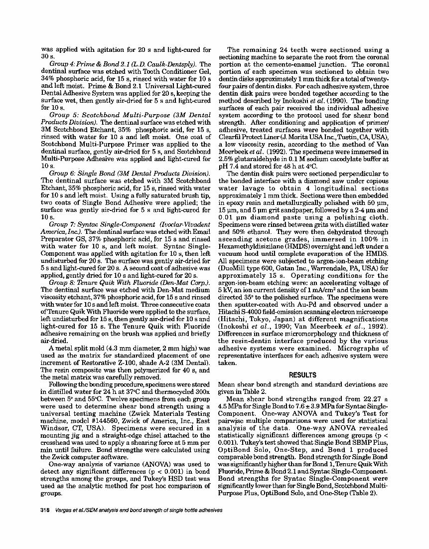
Fig. 1. Micrograph illustrating a cross-sectional view of the resin-dentin interface produced by Scotchbond MultiPurpose. A uniform hybrid layer (approximately 4 pm) is obtained after argon-ion etching. Constriction of the resin tags as they enter the tubular orifice can he observed (arrow). AR = adhesive resin, D = dentin, IHL = intertubular hybrid layer, PHL = peritubular hybrid layer and RT = resin tag.
The res in-dent in interfaces of the representat ive dentin adhesive systems are illustrated in the scanning electron micrographs shown in Figs. 1-12. All adhesive systems studied involved removal of the smear layer by conditioning with 32%-37% H3PO 4 prior to application of the adhesive resin or primer/adhesive. Based on bonding protocols and treatment conditions, slight variations in the micromorphological characteristics of the resin-dentin interface could be distinguished. For the conventional dentin adhesive, Scotchbond Multi-Purpose Plus, the typical structure of the resin-dentin interdiffusion zone obtained aider argon-ion etching is represented in Fig. 1. The hybrid layer, having low resistance to argon-ion bombardment, can be clearly distinguished from the adhesive resin layer and resin tags, which are highly resistant to argon-ion etching. This dist inct resin-
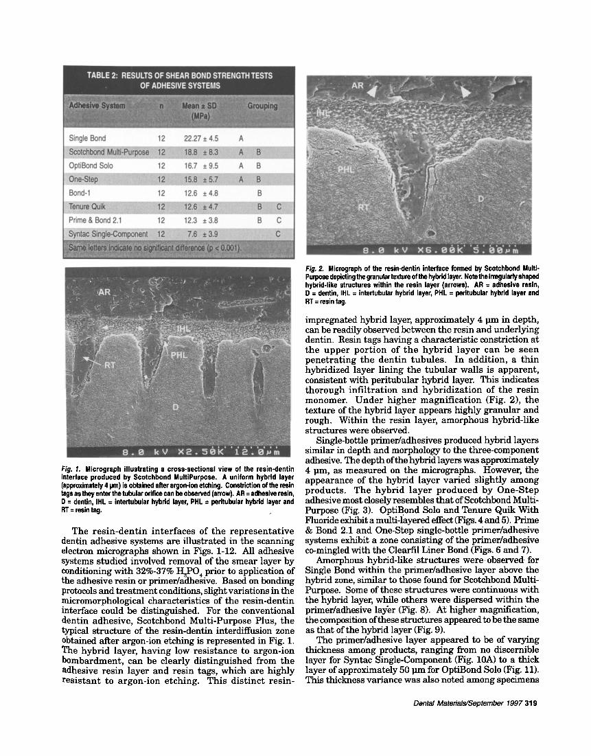
Fig. 2. Microgrsph of the resin-dentin interface formed by Scotchbond Multi- Purpose depicting the granular texture of the hybrid layer. Note the irregularly shaped hybrid-like structures within the resin layer (arrows). AR = adhesive resin, D = dentin, IHL = intertubular hybrid layer, PHL = peritubular hybrid layer and RT = resin tag.
impregnated hybrid layer, approximately 4 llm in depth, can be readily observed between the resin and underlying dentin. Resin tags having a characteristic constriction at the upper port ion of the hybr id layer can be seen penetrat ing the dent in tubules. In addition, a thin hybridized layer lining the tubular walls is apparent, consistent with peritubular hybrid layer. This indicates thorough infil tration and hybridization of the resin monomer. Under h igher magnificat ion (Fig. 2), the texture of the hybrid layer appears highly granular and rough. Within the resin layer, amorphous hybrid-like structures were observed.
Single-bottle primer/adhesives produced hybrid layers similar in depth and morphology to the three-component adhesive. The depth of the hybrid layers was approximately 4 llm, as measured on the micrographs. However, the appearance of the hybrid layer varied slightly among products. The hybrid layer produced by One-Step adhesive most closely resembles that of Scotchbond Multi- Purpose (Fig. 3). OptiBond Solo and Tenure Quik With Fluoride exhibit a multi-layered effect (Figs. 4 and 5). Prime & Bond 2.1 and One-Step single-bottle primer/adhesive systems exhibit a zone consisting of the primer/adhesive co-mingled with the Clearfil Liner Bond (Figs. 6 and 7).
Amorphous hybrid-like structures were observed for Single Bond within the primer/adhesive layer above the hybrid zone, similar to those found for Scotchbond Multi- Purpose. Some of these structures were continuous with the hybrid layer, while others were dispersed within the primer/adhesive layer (Fig. 8). /~t higher magnification, the composition of these structures appeared to be the same as that of the hybrid layer (Fig. 9).
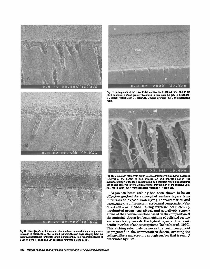
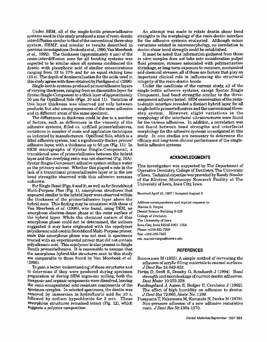
The primer/adhesive layer appeared to be of varying thickness among products, ranging from no discernible layer for Syntac Single-Component (Fig. 10A) to a thick layer of approximately 50 ~m for OptiBond Solo (Fig. 11). This thickness variance was also noted among specimens
Dental Matedala/Septernber 1997 319
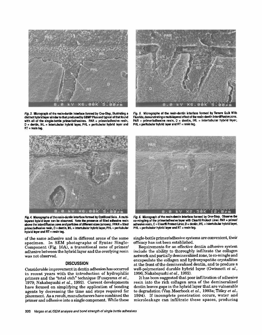
Fig. 3. Microgreph of the resin-dentin interface formed by One-Step, illustrating a distinct hybrid layer similar to that produced by SBMP Plus and typical of that found with all of the single-bottle primer/adhesives. PAR = primer/adhesive resin, D = dentin, IHL = intertubular hybrid layer, PHL = paritubular hybrid layer and RT = resin tag.
Fig. 5. Micrographs of the resin-dentin interface formed by Tenure Ouik With Fluoride, demonstrating a multi-layered effect of the resin-dentin interdiffusion zone. PAR = primer/adhesive resin, D = dentin, IHL = intertubular hybrid layer, PHL = peritubular hybrid layer and RT = resin tag.
Fig. 4. Microgmphs of the resin-dentin interface formed by OptiBond Solo. A multi- layered hylxid layer can he observed. Note the presence of filled adhesive resin above the interdiffusion zone and particles of different sizes (arrows). FPAR = filled primer/adhesive resin, D = dentin, IHL = intertubular hybrid layer, PHL = peritubular hybrid layer and RT = resin tag.
of the same adhesive and in different areas of the same specimen. In SEM photographs of Syntac Single- Component (Fig. 10A), a transitional zone of primer/ adhesive between the hybrid layer and the overlying resin was not observed.
D I S C U S S I O N
Considerable improvement in dentin adhesion has occurred in recent years with the introduction of hydrophilic primers and the "total etch" technique (Fusayama et al., 1979; Nakabayashi et al., 1992). Current developments have focused on simplifying the application of bonding agents by decreasing the time and steps required for placement. As a result, manufacturers have combined the primer and adhesive into a single component. While these
Fig. 6. Microgreph of the resin-dentin interface formed by One-Step. Observe the co-mingling of the primer/adhesive layer with Clearfil Protect Liner. PAR = primer/ adhesive resin, C = Clearfil Protect Liner, D = dentin, IHL = intertubular hybrid layer, PHL = peritubular hybrid layer and RT = resin tag.
single-bottle primer/adtiesive systems are convenient, their efficacy has not been established.
Requirements for an effective dentin adhesive system include the ability to thoroughly infiltrate the collagen network and partially deminerMized zone, to co-mingle and encapsulate the collagen and hydroxyapatite crystallites at the front of the demineralized dentin, and to produce a well-polymerized durable hybrid layer (Gwinnett et al., 1996; Nakabayashi et al., 1992).
It has been suggested that poor infiltration of adhesive resin into the rich collagen area of the demineralized dentin leaves gaps in the hybrid layer that are vulnerable to degradation (Van Meerbeek et al., 1993a; Titley et al., 1994). If incomplete penetration occurs, water and microleakage can infiltrate these spaces, producing
320 Vargas et aL/SEM analysis and bond strength of single bottle adhesives
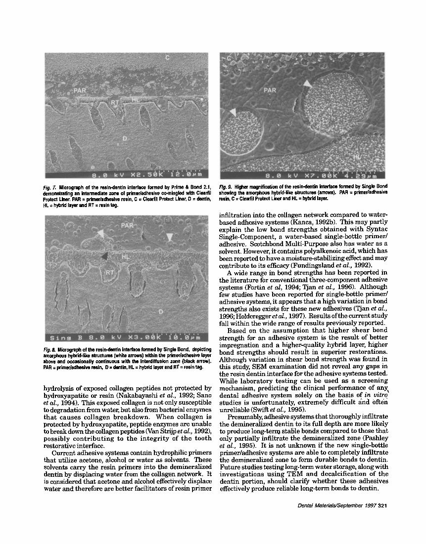
Fig. 7. Micrograph of the resin-dentin interface formed by Prime & Bond 2.1, demonstrating an intermediate zone of primer/adhesive co-mingled with Clesrfil Protect Liner. PAR = primer/adhesive resin, C = Clesrfil Protect Liner, D = dentin, HL = hybrid layer and RT = resin tag.
Fig. 8. Micrograph of the resin-dentin interface formed by Single Bond, depicting amorphous hybrid-like structures (white arrows) within the primer/adhesive layer above and occasionally continuous with the interdiffusion zone (black arrow). PAR = primer/adhesive resin, D = dentin, HL = hybrid layer and RT = resin tag.
hydrolysis of exposed collagen peptides not protected by hydroxyapatite or resin (Nakabayashi et al., 1992; Sano et al., 1994). This exposed collagen is not only susceptible to degradation from water, but also from bacterial enzymes that causes collagen breakdown. When collagen is protected by hydroxyapatite, peptide enzymes are unable to break down the collagen peptides (Van Strijp et al., 1992), possibly contr ibuting to the integri ty of the tooth restorative interface.
Current adhesive systems contain hydrophilic primers that utilize acetone, alcohol or water as solvents. These solvents carry the resin primers into the demineralized dentin by displacing water from the collagen network. It is considered that acetone and alcohol effectively displace water and therefore are better facilitators of resin primer
Fig. 9. Higher magnification of the resin-dentin interface formed by Single Bond showing the amorphous hybrid-like structures (arrows). PAR = primer/adhesive resin, C = Clesrfil Protect Liner and HL = hybrid layer.
infiltration into the collagen network compared to water- based adhesive systems (Kanca, 1992b). This may partly explain the low bond strengths obtained with Syntac Single-Component, a water-based single-bottle primer/ adhesive. Scotchbond Multi-Purpose also has water as a solvent. However, it contains polyalkenoic acid, which has been reported to have a moisture-stabilizing effect and may contribute to its efficacy (Fundingsland et al., 1992).
A wide range in bond strengths has been reported in the literature for conventional three-component adhesive systems (Fort-in et al, 1994; Tjan et al., 1996). Although few studies have been reported for single-bottle primer/ adhesive systems, it appears that a high variation in bond strengths also exists for these new adhesives (Tjan et al., 1996; Holderegger et al., 1997). Results of the current study fall within the wide range of results previously reported.
Based on the assumption tha t higher shear bond strength for an adhesive system is the result of better impregnation and a higher-quality hybrid layer, higher bond strengths should result in superior restorations. Although variation in shear bond strength was found in this study, SEM examination did not reveal any gaps in the resin dentin interface for the adhesive systems tested. While laboratory testing can be used as a screening mechanism, predicting the clinical performance of any, dental adhesive system solely on the basis of in vitro" studies is unfortunately, extremely difficult ah(i Ofter~ unreliable (Swift et al., 1995).
Presumably, adhesive systems that thoroughly infiltrate the demineralized dentin to its full depth are more likely to produce long-term stable bonds, compared to those that only partially infiltrate the demineralized zone (Pashley et al., 1995). It is not unknown if the new single-bottle primer/adhesive systems are able to completely infiltrate the demineralized zone to form durable bends to dentin. Future studies testing long-term water storage, along with investigations using TEM and decalcification of the dentin portion, should clarify whether these adhesives effectively produce reliable long-term bonds to dentin.
Dental Matenals/September 1997 321
! :"
s . o kV xa.5~k''i~ig~
Fig. 11. Microgrephs of the resin-dentin interface for OptiBond Solo. Due to the filled adhesive, a much greater thickness in this layer (50 pm) is produced. C = Clearfil Protect Liner, D = dentin, HL = hybrid layer and PAR = primer/adhesive resin.
Fig.lO. Micrographs of the resin-dentin interface, demonstrating a progressive increase in thickness of the unfilled primer/edhesive layer ranging from no discernable thickness for Syntac Single-Component (A), to a minimal thickness of 2 pm for Bond-1 (B), and a 6 pm thick layer for Prime & Bond 2.1 (C).
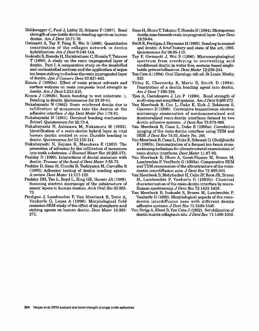
Fig. 12. Micrograph of the resin-dentin interface formed by Single Bond. Following removal of the dentin by demineralization and deproteinization, the micromorphology of the resin-encapsulated, acid-resistant hybrid-like structures can still be observed (arrows), indicating that they are part of the adhesive joint. HL = hybrid layer, PAR = Primer/adhesive resin and RT = resin tag.
Argon ion beam etching has been shown to be an effective method for removal of surface layers from materials to expose underlying characterist ics and accentuate the differences in structural composition (Van Meerbeek et al., 1995b). During argon ion beam etching, accelerated argon ions attack and selectively remove atoms at the specimen surface based on the composition of the material. Argon ion beam etching of polished section surfaces clearly reveals the hybrid layer at the resin- dentin interface of adhesive systems (Inokoshi et al., 1990). This etching selectively removes the resin component impregnated in the demineralized dentin, exposing the collagen fibers and creating a rough surface that is readily observable by SEM.
322 Vargas et al./SEM analysis and bond strength of single bottle adhesives
Under SEM, all of the single-bottle primer/adhesive systems used in this study produced a zone of resin-dentin interdiffusion similar to that of the conventional three-step system, SBMP, and similar to results described in previous investigations (Inokoshi et al., 1990;Van Meerbeek et al., 1992). The thickness (approximately 4 lJm) of the resin-interdiffusion zone for all bonding systems was expected to be similar since all systems conditioned the dentin with phosphoric acid of similar concentrations ranging from 32 to 37% and for an equal etching time (15 s). The depth of demineralization for the acids used in this study agrees with those obtained by Perdigao et al. (1996).
Single-bottle systems produced primer/adhesive layers of varying thickness, ranging from no discernible layer for Syntac Single-Component to a thick layer of approximately 50 pm for OptiBond Solo (Figs. 10 and 11). Variation of this layer thickness was observed not only between products, but also among specimens of the same adhesive and in different areas of the same specimen.
The differences in thickness could be due to a number of factors, such as differences in the viscosity of the adhesive systems, filled vs. unfilled primer/adhesives, variations in number of coats and application techniques as indicated by manufacturers. OptiBond Solo, which is a filled adhesive system, had a significantly thicker primer/ adhesive layer, with a thickness up to 50 pm (Fig. 11). In SEM micrographs of Syntac Single-Component, a transitional zone of primer/adhesive between the hybrid layer and the overlying resin was not observed (Fig. 10A). Syntac Single-Component adhesive system utilizes water as the primary solvent. Whether this played a role in the lack of a transitional primer/adhesive layer or in the low bond strengths observed with this adhesive remains unknown.
For Single Bond (Figs. 8 and 9), as well as for Scotchbond Multi-Purpose Plus (Fig. 1), amorphous structures that appeared similar to the hybrid layer were observed within the thickness of the primer/adhesive layer above the hybrid zone. This finding may be consistent with those of Van Meerbeek et al. (1996), who found, using TEM, an amorphous electron-dense phase at the outer surface of the hybrid layer. While the chemical nature of this amorphous phase could not be determined, the authors suggested it may have originated with the copolymer polyalkenoic acid used in Scotchbond Multi-Purpose primer, since this amorphous phase was not seen in specimens treated with an experimental primer that did not contain polyalkenoic acid. This copolymer is also present in Single Bond's primer/adhesive. It is reasonable to assume that the amorphous hybrid-like structures seen in this study are comparable to those found by Van Meerbeek et al. (1996).
To gain a better understanding of these structures and to determine if they were produced during specimen preparation or during SEM argon-ion milling, both the inorganic and organic components were dissolved, leaving the resin-encapsulated acid-resistant components of the specimen complex. In selected specimens, the dentin was removed by immersion in hydrofluoric acid for 10 s, followed by sodium hypochloride for 2 min. These amorphous structures remained intact (Fig. 12), which SUggests a polymer composition.
An attempt was made to relate dentin shear bond strengths to the morphology of the resin-dentin interface for the adhesive systems evaluated. Although minor variations existed in micromorphology, no correlation to dentin shear bond strength could be established.
It must be noted that information gathered from these in vitro samples does not take into consideration pulpal fluid pressure, stresses associated with polymerization shrinkage, or long-term exposure to moisture, mechanical and chemical stresses; all of these are factors that play an important clinical role in influencing the structural integrity of the resin-dentin bonds.
Under the conditions of the current study, all of the single-bottle adhesive systems, except Syntac Single Component, had bond strengths similar to the three- component adhesive tested. SEM examination of the resin- to-dentin interface revealed a distinct hybrid layer for all single-bottle primer/adhesives and the conventional three- step system. However, s l ight var ia t ions in the morphology of the interfacial ultrastructures were found for the various adhesives. In addition, a correlation was not found between bond s t rengths and interfacial morphology for the adhesive systems investigated in this study. In vivo studies are necessary to determine the efficacy and long-term clinical performance of the single- bottle adhesive systems.
ACKNOWLEDGMENTS
This investigation was supported by The Department of Operative Dentistry, College of Dentistry, The University of Iowa. Technical expertise was provided by Randy Nessler of the Electron Microscopy Research Facility at The University of Iowa, Iowa City, Iowa.
Received April 22, 1997 / Accepted August 3
Address correspondence and reprint requests to: Marcos A. Vargas Dental Science Building S-239 College of Dentistry The University of Iowa Iowa City, Iowa 52242-1001 USA Phone: +319-335-7208 Fax: +319-335-7267 era: marcos-vargasC~iowa.edu
REFERENCES
Buonocuore M (1955). A simple method of increasing the adhesion of acrylic filling materials to enamel surfaces. J Dent Res 34:849-853,
Fortin D, Swift E, Denehy G, Reinhardt J (1994). Bond strength and microleakage of current dentin adhesives. Dent Mater 10:253-258.
Fundingsland J, Aasen S, Bodger P, Cernhous J (1992). The effect of high humidity on adhesion to dentin° J D e n t Res 72:665, Abstr. No. 1199.
Fusayama T, Nakamura M, Kurosaki N, Iwaku M (1979). Non-pressure adhesion of a new adhesive restorative resin. J Dent Res 58:1364-1370.
Dental Mateflals/September 1997 323
Holderegger C, Paul J, Liithy H, Scharer P (1997). Bond strength of one-bottle dentin bonding agents on human dentin. Am JDent 10:71-76.
Gwinnett A, Tay F, Pang K, Wei S (1996). Quantitative con t r ibu t ion of the collagen ne twork in den t in hybridization. Am J Dent 9:140-144.
Inokoshi S, Hosoda H, Harniratissai C, Himida Y, Tatsumi T (1990). A study on the resin impregnated layer of dentin. Part I: A comparative study on the decalcified and undecalcified sections and the application of argon ion beam etching to disclose the resin impregnated layer of dentin. Jpn J Conserv Dent 33:427-442.
Kanca J (1992a). Effect of resin primer solvents and surface wetness on resin composite bend strength to dentin. Am J Dent 5:213-215.
Kanca J (1992b). Resin bonding to wet substrate. 1. Bonding to dentin. Quintessence Int 23:39-41.
Nakabayashi N (1982). Resin reinforced dentin due to inf i l t ra t ion of monomers into the dent in at the adhesive interface. J Jap Dent Mater Dev 1:78-81.
Nakabayashi N (1991). Dentinal bonding mechanisms [letter]. Quintessence Int 22:73-4.
Nakabayashi N, Ashizawa M, Nakamura M (1992). Identification of a resin-dentin hybrid layer in vital human dentin created in vivo: Durable bonding to dentin. Quintessence Int 23:135-141.
Nakabayashi N, Kojima K, Masuhara E (1982). The promotion of adhesion by the infiltration of monomers into tooth substrates. JBiomed Mater Res 16:265-273.
Pashley D (1990). Interactions of dental materials with dentin. Transac of the Acad of Dent Mater 3:55-73.
Pashley D, Sano H, Ciucchi B, Yoshiyama M, Carvalho R (1995). Adhesion testing of dentin bonding agents: A review. Dent Mater 11:117-125.
Pashley DH, Tao L, Boyd L, King GE, Horner JA (1988). Scanning electron microscopy of the substructure of smear layers in human dentine. Arch Oral Bio 33:265- 70.
Perdigao J, Lambrechts P, Van Meerbeek B, Tome A, Vanherle G, Lopes A (1996). Morphological field emission-SEM study of the effect of six phosphoric acid etching agents on human dentin. Dent Mater 12:262- 271.
Sano H, Shono T, Takatsu T, Hosoda H (1994). Microporous dentin zone beneath resin-impregnated layer. Oper Dent 19:59-64.
Swift E, Perdigao J, Heymann H (1995). Bonding to enamel and dentin: A brief history and state of the art, 1995. Quintessence Int 26:95-110.
Tay F, Gwinnett J, Wei S (1996). Micromorphological s p e c t r u m from overd ry ing to ove rwe t t i ng acid conditioned dentin in water free, acetone based single- bottle primer/adhesives. Dent Mater 12:236-244.
Ten Cate A (1994). Oral Histology. 4th ed. St Louis: Mosby, 532.
Ti t ley K, Chernecky R, Maric B, S m i t h D (1994). Penetration of a dentin bonding agent into dentin. Am J Dent 7:190-194.
Tjan A, Castelnuovo J, Liu P (1996). Bond strength of multi-step and simplified systems. Am JDent 9:269-272.
Van Meerbeek B, Con L, Duke E, Eick J, Robinson S, Guerrero D (1996). Correlative transmission electron microscopy examination of nondemineral ized and demineralized resin-dentin interfaces formed by two dentin adhesive systems. JDent Res 75:979-988.
Van Meerbeek B, Conn L, Duke E (1995a). Correlative imaging of the resin-dentin interface using TEM and SEM. JDent Res 74:32, Abstr. No. 166.
Van Meerbeek B, Conn L, Duke E, Schraub D, Ghafghaichi F (1995b). Demonstration of a focused ion-beam cross- sectioning technique for ultrastructural examination of resin-dentin interfaces. Dent Mater 11:87-92.
Van Meerbeek B, Dhem A, Goret-Nicaise M, Braem M, Lambrechts P, VanHerle G (1993a). Comparative SEM and TEM examination of the ultrastructure of the resin- dentin interdiffusion zone. J Dent Res 72:495-501.
Van Meerbeek B, Mohrbacher H, Celis JP, Roos JR, Braem M, Lambrechts P, Vanherle G (1993b). Chemical characterization of the resin-dentin interface by micro- Raman spectroscopy. J Dent Res 72:1423-1428.
Van Meerbeek B, Inokoshi S, Braem M, Lambrechts P, Vanherle G (1992). Morphological aspects of the resin- dent in interdiffusion zone wi th di f ferent dent in adhesive systems. JDent Res 71:1530-1540.
Van Strijp A, Klont B, Ten Cate J (1992). Solubilization of dentin matrix collagen in situ. JDentRes 71:1498-1502.
324 Vargas et al./$EM analysis and bond strength of single bottle adhesives