Laboratory Evaluation and Clinical Application of a New One-Bottle Adhesive
13
JOURNAL OF ESTHETIC DENTISTRY Laboratory Evaluation and Clinical Application of a New One-Bottle Adhesive JORGE PERDIGAO, DMD, MS, PHD" LUIZ N. BARATIERI, DDS, MS, PHD+ MANUELA LOPES, BS* ABSTRACT Purpose: The purposes of this project were to compare the enamel and dentin bond strengths of a new nanofilled one-coat adhesive system with its predecessor, an unfilled two-coat adhesive sys- tem; to analyze the dentin interfacial ultramorphology, using scanning and transmission electron microscopy (SEM and TEM); and to illustrate the clinical technique associated with the use of the new nanofilled one-coat adhesive system. Material and Methods: Twenty flat dentin surfaces and 20 flat enamel surfaces were polished on the labial surface of bovine incisors mounted in acrylic resin. The specimens were equally and randomly assigned to four bonding groups: (1) dentin with Prime & Bond 2.1; (2) dentin with Prime & Bond NT; (3) enamel with Prime & Bond 2.1; and (4) enamel with Prime & Bond NT. A composite post was then adapted to the treated area and light-cured. After thermocycling, shear bond strengths were determined by testing the shear strength of the specimens. The data were analyzed using one-way analysis of variance (ANOVA) and Student's t-test. For SEM and TEM, six dentin disks were obtained from middle dentin of human third molars and assigned equally to each adhesive. The adhesives were applied to dentin according to manufacturer's directions. The hybrid layer and resin penetration into dentin tubules were analyzed at an ultramorphologic level, and the observations were compared. Results: Shear bond strengths were as follows: group 1: 17.8 f 4.1 MPa; group 2: 20.5 e 3.5 MPa; group 3: 24.7 adhesives penetrated the dentin tubules and formed a fully infiltrated hybrid layer. The nanofiller included in the new one-application adhesive penetrated the dentin tubules and infiltrated the microspaces between the collagen fibers within the hybrid layer. CLINICAL SIGNIFICANCE: The new one-application nanofilled adhesive tested in this study resulted in bond strengths and dentin hybridization comparable to those obtained with the corresponding two-application system. The clinical sequences presented illustrate the ease of use of the newest simplified adhesives. 6.7 MPa; and group 4: 27.0 2 5.4 MPa. Electron microscopy showed that both estorative resins may bond to Bonding to enamel has been satis- factorily achieved with the acid- etch techniq~e,~ through the infil- tration of monomers within the microporosities created on enamel surface by an acid.s In contrast, a reliable and durable bonding to dentin has been more difficult to R dentin through mechanisms involving either the inorganic or the organic constituents of *Associate Professor, Department of Operative Dentistry, University of North Carolina at Chapel Hill, Chapel Hill, North Carolina tProfessor and Chairman, Department of Operative Dentistry, Federal University of Santa Catarina, Florian6polis, Brazil $Research Assistant, Electron Microscopy Laboratory, Dental Research Center, University of North Carolina at Chapel Hill, Chapel Hill, North Carolina VOLUME 11, NUMBER 1
Transcript of Laboratory Evaluation and Clinical Application of a New One-Bottle Adhesive
J O U R N A L O F E S T H E T I C D E N T I S T R Y
Laboratory Evaluation and Clinical Application of a New One-Bottle Adhesive
J O R G E PERDIGAO, D M D , MS, PHD" LUIZ N . BARATIERI, DDS, MS, PHD+ MANUELA LOPES, BS*
ABSTRACT
Purpose: The purposes of this project were to compare the enamel and dentin bond strengths of a new nanofilled one-coat adhesive system with its predecessor, an unfilled two-coat adhesive sys- tem; to analyze the dentin interfacial ultramorphology, using scanning and transmission electron microscopy (SEM and TEM); and to illustrate the clinical technique associated with the use of the new nanofilled one-coat adhesive system.
Material and Methods: Twenty flat dentin surfaces and 20 flat enamel surfaces were polished on the labial surface of bovine incisors mounted in acrylic resin. The specimens were equally and randomly assigned to four bonding groups: (1) dentin with Prime & Bond 2.1; (2) dentin with Prime & Bond NT; ( 3 ) enamel with Prime & Bond 2.1; and (4) enamel with Prime & Bond NT. A composite post was then adapted to the treated area and light-cured. After thermocycling, shear bond strengths were determined by testing the shear strength of the specimens. The data were analyzed using one-way analysis of variance (ANOVA) and Student's t-test. For SEM and TEM, six dentin disks were obtained from middle dentin of human third molars and assigned equally to each adhesive. The adhesives were applied to dentin according to manufacturer's directions. The hybrid layer and resin penetration into dentin tubules were analyzed at an ultramorphologic level, and the observations were compared.
Results: Shear bond strengths were as follows: group 1: 17.8 f 4.1 MPa; group 2: 20.5 e 3.5 MPa; group 3: 24.7 adhesives penetrated the dentin tubules and formed a fully infiltrated hybrid layer. The nanofiller included in the new one-application adhesive penetrated the dentin tubules and infiltrated the microspaces between the collagen fibers within the hybrid layer.
CLINICAL SIGNIFICANCE: The new one-application nanofilled adhesive tested in this study resulted in bond strengths and dentin hybridization comparable to those obtained with the corresponding two-application system. The clinical sequences presented illustrate the ease of use of the newest simplified adhesives.
6.7 MPa; and group 4: 27.0 2 5.4 MPa. Electron microscopy showed that both
estorative resins may bond to Bonding to enamel has been satis- factorily achieved with the acid- etch techniq~e ,~ through the infil- tration of monomers within the
microporosities created on enamel surface by an acid.s In contrast, a reliable and durable bonding to dentin has been more difficult to
R d entin through mechanisms involving either the inorganic or the organic constituents of
*Associate Professor, Department of Operative Dentistry, University of North Carolina at Chapel Hill, Chapel Hill, North Carolina tProfessor and Chairman, Department of Operative Dentistry, Federal University of Santa Catarina, Florian6polis, Brazil $Research Assistant, Electron Microscopy Laboratory, Dental Research Center, University of North Carolina at Chapel Hill, Chapel Hill, North Carolina
V O L U M E 1 1 , N U M B E R 1
J O U R N A L O F E S T H E T I C D E N T I S T R Y
Laboratorv Evaluation and Clinical Application of a Kew One-Bottle Adhesive
achieve, owing to the wet tubular ultrastructure and organic composi- tion of the dentin substrate.6
Recently several one-bottle dentin adhesive systems (DBAs) have been introduced that combine primer and adhesive resin into a single solution. These materials have received con- siderable attention from the dental profession, since clinicians often select the material that is easiest to use. Most of these new DBAs are composed of a separate phosphoric acid-based conditioner to etch enamel and dentin simultaneously (total-etch) and a blend of hydro- philic and hydrophobic resins dis- solved in an organic solvent, such as ethanol or acetone. That mixture is applied on dentin and enamel in one or several coats. The effects of phosphoric acid on dentin have been reported in several The acid removes the smear layer, opens up the dentin tubules, and demineralizes dentin by removing hydroxyapatite and exposing colla- gen fibers on the most superficial few microns (2-5 pm). After the etchant is rinsed off, dentin is left moist to prevent the collagen fibers from collapsing.*-10 The residual water occupies the spaces between the fibers, favoring the use of a wet- bonding t e c h n i q ~ e . ~ The solvent carries the blend of monomers into intimate contact with the dense filigree of moist dentin collagen fibers,'O resulting in an entangle- ment of the dentin adhesive within the spaces of the collagen fiber net-
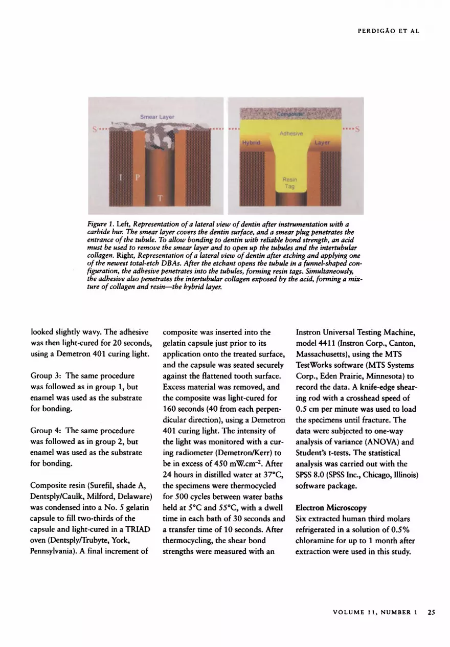
work. The structure resulting from this entanglement has been named the hybrid layer," or resin-dentin interdiffusion zone,2 and has been reported to provide retention for the majority of restorations performed with tooth-colored materials in contact with dentin (Figure 1 ) .2J
A new one-application nanofilled DBA recently has been developed, Prime & Bond NT (Dentsply DeTrey, Konstanz, Germany). The predecessor of this product is Prime & Bond 2.1. The manufacturer has added nanofiller (0.007 pm average particle size), a cross-linking agent, and a small resin molecule to pro- vide better infiltration into the tooth structure. The objective of this project was to evaluate the in vitro bond strength obtained with a simplified nanofilled DBA and to characterize its interaction with dentin a t an ultramorphologic level. Secondarily, the corresponding clinical technique for Class I1 and Class V restorations is described.
MATERIAL A N D M E T H O D S
Shear Bond Strength Forty bovine teeth refrigerated in a solution of 0.5% chloramine for up to 3 days after extraction were used in this project. The teeth were clean- ed of debris and mounted in pheno- lic rings with cold-cure acrylic resin. The labial surface of each tooth was ground with a mechanical grinder to expose dentin (20 teeth) or enamel (20 teeth) and was subsequently pol- ished for l minute with wet 400-grit
and 600-grit silicon carbide abrasive paper. Specimens were randomly assigned to four treatment sequences (10 specimens each), as follows:
Group 1: Dentin was etched with 36% phosphoric acid (Conditioner 36, Dentsply DeTrey) for 15 sec- onds and then rinsed with water for 15 seconds. Excess water was removed by blotting with tissue paper, leaving dentin visibly moist. Prime & Bond 2.1 adhesive was applied to the dentin surface in ample amounts, and the surface was left wet and undisturbed for 20 seconds. The adhesive was gently air-dried with oil-free compressed air from an air syringe for 5 sec- onds, keeping the air syringe 2 cm from the surface, and light-cured for 10 seconds, using a Demetron 401 curing light (DemetrodKerr, Danbury, Connecticut). A second coat was applied and gently air-dried for 5 seconds but not light-cured.
Group 2: Dentin was etched with 36% phosphoric acid (Conditioner 36) for 15 seconds and then rinsed with water for 15 seconds. Excess water was removed by blotting with a tissue paper, leaving dentin visibly moi'st. Prime & Bond NT adhesive was applied to the dentin surface in ample amounts, and the surface was left wet and undis- turbed for 20 seconds. The adhesive was air-dried with oil-free com- pressed air from an air syringe for 5 seconds, keeping the air syringe 2 cm from the surface. The surface
24 1 9 9 9
PERDIGAO ET A L
Figure 1. Left, Representation of a lateral view of dentin after instrumentation with a carbide bur. The smear layer covers the dentin surface, and a smear plug penetrates the entrance of the tubule. To allow bonding to dentin with reliable bond strength, an acid must be used to remove the smear layer and to open up the tubules and the intertubular collagen. Right, Representation of a lateral view of dentin after etching and applying one o f the newest total-etch DBAs. After the etchant opens the tubule in a funnel-shaped con- figuration, the adhesive penetrates into the tubules, forming resin tags. Simultaneously, the adhesive also penetrates the intertubular collagen exposed by the acid, forming a mix- ture of collagen and resin-the hybrid layer.
looked slightly wavy. The adhesive was then light-cured for 20 seconds, using a Demetron 401 curing light.
Group 3: The same procedure was followed as in group 1, but enamel was used as the substrate for bonding.
Group 4: The same procedure was followed as in group 2, but enamel was used as the substrate for bonding.
Composite resin (Surefil, shade A, DentsplyKaulk, Milford, Delaware) was condensed into a No. 5 gelatin capsule to fill two-thirds of the capsule and light-cured in a TRLAD oven (Dentsplynrubyte, York, Pennsvlvania). A final increment of
composite was inserted into the gelatin capsule just prior to its application onto the treated surface, and the capsule was seated securely against the flattened tooth surface. Excess material was removed, and the composite was light-cured for 160 seconds (40 from each perpen- dicular direction), using a Demetron 401 curing light. The intensity of the light was monitored with a cur- ing radiometer (Demetroflerr) to be in excess of 450 mW.cm2. After 24 hours in distilled water at 37"C, the specimens were thermocycled for 500 cycles between water baths held at 5°C and 55"C, with a dwell time in each bath of 30 seconds and a transfer time of 10 seconds. After thermocycling, the shear bond
~ ~ strengths were measured with an
Instron Universal Testing Machine, model 441 1 (Instron Corp., Canton, Massachusetts), using the MTS TestWorks software (MTS Systems Corp., Eden Prairie, Minnesota) to record the data. A knife-edge shear- ing rod with a crosshead speed of 0.5 cm per minute was used to load the specimens until fracture. The data were subjected to one-way analysis of variance (ANOVA) and Student's t-tests. The statistical analysis was carried out with the SPSS 8.0 (SPSS Inc., Chicago, Illinois) software package.
Electron Microscopy Six extracted human third molars refrigerated in a solution of 0.5% chloramine for up to 1 month after extraction were used in this study.
V O L U M E 1 1 , NUMBER 1 25
J O U R N A L OF ESTHETIC D E N T I S T R Y
Mean Bond Strength I SD (MPa) Type of Failure
The occlusal enamel was removed, and six dentin disks with a thick- ness of 0.8 * 0.2 mm were obtained from middle dentin by slow-speed diamond-saw sectioning parallel to the occlusal surface. A smear layer was created on the top surface by wet sanding with 600-grit S ic sand- paper for 60 seconds.'l Dentin was etched with 36% phosphoric acid (Conditioner 36) for 15 seconds and then rinsed with water for 15 sec- onds. Excess water was removed by blotting with a tissue paper, leaving dentin visibly moist. Three disks were assigned to each one of the two adhesives tested. The adhesives were applied as described for groups 1 and 2.
'
A 1-mm thick layer of a low-viscos- ity composite resin (AEliteflo, Bisco Inc., Schaumburg, Illinois) was applied to the treated dentin surface and light-cured for 40 seconds. A
Laboratorv Evaluation and Clinical Application of a New One-Bottle Adhesive
low-viscosity resin was used to facil- itate ultramicrotomy. The disks were then cross-sectioned in two halves with a diamond saw under water cooling.' One half from each dentin disk was assigned to scan- ning electron microscopy (SEM); the other half was assigned to transmission electron microscopy (TEM). For the SEM observation, the specimens were processed according to the protocol for speci- men preparation of biologic speci- mens for SEM.I3 The interfaces were observed under a JSM 6300 (JEOL USA, Inc., Peabody, Massa- chusetts), a t an accelerating voltage of 10 to 12 kV. An extra dentin disk was processed and observed to evaluate the effects of the phos- phoric acid etchant on dentin.
For the TEM observation, small sticks with a cross-section of 1.5 mm x 1.5 mm were cut from the bonded
half dentin disk using a slow-speed diamond saw under water. The specimens were then processed according to the technique described for biologic specimens. l4 After sec- tioning in an ultramicrotome in 0.085 pm-thick sections, the inter- faces were analyzed under a Philips CM-12 (Philips Electron Optics Inc., Mahwah, New Jersey), at an accelerating voltage of 100 kV.
R E S U L T S
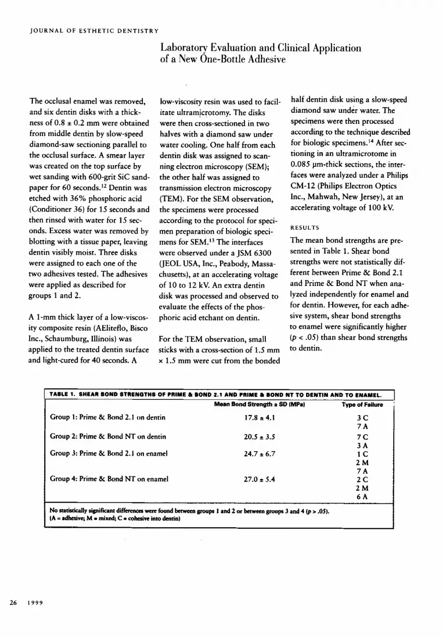
The mean bond strengths are pre- sented in Table 1. Shear bond strengths were not statistically dif- ferent between Prime & Bond 2.1 and Prime & Bond NT when ana- lyzed independently for enamel and for dentin. However, for each adhe- sive system, shear bond strengths to enamel were significantly higher ( p < .05) than shear bond strengths to dentin.
Group 1: Prime & Bond 2.1 on dentin
Group 2: Prime (k Bond NT on dentin
Group 3: Prime & Bond 2. I on enamel
Group 4: Prime & Bond NT on enamel
17.8 t 4.1
20.5 2 3.5
24.7 t 6.7
27.0 t 5.4
3 c 7 A 7 c 3 A 1 c 2 M 7A 2c 2 M 6 A
No statistically significant differems were found between groups 1 and 2 or between groups 3 and 4 (p > .05). (A = adhcsivc, M = mixed; C = cohesive into dentin) I
26 1 9 9 9
P E R D I G A O E T A L
When data were pooled for “sub- strate” (n = 20), shear bond strengths to enamel were statisti- cally greater than shear bond strengths to dentin (25.9 MPa vs. 19.2 MPa). When data were pooled for “adhesive” (n = 20), there were no statistical differences in bond strengths (23.8 MPa for Prime & Bond NT vs. 21.3 MPa for Prime & Bond 2.1).
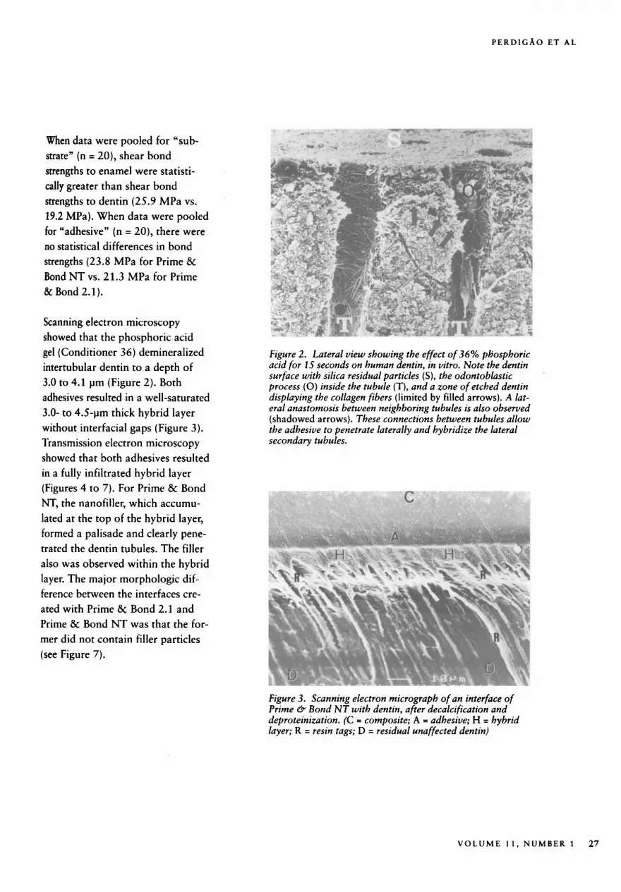
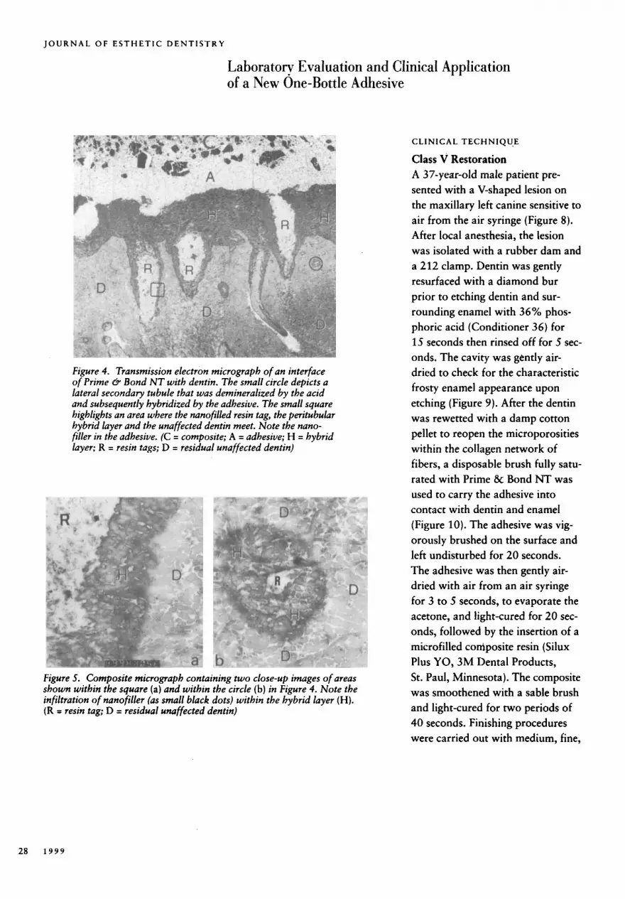
Scanning electron microscopy showed that the phosphoric acid gel (Conditioner 36) demineralized intertubular dentin to a depth of 3.0 to 4.1 pm (Figure 2). Both adhesives resulted in a well-saturated 3.0- to 4.5-pm thick hybrid layer without interfacial gaps (Figure 3). Transmission electron microscopy showed that both adhesives resulted in a fully infiltrated hybrid layer (Figures 4 to 7). For Prime & Bond NT, the nanofiller, which accumu- lated at the top of the hybrid layer, formed a palisade and clearly pene- trated the dentin tubules. The filler also was observed within the hybrid layer. The major morphologic dif- ference between the interfaces cre- ated with Prime & Bond 2.1 and Prime & Bond NT was that the for- mer did not contain filler particles (see Figure 7).
Figure 2. Lateral view showing the effect of 36% phosphoric acid for 15 seconds on human dentin, in vitro. Note the dentin surface with silica residual particles ( S ) , the odontoblastic process (0) inside the tubule (T) , and a zone of etched dentin displaying the collagen fibers (limited by filled arrows). A lat- eral anastomosis between neighboring tubules is also observed (shadowed arrows). These connections between tubules allow the adhesive to penetrate laterally and hybridize the lateral secondary tubules.
Figure 3. Scanning electron micrograph of an interface of Prime & Bond NT with dentin, after decalcification and deproteinization. (C = composite; A = adhesive; H = hybrid layer; R = resin tags; D = residual unaffected dentin)
V O L U M E 1 1 , N U M B E R 1 27
JOURNAL OF ESTHETIC DENTISTRY
Laboratorv Evaluation and Clinical Application of a New One-Bottle Adhesive
Figure 4. Transmission electron micrograph of an interface o f Prime 8 Bond NT with dentin. The small circle depicts a lateral secondary tubule that was demineralized by the acid and subsequently hybridized by the adhesive. The small square highlights an area where the nunofilled resin tag, the peritubular hybrid layer and the unaffected dentin meet. Note the nano- filler in the adhesive. (C = composite; A = adhesive; H = hybrid layer; R = resin tags; D = residual unaffected dentin)
Figure 5. Composite micrograph containing two close-up images of areas shown within the square (a) and within the circle (b ) in Figure 4. Note the infiltration of nanofiller (as small black dots) within the hybrid layer ( H ) . (R = resin tag; D = residual unaffected dentin)
C L I N 1 C A L TECH N I Q U,E
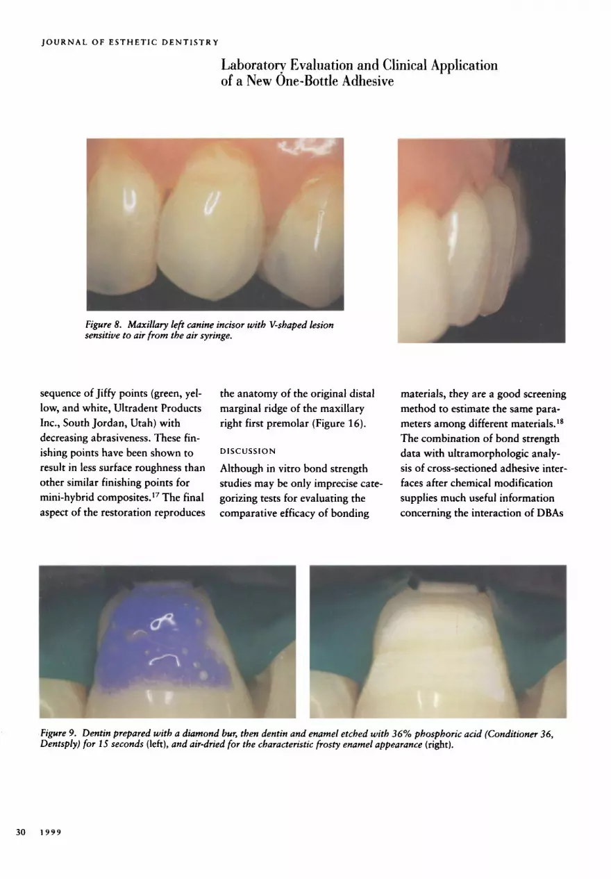
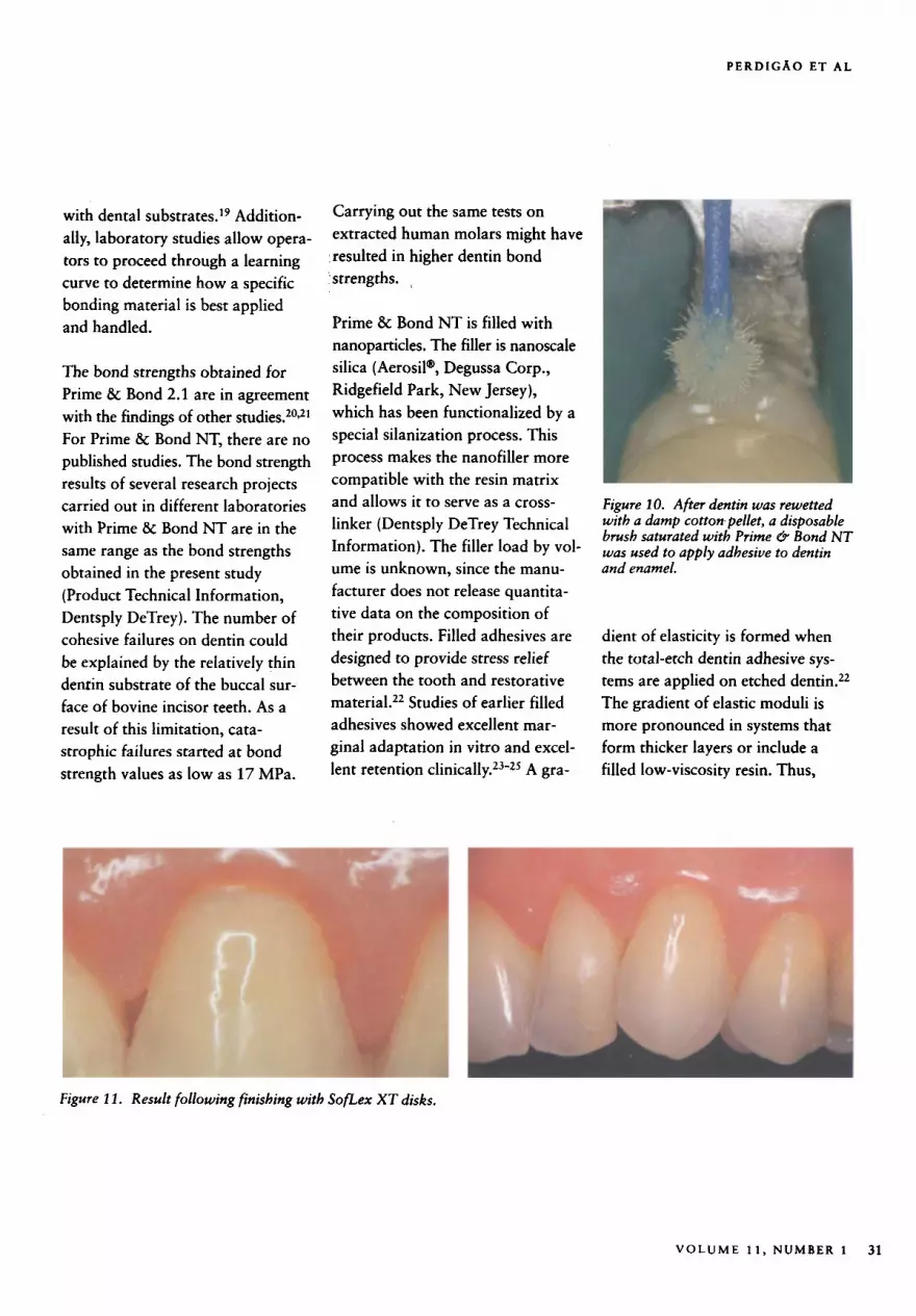
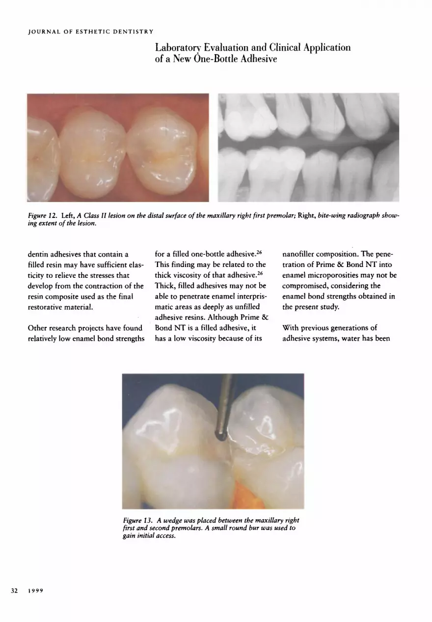
Class V Restoration A 37-year-old male patient pre- sented with a V-shaped lesion on the maxillary left canine sensitive to air from the air syringe (Figure 8). After local anesthesia, the lesion was isolated with a rubber dam and a 212 clamp. Dentin was gently resurfaced with a diamond bur prior to etching dentin and sur- rounding enamel with 36% phos- phoric acid (Conditioner 36) for 15 seconds then rinsed off for 5 sec- onds. The cavity was gently air- dried to check for the characteristic frosty enamel appearance upon etching (Figure 9). After the dentin was rewetted with a damp cotton pellet to reopen the microporosities within the collagen network of fibers, a disposable brush fully satu- rated with Prime & Bond NT was used to carry the adhesive into contact with dentin and enamel (Figure 10). The adhesive was vig- orously brushed on the surface and left undisturbed for 20 seconds. The adhesive was then gently air- dried with air from an air syringe for 3 to 5 seconds, to evaporate the acetone, and light-cured for 20 sec- onds, followed by the insertion of a microfilled coniposite resin (Silux Plus YO, 3M Dental Products, St. Paul, Minnesota). The composite was smoothened with a sable brush and light-cured for two periods of 40 seconds. Finishing procedures were carried out with medium, fine,
28 1 9 9 9
P E R D I C A O E T A L
and extra-fine SofLex XT disks (3M Dental Products). Figure 11 shows the final result.
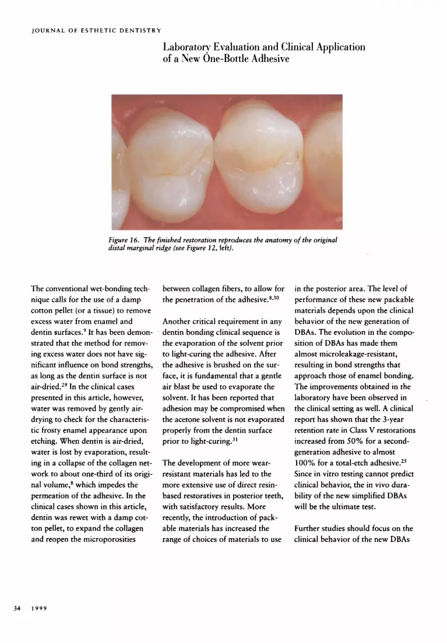
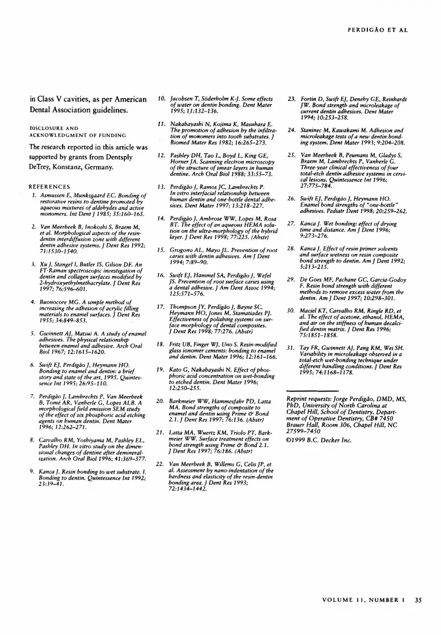
Class II Restoration A 34-year-old healthy female pre- sented with a Class I1 lesion on the distal surface of the maxillary right first premolar (Figure 12). After local anesthesia and shade selec- tion, the teeth were isolated with a rubber dam. A wedge was inserted between the maxillary right first and second premolars to allow for adequate contact after removing the band (Figure 13). After the lesion was examined through the initial conservative access, the preparation was widened and the carious tissue was removed (Figure 14). Facial contact was not broken. In an adhesive restoration, the surround- ing tissue becomes more resistant to recurrence of the A Palodent'" (Darway Inc., San Mateo, California) kidney-shaped concave matrix and a BiTine Ring'" (Danvay) were used to restore the missing distal wall. The enamel and dentin were etched with 36% phosphoric acid (Conditioner 36) for 15 seconds (Figure 15) and rinsed with water for 5 seconds. Prime & Bond NT was applied in one coat with a fully saturated brush; and Tetric Ceram shade A2 (IvoclarNivadent) was inserted in two increments, with each increment light-cured for #seconds (Figure 15). Finishing procedures were carried out with a
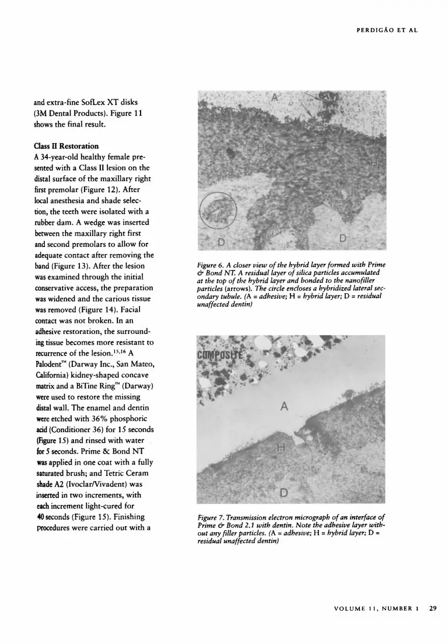
Figure 6. A closer view of the hybrid layer formed with Prime & Bond NT. A residual layer of silica particles accumulated at the top of the hybrid layer and bonded to the nanofiller particles (arrows). The circle encloses a hybridized lateral sec- ondary tubule. (A = adhesive; H = hybrid layer; D = residual unaffected dentin)
Figure 7. Transmission electron micrograph of an interface of Prime e9 Bond 2.1 with dentin. Note the adhesive layer with- out any filler particles. (A = adhesive; H = hybrid layer; D = residual unaffected dentin)
V O L U M E 1 1 , N U M B E R 1 29
J O U R N A L OF ESTHETIC DENTISTRY
Laboratorv Evaluation and Clinical Application of a New One-Bottle Adhesive
Figure 8. Maxillary left canine incisor with V-shaped lesion sensitive to air from the air syringe.
sequence of Jiffy points (green, yel- low, and white, Ultradent Products Inc., South Jordan, Utah) with decreasing abrasiveness. These fin- ishing points have been shown to result in less surface roughness than other similar finishing points for mini-hybrid composite^.^' The final aspect of the restoration reproduces
the anatomy of the original distal marginal ridge of the maxillary right first premolar (Figure 16).
DISCUSSION
Although in vitro bond strength studies may be only imprecise cate- gorizing tests for evaluating the comparative efficacy of bonding
materials, they are a good screening method to estimate the same para- meters among different materials.'S The combination of bond strength data with ultramorphologic analy- sis of cross-sectioned adhesive inter- faces after chemical modification supplies much useful information concerning the interaction of DBAs
Figure 9. Dentin prepared with a diamond bur, then dentin and enamel etched with 36% phosphoric acid (Conditioner 36, Dentsply) for 15 seconds (left), and air-dried for the characteristic frosty enamel appearance (right).
30 1 9 9 9
PERDICAO ET A L
with dental substrate^.'^ Addition- ally, laboratory studies allow opera- tors to proceed through a learning curve to determine how a specific bonding material is best applied and handled.
The bond strengths obtained for Prime & Bond 2.1 are in agreement with the findings of other studies.20p21 For Prime & Bond NT, there are no published studies. The bond strength results of several research projects carried out in different laboratories with Prime & Bond NT are in the same range as the bond strengths obtained in the present study (Product Technical Information, Dentsply DeTrey). The number of cohesive failures on dentin could be explained by the relatively thin denrin substrate of the buccal sur- face of bovine incisor teeth. As a result of this limitation, cata- strophic failures started at bond strength values as low as 17 MPa.
Carrying out the same tests on extracted human molars might have resulted in higher dentin bond strengths. ,
Prime & Bond NT is filled with nanoparticles. The filler is nanoscale silica (AerosiP, Degussa Corp., Ridgefield Park, New Jersey), which has been functionalized by a special silanization process. This process makes the nanofiller more compatible with the resin matrix and allows it to serve as a cross- linker (Dentsply DeTrey Technical Information). The filler load by vol- ume is unknown, since the manu- facturer does not release quantita- tive data on the composition of their products. Filled adhesives are designed to provide stress relief between the tooth and restorative rnateriaLz2 Studies of earlier filled adhesives showed excellent mar- ginal adaptation in vitro and excel- lent retention ~ l i n i c a l l y . ~ ~ - ~ ~ A gra-
Figure 10. After dentin was rewetted with a damp cotton pellet, a disposable brush saturated with Prime & Bond NT was used to apply adhesive to dentin and enamel.
dient of elasticity is formed when the total-etch dentin adhesive sys- tems are applied on etched dentin.22 The gradient of elastic moduli is more pronounced in systems that form thicker layers or include a filled low-viscosity resin. Thus,
Figure 11. Result following finishing with SofLex XT disks.
VOLUME 1 1 , NUMBER 1 31
J O U R N A L OF ESTHETIC D E N T I S T R Y
Laboratory Evaluation and Clinical Application of a New One-Bottle Adhesive
Figure 12. Left, A Class 11 lesion on the distal surface of the maxillary right first premolar; Right, bite-wing radiograph show- ing extent of the lesion.
dentin adhesives that contain a filled resin may have sufficient elas- ticity to relieve the stresses that develop from the contraction of the resin composite used as the final restorative material.
Other research projects have found relatively low enamel bond strengths
for a filled one-bottle adhesive.26 This finding may be related to the thick viscosity of that adhesive.26 Thick, filled adhesives may not be able to penetrate enamel interpris- matic areas as deeply as unfilled adhesive resins. Although Prime & Bond NT is a filled adhesive, it has a low viscosity because of its
nanofiller composition. The pene- tration of Prime & Bond NT into enamel microporosities may not be compromised, considering the enamel bond strengths obtained in the present study.
With previous generations of adhesive systems, water has been
Figure 13. A wedge was placed between the maxillary right first and second premolars. A small round bur was used to gain initial access.
32 1 9 9 9
PERDICAO ET A L
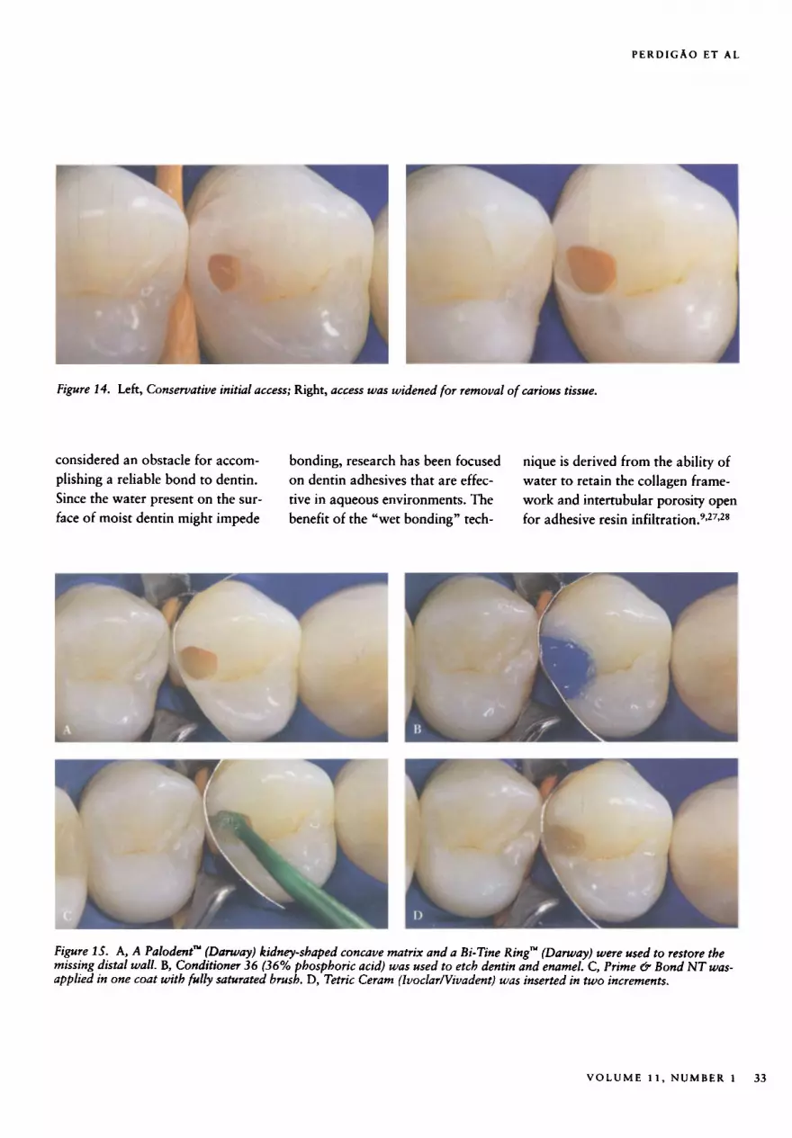
Figure 14. Left, Conservative initial access; Right, access was widened for removal o f carious tissue.
considered an obstacle for accom- plishing a reliable bond to dentin. Since the water present on the sur- face of moist dentin might impede
bonding, research has been focused on dentin adhesives that are effec- tive in aqueous environments. The benefit of the "wet bonding" tech-
nique is derived from the ability of water to retain the collagen frame- work and intertubular porosity open for adhesive resin i n f i l t r a t i ~ n . ~ * ~ ~ . ~ ~
Figure 15. A, A Palodent" (Darway) kidney-shaped concave matrix and a Bi-Tine Ring" (Danuay) were used to restore the missing distal wall. B, Conditioner 36 (36% phosphoric acid) was used to etch dentin and enamel. C, Prime 8 Bond NT was- applied in one coat with fully saturated brush. D, Tetric Ceram (Iuoclar/Vivadent) was inserted in two increments.
V O L U M E 1 1 , NUMBER 1 33
J O U R N A L OF ESTHETIC DENTISTRY
Laboratow Evaluation and Clinical Application of a New One-Bottle Adhesive
Figure 16. The finished restoration reproduces the anatomy of the original distal marginal ridge (see Figure 12, left).
The conventional wet-bonding tech- nique calls for the use of a damp cotton pellet (or a tissue) to remove excess water from enamel and dentin surface^.^ It has been demon- strated that the method for remov- ing excess water does not have sig- nificant influence on bond strengths, as long as the dentin surface is not a i ~ d r i e d . ~ ~ In the clinical cases presented in this article, however, water was removed by gently air- drying to check for the characteris- tic frosty enamel appearance upon etching. When dentin is air-dried, water is lost by evaporation, result- ing in a collapse of the collagen net- work to about one-third of its origi- nal volume,8 which impedes the permeation of the adhesive. In the clinical cases shown in this article, dentin was rewet with a damp cot- ton pellet, to expand the collagen and reopen the microporosities
between collagen fibers, to allow for the penetration of the adhe~ive.~*~O
Another critical requirement in any dentin bonding clinical sequence is the evaporation of the solvent prior to light-curing the adhesive. After the adhesive is brushed on the sur- face, it is fundamental that a gentle air blast be used to evaporate the solvent. It has been reported that adhesion may be compromised when the acetone solvent is not evaporated properly from the dentin surface prior to light-~uring.~'
The development of more wear- resistant materials has led to the more extensive use of direct resin- based restoratives in posterior teeth, with satisfactory results. More recently, the introduction of pack- able materials has increased the range of choices of materials to use
in the posterior area. The level of performance of these new packable materials depends upon the clinical behavior of the new generation of DBAs. The evolution in the compo- sition of DBAs has made them almost microleakage-resistant, resulting in bond strengths that approach those of enamel bonding. The improvements obtained in the laboratory have been observed in the clinical setting as well. A clinical report has shown that the 3-year retention rate in Class V restorations increased from 50% for a second- generation adhesive to almost 100% for a total-etch adhesive.25 Since in vitro testing cannot predict clinical behavior, the in vivo dura- bility of the new simplified DBAs will be the ultimate test.
Further studies should focus on the clinical behavior of the new DBAs
34 1 9 9 9
P E R D I C A O E T A L
in Class V cavities, as per American Dental Association guidelines,
DISCLOSURE AND ACKNOWLEDGMENT OF FUNDING
The research reported in this article was supported by grants from Dentsply DeTrey, Konstanz, Germany.
REFERENCES 1. Asmussen E, Munksgaard EC. Bonding of
restorative resins to dentine promoted by aqueous mixtures of aldehydes and active monomers. Int Dent J 1985: 35:16O-165.
2. Van Meerbeek 6, Inokoshi S, Braem M, et al. Morphological aspects of the resin- dentin interdiffusion zone with different dentin adhesive systems. J Dent Res 1992; 71:l S3O-lS40.
3. Xu J , Stangel I . Butler IS, Gilson DF. An FT- Raman spectroscopic investigation of dentin and collagen surfaces modified by 2-hydroxyethylmethacrylate. J Dent Res 1997; 76.596-601.
4. Buonocore MG. A simple method of increasing the adhesion of acrylic filling materials to enamel surfaces. J Dent Res 1955; 34:849-853.
5. Gwinnett AJ, Matsui A. A study of enamel adhesives. The physical relationship between enamel and adhesive. Arch Oral Biol 1967; 12:1615-1620.
Swift EJ. Perdigrio J. Heymann HO. Bonding to enamel and dentin: a brief story and state o f the art, 1995. Quintes- sence Int 199.T: 26:9F-110
Perdigrio J. Lambrechts P, Van Meerbeek 6, Tome AR, Vanherle G, Lopes ALB. A morphological field emission S E M study of the effect of six phosphoric acid etching agents on human dentin. Dent Mater 1996: 12:262-271.
Carvalho RM. Yoshiyama M, Pashley EL, Pashley DH. In vitro study on the dimen- sional changes o f dentine after demineral- iration. Arrh Oral Biol 1996; 41:369-377.
Kanca]. Resin bonding to wet substrate. 1. Bonding to dentin. Quintessence Int 1992; 23:3 9-4 1.
10. Jacobsen T, Soderholm K-J. Some effects of water on dentin bonding. Dent Mater
1 1 . Nakabayashi N, Kojima K , Masuhara E. The promotion o f adhesion by the infiltra- tion of monomers into tooth substrates. J Biomed Mater Res 1982; 16:265-273.
12. Pashley DH, Tao L, Boyd L, King GE, Horner ]A. Scanning electron microscopy o f the structure of smear layers in human dentine. Arch Oral Bioll988; 3355-73.
13. Perdigcio J , Ramos JC, Lambrechts P. In vitro interfacial relationship between human dentin and one-bottle dental adhe- sives. Dent Mater 1997; 13:218-227.
14. Perdigcio J , Ambrose WW, Lopes M, Rosa BT. The effect of an aqueous HEMA solu- tion on the ultra-morphology of the hybrid layer. J Dent Res 1998; 77:225. (Abstr)
15. Grogono AL, Mayo JL. Prevention of root caries with dentin adhesives. Am J Dent
16. Swift EJ, Hammel SA, Perdigcio J , Wefel JS. Prevention of root surface caries using a dental adhesive. J Am Dent Assoc 1994;
17. Thompson JY , Perdigrio J , Bayne SC, Heymann HO, Jones M , Stamatiades PJ. Effectiveness o f polishing systems on sur- face morphology of dental composites. J Dent Res 1998; 77276. (Abstr)
18. Fritz LIB, Finger WJ, Uno S. Resin-modified glass ionomer cements: bonding to enamel and dentin. Dent Mater 1996; 12:I 6 1-1 66.
19. Kato G, Nakabayashi N. Effect of phos- phoric acid concentration on wet-bonding to etched dentin. Dent Mater 1996:
1995; l,l:132-136.
1994; 7:89-90.
1 2S:571-576.
23. Fortin D, Swift EJ, Denehy GE, Reinhardt JW. Bond strength and microleakage of current dentin adhesives. Dent Mater 1994; 10:253-258.
24. Staninec M , Kawakami M . Adhesion and microleakage tests of a new dentin bond- ing system. Dent Mater 1993; 9204-208.
25. Van Meerbeek 6, Peumans M, Gladys S , Braem M , Lambrechts P, Vanherle G. Three- year clinical effectiveness of four total-etch dentin adhesive systems in cervi- cal lesions. Quintessence Int 1996;
26. Swift EJ, Perdigio J , Heymann HO. Enamel bond strengths of "one-bottle " adhesives. Pediatr Dent 1998; 20:259-262.
27. Kanca J. Wet bonding: effect of drying time and distance. Am J Dent 1996;
2 7:77S-784.
9273-2 76.
28. Kanca I . Effect o f resin Drimer solvents and &face wetness on iesin composite bond strength to dentin. Am J Dent 1992; S:2 13-2 15.
and &face wetness on iesin composite bond s t rend to dentin. Am I D m t 1992;
29. De Goes MF, Pachane GC, Garcia-Godoy F. Resin bond strength with different methods to remove excess water from the dentin. Am J Dent 1997; 10298-301.
30. Maciel KT, Carvalho RM, Ringle RD, et al. The effect o f acetone, ethanol, HEMA, and air on the stiffness of human decalci- fied dentin matrix. J Dent Res 1996; 7S:185 1 - 1 858.
31. Tay FR, Gwinnett AJ, Pang K M , Wei SH. Variability in microleakage observed in a total-etch wet-bonding technique under different handling conditions. J Dent Res 1995; 74:1168-1178.
12:2SO-255.
20. Barkmeier W w , Hammesfahr p ~ , Latta MA. Bond strengths o f composite to enamel and dentin using Prime 6 Bond 2.1. J Dent Res 1997; 76r136. (Abstr)
Reprint requests: l o w Perdigcjo, DMD, MS, PhD, University of North Carolina at Chapel Hill, .khool of Dentistry, Depart- ment of Operative Dentistry. CB# 7450 Brauer Hall, Room 306, Chapel Hill, N C
01 999 B.C. Decker In,-. 21. Latta MA, Wuertz K M , Triolo PT, Bark- 27599-7450
meier ww. surface treatment effects On bond strength using Prime 6 Bond 2.1. J Dent Res 1997; 715186. (Abstr)
22. Van Meerbeek B, Willems G, Celis JP, et al. Assessment by nano-indentation of the hardness and elasticity o f the resin-dentin bonding area. J Dent Res 1993; 72: 1434-1 442,
V O L U M E 1 1 , NUMBER 1 35