Phylogeography of mtDNA haplogro up R7 in the Indian peninsula
Upload
independentCategory
view
6download
0
1997, 17(4):2217. Mol. Cell. Biol.
Banerjee and M P CzechH Meisner, A Daga, J Buxton, B Fernández, A Chawla, U photoreceptor cell development.growth factor receptors and role of Cbl in R7 Interactions of Drosophila Cbl with epidermal
http://mcb.asm.org/content/17/4/2217Updated information and services can be found at:
These include:
CONTENT ALERTS more»cite this article),
Receive: RSS Feeds, eTOCs, free email alerts (when new articles
http://journals.asm.org/site/misc/reprints.xhtmlInformation about commercial reprint orders: http://journals.asm.org/site/subscriptions/To subscribe to to another ASM Journal go to:
on July 8, 2014 by guesthttp://m
cb.asm.org/
Dow
nloaded from
on July 8, 2014 by guesthttp://m
cb.asm.org/
Dow
nloaded from
MOLECULAR AND CELLULAR BIOLOGY,0270-7306/97/$04.0010
Apr. 1997, p. 2217–2225 Vol. 17, No. 4
Copyright q 1997, American Society for Microbiology
Interactions of Drosophila Cbl with Epidermal Growth FactorReceptors and Role of Cbl in R7 Photoreceptor
Cell DevelopmentHERMAN MEISNER,1,2* ANDREA DAGA,3 JOANNE BUXTON,1 BELEN FERNANDEZ,1
ANIL CHAWLA,1 UTPAL BANERJEE,3 AND MICHAEL P. CZECH1,2
Program in Molecular Medicine1 and Department of Biochemistry and Molecular Biology,2
University of Massachusetts Medical Center, Worcester, Massachusetts 01605,and Department of Molecular, Cell and Developmental Biology,Molecular Biology Institute, and Brain Research Institute,University of California, Los Angeles, California 900953
Received 23 September 1996/Returned for modification 21 October 1996/Accepted 24 December 1996
The human proto-oncogene product c-Cbl and a similar protein in Caenorhabditis elegans (Sli-1) contain aproline-rich COOH-terminal region that binds Src homology 3 (SH3) domains of proteins such as the adapterGrb2. Cbl-Grb2 complexes can be recruited to tyrosine-phosphorylated epidermal growth factor (EGF) recep-tors through the SH2 domain of Grb2. Here we identify by molecular cloning a Drosophila cDNA encoding aprotein (Drosophila Cbl [D-Cbl]) that shows high sequence similarity to the N-terminal region of human c-Cblbut lacks proline-rich sequences and fails to bind Grb2. Nonetheless, in COS-1 cells, expression of hemag-glutinin epitope-tagged D-Cbl results in its coimmunoprecipitation with EGF receptors in response to EGF.EGF also caused tyrosine phosphorylation of D-Cbl in such cells, but no association of phosphatidylinositol3-kinase was detected in assays using anti-p85 antibody. A point mutation in D-Cbl (G305E) that suppressesthe negative regulation of LET-23 by the Cbl homolog Sli-1 in C. elegans prevented tyrosine phosphorylationof D-Cbl as well as binding to the liganded EGF receptor in COS-1 cells. Colocalization of EGF receptors withboth endogenous c-Cbl or expressed D-Cbl in endosomes of EGF-treated COS-1 cells is also demonstrated byimmunofluorescence microscopy. In lysates of adult transgenic Drosophila melanogaster, GST-DCbl binds to thetyrosine-phosphorylated 150-kDa torso-DER chimeric receptor. Expression of D-Cbl directed by the sevenlessenhancer in intact Drosophila compromises severely the development of the R7 photoreceptor neuron. Thesedata suggest that despite the lack of Grb2 binding sites, D-Cbl functions as a negative regulator of receptortyrosine kinase signaling in the Drosophila eye by a mechanism that involves its association with EGF receptorsor other tyrosine kinases.
The human proto-oncogene product c-Cbl is a 908-amino-acid (aa) protein containing a ring finger domain and an ex-tended proline-rich COOH terminus with multiple PXXP mo-tifs that bind proteins having Src homology 3 (SH3) domains,including Grb2 and Nck (4, 9, 12, 21, 23, 25, 28). Activation ofhematopoietic cells leads to rapid tyrosine phosphorylation ofc-Cbl and the recruitment of p85/phosphatidylinositol 3-kinase(PI 3-kinase) and Crk to tyrosine phosphorylation sites on Cbl(6, 8, 9, 12, 14, 21, 23, 25, 26, 28). In other cell types, Cblbecomes tyrosine phosphorylated in response to a variety ofextracellular signals, including epidermal growth factor (EGF),nerve growth factor, granulocyte-macrophage colony-stimulat-ing factor, and transforming growth factor a (9, 11, 13, 19, 22,23). Rapid association of c-Cbl with autophosphorylated EGFand colony-stimulating factor 1 (CSF-1) receptors also occurs(5, 13, 22, 32). A mechanism by which Cbl is recruited to theEGF receptor in response to EGF appears to involve bindingof the SH2 domain of the adapter Grb2 to tyrosine-phosphor-ylated sites on the receptor (22, 32). The oncogenic form,v-Cbl, is a truncated 40-kDa protein lacking the ring finger andproline-rich COOH-terminal domain (3, 17) that mediates po-
tent signals leading to cell transformation. A recent report hasshown that v-Cbl is poorly tyrosine phosphorylated upon EGFstimulation but nonetheless is recruited to the liganded EGFreceptor (5). These and other data argue that the N-terminalregions of c-Cbl (13) and v-Cbl (5) may also interact directlywith the EGF receptor.The physiological functions of c-Cbl are poorly understood.
The binding of Grb2 and other proteins to c-Cbl suggests thatit may serve as a docking protein for adapter molecules toenhance the formation of protein complexes on activated re-ceptors at the cell membrane. Genetic data obtained with Sli-1,a Caenorhabditis elegans protein similar to c-Cbl, indicates apotential suppressor role in signaling by LET-23, the EGFreceptor homolog (35). Thus, expression of Sli-1 in the pres-ence of a defective LET-23 receptor prevented vulva develop-ment, which is dependent on the p21ras signaling pathway (24).These data suggest the hypothesis that c-Cbl negatively mod-ulates EGF receptor signaling and that v-Cbl may act in adominant inhibitory mode to block the negative influence ofc-Cbl. To clarify the role(s) of Cbl proteins in cellular signalingprocesses, we searched for similar proteins expressed in Dro-sophila melanogaster. Here we report the molecular cloning ofa gene encoding Drosophila Cbl (D-Cbl); we show that thisprotein lacks the entire C-terminal proline-rich domain ofmammalian Cbl and does not bind p85 or Grb2. Despite thelack of these sites, D-Cbl is rapidly recruited to EGF receptorsfollowing EGF stimulation when heterologously expressed in
* Corresponding author. Mailing address: Program in MolecularMedicine and Department of Biochemistry and Molecular Biology,University of Massachusetts Medical School, 373 Plantation St.,Worcester, MA 01655. Phone: (508) 856-6929. Fax: (508) 856-1617.E-mail: [email protected].
2217
on July 8, 2014 by guesthttp://m
cb.asm.org/
Dow
nloaded from
mammalian cells and exerts a negative influence on the signal-ing pathway required for R7 photoreceptor neuron develop-ment in intact Drosophila.
MATERIALS AND METHODS
Cloning of D-Cbl. Degenerate primers were made based on two regions,TYDEVK and YELYCEM, that share 100% amino acid sequence identityamong the human, mouse, and C. elegans homologs of Cbl. These primers wereused to amplify a 309-bp DNA fragment from Drosophila genomic DNA. Thisfragment was subcloned, and sequence analysis confirmed that it contained anopen reading frame highly homologous to those encoding Cbl proteins. A Dro-sophila eye-specific cDNA library (gift of G. M. Rubin) was screened by using the309-bp fragment as a probe, and nine independent clones were isolated. Thelongest cDNA, a 2.66-kb insert, was entirely sequenced with a model 373A DNAsequencer (Applied Biosystems Inc.), using dye-coupled terminators.Generation of transformants. The D-Cbl cDNA was subcloned as an EcoRI
fragment into a modified Sev-S11 vector (gift of K. Basler and E. Hafen) con-taining two copies of the sevenless enhancer (sE) and an hsp70 promoter. Theresulting transformation construct was named sE[D-Cbl]. To generate transfor-mant flies, Drosophila w1118 females were crossed to males containing P(D2-3) asa source of transposase. The embryos from this cross were injected with sE[D-Cbl], giving rise to flies carrying a D-Cbl transgene.Drosophila binding. Flies carrying a chimeric Drosophila torso-EGF receptor
on the X chromosome controlled by an hsp70 promoter (27) were provided byErnst Hafen (Zurich, Switzerland) and by Jean Olivier (Toronto, Ontario, Can-ada). Flies were heat shocked at 378C for 45 min, allowed to recover at 228C for2.5 h, and lysed in 1% Triton X-100–10% glycerol–150 mM NaCl–20 mM Tris(pH 7.5)–1.5 mM Mg21–1 mM EGTA containing 1 mM Na2VO4, 2.5 mg ofaprotinin per ml, and 0.1 mM phenylmethylsulfonyl fluoride. After centrifuga-tion, clarified supernatants were incubated with 2 mg of glutathione S-transferase(GST) or GST-DCbl for 1 h at 48C, washed twice in 0.2% Triton X-100 bufferand once in 0.2% Triton X-100 buffer containing 0.5 M NaCl, and boiled insodium dodecyl sulfate (SDS) sample buffer, and proteins were resolved bySDS-gel electrophoresis.Antibodies and reagents. The Drosophila cDNA of 2.66 kb was amplified by
PCR with Taq polymerase to yield an N-terminal EcoRI-methionine-NcoI0.66-kb fragment, using oligonucleotides 59-GAATTCATGGCGACGAGAGGCAGT-39 (sense) and 59-GTGGTCTTTAGGGCCAT-39 (antisense), and a C-terminal NcoI-BamHI 0.76-kb fragment, using oligonucleotides 59-TAATCTCCGGCCTGGAG-39 (sense) and 59-GCGGATCCTGTGAACAAAACTATACC-39 (antisense). Both PCR products were ligated into EcoRI-BamHI-restrictedpCMV5. Next, an epitope tag containing a methionine start codon and threetandem copies of the hemagglutinin (HA1) epitope (34) was ligated in frame intothe EcoRI site at the N terminus of D-Cbl, and the 1.5-kb cDNA was verified bysequencing. The G305E point mutation was generated by ligating a ClaI-BstUI211-bp fragment and a BstUI-PstI double-stranded oligonucleotide containingthe desired mutation into pCMV5-HA D-Cbl that was restricted with ClaI andPstI to remove the ClaI-PstI fragment. The resulting plasmid DNA was se-quenced to confirm the mutation. A full-length D-Cbl–GST fusion protein wasgenerated by amplification of D-Cbl cDNA with Vent polymerase (New EnglandBiolabs) and ligation of the product in frame into the BamHI-EcoRI site ofpGEX2T. Fusion proteins were affinity purified on glutathione-agarose beads(Sigma) according to standard procedures. An N-terminal human Cbl-GST fu-sion protein, consisting of aa 26 to 283, was prepared by PCR amplification of thehuman Cbl cDNA with primers 59-GATTGGGATCCTGAAGGACGC-39(sense) and 59-GAATTTCTGGATCCGAGCTTTC-39 (antisense). The GST–N-Cbl cDNA was cloned into pGEX2T and sequenced to ensure fidelity ofamplification, and the expressed fusion protein was injected into rabbits. Thepolyclonal antibody (R4575) quantitatively precipitates Cbl from lysates of COScells. Polyclonal Cbl antibody for Western blots was purchased from Santa Cruz,Inc., and EGF receptor antibody was a gift from Roger Davis, Howard HughesMedical Institute, University of Massachusetts Medical Center. An anti-HAantibody (R4289) was raised in rabbits against a peptide corresponding to theHA epitope and purified by binding to protein A-Sepharose. An anti-HA mono-clonal antibody (MAb) (12CA5) used to blot was purchased from BerkeleyAntibody Co. Polyclonal anti-p85 antibody was purchased from Upstate Bio-technology, Inc., and anti-Grb2 MAb was purchased from Transduction Labo-ratories. A polyclonal Grb2 antibody was produced as described previously (21).A phosphopeptide corresponding to the likely p85 binding site on Cbl (SCTpYEAMYNIQSQA) was synthesized in the Peptide Synthesis Facility at theUniversity of Massachusetts Medical Center. The peptide was coupled to Affi-Gel 10 as instructed by the manufacturer (Bio-Rad).Cell culture and activation. COS-1 cells were grown in 10-cm-diameter plates
in Dulbecco’s modified Eagle’s medium containing 8% fetal calf serum. Cellswere transfected with 1 mg of cDNA in Lipofectamine (GIBCO) according to themanufacturer’s protocol. After 2 days, subconfluent cells were serum starved for4 h and activated at 378C with 300 ng of EGF (Preprotec) per ml for the indicatedtimes. Reactions were stopped with cold phosphate-buffered saline (PBS) con-taining 0.5 mM Na2VO4, 5 mM EDTA, and 10 mM NaF and lysed in a mixture
containing 1% Nonidet P-40 (NP-40), 10 mM Tris (pH 7.5), 1 mM EDTA, 150mM NaCl, 1 mM Na2VO4, 0.1 mM phenylmethylsulfonyl fluoride, and 2.5 mg ofaprotinin per ml.Immunofluorescence microscopy. COS-1 cells were seeded on coverslips and
transfected with Lipofectamine and after 2 days were serum starved. EGF (300ng/ml) was added for 20 min; the cells were washed in cold PBS and fixed in cold4% formaldehyde. Cells were permeabilized and blocked in PBS containing 1%fetal bovine serum and 0.5% Triton-X 100 for 20 min. Primary antibodies weresheep anti-EGF receptor, rabbit anti-Cbl, and rabbit anti-HA; secondary anti-bodies were donkey anti-rabbit–fluorescein isothiocyanate (FITC; Pierce) ordonkey anti-sheep–rhodamine (Cortex Biochemicals). Cells were mounted in90% glycerol–PBS–2.5% 1,4-diazabicyclo-(2,2,2)-octane, and images were takenwith a digital imaging microscope (Nikon Diaphot 200) fitted with a NikonApo60/1.4 immersion lens.Immunoprecipitation and blotting. Antibodies were mixed with clarified ly-
sates containing 0.5 mg of protein and incubated at 48C overnight. ProteinA-Sepharose was added for 15 min with constant shaking; beads were subse-quently washed several times in 0.2% NP-40 buffer and once in 0.2% NP-40buffer containing 0.5 M NaCl. Proteins were resolved by SDS-polyacrylamide gelelectrophoresis, transferred to nitrocellulose, and blotted. Detection was bychemiluminescence (Boehringer Mannheim).Nucleotide sequence accession number. The nucleotide sequence of Drosoph-
ila Cbl has been submitted to GenBank and given accession number U87925.
RESULTS
Isolation of a Drosophila cDNA related to the proto-onco-gene c-cbl. A D-cbl DNA fragment was PCR amplified fromDrosophila genomic DNA by using a set of degenerate primers.Screening of a Drosophila eye disc cDNA library with thisfragment led to the isolation of a 2.66-kb cDNA containing 313nucleotides of 59 noncoding region, a 1,344-nucleotide openreading frame preceding an in-frame TAG stop codon, and1,317 nucleotides of 39 noncoding region. A comparison of thederived amino acid sequences of human-Cbl, D-Cbl, and C.elegans Cbl (Sli-1) (Fig. 1A) shows no homology over the first40 aa or beyond residue 426 of D-Cbl. However, within theconserved region of approximately 380 aa, the three speciesshow 63% similarity between aa 46 and 205 and 93% similaritybetween aa 205 and 330. Within the N-terminal conservedregion, several tyrosine residues found in human Cbl are notpresent in D-Cbl, including the human Y141EEN sequence(F129EDN in D-Cbl), which is a potential Src kinase SH2binding motif. A high degree of similarity is also observedbetween aa 354 and 425. This region includes a ring fingerdomain and a sequence of 17 aa (called 70Z/3) which, whendeleted, renders c-Cbl transforming (1, 3, 4). Interestingly,D-Cbl is only 93 residues longer than v-Cbl (Fig. 1B), but theseamino acids include the important 70Z/3 sequence and ringfinger regions. Finally, unlike the protein found in C. elegans ormammals, D-Cbl contains no proline-rich motifs.D-Cbl negatively regulates R7 development. In the develop-
ing Drosophila eye, the differentiation of photoreceptor neu-rons (R cells) depends on signaling through the Sevenless andEGF receptor tyrosine kinases (reviewed in reference 36). Thedeveloping eye constitutes a genetic system where the in vivofunction of Drosophila Cbl could be studied. Mutations in theD-cbl locus have not yet been identified, and the region en-coding this locus is not covered by deficiencies or transposableelements. We therefore generated transformant flies carryingthe D-Cbl cDNA under the transcriptional control of sE, whichwill cause the expression of the D-Cbl cDNA in all cells thatexpress Sevenless, including the R7 photoreceptor precursor(2). To assess the role of D-Cbl in R7 development, we testedthese transgenic flies in a sensitized genetic assay (reviewed inreference 7). In this assay, signaling through Sevenless is com-promised by using a disabled Sevenless kinase, sevE4. Thisdefect is partially rescued by combining sevE4 with one copy ofSosJC2, an allele of the son of sevenless gene encoding an over-active protein. In the resulting sevE4/Y; SosJC2/1 fly, the re-
2218 MEISNER ET AL. MOL. CELL. BIOL.
on July 8, 2014 by guesthttp://m
cb.asm.org/
Dow
nloaded from
ceptor tyrosine kinase signal is weakened so that an R7 celldevelops in only 17% of the facets (ommatidia) in the eye (Fig.2).The above-described genetic background is extremely sensi-
tive to the dosage of genes participating in receptor tyrosinekinase signaling, and the fraction of ommatidia developing R7cells is a readout for the strength of the transduced signal. Wehave previously shown that removing a single copy of a positiveregulator such as Ras or Sos abolishes the development of R7cells in this genetic background, while removing one copy of anegative regulator, such as GAP1, will increase this number
significantly (29). Removing a copy of the EGF receptor in thisbackground also completely abrogates R7 differentiation (Fig.2). This finding suggests that the R7 developmental readout isdue to the combination of Sev and EGF receptor tyrosinekinase signals. This result is consistent with the recent obser-vations of Freeman (10) showing that both of these receptorsare essential for R7 development. We introduced a copy of theD-Cbl transgene in the sensitized background to generate a flyof the genotype sevE4/Y; SosJC2/1; sE[D-Cbl]. In the threeindependently generated transgenic lines shown, the develop-ment of R7 cells was essentially eliminated when the level of
FIG. 1. (A) Comparison of the amino acid sequences of D-Cbl with those of human Cbl (H-Cbl) and C. elegans Cbl (CESli-1). The most conserved region of thethree proteins, from residues 205 to 330 of dCbl, is overlined. The 70Z/3 17-aa sequence is underlined, and the ring finger domain is boxed. (B) Schematic representationof human c-Cbl, D-Cbl, C. elegans Cbl (CeCbl), and v-Cbl.
VOL. 17, 1997 INTERACTIONS OF DROSOPHILA Cbl WITH EGF RECEPTORS 2219
on July 8, 2014 by guesthttp://m
cb.asm.org/
Dow
nloaded from
D-Cbl was raised in the R7 precursors (Fig. 2). No change inphenotype was observed when D-Cbl was added to flies with awild-type genetic background (data not shown). This resultstrongly argues that in the Drosophila eye, D-Cbl acts as anegative regulator of one or more receptor tyrosine kinasepathways that are essential for photoreceptor differentiation.D-Cbl does not bind p85 or Grb2. Since D-Cbl does not
contain any of the proline-rich domain found in the C. elegansSli-1 homolog or in mammalian Cbl (Fig. 1) and has only one(Y370CEM) of two possible p85 binding sites, we testedwhether Grb2 or p85 was bound to D-Cbl. This was accom-plished by heterologously expressing D-Cbl or human c-Cbl inCOS-1 monkey kidney cells and activating with EGF. COS-1cells were transiently transfected with HA epitope-tagged D-Cbl cDNA or human c-Cbl cDNA 48 h prior to lysis andimmunoprecipitation with anti-c-Cbl or anti-HA antibody. Im-munoblots of anti-HA precipitates with anti-p85 showed nodetectable p85 associated with D-Cbl (Fig. 3, lanes 5 and 6),consistent with the lack of the Y731EAM motif in D-Cbl. InCOS cells transfected with c-Cbl cDNA, an EGF-dependent
binding of p85 to Cbl is observed, as expected (Fig. 3, lanes 3and 4), and immobilized phosphopeptide containing the c-CblSH2 recognition motif (Y731EAM) for p85 binds a similaramount of p85 (Fig. 3, lane 7). These data indicate that the p85concentration in these lysates is not rate limiting for binding toD-Cbl and that the Y731EAM motif is phosphorylated by EGFand binds p85 in these cells. The Grb2 immunoblot (Fig. 3,middle panel) of the anti-HA and anti-Cbl immunoprecipitatesshows that Grb2 is bound to Cbl in c-Cbl-transfected cells (Fig.3, lanes 3 and 4) but is not associated with D-Cbl (Fig. 3, lanes5 and 6). Interestingly, activation of COS-1 cells with EGFcauses more Grb2 to be complexed with Cbl (Fig. 3, lane 4),consistent with an increased Grb2/Cbl stoichiometry observedin activated Jurkat T cells (21), as well as EGF-activated 293kidney (22) and human mammary epithelial cells (12).Recruitment of D-Cbl to the EGF receptor. c-Cbl is recruited
to the EGF receptor in mammalian cells following EGF stim-ulation (5, 13, 22, 32) and appears to negatively regulate EGFreceptor and/or Sevenless signaling inDrosophila (Fig. 2). In C.elegans, the 582-residue Sli-1 homolog of c-Cbl appears to be anegative regulator of the EGF receptor homolog, LET-23 (35).We therefore examined whether HA epitope-tagged D-Cblexpressed in COS-1 cells becomes associated with the EGFreceptor. Control experiments were first conducted with c-Cblin the COS-1 cell system to ensure that EGF stimulation ofthese cells causes formation of EGF receptor–c-Cbl com-plexes. Figure 4 shows that expression of c-Cbl (lane 3 and 4 or7 and 8) is indeed accompanied by an EGF-dependent increasein a tyrosine phosphorylated p170 bound to c-Cbl immunopre-cipitates (top [compare lanes 6 and 8]) and that p170 immu-noreacts with EGF receptor antibody (bottom, lane 8). Com-plexes of EGF receptor and endogenous c-Cbl innontransfected cells are also readily observed in these experi-ments (top, lanes 5 and 6). Unlike the transient phosphoryla-tion of c-Cbl in response to T-lymphocyte activation (9, 21),tyrosine phosphorylation of the EGF receptor and Cbl inCOS-1 cells is more stable, exhibiting a half-life of 2 h (notshown).Transfection of COS-1 cells with the HA epitope-tagged
D-Cbl cDNA (Fig. 5) revealed a p60 polypeptide correspond-
FIG. 2. D-Cbl is a negative regulator of R7 development. The number ofdeveloping R7 cells in a sevE4/Y; SosJC2/1 genetic background is sensitive to asingle-copy loss-of-function mutation in the EGF receptor (egfr) and to thesE[D-Cbl] transgene. The eyes were scored by the pseudopupil technique, andfor each genotype at least 2,000 ommatidia were counted.
FIG. 3. Grb2 and p85 do not associate with D-Cbl. COS-1 cells were trans-fected with c-Cbl (lanes 3 and 4) or HA-DCbl cDNA (lanes 5 and 6) andactivated with EGF for 5 min. Lysates containing 500 mg of protein were immu-noprecipitated (IP) with anti-Cbl (a-Cbl; lanes 1 to 4) or anti-HA (a-HA; lanes5 and 6). A p85 phosphopeptide coupled to Affi-Gel (lane 7, top panel) oranti-Grb2 coupled to protein A-Sepharose (lane 7, middle panel) was added tolysates from EGF-activated cells. After electrophoresis, nitrocellulose filterswere blotted with the indicated antibody.
FIG. 4. Human c-Cbl is recruited to the EGF receptor upon EGF stimula-tion of transiently transfected COS-1 cells. Cells containing endogenous Cbl(lanes 1, 2, 5, and 6) or transfected with c-Cbl (lanes 3, 4, 7, and 8) were activatedwith EGF (310 ng/ml) for 20 min. Lysates were prepared, and 500 mg of proteinwas immunoprecipitated with anti-Cbl (a-Cbl) and blotted with antiphosphoty-rosine (a-PY; top), anti-Cbl (middle), or anti-EGF receptor (a-EGFR; bottom).Lanes 1 to 4, 10 mg of total cell lysate. Sizes are indicated in kilodaltons.
2220 MEISNER ET AL. MOL. CELL. BIOL.
on July 8, 2014 by guesthttp://m
cb.asm.org/
Dow
nloaded from

ing to the expected migration of D-Cbl in lysates and in an-ti-HA immunoprecipitates of transfected cells that was immu-noreactive with anti-HA MAb. This polypeptide band was notfound in anti-HA immunoprecipitates of nontransfected cells(Fig. 5, lane 3). In nonstimulated cells, D-Cbl was tyrosinephosphorylated (lanes 1 and 4), and activation with EGF led toincreased phosphorylation (lanes 2 and 5), as well as the ap-pearance of p170 in anti-HA immunoprecipitates (top, lane 5).Blotting anti-HA immunoprecipitates of COS-1 cells trans-fected with D-Cbl cDNA with anti-EGF receptor (bottom,lane 5) showed that D-Cbl is recruited to the 170-kDa EGFreceptor in an activation-dependent manner. These resultswere confirmed by identifying D-Cbl in immunoprecipitates ofthe EGF receptor (Fig. 6). When EGF receptors were immu-noprecipitated from D-Cbl-transfected cells and immunoblot-ted with anti-HA antibody, a small amount of D-Cbl waspresent. EGF addition to the COS-1 cells increased this asso-ciation of D-Cbl with EGF receptors several fold (lanes 5 and6).A Gly315-to-Glu point mutation in the C. elegans homolog of
Cbl, Sli-1, suppresses the ability of this protein to act as anegative regulator of LET-23 signaling (35). We thereforetested whether the association of D-Cbl and the EGF receptorwas also prevented by the analogous mutation in D-Cbl. In Fig.7, COS-1 cells were transfected with D-Cbl or the correspond-ing G305E D-Cbl point mutation, and after stimulation withEGF, detergent-soluble lysates were immunoprecipitated withanti-HA. The anti-HA blot shows that although the wild-typeand mutant proteins were expressed to the same extent (Fig. 7,bottom, lanes 2 and 3), only D-Cbl was bound to the EGFreceptor (top, lane 3). Importantly, the G305E mutation ofD-Cbl totally prevented tyrosine phosphorylation of the ex-pressed protein by EGF (middle, lanes 2 and 3). We concludethat the loss-of-function mutation in C. elegans acts by prevent-ing the association of Cbl with the EGF receptor.The association of transiently expressed D-Cbl with liganded
EGF receptors in COS-1 cells was confirmed in Drosophila byin vitro binding of a D-Cbl fusion protein to extracts of fliesexpressing a chimeric torso-DER receptor. The chimeric re-ceptor consists of the extracellular domain of a mutant torsoreceptor fused to the cytoplasmic domain of DER under thecontrol of the hsp70 heat shock promoter (27). In Fig. 8, heatshock of adult transgenic Drosophila for 45 min at 378C, fol-lowed by a recovery at 228C for 3 h, causes a pronouncedtyrosine phosphorylation of many proteins (lanes 1 and 2).
FIG. 5. EGF stimulates recruitment of D-Cbl to the EGF receptor in tran-siently transfected COS-1 cells. Cells were transfected with HA-DCbl and 48 hlater serum starved for 2 h. Cells were activated with EGF (310 ng/ml), anddetergent-soluble lysates were immunoprecipitated (IP) with anti-HA (a-HA).Proteins bound to nitrocellulose filters were blotted with antiphosphotyrosine(a-PY; top), anti-HA (middle), or anti-EGF receptor (a-EGFR; bottom). Lanes1 and 2, 10 mg of total cell lysates; lanes 3 to 5, anti-HA immunoprecipitates.Lane 3, lysates of nontransfected cells immunoprecipitated with anti-HA.
FIG. 6. Immunoprecipitates of COS-1 cell EGF receptors contain D-Cbl.Cells were transfected with HA-DCbl as noted and activated for 20 min withEGF, and 0.5 mg of detergent-soluble lysates was immunoprecipitated withanti-HA (a-HA; lanes 1 to 3) or anti-EGF receptor (a-EGFR; lanes 4 to 6).Lanes 7 to 9, 10 mg of total cell lysates. Sizes are indicated in kilodaltons.
FIG. 7. A G305E point mutation in D-Cbl prevents binding to the ligandedEGF receptor (EGFR) in COS-1 cells. Cells were transfected with HA-DCbl(lane 3) or HA-G305E-DCbl (lane 2) and activated with EGF, and lysates wereimmunoprecipitated with anti-HA. Proteins bound to nitrocellulose filters wereblotted to anti-HA (bottom) or antiphosphotyrosine (anti-PY).
FIG. 8. D-Cbl binds in vitro to the Drosophila EGF receptor. Adult Drosoph-ila flies transfected with a torso-DER chimeric receptor were heat shocked for 45min. After recovery, lysates from control (2) and heat-shocked (1) flies wereprepared in a 1% Triton X-100 buffer, and 0.5 (lanes 5 and 6) or 1.0 (lanes 7 and8) mg of protein was bound to 2 mg of GST-DCbl. Washed complexes bound toglutathione-agarose were electrophoresed, transferred to nitrocellulose, andblotted with antiphosphotyrosine. In lanes 3 and 4, 2 mg of GST protein wasbound to 1 mg of cell lysate from heat-shocked flies. Lanes 1 and 2, 20 mg of totalcell lysates (lys). Sizes are indicated in kilodaltons.
VOL. 17, 1997 INTERACTIONS OF DROSOPHILA Cbl WITH EGF RECEPTORS 2221
on July 8, 2014 by guesthttp://m
cb.asm.org/
Dow
nloaded from
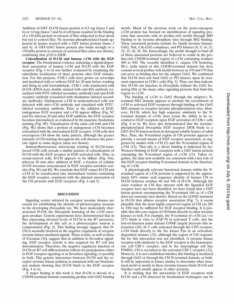
Addition of GST–D-Cbl fusion protein to 0.5 mg (lanes 5 and6) or 1.0 mg (lanes 7 and 8) of cell lysate resulted in the bindingof a 150-kDa protein in extracts of flies subjected to heat shockbut not to control flies. No tyrosine-phosphorylated bands ap-peared in lysates reacted with a GST fusion protein (lanes 3and 4). A GST-Grb2 fusion protein also binds strongly to a150-kDa protein in extracts of activated flies (data not shown),confirming that p150 is DER.Colocalization of D-Cbl and human c-Cbl with the EGF
receptor. The biochemical evidence indicating a ligand-depen-dent association of human c-Cbl and D-Cbl with the EGFreceptor (Fig. 4 to 6) was further evaluated by examining thesubcellular localization of these proteins after EGF stimula-tion. For this purpose, COS-1 cells were grown on coverslipsand incubated with or without EGF for 20 min before washingand fixing in cold formaldehyde. COS-1 cells transfected withD-Cbl cDNA were double stained with anti-HA antibody (vi-sualized with FITC-labeled secondary antibody) and anti-EGFreceptor antibody (visualized with rhodamine-labeled second-ary antibody). Endogenous c-Cbl in nontransfected cells wasdetected with anti-c-Cbl antibody and visualized with FITC-labeled secondary antibody. Prior to the addition of EGF,endogenous EGF receptor and c-Cbl appear diffuse (Fig. 9Cand D), whereas 20 min after EGF addition, the EGF receptorbecomes internalized, as evidenced by the punctate rhodaminestaining (Fig. 9F). Visualization of the same cell with anti-Cbl(Fig. 9E) shows that a portion of endogenous c-Cbl becomescolocalized with the internalized EGF receptor. COS cells thatoverexpress Cbl show the same pattern, although the greaterintensity of Cbl staining throughout the cell obscures the punc-tate signal to some degree (data not shown).Immunofluorescence microscopy staining of D-Cbl-trans-
fected COS cells reveals a similar pattern of colocalization ofD-Cbl with the EGF receptor in EGF-activated cells. Thus, inserum-starved cells, D-Cbl appears to be diffuse (Fig. 9A),whereas 20 min after addition of EGF, a fraction of cellularD-Cbl becomes concentrated in EGF receptor-enriched vesi-cles (Fig. 9G and H). We conclude that EGF causes D-Cbl andc-Cbl to be translocated into internalized vesicles containingthe EGF receptor, consistent with the physical association ofthe Cbl proteins with EGF receptors (Fig. 4 and 5).
DISCUSSION
Signaling events initiated by receptor tyrosine kinases arecrucial for establishing the identity of photoreceptor neuronsin the developing Drosophila eye. We have molecularly char-acterized D-Cbl, the Drosophila homolog of the c-Cbl onco-gene product. Genetic experiments have demonstrated that inflies expressing elevated levels of D-Cbl in the R7 precursor,the development of this cell as a photoreceptor neuron iscompromised (Fig. 2). This finding strongly suggests that D-Cbl is normally involved in the negative regulation of receptortyrosine kinase-mediated signals. These results, as well as thoseof Freeman (10), suggest that in addition to Sevenless signal-ing, EGF receptor activity is also required for R7 cell fatedetermination. Therefore, the negative regulatory function ofD-Cbl on R7 cell differentiation reflects its participation eitherin EGF receptor signaling or in Sevenless signaling, or possiblyin both. This genetic interaction between D-Cbl and the re-ceptor tyrosine kinase pathway is consistent with our biochem-ical analysis showing that D-Cbl binds to the EGF receptor(Fig. 4 to 6).A major finding in this work is that D-Cbl is devoid of a
COOH-terminal domain containing proline-rich Grb2 binding
motifs. Much of the previous work on the proto-oncogenec-Cbl protein has focused on identification of signaling pro-teins that associate with its proline-rich motifs through SH3binding or its tyrosine phosphate sites through SH2 binding.These associated proteins include Src family tyrosine kinases,Grb2, Nck, Crk-G3G complexes, and PI3 kinases (8, 9, 14, 21,23, 25, 26, 28, 30). Interestingly, the motifs thought to bind allof these associated proteins are believed to reside in the pro-line-rich COOH-terminal region of c-Cbl containing residues480 to 850. The recently identified C. elegans Cbl homolog,Sli-1, lacks much of this COOH-terminal domain but doescontain several proline-rich motifs between aa 470 and 570 thatcan serve as binding sites for the adapter Grb2. We confirmedthat D-Cbl does not bind Grb2 or PI3 kinases upon its tran-sient expression in COS-1 cells (Fig. 3). Thus, our data indicatethat D-Cbl can function in Drosophila without the Grb2 ho-molog Drk or the many other signaling proteins that bind thisregion in c-Cbl.The binding of c-Cbl to Grb2 through the adapter’s N-
terminal SH3 domain appears to mediate the recruitment ofc-Cbl to activated EGF receptors through binding of the Grb2SH2 domain to receptor tyrosine phosphorylation sites. How-ever, D-Cbl, which has high sequence similarity to the N-terminal domain of c-Cbl, does retain the ability to be re-cruited to EGF receptors upon EGF activation of COS-1 cells(Fig. 4 to 6). We have also demonstrated that the tyrosine-phosphorylated Drosophila EGF receptor DER binds to aGST–D-Cbl fusion protein in detergent-soluble lysates of adultflies. Thus, the N-terminal region of Cbl proteins appears toprovide a second means of EGF receptor association, as sug-gested by studies with v-Cbl (5) and the N-terminal region ofc-Cbl (13). That this is a direct binding is indicated by far-Western blotting of EGF receptors with a GST fusion proteincontaining the N-terminal region of c-Cbl (13). Taken to-gether, the data now available are consistent with a key role ofthis EGF receptor-binding N-terminal domain in the function-ing of c-Cbl.A physiologically relevant functional domain within the N-
terminal region of c-Cbl proteins is supported by the approx-imate 68% amino acid sequence identity of human Cbl toD-Cbl between residues 40 and 426 of D-Cbl. Although theexact residues in Cbl that interact with the liganded EGFreceptor have not been identified, we have found that a GSTfusion protein encompassing the N-terminal 160 aa of c-Cbldoes not associate (not shown). Based on the G305E mutationin D-Cbl that ablates receptor association (Fig. 7), it seemsplausible that the most highly conserved region of Cbl (aa 205to 330) may be sufficient for EGF receptor binding. It is pos-sible that this core region of Cbl binds directly to other tyrosinekinases as well. For example, the N terminus of c-Cbl (aa 1 to357) binds in vitro to ZAP-70 in activated T cells, and theloss-of-function point mutant G306E largely prevents this in-teraction (20). In T cells activated through the CD3 receptor,c-Cbl binds directly to the Src kinase Fyn in an activation-dependent manner (33), although the region of Cbl responsi-ble for this interaction was not identified. A tyrosine kinasereceptor with similarity to the EGF receptor is the hematopoi-etic cell CSF-1 receptor, and in the macrophage cell lineP388D1, Cbl is recruited to the activated CSF-1 receptor (32).However, it is not established whether this binding is mediatedthrough Grb2 or through the Cbl N-terminal domain, or both.It will be important in future studies to determine what struc-tural motif or motifs in these tyrosine kinases binds D-Cbl andwhether such motifs appear in other proteins.It is striking that the association of EGF receptors with
D-Cbl and c-Cbl observed by biochemical techniques can be
2222 MEISNER ET AL. MOL. CELL. BIOL.
on July 8, 2014 by guesthttp://m
cb.asm.org/
Dow
nloaded from
FIG. 9. Colocalization of D-Cbl and c-Cbl with internalized EGF receptors following activation with EGF. Cells were serum starved for 1 h and stimulated for 20min. with EGF. After fixing and permeabilizing, cells were stained with sheep anti-EGF receptor (anti-EGFR) and donkey anti-sheep–rhodamine. Human Cbl wasvisualized with rabbit anti-Cbl plus donkey anti-rabbit–FITC; D-Cbl was visualized with rabbit anti-HA plus donkey anti-rabbit–FITC. (C to F) Nontransfected COS-1cells; (A, B, G, and H) HA-DCbl-transfected cells.
2223
on July 8, 2014 by guesthttp://m
cb.asm.org/
Dow
nloaded from

visualized by immunofluorescence microscopy of intact COS-1cells (Fig. 9). Colocalization of Cbl proteins and EGF recep-tors can be observed as early as a few minutes after addition ofEGF to these cells (not shown), and by 20 min of incubation,an intense punctate pattern of Cbl and the EGF receptorproteins is evident. It is noteworthy that these images can bevisualized with endogenous EGF receptors in the COS-1 cells.Our data are consistent with the hypothesis that Cbl-EGFreceptor complexes are internalized into endosomes throughthe coated pit pathway, as previously described for EGF re-ceptors (31). Previously, Tanaka et al. (32) demonstrated byimmunofluorescence that Cbl is translocated to a perinuclearregion after Fcg receptor cross-linking or EGF stimulation.The continued association of D-Cbl and c-Cbl proteins withEGF receptors traffiking within intracellular membranes cor-relates with the known retention of phosphorylated tyrosineresidues and active tyrosine kinase activity in the intracellularEGF receptors (15, 16, 31). It seems likely from these obser-vations that modulation of EGF function by Cbl proteins in-volves the transient association with both cell surface and in-tracellular EGF receptors. In this context, it is intriguing thatmutations in the sli-1 gene, encoding the C. elegans homolog ofCbl, have been shown to interact genetically with unc-101 inthe regulation of the EGF receptor pathway leading to vulvaldifferentiation (18). unc-101 encodes a clathrin-associated pro-tein homologous to mouse AP47, which constitutes one of themain components of coated pits and vesicles. Although sli-1mutations alone are silent, in combination with unc-101 muta-tions, they cause excessive vulval differentiation. These geneproducts, thus, appear to cooperate synergistically in the neg-ative control of EGF receptor signaling. Part of the mechanismwhereby Cbl proteins exert an attenuation on receptor tyrosinekinase signaling might involve the downregulation of receptoractivity by directing the intracellular degradation of the recep-tor-Cbl complexes.
ACKNOWLEDGMENTS
The expertise of Doug Bowman with the digital imaging microscopeand the help of Andrew Blane with transfections are gratefully appre-ciated. We thank Paul Sternberg for communicating unpublished re-sults and for helpful advice and discussions.This work was supported by Juvenile Diabetes Foundation Interna-
tional Program Project grant 892004 (M.P.C. and H.M.) and by Na-tional Institutes of Health grant RO1EY08152-06 (to U.B.).
REFERENCES
1. Andoniou, C., C. Thien, and W. Langdon. 1994. Tumour induction by acti-vated abl involves tyrosine phosphorylation of the product of the cbl onco-gene. EMBO J. 13:4515–4523.
2. Basler, K., B. Christen, and E. Hafen. 1991. Ligand-independent activationof the sevenless receptor tyrosine kinase changes the fate of cells in thedeveloping Drosophila eye. Cell 64:1069–1081.
3. Blake, T., K. Heath, and W. Langdon. 1993. The truncation that generatedthe v-cbl oncogene reveals an ability for nuclear transport, DNA binding andacute transformation. EMBO J. 12:2017–2026.
4. Blake, T., M. Shapiro, H. Morse, and W. Langdon. 1991. The sequences ofthe human and mouse c-cbl proto-oncogenes show v-cbl was generated by alarge truncation encompassing a proline-rich domain and a leucine zipper-like motif. Oncogene 6:653–657.
5. Bowtell, D., and W. Langdon. 1995. The protein product of the c-cbl onco-gene rapidly complexes with the EGF receptor and is tyrosine phosphory-lated following EGF stimulation. Oncogene 11:1561–1567.
6. Buday, L., A. Khwaja, S. Sipeki, A. Farago, and J. Downward. 1996. Inter-actions of Cbl with two adaptor proteins, Grb2 and Crk, upon T cell activa-tion. J. Biol. Chem. 271:6159–6163.
7. Daga, A., and U. Banerjee. 1994. Resolving the Sevenless pathway usingsensitized genetic backgrounds. Cell. Mol. Biol. Res. 40:245–251.
8. deJong, R., J. ten Hoeve, N. Heisterkamp, and J. Groffen. 1995. Crk1 is
complexed with tyrosine-phosphorylated Cbl in Ph-positive leukemia. J. Biol.Chem. 270:21468–21471.
9. Donovan, J., R. Wange, W. Langdon, and L. Samelson. 1994. The proteinproduct of the c-cbl protooncogene is the 120-kDa tyrosine-phosphorylatedprotein in Jurkat cells activated via the T cell antigen receptor. J. Biol. Chem.169:22921–22924.
10. Freeman, R. 1996. Reiterative use of the EGF receptor triggers differenti-ation of all cell types in the Drosophila eye. Cell 87:651–660.
11. Fukazawa, T., S. Miyake, V. Band, and H. Band. 1996. Tyrosine phosphor-ylation of Cbl upon epidermal growth factor (EGF) stimulation and itsassociation with EGF receptor and downstream signaling proteins. J. Biol.Chem. 271:14554–14559.
12. Fukazawa, T., K. Reedquist, T. Trub, S. Soltoff, G. Panchamoorthy, B.Druker, L. Cantley, S. Shoelson, and H. Band. 1995. The SH3 domain-binding T cell tyrosyl phosphoprotein p120. Demonstration of its identitywith the c-cbl protooncogene product and in vivo complexes with Fyn, Grb2,and phosphatidylinositol 3-kinase. J. Biol. Chem. 270:19141–19150.
13. Galisteo, M., I. Dikic, A. Batzer, W. Langdon, and J. Schlessinger. 1995.Tyrosine phosphorylation of the c-cbl proto-oncogene product and associa-tion with epidermal growth factor (EGF) receptor upon EGF stimulation.J. Biol. Chem. 270:20242–20245.
14. Hartley, D., H. Meisner, and S. Corvera. 1995. Specific association of the bisoform of the p85 subunit of phosphatidylinositol-3-kinase with the pro-tooncogene c-cbl. J. Biol. Chem. 270:18260–18263.
15. Honegger, A., T. Dull, S. Felder, E. van Obberghen, F. Bellot, D. Szapary, A.Schmidt, A. Ullrich, and J. Schlessinger. 1987. Point mutation at the ATPbinding site of EGF receptor abolishes protein-tyrosine kinase activity andalters cellular routing. Cell 51:199–209.
16. Lai, W., P. Cameron, J. Doherty, B. Posner, and J. Bergeron. 1989. Ligand-mediated autophosphorylation activity of the epidermal growth factor re-ceptor during internalization. J. Cell Biol. 109:2751–2760.
17. Langdon, W., J. Hartley, S. Klinken, S. Ruscetti, and H. Morse. 1989. v-cbl,an oncogene from a dual recombinant murine retrovirus that induces earlyB-lineage lymphomas. Proc. Natl. Acad. Sci. USA 91:9665–9669.
18. Lee, J., G. D. Jongeward, and P. Sternberg. 1994. unc-101 a gene requiredfor many aspects of C. elegans development and behavior, encodes a clathrin-associated protein. Genes Dev. 8:60–73.
19. Levkowitz, G., L. Klapper, E. Tzahar, A. Freywald, M. Sela, and Y. Yarden.1996. Coupling of the c-Cbl protooncogene product to ErbB-1/EGF-recep-tor but not to other ErbB proteins. Oncogene 12:1117–1127.
20. Lupher, M., K. Reedquist, S. Miyake, W. Langdon, and H. Band. 1996. Anovel phosphotyrosine-binding domain in the N-terminal transforming re-gion of Cbl interacts directly and selectively with ZAP-70 in T cells. J. Biol.Chem. 271:24063–24068.
21. Meisner, H., B. Conway, D. Hartley, and M. P. Czech. 1995. Interactions ofCbl with Grb2 and phosphatidylinositol 39-kinase in activated Jurkat cells.Mol. Cell. Biol. 15:3571–3578.
22. Meisner, H., and M. P. Czech. 1995. Coupling of the proto-oncogene prod-uct of c-Cbl to the EGF receptor. J. Biol. Chem. 270:25332–25335.
23. Odai, H., K. Sasaki, A. Iwamatsu, Y. Hanazono, T. Tanaka, K. Mitani, Y.Yazaki, and H. Hirai. 1995. The proto-oncogene product c-Cbl becomestyrosine phosphorylated by stimulation with GM-CSF1 or Epo and consti-tutively binds to the SH3 domain of Grb2/Ash in human hematopoietic cells.J. Biol. Chem. 270:10800–10805.
24. Olivier, J., T. Taabe, M. Henkemeyer, B. Dickson, G. Mbamalu, B. Margolis,J. Schlessinger, E. Hafen, and T. Pawson. 1993. A Drosophila SH2-SH3adaptor protein implicated in coupling the sevenless tyrosine kinase to anactivator of Ras guanine nucleotide exchange, Sos. Cell 73:179–191.
25. Panchamoorthy, G., T. Fukazawa, S. Miyake, S. Soltoff, K. Reedquist, B.Druker, S. Shoelson, L. Cantley, and H. Band. 1996. p120cbl is a majorsubstrate of tyrosine phosphorylation upon B cell antigen receptor stimula-tion and interacts in vivo with Fyn and Syk tyrosine kinases, Grb2 and Shcadapters, and the p85 subunit of phosphatidylinositol 3-kinase. J. Biol.Chem. 271:3187–3194.
26. Reedquist, K., T. Fukazawa, B. Druker, G. Panchamoorthy, S. Shoelson, andH. Band. 1994. Rapid T-cell receptor-mediated tyrosine phosphorylation ofp120, an Fyn/Lck Src homology 3 domain-binding protein. Proc. Natl. Acad.Sci. USA 91:4135–4139.
27. Reichman-Fried, M., B. Dickson, E. Hafen, and B.-Z. Shilo. 1994. Elucida-tion of the role of breathless, a Drosophila FGF receptor homolog, intracheal cell migration. Genes Dev. 8:428–439.
28. Rivero-Lezcano, O., J. Sameshima, A. Marcilla, and K. Robbins. 1994.Physical association between src homology 3 elements and the protein prod-uct of the c-cbl proto-oncogene. J. Biol. Chem. 269:17363–17366.
29. Rogge, R., R. Cagan, A. Majumdar, T. Dulaney, and U. Banerjee. 1992.Neuronal development in the Drosophila retina: the sextra gene defines aninhibitory component in the developmental pathway of R7 photoreceptorcells. Proc. Natl. Acad. Sci. USA 89:5271–5275.
30. Soltoff, S., and L. Cantley. 1996. p120cbl is a cytosolic adapter protein thatassociates with phosphoinositide 3-kinase in response to epidermal growthfactor in PC12 and other cells. J. Biol. Chem. 271:563–567.
2224 MEISNER ET AL. MOL. CELL. BIOL.
on July 8, 2014 by guesthttp://m
cb.asm.org/
Dow
nloaded from
31. Sorkin, A., and C. Waters. 1993. Endocytosis of growth factor receptors.Bioessays 15:375–382.
32. Tanaka, L. Neff, R. Baron, and J. Levy. 1995. Tyrosine phosphorylation andtranslocation of the c-Cbl protein after activation of tyrosine kinase signalingpathways. J. Biol. Chem. 270:14347–14351.
33. Tsygankov, A., S. Mahajan, J. Fincke, and J. Bolen. 1996. Specific associa-tion of tyrosine-phosphorylated c-Cbl with Fyn tyrosine kinase in T cells.J. Biol. Chem. 271:27130–27137.
34. Tyers, M., G. Tokiwa, R. Nash, and B. Futcher. 1992. The Cln3-Cdc28 kinasecomplex of S. cerevisiae is regulated by proteolysis and phosphorylation.EMBO J. 11:1773–1784.
35. Yoon, C., J. Lee, G. Jongeward, and P. Sternberg. 1995. Similarity of sli-1, aregulator of vulval development in C. elegans, to the mammalian proto-oncogene c-cbl. Science 269:1102–1105.
36. Zipursky, S. L., and G. M. Rubin. 1994. Determination of neuronal cell fate:lessons from the R7 neuron of Drosophila. Annu. Rev. Neurosci. 17:373–397.
VOL. 17, 1997 INTERACTIONS OF DROSOPHILA Cbl WITH EGF RECEPTORS 2225
on July 8, 2014 by guesthttp://m
cb.asm.org/
Dow
nloaded from
Copyright © 2022 FDOKUMEN