Calibration and Verification Test of Lily Bulb Simulation ... - MDPI
ARTICLES
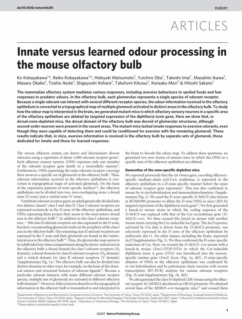
Innate versus learned odour processing inthe mouse olfactory bulbKo Kobayakawa1*, Reiko Kobayakawa1*, Hideyuki Matsumoto2, Yuichiro Oka1, Takeshi Imai1, Masahito Ikawa3,Masaru Okabe3, Toshio Ikeda4, Shigeyoshi Itohara4, Takefumi Kikusui5, Kensaku Mori2 & Hitoshi Sakano1
The mammalian olfactory system mediates various responses, including aversive behaviours to spoiled foods and fearresponses to predator odours. In the olfactory bulb, each glomerulus represents a single species of odorant receptor.Because a single odorant can interact with several different receptor species, the odour information received in the olfactoryepithelium is converted to a topographical map of multiple glomeruli activated in distinct areas in the olfactory bulb. To studyhow the odour map is interpreted in the brain, we generated mutant mice in which olfactory sensory neurons in a specific areaof the olfactory epithelium are ablated by targeted expression of the diphtheria toxin gene. Here we show that, indorsal-zone-depleted mice, the dorsal domain of the olfactory bulb was devoid of glomerular structures, althoughsecond-order neurons were present in the vacant areas. The mutant mice lacked innate responses to aversive odorants, eventhough they were capable of detecting them and could be conditioned for aversion with the remaining glomeruli. Theseresults indicate that, in mice, aversive information is received in the olfactory bulb by separate sets of glomeruli, thosededicated for innate and those for learned responses.
The mouse olfactory system can detect and discriminate diverseodorants using a repertoire of about 1,000 odorant receptor genes1.Each olfactory sensory neuron (OSN) expresses only one memberof the odorant receptor gene family in a monoallelic manner2.Furthermore, OSNs expressing the same odorant receptor convergetheir axons to a specific set of glomeruli in the olfactory bulb3. Thus,odorous information received in the olfactory epithelium is con-verted to topographical maps of activated glomeruli. On the basisof the expression patterns of zone-specific markers4,5, the olfactoryepithelium can be divided into two, non-overlapping areas: a dorsalzone (D zone) and a ventral zone (V zone).
Vertebrate odorant receptor genes are phylogenetically divided intotwo distinct classes6: class I and class II. Class I odorant receptors areexpressed exclusively in the D zone of the olfactory epithelium, andOSNs expressing them project their axons to the most antero-dorsalarea in the olfactory bulb7,8. In addition to the class I odorant recep-tors, ,300 class II odorant receptors are also expressed in the D zone,but their corresponding glomeruli reside on the periphery of the class Iarea in the olfactory bulb. The remaining class II odorant receptors areexpressed in the V zone and their glomeruli are found in the ventro-lateral area in the olfactory bulb4,9. Thus, the glomerular map seems tobe subdivided into three compartments along the dorso-ventral axis inthe olfactory bulb: a dorsal domain for class I odorant receptors (DI
domain), a dorsal domain for class II odorant receptors (DII domain)and a ventral domain for class II odorant receptors (V domain)(Supplementary Fig. 1a). The olfactory bulb can also be divided intodistinct domains in other ways, for example, on the basis of the chem-ical natures and structural features of odorous ligands10. Because aparticular odorant interacts with many different odorant receptorspecies, multiple sets of glomeruli are activated in different olfactorybulb domains11. However, little is known about how the topographicalinformation in the olfactory bulb is transmitted to and interpreted in
the brain to decode the odour map. To address these questions, wegenerated two new strains of mutant mice in which the OSNs in aspecific area of the olfactory epithelium are ablated.
Generation of the zone-specific depletion mice
We reported previously that the rat Omacs gene, encoding olfactory-specific medium-chain acyl-CoA synthetase, is expressed in theolfactory epithelium in a D-zone-specific manner before the onsetof odorant receptor gene expression5. This was also confirmed inmouse by in situ hybridization and immunohistochemistry (Supple-mentary Fig. 2). We used the D-zone-specific O-MACS (also knownas BC048390) promoter to ablate the D zone OSNs in mice (DD) bytargeted expression of the diphtheria toxin gene12. We first generateda knock-in mouse strain in which the coding sequence of theO-MACS was replaced with that of the Cre-recombinase gene (O-MACSRcre). We then crossed this knock-in mouse with anothermouse strain carrying the Cre-inducible lacZ gene. The lacZ reporter,activated by Cre that is driven from the O-MACS promoter, wasselectively expressed in the D zone of the olfactory epithelium byembryonic day 11. No other tissues, including the brain, expressedlacZ (Supplementary Fig. 3). We thus confirmed the D-zone-specificinduction of Cre. Next, we crossed the O-MACSRcre mouse with aknock-in mouse (Eno2-STOP-DTA) in which the Cre-induciblediphtheria toxin A gene (DTA) was introduced into the neuron-specific enolase gene (Eno2) locus (Fig. 1a, DD). D-zone-specificablation of OSNs in the olfactory epithelium was confirmed byin situ hybridization and by polymerase chain reaction with reversetranscription (RT–PCR) analyses for various odorant receptors(Fig. 1b and Supplementary Fig. 1b, DD).
We also generated the class-II-depleted (DII) mouse using the olfact-ory receptor 16 (MOR23, also known as Olfr16) promoter. We obtainedseveral lines of the MOR23Rcre transgenic mice13 and crossed them
*These authors contributed equally to this work.
1Department of Biophysics and Biochemistry, Graduate School of Science, The University of Tokyo, Tokyo 113-0032, Japan. 2Department of Physiology, Graduate School of Medicine,The University of Tokyo, Tokyo 113-0033, Japan. 3Research Institute for Microbial Diseases, The Osaka University, Osaka 565-0871, Japan. 4Laboratory for Behavioral Genetics, BrainScience Institute, RIKEN, Saitama 351-0198, Japan. 5Laboratory of Veterinary Ethology, The University of Tokyo, Tokyo 113-8657, Japan.
doi:10.1038/nature06281
1Nature ©2007 Publishing Group

with the Eno2-STOP-DTA mouse to ablate OSNs under the control ofthe MOR23 promoter (Fig. 1a,DII). With one particular transgenic line,the depletion unexpectedly occurred throughout the entire class IIregion of the olfactory epithelium (Fig. 1b and Supplementary Fig.1b, DII). In situ hybridization and RT–PCR analysis demonstrated thatnone of the class I odorant receptor genes was affected in this mouse(Fig. 1b and Supplementary Fig. 1b,DII), with the exception of two classI genes8 expressed in the V zone (data not shown).
Arrangement of glomeruli in the olfactory bulb
We analysed the formation and arrangement of glomeruli in themutant mice by immunohistochemistry (Fig. 1c and SupplementaryFig. 4). In the DD mouse, glomeruli were found only in the postero-ventral region of the olfactory bulb (V domain), leaving the antero-dorsal region (D domain) vacant (Fig. 1c and Supplementary Fig. 4,DD). This was also confirmed by imaging of intrinsic signals in theolfactory bulb. In contrast to the wild-type mouse, no odour-evokedactivities were detected on the dorsal surface of the olfactory bulb inthe DD mouse (n 5 3) (Supplementary Figs 5a, 6 and 7). Despite thecomplete absence of glomeruli, the D domain of the mutant olfactorybulb showed otherwise normal cytoarchitecture with distinct layers(Supplementary Fig. 8a). Individual mitral cells in the D domain of themutant olfactory bulb emitted several dendrites into the externalplexiform layer, but did not form dendritic terminal tufts, suggesting
that these mitral cells lacked synaptic inputs from OSN axons(Supplementary Fig. 8b, c). V-zone OSN axons did not form ectopiccontacts with the D-domain mitral cells in the DD olfactory bulb. Noglomeruli were found in the D domain in the DD mouse during thecourse of embryonic development (data not shown). These resultsindicate that mitral cells are specified as D-domain and V-domainsubsets before the projection of OSN axons occurs. The D-domainmitral cells seem to be committed to receive olfactory inputs exclu-sively from D-zone OSNs.
We then analysed theDII mouse. In contrast to theDD mutant, theDII mouse had glomeruli only in the most antero-dorsal part ofthe D domain (Fig. 1c and Supplementary Fig. 4, DII). To determinethe boundary of class I and class II areas in the wild-type olfactorybulb, a mouse containing the Cre-inducible green fluorescent protein(GFP) gene was crossed with the MOR23Rcre mouse to produce amouse in which class-II-expressing OSNs are labelled with GFP (classII-GFP). In the class II-GFP mouse, the DI-domain glomeruli (nega-tive for GFP) and DII-domain glomeruli (positive for GFP) aresegregated into two distinct areas in the D domain of the olfactorybulb, even though both types of D-zone OSNs are intermingled withinthe D zone of the olfactory epithelium8 (Fig. 1c and SupplementaryFig. 4, wild type). In the DII mouse, glomeruli were found in the DI
domain, and were absent in the class II regions (DII domain and Vdomain) of the olfactory bulb (Fig. 1c and Supplementary Fig. 4,DII).
7-113-219-226-127-130-233-142-1
108-2135-14139-3163-1164-1
171-10174-13179-5180-1223-9224-8255-1265-1
267-13277-1103-1244-1V1RsV2Rs
0 100 200 (%)100500 (%)150
D zone(class I)
D zone(class II)
V zone(class II)
13-219-226-127-130-233-140-1
108-2135-14139-3163-1164-1
171-10174-13179-5180-1223-9224-8255-1265-1
267-13
MOR23
103-1103-15244-2
Expression
V1RsV2RsVNR
P creO-MACS
P DTASTOPloxP loxP
Eno2
P creMOR23
P DTASTOPloxP loxP
Eno2
∆D
∆D
∆II
∆II
∆D ∆II
7-1Expression
a
Syn
/GFP
/OC
AM
b
Wild typec
DI domain
DII domain
V domain
Glomeruli:
D
VM L
Figure 1 | Odorant receptor gene expression andglomerular map formation in the DD and DIImutant mice. a, Plasmid constructs for theD-zone depletion (DD) and the class II depletion(DII) are schematically illustrated. DTA and creare the genes for diphtheria toxin A and Crerecombinase, respectively. b, Expression of theodorant receptor and vomeronasal receptor(VNR) genes. Olfactory epithelium sectionsdissected from the DD, DII and wild-type micewere analysed by in situ hybridization. Thenumber of OSNs expressing a given receptor perunit volume of mutant olfactory epithelium wascompared with that of wild-type control (%). Inthe DD mutant, OSNs expressing the D-zone-specific odorant receptor genes, both class I(green) and class II (blue), were ablated. In theDIImice, OSNs expressing the class II odorantreceptor genes, in the both D (blue) and the V(red) zones, were ablated. Expression of thevomeronasal receptor genes (purple)—V1Rs(V1re8, V1rg1 and V1rj3) and V2Rs (V2r1b, V2r11and V2r15)—was also analysed, and the meanratios are shown. c, Coronal sections of olfactorybulbs. Samples were dissected from the mutantmice (DD and DII) and the wild-type mouse inwhich the class II odorant-receptor-expressingOSNs were labelled with GFP. Olfactory bulbsections were stained with anti-synaptotagmin(green, Syn), anti-GFP (blue) and anti-OCAM(also known as NCAM2; red) antibodies.Distributions of glomeruli are schematicallyshown below the images (DI-, DII- and V-domainglomeruli are illustrated in green, blue and red,respectively). Borders for DI domains (GFP2/OCAM2) and DII domains (GFP1/OCAM2) areindicated by solid lines. D, dorsal; L, lateral; M,medial; V, ventral. Scale bars, 500mm.
ARTICLES NATURE
2Nature ©2007 Publishing Group
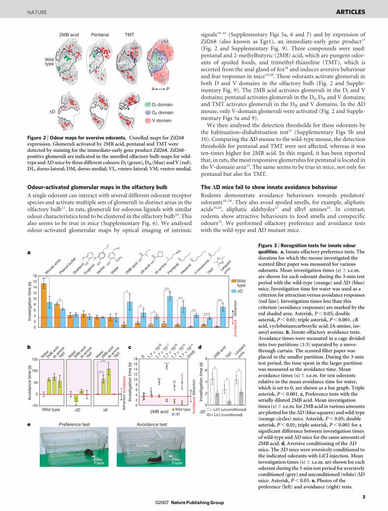
Odour-activated glomerular maps in the olfactory bulb
A single odorant can interact with several different odorant receptorspecies and activate multiple sets of glomeruli in distinct areas in theolfactory bulb11. In rats, glomeruli for odorous ligands with similarodour characteristics tend to be clustered in the olfactory bulb10. Thisalso seems to be true in mice (Supplementary Fig. 6). We analysedodour-activated glomerular maps by optical imaging of intrinsic
signals14–16 (Supplementary Figs 5a, 6 and 7) and by expression ofZif268 (also known as Egr1), an immediate-early gene product17
(Fig. 2 and Supplementary Fig. 9). Three compounds were used:pentanal and 2-methylbutyric (2MB) acid, which are pungent odor-ants of spoiled foods, and trimethyl-thiazoline (TMT), which issecreted from the anal gland of fox18 and induces aversive behaviourand fear responses in mice19,20. These odorants activate glomeruli inboth D and V domains in the olfactory bulb (Fig. 2 and Supple-mentary Fig. 9). The 2MB acid activates glomeruli in the DI and Vdomains; pentanal activates glomeruli in the DI, DII and V domains;and TMT activates glomeruli in the DII and V domains. In the DDmouse, only V-domain glomeruli were activated (Fig. 2 and Supple-mentary Figs 5a and 9).
We then analysed the detection thresholds for these odorants bythe habituation–dishabituation test21 (Supplementary Figs 5b and10). Comparing the DD mouse to the wild-type mouse, the detectionthresholds for pentanal and TMT were not affected, whereas it wasten-times higher for 2MB acid. In this regard, it has been reportedthat, in rats, the most responsive glomerulus for pentanal is located inthe V-domain area22. The same seems to be true in mice, not only forpentanal but also for TMT.
The DD mice fail to show innate avoidance behaviour
Rodents demonstrate avoidance behaviours towards predators’odorants18–20. They also avoid spoiled smells, for example, aliphaticacids19,20, aliphatic aldehydes23 and alkyl amines24. In contrast,rodents show attractive behaviours to food smells and conspecificodours25. We performed olfactory preference and avoidance testswith the wild-type and DD mutant mice.
cb d
Preference test Avoidance test
2MB acid
Inve
stig
atio
n tim
e (s
)
Inve
stig
atio
n tim
e (s
)
0
02468
1012141618 1.
7 × 1
0–8
1.7
× 10–7
1.7
× 10–6
1.7
× 10–5
1.7
× 10–4
∗∗∗
∗∗
(mol)
Att
ract
ion
Avo
idan
ce
∗∗∗
Wild type∆D
∗∗
0
2
4
6
8 2MB ac
id
Leop
ard
urine
TMT
+ LiCl (conditioned)
– LiCl (unconditioned)
∗
∗
∆D
∗
e
Avo
idan
ce t
ime
(s)
Wild type
∗∗∗
∗∗∗W
ater
2MB ac
id
IA-a
mine
TMT
150
100
50
0
–50∆D ∆II A
ttra
ctio
nA
void
ance
Wat
er
2MB ac
id
IA-a
mine
TMT
Wat
er
2MB ac
id
IA-a
mine
TMT
a
Att
ract
ion
Avo
idan
ce
∗∗∗
∗
0
2
4
6
8
10
12
14
16
18W
ater
Mou
se u
rine
Inve
stig
atio
n tim
e (s
)
TMT Le
opar
d
urine
Eugen
ol
IA-a
mine
∗∗∗
∗∗
Penta
nal
2MB ac
id
2-he
xano
ne
Guaiac
ol
cB-a
cid
∗
O
HOHO S
N
Hexan
al
∗∗
NH 2
Peanu
t but
ter
Vanil
in
Wildtype
∗∗
∗∗
∗∗∗∗∗∗
O
O O
OO
OH OH
O
OCH 3O
OH
∆D
Filter Paper
Filter Paper
Curtain
Figure 3 | Recognition tests for innate odourqualities. a, Innate olfactory preference tests. Theduration for which the mouse investigated thescented filter paper was measured for variousodorants. Mean investigation times (s) 6 s.e.m.are shown for each odorant during the 3-min testperiod with the wild-type (orange) and DD (blue)mice. Investigation time for water was used as acriterion for attraction versus avoidance responses(red line). Investigation times less than thiscriterion (avoidance responses) are marked by thered shaded area. Asterisk, P , 0.05; doubleasterisk, P , 0.01; triple asterisk, P , 0.001. cBacid, cyclobutanecarboxylic acid; IA-amine, iso-amyl amine. b, Innate olfactory avoidance tests.Avoidance times were measured in a cage dividedinto two partitions (1:3) separated by a move-through curtain. The scented filter paper wasplaced in the smaller partition. During the 3-mintest period, the time spent in the larger partitionwas measured as the avoidance time. Meanavoidance times (s) 6 s.e.m. for test odorantsrelative to the mean avoidance time for water,which is set to 0, are shown as a bar graph. Tripleasterisk, P , 0.001. c, Preference tests with theserially diluted 2MB acid. Mean investigationtimes (s) 6 s.e.m. for 2MB acid in various amountsare plotted for theDD (blue squares) and wild-type(orange circles) mice. Asterisk, P , 0.05; doubleasterisk, P , 0.01; triple asterisk, P , 0.001 for asignificant difference between investigation timesof wild-type andDD mice for the same amounts of2MB acid. d, Aversive conditioning of the DDmice. The DD mice were aversively conditioned tothe indicated odorants with LiCl injection. Meaninvestigation times (s) 6 s.e.m. are shown for eachodorant during the 3-min test period for aversivelyconditioned (grey) and unconditioned (white)DDmice. Asterisk, P , 0.05. e, Photos of thepreference (left) and avoidance (right) tests.
DI domain
DII domain
V domain
PA
VMVM
DMDM
VLVL
DLDL
VM
DM
VL
DL
TMTPentanal
Wild type
∆D
2MB acid
Figure 2 | Odour maps for aversive odorants. Unrolled maps for Zif268expression. Glomeruli activated by 2MB acid, pentanal and TMT weredetected by staining for the immediate-early gene product Zif268. Zif268-positive glomeruli are indicated in the unrolled olfactory bulb maps for wild-type andDD mice by three different colours: DI (green), DII (blue) and V (red).DL, dorso-lateral; DM, dorso-medial; VL, ventro-lateral; VM, ventro-medial.
NATURE ARTICLES
3Nature ©2007 Publishing Group

In the preference test (Fig. 3a and Supplementary Video), filterpapers scented with various odorants were presented to the wild-typeand DD mutant mice. The lengths of time the mouse spent investi-gating and sniffing the odorants were measured. Investigation timesfor wild-type mice were approximately 4 s for water (neutral odor-ant), within 0.3–1.3 s for acids, aldehydes, amines, ketones and pred-ator odours, and 8–15 s for mouse urine and peanut butter. We usedthe investigation time for water (4 s) as a criterion for avoidance(shorter than 4 s) versus attraction (longer than 4 s) responses. Incontrast to the wild-type, the DD mice spent a much longer time,4–10 s, investigating acids, aldehydes, amines, ketones and predatorodours. The failure of DD mutants to demonstrate avoidance beha-viour was also confirmed by the innate avoidance test (Fig. 3b). Weperformed the innate-olfactory preference test using serially diluted2MB acid (Fig. 3c). Wild-type mice demonstrated avoidance res-ponses at higher concentrations of 2MB acid, but showed neutralbehaviours at lower concentrations. In contrast, DD mice showedattractive behaviours at higher concentrations, and neutral responsesat lower concentrations.
These results indicate thatDD mice are able to detect 2MB acid, butfail to show avoidance behaviour. It should be noted that the mousecan be conditioned for aversive reactions by injection of irritatingLiCl26. We used 2MB acid, TMT and snow leopard urine in the con-ditioning test, and found that the investigation time was significantlyshortened after conditioning (Fig. 3d). It seems that DD mice can beconditioned for aversion, but they lack innately aversive responses.
The D zone is sufficient for innate avoidance behaviour
To examine the discrimination capabilities of the DD mouse, weperformed an olfactory discrimination test27 using four differentpairs of odorants (Fig. 4). In this test, food-restricted animals weretrained to associate either of the two related odorants to sugarrewards. After four days of training, the embedded sugar wasremoved from the test cage, and digging times for the two relatedodorants were compared within the same animal. It was found thatDD mutants were able to discriminate 2MB acid, pentanal and(–)carvone from the competing odorants (cyclobutanecarboxylicacid, hexanal and (1)carvone, respectively). Mutant mice could dis-criminate even subtle differences between enantiomers as well ascould wild-type mice. The wild-type mice could not be trained toassociate TMT to sugar rewards, because the innate avoidance res-ponse was overwhelming. In contrast, the DD mouse easily discrimi-nated TMT from eugenol, because the mutant did not avoid TMT.
To examine whether the D domain is sufficient to cause innateavoidance behaviour, we tested another OSN-ablated mutant, theDIImouse. The DII mouse demonstrated clear avoidance behaviourstowards 2MB acid and iso-amyl amine, both of which activate DI-and V-domain glomeruli (Fig. 3b). Interestingly, theDII mice did notavoid TMT, probably because the D-domain glomeruli for TMT arelocated exclusively in the DII domain, which are absent in the DIImice (Fig. 2 and Supplementary Fig. 5a).
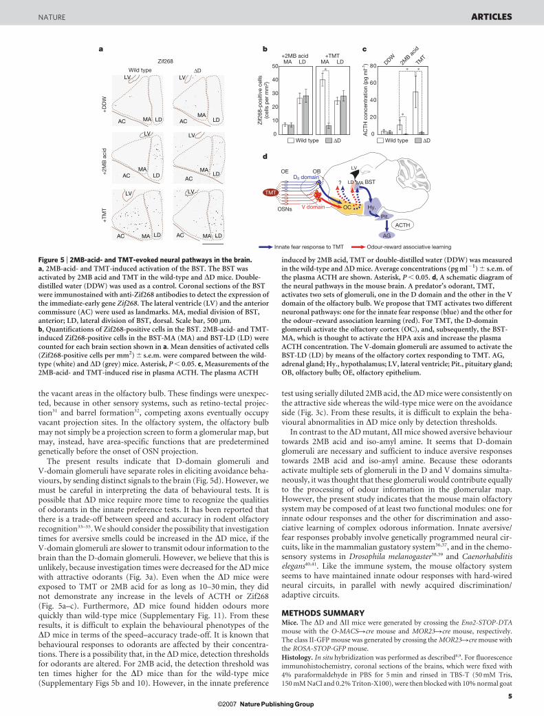
The innate fear pathway is not activated in the DD mouse
TMT activates the hypothalamic-pituitary–adrenal (HPA) axis in therat brain, which has a significant role in regulating the adrenocorti-cotropic hormone (ACTH) level in response to stress18,28. Neurotoxiclesions of the lateral and basal nuclei of the rat amygdala do notprevent the TMT-induced freezing reaction29. Interestingly, musci-mol (an agonist for a neurotransmitter, c-aminobutyric acid A,GABAA) does not block the TMT-induced freezing of rats wheninfused into the amygdala, but does block this reaction whenintroduced into the bed nucleus of the stria terminalis (BST)30.These observations indicate that the BST is involved in the TMT-induced fear processing. Using the immediate-early gene productZif268 as a marker, we examined the TMT-induced activation ofthe BST (Fig. 5a, b). In the wild-type mouse, the BST was stronglyactivated in the medial aspect (BST-MA), and moderately in the
lateral division (BST-LD). This is consistent with the previousobservation that TMT activates the BST-MA, leading to the stimu-lation of the HPA axis in rats28. In contrast to the wild type, the BST-MA was not activated by TMT in the DD mice, although the BST-LDwas activated as in wild-type mice. We also examined 2MB-acid-induced activation of the BST. In contrast to TMT, 2MB acid acti-vated the BST-LD, but not BST-MA, in both wild-type and DD mice(Fig. 5a, b).
The HPA axis in rats, when activated by TMT, increases bloodACTH28. We measured the plasma concentrations of ACTH in bothwild-type and DD mice (Fig. 5c). ACTH concentrations were foundto increase significantly in the wild-type mouse, but not in the DDmutant, when TMT was present. These results indicate that theinnate fear pathway in the DD mouse cannot be activated by TMT.With 2MB acid, the rise of ACTH was very small, if at all, even in thewild-type mouse (Fig. 5c). Although both TMT and 2MB acid causeavoidance responses in the wild-type mouse (Fig. 3a, b), only TMTactivates the BST-MA and elevates plasma ACTH (Fig. 5a–c). Theseresults indicate that fear responses induced by TMT and aversiveresponses to 2MB acid are separately processed in the brain withdifferent neural circuits.
Discussion
In the present study, we generated zone-specific ablation mice, andstudied glomerular map formation and odour perception in thesemutants. When OSNs in specific olfactory epithelium zones wereablated, the corresponding olfactory bulb areas were devoid of glom-erular structures. However, second-order neurons were present in
Dig
ging
tim
e (s
) 80
60
40
20
0
100
80
60
40
20
0
100
80
60
40
20
0
100
80
60
40
20
0
100
O
HO
2MB acid
cB acid
O
HO
∗∗∗ ∗∗∗
∗∗∗
2MB acid cB acid
+ sugar
2MB acid cB acid
O
O
Pentanal
Hexanal
∗∗∗ ∗∗∗
TMT
Eugenol
S
N
O
OH
∗ ∗
(–)carvoneO
O(+)carvone
∗∗∗
∗∗∗
∆D∆D∆D∆D Wild
type
Wild
type
Wild
type
Wild
type
Training (day 1–4)Discrimination test (day 5)
O
HOO
HO O
HO
O
HO
Figure 4 | Discrimination tests with structurally related odorants. Thewild-type and DD mice were trained for four days to associate the reward(sugar grains) with either of the two related odorants. On day five, the sugarreward was removed from the bed, and digging times were measured for eachpair of related odorants. Mean digging times (s) 6 s.e.m. during the 2-mintest period are shown as bar graphs for odorants paired with the sugar reward(red bars), and for unpaired odorants (white bars). Asterisk, P , 0.05;double asterisk, P , 0.01; triple asterisk, P , 0.001. Photos of thediscrimination test are shown underneath.
ARTICLES NATURE
4Nature ©2007 Publishing Group
the vacant areas in the olfactory bulb. These findings were unexpec-ted, because in other sensory systems, such as retino-tectal projec-tion31 and barrel formation32, competing axons eventually occupyvacant projection sites. In the olfactory system, the olfactory bulbmay not simply be a projection screen to form a glomerular map, butmay, instead, have area-specific functions that are predeterminedgenetically before the onset of OSN projection.
The present results indicate that D-domain glomeruli andV-domain glomeruli have separate roles in eliciting avoidance beha-viours, by sending distinct signals to the brain (Fig. 5d). However, wemust be careful in interpreting the data of behavioural tests. It ispossible that DD mice require more time to recognize the qualitiesof odorants in the innate preference tests. It has been reported thatthere is a trade-off between speed and accuracy in rodent olfactoryrecognition33–35. We should consider the possibility that investigationtimes for aversive smells could be increased in the DD mice, if theV-domain glomeruli are slower to transmit odour information to thebrain than the D-domain glomeruli. However, we believe that this isunlikely, because investigation times were decreased for the DD micewith attractive odorants (Fig. 3a). Even when the DD mice wereexposed to TMT or 2MB acid for as long as 10–30 min, they didnot demonstrate any increase in the levels of ACTH or Zif268(Fig. 5a–c). Furthermore, DD mice found hidden odours morequickly than wild-type mice (Supplementary Fig. 11). From theseresults, it is difficult to explain the behavioural phenotypes of theDD mice in terms of the speed–accuracy trade-off. It is known thatbehavioural responses to odorants are affected by their concentra-tions. There is a possibility that, in the DD mice, detection thresholdsfor odorants are altered. For 2MB acid, the detection threshold wasten times higher for the DD mice than for the wild-type mice(Supplementary Figs 5b and 10). However, in the innate preference
test using serially diluted 2MB acid, theDD mice were consistently onthe attractive side whereas the wild-type mice were on the avoidanceside (Fig. 3c). From these results, it is difficult to explain the beha-vioural abnormalities in DD mice only by detection thresholds.
In contrast to the DD mutant, DII mice showed aversive behaviourtowards 2MB acid and iso-amyl amine. It seems that D-domainglomeruli are necessary and sufficient to induce aversive responsestowards 2MB acid and iso-amyl amine. Because these odorantsactivate multiple sets of glomeruli in the D and V domains simulta-neously, it was thought that these glomeruli would contribute equallyto the processing of odour information in the glomerular map.However, the present study indicates that the mouse main olfactorysystem may be composed of at least two functional modules: one forinnate odour responses and the other for discrimination and asso-ciative learning of complex odorous information. Innate aversive/fear responses probably involve genetically programmed neural cir-cuits, like in the mammalian gustatory system36,37, and in the chemo-sensory systems in Drosophila melanogaster38,39 and Caenorhabditiselegans40,41. Like the immune system, the mouse olfactory systemseems to have maintained innate odour responses with hard-wiredneural circuits, in parallel with newly acquired discrimination/adaptive circuits.
METHODS SUMMARYMice. The DD and DII mice were generated by crossing the Eno2-STOP-DTA
mouse with the O-MACSRcre mouse and MOR23Rcre mouse, respectively.The class II-GFP mouse was generated by crossing the MOR23Rcre mouse with
the ROSA-STOP-GFP mouse.
Histology. In situ hybridization was performed as described8,9. For fluorescence
immunohistochemistry, coronal sections of the brains, which were fixed with
4% paraformaldehyde in PBS for 5 min and rinsed in TBS-T (50 mM Tris,
150 mM NaCl and 0.2% Triton-X100), were then blocked with 10% normal goat
d
Odour-reward associative learningInnate fear response to TMT
?
OC Hy.
BST
Pit.
ACTH
AG
LV
MALD
OB
OSNs
OE
TMT
a
AC
TH c
once
ntra
tion
(pg
ml–1
)
0
20
40
60
80
c
V domain V domain V domain
DII domain DII domain DII domain
b
Wild type
Zif2
68-p
ositi
ve c
ells
(cel
ls p
er m
m2 )
0
10
20
30
40
50 ∗MA LD
+TMT
AC
LV
LDMA
AC
LV
LDMA
Wild type ∆D
∆D Wild type ∆D
LV
AC LDMA AC
LV
LDMA
+TM
T+
DD
W
Zif268
LV
AC
MALD
LV
MALDAC+
2MB
aci
d
MA LD+2MB acid
∗
∗
2MB ac
id
TMT
DDW
∗
Figure 5 | 2MB-acid- and TMT-evoked neural pathways in the brain.a, 2MB-acid- and TMT-induced activation of the BST. The BST wasactivated by 2MB acid and TMT in the wild-type and DD mice. Double-distilled water (DDW) was used as a control. Coronal sections of the BSTwere immunostained with anti-Zif268 antibodies to detect the expression ofthe immediate-early gene Zif268. The lateral ventricle (LV) and the anteriorcommissure (AC) were used as landmarks. MA, medial division of BST,anterior; LD, lateral division of BST, dorsal. Scale bar, 500mm.b, Quantifications of Zif268-positive cells in the BST. 2MB-acid- and TMT-induced Zif268-positive cells in the BST-MA (MA) and BST-LD (LD) werecounted for each brain section shown in a. Mean densities of activated cells(Zif268-positive cells per mm2) 6 s.e.m. were compared between the wild-type (white) and DD (grey) mice. Asterisk, P , 0.05. c, Measurements of the2MB-acid- and TMT-induced rise in plasma ACTH. The plasma ACTH
induced by 2MB acid, TMT or double-distilled water (DDW) was measuredin the wild-type and DD mice. Average concentrations (pg ml21) 6 s.e.m. ofthe plasma ACTH are shown. Asterisk, P , 0.05. d, A schematic diagram ofthe neural pathways in the mouse brain. A predator’s odorant, TMT,activates two sets of glomeruli, one in the D domain and the other in the Vdomain of the olfactory bulb. We propose that TMT activates two differentneuronal pathways: one for the innate fear response (blue) and the other forthe odour–reward association learning (red). For TMT, the D-domainglomeruli activate the olfactory cortex (OC), and, subsequently, the BST-MA, which is thought to activate the HPA axis and increase the plasmaACTH concentration. The V-domain glomeruli are assumed to activate theBST-LD (LD) by means of the olfactory cortex responding to TMT. AG,adrenal gland; Hy., hypothalamus; LV, lateral ventricle; Pit., pituitary gland;OB, olfactory bulb; OE, olfactory epithelium.
NATURE ARTICLES
5Nature ©2007 Publishing Group

serum in TBS-T, and incubated with primary antibodies at 23 uC for 12–16 h.Sections were washed in TBS-T, and incubated with secondary antibodies.
Mapping odour-evoked activities. Adult male mice were habituated to the cage
for 2 h, and then filter paper scented with test odorants was presented for 30 min.
After 1 h, mice were killed and perfused with 4% paraformaldehyde in PBS.
Zif268 mapping was performed as described with minor modifications17.
Innate olfactory preference test. Adult DD and littermate mice (n 5 110 for
each genotype, and n $ 6 for each test) were used in the test. To avoid confound-
ing of data owing to learning, mice were used only once.
After habituating to the experimental environment, mice were transferred to the
test cage, and a filter paper scented with a test odorant was introduced. Investi-
gation times for the filter paper during the 3-min test period were measured.
Innate olfactory avoidance test. The DD, DII and wild-type littermates (adult
male) (n 5 26 for each genotype, and n $ 6 for each test) were used in the test.
The test cage was divided into two compartments (1:3) with a partition curtain.
A filter paper scented with test odorant was introduced into the smaller com-
partment. Times (seconds) spent in the larger compartment were measured.
Full Methods and any associated references are available in the online version ofthe paper at www.nature.com/nature.
Received 3 August; accepted 14 September 2007.Published online 7 November 2007.
1. Buck, L. & Axel, R. A novel multigene family may encode odorant receptors: amolecular basis for odor recognition. Cell 65, 175–187 (1991).
2. Serizawa, S., Miyamichi, K. & Sakano, H. One neuron–one receptor rule in themouse olfactory system. Trends Genet. 20, 648–653 (2004).
3. Mombaerts, P. et al. Visualizing an olfactory sensory map. Cell 87, 675–686 (1996).4. Yoshihara, Y. et al. OCAM: a new member of the neural cell adhesion molecule
family related to zone-to-zone projection of olfactory and vomeronasal axons.J. Neurosci. 17, 5830–5842 (1997).
5. Oka, Y. et al. O-MACS, a novel member of the medium-chain acyl-CoAsynthetase family, specifically expressed in the olfactory epithelium in a zone-specific manner. Eur. J. Biochem. 270, 1995–2004 (2003).
6. Zhang, X. & Firestein, S. The olfactory receptor gene superfamily of the mouse.Nature Neurosci. 5, 124–133 (2002).
7. Zhang, X. et al. High-throughput microarray detection of olfactory receptor geneexpression in the mouse. Proc. Natl Acad. Sci. USA 101, 14168–14173 (2004).
8. Tsuboi, A., Miyazaki, T., Imai, T. & Sakano, H. Olfactory sensory neuronsexpressing class I odorant receptors converge their axons on an antero-dorsaldomain of the olfactory bulb in the mouse. Eur. J. Neurosci. 23, 1436–1444 (2006).
9. Miyamichi, K., Serizawa, S., Kimura, H. M. & Sakano, H. Continuous andoverlapping expression domains of odorant receptor genes in the olfactoryepithelium determine the dorsal/ventral positioning of glomeruli in the olfactorybulb. J. Neurosci. 25, 3586–3592 (2005).
10. Mori, K., Takahashi, Y. K., Igarashi, K. M. & Yamaguchi, M. Maps of odorantmolecular features in the Mammalian olfactory bulb. Physiol. Rev. 86, 409–433(2006).
11. Malnic, B., Hirono, J., Sato, T. & Buck, L. B. Combinatorial receptor codes for odors.Cell 96, 713–723 (1999).
12. Gogos, J. A., Osborne, J., Nemes, A., Mendelsohn, M. & Axel, R. Genetic ablationand restoration of the olfactory topographic map. Cell 103, 609–620 (2000).
13. Imai, T., Suzuki, M. & Sakano, H. Odorant receptor-derived cAMP signals directaxonal targeting. Science 314, 657–661 (2006).
14. Rubin, B. D. & Katz, L. C. Optical imaging of odorant representations in themammalian olfactory bulb. Neuron 23, 499–511 (1999).
15. Uchida, N., Takahashi, Y. K., Tanifuji, M. & Mori, K. Odor maps in the mammalianolfactory bulb: domain organization and odorant structural features. NatureNeurosci. 3, 1035–1043 (2000).
16. Gurden, H., Uchida, N. & Mainen, Z. F. Sensory-evoked intrinsic optical signals inthe olfactory bulb are coupled to glutamate release and uptake. Neuron 52,335–345 (2006).
17. Inaki, K., Takahashi, Y. K., Nagayama, S. & Mori, K. Molecular-feature domainswith posterodorsal–anteroventral polarity in the symmetrical sensory maps ofthe mouse olfactory bulb: mapping of odourant-induced Zif268 expression. Eur. J.Neurosci. 15, 1563–1574 (2002).
18. Vernet-Maury, E., Polak, E. H. & Demael, A. Structure–activity relationship ofstress-inducing odorants in the rat. J. Chem. Ecol. 10, 1007–1018 (1984).
19. Hebb, A. L. et al. Odor-induced variation in anxiety-like behavior in mice isassociated with discrete and differential effects on mesocorticolimbiccholecystokinin mRNA expression. Neuropsychopharmacology 27, 744–755 (2002).
20. Hebb, A. L. et al. Brief exposure to predator odor and resultant anxiety enhancesmesocorticolimbic activity and enkephalin expression in CD-1 mice. Eur. J.Neurosci. 20, 2415–2429 (2004).
21. Ferguson, J. N. et al. Social amnesia in mice lacking the oxytocin gene. NatureGenet. 25, 284–288 (2000).
22. Johnson, B. A. & Leon, M. Modular glomerular representations of odorants in therat olfactory bulb and the effects of stimulus concentration. J. Comp. Neurol. 422,496–509 (2000).
23. Wood, R. W. & Coleman, J. B. Behavioral evaluation of the irritant properties offormaldehyde. Toxicol. Appl. Pharmacol. 130, 67–72 (1995).
24. Dielenberg, R. A. & McGregor, I. S. Defensive behavior in rats towards predatoryodors: a review. Neurosci. Behavi. Rev. 25, 597–609 (2001).
25. Nyby, J., Kay, E., Bean, N. J., Dahinden, Z. & Kerchner, M. Male mouse attraction toairborn urinary odors of conspecific and to food odors; effects of food deprivation.J. Comp. Psychol. 99, 479–490 (1985).
26. Kay, E. & Nyby, J. LiCl aversive conditioning has transitory effects on pheromonalresponsiveness in male house mice (Mus domesticus). Physiol. Behav. 52, 105–113(1992).
27. Schellinck, H. M., Forestell, C. A. & LoLordo, V. M. A simple and reliable test ofolfactory learning and memory in mice. Chem. Senses 26, 663–672 (2001).
28. Day, H. E., Masini, C. V. & Campeau, S. The pattern of brain c-fos mRNA inducedby a component of fox odor; 2,5-dihydro-2,4,5-trimethylthiazoline (TMT), in rats,suggests both systemic and processive stress characteristics. Brain Res. 1025,139–151 (2004).
29. Wallece, K. J. & Rosen, J. B. Neurotoxic lesions of the lateral nucleus of theamygdala decrease conditioned fear but not unconditioned fear of a predatorodor: comparison with electrolytic lesions. J. Neurosci. 21, 3619–3627 (2001).
30. Fendt, M., Endres, T. & Apfelbach, R. Temporary inactivation of the bed nucleus ofthe stria terminalis but not the amygdale blocks freezing induced bytrimethylthiazoline, a component of fox feces. J. Neurosci. 23, 23–28 (2003).
31. Horder, T. J. Retention, by fish optic nerve fibres regenerating to new terminalsites in the tectum, of ‘chemospecific’ affinity for their original sites. J. Physiol.(Lond.) 6, 53P–55P (1971).
32. Van der Loos, H. & Woolsey, T. A. Somatosensory cortex: structural alterationsfollowing early injury to sense organs. Science 179, 395–398 (1973).
33. Uchida, N. & Mainen, Z. F. Speed and accuracy of olfactory discrimination in therat. Nature Neurosci. 6, 1224–1229 (2003).
34. Abraham, N. M. et al. Maintaining accuracy at the expense of speed: stimulussimilarity defines odor discrimination time in mice. Neuron 44, 865–876 (2004).
35. Rinberg, D., Koulakov, A. & Gelperin, A. Speed–accuracy tradeoff in olfaction.Neuron 51, 351–358 (2006).
36. Zhao, G. Q. et al. The receptors for mammalian sweet and umami taste. Cell 115,255–266 (2003).
37. Huang, A. L. et al. The cells and logic for mammalian sour taste detection. Nature442, 934–938 (2006).
38. Suh, G. S. et al. A single population of olfactory sensory neurons mediates aninnate avoidance behaviour in Drosophila. Nature 431, 854–859 (2004).
39. Stockinger, P., Kvitsiani, D., Rotkopf, S., Tirian, L. & Dickson, B. J. Neural circuitrythat governs Drosophila male courtship behavior. Cell 121, 795–807 (2005).
40. Troemel, E. R., Kimmel, B. E. & Bargmann, C. I. Reprogramming chemotaxisresponses: sensory neurons define olfactory preferences in C. elegans. Cell 91,161–169 (1997).
41. Tobin, D. et al. Combinatorial expression of TRPV channel proteins defines theirsensory functions and subcellular localization in C. elegans neurons. Neuron 35,307–318 (2002).
42. Shimshek, D. R. et al. Codon-improved Cre recombinase (iCre) expression in themouse. Genesis 32, 19–26 (2002).
43. Feng, G. et al. Imaging neuronal subsets in transgenic mice expressing multiplespectral variants of GFP. Neuron 28, 41–51 (2000).
44. Serizawa, S. et al. Mutually exclusive expression of odorant receptor transgenes.Nature Neurosci. 3, 687–693 (2000).
Supplementary Information is linked to the online version of the paper atwww.nature.com/nature.
Acknowledgements We thank R. Sprengel for the pBS-iCre vector, J. R. Sanes forthe Thy1-YFP-G mouse and Tama Zoo for the snow leopard’s urine. We also thankY. Maruyama and A. Kawai for technical assistance. We are grateful toP. Mombaerts, J. Ngai, T. Yamamori and members of our laboratory for comments.This work was supported by the CREST program of the Japan Science andTechnology Agency; the Mitsubishi Foundation; and the Special PromotionResearch Grant from the Ministry of Education, Science and Culture of Japan.R.K. is supported by the PRESTO program of the Japan Science and TechnologyAgency.
Author Contributions K.K. and R.K. planned and performed most of theexperiments. Optical imaging of intrinsic signals and mitral cell labelling were doneby H.M. and K.M. Y.O. participated in the initial characterization of theO-MACSRcre knock-in mouse. T. Imai generated the MOR23Rcre transgenicmouse. M.I. and M.O. helped generate the O-MACSRcre mouse. T. Ikeda and S.I.generated the Eno2-STOP-DTA mouse. T.K. advised on the behavioral analysis. H.S.supervised the project. The manuscript was written by K.K., R.K. and H.S.
Author Information Reprints and permissions information is available atwww.nature.com/reprints. Correspondence and requests for materials should beaddressed to H.S. ([email protected]).
ARTICLES NATURE
6Nature ©2007 Publishing Group

METHODSMice. To generate the O-MACSRcre mice, a 1.3-kilobase (kb) fragment just
upstream of the start codon of the O-MACS gene and a 6.9-kb fragment contain-
ing exon 2 and exon 3 of O-MACS were amplified by PCR from the mouse
genome, and subcloned into pBluescript (Stratagene), containing the herpes
simplex virus thymidine kinase (hsv-tk) gene. The SV40-polyA was amplified
by PCR from pEGFP-N1 (Clontech), and integrated downstream of the codon-
improved Cre recombinase (iCre) gene in pBlue.iCre42. The resulting iCre-SV40
polyA sequence was integrated in-frame into the ATG site of the O-MACS gene.
The phosphoglycerate kinase 1 (pgk)-Neo-polyA sequence flanked by two FLP
recognition targets (FRTs) derived from pPGK-neo-FRT (Gene Bridges), was
inserted downstream of the iCre-polyA. The resulting targeting construct was
transfected into embryonic stem cells, D3. Generation of the MOR23Rcre trans-
genic mice has been described13. The Eno2-STOP-DTA mouse (T. Ikeda and S.I.,
unpublished observations) will be described elsewhere. The ROSA-STOP-lacZ
(stock number 3309) and ROSA-STOP-GFP (stock number 4077) reporter mice
were purchased from The Jackson Laboratory. The Thy1-YFP-G43 mouse
(Supplementary Fig. 8) was provided by J. R. Sanes. The O-MACSRcre
knock-in mouse is in a mixed background of C57BL6 and 129Svj. Both
MOR23Rcre and Eno2-STOP-DTA mice are of the C57BL6 background. For
behavioural analyses, the wild-type littermates were used as controls.
Histology. In situ hybridization was performed as described8,9. Antisense RNA
probes for the odorant receptor genes8,9 and O-MACS5 were prepared as
described. Other probes were prepared from the following open reading frame
sequences by PCR: V2r1b (nucleotides 1,696–2,551), V2r11 (also known as
Vmn2r89, 1,599–2,941), V2r15 (42–911), V1re9 (710–1,396), V1rj3 (279–821),
V1rg1 (100–732) and Reln (960–1,920). For fluorescence immunohistochem-
istry, coronal sections of the brains from animals, which were perfused with 4%
paraformaldehyde, were fixed with 4% paraformaldehyde in PBS for 5 min and
rinsed in TBS-T (50 mM Tris, 150 mM NaCl and 0.2% Triton-X100) three times.
Samples were then blocked with 10% normal goat serum in TBS-T, and incu-
bated with primary antibodies at room temperature (23 uC) overnight (12–16 h).
Sections were washed in TBS-T three times, and incubated with secondary
antibodies conjugated to Cy3 (1:200, Jackson ImmunoResearch), Alexa Fluor-
488 (1:200, Molecular Probes) or Alexa Fluor-647 (1:200, Molecular Probes).
The following primary antibodies were used: O-CAM (1:1,000)4, GFP (1:100,
Nacalai Tesque), synaptotagmin (1:500, Chemicon), Zif268 (1:500, Santa Cruz
Biotechnology), GABA (1:2,000, Sigma) and tyrosine hydroxylase (1:200,
Chemicon). For labelling of mitral cells, the dorsal surface of the olfactory bulb
was surgically exposed, and a glass micropipette (filled with 2% neurobiotin in
1 M NaCl) was inserted vertically into the olfactory bulb. Positive current was
applied for labelling the neuron (300–400 nA at 50% duty cycle of 7-s pulses for
30 min). The coronal olfactory bulb sections were dissected from the labelled
animal and stained with the ABC kit (Vector Laboratories). Anatomical analyses
of the olfactory epitheliums and olfactory bulbs of DD and DII mice will be
described elsewhere (R.K. et al., manuscript in preparation). X-gal (5-bromo-
4-chloro-3-indolyl-b-D-galactoside) staining was performed as described44.
Polymerase chain reaction with reverse transcription. DNase-treated total
RNA extracted from the olfactory epithelium with the RNeasy kit (Qiagen)
was used to prepare random-primed complementary DNA with Superscript
III (Invitrogen). PCR primers have been described8,9.
Mapping odour-evoked activities. Adult male mice were habituated to the cage
for 2 h, and then filter paper scented with one of the following odorants was
presented for 30 min: 2MB acid (870mM), n-pentanal (370mM) or TMT
(1,200mM). After 1 h, mice were killed and perfused with 4% paraformaldehyde
in PBS. For Zif268 mapping, the published method17 was modified. Consecutive
coronal sections of the class II-GFP (wild-type) and DD mice (n 5 1 for each
condition) after the exposure to odorants, were immunostained with anti-Zif268
or anti-OCAM antibodies. The sections were also immunostained with anti-GFP
and anti-synaptotagmin antibodies to identify the boundary of the DI and DII
domains in the wild-type olfactory bulb. Zif268-positive and -negative glomeruli
were mapped on the unrolled map. Boundaries between the DII and V domains
were identified by OCAM. For the Zif268 mapping in the BST, coronal sections
were immunostained with anti-Zif268 antibodies. Zif268-positive cells were
counted for both DD and wild-type mice (n 5 3 for each genotype).
Unrolled maps of the olfactory bulb. Unrolled maps were constructed as
described15,17. In brief, a smooth line was traced along the centre of the glom-
erular layer of immunostained coronal sections. The line was flattened by open-
ing at the most ventro-lateral point. For glomerular arrangement (Fig. 2 and
Supplementary Figs 4, 5a, 6 and 7), DI-, DII- and V-domain glomeruli were
mapped on the flattened lines. For mapping the odour-evoked activities
(Fig. 2), Zif268-positive and -negative glomeruli were mapped along the flat-
tened lines. The unrolled map was generated by aligning the flattened traces of
consecutive sections using the dorso-medial edge of the glomerular layer as a
reference. For Supplementary Figs 6 and 7, the imaged region was determined
with reference to the dye-injected points, and superimposed onto the map.
Optical imaging of intrinsic signals. Imaging and marker-dye injections were
performed on adult, male, class II-GFP (n 5 2) and DD (n 5 3) mice as
described15. Consecutive coronal sections of olfactory bulb from imaged animals
were immunostained with anti-OCAM, anti-GFP and anti-synaptotagmin anti-
bodies. Dorsal views of glomeruli in the DI domain (GFP2/OCAM2), DII
domain (GFP1/OCAM2) and V domain (OCAM1) were constructed and
superimposed onto the optical imaging signals using dye-injected points as
references.
Olfactory habituation–dishabituation test. The test was performed as
described21 with minor modifications using adult littermates (n 5 59 for DD
and n 5 61 for wild type; n $ 6 for each test). Mice were habituated to the cage
(20 3 15 3 13 cm), and then a filter paper (2 3 2 cm) with 20ml of distilled water
was presented for 3 min. This was repeated three times with 1-min intervals. On
the fourth trial, a filter paper with the indicated amount of the test odorant was
presented for 3 min. Investigation times during the 3-min test period were mea-
sured on the third and fourth trials. The mouse behaviour was recorded with a
digital video camera (30 frames per second, 640 3 480 pixels) for the analysis.
We defined ‘an investigation’ as a nasal contact with the filter paper within a
1 mm distance.
Innate olfactory preference test. Adult DD and littermate mice (n 5 110 for
each genotype, and n $ 6 for each test) were used in the test. To avoid confound-
ing of data owing to learning, mice were used only once. To habituate to the
experimental environment, mice were placed individually in a cage that was
identical to the test cage (20 3 15 3 13 cm). After 30 min of habituation, mice
were transferred to a new cage. This habituation was repeated four times for each
animal. Soon after the habituation, mice were transferred to the test cage, and a
filter paper (2 3 2 cm) scented with a test odorant was introduced. Investigation
times for the filter paper during the 3-min test period were measured. The mouse
behaviour was recorded with a digital video camera for the analysis. Odorants
used were: guaiacol (158 mM), eugenol (128mM), vanillin (64mM), 2MB acid
(174mM), cyclobutanecarboxylic acid (206 mM), iso-amyl amine (16.8mM),
n-pentanal (74 mM), hexanal (156mM), 2-hexanone (158mM, Tokyo chemical
industry), TMT (314mM, Pherotech), peanut butter (10% w/v, 40ml), urine of
female mice (20 ml) and snow leopard urine (20ml). Aversion conditioning with
LiCl was adapted from the published procedure42 as follows. The DD mouse
(n 5 36, and n 5 6 for each test) was first exposed to test odorants for 3 min as
described for the innate olfactory preference test. For aversive conditioning,
0.5 M LiCl solution was immediately injected (20 ml kg21) into the peritoneal
cavity. After 2 days, the mice were subjected to the olfactory preference test with
the same odorants, and investigation times (seconds) were measured.
Innate olfactory avoidance test. The DD, DII and wild-type littermates (adult
male) (n 5 26 for each genotype, and n $ 6 for each test) were used in the test. To
avoid confounding owing to learning, each mouse was used only once in the test.
The test cage (20 3 15 3 13 cm) was divided into two compartments (1:3) with a
partition curtain. Mice could move freely between the two compartments. The
mice were habituated to the cage for ,10 min, and then a filter paper scented
with 20ml test odorant was introduced into the smaller compartment. Times
(seconds) spent in the larger compartment were measured during the 3-min test
period. Avoidance times were recorded as the time spent in the larger compart-
ment for each odorant subtracted by the time spent for distilled water. The
mouse behaviour was recorded with a digital video camera for the analysis.
Odorants used were 2MB acid (8.7 M), iso-amyl amine (8.4 M) and TMT
(15.7 M).
Olfactory discrimination test. The test was performed as described27 with minor
modifications. The DD and wild-type littermates (adult male) (n 5 24 for each
genotype, n 5 6 for each test) were tested. The mice were food-restricted to
maintain 80–85% of their free feeding weights and trained to associate one of
the two related odorants with the sugar reward for 4 days. During the training,
the mice received four 10-min trials a day: two trials for an odour paired with
sugar reward and two for the unpaired odour. On day five, each test odorant was
placed independently, without sugar, in the cage (26 3 40 3 18 cm) with bed-
ding (,4,000 cm3). The mouse behaviour was recorded with a digital video
camera for the analysis. The time (seconds) spent digging for each odorant
was measured during the 2-min test period. Odorant concentrations were:
2MB acid (8.7 M), cyclobutanecarboxylic acid (10.3 M), n-pentanal (3.7 M),
hexanal (7.8 M), eugenol (6.4 M), (–)carvone (6.3 M, Tokyo chemical industry),
(1)carvone (6.2 M, Tokyo chemical industry) and TMT (15.7 M). For each
odorant, 20 ml was used.
ACTH assay. The ACTH assay was adapted from the published procedure28. The
DD and wild-type littermates (10–12-week-old male) were used (n 5 24 for each
genotype, n 5 8 for each analysis). The mice were housed individually to avoid
doi:10.1038/nature06281
Nature ©2007 Publishing Group

the elevation of plasma ACTH owing to fighting. Mice were maintained with12:12 h light:dark schedule (lights turned on at 9:00). Odour presentation and
blood sampling were performed between 10:00 and 11:00. Filter paper, scented
with either double-distilled water or TMT (77.6ml each), was quietly dropped
into the cage with great care not to excite the animal. After exposure to the
odorant for 10 min, the mice were decapitated quickly (within 15 s of removing
them from their cages), and blood samples were collected. Plasma ACTH con-
centrations were measured using the ACTH ELISA kit (MD Biosciences).
Statistics. Data are presented as mean 6 s.e.m. Statistical analysis was performed
using a Student’s t-test. The criterion for statistical significance is P , 0.05 in all
cases.
doi:10.1038/nature06281
Nature ©2007 Publishing Group
Copyright © 2022 FDOKUMEN