Influence of Periodontal Biotype on Buccal Bone Remodeling after Tooth Extraction Using the Flapless...
15
Influence of Periodontal Biotype on Buccal Bone Remodeling after Tooth Extraction Using the Flapless Approach with a Xenograft: A Histomorphometric and Fluorescence Study in Small Dogs Luciana Prado Maia, MScD;* Danilo Maeda Reino, MScD;* Arthur Belém Novaes Jr, DSc; † Valdir Antonio Muglia, DSc; ‡ Mário Taba Jr, DSc; § Márcio Fernando de Morais Grisi, DSc; § Sérgio Luís Scombatti de Souza, DSc; § Daniela Bazan Palioto, DSc § ABSTRACT Background: Several approaches have been used to counteract alveolar bone resorption after tooth extraction. Purpose: The aim of the present study was to evaluate the influence of gingival thickness and bone grafting on buccal bone remodeling in extraction sockets with thin buccal bone, using a flapless approach. Materials and Methods: The gingiva of 8 dogs was thinned at one side of the mandible and mandibular premolars were extracted without flaps. The sockets were randomly assigned to the test group (thin gingiva) (TG), the test group with grafting material TG + GM, the control group (normal gingiva) (CG), or the control group with grafting material CG + GM. Ground sections were prepared from 12-week healing biopsies, and histomorphometry and fluorescence analysis were performed. Results: In the groups with thin gingiva, numerically greater buccal bone loss was observed, while there were no differences between grafted and nongrafted sites. A numerically higher rate of mineralization was observed for the grafted sites, as compared with the nongrafted sites, at 12 weeks. Conclusions: A thin buccal bone plate leads to higher bone loss in extraction sockets, even with flapless surgery. The gingival thickness or the use of a graft material did not prevent buccal bone resorption in a naturally thin biotype, but modified the mineralization process. KEY WORDS: alveolar bone remodeling, animal model, extraction socket, flapless implant surgery, xenograft D uring the healing process of an extraction socket a series of events occurs, including the formation and maturation of a coagulum that will be subsequently replaced by a provisional matrix and woven bone. 1–3 Further, the socket walls will be resorbed and gradually remodeled, and the distinct outline of the extraction socket will disappear. 4 When, during healing, a cortical ridge is established in the entrance of the socket, the immature woven bone is remodeled and replaced by lamellar bone and marrow. 5 *Doctoral student of periodontology, Department of Oral and Max- illofacial Surgery and Traumatology and Periodontology, School of Dentistry of Ribeirão Preto, University of São Paulo, Ribeirão Preto, São Paulo, Brazil; † chairman of periodontology, Department of Oral and Maxillofacial Surgery and Traumatology and Periodontology, School of Dentistry of Ribeirão Preto, University of São Paulo, Ribeirão Preto, São Paulo, Brazil; ‡ professor of prosthodontics, Department of Dental Materials and Prosthetics, School of Dentistry of Ribeirão Preto, University of São Paulo, Ribeirão Preto, São Paulo, Brazil; § associate professor of periodontology, Department of Oral and Maxillofacial Surgery and Traumatology and Periodontology, School of Dentistry of Ribeirão Preto, University of São Paulo, Ribeirão Preto, São Paulo, Brazil Reprint requests: Ms. Luciana Prado Maia, Faculdade de Odontologia de Ribeirão Preto, Universidade de São Paulo, Avenida do Café – s/n, CEP 14040-904, Ribeirão Preto, SP, Brazil; e-mail: lucianapmaia @gmail.com © 2013 Wiley Periodicals, Inc. DOI 10.1111/cid.12182 e221
Transcript of Influence of Periodontal Biotype on Buccal Bone Remodeling after Tooth Extraction Using the Flapless...

Influence of Periodontal Biotype on BuccalBone Remodeling after Tooth Extraction Usingthe Flapless Approach with a Xenograft:A Histomorphometric and Fluorescence Studyin Small DogsLuciana Prado Maia, MScD;* Danilo Maeda Reino, MScD;* Arthur Belém Novaes Jr, DSc;†
Valdir Antonio Muglia, DSc;‡ Mário Taba Jr, DSc;§ Márcio Fernando de Morais Grisi, DSc;§
Sérgio Luís Scombatti de Souza, DSc;§ Daniela Bazan Palioto, DSc§
ABSTRACT
Background: Several approaches have been used to counteract alveolar bone resorption after tooth extraction.
Purpose: The aim of the present study was to evaluate the influence of gingival thickness and bone grafting on buccal boneremodeling in extraction sockets with thin buccal bone, using a flapless approach.
Materials and Methods: The gingiva of 8 dogs was thinned at one side of the mandible and mandibular premolars wereextracted without flaps. The sockets were randomly assigned to the test group (thin gingiva) (TG), the test group withgrafting material TG + GM, the control group (normal gingiva) (CG), or the control group with grafting material CG +GM. Ground sections were prepared from 12-week healing biopsies, and histomorphometry and fluorescence analysis wereperformed.
Results: In the groups with thin gingiva, numerically greater buccal bone loss was observed, while there were no differencesbetween grafted and nongrafted sites. A numerically higher rate of mineralization was observed for the grafted sites, ascompared with the nongrafted sites, at 12 weeks.
Conclusions: A thin buccal bone plate leads to higher bone loss in extraction sockets, even with flapless surgery. The gingivalthickness or the use of a graft material did not prevent buccal bone resorption in a naturally thin biotype, but modified themineralization process.
KEY WORDS: alveolar bone remodeling, animal model, extraction socket, flapless implant surgery, xenograft
During the healing process of an extraction socket a
series of events occurs, including the formation
and maturation of a coagulum that will be subsequently
replaced by a provisional matrix and woven bone.1–3
Further, the socket walls will be resorbed and gradually
remodeled, and the distinct outline of the extraction
socket will disappear.4 When, during healing, a cortical
ridge is established in the entrance of the socket, the
immature woven bone is remodeled and replaced by
lamellar bone and marrow.5
*Doctoral student of periodontology, Department of Oral and Max-illofacial Surgery and Traumatology and Periodontology, School ofDentistry of Ribeirão Preto, University of São Paulo, Ribeirão Preto,São Paulo, Brazil; †chairman of periodontology, Department of Oraland Maxillofacial Surgery and Traumatology and Periodontology,School of Dentistry of Ribeirão Preto, University of São Paulo,Ribeirão Preto, São Paulo, Brazil; ‡professor of prosthodontics,Department of Dental Materials and Prosthetics, School of Dentistryof Ribeirão Preto, University of São Paulo, Ribeirão Preto, São Paulo,Brazil; §associate professor of periodontology, Department of Oraland Maxillofacial Surgery and Traumatology and Periodontology,School of Dentistry of Ribeirão Preto, University of São Paulo,Ribeirão Preto, São Paulo, Brazil
Reprint requests: Ms. Luciana Prado Maia, Faculdade de Odontologiade Ribeirão Preto, Universidade de São Paulo, Avenida do Café – s/n,CEP 14040-904, Ribeirão Preto, SP, Brazil; e-mail: [email protected]
© 2013 Wiley Periodicals, Inc.
DOI 10.1111/cid.12182
e221

Alveolar bone resorption after tooth extraction is an
inherent condition of the healing process, and the eden-
tulous site of the alveolar process will undergo marked
alterations of height and width.4–8 The healing process
following tooth removal apparently results in more pro-
nounced resorption of the buccal aspect of the ridge
than of the lingual/palatal aspect.4,5 The higher bone
density, represented by the lower number of marrow
spaces, in association with the thinner aspect of the
buccal bone plates, makes them more fragile to absorb
compared with the lingual bone plates.9 Further, the
processes resulting in tissue reduction seem to be more
pronounced during the initial phase of wound healing
than during later periods following tooth removal.4,8
Several investigations have been performed to
establish the real scale of the resorption. A recent meta-
analysis on socket preservation therapies reports ridge
reductions of 2.6 to 4.5 mm in width and 0.4 to 3.9 mm
in height in naturally healed sockets.10 These reductions
complicate ideal implant positioning and affect the pre-
dictability of esthetic outcomes, particularly in anterior
areas.11 At the moment, the main focus of treating the
extraction socket must be to preserve the tissue volume
to a certain extent and improve the soft tissue conditions
for delayed implant placement.
Therefore, various graft materials, including autog-
enous bone, bioactive glass, coralline calcium carbonate,
decalcified freeze-dried bone, deproteinized bovine
bone, and hydroxyapatite, have been used in attempts
to preserve the alveolar ridge following tooth
extraction.12–22 One particular xenogenic graft com-
posed of deproteinized bovine bone mineral is the
material of which use in socket preservation procedures
is most frequently reported in the literature.13,16,17,19
This biomaterial acts as a scaffold for new bone
formation.23,24 Some authors have reported good
osseointegration, resorption, and replacement of the
graft particles with bone tissue,24 while in some cases
fibrous encapsulation of the biomaterial25 and incor-
poration with minimal resorption12,17,25 have been
observed. This same deproteinized bovine bone mineral
combined with collagen (Bio-Oss Collagen [BOC],
Geistlich Pharma AG, Wolhusen, Switzerland) has
been recently introduced in dentistry. This biomaterial
seems to modify modeling and counteract marginal
ridge contraction that occurs following tooth removal.12
Despite the promising results, conflicting data exist on
the outcome of placing BOC in extraction sockets, and
some authors disagree with the potential of the bioma-
terial to limit the postoperative contour shrinkage.6,26–29
Ten Heggeler and colleagues10 emphasized the scar-
city of data on socket preservation therapies in humans.
These authors concluded that socket preservation tech-
niques do not prevent bone resorption but may reduce
it, with consequent superior results to those recorded
in cases of natural healing. Nevertheless, other aspects
must be observed in an attempt to reduce the bone
loss after tooth extraction, such as the initial thickness of
the soft tissue and buccal bone plate and the surgical
approach used.
The influence of mucosal thickness and biologic
width formation on tissue health maintenance and bone
wall preservation around implants has recently been
discussed. The term periodontal biotype was introduced
by Seibert and Lindhe30 to describe the thickness of the
gingiva in the buccolingual dimension, and this is an
important aspect in the maintenance of gingival health.
Although it seems to be an important factor for oral
rehabilitation, research regarding the effects of gingival
thickness on bone remodeling after tooth extraction is
lacking.
The initial thickness of the buccal bone plate in the
extraction socket seems to have a significant influence
on the amount of horizontal and vertical crest resorp-
tion in human sockets.31–33 Sites with thin buccal bone
plate (2 1 mm) present more height and width of bone
loss after extraction of maxillary anterior teeth.32 The
literature showed that hard tissue changes during
healing are consistently dependent on the baseline char-
acteristics. Therefore, the degree of crestal resorption is
dependent on the thickness of the buccal/palatal bone
walls.31,33
The surgical approach is another important aspect
that must be considered. Surgical trauma that includes
the separation of the periosteum and the rupture of
the connective tissue attachment at the bone surface
will induce remodeling of the alveolar bone surface
layer in the exposed area.34,35 In a histological analysis
of sites following tooth extraction with a full-thickness
approach, osteoclasts were indeed present in the exposed
area of the alveolar ridge, which exhibited signs of
surface resorption.4 This may, in part, explain the
marked dimensional alterations after tooth extraction
demonstrated in the above-cited study. On the other
hand, leaving the periosteum in place decreases the
resorption rate of the extraction socket.36
e222 Clinical Implant Dentistry and Related Research, Volume 17, Supplement 1, 2015
Based on this, the aim of the present histomor-
phometric and fluorescence study was to evaluate the
influence of a thin periodontal biotype on buccal
bone plate remodeling in extraction sockets following
a flapless approach, associated or not associated with
xenografts. Also, the dynamics of early bone healing
were investigated.
MATERIALS AND METHODS
Study Design and Randomization Procedure
This study was conducted after the approval of the
Animal Experimental Ethics Committee (protocol
number 11.1.138.53.6). Eight beagle dogs, around 1 year
old and weighing on average 11.5 kg, were used. Such
small dogs were selected to ensure that all of them would
present thin buccal bone plates. All selected animals had
normal mandibles, no generalized occlusal trauma, no
viral or fungal mouth lesions, good overall health, and
no systemic compromises attested by veterinary exams.
The dogs were immunized with vaccines, received
antiparasitic treatment, and were submitted to dental
prophylaxis using ultrasonic points (Cavitron 3000,
Dentisply York, PA, USA) for removal of dental calculus
and biofilm.
A computer-generated random permuted block
was used to allocate each side of each mandible to one
of the four experimental groups – test group (TG; thin
gingiva), test group with grafting material (TG + GM),
control group (CG, normal gingiva), and control group
with grafting material (CG + GM).
Presurgical Procedures
In the first stage of this study, on the test side, the
buccal mucosa of the teeth scheduled for surgery was
thinned using a high-speed bur (gingival peeling)
(Figure 1), while the control side was left without
alteration in tissue thickness. Four weeks after the gin-
gival peeling, the gingiva of the test side had become
Figure 1 Gingival peeling: A, Initial aspect of the mucosa; B, The buccal mucosa of the teeth scheduled for surgery was thinnedusing a high-speed bur; C, Aspect of the mucosa immediately after the gingival peeling; D, Aspect of the mucosa 7 days after thegingival peeling; E, Gingival aspect at the moment of implant placement of the test group, showing the visible lower gingivalthickness in the test group; F, Gingival aspect at the moment of implant placement of the control group.
Influence of Biotype in Extraction Sockets e223

similar to the control side of the mandible and another
gingival peeling was carried out, followed by weekly
traumatic brushing in an attempt to maintain the thin-
ness of keratinized tissue on the test side. Eight weeks
after the second gingival peeling, a statistically signifi-
cant difference between test and control sides in kera-
tinized tissue thickness was observed (see Figure 1, E
and F), and the dogs were ready for the surgery proce-
dure. During these procedures, the animals were anes-
thetized with intravenously administered zolazepam
(0.1 mL/kg; Zoletil 50®, Verbac, São Paulo, São Paulo,
Brazil) and acepromazine (2.0%; Acepran®, Univet, São
Paulo, São Paulo, Brazil).
Clinical Parameters
The clinical parameters evaluated were thickness of
keratinized tissue (TKT), alveolar thickness (AT), and
buccal bone thickness (BBT). All clinical assessments
were recorded by the same calibrated experienced perio-
dontist (L.P.M.).
TKT and AT were assessed with the aid of an
acrylic stent to determine the exact measurement sites
before the first gingival peeling (T0) and immediately
before tooth extraction (T1). The TKT was assessed
using an anesthetic needle attached to a silicone disk
stop. The needle was placed in an orifice in the acrylic
stent, 3 mm below the gingival margin, and inserted
perpendicular to the mucosa surface through the
soft tissue with light pressure until a hard surface
was felt. The AT was assessed using a surgical micro-
meter, also in the orifice of the acrylic stent. The pen-
etration depth of both the needle and the micrometer
was measured using a digital caliper with 0.05-mm
resolution.
The BBT was assessed using a surgical micrometer
during the surgery, after tooth extractions, and the pen-
etration of the micrometer was measured using a digital
caliper with 0.05-mm resolution.
Surgical Procedures
The surgical procedures were performed by a single
experienced surgeon 12 weeks after the first gingival
peeling (see “Presurgical Procedures”).
The animals were kept on fast starting the night
preceding the surgical procedures. For the surgery itself,
the dogs were preanesthetized with 10% zolazepam
(0.10 mL/kg) and acepromazine (2.0%). Anesthesia was
maintained using volatile anesthetics, so the animals
were submitted to tracheal intubation with a Magill
probe for adaptation of the anesthetic device and for
administration of oxygen-diluted volatile isoflurane
(2% v/v; Isothane®, Baxter Hospitalar, São Paulo, São
Paulo, Brazil). Additionally, local anesthesia was used on
the premolar regions.
After local anesthesia, the four lower bicuspids (pre-
molars) on both sides of the mandible were extracted
without flap elevations (flapless). The teeth were
buccolingually sectioned (Figure 2A), and the roots were
carefully removed (see Figure 2B), avoiding damage to
the alveolar bone walls. After tooth extractions, the BBT
was assessed using a surgical micrometer. The mesial
sockets of the second (P2) and the fourth (P4) premo-
lars on both quadrants of the mandible were selected for
the experiment. This procedure was also part of another
study, and the remaining sockets were used for immedi-
ate implantation evaluation. As buccolingual sections
of the middle portions of the experimental sites were
evaluated in both studies, using the dogs for two experi-
ments had no detrimental effects on the experimental
model. Randomly, one socket in each hemimandible
received xenografts (BOC) to fill the fresh extraction
socket, while in the other socket a blood clot was allowed
to form in the empty alveolus (see Figure 2C). Following
this, the wounds were sutured with 5-0 nylon suture on
both sides of the mandible.
For postoperative care, tramadol chlorhydrate
(50 mg/mL; Tramal®, União Química Farmacêutica
Figure 2 A, Hemi-section of the bicuspids. B, Sockets after tooth extraction. C, Arrows indicate the blood clot in the P2 mesial socketand the bone graft in the P4 mesial socket.
e224 Clinical Implant Dentistry and Related Research, Volume 17, Supplement 1, 2015
Nacional, Pouso Alegre, Minas Gerais, Brazil) was used at
a dose of 3 mg/kg every 12 hours for 3 days as analgesic
therapy, and meloxicam (2 mg/20 kg; Maxicam®, Ouro
Fino Saúde Animal, Cravinhos, São Paulo, Brazil)
was used for 5 days as anti-inflammatory therapy. The
animals also received spiramycin (750,000 IU/10 kg) and
metronidazole (125 mg/10 kg) (Stomorgyl® 10, Merial
Saúde Animal, Paulínia, São Paulo, Brazil) for 10 days as
antibiotic therapy. Seven days later the sutures were
removed. The animals were maintained on a soft ration
diet for 15 days, and healing control was performed daily
with a topical application of 0.12% chlorhexidine to limit
microbial biofilm adherence. The remaining teeth were
cleaned monthly with ultrasonic points.
During the healing period, four different fluo-
rescent bone markers were administered in order to
observe the degree and extension of bone mineraliza-
tion. One week after tooth extraction, 20 mg calcein
green/kg body weight was administered i.v. in each
dog; at 2 weeks, 20 mg alizarin red/kg body weight was
administered i.v.; at 4 weeks, 20 mg tetracycline/kg body
weight was administered i.v.; and at 12 weeks, 20 mg
calcein blue/kg body weight was administered i.v. (all
dyes from Sigma Chemical, St. Louis, MO, USA). All
dyes were prepared immediately before use with 2%
sodium bicarbonate or saline. After preparation, pH was
adjusted to 7.4 and the solution was filtered through a
0.45-μm filter (Schleicher & Schuell, Dassel, Germany).
Each dog received a total dose of 3 mL.
Twelve weeks after implant placement, the animals
were sacrificed by induction of deep anesthesia with
a subsequent intravenous sodium thiopental and
potassium chloride overdose. The hemimandibles were
removed, dissected, cut, and fixed in 4% phosphate-
buffered formalin (pH 7) until processing. The speci-
mens were dehydrated in increasing concentrations of
alcohol up to 100%, infiltrated, and embedded in LR
White resin (London Resin Company, Berkshire, UK)
and hard-sectioned using the technique described
by Donath and Breuner 37. From each alveolus unit, one
buccolingual section representing the central area of the
site was prepared. The sections were reduced to a thick-
ness of about 25 μm by microgrinding and polishing.
The sections were prepared for fluorescence analysis
and then for histomorphometry after being stained with
alizarin red for optic microscopic analysis.
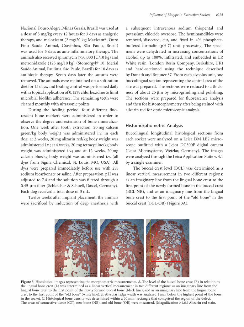
Histomorphometric Analysis
Buccolingual longitudinal histological sections from
each socket were analyzed on a Leica DM LB2 micro-
scope outfitted with a Leica DC300F digital camera
(Leica Microsystems, Wetzlar, Germany). The images
were analyzed through the Leica Application Suite v. 4.1
by a single examiner.
The buccal crest level (BCL) was determined as a
linear vertical measurement in two different regions:
as an imaginary line from the lingual bone crest to the
first point of the newly formed bone in the buccal crest
(BCL-NB), and as an imaginary line from the lingual
bone crest to the first point of the “old bone” in the
buccal crest (BCL-OB) (Figure 3A).
Figure 3 Histological images representing the morphometric measurements. A, The level of the buccal bone crest (B) in relation tothe lingual bone crest (L) was determined as a linear vertical measurement in two different regions: as an imaginary line from thelingual bone crest to the first point of the newly formed buccal bone (black line), and as an imaginary line from the lingual bonecrest to the first point of the “old bone” (white line). B, Alveolar ridge width was analyzed 1 mm below the highest point of the bonein the socket. C, Histological bone density was determined within a 30 mm2 rectangle that comprised the region of the defect.The areas of connective tissue (CT), new bone (NB), and old bone (OB) were measured. (Magnification ×1.6.) Alizarin red stain.
Influence of Biotype in Extraction Sockets e225
The alveolar ridge width (ARW) was determined
as a linear horizontal measurement, between both the
buccal and lingual external walls, as an imaginary line
1 mm below the highest point of the bone in the socket
(see Figure 3B).
The bone area was determined within a rectangle
that comprised the region of the socket (30 mm2), mea-
sured starting with the highest point of the socket. These
measurements evaluated the percentage of the region
occupied by mineralized bone in relation to the percent-
age occupied by marrow spaces. The connective tissue
(CT), total bone area (TBA), and new bone area (NBA)
were measured. Finally, the percentage of the sockets
occupied by residual graft particles (RGP) was measured
(see Figure 3C).
Fluorescence Analysis
Fluorescence microscopic images were longitudinally cap-
tured from each sample through a Leica DC300F video
camera joined to a Leica DM LB2 microscope, using
appropriate barrier filters. The wavelength filters used
were I3 for calcein green (excitation level 450–490 nm),
N2-1 for alizarin red (excitation level 515–560 nm), D for
tetracycline (excitation level 355–425 nm), and A for
calcein blue (excitation level 340–380 nm). All the images
were adjusted and analyzed with Leica QWin software
(Leica Microsystems) to determine the percentage of the
alveoli occupied by marked bone. Thus, one rectangle
comprising the region of the socket (30 mm2, from the
highest point of the socket) was used to evaluate the
percentage of space occupied by fluorescent bone.
The marked bone measurements evaluated the percentage
of the total area occupied by fluorescent bone.
Statistical Analysis
Quantitative data were recorded as mean and standard
deviations. The experimental unit was the dog (n = 8).
To verify the normality of the data, the Kolmogorov-
Smirnov test was used. For the parametric data (BBT,
TKT, AT, and histomorphometric analysis), the one-
way ANOVA was used for intragroup (T0 vs. T1) and
intergroup (TG vs. TG + GM vs. CG vs. CG + GM)
comparisons. For the nonparametric data (fluorescence
analysis), the Kruskal-Wallis one-way ANOVA on ranks
was applied for intergroup comparisons. A confidence
interval of 95% was adopted. SigmaStat (Systat Software
Inc., San Jose, CA, USA) version 3.5 was the statistical
software used for the analysis.
RESULTS
Clinical Findings
The healing was uneventful, and there were no compli-
cations during the experimental period.
Clinical Analysis
Data regarding the TKT, the AT, and the BBT are sum-
marized in Table 1. There was no statistically significant
difference in mean TKT among the groups at baseline.
In TG and TG + GM the mean TKT was significantly
decreased from T0 to T1 (TG: p < .001; TG + GM:
p = .004). For CG and CG + GM, the mean TKT
observed at T0 remained stable until T1, with no
statistically significant changes. This reduction in the
mean TKT in TG and TG + GM resulted in statistically
significant differences between these groups and the
control groups (CG and CG + GM) at T1 (p < .001) (see
TABLE 1 Thickness Measurements before GingivalPeeling (T0) and at Tooth Extraction (T1)(Mean 1 SD): Intragroup and IntergroupComparisons
T0 T1 p Value
TKT (mm)
TG 1.24 1 0.13A 0.73 1 0.16Ba <0.001
TG + GM 1.19 1 0.22A 0.78 1 0.14Ba 0.004
CG 1.20 1 0.19 1.16 1 0.13b NS
CG + GM 1.18 1 0.28 1.16 1 0.14b NS
p Value NS <.001
AT (mm)
TG 3.39 1 0.96 3.08 1 0.46 NS
TG + GM 3.64 1 0.86 3.44 1 0.64 NS
CG 3.73 1 1.00 3.15 1 0.45 NS
CG + GM 3.39 1 0.58 3.04 1 0.61 NS
p Value NS NS
BBT (mm)
TG — 0.53 1 0.12
TG + GM — 0.55 1 0.11
CG — 0.49 1 0.19
CG + GM — 0.53 1 0.06
p Value NS
One-way ANOVA with Tukey’s test. Different superscript letters representstatistically significant difference (p < .05).A,BIntragroup comparisons.a,bIntergroup comparisons.TKT = thickness of keratinized tissue; AT = alveolar thickness;BBT = buccal bone thickness; TG = test group; CG = control group;GM = graft material; NS = not statistically significant.
e226 Clinical Implant Dentistry and Related Research, Volume 17, Supplement 1, 2015
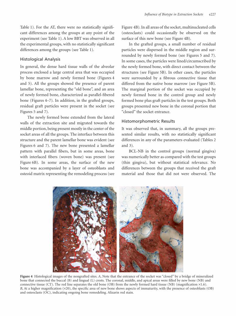
Table 1). For the AT, there were no statistically signifi-
cant differences among the groups at any point of the
experiment (see Table 1). A low BBT was observed in all
the experimental groups, with no statistically significant
differences among the groups (see Table 1).
Histological Analysis
In general, the dense hard tissue walls of the alveolar
process enclosed a large central area that was occupied
by bone marrow and newly formed bone (Figures 4
and 5). All the groups showed the presence of parent
lamellar bone, representing the “old bone”, and an area
of newly formed bone, characterized as parallel-fibered
bone (Figures 4–7). In addition, in the grafted groups,
residual graft particles were present in the socket (see
Figures 5 and 7).
The newly formed bone extended from the lateral
walls of the extraction site and migrated towards the
middle portion, being present mostly in the center of the
socket areas of all the groups. The interface between this
structure and the parent lamellar bone was evident (see
Figures 6 and 7). The new bone presented a lamellar
pattern with parallel fibers, but in some areas, bone
with interlaced fibers (woven bone) was present (see
Figure 6B). In some areas, the surface of the new
bone was accompanied by a layer of osteoblasts and
osteoid matrix representing the remodeling process (see
Figure 4B). In all areas of the socket, multinucleated cells
(osteoclasts) could occasionally be observed on the
surface of this new bone (see Figure 4B).
In the grafted groups, a small number of residual
particles were dispersed in the middle region and sur-
rounded by newly formed bone (see Figures 5 and 7).
In some cases, the particles were lined/circumscribed by
the newly formed bone, with direct contact between the
structures (see Figure 5B). In other cases, the particles
were surrounded by a fibrous connective tissue that
differed from the native bone marrow (see Figure 5B).
The marginal portion of the socket was occupied by
newly formed bone in the control group and newly
formed bone plus graft particles in the test groups. Both
groups presented new bone in the coronal portion that
“closed” the socket entrance.
Histomorphometric Results
It was observed that, in summary, all the groups pre-
sented similar results, with no statistically significant
differences in any of the parameters evaluated (Tables 2
and 3).
BCL-NB in the control groups (normal gingiva)
was numerically better as compared with the test groups
(thin gingiva), but without statistical relevance. No
differences between the groups that received the graft
material and those that did not were observed. The
Figure 4 Histological images of the nongrafted sites. A, Note that the entrance of the socket was “closed” by a bridge of mineralizedbone that connected the buccal (B) and lingual (L) crests. The coronal, middle, and apical areas were filled by new bone (NB) andconnective tissue (CT). The red line separates the old bone (OB) from the newly formed hard tissue (NB) (magnification ×1.6).B, At a higher magnification (×20), the specific area of new bone shows aspects of immaturity, with the presence of osteoblasts (OB)and osteoclasts (OC), indicating ongoing bone remodeling. Alizarin red stain.
Influence of Biotype in Extraction Sockets e227

BCL-OB and the ARW were very similar for the groups
and without statistical relevance (see Table 2).
The total area of the frame (100%) was divided into
connective tissue and bone area. All the experimental
groups showed around 50% connective tissue and 50%
bone area, on average, without any statistically signifi-
cant difference. The new bone area represented around
35% of the total frame, also without any statistically
significant difference among the experimental groups
(see Table 3).
Figure 5 Histological images of the grafted sites. A, The fresh extraction socket was grafted with a synthetic bone graft. The residualgraft particles (RGPs, brown stain) were embebbed in newly formed bone (NB) and fibrous connective tissue (CT). Note that thebiomaterial occupied a large portion of the socket entrance, which was “closed” by a a bridge of mineralized bone and graft material.The red line separates the old bone (OB) from the newly formed hard tissue (NB) (Magnification ×1.6.) B, The circumscribed areain a higher magnification (×20), the presence of some RGPs lined/circumscribed by the newly formed bone (NB) was more evident,with direct contact between the structures, while other RGPs were surrounded by fibrous connective tissue (CT). Alizarin red stain.
Figure 6 Histological images of the nongrafted sites. A, Note the difference between the new bone (NB) and the old bone (OB).The structures are connected by remodeling tissue. B, Under polarized light, it was possible to observe the presence of the old bone(OB) with its lamellar pattern, and the newly formed bone (NB) with areas of interlaced fibers (woven bone, WB) and parallel fibers(PF). Alizarin red stain. (Magnification ×20.)
e228 Clinical Implant Dentistry and Related Research, Volume 17, Supplement 1, 2015
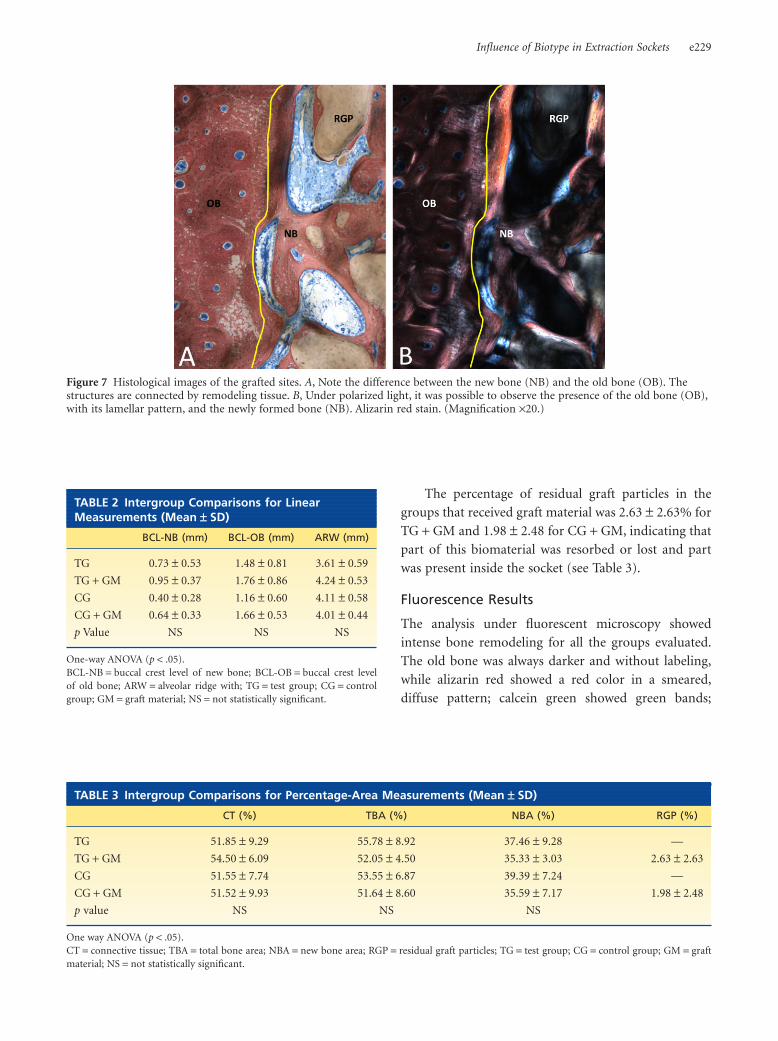
The percentage of residual graft particles in the
groups that received graft material was 2.63 1 2.63% for
TG + GM and 1.98 1 2.48 for CG + GM, indicating that
part of this biomaterial was resorbed or lost and part
was present inside the socket (see Table 3).
Fluorescence Results
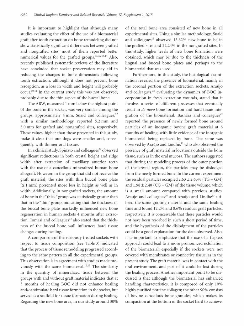
The analysis under fluorescent microscopy showed
intense bone remodeling for all the groups evaluated.
The old bone was always darker and without labeling,
while alizarin red showed a red color in a smeared,
diffuse pattern; calcein green showed green bands;
Figure 7 Histological images of the grafted sites. A, Note the difference between the new bone (NB) and the old bone (OB). Thestructures are connected by remodeling tissue. B, Under polarized light, it was possible to observe the presence of the old bone (OB),with its lamellar pattern, and the newly formed bone (NB). Alizarin red stain. (Magnification ×20.)
TABLE 2 Intergroup Comparisons for LinearMeasurements (Mean 1 SD)
BCL-NB (mm) BCL-OB (mm) ARW (mm)
TG 0.73 1 0.53 1.48 1 0.81 3.61 1 0.59
TG + GM 0.95 1 0.37 1.76 1 0.86 4.24 1 0.53
CG 0.40 1 0.28 1.16 1 0.60 4.11 1 0.58
CG + GM 0.64 1 0.33 1.66 1 0.53 4.01 1 0.44
p Value NS NS NS
One-way ANOVA (p < .05).BCL-NB = buccal crest level of new bone; BCL-OB = buccal crest levelof old bone; ARW = alveolar ridge with; TG = test group; CG = controlgroup; GM = graft material; NS = not statistically significant.
TABLE 3 Intergroup Comparisons for Percentage-Area Measurements (Mean 1 SD)
CT (%) TBA (%) NBA (%) RGP (%)
TG 51.85 1 9.29 55.78 1 8.92 37.46 1 9.28 —
TG + GM 54.50 1 6.09 52.05 1 4.50 35.33 1 3.03 2.63 1 2.63
CG 51.55 1 7.74 53.55 1 6.87 39.39 1 7.24 —
CG + GM 51.52 1 9.93 51.64 1 8.60 35.59 1 7.17 1.98 1 2.48
p value NS NS NS
One way ANOVA (p < .05).CT = connective tissue; TBA = total bone area; NBA = new bone area; RGP = residual graft particles; TG = test group; CG = control group; GM = graftmaterial; NS = not statistically significant.
Influence of Biotype in Extraction Sockets e229
tetracycline showed thin yellow-green lines; and, finally,
calcein blue was characterized by a blue color in a very
diffuse pattern (Figure 8).
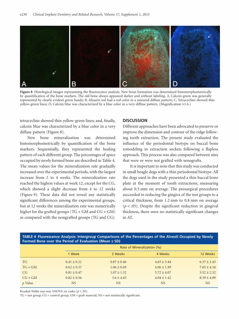
New bone mineralization was determined
histomorphometrically by quantification of the bone
markers. Sequentially, they represented the healing
pattern of each different group. The percentages of space
occupied by newly formed bone are described in Table 4.
The mean values for the mineralization rate gradually
increased over the experimental periods, with the largest
increase from 2 to 4 weeks. The mineralization rate
reached the highest values at week 12, except for the CG,
which showed a slight decrease from 4 to 12 weeks
(Figure 9). These data did not reveal any statistically
significant differences among the experimental groups,
but at 12 weeks the mineralization rate was numerically
higher for the grafted groups (TG + GM and CG + GM)
as compared with the nongrafted groups (TG and CG).
DISCUSSIONDifferent approaches have been advocated to preserve or
improve the dimension and contour of the ridge follow-
ing tooth extraction. The present study evaluated the
influence of the periodontal biotype on buccal bone
remodeling in extraction sockets following a flapless
approach. This process was also compared between sites
that were or were not grafted with xenografts.
It is important to note that this study was conducted
in small beagle dogs with a thin periodontal biotype. All
the dogs used in the study presented a thin buccal bone
plate at the moment of tooth extractions, measuring
about 0.5 mm on average. The presurgical procedures
succeeded in reducing the gingiva of the test groups to a
critical thickness, from 1.2 mm to 0.8 mm on average
(p < .05). Despite the significant reduction in gingival
thickness, there were no statistically significant changes
in AT.
Figure 8 Histological images representing the fluorescence analysis. New bone formation was determined histomorphometricallyby quantification of the bone markers. The old bone always appeared darker and without labeling. A, Calcein green was generallyrepresented by clearly evident green bands; B, Alizarin red had a red color in a smeared diffuse pattern; C, Tetracycline showed thinyellow-green lines; D, Calcein blue was characterized by a blue color in a very diffuse pattern. (Magnification ×1.6.)
TABLE 4 Fluorescence Analysis: Intergroup Comparisons of the Percentages of the Alveoli Occupied by NewlyFormed Bone over the Period of Evaluation (Mean 1 SD)
Rate of Mineralization (%)
1 Week 2 Weeks 4 Weeks 12 Weeks
TG 0.41 1 0.21 0.87 1 0.46 4.65 1 3.44 6.37 1 2.43
TG + GM 0.62 1 0.37 1.06 1 0.69 4.06 1 1.99 7.65 1 4.34
CG 0.81 1 0.47 1.07 1 1.32 5.72 1 4.07 5.52 1 2.32
CG + GM 0.82 1 0.56 3.6 1 4.43 4.04 1 1.42 8.39 1 4.89
p Value NS NS NS NS
Kruskal-Wallis one-way ANOVA on ranks (p < .05).TG = test group; CG = control group; GM = graft material; NS = not statistically significant.
e230 Clinical Implant Dentistry and Related Research, Volume 17, Supplement 1, 2015

The buccal crest level in all specimens was slightly
apical to the corresponding lingual bone crest. All the
groups showed new bone formation above the old bone
crest level, reducing the differences between the buccal
and lingual crests during the healing period. No statis-
tically significant difference was observed between the
test groups (thin gingiva, TG and TG + GM) and the
control groups (normal gingiva, CG and CG + GM),
but the distance between buccal and lingual crests was
numerically superior for the test groups, mainly in
relation to the new bone. This difference, although it
is small, and despite this being an animal study and
therefore using a small sample size, may have clinical
implications. Both animal38 and human trials39–41 that
evaluated the influence of thin ridge mucosal tissues on
crestal bone stability around dental implants reported
more pronounced crestal bone loss in cases with in-
sufficient gingiva. The results of the current experiment
suggest that the influence of gingival thickness on bone
healing occurs after tooth extraction, independently of
implant placement.
No difference between grafted (CG + GM: 0.40 1
0.28 mm) and nongrafted (CG: 0.64 1 0.33 mm) sites
was observed in our study regarding the distance
between the buccal and lingual crests. Similar results
have been reported in the literature.22,42,43 Bashara and
colleagues42 studied the effect of different biomaterials
on hard tissue remodeling following their placement
into fresh extraction sockets in dogs after 6 months of
healing, and reported a similar amount of vertical buccal
bone loss in sites treated with bovine bone (Bio-Oss)
(0.65 1 0.40 mm) and sites that healed with a blood
clot (0.70 1 0.35 mm). Rothamel and colleagues,43
evaluating the alterations in ridge dimensions following
application of a nanocrystalline hydroxyapatite paste
in fresh extraction sockets in dogs, observed a difference
in bone height between buccal and lingual bone of
0.54 1 0.62 mm in grafted sites and 0.85 1 0.17 mm
in nongrafted sites at 3 months of healing. Suaid and
colleagues22 also reported low bone loss in extraction
sockets grafted with biphasic calcium phosphate. The
authors reported a buccal bone level of 0.26 1 0.67 mm
apical to the lingual bone in grafted sites and
0.80 1 1.14 mm apical in the nongrafted sites. On the
other hand, Araújo and colleagues,26 who assessed the
effect on bone remodeling following the placement of
the same xenograft in fresh extraction sockets in dogs,
found a mean vertical distance between the buccal and
lingual bone crests of 2.1 1 0.5 mm at nongrafted sites
and 1.9 1 0.6 mm at grafted sites. These values, higher
compared with ours, could be attributed to the flap
elevation performed in that study.
The advantageous effects of tooth extraction
without the elevation of a mucoperiosteal flap on
bone remodeling of extraction sockets were analyzed by
Fickl and colleagues28 in an animal model. The authors
demonstrated significantly lower resorption rates in the
flapless group. Also, Araújo and colleagues12 compared
hard tissue healing following tooth extraction with
and without prior elevation of mucosal full-thickness
flaps and found better results for the flapless approach
regarding the distance between the cementum/enamel
junction of the adjacent tooth and the bone crest (flap:
1 1 0.1 mm; flapless: 0.7 1 0.2 mm).
Figure 9 Percentages of the sockets occupied by newly formed bone for the experimental groups over the period of evaluation.
Influence of Biotype in Extraction Sockets e231
It is important to highlight that although many
studies evaluating the effect of the use of a biomaterial
graft after tooth extraction on bone remodeling did not
show statistically significant differences between grafted
and nongrafted sites, most of them reported better
numerical values for the grafted groups.22,26,42,43 Also,
recently published systematic reviews of the literature
have concluded that socket preservation may aid in
reducing the changes in bone dimensions following
tooth extraction, although it does not prevent bone
resorption, as a loss in width and height will probably
occur.10,44 In the current study this was not observed,
probably due to the thin aspect of the buccal bone.
The ARW, measured 1 mm below the highest point
of the bone in the socket, was very similar among the
groups, approximately 4 mm. Suaid and colleagues,22
with a similar methodology, reported 5.2 mm and
5.0 mm for grafted and nongrafted sites, respectively.
These values, higher than those presented in this study,
make it clear that our dogs were smaller and, conse-
quently, with thinner oral tissues.
In a clinical study, Spinato and colleagues32 observed
significant reductions in both crestal height and ridge
width after extraction of maxillary anterior teeth
with the use of a cancellous mineralized human bone
allograft. However, in the group that did not receive the
graft material, the sites with thin buccal bone plate
(2 1 mm) presented more loss in height as well as in
width. Additionally, in nongrafted sockets, the amount
of bone in the “thick” group was statistically greater than
that in the “thin” group, indicating that the thickness of
the buccal bone plate markedly influenced new bone
regeneration in human sockets 4 months after extrac-
tion. Tomasi and colleagues33 also stated that the thick-
ness of the buccal bone wall influences hard tissue
changes during healing.
A comparison of the variously treated sockets with
respect to tissue composition (see Table 3) indicated
that the process of tissue remodeling progressed accord-
ing to the same pattern in all the experimental groups.
This observation is in agreement with studies made pre-
viously with the same biomaterial.12,13 The similarity
in the quantity of mineralized tissue between the
groups with and without graft material indicates that at
3 months of healing BOC did not enhance healing
and/or stimulate hard tissue formation in the socket, but
served as a scaffold for tissue formation during healing.
Regarding the new bone area, in our study around 30%
of the total bone area consisted of new bone in all
experimental sites. Using a similar methodology, Suaid
and colleagues22 observed 15.62% new bone to be in
the grafted sites and 22.24% in the nongrafted sites. In
this study, higher levels of new bone formation were
obtained, which may be due to the thickness of the
lingual and buccal bone plates and perhaps to the
biomaterial that was used.
Furthermore, in this study, the histological exami-
nation revealed the presence of biomaterial, mainly in
the coronal portion of the extraction sockets. Araújo
and colleagues,25 evaluating the dynamics of BOC in-
corporation in fresh extraction wounds, stated that it
involves a series of different processes that eventually
result in de novo bone formation and hard tissue inte-
gration of the biomaterial. Bashara and colleagues42
reported the presence of newly formed bone around
particles of an inorganic bovine graft material at 6
months of healing, with little evidence of the inorganic
biomaterial being replaced by bone. The same was
observed by Araújo and Lindhe,12 who also observed the
presence of graft material in locations outside the bone
tissue, such as in the oral mucosa. The authors suggested
that during the modeling process of the outer portion
of the crestal region, the particles may be dislodged
from the newly formed bone. In the current experiment
the residual particles occupied 2.63 1 2.63% (TG + GM)
and 1.98 1 2.48 (CG + GM) of the tissue volume, which
is a small amount compared with previous studies.
Araújo and colleagues26 and Araújo and Lindhe13 uti-
lized the same grafting material and the same healing
time and found 12.2% and 8.6% residual graft particles,
respectively. It is conceivable that these particles would
not have been resorbed in such a short period of time,
and the hypothesis of the dislodgment of the particles
could be a good explanation for the data observed. Also,
it is important to emphasize that the use of a flapless
approach could lead to a more pronounced exfoliation
of the biomaterial, especially if the sockets were not
covered with membranes or connective tissue, as in the
present study. The graft material was in contact with the
oral environment, and part of it could be lost during
the healing process. Another important point to be dis-
cussed is that although the biomaterial has enhanced
handling characteristics, it is composed of only 10%
highly purified porcine collagen; the other 90% consists
of bovine cancellous bone granules, which makes its
compaction at the bottom of the socket hard to achieve.
e232 Clinical Implant Dentistry and Related Research, Volume 17, Supplement 1, 2015

Further, the study aimed to use fluorescence analysis
to investigate the dynamics of early bone healing in fresh
extraction wounds in thin buccal bone filled with BOC.
The application of bone markers at different time points
permits evaluation of bone formation and remodel-
ing throughout the different stages of healing. Alizarin,
calcein green, tetracycline, and calcein blue fluoro-
chromes present different colors and supply sequential
information when applied intercalated. The bone
markers used in the present study can be compared
because they bind to calcium ions by chelation,45 cor-
rectly indicating the areas of active mineralization. The
fluorochrome incorporation followed a pattern among
the different groups over the period of evaluation from
7 days to 12 weeks. Similarly to the histomorphometric
findings, comparison among the groups did not show
statistically significant differences in the fluorescence
analysis results. Generally, it was observed for all the
experimental groups that the area of marked bone was
slight in the first week and gradually increased at 2 and 4
weeks, reaching the highest values at the 12-week evalu-
ation.According to Araújo and colleagues,25 the first steps
of bone remodeling in extraction sockets filled with BOC
involved the presence of a clot containing large numbers
of mesenchymal cells, leukocytes, and vascular struc-
tures, followed by the formation of woven bone. In the
interval between 2 and 4 weeks, the rate of new bone
formation is pronounced. These observations support
the results found in the present study that showed an
increase of almost four times in the mineralization
rate from 2 to 4 weeks, except for the CG + GM. In the
sequence of this ongoing process, at 4 weeks the woven
bone in such locations is in the process of remodeling,
indicating that the mineralization rate could continue
to increase in the subsequent weeks. This continuous
dynamic is also observed in naturally healed tooth
extraction sockets, where there is a pronounced altera-
tion of the tissue within the extraction socket between 4
and 8 weeks.4 At 8 weeks a cortical ridge is formed, sealing
the entrance of the extraction site, and the woven bone is
to a large extent replaced by lamellar bone and marrow.4
The results of the present study corroborate the findings
in the literature, showing an increase in the mineraliza-
tion rate between 4 and 12 weeks.
Also, although no statistically significant difference
was observed among the groups regarding the mineral-
ization rate, at 12 weeks it was numerically superior
for the grafted groups. The results support the histo-
morphometric findings of previous studies,26,46 which
stated that the placement of this biomaterial in an
extraction socket may modify the modeling process that
occurs following tooth removal.
CONCLUSION
Based on these findings, it can be speculated that a natu-
rally thin buccal bone plate leads to higher bone loss in
extraction sockets, even with flapless surgery. Reduction
in gingival thickness did not result in a significant
contribution to bone loss. The use of graft material did
not prevent buccal bone resorption in a thin biotype,
but modified the mineralization process that occurs
following tooth removal.
ACKNOWLEDGMENTS
The authors thank the Coordination for the Improve-
ment of Graduated Personnel (CAPES) for the scholar-
ship that supported Ms. Maia, the State of São Paulo
Research Foundation for the financial support of this
study (process number 11/00674-6) and Geistlich
Pharma AG (Wolhusen, Switzerland) for the donation of
the graft material used in this study.
REFERENCES
1. Amler MH. The time sequence of tissue regeneration in
human extraction wounds. Oral Surg Oral Med Oral Pathol
1969; 27:309–318.
2. Kuboki Y, Hashimoto F, Ishibashi K. Time-dependent
changes of collagen crosslinks in the socket after tooth
extraction in rabbits. J Dent Res 1988; 67:944–948.
3. Lin WL, McCulloch CA, Cho MI. Differentiation of peri-
odontal ligament fibroblasts into osteoblasts during socket
healing after tooth extraction in the rat. Anat Rec 1994;
240:492–506.
4. Araujo MG, Lindhe J. Dimensional ridge alterations follow-
ing tooth extraction. An experimental study in the dog.
J Clin Periodontol 2005; 32:212–218.
5. Cardaropoli G, Araujo M, Lindhe J. Dynamics of bone tissue
formation in tooth extraction sites. An experimental study in
dogs. J Clin Periodontol 2003; 30:809–818.
6. Fickl S, Zuhr O, Wachtel H, Stappert CF, Stein JM,
Hurzeler MB. Dimensional changes of the alveolar ridge
contour after different socket preservation techniques. J Clin
Periodontol 2008; 35:906–913.
7. Mecall RA, Rosenfeld AL. Influence of residual ridge resorp-
tion patterns on implant fixture placement and tooth posi-
tion. 1. Int J Periodontics Restorative Dent 1991; 11:8–23.
8. Schropp L, Wenzel A, Kostopoulos L, Karring T. Bone heal-
ing and soft tissue contour changes following single-tooth
Influence of Biotype in Extraction Sockets e233
extraction: a clinical and radiographic 12-month prospective
study. Int J Periodontics Restorative Dent 2003; 23:313–323.
9. Novaes AB Jr, Macedo GO, Suaid FA, Barros RR, Souza SL,
Silveira ESAM. Histologic evaluation of the buccal and
lingual bone plates in anterior dog teeth: possible influence
on implant dentistry. J Periodontol 2011; 82:872–877.
10. Ten Heggeler JM, Slot DE, Van der Weijden GA. Effect of
socket preservation therapies following tooth extraction in
non-molar regions in humans: a systematic review. Clin Oral
Implants Res 2011; 22:779–788.
11. Buser D, Bornstein MM, Weber HP, Grutter L, Schmid B,
Belser UC. Early implant placement with simultaneous
guided bone regeneration following single-tooth extraction
in the esthetic zone: a cross-sectional, retrospective study in
45 subjects with a 2- to 4-year follow-up. J Periodontol 2008;
79:1773–1781.
12. Araujo MG, Lindhe J. Ridge preservation with the use
of Bio-Oss collagen: a 6-month study in the dog. Clin Oral
Implants Res 2009; 20:433–440.
13. Araujo MG, Lindhe J. Socket grafting with the use of autolo-
gous bone: an experimental study in the dog. Clin Oral
Implants Res 2011; 22:9–13.
14. Artzi Z, Tal H, Dayan D. Porous bovine bone mineral in
healing of human extraction sockets. Part 1: histomor-
phometric evaluations at 9 months. J Periodontol 2000;
71:1015–1023.
15. Camargo PM, Lekovic V, Weinlaender M, et al. Influence
of bioactive glass on changes in alveolar process dimensions
after exodontia. Oral Surg Oral Med Oral Pathol Oral Radiol
Endod 2000; 90:581–586.
16. Carmagnola D, Abati S, Celestino S, Chiapasco M,
Bosshardt D, Lang NP. Oral implants placed in bone defects
treated with Bio-Oss, Ostim-Paste or PerioGlas: an experi-
mental study in the rabbit tibiae. Clin Oral Implants Res
2008; 19:1246–1253.
17. Carmagnola D, Adriaens P, Berglundh T. Healing of human
extraction sockets filled with Bio-Oss. Clin Oral Implants
Res 2003; 14:137–143.
18. Fernandes PG, Novaes AB Jr, de Queiroz AC, et al. Ridge
preservation with acellular dermal matrix and anorganic
bone matrix cell-binding peptide P-15 after tooth extraction
in humans. J Periodontol 2011; 82:72–79.
19. Lambert F, Vincent K, Vanhoutte V, Seidel L, Lecloux G,
Rompen E. A methodological approach to assessing alveolar
ridge preservation procedures in humans: hard tissue profile.
J Clin Periodontol 2012; 39:887–894.
20. Roriz VM, Rosa AL, Peitl O, Zanotto ED, Panzeri H,
de Oliveira PT. Efficacy of a bioactive glass-ceramic
(Biosilicate) in the maintenance of alveolar ridges and in
osseointegration of titanium implants. Clin Oral Implants
Res 2010; 21:148–155.
21. Santos FA, Pochapski MT, Martins MC, Zenobio EG,
Spolidoro LC, Marcantonio E Jr. Comparison of biomaterial
implants in the dental socket: histological analysis in dogs.
Clin Implant Dent Relat Res 2010; 12:18–25.
22. Suaid F, Grisi MF, Souza SL, Palioto DB, Taba M Jr,
Novaes AB Jr. Buccal bone remodeling after tooth extraction
using the flapless approach with and without synthetic bone
grafting. A histomorphometric study in dogs. Clin Oral
Implants Res 2013; 24:407–413.
23. Araujo MG, Carmagnola D, Berglundh T, Thilander B,
Lindhe J. Orthodontic movement in bone defects aug-
mented with Bio-Oss. An experimental study in dogs. J Clin
Periodontol 2001; 28:73–80.
24. Berglundh T, Lindhe J. Healing around implants
placed in bone defects treated with Bio-Oss. An experi-
mental study in the dog. Clin Oral Implants Res 1997; 8:
117–124.
25. Araujo MG, Liljenberg B, Lindhe J. Dynamics of Bio-Oss
Collagen incorporation in fresh extraction wounds: an
experimental study in the dog. Clin Oral Implants Res 2010;
21:55–64.
26. Araujo M, Linder E, Wennstrom J, Lindhe J. The influence
of Bio-Oss Collagen on healing of an extraction socket: an
experimental study in the dog. Int J Periodontics Restorative
Dent 2008; 28:123–135.
27. Cardaropoli G, Araujo M, Hayacibara R, Sukekava F,
Lindhe J. Healing of extraction sockets and surgically
produced – augmented and non-augmented – defects in
the alveolar ridge. An experimental study in the dog. J Clin
Periodontol 2005; 32:435–440.
28. Fickl S, Zuhr O, Wachtel H, Bolz W, Huerzeler MB. Hard
tissue alterations after socket preservation: an experimental
study in the beagle dog. Clin Oral Implants Res 2008; 19:
1111–1118.
29. Heberer S, Al-Chawaf B, Hildebrand D, Nelson JJ, Nelson K.
Histomorphometric analysis of extraction sockets aug-
mented with Bio-Oss Collagen after a 6-week healing period:
a prospective study. Clin Oral Implants Res 2008; 19:1219–
1225.
30. Seibert J, Lindhe J. Esthetics and periodontal therapy.
In: Lindhe J, ed. Textbook of clinical periodontology. 2nd ed.
Copenhagen: Munksgaard, 1989:477–514.
31. Ferrus J, Cecchinato D, Pjetursson EB, Lang NP, Sanz M,
Lindhe J. Factors influencing ridge alterations following
immediate implant placement into extraction sockets. Clin
Oral Implants Res 2010; 21:22–29.
32. Spinato S, Galindo-Moreno P, Zaffe D, Bernardello F,
Soardi CM. Is socket healing conditioned by buccal plate
thickness? A clinical and histologic study 4 months after
mineralized human bone allografting. Clin Oral Implants
Res 2012 [Epub ahead of print].
33. Tomasi C, Sanz M, Cecchinato D, et al. Bone dimensional
variations at implants placed in fresh extraction sockets: a
multilevel multivariate analysis. Clin Oral Implants Res
2010; 21:30–36.
e234 Clinical Implant Dentistry and Related Research, Volume 17, Supplement 1, 2015
34. Bragger U, Pasquali L, Kornman KS. Remodelling of
interdental alveolar bone after periodontal flap procedures
assessed by means of computer-assisted densitometric image
analysis (CADIA). J Clin Periodontol 1988; 15:558–564.
35. Wood DL, Hoag PM, Donnenfeld OW, Rosenfeld LD. Alveo-
lar crest reduction following full and partial thickness flaps.
J Periodontol 1972; 43:141–144.
36. Fickl S, Zuhr O, Wachtel H, Bolz W, Huerzeler M. Tissue
alterations after tooth extraction with and without surgical
trauma: a volumetric study in the beagle dog. J Clin
Periodontol 2008; 35:356–363.
37. Donath K, Breuner G. A method for the study of
undecalcified bones and teeth with attached soft tissues.
The Sage-Schliff (sawing and grinding) technique 1982;
11:318–326.
38. Berglundh T, Lindhe J. Dimension of the periimplant
mucosa. Biological width revisited. J Clin Periodontol 1996;
23:971–973.
39. Kim BS, Kim YK, Yun PY, et al. Evaluation of peri-implant
tissue response according to the presence of keratinized
mucosa. Oral Surg Oral Med Oral Pathol Oral Radiol Endod
2009; 107:e24–e28.
40. Linkevicius T, Apse P, Grybauskas S, Puisys A. The influence
of soft tissue thickness on crestal bone changes around
implants: a 1-year prospective controlled clinical trial. Int J
Oral Maxillofac Implants 2009; 24:712–719.
41. Linkevicius T, Apse P, Grybauskas S, Puisys A. Influence
of thin mucosal tissues on crestal bone stability around
implants with platform switching: a 1-year pilot study. J Oral
Maxillofac Surg 2010; 68:2272–2277.
42. Bashara H, Wohlfahrt JC, Polyzois I, Lyngstadaas SP,
Renvert S, Claffey N. The effect of permanent grafting mate-
rials on the preservation of the buccal bone plate after tooth
extraction: an experimental study in the dog. Clin Oral
Implants Res 2012; 23:911–917.
43. Rothamel D, Schwarz F, Herten M, et al. Dimensional
ridge alterations following socket preservation using a
nanocrystalline oxyapatite paste. A histomorphometrical
study in dogs. Int J Oral Maxillofac Surg 2008; 37:741–747.
44. Horvath A, Mardas N, Mezzomo LA, Needleman IG,
Donos N. Alveolar ridge preservation. A systematic review.
Clin Oral Investig 2013; 17:341–363.
45. Sun TC, Mori S, Roper J, Brown C, Hooser T, Burr DB.
Do different fluorochrome labels give equivalent histomor-
phometric information? Bone 1992; 13:443–446.
46. Araujo M, Linder E, Lindhe J. Effect of a xenograft on early
bone formation in extraction sockets: an experimental study
in dog. Clin Oral Implants Res 2009; 20:1–6.
Influence of Biotype in Extraction Sockets e235