INFLAMMATION and repair - NUS Medicine
21
General Pathology INFLAMMATION AND REPAIR G.Soon | Pathweb | Department of Pathology, NUHS | 2022 1 Objectives • List causes of inflammation • List the cardinal signs of acute inflammation and understand their pathogenesis • Understand the role of various inflammatory cells in acute and chronic inflammation • Understand the role of various chemical mediators of inflammation • Appreciate that inflammation can have both damaging and beneficial effects, and local and systemic manifestations • Be aware of the morphologic patterns of acute inflammation • Distinguish between acute and chronic inflammation, and recognize granulomatous inflammation as a distinct form of chronic inflammation • Understand the various possible outcomes of inflammation • Recognize that tissue repair may occur through regeneration and/or scarring, and function may not be completely restored in the latter • List local and systemic factors that affect tissue repair, and possible complications of tissue repair Outline I. General Features of Inflammation a. Definitions and General Sequence of Events (5 ‘R’s) b. Causes of Inflammation c. Recognition of Microbes and Damaged Cells II. Acute Inflammation a. Increased Blood Flow and Vascular Permeability: Vasodilatation and stasis, Vascular leakage, Response of lymphatic system b. Recruitment of Leukocytes to Sites of Inflammation: Margination, Rolling, Adhesion, Migration, Chemotaxis c. Removal of Offending Agent via Phagocytosis: Recognition and attachment, Engulfment, Intracellular destruction d. Leukocyte-mediated Tissue Injury e. Regulation / Termination of the Acute Inflammatory Response f. Mediators of Acute Inflammation and their Pharmacologic Inhibitors: Vasoactive amines, Arachidonic acid metabolites, Cytokines and chemokines, Complements, Others g. Morphologic Patterns of Acute Inflammation: General features, Serous inflammation, Fibrinous inflammation, Suppurative inflammation, Ulcers h. Outcomes of Acute Inflammation: Complete resolution, Scarring / fibrosis, Progression to Chronic Inflammation III. Chronic Inflammation a. Causes of Chronic Inflammation b. Morphologic Features
-
Upload
khangminh22 -
Category
Documents
-
view
0 -
download
0
Transcript of INFLAMMATION and repair - NUS Medicine

General Pathology INFLAMMATION AND REPAIR
G.Soon | Pathweb | Department of Pathology, NUHS | 2022 1
Objectives
• List causes of inflammation • List the cardinal signs of acute inflammation and understand their pathogenesis • Understand the role of various inflammatory cells in acute and chronic inflammation • Understand the role of various chemical mediators of inflammation • Appreciate that inflammation can have both damaging and beneficial effects, and local and systemic
manifestations • Be aware of the morphologic patterns of acute inflammation • Distinguish between acute and chronic inflammation, and recognize granulomatous inflammation as
a distinct form of chronic inflammation • Understand the various possible outcomes of inflammation • Recognize that tissue repair may occur through regeneration and/or scarring, and function may not
be completely restored in the latter • List local and systemic factors that affect tissue repair, and possible complications of tissue repair
Outline
I. General Features of Inflammation a. Definitions and General Sequence of Events (5 ‘R’s) b. Causes of Inflammation c. Recognition of Microbes and Damaged Cells
II. Acute Inflammation a. Increased Blood Flow and Vascular Permeability: Vasodilatation and stasis, Vascular
leakage, Response of lymphatic system b. Recruitment of Leukocytes to Sites of Inflammation: Margination, Rolling, Adhesion,
Migration, Chemotaxis c. Removal of Offending Agent via Phagocytosis: Recognition and attachment, Engulfment,
Intracellular destruction d. Leukocyte-mediated Tissue Injury e. Regulation / Termination of the Acute Inflammatory Response f. Mediators of Acute Inflammation and their Pharmacologic Inhibitors: Vasoactive amines,
Arachidonic acid metabolites, Cytokines and chemokines, Complements, Others g. Morphologic Patterns of Acute Inflammation: General features, Serous inflammation,
Fibrinous inflammation, Suppurative inflammation, Ulcers h. Outcomes of Acute Inflammation: Complete resolution, Scarring / fibrosis, Progression to
Chronic Inflammation III. Chronic Inflammation
a. Causes of Chronic Inflammation b. Morphologic Features

General Pathology INFLAMMATION AND REPAIR
G.Soon | Pathweb | Department of Pathology, NUHS | 2022 2
c. Cells and Mediators of Chronic Inflammation: Macrophages, Lymphocytes, Others d. Granulomatous Inflammation
IV. Systemic Effects of Inflammation a. Acute phase response: Fever, Elevated levels of acute-phase proteins, Leukocytosis, Other
symptoms and signs b. Septic shock
V. Tissue Repair a. Regeneration: Cell proliferation, Mechanisms of tissue regeneration b. Scarring: Angiogenesis, Deposition and Remodeling of connective tissue c. Factors that Influence Tissue Repair: Local vs Systemic d. Abnormalities in Tissue Repair: Deficient scar formation, Excessive scarring, Contracture e. Examples of Tissue Repair and Fibrosis: Cutaneous wound healing (primary vs secondary
intention), Fibrosis in parenchymal organs
References
Kumar V, Abbas A, Aster J. Robbins & Cotran Pathologic Basis of Disease. 10th ed.
Note: Pathweb Study Notes are based on the key topics covered in the lectures in the Yong Loo Lin School of Medicine, as well as additional topics covered in major texts. For more comprehensive discussion on specific pathology topics, readers are advised to refer to the recommended texts in your respective courses.

General Pathology INFLAMMATION AND REPAIR
G.Soon | Pathweb | Department of Pathology, NUHS | 2022 3
I. GENERAL FEATURES OF INFLAMMATION
Inflammation = tissue response to cell injury and infection, by delivery from the vascular circulation and activation of inflammatory cells and host defense molecules to remove both cause (e.g. microbes, toxins) and consequences (e.g. necrotic cells) of cell injury
Typical sequence of events (5 ‘R’s):
• Recognition: of the initiating stimulus for inflammation via cell receptors that recognize microbial products and other released substances, leading to production of mediators of inflammation that trigger subsequent steps of the inflammatory response (see “Recognition of microbes and damaged cells” below)
• Recruitment: of leukocytes and plasma proteins from the circulation into tissue • Removal: of the stimulus for inflammation, mostly by phagocytic cells that ingest and destroy
microbes and dead cells • Regulation: of the inflammatory response to terminate the reaction when objective is achieved • Repair: to heal damaged tissue by regeneration and/or replacement by connective tissue (scarring)
Meant to be a beneficial survival response, mediated by response of blood vessels, phagocytic leukocytes, antibodies and complement proteins. However, there is also often accompanying local tissue damage and its signs and symptoms (e.g. pain, functional impairment). While this is often self-limited and resolves with no sequelae, misdirection or inadequate control of this inflammatory reaction can become the cause of both acute and chronic diseases e.g. in autoimmune diseases or in allergies, requiring treatment with anti-inflammatory drugs. Inflammation is also now thought to contribute to a variety of diseases that are metabolic, degenerative or genetic e.g. diabetes, Alzheimer disease and cancer.
The inflammatory reaction is mostly localized but can also be systemic (systemic inflammatory response syndrome / sepsis). It can also be acute and/or chronic:
Characteristic Acute Chronic Time course Rapid, often self-limited response to
offending agents that are readily eliminated. Onset: min-hrs, Duration: hrs- few days
Response to agents difficult to eliminate, and may follow acute inflammation or arise de novo Onset: days, Duration: long
Cellular infiltrate
Neutrophils (associated with tissue oedema due to fluid and plasma protein exudates)
Monocytes/macrophages and lymphocytes
Tissue injury Usually self-limited More destruction and scarring (fibrosis) Clinical signs Prominent with 4 cardinal signs:
Rubor (redness), tumor (swelling), calor (heat) and dolor (pain). 5th clinical sign: loss of function (functio laesa)
Less obvious local and systemic signs. 5th clinical sign: loss of function, may be present

General Pathology INFLAMMATION AND REPAIR
G.Soon | Pathweb | Department of Pathology, NUHS | 2022 4
Causes of inflammation
• Infections: Most common cause. Different microbial organisms and toxins elicit varying inflammatory responses from mild to severe or acute to chronic reactions. Outcome depends largely on type of pathogen, host response and host characteristics
• Tissue necrosis: Elicits inflammation regardless of the cause of cell death (e.g. ischaemia, trauma, thermal/chemical injury)
• Foreign bodies: Can be due to their presence and/or the trauma they cause / associated microbes. Can be endogenous e.g. urate crystal deposits in gout
• Immune reactions / hypersensitivity: When the normally protective immune system is inappropriately directed against self-antigens or environmental substances, damaging the individual’s own tissues e.g. autoimmune diseases and allergies. As these stimuli cannot be eliminated, the inflammation tends to be persistent and difficult to cure, causing significant morbidity and mortality
Recognition of microbes and damaged cells
Initiating step in inflammatory reactions. This can occur via:
• Cellular receptors for microbes (e.g. Toll-like receptors (TLRs)): Receptors can be expressed in the plasma membrane (for extracellular microbes), endosomes (for ingested microbes) and cytosol (for intracellular microbes). Expressed on many cell types including epithelial cells, dendritic cells, macrophages and other leukocytes. Engaged receptors triggers production of molecules involved in inflammation e.g. cytokines
• Sensors of cell damage (e.g. NOD-like receptors (NLRs)): All cell types have cytosolic receptors that recognize molecules released or altered due to cell damage e.g. uric acid (from DNA breakdown), ATP (from damaged mitochondria), reduced intracellular K+ concentrations (due to plasma membrane injury). The receptors activate the inflammasome (a multiprotein cytosolic complex) which induces production of cytokine interleukin-1 (IL-1) that recruits leukocytes and induces inflammation
• Other cellular receptors involved in inflammation: Many leukocytes express receptors for the Fc tails of antibodies and complement proteins which coat microbes (opsonization). This then promotes ingestion and destruction of microbes as well as inflammation
• Circulating proteins: e.g. complement proteins, mannose-binding lectin (recognizes microbial sugars), collectins
II. ACUTE INFLAMMATION
Involves both a vascular and cellular response:
(I) Increased blood flow and vascular permeability
Vascular changes are designed to maximise exudation i.e. the movement of plasma proteins and leucocytes (mediators of host defence) out of the circulation and into the site of infection or injury.

General Pathology INFLAMMATION AND REPAIR
G.Soon | Pathweb | Department of Pathology, NUHS | 2022 5
• Exudate = extravascular fluid with high protein concentration and contain cellular debris. Implies presence of a process causing increased vascular permeability
o Pus = purulent exudate rich in leukocytes (mostly neutrophils), dead cellular debris +/- microbes
• Transudate = extravascular fluid with low protein concentration (mostly albumin), little/no cells and low specific gravity. Usually produced as a result of osmotic or hydrostatic imbalance across the vessel wall without an increase in vascular permeability
• Oedema = excess fluid in interstitial tissue; or serous cavities (effusion). Can be either exudate or transudate
Changes in vascular flow and caliber (vasodilatation and stasis)
• Vasodilatation: One of the earliest manifestations of acute inflammation, first involving arterioles and then opening of new capillary beds
o Induced by several mediators (histamine in particular) on vascular smooth muscle o Resulting increased blood flow causes heat and redness (erythema)
• This together with the subsequent increased vascular permeability (and fluid extravasation) leads to stasis i.e. engorgement of small vessels by increased concentration of slowly moving red blood cells
o Manifests as vascular congestion and localized redness (erythema) o Blood leukocytes (primarily neutrophils) marginate along the vascular endothelium and
subsequently migrate through the vascular wall into the interstitium (see “Leukocyte recruitment to sites of inflammation”)
Increased vascular permeability of post-capillary venules (vascular leakage)
• Due to combination of varying degrees of both endothelial cell contraction and injury • Contraction of endothelial cells: Immediate transient response: occurs rapidly after exposure to the
mediator and usually short-lived (15-30 mins). Can also be delayed (after 2-12 hrs) e.g. sunburn o Caused by histamine, bradykinin, leukotrienes and other chemical mediators o Results in opening of interendothelial gaps
• Endothelial injury: Usually immediate and sustained until damage is repaired / vessels thrombosed o Caused by direct physical damage (e.g. thermal burns), induced by microbes/microbial
toxins or adhering neutrophils that are recruited and amplify the reaction o Results in endothelial cell necrosis and detachment
Response of lymphatic system
• In inflammation, lymph flow (comprising fluid as well leukocytes and cell debris) is increased due to the increased extravascular fluid that accumulates from increased vascular permeability; the lymphatic vessels may also proliferate to handle this increased load
• The lymphatics may become secondarily inflamed (lymphangitis, can manifest as red streaks), with enlargement of the draining lymph nodes (reactive / inflammatory lymphadenitis, can be painful) due to lymphoid hyperplasia

General Pathology INFLAMMATION AND REPAIR
G.Soon | Pathweb | Department of Pathology, NUHS | 2022 6
(II) Recruitment of leukocytes to sites of inflammation
Leukocyte influx into tissues following the above vascular changes is important to eliminate the offending agents, and usually involve leukocytes with phagocytic functions i.e. neutrophils and macrophages that can ingest and destroy microbes as well as necrotic tissue and foreign substances. While macrophages also produce growth factors that help in repair, other leukocyte products can also injure normal surrounding tissues and prolong the inflammatory reaction.
The process of leukocyte migration is multistep, mediated and controlled by adhesion molecules and cytokines called chemokines:
1. Margination, rolling and adhesion to endothelium: Blood stasis causes margination of more white cells (i.e. the white cells move peripherally and more slowly along the endothelial surface). The white cells sense signals from the endothelium, and then start rolling and adhering to the endothelium via adhesion molecules, the expression of which is induced / enhanced by cytokines e.g. tumour necrosis factor (TNF), IL-1 and chemokines secreted by tissue macrophages, mast cells and endothelial cells. Adhesion molecules include:
a. Selectins: 3 types – L-selectin (expressed on leukocytes), E-selectin (on endothelium) and platelets/endothelium (P-selectin). Their ligands are sialylated oligosaccharides bound to glycoproteins, which are also expressed on endothelial cells and leukocytes. The ligands bind to the selectins as low-affinity interactions that are easily disrupted by flowing blood, causing a “rolling” bind-detach-bind process by leukocytes
b. Integrins: Integrins are expressed on leukocytes e.g. VCAM-1, which are activated from a low-affinity state to a high-affinity state by chemokines thereby increasing avidity. Cytokines also promote expression of integrin ligands on the endothelium. Their binding results in firm adhesion of leukocytes to the endothelium at the site of inflammation
2. Migration across the endothelium and vessel wall: Leukocytes are stimulated via chemokines to migrate through intact endothelium via interendothelial gaps (transmigration / diapedesis), mainly in postcapillary venules, along a chemotactic concentration gradient (i.e. towards the site where the chemokines are produced). This is facilitated by adhesion molecules present in the intercellular junctions between endothelial cells such as CD31/PECAM-1. The leukocytes then need to penetrate the endothelium basement membrane (likely by secreting collagenases) to enter and accumulate in the extravascular space
3. Migration in tissues towards chemotactic stimuli (chemotaxis): Both exogenous and endogenous substances act as chemoattractants to leukocytes e.g. bacterial products, cytokines (particularly chemokines like IL-8), components of the complement system (e.g. C5a) and arachidonic acid (AA) metabolites (e.g. leukotriene B4). These chemotactic agents bind to specific cell surface receptors on leukocytes, initiating signals that induce reorganization of the cell cytoskeleton to allow the leading edge of the cell to extend filopodia that pull the rest of the cell forward
The predominant inflammatory cell type in acute inflammation depends on the timing and type of stimulus:

General Pathology INFLAMMATION AND REPAIR
G.Soon | Pathweb | Department of Pathology, NUHS | 2022 7
• 6-24 hrs: Neutrophils. Early responder as they are more numerous than other leukocytes in the blood, respond more rapidly to chemokines and may attach more firmly to endothelial cells. However, they are short-lived, with most in extravascular tissue undergoing apoptosis in a few days
• 24-48 hrs: Monocytes / macrophages. Survive longer than neutrophils and can also proliferate in tissues, and therefore eventually dominates in prolonged inflammation
• Exceptions to the above depend on type of stimulus: lymphocytes predominate for viral infections, eosinophils for parasites and allergic reactions, etc.
Removal of the offending agent via phagocytosis
Neutrophils and macrophages are the two major phagocytes involved. Leukocyte activation must first occur via recognition of microbes or dead cells to result in several functional responses including phagocytosis and intracellular killing.
Phagocytosis involves several steps sequentially:
1. Recognition and attachment of the particle to be ingested by phagocytic receptors: Mannose receptors binds sugars that are typically part of molecules in microbial cell walls. Scavenger receptors bind a variety of microbes in addition to modified LDL particles. Macrophage integrins may also bind microbes. The efficiency of phagocytosis is enhanced when microbes are coated with opsonins with corresponding high-affinity receptors expressed by phagocytes e.g. IgG antibodies, C3b complement breakdown product and certain plasma lectins
2. Engulfment with subsequent formation of a phagocytic vacuole: After a particle is bound to phagocyte receptors, cytoplasmic extensions flow around it and the plasma membrane pinches off to form an intracellular vesicle (phagosome) that encloses the particle. The phagosome then fuses with a lysosomal granule to form a phagolysosome. The process is triggered by similar signals as for chemotaxis. During this process, lysosome contents may also leak into the extracellular space, resulting in tissue damage (see below “Leukocyte-mediated tissue injury and its limiting”)
3. Intracellular destruction of the ingested material: Reactive oxygen species (ROS), reactive nitrogen species (derived from nitric oxide (NO)) and lysosomal enzymes then kill and destroy phagocytosed materials within the phagolysosome, which is sequestered from the cell cytoplasm and therefore avoids damage to the rest of the cell
a. ROS: produced by the rapid assembly and activation of NADPH oxidase (part of the respiratory burst in neutrophils). ROS in concert with the enzyme myeloperoxidase (MPO) in neutrophils forms a potent antimicrobial agent (HOCl). This and other oxygen-derived free radicals bind to and modify cellular lipids, proteins and nuclei acids, thereby destroying cells. Plasma, tissue fluids and host cells possess antioxidant mechanisms that protect healthy cells from these potentially harmful oxygen-derived radicals
b. NO: produced by nitric oxide synthase (NOS). Endothelial and neuronal NOS are constitutively expressed at low levels, while the inducible type (iNOS) is involved in microbial killing and induced when macrophages +/- neutrophils are activated by cytokines or microbial products. In macrophages, NO reacts with superoxide to generate nitrogen-derived free radicals that, similar to ROS, bind to and damage cellular structures

General Pathology INFLAMMATION AND REPAIR
G.Soon | Pathweb | Department of Pathology, NUHS | 2022 8
c. Lysosomal enzymes: Neutrophils have 2 main types of lysosomal granules – primary granules contain MPO, bactericidal proteins (lysozyme, defensins), acid hydrolases, neutral proteases (e.g. elastase), while secondary granules contain lysozyme, collagenase, alkaline phosphatase etc. Both granule types can fuse with phagocytic vacuoles or release it contents directly into the extracellular space. Macrophages also contain acid hydrolases, collagenase, elastase etc. These various lysosomal enzymes degrade bacteria, debris, extracellular matrix components and complement proteins
Leukocyte-mediated tissue injury and its regulation
• Activated leukocytes cause injury to normal tissues (pathologic vs physiologic consequence of inflammation) via the above mechanisms in several situations: 1. As part of normal defense reaction against infections, especially if infections are difficult to
eradicate e.g. tuberculosis 2. Inappropriate direction of inflammatory response against normal tissue e.g. autoimmune
diseases 3. Excessive response against usually harmless environmental substances e.g. allergies
• The damage is contributed by release of phagolysosome contents into the extracellular space, which occurs especially if phagocytes encounter materials that cannot be easily ingested e.g. immune complexes on large surfaces which cannot be surrounded by phagocytes (e.g. glomerular basement membrane) (‘frustrated phagocytosis’), or substances that may damage the phagolysosome membrane e.g. urate crystals
• Antiproteases in the serum and tissue fluids limit the destructive effects on surrounding tissues e.g. α1-antitrypsin (major inhibitor of neutrophil elastase)
• Neutrophils produce neutrophil extracellular traps (NETs), extracellular fibrillar networks that concentrate antimicrobial substances at sites of infection and trap microbes, thereby helping to prevent their spread
Regulation / termination of the acute inflammatory response
• The response is terminated once the offending agent / stimulus for inflammation is removed and mediators of inflammation are no longer produced
• Neutrophils also have short half-lives in tissues (they die by apoptosis several hours after leaving blood)
• The inflammatory process itself also triggers signals for active termination mechanisms e.g. a switch in type of arachidonic metabolite produced (from proinflammatory leukotrienes to anti-inflammatory lipoxins), and release of anti-inflammatory cytokines e.g. TGF-β from macrophages
Mediators of acute inflammation
Inflammatory mediators = substances that initiate and regulate inflammatory reactions
• Mediators can have distinct mechanisms of action but also some overlap (redundancy) to ensure the protective response is robust

General Pathology INFLAMMATION AND REPAIR
G.Soon | Pathweb | Department of Pathology, NUHS | 2022 9
• Mediators can be secreted by cells or generated from plasma proteins: o Cell-derived: (i) normally sequestered in intracellular granules and rapidly secreted by
granule exocytosis e.g. histamine, or (ii) synthesized de novo in response to stimulus e.g. prostaglandins
o Plasma-derived: produced mainly in the liver and present in the circulation as inactive precursors that must be activated e.g. complement proteins
• Active mediators are produced only in response to stimuli, which is important in triggering inflammation only when appropriate and terminating the response when the stimulus is removed. Furthermore, most mediators are short-lived
• One mediator can stimulate the release of other mediators (secondary mediators), which may have similar or different or opposing activities to the initial mediator.
Mediator Source Stimuli Action Vasoactive amines: important actions on blood vessels Histamine Mast cells, basophils, platelets
Stored preformed among the first mediators to be released via mast cell degranulation
1. Physical injury, cold/ heat
2. Binding of antigens to IgE antibodies on the surfaces of mast cells
3. Anaphylatoxins (complement products C3a and C5a), cytokines, neuropeptides
1. Vasodilation of arterioles 2. Increased permeability of
venules
Arachidonic acid (AA) metabolites: lipid products Prostaglandins Mast cells, leukocytes,
endothelial cells, platelets Produced from AA in membrane phospholipids via phospholipase A2. AA-derived mediators (eicosanoids) are then synthesized by cyclooxygenases (COX) (for prostaglandins) and lipoxygenases (for leukotrienes) - COX-1: constitutively expressed in most cells - COX-2: inducible mainly in cells involved in inflammation - 5-lipoxygenase: predominant lipoxygenase in neutrophils Note: Lipoxins are also generated by lipoxygenase from AA, but suppress inflammation instead
Mechanical, chemical, physical stimuli or other mediators e.g. C5a
Eicosanoids bind to G protein-coupled receptors on many cells and generally cause vasodilation, pain and fever • PGD2 and PGE2: vasodilation,
increases vascular permeability. PGD2 is also a neutrophil chemoattractant, while PGE2 is hyperalgesic
• Prostacyclin (PGI2): vasodilator, inhibitor of platelet aggregation
• Thromboxane A2 (TxA2): vasoconstrictor, platelet-aggregating agent; rapidly inactivated
Leukotrienes Increased vascular permeability, chemotaxis, leukocyte adhesion and activation esp. LTB4, as well as bronchospasm
General Pathology INFLAMMATION AND REPAIR
G.Soon | Pathweb | Department of Pathology, NUHS | 2022 10
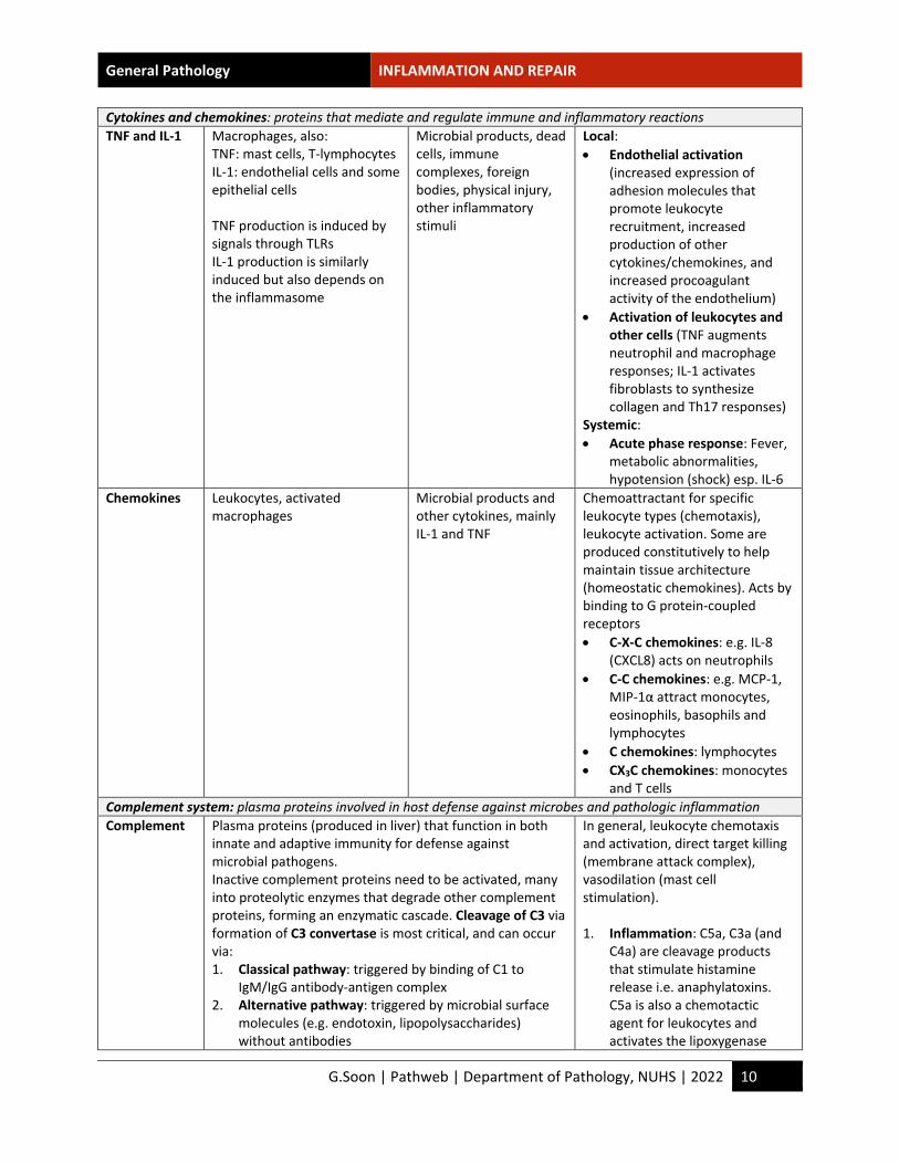
Cytokines and chemokines: proteins that mediate and regulate immune and inflammatory reactions TNF and IL-1 Macrophages, also:
TNF: mast cells, T-lymphocytes IL-1: endothelial cells and some epithelial cells TNF production is induced by signals through TLRs IL-1 production is similarly induced but also depends on the inflammasome
Microbial products, dead cells, immune complexes, foreign bodies, physical injury, other inflammatory stimuli
Local: • Endothelial activation
(increased expression of adhesion molecules that promote leukocyte recruitment, increased production of other cytokines/chemokines, and increased procoagulant activity of the endothelium)
• Activation of leukocytes and other cells (TNF augments neutrophil and macrophage responses; IL-1 activates fibroblasts to synthesize collagen and Th17 responses)
Systemic: • Acute phase response: Fever,
metabolic abnormalities, hypotension (shock) esp. IL-6
Chemokines Leukocytes, activated macrophages
Microbial products and other cytokines, mainly IL-1 and TNF
Chemoattractant for specific leukocyte types (chemotaxis), leukocyte activation. Some are produced constitutively to help maintain tissue architecture (homeostatic chemokines). Acts by binding to G protein-coupled receptors • C-X-C chemokines: e.g. IL-8
(CXCL8) acts on neutrophils • C-C chemokines: e.g. MCP-1,
MIP-1α attract monocytes, eosinophils, basophils and lymphocytes
• C chemokines: lymphocytes • CX3C chemokines: monocytes
and T cells Complement system: plasma proteins involved in host defense against microbes and pathologic inflammation Complement Plasma proteins (produced in liver) that function in both
innate and adaptive immunity for defense against microbial pathogens. Inactive complement proteins need to be activated, many into proteolytic enzymes that degrade other complement proteins, forming an enzymatic cascade. Cleavage of C3 via formation of C3 convertase is most critical, and can occur via: 1. Classical pathway: triggered by binding of C1 to
IgM/IgG antibody-antigen complex 2. Alternative pathway: triggered by microbial surface
molecules (e.g. endotoxin, lipopolysaccharides) without antibodies
In general, leukocyte chemotaxis and activation, direct target killing (membrane attack complex), vasodilation (mast cell stimulation). 1. Inflammation: C5a, C3a (and
C4a) are cleavage products that stimulate histamine release i.e. anaphylatoxins. C5a is also a chemotactic agent for leukocytes and activates the lipoxygenase

General Pathology INFLAMMATION AND REPAIR
G.Soon | Pathweb | Department of Pathology, NUHS | 2022 11
3. Lectin pathway: plasma mannose-binding lectin binds to carbohydrates on microbes and directly activates C1
C3 is cleaved into C3a and C3b: C3a is released, while C3b binds to the cell/molecule where complement is activated. C3b also binds to other previously generated complement fragments to form C5 convertase, which cleaves C5 to release C5a and leave C5b attached to the cell surface. C5b then binds to the late components (C6-C9), resulting in formation of the membrane attack complex (MAC), which comprises multiple C9 molecules. Regulators of complement activation and cascade (expressed on normal host cells): • C1 inhibitor: blocks activation of C1 (the first protein
of classical pathway) • Decay accelerating factor (DAF): prevents formation
of C3 convertases • CD59: inhibits formation of MAC • Complement Factor H: inhibits the alternative
pathway by promoting destruction of C3b and turnover of C3 convertase
pathway of AA metabolism in neutrophils and monocytes
2. Opsonisation and phagocytosis: C3b and its cleavage product (inactive C3b), when fixed to a microbial cell wall, act as opsonins and promote phagocytosis by neutrophils and macrophages
3. Cell lysis: Deposition of MAC makes the cells permeable to water and ions, resulting in osmotic lysis (esp. important in microbes with thin cell walls e.g. Neisseria bacteria)
Others Platelet activating factor (PAF)
Leukocytes, mast cells, platelets, endothelial cells Phospholipid-derived
Platelet aggregation Vasoconstriction and bronchoconstriction (at low concentrations) vasodilation and increased vascular permeability
Kinins e.g. Bradykinin
Plasma (produced in liver) Vasoactive peptide derived from kininogens by proteases called kallikreins. Short-lived as quickly inactivated by kininase.
Increased vascular permeability, smooth muscle contraction, vasodilation, pain (similar to histamine)
Inflammatory reaction Principal Mediator Vasodilation Histamine
Prostaglandins Increased vascular permeability Histamine, serotonin
C3a and C5a (indirectly by liberating vasoactive amines) Leukotrienes C4, D4, E4
Chemotaxis, leukocyte recruitment and activation TNF, IL-1, chemokines C3a, C5a Leukotriene B4
Fever IL-1, TNF Prostaglandins
Pain Prostaglandins Bradykinin, Substance P
Tissue damage Lysosomal enzymes of leukocytes Reactive oxygen species
General Pathology INFLAMMATION AND REPAIR
G.Soon | Pathweb | Department of Pathology, NUHS | 2022 12
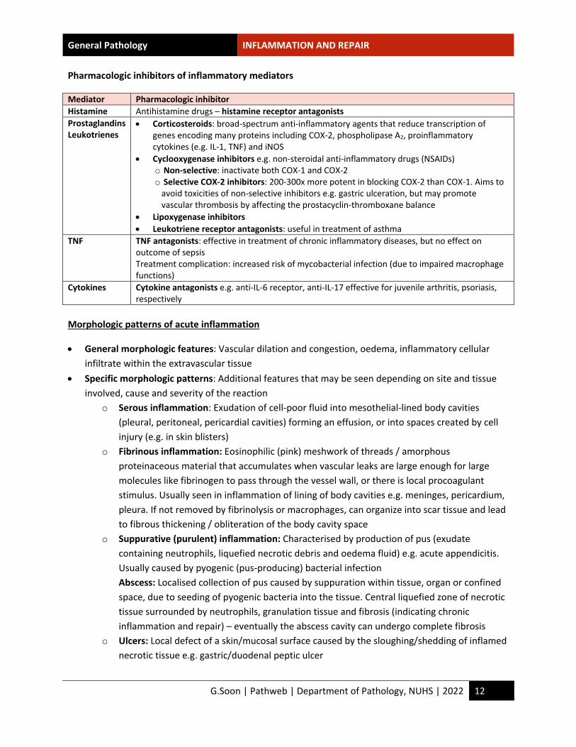
Pharmacologic inhibitors of inflammatory mediators
Mediator Pharmacologic inhibitor Histamine Antihistamine drugs – histamine receptor antagonists Prostaglandins Leukotrienes
• Corticosteroids: broad-spectrum anti-inflammatory agents that reduce transcription of genes encoding many proteins including COX-2, phospholipase A2, proinflammatory cytokines (e.g. IL-1, TNF) and iNOS
• Cyclooxygenase inhibitors e.g. non-steroidal anti-inflammatory drugs (NSAIDs) o Non-selective: inactivate both COX-1 and COX-2 o Selective COX-2 inhibitors: 200-300x more potent in blocking COX-2 than COX-1. Aims to
avoid toxicities of non-selective inhibitors e.g. gastric ulceration, but may promote vascular thrombosis by affecting the prostacyclin-thromboxane balance
• Lipoxygenase inhibitors • Leukotriene receptor antagonists: useful in treatment of asthma
TNF TNF antagonists: effective in treatment of chronic inflammatory diseases, but no effect on outcome of sepsis Treatment complication: increased risk of mycobacterial infection (due to impaired macrophage functions)
Cytokines Cytokine antagonists e.g. anti-IL-6 receptor, anti-IL-17 effective for juvenile arthritis, psoriasis, respectively
Morphologic patterns of acute inflammation
• General morphologic features: Vascular dilation and congestion, oedema, inflammatory cellular infiltrate within the extravascular tissue
• Specific morphologic patterns: Additional features that may be seen depending on site and tissue involved, cause and severity of the reaction
o Serous inflammation: Exudation of cell-poor fluid into mesothelial-lined body cavities (pleural, peritoneal, pericardial cavities) forming an effusion, or into spaces created by cell injury (e.g. in skin blisters)
o Fibrinous inflammation: Eosinophilic (pink) meshwork of threads / amorphous proteinaceous material that accumulates when vascular leaks are large enough for large molecules like fibrinogen to pass through the vessel wall, or there is local procoagulant stimulus. Usually seen in inflammation of lining of body cavities e.g. meninges, pericardium, pleura. If not removed by fibrinolysis or macrophages, can organize into scar tissue and lead to fibrous thickening / obliteration of the body cavity space
o Suppurative (purulent) inflammation: Characterised by production of pus (exudate containing neutrophils, liquefied necrotic debris and oedema fluid) e.g. acute appendicitis. Usually caused by pyogenic (pus-producing) bacterial infection Abscess: Localised collection of pus caused by suppuration within tissue, organ or confined space, due to seeding of pyogenic bacteria into the tissue. Central liquefied zone of necrotic tissue surrounded by neutrophils, granulation tissue and fibrosis (indicating chronic inflammation and repair) – eventually the abscess cavity can undergo complete fibrosis
o Ulcers: Local defect of a skin/mucosal surface caused by the sloughing/shedding of inflamed necrotic tissue e.g. gastric/duodenal peptic ulcer

General Pathology INFLAMMATION AND REPAIR
G.Soon | Pathweb | Department of Pathology, NUHS | 2022 13
Outcomes of acute inflammation
Typically 3 possible outcomes ensue:
1. Complete resolution: Ideal outcome. Usually only if injury is limited or short-lived with little tissue destruction, allowing removal of cellular debris and microbes by macrophages, resorption of oedema fluid and regeneration of damaged cells.
2. Scarring / fibrosis: Healing by connective tissue replacement (organization). Occurs after substantial tissue destruction, the damaged tissue cannot regenerate or fibrinous exudate cannot be cleared adequately
3. Progression to chronic inflammation: Occurs when the acute inflammatory response cannot be resolved due to persistence of injurious agent or other interference with the normal healing process
III. CHRONIC INFLAMMATION
Prolonged host response (weeks or months) to persistent stimuli in which inflammation, tissue injury and attempts at repair coexist in varying combinations
Causes of chronic inflammation
May follow acute inflammation, or begin insidiously as a low-grade response without evidence of preceding acute inflammation:
• Persistent infections: Unresolved acute inflammation with persistent infection can evolve into chronic inflammation e.g. chronic abscess. Microorganisms that are difficult to eradicate e.g. mycobacteria, certain fungi, parasites or viruses can sometimes cause a type of chronic inflammation called granulomatous reaction, which may involve a delayed-type hypersensitivity immune reaction
• Hypersensitivity diseases: Excessive and inappropriate activation of the body’s immune system results in chronic inflammation and tissue damage e.g. rheumatoid arthritis, inflammatory bowel disease. These diseases may show a mixed acute and chronic inflammatory patterns as they are characterized by recurring bouts of inflammation
• Prolonged exposure to potentially toxic agents: Exogenous agents include silica (can result in silicosis). Endogenous agents include cholesterol and other lipids (causing atherosclerosis)
Morphologic features
• Mononuclear cell infiltrate: Macrophages, lymphocytes, plasma cells • Tissue destruction: From the offending agent or inflammatory cells • Healing attempts: Connective tissue replacement of damaged tissue via angiogenesis (proliferation
of small blood vessels) and fibrosis (see Section V. Tissue Repair)

General Pathology INFLAMMATION AND REPAIR
G.Soon | Pathweb | Department of Pathology, NUHS | 2022 14
Cells and mediators of chronic inflammation
Macrophage
• Dominant cell in most chronic inflammatory reactions, by secreting cytokines and growth factors that act on various cells (in particular, activating T lymphocytes), eliminating foreign invaders, causing accompanying tissue injury and also initiating tissue repair
• Some tissues have a resident macrophage population (e.g. Kupffer cells in the liver, microglia in the brain). Most connective tissues also have scattered macrophages, which arise from circulating monocytes in the blood that migrate into the tissues and differentiate into macrophages
o In inflammatory reactions, monocytes begin to emigrate into extravascular tissues (via processes similar to neutrophils) to form the predominant cell type within 48 hours
• Macrophage functions include: o Phagocytosis: ingest and eliminate microbes and dead tissues o Secretion of cytokines and eicosanoids (mediators of inflammation): initiate and propagate
inflammatory reactions o Initiation of tissue repair: involved in scar formation and fibrosis o Interaction with T-lymphocytes: important in cell-mediated immune responses
• 2 major pathways of macrophage activation, resulting in different functional activities. Most reactions contain varying numbers of both macrophage types
Classical macrophage activation (M1) Alternative macrophage activation (M2) Induced by
Microbial products (esp. TLR ligands) e.g. endotoxin, T cell-derived signals e.g. IFN-γ or foreign particles
Cytokines other than IFN-γ e.g. IL-4, IL-13
Secretes • NO, lysosomal enzymes, ROS • IL-1, IL-12, IL-23, chemokines
• Growth factors, TGF-β • IL-10, TGF-β
Function • Microbicidal actions • Inflammation
• Tissue repair, fibrosis • Anti-inflammatory effects
Lymphocytes
• T and B lymphocytes are activated by microbes and other environmental antigens, which can lead to generation of long-lived memory cells causing a more persistent and severe inflammatory reaction
• CD4+ T-cells: secretes cytokines that promotes various types of inflammation o Th1 cells: produce IFN-γ activates macrophages via classical pathway o Th2 cells: produce IL-4, IL-5 and IL-13 alternative macrophage activation; also recruits
and activates eosinophils (important in parasites and allergic inflammation) o Th17 cells: produce IL-17 and other cytokines induces secretion of chemokines to recruit
neutrophils and monocytes • T-Lymphocytes and macrophages interact bidirectionally to propagate chronic inflammation:
macrophages display antigens to T cells, express costimulators and produce cytokines (IL-12) to stimulate T-cell response. Activated T cells in turn produce cytokines that recruit and activate macrophages, resulting in a cycle of inflammation which can result in granulomas
• Activated B lymphocytes and plasma cells: while also often seen in chronic inflammation, the role of the produced antibodies is unclear

General Pathology INFLAMMATION AND REPAIR
G.Soon | Pathweb | Department of Pathology, NUHS | 2022 15
Other cell types
• Eosinophils: abundant in immune reactions mediated by IgE and parasitic infections. Granules contain major basic protein that is toxic to helminths but may also injure host epithelial cells e.g. in allergies
• Mast cells: participates in both acute and chronic inflammatory reactions e.g. immediate hypersensitivity reactions
• Neutrophils: although characteristic of acute inflammatory, can persist in many forms of chronic inflammation (acute on chronic inflammation)
Granulomatous inflammation
Form of chronic inflammation characterized by granulomas (aggregates of activated macrophages, often with T lymphocytes and sometimes associated with necrosis)
• Cellular attempt to contain a difficult to eradicate offending agent • Different types of granulomas may be induced by different causes:
o Foreign body granulomas: incited by inert (non-immunogenic) foreign bodies (e.g. talc, sutures) too large for phagocytosis, in the absence of T-cell mediated immune response
o Immune granulomas: caused by any agent (usually difficult to eradicate e.g. persistent microbe) capable of inducing a persistent T-cell mediated immune response, which then activate macrophages (e.g. via Th1 cells or Th2 cells) E.g. Mycobacterium tuberculosis in tuberculosis, Troponema pallidum in syphilis E.g. immune reaction against intestinal bacterial ?self-antigens in Crohn disease
o Sarcoid granulomas: unknown etiology; tends to be non-caseating and ‘naked’ i.e. not surrounded by lymphocytes
As only a limited number of conditions cause granulomatous inflammation, recognition of granulomas and exclusion of treatable conditions (e.g. tuberculosis) is important. Ancillary investigations include special stains for organisms (Ziehl-Neelson stain for acid-fast bacilli), cultures, molecular techniques and serology
• Microscopy: Activated macrophages in granulomas may have abundant cytoplasm resembling epithelial cells (epithelioid histiocytes) and fuse (multinucleated giant cells). The macrophage aggregates may be surrounded by a collar of lymphocytes or fibroblasts. Granulomas may have a central zone of necrosis (classically seen in Mycobacterium tuberculosis infection) which appears as amorphous eosinophilic granular debris. This is called caseous necrosis due to its resemblance to cheese on gross appearance. Granulomas in Crohn disease, sarcoidosis and foreign body reaction tend to be non-necrotizing / non-caseating. Granulomas generally heal by fibrosis, which can be extensive

General Pathology INFLAMMATION AND REPAIR
G.Soon | Pathweb | Department of Pathology, NUHS | 2022 16
IV. SYSTEMIC EFFECTS OF INFLAMMATION
Inflammation (even if localized) is associated with cytokine-induced systemic reactions called the acute-phase response. The cytokines produced in response to bacterial products and other inflammatory stimuli, in particular TNF, IL-1, IL-6 and interferons are important mediators of this response. The systemic reactions include:
• Fever: elevation of body temperature, often when associated with infection. Thought to be a protective host response. Pyrogens are substances that induce fever, and include bacterial products (exogenous pyrogens e.g. lipopolysaccharides) and cytokines (endogenous pyrogens e.g. IL-1, TNF)
o Exogenous pyrogens act by stimulating release of endogenous pyrogens IL-1 and TNF o IL1- and TNF upregulate cyclooxygenases to synthesize prostaglandins o Prostaglandins (esp. PGE2) synthesized by the vascular and perivascular cells of the
hypothalamus stimulate the production of neurotransmitters by the hypothalamus to reset the body’s steady-state temperature to a higher level, which is accomplished by vasoconstriction (to reduce heat loss) and effects on brown fat and skeletal muscle (to increase heat generation)
• Elevated levels of acute-phase proteins: plasma proteins mostly synthesized in the liver e.g. C-reactive protein (CRP), fibrinogen, serum amyloid A (SAA) protein. Cytokines esp. IL-6, IL-1 and TNF stimulate increased synthesis of these proteins
o Many can bind to microbial cell walls, act as opsonins and fix complement, and also bind chromatin to aid in clearing of necrotic debris
o Fibrinogen binds to red blood cells to form stacks (rouleaux) – these sediment more rapidly than individual RBCs, and can be used as a measure of inflammatory response (erythrocyte sediment rate)
o While acute phase-proteins are beneficial during acute inflammation, prolonged production can have harmful effects (e.g. SAA can result in secondary amyloidosis, elevated CRP is thought to be a marker of increased myocardial infarction risk in patients with coronary artery disease due to predisposition to thrombosis)
• Leukocytosis: especially high in bacterial infections. Extreme elevations that mimic leukemia are called leukemoid reactions
o Occurs initially due to accelerated release of both mature and immature granulocytes from bone marrow by cytokines
o Prolonged infection also induces proliferation of precursors in the bone marrow (via production of colony-stimulating factors by macrophages and marrow stromal cells)
o Bacterial infections usually cause neutrophilia, viral infections lymphocytosis, allergies and helminth infestations eosinophilia. However, certain infections e.g. typhoid fever are associated with leukopenia instead partly due to sequestration of activated leukocytes in vascular spaces and tissues
• Symptoms and signs such as elevated pulse and blood pressure, decreased sweating (blood flow is redirected from cutaneous to deep vascular beds to minimize heat loss), chills, rigors, anorexia, malaise, somnolence

General Pathology INFLAMMATION AND REPAIR
G.Soon | Pathweb | Department of Pathology, NUHS | 2022 17
• Septic shock: a severe often fatal form of systemic inflammatory response syndrome (SIRS) in severe bacterial infections (sepsis), whereby the large amounts of bacteria and their products in the blood stimulate production of high blood levels of cytokines (esp. TNF and IL-1), causing disseminated intravascular coagulation (DIC), hypotensive shock and metabolic disturbances such as insulin resistance and hyperglycemia
V. TISSUE REPAIR
Process of healing - restoration of tissue architecture and function after an injury, set in motion by the inflammatory response. This occurs in 2 ways: regeneration and scarring, both involving cell proliferation and cell-matrix interactions. Mammals have limited capacity for regeneration of most tissues and organs; both regeneration and scar formation therefore contribute to repair
Regeneration
Replacement of damaged components to return to normal state via cell proliferation of differentiated cells that still retain the capacity to proliferate, or via tissue stem cells / progenitors. Restoration of normal tissue structure can only occur if the residual tissue is structurally intact e.g. after partial surgical resection, in contrast to damage by infection or inflammation whereby scarring will also occur
Cell proliferation
• Driven by growth factors, and critically dependent on the integrity of the extracellular matrix (ECM) o Growth factors are typically produced by cells nears the site of damage, mostly by activated
macrophages but also epithelial and stromal cells. All growth factors activate signaling pathways that stimulate DNA replication and also changes in cell metabolism to promote biosynthesis of other cellular components for replication
o Cells also bind to ECM proteins via integrins; signals from integrins can also stimulate cell proliferation
• The capacity for regeneration depends on the tissues’ intrinsic proliferative capacity and the presence of tissue stem cells:
o Labile (continuously dividing) tissues: Cells of these tissues are continuously being lost and replaced by maturation from tissue stem cells and proliferation of mature cells e.g. haematopoietic cells, surface epithelial cells e.g. skin epidermis, mucosal lining of gastrointestinal tract. These tissues can readily regenerate after injury as long as the stem cells are preserved
o Stable tissues: Cells of these tissues are quiescent (in the G0 stage of the cell cycle) with minimal proliferative activity in the normal state, but can divide in response to injury or loss of tissue mass e.g. parenchyma of most solid organs e.g. liver, kidney; also endothelial cells, fibroblasts, smooth muscle cells. Generally limited capacity to regenerate after injury except for liver
General Pathology INFLAMMATION AND REPAIR
G.Soon | Pathweb | Department of Pathology, NUHS | 2022 18
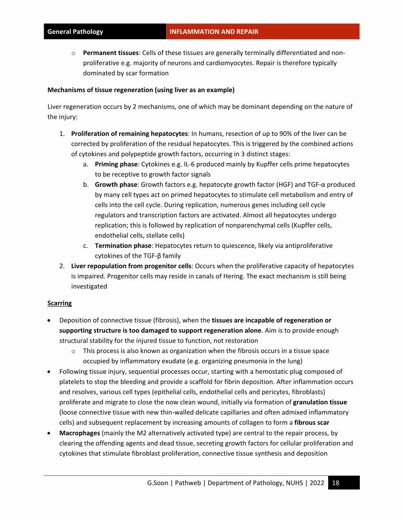
o Permanent tissues: Cells of these tissues are generally terminally differentiated and non-proliferative e.g. majority of neurons and cardiomyocytes. Repair is therefore typically dominated by scar formation
Mechanisms of tissue regeneration (using liver as an example)
Liver regeneration occurs by 2 mechanisms, one of which may be dominant depending on the nature of the injury:
1. Proliferation of remaining hepatocytes: In humans, resection of up to 90% of the liver can be corrected by proliferation of the residual hepatocytes. This is triggered by the combined actions of cytokines and polypeptide growth factors, occurring in 3 distinct stages:
a. Priming phase: Cytokines e.g. IL-6 produced mainly by Kupffer cells prime hepatocytes to be receptive to growth factor signals
b. Growth phase: Growth factors e.g. hepatocyte growth factor (HGF) and TGF-α produced by many cell types act on primed hepatocytes to stimulate cell metabolism and entry of cells into the cell cycle. During replication, numerous genes including cell cycle regulators and transcription factors are activated. Almost all hepatocytes undergo replication; this is followed by replication of nonparenchymal cells (Kupffer cells, endothelial cells, stellate cells)
c. Termination phase: Hepatocytes return to quiescence, likely via antiproliferative cytokines of the TGF-β family
2. Liver repopulation from progenitor cells: Occurs when the proliferative capacity of hepatocytes is impaired. Progenitor cells may reside in canals of Hering. The exact mechanism is still being investigated
Scarring
• Deposition of connective tissue (fibrosis), when the tissues are incapable of regeneration or supporting structure is too damaged to support regeneration alone. Aim is to provide enough structural stability for the injured tissue to function, not restoration
o This process is also known as organization when the fibrosis occurs in a tissue space occupied by inflammatory exudate (e.g. organizing pneumonia in the lung)
• Following tissue injury, sequential processes occur, starting with a hemostatic plug composed of platelets to stop the bleeding and provide a scaffold for fibrin deposition. After inflammation occurs and resolves, various cell types (epithelial cells, endothelial cells and pericytes, fibroblasts) proliferate and migrate to close the now clean wound, initially via formation of granulation tissue (loose connective tissue with new thin-walled delicate capillaries and often admixed inflammatory cells) and subsequent replacement by increasing amounts of collagen to form a fibrous scar
• Macrophages (mainly the M2 alternatively activated type) are central to the repair process, by clearing the offending agents and dead tissue, secreting growth factors for cellular proliferation and cytokines that stimulate fibroblast proliferation, connective tissue synthesis and deposition

General Pathology INFLAMMATION AND REPAIR
G.Soon | Pathweb | Department of Pathology, NUHS | 2022 19
Steps in scar formation (connective tissue repair)
• Angiogenesis: Process of new blood vessel formation from existing vessels. Seen in healing, as well as development of collateral blood supply in ischaemia and tumour growth. Involves the following steps via Notch and other signaling pathways, cell-cell interactions, ECM proteins (which interact with integrin receptors and provide scaffolding for vessel growth) and tissue enzymes (e.g. metalloproteinases that degrade the ECM to permit remodeling):
o Vasodilation and increased permeability: in response to nitric oxide and vascular endothelial growth factor (VEGF)
o Separation of pericytes from the abluminal surface and breakdown of basement membrane to allow formation of a vessel sprout; via angiopoietins (Ang 1 and 2)
o Migration of endothelial cells: towards the tissue injury o Proliferation of endothelial cells: just behind the leading front of migrating cells; mainly
stimulated by fibroblast growth factors (FGF) o Remodeling into capillary tubules o Recruitment of periendothelial cells e.g. pericytes/smooth muscle cells to form mature
vessels; via platelet-derived growth factor (PDGF) o Suppression of endothelial proliferation and migration: also deposition of basement
membrane; via TGF-β • Deposition of connective tissue: involves 1) migration and proliferation of fibroblasts into site of
injury, and 2) deposition of ECM proteins produced by these cells, mediated by locally produced cytokines and growth factors produced by M2 macrophages and inflammatory cells, esp. TGF-β
o As healing progresses and the scar matures, there is increased ECM deposition and vascular regression with decreasing proliferating fibroblasts
o Some fibroblasts may also transform into myofibroblasts (acquiring features of smooth muscle cells) that contribute to scar contraction
• Remodeling of connective tissue: After deposition of connective tissue, remodeling occurs depending on the balance between synthesis and degradation of ECM proteins by matrix metalloproteinases (MMPs)
o MMPs are produced as inactive precursors (zymogens) by a variety of cell types (fibroblasts, macrophages, neutrophils, synovial cells, some epithelial cells) via growth factors and cytokines, and activated by proteases present only at sites of injury
o MMPs are inhibited by specific tissue inhibitors of metalloproteinases (TIMPs) produced by most mesenchymal cells

General Pathology INFLAMMATION AND REPAIR
G.Soon | Pathweb | Department of Pathology, NUHS | 2022 20
Factors that influence tissue repair: extrinsic vs intrinsic, systemic vs local factors
Local factors
• Infection: Clinically one of the most important causes of delayed healing. Prolongs inflammation and likely increases local tissue injury
• Mechanical factors: e.g. Increased local pressure, torsion, causing wound dehiscence • Poor vascular perfusion: e.g. due to peripheral vascular disease, arteriosclerosis, diabetes,
obstructed venous drainage in varicose veins • Foreign bodies: perpetuates chronic inflammation • Type, extent and location of tissue injury: Injury to permanent tissues will always result in scarring • Location of injury: in tissue spaces e.g. serous cavities, inflammation results in extensive exudates,
which can either undergo resolution (via proteolytic digestion and resorption) or organisation (when granulation tissue grows into the exudate and fibrous scarring occurs)
Systemic factors
• Diabetes: Metabolic disease causing impaired tissue healing for various reasons (macro and microangiopathy, prone to infections)
• Nutritional status: Protein deficiency and vitamin C deficiency inhibit collagen synthesis and retard healing
• Glucocorticoids (steroids): Anti-inflammatory effects may cause scar weakness due to inhibition of TGF-β, although may be useful in certain circumstances e.g. to reduce scarring
• Age: Younger patients generally heal better
Abnormalities in tissue repair
• Deficient scar formation (chronic wounds): Due to local and systemic factors affecting tissue repair, e.g. venous leg ulcers (due to venous congestion causing poor oxygenation of local tissues), arterial ulcers (in patients with peripheral vascular disease), diabetic ulcers, pressure sores (due to prolonged mechanical tissue compression and local ischemia), wound dehiscence (when a wound reopens e.g. due to obesity, malnutrition, infections)
• Excessive scarring: Can cause hypertrophic scars (raised scar that regresses over months, generally after thermal or traumatic injuries involving the deep dermis) and keloids (when the scar tissue grows beyond the boundaries of the original wound and does not regress; seems to be an individual predisposition). Excessive granulation tissue that protrudes above the level of the surrounding skin and blocks re-epithelization can also occur, requiring surgical removal/cautery
• Contracture: Excess contraction of the wound, resulting in deformities and impairment of joint movement. Commonly after serious burns

General Pathology INFLAMMATION AND REPAIR
G.Soon | Pathweb | Department of Pathology, NUHS | 2022 21
Examples of tissue repair and fibrosis
Cutaneous wound healing
Healing occurs via first or second intention depending on the nature and size of wound
• Healing by first intention (primary union): When the injury is relatively limited (with closely apposed wound edges), involving mostly the epithelial layer, with only focal disruption of the epithelial basement membrane and death of relatively few epithelial and connective tissue cells e.g. a clean uninfected surgical incision
o After the wound occurs, coagulation pathways are activated, forming a blood clot that stops bleeding and supports migrating cells attracted by cytokines, chemokines and growth factors
o Within 24 hours, neutrophils are seen at the incision margin and migrating towards the fibrin clot. Within 24-48 hours, epithelial cells from both skin edges migrate and proliferate along the dermis, forming a thin and continuous epithelial layer that closes the wound
o By day 3, neutrophils are replaced by macrophages, and blood clot replaced by granulation tissue, which peaks in day 5. Fibroblasts progressively migrate into the oedematous vascular granulation tissue to lay down collagen
o During the 2nd week, there is conversion of the granulation tissue into a collagenous scar, which is eventually covered an essentially normal epidermis by the end of the first month
o The tensile strength of the wound increases over the first 2 months of healing by cross-linking of collagen fibers and increased fiber size, and reaches a maximum of 70-80% of normal by 3 months
• Healing by second intention (secondary union): When the wound is larger and gaping with edges not apposed. This means the fibrin clot is therefore also larger, with more exudate, necrosis and inflammation that can cause greater secondary inflammation-mediated injury. Destroyed dermal appendages at the wound site are permanently lost. The larger amount of granulation tissue formed also results in a greater mass of scar tissue. Wound contraction via action of myofibroblasts is important to help close the wound by decreasing the wound surface area (reduction in 5-10% in size within 6 weeks)
Fibrosis in parenchymal organs
Mechanism of fibrosis (excessive deposition of collagen and other ECM components in tissue) is basically similar to scar formation in the skin during tissue repair. However, fibrosis can be responsible for substantial organ dysfunction / failure e.g. liver cirrhosis, fibrosing diseases of the lung