The Raffles Bulletin of Zoology - NUS Digital Libraries
423
An International Journal of Southeast Asian Zoology Articles appearing in this journal are indexed in: SCIENCE CITATION INDEX ® ; CURRENT CONTENTS ® ; AGRICULTURE, BIOLOGY & ENVIRONMENTAL SCIENCE; SCISEARCH ® ; RESEARCH ALERT ® ; BIOLOGICAL ABSTRACTS ® ; CAMBRIDGE SCIENTIFIC ABSTRACTS ® ; AQUATIC SCIENCES & FISHERIES ABSTRACTS On a new species of freshwater crab of the genus Ovitamon Ng & Takeda, 1992 (Crustacea: Brachyura: Potamidae) from Panay Island, Philippines. Daniel Edison M. Husana, Tomoki Kase and Peter K. L. Ng................................................................................................. 651 Synonymy of Spicatella Thibaud, 2002 with Delamarephorura Weiner & Najt, 1999, and description of two new species (Collembola: Tullbergiidae). Charlene Janion, Louis Deharveng and Wanda Maria Weiner......................................................................................... 657 Guide to the aquatic Heteroptera of Singapore and Peninsular Malaysia. XI. Infraorder Nepomorpha—Families Naucoridae and Aphelocheiridae. Dan A. Polhemus and John T. Polhemus....................................................................................................................................................... 665 A review of the genus Arocatus from Palaearctic and Oriental regions (Hemiptera: Heteroptera: Lygaeidae). Cuiqing Gao, Előd Kondorosy and Wenjun Bu ................................................................................................................................................................................................. 687 A taxonomic review of common but little known crickets from Singapore and the Philippines (Insecta: Orthoptera: Eneopterinae). Tony Robillard and Ming Kai Tan........................................................................................................................................................................... 705 Taxonomic notes on the species of the genus Malayepipona Giordani Soika (Hymenoptera: Vespidae: Eumeninae) from northern Vietnam, with description of three new species. Nguyen Thi Phuong Lien and James M. Carpenter...................................................................... 727 Three new species of freshwater halfbeaks (Teleostei: Zenarchopteridae: Hemirhamphodon) from Borneo. Heok Hui Tan and Kelvin K. P. Lim .................................................................................................................................................................................................................... 735 Review of Stiphodon (Gobiidae: Sicydiinae) from western Sumatra, with description of a new species. Ken Maeda and Heok Hui Tan ..... ............................................................................................................................................................................................................................ 749 Nomenclature and identity of the tongue soles Paraplagusia bilineata, “Cynoglossus bilineatus” and Paraplagusia blochii (Teleostei: Pleuronectiformes). Maurice Kottelat ............................................................................................................................................................. 763 CONSERVATION AND ECOLOGY Recovery of litter and soil invertebrate communities following swidden cultivation in Sarawak, Malaysia. Megumi Yoshima, Yoko Takematsu, Aogu Yoneyama and Michiko Nakagawa ...................................................................................................................................................... 767 The nuisance midges (Diptera: Chironomidae) of Singapore’s Pandan and Bedok reservoirs. P. S. Cranston, Y. C. Ang, A. Heyzer, R. B. H. Lim, W. H. Wong, J. M. Woodford and R. Meier ........................................................................................................................................ 779 Diversity and assemblage patterns of juvenile and small sized fishes in the nearshore habitats of the Gulf of Thailand. Surasak Sichum, Pitiwong Tantichodok and Tuantong Jutagate.............................................................................................................................................. 795 A mark-recapture study of a dog-faced water snake Cerberus schneiderii (Colubridae: Homalopsidae) population in Sungei Buloh Wetland Reserve, Singapore. C. K. Chim and C. H. Diong ..........................................................................................................................................811 Ornithology of the Kelabit Highlands of Sarawak, Malaysia. Frederick H. Sheldon, Clare E. Brown, Mustafa Abdul Rahman, Guan Khoon Tay and Robert G. Moyle ................................................................................................................................................................... 827 Variation in the nucleolar organiser regions of the long-tailed giant rats (Rodentia, Muridae, genus Leopoldamys) in Malaysia. Hoi Sen Yong, Phaik Eem Lim, Daicus M. Belabut and Praphathip Eamsobhana ........................................................................................................... 855 Camera-trapping survey of mammals in and around Imbak Canyon Conservation Area in Sabah, Malaysian Borneo. Henry Bernard, Abdul Hamid Ahmad, Jedediah Brodie, Anthony J. Giordano, Maklarin Lakim, Rahimatsah Amat, Sharon Koh Pei Hue, Lee Shan Khee, Augustine Tuuga, Peter Titol Malim, Darline Lim-Hasegawa, Yap Sau Wai and Waidi Sinun ............................................................. 861 Insights into the spatial and temporal ecology of the Sunda clouded leopard Neofelis diardi. Andrew J. Hearn, Joanna Ross, Daniel Pamin, Henry Bernard, Luke Hunter and David W. Macdonald............................................................................................................................ 871 Population estimates and distribution patterns of Irrawaddy dolphins (Orcaella brevirostris) and Indo-Pacific finless porpoises (Neophocaena phocaenoides) in the Kuching Bay, Sarawak. Gianna Minton, Cindy Peter, Anna Norliza Zulkifli Poh, Jenny Ngeian, Gill Braulik, Philip S. Hammond and Andrew Alek Tuen ............................................................................................................................................................ 877 CORRIGENDA Corrigendum. Soo O. Y. M. and Lim L. H. S. ................................................................................................................................................ 889 Corrigendum. Tan H. H. ................................................................................................................................................................................... 893 The Raffles Bulletin of Zoology CONTENTS Editorial. Tan H. H. .................................................................................................................................................................................... i TAXONOMY AND SYSTEMATICS A first record of freshwater sponge from Singapore and redescription of Eunapius conifer (Annandale, 1916) (Haplosclerida: Spongillina: Spongillidae). Swee-Cheng Lim and Koh-Siang Tan ..........................................................................................................................453 A taxonomic review of the genus Asteromorpha Lütken (Echinodermata: Ophiuroidea: Euryalidae). Masanori Okanishi, Jennifer M. Olbers and Toshihiko Fujita .................................................................................................................................................................461 Bravohollisia geruti, new species (Monogenea: Ancyrocephalidae) from Pomadasys hasta (Osteichthyes: Haemulidae) of Peninsular Malaysia. W. B. Tan and L. H. S. Lim..................................................................................................................................................481 Revision of Anteropora (Cestoda: Lecanicephalidea) and descriptions of five new species from stingrays (Myliobatiformes: Dasyatidae) in Borneo. Kendra R. Mojica, Kirsten Jensen and Janine N. Caira ................................................................................................491 A new species of Phalium Link, 1807 (Gastropoda: Tonnoidea: Cassidae) from the Sunda Shelf. S. K. Tan, H. E. Ng and L. H. S. Nguang ....................................................................................................................................................................................................507 Saging cebuana, a new genus and species of taeniacanthid copepod (Cyclopoida) parasitic on a filefish (Actinopterygii: Monacanthidae) collected from Cebu Island, the Philippines. Daisuke Uyeno, Danny Tang and Kazuya Nagasawa ................................................515 Anostraca catalogus (Crustacea: Branchiopoda). D. Christopher Rogers ...........................................................................................525 A new ingolfiellid amphipod crustacean from sandy beaches of the Gura Ici islands, western Halmahera (north Moluccas). R. Vonk and D. Jaume ..........................................................................................................................................................................................547 Three isopod parasites (Bopyridae: Pseudioninae), including two new species, of hermit crabs from the South China Sea. Jianmei An, Xinzheng Li and John C. Markham ....................................................................................................................................................561 Two new species of Acutigebia (Crustacea: Decapoda: Gebiidea: Upogebiidae) from the South China Sea. Wenliang Liu and Ruiyu Liu............................................................................................................................................................................................................571 Verification of four species of the mud lobster genus Thalassina (Crustacea: Decapoda: Gebiidea: Thalassinidae) using molecular and morphological characters. Moh H. H., Chong V. C., Lim P. E., Tan J. and Daley G. ......................................................................577 Six new species of the hermit crab genus Decaphyllus de Saint Laurent, 1968 (Crustacea: Decapoda: Anomura: Paguridae) from the Boholo Sea, the Philippines, and the Ryukyu Islands, Japan. Tomoyuki Komai and Dwi Listyo Rahayu .......................................589 Two new species of Pylopaguropsis Alcock (Crustacea: Decapoda: Anomura: Paguridae) from the Philippines. Dwi Listyo Rahayu and Tomoyuki Komai .............................................................................................................................................................................621 A new species of the genus Parasesarma (Crustacea: Brachyura: Sesarmidae) from Taiwan and the Philippines, and redescription of P. jamelense (Rathbun, 1914). Dwi Listyo Rahayu and Jheng-Jhang Li ...........................................................................................633 Systematics of the Indo-West Pacific broad-fronted fiddler crabs (Crustacea: Ocypodidae: genus Uca). Hsi-Te Shih, Peter K. L. Ng and Min-Yun Liu ....................................................................................................................................................................................641 (continues on back cover) Volume 61 30 August 2013 Number 2 CONTENTS (continued from front cover) Layout by Photoplates Pte ltd Published by the Department of Biological Sciences, National University of Singapore THE RAFFLES BULLETIN OF ZOOLOGY 2013 VOL. 61(2)
-
Upload
khangminh22 -
Category
Documents
-
view
1 -
download
0
Transcript of The Raffles Bulletin of Zoology - NUS Digital Libraries

An International Journal of Southeast Asian Zoology
Articles appearing in this journal are indexed in: SCIENCE CITATION INDEX®; CURRENT CONTENTS®; AGRICULTURE, BIOLOGY & ENVIRONMENTAL SCIENCE; SCISEARCH®; RESEARCH ALERT®; BIOLOGICAL ABSTRACTS®; CAMBRIDGE SCIENTIFIC ABSTRACTS®; AQUATIC SCIENCES & FISHERIES ABSTRACTS
On a new species of freshwater crab of the genus Ovitamon Ng & Takeda, 1992 (Crustacea: Brachyura: Potamidae) from Panay Island, Philippines. Daniel Edison M. Husana, Tomoki Kase and Peter K. L. Ng ................................................................................................. 651
Synonymy of Spicatella Thibaud, 2002 with Delamarephorura Weiner & Najt, 1999, and description of two new species (Collembola: Tullbergiidae). Charlene Janion, Louis Deharveng and Wanda Maria Weiner ......................................................................................... 657
Guide to the aquatic Heteroptera of Singapore and Peninsular Malaysia. XI. Infraorder Nepomorpha—Families Naucoridae and Aphelocheiridae. Dan A. Polhemus and John T. Polhemus ....................................................................................................................................................... 665
A review of the genus Arocatus from Palaearctic and Oriental regions (Hemiptera: Heteroptera: Lygaeidae). Cuiqing Gao, Előd Kondorosy and Wenjun Bu ................................................................................................................................................................................................. 687
A taxonomic review of common but little known crickets from Singapore and the Philippines (Insecta: Orthoptera: Eneopterinae). Tony Robillard and Ming Kai Tan ........................................................................................................................................................................... 705
Taxonomic notes on the species of the genus Malayepipona Giordani Soika (Hymenoptera: Vespidae: Eumeninae) from northern Vietnam, with description of three new species. Nguyen Thi Phuong Lien and James M. Carpenter ...................................................................... 727
Three new species of freshwater halfbeaks (Teleostei: Zenarchopteridae: Hemirhamphodon) from Borneo. Heok Hui Tan and Kelvin K. P. Lim .................................................................................................................................................................................................................... 735
Review of Stiphodon (Gobiidae: Sicydiinae) from western Sumatra, with description of a new species. Ken Maeda and Heok Hui Tan ..... ............................................................................................................................................................................................................................ 749
Nomenclature and identity of the tongue soles Paraplagusia bilineata, “Cynoglossus bilineatus” and Paraplagusia blochii (Teleostei: Pleuronectiformes). Maurice Kottelat ............................................................................................................................................................. 763
CONSERVATION AND ECOLOGY
Recovery of litter and soil invertebrate communities following swidden cultivation in Sarawak, Malaysia. Megumi Yoshima, Yoko Takematsu, Aogu Yoneyama and Michiko Nakagawa ...................................................................................................................................................... 767
The nuisance midges (Diptera: Chironomidae) of Singapore’s Pandan and Bedok reservoirs. P. S. Cranston, Y. C. Ang, A. Heyzer, R. B. H. Lim, W. H. Wong, J. M. Woodford and R. Meier ........................................................................................................................................ 779
Diversity and assemblage patterns of juvenile and small sized fishes in the nearshore habitats of the Gulf of Thailand. Surasak Sichum, Pitiwong Tantichodok and Tuantong Jutagate.............................................................................................................................................. 795
A mark-recapture study of a dog-faced water snake Cerberus schneiderii (Colubridae: Homalopsidae) population in Sungei Buloh Wetland Reserve, Singapore. C. K. Chim and C. H. Diong ..........................................................................................................................................811
Ornithology of the Kelabit Highlands of Sarawak, Malaysia. Frederick H. Sheldon, Clare E. Brown, Mustafa Abdul Rahman, Guan Khoon Tay and Robert G. Moyle ................................................................................................................................................................... 827
Variation in the nucleolar organiser regions of the long-tailed giant rats (Rodentia, Muridae, genus Leopoldamys) in Malaysia. Hoi Sen Yong, Phaik Eem Lim, Daicus M. Belabut and Praphathip Eamsobhana ........................................................................................................... 855
Camera-trapping survey of mammals in and around Imbak Canyon Conservation Area in Sabah, Malaysian Borneo. Henry Bernard, Abdul Hamid Ahmad, Jedediah Brodie, Anthony J. Giordano, Maklarin Lakim, Rahimatsah Amat, Sharon Koh Pei Hue, Lee Shan Khee, Augustine Tuuga, Peter Titol Malim, Darline Lim-Hasegawa, Yap Sau Wai and Waidi Sinun ............................................................. 861
Insights into the spatial and temporal ecology of the Sunda clouded leopard Neofelis diardi. Andrew J. Hearn, Joanna Ross, Daniel Pamin, Henry Bernard, Luke Hunter and David W. Macdonald ............................................................................................................................ 871
Population estimates and distribution patterns of Irrawaddy dolphins (Orcaella brevirostris) and Indo-Pacific finless porpoises (Neophocaena phocaenoides) in the Kuching Bay, Sarawak. Gianna Minton, Cindy Peter, Anna Norliza Zulkifli Poh, Jenny Ngeian, Gill Braulik, Philip S. Hammond and Andrew Alek Tuen ............................................................................................................................................................ 877
CORRIGENDA
Corrigendum. Soo O. Y. M. and Lim L. H. S. ................................................................................................................................................ 889
Corrigendum. Tan H. H. ................................................................................................................................................................................... 893
The Raffles Bulletinof Zoology
CONTENTS
Editorial. Tan H. H. .................................................................................................................................................................................... i
TAXONOMY AND SYSTEMATICS
A first record of freshwater sponge from Singapore and redescription of Eunapius conifer (Annandale, 1916) (Haplosclerida: Spongillina: Spongillidae). Swee-Cheng Lim and Koh-Siang Tan ..........................................................................................................................453
A taxonomic review of the genus Asteromorpha Lütken (Echinodermata: Ophiuroidea: Euryalidae). Masanori Okanishi, Jennifer M. Olbers and Toshihiko Fujita .................................................................................................................................................................461
Bravohollisia geruti, new species (Monogenea: Ancyrocephalidae) from Pomadasys hasta (Osteichthyes: Haemulidae) of Peninsular Malaysia. W. B. Tan and L. H. S. Lim ..................................................................................................................................................481
Revision of Anteropora (Cestoda: Lecanicephalidea) and descriptions of five new species from stingrays (Myliobatiformes: Dasyatidae) in Borneo. Kendra R. Mojica, Kirsten Jensen and Janine N. Caira ................................................................................................491
A new species of Phalium Link, 1807 (Gastropoda: Tonnoidea: Cassidae) from the Sunda Shelf. S. K. Tan, H. E. Ng and L. H. S. Nguang ....................................................................................................................................................................................................507
Saging cebuana, a new genus and species of taeniacanthid copepod (Cyclopoida) parasitic on a filefish (Actinopterygii: Monacanthidae) collected from Cebu Island, the Philippines. Daisuke Uyeno, Danny Tang and Kazuya Nagasawa ................................................515
Anostraca catalogus (Crustacea: Branchiopoda). D. Christopher Rogers ...........................................................................................525
A new ingolfiellid amphipod crustacean from sandy beaches of the Gura Ici islands, western Halmahera (north Moluccas). R. Vonk and D. Jaume ..........................................................................................................................................................................................547
Three isopod parasites (Bopyridae: Pseudioninae), including two new species, of hermit crabs from the South China Sea. Jianmei An, Xinzheng Li and John C. Markham ....................................................................................................................................................561
Two new species of Acutigebia (Crustacea: Decapoda: Gebiidea: Upogebiidae) from the South China Sea. Wenliang Liu and Ruiyu Liu............................................................................................................................................................................................................571
Verification of four species of the mud lobster genus Thalassina (Crustacea: Decapoda: Gebiidea: Thalassinidae) using molecular and morphological characters. Moh H. H., Chong V. C., Lim P. E., Tan J. and Daley G. ......................................................................577
Six new species of the hermit crab genus Decaphyllus de Saint Laurent, 1968 (Crustacea: Decapoda: Anomura: Paguridae) from the Boholo Sea, the Philippines, and the Ryukyu Islands, Japan. Tomoyuki Komai and Dwi Listyo Rahayu .......................................589
Two new species of Pylopaguropsis Alcock (Crustacea: Decapoda: Anomura: Paguridae) from the Philippines. Dwi Listyo Rahayu and Tomoyuki Komai .............................................................................................................................................................................621
A new species of the genus Parasesarma (Crustacea: Brachyura: Sesarmidae) from Taiwan and the Philippines, and redescription of P. jamelense (Rathbun, 1914). Dwi Listyo Rahayu and Jheng-Jhang Li ...........................................................................................633
Systematics of the Indo-West Pacific broad-fronted fiddler crabs (Crustacea: Ocypodidae: genus Uca). Hsi-Te Shih, Peter K. L. Ng and Min-Yun Liu ....................................................................................................................................................................................641
(continues on back cover)
Volume 61 30 August 2013 Number 2
CONTENTS(continued from front cover)
Layout by Photoplates Pte ltd
Published by the Department of Biological Sciences, National University of Singapore
TH
E R
AFFL
ES B
UL
LE
TIN
OF Z
OO
LO
GY
2013
VO
L. 61(2)

The Raffl es Bulletin of Zoology | R B Z | rmbr.nus.edu.sg/rbz/The Raffl es Bulletin of Zoology (RBZ) is an online, peer-reviewed journal which publishes high quality papers in Taxonomy, Systematics, Ecology, and Conservation Biology of animals from Southeast Asia and its adjacent areas. The Journal aims to build up quality information on the “animal diversity” of Southeast Asia in particular. Papers from outside the stated geographic range that deal with material deposited in the Zoological Reference Collection (ZRC) of the Raffl es Museum of Biodiversity Research (RMBR), National University of Singapore (NUS) will also be published. Both descriptive and experimental papers will be considered. Single species descriptions and ecosystem studies will be considered for publication. Papers outside the stated policy will be accepted at the discretion of the Editors/Editorial Board.
EDITORIAL BOARDBarry W. Brook (Charles Darwin University, Australia) • Chou Loke Ming (NUS, Singapore) • Indraneil Das (Universiti Malaysia Sarawak, Malaysia) • R. A. I. Drew (Griffith University, Australia) • Hugh A. Ford (University of New England, Australia) • Fabian Herder (Zoologisches Forschungsmuseum Alexander Koenig, Germany) • Michel Jangoux (University of Brussels, Belgium) • Maurice Kottelat (Cornol, Switzerland) • Damir Kovac (Senckenberg Museum, Germany) • Kelvin K. P. Lim (NUS, Singapore) • John E. Randall (Bernice P. Bishop Museum, USA) • Fred Sheldon (Louisiana State University, USA) • Daniel Simberloff (University of Tennessee, USA) • S. H. Tan (NUS, Singapore) • Y. Tsubaki (Center for Ecological Research, Kyoto University, Japan) • E. O. Wilson (Harvard University, USA) • S. Yamagishi (Kyoto University, Japan) • H. S. Yong (University of Malaya, Malaysia).
MANAGING EDITORIAL BOARDEDITOR-IN-CHIEF: Peter K. L. Ng (NUS, Singapore) MANAGING EDITOR: Tan Heok Hui (NUS, Singapore) ASSOCIATE EDITORS: Kevin Conway (Texas A&M University, USA) • Hwang Wei Song (NUS, Singapore) • Zeehan Jaafar (NUS, Singapore) • Sebastian Klaus (Johan Wolgang Goethe-Universität Biologicum, Germany) • Jeffrey Kwik Teik Beng (NUS, Singapore) • Leong Tzi Ming (NUS, Singapore) • Li Daiqin (NUS, Singapore) • Norman Lim T-Lon (University California, Davis, USA) • Rudolf Meier (NUS, Singapore) • Jose Christopher E. Mendoza (NUS, Singapore) • Tohru Naruse (University of Ryukyus, Japan) • James Reimer (University of Ryukyu, Japan) • Frank E. Rheindt (NUS, Singapore) • Tan Koh Siang (TMSI, Singapore) • Tan Siong Kiat (NUS, Singapore) • Peter A. Todd (NUS, Singapore) • Tran Anh Duc (Hanoi University of Science, Vietnam) • Darren C. J. Yeo (NUS, Singapore) SENIOR COPY EDITOR: Hazelina H. T. Yeo (NUS, Singapore) COPY EDITOR: Jeremy W. L. Yeo (NUS, Singapore) PRODUCTION EDITOR: Hazelina H. T. Yeo (NUS, Singapore) EDITORIAL ADMINISTRATOR: Greasi Simon (NUS, Singapore) WEBMASTER: Chua Keng Soon (NUS, Singapore)
PUBLICATION DETAILSThe Raffl es Bulletin of Zoology will consist of a single volume (two issues) each year, continuing the sequence of its two predecessors, Bulletin of the Raffl es Museum (1928–1960) and Bulletin of the National Museum of Singapore (1961–1970). A separately numbered supplement series will be published as and when manuscripts and funding permit. About 14 hard-copies of the Journal will be deposited in major publicly accessible libraries to satisfy Article 8.6 of the Fourth Edition of the International Code of Zoological Nomenclature (1999) so that all new names published in the RBZ are considered to be published and available.
COPYRIGHT AND EXCHANGESAll articles published by the RBZ may be downloaded from http://rmbr.nus.edu.sg/rbz/biblio/ for research purposes. A condition of publication is that authors assign publication rights to The Raffl es Bulletin of Zoology. After publication, authors may use the article without prior permission from the Journal provided acknowledgement is given to the Journal as the original source of publication. It is the responsibility of authors to obtain permission to use copyright material from other sources. For permissions and copyright matters, please contact the Managing Editor at [email protected]. For queries on the purchase of back issues or journal exchanges, please contact the Editorial Administrator at [email protected].
PUBLISHER’S ADDRESSThe Raffl es Bulletin of Zoology • Raffl es Museum of Biodiversity Research • Department of Biological Sciences • National University of Singapore • Block S6 : Level 3 • Science Drive 2 • Singapore 117546 • Republic of Singapore
The Raffl es Bulletin of Zoology is online at http://rmbr.nus.edu.sg/rbz/
ISSN 0217-2445
INSTRUCTIONS TO AUTHORS
Submission of manuscripts. — All manuscripts are to be submitted via email to the Managing Editor, Dr. Tan Heok Hui ([email protected]). Hard copy manuscript submissions will not be considered. Where possible, authors are encouraged to deposit representative material from papers published in the RBZ with the Zoological Reference Collection (ZRC) of the Raffl es Museum of Biodiversity Research (RMBR), National University of Singapore. A recommended list of up to three potential reviewers will be greatly appreciated.
Presentation. — Documents produced with Microsoft® Word (.doc) are preferred. Text must be double-spaced throughout. The title page should contain the full title of the paper, the author’s name, professional affi liation, and email address, and a short running title of not more than 35 characters. All numbers (except in Material section in taxonomic papers) less than 10 should be spelt in full. Italicise all scientifi c names. All scientifi c names used or proposed must be in accordance with the Fourth Edition of the International Commission of Zoological Nomenclature (1999). Telegraphic style is recommended for descriptions, diagnoses, and keys in taxonomic papers. The holotype must be clearly designated and depositories for all type specimens must be clearly stated, including catalogue numbers if possible. The origins of all new names (Etymology) must be briefl y explained. For new genera, the gender must be stated. Descriptions of new taxa by one author in a paper under another’s name are strongly discouraged (e.g., Lim, in Tan & Ong, 1986). Synonymies must be cited in the short form (taxon, author, year, page), with full references at the end of the paper in the LITERATURE CITED. Please refer to the most recent issue of the Journal for the detailed format.
Digital images. — For initial submission, greyscale, colour photographic images or line drawings are to be submitted in JPEG format (.jpg) embedded in Microsoft® Word documents. Authors should arrange their line drawings in such a way as to fi t into an A4-sized page (210 × 297 mm).
Review. — All manuscripts will be sent to an Associate Editor who will have them reviewed by two referees. The Associate Editor decides on provisional acceptance or rejection based on comments submitted by the referees. After acceptance, the manuscript must be revised and emailed to the Associate Editor for verifi cation.
Abstract. — All articles should be accompanied by an abstract of not more than 300 words, clearly stating the results and conclusions of the paper. Key words (four to eight words) should be listed following the abstract.
Literature cited. — All references cited in the text, including taxonomic authorities for any scientifi c names, should be included in the LITERATURE CITED section. References are to be cited in the text by the author’s family name or surname and year of publication, e.g. (Chan, 1985). For two authors, both names should be cited, e.g. (Polhemus & Polhemus, 1988). For three or more authors, cite only the fi rst name followed by “et al.”, e.g. (Harrison et al., 1950). Citations are listed at the end of the manuscript in alphabetical and chronological order. Journal references should include year of publication, title of paper, full title of journal, volume and issue number, and page numbers. Book references should include author’s family name and initials, year of publication, title of book chapter, editor (if any), title of book, publisher, city of publication, and the page numbers of the chapter or the book. For example:
King, B., M. Woodcock & E. C. Dickinson, 1975. A Field Guide to the Birds of South-East Asia. Collins, London. 480 pp.Murphy, D. H., 1990. The natural history of insect herbivory on mangrove trees in and near Singapore. Raffl es Bulletin
of Zoology, 38(2): 119–204.Nakasone, Y. & M. Agena, 1984. Role of crabs as degrader of mangrove litters in the Okinawan mangals, and food habits
of some estuarine fi shes. In: Ikehara, S. & N. Ikehara (eds.), Ecology and Physiology of the Mangrove Ecosystem. College of Science, University of Ryukyus. Pp. 153–167.
Tables. — All tables must have their own legends and be self-explanatory. Tables must be typed separately with double spacing, and formatted with no vertical lines and minimal horizontal lines.
Page and colour page charges. — There are no page charges. Authors with papers longer than 20 pages are advised to write to the Managing Editor before submission.
Proofs and reprints. — Proofs in PDF format are emailed to authors for correction and approval, together with the copyright transfer and reprint order forms. Reprint orders are taken with returned proofs. Authors will receive an electronic reprint in PDF format for personal use (note that copyright remains with the publisher). Authors may purchase hard copy reprints at a cost of S$1.00 per page without colour plates, and S$2.00 for a page with colour plates.
Detailed instructions to authors are available at http://rmbr.nus.edu.sg/rbz/author.htm

EDITORIAL
Welcome to volume 61, issue 2 of 2013, last of the regular issues and the start of an exciting era. This present volume is the largest regular issue we have ever done, with 33 papers, comprising of 24 papers in the Taxonomy and Systematics section, and 9 papers in Ecology and Conservation section. A total of 36 new taxa are made available from this issue.
Looking back over the decades, the practice of regular issues was closely associated with paper hardcopy prints. From 1928 to 1960 as the Bulletin of the Raffl es Museum, 1961 to 1970 as the Bulletin of the National Museum, most issues were annual but with some missing years. When the journal was revived as the Raffl es Bulletin of Zoology from 1988 to present, the publication was issued out twice yearly (except in 1994, where a single brave attempt was made for quarterly issues). The physical size of the publication had also been changed from a B5 paper format to A4 paper format (in 2000, volume 48), chiefl y to accommodate more papers. The scope of the journal has also changed from its inception. In the earlier years (1928 to 1970), most papers were regional or within British colonial territories. In its modern era (1988 to present), the emphasis has always been Southeast Asian geographic scope, except for a brief period where the scope was enlarged to Asia; but this was quickly back to Southeast Asia due to overwhelming response. The current geographic scope is still Southeast Asian, with topical areas covering taxonomy, systematics, conservation and ecology of insular fauna. A new facet in 2014 will include invasive fauna.
As of 2014, the Raffl es Bulletin of Zoology will be going fully electronic and no longer be published in just two numbers a year. It will now adopt a continuous style of publication. Simultaneously, the Raffl es Bulletin of Zoology will start using ZooBank, the offi cial portal for the International Commission for Zoological Nomenclature, for registering new taxon which is published completely electronically. The journal will continue to have two main categories of publication, i.e., Taxonomy and Systematics, and Conservation and Ecology. A third category will also be revived—Perspectives—but this will be by invitation only. The continuous style of publication will mean only a single volume published annually from 2014, and will have a sequential page series with manuscripts published as and when ready. The date of the electronic publication will be the date the publication goes on the web. Hardcopy reprints will still be available for ordering. The format of the Raffl es Bulletin of Zoology will also be tweaked to make it more streamlined, and hopefully make it more user-friendly.
The ISI is currently 0.752 (2012), less than last year but still encouraging. This is also my third year of helming the Raffl es Bulletin of Zoology and it has been an arduous but rewarding task. This could not be possible without huge inputs from authors, reviewers, Associate Editors (Rudolf Meier, Peter Todd, Darren Yeo, Tran Anh Duc, Tohru Naruse, Kevin Conway, Zeehan Jaafar, Li Daiqin, Tan Koh Siang, Leong Tzi Ming, Jose Christopher Mendoza, Frank Rheindt and Hwang Wei Song), Editorial Board members and Editorial staff (Greasi Simon, Chua Keng Soon, and particularly Hazelina Yeo for copy editing).
Concurrently, the structure and composition of the Editorial Board is being reviewed and news of the new Editorial Board will be announced in 2014. One of the esteemed Editorial Board member – John T. Polhemus, has passed away on 21 May 2013. He had been a prolifi c author, with more than 270 scientifi c publications in the fi eld of aquatic entomology. His expertise will be missed.
I have also recruited more Associate Editors to help with the expected infl ux of manuscripts. Please welcome on board: Jeffrey Kwik Teik Beng (National University of Singapore), in charge of ecological papers on aquatic organisms; Norman Lim T-Lon (University California, Davis, USA), in charge of mammals; James Reimer (University of Ryukyu, Japan), in charge of marine invertebrates (cnidaria, zooanthids); Tan Siong Kiat (National University of Singapore), in charge of terrestrial and freshwater Mollusca; and Sebastian Klaus (Johan Wolgang Goethe-Universität Biologicum, Frankfurt, Germany), in charge of crustaceans.
At the same time, some Associate Editors will have a change of portfolio: Darren Yeo (National University of Singapore), in charge of ecological papers on invasive organisms; Tan Koh Siang (Tropical Marine Science Institute), in charge of marine invertebrates; Frank Rheindt (National University of Singapore), in charge of birds.
A new Editorial staff, Copy Editor Jeremy Yeo, has recently been hired to assist the newly promoted Senior Copy Editor, Hazelina Yeo. In addition to the Raffl es Bulletin of Zoology, they will both be handling Nature in Singapore and the Raffl es Museum Book series.
THE RAFFLES BULLETIN OF ZOOLOGY 2013 61(2): i–iiDate of Publication: 30 Aug.2013 © National University of Singapore
i

As I hold the last physical issue of the Raffl es Bulletin of Zoology, I feel nostalgia for paper issues and the smell of old books. But it is time for change and advance forth, for time and tide wait for no man/woman.
Tan Heok HuiManaging Editor
ii

453
THE RAFFLES BULLETIN OF ZOOLOGY 2013
A FIRST RECORD OF FRESHWATER SPONGE FROM SINGAPORE ANDREDESCRIPTION OF EUNAPIUS CONIFER (ANNANDALE, 1916)
(HAPLOSCLERIDA: SPONGILLINA: SPONGILLIDAE)
Swee-Cheng LimTropical Marine Science Institute, National University of Singapore
18 Kent Ridge Road, Singapore 119227, Republic of SingaporeEmail: [email protected]
Koh-Siang TanTropical Marine Science Institute, National University of Singapore
18 Kent Ridge Road, Singapore 119227, Republic of SingaporeEmail: [email protected]
ABSTRACT. — Eunapius conifer is reported for the fi rst time from Singapore, extending its distribution signifi cantly south to the equator from China. The identity of E. conifer has been confusing and uncertain since Annandale described the species in 1916 from Tai Hu near Shanghai, China. Smooth gemmuloscleres (oxeas), 65–115 μm in length, and gemmules, 250–350 μm in diameter, are characteristic of type material which do not agree with Annandale’s original description, where it was stated that gemmules were not more than 140 μm in diameter and short, spiny gemmuloscleres were 30 μm in length. We conclude that Annandale’s original description of E. conifer is in error and we provide a redescription based on type material as well as living specimens from Singapore. The latter specimens also constitute the fi rst record of freshwater sponge from Singapore.
KEY WORDS. — Porifera, freshwater sponge, Eunapius conifer, redescription, Singapore, biodiversity
THE RAFFLES BULLETIN OF ZOOLOGY 2013 61(2): 453–459 Date of Publication: 30 Aug.2013 © National University of Singapore
INTRODUCTION
Freshwater sponges are a fairly successful group of animals with about 250 species (see Manconi & Pronzato, 2008; Van Soest, 2013) distributed around the world, with the exception of Antarctica, in lakes, ponds, rivers and streams. Some species are common and widespread, such as Spongilla lacustris (Linnaeus, 1759) and Eunapius carteri (Bowerbank, 1863), while about half of them appear to be considerably restricted in their distribution (Manconi & Pronzato, 2008). They are generally able to live in wide variety of habitats with fl uctuating environmental conditions by having tough and resilient gemmules that can survive extreme temperatures without water and can be transported over long distances by insects, birds, mammals (including humans) and wind (Smith, 2001).
Freshwater sponges remain poorly known in most countries of Southeast Asia including Cambodia, Myanmar (Burma), Thailand and Indonesia. There appears to be no records of freshwater sponges from Laos, Vietnam, Malaysia (both peninsular and East Malaysia), Brunei and Singapore. Indonesia has some 15 species of freshwater sponges (see Weber, 1890; Weltner, 1901; Koningsberger, 1915; Vorstman,
1927, 1928; Gee, 1930, 1932c; Annandale, 1918; Penney & Racek, 1968), Myanmar has some nine species (see Kirkpatrick, 1908; Annandale, 1911, 1918; Gee, 1930, 1932c; Penney & Racek, 1968), and Thailand has eight species (see Evans, 1901; Annandale, 1918; Gee, 1932c; Penney & Racek, 1968; Manconi et al., 2012; Ruengsawang et al., 2012), with two new species described recently. Three species were recorded from Cambodia in a recent study by Masuda (2004). The total number of freshwater species recorded from these countries in Southeast Asia is around 25 species. This number is comparable to India (31 species) and China (26 species) where freshwater sponges have been well studied (see Annandale, 1911, 1918; Soota, 1991; Gee, 1927a, 1927b, 1931; Chen et al., 1991, respectively).
The freshwater sponge fauna is uncharacteristically depauperate in peninsular Malaysia (see Addis, 2004) and Singapore (Annandale, 1918), despite the ubiquitous presence of freshwater bodies fed and maintained by abundant rainfall and high temperature throughout the year. Singapore is a small island approximately 700 km2 in area at the southern tip of Malay peninsula. The terrain is relatively fl at and natural bodies of water are absent, although streams and rivers occur throughout the land. More recently, artifi cial freshwater

454
Lim & Tan: A fi rst record of freshwater sponge from Singapore
reservoirs were formed by damming rivers and streams at various locations along their paths, which were also canalised (see Koninck et al., 2008). These now support both native and alien biodiversity (Yeo & Lim, 2011) but freshwater sponges have not been recorded. Annandale (1918) came to Singapore and failed to fi nd freshwater sponges, in spite of his conviction that these locations were very favourable for the growth of sponges. He also concluded “There can be no doubt, therefore, that in most parts of Malaya, as in Ceylon, some unknown obstacle to the growth of sponges is wide-spread in fresh water”.
This study reports the fi rst record of a freshwater sponge, Eunapius conifer, from Singapore. The identity of E. conifer has been confusing and uncertain since Annandale (1916) described the species from Tai Hu near Shanghai, China. We examined the differing accounts of E. conifer and verifi ed its identity through examination of holotype and paratype material from the Zoological Survey, India and Smithsonian Institution, USA. A redescription is provided with additional observations on the species from examination of living material.
MATERIAL AND METHODS
Freshwater sponge survey was carried by visual census from a boat, as well as by walking and wading along the edge of freshwater bodies. Sponges were photographed in situ before preserving in 70% ethyl alcohol. Observations were made using both light microscopy (LM) and scanning electron microscopy (SEM). To examine skeletal architecture, paraffi n-embedded sponge tissue was sectioned either by hand or by using a microtome. The sections were then cleared in either Histoclear™ or a phenol-xylene mixture and mounted in Dpex™ on glass slides. Spicule preparations were made on a glass slide by dissolving a small piece of the specimen in a few drops of concentrated nitric acid over an alcohol fl ame. These were mounted either in Dpex™ on glass slides for light microscopy or transferred onto brass stubs for SEM, following the methods described in Hooper (2000). Gemmules were dried, sputter coated with platinum and observed under SEM (Jeol LV6510). Gemmule size range was estimated by measuring 25 gemmules from each specimen. Spicule size range was estimated by measuring 25 spicules from each specimen, unless stated otherwise, and presented as lowest value of range–mean–highest value of range of length, by lowest value of range–mean–highest value of range of width. Gemmules and spicules from a total of seven specimens were examined from China and Singapore. The classifi cation used here follows Manconi & Pronzato (2002).
Acronyms: National Museum of Natural History, Smithsonian Institution, Washington D.C., USA (USNM), Zoological Survey of India, Kolkata, India (ZSI), Zoological Reference Collection, Raffles Museum of Biodiversity Research, National University of Singapore (ZRC).
TAXONOMY
Class Demospongiae Sollas, 1888Order Haplosclerida Topsent, 1928
Sub Order Spongillina Manconi & Pronzato, 2002Family Spongillidae Gray, 1867
Genus Eunapius Gray, 1867
Eunapius conifer (Annandale, 1916)(Figs. 1–6)
Spongilla (Eunapius) conifera – Annandale, 1916: 51 (no illustration provided)
Spongilla conifera – Annandale, 1918: 203, pl. IX, fi gs. 3–5; Gee, 1926: 110; 1927a: 3; 1927b: 184; Gee & Wu, 1927b: 8, fi g. 9; Gee, 1931: 36; 1932b: 37; 1932c: 54; Sasaki, 1969: 163
Spongilla (Eunapius) conifera – Gee & Wu, 1927a: 258, fi gs. a–dEunapius coniferus – Penney, 1960: 15; Penney & Racek, 1968:
33; Masuda & Satoh, 1989: 80Eunapius conifer – Van Soest, 2013, World Porifera Database
webpage
Materials examined. — Holotype (in ethanol) ZEV 7105 – 6/7, Spongilla (Eunapius) conifera Annandale, mouth of Moo-Too Creek, Tai Hu, Kiang Su Province, China, ‘stn 12’, 5 Dec.1915.
Paratype USNM 21524 (dry material, labeled as Co-Type), Spongilla conifera Annandale, mouth of Moo-Too Creek, Tai Hu, Kiang Su Prov., China, EX.ZEV 7106/7, Dec.1915.
Paratype USNM 21524 (slide, labeled as Type and Schizoholotype), Spongilla conifera Annandale, mouth of Moo-Too Creek, Tai Hu, Kiang Su Prov., China, Gee no. 54388.
ZRC.POR.0274. Singapore, 2 Feb.2007, on concrete wall of canal, Yishun, Singapore
ZRC.POR.0275. Singapore, 7 Jan.2011, on concrete wall of canal, Yishun, Singapore
USNM P0039458 (dry material), Spongilla conifera Annandale, China; Shandong; Qingdao (as Tsingtao), Gist Gee Freshwater Sponge Collection, collection date unknown.
Fig. 1. Eunapius conifer individual encrusting on sloping concrete surface of a shallow drain channel in a storm canal in Singapore. Scale bar = 2 cm. Inset shows the drain channel (water depth about 20 cm) inside storm canal (about 10 m wide).

455
THE RAFFLES BULLETIN OF ZOOLOGY 2013
USNM P0040644 (dry material), Spongilla conifera Annandale, China; Jiangsu, Nanjing, Gist Gee Freshwater Sponge Collection, collection date unknown.
Description. — Material from Singapore and Qingdao, China (Gee Freshwater Sponge Collection), are encrusting, typically 5–8 cm wide and 2–3 cm in height (Fig. 1). The type material (ZSI and USNM, Fig. 2) from Tai Hu (Tai Lake) near Shanghai are very thin, approximately 1 mm in thickness, growing on the leaf blade of an eel grass, Vallisneria spiralis (Hydrocharitaceae). It is interesting to note that gemmules occupied a signifi cant volume of the thin sponge. Colour ranged from almost colourless white to green or brown.
Fig. 2. Eunapius conifer. A, Holotype ZEV 7105 – 6/7 from ZSI showing encrusting sponge on surface of leaf blade of Vallisneria spiralis. B, Paratype USNM 21524 (dry material). C, Paratype USNM 21524 (slide).
Consistency of the living sponge was moderately fi rm and compact but fragile and friable. Surface smooth, hispid under the light microscope. Oscules fairly numerous, mostly 1–2 mm in diameter. Ostia numerous, slightly less than 1 mm in diameter. Subectosomal cavities not common. The dark gemmules were numerous and can be easily seen through the skeleton of the sponge in the fi eld. Ectosomal skeleton undifferentiated; choanosomal skeleton consists of irregular anisotropic paucispicular tracts: primary tracts typically 2–6 spicules thick; secondary tracts 1–3 spicules thick (Fig. 3). Spongin sparse. Oxe as, 210–232.7–255 μm × 7.5–8.8–11 μm, smooth, straight or slightly bent (Fig. 4A). Spicules from type material were larger, ranging 180–350 μm × 8–17 μm. Microscleres absent. Gemmules (Figs. 5, 6) were conical in shape with a fl attened subspherical base (250–315–350 μm) in all type material examined. Pneumatic layer present, thickest at the foramen, becoming thinner towards the base and was thinnest at the base of the gemmule. Gemmuloscleres
Fig. 3. Eunapius conifer (ZRC.POR.0274). Skeletal cross-section, with gemmules scattered at the base. Scale bar = 200 μm.
Fig. 4. Eunapius conifer (ZRC.POR.0274). A, gemmulosclere. Scale bar = 5 μm. B, skeletal oxea. Scale bar = 20 μm.

456
Lim & Tan: A fi rst record of freshwater sponge from Singapore
Fig. 6. Eunapius conifer (ZRC.POR.0274). A, Tangentially cut section of the foramen tip to reveal gemmuloscleres embedded around it. Scale bar = 50 μm. B, Magnifi ed view of gemmuloscleres embedded in pneumatic layer around foramen. Scale bar = 30 μm.
Fig. 5. Eunapius conifer. A, ZRC.POR.0274, Gemmule viewed from the side to show gemmuloscleres localised around the foramen (indicated by arrow). Scale bar = 60 μm. B, Paratype USNM 21524, smooth gemmuloscleres around foramen (indicated by arrow). Scale bar = 50 μm.
were embedded in the pneumatic layer only around the foramen (Figs. 5, 6) and were absent on gemmular surface as well as in other parts of the pneumatic layer. Foramen single with a simple foraminal tubule, simple without collar. Gemmular theca tri-layered. Outer layer consists of outlines of pneumatic chambers evident at the gemmular surface. Pneumatic layer 10–100 μm in thickness with regular lines of polygonal chambers. Foramen opening about 25 μm in diameter, foramen tube straight and simple, without collar. Gemmules are singly scattered throughout the body and are most numerous at the base of (Fig. 3). Gemmuloscleres are oxeas me asuring 65–81.5–115 μm × 2–2.6–3 μm. The oxeas are smooth, straight, sometimes slightly bent, with blunt tips (Fig. 4B).
Habitat. Eunapius conifer was fairly common on a concrete wall lining a storm canal at Yishun, Singapore. It could only be found at the upper, non-tidal reaches of the Yishun–Khatib Bongsu storm canal (Fig. 1). Six specimens of this species were observed on the concrete wall of the canal over a three-meter stretch, just below the surface of running freshwater in Jan.2011 about 50 m downstream of the Yishun Pond, where road runoff accumulated. However, repeated observations made along the edge of the pond itself did not detect the presence of freshwater sponges inside Yishun pond. Similarly, no sponges were found further downstream toward the mouth of the canal leading into the Khatib Bongsu mangroves. Despite visiting 17 localities across reservoirs and streams in Singapore, no other specimens of this species was observed.
DISCUSSION
This study reports Eunapius conifer for the fi rst time from Singapore, which is also the fi rst discovery of a freshwater sponge from this country. The occurrence of this species

457
THE RAFFLES BULLETIN OF ZOOLOGY 2013
in Singapore extends its previously known geographical distribution from China and Japan to the equator. A disjunct distribution of some 30° in latitude appears to occur between China and Singapore but this is probably due to lack of studies in this region.
Identity of Eunapius conifer. — There are a number of differing historical accounts on the morphological characteristics of Eunapius conifer that have caused considerable confusion regarding its identity. After the original description of the species in 1916, Annandale provided additional notes on E. conifer [sic] two years later (Annandale, 1918: 203): “The most remarkable features of this sponge are the small size of all its parts and the peculiar structure of the gemmules; this is clearly shown in the fi gures on pl. IX. Round the base of the gemmule there is often a circle of minute spinelets formed owing to an imperfect development of the pneumatic cells in this region. I have discovered a few free-microscleres in specimens since the original description was published. These microscleres are cylindrical, straight, blunt at the extremities and covered with short spines. Minute smooth amphioxi occur occasionally in the parenchyma, but are probably young macroscleres, also spiny amphioxi and amphistrongli which are apparently adventitious. The macroscleres are occasionally amphistrongylous and vary greatly in size, proportions and outline; they are always smooth.” This account contains new observations of the gemmule base and adventitious spicules, but there was no amendment to his original description.
Following Annandale’s accounts of E. conifer in 1916 and 1918, Gee & Wu (1927a) redescribed E. conifer based on paratype material (labeled as Co-Type). The descriptions by Annandale (1916) and Gee & Wu (1927a) have signifi cant differences: 1) Gemmuloscleres are smooth, 80–110 μm × 4–6 μm in Gee & Wu (1927a) but were described as spiny and 30 μm in length in Annandale (1916); 2) Gemmule diameter is 255–290 μm in Gee & Wu (1927a) but not more than 140 μm in Annandale (1916). The gemmules and gemmuloscleres are important characters in freshwater sponge taxonomy and these considerable differences in the descriptions would likely constitute two different species.
Gee & Wu (1927a: 259) mentioned: “The following description of the gemmules is a modifi cation of the one given by Annandale”. However, they did not state the reasons for the redescription explicitly. Their account seemed to suggest that Annandale was mistaken in his observations. It is important to note that Gee and Annandale had a close working relationship. Gee hosted Annandale in China during the collection of E. conifer (see Gee & Wu, 1927a). Annandale brought the sponge back to India whilst Gee kept a small piece of paratype material. However, Gee probably had better knowledge of E. conifer since he was based in that region and had access to living populations of the sponge at the type locality and surrounding areas.
There remains a possibility that Annandale and Gee & Wu were looking at different species, but Annandale passed away in 1924 (see Clover, 1924; Ramakrishna et al., 2010)
before Gee & Wu’s (1927a) article was published. Much later, Penney & Racek (1968) in their seminal work provided a description of Eunapius conifer (as Eunapius coniferus) similar to Annandale (1916). They had access to “fraction of paratype, and several slides of paratype obtained by Gee; material and slides from China (N. Gist Gee)” in their “Material” section but gave a similar description of E. conifer as Annandale (1916). Penney & Racek (1968) cited four of Gee’s works; of these, E. conifer only appears in species and distribution lists, descriptions being absent (Gee, 1931, 1932b, 1932c). The fourth, “Gee, N. G., and Wu, C. F. 1927. Descriptions of some freshwater sponges from China. The China Journal of Science & Arts, Shanghai 4, pp. 136, 235-237, 258-260”, was cited erroneously. The redescription of E. conifer is in volume 6 instead of volume 4. In any case, Penney & Racek (1968) did not mention or discuss the discrepancies of E. conifer described by Annandale (1916: 51) and Gee & Wu (1927a: 258).
Interestingly, Sasaki (1969) reported Eunapius conifer (as Spongilla conifera) that conformed to the description provided by Gee & Wu (1927a) from Japan in the following year. He cited Annandale (1916) and Gee & Wu (1927b) but Penney & Racek’s (1968) work was not mentioned. However, Sasaki did not cite Gee & Wu (1927a) which contains the redescription of E. conifer but cited Gee & Wu (1927b) which only provides a key to species (Spongilla conifera, S. gee, S. carteri, S. fragilis) and a drawing of E. conifer spicules, gemmule and, most importantly, the smooth gemmuloscleres. Sasaki (1969) did not provide a detailed description and dimensions of E. conifer and probably identifi ed the sponge in Japan based only on Gee & Wu (1927b). There was no mention of the discrepancies in the descriptions of E. conifer provided by Annandale (1916) and Gee & Wu (1927a) as well. Masuda & Satoh (1989) produced detailed SEM images of E. conifer (as E. coniferus) to complement Sasaki’s (1969) description of Japanese E. conifer and referred to the accounts of Annandale (1916, 1918), Penney & Racek (1968) and the differing Sasaki (1969), but dismissed the smooth gemmuloscleres in Japanese material as immature gemmuloscleres and did not discuss them further. Apparently, the little-known redescription by Gee & Wu (1927a) was not known to them as well.
Prior to this study, the original description of Eunapius conifer was widely accepted as it is supported by Penney & Racek (1968) and the “World Porifera Database” (Van Soest, 2013). Hence, the identity and characteristics of E. conifer had been confusing and uncertain. In order to fi nd what E. conifer really is, i.e., whether it possesses the small spiny gemmuloscleres and small gemmules described by Annandale (1916) or the larger smooth gemmuloscleres and larger gemmules described by Gee & Wu (1927a), holotype material, ZEV 7105/7 (Fig. 2A) at ZSI, India and paratype material, USNM 21524 (Fig. 2B & C), slides and dry fragment at Smithsonian Institute, USA were examined. The larger smooth gemmuloscleres and gemmules described in Gee & Wu (1927a) in both holotype paratype material in collections of the Zoological Survey and USNM, were observed. The small spiny gemmuloscleres, and the small gemmules described in Annandale (1916)

458
Lim & Tan: A fi rst record of freshwater sponge from Singapore
were absent. Additional material from Qingdao and Nanjing, China (USNM P0039458 and USNM P0040644) in the “Gee Freshwater Sponge Collection” of the Smithsonian Institution labelled “Spongilla conifera” were also examined and observed to be similar to the description provided by Gee & Wu (1927a).
Similar species. — We examined other Eunapius species to ensure that Eunapius conifer redescribed by Gee & Wu (1927a) is valid. There are 17 valid Eunapius species worldwide (Manconi et al., 2008; Manconi & Pronzato, 2007, 2009; Van Soest, 2013). Most Eunapius species have only spined gemmuloscleres. To date, only two species, Eunapius conifer and E. carteri, possess only smooth gemmuloscleres. Both E. conifer and carteri are variable in morphology and can look similar. The main distinguishing character separating the gemmules of E. conifer from those of E. carteri is the localisation of gemmuloscleres in the pneumatic layer exclusively around the foramen in E. conifer. Unlike E. carteri, which typically has abundant gemmuloscleres lying tangentially on the surface of the gemmular layer, the localisation of gemmuloscleres solely in the pneumatic layer was observed in E. conifer. This is also the case for the members of the genus Spongilla. However, the absence of both microscleres and spiny gemmuloscleres indicates E. conifer should be placed in the genus Eunapius, and not Spongilla. The second key difference is the small size of gemmuloscleres (65–81.5–115 μm × 2–2.6–3 μm) belonging to E. conifer. The gemmuloscleres of E. carteri are more than twice as large in both length and width, having an average of 166–180–207 μm × 6.1–6.7–9.3 μm compared to those of E. conifer. The oxeas and gemmules of E. conifer are also signifi cantly smaller than those of E. carteri, falling outside the size range recorded for E. carteri by Carter (1849), Bowerbank (1863), Annandale (1911), Arndt (1923), Gee (1930, 1932a), Penney & Racek (1968), Soota (1991), Gugel (1995), Manconi & Pronzato (2002), Masuda (2004), Manconi et al. (2008). We also examined BMNH material of E. carteri and a fresh E. carteri collected from the type locality of var. mollis fi rst described by Annandale (1911), and these confi rmed the differences in spicule dimension between E. carteri and E. conifer.
We conclude that Eunapius conifer consists of smooth gemmuloscleres 65–115 μm in length and gemmules 250–350 μm in diameter and does not possess spiny gemmuloscleres (30 μm in length) nor minute gemmules (diameter, not more than 140 μm) as erroneously stated in the original description (Annandale, 1916).
ACKNOWLEDGEMENTS
We gratefully acknowledge the support of the Singapore-Delft Water Alliance, National University of Singapore (SDWA, NUS) through Peter Ng and Sanjay Swarup at the Department of Biological Sciences, NUS as part of the SDWA’s Towards Improved Urban Water Management through Aquatic Science Centres in Singapore research programme (R-264-001-002-272). Our appreciation is extended to Tan Heok
Hui and Tan Swee Hee (Raffl es Museum of Biodiversity Research, NUS) who notifi ed us and brought us to the site where the fi rst freshwater sponge was found in Singapore. We thank Hans Eikaas (Public Utilities Board) for access to the reservoirs and for providing boats for surveys; and Rachel Ker, Toh Pei Han, Lee Pei Ling and Quek Boon Shan (Public Utilities Board) who cheerfully assisted us during fi eld surveys at the reservoirs. We are also grateful to all the boatmen who drove us around in the reservoirs; and to the Ministry of Defence for granting permission to survey the Sarimbun, Murai, Poyang and Tengeh reservoirs. K. Venkataraman, Director, Zoological Survey of India (ZSI, Kolkata) kindly granted us permission to work at ZSI; and we thank J.G. Pattanayak, curator of ZSI Porifera collection for his assistance in locating sponge specimens in his care; R. Venkitesan, B. Tripathi, C. Satyanarayana (all ZSI) further assisted us in many ways during our stay in Kolkata. We are also very grateful to Klaus Ruetzler (USNM) for his kind help in locating the type material under his care and hosting the fi rst author at his laboratory. Yoshiki Masuda (Dept. of Natural Sciences, Kawasaki Medical School) engaged in very helpful discussion to determine the identity of Eunapius conifer from Japan and Singapore.
LITERATURE CITED
Addis, J. S., 2004. Porifera. In: Yule, C. M. & H. S. Yong, (eds.), Freshwater Invertebrates of the Malaysian Region. Academy of Sciences, Malaysia. Pp. 48–54.
Annandale, N., 1911. Freshwater Sponges, Hydroids and Polyzoa. Porifera. In: Shipley, A. E. (ed.), Fauna of British India, Including Ceylon and Burma. Taylor and Francis, London. Pp. 27–126, 241–245, pls. I–II.
Annandale, N., 1916. Freshwater sponges from the T’Ai Hu (Great Lake) of the Kiangsu Province, China. Journal of the North China Branch of the Royal Asiatic Society, 47: 49–52.
Annandale, N., 1918. Zoological results of a tour in the Far East. Memoirs of the Asiatic Society of Bengal, 6: 195–216, pl. II, IX.
Arndt, W., 1923. Balkanspongilliden. Mit eider Bemerkung über ungarische und chinesische Kolonien von Spongilla carteri Carter. Zoologischer Anzeiger, 56: 74–81.
Bowerbank, J. S., 1863. A monograph of the Spongillidae. Proceedings of the Zoological Society, London, 1863: 440–472.
Carter, H. J., 1849. A descriptive account of the Freshwater Sponges (genus Spongilla) in the Island of Bombay, with Observations on their structure and development. Annals and Magazine of Natural History, 4: 81–100.
Chen, L., K. L. Wang, Z. M. Yu & Y. Yang, 1991. Fresh-water sponges from China and description of 3 new species. Zoological Research, 12: 235–240, pl. I.
Clover, C., 1924. Dr. Nelson Annandale, C.I.E., Obituaries. Nature, 113: 615.
Evans, R., 1901. A description of Ephydatia blembingia with an account of the formation and structure of the gemmule. Quarterly Journal of Microscopical Science, 44: 71–103.
Gee, N. G., 1926. Chinese freshwater sponges. Journal of the Royal Asiatic Society North China Branch, 57: 110–112.
Gee, N. G., 1927a. Chinese freshwater sponges. Lingnaam Agricultural Review, 4: 1–10.

459
THE RAFFLES BULLETIN OF ZOOLOGY 2013
Gee, N. G., 1927b. Some notes on Chinese freshwater sponges. Lingnaam Agricultural Review, 4: 175–187.
Gee, N. G., 1930. Notes on the freshwater sponges from the Dutch East Indies. II. Descriptions. Treubia, 12: 67–114.
Gee, N. G., 1931. A contribution towards an alphabetical list of the known freshwater sponges. Peking Natural History Bulletin, 5: 31–52.
Gee, N. G., 1932a. Spongilla carteri and its varieties. Records of the Indian Museum, 34: 185–194.
Gee, N. G., 1932b. The known freshwater sponges. Peking Natural History Bulletin, 6: 25–51.
Gee, N. G., 1932c. Additional records of occurrence of Chinese freshwater sponges. Peking Natural History Bulletin, 6: 53–55.
Gee, N. G. & C. F. Wu, 1927a. Descriptions of some freshwater sponges from China. China Journal of Science and Arts, 6: 136, 235–237, 258–260.
Gee, N. G. & C. F. Wu, 1927b. A synopsis of China’s freshwater sponges. Peking Society of Natural History, 2: 1–14, fi gs. 1–25.
Gray, J. E., 1867. Notes on the arrangement of sponges, with the descriptions of some new genera. Proceedings of the Zoological Society of London, 1867: 492–558, pls. XXVII–XXVIII.
Gugel, J., 1995. Erstnachweis von Eunapius carteri (Bowerbank 1863) (Porifera, Spongillidae) für Mitteleuropa. Lauterbornia, 20: 103–110.
Hooper, J. N. A., 2000. ‘Sponguide’. Guide to Sponge Collection and Identifi cation. Available at: http://www.qmuseum.qld.gov.au/organisation/sections/SessileMarineInvertebrates.
Kirkpatrick, R., 1908. Description of a new variety of Spongilla loricata, Weltner. Records of the Indian Museum, 2: 97–99, pl. IX.
Koninck, R. De, J. Droplet & M. Girard, 2008. Singapore: An Atlas of Perpetual Territorial Transformation. Singapore University Press. 104 pp.
Koningsberger, J. C., 1915. Java, zoölogisch en biologisch, door Dr. J.C. Koningsberger. Buitenzorg, Drukkerij Dep. v. L. N. en H. 663 pp.
Manconi, R., S. Murgia & R. Pronzato, 2008. Sponges from African inland waters: The genus Eunapius (Haplosclerida, Spongillina, Spongillidae). Fundamental and Applied Limnology, Archive für Hydrobiologie, 170: 333–350.
Manconi, R. & R. Pronzato, 2002. Suborder Spongillina suborder. nov.: Freshwater sponges. In: Hooper, J. N. A. & R. W. M. Van Soest (eds.), Systema Porifera. A guide to the classifi cation of sponges, Vol. 1. Kluwer Academic/Plenum Publishers: New York, Boston, Dordrecht, London, Moscow. Pp. 921–1020.
Manconi, R. & R. Pronzato, 2007. Gemmules as a key structure for the adaptive radiation of freshwater sponges: A morpho-function and biogeographical study. In: Custodio, M. R., E. Hajdu, G. Lobo-Hadju & G. Muricy (eds.), Porifera Research: Biodiversity, Innovation and Sustainability. Proceedings of the 7th International Symposium 2006. Pp. 61–77.
Manconi, R. & R. Pronzato, 2008. Global diversity of sponges (Porifera: Spongillina) in freshwater. Hydrobiologia, 595: 27–33.
Manconi, R. & R. Pronzato, 2009. Atlas of African Freshwater Sponges. Studies in Afrotropical Zoology Tervuren, Vol. 295. Royal Museum for Central Africa. 214 pp.
Manconi, R., N. Ruengsawang, F. D. Leda, C. Hanjavanit & N. Sangpradub, 2012. Biodiversity assessment in the Lower Mekong basin: First record of the genus Oncosclera (Porifera: Spongillina: Potamolepidae) from the Oriental Region. Zootaxa, 3544: 41–51.
Manconi, R., S. Murgia & R. Pronzato, 2008. Sponges from African inland waters: The genus Eunapius (Haplosclerida, Spongillina, Spongillidae). Fundamental and Applied Limnology Archiv für Hydrobiologie, 170: 333–350.
Masuda, Y., 2004. A study of the freshwater sponges, Eunapius carteri (Bowerbank, 1863), Eunapius crassissimus (Annandale, 1907), and Trochospongilla latouchiana Annandale, 1907, Spongillidae (Demospongiae) from Lake Tonle Sap in Cambodia. Kawasaki Medical Journal, Liberal Arts and Science Course, 30: 47–62.
Masuda, Y. & K. Satoh, 1989. Scanning electron microscopic observations on spicules, gemmule coats, and micropyles of the freshwater sponges Spongilla alba Carter, Eunapius coniferus (Annandale) and Trochospongilla latouchiana Annandale. Kawasaki Medical Journal, Liberal Arts and Science Course, 15: 75–96.
Penney, J. T., 1960. Distribution and bibliography (1892–1957) of the freshwater sponges. University of South Carolina Publication, Series 3, 3: 1–97.
Penney, J. T. & A. A. Racek, 1968. Comprehensive revision of a worldwide collection of freshwater sponges (Porifera: Spongillidae). Bulletin of the United States National Museum, 272: 1–184.
Ramakrishna, R. Charaborty & J. G. Pattanayak, 2010. Biography and Bibliography of Dr. Thomas Nelson Annandale (1876–1924). Zoological Survey of India. 152 pp.
Ruengsawang, N., N. Sangpradub, C. Hanjavanit & R. Manconi, 2012. Biodiversity assessment of the Lower Mekong Basin: A new species of Corvospongilla (Porifera: Spongillina: Spongillidae) from Thailand. Zootaxa, 3320: 47–55.
Sasaki, N., 1969. The fresh-water sponges caught in Shikoku and Kyushu, Japan. Journal of Shimonoseki University of Fisheries, 17(3): 65–82, pl. I–VIII.
Smith, D. G., 2001. Pennak’s Freshwater Invertebrates of the United States: Porifera to Crustacean. Fourth Edition. John Wiley & Sons, New York. 664 pp.
Soota, T. D., 1991. Freshwater sponges of India. Records of Zoological Survey of India. Occasional Paper No. 138. Zoological Survey of India, Calcutta. 116 pp.
Van Soest, R., 2013. Eunapius conifer (Annandale, 1916). In: Van Soest, R.W.M, N. Boury-Esnault, J. N. A. Hooper, K. Rützler, N. J. de Voogd, B. Alvarez de Glasby, E. Hajdu, A. B. Pisera, R. Manconi, C. Schöenberg, D. Janussen, K. R. Tabachnick, M. Klautau, B. Picton, M. Kelly, J. Vacelet, 2013. World Porifera Database. http://www.marinespecies.org/porifera/porifera.php?p=taxdetails&id=167157 on 2013-06-18.
Vorstman, A. G., 1927. Soetwatersponsen van West Java. Tropische Natuur, 16: 181–184.
Vorstman, A. G., 1928. Sponsen unit eenige plassen en mercy in Oust-Java. Tropische Natuur, 17: 113–117.
Weber, M., 1890. Spongillidae des Indischen Archipels. Zoologische Ergebnisse einer Reise nach Niederlandisch Ost-Indien, 1: 30–47, pl. IV.
Weltner, W, 1901. Süsswasserspongien von Celebes. (Spongillidenstudien IV). Archiv für Naturgeschichte, 1901: 187–204, pls. VI–VII.
Yeo, D. C. J. & K. K. P. Lim, 2011. Freshwater ecosystems. In: Ng, P. K. L., R. T. Corlett & H. T. W. Tan (eds.), Singapore Biodiversity: An Encyclopedia of the Natural Environment and Sustainable Development. Editions Didier Mi llet. Pp. 52–63.

461
THE RAFFLES BULLETIN OF ZOOLOGY 2013
A TAXONOMIC REVIEW OF THE GENUS ASTEROMORPHA LÜTKEN (ECHINODERMATA: OPHIUROIDEA: EURYALIDAE)
Masanori OkanishiSeto Marine Biological Laboratory, Field Science Education and Research Center, Kyoto University
459 Shirahama, Nishimuro, Wakayama 649-2211, JapanEmail: [email protected] (Corresponding author)
Jennifer M. OlbersDepartment of Zoology, University of Cape Town, Private Bag X3, Rhoundebosch, 7701, Republic of South Africa
Email: [email protected]
Toshihiko FujitaDepartment of Biological Science, Graduate School of Science, The University of Tokyo 7-3-1
Hongo, Bunkyo-ku, Tokyo 113-0033 JapanDepartment of Zoology, National Museum of Nature and Science, 4-1-1, Amakubo, Tsukuba, Ibaraki 305-0005 Japan
Email: [email protected]
ABSTRACT. — The genus Asteromorpha Lütken (Echinodermata: Ophiuroidea: Euryalidae: Euryalinae) is revised based on 52 specimens, including six syntypes of Asteromorpha steenstrupi, one syntype of Asteromorpha perplexum (Koehler), one syntype of Asteromorpha koehleri (Döderlein) and the holotype of Astroschema capensis Mortensen. We propose a new combination of Asteroschema capense (Euryalidae: Asteroschematinae) with the genus Asteromorpha. Consequently Asteromorpha includes four species: A. capensis, A. koehleri, A. rousseaui, and A. tenax. These four species are all redescribed. A taxonomic key to the species of the genus Asteromorpha is also provided.
KEY WORDS. — Taxonomy, euryalid ophiuroid, Asteromorpha, Asteroschema, Indian Ocean, Pacifi c Ocean
THE RAFFLES BULLETIN OF ZOOLOGY 2013 61(2): 461–480 Date of Publication: 30 Aug.2013 © National University of Singapore
INTRODUCTION
The snake stars of the genus Asteromorpha (Ophiuroidea: Euryalida: Euryalidae: Euryalinae) are known from deep waters (75–382 m) of the south-western Indian Ocean, off Reunion Island (Michelin, 1862; Lütken, 1869; de Loriol, 1893), and from the south-western Pacifi c Ocean, eastern Indonesia and eastern Australia (Döderlein, 1898, 1911; Koehler, 1905, 1930; Mortensen, 1933; Baker, 1980). They have an oral bridge on the oral side of the vertebrae, arm spines with smooth lamina on the distal portion of the arms, and a body is covered by plate-shaped external ossicles.
This genus was erected by Lütken (1869) who designated Asteromorpha steenstrupi Lütken, 1869 as the genotype. Later, Lyman (1872) synonymised the genus Asteromorpha with the genus Asteroschema Örsted & Lütken, 1856 (in Lütken, 1856) (Euryalidae; Asteroschematinae) and synonymised Asteromorpha steenstrupi with Asteroschema rousseaui Michelin, 1862. For the next 60 years, Asteromorpha was considered to be a junior synonym of Asteroschema until Mortensen (1933) separated Asteromorpha from Asteroschema
as a valid genus and synonymised the monotypic genus (Ophiogelas with O. perplexum Koehler, 1930 as type) with Asteromorpha (Mortensen, 1933). Mortensen (1933) included Asteromorpha rousseaui (Michelin, 1862) and Asteromorpha perplexum (Koehler, 1930) in Asteromorpha. Mortensen (1933) also suggested that Astroschema koehleri Döderlein, 1898 should be transferred to the genus Asteromorpha and Asteromorpha perplexum is a junior synonym of the Asteromorpha koehleri (Döderlein, 1898) in postscript (see Mortensen, 1933: 73). However, detailed justifi cation for the synonymy of the two species has never been discussed. Baker (1980) included A. rousseaui, A. koehleri (Döderlein, 1898), and a new species A. tenax Baker, 1980 in Asteromorpha in his work of the euryalids from Australia and New Zealand. This genus is currently composed of three species: A. rousseaui, A. koehleri, and A. tenax.
External features of species in the genus Asteromorpha and some of the species of the Asteroschema are very similar and species can almost only be distinguished from each other by the differences in the internal vertebral ossicle morphology (Mortensen, 1933). However, the traditional taxonomic

462
Okanishi et al.: A taxonomic review of the genus Asteromorpha
descriptions of Asteromorpha and Asteroschema depended on external morphology. Some species of Asteromorpha were originally described as Asteroschema and vice versa, i.e., A. rousseaui and A. koehleri were originally described as species’ of Asteroschema, while Asteroschema laevis (Lyman, 1872) was originally described as a species’ of Asteromorpha. The genus Asteroschema now includes 35 valid species but the specific taxonomy has never been suffi ciently investigated (Okanishi & Fujita, 2009; Okanishi et al., 2011b; Parameswaran & Jaleel, 2012). Therefore, some species of Asteroschema may in fact be Asteromorpha. Asteroschema capense Mortensen, 1925 has distinct external features, such as two arm spines on the basal portion of the arms (Mortensen, 1925) and Okanishi & Fujita, 2009 questioned its taxonomic position. Asteroschema capense has only been described once and the similarity to Asteromorpha has never been discussed.
In this study, specimens examined included eight type specimens and 43 additional specimens of Asteromorpha and the holotype of Astroschema capensis Mortensen, 1925, which has led to the conclusion that A. perplexum is certainly a junior synonym of A. koehleri as Mortensen (1933) suggested and Asteroschema capense is a species of the genus Asteromorpha.
MATERIAL AND METHODS
The 52 specimens examined in this study are deposited in the Durban Natural Science Museum, South Africa (DNSM), the Zoologische Staatssammlung München, Germany (ZSM), The Muséum National d’Histoire Naturelle, France (MNHN), the National Museum of Natural History, Smithsonian Institution, USA (USNM), the Museum of Comparative Zoology, Harvard University, USA (MCZ), and Museum Victoria, Australia (MV).
The specimens of Asteromorpha capensis in MNHN and MV F111585 were fi xed in 70% ethanol while the fi xation methods of all other examined specimens are unknown.
Ossicles were isolated by immersion in domestic bleach (ca. 5% sodium hypochlorite solution), washed in deionised water, dried in air, and mounted on SEM stubs using double-sided conductive tape. The preparations were sputter-coated with gold-palladium and examined with a Jeol JSM 6380 LV SEM.
In recent descriptions, we used “epidermal ossicles” for superfi cial ossicles of euryalid ophiuroids (Okanishi et al., 2011b). However, we use here “external ossicles” for these ossicles because epidermis is frequently lost in echinoderms. The relative size and thickness of external ossicles is presented in terms of the length of the longest axis and the depth from external to internal side, respectively. The length and thickness are referred as “long” and “thick” in this study. The long of the ossicles was measured using an ocular micrometer on a binocular stereoscopic microscope without dissecting the ossicles. Some ossicles were dissected and separated, and the thick of each ossicle was measured.
Other terms used to describe euryalid ophiuroids follow those of Okanishi & Fujita (2011), and terms for the structures of the ossicles are those of Martynov (2010). Family-level classifi cation follows that of Okanishi et al. (2011a).
TAXONOMY
Family Euryalidae Gray, 1840, emend. Okanishi et al., 2011
Subfamily Euryalinae Gray, 1840, emend. Okanishi et al., 2011
Asteromorpha Lütken, 1869
Asteromorpha Lütken, 1869: 42–45; Mortensen, 1933: 57; Baker, 1980: 70–72
Ophiogelas Koehler, 1930: 42–43
Type species. — Asteromorpha steenstrupi Lütken, 1869 (=Asterochema rousseaui Michelin, 1862)
Diagnosis. — External ossicles on body either plate-shaped, in full contact with each other, or granule-shaped and only partly in contact. Teeth triangular or square. Oral papillae domed, granule-shaped, on lateral side of jaws. Tentacle pores with two arm spines from fourth (rarely fi fth) arm segment. Radial shields may bear large domed tubercles. Oral side of vertebrae with an oral bridge. Lamina of distal arm spines smooth.
Remarks. — Based on this study, Asteromorpha is currently composed of four species, A. capensis (Mortensen, 1925), A. rousseaui (Michelin, 1862), A. koehleri (Döderlein, 1898), and A. tenax Baker, 1980. A tabular key to the species of Asteromorpha is provided (Table 1).
Species of this genus are distributed in south-eastern Africa, south-western and eastern Australia, central Indonesia and south-eastern New Caledonia (Fig. 1).
Asteromorpha capensis (Mortensen, 1925)(Figs. 2–5)
Astroschema capensis Mortensen, 1925: 152–155, pl. VIII fi gs. 4–5, text-fi g. 5; 1933: 221, 227 (new combination)
Asteroschema capensis. A. M. Clark & Courtman-Stock, 1976: 130; Sink et al., 2006: 469–470
Asteroschema capense. Okanishi & Fujita, 2009: 116, 119, 123, 125; 2011: 149
Type material examined. — Dry holotype of Astroschema capensis, DNSM ECH1, ca. 29–32 km (18–20 miles) off Umvoti River Mouth, eastern South Africa, ca. 64–73 m (35–40 fathoms), Nov.1920 (Fig. 1).
Other material examined. — Two dry specimens, USNM 1201805, Anton Bruun Ridge, northeast coast of Somalia, Madagascar, 11°37'S, 51°27'E, Anton Bruun Stn 465, 67–72 m, 18 Dec.1964; one ethanol preserved specimen, deposited in Echinoderm Collection at MNHN, south Madagascar.

463
THE RAFFLES BULLETIN OF ZOOLOGY 2013
Diagnosis. — External ossicles on aboral surface of the body plate-shaped, polygonal, tessellated. No regular transverse rows of external ossicles on aboral and lateral surface of the arms. Body reddish purple with creamy white spots on aboral disc and white bands on aboral and lateral surface of the arms, or body light brown aborally and white orally. No tubercles on radial shields. Five arms, non-fi ssiparous.
Description of USNM 1201805. — Disc diameter 6.3 mm, arm length ca. 34.5 mm (Fig. 2).
Disc fi ve-lobed with notched interradial edges, no obvious fi ssion plane. On aboral surface, radial shields and their surroundings tumid (Fig. 2A). Aboral surface of disc covered by slightly domed and polygonal plate-shaped external ossicles (Fig. 2A–C), ca. 100 μm long and 80 μm thick on periphery (Fig. 2B), and ca. 70 μm long and 80 μm thick on
Fig. 1. Distribution of Asteromorpha capensis, A. rousseaui, A. koehleri, and A. tenax.
central area (Fig. 2C). Radial shields tumid, ca. 1.25 mm long and 0.60–1.25 mm wide (Fig. 2A) and completely covered by external ossicles.
Oral surface of disc entirely covered by fl at and round plate-shaped external ossicles, ca. 70 μm long and 60 μm thick on periphery (Fig. 2D, E) and ca. 100 μm long and 100 μm thick on oral plates (Fig. 2D, F). Four square teeth forming a vertical row on dental plate (Fig. 2F). Six to seven domed oral papillae lying on each side of the jaw (Fig. 2D, E).
Lateral interradial surface of disc nearly vertical, covered by fl at and round plate-shaped external ossicles similar to those on oral surface, ca. 50–80 μm long (Fig. 2G). Two genital slits in each interradius, 0.90 mm long and 0.30 mm wide. One oral interradial bulge present suggesting the presence of at least one madreporite plate.

464
Okanishi et al.: A taxonomic review of the genus Asteromorpha
Fig. 2. Asteromorpha capensis (USNM 1201805): A, aboral disc; B, aboral periphery part of disc; C, aboral central part of disc; D, oral disc; E, oral periphery part of disc; F, jaws; G, lateral interradial part of disc; H, aboral basal portion of the arm. Abbreviations: GS, genital slit; OP, oral papillae; T, teeth.

465
THE RAFFLES BULLETIN OF ZOOLOGY 2013
Arms simple, fi ve in number, no abrupt reduction in width of arms. Distal arms tapering gradually. Basal portion of arms 2.0 mm wide and 2.1 mm high, square in cross-section. Aboral surface arched and oral surface fl attened from middle to distal portion of arms.
Basal portion of arms completely covered by slightly domed and polygonal plate-shaped external ossicles, ca. 100 μm long and 70 μm thick on aboral and lateral surface (Figs. 2H, 4A, B) and ca. 70–80 μm long and 50 μm thick on oral surface (Fig. 3A). These ossicles densely tessellated (Figs. 2H, 3A). In the middle portion of arms, aboral and lateral surface covered by plate-shaped external ossicles, similar to those on basal portion of arms (Fig. 3B), ca. 70–80 μm long and 70 μm thick. Oral surface covered by fl at and round granule-shaped external ossicles, ca. 40–50 μm long and 50 μm thick (Fig. 3C). In distal portion of arms, aboral and lateral surface covered by fl at and round granule-shaped external ossicles, ca. 50 μm long and 20 μm thick (Figs. 3D, 4C, D). External ossicles on oral surface gradually decreasing in size distally becoming absent near arm tips (Fig. 3E).
First to third tentacle pores lacking arm spines; fourth pores with one arm spine and from fi fth pores, two arm spines (Fig. 3A). In fi rst third of arms, arm spines ovoid and minute (Fig. 4H). Inner arm spines ca. one-third to half length of corresponding arm segment, while outer arm spines slightly
shorter. In second third of arm, arm spines bear fi ne spinelets at tips (Fig. 4I). Inner arm spines ca. two-thirds length of corresponding arm segment, while outer arm spines almost same length as inner ones (Fig. 3C). In distal third of arms, arm spines hook-shaped with smooth lamina on distal side (Fig. 4J). Inner arm spines three quarters length of corresponding arm segment, while outer arm spines almost same length as inner ones.
Lateral arm plates concealed by external ossicles, with pairs of a muscle and nerve openings, associated with each arm spine articulation (Fig. 4E). Vertebrae with oral bridge from distal third on arms (Fig. 4F, G).
Colour: Uniformly light brown aborally and uniformly white orally (Fig. 3A, D).
Ossicle morphology of DNSM ECH1. — Disc diameter 8 mm, arm length ca. 50 mm.
Vertebrae in distal portion of arms with oral bridge (Fig. 4K).
Variation. — Some colour variations were observed across the four specimens. Holotype and one alcohol preserved MNHN specimen have white spots on aboral disc and bands on aboral and lateral surface of the arm (Fig. 5) but the two dry specimens (USNM 1201805) do not have such spots.
Fig. 3. Asteromorpha capensis (USNM 1201805): A, oral basal portion of the arm; B, aboral middle portion of the arm; C, oral middle portion of the arm; D, aboral distal portion of the arm, tiny and scattered external ossicles are indicated by arrows; E, lateral distal portion of the arm. Abbreviation: AS, arm spine; TP, tentacle pore.

466
Okanishi et al.: A taxonomic review of the genus Asteromorpha
Fig. 4. Asteromorpha capensis (USNM 1201805) (A–J) and (DNSM ECHI1: holotype of Astroschema capansis) (K), SEM photographs of internal ossicles: A, B, plate-shaped external ossicles at aboral basal portion of the arm, external (A) and lateral (B) views; C, D, granule-shaped external ossicles at distal portion of the arm, external (C) and lateral (D) views; E, lateral arm plate at basal portion of the arm, external view; F, G, vertebrae at distal portion of the arm, oral view (F) and basal view (G); H–J, arm spines from basal portion of the arm (H), middle portion of the arm (I) and distal portion of the arm (J); (K), vertebrae at distal portion of the arm, distal view. Arrows indicate orientation (B, D, F): bas, basal side; dis, distal side; ext, external side; int, internal side. Abbreviation: L, lamina; MO, muscle opening; NO, nerve opening; OB, oral bridge.

467
THE RAFFLES BULLETIN OF ZOOLOGY 2013
Tabl
e 1.
Rev
ised
tabu
lar c
hara
cter
s ke
y to
the
spec
ies
of th
e ge
nus
Aste
rom
orph
a.
Sp
ecie
s C
olou
r, fo
rm a
nd a
rran
gem
ents
of e
pide
rmal
oss
icle
s Tu
berc
les
on
Abo
ral s
urfa
ce o
f dis
c A
bora
l and
late
ral s
urfa
ce
radi
al s
hiel
ds
Bod
y co
lour
R
epro
duct
ion
stat
us
of b
asal
por
tion
of a
rms
A. c
apen
sis
(Mor
tens
en, 1
925)
Fl
at p
late
-sha
ped,
no
regu
lar
Flat
pla
te-s
hape
d, n
o re
gula
r A
bsen
t Tw
o va
riatio
ns: 1
) red
dish
pur
ple
with
N
on fi
ssip
arou
s
arra
ngem
ent
arra
ngem
ent
w
hite
spo
ts o
n ab
oral
dis
c an
d w
hite
ba
nds
on m
iddl
e to
dis
tal a
rms;
2)
abo
rally
ligh
t bro
wn
and
oral
ly w
hite
A.
rou
ssea
ui (M
iche
lin, 1
862)
1)
Bro
wn,
roun
d an
d do
med
, Th
ree
row
s of
bro
wn
ossi
cles
A
bsen
t W
hite
with
bro
wn
spot
s on
radi
al s
hiel
ds
Non
fi ss
ipar
ous
2)
whi
te, p
olyg
onal
and
fl at
ap
pear
ing
amon
g te
ssel
late
d
and/
or b
row
n in
terr
adia
l rad
iatin
g lin
es
plat
e-sh
aped
, reg
ular
ly
whi
te o
ssic
les
in e
ach
arm
from
dis
c ce
nter
, and
bro
wn
band
s on
sc
atte
red
segm
ent
ar
ms
A. k
oehl
eri (
Död
erle
in, 1
898)
1)
Bro
wn,
roun
d an
d do
med
, Tw
o pa
irs o
f alte
rnat
ing
row
s A
bsen
t W
hite
with
bro
wn
spot
s on
abo
ral d
isc
Fiss
ipar
ous
2)
whi
te, p
olyg
onal
and
fl at
of
bro
wn
and
whi
te o
ssic
les
an
d br
own
band
s on
arm
s
plat
e-sh
aped
, reg
ular
ly
in e
ach
arm
seg
men
t
scat
tere
d
A. te
nax
Bak
er, 1
980
Flat
pla
te-s
hape
d, n
o re
gula
r Fl
at p
late
-sha
ped,
no
regu
lar
Pres
ent
Uni
form
ly w
hite
Fi
ssip
arou
s
arra
ngem
ent
arra
ngem
ent
Fig. 5. Asteromorpha capensis (DNSM ECH1: holotype of Astroschema capensis) (A–C) and (MNHN specimen) (D): A, aboral view; B, aboral periphery of disc and basal portion of arms; C, oral disc and basal portion of arms, arrows indicate arm spines; D, aboral view. Abbreviation: AS, arm spine.
Distribution. — MADAGASCAR: south and northeast of Madagascar (present study); SOUTH AFRICA: off Umvoti River, 64–73 m (Mortensen, 1925).
Remarks. — Mortensen (1925) described the present species as Asteroschema of present Asteroschematinae based on external observations. The holotype of Astroschema capensis deposited in the Durban Natural Science Museum has oral bridge on oral side of vertebrae of distal portion of arms (Fig. 4K) and two arm spines from fi fth arm segment (Fig. 5C). These morphological features confi rm an affi liation with the Euryalinae (Mortensen, 1933; Okanishi et al., 2011). Body being covered mostly by external ossicles and the distal arm spines having a smooth basal lamina support this species’ placement in the the genus Asteromorpha. Thus, here we conclude that Asteroschema capense should be transferred to the genus Asteromorpha of Euryalinae.
Asteromorpha capensis (Mortensen, 1925) can be distinguished from the other species of Asteromorpha by its morphological characters: fl at and polygonal plate-shaped external ossicles that are densely tessellated on aboral body; a body that is either reddish purple with creamy white spots on aboral disc and banded aboral and lateral surfaces of the arms, or light brown aborally and white orally; radial shields that lack tubercles; and fi ve arms, showing no signs of fi ssiparity. See also remarks under A. rousseaui for a detailed account of these taxonomic characters (Table 1).

468
Okanishi et al.: A taxonomic review of the genus Asteromorpha
Asteromorpha rousseaui (Michelin, 1862)(Figs. 6–9)
Asterochema rousseaui Michelin, 1862: 6; Hoffman, 1874: 53Astroschema rousseaui von Martens, 1869: 129; Lyman, 1880:
45; 1882: 278Asteroschema rousseaui Lyman, 1872: 4; de Loriol, 1893: 55–56;
–Döderlein, 1911: 111Asteromorpha perplexum. A. M. Clark, 1976: 111, 112, 117, fi g. 1.
Non Asteromorpha perplexum (Koehler, 1930) Asteromorpha steenstrupi Lütken, 1869: 60–63, one plateAsteromorpha rousseaui Lütken, 1872: 96–98; Mortensen, 1933:
57–60, fi gs. 42–44, pl. VI fi gs. 6–9.Astroschema steenstrupi Lyman, 1875: 26Asteroschema steenstrupi Brock, 1888: 538
Type material examined. — Six dry syntypes of Asteromorpha steenstrupi, ZMUC OPH-479, off Reunion Island.
Other materials examined. — One dry specimen, USNM E5956, off Port Louis, Mauritius, 200 m, Dec.1929: three ethanol preserved specimens, MNHN IE-2013-4010, IE-2013-4002, IE-2013-4008, collected by R/V MARION DUFRESNE, MD32 CP172, north of Reunion Island, east of Madagascar, 20°52.S, 55°37.E 105–120 m, 8 Sep.1982: one ethanol preserved specimen, MNHN IE-2013-4011, collected by R/V MARION DUFRESNE, station MD 32 FA92, north of Reunion Island, east of Madagascar, 19°45.S, 54°07.E 75–125 m, 28 Aug.1982: one ethanol preserved specimen, respectively, MNHN IE-2013-4012, IE-2013-4006, collected by R/V MARION DUFRESNE, MD32 DC176, west of Reunion Island, east of Madagascar, 21°01.S, 55°10.E 165–195 m, 8 Sep.1982: one ethanol preserved specimen, MNHN IE-2013-8007, collected by MIRIKY, CP3260, between Majunga and Cape Saint-Andre, north-western Madagascar, 15°35.S, 45°45.E 179–193 m, 10 Jul.2009 (Fig. 1).
Diagnosis. — Two types of external ossicles on aboral surface of body, one white, domed and round plate-shaped, while the other brown, fl at and polygonal plate-shaped. Brown ossicles of disc forming radiating straight rows interradially and/or regularly arranged on radial shields, while the basal portion of arms (aborally and lateral surfaces), bears brown ossicles forming three transverse rows on each arm segment. White ossicles tessellated between these rows. No tubercles on radial shields. Five or six arms, non-fi ssiparous.
Description of USNM E5956. — Disc diameter 6.1 mm, arm length ca. 52 mm (Fig. 6).
Disc circular with no fi ssion plane (Fig. 6A). Aboral surface tumid, covered by both white, slightly domed and round plate-shaped external ossicles and brown, fl at and polygonal plate-shaped external ossicles (Fig. 6B, C). Brown external ossicles forming fi ve straight rows radiating from center of disc interradially, and patches of two or three brown external ossicles scattered at regular intervals among white external ossicles on radial shields (Fig. 6A). White external ossicles ca. 80–120 μm long and 70 μm thick and brown external ossicles ca. 70–100 μm long and 30 μm thick, respectively. Radial shields triangular, contiguous and completely covered by external ossicles, ca. 2.7 mm long and 1.3 mm wide, (Fig. 6A).
Oral surface of disc entirely covered by only white, fl at and polygonal plate-shaped external ossicles (Fig. 6D), ca. 60–90 μm long and 30 μm thick on periphery (Fig. 6F) and ca. 100 μm long and 40 μm thick on oral plates (Fig. 6E). Four teeth forming vertical row on dental plate. Oralmost tooth triangular (Fig. 6E), remaining teeth square, domed oral papillae on each side of the jaws (Fig. 6E).
Lateral interradial surface of disc nearly vertical, covered by white, fl at and polygonal plate-shaped external ossicles similar to those on oral surface (Fig. 6G). Two genital slits in each interradius, 1.0 mm long and 0.40 mm wide. Gonads visible inside each genital slit (Fig. 6G). No distinct ossicles suggesting presence of madreporites visible on oral interradius.
Arms fi ve, simple, basal third and/or fourth arm segments thickened (2.0 mm wide and 2.0 mm in high), with fl attened aboral and oral surfaces. Remaining segments 1.6 mm width and 1.45 mm height, with arched aboral surface and fl attened oral surface. Arms tapering gradually from middle to distal extremities.
Aboral and lateral surface of basal portion of arms covered by white, slightly domed and round plate-shaped external ossicles, ca. 100–150 μm long and 80 μm thick, and brown, domed and round plate-shaped external ossicles, ca. 100–180 μm long and 40 μm thick (Fig. 6H), similar to those on aboral disc.
Basal arm segments (both aboral and lateral surface) covered entirely by white external ossicles interrupted by three transverse rows of brown ossicles. Basal-most row contains only brown ossicles while other two rows contain regularly scattered white ossicles (Fig. 6H). Oral surface of arms covered only by fl at, polygonal plate-shaped external ossicles, ca. 50–80 μm long and 50 μm thick (Fig. 7A), similar to those on oral disc. In middle portion of arms, aboral and lateral surface also covered by white and brown external ossicles similar to those on basal portion of arms, ca. 100–130 μm long and 100 μm thick, and ca. 70–100 μm long and 50 μm thick, respectively (Fig. 7B). Similarly, on arm segments, brown external ossicles form two transverse rows, with basal rows being continuous and distal rows fragmented (Fig. 7B, D). Oral surface covered by white, fl at and polygonal plate-shaped external ossicles, similar to those on basal portion of oral arms, ca. 50–80 μm long and 50 μm thick (Fig. 7C). Distal portion of arms entirely covered by uniform fl at and round granule-shaped external ossicles, ca. 80 μm long and 30 μm thick (Fig. 7E, F). Each arm segment with row of brown external ossicles on aboral and lateral surface (Fig. 7F).
First to third tentacle pores lacking arm spines; from fourth pore, two arm spines (Fig. 7A). In fi rst third of arms, arm spines ovoid and minute (Fig. 8F), with inner arm spines ca. one-third of length of corresponding arm segment and outer arm spines four-fi fth of length of inner ones (Fig. 8F). In second third of arms, arm spines bearing fi ne spinelets at tips (Fig. 8G). Inner arm spines two-thirds of length of

469
THE RAFFLES BULLETIN OF ZOOLOGY 2013
Fig. 6. Asteromorpha rousseaui (USNM E5956): A, aboral disc; B, aboral periphery part of disc; C, aboral central part of disc; D, oral disc; E, jaws; F, oral periphery of disc; G, lateral interradial part of disc; H, aboral basal portion of the arm. Double arrow indicates an arm segment. Abbreviations: BTR, basal transverse row; DTR, distal transverse row; Go. Gonad; GS, genital slit; OP, oral papillae; T, teeth.

470
Okanishi et al.: A taxonomic review of the genus Asteromorpha
Fig. 7. Asteromorpha rousseaui (USNM E5956): A, oral basal portion of the arm; B, aboral middle portion of the arm, double arrow indicates an arm segment; C, oral middle portion of the arm; D, aboral distal portion of the arm, double arrow indicates an arm segment; E, oral distal portion of the arm; F, lateral distal portion of the arm. Double arrows indicate arm segments. Abbreviations: BTR, basal transverse row; DTR, distal transverse row; IAS, inner arm spine; OAS, outer arm spine; TR, transverse row.
corresponding arm segment and outer arm spines half length of inner ones (Figs. 7C, D, 8G). In distal third of arms, arm spines hook-shaped with smooth lamina on distal side (Fig. 8H). Inner arm spines half length of corresponding arm segment with outer arm spines almost same length as inner ones (Figs. 7E, 8H).Lateral arm plates concealed by external ossicles, with one or two pairs of a muscle and a
nerve opening, and each of them associated with an arm spine articulation (Fig. 8E). Vertebrae in middle to distal portion of arms with oral bridge (Fig. 8I).
Colour: Aboral disc surface, five brown lines radiating interradially from disc center. Radial wedges are defi ned by scattered brown spots that form dashed concentric triangles

471
THE RAFFLES BULLETIN OF ZOOLOGY 2013
Fig. 8. Asteromorpha rousseaui (USNM E5956), SEM photographs of internal ossicles: A, B, white and domed plate-shaped external ossicles at aboral basal portion of the arm, external (A) and lateral (B) views; C, D, brown and fl at plate-shaped external ossicles at oral basal portion of the arm, external (C) and lateral (D) views; E, lateral arm plate at basal portion of the arm, external view; F–H, arm spines from basal portion of the arm (F), middle portion of the arm (G) and distal portion of the arm (H); I, vertebrae at distal portion of the arm, oral view. Arrows indicate orientations (B, D, I): bas, basal side; dis, distal side; ext, external side; int, internal side. Abbreviations: L, lamina; MO, muscle opening; NO, nerve opening; OB, oral bridge.
(Fig. 6A–C). Aboral and lateral surface of arms white with brown transverse rows. Confi guration of brown rows outlined above (Figs. 6H, 7B, D). Oral surface uniformly white.
Variation. — Some colour variations were observed as Mortensen (1933) indicated. The specimens MNHN IE-2013-4006, IE-2013-4002, IE-2013-4008, IE-2013-8007 show similar colour to USNM E5956 described above and have radiating rows of brown plate-shaped external ossicles on aboral disc (Fig. 6A). However, syntypes of A. steenstrupi and specimens of MNHN IE-2013-4010, IE-2013-4011, IE-2013-4012 show no such rows or scattered brown ossicles on the aboral disc (Fig. 9A). Brown transverse rows appear on arms of all examined specimens (Fig. 9A, B).
Distribution. — REUNION: around Reunion Island, 75–195 m (Lütken, 1869; present study). MAURITIUS: off Port Louis, 200 m (present study); northwest of Majunga, 179–193 m (present study).
Remarks. — According to Mortensen (1933) and Baker (1980), A. rousseaui can be distinguished from other species by: 1) absence/presence of oral bridge of vertebrae in the basal portion of the arms, 2) fi ssiparous/non-fi ssiparous, 3) absence/presence of tubercles on radial shields, and 4) absence/presence of transverse rows of external ossicles on aboral and lateral surface of the arms.
Mortensen (1933) found that A. rousseaui possesses an oral bridge only in distal portion of the arms but the other species of Asteromorpha possess it throughout the arms (Mortensen, 1933). We refrain from using this character to distinguish the species because it might be variable depending on growth stage. Mortensen (1933) examined specimens of A. rousseaui that were much larger than those of A. perplexum (Mortensen, 1933). To determine the reliability of this character, examination of a series of smaller specimens of A. rousseaui is required.

472
Okanishi et al.: A taxonomic review of the genus Asteromorpha
Fig. 9. Asteromorpha steenstrupi Lütken, 1869, one syntype (ZMUC OPH-479): A, aboral disc and basal portion of arms; B, aboral middle portion of arm. Double arrows indicate arm segments. Abbreviations: BTR, basal transverse row; DTR, distal transverse row.
Fissiparity and the absence/presence of tubercles on radial shields were useful taxonomic characters for distinguishing A. rousseaui. All examined specimens in this study of Asteromorpha capensis (N = 4) and A. rousseaui (N = 14) have fi ve or six arms that are uniform in width and have no fi ssion plane. On the other hand, 12 of 18 (67%) examined specimens of A. koehleri and 12 of 16 (75%) of A. tenax have fi ssion planes and six arms with a different width (see Remarks of these two species). Twelve of 16 (75%) examined specimens of A. tenax (including specimens both with/without fi ssion planes) have large tubercles on the radial shields, which are absent in A. rousseaui (see Remarks of each species; Table 1).
Presence/absence of transverse rows of brown external ossicles on the aboral and lateral surfaces of the arms also proved to be a useful taxonomic character but may require more rigorous investigation. Of the four Asteromorpha species, the transverse rows only occur in A. rousseaui and A. koehleri. In the basal portion of the arms, A. koehleri has two rows and A. rousseaui three (Table 1). In this study, the number of rows of external ossicles was also a useful diagnostic character that can distinguish A. rousseaui from A. koehleri.
Our study of 52 specimens of Asteromorpha revealed that body colour is also a useful diagnostic character. Asteromorpha rousseaui has brown spots at regular intervals and/or brown interradial radiating lines on aboral disc and brown bands on arms. Asteromorpha koehleri is similar to A. rousseaui in colour but lacks brown interradial radiating lines on aboral disc (Table 1).
Mortensen (1933) recognised two colour variations in A. rousseaui (see Variation above). These variations are distinct
and could possibly be distinguished as different species or subspecies. However, we have not examined the type specimens of A. rousseaui and we refrain from describing these variations as different (sub)species here. If the type specimens of A. rousseaui have no radiating interradial lines like the syntypes of A. steenstrupi, then A. steenstrupi should be retained as a synonym of A. rousseaui, and specimens with the radiating lines should be described as a new species. However, if the type specimens of A. rousseaui have radiating lines, then A. steenstrupi could be revived. It is unfortunate that the colour pattern was not sufficiently detailed in the original description of A. rousseaui (Michelin, 1862). Therefore, examination of the type specimens of A. rousseaui is required for determining the taxonomic status of these two colour variations. Jangoux (1985) noted that the type specimen(s) were deposited in Museum d’histoire naturelle de Lyon. However, the whereabouts of the type specimens are unknown at present (Sabine Stöhr, pers. comm.).
Asteromorpha koehleri (Döderlein, 1898)(Figs. 10–12)
Astroschema koehleri Döderlein, 1898: 131–132, pl. 5, 5aAsteroschema koehleri Döderlein, 1911: 111Astroschema rousseaui Koehler, 1905: 123. Non Asteromorpha
rousseaui (Michelin, 1862)Ophiogelas perplexum Koehler, 1930: 43–45, pls. 2, 6; pls. 4, 9–12Asteromorpha perplexum Mortensen, 1933: 60–62, 73, fi gs. 45, 46
Materials examined. — One ethanol preserved syntype of Astroschema koehleri, ZSM 424/1, off Ambon, eastern Indonesia: dry holotype of Ophiogelas perplexum, MCZ E5864, off Ambon, eastern Indonesia, 125 m; 16 ethanol preserved specimens, MV F111585, collected by R/V SOUTHERN SURVEYOR, SS10/2005 18, off D’Entrecasteaux National Park, 34°53'10.S, 115°30'25.E–34°53'02.S, 115°29'56.E, 95–100 m, 21 Nov.2005.

473
THE RAFFLES BULLETIN OF ZOOLOGY 2013
Fig. 10. Asteromorpha koehleri (MCZ E5864: holotype of Ohphiogelas perplexum): A, aboral disc and basal portion of arms; B, aboral central part of disc; C, aboral periphery part of disc and basal portion of arms; D, oral disc, two parallel oral-most teeth are indicated by arrows; E, lateral interradial part of disc; F, basal portion of oral arm. Abbreviations: AS, arm spine; GS, genital slit; T, teeth; OP, oral papillae.

474
Okanishi et al.: A taxonomic review of the genus Asteromorpha
Fig. 11. Asteromorpha koehleri (MCZ E5864: holotype of Ohphiogelas perplexum): A, lateral middle portion of the arm; B, aboral distal portion of the arm, tiny and scattered external ossicles are indicated by arrows. Abbreviation: AS, arm spine; BrTR, brown transverse row; WTR, white transverse row.
Diagnosis. — Two types of external ossicles on aboral surface of body, white, domed and round plate-shaped ossicles and brown, flat and polygonal plate-shaped ossicles. Brown ossicles regularly arranged on radial shields. External ossicles on aboral and lateral surface of arms forming alternative transverse rows of brown and white ossicles on each arm segment. Two rows of brown ossicles on each arm segment in basal portion of arms. No tubercles on radial shields. Usually six arms, fi ssiparous.
Description of MCZ E5864. — Disc diameter 2.3 mm, arm length at least 12 mm (arms convoluted).
Disc six-lobed in shape with no fi ssion plane. Aboral surface tumid in radial regions, covered by white, domed and round plate-shaped external ossicles as well as brown, fl at and round plate-shaped external ossicles (Fig. 10A–C). Aboral surface of disc covered by white external ossicles with brown external ossicles scattered at regular intervals (Fig. 10B, C). White external ossicles and brown external ossicles ca. 70–110 μm long and 30–40 μm long, respectively (Fig. 10B, C). Radial shields oval, completely covered by external ossicles, ca. 1.1 mm long and 0.4 mm wide (Fig. 10A, C).
Oral surface of the disc entirely covered by only white, fl at and polygonal plate-shaped external ossicles, ca. 80 μm long (Fig. 10D). Three to four teeth forming a vertical row on dental plate, except on two jaws that have two parallel teeth are in oral-most position (Fig. 10D). Four to fi ve domed oral papillae lying on each side of the jaw (Fig. 10D).
Lateral interradial surface of disc nearly vertical, covered by white, fl at and polygonal plate-shaped external ossicles similar to those on oral surface (Fig. 10E). Two narrow genital slits in each interradius, 50 μm long and 7.5 μm wide. No distinct ossicles suggesting existence of madreporites (Fig. 10E).
Arms simple, six. Two arms thickened (0.9 mm and 0.7 mm width, respectively) on basal third to fourth segments with fl attened aboral and oral surfaces. Remaining segments, 0.3 mm in width, with arched aboral surface and fl attened oral surface. Arms tapering gradually towards tip of arm from middle. Remaining four arms fl attened on both aboral and oral surfaces, square in cross-section and tapering gradually towards arm tip.
In basal portion of arms, aboral and lateral surface completely covered by white, domed and round plate-shaped external ossicles, ca. 90–105 μm long, and brown, fl at and round plate-shaped external ossicles, ca. 45–60 μm long (Fig. 10C), similar to those on aboral disc. Each arm segment entirely covered by two pairs of brown and white external ossicles forming alternately arranged transverse rows (Fig. 10C). Oral surface covered by white, fl at and polygonal plate-shaped external ossicles ca. 45–60 μm long (Fig. 10F), similar to those on oral surface. In middle portion of arms, the aboral and lateral surface also covered by white and brown external ossicles similarly arranged to those on basal portion of arms, both ca. 60 μm long (Fig. 11A). Oral surface covered by white, fl at and polygonal external ossicles, similar to those on basal portion of the arms, ca. 45 μm long. The distal aboral and lateral surfaces covered only with white granule-shaped external ossicles, ca. 45 μm long (Fig. 11B). No external ossicles on oral surface of distal portion of arms.
First to third tentacle pores lacking arm spines; from fourth pores, two arm spines present (Fig. 10F). In fi rst third of arms, arm spines ovoid (Fig. 10F) with inner and outer arm spines almost same length, approximately half the length of corresponding arm segment (Fig. 10F). In second third of arms, inner arm spines half length of corresponding arm segment with outer arm spines four-fi fths length of inner one (Fig. 11A) and from fi rst third to midpoint of that, arm spines cylindrical (Fig. 11A) and from that midpoint to second third

475
THE RAFFLES BULLETIN OF ZOOLOGY 2013
of the arm, arm spines hook-shaped (Fig. 11A). In distal third of arms, all arm spines hook-shaped, inner arm spines half length of corresponding arm segment and outer arm spines almost same length as inner ones (Fig. 11B).
Colour: Aboral surface of disc white with brown spots scattered between white ones at regular intervals (Fig 10A). Arms banded from basal to middle portion of arms on aboral and lateral surfaces, (Figs. 10C, 11A). Distal portion of aboral arms (Fig. 11B) and whole oral surface uniformly white.
Ossicle morphology of MV F111585. — Disc diameter 3.2 mm, arm length ca. 20 mm.
White and domed plate-shaped external ossicles on aboral surface of middle portion of arms, ca. 80 μm long and 40 μm thick (Fig. 12A, B) and white granule-shaped external ossicles on aboral surface of distal portion of arms, ca. 50 μm long and 20 μm thick (Fig. 12C, D). Lateral arm plates in middle portion of arms with one or two pairs of a muscle and a nerve opening, and each of them associated with arm spine articulation (Fig. 12E).
Vertebrae with oral bridge in distal portion of arm (Fig. 12F, G). Arm spines for fi rst third of arms ovoid and minute (Fig. 12H) with remaining arm spines hook-shaped with inner teeth and smooth lamina on distal side gradually decreasing in size (Fig. 12I, J).
Fig. 12. Asteromorpha koehleri (MCZ E5864: holotype of Ohphiogelas perplexum), SEM photographs of internal ossicles: A, B, plate-shaped external ossicles at aboral basal portion of the arm, external (A) and lateral (B) views; C, D, granule-shaped external ossicles at distal portion of the arm, external (C) and lateral (D) views; E, lateral arm plate at middle portion of the arm, external view, muscle openings are indicted by arrows; F, G, vertebrae at distal portion of the arm, oral view (F) and basal view (G); H–J, arm spines from basal portion of the arm (H), middle portion of the arm (I) and distal portion of the arm (J). Arrows indicate orientations (B, D, F): bas, basal side; dis, distal side; ext, external side; int, internal side. Abbreviations: L, lamina; NO, nerve opening; OB, oral bridge.

476
Okanishi et al.: A taxonomic review of the genus Asteromorpha
Variation. — This specimen and a syntype of Astroschema koehleri (Döderlein, 1898) show irregular brown bands on aboral and lateral surface of basal to middle portion of the arms, but 16 specimens from south-western Australia show thicker brown bands every three to five arm segments. This specimen shows an abrupt gap on basal portion of the arms in thickness, but this is not evident in any of the other specimens examined.
Distribution. — INDONESIA: off Ambon Island and off Kei Island, eastern Indonesia, 90–125 m (Döderlein, 1898; Koehler, 1930); AUSTRALIA: off D’Entrecasteaux National Park, south-western Australia, 95–100 m (present study).
Remarks. — Asteromorpha koehleri was originally described by Döderlein (1898) as a species of the genus Asteroschema. Koehler (1930) described Ophiogelas perplexum as a monotypic genus. Mortensen (1933) transferred O. perplexum to Asteromorpha, synonymising it with A. koehleri in a postscript. Baker (1980) also included A. koehleri in Asteromorpha.
Our comparison of a syntype of Astroschema koehleri with a syntype of Ophiogelas perplexum showed that these species both have alternating transverse rows of white, domed external ossicles and brown, fl at external ossicles, both with two pairs of rows in basal portion of the arms. Based on this diagnostic character, we certainly conclude that the latter species (O. perplexum) is a junior subjective synonym of A. koehleri as Mortensen (1933) suggested.
Asteromorpha koehleri (Döderlein, 1898) can be distinguished from the other species of Asteromorpha by the following morphological characters: two types of external ossicles on the aboral body, fi rst, white, domed and round plate-shaped ossicles, and second, brown, fl at and polygonal plate-shaped ossicles; the radial shields covered in regularly arranged brown ossicles; on the aboral and lateral surface of the arms transverse rows of white ossicles and brown ossicles alternating; in the basal portion of the arms, two rows of brown ossicles on each arm segment; no tubercles on radial shields; usually six arms and fi ssiparous (Table 1).
Both A. rousseaui and A. koehleri have two types of external ossicles on their aboral body. In the basal portion of the arm of aboral and lateral surface, A. koehleri has two transverse rows of brown ossicles while A. rousseaui has three (see also Remarks of A. rousseaui above). Asteromorpha capensis and A. tenax possess only one type of external ossicles on their aboral body.
Of 18 examined specimens of A. koehleri, 17 have six arms and only one has fi ve arms. Twelve of the 17 six-armed specimens show conspicuous fi ssion planes in their discs which suggests that A. koehleri is fi ssiparous. Asteromorpha tenax is also fi ssiparous (see also Remarks of A. tenax), while A. capensis and A. rousseaui are non-fi ssiparous.
A. koehleri can be distinguished from the A. tenax by having no tubercles on their radial shields (see also Remarks of A. tenax).
Asteromorpha tenax Baker, 1980(Figs. 13–15)
Asteromorpha tenax Baker, 1980: 70–72, fi gs. 26a, 32
Materials examined. — Twelve ethanol preserved specimens, MNHN IE-2013-4009, collected by the R/V VAUBAN, station DW205, southeast of New Caledonia, 22°38.S, 167°07.E 140–160 m, 27 Sep.1989: four ethanol preserved specimens, MNHN IE-2013-4003, collected by the R/V Le SUROIT, station PL18, southeast of New Caledonia, 22°46.S, 167°20.E 70–301 m, 3 Sep.1989.
Diagnosis. — External ossicles on aboral surface of body polygonal plate-shaped, densely tessellated. No regular transverse rows of external ossicles on aboral and lateral surface of arms. Body uniformly white. Large tubercles on radial shields. Usually six arms, fi ssiparous.
Description of MNHN IE-2013-4009. — Disc diameter 1.7 mm, arm length ca. 6.6 mm (Fig. 13)
Disc six-lobed with no fi ssion plane (Fig. 13A). Radial shields and aboral interradial areas slightly tumid (Fig. 13A). Aboral surface of the disc covered by fl at and polygonal plate-shaped external ossicles with three domed and round tubercles (Fig. 13A, B). On disc, external ossicles ca. 100 μm long and 50 μm thick on periphery and ca. 80 μm long and 40 μm thick in center (Fig. 13B). Tubercles ca. 4–6 mm in length, ca. 3–4 mm in height (Fig. 13B). Radial shields triangular, completely covered by external ossicles and tubercles, ca. 0.7 mm long and 0.2–0.4 mm wide (Fig. 13A, B).
Oral surface of disc entirely covered by domed and polygonal plate-shaped external ossicles, ca. 50 μm long and 50 μm thick (Fig. 13C, D). Three triangular teeth forming a vertical row on dental plate (Fig. 13D) with two or three domed oral papillae on either each side of jaw (Fig. 13D).
Lateral interradial surface of disc nearly vertical, covered by fl at and polygonal plate-shaped external ossicles similar to those on oral surface (Fig. 13E). Two pore-like genital slits, 0.1 mm long and 0.06 mm wide in each interradius. No distinct ossicles suggesting existence of madreporites visible on oral interradius.
Arms simple, six, with no abrupt change in width of arms. Basal portion of arm 0.5 mm wide and 0.4 mm high, tapering gradually towards arm tip.
Basal portion of arms completely covered by flat and polygonal plate-shaped external ossicles, ca. 50–100 μm long and 50 μm thick on aboral and lateral surface (Fig. 13F), and ca. 50 μm long and 40 μm thick on oral surface (Fig. 13G). These ossicles densely tessellated (Fig. 13F, G). In middle portion of arms, aboral and lateral surface covered by fl at and round granule-shaped external ossicles, ca. 50 μm long and 20 μm thick (Fig. 14A). Orally, external ossicles gradually decreasing in size disappearing from middle portion of arms. No external ossicles presenting on distal portion of arms (Fig. 14B).

477
THE RAFFLES BULLETIN OF ZOOLOGY 2013
Fig. 13. Asteromorpha tenax (MNHN IE-2013-4003): A, aboral disc and oral basal portion of arms; B, aboral periphery part of disc; C, oral disc and basal portion of arm; D, oral periphery part of disc and jaws; E, lateral interradial part of disc; F, aboral basal portion of the arms; G, oral basal portion of the arm. Abbreviations: AS, arm spine; GS, genital slit; OP, oral papillae; Te, teeth; Tu, tubercle.

478
Okanishi et al.: A taxonomic review of the genus Asteromorpha
Fig. 15. Asteromorpha tenax (MNHN IE-2013-4003), SEM photographs of internal ossicles: A, arm spine from middle portion of the arm; B, lateral arm plate at middle portion of the arm, external view; C, D, vertebrae at distal portion of the arm, oral view (C), distal view (D). Arrows indicate orientations: bas, basal side; dis, distal side. Abbreviations: L, lamina; OB, oral bridge.
Fig. 14. Asteromorpha tenax (MNHN IE-2013-4003): A, lateral distal portion of the arm; B, oral tip of the arm. Abbreviation. AS, arm spine.

479
THE RAFFLES BULLETIN OF ZOOLOGY 2013
First to third tentacle pores lacking arm spines; from fourth pores with two arm spines (Fig. 13G). In fi rst third of arms, arm spines ovoid and minute, both inner and outer arm spines one-third of the length of corresponding arm segment (Fig. 13G). In second and distal thirds of arms, arm spines hook-shaped, their number decreasing to one (Figs. 14B, 15A) which is half the length of corresponding arm segment (Fig. 14B).
Lateral arm plates concealed by external ossicles (Fig. 15B). Vertebrae with an oral bridge in distal portion of arms (Fig. 15C, D).
Colour: Uniformly white (Figs. 13, 14).
Distribution. — AUSTRALIA: off Morton Bay, depth unknown (Baker, 1980); NEW CALEDONIA: south-eastern New Caledonia, 70–301 m (present study, new locality).
Remarks. — Asteromorpha tenax is related to A. capensis in sharing polygonal plate-shaped external ossicles densely tessellated on aboral body while A. rousseaui and A. koehleri have two types of external ossicles on aboral body.
Asteromorpha tenax is also related to A. koehleri in sharing the same reproductive mode. Of the 16 examined specimens of A. tenax, 13 have six arms and the other three have three, four, and fi ve arms. Eleven specimens with six arms and one specimen with four arms show conspicuous fi ssion planes across their discs, suggesting fi ssiparous reproduction of this species. A. rousseaui and A. capensis are not fi ssiparous.
Asteromorpha tenax is distinguished from the other three species (A. capensis, A. rousseaui, and A. koehleri) in having large tubercles on the radial shields. One to four diagnostic large tubercles are present on the radial shields of 12 of the 16 examined specimens including specimens with/without fi ssion planes. The other four specimens without the large tubercles have conspicuous fi ssion planes and thus large tubercles may have been lost when their discs divided.
Asteromorpha tenax can also be distinguished from the other three species by having a uniformly white body colour (see Colour of A. capensis, A. rousseaui and A. koehleri).
ACKNOWLEDGEMENTS
We wish to express our sincere gratitude to Robert M. Woollacott and Mary C. Boyett (MCZ) and Bernhard Ruthensteiner (ZSM) for their assistance with the examination of the type specimens. We are most grateful to David and Doris L. Pawson, Chad T. Walter (USNM), Robin S. Wilson (MV), Nadia Améziane and Marc Eleaume (NHNM) for arranging the loans of examined specimens. Thanks are also extended to the captains and crew members of the R/Vs MARION DUFRESNE of TAAF, SOUTHERN SURVEYOR of the CSIRO, Le SUROIT of Ifremer and VAUBAN of IRD and the fi shery vessel MIRIKY for their generous help
in collecting specimens. We thank two anonymous referees for careful reading our manuscript and for giving useful comments. This work was supported by grants from the Research Institute of Marine Invertebrates (Tokyo), the Showa Seitoku Memorial Foundation, and the Japan Society for the Promotion of Society (Scientifi c Research [C] No. 22570104, Research fellowships for Young Scientists No. 22506).
LITERATURE CITED
Baker, A. N., 1980. Euryalinid Ophiuroidea (Echinodermata) from Australia, New Zealand, and the south-west Pacifi c Ocean. New Zealand Journal of Zoology, 7: 11–83.
Brock, J., 1888. Die Ophiuridenfauna des indischen Archipels. Zeitschrift für wissenschaftliche Zoologie, 47: 465–539.
Clark, A. M., 1976. Tropical epizoic echinoderms and their distribution. Micronesica, 12: 111–117.
Clark, A. M. & J. Courtman-Stock, 1976. The Echinoderms of Southern Africa. British Museum (Natural History), London. 277 pp.
Döderlein, L., 1898. Ueber einige epizoisch lebende Ophiuroidea. Denkschriften der medicinisch-naturwissenschaftlichen Gesellschaft zu Jena, 8: 475–480.
Döderlein, L., 1911. Über japanische und andere Euryalae. Abhandlungen der math.-phys. Klasse der K. Bakey. Akademie der Wissenschachaften suppl., 2: 1–123.
Jangoux, M., 1985. Catalogue commenté des types d’echinodermes actuel conservés dans les collections Lyonnaises. Nouvelles archives du Muséum d’histoire naturelle de Lyon, 23, 3–11.
Koehler, R., 1905. Ophiures de l’Expedition du Siboga. 2. Ophiures littorales. Siboga-Expeditie, 45b: 1–142.
Koehler, R., 1930. Ophiures recueillies par le Docteur Th. Mortensen dans les mers d’Australie et dans l’Archipel Malais. Videnskabelige Meddelelser fra Dansk naturahistorisk Forening i København, 89: 1–295.
de Loriol, P., 1893. Catalogue raisonné des Échinodermes recuillis par M.V. de Robillard a l’Ile Maurice III. Ophiurides et Astrophytides. Mémoires de la Société de physique et d’Histoire Naturelle de Genève, 32: 3–63.
Lütken, C. F., 1859. Additamenta ad historiam Ophiuroidarum. Beskrivelser af nye eller hidtil kun afuldstaendigt kjendte Arter af Slangestjerner. Kongelige Danske Vedenskabernes Selskabs Skrifter, Kjøbenhavn, 5: 1–74.
Lütken, C. F., 1869. Additamenta ad historiam Ophiuroidarum. Beskrivende og kritiske Bidrag til Kundskab om Slangestjernerne. Vidensk. Selsk. Skr., 5 Raekke, naturvidenskabelig og mathematisk, 8: 24–109.
Lütken, C. F., 1872. Ophiuridarum novarum vel minus cognitarum descriptiones nonnullae. Nogle nye eller mindre bekjendte Slamgestjerner beskrevne –med nogle Bemaerkninger om Selvdelingen bos Straaledyrene–. Oversigt over det Kongelige Danske videnskabernes selskabs forhandlinger, 77: 75–158.
Lyman, T., 1872. Note sur les Ophiurides et Euryales qui se trouvent dans les collections du Muséum d’Histoire Naturelle de Paris. Annales des Sciences Naturelles cinquième série Zoologie et Paléontologie, series 5, 16: 1–8.
Lyman, T., 1875. Zoölogical results of the Hassler Expedition, II. Ophiuridae and Astrophytidae. Illustrated Catalogue of the Museum of Comparative Zoölogy, at Harvard College, 8: 3–34.

480
Okanishi et al.: A taxonomic review of the genus Asteromorpha
Lyman, T., 1880. A Preliminary List of the Known Genera and Species of living Ophiuridae and Astrophytidae. With Their Localities, and the Depth at Which They Have Been Found; and References to the Principal Synonyms and Authorities. Cambridge, U. S. A. 45 pp.
Martynov, A, 2010. Reassessment of the classification of the Ophiuroidea (Echinodermata), based on morphological characters. I. General character evaluation and delineation of the families Ophiomyxidae and Ophiacanthidae. Zootaxa, 2697: 1–154.
Michelin, H. M., 1862. Echinides et Stelleredes. In: Maillard, L. (ed.), Notes sur l’ile de la Réunion. Annexe A. Dentu Libraire Palais-Royal, Paris. Pp. 1–7.
Mortensen, T., 1925. On some echinoderms from South Africa. Annals and Magazine of Natural History Including Zoology, Botany, and Geology, Ninth Series, 16: 146–155
Mortensen, T., 1933. Studies of Indo-Pacifi c euryalids. Videnskabelige Meddelelser fra Dansk Naturhistorisk Foreningi Kjøbenhavn, 96: 1–75.
Okanishi, M. & T. Fujita, 2009. A new species of Asteroschema (Echinodermata: Ophiuroidea: Asteroschematidae) from southwestern Japan. Species Diversity, 14: 115–129.
Okanishi, M. & T. Fujita, 2011. A taxonomic review of the genus Astrocharis Koehler (Echinodermata: Ophiuroidea: Asteroschematidae), with a description of a new species. Zoological Science, 28: 148–157.
Okanishi, M., T. D. O’Hara & T. Fujita, 2011a. Molecular phylogeny of the order Euryalida (Echinodermata: Ophiuroidea), based on mitochondrial and nuclear ribosomal genes. Molecular Phylogenetics and Evolution, 61: 392–399.
Okanishi, M., T. D. O’Hara & T. Fujita, 2011b. A new genus Squamophis of Asteroschematidae (Echinodermata, Ophiuroidea, Euryalida) from Australia. Zookeys, 129: 1–15.
Parameswaran, U. A. & K. U. A. Jaleel, 2012. Asteroschema sampadae (Ophiuroidea: Asteroschematinae), a new deep-sea brittle star from the continental slope off the southern tip of India. Zootaxa, 3269: 47–56.
Sink, K. J., W. Boshoff, T. Samaai, P. G. Timm & S. E. Kerwath, 2006. Observations of the habitats and biodiversity of the submarine canyons at Sodwana Bay. South African Journal of Science, 102: 466–474.
von Martens, E., 1869. Seesterne und Seeigel. Baron Carl Claus vonder Decken’s Reisen in Ost-Afrika in den Jahren 1859–1865, 3: 124–134.

481
THE RAFFLES BULLETIN OF ZOOLOGY 2013
BRAVOHOLLISIA GERUTI, NEW SPECIES (MONOGENEA: ANCYROCEPHALIDAE)FROM POMADASYS HASTA (OSTEICHTHYES: HAEMULIDAE)
OF PENINSULAR MALAYSIA
W. B. TanInstitute of Biological Sciences, Faculty of Science, University of Malaya, Kuala Lumpur, Malaysia
L. H. S. LimInstitute of Biological Sciences, Faculty of Science, University of Malaya, 50603 Kuala Lumpur, MalaysiaPhone: +603 7967 4368; Fax: +603 7967 4178/4173; Email: [email protected] (Corresponding author)
ABSTRACT. — Bravohollisia geruti, new species, has been collected from Pomadasys hasta (Bloch) off Pulau Ketam, Peninsular Malaysia. This new species is very similar to B. kritskyi Lim, 1995 in the overall size of their anchors and structure of the associated net-like secretions but are different in terms of the detailed shape of anchors, and shape and size of male copulatory organ and connective bars. It also differs from the other six known Bravohollisia species in the shape and size of the anchors, male copulatory organ and connective bars. Principal component analysis (PCA) of the morphometric data of the male copulatory organ and connective bars of the four known and one new Bravohollisia species from off Peninsular Malaysia also show that this new species is different from the four known Bravohollisia species based on these two features.
KEY WORDS. — Bravohollisia, Ancyrocephalidae, Pomadasys hasta, Haemulidae, Malaysia
THE RAFFLES BULLETIN OF ZOOLOGY 2013 61(2): 481–489 Date of Publication: 30 Aug.2013 © National University of Singapore
INTRODUCTION
To date there are seven species of Bravohollisia Bychowsky & Nagibina, 1970. Of these, fi ve (B. gussevi Lim, 1995, B. kritskyi Lim, 1995, B. magna Bychowsky & Nagibina, 1970, B. reticulata Lim, 1995, and B. rosetta Lim, 1995) could be found on Pomadasys hasta (Bloch) obtained off Peninsular Malaysia (Lim, 1995) whilst B. pomadasis Bychowsky & Nagibina, 1970 and B. tecta Bychowsky & Nagibina, 1970 were from P. maculatus (Bloch) and P. argenteus (Forsskål), respectively, off Hainan (Table 1). B. magna was originally described from P. argenteus and P. hasta off Hainan. Besides these fi ve Bravohollisia species, P. hasta also harbours four species of Caballeria Bychowsky & Nagibina, 1970 (C. intermedius Lim, 1995, C. liewi Lim, 1995, C. pedunculata Bychowsky & Nagibina, 1970, and C. robusta Bychowsky & Nagibina, 1970) (Lim, 1995).
When monogeneans specimens from an investigation on the distribution of monogeneans (2000 to 2002 and 2012) on Pomadasys hasta from off Pulau Ketam were analysed, we recovered three species of Caballeria, viz. C. liewi, C. intermedius and C. pedunculata, four known species of Bravohollisia (B. rosetta, B. reticulata, B. gussevi, B. kritskyi), and a new Bravohollisia species which is given herein. To provide an objective comparison of the new and known species of Bravohollisia, morphometric data
of the Bravohollisia species collected in this study were analysed using principal component analysis (PCA) and results incorporated in the differential diagnosis of the new species. PCA has been used to differentiate morphometric variants (Tan et al., 2010). Such analysis (PCA) is not usually incorporated in taxonomic description but it was used here because we were able to measure many specimens which we have collected for our ecological studies (see Soo & Lim, 2012).
MATERIAL AND METHODS
Forty-two specimens of Pomadasys hasta were obtained from a marine cage culture farm off Pulau [=Island] Ketam, Peninsular Malaysia (6°24'N; 100°7'E) (from 2000 to 2002 and 2012) and examined for monogeneans. Monogeneans were removed from the gills and prepared for investigations of their soft and hard parts (as in Lim, 1991). Briefl y the specimens collected were fl attened to varying degrees to best expose the soft anatomical structures and fi xed and cleared in modifi ed ammonium picrate glycerine (Lim, 1991) and later made into permanent mounts after dehydration through increasing concentration of ethanol and mounted in Canada balsam (as in Lim, 1994). Stained specimens (in Gomori’s triple stain) were also prepared from specimens fi xed in modifi ed ammonium picrate glycerine (as in Lim, 2006) or
482
Tan & Lim: Bravohollisia geruti, new species
in alcohol formalin acetic acid (AFA) (as in Lim & Gibson, 2010). The specimens were examined using both bright fi eld and phase contrast optics. Images of the hard parts were captured using a Leica digital camera (Leica DFC320) and drawn using a digitising tablet (WACOM) and Adobe Illustrator software (Figs. 1, 2).
Measurements of the sclerotised hard-parts (both haptoral and reproductive) were made on fl attened specimens cleared in ammonium picrate glycerine. The basic measurements taken using Leica image analysis software (QWin Plus) as indicated in Fig. 1B, C, E & F, are summarised and given as the mean and range (within parentheses) in micrometres (μm) in Table 2 and used in the description of the new species. Table 2 also includes the morphometric measurements of the sclerotised hard parts of the seven known Bravohollisia species from Lim (1995) and Bychowsky & Nagibina (1970). The illustrations given in the original publications were measured whenever morphometric data were not available in the description of the species (see Table 2). The discrepancies noted in the point length in the present study and in Lim (1995) (see Table 2) are due to the way the point length is measured in the two studies as shown in Fig. 1B (cf. pt and ptl). The morphometric data obtained (stored in excel sheet) were analysed using principal component analysis (PCA). Type-specimens were deposited at the Zoological Reference Collection, Raffl es Museum of Diversity Research, National University of Singapore, Singapore (ZRC), and Zoological Museum University of Malaya, Kuala Lumpur (MZUM).
Morphometric data analysis using principal component analysis in R. — Basic morphometric data (see Fig. 1B, C, E, F) are from the two dorsal and two ventral anchors [inner root length (ir), outer root length (or), inner length (il), outer length (ol) and point length (pt)], one dorsal and one ventral connective bar [length (bl) and width (bw)], marginal hook [length (ml)] as well as the male copulatory organ [initial length (cil), greatest width of initial part (woi) and total length (ctl)]. A total of 28 parameters are measured. These measurements are taken from 744 specimens belonging to fi ve species of Bravohollisia viz. B. rosetta (150 specimens), B. reticulata (180 specimens), B. gussevi (150 specimens), B. kritskyi (150 specimens) and the new Bravohollisia species (114 specimens) from 42 P. hasta. The morphometric data of the anchors, bars and copulatory organ are analysed separately as well as together using principal component analysis (PCA) in R (Version 2.8.1; R Core Development Team, 2008). All the results from the PCA are presented as scatterplots (Figs. 3–6). Biplot is also generated to determine the main morphological character distinguishing the different species (Fig. 7). The information from analysis of morphometric data is used in the differential diagnosis of the new species.
For comparison we have included illustrations of the anchors and their associated secretory nets, the male copulatory organs and the connective bars of known Bravohollisia spp. in the respective PCA scatterplots (Figs. 3–5). The illustrations of the anchors, male copulatory organs and connective bars of the known species, B. kritskyi, B. reticulata, B. rosetta, B. gussevi and B. magna are re-drawn to scale from Lim (1995)
and B. tecta and B. pomadasis are re-drawn to scale from Bychowsky & Nagibina (1970).
RESULTS
In previous studies, the initial part of the copulatory organ of Bravohollisia spp. has been classifi ed into cup-shape and bell-shape arbitrarily (see Lim, 1995; Bychowsky & Nagibina, 1970). In this paper we defi ne the initial part as being bell-shape when the depth is 20–53 μm and width is 16–50 μm, and as cup-shape when the depth is 6–23 μm and width is 10–37 μm (see Lim, 1995; Bychowsky & Nagibina, 1970; Table 3; Fig. 3). Based on the above size defi nition the initial part of B. kritskyi which has a depth of 46 (39–53) and width of 39 (30–47) should be considered as bell-shape instead of cup-shape (see Lim, 1995; Table 2; Fig. 3). Lim (1995) described the pyriform structures in the haptor as haptoral glands, but these structures are actually reservoirs storing secretory products and was termed haptoral reservoir by Lim (2002).
TAXONOMY
Bravohollisia geruti, new species (Figs. 1A–G, 2)
Type-host. — Pomadasys hasta Bloch, 1790
Type-locality. — Off Pulau Ketam, Straits of Malacca, Peninsular Malaysia
Type specimens. — Holotype: ZRC.PAR.31Paratypes: 2 paratypes ZRC.PAR.32–33 and 111 paratypes MZUM(P)2013.468(P) – 2013.578(P).
Material examined. — 114 specimens studied; 114 measured
Etymology. — The specifi c appellation is derived from the local name of the host species “gerut-gerut”.
Description. — Body 682 (583–766) × 130 (114–148). Four eye-spots of pigmented granules, lenses not observed; anterior pair smaller than posterior pair. Alimentary system with subterminal mouth, muscular pharynx, oesophagus and intestinal caeca unite posterior to testis. Peduncle 157 (120–186) × 88 (68–109). Haptor small, 65 (51–84) × 93 (77–114). Four pyriform haptoral reservoirs observed associated with each of the four anchors. Lace-like net structure observed near tip of anchors. Four anchors with lateral external grooves extending from shaft to point; dorsal anchors with inner length 22 (19–25), outer length 21 (18–24), inner root 11 (9–13), outer root 5 (4–6), point 11 (10–13); ventral anchors with inner length 24 (22–27), outer length 22 (19–24), inner root 13 (11–14), outer root 5 (4–7), point 12 (10–13). Dorsal connective bar 33 (27–38) × 8 (6–9) with two anterior protuberances. Ventral connective bar 29 (23–34) × 7 (6–10). Fourteen marginal hooks of larval type, small, 11 (10–12). Ovary ovoid; oviduct arises from anterior margin of ovary; uterine pore near copulatory organ. Vaginal pore

483
THE RAFFLES BULLETIN OF ZOOLOGY 2013
Fig. 1. Sclerotised parts of Bravohollisia geruti, new species: A, dorsal bar; B, dorsal anchors; C, ventral bar; D1, D2, ventral anchors (slight variations); E, marginal hook; F, copulatory organ; G, anchor with associated haptoral reservoir (hr) and secretory nets (sn). Abbreviations: parameters measured: il, inner length; ir, inner root length; ol, outer length; or, outer root length; pt, point length; ptl, point length taken by Lim (1995) (see Table 2); bw, bar width; bl, bar length; ml, marginal hook length; cil, copulatory organ initial length; ctl, copulatory organ total length; woi, greatest width of initial part.

484
Tan & Lim: Bravohollisia geruti, new species
Table 1. List of Bravohollisia Bychowsky & Nagibina, 1970 and Caballeria Bychowsky & Nagibina, 1970 and their Pomadasys hosts. (Type hosts and type localities always listed fi rst)
Monogenean Pomadasys spp. Locality ReferencesBravohollisia Bychowsky & Nagibina, 1970 B. magna (Type species) P. argenteus off Hainan, South China Sea Bychowsky & Nagibina, 1970 P. hasta off Hainan, South China Sea Bychowsky & Nagibina, 1970 P. hasta off Kuantan, Pahang Lim, 1995B. geruti, new species P. hasta off Pulau Ketam, Straits of Malacca Present studyB. gussevi P. hasta off Sungai Buloh, Straits of Malacca Lim, 1995 P. hasta off Sungai Sementa, Straits of Malacca Lim, 1995 P. hasta off Matang, Straits of Malacca Lim, 1995 P. hasta off Pulau Ketam, Straits of Malacca Present studyB. kritskyi P. hasta off Sungai Buloh, Straits of Malacca Lim, 1995 P. hasta off Sungai Sementa, Straits of Malacca Lim, 1995 P. hasta off Matang, Straits of Malacca Lim, 1995 P. hasta off Pulau Ketam, Straits of Malacca Present studyB. reticulata P. hasta off Sungai Buloh, Straits of Malacca Lim, 1995 P. hasta off Sungai Sementa, Straits of Malacca Lim, 1995 P. hasta off Matang, Straits of Malacca Lim, 1995 P. hasta off Pulau Ketam, Straits of Malacca Present studyB. rosetta P. hasta off Sungai Buloh, Straits of Malacca Lim, 1995 P. hasta off Sungai Sementa, Straits of Malacca Lim, 1995 P. hasta off Matang, Straits of Malacca Lim, 1995 P. hasta off Kuantan, Pahang Lim, 1995 P. hasta off Pulau Ketam, Straits of Malacca Present studyB. pomadasis P. maculatus off Hainan, South China Sea Bychowsky & Nagibina, 1970B. tecta P. argenteus off Hainan, South China Sea Bychowsky & Nagibina, 1970 Caballeria Bychowsky & Nagibina, 1970 C. pedunculata (Type species) P. hasta off Hainan, South China Sea Bychowsky & Nagibina, 1970 P. argenteus off Hainan, South China Sea Bychowsky & Nagibina, 1970 P. hasta off Kuantan, Pahang Lim, 1995 P. hasta off Pulau Ketam, Straits of Malacca Present studyC. liewi P. hasta off Sungai Buloh, Straits of Malacca Lim, 1995 P. hasta off Sungai Sementa, Straits of Malacca Lim, 1995 P. hasta off Matang, Straits of Malacca Lim, 1995 P. hasta off Kuantan, Pahang Lim, 1995 P. hasta off Pulau Ketam, Straits of Malacca Present studyC. intermedius P. hasta off Kuantan, Pahang Lim, 1995 P. hasta off Pulau Ketam, Straits of Malacca Present studyC. robusta P. argenteus off Hainan, South China Sea Bychowsky & Nagibina, 1970 P. hasta off Hainan, South China Sea Bychowsky & Nagibina, 1970 P. hasta off Kuantan, Pahang Lim, 1995
sclerotised, located dextrally submedial at midbody; proximal region sclerotised cup-shaped with pouch-liked appendix; distal section an elongate tube. Testis dorsal, posterior to ovary; vas deferens arises from anterior part of testis, loops around left caecum to ventral side, ascends, dilates forming fi rst seminal vesicle; ductus ejaculatorius enters bell-shaped initial part of copulatory tube. Copulatory organ without accessory piece; bell-shaped initial part, depth 25 (20–28), width 21 (18–25); tapering tube, 50 (47–54). One prostatic reservoir enters copulatory tube.
Differential diagnosis. — This new species is observed to be different from previously described Bravohollisia spp. mainly in the shapes and sizes of the male copulatory organ
and connective bars and in the detailed structure of anchors. Comparative examinations of the specimens of the known and new species show that the new species possesses male copulatory organ with bell-shape initial part and tapering copulatory tube which is similar to that of B. kritskyi, B. rosetta and B. magna. However it differs from them in terms of size and length of copulatory tube: in the new species the initial part is 25 (20–28) × 21 (18–25), and short tapering tube 50 (47–54); in B. kritskyi the initial part is 39 (30–47) × 46 (39–53) and tapering tube 115 (109–128) with kink prior to ending; in B. rosetta the initial part is 27 (22–28) × 19 (16–20) and tapering tube with a hook-like distal tip is 87 (78–95) (Lim, 1995) whilst in B. magna the initial part is 50 × 45 (40–50) and the length of tapering tube with long

485
THE RAFFLES BULLETIN OF ZOOLOGY 2013
whip-like ending is 130 (Table 2). B. reticulata, B. gussevi, B. tecta, and B. pomadasis are different from the new species in having cup-shape initial part and copulatory tube of different length (cf. Table 2 & Fig. 3).
Fig. 2. Haptor of Bravohollisia geruti, new species showing net-like secretory product (N), 4 pyriform haptoral reservoirs (HR), 4 anchors, 2 bars and 14 marginal hooks.
In this study, the scatterplot based on the morphometric data of the male copulatory organ separates the 744 Bravohollisia individuals into fi ve distinct groups corresponding to the fi ve species, B. rosetta, B. reticulata, B. gussevi, B. kritskyi, and Bravohollisia geruti, new species (Fig. 3). This scatterplot confirms the observed morphological differences of the male copulatory organ of the present new species from that of B. rosetta, B. reticulata, B. gussevi and B. kritskyi (Fig. 3; Table 2).
Although the anchors of this new species are different in terms of detailed shape to the anchors of B. kritskyi, they are both metrically similar (Table 2). This metrical similarity between the anchors of this new species and that of B. kritskyi is supported by the scatterplot resulting from the PCA of the morphometric data of the anchors, which separates the 744 specimens into four groups which correspond to B. kritskyi—Bravohollisia geruti, new species (overlapping), B. reticulata, B. rosetta, and B. gussevi (Fig. 4). The anchors of this new species are bigger than those of B. tecta and B. pomadasis (cf. Table 2 & Fig. 4). B. magna differs from the present new species in the shape and size of anchors (cf. Table 2 & Fig. 4).
Morphologically the connective bars of the new species are different in terms of sizes and shapes from all the known species (cf. Table 2 & Fig. 5). The bars of this new species are bigger than those of B. tecta but are smaller than the bars of B. magna, B. rosetta, B. kritskyi, and B. gussevi (cf. Table 2 & Fig. 5). The bars of B. pomadasis and B. reticulata are almost similar in size as the bars of the new species but differ in terms of shape (cf. Table 2 & Fig. 5). The scatterplot resulting from PCA of the morphometric data of dorsal and ventral connective bars shows the new species is distinct from
Fig. 3. PCA scatterplot of 744 specimens of fi ve Bravohollisia spp. from off Peninsular Malaysia based on morphometric data of the copulatory organ. Horizontal and vertical barplots indicate one dimensional summary of the PC (PC1 and PC2) axes. Male copulatory organs re-drawn to scale from original publications. (A–C not included in PCA).
Fig. 4. PCA scatterplot of 744 specimens of fi ve Bravohollisia spp. from off Peninsular Malaysia based on morphometric data of the dorsal and ventral anchors. Horizontal and vertical barplots indicate one dimensional summary of the PC (PC1 and PC2) axes. Anchors and associated net-like structures re-drawn to scale from original publications. (A–C not included in PCA).

486
Tan & Lim: Bravohollisia geruti, new species
Tabl
e 2.
Mor
phom
etric
dat
a of
anc
hors
, bar
s, m
ale
copu
lato
ry o
rgan
and
mar
gina
l hoo
k of
kno
wn
and
new
Bra
voho
llisi
a sp
p.
Para
mete
rs B.
ger
uti,
B. kr
itsky
i B.
retic
ulat
a B.
rose
tta
B.gu
ssev
i B.
mag
na
B. te
cta
B.
new
spec
ies
poma
dasis
Pres
ent d
ata
Pres
ent d
ata
Lim
(199
5)
Pres
ent d
ata
Lim
(199
5)
Pres
ent d
ata
Lim
(199
5)
Pres
ent d
ata
Lim
(199
5)
Bych
owsk
y By
chow
sky
Bych
owsk
y
(n
=114
) (n
=150
) (n
=10)
(n
=180
) (n
=10)
(n
=150
) (n
=10)
(n
=150
) (n
=10)
&
Nag
ibin
a &
Nag
ibin
a &
Nag
ibin
a
(197
0)
(197
0)
(197
0)Do
rsal a
ncho
r:
Inn
er ro
ot
11 (9
–13)
10
(8–1
3)
9 (8
–13)
10
(8–1
1)
11 (9
–11)
10
(7–1
3)
11 (1
0–11
) 12
(8–1
4)
12 (1
1–14
) 10
**
10**
10
**
Outer
root
5
(4–6
) 5
(4–7
) 4
(3–6
) 5
(4–6
) 5
(4–6
) 7
(4–9
) 7
(6–8
) 11
(8–1
4)
12 (1
1–14
) 10
**
4**
5**
In
ner l
engt
h 22
(19–
25)
23 (2
0–25
) 22
(19–
22)
15 (1
3–19
) 14
(13–
15)
19 (1
5–22
) 19
(18–
22)
21 (1
8–24
) 20
(18–
22)
19**
15
**
15**
Ou
ter le
ngth
21
(18–
24)
23 (2
0–25
) 22
(19–
22)
16 (1
4–18
) 15
(13–
17)
21 (1
7–24
) 21
(18–
24)
28 (2
1–32
) 26
(22–
28)
22**
19
**
21**
Po
int*
11
(10–
13)
12 (1
0–14
) 5
(4–6
) 10
(7–1
3)
6 (4
–6)
10 (8
–13)
6
(5–7
) 12
(8–1
4)
6 (4
–7)
10**
7*
* 6*
*Ve
ntra
l anc
hor:
Inne
r roo
t 13
(11–
14)
12 (1
0–14
) 11
(8–1
3)
10 (8
–12)
11
(10–
11)
11 (8
–14)
11
(8–1
3)
12 (8
–15)
11
(8–1
4)
13**
9*
* 9*
*
Outer
root
5
(4–7
) 6
(4–7
) 5
(4–6
) 5
(4–6
) 5
(4–6
) 7
(5–9
) 7
(6–1
1)
11 (8
–13)
11
(8–1
4)
12**
4*
* 4*
*
Inne
r len
gth
24 (2
2–27
) 26
(23–
29)
25 (2
2–27
) 15
(12–
18)
16 (1
4–17
) 20
(18–
23)
21 (1
9–25
) 22
(18–
25)
21 (1
8–22
) 22
**
14**
14
**
Outer
leng
th
22 (1
9–24
) 24
(22–
27)
24 (2
1–27
) 16
(14–
19)
16 (1
4–19
) 22
(18–
25)
22 (1
9–27
) 29
(23–
33)
26 (2
1–31
) 27
**
18**
20
**
Poin
t*
12 (1
0–13
) 13
(10–
15)
6 (4
–8)
10 (8
–13)
5
(4–6
) 11
(7–1
3)
5 (3
–6)
11 (8
–14)
6
(5–7
) 10
**
7**
7**
Dorsa
l bar
:
Le
ngth
33
(27–
38)
38 (3
3–45
) 38
(36–
39)
35 (2
9–40
) 35
(33–
36)
44 (3
5–52
) 46
(41–
50)
56 (4
2–67
) 59
(50–
64)
68–7
8 23
–30
29–3
3
Wid
th
8 (6
–9)
7 (5
–9)
6 (6
–7)
5 (3
–7)
5 (3
–6)
7 (5
–9)
8 (6
–11)
8
(5–1
3)
8 (7
–8)
6–15
**
4**
5**
Vent
ral b
ar:
Leng
th
29 (2
3–34
) 39
(33–
47)
39 (3
6–42
) 34
(30–
41)
34 (3
0–36
) 41
(33–
48)
43 (3
6–45
) 51
(39–
65)
53 (4
7–58
) 63
–73
23–3
0 29
–33
W
idth
7
(6–1
0)
6 (4
–8)
6 (6
–8)
5 (3
–7)
5 (3
–6)
8 (5
–10)
8
(6–8
) 11
(6–1
7)
10 (8
–11)
8–
16**
4*
* 5*
*Co
pulat
ory
orga
n:
Total
leng
th
75 (6
7–82
) 14
2 (1
21–1
62)
161
(148
–181
) 41
(33–
48)
51 (4
4–56
) 10
1 (8
2–11
6)
114
(100
–123
) 98
(78–
110)
10
0 (9
2–10
9)
180*
* 48
**
51**
Initi
al pa
rt:
Dept
h 25
(20–
28)
43 (3
1–51
) 46
(39–
53)
10 (6
–14)
11
(8–1
4)
29 (2
2–35
) 27
(22–
28)
17 (1
1–23
) 17
(14–
20)
50**
8*
* 11
**
Wid
th
21 (1
8–25
) 31
(22–
39)
39 (3
0–47
) 14
(10–
19)
14 (1
1–14
) 19
(15–
22)
19 (1
6–20
) 33
(28–
37)
36 (3
3–36
) 45
(40–
50)
12
10M
argin
al ho
ok:
Leng
th
11 (1
0–12
) 11
(9–1
2)
11 (1
0–11
) 10
(9–1
1)
11 (1
0–11
) 11
(9–1
2)
11 (1
0–11
) 11
(9–1
2)
11 (1
0–11
) 11
–13
10–1
1 10
–12
*Disc
repa
ncie
s in
poin
t len
gth
in p
rese
nt d
ata
and
in L
im (1
995)
due
to d
iffer
ence
s in
met
hods
of m
easu
rem
ents
(see
Fig
. 1B)
. **D
ata
obta
ined
by
mea
surin
g ill
ustra
tions
in o
rigin
al p
ublic
atio
n.

487
THE RAFFLES BULLETIN OF ZOOLOGY 2013
B. reticulata, B. kritskyi, B. rosetta, and B. gussevi (Fig. 5). The morphometric measurements of the bars of B. reticulata, B. kritskyi, B. rosetta, and B. gussevi tend to overlap (Fig. 5). The lace-like nets of this new species resemble that of B. kritskyi but differ from the reticulate-like, rosette-like and stellate-like nets of B. reticulata, B. rosetta, and B. gussevi, respectively (see Lim, 1995; Fig. 4). The difference between the rosette-like nets and the lace-like nets is in the endings of
Fig. 7. Biplot of the fi rst and third principal components for the fi ve Bravohollisia species with mean coordinates of the species indicated.
Fig. 5. PCA scatterplot of 744 specimens of fi ve Bravohollisia spp. from off Peninsular Malaysia based on morphometric data of the dorsal and ventral bars. Horizontal and vertical barplots indicate one dimensional summary of the PC (PC1 and PC2) axes. Bars re-drawn to scale from original publications. (A–C not included in PCA).
Fig. 6. PCA scatterplot of the 744 specimens of the fi ve Bravohollisia spp. from off Peninsular Malaysia based on all morphometric data of anchors, copulatory organs, bars and marginal hooks. The horizontal and vertical barplots indicate one dimensional summary of the PC (PC1 and PC3) axes.
the nets which is expanded in the former and not expanded in the latter.
The validity of this new species is also confi rmed by the morphometric analysis (PCA) using all morphometric data from anchors, connective bars, copulatory organ and marginal hook (Fig. 6). The combination of PC1 and PC3 provide the best separation of the 744 specimens into the fi ve groups which correspond to the four known and one new species of Bravohollisa. The biplot indicates that the main character in differentiating the four known and the new species is mainly in the length of the male copulatory organ (Fig. 7) supporting our observation (see above).
DISCUSSION
This is the eighth species of Bravohollisia to be described from Pomadasys spp. and the sixth species from P. hasta (Table 1). It should be noted that B. magna is not present in the collection of monogeneans from P. hasta off Pulau Ketam, although it was found on P. hasta off Kuantan (Lim, 1995). This new species can be differentiated from known species mainly by the sizes and shapes of the male copulatory organ (Fig. 3), connective bars (Fig. 5) and by the detailed shape of the anchors (Fig. 4). To facilitate comparison we have included in the PCA scatterplots illustrations of the anchors, connective bars and copulatory organs (Figs. 3–5). Two protuberances are observed on the anterior surface of the dorsal connective bar of all the known Bravohollisia species (Fig. 5). Similar protuberances can also be found on the dorsal bar in all four Caballeria species (Lim, 1995). Protuberances (known as antero-median protuberances) are observed on the ventral bar of Ligophorus species but these protuberances can range from very simple structure as in L. bantingensis Soo & Lim, 2012 to complex structures as in L. fenestrum Soo & Lim, 2012 (see Soo & Lim, 2012). The signifi cance of the protuberances as generic feature of

488
Tan & Lim: Bravohollisia geruti, new species
Table 3. Categorisation of initial part of male copulatory organs of Bravohollisia spp. as cup and bell-shape based on metric criteria (in μm) (*= ranges not given in the original descriptions)
Cup-shape Bell-shape References Depth Width Depth Width Metric criteria (6–23) (10–37) (20–53) (16–50) Present studyB. geruti, new species 25(20–28) 21(18–25) Present studyB. kritskyi 46(39–53) 39(30–47) Lim, 1995 43(31–51) 31(22–39) Present studyB. rosetta 27(22–28) 19(16–20) Lim, 1995 29(22–35) 19(15–22) Present studyB. magna 50* 45(40–50) Bychowsky & Nagibina, 1970B. gussevi 17(14–20) 36(33–36) Lim, 1995 17(11–23) 33(28–37) Present studyB. reticulata 11(8–14) 14(11–14) Lim, 1995 10(6–14) 14(10–19) Present studyB. tecta 8* 12* Bychowsky & Nagibina, 1970B. pomadasis 11* 10* Bychowsky & Nagibina, 1970
Bravohollisia can only be evaluated when more species of Bravohollisia are available for study.
As in the previously described species, the anchors of this new species are also associated with four pyriform haptoral reservoirs (Fig. 2; see Lim, 1995). As for the previous fi ve Bravohollisia spp., the necks of the pyriform reservoirs in the present species are connected to one side of the anchor only and not as in Lethrinitrema spp. where the necks of the reservoirs bifurcates prior to entering into the anchor on both sides separately (see Lim & Justine, 2011). In Bravohollisia species the necks of the haptoral reservoirs do not bifurcate outside of the anchors as in Lethrinitrema spp. It has been proposed and shown that in Bravohollisia spp. the haptoral reservoir enters the anchor at one entry point and the secretions are probably channelled into a bifurcated channel within the anchor and the secretions are exudated via concealed exit pore and drain into external grooves on either side of the anchors of B. rosetta and B. gussevi (see Wong et al., 2008). The possible explanation for the non-observation of the entry and exit pores could be that the pores at the point of entry and exit are concealed (Wong et al., 2008) probably by a fl ap.
Haptoral reservoirs and net-like secretions have been described in the present species and in the other five Bravohollisia species, viz. B. reticulata, B. kritskyi, B. rosetta, B. gussevi, B. magna (see Lim, 1995) as well as in four species of Caballeria, viz. C. intermedius, C. liewi, C. pedunculata and C. robusta (see Lim, 1995) and in two species of Ancyrocephaloides, viz. A. triacanthi Yamaguti, 1938 and A. chauhani Bychowsky & Nagibina, 1975 (see Lim & Gibson, 2008). The secretory nets of the present new species are similar to that of B. kritskyi but different structurally from those of B. reticulata, B. rosetta and B. gussevi (Fig. 4). The nets of these Bravohollisia spp. are also different from those of Caballeria spp. (see Lim, 1995) and Ancyrocephaloides spp. (see Lim & Gibson, 2008). The nets of Caballeria spp. are not as extensive as that observed for Bravohollisia and Ancyrocephaloides (see Lim, 1995; Lim &
Gibson, 2008). The presence of haptoral reservoirs without nets have been reported in several monogenean species such as Chauhanellus youngi (see Paperna, 1960), Haliotrema chrysostomi (see Young, 1968), Tetraonchus monenteron (see Yamaguti, 1942) and Lethrinitrema species (see Lim & Justine, 2011). It is highly probable that secretions from the haptoral reservoirs of these monogenean species could also produce net-like structures which are not observed due to dislodgement or to net-like structures being not extensive and inconspicuous. In fact, the presence of net-like structures in A. triacanthi and A. chauhani are reported by Lim & Gibson (2008) but not noted in their original descriptions (Yamaguti, 1938; Bychowsky & Nagibina, 1975). Lim (1995) proposed that the nets are used for attachment and Lim & Gibson (2008) elaborated that the nets function as ‘safety belt’ or ‘belay device’ during the locomotion of the worm.
Lim (1995) suggested that the differences in the structure of the nets could be due to differences in the biochemical properties of the secretion and/or shape of the anchors and the location and structural differences of the exudation pores could also play a role in the structure of the nets. Wong et al. (2008) observed that at the ultra-structural level, the cores of the dual electron-dense (DED) secretory bodies in the secretions of B. rosetta and B. gussevi are different (oval and concave cores in B. rosetta and oval cores in B. gussevi), which could explain their rosette-like nets and stellate nets, respectively. There also seems to be some correlations between the structure of the nets and the shapes of the anchors (see Fig. 4) but this needs more investigation. The anchors of B. rosetta and B. gussevi are morphometrically different, while that of B. kritskyi and the present new species are comparatively similar (Fig. 4). Currently it is diffi cult to ascertain if the biochemical properties or the physical shape of the anchors in particular the shape of the point and pore or both are responsible for the differences in net-like structures of the secretions of the Bravohollisia, Caballeria and Ancyrocephaloides spp. More studies are needed to ascertain the factors affecting the net structures.

489
THE RAFFLES BULLETIN OF ZOOLOGY 2013
ACKNOWLEDGEMENT
The authors would like to thank K. S. Liew and J. Chuan for collecting the fi sh hosts, recovering and preparing the monogeneans for analyses, and also Khang Tsung Fei and Michelle Soo for assisting in the statistical analysis using the R programme. This project is partially supported by a UMRG research grant (RP008-2012B) from the University of Malaya, Kuala Lumpur to the corresponding author.
LITERATURE CITED
Bychowsky, B. E. & L. F. Nagibina, 1970. Ancyrocephalinae (Monogenoidea, Dactylogyridae) from the sea fishes of the family Pomadasyidae. Anales del Instituto de Biologia, Universidad Nacional Autonoma de Mexico, 41: 19–28.
Bychowsky, B. E. & L. F. Nagibina, 1975. New data about genus Ancyrocephaloides Yamaguti, 1938 (Dactylogyridae, Ancyrocephalinae). In: Tiwari, K. K., C. B. L. Srivastava & R. B. S. Chauhan (eds.), Commemorative Volume. Zoological Society of India. Pp. 68–73.
Lim, L. H. S., 1991. 3 new species of Bychowskyella Achmerow, 1952 (Monogenea) from Peninsular Malaysia. Systematic Parasitology, 19: 33–41.
Lim, L. H. S., 1994. Chauhanellus Bychowsky and Nagibina, 1969 (Monogenea) from ariid fi shes (Siluriformes) of Peninsular Malaysia. Systematic Parasitology, 28: 99–124.
Lim, L. H. S., 1995. Bravohollisia Bychowsky and Nagibina, 1970 and Caballeria Bychowsky and Nagibina, 1970 (Monogenea, Ancyrocephalidae) from Pomadasys hasta (Bloch) (Pomadasyidae), with the description of a new attachment mechanism. Systematic Parasitology, 32: 211–224.
Lim, L. H. S., 2002. Bio-adhesive secretions from monogenean parasites. UM Research Newsletter, 2(20): 15–16.
Lim, L. H. S., 2006. Diplectanids (monogenea) on the archerfi sh Toxotes jaculatrix (Pallas) (Toxotidae) off Peninsular Malaysia. Systematic Parasitology, 64: 13–25.
Lim, L. H. S. & D. I. Gibson, 2008. Redescriptions of species of Ancyrocephaloides Yamaguti, 1938 (Monogenea: Ancyrocephalidae) from triacanthid fi shes caught off Peninsular Malaysia and a report of their haptoral secretions. Systematic Parasitology, 69: 59–73.
Lim, L. H. S. & D. I. Gibson, 2010. Species of Neohaliotrema Yamaguti, 1965 (Monogenea: Ancyrocephalidae) from the pomacentrid Abudefduf vaigensis (Quoy & Gaimard) off Pulau Langkawi, Malaysia, with a revised diagnosis of the genus and a key to its species. Systematic Parasitology, 77: 107–129.
Lim, L. H. S. & J-L. Justine, 2011. Two new species of monogeneans from Lethrinus rubrioperculatus (Perciformes, Lethrinidae) off New Caledonia, with proposal of Lethrinitrema n. gen. (Monogenea, Ancyrocephalidae). Systematic Parasitology, 78: 123–138.
Paperna, I., 1960. Studies on monogenetic trematodes in Israel. 2. Monogenetic trematodes of cichlids. Bamidgeh, Bulletin of Fish Culture in Israel, 11: 183–187.
R Development Core Team, 2008. R: A Language and Environment for Statistical Computing. R Foundation for Statistical Computing, Vienna. Available from: http://www.r-project.org.
Soo, O. Y. M. & L. H. S. Lim, 2012. Eight new species of Ligophorus Euzet & Suriano, 1977 (Monogenea: Ancyrocephalidae) from mugilids off Peninsular Malaysia. Raffl es Bulletin of Zoology, 60: 241–264.
Tan, W. B., T. F. Khang & L. H. S. Lim, 2010. Morphometric analysis of Trianchoratus Price & Berry, 1966 from Channa species off Peninsular Malaysia. Raffl es Bulletin of Zoology, 58: 165–172.
Wong, W. L., G. P. Brennan, D. W. Halton, A. G. Maule & L. H. S. Lim, 2008. Secretory products of the haptoral reservoirs and peduncular glands in two species of Bravohollisia (Monogenea: Ancyrocephalidae). Invertebrate Biology, 127: 139–152.
Yamaguti, S., 1938. Studies on the helminth fauna of Japan. Part 24. Trematodes of fi shes. Japanese Journal of Zoology, 8: 15–74.
Yamaguti, S., 1942. Trematodes of Fishes, VIII. Japanese Journal of Medical Sciences, 2: 105–129.
Young, P. C., 1968. Ten new species of Haliotrema (Monogenoidea: Dactylogyridae) from Australian fi sh and a revision of the genus. Journal of Zoology (London), 154: 41–75.

491
THE RAFFLES BULLETIN OF ZOOLOGY 2013
REVISION OF ANTEROPORA (CESTODA: LECANICEPHALIDEA) AND DESCRIPTIONS OF FIVE NEW SPECIES FROM STINGRAYS
(MYLIOBATIFORMES: DASYATIDAE) IN BORNEO
Kendra R. Mojica Department of Ecology and Evolutionary Biology and the Biodiversity Institute, University of Kansas
1200 Sunnyside Ave., Lawrence, Kansas, 66045, USADepartment of Ecology and Evolutionary Biology, University of Connecticut
75 N. Eagleville Road, Storrs, Connecticut, 06269–3043, USA
Kirsten Jensen Department of Ecology and Evolutionary Biology and the Biodiversity Institute, University of Kansas
1200 Sunnyside Ave., Lawrence, Kansas, 66045, USAEmail: [email protected] (Corresponding author)
Janine N. CairaDepartment of Ecology and Evolutionary Biology, University of Connecticut
75 N. Eagleville Road, Storrs, Connecticut, 06269–3043, USA
ABSTRACT. — The discovery of fi ve new species of Anteropora from dasyatid stingrays in Malaysian and Indonesian Borneo requires expansion of the concepts of the genus and family to accommodate these euapolytic (rather than hyperapolytic) forms. The fi ve species are as follows: Anteropora joannae, new species, and A. patulobothridium, new species, both from Taeniura lymma 1, A. cuba, new species, from Himantura cf. gerrardi 1, as well as A. glandapiculis, new species, and A. pumilionis, new species, both from Himantura pastinacoides 1. Unlike the apical organs of A. patulobothridium, new species, and A. pumilionis, new species, the apical organs of A. joannae, new species, A. glandapiculis, new species, and A. cuba, new species, are primarily glandular, rather than muscular. The latter is the largest of the fi ve species and possesses a spherical rather than dorso-ventrally fl attened scolex. Anteropora pumilionis, new species, is unique in its possession of lateral (as well as posterior) bothridial notches and also in possessing fewer proglottids than its four euapolytic congeners. Among the euapolytic species, A. joannae, new species, and A. glandapiculis, new species, are most similar, but differ in genital pore position. A key to the nine species of Anteropora is presented. This is the fi rst report of lecanicephalidean cestodes from the Himantura pastinacoides and Himantura gerrardi species complexes.
KEY WORDS. — Cestoda, Lecanicephalidea, Anteropora, Taeniura, Himantura, Borneo
THE RAFFLES BULLETIN OF ZOOLOGY 2013 61(2): 491–506 Date of Publication: 30 Aug.2013 © National University of Singapore
INTRODUCTION
Both genera in the lecanicephalidean family Anteroporidae Euzet, 1994, Anteropora Subhapradha, 1955 and Sesquipedalapex Jensen, Nikolov & Caira, 2011, are diagnosed as hyperapolytic; thus all fi ve species in these genera (A. indica Subhapradha, 1955, A. japonica [Yamaguti, 1934] Euzet, 1994, A. klosmamorphis Jensen, Nikolov & Caira, 2011, A. leelongi Jensen, 2005, and S. comicus Jensen, Nikolov & Caira, 2011) exhibit strobila bearing immature proglottids only. Jensen et al. (2011) distinguished the two genera primarily on the basis of the remarkably elongate scolex seen in Sesquipedalapex. Collectively, species of both genera parasitise electric rays (Torpediniformes) in the families Narcinidae and Narkidae, with only A. leelongi
described from the epaulette shark, Hemiscyllium ocellatum (Bonnaterre) (Orectolobiformes: Hemiscylliidae).
During a survey of the metazoan parasites of elasmobranchs of Borneo, fi ve lecanicephalidean species consistent in scolex morphology with Anteropora were collected from dasyatid stingrays (Myliobatiformes). Unlike the four described species of Anteropora, all fi ve of these new species are euapolytic (i.e., retain mature, but not gravid proglottids on their strobila). Two species were found parasitising Taeniura lymma 1 (sensu Naylor et al., 2012a), a host also known to be parasitised by the lecanicephalidean Aberrapex manjajiae Jensen, 2006 (see Jensen, 2006; as Taeniura lymma). The remaining three species parasitise dasyatids not reported to host lecanicephalideans. The fi ve species are described

492
Mojica et al.: Five new species of Anteropora
here as new species and the diagnoses of Anteroporidae and Anteropora are modifi ed to accommodate these euapolytic forms. In addition, a key to the nine species of Anteropora is presented.
MATERIAL AND METHODS
Naylor et al. (2012a) found substantial undescribed novelty within each of the nominal species Taeniura lymma (Forsskål), Himantura pastinacoides (Bleeker), and Himantura gerrardi (Gray). For precision in host identifi cations, we have followed their taxon designations for stingrays from which the cestodes described here were collected. Eleven specimens of Taeniura lymma 1, nine of Himantura pastinacoides 1, and one of Himantura cf. gerrardi 1 were examined and found to be parasitised by new species of Anteropora (detailed host and collection data are provided in Table 1). These stingrays were collected either using hand spears or by local fi sherman using gill nets or small trawls. Images of the stingrays are available via the Global Cestode Database (http://elasmobranchs.tapewormdb.uconn.edu) by searching the host specimen numbers provided in Table 1.
In the fi eld, the body cavity of each host specimen was opened by a ventral longitudinal incision from the anus to the pericardial chamber, and the spiral intestine removed and opened with a longitudinal incision. Cestodes encountered were immediately removed and fixed in 10% formalin buffered with seawater and later transferred to 70% ethanol for storage. In each case, the spiral intestine and its contents were fi xed in 10% formalin buffered with seawater and subsequently transferred to 70% ethanol for storage. A small piece of liver tissue from each stingray specimen was preserved in 95% ethanol for later molecular identity confi rmation (see Naylor et al., 2012a).
At the University of Kansas, the spiral intestines were examined for cestodes using a dissecting microscope. All cestode specimens were sorted to order and lecanicephalideans further sorted to genus. Specimens of Anteropora were prepared as whole mounts for examination with light microscopy as follows. They were hydrated in distilled water, stained with Delafi eld’s hematoxylin, differentiated in tap water, destained in 70% acid ethanol, alkalinised in 70% basic ethanol, dehydrated in a graded ethanol series, then cleared in methyl salicylate and mounted in Canada balsam on glass slides.
Whole worms and scoleces prepared for scanning electron microscopy (SEM) were hydrated in a graded ethanol series, post-fi xed in 1% osmium tetroxide overnight, washed in distilled water, dehydrated in a graded ethanol series, transferred to hexamethyldisilizane (HMDS) for 20 minutes, air-dried and mounted on aluminum stubs on double-sided adhesive carbon tape. Specimens were sputter coated with c. 35 nm of gold and examined with a Zeiss LEO 1550 fi eld emission scanning electron microscope at the Microscopy and Analytical Imaging Laboratory, University of Kansas, Lawrence, Kansas, USA.
For histological sections, selected proglottids were dehydrated in a graded ethanol series, cleared in xylene and embedded in paraffi n according to conventional techniques. Serial sections were cut at 7-μm intervals using a TBS OLYMPUS CUT 4060 microtome, attached to glass slides by fl oating sections on 3% sodium silicate solution and air-drying. Sections were subsequently stained with Delafi eld’s hematoxylin, counterstained with eosin, differentiated in Scott’s solution, dehydrated in a graded ethanol series, cleared in xylene and mounted in Canada balsam.
Line drawings were made using a drawing tube attached to a Zeiss Axioskop 2 plus. Light microscope images of whole mounts and histological sections were taken using a Leica FireCam DFC 320 or DFC 480, scale bars were added using ImageJ 1.36b. Measurements were taken with a Leica Firecam DFC 320 digital camera mounted on a Zeiss Axioscop 2 Plus and the image analysis program Openlab Demo Version 4.0.4. Reproductive organs were measured in terminal mature proglottids only. Measurements are given in micrometers (μm) unless otherwise indicated, and are provided as the range followed in parentheses by the mean, standard deviation, number of worms examined, and the total number of measurements if more than one measurement was taken per worm. Microthrix terminology follows Chervy (2009). Elasmobranch classifi cation follows Naylor et al. (2012b).
Museum abbreviations used are as follows: LRP, Lawrence R. Penner Parasitology Collection, Department of Ecology and Evolutionary Biology, University of Connecticut, Storrs, Connecticut, USA; MZUM(P), Parasite Collection, Muzium Zoologi, Universiti Malaya, Kuala Lumpur, Malaysia; MZB, Museum Zoologicum Bogoriense, Center for Biology, Indonesian Institute of Science, Cibinong, Jakarta-Bogor, Java, Indonesia; SBC, Sarawak Biodiversity Center, Kuching, Sarawak, Malaysia; USNPC, United States National Parasite Collection, Beltsville, Maryland, USA; ZRC, Zoological Reference Collection, Raffles Museum of Biodiversity Research, National University of Singapore, Singapore.
TAXONOMY
Anteropora joannae, new species(Figs. 1A–C, 2A–E, 8A, C, D)
Type and only known host. — Taeniura lymma 1 (sensu Naylor et al., 2012a) (Myliobatiformes: Dasyatidae)
Site of infection. — Spiral intestine
Holotype. — MZUM(P) 2013.7(H) ex Taeniura lymma 1 (sensu Naylor et al., 2012a) (host no. BO-127), MALAYSIA: Pulau [=Island] Mabul (04°14'N, 118°38'E), Sabah, Celebes Sea, 5 May 2003, coll. J. N. Caira & K. Jensen.
Paratypes. — Ex Taeniura lymma 1 (sensu Naylor et al., 2012a), MALAYSIA: Semporna (04°28'N, 118°37'E), Sabah, Celebes Sea, 27 Jun.2002 (host no. BO-86) and Pulau

493
THE RAFFLES BULLETIN OF ZOOLOGY 2013
Table 1. Host specimen collection data. Host specimen numbers in bold indicate specimens included in Naylor et al. (2012a).
Host Sex DW Host specimen Collection Localityspecies (in cm) no.* date Taeniura lymma 1 1 female 21 BO-86 27 Jun.2002 Semporna (04°28'N, 118°37'E), Sabah, Celebes Sea, Malaysia 4 females, 2 males 19–24 BO-122, BO-125, 5 May 2003 Pulau [=Island] Mabul (04°14'N, 118°38'E), BO-127, BO-128, Sabah, Celebes Sea, Malaysia BO-130 BO-131 1 male 20 KA-417 24 Jul.2008 Tanjung Batu (02°16'N, 118°06'E), East Kalimantan, Sulawesi Sea, Indonesia 1 female, 27–30 KA-418, KA-419, 25 Jul.2008 Pulau [=Island] Rabu Rabu (02°19'N, 118°07'E), 2 males KA-420 East Kalimantan, Sulawesi Sea, Indonesia
Himantura pastinacoides 1 2 females 20–42 BO-12, BO-168 1 Jun.2002 – Sematan (01°48'N, 109°46'E), Sarawak, 14 May 2003 South China Sea, Malaysia 1 male 46 BO-61 12 Jun.2002 Mukah (02°54'N, 112°05'E), Sarawak, South China Sea, Malaysia 3 females, 51–59 BO-76, BO-98, 21 Jun.2002 – Kampung [=Village] Tetabuan (06°01'N, 117°42'E), 1 male BO-100, BO-116 3 May 2003 Sabah, Sulu Sea, Malaysia
1 female 23 KA-105 4 Dec.2006 Kalapseban (03°14'S, 112°55'E), Central Kalimantan, Java Sea, Indonesia 1 female 57 KA-421 29 Jul.2008 Manggar (01°13'S, 116°58'E), East Kalimantan, Makassar Strait, Indonesia
Himantura cf. gerrardi 1 1 male 50 BO-400 19 Apr.2004 Kuching (02°00'N, 110°38'E), Sarawak, South China Sea, Malaysia
*See http://elasmobranchs.tapewormdb.uconn.edu for host specimen details.
[=Island] Mabul (04°14'N, 118°38'E), Sabah, Celebes Sea, 5 May 2003 (host nos. BO-122, BO-125, BO-127, BO-130, BO-131), INDONESIA: Tanjung Batu (02°16'N, 118°06'E), East Kalimantan, Sulawesi Sea, Pulau [=Island] Rabu-Rabu (02°19'N, 118°07'E), East Kalimantan, Celebes Sea, 25 Jul.2008 (host nos. KA-418, KA-419, KA-420), coll. J. N. Caira & K. Jensen. MZUM(P) 2013.8(P)–11(P) (4 whole mounts) (host nos. BO-86, BO-122, BO-127); MZBCa 178, 179 (2 whole mounts) (host no. KA-418); ZRC.PAR. 25, 26 (2 whole mounts) (host nos. BO-130, KA-419); USNPC 10625–10630 (6 whole mounts and proglottid cross-section series) (host nos. BO-86, BO-127, BO-131, KA-418, KA-419, KA-420); LRP 7974–7981 (5 whole mounts, and proglottid cross-section series and voucher) (host nos. BO-125, BO-127, BO-130). Three whole worms (host nos. BO-125, BO-131) prepared for SEM retained by K. Jensen at the University of Kansas.
Etymology. — This species is named in honor of Joanna Cielocha for her support of the senior author throughout this project.
Description. — Based on 25 specimens: 20 whole mounts of mature worms, two cross-section series of mature proglottids, and three whole worms prepared for SEM.
Worms 986–2,657 (1,541 ± 391; 20) long; maximum width at scolex, mid-strobila, or terminal proglottid, euapolytic; proglottids 10–25 (18 ± 6; 20) in number. Scolex 142–217 (177 ± 23; 20) long by 123–190 (150 ± 20; 15) wide, consisting of four acetabula, apical modifi cation of scolex proper and apical organ; cephalic peduncle absent. Acetabula bothridiate in form, elongate oval in shape, with posterior notch at midline, 105–178 (143 ± 20; 19; 37) long by 57–110 (80 ± 14; 16; 30) wide. Apical modifi cation of scolex proper conspicuously dome-shaped, with aperture at center, housing apical organ. Apical organ (Fig. 8A) primarily glandular, weakly muscular, conical in form, 38–58 (49 ± 5; 20) long by 33–63 (47 ± 6; 20) wide, non-protrusible.
Apical modifi cation of scolex proper covered with hastate spinitriches and acicular fi litriches (Fig. 2B); scolex proper at base of apical modifi cation with acicular fi litriches only (Fig. 2C). Distal (Fig. 2D) and proximal (Fig. 2E) bothridial surfaces covered with trullate spinitriches and acicular fi litriches. Proglottids covered with capilliform fi litriches throughout, also with small hastate spinitriches on anterior margins and with small scolopate spinitriches along posterior proglottid margins (Fig. 2F).
Proglottids craspedote, non-laciniate. Immature proglottids 9–23 (16 ± 5; 20) in number, initially wider than long,

494
Mojica et al.: Five new species of Anteropora
Fig. 1. Line drawings of Anteropora species. A–C, A. joannae, new species. A, whole worm, holotype (MZUM[P] 2013.7[H]); B, scolex, paratype (USNPC 106528); C, terminal proglottid, paratype (USNPC 106528). D–F, A. cuba, new species. D, whole worm, holotype (MZUM[P] 2013.1[H]); E, scolex, paratype (USNPC 106521); F, subterminal proglottid, paratype (USNPC 106521).

495
THE RAFFLES BULLETIN OF ZOOLOGY 2013
Fig. 2. Scanning electron micrographs of Anteropora species. A–F, A. joannae, new species. A, scolex (small letters indicate location of details in B–F); B, surface of apical modifi cation of scolex proper; C, surface of scolex proper at base of apical modifi cation; D, distal bothridial surface; E, proximal bothridial surface; F, surface of posterior margin of proglottid. G–K, A. cuba, new species. G, scolex (small letters indicate location of details in H–K); H, surfaces of apical modifi cation of scolex proper; I, distal bothridial surface; J, proximal bothridial surface; K, surface of posterior margin of proglottid.
becoming longer than wide with maturity. Mature proglottids 1–3 in number; subterminal proglottid 162–454 (243 ± 71; 20) long by 60–181 (127 ± 32; 20) wide; terminal proglottid 293–543 (395 ± 69; 20) long by 105–170 (135 ± 21; 20) wide. Testes invariably four in number, arranged in single column, 21–62 (40 ± 9; 20; 60) long by 49–120 (78 ± 18; 20; 60) wide, extending from anterior margin of proglottid to slightly overlap anterior margin of ovary. Vasa efferentia not observed. Vas deferens in fully mature proglottids enlarged
to form external seminal vesicle, extending along lateral margin of proglottid from ootype region to anterior margin of cirrus-sac. Internal seminal vesicle not observed. Cirrus-sac pyriform, positioned between anterior-most two testes, slightly angled anteriorly, 64–117 (93 ± 15; 20) long by 19–40 (29 ± 6; 19) wide, containing coiled cirrus. Cirrus armed with spinitriches. Ovary smooth, H-shaped in frontal view, tetralobed in cross-section (Fig. 8D), symmetrical, 68–134 (95 ± 19; 20) long by 57–191 (89 ± 28; 19) wide; ovarian

496
Mojica et al.: Five new species of Anteropora
bridge at middle of ovary. Mehlis’ gland posterior to ovarian bridge. Vagina extending along lateral margin of proglottid from ootype region to genital atrium, opening into genital atrium posterior to cirrus-sac. Genital pores lateral, irregularly alternating, 71–81% (77 ± 3; 20) of proglottid length from posterior end. Uterus saccate, extending essentially along midline of proglottid from level of ovarian bridge to posterior margin of anterior-most testis. Vitellarium follicular; vitelline follicles 13–32 (23 ± 5; 19; 57) long by 13–39 (24 ± 7; 19; 57) wide, in two lateral fi elds; each fi eld consisting of two columns (Fig. 8C), extending from posterior margin of anterior-most testis on aporal side and from posterior margin of cirrus-sac on poral side to posterior margin of proglottid, interrupted by ovary. Two pairs of excretory vessels. Eggs not observed.
Remarks. — This species is unlike A. indica, A. japonica, A. klosmamorphis and A. leelongi in that it is euapolytic rather than hyperapolytic. Anteropora joannae, new species, differs further from A. indica in that its ovary is tetralobed in cross-section, rather than consisting of two to three lobes on each side (see Subhapradha, 1955, fi g. 5). Whereas the apical organ of the new species is primarily glandular, that of A. japonica is muscular. Furthermore, whereas both, A. japonica and A. leelongi possess six testes and bothridia that are essentially round with intact margins, A. joannae, new species, has four testes and bothridia that are elongate-oval with a posterior notch at the midline. The scolex of A. joannae, new species, is most similar to that of A. klosmamorphis, however the strobila of the new species has many fewer proglottids (10–25 vs 87–274).
This new species appears to be more variable in total length, number of proglottids and width of the subterminal proglottids, than seen in any of its congeners. However, this is considered to represent intraspecifi c variation at this time.
Anteropora cuba, new species(Figs. 1D–F, 2G–K)
Type and only host. — Himantura cf. gerrardi 1 (sensu Naylor et al., 2012a) (Myliobatiformes: Dasyatidae)
Site of infection. — Spiral intestine
Holotype. — MZUM(P) 2013.1(H) ex Himantura cf. gerrardi 1 (sensu Naylor et al., 2012a) (host no. BO-400), MALAYSIA: ~32 km off Kuching (02°00'N, 110°38'E), Sarawak, South China Sea, 9 Apr.2004, coll. J. N. Caira & K. Jensen.
Paratypes. — Ex Himantura cf. gerrardi 1 (sensu Naylor et al., 2012a) (host no. BO-400), same as holotype. MZUM(P) 2013.2(P), 3(P) (2 whole mounts); SBC-P-00060 (1 whole mount); ZRC.PAR. 22 (1 whole mount); USNPC 106521 (4 whole mounts) and LRP 7968, 7969 (2 whole mounts). Two scoleces prepared for SEM retained by K. Jensen at the University of Kansas.
Etymology. — Derived from cubus (L.), referring to the shape of the scolex.
Description. — Based on 13 specimens: 11 whole mounts of mature worms and two scoleces prepared for SEM.
Worms 2,925–6,195 (4,139 ± 1,108; 11) long; maximum width at scolex, euapolytic; proglottids 43–82 (60 ± 11; 11) in number. Scolex 205–295 (250 ± 30; 11) long by 270–392 (327 ± 37; 10) wide, more or less spherical, consisting of four acetabula, apical modifi cation of scolex proper and apical organ; cephalic peduncle absent. Acetabula bothridiate in form, oval to rectangular in shape, with slight posterior indentation at midline, 150–231 (184 ± 21; 11; 22) long by 153–217 (176 ± 18; 10; 20) wide. Apical modifi cation of scolex proper dome-shaped, with aperture at center, housing apical organ. Apical organ primarily glandular, weakly muscular, spherical to conical in form, 88–146 (110 ± 15; 11) long by 82–144 (100 ± 17; 11) wide, non-protrusible.
Apical modifi cation of scolex proper covered with hastate spinitriches and acicular fi litriches (Fig. 2H). Distal (Fig. 2I) and proximal (Fig. 2J) surfaces of bothridia covered with trullate spinitriches and acicular fi litriches. Proglottids with capilliform fi litriches throughout (Fig. 2K), also with small scolopate spinitriches along posterior proglottid margins.
Proglottids craspedote, non-laciniate. Immature proglottids 38–76 (54 ± 10; 11) in number, initially wider than long, becoming longer than wide with maturity. Mature proglottids 3–8 in number; subterminal proglottid 326–534 (433 ± 70; 11) long by 122–189 (163 ± 25; 11) wide; terminal proglottid 468–720 (584 ± 75; 11) long by 125–197 (164 ± 26; 11) wide. Testes invariably four in number, arranged in single column, 31–66 (46 ± 8; 11; 33) long by 50–99 (73 ± 14; 11; 33) wide, extending from anterior margin of proglottid to slightly overlap anterior margin of ovary. Vasa efferentia not observed. Vas deferens in fully mature proglottids enlarged to form extensive external seminal vesicle, extending from ootype region to posterior margin of anterior-most testis. Internal seminal vesicle not observed. Cirrus-sac pyriform, lateral to second testis, slightly angled anteriorly, 44–67 (57 ± 7; 11) long by 68–140 (107 ± 22; 11) wide, containing coiled cirrus. Cirrus armed with spinitriches. Ovary smooth, H-shaped in frontal view, tetralobed in cross-section, symmetrical, 108–180 (143 ± 23; 11) long by 74–144 (110 ± 21; 11) wide; ovarian bridge at middle of ovary. Mehlis’ gland posterior to ovarian bridge. Vagina extending along lateral margin of proglottid from ootype region to genital atrium, opening into genital atrium posterior to cirrus-sac. Genital pores lateral, irregularly alternating, 64–73% (68 ± 3; 11) of proglottid length from posterior end. Uterus saccate, extending along midline of proglottid from ovarian bridge to posterior margin of anterior-most testis. Vitellarium follicular; vitelline follicles 22–66 (41 ± 13; 11; 33) long by 23–64 (37 ± 10; 11; 33) wide, in two lateral fi elds; each fi eld consisting of two columns, extending from posterior margin of anterior-most testis on aporal side and from posterior margin of cirrus-sac on poral side to posterior margin of proglottid, partially interrupted by ovary. Two pairs of excretory vessels. Eggs not observed.

497
THE RAFFLES BULLETIN OF ZOOLOGY 2013
Remarks. — Unlike A. indica, A. japonica, A. klosmamorphis and A. leelongi, A. cuba, new species, is euapolytic rather than hyperapolytic. In addition, A. cuba, new species, has fewer testes than A. japonica and A. leelongi (four vs six), and a larger glandular apical organ than A. klosmamorphis (88–146 long by 82–144 wide vs 53–67 long by 51–68 wide). Furthermore, A. cuba, new species, possesses a vas deferens that is expanded to form an extensive external seminal vesicle while the vas deferens of A. indica is minimal. Like Anteropora joannae, A. cuba, new species, is euapolytic and possesses a glandular apical organ, but it differs conspicuously from A. joannae in its greater total length (2,925–6,195 vs 986–2,657), greater number of proglottids (43–82 vs 10–25) and unlike A. joannae, the vas deferens of A. cuba, new species, is expanded to form an extensive external seminal vesicle (Fig. 1F). Furthermore, the scolex of A. cuba, new species, is spherical and its bothridia are oval to rectangular in shape with only a slight posterior indentation at midline, rather than oval and clearly posteriorly notched.
Anteropora glandapiculis, new species(Figs. 3A–C, 4)
Type and only host. — Himantura pastinacoides 1 (sensu Naylor et al., 2012a) (Myliobatiformes: Dasyatidae)
Site of infection. — Spiral intestine
Holotype . — MZUM(P) 2013.4(H) ex Himantura pastinacoides 1 (sensu Naylor et al., 2012a) (host no. BO-98), MALAYSIA: off Kampung [=Village] Tetabuan (06°01'N, 117°42'E), Sabah, Sulu Sea, 28 Apr.2003, coll. J. N. Caira & K. Jensen.
Paratypes. — Ex Himantura pastinacoides 1 (sensu Naylor et al., 2012a), MALAYSIA: Sematan (01°48'N, 109°46'E), Sarawak, South China Sea, 1 Jun.2002 (host no. BO-12) and 14 May 2003 (host no. BO-168), and Kampung [=Village] Tetabuan (06°01'N, 117°42'E), Sabah, Sulu Sea, 21 Jun.2002 (host nos. BO-76) and 3 May 2003 (host no. BO-116), INDONESIA: Kalapseban (03°14'S, 112°55'E), Central Kalimantan, Java Sea, 4 Dec.2006 (host no. KA-105) and Manggar (01°13'S, 116°58'E), East Kalimantan, Makassar Strait, 29 Jul.2008 (host no. KA-421), coll. J. N. Caira & K. Jensen. MZUM(P) 2013.5(P), 6(P) (2 whole mounts) (host nos. BO-116, KA-105); SBC-P-00061, 00062 (2 whole mounts) (host no. BO-168); MZBCa 176, 177 (2 whole mounts) (host no. KA-105); ZRC.PAR. 23, 24 (2 whole mounts) (host nos. BO-12, KA-421); USNPC 106522–106524 (5 whole mounts) (host nos. BO-12, KA-105, KA-421); LRP 7970–7973 (4 whole mounts) (host nos. BO-12, BO-76, BO-168). Four specimens (host no. BO-168) prepared for SEM retained by K. Jensen at the University of Kansas.
Etymology. — Derived from glans (L., acorn-shaped) and apiculus (L. diminutive, point) referring to the glandular nature of the prominent apical organ.
Description. — Based on 22 specimens: 18 whole mounts of mature worms, four specimens prepared for SEM.
Worms 553–1,180 (830 ± 170; 18) long; maximum width at scolex, euapolytic; proglottids 7–14 (10 ± 2; 18) in number. Scolex 123–187 (152 ± 17; 18) long by 164–237 (189 ± 23; 9) wide, consisting of four acetabula, apical modifi caton of scolex proper and apical organ; cephalic peduncle absent. Acetabula bothridiate in form, elongate oval in shape, with posterior notch at midline, 114–206 (158 ± 22; 18; 34) long by 74–118 (93 ± 12; 14; 24) wide. Apical modifi cation of scolex proper dome-shaped, with aperture at center, housing apical organ. Apical organ primarily glandular, weakly muscular, spherical to conical in form, 28–73 (54 ± 10; 18) long by 47–82 (56 ± 9; 18) wide, non-protrusible.
Apical modifi cation of scolex proper (Fig. 4C) and scolex proper at base of apical modifi cation (Fig. 4D) covered with hastate spinitriches and acicular fi litriches. Distal (Fig. 4E) and proximal (Fig. 4F) surfaces of bothridia covered with trullate spinitriches and acicular fi litriches. Proglottids with capilliform fi litriches throughout, also with small hastate spinitriches along anterior margins and with small scolopate spinitriches along posterior proglottid margins (Fig. 4G).
Proglottids craspedote, non-laciniate. Immature proglottids 6–12 (8 ± 2; 18) in number, initially wider than long, becoming longer than wide with maturity. Mature proglottids 1–2 in number; subterminal proglottid 84–201 (141 ± 40; 18) long by 83–150 (111 ± 19; 18) wide; terminal proglottid 235–456 (349 ± 56; 18) long by 107–165 (128 ± 14; 18) wide. Testes invariably four in number, arranged in single column, 32–62 (44 ± 6; 20; 53) long by 62–104 (83 ± 10; 20; 52) wide, extending from anterior margin of proglottid to slightly overlap anterior margin of ovary. Vasa efferentia not observed. Vas deferens in fully mature proglottids enlarged to form external seminal vesicle, extending along lateral margin of proglottid from ootype region to anterior margin of cirrus-sac. Internal seminal vesicle not observed. Cirrus-sac pyriform, at level of second testis, slightly angled anteriorly, 54–96 (79 ± 14; 13) long by 36–67 (48 ± 8; 18) wide, containing coiled cirrus. Cirrus armed with spinitriches. Ovary smooth, H-shaped in frontal view, tetralobed in cross-section, symmetrical, 36–99 (70 ± 17; 18) long by 63–110 (82 ± 12; 17) wide; ovarian bridge at middle of ovary. Mehlis’ gland posterior to ovarian bridge. Vagina extending along lateral margin of proglottid from ootype region to genital atrium, opening into genital atrium posterior to cirrus-sac. Genital pores lateral, irregularly alternating, 58–70% (65 ± 3; 16) of proglottid length from posterior end. Uterus saccate, extending essentially along midline of proglottid from ovarian bridge to posterior margin of anterior-most testis. Vitellarium follicular; vitelline follicles 7–33 (17 ± 5; 15; 42) long by 12–43 (27 ± 8; 14; 39) wide, in two lateral fi elds; each fi eld consisting of two columns, extending from posterior margin of anterior-most testis on aporal side and from posterior margin of cirrus-sac on poral side to posterior margin of proglottid, partially interrupted by ovary. Two pairs of excretory vessels. Eggs not observed.

498
Mojica et al.: Five new species of Anteropora
Fig. 3. Line drawings of Anteropora species. A–C, A. glandapiculis, new species. A, whole worm, paratype (USNPC 106524); B, scolex, paratype (USNPC 106522); C, terminal proglottid, holotype (MZUM[P] 2013.4[H]). D–F, A. patulobothridium, new species. D, whole worm, holotype (MZBCa 181); E, scolex, paratype (USNPC 106531); F, terminal proglottid, paratype (USNPC 106532).

499
THE RAFFLES BULLETIN OF ZOOLOGY 2013
Remarks. — The euapolytic nature of A. glandapiculis, new species, clearly distinguishes it from A. indica, A. japonica, A. klosmamorphis and A. leelongi. In addition, Anteropora glandapiculis, new species, has fewer testes than A. japonica and A. leelongi (four vs six), and many fewer proglottids than A. klosmamorphis (7–14 vs 87–274). It further differs from A. indica in that its genital pore is positioned between the second and third testis from the anterior end of the proglottid rather than between the fi rst and second testis. With respect to its euapolytic congeners, A. glandapiculis, new species, is a shorter worm (553–1,180 vs 2,925–6,195), possesses many fewer proglottids overall (7–14 vs 43–82) and also fewer mature proglottids (1–2 vs 3–8) than A. cuba. With respect to A. joannae, A. glandapiculis, new species, possesses a more posterior genital pore (58–70% vs 71–81% of proglottid length from posterior end) and the cirrus-sac is positioned at the level of the second testis, rather than between the fi rst and second testis as seen in A. joannae.
Anteropora patulobothridium, new species(Figs. 3D–F, 5)
Type and only known . — Taeniura lymma 1 (sensu Naylor et al., 2012a) (Myliobatiformes: Dasyatidae)
Site of infection. — Spiral intestine
Fig. 4. Scanning electron micrographs of Anteropora glandapiculis, new species. A, whole worm; B, scolex (small letters indicate location of details in C–G); C, surface of apical modifi cation of scolex proper; D, surface of scolex proper at base of apical modifi cation; E, distal bothridial surface; F, proximal bothridial surface; G, surfaces at boundary between adjacent proglottids.
Holotype. — MZBCa 181 ex Taeniura lymma 1 (sensu Naylor et al., 2012a) (host no. KA-419), INDONESIA: off Pulau [=Island] Rabu-Rabu (02°19'N, 118°07'E), East Kalimantan, Celebes Sea, 25 Jul.2008, coll. J. N. Caira & K. Jensen.
Paratypes. — Ex Taeniura lymma 1 (sensu Naylor et al., 2012a), MALAYSIA: Pulau [=Island] Mabul (04°14'N, 118°38'E), Sabah, Celebes Sea, 5 May 2003 (host nos. BO-128, BO-131), INDONESIA: Tanjung Batu (02°16'N, 118°06'E), East Kalimantan, Celebes Sea, 24 Jul.2008 (host no. KA-417) and Pulau [=Island] Rabu-Rabu (02°19'N, 118°07'E), East Kalimantan, Celebes Sea, 25 Jul.2008 (host nos. KA-418, KA-419, KA-420), coll. J. N. Caira & K. Jensen. MZBCa 180, 182 (2 whole mounts) (host no. KA-419); MZUM(P) 2013.12(P), 13(P) (2 whole mounts) (host nos. KA-418, KA-420); ZRC.PAR. 27, 28 (2 whole mounts) (host nos. KA-417, KA-419); USNPC 106531, 106532 (6 whole mounts) (host nos. KA-418, KA-419); LRP 7982–7986 (5 whole mounts) (host nos. KA-417, KA-418, KA-419). Two whole worms (host nos. BO-128, BO-131) prepared for SEM retained by K. Jensen at the University of Kansas.
Etymology. — Derived from patulus (L., open, spread out, broad) referring to the ability of this species to extend its scolex laterally.
Description. — Based on 20 specimens: 18 whole mounts of mature worms and two whole worms prepared for SEM.

500
Mojica et al.: Five new species of Anteropora
Fig. 5. Scanning electron micrographs of Anteropora patulobothridium, new species. A, scolex (small letters indicate location of details in E, F, H); B, scolex with scolex proper laterally extended; C, apical modifi cation of scolex proper (small letters indicate location of details in D, G); D, surface of raised rim of apical modifi cation of scolex proper; E, distal bothridial surface; F, proximal bothridial surface; G, surface of apical modifi cation of scolex proper; H, surface of scolex proper posterior to bothridia; I, surface of posterior margin of proglottid.
Worms 583–1,129 (859 ± 138; 18) long; maximum width at scolex, euapolytic; proglottids 8–11 (10 ± 1; 18) in number. Scolex 84–187 (152 ± 22; 18) long by 119–317 (190 ± 59; 16) wide, consisting of four acetabula, apical modifi cation of scolex proper, apical organ, and conspicuous region of scolex proper posterior to acetabula; cephalic peduncle absent. Acetabula bothridiate in form, triangular to rectangular in shape, lacking posterior notch at midline in some, 65–125 (99 ± 17; 16; 32) long by 58–102 (83 ± 11; 15; 30) wide. Apical modifi cation of scolex proper variable, dome-shaped to conical, with raised rim, apparently lacking aperture at center, housing apical organ. Apical organ primarily muscular, with gland cells at base, inverted campanulate in form, 19–33 (27 ± 3; 18) long by 22–33 (27 ± 3; 18) wide, non-protrusible.
Apical modifi cation of scolex proper (Fig. 5C) covered with hastate spinitriches and capilliform fi litriches (Fig. 5G), with conspicuous apical rim bearing hamulate spinitriches and capilliform fi litriches (Fig. 5D). Distal (Fig. 5E) and proximal (Fig. 5F) surfaces of bothridia covered with gladiate spinitriches and acicular fi litriches. Scolex proper posterior to bothridia covered with hastate spinitriches and acicular fi litriches (Fig. 5H). Proglottids covered with capilliform fi litriches throughout, also with small scolopate spinitriches along posterior proglottid margins (Fig. 5I).
Proglottids craspedote, non-laciniate. Immature proglottids 6–10 (8 ± 1; 18) in number, initially wider than long, becoming longer than wide with maturity. Mature proglottids

501
THE RAFFLES BULLETIN OF ZOOLOGY 2013
1–2 in number; subterminal proglottid 95–232 (162 ± 42; 18) long by 62–105 (87 ± 13; 18) wide; terminal proglottid 253–470 (341 ± 50; 18) long by 80–142 (101 ± 14; 18) wide. Testes invariably four in number, arranged in single column, 28–55 (38 ± 6; 18; 54) long by 45–84 (65 ± 9; 18; 54) wide, extending from anterior margin of proglottid to slightly overlap anterior margin of ovary. Vasa efferentia not observed. Vas deferens in fully mature proglottids enlarged to form external seminal vesicle, extending along lateral margin of proglottid from ootype region to anterior margin of cirrus-sac. Internal seminal vesicle not observed. Cirrus-sac pyriform, extending between fi rst and second testis, slightly angled anteriorly, 41–70 (57 ± 8; 18) long by 16–34 (24 ± 4; 18) wide, containing coiled cirrus. Cirrus armed with spinitriches. Ovary smooth, H-shaped in frontal view, tetralobed in cross-section, symmetrical, 46–105 (83 ± 14; 18) long by 48–89 (68 ± 10; 18) wide; ovarian bridge at middle of ovary. Mehlis’ gland present posterior to ovarian bridge. Vagina extending along lateral margin of proglottid from ootype region to genital atrium, opening into genital atrium posterior to cirrus-sac. Genital pores lateral, irregularly alternating, 69–79% (76 ± 3; 18) of proglottid length from posterior end. Uterus saccate, extending essentially along midline of proglottid from ovarian bridge to posterior margin of anterior-most testis. Vitellarium follicular; vitelline follicles 11–40 (20 ± 7; 18; 54) long by 9–36 (18 ± 5; 18; 54) wide, in two lateral fi elds; each fi eld consisting of two columns, extending from posterior margin of anterior-most testis on aporal side and from posterior margin of cirrus-sac on poral side to posterior margin of proglottid, interrupted by ovary. Two pairs of excretory vessels. Eggs not observed.
Remarks. — This species differs from A. indica, A. japonica, A. klosmamorphis and A. leelongi in that it is euapolytic rather than hyperapolytic. Moreover, it differs from A. klosmamorphis and A. leelongi in that its apical organ is primarily muscular, rather than glandular. It differs further from A. japonica in its possession of four rather than six testes and from A. indica in its possession of an ovary that is tetralobed in cross-section, rather than consisting of two to three lobes on each side. With respect to its euapolytic congeners, A. patulobothridium, new species, conspicuously differs from A. joannae, A. cuba and A. glandapiculis in that its apical organ is primarily muscular and associated with an apical modifi cation of the scolex proper with a raised rim that bears hamulate spinitriches, rather than an apical organ that is primarily glandular and lacking such an apical modifi cation of the scolex proper. In addition, unlike its euapolytic congeners, A. patulobothridium, new species, possesses an elongated region of the scolex proper posterior to the bothridia that bears hastate spinitriches. In addition, A. patulobothridium, new species, possesses fewer proglottids than A. joannae (8–11 vs 10–25) and a narrower cirrus-sac (16–34 vs 36–67) than A. glandapiculis; it is shorter (583–1,129 vs 2,925–6,195 in total length), possesses conspicuously fewer proglottids overall (8–11 vs 43–82) and also fewer mature proglottids (1–2 vs 3–8) than A. cuba.
The scolex of A. patulobothridium, new species, varied substantially in form depending on its degree of contraction.
In some specimens, the scolex was extremely wide and the bothridia in a pair were separated from one another by a distance of greater than the width of a bothridium (e.g., Figs. 3E and 5B); in other specimens, the bothridia in a pair were adjacent to one another (e.g., Figs. 3D and 5A). Presumably, this morphological fl exibility refl ects this worm’s ability to alter the form of its scolex in order to lodge its bothridia between the rows of adjacent villi of the spiral intestine of its host.
Anteropora pumilionis, new species(Figs. 6, 7, 8B)
Type and only host. — Himantura pastinacoides 1 (sensu Naylor et al., 2012a) (Myliobatiformes: Dasyatidae)
Site of infection. — Spiral intestine
Holotype. — MZUM(P) 2013.14(H) ex Himantura pastinacoides 1 (sensu Naylor et al., 2012a) (host no. BO-61), MALAYSIA: Mukah (02°54'N, 112°05'E), Sarawak, South China Sea, 12 Jun.2002, coll. J. N. Caira & K. Jensen.
Paratypes. — Ex Himantura pastinacoides 1 (sensu Naylor et al., 2012a), MALAYSIA: Mukah (02°54'N, 112°05'E), Sarawak, South China Sea, 12 Jun.2002 (host nos. BO-61, BO-168), and Kampung [=Village] Tetabuan (06°01'N, 117°42'E), Sabah, Sulu Sea, 21 Jun.2002 (host no. BO-76), 28 Apr.2003 (host no. BO-100), and 3 May 2003 (host no. BO-116), coll. J. N. Caira & K. Jensen. MZUM(P) 2013.15(P)–17(P) (3 whole mounts) (host nos. BO-61, BO-100); SBC-P-00063, 00064 (2 whole mounts) (host no. BO-61); ZRC.PAR. 29, 30 (2 whole mounts) (host nos. BO-61, BO-76); USNPC 106533–106535 (7 whole mounts) (host nos. BO-61, BO-100, BO-116); LRP 7987–7991 (5 whole mounts) (host no. BO-61). Two specimens (host no. BO-61) prepared for SEM retained by K. Jensen at the University of Kansas.
Etymology. — Derived from pumilio (L., dwarf) in reference to the small size of this species.
Description. — Based on 22 specimens: 20 whole mounts of mature worms and two whole worms prepared for SEM.
Worms 567–814 (697 ± 71; 20) long; maximum width at scolex, euapolytic; proglottids 6–7 (6 ± 1; 20) in number. Scolex 128–171 (144 ± 11; 20) long by 125–172 (145 ± 13; 15) wide, consisting of four acetabula, apical modifi caton of scolex proper, apical organ, and short region of scolex proper posterior to acetabula; cephalic peduncle absent. Acetabula bothridiate in form, oval in shape; each bothridium with medial bilateral notches and posterior notch at midline, 97–127 (110 ± 7; 20; 40) long by 61–87 (73 ± 6; 16; 32) wide. Apical modifi cation of scolex proper dome-shaped, with raised rim, apparently lacking aperture at center, housing apical organ. Apical organ inverted campanulate in form, primarily muscular, with few gland cells at base, 19–27 (23 ± 2; 20) long by 23–43 (37 ± 5; 20) wide, non-protrusible.

502
Mojica et al.: Five new species of Anteropora
Fig. 6. Line drawings of Anteropora pumilionis, new species. A, whole worm, holotype (MZUM[P] 2013.14[H]); B, scolex, holotype (MZUM[P] 2013.14[H]); C, terminal proglottid, paratype (USNPC 106534).
Apical modifi cation of scolex proper covered with hastate spinitriches and acicular fi litriches (Fig. 7D), with conspicuous apical rim bearing hamulate spinitriches and capilliform fi litriches (Fig. 7C). Distal (Fig. 7E) and proximal (Fig. 7F) surfaces of bothridia covered with gladiate spinitriches and acicular fi litriches. Scolex proper posterior to bothridia with hastate spinitriches and acicular fi litriches (Fig. 7G). Proglottids covered with capilliform fi litriches throughout, also with small hastate spinitriches on anterior margins and with small scolopate spinitriches along posterior proglottid margins (Fig. 7H).
Proglottids craspedote, non-laciniate. Immature proglottids 5–6 (5 ± 1; 20) in number, initially wider than long, becoming longer than wide with maturity. Mature proglottid one in number; subterminal proglottid 77–214 (126 ± 35; 20) long by 50–86 (73 ± 8; 20) wide; terminal proglottid 285–432 (359 ± 43; 20) long by 89–134 (115 ± 14; 20) wide. Testes invariably four in number, arranged in single column, 25–43 (32 ± 4; 14; 36) long by 50–84 (69 ± 8; 14; 39) wide, extending from anterior margin of proglottid to slightly overlap anterior margin of ovary. Vasa efferentia not observed. Vas deferens in fully mature proglottids enlarged to form external seminal vesicle, extending from ootype region to anterior margin of cirrus-sac. Internal seminal vesicle not observed. Cirrus-sac pyriform, extending between fi rst and second testis, slightly angled anteriorly, 59–87 (74 ± 10; 19) long by 23–44 (32 ± 5; 17) wide, containing coiled cirrus. Cirrus armed with spinitriches. Ovary smooth, H-shaped in frontal view, tetralobed in cross-section, symmetrical, 60–121 (88 ± 17; 19) long by 55–98 (83 ± 13; 20) wide; ovarian bridge at middle of ovary. Mehlis’ gland posterior to ovarian bridge. Vagina extending along lateral margin of proglottid from ootype region to genital atrium, opening into genital atrium posterior to cirrus-sac. Genital pores lateral, irregularly alternating, 69–80% (74 ± 3; 20) of proglottid length from posterior end. Uterus saccate, extending essentially along midline of proglottid from ovarian bridge to posterior margin of anterior-most testis. Vitellarium follicular; vitelline follicles 11–29 (19 ± 5; 19; 57) long by 14–39 (26 ± 7; 19; 57) wide, in two lateral fi elds; each fi eld consisting of two columns, extending from posterior margin of anterior-most testis on aporal side and from posterior margin of cirrus-sac on poral side to posterior margin of proglottid, interrupted by ovary. Two pairs of excretory vessels. Eggs not observed.
Remarks. — Unlike A. indica, A. japonica, A. klosmamorphis and A. leelongi, A. pumilionis, new species, is euapolytic, rather than hyperapolytic. Unlike all of its congeners except A. patulobothridium and A. japonica (and A. indica in which the scolex is not known), this new species exhibits an apical organ that is primarily muscular rather than glandular. It has fewer testes than A. japonica (four vs six) and possesses a vas deferens expanded to form an external seminal vesicle while the vas deferens in A. indica is minimal. In addition, it is a much shorter worm (total length 567–814 vs 2,925–6,195 and 986–2,657, respectively) with many fewer total proglottids (6–7 vs 43–82 and 10–25, respectively) than A. cuba and A. joannae. Like A. patulobothridium, A. pumilionis, new species, possesses an apical modification of the scolex

503
THE RAFFLES BULLETIN OF ZOOLOGY 2013
Fig. 7. Scanning electron micrographs of Anteropora pumilionis, new species. A, whole worm; B, scolex (small letters indicate location of details in C–H); C, surface of raised rim of apical modifi cation of scolex proper; D, surface of apical modifi cation of scolex proper; E, distal bothridial surface; F, proximal bothridial surface; G, surface of scolex proper posterior to bothridia; H, surfaces at boundary between adjacent proglottids.
proper with a raised rim bearing hamulate spinitriches and a region of the scolex proper posterior to the bothridia that bears hastate spinitriches. It is unique among its congeners in that its bothridia are notched on the lateral margins (as well as on the posterior margin) and can be further distinguished from A. patulobothridium in its possession of fewer total proglottids (6–7 vs 8–11).
The diagnoses of family and genus are emended below to accommodate the inclusion of these fi ve new species.
Anteroporidae Euzet, 1994
Type genus. — Anteropora Subhapradha, 1955
Familial diagnosis. — (Modifi ed from Jensen et al., 2011). Scolex with short or extremely long apical modifi cation of scolex proper, apex expanded to accommodate muscular and/or glandular apical organ, with four acetabula in the form of bothridia or suckers. Strobila craspedote, euapolytic or hyperapolytic. Genital pores lateral or sublateral, irregularly
alternating. Testes few (usually four or six) aligned in single median column anterior to ovary. Ovary posterior, H-shaped or irregular in frontal view, tetralobed or irregular in cross-section. Vagina opening into genital atrium posterior to, or at same level as, cirrus-sac. Vitellarium follicular; vitelline follicles arranged in two lateral fi elds, each fi eld consisting of one to two columns. Uterus saccate. Eggs with or without bipolar fi laments; surface of eggs corrugated or with papilliform protuberances. Type genus: Anteropora Subhapradha, 1955. Other genus: Sesquipedalapex Jensen, Nikolov & Caira, 2011. Parasites of sleeper rays (Narkidae), numbfi shes (Narcinidae), the epaulette shark Hemiscyllium ocellatum (Bonnaterre) (Hemiscylliidae) and stingrays (Dasyatidae).
Anteropora Subhapradha, 1955
Type species. — Anteropora indica Subhapradha, 1955
Other species. — Anteropora cuba, new species, A. glandapiculis, new species, A. japonica (Yamaguti, 1934)

504
Mojica et al.: Five new species of Anteropora
Fig. 8. Light micrographs of Anteropora species. A, primarily glandular apical organ of A. joannae, new species, holotype (MZUM[P] 2013.7[H]); B, primarily muscular apical organ of A. pumilionis, new species, paratype (USNPC 106535); C, cross-section through proglottid of A. joannae, new species anterior to cirrus-sac, paratype (USNPC 106526); D, cross-section through proglottid of A. joannae, new species at level of ovary, paratype (USNPC 106526). (Abbreviations: ESV, external seminal vesicle; MG, Mehlis’ gland; O, ovary; T, testis; U, uterus; VG, vagina; VT, vitelline follicle.)
Euzet, 1994, A. joannae, new species, A. klosmamorphis Jensen, Nikolov & Caira, 2011, A. leelongi Jensen, 2005, A. patulobothridium, new species, A. pumilionis, new species.
Generic diagnosis. — (Modifi ed from Jensen, 2005). Worms hyperapolytic or euapolytic. Scolex with four acetabula, apical modifi cation of scolex proper, and apical organ; with or without region of scolex proper posterior to acetabula; cephalic peduncle absent. Acetabula bothridiate in form, facially unmodifi ed, oval to triangular or rectangular in shape, with or without posterior notch at midline and/or bilateral notches. Apical modifi cation of scolex proper variable, dome-shaped to conical in form, with or without raised rim covered with hastate spinitriches, housing apical organ.
Apical organ primarily muscular or glandular, spherical or conical to inverted campanulate in form, non-protrusible. Proglottids craspedote, non-laciniate, anterior region surface covered with gladiate or hastate spinitriches; detached proglottids of hyperapolytic species with conspicuous anterior, vacuous spherical region. Testes 4–6 (rarely three) in number, in single median column anterior to ovary. Vas deferens extending from near ootype to cirrus-sac, may be expanded to form external seminal vesicle. Internal seminal vesicle absent. Cirrus-sac elliptical or elongate oval to pyriform; cirrus armed with spinitriches. Ovary essentially H-shaped in frontal view, irregular or tetralobed in cross-section. Vagina lateral or medial in proglottid, entering genital atrium posterior to or at same level as cirrus-sac. Genital pores lateral to sublateral, irregularly alternating. Uterus saccate, extending along or near midline of proglottid to posterior margin of anterior-most testis. Vitellarium follicular; vitelline follicles arranged in two lateral fi elds; each fi eld consisting of one to two columns, extending from near genital pore on poral side, and near anterior-most testis or genital pore on aporal side to posterior margin of proglottid, partially or entirely interrupted by ovary in some. Excretory ducts four, arranged in one dorsal and one ventral pair. Eggs single, with bipolar fi laments. Parasites of sleeper rays (Narkidae), numbfi shes (Narcinidae), the epaulette shark Hemiscyllium ocellatum (Bonnaterre) (Hemiscylliidae) and stingrays (Dasyatidae) in the Central Indo-Pacifi c, including off India and Japan. Anteroporidae, Lecanicephalidea.
DISCUSSION
The four hyperapolytic species of Anteropora (i.e., Anteropora indica, A. japonica, A. leelongi, and A. klosmamorphis) recognised prior to this study produce free mature proglottids that are very distinctive in bearing a conspicuous anterior spherical region, the surface of which is covered by gladiate spinitriches (e.g., see Jensen et al., 2011, fi gs. 5D, 6H). Gravid proglottids were not seen in any of the fi ve euapolytic species described here for comparison. However, a number of scolex and other proglottid characteristics support their placement in Anteropora. Consistent with the generic diagnosis presented by Jensen (2005) and revised by Jensen et al. (2011), these are as follows: a scolex with bothridiate acetabula, an apical modifi cation of the scolex proper covered with spinitriches,

505
THE RAFFLES BULLETIN OF ZOOLOGY 2013
an apical organ that is either primarily muscular or glandular, few testes (six or less) arranged in a single column, generally no vitelline follicles anterior to the cirrus-sac on the poral side, and spinitriches on the anterior regions of the proglottids. An additional feature uniting at least a subset of species of Anteropora is the presence of a region of the scolex proper posterior to the bothridia that is conspicuously armed with large, hastate spinitriches. In addition to A. patulobothridium and A. pumilionis, this feature is seen in A. leelongi and A. klosmamorphis. Expanding the concepts of the family and genus to accommodate euapolytic species would seem to be the most appropriate taxonomic action to take at this time.
Discovery of these fi ve new species greatly expands the known host associations of Anteropora. Members of the genus had been previously reported from two families of electric rays (Torpediniformes: Narkidae and Narcinidae) and one of carpet sharks (Orectolobiformes: Hemiscylliidae). The species described herein require that stingrays of the family Dasyatidae (Myliobatiformes), specifi cally the genera Himantura Müller & Henle and Taeniura Müller & Henle, be added to the list of known hosts. Moreover, although not formally described here, our work from Borneo has yielded evidence of additional novel species of Anteropora parasitising other members of these two stingray genera as well as other dasyatid genera beyond those reported here, specifi cally Pastinachus Rüppell, Neotrygon Castelnau, and Dasyatis Rafi nesque. The pattern seen here of more than a single species of Anteropora parasitising the same host species has also been observed in these other dasyatid taxa. As a consequence, the genus is likely to be much more speciose than formal reports would suggest. It is of note that in the instances of congeners parasitising the same host species (A. joannae and A. patulobothridium in Taeniura lymma 1; A. glandapiculis and A. pumilionis in Himantura pastinacoides 1), one species in each pair exhibits an apical organ that is primarily muscular with an apical modifi cation of the scolex proper with a raised rim bearing hamulate spinitriches and a region of the scolex proper posterior to the bothridia that bears hastate spinitriches, whereas the other species in the pair bears an apical organ that is primarily glandular, and lacks the specialized raised rim and scolex proper.
This study serves to emphasize the importance of the Dasyatidae, and specifi cally members of one of its most speciose genera, Himantura, as hosts of lecanicephalidean cestodes. These are the fi rst lecanicephalideans reported from the Himantura pastinacoides and H. gerrardi species complexes. Tens of species of Himantura remain to be examined for cestodes, not only in Borneo, but also in other regions of the Indo-Pacifi c. Such work is likely to be particularly rewarding, but care must be taken in the determination of host identities, because the work of Naylor et al. (2012a) revealed over ten potentially novel species of Himantura from among their Indo-Pacifi c samples.
This study more than doubled the number of Anteropora species described to date. Given this is a relatively morphologically heterogeneous assemblage of species (e.g., with primarily glandular or muscular apical organ,
hyperapolytic or euapolytic, with or without region of scolex proper posterior to the acetabula), a phylogenetic analysis based on molecular sequence data of its members is needed to confi rm generic monophyly. In the meantime, a key to the nine species of Anteropora is presented to facilitate identifi cation.
Key to species of Anteropora
1. Hyperapolytic ..........................................................................2– Euapolytic ................................................................................52. Four (rarely three) testes .........................................................3– Six testes ..................................................................................43. Egg diameter 12–14 μm. Spinithrix length on anterior region
of proglottid 5–6 μm ....................................A. klosmamorphis– Egg diameter 15 μm. Spinithrix length on anterior region of
proglottid 15 μm ......................................................... A. indica4. Primarily glandular apical organ. Pyriform cirrus-sac .............
................................................................................. A. leelongi– Primarily muscular apical organ. Oblong cirrus-sac ................
................................................................................ A. japonica5. Primarily glandular apical organ .............................................6– Primarily muscular apical organ .............................................76. Fewer than 30 proglottids. Scolex dorso-ventrally fl attened.
Bothridia with conspicuous posterior notch at midline ..........8– Greater than 30 proglottids. Scolex approx. spherical. Bothridia
without or with inconspicuous posterior notch at midline ....... ...................................................................................... A. cuba
7. Cirrus-sac and genital pore positioned between two anterior-most testes ............................................................... A. joannae
– Cirrus-sac and genital pore positioned between second and third testis from anterior margin of proglottid ....... A. glandapiculis
8. Fewer than eight proglottids. Oval bothridia ...... A. pumilionis– Eight or more proglottids. Triangular to rectangular bothridia
.................................................................. A. patulobothridium
ACKNOWLEDGEMENTS
We thank Loren Caira for collecting many of the specimens of Taeniura lymma 1. We are grateful to two anonymous reviewers for their thoughtful comments on an earlier version of this manuscript. This project was supported in part with funds from NSF BS&I 0103640, NSF BS&I 0542846 and 0542941, and NSF PBI 0818696 and 0818823.
LITERATURE CITED
Chervy, L., 2009. Unifi ed terminology for cestode microtriches: a proposal from the International Workshops on Cestode Systematics in 2002–2008. Folia Parasitologica, 56: 199–230.
Euzet, L., 1994. Order Lecanicephalidea Wardle & McLeod, 1952. In: Khalil, L. F., A. Jones & R. A. Bray (eds.), Keys to the Cestode Parasites of Vertebrates. CAB International, Wallingford. Pp. 195–204.
Jensen, K., 2005. Tapeworms of Elasmobranchs (Part 1). A monograph on the Lecanicephalidea (Platyhelminthes, Cestoda). Bulletin of the University of Nebraska State Museum, 18: 1–241.
Jensen, K., 2006. A new species of Aberrapex Jensen, 2001 (Cestoda: Lecanicephalidea) from Taeniura lymma (Forsskål) (Myliobatiformes: Dasyatidae) from off Sabah, Malaysia. Systematic Parasitology, 64: 117–123.

506
Mojica et al.: Five new species of Anteropora
Jensen, K., P. Nikolov & J. N. Caira, 2011. A new genus and two new species of Anteroporidae (Cestoda: Lecanicephalidea) from the darkspotted numbfi sh, Narcine maculata (Torpediniformes: Narcinidae), off Malaysian Borneo. Folia Parasitologica, 58: 95–107.
Naylor, G. J. P., J. N. Caira, K. Jensen, K. A. M. Rosana, W. T. White & P. R. Last, 2012a. A DNA sequence based approach to the identifi cation of shark and ray species and its implications for global elasmobranch diversity and parasitology. American Museum of Natural History Bulletin, 367: 1–262.
Naylor, G. J. P., J. N. Caira, K. Jensen, K. A. M. Rosana, N. Straube & C. Lakner, 2012b. Elasmobranch phylogeny: A mitochondrial estimate based on 595 species. In: Carrier, J. C., J. A. Musick & M. R. Heithaus (eds.), Biology of Sharks and Rays and their Relatives. CRC Press, Boca Raton, Florida. Pp. 31–57.
Subhapradha, C. K., 1955. Cestode parasite of fi shes of Madras Coast. Indian Journal of Helminthology, 7: 41–132.
Yamaguti, S., 1934. Studies on the helminth fauna of Japan. Part 4. Cestodes of fi shes. Japanese Journal of Zoology, 6: 1–112.

507
THE RAFFLES BULLETIN OF ZOOLOGY 2013
A NEW SPECIES OF PHALIUM LINK, 1807 (GASTROPODA: TONNOIDEA: CASSIDAE) FROM THE SUNDA SHELF
S. K. TanRaffl es Museum of Biodiversity Research, Department of Biological Sciences, National University of Singapore
Block S6, Science Drive 2, #03-01, Singapore 117546, Republic of SingaporeEmail: [email protected] (Corresponding author)
H. E. Ng5001, Beach Road, #02-80G, Golden Mile Complex, Singapore 199588
Email: [email protected]
L. H. S. NguangVBox 888313, Singapore 919191Email: [email protected]
ABSTRACT. — A new cassid species of the genus Phalium Link, 1807 is described from material trawled in the Sunda Shelf. Phalium pseudobandatum, new species, is superfi cially close to P. bandatum (Perry, 1811) because of similarities in shell colour and markings. However, the new species can be separated from P. bandatum by conchological characteristics. The position of the posterior sinus in the new species is at or extends above the shoulder, accompanied by a low spire with a side profi le that is smooth and not step-like. It also has a less rotund body whorl with a more angular shoulder compared to P. bandatum. A distinct groove on the parietal shield where it adjoins the body whorl is also present on shells of the new species. In addition, the thickened middle part of the outer lip is not seen in similarly sized specimens of P. bandatum. The siphonal canal of the new species is not pigmented, and the crenulations at the anterior edge of the outer lip are more numerous but less distinct than in P. bandatum. We also suggest that P. exaratum (Reeve, 1848) is a distinct Indian Ocean species rather than a geographical subspecies of P. bandatum.
KEY WORDS. — Mollusca, Cassidae, Sunda Shelf, Phalium pseudobandatum, new species
THE RAFFLES BULLETIN OF ZOOLOGY 2013 61(2): 507–514 Date of Publication: 30 Aug.2013 © National University of Singapore
INTRODUCTION
Members of the family Cassidae form a well-known group of mostly medium to large sized predatory gastropods with a worldwide distribution in tropical and temperate seas. They are known to feed exclusively on echinoids (Hughes & Hughes, 1981). Most of the living species can be easily separated by their shell characteristics, which have been treated excellently by Abbott (1968) and more recently, in a monograph by Kreipl (1997). At present, the Indo-Pacifi c genus Phalium Link, 1807, sensu stricto, comprises eight Recent taxa, namely P. areola (Linnaeus, 1758), P. bandatum (Perry, 1811), P. decussatum (Linnaeus, 1758), P. exaratum (Reeve, 1848), P. fi mbria (Gmelin, 1791), P. fl ammiferum (R öding, 1798), which was treated as P. strigatum (Gmelin, 1791 [non Müller, 1774]) in Abbott (1968), P. glaucum (Linnaeus, 1758), and P. muangmani Raybaudi Massilia & Prati Musetti, 1995.
Amongst material referable to the family Cassidae trawled from the Sunda Shelf, specimens that superfi cially resembled P. bandatum in colouration were obtained. As more material became available, a comparative study with congeners from a larger number of localities was undertaken. It then became apparent that these specimens represented a hitherto undescribed species. This species is described herein.
MATERIAL AND METHODS
Material of the new species described herein was obtained from trawler fishermen based in Sedili, Johor, on the southeast coast of Peninsular Malaysia, with the locality data and other information personally communicated to us. Types and comparative material used to support this study are deposited in the following collections: ZRC (Zoological Reference Collection) of the Raffl es Museum of Biodiversity Research, National University of Singapore (NUS); RGM (Nationaal Natuurhistorisch Museum Naturalis, in Leiden);

508
Tan et al.: New Phalium from the Sunda Shelf
CSY (Collection of Chan S. Y.); CNHE (Collection of Ng H. E.); CLN (Collection of L. H. S. Nguang); and TSK (Collection of Tan S. K.). Other abbreviations used: SH = shell height; SW = shell width. Shell height is defi ned as the distance from the shell apex to the lowest part of the basal side of the peristome, and shell width is the distance between the edges of the widest region of the body whorl (including the lip) perpendicular to the coiling axis. Some specimens examined had part of the apex broken off, thus measurements may not refl ect their true size. This is indicated by the addition of an asterisk (SH*), but specimens with a slightly chipped or imperfect protoconchs are not indicated. All measurements are in millimetres.
TAXONOMIC ACCOUNT
CASSIDAE Latreille, 1825
Phaliinae Beu, 1981
Phalium Link, 1807
Type species. — Buccinum glaucum Linnaeus, 1758, subsequent designation by Dall (1909).
Phalium pseudobandatum, new species(Figs. 1, 2 A–C, 3B, 5)
Phalium bandatum (Perry, 1811) – Dharma, 2005: p. 198, pl. 74, fi g. 5a ([in part] non Cassidea bandata Perry, 1811).
Material examined. — Holotype: 1 ex. (SH* 61.1 × SW 37.2) (ZRC.MOL.3688), Peninsular Malaysia, Johor, off Sedili, trawled 20–25 m, on muddy sand bottom, coll. by local fi shermen, 2010. Paratypes: Malaysia: 1 ex. (SH* 60.3 × SW 34.4) (ZRC.MOL.3689; paratype #1), 2 ex. (SH* 60.5 × SW 36.4, SH* 62.9 × SW 36.4) (CNHE; paratype #2 & #3), same data as holotype; 1 ex. (SH 62 × SW 39) (CLN; paratype #13), Johor, off Sedili, trawled, 1991. Indonesia: 3 ex. (SH 41.6 × SW 24.5 – SH* 59.3 × SW 37.3) (ZRC.MOL.3690; paratype #4–#6); 2 ex. (SH* 58.8 × SW 35.8, SH* 64.6 × SW 38.6) (CNHE; paratype #7 & #8), 2 ex. (SH* 52.1 × SW 30, SH* 63.4 × SW 37.5) (CSY 409.3.63.0; paratype #9 & #10), 2 ex. (SH* 56.7 × SW 35.6, SH* 59.3 × SW 35.9) (CLN; paratype #11 & #12), Riau, off Natuna Islands, trawled ca. 20–32 m on muddy sand bottom, coll. by fi shermen, Feb.–Aug.2008.
Type locality. — Sedili, Johor, Peninsular Malaysia.
Description. — Shell medium-sized, ovate, pale cream to greyish-brown with fi ve very faint indistinct spiral and/or a few wavy axial bands on the body whorl, surface usually smooth, occasionally malleated (with many small shallow indentations), body whorl shouldered by a row of white pointed triangular knobs, sometimes with a former varix on the spire or body whorl, spire concave in profi le, whorls slightly convex. Outer lip strong and recurved, dorsal side white with 6 squarish brown blotches, ventral side white with 5–6 faint orange blotches, inner edge of lip denticulate, middle part thicker with a straight or convex profi le, base of outer lip crenulate with 2–6 (usually obsolete) knobs, aperture Fig. 1. Phalium pseudobandatum, new species, holotype (SH 61.1
× SW 37.2) (ZRC.MOL.3058).
dark cream colour with a brown zone at the anterior end. Parietal shield with a distinct groove at body whorl, lower half wrinkled, a few wrinkles present near the posterior sinus, parietal side of posterior sinus calloused, posterior sinus positioned at or extending above shoulder. Siphonal canal recurved, white darkening to a shade of cream at the edge.
Etymology. — The species epithet is derived from its morphological affi nity to P. bandatum, with which it has been confused; the prefi x ‘pseudo’ meaning false.
Distribution. — Sunda Shelf, in an area encompassing the eastern region of peninsular Malaysia and the Sunda Islands of Borneo, Sumatra and Java (Fig. 5; Dharma, 2005: 198).
Remarks. — Only empty shells of Phalium pseudobandatum were obtained for this study, and its operculum and soft parts remain undescribed. Apart from the locality data no other information is available. This species seems to be rather rare. We found a single record that could be confi rmed after a search of the literature; from Mesuji, South Sumatra (Dharma, 2005: 198; in part as P. bandatum). Most P. pseudobandatum specimens seen for this study are about 60 mm in shell height, and appear to be mature.

509
THE RAFFLES BULLETIN OF ZOOLOGY 2013
Fig. 2. Shells of Phalium pseudobandatum, new species (A–C), P. bandatum (Perry, 1811) (D–E), and P. glaucum (Linnaeus, 1758) (F–G). A, off Sedili, Johor, Malaysia, (SH 60.3 × SW 34.4) (ZRC.MOL.3689; paratype #1); B–C, Natuna Islands, Riau, Indonesia, (SH 55.2 × SW 33.6, SH 59.3 × SW 37.3) (ZRC.MOL.3690; paratype #5 & #6). D, Townsville, Queensland, Australia (SH 83.1 × SW 52.1) (CLN); E, Olango Island, Cebu, Philippines (SH 110.9 × SW 72.2) (CNHE). F, Endau, Johor, Peninsular Malaysia (SH 101.4 × SW 67.4) (CNHE); G, Chek Jawa, Pulau Ubin, Singapore (SH 73.8 × SW 50.6) (ZRC.MOL.3687).

510
Tan et al.: New Phalium from the Sunda Shelf
Fig. 3. A comparison of the shells of similarly sized juveniles: A, Phalium bandatum (SH 44.4 × SW 26.4), Queensland, Australia; B, P. pseudobandatum (SH 41.6 × SW 25.5), Natuna Islands, Riau, Indonesia; C, P. glaucum (SH 43 × SW 25.5), Natuna Islands, Riau, Indonesia. Note the sculptural differences and the relatively narrow aperture of P. pseudobandatum.
DISCUSSION
Among the living species of the genus Phalium sensu stricto, P. pseudobandatum appears most similar to P. bandatum, but the new species differs from P. bandatum in the position of the posterior sinus, which is at or extends above shoulder in the new species. There is also a distinct groove on the parietal shield where it abuts the body whorl that is absent in P. bandatum. The shell of the new species also possesses a lower profi le spire that is not stepped in outline, a less rotund and a more angular shoulder, as well as a siphonal canal without pigmentation, when compared to P. bandatum. The middle part of the outer lip of P. pseudobandatum is also thickened with a straight to convex profi le on inner edge, which is seen only in large specimens of P. bandatum. In addition, there are 2–6 obsolete knobs (or crenulations) at the base (anterior edge) of the outer lip of P. pseudobandatum as opposed to 3–4 weak knobs or teeth in P. bandatum (see also Table 1).
Shells of P. glaucum and P. exaratum with knobbed shoulders also resemble P. pseudobandatum. However, P. glaucum (Fig. 2F, G) can usually be easily distinguished from P. pseudobandatum by its grey and more rotund shell, and the prominent spine-like teeth at the anterior edge of its outer lip, and P. exaratum (Fig. 4) by the spirally grooved shell, well developed parietal shield, and coarsely beaded sculpture on the spire (see also Table 1). Diagnosis of small juvenile shells is more diffi cult as many of the characters mentioned are not yet developed (see Fig. 3).
Since Abbott (1968), P. exaratum, a rare Mascarene Islands endemic (Fig. 5), has been regarded as a subspecies of P. bandatum by recent authors (e.g., Kreipl, 1997; Jarrett, 2000; Drivas & Jay, 2001; Robin, 2008). Abbott (1968) regarded the two as subspecies based on allopatry and on the presence of spiral grooves in juveniles of P. bandatum, a character that is shared with most, if not all species assigned to the genus (see also Fig. 3). Although only one specimen of P.

511
THE RAFFLES BULLETIN OF ZOOLOGY 2013
Table 1. Conchological characteristics of Phalium pseudobandatum, new species, compared with three closely related congeners: P. bandatum, P. exaratum, and P. glaucum. Character states distinctive to the respective species are denoted by an asterisk (*).
Conchological Phalium Phalium bandatum Phalium exaratum Phalium glaucumcharacteristics pseudobandatum, (Perry, 1811) (Reeve, 1848) (Linnaeus, 1758) new speciesGeneral shell colour Pale cream to Pale cream to light bluish White to pale Very light ash grey toand pattern greyish-brown, with grey, with pronounced yellowish-white, with bluish-grey, with very very faint indistinct orange to light brown orange to light brown light yellow to beige orangey spiral bands spiral bands and lighter spiral and axial bands bands usually only and/or wavy axial bands wavy axial bands crossed to form squarish discernible near lip* crossed to form squarish patterns patterns General shell shape Ovate, spire moderately Rotund, spire drawn out, Elliptically ovate; spire Rotund; spire low, low, about 1/4 to 1/5 of about 1/4 to 1/5 of shell drawn out, about 1/3 to about 1/5 to 1/6 of shell shell height height 1/4 of shell height heightShell surface of body Smooth, occasionally Smooth, occasionally Smooth with numerous Smooth or malleatedwhorl of adult specimens malleated malleated spiral grooves* Colour of siphonal canal Pale whitish cream Entirely brown or a Entirely brown Small purplish brown without obvious dark brown blotch blotch pigmentation* Position of posterior Aligned with shoulder, Well below the shoulder Aligned with the lowest Just below the shoulder, sinus sometimes extending beaded spiral ridge often touching the lower above* below shoulder edge of shoulder knobs.Spire whorls Fine cancellate Cancellate sculpture, 3 to 4 coarsely Fine cancellate sculpture, sculpture, angled by a rather step-like, granulated ridges, prominently angled by stronger row of beads prominently angled by a straight to slightly a keel of large beads keel of large beads convex* Shoulder profi le Flat to somewhat convex Flat to concave Flat to convex Flat to concaveShoulder angle 30° to 45° 30° to 50° About 50° 20° to 40°Shoulder knobs Triangular and pointed* Triangular to squarish Rounded to pointed, Triangular to squarish closely set on a raised (commonly absent in ridge* large specimens)Parietal shield and callus Distinct groove on the Shallow gradual Fringe rather defl ected Shallow gradual posterior half where it depression where it away from body whorl; depression where it abuts the body whorl*; adjoins the body whorl; callus thick and well adjoins the body whorl; callus thin callus thin and indistinct developed, forming a callus thin and complete ‘shield’* indistinctApertural side of outer Middle part thickened Middle part thickened in Middle part thickened Middle part thickenedlip with a straight to convex large specimens, with a with a straight profi le on in large specimens, but profi le on inner edge straight to convex profi le inner edge maintaining a concave on inner edge profi le* Basal edge of outer lip 2–6 obsolete knobs or 3–4 weak knobs or Indistinct bumps or 3–4 prominent crenulations teeth entirely smooth spine-like teeth*
exaratum was examined in this study (Fig. 4; see Comparative material examined), the species has been excellently fi gured by many authors (e.g., Reeve, 1848: pl. XII, fi gs. 32a, 32b; Tryon, 1885: Cassidae, pl. 6, fi gs. 82, 83; Abbott, 1968: pl. 7, fi g. 17, and pl. 60 [lectotype]; Abbott & Dance, 1986: 111; Kreipl, 1997: pl. 11, fi gs. 31, 31a; Jarrett, 2000: 43, fi g. 175; Drivas & Jay, 2001: pl. 14, fi g. 7; Robin, 2008: 141, fi g. 3), which allowed for reasonably good comparisons. Based on morphological characteristics (see Table 1), we consider it more parsimonious to recognise P. exaratum as a distinct species.
The fossil P. rembangense (Martin, 1899) from Java, Indonesia, is the only taxon that cannot be clearly dismissed as distinct from P. pseudobandatum, new species. It was
regarded as a synonym of Semicassis bisulcata (Schubert & Wagner, 1829) by Abbott (1968), but was treated by Beu (2005) as a Phalium species, possibly synonymous with P. glaucum. This lack of consensus is probably due to the fact that the types of P. rembangense comprise a few juvenile specimens and a larger fragment (see Beu, 2005; see also Leloux & Wesselingh, 2009). The syntypes RGM.9976 and RGM.9977 lack shoulder knobs and appear different from P. pseudobandatum in shell shape, form of the outer lip, and presence of fi ner surface sculpture in P. rembangense (see Beu, 2005: 175, fi gs. 104, 105, and Leloux & Wesselingh, 2009: 617, pl. 215, fi gs. 3–6). We conclude that although the validity and identity of P. rembangense cannot be determined without additional fossil material, this species is not conspecifi c with P. pseudobandatum.

512
Tan et al.: New Phalium from the Sunda Shelf
Fig. 5. Distribution of Phalium bandatum (square), P. glaucum (circle), P. exaratum (inverted triangle), and P. pseudobandatum (triangle). Solid symbols indicate material examined, while open symbols are literature records based on Abbott (1968), Kreipl (1997), Jarrett (2000), Qi (2004), Dharma (2005), and Thach (2005).
Fig. 4. Phalium exaratum (Reeve, 1848) (SH 100.5 × SW 60.4) (CNHE [ex-T.-C. Lan Collection]), an Indian Ocean species often treated as a subspecies of P. bandatum (Perry, 1811).
Phalium glaucum, which ranges from the western Africa to Melanesia, and from southern Japan to northern Australia, is the most widespread species among morphologically similar congeners (Table 1). Phalium bandatum ranges further north in Japan, further south along the coasts of Australia, but do not extend to the Indian Ocean or as far eastwards as P. glaucum, while P. exaratum appears to be restricted to the Mascarene Islands region in the Indian Ocean (Fig. 5). Phalium pseudobandatum is currently known only from a small area of the Sunda Shelf where the distribution ranges of P. bandatum and P. glaucum overlap (see Fig. 5), but it is probably more widespread, especially in the region of the South China Sea and Sunda Islands. An accurate geographical distribution of P. pseudobandatum can only be confi rmed and elucidated by future reports.
The other congeners P. areola, P. decussatum, P. fl ammiferum, and P. muangmani have shells with a rounded shoulder devoid of knobs, varices that are regularly retained, and are entirely smooth at the anterior edge of the outer lip. These characters set them apart from P. pseudobandatum. The axially ribbed shell of Phalium fi mbria is unique (see also Abbott, 1968: 93–94). Comparisons of P. pseudobandatum with these congeners are thus not treated in detail here since they can be easily separated conchologically.
Comparative material examined. — Phalium bandatum: Japan: 1 ex. (SH 110 × SW 63) (CHNE), Okinawa, Nago, 5–8 m, sand, 2010. Taiwan: 1 ex. (SH 101 × SW 58 mm) (CNHE), Green Island, 15–20 m, sand, 2010. Philippines: 1 ex. (SH 110.9 × SW 72.2) (CNHE), Cebu, Olango island, in
shallow water, coll. local fi sherman, Aug.2011; 1 ex. (SH* 106.4 × SW 65.6) (TSK), no other data; 1 ex. (SH* 58.3 × SW 34.0) (CSY 156.9.7.0), Samar, no date. Indonesia: 1 ex. (SH* 127.3 × SW 80.5) (CSY 156.9.7.1), Sulawesi, no other data; 1 ex. (SH* 85.8 × SW 56.2) (CSY 156.9.7.2), no other data; 13 ex. (SH 50.4 × SW 27.7 – SH* 75.4 × SW 43.2) (CNHE), off Natuna Islands, trawled ca. 20–32 m on muddy sand bottom, coll. fi shermen, Feb.–Aug.2008; 1 ex. (SH 94.8 × SW 59.3) (CLN), Bali, 1–70 m. depth, 1991; 1 ex. (SH 74.6 × SW 43) (CLN), 1992, no other data. Australia: 2 ex. (SH 72.8 × SW 44, SH 73.3 × SW 43.7) (TSK), Queensland, Dingo Beach, on sand and rubble during extreme low tide, 2003; 1 ex. (SH 91.2 × SW 56.6) (CLN), Queensland, Prudhoe Island, trawled, no date; 1 ex. (SH 83.1 × SW 52.1) (CLN), Queensland, off Townsville, deep water, trawled, no date; 12 ex. (SH 32.2 × SW 18.9 – SH 76.2 × SW 44.3) (CLN), no other data.
Phalium exaratum: Mauritius: 1 ex. (SH 100.5 × SW 60.4) (CNHE [ex-T.-C. Lan Collection]), Saya de Malha Bank (Mascarene Ridge), 80 m, sandy silt and shell debris, no date.
Phalium glaucum: Mozambique: 3 ex. (SH 44 × SW 30 – SH 74 × SW 52) (CNHE), Inhambane, no other data. South Africa: 1 ex. (SH 53 × SW 33) (CNHE), North Zululand, off Natal, trawled, coll. local fi shermen, no date. Sri Lanka: 1 ex. (SH* 54.2 × SW 34.3) (CSY 156.9.2.2), Jeffna, coll. local fi sherman, no date. China: 1 ex. (SH 43.0 × SW 28.1) (ZRC 1980.12.19.12 [ex-Biology Dept., Nanyang University, 292: M5]), Hong Kong, no other data. Japan: 2 ex. (SH 99 × SW 64, SH 117 × SW 75) (CNHE), Okinawa, Nago, 5–8 m, sand, 2010. Thailand: 1 ex. (SH* 131.0 × SW 91.5) (CSY 156.9.2.0), west of Phuket Island, coll. local fi sherman, no date; 1 ex. (SH* 76.1 × SW 51.2) (CLN), Phuket Island, 1992. Malaysia: 1 ex. (SH 101.4 × SW 67.4) (CNHE), Johor, Endau, in fi sh nets at 5–8 m, coll. H. E. Ng, Jun.2004; 2 ex. (SH* 58.2 × SW 38.7, SH 61.1 × SW 39.6) (CNHE), Johor, off Sedili, trawled 20–25 m, on muddy sand bottom, coll. local fi shermen, 2010; 1 ex. (SH 96.1 × SW 64.4) (CLN), Kedah, Pulau Langkawi, Teluk Burau, sandy beach, trail in sand bank at night, coll. S. K.
513
THE RAFFLES BULLETIN OF ZOOLOGY 2013
Tan, 29 Oct.2003. Singapore: 2 ex. (SH 65.5 × SW 45.1, SH 66.8 × SW 44.2) (ZRC.MOL.3083), Cyrene Reef, coll. C. H. Toh, 30 Aug.2011; 1 ex. (SH 73.8 × SW 50.6) (ZRC.MOL.3687), stn. DW 29, Pulau Ubin, off Chek Jawa, with hermit crab, dredged 13–25 m on sand/mud bottom, coll. CMBS, 18 Oct.2012; 1 ex. (SH* 84.8 × SW 61.5) (CSY 156.9.2.1), Changi Coast North, with hermit crab, coll. S. Y. Chan, 01 Jan.1995; 4 ex. (SH* 79.4 × SW 53.8 – SH* 95.6 × SW 64.5) (CSY 156.9.2.3), Changi, Red Cliff Shoal area, alive in sand during morning low tide, coll. S. Y. Chan, 20 May 1999; 1 ex. (SH 80.8 × SW 51.9) (CSY 156.9.2.5), Changi, Red Cliff Shoal area, alive in sand, afternoon low tide, coll. S. Y. Chan, 6 Jun.2005; 2 ex. (SH 72.0 × SW 45.4, SH 78.9 × SW 51.2) (CSY 156.9.2.4 [ex-J. Huang Collection]), off Changi, tangled in fi sh nets, coll. J. Huang, no date; 1 ex. (SH 79.0 × SW 52.7) (TSK), Changi, Red Cliff Shoal area, on sand in shallow water during low tide, coll. S. K. Tan, 7 Oct.1998; 1 ex. (SH 29.3 × SW 18.7) (TSK), Changi, Red Cliff Shoal area, strandline, coll. S. K. Tan, Oct.1998. Indonesia: 1 ex. (SH 64.5 × SW 42.6) (CNHE), Sumatra, Padang, Air Manis, from fi shermen’s nets at 10–15 m, coll. H. E. Ng, 30 Sep.1992; 4 ex. (SH 43 × SW 25.5 – SH* 59.8 × SW 40.3) (CNHE), off Natuna Islands, trawled ca. 20–32 m on muddy sand bottom, coll. fi shermen, Feb.–Aug.2008; 1 ex. (SH 91.6 × SW 60.1) (CNHE), Maluku, Ambon, from fi sherman at Liang, coll. H. E. Ng, Sep.2008; 1 ex. (SH 71.8 × SW 48.3) (CLN), 1993, no other data. No locality data: 2 ex. (SH* 65.4 × SW 44.5, SH* 70.7 × SW 50.1) (ZRC 1980.12.19.17–18 [ex-Biology Dept., Nanyang University, 281: M5]).
ACKNOWLEDGEMENTS
We are deeply indebted to Sow Yan Chan for his enthusiastic participation in our discussions, help with literature searches and loan of comparative material from his collection. We would like to thank Peter Ng and Martyn Low (both Raffles Museum of Biodiversity Research, NUS) for discussions and suggestions, and Frank Wesselingh (Nationaal Natuurhistorisch Museum Naturalis, Leiden) for his assistance and information on specimens under his care. Kelvin Lim (Raffl es Museum of Biodiversity Research, NUS) kindly provided the map for our use. Thanks are also due to Bee Yan Lee and Martyn Low for their assistance with Photoshop, which improved the plates and figures significantly. This manuscript was greatly improved by comments and suggestions from Koh Siang Tan (Tropical Marine Science Institute, NUS) and the referees, Alan Beu (GNS Science, New Zealand) and an anonymous reviewer.
LITERATURE CITED
Abbott, R.T., 1968. The helmet shells of the world (Cassidae). Part 1. Indo-Pacifi c Mollusca, 2(9): 7–202.
Abbott, R. T. & S. P. Dance, 1986. Compendium of Seashells. E. P. Dutton, New York. I–X + 411 pp.
Beu, A. G., 1981. Australian gastropods of the Family Bursidae. Part 1. The families of Tonnacea, the genera of Bursidae, and
species previously referred to Tutufa Jousseaume, 1881. Records of the Australian Museum, 33: 248–324.
Beu, A. G., 2005. Neogene fossil tonnoidean gastropods of Indonesia. Scripta Geologica, 130: 1–186.
Dall, W. H., 1909. Contributions to the Tertiary palaeontology of the Pacifi c coast. I. The Miocene of Astoria and Coos Bay, Oregon. United States Geological Survey, Professional Paper, 59: 1–278.
Dharma, B., 2005. Recent & Fossil Indonesian Shells. Conchbooks, Hackenheim. 424 pp.
Drivas, J. & M. Jay, 2001. Coquillages de la Réunion et de I’île Maurice. Times Media Private Ltd., Singapore. 159 pp.
Gmelin, J. F., 1791. Caroli a Linné Systema Naturae per regna tria naturae. Editio decima tertia. Lipsiae. Vol. 1, pt. 6 (Vermes). Pp. 3021–3910.
Hughes, R. N. & H. P. I. Hughes, 1981. Morphological and behavioural aspects of feeding in the Cassidae (Tonnacea, Mesogastropoda). Malacologia, 20: 385–402.
Jarrett, A. G., 2000. Marine Shells of the Seychelles. Carole Green Publishing, Cambridge. 149 pp.
Kreipl, K., 1997. Recent Cassidae. Verlag Christa Hemmen, Wiesbaden. 151 pp.
Latreille, P. A., 1825. Familles naturelles du règne animal, exposées succinctement et dans un ordre analytique avec l’indication de leurs genres. J.-B. Baillière, Paris. 570 pp.
Leloux, J. & F. P. Wesselingh, 2009. Types of Cenozoic Mollusca from Java in the Martin Collection of Naturalis. NNM Technical Bulletin, 11: 1–765.
Link, H. F., 1807. Beschreibung der Naturalien-Sammlung der Universität zu Rostock [part 2]. A. Erben, Rostock. 160 pp.
Linnaeus, C., 1758. Systema Naturae per Regna Tria Naturae, Secundum Classes, Ordines, Genera, Species, cum Characteribus, Differentiis, Synonymis, Locis (edition 10). 1: I–III + 1–824.
Martin, K., 1899. Die Fossilien von Java, auf Grund einer Sammlung von Dr. R. D. M. Verbeek. I. Band. Gasteropoda. Heft 6-8. Sammlungen des Geologischen Reichs-Museums in Leiden (neue folge), 1: 133–221.
Müller, O. F., 1774. Vermium terrestrium et fluviatilium, seu animalium infusoriorum, heminthicorum, et testaceorum, non marinorum, succincta historia. Heineck & Faber, Hafniae & Lipsiae, Volume Alterum. I–XXV + 214 pp.
Perry, G., 1811. Conchology, or the Natural History of Shells; Containing a New Arrangement of the Genera and Species. W. Miller, London. 4 + [61] + [1] pp., 61 pls.
Qi, Z. (ed.), 2005. Seashells of China. China Ocean Press, Beijing. 418 pp., 193 pls.
Raybaudi Massilia, L. & A. Prati Musetti, 1995. A new species of Cassidae from Thailand: Phalium muangmani, n. sp. World Shells, 12: 14–17.
Reeve, L. A., 1848. Monograph of the genus Cassis. In: Conchologia Iconica. Vol. 5. Reeve, Benham, and Reeve, London. Unnumbered pp. + pls. 1–12.
Robin, A., 2008. Encyclopedia of Marine Gastropods. Afc-Xenophora and Conchbooks, Hackenheim. 480 pp.
Röding, P. F., 1798. Museum Boltenianum sive Catalogus cimeliorum e tribus regnis naturae. Pars Secunda continens Conchylia sive Testacea univalvia, bivalvia & multivalvia. Johan. Christi. Trappii, Hamburgi, [3 unnumbered pages] + [8 unnumbered pages] +199 pp.

514
Tan et al.: New Phalium from the Sunda Shelf
Schubert, G. H. & J. A. Wagner, 1829. Neues systematisches Conchylien-Cabinet angefangen von Martini und Chemnitz. Vol. 12. Bauer & Raspe, Nürnburg. i–xi + 196 pp., pls. 214–237.
Thach, N. N., 2005. Shells of Vietnam. Conchbooks, Hackenheim. 338 pp., 91 pls.
Tryon, G. W., 1885. Manual of Conchology, Vol. VII: Terebridae, Cancellariidae, Strombidae, Cypraeidae, Ovulidae, Cassididae, Doliidae. Academy of Natural Sciences of Philadelphia. 309 pp. + pls.

515
THE RAFFLES BULLETIN OF ZOOLOGY 2013
SAGING CEBUANA, A NEW GENUS AND SPECIES OF TAENIACANTHID COPEPOD (CYCLOPOIDA) PARASITIC ON A FILEFISH (ACTINOPTERYGII: MONACANTHIDAE)
COLLECTED FROM CEBU ISLAND, THE PHILIPPINES
Daisuke UyenoFaculty of Science, University of the Ryukyus, 1 Senbaru, Nishihara, Okinawa 903-0213, Japan
Email: [email protected] (Corresponding author)
Danny TangGraduate School of Biosphere Science, Hiroshima University, 1–4–4 Kagamiyama, Higashi–Hiroshima, Hiroshima 739–8528, Japan
Email: [email protected]
Kazuya NagasawaGraduate School of Biosphere Science, Hiroshima University, 1–4–4 Kagamiyama, Higashi–Hiroshima, Hiroshima 739–8528, Japan
Email: [email protected]
ABSTRACT. — A new taeniacanthid genus, Saging, is proposed for a new species, Saging cebuana, based on specimens of both sexes collected from the outer surface of the strap-weed fi lefi sh Pseudomonacanthus macrurus (Bleeker) (Actinopterygii: Tetraodontiformes) caught in the North Pacifi c Ocean, off Cebu Island, the Philippines. The new genus is characterised by the following: a spiniform process present on the fi rst antennulary segment; an auxiliary spiniform process present near the base of the postantennal process; the female maxilliped completely lacks a terminal claw (endopod); leg 1 is sexually dimorphic (not lamelliform in the male); legs 2 to 4 bear stout, coarsely serrated spines on each ramus; and legs 2 to 4 have two-segmented endopods, with an extremely elongated proximal segment on legs 2 and 3. We also propose that S. cebuana is an intermediate form between two other taeniacanthids, namely Taeniacanthus mcgroutheri Tang, Uyeno & Nagasawa, 2011 and Umazuracola elongatus Ho, Ohtsuka & Nakadachi, 2006, also reported on the outer surface of fi lefi shes.
KEY WORDS. — parasitic copepod, new genus, Philippines, Taeniacanthidae, Pseudomonacanthus macrurus
THE RAFFLES BULLETIN OF ZOOLOGY 2013 61(2): 515–523 Date of Publication: 30 Aug.2013 © National University of Singapore
INTRODUCTION
Ta eniacanthidae C. B. Wilson, 1911 is a family of cyclopoid copepods containing members that are either parasitic on marine fi shes or associated with sea urchins (Dojiri & Humes, 1982; Dojiri & Cressey, 1987; Boxshall & Halsey, 2004). This family, along with Bomolochidae Sumpf, 1870, Tuccidae Vervoort, 1962, and Tegobomolochidae G. V. Avdeev, 1978, are members of the bomolochiform complex characterised by the presence of: 1) an indistinctly four-segmented antenna bearing two pectinate processes, claw-like spines, and setae; 2) a mandible with two subequal spinulated blades; 3) a maxilla bearing spinulated elements; 4) a concave ventral surface of the cephalothorax; and 5) a lamelliform leg 1 (Dojiri & Cressey, 1987; Boxshall & Halsey, 2004). Dojiri & Cressey (1987) recognised 14 genera in Taeniacanthidae, but three genera, Biacanthus Tang & Izawa, 2005, Caudacanthus Tang & Johnston, 2005, and Makrostrotos Ho & Lin, 2006, was established since then (Tang & Izawa, 2005; Tang & Johnston, 2005; Ho & Lin, 2006). Recently, Huys et al.
(2012) transferred Tucca Krøyer, 1837 to Taeniacanthidae, established Makrostrotidae Huys, Fatih, Ohtsuka & Llewellyn-Hughes, 2012 to accommodate Makrostrotos acuminatus Ho & Lin, 2006 and M. hamus Ho & Lin, 2006, and considered Pseudotaeniacanthus Yamaguti & Yamasu, 1959 as a sister group to Bomolochidae based on a re-evaluation of the morphological features of these taxa. More importantly, Huys et al. (2012) relegated the Umazuracolidae Ho, Ohtsuka & Nakadachi, 2006 to a junior synonym of the Taeniacanthidae based on a phylogenetic analysis using complete ssrDNA (18S) sequences of Umazuracola elongatus Ho, Ohtsuka & Nakadachi, 2006 and 43 other species belonging to 21 families of cyclopoid copepods. They also convincingly demonstrated that the phylogenetic analysis used by Ho et al. (2006) to support the establishment of the Umazuracolidae was fl awed due to poor data quality and incorrect assessments of homology. Umazuracola elongatus, which was described from the black scraper Thamnaconus modestus (Günther) (Tetraodontiformes: Monacanthidae) from Japan, has similar antennae and mouthparts to those of the bomolochiform

516
Uyeno et al.: A new genus and species of taeniacanthid copepod
complex, but differs in that it lacks the concave ventral surface of the cephalothorax and has a reduced and non-lamelliform leg 1. In this study, a new species of taeniacanthid is described based on material collected from the head and body surface of the strap-weed fi lefi sh Pseudomonacanthus macrurus (Bleeker) (Tetraodontiformes: Monacanthidae) caught off Cebu Island, the Philippines. This copepod bears some uncommon characters which are not shared with any known taeniacanthid genus. Thus, a new genus is also established herein to accommodate this new species.
MATERIAL AND METHODS
Ten specimens of Pseudomonacanthus macrurus collected in the North Pacifi c Ocean, off Cebu Island, the Philippines were subsequently purchased at the Pasil Fish Market (10°17'N, 123°53'E) on Cebu Island on 4 Mar.2009. Copepods were collected by rinsing the head and body of the hosts with freshwater and then preserved in 80% ethanol. Selected copepod specimens were soaked in lactophenol for 24 h before dissection and observations were carried out using the method proposed by Humes & Gooding (1964). Drawings were made with the aid of a drawing tube. Morphological terminology follows Huys & Boxshall (1991). Measurements given are in micrometres with the range followed by the mean and standard deviation in parentheses. Type specimens are deposited in the crustacean collection at the Zoological Reference Collection, Raffles Museum of Biodiversity Research, National University of Singapore (ZRC), and the National Museum of Nature and Science, Tsukuba, Japan (NSMT).
TAXONOMY
Taeniacanthidae C. B. Wilson, 1911Saging, new genus
Type species. — Saging cebuana, new genus, new species, by present designation.
Etymology. — The generic name means “banana” in the Cebuano language. It alludes to the laterally compressed and slightly curved shape of the female body of the new genus. Gender feminine.
Diagnosis. — Adult female. Body laterally compressed. Cephalothorax composed of cephalosome and fi rst pediger. Second to fourth pedigerous somites and urosomites free, decreasing in width posteriorly. Genital double-somite quadrangular, widest at mid-length. Abdomen composed of three free somites. Caudal ramus with six setae. Rostrum large, well developed. Antennule six-segmented, with pointed, outwardly curved process on anterior margin of proximalmost segment and armature formula 5, 15, 8, 4, 2 + 1 asethetasc, 7 + 1 aesthetasc. Postantennal process present, with additional spiniform process near its base. Antenna four-segmented, composed of coxobasis and three-segmented endopod; second endopodal segment bearing two pectinate processes
and one medial claw; third endopodal segment with two terminal claws and three setae. Labrum broad, fringed with row of spinules. Mandible one-segmented, with one terminal and one subterminal blades. Paragnath triangular. Maxillule represented by simple lobe armed with four setae. Maxilla two-segmented, composed of syncoxa and basis; latter tapering into serrated process and bearing one subterminal seta. Maxilliped two-segmented, composed of unarmed syncoxa and basis with two elements. Leg 1 biramous and lamelliform, composed of coxa, basis, and two-segmented rami. Legs 2 to 4 biramous, each composed of coxa, basis, three-segmented exopod, and two-segmented endopod; both rami bearing stout, serrated spines; legs 2 and 3 with extremely elongated proximal endopodal segment. Leg 5 uniramous, two-segmented, composed of protopod and one-segmented exopod; exopod bearing four long naked setae. Leg 6 knob-like, bearing three setae in egg sac attachment area. Egg sac multiserate.
Adult male. Body with weak lateral compression. Cephalothorax composed of cephalosome and fi rst pediger. Second to fourth pedigerous somites and urosomites free, decreasing in width posteriorly. Genital somite rectangular, with paired genital opercula located posteroventrally. Abdomen composed of three free somites. Caudal ramus with six setae. Rostrum large, well developed. Antennule as in female except proximalmost segment bearing smaller process than that of female. Antenna, postantennal process, mandible, paragnath, maxilllule, and maxilla as in female. Maxilliped highly developed as grasping organ, four-segmented. Leg 1 biramous, not lamelliform. Legs 2 and 3 similar to those of female except with patch of spinules on proximal endopodal segment and only three spines on distal endopodal segment, respectively. Legs 4 and 5 similar to those of female.
Remarks. — Saging, new genus, belongs to Taeniacanthidae because it possesses the following combination of characters: a postantennal process; two pectinate processes, plus claw-like spines and setae, on the endopod of the antenna; two subequal, spinulated blades on the mandible; a spinulated terminal process on the maxilla; and a modifi ed, lamelliform leg 1. The new genus resembles Umazuracola in the presence of a spinulated terminal process and naked seta on the maxillary basis, a two-segmented endopod on legs 2 to 4, and strong, coarsely serrated spines on the rami of legs 2 and 3 and on the endopod of leg 4. Ho et al. (2006) described the female maxilliped of U. elongatus as being indistinctly three-segmented, with the basis bearing a rudimentary seta and the endopod drawn out into a slender terminal process. The latter is here re-interpreted as a basis element rather than a vestige of the endopod. While a remnant of the endopod on the female maxilliped may be represented by one or more apical setae in some taeniacanthids, such as Echinosocius gulicolus Dojiri & Humes, 1982 (see Dojiri & Humes, 1982: fi g. 26c) and Irodes upenei (Yamaguti, 1954) (see Dojiri & Cressey, 1987: fi g. 125h) for example, two closely-set setae are also present on the maxilliped basis in virtually all taeniacanthids. The two elements on the maxilliped basis are typically situated proximally, but they may also originate near the apical margin as in U. elongatus, the new genus, and other

517
THE RAFFLES BULLETIN OF ZOOLOGY 2013
taeniacanthids such as Anchistrotos gobii Brian, 1906 (Dojiri & Cressey, 1987: fi g. 101g) and Taeniacanthodes haakeri Ho, 1972 (Dojiri & Cressey, 1987: fi g. 150h). The morphological similarities between the new genus and Umazuracola provide further evidence that the latter is a derived member of Taeniacanthidae as proposed initially by Huys et al. (2012). The new genus can be distinguished from Umazuracola by the following combination of characters: 1) third and fourth pedigers separate (vs fused); 2) urosomites distinct (vs fused); 3) postantennal process present (vs absent); 4) leg 1 well-developed, lamelliform, with two-segmented rami (vs reduced, not lamelliform, with two-segmented exopod and one-segmented endopod); 5) fi rst endopodal segment highly elongated on legs 2 and 3 (vs short); 6) leg 6 hidden in a recess on the genital double-somite (vs exposed on urosome); and 7) paired egg sacs multiseriate (vs uniseriate).
Saging cebuana, new species(Figs. 1–3)
Etymology. — The specifi c name of the new species, cebuana, refers to the type locality, Cebu Island.
Material examined. — Holotype: adult female (ZRC 2013.0516), ex Pseudomonacanthus macrurus (Bleeker) (Tetraodontiformes: Monacanthidae), Pasil Fish Market (10°17'N, 123°53'E), Cebu Island, the Philippines, 4 Mar.2009.
Allotype – adult male (ZRC 2013.0517), collection data same as those of holotype.
Paratypes – 5 adult females and 1 adult male (ZRC 2013.0518) in 70% ethanol; 6 adult females and 1 adult male (NSMT-Cr 22374) in 70% ethanol, all collection data same as those of holotype.
Description. — Female holotype. Body (Fig. 1A, B) 853 long, laterally compressed, and slightly curved in lateral view. Prosome 601 long. Cephalothorax (Fig. 1A, B) slightly wider than long, 228 × 248, rounded, and composed of cephalosome and fi rst pedigerous somite. Second to fourth pedigerous somites and urosomites free, progressively narrower posteriorly. Genital double-somite quadrangular, wider than long, 78 × 111, and widest at mid-length. Abdomen 135 long, composed of three free somites. Caudal ramus (Fig. 1C) longer than wide, 27 × 23, with six setae (seta I not observed).
Rostrum (Fig. 1A) large, highly protuberant. Antennule (Fig. 1D) six-segmented (articulation between ancestral segments XIV–XVII and XVIII–XX not expressed), with pointed, outwardly curved process on anterior margin of proximal-most segment; armature formula 5, 15, 8, 4, 2 + 1 asethetasc, 7 + 1 aesthetasc; all setae naked. Antenna (Fig. 1E) four-segmented, composed of coxobasis and 3-segmented endopod; coxobasis large, bearing one distal plumose seta; fi rst endopodal segment rod-like, as long as coxobasis, and armed with one naked seta; second endopodal segment with two pectinate processes (larger one with numerous rows of spinules) and one inner apical claw; terminal endopodal segment extending beyond pectinate processes, with two
apical claw-like spines and three naked setae. Postantennal process (Fig. 1F) present. Additional spiniform process (Fig. 1F) present near base of postantennal process. Labrum (Fig. 1G) broad, indented along posterior margin, and fringed with spinules. Mandible (Fig. 1H) one-segmented, with one terminal and one subterminal, serrated blades. Paragnath (Fig. 1I) triangular, ornamented with spinules apically. Maxillule (Fig. 1J) represented by simple lobe armed with three long, naked and one long, plumose setae. Maxilla (Fig. 2A) two-segmented, composed of syncoxa and basis; former unarmed and latter tapering into apically serrated process and bearing one subterminal naked seta. Maxilliped (Fig. 2B) two-segmented, composed of unarmed syncoxa and irregularly-shaped basis with two minute spines near apex.
Legs 1 to 4 (Fig. 2C–F) biramous; leg 1 with two-segmented rami; legs 2–4 with three-segmented exopod and two-segmented endopod. Leg armature formula as follows:
Coxa Basis Exopod EndopodLeg 1 0–1 1–1 1–0; 8 0–1; 6Leg 2 0–0 1–0 I–0; I–1; II, I, 3 0–1; II, I, 2Leg 3 0–0 1–0 I–0; I–1; II, I, 3 0–1; II, I, 1Leg 4 0–0 1–0 1–0; 1–1; 1, 1, 4 0–1; I, I, 1
Leg 1 (Fig. 2C) lamelliform; intercoxal sclerite triangular, ornamented with spinules near posterior margin; coxa ornamented with rows of setules along outer margin; basis with row of spinules along inner distal margin; both rami with plumose setae, excluding outer setae on exopod. Intercoxal sclerite of legs 2 to 4 (Fig. 2D–F) with rows of spinules at distolateral corners. Coxa of legs 2 and 3 (Fig. 2D–F) with several rows of spinules on anterior surface and along distolateral corner; coxa of leg 4 similar to that of leg 3 except without distolateral spinules. Basis of legs 2 to 4 (Fig. 2D–F) with row of spinules along inner margin and at insertion point of endopod. Exopod of legs 2 and 3 (Fig. 2D, E) bearing plumose setae and stout, serrated spines, each with subterminal fl agellum. Leg 4 exopod (Fig. 2F) bearing naked setae, except for inner seta on middle segment; terminal exopodal segment elongate, with two apical knobs, each bearing one pointed process. Endopod of legs 2 to 4 (Fig. 2D–F) ornamented with spinules along outer margin of proximal segment and bearing stout, serrated spines and plumose setae (except for innermost seta on distal endopodal segment of leg 4). Proximal endopodal segment of legs 2 and 3 (Fig. 2D, E) highly elongated, as long as exopod in leg 2 and longer than exopod in leg 3.
Leg 5 (Fig. 2G) uniramous, composed of protopod and one-segmented exopod; protopod bearing one long naked seta; exopod bearing four long naked setae. Leg 6 (Fig. 2G) knob-like, bearing three naked setae in egg sac attachment area.
Male allotype: Body (Fig. 3A, B) 510 long, with weak lateral compression. Prosome 319 long. Cephalothorax (Fig. 3A) subquadrate, longer than wide, 186 × 157, and composed of cephalosome and fi rst pediger. Second to fourth pedigerous somites and urosomites free, narrowing posteriorly. Genital somite (Fig. 3C) wider than long, 65 × 72, rectangular, and with paired genital opercula located posteroventrally.

518
Uyeno et al.: A new genus and species of taeniacanthid copepod
Fig. 1. Saging cebuana, new genus, new species, holotype female (ZRC 2013.0516). A, habitus, dorsal; B, habitus, lateral; C, right caudal ramus, dorsal; D, left antennule, ventral; E, right antenna, posterior; F, postantennal area, ventral; G, labrum; H, right mandible, anterior; I, right paragnath; J, right maxillule, anterior. Scale bars = 200 μm (A, B), 20 μm (C, E–H, J), 50 μm (D), 10 μm (I).

519
THE RAFFLES BULLETIN OF ZOOLOGY 2013
Fig. 2. Saging cebuana, new genus, new species, holotype female (ZRC 2013.0516) (A–G) and paratype female (ZRC 2013.0518) (H). A, right maxilla, anterior; B, left maxilliped, anterior; C, left leg 1, anterior; D, right leg 2, anterior; E, right leg 3, anterior; F, left leg 4 with enlarged view of tip of distal exopodal segment, anterior; G, right leg 5 and leg 6, dorsal; H, left egg sac, dorsal. Scale bars = 20 μm (A, B), 30 μm (C), 50 μm (D, E); 40 μm (F, G), 100 μm (H).

520
Uyeno et al.: A new genus and species of taeniacanthid copepod
Fig. 3. Saging cebuana, new genus, new species, allotype male (ZRC 2013.0517). A, habitus, dorsal; B, habitus, lateral; C, fi fth pediger and genital complex, ventral; D, fi rst and second segments of right antennule, ventral; E, right maxilliped, posterior; F, right leg 1, anterior; G, endopod of left leg 2, anterior; H, endopod of left leg 3, anterior. Scale bars = 100 μm (A, B), 30 μm (C, G, H), 20 μm (D–F).

521
THE RAFFLES BULLETIN OF ZOOLOGY 2013
Abdomen 92 long, composed of three free somites. Caudal ramus longer than wide, 20 × 15, armed as in female.
Antennule (Fig. 3D) with smaller pointed process on fi rst segment. Maxilliped (Fig. 3E) four-segmented, highly developed as grasping organ; proximal segment (syncoxa) large, unarmed; second segment (basis) bearing two proximal setae and ornamented with irregular rows of small denticles; third (fi rst endopodal) segment small, unarmed; terminal (distal endopodal) segment curved, claw-like, bearing three setae and one conical process near base.
Intercoxal sclerite of leg 1 (Fig. 3F) rod-like, with two patches of spinules. Leg 1 (Fig. 3F) biramous, not lamelliform; coxa with one inner plumose seta; basis ornamented with spinules along posterior margin and bearing one outer plumose seta and one inner naked seta; proximal exopodal segment with one outer and one inner naked seta; terminal exopodal segment with seven naked setae; armature formula of endopod as in female. Endopod of leg 2 (Fig. 3G) with patch of spinules near distal margin of proximal segment and lacking two inner setae on terminal segment. Endopod of leg 3 (Fig. 3H) without small inner seta on terminal segment. Leg 6 (Fig. 3C) modifi ed, represented by unarmed genital operculum.
Variability. — Three female paratypes with multiseriate egg sacs (Fig. 2H). Measurements of female paratypes (n = 11) are as follows: body length (excluding caudal setae) 750–941 (860 ± 61); prosome length 549–667 (614 ± 40); cephalothorax length 251–298 (273 ± 15) and width 225–301 (263 ± 24); genital double-somite length 60–78 (69 ± 5) and width 105–122 (115 ± 6); abdomen length 105–159 (135 ± 15); caudal ramus length 21–27 (25 ± 2) and width 17–21 (18 ± 1).
Measurements of male paratypes (n = 2) are as follows: body length (excluding caudal setae) 465–494 (480 ± 20); prosome length 297–307 (302 ± 7); cephalothorax length 161–182 (172 ± 14) and width 144–146 (145 ± 2); genital somite
length 56–63 (59 ± 5) and width 65–75 (70 ± 7); abdomen length 81–89 (85 ± 6); caudal ramus length 16 (16 ± 0) and width 13–14 (13 ± 1).
Attachment site. — Surface of the head and the trunk.
DISCUSSION
The large spiniform projection on the first antennulary segment and the auxiliary spiniform process anterior to the postantennal process are two unique features of Saging, new genus. These accessory structures were presumably formed in Saging to aid its attachment to the outer surface of its host. Other taeniacanthids reported from the outer surface of their fi sh hosts also possess accessory attachment structures, such as the ventromedian spiniform process on the rostral area of Taeniacanthodes C. B. Wilson, 1935 and Tucca, the paired uncinate processes posterior to the antennulary bases in Biacanthus, and the shield-like rostrum bearing longitudinal ridges in Taeniastrotos Cressey, 1969 (Ho, 1967; Dojiri & Cressey, 1987; Tang & Izawa, 2005).
The sexually dimorphic leg 1 in Saging is also unique character for the family Taeniacanthidae. The male leg 1 is less modifi ed and bears one fewer armature element on the distal exopodal segment than that of the female. Although a sexually dimorphic leg 1 is also characteristic of the Bomolochidae (Boxshall & Halsey, 2004), the structure of the female leg 1 in Saging is identical to that of most taeniacanthids, with a well-developed inner basal seta and the outwardly-directed endopod bearing setae along the inner margin.
So far 11 species of taeniacanthids have been reported from monacanthids (Table 1). Of these, Taeniacanthus mcgroutheri Tang, Uyeno & Nagasawa, 2011 and U. elongatus share the following combination of female characters with S. cebuana, new species: 1) a large rostrum lacking sclerotised structures on the ventral surface; 2) naked setae on the
Table 1. List of taeniacanthid species reported from monacanthid fi shes.
Copepod Reference(s)Cirracanthus monacanthi (Yamaguti, 1939) Dojiri & Cressey (1987); Tang et al. (2011)Cirracanthus spinosus Dojiri & Cressey, 1987 Dojiri & Cressey (1987); Tang et al. (2011)Nudisodalis acicula Dojiri & Cressey 1987 Dojiri & Cressey (1987); Tang et al. (2011)Saging cebuana, new species Present studyTaeniacanthus aluteri (Avdeev, 1977) Avdeev (1977)*Taeniacanthus balistae (Claus, 1864) Yamaguti & Yamasu (1959); Shiino (1960); Dojiri & Cressey (1987); Lin & Ho (2006)Taeniacanthus brayae Tang, Uyeno & Nagasawa, 2011 Tang et al. (2011)Taeniacanthus mcgroutheri Tang, Uyeno & Nagasawa, 2011 Tang et al. (2011)Taeniacanthus occidentalis (C. B. Wilson, 1924) Wilson (1924); Humes & Rosenfi eld (1960); Dojiri & Cressey (1987)Taeniacanthus similis Dojiri & Cressey, 1987 Dojiri & Cressey (1987)Umazuracola elongatus Ho, Ohtsuka & Nakadachi, 2006 Ho et al. (2006)
*Tang et al. (2011) argued that Avdeev’s (1977) record is erroneous.

522
Uyeno et al.: A new genus and species of taeniacanthid copepod
antennule; 3) two blades on the mandible; 4) a spinulated terminal process and one seta on the maxillary basis; 5) six elements on the terminal exopodal segment of legs 2 to 4; 6) serrated spines, each with a subapical fl agellum, on the exopod of legs 2 and 3; 7) setiform spines on the exopod of leg 4; 8) a two-segmented endopod on legs 2 to 4; and 9) armature of II, I, 2 on the distal endopodal segment of leg 2 (see Ho et al., 2006; Tang et al., 2011; present study). All three species (including Taeniacanthus brayae Tang, Uyeno & Nagasawa, 2011 which shares all the aforementioned characters except Nos. 2, 8, and 9) also attach to the outer surface of their monacanthid hosts (i.e., eye, head, or body), unlike the other seven taeniacanthid species which infect the gills and/or branchial cavity wall of their monacanthid hosts (Tang, 2006; Tang et al., 2011). It is worth noting that all seven other species possess character No. 6 as well, with Nudisodalis acicula Dojiri & Cressey, 1987 also having character Nos. 3 and 7 and Cirracanthus monacanthi (Yamaguti, 1939) having character No. 7 (Dojiri & Cressey, 1987; Tang et al., 2011). Saging cebuana appears to be an intermediate form between the less derived T. mcgroutheri and the more derived U. elongatus. For example, S. cebuana shares numerous spinules on the large pectinate process of the antenna, the terminal segment of the antenna extends beyond the pectinate processes and bears setal elements that are isolated from the two apical claws, an armature of II, I, 1 on the terminal endopodal segment of leg 3, and four extremely long setae on the free exopodal segment of leg 5 with T. mcgroutheri. On the other hand, it shares a laterally compressed body, a naked anal somite, an antenna with the proximal endopodal segment longer than the remaining endopodal segments combined, a female maxilliped lacking an endopod, and strong, coarsely serrated spines on the rami of legs 2 and 3 and on the endopod of leg 4 with U. elongatus (Ho et al., 2006; Tang et al., 2011; present study). Based on the aforementioned morphological similarities between T. mcgroutheri, S. cebuana, and U. elongatus, including their predilection for the host’s outer surface, it is conceivable that S. cebuana and U. elongatus had evolved from an ancestral form of T. mcgroutheri.
In this study, S. cebuana was collected from Pseudomonacanthus macrurus from the Philippines, one of the countries located within the Central Indo-Pacifi c biogeographic realm (sensu Spalding et al., 2007). This fi lefi sh was recently reported for the fi rst time by Yoshigou et al. (2009) from the Ryukyu Islands, Japan, within the Temperate Northern Pacific biogeographic realm. We have examined 10 specimens of P. macrurus collected from Kin Bay, Okinawa-jima Island, the Ryukyu Islands, Japan, from 2008 to 2009; however, none carried S. cebuana. Despite this, we anticipate that additional sampling of P. macrurus will reveal that S. cebuana also occurs in southern Japan.
ACKNOWLEDGEMENTS
We are grateful to Danilo T. Dy (University of San Carlos, Cebu), Garry Rollan (General Milling Corporation, Cebu), and Hiroko Okawachi (Hiroshima University, Hiroshima)
for their assistance with the fi sh collections. Part of this work received financial support from Grants-in-Aid for Support Program for Improving Graduate School Education from the Ministry of Education, Culture, Sports, Science and Technology of Japan (MEXT) to Hiroshima University and the Japan Society for Promotion of Science (JSPS) Postdoctoral Fellowships to D. U. (Grant No. 23-4311) and to D. T. (Grant No. 21-09117).
LITERATURE CITED
Avdeev, G. V., 1977. Two new and one known species of parasitic copepods of the Anchistrotos Brian, 1906 genus (Cyclopoida, Taeniacanthidae) from the Indian Ocean. Izvestiya Tikhookeanskogo Nauchno-Issle-dovatelskogo Instituta Rybnogo Khozyaistva i Okeanografi , 101: 132–138. (Text in Russian with English summary).
Avdeev, G. V., 1978. Sistematicheskoe polozhenie roda Tegobomolochus Izawa, 1976 (Copepoda, Cyclopoida). Izvestiya Tikhookeanskogo Nauchno-issledovatel'skogo Instituta Rybnogo Khozyaistva i Okeanografi i (TINRO), 102: 119–122.
Boxshall, G. A & S. H. Halsey, 2004. An Introduction to Copepod Diversity. The Ray Society, London. 966 pp.
Brian, A., 1906. Copepodi parassiti dei Pesci d'Italia. Genova: 1–191.Claus, C., 1864. Beiträge zur Kenntnis der Schmarotzerkrebse.
Zeitschrift für Wissenschaftliche Zoologie, 14: 365–383.Cressy, R. F., 1969. Five new parasitic copepods from California
inshore fish. Proceedings of the Biological Society of Washington, 82: 409–428.
Dojiri, M. & R. F. Cressey, 1987. Revision of the Taeniacanthidae (Copepoda: Poecilostomatoida) parasitic on fi shes and sea urchins. Smithsonian Contributions to Zoology, 447: 1–250.
Dojiri, M. & A. G. Humes, 1982. Copepods (Poecilostomatoida: Taeniacanthidae) from sea urchins (Echinoidea) in the southwest Pacifi c. Zoological Journal of the Linnean Society, London, 74: 381–436.
Ho, J.-S., 1967. Cyclopoid copepods of the genus Tucca (Tuccidae) parasitic on diodontid and tetraodontid fi shes. Fishery Bulletin, United States National Marine Fisheries Service, 66: 285–298.
Ho, J.-S., 1972. Copepod parasites of California halibut, Paralichthys californicus (Ayres), in Anaheim Bay, California. Journal of Parasitology, 58: 993–998.
Ho, J.-S. & C.-L. Lin, 2006. Two species of Makrostrotos n. gen. (Copepoda: Taeniacanthidae) parasitic on laced moray (Gymnothorax favagineus Bloch & Schneider) in Taiwan. Zoological Studies, 45: 578–585.
Ho, J.-S., S. Ohtsuka & N. Nakadachi, 2006. A new family of poecilostomatoid copepods (Umazuracolidae) based on specimens parasitic on the black scraper (Thamnaconus modestus) in Japan. Zoological Science, 23: 483–496.
Humes, A. G. & R. U. Gooding, 1964. A method for studying the external anatomy of copepods. Crustaceana, 6: 238–240.
Humes, A. G. & D. C. Rosenfi eld, 1960. Anchistrotos occidentalis C. B. Wilson, 1924 (Crustacea, Copepoda), a parasite of the orange fi lefi sh. Crustaceana, 1: 179–187.
Huys, R. & G. A. Boxshall, 1991. Copepod Evolution. The Ray Society, London. 468 pp.
Huys, R., F. Fatih, S. Ohtsuka & J. Llewellyn-Hughes, 2012. Evolution of the bomolochiform superfamily complex

523
THE RAFFLES BULLETIN OF ZOOLOGY 2013
(Copepoda: Cyclopoida): New insights from ssrDNA and morphology, and origin of umazuracolids from polychaete-infesting ancestors rejected. International Journal for Parasitology, 42: 71–92.
Krøyer, H., 1837. Concerning parasitic Crustacea with special reference to the Danish fauana. Om Snyltekrebsene, isaer med Hensyn til den danske Fauna. Naturhistorisk Tidsskrift, 1: 476–506.
Lin, C.-L. & J.-S. Ho, 2006. Copepods of the genus Taeniacanthus Sumpf, 1871 (Poecilostomatoida: Taeniacanthidae) parasitic on marine fi shes of Taiwan. Journal of the Fisheries Society of Taiwan, 33: 171–191.
Shiino, S. M., 1960. Copepods parasitic on fishes from Seto, Province Kii, Japan. Report of Faculty of Fisheries, Prefectural University of Mie, 3: 502–517.
Spalding, M. D., H. E. Fox, G. R. Allen, N. Davidson, Z. A. Ferdaña, M. Finlayson, B. S. Halpern, M. A. Jorge, A. Lombana, S. A. Lourie, K. D. Martin, E. McManus, J. Molnar, C. A. Recchia & J. Robertson, 2007. Marine ecoregions of the world: A bioregionalization of coastal and shelf areas. Bioscience, 57: 573–583.
Sumpf, K., 1871. Über eine neue Bomolochiden Gattung nebst Bemerkungen über die Mundwerkzeuge der sogenannten Poecilostomen. Inaugural-Dissertation, Universität Göttingen. 32 pp.
Tang, D., 2006. Taxonomy and Evolution of the Taeniacanthidae (Copepoda: Poecilostomatoida). Ph.D. Thesis, The University of Western Australia, Crawley. 260 pp.
Tang, D. & K. Izawa, 2005. Biacanthus pleuronichthydis (Yamaguti, 1939) gen. n., comb. n. (Copepoda: Taeniacanthidae), an ectoparasite of fl atfi shes from Japanese waters. Zootaxa, 1071: 47–60.
Tang, D. & M. D. Johnston, 2005. Caudacanthus, a new genus for Caudacanthus narcini (Pillai 1963) comb. n. (Poecilostomatoida: Taeniacanthidae), a parasitic copepod of batoid fi shes (Chondrichthyes: Elasmobranchii) from the Indo-West Pacifi c. Zoological Studies, 44: 337–346.
Tang, D., D. Uyeno & K. Nagasawa, 2011. Parasitic copepods of the family Taeniacanthidae (Crustacea) from triggerfi shes (Teleostei, Balistidae) and fi lefi shes (Teleostei, Monacanthidae) collected in the Indo-West Pacifi c region, with descriptions of two new species of Taeniacanthus Sumpf, 1871. Zootaxa, 3103: 33–56.
Vervoort, W., 1962. A review of the genera and species of the Bomolochidae (Crustacea, Copepoda) including the description of some old and new species. Zoologische Verhandelingen, Leiden, 56: 1–111.
Wilson, C. B., 1911. North American parasitic copepods belonging to the family Ergasilidae. Proceedings of the United States National Museum, 39: 263–400.
Wilson, C. B., 1924. New North American parasitic copepods, new hosts, and notes on copepod nomenclature. Proceedings of the United States National Museum, 64: 1–22.
Wilson, C. B., 1935. Parasitic copepods from the Dry Tortugas. Papers of the Tortugas Laboratory, 29: 327–347.
Yamaguti, S., 1939. Parasitic copepods from fi shes of Japan. Part 5. Caligoida, III. Volume Jubilare pro Professore Sadao Yoshida, 2: 443–487.
Yamaguti, S., 1954. Parasitic copepods from fi shes of Celebes and Borneo. Publications of the Seto Marine Biological Laboratory, 3: 375–398.
Yamaguti, S. & T. Yamasu, 1959. Parasitic copepods from fi shes of Japan with descriptions of 26 new species and remarks on two known species. Biological Journal of Okayama University, 5: 89–165.
Yoshigou, H., T. Inoue & T. Yoshino, 2009. First record of Pseudomonacanthus macrurus (Tetraodontiformes: Monacanthidae) from Japan. Japanese Journal of Ichthyology, 56: 59–62. (Text in Japanese with English abstract).

525
THE RAFFLES BULLETIN OF ZOOLOGY 2013
ANOSTRACA CATALOGUS (CRUSTACEA: BRANCHIOPODA)
D. Christopher RogersKansas Biological Survey, Kansas University, Higuchi Hall, 2101 Constant Avenue, Lawrence, KS 66047-3759 USA
University of New England, Armidale, 2351, AustraliaEmail: [email protected]
ABSTRACT. — A checklist of the Anostraca (fairy shrimp) is presented with synonyms. More than 700 anostracan taxa are presented, of which 407 are considered valid families, genera and species. Chresonyms are provided for taxa redescribed according to modern standards. Nomenclatural problems are examined and resolved concerning the genus Chirocephalus and its junior synonym Galaziella. The presentation of the suborders and families follows recent molecular phylogenetic analyses presented elsewhere.
KEY WORDS. — Fairy shrimp, checklist, systematics, nomenclature
THE RAFFLES BULLETIN OF ZOOLOGY 2013 61(2): 525–546 Date of Publication: 30 Aug.2013 © National University of Singapore
INTRODUCTION
In preparation for an examination of anostracan zoogeography, it became apparent that an up to date catalogue of the species was necessary, particularly as pertaining to the distribution of different taxa. This catalogue is patterned in part on the recent catalogues on decapod crustaceans (e.g., Ng et al., 2008; De Grave & Fransen, 2011).
The number of new anostracan species described each year has not decreased substantially since the 1940s and 353 valid species names are recognised here. This represents a 21.4% increase over Belk & Brtek (1995, 1997) (273 species) and a 20.0% increase over Brtek (1997, 2002) (285 species). Endemicity is high, with 28.8% of species known only from the type locality and 56.2% known from ten or less localities.
This catalogue also refl ects the many advances in phylogeny made in recent years (e.g., Weekers et al., 2002), as well as classifi cations based upon modern genus concepts (e.g., Belk, 1995; Brendonck, 1995a, 1995b, 1997; Brendonck & Belk, 1997; Rogers, 2006a). There are 764 taxa presented in this checklist under the order Anostraca, including two suborders, ten valid families, 42 valid genera and subgenera, 353 valid species (including subspecies), and 362 synonyms, homonyms, nomina nuda, nomina dubia, and nomina oblita. Chresonyms are provided for taxon redescriptions that facilitate identifi cation and evolutionary relationships. Spelling errors from the literature are not included.
Fossil anostracans are included. It is important to note that anostracans do not preserve well, being very soft bodied (Tasch, 1969; Schram, 1986), but with very few described or described well (e.g., Schram, 1986; Shen & Huang, 2008;
Harvey et al., 2012). Some fossil taxa (such as Branchipusites Goldenberg, 1873 and Rochdalia Woodward, 1913) have proved to be insect nymphs (Tasch, 1969).
A brief history of anostracan taxonomy. — Systematic methods in anostracan studies have evolved signifi cantly. The history of the systematic method has taught us that the primary function of the systematist is to expose your predecessor’s errors and replace them with your own. The very fi rst defi nitive record of anostracans comes from an anonymous geographer in the year 982, who reported that the Persian (Iranian) Lake Urmia supported salt worms (Asem, 2008). Possible allusions to anostracans were made in classical Japanese works dating between 712 and 1590 (Harada, 1993). The earliest known anostracan depiction was by Petiver in his “Gazophylacion Naturae et Artis” in 1709, of British Chirocephalus as “Squilla lacustris”. Schlösser (1755) is the next record, reporting an “unknown insect” from salt works in Lymington, England, with an excellent drawing of Artemia (Kuenen, 1939). King (1767) reported what would eventually be known as Chirocephalus (as “a very remarkable aquatic insect”) from a ditch in Britain stating: “From their being prolifi c in this state, I suspect it to be their only one, and that they are merely aquatic, and never turn into fl ies, as many aquatic insect do.” Linnaeus (1746) wondered about that issue as well when he fi gured a fairy shrimp with the question; “An larva Ephemerae?”
Linnaeus described the fi rst two fairy shrimp species in 1758 under his classification scheme: Cancer salinus (=Artemia salina) and Cancer stagnalis (=Chirocephalus diaphanos). Müller (1785) fi rst recognised fairy shrimp (and tadpole shrimp) as a distinct group, which he named the “Entomostraca”. Latreille (1806) defi ned the Entomostraca,

526
Rogers: Anostraca Catalogus
as containing the orders Phyllopoda (tadpole shrimp) and the order Cephalota (the fairy shrimp). Later, recognising commonalities amongst the fairy shrimp, tadpole shrimp and clam shrimp, Latreille (1817) combined the Phyllopoda and the Cephalota into the order Branchiopoda (which was later raised to subclass by Calman, 1909, and class by Linder, 1945) to encompass these and a few other crustacean groups.
Three of the currently recognised genera (Branchipus, Artemia, and Chirocephalus) had been established by 1840, although six other genera (two synonyms of Branchipus and four synonyms of Artemia) were reported at that time. Milne-Edwards (1840) established the family Branchipodidae in a taxon he called the ‘Legion Branchiopodes’ (apparently a synonym of Entomostraca) to accommodate all these genera, and Sars (1867) placed this single family in the suborder Phyllopoda.
Packard (1883) produced the fi rst monograph on branchiopod Crustacea, however the work focused upon the North American fauna and unfortunately caused a great deal of confusion surrounding certain species (see Lynch, 1964; Dexter, 1993). Packard recognised two subfamilies within the Branchipodidae; the Branchipodinae, which contained the currently recognised Branchinecta, Branchipus (including some Eubranchipus), Artemia, Chirocephalus (including some Eubranchipus), and Streptocephalus; and the Thamnocephalinae, which contained Thamnocephalus.
Simon created the Polyartemiidae in 1886 to encompass the genus Polyartemia, and elevated the subfamily Thamnocephalinae to family status. Grochowskii (1896) moved Artemia into its own family, the Artemiidae. Eleven additional genera (seven remain valid) had been described by 1909, and Daday published the first monograph addressing all known Anostraca in 1910. Daday described six (two valid) genera, and moved Branchinecta and Streptocephalus each to their own families. Daday’s system recognised fi ve families: Polyartemiidae, Branchinectidae, Chirocephalidae, Branchipodidae, and Streptocephalidae. Daday did not recognise Packard’s Thamnocephalidae, and lumped Thamnocephalus into a new family, the Chirocephalidae, along with Chirocephalus, Eubranchipus, Branchinella, Dendrocephalus plus other genera not currently recognised. Daday’s scheme placed Artemiopsis, Artemia, and Branchinectella in the Branchinectidae with Branchinecta.
Daday’s classifi cation stayed until Linder (1941) revised the Anostraca, and defi ned the majority of morphological terms currently used in their descriptions. Linder provided the fi rst real defi nitions and quantifi cations for anostracan families and genera, as well as a basic framework towards understanding the generic relationships. Linder’s choice of the generic and familial level characters gave stability to the higher anostracan classifi cation that had never been there previously: the reason being that his method coincided with biological reality. Linder (1941) fi rst utilised male genitalia to establish higher taxonomic categories of the Anostraca. Since that time male genitalia have been used extensively to defi ne families and genera (Biraben, 1951; Belk, 1991, 1995; Hamer
& Brendonck, 1995; Brendonck, 1995a; Pereira & Ruiz, 1995; Brendonck & Belk, 1997; Rogers, 2002, 2003a, 2003b, 2005, 2006a, 2006b, 2006c). Linder recognised seven anostracan families: Artemiidae, Branchipodidae, Streptocephalidae, Thamnocephalidae, Chirocephalidae, and Polyartemiidae. Linder moved Branchinectella and Artemiopsis into the Chirocephalidae, based on characters of the genitalia and the thoracopods. Linder also reinstated the Artemiidae and the Thamnocephalidae.
Tasch (1969) combined the anostracans with the fossil Lipostraca under the subclass Sarsostraca, named in honour of G. O. Sars. Tasch’s grouping was problematic (see discussion in Martin & Davis, 2001), however the assertion that the Anostraca belong in their own subclass is strongly supported by both molecular as well as morphological data (Fryer, 1985, 1987; Wallosek, 1993; Hanner & Fugate, 1997; Negrea, et al., 1999; Spears & Abele, 2000; deWaard et al., 2006). Martin & Davis (2001) resurrected the Sarsostraca, to refl ect the separate lineage of the anostracans.
Brtek created the families Linderiellidae (1964) (for Linderiella and Dexteria) and Artemiopsidae (1966) (for Artemiopsis) out of the Chirocephalidae. Brtek also raised two subgenera of Eubranchipus (Siphonophanes and Drepanosurus) to genus level. Belk (1995), Brendonck (1995a) and Brendonck & Belk (1997), building on Linder’s (1941) work, reemphasized the importance of male genital morphology in anostracan taxonomy and set defensible, quantifi able standards for defi ning anostracan genera. Belk (1995) demonstrated that Siphonophanes and Drepanosurus (Simon, 1886) were both junior synonyms of Eubranchipus Verrill, 1870, and gave us insight into the geographic origin of the genus. Belk (in Martin & Davis, 2001) following the same reasoning, argued for subsuming the Linderiellidae and Artemiopsidae back into the Chirocephalidae. For the Linderiellidae, this was further supported by molecular data (Weekers et al., 2002), and morphological data (Rogers, 2002a, 2003).
Belk & Brtek (1995, 1997) produced excellent annotated checklists of the world anostracan fauna. However, due to differences of opinion on generic level defi nitions, Brtek (1997, 2002) published his own checklists (covering all Branchiopoda) to refl ect his opinions. Without providing any justifi cation or analyses, and ignoring the corpus of recent literature defi ning anostracan genera, he added to the confusion surrounding various groups. The English is poor, and many genera and families previously synonymised are resurrected without justification (Siphonophanes, Drepanosurus, Nematosurus, Branchinellites; a discussion of Brtek’s treatment of Branchinellites can be found elsewhere [Rogers, 2006a]). Errors are also present for non-anostracan taxa (e.g., some clam shrimp are presented as having valid names in both Cyzicus and Eocyzicus).
Brtek & Mura (2000) published keys to the anostracan families and genera. The authors also chose to resurrect the genera Branchinellites, Drepanosurus, and Siphonophanes but again without any morphological or taxonomical analyses

527
THE RAFFLES BULLETIN OF ZOOLOGY 2013
(Rogers, 2003b, 2006a, 2006b). They ignored the critical analyses of Belk (1995) who quantitatively demonstrated that Drepanosurus and Siphonophanes were junior synonyms of Eubranchipus, with the only statement justifying their position being: “…many adjustments have been made…due to the fact that additional characters were considered valid, and helpful in better identifi cation.” No methods or materials sections appear and there is no character analysis, no critical analysis of Belk’s work, or any citations to defend their position (Rogers, 2003a). Additionally, Brtek & Mura (2000) subdivided the genus Branchinecta into “…five distinct groups….” that they claim are supported based on male second antennal morphology. The authors then describe these groups based on geography, with no indication what characters were used to defi ne the groups, nor what species are in those groups (Rogers, 2006c). Furthermore, their keys have rubrics that are mostly long, unclear character combinations, rather than discreet character states.
Naganawa & Orgiljanova (2001) created a subclass they called the ‘Protobranchiopoda’ to separate the ‘Large Branchiopods’ (i.e., the Anostraca, Notostraca, Laevicaudata, Spinicaudata, and Cyclestheida) from the Cladocera. However, this move was unwarranted as it obscured the relationships between the Notostraca and the Laevicaudata, the Spinicaudata with the Cyclestherida and the Cladocera, and ignored the strong divergence of the Anostraca from the other extant orders of the class as based upon a vast corpus of morphological and molecular data (for examples: Bowman & Abele, 1982; Wallosek, 1993; Wallosek & Müller, 1998; Fryer, 1985,1987; Negrea et al., 1999; Spears & Abele, 2000; Martin & Davis, 2001; deWaard et al., 2006).
Molecular analyses of the Anostraca have been conducted in an effort to determine the phylogenetic relationships amongst the families and genera, including: Remigio & Hebert (2000), Spears & Abele (2000) and Weekers et al. (2002). These studies have demonstrated and confi rmed the monophyly of the Anostraca. The phylogenetic study of Weekers et al. (2002) analysed the most genera, and has provided us with the clearest picture to date of the relationships between the genera and families. Furthermore, their analyses discovered two suborders within the Anostraca and two cryptic families, moving Parartemia to its own family (Parartemiidae), and moving Tanymastix and Tanymastigites into their own family (Tanymastigidae). In addition, the Polyartemiidae and the Linderiellidae were found to belong within the Chirocephalidae, justifying Belk’s (in Martin & Davis, 2001) assertions.
CATALOGUE STRUCTURE
The basic systematic structure for all anostracan taxonomic levels above genus follows Weekers et al. (2002). Supraspecifi c taxa are presented in bold. Genera and species are presented alphabetically. Subgenera and subspecies are presented with the nominate taxon listed fi rst. Synonyms are presented following an equal sign (=) and are indented. Where an important analysis is presented for a synonymy, a reference
is provided as “fide” the synonymiser. For example: =Branchipodopsis terpogossiani Smirnov, 1936 (fide Hartland-Rowe, 1968). Nomina nuda and dubia are listed under separate header for each family. Chresonyms are presented parenthetically after the original author and date, and are referred to as “in the sense of” the redescriber. For example: Parartemia longicaudata Linder, 1941 (sensu Timms, 2010).
REMARKS
Brtek’s subfamilies and subgenera. — Brtek (1966, 1972, 1997, 2002) created several subfamilies and subgenera, some were eventually elevated to full family or genus status and then further subdivided. Unfortunately, these taxa were never clearly defi ned, particularly the subfamilies. Diffi culties concerning the validity of some of the subgeneric names as well as their composition have been raised elsewhere (Rogers, 2003, 2004), and other names are shown below to be preoccupied. Since these subfamilial and subgeneric concepts need to be properly vetted, they are shown as synonyms in the catalogue below. It is my opinion that subfamilial and subgeneric categories may be useful to elucidate certain relationships, but that they should be used sparingly. It should be remembered that while the species designation is an exclusive concept, the higher taxonomic categories are inclusive concepts.
Chirocephalus and Galaziella. — Naganawa & Orgiljanova (2000) described the genus Galaziella based on material from Mongolia, and later Naganawa (2001) placed the genus in a new family, Galaziellidae. Rogers (2003a) discussed the validity of the genus based on the original description, and concluded that the genus was synonymous to Chirocephalus in the family Chirocephalidae. Naganawa & Zagas (2003) resurrected and redescribed the genus, along with one new species, moving another species of Chirocephalus into Galaziella, and created a new subfamily within the Chirocephalidae to accommodate the genus. Rogers (2005) provisionally accepted the genus based on the limited gonopod description, but with reservations. Alonso & Naganawa (2008) described another new species in the genus Galaziella, diagnosing the genus with three parameters:
1. Form of the male second antenna distal antennomere. Naganawa & Zagas (2003) and Alonso & Naganawa (2008) state that the proximal projection is posterioventral in Galaziella and medial in Chirocephalus. However, the form of the second antenna cannot be used to defi ne an anostracan genus (see above). Great variation in second antennal morphology exists within other genera, such as Branchinella, Branchinecta, Eubranchipus, and Chirocephalus. The disparate range of morphologies in these genera is much greater than what Naganawa & Zagas (2003) and Alonso & Naganawa (2008) claim separate Galaziella and Chirocephalus. Furthermore, this character state also occurs in Chirocephalus shadini and C. orghidani. This projection is entirely absent in C. josephinae.

528
Rogers: Anostraca Catalogus
2. Form of the gonopod. Alonso & Naganawa (2008) state that the gonopod morphology is “…a boom-like process with pouch [sic] and a conical toothed cirrus directed outwards, a feature not reported in other anostracans…” As was stated in Rogers (2003) the conical eversible portion of the Galaziella gonopod appears to be within the range of variation observed in the genus Chirocephalus. The “boom-like” process is present in other Chirocephalus species (see Rogers, 2003 for analyses) including C. carnuntanus, and C. shadini, and in reduced form in C. brevipalpis, C. croaticus, Eubranchipus bundyi, E. ornatus, E. serratus, E. vernalis, and most importantly some (but not all!) forms of C. diaphanos.
3. Form of the egg. Alonso & Naganawa (2008) fi gure the egg and claim that Galaziella has “…a unique resting egg morphology, found only in some extinct anostracans…” Contrary to Alonso and Naganwa’s assertion, similar egg shell texturing is observed in other species of Chirocephalus, most notably C. skorikowi, C. shadini, and C. priscus, as well as to a lesser extent in C. slovacicus, C. chyzeri, C. povolnyi, and C. marchesonii (see fi gures in Mura, 2001). Egg morphology similar to Galaziella has been depicted for Branchinella occidentalis (Timms et al., 2004) and various Branchinecta and Streptocephalus species (César, 1989; Hill & Shepard, 1997; Shepard & Hill, 2001). Furthermore, Mura (2001) demonstrated the remarkable plasticity of egg morphology within individual species of Chirocephalus.
The genus Chirocephalus is obviously in need of revision utilising both morphological and molecular techniques. This is a large genus with many species and with a great deal of morphological variation, and the validity of many species has been called into question (Belk & Brtek, 1995). That being said, I can fi nd neither consistent nor quantitative character states that support the separation of Galaziella from Chirocephalus. However, any divisions must start from a thorough and proper review of the genus as a whole.
Chirocephalus graziellae (Alonso & Naganawa, 2008) nomen novum. — This species was described as Galaziella murae. It was named in honour of Graziella Mura, who has made vast contributions to the corpus of anostracan knowledge. However, since Chirocephalus murae is a preoccupied name, I changed the specifi c epithet to the Latinised form of her fi rst name.
SYSTEMATICS
CLASS BRANCHIOPODA Latrielle, 1817SUBCLASS SARSOSTRACA Tasch, 1969
ORDER ANOSTRACA Sars, 1867
=Cephalota Latrielle, 1806=Gymnota Gerstaecker, 1868=Xiphophallophora Daday, 1910=Echinophallophora Daday, 1910
Suborder ARTEMIINA Weekers et al., 2002Family ARTEMIIDAE Grochowski, 1896
=Branchipusidae Baird, 1845 (pro partim)=Branchipodidae Simon, 1886 (pro-partim)=Branchipodinae Packard, 1883 (pro-partim)
Artemia Leach, 1819 (sensu Brendonck & Belk, 1997)=Artemesia Latrielle, 1816=Eulimene Latrielle, 1816=Artemisus Lamarck, 1818=Artemis Thompson, 1834=Callaonelia Kulczycki, 1885
Artemia franciscana Kellogg, 1906=Artemia fertilis Verrill, 1869(rejected and invalid name, Opinion 1704: ICZN, 1993)=Artemia utahensis Lockington, 1876(rejected and invalid name, Opinion 1704: ICZN, 1993)=Artemis guildingi Thompson, 1834 (rejected and invalid name, Opinion 1704: ICZN,1993)
Artemia monica Verill, 1869=Artemia franciscana monica Triantiphyllidis et al., 1998
Artemia persimilis Piccinelli & Prosdocimi, 1968Artemia salina (Linneaus, 1758)
=Brine worm Schlösser, 1756=Cancer salinus Linnaeus, 1758=Branchipus salina (Linneaus, 1758)=Gammarus salinus Fabricius, 1775=Artemia tunisiana Bowen and Sterling, 1978
Artemia sinica Cai, 1989Artemia tibetiana Abatzopoulos, Zhang & Sorgeloos, 1998Artemia urmiana Gunther, 1899
=“Worm” Anonymous, 1982
Incerta sedisArtemia gracilis Verill, 1869 (possible senior synonym of A. francisana Kellogg, 1906, Opinion 1704: ICZN, 1993)
Nomina dubiaArtemia asiatica Walter, 1887Artemia australis Sayce, 1903Artemia eulimene Leach, 1819Artemia köppeniana Fischer, 1851
=Branchipus köppenianus (Fischer, 1851)Artemia parthenogenetica Bowen & Sterling, 1978 (sensu Baxevanis et al., 2006)
Nomina nudaArtemia americana Barrigozzi, 1974Artemia bivalens partenogenetica Artom, 1912Artemia cagliartiana Samter & Heymons, 1902Artemia elegans in Seale, 1933Artemia jelskii Grube, 1874
=Callaonella jelskii (Grube, 1874)Artemia micropirencia Artom, 1921Artemia odessensis Barrigozzi, 1980Artemia proxima (King, 1855)
=Artemisia proxima King, 1855Artemia salina f. arietina Fischer, 1851Artemia salina f. intermedia Simon, 1886

529
THE RAFFLES BULLETIN OF ZOOLOGY 2013
Artemia salina var. principalis Simon, 1886Artemia salina var. biloba Entz, 1886Artemia salina var. furcata Entz, 1886Artemia salina var. pacifi ca Sars, 1904Artemia salina partenogenetica Artom, 1906Artemia salina sessuata Artom, 1912Artemia salina univalens Artom, 1912Artemia salina bivalens Artom, 1912Artemia salina f. typica Perrier, 1929Artemia sessuata Artom, 1906Artemia univalens sessuata Artom, 1912Artemia westraliensis Sayce, 1903Branchipus milhausenii Fischer, 1834Branchipus oudneyi Liévin, 1856Callaonella dybowski Grochowski, 1896Eulimene albida Latreille, 1817
Family PARARTEMIIDAE Daday, 1910(sensu Weekers et al., 2002)
Parartemia Sayce, 1903 (sensu Brendonck & Belk, 1997)Parartemia acidiphila Timms & Hudson, 2009Parartemia auriciforma Timms & Hudson, 2009Parartemia bicorna Timms, 2010Parartemia boomeranga Timms, 2010Parartemia contracta Linder, 1941Parartemia cylindrifera Linder, 1941 (sensu Timms, 2010)Parartemia extracta Linder, 1941Parartemia informis Linder, 1941Parartemia laticaudata Timms, 2010Parartemia longicaudatus Linder, 1941 (sensu Timms, 2010)Parartemia mouritzi Timms, 2010Parartemia minuta Geddes, 1973Parartemia purpurea Timms, 2010Parartemia serventyi Linder, 1941Parartemia triquetra Timms & Hudson, 2009Parartemia veronicae Timms, 2010Parartemia yarleensis Timms & Hudson, 2009Parartemia zietziana Sayce, 1903
Incerta sedisParartemia sp. e Savage & Timms in Timms, 2005a
Suborder ANOSTRACINA Weekers et al., 2002Family STREPTOCEPHALIDAE Daday, 1910
(sensu Maeda-Martínez et al., 1995b)
Streptocephalus Baird, 1852Streptocephalus (Streptocephalus) Baird, 1852 (sensu Brendonck & Belk, 1997)
=Heterobranchipus Verrill, 1869=Streptocephalellus Daday, 1910=Streptocephalopsis Daday, 1910
Streptocephalus (Streptocephalus) annanarivensis Thiele, 1907
=Streptocephalus distinctus var. annanarivensis Thiele, 1907
Streptocephalus (Streptocephalus) antillensis Mattox, 1950 (sensu Maeda-Martínez et al., 1995)Streptocephalus (Streptocephalus) bidentatus Hamer & Appleton, 1993Streptocephalus (Streptocephalus) bimaris Gurney, 1909Streptocephalus (Streptocephalus) bourquinii Hamer & Appleton, 1993Streptocephalus (Streptocephalus) bouvieri Daday, 1908 (sensu Hamer et al., 1994a)Streptocephalus (Streptocephalus) cafer (Lovén, 1847) (sensu Hamer et al., 1994)
=Branchipus cafer Lovén, 1847=Heterobranchipus cafer (Lovén, 1847)
Streptocephalus (Streptocephalus) caljoni Beladjal, Mertens, & Dumont, 1996Streptocephalus (Streptocephalus) cirratus Daday, 1908 (sensu Hamer et al., 1994)Streptocephalus (Streptocephalus) cladophorus Barnard, 1924 (sensu Hamer et al., 1994b)Streptocephalus (Streptocephalus) coomansi Brendonk & Belk, 1993Streptocephalus (Streptocephalus) dendrophorus Hamer & Appleton, 1993Streptocephalus (Streptocephalus) dendyi Barnard, 1929 (sensu Hamer et al., 1994)Streptocephalus (Streptocephalus) dichotomius Baird, 1860 (sensu Velu & Munuswamy, 2005)
=Branchipus bengalensis Alcock, 1896Streptocephalus (Streptocephalus) distinctus Thiele, 1907 (sensu Hamer et al., 1994a)Streptocephalus (Streptocephalus) dorothae Mackin, 1942 (sensu Maeda-Martínez et al., 1995)Streptocephalus (Streptocephalus) dregei Sars, 1899 (sensu Hamer et al., 1994)Streptocephalus (Streptocephalus) echinus Bond, 1934 (sensu Velu & Munuswamy, 2005)
=Streptocephalus simplex echinus Bond, 1934Streptocephalus (Streptocephalus) gracilis Sars, 1898 (sensu Hamer et al., 1994)Streptocephalus (Streptocephalus) guzmani Maeda-Martínez, Belk, Obregon-Barbóza & Dumont, 1995
=Streptocephalus sp. Maeda-Martínez et al., 1991=Streptocephalus sp. B Maeda-Martínez et al., 1995
Streptocephalus (Streptocephalus) henridumontis Maeda-Martínez, Obregon-Barbóza, Prieto-Salazar & García-Velazco, 2005
=Streptocephalus sp. Maeda-Martínez et al., 1997; Maeda-Martínez et al., 2002=Streptocephalus dumonti Daniels et al., 2004 (nomen nudum)
Streptocephalus (Streptocephalus) indistinctus Barnard, 1924 (sensu Hamer et al., 1994)Streptocephalus (Streptocephalus) kargesi Spicer, 1985 (sensu Maeda-Martínez et al., 1995)Streptocephalus (Streptocephalus) lahorensis Ghauri & Lahoon, 1980Streptocephalus (Streptocephalus) linderi Moore, 1966 (sensu Maeda-Martínez et al., 1995)

530
Rogers: Anostraca Catalogus
Streptocephalus (Streptocephalus) longimanus Bond, 1934 (sensu Velu & Munuswamy, 2005)
=Streptocephalus simplex longimanus Bond, 1934Streptocephalus (Streptocephalus) mackini Moore, 1966 (sensu Maeda-Martínez et al., 1995)
=Streptocephalus sp. type A Moore, 1958Streptocephalus (Streptocephalus) macrourus Daday, 1908 (sensu Hamer et al., 1994)Streptocephalus (Streptocephalus) mattoxi Maeda-Martínez, Belk, Obregon-Barbóza & Dumont, 1995
=Streptocephalus sp. C Maeda-Martínez et al., 1995Streptocephalus (Streptocephalus) moorei Belk, 1973 (sensu Maeda-Martínez et al., 2005)Streptocephalus (Streptocephalus) namibensis Hamer & Brendonck, 1993Streptocephalus (Streptocephalus) neumanni Thiele, 1904 (sensu Hamer et al., 1994a)Streptocephalus (Streptocephalus) ovamboensis Barnard, 1924 (sensu Hamer et al., 1994a)Streptocephalus (Streptocephalus) papillatus Sars, 1906 (sensu Hamer et al., 1994)Streptocephalus (Streptocephalus) potosinensis Maeda-Martínez, Belk, Obregon-Barbóza & Dumont, 1995
=Streptocephalus sp. D Maeda-Martínez et al., 1994Streptocephalus (Streptocephalus) probiscideus (Frauenfeld, 1873) (sensu Brendonck, 1989)
=Branchipus proboscideus Frauenfeld, 1873Streptocephalus (Streptocephalus) purcelli Sars, 1898 (sensu Hamer et al., 1994)
=Streptocephalus purcelli var. sarsi Daday, 1910 fi de Brtek, 1997
Streptocephalus (Streptocephalus) reunionensis Thiéry & Champaneau, 1994Streptocephalus (Streptocephalus) rothschildi Daday, 1908 (sensu Hamer et al., 1994a)Streptocephalus (Streptocephalus) rubricaudatus (Klunzinger, 1867) (sensu Hamer et al., 1994a)
=Branchipus rubricaudatus Klunzinger, 1867Streptocephalus (Streptocephalus) rugosus Brehm, 1960Streptocephalus (Streptocephalus) sealii Ryder, 1879 (sensu Maeda-Martínez et al., 1995)
=Streptocephalus fl oridanus Packard, 1880=Streptocephalus coloradensis Dodds, 1915=Streptocephalus americanus Pesta, 1921
Streptocephalus (Streptocephalus) similis Baird, 1852 (sensu Maeda-Martínez et al., 1995)
=Branchipus similis (Baird, 1852)=Heterobranchipus similis (Baird, 1852)
Streptocephalus (Streptocephalus) simplex Gurney, 1906 (Not Bond, 1943 (in Velu &Munuswamy)) (sensu Velu & Munuswamy, 2005)
=Streptocephalus dichotomus var. simplex Gurney, 1906=Chirocephalus stoliczkae Wood-Mason (nomen nudum in Gurney, 1906)=Streptocephalus simplex arabicus Bond, 1934
Streptocephalus (Streptocephalus) sirindhornae Sanoamuang, Murugan, Weekers & Dumont, 2000Streptocephalus (Streptocephalus) spinicaudatus Hamer & Appleton, 1993
Streptocephalus (Streptocephalus) spinifer Gurney, 1906 (sensu Velu & Munuswamy, 2005)Streptocephalus (Streptocephalus) spinosus Daday, 1908 (sensu Hamer et al., 1994a)Streptocephalus (Streptocephalus) texanus Packard, 1871 (sensu Maeda-Martínez et al., 1995)
=Streptocephalus watsoni Packard, 1877Streptocephalus (Streptocephalus) thomasbowmani Maeda-Martínez, Obregon-Barbóza, Prieto-Salazar & García-Velazco, 2005
=Streptocephalus bowmani Daniels et al., 2004 (nomen nudum)
Streptocephalus (Streptocephalus) torvicornis (Waga, 1842) (sensu Hamer et al., 1994a)
=Branchipus auritus Koch, 1841(nomen oblitum)=Branchipus torvicornis Waga, 1842=Branchipus raddeanus Walter, 1888=Streptocephalus torvicornis bucheti Daday, 1910=Streptocephalus bucheti Daday, 1910=Streptocephalus torvicornis var. braueri Daday, 1910=Streptocephalus areva Brehm, 1954=Strepocephalus auritus var. areva Brehm, 1954
Streptocephalus (Streptocephalus) trifi dus Hartland-Rowe, 1969 (sensu Hamer et al., 1994)Streptocephalus (Streptocephalus) vitreus (Brauer, 1877) (sensu Hamer et al., 1994a)
=Branchipus vitreus Brauer, 1877Streptocephalus (Streptocephalus) wirminghausi Hamer, 1994Streptocephalus (Streptocephalus) woottoni Eng, Belk & Eriksen, 1990 (sensu Maeda-Martínez et al., 1995)Streptocephalus (Streptocephalus) zeltneri Daday, 1910 (sensu Hamer et al., 1994a)Streptocephalus (Parastreptocephalus) Brendonck, Hamer & Thiéry, 1992Streptocephalus (Parastreptocephalus) archeri (Sars, 1896)
=Branchinella archeri Sars, 1896Streptocephalus (Parastreptocephalus) kaokoensis Barnard, 1929 (sensu Hamer et al., 1994)Streptocephalus (Parastreptocephalus) lamellifer Thiele, 1900 (sensu Hamer et al., 1994a)Streptocephalus (Parastreptocephalus) queenslandicus Hebert & Timms, 2000Streptocephalus (Parastreptocephalus) siamensis Sanoamuang & Saengphan, 2006Streptocephalus (Parastreptocephalus) sudanicus Daday, 1910 (sensu Hamer et al., 1994a)Streptocephalus (Parastreptocephalus) zuluensis Brendonk & Hamer, 1992
Nomina dubiaStreptocephalus cf. bidentatus Brendonck & Riddoch, 1997Streptocephalus cf. cladophorus/dendrophorus Brendonck & Riddoch, 1997Streptocephalus chappuisi Brehm, 1935 (type specimens are immature)Streptocephalus dubius Wolf, undescribed (see Forró & Brtek, 1984)Streptocephalus jakubskii Grochmalicki, 1921(types are immature)

531
THE RAFFLES BULLETIN OF ZOOLOGY 2013
Streptocephalus javaensis Brehm, 1955 (types are immature and appear to be two species)Streptocephalus propinquus Brady, 1916 (types are immatures)Streptocephalus sp. Thiéry, 1996 (similar to S. torvicornis, may be a hybrid)
Nomen nudumStreptocephalus gauthieri Brtek, 1974 (based on drawing and brief description in Gauthier (1939); no material examined or deposited)
Family TANYMASTIGIDAE Brtek, 1972(sensu Weekers et al., 2002)
=Tanymastiginae Brtek, 1972=Tanymastigidae Weekers et al., 2002=Tanymastigiidae Weekers et al., 2002=Tanymastigitidae Brendonck et al., 2008
Tanymastix Simon, 1886 (sensu Brendonck & Belk, 1997)
Tanymastix affi nis Daday, 1910Tanymastix motasi Orghidan, 1945Tanymastix stagnalis (Linnaeus, 1758)
=Cancer stagnalis Linnaeus, 1758=Branchipus (Chirocephalus) stagnalis (Linneaus, 1758)=Branchipus lacunae Guérine, 1829=Chirocephalus lacunae (Guérine, 1829)=Branchipus (Chirocephalus) braueri Frauenfeld, 1873
Tanymastix stellae Cottarelli, 1967
Tanymastigites Brtek, 1972 (sensu Brendonck & Belk, 1997)Tanymastigites brteki Thiéry, 1986Tanymastigites cyrenaica Brtek, 1972Tanymastigites lusitanica Machado & Sala, 2013Tanymastigites mzabica (Gauthier, 1928)
=Tanymastix mzabica Gauthier, 1928Tanymastigites perrieri (Daday, 1910)
=Tanymastix perrieri Daday, 1910=Tanymastigities jbletica Thiéry & Brtek, 1985
Family BRANCHIPODIDAE Baird, 1852(sensu Daday, 1910)
=Branchipiens Milne-Edwards, 1840=Branchipusidae Baird, 1845 (pro partim)=Branchipidae Burmeister, 1846
Australobranchipus Rogers, Timms, Jocquè & Brendonck, 2007Australobranchipus gilgaiphila Rogers, Timms, Jocquè & Brendonck, 2007Australobranchipus parooensis Rogers, Timms, Jocquè & Brendonck, 2007
†Branchipodites Woodward, 1879†Branchipodites vectensis Woodward, 1879
Branchipodopsis Sars, 1898 (sensu Brendonck & Belk, 1997)=Eubranchinella Daday, 1910=Mongolobranchipus Dybowskyi, 1928
Branchipodopsis abiadi (Brauer, 1877)=Branchipus abiadi Brauer, 1877=Branchinecta abiadi (Brauer, 1877)=Eubranchinella abadai (Brauer, 1877)
Branchipodopsis affi nis Sars, 1901=Mongolobranchipus talkohryncewiczi Dybowski, 1928 (fi de Smirnov, 1932, not Belk & Brtek, 1995)=Branchipodopsis terpogossiani Smirnov, 1936 (fi de Hartland-Rowe, 1968)=Branchipodopsis acanthopenes (Malhortra & Duda, 1970) (fi de Tiwari, 1972)=Branchinecta acanthopenes Malhortra & Duda, 1970
Branchipodopsis barnardi Hamer & Appleton, 1996Branchipodopsis browni Barnard, 1929 (sensu Hamer & Appleton, 1996)Branchipodopsis buettikeri Thiéry & Jean, 2004Branchipodopsis candea Löffl er, 1968Branchipodopsis dayae Hamer & Appleton, 1996Branchipodopsis drakensbergensis Hamer & Appleton, 1996Branchipodopsis drepanae Barnard, 1929 (sensu Hamer & Appleton, 1996)Branchipodopsis hodgsoni Sars, 1898 (sensu Hamer & Appleton, 1996)
=Branchipodopsis braueri Wolf, undescribed, in Pesta, 1921 (nomen nuda)
Branchipodopsis hutchinsoni Hamer & Appleton, 1996Branchipodopsis kalaharensis Daday, 1910 (sensu Hamer & Appleton, 1996)Branchipodopsis kaokoensis Barnard, 1929 (sensu Hamer & Appleton, 1996)Branchipodopsis karroensis Barnard, 1929 (sensu Hamer & Appleton, 1996)Branchipodopsis natalensis Barnard, 1929 (sensu Hamer & Appleton, 1996)Branchipodopsis relictus Van Damme, Dumont, & Weekers, 2004Branchipodopsis scambus Barnard, 1929 (sensu Hamer & Appleton, 1996)Branchipodopsis simplex Barnard, 1929 (sensu Hamer & Appleton, 1996)Branchipodopsis tridens Daday, 1910 (sensu Hamer & Appleton, 1996)
=Branchipodopsis schultzei Wolf, undescribed, in Daday, 1910 (nomen nuda)
Branchipodopsis underbergensis Hamer & Appleton, 1996Branchipodopsis wolfi Daday, 1910 (sensu Hamer & Appleton, 1996)
Branchipus Schaeffer, 1766 (sensu Brendonck & Belk, 1997)=Branchiopoda Lamarck, 1801=Ino Schrank, 1803
Branchipus blanchardi Daday, 1908=Branchipus alpinus Colosi, 1922
Branchipus cortesi Alonso & Jaume, 1991Branchipus intermedius Orghidan, 1947Branchipus laevicornis Daday, 1912Branchipus schaefferi Fischer, 1834

532
Rogers: Anostraca Catalogus
=Branchiopoda stagnalis Linnaeus, 1758=Gammarus stagnalis (Linnaeus, 1758) Fabricius, 1775=Ino piscina Schrank, 1803 (Nomen dubium)=Branchipus melanurus Koch, 1841=Branchipus pisciformis Baird, 1852=Branchipus ledoulxi Barrois, 1892=Branchipus stagnalis f. typica Kertész, 1956=Branchipus stagnalis f. visnyai Kertész, 1956 (fi de Zattarini et al., 2001)=Branchipus serbicus Marinček & Petrov, 1998
Metabranchipus Masi, 1925 (sensu Brendonck & Belk, 1997; Rogers & Hamer, 2012)Metabranchipus patrizii Masi, 1925Metabranchipus rubra Rogers & Hamer, 2012Metabranchipus prodigiosus Rogers & Hamer, 2012
Pumilibranchipus Hamer & Brendonck, 1995 (sensu Brendonck & Belk, 1997)Pumilibranchipus deserti Hamer & Brendonck, 1995
Rhinobranchipus Brendonck, 1995 (sensu Brendonck & Belk, 1997)Rhinobranchipus martensi Brendonck, 1995
Incerta sedisBranchipus pasai Cottarelli, 1967
Nomina nudaBranchipodopsis brehmi Brtek, 1997Branchipus pellucidus Joseph, 1882 Branchipus serbicus Petrov & Marinček, 1991
Family THAMNOCEPHALIDAE Packard, 1883(sensu Rogers, 2006a)
Branchinella Sayce, 1903 (sensu Rogers, 2006a)Branchinella (Branchinella) Sayce, 1903 (sensu Rogers, 2006a)
=Podochirus Schwartz, 1917Branchinella (Branchinella) affinis Linder, 1941 (sensu Geddes, 1981)
=Branchinella affi nis var. wonganensis Linder, 1941=Branchinella clandestina Timms, 2005 (fi de Timms, 2012)
Branchinella (Branchinella) anatinorhyncha Timms, 2012Branchinella (Branchinella) apophysata Linder, 1941 (sensu Geddes, 1981)Branchinella (Branchinella) arborea Geddes, 1981Branchinella (Branchinella) australiensis (Richters, 1876) (sensu Geddes, 1981)
=Branchipus australiensis Richters, 1876=Chirocephalus australiensis (Richters, 1876)=Branchinella eyrensis Sayce, 1903
Branchinella (Branchinella) basispina Geddes, 1981Branchinella (Branchinella) buchananensis Geddes, 1981 (sensu Timms, 2005)
=Branchinella nichollsi buchananensis Geddes, 1981Branchinella (Branchinella) budjiti Timms, 2001
Branchinella (Branchinella) campbelli Timms, 2001Branchinella (Branchinella) compacta Linder, 1941 (sensu Timms, 2008)Branchinella (Branchinella) complexidigitata Timms, 2002Branchinella (Branchinella) denticulata Linder, 1941 (sensu Geddes, 1981)Branchinella (Branchinella) dubia (Schwartz, 1917) (sensu Geddes, 1981)
=Podochirus dubius Schwartz, 1917Branchinella (Branchinella) erosa Timms, 2012Branchinella (Branchinella) frondosa Henry, 1924 (sensu Geddes, 1981)
=Branchinella mirabilis Milner, 1929Branchinella (Branchinella) halsei Timms, 2002Branchinella (Branchinella) hattahensis Geddes, 1981 (sensu Timms, 2005)
=Branchinella nichollsi hattahensis Geddes, 1981Branchinella (Branchinella) hearnii Timms, 2012Branchinella (Branchinella) herrodi Timms, 2012Branchinella (Branchinella) insularis Timms & Geddes, 2003Branchinella (Branchinella) kadjikadji Timms, 2002Branchinella (Branchinella) lamellata Timms & Geddes, 2003Branchinella (Branchinella) latzi Geddes, 1981Branchinella (Branchinella) longirostris Wolf, 1911 (sensu Geddes, 1981)Branchinella (Branchinella) lyrifera Linder, 1941 (sensu Geddes, 1981)Branchinella (Branchinella) mcraeae Timms, 2005Branchinella (Branchinella) minmina Timms, 2012Branchinella (Branchinella) multidigitata Timms, 2008Branchinella (Branchinella) nana Timms, 2002Branchinella (Branchinella) nichollsi Linder, 1941 (sensu Timms, 2005)Branchinella (Branchinella) occidentalis (Dakin, 1914) (sensu Geddes, 1981)
=Branchinella australiensis var. occidentalis Dakin, 1914=Branchinecta parooensis Henry, 1924=Branchinella parooensis (Henry, 1924)
Branchinella (Branchinella) papillata Timms, 2008Branchinella (Branchinella) pinderi Timms, 2008Branchinella (Branchinella) pinnata Geddes, 1981Branchinella (Branchinella) proboscida Henry, 1924 (sensu Geddes, 1981(misspelled as “probiscida”))Branchinella (Branchinella) simplex Linder, 1941 (sensu Geddes, 1981)Branchinella (Branchinella) tyleri Timms & Geddes, 2003Branchinella (Branchinella) vosperi Timms, 2008Branchinella (Branchinella) wellardi Milner, 1929Branchinella (Branchinellites) Daday, 1910 (sensu Rogers, 2006a)Branchinella (Branchinellites) chudeaui (Daday, 1910)
=Branchinellites chudeaui Daday, 1910Branchinella (Branchinellites) hardingi (Qadri & Baqai, 1956) (sensu Rogers, 2006a)
=Streptocephalus hardingi Qadri & Baqai, 1956=Branchinellites hardingi (Qadri & Baqai, 1956) =Branchinellites (Branchinellopsis) hardingi (Qadri & Baqai, 1956) in Brtek, 1997=Branchinella? hardingi (Qadri & Baqai, 1956) in Tiwari, 1971

533
THE RAFFLES BULLETIN OF ZOOLOGY 2013
Branchinella (Branchinellites) kugenumaensis (Ishikawa, 1895)
=Branchipus kugenumaensis Ishikawa, 1895=Branchinella yunanensis Shen, 1949 (fi de Rogers et al., 2013)=Branchinella minuta Rœn, 1952 (types are immatures) (fi de Rogers et al., 2013)=Branchinellites kugenumaensis (Ishikawa, 1895)
Branchinella (Branchinellites) maduraiensis (Raj, 1951)=Branchinellites kugenumaensis var. madurai Raj, 1951=Branchinellites madurai (Raj, 1951)=Streptocephalus karachiensis Qadri & Baqai, 1956=Branchinella karachiensis (Qadri & Baqai, 1956)=Branchinella maduraiensis (Raj, 1951)
Branchinella (Branchinellites) ondonguae (Barnard, 1924)=Branchinellites ondonguae Barnard, 1924=Branchinella ondonguae (Barnard, 1924)
Branchinella (Branchinellites) thailandensis Sanoamuang, Saengphan, & Murugan, 2002
Carinophallus Rogers, 2006=Branchinema Wolf, nomen nudum in Daday, 1910
Carinophallus ornata (Daday, 1910)=Branchinella ornata Daday, 1910=Branchinema ornata Wolf, in Daday, 1910 (nomen nudum)=Branchinella biswasi Tiwari, 1958=Branchinella sambhariana Baid, 1975
Dendrocephalus Daday, 1908 (sensu Rogers, 2006a)Dendrocephalus (Dendrocephalus) Daday, 1908 Dendrocephalus (Dendrocephalus) affi nis Pereira, 1984Dendrocephalus (Dendrocephalus) argentinus Pereira & Belk, 1987Dendrocephalus (Dendrocephalus) brasiliensis Pesta, 1921 (sensu Rabet & Thiérry, 1996)
=Dendrocephalus ornatus Lutz, 1929Dendrocephalus (Dendrocephalus) carajaensis Rogers, Corrêa & Vieira, 2012Dendrocephalus (Dendrocephalus) cervicornis (Weltner, 1890) (sensu Pereira, 1983)
=Branchipus (Chirocephalus) cervicornis Weltner, 1890=Streptocephalus cervicornis (Weltner, 1890)
Dendrocephalus (Dendrocephalus) conosurius Pereira & Ruiz, 1995Dendrocephalus (Dendrocephalus) cornutus Pereira & Belk, 1987Dendrocephalus (Dendrocephalus) geayi Daday, 1908 (sensu Pereira, 1983)Dendrocephalus (Dendrocephalus) goiasensis Rabet & Thiéry, 1996Dendrocephalus (Dendrocephalus) orientalis Rabet & Thiéry, 1996Dendrocephalus (Dendrocephalus) sarmentosus Periera & Belk, 1987Dendrocephalus (Dendrocephalus) spartanovae Margalef, 1961 (sensu Pereira, 1983)Dendrocephalus (Dendrocephalus) thieryi Rabet, 2006Dendrocephalus (Dendrocephalus) venezolanus Pereira, 1984Dendrocephalus (Dendrocephalinus) Rogers, 2006
Dendrocephalus (Dendrocephalinus) acacioidea (Belk & Sissom, 1992)
=Branchinella acacioidea Belk & Sissom, 1992 (fi de Rogers, 2006a)
Dendrocephalus (Dendrocephalinus) alachua (Dexter, 1953)=Branchinella alachua Dexter, 1953 (fi de Rogers, 2006a)
Dendrocephalus (Dendrocephalinus) lithaca (Creaser, 1940)=Chirocephalus lithicus Creaser, 1940=Branchinella lithaca (Creaser, 1940) (fide Rogers, 2006a)
Phallocryptus Biraben, 1951(sensu Rogers, 2003a)Phallocryptus spinosa (Milne-Edwards, 1840) (sensu Rogers, 2003a)
=Branchipus spinosa Milne-Edwards, 1840=Branchinecta spinosa (Milne-Edwards, 1840)=Branchinella spinosa (Milne-Edwards, 1840) (fide Rogers, 2003a)=Branchinema aculeata Wolf, undescribed, deposited types (see Forró & Brtek, 1984)
Phallocryptus sublettei (Sissom, 1976) (sensu Rogers, 2003a)=Branchinella sp. Sublette & Sublette, 1967; Sissom, 1971; Horne, 1974=“Branchinella arlica” Belk (undescribed holotype (143837) & paratypes (143838) deposited at the USNM)=Branchinella sublettei Sissom, 1976 (fi de Rogers, 2003a)
Phallocryptus tserensodnomi Alonso & Ventura, 2013Phallocryptus wrighti (Smirnov, 1948) (sensu Rogers, 2003a)
=Branchinelia wright Smirnov, 1948 (in error)=Branchinella wrighti Smirnov, 1948=Phallocryptus salinicola Birabén, 1951 (fi de Rogers, 2003a)
Spiralifrons Dixon, 2010=Gurneya Brtek, 1996 (nomen praeoccupatum, fide Dixon, 2010)
Spiralifrons mira (Gurney, 1931)=Dendrocephalus mirus Gurney, 1931=Branchinella mira (Gurney, 1931) in Belk & Brtek, 1995=Gurneya mira Brtek, 1996
Thamnocephalus Packard, 1877 (sensu Rogers, 2006a)Thamnocephalus (Thamnocephalus) Packard, 1877Thamnocephalus (Thamnocephalus) mexicanus Linder, 1941
=Thamnocephalus platyurus var. mexicanus Linder, 1941Thamnocephalus (Thamnocephalus) platyurus Packard, 1877Thamnocephalus (Thamnocephalus) venezuelensis Belk & Pereira, 1982Thamnocephalus (Simplicephalus) Cohen, 2002Thamnocephalus (Simplicephalus) salinarum Cohen, 2002
Nomina dubiaBranchinella nallurensis Velu & Munuswamy, 2007 (fi de Rogers et al., 2013)Branchinella northamensis Dakin, 1914 (based on a single female)Branchinella tenuis (Henry, 1924)
=Branchinecta tenuis Henry, 1924

534
Rogers: Anostraca Catalogus
Nomina nudaBranchinella nalurensis Munuswamy, 2005Branchinella nr. pinnata Geddes, 1981, in Timms, 2008
Family BRANCHINECTIDAE Daday, 1910(sensu Rogers & Coronel, 2011)
Archaebranchinecta Rogers & Coronel, 2011†Archaebranchinecta barstowensis (Belk & Schram, 2001)
=†Branchinecta barstowensis Belk & Schram, 2001Archaebranchinecta pollicifera (Harding, 1940) (sensu Rogers & Coronel, 2011)
=Branchinecta pollicifera Harding, 1940
Branchinecta Verrill, 1869 (sensu Rogers & Coronel, 2011)=Branchiopsyllus Sars, 1897=Artemiella Daday, 1910
Branchinecta achalensis César, 1985 (sensu Cohen, 1987)Branchinecta belki Maeda-Martínez, Obergón-Barboza & Dumont, 1992
=Branchinecta sp. A Maeda-Martinez, 1991Branchinecta brushi Hegna & Lazo-Wasem, 2008Branchinecta campestris Lynch, 1960Branchinecta coloradensis Packard, 1874 (sensu Lynch, 1964)
=Branchinecta shantzi Mackin, 1952Branchinecta conservatio Eng, Belk & Eriksen, 1990Branchinecta constricta Rogers, 2006cBranchinecta cornigera Lynch, 1958Branchinecta dissimilis Lynch, 1972Branchinecta ferox (Milne-Edwards, 1840) (sensu Petkovski, 1991a)
=Branchipus ferox Milne-Edwards, 1840=Branchipus eximius Baird, 1861=Branchipus (Branchinecta) ferus Brauer, 1877=Branchipus ferox f. aestivalis Daday, 1890=Branchipus ferox f. hibernalis Daday, 1890=Branchipus ferox f. vernalis Daday, 1890
Branchinecta ferrolimneta Rogers & Ferreira, 2007Branchinecta fueguina Cohen, 2008Branchinecta gaini Daday, 1910Branchinecta gigas Lynch, 1937Branchinecta granulosa Daday, 1902 (sensu Cohen, 1992, 1995)
=Branchinecta santacrucensis César, 1987 (fi de, Cohen, 1992)
Branchinecta hiberna Rogers & Fugate, 2001Branchinecta iheringi Lilljeborg, 1889 (sensu Cohen, 1995a)Branchinecta kaibabensis Belk & Fugate, 2000Branchinecta lateralis Rogers, 2006Branchinecta leonensis César, 1987Branchinecta lindahli Packard, 1883 (sensu Lynch, 1964)Branchinecta longiantenna Eng, Belk & Eriksen, 1990Branchinecta lynchi Eng, Belk & Eriksen, 1990Branchinecta lutulenta Rogers & Hill, 2013Branchinecta mackini Dexter, 1956 (sensu Belk, 2000)Branchinecta mediospinosa Rogers, Dasis & Murrow, 2011Branchinecta mesovallensis Belk & Fugate, 2000
Branchinecta mexicana Maeda-Martínez, Obergón-Barboza & Dumont, 1993
=Branchinecta sp. B Maeda-Martínez, 1991Branchinecta minuta Smirnov, 1948Branchinecta oriena Belk & Rogers, 2002Branchinecta orientalis Sars, 1901 (sensu Petkovski, 1991a)
=Branchinecta cervantesi Margalef, 1947=Branchinecta ferox orientalis Sars, 1901
Branchinecta oterosanvicentei Obregón-Barboza, Maeda-Martínez, García-Velazco & Dumont, 2001Branchinecta packardi Pearse, 1912 (sensu Lynch, 1964)Branchinecta paludosa (Müller, 1788)
=Cancer stagnalis Fabricius, 1780=Cancer paludosus Müller, 1788=Branchipus paludosus Kröyer, 1838=Artemis paludosus Thompson, 1834 =Branchipus middendorfi anus Fischer, 1851=Branchipus groenlandicus Verrill, 1869=Branchipus groenlandica Verrill, 1869=Branchipus arctica Verrill, 1869 =Branchipus arcticus Verrill, 1869=Branchinecta groenlandica Packard, 1874=Branchinecta arctica Packard, 1874=Branchipus verilli Miers, 1877=Branchipus grubei Gerstäcker, 1879=Branchinecta polonica Gajl, 1934
Branchinecta paludosa tjanshanica Akatova, 1987Branchinecta palustris Biraben, 1946 (sensu Cohen, 1981)Branchinecta papillata Rogers, de los Rios, & Zuniga, 2008Branchinecta papillosa Biraben, 1946Branchinecta potassa Belk, 1979Branchinecta prima Cohen, 1983Branchinecta raptor Rogers, Quinney, Weaver & Olesen, 2006Branchinecta readingi Belk, 2001Branchinecta roacensis Cohen, 1982Branchinecta sandiegonensis Fugate, 1993Branchinecta serrata Rogers, 2006cBranchinecta somuncurensis Cohen, 1983Branchinecta tarensis Birabén, 1946Branchinecta tolli (Sars, 1897)
=Branchiopsyllus tolli Sars, 1897=Artemiella skorikowi Daday, 1910=Branchinecta skorikowi (Daday, 1910)
Branchinecta valchetana Cohen, 1981Branchinecta vuriloche Cohen, 1985
Nomen nudumBranchinecta madryensis César, 1989 (possibly Branchinecta iheringi Lilljeborg, 1889, fi de Brtek, 1997)
Family CHIROCEPHALIDAE Daday, 1910(sensu Rogers, 2003b)
=Polyartemiidae Simon, 1886=Branchipusidae (pro partem) Baird, 1845=Branchipodidae Baird, 1850=Branchipidae Verrill, 1870 (error)=Eubranchipodinae Daday, 1910=Linderiellidae Brtek, 1964

535
THE RAFFLES BULLETIN OF ZOOLOGY 2013
=Artemiopsidae Brtek, 1966=Galaziellidae Naganawa, 2001 (fi de Rogers, 2003b)=Galaziellinae Naganawa & Zagas, 2003(fi de Rogers, 2005)
Artemiopsis Sars, 1897 (sensu Brendonck & Belk, 1997)Artemiopsis bungei Sars, 1897Artemiopsis plovmornini Jaschnov, 1925 (sensu Vekhov, 1998)
=Artemiopsis bungei plovmornini Jaschnov, 1925Artemiopsis steffansoni Johansen, 1921
=Artemiopsis steffansoni var. groenlandicus Linder, 1932
Branchinectella Daday, 1910 (sensu Brendonck & Belk, 1997)Branchinectella media (Schmankewitsch, 1873)
=Branchipus medius Schmankewitsch, 1873=Branchinella media (Schmankewitsch, 1873)=Branchinecta salina Daday, 1908=Branchinectella gurneyi Smirnov, 1932=Branchinecta salina var. transcaucasiana Smirnov, 1932=Branchinectella arctica Jaschnov, 1940
Chirocephalus Prevost, 1803 (sensu Brendonck & Belk, 1997)
=Squilla Petiver, 1709=Pristicephalus Daday, 1910=Chirocephalellus Daday, 1910=Chirocephalopsis Daday, 1910=Ceratocephalus Orghidan, 1948=Palpicephalus Orghidan, 1953=Galaziella Naganawa & Orgiljanova, 2000 (fi de Rogers, 2003b, this work)
Chirocephalus algidus Cottarelli, Aygen & Mura, 2010Chirocephalus anatolicus Cottarelli, Mura, & Özkütük, 2007Chirocephalus appendicularis Vavra, 1905Chirocephalus baikalensis (Naganawa & Orgiljanova, 2000)
=Galaziella baikalensis Naganawa & Orgiljanova, 2000 (fi de Rogers, 2003b)
Chirocephalus bairdi (Brauer, 1877)=Branchipus (Chirocephalus) bairdi Brauer, 1877
Chirocephalus bobrinskii (Alcock, 1898)=Branchipus (Chirocephalus) bobrinskii Alcock, 1898=Chirocephalus altaicus Daday, 1910
Chirocephalus brevipalpis (Orghidan, 1953) (sensu Petkovski, 1991)
=Palpicephalus brevipalpis Orghidan, 1953Chirocephalus brteki Cottarelli, Aygen &Mura, 2010Chirocephalus carnuntanus (Brauer, 1877)
=Branchipus (Chirocephalus) carnuntanus Brauer, 1877=Branchipus birostratus var. carnuntanus (Brauer, 1877)=Pristicephalus carnuntanus (Brauer, 1877)
Chirocephalus chyzeri (Daday, 1890)=Branchipus diaphanus var. chyzeri Daday, 1890=Chirocephalus spinicaudatus var. chyzeri Daday, 1890
Chirocephalus croaticus Steuer, 1899 (sensu Brancelj & Gorjanc, 1999)
=Chirocephalus diaphanus var. croatica Steuer, 1899=Chirocephalus spinicaudatus croaticus Steuer, 1899
Chirocephalus cupreus Cottarelli, Mura, & Özkütük, 2007
Chirocephalus diaphanus Prévost, 1803 (not in Jurine, 1820)=Cancer stagnalis Shaw, 1791 (nomen praeoccupatum)=Branchipus chirocephalus Guérine, 1829=Branchipus prevostii Fischer, 1834=Chirocephalus prevostii (Fischer, 1834)=Branchipus diaphanus (Prévost, 1803)=Chirocephalus diaphanus carinatus Daday, 1910=Chirocephalopsis diaphanus (Prévost, 1803)=Chirocephalus stagnalis var. carinatus Daday, 1910a=Chirocephalus stagnalis var. pentheri Pesta, 1921=Chirocephalus diaphanus romanicus Stoicescu, 1992
Chirocephalus festae Colosi, 1922Chirocephalus gobisteppensis (Naganawa & Zagas, 2003)
=Galaziella gobisteppensis Naganawa & Zagas, 2003Chirocephalus graziellae nom. nov.
=Galaziella murae Alonso & Naganawa, 2008 (nomen praeoccupatum)
Chirocephalus hardingi Brtek, 1965Chirocephalus horribilis Smirnov, 1948Chirocephalus jaxartensis (Smirnov, 1948)
=Pristicephalus jaxartensis Smirnov, 1948Chirocephalus josephinae (Grube, 1853)
=Branchipus josephinae Grube, 1853=Pristicephalus josephinae (Grube, 1853)
Chirocephalus kerkyrensis Pesta, 1936=Chirocephalus kerkyrensis stellae Brtek, 1966=Chirocephalus stellae (Brtek, 1966)
Chirocephalus ludmilae Vekhoff, 1992aChirocephalus marchesonii Ruffo & Vesentini, 1957Chirocephalus mongolianus Uéno, 1940
=Galaziella mongoliana (Uéno, 1940)Chirocephalus murae Brtek & Cottarelli, 2006Chirocephalus nankinensis (Shen, 1933)
=Eubranchipus nankinensis Shen, 1933Chirocephalus neumanni Hartland-Rowe, 1967Chirocephalus orghidani Brtek, 1966
=Ceratocephalus recticornis Orghidan, 1948 Chirocephalus paphlogonicus Cottarelli, 1971Chirocephalus pelagonicus Petkovski, 1986Chirocephalus ponticus Beladjal & Mertens, 1997Chirocephalus povolnyi Brtek, 1967Chirocephalus priscus (Daday, 1910)
=Pristicephalus priscus Daday, 1910Chirocephalus recticornis (Brauer, 1877)
=Branchipus recticornis Brauer, 1877=Pristicephalus recticornis (Brauer, 1877)
Chirocephalus reiseri Marcus, 1913Chirocephalus ripophilus (Lepeschkin, 1921)
=Pristicephalus josephinae var. ripophilus Lepeschkin, 1921
Chirocephalus robustus Müller, 1966=Chirocephalus spinicaudataus robustus Müller, 1966=Chirocephalus spinicaudataus robukstus Müller, 1966
Chirocephalus ruffoi Cottarelli & Mura, 1984Chirocephalus salinus Daday, 1910a
=Chirocephalus stagnalis var. salinus Daday, 1910Chirocephalus shadini (Smirnov, 1928)
=Pristicephalus josephinae var. shadini Smirnov, 1928=Pristicephalus hungaricus Kertész, 1956=Pristicephalus hungaricus warsoviensis Zwolski, 1956

536
Rogers: Anostraca Catalogus
Chirocephalus sibyllae Cottarelli & Mura, 1975Chirocephalus sinensis Thiele, 1907Chirocephalus skorikowi Daday, 1912Chirocephalus slovacicus Brtek, 1971Chirocephalus soulukliensis Rogers & Soufi , 2013Chirocephalus spinicaudatus Simon, 1886
=Chirocephalus spinicaudatus var. typicus Daday, 1910Chirocephalus tauricus Pesta, 1921(sensu Cottarelli et al., 2010)
=Chirocephalus (Chirocephalellus) tauricus Pesta, 1921Chirocephalus tereki Brtek, 1984Chirocephalus turkestanicus Daday, 1910Chirocephalus vornatscheri Brtek, 1968Chirocephalus vornatscheri bulgaricus Flößner, 1908Chirocephalus wangi Hsu, 1933Chirocephalus weisigi Smirnov, 1933
Dexteria Brtek, 1965 (sensu Rogers, 2002)Dexteria fl oridana (Dexter, 1953) (sensu Rogers, 2002b)
=Eubranchipus fl oridianus Dexter, 1953
Eubranchipus Verrill, 1870 (sensu Belk, 1995, Brendonck & Belk, 1997, Rogers, 2002)Eubranchipus (Eubranchipus) Verrill, 1870
=Creaseria Brtek, 1966 (nomen praeoccupatum: Creaseria Holthuis, 1950 (Crustacea: Decapoda)=Forbesia Brtek, 1966 (nomen praeoccupatum: Forbesia Lacaze-Duthiers & Delage, 1892 (Tunicata: Asidiacea))
Eubranchipus (Eubranchipus) bundyi Forbes, 1876=Branchipus bundyi (Forbes, 1876) Underwood, 1886=Branchipus gelidus Hay & Hay, 1889 =Eubranchipus gelidus (Hay & Hay, 1889) Johansen, 1921 (fi de Creaser, 1930)=Pristicephalus bundyi (Forbes, 1876) Creaser, 1935=Chirocephalopsis bundyi (Forbes, 1876) Linder, 1941=Chirocephalus bundyi (Forbes, 1876) Broch, 1965
Eubranchipus (Eubranchipus) holmani (Ryder, 1879)=Chirocephalus holmani Ryder, 1879 (not 1880, as per Brtek, 1997)=Branchinella gissleri Daday, 1910 (nomen dubium)=Branchinella holmani (Ryder, 1879)=Ino holmanii (Ryder, 1979) Fowler, 1912=Tanymastix holmani (Ryder, 1979)=Pristicephalus comptus Mattox, 1936
Eubranchipus (Eubranchipus) intricatus Hartland-Rowe, 1967
=Chirocephalopsis sp. Hartland-Rowe, 1965=Eubranchipus gelidus (in error) Brtek, 1966
Eubranchipus (Eubranchipus) moorei Brtek, 1967Eubranchipus (Eubranchipus) neglectus Garman, 1926 (Belk et al., 1998)
=Eubranchipus vernalis neglectus Brtek, 1966Eubranchipus (Eubranchipus) oregonus Creaser, 1930Eubranchipus (Eubranchipus) ornatus Holmes, 1910Eubranchipus (Eubranchipus) serratus Forbes, 1876
=Branchipus serratus (Forbes, 1876) in Underwood, 1886=Chirocephalus serratus (Forbes, 1876)=Eubranchipus dadayi Pearse, 1913(fi de Van Cleave, 1928)
Eubranchipus (Eubranchipus) stegosus Rogers, Jensen & Floyd, 2004Eubranchipus (Eubranchipus) vernalis (Verrill, 1869) (sensu Belk et al., 1998)
=Branchipus vernalis Verrill, 1869=Branchipus stagnalis De Kay, 1844=Branchipus sp. Leidy, 1880
Eubranchipus (Drepanosurus) Simon, 1886Eubranchipus (Drepanosurus) birostratus (Fischer, 1851)
=Branchipus birostratus Fischer, 1851=Chirocephalopsis birostratus (Fischer, 1851)=Chirocephalus birostratus (Fischer, 1851)=Drepanosurus birostratus (Fischer, 1851)
Eubranchipus (Drepanosurus) claviger (Fischer, 1851)=Branchipus claviger Fischer, 1851=Chirocephalopsis claviger (Fischer, 1851)=Chirocephalus claviger (Fischer, 1851)=Drepanosurus claviger (Fischer, 1851)
Eubranchipus (Drepanosurus) hankoi (Dudich, 1927)=Chirocephalopsis hankoi Dudich, 1927=Chirocephalopsis convergens Schäferna, 1931=Drepanosurus hankoi (Dudich, 1927)
Eubranchipus (Drepanosurus) rostratus (Daday, 1910)=Chirocephalopsis rostratus Daday, 1910=Drepanosurus rostratus (Daday, 1910)
Eubranchipus (Drepanosurus) uchidai (Kikuchi, 1957)=Chirocephalopsis uchidai Kikuchi, 1957=Drepanosurus uchidai (Kikuchi, 1957)
Eubranchipus (Drepanosurus) vladimiri Vekhoff & Vekhova, 1992
=Drepanosurus vladimiri Vekhoff & Vekhova, 1992Eubranchipus (Siphonophanes) Simon, 1886Eubranchipus (Siphonophanes) grubii (Dybowski, 1860)
=Branchipus grubii Dybowski, 1860=Branchipus hungaricus Chyzer, 1861=Chirocephalus (Siphonophanes) grubii (Dybowski, 1860)=Chirocephalopsis (Siphonophanes) grubii (Dybowski, 1860)=Siphonophanes grubii (Dybowski, 1860)
Linderiella Brtek, 1964 (sensu Rogers, 2002)Linderiella africana Thiéry, 1986aLinderiella baetica Alonso & Garcia-de-Lomas, 2009Linderiella massaliensis Thiéry & Champeau, 1988Linderiella occidentalis (Dodds, 1923)
=Branchinecta occidentalis Dodds, 1923=Eubranchipus occidentalis (Dodds, 1923)=Pristicephalus occidentalis (Dodds, 1923) Linder, 1941
Linderiella santarosae Thiéry & Fugate, 1994
Parartemiopsis Rogers, 2005Parartemiopsis longicornis (Smirnov, 1930) (sensu Rogers, 2005, 2006b)
=Pristicephalus longicornis Smirnov, 1930=Chirocephalus longicornis (Smirnov, 1930)=Parartemiopsis mongolicus Rogers, 2005

537
THE RAFFLES BULLETIN OF ZOOLOGY 2013
Polyartemia S. Fischer, 1851 (sensu Brendonck & Belk, 1997)Polyartemia forcipata S. Fischer, 1851
=Branchipus forcipatus (S. Fischer, 1851)
Polyartemiella Daday, 1910 (sensu Rogers, 2002)Polyartemiella hazeni (Murdoch, 1884)
=Polyartemia hazeni Murdoch, 1884Polyartemiella judayi Daday, 1910
FAMILIA INCERTAE SEDISFamily †GILSONICARIDIDAE Van Straelen, 1943
†Gilsonicaris Van Straelen, 1943†Gilsonicaris rhemanus Van Straelen, 1943
Family †PALAEOCHIROCEPHALIDAE Trussova, 1975
†Palaeochirocephalus Trussova, 1975†Palaeochirocephalus rasnitsyni (Trussova, 1971)
=†Chirocephalus rasnitsyni Trussova, 1971†Palaeochirocephalus (?) vialovi Trussova, 1975
ACKNOWLEDGEMENTS
I am extremely grateful to Sergey Anosov and Alexey Kotov for helping me fi nd certain Russian literature. As always, I am indebted to my very good friend Brian V. Timms for his excellent reviews and comments. I would also like to extend my gratitude to Alain Thiéry for catching some unfortunant errors.
LITERATURE CITED
Abatzopoulos, T. J., B. Zhang & P. Sorgellos, 1998. Artemia tibetiana: Preliminary characterization of a new Artemia species found in Tibet (People’s Republic of China). International Study on Artemia. LIX. International Journal of Salt Lake Research, 7: 41–44.
Akatova, N. A., 1987. On the study of the genus Branchinecta (Crustacea Anostraca). Trudy Zoologicheskogo Instituta Akademii Nauk SSSR, 172: 167–176.
Alcock, A., 1898. Description of a new species of Branchipus from Calcutta. Journal of the Asiatic Society, Bengal, 65: 538–539.
Alonso, M. & D. Jaume, 1991. Branchipus cortesi n. sp.: A new anostracan from western Spain (Crustacea, Anostraca). Hydrobiologia, 212: 221–230.
Alonso, M. & J. Garcia-De-Lomas, 2009. Systematics and ecology of Linderiella baetica n. sp. (Crustacea, Branchiopoda, Anostraca, Chirocephalidae), a new species from southern Spain. Zoosystema, 31: 807–827.
Alonso, M. & H. Naganawa, 2008. A new fairy shrimp Galaziella murae (Branchiopoda: Anostraca) from Mongolia. Journal of Biological Research- Thessaloniki, 10: 119–128.
Alonso, M. & M. Ventura, 2013. A new fairy shrimp Phallocryptus tserensodnomi (Branchiopoda: Anostraca) from Mongolia. Zootaxa, 3670: 349–361.
Asem, A., 2008. Historical record on brine shrimp Artemia more than one thousand years ago from Urmia Lake, Iran. Journal of Biological Research-Thessaloniki 9: 113–114.
Baird, W., 1852. Monograph of the Family Branchiopodidae, a family of crustaceans belonging to the division Entomostraca, with a description of a new genus and species of the family, and two new species belonging to the family Limnadiidae. Proceedings of the Zoological Society of London, 20: 18–37.
Baird, W., 1860. Description of two new species of entomostracous crustaceans from India. Proceedings of the Zoological Society of London, 28: 445–446.
Baird, W., 1861. Description of a new species of Branchipus (B. eximus) from the pool of Gihon in Jerusalem. Annals and Magazine of Natural History, Series 3, 8: 209–210.
Barigozzi, C., 1974. Artemia: A survey of its signifi cance in genetic problems. In: Dobzhansky, T., M. K. Hecht & W. C. Steere (eds.), Evolutionary Biology. Volume 7. Plenum Press, New York. Pp. 221–252
Barnard, K. H., 1924. Contributions to a knowledge of the fauna of southwest Africa, II. Crustacea Entomostraca, Phyllopoda. Annals of the South African Museum, 20: 213–228.
Barnard, K. H., 1929. Contributions to the crustacean fauna of South Africa, 10. Revision of Branchiopoda. Annals of the South African Museum, 29: 181–270.
Baxevanis, A. D., I. Kappas & T. Abatzopoulos, 2006. Molecular phylogenetics and asexuality in the brine shrimp Artemia. Molecular Phylogenetics and Evolution, 40: 724–738.
Beladjal, L. & J. Mertens, 1997. Chirocephalus ponticus n.sp. (Crustacea: Anostraca) and its affi nities to the other Turkish species of the genus. Hydrobiologia, 359: 101–111.
Beladjal, L., J. Mertens & H. J. Dumont, 1996. Streptocephalus caljoni n.sp. (Crustacea: Anostraca) from Burundi and analysis of limb structure. Hydrobiologia, 319: 149–157.
Belk, D., 1973. Streptocephalus moorei n. sp., a new fairy shrimp (Anostraca) from Mexico. Transactions of the American Microscopical Society, 92: 507–512.
Belk, D., 1975. Key to the anostracan (fairy shrimps) of North America. Southwestern Naturalist, 20: 91–103.
Belk, D., 1977. Zoogeography of the Arizona fairy shrimps, (Crustacea: Anostraca). Arizona Academy of Sciences, 12: 70–78.
Belk, D., 1979. Branchinecta potassa new species (Crustacea: Anostraca), a new fairy shrimp from Nebraska, USA. Southwestern Naturalist, 24: 93–36.
Belk, D., 1991. Anostracan mating behavior: A case of scramble-competition polygyny. In: Bauer, R. T. & J. W. Martin (eds.), Crustacean Sexual Biology. Columbia University Press, New York. Pp. 111–125.
Belk, D., 1995. Uncovering the Laurasian roots of Eubranchipus. Hydrobiologia, 298: 241–243.
Belk, D., 2000. Branchinecta readingi, a new species name for a well-known fairy shrimp from east of the American Continental Divide. Journal of Crustacean Biology, 20: 566–570.
Belk, D. & J. Brtek, 1995. Checklist of the Anostraca. Hydrobiologia, 298: 315–353.

538
Rogers: Anostraca Catalogus
Belk, D. & J. Brtek, 1997. Supplement to ‘Checklist of the Anostraca’. Hydrobiologia, 359: 243–245.
Belk, D. & M. Fugate, 2000. Two new Branchinecta (Crustacea: Anostraca) from the southwestern United States. The Southwestern Naturalist, 45: 111–117.
Belk, D. & G. Pereira, 1982. Thamnocephalus venezuelensis, new species (Anostraca: Thamnocephalidae), fi rst report of Thamnocephalus in South America. Journal of Crustacean Biology, 2: 223–226.
Belk, D. & D. C. Rogers, 2002. A confusing trio of Branchinecta (Crustacea: Anostraca) from western North America with a description of a new species. Hydrobiologia, 486: 49–55.
Belk, D. & F. Schram, 2001. A new species of anostracan from the Miocene of California. Journal of Crustacean Biology, 21: 49–55.
Belk, D. & S. L. Sissom, 1992. New Branchinella (Anostraca) from Texas, U. S. A., and the problem of antennalike processes. Journal of Crustacean Biology, 12: 312–316.
Belk, D., G. Mura & S. C. Weeks, 1998. Untangling confusion between Eubranchipus vernalis and Eubranchipus neglectus (Branchiopoda: Anostraca). Journal of Crustacean Biology, 18: 147–152.
Birabén, M., 1946. Sobre tres nuevas especies de Branchinecta de la Patagonia (Phyllopoda). Notas del Museo de la Plata Zoologia, 11(94): 128–139.
Birabén, M., 1951. Nuevo género de Phyllopoda Anostraca (Crust.). Physis, 20: 324–329.
Bond, R. M., 1934. Report on phyllopod Crustacea (Anostraca, Notostraca and Conchostraca) including a revision of the Anostraca of the Indian Empire. Memoirs of the Connecticut Academy of Arts and Science, 10: 29–62.
Bowman, T. E. & L. G. Abele, 1982. Classifi cation of the recent Crustacea. In: Bliss, D. E. (ed.), The Biology of the Crustacea. Vol. I. L. G. Abele (ed.), Systematics, the Fossil Record, and Biogeography. Pp. 1–27. Academic Press, New York.
Brady, G. S., 1916. On freshwater Entomostraca from various parts of South Africa. Annals of the Natal Museum, 2: 459–480.
Brancelj, A. & N. Goranc, 1999. On the presence of Chirocephalus croaticus (Steuer, 1899) in an intermittent lake in SW Slovenia. Hydrobiologia, 412: 25–34.
von Brauer, F., 1877. Beiträge zur Kenntniss der Phyllopoden. Sitzungsberichte der Kaiserlichen Akademie der Wissenschaften, Mathematisch-Naturewissenschaftliche Classe, 1, 75: 583–614, 7 plates.
Brehm, V., 1935. 1. Cladocera und Euphyllopoda. Mission scientifi que de l’Omo. Mémoires du Muséum National d’Histoire Naturelle, Nouvelle Série, 2: 141–166.
Brehm, V., 1954. Filopodos de Persia recolectados por el Dr. K. Lindberg. Publicaciones del Instituto de Biologia Aplicada, 16: 121–125.
Brehm, V., 1955. Ein neuer Streptocephalus aus Indonesien (Streptocephalus javanensis n. sp.).Sitzungsberichte der Kaiserlichen Akademie der Wissenschaften. Mathematisch-Naturewissenschaftliche Classe, 164: 249–254.
Brehm, V., 1960. Ergebnisse der Österreichischen Madagaskar-Expedition 1958. 1. Beitrag zur Kenntnis der Crustacea Madagassischen Stillgewässer. Mémoires de l’Institut Scientifi que de Madagascar, Volume: (A), 14: 39–58, Figs. 1–11.
Brendonck, L., 1989. Redescription of Streptocephalus proboscideus (Frauenfeld, 1873) (Crustacea, Anostraca). Bulletin de L’Instustitute Royal des Sciences Naturelles de Belgique, Biologie, 59: 49–57
Brendonck, L., 1995a. A new branchiopodid genus and species (Crustacea: Branchiopoda: Anostraca) from South Africa. Zoological Journal of the Linnean Society, 115: 359–372.
Brendonck, L., 1995b. An updated diagnosis of the Branchipodid genera (Branchiopoda: Anostraca Branchipodidae) with refl ections on the genus concept by Dubois (1988) and the importance of genital morphology in anostracan taxonomy. Archiv fur Hydrobiologie, Suppliment, 107: 149–186.
Brendonck, L., 1997. The Anostracan genus Branchinella (Crustacea: Branchiopoda), in need of a taxonomic revision; evidence from penile morphology. Zoological Journal of the Linnean Society, 119: 447–455.
Brendonck, L. & D. Belk, 1993. Streptocephalus coomansi, a new fairy shrimp species (Crustacea: Branchiopoda: Anostraca) from Kenya. Journal of African Zoology, 107: 535–541.
Brendonck, L. & D. Belk, 1997. On potentials and relevance of the use of copulatory structures in anostracan taxonomy. Hydrobiologia, 359: 83–92.
Brendonck, L., M. Hamer & A. Thiéry, 1992. Occurrence of tetrahedral eggs in the Streptocephalidae Daday (Branchiopoda: Anostraca) with descriptions of a new subgenus, Parastreptocephalus, and a new species, Streptocephalus (Parastreptocephalus) zuluensis Brendonck & Hamer, 1992. Journal of Crustacean Biology, 12: 282–297.
Brendonck, L., D. C. Rogers, J. Olesen, S. Weeks & R. Hoeh, 2008. Global diversity of large branchiopods (Crustacea: Branchiopoda) in fresh water. Hydrobiologia, 595: 167–176.
Brtek, J., 1964. Eine neue Gattung und Familie der Ordnung Anostraca. Annotationes Zoologicae et Botanicae (Bratislava), 7: 11–7.
Brtek, J., 1965. Eine neue Art and eine neue Gattung der Ordnung Anostraca. Annotationes Zoologicae et Botanicae (Bratislava), 21: 1–4.
Brtek, J., 1966. Einige Notizen zur Taxonomie der Familie Chirocephalidae Daday 1910. Annotationes Zoologicae et Botanicae (Bratislava), 33: 1–65.
Brtek, J., 1967. Anostraca, Notostraca und Conchostraca in den Sammlungen des Heimat-Museums in Bojnice. Mùzeum Bratislava, 4: 32–36.
Brtek, J., 1967a. Eubranchipus (Creaseria) moorei n. sp. Annotationes Zoologicae et Botanicae (Bratislava), 36: 1–7.
Brtek, J., 1967b. Beiträge zur Kenntnis der Fauna Afghanistans Anostraca. Acta Musei Moraviae, 52: 217–221.
Brtek, J., 1968. Chirocephalus vornatscheri n. sp. aus der Türkei. Annotationes Zoologicae et Botanicae, 51: 1–5.
Brtek, J., 1971. Chirocephalus slovacicus n. sp. Annotationes Zoologicae et Botanicae, 69: 1–8.
Brtek, J., 1972. Tanymastigites cyrenaica n. g., n. sp. und noitzen zur taxonmie der familie Branchipodidae. Annotationes Zoologicae et Botanicae, 80: 1–6.
Brtek, J., 1974. Zwei Streptocephalus Arten aus Afrika und einige Notizen zur Gattung Streptocephalus. Annotationes Zoologicae et Botanicae, 96: 1–9.
Brtek, J., 1975. Dodatky k clánku: Anostraca, Notostraca a Conchostraca v zbierkach vlastivedného múzea v Bojniciach. Múzeum Bratislava, 20: 50–51.

539
THE RAFFLES BULLETIN OF ZOOLOGY 2013
Brtek, J., 1984. Chirocephalus tereki sp. n. (Anostraca, Chirocephalidae) from the Pamir Mountains. Annotationes Zoologicae et Botanicae (Bratislava), 162: 1–6.
Brtek, J., 1995. Some notes on the taxonomy of the family Chirocephalidae (Crustacea, Branchiopoda, Anostracaa). Zborník Slovenského Národného Múzea, Prírodné Vedy, 41: 3–15.
Brtek, J., 1996. Gurneya, a new genus of Thamnocephalidae (Branchiopoda, Anostraca), with some notes on the taxonomy of the family. Zborník Slovenského Národného Múzea, Prírodné Vedy, 42: 3–7.
Brtek, J., 1997. Checklist of the valid and invalid names of the “large branchiopods” (Anostraca, Notostraca, Spinicaudata and Laevicaudata), with a survey of the taxonomy of all Branchiopoda. Zborník Slovenského Národného Múzea, Prírodné Vedy, 43: 3–66.
Brtek, J., 2002. Taxonomical survey of the Anostraca, Notostraca Cyclestherida, Spinicaudata and Laevicaudata. Zbornik Slovenského Národneho Músea (Acta Rerum Naturualium Musei Nationalis Slovaci, Bratislava), 48: 49–59.
Brtek, J. & V. Cottarelli, 2006. Chirocephalus murae, a new species (Branchiopoda, Anostraca) from Turkey. Crustaceana, 79: 89–98.
Brtek, J. & G. Mura, 2000. Revised key to the families and genera of the Anostraca with notes on their geographical distribution. Crustaceana, 73: 1037–1088.
Cai, Y., 1989. A redescription of the brine shrimp (Artemia sinica). Wasmann Journal of Biology, 47: 105–110.
Calman, W. T., 1909. The Branchiopoda. In: Lankester, E. R. (ed.), A Treatise on Zoology: Crustacea, Pt. 7, Appendiculata. A & C Black, London. 332 pp.
César, I. I., 1985. Branchinecta achalensis sp. nov. (Crustacea: Anostraca) de la Pampa de Achala (Cordoba, Republica Argentina). Neotropica, 31: 89–100.
César, I. I., 1987. Branchinecta leonensis, nes species (Crustacea: Anostraca) from the Santa Cruz Province, Argentina. Notas del Museo de la Plata Zoologia, 21: 76–86.
César, I. I., 1987a. Branchinecta santacrucensis, new species (Crustacea: Anostraca) from Patagonia [Argentina]. Neotropica (La Plata), 33: 75–82.
César, I. I., 1988. Primer registro de Branchinecta iheringi Lilljeborg 1889 (Crustacea: Anostraca) para la Argentina. Revista de la Asociacion de Ciencias Naturales del Litoral, 19: 101–111.
César, I. I., 1989. Comparative study on the resting eggs of several anostracans (Crustacea). Key for the determination of the species based upon the egg structure and diameter. Studies on Neotropical Fauna and Environment, 24: 169–181.
Cohen, R. G., 1981. Notas sobre anostracos neotropicales (Crustacea). I. Branchinecta valchetana sp. nov. y redescripcion de Branchinecta palustris Birabén, 1946. Physis (B), 40: 1–13.
Cohen, R. G., 1982. Notas sobre anostracos neotropicales (Crustacea). II. Branchinecta rocaensis sp. nov. Physis (B) 40: 1–13.
Cohen, R. G., 1983. Notas sobre anostracos neotropicales (Crustacea). III. Branchinecta somuncurensis y Branchinecta prima spp. nov. Physis (B), 41: 69–80.
Cohen, R. G., 1985. Notas sobre anostracos neotropicales (Crustacea). IV. Branchinecta vuriloche sp. nov. Physis (B), 43: 1–6.
Cohen, R. G., 1987. Notas sobre anostracos neotropicales (Crustacea). V. Nueva observaciones morphologicas sobre Branchinecta achalensis César, 1986. Physis (B), 45: 13–20.
Cohen, R. G., 1992. Redescription of Branchinecta granulosa Daday, 1902 from Argentina (Crustacea: Branchiopoda). Hydrobiologia, 228: 195–202.
Cohen, R. G., 1995. Concerning the identity of Branchinecta santacrucensis César, 1987. Hydrobiologia, 298: 239–240.
Cohen, R. G., 1995a. Intraspecifi c variability in Branchinecta iheringi Lilljeborg, 1889 (Crustacea: Anostraca). Studies on Neotropical Fauna and Environment, 30: 61–64.
Cohen, R. G., 2002. Description of a new subgenus and a new species of Thamnocephalus (Crustacea: Branchiopoda, Anostraca) from the Salinas Grandes Basin, Córdoba Province, Argentina. Hydrobiologia, 486: 91–100.
Cohen, R. G., 2008. A new species of Branchinecta (Crustacea: Branchiopoda: Anostraca) from the Argentinian Tierra del Fuego. Zootaxa, 1884: 60–68.
Colosi, G., 1922. Note spora alcuni Eufi llopodi. Atti Societa Italiana di Scienze Naturale e Museo Civico di Storia Naturale Milan, 61: 287–297.
Cottarelli, V., 1967. Una nuova specie di Fillopode Anostraco della Sardegna(Tanymastix stellae, n. sp.). Archivo Zoologico Italiano, 52: 345–352.
Cottarelli, V., 1968. Osservazioni tassonomiche su Branchipus visnyai Kertész e diagnosi preliminare di Branchipus pasai n. sp. (Crustacea Euphyllopoda, Anostraca). Fragmenta Entomologica, 6: 63–68.
Cottarelli, V., 1971. Su una nuova specie di Anostraco e su un Conchostraco (Crustacea, Phyllopoda) della Turchia asiatica. Bollettino di Zoologia 38: 127–138.
Cottarelli, V. & G. Mura, 1975. Una nuova specie di Anostraco (Crustacea:Branchiopoda) dell’ Italia peninsulare: Chirocephalus sibyllae sp. nov. Bollettino di Zoologia, 42: 187–196.
Cottarelli, V. & G. Mura, 1984. Chirocephalus ruffoi n. sp. (Crustacea Euphyllopoda Anostraca) di acque astatiche d’ alta quota dell’ Appenninbo Calabro-Lucano. Bollettino Museo Civico di Storia Naturale Verona, 11: 515–523.
Cottarelli, V., G. Mura & R. S. Özkütük, 2007. On two new Anostraca species (Branchiopoda) from Turkey: Chirocephalus anatolicus sp. nov. and Chirocephalus cupreus sp. nov. Crustaceana, 80: 455–474.
Cottarelli, V., C. Aygen & G. Mura, 2010. Fairy shrimps from Asiatic Turkey: Redescription of Chirocephalus taurics Pesta, 1921 and descriptions of Chirocephalus algidus sp. nov. and Chirocephalus brteki sp. nov. (Crustacea, Branchiopoda, Anostraca). Zootaxa, 2528: 29–52.
Creaser, E. P., 1930. Revision of the phyllopod genus Eubranchipus, with the description of a new species. Occasional Papers of the Museum of Zoology at the University of Michigan, 208: 1–13.
Creaser, E. P., 1940. A new species of phyllopod crustacean from Stone Mountain, Georgia. Journal of the Washington Academy of Sciences, 30: 435–437.
Daday de Dees, E., 1890. Conspectus specierum Branchipodorum faunae Hungaricae. Mathematikai és Természettudományi Közlemények, 23: 265–301.
Daday de Dees, E., 1908. Diagnoses praecursoriae specierum aliquot novarum e familia Branchiopodidae. Annales de Sciences Naturelles, neuvème série, Zoologie, 7: 137–150.

540
Rogers: Anostraca Catalogus
Daday de Dees, E., 1910. Monographie Systématique des Phyllopodes Anostracés. Annales des Sciences Naturalles, Zoologie series 9, 11: 91–489.
Daday de Dees, E., 1910a. Quelques phyllopodes anostraces nouveaux. Annales des Sciences Naturelles, Zoologie series 9, 12: 241–264.
Daday de Dees, E., 1912. Nehany uj, Vagy kevesse ismert Phyllopoda Anostraca. Mathematikai és Természettudományi Értesitö (Budapest), 30: 98–108.
Dakin, W. J., 1914. Fauna of Western Australia. II. The Phyllopoda of Western Australia. Proceedings of the Zoological Society of London, 1914: 293–305.
Daniels, S. R., M. Hamer & D. C. Rogers, 2004. Molecular evidence suggests an ancient radiation for the fairy shrimp genus Streptocephalus (Branchiopoda: Anostraca). Biological Journal of the Linnaean Society, 82: 313–327.
De Grave, S. & C. H. J. M. Fransen, 2011. Carideorum Catologus: The recent species of the dendrobranchiate, stenopodidean, procarididean and caridean shrimps (Crustacea: Deacapoda). Zoologische Mededelingen, Leiden, 84: 195–589.
DeWaard, J. R., V. Sacherova, M. E. A. Cristescu, E. A. Remigio, T. J. Crease & P. D. N. Hebert, 2006. Probing the origins of the branchiopod crustaceans. Molecular Phylogenetics and Evolution, 39: 491–502.
Dixon, C. J., 2010. Spiralifrons, Replacement Name for Gurneya Brtek, 1996 (Branchiopoda, Thamnocephalidae), Junior Homonym of Gurneya Roth, 1974 (Insecta, Blattaria). Crustaceana, 83: 1279–1280.
Gauthier, H., 1939. Contribution à l’ étude de la faune dulçaquicole de la région du Tchad et particulièrement des Branchiopodes et des Ostracodes. Bulletin de l’ Institut Francais d’ Afrique Noire, Dakar, 1: 110–244.
Dexter, R. W., 1953. Studies on North American fairy shrimps with the descriptions of two new species. American Midland Naturalist, 49: 751–771.
Dexter, R. W., 1956. A new fairy shrimp from western United States, with notes on other North American species. Journal of the Washington Academy of Sciences, 46: 159–165.
Dexter, R. W., 1993. Highlights in the history of research on freshwater Anostraca Crustacea in North America. In: Truesdale, F. (ed.), History of Carcinology. Crustacean Issues 8. A. A. Balkema, Rotterdam. Pp. 275–284.
Dodds, G. S., 1923. A new species of phyllopod (Branchinecta occidentalis). Occasional Papers of the Museum of Zoology, 141: 1–3.
Dubois, A., 1988. The genus in zoology: A contribution to the theory of evolutionary systematics. Mémoires du Muséum National D’Histoire Naturelle, 140: 1–122.
Dudich, E., 1927. Neue Krebstiere in der Fauna Ungarns. Archivum Balatonicum, 1: 344–348.
Dybowski, B., 1860. Beitrag zur Phyllopoden-Fauna der Umgegend Berlins, nebst kurzen Bemerkungen über Cancer paludosus O. F. M. Archiv für Naturgeschichte, 26: 195–204.
Dybowski, B., 1928. Über eine neue Form von Branchipus, Mongolobranchipus , Talkohyrncewiczi etc. Bulletin International de l’Académie Polonaise des Sciences et lettres, Classa des sciences mathématiques et naturelles, (Cracovie), Serie B. I. Sciences Naturelles: 39–44.
Eng, L., D. Belk & C. Eriksen, 1990. Californian Anostraca: Distribution, habitat, and status. Journal of Crustacean Biology, 10: 247–277.
Fischer, G. W., 1834. Notice sur une nouvelle espece de Branchipus de Latreille. Bulletin de la Société Impériale des Naturalistes de Moscou, 7: 452–461.
Fisher, S., 1851. Branchiopoden und Entomostraceen. In: von Middendorf, Reise (ed.), im den Äussersten Norden und Osten Sibiriens. Zoologie, 1: 149–162.
Flößner, D., 1980. Über zwei bemerkenswerte Branchiopoda aus Bulgarien – Chirocephalus vornatscheri bulgaricus subsp. n. (Crustacea, Anostraca) und Daphnia chevreuxi Richard (Crustacea, Cladocera). Acta Zoologica Bulgarica, 16: 63–68.
Forbes, S. A., 1876. List of Illinios Crustacea, with Descriptions of New Species. Bulletin of the Illinois Museum of Natural History, 1: 3–25.
Forró, L. & J. Brtek, 1984. Anostraca and Conchostraca taxa described by E. Daday together with a catalogue of pertinent material in the Hungarian Natural History Museum. Miscellanea Zoologica Hungarica, 2: 75–104.
Frauenfeld, G., 1873. Zoologische Miscellen. Verhandllungen der kaiserlich-kongiglichen zoologish-botanischen Gesellschaft in Wien, 23: 183.
Fryer, G., 1985. Structure and habits of living branchiopod Crustaceans and their bearing on the interpretation of fossil forms. Transactions of the Royal Society of Edinburgh, 76: 103–113.
Fryer, G., 1987. A new classifi cation of the branchipod Crustacea. Zoological Journal of the Linnean Society, 91: 357–383.
Fugate, M. L., 1993. Branchinecta sandiegonensis, a new species of fairy shrimp (Crustacea: Anostraca) from Western North America. Proceedings of the Biological Society of Washington, 106: 296–304.
Garman, H., 1926. A new phyllopod crustacean from Kentucky. Transactions of the Kentucky Academy of Sciences, 1: 85–87.
Gauthier, H., 1928. Description du Tanymastix mzabica, nouveau Phyllopode Anostracé du Sahara et liste des Anostracés récoltés récemment par l’auteur en Algérie en et Tunisie. Bulletin de la Société d’Histoire Naturelle de la Afrique du Nord, 19: 87–90.
Geddes, M. C., 1973. A new species of Parartemia (Anostraca) from Australia. Crustaceana, 25: 5–12.
Geddes, M. C., 1981. Revision of Australian species of Branchinella (Crustacea: Anostraca). Australian Journal of Marine and Freshwater Research, 32: 253–295.
Ghauri, A. A. & M. S. Lahoon, 1980. Streptocephslau lahorensis, sp. nov. Biologia Lahore, 26: 269–271.
Grochmalicki, J., 1921. Beiträge zur Kenntnis der Süsswasserfauna Ostafrikas (Phyllopoda). Prace Komisji mat.-przyr. Tow. przyjaciol nauk Ser. B., 1: 142–154.
Grochowski, M., 1896. Ueber eine neue im Süsswasser lebende Species von Artemia. V erhandllungen der kaiserlich-kongiglichen zoologish-botanischen Gesellschaft in Wien, 45: 95.
Grube, A. E., 1853. Bemerkungen über die Phyllopoden, nebst einer Übersicht igrer Gattungen und Arten. Archiv für Naturgeschichte, 19: 71–172.
Günther, R. T., 1899. Crustacea. Pp. 394–399. In: Günther, R. T. (ed.), Contributions to the Natural History of Lake Urmi, N.W Persia, and its Neighbourhood. The Journal of Linnean Society (Zoology), 27: 345–453.
Gurney, R., 1906. On some freshwater Entomostraca in the collection of the Indian Museum, Calcutta. Journal of the Asiatic Society, Bengal, 2: 273–281.

541
THE RAFFLES BULLETIN OF ZOOLOGY 2013
Gurney, R., 1909. On the freshwater Crustacea of Algeria and Tunisia. Journal of the Royal Microscopical Society, 3: 273–305.
Gurney, R., 1931. Branchiopoda. Reports of an expedition to Brazil and Paraguay in 1926–7. Journal of the Linnean Society of London, Zoology, 37: 265–275.
Hamer, M., 1994. A new Streptocephalus species (Crustacea: Branchiopoda: Anostraca) from Zimbabwe. Annals of the Natal Museum, 35: 1–4.
Hamer, M. & C. C. Appleton, 1993. Four new Streptocephalus (Crustacea, Branchiopoda, Anostraca) species from south-east Africa. Annals of the South African Museum, 103: 167–181.
Hamer, M. & C. C. Appleton, 1996. The genus Branchipodopsis (Crustacea, Branchiopoda, Anostraca) in southern Africa. Morphology, distribution, relationships and the description of fi ve new species. Annals of the South African Museum, 104: 311–377.
Hamer, M. & L. Brendonck, 1993. A new species of Streptocephalus (Crustacea, Branchiopoda, Anostraca) from Namibia. Annals of the South African Museum, 103: 183–189.
Hamer, M. & L. Brendonck, 1995. Pumilibranchipus desertii, a new genus and species of branchiopod (Crustacea: Branchiopoda: Anostraca) from Namibia. Annals of the Natal Museum, 36: 1–7.
Hamer, M. & L. Brendonck, A. Coomans & C. Appleton, 1994. A review of the African Streptocephalidae (Crustacea: Branchiopoda: Anostraca) Part 1: south of Zambezi and Kunene rivers. Archiv für Hydrobiologie, Supplement, 99: 235–277.
Hamer, M. & L. Brendonck, A. Coomans & C. Appleton, 1994a. A review of the African Streptocephalidae (Crustacea: Branchiopoda: Anostraca) Part 2: north of Zambezi and Kunene rivers. Archiv für Hydrobiologie, Supplement, 99: 279–311.
Hanner, R. & M. L. Fugate, 1997. Branchiopod phylogenetic reconstruction from 12S rDNA sequence data. Journal of Crustacean Biology, 17: 174–183.
Harada, E., 1993. Carcinology in classical Japanese works, In: Truesdale, F. (ed.), History of Carcinology. Crustacean Issues 8. A. A. Balkema, Rotterdam. Pp. 243–258.
Harding, J. P., 1940. VIII. Crustacea: Anostraca and Conchostraca. Transactions of the Linnean Society, 3rd series, 1: 149–153.
Hartland-Rowe, R., 1967. Eubranchipus intricatus n. sp., a widely distributed North American fairy-shrimp, with a note on its ecology. Canadian Journal of Zoology, 45: 663–666.
Hartland-Rowe, R., 1967. Chirocephalus neumanni, a new species of fairy-shrimp from Israel. Israel Journal of Zoology, 16: 83–87.
Hartland-Rowe, R., 1968. The genus Branchipodopsis in Asia (Anostraca). Crustaceana, 15: 214–215.
Hartland-Rowe, R., 1969. A new species of Streptocephalus (Anostraca) from Rhodesia. Crustaceana, 16: 78–80.
Harvey, T. H. P., M. I. Vélez & N. J. Butterfi eld, 2012. Exceptionally preserved crustaceans from western Canada reveal a cryptic Cambrian radiation. Proceedings of the National Academy of Science of the United States, 109: 1589–1594.
Hegna, T. A. & E. A. Lazo-Wasem, 2010. Branchinecta brushi n. sp. (Branchiopoda: Anostraca: Branchinectidae) from a volcanic crater in northern Chile (Antofagasta Province): A new altitude record for crustaceans. Journal of Crustacean Biology, 30: 445–464.
Herbert, D. & B. V. Timms, 2000. A new species of Streptocephalus (Parastreptocephalus) (Crustacea: Anostraca; Streptocephalidae) from north Queensland, Australia. Memoirs of the Queensland Museum, 45: 385–390.
Henry, M., 1924. A monograph of the freshwater Entomostraca of New South Wales. IV. Phyllopoda. Proceedings of the Linnean Society of New South Wales, 49: 120–137.
Hill, R. E. & W. D. Shepard, 1997. Observations on the identifi cation of California anostracan cysts. Hydrobiologia, 359: 113–123.
Holmes, S. J., 1910. Description of a new species of Eubranchipus from Wisconsin with observations on its reaction to light. Transactions of the Wisconsin Academy of Arts and Letters, 16: 1252–1256.
Hsü, F., 1933. The Anostraca of Nanking and its vicinity. Contributions from the Biological Laboratory of the Science Society of China, 9: 329–340.
Ishikawa, C., 1895. Phyllopod Crustacea of Japan. Dobutsugaky Zasshi (Zoological Magazine of Tokyo), 7: 1–6.
Jaschnov, V. A., 1925. Crustacea Novoj Zemli. Trudy Plovuchego Morskogo Nauchnogo Instituta, 12: 49–76.
Johansen, F, 1921. The larger freshwater Crustacea of Canada and Alaska, III. Euphyllopoda. Canadian Field-Naturalist, 35: 21–30.
Kellogg, V. L., 1906. A new Artemia and its life conditions. Science, 24: 594–596.
Kikuchi, H., 1957. Occurrence of a new fairy shrimp, Chirocephalopsis uchidai sp. nov., from Hokkaido, Japan (Chirocephalidae, Anostraca). Journal of the Faculty of Sciences, Hoddaido University, Ser. 6, Zoology, 13: 59–62.
King, A. E., 1767. A description of a very remarkable aquatic insect. Philosophical Transactions of the Royal Society, 57: 72.
Klunzinger, C. B., 1867. Über Branchipus rubricaudatus n. sp. Zeitschrift fuer Wissenschaftliche Zoologie, Abteilung A, 17: 23–33.
Kuenen, D. J., 1939. Systematical and physiological notes on the brine shrimp Artemia. Archives Neerlanaises de Zoologie, 3: 365–449.
Latreille, P. A., 1806. Genera Crustorum et Insectorum, secundem ordinem naturalem in familias disposita, iconibus exemplisque plurimis explicata. 334 pp.
Latreille, P. A., 1817. Les Crustaceés, les Arachnides et les Insectes. In: Cuvier, G. (ed.), Le Règne Animal. T. III. Paris.
Leach, W. E., 1819. Dictionnaire des Sciences Naturelles Entomostracés., T: XIV.
Lepeschkin, V., 1921. Novaja raznovidnost Pristicephalus josephinae Daday (Branchipus josephinae Grube). Trudy Yaroslavskogo Estestvenno-Istoricheskogo Obschestva, 3: 124–125.
Lilljeborg, W., 1889. Diagnosen zweier Phyllopoden-Arten aus Süd-Brasilien. Abhandlungen herausgegeben vom naturwissenschaftlichen Vereine zu Bremen, 10: 424–457.
Linder, F., 1941. Contributions to the morphology and the taxonomy of the Branchiopoda Anostraca. Zoologiska Bidrag från Uppsala, 20: 101–303.
Linder, F., 1945. Affi nities within the Branchiopoda with notes on some dubious fossils. Arkiv Zoologi, 37A: 1–28.
Linnaeus, C., 1746. Fauna Suecica.Linnaeus, C., 1758. Systema Naturae (10th edition).Löffler, H., 1968. Die Crustaceenfauna der Binnengewaser
ostafrikanischer Hochgebirge. Hochgebirgsforschung, 1: 107–170.
Lovén, T., 1847. Fyra nya Arter of Sotvatttens-Crustaceer fran Sodra Afrika. Kongliga Svenska Vetenskapsakademiens Handlingar for Ar 1845, 1847, 3: 427–439.

542
Rogers: Anostraca Catalogus
Lynch, J. D., 1937. A giant new species of fairy shrimp of the genus Branchinecta from the state of Washington. Proceedings of the United States National Museum, 84: 555–562.
Lynch, J. D., 1958. Branchinecta cornigera, a new species of anostracan phyllopod from the state of Washington. Proceedings of the United States National Museum, 108: 25–37.
Lynch, J. D., 1960. The fairy shrimp Branchinecta campestris from northwestern United States (Crustacea: Phyllopoda). Proceedings of the United States National Museum, 112: 549–561.
Lynch, J. D., 1964. Packard’s and Pearse’s species of Branchinecta: Analysis of a nomenclatural involvement. American Midland Naturalist, 71: 466–488.
Lynch, J. D., 1972. Branchinecta dissimilis n. sp., a new species of fairy shrimp with a discussion of specifi c characters in the genus. Transactions of the American Microscopical Society, 9: 240–243.
Mackin, J. G., 1942. A new species of phyllopod crustacean from the southwestern shortgrass prairies.Proceedings of the United States National Museum, 92: 33–39.
Machado, M. & J. Sala, 2013. Tanymastigites lusitanica sp. nov. (Crustacea: Branchiopoda: Anostraca) from Portugal, forst representative of the genus in Europe. Zootaxa, 3681: 501–523.
Maeda-Martínez, A., H. Obregón -Barboza & H. J. Dumont, 1992. Branchinecta belki n. sp. (Branchiopoda: Anostraca) a new Fairy Shrimp from Mexico, Hybridizing with B. packardi Under Laboratory Conditions. Hydrobiologia, 239: 151–162.
Maeda-Martínez, A., H. Obregón -Barboza & H. J. Dumont, 1993. Branchinecta mexicana, new species (Branchiopoda: Anostraca) a Fairy Shrimp from Central Mexico. Journal of Crustacean Biology, 13: 585–593.
Maeda-Martínez, A., D. Belk, H. Obregón-Barboza & H. J. Dumont, 1995a. Diagnosis and phylogeny of the New World Streptocephalidae (Branchiopoda: Anostraca). Hydrobiologia, 298: 15–44.
Maeda-Martínez, A., D. Belk, H. Obregón-Barboza & H. J. Dumont, 1995b. A contribution to the systematics of the Streptocephalidae (Branchiopoda: Anostraca). Hydrobiologia, 298: 203–232.
Maeda-Martínez, A., D.C. Rogers & R. D. Worthington, 2005. First records of the fairy shrimp Streptocephalus moorei Belk, 1973 (Branchiopoda: Anostraca) from the United States. Journal of Crustacean Biology, 25: 547–550.
Marcus, K., 1913. Beiträge zur Kenntnis der Süsswasserfauna der nordwestlichen Balkanhalbinsel. Zoologischer Anzeiger, 41: 409–412.
Margelef, R., 1961. La vida en los charcos de agua dulce de Nueva Esparta (Venezuela). Memoria de la Sociedad de Ciencias Naturales La Salle, 21: 75–110.
Martin, J. W. & G. E. Davis, 2001. An Updated Classifi cation of the Recent Crustacea. Natural History Museum of Los Angeles County, Science Series 39. 124 pp.
Masi, L., 1925. Descrizioni di due Fillopodi Anostraci della Somalia Italiana. Annali del Museo Civico di Storia NaturelleGiacomo Doria, 52: 93–99.
Mattox, N. T., 1936. A new fairy shrimp belonging to the genus Pristicephalus. American Midland Naturalist, 17: 950–953.
Mattox, N. T., 1950. A new species of phyllopod of the genus Streptocephalus from Mona Island, Puerto Rico. Journal of the Washington Academy of Science, 40: 413–415.
Milne-Edwards, M., 1840. Histoire naturelle des Crustacés, T. 3: 367.
Milner, D. F., 1929. A description of two new species of Anostraca Phyllopoda from Western Australia. Journal of the Royal Society of Western Australia, 15: 25–35.
Moore, W. G., 1966. New World fairy shrimps of the genus Streptocephalus (Branchiopoda; Anostraca). The Southwestern Naturalist, 11: 24–48.
Müller, G. I., 1966. Chirocephalus spinicaudatus robukstus ssp. n. (Phyllopoda, Anostraca), descriere si note biologice. Studii şi Cercetǎri Biologie Seria Zoologie, 18: 217–220.
Müller, G. I., 1785. Entomostraca seu Insecta testacea, quae in aquis Daniae et Norvegiae reperit, descriptis et iconibus illustravit. 134 pp. +1 p. index, 21 pls.
Müller, G. I., 1788. Zoologia Danica sev animalium Daniae et Norvegiae rariorum ac minus notorum descriptiones et historia. Vol. 2. 124 pp., pls. 51–80.
Murdoch, J., 1885. Description of seven new species of Crustacea and one Worm from Arctic Alaska. Proceedings of the United States National Museum, 7: 518–522.
Munuswamy, N., 2005. Fairy shrimps as live food in aquaculture. Aqua Feeds: Formulation and Beyond, 2: 10–12.
Naganawa, H., 2001. Current classification of recent “large branchiopods”. Japanese Journal of Limnology, 62: 75–86.
Naganawa, H. & T. Orgiljanova, 2000. Galaziella baikalensis, a new genus and species of Chirocephalid (Crustacea: Branchiopoda: Anostraca) from Russia and the zoogeography of east Asian Anostraca. Limnology, 1: 209–216.
Naganawa, H. & B. Zagas, 2003. Current prospects of the recent large branchiopodan fauna of East Asia: 6. Revision of the genus Galaziella (Anostraca: Chirocephalidae: Galaziellinae). Aquabiology, 148: 387–393.
Negrea, S., N. Botnariuc & H. J. Dumont, 1999. Phylogeny, evolution and classifi cation of the Branchiopoda (Crustacea). Hydrobiologia, 412: 191–212.
Ng, P. K. L., D. Guinot & P. J. F. Davie, 2008. Systema Branchyurorum: part 1. An annotated checklist of extant brachyuran crabs of the world. Raffl es Bulletin of Zoology, 17: 1–286.
Obregon-Barbóza, H., A. M. Maeda-Martínez, H. García-Velazco & H. J. Dumont, 2001. Branchiencta oterosanvicenti n. sp. (Branchiopda: Anostraca), a new fairy shrimp from the Chihuahuan desert, with a proposal for the conservation of the Branchinectidae of Mexico. Hydrobiologia, 467: 45–56.
Orghidan, T., 1945. Sur une nouvelle espèce du genre Tanymastix (Sim.) en Roumanie. Bulletin de la Société des Sciences Academy Roumanie, 27: 676–680.
Orghidan, T., 1947. Un phyllopode anostracé nouveau trouvé en Roumanie (Branchipus intermedius). Bulletin de la Société des Sciences Academy Roumanie, 29: 385–391.
Packard, A. S., 1871. Preliminary notice of North American Phyllopoda. The American Journal of Science and Arts, Series 3, 2: 108–113.
Packard, A. S., 1874. Synopsis of the freshwater phyllopod Crustacea of North America. Seventh Annual Report of the United States Geographical Survey of the Territory for 1873: Part III, article 13: 613–622.
Packard, A. S., 1877. Descriptions of new phyllopod Crustacea from the west. Bulletin of the United States Geological and Geographic Survey of the Territories, III: 171–179.

543
THE RAFFLES BULLETIN OF ZOOLOGY 2013
Packard, A. S., 1880. Notes on phyllopod Crustacea. American Naturalist, 14: 43–55.
Packard, A. S., 1883. A monograph of the phyllopod Crustacea of North America, with remarks on the Order Phyllocarida. Twelfth Annual Report of the United States Geological and Geographical Survey of the Territory for 1878, Part I, section II, Pp. 295–592.
Pearse, A. S., 1912. Notes on phyllopod Crustacea. Report of the Michigan Academy of Science, 14: 191–197.
Pereira, G., 1983. Taxonomic importance of the frontal appendage in the genus Dendrocephalus (Anostraca: Thamnocephalidae). Journal of Crustacean Biology, 3: 293–305.
Pereira, G., 1984. Two new species of Dendrocephalus (Anostraca: Thamnocephalidae) from Venezuela. Journal of Crustacean Biology, 4: 147–153.
Pereira, G. & D. Belk, 1987. Three New Species of Dendrocephalus (Anostraca: Thamnocephalidae) from Central and South America. Journal of Crustacean Biology, 7: 572–580.
Pereira, G., G. A. & L. Ruiz B, 1995. A new species of Dendrocephalus (Anostraca, Thamnocephalidae) from Argentina. Crustaceana, 68: 567–574.
Pesta, O., 1921. Kritische Revision der Branchiopodedensammlungdes Wiener Naturhistorischen Staatsmuseums. Annalen desNaturhistorischen Staatsmuseums, 34: 80–98.
Pesta, O., 1936. Entomostraken von der inseln Korfu. ZoologischerAnzeiger, 114: 241–252.
Petkovski, S. T., 1986. The fairy shrimp Chirocephalus pelagonicus n. sp. (Crustacea, Branchiopoda) from Macedonia. Fragmenta Balcanica 13(3), 281: 15–26.
Petkovski, S. T., 1991. The fairy shrimp Chirocephalus brevipalpis (Orghidan, 1953), (Crustacea: Anostraca) in Yugoslavia. Hydrobiologia, 212: 261–266.
Petkovski, S. T., 1991a. On the presence of the genus Branchinecta Verrill, 1869 (Crustacea, Anostraca) in Yugoslavia. Hydrobiologia, 226: 17–27.
Petrov, M. & M. Marinček, 1991. On the Anostraca (Crustacea) of Yugoslavia. Hydrobiologia, 212: 267–272.
Piccinelli, M. & T. Prosdocimi, 1968. Descrizione tassonomica delle due specie Artemia salina L. Artemia persimilis. Rendicontni/Reale Instituto Lombardo Scienze e Lettere. Classe di Scienze matematiche e naturali, 102: 113–118.
Prevost, S., 1803. Memoire sur le Chirocéphale. Journal de Physique, 57.
Qadri, M. A. H. & I. U. Baqai, 1956. Some branchiopods (Anostraca and Conchostraca) of Indo-Pakistan sub-continent, with description of new species. Proceedings of the Pakistan Academy of Sciences, 1: 7–18.
Rabet, N., 2006. A new species of Brazilian Dendrocephalus (Anostraca, Thamnocephalidae). Zootaxa, 1370: 49–57.
Rabet, N. & A. Thiéry, 1996. The Neotropical genus Dendrocephalus (Crustacea: Anostraca: Thamnocephalidae) in Brazil (South America), with a description of two new species. Journal of Natural History, 30: 479–503.
Raj, P. J. S., 1951. The fi rst record of the genus Branchinella Sayce, in India and a new variety of Branchinella kugenumaensis (Ishikawa). Current Science, 20: 334.
Remigio, E. A. & P. D. N. Hebert, 2000. Affi nities among anostracan (Crustacea: Branchiopoda) families inferred from phylogenetic analyses of multiple gene sequences. Molecular Phylogenetics and Evolution, 18: 117–128.
Richters, F., 1876. Branchipus australiensis n. sp. Journal des Museum Gedeffroy, 12: 43–44.
Rœn, U., 1952. On some Euphyllopoda from north China. Videnskabelige Meddelelser Dansk Naturhistorisk Forening, 64: 203–215.
Rogers, D. C., 2002. A morphological re-evaluation of the anostracan families Linderiellidae and Polyartemiidae, with a redescription of the linderiellid Dexteria fl oridana (Dexter 1956) (Crustacea: Branchiopoda). Hydrobiologia, 486: 57–61.
Rogers, D. C., 2003a. Revision of the thamnocephalid genus Phallocryptus (Crustacea: Branchiopoda: Anostraca). Zootaxa, 257: 1–14.
Rogers, D. C., 2003b. The development of the male second antenna in Polyartemiella hazeni (Murdoch, 1884) with a morphological definition of the Chirocephalidae (Crustacea: Anostraca). Zootaxa, 251: 1–12.
Rogers, D. C., 2005. A new genus and species of Chirocephalid fairy shrimp (Crustacea: Branchiopoda: Anostraca) from Mongolia. Zootaxa, 997: 1–10.
Rogers, D. C., 2006a. A genus level revision of the Thamnocephalidae (Crustacea: Branchiopoda: Anostraca). Zootaxa, 1260: 1–25.
Rogers, D. C., 2006b. Parartemiopsis longicornis (Smirnov), senior synonym of P. mongolica Rogers (Crustacea: Branchiopoda: Anostraca), with remarks on the validity of the genus. Zootaxa, 1188: 49–54.
Rogers, D. C., 2006c. Three new species of Branchinecta (Crustacea: Branchiopoda: Anostraca) from the Nearctic. Zootaxa, 1126: 35–51.
Rogers, D. C., 2009. Branchiopoda (Anostraca, Notostraca, Laevicaudata, Spinicaudata, Cyclestherida). In: Likens, G. F. (ed.), Encyclopedia of Inland Waters. Vol. 2. Pp. 242–249.
Rogers, D. C. & J. S. Coronel, 2011. A redescription of Branchinecta pollicifera Harding, 1940, and its placement in a new genus (Branchiopoda: Anostraca: Branchinectidae). Journal of Crustacean Biology, 31: 17–724.
Rogers, D. C. & A. Ferreira, 2007. A new species of Branchinecta (Crustacea: Branchiopoda) from Brasil. Zootaxa, 1445: 27–34.
Rogers, D. C. & M. Fugate, 2001. Branchinecta hiberna, a new species of fairy shrimp (Crustacea: Anostraca) from western North America. Western North American Naturalist, 61: 11–18.
Rogers, D. C. & M. Hamer, 2012. Two new species of Metabranchipus Masi, 1925 (Anostraca: Branchipodidae). Journal of Crustacean Biology, 32: 972–980.
Rogers, D. C. & M. Hill, 2013. Annotated checklist of the large branchiopod crustaceans of Idaho, Oregon and Washington, USA, with the “rediscovery” of a new species of Branchinecta (Anostraca: Branchinectidae). Zootaxa, 3694: 249–261.
Rogers, D. C. & M. Soufi , 2013. A new species of Chirocephalus (Crustacea: Anostraca) from Iran. Zootaxa, 3609: 319–326.
Rogers, D. C., D. Dasis & D. G. Murrow, 2011. A new species of Branchinecta (Crustacea: Anostraca) with comments on the large branchiopods of Kansas. Zootaxa, 2749: 62–68.
Rogers, D.C., P. de los Rios & O. Zuniga, 2008. The Anostraca of Chile (Branchiopoda). Journal of Crustacean Biology, 28: 543–550.
Rogers, D. C., J. B. Jensen & T. M. Floyd, 2004. Eubranchipus stegosus n. sp. (Crustacea: Anostraca), a bizarre new species of fairy shrimp from the southeastern United States. Hydrobiologia, 528: 201–207.

544
Rogers: Anostraca Catalogus
Rogers, D. C., S. Shu & J. Yang, 2013. The identity of Branchinella yunanensis Shen, 1949, with a brief review of the subgenus Branchinellites (Branchiopoda: Anostraca: Thamnocephalidae). Journal of Crustacean Biology, 34: 576–581.
Rogers, D. C., D. Quinney, J. Weaver & J. Olsen, 2006. A new species of giant predatory fairy shrimp from Idaho (Branchiopoda: Anostraca). Journal of Crustacean Biology, 26: 1–12.
Rogers, D. C., B. V. Timms, M. Jocque & L. Brendonck, 2007. A new genus and species of fairy shrimp (Crustacea: Branchiopoda: Anostraca) from Australia. Zootaxa, 1551: 49–59.
Ruffo, S. & G. Vessentini, 1957. Una nuova specie di fi llopode anostraco dei Monti Sibillini (Chirocephalus marchesonii n.sp.). Memorie del Museo Civico di Storia Naturale di Verona (2 Serie), 6: 1–8.
Ryder, J. A., 1879. Description of a new species of Streptocephalus. Proceedings of the Academy of Natural Sciences of Philadelphia, 1879: 148–149.
Sanoamuang, L., G. Murugan, P. H. H. Weekers & H. J. Dumont, 2000. Streptocephalus sirindornae, new species of freshwater fairy shrimp (Anostraca) from Thailand. Journal of Crustacean Biology, 20: 559–565.
Sanoamuang, L. & N. Saengphan, 2006. A new species of Streptocephalus fairy shrimp (Crustacea, Anostraca) with tetrahedral cysts from Central Thailand. International Review for Hydrobiology, 91: 250–256.
Sanoamuang, L., N. Saengphan & G. Murugan, 2002. First record of the family Thamnocephalidae (Crustacea: Anostraca) from Southeast Asia and description of a new species of Branchinella. Hydrobiologia, 486: 63–69.
Sars, G. O., 1867. Histoire naturelle des Crustacés d’eau douce Norvège: 145 p., 10 pl., C. Johnson (Christiana).
Sars, G. O., 1874. Bemaerkn. Om de til Norges Fauna Hörend. Phyllopoder (Forh. Vid. Selsk. Kristiana for 1873).
Sars, G. O., 1896. Description of two new Phyllopoda from North Australia. Archiv for Mathematik og Naturvidenskab, 18: 1–40.
Sars, G. O., 1897. The Phyllopoda of the Jana-Expedition. Annals of the Museum of the Zoological Academey of Sciences, St. Petersbourg 2: 461–493.
Sars, G. O., 1898. Description of two additional South-African Phyllopoda. Archiv for Mathematik og Naturvidenskab, 20: 133–153.
Sars, G. O., 1898. On some South-African phyllopods raised from dried mud. Archiv for Mathematik of Naturvidenskab, B, 20: 1–43.
Sars, G. O., 1899. Additional notes on South-African Phyllopoda. Archiv for Mathematik og Naturvidenskab, B, 21: 1–29.
Sars, G. O., 1901. On the crustacean fauna of Central Asia. Part I. Amphipoda and Phyllopoda. Annuaire du Musee Zoologique de l’Academie de Sciences de St. Petersbourg, 4: 130–164.
Sars, G. O., 1906. On two apparently new Phyllopoda from South Africa. Archiv for Mathematik og Naturvidenskab, 27: 1–16.
Sayce, O. A., 1903. The Phyllopoda of Australia, including descriptions of some new genera and species. Proceedings of the Royal Society of Victoria, 15: 224–261.
Schaeffer, J. C., 1766. Branchipus pisciformis. Elementa Entomologica, 29: 6–7.
Schaeffer, J. C., 1752. Apus pisciformis insecti aquatici species noviter detecta brevigusque descripta. Norimbergae: 1–25.
Schlösser, A. F., 1755. Observ. period. Sur la Physique par gautier. 2 pp.
Schmankewitsch, W. J., 1873. Facts relevant to the infl uence of the environment on the physiological functions and organization of animals. Trudy tret’ego syezda russkikh estestvoispitateley, Zoologiya, anatomiya i fi ziologiya, Kiev, 65: 117.
Schram, F., 1986. Crustacea. Oxford University Press, Oxford. 606 pp.
Schwartz, K. Y., 1917. Results of Dr. E. Mjobergs Swedish scientifi c expedition to Australia 1910–1913. XV. Descriptions of two Australian phyllopods. Konglia Svenska Vetenskaps-akademiens Handlingar, 52: 7–8.
Shantz, H. L., 1905. Notes on the North American species of Branchinecta and their habits. Biological Bulletin, 9: 249–264.
Shen, C. J., 1933. On two species of Chinese Branchiopoda of the family Chirocephalidae. Bulletin of the Fan Memorial Institute of Biolology, 4: 1–8.
Shen, Y.-B. & D.-Y. Huang, 2008. Extant clam shrimp egg morphology: Taxonomy and comparison with other fossil branchiopod eggs. Journal of Crustacean Biology, 28: 342–350
Shepard, W. D. & R. E. Hill, 2001. Anostracan cysts found in California salt lakes. Hydrobiologia, 466: 149–158.
Simon, E., 1886. Étude sur les crustacés du sous-ordre des phyllopodes. Annalales de la Société Entomologique de France, 6: 393–460.
Sissom, S. L., 1976. Studies on a new fairy shrimp from the playa lakes of west Texas (Branchiopoda Anostraca, Thamnocephalidae). Crustaceana, 30: 39–42.
Smirnov, S. S., 1928. To the phyllopod fauna of the environs of Murom town. Raboty Okskoi Biologischen Stantsii, 5: 117–124.
Smirnov, S. S., 1930. On a new species of Phyllopoda Anostraca from the Ussuri Territory. Doklady Akademii Nauk SSSR, Ser. A: 75–78.
Smirnov, S. S., 1932. Bemerkungen uber Phyllopoden. Zoologischer Anzeiger, 100: 149–155.
Smirnov, S. S., 1933. Zur Phyllopodenfauna Transkaukasiens. Zoologischer Anzeiger, 102: 58–64.
Smirnov, S. S., 1936. Phyllopoda of the Arctic. Trudy Arkticheskogo Instituta / Vsesoi͡ uznyĭ arkticheskiĭ institut, 51: 1–100.
Smirnov, S. S., 1936a. Zweiter Beitrag zur Phyllopodenfauna Transkaukasiens. Zoologischer Anzeiger, 113.
Smirnov, S. S., 1948. New species of fairy shrimps (Crustacea, Anostraca). Trudy ZIN AN SSSR, 7: 184–199.
Spears, T. & L. Abele, 2000. Branchiopod monophyly and interordinal phylogeny inferred from 18S ribosomal DNA. Journal of Crustacean Biology, 20: 1–24.
Spicer, G. S., 1982. A new state record of Streptocephalus texanus Packard (Anostraca) from Mexico. The Southwestern Naturalist, 27: 244–245.
Spicer, G. S., 1985. A new fairy shrimp of the genus Streptocephalus from Mexico with a phylogenetic analysis of the North American Species (Anostraca). Journal of Crustacean Biology, 5: 168–174.
Steuer, A., 1899. Die Entomostraken der Plitvicer-Seen und des Blata-Sees (Croatien). Annalen des Naturhistorisches Hofmuseums, 13: 154.
Tasch, P., 1969. Branchiopoda. In: Moore, R. C. (ed.), Treatise on Invertebrate Palenotology, Part R: Arthropoda 4, Vol. I. Geological Society of America, University of Kansas Press, Lawrence, Kansas. Pp. 128–191.

545
THE RAFFLES BULLETIN OF ZOOLOGY 2013
Thiele, J., 1900. Über einige Phyllopoden aus Deutsch Ost-Afrika. Zoologischen Jahrbüchen: Abtheilung für Systematik Geographie und Biologie der Thiere, 13: 563–576.
Thiele, J., 1904. Über eine von Herrn O. Neumann gefundene Phyllopoden-Art. Zoologischen Jahrbüchen: Abtheilung für Systematik Geographie und Biologie der Thiere, 20: 371–374.
Thiele, J . , 1907. Einige neue Phyllopoden-Arten des BerlinerMuseums. Sitzungsberichte der Gesellschaft NaturforschenderFreunde zu Berlin, 9: 288–297.
Thiéry, A., 1986. Un nouveau Branchiopode Anostracé des eauxstagnantes temporaires du Maroc: Tanymastigites brteki n. sp. Fragmenta Entomologica, Roma, 18: 225–236.
Thiéry, A., 1986a. Linderiella africana n. sp., premierreprésentant des Linderiellidae (Branchiopoda Anostraca) en Afrique du Nord (Maroc). Crustaceana, 51: 155–162.
Thiéry, A., 1996. Large branchiopods (Crustacea: Anostraca, Notostraca, Spinicaudata, Laevicaudata) from temporary inland waters of the Arabian Peninsula. Fauna of Saudi Arabia, 15: 37–98.
Thiéry, A. & J. Brtek, 1985. Tanymastigites jbiletica n. sp. from Morocco. (Anostraca, Branchiopodidae). Annotationes Zoologicae et Botanicae (Bratislava), 164: 1–4.
Thiéry, A. & A. Champeau., 1988. Linderiella massaliensis new species (Anostraca: Linderiellidae), a fairy shrimp from southeastern France, its ecology and distribution. Journal of Crustacean Biology, 8: 7078.
Thiéry, A. & A. Champeau, 1994. Streptocephalus reunionensis n.sp. (Crustacea, Branchiopoda, Anostraca) a new fairy shrimp species from La Réunion Island (Indian Ocean): taxonomy and ecology. Journal of African Zoology, 108: 37–47.
Thiéry, A. & M. Fugate, 1994. A new american fairy shrimp, Linderiella santarosae (Crustacea: Anostraca: Linderiellidae), from vernal pools of California, U.S.A. Proceedings of the Biological Society of Washington, 107: 641–656.
Thiéry, A. & S. Jean, 2004. Branchipodopsis buettikeri, new species (Anostraca, Branchiopodidae), the fi rst Arabian (Sultanate of Oman) representative of the genus of freshwater fairy shrimps, with biogeographical remarks. Journal of Crustacean Biology, 24: 398–408.
Timms, B. V., 2001. Two new species of fairy shrimp (Crustacea: Anostraca: Thamnocephalidae: Branchinella) from the Paroo, Inland Australia. Records of the Australian Museum, 53: 247–254.
Timms, B. V., 2002. The fairy shrimp genus Branchinella Sayce, (Crustacea: Anostraca: Thamnocephalidae) in Western Australia, including a description of four new species and a key to all species. Hydrobiologia, 486: 71–89.
Timms, B. V., 2005. Two new species of Branchinella (Anostraca: Thamnocephalidae) and a reappraisal of the B. nichollsi group. Memoirs of the Queensland Museum, 50: 441–452.
Timms, B. V., 2005a. An Identifi cation Guide to the Fairy Shrimps (Crustacea: Anostraca) of Australia. Identifi cation and Ecology Guide no. 47. Cooperative Research Centre for Freshwater Ecology, Thurgoona, NSW. 76 pp.
Timms, B. V., 2008. Further studies on the fairy shrimp genus Branchinella (Crustacea, Anostraca, Thamnocephalidae) in Western Australia, with descriptions of new species. Records of the Western Australian Museum, 24: 289–306.
Timms, B. V., 2010. Six new species of the brine shrimp Parartemia Sayce, 1903 (Crustacea: Anostraca: Artemiina) in Western Australia. Zootaxa, 2715: 1–35.
Timms, B. V., 2012. Further studies on the fairy shrimp genus Branchinella (Crustacea: Anostraca: Thamnocephalidae) in Australia, with descriptions of five new species. Zootaxa, 3595: 35–60.
Timms, B. V. & M. Geddes, 2003. The fairy shrimp genus Branchinella Sayce, 1903 (Crustacea: Anostraca: Thamnocephalidae) in South Australia and the Northern Territory, including descriptions of three new species. Transactions of the Royal Society of South Australia, 127: 53–68.
Timms, B. V. & P. Hudson, 2009. The brine shrimps (Artemia and Parartemia) of South Australia, including descriptions of four new species of Parartemia (Crustacea: Anostraca: Artemiina). Zootaxa, 2248: 47–68.
Tiwari, K. K. 1972. Taxonomic status of two recently described Branchiopoda from Kashmir, India. Crustaceana, 23: 311–314.
Trussova, E. K., 1971. On the fi rst fi nding of the Mesozoic species of order Anostraca (Crustacea). Paleontologicheskii Zhurnal, 4: 68–73
Trussova, E. K., 1975. On the taxonomic state of Anostraca, Crustacea from the lower Cretaceous of the Eastern Transbaikal. Paleontologicheskii Sbornik, 12: 60–66.
Uéno, M., 1940. Phyllopod Crustacea of Manchoukuo. Bulletin of the Biogeographic Society of Japan, 10: 87–102.
Van Cleave, H. J., 1928. The fairy shrimps of Illinois. Transactions of the Illinois State Academy of Science for 1927, 20: 130–132.
Van Damme, K., H. J. Dumont & P. Weekers, 2004. Anostraca (Crustacea: Branchiopoda) from Socotra Island: a new Branchipodopsis and its relationship with its African congeners. Fauna of Arabia, 20: 193–209.
Van Straelen, V., 1943. Gilsonicaris rhenanus nov. gen., nov. spec., branchiopode anostracé de l’éodévonien du Hunsrück: Museé Royal Histoire. National Belgique Bulletin, 19: 1–10.
Vavra, V., 1905. Rotatorien und Crustaceen (Ergebn. naturw. Reisez. Erdschias-Dagh, Kleinasein). Annalen des Naturhistorisches Hofmuseums, 20: 1–2.
Vekhov, N. V., 1992. Rare and little known species of fairy shrimps (Crustacea Anostraca) in Russian and Kazakhstanian faunas (addenda to the descriptions). Arthropoda Selecta, 1: 75–85.
Vekhov, N. V., 1992a. A new species of Anostraca (Crustacea) from Georgia. Vestnik Zoologii, 1992: 82–84.
Vekhov, N. V., 1998. Artemiopsis plovmornini–endemičnyj žabronog na ostravach vostočnoj časti Barenceva regiona (Crustacea, Anostraca). Vestnik zoologii, 32: 28–32.
Vekhov, N. V. & T. P. Vekhova. 1992. A new species of Anostraca (Crustacea) from the water body in central Russia. Vestnik Zoologii, 1992: 63–66.
Velu, C. S. & N. Munuswamy, 2005. Updated diagnoses for the Indian species of Streptocephalus (Crustacea: Branchiopoda: Anostraca). Zootaxa, 1049: 33–48.
Velu, C. S. & N. Munuswamy, 2007. A new fairy shrimp species, Branchinella nalurensis from south India. Journal of the Bombay Natural History Society, 104: 334–338.
Verrill, A. E., 1869. Descriptions of some new American phyllopod Crustacea. American Journal of Science and Arts, 48: 244–254.
Verrill, A. E., 1870. Observations on phyllopod Crustacea of the family Branchipidae, with descriptions of some new genera and species, from America. Proceedings of the American Association for the Advancement of Science, 1869 B. Natural History and Zoology, 18: 230–263.

546
Rogers: Anostraca Catalogus
Waga, W., 1842. Nouvelle espece de Crustaces du genre des Branchipus. Annales de la Société Entomologique de France, 1842: 261–263.
Walossek, D., 1993. The Upper Cambrian Rehbachiella and the phylogeny of Branchiopoda and Crustacea. Fossils and Strata, 32: 1–202.
Walossek, D. & K. J. Müller, 1998. Early arthropod phylogeny in light of the Cambrian “Orsten” fossils. In: Edgecombe, G. D. (ed.), Arthropod Fossils and Phylogeny. Columbia University Press, New York. Pp. 185–232.
Weltner, W., 1890. Branchipus (Chirocephalus) cervicornis, n. sp. aus Südamerika. Sitzungberichte der Gesellschaft Naturforschender Freund zu Berlin, 3: 35–41.
Weekers, P. H. H., G. Murugan, J. R. Vanfl eteren, D. Belk & H. J. Dumont, 2002. Phylogenetic analysis of anostracans (Branchiopoda: Anostraca) inferred from nuclear 18S ribosomal DNA (18s rDNA) sequences. Molecular Phylogenetics and Evolution, 25: 535–544.
Wolfe, E., 1911. Phyllopoda. In: Michaelsen, W. & R. Hartneyer (eds.), Die Fauna Sudwest-Australiense Ergebnisse der hambergen sudwestaustralischen Forschungsreise 1905. Vol. 3. G. Fisher, Jena. Pp. 251–276.
Woodward, H., 1879. On the occurrence of Branchipus (or Chirocephalus) in a fossil state, associated with Eosphaeroma and with numerous insect remains, in the Eocene Freshwater (Bembridge) Limestone of Gurnet Bay, Isle of Wight. Quarterly Journal of the Geological Society, London, 35: 342.
Yan-bin, S. & H. Di-ying, 2008. Extant clam shrimp egg morphology: Yaxonomy and comparison with other fossil branchiopod eggs. Journal of Crustacean Biology, 28: 352–360.
Zattarini, P., G. M. V. Rossi & G. Mura, 2001. Further evidence of the synonymy between Branchipus scaefferi and B. visnayi (Crustacea, Anostraca). Italian Journal of Zoology, 68: 79–82.

547
THE RAFFLES BULLETIN OF ZOOLOGY 2013
A NEW INGOLFIELLID AMPHIPOD CRUSTACEAN FROM SANDY BEACHESOF THE GURA ICI ISLANDS, WESTERN HALMAHERA (NORTH MOLUCCAS)
R. VonkNaturalis Biodiversity Center, P. O. Box 9517, 2300 RA Leiden, The Netherland
Institute for Biodiversity and Ecosystem Dynamics, University of Amsterdam, Amsterdam 1098 XH, The NetherlandsEmail: [email protected] (Corresponding author)
D. JaumeIMEDEA (CSIC-UIB), Instituto Mediterráneo de Estudios Avanzados
C/ Miquel Marquès 21, 07190 Esporles, Balearic Islands, SpainEmail: [email protected]
ABSTRACT. — Ingolfi ella moluccensis, a new species of ingolfi ellid amphipod (Ingolfi ellidae), is described from coarse coral sands of the Gura Ici islands (northern Moluccas; Indonesia). The species is unique among ingolfi ellideans in the display of a rounded outgrowth on the proximo-lateral margin of basis of P5, and of a sexually-dimorphic, modifi ed medial margin of coxa in the same limb. Other features point to close affi nities with a group of seven insular species that show a broad but punctuated distribution along the Atlantic and SW Pacifi c oceans. These species share the combined display of four denticles on the posterior margin of dactylus of G1 & G2; three comb rows of denticles on the medial margin of the protopod of U2; a trifi d unguis of P3–P4; and a bifi d unguis of P5–P7. The placement of ingolfi ellideans among the amphipods is briefl y discussed in relation to current analyses. An overview of all species of Ingolfi ella within a comparison of 12 characters is provided, including the eight new species described since 2003.
KEY WORDS. — Stygofauna, Amphipoda, Ingolfi ellidea, marine interstitial, groundwater, Indonesia
THE RAFFLES BULLETIN OF ZOOLOGY 2013 61(2): 547–560 Date of Publication: 30 Aug.2013 © National University of Singapore
INTRODUCTION
Ingolfi ellideans are extremely modifi ed amphipod crustaceans strongly adapted to life in aquatic subterranean habitats. Despite their low diversity—so far 45 species are recognised, distributed in two families and six genera—they are present in all continents and major oceanic basins, except in Antarctica and the polar seas (Vonk & Schram, 2003). Within this vast geographical range, the group covers an amazingly broad range of biotopes, spanning from deep-sea ooze at 4,800 m in the North Atlantic (Mills, 1967) to alluvial sediments up to 2,000 m above sea level in the Argentinian High Andes (Noodt, 1965). These extremely diverse environmental tolerances are unique in the entire crustacean world.
Ingolfi ellideans display a vermiform aspect, a feature that occurs also in other groups of malacostracans adapted to life in the interstices of non-consolidated sediments, viz. bathynellacean syncarids, gnathostenetroidoid, microcerberid and microparasellid isopods, or bogidiellid amphipods. Other ingolfi ellidean features were once considered to be so remarkable as to give the group a separate suborder rank within the amphipods (see Hansen, 1903), a category that
has been customarily maintained by most specialists. These features include the display of presumed eyestalks; the presence of foliaceous, uniramous unsegmented pleopods; the lack of an ischial endite [= outer plate] on the maxilliped; and the carpo-subchelate gnathopods. Advancement in the knowledge of amphipod diversity has nevertheless shown that some of these traits occur also in other amphipod groups (like carpo-subchelate gnathopods and the absence of an ischial endite on the maxilliped), or represent novel structures lacking the presumed phylogenetic relevance they were supposed to bear (the presumed eyestalks, which we regard now as hinged integumentary extensions of the anterolateral margin of the head, are thus non-homologous to the proper eyestalks of other peracarids; Dahl, 1977; Lowry & Poore, 1989). Furthermore, the odd modifi ed pleopods of ingolfi ellids do not represent the plesiomorphic condition of the group anymore after the discovery of Metaingolfi ella Ruffo, 1969, which displays ordinary, biramous pleopods. Lowry & Poore (1989) have gone further in suggesting the placement of ingolfi ellideans in a basal position within the gammarideans, together with some primitive families such as the leucothoids. Contrary to this evidence, the combined analysis of 18S rRNA sequences and morphological characters performed

548
Vonk & Jaume: New ingolfi ellid amphipod from the Moluccas, Indonesia
by Wilson (2009) maintains the ingolfi ellideans as a sister-group of a polymorphic clade of Gammaridea, Caprellidea, and Hyperiidea.
As stated above, the current diversity of ingolfi ellideans is probably grossly underrepresented due to their cryptic, mostly inaccessible habitat. Estimations for marine biodiversity in general reveal that one to two-thirds remain to be discovered yet (Appeltans et al., 2012). In the case of ingolfi ellideans, most taxa are known only from one sex, a single specimen or a few at most, rendering the phylogenetic relationships among taxa extremely difficult to resolve. Whereas the single member of the subfamily Metaingolfi ellidae Ruffo, 1969, and the cluster of six southern African ingolfi ellid taxa in the genera Stygobarnardia Ruffo, 1985, Trogloleleupia Ruffo, 1974a, Rapaleleupia Vonk & Schram, 2007 and Proleleupia Vonk & Schram, 2003 are easily characterised on the basis of their comparatively large body size (>10 mm) and features pertaining to the pleopods and uropods (see Griffi ths, 1989, 1991 and references therein), the bulk of ingolfi ellideans—less than 3 mm in length—are customarily placed in Ingolfi ella Hansen, 1903 despite their heterogeneity (see Tables 1 & 2). Indeed, the diversity found in some sexually-dimorphic features suggests that Ingolfi ella is most probably paraphyletic, but until more species and males are known it will not be possible to split it convincingly into natural subunits (Stock, 1976; Dojiri & Sieg, 1987; Ruffo & Vigna Taglianti, 1989; Lowry & Poore, 1989; Vonk & Sánchez, 1991).
Here we describe a new species of marine littoral Ingolfi ella from coarse coral sands sampled at Pulau [=Island] Lelei of the Gura Ici archipelago, a group of eight small islets stretched over 10 km, west off Halmahera, in the northern Moluccas (Indonesia; Fig. 1). The species is unique among ingolfi ellideans in the display of a rounded outgrowth on the proximo-lateral margin of basis of P5, and of a sexually-dimorphic, modifi ed medial margin of coxa in the same limb. Other features point to a group of seven insular species that show a broad but punctuated distribution across the Atlantic and SW Pacifi c oceans as its closer relatives.
MATERIAL AND METHODS
Samples were taken during a Naturalis Zeeteam / LIPI expedition in 2009 (Hoeksema & Van der Meij, 2010) in coarse coral sand beaches using a biophreatical Bou-Rouch groundwater pump and steel pipes (see Bou, 1974) placed near to the waterline. When the marine groundwater fl ow was not steady and pipe holes or pump cylinder were clogged with sand and silt, the pump was placed directly in the sea. Low tide was the preferable time to sample but since the tidal difference was low (between 1–2 m) and locations logistically restricted, sampling was performed at all tides. The 2% formalin-preserved samples (short time for hardening tissue) were later sorted in the LIPI Ternate fi eld station laboratory under a dissecting microscope and transferred to 70% ethanol. Before study, specimens were treated with lactic acid to soften the cuticle and remove internal tissues to facilitate
Fig. 1. Map of the northern Moluccas showing the location of the Gura Ici islands, off western Halmahera.
observation. Drawings were prepared using a camera lucida on a Leica DM 2500 microscope equipped with Nomarski differential interference contrast. Material preserved on slides was mounted in lactophenol and the coverslips sealed with nail varnish. Body measurements were derived from the sum of the maximum dorsal dimensions (including telescoped portions) of head, pereionites, pleosomites and urosomites, and exclude telson length. Material is deposited in the Crustacea collection of Naturalis Biodiversity Center, Leiden; in the Zoological Reference Collection (ZRC) of the Raffl es Museum (RMBR) Singapore; and in the Division of Zoology, Museum Zoologicum Bogoriense, Indonesian Institute of Sciences (LIPI), Cibinong, Indonesia. Other stygobiont amphipods found in the same samples are described in Vonk et al. (2011). Following Watling (1989), the term “spine” in descriptions is restricted for rigid armature elements with a hollow central core that do not articulate basally to the body integument. Gnathopods I and II, and pereiopods III to VII appear abbreviated elsewhere as G1–G2 and P3–P7, respectively; plepopods II–III, and uropods I–III, as PL2–PL3 and U1–U3.

549
THE RAFFLES BULLETIN OF ZOOLOGY 2013Ta
ble
1. K
now
n di
strib
utio
n an
d di
vers
ity o
f the
gen
us In
golfi
ella
.
Spec
ies
Dis
trib
utio
n &
Hab
itat
Sexe
s K
now
n O
bser
vatio
ns
M
F
I. ab
yssi
Han
sen,
190
3 D
avis
Stra
it, N
W A
tlant
ic (E
Gre
enla
nd);
3,52
1 m
dep
th
– +?
K
now
n fr
om a
sin
gle
spec
imen
, pro
babl
y ju
veni
le.
Supp
lem
enta
ry d
escr
iptio
n in
Von
k &
Shr
am (2
003)
I. ac
hero
ntis
(Kar
aman
, 193
3)
Skop
je (M
aced
onia
); fr
eshw
ater
wel
l –
+?
Poor
ly d
escr
ibed
; mat
eria
l los
t. Su
pple
men
tary
desc
riptio
n in
Kar
aman
(195
9)I.
alba
Iann
illi,
Ber
era
& C
otta
relli
, 200
8 M
indo
ro a
nd M
arin
duqu
e is
land
s (P
hilip
pine
s); m
arin
e
+ +
lit
tora
l, co
ral s
and
I.
atla
ntis
i Mill
s, 19
67
NW
Atla
ntic
off
Ber
mud
a; 4
,700
m d
epth
–
+?
Kno
wn
from
a s
ingl
e sp
ecim
en, p
roba
bly
fem
ale.
Supp
lem
enta
ry d
escr
iptio
n in
Doj
iri &
Sie
g (1
987)
I. au
stra
liana
Low
ry &
Poo
re, 1
989
Bas
s St
rait
(SE
Aus
tralia
); m
arin
e, 8
5 m
dep
th
+ +
I. az
oren
sis
Rub
al &
Lar
sen,
201
2 A
zore
s; m
arin
e, 1
0–15
m d
epth
+
– I.
bass
iana
Low
ry &
Poo
re, 1
989
Bas
s St
rait
(SE
Aus
tralia
); m
arin
e, 1
21 m
dep
th
+ +
I. be
atri
cis
Ruf
fo &
Von
k, 2
001
Slov
enia
; fre
shw
ater
cav
e –
+ K
now
n fr
om a
sin
gle
fem
ale
I. be
rris
ford
i Ruf
fo, 1
974b
C
ape
Tow
n (S
Afr
ica)
; mar
ine
sand
y be
ach
+ –
I. br
itann
ica
Spoo
ner,
1960
En
glis
h C
hann
el; m
arin
e, 4
6 m
dep
th
– +
Kno
wn
from
bot
h se
xes?
(pro
babl
y on
ly fe
mal
es)
I. ca
nari
ensi
s Vo
nk &
Sán
chez
, 199
1 Te
nerif
e an
d El
Hie
rro
isla
nds
(Can
arie
s); b
each
es a
nd
+ +
an
chia
line
cave
I. ca
tala
nens
is C
oine
au, 1
963
Fran
ce; i
n al
luvi
al s
edim
ents
+
– Su
pple
men
tary
des
crip
tion
in C
oine
au (1
968)
I. cf
. cat
alan
ensi
s se
nsu
Vonk
& N
oten
boom
(199
6)
Spai
n; fr
eshw
ater
wel
l +
– K
now
n fr
om a
sin
gle
mal
eI.
cotta
relli
Ruf
fo &
Vig
na T
aglia
nti,
1989
Ta
vola
ra Is
land
(Sar
dini
a); f
resh
wat
er c
ave
+ +
I. fo
ntin
alis
Sto
ck, 1
977
Bon
aire
(Dut
ch A
ntill
es);
fres
hwat
er s
prin
g +
+ Va
riabi
lity
in u
ngui
s of
P3–
P4 a
nd P
7 (s
ee T
able
2)
sugg
est d
escr
iptio
n m
ight
be
base
d on
mor
e th
an
one
taxo
nI.
fusc
ina
Doj
iri &
Sie
g, 1
987
Atla
ntic
coa
st o
f Nor
th A
mer
ica
and
Gul
f of M
exic
o;
+ +
m
arin
e, 1
7–15
1 m
dep
th
I.
geor
gei A
ndre
s, 20
05
Sein
e Se
amou
nt (N
E A
tlant
ic);
210–
235
m d
epth
+
+ I.
gran
disp
ina
Stoc
k, 1
979
Cur
açao
(Dur
ch A
ntill
es);
anch
ialin
e ca
ve
– +
Kno
wn
from
a s
ingl
e fe
mal
eI.
iner
mis
Shi
mom
ura,
Oht
suka
& T
omik
awa,
200
6 O
kina
wa
(Jap
an);
mar
ine
sand
y be
ache
s –
+ I.
isch
itana
Sch
ieck
e, 1
976
Isch
ia (I
taly
); m
arin
e, 6
–30
m d
epth
+
+ I.
kapu
ri C
oine
au &
Rao
, 197
3 A
ndam
an &
Nic
obar
Isla
nds;
mar
ine
litto
ral (
cora
l san
d)
– +?
I.
litto
ralis
Han
sen,
190
3 Th
aila
nd; m
arin
e lit
tora
l (co
ral s
and)
–
+?
Kno
wn
from
sin
gle
spec
imen
; pro
babl
y ju
veni
le.
Su
pple
men
tary
des
crip
tion
in V
onk
& S
chra
m (2
003)
I. lo
ngip
es S
tock
, Ske
t & Il
iffe,
198
7 B
erm
uda;
anc
hial
ine
cave
–
+ K
now
n fr
om a
sin
gle
fem
ale
I. m
aced
onic
a K
aram
an, 1
959
Mac
edon
ia; f
resh
gro
undw
ater
–
+ K
now
n fr
om a
sin
gle
spec
imen
, pro
babl
y fe
mal
eI.
man
ni N
oodt
, 196
1 C
hile
; fre
sh in
ters
titia
l gro
undw
ater
800
m a
.s.l.
+ +
I. m
arga
rita
e St
ock,
197
9 Is
la M
arga
rita
(Ven
ezue
la);
fres
hwat
er w
ell
+ –
Kno
wn
from
a s
ingl
e m
ale

550
Vonk & Jaume: New ingolfi ellid amphipod from the Moluccas, Indonesia
Tabl
e 1.
Con
t’d.
Spec
ies
Dis
trib
utio
n &
Hab
itat
Sexe
s K
now
n O
bser
vatio
nsI.
ogas
awar
ensi
s Sh
imom
ura
& K
akui
, 201
1 O
gasa
war
a Is
land
s (J
apan
); m
arin
e, 1
65 m
dep
th
– +
I. pe
tkov
skii
Kar
aman
, 195
7 M
aced
onia
; Bul
garia
?; f
resh
wat
er w
ells
and
allu
vial
sed
imen
ts
+ +
Cve
tkov
(196
4) re
cord
of t
he s
peci
es in
Bul
garia
to
be
confi
rmed
I. cf
. pet
kovs
kii s
ensu
Bou
(197
0)
Eubo
ea Is
land
(Gre
ece)
; fre
shw
ater
wel
ls
+ +
I. pu
teal
is S
tock
, 197
6 B
onai
re (D
utch
Ant
illes
); an
chia
line
wel
ls
+ +
Supp
lem
enta
ry d
escr
iptio
n in
Sto
ck (1
977)
I. qu
adri
dent
ata
Stoc
k, 1
979
Cur
açao
(Dut
ch A
ntill
es);
shal
low
sub
litto
ral c
oars
e sa
nd u
p
– +
to
4 m
dep
th
I.
quok
ka G
alle
go M
artín
ez &
Poo
re, 2
003
W A
ustra
lia; m
arin
e sa
ndy
beac
h +
+ I.
roca
ensi
s Se
nna
& S
erej
o, 2
005
Ato
l das
Roc
as (B
razi
l); m
arin
e, 1
4 m
dep
th (w
ashe
d sp
onge
s)
– +
Kno
wn
from
a s
ingl
e fe
mal
eI.
ruffo
i Sie
win
g, 1
958
Peru
; bra
ckis
h w
ater
in c
oars
e sh
ingl
e m
arin
e be
ach
+ +
Supp
lem
enta
ry d
escr
iptio
n in
Ruf
fo &
V
igna
Tag
liant
i (19
89)
I. sa
ndro
ruffo
i And
res,
2004
G
reat
Met
eor S
eam
ount
(N A
tlant
ic);
297–
476
m d
epth
+
+ I.
sim
ilis
Ron
dé–B
roek
huiz
en &
Sto
ck, 1
987
Fuer
teve
ntur
a (C
anar
y Is
land
s); a
nchi
alin
e w
ater
in in
ters
titia
+
+ Su
pple
men
tary
des
crip
tion
in V
onk
&
and
wel
l
Sá
nche
z (1
991)
I. ta
bula
ris
Stoc
k, 1
977
Cur
açao
; Aru
ba (D
utch
Ant
illes
); sa
ndy
beac
hes
and
+
+ Su
pple
men
tary
des
crip
tion
in S
tock
(197
9):
an
chia
line
cave
s
U
ngui
s of
P3
& P
7 is
des
crib
ed a
s si
mpl
e in
stea
d of
bi
fi d in
spe
cim
ens
from
a s
ite d
iffer
ent t
o th
e ty
pe
loca
lity
(Tab
le 2
)I.
thib
audi
Coi
neau
, 196
8 Fr
ance
; in
allu
vial
sed
imen
ts
+ +
I. un
guic
ulat
a St
ock,
199
2 M
adei
ra; a
nchi
alin
e po
ol
+ +
I. us
palla
tae
Noo
dt, 1
965
Arg
entin
a; in
allu
vial
sed
imen
ts 2
000
m a
.s.l.
– +
Kno
wn
from
a s
ingl
e fe
mal
eI.
vand
eli B
ou, 1
970
mai
nlan
d G
reec
e; in
allu
vial
sed
imen
ts
+ +
I. xa
rifa
e R
uffo
, 196
6 M
aldi
ves;
mar
ine
litto
ral (
was
hed
cora
ls)
– +?
O
nly
two
spec
imen
s kn
own,
pro
babl
y fe
mal
es

551
THE RAFFLES BULLETIN OF ZOOLOGY 2013
Table 2. Matrix of some relevant features pertaining to Ingolfi ella species. Species apparently most closely related to I. moluccensis sp. nov. are marked with asterisks.
Species
I. moluccensis* + 4–4 3 3 2 + – + + + + P3–P4I. abyssi + 4–4 4 2 1 ? ? ? ? ? + ?I. acherontis + 3–3 3 2 2 ? ? ? ? ? ? ?I. alba* + 4–4 3 3 2 + – + + + + P3–P5I. atlantisi + 4–4 3 P3: 2 1 ? ? ? ? ? + ? P4: 1 I. australiana + 4–3 5 2 1 – – – – + + ?I. azorensis* – 4–4 3 3 2 – – – – + ? ?I. bassiana + 3–3 4 2 1 +? +? – + + + ?I. beatricis + 4–4 3 2 2 ? ? ? ? ? + ?I. berrisfordi + 3–3 5 3 1 – – – + + ? ?I. britannica + 4–4 3–4 1 ? ? ? ? ? ? + P3–P5I. canariensis* + 4–4 3 3 2 + – + + + + P3–P5I. catalanensis ? 3–3 3 2 2 – + – + – ? ?I. cf. catalanensis – 3–3 8 2 2 – – – + – ? ?I. cottarelli – 3–3 6–7 1 1 – – – – – – ?I. fontinalis + 3–3 3 1/2 P5: 2 – + – + + + ? P6: 2 P7: 1/2 I. fuscina + 4–4 4 3 2 – – – – + + P3–P5I. georgei + 3–3 3–4 4 1 – – – + + + ?I. grandispina + 3–3 5 4 1 ? ? ? ? ? + P3–P4I. inermis* + 4–4 3 3 2 ? ? ? ? ? + P3–P4I. ischitana + 3–3 4 4 1 – – – – + + P3–P5I. kapuri + 4–4 3 3 2 ? ? ? ? ? + ?I. littoralis + 0–3 3 2 1 ? ? ? ? ? + ?I. longipes + 4–4 3 2 2 ? ? ? ? ? + ?I. macedonica – 4–4 4 ? 1 – – – – – + ?I. manni – 3–3 3 1 1 – – – – + + ?I. margaritae – 3–3 3 2 2 – + – + + ? ?I. ogasawarensis* + 4–4 3 3 2 ? ? ? ? ? + P3–P5I. petkovskii – 3–3 3 2 2 – + – – + + ?I. cf. petkovskii ? 3–3 3 2 2 – + – + + + ?I. putealis + 3–3 3 2 2 – + – + + + ?I. quadridentata + 4–4 3 4 2 ? ? ? ? ? + P3–P4I. quokka* + 4–4 3 3 2 + – – + + + ?I. rocaensis* + 4–4 ? 3 2 ? ? ? ? ? + P3–P4I. ruffoi + 4–4 3 2 2 – – – – + + P3–P4I. sandroruffoi + 4–4 4 4 3 +? – – – + + P3–P4I. similis – 3–3 3 2 2 – + – + + + ?I. tabularis + 3–3 3 2 2 – + – + + + ?I. thibaudi ? 3–3 5 1 1 ? – – ? – – ?I. unguiculata + 3–3 3 2 2 – + – + + + ?I. uspallatae – 3–3 4 1? 1? ? ? ? ? ? + ?I. vandeli ? 3–3 3 2 2 – + – + + + ?I. xarifae + 3–4 3 3 2 ? ? ? ? ? + ?
Cep
halic
("oc
ular
") lo
bes
G1–
G2,
# d
entic
les o
n po
ster
ior
mar
gin
of d
acty
lus
U2,
# d
entic
le c
ombs
on
med
ial s
urfa
ce o
f pro
topo
d
P3–P
4 un
guis
(1: s
impl
e;
2: b
ifi d;
3: t
rifi d
; 4:
mul
tiden
ticul
ate)
P5–P
7 un
guis
(1: s
impl
e; 2
: bi
fi d; 3
: trifi
d)
mal
e G
2, b
ifi d
robu
st s
eta
on
palm
mal
e G
2, m
odifi
ed re
vers
e se
ta o
n po
ster
ior m
argi
n of
carp
us
mal
e G
2 m
erus
, hya
line
frill
mal
e U
2, b
asof
acia
l rob
ust
seta
on
prot
opod
mal
e PL
2–PL
3
fem
ale
pleo
pods
oöst
egite
s

552
Vonk & Jaume: New ingolfi ellid amphipod from the Moluccas, Indonesia
TAXONOMY
Order Amphipoda Latreille, 1816Suborder Ingolfi ellidea Hansen, 1903
Genus Ingolfi ella Hansen, 1903Ingolfi ella moluccensis sp. nov.
(Figs. 2–6)
Material examined. — Collected by R. Vonk and Mr. Sumadijo, 9 Nov.2009. Northern beach of Pulau Lelei, Gura Ici islands, northern Moluccas (0°01'38.64"N, 127°14'38.53"E). Holotype: Preparatory female [= with non-setose oöstegites] 1.57 mm, completely dissected and mounted on single slide (MZB, Cibinong). Paratypes: Two males of 1.39 mm (MZB) and 1.43 mm (RMNH, Leiden) and fi ve preparatory females 1.55, 1.55, 1.59, 1.62, and 1.34 mm (ZRC of RMBR, Singapore); all preserved in single 70% ethanol vial.
Diagnosis. — Basis of P5 with proximolateral lobe. Medial margin of coxa V modifi ed, produced ventrally into ridge; ridge sexually-dimorphic, rounded in female, acutely pointed in male. Cephalic ("ocular") lobes present. Dactylus of gnathopods provided with four denticles along posterior margin. Medial surface of protopod of U2 with three denticle combs. Unguis of P3–P4 trifi d; that of P5–P7 bifi d. Pleopods I–III present in both sexes. Male G2 with bifi d robust seta close to palm margin; no modifi ed reverse seta on posterior margin of carpus; merus provided with long hyaline frill extension posterodistally. Male U2 protopod with basofacial robust seta. Oöstegites on P3–P4 only.
Etymology. — Species name refers to its currently known distribution, the Moluccas in Indonesia.
Distribution. — Known so far only from the type locality.
Description of female. — Body (Fig. 2A) vermiform, unpigmented, body somites smooth except for sparsely set simple setae distributed as in Figs. 2A, 3A, 6A, C. Head (Figs. 2A, 3A) clearly longer than broad and more than twice as long as pereionite I, with weakly protruding rostrum; lateral lobes and post-antennal sinus each hardly developed; cephalic ("ocular") lobe not overreaching second segment of antennal peduncle. Epimeral plates on pleonites I–III hardly developed as postero-ventral rounded extensions each crowned with simple seta (Fig. 2A).
Antennule (Fig. 3A) peduncle segments 1–3 progressively shorter towards distal, length ratio as 1:0.38:0.34. Flagellum 4-articulate, shorter than peduncle segments 2–3 combined; proximal article unarmed, distal longest; articles 2–4 each provided with aesthetasc, aesthetascs progressively shorter towards distal. Accessory fl agellum 3-articulate, shorter than two proximal articles of main fl agellum combined.
Antenna (Fig. 3A) slightly shorter than antennule; gland cone short, hardly protruding dorsomedially; protopodal segments 3–5 progressively shorter towards distal, length ratio as 1:0.81:0.76; fourth segment with exceedingly long (as long as fi fth protopodal segment), simple robust seta with rounded tip placed subdistally on posterior margin. Flagellum 5-articulate, shorter than protopodal segments 4–5 combined.
Labrum and paragnaths (not fi gured) ordinary, latter lacking inner lobes.
Mandibles with molar process non-triturative, spiniform. Left mandible (Fig. 3C) incisor subrectangular, cutting-edge irregularly multi-denticulate; lacinia subrectangular, as broad as incisor, cutting edge 5-denticulate; spine row consisting of three pectinate elements plus ca. four tiny pointed processes; spiniform molar process fi nely serrated. Right mandible (Fig. 3D) with 7-denticulate incisor and fi nely multi-denticulate lacinia, latter constricted basally; spine row reduced to three short rounded bulges; spiniform molar process apparently smooth.
Maxillule (Fig. 4D) coxal endite [= inner lobe] with three simple setae; basal endite [= outer lobe] with six robust setae of which one bicuspidate, other 3-cuspidate, other 4-cuspidate, two (longer) 7- and 8-cuspidate, respectively, and one (innermost) comb-like; endopod [= palp] 2-segmented, proximal segment unarmed, distal with two setae 3- and 4-cuspidate, respectively.
Maxilla (Fig. 3E) with short, subequal blunt plates, each bearing fi ve distal setae; three out of setae on outer plate sparsely setulose.
Maxilliped (Fig. 3F) basal endite slender, fi nger-like, with two simple setae; ischium with three simple setae on inner margin; merus, carpus, and propodus each with single simple seta on medial margin; propodus with row of long setules on outer margin; dactylus (Fig. 3G) short, subtriangular, with simple robust seta proximally on outer margin, pinnate distomedial seta, and long (longer than segment) unguis.
Coxal gills present on P3–P5, ovoid, only that on P5 clearly stalked (Fig. 5A–C). Oöstegites (Fig. 5A, B) on P3–P4, short, subrectangular, shorter than corresponding coxal gill, with three short pointed processes (regressed setae suggesting preparatory female condition?) on distal margin; that on P3 with short simple seta subdistally. Oöstegites of paratype of 1.34 mm reduced and smooth, suggesting specimen probably juvenile.
Gnathopod I (Fig. 4A) carpo-subchelate, carpus 2.5 times as long as broad, with three short, apparently bifi d fl agellate robust setae along lateral side of palm margin, stout simple robust seta on palm angle, and two shorter stout simple setae and broad triangular spine on medial surface of segment as fi gured; palm margin strongly oblique, straight and smooth; posteromedial surface of carpus with excavation apparently to accommodate distal portion of unguis. Dactylus with four slender denticles along posterior margin.
Gnathopod II (Fig. 4B) carpo-subchelate, carpus massive, shorter (attaining only 88% of length) and stouter (twice as long as broad vs 2.5 times) than carpus of G1; palm margin strongly convex, sparsely serrated, lined up with three apparently unicuspidate short, fl agellate robust setae along lateral side; palm angle marked by stout, slightly curved simple robust seta; medial surface of segment with

553
THE RAFFLES BULLETIN OF ZOOLOGY 2013
Fig. 2. Ingolfi ella moluccensis sp. nov. (A: female paratype 1.55 mm; B–G: female holotype; H: male paratype 1.39 mm). A, body, lateral; B, left fi rst uropod; lateral; C, same, medial; D, left second uropod, lateral; E, detail of denticles conforming combs on U2 protopod; F, right third uropod, dorsal [= posterior]; G, telson, dorsal [= posterior]; H, male left second uropod, lateral. Scale bars = 0.1 mm (A), 0.05 mm (B–D, F–H).

554
Vonk & Jaume: New ingolfi ellid amphipod from the Moluccas, Indonesia
Fig. 3. Ingolfi ella moluccensis sp. nov. (A, C–E: female holotype; B: male paratype 1.39 mm). A, head with right antennule and antenna, lateral; B, distal portion of male left antennule, lateral; C, left mandible; D, right mandible; E, right maxilla, posterior [= ventral]; F, right maxilliped, posterior; G, inset of distal segments of maxillipedal endopod.

555
THE RAFFLES BULLETIN OF ZOOLOGY 2013
Fig. 4. Ingolfi ella moluccensis sp. nov. (A, B, D: female holotype; C: male paratype 1.39 mm). A, left gnathopod I, lateral; B, left gnathopod II, lateral; C, male right gnathopod II, medial; D, right maxillule, posterior [= ventral]. Scale bars = 0.05 mm (A–C), 0.025 mm (D).

556
Vonk & Jaume: New ingolfi ellid amphipod from the Moluccas, Indonesia
Fig. 5. Ingolfi ella moluccensis sp. nov., female holotype left pereiopods III–VII in lateral view. A, pereiopod III; B, pereiopod IV; C, pereiopod V; D, pereiopod VI; E, pereiopod VII. Notice oöstegites attached on P3–P4. Arrowhead points to proximolateral bulge on basis of P5.

557
THE RAFFLES BULLETIN OF ZOOLOGY 2013
Fig. 6. Ingolfi ella moluccensis sp. nov. (A–C, male paratype 1.39 mm; D, female holotype). A, detail of proximal portion of left pereiopods VI–V showing pointed process on coxa V, lateral; B, fi fth pereionite with fi fth pereiopods attached, ventral; C, pleonites I–III with corresponding pleopods, ventral; D,proximal portion of female left fi fth pereiopod, lateral. Arrowheads point to pointed process on male coxa V.

558
Vonk & Jaume: New ingolfi ellid amphipod from the Moluccas, Indonesia
short, stout simple robust seta and strong triangular spine as fi gured; posteromedial surface of carpus with excavation apparently to accommodate distal portion of unguis. Dactylus with four denticles along posterior margin, spines stouter than G1 counterparts.
Pereiopods III–IV subequal except for slightly longer propodus and stouter flagellate robust subdistal seta on posterior margin of carpus in P4 (compare Fig. 5A, B). Dactylus subquadrangular, posterodistal angle spur-like, with short simple robust seta partially embedded into hyaline sheath (see inset of Fig. 5A). Unguis shorter than dactylus, tricuspidate.
Pereiopods V–VII (Fig. 5C–E) progressively longer towards posterior; basis of P5–P6 broad, that of P7 slender; each with dactylus provided with two stiff simple setae on distomedial angle; unguis bifid, with strong triangular tooth subterminally on lateral margin. Basis of P5 (Fig. 5C; arrowed) strongly modifi ed, with rounded outgrowth proximally on outer margin; coxa also peculiar, with medial margin strongly produced ventrally into rounded ridge (see Fig. 6D). Pereiopod VII with one of distal armature elements on distolateral angle of carpus modifi ed into comb-like robust seta provided with proximal spur (see inset of Fig. 5E).
Pleopods as in male (Fig. 6C), foliaceous, unarmed, members of each pair appressed medially, with straight medial margin and evenly rounded lateral margin. Distomedial angle of pleopods II & III produced into short rounded process, that of pleopod I indistinct.
Uropod I (Fig. 2B, C) protopod long and slender, subrectangular, with two simple setae on anterolateral [= ventrolateral] margin and simple seta subdistally on posterolateral [= dorsolateral] margin. Exopod unsegmented, much shorter than endopod, acuminate, with simple seta placed at ca. three-fi fths length of outer margin. Endopod with short terminal spine plus row of four stout triangular robust setae subterminally; nine setae disposed on segment as fi gured. Medial surface of protopod adorned with patch of tiny pointed denticles; that of endopod with series of crescent scales (Fig. 2C).
Uropod II (Fig. 2D) protopod bearing three oblique combs of large denticles on medial surface; denticles apparently triangular but with variably frayed tips (see Fig. 2E); six simple setae distributed on segment as fi gured, plus row of long setules along posterolateral margin. Rami tapering, exopod stouter and slightly shorter than endopod, more infl ated basally, provided with one stout simple seta on medial surface and slender simple seta on dorsolateral margin. Endopod with fi ve simple setae distributed as fi gured.
Uropod III (Fig. 2F) tiny, uniramous, protopod subquadrate, with simple seta provided with hyaline process on distolateral angle, plus another one midway of distoventral margin of segment. Exopod much shorter than protopod, acuminate, with long simple seta provided with hyaline process placed subterminally on lateral margin of segment.
Telson (Fig. 2G) entire, fl eshy, globose (see Fig. 2A), about as long as broad in dorsal aspect, distal margin evenly rounded; armature reduced to two long simple setae provided with hyaline process subdistally and two pairs of short penicillate setae, all disposed dorsally as fi gured.
Description of male. — None of the male specimens seem to display penile papillae, and contrary to most species, their fi rst pair of pleopods appears undifferentiated with respect to the rest of pleopods and also with those of the female. Both features suggest these two males are juveniles. In any case, relevant differences with respect to the female—aside of body size (see material examined)—pertain to the antennule, gnathopod II, coxal plate V and uropod II. Thus, the male antennule (Fig. 3B) displays the proximal article of the main fl agellum expanded basally and provided with a long aesthetasc (vs article unarmed in the female).
The male G2 (Fig. 4C) has a foliaceous hyaline frill implanted close to the posterodistal angle of merus, on medial surface of segment (vs frill absent in female). The carpus wears only two short fl agellate robust setae along the palm margin (vs three in the female), whereas the robust seta present on the medial surface of this segment is strongly modifi ed, stout and broadly expanded distally, forked (vs small and simple in the female). In addition, the excavation present on the posteromedial surface of segment (to accommodate unguis) is much more pronounced here than in the female.
The medial margin of the coxa of P5 (Fig. 6A, B) is acutely produced (vs ridge rounded in female).
The U2 (Fig. 2H) protopod has a stout basofacial robust seta with refl exed tip (absent in female). In addition, the setae on the medial surface of endopod are swollen basally (vs ordinary in female).
REMARKS
The new species from Gura Ici differs from any other ingolfiellidean known thus far by the peculiar structure of its pereiopod V, which displays a rounded process proximolaterally on its basis, whereas the coxa has its medial margin produced ventrally into a conspicuous ridge; this ridge is sexually-dimorphic, being rounded in the female and acutely pointed in the male.
Ingolfi ella moluccensis sp. nov. is related to a group of seven insular species that show a broad but punctuated distribution along the Atlantic and SW Pacifi c oceans. Namely, I. alba, I. azorensis, I. canariensis, I. inermis, I. ogasarawensis, I. quokka, and I. rocaensis (see Table 1 for their precise distribution). These species share the combined display of four denticles on the posterior margin of dactylus of G1 & G2; three comb rows of denticles on the medial margin of the protopod of U2; a trifi d unguis of P3–P4; and a bifi d unguis of P5–P7 (see Table 2). They presumably share also the display of three pairs of pleopods in both sexes. The morphology of the male G2 approaches the new species to I.

559
THE RAFFLES BULLETIN OF ZOOLOGY 2013
alba from the Philippines and I. canariensis from the Canary Islands, although both display oöstegites on P3–P5 whereas I. moluccensis sp. nov. displays them only on P3–P4 (see Table 2). Regarding I. inermis (Okinawa) and I. rocaensis (Brazil), the males of both are unknown, but share with the new species the lack of oöstegites on P5 and might be its closest relatives.
ACKNOWLEDGEMENTS
We are grateful to Suharsono, former director of RCO-LIPI, for sponsoring the research. Bert Hoeksema (Naturalis Biodiversity Center) and Yosephine Tuti (LIPI, Jakarta) are thanked for initiating and organising the 2009 Ternate expedition. We thank Fasmi Ahmad, head of the LIPI research station in Ternate for his assistance. Sumadijo (RCO-LIPI, Jakarta) and students Samar Ishak and Dodi Kahar assisted in the fi eldwork and were guides and translators; their help and creativity were indispensable. The research permit was issued by the Indonesian State Ministry of Research and Technology (RISTEK). The Naturalis Zeeteam is thanked for their group support. Contribution to Spanish MCINN project CGL2009-08256, partially fi nanced with FEDER funds.
LITERATURE CITED
Andres, H. G., 2004. Ingolfi ella sandroruffoi sp. nov. (Crustacea: Amphipoda: Ingolfi ellidae) from the Great Meteor Seamount (North-eastern Atlantic Ocean). Mitteilungen aus dem Hamburgischen Zoologischen Museum und Institut, 101: 225–236.
Andres, H. G., 2005. Ingolfiella georgei sp. nov. (Crustacea, Amphipoda, Ingolfi ellidae) recorded from the Atlantic Seine seamount north east of Madeiran Islands. Mitteilungen aus dem Hamburgischen Zoologischen Museum und Institut, 102: 71–84.
Appeltans, W., S. T. Ahyong, G. Anderson, M. V. Angel, T. Artois, N. Bailly, R. Bamber, A. Barber, I. Bartsch, A. Berta, M. Blazewicz-Paszkowycz, P. Bock G. A. Boxshall, C. B. Boyko, S. Nunes Brandão, R. A. Bray, N. L. Bruce, S. D. Cairns, T. Y. Chan, L. Cheng, A. G. Collins, T. Cribb, M. Curini-Galletti, F. Dahdouh-Guebas, P. J. F. Davie, M. N. Dawson, O. De Clerck, W. Decock, S. De Grave, N. J. De Voogd, D. P. Domning, C. C. Emig, C. Erséus, W. Eschmeyer, K. Fauchald, D. G. Fautin, S. W. Feist, C. H. J. M. Fransen, H. Furuya, O. García-Alvarez, S. Gerken, D. Gibson, A. Gittenberger, S. Gofas, L. Gómez-Daglio, D. P. Gordon, M. D. Guiry, F. Hernández, B. W. Hoeksema, R. Hopcroft, D. Jaume, P. Kirk, N. Koedam, S. Koenemann, J. B. Kolb, R. M. Kristensen, A. Kroh, G. Lambert, D. B. Lazarus, R. Lemaitre, M. Longshaw, J. Lowry, E. Macpherson, L. P. Madin, C. Mah, G. Mapstone, P. McLaughlin, J. Mees, K. Meland, C. G. Messing, C. E. Mills, T. N. Molodtsova, R. Mooi, B. Neuhaus, P. K. L. Ng, C. Nielsen, J. Norenburg, D. M. Opresko, M. Osawa, G. Paulay, W. Perrin, J. F. Pilger, G. C. B. Poore, P. Pugh, G. B. Read, J. D. Reimer, M. Rius, R. M. Rocha, J. I. Saiz-Salinas, V. Scarabino, B. Schierwater, A. Schmidt-Rhaesa, K. E. Schnabel, M. Schotte, P. Schuchert, E. Schwabe, H. Segers, C. Self-Sullivan, N. Shenkar, V. Siegel, W. Sterrer, S. Stöhr, B. Swalla, M. L. Tasker, E. V. Thuesen, T. Timm, A. Todaro, X. Turón, S. Tyler, P. Uetz, J. Van der Land, B. Vanhoorne, L. P. Van Ofwegen, R. W. M. Van Soest,
J. Vanaverbeke, G. Walker-Smith, T. C. Walter, A. Warren, G. C. Williams, S. P. Wilson & M. J. Costello, 2012. The magnitude of global marine species diversity. Current Biology, 22: 1–14.
Bou, C., 1970. Observations sur les ingolfiellides (Crustacés Amphipodes) de Grèce. Biologia Gallo-Hellenica, 3: 57–70.
Bou, C., 1974. Les méthodes de récolte dans les eaux souterraines interstitielles. Annales de Spéologie, 29: 611–619.
Coineau, N., 1963. Présence du sous-ordre des Ingolfiellidea Reibisch (Crustacea, Amphipoda) dans les eaux souterraines continentales de France. Comptes Rendus de l’Académie des Sciences, 256: 4729–4731.
Coineau, N., 1968. Contribution à l’étude de la faune interstitielle isopodes et amphipodes. Mémoires du Muséum national d’Histoire naturelle, Paris (a), 55: 147–214.
Coineau, N. & G. C. Rao, 1973. Isopodes et Amphipodes des sables intertidaux des Iles Andaman et Nicobar (Golfe du Bengale). Vie et Milieu, 23: 65–100.
Cvetkov, L., 1964. Sur l’identifi cation en Bulgaire de l’Ingolfi ella petkovskii Karaman (Crustacea Amphipoda). Bulletin de L’Institut de Zoologie et Musée (Sofi a), 15: 185–190. (Text in Bulgarian).
Dahl, E., 1977. The amphipod functional model and its bearing upon systematics and phylogeny. Zoologica Scripta, 6: 221–228.
Dojiri, M. & J. Sieg, 1987. Ingolfiella fuscina, new species (Crustacea: Amphipoda) from the Gulf of Mexico and the Atlantic coast of North America, and partial redescription of I. atlantisi Mills, 1967. Proceedings of the Biological Society of Washington, 100: 494–505.
Gallego-Martínez, S. & C. B. Poore, 2003. A new species of ingolfi ellid amphipod (Crustacea: Amphipoda) from western Australia. Records of the Western Australian Museum, 22: 75–80.
Griffi ths, C. L., 1989. The Ingolfi ellidae (Crustacea: Amphipoda) of southern Africa, with descriptions of two new species. Cimbebasia, 11: 59–70.
Griffi ths, C. L., 1991. A new ingolfi ellid (Crustacea: Amphipoda) from subterranean waters in western Namibia. Cimbebasia, 13: 75–79.
Hansen, H. J., 1903. The Ingolfi ellidae, fam. n., a new type of Amphipoda. Journal of the Linnaean Society (Zoology), London, 29: 117–133.
Hoeksema, B. W. & S. E. T. van der Meij (eds.), 2010. Crossing Marine Lines at Ternate: Capacity Building Of Junior Scientist in Indonesia for Marine Biodiversity Assessments. Naturalis, Leiden. 85 pp.
Iannilli, V., R. Berera & V. Cottarelli, 2008. Description of the fi rst marine interstitial ingolfi ellid from Philippines, Ingolfi ella alba sp. nov., with some remarks on the systematic of the genus (Amphipoda: Ingolfi ellidae). Zootaxa, 1675: 49–58.
Karaman, S. L., 1933. Über zwei neue Amphipoden, Balcanella und Jugocrangonyx, aus dem Grundwasser von Skopje. Zoologischer Anzeiger, 103: 41–47.
Karaman, S. L., 1957. Eine neue Ingolfiella aus Jugoslavien, Ingolfi ella petkovskii n. sp. Folia Balcanica, 1(7): 35–38.
Karaman, S. L., 1959. Über die Ingolfi elliden Jugoslawiens. Bioloski Glasnik, 12: 63–80.
Latreille, P. A., 1816. Amphipoda. In: Nouveau Dictionaire d’histoire naturelle, appliquée aux Arts, à l’Agriculture, à l’Économie rurale et domestique, à la Médecine, etc. Par une société de Naturalistes et d’Agriculteurs. 2nd Edition. Vol. 1. Deterville, Paris. Pp. 467–469.

560
Vonk & Jaume: New ingolfi ellid amphipod from the Moluccas, Indonesia
Lowry, J. K. & G. C. B. Poore, 1989. First ingolfi ellids from the southwestern Pacifi c (Crustacea: Amphipoda) with a discussion of their systematics. Proceedings of the Biological Society of Washington, 102: 933–946.
Mills, E. L., 1967. Deep-sea Amphipoda from the western North Atlantic Basin. I. Ingolfi ellidea and an unusual new species in the gammaridean family Pardaliscidae. Canadian Journal of Zoology, 45: 347–355.
Noodt, W., 1961. Estudios sobre crustáceos Chilenos de aguas subterráneas. II. Nueva Ingolfiella de aguas subterráneas límnicas de las Lomas de Paposo en el Norte de Chile. Investigaciones Zoológicas Chilenas, 7: 7–16.
Noodt, W., 1965. Interstitielle Amphipoden der konvergenten Gattungen Ingolfi ella Hansen und Pseudingolfi ella n. gen. aus Sudamerika. Crustaceana, 9: 17–30.
Rondé-Broekhuizen, B. L. M. & J. H. Stock, 1987. A new ingolfi ellid (Crustacea, Amphipoda) with West Indian affi nities from the Canary Islands. Archiv für Hydrobiologie, 110: 441–450.
Rubal, M. & K. Larsen, 2012. A new species of Ingolfi ellidae (Peracarida, Amphipoda, Crustacea) from the Azores, Portugal. Helgoland Marine Research. DOI 10.1007/s10152-012-0311-6.
Ruffo, S., 1966. Ingolfi ella xarifae (Crustacea Amphipoda) nuova specie dell’Oceano Indiano. Memorie del Museo Civico di Storia Naturale di Verona, 14: 177–182.
Ruffo, S., 1969. Descrizione di Metaingolfi ella mirabilis n. gen. n. sp. (Crustacea, Amphipoda, Metaingolfi ellidae fam. nova) delle acque sotterranee del Salento nell’Italia meridionale. Memorie del Museo Civico di Storia Naturale di Verona, 16: 239–260.
Ruffo, S., 1974a. Due cambiamenti di nome di generi di crostacei anfi podi. Bollettino del Museo Civico di Storia Naturale di Verona, 1: 507.
Ruffo, S., 1974b. Nuovi anfi podi interstiziali delle coste del Sud Africa. Atti del Reale Istituto Veneto di Scienze, Lettere ed Arti, 132: 399–419.
Ruffo, S., 1985. Un nuovo ingolfi ellideo delle acque sotterranee della Namibia: Stygobarnardia caprellinoides n. gen. n. sp. Atti della Società Italiana di Scienze Naturali e del Museo Civico di Storia Naturale di Milano, 126: 43–53.
Ruffo, S. & A. Vigna Taglianti, 1989. Description of a new cavernicolous Ingolfi ella species from Sardinia, with remarks on the systematics of the genus. Annali del Museo Civico di Storia Naturale di Genova, 87: 237–261.
Ruffo, S. & R. Vonk, 2001. Ingolfi ella beatricis n. sp., a new ingolfi ellid from a Slovenian cave. Journal of Crustacean Biology, 21: 484–491.
Schiecke, U., 1976. Eine marine Ingolfiella (Amphipoda: Ingolfiellidae) im Golf von Neapel: Ingolfiella ischitana. Bollettino del Museo Civico di Storia Naturale di Verona, 3: 413–420.
Senna, A. S. & C. Serejo, 2005. Ingolfi ella rocaensis sp. nov. (Crustacea: Amphipoda: Ingolfiellidea): First record of ingolfi ellidean amphipods in Brazilian waters. Zootaxa, 962: 1–6.
Shimomura, M., S. Ohtsuka & K. Tomikawa, 2006. Ingolfi ella inermis n. sp., a new interstitial ingolfiellid Amphipod from Okinawa, Southern Japan (Peracarida, Amphipoda). Crustaceana, 79: 1097–1105.
Shimomura, M. & K. Kakui, 2011. A new species of the genus Ingolfi ella Hansen, 1903 (Peracarida, Amphipoda) from the Ogasawara Islands, Southern Japan. Crustaceana, 84: 543–551.
Siewing, R., 1958. Ingolfiella ruffoi nov. spec., eine neue Ingolfi ellidae aus dem Grundwasser der Peruanischen Küste. Kieler Meeresforschung, 14: 97–102.
Spooner, G. M., 1960. The occurrence of Ingolfi ella in the Eddystone shell gravel, with description of a new species. Journal of the Marine Biological Association, U.K., 39: 319–329.
Stock, J. H., 1976. A new member of the crustacean suborder Ingolfi ellidea from Bonaire, with a review of the entire suborder. Studies on the Fauna of Curaçao and other Caribbean Islands, 50: 56–75.
Stock, J. H., 1977. The zoogeography of the crustacean suborder Ingolfiellidea, with descriptions of new West Indian taxa. Studies on the Fauna of Curaçao and other Caribbean Islands, 55: 131–146.
Stock, J. H., 1979. New data on taxonomy and zoogeography of ingolfi ellid Crustacea. Bijdragen tot de Dierkunde, 49: 81–97.
Stock, J. H., 1992. A new species of Ingolfiella (Crustacea, Amphipoda, Ingolfi ellidea) from mixohaline waters in Madeira. Bocagiana, 159: 1–11.
Stock, J. H., B. Sket & T. M. Iliffe, 1987. Two new amphipod crustaceans from anchialine caves in Bermuda. Crustaceana, 53: 54–66.
Vonk, R., B. W. Hoeksema & D. Jaume, 2011. A new marine interstitial Psammogammarus (Crustacea: Amphipoda: Melitidae) from Gura Ici Island, off western Halmahera (North Moluccas, Indonesia), and an overview of the genus. ZooKeys, 128: 53–73
Vonk, R. & J. Notenboom, 1996. The phreatic aquifer of the ‘Plana de Castellón (Spain): First Iberian record of ingolfi ellid Amphipoda. Bollettino del Museo Civico di Storia Naturale di Verona, 20: 337–343.
Vonk, R. & E. Sánchez, 1991. A new marine interstitial ingolfi ellid (Crustacea, Amphipoda, Ingolfiellidea) from Tenerife and Hierro. Hydrobiologia, 223: 293–299.
Vonk, R. & F. R. Schram, 2003. Ingolfiellidea (Crustacea, Malacostraca, Amphipoda): A phylogenetic and biogeographic analysis. Contributions to Zoology, 72: 39–72.
Vonk, R. & F. R. Schram, 2007. Rapaleleupia, a new name for Paraleleupia (Crustacea: Amphipoda: Ingolfiellidea) preoccupied by Paraleleupia (Insecta: Coleptera: Staphelinidae: Pselaphinae). Journal of Crustacean Biology, 27: 693–693.
Watling, L., 1989. A classifi cation system for crustacean setae based on the homology concept. In: Felgenhauer, B., L. Watling & A. B. Thistle (eds.), Functional Morphology of Feeding and Grooming in Crustacea. A. A. Balkema, Rotterdam. Pp. 15–26.
Wilson, G. D. F., 2009. The phylogenetic position of the Isopoda in the Peracarida (Crustacea: Malacostraca). Arthropod Systematics & Phylogeny, 67: 159–198.

561
THE RAFFLES BULLETIN OF ZOOLOGY 2013
THREE ISOPOD PARASITES (BOPYRIDAE: PSEUDIONINAE), INCLUDING TWO NEW SPECIES, OF HERMIT CRABS FROM THE SOUTH CHINA SEA
Jianmei AnSchool of Life Science, Shanxi Normal University, Linfen, 041004, China
Email: [email protected] (Correspondence author)
Xinzheng LiInstitute of Oceanology, Chinese Academy of Sciences, Qingdao 266071, China
John C. MarkhamArch Cape Marine Laboratory, Arch Cape, Oregon 97102-0133, U.S.A.
ABSTRACT. — Three bopyrid species belonging to two genera infesting hermit crabs from the South China Sea are reported. This is the fi rst record of the genus Pagurion Shiino, 1933, in Chinese waters, where it is represented by two species: Pagurion tuberculata Shiino, 1933, infesting Da rdanus aspersus (Berthold, 1846), and Pagurion arrosor n. sp., infesting Dardanus arrosor (Herbst, 1796). The female of Pagurion arrosor differs from that of P. tuberculata in the structure of its barbula, pleopods, and terminal pleomere. The fi rst Chinese record of the genus Pseudionella Shiino, 1949, is P. spiropaguri n. sp. found infesting Spiropagurus profundorum Alcock, 1905, S. spiriger (De Haan, 1849), and Spiropagurus sp. Pseudionella spiropaguri is contrasted with the other four previously described species of the genus.
KEY WORDS. — Bopyridae, Pagurion, Pseudionella, new species, new records, hermit crabs, South China Sea
THE RAFFLES BULLETIN OF ZOOLOGY 2013 61(2): 561–569 Date of Publication: 30 Aug.2013 © National University of Singapore
INTRODUCTION
Recent examination of material of fi ve species of hermit crabs from the South China Sea deposited in the Marine Biological Museum of the Chinese Academy of Sciences in the Institute of Oceanology, Chinese Academy of Sciences, in Qingdao, revealed specimens of three bopyrid species, two (one a new record for China, one new to science) in the genus Pagurion Shiino, 1933, and the third, also new to science, in Pseudionella Shiino, 1949. This is the fi rst record of bopyrid infestation of the pagurid hermit crab species Spiropagurus profundorum Alcock, 1905, and S. spiriger (De Haan, 1849).
MATERIAL AND METHODS
Material for this study was collected by the China-Vietnam Joint Comprehensive Oceanographic Survey from Beibu Gulf (Gulf of Tonkin) (1959–1960, 1962), which is deposited in the Marine Biological Museum of the Chinese Academy of Sciences (MBMCAS) in the Institute of Oceanology, Chinese Academy of Sciences, in Qingdao (IOCAS). The specimens were observed and drawn using a Zeiss Stemi SV Apo microscope. Males studied by scanning electron microscope were fixed in 2.5% glutaraldehyde in 0.2 M Millonig’s
phosphate buffer at pH 7.4 for 1.5 h and postfi xed in 1% osmium tetroxide in 0.2 M Millonig’s buffer for 1 h, then dehydrated through a graded series of ethanol, followed by critical point drying. After sputter coating with colloidal gold, the specimens were examined with a KYKY2800B scanning electron microscope. CIEA: C=Crustacea; I=Isopoda; E=Epicaridea; A=Anomura.
SYSTEMATICS
Order Isopoda Latreille, 1817Family Bopyridae Rafi nesque-Schmaltz, 1815
Subfamily Pseudioninae Codreanu, 1967
Genus Pagurion Shiino, 1933
Type-species. — Pagurion tuberculata Shiino, 1933.
Pagurion tuberculata Shiino, 1933(Fig. 1)
Pagurion tuberculata Shiino, 1933: 254–256; fi g. 2 [type locality: Tanabe Bay, Japan; infesting Pagurus watasei Terao, 1913 (= Dardanus scutellatus H. Milne Edwards, 1848)]. Shiino, 1972:

562
An et al.: Three bopyrids infesting hermit crabs from the South China Sea
7. Harada, 1991: 201. Saito et al., 2000: 36. Markham, 2003: 72. Madad, 2008: 2, 5, 6, 17, 33–34, 48, 51, 86, 87; fi g. 9 [Batangas, Philippines; infesting Clibanarius gaimardii (H. Milne Edwards, 1848) [= Calcinus gaimardii (H. Milne Edwards, 1848)], Calcinus minutus Buitendijk, 1937] and Dardanus lagopodes (Forsskål, 1775)]. Markham, 2010: 151, 152; tab. 1, 156–158; fi gs. 6, 7 [Queensland, Australia; infesting Dardanus arrosor (Herbst, 1796)]. McDermott et al., 2010: 8; tab. 1.
Material examined. — Infesting Dardanus aspersus (Berthold, 1846), 1♀, CIEA600401, 1♂, CIEA600402, South China Sea, Stn 6004, 23°30'N, 117°30'E, 39 m, coll. Huiliang Chen, 24 Apr.1960.
Remarks. — Of the fi ve specimens of Dardanus aspersus (Berthold, 1846) examined, only one was infested with Pagurion tuberculata. This, the fourth record of P. tuberculata, is the fi rst from China. The present specimens match the description of P. tuberculata by Shiino (1933) thus: 1) slight distortion of body; 2) non-extended smoothly rounded head bearing thin frontal lamina across all of anterior margin; 3) heavy tuberculation of oostegites and pleopods; 4) extensive setation of maxillipedal palp; 5) deeply digitate fringes on barbula and interior ridge of fi rst oostegite; and 6) distinctive lateral coverage of fi nal one or two pleomeres
Fig. 1. Pagurion tuberculata Shiino, 1933: A–N, female, CIEA600401; O–U, male, CIEA600402. A, Dorsal view; B, ventral view; C, left antennae; D, left maxilliped; E, palp of same; F, left side of barbula; G, left oostegite 1, external view; H, same, internal view; I, left pereopod 7; J, left pleopod 1; K, left pleopod 2; L, left pleopod 3; M, left pleopod 4; N, left pleopod 5; O, dorsal view; P, ventral view; Q, right antennae; R, left pereopod 1; S, left pereopod 2; T, left pereopod 3; U, left pereopod 4. Scale bar = 1 mm (A, B), 0.13 mm (C, E, Q–U), 0.27 mm (D, F–H ), 0.22 mm (I), 0.57 mm (J–N), 0.36 mm (O, P).
by preceding pleomeres. The present female differs from those of P. tuberculata recorded from Queensland, Australia, by Markham (2010) thus: eyes present, posterolateral point rounded, left fi rst three and right fi rst four pereomeres with prominent dorsolateral bosses (Fig. 1A). In contrast, the females from Australia lacked eyes, had sharp posterolateral points, and only the left and right first two pereomeres bearing dorsolateral bosses (Markham, 2010). In both the present females and those from Australia, the maxillipedal palp (Fig. 1D, E) articulates fully. Shiino’s (1933) description of the type female did not mention the palp. In the present specimen, the fi rst oostegite ends in a round posterolateral point (Fig. 1G, H), more similar to that of the type than to those from Australia.
All known male specimens, namely the present specimen, Australian specimens and Shiino’s type male, have short heads about half as broad as the fi rst pereomeres, sides of pereon nearly parallel, dactyli of pereopods of the fi rst two or three pairs (Fig. 1R–U) much larger than those of the following pereopods and fi ve pairs of fl aplike pleopods. In the present male and Shiino’s type male, the fi nal two pleomeres are separated, in contrast with the partial fusion

563
THE RAFFLES BULLETIN OF ZOOLOGY 2013
of the fi nal two pleomeres of the Australian male. Madad (2008) presented the only SEM images prior to the present work showing that the male from the Philippines bore minute tubercles on articles of the pereopods. The shape of the pleopods of the present male is more similar to that of Madad’s (2008) Philippine specimen than other known specimens of the species.
All reported hosts of Pagurion tuberculata, including this record, belong to the Diogenidae, in the genera Dardanus and Calcinus. Although the geographic range of P. tuberculata seems extensive, it is evidently never common. The sample from Australia (Markham, 2010) consisted of two pairs, while the present material and the other two known collections were all of single pairs. The previously known distribution and hosts of Pagurion tuberculata are summarised in the synonymy above. This is the fi rst record of bopyrid infestation of Dardanus aspersus, a member of the genus most commonly recorded to host P. tuberculata elsewhere.
Pagurion arrosor, new species(Fig. 2)
Material examined. — Infesting Dardanus arrosor (Herbst, 1796). South China Sea, Stn 6238, 20°00'N, 108°00'E, 83 m, Fengshan Xu, coll. 27 Feb.1958: 1 ♀, holotype, CIEA623801; 1 ♂, allotype, CIEA623802.
Description of holotype female (Fig. 2A–K). — Length 13.01 mm, maximal width (across third pereomere) 10.16 mm, head length 2.42 mm, head width 3.39 mm, pleon length 3.27 mm, distortion 8°. All body regions and segments distinct. No pigmentation (Fig. 2A, B).
Head subelliptical, without frontal lamina, anterior edge with deep semicircular notch. Eyes absent. Antennae of 4 and 5 articles, respectively (Fig. 2C), without setae; basal articles of second antennae greatly expanded. Maxilliped (Fig. 2D, E) with prominent articulating palp bearing many setae on
Fig. 2. Pagurion arrosor n. sp: A–K, holotype female, CIEA623801; L–P, allotype male, CIEA623802. A, Dorsal view; B, ventral view; C, left antennae; D, left maxilliped, external view; E, palp of same; F, left side of barbula; G, left oostegite 1, external view; H, left oostegite 1, internal view; I, left pereopod 1; J, left pleopod 1; K, left pleopod 5; L, dorsal view; M, ventral view; N, left antennae; O, right pereopod 1; P, pereopod 6. Scale bar = 1 mm (A, B), 0.13 mm (C, E), 0.57 mm (D), 0.31 mm (F), 0.44 mm (G, H), 0.22 mm (I), 0.67 mm (J), 0.50 mm (K), 0.36 mm (L, M), 0.12 mm (N–P).

564
An et al.: Three bopyrids infesting hermit crabs from the South China Sea
margin, some medial setae longer than others; anteromedial margin of maxilliped bearing shorter setae. Barbula (Fig. 2F) with one large digitate projection on each side, many small projections near middle region.
Pereomeres distinct, third one broadest; coxal plates on right sides of pereomeres 1–4 and left sides of pereomeres 1–5; slight medial depressions along anterior margins of pereomeres 3–7. Brood pouch completely enclosed by oostegites (Fig. 2B). Anterior segment of oostegite 1 (Fig. 2G, H) with anteriorly setose margin and patch of tubercules on external surface near lateral margin, internal ridge bearing many irregularly digitate projections, short bluntly rounded falcate posterolateral point with large swelling on inner surface. Pereopods all of about same size, dactyli reduced, bases with large round tubercules (Fig. 2I)
Pleon of 6 pleomeres, fi rst four produced into small lateral plates. Large foliate tuberculate biramous pleopods (Fig. 2J, K) on pleomeres 1–4 almost completely covering ventral surface of pleon. Pleomere 5 with slender triangular lateral plates and biramous pleopods. Short terminal (sixth) pleomere refl exed under fi fth pleomere, without lateral plates. No uropods.
Description of allotype male (Fig. 2L–P). — Length 4.93 mm, maximal width (across pereomere 7) 1.79 mm, head length 0.80 mm, head width 0.90 mm, pleonal length 1.95 mm. All body segments except last two pleomeres distinct. Sides of body nearly parallel medially and smoothly tapered posteriorly. Minute black eyes, but no other pigment (Fig. 2L, M).
Head almost circular, bearing small eyes near posterior edge, posterior margin broadly “V” shaped and embedded into fi rst pereomere. Antennae of 3 and 7, articles respectively, both sparsely setose distally (Fig. 2N). Second antenna much longer than fi rst antenna, extended far beyond margin of head and visible in dorsal view (Fig. 2L, M).
Pereomeres almost equally wide, seventh pereomere slightly broadest, their margins broadly rounded and notched anterolaterally. Small midventral projections on pereomeres 2–7 (Fig. 2M). Pereopods 1–2 larger than others, dactyli of fi rst two pereopods much larger and more sharply pointed and propodi somewhat larger than those of posterior pereopods, other articles all of about same size (Fig. 2O, P).
Pleon of 6 pleomeres, each narrower than that before it, without midventral projections. Pleomeres 1–4 distinct, pleomeres 5–6 fused dorsally but with segmentation indicated by lateral indentations and partial ventral suture. Four pairs of oval fl aplike uniramous pleopods lying against surface of pleomeres. No fi fth pleopods or uropods.
Etymology. — The specifi c name arrosor, a Latin noun used in apposition, means “sponger” or “one who lives at the expense of another.” It has been selected because it is the species name of the new species’ host, Dardanus arrosor
(Herbst, 1796). The word arrosor can also mean “parasite,” so its selection seems more appropriate here than in the original use for the host species.
Remarks. — Pagurion arrosor, n. sp. is distinguished from its only known congener, P. tuberculata Shiino, 1933, thus: Female: Frontal lamina of head, absent in the new species (Fig. 2A), well developed in P. tuberculata (Shiino, 1933: fi g. 2A). Antennae 1 and 2 of 4 and 5 articles, respectively, in P. arrosor (Fig. 2C), of 3 and 4 articles in P. tuberculata (Shiino, 1933: fi g. 2L). Pleomere 6 obscure in P. arrosor (Fig. 2A), distinct in P. tuberculata (Shiino, 1933: fi g. 2A). In males, the last two pleomeres of P. arrosor are somewhat fused (Fig. 2L), while those of P. tuberculata are distinctly separated.
Distribution and hosts. — South China Sea, infesting Dardanus arrosor (Herbst). D. arrosor has been reported to host two other pseudionine (branchial) bopyrid species, Asymmetrione dardani Bourdon, 1968 in Morocco (Bourdon, 1968) and Pagurion tuberculata (discussed above) in Australia and one athelgine (abdominal) species, Parathelges carolii Codreanu, 1968 in Italy (Codreanu, 1968).
Genus Pseudionella Shiino, 1949
Type-species. — By monotypy, Pseudionella attenuata, Shiino, 1949.
Pseudionella spiropaguri, new species(Figs. 3, 4)
Material examined. — Infesting Spiropagurus profundorum A lcock, 1905. South China Sea, Stn 6106, 20°30'N, 112°00'E, 72 m, 20 Apr.1959, coll. Xiutong Ma, 1♀, holotype, CIEA610601, 1♂, allotype, CIEA610602. Paratypes: South China Sea, Stn 6091, 20°30'N, 112°30'E, 78 m, 9 Feb.1960, coll., Jingzuo Qu, 1♀, CIEA609101, 1♂, CIEA609102. South China Sea, Stn 6048, 21°00'N, 114°30'E, 79.6 m, 9 Jan.1960, coll., Baoling Wu, 1♀, CIEA604801, 4♂♂ (immature), CIEA604802.
Other materials examined. — Infesting Spiropagurus spiriger (De Haan, 1849). South China Sea, Stn 6230, 18°45'N, 108°15'E, 49 m, 17 Apr.1959, coll., Fengshan Xu, 1♀, CIEA623001, 1♂, CIEA623002.
Infesting Spiropagurus sp. South China Sea, Stn 6078, 20°30'N, 1 13°00'E, 88 m, 8 Apr.1960, coll., Zhican Tang, 1♀, CIEA607801, 1♂, CIEA607802. South China Sea, Stn 6091, 20°30'N, 112°30'E, 74 m, 22 Oct.1959, coll., Jingzuo Qu, 1♀, CIEA609103; 1♂, CIEA609104. South China Sea, Stn 6066, 20°30'N, 113°30'E, 89 m, 25 Apr.1959, coll., Fuzeng Sun, 1♀, CIEA606601, 1♂, CIEA606602.
Description of holotype female (CIEA610601) (Fig. 3A–L). — Length 6.74 mm, maximal width 4.48 mm across pereomere 3, head length 1.46 mm, head width 1.70 mm, pleon length 1.98 mm. Body distortion 31°, dextral. All body regions and segments distinct. No pigmentation (Fig. 3A, B).

565
THE RAFFLES BULLETIN OF ZOOLOGY 2013
Fig. 3. Pseudionella spiropaguri n. sp.: A–L, holotype female, CIEA610601; M, N, allotype male, CIEA610602. A, dorsal view; B, ventral view; C, right maxilliped, external view; D, right side of barbula; E, right oostegite 1, external view; F, right oostegite 1, internal view; G, right pereopod 1; H, right pereopod 6; I, right pleopod 1; J, right pleopod 2; K, right pleopod 3; L, pleon, ventral view; M, dorsal view; N, ventral view. Scale bar = 1 mm (A, B), 0.57 mm (C–F), 0.34 mm (G, H), 1.20 mm (I–L), 0.46 mm (M, N).
Head (Fig. 3A) wider than long and roughly heart-shaped behind frontal lamina, extending slightly around anterolateral curves of head. Eyes absent. Antennae reduced. Maxilliped (Fig. 3C) smoothly rounded anteriorly with non-articulating palp displaced from medial margin, sparse setae on inner side of palp; plectron short and sharply pointed, extending anteriorly medial to anterior article. Barbula (Fig. 3D) with three pairs of lateral projections on each side, outer one simple, inner two bifi d, and many small projections medially.
Pereon broadest across pereomere 3 (Fig. 3A). Narrow coxal plates and round dorsolateral bosses on both sides of pereomeres 1–4. Oostegites incompletely enclosing brood pouch (Fig. 3B). Outer surfaces of oostegites 2–5 tuberculate. First oostegite (Fig. 3E, F) with slightly larger anterior article, deep groove separating it from posterior article externally; internal ridge without any projections, posterolateral point curled inwardly. All pereopods (Fig. 3G, H) visible in dorsal view, similar in structure but larger posteriorly, all with minute dactyli.
Pleon of 6 pleomeres, all bearing prominent lateral plates, fi rst three bearing biramous pleopods (Fig. 3I–K), last three with uniramous pleopods and uropods (Fig. 3L). Ventral surface of pleon lined with tuberculae. Prominent anal cone between widely separated uropods, with them creating trifi d posterior margin.
Description of allotype male (CIEA610602) (Fig. 3M, N). — Length 2.14 mm, maximal width, across pereomere 4, 0.99 mm, head length 0.37 mm, head width 0.56 mm, pereonal length 1.27 mm. All body regions and segments distinctly separated (Fig. 3M, N).
Head (Fig. 3M) suboval, its convexly curved posterior margin slightly embedded in fi rst pereomere. Small round dark eyes near posterolateral corners. Antennae of 3 and 4 articles, respectively.
Pereon widest across pereomere 4, tapering smoothly anteriorly and posteriorly, all pereomeres laterally separated

566
An et al.: Three bopyrids infesting hermit crabs from the South China Sea
Fig. 4. Pseudionella spiropaguri n. sp., SEM micrographs of paratype male, CIEA623002: A, ventral view; B, left antennae 1; C, left antennae 2; D, distal portion of antennae 2; E, pereopods 3; F, pleon, lateral view; G, end of pleon. Scale bar = 1 mm (A), 10 μm (B, D, G), 100 μm (C, E, F).

567
THE RAFFLES BULLETIN OF ZOOLOGY 2013
by deep anterolateral notches. No midventral projections. Pereopods 1–3 larger than others and with much longer dactyli and somewhat broader propodi (Fig. 3N).
Pleon abruptly narrower than pereon, tapering smoothly posteriorly. First pleomere shortest, sixth longest. Five pairs of uniramous fl aplike pleopods (Fig. 3N) extending medially, progressively smaller posteriorly. Sixth pleomere produced into triangular uropods bearing sparse setae posteriorly.
Further details as shown in SEM examination of paratype male (CIEA623002) (Fig. 4).
Antennae and antennules with distal setae on all articles, most on distal article (Fig. 4B–D). Pereopods (Fig. 4E) with minute setae on distal margins of carpi, propodi depressed along margins meeting dactyli. Posterior pleopods (Fig. 4F) reduced to tubercles. Posterior projections of terminal pleomere (Fig. 4G) ornamented with many minute plates with fi nely dentate edges among setae. These details should be part of the general description of the male, not a separate section.
Notes on other paratypic material. — The paratype females and males conform well to the principal types except for minor details. One female (CIEA609101) is slightly decayed, so some details of its morphology are obscured. It is somewhat longer than the holotype. Four immature cryptoniscus males (CIEA604802) were attached to the pleon of a single female (CIEA604801).
Etymology. — The specifi c name, spiropaguri, is the genitive form of the genus name of its host hermit crab, Spiropagurus.
Distribution and hosts. — South China Sea, China, Infesting Spiropagurus profundorum Alcock, 1905 and S. spiriger (De Haan, 1849).
Remarks. — Characters which Pseudionella spiropaguri, new species, shares with other members of the genus Pseudionella are in the female: Head relatively large, markedly broader than long, with squarish anterior corners, completely embedded
in fi rst pereomere, with very short frontal lamina extending completely across front; maxilliped with no palp or small non-articulating palp placed on front margin some distance from anteromedial corner; plectron extending medially beyond margin of anterior article. Pereopods long and slender, most extending beyond margins of pereomeres; oostegites completely covering brood pouch, oostegite 1 lacking ornamentation on internal ridge. Pleon of 6 pleomeres; fi ve pairs of foliate biramous pleopods; uniramous pleopods of structure similar to that of pleopodal rami. Its male is like that of its congeners thus: Head much broader than long, extending well out from broader fi rst pereomere. Pereon smoothly tapered anteriorly and posteriorly from pereomeres 3 and 4; pereomeres all distinct and slightly to greatly separated anterolaterally; pereopods almost completely covering ventral surface of pereon but not extending beyond its margins; dactyli longer and propodi broader on anterior pereopods than on posterior ones. Pleon abruptly narrower than last pereomere, extending far posteriorly, of 6 pleomeres; fi ve pairs of uniramous fl aplike pleopods; uropods absent to considerably extended posterior triangles with anal cone between.
So far, four species have been described as members of the genus Pseudionella. One of those, P. pyriforma Shiino, 1958, from Japan (Shino, 1958) was transferred to the genus Bopyrissa Nierstrasz & Brender à Brandis, 1931, by Bourdon (1979) and so is not considered here. On the other hand, the species Pseudasymmetrione markhami Adkison & Heard, 1978, the type and sole species of its genus described by Adkison & Heard (1978) was transferred to Pseudionella when Boyko & Williams (2001) incorporated Pseudasymmetrione into Pseudionella. With the addition of P. spiropaguri, there are now fi ve species in the genus, whose occurrence Table 1 summarises. Noteworthy are the facts that the genus is very widespread across the world’s oceans, that only two of the species have been collected more than once, and that Pseudionella spp. predominantly infest hosts belonging to the family Paguridae, only P. akuaku being known to infest a diogenid hermit crab.
Table 1. Distribution of described species of Pseudionellla Shiino, 1949.
Species Location Host species ReferenceP. akuaku Boyko & Easter Island Calcinus imperialis Whitelegge, 1901 Boyko & Williams, 2001Williams, 2001 P. attenuata Shiino, 1949 Seto, Japan Pagurus sp. Shiino, 1949 P. defl exa Bourdon, 1979 Off Brazil P. criniticornis (Dana, 1852) Bourdon, 1979 Bahamas P. brevidactylus (Stimpson, 1859) Boyko & Williams, 2001 P. markhami (Adkison & North Carolina, USA P. annulipes (Stimpson, 1860) Adkison & Heard, 1978 Heard, 1978) Magdalena, Colombia P. brevidactylus (Stimpson, 1859) Markham, 1988 Magdalena, Colombia Pagurus stimpsoni (A. Milne-Edwards Markham, 1988 & Bouvier, 1893) Venezuela Iridopagurus iris (A. Milne-Edwards, 1880) Markham, 1978 P. spiropaguri n. sp. South China Sea Spiropagurus spp. This paper

568
An et al.: Three bopyrids infesting hermit crabs from the South China Sea
Pseudionella spiropaguri is most similar to P. akuaku Boyko & Williams, 2001, but only the male keys to that species in the key presented by Boyko & Williams (2001). Its female keys to P. defl exa Bourdon, 1979, but it more closely resembles the female of P. attenuata Shiino, 1949. The female of P. attenuata, according to Shiino (1949) differs from that of P. spiropaguri in having a proportionately larger head, relatively more slender body, no tubercles on oostegites and uropods close together without anal cone between them. The female of P. akuaku, according to Boyko & Williams (2001) differs from that of P. spiropaguri in having the head largely fused with the fi rst pereomere, the fi rst oostegite less sharply pointed and the pleopods less dorsally placed, while the male of that species differs in having the head proportionately larger, the sides of the pereon more parallel, the pleon less abruptly narrowing and the terminal pleomere much shorter and broader. The female of P. defl exa differs markedly from the all other species of the genus in having a body distortion of almost 90° and maxilliped completely lacking a palp, while its male is the only other species of Pseudionella matching that of the type species in having its head fused with the pereon and pleotelson ending in a single point. There might have been some slight question about the inclusion of the new species in Pseudionella before the description of P. akuaku, whose female is the only species of the genus to have the head fused with the fi rst pereomere; the addition of P. akuaku to Pseudionella enlarges the defi nition of the genus enough to accommodate P. spiropaguri as well. Females of Pseudionella occur in both dextral (two species) and sinistral (three species) forms.
Species of the pagurid hermit crab genus Spiropagurus Stimpson have been only rarely reported to host bopyrids, all in the South China Sea. An et al. (2010, 2011) recorded a Spiropagurus sp. being infested by another branchial bopyrid, Asymmetrione globifera An, Markham & Yu, 2010, and a Spiropagurus sp. hosts the athelgine (abdominal) bopyrid Parathelges enoshimensis Shiino. Whether those species are the same as each other or the Spiropagurus sp. reported here as host of Pseudionella spiropaguri n. sp. is unknown. This is the fi rst record of bopyrid infestation of Spiropagurus profundorum and S. spiriger.
ACKNOWLEDGEMENTS
This study was supported by the National Natural Youth Science Foundation (Grant nos. 31101614 and 31071889). We are indebted to Yongliang Wang and Ruiyu Liu (IOCAS) for their identifi cation of the hosts. Thanks are also due to all collectors of the China-Vietnam Joint Comprehensive Oceanographic Survey from Beibu Gulf (Gulf of Tonkin).
LITERATURE CITED
Adkison, D. L. & R. W. Heard, 1978. Description of a new genus and species of Pseudioninae (Isopoda: Bopyridae) parasite of the hermit crab Pagurus annulipes (Stimpson, 1860) from North Carolina. Proceedings of the Biological Society of Washington, 91: 408–417.
Alcock, A., 1905. A revision of the “Genus” Penaeus, with diagnoses of some new species and varieties. Annals and Magazine of Natural History, 16: 508–532.
An, J., J. C. Markham & H. Yu, 2010. Description of two new species and a new genus of bopyrid isopod parasites (Bopyridae: Pseudioninae) of hermit crabs from China. Journal of Natural History, 44: 2065–2073.
An, J., J. D. Williams & H. Yu, 2011. Three abdominal parasitic isopods (Isopoda: Epicaridea: Bopyridae: Athelginae) on hermit crabs from China and Hong Kong. Journal of Natural History, 45: 2901–2913.
Berthold, A. A., 1846. Ueber verschiedene neue oder seltene Reptilien aus New Granada und Crustaceen aus China. Nachrichten Gesellschaft Wissenschaftern Göttingen, 1845: 37–59.
Bourdon, R., 1968. Les Bopyridae des mers Européennes. Mémoires du Muséum National d’Histoire Naturelle de Paris, Nouvel Série A, 50: 77–424.
Bourdon, R., 1979. Campagne de la Calypso au large des côtes Atlantiques de l’Amérique du Sud (1961–1962) I. 32. Crustacés Isopodes: Bopyridae parasites de Pagures. Résultats scientifi ques des Campagnes de la Calypso, 9: 139–144.
Boyko, C. B. & J. D. Williams, 2001. A review of Pseudionella Shiino, 1949 (Crustacea: Isopoda: Bopyridae), with the description of a new species parasitic on Calcinus hermit crabs from Easter Island. Proceedings of the Biological Society of Washington, 114: 649–659.
Buitendijk, A. M., 1937. Note on Dardanus crassimanus (H. M.-Edw.) and Dardanus rufus nov.spec. Zoologische Mededellingen, 20: 55–57.
Codreanu, R., 1967. Clasifi carea evolutiva a bopirienilor, isopode parazite ale crustaceelor decapode si importanta lor biologica generala. Studii si Cercetari de Biologie Seria Zoologie, 19: 203–211.
Codreanu, R., 1968. Y a-t-il des espèces biologiques (jumelles) chez les épicarides et les rhizocéphales? Travaux Muzeul National de Istorie Naturala Grigore Antipa, Bucareşti, 8: 601–614.
Dana, J. D., 1852. Conspectus crustaceorum, &c. Conspectus of the Crustacea of the exploring expedition under Capt. C. Wilkes, U.S.N. Macroura. Proceedings of the Academy of Natural Sciences, Philadelphia, 6: 6–28.
Forsskål, P., 1775. Descriptiones Animalium, Avium, Amphibiorum, Piscium, Insectorum, Vermium; quae in Itinere Orientali Observavit Petrus Forsskål. Prof. Haun. Post Mortem Auctoris Ed. Carsten Neibuhr. Adjuncta est Materia Medica Kahirina atque Tabula Maris Rubri Geographica. Hauniae. Pp 1–20 + i–xxxiv + 1–164, map.
De Haan, W., 1833–1850. Crustacea. In: von Siebold, P. F., Fauna Japonica sive Descriptio Animalium, quae in Itinere per Japoniam, Jussu et Auspiciis Superiorum, qui Summum in India Batava Imperium Tenent, Suspecto, Annis 1823–1830 Collegit, Notis, Observationibus et Adumbrationibus Illustravit. Lugduni-Batavorum. Pp. i–xxxi, ix–xvi, 1–243, pls. A–J, L–Q, 1–55.
Harada, E., 1991. Inventory of Zoological type specimens in the museum of Seto Marine Biological Laboratory. Publications of the Seto Marine Biological Laboratory, 35: 171–233.
Herbst, J. F. W., 1791–1796. Versuch einer Naturgeschichte der Krabben und Krebse nebst einer systematischen Beschreibung ihrer verschiedenen Arten. Vol. 2. Stralsund, Berlin. 226 pp.
Latreille, P. A., 1817. Isopodes, Isopoda. Nouveau dictionnaire d’histoire naturelle: appliquée aux arts, à l’agriculture, à

569
THE RAFFLES BULLETIN OF ZOOLOGY 2013
l’économie rurale et domestique, à la médecine, etc. Deterville, Paris.
Madad, A. Z., 2008. New Records and Descriptions of Branchial Parasitic Isopods (Crustacea: Isopoda: Bopyridae: Pseudioninae) of Anomurans from the Philippines. MSc thesis, Hofstra University. 99 pp.
Markham, J. C., 1978. Bopyrid isopods parasitizing hermit crabs in the northwestern Atlantic Ocean. Bulletin of Marine Science, 28: 102–117.
Markham, J. C., 1988. Descriptions and revisions of some species of Isopoda Bopyridae of the north western Atlantic Ocean. Zoologische Verhandelingen, 246: 1–63.
Markham, J. C., 2003. A worldwide list of hermit crabs and their relatives (Anomura, Paguroidea) reported as hosts of Isopoda Bopyridae. Memoirs of Museum Victoria, 60: 71–77.
Markham, J. C., 2010. The isopod parasites (Crustacea: Isopoda: Bopyridae) of decapod Crustacea of Queensland, Australia, with descriptions of three new species. Memoirs of the Queensland Museum, Nature, 54: 151–197.
McDermott, J. J., J. D. Williams & C. B. Boyko, 2010. The unwanted guests of hermits: A global review of the diversity and natural history of hermit crab parasites. Journal of Experimental Marine Biology and Ecology, 394: 2–44.
Milne-Edwards, A., 1880. Reports on the results of dredging under the supervision of Alexander Agassiz, in the Gulf of Mexico and in the Caribbean Sea, etc. VIII. Études préliminaires sur les Crustacés. Bulletin of the Museum of Comparative Zoology at Harvard College, 8: 1–68, 2 pls.
Milne-Edwards, A. & Bouvier, E. L., 1893. Description des Crustacés de la famille des Paguriens recueillis pendant I’Expédition. Reports on the results of dredging under the supervision of Alexander Agassiz, in the Gulf of Mexico (1877–78), in the Caribbean Sea (1878–79), and along the Atlantic Coast of the United States (1880), by the U.S. Coast Survey Steamer Blake, Lieut. Com. S.D. Sigsbee, U.S.N., and commander J.R.Bartlett, U.S.N., Commanding. 33. Memoirs of Museum of Comparative Zoology, 14(3): 1–172.
Milne-Edwards, H., 1848. Note sur quelques nouvelles sepèces du genre Pagure. Annales des Sciences Naturelles Zoologie, Paris, 3: 59–64.
Nierstrasz, H. F. & Brender a Brandis, G. A., 1931. Papers from Dr. Th. Mortensen’s Pacifi c Expedition 1914–16. 57. Epicaridea 2. Videnskabelige Meddelelser fra Dansk Naturhistorisk Forening i Kjobenhavn, 91: 147–225.
Rafi nesque, C. S. [-Schmaltz], 1815. Analyse de la Nature ou Tableau de l’univers et des corps organisés. Palerme. —224 pp.
Saito, N., G. Itani & N. Nunomura, 2000. A preliminary check-list of isopod crustaceans in Japan. Bulletin of the Toyama Science Museum, 23: 1–107.
Shiino, S. M., 1933. Bopyrids from Tanabe Bay. Memoirs of the College of Science, Kyoto Imperial University (B), 8: 249–300.
Shiino, S. M., 1949. On two new genera of Bopyridae found in Japan. Bulletin of the Biogeographical Society of Japan, 14(11): 57–63.
Shiino, S. M., 1958. Note on the bopyrid fauna of Japan. Report Faculty of Fisheries Prefectural University of Mie, 3: 27–73.
Shiino, S. M., 1972. The Epicaridea (list of species) from Japan. Kansai Shizenkagaku, 24: 7–10. (Text in Japanese).
Stimpson, W., 1859. Prodromus descriptionis animalium evertebratorum, quae in Expeditione ad Oceanum Pacifi cum Septentrionalem, a Republica Federata missa, Cadwaladaro Ringgoid et Johanne Rodgers Ducibus, observavit et descripsit W. Stimpson. P. VII, Crustacea anomoura. Proceedings of the Academy of Natural Sciences of Philadelphia, 1858: 225–252.
Stimpson, W., 1860. Notes on North American Crustacea, in the Museum of the Smithsonian Institution, No. II. Annals of the Lyceum of Natural History of New York, 7: 177–246, pls. 2, 5.
Terao, A., 1913. A catalogue of hermit-crabs found in Japan (Paguridea excluding Lithodidae), with descriptions of four new species. Annotationes Zoologicae Japonenses, 8: 355–391.
Whitelegge, T., 1901. Scientifi c results of the trawling expedition of H.M.C.S. “Thetis”, off the coast of New South Wales. Crustacea, Pt. II. Isopoda. Part I. Memoirs of the Australian Museum, 4: 201–246.

571
THE RAFFLES BULLETIN OF ZOOLOGY 2013
TWO NEW SPECIES OF ACUTIGEBIA (CRUSTACEA: DECAPODA: GEBIIDEA: UPOGEBIIDAE) FROM THE SOUTH CHINA SEA
Wenliang LiuThe State Key Laboratory of Estuarine and Coastal Research, East China Normal University
Shanghai 200062, ChinaEmail: [email protected]
Ruiyu Liu (J. Y. Liu)Institute of Oceanology, Chinese Academy of Sciences, Qingdao 266071, China
Email: [email protected]
ABSTRACT. — Two new species of the genus Acutigebia (Decapoda: Gebiidae: Upogebiidae) collected from Xisha (Spratly) Islands are described. Acutigebia serrifera, new species, most closely resembles Acutigebia simsoni (Thomson, 1893), but differs markedly in the propodus of pereopod 1 with lower teeth. Acutigebia laticauda, new species, most closely resembles Acutigebia trypeta (Sakai, 1970), but differs markedly in the proportionally wider telson. This is the fi rst record of the genus Acutigebia from the South China Sea. An updated key to the species of Acutigebia is provided.
KEY WORDS. — Upogebiidae, Acutigebia, new species, South China Sea
THE RAFFLES BULLETIN OF ZOOLOGY 2013 61(2): 571–577 Date of Publication: 30 Aug.2013 © National University of Singapore
INTRODUCTION
The upogebiid genus Acutigebia Sakai, 1982, is currently represented by four species from the Indo-West Pacifi c: Acutigebia danai (Miers, 1876), A. kyphosoma Sakai, 1993, A. simsoni (Thomson, 1893), and A. trypeta (Sakai, 1970). In addition, one unnamed species, reported by De Man (1928) as Upogebia sp. α, has been assigned to Acutigebia (cf. Sakai, 1982, 2006). The genus is characterised by the following features: rostrum tapering anteriorly, provided with an apical denticle; maxilliped 3 ischium with row of teeth on inner face and merus ornamented with denticles on lower margin; pereopod 1 subchelate; dactylus bearing dorsolateral plate; telson subquadrate, proximal half of equal width, distal half narrowing to some extent; uropodal endopod and exopod slender and leaf-like, endopod with single longitudinal ridge, exopod with double ridge, longer than endopod.
While working on the systematic study of the gebiidean fauna of the China seas, two undescribed species of Acutigebia were found from the Xisha (Spratly) Islands, South China Sea. In this paper, we describe and illustrate the two new species, and incorporate them into an updated key for the genus Acutigebia. This fi nding represents the fi rst record of the genus from the South China Sea.
MATERIAL AND METHODS
Material for this study came from Xisha (Spratly) Islands and has been deposited in the Institute of Oceanology, Chinese Academy of Sciences, Qingdao, China (IOCAS). The drawings were made with the aid of drawing tube mounted on a Zeiss Stemi Sv11 compound microscope. The following abbreviation is used throughout the text: cl, length of carapace (postorbital).
TAXONOMY
Upogebiidae Borradaile, 1903Acutigebia Sakai, 1982
Acutigebia serrifera, new species(Figs. 1–3)
Mater ia l examined . — Ho lo type : ♂ ( c l 4 . 3 mm) , MBM137002/80X-156-1, Shanhu Islands, Xisha (Spratly) Islands, in coral reef, coll. Xianqiu Ren, 19–21 May 1980. Paratype: 1 ovig. ♀ (cl 4.9 mm), MBM137002/80X-156-1, same data as holotype.
Diagnosis. — Rostrum tapering anteriorly, provided with apical denticle, comparatively broad at base. Maxilliped 3 with exopod; merus armed with nine small teeth on lower margin. Pereopod 1 subchelate; lower margin of propodus with some small teeth. Telson subquadrate, broader than

572
Liu & Liu: Two new species of Acutigebia
Fig. 1. Acutigebia serrifera, new species: A, Holotype male, 80X-156-1, entire animal, lateral view; B, paratype female, 80X-156-1, entire animal, lateral view. Scale bar = 1 mm.
long. Uropodal endopod and exopod slender and leaf-like; endopod with single longitudinal ridge; exopod longer than endopod, with two ridges. A small species, total length of
Fig. 2. Acutigebia serrifera, new species: A, B, C, D, E, Holotype male, 80X-156-1; D, paratype female, 80X-156-1. A, anterior carapace, dorsal view; B, anterior carapace, lateral view; C, abdominal somite 6, telson and uropods, dorsal view; D, anterior carapace, dorsal view (rostrum broken); E, maxilliped 3, outer and inner view. Scale bar = 0.6 mm (A, B, D), 1 mm (C), 0.8 mm (E).
male about 8.5 mm and ovigerous female 10.1 mm, found inside coral reef.
Description. — Rostrum (Fig. 2A, B) triangular, tapering anteriorly, provided with apical denticle, longer than wide (at base), extending to penultimate article of antennular peduncle; dorsal surface unarmed but setose; each lateral margin bearing some teeth. Lateral ridges of gastric region anteriorly diverging, relatively wide, not extended to middle of rostrum, tip pointed, and separated from median gastric region by longitudinal grooves; median gastric region setose and spineless. Anterolateral border of carapace unarmed; cervical groove long and deep, unarmed; postorbital region with 2–3 small spines.
Eyestalks stout, unarmed; cornea almost fully pigmented, light brown (in alcohol). Antennular peduncle about as long as antennal peduncle; second article with one ventrodistal spine. Antennal peduncle moderately stout, articles 2 and 3 each with one lower spine; article 3 with a scale ending in two sharp spines on upper surface; articles 4 and 5 unarmed. Maxilliped 3 (Fig. 2E, F) with simple exopod consisting of one article, reaching to end of ischium; ischium with row of small teeth on inner surface; merus with row of 10 small teeth on lower margin.

573
THE RAFFLES BULLETIN OF ZOOLOGY 2013
Fig. 3. Acutigebia serrifera, new species: A, C–F, Holotype male, 80X-156-1; B, G, paratype female, 80X-156-1. A, pereopod 1, outer view; B, pereopod 1, outer view; C, pereopod 2, outer view; D, pereopod 3, outer view; E, pereopod 4, outer view; F, pereopod 5, outer view; G, pleopod 1 with egg. Scale bar = 1 mm (A, C–F), 0.6 mm (B), 0.5 mm (G).

574
Liu & Liu: Two new species of Acutigebia
Fig. 4. Acutigebia laticauda, new species: Holotype female, 58C-S63, entire animal, lateral view. Scale bar = 1 mm.
Pereopod 1 (Fig. 3A) subchelate. Ischium with two spines on lower margin. Merus about 2.0 times as long as high, with row of small spines on lower margin and one subterminal spine on upper margin. Carpus triangular, about 0.5 times length of merus, with one upper and one lower subdistal spines. Propodus 2.0 times as long as high, 2.2 times length of carpus, with row of small teeth on midline of lower margin surface; fi xed fi nger triangular, narrow, terminating in acute tip, cutting edge slightly curved and with six small denticles proximally. Dactylus slender, with corneous tip, about 0.8 times length of palm; lower margin arched, smooth, unarmed; lateral surface carinate medially; upper surface carinate, corrugated in basal half.
Pereopod 2 (Fig. 3C) with ischium unarmed. Merus with two lower and one upper subdistal spines. Carpus unarmed. Propodus about 2.1 times as long as high, unarmed. Dactylus pointed at tip, about 0.8 times length of propodus.
Pereopod 3 (Fig. 3D) with ischium unarmed. Merus with four spines on lower margin. Carpus unarmed. Propodus about 1.5 times as long as high, unarmed. Dactylus slender, about as long as propodus.
Pereopod 4 (Fig. 3E) unarmed; dactylus elongate, slightly longer than propodus.
Pereopod 5 (Fig. 3F) subchelate, unarmed; dactylus elongate, curved.
Abdominal stemites smooth. Telson (Fig. 2C) broad, 1.5 times as wide as long and about 0.6 times length of abdominal somite 6; posterior margin slightly concave, lacking median spine.
Male pleopod 1 absent; pleopod 2–5 biramous, with exopods larger than endopods. Female pleopod 1 (Fig. 3G) uniramous, consisting of two articles. Uropodal protopod bearing posterolateral spine; exopod subtriangular, about 1.4 times as long as wide, truncate on posterior margin; endopod shorter than exopod, about 2.0 times as long as wide.
Variation. — Lateral ridges of gastric region broader and shorter in female than in male (cf. Fig. 2A, D). Female pereopod 1 (Fig. 2B) slightly stronger than male, and different in the fi xed fi nger being broader and shorter, and the lower teeth of the propodus extending to the base, but being smaller and inconspicuous.
Remarks. — This new species closely resembles Acutigebia simsoni (Thomson, 1893) known from Australia (Thomson, 1893; Poore & Griffi n, 1979), in the form of the broad telson (about 1.5 times as wide as long) and pereopod 1 (ischium with 2–3 spines on lower margin and the palm with spines ventrally). However, it differs from the latter in the proportionally shorter rostrum (1.5 times longer than eyestalk versus 2.0 times as long) and the merus of pereopod 1 bearing a row of small teeth on the lower margin over the entire length, rather than having two to four teeth proximally on the lower margin.
Etymology. — The species name is based on the merus of pereopod 1, which bears a row of small teeth on the lower margin over the entire length.
Distribution and habitat. — Presently only known from the type locality. The specimens were found living in the narrow tunnels of coral reefs.
Acutigebia laticauda, new species(Figs. 4–6)
Material examined. — Holotype: ♀ ovig. (cl, 2.9 mm), MBM136974/58C-S63, Jinqing Island of Xisha (Spratly) Islands, in coral reef, coll. Zhengang Fan & Jieshan Xu, 25–26 Apr.1953.
Diagnosis. — Rostrum tapering anteriorly, broader, and shorter than the width (at base), aprovided with an apical denticle. Maxilliped 3 exopod present, merus ornamented with two small teeth on lower margin. Pereopod 1 subchelate; lower margin of propodus unarmed. Telson subquadrate, broader than long. Uropodal endopod and exopod slender and leaf-like; endopod with single longitudinal ridge; exopod with double ridge, longer than endopod. A small species, total length of ovigerous female about 9.0 mm, found inside coral reef.
Description. — Rostrum (Fig. 5A, B) triangular, tapering anteriorly, broad and short, about 0.65 times as long as wide at base, provided with an apical denticle, extending to the penultimate article of antennular peduncle; dorsal surface unarmed and without seta; each lateral margin bearing some teeth. Lateral ridges of gastric region anteriorly diverging, relatively wide, extending to the middle of rostrum, tip pointed, and separated from median gastric region by longitudinal grooves; median gastric region unarmed. Anterolateral border of carapace with some teeth; cervical groove long and deep, unarmed; postorbital region unarmed.
Eyestalks stout, unarmed, cornea almost fully pigmented, light brown (in alcohol). Antennular peduncle about as long as antennal peduncle, unarmed. Antennal peduncle thick, articles 2 and 3 with one lower spine each; article 3 with

575
THE RAFFLES BULLETIN OF ZOOLOGY 2013
ovate scale of one tooth at tip on upper surface; article 4 and 5 unarmed. Maxilliped 3 (Fig. 5D) exopod present, with fl agellum, reaching to distal part of ischium; ischium with row of small teeth on inner surface, merus with two small teeth on lower margin.
Pereopod 1 subchelate (Fig. 6A). Ischium unarmed. Merus about 2.0 times as long as high, with a subterminal spine on upper margin. Carpus triangular, about 0.6 times length of merus, with a lower subdistal spines. Propodus 2.0 times as long as high, 2.2 times length of carpus, unarmed; fi xed fi nger triangular (distally broken), cutting edge slightly curved and with four small denticles proximally; dactylus slender with corneous tip, about 0.7 times length of palm, lower margin arched, smooth, unarmed; lateral surface carinate medially, upper surface carinate, corrugated in middle.
Pereopod 2 (Fig. 6B) with ischium unarmed. Merus unarmed, about 3.5 times as long as high. Carpus about 0.4 times length of merus, each with a terminal spine on upper and lower margin. Propodus about 1.8 times as long as high, unarmed. Dactylus pointed at tip, about 0.8 times length of propodus.
Pereopod 3 missing.
Pereopod 4 (Fig. 6C) with ischium unarmed. Merus unarmed, about 3.6 times as long as high. Carpus about 0.6 times length of merus. Propodus about 2.0 times as long as high, bearing four small blunt subterminal spines on lower margin. Dactylus pointed at tip, slightly longer than propodus, bearing some sharp spines on distal half of lower margin and six small blunt spines on basal half of upper margin.
Pereopod 5 (Fig. 6D) subchelate, unarmed; dactylus elongate, curve.
Abdominal stemites smooth. Telson (Fig. 2C) broad, about 1.7 times as wide as long and about 0.6 times length of abdominal somite 6; posterior margin straight, lacking median spine.
Female pleopod 1 (Fig. 6E) uniramous, consisting of two articles. Uropodal protopod without posterolateral spine; exopod subtriangular, about 1.4 times as long as wide, truncate on posterior margin; endopod shorter than exopod, about 1.9 times as long as wide.
Remarks. — Acutigebia laticauada, new species, closely resembles Acutigebia trypeta (Sakai, 1970) known from Japan (Sakai, 1970), in the palm of pereopod 1 palm being unarmed on the lower margin and the rectangular telson being
Fig. 5. Acutigebia laticauda, new species: Holotype female, 58C-S63. A, anterior carapace, dorsal view; B, anterior carapace, lateral view; C, abdominal somite 6, telson and uropods, dorsal view; D, maxilliped 3, outer view. Scale bar = 1 mm (A, B, D), 0.5 mm (C).

576
Liu & Liu: Two new species of Acutigebia
Fig. 6. Acutigebia laticauda, new species: Holotype female, 58C-S63. A, pereopod 1, outer view; B, pereopod 2, outer view; C, pereopod 4, outer view; E, pleopod 1 with egg. Scale bar = 1.32 mm (A, C), 1 mm (B, D, E).

577
THE RAFFLES BULLETIN OF ZOOLOGY 2013
broader than long. It differs from the latter in the relatively wider telson (about 1.7 times as wide as long versus about 1.5 times as wide), the relatively short and broad rostrum (about 0.65 times as long as wide at base versus about 1.2 times as long as wide), and the merus of pereopod 1 being unarmed, rather than bearing several teeth on the lower margin.
Etymology. — The species name is based on the short and wide shape of the telson.
Distribution and habitat. — Presently only known from the type locality. The specimen were found living in the narrow tunnels of coral reef.
KEY TO THE SPECIES OF THE GENUS ACUTIGEBIA SAKAI, 1982
1. Pereopod 1 propodus unarmed on lower margin ................. 2– Pereopod 1 propodus armed with spinules on lower margin ...
................................................................................................ 42. Telson square, as long as wide .... A. kyphosoma Sakai, 1993– Telson subsquare, wider than long ........................................ 33. Telson about 1.5 times as wide as long ...................................
........................................................... A. trypeta (Sakai, 1970)– Telson about 1.7 times as wide as long ...................................
........................................................ A. laticauda, new species4. Telson square, as long as wide ............................................. 5– Telson subsquare, broader than long .................................... 65. Lateral ridges of gastric region denticulate .............................
............................................................ A. danai (Miers, 1876)– Lateral ridges of gastric region unarmed .................................
........................................................... A. sp. α (de Man, 1928)6. Rostrum about 2 times longer than eyestalk. Merus of pereopod
1 having two to four teeth proximally on the lower margin ... .................................................... A. simsoni (Thomson, 1893)
– Rostrum about 1.5 times longer than eyestalk. Merus of pereopod 1 bearing a row of small teeth on the lower margin ................ ......................................................... A. serrifera, new species
ACKNOWLEDGEMENTS
This work was supported by the National Natural Science Found of China (Grant No. 31201704 and 31061160187) and the Knowledge Innovation Program of Chinese Academy of Sciences (Grant No. KSCX2-EW-Z-8). We thank Professors Yongliang Wang and Xianqiu Ren for their help.
LITERATURE CITED
Borradaile, L. A., 1903. On the classifi cation of the Thalassinidea. Annals and Magazine of Natural History, 7: 534–551.
De Man, J. G., 1928. The Decapoda of the Siboga-Expedition. Part 7. The Thalassinidae and Callianassidae collected by the Siboga-Expedition with some remarks on the Laomediidae. Siboga Expéditie Monographie, 39a6: 1–187.
Miers, E., 1876. Descriptions of some new species of Crustacea, chiefl y from New Zealand. Annals and Magazine of Natural History, 17: 218–229.
Poore, G. C. B. & Griffin, D. J. G., 1979. The Thalassinidea (Crustacea: Decapoda) of Australia. Records of the Australian Museum, 32: 217–321.
Sakai, K., 1970. A new coral burrower, Upogebia trypeta sp. nov. (Crustacea, Thalassinidea) collected from Amami-oshima, Japan. Publications of the Seto Marine Biological Laboratory, 18: 49–56.
Sakai, K., 1982. Revision of Upogebiidae (Decapoda, Thalassinidea) in the Indo-West Pacifi c region. Researches on Crustacea, Special Number, 1: 1–106.
Sakai, K., 1993. On a collection of Upogebiidae (Crustacea, Thalassinidea) from the Northern Territory Museum, Australia, with the description of two new species. The Beagle, Occasional Papers of the Northern Territory Museum of Arts and Sciences, 10: 87–114.
Sakai, K., 2006. Upogebiidae of the world (Decapoda, Thalassinidea). Crustaceana Monographs, 6: 1–185.
Thomson, G., 1893. Notes on Tasmanian Crustacea, with descriptions of new species. Papers and Proceedings of the Royal Society of Tasmania, 1: 45–76.

579
THE RAFFLES BULLETIN OF ZOOLOGY 2013
VERIFICATION OF FOUR SPECIES OF THE MUD LOBSTERGENUS THALASSINA (CRUSTACEA: DECAPODA: GEBIIDEA: THALASSINIDAE)
USING MOLECULAR AND MORPHOLOGICAL CHARACTERS
Moh H. H.Institute of Biological Sciences, Faculty of Science, University of Malaya, 50603 Kuala Lumpur, Malaysia
Chong V. C.Institute of Biological Sciences/ Institute of Ocean & Earth Sciences, University of Malaya, 50603 Kuala Lumpur, Malaysia
Email: [email protected] (Corresponding author)
Lim P. E.Institute of Biological Sciences/ Institute of Ocean & Earth Sciences, University of Malaya, 50603 Kuala Lumpur, Malaysia
Tan J.Institute of Biological Sciences, Faculty of Science, University of Malaya, 50603 Kuala Lumpur, Malaysia
Dally G.Museum and Art Gallery of the Northern Territory, Darwin, NT 0801 Australia
ABSTRACT. — Population and species distinctness among three sympatric species of mud lobsters from Malaysia (Thalasssina kelanang Moh & Chong, 2009, T. anomala (Herbst, 1804) and T. gracilis Dana, 1852) and T. squamifera De Man, 1915 from Australia, are verifi ed by discriminant analysis of their morphological traits and molecular gene markers (PEPCK, NaK, and COI). Both methods agree that T. anomala and T. gracilis are the most distant pair among the four species. Molecular analysis of the combined markers shows that the four species of Thalassina belong to a monophyletic clade, and that T. squamifera and T. kelanang are two closely similar, but distinct, species. Based on the molecular evidence, their distinct movable scaphocerite and existing records of collections, it is hypothesized that T. squamifera and T. kelanang are two basal sister species separated by Wallace’s line to the east and west, respectively.
KEY WORDS. — Malaysia, Central Indo-Pacifi c, Thalassina, molecular gene markers, morphology, distribution
THE RAFFLES BULLETIN OF ZOOLOGY 2013 61(2): 579–588 Date of Publication: 30 Aug.2013 © National University of Singapore
INTRODUCTION
The family Thalassinidae Latreille, 1831, contains one genus Thalassina Latreille, 1806, with members, commonly called mud lobsters, widely distributed across the Indo-West Pacifi c region. This genus had, for some time, been considered monotypic (Glaessner, 1969), but a recent work and review by Ngoc-Ho & de Saint Laurent (2009) recognised seven species. In the latter’s study, Thalassina gracilis Dana, 1852, collected from Telegraph Island near Singapore and synonymised with Thalassina anomala (Herbst, 1804) by De Man (1928), was redescribed and a neotype, collected from Lim Chu Kang mangroves, Singapore, was designated. Ngoc-Ho & de Saint Laurent (2009) further described three new species—Thalassina spinirostris (type locality: Lim Chu Kang mangroves, Singapore), T. spinosa (type locality: Mentawi Island, Indonesia), and T. krempfi (type
locality: Saigon, Vietnam). At almost the same time, another new species, T. kelanang, collected from Kelanang Beach, Selangor, western Peninsular Malaysia, was described by Moh & Chong (2009).
De Man (1915) described certain unusual specimens of T. anomala, collected off Beo, Karakelong Island (northern Sulawesi) during the Siboga Expedition, which had some notable morphological differences, in particular, a distinct, movable scaphocerite, and recognised them as a distinct variety, T. anomala var. squamifera. Later, Poore & Griffi n (1979) redescribed and formally elevated the Australian specimens of T. anomala var. squamifera De Man, 1915, as a valid species. The extant Australian mud lobsters may bear another species, T. emerii Bell, 1844, a fossil species considered extant by Ngoc-Ho & de Saint Laurent (2009) based on their examination of recent specimens (MNHN

580
Moh et al.: Verifi cation of four Thalassina species
Th 1524, MNHN Th 1523, and RMNH D 51758) collected from Australia and Indonesia. However, in the most recent review of the genus, Sakai & Türkay (2012) argue that T. emerii is a nomen dubium based on their examination of the recent specimens which, instead, comprised of two new species: T. australiensis Sakai & Türkay, 2012 (RMNH D 51758, type locality: Aru Islands, Indonesia; MNHN Th 1523, type locality: NE of Port Hedland, N.W. Australia) and T. saetichelis Sakai & Türkay, 2012; NHN-IU-2011-5615 (=MNHN Th1524), type locality: Roebourne, N.W. Australia). Hence, these authors have updated the present number of mud lobster species to nine.
The identifi cation keys by Ngoc-Ho & de Saint Laurent (2009) and Sakai & Türkay (2012) both largely rest on the adult morphologies of the carapace, rostrum, cheliped and the abdominal sternites, which nonetheless overlap among species or are variable within species to some degree. Moh & Chong (2009), however, distinguished four outwardly similar species, Thalassina kelanang, T. anomala, T. gracilis, and T. squamifera, based on their distinct male gonopods. The importance of this character for species diagnosis is also recognised by Sakai & Türkay (2012) in their fi gures.
The work of Ngoc-Ho & de Saint Laurent (2009) cast doubt on the geographical distribution of T. squamifera which, according to them, was found from Australia to Thailand, including the Solomon Islands, Vanuatu, Fiji, New Caledonia, Papua New Guinea, the Philippines, Indonesia, Singapore and Malaysia. They rested their argument on purported T. squamifera specimens collected from these countries (except Malaysia). This wide distribution could be an oversight because of the closely similar morphological characters shared by T. squamifera and T. kelanang. There is also some uncertainty in the species distinction between T. squamifera and T. gracilis. Ngoc-Ho & de Saint Laurent (2009) reported that the type of T. gracilis was very likely to be lost and selected a neotype (male, TL = 91.5 mm; ZRC 2007.0511) from Singapore to stabilise the taxonomy of this species, citing the unique morphology of the rostrum. Sakai & Türkay (2012) argued, however, that the selection of this neotype was inappropriate because the original type specimen was a small female (TL = 63.5 mm) which they believed was morphologically different from the neotype. They pointed out that the number of spinules/denticles on the dorsal margin of the cheliped, as fi gured by Dana (1855: pl. 32, fi g. 5a, d), did not match that of the neotype. Based on similar cheliped armature, they argue that T. gracilis Dana, 1852 in the original sense should be synonymous with T. squamifera.
It is apparent that population and species distinctness of the outwardly similar Thalassina anomala, T. kelanang and T. gracilis from the Malay Peninsula, and T. squamifera from Australia, need verifi cation. In this paper, we report on (1) the population distinctiveness among the three sympatric species, and between T. kelanang and T. squamifera based on their meristic and morphometric features, and (2) species differentiation among T. kelanang, T. squamifera, T. anomala and T. gracilis based on three molecular markers comprising two nuclear protein-coding genes, phosphoenolpyruvate
carboxykinase (PEPCK) and sodium–potassium ATPase a-subunit (NaK), and one mitrochondrial-coding gene, cytochrome c oxidase subunit I (COI).
MATERIAL AND METHODS
Material examined. — Mud lobsters were sampled from two nearby sites at Kelanang Beach and Carey Island (<50 km apart) in Selangor, Malaysia. A total of 58 mud lobsters comprising T. anomala (n = 24), T. kelanang (n = 25) and T. gracilis (n = 9) were collected inside or near to the mangrove forest for meristic and morphological studies. All specimens were deposited in the Zoological Museum University of Malaya (ZMUM). Eleven specimens of T. squamifera were loaned out from the Museum and Art Gallery of the Northern Territory (MAGNT), Darwin, Australia. A further two specimens of each species were collected and prepared for molecular analysis. Tissues from the chelae were removed from fresh specimens killed by freezing and immediately preserved in absolute ethanol (99.9%).
Meristics and morphometrics. — A total of 13 morphometric and six meristic characters were used for discriminant analysis (Table 1). The morphometric measurements were made using a pair of digimatic calipers, with a precision of 0.01mm. The meristic characters were counted under a dissecting binocular microscope. Statistical analysis. — Data on 11 morphometric (ABL, CW, ABW, ATUL, LPL, LPW, LPH, SPL, SPW, SPH, RL) and six meristic (LMDS, LGP, LMLS, SMDS, SGP, SMLS) characters were used for discriminant analysis. To approximate multivariate normality and linear relationships, all data were first transformed to base 10 logarithms (Pimentel, 1979). Because of the variation in size of mud lobsters, all body part measurements were corrected for differences in body size. Carapace length (CL) was used to indicate body size and as the covariate. Analysis of covariance was used to adjust each morphometric character to the overall mean total length (Misra & Ni, 1983). This adjustment used the following formula: M′ij = logMij – [RCij (logCLi – log )] where M’ij is the measurement adjusted for character j of individual i, Mij is the original value, RCij is the pooled regression coeffi cient of logM on logCL, CLi is the carapace length of individual i, and is the overall mean carapace length.
The meristic and morphometric variations among species were analysed using forward stepwise discriminant function analysis (SDFA). All statistical analyses were performed using the software Statistica Version 10. Default settings were retained as the following: tolerance level at 0.010, F to enter at 3 and F to remove at 2.
MOLECULAR ANALYSIS
DNA extraction, polymerase chain reaction (PCR) and sequencing. — The genomic DNA were isolated from approximately 100 mg of Thalassina cheliped tissues preserved in absolute ethanol (99.9%) using i-genomic CTB DNA Extraction Mini Kit (iNtRON Biotechnology, Inc, Korea).

581
THE RAFFLES BULLETIN OF ZOOLOGY 2013
Table 1. Defi nitions of 13 morphometric and six meristic characters of four species of Thalassina (T. anomala, T. gracilis, T. kelanang, and T. squamifera).
Abbreviation Character DescriptionTL Total length Tip of the rostrum to the end of the telsonCL Carapace length Tip of the rostrum to the posterior edge of the carapaceABL Abdomen length Anterior edge of the fi rst tergite to the tip of the telsonCW Carapace width Straight line measurement between lateral surfaces across the linea thalassinicaABW Abdomen width Straight line measurement between lateral surfaces of third abdominal segment at midregionATUL Antennule length Base to tip of the antennuleLPL Propodus length, large chela Proximal to distal edge of propodus along the mesial dorsal carinaLPW Propodus width, large chela Straight line measurement between lateral surfaces at the midregion of propodusLPH Propodus height, large chela Dorsal to ventral edge measured at the midregion of propodusSPL Propodus length, small chela Proximal to distal edge of propodus along the mesial dorsal carinaSPW Propodus width, small chela Straight line measurement between lateral surfaces at the midregion of propodusSPH Propodus height, small chela Dorsal to ventral edge measured at the midregion of propodus.RL Rostral length Tip of rostrum to postorbital edge of carapace
LMDS No. of dorsal spines on the merus of large chela SMDS No. of dorsal spines on the merus of small chela LGP No. of spines/tubercles on the mesial dorsal carina of propodus of large chela SGP No. of spines/ tubercles on the mesial dorsal carina of propodus of small chela LMLS No. of large dorsal spines on anteriormost margin of the merus of large chela SMLS No. of large dorsal spines on anteriormost margin of the merus of small chela
Mor
phom
etri
c M
easu
rem
ents
Mer
istic
Cou
nts
The sequences of nuclear encoded phosphoenolpyruvate carboxykinase (PEPCK) and sodium–potassium ATPase a-subunit (NaK) were amplifi ed using the following primer sets: (1) PEPCK for2: 5’-GCA AGA CCA ACC TGG CCA TGA TGA C-3’ and PEPCK rev3: 5’- CGG GYC TCC ATG CTS AGC CAR TG-3’ and (2) NaK for-a: ‘5- GTG TTC CTC ATT GGT ATC ATT GT-3’ and NaK rev2: 5’- ATG ACA GTT GCT CAT ATG TGG TT-3’ (Tsang et al., 2008). The partial sequences of mitochondrial encoded markers, namely, cytochrome c oxidase subunit I (COI) were amplifi ed using primer sets LCO1490: 5’- GGT CAA CAA ATC ATA AAG ATA TTG G-3’ and HCO2198: 5’-TAA ACT TCA GGG TGA CCA AAA AAT CA-3’ (Folmer et al., 1994).
PCR amplifi cation of all the molecular markers was carried out using MultiGene Gradient Thermal Cycler (Labnet, USA). The PCR amplifi cation was carried out as in Lim et al. (2012) except that the annealing temperature was varied for PECK and NaK at 60°C and for COI at 50°C. PCR products were assayed by electrophoresis on 1.0% agarose mini gel stained with SYBR®Safe DNA gel stain (Invitrogen, USA) and visualised under UV light. The target DNA fragments were isolated and purifi ed by the LaboPassTM PCR purifi cation kit (Cosmo Genetech, South Korea). The purifi ed PCR products were sent to a commercial company, Lucigen (Taiwan) for sequencing. The same primers set for PCR amplifi cations were used for DNA sequencing. Two decapod species, a penaeid, Metapenaeus brevicornis (H.
Milne Edwards, 1837) and a palaemonid, Exopalaemon styliferus (H. Milne Edwards, 1840), were used as outgroups in this study. The palaemonid belongs to the Caridea, a sister group to the Gebiidea within the Pleocyemata, while the penaeid represents the Dendrobranchiata, sister group to the Pleocyemata (Bracken et al., 2009; Lin et al., 2012).
Sequence alignment and molecular analysis. — The generated sequences were initially aligned using the CLUSTAL X program (Thompson et al., 1997) and subsequently aligned manually. Additional T. anomala (from Singapore) sequences, for PEPCK (EU427241) and NaK (EU427172), from GenBank were used in the analysis. The aligned sequences were subjected to maximum-parsimony (MP). The MP tree was constructed using the heuristic search option, 100 random sequence additions, tree bisection reconnection (TBR) branch swapping, and unordered and unweighted characters. Bootstrap percentage (BP) was computed with 1000 replications. Maximum Likelihood (ML) analysis was performed by Treefi nder version October 2008 (Jobb et al., 2004). Bayesian inference (BI) analysis was performed using MrBayes 3.1.1 (Huelsenbeck & Ronquist, 2001). Best-fi t nucleotide substitution model was determined using KAKUSAN v.3 (Tanabe, 2007), which also generated the input fi les for ML and BI.
Best-fi t models were evaluated using the corrected Akaike Information Criterion (AICc) (Akaike, 1973; Shono, 2000) for

582
Moh et al.: Verifi cation of four Thalassina species
ML and the Bayesian Information Criterion (BIC) for BI. ML analysis was performed with 1,000 bootstrap replicates. Two parallel runs were performed in MrBayes, each consisting of four chains, two “cold” and two incrementally heated. Four million Markov chain Monte Carlo (MCMC) generations were run, with convergence diagnostics calculated every 1000th generation for monitoring the stabilisation of log-likelihood scores. Trees in each chain were sampled every 100th generation. A 50% majority rule consensus tree was generated from the sampled trees after discarding the fi rst 20%. The likelihood scores stabilised before 800,000 generations (20%) for all three individual molecular marker analyses and also the combined markers analysis.
To assess the level of variation in PEPCK, NaK and COI among the selected samples of different taxa, uncorrected “p” pairwise genetic distances were estimated using PAUP* 4.0b10 software (Swofford, 2002). The DNA sequences used in this study were deposited in GenBank and their accession numbers are given in Table 2.
RESULTS
Analysis of morphometric and meristic data. — Of the 17 characters used in the discriminant analysis (SDFA), seven characters (ABL, SMLS, SGP, CW, RL, LMLS, LGP) were adopted by the SDFA model that best distinguished the four species of Thalassina, while the remaining 10 characters were
Table 2. GenBank accession numbers for the four species of thalassinids and outgroups.
Species Voucher Number GenBank Accession Number
PEPCK NaK COIThalassina anomala 1 ZMUMCTA19 JX100440 JX100454 JX100447Thalassina anomala 2 ZMUMCTA20 JX100441 JX100455 JX100448Thalassina gracilis 1 ZMUMCTG02 JX100442 JX100456 JX100449Thalassina gracilis 2 ZMUMCTG03 JX512419 JX512423 JX512421Thalassina kelanang 1 ZMUMCTK17 JX100443 JX100457 JX100450Thalassina kelanang 2 ZMUMCTK18 JX512420 JX512424 JX512422Thalassina squamifera 1 Cr014928 JX100444 JX100458 JX100451Metapenaeus brevicornis ZMUMCMB01 JX100445 JX100459 JX100452Exopalaemon styliferus ZMUMCES01 JX100446 JX100460 JX100453
Table 3. Standardised coeffi cients for canonical variables derived by SFDA of morphometric and meristic characters.
Variable Standardised Coeffi cient for Canonical Variables
Root 1 Root 2 Root 3ABL 0.6827 – 0.2218 0.1755SMLS – 0.1709 0.6094 – 0.5403SGP 0.0618 – 0.3607 – 0.3778CW 0.4518 0.2088 – 0.4275RL 0.5026 0.2392 0.1471LMLS – 0.1244 0.4241 – 0.5837LGP – 0.1483 – 0.1739 – 0.5837Eigen 67.3980 14.9089 3.1199Cum. P 0.7890 0.9635 1.0000
Eigen = eigenvalue, Cum. P = cumulative proportion of total variance. For variables, see Table 1 for explanation.
removed (Wilks’ λ = 0.00022; F value (21, 169) = 143.20, P < 0.001). The classifi cation matrix which compares the known membership with the predicted membership, based on the model’s classifi cation functions, showed 100% correctly predicted membership for all species.
The SDFA generated three canonical functions (roots), with the fi rst root contributing to 79% and the second root 17% of the total variance. Hence, the fi rst two roots captured most of the discriminatory power of the SDFA model and were used to interpret the contribution of the measured characters to discrimination of the four species.
The fi rst root was loaded highest by the abdominal length, ABL (0.683), rostral length, RL (0.503) and carapace width, CW (0.452) (Table 3). The second root was loaded highest by the number of large dorsal spines on the anteriormost margin of the merus of the small chela, SMLS (0.609), number of large dorsal spines on the anteriormost margin of the merus of the large chela, LMLS (0.424) and number of spines or tubercles on the inner ridge of the propodus of the small chela, SGP (–0.361).
Plots of the canonical scores of all specimens show clear separation of the four population samples of Thalassina species (Fig. 1). On the fi rst root, T. anomala with the longest ABL, RL and CW was farthest from T. gracilis which had the shortest measurements of these characters. The second root separates T. kelanang with the highest SMLS (3–5) from T.

583
THE RAFFLES BULLETIN OF ZOOLOGY 2013
anomala (2) and T. gracilis (2–3). On the other hand, SGP was highest in T. gracilis (17–22) and T. anomala (12–17) as compared to T. squamifera (8–15) and T. kelanang (8–14).
The squared Mahalanobis distance between the group centroids shows the farthest distance between T. anomala and T. gracilis (516.8) and the shortest distance between T. squamifera and T. gracilis (84.3). Thalassina kelanang was not the closest to T. squamifera (154.0), as it was with T. anomala (103.9).
DNA sequences. — The aligned sequences of PEPCK consisted of 607 sites, of which 425 characters were constant, 79 characters were parsimony informative and 103 characters were parsimony uninformative. The aligned sequences of NaK consisted of 770 sites, of which 565 characters were constant, 73 characters were parsimony informative and 132 characters were parsimony uninformative. The aligned sequences of COI consisted of 710 sites, of which 459 characters were constant, 184 characters were parsimony informative and 67 characters were parsimony uninformative.
Molecular analysis. — The phylogenetic trees constructed using the three methods (ML, MP and BI) for molecular markers PEPCK, NaK and COI, and combined markers, had Fig. 1. SFDA ordination diagram of morphological and meristic
characters for four species of Thalassina.
Fig. 2. The 50% majority-rule consensus tree resulting from maximum likelihood analysis of (a) partial PEPCK sequences (substitution rate parameters: TC = 0.5206, TA = 0.1254, TG = 0.0125, CA = 0.1254, CG = 0.0125, AG = 0.2036), - Ln likelihood 1935.877; (b) partial NaK sequences (TC = 0.5183, TA = 0.1061, TG = 0.0727, CA = 0.0972, CG = 0.0223, AG = 0.1834), - Ln likelihood 2117.352; (c) partial COI sequences (TC = 0.7231, TA = 0.1315, TG = 1.4301e–5, CA = 0.0153, CG = 0.0311, AG = 0.0991), - Ln likelihood 2729.365; (d) combined PEPCK, NaK and COI DNA sequences (TC = 0.5687, TA = 0.1190, TG = 0.0211, CA = 0.1190, CG = 0.0211, AG = 0.1511), - Ln likelihood 6914.207.The bootstrap values (ML/MP/BI) are shown at the branches. Bar indicates substitutions per site.

584
Moh et al.: Verifi cation of four Thalassina species
similar topology except for variation in the bootstrap support values (Fig. 2a–d). Hence only ML trees are presented here with support from all the analyses. Only results from one specimen of T. squamifera were usable because the DNA of the second sample was not able to be extracted due to its poor condition.
PEPCK. — The phylogenetic tree of PEPCK (Fig. 2a) showed that all the four species T. anomala, T. squamifera, T. gracilis, and T. kelanang are grouped in a monophyletic clade with full support for all analyses. Thalassina kelanang (T. kelanang 1 and T. kelanang 2) was the most basal species among the four species. The three T. anomala (T. anomala 1, T. anomala 2, and T. anomala EU427241) were grouped in a monophyletic clade with strong to moderate support values (ML = 100%, MP = 100%, BI = 0.79). Thalassina squamifera was included in the same clade with T. anomala and T. gracilis.
NaK. — The phylogenetic tree of NaK (Fig. 2b) showed all four species grouped in a monophyletic clade with full support for all analyses. Thalassina squamifera was the most basal species and showed a sister relationship with the clade containing the other three species (T. anomala, T. gracilis, and T. kelanang) which has moderate support from the various analyses (ML = 72%, MP = 74%, BI = 0.65).
COI. — The phylogenetic tree of COI (Fig. 2c) showed the four species grouped in a monophyletic clade, with bootstrap support values varying from low to high (ML = 55%, MP = 61%, BI = 1.00). Thalassina squamifera was the most basal species. Thalassina kelanang and T. gracilis were in the same clade, supported by variable bootstrap values (ML = 67%, MP = 76%, BI = 0.96).
Combined markers. — The combined phylogenetic tree of PEPCK, NaK, and COI (Fig. 2d) also grouped the four species T. anomala, T. squamifera, T. gracilis, and T. kelanang in a monophyletic clade with full support from all analyses. Thalassina squamifera was shown to be the most basal species among the four species. Thalassina squamifera showed a sister relationship to the other three species.
Uncorrected “p” distance. — The uncorrected “p” distances of the three markers (PEPCK, NaK, COI) and their combined markers are shown in Table 4. The uncorrected “p” distances between T. anomala 1 and T. anomala 2 were close, which ranged from 0.5% for NaK to 0.7% for PEPCK; the combined markers gave 0.6%.
The uncorrected “p” distance between T. gracilis and T. kelanang ranged from 0.5% for NaK to 13.1% for COI; the combined markers gave 5.2–5.4%. The distance between T. gracilis and T. squamifera ranged from 1.3% for NaK to 16.5% for COI, with 6.6–6.7% for combined markers. Between T. kelanang and T. squamifera, the distance ranged from 2.5% for PEPCK to 14.1% for COI, with the combined markers giving 5.9–6.0%. In summary, the uncorrected “p” distance among species varied according to the type of
molecular markers used. The COI gave the highest distance among species ranging from 13.1% (T. kelanang 1 and T. gracilis 1) to 17.1% (T. anomala 1 and T. gracilis 2), while NaK gave the lowest distance among species of 0.5% (T. kelanang 2 and T. gracilis 1 & 2) to 3.3% (T. anomala 2 and T. squamifera). Overall, based on the combined markers, T. anomala 2 was the most distant from T. gracilis 2 (8.2%), while the closest pair was T. kelanang 1 and T. gracilis 1 (5.2%).
DISCUSSION
Meristic, morphometric and molecular evidence has verifi ed that the sampled populations of Thalassina belong to four distinct species, which form a monophyletic clade. The molecular evidence justifi es the recognition of T. squamifera and T. anomala as distinct species. Also, T. kelanang (from Malaysia) is a distinct species from T. squamifera (from Australia), supporting the assertion of Moh & Chong (2009) which was based on its distinctive rostrum and male gonopod. On the basis of the fi gures and descriptions of Ngoc-Ho & de Saint Laurent (2009: 149, fi g. 12A, B), the identity of their T. squamifera specimen from Thailand (MNHN Th 438) is doubtful since it very closely resembles T. kelanang (cf. Moh & Chong, 2009: 466, 468, fi gs. 1, 3A). Moh & Chong (2009) described T. kelanang as having a waisted rostrum, adrostral carina extending ½ the distance of the gastro-orbital carina and median sulcus extending behind the adrostrals (clearly seen in MNHN Th 438, fi g. 12A in Ngoc-Ho & de Saint Laurent, 2009). Moh & Chong (2009) also described the chela as having a dorso-lateral carina extending ¾ the propodal length and merus bearing 3–5 large dorsal spines (clearly seen in MNHN Th 438, fi g. 12B in Ngoc-Ho & de Saint Laurent, 2009). However, the Australian material of Ngoc-Ho & de Saint Laurent (2009: 149, fi g. 12C, D) is clearly T. squamifera (MNHN Th 1518, bearing 2 or 3 large dorsal spines on the merus). Therefore, it is more likely that T. kelanang has a widespread distribution in the Southeast Asian region, rather than T. squamifera spreading out from Australia to Thailand. Our recent examination of one Indonesian specimen of ‘T. squamifera’ (MZB.Cru.211, coll. C. Boden Kloss, 1924), collected from Sipora, W. Sumatra, now in the Wet Biological Collection, Indonesian Institute of Sciences (LIPI) (Citra Dewi, LIPI, pers. comm.), revealed that it was actually a female T. kelanang. Another specimen from LIPI (MZB.Cru. 2252, coll. W. T. Laksono & D. C. Murniati, 2008), a male collected from Legon Cibariang, Panaitan Island, Java, is also identifi ed as T. kelanang. Two other confi rmed records of T. kelanang from locations outside Peninsular Malaysia, Semakau Island, Singapore (Ron Yeo, Raffl es Museum of Biodiversity Research, Singapore, pers. comm., 2009), and Lahat Datu, Sabah (Tungku Beach Resort, pers. comm., 2011), have been made based on specimen photographs which included the rostrum and male gonopod.
Results from morphometric and molecular data concur in that T. anomala and T. gracilis form the most distant pair in terms of morphology and genetics, respectively, while

585
THE RAFFLES BULLETIN OF ZOOLOGY 2013
T. anomala and T. squamifera form the third most distant pair (Table 4). Except for these agreements, the conclusions regarding the affi nities among other species pairs did not match. For instance, T. kelanang and T. gracilis were considered to be the closest pair (uncorrected “p” distance = 5.2) based on molecular evidence, but were the second most distant pair based on meristics and morphometrics (Mahalanobis distance = 348.67). In fact, meristics and morphometrics placed T. gracilis as morphologically closest to T. squamifera. The incongruence is not unexpected since the meristic and morphometric characters used were selected while the molecular gene markers do not necessarily refl ect these expressions.
The molecular evidence suggests that T. kelanang or T. squamifera are basal species depending on the molecular marker used. In fact, NaK, COI, and combined markers indicate the affi nity between the two species based on their uncorrected ‘p’ distance (Table 4). The combined gene markers, however, indicate T. squamifera to be the most basal species (Fig. 2d). Also, only these two species consistently retain a distinct, movable, and setose scaphocerite: one of the caridoid facies of primitive eumalacostracans. Thus, T. squamifera and T. kelanang are likely two basal sister species retaining most of their ancestral characters, and the other
Table 4. Uncorrected “p” distance measures (%) among four species of Thalassina based on PEPCK, NaK, COI and combined molecular markers.
Species (site) Species
1 2 3 4 5 61. T. anomala 1 2. T. anomala 2 PEPCK 0.7 NAK 0.5 COI 0.8 COMBINED 0.7 3. T. gracilis 1 PEPCK 5.0 4.6 NAK 2.5 3.0 COI 16.8 16.9 COMBINED 7.9 8.0 4. T. gracilis 2 PEPCK 5.1 4.8 0.5 NAK 2.5 3.0 0.0 COI 17.1 16.9 0.0 COMBINED 8.0 8.2 0.2 5. T. kelanang 1 PEPCK 4.6 4.5 2.3 2.3 NAK 2.6 2.9 0.7 0.7 COI 15.2 14.7 13.1 13.1 COMBINED 7.3 7.4 5.2 5.3 6. T. kelanang 2 PEPCK 4.9 5.1 3.2 3.2 1.0 NAK 2.5 3.0 0.5 0.5 0.1 COI 15.5 15.1 12.8 12.7 0.6 COMBINED 7.4 7.7 5.3 5.4 0.5 7. T. squamifera PEPCK 3.6 3.3 2.7 2.0 2.5 2.7 NAK 2.7 3.3 1.3 1.3 1.2 1.0 COI 15.8 15.1 16.4 16.5 13.8 14.1 COMBINED 7.3 7.3 6.7 6.6 5.9 6.0
1, T. anomala 1 (Kelanang Beach); 2, T. anomala 2 (Kelanang Beach); 3, T. gracilis 1 (Carey Island); 4, T. gracilis 2 (Carey Island); 5, T. kelanang 1 (Kelanang Beach); 6, T. kelanang 2 (Kelanang Beach); 7, T. squamifera (northern Australia).
species are possibly derived from their common or shared ancestor. One such species, T. anomala, the most distant from all other species, is likely derived from a shared ancestor with T. kelanang. Thalassina anomala is reported to have a wide distribution from west India to Fiji, and as far north as southwest Japan but it is not known in Australia (Davie, 2002; Ngo-Ho & de Saint Laurent, 2009). Some of the derived traits of T. anomala may include the distinctively long hooked spine on the posterior dorsomedian margin of the carapace, absence of (or rudimentary) movable scaphocerite on the antennal peduncle, and sexually dimorphic 3rd maxilliped (dactylus bearing stiff setae in males).
Since the molecular gene markers have also conclusively resolved the distinction between the species T. squamifera and T. kelanang, we hypothesize that these species represent two sister groups in their present biogeographical regions. The records of their collections, especially T. squamifera (see Ngoc-Ho & de Saint Laurent, 2009: 148; Sakai & Türkay, 2012: 1371–1373), attest to their distribution east and west of Wallace’s line, respectively (Fig. 3). This hypothesis of two sister groups is also supported by the molecular evidence and by the retention of closely similar morphological traits in the two ‘basal’ sister species (see Moh & Chong, 2009). These morphological traits have caused confusion in the

586
Moh et al.: Verifi cation of four Thalassina species
work of Ngoc-Ho & de Saint Laurent (2009) regarding the biogeographical occurrence of T. squamifera. The identity of the lone specimen of T. squamifera from Ranong, Thailand, in Sakai & Türkay (2012) is similarly doubtful. Like Ngoc Ho & de Saint Laurent (2009), they did not examine material of T. kelanang. It is not likely that T. squamifera is found in Singapore and Thailand, but escaped detection in Peninsular Malaysia. On the basis of the available evidence, T. squamifera cannot be regarded as synonymous to T. gracilis Dana, 1852, as suggested by Sakai & Türkay (2012). The evidence also supports the identities of these species from the perspective of the the neotype designation of T. gracilis by Ngoc Ho & de Saint Laurent (2009). Sakai & Türkay’s (2012) assertion that the wrong specimen was selected may not be valid. From an ecological point of view, it is unlikely that two very similar species are found together (co-exist) in a similar habitat. For instance, in the Langat estuary, Selangor, either T. kelanang or T. gracilis live sympatrically on the lower intertidal shore with T. anomala on the upper and
supratidal shore of mangrove forests (Moh, unpublished data). From the molecular data, T. kelanang is the species closest to T. gracilis, but the indication from extensive samplings in Selangor is that they are spatially separated—the latter occupying the upper estuary while the former is on the coast.
As for T. gracilis Dana, 1852 being a nomen dubium, we have examined further our material of T. gracilis, in particular two small females slightly larger than Dana’s type specimen: 1) TL = 77.50 mm/ CL = 28.15 mm, (ZMUM CTG010), coll. H. H. Moh, 10 Nov.2010, number of dorsomesial denticles on cheliped: right = 15, left = 16; and 2) TL=99.19 mm/CL=32.34 mm (ZMUM CTG011), coll. H. H. Moh, 10 Nov.2010, number of dorsomesial denticles on cheliped: right = 18, left = 17. These results appear to support Sakai & Türkay’s (2012) argument that this is a case of nomen dubium, on the assumption that the small and schematic drawing provided by Dana (1855: pl. 32, fi g. 5d) accurately portrays the spination of the cheliped. Nonetheless, his description
Fig. 3. Locations of examined specimens of T. squamifera (circles) and T. kelanang (triangles) by various authors. Museum catalogue numbers at each site are indicated. = Poore & Griffi n, 1979; ● = Ngoc-Ho & de Saint Laurent, 2009; = Moh & Chong, 2009; = Sakai & Turkay, 2012; ▲ = Moh & Chong, 2009, this study. Dotted line indicates Wallace’s Line. Modifi ed base map from http://commons.wikimedia.org.

587
THE RAFFLES BULLETIN OF ZOOLOGY 2013
and illustration of a short acute rostrum (Dana, 1852: 515; 1855: pl. 32, fi g. 5c) are clearly not in agreement with T. anomala, T. squamifera or T. kelanang as we understand these species. This is inclusive of three young individuals (TL 37.80, 47.14, and 61.63 mm) of T. kelanang in our collection. Dana’s description that on “either side of the beak there is a slight ridge running longitudinally for a short distance from the front edge” clearly meant the rostrum of T. gracilis. None of the other species show this feature since the ridge on their rostrum extends anteriorly to the rostral tip. Even if the description meant extending posteriorly, the ridge in T. gracilis only extends about the same length as its short rostrum, whereas in the others it extends a distance farther than the length of their (longer) rostrum (see Moh & Chong, 2009: 464, fi g. 3). On this basis, we believe that it is likely that Dana’s drawings of the denticles on the cheliped of T. gracilis were done somewhat schematically and he had missed some of the less obvious denticles. It appears, therefore, that Ngoc-Ho & de Saint Laurent’s (2009) use of rostral characters to decide on the choice of a neotype was warranted. Needless to say, their taxonomic action of formally selecting a neotype is valid under the Code (ICZN, 1999) and, therefore, binding on all subsequent workers. Thalassina gracilis Dana, 1852, therefore cannot be treated as a nomen dubium.
Fossil specimens of Thalassina emerii Bell, 1844 are known from Australia, but living specimens with rudimentary scaphocerites, thought to be of this species, have been redescribed by Ngo-Hoc & de Saint Laurent (2009). Notwithstanding the assertion of Sakai & Türkay (2012) that T. emerii is a species inquirenda without further status, the living specimens of two new species (Sakai & Türkay, 2012) instead may suggest a similarly derived condition from T. squamifera. Three further thalassinid species not treated in the present study (Thalassina spinosa, T. krempfi , and T. spinirostris) are unlikely to be basal species based on their described morphological traits. Unlike the basal species with movable developed scaphocerite, Thalassina spinosa, T. krempfi , and T. spinirostris have either a rudimentary scaphocerite or none at all. Thalassina spinosa and T. krempfi are described as morphologically similar to T. anomala, while T. spinirostris is similar to T. gracilis (Sakai & Türkay, 2012).
The molecular evidence based on NaK, COI and combined markers suggests a close phylogenetic relationship between T. gracilis and T. kelanang, probably reflecting a more recent speciation of the former due to its small scaphocerite, short dorsomedian process, and a third maxilliped that is not sexually dimorphic. Nonetheless, the depressed, spiny-tipped rostrum of T. gracilis is likely a derived feature not observed in the basal species. On the other hand, the examined morphological traits suggest a close relationship between T. gracilis and T. squamifera, which is supported by their similar morphologies with T. kelanang. The T. kelanang + T. gracilis pairing is however more plausible based on the molecular evidence (Fig. 2b–d), and the fact that more specimens of T. gracilis have been collected in the Asian region including Thailand, Malaysia, Singapore and Indonesia (Sumatra), whereas there was only one broken specimen of dubious
identity recorded from northwest Australia (Ngoc-Ho & de Saint Laurent, 2009). Our hypotheses and speculations, however, require further substantiation from molecular work on the remaining extant species, T. krempfi , T. spinosa, T. spinirostris, T. australiensis, and T. saetichelis, in order to validate their species identities and to fully elucidate the phylogenetic relationships of the thalassinid mud lobsters.
ACKNOWLEDGEMENTS
We wish to thank Sime Darby Plantation Private Limited for providing a research grant (55-02-03-1031) and fi eld assistance, to support this study which forms part of a PhD thesis presently undertaken by the fi rst author. We thank the University of Malaya for providing research facilities and a research grant (PS299-2009B). We are very grateful to the Museum and Art Gallery of the Northern Territory, Australia for the loan of their specimens and samples for analysis. We thank Chris Tudge, J. C. Mendoza, Peter Ng K. L., and an anonymous reviewer for their critical comments to improve the manuscript.
LITERATURE CITED
Akaike, H., 1973. Information theory and an extension of the maximum likelihood principle. In: Petrov, B. N. & F. Csaki (eds.), Second International Symposium on Information Theory. Akademia Kiado, Budapest. Pp. 267–281.
Bell, T., 1844. On the Thalassina emerii, a fossil crustacean, forwarded by Mr. W. S. MacLeay, from New Holland. Proceedings of the Geological Society of London, 4: 360–362.
Bracken, H. D., A. Toon, D. L. Felder, J. W. Martin, M. Finley, J. Rasmussen, F. Palero & K. A. Crandall, 2009. The decapod tree of life: Compiling the data and moving toward consensus on decapod evolution. Arthropod Systematics & Phylogeny, 67: 99–116.
Dana, J. D., 1852. Crustacea. Part I. In: United States Exploring Expedition, During the Years 1838, 1839, 1840, 1841, 1842, Under the Command of Charles Wilkes, U.S.N. Vol. 13. C. Sherman, Philadelphia. viii + 685 pp.
Dana, J. D., 1855. Crustacea. Atlas. In: United States Exploring Expedition,. During the Years 1838, 1839, 1840, 1841, 1842. Under the Command of Charles Wilkes, U.S.N. C. Sherman, Philadelphia. Pls. 1–96.
Davie, P. J. F., 2002. Crustacea: Malacostraca: Phyllocarida, Hoplocarida, Eucarida (Part 1). In: Wells, A. & W. W. K. Houston (eds.), Zoological Catalogue of Australia. Vol. 19.3A. CSIRO Publishing, Melbourne. pp. i–xii + 1–551.
De Man, J. G., 1915. Zur Fauna von Nord-Neuguinea. Nach den Sammlungen von Dr. P. N. van Kampen und K. Gjellerup in den Jahren 1910–1911. Macrura. Zoologisches Jahrbücher Abtheilung für Systematik, Jena, 38: 385–458, pls. 27–29.
De Man, J. G., 1928. The Decapoda of the Siboga-Expedition. Part 7. The Thalassinidae and Callianassidae collected by the Siboga-Expedition with some remarks on the Laomediidae. Siboga Expéditie Monographie, 39a: 1–187.
Folmer, O., Back, M. Hoeh, W. Lutz & R. Vrijenhoek, 1994. DNA primers for amplifi cation of mitochondrial cytochrome c oxidase subunit I from diverse metazoan invertebrates. Molecular Marine Biology and Biotechnology, 3: 294–299.

588
Moh et al.: Verifi cation of four Thalassina species
Glaessner, M. F., 1969. Decapoda: R400-533. In: Moore, R. C. (ed.), Treatise on Invertebrate Paleontology, Part R, Arthropoda 4(2). University of Kansas Press and Geological Society of America, Inc., Boulder, Colorado.
Herbst, J. F. W., 1804. Versuch einer Naturgeschichte der Krabben und Krebse nebst einer systematischen Beschreibung ihrer verschiedenen Arten, 3(4): 1–49, pls. 59–62. Gottlieb August Lange, Berlin & Stralsund.
Huelsenbeck, J. P. & F. Ronquist, 2001. MrBayes: Bayesian inference of phylogenetic trees. Bioinformatics, 17: 754–755.
ICZN (International Commission on Zoological Nomenclature), 1999. International Code of Zoological Nomenclature 4th Edition. The International Trust for Zoological Nomenclature and The Natural History Museum, London. I–XXIX + 1–306 pp.
Jobb, G., A. von Haeseler & K. Strimmer, 2004. Treefinder: A powerful graphical analysis environment for molecular phylogenetics. BMC Evolutionary Biology, 4: 18.
Latreille, P. A., 1806. Genera Crustaceorum et Insectorum secundum ordinem naturalem in familias disposita, iconibus exeplisque plurimis explicata. A. Koenig Press, Paris, Argentorati. i–xviii + 302 pp.
Latreille, P. A., 1831. Cours d’entomologie ou de I’histoire naturelle des Crustaces, des Arachnides, des Myriopodes et des Insectes. Exposition methodique des ordres, des families et des genres des trois premieres classes. A I’usage des eleves de I’ecole du Museum d’Histoire Naturelle, Paris. i–xiii, 1–568 pp., atlas: pls. 1–26.
Lim, P. E., J. Tan, I. W. Suana, P. Eamsobhana & H. S. Yong, 2012. Distinct genetic lineages of Bactrocera caudate (Insecta: Tephritidae) revealed by COI and 16S DNA sequences. PLoS ONE, 7(5): e37276. doi:10.1371/journal.pone.0037276.
Lin, F. J., Y. Liu, Z. L. Sha, L. M. Tsang, K. H. Chu, T. Y. Chan, R. Y. Liu & Z. X. Cui, 2012. Evolution and phylogeny of the mud shrimps (Crustacea: Decapoda) revealed from complete mitochondrial genomes. BMC Genomics, 13: 631
Milne Edwards, H., 1837. Histoire naturelle des Crustacés, comprenent l’anatomie, la physiologic et la classifi cation de ces animaux. II. Librairie de Roret, Paris. 532 pp.
Milne Edwards, H., 1840. Histoire naturelle des Crustacés, comprenant l’anatomie, la physiologie et la classification de ces animaux. III. Librairie de Roret, Paris. 638 pp.
Misra, R. K. & I-H. Ni, 1983. Distinguishing beaked redfi shes (deepwater redfi sh, Sebastes mentella and Labrador redfi sh, S. fasciatus) by discriminant analysis (with covariance) and multivariate analysis of covariance. Canadian Journal of Fisheries and Aquatic Science, 40: 1507–1511.
Moh, H. H. & V. C. Chong, 2009. A new species of Thalassina (Crustacea: Decapoda: Thalassinidea) from Malaysia. Raffl es Bulletin of Zoology, 57: 465–473.
Ngoc-Ho, N. & M. de Saint Laurent, 2009. The genus Thalassina Latreille, 1806 (Crustacea: Thalassinidea: Thalassinidae). Raffl es Bulletin of Zoology, Supplement, 20: 121–158.
Pimentel, R. A., 1979. Morphometrics: The Multivariate Analysis of Biological Data. Kendall Hunt Publishing Company, Dubuque, Iowa. 276 pp.
Poore, G. C. B. & D. J. G. Griffin, 1979. The Thalassinidea (Crustacea: Decapoda) of Australia. Records of the Australian Museum, 32: 217–321.
Sakai, K. & M. Türkay, 2012. A review of the species of the genus Thalassina (Thalassinidea, Thalassinidae). Crustaceana, 85: 1339–1376.
Shono, H., 2000. Effi ciency of the fi nite correction of Akaike’s Information Criteria. Fisheries Science, 66: 608–610.
Swofford, D. L., 2002. PAUP*: Phylogenetic Analysis Using Parsimony (*and Other Methods). Version 4. Sinauer Associates, Sunderland, Massachusetts.
Tanabe, A. S., 2007. Kakusan: A computer program to automate the selection of a nucleotide substitution model and the confi guration of a mixed model on multilocus data. Molecular Ecology Notes, 7: 962–964.
Thompson, J. D., T. J. Gibson, F. Plewniak, F. Jeanmougin & D.G. Higgins, 1997. The ClustalX windows interface: Flexible strategies for multiple sequence alignment aided by quality analysis tools. Nucleic Acids Research, 24: 4876–4882.
Tsang, L. M., K. Y. Ma, S. T. Ahyong, T. Y. Chan & K. H. Chu, 2008. Phylogeny of Decapoda using two nuclear protein-coding genes: Origin and evolution of the Reptantia. Molecular Phylogenetics and Evolution, 48: 359–368.

589
THE RAFFLES BULLETIN OF ZOOLOGY 2013
SIX NEW SPECIES OF THE HERMIT CRAB GENUSDECAPHYLLUS DE SAINT LAURENT, 1968 (CRUSTACEA: DECAPODA:
ANOMURA: PAGURIDAE) FROM THE BOHOLO SEA, THE PHILIPPINES, AND THE RYUKYU ISLANDS, JAPAN
Tomoyuki KomaiNatural History Museum and Institute, Chiba, 955-2 Aoba-cho, Chuo-ku, Chiba, 260-8682 Japan
Email: [email protected]
Dwi Listyo RahayuMarine Bio-Industry Technical Implementation Unit, Mataram, Research Center for OceanographIndonesian Institute of Sciences (LIPI), Teluk Kodek, Pemenang, Lombok Barat, NTB, Indonesia
Email: [email protected]
ABSTRACT. — The pagurid genus Decaphyllus de Saint Laurent, 1968 was represented by fi ve species prior to this study. In this work, six new species of the genus are described and illustrated on the basis of material mainly collected by the PANGLAO 2004 Marine Biodiversity Project, carried out in Bohol, the Philippines, supplemented by a small collection from Japan: D. brevis, D. deliquus, D. litoralis, D. proprius, D. spinulodigitus, and D. tenuis. Affi nities of these new species are discussed. Interspecifi c variation in the development of the arthrobranch gills on the third maxilliped is seen. The genus Decaphyllus is recorded from the Philippines for the fi rst time. The present work increases the number of species of Decaphyllus to 11.
KEY WORDS. — Crustacea, Decapoda, Anomura, Paguridae, Decaphyllus, new species, Philippines, Japan
THE RAFFLES BULLETIN OF ZOOLOGY 2013 61(2): 589–620 Date of Publication: 30 Aug.2013 © National University of Singapore
INTRODUCTION
In her revision of the pagurid hermit crab genera Catapaguroides A. Milne-Edwards & Bouvier, 1892 and Cestopagurus Bouvier, 1897, de Saint Laurent (1968a) erected a new genus Decaphyllus de Saint Laurent, 1968 for a previously unknown species D. spinicornis de Saint Laurent, 1968a from Japan. In this first publication of serial papers, she presented only a brief generic diagnosis and species diagnosis of the type species of the genus, D. spinicornis. A general description of the genus and full descriptions of D. spinicornis and two new species, D. similis de Saint Laurent, 1968b from Indonesia, and D. junquai de Saint Laurent, 1968b from New Guinea and Indonesia, were presented in a subsequent paper (de Saint Laurent, 1968b). Two additional new species have been described recently by McLaughlin (1997) from Indonesia, D. barunajaya McLaughlin, 1997 and D. maci McLaughlin, 1997. Komai & Takeda (2006) reported D. spinicornis from Sagami Bay, Japan, providing a description supplementing the original description. Decaphyllus is one of the fi ve genera of the Paguridae characterised by the lack of a pleurobranch above the fourth pereopod (seventh thoracomere). Catapaguroides appears closest to Decaphyllus (de Saint Laurent, 1968a, 1968b). Diagnostic characters of Decaphyllus include: third
maxilliped with crista dentata reduced, consisting only of a few to some sharp teeth, and lacking accessory tooth; fourth pereopod non-chelate, with strongly reduced propodal rasp consisting of 1 or 2 minute corneous scales; male with very long right sexual tube directed from right to left under thorax, and recurved dorsally and anteriorly; coxa of left fi fth pereopod with short sexual tube directed from left to right; female with unpaired left gonopore; male with four unpaired left pleopods; and lateral indentations of telson absent (McLaughlin, 2003; Komai & Takeda, 2006).
The present study reports on species of Decaphyllus collected during the PANGLAO 2004 Marine Biodiversity Project, which was carried out in the Bohol Sea, the Philippines. This expedition resulted in extensive collection of marine decapod crustaceans (Bouchet et al., 2009), and several reports on hermit crabs have been published (McLaughlin & Rahayu, 2007; McLaughlin, 2008; McLaughlin & Lemaitre, 2009; Rahayu & Forest, 2009; Asakura, 2010; Komai & Rahayu, 2013a, 2013b; Komai, 2013; Rahayu & Komai, in press). In addition to this PANGLAO 2004 material, supplemental specimens from other sources were also examined during this study. Six species, all new to science, are recognized, i.e., D. brevis, D. deliquus, D. litoralis, D. proprius, D. spinulodigitus, and D. tenuis. Affi nities of these new species

590
Komai & Rahayu: Six new species of Decaphyllus
are discussed in detail. An identifi cation key to the all known species of the genus is also provided.
The specimens examined are deposited in the following institutions: National Museum of the Philippines, Manila (NMCR); Zoological Reference Collection (ZRC), the Raffl es Museum of Biodiversity Research, National University of Singapore; and Natural History Museum and Institute, Chiba (CBM). The shield length (sl) is measured from the tip of the rostrum to the midpoint of the posterior margin of the shield. Terminology used in the description generally follows McLaughlin et al. (2007). Measurements of the chelipeds and ambulatory legs follow the protocol proposed by Komai (2010).
For comparison, the following specimens were examined.Decaphylus spinicornis de Saint Laurent, 1968: 1 female (sl 2.1 mm), Tateyama Bay, S of Boso Peninsula, 34°59.38′N, 139°37.38'E, 62–73 m, oyster bed, 28 May 2004, dredge, coll. T. Komai, CBM-ZC 8432; 1 male (sl 2.8 mm), 1 female (sl 2.5 mm), same locality, 34°59.37'N, 139°47.40'E, 65–77 m, oyster bed, 28 May 2004, dredge, coll. T. Komai, CBM-ZC 8435; 1 female (sl 2.0 mm), same data, CBM-ZC 11607.
TAXONOMIC ACCOUNT
Decaphyllus de Saint Laurent, 1968
Decaphyllus de Saint Laurent, 1968a: 925; 1968b: 1100; McLaughlin, 1997: 447; 2003: 121.
Remarks. — As discussed by de Saint Laurent (1968a, 1968b), Decaphyllus appears closest to Catapaguroides. Differentiating characters between the two genera are well presented by de Saint Laurent (1968a, 1968b), though McLaughlin (1997) proposed an emended generic diagnosis to accommodate the new species described by her. The six new species described in this study fall within the generic diagnosis of McLaughlin (1997, 2003). Practically, Decaphyllus can be distinguished from Catapaguroides by the setose ambulatory dactyli lacking any armature, the non-chelate fourth pereopods with the dactylus bearing thick setae covering terminal claw, and the characteristic shape of the telson. In addition to these characters mentioned by previous authors, we noticed that the following characters are supplemental in differentiating species of Decaphyllus from those of Catapaguroides: ocular peduncle having dorsal or dorsomesial row of tufts of long setae (such tufts of setae absent in Catapaguroides); ultimate segment of antennular peduncle without one or two long feathered setae on dorsolateral distal angle (these long setae present in Catapaguroides); meri of both chelipeds bearing a proximoventral protuberance or spine on mesial surface (no such protuberance or spine in Catapaguroides); ischia of both chelipeds bearing two widely spaced small spine or spinules on ventromesial margin (such spines absent in Catapaguroides).
During this study, we have found reduction or loss of the arthrobranchs on the third maxilliped is rather widely seen in species of Decaphyllus (see Table 1), like in species of Catapaguroides (cf. Komai & Rahayu, 2013). The complete loss of those gills is seen in D. deliquus, new species, in addition to D. barunajaya. In D. litoralis, new species, there is only a single arthrobranch strongly reduced to a minute, simple bud. In the other species examined in this study, the arthrobranchs are very small in size, often reduced to non-lamellate, bud-like structure. Therefore, it is reasonable to consider that the loss of the arthrobranchs on the third maxilliped could not be diagnostic alone at genus level.
Decaphyllus brevis, new species(Figs. 1–3)
Material examined. — Holotype: ovigerous female (sl 1.3 mm), PANGLAO 2004, stn T4, Bolod, Panglao Islands, 09°33.0'N, 123°48.5'E, 82 m, many sponges, 1 Jun.2004, trawl, NMCR 39086.Paratype: 1 ovigerous female (sl 1.6 mm), PANGLAO 2004, stn P1, Maribohoc Bay, Panglao Islands, 09°36.1'N, 123°45.0'E, 90–200 m, 30 May 2004, tangle nets from local fi shermen, ZRC 2013.0678.
Description. — Ten pairs of biserial phyllobranchiae (no pleurobranchs). Arthrobranchs above base of third maxilliped very small, each gill bilobed.
Shield (Fig. 1A) approximately as long as wide; anterior margin between rostral region and lateral projection very slightly concave; anterolateral margins sloping; posterior margin roundly truncate; dorsal surface with poorly calcifi ed area along midline, with few short setae laterally. Rostrum obsolete. Lateral projections moderately developed, each with terminal spinule.
Ocular peduncle (Fig. 1A) about 0.8 length of shield, faintly constricted at midlength; dorsal surface with mesial row of tufts of moderately short to long setae directed mesially, few median and lateral setae, and prominent tuft of long setae at base of cornea; cornea not dilated, its width about 0.3 of length of ocular peduncle; basal width subequal to corneal width. Ocular acicle tapering distally into acute spine, mesial margin with few long setae; separated basally by width of 1 acicle. Interocular lobe visible in dorsal view, anteriorly fl at.
Antennular peduncle (Fig. 1A) overreaching distal corneal margin by about half length of ultimate segment. Basal segment with prominent spine on lateral margin of statocyst lobe, without ventromesial subdistal spine. Penultimate and ultimate segments unarmed, almost glabrous except for 2 thin short setae at dorsomesial distal angle of ultimate segment.
Antennal peduncle (Fig. 1A) reaching to base of cornea of ocular peduncle. Fifth and fourth segments with few setae. Third segment with prominent spine on ventromesial distal margin. Second segment with dorsolateral distal angle strongly produced, terminating in bifi d spine (lateral spine distinctly subterminal), dorsomesial distal angle with tiny spine. First segment with 1 small spine on ventrodistal margin; lateral surface unarmed. Antennal acicle slightly falling short

591
THE RAFFLES BULLETIN OF ZOOLOGY 2013
or reaching distal margin of fi fth peduncular segment or of corneal base or reaching to corneal base, terminating in small spine; mesial surface almost glabrous; lateral margin unarmed. Antennal fl agellum with 2–4 short setae on distal margin of each article.
Third maxilliped (Fig. 1B) with merus armed with strong dorsodistal spine; crista dentata on ischium consisting of 2 widely separated triangular teeth; basis unarmed on mesial face. Exopod long, reaching to distal margin of carpus.
Chelipeds (Fig. 2) subequal in length; right only slightly longer, but appreciably stronger. Right cheliped (Fig. 2A–D) with chela elongate subovate in dorsal view, about 2.2 times longer than wide. Dactylus (Fig. 2C) set at slightly oblique angle to palm, slightly shorter than palm; dorsal surface with 3 or 4 tiny spines or tubercles proximally; all surfaces with scattered moderately short to long setae, particularly numerous on mesial surface; cutting edge with row of small, blunt calcareous teeth, terminating in tiny corneous claw. Palm (Fig. 2A–C) subequal in length to carpus; dorsomesial margin with row of small spines, dorsal midline with row of small spines or tubercles decreasing in size distally, dorsolateral margin not delimited and without spines, dorsal surface lateral to midline with some small spines or tubercles near base of fi xed fi nger; lateral surface with scattered short to moderately short setae; mesial surface also with scattered moderately short setae; ventral surface convex, smooth, with sparse setae. Fixed fi nger with row of blunt calcareous teeth on cutting edge, terminating in tiny calcareous claw. Carpus (Fig. 2A–C) subequal in length to merus, moderately widened distally, about 1.7 times longer than wide; dorsomesial margin with row of 4 small to moderately large spines, dorsolateral surface with row of 3 moderately large spines; all surfaces with scattered short to long setae, subdistal transverse row of setae particularly prominent; ventrolateral distal angle and ventromesial angle each with 1 tiny spine. Merus (Fig. 2A, B, D) with 1 small spine on dorsodistal margin mesially; dorsal surface with sparse setae; ventrolateral margin with 1 moderately small spine subdistally; mesial surface with 1 small spiniform tubercle proximoventrally, ventromesial margin with 2 small spines, proximal spine directed mesially; ventral surface with 1 spine medially. Ischium (Fig. 2D) with 1 small spine on ventromesial margin distal to midlength; lateral surface with 1 spinule subdistally.
Left cheliped (Fig. 2E–H) without hiatus between dactylus and fixed finger. Dactylus (Fig. 2E, G) about 1.1 times longer than palm, with 1 minute tubercle on dorsal surface mesially and proximal to midlength, and with short to long setae, particularly numerous on mesial surface; cutting edge with row of small, blunt calcareous teeth, terminating in small corneous claw. Palm (Fig. 2E, G) about 0.8 length of carpus; dorsomesial margin with row of 5 minute spines, dorsal midline with 2 small proximal spines and row of 4 minute tubercles, dorsolateral margin with irregular row of minute tubercles or denticles extending onto fi xed fi nger; all surfaces with scattered short to long setae. Fixed fi nger with row of tiny calcareous teeth on cutting edge, terminating in small corneous claw. Carpus (Fig. 2E–G) about 2.6 times Ta
ble
1. C
ompa
rison
of d
evel
opm
ent o
f arth
robr
anch
s on
third
max
illip
ed in
spe
cies
of D
ecap
hyllu
s de
Sai
nt L
aure
nt, 1
968.
Spec
ies
Con
ditio
n So
urce
D. b
arun
ajay
a co
mpl
etel
y ab
sent
M
cLau
ghlin
(199
7)D
. bre
vis,
new
spe
cies
tw
o ar
thro
bran
chs
pres
ent,
each
ver
y sm
all,
cons
istin
g of
two
lam
ella
e th
is s
tudy
D. d
eliq
uus,
new
spe
cies
co
mpl
etel
y ab
sent
th
is s
tudy
D. j
anqu
ai
two
arth
robr
anch
s pr
esen
t de
Sai
nt L
aure
nt (1
968)
; McL
augh
lin (1
997)
D. l
itora
lis, n
ew s
peci
es
only
one
arth
robr
anch
pre
sent
, ver
y sm
all,
non-
lam
ella
te, b
ud-li
ke
this
stu
dyD
. mac
i tw
o ar
thro
bran
ch p
rese
nt, b
oth
lam
ella
te
McL
augh
lin (1
997)
D. p
ropr
ius,
new
spe
cies
tw
o ar
thro
bran
chs
pres
ent,
each
ver
y sm
all,
but w
ith fe
w la
mel
lae
this
stu
dyD
. sim
ilis
two
arth
robr
anch
s pr
esen
t, ea
ch v
ery
smal
l, an
terio
r gill
sim
ple,
bud
-like
, pos
terio
r gill
bilo
bed
this
stu
dyD
. spi
nico
rnis
tw
o ar
thro
bran
chs
pres
ent,
both
ver
y sm
all,
non-
lam
ella
te, b
ud-li
ke
Kom
ai &
Tak
eda
(200
6)D
. ten
uis,
new
spe
cies
tw
o ar
thro
bran
chs
pres
ent,
each
ver
y sm
all,
ante
rior g
ill s
impl
e, b
ud-li
ke, p
oste
rior g
ill b
ilobe
d th
is s
tudy

592
Komai & Rahayu: Six new species of Decaphyllus
Fig. 1. Decaphyllus brevis, new species, holotype, female (sl 1.3 mm), PANGLAO 2004, stn T4, NMCR 39086. A, shield and cephalic appendages, dorsal view; B, left third maxilliped, lateral view (crista dentata on ischium partially visible); C, distal three segments of left fourth pereopod; D, sixth thoracic sternite, ventral view; E, eighth thoracic sternite, ventral view; F, telson, dorsal view. Scale bars = 0.5 mm.
longer than wide, moderately widened distally; dorsolateral margin with 3 small spines in distal half, dorsomesial margin with 4 spines (distal second spine largest); ventrolateral distal angle with 1 minute spine, ventromesial distal angle unarmed; all surfaces with scattered setae. Merus (Fig. 2E, F, H) with sparse setae on dorsal surface; dorsodistal margin with 1 small spine; ventrolateral margin with 1 small subdistal spine; mesial surface with 1 prominent, anteriorly curved spine proximoventrally, ventromesial margin with 3 spines, second spine directed posteriorly, others directed anteriorly; ventral surface unarmed, with scattered setae. Ischium (Fig. 2H) with 2 tiny, mesially directed spines on ventromesial margin; lateral surface unarmed.
Ambulatory legs (Fig. 3) overreaching tip of right cheliped. Dactyli (Fig. 3A, B, D) 1.6–1.8 times longer than propodi, 11–12 times longer than broad, slightly curved ventrally; all surfaces unarmed, but with numerous setae, particularly longer on dorsal margins. Propodi unarmed, but with row of sparse short setae on dorsal and ventral margins and scattered
very short setae on lateral and mesial faces (second, Fig. 3A) or almost glabrous (third, Fig. 3D). Carpi each with dorsodistal spine, and 2 additional small spines (second, Fig. 3C) or 2 minute denticles (third, Fig. 3E) located on proximal one-third of dorsal margin; dorsal and ventral surfaces with sparse setae. Meri (Fig. 3A, C–E) each with 2 small dorsal spines (distal spine located at distal 0.2 in second, distal 0.4 in third; proximal spine at proximal 0.2 in both second and third): dorsal and ventral margins with sparse long, distinctly plumose setae, latter with spinule distal to midlength (second) or unarmed (third). Ischium (Fig. 3C, E) with 1 small subdistal spine on ventral margin (second) or unarmed (third).
Fourth pereopods (Fig. 1C) with claw of dactylus entirely masked by tufts of short, dense setae; propodus with sparse setae on dorsal and ventral margins; no corneous scales apparently present. Fifth pereopods semichelate.
Female with unpaired left gonopore.

593
THE RAFFLES BULLETIN OF ZOOLOGY 2013
Fig. 2. Decaphyllus brevis, new species, holotype, female (sl 1.3 mm), PANGLAO 2004, stn T4, NMCR 39086. A, right cheliped, mesial view; B, same, lateral view (setae omitted); C, same, chela and carpus, dorsal view (setae omitted); D, same, merus, dorsal view (setae omitted); E, left cheliped, mesial view; F, same, lateral view (setae omitted); G, same, chela and carpus, dorsal view (setae omitted); H, same, merus, dorsal view (setae omitted). Scale bar = 0.5 mm.

594
Komai & Rahayu: Six new species of Decaphyllus
Anterior lobe of thoracic sternite 6 (third pereopods, Fig. 1D) subtrapezoidal, slightly skewed to left, bearing some moderately long setae anteriorly. Sternite of thoracic sternite 8 (fi fth pereopods) in female (Fig. 1E) subcircular, with row of moderately short setae anteriorly.
Telson (Fig. 1F) with median cleft small, V-shaped; terminal margin with prominently produced, spinose left exterior angle separated from weakly developed, also spinose right exterior angle, otherwise unarmed; lateral margins not forming chitinous plate.
Male unknown.
Colouration. — In preservative. No distinct markings seen on body and appendages. Shield, chelipeds and ambulatory legs with iridescence.
Distribution. — Known only from off Panglao Island, 82 m.
Remarks. — Although only two ovigerous females are available, this new species is safely assigned to the genus Decaphyllus by unarmed but setose dactyli of the ambulatory legs, the non-chelate fourth pereopod, and the entire thoracic sternite 8.
Decaphyllus brevis, new species, resembles D. litoralis, new species, described below. Shared characters include: ocular
Fig. 3. Decaphyllus brevis, new species, holotype, female (sl 1.3 mm), PANGLAO 2004, stn T4, NMCR 39086. A, right second pereopod, lateral view; B, same, dactylus, mesial view (only mesial setae shown); C, same, carpus to ischium, mesial view (setae omitted); D, left third pereopod, lateral view; E, same, carpus and merus, mesial view (setae omitted). Scale bar = 0.5 mm.

595
THE RAFFLES BULLETIN OF ZOOLOGY 2013
peduncle relatively short, 0.8–0.9 times as long as shield, with cornea showing no dilation; antennal acicle not overreaching distal margin of fi fth peduncular segment; dactylus of right cheliped with at least a few tiny spines or tubercles on dorsal surface; dorsal surface of palm of right cheliped lateral to midline without scattered spines or tubercles; and merus of right cheliped bearing mid-ventral spine. The present new species can be differentiated from D. litoralis by the following characters. On the third maxilliped, there are two arthrobranch gills, each bilobed in D. brevis, whereas there is only a single, simple, bud-like arthrobranch gill in D. litoralis. The antennal acicle falls short of the distal margin of the fi fth segment of the antennal peduncle in D. brevis, rather than slightly overreaching it in D. litoralis. The distal-sided dorsal spine on the merus of the second pereopod is located more distally in D. brevis than in D. litoralis (about distal 0.3 versus about midlength). The merus of the second pereopod is also armed with a median spinule on the ventral margin in D. brevis, while unarmed in D. litoralis. The terminal margin of the telson is devoid of spinules between the exterior angles in D. brevis, rather than armed with spinules in D. litoralis. Finally, in D. brevis, iridescent sheen is seen on the shield, chelipeds and ambulatory legs, while it is absent in D. litoralis.
The present new species is also somewhat similar to D. spinicornis and D. tenuis, new species. Decaphyllus spinicornis is immediately distinguished from D. brevis by the presence of a prominent spine on the mesial surface of the basal segment of the antennular peduncle and the relatively longer antennal acicle (Komai & Takeda, 2006). In D. brevis, the mesial surface of the ultimate segment of the antennular peduncle is unarmed; the antennal acicle slightly falls short of the distal margin of the fi fth peduncular segment, rather than overreaching in D. spinicornis. Decaphyllus tenuis is also readily distinguished from D. brevis by the more pronounced rostral lobe and the more slender right chela (about 3.0 times as long as wide versus 2.2 times), which bears scattered small spines or tubercles on the dorsal surface lateral to the midline.
Etymology. — From the Latin “brevis” (= short), in reference to the relatively short antennal acicle of this new species.
Decaphyllus deliquus, new species(Figs. 4–7)
Material examined. — Holotype: ovigerous female (sl 1.6 mm), PANGLAO 2004, stn P1, Maribohoc Bay, Panglao Islands, 09°36.1'N, 123°45.0'E, 90–200 m, 30 May 2004, tangle nets from local fi shermen, NMCR 39087.
Description. — Eight pairs of biserial phyllobranchiae (no pleurobranchs). Arthrobranch gills above base of third maxilliped absent.
Shield (Fig. 4A) approximately as long as wide; anterior margin between rostral region and lateral projection slightly concave; anterolateral margins sloping; posterior margin roundly truncate; dorsal surface with anteromedian and
posteromedian parts poorly calcifi ed, with few short setae laterally. Rostrum very broadly rounded. Lateral projections weakly developed, producing as far as rostrum, each with terminal spinule.
Ocular peduncle (Fig. 4A) subequal in length to shield; dorsal surface with mesial row of tufts of moderately short to long setae directed mesially, few lateral setae, and prominent tuft of long setae at base of cornea; cornea not dilated, its width slightly more than 0.2 of length of ocular peduncle; basal part slightly infl ated, basal width slightly greater than corneal width. Ocular acicle drawn out distally into acute spine, mesial margin with several long setae; separated basally slightly less than width of 1 acicle. Interocular lobe visible in dorsal view, anteriorly slightly concave.
Antennular peduncle (Fig. 4A) overreaching distal corneal margin by about 0.3 length of ultimate segment. Basal segment with prominent spine on lateral margin of statocyst lobe, but without ventromesial subdistal spine. Penultimate and ultimate segments unarmed, almost glabrous except for 1 short thin seta at dorsomesial distal angle of ultimate segment.
Antennal peduncle (Fig. 4A) only reaching distal 0.4 of ocular peduncle. Fifth and fourth segments with few setae. Third segment with small spine on ventromesial distal margin. Second segment with dorsolateral distal angle strongly produced, terminating in bifid spine; dorsomesial distal angle with small spine. First segment with 1 strong spine on ventrodistal margin; lateral surface unarmed. Antennal acicle slightly overreaching distal margin of fi fth peduncular segment, and reaching to corneal base, terminating in small spine; mesial surface with several long setae; lateral margin unarmed. Antennal fl agellum with 2–4 short setae on distal margin of each article.
Third maxilliped (Fig. 4B) with merus armed with strong dorsodistal spine (right) or unarmed (left); crista dentata on ischium consisting of row of 4 triangular teeth; basis with 1 acute distal denticle on mesial face. Exopod long, reaching to distal margin of carpus.
Chelipeds (Figs. 5, 6) subequal in length; right only slightly longer, but appreciably stronger. Right cheliped (Fig. 5) with chela about 2.5 times longer than wide. Dactylus (Fig. 5C) set at slightly oblique angle to palm, slightly shorter than palm; dorsal surface with 1 small acute proximal spine mesially; all surfaces with scattered moderately short to long setae, particularly numerous on mesial surface; cutting edge with row of small, blunt calcareous teeth, terminating in tiny corneous claw. Palm (Fig. 5A, C) subequal in length to carpus; dorsomesial margin with row of moderately small spines, dorsal midline with row of moderately small spines or tubercles not extending onto fi xed fi nger, dorsolateral margin with irregular row of small spines or tubercles, dorsal surface lateral to midline with only few small tubercles; lateral and mesial surfaces with scattered moderately short setae; ventral surface convex, smooth, with scattered setae. Fixed fi nger with row of blunt calcareous teeth on cutting edge, terminating in tiny calcareous claw. Carpus (Fig.

596
Komai & Rahayu: Six new species of Decaphyllus
Fig. 4. Decaphyllus deliquus, new species, holotype, ovigerous female (sl 1.6 mm), PANGLAO 2004, stn P1, NMCR 39087. A, shield and cephalic appendages, dorsal view; B, left third maxilliped, lateral view (crista dentata on ischium partially visible); C, distal three segments of left fourth pereopod; D, sixth thoracic sternite, ventral view; E, eighth thoracic sternite, ventral view; F, telson, dorsal view. Scale bars = 0.5 mm.
5A–C) slightly widened distally, subequal in length to merus, about 2.1 times longer than wide; dorsomesial margin with row of 5 spines of various sizes (distal second spine largest), dorsolateral margin with row of 5 smaller spines (proximalmost spine minute, tubercle-like); all surfaces with scattered short to long setae, subdistal transverse row of setae particularly prominent; ventrolateral distal angle with 1 minute spine; distomesial angle unarmed. Merus (Fig. 5A, B, D) with 1 small spine on dorsodistal margin mesially; dorsal surface with sparse setae; ventrolateral margin with 1 moderately large spine subdistally; mesial surface with 1 small protuberance proximoventrally, ventromesial margin with 2 widely spaced small spines; ventral surface with small spine medially. Ischium (Fig. 5D) with only proximal spinule on ventromesial margin, directed mesially; lateral surface with 1 spinule ventrally.
Left cheliped (Fig. 6) with narrow hiatus between dactylus and fi xed fi nger. Dactylus (Fig. 6A, C) about 1.3 times longer than palm, unarmed, but with short to long setae particularly numerous mesially; cutting edge with row of minute corneous teeth in distal half. Palm (Fig. 6A, C) about 0.7 length of carpus; dorsomesial margin with row of 4 small spines, dorsal midline with 2 small spines proximally and 2 tiny spinulose tubercles distally, dorsolateral margin with irregular row of tiny spines or spinulose tubercles extending onto fi xed fi nger; all surfaces with scattered short to long setae. Fixed fi nger with faintly denticulate cutting edge, terminating in tiny corneous claw. Carpus (Fig. 6A–C) slightly widened distally, about 3.2 times longer than wide; dorsolateral margin with 4 small tubercles or spines, dorsomesial margin with 3 moderately small spines; ventrolateral distal angle with 1 minute spine; all surfaces with scattered setae. Merus (Fig.

597
THE RAFFLES BULLETIN OF ZOOLOGY 2013
Fig. 5. Decaphyllus deliquus, new species, holotype, ovigerous female (sl 1.6 mm), PANGLAO 2004, stn P1, NMCR 39087. A, right cheliped, mesial view; B, same, lateral view (setae omitted); C, same, chela and carpus, dorsal view (setae omitted); D, same, merus, dorsal view (setae omitted). Scale bar = 0.5 mm.
6A, B, D) with sparse setae on dorsal surface; dorsodistal margin with 1 small spine; ventrolateral margin with 2 small, widely spaced spines; mesial surface with 1 prominent, proximally curved spine proximoventrally, ventromesial margin with 2 small spines; ventral surface unarmed, with scattered long setae. Ischium (Fig. 6D) with 1 anteriorly directed and 1 posteriorly directed spines on ventromesial margin; lateral surface with 1 minute spine.
Ambulatory legs (Fig. 7) overreaching tip of right cheliped. Dactyli (Fig. 7A, B, D) 1.5–1.8 times longer than propodi, 12.5–12.8 times longer than broad, slightly curved ventrally;
all surfaces unarmed, but with numerous setae, particularly longer and stronger on dorsal margins. Propodi (Fig. 7A, D) unarmed, but with scattered short to long setae on surfaces and margins. Carpi each with dorsodistal spine (second) or minute tubercle (third, Fig. 7E), and 1 additional small spine located on proximal one-third of dorsal margin (second and third, Fig. 7C, E). Meri (Fig. 7A, C–E) each with 1 small spine slightly distal to midlength of dorsal margin, also with 1 additional spine at about proximal 0.2 on second pereopods; dorsal and ventral margins with numerous long setae, latter spineless. Ischium (Fig. 7C, E) unarmed.

598
Komai & Rahayu: Six new species of Decaphyllus
Fig. 6. Decaphyllus deliquus, new species, holotype, ovigerous female (sl 1.6 mm), PANGLAO 2004, stn P1, NMCR 39087. A, left cheliped, mesial view; B, same, lateral view (setae omitted); C, same, chela and carpus, dorsal view (setae omitted); D, same, merus, dorsal view (setae omitted). Scale bar = 0.5 mm.
Fourth pereopods (Fig. 4C) with claw of dactylus entirely masked by tufts of short, dense setae; propodus with 1 minute scale subterminally (Fig. 4C). Fifth pereopods semichelate.
Female with unpaired left gonopore.
Anterior lobe of sixth thoracic sternite (third pereopods, Fig. 4D) subsemicircular, slightly skewed to left, bearing moderately long submarginal setae. Sternite of eighth thoracic sternite (fi fth pereopods) in female (Fig. 4E) subovate, with row of moderately long setae anteriorly.
Telson (Fig. 4F) with median cleft not apparent; left terminal margin with 1 minute denticle, left exterior angle prominently produced; right terminal margin narrow, with 4 minute denticles, right exterior angle spinose, weakly developed; left lateral margin with narrow chitinous plate.
Males unknown.
Colouration. — In preservative. Shield with slight iridescence posteriorly. Cornea light yellowish brown. Chelipeds and ambulatory legs with slight iridescence; merus of right cheliped with reddish brown patch on dorsal surface subdistally.
Distribution. — Known only from the type locality, Maribohoc Bay, Panglao Island. The real bathymetrical range is unknown, because the unique holotype came from steep slope at depths of 90–200 m.
Remarks. — Although only a single ovigerous female is available, this new species is safely assigned to Decaphyllus by unarmed but setose dactyli of the ambulatory legs, the non-chelate fourth pereopod, and the entire eighth thoracic sternite.
The complete loss of the arthrobranch gills on the third maxilliped links Decaphyllus deliquus, new species, to D.
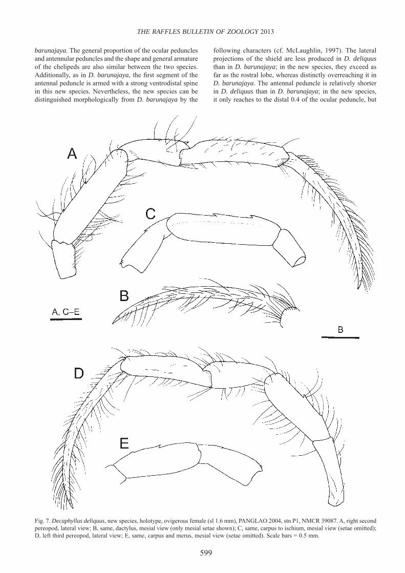
599
THE RAFFLES BULLETIN OF ZOOLOGY 2013
Fig. 7. Decaphyllus deliquus, new species, holotype, ovigerous female (sl 1.6 mm), PANGLAO 2004, stn P1, NMCR 39087. A, right second pereopod, lateral view; B, same, dactylus, mesial view (only mesial setae shown); C, same, carpus to ischium, mesial view (setae omitted); D, left third pereopod, lateral view; E, same, carpus and merus, mesial view (setae omitted). Scale bars = 0.5 mm.
barunajaya. The general proportion of the ocular peduncles and antennular peduncles and the shape and general armature of the chelipeds are also similar between the two species. Additionally, as in D. barunajaya, the fi rst segment of the antennal peduncle is armed with a strong ventrodistal spine in this new species. Nevertheless, the new species can be distinguished morphologically from D. barunajaya by the
following characters (cf. McLaughlin, 1997). The lateral projections of the shield are less produced in D. deliquus than in D. barunajaya; in the new species, they exceed as far as the rostral lobe, whereas distinctly overreaching it in D. barunajaya. The antennal peduncle is relatively shorter in D. deliquus than in D. barunajaya; in the new species, it only reaches to the distal 0.4 of the ocular peduncle, but

600
Komai & Rahayu: Six new species of Decaphyllus
it reaches nearly to the corneal base in D. barunajaya. The dactylus of the right cheliped is armed with one small but distinct proximal spine on the dorsal surface in D. deliquus, but it is unarmed in D. barunajaya. The right palm bears numerous additional spines lateral to the midline in D. barunajaya, but such numerous spines are not seen in D. deliquus. The cutting edge of the fi xed fi nger of the left chela is entire and armed only with a distal row of minute corneous teeth in D. deliquus, rather than having a row of small calcareous teeth in D. barunajaya.
Decaphyllus deliquus also resembles D. brevis, new species, and D. litoralis, new species. As noted above, there are no arthrobranchs on the third maxilliped in D. deliquus, but in the latter two species, a single bud (D. litoralis) or paired tiny lamellae (D. brevis) are present. Other characters differentiating D. deliquus and the latter two taxa are: the ocular peduncle is proportionally longer in D. deliquus than in the latter two species (subequal in the length to the shield versus 0.8–0.9 times as long); and spines on the right chela are stronger and more conspicuous in D. deliquus than in the latter two species. Furthermore, the lateral projections of the shield are less produced in D. deliquus than in D. litoralis (cf. Fig. 4A versus Fig. 8A). The antennal acicle slightly overreaches the distal margin of the fi fth peduncular segment in D. deliquus, rather than slightly falling short of or just reaching to it in D. brevis.
Etymology. — From the Latin “deliquus”, meaning missing, in referent to the complete loss of arthrobranchs on the third maxilliped.
Decaphyllus litoralis, new species(Figs. 8–12)
Material examined. — Holotype: male (sl 1.8 mm), Manza, Okinawa, Ryukyu Islands, Japan, 18 m, 3 Jul.2011, SCUBA diving, coll. S. Komai, CBM-ZC 11707.Paratype: ovigerous female (sl 1.7 mm), PANGLAO 2004, stn B5, Biking, Panglao Island, 09°35.2'N, 123°50.4'E, 4 m, reef slope with overhangs, 2 Jun.2004, NMCR 39088.Non-type: 1 juvenile (sl 1.2 mm), Horseshoe, Onna Village, Okinawa, 19 m, 13 Sep.2012, SCUBA diving, coll. Y. Yamada, CBM-ZC 11708.
Description. — Nine pairs of biserial phyllobranchiae (no pleurobranchs). Single arthrobranch above base of third maxilliped reduced to minute bud.
Shield (Fig. 8A) approximately as long as wide; anterior margin between rostral region and lateral projection slightly concave; anterolateral margins sloping; posterior margin truncate; dorsal surface with anteromedian part poorly calcifi ed, with several short setae laterally. Rostrum obsolete. Lateral projections moderately developed, exceeding beyond rostral lobe, each with terminal spinule.
Ocular peduncle (Fig. 8A) about 0.8–0.9 times as long as shield, faintly constricted at midlength; dorsal surface with mesial row of tufts of moderately short to long setae
directed mesially, scattered short setae on dorsal surface, and prominent tuft of long setae at base of cornea; cornea not dilated, its width slightly less than 0.3 of length of ocular peduncle; basal part slightly infl ated, its width greater than corneal width. Ocular acicle tapering distally to acute spine, mesial margin glabrous; separated basally by width of 1 acicle. Interocular lobe visible in dorsal view, anteriorly slightly convex.
Antennular peduncle (Fig. 8A) overreaching distal corneal margin by about 0.4 length of ultimate segment. Basal segment with prominent spine on lateral margin of statocyst lobe, without ventromesial subdistal spine. Penultimate and ultimate segments unarmed, almost glabrous except for 1 short thin seta on dorsomesial distal angle of ultimate segment.
Antennal peduncle (Fig. 8A) reaching or slightly falling short of base of cornea of ocular peduncle. Fifth and fourth segments with few setae. Third segment with prominent spine on ventromesial distal margin. Second segment with dorsolateral distal angle strongly produced, terminating in bifi d spine (lateral spine distinctly subterminal), dorsomesial distal angle with small spine. First segment unarmed on ventrodistal margin; lateral surface unarmed. Antennal acicle slightly overreaching distal margin of fi fth peduncular segment, reaching or slightly falling short of corneal base, terminating in small spine; mesial surface with sparse row of setae; lateral margin unarmed. Antennal fl agellum with 2–4 short to moderately long setae on distal margin of each article.
Third maxilliped with merus armed with strong dorsodistal spine; crista dentata on ischium consisting of 2–4 triangular teeth; basis unarmed or with minute denticle on mesial face. Exopod long, reaching nearly to distal margin of carpus.
Chelipeds (Figs. 9, 10) slightly unequal in length; right slightly longer but appreciably stronger. Right cheliped (Fig. 9) with chela elongate subovate in dorsal view, 2.3–2.5 times longer than wide. Dactylus (Fig. 9C) set at slightly oblique angle to palm, slightly shorter than palm; dorsal surface with 2–4 tiny spines or tubercles proximally; all surfaces with scattered moderately short to long setae, particularly numerous on mesial surface; cutting edge with row of small, blunt calcareous teeth in proximal 0.8 and microscopic corneous teeth in distal 0.2, terminating in tiny corneous claw. Palm (Fig. 9A, C) subequal in length to carpus; dorsomesial margin with row of 8 tiny spines, dorsal midline with 1 small proximal spine and row of minute tubercles or spinules not extending onto fi xed fi nger, dorsolateral margin not delimited and with irregular row of minute tubercles or spinules, dorsal surface lateral to midline without conspicuous spines; lateral and mesial surfaces with scattered short to moderately short setae; ventral surface gently convex, smooth, with sparse setae. Fixed fi nger with row of blunt calcareous teeth on cutting edge, terminating in small calcareous claw. Carpus (Fig. 9A–C) moderately widened distally, subequal in length to merus, 1.9–2.2 times longer than wide; dorsomesial margin with row of 4 or 5 small to moderately strong spines, dorsolateral surface with

601
THE RAFFLES BULLETIN OF ZOOLOGY 2013
Fig. 8. Decaphyllus litoralis, new species, holotype, male (sl 1.8 mm), Manza, Okinawa, CBM-ZC 11707. A, shield and cephalic appendages, dorsal view; B, distal three segments of left fourth pereopod; C, sixth thoracic sternite, ventral view; D, coxae of fi fth pereopods, sexual tubes, and eighth thoracic sternite, ventral view; E, telson, dorsal view. Scale bars = 0.5 mm.
row of 3 or 4 moderately small spines; all surfaces with scattered short to long setae, subdistal transverse row of setae particularly prominent; ventrolateral distal angle with spinule, distomesial angle unarmed. Merus (Fig. 9A, B, D) with 1 small spine on dorsodistal margin mesially; dorsal surface with sparse setae; ventrolateral margin with 1 or 2 small spines on distal half; mesial surface with small spine or protuberance proximoventrally, ventromesial margin with 2 small spines, distal spine directed proximally; ventral surface with small spine medially. Ischium (Fig. 9D) with 2 widely spaced spinules on ventromesial margin; lateral surface unarmed or with spinule subdistally.
Left cheliped (Fig. 10) without hiatus between dactylus and fi xed fi nger. Dactylus (Fig. 10A, C) about 1.3 times longer than palm, with few minute proximal tubercles or spinules on dorsal surface mesially, and with short to long setae particularly numerous on mesial surface; cutting edge with row of calcareous denticles, terminating in small corneous claw. Palm (Fig. 10A, C) about 0.6–0.7 length of
carpus; dorsomesial margin with row of 4 or 5 spinules, dorsal midline with 1 small proximal spine and row of minute tubercles, dorsolateral margin not delimited and with some minute tubercles or spinules and also with scattered short to long setae. Fixed fi nger with cutting edge faintly denticulate, with row of minute corneous teeth in distal 0.4, terminating in small corneous claw. Carpus (Fig. 10A–C) moderately widened distally, about 3.0–3.4 times longer than wide; dorsolateral margin with 3 small spines in distal half, dorsomesial margin with 4 or 5 small to moderately strong spines (distal second spine largest); ventrolateral distal angle with spinule, distomesial angle unarmed; all surfaces with scattered setae. Merus (Fig. 10A, B, D) with sparse setae on dorsal surface; dorsodistal margin with 1 small spine; ventrolateral margin with 2 small spines; mesial surface with 1 small, anteriorly curved spine proximoventrally, ventromesial margin with 1 or 2 spines; ventral surface with 1 small spine medially and with scattered setae. Ischium (Fig. 10D) with 2 widely spaced, anteriorly directed spinules on ventromesial margin; lateral surface with subdistal spinule ventrally.

602
Komai & Rahayu: Six new species of Decaphyllus
Fig. 9. Decaphyllus litoralis, new species, holotype, male (sl 1.8 mm), Manza, Okinawa, CBM-ZC 11707. A, right cheliped, mesial view; B, same, lateral view (setae omitted); C, same, chela and carpus, dorsal view (setae omitted); D, same, merus, dorsal view (setae omitted). Scale bar = 0.5 mm.
Ambulatory legs (Fig. 11) overreaching tip of right cheliped. Dactyli (Fig. 11A, B, D) 1.6–1.9 times longer than propodi, 9.8–12.0 times longer than broad, gently curved ventrally; all surfaces unarmed, but with numerous setae, particularly longer and stronger on dorsal margins. Propodi (Fig. 11A, D) unarmed, but with row of sparse setae on dorsal and ventral margins and scattered short setae on lateral and mesial faces. Carpi each with dorsodistal spine (spine distinctly stronger in second than in third), and 1 additional small spine slightly proximal to midlength (second, Fig. 11C) or no additional spine (third, Fig. 11D). Meri (Fig. 11A, C, D) each with 2 small spines (located at slightly distal to midlength and proximal 0.2) on dorsal margin: dorsal and ventral margins
with sparse long setae, latter unarmed. Ischium with 1 subdistal spinule on ventral margin mesially (second; Fig. 11C) or unarmed (third).
Fourth pereopods (Fig. 8B) non-chelate, with claw of dactylus entirely masked by tufts of short, dense setae; propodus with sparse setae on dorsal margin and distal half of ventral margin, 1 or 2 minute corneous scales present distally. Fifth pereopods semichelate.
Male with right sexual tube (Fig. 8D) long, directed from right to left across ventral body surface and curved anteriorly, reaching to level of coxa of left third pereopod; distal part

603
THE RAFFLES BULLETIN OF ZOOLOGY 2013
somewhat fl attened, slightly widened. Left sexual tube (Fig. 8D) directed from left to right, reaching to anteromesial part of coxa of right fi fth pereopod, twisted, markedly broadened distally. Female with unpaired left gonopore.
Anterior lobe of thoracic sternite 6 (third pereopods, Fig. 8C) subsemicircular, slightly skewed to left, bearing some moderately long setae anteriorly. Sternite of thoracic sternite 8 (fi fth pereopods) in male (Fig. 8D) transversely subovate, almost glabrous; that in female subcircular, with row of moderately short setae anteriorly.
Pleon dextrally twisted. Male with 4 unpaired pleopods; second, fourth and fi fth pleopods uniramous, third unequally
Fig. 10. Decaphyllus litoralis, new species, holotype, male (sl 1.8 mm), Manza, Okinawa, CBM-ZC 11707. A, left cheliped, mesial view; B, same, lateral view (setae omitted); C, same, chela and carpus, dorsal view (setae omitted); D, same, merus, dorsal view (setae omitted). Scale bar = 0.5 mm.
biramous. Female with 4 unpaired, unequally biramous pleopods.
Telson (Fig. 8E) with shallow median cleft; terminal margin with prominently produced, spinose left exterior angle separated from faintly produced, minutely spinose right exterior angle, and with 1 or 2 minute spinules on either side of median cleft; left lateral margin forming very narrow chitinous plate, but right lateral margin simple.
Colouration. — In life (Fig. 12). Ocular peduncle generally light gray-brown. Antennular peduncle also generally light gray-brown, ultimate segment with narrow white ring distally. Antennal fl agellum generally white with reddish brown distal

604
Komai & Rahayu: Six new species of Decaphyllus
Fig. 11. Decaphyllus litoralis, new species, holotype, male (sl 1.8 mm), Manza, Okinawa, CBM-ZC 11707. A, right second pereopod, lateral view; B, same, dactylus, mesial view (only mesial setae shown); C, same, carpus to ischium, mesial view (setae omitted); D, left third pereopod, lateral view. Scale bars = 0.5 mm.
part. Chelipeds generally light gray-brown, with brown transverse markings at least on palms and carpi. Ambulatory legs generally light gray-brown; propodi whitish distally and brownish proximally.
In preservative. No iridescent sheen on shield and appendages.
Distribution. — Ryukyu Islands, Japan, and the Bohol Sea, Philippines; 4–18 m.
Remarks. — As discussed above, Decaphyllus litoralis, new species, is similar to D. brevis, new species, and D. deliquus, new species. Differentiating characters are discussed under “Remarks” of respective species.
Decaphyllus litoralis is the sole representative of the genus occurring in the shallow coral reefs at depths of 4–18 m. Other known species in the genus occur at sublittoral depths greater than 50 m.

605
THE RAFFLES BULLETIN OF ZOOLOGY 2013
Fig. 12. Decaphyllus litoralis, new species, holotype, male (sl 1.8 mm), Manza, Okinawa, CBM-ZC 11707, in situ. (Photograph by: Satoko Komai).
Etymology. — From the Latin litoralis (= coastal), in reference to the habitat of this new species in the shallow coral reefs, which is unique for the genus.
Decaphyllus proprius, new species(Figs. 13–16, 23A)
Material examined. — Holotype: male (sl 1.4 mm), PANGLAO 2004, stn T39, Cervera shoal, West Pamilacan Island, 09°30.1'N, 123°50.4'E, 100–138 m, muddy sand, 6 Jul.2004, NMCR 39089.
Description. — Ten pairs of biserial phyllobranchiae (no pleurobranchs). Two very small arthrobranchs above base of third maxilliped, each bearing few lamellae (Fig. 13C).
Shield (Fig. 13A) approximately as long as wide; anterior margin between rostral region and lateral projection slightly concave; anterolateral margins sloping; posterior margin roundly truncate; dorsal surface with median part poorly calcifi ed, with several minute setae anteriorly and laterally. Rostrum broadly rounded. Lateral projections weakly developed, exceeding as far as rostral lobe, each with terminal spinule.
Ocular peduncle (Fig. 13A) approximately as long as shield, faintly constricted at midlength; dorsal surface with mesial row of tufts of moderately short to long setae directed mesially and few individual setae laterally, and prominent tuft of long setae at base of cornea; cornea not dilated, its width slightly more than 0.2 of length of ocular peduncle; basal part slightly infl ated, its width greater than corneal width. Ocular acicle drawn out into long acute spine, mesial margin with some moderately short setae; separated basally by width of less than 1 acicle. Interocular lobe visible in dorsal view, anteriorly slightly convex.
Antennular peduncle (Fig. 13A) overreaching distal corneal margin by about 0.4 length of ultimate segment. Basal segment with prominent spine on lateral margin of statocyst
lobe, without ventromesial subdistal spine. Penultimate and ultimate segments unarmed, almost glabrous except for 1 short, thin seta on dorsomesial distal angle.
Antennal peduncle (Fig. 13A) reaching to distal 0.4 of ocular peduncle. Fifth and fourth segments with few setae. Third segment with prominent spine on ventromesial distal margin. Second segment with dorsolateral distal angle strongly produced, terminating in bifi d spine (lateral spine distinctly subterminal), dorsomesial distal angle with strong spine. First segment with strong spine on ventrodistal margin; lateral surface unarmed. Antennal acicle slightly overreaching distal margin of fi fth peduncular segment, far falling short of corneal base, terminating in small simple (left) or slightly falling short of distal margin of fi fth peduncular segment, terminating in bifi d spine (right) (possibly abnormal condition); mesial surface with sparse row of setae; lateral margin with small spine located at proximal 0.3. Antennal fl agellum with 2–4 short to moderately setae on distal margin of each article.
Third maxilliped (Fig. 13B) with merus armed with strong dorsodistal spine; crista dentata on ischium consisting of 3 triangular teeth; basis with minute denticle on mesial face. Exopod long, reaching nearly to distal margin of carpus.
Chelipeds (Figs. 14, 15) slightly unequal in length; right slightly longer but appreciably stronger. Right cheliped (Fig. 14) with chela elongate subovate in dorsal view, 2.9 times longer than wide. Dactylus (Fig. 14C) set at slightly oblique angle to palm, subequal in length to palm; dorsal surface with 1 tiny spine proximally; all surfaces with scattered moderately short to moderately long setae, particularly numerous on mesial surface; cutting edge with row of small, blunt calcareous teeth, terminating in tiny corneous claw. Palm (Fig. 14A, C) slightly shorter than carpus; dorsomesial margin with row of spinules or minute tubercles, dorsal midline with row of prominent spines decreasing in size distally and not extending onto fi xed fi nger, dorsolateral margin not delimited and with irregular row of slender small spines, dorsal surface lateral to midline unarmed; lateral and mesial surfaces with scattered short to long setae; ventral surface convex, smooth, with sparse setae. Fixed fi nger with row of small, blunt or subtriangular calcareous teeth on cutting edge, terminating in small calcareous claw. Carpus (Fig. 14A–C) subequal in length to merus, slightly widened distally, 2.4 times longer than wide; dorsomesial margin with row of 7 spines (distalmost 2 minute, others moderately strong), dorsolateral surface with row of 7 small and minute spines; all surfaces with scattered short to long setae, subdistal transverse row of setae particularly prominent; both ventrolateral distal and ventromesial distal angles unarmed. Merus (Fig. 14A, B, D) with 1 small spine on dorsodistal margin mesially; dorsal surface with short transverse rows of setae; lateral surface with 1 low, small tubercle at about midlength adjacent to ventral margin, ventrolateral margin with 1 small subdistal spine; mesial surface with small, low protuberance adjacent to ventral margin proximally, ventromesial margin with 2 small, widely spaced spines; ventral surface unarmed. Ischium (Fig. 14D) with 2 widely spaced small spines on ventromesial margin, distal spine directed distally, proximal

606
Komai & Rahayu: Six new species of Decaphyllus
Fig. 13. Decaphyllus proprius, new species, holotype, male (sl 1.4 mm), PANGLAO 2004, stn T39, NMCR 39089. A, shield and cephalic appendages, dorsal view; B, left third maxilliped, lateral view; C, same, arthrobranchs; D, distal three segments of left fourth pereopod; E, sixth thoracic sternite, ventral view; F, coxae of fi fth pereopods, sexual tubes, and eighth thoracic sternite, ventral view; G, telson, dorsal view. Scale bars = 0.5 mm (A, B, D–G), 0.25 mm (C).
spine curved proximally and hook-like; lateral surface with 2 closely spaced spinules ventrodistally.
Left cheliped (Fig. 15) with distinct hiatus between dactylus and fi xed fi nger. Dactylus (Fig. 15A, C) about 1.2 times longer than palm, dorsal midline with 3 spinules in proximal 0.4; all surfaces with numerous short to long setae particularly on mesial surface; cutting edge with row of minute corneous teeth in distal half, terminating in small corneous claw. Palm (Fig. 15A, C) about 0.6 length of carpus; dorsomesial margin not delimited, dorsal midline with row of 5 small spines not extending onto fi xed fi nger, dorsolateral margin not delimited, unarmed; all surfaces with scattered short to long setae. Fixed fi nger with row of calcareous denticles increasing in size distally on cutting edge, terminating in small corneous claw.
Carpus (Fig. 15A–C) slightly widened distally, about 3.2 times longer than wide; dorsolateral margin with 4 moderately large spines, dorsomesial margin with 5 moderately strong spines; ventrolateral distal and distomesial angles unarmed; all surfaces with scattered setae. Merus (Fig. 15A, B, D) with sparse setae on dorsal surface; dorsodistal margin unarmed; lateral surface with 1 minute denticle adjacent to ventral margin, ventrolateral margin with 2 small subdistal spines and 1 median spinule; mesial surface with 1 small protuberance proximoventrally, ventromesial margin with 2 widely spaced spines in distal half. Ischium (Fig. 15D) with 2 widely spaced small spines on ventromesial margin, distal spine directed distally, proximal spine curved proximally; lateral surface with spinule ventrally.

607
THE RAFFLES BULLETIN OF ZOOLOGY 2013
Fig. 14. Decaphyllus proprius, new species, holotype, male (sl 1.4 mm), PANGLAO 2004, stn T39, NMCR 39089. A, right cheliped, mesial view; B, same, lateral view (setae omitted); C, same, chela and carpus, dorsal view (setae omitted); D, same, merus, dorsal view (setae omitted). Scale bar = 0.5 mm.
Ambulatory legs (Fig. 16) overreaching tip of right cheliped. Dactyli (Fig. 16A, B, D) 1.4–1.5 times longer than propodi, 13.3–14.6 times longer than broad, gently curved ventrally; all surfaces unarmed, but with numerous setae, particularly longer and stronger on dorsal margins. Propodi (Fig. 16A, D) unarmed, but with row of sparse setae on dorsal and ventral margins and with scattered short setae on lateral and mesial faces. Carpi each with dorsodistal spine and 2 additional
small dorsal spines in proximal 0.4 (second, Fig. 16C) or entirely unarmed (third, Fig. 16E). Meri each with 2 small spines (located at slightly distal to midlength and proximal 0.2) on dorsal margin; dorsal and ventral margins with sparse short to long setae, latter armed with spinule at distal 0.3 (second, Fig. 16C) or unarmed (third, Fig. 16E). Ischium with distal spinule on ventral margin mesially (second, Fig. 16C) or unarmed (third, Fig. 16E).

608
Komai & Rahayu: Six new species of Decaphyllus
Fig. 15. Decaphyllus proprius, new species, holotype, male (sl 1.4 mm), PANGLAO 2004, stn T39, NMCR 39089. A, left cheliped, mesial view; B, same, lateral view (setae omitted); C, same, chela and carpus, dorsal view (setae omitted); D, same, merus, dorsal view (setae omitted). Scale bar = 0.5 mm.
Fourth pereopods (Fig.13D) non-chelate, with claw of dactylus entirely masked by tufts of short, dense setae; propodus with sparse setae on dorsal margin and distal half of ventral margin; 2 minute corneous scales present distally. Fifth pereopods semichelate.
Male with right sexual tube (Fig. 13F) long, directed from right to left across ventral body surface and curved anteriorly, reaching to level of coxa of left second pereopod; distal part somewhat fl attened. Left sexual tube (Fig. 13F) directed from left to right, reaching to anteromesial part of coxa of right fi fth pereopod, twisted, slightly broadened distally.
Anterior lobe of thoracic sternite 6 (third pereopods, Fig. 13E) subsemicircular, slightly skewed to left, bearing several moderately long setae anteriorly. Sternite of thoracic sternite 8 (fi fth pereopods) in male (Fig. 13F) transversely subovate, almost glabrous; anterior surface slightly concave.
Pleon dextrally twisted, with 4 unpaired left pleopods, second, fourth and fi fth uniramous, third unequally biramous.
Telson (Fig. 13G) with shallow median cleft; terminal margin with prominently produced, spinose left exterior angle separated from less produced, but also prominent, spinose right exterior angle, and with 1 minute spinules on either side of median cleft; left lateral margin not chitinous.
Female unknown.
Colouration. — In life (Fig. 23A). Carapace generally pinkish, shield slightly mottled; posterior carapace with scattered darker pink spots. Ocular peduncles mottled with pink. Antennular and antennal peduncles pale pink, without conspicuous markings; antennal fl agellum generally translucent. Chelipeds generally whitish, carpi and meri with large darker reddish blotches. Ambulatory legs obscurely

609
THE RAFFLES BULLETIN OF ZOOLOGY 2013
Fig. 16. Decaphyllus proprius, new species, holotype, male (sl 1.4 mm), PANGLAO 2004, stn T39, NMCR 39089. A, right second pereopod, lateral view; B, same, dactylus, mesial view (only mesial setae shown); C, same, carpus to ischium, mesial view (setae omitted); D, left third pereopod, lateral view; E, same, carpus to ischium, mesial view (setae omitted). Scale bars = 0.5 mm.
banded by dull red and white; dactyli whitish; propodi with distal part white and proximal part white, remainder alternated with red (distal to midlength) and white (proximal to midlength); carpi reddish; meri each with obscure 2 reddish bands. Pleon pinkish.
In preservative. Slight iridescence seen on shield, chelipeds and ambulatory legs.
Distribution. — Known only from Panglao Islands, 100–138 m.
Remarks. — In proportions of the cephalic appendages and armature of the chelipeds, Decaphyllus proprius, new species, most closely resembles D. maci. However, the present new species is quite distinctive in the genus in having a small but prominent spine on the lateral margin of the antennal

610
Komai & Rahayu: Six new species of Decaphyllus
acicle, which is located proximal to the midlength. In other congeneric species, such a spine is absent. The lack of a dorsodistal spine on the merus of the left cheliped appears also unique for the genus. Furthermore, D. proprius differs from D. maci in the following characters: shield proportionally wider in D. proprius than in D. maci (approximately as long as wide versus about 1.2 times as long as wide); lateral projections of shield less produced in D. proprius than in D. maci; fi ngers of left chela, when closed, leaving a distinct hiatus in D. proprius, whereas no hiatus in D. maci; dactylus of left cheliped with short proximal row of small spines on dorsal midline in D. proprius, while unarmed in D. maci; dorsomesial margin of palm of left cheliped unarmed in D. proprius, whereas armed with 2 conspicuous spines in D. maci; and anterior lobe of thoracic sternite 6 subsemicircular in D. proprius, rather than roundly subtriangular in D. maci.
Etymology. — From the Latin proprius (= characteristic), in reference to the unique armature of the antennal acicle in this new species.
Decaphyllus spinulodigitus, new species(Figs. 17–19)
Material examined. — Holotype: ovigerous female (sl 2.3 mm), PANGLAO 2004, stn T1, Bolod, Panglao Island, 9°32.4'N, 123°47.3'E, 83–102 m, mud and many sponges, 30 May 2004, trawl, NMCR 39090.
Description. — Ten pairs of biserial phyllobranchiae (no pleurobranchs). Two very small arthrobranch gills above base of third maxilliped, anterior gill simple, posterior gill bilobed (Fig. 17C).
Shield (Fig. 17A) approximately as long as wide; anterior margin between rostral region and lateral projection slightly concave; anterolateral margins sloping; posterior margin roundly truncate; dorsal surface with anteromedian part poorly calcifi ed, with several minute to short setae laterally. Rostrum broadly rounded. Lateral projections weakly developed, exceeding as far as rostral lobe, each with terminal spinule.
Ocular peduncle (Fig. 17A) about 0.7 times as long as shield, faintly constricted at midlength; dorsal surface with mesial row of long setae directed mesially and few setae laterally, and prominent tuft of moderately long setae at base of cornea; cornea not dilated, its width slightly less than 0.3 of length of ocular peduncle; basal part slightly infl ated, its width greater than corneal width. Ocular acicle drawn out distally into acute spine, mesial margin with tuft of moderately short setae medially; separated basally slightly less than width of 1 acicle. Interocular lobe visible in dorsal view, anteriorly slightly concave.
Antennular peduncle (Fig. 17A) overreaching distal corneal margin by about 0.8 length of ultimate segment. Basal segment with prominent spine on lateral margin of statocyst lobe, mesial face unarmed. Penultimate and ultimate segments
unarmed, almost glabrous except for 1 thin minute seta at dorsomesial distal angle.
Antennal peduncle (Fig. 17A) slightly overreaching base of cornea of ocular peduncle. Fifth and fourth segments with few setae. Third segment with prominent spine on ventromesial distal margin. Second segment with dorsolateral distal angle strongly produced, terminating in bifi d spine (lateral spine subterminal), dorsomesial distal angle with strong spine. First segment with small spine on ventrodistal margin; lateral surface unarmed. Antennal acicle slightly falling short of distal margin of fi fth peduncular segment, reaching corneal base, terminating in small spine; mesial surface with row of sparse setae; lateral margin unarmed. Antennal fl agellum with 2–4 short to moderately setae on distal margin of each article.
Third maxilliped with merus bearing strong dorsodistal spine; crista dentata on ischium consisting of 3 widely spaced, triangular teeth (Fig. 17B); basis with minute denticle distally on mesial surface. Exopod long, reaching nearly to distal margin of carpus.
Chelipeds (Fig. 18) slightly unequal in length, right slightly longer, appreciably stronger. Right cheliped (Fig. 18A–D) with chela elongate subovate in dorsal view, about 2.2 times longer than wide. Dactylus set at slightly oblique angle to palm (Fig. 18C), shorter than palm; dorsal surface mesially with several small spines or tubercles in proximal half; all surfaces with scattered moderately short to long setae, particularly numerous on mesial surface; cutting edge with row of moderately large, blunt calcareous teeth, terminating in tiny corneous claw. Palm subequal in length to carpus (Fig.18A, C); dorsomesial margin with row of 8 small spines, dorsal midline with row small spines decreasing in size and acuteness distally and not extending onto fi xed fi nger, dorsolateral margin not delimited and with few minute tubercles, dorsal surface lateral to midline with scattered microscopic spinules; lateral and mesial surfaces with scattered short to long setae; ventral surface gently convex, smooth, with sparse setae. Fixed fi nger with row of blunt calcareous teeth on cutting edge, terminating in tiny corneous claw. Carpus (Fig. 18A–C) moderately widened distally, subequal in length to merus, 1.8 times longer than wide; dorsomesial margin with row of 4 moderately strong spines, dorsolateral surface with row of 4 moderately small spines and 1 proximal spinule; all surfaces with scattered short to long setae, subdistal transverse row of setae particularly prominent; ventrolateral distal angle with small spine, ventromesial distal angle unarmed. Merus (Fig. 18A, B, D) with 1 small spine on dorsodistal margin mesially; dorsal surface with sparse setae; lateral surface unarmed, ventrolateral margin with 1 small subdistal spine; mesial surface with small tubercle proximoventrally, ventromesial margin with 1 small subdistal spine; ventral surface unarmed. Ischium (Fig. 18D) with 2 widely spaced spinules on ventromesial margin; lateral surface with minute spine ventrodistally.
Left cheliped (Fig. 18E–H) with distinct hiatus between dactylus and fi xed fi nger. Dactylus (Fig 18G) about 1.2 times longer than palm, gently curved; all surfaces unarmed, but

611
THE RAFFLES BULLETIN OF ZOOLOGY 2013
Fig. 17. Decaphyllus spinulodigitus, new species, holotype, ovigerous female (sl 2.3 mm), PANGLAO 2004, stn T1, NMCR 39090. A, shield and cephalic appendages, dorsal view; B, ischium of left third maxilliped, ventral view; C, arthrobranchs on left third maxilliped; D, distal three segments of left fourth pereopod; E, sixth thoracic sternite, ventral view; F, telson, dorsal view. Scale bars = 1 mm (A), 0.5 mm (B, D–F), 0.25 mm (C).
with numerous short to long setae particularly on mesial surface; cutting edge microscopically denticulate, terminating in small corneous claw. Palm (Fig. 18E–G) about 0.6 length of carpus; dorsomesial margin without conspicuous spines, though few minute tubercles adjacent to dorsomesial margin; dorsal midline with row of spinules not extending onto fi xed fi nger, dorsolateral margin not delimited and only with some minute tubercles; all surfaces with scattered short to long setae. Fixed fi nger with row of minute calcareous denticles on cutting edge, terminating in small corneous claw. Carpus (Fig. 18E–G) moderately widened distally, about 3.0 times longer than wide; dorsolateral margin with 2 moderately small spines in distal half, dorsomesial margin with 5 small to moderately strong spines; ventrolateral distal angle
with spinule, distomesial angle unarmed; all surfaces with scattered setae. Merus (Fig. 18E, F, H) with sparse setae on dorsal surface; dorsodistal margin with small spine; lateral surface unarmed, ventrolateral margin with 2 widely spaced, small spines; mesial surface with spine-like tubercle proximoventrally, ventromesial margin with 2 widely spaced, small spines; ventral surface with tiny spine medially and scattered long setae. Ischium (Fig. 18H) with 2 widely spaced spinules on ventromesial margin, both directed forward; lateral surface with spinule distoventrally.
Ambulatory legs (Fig. 19) overreaching tip of right cheliped. Dactyli (Fig. 19A, B, D) about 1.6 times longer than propodi, 11.7–14.6 times longer than broad, gently curved ventrally;

612
Komai & Rahayu: Six new species of Decaphyllus
Fig. 18. Decaphyllus spinulodigitus, new species, holotype, ovigerous female (sl 2.3 mm), PANGLAO 2004, stn T1, NMCR 39090. A, right cheliped, mesial view; B, same, lateral view; C, same, chela and carpus, dorsal view; D, same, merus, dorsal view; E, left cheliped, mesial view; F, same, lateral view; G, same, chela and carpus, dorsal view; H, same, merus, dorsal view. Setae omitted except for E. Scale bar = 1.0 mm.

613
THE RAFFLES BULLETIN OF ZOOLOGY 2013
Fig. 19. Decaphyllus spinulodigitus, new species, holotype, ovigerous female (sl 2.3 mm), PANGLAO 2004, stn T1, NMCR 39090. A, right second pereopod, lateral view; B, same, dactylus, mesial view (only mesial setae shown); C, same, carpus to ischium, mesial view (setae omitted); D, left third pereopod, lateral view; E, same, carpus and merus, mesial view (setae omitted). Scale bars = 1.0 mm.
all surfaces unarmed, but with numerous setae, particularly longer and stronger on dorsal margins. Propodi (Fig. 19A, D) unarmed, but with row of sparse setae on dorsal and ventral margins. Carpi each with dorsodistal spine (second;
Fig. 19A) or without dorsodistal spine (third; Fig. 19D), and with 1 additional small spine located at proximal 0.4 (second; Fig. 19C) or no additional spine (third; Fig. 19E). Meri each with 2 small dorsal spines (distal-sided spine located slightly

614
Komai & Rahayu: Six new species of Decaphyllus
distal to midlength and another at proximal 0.2); dorsal and ventral margins with sparse long setae, latter armed with spinule at distal 0.4 (second, Fig. 19C) or unarmed (third, Fig. 19E). Ischium with distal spinule on ventral margin mesially (second, Fig. 19C) or unarmed (third, Fig. 19E)
Fourth pereopods (Fig. 17D) non-chelate, with claw of dactylus entirely masked by tufts of short, dense setae; propodus roundly subquadrate, slightly wider than long, with numerous setae on dorsal margin and distal half of ventral margin; 3 minute corneous scales located distally on ventral margin. Fifth pereopods semichelate.
Female with unpaired left gonopore.
Anterior lobe of thoracic sternite 6 (third pereopods, Fig. 17E) subsemicircular, slightly skewed to left, bearing numerous long setae anteriorly. Sternite of thoracic sternite 8 (fi fth pereopods) in female transversely subovate, with setae along anterior margin.
Telson (Fig. 17F) with shallow median cleft; terminal margin with prominently produced, spinose left exterior angle separated from non-produced, minutely pointed right exterior angle, and with 1 spinule on either side of median cleft; lateral margins apparently not chitinous.
Male unknown.
Colouration. — In preservative. No distinct markings seen on body and appendages. Chelipeds and ambulatory legs with iridescence, particularly strong on chelipeds.
Distribution. — Known only from Panglao Island, the Philippines, 83–102 m.
Remarks. — Although only a single ovigerous female is available, this new species is safely assigned to Decaphyllus by the unarmed but setose dactyli of the ambulatory legs, the non-chelate fourth pereopod, the entire eighth thoracic sternite and the characteristic shape of the telson. The setation of the ocular peduncle also supports the generic assignment.
The presence of several small spines or tubercles on the dorsal surface of the dactylus of the right cheliped and the relatively long antennular peduncle of Decaphyllus spinulodigitus, new species, suggests a close relationship with D. similis; however, there are some morphological differences of diagnostic signifi cance between the two taxa (de Saint Laurent, 1968b). The strongly produced dorsolateral distal angle of the second segment of the antennal peduncle terminates in a clearly bifi d spine in D. spinulodigitus, rather than terminating into a simply acuminate spine in D. similis. The basal segment of the antennular peduncle is unarmed on the mesial face in D. spinulodigitus, but it is armed with a small spine on the mesial face in D. similis. The antennal acicle does not reach the distal margin of the fi fth segment of the antennal peduncle in D. spinulodigitus, rather than slightly overreaching it in D. similis. The armature of the
right cheliped is weaker in D. spinulodigitus than in D. similis. For example, the dorsomedian row on the palm consists of small spines or tubercles decreasing in the size and acuteness distally in D. spinulodigitus, whereas it is composed of numerous conspicuous spines not weakened distally in D. similis. The merus of the second pereopod bears a small spine on the ventral margin, located slightly distal to the midlength in D. spinulodigitus, but such a spine is absent in D. similis.
Etymology. — From the combination of the Latin spinuloso (spinulose) and digitus (fi nger), in reference to the dactyli of the right cheliped bearing several spinules on the dorsal surface. Used as a noun in apposition.
Decaphyllus tenuis, new species(Figs. 20–22)
Material examined. — Holotype: male (sl 1.8 mm), PANGLAO 2004, stn T5, Bohol Island, W of Baclayon, 09°35.3'N, 123°52.2"E, 84–87 m, coarse sand and mud, 2 Jun.2004, NMCR 39091.Paratypes: 2 females (sl 1.3, 1.3 mm), PANGLAO 2004, stn T18, Cortes, Bohol Island, 09°41.8'N, 123°49.9'E, 80–100 m, muddy bottom with sponges, 19 Jun.2004, ZRC 2013.0679; 1 male (sl 1.9 mm), same data, CBM-ZC 11709; stn T41, 2 males (sl 1.0, 1.1 mm), Cervera shoal, West Pamilacan Island, 09°29.7'N, 123°50.2'E, 110–112 m, 6 Jul.2004, ZRC 2013.0680.
Description. — Ten pairs of biserial phyllobranchiae (no pleurobranchs). Two very small arthrobranchs above base of third maxilliped, anterior gill simple, posterior gill bilobed (Fig. 20C).
Shield (Fig. 20A) approximately as long as wide; anterior margin between rostral region and lateral projection concave; anterolateral margins sloping; posterior margin roundly truncate; dorsal surface with anteromedian part poorly calcifi ed, with some tufts of short setae laterally. Rostrum rounded. Lateral projections moderately well developed, exceeding as far as rostral lobe, each with terminal spinule.
Ocular peduncle (Fig. 20A) approximately as long as shield, faintly constricted distal to midlength; dorsal surface with mesial row of tufts of moderately short to long setae directed mesially and some individual setae laterally, and prominent tuft of moderately long setae at base of cornea; cornea slightly dilated, its width slightly more than 0.2 of length of ocular peduncle; basal part not infl ated, its width narrower than corneal width. Ocular acicle drawn out distally into acute spine, mesial margin with some long setae; separated basally by width of less than 1 acicle. Interocular lobe visible in dorsal view, anteriorly slightly concave.
Antennular peduncle (Fig. 20A) overreaching distal corneal margin by 0.4–0.5 length of ultimate segment. Basal segment with prominent spine on lateral margin of statocyst lobe, mesial face unarmed. Penultimate and ultimate segments unarmed, almost glabrous except for thin short setae at dorsomesial distal angle of ultimate segment.

615
THE RAFFLES BULLETIN OF ZOOLOGY 2013
Fig. 20. Decaphyllus tenuis, new species. A–D, F, G, holotype, male (sl 1.8 mm), PANGLAO 2004, stn T5, NMCR 39091; E, paratype, female (sl 1.3 mm), PANGLAO 2004, stn T18, ZRC 2013.0679. A, shield and cephalic appendages, dorsal view; B, ischium of left third maxilliped, ventral view; C, arthrobranchs on left third maxilliped; D, left fourth pereopod, lateral view; E, sixth thoracic sternite, ventral view; F, coxae of fi fth pereopods, sexual tubes, and eighth thoracic sternite, ventral view; G, telson, dorsal view. Scale bars = 0.5 mm (A, B, D–G), 0.25 mm (C).
Antennal peduncle (Fig. 20A) slightly falling short of base of cornea of ocular peduncle. Fifth and fourth segments with few setae. Third segment with prominent spine on ventromesial distal margin. Second segment with dorsolateral distal angle strongly produced, terminating in bifi d spine, dorsomesial distal angle with small spine. First segment with strong spine on ventrodistal margin; lateral surface unarmed. Antennal acicle reaching or overreaching distal margin of fi fth peduncular segment, or not reaching to reaching corneal base, terminating in small spine; mesial surface with row of sparse setae; lateral margin unarmed. Antennal fl agellum with 2–4 short to moderately long setae on distal margin of each article.
Third maxilliped with merus armed with strong dorsodistal spine; crista dentata on ischium consisting of 4 or 5 triangular teeth (Fig. 20B); basis with acute denticle on mesial face. Exopod long, reaching nearly to distal margin of carpus.
Chelipeds (Fig. 21) slightly unequal in length; right slightly longer but appreciably stronger. Right cheliped (Fig. 21A–D) slender, with chela elongate subovate in dorsal view, about 3.0 times longer than wide. Dactylus set at slightly oblique angle to palm (Fig. 21C), subequal in length to palm; dorsal surface with 1 or 2 tiny spines or tubercles proximally; all surfaces with scattered moderately short to long setae, particularly numerous on mesial surface; cutting edge with

616
Komai & Rahayu: Six new species of Decaphyllus
Fig. 21. Decaphyllus tenuis, new species, holotype, male (sl 1.8 mm), PANGLAO 2004, stn T5, NMCR 39091. A, right cheliped, mesial view; B, same, lateral view; C, same, chela and carpus, dorsal view; D, same, merus, dorsal view; E, left cheliped, mesial view; F, same, lateral view; G, same, chela and carpus, dorsal view; H, same, merus, dorsal view. Setae omitted. Scale bar = 1.0 mm.

617
THE RAFFLES BULLETIN OF ZOOLOGY 2013
Fig. 22. Decaphyllus tenuis, new species, holotype, male (sl 1.8 mm), PANGLAO 2004, stn T5, NMCR 39091. A, right second pereopod, lateral view; B, same, dactylus, mesial view (only mesial setae shown); C, same, carpus to ischium, mesial view (setae omitted); D, right third pereopod, lateral view; E, same, carpus to ischium, mesial view (setae omitted). Scale bars = 1.0 mm.
row of numerous small, blunt or acute calcareous teeth in proximal 0.8 and row of spaced minute corneous teeth in distal 0.2, terminating in small calcareous claw. Palm (Fig. 21A, C) distinctly shorter than carpus; dorsomesial margin with row of small spines, dorsal midline with row
of moderately small spines not extending onto fi xed fi nger, dorsolateral margin not delimited, dorsal surface lateral to midline with scattered small slender spines or low tubercles; lateral and mesial surfaces with scattered short to long setae; ventral surface gently convex, smooth, with sparse setae.

618
Komai & Rahayu: Six new species of Decaphyllus
Fixed fi nger (Fig. 21A, C) with cutting edge bearing row of numerous blunt to acute calcareous teeth, terminating in small calcareous claw. Carpus (Fig. 21A–C) subequal in length to merus, slightly widened distally, 2.3–2.9 times longer than wide; dorsomesial margin with row of 4–7 small spines, dorsolateral margin with row of 4–12 small spines (distalmost spine at dorsolateral distal angle minute); all surfaces with scattered short to long setae, subdistal transverse row of setae particularly prominent; both ventrolateral distal and distomesial angles unarmed. Merus (Fig. 21A, B, D) with 1 small spine on dorsodistal margin mesially; dorsal surface with sparse short setae; lateral surface without conspicuous spines or tubercles, ventrolateral margin unarmed or with 1 or 2 small, widely spaced spines or tubercles; mesial surface with 1 small proximoventral tubercle, ventromesial margin with 2 small, widely spaced spines or tubercles in distal half; ventral surface without conspicuous spine. Ischium (Fig. 21D) with 1 or 2 widely spaced small spines on ventromesial margin, distal spine, if present, directed distally, proximal spine also directed distally; lateral surface with spinule ventrally. Spines or spinules on merus and ischium sharper in small specimens than in large specimens.
Left cheliped (Fig. 21E–H) without hiatus between dactylus and fi xed fi nger. Dactylus (Fig. 21E, G) about 1.3 times longer than palm, unarmed; all surfaces with numerous short to long setae, on mesial surface; cutting edge minutely denticulate, with row of minute corneous teeth in distal half. Palm (Fig. 21E–G) about half-length of carpus; dorsal surface with irregular longitudinal rows of small spines or tubercles, including stronger spines on midline; all surfaces with scattered short to long setae. Fixed fi nger with row of small calcareous teeth decreasing in size distally, several proximal teeth spine-like. Carpus (Fig. 21E–G) very slightly widened distally, about 3.8 times longer than wide; dorsolateral margin with 4–7 minute to moderately large spines, dorsomesial margin with 3–5 minute to moderately large spines; ventrolateral distal angle unarmed or with minute denticle, distomesial angle unarmed; all surfaces with scattered short to long setae. Merus (Fig. 21E, F, H) with sparse setae on dorsal surface; dorsodistal margin with 1 small spine; lateral surface with 1 spinule adjacent to ventral margin, ventrolateral margin with 1 subdistal spine; mesial surface with small distally curved spine proximoventrally, ventromesial margin with 1 small spine distal to midlength; ventral surface unarmed, with scattered long setae. Ischium (Fig. 21E, H) with 2 widely spaced small spines on ventromesial margin, distal spine directed mesially, proximal spine curved proximally; lateral surface unarmed.
Ambulatory legs (Fig. 22) overreaching tip of right cheliped. Dactyli (Fig. 22A, B, D) 1.4–1.5 times longer than propodi, 14.5–16.7 times longer than broad, gently curved ventrally; all surfaces unarmed, but with numerous setae, particularly longer and stronger on dorsal margins (several distal setae on dorsal margins bristle-like). Propodi (Fig. 22A, D) unarmed, but with row of sparse setae on dorsal and ventral margins and scattered short setae on lateral and mesial faces. Carpi each with dorsodistal spine (second; Fig. 22A) or without dorsodistal spine (third; Fig. 22D), and with 2 additional small
spines in proximal 0.3 (second, Fig. 22C) or no additional spines (third, Fig. 22E). Meri (Fig. 22A, C–E) each with 2 small spines (one located slightly distal to midlength and another at proximal 0.2) on dorsal margin; dorsal and ventral margins with sparse long setae, latter unarmed. Ischium with distal spinule on ventral margin mesially (second, Fig. 22C) or unarmed (third, Fig. 22E).
Fourth pereopods (Fig. 20D) non-chelate, with claw of dactylus entirely masked by tufts of short, dense setae; propodus with sparse setae on dorsal margin and distal half of ventral margins; 2 minute corneous scales present ventrodistally. Fifth pereopods semichelate.
Male with right sexual tube (Fig. 20F) long, directed from right to left across ventral body surface and curved anteriorly, reaching to level of coxa of left second pereopod; distal part somewhat fl attened. Left sexual tube (Fig. 20F) directed from left to right, reaching to anteromesial part of coxa of right fi fth pereopod, slightly twisted, slightly broadened distally, with sparse setae terminally and posteriorly. Female with unpaired left gonopore.
Anterior lobe of thoracic sternite 6 (third pereopods, Fig. 20E) subsemicircular, slightly skewed to left, bearing numerous long setae anteriorly. Sternite of thoracic sternite 8 (fi fth pereopods) in male (Fig. 20F) asymmetrical, anteriorly concave, with few setae ventrally; that in female transversely subovate.
Pleon dextrally twisted. Male with 4 unpaired pleopods, second, fourth and fi fth uniramous, third unequally biramous; ramus of second pleopod bearing numerous marginal setae. Female with 4 unpaired, unequally biramous pleopods.
Telson (Fig. 20G) with shallow median cleft; terminal margin with prominently produced, spinose left exterior angle separated from less produced, but also prominent, spinose right exterior angle, and with 1 or 2 spinules on either side of median cleft; lateral margins forming chitinous plate.
Colouration. — In life (Fig. 23B). Carapace generally reddish, shield slightly mottled. Ocular peduncles pinkish, with scattered white spots. Antennal peduncles reddish, without conspicuous markings; antennal fl agellum whitish in proximal half, blue in distal half. Chelae generally whitish, carpi generally reddish with whitish distal part; meri whitish, with reddish blotches. Ambulatory legs indistinctly banded by red and white; dactyli entirely whitish; propodi with distal part white and proximal part white, remainder alternated with red (distal to midlength) and white (proximal to midlength); carpi reddish; meri reddish with white distal parts. Pleon reddish.
In preservative. Chelipeds and ambulatory legs with iridescent sheen.
Variation. — Acuteness of dorsal spines on the right palm, scattered lateral to the midline, is substantially variable, although it seems to be size related. In small specimens (sl 1.0–1.3 mm), those spines are slender and acute, but in large

619
THE RAFFLES BULLETIN OF ZOOLOGY 2013
Fig. 23. A, Decaphyllus proprius, new species, holotype, male (sl 1.4 mm), PANGLAO 2004, stn T39, NMCR 39089, entire animal in dorsal view, showing living colouration; B, Decaphyllus tenuis, new species, holotype, male (sl 1.8 mm), PANGLAO 2004, stn T5, NMCR 39091, entire animal in dorsal view, showing living colouration.
specimens (sl 1.8, 1.9 mm), they are reduced to blunt, rounded tubercles. The number of dorsolateral and dorsomesial spines on the carpi of chelipeds seems to increase with the increase of the body size.
Distribution. — Known only from Panglao Islands, 82–200 m.
Remarks. — Decaphyllus tenuis, new species, is very similar to D. janquai in the general proportion of the cephalic appendages and the armature of the right chela. In particular, the dorsomedian row of spines on the right palm is clearly recognisable only in the proximal portion in both species. Decaphyllus tenuis is distinguished from D. janquai by the relatively shorter antennular peduncle overreaching the distal corneal margin by about half length of the ultimate segment (versus about 0.8 length) and the relatively slender right chela (about 3.0 times as long as wide versus 2.0 times as long).
As discussed above, D. brevis, new species, and D. spinicornis also superficially resemble D. tenuis. Differentiating
characters between the three species are discussed under Remarks of D. brevis.
Decaphyllus barunajaya (cf. McLaughlin, 1997) has a right chela being armed similarly to D. tenuis, but this species is readily distinguished from D. tenuis by the complete loss of arthrobranch gills on the third maxilliped, the stouter and less elongate right cheliped, the presence of a hiatus between fi ngers of the left cheliped, and the possession of a row of small calcareous teeth on the cutting edge of the dactylus of the left chela.
Etymology. — From the Latin tenuis [= narrow], in reference to the slender, elongate right cheliped of this new species.
KEY TO SPECIES OF DECAPHYLLUS
Species of Decaphyllus are represented only by respective name bearing type and/or a few paratypic or additional specimens subsequently reported, and therefore, assessment of diagnostic characters is not easy. The following key should be used with caution considering the current situation. Non-dichotomous characters being possibly useful in species recognition are cited in brackets.
1. Antennal acicle with spine on lateral margin proximally......... .......................................................... D. proprius, new species
1. Antennal acicle without spine on lateral margin ....................22. No or only one bud-like arthrobranch gill on third maxilliped
.................................................................................................32. Two arthrobranch gills on third maxilliped, though often non-
lamellate, bud-like ...................................................................53. Dactylus of right cheliped unarmed on dorsal surface, dorsal
surface of right palm with numerous scattered small spines or tubercles lateral to midline; [lateral projections of shield distinctly overreaching rostral lobe; no arthrobranch gill on third maxilliped] ................. D. barunajaya McLaughlin, 1997
3. Dactylus of right cheliped with 1 to few spinules or tubercles dorsomesially, dorsal surface of right palm without conspicuous spines lateral to midline ..........................................................4
4. No arthrobranch gill on third maxilliped; lateral projections of shield reaching as far as rostral lobe; palm of right cheliped with rows of prominent spines on dorsal midline and dorsomesial margin; shield, chelipeds and ambulatory legs with slight iridescent sheen .................................D. deliquus, new species
4. Single bud-like arthrobranch gill on third maxilliped; lateral projections of shield distinctly overreaching rostral lobe; palm of right cheliped with row of tiny spines or tubercles on dorsal midline and row of tiny spines on dorsomesial margin; no iridescent sheen on shield and ambulatory legs ....................... ........................................................... D. litoralis, new species
5. Dactylus of right cheliped with clustered several small spines or tubercles just proximal to midlength ..................................6
5. Dactylus of right cheliped proximally with 1 small spine or 1–3 tiny tubercles ...........................................................................7
6. Dorsolateral distal angle of second segment of antennal peduncle terminating in simple spine; palm of right chela with conspicuous spines on dorsal midline, not decreasing in size and acuteness distally .................................D. similis de Saint Laurent, 1968
6. Dorsolateral distal angle of second segment of antennal peduncle terminating in bifi d spine; palm of right chela with small spines and tubercles on dorsal midline, decreasing in size and acuteness distally ..................................... D. spinulodigitus, new species

620
Komai & Rahayu: Six new species of Decaphyllus
7. Shield about 1.2 times as long as wide; anterior lobe of thoracic sternite 6 roundly subtriangular ....D. maci McLaughlin, 1997
7. Shield 1.0–1.1 times as long as wide; anterior lobe of thoracic sternite 6 subsemicircular or roundly subtrapezoidal .............8
8. Antennular peduncle overreaching distal corneal margin by 0.8 or more length of ultimate segment; [right chela about twice as long as wide, with scattered tiny tubercles lateral to midline .. ........................................... D. janquai de Saint Laurent, 1968
8. Antennular peduncle overreaching distal corneal margin by about half length of ultimate segment ....................................9
9. Basal segment of antennular peduncle with prominent spine on mesial surface; [rostral lobe obsolescent, exceeded by lateral projections] ...................D. spinicornis de Saint Laurent, 1968
9. Basal segment of antennular peduncle without spine on mesial surface ....................................................................................10
10. Rostrum relatively narrowly rounded, exceeding as far as lateral projections; right chela about 3.0 times as long as wide, with scattered small spines or tubercles lateral to midline ............... ...............................................................D. tenuis, new species
10. Rostrum obsolescent, exceeded by lateral projections; right chela about 2.2 times as long as wide, without scattered small spines or tubercles lateral to midline ... D. brevis, new species
ACKNOWLEDGEMENTS
We deeply thank two anonymous referees for their careful reviews of the manuscript and for offering valuable comments and suggestions for improvements. The Raffl es Museum Research Fellowship helped fund the fi rst author’s research stint in Singapore in 2012. The “PANGLAO 2004” Marine Biodiversity Project was a collaboration between Muséum national d’Histoire naturelle, Paris (Principal Investigator, Philippe Bouchet) and University of San Carlos, Cebu City (Principal Investigator, Danilo Largo). The “PANGLAO 2004” was supported by the Total Foundation for Biodiversity and the Sea, the French Ministry of Foreign Affairs, and the ASEAN Regional Center for Biodiversity Conservation (ARCBC). The Philippines Bureau of Fisheries and Aquatic Resources (BFAR) is acknowledged for issuing a research permit on the material collected by the “PANGLAO 2004”.
LITERATURE CITED
Asakura, A., 2010. A new species of hermit crabs of the teevana group of Pylopaguropsis (Decapoda: Anomura: Paguridae) from the western Pacifi c collected during the PANGLAO Expedition. Nauplius, 18: 35–43.
Bouchet, P., P. K. L. Ng, D. Largo & S. H. Tan, 2009. PANGLAO 2004: Investigation of the marine species richness in the Philippines. Raffl es Bulletin of Zoology, Supplement, 20: 1–19
Bouvier, E. L., 1897. Sur deux paguriens nouveaux trouvés par M. Coutière dans réclifs madréporiques à Djibouti. Bulletin du Muséum d'Histoire naturelle Paris, 6: 229–233.
Komai, T., 2010. New species and new records of the hermit crabs genus Pagurixus Melin, 1939 (Crustacea: Decapoda: Anomura: Paguridae) from the Indo-West Pacifi c. Journal of Natural History, 44: 1269–1342.
Komai, T., 2013. A new genus and new species of Paguridae (Crustacea: Decapoda: Anomura) from the Bohol Sea, the Philippines. Species Diversity, 18: 23–32.
Komai, T. & M. Osawa, 2001. A new distinctive species of pagurid hermit crab (Crustacea: Decapoda: Anomura) from Japan. Zoological Science, 18: 1291–1301.
Komai, T. & D. L. Rahayu, 2013a. Records of the hermit crab genus Pagurixus Melin, 1939 (Decapoda: Anomura: Paguridae) from shallow coral reefs in the Panglao Islands, the Philippines, with description of a new species. Raffl es Bulletin of Zoology, 61: 133–141.
Komai, T. & D. L. Rahayu, 2013b. The hermit crab genus Catapaguroides A. Milne-Edwards & Bouvier, 1892 (Crustacea: Decapoda: Anomura: Paguridae) from the Bohol Sea, Philippines, with descriptions of eight new species. Raffl es Bulletin of Zoology, 61: 143–188.
Komai, T. & M. Takeda, 2006. A review of the pagurid hermit crab (Decapoda: Anomura: Paguroidea) fauna of the Sagami Sea, Central Japan. Memoirs of the National Science Museum, Tokyo, 41: 71–144.
McLaughlin, P. A., 1997. Crustacea Decapoda: Hermit crabs of the family Paguridae from the KARUBAR cruise in Indonesia. In: Crosnier, A. & P. Bouchet, P. (eds.), Résultats des Campagnes MUSORSTOM, vol. 16. Memoires du Muséum National d’Histoire Naturelle, Paris, 172: 433–572.
McLaughlin, P. A., 2003. Illustrated keys to families and genera of the superfamily Paguroidea (Crustacea: Decapoda: Anomura), with diagnoses of genera of Paguridae. Memoirs of Museum Victoria, 60: 111–144.
McLaughlin, P. A., 2008. A new species of the hermit crab genus Cancellus (Decapoda: Paguroidea: Diogenidae) from the Panglao Expedition to the Philippine Islands. Raffl es Bulletin of Zoology, Supplement, 19: 83–90.
McLaughlin, P. A. & R. Lemaitre, 2009. A new classifi cation for the Pylochelidae (Decapoda: Anomura: Paguroidea) and description of a new taxa. Raffl es Bulletin of Zoology, Supplement, 20: 159–231.
McLaughlin, P. A. & D. L. Rahayu, 2007. Pseudopagurodes McLaughlin, 1997 (Crustacea: Anomura: Paguroidea; Paguridae) revisited. Raffl es Bulletin of Zoology, Supplement, 16: 21–27.
McLaughlin, P. A., D. L. Rahayu, T. Komai & T.-Y. Chan, 2007. A Catalog of the Hermit Crabs (Paguroidea) of Taiwan. National Taiwan Ocean University, Keelung. viii + 365 pp.
Rahayu, D. L. & J. Forest, 2009. Le genre Paguristes Dana aux Philippines avec la description de deux nouvelles espèces (Decapoda, Anomura, Diogenidae). Crustaceana, 82: 1307–1338
Rahayu, D. L. & T. Komai, in press. Two new species of Pseudopagurodes McLaughlin, 1997 (Crustacea, Decapoda, Anomura, Paguridae) from the Philippines. Mémoires du Muséum national d’Histoire naturelle.
Saint Laurent, M. de, 1968a. Révision des genres Catapaguroides et Cestopagurus et description de quatre genres nouveaux. I. Catapaguroides A. Milne- Edwards et Bouvier et Decaphyllus nov. gen. (Crustacés Décapodes Paguridae). Bulletin du Muséum National d’Histoire Naturelle, Paris, (2)39: 923–954. [Dated 1967, published 1968].
Saint Laurent, M. de, 1968b. Révision des genres Catapaguroides et Cestopagurus et description de quatre genres nouveaux. I. Catapaguroides A. Milne- Edwards et Bouvier et Decaphyllus nov. gen. (Crustacés Décapodes Paguridae) (suite). Bulletin du Muséum National d’Histoire Naturelle, Paris, (2)39: 1100–1119. [Dated 1967, published 1968].
Siddiqui, F. A. & T. Komai. 2008. A new species of the hermit crab genus Pagurus (Decapoda: Anomura: Paguridae) from Pakistan. Raffl es Bulletin of Zoology, 56: 317–325.

621
THE RAFFLES BULLETIN OF ZOOLOGY 2013
TWO NEW SPECIES OF PYLOPAGUROPSIS ALCOCK(CRUSTACEA: DECAPODA: ANOMURA: PAGURIDAE) FROM THE PHILIPPINES
Dwi Listyo RahayuMarine Bio-Industry Technical Implementation Unit, Mataram, Research Center for
Oceanography-Indonesian Institute of Sciences (LIPI), Teluk Kodek, Pemenang, Lombok Utara, NTB, IndonesiaEmail: [email protected] (Corresponding author)
Tomoyuki KomaiNatural History Museum and Institute, Chiba, 955-2 Aoba-cho, Chuo-ku, Chiba, 260-8682 Japan
Email: [email protected]
ABSTRACT. — Two new species of the pagurid genus Pylopaguropsis Alcock, 1905 are described and illustrated from the Bohol Sea, the Philippines. Pylopaguropsis pygmaeus, new species, resembles P. keijii McLaughlin & Haig, 1989, P. lemaitrei Asakura & Pauly, 2003, P. lewinsohni McLaughlin & Haig, 1989 and P. zebra (Henderson, 1893) in sharing appreciably dissimilar third pereopods and relatively long ocular peduncles, but differs in having corneas not dilated, and fewer corneous spines on the ventral margins of the dactyls of the ambulatory legs. Pylopaguropsis similis, new species, differs from the closely related species P. bellula Osawa & Okuno, 2007, P. furusei Asakura, 2000, P. laevispinosa McLaughlin & Haig, 1989 and P. vicina Komai & Osawa, 2004, by the relatively long, acute spines on the right palm, the lack of additional dorsal spines or spinules on the carpi of the second pereopods and two rows (rather than one) of corneous scales on the propodal rasp of the fourth pereopod.
KEY WORDS. — Paguridae, Pylopaguropsis, new species, the Philippines
THE RAFFLES BULLETIN OF ZOOLOGY 2013 61(2): 621–631 Date of Publication: 30 Aug.2013 © National University of Singapore
INTRODUCTION
Thanks to the revision by McLaughlin & Haig (1989), the pagurid hermit crab species of the genus Pylopaguropsis Alcock, 1905 have been fairly well studied, with several new species subsequently described (Asakura, 2000, 2010; Asakura & Pauly, 2003; Komai & Osawa, 2004; Osawa & Okuno, 2007). The genus is characterised by the possession of 13 pairs of biserial gills and the presence of paired fi rst pleopods in the female, but most species are easily recognised by their brilliant colour in life and the massive, operculiform or suboperculiform right cheliped. Currently, the genus contains 17 species (Asakura, 2010; McLaughlin et al., 2010), mostly occurring in the Indo-Pacifi c; two species [P. teevana (Boone, 1932) and P. garciai McLaughlin & Haig, 1989] are found in the eastern Pacifi c, and one species (P. atlantica Wass, 1963) is in the western Atlantic.
During the PANGLAO 2004 Expedition in Bohol, the Philippines, several specimens of Pylopaguropsis were collected and sent to Akira Asakura (currently Seto Marine Bological Laboratory, Kyoto University) for study, and one new species, P. rahayuae Asakura, 2010, was described (Asakura, 2010). However, some specimens were left behind
unintentionally, and found to consist of two undescribed species. In this study, we describe these two new species, P. pygmaeus and P. similis. Pylopaguropsis pygmaeus, new species, is assigned to the P. magnimanus (Henderson, 1896) species group, characterised by the appreciably dissimilar third pereopods (McLaughlin & Haig, 1989); and P. similis, new species, is referred to the P. teevana (Boone, 1932) species group, characterised by the generally similar third pereopods. The present paper discusses the differentiating characters between these two new species and closely related congeners. An identifi cation key to species of the genus Pylopaguropsis, emended from the key provided by Komai & Osawa (2004) and Asakura (2010), is presented.
The specimens used in this study are deposited in the National Museum of the Philippines, Manila (NMCR), the Zoological Reference Collection (ZRC), the Raffl es Museum of Biodiversity Research, National University of Singapore, and Museum national d’Histoire naturelle (MNHN), Paris. The terminology used in the description generally follows McLaughlin et al. (2007). Shield length (sl), measured from the tip of the rostrum to the midpoint of the posterior margin of the shield, indicates specimen size.

622
Rahayu & Komai: New species of Pylopaguropsis
TAXONOMY
Pylopaguropsis Alcock, 1905
Pylopaguropsis pygmaeus, new species(Figs. 1–3)
Material examined. — Holotype: ovigerous female (1.5 mm) (NMCR-39095), PANGLAO 2004, Stn. T36, West Pamilacan Island, Cervera shoal, 9°29.3'N, 123°51.5'E, sand on echinoderms bed, 95–128 m, 4 Jul.2004. Paratypes: 1 male (1.1 mm), 1 ovigerous female (1.2 mm) (MNHN); 1 male, (1.4 mm), 1 ovigerous female (1.5 mm) (ZRC.2013.0866), same data as holotype; 1 ovigerous female (1.4 mm), Stn. L42, Balicasag Island, 9°31.2'N, 123°40.7'E, 80–90 m, 2 Jul.2004 (ZRC.2013.0867).
Description. — Thirteen pairs of biserial gills.
Shield as long as broad (Figs.1, 2A); anterolateral margins sloping; anterior margin between rostrum and lateral projections weakly concave; posterior margin slightly emarginate medially; dorsal surface smooth. Rostrum prominent, moderately broadly triangular, terminating in spinule, overreaching lateral projections. Lateral projections obtusely triangular, each with small marginal spine.
Fig. 1. Pylopaguropsis pygmaeus, new species, paratypes. A, female (sl 1.2 mm), PANGLAO 2004, stn T 36, MNHN; B, female (sl 1.5 mm), PANGLAO 2004, stn T 36, ZRC 2013.0866.
Ocular peduncles (Fig. 2A) stout, 0.9 length of shield, slightly infl ated proximally, with row of sparse setae on dorsal surface; corneas not dilated, width about 0.2 of peduncular length. Ocular acicles triangular, terminating acutely; separated basally by 0.7 basal width of 1 acicle.
Antennular peduncles (Fig. 2A), when fully extended, overreaching distal corneal margins by about half length of ultimate peduncular segment. Ultimate segment slightly widened in dorsal view, with 3 lateral setae on distal 0.2. Penultimate segment glabrous, much shorter than ultimate segment. Basal segment with prominent spine on dorsolateral margin of statocyst lobe.
Antennal peduncles (Fig. 2A), when fully extended, reaching distal corneal margins. Fifth and fourth segments with few scattered setae. Third segment with prominent spine at ventromedial distal angle and few setae distally. Second segment with dorsolateral distal angle strongly produced, terminating in strong spine; dorsomesial distal angle with small spine. First segment unarmed. Antennal acicle barely reaching base of cornea or proximal third of fi fth peduncular segment, slightly arcuate; terminating acutely, with row of individual or tufts of long setae. Antennal fl agellum far overreaching tips of dactyls of outstretched ambulatory legs; most articles each with several short and moderately long setae.
Maxillule with external lobe of endopod very weakly developed, not recurved. Third maxilliped with carpus and merus unarmed; crista dentata and 1 accessory tooth on ischium. Basis with 2 spinules on mesial margin.
Right cheliped (Fig. 2B, C) much stronger than left (Fig. 2D, E), operculiform. Chela about 1.4 times as long as broad measured at bases of fi ngers. Dactyl slightly longer than palm, much longer than fixed finger, articulating strongly obliquely; dorsomesial margin with row of large spines, slightly decreasing in size distally; dorsal surface slightly convex, row of spinules in proximal half adjacent to dorsomesial margin, remaining of dorsal surface with few scattered tubercles and tufts of sparse short setae; ventral surface with scattered tubercles and few tufts of short setae; cutting edge with 2 calcareous teeth in proximal half, and 1 subdistal tooth, terminating in small corneous claw. Palm as long as or slightly shorter than carpus; dorsomesial margin with row of small spines; dorsal surface slightly convex, irregular row of moderately large spines adjacent to dorsomesial margin, scattered small spinules and sparse setae on remaining surface; dorsolateral margin delimited by row of small spines, extending onto fi xed fi nger, increasing in size distally. Fixed fi nger with scattered tiny tubercles and sparse setae on dorsal surface; ventral surface with scattered tubercles and few short setae; cutting edge with 1 large calcareous tooth proximally, 1 large calcareous tooth subdistally, row of calcareous denticles distally, terminating in tiny corneous claw. Carpus (Fig. 2B, C) slightly longer than merus, somewhat convex ventrally, distal width about 1.4 of proximal width; dorsodistal margin with row of large spines, dorsal surface slightly convex, with irregular rows

623
THE RAFFLES BULLETIN OF ZOOLOGY 2013
Fig. 2. Pylopaguropsis pygmaeus, new species, holotype, female (sl 1.5 mm), PANGLAO 2004, stn T 36, NMCR. A, shield and cephalic appendages, dorsal view; B, right cheliped, dorsal view; C, right cheliped, mesial view; D, left cheliped lateral view; E, left cheliped dorsal view; F, left fourth pereopod, lateral view; G, telson, dorsal view; H, anterior lobe of sternite of third pereopod. Scale bars = 1 mm (A–C), 0.5 mm (D–H). Setae partially omitted.

624
Rahayu & Komai: New species of Pylopaguropsis
of spinules and few setae; dorsomesial margin with row of moderately strong spines; dorsolateral margin with row of spines, smaller, blunt spines proximally; mesial face with sparse tubercles, ventromesial margin with row of spines; ventral surface with acute spines. Merus (Fig. 2C) triangular; dorsal surface with row of low tubercles or protuberances in distal half, dorsodistal margin unarmed; lateral surface with scattered small tubercles, ventrolateral margin with row of tubercles; mesial surface smooth, ventromesial margin with row of acute spines; ventral surface weakly tuberculate. Ischium unarmed.
Left cheliped (Fig. 2D, E) slender, propodal-carpal articulation twisted. Dactyl approximately as long as palm; dorsal surface with sparse setae, tiny spine proximally adjacent to dorsolateral margin; cutting edge with row of small calcareous teeth distally, terminating in small corneous claw. Palm about 0.7 length of carpus; dorsal surface with sparse tubercles and setae, dorsomesial margin with row of small spines. Fixed fi nger with sparse setae on dorsal surface; cutting edge with small calcareous teeth, terminating in small, bifi d, corneous claw. Carpus about as long as merus; dorsomesial margin with row of strong spines and few setae; dorsal surface with irregular row of small spines and scattered setae, dorsodistal margin with moderately large spines; dorsolateral margin unarmed. Merus with low protuberances on dorsal surface, dorsodistal margin unarmed; lateral surface smooth, with few short setae, ventrolateral margin with row of moderately strong spines; mesial surface with few short setae, ventromesial margin with row of slightly smaller spines. Ischium unarmed.
Second and left third pereopods generally similar, right third pereopod slightly larger than left. Second pereopods (Figs. 3A–D) with dactyli about 1.4 length of propodi; dorsal margins each with row of long stiff setae, becoming bristle in right second pereopod, ventral margins each with 6 corneous spines, terminating in moderately large claw; mesial surfaces each with 2 corneous spines distally; shallow, narrow median sulcus on right pereopod. Propodi each with small corneous ventrodistal spine, dorsal margins each with sparse long setae. Carpi each with dorsodistal spine and 2 proximal spines, and sparse long setae. Meri each with 2 subdistal spines on ventral margin.
Left third pereopod (Fig. 3E, F) with dactyl 1.2 length of propodus, slightly twisted in dorsal view; dorsal margin with row of sparse long stiff setae, becoming bristle distally; ventral margin with row of 6 corneous spines, terminating in large corneous claw; lateral face with row of setae on midline, mesial face with median row of corneous spines and setae. Propodus with 1 corneous spine on ventrodistal margin, dorsal and ventral margins each with row of sparse long setae. Carpus with small dorsodistal spine and few setae on dorsal margin. Merus unarmed, with sparse setae on dorsal and ventral margins.
Right third pereopod (Fig. 3G, H) with dactyl about 1.5 length of propodus, slightly twisted in dorsal view; dorsal margin with row of long setae, becoming bristles distally;
lateral face with wide, shallow longitudinal sulcus; mesial face with median row of corneous spines in distal half; ventral margin with row of 7 corneous spines. Propodus with row of long setae on dorsal margin; ventrodistal margin with 2 strong and 1 weak corneous spines; lateral face concave with low, narrow longitudinal ridge on midline; mesial face with median row of setae. Carpus with 2 small dorsodistal spines and sparse long setae on dorsal margin. Merus unarmed, with sparse setae on dorsal and ventral margins. Ischium unarmed.
Fourth pereopods (Fig. 2F) semichelate, lacking preungual process on dactyl. Dactyl with row of minute denticles on ventral margin. Propodal rasp consisting of 1 row of scales. Fifth pereopods chelate.
Sternite of third pereopod (sixth thoracomere) with anterior lobe subrectangular (Fig. 2H).
Telson (Fig. 2G) with very shallow lateral indentations; posterior lobe divided by shallow median cleft, left lobe slightly longer than right; terminal margins oblique, each with 4 prominent spines and few long setae.
Colouration in life. — (See Fig. 1). Shield cream or whitish, with several red spots on rostrum and dorsal surface; ocular peduncle, with longitudinal red stripes mesially and laterally on cream background, dorsal surface with slightly wider reddish orange longitudinal stripe. Antennular peduncle pinkish. Right cheliped generally cream with few red dots on dactyl and fi xed fi nger proximally, and on outer surface of palm. Left cheliped also generally cream with longitudinal red stripes on dorsal surface of fi xed fi nger and dorsodistal margin of palm extending onto carpus and merus. Second and third pereopods generally cream, with red longitudinal stripes, continuing or interrupted, on dorsal, lateral and mesial faces of dactyli to meri.
Etymology. — From the Latin pygmaeus, (= small, little), alluding to the small size of the individuals of this species. Used as a noun in apposition.
Remarks. — Pylopaguropsis pygmaeus, new species, is assigned to the P. magnimanus (Henderson, 1896) species group (cf. McLaughlin & Haig, 1989) based on the appreciably dissimilar third pereopods with the right propodus and dactyl being appreciably broader than the left and sculptured on the lateral faces. Morphologically, it appears close to P. keijii McLaughlin & Haig, 1989, P. lemaitrei Asakura & Pauly, 2003, P. lewinsoni McLaughlin & Haig, 1989 and P. zebra (Henderson, 1893). Pylopaguropsis pygmaeus, new species, differs from the latter four species in the non-dilated cornea and the different armature of the left chela. In P. keijii, P. lemaitrei, P. lewinsohni and P. zebra, the cornea is slightly dilated. The left chela is armed with a dorsomesial row of tiny spines in P. pygmaeus; it is devoid of armature in P. keijii, P. lemaitrei, and P. zebra; in P. lewinsohni, there are a few minute spinulose tubercles on the dorsal surface of the palm. The appreciably basally infl ated ocular peduncles immediately distinguish P. keijii and P. lemaitrei from the new species, as well as P. lewinsohni and P. zebra. The

625
THE RAFFLES BULLETIN OF ZOOLOGY 2013
Fig. 3. Pylopaguropsis pygmaeus, new species, holotype, female (sl 1.5 mm), PANGLAO 2004, stn T39, NMCR. A, left second pereopod, mesial view; B, left second pereopod, lateral view; C, right second pereopod, lateral view; D, right second pereopod, mesial view; E, left third pereopod, mesial view; F, left third pereopod, lateral view; G, right third pereopod, lateral view; H, right third pereopod, mesial view. Scale bars = 1 mm. Setae partially omitted.

626
Rahayu & Komai: New species of Pylopaguropsis
sculpture of the lateral surface of the propodus of the right third pereopod is most pronounced in P. lewinsohni and P. lemaitrei and has three longitudinal sulci. In P. pygmaeus, new species, P. keijii, and P. zebra, there is only one longitudinal sulcus or concavity on the lateral surface of the propodus of the right third pereopod. The carpus of the left cheliped is armed at least with a dorsomesial row of spines in P. lewinsohni, P. pygmaeus, new species, and P. zebra, whereas it bears only a dorsodistal spine in P. keijii and P. lemaitrei. The dactyli of the ambulatory legs bear fewer ventral spines in P. pygmaeus than in the other four species (six or seven versus seven to 12). The terminal margins of the telson are oblique and armed with three or four prominent spines in P. pygmaeus, new species, whereas they are nearly horizontal or slightly oblique in P. lewinsohni and P. zebra, and similarly oblique, but having more numerous spines (fi ve or more) on the left margin in P. keijii and P. lemaitrei. Furthermore, P. pygmaeus is one of the smallest species in the genus. The largest specimen examined, an ovigerous female, measures 1.5 mm in shield length. The other four species mentioned herein attain at least 3.0 mm in shield length.
Although the presence of a preungual process on the dactyl of the fourth pereopod has been described in many species of Pylopaguropsis (McLaughlin & Haig, 1989; Asakura, 2000), we confi rmed that the preungual process is absent in P. pygmaeus, new species. In P. keijii and P. zebra the fourth pereopod has a preungual process (McLaughlin & Haig, 1989). Although Asakura (2000) stated that there was a small preungual process in P. keijii, but his fi gure does not show the presence of such a process (Asakura, 2000: Fig. 7J). In describing P. lemaitrei Asakura & Pauly (2003) stated that there was no preungual process on its fourth pereopod. However, considering the close resemblance between P. keijii and P. lemaitrei, it is advisable to reexamine if the preungual process is really absent in P. lemaitrei.
The colour in life is also quite different among the fi ve species. In P. pygmaeus, the ocular peduncles are cream with red longitudinal stripes; the left cheliped and ambulatory legs are whitish cream with red longitudinal stripes. In P. keijii, the ocular peduncles are light purple with dark purple longitudinal stripes on the dorsal surfaces; the chelipeds and ambulatory legs are deep magenta (Asakura, 2000). In P. lemaitrei, the ocular peduncles are purple or magenta, without distinct longitudinal stripes; the left chelipeds and ambulatory legs are purple or deep magenta, being devoid of longitudinal stripes (Asakura, 2003). In P. lewinsohni, the ocular peduncles are purple, without distinct longitudinal stripes; the left chelipeds and ambulatory legs have reddish purple and thin white longitudinal stripes (Okuno &Arima, 2004; Okuno et al., 2006). In P. zebra, the ocular peduncles are purplish, without longitudinal stripes; the carpi and meri of the chelipeds and ambulatory legs have red and white longitudinal stripes (McLaughlin et al., 2007).
Distribution. — Pamilacan and Balicasag Islands, Bohol, the Philippines; 80–128 m.
Pylopaguropsis similis, new species(Figs. 4, 5)
Material examined. — Holotype, male (4.3 mm) (NMCR-39096), Balicasag Island , Philippines, coll. local fi sherman, Jul.2003. Paratypes: 1 female (8.2 mm) (MNHN), same data as holotype; 1 female (7.4 mm) (ZRC.2013.0868) Pamilacan Island, Stn. P5, 9°30.0'N, 123°54.6'E, tangle nets from local fi shermen, ca. 100 m, 3 Jun.2004.
Description. — Thirteen pairs of biserial gills.
Shield approximately as long as broad (Fig. 4A); anterolateral margins sloping; anterior margin between rostrum and lateral projections weakly concave; posterior margin faintly emarginate medially; dorsal surface smooth. Rostrum prominent, triangular, terminating in spinule, overreaching lateral projections. Lateral projections prominent, each with small marginal spine.
Ocular peduncles (Fig. 4A) about 0.9 length of shield; corneas very slightly dilated, width about 0.2 of peduncular length. Ocular acicles triangular, terminating acutely; separated basally by 0.7 basal width of 1 acicle.
Antennular peduncles (Fig. 4A), when fully extended, overreaching distal corneal margins by about 0.2 length of ultimate peduncular segment. Ultimate segment with few setae on dorsal surface. Penultimate segment glabrous, much shorter than ultimate segment. Basal segment with prominent spine on dorsolateral margin of statocyst lobe.
Antennal peduncles (Fig. 4A), when fully extended, reaching distal corneal margins. Fifth and fourth segments with few setae. Third segment with spinule at ventromedial distal angle, few setae distally. Second segment with dorsolateral distal angle strongly produced, terminating in strong spine; dorsomesial distal angle with small spine. First segment unarmed. Antennal acicle not reaching base of cornea, or reaching midlength of fi fth peduncular segment; terminating acutely, with row of tufts of long setae mesially. Antennal fl agellum far overreaching tips of dactyls of outstretched ambulatory legs; most articles with several short and moderately long setae.
Maxillule with external lobe of endopod weakly developed, not recurved. Third maxilliped with carpus and merus unarmed. Ischium with crista dentata and 1 accessory tooth. Basis with 2 spinules on mesial margin.
Right cheliped (Fig. 4B, C) much stronger than left (Fig. 4D, E), propodal-carpal articulation slightly twisted. Chela non-operculiform. Dactyl slightly shorter than palm, longer than fixed finger, articulating slightly obliquely; dorsomesial margin with row of prominent, corneous-tipped spines and few long setae; dorsal surface slightly convex, row of corneous-tipped spines in proximal half adjacent to dorsomesial margin, remaining dorsal surface smooth; ventral surfaces with row of strong spines; cutting edge with strong calcareous tooth medially, corneous teeth distally, terminating in corneous claw. Palm shorter than carpus;

627
THE RAFFLES BULLETIN OF ZOOLOGY 2013
Fig. 4. Pylopaguropsis similis, new species, holotype, male (sl 4.3 mm), PANGLAO 2004, Balicasag, NMCR. A, shield and cephalic appendages, dorsal view; B, right cheliped, dorsal view; C, right cheliped, mesial view; D, left cheliped dorsal view; E, left cheliped, mesial view; F, anterior lobe of sternite of third pereopod. Scale bars = 1 mm. Setae partially omitted.

628
Rahayu & Komai: New species of Pylopaguropsis
dorsomesial margin with row of strong, corneous-tipped spines and few long setae; dorsal surface with rows of strong, sometimes corneous-tipped spines, stronger and denser near dorsolateral margin; dorsolateral margin delimited by row of strong, sometimes corneous-tipped spines and few long setae, extending onto fi xed fi nger. Fixed fi nger with row of spines on dorsal surface adjacent to dorsomesial margin, otherwise mostly smooth; ventral surface with rows of smaller, sometimes corneous-tipped spines; cutting edge with 1 large calcareous teeth proximally, row of calcareous denticles distally, terminating in bifi d corneous claw. Carpus slightly longer than merus, somewhat convex ventrally, distal width about twice of proximal width; dorsodistal margin with row of strong spines, dorsal surface slightly convex, with row of strong, sometimes corneous-tipped spines adjacent to dorsomesial margin, irregular rows of slightly smaller spines adjacent to dorsolateral margin; dorsomesial margin with row of strong spines; dorsolateral margin with row of small spines; lateral, mesial and ventral faces each with scattered tubercles. Merus triangular; dorsal, lateral and mesial faces weakly tuberculate; ventromesial and ventrolateral margins each with row of acute spines; ventral surface tuberculate. Ischium with 2 distal spinules on ventromesial margin.
Left cheliped (Fig. 4D, E) slender, propodal-carpal articulation twisted. Dactylus approximately as long as palm; dorsomesial margin with double row of strong spines and few long setae, dorsal surface smooth; cutting edges with row of corneous teeth, terminating in small corneous claw. Palm about half length of carpus; dorsomesial margin with row of strong corneous-tipped spines and few long setae, dorsal surface with rows of strong corneous-tipped spines and few long setae, dorsolateral margin delimited by row of strong spines extending onto fi xed fi nger; ventral surface with scattered tubercles. Fixed fi nger with row of strong spines on dorsal surface not reaching to tip; cutting edge with row of small calcareous teeth terminating in small, bifi d corneous claw. Carpus approximately as long as merus; dorsomesial margin with row of moderately strong spines and few long setae; dorsal surface with row of spines; lateral face slightly concave with scattered tubercle and few setae; mesial face with scattered tubercles. Dorsal margin of merus smooth, unarmed, dorsodistal margin unarmed; lateral and mesial surfaces with few tubercles, ventrolateral and ventromesial margins each with row of strong spines. Ischium with row of spinule on ventromesial margin.
Second and third pereopods (Fig. 5A–D) similar from right to left. Dactyli slightly twisted in dorsal view, about 1.5 length (second) or 1.6 length (third) of propodi; dorsal margins each with row of sparse long setae, lateral faces each with shallow longitudinal sulcus medially; mesial face with row of 9–10 (16–17 in paratypes) corneous spines dorsally, shallow longitudinal sulcus medially; ventral margins each with row of 10–12 (14–16 in paratypes) corneous spines. Propodi each with sparse row of long setae on dorsal margin; ventral margins each with 1–4 small spinules. Carpi each with small dorsodistal spine and tuft of long setae on dorsal margin, dorsal surfaces otherwise unarmed. Meri unarmed, with sparse setae on dorsal and ventral margins. Ischia unarmed.
Fourth pereopods (Fig. 5E) semichelate, dactyli bearing row of minute denticles on ventral margin, lacking preungual process; propodal rasp consisting of 2 rows of scales. Fifth pereopods chelate.
Sternite of third pereopod (sixth thoracomere) (Fig. 4F) with anterior lobe subrectangular.
Pleon of male with 2 unpaired, unequally biramous pleopods (left third and fi fth pleopods), fourth pleopod absent.
Telson (Fig. 5F) with shallow but distinct lateral indentations; posterior lobe divided by shallow median cleft, left lobe longer than right; terminal margins oblique, left lobe with 2–6 spinules, right lobe with 4–6 spinules.
Colouration in life. — Not known.
Etymology. — From the Latin similis (= similar), in reference to the superfi cial similarity to several members of the genus Pagurus.
Remarks. — Without examining the gill formula and the fi rst pleopods in female specimens, this new species can be easily mistaken for species of the genus Pagurus Fabricius, 1775 because of the non-operculiform, spinose chelipeds.
This new species is referred to the P. teevana species group in having similar third pereopods, and most closely resembles P. bellula Osawa & Okuno, 2007, P. furusei Asakura, 2000, P. laevispinosa McLaughlin & Haig, 1989, and P. vicina Komai & Osawa, 2004 in having non-operculiform, spinose or tuberculate chelae. Nevertheless, acute, corneous-tipped spines on the right chela, the absence of additional dorsal spines or spinules on the carpi of the second pereopods, and the possession of two rows of corneous scales in the propodal rasp of the fourth pereopod immediately distinguish P. similis, new species, from those four species. In those four species, the armature on the dorsal surface of the right palm consists of blunt tubercles (P. bellula and P. vicina) or subacute, non-corneous-tipped spines (P. furusei and P. laevispinosa); the carpi of the second pereopods bear one to three dorsal spines or spinules in addition to the dorsodistal spine; and the propodal rasp of the fourth pereopod consists only of a partial row of corneous scales. Furthermore, the absence of a longitudinal groove on the right chela adjacent to the dorsolateral margin differentiates the new species from P. bellula, P. laevispinosa, and P. vicina.
In having a spinose chela of the left cheliped, P. rahayuae is also comparable with the present new species and the four above mentioned species. However, P. rahayuae is immediately distinguished from the present new species in having blunt, various-sized tubercles on the right chela and the possession of a dorsal row of strong spines on the carpi of the second pereopods.
As only one male specimen is present, it is not clear if the lack of the fourth pleopod is normal or aberrant. Nevertheless, it is possible that the missing pleopod is a result of damage or poor preservation condition of the specimen.

629
THE RAFFLES BULLETIN OF ZOOLOGY 2013
Fig. 5. Pylopaguropsis similis, new species, holotype, fmale (sl 4.3 mm), PANGLAO 2004, Balicasag, NMCR. A, left second pereopod, lateral view; B, left second pereopod, mesial view; C, left third pereopod, lateral view; D, right third pereopod, mesial view; E, left fourth pereopod, lateral view; F, telson. Scale bars = 1 mm. Setae partially omitted.

630
Rahayu & Komai: New species of Pylopaguropsis
Distribution. — So far known only from the Philippines; about 100 m deep.
KEY TO SPECIES OF PYLOPAGUROPSIS
1. Palm of right chela fringed with dense long setae ................... ....................................P. fi mbriata McLaughlin & Haig, 1989
(Indonesia, east Malaysia, Guam,and Okinawa, Japan)– Palm of right chela not fringed with long setae .....................22. Left chela with 2 or more rows of spines on dorsal surface and
on fi xed fi nger..........................................................................3– Left chela unarmed or at most with few spinules or spinulose
tubercles on dorsal surface, none on fi xed fi nger (except for P. atlantica) ..................................................................................9
3. Right chela operculiform; dactyl of left cheliped unarmed ....4– Right chela non-operculiform; dactyl of left cheliped with 1 or
more rows of spines or tubercles ............................................54. Right chela with covering of small granules on dorsal
surface, dorsolateral margin not delimited; dactyl of left chela unarmed ........................................ P. granulata Asakura, 2000
(Kume Island, Okinawa, Japan)– Right chela with mixture of small and large tubercles,
dorsolateral margin distinctly delimited with row of blunt spines; dactyl of left chela armed with rows of spines ......................... .......................................................P. rahayuae Asakura, 2010
(Bohol Sea, Philippines)5. Dorsal surface of fi ngers and palm of right cheliped with blunt
tubercles or non-corneous-tipped, subacute spines; propodal rasp of fourth pereopod with 1 partial row of corneous scales; carpi of second pereopods armed with additional spines or spinules on dorsal margin ......................................................................6
– Dorsal surface of fi ngers and palm with acute, corneous-tipped spines; propodal rasp of fourth pereopod with 2 rows of corneous scales; telson with terminal margin of posterior lobe slightly oblique; carpi of second pereopods armed only with dorsodistal spine; [right chela without longitudinal groove adjacent to dorsolateral margin] ...............................P. similis, new species
(Bohol Sea, Philippines)6. Ocular peduncles 0.6–0.7 times as long as shield; right chela
without longitudinal groove adjacent to dorsolateral margin; dactyli of second and third pereopods 1.1–1.4 times as long as propodi; propodi of third pereopods each with 5–15 corneous spines on ventral margin ...................P. furusei Asakura, 2000
(Izu Ogasawara Arc, and Honshu to Kyushu, Japan)– Ocular peduncles about 0.9 times as long as shield; right chela
with longitudinal groove adjacent to dorsolateral margin; dactyli of second and third pereopods 1.5–1.6 times as long as propodi; propodi of third pereopods each with 1–4 corneous spines on ventral margin ..........................................................................7
7. Many dorsal spines on palm of right chela with tuft of short plumose setae at base, making dorsal surface of palm setose; terminal margin of telson each with 12–16 spines extending onto lateral margin .................................................................... .............................. P. laevispinosa McLaughlin & Haig, 1989
(Okinawa, Japan)– Many dorsal spines on palm of right chela with few short
plumose setae at base, dorsal surface of palm not setose; terminal margin of telson each with 1–9 spines not extending onto lateral margin ......................................................................................8
8. Carpus of left cheliped subequal to or shorter than merus; dactyli of second and third pereopods each with row of bristle-like setae on distal half of dorsal margin ......................................... .............................................. P. vicina Komai & Osawa, 2004
(Kii Peninsula to Ryukyu Islands, Japan, and Moluccas, Indonesia)
– Carpus of left cheliped slightly longer than merus; dactyli of second and third pereopods each with row of stiff setae on dorsal margin, none bristle-like................................................. .............................................P. bellula Osawa & Okuno, 2007
(Ryukyu Islands, Japan)9. Propodal rasp of fourth pereopod consisting of 3 or 4 rows of
corneous scales ......................................................................10– Propodal rasp of fourth pereopod consisting of 1 partial or
complete row of corneous scales ..........................................1110. Carpus of right cheliped with dorsolateral surface weakly armed;
telson with terminal margin concave ........................................ .............................................................P. atlantica Wass, 1963
(Southeast coast of Florida to Suriname)– Carpus of right cheliped with dorsolateral surface moderately
to strongly armed; telson with terminal margin oblique .......... ............................................. P. magnimanus Henderson, 1896
(Northern Arabian Sea to Bay of Bengal and Srilanka)11. Dactyl of right third pereopod appreciably broader than left,
with prominent longitudinal sulcus on lateral surface dorsal to midline ...................................................................................12
– Dactyl of right third pereopod not appreciably broader than left, lateral surface with or without shallow median sulcus ..... ...............................................................................................17
12. Dactyl of right chela with closely-spaced, low, fl attened tubercles on dorsal surface, dorsomesial margin with plate-like tubercles; merus of left cheliped with prominent tubercle at ventromesial proximal angle ............ P. speciosa McLaughlin & Haig, 1989
(Izu Islands to Ryukyu Islands, Japan)– Dactyl of right chela with scattered, small, spinulose tubercles,
dorsomesial margin with row of acute spines; merus of left cheliped with row of spines on ventromesial margin. ..........13
13. Cornea not dilated at all; palm of left chela with dorsomesial row of tiny spines; [carpus of left cheliped with dorsolateral and dorsomesial rows of spines] .....P. pygmaeus, new species
(Bohol Sea, Philippines)– Cornea slightly dilated; palm of left chela unarmed or armed
with few minute spinulose tubercles on dorsal surface ........1414. Ocular peduncles appreciably broader proximally than at base of
corneas; carpus of left cheliped only with dorsodistal spine ... ...............................................................................................15
– Ocular peduncles not appreciably broader proximally than at base of corneas; carpus of left cheliped at least with dorsomesial row of spines .........................................................................16
15. Propodus of left third pereopod with 1 longitudinal sulcus ..... .......................................... P. keijii McLaughlin & Haig, 1989
(Indo-West Pacifi c and Hawaii)– Propodus of left third pereopod with 3 longitudinal sulcus .....
........................................................P. lemaitrei Asakura, 2003 (French Polynesia)16. Lateral surface of propodus of right third pereopod with
longitudinal sulcus on upper half .............................................. ...................................................... P. zebra (Henderson, 1893)
(Indo-West Pacifi c)– Lateral surface of propodus of right third pereopod deeply
concave in upper third, deep concavity in midline and small concavity near ventral margin, producing 3 prominent, longitudinal sulci. ....P. lewinsohni McLaughlin & Haig, 1989
(Gulf of Aqaba, Moluccas, Indonesia, and southern Japan)17. Propodus of right third pereopod with lateral surface fl attened;
propodal rasp of fourth pereopod with 2 rows of corneous scales. .........................P. pustulosa McLaughlin & Haig, 1989
(Somalia and Taiwan)– Propodus of right third pereopod with lateral surface convex;
propodal rasp of fourth pereopod with 1 partial row of corneous scales ......................................................................................18

631
THE RAFFLES BULLETIN OF ZOOLOGY 2013
18. Right chela with mesial face strongly produced ventrally in proximal half, ventral surface strongly excavated in lateral half; dactyl of right third pereopod with median longitudinal sulcus on lateral surface ..............................P. teevana (Boone, 1932)
(Pacifi c coast of Colombia to Equador, and Galapagos)– Right chela with mesial face not strongly produced ventrally in
proximal half, ventral surface not strongly excavated in lateral half; dactyl of right third pereopod without median longitudinal sulcus on slightly convex lateral surface .................................. .......................................P. garciai McLaughlin & Haig, 1989
(Easter Island)
ACKNOWLEDGEMENTS
The “PANGLAO 2004” Marine Biodiversity Project was a collaboration between Muséum national d’Histoirenaturelle, Paris (Principal Investigator, Philippe Bouchet) and University of San Carlos, Cebu City (Principal Investigator, Danilo Largo). The “PANGLAO 2004” was supported by the Total Foundation for Biodiversity and the Sea, the French Ministry of Foreign Affairs, and the ASEAN Regional Center for Biodiversity Conservation (ARCBC). The Philippines Bureau of Fisheries and Aquatic Resources (BFAR) is acknowledged for issuing a research permit on the material collected by the “PANGLAO 2004”. The Raffl es Museum Research Fellowship helped fund the authors’s research period in Singapore in 2012. Colour photographs were retouched by Joelle Lai.
LITERATURE CITED
Alcock, A., 1905. Catalogue of the Indian Decapods Crustacea in the Collection of the Indian Museum. Part II. Anomura. Fasc. I., Pagurides. Indian Museum, Calcutta.
Asakura, A., 2000. A review of Japanese species of Pylopaguropsis Alcock, 1905 (Decapoda: Anomura: Paguridae).Crustacean Research, 29: 70–108.
Asakura, A., 2010. A new species of hermit crab of the teevana group of Pylopaguropsis (Decapoda: Anomura: Paguridae) from the western Pacifi c, collected during the PANGLAO Expedition. Nauplius, 18: 35–43.
Asakura, A. & G. Paulay, 2003. Pylopaguropsis lemaitrei, a new species of hermit crab (Decapoda: Anomura: paguridae) from French Polynesia. Crustacean Research, 32: 13–25.
Boone, L., 1932. The littoral crustacean fauna of the Galapagos Islands. Part 2. Anomura. Zoologica, New York, 14: 1–62.
Henderson, J. R., 1893. A contribution to Indian carcinology. Transactions of the Linnean Society of London, Zoology, (2)5: 325–458.
Henderson, J. R., 1896. Natural history notes from H. M. ‘Investigator’ Commander C.F. Oldham, R.N., commanding.—Series II., No. 24. Report on the Paguridae collected during the season 1893–94. Journal of the Asiatic Society of Bengal, 65: 516–536.
Komai, T. & M. Osawa, 2004. A new hermit crab species of Pylopaguropsis (Crustacea: Decapoda: Anomura: Paguridae) from the western Pacific, and supplemental note on P. laevispinosa McLaughlin & Haig. Zoological Science, 21: 93–104.
McLaughlin, P. A. & J. Haig, 1989. On the status of Pylopaguropsis zebra (Henderson), P. magnimanus (Henderson), and Galapagurus teevanus Boone, with description of seven new species of Pylopaguropsis (Crustacea: Anomura: Paguridae). Micronesica, 22: 123–171.
McLaughlin, P. A. , D. L. Rahayu, T. Komai & T.-Y. Chan, 2007. A Catalog of the Hermit Crabs (Paguroidea) of Taiwan. National Taiwan Ocean University, Keelung. 365 pp.
McLaughlin, P. A., R. Lemaitre, T. Komai & D. L. Rahayu, 2010. Annotated checklist of the Anomuran Decapod crustaceans of the world (exclusive of Kiwaoidea and families Chirostylidae and Galatheidae of the Galatheoidea). Part I – Lithodoidea, Lomisoidea and Paguroidea. Raffles Bulletin of Zoology, Supplement, 23: 5–107.
Okuno, J. & H. Arima, 2004. An annotated checklist of inshore hermit crabs (Crustacea, Decapoda, Anomura) of Izu-ohshima Island, the northern Izu Islands, Japan. Bulletin of the Biogeographical Society of Japan, 59: 49–69. (Text in Japanese with English abstract).
Okuno, J., M. Takeda & M. Yokota, 2006. Inshore hermit crabs (Crustacea: Decapoda: Anomura) collected from the Izu Oceanic Park, eastern coast of Izu Peninsula, Japan. Memoirs of the National Science Museum, 41: 143–171. (Text in Japanese with English abstract).
Osawa, M. & J. Okuno, 2007.A new species of the genus Pylopaguropsis (Crustacea: Decapoda: Anomura: Paguridae) from the Ryukyu Islands, southwestern Japan, with notes on two poorly known pagurids. Species Diversity, 12: 29–46.
Wass, M. L., 1963. New species of hermit crabs (Decapoda, Paguridae) from the western Atlantic. Crustaceana, 6: 133–157.

633
THE RAFFLES BULLETIN OF ZOOLOGY 2013
A NEW SPECIES OF THE GENUS PARASESARMA (CRUSTACEA: BRACHYURA: SESARMIDAE)
FROM TAIWAN AND THE PHILIPPINES, AND REDESCRIPTION OF P. JAMELENSE (RATHBUN, 1914)
Dwi Listyo RahayuMarine Bio-Industry Technical Implementation Unit, Mataram, Research Center for
Oceanography Indonesian Institute of Sciences (LIPI), Teluk Kodek, Pemenang, Lombok Barat, NTB, IndonesiaEmail: [email protected] (Corresponding author)
Jheng-Jhang LiDepartment Exhibition, National Museum of Marine Biology and Aquarium, 2 Houwan Road, Checheng, Pingtung, 944, Taiwan
Email: [email protected]
ABSTRACT. — Parasesarma jamelense (Rathbun, 1914), a poorly known species, is redescribed, and a closely related and new species, P. cognatum, is described. Parasesarma cognatum, new species, can be separated from P. jamelense by the different shape and slightly greater number of the dactylar tubercles on the male cheliped, relatively longer walking legs and more slender male fi rst pleopod.
KEY WORDS. — Crustacea, Decapoda, Sesarmidae, Parasesarma, new species, taxonomy
THE RAFFLES BULLETIN OF ZOOLOGY 2013 61(2): 633–639 Date of Publication: 30 Aug.2013 © National University of Singapore
INTRODUCTION
The sesarmid crabs of the genus Parasesarma De Man, 1895, are fairly well studied. The taxonomical status of some species have been clarifi ed (Davie, 1993; Rahayu & Ng, 2010) and several new species recently described (Rahayu & Ng, 2005, 2009; Yeo et al., 2008; Davie & Pabriks, 2010; Koller et al., 2010; Naderloo & Schubart, 2010). The genus is species-rich, having 34 known species (Ng et al, 2008; Davie & Pabriks, 2010; Koller et al., 2010; Naderloo & Schubart, 2010; Rahayu & Ng, 2009, 2010), but several of the described species are poorly known—P. batavianum (De Man, 1890), P. calypso (De Man, 1895), P. catenatum (Ortmann, 1897), P. ellenae (Pretzman, 1968), P. jamelense (Rathbun, 1914), P. kuekenthali (De Man, 1902), P. leptosoma (Hilgendorf, 1869), P. mellisa (De Man, 1887), P. moluccense (De Man, 1892), P. obliquifrons (Rathbun, 1924), P. pangaruanense (Rathbun, 1914)—therefore a revision is clearly necessary.
Recent collections in the Philippines and Taiwan have uncovered a new species of Parasesarma. The new species closely resembles P. jamelense (Rathbun, 1914), a species known only from the type specimens from the Philippines. In describing P. cognatum, new species, we also take the opportunity to redescribe P. jamelense.
Specimens examined are deposited in the National Museum of Natural History (USNM), Smithsonian Institution,
Washington, D.C.; National Museum of Marine Biology and Aquarium (NMMBA), Pingtung, Taiwan R.O.C.; and the Zoological Reference Collection (ZRC), Raffl es Museum of Biodiversity Research, National University of Singapore. The abbreviations P2, P3, P4 and P5 are used for fi rst, second, third, and fourth ambulatory legs, respectively; G1 and G2 are for the fi rst and second male pleopods, respectively. Measurements provided, in millimetres, are of the carapace breadth at the widest point followed by the carapace length.
TAXONOMY
Parasesarma jamelense (Rathbun, 1914)(Figs. 1–3)
Sesarma (Parasesarma) moluccensis jamelensis Rathbun, 1914: 81Sesarma (Parasesarma) jamelensis – Tesch, 1917: 178; Serène,
1968: 108 (list)Parasesarma jamelense – Ng et al., 2008: 222 (list)
Material examined. — Holotype, male (11.5 × 10.0 mm) (USNM 45917), River station, “Point Jamelo”, Batangas, Luzon, Philippines, 20 feet, seine, 13 Jul.1908. Paratypes, male (11.1 × 9.7 mm), 2 females (12.5 × 10.5 mm; 10.5 × 9.4 mm) (USNM 120524), same locality as holotype.
Diagnosis. — Carapace 1.2 times broader than long; regions wel l defi ned; postfrontal region separated into 4 lobes by

634
Rahayu & Li: New species of Parasesarma
Fig. 1. Parasesarma jamelense (Rathbun, 1914), holotype, male (11.5 × 10.0 mm). A, whole animal, dorsal view; B, dorsal view of carapace.
narrow, deep grooves; frontal margin bilobed from dorsal view, each lobe broadly convex; exorbital tooth directed obliquely outwards; eyes not extending beyond edge of exorbital tooth. Upper surface of cheliped palm with two transverse pectinated crests (one crest with 18 corneous teeth, the other with 11); outer surface of palm striated proximally, slightly granular distally, inner surface of palm with several tubercles; dorsal surface of dactylus with 10–11 symmetrical tubercles; fi rst, second tubercles small, third to ninth tubercles prominent, last two tubercles indistinct; tubercles obliquely transverse. Ambulatory legs robust, fl attened, broad; P4 merus approximately 2.3 times as long as broad; P4 propodus about 2.9 times as long as broad; P4 dactylus about 0.8 length of propodus, slightly recurved. Male telson semicircular, evenly rounded, slightly longer than sixth somite. G1 slender, straight; apical pectinated process long, tip truncate, bent at an angle of 45º.
Redescription. — Carapace 1.2 times broader than long (Fig. 1); regions well defi ned, separated by well-marked grooves; lateral carapace surface lined with strong oblique striae; dorsal surface with sparse, scattered tufts of short setae, lateral margins with short setae. Postfrontal region distinct, separated into 4 similar lobes by narrow, deep grooves; median lobes approximately equal in width as lateral lobes. Front defl exed downwards (Fig. 2B), frontal margin bilobed from dorsal view, each lobe broadly convex, separated by broad median concavity. Supraorbital margin
Fig. 2. Parasesarma jamelense (Rathbun, 1914), holotype, male (11.5 × 10.0 mm). A, ventral view showing anterior part of thoracic sternum, abdomen; B, frontal view of carapace; C, cheliped; D, dactylus of cheliped, showing dactylar tubercles.
entire, gently convex. Exorbital tooth triangular, directed obliquely outwards, representing point of greatest width; lateral carapace margin entire; gently sinuous, subparallel along most of length before curving to join straight posterior margin; antero-, posterolateral margins not demarcated, without trace of indentation. Eyestalks not extending beyond exorbital tooth.
Basal segments of antenna, antennule adjacent, not separated by septum; basal antennular segment swollen. Antennal fl agellum relatively long, entering orbit. Third maxilliped with shallow median sulcus on surface of ischium , surface of merus with distinct submedian ridge; exopod slender, tip overreaching half length of merus outer margin , fl agellum long; inner margin of merus, ischium with long setae, proximal outer margin of ischium, base of exopod with long, densely packed setae.
Chelipeds subequal, large, robust (Figs. 1, 2C). Upper surface of palm with 2 transverse pectinated crests (Fig. 2D). Primary (distalmost) crest composed of 16–18 teeth; proximal crest well developed, shorter than primary, with 10–11 teeth; crests followed by row of blunt tubercles; row of small tubercles below proximal crest. Outer surface of palm striated proximally, slightly granular distally; inner surface of palm with several tubercles. Fixed fi nger smooth on outer, inner surfaces. Cutting edges of fi xed fi nger, dactylus with variably sized, rounded teeth. Dorsal surface of dactylus with

635
THE RAFFLES BULLETIN OF ZOOLOGY 2013
Fig. 3. Parasesarma jamelense (Rathbun, 1914), holotype, male (11.5 × 10.0 mm). A–D, left G1; E, abdomen. Scale bars =1 mm (A, B, E), 0.5 mm (C, D). Setae omitted.
10–11 symmetrical, obliquely transverse tubercles (Fig. 2D), fi rst two proximal tubercles small, obscured by adjacent low tubercles, third to ninth tubercles large; distal two tubercles usually indistinct; several low tubercles on proximal third of dactylus, and scattered tubercles along proximal third of inner edge. Fingertips spoon-like, chitinous; proximal gap distinct when fi ngers closed. Carpus inner angle not produced, outer margin, dorsal surface striated. Merus outer margin tuberculate, with small subdistal spine; inner margin tuberculate ending in large subdistal spine; outer surface with striation, inner surface with longitudinal row of setae, scattered setae near upper margin.
Ambulatory legs robust, fl attened, broad; P3, P4 longest, about 1.4 times carapace width. P4 merus approximately 2.3
times as long as broad; propodus 2.9 times as long as broad; dactylus about 0.8 length of propodus. Anterior margin of meri with acute subdistal spine, unarmed terminally; upper surface with transverse striations anteriorly; carpi with 2 longitudinal carinae on outer surface; propodi with inferior longitudinal carina along entire length, and few stiff setae; dactyli slightly recurved, terminating in acute chitinous tip. Except on meri, ambulatory legs with few stiff bristles on anterior and posterior margins.
Male abdomen relatively broad (Figs. 2B, 3E). Telson semicircular, evenly rounded, slightly longer than somite 6; somite 6 about 2.5 times as long as wide, lateral margins slightly convex. Somites 3–5 progressively more trapezoidal, lateral margins of somites 4, 5 straight, lateral margins

636
Rahayu & Li: New species of Parasesarma
of somite 3 slightly convex, somites 1, 2 very narrow longitudinally.
G1 slender (Figs. 3A–D), apical process corneous, long, bent at angle of 45º, tip truncate; long simple setae at base of apical process. G2 very short.
Female chelipeds smaller, pectinated crests on palm replaced by 2 transverse rows of tubercles, dactylar tubercles indistinct. Vulva on anterior edge of sternite 5.
Remarks. — Parasesarma jamelense was described from Luzon, Philippines, as Sesarma (Parasesarma) moluccensis jamelensis, by Rathbun (1914) without any fi gures provided. Tesch (1917), Serène (1968) and Ng et al. (2008) listed this species, but no new specimens have been reported, and its taxonomy was not discussed. Rathbun (1914) considered this species to be allied to P. moluccensis (De Man, 1896) and noted the differences in the narrower carapace, and the presence of 9 or 10 obliquely transverse dactylar tubercles instead of 8 or 9 longitudinally positioned tubercles. Examination of the holotype of P. jamelense showed that the number of dactylar tubercles is actually 10 or 11, not 9 or 10 as described by Rathbun (1914: 81). Rathbun probably missed the distalmost tubercles which are low and indistinct.
Distribution. — This species is known only from the type locality, “Point Jamelo”, in what is now called Hamilo Cove, near Nasugbu town, Batangas province, southwestern Luzon, 14°10'36"N, 120°35'48"E, Philippines.
Parasesarma cognatum, new species(Figs. 4–7)
Material examined. — Holotype, male (14.3 × 13.1 mm) (NMMBCD 3975), mouth of Kankou stream, Manchow, Pintung County, south eastern Taiwan, 1 Sep.2012. Paratypes: 1 male (13.9 × 12.2 mm (ZRC 2013.722), 1 male (11.8 × 10.3 mm) (USNM), 1 ovigerous female (11.52 × 10.5 mm) (NMMBCD 3976), male (14.1 × 12.9 mm), (NMMBCD 3506), 8 Jun.2012, 2 females (11.5 × 10.3 mm; 14.4 × 12.6 mm) (NMMBCD 3505), 7 Jun.2012, 7 males (9.9 × 9.0 mm – 12.3 × 10.6 mm), 6 females (9.7 × 8.3 mm – 13.0 × 11.8 mm) (NMMBCD 3507), 1 Jun.2012; same locality as holotype.
Other material. — 1 male (10.2 × 10.1mm), 1 female (10.2 ×10.1mm) (NMMBCD3980), Houwan, Pintung County, south western Taiwan, 20 May 2013.6 males (7.2 × 6.5 mm – 11.5 × 10.4 mm) (NMMBCD3508), 27 Jun.2012, mouth of Kanzai stream, Manchow, Pintung County, south eastern Taiwan; 2 males (12.9 × 11.3 mm; 12.0 × 10.7 mm), 28 Jul.2012, mouth of Meilun stream, Hualian city, eastern Taiwan. (NMMBCD3509); 3 males (14.6 × 12.7 mm; 13.9 × 12.4 mm; 13.3 × 11.4 mm) (ZRC.2008.1017), 1 male (13.9 × 12.4 mm) (ZRC.2008.0901), Kawasan Falls, southern Cebu, Philippines, coll. P. K. L. Ng et al., 4 Dec.2001.
Diagnosis. — Carapace 1.1 times broader than long; regions well defi ned; postfrontal region separated into four lobes by narrow, deep grooves; frontal margin bilobed from dorsal view, each lobe broadly convex; exorbital tooth directed forward; eyes not extending beyond tip of exorbital tooth. Upper surface of cheliped palm with two transverse pectinated
Fig. 4. Parasesarma cognatum, new species, live colouration. A, paratype, male (14.1 × 12.9 mm); B, male (13.9 × 12.4 mm).
crests (one crest with 14 corneous teeth, the other with 10); outer surface of palm striated proximally, granular distally, inner surface with numerous tubercles; dorsal surface of dactylus with 11–12 symmetrical, obliquely elongate tubercles, fi rst three tubercles small, fourth to tenth tubercles large but becoming smaller distally, last two tubercles indistinct. Ambulatory legs relatively stout; P4 merus 2.8 times as long as broad; P4 propodus 3.6 times as long as broad; P4 dactylus 0.8 times length of propodus. Male telson semicircular, evenly rounded, slightly shorter than somite 6; G1 straight; apical process corneous, slightly bent at angle of 60°, long, stout, ending in rounded tip.
Description. — Carapace 1.1 times broader than long (Figs. 4, 5); regions well defi ned, separated by well marked grooves; lateral surface lined with strong oblique striae; dorsal surface with numerous tufts of short setae, lateral margins with row of short setae. Postfrontal region distinct (Fig. 6B), separated into four lobes by narrow, deep grooves; median lobes approximately same width as lateral lobes. Front defl exed downwards, margin bilobed from dorsal view, each lobe broadly convex, separated by broad median concavity. Supraorbital margin gently convex, entire. Exorbital tooth triangular, directed obliquely forward, representing point of greatest width; contiguous with entire lateral carapace margin; antero-, posterolateral margins not demarcated, without trace of tooth or indentation, lateral margin gently sinuous, subparallel along most of length before curving to join almost straight posterior carapace margin. Eyes not extending beyond edge of exorbital tooth. Basal segments of antenna, antennule adjacent, not separated by septum; basal antennular segment swollen. Antennal fl agellum relatively long, entering orbit. Third maxilliped with shallow median sulcus on surface of ischium, surface of merus with distinct

637
THE RAFFLES BULLETIN OF ZOOLOGY 2013
Fig. 5. Parasesarma cognatum, new species, holotype, male (14.3 × 13.1 mm), A, whole animal, dorsal view; B, dorsal view of carapace.
submedian ridge; exopod slender, tip overreaching half length of outer margin of merus, fl agellum long; inner margin of merus, ischium with long setae, proximal outer margin of ischium, base of exopod with long, densely packed setae.
Chelipeds subequal, large, robust (Figs. 4, 5A, 6C). Merus outer margin tuberculate, with large subdistal spine; inner margin tuberculate ending in large subdistal spine; outer surface striated, inner surface with longitudinal row of setae, scattered setae near upper margin. Carpus inner angle not produced, outer margin, across dorsal surface tuberculate. Upper surface of palm with two transverse pectinated crests (Fig. 6D). Primary (distalmost) crest composed of 14 high corneous teeth; proximal crest well developed, shorter than primary crest, with 8–10 lower, more widely spaced corneous teeth; crests not followed by row of tubercles. Outer surface of palm striated proximally, granular distally, glabrous; inner surface of palm with numerous tubercles. Fixed fi nger smooth on outer, inner surfaces. Cutting edge of fi xed fi nger, dactylus with small, large rounded teeth. Dorsal surface of dactylus with 11–12 obliquely elongate tubercles (Fig. 6D), fi rst three proximal tubercles small, fourth to sixth tubercles larger, seventh to tenth tubercles rounded, smaller, last two tubercles indistinct. Several low tubercles on proximal third of upper surface of dactylus; scattered low tubercles also on proximal third of inner edge of dorsal surface of dactylus. Fingers with chitinous tips, proximal gap distinct when fi ngers closed.
Ambulatory legs long, robust, laterally fl attened (Figs. 4A, 5A); P3, P4 subequal, longer than others, about 1.9 times carapace width. P4 merus 2.8 times as long as broad; propodus 3.6 times as long as broad; dactylus 0.8 times length of
Fig. 6. Parasesarma cognatum, new species, holotype, male (14.3 × 13.1 mm). A, ventral view showing anterior part of thoracic sternum, abdomen; B, frontal view of carapace; C, cheliped; D, dactylus of cheliped, showing dactylar tubercles.
propodus. In P2 to P5 upper margin of meri with an acute subdistal spine, carpi with two accessory carinae on outer surface, propodi with accessory carina on inferior proximal portion of outer surface, dorsal, ventral margins with short stiff setae; tip of dactyli slightly recurved, terminating in acute, corneous tip; dorsal, ventral margins with short stiff setae.
Male abdomen relatively broad (Figs. 6A, 7E). Telson semicircular, evenly rounded, about same length as somite 6; somite 6 about 2 times as long as wide, lateral margins slightly convex. Somites 3–5 progressively more trapezoidal, lateral margins of somites 4, 5 straight, lateral margins of somite 3 slightly convex, somites 1, 2 very narrow longitudinally.
G1 slender (Figs. 7A–D), apical process corneous long, bent at an angle of 60°, tip rounded. Setae long, simple, originating at base of apical process. G2 very short.
Female with smaller chelipeds, pectinated crests on palm replaced by two transverse rows of tubercles, dactylar tubercles indistinct. Vulvae on anterior edge of sternite 5.
Ecological note. — Parasesarma cognatum was usually found under rocks or stones in river/stream bank, about 50–200 m upstream from mouth.
Colour. — Specimen from Taiwan: carapace light brown with black and dark brown blotches, chelipeds yellow to brownish

638
Rahayu & Li: New species of Parasesarma
Fig. 7. Parasesarma cognatum, new species, holotype, male (14.3 × 13.1 mm). A–D, left G1; E, abdomen. Scale bars = 1 mm (A, B, E), 0.5 mm (C, D). Setae omitted.
yellow (Fig. 4). Specimen from Philippines (preserved in alcohol for 12 years): carapace light brown mottled with dark brown, chelipeds cream or whitish.
Distribution. — Southern and Eastern Taiwan and Cebu, Philippines.
Etymology. — From the Latin cognatus, related to, for the close resemblance to P. jamelense.
Remarks. — Rahayu & Ng (2009) recognised two major groups of species in Parasesarma: one group with relatively short and broad walking legs in which the meri and propodi of the second and third legs are less than three times as long as broad, and a second group which has relatively long and slender walking legs, with the meri and propodi of the second and third legs more than three times as long as wide. Parasesarma cognatum, new species, belongs to the fi rst species-group. It most resembles P. jamelense in having the carapace almost as long as broad, and having the same number of dactylar tubercles on the chelipeds (11 to 12 tubercles vs 10 to11 tubercles in P. jamelense),
and with the distal corneous part of G1 long and stout. In P. jamelense, however, the ambulatory legs are relatively shorter and stouter, with the length of the P4 1.4 times the carapace width, and the merus 2.3 times longer than broad (Fig.1A). In P. cognatum, the length of the P4 is 1.9 times the carapace width, with the merus slightly less than three times as long as broad (Fig. 4A, 5A). The shape of each male dactylar tubercle is also quite different. Parasesarma jamelense has oblique, narrow, closely-spaced tubercles (Fig. 2D), while in P. cognatum only the fi rst three tubercles are narrow and oblique, with the following three wide, oblique, and the seventh to twelfth tubercles are rounded and widely spaced (Fig. 5D).
Among the species of Parasesarma with broad and short ambulatory legs, P. cognatum is also closely related to P. dumacense (Rathbun, 1914) in having the fi rst three tubercles small, obliquely elongate (Rahayu & Ng, 2010: fi g. 13D), and the stout, broad-tipped of G1 (Rahayu & Ng, 2010: fi g. 14A–D). However, the number of dactylar tubercles in P. dumacense is eight, each with fi ne transverse lines, while in P. cognatum it is 11 or 12 tubercles, without transverse lines

639
THE RAFFLES BULLETIN OF ZOOLOGY 2013
(Fig. 5D). Their G1 structures are very different. The apical process of the G1 of P. dumacense is wide distally (Rahayu & Ng, 2010, fi g. 14A–D), but in P. cognatum, it is slightly narrowed at the tip (Figs. 6A–D).
Parasesarma liho Koller et al., 2010, a recently described species from eastern Taiwan, shares some characters with P. cognatum, such as relatively slender G1 and long ambulatory legs. In P. cognatum, however, the ambulatory propodi are longer (3.6 as long as broad vs 2.8 as long as broad in P. liho), the apical process of the G1 is relatively broader and longer (Fig. 6A–D) (vs slightly tapered tip in P. liho; cf. Koller et al., 2010: fi g. 2d, e). The two species can also be easily separated by the number and shape of the dactylar tubercles of the male chela (10–13 dactylar tubercles which are oblique and perpendicular to the orientation of dactylus in P. liho vs 11–12 obliquely elongate and rounded dactylar tubercles in P. cognatum). Moreover their coloration is very different, P. liho has a grey-beige carapace with violet blotches and light grey-violet chelae with fi ngers fading into cream or yellow ventrally (Koller et al., 2010: fi g. 3) while P. cognatum has a light brown carapace with black and dark brown blotches, and yellow to brownish yellow chelipeds (Fig. 4).
ACKNOWLEDGEMENTS
The authors are grateful to Peter K. L. Ng from the National University of Singapore for overall assistance with the work and improving an earlier version of the manuscript, and to J.-F. Huang from the National Kaohsiung Marine University, Taiwan, for his assistance and advice. The useful comments of two anonymous reviewers improved the manuscript.
LITERATURE CITED
Davie, P. J. F., 1993. A new species of sesarmine crab (Brachyura: Grapsidae) from Japan and Taiwan previously known as Parasesarma erythrodactyla Hess, 1865. Crustacean Research, 22: 65–74.
Davie, P. J. F. & L. Pabriks, 2010. A new species of Parasesarma (Crustacea: Brachyura: Sesarmidae) from the mangrove of Western Australia. Zootaxa, 2564: 62–68.
Hilgendorf, F., 1869. Crustaceen. In: Peters,W. C. H., J. Cabanis, F. Hilgendorf, E. D. von Martens & C. Semper (eds.), Carl Claus von der Decken’s Reisen in Ost-Afrika in den Jahren 1959 bis 1865. III:Wissenschaftlige Ergebnisse. Erste Abteilung: Säugethiere, Vögel, Amphibien, Crustaceen, Mollusken und Echinodermen. C. F. Winter’sche Verlagshandlung, Leipzig und Heidelberg. Pp. 69–116.
Koller, P., H.-C. Liu & C. D. Schubart, 2010. A new semiterrestrial species of Parasesarma De Man, 1895, from Taiwan (Decapoda, Brachyura, Sesarmidae). In: Fransen, C. H. J. M., S. De Grave & P. K. L. Ng (eds.), Studies on Malacostraca: Lipke Bijdeley Holthuis Memorial Volume. Crustaceana Monographs, 14: 357–368.
Man, J. G. De, 1887–1888. Report on the Podophthalmous Crustacea of the Mergui Archipelago, collected for the Trustees of the Indian Museum, Calcutta, by Dr John Anderson, F.R.S., Superintendent of the Museum. Part III. Journal of the Linnean Society of London, 22(136): 1–64; 22(137): 65–128; 22(138): 129–176; 22(139): 177–240; 22(140): 241–305, pls. 1–12.
Man, J. G. De, 1890. Carcinological studies in the Leiden Museum, No. 4. Notes from the Leiden Museum, 12(13): 49–126, pls.3–6.
Man, J. G. De, 1892. Decapoden des Indischen Archipels. Zoologische Ergebnisse einer Reise in Niederlandisch Ost-Indien, 2: 265–527.
Man, J. G. De, 1895. Bericht uber die von Herrn Schiffscapitän Storm zur Atjeh, an den westlichen Küsten von Malakka, Borneo und Celebes sowie in der Java-See gesammelten Decapoden und Stomatopoden. Zoologische Jahrbücher, Abtheilung für Systematik, Geographie und Biologie der Thiere, 8: 485–609, pls. 12–14.
Man, J. G. De, 1896. Bericht uber die von Herrn Schiffscapitän Storm zur Atjeh, an den westlichen Küsten von Malakka, Borneo und Celebes sowie in der Java-See gesammelten Decapoden und Stomatopoden. Zoologische Jahrbücher, Abtheilung für Systematik, Geographie und Biologie der Thiere, 8: 76–217.
Man, J. G. De, 1902. Die von Herrn Professor Kükenthal in Indischen Archipel gesammelten Dekapoden und Stomatopoden. In: W. Kükenthal, Ergebnisse einer zoologischen Forschungsreise in den Molukken und Borneo. Abhandlungen DerSenckenbergischen Naturforschenden Gesellschaft, 25: 467–929.
Naderloo, R. & C. D. Schubart, 2010. Description of a new species of Parasesarma (Crustacea; Decapoda; Brachyura; Sesarmidae) from the Persian Gulf, based on the morphological and genetic characteristics. Zoologischer Anzeiger, 249: 33–43.
Ng, P. K. L., D. Guinot & P. J. F. Davie, 2008. Systema Brachyuorum Part I. An annotated check list of extant Brachyuran crabs of the world. Raffl es Bulletin of Zoology, Supplement, 17: 1–286.
Ortmann, A. E., 1897. Carcinologische studien. Zoologische Jahrbücher Abtheilung für Systematik, Geographie und Biologie der Thiere, 10: 258–372
Pretzman, G., 1968. Neue Südamerikanische Süswasserkrabben der Gattung Pseudothelphusa. Entomologisches Nachrichtenblatt, 15: 1–15.
Rahayu, D. L. & P. K. L. Ng, 2005. On two new species of the genera Haberma and Parasesarma (Crustacea: Decapoda: Brachyura: Sesarmidae) from Papua, Indonesia. Zoologische Mededelingen, 79-2(8): 167–178.
Rahayu, D. L. & P. K. L. Ng, 2009. Two new species of Parasesarma from Southeast Asia (Crustacea: Decapoda: Brachyura: Sesarmidae). Zootaxa, 1980: 29–40
Rahayu, D. L. & P. K. L. Ng, 2010. Revision of Parasesarma plicatum (Latreille, 1803) species-group (Crustacea: Decapoda: Brachyura: Sesarmidae). Zootaxa, 2327: 1–22.
Rathbun, M. J., 1914. New species of crabs of the families Grapsidae and Ocypodidae. Proceedings of the U.S. National Museum, 47(2044): 69–85.
Rathbun, M. J., 1924. New species of crabs from Samoa. Proceeding of the Biological Society of Washington, 37: 127–128.
Serène, R., 1968. The Brachyura of the Indo-Pacifi c Region. In: Prodromus for a Check List of the Non-planctonic Marine Fauna of South East Asia. Special Publication of the Singapore National Academy of Science, 1: 33–120.
Tesch, J. J., 1917. Synopsis of the genera Sesarma, Metasesarma, Sarmatium and Clistocoeloma, with a key to the determination of the Indo-Pacific species. Zoologische Mededelingen, 3: 127-260, pls. 15–17.
Yeo, D. C. J., D. L. Rahayu & P. K. L. Ng, 2004. Brachyura (Crustacea) of the Anambas Expedition 2002. In: Ng, P. K. L., D. Wowor & D. C. J. Yeo (eds.), Scientific Results of the Anambas Expedition 2002. Raffl es Bulletin of Zoology, Supplement, 11: 79–88.

641
THE RAFFLES BULLETIN OF ZOOLOGY 2013
SYSTEMATICS OF THE INDO-WEST PACIFIC BROAD-FRONTED FIDDLER CRABS(CRUSTACEA: OCYPODIDAE: GENUS UCA)
Hsi-Te ShihDepartment of Life Science, National Chung Hsing University, Taichung 40227, Taiwan
Email: [email protected]
Peter K. L. NgDepartment of Biological Sciences, National University of Singapore, Kent Ridge, Singapore 119260, Republic of Singapore
Email: [email protected]
Min-Yun LiuTaiwan Ocean Research Institute, National Applied Research Laboratories, Qieding, Kaohsiung City 85243, Taiwan
Email: [email protected] (Corresponding author)
Abstract. — Fiddler crabs (genus Uca) with broad-fronts (BF) belong to a group of small-sized species with complex behaviors and have been suggested to be more “advanced” compared to the narrow-fronted species groups. Three Indo-West Pacifi c subgenera, Austruca Bott, 1973, Cranuca Beinlich & von Hagen, 2006, and Pa raleptuca Bott, 1973, are reappraised using two mitochondrial (16S rRNA and cytochrome oxidase I) and one nuclear (28S rRNA) markers. The phylogenetic analyses show that the three clades agree relatively well with the three subgenera as currently defi ned. Our study confi rms that the Indo-West Pacifi c BF species that had been placed with the American Celuca sensu Crane, 1975, are genetically unsupported, and should be classifi ed in Austruca, together with U. sindensis (Alcock, 1900) (currently in Paraleptuca). Austruca now contains 11 species. Cranuca, a subgenus established with only U. inversa (Hoffmann, 1874), is supported by its monophyly and its signifi cant distance from other subgenera. In addition, Paraleptuca (= Amphiuca Crane, 1975) is restricted for U. chlorophthalmus (H. Milne Edwards, 1837), U. crassipes (White, 1847) and U. splendida (Stimpson, 1858). The two American BF subgenera, Minuca Bott, 1954 and Leptuca Bott, 1973, form a mixed clade and further studies will be needed to clarify their validities.
KEY WORDS. — Uca, Austruca, Cranuca, Paraleptuca, fi ddler crab, 16S rRNA, cytochrome oxidase I, 28S rRNA, systematics
THE RAFFLES BULLETIN OF ZOOLOGY 2013 61(2): 641–649 Date of Publication: 30 Aug.2013 © National University of Singapore
INTRODUCTION
Fiddler crabs (genus Uca Leach, 1814) are a common group of crabs on most tropical and subtropical coastal areas and are one of best-studied brachyuran groups (Crane, 1975; von Hagen, 1976; Rosenberg, 2001). Crane (1975) revised the genus and divided it into nine subgenera. However, most of her subgeneric names have to be replaced by taxa briefl y diagnosed by Bott (1973) but have nomenclatural priority (von Hagen, 1976; Rosenberg, 2001; Beinlich & von Hagen, 2006).
The members of the nine subgenera can be categorised into two groups - narrow-fronted (NF) and broad-fronted (BF). According to Crane (1975), the BF species (including Uca tangeri (Eydoux, 1835)) and American NF species, have advanced social behavior with complex waving displays. Indo-West Pacifi c (=IWP) NF species, however, only have
simple displays and were considered as primitive (i.e., ancestral) forms.
Five BF subgenera were recognised by Crane (1975), viz. Afruca Crane, 1975 (type species Gelasimus tangeri Eydoux, 1835, eastern Atlantic), Amphiuca (type species Gelasimus chlorophthalmus H. Milne Edwards, 1837, IWP), Boboruca Crane, 1975 (type species Uca thayeri Rathbun, 1900, America), Celuca Crane, 1975 (type species Uca deichmanni Rathbun, 1935, IWP and America) and Minuca Bott, 1954 (type species Gelasimus mordax Smith, 1870, America). Afruca is only for U. tangeri, although the subgenus was treated as a synonym of the subgenus Uca Leach, 1814 (see Rosenberg, 2001; Beinlich & von Hagen, 2006; Ng et al., 2008). However, Spivak & Cuesta (2009) made a good case to keep U. tangeri in its own subgenus Afruca, with which we agree. Crane (1975) proposed Amphiuca to include U. chlorophthalmus (H. Milne Edwards, 1837), U. crassipes

642
Shih et al.: Systematics of Indo-West Pacifi c broad-fronted Uca
(White, 1847), U. inversa (Hoffmann, 1874) and U. sindensis (Alcock, 1900), but her name has to be synonymised with Paraleptuca Bott, 1973, which has priority and the same type species (von Hagen, 1976; Rosenberg, 2001). Later, U. inversa was removed to a new subgenus Cranuca Beinlich & von Hagen, 2006. As for Celuca, although many authors agreed that it can in fact be separated into two taxa: Leptuca Bott, 1973 (type species Gelasimus stenodactylus H. Milne Edwards & Lucas, 1843, America) and Austruca Bott, 1973 (type species Gelasimus annulipes H. Milne Edwards, 1837, IWP) (see Rosenberg, 2001), Beinlich & von Hagen (2006) preferred to refer all the IWP “Celuca” species to Paraleptuca instead. Naderloo et al. (2010) disagreed and resurrected Austruca as a valid subgenus for members of the Uca lactea species-complex based on morphology and a genetic study by Shih et al. (2009) that showed that the group was monophyletic. Boboruca (= Planuca Bott, 1973, type species Uca thayeri Rathbun, 1900) contains only U. thayeri Rathbun, 1900, and U. umbratila Crane, 1941, but it is now regarded as a junior synonym of Minuca (see Rosenberg, 2001; Beinlich & von Hagen, 2006; Ng et al., 2008). Minuca is superfi cially close to the American Celuca (= Leptuca Bott, 1973), but Bott (1973) and Crane (1975) separated the two taxa on the basis of a suite of adult characters.
As the taxonomic treatments for the IWP BF fi ddler crabs have been based on different characters (Crane, 1975; Beinlich & von Hagen, 2006; Naderloo et al., 2010; Shih et al., 2012), it is clearly necessary to clarify the phylogenetic relationships of the species involved using molecular tools. In this study, we revise the BF subgenera from IWP by using the mitochondrial 16S rRNA and cytochrome oxidase subunit I (COI), and the nuclear 28S rRNA.
MATERIAL AND METHODS
Specimens of all known species of the IWP BF Uca (except U. cryptica Naderloo, Türkay & Chen, 2010, but including an undescribed taxon [U. aff. annulipes] from Madagascar) from various localities were collected and preserved in 75–95% ethanol, or obtained from museums (Table 1). Other BF subgenera of Minuca and Leptuca from America, Afruca from eastern Atlantic, and IWP NF subgenera of Tubuca, Australuca and Gelasimus, were included as comparative taxa (Table 1). Based on the results of Levinton et al. (1996) and Sturmbauer et al. (1996), we select Afruca as the outgroup. While the mitochondrial 16S and COI markers are commonly used for brachyuran phylogenetic studies (e.g., Schubart, 2000; Yeo et al., 2007; Shih et al., 2011a–c), the nuclear 28S gene is also useful for phylogenetic studies of species as well as genera (e.g., Ragionieri et al., 2009; Shih et al., 2011c). In this study, the three markers were used for reconstructing the phylogeny of these fi ddler crabs.
Genomic DNA was isolated from the muscle tissue of legs by using the GeneMark tissue and cell genomic DNA purifi cation kit (Taichung, Taiwan). A region of ~550 basepairs (= bp) of the 5’-end of the 16S gene was selected for amplifi cation with polymerase chain reaction (PCR) using the primers 1471, 1472
(Crandall & Fitzpatrick, 1996), 16Sar and 16Sbr (Palumbi et al., 1991). A portion of the COI gene was amplifi ed with PCR using the primers LCO1490 and HCO2198 (Folmer et al., 1994). An internal primer from Roman & Palumbi (2004) was also used. The PCR conditions for the above primers were denaturation for 50 s at 94°C, annealing for 70 s at 45–47°C, and extension for 60 s at 72°C (40 cycles), followed by extension for 10 min at 72°C. The primers for 28S were 28L4 and 28H4 (Ragionieri et al., 2009), and the new designed 28L4F (5’-TCGTGATGTAGGTCGCCGCGACCCG-3’) and 28H4F (5’-GGACAGAGCAGGATCGGAAGGC-3’), with the annealing temperature 47–50°C in PCR condition. Sequences were obtained by automated sequencing (Applied Biosystems 3730) and were aligned with the aid of ClustalW (vers. 1.4, Thompson et al., 1994), after verifi cation with the complimentary strand. The missing data of the COI haplotype of U. umbratila with shorter sequence were designated as a ‘?’ in the alignment. Sequences of the different haplotypes have been deposited in the DNA Data Bank of Japan (DDBJ) (accession numbers in Table 1).
Several 28S sequences were found to be ambiguous so their PCR products were cloned. The products were purifi ed by using the QIAquick Gel Extraction kit (Qiagen) fi rst and were cloned using the pGEM-T Easy Vector System (Promega). Three colonies from each sample were selected, and used for insert verification. Verified colonies were used for additional PCR amplifi cation using the original 28S primers. All products were visualised under ultraviolet light stained with ethidium bromide, with a comigrating 100-bp ladder molecular-weight marker to confi rm the correct amplifi cation. Amplification products were cycle-sequenced and the sequences were obtained by automated sequencing (see above). Hillis & Dixon (1991) and Colgan et al. (2000) have reported multiple copies in the ribosomal DNA, including 28S rRNA. In our cloning, the three sequences selected from the samples of U. tangeri and U. splendida (Stimpson, 1858) (#1) (Table 1) only differ in 0.3% and 1.6%, respectively. Therefore we randomly selected one sequence from each sample for the analyses.
For a combined analysis of mitochondrial (16S and COI) and nuclear (28S) markers, phylogenetic congruence among the three dataset partitions was tested under the maximum parsimony criterion using the incongruence length-difference (ILD) test (Farris et al., 1994) implemented in the PAUP* program (vers. 4.0b10, Swofford, 2003) as the partition homogeneity test. The parameters included 1000 reiterations of a heuristic search with 100 randomly added sequence replications, TBR branch-swapping, using Steepest Descent and the MULTREES option enabled. The topologies of the three data sets were congruent (P = 0.17) and as such, the sequences were combined.
For the combined 16S, COI and 28S dataset, the best-fi tting models for sequence evolution of individual datasets were determined by jModelTest (vers. 0.1.1, Posada, 2008; Guindon & Gascuel, 2003), selected by the Bayesian information criterion (BIC). The obtained best models were TrN+I+G, TPM1uf+I+G and TPM2uf+I+G, respectively,

643
THE RAFFLES BULLETIN OF ZOOLOGY 2013 T a
ble
1. H
aplo
type
s of
16S
rRN
A, C
OI a
nd 2
8S rR
NA
gen
es fo
r the
Indo
-Wes
t Pac
ifi c
(IW
P) b
road
-fro
nted
(BF)
fi dd
ler c
rabs
, inc
ludi
ng th
e su
bgen
era
Aust
ruca
, Par
alep
tuca
and
Cra
nuca
, as
wel
l as
the
com
para
tive
taxa
, inc
ludi
ng th
e A
mer
ican
BF
Min
uca
and
Lept
uca,
the
east
ern
Atla
ntic
Afr
uca,
and
IWP
narr
ow-f
ront
ed T
ubuc
a, A
ustr
aluc
a an
d G
elas
imus
.
Subg
enus
Sp
ecie
s Lo
calit
y C
atal
ogue
no.
of N
CH
UZO
OL
DD
BJ A
cces
s.
DD
BJ A
cces
s.
DD
BJ A
cces
s.
(unl
ess
indi
cate
d)
no. o
f 16S
no
. of C
OI
no. o
f 28S
Aust
ruca
U
. ann
ulip
es (H
. Miln
e Ed
war
ds, 1
837)
Th
aila
nd: P
huke
t 13
258
AB
4718
94
AB
4911
61
AB
8136
86
U. a
ff. a
nnul
ipes
M
adag
asca
r ZR
C
AB
8136
48
AB
8136
69
AB
8136
87
U. i
rani
ca P
retz
man
n, 1
971
Iran
: Gav
band
i 13
245
AB
4718
96
AB
4719
08
AB
8136
88
U. a
lbim
ana
(Kos
sman
n, 1
877)
Eg
ypt:
Sina
i 13
242
AB
4718
93
AB
4719
06
AB
8136
89
U. m
joeb
ergi
Rat
hbun
, 192
4 A
ustra
lia: B
edfo
rd I.
Q
M-W
2025
3 A
B47
1900
A
B47
1914
A
B81
3690
U
. per
plex
a (H
. Miln
e Ed
war
ds, 1
852)
(#1)
Ta
iwan
: Dul
anw
an, T
aitu
ng
NTO
U
AB
4719
01
AB
4719
15
AB
8136
91
U. p
erpl
exa
(#2)
N
ew C
aled
onia
13
573
AB
8136
49
AB
8136
70
AB
8136
92
U. l
acte
a (D
e H
aan,
183
5)
Hon
g K
ong
1325
0 A
B47
1898
A
B47
1912
A
B81
3693
U
. tri
angu
lari
s (A
. Miln
e-Ed
war
ds, 1
873)
Ph
ilipp
ines
: Ceb
u 13
574
AB
8136
50
AB
8136
71
AB
8136
94
U. b
enga
li C
rane
, 197
5 M
alay
sia:
Sel
ango
r 13
575
AB
8136
51
AB
8136
72
AB
8136
95
U. s
inde
nsis
(Alc
ock,
190
0)
Iran
: Qes
hm
1357
6 A
B81
3652
A
B81
3673
A
B81
3696
Para
lept
uca
U. s
plen
dida
(Stim
pson
, 185
8) (#
1)
Taiw
an: C
ingl
uo, P
engh
u 13
457
AB
8136
53
AB
7346
41
AB
8136
97
U. s
plen
dida
(#2)
V
ietn
am: N
ha T
rang
13
448
AB
8136
54
AB
7346
54
AB
8136
98
U. s
plen
dida
(#3)
H
ong
Kon
g: T
ai T
am
1336
8 A
B81
3655
A
B73
4648
A
B81
3699
U
. cra
ssip
es (W
hite
, 184
7) (#
1)
Ryuk
yus:
Hiy
ane,
Oki
naw
a 13
467
AB
8136
56
AB
7346
56
AB
8137
00
U. c
rass
ipes
(#2)
M
oore
a, F
renc
h Po
lyne
sia:
Haa
piti
1347
8 A
B81
3656
A
B73
4656
A
B81
3701
U
. chl
orop
htha
lmus
Ea
st A
fric
a: M
ayot
te
MN
HN
A
B81
3657
JX
0509
99
AB
8137
02
(H. M
ilne
Edw
ards
, 183
7) (#
1)
(M
DEC
A79
1-12
)
U. c
hlor
opht
halm
us (#
2)
East
Afr
ica:
May
otte
M
NH
N
AB
8136
57
JX05
0997
A
B81
3702
(MD
ECA
793-
12)
Cra
nuca
U
. inv
ersa
(Hof
fman
n, 1
874)
(#1)
K
enya
: Gaz
i M
ZUF
1024
A
B81
3658
A
B81
3674
A
B81
3703
U
. inv
ersa
(#2)
Ta
nzan
ia: D
ar e
s Sa
laam
13
255
AB
4719
04
AB
4719
17
AB
8137
03
Le
ptuc
a U
. uru
guay
ensi
s N
obili
, 190
1 A
rgen
tina:
Sam
boro
mbó
n 13
577
AB
8136
59
AB
8136
75
AB
8137
04
U. d
eich
man
ni R
athb
un, 1
935
Pana
ma:
Cul
ebra
I.
1358
3 A
B81
3660
A
B81
3676
A
B81
3705
U
. ter
psic
hore
s C
rane
, 194
1 Pa
nam
a: C
uleb
ra I.
13
582
AB
8136
61
AB
8136
77
AB
8137
06
U. p
ugila
tor
(Bos
c, 1
802)
U
SA: G
eorg
etow
n, S
outh
Car
olin
a 13
586
AB
8136
62
AB
8136
78
AB
8137
07M
inuc
a U
. um
brat
ila C
rane
, 194
1 Pa
nam
a: D
iabl
o H
eigh
ts
1357
9 A
B81
3663
A
B81
3679
A
B81
3708
U
. her
radu
rens
is B
ott,
1954
Pa
nam
a: D
iabl
o H
eigh
ts
1358
0 A
B81
3664
A
B81
3680
A
B81
3709
U
. rap
ax (S
mith
, 187
0)
USA
: Cor
pus
Chr
isti
Bay
, Tex
as
1358
4 A
B81
3665
A
B81
3681
A
B81
3710
Afru
ca
U. t
ange
ri (E
ydou
x, 1
835)
Sp
ain:
Pue
rto d
e Sa
nta
Mar
ía, C
adiz
13
585
AB
8136
66
AB
8136
82
AB
8137
11Tu
buca
U
. arc
uata
(De
Haa
n, 1
835)
C
hina
: Hai
nan
I. 13
363
AB
8136
67
AB
8136
84
AB
8137
13Au
stra
luca
U
. sei
smel
la C
rane
, 197
5 In
done
sia:
Iria
n Ja
ya
ZRC
200
0.20
59
AB
8136
68
AB
8136
85
AB
8137
14G
elas
imus
U
. voc
ans
(Lin
naeu
s, 17
58)
Phili
ppin
es: B
ohol
I.
1320
5 A
B53
5399
A
B81
3683
A
B81
3712
MN
HN
, Mus
eum
Nat
iona
l d’H
isto
ire N
atur
elle
, Par
is, F
ranc
e; M
ZUF,
Mus
eo Z
oolo
gico
de
la U
nive
rsitá
de.
Fire
nze,
Ital
y; N
CH
UZO
OL,
the
Zool
ogic
al C
olle
ctio
ns o
f the
Dep
artm
ent o
f Life
Sc
ienc
e, N
atio
nal C
hung
Hsi
ng U
nive
rsity
, Tai
chun
g, T
aiw
an; N
TOU
, Dep
artm
ent o
f Env
ironm
enta
l Bio
logy
and
Fis
herie
s Sc
ienc
e, N
atio
nal T
aiw
an O
cean
Uni
vers
ity, K
eelu
ng, T
aiw
an; Q
M,
Que
ensl
and
Mus
eum
, Que
ensl
and,
Aus
tralia
; ZR
C, Z
oolo
gica
l Ref
eren
ce C
olle
ctio
n of
the
Raf
fl es
Mus
eum
of B
iodi
vers
ity R
esea
rch,
Nat
iona
l Uni
vers
ity o
f Sin
gapo
re, S
inga
pore

644
Shih et al.: Systematics of Indo-West Pacifi c broad-fronted Uca
and were subsequently applied for the partitioned Bayesian inference (BI) analysis. The BI was performed with MrBayes (vers. 3.2.1, Ronquist et al., 2012) and the search was run with four chains for 10 million generations, with trees sampled every 1000 generations. The convergence of chains was determined by the effective sample size (ESS) (>200 as recommended) in Tracer (vers. 1.5, Rambaut & Drummond, 2009) and the fi rst 500 trees were discarded as the burn-in (determined by the average standard deviation of split frequency values below the recommended 0.01; Ronquist et al., 2005). Maximum likelihood (ML) analysis was conducted in RAxML (vers. 7.2.6, Stamatakis, 2006) for the combined dataset. The model GTR + G (i.e. GTRGAMMA) was used for all subsets with 100 runs, and found the best ML tree by comparing the likelihood scores. The robustness of the ML tree was evaluated by 1000 bootstrap pseudoreplicates under the model GTRGAMMA.
Other analyses, including the nucleotide composition, variable and parsimony informative positions, were calculated using MEGA (vers. 5.10, Tamura et al., 2011).
RESULTS
Sequence diversity. — For the 20 specimens of IWP BF Uca, a 543 bp segment of the 16S was amplifi ed and aligned; of which 163 positions were variable and 120 were parsimony informative. Among the total number of sequences, 18 different haplotypes were distinguished (Table 1). The studied segment of 16S was AT rich (70.6%) (T: 36.6%, A: 34.0%, G: 18.7%, C: 10.7%). For COI, a 658 bp segment was compared, resulting in 19 different haplotypes. The COI segment was AT rich (61.7%) (T: 33.5%, A: 28.2%, G: 17.5%, C: 20.8%). In this gene, 221 positions were variable and 194 were parsimony informative. A 605 bp segment of the 28S was compared and 17 different haplotypes were obtained. The segment of 28S was GC rich (66.2%) (T: 19.5%, A: 14.3%, G: 35.1%, C: 31.1%), with 70 positions variable and 40 were parsimony informative.
Phylogenetic analyses. — The phylogenetic tree, based on 1815 bp of the combined 16S, COI and 28S, was constructed using BI, with the support values from BI and ML analyses (Fig. 1). With regard to the IWP BF Uca, there are three clades corresponding well to Austruca, Paraleptuca, and Cranuca, although some members under Austruca and Paraleptuca have to be transferred. The analysis indicates the IWP BF and NF subgenera form a major clade (only highly supported by BI). However, the Paraleptuca and Cranuca clades, as well as the three IWP NF subgenera, are closer.
It is clear that the Austruca clade is highly supported by BI, although weakly supported under ML. This clade includes three subclades. The fi rst subclade contains three species groups – (1) U. annulipes group: U. albimana (Kossmann, 1877), U. annulipes (H. Milne Edwards, 1837), U. aff. annulipes and U. iranica Pretzmann, 1971; (2) U. lactea group: U. lactea (De Haan, 1835) and U. perplexa (H. Milne Edwards, 1852); and (3) U. mjoebergi Rathbun, 1924. The
second subclade is the U. triangularis complex with U. bengali Crane, 1975, and U. triangularis (A. Milne-Edwards, 1873). The last subclade only includes U. sindensis.
The Paraleptuca clade is highly supported and includes three species (U. chlorophthalmus, U. crassipes, and U. splendida). The distinct Cranuca clade contains only the East African U. inversa and forms a large clade with the NF Gelasimus. Both the NF Tubuca and Australuca are closely related.
For the American BF Uca, three Minuca species and four Leptuca species form a highly supported, but mixed, clade without a clear division between Minuca and Leptuca.
DISCUSSION
Based on the classifi cation of Crane (1975), the IWP BF Uca species belong to two subgenera, Celuca (= Austruca Bott, 1973) and Amphiuca (= Paraleptuca Bott, 1973). Beinlich & von Hagen (2006) subsequently established Cranuca for U. inversa because of some unusual characters (see below). In our results, the subgenera Austruca, Paraleptuca and Cranuca are strongly supported by two mitochondrial and one nuclear markers (Fig. 1), although the subgeneric assignments of some species need to be changed.
In Crane’s monograph (1975), Celuca is the largest subgenus, with 27 species and subspecies from America and six from IWP. As noted by von Hagen (1976), Celuca Crane, 1975, has to be synonymised under Leptuca Bott, 1973. Their respective type species, U. stenodactylus (H. Milne Edwards & Lucas, 1843) and Uca deichmanni Rathbun, 1935, are American and closely related. Rosenberg (2001) suggested if the IWP species of Celuca form a different clade from the American one, then they would have to called Austruca Bott, 1973, as its type species is the IWP U. annulipes. Beinlich & von Hagen (2006), however, regarded the subgenus Paraleptuca as including all of Crane’s IWP species of Celuca (except U. inversa) and Amphiuca.
The subgenus Austruca (with the type species U. annulipes) is supported by its monophyly (Fig. 1), different from the American BF clade (including two subgenera, see below). Excluding U. inversa, the species in Bott’s (1973) Austruca and Crane’s (1975) IWP Celuca are largely retained, although U. triangularis was never treated by Bott (1973) and U. sindensis was placed in another subgenus (Paraleptuca) by Crane (1975). Our revised Austruca includes nine described species and one undescribed species. The U. lactea complex, with seven species, forms a highly supported clade (Fig. 1), with three subclades composed of the U. annulipes, U. lactea and U. mjoebergi species groups. These agree well with the results in Shih et al. (2009) which used only 16S and COI.
The undescribed species from the East African region has been identifi ed as U. annulipes by Crane (1975) and followed by subsequent authors (e.g., Tanzania: Skov & Hartnoll, 2001; Zanzibar: Ólafsson & Ndaro, 1997; Mozambique: Litulo, 2005; South Africa: Backwell & Passmore, 1996; Jennions

645
THE RAFFLES BULLETIN OF ZOOLOGY 2013
Fig. 1. A Bayesian inference (BI) tree of the Indo-West Pacifi c (IWP broad-fronted (BF) fi ddler crabs (subgenera Austruca, Paraleptuca and Cranuca) and the comparative taxa (the American Minuca and Leptuca, the eastern Atlantic Afruca, and IWP narrow-fronted Tubuca, Australuca and Gelasimus), based on the combined 16S rRNA, cytochrome oxidase subunit I genes (COI) and 28S rRNA. For the details of specimens see Table 1. Probability values at the nodes represent support values for BI and maximum likelihood (ML). The doted lined block means the Uca lactea complex. For the clade of “Minuca & Leptuca”, the species names with gray block belong to the subgenus Minuca, and the remaining species belong to the subgenus Leptuca.
& Backwell, 1996, 1998; Backwell et al., 1999). The identity of the East African “U. annulipes” has been questioned by Shih et al. (2009: fi g. 1) (as a dotted line, different from the solid line of U. annulipes in Asia), because of its disjunct distribution and genetic distinctiveness. However, we have not been able to discern reliable and consistent morphological characters to characterise them. An extensive collection from various areas of East Africa and the detailed examination for stable distinguishing characters will be necessary to ascertain the identity of the East African taxon.
Another species within the U. lactea complex, Uca cryptica Naderloo, Türkay & Chen, 2010, was not included in our study. Based on morphology, Naderloo et al. (2010) have suggested it should be included in the Clade W (including U. albimana, U. annulipes and U. iranica) proposed by Shih et al. (2009). If so, it should be placed in our U. annulipes species group (Fig. 1). Further collections of this species for molecular study are necessary to confi rm its phylogenetic position.
Crane (1975) treated the small-sized U. triangularis and U. bengali as two subspecies due to their morphological similarity. However, their genetic distance is relatively large according to the branch length (Fig. 1.) Their distribution seems to be isolated geographically by Malay Peninsula, i.e., U. triangularis is widely distributed in West Pacifi c, whereas U. bengali is limited to the eastern Indian Ocean (Andaman Sea and Bay of Bengal) (Crane, 1975). Uca sindensis is distributed along the northern coastal area of the Arabian Sea, including Pakistan, Iran, Iraq and Kuwait (Alcock, 1900; Crane, 1975; Collins et al., 1984; Naser et al., 2010; Mokhlesi et al., 2011). This species is sister to the remaining Austruca species (Fig. 1), suggesting it may represent an older lineage, and the Arabian Sea may be associated with the cladogenesis of this subgenus.
In the context of the present study, 11 species of Austruca are now identifi ed - eight from the U. lactea complex, as well as U. bengali, U. sindensis and U. triangularis. In addition, some cryptic species based on molecular evidence within Austruca

646
Shih et al.: Systematics of Indo-West Pacifi c broad-fronted Uca
are still under study (unpublished data), and initial results suggest that this subgenus is even more diverse. Our results do not support the redefi nition of Paraleptuca by Beinlich & vo n Hagen (2006), who transferred all the IWP BF Uca into this subgenus (except U. inversa), although they did highlight the possible confusion of the names between Austruca and Australuca Crane, 1975 (type species Gelasimus bellator White, 1847). Naderloo et al. (2010) proposed that members of the U. lactea complex be taken out of Paraleptuca and assigned to Austruca instead. Our study adds even more species into Austruca.
Some of Crane’s (1975) conclusions, including the relationship between Celuca species from IWP and America, have been criticized by Salmon & Zucker (1988). They proposed the morphological similarity was due to parallel evolution and not a shared phylogenetic history (see also Rosenberg, 2001). This hypothesis was supported by Levinton et al. (1996) and Sturmbauer et al. (1996) based on a single mitochondrial 16S marker. Our study corroborates this hypothesis using three mitochondrial and nuclear markers (Fig. 1).
Bott (1973) only included U. chlorophthalmus (type species) and U. gaimardi (H. Milne Edwards, 1852) (= U. crassipes) (Crane, 1975; Shih et al., 2012) in Paraleptuca (= Amphiuca Crane, 1975). Although Crane (1975) included U. inversa and U. sindensis in her Amphiuca, this is not supported in our study. Instead, it indicates that U. inversa should be moved to Cranuca as suggested by Beinlich & von Hagen (2006), and U. sindensis be transferred to Austruca (Fig. 1). As U. splendida was recently resurrected from the synonymy of U. crassipes (Shih et al., 2012), three species can now be included in our redefi ned Paraleptuca. While U. chlorophthalmus occurs in the western Indian Ocean, U. crassipes is widely distributed from eastern Indian Ocean to central and southern Pacifi c Ocean (Crane, 1975). Uca splendida, however, is limited to continental East Asia and Vietnam (Shih et al., 2010, 2012). Uca crassipes and U. splendida are sympatric in Penghu (islands in the middle of Taiwan Strait), western Taiwan and Dongsha Island (= Pratas Island, in the northeastern South China Sea) (Shih et al., 2012).
The subgenus Cranuca was established for U. inversa based on some characters, like the absence of a pleonal clasping apparatus, presence of a large triangular subdistal tooth on the dactylus of the major cheliped, and lacking a tuberculate ridge on the inner surface of the manus (Beinlich & von Hagen, 2006). It is supported by our study as a distinct clade (Fig. 1). However, because all BF from IWP and America have a pleonal clasping apparatus (Beinlich & von Hagen, 2006), the absence of this character in U. inversa may suggest a close relationship with NF subgenera, which is supported by the monophyly between Cranuca and Gelasimus (Fig. 1). The mix of BF and NF in the IWP fi ddler crabs has already been shown in Levinton et al. (1996) and Sturmbauer et al. (1996). Future studies with more taxa of IWP NF subgenera will be necessary to clarify their relationships.
Although we can confi rm that there is no close genetic relationship between IWP and American Celuca sensu Crane, 1975, there remains a problem. Crane (1975) recognised three BF subgenera as present in America, viz. Celuca (= Leptuca Bott, 1973), Minuca and Boboruca (= Planuca Bott, 1973). Boboruca was established for U. thayeri Rathbun, 1900, and U. umbratila Crane, 1941, but it has been treated as a synonym of Minuca by several authors (Albrecht & von Hagen, 1981; Rosenberg, 2001; Beinlich & von Hagen, 2006). In our study, we do not fi nd any support for the separation between the two American subgenera, because the clade composed of Leptuca and Minuca is mixed (Fig. 1). The mixed relationship between the two subgenera was reported by Levinton et al. (1996) and Sturmbauer et al. (1996) using the 16S marker. In fact, because of the mix of characters, it has proven diffi cult to assign some species to its subgenus, including U. argillicola Crane, 1941, U. panamensis (Stimpson, 1859), U. pygmaea Crane, 1941, and U. subcylindrica (Stimpson, 1859) (see Crane, 1975; Barnwell & Thurman, 1984; Levinton et al., 1996; Beinlich & von Hagen, 2006). To ascertain if the subgenera are monophyletic, more American species will need to be included and further morphological studies undertaken.
On the basis of the structure at the base of gastric mills, Beinlich & von Hagen (2006) proposed the American Minuca and Leptuca were derived from the IWP BF U. sindensis and U. inversa, perhaps via the ancient Tethys Sea. The hypothesis is not supported by the present study (Fig. 1) as we could detect no direct phylogenetic relationship between the American and IWP BF Uca.
In conclusion, our study supports the hypothesis that the IWP BF fi ddler crabs can be separated into three distinct and monophyletic subgenera: Austruca, Cranuca and Paraleptuca. Cranuca contains only U. inversa; Paraleptuca includes U. chlorophthalmus, U. crassipes and U. splendida; and all the remaining IWP BF species belong in Austruca. There was no observable phylogenetic relationship between the BF subgenera from IWP and America, although the American BF subgenera do form a mixed clade.
ACKNOWLEDGEMENTS
This study was supported by grants from the National Science Council (NSC 98-2621-B-005-001-MY3, 101-2621-B-005-001-MY3), Executive Yuan, Taiwan, to HTS. Thanks are also due to John Christy, Carl Thurman, Pablo D. Ribeiro, Ehsan Kamrani, Christoph Schubart, Sara Fratini, Stefano Cannicci, Heok Hui Tan, A. Sasekumar, Joseph Poupin, Bertrand Richer de Forges, Laure Corbari and Benny K. K. Chan for helping specimen collection, and the members of the fi rst author’s laboratory for helping in molecular work. We acknowledge the kind comments of two anonymous referees who greatly improved the manuscript.

647
THE RAFFLES BULLETIN OF ZOOLOGY 2013
LITERATURE CITED
Albrecht, H. & H. O. von Hagen, 1981. Differential weighting of electrophoretic data in crayfi sh and fi ddler crabs (Decapoda: Astacidae and Ocypodidae). Comparative Biochemistry and Physiology, 70B: 393–399.
Alcock, A., 1900. Materials for a carcinological fauna of India. No. 6. The Brachyura Catometopa or Grapsoidea. Journal of the Asiatic Society of Bengal, 69: 279–456.
Backwell, P. R. Y., M. D. Jennions, J. H. Christy & N. I. Passmore, 1999. Female choice in the synchronously waving fi ddler crab Uca annulipes. Ethology, 105: 415–421.
Backwell, P. R. Y. & N. I. Passmore, 1996. Time constraints and multiple choice criteria in the sampling behaviour and mate choice of the fi ddler crab, Uca annulipes. Behavioral Ecology and Sociobiology, 38: 407–416.
Barnwell, F. H. & C. L. Thurman II, 1984. Taxonomy and biogeography of the fi ddler crabs (Ocypodidae: genus Uca) of the Atlantic and Gulf coasts of eastern North America. Zoological Journal of the Linnean Society, 81: 23–87.
Beinlich, B. & H. O. von Hagen, 2006. Materials for a more stable subdivision of the genus Uca Leach. Zoologische Mededelingen, 80: 9–32.
Bosc, L. A. G., 1802. Manuel de l’histoire naturelle des crustacés, contenant leur description et leurs moeurs; avec figures dessinées d’aprés nature. Volume 1. Deterville, Paris. 258 pp.
Bott, R., 1954. Dekapoden (Crustacea) aus El Salvador. 1. Winkerkrabben (Uca). Senckenbergiana Biologica, 35: 155–180.
Bott, R., 1973. Die verwandtschaftlichen Beziehungen der Uca Arten. Senckenbergiana Biologica, 54: 315–325.
Colgan, D. J., W. F. Ponder & P. E. Eggler, 2000. Gastropod evolutionary rates and phylogenetic relationships assessed using partial 28S rDNA and histone H3 sequences. Zoologica Scripta, 29: 29–63.
Collins, M. J., D. A. Jones & D. A. Clayton, 1984. Redescription of Uca sindensis (Alcock, 1900) (Brachyura: Ocypodidae) with notes on the ecology of a population from Kuwait. Journal of Crustacean Biology, 4: 318–328.
Crandall, K. A. & J. F. J. Fitzpatrick, 1996. Crayfi sh molecular systematics: Using a combination of procedures to estimate phylogeny. Systematic Biology, 45: 1–26.
Crane, J., 1941. Crabs of the genus Uca from the west coast of Central America. Eastern Pacifi c Expeditions of the New York Zoological Society. XXVI. Zoologica, 26: 145–208.
Crane, J., 1975. Fiddler Crabs of the World (Ocypodidae: Genus Uca). Princeton University Press, Princeton, New Jersey. xxiii + 736 pp.
De Haan, W., 1833–1850. Crustacea. In: von Siebold, P. F. (ed.), Fauna Japonica sive Descriptio Animalium, Quae in Itinere per Japoniam, Jussu et Auspiciis Superiorum, qui Summum in India Batava Imperium Tenent, Suscepto, Annis 1823–1830 Collegit, Noitis, Observationibus et Adumbrationibus Illustravit. Lugduni-Batavorum, Leiden. Pp. i–xvii, i–xxxi, ix–xvi, 1–243, pls. 1–55, A–Q, (2).
Eydoux, F., 1835. Gélasime. Gelasimus Latr. G. de Tanger. G. Tangeri. F. Eydoux. Magasin de Zoologie, 5 (Cl. VII): 4 unnumbered pages, pl. 17.
Farris, J. S., M. Källersjö, A. G. Kluge & C. Bult, 1994. Testing signifi cance of incongruence. Cladistics, 10: 315–319.
Folmer, O., M. Black, W. Hoeh, R. Lutz & R. Vrijenhoek, 1994. DNA primers for amplifi cation of mitochondrial cytochrome c oxidase subunit I from diverse metazoan invertebrates. Molecular Marine Biology and Biotechnology, 3: 294–299.
Guindon, S. & O. Gascuel, 2003. A simple, fast, and accurate algorithm to estimate large phylogenies by maximum likelihood. Systematic Biology, 52: 696–704.
Hillis, D. M. & M. T. Dixon, 1991. Ribosomal DNA: Molecular evolution and phylogenetic inference. Quarterly Review of Biology, 66: 411–453.
Hoffmann, C. K., 1874. Crustacés et echinodermes de Madagascar et de l’ile de la Runion. In: Pollen, F. P. L. & D. C. Van Dam (eds.), Recherches sur la Fauna de Madagascar et de ses dépendances, part 5. E. J. Brill, Leiden. Pp. i–xvii, i–xxxi, ix–xvi, 1–58, pls. 1–10.
Jennions, M. D. & P. R. Y. Backwell, 1996. Residency and size affect fi ght duration and outcome in the fi ddler crab, Uca annulipes. Biological Journal of the Linnean Society, 57: 293–306.
Jennions, M. D. & P. R. Y. Backwell, 1998. Variation in courtship rate in the fi ddler crab, Uca annulipes: Is it related to male attractiveness? Behavioral Ecology, 9: 605–611.
Kossmann, R., 1877. Malacostraca (1. Theil: Brachyura). Chapter Erste Halfte, III. In: Kossmann, R. (ed.), Zoologische Ergebnisse einer im Auftrage der Koniglichen Academie der Wissenschaften zu Berlin ausgefuhrten Reise in die Kustengebiete des Rothen Meeres. W. Engelmann, Leipzig. Pp. 1–66, pls. 1–3.
Leach, W. E., 1814. Crustaceology. In: Brewster, D. (ed.), The Edinburgh Encyclopaedia, vol. 7. A. Balfour, Edinburgh. Pp. 383–437, pl. 221.
Levinton, J. S., C. Sturmbauer & J. Christy, 1996. Molecular data and biogeography: Resolution of a controversy over evolutionary history of a pan-tropical group of invertebrates. Journal of Experimental Marine Biology and Ecology, 203: 117–131.
Linnaeus, C., 1758. Systema Naturae per Regna Tria Naturae, Secundum Classes, Ordines, Genera, Species, cum Characteribus, Differentiis, Synonymis, Locis, Edition 10, vol. 1. Laurentii Salvii, Holmiae. Pp. i–iii+1–824.
Litulo, C., 2005. Population biology of the fiddler crab Uca annulipes (Brachyura: Ocypodidae) in a tropical East African mangrove (Mozambique). Estuarine, Coastal and Shelf Science, 62: 283–290.
Milne-Edwards, A., 1873. Recherches sur la faune carcinologique de la Nouvelle Calédonie, II. Nouvelles Archives du Muséum d’Histoire Naturelle, Paris, 9: 155–332, pls. 4–18.
Milne Edwards, H., 1837. Histoire naturelle des Crustacés comprenant l’anatomie, la physiologie et la classifi cation de ces animaux. Librairie Encyclopedique de Roret, Paris. Vol. II: 531 pp.; + separate atlas to Vol. II: 32 pp.
Milne Edwards, H., 1852. Observations sur les affi nités zoologiques et la classifi cation naturelle des Crustacés. Annales des Sciences Naturelles, Zoology, 3(18): 109–166.
Milne Edwards, H. & H. Lucas, 1842–1844. Crustacés. In: d’Orbigny, A. D. (ed.), Voyage dans l’Amerique méridionale (le Brésil, la république orientale de l’Uruguay, la république Argentine, la Patagonie, la république du Chili, la république de Bolivia, la république du Pérou), exécute pendant les années 1826, 1827, 1828, 1829, 1830, 1831, 1832 et 1833. Vol. 6. P. Bertrand and Vve Levrault, Paris and Strassbourg. Pp. 1–37, 1–17.

648
Shih et al.: Systematics of Indo-West Pacifi c broad-fronted Uca
Mokhlesi, A., E. Kamrani, P. Backwell & M. Sajjadi, 2011. Study on the behaviour of two fi ddler crabs, Uca sindensis and Uca annulipes (Decapoda: Ocypodidae), in Bandar Abbas, Iran. Journal of the Marine Biological Association of the United Kindom, 91: 245–249.
Naderloo, R., M. Türkay & H.-L. Chen, 2010. Taxonomic revision of the wide front fi ddler crabs of the Uca lactea group (Crustacea: Decapoda: Brachyura: Ocypodidae) in the Indo West Pacifi c. Zootaxa, 2500: 1–38.
Naser, M. D., M. H. Ali & A. G. Yasser, 2010. New record of the fi ddler crab Uca (Paraleptuca) sindensis (Crustacea: Brachyura: Ocypodidae) from Khor Al Zubair, Basrah, Iraq. Marine Biodiversity Records, 3: e87.
Ng, P. K. L., D. Guinot & P. J. F. Davie, 2008. Systema Brachyurorum: Part I. An annotated checklist of extant brachyuran crabs of the world. Raffl es Bulletin of Zoology, Supplement, 17: 1–296.
Nobili, G., 1901. Decapodi raccolti dal Dr. Filippo Silvestri nell’America meridionale. Bollettino dei Musei di Zoologia ed Anatomia comparata della R. Universit di Torino, 16(402): 1–16.
Ólafsson, E. & S. G. M. Ndaro, 1997. Impact of the mangrove crabs Uca annulipes and Dotilla fenestrata on meiobenthos. Marine Ecology Progress Series, 158: 225–231.
Palumbi, S. R., A. Martin, S. Romano, W. O. McMillan, L. Stice & G. Grabowski, 1991. The Simple Fool’s Guide to PCR. A Collection of PCR Protocols, Version 2. University of Hawaii, Honolulu, Hawaii.
Posada, D., 2008. jModelTest: phylogenetic model averaging. Molecular Biology and Evolution, 25: 1253–1256.
Pretzmann, G., 1971. Ergebnisse einiger Sammelreisen nach Vorderasien 2. Teil: Marine Brachyura. Annalen des Naturhistorischen Museums in Wien, 75: 477–487.
Ragionieri, L., S. Fratini, M. Vannini & C. D. Schubart, 2009. Phylogenetic and morphometric differentiation reveal geographic radiation and pseudo-cryptic speciation in a mangrove crab from the Indo-West Pacifi c. Molecular Phylogenetics and Evolution, 52: 825–834.
Rambaut, A. & A. J. Drummond, 2009. Tracer, Version 1.5. http://beast.bio.ed.ac.uk/Tracer.
Rathbun, M. J., 1900. Results of the Branner Agassiz expedition to Brazil. I. The decapod and stomatopod Crustacea. Proceedings of the Washington Academy of Sciences, 2: 133–156.
Rathbun, M. J., 1924. Results of Dr. E. Mjöberg’s Swedish scientifi c expeditions to Australia 1910–1913. 37. Brachyura, Albuneidae and Porcellanidae. Arkiv för Zoologi, 16(23): 1–33.
Rathbun, M. J., 1935. Preliminary descriptions of six new species of crabs from the Pacifi c coast of America. Proceedings of the Biological Society of Washington, 48: 49–52.
Roman, J. & S. R. Palumbi, 2004. A global invader at home: Population structure of the green crab, Carcinus maenas, in Europe. Molecular Ecology, 13: 2891–2898.
Ronquist, F., J. P. Huelsenbeck & P. van der Mark, 2005. MrBayes, Version 3.1 Manual. http://mrbayes.csit.fsu.edu/manual.php.
Ronquist, F., M. Teslenko, P. van der Mark, D. L. Ayres, A. Darling, S. Höhna, B. Larget, L. Liu, M. A. Suchard & J. P. Huelsenbeck, 2012. MRBAYES 3.2: Effi cient Bayesian phylogenetic inference and model choice across a large model space. Systematic Biology, 61: 539–542.
Rosenberg, M. S., 2001. The systematics and taxonomy of fi ddler crabs: A phylogeny of the genus Uca. Journal of Crustacean Biology, 21: 839–869.
Salmon, M. & N. Zucker, 1988. Interpreting differences in the reproductive behaviour of fiddler crabs (genus Uca). In: Chelazzi, G. & M. Vannini (eds.), Behavioral Adaptation to Intertidal Life. Plenum Press, New York. Pp. 387–407.
Schubart, C. D., J. E. Neigel & D. L. Felder, 2000. Use of the mitochondrial 16S rRNA gene for phylogenetic and population studies of Crustacea. Crustacean Issues, 12: 817–830.
Shih, H.-T., E. Kamrani, P. J. F. Davie & M.-Y. Liu, 2009. Genetic evidence for the recognition of two fi ddler crabs, Uca iranica and U. albimana (Crustacea: Brachyura: Ocypodidae), from the northwestern Indian Ocean, with notes on the U. lactea species complex. Hydrobiologia, 635: 373–382.
Shih, H.-T., P. K. L. Ng, S.-H. Fang, B. K. K. Chan & K. J. H. Wong, 2010. Diversity and distribution of fi ddler crabs (Brachyura: Ocypodidae: Uca) from China, with new records from Hainan Island in the South China Sea. Zootaxa, 2640: 1–19.
Shih, H.-T., P. K. L. Ng, T. Naruse, S. Shokita & M.-Y. Liu, 2011a. Pleistocene speciation of freshwater crabs (Crustacea: Potamidae: Geothelphusa) from northern Taiwan and southern Ryukyus, as revealed by phylogenetic relationships. Zoologischer Anzeiger, 250: 457–471.
Shih, H.-T., P. K. L. Ng, K. J. H. Wong & B. K. K. Chan, 2012. Gelasimus splendidus Stimpson, 1858 (Crustacea: Brachyura: Ocypodidae), a valid species of fi ddler crab from the northern South China Sea and Taiwan Strait. Zootaxa, 3490: 30–47.
Shih, H.-T., J.-Y. Shy, T. Naruse, D. C. J. Yeo & P. K. L. Ng, 2011b. Introduction of an Indochinese freshwater crab Sayamia germaini (Crustacea: Brachyura: Gecarcinucidae) to Taiwan: Morphological and molecular evidence. Raffl es Bulletin of Zoology, 59: 83–90.
Shih, H.-T., X.-M. Zhou, G.-X. Chen, I-C. Chien & P. K. L. Ng, 2011c. Recent vicariant and dispersal events affecting the phylogeny and biogeography of East Asian freshwater crab genus Nanhaipotamon (Decapoda: Potamidae). Molecular Phylogenetics and Evolution, 58: 427–438.
Skov, M. W. & R. G. Hartnoll, 2001. Comparative suitability of binocular observation, burrow counting and excavation for the quantifi cation of the mangrove fi ddler crab Uca annulipes (H. Milne Edwards). Hydrobiologia, 449: 201–212.
Smith, S. I., 1870. Notes on American Crustacea. No. 1. Ocypodoidea. Transactions of the Connecticut Academy of Arts and Science, 2: 113–176.
Spivak, E. D. & J. A. Cuesta, 2009. The effect of salinity on larval development of Uca tangeri (Eydoux, 1835) (Brachyura: Ocypodidae) and new fi ndings of the zoeal morphology. Scientia Marina, 73: 297–305.
Stamatakis, A., 2006. RAxML-VI-HPC: Maximum likelihood-based phylogenetic analyses with thousands of taxa and mixed models. Bioinformatics, 22: 2688–2690.
Stimpson, W., 1858. Prodromus descriptionis animalium evertebratorum, quae in Expeditione ad Oceanum Pacifi cum Septentrionalem, a Republica Federata missa, Cadwaladaro Ringgold et Johanne Rodgers Ducibus, observavit et descripsit.Pars V. Crustacea Ocypodoidea. Proceedings of the Academy of Natural Sciences of Philadelphia, 10: 93–110.
Stimpson, W., 1859. Notes on North American Crustacea, No. 1. Annals of the Lyceum of Natural History of New York, 7: 49–93.
Sturmbauer, C., J. S. Levinton & J. Christy, 1996. Molecular phylogeny analysis of fi ddler crabs: Test of the hypothesis of increasing behavioral complexity in evolution. Proceedings of the National Academy of Sciences of the United States of America, 93: 10855–10857.

649
THE RAFFLES BULLETIN OF ZOOLOGY 2013
Swofford, D. L., 2003. PAUP*: Phylogenetic Analysis Using Parsimony (*and Other Methods), Version 4. Sinauer Associates, Sunderland, Massachusetts.
Tamura, K., D. Peterson, N. Peterson, G. Stecher, M. Nei & S. Kumar, 2011. MEGA5: Molecular Evolutionary Genetics Analysis using Maximum Likelihood, Evolutionary Distance, and Maximum Parsimony Methods. Molecular Biology and Evolution, 28: 2731–2739.
Thompson, J. D., D. G. Higgins & T. J. Gibson, 1994. CLUSTAL W: Improving the sensitivity of progressive multiple sequence alignment through sequence weighting, position specifi c gap penalties and weight matrix choice. Nucleic Acids Research, 22: 4673–4680.
von Hagen, H. O., 1976. Review of Jocelyn Crane: Fiddler crabs of the world. Ocypodidae: genus Uca. Crustaceana, 31: 221–224.
White, A., 1847. Short descriptions of some new species of Crustacea in the collection of the British Museum. Proceedings of the Zoological Society of London, 1847(15): 84–86.
Yeo, D. C. J., H.-T. Shih, R. Meier & P. K. L. Ng, 2007. Phylogeny and biogeography of the freshwater crab genus Johora (Crustacea: Brachyura: Potamidae) from the Malay Peninsula, and the origins of its insular fauna. Zoologica Scripta, 36: 255–269.

651
THE RAFFLES BULLETIN OF ZOOLOGY 2013
ON A NEW SPECIES OF FRESHWATER CRAB OF THE GENUSOVITAMON NG & TAKEDA 1992 (CRUSTACEA: BRACHYURA: POTAMIDAE)
FROM PANAY ISLAND, PHILIPPINES
Daniel Edison M. HusanaEnvironmental Biology Division, Institute of Biological Sciences, CAS, University of the Philippines Los Baños, College, Laguna, Philippines 4031
Email: [email protected]
Tomoki KaseDepartment of Geology and Paleontology, National Museum of Nature and Science
4-1-1 Amakubo, Tsukuba City, Ibaraki 305-0005Email: [email protected]
Peter K. L. NgRaffl es Museum of Biodiversity Research, Department of Biological Sciences, National University of Singapore
14 Science Drive 4, 117543 SingaporeEmail: [email protected]
ABSTRACT. — A new species of potamid freshwater crab Ovitamon Ng & Takeda, 1992, is described from Panay Island, Philippines. Ovitamon agmamba, new species, is the largest known member of the genus, and while its overall appearance is similar to O. tomaculum Ng & Takeda, 1992, also described from Panay, its male fi rst gonopod morphology is closer to that of O. arcanum Ng & Takeda, 1992, from Marinduque Island. The distinguishing characters for the new species are the concave proximal lateral margin of the male telson and the diagnostic structure of the male fi rst gonopod, which is relatively more sinuous, as well as having a more slender terminal segment.
KEY WORDS. — Potamidae, Ovitamon, Ovitamon agmamba, new species, Panay Island, Philippines
THE RAFFLES BULLETIN OF ZOOLOGY 2013 61(2): 651–655 Date of Publication: 30 Aug.2013 © National University of Singapore
INTRODUCTION
Six species of Ovitamon Ng & Takeda, 1992, all from the Philippines, are known at present: O. baloy Manuel-Santos & Ng, 2013 [western Panay Island]; O. lubang Manuel-Santos & Ng, 2013 [Mindoro Island]; O. arcanum Ng & Takeda, 1992 [Marinduque Island]; O. artifrons (Bürger, 1884) [southwestern Luzon]; O. cumingii (Miers, 1884) [Guimaras Island]; and O. tomaculum Ng & Takeda, 1992 [southern Panay Island] (Ng et al., 2008; Manuel-Santos & Ng, 2013). Ovitamon agmamba, new species, from northeast Panay Island, is the third species to be described from this central Philippine island and the seventh species in the genus. This new species is also the largest known member of the genus.The terminology used here follows that of Ng & Takeda (1992). The abbreviations G1 and G2 are used for the male fi rst and second gonopods, respectively, and the measurements provided are for the carapace width and carapace length, respectively, in millimeters. Material examined is deposited in the Crustacean Reference Collection, National Museum of the Philippines, Manila (NMCR); the Zoological Reference
Collection, Raffles Museum of Biodiversity Research, National University of Singapore (ZRC); and the National Museum of Nature and Science, Tokyo (NSMT).
TAXONOMY
Family Potamidae Ortmann, 1896
Ovitamon Ng & Takeda, 1992
Ovitamon agmamba, new species(Figs. 1–4)
Material examined. — Holotype: male (50.0 × 38.8 mm) (NMCR 39075) Agmamba creek, Barangay Traciano, Dumarao, Capiz, Panay Island, 11°14.662'N, 122°38.528'E, Philippines, coll. T. Kase, 12 May 2012. Paratypes: 1 female (50.7 × 39.4 mm) (NMCR 39080), same data as holotype; 1 male (50.4 × 40.1 mm) (NSMT-Cr 22314), 1 female (49.7 × 38.6 mm) (NSMT-Cr 22315), 1 male (41.4 × 32.7 mm), 1 female (46.4 × 37.0 mm) (ZRC 2013.0273), same data as holotype.

652
Husana et al.: New Ovitamon from Panay Island, Philippines
Comparative material. — Ovitamon tomaculum (Ng & Takeda, 1992), paratypes: 2 males, 5 females (largest 21.6 × 17.1 mm), two juveniles (NSMT-Cr 11220) Pitogo River, Panay, Philippines, coll. M. Takeda & S. Shokita, 19 Aug.1985. Ovitamon baloy Manuel-Santos & Ng, 2013, holotype male (19.5 × 14.7 mm) (NMCR 15007), Philippines, Panay island, Antique, Valderama, Mount Baloy, 1340 m asl, coll. M. R. Manuel, Oct.1989.
Diagnosis. — Carapace ovoid, proportionately rounded, smooth; anteroexternal angle of third maxilliped merus produced; male telson longer than somite 6, proximal lateral margin concave; G1 proportionately stout, tapering to distal end, slender, terminal segment cylinder-shaped, upcurved, surfaces covered with dense, smooth, spiniform, short setae, outer distolateral margin of subterminal segment concave.
Description of holotype male. — Carapace ovoid (Fig. 1a), broader than long; dorsal surface and posterolateral regions smooth; suborbital, sub-branchial and pterygostomial smooth; branchial region swollen, carapace appearing infl ated
Fig. 1. Ovitamon agmamba, new species, holotype male (50.0 × 38.8 mm) (NMCR 39075), Panay Island, Philippines: a, habitus, dorsal view; b, cephalothorax, anterior view; c, buccal fi eld and thoracic sternum, ventral view. Scale bars = 10.0 mm (a), 5.0 mm (b, c).
laterally and longitudinally. Frontal margin gently sinuous; supraorbital margin entire; external orbital angle triangular, outer margin granulated; epibranchial tooth low, blunt, distinctly separated from external orbital angle; anterolateral margin convex, granulated; posterolateral margin distinctly converging posteriorly. Epigastric cristae distinct, low, rugose, vaguely confl uent with low but sharp postorbital cristae.
Eyes well developed, well pigmented, fi lls entire cavity, cornea wider than peduncle in dorsal view. Buccal cavern quadrate. Exopod of third maxilliped with long fl agellum, as long as width of merus; ischium squarish with shallow indistinct median sulcus on proximal three-quarters of ventral surface, relatively deeper proximally; anteroexternal angle distal margin of merus produced (Fig. 1b, c). Epistome relatively wide, margins cristate, posterior margin with triangular median lobe with concave margins; lateral lobes low (Fig. 1b).
Chelipeds unequal, outer surface of cheliped smooth, fi nger subequal in length with palm (Fig. 2a, b), carpus with prominent inner distal spine and sharp basal granule on proximal part. Ambulatory legs relatively slender, long, surfaces smooth, without subdistal spines on dorsal margins of meri; posterior margins of all legs smooth.
Thoracic sternites smooth, setae present on junctions of suture and lateral margins of sternites 2, 3; suture between sternites 2, 3 gently convex; sternites 1, 2 completely fused, with setae on suture, triangular with apex long, sharp. Sterno-abdminal cavity reaching imaginary line joining anterior edges of coxae of chelipeds. Abdominal segment triangular (Fig. 3a);
Fig. 2. Chelipeds. Ovitamon agmamba, new species: a, holotype male (50.0 × 38.8 mm) (NMCR 39075); b, paratype female (49.7 × 38.6 mm) (NSMT-Cr22315); Panay Island, Philippines. Scale bars = 10.0 mm.

653
THE RAFFLES BULLETIN OF ZOOLOGY 2013
Fig. 3. Ovitamon agmamba, new species, a, c–f, holotype male (50.0 × 38.8 mm) (NMCR 39075); b, paratype female (49.7 × 38.6 mm) (NSMT-Cr22315); Panay Island, Philippines. a, b, abdomen; c, male right anterolateral margin and right cheliped; d, left G1, dorsal view; e, left G1, ventral view; f, left G2, ventral view. Scale bars = 10.0 mm (a, b), 5.0 mm (c), 2.0 mm (d–f).
somite 3 widest, narrowing progressively towards telson; lateral margins of somite 6 almost straight; telson longer than somite 6, lateral margins slightly concave proximally.
G1 tapering to distal end (Figs. 3d, e, 4a, b, d, e), sinuous, slender, membranous collar separating terminal and subterminal segments dorsally, slightly swollen proximally at dorsomesial corner. Terminal segment cylindrical, upcurved, 0.38 times length of subterminal segment. Surfaces of subterminal segment covered with dense, smooth, spiniform, short setae, longitudinal groove running from base to distal end of ventral surface, lined with long setae along margins. Distal segment of G2 (Figs. 3f, 4c, f) long, 0.57 times length of basal segment.
Paratype female. — The female agrees in all aspects to the male holotype in non-sexual characters. The subcircular abdomen covers almost the entire thoracic sternum (Fig. 3b).
Etymology. — The new species is named after the type locality of this species. The name is used as a noun in apposition.
Habitat. — Ovitamon agmamba, new species, was collected from small creek in low mountain area. This new species was found hiding under rocks and crevices during daytime.
Remarks. — Three species of potamids are known from Panay-Guimaras region on Panay Island: Ovitamon cumingii Miers, 1884 (from Guimaras Island, ca. 10°35'N, 122°36'E), O. tomaculum Ng & Takeda, 1992 (from Pitogo River, ca. 10°34.78'N, 122°3.82'E, in southern Panay), and O. baloy Manuel-Santos & Ng, 2013 (Mount Baloy, western Panay, ca. 11°8.800'N, 122°15.183'E). The new species, O. agmamba (from northeastern Panay, 11°14.662'N, 122°38.528'E), is the fourth to be described from this region. This is also the largest known Ovitamon species and can easily be distinguished from all congeners by several very distinct characters.
Compared to O. cumingii, which is only known from dried specimens from Guimaras Island (G1 structure not known) (see Ng & Takeda, 1993), the overall appearance of the carapace of O. agmamba, new species, is more rounded, the lateral margins are more convex and inflated while the branchial regions are relatively smooth (Fig. 1a) (vs

654
Husana et al.: New Ovitamon from Panay Island, Philippines
Fig. 4. Ovitamon agmamba, new species, holotype male (50.0 × 38.8 mm) (NMCR 39075), Panay Island, Philippines. a, left G1, dorsal view; b, left G1, ventral view; c, left G2, ventral view; d, distal segment of left G1, dorsal view; e, distal segment of left G1, ventral view; f, distal segment of left G2, dorsal view. Scale bars = 2.0 mm (a–c), 1.0 mm (d–f).
squarish carapace, lateral margins are less convex and the branchial regions have weak but distinct striae or granules in O. cumingii, cf. Ng & Takeda, 1993: fi g. 1A, B). These differences cannot be accounted for by size related aspects as the type male of O. cumingii is relatively large at 38.0 by 28.9 mm (Ng & Takeda, 1993: 112), and comparable in size to one of the small paratype males (ZRC 2013.0273, 41.4 × 32.7 mm) of O. agmamba examined here.
The dorsal surface of the carapace of O. agmamba is similar in appearance to O. tomaculum and the adult material of the latter could easily be mistaken as a juvenile form of the new species if they are compared side by side. Other than the much larger adult size of O. agmamba (which reaches carapace widths of 40–50 mm vs only 20 mm for O. tomaculum), other differences include the lateral margins of the male telson being concave in the proximal part (Fig. 3a) (vs almost straight in O. tomaculum, cf. Ng & Takeda, 1992: 156), the middle angle of the distal margin of the third maxilliped merus is relatively more produced (Fig. 1b) (vs less produced in O. tomaculum, cf. Ng & Takeda, 1992: fi g. 3D), the outer distolateral margin of the subterminal segment of the G1 is more concave (Figs. 3d, e, 4a, b, d, e) (vs slightly sinuous in O. tomaculum, cf. Ng & Takeda, 1992: fi g. 3E–F, J–K, G–H), and the terminal segment of the G1 is proportionately more slender (Figs. 3d, e, 4a, b, d, e) (vs stouter in O. tomaculum, cf. Ng & Takeda, 1992: fi g. 3E–F, J–K, G–H).
Adult specimens of O. agmamba are twice as large as O. baloy and the identities of the two species cannot be confused, even though they are from nearby localities. The carapaces of the two species are quite different from each other: the
anterolateral margin of O. agmamba is distinctly serrated due to the presence of granules (Figs. 1a–b, 3c) (vs absent in O. baloy, cf. Manuel-Santos, 2013: fi g. 1A, B). The G1s of the two species are also quite different, with that of O. agmamba relatively more slender (Figs. 3d, e; 4a, b, d, e) (vs stouter in O. baloy, cf. Manuel-Santos, 2013: fi g. 3A–B)
With regard to the general shape of the G1, that of O. agmamba is most similar to O. arcanum Ng & Takeda, 1992, described from Marinduque Island. The G1 of O. agmamba, however, is relatively more slender towards the distal end and the terminal segment is more upright distally (Figs. 3d, e, 4a, b, d, e) (vs evenly cylindrical with more prominently bent terminal segment in O. arcanum, cf. Ng & Takeda, 1992: fi g. 1D–G). The shape of the male telson is also different, with the lateral margins of O. agmamba distinctly concave (Fig. 3a) (vs straight in O. arcanum, cf. Ng & Takeda, 1992: fi g. 1C).
ACKNOWLEDGEMENTS
The first author wishes to express his gratitude to the following: Raffles Museum of Biodiversity Research, National University of Singapore, through a visiting fellowship grant; J. C. E. Mendoza (NUS) for the help and assistance during his visit at NUS; M. R. Manuel-Santos (NMCR), as well as H. Komatsu (NSMT) and S. K. Tan (ZRC) for their help with specimen loans and cataloguing. The second author thanks Y. M. Aguilar, W. Mago, and E. Azurin (Mines and Geosciences Bureau, Quezon City, Philippines) for their help in fi eldwork. We also thank P. A. Jaranilla (Iloilo City) for helping us locate the Pitogo River

655
THE RAFFLES BULLETIN OF ZOOLOGY 2013
in Panay Island. Finally, we are grateful to two anonymous reviewers for their valuable comments and suggestions to improve this manuscript.
LITERATURE CITED
Bürger, O., 1884. Beiträge zur Kenntnis der Gattung Telphusa. Zoologische Jahrbücher, Abtheilung für Systematik, Geographie und Biologie der Thiere, 8: 1–7, pl. 1.
Manuel-Santos, M. & P. K. L. Ng, 2013. Two new species of Ovitamon Ng & Takeda, 1992 (Crustacea: Brachyura: Potamidae) from the Philippines. Zootaxa, 3619: 394–400.
Miers, E. J., 1884. Crustacea. In: Report on the zoological collections made in the Indo-Pacifi c Ocean during the voyage of H.M.S. Alert 1881–1882. Part I. The collections from Melanesia. London, British Museum (Natural History). Pp. 178–322, pls. 18–32.
Ng, P. K. L., D. Guinot & P. J. F. Davie, 2008. Systema Brachyurorum: Part I. An annotated checklist of extant brachyuran crabs of the world. Raffl es Bulletin of Zoology, Supplement, 17: 1–286.
Ng, P. K. L. & M. Takeda, 1992. The freshwater crab fauna (Crustacea, Brachyura) of the Philippines. I. The family Potamidae Ortmann, 1896. Bulletin of the National Science Museum, Tokyo, (A)18(4): 149–166.
Ng, P. K. L. & M. Takeda, 1993. The freshwater crab fauna (Crustacea, Brachyura) of the Philippines. III. The identity of Telphusa cumingii Miers, 1884, and its placement in the genus Ovitamon Ng et Takeda, 1992 (family Potamidae). Bulletin of the National Science Museum, Tokyo, (A)19(3): 111–116.
Ortmann, A., 1896. Das System der Decapoden-Krebse. Zoologische Jahrbücher, Abtheilung für Systematik, Geographie und Biologie der Thiere, 9: 409–453.

657
THE RAFFLES BULLETIN OF ZOOLOGY 2013
SYNONYMY OF SPICATELLA THIBAUD, 2002 WITH DELAMAREPHORURA WEINER & NAJT, 1999, AND DESCRIPTION OF TWO NEW SPECIES
(COLLEMBOLA: TULLBERGIIDAE)
Charlene JanionCentre for Invasion Biology, Department of Botany and Zoology, Stellenbosch University
Private Bag x1, Matieland, 7602, South AfricaEmail: [email protected]
Louis DeharvengUMR7205 CNRS, Département Systématique et Évolution, Muséum National d’Histoire Naturelle
CP50, 45 rue Buffon, Paris 75005, FranceEmail: [email protected]
Wanda Maria WeinerInstitute of Systematics and Evolution of Animals, Polish Academy of Sciences
Sławkowska 17, Pl 31-016 Kraków PolandEmail: [email protected] (Corresponding author)
ABSTRACT. — The monospecifi c genus Spicatella Thibaud, 2002 is synonymised with Delamarephorura Weiner & Najt, 1999. Delamarephorura is redefi ned and a key to its species is given. Delamarephorura capensis, new species, from South Africa and D. tami, new species, from Vietnam are described and illustrated. D. capensis, new species, is the only species of the genus with pseudocellar formula 11/111/11111; D. tami, new species, is the only species of the genus with chaetae M absent on tibiotarsi.
KEY WORDS. — taxonomy, chaetotaxy, extinction risk, South Africa, Vietnam
THE RAFFLES BULLETIN OF ZOOLOGY 2013 61(2): 657–663 Date of Publication: 30 Aug.2013 © National University of Singapore
INTRODUCTION
Weiner & Najt (1999) established the genus Delamarephorura for Mesaphorura salti Delamare-Deboutteville, 1953 from Tanzania, on the basis of a set of morphological features: Abd. VI with a medial process ventrally, double crescentic ridges and two strong spiniform processes dorsally; simple vesicles in the PAO; pseudocelli shape; and pseudocellar formula. A few years later, Thibaud (2002) proposed a new genus Spicatella for a new species S. bedosae collected in littoral dunes of southern Vietnam, based on a combination of characters among which its ear-shaped postantennal organ was the most characteristic, but omitted in the description to compare it to Delamarephorura. Another form collected more recently in Madagascar was considered by Thibaud (2008) to be very close to S. bedosae. In 2009, Barra and Weiner described a second species of Delamarephorura from South Africa, D. szeptyckii, which is very similar to D. salti. The two new species described in this paper, one from South Africa and one from Vietnam, have characters of both Spicatella and Delamarephorura, making the differences between the two genera indistinct. This led us to carefully re-examine the fi ve concerned species, and propose that Spicatella is
considered as a synonym of Delamarephorura, as shown below. Delamarephorura is re-diagnosed accordingly.
MATERIAL AND METHODS
Studied material. — Besides the two new species described here, the following specimens were examined for the re-evaluation of the status of Spicatella and Delamarephorura:Mesaphorura salti Delamare-Deboutteville, 1953 (type species of Delamarephorura): holotype and two paratypes from TanzaniaSpicatella bedosae Thibaud, 2002 (type species of Spicatella): holotype and 1 paratype from Ca Na (Vietnam); 3 non-type specimens from Binh Chau (Vietnam)Spicatella cf. bedosae: 5 specimens from Madagascar (Thibaud, 2008)Delamarephorura szeptyckii Barra & Weiner, 2009: holotype and two paratypes from South Africa
Fresh specimens were cleared in lactic acid, and permanently mounted on slide in Marc André II. They were observed and illustrated using a Leica DMLB microscope.
Material deposited. — Specimens are deposited in the South African Museum, Cape Town, South Africa (SAMC), Institute of Systematics

658
Janion et al.: Synonymy of Spicatella with Delamarephorura
and Evolution of Animals, Polish Academy of Sciences, Kraków, Poland (ISEA), Institute of Tropical Biology, Ho Chi Minh City, Vietnam (ITB) and Muséum national d’Histoire naturelle, Paris, France (MNHN).
Abbreviations used. — AIIIO, organite of third antennal segment; Abd., abdominal tergum; Ant., antennal segment; Th., thoracic tergum; Tita, tibiotarsus.
TAXONOMY
We followed Rusek (1971) and D’Haese (2003) for antennal chaetae, and Fjellberg (1991) for tibiotarsal chaetae. The strong reduction of tibiotarsal chaetotaxy and induced shifts in chaeta positions makes the notation uncertain (hence homology) of some dorsal chaetae of tibiotarsi with those of the standard pattern described for Poduromorpha (Deharveng, 1983). The same partly holds for terga chaetotaxy (see Barra & Weiner, 2009: 59).
TULLBERGIIDAE Bagnall, 1935
Delamarephorura Weiner & Najt, 1999
Syn. — Spicatella Thibaud, 2002: 206Type species. — Mesaphorura salti Delamare-Deboutteville, 1953
Diagnosis. — Habitus and dorsal chaetotaxy similar to Metaphorura Stach, 1954. Antenna III-organ with two large sensory clubs and two sensory rods protected by three large guard papillae and four guard chaetae dorsally; one large sensory club ventrally. Antennal segment IV with a small simple exsertile vesicle, subapical organite in latero-dorsal position, close to microsensillum; fi ve thickened sensilla. Postantennal organ ear-like with 8–18 simple, rather large vesicles arranged obliquely to the axis in two regular rows. Pseudocelli faintly double-striate (type II of Weiner & Najt [1991], not type III as hypothesized with doubt by Thibaud [2002]), their formula per half tergite 11/122/22221 or 11/111/11111. Abdomen VI with or without crescentic ridges close to chaeta a2, with two simple anal spines, and often two lateral spiniform processes, chaetae a0 and p0 present, a small to minute medioventral process often present. Distal whorl of tibiotarsi I–III with fi ve chaetae (ventral chaetae A4 and A5 absent); proximal whorl of tibiotarsi I–III with 3–6 chaetae; chaeta M present or absent.
Discussion. — Some chaetotaxic details given in literature descriptions have to be corrected. In the original description of Spicatella bedosae (Thibaud, 2002: 205, Fig.7) chaeta a0 on Abd. VI has been overlooked. In the re-description of Delamarephorura salti by Weiner & Najt (1999), the tibiotarsal chaeta M is given as absent, while it is actually present. In the description of D. szeptyckii Barra & Weiner, 2009, the tibiotarsal chaetotaxy is given as 5, 5, 4 in row B; it is actually 5, 5, 5, like in most other Delamarephorura.
Spicatella is here synonymised with Delamarephorura. In its original description (Thibaud 2002), Spicatella was
not compared to Delamarephorura, probably because the characterisation of this last genus by the presence of strong dorsal processes on the sixth abdominal tergum placed it at fi rst sight well apart from Spicatella. Today however, several species that might be placed in these two genera are intermediate between them for this character. Actually, within this group of species, no morphological character allows to separate Delamarephorura and Spicatella except one: the presence of a very unusual interno-distal sac inside the fourth antennal segment in S. bedosae, type species of Spicatella, and only in this species (Table 1). However, in other morphological characters, S. bedosae is very similar to other species of the group; this similarity is illustrated by the fact that Thibaud (2008) assigned to Spicatella a species that was devoid of the internal sac inside the fourth antennal segment. At this stage, this character cannot be retained as diagnostic without other morphological support, and we propose that Spicatella is sunk into Delamarephorura.
The genus Delamarephorura keys out near Dinaphorura in Dunger & Schlitt (2011) due to the development of spiniform process on Abd. VI. However, these processes vary from large to absent in Delamarephorura as redefi ned here, and several other characters of generic value differ between the two genera (especially PAO and AIIIO morphology). Actually, Delamarephorura is more similar to Metaphorura in most characters of supra-generic value, particularly the large size of its three guard papillae of AIIIO. The only consistent differences between these two genera is the morphology and arrangement of vesicles in postantennal organ (8–18 large, simple vesicles arranged as ear-like versus 14–28 often bilobed vesicles not arranged as ear-like in Metaphorura). However, there are puzzling differences in the published representation of the morphology of both the AIIIO papillae and the PAO vesicles in a species like M. affi nis (Börner, 1902), suggesting that the discrimination between Delamarephorura and Metaphorura needs to be re-evaluated, which is beyond the scope of this paper.
KEY TO KNOWN SPECIES OF THE GENUS DELAMAREPHORURA WEINER & NAJT, 1999
1. Pseudocellar formula: 11/122/22221. Tibiotarsal chaeta M present or absent ......................................................................2
– Pseudocellar formula: 11/111/11111. Tibiotarsal chaeta M present. Furcal area not individualised, covered with secondary granulation ...................D. capensis new species, South Africa
2. Strong spine-like processes on abdominal tergum VI present . .................................................................................................3
– Spine-like processes on abdominal tergum VI absent or very small .........................................................................................4
3. Dorsal mesochaetae of relatively large size. Head with p2 small, two to three times shorter than p1. Medioventral process of Abd. VI distinct. Claw with inner tooth ............................................ ..................D. salti (Delamare-Deboutteville, 1953), Tanzania
– Dorsal mesochaetae very short. Chaetae p1 and p2 on the head short and subequal. Medioventral process of Abd. VI very small. Claw without inner tooth........................................................... .................. D. szeptyckii Barra & Weiner, 2009, South Africa
4. Antennal segment IV without a large internal sac distally. Furcal area individualised, devoid of secondary granulation ............5

659
THE RAFFLES BULLETIN OF ZOOLOGY 2013
– Antennal segment IV with a large internal sac distally. Furcal area not individualised, covered with secondary granulation. Tibiotarsal chaeta M present ..................................................... ......................................D. bedosae (Thibaud, 2002), Vietnam
5. Antennal sensilla thick. Tibiotarsal chaeta M absent ............... .................................................. D. tami new species, Vietnam
– Antennal sensilla thinner. Tibiotarsal chaeta M present ........... ....................... D. cf. bedosae in Thibaud (2008), Madagascar
Delamarephorura capensis, new speciesFig. 1, Table 2
Material examined. — Holotype: 1 female (deposited in SAMC), South Africa, Western Cape province, Kleinmond, Betty’s Bay, sandy soil, Berlese extraction, coll. Louis Deharveng & Anne Bedos (SAF-064), 11 Mar.2008.Paratypes: 4 paratypes (1 male and 3 juveniles) in SAMC; 4 paratypes (1 male, 1 female and 2 juveniles) in MNHN; 3 paratypes (2 females and 1 juveniles) in ISEA; same data as holotype.
Description. — Length. Holotype female: 1.16 mm, paratype male length: 1.07 mm, paratypes juvenile: 0.47–0.55 mm. Colour: white in alcohol. Granulation coarser on dorsal side of the body, with secondary granules larger on axial and lateral areas from Th. I to Abd. IV. Double-striate pseudocelli (type II after Weiner & Najt, 1991), their formula per half terga as 11/111/11111 (Fig. 1A).
Antennal segment IV with five sensilla S1, S4, S7, S8 and S9 (after D’Haese, 2003) = a–e (after Rusek, 1971), a microsensillum, a subapical organite very short, rooting
Table 1. Differential characters of the species of Delamarephorura.
D. salti D. szeptyckii D. bedosae D. capensis D. tami D. cf. bedosae
Size (in mm) 1.5 0.77–0.86 0.40–0.55 1.07–1.16 0.65–0.78 up to 0.7Pseudocellar formula 11/122/22221 11/122/22221 11/122/22221 11/111/11111 11/122/22221 11/122/22221Large internal sac absent absent present absent absent absentapically on ant. IV PAO: number of 14 or 15 12 12 to 18 8 to 12 15 15 or 16vesiclesNumber of chaetae of 10, 10, 9 10, 10, 10 10, 10, 10 10, 10, 10 11, 11, 10 10, 10, 10tibiotarsi Tita: chaeta M present present present present absent presentTita: whorl B* B3B4B5(B6) B3B4B5B6 B3B4B5B6 B3B4B5B6 (B1)B2B3B4B5B6 B3B4B5B6Secondary granules on furcal area absent absent present present absent absentCrescentic ridges present well marked indistinct absent or present indistinct very faint Dorsal spine-like present present absent absent present very small processes Ventro-medial process present very small very small small absent small
Distribution Tanzania South Africa Southern South Africa Southern Madagascar Vietnam Vietnam
Ecology about 4000 m grassland, seashore sandy soil lowland seashore a.s.l. 1600 m a.s.l. under bushes, secondary sea level forest, in soil
*Between brackets, chaetae absent on Tita III
deeply into the integument, and a small exsertile apical vesicle. Antennal III-organ dorsally with two large ovoid sensory clubs and two small sensory rods, protected by three large guard papillae and four guard chaetae; ventrally, one ovoid bent sensory club (Fig. 1C, D). Antennal segment I and II with 7 and 11 chaetae respectively. Postantennal organ ear-shaped, 2.5 times longer than pseudocellus diameter, with 10 (8–12) simple vesicles in two regular rows (Fig. 1B). Labral chaetotaxy: 2/42.
Dorsal chaetotaxy as in Fig. 1A, G and Table 2 with macro-, meso- and microchaetae, sensory chaetae “s” not clearly recognised. Lateral microsensilla on thoracic terga II and III present. Head with chaetae p1 and p2 as microchaetae, p3 as mesochaeta, p4 as tiny microchaeta and p5 as macrochaeta. Abdominal tergum VI with crescentic ridges very faint or absent, dorsal processes absent, a very small ventro-medial process (Fig. 1H) and two anal spines on distinct papillae. Anal spines 1.5 times as long as inner edge of claw and 1.8 times as long as their basal diameter. Thoracic sterna II and III with 1+1 chaetae each.
Ventral abdominal chaetotaxy as in Fig. 1H. Abdominal sternum I with 2+2 chaetae and ventral tube with 4+4 latero-distal chaetae. No fi ne granulated area on abdominal sternum IV but 2+2 chaetae present in the position of the furcal rudiment.
Tibiotarsi I, II and III with 10, 10, 10 chaetae (A1, A2, A3, A6, A7 in whorl A; B3, B4, B5, B6 in whorl B; chaeta M present, Fig. 1E, F). Femora I, II and III each with 8 chaetae;

660
Janion et al.: Synonymy of Spicatella with Delamarephorura
Fig. 1. Delamarephorura capensis, new species: A, dorsal chaetotaxy; B, postantennal organ and pseudocellus; C, antenna; D, antenna III-organ: sensory clubs and sensory rods; E, ventro-lateral view of tibiotarsus III; F, ventral view of tibiotarsus III, other specimen; G, chaetotaxy of abdominal terga V and VI; H, ventral chaetotaxy of abdomen, with ventral process of abdomen VI. Scale bars = 0.1 mm (A, H), 0.05 mm (G), 0.01 mm (B, C, E, F).

661
THE RAFFLES BULLETIN OF ZOOLOGY 2013
trochanters I, II and III with 5, 5, 4 chaetae; coxae I, II and III with 3, 6, 7 chaetae; subcoxae 2 of legs I without chaetae, of legs II and III, each with 4 chaetae; subcoxae 1 of legs I, II and III with 2, 3, 3 chaetae. Claw stout, without tooth. Empodial appendage relatively thin and pointed, subequal on all legs, about 1/2.5 as long as inner edge of claw.
Etymology. — The species is named after the biogeographical province where it was collected.
Distribution. — Only known thus far from the type locality, in sandy soil of coastal fynbos vegetation, probably endemic.
Remarks. — Delamarephorura capensis, new species, is the only species of the genus with pseudocellar formula as 11/111/11111. See Table 1 for other differential characters.
Delamarephorura tami, new speciesFig. 2, Table 3
Material examined. — Holotype: 1 female (deposited in MNHN), Vietnam, Kien Giang province, Kien Luong, Hon Chong hills, Nui Bai Voi, cirque du Français, soil, Berlese extraction, coll. Quan-Mai (Vn04Hol-055), 2 Mar.2004.Paratypes: 1 female and 1 male juvenile deposited in MNHN; 1 male juvenile in ITB; 1 male juvenile in ISEA; same data as holotype.
Description. — Length. Holotype female: 0.65 mm, paratype male: 0.78 mm. Colour: white in alcohol. Granulation coarser on dorsal side of the body, with secondary granules larger on axial and lateral areas from Th. I to Abd. IV. Double-striate pseudocelli (type II after Weiner & Najt, 1991), their formula per half terga as 11/122/22221 (Fig. 2A).
Antennal segment IV with fi ve rather strong sensilla S1, S4, S7, S8, and S9 (after D’Haese, 2003) = a–e (after Rusek, 1971), a microsensillum, a subapical organite very short,
Table 3. Formula of dorsal chaetotaxy per half tergum of Delamarephorura tami, new species.
Terga / Chaetae rows Th.I Th.II Th.III Abd.I Abd.II Abd.III Abd.IV Abd.VA – 51 51 54 54 54 57 58
M – 52 42 15 15 15 – –P 4 43 43 56 56 56 59 310
scx/pl 2 3 3 2 3 3 6 2
1 – a4 absent; 2 – m1, m3, m4, m5, m6=s present; 3 – p2, p6 absent; 4 – a4 absent, 5 – m5 present; 6 – p5 absent; 7 –a3 absent; 8 – a3 absent, 9 – p3 absent, 10 – p2, p5, p6 present.
rooting deeply into the integument and a small exsertile apical vesicle. Antennal III-organ dorsally with two large ovoid sensory clubs and two small sensory rods protected by three large guard papillae and four guard chaetae, thick; ventrally, one ovoid bent sensory club (Fig. 2C, D). Antennal segment I and II with 7 and 11 chaetae respectively. Postantennal organ ear-shaped, 3 times as long as pseudocellus diameter, with 15 simple vesicles in two regular rows (Fig. 2B). Labral chaetotaxy: 2/42.
Dorsal chaetotaxy as in Figs. 2A, G and Table 3 with macro-, meso- and microchaetae, S-chaetae not clearly recognized. Lateral microsensilla on thoracic terga II and III present. Head with chaetae p1, p2 and p3 as mesochaetae, p4 as microchaeta and p5 as macrochaeta. Abdominal tergum VI with crescentic ridges, two conspicuous dorsal process and two anal spines on distinct papillae; no ventro-medial process. Anal spines 1.3 as long as inner edge of claw and 2.2 times as long as their basal diameter. Thoracic sterna II and III with 1+1 chaetae each.
Ventral abdominal chaetotaxy as in Fig. 2H. Abdominal sternum I with 2+2 chaetae and ventral tube with 4+4 latero-distal chaetae. Fine granulated area on abdominal sternum IV present in the position of the furcal rudiment, with 2+2 chaetae.
Tibiotarsi I, II and III with 11, 11, 10 chaetae: A1, A2, A3, A6 and A7 in whorl A; B1, B2, B3, B4, B5, B6 (B1 absent in tibiotarsus III, Fig. 2E, F); chaeta M absent. Femora I, II and III, each with 9 chaetae; trochanters I, II and III each with 5 chaetae; coxae I, II and III with 3, 6, 7 chaetae; subcoxae 2 of legs I without chaetae, of legs II and III, each with 4 chaetae; subcoxae 1 of legs I, II and III with 2, 3, 3 chaetae. Claw without tooth. Empodial appendage relatively thin and pointed,subequal on all legs, about 1/5 as long as inner edge of claw.
Table 2. Formula of dorsal chaetotaxy per half tergum (scx, subcoxa 1; pl, abdominal pleurite) of Delamarephorura capensis, new species.
Terga/Chaetae rows Th.I Th.II Th.III Abd.I Abd.II Abd.III Abd.IV Abd.VA — 51 51 54 54 54 57 58
M — 42 42 15 15 15 – –P 4 43 43 56 56 56 59 310
scx/pl 2 3 3 2 3 3 6 2
1 – a4 absent; 2 – m1, m4, m5, m6=s present; 3 – p2, p6 absent; 4 – a6 absent, 5 – m5 present; 6 – p5 absent; 7 –a3 absent; 8 – a6 absent, 9 – p3 absent, 10 – p2, p4, p5 present.

662
Janion et al.: Synonymy of Spicatella with Delamarephorura
Fig. 2. Delamarephorura tami, new species: A, dorsal chaetotaxy; B, postantennal organ and pseudocellus; C, antenna; D, antenna III-organ: sensory clubs and sensory rods; E, F, tibiotarsus III; G, chaetotaxy of abdominal terga V and VI; H, ventral chaetotaxy of abdomen. Scale bars = 0.1 mm (A, H), 0.05 mm (G), 0.01 mm (B, C, E, F).

663
THE RAFFLES BULLETIN OF ZOOLOGY 2013
Fig. 3. Satellite view of Bai Voi hill (Kien Giang province, Vietnam) in 2006 and in 2011, illustrating the destruction of the type and only known locality of Delamarephorura tami, new species, by limestone quarrying (dates of the photos were inverted in Google Earth consulted in Dec.2011).
Etymology. — The species is named in honour of Truong Quang Tam from ITB of Ho Chi Minh City for his efforts to protect the highly threatened Hon Chong hills where the new species was collected.
Distribution. — Only known thus far from the type locality, in calcareous soil, at about 5 cm depth, under a dense thicket of broadleaved bushes, endemic species.
Remarks. — Delamarephorura tami, new species, is the only species of the genus with 11 chaetae on tibiotarsi I–II and chaeta M absent (See Table 1 for other differential characters). The species was collected in calcareous soil, at about 5 cm depth, under a dense thicket of broadleaved bushes. Among the hundreds of soil samples carried out in the Hon Chong hills, D. tami, new species, was only found in a single soil core from the “Cirque du Français”, a deep depression that is currently being quarried-out, as will be most of the Bai Voi hill (Fig. 3). D. tami, new species, is another endemic species of the Hon Chong karst at risk of extinction , which can be added to the extensive list given by Deharveng et al. (2009).
ACKNOWLEDGEMENTS
We thank CapeNature for collection permits in Western Cape (South Africa), and Quan-Mai from Ho Chi Minh City University who collected the Vietnamese species. The fi eld trip in South Africa was supported by the France-South Africa grant “Protea” no. 68652 to L. Deharveng and by DST-NRF Centre of Excellence for Invasion Biology to C. Janion.
LITERATURE CITED
Bagnall, R. S., 1935. On the classifi cation of the Onychiuridae (Collembola), with particular reference to the genus Tullbergia Lubbock and its allies. Annals and Magazine of Natural History, 10: 236–242.
Barra, J. A. & W. M. Weiner, 2009. A new species of Delamarephorura Weiner & Najt, 1999 (Collembola, Tullbergiidae) from Cape Province (South Africa). Acta Zoologica Cracoviensia, Series B: Invertebrata, 52: 57–60.
Börner, C., 1902. Das genus Tullbergia Lubbock. Zoologischer Anzeiger, 26: 123–131.
Deharveng, L., 1983. Morphologie évolutive des Collemboles Neanurinae en particulier de la lignée neanurienne. Travaux du Laboratoire d’Écobiologie des Arthropodes Édaphiques, Toulouse, 4: 1–63.
Deharveng, L., A. Bedos, Le Cong Kiet, Le Cong Man & Truong Quang Tam, 2009. Endemic arthropods of the Hon Chong hills (Kien Giang), an unrivaled biodiversity heritage in Southeast Asia. In: Le Cong Kiet, Truong Quang Tam & Ly Ngoc Sam (eds.), Beleaguered Hills: Managing the Biodiversity of the Remaining Karst Hills of Kien Giang, Vietnam. Nha Xuat Ban Nong Nghiep, TP. Ho Chi Minh. Pp. 31–57.
Delamare Deboutteville, C., 1953. Collemboles du Kilimanjaro récoltés par le Dr. George Salt. Annals and Magazine of Natural History, 12: 817–831.
D’Haese, C., 2003. Homology and morphology in Poduromorpha (Hexapoda, Collembola). European Journal of Entomology, 101: 385–407.
Dunger, W. & B. Schlitt, 2011. Tullbergiidae. In: Dunger, W. (ed.), Synopses on Palaearctic Collembola. Volume 6/1. Senckenberg, Museum of Natural History Görlitz. 168 pp.
Fjellberg, A., 1991. Tibiotarsal chaetotaxy in Tullbergiinae. Entomologica Scandinavica, 21: 431–434.
Rusek, J., 1971. Zur Taxonomie der Tullbergia (Mesaphorura) krausbaueri (Börner) und ihre Verwandten (Collembola). Acta Entomologica Bohemoslovaca, 68: 188–206.
Stach, J., 1954. The Apterygotan Fauna of Poland in Relation to the World-Fauna of this Group of Insects. Family: Onychiuridae. Państwowe Wydawnictwo Naukowe, Kraków. 219 pp.
Thibaud, J.-M., 2002. Contribution à la connaissance des collemboles interstitiels des sables littoraux du Vietnam. Revue française d’Entomologie (N.S.), 24: 201–209.
Thibaud, J.-M., 2008. Les collemboles des sables littoraux de Madagascar. Annales de la Société Entomologique de France, 44: 503–513.
Weiner, W. M. & J. Najt, 1991. Collemboles Poduromorpha de Nouvelle Calédonie. 6. Onychiuridae Tullbergiinae. In: J. Chazeau, S. Tillier (eds). Zoologia Neocaledonica, 2. Mémoires du Muséum national d’Histoire naturelle A, 149: 119–130.
Weiner, W. M. & J. Najt, 1999. New genus of Tullbergiinae (Collembola). Annales de la Société Entomologique de France (N.S.), 35: 183–187.

665
THE RAFFLES BULLETIN OF ZOOLOGY 2013
GUIDE TO THE AQUATIC HETEROPTERA OF SINGAPORE AND PENINSULAR MALAYSIA. XI. INFRAORDER NEPOMORPHA—
FAMILIES NAUCORIDAE AND APHELOCHEIRIDAE
Dan A. PolhemusDept. of Natural Sciences, Bishop Museum, 1525 Bernice St., Honolulu, HI 96817 USA
Email: [email protected]
John T. Polhemus†
Colorado Entomological Institute, 3115 S. York St., Englewood, CO 80112 USA
ABSTRACT. — This is the eleventh part in a series of papers constituting a Guide to the Aquatic Heteroptera of Singapore and Peninsular Malaysia, and treats the families Naucoridae and Aphelocheirdae in the infraorder Nepomorpha. Keys are provided to the subfamilies and genera of Naucoridae occurring in the region, and to the regional species in the genera Naucoris, Ctenipocoris, Heleocoris, and Aphelocheirus; new distributional records are also provided for many of these species. In the naucorid subfamily Naucorinae, the new species Naucoris minutus is described from Singapore, and Naucoris rhizomatus J. Polhemus is placed in synonymy under Naucoris scutellaris Stål. In the naucorid subfamily Laccocorinae, new distributional records are provided for Ctenipocoris asiaticus; the new species Heleocoris malayensis is described from Peninsular Malaysia; and Laccocoris nervicus and Heleocoris ovatus are found to have been incorrectly recorded from Peninsular Malaysia in previous publications. Colour habitus photos are provided for the two new species described, and line drawings of male and female genitalic structures are provided for all species in these two families known to occur in Singapore and Peninsular Malaysia.
KEY WORDS. — Naucoridae, Aphelocheiridae, Naucoris, Ctenipocoris, Heleocoris, Aphelocheirus, Singapore, Peninsular Malaysia, keys, new species
THE RAFFLES BULLETIN OF ZOOLOGY 2013 61(2): 665–686 Date of Publication: 30 Aug.2013 © National University of Singapore
INTRODUCTION
This contribution is the eleventh in a series of papers constituting a Guide to the Aquatic Heteroptera of Singapore and Peninsular Malaysia (Cheng et al., 2001a; Cheng et al., 2001b; Andersen et al., 2002; Nieser, 2002, 2004; Yang & Zettel, 2005; Yang & Murphy, 2011; Zettel et al., 2011; D. Polhemus & J. Polhemus, 2012; J. Polhemus & D. Polhemus, 2012; D. Polhemus & J. Polhemus, 2013), and treats the families Naucoridae and Aphelocheiridae. The Aphelocheiridae have been treated as a separate family for the purposes of this part following the classifi cation of Štys & Jansson (1988), but have been considered by some authors to be a specialised subfamily of the Naucoridae, and treated as the Aphelocheirinae in the literature prior to 1990 (D. Polhemus & J. Polhemus, 1989 and references therein). The relationship between these two families is equivocal: the phylogenetic analysis of Rieger (1976) placed the Naucoridae as the sister group to the Neotropical Potamocoridae, with Aphelocheiridae as part of a sister clade that also included
Notonectidae, Helotrephidae, and Pleidae, whereas the phylogenetic analysis of Mahner (1993) resolved a clade containing Naucoridae and Aphelocheiridae as sister groups, with the placement of the Potamocoridae uncertain. Whether or not the two families actually constitute a monophyletic group, both still share similar benthic ecologies and predaceous habits, and the consequent development of dorsoventally flattened bodies and other morphological specialisations for subaquatic predation.
MATERIAL AND METHODS
Keys are provided to the subfamilies and genera of Naucoridae occurring in the region, and to the regional species in the genera Naucoris, Heleocoris, Ctenipocoris, and Aphelocheirus. The geographic scope of this work includes the island of Singapore, and the Malay Peninsula south of the Isthmus of Kra. Because certain species previously known only from Sumatra or Indochina have proven to be present
†Deceased

666
Polhemus & Polhemus: Guide to aquatic Heteroptera, XI. Naucoridae and Aphelocheiridae
in the southern Malay Peninsula, widespread extralimital species with geographically proximal ranges are also included in order to allow recognition of these taxa should they be encountered in future collections from the region.
New and clarifi ed distributional records from Singapore and Peninsular Malaysia are provided under the individual treatments for each species. In some cases new extralimital records are also included in order to establish broader distributional context, and to document populations examined by the authors in interpreting species concepts. Most of the material listed is housed in the Zoological Reference Collection of the National University of Singapore (ZRC), with additional records from the J. T. Polhemus Collection in Englewood, Colorado (JTPC); the National Museum of Natural History, Smithsonian Institution, Washington, DC, USA (USNM); the Bernice P. Bishop Museum, Honolulu, Hawaii, USA (BPBM); the Indonesian Institute of Sciences, Bogor, Indonesia (LIPI); and the Natural History Museum, London, UK (BMNH).
In the Material Examined sections local conventions have been used in regard to geographic names without translation to English, so that the Malay “Sungai Gombak”is retained, rather than translating this locality to “Gombak River”. In certain cases additional notations have been added in brackets to provide clarity in cases where label data was insuffi ciently detailed. For material in JTPC, the CL numbers following collection localities refer to a numbering scheme allowing cross-referencing of photographs and other metadata to specifi c collecting localities.
All measurements are given in millimeters. Synonymies provided under species are nomenclatural only.
Family NAUCORIDAE Leach, 1815
Discussion. — The family Naucoridae, also known as creeping water bugs, has a worldwide distribution in tropical and temperate zones, with its greatest species richness in the Neotropical and Oriental regions (J. Polhemus & D. Polhemus, 2008). Members of this family are moderate sized, with generally ovate body forms and a modest to pronounced degree of dorsoventral fl attening. Character states within the family include 4-segmented antennae; a short, stout labium; enlarged, raptorial fore femora; the fore tarsal segments fused with the fore tibia and generally non-articulating; the absence of venation on the forewing; the male genital capsule inverted and folded forward within the body when at rest, with the proctiger and parameres lying anterior the the phallothecal base; and the abdominal spiracles lacking rosettes, but with the adjacent paratergites often bearing a variety of pressure receptors.
Five subfamilies are currently recognised within the Naucoridae (Štys & Jansson, 1988), of which three, the Naucorinae, Laccocorinae, and Cheirochelinae, occur in the region under study.
KEY TO THE SUBFAMILIES OF NAUCORIDAEoccurring in Singapore and Peninsular Malaysia
1. Apex of head folded under and backward, such that the rostrum arises at a point posterior to the anterior margin of the head when viewed laterally or ventrally ..........................................2
– Apex of of head not folded under and backward, rostrum arising at anterior margin of head when viewed laterally .................... ................................................................................ Naucorinae
2. Rostrum recessed into a cavity on the underside of the head .. ...........................................................................Cheirochelinae
– Rostrum not recessed into a cavity, pointing backward but fl ush with the underside of the head ............................ Laccocorinae
Subfamily NAUCORINAE Leach, 1815
Genus NAUCORIS Geoffroy, 1762
Discussion. — The genus Naucoris presently contains 23 species, with nine in Africa, six in Asia, fi ve in Australia, and three in the Palearctic region. The genus is absent from New Guinea as far as is known, and is functionally replaced in the Neotropical region by the morphologically similar naucorine genus Pelocoris (J. Polhemus & D. Polhemus, 2008a). The genus Thurselinus Distant, although treated as valid by Zettel & Lane (2011), is considered herein to be a synonym of Naucoris following Bergroth (1911).
Fig. 1. Naucoris sigaloeis La Rivers, male, dorsal habitus, specimen from Vietnam, Dông Nai Prov., Nam Cát Tiên (Young Sohn illustration).

667
THE RAFFLES BULLETIN OF ZOOLOGY 2013
Figs. 2–10. Naucoris species, structural details of male genitalia. 2–4. N. sigaloeis La Rivers, specimen from NE Thailand. 2. Left paramere. 3. Right paramere. 4. Phallotheca. 5, 6. N. scutellaris Stål, specimen from Singapore, Lower Fort Peirce Forest. 5. Left paramere. 6. Right paramere. 7–10. N. minutus, new species, specimen from Singapore, Chestnut Drive. 7. Left paramere. 8. Right paramere. 9. Phallotheca. 10. Proctiger.

668
Polhemus & Polhemus: Guide to aquatic Heteroptera, XI. Naucoridae and Aphelocheiridae
KEY TO SPECIES OF NAUCORISoccurring in Singapore, Peninsular Malaysia,
and Indochina
1. Body shiny; hemelytral commisure arcuate; brachypterous forms predominant, with forewing membranes reduced, apices pointed; male right paramere with apex rounded, not hooked (Fig. 3); Thailand and Vietnam .............N. sigaloeis La Rivers
– Body dull; hemelytral commisure straight; macropterous forms predominant, forewing membranes usually fully developed, apices rounded (Fig. 15); male right paramere with apex hooked to some extent (Figs. 6, 8) ......................................................2
2. Very small species, body length less than 5 mm; apex of male right paramere with a small, blunt, upward-pointing hook (Fig. 8); Singapore ......................................N. minutus, new species
– Body length exceeding 6 mm .................................................33. Apex of male right paramere with a sharp, downward-pointing
hook (Fig. 6); female subgenital plate elongate (Fig. 13); widespread from Southeast Asia to Australia ........................... ......................................................................N. scutellaris Stål
– Female subgenital short and truncate (Fig. 14); male unknown; Sumatra (?) .............................................N. sumatrensis Fieber
Naucoris minutus, new species(Figs. 7–11, 15)
Material examined. — Holotype, male, SINGAPORE, Chestnut Drive [Selatar Reservoir area], 10 May 1994, Nature Reserves Survey, NS130B (ZRC). Paratypes: SINGAPORE: 2 males, 3 females, same data as holotype (ZRC, BPBM); 1 female, Chestnut Drive, stagnant pool, 10 May 1994, NS130B (ZRC).
Description. — Macropterous form: Of small size for genus, general body form ovate, widest across basal abdomen, basic colouration pale brown, with scutellum, hemelytra and wing membrane dark brown (Fig. 15). Male length 4.40 mm; maximum width (across abdomen) 2.40 mm; female length 4.80 mm, maximum width 2.90 mm.
Head tan, with scattered dark brown dots centrally on vertex, these dots coagulating to form a large, irregular brown patch adjacent to posterior margin of vertex, width across eyes/length = 1.45/0.90; eyes dark red, shining, roughly teardrop-shaped when viewed from above with weakly developed lateral fl ange, tapering anteriorly, width/length = 0.40/0.60, inner margins straight and slightly convergent anteriorly, lateral margins broadly curving, separated from vertex by shallow furrows, anterior/posterior interocular width = 0.80/0.95; posterior margin of vertex broadly and gently curved, slightly produced behind eyes; anteclypeus broadly rounded, projecting slightly ahead of eyes, anterior margin not projecting beyond rostrum, lacking pits or other sensory structures; labrum golden, semicircular, ventral margin broadly rounded, apex blunt; rostrum evenly tapering to apex, all segments golden brown; antennae with all segments relatively slender, not extending to lateral margin of head, segments II–IV subequal in length.
Pronotum tan, irregularly dotted and mottled with dark brown, lateral margins each with a single more prominent
brown spot on anterior third, posterior margin broadly grayish brown; lateral margins nearly parallel, very weakly explanate, humeri slightly raised posterolaterally, dorsal surface broadly and evenly domed, not depressed medially behind vertex, width/length (midline) = 2.30/1.00, posterolateral
Figs. 11–14. Naucoris species, female terminal abdomens showing shapes of subgenital plates. 11. N. minutus, new species, specimen from Singapore, Chestnut Drive. 12. N. scutellaris Stål, specimen from Malaysia, Perak, S. of Grik, CL 2077. 13. N. sigaloeis La Rivers, specimen from Vietnam, Dông Nai Prov., Nam Cát Tiên National Park. 14. N. sumatranus Fieber, holotype from Sumatra (after Zettel, 2011).

669
THE RAFFLES BULLETIN OF ZOOLOGY 2013
angles blunt, rounded. Scutellum medium brown, rugulose, anterior margin narrowly transversely depressed, width/length (midline) = 1.20/0.70, basal and lateral margins very broadly and weakly sinuate. Hemelytra dark blackish-brown, rugulose, basal half of embolium translucent golden yellow, posterior margin of this pale area forming a straight line perpendicular to embolar margin; clavus and corium well defi ned, membrane completely developed; tips of hemelytra broadly rounded, extending nearly to apex of genital segment; embolium demarcated by broadly arcuate furrow along inner margin, lateral margin lacking setae or spinules.
Abdomen with lateral portions of segments II–IV very slightly exposed beyond lateral wing margins when viewed dorsally, all visible paratergites pale golden yellow, dark brown on posterior thirds, posterolateral angles symmetrical, not produced, lateral margins bearing short golden setae.
Ventral surface pale brown, bearing very fi ne, appressed golden setae, long golden setae present on central sections of mesosternum and abdominal ventrites I–VII; propleura not refl exed posteriorly, barely covering basolateral portions of mesosternal plate, anterolateral angles lacking hydrostatic sense organs; mesosternal plate with anteromedial section raised into a roughly conical tumescence, this anterior tumescence separated by a low sulcus from the broadly tumescent posteromedial section of plate; metasternal plate small, medially carinate. Abdominal sternites and parasternites delineated by distinct sutures; all parasternites dull brown, each with 2 ovate glabrous patches.
Legs pale yellow; fore coxa and trochanter lacking hair pads; anterior femur greatly expanded, concave anteroventrally, pale yellow marked with scattered light brown spots posteroventrally, bearing a thick fringe of short, dense gold setae along anterior margin, posterior margin lacking setae; anterior tibia broadly curving, tapering distally, ventral margin lacking setal pads; anterior tarsi one segmented, bearing one tiny claw, ventral tarsal surfaces lacking hair pads; middle and hind femora each bearing a longitudinal row of tightly packed, tiny reddish spinules along posterior margins; middle and hind tibiae bearing numerous short, stout reddish-brown spines, posterior margin of hind tibia with fringe of long gold swimming hairs.
Male genitalia with parameres asymmetrical (Figs. 7, 8); phallotheca slender, elongate, asymmetrical, apex acute (Fig. 9); proctiger elongate (Fig. 10). Female subgenital plate roughly trapezoidal, distal section elongate and parallel sided, posterior margin broadly rounded (Fig. 11).
Brachypterous form: Unknown.
Distribution. — Currently known only from Singapore.
Discussion. — Naucoris minutus is the smallest species of Naucoris so far known from Southeast Asia. The shapes of the parameres are distinctive (Figs. 7, 8), and in combination with the shape of the female’s subgenital plate (Fig. 11) and small body size easily separate this species from other Southeast Asian congenors.
Naucoris scutellaris Stål, 1860(Figs. 5, 6, 12)
Naucoris scutellaris Stål, 1860: 266Thurselinus greeni Distant, 1904: 33; syn. by Lundblad, 1933: 65Naucoris rhizomatus J. Polhemus, 1984: 157. New synonymy.
Material examined. — SINGAPORE: 1 male, 1 female, Lower Fort Peirce Forest, standing pool in stream, 13 Jul.1990 (ZRC); 2 females, Bukit Batok Nature Reserve Park, 7 Nov.1990, coll. K. L. Yeo, YKL0704 (ZRC); 1 female, Chestnut Drive, Selatar Reservoir, “F” Stream, 16 May 1994, coll. T. B. Lim et al., NS133B (ZRC). MALAYSIA, Perlis: 1 female, Sintok–Padang, Senai Road, 13 Feb.1997, coll. H. K. Lua, LHK0330 (ZRC). Pahang: 2 females, Lake Chini, on shore near chalet,14 Apr.1997, coll. K. L. Yeo, YKL901P (ZRC); 1 female, 71 km to Kuantan, 15 May 1995, coll. B. Tan and G. Sumita, TG01 (ZRC). Johor: 1 female, Sungai Selangi, 28 Apr.1943, coll. C. M. Yang et al., Y827 (ZRC); 1 male, swamp forest stream 15 km W. of Sedili Besar, 20 m., 16 Oct.1986, CL 2218, coll. D. A. & J. T. Polhemus (JTPC); 2 males, swamp forest stream 12 km N. of Labis, nr. Ayer Panas, 22 Aug.1985, CL 2087, coll. J. T. & D. A. Polhemus (JTPC). Malacca: 1 male, south of Malacca, polluted ditch, 29 Oct.1963 (ZRC); 1 female, Jasin–Kesang Tua road, Station 1, 5 Nov.1966 (ZRC). Trengganu: 1 male, Kuala Arang, 16 May 1995, coll. B. Tan & G. Sumita, TG05 (ZRC). Perak: 1 male, 4 females, stream 58 km S. of Grik, 19 Aug.1985, CL 2077, coll. J. T. & D. A. Polhemus (JTPC); 2 males, 2 females, Kerunai River, 9 km N. of Grik, 19 Aug.1985, CL 2078, coll. J. T. & D. A. Polhemus (JTPC).
Fig. 15. Naucoris minutus, new species, male, colour photo of dorsal habitus. Specimen from Singapore, Chestnut Drive.

670
Polhemus & Polhemus: Guide to aquatic Heteroptera, XI. Naucoridae and Aphelocheiridae
Extralimital material examined. — INDONESIA, Bengkulu Prov.: 3 males, 3 females, 1 immature, Sumatra, Hutabarna River and trib. at Tabarenah, 7 km W. of Curup, 600 m, 7 Sep.1991, CL 2582, coll. D. A. & J. T. Polhemus (JTPC). THAILAND, Chiang Mai Prov.: 1 male, 1 immature, stream 10 km NW of Mae Rim, 19 Nov.1985, CL 2204, coll. D. A. & J. T. Polhemus (JTPC); 2 males, 2 females, Fang Dist., ponds at Fang Horticultural Station, 500 m, 15 Nov.1985, CL 2201, coll. J. T. & D. A. Polhemus (JTPC). Nakhon Ratchasima Prov.: 8 males, 10 females, Sakaerat Exp. Station, 60 km S. of Nakhon Ratchasima, 300–600 m., 14°30'N, 101°55'E, 2–4 Mar.1971, coll. P. & P. Spangler (USNM); 3 females, 1 km. S. of Phi Mai, 15°14'N, 102°31'E, 10 Mar.1971, coll. P. & P. Spangler (USNM). VIETNAM, Nghe An Prov.: 1 male, Pu Mat Nature Reserve, Bac Stream, 2 Apr.2000, CL 4388, coll. P. Nguyen & J. T. Polhemus (JTPC); 1 male, Pu Mat Nature Reserve, Kem Waterfall, 400 m, 1 Apr.2000, CL 4385, coll. P. Nguyen & J. T. Polhemus (JTPC). Kontum Prov.: 15 males, 15 females, Ialing Rapids, 64 km SW of Kontum, 4 km W. of Ialy, 500 m, 14°12'02"N, 107°48'42"E, water temp. 24°C, 8 Mar.2001, CL 4285, coll. J. T. Polhemus & P. Nguyen (JTPC). Gia Lai Prov.: 2 females, Voi River, 2.5 km N. of An Khe on Kanat road, 430 m, 13°59'15"N, 108°40'54"E, water temp. 24°C, 14 Mar.2001, CL 4295, coll. J. T. Polhemus and P. Nguyen (JTPC). LAOS, Khamouane Prov.: 1 male, Phon Tiou, 11 Jun.1965, coll. N. Wilson (BPBM). BURMA, Sagaing Division: 1 female, Kanbalu Township, Chatthin Wildlife Sanctuary, pools along Chaung Mito stream at Line Three Camp, 165 m, 23°32.446'N, 95°36.794'E, water temp. 28°C, 9 Oct.1998, CL 4007, coll. D. A. & J. T. Polhemus (JTPC); 1 male, Kanbalu Township, Chatthin Wildlife Sanctuary, forest pond 1.5 km W. of San Myaung Camp, 230 m, water temp. 28°C, 9 Oct.1998, CL 4008, coll. D. A. & J. T. Polhemus (JTPC). Mandalay Division: 1 male, Maymyo Township, Gelaung River at Pwe Kauk Falls, 8 km E. of Maymyo, 1005 m, 22°03.523'N, 96°31.956'E, water temp. 24.5° C, 19 Oct.1998, CL 4008, coll. D. A. & J. T. Polhemus (JTPC).
Diagnosis. — Length 6.5–7.2 mm, maximum width (across abdomen) 4.1–4.5 mm. Head and pronotum dull yellowish-brown variably maculated with scattered darker brown markings, anterior pronotal margin with two small dark spots medially, lateral pronotal margins explanate, each with a pair (1+1) of widely separated, small dark patches; hemelytra brown, with basal half of embolium broadly dark yellow, wing membrane black; abdominal laterotergites dark yellow on anterior halves, dark brown on posterior halves, creating a striped appearance. This species may recognised by its moderate size, dull head and pronotum, explanate lateral pronotal margins, the fully developed wing membrane of the hemelytra, the shape of the female subgenital plate (Fig. 12), and the distinctive male parameres (Figs. 5, 6).
Distribution. — Originally described from Java, with subsequent records from Ceylon, India, Thailand, Peninsular Malaysia (Johor), Java, Sulawesi, the Philippines (Fernando & Cheng, 1974; Zettel et al., 1999) and Australia (J. Polhemus, 1984, as N. rhizomatus). The records below are the fi rst published for Singapore and the Peninsular Malaysian states of Perlis, Perak, Pahang, and Malacca.
Discussion. — Naucoris scutellaris is a widespread species in both lotic and lentic habitats throughout the lowlands of Southeast Asia, where it can be found along the margins of ponds and slow moving stream pools; for further discussion see Zettel et al. (1999). Within a given series from a single
locality there can often be signifi cant inter- and intrasexual variations in body size and wing development, but the distinctively broad body shape of this species, which is widest across the base of the abdomen and then tapers to an angular point at the posterior terminus of the abdomen, allows quick recognition of this taxon in the fi eld.
The distinctive structures of the male genitalia were fi gured by Lundblad (1933, Figs. 19F–H), and again by Zettel et al. (1999, Figs. 22–24), and a dorsal habitus fi gure was provided by Chen et al. (2005, Fig. 119). We have dissected paratypes of N. rhizomatus J. Polhemus, and determined that the male parameres are identical to those of N. scutellaris; we therefore place the former species in synonymy.
Naucoris sigaloeis La Rivers, 1974(Figs. 1–4, 13)
Naucoris sigaloeis La Rivers, 1974: 4
Extralimital material examined. — THAILAND, Nakhon Sawan Prov.: 2 males, 1 female, Bung Borapet, Sep.1971, coll. J. K (paratypes, JTPC). Nakhon Ratchasima Prov.: 5 male, 5 females, Sakaerat Exp. Station, 60 km S. of Nakhon Ratchasima, 300–600 m, 14°30'N, 101°55'E, 2–4 Mar.1971, coll. P. & P. Spangler (USNM). Khon Kaen Prov.: 1 female, Ubolratana Dam, 20 km W. of Mae Nam Pong, 16°46'30"N, 102°36'30"E, 10 Mar.1971, coll. P. & P. Spangler (USNM). Prov. uncertain: 1 male, NE Thailand, 10 Jan.1953, FN 537, coll. M. E. Griffi th (JTPC). VIETNAM, Dông Nai Prov.: 4 males, 3 females, Nam Cát Tiên National Park, Crocodile Lake, ~ 100 m, 11°27'25"N, 107°20'58"E, 5 May 1998, CL 3071, coll. J. T. Polhemus (JTPC).
Diagnosis. — Length 7.0–8.2 mm, maximum width (across abdomen) 4.2–4.5 mm (Fig. 1). Head and pronotum shiny, yellowish brown with numerous scattered, small, medium brown spots, these spots aggregated in posteriomedial section of pronotum to form larger medium brown maculations to either side of longitudinal midline, anterior pronotal margin with a small brown spots medially, lateral margins of pronotum not explanate, lacking darker markings; scutellum and hemelytra yellowish brown, heavily flecked with medium brown with basal half of embolium broadly dark yellow, wing membrane attenuated, dark brown; abdominal laterotergites dark yellow on anterior two-thirds, dark brown on posterior one-third, creating a striped appearance. Male genitalia with left paramere stout, apex blunt (Fig. 2), right paramere slender, twisted, apex rounded, phallotheca as in Fig. 4; female subgenital plate elongate, roughly trapezoidal, apex truncate (Fig. 13).
Distribution. — Originally described from Bung Borapet, Thailand, and newly recorded from Vietnam herein.
Discussion. — This species may recognised by its relatively large size for the genus, shiny head and pronotum, absence of explanate lateral margins on the pronotum (Fig. 1), the reduced wing membrane of the hemelytra, the shape of the female subgenital plate (Fig. 13), and the distinctive male genitalic structures (Figs. 2–4). This shiny, chestnut brown

671
THE RAFFLES BULLETIN OF ZOOLOGY 2013
species is known from Thailand and Vietnam, and may also possibly occur in the extreme northern portion of Peninsular Malaysia. It has been collected from both lotic and lentic habitats, including slow stream pools and shallow, standing waters along the margins of lakes, including the Crocodile Lake in southern Vietnam and Bung Borapet in Thailand.
Naucoris sumatranus Fieber, 1851(Fig. 14)
Naucoris sumatrana Fieber, 1851: 17Naucoris sumatranus: Lundblad, 1933: 63
Diagnosis. — Length 8.0 mm, maximum width (across abdomen) 4.9 mm. Head and pronotum dull yellowish-brown variably maculated with scattered darker brown markings, but lacking medial spots, lateral pronotal margins not explanate, lacking dark patches; scutellum predominantly dark brown, with mesoscutum bearing transverse yellowish brown patches to either side of midline; hemelytra medium brown, with basal half of embolium broadly dark yellow, wing membrane black; abdominal laterotergites uniformly yellowish-brown, without darker markings; female subgenital plate only moderately long, posteriorly truncate, posterior margin slightly concave medially (Fig. 14); male unknown.
Distribution. — Originally described from Sumatra according to Fieber (1851), and so far known only from that island. Zettel (2011) examined the holotype and noted that a Montandon label on the specimen indicates some skepticism as to whether the holotype specimen originated in Sumatra, but offers no alternative geographic origin. Assuming that the original type locality of Sumatra is correct, this species could potentially occur in Singapore or southern Peninsular Malaysia.
Discussion. — Fieber’s original description of this species is brief and deals only with details of colouration. This species was also listed by Lundblad (1933) in his work on the aquatic Heteroptera of Java, Sumatra and Bali, but not discussed or illustrated. Zettel (2011) located the female holotype in the Vienna Museum and provided a useful and detailed redescription. Based on this and the fi gures provided, N. sumatranus may recognised by its dull head and pronotum, lack of explanate lateral pronotal margins, dark scutellum, uniformly pale brown abdominal laterotergites, and truncate female subgenital plate (for illustrations of these structures see Zettel, 2011).
Subfamily LACCOCORINAE Stål, 1876
Discussion. — The subfamily Laccocorinae may be recognised by its distinctive head morphology, with the labrum displaced posteriorly beneath the folded anterior margin, and occurs in all tropical regions of the world except Australia, New Guinea, and the Pacifi c Islands. As currently interpreted, the subfamily contains 10 genera, with two of these endemic to Africa and Madagascar (Temnocoris,
Aneurocoris), three endemic to Asia (Diaphorocoris, Namtokocoris, Pogonocaudina), two endemic to the Western Hemisphere (Decarloa, Interocoris), two shared between Africa and Asia (Laccocoris, Heleocoris), and one (Ctenipocoris) occurring in Africa, Asia, and South America (J. Polhemus & D. Polhemus, 2008a).
Most descriptions of the Southeast Asian Heleocoris and Laccocoris species date from the 1890–1910 period, and the types are widely scattered in many different European museums, which has hindered revisionary work. A regional treatment of Heleocoris for Thailand was provided by Sites & Vitheepradit (2011), including the description of a new species, but the last complete key to Heleocoris was that of Montandon (1897b). The species of Laccocoris have never been keyed. The other two genera in the region, Ctenipocoris and Namtokocoris, each have fewer species and have been the subject of recent taxonomic work, with a key to species available for the latter genus (Sites &Vitheepradit, 2007), and a key to Asian Ctenipocoris species provided herein.
KEY TO GENERA OF LACCOCORINAE occurring in Singapore, Peninsular Malaysia, and
immediately adjacent areas
1. Fore tarsi in both sexes two-segmented, bearing two large apical claws (Fig. 16); male phallotheca symmetrical (Fig. 18) ......... .............................................................................. Ctenipocoris
Fig. 16. Ctenipocoris asiaticus Montandon, female, dorsal habitus, specimen from Vietnam, Lam Dong Prov., nr. Lan Hanh, CL 3094 (Young Sohn illustration).

672
Polhemus & Polhemus: Guide to aquatic Heteroptera, XI. Naucoridae and Aphelocheiridae
– Fore tarsi either one- segmented in both sexes, or two-segmented in males only; male phallotheca asymmetrical .......................2
2. Fore tarsi in both sexes one-segmented, bearing only a single apical claw (Fig. 22) ........................................... Namtokocoris
– Fore tarsi two-segmented in males, one-segmented in females, bearing two small apical claws in both sexes (Fig. 23) ........... ................................................................................. Heleocoris
Genus CTENIPOCORIS Montandon, 1897
Discussion. — This distinctive genus may be recognised by the strong anterior convergence of the eyes, with the minimum interocular distance being reached ventrally, beneath the apex of the head (Figs. 16, 17); the short legs set with stout, stubby spines (Fig. 16); the narrow, spine-like metaxyphus; and the maxillary plates with tufts of long setae arising from their margins and cradling the rostrum. The body is less strongly dorsoventrally fl attened than in other members of the Laccocorinae, and the eyes as viewed from above are nearly triangular in shape (Fig. 16). The fore femur is small and stubby in relation to the tibia, being only weakly incrassate, and the apex of the foreleg bears two tarsal segments in both sexes, with two large apical claws. The male phallotheca is symmetrical (Fig. 18), unlike the asymmetrical character state exhibited in Heleocoris and Laccocoris, and the parameres are elongate and unmodifi ed (Figs. 18, 20).
Ctenipocoris was formerly considered to be an exclusively Paleotropical genus, but various Neotropical species previously held in Heleocoris have now been re-assigned to it (D. Polhemus, 1987; López Ruf, 2004; J. Polhemus & D. Polhemus, 2008). In Asia the genus is represented by a widespread species, C. asiaticus, which occurs in Singapore and Peninsular Malaysia. A second species, C. sinicus, was recently described from China based on a single female specimen (Zettel, 2012).
KEY TO SPECIES OF CTENIPOCORISoccurring in Southeast Asia
(based on characters presented in Zettel, 2012)
1. Smaller species, body length 7.9–8.4 mm, width of pronotum 3.9–4.2 mm; sides of pronotum curving weakly downward, ventral pronotal surface nearly fl at ....C. asiaticus Montandon
– Larger species, body length 9.4 mm, width of pronotum 4.7 mm; sides of pronotum curving strongly downward, ventral pronotal surface distinctly concave ................ C. sinicus Zettel
Ctenipocoris asiaticus Montandon, 1897(Figs. 16–20)
Ctenipocoris asiaticus Montandon, 1897a: 374
Material examined. — SINGAPORE: 1 immature, Lower Peirce Forest, stream ‘E’, pool under bamboo, coll. D. H. Murphy, 31 Oct.1991 (ZRC); 1 male, Bukit Timah Nature Reserve, 6 Dec.1995, coll. H. K. Lua, NS 204 (ZRC); 1 immature, McRitchie Reservoir, SICC, nr. Plot #4, 28 May 1993, NS104 (ZRC); 1 male, Sime Road, 24 Apr.1996 (ZRC); 1 male, Nee Soon swamp forest and drain,
13 Oct.1986, CL 2214, coll. D. A. & J. T. Polhemus (USNM). MALAYSIA, Pahang: 1 male, Kuala Lipis, small streamlet leading to Sungai Jelai, 11 Apr.1997, coll. K. L. Yeo, YKL901E (ZRC). Selangor: 1 female, 5 immatures, Ulu Gombak [upper Gombak River], 15 Nov.1995, coll. C. M. Yang, YCM78 (ZRC). Johor: 1 male, Gunung Pulai, 4 Mar.1992, coll. C. M. Yang & K. L. Yeo, Y781A (ZRC); 1 female, Mawai–Sedili, 6 km, 29 Apr.1961, coll. Fernando (JTPC).
Extralimital material examined. — INDONESIA, Java, Jawa Barat Prov.: 1 male, Bandoeng [Bandung], 700 m., 28 Jun.1940, coll. J. Olthof (LIPI); 1 female, same locality but 750 m., 10 Jul.1938, coll. F. C. Drescher (LIPI). MALAYSIA, Borneo, Sabah: 1 female, Mt. Trus Madi, 1800 ft., 18–28 Aug.1977, coll. M. E. Bacchus (BMNH). VIETNAM, Lam Dong Prov.: 2 females, Lam Dong Prov., small stream nr. Lan Hanh, 31 km E. of Di Linh, 825 m., 11°36'15"N, 108°19'17"E, water temp. 24°C, 27 Mar. 2001, CL 3094, coll. J. T. Polhemus (USNM). Kontum Prov.: 1 male, 1 female, stream in dry forest hills 29 km NE of Kontum on Hwy. 24, 565 m., 14°27'01"N, 108°09'12"E, water temp. 20.5°C, 19 Mar.2001, CL 4284, coll. D. A. Polhemus, J. T. Polhemus & P. Nguyen (JTPC); 1 male, stream in laterite hills 70 km NE of Kontum on Hwy. 24, 1100 m, 14°36'48"N, 108°20'24"E, water temp. 27°C, 20 Mar.2001, CL 4306 coll. D. A. Polhemus, J. T. Polhemus & P. Nguyen (JTPC). Gia Lai Prov.: 1 male, Canh Stream, small trib. to Ngoe Ba River, 12 km S. of An Khe, 455 m., 13°53'24"N, 108°35'00"E water temp. 27°C, 11 Mar.2001, CL 4289, coll. J. T. Polhemus & P. Nguyen (JTPC). Bac Kan Prov.: 1 female, Ba Be Nat. Park, riverside spring nr. Ba Be Falls, 170 m, 22°25'N, 105°38'E, water temp. 20.5°C, 20 Mar.2000, CL 4364, coll. J. T. Polhemus & P. Nguyen (JTPC). THAILAND, Chiang Mai Prov.: 1 male, stream 10 km NW of Mae Rim, 19 Nov.1985, CL 2204, coll. D. A. & J. T. Polhemus (JTPC). LAOS, Vientiane Prov.: 1 male, Ban Van Eue, 30 Nov.1965, coll. native collector (BPBM).
Diagnosis. — Male length 7.8–8.7 mm, maximum width (across abdomen) 4.5–5.1 mm; female length 8.2–8.3 mm, maximum width (across abdomen) 4.7–5.0 mm. Body form ovate (Fig. 16); head and pronotum shining yellowish brown; eyes triangular when viewed from above, reaching greatest degree of anterior convergence ventrally (Fig. 17); pronotum with lateral margins narrowly explanate; scutellum shining reddish brown, lateral margins dark yellow, entire scutellar surface bearing numerous tiny pale punctures; hemelytra dull dark brown, bearing numerous tiny pale punctures, outer portion of embolium translucent pale brown, wing membrane poorly defi ned, venation not evident, hemelytral corium and wing membrane bearing scattered fi ne, pale, recumbent setae, these setae more numerous on wing membrane; abdominal laterotergites medium brown, bearing long, fi ne, golden setae, lacking spines; legs short, stubby, bearing short, stout spines. Male phallotheca symmetrical (Fig. 18); male parameres symmetrical, elongate (Figs. 18, 20).
Distribution. — Ctenipocoris asiaticus was originally described by Montandon (1897) from a single specimen taken in Burma by Fea in 1888. Based on Fea’s notes in the Genoa Museum, the type locality of Carin, “Asciui Chebà” lay at 1200–1300 m. in the modern Karen state of Burma, somewhere in the vicinity of Leito. A number of Fea’s localities, including “Asciui Chebà”, refer the territories occupied by tribal divisions of the Karen people, rather than to specifi c towns or localities. Therefore “Chebà” pertains to this tribe of the Karen, also known as the Biapo. Montandon’s

673
THE RAFFLES BULLETIN OF ZOOLOGY 2013
Figs. 17–20. Ctenipocoris asiaticus Montandon, structural details, specimen from Vietnam, Lam Dong Prov., nr. Lan Hanh, CL 3094. 17. Head, ventral view. 18. Male phallotheca. 19. Medial process of male phallotheca. 20. Male left paramere.
type specimen was subsequently fi gured by Distant (1906) in the Fauna of British India; the specimen is deposited in the Genoa Museum, is in good condition, and has been examined by the authors.
Based on our current concept of this species, it is widespread in Southeast Asia, from Burma through Indochina and the Malay Peninsula to the Greater Sunda Islands. Fernando & Cheng (1974) recorded this species from Peninsular Malaysian state of Johor. We provide new records for Singapore and the Peninsular Malaysian states of Pahang and Selangor, as well as additional records from Thailand and Vietnam. Based on our experience, C. asiaticus is an uncommonly encountered species, and most of the material we have seen consists of singletons or pairs taken from widely scattered localities.
Discussion. — This species may recognised by its distinctive head morphology, with eyes that are triangular when viewed from above and reach their greatest degree of inner convergence on the ventral side of the head (Figs. 16, 17), the distinctive male genitalic strucutures, particularly the symmetrical phallotheca (Figs. 18–20), and the short, posteriorly truncate female subgenital plate (Fig. 21).
Ctenipocoris asiaticus is a relatively small, ovate naucorid, typically found along the margins of swamp forest streams and other slow water lotic habitats. It is easily separated from the superfi cally similar Heleocoris montandoni, with which it sometimes co-occurs, by the unusual stucture of the eyes (see previous discussion); the very stout reddish spines on the middle and hind tibiae; the rather stubby forelegs which bear two apical claws in both sexes; the symmetrical male phallotheca (compare Figs. 18 and 25); the well-developed male parameres; and the shape of the female subgenital plate (compare Figs. 21 and 31).
Genus NAMTOKOCORIS Sites, 2007
Discussion. — The genus Namtokocoris is endemic to Indochina, being represented by five species (Sites & Vitheepradit, 2007). Members of the genus inhabit seeping rheocrenes, often in proximity to waterfalls, and are easily recognised by the character states of the foreleg, the apex of which bears only a single tarsal segment in both sexes with only a single apical claw present (Fig. 22); by the anteriorly divergent interocular space (Fig. 22); and by the prominent scutellar tubercles. Although currently unknown south of the Isthmus of Kra, Namtokocoris does occur in the southern peninsula of Thailand, therefore one or more species of could conceivably occur in the mountains of the northern Peninsular Malaysia, where they should be searched for on vertical or sloping wet bedrock faces. Previous records of the Indian and Ceylonese genus Diaphorocoris from Indochina, based on a single female unidentifi ed as to species (Chen et al., 2005), are in fact referable to Namtokocoris (Sites & Zettel, 2011).
Fig. 21. Ctenipocoris asiaticus Montandon, female terminal abdomen showing shape of subgenital plate, specimen from Malaysia, Johor, Mawai-Sedili.

674
Polhemus & Polhemus: Guide to aquatic Heteroptera, XI. Naucoridae and Aphelocheiridae
24–28), but their shapes are also interspecifi cally distinctive. In certain species, male left paratergite VI may also bear a distinctive lateral process (Figs. 34, 35). The female subgenital plate is roughly trapezoidal, and its shape is once again useful for species separation (Figs. 31, 32), although this structure is often thickly covered with long gold hairs which may create diffi culty in ascertaining its precise details.
KEY TO MALES OF HELEOCORIS SPECIESoccurring in Singapore, Peninsular Malaysia, and adjacent
Indochina
1. Small species, body length 8 mm or less ................................. ..........................................................H. montandoni Lundblad
– Larger species, body length exceeding 9 mm ........................22. Embolium strongly produced laterally, posterolateral margin
cut sharply inward, projecting markedly beyond remaining hemelytral margin (Fig. 23); apex of male phallotheca bluntly rounded, lacking prominent lobes (Fig. 28) ............H. strabus Montandon
– Embolium not strongly produced laterally, posterolateral margin merging smoothly and evenly with remaining hemelytral margin; apex of male phallotheca either acute, or bearing prominent projecting lobes (Figs. 24, 26, 27) ..........................................3
3. Male left paratergite VI bearing a hooked process laterally (Fig. 34); male phallotheca with elongate distal lobe (Fig. 24); Peninsular Malaysia ..................... H. malayensis, new species
– Male left paratergite VI bearing a blunt process laterally (Fig. 35); distal lobe of male phallotheca shorter (Fig. 27); Indochina ................................................H. ovatus Montandon
Fig. 22. Namtokocoris kem Sites & Vitheepradit, male, dorsal habitus, specimen from Vietnam, Quang Ngai Prov., Via Lac Pass (Young Sohn illustration).
Fig. 23. Heleocoris strabus, male, dorsal habitus, specimen from Burma, Kachin Dist, S. of Putao (Young Sohn illustration).
Genus HELEOCORIS Stål, 1876
Discussion. — The type-species of Heleocoris is H. obliquatus Spinola, described from Bombay, and designated by Stål (1876) as the genotype. The genus contains 29 species, of which 14 occur in southern and southeastern Asia, 11 in India, and 3 in Madagascar (J. Polhemus & D. Polhemus, 2008a). The one Neotropical species still held in this genus, Heleocoris plaumanni De Carlo, is assigned here on a provisional basis pending examination of further material, with all other Neotropical taxa formerly placed in Heleocoris having been transferred to Ctenipocoris (J. Polhemus & D. Polhemus, 2008b). Of the 14 Southeast Asian taxa, four are known from continental Southeast Asia, nine from the Sunda Islands, and one from the Philippines. The generic limits of Heleocoris in relation to the closely related genus Laccocoris are poorly constrained, and certain Asian species currently held in the latter genus may eventually prove to be more properly assigned to Heleocoris.
The apex of the foreleg in Heleocoris bears two tarsal segments in males and only a single segment in females, with two small apical claws present in both sexes. The male phallotheca is asymmetrical, and its shape is useful for species separation (Figs. 24–28). The male proctiger is relatively large, roughly triangular, and lies over the top of the phallotheca in the intact genital capsule; its shape may also be useful for species discrimination (Figs. 29, 30), and care should be taken not to damage it during dissection. The male parameres are highly reduced and often inconspicuous (Figs.

675
THE RAFFLES BULLETIN OF ZOOLOGY 2013
Figs. 24–28. Heleocoris species, male phallotheca. 24. Heleocoris malayensis, new species, specimen from Malaysia, Terengganu, Sungai Brang. 25. Heleocoris montandoni Lundblad, specimen from Singapore, Nee Soon swamp forest. 26. Heleocoris nebulosus Montandon, specimen from Indonesia, Bali, Melangit River. 27. Heleocoris ovatus Montandon, specimen from Vietnam, Lam Dong Prov., Pongour Falls, CL 3091. 28. Heleocoris strabus Montandon, specimen from Thailand, Chiang Mai Prov., Huay Hia Creek, CL 2198.
Heleocoris montandoni Lundblad, 1933(Figs. 25, 31)
Heleocoris bengalensis montandoni Lundblad, 1933: 70Heleocoris montandoni: Chen et al., 2005: 418
Material examined. — SINGAPORE: 1 male, 1 female, Nee Soon Swamp Forest, 18 Jun.1994, coll. H. K. Lua et al., NS162A (ZRC); 1 male, Bukit Timah Nature Reserve, Taban Valley, 26 Oct.2001, coll. M. S. Choy, CMS0104 (ZRC); 1 male, Pulau Tekong, fresh water stream, 27 Nov.2001, YCM273 (ZRC); 3 females, Nee Soon Swamp Forest, 3 May 1994, NS 1279 (ZRC); 1 male, Selatar Reservoir Park, stream, 10 May 1991, coll. C. M. Yang et al., YKL0753 (ZRC). MALAYSIA, Penang: 1 male, Sungai Relau (upper reaches), Kampung Darat, 9 Jun.1993, coll. H. H. Tan & S. H. Tan, Y848 (ZRC). Johor: 1 male, Gunung Pulai, 20 May
1993, Y833 (ZRC). Perak: 2 females, stream 58 km S. of Grik, 19 Aug.1985, CL 2077, coll. D. A. & J. T. Polhemus (JTPC).
Extralimital material examined. — INDONESIA, Jambi Prov.: 2 males, Sumatra, Jambi, stream crossing road 40 km towards Bajubang from Jambi, 1°47’47”S, 103°25'23"E, 24 Jul.1997, THH9741, coll. H. H. Tan (ZRC). Riau Islands Prov.: 1 male, Pulau Bintan, TT3 (ZRC); 1 female, Pulau Bintan, TT2 (ZRC). Kalimantan Barat Prov.: 1 male, Anambas Is. [Natuna Archipelago], S. Pulau Bajau, 19 Mar.2002, EA_DW14, coll. D. Wowor (ZRC). VIETNAM, Lam Dong Prov.: 1 female, small stream nr. Lan Hanh, 31 km E. of Di Linh, 825 m., 11°36'15"N, 108°19'17"E, water temp. 24°C, 27 Mar.2001, coll. D. A. Polhemus, J. T. Polhemus & P. Nguyen (JTPC).
Diagnosis. — Length 7.6–9.5 mm, maximum width (across abdomen) 4.8–5.9 mm, general body form ovate. Head and pronotum dull yellowish brown, spotted and maculated with dark brown or black; scutellum uniformly dark brown; hemelytra fi nely rugulose, dark reddish brown, except anterolateral half of embolium translucent yellow, wing membrane black, poorly defi ned, venation obscure; abdominal laterotergites translucent yellow on anterior three-quarters of each, dark brown on posterior one-quarter of each, creating a striped appearance. This species may recognised by its small size for the genus (body length less than 10 mm), the predominantly dark hemelytra which lack yellow markings except on the outer section of the embolium, the distinctive male genitalic strucutures (Fig. 25), and the shape of the female subgenital plate (Fig. 31).
Distribution. — Originally described from Sumatra and Java (Lundblad, 1933). Sites & Vitheepradit (2011) recorded this taxon (as H. bengalensis montandoni) from Thailand, Laos, Vietnam, Malaysia (Johor, Penang, Kedah), Singapore, Java, Sumatra, and Anambas Island. As noted below, there are morphological variations in the male genitalia that suggest multiple species may exist across this geographic range.
Discussion. — This is a small Heleocoris species that is widespread in Singapore and Peninsular Malaysia, occurring amid leaf packs along small streams. It is occasionally syntopic with the superfi cially similar Ctenipocoris asiaticus, but may be easily separated by the characters given in the key to genera of Laccocorinae, and discussed further under C. asiaticus. In particular, the asymmetrical male phallotheca (Fig. 25) of H. bengalensis is quite unlike the symmetrical male phallotheca of C. asiaticus (Fig. 18). The eyes of both species are triangular when viewed from above, but in H. montandoni they do not fold under the head and reach their greatest point of convergence ventrally, as is the case in C. asiaticus (Figs. 16, 17).
Heleocoris bengalensis was described by Montandon (1910) from specimens taken in India from the Manbhum district of what is now West Bengal, immediately west of Calcutta. The populations conforming to the broad concept of Heleocoris bengalensis are widely distributed from India (Distant, 1910; Montandon, 1910) eastward through Southeast Asia to the Greater Sunda Islands (Lundblad, 1933; Sites & Vitheepradit, 2011). There are subtle localised variations in

676
Polhemus & Polhemus: Guide to aquatic Heteroptera, XI. Naucoridae and Aphelocheiridae
Figs. 29, 30. Heleocoris species, male proctigers. 29. Heleocoris montandoni Lundblad, specimen from Singapore, Nee Soon swamp forest. 30. Heleocoris malayensis new species, specimen from Malaysia, Terengganu, Sungai Brang.
posterior margin broadly dark, width across eyes/length = 4.20/1.40; eyes dark red, shining, roughly teardrop-shaped when viewed from above with well developed lateral fl ange, tapering anteriorly, width/length = 0.70/1.20, inner margins straight and slightly convergent anteriorly, lateral margins broadly curving, separated from vertex by shallow furrows, anterior/posterior interocular width = 1.70/2.20; posterior margin of vertex nearly straight, not produced behind eyes; anteclypeus broadly rounded, not projecting ahead of eyes, anterior margin projecting beyond rostrum for 0.42 length of rostrum, bearing a pair (1+1) of transversely ovate dark brown pits widely spaced to either side of longitudinal midline, these pits bearing short, pale setae; labrum golden, roughly triangular, broadly rounded, apex blunt; rostrum evenly tapering to apex, dark yellow, terminal segment dark brown; antennae with all segments thickened, not extending to lateral margin of head, segment III longest.
Pronotum yellowish brown, irregularly dotted with numerous small, dark brown spots, lateral margins translucent golden, posterior margin broadly grayish brown; dorsal surface broadly and evenly domed, not depressed medially behind vertex, width/length (midline) = 6.15/3.60, lateral margins not explanate, posterolateral angles truncate, rounded.
male genitalic morphology throughout this range, making precise delineation of species concepts challenging. Lundblad (1933) considered the populations occurring on Sumatra and Java to be suffi ciently distinct from those in India and Ceylon to warrant separate designation as a variant under the name H. bengalensis montandoni. We concur with this assessment of regional differentiation, and treat this taxon as a full species herein, following the lead of Chen et al. (2005, pg. 418), although Zettel (in litt.) has questioned whether montandoni is an available name in the sense of Art. 45.6.4 ICZN. For the present we have assigned here all the specimens so far seen from the Greater Sunda Islands, Vietnam, Singapore, and Peninsular Malaysia. Even within this regional assemblage there is, however, a certain amount of variability in the male genitalic structure. Our examination of specimens from Singapore, Anambas Island, and Sumatra reveals small but potentially signifi cant differences in the structure of the parameres and phallotheca, with the shape of the latter structure being most similar in the specimens from the two former areas, while that of the Sumatra specimen is less elongate. The proctigers of the three populations cannot be compared, because they are missing from the vials containing the dissected genitalia of the available Anambas and Sumatra specimens. The shape of this structure is in fact extremely useful in species separation (Figs. 29, 30), and it should always be retained for subsequent examination. Overall, the status of species concepts in the H. bengalensis complex is a problem which requires further detailed study that is beyond the scope of the present work, and it should be anticipated that the taxonomy may be further revised in the future as additional character systems are analysed.
Heleocoris malayensis, new species(Figs. 24, 30, 32, 33, 34)
Material examined. — Holotype, male, MALAYSIA, Pahang, Sungai Cheroh, 6 km E. of Tapah, 18 Aug.1985, CL 2072, coll. D. A. & J. T. Polhemus (JTPC). Paratypes: MALAYSIA, Pahang: 17 males, 18 females, same data as holotype (JTPC, ZRC); 1 female, Taman Negara, coll. L. C. Fong (ZRC); 1 female, Raub, Sungai Kla, 11 Nov.1992, coll. J. Cramphorn (ZRC). Perak: 19 males, 16 females, Sungai Kerunai, 9 km N. of Grik, 19 Aug.1985, CL 2078, coll. D. A. & J. T. Polhemus (JTPC); 1 female, Sungai Kenderong at Gerik, 17 Feb.1997, coll. H. K. Lua, LHK0324 (ZRC); 1 male, Sungai Korbu at Jalong, 16 Feb.1997, coll. H. K. Lua, LHK0321 (ZRC); Trengganu: 1 male, Sungai Trengganu tributary, Sekayu, 16 May 1995, coll. B. Tan & G. Sumita, TG06 (ZRC); 1 male, tributary of Sungai Trengganu downriver of Sekayu Waterfall Park, 18 Mar.1992, LHK179c (ZRC); 8 males, 5 females, Sekayu, Sungai Brang, 21 Oct.1998, coll. H. K. Lua, LHK0401 (ZRC). Description. — Macropterous form: Of moderate size for genus, general body form ovate, widest across embolium of hemelytra (Fig. 33), basic colouration pale yellowish brown, with scutellum, hemelytra and wing membrane dark brown. Male length 11.00 mm; maximum width (across embolia) 7.40 mm; female length 10.90 mm, maximum width 7.35 mm.
Head pale yellowish brown, with scattered dark brown dots medially and 3 ovate, evenly spaced dark patches along lateral margins of vertex adjacent to each inner eye margin;

677
THE RAFFLES BULLETIN OF ZOOLOGY 2013
Figs. 31, 32. Heleocoris species, female terminal abdomen, ventral view, showing shape of subgenital plate. 31. Heleocoris montandoni Lundblad, specimen from Vietnam, Lam Dong Prov., CL 3094. 32. Heleocoris malayensis, new species, specimen from Malaysia, Perak, Kerunai River, CL 2078.
Scutellum mottled medium to dark brown, set with numerous tiny pale asperities, extreme apex pale yellow, width/length (midline) = 4.00/1.92, basal margin broadly posteriorly concave, lateral margins broadly sinuate.
Hemelytra dark blackish-brown set with numerous tiny pale aperities, outer half of embolium translucent golden yellow, inner margin of this pale area forming a straight line angling from inner anterior base of embolium backward and outward to outer posterior terminus; clavus and corium well defi ned, membrane highly reduced, obscure; tips of hemelytra broadly rounded, extending to apex of genital segment; embolium demarcated by broadly arcuate furrow along inner margin, furrow marking posterior margin obscure, basal half of lateral margin bearing very short, posteriorly angling reddish-brown spinules.
Abdomen with lateral portions of segments II–IV exposed when viewed dorsally, all visible paratergites pale golden yellow, dark brown at extreme posterolateral angles, these posterolateral angles symmetrical, produced into short, curving spinose projections on paratergites II–V, lateral margins immediately ahead of each projection inwardly notched, these notches each bearing 3 short, stout spines plus acuminate tufts of very long golden setae, remaining lateral margins of paratergites set with very short reddish posteriorly angling brown spinules; lateral margin of left paratergite VI bearing a hooked process (Fig. 34).
Ventral surface pale brown, generally lacking setae, with posterolateral sections of propleuron and central sections of abdominal ventrites I–VII covered with thick recumbent gold hydrofuge pile; propleura not reflexed posteriorly, barely covering basolateral portions of mesosternal plate, anterolateral angles bearing depressed, brown, ovate hydrostatic sense organs present below lateral eye margins; mesosternal plate broadly tumescent centrally, anterior margin of this tumescence transversely folded and raised, entire medial tumescence bearing long, scattered, fine golden setae; metasternal plate elongate, posterior section elongate and triangular with acute posterior apex, longitudinal midline raised into a sharp carina on basal half, this carina becoming widened and rounded on posterior half. Abdominal sternites and parasternites delineated by distinct sutures; all parasternites dull brown, each with 1–3 elongate glabrous patches.
Legs pale yellow; fore coxa unmodifi ed; fore trochanter bearing roughly circular patch of very short, densely packed reddish brown setae centrally; anterior femur uniformly pale yellow, bearing a thick fringe of short, dense gold setae along anterior margin, posterior margin lacking setae; anterior tibia slender, straight, slightly expanded distally, broadly grooved along inner face, ventral margin bearing a distally expanding fringe of short, dense, reddish-brown setae along entire length; anterior tarsi two segmented, bearing two moderately large golden-brown claws, ventral tarsal surfaces bearing thick hair pads similar to that on anterior tibia; middle and hind coxae each bearing a single glabrous tubercle distally; middle and hind femora ventrally with a longitudinal row of tightly Fig. 33. Heleocoris malayensis, new species, male, colour photo of
dorsal habitus. Specimen from Malaysia, Terengganu, Sungai Brang.

678
Polhemus & Polhemus: Guide to aquatic Heteroptera, XI. Naucoridae and Aphelocheiridae
Figs. 34, 35. Heleocoris species, male left abdominal paratergites. 34. Heleocoris malayensis, new species, specimen from Malaysia, Terengganu, Sungai Brang. 35. Heleocoris ovatus Montandon, specimen from Vietnam, Lam Dong Prov., Pongour Falls.
Hwy. 19, 365 m., 13°57'53"N, 108°45'48"E, water temp. 22°C, 14 Mar.2001, CL 4293, coll. J. T. Polhemus & P. Nguyen (USNM). THAILAND, Chiang Mai Prov.: 11 males, 10 females, Fang Dist., Mae Mao River, S. of Fang Horticultural Station, 500 m, 16 Nov.1985, CL 2200, coll. D. A. & J. T. Polhemus (JTPC); 1 male, 1 female, stream 10 km NW of Mae Rim, 19 Nov.1985, CL 2204, coll. D. A. & J. T. Polhemus (USNM). BURMA, Shan Division: 10 males, 11 females, Magwe River at confl uence with clear tributary in sandstone bed, 26 km NW of Kalaw along Kalaw to Thazi road, 520 m, 20°43.796'N, 96°29.477'E, water temp. 29°C (main river), 24 Oct.1998, CL 4023, coll. D. A. & J. T. Polhemus (USNM). HONG KONG: 1 male, 2 females, Lam Tsuen River, lower course, from trailing vegetation, coll. D. Dudgeon (JTPC).
Diagnosis. — Length 10.2–11.0 mm, maximum width (across abdomen) 7.0–7.4 mm, general body form ovate. Head and pronotum dull medium brown fi nely maculated with dark brown or black; scutellum blackish brown, extreme
34
35
packed, elongate, reddish spinules, additional spinules of this same type present ventrally on basal part of anterior margin, posterior margin of middle femur also bearing thick fringe of short, dense, reddish-golden setae; middle tibiae bearing numerous short, stout brown spines, ventral surface with thick pad of dark golden setae on distal four-fi fths, tarsal segments of middle leg bearing similar hair pads ventrally; hind tibiae with scattered moderately long, stout dark brown spines, posterior margin with fringe of long gold swimming hairs.
Male genitalia with proctiger elongate, apex acute (Fig. 30); parameres highly reduced, elongate ovate (Fig. 24); male phallotheca asymmetrical, with apex expanded into a rounded lobe (Fig. 24). Female subgenital plate roughly trapezoidal, posterior broadly concave (Fig. 32).
Brachypterous form: Unknown.
Distribution. — Peninsular Malaysia (below the Isthmus of Kra).
Discussion. — The larger of the two Heleocoris species currently known to occur in Peninsular Malaysia, H. malayensis may be recognised by its body size in excess of 11 mm; the generally dark hemelytra with the anterolateral half of the embolium broadly pale yellow (Fig. 33); the hooked process on the lateral margin of male abdominal left paratergite VI (Fig. 34), the shape of the male phallotheca (Fig. 24); the elongate shape of the male proctiger (Fig. 30), and the shape of the female subgenital plate (Fig. 32).
Previous records of Laccocoris nervicus Montandon from the Peninsular Malaysian states of Perak and Selangor (Fernando & Cheng, 1974) are referable to this species, whereas L. nervicus is endemic to the mountains of Sumatra. Previous records of H. ovatus Montandon from Peninsular Malaysia are also represent this species (see below).
Heleocoris ovatus Montandon, 1897(Figs. 27, 35)
Heleocoris ovatus Montandon, 1897b: 451
Extralimital material examined. — VIETNAM, Nghê An Prov.: 6 males, 2 females, Pu Mat Nature Reserve, SW of Con Cuông, Khe Moi stream, 235 m, water temp. 22°C., 1 Apr.2000, CL 4382, coll. J. T. Polhemus & P. Nguyen (JTPC, BPBM). Lam Dong Prov.: 5 males, 3 females, Pongour Falls, 47 km SW of Dalat, 825 m, 11°41'19"N, 108°15'55"E, water temp. 25.5°C, 13 and 16 May 1998, CL 3091, coll. D. A. & J. T. Polhemus (JTPC, BPBM); 1 female, small stream nr. Lan Hanh, 31 km E. of Di Linh, 825 m, 11°36'15"N, 108°19'17"E, water temp. 24°C, 27 Mar.2001, CL 3094, coll. J. T. Polhemus (USNM). Kontum Prov.: 2 males, 2 females, Ialing Rapids, 64 km SW of Kontum, 4 km W. of Ialy, 500 m, 14°12'02"N, 107°48'42"E, water temp. 24°C, 8 Mar.2001, CL 4285, coll. J. T. Polhemus & P. Nguyen (USNM). Quang Ngai Prov., 10 males, 10 females, Nuoc Xi stream, 123 km NE of Kontum on Hwy. 24, 120 m, 14°43'10"N, 108°35'48"E, water temp. 26°C, 18 Mar.2001, CL 4299, coll. D. A. Polhemus, J. T. Polhemus & P. Nguyen (USNM). Binh Dinh Prov., 1 male, 1 female, spring fed stream on E. side of An Khe Pass, 15.5 km E. of An Khe on

679
THE RAFFLES BULLETIN OF ZOOLOGY 2013
apex dark yellow; hemelytra dark blackish brown with tiny pale asperities, outer half of embolium dark yellow, wing membrane in submacropterous forms poorly defi ned, dark brown; abdominal laterotergites dark yellow, extreme posterior margins dark brown. This species may recognised by its moderate size for the genus, the predominantly dark hemelytra which lack yellow markings except on the outer half of the corium, the projecting tab on the lateral margin of male abdominal left paratergite V (Fig. 35), and the distinctive male genitalic strucutures (Fig. 27).
Distribution. — Montandon (1897a) described H. ovatus from a single specimen taken in the vicinity of Lakhon, in northern Laos, by François Jules Harmand, a French doctor who from 1877–1878 made a traverse of the country lying between Lakhon, on the Mekong River, and Quang Tri, in modern Vietnam. Montandon did not indicate the sex of his Laotian holotype, and although this specimen was recently re-examined and discussed by Sites & Vitheepradit (2011), these latter authors did not indicate the sex in their publication either. We have contacted Eric Guilbert of the Muséum National d’Histoire Naturelle in Paris, where the holotype is held, who has confi rmed that this specimen is a female, and has provided useful notes on its morphology. As noted by Sites & Vitheepradit (2011), H. ovatus is widespread in Indochina, with records provided by these authors for China (Hainan), Burma, Thailand, and Vietnam, to which we can now add a new record from Hong Kong as well. As explained below, records of H. ovatus for Malaysia (and perhaps elsewhere in Indochina) listed by these latter authors are misidentifi cations, and refer instead to the new species H. malayensis described herein.
Discussion. — Males of H. ovatus are easily recognised by the blunt, anteriorly projecting tab on the lateral margin of male left abdominal paratergite VI (Fig. 35), which is very different in form from the hooked projection on the left abdominal paratergite VI in males of H. malayensis (Fig. 34). No other species of Heleocoris so far known from Southeast Asia possess similar projections on male left paratergite VI. The male phallotheca also differs in shape between the two species, with the tip being more produced and evenly rounded in H. malayensis, and the distal internal sclerite of a different shape (compare Figs. 24 and 27). Females are by contrast far more similar, with the posterior margin of the subgenital plate being broadly concave in H. malayensis (Fig. 32), rather than bearing a broad, V-shaped incision as in H. ovatus; the latter character state has been confi rmed on the basis of an examination of the holotype female by Guilbert in relation to illustrations of the subgenital plates of both H. ovatus and H. malayensis provided by the authors.
Although Sites & Vitheepradit (2011) provided records of H. ovatus from the Peninsular Malaysian states of Pahang, Terengganu, and Selangor, we have re-examined the Terengganu series and determined that it is in fact composed of specimens of H. malayensis (see paratype material listed under that species). Given that all other specimens of Heleocoris of appropriate size and colouration that we have examined from Peninsular Malaysia also represent H.
malayensis rather than H. ovatus, we have concluded that the Malaysian records of the latter species listed by Sites & Vitheepradit (2011) are probably all misidentifi cations. In addition, because the above authors did not utilise male paratergite or genitalic characters in their taxonomic analysis, and therefore did not realise that two species were co-mingled under their concept of H. ovatus, we consider it probable that at least some of the specimens of “H. ovatus” that they list from southern peninsular Thailand may represent H. malayensis as well, since it appears that H. malayensis may occur as far north as Laos (Zettel, in litt.). As such, all of the extensive material listed as H. ovatus by Sites & Vitheepradit (2011) will need to be critically re-examined on the basis of the characters discussed above in order to determine which of these two species was represented at any given locality, and the distribution map for this species given in their Fig. 9 will need to be revised for the southern half of the distribution in question, given that some of the symbols likely depict populations of H. malayensis rather than H. ovatus.
Heleocoris strabus Montandon, 1897(Figs. 23, 28)
Helecoris strabus Montandon, 1897a: 372
Extralimital material examined. — THAILAND, Chiang Mai Prov.: 5 females, Fang Dist., Nam Chai River above hydro intake, near Fang Horticultural Station, 500 m, 15 Nov.1985, CL 2197, coll. D. A. & J. T. Polhemus (JTPC); 1 male, 5 females, Fang Dist., Huay Hia Creek, Fang Horticultural Station, 500 m, 15 Nov.1985, CL 2198, coll. D. A. & J. T. Polhemus (JTPC). VIETNAM, Lam Dong Prov.: 1 male, small steeply dropping stream at Mother Mary Shrine, along Hwy. 20, 18 km W. of Bao Lac, 800 m, water temp. 76°F., 7 May 1998, CL 3076, coll. J. T. Polhemus (JTPC). Ninh Thuãn Prov.: 2 males, 10 females, fi rst small stream on E. side of Belleview Pass (Ngoan Muc Pass), 66 km NW of Phan Rang, 900 m, pools and cascades, water temp. 76°F., 12 May 1998, CL 3087, coll. J. T. Polhemus (JTPC). Vinh Phu Prov.: 2 females, stream at Tam Dao, NW of Hanoi, 940 m, 21°28'00"N, 105°38'00"E, water temp. 17°C, 18 Mar.2000, CL 4359, coll. J. T. Polhemus & P. Nguyen (JTPC). Nghê An Prov.: 2 males, Bac stream, nr. Pu Mat Nature Reserve, 200 m, water temp. 24°C, 2 Apr.2000, CL 4388, coll. J. T. Polhemus & P. Nguyen (JTPC). LAOS, Bokeo Prov.: 2 males, Tonpheng [Ton Pheung Dist.], 16 Dec.1966, coll. native collector (BPBM). BURMA, Kachin Division: 1 male, 1 female, stream at Mularshidi village, 12 km S. of Putao, 500–550 m, 27°15.13'N, 97°24.95'E, 31 May 1999, coll. H. Schillhammer (USNM); 1 female, stream on E. slope of pass from Hopin to Indawgyi Lake, 380 m, 24°59.51'N, 96°24.31'E, 26 May 1999, coll. H. Schillhammer (USNM).
Diagnosis. — Length 11.5–12.5 mm, maximum width (across abdomen) 8.3–9.0 mm, general body form ovate. Head and pronotum dull yellowish brown extensively fl ecked with small dark brown spots; scutellum brown, basal angles and apex sparingly yellow; hemelytra dark blackish brown, outer half of embolium broadly yellow, wing membrane black; abdominal laterotergites uniformly yellow. This species may recognised by its moderately large size, the distinctive shape of the embolium which is produced posterolaterally and then cut sharply inward (Fig. 23), and the distinctive male genitalic strucutures (Fig. 28).

680
Polhemus & Polhemus: Guide to aquatic Heteroptera, XI. Naucoridae and Aphelocheiridae
Range. — Described from Thailand (D. Polhemus et al., 2008), and to date known only from that country.
Discussion. — Members of the genus Gestroiella have a unique body shape that is immediately recognisable among extant Naucoridae (Fig. 36). The only member of this genus to occur near the region under study, Gestroiella siamensis, occupies an elongate north-to-south range extending from the southern Shan Plateau southward through the entire mountain spine of the Thai-Burmese border area to the Isthmus of Kra (see Fig. 35 in D. Polhemus et al., 2008), and as such may occur in the mountain streams of far northern Peninsular Malaysia. This species has been collected in clear, rocky-bottomed streams with a wide range of current velocities, but is absent from sluggish or stagnant reaches.
Family APHELOCHEIRIDAE Fieber, 1851
Genus APHELOCHEIRUS Westwood, 1833
Discussion. — Members of the Aphelocheiridae are similar to Naucoridae in regard to their dorsoventally flattened bodies, lack of forewing venation, enlarged fore femora, anteriorly-directed male genitalia, and benthic ecology. As a result, they have been treated as a subfamily of Naucoridae by many previous authors (D. Polhemus & J. Polhemus, 1989 and references therein). Distinguishing characters for Aphelocheiridae include the antennae, which although 4-segmented as in Naucoridae are more slender and elongate, projecting well beyond the eyes when viewed from above; the labium, which is long, usually extending
Fig. 36. Gestroiella siamensis D. Polhemus, J. Polhemus & Sites, male, dorsal habitus, specimen from Thailand, Songkla Prov., Ton Nga Chang (Young Sohn illustration).
Distribution. — Described from southern Burma (Tenasserim), and subsequently recorded from India (Meghalaya), China (Yunnan), Burma, Thailand, Laos and Vietnam (Ding & Liu, 2005; Sites & Vitheepradit, 2011).
Discussion. — Originally described from Burma, H. strabus is widespread in Indochina north of the Isthmus of Kra, and as such is included in this treatment on the possibility that it may occur in far northern Peninsular Malaysia.
Subfamily CHEIROCHELINAE Montandon, 1897
Genus GESTROIELLA Montandon, 1897
Discussion. — Members of this genus are distinctive within the regional naucorid assemblage by having the body broadly oval to nearly round (Fig. 36), and strongly dorsoventally fl attened; a posteroventral folding of the preclypeal head; the retraction of the labrum, rostrum and antennae into cavities on the underside of the head; and the presence of large pads of bristle-like setae ventroapically on the middle and hind tibiae. The pronotum is very broad, with the posterior width approximately 2× the width of the head, the anterolateral angles of the pronotum are simple, not forming cup-shaped depressions ventrally at the apices as in the related Indochinese genus Cheirochela, and the posterolateral angles are acute but not spinose (Fig. 36). The hemelytra of brachypterous forms are long, with the apices angulate, reaching to or surpassing abdominal tergite VI (Fig. 36), and the abdominal and thoracic venter have a fi ne pile of short, closely appressed hydrofuge hairs. The male phallotheca and parameres are symmetrical, with the paramere stout basally, then suddenly narrowing on the distal half to form a tapering arm, the apex of which is weakly notched (Fig. 38).
Gestroiella siamensis D. Polhemus, J. Polhemus & Sites, 2008Figs. 36–39
Gestroiella siamensis, D. Polhemus, J. Polhemus & Sites, 2008: 275
Diagnosis. — This the smallest species in the genus, with a body length of 12.8–14.4 mm. The dorsomedial process of the male genital capsule is broadly triangular (Fig. 39), and the lateral spines of abdominal segments III and IV are relatively small. In males, the posterior margin of abdominal sternum VI is distinctly convex in the middle, and the opposable surfaces of the profemur and tibia are evenly arcuate, rather than having an anteriorly bowed profemur with a gap between it and the protibia as in other members of the genus. The male phallotheca is abruptly narrowed beyond the tips of the parameres, the parameres themselves are relatively slender (Fig. 38), and a mat of dark hairs is present on the phallobase. The female subgenital plate is weakly notched apicomedially (Fig. 37).

681
THE RAFFLES BULLETIN OF ZOOLOGY 2013
onto the metasternum, with segment III very elongated; the tarsi, which are 3-segmented, with the fore tarsi articulated and not fused to fore femur as in Naucoridae; the persistent dorsal abdominal scent glands in adults; and the abdominal spiracles, which are surrounded by rosettes, this latter character diagnostic for the family. The combination of a long rostrum, long antennae, and abdominal spiracular rosettes is distinctive unique within the Nepomorpha (see Fig. 40, depicting the macropterous form of A. pallens Horváth from New Guinea as an exemplar of this family). Useful characters for separating individual species include the male genitalia (Figs. 43, 51, 55, 58, 59), female subgenital plate (Figs. 44, 50, 57), and shape of the proacetabula (Figs. 46, 52, 56).
Aphelocheirus species are inhabitants of rocky upland streams, where their use of plastron respiration, associated with the development of the spiracular rosettes, allows them to stay submerged underwater for an indefi nite period of time. Individuals are most abundant in areas of mixed gravel and cobble substrate swept by moderate current (D. Polhemus & J. Polhemus, 1989). Such habitats are generally absent in Singapore, but are by contrast extensive in the mountains of Peninsular Malaysia, where representatives of both subgenera currently recognised in the genus, Aphelocheirus and Micraphelocheirus, are known to occur, and it is likely that the current aphelocheirid fauna of this area is underestimated. No species of Aphelocheirus has yet been recorded from Singapore, and the genus is unlikely to occur there given
the general absence of suitable rocky streams on the island.
Following the monograph of D. Polhemus & J. Polhemus (1989), which established a fi rm taxonomic foundation for the genus in tropical Asia, a large number of additional species were described from this region (J. Polhemus, 1989; Chen & Nieser, 1991; D. Polhemus, 1994; Liu & Zheng, 1994; Sites et al., 1997; Zettel, 1998, 1999, 2000, 2001; Nieser et al., 2004; Sites, 2005; Sites & Zettel, 2005; Zettel & Papáček, 2006; Liu & Ding, 2005; Thirumalai, 2008; Zettel et al., 2008; Zettel & Tran, 2009; Zettel & Pangantihon, 2010). As a result, the genus currently contains 92 species worldwide (exclusive of subspecies), with the vast majority of the species added since 1989 having come from tropical Asia. This rush of new taxonomic work was not accompanied by any synthetic review or supplementary monograph, leading to a rather confusing situation for non-specialists at the present time.
KEY TO THE SPECIES OF APHELOCHEIRUS occurring in Singapore and Peninsular Malaysia
(modifi ed from D. Polhemus & J. Polhemus, 1989)
1. Small species, body length less than 5 mm (subgenus Micraphelocheirus) ................................................................... ........................... Aphelocheirus malayensis Zettel & Papáček
– Larger species, body length exceeding 7 mm (subgenus Aphelocheirus) .........................................................................2
2. Male with conspicuous projecting tab on abdominal ventrite IV (Fig. 49); female subgenital plate very short and truncate,
Figs. 37–39. Gestroiella siamensis D. Polhemus, J. Polhemus & Sites, structural details, specimen from Thailand, Thailand, Songkla Prov., Ton Nga Chang. 37. Female terminal abdomen, ventral view, showing shape of subgenital plate. 38. Male left paramere. 39. Medial process of male phallotheca.
Fig. 40. Aphelocheirus (Aphelocheirus) pallens Horváth, macropterous male, dorsal habitus, specimen from Papua New Guinea, Gulf Prov., Sapoi River nr. Lakekamu, CL 7146 (Young Sohn illustration).

682
Polhemus & Polhemus: Guide to aquatic Heteroptera, XI. Naucoridae and Aphelocheiridae
Polhemus coll. (paratypes, JTPC). Selangor: 1 macropterous male, 1 brachypterous male, 1 brachypterous female, 9 mi. S. Gombak road, Jul.1967, D. Tan coll. (paratypes, JTPC).
Diagnosis. — Brachypterous forms with length 7.4–7.8 mm; maximum width (across abdomen) 4.5–4.8 mm (Fig. 41); macropterous forms with length 7.1–7.2 mm, maximum width (across abdomen) 4.4 mm (Fig. 42). Colouration dull blackish-brown with extensive yellowish markings. Males are easily recognised by the presence of dark, raised swellings on the ventral surfaces of the hind femora and trochanters (Fig. 45), and by the genitalic structures (Fig. 43). Females may be recognised by the shape of the subgenital plate (Fig. 44) and the explanate posterior margins of abdominal tergite VII (Fig. 44).
Distribution. — Described from peninsular Malaysia (Perak), with paratypes from additional Malaysian localities in Selangor, and from northern Thailand. Futher records from Thailand and southwestern China were provided by Sites et al. (1997) and Liu & Ding (2005).
Discussion. — Although originally described from Peninsular Malaysia, A. femoratus has proven to widely distributed in Southeast Asia, and has one of the largest geographic ranges of any Aphelocheirus in this region. The unusual swellings on the ventral surface of the male hind legs are seen in no other Southeast Asian species, although this character state does occur in several Aphelocheirus species from Madagascar (D. Polhemus & J. Polhemus, 1989; Zettel, 2002, 2009).
Aphelocheirus (Aphelocheirus) grik D. Polhemus & J. Polhemus, 1989
(Figs. 47–52)
Aphelocheirus grik D. Polhemus & J. Polhemus, 1989: 218
Material examined. — MALAYSIA, Perak: 50 brachypterous males, 50 brachypterous females, Kerunai River, 9 km N. of Grik, 135 m, 5°30'53"N, 101°07'50'E, 19 Aug.1985, CL 2078, D. A. & J. T. Polhemus coll. (paratypes, JTPC); 6 brachypterous males, 6 brachypterous females, stream 58 km S. of Grik, 19 Aug.1985, CL 2077, D. A. & J. T. Polhemus coll. (paratypes, JTPC); 1 female, Gerik [Grik], 17 Feb.1997, LHK324, coll. H. K. Lua coll. (ZRC).
Diagnosis. — Brachypterous forms with length 7.2–7.7 mm, maximum width (across abdomen) 4.8–5.0 mm (Fig. 47); macropterous forms with length 7.9–8.3 mm, maximum width (across abdomen) 4.9–5.1 mm (Fig. 48). Colouration of brachypterous forms uniformly blackish to brownish, with head dark yellow; colouration of macropterous forms entirely dark brown. This species may be immediately recognised by its relatively small size, the short, truncate female subgenital plate (Fig. 50), the projecting tab on the posterior margin of abdominal sternite V in the male (Fig. 49), and the male genitalic structures (Fig. 51).
Distribution. — Described from peninsular Malaysia (Perak), with paratypes from additional Malaysian localities in that state, and from northern Thailand (Chiang Mai Province).
Figs. 41–46. Aphelocheirus (Aphelocheirus) femoratus D. Polhemus & J. Polhemus, structural details, specimens from Malaysia, Pahang, Cameron Highlands. 41. Brachypterous male, dorsal habitus. 42. Macropterous male, dorsal habitus. 43. Male parameres and phallotheca. 44. Female subgenital plate. 45. Male hind femur, ventral view. 46. Proacetabula.
broader than long, with two long hair tufts on posterior margin (Fig. 50) ...........................A. grik D. Polhemus & J. Polhemus
– Male lacking a projecting tab on abdominal ventrite IV; female subgenital plate longer, shape roughly triangular or elongate trapezoidal with elongate lateral hair tufts (Figs. 44, 57) ......3
3. Male with dark, raised, ovate swelling on ventral face of hind femur (Fig. 45); body length in both sexes less than 8.0 mm; head produced ahead of eyes for 0.70 the dorsal length of an eye (Figs. 41, 42)...A. femoratus D. Polhemus & J. Polhemus
– Male lacking dark, raised, ovate swelling on ventral face of hind femur; body length in both sexes exceeding 8.5 mm; head produced ahead of eyes for 0.60 the dorsal length of an eye (Figs. 53, 54) ........A. malayanus D. Polhemus & J. Polhemus
Subgenus Aphelocheirus Westwood, 1833
Aphelocheirus (Aphelocheirus) femoratus D. Polhemus & J. Polhemus, 1989
(Figs. 41–46)
Aphelocheirus femoratus D. Polhemus & J. Polhemus, 1989: 214
Material examined. — MALAYSIA, Perak: 2 macropterous males, 5 brachypterous males, 7 brachypterous females, Iskandar Waterfall, 24 km W. of Tapah on Cameron Highlands road, 450 m, 4°19'28"N, 101°19'30'E, 18 Aug.1985, CL 2074, D. A. & J. T.

683
THE RAFFLES BULLETIN OF ZOOLOGY 2013
Discussion. — Aphelocheirus grik is the smallest of the species in the subgenus Aphelocheirus known to occur in Peninsular Malaysia, and may be quickly recognised by the distinctive ventral abdominal characters of both males and females as noted in the diagnosis. Brachypterous specimens of A. grik exhibit two distinct colour morphs, with some individuals brown, and others black, in each case with the head dark yellow. The macropterous forms also have a distinctive texture on the hemelytral corium consisting of small, raised, shiny dots on an otherwise dull background.
Aphelocheirus (Aphelocheirus) malayanus D. Polhemus & J. Polhemus, 1989
(Figs. 53–57)
Aphelocheirus malayanus D. Polhemus & J. Polhemus, 1989: 216.Aphelocheirus malayensis: D. Polhemus & Polhemus, 1989: 264.
Incorrect emendation.
Material examined. — MALAYSIA, Perak: 15 brachypterous males, 20 brachypterous females, waterfall and rocky stream 60 km W. of Jeli, 20 Aug.1985, CL 2081, D. A. & J. T. Polhemus coll. (paratypes, JTPC); 3 brachypterous males, 5 brachypterous females, Iskandar Waterfall, 24 km W. of Tapah on Cameron Highlands road, 450 m, 4°19'28"N, 101°19'30'E, 18 Aug.1985, CL 2074, D. A. & J. T. Polhemus coll. (paratypes, JTPC). Selangor: 1 brachypterous male, Selangor River nr. Kota Kuba Baharu [Kuala Kuba Bharu], 160 m, 3°34'09"N, 101°41'44"E, 13 Aug.1978, G. F. & C. H. Edmunds coll. (paratype, JTPC).
Diagnosis. — Brachypterous forms with length 8.9–9.1 mm, maximum width (across abdomen) 5.8–5.9 mm (Fig. 53); macropterous forms with length 8.8–10.1 mm, maximum width (across abdomen) 5.6 mm (Fig. 54). Colouration dark blackish-brown with extensive yellowish markings. This species may be recognised among the local set of Aphelocheirus species occurring in the Peninsular Malaysia by its relatively large size, extensive yellowish colouration on the dorsum, male genitalic structures (Fig. 55), triangular female subgenital plate (Fig. 57), and the absence of raised, dark swellings on the ventral surfaces of the male hind legs.
Distribution. — Described from peninsular Malaysia (Perak), with paratypes from additional Malaysian localities in Perak and Selangor.
Discussion. — Originally described from material collected in Perak, A. malayanus appears to be endemic to Peninsular Malaysia, with records to date from the above state as well
Figs. 47–52. Aphelocheirus (Aphelocheirus) grik D. Polhemus & J. Polhemus, structural details. 47. Brachypterous male, dorsal habitus, Malaysia, Perak, Grik. 48. Macropterous female, dorsal habitus, specimen from Thailand, Chiang Mai Prov.. Mae Ping River. 49. Male ventral abdomen, showing tab on abdominal ventrite IV. 50. Female subgenital plate. 51. Male parameres and phallotheca. 52. Proacetabula.
Figs. 53–57. Aphelocheirus (Aphelocheirus) malayanus D. Polhemus & J. Polhemus, structural details. 53. Brachypterous male, dorsal habitus, specimen from Malaysia, Perak, near Jeli. 54. Macropterous female, dorsal habitus, specimen from Malaysia, Pahang, Cameron Highlands. 55. Male parameres and phallotheca. 56. Proacetabula. 57. Female subgenital plate.

684
Polhemus & Polhemus: Guide to aquatic Heteroptera, XI. Naucoridae and Aphelocheiridae
ACKNOWLEDGEMENTS
We wish to thank Lanna Cheng, of the Scripps Institute of Oceanography, La Jolla, California, for providing the constant encouragement that allowed completion of this work; and Peter K. L. Ng and Yang Chang Man, of the National University of Singapore, for providing generous logistical support to the authors during several visits to Singapore for work in the collections held there. We also acknowledge the kind assistance of Eric Guilbert of the Muséum National de Histoire Naturelle in Paris for his generous assistance in clarifying the proper identity of Heleocoris ovatus by comparison with the holotype held in that collection.
LITERATURE CITED
Andersen, N. M., C. M. Yang & H. Zettel, 2002. Guide to aquatic Heteroptera of Singapore and Peninsular Malaysia. II. Vellidae. Raffl es Bulletin of Zoology, 50: 231–249.
Bergroth, E., 1911. On some recently described Hemiptera, chiefl y from India. Annales de la Sociéte Entomologique de Belgique, 55: 184–189.
Chen, P.-p. & N. Nieser, 1991. Naucoridae, Nepidae, and Notonectidae, mainly from Sulawesi and Pulau Buton (Indonesia). Notes on Malesian aquatic and semiaquatic bugs (Heteroptera). I. Tijdschrift voor Entomologie, 134: 47–67.
Chen, P.-p., N. Nieser & H. Zettel, 2005. The Aquatic and Semi-Aquatic Bugs (Heteroptera: Nepomorpha & Gerromorpha) of Malesia. Fauna Malesiana Handbook 5. Brill, Leiden, Boston. ix + 546 pp.
Cheng, L., C. M. Yang & N. M. Andersen, 2001a. Guide to aquatic Heteroptera of Singapore and Peninsular Malaysia. I. Gerridae and Hermatobatidae. Raffl es Bulletin of Zoology, 49: 129–148.
Cheng, L., C. M. Yang & J. T. Polhemus, 2001b. Guide to aquatic Heteroptera of Singapore and Peninsular Malaysia. Introduction and key to families. Raffl es Bulletin of Zoology, 49: 121–127.
Ding, J.-H. & G.-Q. Liu, 2005. One newly recorded genus Heteroptera Stål (Hemiptera: Naucoridae) from China. Acta Zoologica Sinica, 51: 123–126.
Distant, W. L., 1904. New Rhynchota-Cryptocerata. Entomologist, 37(497): 258–259.
Distant, W. L., 1906. The Fauna of British India, Including Ceylon and Burma. Rhynchota,–Vol. III. (Heteroptera–Homoptera). Taylor & Francis, London. xiv + 503 pp.
Distant, W. L., 1910. The Fauna of British India, Including Ceylon and Burma. Rhynchota,–Vol. V. Heteroptera: Appendix. Taylor & Francis, London. xii + 362 pp.
Fernando, C. H. & L. Cheng, 1974. A preliminary study of the fauna and distribution of aquatic Hemiptera in Malaya and Singapore. Federation Museums Journal, 19: 21–44.
Fieber, F. X., 1851. Genera Hydrocoridum secundum ordinem naturalem in familias disposita. Abhandlung der königlische böhmischen Gesellschaft der Wissenschaften in Prag, (5)VII: 181–212.
Geoffroy, E. L., 1762. Histoire abrégée des Insectes que se trouvent aux environs de Paris, dans laquelle ces animaux sont rangés suivant un ordre méthodique. Vol. I. Durand, Paris. 523 pp., pls. 1–10.
as Selangor. No subsequent collections of this species have been reported since the original description. This relatively large, yellowish species is an inhabitant of rocky upland streams, and is somewhat similar in dorsal aspect to A. femoratus, with which it occasionally co-occurs. It is easily separated from the latter species by the absense of raised, dark swellings on the ventral surfaces of the male hind legs (Fig. 45), and other characters as given in the diagnosis.
Subgenus Micraphelocheirus Hoberlandt & Štys, 1979
Aphelocheirus (Micraphelocheirus) malayensis Zettel & Papáček, 2006
(Figs. 58, 59)
Aphelocheirus (Micraphelocheirus) malayensis Zettel & Papáček, 2006: 104
Diagnosis. — Macropterous forms with length 4.02–4.36 mm, maximum width (across abdomen) 1.97–2.23 mm. Brachypterous forms unknown. Colouration with head and pronotum dark brown, hemelytra pale brown with clavus yellowish, ventral surface medium to dark brown with rostrum and legs other than coxae light yellow. Male parameres as in Figs. 58, 59.
Distribution. — Described from “Ipoh, 5 km from Tanjong Rambutan” in Peninsular Malaysia (Zettel & Papáček 2006), and so far not recorded elsewhere. We have not examined the type series of this species, and no subsequent collections of this species have been reported from either Singapore or peninsular Malaysia.
Discussion. — Easily recognised among the currently known suite of Peninsular Malaysian Aphelocheirus by its small size and male genitalic structures (Figs. 58, 59). For additional discussion of salient morphological characters see Zettel & Papáček (2006).
Figs. 58, 59. Aphelocheirus (Micraphelocheirus) malayensis Zettel & Papáček, male parameres. 58. Left paramere. 59. Right paramere (after Zettel & Papáček 2006).

685
THE RAFFLES BULLETIN OF ZOOLOGY 2013
Hoberlandt, L. & P. Stys, 1979. Tamopocoris asiaticus gen. and sp. n. - A new Aphelocheirinae from Vietnam and further studies on Naucoridae (Heteroptera). Acta Entomologica Musei Nationalis Pragae, 33B: 1–20.
La Rivers, I., 1974. Catalogue of taxa described in the family Naucoridae: Supplement No. 1: Corrections, emendations and additions, with descriptions of new species. Biological Society of Nevada Occasional Papers, 38: 1–17.
Leach, W. E., 1815. Hemiptera. Page 123. In: Brewster’s Edinburgh Encyclopedia, Volume 9. Edinburgh, Scotland. Pp. 57–192.
Liu, G.-Q. & J.-H. Ding, 2005. A new species and two new records of the genus Aphelocheirus (Hemiptera: Aphelocheiridae) from China. Entomotaxonomia, 27: 272–276. (Text in Chinese with English summary).
Liu, G. Q. & L. Y. Zheng, 1994. Two new species of genus Aphelocheirus Westwood from China (Insecta: Heteroptera: Aphelocheiridae). Reichenbachia Museum für Tierkunde Dresden, 30: 113–117.
López Ruf, M., 2004. Redescriptión de Ctenipocoris schadei y descripción de las larvas III y V (Hemiptera: Naucoridae). Revista de la Sociedad Entomológia Argentina, 63(3–4): 19–24.
Lundblad, O. M., 1933. Zur Kenntnis der aquatilen und semi-aquatilen Hemipteren von Sumatra, Java und Bali. Archiv für Hydrobiologie Supplement, Band 12: 1–195, 263–488, pls. 1–21.
Mahner, M., 1993. Systema cryptoceratum phylogeneticum (Insecta, Heteroptera). Zoologica, 143: ix + 302 pp.
Montandon, A. L., 1897a. Viaggio di Leonardo Fea in Birmania e regioni vicine. LXXV. Hemiptera Cryptocerata. Annali del Museo Civico di Storia Naturale di Genova, Serie 2, 17(37): 365–377.
Montandon, A. L., 1897b. Hemiptera cryptocerata. Fam. Naucoridae.–Sous.-fam. Laccocorinae. Verhhandlungen der kaiserlich-koniglichen zoologisch-botanischen Gesellschaft in Wien, 47: 435–454.
Montandon, A. L., 1897c. Hemiptères-Hétéroptères exotiques. Notes et descriptions. Annales de la Societe Entomologique des Belgique, 41: 50–66.
Montandon, A. L., 1910. Espécies nouvelles ou peu connues d’Hydrocorises de l’Inde. Bulletin de la Sociéte des Sciences de Bucharest–Roumanie, 19: 652–658.
Nieser, N., 2002. Guide to aquatic Heteroptera of Singapore and Peninsular Malaysia. IV. Corixoidea. Raffl es Bulletin of Zoology, 50: 263–274.
Nieser, N., 2004. Guide to aquatic Heteroptera of Singapore and Peninsular Malaysia. III. Pleidae and Notonectidae. Raffl es Bulletin of Zoology, 52: 79–96.
Nieser, N., P.-p. Chen, P. Leksawasdi, A. Thanyakam & C. Duangsupa, 2004. Five new species of Nepomorpha (Heteroptera) from Southeast Asia. Tijdschrift voor Entomologie, 147: 29–40.
Polhemus, D. A., 1987. Heleocoris brasiliensis De Carlo is a Ctenipocoris (Hemiptera: Naucoridae). Pan-Pacific Entomologist, 63: 370.
Polhemus, D. A., 1994. A new species of Aphelocheirus from Sumatra, and addenda to the world checklist. Journal of the New York Entomological Society, 102: 74–78.
Polhemus, D. A. & J. T. Polhemus, 1989. The Aphelocheirinae of tropical Asia (Heteroptera: Naucoridae). Raffl es Bulletin of Zoology, 36: 167–310.
Polhemus, D. A. & J. T. Polhemus, 2012. Guide to aquatic Heteroptera of Singapore and Peninsular Malaysia. IX. Infraorder Nepomorpha, Familes Ochteridae and Gelastocoridae. Raffl es Bulletin of Zoology, 60: 343–359.
Polhemus, D. A. & J. T. Polhemus, 2013. Guide to aquatic Heteroptera of Singapore and Peninsular Malaysia. X. Infraorder Nepomorpha, Familes Belostomatidae and Nepidae. Raffl es Bulletin of Zoology, 61: 25–45.
Polhemus, D. A., J. T. Polhemus & R. W. Sites, 2008. A revision of the Indochinese genera Cheirochela and Gestroiella (Heteroptera: Naucoridae), and a review of the tribe Cheirochelini. Raffl es Bulletin of Zoology, 56: 255–279.
Polhemus, J. T., 1984. Review of the Naucorinae of Australia (Heteroptera: Naucoridae). Journal of the Australian Entomological Society, 23: 157–160.
Polhemus, J. T., 1989. The Aphelocheirinae of tropical Asia (Heteroptera: Naucoridae)–Corrigenda and addenda. Raffl es Bulletin of Zoology, 37: 171–172.
Polhemus, J. T. & D. A. Polhemus, 2008a. Global diversity of true bugs (Heteroptera: Insecta) in freshwater. Hydrobiologia, 595: 379–391.
Polhemus, J. T. & D. A. Polhemus, 2008b. Intraspecifi c morphological polymorphism in Naucoridae (Hemiptera: Heteroptera) with notes on nomenclature and synonymy. Acta Entomologica Musei Nationalis Pragae, 48: 289–298.
Polhemus, J. T. & D. A. Polhemus, 2012. Guide to aquatic Heteroptera of Singapore and Peninsular Malaysia. VIII. Leptopodomorpha, Familes Saldidae, Leptopodidae and Omaniidae. Raffl es Bulletin of Zoology, 60: 329–341.
Rieger, C., 1976. Skelett und Muskulatur des Kopfes und Prothorax von Ochterus marginatus Latreille. Zoomorphologie, 83: 109–191.
Sites, R. W., 2005. New species of Aphelocheirus (Heteroptera: Aphelocheiridae) from Thailand. Natural History Bulletin of the Siam Society, 53: 215–235.
Sites, R. W., B. J. Nichols & S. Permkam, 1997. The Naucoridae (Heteroptera) of southern Thailand. The Pan-Pacific Entomologist, 73: 127–134.
Sites, R. W. & A. Vitheepradit, 2007. Namtokocoris Sites, a new genus of Naucoridae (Hemiptera: Heteroptera) in waterfalls of Indochina, with descriptions of six new species. Zootaxa, 1588: 1–29.
Sites, R.W. & H. Zettel, 2005. Three new species of Aphelocheirus (Heteroptera: Aphelocheiridae) from northern Thailand. Aquatic Insects, 27: 99–112.
Sites, R. W. & A. Vitheepradit, 2011. Heleocoris (Heteroptera: Naucoridae: Laccocorinae) of Thailand, with description of a new species. Zootaxa, 2736: 1–16.
Sites, R. W., H. Zettel & M. Arunchalam, 2011. Waterfall-inhabiting Naucoridae (Hemiptera: Heteroptera) of southern India and Sri Lanka: Pogonocaudina Sites and Zettel n. gen., and a review of Diaphorocoris with descriptions of two new species. Zootaxa, 2760: 1–17.
Stål, C., 1860. Zoologi. I. Insecta. Hemiptera. Species nova descripsit. In: Akademien (ed.), Kongliga Svenska Fregatten Eugenies Resa Omkring Jorden 1851–1853 under befäl af C. A. Virgin Åren 1851–1853 2. Norstedt & Söner, Stockholm. Pp. 219–298.

686
Polhemus & Polhemus: Guide to aquatic Heteroptera, XI. Naucoridae and Aphelocheiridae
Zettel, H., 2009. Zwei neue Arten der Gattung Aphelocheirus Westwood, 1833 (Heteroptera: Aphelochiridae) aus Madagascar. Zeitschrift der Arbeitsgemeinschaft österreichischer Entomologen, 61: 105–112.
Zettel, H., 2011. The type of the enigmatic creeping water bug Naucoris sumatranus Fieber, 1851 (Hemiptera: Heteroptera: Naucoridae). Zootaxa, 2981: 65–68.
Zettel, H., 2012. New creeping water bugs (Insecta: Heteroptera: Naucoridae) from China and the Philippines. Annalen des Naturhistorischen Museums in Wien, Serie B, 113: 27–36.
Zettel, H. & D. J. W. Lane. & S. Moore, 2011. The creeping water bugs (Insecta: Heteroptera: Naucoridae) of Brunei. Annalen des Naturhistorischen Museums in Wien, Serie B, 112: 163–171.
Zettel, H., D. J. W. Lane & S. Moore, 2008. Aphelocheirus (s.str.) bruneiensis sp. n., a new benthic water bug (Hemiptera: Heteroptera: Aphelocheiridae) from Brunei, and a key to Bornean Aphelocheirus. Zootaxa, 1920: 61–68.
Zettel, H. & C. V. Pangantihon, 2010. Aphelocheirus (s.str.) freitagi nov.sp. from Mindoro Island and additional notes on Philippine Aphelocheiridae (Heteroptera). Linzer biologische Beiträge, 42: 1353–1362.
Zettel, H., M. Papáček & D. Kovak, 2011. Guide to aquatic Heteroptera of Singapore and Peninsular Malaysia. VII. Helotrephidae. Raffl es Bulletin of Zoology, 59: 171–179.
Zettel, H., N. Nieser & D. A. Polhemus, 1999. The Naucoridae (Insecta: Heteroptera) of the Philippine Islands. Annalen des Naturhistorischen Museums in Wien, 101B: 43–105.
Zettel. H., & M. Papáček, 2006. Vier neue Arten aus der Untergattung Micraphelocheirus Hoberlandt & Stys, 1979 der Gattung Aphelocheirus Westwood, 1833 (Insecta: Heteroptera: Aphelocheiridae) aus Südostasien. Annalen des Naturhistorischen Museums in Wien, 107B: 99–112.
Zettel, H. & A. D. Tran, 2009. Notes on the Aphelocheiridae (Heteroptera) from Indochina, with redescriptions of Aphelocheirus inops and A. gularis and the description of a new species form Vietnam. Acta Zoologica Academiae Scientarum Hungaricae, 55: 211–226.
Stål, C., 1876. Enumeratio Hemipterorum, Bidrag till en förteckning öfver alla hitills kända Hemiptera, jemtes systematica meddelanden. 5. Kongliga Svenska Vetenskaps-Akademiens Handlingar, 14(4): 1–162.
Štys, P. & A. Jansson, 1988. Check-list of recent family-group and genus-group names of Nepomorpha (Heteroptera) of the world. Acta Entomologica Fennica, 50: 1–44.
Thirumalai, G., 2008. Aphelocheirus (Aphelocheirus) narmadaensis sp. nov. (Hemiptera: Nepomorpha: Aphelocheiridae) from Madhya Pradesh, India. Zootaxa, 1923: 55–61.
Westwood, J. O., 1833. On the connecting links between the Geocorisae and Hydrocorisae of Latreille. Annals and Magazine of Natural History, 6: 228–229.
Yang, C. M. & H. Zettel, 2005. Guide to aquatic Heteroptera of Singapore and Peninsular Malaysia. V. Hydrometridae. Raffl es Bulletin of Zoology, 53: 79–97.
Yang, C. M. & D. H. Murphy, 2011. Guide to aquatic Heteroptera of Singapore and Peninsular Malaysia. VI. Mesoveliidae, with description of a new Nereivelia species from Singapore. Raffl es Bulletin of Zoology, 59: 79–97.
Zettel, H., 1998. Neue Taxa der Gattung Aphelocheirus Westwood, 1833 (Insecta: Heteroptera: Aphelocheiridae) aus der Orientalischen Region sowie Bemerkungen zu einigen beschriebenen Arten und zu den Raubbeinen der Naucoroidea. Annalen des Naturhistorischen Museums in Wien, 100B: 77–97.
Zettel, H., 1999a. Aphelocheirus kodadai sp. nov. aus Borneo (Heteropera: Aphelocheiridae). Entomological Problems, 30: 57–59.
Zettel, H., 1999b. Two new Philippine Aphelocheirus Westwood, 1833 (Insecta: Heteroptera: Aphelocheiridae) and a key to the Philippine species. Annalen des Naturhistorischen Museums in Wien, 101B: 107–121.
Zettel, H., 2000. Aphelocheirus boukali sp. n. (Heteroptera: Aphe loche i r idae) aus Südind ien . Zei t schr i f t der Arbeitsgemeinschaft österreichischer Entomologen, 52: 11–14.
Zettel , H. , 2001. Zur Kenntnis der südostasiat ischen Grundwanzen Aphelocheirus ashlocki, A. jendeki, A. fang und A. lahu (Heteroptera: Aphelocheiridae). Zeitschrift der Arbeitsgemeinschaft österreichischer Entomologen, 53: 1–5.
Zettel, H., 2009. Aphelocheirus gusenleitneri nov. sp. — Estnachweis der Aphelocheiridae (Heteroptera) aus Myanmar. Linzer biologische Beiträge, 41: 1071–1077.

687
THE RAFFLES BULLETIN OF ZOOLOGY 2013
A REVIEW OF THE GENUS AROCATUS FROM PALAEARCTIC AND ORIENTAL REGIONS (HEMIPTERA: HETEROPTERA: LYGAEIDAE)
Cuiqing GaoInstitute of Entomology, College of Life Sciences, Nankai University, 94 Weijin Road, Tianjin, 300071, China
College of Forest Resources and Environment, Nanjing Forestry University, 159 Longpan Road, Nanjing, 210000, China
Előd KondorosyDepartment of Animal Science, Georgikon Faculty, Pannon University, H-8360 Keszthely, Hungary
Email: [email protected] (Corresponding author)
Wenjun BuInstitute of Entomology, College of Life Sciences, Nankai University, 94 Weijin Road, Tianjin, 300071, China
Email: [email protected] (Corresponding author)
ABSTRACT. — The species of Arocatus Spinola, 1837 from Palaearctic and Oriental Regions are reviewed. The following taxonomic changes are proposed: one new combination: Arocatus nicobarensis (Mayr, 1865), new combination (transferred from Caenocoris Fieber, 1860); three new synonymies: Arocatus nanus (Breddin, 1900) = A. aurantium Zou & Zheng, 1981, new synonymy; A. sericans (Stål, 1859) = A. continctus Distant, 1906, new synonymy = Caenocoris dimidiatus Breddin, 1907, new synonymy. Aroc atus pseudosericans, new species, is described from China and Japan. Arocatus melanocephalus (Fabricius, 1798) is reported from China, A. nanus (Breddin, 1900) from Cambodia, India, Laos and Thailand, and A. sericans (Stål, 1859) from Vietnam and Ethiopia for the fi rst time. A diagnosis of the genus, a key to all the species, habitus photos and male genitalia illustrations of selected species are presented.
KEY WORDS. — Hemiptera, Heteroptera, Lygaeidae, Arocatus, Palaearctic Region, Oriental Region
THE RAFFLES BULLETIN OF ZOOLOGY 2013 61(2): 687–704 Date of Publication: 30 Aug.2013 © National University of Singapore
INTRODUCTION
The genus Arocatus Spinola, 1837 belongs to the subfamily Lygaeinae of the family Lygaeidae. Prior to this study, 18 species have been considered valid (Slater, 1964a; Slater & O’Donnell, 1995; Péricart, 2001). The genus occurs in the Old World, with the majority of the species being distributed in the Palaearctic, Oriental and Australian Regions; there are seven species occurring in the Australian Region (Slater, 1978, 1985; Cassis & Gross, 2002) and only three species in the Ethiopian Region (Slater, 1964a, 1964b, 1972; Slater & O’Donnell, 1995).
In the present paper, the Arocatus species from the Palaearctic and Oriental Regions are surveyed. One new combination and three new synonymies are proposed, and A. pseudosericans, new species, is described from China and Japan. As a result, 18 valid species are currently included in the genus, 10 of them occurring in the Palaearctic and Oriental Regions. A key to all the described species of the genus is given.
MATERIAL AND METHODS
Abbre viations for depositories:BMNH, Natural History Museum, London, United Kingdom; DEIC, Deutsches Entomologisches Institut, Eberswalde, Germany; EKCK, Előd Kondorosy collection, Keszthely, Hungary; HNHM, Hungarian Natural History Museum, Budapest, Hungary; ISNB, Institut Royal des Sciences Naturelles de Belgique, Brussels, Belgium; IZAS, Institute of Zoology, Academy of Science, Beijing, China; MCZR, Museo Civico di Zoologia, Roma, Italy; MGAB, Muzeul de Istoria Naturala “Grigore Antipa”, Bucharest, Romania; MMBC, Moravian Museum, Brno, Czech Republic; NHMW, Naturhistorisches Museum Wien, Vienna, Austria; NHRS, Naturhistoriska Riksmuseet, Stockholm, Sweden; NKUM, Institute of Entomology, Nankai University, Tianjin, China; NMPC, National Museum, Prague, Czech Republic; SHEM, Shanghai Entomological Museum, Shanghai, China; ZMAS, Zoological Institute, Russian Academy of Sciences, St. Petersburg, Russia; ZMUC, Zoological Museum, University of Copenhagen, Copenhagen, Denmark.

688
Gao et al.: Arocatus from Palaearctic and Oriental Regions
Photographs were taken using a Nikon SMZ1000 microscope equipped with a computer-controlled SPOT RT digital camera and related software. Dissecting methods and terminology of the paramere and phallus follow Ashlock (1957). The new records of countries and provinces of China are marked with an asterisk (*) in the section on distribution of each species. Measurements were taken with an ocular micrometer, and are given in millimetres (mm). The distribution data are based partly on material examined by us, partly on literature data. In the section on type material examined of some species, lines were separated with “/”, labels with “//”; “hw”: handwriting, otherwise printed.
TAXONOMY
Arocatus Spinola, 1837
Arocatus Spinola, 1837: 257. Type species: Lygaeus melanocephalus Fabricius, 1798, by monotypy.
Tetralaccus Fieber, 1860: 44 (syn. Stål, 1872: 42). Type species: Lygaeus roeselii Schilling, 1829, by monotypy.
Microcaenocoris Breddin, 1900: 171 (syn. Deckert, 1991: 365). Type species: Microcaenocoris nanus Breddin, 1900, by monotypy.
References. — Distant, 1904: 15 (diagnosis, fauna of British India); Stichel, 1957: 81 (fauna of Europe); Stichel, 1959: 314 (catalogue, Europe); Slater, 1964a: 18 (catalogue); Kumar, 1968: 254 (morphology); Putshkov, 1969: 71 (redescription, fauna of Ukraine); Hamid & Meher, 1973: 36 (keyed, redescription); Zheng & Zou, 1981: 17 (fauna of China); Slater, 1985: 309 (diagnosis, redescription, keyed); Slater & O’Donnell, 1995: 3 (catalogue); Péricart, 1999a: 162 (redescription, European fauna); Péricart, 2001: 37 (catalogue, Palaearctic); Ishikawa et al., 2012: 376 (redescription, fauna of Japan).
Diagnosis. — Moderately elongate, nearly parallel-sided. Body usually covered with semidecumbent, moderately long or longer erect hairs, seldom Palaearctic species without erect hairs. Head at least slightly swollen posteriorly to eye; eyes separated from anterior margin of pronotum; ocellus closer to eye than interocular distance; antennal segment IV not or slightly longer than segment II. Pronotum subtrapezoid; punctured except callus and extreme base; impressed and constricted behind callus; sometimes with median carina behind callus; callus moderately swollen, slightly oblique, almost reaching lateral margin at anterior angle of pronotum. Scutellum with T-shaped carina, lateral fovea deeply, coarsely punctured. Fore femur unarmed. Ostiolar peritreme of metathoracic scent gland well developed, protruding, yellow or reddish. Posterior margin of metapleuron straight.
Differential diagnosis. — The eyes are not adjacent with the anterior pronotal angles, and the head is slightly swollen posteriorly to eyes in both Arocatus and the genera of the Achrobrach ys Horváth, 1914, Thunbergia Horváth, 1914 and Caenocoris Fieber, 1860. Arocatus differs from Achrobrachys by the antennal segment II being about as 0.8–1.3 times long as segment IV, and the elongate, nearly parallel-sided body; antennal segment II is about as half long as segment IV and the body is broad and subovate in the latter genus. Thunbergia
can be separated from Arocatus by the presence of a short, subapical spine on the fore femur of both sexes, and the distinct collar of the anterior pronotal margin (Slater, 1978), and the antennal segment II is about as 0.55–0.7 times long as segment IV. The limits between Arocatus and Caenocoris are not distinct. Although the main character, the antennal segment II being “not much” or “much” shorter than IV, was repeated again and again in the literature, it may not be reliable. Slater (1978) thought Caenocoris could be separated from Arocatus by the presence of a short subapical spine on the fore femora of both sexes, and Stål (1872) stated that the former lacks a distinct carina on pronotum.
Emphanisis China, 1925 is also similar to Arocatus in general habitus, but we think it can be distinguished from the latter genus by the body being mainly bronze-coloured, covered with dense golden appressed hairs (erect hairs lacking), the pronotum being rugose, the punctures on the posterior lobe of pronotum being large and linked together, and the much broader abdomen of both sexes.
Arocatus longiceps Stål, 1872(Figs. 1A; 2A–C)
Arocatus longiceps Stål, 1872: 42. Holotype (male): Greece; NHRS.Arocatus grassii Picco, 1920: 101 (syn. Stichel, 1959: 314).
Syntype(s): Italy, Lazio; MCZR?For detailed synonymy including infrasubspecifi c taxa, see Péricart
(2001: 38).
References. — Stichel, 1957: 82 (keyed, redescription, host plant, distribution, intraspecifi c variability); Stichel, 1959: 314 (listed); Slater, 1964a: 20 (catalogue); Putshkov, 1969: 76 (redescription, larva, distribution, biology); Çağatay, 1995: 169 (male genitalia); Kondorosy, 1997: 249 (Hungary record); Péricart, 1999a: 170 (redescription, habitus, larva, biology, distribution); Stehlík & Hradil, 2000: 99 (intraspecifi c variability, Czech Republic record); Péricart, 2001: 38 (catalogue); Kment & Bryja, 2001: 238 (Slovakia record, host plants, distribution); Protić, 2001: 22 (Slovenia, Serbia and Macedonia records); Bianchi & Štepanovičová, 2003: 75 (distribution); Hoffmann, 2003: 27 (Switzerland record); Austin, 2006 (as A. roeselii, Great Britain: Guernsey record); Aukema et al., 2007 (as A. roeselii, Belgium record); Nau & Straw, 2007: 8 (as A. roeselii, Great Britain record); Rieger, 2008: 29 (host plant); Ribes & Pagola-Carte, 2008: 353 (Spain record); Barndt, 2008: 187 (Germany: Berlin record); Aukema & Hermes, 2009: 71 (Netherlands record); Göricke, 2008: 23 (Portugal record); Linnavuori, 2011: 30 (Iran record, host plant, distribution); Gil et al., 2011: 26 (Poland record); Aukema et al., 2013: 354 (catalogue).
Diagnosis. — Pale species, ground colour varying from yellowish to orange or red. Antennae, and legs invariably concolorous with the ground colour. Vertex black, middle of head red to black, fore part of head red. Anterior half of pronotum red, hind part often red or with black punctures or with black spots or mostly black. Scutellum black with T-shaped red carina. Clavus red, corium often with indistinct dark areas except along margins; connexivum red. Body ventrally red except middle of thoracic sterna, sometimes abdominal sterna with a row of small black spots.
Type material examined. — Holotype, male: Graecia. // A. Dohrn (hw) // (red) Typus // Naturhistoriska / Riksmuseet / Stockholm / Loan no. 242/90 (NHRS).

689
THE RAFFLES BULLETIN OF ZOOLOGY 2013
Fig. 1. Arocatus spp., dorsal, ventral or lateral view. A, A. longiceps; B, C, A. melanocephalus; D, E, A. melanostoma; F, G, A. nanus; H, I, A. nicobarensis, one of the syntypes. Scale bars = 5.0 mm.

690
Gao et al.: Arocatus from Palaearctic and Oriental Regions
Additional material examined. — BULGARIA: 1 male, Blagoevgrad, 42°1'N 23°6'E, coll. Y. H. Wang, 25 Jun.2012, alt. 480 m (NKUM); GREECE: 3 males, 2 females, Attica, coll. Reitter (HNHM); 1 female, Cyclades, coll. Krüper (HNHM); 1 male, 1 female, Ins. Poros (HNHM); HUNGARY: 1 male, Hőgyész, coll. E. Kondorosy, 9 Sep.1990 (EKCK); 3 males, 2 females, Keszthely, coll. E. Kondorosy, 15 Nov.1992 (EKCK); TURKEY: 1 male, Brussa [= Bursa], coll. Merkl (HNHM).
Host plants. — Recorded on Acer, Carpinus, Castanea, Tilia, Alnus and Platanus trees (Protić, 2001; Nau & Straw, 2007; Rieger, 2008; Linnavuori, 2011). But we think the only sure food plant is Platanus.
Distribution. — Asia: Armenia, Azerbaijan, Cyprus, Iran, Israel, Turkey; Europe: Albania, Austria, Belgium, Bulgaria, Czech Republic, France, Germany, Great Britain, Greece, Hungary, Italy, Macedonia, Netherlands, Poland, Portugal, Russia (South European Territory), Serbia, Slovakia, Slovenia, Spain, Switzerland, Ukraine (Kment & Bryja, 2001; Péricart, 2001; Protić, 2001; Hoffmann, 2003; Aukema et al., 2007, 2013; Nau & Straw, 2007; Göricke, 2008; Ribes & Pagola-Carte, 2008; Aukema & Hermes, 2009; Gil et al., 2011).
Discussion. — In the last years, the limits of A. longiceps and A. roeselii became uncertain, because the specimens found in Western Europe on Platanus showing the characters of A. roeselii together with typical longiceps specimens and some transitional exemplars (Carayon, 1989; Barclay, 2007; Hoffmann, 2008). Hoffmann (2012) tried to fi nd at least genetic difference between the both species but it was unsuccessful. Therefore the validity of A. longiceps is questionable. However, when check the genitalia of them, we fi nd the pygophore opening is parallel in anterior part in A. longiceps, whereas anteriorly widened in A. roeselii (Fig. 2A, D). In addition, parameres are also different, e.g., base of blade nearly straight while in A. roeselii it is strongly convex (Fig. 2B–C, E–F). The decision needs further investigations.
Arocatus melanocephalus (Fabricius, 1798)(Figs. 1B, C, 3A, B, L, M, 5A–D, 6A–B)
Lygaeus melanocephalus Fabricius, 1798: 540. Lectotype (Péricart, 1999b: 82) (female): France; ZMUC.
Lygaeus pruinosus Eversmann, 1837: 36. Nomen nudum.For detailed synonymy including infrasubspecifi c taxa, see Péricart
(2001: 38).
References. — Stichel, 1957: 84 (keyed, redescription, fi gures, habitat, distribution, interspecific variability); Stichel, 1959: 314 (listed); Slater, 1964a: 22 (catalogue); Putshkov, 1969: 73 (redescription, habitus, egg, larva, distribution, habitat, biology); Çağatay, 1995: 170 (male genitalia); Péricart, 1999a: 164 (redescription, habitus, egg, larva, biology, distribution); Péricart, 2001: 38 (catalogue); Protić, 2001: 22 (Slovenia, Bosnia & Herzegovina, Serbia and Montenegro records); Štepanovičová, 2003: 30 (Slovakia record); Bianchi & Štepanovičová, 2003: 75 (distribution); Reggiani et al., 2005: 119 (mass occurrence, morphology, bio-ecology); Maistrello et al., 2006: 594 (biology); Linnavuori, 2007: 57 (Iran record); Ferracini & Alma, 2008: 193 (biology); Fent & Aktaç, 2008: 13 (host plant); Pedroni et al., 2008:
173 (morphology, metathoracic scent gland); Barndt, 2008: 187 (mass occurrence); Dutto & Carapezza, 2011: 65 (mass occurrence); Hoffmann & Terme, 2012: 27 (mass occurrence); Aukema et al., 2013: 354 (catalogue).
Diagnosis. — Generally body colour dark red, with very short, decumbent hairs. The following parts black: head; antenna (sometimes segment III and IV partly red); labium; narrow anterior margin of pronotum; large M-shaped spot on posterior lobe of pronotum; scutellum; inner margin of clavus; costal margin and apical half of corium; femora except basal half and apex; base of tibiae; tarsal segments III; majority of thoracic sterna and round sublateral spots on abdominal sterna. Connexivum mostly darker anteriorly. Hemelytral membrane hyaline, pale.
Complementary description. — Posterior margin of pygophore and cuplike sclerite not fused, in the middle of each of them with a process (Fig. 3A, B). From lateral view, the blade and shank of paramere forming a right angle (Figs. 3L, M, 5A–D). Phallotheca moderately pigmented; gonoporal process twisted about three times; a sclerotized helicoids process present (Fig. 6A–B).
Material examined. — CHINA: Xinjiang: cca. 300 males, 300 females, Yining city, 43°56'N 81°19'E, coll. C. Q. Gao, Y. H. Wang & Q. Xie, 26–28 Jul.2011, alt. 570 m (NKUM); 1 female, Sailimu lake, 44°29'N 81°9'E, Huocheng county, coll. Q. Xie, 22–24 Jul.2011, alt. 2100 m (NKUM); 1 female, Urumchi, 43°24'N 87°9'E, coll. Q. Xie, 16 Aug.2011, alt. 2000 m (NKUM); 3 males, 3 females, Yemenle township, Tacheng, coll. Y. L. Ke, 24 Jul.2002 (NKUM); AUSTRIA: 4 males, 4 females, Wien, Prater, 27 Mar.[18]84 (HNHM); FRANCE: 1 female, Broût-Vernet, 10 Jan.[19]08, coll. H. du Buysson (HNHM); 1 male, Montpellier, 11 Dec.1891 (HNHM); GEORGIA: 1 female, Caucas, Meskiseh, coll. Leder/ Reitter (HNHM); GREECE: 1 male, 1 female, Corfu, coll. J. Sahlberg (HNHM); HUNGARY: 1 female, Budapest, coll. Szigligeti, 1893 (HNHM); 1 female, Com. Baranya, Máriagyűd, coll. L. Ábrahám, 4 Jun.1999 (EKCK); 1 female, Com Fejér, Csór, coll. E. Kondorosy, 13 May 2002, on Tilia (EKCK); 1 male, 1 female, Simontornya, on Ulmus glabra, coll. F. Pillich, 11 Aug.1930 (HNHM); ITALY: 1 male, Firenze, 20 Oct.[18]86 (HNHM); SERBIA: 1 male, Magyarkanizsa (= Kanjiža), coll. Kuthy, 1908 (HNHM).
Host plants and bionomics. — Recorded on Ulmus spp., Platanus orientalis (Protić, 2001), in bark crevices of Pappeln and the empty puparium of Schmetterlingen (Fent & Aktaç, 2008).
Massive number of this species was found in bark crevices and on leaves of elm trees (Ulmus sp., Ulmaceae) in a park of Yining city, Xinjiang, China. Meanwhile, many specimens intrude inside the buildings near the park. In addition, the sudden outbreaks and intrusions of this species inside urban building have been reported in Italy since 1999 and Germany since 2010 during summertime (Reggiani et al., 2005; Maistrello et al., 2006; Dutto & Carapezza, 2011; Hoffmann & Terme, 2012). There are recent biological and even morphological literatures on the species in connection with the mass occurrences (Reggiani et al., 2005; Maistrello et al., 2006; Ferracini & Alma, 2008; Pedroni et al., 2008).
Distribution. — Asia: *China (*Xinjiang), Armenia, Azerbaijan, Georgia, Iran, Turkey; Europe: Andorra, Austria, Bosnia Herzegovina, Bulgaria, Croatia, Czech Republic, France, Germany, Greece, Hungary, Italy, Moldavia,

691
THE RAFFLES BULLETIN OF ZOOLOGY 2013
Fig. 2. A–C, A. longiceps. A, pygophore in posterodorsal view; B, right paramere in ventral view; C, right paramere in dorsal view. D–F, A. roeselii. D, pygophore in posterodorsal view; E, right paramere in vental view; F, right paramere in dorsal view. Scale bars = 0.5 mm (A, D), 0.2 mm (B, C, E, F).
Montenegro, Poland, Portugal, Romania, Russia (Central European Territory, South European Territory), Serbia, Slovakia, Slovenia, Spain, Switzerland, Ukraine (Péricart, 2001; Protić, 2001; Štepanovičová, 2003; Linnavuori, 2007; Aukema et al., 2013). It is reported for the fi rst time from China.
Arocatus melanostoma Scott, 1874(Figs. 1D, E; 3C, D, N–P)
Arocatus melanostoma Scott, 1874: 426. Lectotype (Péricart, 1999b: 82) (male): Japan; BMNH.
Arocatus maculifrons J akovlev, 1881: 208 (syn. Horváth, 1889: 326). Holotype (male): Russia (Far East), Vladivostok; ZMAS.
References. — Lindberg, 1934: 23 (China: Gansu record); Esaki, 1952: 221 (redescription, distribution); Stichel, 1959: 314 (listed); Slater, 1964a: 24 (catalogue); Zheng & Zou, 1981: 18 (keyed, redescription); Liu & Zheng, 1992: 266 (fi gure, redescription); Liu, 1996: 38 (China: Jilin record); Dong et al., 1997: 238 (redescription, distribution); Cui et al., 1999: 57 (China: Henan record); Bu et al., 2001: 270 (listed, distribution); Hua, 2000: 187 (listed); Péricart, 2001: 38 (catalogue); Li et al., 2007: 30 (China: Shanxi record);
Zhang et al., 2008: 801 (China: Anhui record); Xie et al., 2009: 341 (keyed, redescription); Ye, 2009: 55 (China: Zhejiang record); Vinokurov et al., 2010: 182 (catalogue); Ishikawa et al., 2012: 376, pl. 84 (distribution, photos of larva); Aukema et al., 2013: 354 (catalogue).
Diagnosis. — Dorsum of body with semidecumbent and moderately long erect white hairs. Head red, with separated black spots on vertex and clypeus; middle part of ventral surface of head black. Pronotum red with an inverted V-shaped black vitta, with slightly elevated median keel behind calli. Scutellum black with red median longitudinal keel. Clavus black except extreme base. Corium black, with costal and apical margins broadly red, apex of the latter narrowly black. Eyes, antennae and legs black. Hemelytral membrane translucent, dark brown. Posterior margin of pygophore and cuplike sclerite fused together (Fig. 3C, D). Paramere as shown in Fig. 3N–P.
Type material examined. — Lectotype, male: Japan, coll. Scott, 88-11 (BMNH).
Additional material examined. — CHINA: Hebei: 1 male, 3 females, Chiyabao, Xiaowutaishan, coll. W. J. Bu & W. B. Zhu, 2 Aug.2000, alt. 1300 m (NKUM); 1 female, Xinglong, coll. W. J. Bu, 23 Jun.1995, alt. 500 m (NKUM); 1 male, Wulingshan, Xinglong,

692
Gao et al.: Arocatus from Palaearctic and Oriental Regions
coll. N. Lu, 22 Jun.1995, alt. 1850 m (NKUM); Tianjin: 1 female, Baxianzhuozi, Jixian county, coll. G. Q. Liu, 9 Sep.2001 (NKUM); Heilongjiang: 1 male, Maoershan, 4 Jul.1988 (NKUM); 2 females, Xiaojinshan, 21 Jun.1961 (NKUM); 1 female, Yichun, coll. H. F. Zhu, 27 May 1950 (IZAS); Liaoning: 1 male, Dongling, Shenyang, coll. M. C. Wei, 24 May 1989 (NKUM); Shaanxi: 1 female, Badu, Longxian county, coll. B. X. Jin, 19 May 1987 (NKUM); Anhui: 1 male, Yunwusi, Huangshan, coll. S. Z. Wang, 15 May 1978 (NKUM); 1 female, Tiantangzhai township, Jinzhai county, Liu’an city, coll. X. M. Li, 3 Aug.2004, alt. 479 m (NKUM); Jiangxi: 2 males, Guling, Lushan, coll. S. L. Liu, 11–14 Sep.1965 (NKUM); Zhejiang: 1 female, Laodian, Tianmushan, coll. S. L. Liu, 13 Aug.1965 (NKUM); Hubei: 2 males, Shennongjia, coll. L. Y. Zheng
& H. G. Zou, 29 Jun.1977 (NKUM); 1 male, 1 female, Shanyuan, Hefeng, coll. L. Y. Zheng, 18 Jul.1999, alt. 1260 m, on Dioscorea sp. (NKUM); Jilin: 1 female, coll. M. Volkoff, 28 Jul.1939 (IZAS); Fujian: 1 male, Chong’an, coll. C. L. Ma, 17 May 1960, alt. 740–900 m (IZAS); 1 female, “Kuatun” [= Guadun], “Fukien” [= Fujian], 27.40°N 117.40°E, coll. J. Klapperich, 16 Jun.1938, alt. 2300 m (NMPC); 1 female, “Kuatun” [= Guadun], “Fukien” [= Fujian], 10 May [19]46 (Tschungsen) (NMPC).
Host plants. — Dioscorea sp. (new discovery in our study).
Distribution. — Asia: China (Anhui, Beijing, *Fujian, Gansu, Guangdong, Hainan, Hebei, Heilongjiang, Henan, Hubei,
Fig. 3. A–G, J, K, Pygophores in posterodorsal or posterior view. A, B, A. melanocephalus; C, D, A. melanostoma; E, F, A. pseudosericans, new species; G, A. rufi pes; J, K, A. sericans. H, I, L–T: Left parameres in different aspects. H, I, A. rufi pes; L, M, A. melanocephalus; N–P, A. melanostoma; Q, R, A. pseudosericans, new species; S, T, A. sericans. Scale bars = 0.5 mm (A–G, J, K), 0.1 mm (H, I, L–T).

693
THE RAFFLES BULLETIN OF ZOOLOGY 2013
Hunan, Jiangxi, Jilin, *Liaoning, Shanxi, Shaanxi, *Tianjin, Zhejiang), Japan, Korea, Russia (Far East, Siberia).
Arocatus nanus (Breddin, 1900)(Fig. 1F, G)
Microcaenocoris nanus Breddin, 1900: 171. Lectotype (Gaedike, 1971: 118) (male): Indonesia: Sumbawa; DEIC.
Arocatus aurantium Zou & Zheng, 1981 in Zheng & Zou (1981: 17). Holotype (female), China, Yunnan, Xishuangbanna, Ganlanba; IZAS. New synonymy.
References. — Slater, 1964a: 149 (catalogue); Deckert, 1991: 367 (nanus, redescription, fi gure); Slater & O’Donnell, 1995: 3 (nanus, aurantium, catalogue); Hua, 2000: 187 (aurantium, listed); Péricart, 2001: 38 (aurantium, catalogue).
Diagnosis. — Body dorsally uniformly red, only eyes, antennae, legs and labium black, with semidecumbent and long erect white hairs. Posterior lobe of pronotum coarsely punctured, with distinct transverse impression and median keel elevated at middle of pronotum. Hemelytral membrane translucent, dark brown basally, gradually becoming paler apically. Pro- and mesosternum partially black. Genital segment black or red.
Type material examined. — Holotype of A. aurantium, female, China, Yunnan, Xishuangbanna [37°12'N 100°6'E], Ganlanba, 16 Mar.1957 (IZAS); Paratype of A. aurantium, female, ibid. (NKUM).
Additional material examined. — CHINA: Yunnan: 1 male, Menghai, Xishuangbanna, coll. S. Y. Wang, 18 May 1958, alt. 1200–1600 m (IZAS); Hainan: 1 female, Nada, coll. K. R. Huang, 30 Apr.1954 (IZAS); 1 male, Yinggeling, Baisha, coll. G. Zheng, 20 Aug.2010, alt. 678 m (NKUM); 1 female, ibid., except 22 Aug.2010, alt. 797 m (NKUM); CAMBODIA: 1 male, Angkor Thom, day catch, coll. J. Constant, P. Grootaert & K. Smets, 23 May 2003 (ISNB); INDIA: Tamil Nadu: 1 male, 15 km SE Kotagiri, 11°22'N 76°56'E, Kunchappanai, Tamil Nadu, coll. L. Dembicky & P. Pacholátko, 17–22 May 1997 (NHMW); 1 female, S. India, Coimbatore, coll. P. S. Nathan, hw: Jul.1947 (NMPC); LAOS (South): 2 males, Route (#23) Pakse – Paksong, Ban Itou, Bolaven Plateau, Champassak, 15°10'N 106°05'E, coll. E. Jendek & O. Sausa, 10–18 Apr.1999, alt. 800 m (NHMW); 1 male, Louang Namtha, 21°09'N 101°19'E, Namtha→ Muang Sing, coll. Vit Kubán, 5–31 May 1997, alt. 900–1200 m // Vit Kubán expedition “Laos 1997” (MMBC); THAILAND: 2 females, Chiang Mai, San Pakia village, 19.19°N 98.50°E, coll. Vit Kubán; 1–15 May 1998, alt. 1400 m // Vit Kubán expedition “Thailand 1998” (MMBC).
Host plants and bionomics. — Unknown.
Distribution. — Asia: China (*Hainan, Yunnan), *Cambodia, *India (Tamil Nadu), *Laos, Indonesia (Sumbawa), *Thailand. It is reported for the fi rst time from Cambodia, India, Laos and Thailand.
Remarks. — Deckert (1991) synonymised Microcaenocoris with Arocatus, and gave a detailed redescription of A. nanus. The types of A. aurantium Zou & Zheng, 1981 were re-examined and it was concluded that this species is a junior synonym of A. nanus.
There are specimens deposited in HNHM and NHMW with minor colouration differences on tylus and sternum, however, with strikingly distinct male genitalia. They very likely belong to the genus Caenocoris.
Arocatus nicobarensis (Mayr, 1865), new combination(Fig. 1H, I)
Caenocoris nicobarensis Mayr, 1865: 436. Syntypes: India, Nicobar Islands; NHMW.
References. — Slater, 1964a: 43 (catalogue); Slater, 1978: 854 (transferred to Thunbergia); Slater & O’Donnell, 1995: 28 (catalogue).
Diagnosis. — Body dorsally red, only base of scutellum laterally and membrane (except broad translucent apical margin) black, hemelytra with central obscure dark spot (concerning posterior half of clavus and inner part of corium not reaching behind vein M), eyes sometimes also dark; antennae, legs and labium black, thoracal and abdominal sterna mostly black, lateral part red, supracoxal lobes and trochanters with apical part of coxae pale yellow. Body and appendages with dense short semidecumbent white pubescence, long erect hairs present only on tibiae and femora. Posterior lobe of pronotum fi nely punctured, with distinct transverse impression. Labium reaching abdominal segment III, segment I reaching prosternum.
Type material examined. — Syntypes, all with handwriting: Novara Exp. Sambelong Nicobaren // nicobarensis det. Mayr (without type or paralectotype label!).
Host plants and bionomics. — Unknown.
Distribution. — Asia: India (Nicobar Islands).
Remarks. — Slater (1978) transferred this species from Caenocoris to Thunbergia based only on its original description. When checking the types, we found the specimens lacking femoral spine, and antennal segment II being only slightly shorter than IV, so they are clearly not a Thunbergia and belong to the genus Arocatus as presently understood.
Arocatus pilosulus Distant, 1879(Fig. 4A–C)
Arocatus pilosulus Distant, 1879: 123. Syntypes: Pakistan, Murree; BMNH.
References. — Distant, 1904: 15 (redescription, fi gures, distribution); Slater, 1964a: 25 (catalogue); Hamid & Meher, 1976: 217 (Pakistan record, listed).
Diagnosis. — Body except the elevated long pale hairs (which are dorsally longer than diameter of tibiae, on tibiae some of them about two times longer than diameter of tibiae) with very dense decumbent short silky pilosity. Calli and indistinct spot on posterior lobe of pronotum black. Antennae and legs black. Pleura with glabrous black spots. Pronotum distinctly punctured, with middle keel on posterior lobe.

694
Gao et al.: Arocatus from Palaearctic and Oriental Regions
Variability. — The syntypes (Fig. 4A, B) and other investigated specimens do not agree with the original description, because their pronota are not obscurely punctured. The following characters are apparently subject of intraspecifi c variability: the NHMW specimens have darkened hemelytra; the Meghalaya specimen has the anterior spot of pronotum triangular posteriorly and a testaceous abdomen except on middle; the Tamil Nadu specimens (Fig. 4C) have red middle keel on scutellum and piceous abdomen except connexivum, some of them have a partly obscure and paler spot on head.
Type material examined. — Syntypes, Murree, coll. Distant, 1911-383 (BMNH).
Additional material examined. — INDIA: Tamil Nadu: 3 males, 5 females, 15 km SE Kotagiri, 11°22'N 76°56'E, Kunchappanai, coll. L. Dembicky & P. Pacholátko, 17–22 May 1997 (NHMW); 1 male, 1 female, Trichinopoly [= Tiruchirapp alli], coll. J. Dubreuil (HNHM); Meghalaya: 1 male, 9 km NW of Jowat, 25°30'N 92°10'E, coll. L. Dembicky & P. Pacholátko, 12 May 1999, alt. 1400 m (NHMW).
Host plants and bionomics. — Unknown.
Distribution. — Asia: India (Meghalaya, Tamil Nadu), Pakistan (Punjab).
Arocatus pseudosericans, new species(Figs. 3E, F, Q, R, 4D–F, 5E–H, 6C–E)
Arocatus sericans (non Stål, 1859): Esaki, 1952: 221; Zheng & Zou, 1981: 18; and subsequent authors. Misidentifi cation.
References. — Bu et al., 2001: 270 (sericans, listed, distribution); Hua, 2000: 187 (sericans, listed).
Description. — Colour. The following parts dark brown to black: a large median spot on vertex; distal portion of clypeus; eyes; a pair of broad longitudinal vittae running from anterior margin to posterior margin of pronotum; scutellum except median longitudinal ridge; hemelytra except basal and costal margin of corium; prosternum except anterior margin; meso- and metasterna; middle of propleuron; mesopleuron except supracoxal lobes; metapleuron except lateral and posterior margins, supracoxal lobes and scent gland; transverse fasciae along posterior margins of abdominal sternites III–VII; genital segments, antennal segments, rostrum and legs. Hemelytral membrane translucent, dark brown basally, gradually becoming paler apically. Supracoxal lobes light orange. The remaining parts red.
Structure. Body parallel-sided. Head slightly declivent, moderately swollen behind eye; posterior margin of ocellus situated posteriorly of posterior margin of eye; bucculae moderately produced, slightly convex, gently tapering posteriorly. Antennal segment I surpassing clypeus by about 1/4 of its length. Rostral segment I not reaching anterior margin of prosternum, segment II reaching anterior margin of procoxa, segment III slightly surpassing procoxa, and segment IV reaching about middle of mesocoxa. Pronotum coarsely punctured; with distinct transverse impression; median keel elevated at middle. Scutellum about four times as long as claval commissure; subacute apically. Thoracic
pleura shallowly punctate. Posterior margin of pygophore and cuplike sclerite not fused, in the middle of which without distinct process (Fig. 3E, F). Paramere as in Figs. 3Q, R, 5E–H. Phallotheca moderately pigmented; conjunctiva without lobes; vesica elongate, apically coiled; gonoporal process twisted about four times; without obvious helicoid process (Fig. 6C); ejaculatory reservoir as in Fig. 6D, E.
Measurements. — Length of head 0.64–0.93 (male), 0.88–0.90 (female); width 1.30–1.45 (male), 1.48–1.50 (female); interocular distance 0.88–1.00 (male), 1.00 (female). Length of antennal segments I 0.22–0.35, II 0.85–1.07, III 0.82–0.98, IV 1.10–1.25 (male); I 0.38, II 0.98–1.00, III 0.93–0.95, IV 1.25–1.26 (female). Length of pronotum 1.08–1.45 (male), 1.45–1.47 (female); width of anterior margin 1.00–1.21 (male), 1.28 (female); width of posterior margin 1.63–1.97 (male), 2.20–2.23 (female). Length of scutellum 1.00–1.25 (male), 1.38–1.40 (female); width 0.82–1.03 (male), 1.18–1.19 (female). Distance between apex of clavus and apex of corium measured along midline 1.50–1.75 (male), 1.75–1.77 (female); distance between apex of corium and apex of membrane measured along midline 1.25–1.52 (male), 1.65–1.67 (female). Total body length 5.90–7.00 (male), 7.70–7.80 (female). Five males and two females were measured.
Differential diagnosis. — The new species together with A. sericans and A. melanostoma are strongly similar in having dense and long pilosity, red head with a median black spot, and paired longitudinal black vittae on pronotum and corium. They supposedly form a monophyletic subgroup within Arocatus, and therefore we propose the name “A. sericans species-group” for them. The diagnostic characters between members of this species-group are presented in Table 1.
Etymology. — The specifi c epithet pseudosericans is derived from the name of A. sericans with adding the Greek prefi x pseudo- ‘false’, in allusion to the close resemblance and past confusion between the new species and A. sericans.
Type material. — Holotype: CHINA: Shaanxi: male, Chengguan, Foping county, coll. X. M. Li, 25 Jul.2006 (NKUM).
Paratypes: CHINA: Guizhou: 1 male, Changming township, Guiding county, coll. C. R. Li & C. F. Zhou, 8 Sep.2000, alt. 1050 m (NKUM); Fujian: 1 male, Gucen, Fuzhou, coll. L. C. Wang, 6 May 1965 (NKUM); 1 male, “Kuatun” [= Guadun], “Fukien” [= Fujian], 12 Apr.[19]46 (Tschungsen) (NMPC); Guangxi: 1 female, Jiuniutang, Maoershan, coll. H. J. Xue, 20 Apr.2002, alt. 1100 m (NKUM); Zhejiang: 1 male, Sanmuping, Tianmushan, coll. W. J. Bu, 20 Aug.1999, alt. 800 m (NKUM); 1 female, Tianmushan, coll. O. Piel, 23 May 1937 (SHEM); Sichuan: 1 female, Qingyin'ge, Emeishan, coll. F. X. Zhu, 17 May 1957, alt. 800–1000 m (IZAS); JAPAN: Kyushu: 1 male, Kumamoto, coll. G. Lewis, 25 Apr.[18]81, B.M. 1926 – 369 (BMNH); Honshu: 1 male, Yokohama, coll. Distant, 25 Apr.[18]81, B.M. 1911 – 383 (BMNH); 1 female, Lewis, coll. Distant, 25 Apr.[18]81, B.M. 1911 – 383 (BMNH).

695
THE RAFFLES BULLETIN OF ZOOLOGY 2013
Tabl
e 1.
Dia
gnos
tic c
hara
cter
s of
the
spec
ies
of th
e A.
ser
ican
s sp
ecie
s-gr
oup.
Cha
ract
er
A. s
eric
ans
A. p
seud
oser
ican
s, ne
w s
peci
es
A. m
elan
osto
ma
blac
k sp
ot o
n he
ad
cont
inuo
us s
pot f
rom
ver
tex
to a
pex
of c
lype
us,
sepa
rate
d sp
ots
on c
lype
us a
nd v
erte
x, c
lype
al
sepa
rate
d sp
ots
on c
lype
us a
nd v
erte
x, c
lype
al
not e
xten
ding
to a
nten
nife
rous
tube
rcle
sp
ot ir
regu
lar,
not e
xten
ding
to a
nten
nife
rous
tube
rcle
sp
ot m
ore
regu
lar,
exte
ndin
g to
ant
enni
fero
us
tube
rcle
vent
ral s
urfa
ce o
f hea
d re
d re
d bl
ack
at m
iddl
e
labi
um
surp
assi
ng b
ase
of m
etac
oxa
at
mos
t rea
chin
g m
iddl
e of
mes
ocox
a
surp
assi
ng b
ase
of m
esoc
oxa
segm
ent I
of l
abiu
m
usua
lly s
urpa
ssin
g an
terio
r mar
gin
of p
rost
ernu
m
not r
each
ing
ante
rior m
argi
n of
pro
ster
num
no
t rea
chin
g an
terio
r mar
gin
of p
rost
ernu
m
segm
ent I
V o
f lab
ium
di
stin
ctly
sho
rter t
han
segm
ent I
II
sube
qual
or s
light
ly lo
nger
than
seg
men
t III
di
stin
ctly
sho
rter t
han
segm
ent I
II
long
itudi
nal b
lack
vitt
ae o
n
broa
d, v
ittae
on
ante
rior p
rono
tal l
obe
link
or
broa
d, n
early
par
alle
l na
rrow
, inv
erte
d V-
shap
edpr
onot
um
alm
ost l
ink
toge
ther
med
ian
keel
on
pron
otum
ab
sent
el
evat
ed a
t mid
dle
elev
ated
at m
iddl
e, s
omet
imes
indi
stin
ct
pron
otum
leng
th: w
idth
ratio
m
ale:
0.6
2 m
ale:
0.6
5 m
ale:
0.5
8
fem
ale:
0.5
9 fe
mal
e: 0
.69
fem
ale:
0.5
5
hem
elyt
ra
red
at it
s ex
trem
e ba
se
basa
l and
cos
tal p
art v
ery
narr
owly
red
cost
al a
nd a
pica
l mar
gins
bro
adly
red,
ape
x of
th
e la
tter n
arro
wly
bla
ck
abdo
men
re
d on
ly la
tera
lly
with
mor
e re
d st
reak
s re
d, w
ith a
bro
ad b
lack
stre
ak a
long
the
side
s

696
Gao et al.: Arocatus from Palaearctic and Oriental Regions
Fig. 4. Arocatus spp., dorsal, ventral or lateral view. A, B, A. pilosulus, one of the syntypes; C, A. pilosulus; D, E, A. pseudosericans, new species, holotype; F, A. pseudosericans, new species; G, A. roeselii; H, A. rufi pes; I, A. suboeneus. Scale bars = 5.0 mm.

697
THE RAFFLES BULLETIN OF ZOOLOGY 2013
Host plants and bionomics. — Unknown.
Distribution. — Asia: China (Fujian, Guangxi, Guizhou, Shaanxi, Sichuan, Zhejiang), Japan (Honshu, Kyushu).
Arocatus roeselii (Schilling, 1829)(Figs. 4G; 2D–F)
Lygaeus roeselii Schilling, 1829: 60. Syntype(s): Poland; lost.For detailed synonymy including infrasubspecifi c taxa, see Péricart
(2001: 38).
References. — Stichel, 1957: 82 (keyed, redescription, fi gures, host plant, distribution, interspecifi c variability); Stichel, 1959: 314 (listed); Slater, 1964a: 25 (catalogue); Putshkov, 1969: 75 (redescription, habitus, larva, distribution, habitat, biology); Misja, 1973: 146 (Albania record); Péricart, 1999a: 167 (redescription, intraspecifi c variability, larva, biology, distribution); Friess, 2000: 68 (host plant); Péricart, 2001: 38 (catalogue); Protić, 2001: 23 (Slovenia, Croatia, Bosnia & Herzegovina, Serbia and Macedonia records); Bianchi & Štepanovičová, 2003: 75 (distribution); Aukema et al., 2013: 354 (catalogue).
Diagnosis. — Dorsum of body with very short, decumbent hairs. Body red with head, antennae, scutellum and legs black. Posterior lobe of pronotum with large black M-shaped spot. Corium with black median spot, apical half red. Majority of thoracal sterna, round sublateral spots on abdominal sterna black. Connexivum red. Hemelytral membrane translucent, dark brown.
Material examined. — AUSTRIA: 1 male, Wien, 17 Dec.[18]83 (HNHM); CROATIA: 1 male, Plavisevica, on Alnus, coll. Ujhelyi, 1909 (HNHM); 1 male, Zagreb, 21 Feb.1900, coll. Langhoffer (HNHM); HUNGARY: 2 males, 1 female, Kecskemét, coll. G. Horváth, 1 Sep.1923 (HNHM); 1 female, Budapest, Városliget, coll. Csiki, 10 Mar.1894 (HNHM); 1 female, Magyaróvár, coll. Révy, 20 Nov.1938 (HNHM); 1 male, Pinnye, coll. Streda, Mar.1921 (HNHM); ITALY/SLOVENIA: 1 female, Görz [= Gorizia], coll. Hensch (HNHM); RUSSIA: Daghestan: 1 male, Caucasus, Derbent (HNHM).
Host plants. — Reported on Alnus incana, Alnus glutinosa and plane trees (Platanus sp.) (Friess, 2000; Nau & Straw, 2007; Rieger, 2008).
Distribution. — Asia: Azerbaijan, Kazakhstan, Georgia, Syria, Turkey; Africa: Algeria, Tunisia; Europe: Albania, Austria, Belgium, Bosnia Herzegovina, Bulgaria, Croatia, Czech Republic, France, Germany, Hungary, Italy, Liechtenstein, Luxembourg, Macedonia, Netherlands, Poland, Portugal, Romania, Russia (Central European Territory, North European Territory, South European Territory), Serbia, Slovakia, Slovenia, Spain, Switzerland, Ukraine (Misja, 1973; Péricart, 2001; Protić, 2001; Aukema et al., 2013).
Comment. — For discussion concerning species trenning of longiceps and roeselii see discussion under A. longiceps.
Arocatus rufi pes Stål, 1872(Figs. 3G–I, 4H)
Arocatus rufi pes Stål, 1872: 42. Holotype (female): Russia, “Viäkta” [= Kyakhta] (not Viätka in Péricart, 2001: 39); NHRS.
Arocatus fasciatus Jakovlev, 1889: 328 (syn. Kiritshenko & Kerzhner, 1980: 73). Lectotype (Péricart, 1998: 124) (male): Russia, Troitskossavsk; ZMAS.
References. — Stichel, 1959: 314 (listed); Slater, 1964a: 28 (catalogue); Zheng & Zou, 1981: 17 (fasciatus, keyed, redescription); Slater & O’Donnell, 1995: 3 (catalogue); Nonnaizab, 1999: 70 (fasciatus, listed); Hua, 2000: 187 (listed); Péricart, 2001: 38 (catalogue); Nonnaizab & Li, 2005: 84 (listed, distribution); Xie et al., 2009: 341 (keyed, redescription); Vinokurov et al., 2010: 182 (catalogue); Jia et al., 2011: 391 (fasciatus, Ningxia record); Ishikawa et al., 2012: 377, plate 84 (redescription, distribution, photo).
Diagnosis. — Body colour varies from yellowish brown to reddish brown. Dorsum with very short, decumbent hairs. Antennae and legs invariably mostly red. Pronotum often of red ground colour with black coarse punctures. Corium with distinct black transverse streak and apical spot. Connexivum bicoloured. Pygophore and paramere as shown in Fig. 3G, H, I.
Type material examined. — Holotype, female: Viäkta (hw) // A. Dohrn (hw) // (red) Typus (NHRS).
Additional material examined. — CHINA: Beijing: 1 male, Xishan, coll. S. H. Ying & S. H. Li, 22 Jun.1957 (NKUM); Shaanxi: 1 female, Jiyukou, Qinling, coll. Y. Zhou, 27 May 1952 (NKUM); Tianjin: 3 males, 5 females, Baxianzhuozi, Jixian county, coll. H. G. Zou & W. J. Bu, 18 Jul.1985 (NKUM); JAPAN: Hokkaido: 1 female, Ins. Jesso [= Hokkaido], Sapporo, coll. Matsumura (HNHM); MONGOLIA (Central): 5 males, 3 females, Mongol Els n. res., 47°24'N 103°39'E, dunes, coll. J. Halada, 31 Jul.2005, alt. 1320 m (NMPC).
Host plants. — Ulmus pumila var. pendula (new discovery in our study).
Distribution. — Asia: China (Inner Mongolia, Beijing, *Tianjin, Hebei, Ningxia, Shaanxi), Japan, Mongolia, Russia (East Siberia, Far East).
Arocatus sericans (Stål, 1859)(Figs. 3J, K, S, T, 5I–L, 7)
Lygaeus sericans Stål, 1859: 240. Holotype (female): China, Hong Kong; NHRS.
Arocatus continctus Distant, 1906: 410. Lectotype (Slater, 1978: 856) (female) (Fig. 7C, D): Sri Lanka, Eppawela; BMNH. New synonymy.
Caenocoris dimidiatus Breddin, 1907: 45. Lectotype (Gaedike, 1971: 116) (male) (Fig. 7E, F): Ceylon, Negombo; DEIC. New synonymy.
Graptostethus parvus Distant, 1918: 422 (syn. A. Slater, 1985: 316, with A. continctus). Syntype(s): Australia, Queensland, Townsville; BMNH.
References. — Distant, 1904: 15 (redescription, distribution); Stichel, 1959: 314 (listed); Slater, 1964a: 20 (continctus, catalogue), 29 (sericans, catalogue), 41 (dimidiatus, catalogue); Slater, 1964b:

698
Gao et al.: Arocatus from Palaearctic and Oriental Regions
Fig. 5. Right parameres in four different aspects. A–D, A. melanocephalus; E–H, A. pseudosericans, new species; I–L, A. sericans. Scale bars = 0.1 mm.
57 (continctus, keyed); Scudder, 1968: 156 (long-distance dispersal); Zheng & Zou, 1981: 19 (continctus, keyed, redescription); Slater & O’Donnell, 1995: 3 (continctus, catalogue); Hua, 2000: 187 (continctus, listed); Péricart, 2001: 38 (sericans, continctus, catalogue), 39 (dimidiatus, catalogue).
Diagnosis. — Dorsum of body with semidecumbent and moderately long erect white hairs. Head red, with black spot from vertex to apex of clypeus; ventral surface of head red. Pronotum with broad longitudinal black vittae, without slightly elevated median keel at middle behind calli. Scutellum black with red median vitta. Hemelytra black, only red at its extreme base. Antennae and legs black. Posterior margin of pygophore and cuplike sclerite not fused,
in the middle of each of them with a process (Fig. 3J, K). Paramere as shown in Figs. 3S, T, 5I–L.
Type material examined. — Holotype of A. sericans, female: China. // Kinb. // (red) Typus (NHRS). Lectotype of Arocatus continctus, female, Sri Lanka, Eppawela/ N.C.P. 9-05, coll. Distant, 1888 (BMNH); Lectotype of Caenocoris dimidiatus, male, Negombo, Ceylon, coll. Horn (DEIC).
Additional material examined. — CHINA: Hainan: 3 male s, 3 females, Yaxian, 1935 (NKUM); 1 male, 1 female, Nada, coll. Y. Zhou, Apr.1963 (NKUM); 2 males, Datianpolu natural reserve, Dongfang, coll. G. P. Zhu & Y. R. Mu, 28 Apr.2009, alt. 100 m,

699
THE RAFFLES BULLETIN OF ZOOLOGY 2013
at light (NKUM); 1 male, 1 female, Tongguling National Natural reserve, Wenchang, coll. X. Zhang & Z. H. Fan, 18 Jul.2008 (NKUM); Guangxi: 1 male, 1 female, Longrui, Ningming, coll. L. Wei, 9 May 1984, alt. 130 m (NKUM); INDIA: Tamil Nadu: 1 male, 6 km S Kotagiri, Elk Falls, 11°25'N 76°52'E, coll. L. Dembicky & P. Pacholátko, 12–16 May 1997, alt. 1650 m (NHMW); 2 males, Madura [= Madurai], coll. J. Dubreuil (NHMW); 3 females, Madura [= Madurai], coll. J. Dubreuil (HNHM); many species, Coimbatore, coll. P. S. Nathan, May 1947 (NMPC). Territory of Puducherry: many specimens, Karikal, Kurumbagaram, coll. P. S. Nathan, May 1950 (NMPC); 1 female, Pondichéry, coll. Signoret / continctus det. Distant (NHMW); SRI LANKA: 7 males, 2 females, coll. Brown, 1899, (1 male, 1 female) continctus det. Distant (NHMW); VIETNAM (North): 2 males, 52 km SW of Lang Son, 21.35N 106.30E, coll. P. Pacholátko & L. Dembicky, 27 Apr. – 6 May 1996, 370 m (NHMW); ETHIOPIA: 1 female, “Abyssinia” [= Ethiopia], Kovács, Bubassa, Jun.[1]911(HNHM).
Host plants. — Gomphocarpus spp. and Nerium oleander (Slater, 1985).
Distribution. — Asia: China (Hainan, Hongkong, *Guangxi, Taiwan), India (Tamil Nadu, Territory of Puducherry), Japan? Korea? Sri Lanka, *Vietnam; Africa: *Ethiopia, Guinea?
(Mt. Nimba), Nigeria; Australia. This species is reported for the fi rst time from Vietnam and Ethiopia.
Remarks. — The holotype of A. sericans was examined by us, in addition, the photographs of the holotype of A. sericans are available at the website of Swedish Museum of Natural History (http://www2.nrm.se/en/het_nrm/s/arocatus_sericans.html). Lectotype and specimens identifi ed as A. continctus by W. L. Distant examined by us are undoubtedly conspecifi c with it, therefore we propose synonymy of the two species. The lectotype of Caenocoris dimidiatus Breddin, 1907, described from Ceylon [= Sri Lanka], Negombo, collected by Horn, was also studied. The specimen is conspecifi c with A. sericans, therefore C. dimidiatus is synonym of A. sericans too.
Due to the past confusion between the A. pseudosericans, new species and A. sericans, the distribution of A. sericans in Japan and Korea is doubtful at present. The mount Nimba is situated in the border between Guinea, Ivory Coast and Liberia. It is unsure, where it is collected on the Mt. Nimba, so the distribution of this species in Guinea is also uncertain.
Fig. 6. A–B, A. melanocephalus. A, phallus, dorsal view; B, enlarged ejaculatory reservoir, lateral view; C–E, A. pseudosericans, new species. C, phallus, dorsal view; D, enlarged ejaculatory reservoir, dorsal view; E, enlarged ejaculatory reservoir, lateral view. Lettering: ba: basal apparatus; co: conjunctiva; er: ejaculatory reservoir; gp: gonoporal process; hp: helicoid process; ph: phallotheca; sbp: support bridge prolongation; sg: secondary gonopore; ve: vesica. Scale bars = 0.5 mm.

700
Gao et al.: Arocatus from Palaearctic and Oriental Regions
Fig. 7. A–B, A. sericans. A, dorsal view; B, ventral view; C–D, A. continctus, lectotype. C, lateral view; D, dorsal view; E–F, Caenocoris dimidiatus, lectotype. E, dorsal view; F, labels. Scale bars = 5.0 mm.
Arocatus suboeneus Montandon, 1893(Fig. 4I)
Arocatus suboeneus Montandon, 1893: 404. Syntype(s): “Mozambique, Rikatla”; MGAB?
References. — Slater, 1964a: 30 (catalogue); Péricart, 2001: 38 (catalogue, Palaearctic).
Diagnosis. — Body less than 6.5 mm, dorsally black to dark brown, without red colour (sometimes with yellow colouration). Dorsum of body with rather short decumbent hairs. Head short (antennal segment I reaching apex of head). Labium reaching hind coxae.
Material examined. — YEMEN: 1 male, Jebel Jihaf, Wadi Lejij, coll. H. Scott & E. B. Britton, 28 Sep.1937, ca. 2133 m, beaten from wild Clematis (BMNH); KENYA: 1 female, Jembeni Hills, coll.

701
THE RAFFLES BULLETIN OF ZOOLOGY 2013
Van Someren, May 1947 (BMNH); MALAWI: 1 male, Kandeu, coll. R. C. H. Sweeney, Jun.1958 (BMNH); SOUTH AFRICA: 1 female, Port Elisabeth, coll. Brauns (NHMW); ZAMBIA: 1 male, 240 km SE Mansa, 25 km SE Madutu, 29 Nov.2004, coll. Snizek & Tichy (NHMW).
Host plants. — Collected on Clematis based on label data, but its host plant status needs confi rmation.
Distribution. — Asia: Yemen; tropical Africa (need to be clarifi ed, see Remarks below).
Remarks. — The type of A. suboeneus is perhaps in Muzeul de Istoria Naturala “Grigore Antipa”, B ucharest, Romania. The identity of the species is doubtful, and needs clarifi cation based on the type material. In the collections of BMNH and NHMW specimens identifi ed as A. suboeneus, in fact represent more (at least three) closely related species collected from Africa. The description of them needs further study. The fi gure of this species is based on a BMNH specimen.
KEY TO AROCATUS SPECIES
1. Body dorsally black to dark brown, without red colour (sometimes with yellow colouration). Ethiopian Region. ......2
– Body red (or yellowish red) and black coloured. Palaearctic, Oriental, Australian Regions and sometimes also Ethiopian Region. .....................................................................................3
2. Head long (antennal segment I far surpassing apex of head); labium surpassing middle of abdomen; body with long hairs. .......................................... A. longicephalus J. A. Slater, 1972
– Head short (antennal segment I reaching apex of head); labium reaching hind coxae; body with rather short hairs. .................. ................................................A. suboeneus Montandon, 1893
3. At least tibiae in most part red (sometimes fully black); femora and antennal segments often also partially or totally red; posterior half of pronotum with large black M-shaped spot or red, with black punctures. Dorsum with very short, decumbent hairs. Palaearctic species. ........................................................4
– Legs and antennae fully black; pronotum differently coloured. Dorsum mostly with longer, erect hairs. Eastern Palaearctic, Oriental, Ethiopian species. .....................................................7
4. Corium with distinct black transverse streak and apical spot; antennae, head, and legs invariably red; pronotum with black coarse punctures also on red parts, membrane pale. ................ ..................................................................A. rufi pes Stål, 1872
– Corium with different pattern; antennae mostly black; punctures mostly of the same colour as ground colour of pronotum; membrane brownish or black. .................................................5
5. Corium laterally and apical half black. Head shorter than interocular distance; rostrum reaching only mid coxae. ........... ....................................... A. melanocephalus (Fabricius, 1798)
– Corium with black median spot or without black colour, apical half red. Head at least as long as interocular distance; rostrum reaching or exceeding hind coxae. ..........................................6
6 Corium with black median spot, apical half red; legs and antennae mostly black; posterior half of pronotum with large black M-shaped spot; connexivum bicolored. Pygophore opening anteriorly widened. Brightly coloured species, usually living on Alnus. ............................A. roeselii (Schilling, 1829)
– Corium often with indistinct dark areas, but never with such pattern; connexivum red. Pygophore opening parallel in anterior part. Pale species, usually living on Platanus. ......................... ............................................................. A. longiceps Stål, 1872
7. Head red except apex of clypeus; anterior lobe of pronotum red, posterior lobe dark. Pilosity very short. Body larger than 10 mm. Australian Region. ............A. fastosus A. Slater, 1985
– Head more extensively dark or clypeus also red; pronotum differently coloured. Pilosity longer. Body shorter than 10 mm. ..........................................................................................8
8. Body dorsally mostly red, at least head and pronotum fully red. ...........................................................................................9
– Body dorsally darker, at least head and pronotum partly dark. ...............................................................................................10
9. Scutellum and hemelytra without dark spots (except membrane). Body, antennae and legs with long hairs (many of them longer than eyes width).Oriental Region. ....A. nanus (Breddin, 1900)
– Base of scutellum laterally black, an obscure central spot covering posterior half of clavus and inner half of corium dark. Pubescence short, hairs shorter than half width of eyes (except a few hairs on legs). Nicobar Islands........................................ ................................................... A. nicobarensis (Mayr, 1865)
10. Head fully black. Australian Region. ....................................11– Head at least partially red. Palaearctic, Oriental and sometimes
also Australian Region. .........................................................1311. Pronotum red only anteriorly of calli; thoracic sternum partly
black. Australian Region. ............ A. montanus A. Slater, 1985– At least anterior lobe of pronotum red; thoracic sternum red. .
...............................................................................................1212. Posterior lobe of pronotum black; apical margin of corium
broadly red. Body length 7.5–10 mm. Australian Region. ....... ................................................................A. rusticus Stål, 1867
– Pronotum with indistinct dark pattern; corium fully brown. Body length about 7 mm. New Caledonia................................ ......................................... A. rubromarginatus (Distant, 1920)
13. Dorsum brown, only head red except medially. Australian Region. ................................................ A. aenescens Stål, 1874
– Never with such colour pattern, pronotum red and black. ...1414. Anterior lobe of pronotum red, posterior part black, posterior
margin broadly pale brown. Australian Region. ....................... ......................................................A. chiasmus A. Slater, 1985
– Anterior lobe of pronotum not fully red, pronotum without brown colour. .........................................................................15
15. Calli and indistinct spot on posterior lobe of pronotum black; pleura with glabrous black spots. Body and legs with long, erect pilosity. India and Pakistan. ............A. pilosulus Distant, 1879
– Pronotum with black longitudinal streaks along midline; pleura without glabrous black spots. Body with shorter pubescence. . ...............................................................................................16
16. Head with black median vitta; pronotum without slightly elevated median keel at middle behind calli; hemelytra red only at its extreme base. Oriental and Ethiopian Regions. .. A. sericans (Stål, 1859)
– Vertex with round black spot; pronotum with slightly elevated median keel at middle behind calli; hemelytra usually more extensively red. ......................................................................17
17. All margins of corium broadly red, except a small black spot along apical 1/3 of apical margin; pronotum with an inverted V-shaped black vitta; ventral surface of head black in the middle. China, Japan, Korea and Russia (Far East, Siberia). ................ ..................................................... A. melanostoma Scott, 1874
– Only basal and lateral parts of hemelytra narrowly red; pronotum with a pair of parallel black vittae; ventral surface of head red. China, Japan. ..........................A. pseudosericans, new species
ACKNOWLEDGEMENTS
We are very grateful to Dávid Rédei (HNHM) for the thorough revision of this paper. We are greatly indebted to

702
Gao et al.: Arocatus from Palaearctic and Oriental Regions
Shuqiang Li (IZAS) for his kindness in providing material. Gexia Qiao (IZAS), Haisheng Yin (SHEM), and Weibing Zhu (SHEM) are acknowledged for their hospitality during our visits to their institutions and for providing working facilities. We are very thankful for the possibility to study the types and other specimens to the museum curators: Dávid Rédei (HNHM), Herbert Zettel (NHMW), Michael D. Webb (BMNH), Stephan M. Blank (DEIC), Petr Kment (NMPC), Petr Baňař (MMBC), and Jerome Constant (ISNB). This project was supported by Natural Science Foundation of China (No. 31071959 and No. J0630963).
LITERATURE CITED
Ashlock, P. D., 1957. An investigation of the taxonomic value of the phallus in the Lygaeidae (Hemiptera-Heteroptera). Annals of the Entomological Society of America, 50: 407–426.
Aukema, B., J. M. Bruers & G. Viskens, 2007. Nieuwe en zeldzame Belgische wantsen II (Hemiptera: Heteroptera). Bulletin van de Koninklijke Belgische Vereniging voor Entomologie, 143: 83–91.
Aukema, B. & D. J. Hermes, 2009. Nieuwe en interessante Nederlandse Wantsen III (Hemiptera: Heteroptera). Nederlandse Faunistische Mededelingen, 31: 53–88.
Aukema, B., W. Rabitsch & C. Rieger, 2013. Catalogue of the Heteroptera of the Palaearctic Region. VI. Supplement. The Netherlands Entomological Society, Amsterdam. xxiii + 629 pp.
Austin, R. A., 2006. Entomology Section report for 2005. Report and Transactions of la Société Guernesiaise, 25: 785–798.
Barclay, M., 2007. Some observations and thoughts on the Platanus feeding Arocatus ‘roeselii’ (Lygaeidae) established in London. Het News, 10: 8–9.
Barndt, D., 2008. Contributions on the occurence of Arocatus-species and Salda littoralis (Linnaeus, 1758) (Heteroptera: Lygaeidae et Saldidae) in Brandenburg and Berlin (Germany). Märkische Entomologische Nachrichten, 10: 187–194.
Bianchi, Z. & O. Štepanovičová, 2003. Some notes on the occurrence of Arocatus genus (Heteroptera, Lygaeidae) in Slovakia. Folia faunistica Slovaca, 8: 75–77.
Breddin, G., 1900. Nova studia Hemipterologica. Deutsche Entomologische Zeitschrift, 1: 161–185.
Breddin, G., 1907. Berytiden und Myodochiden von Ceylon aus der Sammelausbeute von Dr. W. Horn (Rhynch. het.). Deutsche Entomologische Zeitschrift, 6: 34–47.
Bu, W. J., Q. Xie & L. Y. Zheng, 2001. Hemiptera: Lygaeidae, Malcidae, Berytidae. In: Wu, H. & C. W. Pan (eds.), Insects of Tianmushan National Nature Reserve. Science Press, Beijing. Pp. 270–274.
Çağatay, N., 1995. Lygaeinae of Turkey (Heteroptera, Lygaeidae). Acta Entomologica Musei Nationalis Pragae, 44: 167–179.
Carayon, J., 1989. Arocatus roeseli hôte des platanes à Paris (Hém. Lygaeidae). L’Entomologiste, 45: 311–313.
Cassis, G. & G. F. Gross, 2002. Hemiptera: Heteroptera (Pentatomomorpha). In: Houston, W. W. K. & A. Wells, (eds.), Zoological Catalogue of Australia. Vol. 27.3B. CSIRO Publishing, Melbourne. xiv + 737 pp.
China, W. E., 1925. The Hemiptera collected by Prof. J. W. Gregory’s expedition to Yunnan, with synonymic notes on allied species. Annals and Magazine of Natural History, 16: 449–485.
Cui, J. X., W. Z. Cai, L. Sun & J. S. Wang, 1999. Heteroptera: Lygaeidae. Fauna and Taxonomy of insects in Henan, 3: 57–71.
Deckert, J., 1991. Zur Morphologie und systematischen Stellung von Microcaenocoris nanus Breddin, 1900 (Heteroptera, Lygaeinae). Deutsche Entomologische Zeitschrift, 38: 365–368.
Distant, W. L., 1879. Descriptions of new species of Hemiptera collected by Dr. Stoliczka during the Forsyth expedition to Kashgar in 1873–4. Transactions of the Royal Entomological Society of London, 1879: 121–126.
Distant, W. L., 1904. Th e Fauna of British India, including Ceylon and Burma. Rhynchota, vol. II (Heteroptera) [fi rst part]. Taylor & Francis, London. 242 pp.
Distant, W. L., 1906. Oriental Heteroptera. Annales de la Societé entomologique Belgique, 50: 405–417.
Distant, W. L., 1918. Contribution to a further knowledge of the rhynchotal family Lygaeidae. Annals and Magazine of Natural History, 1: 416–424.
Distant, W. L., 1920. Rhynchota from New Caledonia. Annals and Magazine of Natural History, 6: 143–164.
Dong, J. Z., L. Y. Zheng & C. Chen, 1997. Hemiptera: Lygaeidae. In: Yang, X. K. (ed.), Insects of the Three Gorge Reservoir Area of Yangtze River. Chongqing Publishing House, Chongqing. Pp. 238–249.
Dutto, M. & A. Carapezza, 2011. Correlazione fra infestazioni domestiche da Arocatus melanocephalus (Hemiptera: Lygaeidae) e condizioni meteorologiche. Analisi di un caso in Piemonte. Naturalista Valtellinese – Atti Museo Civico di Storia naturale Morbegno, 22: 65–69.
Esaki, T., 1952. Hemiptera: Heteroptera. In: Esaki, T., T. Ishii, T. Kawamura, S. Kinoshita, S. Kuwayama, T. Shiraki, S. Uchida & H. Yuasa (eds.), Iconographia Insectorum Japonicorum. Editio secunda, reformata. Hokuryukan, Tokyo. Pp. 179–270.
Eversmann, E., 1837. Insecta Wolgam Fluvium inter et Montes Uralenses observata. Bulletin de la Société Impériale des Naturalistes de Moscou, 10: 33–39.
Fabricius, J. C., 1798. Supplementum Entomologiae Systematicae. Hafniae, Proft et Storch. 572 pp.
Fent, M. & N. Aktaç, 2008. Anmerkungen zu einigen im Adultstadium überwinternden Heteropteren und ihren Überwinterungsplätzen in der (Türkischen) Provinz Edirne. Heteropteron, 28: 11–15.
Ferracini, C. & A. Alma, 2008. Arocatus melanocephalus a hemipteran pest on elm in the urban environment. Bulletin of Insectology, 61: 193–194.
Fieber, F. X., 1860. Die europäischen Hemiptera. Halbfl ügler (Rhynchota Heteroptera). Gerold’s Sohn, Wien. Pp. i–vi, 1–112.
Friess, T., 2000. Beitrag zur Kenntnis der an Grau-, Grün- und Schwarzerlen (Alnus spp.) vorkommenden Heteropteren in Südösterreich (Steiermark, Kärnten). Beiträge zur Entomofaunistik, 1: 57–71.
Gaedike, H., 1971. Katalog der in den Sammlungen des ehemaligen Deutschen Entomologischen Institutes aufbewahrten Typen – V. Heteroptera. Beiträge zur Entomologie, 21: 79–159.
Gil, R., B. Lis & M. Kadej, 2011. Arocatus longiceps Stål (Hemiptera: Heteroptera: Lygaeidae) – nowy dla fauny Polski gatunek pluskwiaka oraz inne pluskwiaki różnoskrzydłe zimujące pod korą platanów we Wrocławiu (Dolny Śląsk). Heteroptera Poloniae – Acta Faunistica, 3: 25–35.

703
THE RAFFLES BULLETIN OF ZOOLOGY 2013
Göricke, P., 2008. Zum Auftreten von Arocatus longiceps Stål, 1872 (Lygaeidae) und Vorkommen von Cephalocteus scarabaeoides (Fabricius, 1803) (Cydnidae) an der Atlantikküste Portugals. Heteropteran, 28: 27.
Hamid, A. & K. Meher, 1973. The genera of Lygaeinae (Heteroptera: Lygaeidae) in West Pakistan. Pakistan Journal of Science and Industrial Research, 16: 35–38.
Hamid, A. & K. Meher, 1976. The Lygaeinae (Heteroptera: Lygaeidae) of Pakistan. Pakistan Journal of Science and Industrial Research, 19: 217–232.
Hoffmann, H.-J., 2003. Ein Massenvorkommen von Arocatus in der Schweiz. Heteropteron, 17: 27–28.
Hoffmann, H.-J., 2008. Auf Platanen: Nur Arocatus longiceps oder doch auch A. Roeselii? Heteropteron, 26: 24–29.
Hoffmann, H.-J., 2012. Versuche einer gentechnischen Differenzierung Platanen-bewohnender Wanzen der Gattung Arocatus (Heteroptera, Lygaeidae). Heteropteron, 37: 23–26.
Hoffmann, H.-J. & L. Terme, 2012. Zum Erstnachweis und Massenvorkommen der Ulmenwanze Arocatus melanocephalus (Fabricius, 1798) (Heteroptera, Lygaeidae) in Dortmund / Nordrhein-Westfalen. Heteropteron, 38: 27–30.
Horváth, G., 1889. Notes synonymiques et géographiques sur l es Hémiptères paléarctiques. Revue d’Entomologie, 8: 325–331.
Horváth, G., 1914. Miscellanea Hemipterologica XIII–XXVII. Annales historico-naturales Musei nationalis hungarici, 12: 623–660.
Hua, L. Z., 2000. List of Chinese insects. Vol. 1. Zhongshan University Press, Guangzhou. 448 pp.
Ishikawa, T., M. Takai & T. Yasunaga (eds.), 2012. A Field Guide to Japanese Bugs: Terrestrial Heteropterans. Vol. 3. Zenkoku Noson Kyoiku Kyokai, Publishing Co., Ltd., Tokyo. 576 pp.
Jakovlev, V. E., 1881. Contributions to the fauna of Heteroptera of Russia and the neighbouring countries. V–VIII. Bulletin de la Société des Naturalistes de Moscou, 56: 194–214. (Text in Russian and German).
Jakovlev, V. E., 1889. Zur Hemipteren-Fauna Russlands und der angrenzenden Länder. Horae Societatis Entomologicae Rossicae, 24: 311–348.
Jia, Y. X., G. J. Yang, T. H. Hu & J. F. Wang, 2011. Analysis of the Fauna Composition of Hemiptera in Helan Mountain Nature Reserve in Ningxia. Journal of Ningxia University (Natural Science Edition), 32: 389–394.
Kiritshenko, A. N. & I. M. Kerzhner, 1980. Terrestrial Heteroptera of the Mongolian People’s Republic. IV. Lygaeidae, 1. Nasekomye Mongolii, 7: 69–84. (Text in Russian).
Kment, P. & J. Bryja, 2001. New and interesting records of true bugs (Heteroptera) from the Czech Republic and Slovakia. Klapalekiana, 37: 231–248.
Kondorosy, E., 1997. További új poloskafajok a magyar faunában (Heteroptera). (Further bug species, new to the fauna of Hungary (Heteroptera)). Folia Entomologica Hungarica, 58: 249–251. (Text in Hungarian).
Kumar, R., 1968. Aspects of the morphology and relationships of the superfamilies Lygaeoidea, Piesmatoidea and Pyrrhocoroidea (Hemiptera: Heteroptera). Entomologist’s Monthly Magazine, 103(1967): 251–261.
Li, C. A., T. W. Cao & R. Wang, 2007. Hemiptera (Tingidae, Miridae, Lygaeidae and Coreidae) in Shanxi Province. Journal of Shanxi Agricultural Sciences, 35(8): 29–32.
Lindberg, H., 1934. Verzeichnis der von R. Malaise im Jahre 1930 bei Vladivostok gesammelten Heteropteren. Notulae Entomologicae, 14: 1–23.
Linnavuori, R. E., 2007. Studies on the Lygaeidae s. lat. (Heteroptera) of Gilan and the adjacent provinces in northern Iran. Acta Entomologica Musei Nationalis Pragae, 47: 57–75.
Linnavuori, R. E., 2011. Studies on the Cimicomorpha and Pentatomomorpha (Hemiptera: Heteroptera) of Khuzestan and the adjacent provinces of Iran. Acta Entomologica Musei Nationalis Pragae, 51: 21–48.
Liu, G. Q. & L. Y. Zheng, 1992. Hemiptera: Lygaeidae. In: Hunan Provincial Forestry Department, Iconography of Forest Insects in Hunan China. Hunan Science and Technology Press, Changsha. Pp. 266–276.
Liu, X. R., 1996. The investigation and study of insects resource from Jiaohe town of Area. Journal of Jilin Teachers College, 17: 36–38.
Maistrello, L., L. Lombroso, E. Pedroni, A. Reggiani & S. Vanin, 2006. Summer raids of Arocatus melanocephalus (Heteroptera: Lygaeidae) in urban buildings in Northern Italy: Is climate change to blame? Journal of Thermal Biology, 31: 594–598.
Mayr, G. L., 1865. Diagnosen neuer Hemipteren II. Verhandlungen der Zoologisch-Botanischen Gesellschaft in Wien, 15: 429–446.
Misja, K., 1973. Rezultate të studimit të gjysmëkrahëfortëve (Hemiptera) të vëndit tonë. (Les resultats de l’etude des Hemipteres en Albanie). Buletin i Shkencave të Natyrës, 1–2: 131–151. (Text in Albanian, French summary).
Montandon, A. L., 1893. Lygaeides exotiques. Notes et descriptions d’especes nouvelles. Annales de la Société entomologique de Belgique, 37: 399–406.
Nau, B. & N. Straw, 2007. Arocatus roeselii established in Britain? Het News, 9: 8.
Nonnaizab, 1999. Insects of Inner Mongolia China. Inner Mongolia people’s Publishing House, Hohhot. 506 pp.
Nonnaizab & J. L. Li, 2005. List of Lygaeidae (Hemiptera- Heteroptera) from Inner Mongolia and Description of New Records of China. Journal of Inner Mongolia Normal University (Natural Science Edition), 34: 84–92.
Pedroni, E., L. Maistrello, P. Boldrini, A. Micciarelli Sbrenna & G. Sbrenna, 2008. Metathoracic scent glands in female adults of Arocatus melanocephalus (Heteroptera: Lygaeidae). Bulletin of Insectology, 61: 173–175.
Péricart, J., 1998. Désignation de lectotypes et paralectotypes pour des Lygaeidae Paléarctiques et commentaires (Heteroptera). 4. Les types des auteurs russes. Revue Française d’ Entomologie (N.S.), 19: 123–129.
Péricart, J., 1999a. Hémiptères Lygaeidae Euro-Méditerranéens Vol 1. Faune de France, 84A. Fédération Française des Sociétés de Sciences Naturelles, Paris. 468 pp.
Péricart, J., 1999b. Reconnaissance de types de divers Lygaeidae et Berytidae Est- paléarctiques, Afrotropicaux et Orientaux (Heteroptera). Revue Française d’Entomologie (N.S.), 21: 77–86.
Péricart, J., 2001. Lygaeidae. In: Aukema, B. & C. Rieger (eds.), Catalogue of the Heteroptera of the Palaearctic Region. Vol. 4. Netherland Entomological Society, Amsterdam. Pp. 35–220.
Picco, L., 1920. Descrizione di tre nuove specie di Emitteri dell’ltalia centrale. Bollettino della Società Zoologica Italiana, 4: 99–107.

704
Gao et al.: Arocatus from Palaearctic and Oriental Regions
Protić, L., 2001. Catalogue of the Heteroptera fauna of Yugoslav countries. Part Two. Prirodnjački Muzej u Beogradu, Posebna Izdanja, 39: 1–272.
Putshkov, V. G., 1969. Vypusk 2: Ligeïdi. In: Fauna Ukraïni, Tom 21, Kiev. 388 pp., pl. 210. (Text in Ukrainian).
Reggiani, A., E. Pedroni & L. Maistrello, 2005. Infestazioni da Arocatus melanocephalus (Fabricius, 1798) (Heteroptera, Lygaeidae) nella città di Modena. Atti della Società dei naturalisti e matematici di Modena, 136: 119–125.
Ribes, J. & S. Pagola-Carte, 2008. Arocatus longiceps Stål, 1872, primera cita para la Península Ibérica (Hemiptera: Heteroptera: Lygaeidae). Boletín Sociedad Entomológica Aragonesa, 42: 353–354.
Rieger, C., 2008. Kurze Bemerkungen zu Arocatus und zu Kleidocerys. Heteropteron, 28: 29.
Schilling, P. S., 1829. Hemiptera Heteroptera Silesiae systematice disposuit. Beiträge zur Entomologie, Breslau, 1: 34–92.
Scott, J., 1874. On a collection of Hemiptera Heteroptera from Japan. Descriptions of various new genera and species. Annals and Magazine of Natural History, Series 4, 14: 289–304, 426–452.
Scudder, G. G. E., 1968. Air-borne Lygaeidae (Hemiptera) trapped over the Atlantic, Indian and pacifi c Oceans, with the description of a new species of Appolonius Distant. Pacifi c Insects, 10: 155–160.
Slater, A., 1978. Taxonomic notes on Lygaeinae from Australia and neighbouring areas (Heteroptera: Lygaeidae). Annals of the Entomological Society of America, 71: 854–858.
Slater, A., 1985. A Taxonomic Revision of the Lygaeinae of Australia (Heteroptera: Lygaeidae). The University of Kansas Science Bulletin, 52: 301–481.
Slater, J. A., 1964a. A Catalogue of the Lygaeidae of the World. Vol.1. Waverly Press, Baltimore, MD. 778 pp.
Slater, J. A., 1964b. Hemiptera (Heteroptera): Lygaeidae. South African Animal Life, 10: 15–228.
Slater, J. A., 1972. The Lygaeidae of Upemba National Park (Hemiptera: Heteroptera). Parc National de L’Upemba.- Mission G. F. de Witte, 72: 17–81.
Slater, J. A. & J. E. O’Donnell, 1995. A Catalogue of the Lygaeidae of the World (1960–1994). New York Entomological Society, New York. 410 pp.
Spinola, M., 1837. Essai sur les insects Hémiptères L. ou Rhyngotes F. et à la section des Hétéroptères Duf., Graviers, Geneva. 383 pp.
Stål, C., 1859. Hemiptera. Species novas descripsit. In: Kongliga Svenska Fregatten Eugenies resa omkring Jorden under befäl af C. A. Virgin åren 1851–1853. Volume 2. Norstedt & Söner, Stockholm. Pp. 219–298.
Stål, C., 1867. Analecta Hemipterologica. Berliner Entomologische Zeitschrift, 10: 151–172, 381–394.
Stål, C., 1872. Genera Lygaeidarum Europae disposuit. Öfversigt af Kungliga Vetenskapsakademiens Förhandlingar, 29: 37–62.
Stål, C., 1874. Enumeratio Hemipterorum. Bidrag till en förteckning öfver alla hittils kända Hemiptera, jemte systematisca meddelanden. 4. Kungliga Svenska Vetenskapsakademiens Handlingar, 12: 1–186.
Stehlík, J. L. & K. Hradil, 2000. Arocatus longiceps Stål in the Czech Republic too (Lygaeidae, Heteroptera). Acta Musei Moraviae, Scientiae Biologicae (Brno), 85: 351–353.
Štepanovičová, O., 2003. First record from Arocatus melanocephalus (Heteroptera, Lygaeidae) from Slovakia. Entomological Problems, 33: 30.
Stichel, W., 1957. Lygaeidae: Lygaeinae. In: Stichel, W. (ed.), Illustrierte Bestimmungstabellen der Wanzen. II. Europa. (Hemiptera-Heteroptera Europae). Vol. IV. Berlin-Hermsdorf. Pp. 60–96.
Stichel, W., 1959. Liste der Paläarktischen Hemiptera-Heteroptera. In: Stichel, W. (ed.), Illustrierte Bestimmungstabellen der Wanzen. II. Europa. (Hemiptera-Heteroptera Europae)Vol. IV, Berlin-Hermsdorf. Pp. 303–352.
Vinokurov, N. N., E. V. Kanyukova & V. B. Golub, 2010. Catalogue of Heteroptera of the Asian Part of Russia. Nauka, Novosibirsk. 320 pp. (Text in Russian).
Xie, Q., J. L. Li, X. Q. Shi & W. J. Bu, 2009. Lygaeoidea. In: Liu, G. Q. & W. J. Bu (eds.), Heteroptera (Hemiptera) The Fauna of Hebei, China. China Agricultural Science and Technology Press, Beijing. Pp. 332–372.
Ye, J. L., 2009. Notes on Hemiptera from Songyang County, Zhejiang Province, China. Jiangxi Plant Protection, 32: 51–58.
Zhang, S., D. M. Han, J. Fang, X. Wan & J. Fan, 2008. The fauna and diversity of Heteroptera insects in Yaoluoping Nature Reserve. Chinese Bulletin of Entomology, 45: 799–805.
Zheng, L. Y. & H. G. Zou, 1981. Lygaeidae. In: Hsiao, T. Y., S. Z. Ren, L. Y. Zheng, X. L. Jing, H. G. Zou & S. L. Liu (eds.), A Handbook for the Determination of the Chinese Hemiptera-Heteroptera. Vol. 2. Science Press, Beijing. Pp. 1–115.

705
THE RAFFLES BULLETIN OF ZOOLOGY 2013
A TAXONOMIC REVIEW OF COMMON BUT LITTLE KNOWN CRICKETSFROM SINGAPORE AND THE PHILIPPINES
(INSECTA: ORTHOPTERA: ENEOPTERINAE)
Tony RobillardMuséum national d’Histoire naturelle, Département Systématique et EvolutionUMR 7205 CNRS-OSEB, CP 50 (Entomologie), 75231 Paris Cedex 05, France
Email: [email protected]
Ming Kai TanDepartment of Biological Sciences, National University of Singapore, 14 Science Drive 4, Singapore 117543, Republic of Singapore
Email: [email protected] (Corresponding author)
ABSTRACT. — In the present paper, we study cricket species which are very common in Singapore but need formal description or systematic revision. We describe one new species of the genus Lebinthus, Lebinthus luae new species from Singapore, and we redescribe the species Nisitrus vittatus (Haan, 1842). We also redescribe the species Lebinthus bitaeniatus Stål, 1877 from the Philippines for comparison with the new species from Singapore. For each species we provide complete descriptions of morphology, including both male and female genitalia and forewing venation, distribution, habitat and calling songs. A neotype series is selected for N. vittatus and deposited in RMNH, Leiden; MNHN, Paris; ZRC, Singapore and in UPLB MNH, Philippines.
KEY WORDS. — Nisitrus, Lebinthus, new species, redescription, neotype, Singapore, Philippines
THE RAFFLES BULLETIN OF ZOOLOGY 2013 61(2): 705–725 Date of Publication: 30 Aug.2013 © National University of Singapore
INTRODUCTION
Crickets of the subfamily Eneopterinae are not only encountered in primary forested areas, but are also often found in large numbers in secondary forests, gardens, and parks (Tan et al., 2012). Theses crickets are known for the diversity of their stridulatory apparatus (Robillard & Desutter-Grandcolas, 2004a) and for their acoustic signals which combine the usual diversity of cricket songs (Robillard & Desutter-Grandcolas, 2011), with original traits at the level of the frequency domain (Desutter-Grandcolas, 1998; Robillard & Desutter-Grandcolas, 2004b; Robillard et al., 2007, 2013; Robillard, 2009).
Here we address species of the two tribes of eneopterines known in the pacific part of South-East Asia and more precisely in Singapore: the Nisitrini Robillard, 2004 and the Lebinthini Robillard, 2004 (see Robillard & Desutter-Grandcolas, 2008). We describe or redescribe two cricket species, which belong to the most common ones in Singapore. Despite being common species, they need formal taxonomic clarifi cation, either because the type series is lost or because the description needs to be completed or clarifi ed using modern criteria of systematics.
Nisitrus vittatus is the type species (Kirby, 1906) and also the most common and widespread species of the diurnal genus Nisitrus. It is found in Singapore, the Malay Peninsula, and in South Sumatra. However, its original description by De Haan (1842) is rather vague and may correspond to any other species of the genus (most of them also in need of proper re-examination and redescription). N. vittatus is in fact only vaguely known and has often been cited and identifi ed without type comparison. Since its description by De Haan in 1842, completed later by Saussure (1878), this species has never been formally redescribed based on modern examination of types and specimen series. The type specimens from Padang (Sumatra, Indonesia) supposed to be in Leiden were not found in this museum in 2006 (T. Robillard, pers. obs.), with no record mentioning them as loaned (R. De Vries, curator of the Orthopteran collection in RMNH, Leiden, pers. comm.; confi rmation in 2007). Specimens possibly belonging to the original type series were also searched for in the collections of several other museums where they might be (Paris, London, Vienna, Basel), but none could be found so far. Before further systematic revision of the Nisitrus species, it is thus necessary to establish a reference neotype specimen and a consistent description upon which to base future descriptions or revision works.

706
Robillard & Tan: Eneopterinae crickets from Singapore and the Philippines
One species of the genus Lebinthus has been observed in Singapore parks, Peninsular Malaysia and Java for long (Chopard, 1931; Tan, 2010, 2012; Tan et al., 2012; Tan & Wang, 2012) and determined as Lebinthus bitaeniatus Stål, 1877 (Robillard, 2011). Closer examinations of L. bitaeniatus and the specimens from Singapore and Malaysia using morphological, molecular and acoustic techniques however revealed that they are two distinct species. We describe here Lebinthus luae Robillard & Tan, new species, from Singapore, south Sumatra and western Java, and we redescribe L. bitaeniatus, from Luzon, Philippines.
MATERIAL AND METHODS
Material. — Field collections and observations were made in several localities in Singapore between 2009 and 2013 (MKT & TR) and in the Philippines (Luzon) in Jul.2011 (TR). Specimens were collected by sight only, by night and day, in order to observe their habitat and period of activity. Newly collected specimens are deposited in the collections of the Muséum national d’Histoire naturelle, Paris (MNHN), in the Zoological Reference Collection, Singapore (ZRC), and/or in the Museum of Natural History of Los Baňos (University of the Philippines, Los Baňos; UPLB MNH). Square brackets are used for additional information not mentioned on specimen labels.
Observations and morphological analysis. — Direct observations and dissections have been made using a binocular microscope Leica MZ16 at magnifi cations up to 160×, equipped with a camera lucida for the line drawings. SEM observations were performed at the Plateforme de Microscopie électronique of the MNHN, using a JEOL-JSM 840 electronic microscope (7kV), after a 60 s gold-coating. Male tegminal veins and cells follow terminology by Desutter-Grandcolas (2003) and Robillard & Desutter-Grandcolas (2004a). Male and female genitalia have been dissected in softened specimens by cutting the membranes between the paraprocts and the subgenital plate, or between the ovipositor and the subgenital plate respectively; they have been observed after cleaning with cold KOH using a binocular microscope Leica MZ16 at magnifi cations up to 160×, and then kept in glycerine in vials pinned under specimens. Photographs of male genitalia have been done with a binocular microscope Leica MZ12 and the montage software Leica Application Suite ver. 2.8.1 (Leica Microsystems). Male genitalia are named according to Desutter (1987), modifi ed in Desutter-Grandcolas (2003) and Robillard & Desutter-Grandcolas (2004a). Dotted parts in fi gures correspond to membranous areas. Abbreviations: see below.
Acoustic data. — The basic cricket song terminology follows Ragge & Reynolds (1998). One song unit is called a syllable and it corresponds to one opening-closure cycle of the male forewings.
The new species and the 2 redescribed species have been recorded in the fi eld and/or in the laboratory from specimens collected in the fi eld as juveniles or sub-adults. The recordings were made with a modifi ed Condenser Microphone Capsule
CM16 (Avisoft Bioacoustics, Berlin), with a relatively fl at frequency response from 3–150 kHz (R. Specht, pers. comm.), connected to a Fostex FR-2LE digital fi eld recorder (96 kHz sampling frequency, 16 bit) in the fi eld, or using or using Avisoft Triggering Harddisk Recorder version 2.97 and a 8-Pre MOTU sound card at a sampling frequency of 96 k-samples s–1 (16 bit). Correlation between emitted sounds and FW movements were established using an AOS S-Pri high speed camera (AOS Technologies) at 1250 frames s–1. Acoustic analyses were performed using the computer software Avisoft-SASLab Pro version 4.40 (Specht, 2008). Song features were measured using the automatic commands under Avisoft-SASLab Pro. All recording fi les are deposited in the Sound Library of the Muséum national d’Histoire naturelle, Paris.
Abbreviations. — Institutions. BPBM, Bernice P. Bishop Museum, Honolulu, Hawaii, USA; MNHN, Muséum national d’Histoire naturelle, Paris, France; MZB, Museum Zoologicum Bogoriense, Bogor, Java, Indonesia; NHRM, Naturhistoriska Rijkmuseet, Stockholm, Sweden; RMNH, Nationaal Natuurhistorisch Museum (formerly Rijksmuseum van Natuurlijke Historie), Leiden, The Netherlands; UPLB MNH, Museum of Natural History, University of the Philippines Los Baños; ZIN, Zoological Institute, Russian Academy of Sciences, S. Petersburg, Russia; ZRC, Zoological Reference Collection, Raffles Museum of Biodiversity Research, National University of Singapore, Singapore.
General morphology. I, II, III, front, median, hind respectively (femora, legs, tibiae); F, femora; FW, forewing; Tarsomere III-1, basal segment of hind leg tarsomere; T, tibiae.
Male genitalia. ec arc, ectophallic arc; ec ap, ectophallic apodeme; ec f, ectophallic fold; en ap, endophallic apodeme; en s, endophallic sclerite; ps l, pseudepiphallic lophi; ps p, pseudepiphallic paramere; r, rami.
Terminal venation. 1A–4A, fi rst to fourth anal veins; CuA, anterior cubitus; CuA1, CuA2, ..., fi rst, second, ... bifurcations of CuA; CuP, posterior cubitus; MA, MP, anterior, posterior media veins; R, radial vein; c1–3, fi rst to third cells of C alignment; d1 cell (mirror), fi rst cell(s) of D alignment; d2, second cell of D alignment; e1, fi rst cell of E alignment; ha, harp area.
Measurements. FIIIL, length of hind femora; FIIIW, width of hind femora; FWL, forewing length; FWW, forewing width (at the level of maximal width); HWT, Hind wing tail length (part of the hind wings longer than the FWs); Ias, inner spines on TIII dorsal side, above the spurs; Ibs, inner spines on TIII dorsal side, between the spurs; Oas, outer spines on TIII dorsal side, above the spurs; Obs, outer spines on TIII dorsal side, between the spurs; OL, ovipositor length; PronL, pronotum length; PronW, pronotum width; ST, number of stridulatory teeth; Tt, teeth on transverse section of the fi le; Lt, teeth on longitudinal section of the fi le; TIIIL, length of hind tibiae; TaIIIs, spines on outer edge of third hind tarsomere, not including the apical spine.

707
THE RAFFLES BULLETIN OF ZOOLOGY 2013
TAXONOMY
Nisitrini Robillard, 2004 (in Robillard & Desutter-Grandcolas, 2004a)
Genus Nisitrus Saussure, 1878
Nisitra Walker, 1869: 91; Chopard, 1940: 199Nisitrus Saussure, 1878: 511, 522 > Nomen novum for Nisitra
Walker. Chopard, 1968: 352; Desutter-Grandcolas, 1990; Otte, 1994: 67 > Eneopteridae: Eneopterinae. Preston-Mafham, 2000: 2241 > behaviour. Robillard & Desutter-Grandcolas, 2004a: 276; 2004b: 578; 2004c: 304; 2006: 644; 2011: 637 > phylogeny and acoustic evolution. Robillard et al., 2007: 1265 > acoustics. Robillard & Desutter-Grandcolas, 2008: 67 > Nisitrini tribe. Desutter-Grandcolas et al., 2010: 616 > cerci evolution. Nattier et al., 2011: 2201 > phylogeny and molecular dating. Eades et al., 2012 > Orthoptera species fi le online.
Type species. — Nisitrus vittatus (Haan, 1942)
Diagnosis. — Among the Eneopterinae genera, Nisitrus is characterised by an elegant, wasp-like, slender and colourful body (Figs. 1, 3, 8). Size average for subfamily. Colouration vivid, dominantly yellow, whitish and black with species specifi c patterns. Characteristic head shape with narrow fastigium and large prominent yellow green eyes with dorso-ventral black stripes. Pronotum setose, square shaped to slightly trapezoidal, usually bisinuated posteriorly. Metanotum with large glandular structures in males, made of a wide pit on scutum and a pair of posterior pits on scutellum, with glandular pores mostly organised by groups of 2 or 3 (Fig. 2). FWs more or less as long as abdomen. Male Nisitrus show modifi ed FW venation and characteristic glossy transparency. Hind wings longer than FWs, bicoloured in some species. Legs long and thin, FIII with a characteristic narrowed area before knee. Other general traits: TI with two tympana, inner one slit-like, covered by a swollen cuticular expansion; outer tympanum oval, its membrane transversally plicate in dorsal half. TI with three apical spurs; outer dorsal spur missing. TII with four apical spurs, inner longest. TIII with three inner and three outer apical spurs, median longest on each side; four pairs of subapical spurs, inner and outer spurs almost straight, their apex hook-like; TIII serrulate over whole length and slightly furrowed dorsally. Tarsomeres III-1 with one row of dorsal spines on external edge in addition to apical spines. Apical claws of legs slightly indented. Both males and females show characteristic genitalia: male genitalia with membranous and setose lophi well individualised; female copulatory papilla conical, apex generally sclerotised and pointed (Fig. 7). Nisitrus is very close morphologically to Paranisitra Chopard, 1925 (revised by Gorochov, 2009), the second genus among the Nisitrini tribe (Robillard & Desutter-Grandcolas, 2008). Paranisitra is mostly characterised by lack of wings in both sexes, but except for this, it shares with Nisitrus its general body shape, long thin legs with indented claws, colouration with yellow and black, head shape and the general structure of male and female genitalia. Nisitrus is less easy to relate to other clades of Eneopterinae.
Nisitrus vittatus (Haan, 1942)(Figs. 1–9)
Gryllus (Platydactylus) vittatus De Haan, 1842: 234Nisitra vittata – Chopard, 1940: 199Nisitrus vittatus – Saussure, 1878: 511> Nomen novum for Nisitra
Walker. Chopard, 1968: 352; Otte, 1994: 67; Tan, 2012: 4; Tan et al., 2012: 66. Robillard & Desutter-Grandcolas, 2004a: 276; 2004b:578; 2004c: 304; 2006: 644; 2011: 637 > phylogeny and acoustic evolution. Robillard et al., 2007: 1265 > acoustics. Robillard & Desutter-Grandcolas, 2008: 67 > Nisitrini tribe. Desutter-Grandcolas et al., 2010: 616 > cerci evolution. Nattier et al., 2011: 2201 > phylogeny and molecular dating. Eades et al., 2012 > Orthoptera species fi le online. Robillard et al., 2013: 2002 > mechanism of stridulation.
Discussion. — The initial type series of Nisitrus vittatus (Haan, 1842) consisted of male and female specimens from Padang (Sumatra, Indonesia), supposed to be located in Leiden museum. The types were however not found in this museum in 2006 (T. Robillard, pers. obs.), with no record mentioning them as loaned (R. De Vries, curator of the Orthopteran collection in RMNH, Leiden, pers. comm.; confi rmation in 2007). Specimens possibly belonging to the original type series were also searched for in the collections of several other museums where they might be (Paris, London, Vienna, Basel), but none could be found. Although vague, the original description may not correspond to others species of the genus known from Sumatra (N. sumatrensis (Rhen, 1909), N. insignis Saussure, 1878): the vertex is yellow with a black median pattern in the type of N. insignis (T. Robillard, pers. obs.; BMNH,
Fig. 1. Habitus of Nisitrus vittatus, drawing by Vanessa Damianthe & Gilbert Hodebert (MNHN). Male specimen from Singapore. Scale bar = 5 mm.

708
Robillard & Tan: Eneopterinae crickets from Singapore and the Philippines
2004) and in the description of N. sumatrensis, while it is black in Haan’s (1842) description. The description however matches a series of specimens from the South of Sumatra (Lampung province), which are themselves very similar to the Nisitrus commonly found in Singapore and Malaysia (Peninsular). We thus use specimens from this area to redescribe formally and defi ne a neotype series for the species N. vittatus, the most common species of the genus, upon which to base future description or revision work about Nisitrus.
Material examined. — Neotype (male): Indonesia, Lampung Province: male (RMNH), Wai Lima, Z. Sum. [South Sumatra], Lampongs (N°40), coll. Karny & Siebers, Nov.–Dec.1921.
Paraneotypes (3 males, 10 females): Indonesia, Lampung Province: 2 females (N°183, 108) (MNHN-ENSIF3200-3198); 1 female (N°48) (ZRC), same information as neotype; 1 male (N°422), identifi ed Nisitra vittata Haan by L. Chopard, and Nisitrus vittatus Haan by T. Robillard (2004) (MNHN-ENSIF1742), Wai Lima, Z. Sum. [South Sumatra], Lampongs (N°40), coll. Karny, Nov.–Dec.1921. 1 male (MNHN-ENSIF1714), 1 male (ZRC), Pahoe, Djambi exp. 1925 (No3) [Jambi = South Sumatra], coll. O. Posthumus, 26 Oct.[1925]; 1 female (No1) (RMNH), 23 Oct.[1925]; 1 female (No2) (MNHN-ENSIF3199), 22 Oct.[1925], 1 female (No5) (MNHN-ENSIF1723), 28 Oct.[1925]; 1 female (MNHN-ENSIF3201), S. Sumatra, 600 m, S. W. Lampongs, Mt. Tanggaamues, coll. Giesting, Lieftinck & Toxopeus, Dec.1934; 1 female (MNHN-ENSIF3202), S. Sumatra, Lalembang, Soeban Djerigi, coll. Soekarno, 15 Jun.1933. 1 female (MNHN-ENSIF3203), Damm[?] Muntok Banka, 25 Nov.[19]23. 1 female (MNHN-ENSIF3204), Pedada-B Lampongs, Jan.1922.
Other material examined: Singapore: 1 male (TR9), day, call recording (MNHN-ENSIF3102), Bukit Timah, [summit], 01°21'16.2"N, 103°46'35.9"E, 120 m, coll. T. Robillard, 29 Jun.2009; 1 male (TR1), day, on leaf (h = 40 cm) (MNHN), 10 Jun.2011; 1 male (TR3), day, call recording (MNHN-ENSIF3136), Bukit Timah, près de l’entrée de la réserve [near entrance], 01°21'06.7"N, 103°46'45.2"E, 92 m, coll. T. Robillard, 11 Jun.2011; 1 male (TR26), night, at rest on plant (MNHN), Bukit Timah Nature Reserve, Hindhede trail, 01°20'57.1"N, 103°46'33.6"E 68 m, coll. T. Robillard, 20 Jun.2011; 2 males (TR29-30), day, on plant (h = 2 m), call recording (MNHN-ENSIF2742-3135), MacRitchie Reserve, 01°21'10.2"N, 103°46'33.6"E, 68 m, coll. T. Robillard, 2 Jul.2009; 1 juvenile, night (TR13) (MNHN), Central Catchment Nature Reserve, secondary forest, 1, 01°22'49.0"N, 103°49'06.7"E, 79 m, coll. T. Robillard, 16 Jun.2011. 6 males, call recording and
Fig. 2. Metanotal glandular structures in Nisitrus vittatus. A, glandular pits (drawing by G. Hodebert (MNHN) modifi ed from Robillard & Desutter-Grandcolas, 2004a); B, SEM view of glandular pores on scutum; C, SEM view of glandular pores on scutellum. Scale bars = 5 mm (A), 1 μm (B), 10 μm (C).
copulation (MNHN); 7 males (MNHN); 5 females, recording of copulation (MNHN); 2 females (MNHN), Singapore, reared specimens (generations F1–F6), 2009–2012, coll. T. Robillard; 1 male, 1 female, grassy to shrubby plot, Neo Tiew Lane 2, Singapore, 17 Jul.2010, coll. M. K. Tan; 1 male, secondary forest, Hindhede Nature Park, Singapore, 30 Oct.2010, coll. M. K. Tan. Malaysia: 1 male, edge of swamp forest, Mersing, Johor, Malaysia, Jan.1993, coll. D. H. Murphy (ZRC); 3 males, call recording (MNHN-ENSIF3132-3134), 1 female, open area near track, Petalling Jaya, Mount Kiara, Selangor, Malaysia coll. T. Robillard, 7 Sep.2002; 15 males, 12 females, 6 juveniles (MNHN), reared specimens (generations F1–F4), 2002–2004, coll. T. Robillard.
Diagnosis. — Species of average size, colouration contrasting with black and yellow, characterised by black vertex, yellow face, orange brown legs, MA/MP area yellow in male, black in female, and details of male and female genitalia.
Redescription. — Habitus typical of this genus (Figs. 1, 3). Vertex black with yellow margin around eyes (Fig. 4). Fastigium black with yellow margins, with few white setae. Scapes yellow to brown, with black patterns. Antennae black with white rings widely spaced out, brown basally. Face yellow, sometimes with a few black spots; facial part of fastigium with 2 black stripes. Mouthparts variable, from brown to yellow. Maxillary palpi yellow; apical segment black apically. Head lateral side yellow, sometimes black behind eyes. Pronotum covered with white setae; disk rectangular to slightly trapezoidal, posterior margin slightly bisinuated; dark grey to black, with tint of yellow. Lateral lobes of pronotum black dorsally, yellow ventrally. Legs orange brown to yellowish brown. Hind femora brown, knees dark brown to black; hind tibiae brown with black spines and spurs, dark brown to black near distal end; tarsomeres dark brown to black. Tarsomeres III with 0–1 spines on dorso-external edges (n = 3). Hind wings bicoloured, hyalinous brown apically, basally transparent; longer than FWs, the dark brown tail exceeding the forewings more than twice as long as the pronotum. Tergites light brown mottled with black; sternites pale, with black median area. Cerci light brown, short and conical.

709
THE RAFFLES BULLETIN OF ZOOLOGY 2013
Fig. 3. Nisitrus vittatus: A, B, male; C, D, female. A, C, dorsal view; B, D, lateral view. Scale bar = 1 cm.
Male. FW colouration (Fig. 5A): Dorsal fi eld cells mostly transparent, veins mostly dark brown to black. Basal area velvety black, basally yellow, brown towards distal part. Cell between 2A and 3A at chordal area yellow. CuA, MA and MP black; R yellow, R projections yellow basally, black apically.
Lateral fi eld basally yellow, MA/MP yellow, transparent ventrally to R. FW venation (Fig. 5A): 1A curved, slightly bisinuated; stridulatory vein with 80–110 teeth on transverse part of 1A only (Table 1; Fig. 5C), with a large hook-like tooth near base of 1A (Fig. 5D). Harp slightly longer than wide,

710
Robillard & Tan: Eneopterinae crickets from Singapore and the Philippines
Fig. 5. Nisitrus vittatus: A, FW venation in male; B, FW venation in female; C, SEM view of stridulatory fi le; D, basal notch. Blue arrow shows approximate location of the basal notch on FW venation. Scale bars = 1 mm (A, B), 100 μm (C, D).
Fig. 4. Nisitrus vittatus, head: A, dorsal view; B, facial view; C, lateral view. Scale bar = 1 mm.

711
THE RAFFLES BULLETIN OF ZOOLOGY 2013
with 2 harp veins, distal one bifurcate. c1 long and wide, c2 shorter and slightly narrower than c1; mirror (d1) longer than wide, not rounded, generally separated in two parts by a faint transverse vein, the posterior part rectangular, shorter than anterior part. Cell d2 as wide a d1, usually subdivided by accessory veins. Apical fi eld short and rounded, with 3 wide cell alignments posterior to mirror and a narrow apical alignment. Lateral fi eld with 5–7 projections of R (m = 6, n = 4) and 2–3 anterior ventral veins (m = 3, n = 4). Epiproct black. Subgenital plate pale with median area black.Male genitalia (Fig. 6). Pseudepiphallus sclerotised, anterior and posterior margins slightly indented. Posterior apex with paired lophi longer than wide, sclerotised laterally only and covered with strong short setae; apex of lophi slightly folded dorsally. Rami slightly swollen preapically, their narrow apex convergent. Pseudepiphallic parameres narrow, divergent posteriorly, their basis membranous, with a sclerotised lobe on anterior apex. Ectophallic arc complete and wide. Ectophallic fold narrowed preapically, with strong rounded
Fig. 6. Nisitrus vittatus, male genitalia: A, dorsal view; B, ventral view. Scale bars = 1 mm.
Fig. 7. Female genitalia: A–C, apex of ovipositor; D–F, copulatory papilla. A, D, Nisitrus vittatus; B, E, Lebinthus bitaeniatus; C, F, Lebinthus luae. Scale bars = 1 mm.
lateral sclerites; apex narrow and membranous between anterior apex of pseudepiphallic parameres. Ectophallic apodemes long and slightly divergent. Endophallic sclerite large and sclerotised, its posterior apex with divergent lateral arms and with a short median expansion curved dorsally. Endophallic apodeme with lateral lamellas and dorsal crest longer than wide. Membrane of endophallic cavity smooth.
Female. FW colouration (Fig. 5B): cells dark grey to black, veins generally yellow brown, sometimes pale yellow to white, more or less distinct. CuA yellow to orange brown. Lateral field: MP orange brown, MA black, R yellow, including its bifurcations; except for veins, areas between veins CuA and R black; ventral part of lateral fi eld transparent. FW venation: 7–10 strong longitudinal veins on dorsal fi eld (m = 8.5, n = 1); lateral fi eld with 4–6 projections of R (m = 5, n = 4) and 2–3 anterior ventral veins (m = 3, n = 4).Female genitalia. Ovipositor: nearly as long as hind femora; apex thin with both dorsal and ventral edges smooth (Fig. 7A). Copulatory papilla conical, apex folded ventrally, pointed and sclerotised; dorsal face with a slecrotised area (Fig. 7D).
Juvenile. First instars mostly black. Following instars characterised by black and pale yellow striped colouration. Subadult colouration light brown mottled with dark brown and black, with a black transverse band on abdomen (Fig. 8C).
Variation. — Populations from Singapore and Malaysia are very similar but they slightly differ from specimens from south Sumatra (neotypes) by details of colouration and proportions. Specimens from Sumatra tend to be larger (see Table 1), females have longer ovipositor and their venation is usually more distinct, with a yellowish line along lateral edge of dorsal fi eld. Female copulatory papilla tend to be more sclerotised dorsally in specimens from Singapore.
Life history traits. —N. vittatus is a diurnal species living in many lowland secondary habitats, on low plants and bushes on forest edges, along tracks and in clearings. Males sing from leaves of plants from early morning to dusk. Mating

712
Robillard & Tan: Eneopterinae crickets from Singapore and the Philippines
couples are generally observed on plant leaves during the day (Fig. 8).
Behaviour. — Calling song (Fig. 9): In the fi eld (n = 4; t°C = 30–32°C), the calling song of N. vittatus consists in rapid triplets of syllables repeated at length to form a continuous trill. Each syllable has a duration of 6.9 ± 1.4 ms. The spectrum shows a dominant peak at 7.3 ± 0.1 kHz and several peaks harmonically related; the fi rst peak of the spectrum slightly dominates over the other peaks. Mating behaviour: Observations in the fi eld and in the laboratory showed multiple matings as described in another Nisitrus species by Preston-Mafham (2000) (T. Robillard, pers. obs.) (Fig. 8B).
Measurements. See Table 1.
Fig. 8. Nisitrus vittatus: A, female on low vegetation along track in McRitchie reservoir park in Singapore; B, couple mating at night along Wallace Trail, Hindhede Nature Park, Singapore; C, subadult specimen in Wallace Trail, Hindhede Nature Park, Singapore.
Tribe Lebinthini Robillard, 2004
Genus Lebinthus Stål, 1877
Type species. — Lebinthus bitaeniatus Stål, 1877
Diagnosis. — Among Lebinthini genera, Lebinthus is closely related to Agnotecous Saussure, 1878, to which it resembles by microptery and FW venation. It is characterised by its rather smaller size, microptery in both sexes (FW short and hind wings absent), and male FW venation with mirror almost not differentiated from apical fi eld, dorsal fi eld as long as or longer than lateral fi eld (it is shorter in Agnotecous), median fold short, triangular and located on dorsum.
Lebinthus bitaeniatus Stål, 1877(Figs. 10, 11A–D, 12A–C, 13A, B, 14A–C, 15A–C, 16)
Lebinthus bitaeniatus Stål, 1877: 50; Bolívar, 1889: 425; Chopard, 1968: 354; Robillard & Desutter-Grandcolas, 2004a: 275; 2006: 644; 2008: 67 (phylogeny and taxonomy)
Synonym namesParaeneopterus bitaeniatus Saussure, 1878Paraeneopterus bitaeniatus Saussure, 1878: 334; Brunner von Wattenwyl, 1898: 279; Chopard, 1968: 355Lebinthus bitaeniatus – Robillard & Desutter-Grandcolas, 2008: 67 >> Paraeneopterus bitaeniatus, replaced by Lebinthus saussureii, synonym of L. bitaeniatus
Lebinthus saussureii Bolívar, 1889Lebinthus saussurei Bolívar, 1889: 425Paraeneopterus bitaeniatus – Chopard, 1968: 355Lebinthus bitaeniatus – Robillard & Desutter-Grandcolas, 2008: 67 > synonym of L. bitaeniatus
Discussion. — Most Lebinthus specimens showing lateral yellow bands along the whole body have generally been identifi ed as L. bitaeniatus by previous authors. However, close re-examination using modern taxonomic criteria suggests that many different species are probably mixed under L. bitaeniatus, such as L. luae Robillard & Tan, new species from Singapore and South Sumatra, L. bifasciatus Chopard, 1951 from Australia and L. lanyuensis Oshiro, 1996 from Taiwan. Combined to close examination of morphology, acoustic analysis of calling songs (Table 2) and molecular analyses reveal clear differences between L. bitaeniatus and L. luae.
Material examined. — Holotype (female): Philippines: Ins. Philipp., semper, 24-25/5-64 (NHRM-ORTH0012705) (examined on photograph, see Fig. 10).
Other material examined. Philippines: 2 females (BPBM), Luzon, Los Banos, Mount Makiling, coll. C. M. Yoshimoto, 17 Sep.1959; 1 male (BPBM), Luzon, Los Banos, Mount Makiling, coll. C. M. Yoshimoto, 19 Sep.1959; 2 males (MNHN: TR145, 151), 2 females (MNHN: TR146, 147 ), 2 juveniles (MNHN: TR15, 154), on leaf litter, 1 female (MNHN: TR127), on plant (h = 10 cm), 2 males (UPLB MNH: TR144, 228), 3 females (UPLB MNH: TR148, 149, 150), 2 juveniles (UPLB MNH: TR153, 155), leaf litter, 2 males (UPLB MNH: TR142, 143), on low plant, Luzon, Los Baňos, Laguna, Mount Makiling, base, Flat Rock, West of Mulawin Creek, secondary forest, 14°08'50.2"N, 121°13'41.5"E, 244 m, coll. T. Robillard, 28 Jun.2011, all night; 3 males (MNHN: TR81, 84, 85), leaf litter, night, 1 male (MNHN: TR86), on plant

713
THE RAFFLES BULLETIN OF ZOOLOGY 2013
(h = 10 cm), night, 1 male (MNHN: TR81), leaf litter, day, 1 male (MNHN-ENSIF3197: TR124), call recording, leaf litter, day, 1 male (MNHN-ENSIF3196: TR266), call recording, on plant (h = 30 cm), day, 1 male (MNHN: TR87), on plant (h = 80 cm), day, 1 juvenile (MNHN: TR107), leaf litter, day, 1 male (UPLB MNH: TR04), 2 females (UPLB MNH: TR105, 106), on plant (h = 30 cm), day, 1 juvenile (UPLB MNH: TR282), leaf litter, night, Luzon, Los Baňos, Laguna, Mount Makiling, base, secondary forest on campus, 14°09'12.9"N, 121°14'05.0"E, 168 m, coll. T. Robillard, 27 Jun.2011 – 3 Jul.2011; 1 male (MNHN: TR230), 1 female (MNHN: TR233), on plant (h = 30 cm), 1 female (MNHN: TR235), on plant (h = 1 m), 1 female (MNHN: TR219), on plant (h = 1.7 m), 1 male (ZRC: TR227), on plant, 1 female (ZRC: TR231), on plant (h = 1 m), 1 male (UPLB MNH: TR229), on plant (h = 30 cm), 1 male (UPLB MNH: TR234), 1 female (UPLB MNH: TR232), on plant (h = 1 m), Luzon, Los Baňos, Laguna, Mount Makiling, base, East of Mulawin Creek, secondary forest, 14°08'51,6"N, 121°13'46,7"E, 182 m, coll. T. Robillard, 29 Jun.2011; 2 males (MNHN: TR181, 182), 1 female (MNHN: TR183), grassland, Luzon, Los Baňos, Laguna, arboretum, 14°09'52,8"N, 121°14'17,1"E, 45 m, coll. T. Robillard, 29 Jun.2011, all day.
Diagnosis. — Species similar to L. luae new species, but differing by general shape more slender, lighter colouration,
with a yellow longitudinal band along the body thinner and underlined ventrally by a narrow black line; male genitalia differ by details and proportions and by strong M-shaped sclerotisation of ectophallic fold, absent in L. luae, but close to that of other species (ex: L. cyclopus Robillard, L. truncatipennis Chopard: Robillard, 2010).
Redescription. — Species of average size for the genus, of slender shape. Colouration contrasting, with brown and dark brown areas and with narrow dorso-lateral yellow longitudinal bands along the whole body (Fig. 11A–D). Head dorsum yellow brown with 6 dark brown longitudinal bands more or less distinct (Fig. 12A–C). Fastigium wider than long, setose, dark brown, apex yellow with two wide black spots on facial part almost touching each other. Scapes yellow and brown; antennae brown. Face variable, from yellow brown to darker brown. Epistomal suture yellow. Mouthparts yellow brown, including maxillary palpi. Lateral part of head with a yellow area posterior to eye, underlined by a black band, then progressively lighter from dorsal to ventral region. Pronotum: dorsal disk yellow brown to brown, slightly mottled with brown, with black spots and with short black longitudinal
Fig. 9. Calling song of Nisitrus vittatus. Oscillogram of 4 echemes (A); oscillogram (B) and sonogram (C) of 1 echeme; linear power spectrum of 1 syllable (D).

714
Robillard & Tan: Eneopterinae crickets from Singapore and the Philippines
Table 1. Measurements of Nisitrus vittatus.
South Sumatra PronL PronW FWL FWW HWT FIIIL FIIIW TIIILMale neotype 1.9 2.8 9.6 3.4 5.4 14.1 2.9 13.7Males (n = 4) 1.9–2 2.6–2.9 9.5–9.9 3.2–3.4 5.3–6.1 12.6–14.5 2.7–3 13–14.8(mean) (1.9) (2.7) (9.7) (3.3) (5.6) (13.6) (2.9) (13.8)Females (n = 2) 2–2.4 2.8–3.1 10.5–12.5 2.8–3.4 5.3–7.5 14.3–16.4 2.7–3.3 13.1–17(mean) (2.2) (3.0) (11.7) (3.2) (6.3) (15.8) (3.1) (15.1) TIIIs
TaIIIs ST (n=3) OL Ias Ibs Oas Obs Male neotype 18 11 24 12 5 ? –Males (n = 4) 13–18 7–11 21–24 9–12 2–5 82–95 –(mean) (15) (10) (23) (11) (4) (87) –Females (n = 5) 13–17 6–8 20–24 6–11 3–4 – 15.9–18.8(mean) (15) (7) (22) (9) (4) – (17.2)Singapore PronL PronW FWL FWW HWT FIIIL FIIIW TIIILMales (n = 3) 1.9 2.7–3.0 9–9.8 3.5–3.7 4.3–5.6 11.6–14.0 2.7–3.0 11.0–14.0(mean) (1.9) (2.9) (9.3) (3.6) (5) (13.0) (2.5) (12.8)Females (n = 4) 1.8–2.4 2.9–3.2 10.4–10.8 2.8–3.1 5.6–7.1 13.4–15.2 2.8–3.4 13.3–15.1(mean) (2.1) (3) (10.8) (2.9) (6.4) (14.2) (3.1) (14.3) TIIIs
TaIIIs ST (n=3) OL Ias Ibs Oas Obs Males (n = 3) 11–12 3–4 19–23 3–5 2.5–3.2 98–110 –(mean) (11.5) (2.5) (21) (4) (2.9) (106) –Females (n = 2) 13–17 2–10 17–22 5–12 0–3 – 13.2–14.3(mean) (15) (6) (19) (7) (1) – (14)
lines on posterior apex; lateral edges yellow. Lateral lobes dark brown to black dorsally and progressively lighter ventrally. Legs I and II light brown to yellow brown, femora with brown spots and longitudinal patterns, tibiae with rings. FIII brown, sometimes with dark spots and with striated dark patterns on outer faces; hind knees black; hind tibiae black with yellow rings. For all pairs of legs, Ta1 and Ta3 yellow basally, dark brown apically. Abdomen homogeneously dark brown dorsally, covered with golden setae, lateral edges with yellow or whitish longitudinal bands. Sternites yellowish brown, with dark brown patterns laterally. Cerci yellowish basally, with black rings near apex, their ventral side black.
Male: FWs not reaching abdomen mid-length (Fig. 13A, B). FW colouration: Cells and veins brown, not translucent; angle between dorsal and lateral fi elds forming a narrow band, whitish to yellow, including basis and distal part of CuA (rest of CuA dark brown), basis of MP, CuA/MP area, and half MA/MP area; lateral fi eld with a thin black line underlying the yellow longitudinal band, then brown ventrally; small median fold not included in the pale longitudinal band. FW venation (Fig. 13A): 1A angle wide (>100°); stridulatory fi le with 138–144 teeth (m = 141, n = 2), located on transverse and longitudinal parts of 1A. CuP absent. Area posterior to plectrum strongly sclerotised. Harp wide, with a longitudinal fold near angle of 1A (claval fold?); with 1 harp vein, sometimes bifurcated distally. Distal part of CuA straight. Mirror (d1) not differentiated, resembling the other few cells of D alignment. Apical fi eld absent, with no bifurcation of CuA posterior to diagonal vein. Lateral fi eld with 5 strong
Fig. 10. Female holotype of Lebinthus bitaeniatus in dorsal (A) and lateral (B) views; labels (C). Type deposited in Swedish Museum of Natural History, Stockholm. (Photographs by: Gunvi Lindberg & Kjell Arne, NHRS).

715
THE RAFFLES BULLETIN OF ZOOLOGY 2013
longitudinal veins including MA, R and 3 more ventral veins; latero-dorsal angle made by MP; R without strong bifurcating veins. Subgenital plate elongate, clog-shaped.Male genitalia (Fig. 14): Pseudepiphallic sclerite trapezoidal, convex dorsally, its apex slightly trilobate, including a short median expansion and 2 small lophi barely individualised, slightly divergent and finely setose. Anterior margin bisinuated, with a median indentation. Rami short, half as long as pseudepiphallic sclerite. Pseudepiphallic parameres
Fig. 11. Lebinthus bitaeniatus (A–D): A, B, male; C, D, female; A, C, dorsal view; B, D, lateral view. Lebinthus luae (E–H): E, F, male; G, H, female; E, G, dorsal view; F, H, lateral view. Scale bar = 1 mm.
with a long sclerotised basis, trilobate, including a postero-dorsal lobe and two ventral ones, the posterior lobe square, the anterior one curved anteriorly and pointed, slightly denticulate. Ectophallic arc complete and wide. Ectophallic fold wide and triangular, with a wide M-shaped sclerotisation; apex membranous. Ectophallic apodemes long and parallel, exceeding anterior margin of pseudepiphallus. Bases of ectophallic apodemes with a pair of ventral membranous expansions, with a small apical sclerite. Endophallic sclerite

716
Robillard & Tan: Eneopterinae crickets from Singapore and the Philippines
Fig. 12. Head: A–C, Lebinthus bitaeniatus; D–F, Lebinthus luae. A, D, dorsal view; B, E, facial view; C, F, lateral view. Scale bar = 1 mm.
long, exceeding anterior margin of pseudepiphallus, convex dorsally, its posterior apex with a small median triangular expansion and with short thick lateral arms; endophallic apodeme made of a narrow median crest.
Female: FWs short (Fig. 13B), slightly longer than pronotum, slightly overlapping basally; dorsal fi eld grey brown, with 5–6
(n = 4; HTF = 6) strong, orange brown parallel longitudinal veins and weak cream transverse veins. Lateral angle of FWs with a narrow yellow longitudinal band including a faint vein. Lateral fi eld with 3–4 (n = 4) strong straight longitudinal veins.Female genitalia: Ovipositor almost as long as hind femora; apex lanceolate, denticulate on dorsal edge (Fig. 7B).

717
THE RAFFLES BULLETIN OF ZOOLOGY 2013
Fig. 13. Forewing venation in dorsal view: A, B, Lebinthus bitaeniatus; C, D, Lebinthus luae. A, C male; B, D, female. Scale bar = 1 mm.
Table 2. Comparison of characteristics of calling songs of L. bitaeniatus and L. luae.
T°C
Dominant Syllable Syllable period Number of Echeme Frequency duration (ms) (ms) syllablesper echeme duration
(kHz)
(s) Start Trill Start Trill Start Trill Total L. bitaeniatus 27.5– 28.5 19.9 ±1.3 42 ±10 11±5 273±212 16±6 35±15 41±8 76±22 11.3±5.1(n = 2) L. luae (n = 4) 27.5–29.5 16.7±1.3 28.1±9.8 13.3±2.2 219.9±127.7 21.6±3.7 11±5 24±3 38±5 3.3±1.2
Copulatory papilla (Fig. 7E) conical, with a narrow basal sclerotised area on ventral face; apex rounded, sclerotised. Juvenile: Similar to adults in colouration, light brown.
Life history traits: L. bitaeniatus is a diurnal species living in secondary habitats or open areas in forest (Fig. 15C). Males sing from low plants above the leaf litter from early morning
to dusk. Mating couples are generally observed during day and night on plant leaves or on top of the litter (Fig. 15A–C).
Behaviour. Calling song (Fig. 16; Table 2): In the fi eld (n = 2; t°C = 27.5–28.5°C) the calling song of L. bitaeniatus lasts for 11.3 ± 5.1 s and is made of very indented syllables. This echeme is organised in two parts, the initial one consisting

718
Robillard & Tan: Eneopterinae crickets from Singapore and the Philippines
of 35 ± 15 longer, well-spaced syllables (longer duration = 42 ± 10 ms and period = 273 ± 212 ms), the second part being a short trill made of 41 ± 8 shorter syllables set closer together (shorter duration = 11 ± 5 ms and period = 16 ± 6 ms). Each syllable is made of discrete pulses, produced by regular plectrum pauses, which in turn are caused by a discontinuous closing phase. Such a pattern produces a broad band spectrum between 12 and 30 kHz), with main energy centred at nearly 19.9 ± 1.3 kHz, which corresponds to the fi rst and only peak of the spectrum.
Measurements. See Table 3.
Lebinthus luae Robillard & Tan, new species(Figs. 11E–H, 12D–F, 13C, D, 14D–F, 15D, E, 17)
Lebinthus bitaeniatus Stål, 1877 – Robillard & Desutter-Grandcolas, 2004a: 275; 2006: 644; Robillard, 2011: 25
Lebinthus sp. – Tan, 2010: 246; Tan et al., 2012: 66; Tan, 2012: 4; Tan & Wang, 2012: 315
Lebinthus n. sp. affi nis bitaeniatus – Robillard et al., 2013: 2003 > mechanism of stridulation
Material examined. — Holotype (male): Singapore: male(TR6), day, leaf litter (ZRC), Labrador park, forêt secondaire littorale [coastal secondary forest], 01°15'59"N, 103°48'8.1"E, 57 m (GPS Lab1), coll. T. Robillard, 12 Jun.2011. Allotype (female): Singapore: female (TR42bis), day, leaf litter (ZRC), Pulau Ubin Island, Jalan Endut Senin, 01°24'19.3"N, 103°57'58.7"E, 0 m, coll. T. Robillard, 30 Jun.2009.
Paratypes (14 males, 7 females): Singapore: 1 male (TR4), call recording (MNHN-ENSIF3207), same information as HT. 1 male (TR17) (ZRC); 1 male (TR41) (UPLB MNH); 1 male (TR16), 1 female (TR53) (MNHN-ENSIF3206), same information as AT. 2 males (TR10, 11), leaf litter, call recording (MNHN-ENSIF3107-3209); 1 male (TR9) copulation recording in the field (MNHN-ENSIF3210), Labrador park, coastal secondary forest, 01°15'58.7"N, 103°48'10.3"E, 46 m (GPS Lab3), day, coll. T. Robillard, 14 Jun.2011. 2 males (TR38, 54), day, leaf litter, enregistrement appel (MNHN-ENSIF2740-3208), Labrador park, 01°16'02.2"N, 103°48'05.6"E, 46 m, coll. T. Robillard, 7 Jul.2009. 2 females, day, leaf litter (dead in captivity) (ZRC), 1 female, day, leaf litter (dead in captivity) (UPLB MNH), Sentosa Island, 01°14'49.4"N, 103°50'01.1"E, 17 m, coll. T. Robillard, 6 Jul.2009. 1 female (MNHN-ENSIF3205), Semakan landfi ll, coll. RMBR Nature
guide, 5 Dec.2009. 1 male (ZRC), Pulau Tekong, 7 Apr.1984, coll. D. H. Murphy; 1 male (ZRC), Sentosa, 20 Jan.1985, coll. D. H. Murphy; 1 male, 1 female (ZRC), Pulau Ubin, along Sensory Trail, 4 Dec.2009, coll. M. K. Tan; 1 male, 1 female (ZRC), Hindhede Nature Park, secondary forest, 2 Jun.2011, coll. M. K. Tan; 1 male (TR23), night (MNHN-ENSIF3090), Bukit Timah Nature Reserve, Hindhede trail, 01°15'57"N, 103°46'33.6"E, 68 m, 20 Jun.2011, coll. T. Robillard.
Other material examined: Singapore: 1 male, 2 females (MNHN), Labrador park, coastal secondary forest, reared specimens (generations F0–F1), 2011, coll. T. Robillard. 1 juvenile, leaf litter, (TR7) (MNHN), Labrador park, coastal secondary forest, day, 01°15'57.8"N, 103°48'11.2"E, 42 m, 12 Jun.2011, coll. T. Robillard. Indonesia: 3 females (MNHN-ENSIF1432-1434), Doerian [Durian Island], Riouw-Arch [Riau Islands], Nov.1923, Coll. Dammerman; 1 male, 2 females (MNHN-ENSIF1431, 1435, 1436), 1 female, identifi ed L. bitaeniatus by T. Robillard (2004, in Robillard & Desutter-Grandcolas, 2004a), Doerian [Durian Island], Riouw-Arch [Riau Islands], Nov.1923, coll. Dammerman; 1 female (MZB.ORTH.10425), Java (Lee???), 22 Oct.1921, coll. L. Wachter. 2 females (MZB.ORTH.9743-9744), Sumatra, Lampong, 4 Feb.1972, coll. Dulhoer; 1 male (MNHN), 1 female (MNHN-ENSIF1424), 2 males, identifi ed L. bitaeniatus by L. Chopard (MZB.ORTH.10417, 10419), [Western Java], Tjibodas, 1400 m, No259, Aug.1921.
Diagnosis. — Species similar to L. bitaeniatus, but differs by general shape more stocky, darker colouration, wider yellowish or whitish longitudinal band along body, without a black line ventrally. Male genitalia wider and shorter than in L. bitaeniatus, differing by shape of pseudepiphallic parameres, ectophallic fold membranous (without strong M-shape sclerotisation).
Description. — Species of average size for the genus, of stocky shape. Colouration dark brown with wide yellow or whitish dorso-lateral longitudinal bands along the whole body (Fig. 11 E–H). Head dorsum with 6 wide dark brown longitudinal bands more or less distinct and sometimes fused together. Fastigium wider than long, setose, dark brown, apex yellow with two black spots on facial part. Scapes yellow and brown; antennae brown. Face variable, from yellow brown to darker brown. Epistomal suture yellow. Mouthparts yellow brown, including maxillary palpi. Lateral part of head with a yellow area posterior to eye, underlined by a large brownish area more or less homogeneous. Pronotum: Dorsal disk dark brown, its lateral edges yellow. Lateral lobes dark brown
Table 3. Measurements of Lebinthus bitaeniatus.
PronL PronW FWL FWW HWT FIIIL FIIIW TIIILMales (n = 5) 2.3–2.6 3.2–3.7 3.2–3.6 2.5–2.9 – 10.8–12.8 3.2–3.6 9.6–10.3(mean) (2.5) (3.5) (3.4) (2.6) – (11.6) (3.4) (9.8)Females (n = 5) 2.7–2.8 3.5–3.9 2.4–2.8 1.6–2.3 – 11.3–13.1 3.5–3.8 10.2–11.3(mean) (2.7) (3.7) (2.7) (1.9) – (12.4) (3.7) (9.8)
TIIIs TaIIIs
ST (n=2) OL
Ias Ibs Oas Obs Tt Lt Males (n = 5) 6–11 4–7 10–15 6–7 3–4 88–110 27–29 –(mean) (8) (6) (13) (7) (3) (99) (28) –Females (n = 5 ) 6–10 4–7 12–15 6–8 3–5 – – 11.4–13.5(mean) (8) (6) (14) (7) (4) – – (12.2)

719
THE RAFFLES BULLETIN OF ZOOLOGY 2013
Fig. 14. Male genitalia: A–C, Lebinthus bitaeniatus; D–F, Lebinthus luae. A, D, dorsal view; B, E, lateral view; C, F, ventral view. Scale bars = 1 mm.
to orange brown, with little distinct lighter patterns near ventral margin, including the ventral corner and a brownish longitudinal line. Legs I and II light brown to yellow brown, femora with brown spots and longitudinal patterns, tibiae with rings. FIII brown, sometimes with dark spots and with striated dark patterns on outer faces; hind knees black; TIII black with yellow rings. For all pairs of legs, Ta1 and Ta3 yellow basally, dark brown apically. Abdomen homogeneously dark
brown dorsally, covered with golden setae, lateral edges with yellow or whitish longitudinal bands. Sternites yellowish brown, with dark brown patterns laterally. Cerci yellowish basally, with black rings near apex, ventral side black.
Male: FWs not reaching abdomen mid-length (Fig. 13C). FW colouration: Cells and veins brown, not translucent; angle between dorsal and lateral fi elds whitish to yellow, forming a

720
Robillard & Tan: Eneopterinae crickets from Singapore and the Philippines
Fig. 15. Lebinthus bitaeniatus (A–C), in Mount Makiling (Luzon, Philippines) on campus near UPLB MNH: A, female on plant at night; B, male courting a female during afternoon on leaf litter ; C, view of secondary habitat. Lebinthus luae (D, E), in Labrador Park, Singapore: D, male and female mating on branch (h = 30 cm) in the afternoon; E, male and juvenile foraging on fallen fruit.
wide longitudinal band including CuA over its whole length, external margin of harp, MP, CuA/MP area, most MA/MP area, and small median fold; lateral fi eld brown, without a black line underlying the yellow longitudinal band. FW venation (Fig. 13C): 1A angle wide (>100°); stridulatory fi le with 117–133 teeth (m = 126, n = 4), located on transverse and longitudinal parts of 1A. CuP absent. Area posterior to plectrum strongly sclerotised. Harp wide, with a longitudinal fold near angle of 1A (claval fold?); with 1 harp vein, strong and sometimes bifurcated at distal end. Distal part
of CuA straight. Mirror (d1) not differentiated, resembling the other few cells of D alignment. Apical fi eld absent, with no bifurcation of CuA posterior to diagonal vein. Lateral fi eld dark brown to brown, with 5 strong longitudinal veins including MA, R and 3 more ventral veins; latero-dorsal angle made by MP; R without strong bifurcating veins. Subgenital plate elongate, clog-shaped.Male genitalia (Fig. 14D–F): Pseudepiphallic sclerite trapezoidal, shorter and wider than in L. bitaeniatus, convex dorsally, its apex slightly trilobate, including a short median

721
THE RAFFLES BULLETIN OF ZOOLOGY 2013
Fig. 16. Calling song of Lebinthus bitaeniatus. Oscillogram of 5 echemes (A); oscillogram (B) and sonogram (C) of 1 echeme; oscillogram (D) and linear power spectrum (E) of 1 syllable in the starting part of the echeme.

722
Robillard & Tan: Eneopterinae crickets from Singapore and the Philippines
Fig. 17. Calling song of Lebinthus luae. Oscillogram of 3 echemes (A); oscillogram (B) and sonogram (C) of 1 echeme; oscillogram (D) and linear power spectrum (E) of 1 syllable in the starting part of the echeme.

723
THE RAFFLES BULLETIN OF ZOOLOGY 2013
expansion and 2 lophi barely individualised, slightly divergent and fi nely setose. Anterior margin bisinuated, with a median indentation. Rami as long as 2/3 pseudepiphallic sclerite, proportionally longer than in L. bitaeniatus. Pseudepiphallic parameres with a wide sclerotised basis, trilobate, including a postero-dorsal lobe and 2 ventral lobes, the anterior one rounded and curved anteriorly. Ectophallic arc complete and wide. Ectophallic fold triangular and membranous. Ectophallic apodemes rather wide, long and parallel, exceeding anterior margin of pseudepiphallus, their bases with a pair of ventral membranous expansions. Endophallic sclerite long, exceeding anterior margin of pseudepiphallus, convex dorsally, its posterior apex with a small median triangular expansion and with short thick lateral arms; endophallic apodeme made of a narrow median crest.
Female: FWs short (Fig. 13D), slightly longer than pronotum, slightly overlapping basally; dorsal fi eld grey brown, with 6 (n = 3) strong orange brown to brown longitudinal veins, less straight than in L. bitaeniatus, sometimes bifurcated; with weak transverse veins. Lateral edge of dorsal fi eld with a wide yellow area including a faint longitudinal vein; fi rst (external) strong longitudinal vein yellow basally. Lateral fi eld with 4–5 (n = 4) strong straight longitudinal veins.Female genitalia: Ovipositor shorter than hind femora; apex lanceolate, denticulate on dorsal edge (Fig.7C). Copulatory papilla (Fig. 7F) conical, with a narrow basal sclerotised area on ventral face; apex rounded, sclerotised on dorsal face only.
Juvenile: Similar to adults in colouration, mostly dark brown.
Life history traits: L. luae is a diurnal species living in more or less forested secondary habitats. Males sing from low plants above the leaf litter from early morning to dusk. Mating couples are generally observed on plant leaves or on top of the litter (Fig. 15D, E).
Behaviour. Calling song (Fig. 17): In the fi eld (n = 4; t°C = 27–30°C) the calling song of L. luae n. sp. lasts for 3.3 ± 1.2 s (echeme period = 30.1 ± 16.5 s) and is made of
Table 4. Measurements of Lebinthus luae.
PronL PronW FWL FWW HWT FIIIL FIIIW TIIILMale holotype 2.9 3.9 4.2 3 – 12.2 3.9 10.5Males (n = 4) 2.4–3.0 3.5–3.9 4.1–4.9 2.6–3 – 12.0–12.6 3.5–3.9 10.2–11.0(mean) (2.8) (3.8) (4.6) (2.9) – (12.3) (3.6) (10.6)Female allotype 3.2 4.6 3.5 2.3 – 15.7 4 13.2Females (n = 3) 2.8–3.2 4.1–4.6 3.5–3.9 2.3–2.5 – 13.0–15.7 3.8–4.9 11.9–13.2(mean) (3) (4.4) (3.9) (2.3) – (14.7) (4.2) (12.8)
TIIIs TaIIIs
ST (n=2) OL Ias Ibs Oas Obs Tt Lt (n=5)
Male holotype 9 5 13 7 2 ? ? –Males (n = 4) 9–11 4–5 12–15 5–7 2–3 98–108 19–25 –(mean) (9.8) (4) (13.5) (6) (2.6) (105) (21) –Female allotype 9 5 15 7 – – 15.3Females (n = 3) 7–11 4–5 14–17 6–7 3–4 – – 11.6–15.3(mean) (9) (4.3) (15) (6) (3) – – (13.1)
very indented syllables (amplitude modulation resulting in pauses within the syllable). As in L. bitaeniatus, this call is organised in two parts, the initial one consisting of 11 ± 5 well-spaced syllables (longer duration = 28.1 ± 9.8 ms; longer period = 219.9 ± 127.7 ms), the second part being a short trill made of 24 ± 3 syllables set closer together (shorter duration = 13.3 ± 2.2 ms; shorter period = 21.6 ± 3.7 ms). Each syllable is made of discrete pulses, produced by regular plectrum pauses, which in turn are caused by a discontinuous closing phase. Such a pattern produces a broad band spectrum between 12 and 30 kHz, with main energy centred at nearly 16.7 ± 1.3 kHz, which corresponds to the fi rst and only peak of the spectrum.
Measurements: See Table 4.
Etymology. — This species is dedicated to H. K. Lua (ZRC curator).
ACKNOWLEDGEMENTS
The fi eld work by TR in Singapore and Luzon, Philippines (2011–2012) was organised by TR (MNHN) and Sheryl Yap (Museum Los Baňos), funded by grants from the ATM “Biodiversité actuelle et fossile”, MNHN (Stéphane Peigné & Philippe Janvier) and ATM “Formes possibles, formes réalisées”, MNHN (Vincent Bels & Pierre-Henri Gouyon), permit for collection granted by the National Parks Board, Singapore (Permit no.: NP/RP924). Collection by MKT in Singapore was granted by the National Parks Board, Singapore (Permit no.: NP/RP10-073a).We thank Pablo V. Quilao (Univ. of the Philippines), and Mark V. Yngente (Univ. of the Philippines) for helping collecting crickets. We also thank and Simon Poulain (CNRS) and Guy Lecorvec (MNHN) for their help in specimen preparation and pictures, and Gilbert Hodebert and V. Damianthe (MNHN) for the habitus drawings of N. vittatus. We thank Yayuk Suhardjono and Erni Ernawati (MZB, Indonesia) and H. K. Lua (ZRC curator) for their help during the study of Eneopterine crickets

724
Robillard & Tan: Eneopterinae crickets from Singapore and the Philippines
in Cibinong (MZB) and Singapore (ZRC) respectively. Photographs of L. bitaeniatus: Gunvi Lindberg & Kjell Arne, Swedish Museum of Natural History, Stockholm. The molecular work in progress about Lebinthus species was done in the Service de Systématique Moléculaire, MNHN (CNRS UMS2700), with a grant from ATM “Taxonomie moléculaire : DNA Barcode et gestion durable des collections” (Sarah Samadi).
LITERATURE CITED
Bolivar, I., 1889. Enumeracion de Grilidos de Filipinas. Anales de la Sociedad espanola de Historia natural, 18: 415–431.
Brunner von Wattenvyl, C. (ed.)., 1898. Orthopteren des Malayischen Archipels, gesammelt von Prof. Kükenthal in den Jahren 1893 und 1894. (Vol. 24). Frankfurt: In commission bei Moritz Diesterweg.
Chopard, L., 1925. Results of Dr. E. Mjöberg’s swedish scientifi c expeditions to Australia 1910–1913. 46. Gryllidae. Arkiv för Zoologi, 18: 1–57.
Chopard, L., 1931. On Gryllidae from the Malay Peninsula. Bulletin of the Raffl es Museum, 6: 124–149.
Chopard, L., 1940. Results of the Oxford University expedition to Sarawak (Borneo),1932. Gryllacrididae and Gryllidae (Orthoptera). The Entomologist’s Monthly Magazine, 76: 184–204.
Chopard, L., 1951. A revision of the Australian Grylloidea. Records of the South Australian Museum, 9: 397–533.
Chopard, L., 1968. Family Gryllidae: Subfamilies Mogoplistinae, Myrecophilinae, Scleropterinae, Cachoplistinae, Pteroplistinae, Pentacentrinae, Phalangopsinae, Trigonidiinae, Eneopterinae; Family Oecanthidae, Gryllotalpidae. In: Beier, M. (ed.), Orthopterorum Catalogus. Volume 12. Uitgeverij Dr. W. Junk N. V.’s, Gravenhage. Pp. 215–500.
De Haan, W., 1842. Bijdragen tot de Kennis de Orthoptera. In: Temminck, K. J. (ed.), Verhanlingen over de Natuurlike Geschiedenis der Nederlandsche Overzeesche Bezittingers. Natuurkuundige Commissie in Indie, Leiden. Pp. 95–138.
Desutter, L., 1987. Structure et évolution du complexe phallique des Gryllidea (Orthoptera) et classifi cation des genres néotropicaux de Grylloidea.1ère partie. Annales de la Société Entomologique de France (N.S.), 23: 213–239.
Desutter-Grandcolas, L., 1990. Etude Phylogénétique, biogéographique et écologique des grylloidea néotropicaux (Insectes, Orthoptéres). PhD thesis. Université Paris XI–Orsay, Orsay. 347 pp.
Desutter-Grandcolas, L., 1998. Broad-frequency modulation in cricket (Orthoptera, Grylloidea) calling songs: Two convergent cases and a functional hypothesis. The Canadian Journal of Zoology, 76: 2148–2163.
Desutter-Grandcolas, L., 2003. Phylogeny and the evolution of acoustic communication in extant Ensifera (Insecta, Orthoptera). Zoologica Scripta, 32: 525–561.
Desutter-Grandcolas, L., E. Blanchet, T. Robillard, C. Magal, F. Vannier & O. Dangles, 2010. Evolution of the cercal sensory system in a tropical cricket clade (Orthoptera: Grylloidea: Eneopterinae): A phylogenetic approach. Biological Journal of the Linnean Society, 99: 614–631.
Eades, D. C., D. Otte, M. M. Cigliano & H. Braun, 2012. Orthoptera Species File Online. Version 2.0/4.1. http://Orthoptera.SpeciesFile.org. (Accessed 1 Apr.2012).
Gorochov, A. V., 2009. A review of the genus Paranisitra Chopard (Orthoptera: Gryllidae: Eneopterinae). Proceedings of the Zoological Institute RAS, 313: 435–439.
Kirby, W. F., 1906. A Synonymic Catalogue of Orthoptera. Vol. 2. The trustees of the British Museum, London.
Nattier, R., T. Robillard, L. Desutter-Grandcolas, A. Couloux & P. Grandcolas, 2011. Older than New Caledonia emergence? A molecular phylogenetic study of the eneopterine crickets (Orthoptera: Grylloidea). Journal of Biogeography, 38: 2195–2209.
Oshiro, Y., 1996. Description of a new species of the genus Lebinthus Stal (Orthoptera, Gryllidae) from Lan Yu Island, Taiwan. Japonese Journal of systematic Entomology, 2: 117–121.
Otte, D., 1994. Orthoptera species fi le. 1. Crickets (Grylloidea). Orthopterists’s Society and the ANSP, Philadelphia, PA.
Preston-Mafham, K., 2000. Diurnal mating behaviour of a Nisitrus sp cricket (Orthoptera : Gryllidae) from Sumatra. Journal of Natural History, 34: 2241–2250.
Ragge, D. R., & W. J. Reynolds, 1998. The Songs of the Grasshoppers and Crickets of Western Europe. Harley Books, Colchester, England.
Rehn, J. A. G., 1909. A contribution to the knowledge of the Orthoptera of Sumatra. Bulletin of the American Museum of Natural History, 26: 191–211.
Robillard, T., 2004. Phylogénie et évolution de la communication acoustique chez les grillons Eneopterinae (Orthoptera, Grylloidea, Eneopteridae). PhD thesis. Université Paris VI, UPMC, Paris.
Robillard, T., 2009. Eneopterinae crickets (Insecta, Orthoptera, Grylloidea) from Vanuatu. Zoosystema, 31: 577–618.
Robillard, T., 2010. New species of the genus Lebinthus (Orthoptera, Grylloidea, Eneopterinae, Lebinthini) from Indonesia and the Solomon Islands. Zootaxa, 2386: 25–48.
Robillard, T., 2011. New Cardiodactylus species from unsuspected places in Southeast Asia (Orthoptera, Grylloidea, Eneopterinae). Zootaxa, 2909: 14–26.
Robillard, T. & L. Desutter-Grandcolas, 2004a. Phylogeny and the modalities of acoustic diversifi cation in extant Eneopterinae (Insecta, Orthoptera, Grylloidea, Eneopteridae). Cladistics, 20: 271–293.
Robillard, T. & L. Desutter-Grandcolas, 2004b. High-frequency calling in Eneopterinae crickets (Orthoptera, Grylloidea, Eneopteridae): An adaptive radiation revealed by phylogenetic analysis. Biological Journal of the Linnean Society, 83: 577–584.
Robillard, T. & L. Desutter-Grandcolas, 2004c. Acoustic evolution in crickets: need for phylogenetic study and a reappraisal of signal effectiveness. Anais da Academia Brasileira de Ciências, 76: 301–315.
Robillard, T. & L. Desutter-Grandcolas, 2006. Phylogeny of the cricket subfamily Eneopterinae (Insecta, Orthoptera, Grylloidea, Eneopteridae) based on four molecular loci and morphology. Molecular Phylogenetics and Evolution 40: 643–661.
Robillard, T. & L. Desutter-Grandcolas, 2008. Clarifi cation of the taxonomy of extant crickets of the subfamily Eneopterinae (Orthoptera: Grylloidea; Gryllidae). Zootaxa, 1789: 66–68.

725
THE RAFFLES BULLETIN OF ZOOLOGY 2013
Robillard, T. & L. Desutter-Grandcolas, 2011. Evolution of calling songs as multicomponent signals in crickets (Orthoptera: Grylloidea: Eneopterinae). Behaviour, 148: 627–672.
Robillard, T., P. Grandcolas & L. Desutter-Grandcolas, 2007. A shift toward harmonics for high-frequency calling shown with phylogenetic study of frequency spectra in Eneopterinae crickets (Orthoptera, Grylloidea, Eneopteridae). Canadian Journal of Zoology, 85: 1264–1275.
Robillard, T., F. Montealegre-Z., L. Desutter-Grandcolas, P. Grandcolas & D. Robert, 2013. Mechanisms of high frequency song generation in brachypterous crickets and the role of ghost frequencies. Journal of Experimental Biology, 216: 2001–2011.
Saussure, H. D., 1878. Melanges Orthopterologiques. VI. Fascicule Gryllides. Mémoires de la Société de Physique et d’Histoire Naturelle de Genève, 25: 369–704.
Specht, R., 2008. Avisoft-SASLab Pro: Sound Analysis and Synthesis Laboratory. Avisoft Bioacoustics, Berlin.
Stål, C., 1877. Orthoptera nova ex Insulis Philippinis. Oefv. k. svenska Vet.-Akad. Förh., 34: 33–58.
Tan, M. K., 2010. Orthoptera in Pulau Ubin. Nature in Singapore, 3: 245–268.
Tan, M. K., 2012. Orthoptera in the Bukit Timah and Central Catchment Nature Reserves (Part 2): Suborder Ensifera. Raffl es Museum of Biodiversity Research, National University Singapore, Singapore. 70 pp. Uploaded 14 Nov.2012.
Tan, M. K., R. W. J. Ngiam & M. R. B. Ismail, 2012. A checklist of Orthoptera in Singapore parks. Nature in Singapore, 5: 61–67.
Tan, M. K. & L. K. Wang, 2012. The Orthoptera of Semakau Landfi ll, Singapore: A Project Semakau checklist. Nature in Singapore, 5: 309–318.
Walker, F., 1869. Catalogue of the specimens of Dermaptera Saltatoria, and supplement to the Blattariae, in the collections of the British Museum. British Museum (Natural History), Londres.

727
THE RAFFLES BULLETIN OF ZOOLOGY 2013
TAXONOMIC NOTES ON THE SPECIES OF THE GENUS MALAYEPIPONA GIORDANI SOIKA (HYMENOPTERA: VESPIDAE: EUMENINAE) FROM NORTHERN VIETNAM,
WITH DESCRIPTION OF THREE NEW SPECIES
Lien Thi Phuong NguyenInsect Ecology Department, Institute of Ecology and Biological Resources, Vietnam Academy of Science and Technology
18 Hoang Quoc Viet Road, Nghia Do, Cau Giay, Hanoi, VietnamEmail: [email protected] (Corresponding author)
James M. CarpenterDivision of Invertebrate Zoology, American Museum of Natural History, New York, NY 10024, USA
Email: [email protected]
ABSTRACT. — A taxonomic study on the solitary wasps in the vespid genus Malayepipona Giordani Soika from the northern part of Vietnam is presented. Malayepipona malickyi (Gusenleitner, 2010), known from Tam Dao, Vietnam, is a good species, not a synonym of M. assamensis manipurensis Giordani Soika, 1995. Three new species are described: M. clypeata Nguyen & Carpenter from Bac Kan province, M. seomyty Nguyen & Carpenter from Lao Cai province, and M. furva Nguyen & Carpenter from Vinh Phuc province. A key to all known species of the genus is provided.
KEY WORDS. — Vespidae, Eumeninae, Malayepipona, new species, northern Vietnam
THE RAFFLES BULLETIN OF ZOOLOGY 2013 61(2): 727–734 Date of Publication: 30 Aug.2013 © National University of Singapore
INTRODUCTION
The potter wasp genus Malayepipona was described by Giordani Soika (1993), monotypic for M. pagdeni Giordani Soika, 1993. Later, in 1995, two subspecies of the M. assamensis were also described by that author, the nominate subspecies distributed in Assam, India, and Laos (Gusenleitner, 2011), and M. a. manipurensis also from Assam.
The fi rst Malayepipona species recorded from Vietnam was M. assamensis manipurensis by Gusenleitner (2012), who listed as a synonym Indodynerus malickyi Gusenleitner, 2010, which was described based on specimens from Tam Dao, Vietnam. However, he provided no explanation of why he synonymised it.
In this paper, based on specimens deposited in the Institute of Ecology and Biological Resources (IEBR), the taxonomy of Vietnamese Malayepipona assamensis manipurensis is revised, and three new species of the genus Malayepipona are described.
MATERIAL AND METHODS
The material examined in the present study is deposited in the collections of the IEBR.
The adult morphological and colour characters were observed on pinned and dried specimens under a stereoscopic microscope. Measurements of body parts were made with the ocular micrometer attached to a stereoscopic microscope. “Body length” indicates the lengths of head, mesosoma and the fi rst two metasomal segments combined. Terminology follows Yamane (1990). Photographic images were made with the Leica EZ4HD 3.0 MegaPixel Digital Stereo Microscope, using LAS exclusive microscopy software (LAS EZ 2.0.0); the plates were fi nished with Photoshop CS6, mostly to adjust the size and background.
Collector is abbreviated as follow: IED-c, staff of the Insect Ecology Department (IEBR); NP, National Park.
TAXONOMY
Genus Malayepipona Gordani Soika, 1993
Malayepipona Giordani Soika, 1993: 151, genus
Type species. — Malayepipona pagdeni Giordani Soika, 1993, by original designation and monotypy.
Diagnosis. — This genus was separated from related genera by the combination of the following characteristics (Gordani Soika, 1993): fi rst metasomal tergum in dorsal view

728
Nguyen & Carpenter: Genus Malayepipona from northern Vietnam
truncate anteriorly with front vertical face weakly convex, with lateral margins weakly divergent backwards (nearly straight), about two third as wide as second tergum and about twice as wide as long; maxillary palp six segmented, labial palp four segmented; head in frontal view as wide as long; temples well developed; mesosoma slightly longer than wide; pronotal carina thin and regular, slightly refl exed on humeri; epicnemial carina absent; pretegular carina well developed; tegulae more than twice as long as wide, with the posterior lobe narrow, but well developed, shaped like a long triangle; parategula small, short, not reaching the apex of the tegula; metanotum moderately convex, oblique; propodeum rounded laterally, ecarinate, lateral surfaces fl at; second metasomal sternum convex.
Gordani Soika (1993) proposed the genus Malayepipona based on M. pagdeni. This is a small species (body length about 9.5mm), covered with small punctures. The other species of this genus, including M. assamensis and species described below, are bigger (body length about 11–13mm), and covered with coarse punctures.
Taxonomy of Malayepipona assamensis manipurensis
Gusenleitner (2010) described the new species Indodynerus malickyi based on a single female from Tam Dao NP, Vinh Phuc, Vietnam. Later, he (2012) synonymised this species under Malayepipona assamensis manipurensis Gordani Soika, 1995, but did not check the type (pers. comm.), and gave no explanation for this change.
Careful examination of his Indodynerus malickyi description and fi gures, and comparison with the specimens of this species on hand, led us to conclude that I. malickyi belongs to the genus Malayepipona but is not a synonym of Malayepipona assamensis manipurensis Gordani Soika 1995. This is shown by the following considerations: Gusenleitner (2010) mentioned in his description of Indodynerus malickyi that the second sternum is slightly and smoothly curved while it is strongly convex in the basal half in Malayepipona assamensis (Soika, 1995). Judging from Gusenleitner’s fi g. 3 of I. malickyi and examining specimens on hand, the fi rst tergum is much less than twice as wide as long, while it is about twice as wide as long in M. assamenssis (Soika, 1995). We therefore conclude that Malayepipona malickyi is a valid species.
Malayepipona malickyi (Gusenleitner, 2010)(Figs. 1–5)
Indodynerus malickyi Gusenleitner, 2010Malayepipona assamensis manipurensis Gordani Soika –
Gusenleitner, 2012: 1047
Material examined. — 1 female, VIETNAM, Tam Dao NP, Vinh Phuc, alt. 900–1200 m, 30 Jul. – 3 Aug.2012, coll. Tran. T. Du.
In his description of Indodynerus malickyi, Gusenleitner (2010) mentioned that the clypeus is slightly longer than wide, but judging
from his fi g.4, it is slightly wider than long. Below we provide a redescription of the species for future study.
Description. — Female. Body length about 11 mm; forewing length about 10.5 mm. Head in frontal view subcircular, about 1.15 times as wide as high. Vertex with cephalic foveae small, bearing dense pubescence, situated far from each other with distance between foveae about equal to distance between posterior ocelli; depression for cephalic foveae obsolete. Distance from posterior ocelli to apical margin of the vertex about twice of the distance from posterior ocelli to inner eye margin (Fig. 1). Gena narrower than eye, in lateral view about 0.7 times as wide as eye; occipital carina complete, present along entire length of the gena. Inner eye margins weakly convergent ventrally; in frontal view about 1.2 times further apart from each other at vertex than at clypeus. Clypeus in lateral view weakly and smoothly convex; in frontal view about 1.1 times as wide as high, with basal margin slightly convex medially (Fig. 2) and distinctly separated from antennal sockets; apical margin shallowly emarginate medially, forming a blunt tooth on each lateral side (Fig. 2); width of the emargination less than 1/3 width of clypeus between inner eye margins. Mandible quadridentate, basal tooth with inner margin much reduced to a straight line, second and third teeth short with inner margins slightly produced to form round edges, outer one pointed apically with inner margin nearly straight (Fig. 3). Antennal scape about 3.5 times as long as its maximum width; fl agellomere I about 1.3 times longer than wide, fl agellomere II about as long as wide, fl agellomeres III–IX wider than long, terminal fl agellomere bullet-shaped, as long as its basal width (Fig. 4).
Pronotal carina slightly raised, slightly produced at humeral angles, reaching ventral corner of pronotum. Mesoscutum weakly convex, about as long as wide between tegulae; anterior margin broadly rounded. Scutellum weakly convex, strongly depressed along anterior margin with many short longitudinal carinae. Metanotum weakly convex, sloping down to apical margin. Propodeum excavated in the middle, the basal fovea about 1/3 of the length of the median carina which runs from the fovea to the apical margin; in lateral view outline of the posterior surface slightly curved; border between posterior and lateral surfaces rounded.
First metasomal segment narrower than second, truncate at base; anterior vertical surface convex, with some shallow punctures, clearly separable from the posterior horizontal part, but without carina; tergum divided laterally by a sharp carina into upper and lower parts. Tergum I in dorsal view less than twice as wide as long; tergum II slightly wider than long; sternum II nearly fl at at base, then slightly convex to apical margin (Fig. 5).
Body covered with short, ferruginous hairs except lower part of propodeum with dense long silver hairs.
Clypeus with dense, large, fl at-bottomed punctures, each bearing a golden bristle, punctures at the center larger than at sides. Mandible with several shallow small punctures. Frons densely covered with coarse punctures. Vertex and gena with

729
THE RAFFLES BULLETIN OF ZOOLOGY 2013
Figs. 1–5. Malayepipona malickyi (Gusenleitner). Female: 1, vertex in dorsal view; 2, clypeus in frontal view; 3, mandibular teeth in frontal view; 4, right antenna; 5, metasomal segments showing second sternum.
punctures similar to those on frons. Pronotum with punctures coarser than punctures on vertex and gena, spaces between punctures very narrow, slightly raised to form reticulation. Mesocutum densely and coarsely covered with fl at-bottomed punctures, punctures on scutellum and metanotum dense, coarse and equal to those on mesoscutum. Mesepisternum with dense, coarse, well-defi ned punctures in posterodorsal part, barely punctured in anteroventral part; border between posterodorsal and anteroventral parts distinct. Dorsal metapleuron with striae, ventral metapleuron with sparse shallow punctures. Propodeum with punctures dorsally and laterally similar to those on mesopleuron, posterior surface with shallow sparse large punctures. Metasomal segments densely covered with strong punctures, punctures on tergum II coarser than on terga III–V, tergum and sternum IV with minute punctures.
Colour. Black; following parts orange-yellow: large spots on upper lateral corner of clypeus, narrow band along inner
eye margin extending from bottom of frons nearly to ocular sinus, a spot on frons, spots on basal mandible, antennal scape beneath, narrow band at apical margin of fi rst and second terga. Legs black. Wings dark-brown, strongly infuscate, veins dark brown.
Malayepipona clypeata Nguyen & Carpenter, new species
(Figs. 6–11)
Material examined. — Holotype: female, VIETNAM, Lang San, Na Ri, Bac Kan, 22°14'N, 106°05'E, alt. 600–700m, 4 Aug.2012, Nguyen T. P. Lien & IED-c.
Diagnosis. — This species can be distinguished from all other known species of the genus Malayepipona by the following combination of characters: head in frontal view much wider than high, about 1.25 times as wide as high;
4

730
Nguyen & Carpenter: Genus Malayepipona from northern Vietnam
Figs. 6–11. Malayepipona clypeata, new species. Female: 6, vertex in dorsal view; 7, clypeus in frontal view; 8, mandibular teeth in frontal view; 9, left antenna; 10, propodeum in posterior view; 11, metasomal segments showing second sternum.
clypeus much wider than high, about 1.3 times as wide as high; propodeum with upper part forming a pair of relatively blunt teeth behind metanotum, dorsal and posterior surfaces connected by a sharp edge, border between posterior and lateral surfaces sharply angulate; head and mesosoma covered
with very coarse punctures, punctures strongly raised to form reticulation.
Description. — Female. Body length about 13 mm; forewing length about 11.5 mm. Head in frontal view subcircular, about

731
THE RAFFLES BULLETIN OF ZOOLOGY 2013
1.25 times as wide as high. Vertex with cephalic foveae small, bearing dense pubescence, situated far from each other with distance between foveae about equal to distance between posterior ocelli; depression for cephalic foveae obsolete. Distance from posterior ocelli to apical margin of the vertex slightly greater than two times of the distance from posterior ocelli to inner eye margin (Fig. 6). Gena slightly narrower than eye, in lateral view about 0.9 times as wide as eye; occipital carina complete, present along entire length of the gena, but dorsally somewhat weak. Inner eye margins weakly convergent ventrally; in frontal view about 1.1 times further apart from each other at vertex than at clypeus. Clypeus in lateral view weakly and smoothly convex; in frontal view about 1.3 times as wide as high (Fig. 7), with basal margin slightly convex medially and distinctly separated from antennal sockets; apical margin deeply emarginate medially, forming a sharp tooth on each lateral side (Fig. 7); width of the emargination less than 1/3 width of clypeus between inner eye margin. Mandible with prominent teeth, second and third teeth triangular with inner side produced, nearly square, the outer one pointed apically, with inner margin nearly straight and forming a right angle with apical margin of the third tooth (Fig. 8). Antennal scape about 3.5 times as long as its maximum width; fl agellomere I about 1.5 times longer than wide, fl agellomeres II–IV about as long as wide, fl agellomeres V–IX wider than long, terminal fl agellomere bullet-shaped, as long as its basal width (Fig. 9).
Mesosoma short, longer than wide in dorsal view. Pronotal carina slightly raised, produced at humeral angles, reaching ventral corner of pronotum. Mesoscutum weakly convex, about as long as wide between tegulae; anterior margin broadly rounded. Scutellum weakly convex, strongly depressed along anterior margin with many short longitudinal carinae. Metanotum weakly convex, slope down to apical margin. Propodeum excavated in the middle, the basal fovea about 1/4 of the length of the median carina which runs from the fovea to the apical margin; in lateral view outline of the posterior surface nearly straight; upper part of propodeum forming a pair of relatively blunt teeth behind metanotum (Fig. 10); dorsal and posterior surfaces connected by a sharp edge; border between posterior and lateral surfaces sharply angulate.
Metasomal segment I narrower than segment II, truncate at base; anterior vertical surface weakly convex, with some shallow punctures, clearly separable from the posterior horizontal part, without carina; tergum divided laterally by a sharp carina into upper and lower part. Tergum I in dorsal view about twice as wide long; second tergum slightly wider than long; second sternum nearly fl at at base, then slightly convex to apical margin (Fig. 11).
Body covered with short, ferruginous hairs except lower part of propodeum with dense long silver hairs.
Clypeus with dense, large, fl at-bottomed punctures, each bearing a golden bristle, punctures at center lager than at sides. Mandible with several shallow small punctures. Frons densely covered with very coarse punctures, punctures strongly
raised to form reticulation. Vertex and gena with punctures similar to those on frons. Pronotum with punctures coarser than punctures in vertex and gena, spaces between punctures very narrow, strongly raised to form reticulation. Mesocutum densely and coarsely covered with fl at-bottomed punctures, punctures on scutellum and metanotum dense, coarse and equal than those on mesoscutum. Mesepisternum with dense, coarse, well-defi ned punctures posterodorsally, barely punctured anteroventrally; border between posterodorsal and anteroventral parts distinct. Dorsal metapleuron with strong striae, ventral metapleuron with sparse shallow punctures. Propodeum with punctures on dorsal and lateral parts similar to those on mesopleuron, posterior surface with shallow, sparse, large punctures. Metasomal segments densely covered with strong punctures, punctures on tergum II coaser than punctures on tergum III–V, tergum and sternum IV with minute punctures.
Colour. Black; following parts orange-yellow: large spots on upper lateral corner and small spots on lower lateral corner of clypeus, narrow band along inner eye margin extending from bottom of frons to nearly ocular sinus, a spot on frons, spots on basal mandible, antennal scape beneath, narrow band at apical margin of fi rst tergum. Legs black except following parts orange-yellow: spots on inner side of fore tibia, upper part of middle and hind femora. Propodeal valvulae dark brown. Wings dark brown, strongly infuscate, veins dark brown.
Etymology. — The specifi c name refers to the wide clypeus in this species.
Malayepipona seomyty Nguyen & Carpenter, new species
(Figs. 12–19)
Material examined. — Holotype: female, VIETNAM, Seomyty, Sapa, Lao Cai, 1700 m, 9 Jul.2009, Pham H. Phong. Paratypes: 2 males, same data as holotype.
Diagnosis. — This species can be distinguished from all other known species of the genus Malayepipona by the following combination of features: distance from posterior ocelli to apical margin of the vertex short, about 1.5 times of the distance from posterior ocelli to inner eye margin; clypeus in lateral view prominently convex at basal half, then slightly depressed and running straight to apical margin (weakly convex in other species); scutellum and metanotum with a longitudinal depression in the middle; propodeum with dorsal and posterior surfaces delimited by a blunt edge, border between posterior and lateral surfaces bluntly angulate.
Description. — Female. Body length about 12 mm; forewing length about 11 mm.
Structure as in Malayepipona clypeata, but differing as follows: Head in frontal view about 1.1 times as wide as high. Distance from posterior ocelli to apical margin of the vertex about 1.5 times of the distance from posterior ocelli

732
Nguyen & Carpenter: Genus Malayepipona from northern Vietnam
to inner eye margin. Gena narrower than eye, in lateral view about 0.85 times as wide as eye. Inner eye margins in frontal view about 1.25 times further apart from each other at vertex than at clypeus. Clypeus in frontal view about 1.1 times as wide as high, apical margin shallowly emarginate medially, forming a blunt tooth on each lateral side (Fig. 12), width of the emargination slightly more than 1/3 width of clypeus between inner eye margins; in lateral view prominently convex at basal half, then slightly depressed and running straight to apical margin (Fig. 13). Mandibular teeth quite short, basal tooth with inner side slightly concave, second and third teeth with inner side slightly produced to form round edges, the outer one pointed apically with inner side nearly straight (Fig. 14). First fl agellomere slightly longer than wide, second fl agellomere about as long as wide, third to ninth fl agellomeres wider than long, terminal fl agellomere bullet-shaped, shorter than its basal width (Fig. 15). Scutellum and metanotum with a longitudinal depression in the middle (Fig. 16). Propodeum with basal fovea about 1/3 of the length of the median carina which runs from the fovea to the apical margin; in lateral view outline of the posterior surface slightly curved; upper part of propodeum normal, without a pair of blunt teeth behind metanotum; dorsal and posterior surfaces connected by a blunt edge; border between posterior and lateral surfaces bluntly angulate. First tergum in dorsal view about twice as wide as long (Fig. 17).
Body covered with less coarse punctures than in M. clypeata. Clypeus covered with shallow punctures, each bearing a golden bristle, diameter of the punctures smaller than distance between the punctures, punctures coarser on apical half. Dorsal metapleuron with weak striae, area between striae smooth; ventral metapleuron with scattered shallow punctures. Propodeum with deep coarse punctures in dorsally, border between punctures cariniform; punctures shallow laterally with border between punctures undefi ned; posterior surface with scattered shallow punctures.
Colour. Black; following parts orange-yellow: a large band at lateral margins of clypeus, a large band along inner eye margin extending from bottom of frons to the half of ocular sinus, a spot on frons, spots on middle of mandible, a small spot on gena behind eye margin, antennal scape beneath, narrow band along pronotal carina with branches extending along posterodorsal margin of pronotum, pretegular carinae, a pair of small spots on basal margin of metanotum, apical bands on terga I–V (widest on tergum I and II and very narrow on terga III–V, bands on terga I and II incise in the middle), spots on lateral apical margin of sternum II. Tegulae and propodeal valvulae dark brown. Legs black except dark brown spots on apical margin of fore and mid femora.
Male. — Body length about 11 mm; forewing length about 10.5 mm.
Structure as in female, but differing from the latter as follows: head proportionally smaller, transverse, about 1.3 times as wide as high in frontal view; eye strongly swollen laterally; inner eye margins strongly convergent, about 1.5 times further apart from each other at vertex than at clypeus;
gena narrow, in lateral view about 0.6 times as wide as eye; clypeus in frontal view slightly wider than high, only slightly produced ventrally, in lateral view strongly convex at basal half, then slightly depressed at apical half, apical margin deeply emarginate medially, forming a sharp pointed tooth on each lateral side (Fig. 18); mandible with four prominent, sharp teeth (Fig. 18). Antenna slightly more slender than in female, scape short, about three times as long as its maximum width; fi rst fl agellomere about 1.5 times as wide as long, second fl agellomere slightly longer than wide, third to fi fth fl agellomeres about as wide as long, sixth to eighth fl agellomeres wider than long, terminal fl agellomere elongate, slightly curved, about twice as long as its basal width (Fig. 19).
Body surface sculpture as in female, but clypeus without large punctures, punctures sparse and small.
Colour. Similar to female, but clypeus orange-yellow except black apical margin, and spots on mandible much larger than in female.
Etymology. — The specifi c name refers to the type locality, Seomyty in Sa Pa, Lao Cai Province of Vietnam; it is to be treated as a noun in apposition.
Remarks. — This species is most similar to M. visenda Gusenleitner known from Laos, but can be easily distinguished from the latter by having the female head with shorter and sparser hairs, second and third teeth of mandible with inner side produced to form round edge, the outer one with inner side nearly straight (in M. visenda, second and third teeth of mandible with inner side nearly straight, the outer one with inner side slightly curved); metanotum more convex and punctures on clypeus and metasomal terga coarser.
Malayepipona furva Nguyen & Carpenter, new species(Figs. 20–23)
Material examined. — Holotype: female, VIETNAM, Tam Dao, Vinh Phuc, 800 m, 12 May 2003, Nguyen T. P. Lien.
Diagnosis. — This species can be distinguished from all other known species of the genus Malayepipona by having the clypeus with apical margin deeply emarginate medially, forming a sharp triangular tooth on each lateral side, width of the emargination wide, more than 1/3 width of clypeus between inner eye margins; mandible with prominent teeth, second and third teeth trapezoid; tergum I less than twice as wide as long in dorsal view, with anterior vertical surface more convex, as in M. malickyi.
Description. — Female. Body length about 11 mm; forewing length about 10.5 mm.
Structure as in Malayepipona clypeata, but differing as follows: head in frontal view about 1.1 times as wide as high. Distance from posterior ocelli to apical margin of vertex about 1.8 times the distance from posterior ocelli to

733
THE RAFFLES BULLETIN OF ZOOLOGY 2013
inner eye margin. Gena narrower than eye, in lateral view about 0.7 times as wide as eye. Inner eye margins in frontal view about 1.3 times further apart from each other at vertex than at clypeus. Clypeus in lateral view weakly convex; in frontal view about 1.1 times as wide as high (Fig. 20); apical margin deeply emarginate medially, forming a sharp tooth on each lateral side (Fig. 20); width of the emargination slightly more than 1/3 the width of clypeus between inner eye margins. Mandible with prominent teeth, second and third teeth trapezoid, the outer one pointed apically (Fig. 21). First antennal fl agellomere 1.3 times as long as wide, second and third fl agellomere about as long as wide, fourth to ninth fl agellomeres wider than long, terminal fl agellomere bullet-shaped, shorter than its basal width (Fig. 22). Propodeum with basal fovea about 1/4 of the length of the median carina which runs from the fovea to the apical margin; in lateral view outline of the posterior surface slightly curved; upper part of propodeum normal, without a pair of blunt teeth behind metanotum; dorsal and posterior surfaces forming a smooth curve; border between posterior and lateral surfaces rounded. Tergum I in dorsal view much less than twice as wide as long (Fig. 23), anterior vertical surface more convex than in M. clypeata, as in M. malickyi.
Body covered with less coarse punctures than in M. clypeata, like in M. seomyty. Clypeus covered with coarse and dense punctures, each bearing a golden bristle, punctures near apical margin coarser. Dorsal metapleuron with weak striae and some shallow punctures between striae; ventral metapleuron with scattered shallow punctures. Propodeum with deep coarse punctures dorsally; punctures laterally shallow with border between punctures undefi ned; posterior surface with scattered shallow punctures.
Colour. Black; following parts orange-yellow: a narrow band along inner eye margin extending from lower frons to the half of ocular sinus, narrow and short band at posterodorsal margin of pronotum, pretegular carinae, apical bands on terga I and II, spots on lateral apical margin of sternum II. Valvulae dark brown. Legs black except dark brown spots on apical margin of all femora.
Etymology. — The specifi c name, furva, is a Latin adjective and refers to the black colour of the species.
Remarks. — This species is similar to M. malickyi except for the following combination of features: clypeus with apical margin deeply emarginate medially, forming a sharp tooth on each lateral side (apical margin shallowly emarginate and forming blunt teeth in M. malickyi); mandible with prominent teeth, inner side long with round edge (teeth with inner side quite short with nearly curve edge in M. malickyi).
KEY TO ALL KNOWN SPECIES OF GENUS MALAYEPIPONA
The characters are applicable to both sexes unless the sex is specifi ed.
1. Small wasps; body length about 9–9.5 mm. Punctures on head and thorax small and strong. Punctures on metasomal terga sparse and less strong ....................M. pagdeni Giordani Soika
– Medium-sized wasps; body length about 11–13 mm. Punctures on head and thorax large and stronger. Punctures on metasomal terga strong ..............................................................................2
2. Metasomal sternum II strongly convex at base ........................ ..................................................M. assamensis Giordani Soika
– Metasomal sternum II nearly fl at at base, then slightly and gradually convex toward its apical margin (Figs. 5, 11) ........3
3. Female clypeus wide, about 1.3 times as wide as high (Fig. 7). Punctures on head and thorax very coarse. Upper part of propodeum with a pair of relatively blunt teeth just behind metanotum (Fig. 10). Mandible with prominent long teeth, second and third teeth triangular with inner side produced, nearly square (Fig. 8) ....................... M. clypeata, new species
– Female clypeus narrower, slightly wider than high (Figs. 2, 12, 20). Punctures on head and thorax coarse. Upper part of propodeum normal, without blunt teeth. Mandible with short teeth (Figs. 3, 14) (except in M. furva with long teeth [Fig. 21], but second and third teeth trapezoid) ......................................4
4. Metasomal tergum I about twice as wide as long in dorsal view (Fig. 17); border between anterior and dorsal surfaces slightly raised with faint edge ..............................................................5
– Metasomal tergum I less than twice as wide as long in dorsal view (Fig. 23); border between anterior and dorsal surfaces bluntly angulate .......................................................................6
5. Female head with long and dense hairs. Clypeus with coarse punctures. Second and third teeth of mandible with inner side nearly straight, the outer one with inner side slightly curved. Punctures on metasomal terga strong ....................................... .......................................................... M. visenda, Gusenleitner
– Female head with short and sparse hairs. Clypeus with coarser punctures. Second and third teeth of mandible with inner side produced with round edge, the outer one with inner side nearly straight (Fig. 14). Punctures on metasomal terga stronger ....... ...........................................................M. seomyty, new species
6. Female clypeus with apical margin deeply emarginate medially, forming a sharp tooth on each lateral side (Fig. 20). Mandible with prominent long teeth, inner side long with round edge (Fig. 21) ................................................. M. furva, new species
– Female clypeus with apical margin shallowly emarginate medially, forming blunt tooth on each lateral side (Fig. 2). Mandibular teeth short with inner side quite short and slightly curved (Fig. 3) ............................... M. malickyi (Gusenleitner)
ACKNOWLEDGEMENTS
The present study was supported by the grant from the Vietnam National Foundation for Science and Technology Development (NAFOSTED: no. 106.12-2011.30) to the senior author. We thank J. Gusenleitner for his kind help in sending pictures of Malayepipona visenda.

734
Nguyen & Carpenter: Genus Malayepipona from northern Vietnam
LITERATURE CITED
Giordani Soika, A., 1993. Di Alcuni Nuovi Eumenidi della Regione Orientale (Hym. Vespoidea). Bollettino del Museo civico di storia naturale di Venezia, 42: 151–163.
Giordani Soika, A., 1995. Nuovi Eumenidi della Regione Orientale e della Papuasia. Bollettino del Museo civico di storia naturale di Venezia, 44: 91–99.
Gusenleitner, J., 2010. Bemerkenswerte Faltenwespen-Funde aus der oriental-ischen Region Teil 5 (Hymenoptera: Vespidae, Eumeninae). Linzer Biologische Beitrage, 42: 695–709.
Gusenleitner, J., 2011. Eine Aufsammlung von Faltenwespen aus Laos im Biologiezentrum Linz (Hymenoptera: Vespidae: Vespinae, Stenogastrinae, Polistinae, Eumeninae). Linzer Biologische Beitrage, 43: 1351–1368.
Gusenleitner, J., 2012. Bemerkenswerte Faltenwespen-Funde aus der orientalischen Region Teil 6 (Hymenoptera: Vespidae, Eumeninae). Linzer Biologische Beitrage, 42: 1045–1052.
Yamane, S., 1990. A revision of the Japanese Eumenidae (Hymenoptera, Vespoidea). Insecta Matsumurana (New Series), 43: 1–189.

735
THE RAFFLES BULLETIN OF ZOOLOGY 2013
THREE NEW SPECIES OF FRESHWATER HALFBEAKS(TELEOSTEI: ZENARCHOPTERIDAE: HEMIRHAMPHODON) FROM BORNEO
Heok Hui TanRaffl es Museum of Biodiversity Research, National University of Singapore, Kent Ridge, Singapore 117600
Email: [email protected]
Kelvin K. P. LimRaffl es Museum of Biodiversity Research, National University of Singapore, Kent Ridge, Singapore 117600
Email: [email protected]
ABSTRACT. — Three new species of Hemirhamphodon are described from Borneo island. Hemirhamphodon sesamum, new species, from lowland basins draining into the Makassar Strait, differs from its congeners in having unique colour markings on its dorsal fi n and lower jaw; males with posterior projections on the fourth anal-fi n ray, with third, fourth and eighth anal-fi n rays branched, and with posterior projections on the fourth anal ray; females with third and fourth anal-fi n rays branched. Hemirhamphodon byssus, new species, from southern Sarawak differs from the allopatric H. kuekenthali in having the anterior dorsal-fi n ray extensions reaching to the middle of the caudal fi n (vs absence or small extensions on dorsal-fi n rays), black pigment on the anterior half of dorsal fi n (vs middle part of dorsal fi n), males with posterior projections on the fourth anal-fi n ray. Hemirhamphodon kecil, new species, from the lower Mahakam in East Kalimantan, can be distinguished from its congeners in having few or no markings except for sparse black pigment along sub-margin of the dorsal fi n and anterior dorsal margin of the caudal-fi n base. It is a small species (up to 41 mm SL). Notes and a fi gure of the holotype of H. phaiosoma are provided, along with colour descriptions of fresh material. An artifi cial key to Hemirhamphodon, inclusive of the new species, is also included.
KEY WORDS. — Hemirhamphodon, Southeast Asia, biodiversity, taxonomy, allopatry
THE RAFFLES BULLETIN OF ZOOLOGY 2013 61(2): 735–747 Date of Publication: 30 Aug.2013 © National University of Singapore
INTRODUCTION
The taxon Hemirhamphodon is one of three genera of freshwater halfbeaks in the family Zenarchopteridae that practice internal fertilisation and viviparity (the other two being Dermogenys and Nomorhamphus) with the anal fi n of the males modifi ed into an andropodium (Meisner, 2001; Collette, 2004). Hemirhamphodon can be further distinguished from other zenarchopterids in having the following characters: where the pleural ribs start on the 2nd vertebra (vs 3rd), and presence of anteriorly directed teeth present along the entire lower jaw (Anderson & Collette, 1991).
Hemirhamphodon tengah is, however, an exception. This species is oviparous (either a secondary or pleisomorphic trait), its mating behaviour documented by Dorn & Greven (2007) who observed similarities with other zenarchopterids (viz. Kottelat & Lim, 1999). Along with H. kecil (new species described herein), H. tengah is also unusual in that both male and females are approximately the same size. In all other species of Hemirhamphodon, males are up to 50% larger than the females (pers. obs.).
The genus Hemirhamphodon was revised by Anderson & Collette (1991), and included descriptions of H. kapuasensis and H. tengah by Collette, and colour images for H. chrysopunctatus (from central Kalimantan), H. kapuasensis (from West Kalimantan), and H. pogonognathus (from Thailand). In their guide to freshwater fishes of western Indonesia, Kottelat et al. (1993) featured all the Hemirhamphodon species, but only H. chrysopunctatus is illustrated in live colour. Roberts (1989) provided some biological notes obtained from a large series of H. pogonognathus from the Kapuas basin in western Borneo, showing that the main diet of this species consists of terrestrial insects, especially ants. He also collected two additional species, H. phaiosoma and H. kapuasensis (as Hemirhamphodon sp.) from the Kapuas.
The genus Hemirhamphodon, along with its congeners Dermogenys and Nomorhamphus, has been used recently in a study by de Bruyn et al. (2013) to predict evolutionary relationships correlated with paleo-geography in Southeast Asia. The main tools used were genetic data, consisting of two mitochondrial and eight nuclear genes.

736
Tan & Lim: Three new halfbeaks from Borneo
In 2011, the fi rst author made a small collection of freshwater fi shes in South Kalimantan, from drainages fl owing into the Makassar Strait. The ichthyofauna of this area is poorly known, and from this recent collection, a new species of Hemirhamphodon was identified. In writing the species description, examination of comparative material led to the discovery of two other distinct and hitherto un-named species from the same genus. These three new taxa are described in the present article.
MATERIAL AND METHODS
Fish specimens were obtained with push nets, then fi xed in formalin and stored in 75% ethanol. All measurements are taken point-to-point from the left side of body (whenever possible) with a pair of dial calipers (0.05 mm) following the methodology of Collette (1974). Standard length is measured from tip of upper jaw to caudal fi n base. Scale counts were not included due to the small size, deciduous nature and possible damage to specimens. Vertebral counts were obtained from radiographs taken with a Faxitron LX60 digital system. Material examined is deposited in the Natural History Museum, London (BMNH); Collection of Maurice Kottelat, Cornol, Switzerland (CMK); Research and Development Centre for Biology, The Indonesian Institute of Sciences (MZB); National Museum of Nature and Science, Tsukuba, Japan (NSMT-P); and the Zoological Reference Collection of the Raffl es Museum of Biodiversity Research, National University of Singapore (ZRC). Abbreviations used are SL: standard length, TL: total length, HL: head length, BL: body length.
TAXONOMY
All three new species described herein belong to the Hemirhamphodon pogonognathus group (sensu Anderson & Collette, 1991) consisting of H. pogonognathus and H. kuekenthali, and defi ned by the following characters: anal fi n of male with prominent posterior projection on 4th ray, dorsal-fi n rays 12–17, absence of prominent red stripe(s) on body (although some populations of H. pogonognathus may exhibit an incomplete or faint red stripe on body), vertebrae 37–44. All the other congeners (i.e., H. phaiosoma, H. chrysopunctatus, H. kapuasensis, and H. tengah) belong to the Hemirhamphodon phaiosoma group, which lack the posterior projection of the fourth anal-fi n ray, but all having one or more red/brown/black stripes on their body (sensu Anderson & Collette, 1991).
Recent but yet to be published molecular research on the phylogeny of the genus Hemirhamphodon has indicated that the pogonognathus and phaiosoma groups (sensu Anderson & Collette, 1991) are not monophyletic (N. R. Lovejoy, pers. comm.).
Artifi cial key to the genus Hemirhamphodon (Zenarchopteridae)
(based on external morphological characters of mature intact specimens)
1. Lower jaw elongated and curved upwards; body with 1 or more prominent stripes (live: bright pink to red, preserved: pink) .2
1*. Lower jaw elongated and straight (sometimes with anterior tip bent downwards); body with distinct stripe (black or brownish), or no pattern (when preserved) ...............................................3
2. Pelvic-fi n origin posterior to dorsal-fi n origin; mature males with 1 to 3 pink stripes on body, mature females with 1 pink stripe; 21 to 25 dorsal-fi n rays; found in Biliton island, West and Central Kalimantan (Borneo) ............................................. ..................................................H. phaiosoma (Bleeker, 1852)
2*. Pelvic-fi n origin anterior to dorsal-fi n origin; mature males and females with 1 pink stripe on body; 17 to 20 dorsal-fi n rays; found in middle to lower Kapuas basin (West Kalimantan) .... .......... H. kapuasensis Collette, in Anderson & Collette, 1991
3. Adult size ≤41 mm SL, male and female about equal size; found only in Borneo .........................................................................4
3*. Adult size ≥45 mm SL; male up to 50% larger than female; found in Sundaland and Sundaic islands ................................5
4. Body with single black/dark brown stripe; adult size never larger than 36 mm SL; dorsal fi n melanophores continuous to base; anal fi n without posterior projections; found only in peat swamps in Central Kalimantan (the only oviparous species recorded) ....H. tengah Collette, in Anderson & Collette, 1991
4*. Body without pattern, non-descript appearance; adult size not larger than 41 mm SL; dorsal fi n melanophores not continuous to base, restricted to submargin; anal fi n with posterior projection on fourth ray; found only from the lower Mahakam basin (East Kalimantan) .............................................H. kecil, new species
5. Dorsal fi n with 2 types of melanophores, with black markings (dots or bars/streaks); body without pattern; found only in Borneo .....................................................................................6
5*. Dorsal fi n with 1 type of melanophore, without any markings; body with or without stripe (when preserved); found in Sundaland and Sundaic islands ...............................................8
6. Dorsal fi n with black bars/streaks on anterior or middle section; adult size up to 70 mm SL; known only from western part of Borneo (Sarawak and Brunei Darussalam) .............................7
6*. Dorsal fi n with black spots on anterior section, anterior 6 to 7 rays elongated into free fi laments (live: yellow, preserved: hyaline), reaching base of caudal fi n; adult size up to 47 mm SL; found only in South Kalimantan (basins draining into the Makassar Straits) ............................. H. sesamum, new species
7. Dorsal fi n with black bars/streaks on anterior section, anterior 6 to 7 rays elongated into free fi laments (live: black, preserved: black), reaching middle of caudal fi n; found from southern to central Sarawak (South of Rejang basin) ................................. ..............................................................H. byssus, new species
7*. Dorsal fi n with black bars/streaks on middle section, little or no elongation of rays; found from central to northern Sarawak (North of Rejang basin) and Brunei Darussalam ..................... ........................................... H. kuekenthali Steindachner, 1901
8. Anal fi n of mature males with posterior projection on fourth ray; body without pattern (when preserved; live: sometimes with thin red stripe); lower jaw straight and sometimes with anterior tip bent downwards; found in Peninsular Thailand, Peninsular Malaysia, Sumatra, southern Borneo (West and Central Kalimantan), Natuna, Biliton and Banka islands......... ...........................................H. pogonognathus (Bleeker, 1853)
8*. Anal fi n without posterior projections; body with distinct stripe (preserved: black, live: iridescent blue blotches along black stripe); lower jaw straight; found only in peat swamps in Central Kalimantan (Borneo) ......H. chrysopunctatus Brembach, 1978

737
THE RAFFLES BULLETIN OF ZOOLOGY 2013
Hemirhamphodon sesamum, new species(Figs. 1A–D, 2A–C, 3A, B, 4, 5A–D, 6A–B)
Material examined. — Holotype – MZB 17209, 36.7 mm SL, male; Indonesia: South Kalimantan: Batulicin basin; stream at Simpang Alok, along road from Batulicin to Mantewe, Desa Gunung Raya (84 m asl); H. Tommy et al., 14 Sep.2011.Paratypes – MZB 17210, 7 ex., 19.2–44.4 mm SL; same locality as holotype. – ZRC 54009, 37 ex., 18.2–46.4 mm SL; Indonesia: South Kalimantan: Cantung basin; north of Batulicin, Sei Kupang area, amidst limestone hills (24 m asl, pH 8.0); H. Tommy et al., 12 Sep.2011. – ZRC 54010, 10 ex., 13.4–44.6 mm SL; Indonesia: South Kalimantan: Batulicin basin; hill stream at foothills of Gunung Kukusan on northeast side (52 m asl); H. Tommy et al., 12 Sep.2011. – ZRC 54013, 2 ex., 41.4–47.4 mm SL; Indonesia: South Kalimantan: north of Batulicin; T. Idei, 2004. CMK 16796, 4 ex., 34.5–46.4 mm SL; Indonesia: South Kalimantan: 25 km south of Damar Datar, Koto to Batulian; T. Idei, 12 Oct.2000.
Diagnosis. — Hemirhamphodon sesamum differs from all congeners in having the following suite of characters: 1) Dorsal fi n with melanophores in two distinct sizes – for males in life (Figs. 2A, 4), fi rst 6 to 7 rays distal one-third to half with yellow suffused throughout the rays and interradial membrane, with iridescent red margin; intense black pigments on the mid-section of interradial membrane between fi rst 3 rays; red pigments on the mid-section of interradial membrane between 6 to 11 rays; rest of fin hyaline. For males in preservative (Fig. 2B, C), colour pattern as above, but the iridescent red margin is absent. For females in life (Fig. 3A), fi rst 6 to 7 rays suffused with pale yellow, with iridescent red margin; small patches of black pigments on the middle section of the interradial membrane of fi rst 6 to 7 rays; rest of fi n hyaline. For females in preservative (Fig. 3B), colour pattern as above, but the iridescent red margin is absent. Black patches on anterior portion of dorsal fi n present in specimens 20 mm SL or larger. 2) Unique dorsal fi n morphology in the males (Fig. 4) – distal portions of fi rst 6 to 7 rays elongated, free of interradial membrane, projected into fi laments up to
Fig. 1. Live and preserved colouration of H. sesamum: A, MZB 17209, 36.7 mm SL, live male holotype; B, ZRC 54009, 47.0 mm SL, preserved male; C, MZB 17210, 44.4 mm SL, live female; D, ZRC 54009, 44.3 mm SL, preserved female (not to scale).
Fig. 2. Dorsal fi n and anal fi n colouration of male H. sesamum: A, live holotype, MZB 17209, 36.7 mm SL; B, ZRC 54009, 47.0 mm SL (white background); C, ZRC 54009, 47.0 mm SL (black background).
twice the depth of the dorsal fi n; adpressed fi n rays reaching caudal-fi n base and beyond. 3) Unique colouration on lower jaw; for males in life (Fig. 5A), upper jaw with corresponding portion of lower jaw yellow; dorsal surface of exposed lower jaw bluish, dermal fl ange below middle section of lower jaw bright red with blue lower margin, tip of lower jaw to region below upper jaw red; dermal fl ange of lower jaw with distinct black ventral margin from tip to region directly below eye. Males in preservative (Fig. 5B) exhibit similar colour pattern but colours are subdued or faded. Females in life (Fig. 5C) have similar colour pattern as male but colours are comparatively less intense. For females in preservative (Fig. 5D), the red band on the dermal fl ange of the lower jaw is replaced by yellow. 4) Males with the fourth anal-fi n ray distinctly enlarged (Figs. 1A, B, 2A–C, 6A), and third, fourth and eighth anal-fi n rays branched; females with third and fourth anal-fi n rays branched (Figs. 1C, D, 3A, B, 6B). 5) Pelvic-fi n origin anterior to dorsal-fi n origin. 6) Dorsal-fi n rays 13 or 14 (mode 13).
Description. — See Figs. 1–5 for general appearance and Table 1 for morphometric data.Head short (head length 24.3–28.7% SL) and body slender and long (body depth 8.6–11.9% SL, caudal peduncle depth 4.7–5.7% SL, body length 70.5–75.3% SL). Lower jaw about half of body length (48.0–63.8% BL), about 1.2–1.7 times head length; usually straight, tip bent downwards in a few specimens; dermal fl ange of lower jaw on male deeper than that on female. Dorsal-fi n origin anterior to anal-fi n origin, posterior to pelvic-fi n origin, situated nearer to caudal fi n
A
B
C
D
A
B
C

738
Tan & Lim: Three new halfbeaks from Borneo
(predorsal length 71.7–76.9% SL), dorsal-fi n base short (dorsal-fin base length 18.9–24.5% SL); dorsal-fin rays 13 or 14 (mode 13); male with fi rst 6 to 7 dorsal-fi n rays elongated into fi laments, adpressed rays reaching caudal-fi n base and beyond. Caudal fi n elongate, rounded, caudal-fi n rays 26. Anal-fi n rays 8 (last ray split to base), with short base (5.8–9.2% SL), male with distinct posterior projection on base of fourth fi n ray, adpressed elongated anal-fi n ray reaching caudal-fi n base, with branched ray on rays 3, 4 and 8, andropodium developed on ray 5 to 8, rays 5 and 8 thickened throughout (Fig. 5A); female with branched ray on rays 3 and 4 (Fig. 5B). Pelvic-fi n rays 6–7, fi ns are adpressed to body, male with longer inner rays, adpressed rays reaching anal-fi n origin; female with shorter rays, adpressed rays not reaching anus. Pectoral-fi n rays 8, rounded. Precaudal vertebrae 23–26, caudal vertebrae 14–16; total vertebrae 38–41 (mode 40, n = 20). First gill arch with up to 13 gill rakers. Maximum size 47.4 mm SL.
Colouration in life. — See Figs. 1A, 1C, 2A, 3A, 5A, 5C.Adult male – Head brown on dorsum and sides, ventrum cream. Upper jaw yellow with corresponding portion of lower jaw also yellow; dorsal surface of exposed lower jaw bluish, middle section of dermal fl ange on lower jaw red with blue lower margin, red section from tip to region below upper jaw; ventrum of lower jaw fl ange with distinct black margin from tip to region directly below eye. Eye with upper half of iris pale reddish. Operculum with scattering of melanophores, and pinkish on posterior part. Body with brown dorsal stripe, dorsum pinkish and sides yellowish brown, ventrum cream with thin reddish ventral stripe. Dorsal fi n with fi rst 6 to 7 rays distal one-third to half with yellow suffused throughout the rays and interradial membrane; intense black pigments
Table 1. Morphometric data of Hemirhamphodon sesamum, H. byssus, and H. kecil.
H. sesamum H. byssus H. kecil ZRC 54009, ZRC 54010 ZRC 37832, ZRC 39508 ZRC 45682SL (mm) 34.8–46.9 45.6–70.3 31.0–40.8Sample size 20 10 10% SL Total length 117.7–123.1 118.5–123.0 114.2–123.8Body length 70.5–75.3 68.9–72.2 71.4–74.9Predorsal-fi n length 71.7–76.9 70.6–75.5 70.6–74.9Preanal-fi n length 80.0–83.2 77.0–82.7 75.8–82.0Prepelvic-fi n length 63.5–68.1 62.0–65.5 59.4–64.9Head length 24.3–28.7 27.3–29.4 25.1–28.1Body depth at anus 8.6–11.9 9.0–13.1 9.3–11.1Caudal peduncle depth 4.7–5.7 4.8–6.3 4.4–5.8Caudal peduncle length 9.8–13.1 11.1–13.7 10.0–11.7Dorsal-fi n base length 18.9–24.5 20.8–24.3 21.5–24.9Anal-fi n base length 5.8–9.2 6.0–7.7 6.4–8.4Lower jaw length 35.7–45.7 39.1–47.6 32.1–42.9Orbital diameter 5.3–6.8 5.7–6.9 5.7–6.8Interorbital width 6.7–8.0 7.2–8.1 6.4–7.2% HL Lower jaw length 124.4–169.2 135.3–170.6 118.4–156.0Orbital diameter 20.2–26.0 19.9–23.8 20.7–25.6Interorbital width 24.4–29.1 25.9–29.1 23.8–27.9
on the mid-section of interradial membrane between fi rst 3 rays; red pigments on the mid-section of interradial membrane between 6 to 11 rays; rest of fi n hyaline. Caudal, anal and pelvic fi ns with bright red margin; anal fi n with bright red blotch on middle of andropodium. Pectoral fi ns hyaline.Adult female: colouration similar, but less intense. Dorsal fi n with fi rst 6 to 7 rays suffused with pale yellow, with iridescent red margin; small patches of black pigments on the middle section of the interradial membrane of fi rst 6 to 7 rays; rest of fi n hyaline.
Colouration in preservative. — See Figs. 1B, D, 2B, C, 3B, 5B, D.
Fig. 3. Dorsal fi n and anal fi n colouration of female H. sesamum: A, live paratype, MZB 17210 (with damaged dorsal and caudal fi ns), 44.4 mm SL; B, ZRC 54009, 44.3 mm SL.
A
B

739
THE RAFFLES BULLETIN OF ZOOLOGY 2013
Fig. 4. Dorsal fi n morphology of freshly preserved H. sesamum male, ZRC 54009, 47.0 mm SL.
Adult male – Head brown on dorsum and sides, ventrum cream. Upper jaw yellow with corresponding portion of lower jaw also yellow; dorsal surface of exposed lower jaw bluish, middle section of dermal fl ange on lower jaw red with blue lower margin, red section from tip to region below upper jaw; ventrum of lower jaw fl ange with distinct black margin from tip to region directly below eye. Eye with upper half of iris pale reddish. Operculum with scattered melanophores, pinkish on posterior part. Body with brown dorsal stripe, dorsum pinkish and sides yellowish brown, ventrum cream with thin reddish ventral stripe (may be faint on specimens preserved for a long time). Dorsal fi n with fi rst 6 to 7 rays and distal one-third to half suffused with yellow throughout the rays and interradial membrane; intense black pigments on the mid-section of interradial membrane between fi rst 3 rays; red pigments on the mid-section of interradial membrane between 6 to 11 rays; rest of fi n hyaline. Caudal, anal, pelvic and pectoral fi ns hyaline.
Fig. 5. Lower jaw colouration of H. sesamum: A, MZB 17209, 36.7 mm SL, live male holotype; B, ZRC 54009, 47.0 mm SL, preserved male; C, MZB 17210, 44.4 mm SL, live female; D, ZRC 54009, 44.3 mm SL, preserved female.
Fig. 6. Radiographs of anal fi n of H. sesamum: A, MZB 17209, holotype, 36.7 mm SL male; B, MZB 17210, paratype, 44.4 mm SL female.
Adult female – colouration as for male, but subdued. All fi ns hyaline. Black patches on anterior portion of dorsal fi n in specimens larger than or equal to 20 mm SL.
Distribution. — Hemirhamphodon sesamum is currently known only from South Kalimantan, Indonesian Borneo, in lowland drainages of the Batulicin and Cantung basins that drain eastwards into the Makassar Strait (Fig. 7).
Fig. 7. Distribution of H. sesamum (solid circle), H. byssus (solid square) and H. kecil (hollow circle) in Borneo.
A
B
A
B
C
D

740
Tan & Lim: Three new halfbeaks from Borneo
Field notes. — Hemirhamphodon sesamum inhabits clear fl owing waters of small streams, about 2–5 m wide and up to 2 m deep, with sand and gravel bottoms (see Fig. 8). It tends to form small groups of 3 to 5 individuals at the surface, preferring quiet pools near or under overhanging bank vegetation. Syntopic ichthyofauna includes: Hampala macrolepidota, Osteochilus cf. waandersii, Rasbora dies, R. elegans, R. lacrimula, Systomus anchisporus, S. banksi (Cyprinidae), Balitoropsis stephensoni, Homalopteroides nebulosus (Balitoridae), Nemacheilus cf. spiniferus (Nemacheilidae), Betta edithae (Osphronemidae), Channa lucius (Channidae), and Macrognathus maculatus (Mastacembelidae).
Etymology. — The species name refers to the minute oily seeds of the Sesamum plant (Pedaliaceae); in allusion to the small black spots/dashes on the dorsal fi n with which resemble black sesame seeds. Used as a noun in apposition.
Comparisons with congeners. — Hemirhamphodon sesamum shares with H. pogonognathus, H. kuekenthali, H. byssus, and H. kecil a posterior projection on the fourth anal-fi n ray, a similar range of dorsal-fi n rays (12–17) and a similar number of vertebrae (37–44). Hemirhamphodon sesame further shares with H. kuekenthali and H. byssus two types of melanophore on the dorsal fi n.
Fig. 8. Habitat of H. sesamum, South Kalimantan: Batulicin basin (2011).
Hemirhamphodon sesamum seems to be most closely related to H. kuekenthali in terms of external morphology. The current distribution of H. kuekenthali within Borneo (Kottelat & Lim, 1995; pers. obs.) is from central to northern Sarawak and Brunei Darussalam. While H. sesamum is currently known only from the southernmost tip of Borneo. It seems to represent a vicariant distribution. The ichthyofauna of the short coastal basins in South Kalimantan draining into the Makassar Straits is poorly known. As evident from other parts of Southeast Asia where short coastal basins are present, such as the western coast of Sumatra (Lumbantobing, 2010), the rate of endemism is expected to be relatively high.
Hemirhamphodon byssus, new species(Figs. 9A–E, 10A, 11A, B)
Dermogenys species undetermined – Doi et al., 2001: 16, Fig. 2Hemirhamphodon kuekenthali (non-Steindachner) – Anderson &
Collette, 1991 (part); Kottelat & Lim, 1995 (part); Doi et al., 2001; Jongkar & Lim, 2004
Hemirhamphodon pogonognathus (non-Bleeker) – Roberts, 1989 (part)
Material examined. — Holotype: ZRC 54067, 1 ex., 70.5 mm SL; Sarawak: Matang Wildlife Centre, Sungai Rayu; M. Kottelat et al., 5 May 1994.Paratypes: ZRC 37832, 6 ex., 24.7–53.0 mm SL; CMK 10861, 4 ex., 12.0–60.4 mm SL; same locality as holotype. – ZRC 26042, 7 ex., 28.0–58.2 mm SL; CMK 8413, 10 ex., 20.2–51.2 mm SL; Sarawak: 42 km before Lundu from Kuching, after Sungai Stinggang; M. Kottelat & P. K. L. Ng, 3 Jul.1992. – ZRC 39361, 5 ex., 31.2–54.0 mm SL; Sarawak: Sungai Stinggang, along Bau-Lundu road; H. H. Tan et al., 6 Sep.1995. – ZRC 37873, 15 ex., 26.8–58.5 mm SL; Sarawak: Lundu-Bau road, 27 km before Bau; M. Kottelat et al., 8 May 1994. – ZRC 39500, 16 ex., 25.5–58.7 mm SL; Sarawak: Sungai Stom Muda, 71 km before Sematan towards Bau; H. H. Tan et al., 6 Sep.1995. – ZRC 39376, 9 ex., 39.5–58.5 mm SL; Sarawak: Sungai Stom Muda, 71 km before Sematan towards Bau; H. H. Tan et al., 7 Sep.1995. – ZRC 39508, 12 ex., 20.2–64.4 mm SL; Sarawak: 12 km before turnoff to Sungai Cinta Matang; H. H. Tan et al., 4 Sep.1995. – NSMT-P 0111739, 2 ex., 40.6–68.2 mm SL; Sarawak: Rayu basin, Sendok stream down stream (0065); A. Doi, 14 Aug.1998. – NSMT-P 0111740, 5 ex., 45.0–69.3 mm SL; Sarawak: Rayu basin, Sendok stream upstream (221); A. Doi, 18 Sep.1998. – NSMT-P 0111741, 3 ex., 45.1–71.3 mm SL; Sarawak: Rayu basin, Buluh stream (380); A. Doi, 11 Sep.1998. – NSMT-P 0111742, 1 ex., 61.8 mm SL; Sarawak: Rayu basin, Ijyo stream (505); A. Doi, 11 Sep.1998. – ZRC 39405, 20 ex., 33.4–55.0 mm SL; Sarawak: Serian, Sungai Kuhas, before Kampung Lanchang along Tebelu Tebakang turnoff; H. H. Tan et al., 5 Sep.1995. – ZRC 41206, 6 ex., 39.5–57.3 mm SL; Sarawak: Serian, tributary of Sungei Kuhas, before Kampung Lanchang along Tebelu Tebakang turnoff; H. H. Tan et al., 19 Feb.1997.Non-type material: ZRC 43642, 5 ex., 35.5–57.4 mm SL; Sarawak: Lundu, Sungai Sebiris, 9 km to Sematan on Lundu-Sematan road; H. H. Tan & P. Yap, 2 Oct.1998. – ZRC 37766, 2 ex., 49.0–63.3 mm SL; Sarawak: Bako, Sungai Senait at goldmine; N. Sivasothi et al., 30 Jun.1994. – ZRC 39459, 8 ex., 27.6–41.8 mm SL; Sarawak: Sungai Noren, 11 km to Bau from Serikin; H. H. Tan et al., 7 Sep.1995. – ZRC 25927, 4 ex., 33.5–58.4 mm SL; Sarawak: Sungai Jaguh, 99 km from Kuching, after Balai Ringin; M. Kottelat & K. K. P. Lim, 2 Jul.1992. – ZRC 39872, 8 ex., 13.6–46.5 mm SL; Sarawak: Gedong peat swamps, along Serian-Sri Aman road; H. H. Tan et al., 16 Jan.1996. – ZRC 37893, 4 ex., 39.1–62.9 mm

741
THE RAFFLES BULLETIN OF ZOOLOGY 2013
SL; Sarawak: blackwater ditch along Sri Aman-Sibu road, ca. 1 km south of junction with Lubok Antu road; M. Kottelat et al., 10 May 1994. – ZRC 37864, 8 ex., 11.5–60.7 mm SL; Sarawak: Sungai Nibung, ca. 1 km north of Durin ferry on Sri Aman-Sibu road; M. Kottelat et al., 15 May 1994.
Diagnosis. — Hemirhamphodon byssus differs from its congeners in having the following suite of characters: 1) Dorsal fi n with melanophores in two sizes (Fig. 10A) – on both male and female examples above 30 mm SL, interradial membranes on the anterior half of the dorsal fi n with large and intense melanophores throughout fi n depth, appearing as thin black streaks. 2) Unique dorsal fi n morphology on large males above 50 mm SL (Figs. 9A, 9C–D, 10A) – distal portions of fi rst 6 to 7 rays elongated, free of interradial membrane, projected into fi laments up to four times the depth of the dorsal fi n; adpressed fi n rays reaching half of caudal-fi n. 3). Males with an enlarged posterior projection on the fourth anal-fi n ray (Fig. 9A, 11A), with third, fourth and eighth anal-fi n rays branched; females with third, fourth and eighth anal-fi n rays branched (Fig. 11B). 4) Pelvic-fi n origin anterior to dorsal-fi n origin. 5) Dorsal-fi n rays 13 to 14 (mode 14).
Description. — See Figs. 9A–E for general appearance and Table 1 for morphometric data.
Fig. 9. Live and preserved colouration of H. byssus: A, live male from Sematan, Sarawak, not preserved (photograph right side reversed, by Daron Tan); B, ZRC 37832, 70.5 mm SL, male holotype; C, ZRC 37832, 47.1 mm SL, female paratype; D, E, ZRC 43642, 57.3 mm SL, male paratype from Sematan (black and white background respectively; not to scale).
Fig. 10. Dorsal fi n morphology: A, H. byssus male, ZRC 43642, 57.3 mm SL, Sarawak: Sematan; B, H. kuekenthali male, ZRC 37991, 61.5 mm SL, Sarawak: Baram.
Head short (head length 27.3–29.4% SL) and body slender and long (body depth 9.0–13.1% SL; caudal peduncle depth 4.8–6.3% SL; body length 68.9–72.2% SL). Lower jaw more than half of body length (54.8–66.7% BL), about 1.4–1.7 times head length; usually straight, tip bent downwards in a few specimens; dermal fl ange of lower jaw on male deeper than that on female. Dorsal-fi n origin anterior to anal-fi n origin, posterior to pelvic-fi n origin, situated nearer to caudal fi n (predorsal length 70.6–75.5% SL), dorsal-fi n base short (dorsal-fi n base length 20.8–24.3% SL); dorsal-fi n rays 13–14 (mode 14); male with fi rst 6 to 7 dorsal-fi n rays elongated into fi laments, adpressed rays reaching midway of caudal fi n. Caudal fi n elongate, rounded, caudal-fi n rays 25. Anal-fi n rays 8 (last ray split to base), with short base (6.0–7.7% SL), male with distinct posterior projection on base of fourth fi n ray, adpressed elongated anal-fi n ray reaching up to midway of caudal fi n, with branched ray on rays 3, 4 and 8, andropodium developed on ray 5 to 7, rays 5 and 7 thickened throughout (Fig. 10A); female with rays 3, 4 and 8 branched (Fig. 10B). Pelvic-fi n rays 8, fi ns adpressed to body, male with longer inner rays, adpressed rays reaching anal-fi n origin; female with shorter rays, adpressed rays not reaching anus. Pectoral-fi n rays 8, rounded. Precaudal vertebrae 24–25, caudal vertebrae 14–16; total vertebrae 39–40 (mode 40, n = 10). First gill arch with up to 16 gill rakers. Maximum size 71.3 mm SL.
Colouration in life. — See Fig. 9E.Adult male – Head yellowish-brown on dorsum and sides, ventrum cream. Upper jaw brownish with corresponding portion of lower jaw with 2 blue iridescent stripes with reddish band in between; dorsal surface of exposed lower jaw iridescent blue, middle section of dermal fl ange on lower jaw reddish with cream sub-margin; ventrum of lower jaw fl ange with thin black margin from tip to region directly below upper jaw. Eye with upper half of iris pale pinkish. Operculum fl ushed bluish with 2 to 3 iridescent blue stripes alternating with pink on posterior half of opercle. Body with reddish dorsal stripe, dorsum pinkish and sides yellowish brown with pinkish fl ush, ventrum cream. Dorsal fi n with fi rst 6 to 7 rays distal one-third to half with dark brown to black pigments suffused throughout the rays and interradial
A
B
C
D
E
A
B

742
Tan & Lim: Three new halfbeaks from Borneo
Fig. 11. Radiographs of anal fi n of H. byssus: A, ZRC 37832, 70.5 mm SL male holotype (note: missing branch of ray 2); B, ZRC 37832, 47.1 mm SL female paratype.
membrane; rest of fi n hyaline. Caudal, anal and pelvic fi ns with bright blue margin. Pectoral fi ns hyaline. Adult female: not recorded.
Colouration in preservative. — See Fig. 9A–D.Adult male – Head brown on dorsum and sides, ventrum cream. Upper jaw dark brown, dorsal surface of exposed lower jaw black, middle section of dermal fl ange on lower jaw brownish and cream, ventrum of lower jaw fl ange with distinct thin black margin from tip to region directly below eye. Operculum with scattering of melanophores, dark brown flush on posterior part. Body with brown dorsal stripe, dorsum dark brown and sides light brown, ventrum cream; no discernable markings on body. First 6 to 7 rays on dorsal fi n with intense melanophores on interradial membrane, resulting in black streaks; distal tips of anterior half of dorsal fi n extended to form black fi laments, reaching up to middle of caudal fi n; rest of fi n hyaline. Caudal, anal, pelvic and pectoral fi ns hyaline.Adult female – colouration as for male, but subdued. Anterior half of dorsal fi n with black streaks, as in male. All other fi ns hyaline. Black streaks on anterior portion of dorsal fi n present in specimens larger than or equal to 30 mm SL.
Distribution. — Hemirhamphodon byssus is known from the lowland stream systems in southern Sarawak that include Sematan, Lundu, Bau, Batu Kawa, Matang, Bako, Serian, Balai Ringin, Gedong and Sri Aman (Fig. 7). From Sibu northwards (including Bintulu and Baram areas, and Brunei Darussalam), it is replaced by H. kuekenthali with which it is apparently allopatric. This distribution pattern is shared with the following allopatric species pairs (in south–north orientation): Rasbora kalochroma/R. kottelati (Lim, 1995), Betta ibanorum/B. akarensis (Tan & Ng, 2004, 2005).
Field notes. — Hemirhamphodon byssus occurs in streams of lowland forest and peat swamps. In streams with clear water, the body is lighter in colour; while in tannin stained waters, the body tends to be darker coloured, at times appearing black.
Etymology. — From the Latin byssus, meaning fi ne thread, in allusion to the distinct fi lamentous dorsal-fi n rays of large males. Used as a noun in apposition.
Comparisons with congeners. — Hemirhamphodon byssus can be distinguished from its closest congener, H. kuekenthali, by the following characters: anterior half of dorsal fi n with black streaks on the interradial membrane (vs black streaks in the middle, Fig. 10B); anterior half of dorsal fi n with fi lamentous fi n rays reaching up to middle of caudal fi n (vs small extensions or none); up to 16 gill rakers on the fi rst gill arch (vs 18). The fi lamentous dorsal-fi n rays of H. byssus are distinct and pronounced in large male specimens from the southern parts of Sarawak (Sematan, Lundu, Bau, Batu Kawa, Matang, Rayu, Bako, Serian, Balai Ringin and Gedong). From Sri Aman northwards to region south of Sibu, the fi lamentous fi n rays are not as pronounced; but the diagnostic black streaks on the dorsal fi n are present.
Remarks. — Hemirhamphodon kuekenthali was described by Steindachner (1901) based on specimens obtained from the Baram River in northern Sarawak. It was regarded as a synonym of H. pogonognathus until it was revalidated by Anderson & Collette (1991). They designated a lectotype for H. kuekenthali and diagnosed it as the only species with two types of melanophores on the dorsal fi n. As they did not have access to fresh specimens from southern Sarawak, they were not able to discern differences between the populations in the northern and southern parts of Sarawak. Subsequent workers identifi ed halfbeaks from all over Sarawak as H. kuekenthali (e.g., Kottelat & Lim, 1995; Jongkar & Lim, 2004). Doi et al. (2001) did observe the dorsal fi n extensions in only three specimens among their more than 1,600 specimens from the Rayu basin of western Sarawak but listed these as ‘Dermogenys species undetermined’. Without access to large series of Hemirhamphodon from various parts of Sarawak for comparison, it would not have been likely for H. byssus to be recognised as distinct from H. kuekenthali.
Hemirhamphodon kecil, new species(Figs. 12A–E, 13A, B)
Hemirhamphodon pogonognathus (non-Bleeker) – Anderson & Collette, 1991 (part); Christenson, 1992; Kottelat et al., 1993 (part); Kottelat, 1994.
Material examined. — Holotype: MZB 17211, 37.4 mm SL; Kalimantan Timur: Mahakam basin; downstream of Taman Wisata Air Terjun at Tanah Merah; H. H. Tan & D. Wowor, 2 Dec.1999.Paratypes: MZB 17212, 3 ex., 30.4–33.4 mm SL; ZRC 45684, 30 ex., 17.0–40.8 mm SL; same locality as holotype. — MZB 6002, 20 ex., 20.4–44.7 mm SL; CMK 21765, 19 ex., 19.4–46.0 mm SL; Kalimantan Timur: Mahakam drainage, Belayan system, REA plantations, Sungai Nyiur, PT SYB (Sasany Yudha Bhakti) Tepian Estate, 0°10'45"N 116°16'04"E; R. K. Hadiaty & M. Kottelat, 19 Nov.2009. — MZB 6003, 8 ex., 28.0–40.0 mm SL; CMK 21871, 8 ex., 32.2–40.6 mm SL; Kalimantan Timur: Mahakam drainage, Belayan system, REA plantations, Long (=Sungai) Buluh, Damai estate, 0°14'29"N 116°19'14"E; R. K. Hadiaty & M. Kottelat, 22 Nov.2009.
A
B

743
THE RAFFLES BULLETIN OF ZOOLOGY 2013
Fig. 12. Preserved colouration of H. kecil: A, ZRC 54684, 37.4 mm SL, male paratype; B. MZB 17211, 34.6 mm SL, male holotype; C, ZRC 54684, 38.6 mm SL, female paratype; D, ZRC 54684, 37.4 mm SL male paratype (white background); E, ZRC 54684, 38.6 mm SL, female paratype (white background; not to scale).
Diagnosis. — Hemirhamphodon kecil differs from its congeners in having the following suite of characters: 1) Absence of discernable markings on body and fi ns, except dorsal part of caudal fi n base with sparse black pigments on both male and female; and submargin of dorsal fi n suffused with black pigments (Fig. 12D, E). 2) Small adult size, up to 41 mm SL (only H. tengah is smaller). 3) Adult sizes for male and female similar (as with H. tengah), other congeners with males up to 50% larger than females. 4) Males with an enlarged posterior projection on the fourth anal-fi n ray (Fig. 13A), with third, fourth, sixth and eighth anal-fi n rays branched; females with third, fourth and eighth anal-fi n rays branched (Fig. 13B). 5) Pelvic-fi n origin anterior to dorsal-fi n origin. 6) Dorsal-fi n rays 14 to 15 (mode 14).
Description. — See Fig. 12A–E for general appearance and Table 1 for morphometric data.Head short (head length 25.1–28.1% SL) and body slender and long (body depth 9.3–11.1% SL; caudal peduncle depth 4.4–5.8% SL; body length 71.4–74.9% SL). Lower jaw about half of body length (44.0–59.9% BL), about 1.2–1.6 times head length; usually straight, tip bent downwards in a few specimens; dermal fl ange of lower jaw on male deeper than on female. Dorsal-fi n origin anterior to anal-fi n origin, posterior to pelvic-fi n origin, situated nearer to caudal fi n (predorsal length 70.6–74.9% SL), dorsal-fi n base short (dorsal-fi n base length 21.5–24.9% SL); dorsal-fi n rays 14–15 (mode 14); male without dorsal-fi n ray extensions; dorsal fi n of male about twice deeper than that of female. Caudal fi n elongate, rounded, caudal-fi n rays 25. Anal-fi n rays 8 (last ray split to base), with short base (6.4–8.4% SL), male with distinct
Fig. 13. Radiographs of anal fi n of H. kecil: A, MZB 17211, 37.4 mm SL male holotype; B, ZRC 54684, 38.6 mm SL female paratype.
posterior projection on base of fourth fi n ray, adpressed elongated anal-fi n ray reaching caudal-fi n base, with branched ray on rays 3, 4, 6 and 8, andropodium developed on ray 5 to 7, rays 5 and 7 thickened throughout (Fig. 13A); female with anal rays 3, 4 and 8 branched (Fig. 13B). Pelvic-fi n with 6 rays, fi ns adpressed to body, male with longer inner rays, adpressed rays reaching anal-fi n origin; female with shorter rays, adpressed rays not reaching anus. Pectoral-fi n rounded, with 9 rays. Precaudal vertebrae 24–25, caudal vertebrae 14–16; total vertebrae 39–41 (mode 39, n = 10). First gill arch with up to 16 gill rakers. Maximum size 41 mm SL.
Colouration in preservative. — See Figs. 12A–E.Adult male – Head brown on dorsum, sides and ventrum cream. Upper rim of lower jaw below upper jaw with black stripe, ventrum of dermal fl ange on lower jaw with distinct black margin from tip to region directly below eye. Operculum with scattering of melanophores, appearing dusky on posterior part. Body with brown dorsal stripe, dorsum brownish and sides cream with diffused pale brown longitudinal stripe, ventrum cream. Dorsal fi n with submargin suffused with black pigments. Dorsal region of anterior part of caudal-fi n base with sparse black pigments. Rest of fi ns hyaline.Adult female – colouration as for male. All fi ns hyaline.
Distribution. — Hemirhamphodon kecil is currently known only from East Kalimantan, Indonesian Borneo, in the waterways of the lower Mahakam basin that drain eastwards into the Makassar Strait (Fig. 7).
Field notes. — Hemirhamphodon kecil occurs in streams with submerged bank vegetation and clear water of pH 7.0 fl owing over rocky, sand and silt substratum. It tends to school in small
A
B
C
D
E
A
B

744
Tan & Lim: Three new halfbeaks from Borneo
groups of about 3 to 5 individuals at the surface, preferring quiet pools and dwelling near or under overhanging bank vegetation. Syntopic ichthyofauna consists of Osteochilus vittatus, Rasbora elegans, Systomus binotatus (Cyprinidae), Betta patoti, Trichopodus trichopterus (Osphronemidae), and Channa lucius (Channidae).
Etymology. — From the Bahasa Indonesian word ‘kecil’, meaning small, in reference to the diminutive size of this species. Used as a noun in apposition.
Comparisons with congeners. — Hemirhamphodon kecil can be distinguished from its closest congener, H. pogonognathus, by the following characters: smaller adult size (41 vs. 58 mm SL); longer body (71.4–74.9 vs 68.7–71.8% SL); shorter head (25.1–28.1 vs 28.4–30.5 mm SL); shorter dorsal-fi n base (21.5–24.9 vs 24.0–26.5% SL).
DISCUSSION
All the nine species of Hemirhamphodon (including the three described here) presently known occur on Borneo. Seven of these appear to have very restricted distribution and are endemic to Borneo, suggesting that this large island could be the centre of speciation for this genus (de Bruyn et al., 2013; present study).
The seven species with restricted ranges are as follows:1. Hemirhamphodon byssus – hill stream, swamp forest and
peat swamp habitats in southern Sarawak (south of the Rejang and Tatau basins).
2. Hemirhamphodon kuekenthali – swamp forest, heath forest and peat swamp habitats from central Sarawak (Rejang and Tatau basins northwards) to Brunei Darussalam.
3. Hemirhamphodon kapuasensis – swamp forest streams and peat swamps in the middle and lower Kapuas basin.
4. Hemirhamphodon chrysopunctatus – lowland peat swamp habitats in Central Kalimantan.
5. Hemirhamphodon tengah – lowland peat swamp habitats in Central Kalimantan.
6. Hemirhamphodon sesamum – eastward fl owing lowland coastal basins of South Kalimantan that drain into the Makassar Straits.
7. Hemirhamphodon kecil – lower Mahakam basin in East Kalimantan, only from low lying hill streams.
The remaining two species are more widely distributed, and are also found outside Borneo.8. Hemirhamphodon pogonognathus – southern Thailand to
Peninsular Malaysia, Singapore, western Borneo, Natuna, Banka and Biliton islands, Java and Sumatra (Anderson & Collette, 1991; Kottelat et al., 1993; Tan & Lim, 2004).
9. Hemirhamphodon phaiosoma – Biliton and Banka islands and western part of Kalimantan Borneo (Roberts, 1989; Anderson & Collette, 1991; present study).
In some locations, more than one species of Hemirhamphodon are present in the same habitat. As all are surface feeders, they may be segregated by niche or diet, but this requires further
Fig. 14. Holotype of H. phaiosoma – BMNH 1866.5.2:21, 40.9 mm SL, Biliton.
investigation. Examples of such syntopy are as follows: 1) In the coastal Anjungan basin of West Kalimantan, H. kapuasensis and H. pogonognathus are found together in a blackwater stream. 2) In Central Kalimantan, H. tengah and H. chrysopunctatus are almost always found together in the lowland peat swamp habitats. Hemirhamphodon chrysopunctatus seems to prefer the faster fl owing sections while H. tengah, areas with more sluggish water. Anderson & Collette (1991) recorded H. phaiosoma occurring syntopically with H. tengah and H. chrysopunctatus in some habitats in Central Kalimantan.
The distribution of H. pogonognathus, H. byssus and H. kuekenthali provides an example of allopatric speciation of closely related or sister species separated by large river basins or mountain ranges. Hemirhamphodon pogonognathus occurs in West Kalimantan. In southern Sarawak to the north, it is replaced by H. byssus. Further north in central and northern Sarawak and Brunei Darussalam, H. kuekenthali takes over (Roberts, 1989; Kottelat & Lim, 1995; present study). A similar pattern is seen on the sister pairs of the cyprinids Rasbora kalochroma and R. kottelati (Lim, 1995), and the osphronemids Betta ibanorum and B. akarensis (Tan & Ng, 2004, 2005). Rasbora kalochroma and Betta ibanorum are restricted to southern Sarawak, while R. kottelati and B. akarensis occur in central and northern Sarawak and Brunei. Hemirhamphodon kecil and H. sesamum appear to form another allopatric pair where H. kecil occupies East Kalimantan and H. sesamum, South Kalimantan, in more or less adjacent river basins (present study).
NOTES ON HEMIRHAMPHODON PHAIOSOMA
Hemirhamphodon phaiosoma (Bleeker, 1852) appears to be the least known species of the genus (pers. obs.), as very little of its biology or colouration is documented.
Bleeker (1852) described H. phaiosoma based on material from Biliton and Banka islands. The holotype from Biliton (BMNH 1866.5.2.21) is in a poor state, with broken fi ns and having lost its lower jaw, scales and markings (Fig. 14). Hemirhamphodon phaisoma was illustrated in a colour lithograph (Bleeker, 1866–1872: Scombres VIII, Fig. 2), showing a drab coloured halfbeak. Weber & de Beaufort (1922: 140) listed the colouration of preserved material of H. phaiosoma being brownish. Thus far, no colouration of live H. phaiosoma has been depicted. In the present article, a live male specimen from Biliton Island, the type locality of this species is illustrated (Fig. 15A), along with another

745
THE RAFFLES BULLETIN OF ZOOLOGY 2013
Fig. 16. Freshly preserved material of H. phaiosoma: A, ZRC 54012, 57.5 mm SL male, Biliton; B, ZRC 54012, 48.0 mm SL female, Biliton; C, ZRC 54049, 54.6 mm SL male, Kalimantan Tengah, Kotawaringin; D, ZRC 54049, 49.7 mm SL female, Kalimantan Tengah, Kotawaringin; E, ZRC 54048, 54.8 mm SL male, Kalimantan Barat, Anjungan.
Fig. 15. Live material of H. phaiosoma: A, live male from Biliton, not preserved (photograph by H. Ishizu); B, ZRC 54048, live male from Anjungan, Kalimantan Barat.
male from the Anjungan area in West Kalimantan (Fig. 15B). Hemirhamphodon phaiosoma is known from West Kalimantan and Central Kalimantan (Anderson & Collette, 1991). The Biliton specimen was obtained from a lowland stream with water slightly stained with tannin (H. Ishizu, pers. comm.). The Anjungan specimens were from a stream at the base of a hill surrounded by peat swamps. The stream had clear water fl owing over a rocky substratum (pers. obs.). In Central Kalimantan, the fi rst author has collected specimens from a fl owing creek in a peat swamp where the water was dark from high concentration of tannins. As observed by de Beaufort (1939), this species exhibits sexual dichromatism in which the male has two or more red stripes on the body and the female with a single red stripe (Fig. 16).
Colouration of H. phaiosoma in life. — Adult male from Biliton (Fig. 15A) – Head yellowish-brown on dorsal and lateral aspects, ventrum cream. Upper jaw yellowish-brown with corresponding portion of lower jaw also yellowish-brown; dorsal surface of exposed lower jaw pale greenish-blue, middle section of dermal fl ange on lower jaw pale blue with iridescent blue lower margin; ventrum of lower jaw fl ange with distinct black margin from tip to region directly below upper jaw. Eye with upper half of iris golden. Operculum with scattering of melanophores, and with pink fl ush on posterior part. Body with brown dorsal stripe, dorsum and sides yellowish brown, ventrum cream; sides with two distinct reddish-brown stripes from post-opercle to caudal-fi n base. Dorsal fi n yellowish. Caudal fi n with reddish margin. Anal, pelvic and pectoral fi ns hyaline.
Adult male from Anjungan (Fig. 15B) – Head brownish on dorsal and lateral aspects, ventrum cream. Upper jaw brownish with corresponding portion of lower jaw bluish; dorsal surface of exposed lower jaw pale yellowish-brown, middle section of dermal fl ange on lower jaw bright red with black upper margin and iridescent blue lower sub-margin; lower jaw flange below upper jaw bright red; ventrum of lower jaw fl ange with distinct black margin from tip to region directly below eye. Eye with upper half of iris blue, with red on both sides. Operculum with scattering of melanophores, and with pink fl ush on posterior part. Body with brown dorsal stripe, dorsum and sides bluish with pink fl ush, ventrum cream; sides with two distinct red stripes from post-operculum to caudal-fi n base, upper stripe more distinct. Dorsal fi n yellowish with red margin. Caudal and anal fi n with red margins. Pelvic and pectoral fi ns hyaline.
Colouration of H. phaiosoma in preservative. — Adult male from Biliton (Fig. 16A) – Head dark brown on dorsum, pale brown on sides, ventrum cream. Upper jaw dark brown with corresponding portion of lower jaw pale brown; dorsal surface of exposed lower jaw pale brown, middle section of dermal fl ange on lower jaw with distinct blue and red markings; ventrum of lower jaw fl ange with distinct black margin from tip to region directly below upper jaw. Eye with upper half of iris pale red. Operculum with scattering of melanophores, and pinkish on posterior part. Body with brown dorsal stripe, dorsum and sides pale brown, ventrum cream; sides with two distinct reddish-pink stripes from post-opercle to caudal-fi n base, a faint third pinkish stripe below the two stripes. Dorsal fi n hyaline. Caudal, anal, pelvic and pectoral fi ns hyaline.Adult female from Biliton (Fig. 16B)– colouration as for male, but subdued. Sides of body with single distinct reddish-pink stripe. All fi ns hyaline.Adult male from Central Kalimantan (Fig. 16C) – head dark brown on dorsum, brown on sides, ventrum cream. Upper jaw yellowish brown with corresponding portion of lower jaw reddish; dorsal surface of exposed lower jaw brown, middle section of dermal fl ange on lower jaw with distinct pale blue and red markings; ventrum of lower jaw fl ange with distinct black margin from tip to region directly below upper jaw. Eye with upper half of iris pale blue. Operculum with scattering of melanophores, and pinkish on posterior part. Body with brown dorsal stripe, dorsum and sides pale brown, ventrum cream; sides with two distinct reddish-pink stripes from post-opercle to caudal-fi n base, faint third and
A
B
A
B
C
D
E

746
Tan & Lim: Three new halfbeaks from Borneo
fourth pinkish stripes below the two stripes. Dorsal fin yellowish. Caudal and anal fi ns with reddish margin. Pelvic fi n yellowish. Pectoral fi n hyaline.Adult female from Central Kalimantan (Fig. 16D) – colouration as for male, but subdued. Sides of body with single distinct reddish-pink stripe and up to two faint pinkish stripes. All fi ns hyaline.Adult male from Anjungan (Fig. 16E) – head dark brown on dorsum and sides, ventrum cream. Upper jaw yellowish brown with corresponding portion of lower jaw reddish; dorsal surface of exposed lower jaw brown, middle section of dermal fl ange on lower jaw with distinct pale blue and red markings; ventrum of lower jaw fl ange with distinct black margin from tip to region directly below upper jaw. Eye with upper half of iris pale blue. Operculum with scattering of melanophores, and pinkish on posterior part. Body with brown dorsal stripe, dorsum and sides pale brown, ventrum cream; sides with two distinct reddish-pink stripes from post-opercle to caudal-fi n base, and a third faint pinkish stripe below the two stripes. Dorsal fi n yellowish. Caudal fi n yellowish. Anal, pelvic and pectoral fi ns hyaline.From the afore-mentioned colour descriptions, H. phaiosoma appears to be variable in colour. Part of this can be attributed to the water conditions in the habitat. For example, in tannin-stained waters, the colours on the fi sh are more intense than those that occur in clear or murky waters. To ascertain if the Kalimantan populations are distinct from H. phaiosoma from Biliton, it would be necessary to obtain a larger series of specimens from the type locality.
Comparative material examined. — Hemirhamphodon phaiosoma – BMNH 1866.5.2:21, holotype, 40.9 mm SL; Biliton. – ZRC 54012, 2 ex., 49.4–57.5 mm SL; Biliton: Badau area; H. Zhou, Feb.2012. – ZRC 54015, 2 ex., 39.4–45.0 mm SL; ZRC 54016, 7 ex., 29.5–46.8 mm SL; Central Kalimantan: Kudangan; T. Idei, 2004. – ZRC 54049, 30 ex., 9.7–54.6 mm SL; Central Kalimantan: Pankalanbun outskirts, Pasir Panjang, Sungai Pasir Panjang; H. Tommy et al., 11 Mar.2008. – ZRC 54048, 23 ex., 25.0–54.8 mm SL; West Kalimantan: Anjungan, Sungai Belado, Bukit Kloncet base; H. H. Tan et al., 17 Aug.2007. – ZRC 49983, 57 ex., 11.3–51.4 mm SL; West Kalimantan: Anjungan, Sungai Belado, Bukit Kloncet base; H. H. Tan et al., 28 Apr.1998.Hemirhamphodon pogonognathus – BMNH 1866.5.2:20, syntype, 50.6 mm SL; Banka. – ZRC 54011, 4 ex., 30.7–45.6 mm SL; Banka: Sungai Baturusa area, near Sempan; H. Zhou, Feb.2012. – ZRC 30729, 7 ex., 28.5–47.7 mm SL; Sumatra: Banka, 9 km east of Muntok; M. Kottelat et al., 4 Mar.1993. – ZRC 31348, 12 ex., 15.2–47.7 mm SL; Sumatra: Banka, 3 km north of Payung; M. Kottelat et al., 5 Mar.1993. – ZRC 31185, 11 ex., 15.0–48.8 mm SL; Sumatra: Banka, 5.5 km north of Payung; M. Kottelat et al., 5 Mar.1993. – ZRC 54014, 3 ex., 43.6–47.0 mm SL; Central Kalimantan: SE Muara Teweh; T. Idei, 2004. – ZRC 54045, 8 ex., 28.7–47.6 mm SL; West Kalimantan: Sungai Sawak, road Sintang-Pontianak near Nanga Pinoh turnoff; H. H. Tan, 15 Aug.2007. – ZRC 54046, 3 ex., 34.1–41.4 mm SL; West Kalimantan: Anjungan, Ulu Sungai Kepayan, km 60 Pontianak on old road; H. H. Tan et al., 17 Aug.2007.
Hemirhamphodon chrysopunctatus – ZRC 54017, 2 ex., 50.2–53.0 mm SL; Central Kalimantan: Kasongan; T. Idei, 2004. – ZRC 54018, 1 ex., 36.4 mm SL; Central Kalimantan: Sampit, Plantalang Hulu; T. Idei, 2004.Hemirhamphodon tengah – ZRC 54019, 1 ex., 28.2 mm SL; Central Kalimantan: Sampit, Plantalang Hulu; T. Idei, 2004. – ZRC 54020, 10 ex., 27.4–33.6 mm SL; Central Kalimantan: Sampit, Kuala Kuayan; T. Idei, 2004.Hemirhaphodon kuekenthali – ZRC 42719, 4 ex., 44.0–63.8 mm SL; Brunei Darussalam: Belait District, Sungai Pelok, fl owing into Sungei Ingei; H. H. Tan et al., 11 May 1996. – ZRC 42693, 3 ex., 34.3–54.0 mm SL; Brunei Darussalam: Tutong District, Sungai Meluncur east of Tasik Merimbun; H. H. Tan et al., 15 May 1996. – ZRC 37991, 13 ex., 23.5–61.5 mm SL; Sarawak: Marudi airport, Lubok Nibong road; M. Kottelat & T. Tan, 19 Jun.1994. – ZRC 54050, 10 ex., 21.4–45.2 mm SL; Sarawak: Bintulu, Ulu Kakus, Tatau River basin: Batu Rusa, swampforest at Bukit Sarang; H. H. Tan et al., 18 Aug.2005. – ZRC 54052, 8 ex., 30.5–46.1 mm SL; Sarawak: Bintulu, Ulu Kakus, Tatau River basin: Bukit Sarang Field Station, unnamed feeder stream to Sungai Sarang; H. H. Tan et al., 17 Aug.2005. – ZRC 54053, 4 ex., 26.9–40.4 mm SL; Sarawak: Bintulu, Ulu Kakus, Tatau River basin: brown water feeder stream fl owing into Sungai Sarang; H. H. Tan et al., 19 Aug.2005. – ZRC 54054, 25 ex., 23.8–46.3 mm SL; Sarawak: Bintulu, Tatau River basin: Sungai Rengah, tributary of Binyo; I. Wong, 26 Sep.2005. – ZRC 54055, 8 ex., 36.0–52.4 mm SL; Sarawak: Bintulu, Tatau River basin: Sungai Pinyilan; I. Wong, 15 Aug.2005. – ZRC 37843, 6 ex., 25.1–61.4 mm SL; Sarawak: outskirts of Sibu, north of airport runway end on Jalan Teku; M. Kottelat et al., 15 May 1994.Hemirhaphodon kapuasensis – ZRC 38461, 48.5 mm SL, male holotype; West Kalimantan: Insiluk, 16 km WNW from Sanggau on road to Pontianak; M. Kottelat et al., 23 Apr.1990. – ZRC 54047, 2 ex., 45.0–49.5 mm SL; West Kalimantan: Anjungan, Ulu Sungai Kepayan, km 60 Pontianak on old road; H. H. Tan et al., 17 Aug.2007. – ZRC 50089, 20 ex., 11.9–48.4 mm SL; West Kalimantan: Sintang area, stream at km 442 Pontianak on Sintang-Putussibau road; H. H. Tan et al., 21 Apr.1998. – ZRC 49922, 70 ex., 13.4–46.5 mm SL; West Kalimantan: Sanggau area, stream at km 249 Pontianak on Sosok-Sanggau road; H. H. Tan et al., 26 Apr.1998. – ZRC 50044, 50 ex., 11.1–47.0 mm SL; West Kalimantan: Sintang area, stream bear Nanga Sayan along Nanga Pinoh-Nanga Sayan road; H. H. Tan et al., 24 Apr.1998.
ACKNOWLEDGEMENTS
Many thanks to the following: T. Idei, Ah Meng, T. Sim, H. Tommy, P. Yap, D. Yong, H. Zhou, for contribution of specimens; X. Giam and H. Tommy, for fi eld assistance and company; L. L. Koh, for inspiration and encouragements to the fi rst author; N. R. Lovejoy, for sharing phylogenetic information; M. Kottelat, for the map of Borneo; H. Ishizu, for aquaristic information and photograph of H. phaiosoma; Daron Tan, for photograph of H. byssus; Oliver Crimmen (BMNH), Keiichi Matsuura and Gento Shinohara (NSMT-P), for access to the collections; K. Conway, M. Kottelat, B.

747
THE RAFFLES BULLETIN OF ZOOLOGY 2013
Collette and F. Herder, for their constructive comments and suggestions. Funding for the research came from National Museum for Nature and Science (Japan), Raffl es Museum of Biodiversity Research and research grants R-154-000-270-112, R-154-000-318-112 and R-264-001-004-272 from the National University of Singapore.
LITERATURE CITED
Anderson, W. D. & B. B. Collette, 1991. Revision of the freshwater viviparous halfbeaks of the genus Hemirhamphodon (Teleostei: Hemiramphidae). Ichthyological Explorations of Freshwaters, 2: 151–176.
Bleeker, P., 1852. Bijdrage tot de kennis der ichthyologische fauna van Blitong (Billiton), met beschrijving van eenige nieuwe soorten van zoetwatervisschen. Natuurkundig Tijdschrift voor Nederlandsch Indië, 3: 87–100.
Bleeker, P., 1866–1872. Atlas ichthyologique des Indie Orientales Néêrlandaises. Tome VI. Pleuronectes, Scombrésoces, Clupées, Clupésoces, Chauliodontes, Saurides. Amsterdam, Fréderic Muller. 170 pp.
Brembach, M., 1978. Ein neuer Halbschnäbler aus Kalimantan (Süd Borneo). Vorläufi ge Beschreibung von Hemirhamphodon chrysopunctatus spec. nov. Aquarium Aqua Terra, 12(110): 340–344.
Christensen, M. S., 1992. Investigations on the ecology and fi sh fauna of the Mahakam River in East Kalimantan (Borneo), Indonesia. Internationale Revue der Gesamten Hydrobiologie, 77: 593–608.
Collette, B. B., 1974. The garfi shes (Hemiramphidae) of Australia and New Zealand. Records of the Australian Museum, 29(2): 11–105.
Collette, B. B., 2004. Family Hemiramphidae Gill 1859 — Halfbeaks. Annotated Checklists of Fishes No. 22. California Academy of Sciences. 35 pp.
de Beaufort, L. F., 1939. On a collection of freshwater fi shes of the island of Billiton. Treubia, 17: 189–198.
de Bruyn, M., L. Rüber, S. Nylinder, B. Stelbrink, N. R. Lovejoy, S. Lavoue, H. H. Tan, E. Nugroho, D. Wowor, P. K. L. Ng, M. N. Siti Azizah, T. von Rintelen, R. Hall & G. R. Carvalho, 2013. Paleo-drainage basin connectivity predicts evolutionary relationships across three Southeast Asian biodiversity hotspots. Systematic Biology Advance Access, published 7 Feb.2013.
Doi, A., T. Iwata, M. Inoue, H. Miyasaka, M. S. Sabki & S. Nakano, 2001. A collection of freshwater fi shes from the Rayu basin of western Sarawak, Malaysia. Raffl es Bulletin of Zoology, 49: 13–17.
Dorn, A. & H. Greven, 2007. Some observations on courtship and mating of Hemirhamphodon tengah Anderson & Collette, 1991 (Zenarchopteridae). Bulletin of Fish Biology, 9: 99–104.
Jongkar, G. & K. K. P. Lim, 2004. Fishes. In: Yong, H. S., F. S. P. Ng & E. E. L. Yen (eds.), Sarawak Bau Limestone Biodiversity. Sarawak Museum Journal, Vol. LIX, No. 80 (New Series); Special Issue No. 6: 285–298.
Kottelat, M., 1994. The fi shes of the Mahakam River, East Borneo: An example of the limitations of zoogeographic analyses and the need for extensive fish surveys in Indonesia. Tropical Biodiversity, 2: 401–426.
Kottelat, M., A. J. Whitten, S. N. Kartikasari & S. Wirjoatmodjo, 1993. Freshwater Fishes of Western Indonesia and Sulawesi. Periplus Editions, Hong Kong. 221 pp. + 84 pl.
Kottelat, M. & K. K. P. Lim, 1995. Freshwater fi shes of Sarawak and Brunei Darussalam: A preliminary annotated checklist. Sarawak Museum Journal, 48: 227–256.
Kottelat, M. & K. K. P. Lim, 1999. Mating behaviour of Zenarchopterus gilli and Zenarchopterus buffonis and function of the modifi ed dorsal and anal fi n rays in some species of Zenarchopterus (Teleostei: Hemiramphidae). Copeia, 1999: 1097–1101.
Lim, K. K. P., 1995. Rasbora kottelati, a new species of cyprinid fi sh from North-western Borneo. Raffl es Bulletin of Zoology, 43: 65–74.
Lumbantobing, D. N., 2010. Four new species of the Rasbora trifasciata-group (Teleostei: Cyprinidae) from Northwestern Sumatra, Indonesia. Copeia, 2010: 644–670.
Meisner, A. D., 2001. Phylogenetic systematics of the viviparous halfbeak genera Dermogenys and Nomorhamphus (Teleostei: Hemiramphidae: Zenarchopterinae). Zoological Journal of the Linnean Society (2001), 133: 199–283.
Roberts, T. R., 1989. The freshwater fi shes of Western Borneo (Kalimantan Barat, Indonesia). Memoirs of the California Academy of Sciences, San Francisco, 14: 1–210.
Steindachner, F., 1901. Fische. [In: Kükenthal, W., Ergebnisse einer zoologischen Forschungsreise in den Molukken und Borneo. Zweiter Teil: Wissenschaftliche Reiseergebnisse. Band 3]. Abhandlungen der Senckenbergischen Naturforschenden Gesellschaft, 25(2): 409–464, pls. 17–18.
Tan, H. H. & K. K. P. Lim, 2004. Inland fi shes from the Anambas and Natuna Islands, South China Sea, with description of a new species of Betta (Teleostei: Osphronemidae). Raffl es Bulletin of Zoology, Supplement, 11: 107–115.
Tan, H. H. & P. K. L. Ng, 2004. Two new species of freshwater fi sh (Teleostei: Balitoridae, Osphronemidae) from southern Sarawak. In: Yong, H. S., F. S. P. Ng & E. E. L. Yen (eds.), Sarawak Bau Limestone Biodiversity. Sarawak Museum Journal, Vol. LIX, No. 80 (New Series); Special Issue No. 6: 267–284.
Tan, H. H. & P. K. L. Ng, 2005. The fi ghting fi shes (Teleostei: Osphronemidae: genus Betta) of Singapore, Malaysia and Brunei. Raffl es Bulletin of Zoology, Supplement, 13: 43–99.
Weber, M. & L.F. de Beaufort, 1922. The Fishes of the Indo-Australian Archipelago. Vol. IV. Heteromi, Solenichthyes, Synentognathi, Percesoces, Labyrinthici, Microcyprini. Leiden, E. J. Brill Ltd. 410 pp.

749
THE RAFFLES BULLETIN OF ZOOLOGY 2013
REVIEW OF STIPHODON (GOBIIDAE: SICYDIINAE) FROM WESTERN SUMATRA, WITH DESCRIPTION OF A NEW SPECIES
Ken MaedaOkinawa Institute of Science and Technology Graduate University, 1919-1 Tancha, Onna, Okinawa, 904-0495 Japan
Email: [email protected] (Corresponding author)
Heok Hui TanRaffl es Museum of Biodiversity Research, Department of Biological Sciences, National University of Singapore
6 Science Drive 2, Singapore 117546Email: [email protected]
ABSTRACT. — Three Stiphodon species are found on the western slope of Sumatra, Indonesia. They include a new species, S. maculidorsalis, distinguished from its congeners by second dorsal- and pectoral-fi n ray counts (usually I, 9 and 15, respectively) and relatively high premaxillary teeth counts, pointed fi rst dorsal fi n of male, scalation on dorsum of head and trunk, black spots scattering dorsally on head and trunk of male and female, broad black bands on distal part of second dorsal fi n and dorsal part of caudal fi n of male, fi ne black spots on pectoral-fi n rays of male, and dusky transverse bars laterally on trunk and tail of female. The other two species are S. ornatus and S. semoni; the latter is fi rst reported from this region, expanding the westernmost limit of the range of this widespread species. These three species appear to form the major components of the Stiphodon gobies in the aquarium trade.
KEY WORDS. — Stiphodon, Sicydiinae, taxonomy, new species, Sumatra
THE RAFFLES BULLETIN OF ZOOLOGY 2013 61(2): 749–761 Date of Publication: 30 Aug.2013 © National University of Singapore
INTRODUCTION
The sicydiine gobies of the genus Stiphodon Weber, 1895, are distributed, in tropical and subtropical freshwater streams, from Sri Lanka and the western coast of Sumatra in the Indian Ocean to southern Japan, north-eastern Australia, and French Polynesia in the Pacifi c Ocean (Watson, 1995). Although 32 species in this genus are currently considered as valid (Eschmeyer, 2013), taxonomy of this genus is not yet fully understood. For example, the possibility that Stiphodon allen Watson, 1996, being a synonym of Stiphodon semoni Weber, 1895, was suggested but has not been investigated in depth (Ebner et al., 2012); while some un-named taxa are waiting to be described. Furthermore, it is diffi cult to understand the accurate distribution range of each Stiphodon species based on reports thus far, because the freshwater gobioid fauna on many tropical islands remain to be studied.
In Stiphodon species, only the life history of Stiphodon percnopterygionus Watson & Chen, 1998, from the Ryukyu Archipelago has been studied to date. This species has the following characteristics: it is amphidromous; it produces small pyriform eggs that are laid on the undersurface of stones in freshwater streams (Yamasaki & Tachihara, 2006); newly hatched larvae, which are small (1.2–1.3 mm in notochord length) and poorly developed, migrate downstream to the
sea shortly after hatching at dusk where they develop as pelagic larvae for 2.5–5 months; the larvae migrate into freshwater streams at 13–14 mm in standard length (SL) for further growth and reproduction (Yamasaki et al., 2007; Maeda & Tachihara, 2010). The life histories of the other Stiphodon species are expected to be similar to that of S. percnopterygionus. Yamasaki et al. (2007) and Maeda et al. (2012a) suggested that the members of this genus are able to colonise distant islands through pelagic larval dispersal, but scant distribution information and unstable taxonomy make such discussion challenging. The reliable knowledge about the distribution of each Stiphodon species is highly valuable to the understanding of the actual situation of larval dispersal, and to discuss population structure and speciation history of this genus.
Three Stiphodon species have been reported from Sumatra; S. carisa Watson, 2008 and S. semoni Weber, 1895, both from Lampung Province in the southern tip of Sumatra and S. ornatus Meinken, 1974 from West Sumatra Province (Meinken, 1974; Watson, 1994, 1998, 2008). We examined 63 Stiphodon specimens collected from Bengkulu, West Sumatra, and Aceh Provinces, deposited in the Zoological Reference Collection of the Raffl es Museum of Biodiversity Research, Singapore and the Research and Development Centre for Biology, the Indonesian Institute of Sciences, and found three

750
Maeda & Tan: Stiphodon from western Sumatra
species; S. ornatus, S. semoni, and one undescribed species. Original descriptions of S. ornatus (described as a subspecies of S. elegans) and S. semoni have not provided suffi cient information to compare with many other species described in the last two decades. Watson (1994) redescribed S. ortnatus, but it examined only one specimen. Stiphodon semoni was redescribed by Watson (1996) with 220 specimens from Indonesia, Papua New Guinea, and the Solomon Islands, but description of Sumatran specimens examined in the present study should further increase comparable morphological information of S. semoni. In the present study, the new species is described, and morphology of other two species is redescribed to contribute to a better understanding of the distribution of Stiphodon in Indonesia where only sporadic information was available. Because these three species are common in the aquarium trade, we also mention the aquarium trade and conservation of the Stiphodon species.
MATERIAL AND METHODS
All measurements and counts were taken from the right side of the fi sh, unless the right side was damaged. Measurements were made point-to-point with a dial calliper or a divider under a stereomicroscope to the nearest 0.1 mm and expressed as a percentage of SL. The measurements and counts followed Nakabo (2002), with the following modifi cations: SL, head length, snout length, predorsal length, and preanal length were measured to the anterior point of the protruding snout; body depths were measured at the origins of the pelvic and the anal fi ns; length of caudal peduncle was measured from the posterior end of the second dorsal- and also from the anal-fi n bases to the midpoint of the caudal-fi n base; fi rst and second dorsal- and anal-fin lengths were measured from the origin of each fi n to the farthermost point when the fi n was depressed; caudal-fi n length was measured as length of the longest ray in central part of the caudal fi n (the data was not used if tip of the central rays were damaged); interval between the fi rst and second dorsal-fi n bases was measured from the posterior end of the fi rst dorsal-fi n base to the second dorsal-fi n origin; anus to anal-fi n length was measured from the centre of the anus to the anal-fi n origin. Scales in longitudinal row were counted from the middle of the posterior end of the hypurals to behind the pectoral-fi n base (this did not include the scales above the pectoral-fi n base, because they did not form a contiguous row with the scales on the lateral midline of the trunk and tail, of which the anterior end was behind the pectoral-fi n base); scales in transverse row were counted along a diagonal line extending posteriorly and ventrally from the fi rst scale anterior to the second dorsal fi n, including one scale on the dorsal midline and another scale at the anal-fi n base; scales in transverse row in caudal peduncle were counted along a vertical line around the narrowest point of the caudal peduncle in a zigzag manner, and included scales on the dorsal and ventral midlines. Teeth counts of the upper and lower jaws were taken from the right of the symphysis, with terms used in dentition following Watson (2008). Abbreviations pertaining to the cephalic sensory pore system followed Akihito et al. in Nakabo (2002).
Forty nine specimens of the three Stiphodon species collected from West Sumatra and Bengkulu Provinces were examined for the morphological descriptions. Additional 14 specimens of S. ornatus and the new species recently collected from Aceh Province were also examined and listed to record the distribution but they were not used for the morphological descriptions. All specimens examined in the present study are deposited in the Raffl es Museum of Biodiversity Research, Singapore (ZRC) and the Research and Development Centre for Biology (ex Museum Zoologicum Bogoriense), the Indonesian Institute of Sciences (MZB).
TAXONOMY
Stiphodon ornatus Meinken, 1974(Figs. 1–4; Tables 1, 2)
Stiphodon elegans ornatus Meinken, 1974: 87 (type locality: Barung Belantai, West Sumatra Province, Indonesia; syntypes lost)
Stiphdon ornatus Meinken, 1974: Watson, 1994: 88
Material examined. — West Sumatra Province (12 males, 10 females): ZRC 46620 (5 males, 44.9–50.6 mm SL; 6 females, 45.0–51.8 mm SL), aquarium trade in Singapore (from Padang), donated by P. Yap, 19 Sep.2001; ZRC 51821 (4 males, 38.8–52.5 mm SL; 3 females, 40.3–48.4 mm SL), South Painan, donated by T. Sim, Sep.2004; ZRC 54113 (3 males, 34.9–38.8 mm SL; 1 female, 38.0 mm SL), Persasa Painan, coll. H. H. Tan from local fi sh collectors, 21 Jul.1997. Aceh Province (2 males, 3 females): ZRC 54181 (1 male, 37.4 mm SL; 1 female, 42.5 mm SL), Air Dingin, Tapaktuan, Aceh Selatan, coll. T. Sim et al., Apr.2009; ZRC 54182 (1 male, 35.0 mm SL; 2 females, 37.2, 39.4 mm SL), Desa Madat, Tapaktuan, Aceh Selatan, coll. T. Sim et al., Apr.2009.
Diagnosis. — Number of soft-rays in second dorsal fi n usually 9, pectoral fi n usually 15; male having pointed fi rst dorsal fi n with elongate spines 3–5; male having large caudal fi n (caudal-fi n length 29–35% of SL); number of premaxillary teeth 33–45 in <50.0 mm SL; 44–47 in ≥ 50.0 mm SL; male lacking white patch behind pectoral-fi n base; nape and posterior half of occipital region always covered by cycloid scales in both sexes. Male usually with 7–11 obscure dusky transverse bars laterally on posterior half of trunk and tail; male sometimes with two broad dusky bars laterally below fi rst and second dorsal-fi n bases. Number of black spots on longest pectoral-fi n ray 6–12 in male, 0–6 in female.
Description. — Morphometric measurements are given in Table 1. Body elongate, cylindrical anteriorly and somewhat compressed posteriorly. Head somewhat depressed with a round snout protruding beyond upper lip. Anterior nostril short tubular, posterior nostril not tubular. Mouth inferior with upper jaw projecting beyond lower jaw. Upper lip thick with small, medial cleft and faintly crenulated with tiny fi mbriate projections. Premaxillary teeth 33–47, fi ne and tricuspid. Dentary with 1–5 canine-like symphyseal teeth in male, none (n = 5) or with 1 smaller canine-like symphyseal tooth (n = 5) in female; dentary also with a row of unicuspid horizontal teeth (34–50) enclosed in a fl eshy sheath. Larger fi sh having more premaxillary and horizontal teeth (Fig. 1). Urogenital

751
THE RAFFLES BULLETIN OF ZOOLOGY 2013
Fig. 1. Number of premaxillary and horizontal dentary teeth of Stiphodon ornatus (triangles), S. semoni (squares), and S. maculidorsalis (circles) from West Sumatra and Bengkulu Provinces, Sumatra. Solid and open symbols represent males and females, respectively.
papilla in both sexes rectangular or rounded with one small projection at both corners of posterior edge.
Dorsal fi ns VI-I, 9 (n = 21) or VI-I, 10 (n = 1); fi rst dorsal fi n in female almost semicircular and usually spine 3 longest; fi rst dorsal fi n in male forming parallelogram with spines 3–5 elongate (usually spine 4 longest) but not fi lamentous, most posterior points of fi rst dorsal fi n (tip of spine 4 or 5) extending to base of soft-rays 2–5 of second dorsal fi n when depressed. Anal fi n I, 9 (n = 1) or I, 10 (n = 21), below second dorsal fi n. In female, anterior rays (usually soft-ray 2 or 3) longest in second dorsal and anal fi ns; in male, posterior rays longer than anterior rays (last and/or next to last rays longest). Caudal fi n with 12 (n = 1 with damaged rays), 13 (n = 18) or 14 (n = 3) branched rays within 16 (n = 1) or 17 (n=21) segmented rays, posterior margin rounded or somewhat truncated, male with larger fi n than female (caudal-fi n length 29–35% of SL in male, 24–27% of SL in female). Pectoral fi n with 14 (n = 1), 15 (n = 19), or 16 (n = 2) rays. Pelvic fi n I, 5, paired fi ns joined together to form a strong cup-like disk with fl eshy frenum.
Fig. 2. Dorsal scalation on head and nape in Stiphodon ornatus: a, male (50.1 mm SL, ZRC 46620); b, female (45.0 mm SL, ZRC 46620).
Scales in longitudinal row 29–33 (Table 2); scales in transverse row 10 (n = 2), 11 (n = 20); scales in transverse row in caudal peduncle 9. Nape and posterior half of occipital region always covered by cycloid scales; most anterior dorsal-scale slightly exceeding middle of occipital region (Fig. 2). Ctenoid scales covering almost entire tail and trunk, but belly covered by cycloid scales. Pectoral-fi n base naked. Cycloid scales occurring along fi rst and second dorsal- and anal-fi n bases and on proximal part of caudal fi n; some scales on most anterior part of lateral sides of trunk (behind pectoral-fi n base) and a few scales along dorsal and ventral midlines on posterior part of caudal peduncle sometimes cycloid.
Cephalic sensory pore system always A, B, C, D, F, H, K, L, N, and O; pore D singular, all others paired (Fig. 3). Oculoscapular canal separated into anterior and posterior canals between pores H and K. Cutaneous sensory papillae developed over lateral and dorsal surface of head (Fig. 3).
Colour in preservation. — Sexual dichromatism well developed.Males (Fig. 4a–c). Background of body and head brown or pale brown; 7–11 obscure dusky transverse bars regularly arranged laterally on posterior half of trunk and tail, but these bars often indistinct; some males with two broad dusky bars laterally below fi rst and second dorsal-fi n bases, and with pale brown gap between those two dusky bars and on caudal peduncle (possibly nuptial colour); lateral sides of head and pectoral-fi n base blackish. First dorsal-fi n membranes grey or blackish, spine 1 with 0–6 black spots, other spines usually entirely blackish without spot. Second dorsal fi n dusky with 1–7 whitish spots on spine and each soft-ray or totally blackish. Anal fi n dusky or blackish sometimes with

752
Maeda & Tan: Stiphodon from western Sumatra
Fig. 3. Diagrammatic illustration of head showing arrangement of the cephalic sensory pores and cutaneous sensory papillae in Stiphodon ornatus (50.1 mm SL, ZRC 46620): a, dorsal view; b, lateral view; c, ventral view.
translucent narrow margin. Caudal fi n pale grey or brown with black spots on central 6–10 rays forming 7–12 black transverse stripes or fi n totally blackish; dorsal margin of fi n transparent. Pectoral-fi n membranes transparent; almost all rays, except ventral 1–2 rays, with distinct black spots distributed over almost entire rays, translucent or whitish between each black spot; number of spots on longest rays (usually rays 7 and/or 8) 8–12, size of each spot similar or smaller than intervals (Fig. 4a, b), but one male having small number (6 on longest ray) of larger spots (Fig. 4c). Middle to proximal part of pelvic-fin rays, fin membranes, and frenum dusky or blackish, distal margin translucent except for around soft-ray 5, which has dusky edge. Blackish fi ns probably nuptial colour.
Females (Fig. 4d, f). Background of body and head cream; black longitudinal band extending from snout to below eye and to middle of pectoral-fi n base, band continuing from behind pectoral-fi n base to posterior end of caudal peduncle through lateral midline, this band sometimes composed of 8–9 obscure black regular spaced blotches on caudal region. Dorsal part of upper lip black. Small black pigments along anal-fi n base and ventral midline of caudal peduncle. Another black longitudinal band from just behind eye extending dorsolaterally to base of upper procurrent caudal-fi n rays. Dorsum between upper lateral bands brown sometimes with 0–2 and 5 obscure cream blotches on trunk and tail, respectively. Snout with U-shaped black band connecting both eyes; irregular black markings scattered between eyes. First dorsal-fi n membranes transparent, spine 1 sometimes with 1–3 black spots, other spines usually dusky without clear markings, but sometimes with 1–4 black spots. Second dorsal-fi n spine and soft-rays often with 1–4 black spots, membranes mostly transparent. Anal fi n usually without remarkable pigments, but one female having dusky anal fi n with transparent margin (Fig. 4e). Black blotch at centre of proximal part of caudal fi n; caudal-fi n rays usually with black spots, forming 3–7 transverse bars on some central rays, membranes mostly transparent. Black lateral band on pectoral-fi n base often spreading to proximal part around rays 5–7 of pectoral fi n; pectoral fi n usually with 1–6 black spots on central rays, but sometimes lacking black spot; membranes transparent. Pelvic fi n translucent without pigment.
Distribution. — All specimens of S. ornatus observed in the present study were collected from West Sumatra Province and the southern part of Aceh Province, Sumatra. The specimens reported in Meinken (1974) and Watson (1994, 1998, 2008) were collected from West Sumatra Province. Aceh population extends the range of this species northwards from West Sumatra Province.
Remarks. — Stiphodon ornatus closely resembles several congeners (viz. S. atratus Watson, 1996, S. imperiorientis Watson & Chen, 1998, S. martenstyni Watson, 1998, S. pelewensis Herre, 1936, S. pulchellus (Herre, 1927), S. weberi Watson, Allen & Kottelat, 1998) in fi n-ray and tooth counts, fi rst dorsal-fi n shape in male, and general colouration. Although caudal-fi n length of S. ornatus male (29–35% of SL, mean 31% of SL) was larger than those of the other species (S. atratus, 20–28% of SL in Watson et al., 1998; S. imperiorientis, 23–29% of SL in Maeda et al., 2012b; S. martenstyni, 24% of SL in Watson, 1998; S. pelewensis, 25–27% of SL in Herre, 1936 and Suzuki et al., 2010; S. pulchellus, 23–29% of SL in Maeda et al., 2012b; S. weberi, 20–25% of SL in Watson et al., 1998), comprehensive and exhaustive studies are required to elucidate the differences in their morphology and to understand phylogeny of this genus.
Stiphodon semoni Weber, 1895(Figs. 1, 5–7; Tables 1, 2)
Stiphodon semoni Weber, 1895: 270 (type locality: Ambon, Maluku Islands, Indonesia; lectotype: ZMA 110.972)

753
THE RAFFLES BULLETIN OF ZOOLOGY 2013
Fig. 4. Stiphodon ornatus: a, male, 45.1 mm SL (ZRC 46620); b, male, 52.5 mm SL (ZRC 51821); c, male, 38.8 mm SL (ZRC 51821); d, female, 46.9 mm SL (ZRC 46620); e, female, 40.3 mm SL (ZRC 52821).
Material examined. — Bengkulu Province (8 males and 7 females): ZRC 54112 (7 males, 28.7–35.0 mm SL; 5 females, 26.2–34.2 mm SL), aquarium trade in Singapore (from South Bengkulu), coll. H. H. Tan, 18 Mar.2008; ZRC 46979 (1 male, 23.5 mm SL; 2 females, 23.6, 23.8 mm SL), aquarium trade in Singapore (from Bengkulu), donated by P. Yap, 4 Feb.2002.
Diagnosis. — First dorsal fi n not pointed in male; number of soft-rays in second dorsal fi n 9, pectoral fi n 15; premaxillary teeth 42–54 in 23.5–35.0 mm SL; dentary with canine-like symphyseal teeth in both sexes; male having a white patch behind pectoral-fi n base; scales in longitudinal row 27–30;
male usually without scale on occipital region and anterior part of nape; female usually without scale on anterior two thirds of occipital region. Pectoral-fi n rays without clear marking in both sexes; dorsal and anal fi ns generally pale grey on male. Female having somewhat serrated black longitudinal band laterally on trunk and tail with irregular spaced 4–6 brown obscure blotches.
Description. — Morphometric measurements are given in Table 1. Body elongate, cylindrical anteriorly and somewhat compressed posteriorly. Head somewhat depressed with a

754
Maeda & Tan: Stiphodon from western Sumatra
round snout protruding beyond upper lip. Anterior nostril short tubular, posterior nostril not tubular. Mouth inferior with upper jaw projecting beyond lower jaw. Upper lip thick and smooth with small, medial cleft. Premaxillary teeth 42–54, fi ne and tricuspid. Dentary with 1–3 canine-like symphyseal teeth in males except for one male without canine-like teeth on right of symphysis (this individual having 2 canine-like teeth on left side); with 1 or 2 small canine-like symphyseal teeth in females; dentary also with a row of unicuspid horizontal teeth (39–56) enclosed in a fl eshy sheath. Larger fi sh having more premaxillary and horizontal teeth (Fig. 1). Male with a white patch behind pectoral-fi n base. Urogenital papilla in male rectangular or rounded; female rectangular with two projections at both sides of tip.
Dorsal fi ns VI-I, 9; fi rst dorsal fi n in both sexes almost semicircular and spines 2 and/or 3 longest; tip of spines usually not extending to origin of second dorsal fi n, but it sometimes touching origin of second dorsal fi n in male. Anal fi n I, 10, below second dorsal fi n. In female, anterior rays (soft-ray 1 or 2 in second dorsal fi n, soft-ray 2 in anal fi n) longest in second dorsal and anal fi ns; in male, posterior rays longer than anterior rays (last and/or next to last rays longest) except for smallest male (23.5 mm SL). Caudal fi n with 13 branched rays within 17 segmented rays, posterior margin rounded or somewhat truncated, male with larger fi n than female (caudal-fi n length 24% of SL in smallest male, 27–30% of SL in other males, 23–26% of SL in female). Pectoral fi n with 15 rays. Pelvic fi n I, 5, paired fi ns joined together to form a strong cup-like disk with fl eshy frenum.
Table 1. Morphometric measurements of Stiphodon ornatus and S. semoni from West Sumatra and Bengkulu Provinces, Sumatra, expressed as a percentage of standard length. D1, fi rst dorsal fi n; D2, second dorsal fi n; A, anal fi n; C, caudal fi n; P1, pectoral fi n; P2, pelvic fi n.
S. ornatus S. semoni Sex Male Female Male FemaleNumber of specimens measured 12 10 8 7Standard length (mm) 34.9–52.5 38.0–51.8 23.5–35.0 23.6–34.2Head length 22.4–24.2 22.4–24.3 23.7–25.6 23.3–24.6Snout length 6.9–8.8 6.6–8.9 7.7–9.1 7.6–8.7Eye diameter 4.6–6.2 4.5–5.2 5.4–5.7 5.1–5.9Postorbital length of head 10.3–12.7 11.3–12.9 11.0–12.9 11.6–12.6Upper jaw length 8.2–9.1 7.6–9.4 8.9–9.8 8.6–9.7Body depth at P2 origin 12.4–14.6 12.6–14.9 12.3–14.3 12.3–14.2Body depth at A origin 12.9–16.3 13.2–16.4 14.0–15.8 13.7–14.8Depth at caudal peduncle 10.5–12.7 10.0–12.4 10.6–11.7 9.9–11.0Length of caudal peduncle from A base 17.7–20.5 17.4–18.9 18.3–20.6 18.2–20.8Length of caudal peduncle from D2 base 20.1–22.5 19.9–21.0 20.5–22.6 20.3–22.3Predorsal length 32.3–35.1 33.5–35.6 33.6–35.9 34.9–36.6Length of D1 base 16.0–20.0 17.2–20.8 16.2–17.9 15.3–18.7D1 length 28.9–35.4 18.7–22.2 17.4–22.1 17.4–20.3Length of longest spine of D1 22.9–29.2 14.3–16.0 15.7–17.8 14.6–16.1Interval between D1 and D2 bases 1.6–5.9 4.1–6.6 2.9–5.1 3.9–5.7Length of D2 base 25.1–27.4 23.9–25.9 24.0–26.0 21.9–25.4D2 length 41.6–50.6 32.4–36.5 34.0–43.4 30.7–31.9Length of longest ray of D2 17.8–24.9 12.4–15.4 17.0–20.3 14.6–16.8Preanal length 47.5–50.5 50.8–55.1 48.9–51.9 51.3–55.2Length of A base 26.5–30.1 24.7–26.6 24.7–27.9 23.7–27.1A length 42.1–49.0 33.7–36.5 30.6–42.8 31.9–34.5Length of longest ray of A 16.5–21.5 11.9–12.9 14.0–17.8 12.3–14.7Anus to A length 3.8–5.4 2.8–5.9 3.1–3.6 3.4–5.0Length of longest ray of P1 20.2–24.0 13.6–21.8 21.1–23.1 19.6–21.4C length 29.1–34.6 24.0–26.6 24.3–29.7 22.9–26.1
Table 2. Number of scales in longitudinal row (LR) of Stiphodon ornatus, S. semoni, and S. maculidorsalis from West Sumatra and Bengkulu Provinces, Sumatra.
LR 27 28 29 30 31 32 33 34 35S. ornatus – – 1 8 7 5 1 – –S. semoni 2 4 8 1 – – – – –S. maculidorsalis – – – 1 1 1 1 7 1

755
THE RAFFLES BULLETIN OF ZOOLOGY 2013
Fig. 5. Dorsal scalation on head and nape in Stiphodon semoni: a, male (33.6 mm SL, ZRC 54112); b, female (29.2 mm SL, ZRC 54112).
Fig. 6. Diagrammatic illustration of head showing arrangement of the cephalic sensory pores and cutaneous sensory papillae in Stiphodon semoni (33.6 mm SL, ZRC 54112): a, dorsal view; b, lateral view; c, ventral view.
Scales in longitudinal row 27–30 (Table 2); scales in transverse row 10 (n = 2), 11 (n = 13); scales in transverse row in caudal peduncle 9. Occipital region and anterior part of nape usually naked in male (Fig. 5a), but occasionally with a few cycloid scales around posterior end of occipital region and anterior part of nape; scales on nape always cycloid. Nape and posterior one third of occipital region usually covered by cycloid scales in female (Fig. 5b). Ctenoid scales covering almost entire tail and trunk, but belly covered by cycloid scales. Pectoral-fi n base naked. Small gap between posterior side of pectoral-fi n base and anterior terminal of scaled area on lateral sides of trunk; some of most-anterior scales on lateral sides of trunk cycloid. Cycloid scales also occurring along fi rst and second dorsal- and anal-fi n bases, and on proximal part of caudal fi n; a few scales dorsally and ventrally on posterior part of caudal peduncle often cycloid.
Cephalic sensory pore system always A, B, C, D, F, H, K, L, N, and O; pore D singular, all others paired (Fig. 6). Oculoscapular canal separated into anterior and posterior canals between pores H and K. Cutaneous sensory papillae developed over lateral and dorsal surfaces of head (Fig. 6).
Colour in preservation. — Sexual dichromatism well developed.Males (Fig. 7a). Background of body and head pale brown; lateral sides of trunk and tail dusky; lateral sides of head, dorsum on snout, upper lip, and pectoral-fi n base blackish; other part of dorsum brown. First and second dorsal fi n pale grey without clear marking. Anal fi n pale grey often with transparent narrow margin. Caudal fi n pale grey with dusky spots on central 5–8 rays forming 7–11 black transverse stripes. Pectoral-fin rays grey without clear markings, membranes transparent but pale grey proximally. Pelvic fi n grey with translucent margin. Colouration of smallest male very similar to smallest females.
Females (Fig. 7b). Background of body and head cream; black longitudinal band extending from snout to below eye and to middle of pectoral-fi n base, band continuing from behind pectoral-fi n base to posterior end of caudal peduncle through lateral midline or slightly lower position of midline; this band usually serrated, and often with 4–6 brown irregular spaced obscure blotches. Dorsal part of upper lip black. Small black pigments along anal-fi n base and ventral midline of caudal peduncle. Another black longitudinal band from just behind eye extending dorsolaterally to base of upper procurrent caudal-fi n rays. Dorsum between upper lateral bands brown, but 0–1, 0–2, and 5 obscure cream transverse bars interrupt brown dorsum on head, trunk, and tail, respectively. Snout with U-shaped black band connecting both eyes. First and second dorsal-fi n membranes transparent; fi rst dorsal-fi n spines dusky without clear marking; second dorsal-fi n rays with 1–2 black spots. Anal fi n with faint black pigments on rays, and often with obscure black band running near its

756
Maeda & Tan: Stiphodon from western Sumatra
Fig. 7. Stiphodon semoni: a, male, 31.6 mm SL (ZRC 54112); b, female, 33.5 mm SL (ZRC 54112).
margin. Black blotch at centre of proximal part of caudal fi n; 1–2 black spots along 3–9 central caudal-fi n rays, membrane mostly transparent. Black lateral band on pectoral-fi n base often spreading to proximal part around rays 5–8 of pectoral fi n; pectoral-fi n rays without clear marking or with 1 black spot on some of central rays; membranes transparent. Pelvic fi n translucent without pigment. Two smallest females (23.6 and 23.8 mm SL) lacking black spots on second dorsal-, caudal-, and pectoral-fi ns.
Distribution. — Watson (2008) reported S. semoni from Lampung Province, south-eastern Sumatra. The record from Bengkulu Province in the present study is the fi rst report of this species from western Sumatra, and it expands the westernmost limit of the range of this widespread species, which has been known to be distributed on north-eastern Australia, Solomon Islands, Papua New Guinea, and Indonesia (islands of Yapen, New Guinea, Ambon, Ceram, Halmahera, Sulawesi, Flores, Bali, and Sumatra) (Watson, 1996, 2008; Watson et al., 1998; Ebner & Thuesen, 2010; Ebner et al., 2012).
Remarks. — The smallest specimen (23.5 mm SL) is considered to be an immature juvenile and identifi ed as male because its occipital region is totally naked. It is generally observed that male juvenile exhibits resemblance to the female in Stiphodon species (Maeda, unpublished data).
Stiphodon semoni is strikingly similar to S. atropurpureus (Herre, 1927), but can be distinguished by the dorsal scalation on head and trunk: anterior half of nape is usually naked in S. semoni male vs scaled in S. atropurpureus; posterior one-third of occipital region is scaled in S. semoni female vs two-thirds scaled in S. atropurpureus. Scales in longitudinal row of S. semoni is fewer than that of S. atropurpureus (27–30 vs 29–31). Comparative material of S. atropurpureus is listed in Maeda et al. (2012b).
Stiphodon maculidorsalis, new species(Figs. 1, 8–10, Tables 2, 3)
Material examined. — Holotype: MZB 17213 (male, 43.7 mm SL), South Painan, West Sumatra Province, Sumatra, donated by T. Sim, Sep.2004.
Paratypes: West Sumatra Province (1 male and 3 females): ZRC 51822 (1 male, 47.0 mm SL; 3 females, 37.2–40.3 mm SL), collected with holotype. Bengkulu Province (1 male and 6 females): ZRC 51836 (3 females, 49.8–54.8 mm SL), aquarium trade in Singapore (from Bengkulu), donated by Qian Hu, 22 Jan.2009; ZRC 51445 (1 male, 25.4 mm SL; 3 females, 28.1–32.5 mm SL), aquarium trade in Singapore (from South Bengkulu), coll. H. H. Tan, 18 Mar.2008.
Non-type material: Aceh Province (5 males and 4 females): ZRC 54183 (1 male, 35.0 mm SL), Kreung Susoh, Aceh Barat, coll. H. H. Ng et al., Jun.2010; ZRC 54185 (4 males, 38.8–42.9 mm SL; 4 females, 34.7–37.8 mm SL), Seunaloh, Kreung Sosoh, Aceh Barat, coll. H. H. Ng et al., Jun.2010.
Diagnosis. — The new species is distinguished by the following character combinations: Number of soft-rays in second dorsal fi n usually 9, pectoral fi n usually 15; male having pointed fi rst dorsal fi n with elongate spines 4 and 5; relatively high tooth-counts (premaxillary teeth 42–46 in <30.0 mm SL; 51–56 in 30.0–39.9 mm SL; 52–59 in 40.0–49.9 mm SL; 64–65 in ≥ 50.0 mm SL); dentary with canine-like symphyseal teeth in both sexes; male lacking white patch behind pectoral-fi n base; anterior half of nape almost naked in male; most of nape scaled and most of occipital region naked in female. Colourations in both male and female are very unique and also distinguish it from all congeners, such as black spots scattering dorsally on head and trunk of male and female, broad black bands on distal part of second dorsal fi n and dorsal part of caudal fi n of male; fi ne black spots on pectoral-fi n rays of male; dusky transverse bars laterally on trunk and tail of female.

757
THE RAFFLES BULLETIN OF ZOOLOGY 2013
Fig. 8. Dorsal scalation on head and nape in Stiphodon maculidorsalis: a, male (47.0 mm SL, ZRC 51822); b, female (37.5 mm SL, ZRC 51822).
Description. — Morphometric measurements are given in Table 3. Body elongate, cylindrical anteriorly and somewhat compressed posteriorly. Head somewhat depressed with a round snout protruding beyond upper lip. Anterior nostril short tubular, posterior nostril not tubular. Mouth inferior with upper jaw projecting beyond lower jaw. Upper lip thick and smooth with small, medial cleft. Premaxillary teeth 42–65, fi ne and tricuspid. Dentary with canine-like symphyseal teeth, number of teeth 3 or 4 in larger males, 2 in smallest male (25.4 mm SL), usually 1 or 2 in females (but 3 in one female); dentary with a row of unicuspid horizontal teeth (37–71) enclosed in a fleshy sheath. Larger fish having more premaxillary and horizontal teeth (Fig. 1). Urogenital papilla in male rectangular, posterior edge with some faint projections, not smooth; female rectangular or somewhat rounded often with two small projections at both sides of tip.
Dorsal fi ns VI-I, 9 (n = 9) or VI-I, 10 (n = 3); in female, fi rst dorsal fi n almost semicircular and spine 2 or 3 longest; in male, fi rst dorsal fi n forming parallelogram with spines 3–5 elongate but not fi lamentous, except smallest male of which fi rst dorsal-fi n shape similar to female. Most posterior points of fi rst dorsal fi n of larger males (tip of spine 4 or 5) extending to base of soft-ray 4 or 5 of second dorsal fi n when depressed. Anal fi n I, 10 (n = 11) or I, 11 (n = 1), below second dorsal fi n. In female, anterior rays (usually soft-ray 1 or 2 in second dorsal fi n, soft-ray 2 or 3 in anal fi n) longest in second dorsal and anal fi ns; in male, posterior rays longer than anterior rays (last or next to last ray longest) except for smallest male. Caudal fi n with 13 (n = 11) or 14 (n = 1) branched rays within 17 segmented rays, posterior margin rounded or somewhat truncated; male with larger fi n than female (caudal-fi n length 27–30% of SL in larger males
but 24% of SL in smallest male, 22–24% of SL in female). Pectoral fi n with 15 (n = 10) or 16 (n = 2) rays. Pelvic fi n I, 5, paired fi ns joined together to form a strong cup-like disk with fl eshy frenum.
Scales in longitudinal row 30–35 (Table 2); scales in transverse row 10 (n = 3) or 11 (n = 9); scales in transverse row in caudal peduncle 9. Anterior half of nape almost naked in male (Fig. 8a); most of nape scaled in female, some scales occurring on posterior part of occipital region and the rest of occipital region usually naked (Fig. 8b), but sometimes a few scales on middle of occipital region. Scales on nape and occipital region usually cycloid, but sometimes some weak ctenoid scales occur posteriorly. Ctenoid scales covering
Fig. 9. Diagrammatic illustration of head showing arrangement of the cephalic sensory pores and cutaneous sensory papillae in Stiphodon maculidorsalis (37.5 mm SL, ZRC 51822): a, dorsal view; b, lateral view; c, ventral view.

758
Maeda & Tan: Stiphodon from western Sumatra
Fig. 10. Stiphodon maculidorsalis: a, holotype, male, 43.7 mm SL (MZB 17213); b, paratype, male, 25.4 mm SL (ZRC 51445); c, paratype, female, 54.8 mm SL (ZRC 51836); d, paratype, female, 37.2 mm SL (ZRC 51822); e, paratype, female, 32.5 mm SL (ZRC 51445).
almost entire tail and trunk, but belly covered by cycloid scales. Pectoral-fi n base naked. Small gap between posterior side of pectoral-fi n base and anterior terminal of scaled area on lateral sides of trunk; some of most-anterior scales on lateral sides of trunk cycloid. Cycloid scales also occurring along second dorsal- and anal-fi n base, and proximal part of caudal fi n.
Cephalic sensory pore system always A, B, C, D, F, H, K, L, N, and O; pore D singular, all others paired (Fig. 9). Oculoscapular canal separated into anterior and posterior canals between pores H and K. Cutaneous sensory papillae developed over lateral and dorsal surface of head (Fig. 9).

759
THE RAFFLES BULLETIN OF ZOOLOGY 2013
Table 3. Morphometric measurements of Stiphodon maculidorsalis from West Sumatra and Bengkulu Provinces, Sumatra, expressed as a percentage of standard length. D1, fi rst dorsal fi n; D2, second dorsal fi n; A, anal fi n; C, caudal fi n; P1, pectoral fi n; P2, pelvic fi n.
Holotype Paratype Sex Male Male FemaleNumber of specimens measured 1 2 9Standard length (mm) 43.7 25.4–47.0 28.1–54.8Head length 23.6 23.0–24.4 21.9–24.2Snout length 8.7 8.3–8.9 8.2–9.2Eye diameter 5.0 4.7–5.5 4.6–6.0Postorbital length of head 11.0 10.2–12.6 9.6–12.1Upper jaw length 9.4 8.7–8.9 8.8–9.8Body depth at P2 origin 15.1 12.6–14.3 13.1–14.5Body depth at A origin 16.5 14.2–15.3 14.6–16.1Depth at caudal peduncle 11.9 10.6–11.3 10.0–11.3Length of caudal peduncle from A base 19.5 18.7–19.7 17.4–20.0Length of caudal peduncle from D2 base 20.1 19.8–22.0 18.4–20.8Predorsal length 34.3 33.6–34.6 33.6–37.0Length of D1 base 20.4 17.3–19.6 17.5–21.1D1 length 34.1 20.5–36.4 19.2–22.1Length of longest spine of D1 27.9 16.5–29.1 15.4–17.5Interval between D1 and D2 bases 1.8 2.3–4.3 2.2–5.5Length of D2 base 27.0 24.4–26.6 23.2–25.9D2 length 48.7 34.3–43.2 32.0–36.9Length of longest ray of D2 23.1 15.0–19.1 14.0–15.5Preanal length 52.9 52.0–54.0 53.3–57.0Length of A base 27.9 25.6–27.9 23.1–26.1A length 42.3 33.9–41.7 31.0–34.7Length of longest ray of A 17.8 13.0–16.0 11.6–13.5Anus to A length 3.7 2.8–3.1 3.6–5.0Length of longest ray of P1 21.7 19.1–20.1 17.5–19.4C length 29.7 23.6–27.4 21.6–24.0
Colour in preservation. — Sexual dichromatism well developed. The two larger males (43.7 and 47.0 mm SL) and smallest male (25.4 mm SL) exhibit different colouration, thus they are described separately below.Larger males (Fig. 10a). Background of body and head pale brown; many black spots scattered dorsally on head and trunk; trunk and caudal region without other distinct markings, or with 3 dusky transverse bars on trunk and 6 dusky transverse bars on caudal region dorsally and laterally. First dorsal-fi n membranes grey, spine 1 with 2–7 black spots, other spines without distinct marking. Distal one third of second dorsal-fi n rays and membranes black forming broad black band; proximal part grey with 2–4 obscure pale grey spots on each ray and middle part along black band lighter grey. Anal fi n entirely greyish. Dorsal part of caudal fi n black; translucent longitudinal bar immediately below this black part; middle and ventral part of caudal fi n dusky with 9–10 black transverse stripes. Pectoral-fi n membranes translucent; rays with fi ne black spots, number of spots on longest rays (rays 7 and 8) 13 or 14. Proximal part of pelvic fi n pale brown, distal part somewhat dusky.
Smallest male (Fig. 10b). Similar to female. Number of spots on longest pectoral-fi n rays (rays 7 and 8) 7.
Females (Fig. 10c–e). Background of body and head cream; 3 and 6–7 dusky transverse bars laterally on trunk and tail, respectively, these bars linked with those on other side by obscure dusky dorsal bars; dorsal side of body somewhat dusky; a lot of black spots scattering dorsally on head and trunk; dusky longitudinal band running along lateral midline from behind pectoral-fi n base to posterior end of caudal peduncle, but this band often obscure; dusky band on upper lip and along lower margin of snout; dusky longitudinal band extending from infraorbital region to middle of pectoral-fi n base, but larger females sometimes lack this band. First and second dorsal-fi n membranes transparent, but sometimes pale grey; fi rst dorsal-fi n spines dusky with 0–4 translucent spots; second dorsal fi n bordered by narrow transparent edge with black band running immediately inside of this transparent margin; 2–4 black spots along each of proximal two thirds of second dorsal-fi n spine and soft-rays. Anal fi n pale grey without clear markings or with black band running near its margin. Black rectangular blotch usually at centre of proximal part of caudal fi n; black band (upside-down “L” shape) along dorsal and posterior margin of caudal fi n with transparent border; 3–6 black spots on 7–9 central caudal-fi n rays often forming transverse bars, membrane mostly transparent. Pectoral-fi n rays with black spots, number of

760
Maeda & Tan: Stiphodon from western Sumatra
spots on longest rays (rays 7 and/or 8) usually 5–9, but 4 in smaller females (28.1 and 28.5 mm SL, n = 2); membranes transparent. Pelvic fi n translucent without pigment.
Etymology. — The name for the new species is from the combination of the Latin words maculosus, meaning spotted, and dorsalis, meaning dorsal, referring to unique spotted dorsum on head and trunk in both sexes. The new specifi c name is treated as an adjective.
Distribution. — The specimens of this new species were collected from Bengkulu, West Sumatra, and Aceh Provinces, Sumatra. Currently, no information about occurrence of this species from other places is known.
Remarks. — The smallest specimen (25.4 mm SL) is identifi ed as male because it has few scales on nape and has more black spots on pectoral-fi n rays than females of same size-class (<30 mm SL). This male is considered to be an immature juvenile (see remarks for S. semoni).
The new species resembles S. multisquamus Wu & Ni, 1986 and S. aureorostrum Chen & Tan, 2005. They have similar meristic characters, scalation, dusky transverse bars laterally on trunk and tail of female, and fi ne black spots on pectoral-fi n rays of male (Wu & Ni, 1986; Chen & Tan, 2005; Wu & Zhong, 2008; Nip, 2010). But the new species differs from S. multisquamus and S. aureorostrum in having black spots scattering dorsally on head and trunk of male and female and broad black bands on distal part of second dorsal fi n and dorsal part of caudal fi n of male. Stiphodon ornatus, S. atratus, S. imperiorientis, S. martenstyni, S. pelewensis, S. pulchellus, and S. weberi have similar fi n-ray counts and fi rst dorsal-fi n shape in male with S. maculidorsalis (see remarks for S. ornatus), but premaxillary teeth counts of these species are lower than that of S. maculidorsalis (except for S. martenstyni) and their colourations of male and female are completely different.
Stiphodon gobies in aquarium trade. — More than half of the specimens of all three Stiphodon species examined in the present study were obtained from aquarium trade in Singapore. Stiphodon gobies are often sold commercially as ornamental fi sh (e.g., Delventhal, 2003; Mukai, 2011). The three species (S. ornatus, S. semoni, and S. maculidorsalis) are common and found in many pet shops often selling them on the Internet. Because S. ornatus and S. maculidorsalis have been reported only from Bengkulu, West Sumatra, and Aceh Provinces, the western slope of Sumatra is believed to be the main source of Stiphodon for the aquarium trade. There exist no expertise for the captive breeding of Stiphodon species due to diffi culty in feeding to their small larvae and the long pelagic larval duration (Yamasaki & Tachihara, 2006; Yamasaki et al., 2007; Maeda & Tachihara, 2010). Therefore, all aquarium Stiphodon species should be collected from the wild.
Live Stiphodon gobies are captured for the aquarium trade from the hill stream habitats in Western Sumatra (THH, pers.
obs.; see Tan, 1999, for more habitat details). Two methods are commonly used to collect riparian gobies including Stiphodon species. The fi rst involves bending down and immersing the head with goggles and visually targeting individual gobies and scooping them using a deep but small mouthed hand net. This method is tedious and involves many hours in high velocity cold water. The second method involves three or more people using a seine net with a heavy metal chain bottom. Two persons drag this net along the rocky bottom, and one or two other person(s) at the front of the net chase fi shes into the net. The fi shes are then bagged and sent to a middle man who will accumulate suffi cient numbers before sending to an exporter. Larger sicydiine gobies are also caught using electricity or seine net and sold as food fi sh, and commonly observed stringed up by the road side for sale. These larger sicydiine gobies (usually Sicyopterus) are gutted and deep fried before consumption. They are sometimes encountered in the aquarium trade as well.
This collection of Stiphodon from the wild may not be sustainable in the long-term if brood stock is not monitored closely or the waterways and adjacent habitats become polluted. As the larvae of Stiphodon require a marine phase which will migrate back to the freshwater system, this is the most vulnerable stage at which any physical or chemical barrier will impose detrimental effects on future population. Currently, most of the lowland coastal zone in western part of Sumatra is undergoing urbanisation and modifi cation for crop planting (THH, pers. obs.). Already in 1999, feral populations of Amatitlania nigrofasciatum (Cichlidae), Oreochromis mossambicus (Cichlidae) and Poecilia reticulata (Poeciliidae) had been observed in a hill stream habitat near Painan, West Sumatra (Tan, 1999).
Stiphodon species in the aquarium trade are usually not identifi ed correctly as well as one of them is named in the present study. We hope that information of this study could provide basic taxonomic knowledge for proper management and conservation of wild Stiphodon populations in Sumatra.
ACKNOWLEDGEMENTS
We are grateful to Kelvin K. P. Lim (Raffl es Museum of Biodiversity Research) for the loan of specimens and his support of our investigation at the museum. We wish to thank Katsunori Tachihara (University of the Ryukyus), Tetsuo Yoshino (Okinawa Churashima Research Center), Takahiko Mukai (Gifu University), Minoru Saito (Tokushima University), Nori Satoh, Mayuko Hamada, and all members of Marine Genomics Unit, OIST, for kind support for this study. This study was supported by JSPS KAKENHI Grant Number 24780200.
LITERATURE CITED
Chen, I.-S. & H. H. Tan, 2005. A new species of freshwater goby (Teleostei: Gobiidae: Stiphodon) from Pulau Tioman, Pahang, Peninsular Malaysia. Raffl es Bulletin of Zoology, 53: 237–242.

761
THE RAFFLES BULLETIN OF ZOOLOGY 2013
Delventhal, N., 2003. Goby queries. Journal of the International Goby Society, 3(1): 4–8.
Ebner, B. C. & P. Thuesen, 2010. Discovery of stream-cling-goby assemblages (Stiphodon species) in the Australian Wet Tropics. Australian Journal of Zoology, 58: 331–340.
Ebner, B. C., P. A. Thuesen, H. K. Larson & P. Keith, 2012. A review of distribution, fi eld observations and precautionary conservation requirements for sicydiine gobies in Australia. Cybium, 35: 397–414.
Eschmeyer, W. N. (ed.), Catalog of Fishes. California Academy of Sciences. http://research.calacademy.org/research/ichthyology/catalog/fi shcatmain.asp. (Accessed 4 Apr.2013).
Herre, A. W. C. T., 1936. Fishes in the Zoological Museum of Stanford University, III: New genera and species of gobies and blennies and a new Myxus, from the Pelew Islands and Celebes. Philippine Journal of Science, 59: 275–287.
Maeda, K. & K. Tachihara, 2010. Diel and seasonal occurrence patterns of drifting fi sh larvae in the Teima Stream, Okinawa Island. Pacifi c Science, 64: 161–176.
Maeda, K., T. Mukai & K. Tachihara, 2012a. A new species of amphidromous goby, Stiphodon alcedo, from the Ryukyu Archipelago (Gobiidae: Sicydiinae). Cybium, 35: 285–298.
Maeda, K., T. Yoshino & K. Tachihara, 2012b. A redescription of Stiphodon pulchellus (Herre, 1927) (Gobiidae: Sicydiinae). Cybium, 35: 319–328.
Meinken, H., 1974. Zwei schöne, neue Süßwassergrundeln (Pisces: Osteichthyes: Goboidea) aus der Unterfamilie Periophthalminae von Sumatra. Das Aquarium, 59: 196–199.
Mukai, T., 2011. How to keep tropical freshwater gobies in your home aquarium. Aqualife, 33(2): 74–79. (Text in Japanese).
Nakabo, T., 2002. Fishes of Japan with Pictorial Keys to the Species. English Edition. Tokai University Press, Tokyo. 1749 pp.
Nip, T. H. M., 2010. First records of several sicydiine gobies (Gobiidae: Sicydiinae) from mainland China. Journal of Threatened Taxa, 2: 1237–1244.
Suzuki, T., J. Sakaue & H. Senou, 2010. Redescription of a gobiid fi sh, Stiphodon pelewensis Herre, 1936, with comments on its standard Japanese name. Japanese Journal of Ichthyology, 57: 69–73. (Text in Japanese with English abstract).
Tan, H. H., 1999. Rasbora vulcanus, a new species of cyprinid fi sh from central Sumatra. Journal of South Asian Natural History, 4: 111–116.
Watson, R. E., 1994. The status of Stiphodon elegans ornatus Meinken 1974 (Pisces: Teleostei: Gobiidae: Sicydiinae). Senckenbergiana Biologica, 74: 87–89.
Watson, R. E., 1995. Gobies of the genus Stiphodon from French Polynesia, with descriptions of two new species (Teleostei: Gobiidae: Sicydiinae). Ichthyological Exploration of Freshwaters, 6: 33–48.
Watson, R. E., 1996. A review of Stiphodon from New Guinea and adjacent regions, with descriptions of fi ve new species (Teleostei: Gobiidae: Sicydiinae). Revue française d’Aquariologie, 23: 113–132.
Watson, R. E., 1998. Stiphodon martenstyni, new species of freshwater goby from Sri Lanka (Teleostei: Gobiidae: Sicydiini). Journal of South Asian Natural History, 3: 69–78.
Watson, R. E., 2008. A new species of Stiphodon from southern Sumatra (Pisces: Gobioidei: Sicydiinae). Zootaxa, 1715: 43–56.
Watson, R. E., G. R. Allen & M. Kottelat, 1998. A review of Stiphodon from Halmahera and Irian Jaya, Indonesia, with descriptions of two new species (Teleostei: Gobiidae). Ichthyological Exploration of Freshwaters, 9: 293–304.
Wu, H. & Y. Ni, 1986. Gobioidei. In: Anonymous (ed.), The Freshwater and Estuaries Fishes of Hainan Island. Guangdong Science and Technology Press, Guangzhou. Pp. 259–314. (Text in Chinese).
Wu, H. & J. Zhong, 2008. Fauna Sinica, Osteichthys, Perciformes (V), Gobioidei. Science Press, Beijing. Pp. i–xxi, 1–951, pls. I–XVI. (Text in Chinese with English abstract).
Yamasaki, N. & K. Tachihara, 2006. Reproductive biology and morphology of eggs and larvae of Stiphodon percnopterygionus (Gobiidae: Sicydiinae) collected from Okinawa Island. Ichthyological Research, 53: 12–18.
Yamasaki, N., K. Maeda & K. Tachihara, 2007. Pelagic larval duration and morphology at recruitment of Stiphodon percnopterygionus (Gobiidae: Sicydiinae). Raffl es Bulletin of Zoology, Supplement, 14: 209–214.

763
THE RAFFLES BULLETIN OF ZOOLOGY 2013
NOMENCLATURE AND IDENTITY OF THE TONGUE SOLESPARAPLAGUSIA BILINEATA, “CYNOGLOSSUS BILINEATUS” AND
PARAPLAGUSIA BLOCHII (TELEOSTEI: PLEURONECTIFORMES)
Maurice KottelatCase postale 57, 2952 Cornol, Switzerland (address for correspondence) and
Raffl es Museum of Biodiversity Research, Department of Biological Sciences, National University of Singapore6 Science Drive 2, Singapore 117546, Singapore
Email: [email protected]
ABSTRACT. — Pleuronectes bilineatus Bloch, 1787 (now Paraplagusia bilineata), “Cynoglossus bilineatus (La Cepède, 1802)”, and Paraplagusia blochii Bleeker, 1851, are objective synonyms as they are based on the same type material. “Achirus bilineatus La Cepède, 1802” was not proposed as a new species but was a new combination of Pleuronectes bilineatus. Paraplagusia blochii was not a new species but a new replacement name for Pleuronectes bilineatus. “Cynoglossus bilineatus (La Cepède, 1802)” should be called C. quadrilineatus (Bleeker, 1851). Paraplagusia bleekeri, new species, is proposed for the species erroneously called Paraplagusia blochii by earlier authors.
KEY WORDS. — Pleuronectes bilineatus, Cynoglossus bilineatus, Paraplagusia blochii, Paraplagusia bleekeri, nomenclatural acts
THE RAFFLES BULLETIN OF ZOOLOGY 2013 61(2): 763–766 Date of Publication: 30 Aug.2013 © National University of Singapore
INTRODUCTION
Paraplagusia bilineata (Bloch, 1787), “Cynoglossus bilineatus (La Cepède, 1802)” and Paraplagusia blochii Bleeker, 1851 are presently recognised as three valid species of tongue soles of the family Cynoglossidae (e.g., Menon, 1980; Chapleau & Renaud, 1993; Munroe, 2001). While reviewing the nomenclature of various marine fi shes that enter estuaries and brackish waters, I examined the original description, later uses and synonyms of Par. bilineata and found that later authors variously attributed this name to Bloch, La Cepède or Cuvier. I found that all these names were actually based on the same material and are objective synonyms.
Abbreviations used: MNHN, Muséum National d’Histoire Naturelle, Paris; RMNH, Nationaal Natuurhistorisch Museum, Leiden; ZMB, Museum für Naturkunde, Berlin.
PARAPLAGUSIA BILINEATA
The fish usually referred to as Paraplagusia bilineata was originally described by Bloch (1787: 29, pl. 188) as Pleuronectes bilineatus in volume 3 of his Naturgeschichte der ausländischen Fische. Paepke (1999: 68) designated ZMB 2432 as the lectotype. This is the only known surviving specimen of the material used by Bloch. Bloch’s unpublished
catalogue mentions only three specimens. Paepke did not explain why he considered ZMB 2432 as part of the type series.
In the original description, Bloch did not explicitly state how many specimens he examined. The closest information is the mention on p. 30 of “Diese Zunge is ein Bewohner der chinesischen Gewässer, wenigsten soll die meinige von einem Ostindienfahrer erhandelt sein. ... Die eigentliche Grösse kann ich nicht bestimmen, der meinige ist wenigstens nicht grösser, als die von ihr genommene Zeichnung” [This sole inhabits the Chinese waters, at least mine is said to have been obtained from an East Indiaman. ... The real size I cannot determine, that of mine at least is not larger than the drawing taken of it]. In both sentences he used the feminine singular, which can only refer to “this sole”. While the fi rst sentence could suggest that Bloch had a single specimen, the second indicates that more than one specimen was involved. “Not larger than the drawing taken of it” is an awkward phrasing that makes sense only if it means that there is more than one specimen, of which the largest is drawn natural size. [Note that “Ostindienfahrer” (in English an East Indiaman) was a type of boat operating for one of the various East India Companies].
In volume 9 of the same work, Bloch (1795: 99) included a post-scriptum to the description of Pleur. bilineatus, in which he mentioned that he received two more specimens from

764
Kottelat: Nomenclature of tongue soles
Mr. John in Tranquebar. There is no additional information, except for the local name, aralmin.
Volume 3 of Bloch’s work was issued almost simultaneously in German and French, the plates being the same, with captions in Latin, German, French and English. The German text appeared in 1787 and the French translations in 1788 (Paepke, 1999: 202). Throughout the work, the German and French texts are generally identical, but there are exceptions. In the French translation of the Pleur. bilineatus account (Bloch, 1788: 22), the last paragraph is modifi ed and more information included: “This fi sh inhabits the seas of China and those of the East Indies; at least the four specimens that I have come from these countries. Of these four specimens, I owe two to the kindness of Mr. Spengler, inspector of the natural history cabinet of the king of Denmark, and the two others to Mr. Chemnitz, preacher of the garrison of Copenhagen. The fi rst one writes me having received them from China, and the second from the East Indies. Its fl esh is probably of a good taste, as that of the other soles. It feeds like them, on shells and small crabs. One takes it with the hook and with the net. I could not determine its real size. The drawing that one sees here, is made after the largest of my specimens”. This is followed by the description of the liver, spleen, stomach and intestine. The author of the 1788 translation is C. J. T. de Laveaux. I did not search for the history and Bloch’s involvement in the translation as this is irrelevant as far as nomenclature of the present names is concerned. But clearly Bloch had enough time to update the text before the publication of the French translation.
There are two other editions in French. In the 1796 edition (p. 1145), the origin of the specimens is given only as Tranquebar. The text of the translation is otherwise the same as the German one, except for the mention of the liver, spleen, stomach and intestine.
A third French edition, edited by Castel, was published in An IX [Year 9] of the French Republican Calendar [23 Sep.1800 – 22 Sep.1801, taken here as 1801]. Because the original 12-volumes work was too expensive for most interested persons as well as too bulky, the publisher Déterville had asked René-Richard Castel to prepare a new “portable” and cheaper edition. Castel re-organised the text in a systematic sequence, did some editing, and added some chapters of his own on cetaceans. Figures were copied, black and white, in smaller size and organised in fewer plates. For bibliographic purposes, the work should be cited as Castel (1801). The text is similar to that of the 1788 translation.
From the above, we know that Bloch had two specimens from “Chinese waters”, and two from Tranquebar which he had received later. The type series includes only the two ‘Chinese’ specimens, and the drawing shows the largest one in natural size. The specimen on the drawing is 295 mm SL, 330 mm TL (P. Keith, pers. comm.; P. Bartsch, pers. comm.). At some stage, Bloch had only three specimens left in his collection as shown by his catalogue (Paepke, 1999: 68); the
whereabouts of the fourth specimen is not known, but it is likely to have been used for exchange and it could possibly survive in another museum (if so, then probably in Germany). Paepke gave the size of the only surviving specimen (ZMB 2432) as 140 mm SL and the locality is mentioned as “Indian Ocean” in the ZMB catalogue.
There is no way to know whether ZMB 2432, 140 mm SL, is the smaller ‘Chinese’ specimen (syntype), or one of the Tranquebar specimens (non-type material). The locality “Indian Ocean” would hint at Tranquebar, but judging from the many geographic confusions in Bloch’s work, it is not clear that this is usable information. In conclusion, it is not possible to confi rm that ZMB 2432 is part of the type series; then, it cannot be recognised as the lectotype. The only specimen whose type status is certain is the one illustrated on plate 188 and I designate it as lectotype of Pleur. bilineatus. As the lectotype is lost, should it become necessary to have a specimen as primary type, it will be easy to designate a neotype. I do not designate a neotype here and leave it to authors familiar with the group to designate one if necessary. I could have designated ZMB 2432 as neotype, but I think it would be more useful to have a specimen with an unambiguous locality.
“CYNOGLOSSUS BILINEATUS”
The fi sh referred to as “Cynoglossus bilineatus” by authors is said to have been described by La Cepède (1802: 659, 663) as Achirus bilineatus. There is no indication that La Cepède had examined material. His account included references to: (1) “Pleuronectes bilineatus. Linné, édition de Gmelin”; (2) “Bloch, pl. 188”; (3) “Pleuronecte, sole à deux lignes. Bonnaterre, planches de l’Encyclopédie méthodique”.
Source (1) is Gmelin (1789: 1235). It explicitly refers to Bloch (1787: 29, pl. 188). It includes no information not already appearing in Bloch’s text and therefore seems based exclusively on Bloch’s data. Moreover, it is not known that Gmelin had examined any material. Source (2) is plate 188 of Bloch (1787); there is no mention of the text. Source (3) is Bonnaterre (1788: 79, pl. 91 fi g. 377). The fi gure is a black and white copy of Bloch’s fi gure. The description is that given by Bloch. There is also explicit reference to the French edition of Bloch (1788: 21), but no mention of inner anatomy.
In conclusion, La Cepède’s account of Achirus bilineatus is based only on Bloch’s Pleur. bilineatus. Achirus bilineatus was not, therefore, a new species but a new combination of Pleur. bilineatus. As a result, the name bilineatus is not available for a species of Cynoglossus. Menon (1977: 36) listed the synonyms of his “Cynoglossus bilineatus”. Among these, the earliest available name for this species is Plagusia quadrilineata Bleeker, 1851, thus the valid name of the species is Cynoglossus quadrilineatus (Bleeker, 1851).

765
THE RAFFLES BULLETIN OF ZOOLOGY 2013
PARAPLAGUSIA BLOCHII
The fish referred to as Paraplagusia blochii is usually considered to be a species described by Bleeker (1851a: 21; repeated in 1851b: 411). In fact, Bleeker did not describe a new species, but, instead, had only proposed a new replacement name for the Pleur. bilineatus of Bloch (1787). Bleeker’s redescription of the species (as Plagusia blochii) ends with an explicit reference to Bloch (1787: pl. 188). Likewise, in the introduction (1851a: 5, 1851b: 402), Bleeker explained that there was confusion among authors about the characters of Plag. bilineata, that most authors referred under this name to a species with two lateral lines on the blind side, and that the real Plag. bilineata has only one. He described the species with two lateral lines on the blind side as Plag. quadrilineata and he replaced the name of Bloch’s Plag. bilineata by Plag. blochii, in order to avoid confusion with Plag. bilineata as had been confused by earlier authors.
Bleeker’s (1851a: 5) text reads as follow: “Regarding Plagusia bilineata Cuv. there is still confusion among authors in the report of its characters. Apart from Pleuronectes bilineatus Bl. and Jerree potoo E. of Russell, in which species the lateral line is double on the left side but single on the right side, authors understand as Plagusia bilineata also a species with a double lateral line on the right side, and this species, very different from Plagusia bilineata, is probably the one already named Plagusia quadrilineata by Kuhl and Van Hasselt and which is described below. For this reason I propose to abandon the name Plagusia bilineata, while for the species that I consider the same as Pleuronectes bilineatus Bl., I have chosen the name of that ichthyologist”.
Plagusia quadrilineata is a Cynoglossus, as discussed above. Bleeker’s reference to Cuvier is a footnote in Cuvier (1829: 344): “Pl[agusia] bilineatus, Bl. 188, ou Jerré potoo, E., Russel, 74 [Russell, 1803]”. In the Atlas ichthyologique, Bleeker (1870: 27) listed Plag. blochii in the synonymy of Paraplagusia bilineata.
Since Plag. blochii is a new replacement name, it takes the same type as Pleur. bilineatus and is an objective junior synonym. Therefore, the ‘lectotype’ of Plag. blochii (RMNH 17879) designated by Menon (1980: 16) has no type status.
Authors usually treated Par. blochii as the valid name of a species distinct from Par. bilineata (e.g., Menon, 1980; Chapleau & Renaud, 1993; Munroe, 2001). As Plag. blochii is a replacement name for Par. bilineata, it cannot be used for this second species, which is without a name. I name it here Paraplagusia bleekeri, new species; a description of the species can be found in Menon (1980: 16, as Par. blochi), and a diagnosis in the keys of Munroe (2001) and Chapleau & Renaud (1993). The 185 mm SL specimen that Menon (1980: 16) invalidly designated as ‘lectotype’ of Plag. blochii is designated as holotype of Par. bleekeri. This specimens is among those used by Bleeker for his description of P. blochii. The type locality is one of the eight localities in Java listed by Bleeker where he collected or observed the species:
Batavia, Cheribon, Tegal, Tjilatjap, Samarang, Rembang, Surabaja, Pasuruan.
It is possible that several species are confused under the name Par. blochii of authors.
ACKNOWLEDGEMENTS
It is a pleasure to thank Martien van Oijen (RMNH) for help in translation of Bleeker’s text, Philippe Keith (MNHN) for help in checking details in Bloch (1788), and Peter Bartsch and Thomas Munroe for commenting on the manuscript.
LITERATURE CITED
Bleeker, P., 1851a. Bijdrage tot de kennis der Pleuronecteoïden van den Soenda-Molukschen archipel. Verhandelingen van het Bataviaasch Genootschap van Kunsten en Wetenschappen, 24(9): 1–28. [Published Jan.1851; preprint of Bleeker, 1852; see Kottelat, 2011]
Bleeker, P., 1851b. Over eeniger nieuwe soorten van pleuronecteoïden van den indischen archipel. Natuurkundig Tijdschrift voor Nederlandsch Indië, 1: 401–416. [Published Apr.1851]
Bleeker, P., 1852. Bijdrage tot de kennis der Pleuronecteoïden van den Soenda-Molukschen archipel. Verhandelingen van het Bataviaasch Genootschap van Kunsten en Wetenschappen, 24(9): 1–32. [Published Sep.1852; also as preprint, see Bleeker, 1851a; appendix (pp. 29–32) fi rst published here]
Bleeker, P., 1865–1875. Atlas ichthyologique des Indes Orientales Néêrlandaises. Tome VI. Pleuronectes, scombrésoces, clupées, clupésoces, chauliodontes, saurides. Müller, Amsterdam. 1865: pls. 232–240 [or 246?], 1869: pls. 241 [or 247?]–258, 1870: pp. 1–40, pls. 259–276 [?], 1871: pp. 41–60, pls. 277–278, 1872: pp. 61–140, 1875: pp. 141–170. [For dates of publication, see Kottelat, 2013c]
Bloch, M. E., 1787. Naturgeschichte der Ausländischen Fische. Dritter Theil. Berlin. xii + 146 pp., pls. 181–216.
Bloch, M. E., 1788. Ichtyologie ou histoire naturelle, générale et particulière des poissons. Sixième partie. De La Garde, Berlin, Didot, Paris & White, London. viii + 151 pp., pls. 181–216.
Bloch, M. E., 1795. Naturgeschichte der Ausländischen Fische. Neunter Theil. Morino, Berlin. 192 pp., pls. 397–429.
Bloch, M. E., 1796. Ichthyologie ou histoire naturelle des poissons. En six parties avec 216 planches dessinées et enluminées d’après nature. Chez l’auteur [Bloch], Berlin, xxxii + 1291 pp., 216 pls.
Bloch, M. E., 1801. See Castel (1801).Bonnaterre, [J. P.], 1788. Tableau encyclopédique et méthodique
des trois règne de la nature. Ichthyologie. Panckouke, Paris. lvi + 215 pp., 102 pls.
Castel, R.-R., An IX [1801]. Histoire naturelle des poissons, avec les fi gures dessinées d’après nature par Bloch. Ouvrage classé par ordres, genres et espèces, d’après le système de Linné; avec les caractères génériques. Tome 2. Déterville, Paris. 360 pp., 27 pls.
Chapleau, F. & C. B. Renaud, 1993. Paraplagusia sinerama (Pleuronectiformes: Cynoglossidae), a new Indo-Pacifi c tongue sole with a revised key to species of the genus. Copeia, 1993: 798–807.

766
Kottelat: Nomenclature of tongue soles
Cuvier, G., 1829. Le règne animal distribué d’après son organisation, pour servir de base à l’histoire naturelle des animaux et d’introduction à l’anatomie comparée. Vol. 2. Déterville, Paris. xv + 406 pp.
Gmelin, J. F., 1789. Caroli a Linné systema naturae per regna tria naturae, secundum classes, ordines, genera, species cum characteribus, differentiis, synonymis, locis. Beer, Lipsiae. vol. 1, pars 3: 1033–2224.
Kottelat, M., 2011. Pieter Bleeker in the Netherlands East Indies (10 March 1842 – ca. 21 September 1860): New biographical data and a chronology of his zoological publications. Ichthyological Exploration of Freshwaters, 22: 11–94.
Kottelat, M., 2013. Dates of publication of Bleeker’s Atlas ichthyologique and Poissons de Madagascar. Zootaxa, 3681: 281–285.
La Cepède, [E.], 1798–1803. Histoire naturelle des poissons. Plassan, Paris. 1(1798): 8 + cxlvii + 532 pp., 25 pls., 2(1800): lxiv + 632 pp., 20 pls., 3(1801): lxvi + 558 pp., 34 pls., 4(1802): xliv + 728 pp., 16 pls., 5(1803): xlviii + 803 pp., 21 pls.
Menon, A. G. K., 1977. A systematic monograph of the tongue soles of the genus Cynoglossus Hamilton-Buchanan (Pisces: Cynoglossidae). Smithsonian Contributions to Zoology, 238: 1–129, pls. 1–21.
Menon, A. G. K., 1980. A revision of the fringed-lip tongue soles of the genus Paraplagusia Bleeker, 1865 (family Cynoglossidae). Matsya, 5: 11–22.
Munroe, T. A., 2001. Cynoglossidae–Tonguesoles. In: Carpenter, K. E. & V. H. Niem (eds.), The Living Marine Resources of the Western Central Pacifi c. Volume 6. Bony Fishes Part 4 (Labridae to Latimeriidae), Estuarine Crocodiles, Sea Turtles, Sea Snakes and Marine Mammals. FAO, Rome. Pp. 3890–3901.
Paepke, H.-J., 1999. Bloch’s Fish Collection in the Museum für Naturkunde der Humboldt-Universität zu Berlin: An Illustrated Catalog and Historical Account. Gantner, Ruggell, Lichtenstein. 216 pp., 32 pls.
Russell, P., 1803. Descriptions and Figures of Two Hundred Fishes; Collected at Vizagapatam on the Coast of Coromandel. Nicol, London. 1: vii + 78 + 4 pp., pls. 1–100; 2: 85 + 4 pp., pls. 101–108.

767
THE RAFFLES BULLETIN OF ZOOLOGY 2013
RECOVERY OF LITTER AND SOIL INVERTEBRATE COMMUNITIES FOLLOWING SWIDDEN CULTIVATION IN SARAWAK, MALAYSIA
Megumi YoshimaFaculty of Agriculture, Nagoya University, Chikusa, Nagoya 464-8601, Japan
Email: [email protected]
Yoko TakematsuFaculty of Agriculture, Yamaguchi University, 1677-1 Yoshida, Yamaguchi 753-8515, Japan
Email: [email protected]
Aogu YoneyamaThe United Graduate School of Agricultural Sciences, Ehime University, Matsuyama 790-8566, Japan
Email: [email protected]
Michiko NakagawaGraduate School of Bioagricultural Sciences, Nagoya University, Chikusa, Nagoya 464-8601, Japan
Email: [email protected] (Corresponding author)
ABSTRACT. — Invertebrates in litter and soil play key roles in nutrient cycling and decomposition processes, yet factors affecting their recovery after disturbance remain poorly understood. We compared communities of invertebrates and microhabitat environments in litter and soil among young fallow, old fallow, and primary forest in Sarawak, Malaysia. In this region, fallows, which are secondary forests regenerated after the traditional agricultural practice of swidden cultivation, are a major component of the landscape. Although whole invertebrate communities of both litter and soil in primary forest exhibited relatively distinct taxonomic group composition, the number of taxonomic groups, total number of individuals, and diversity indices did not signifi cantly differ among forest types. Among microhabitat environments, the number of tree species and basal area were the most frequently identifi ed factors affecting the community structures of litter and soil invertebrates. These results suggest that although whole invertebrate communities in litter and soil recovered relatively quickly after swidden cultivation, the alternation of forest structure in fallows may affect taxonomic group composition through changes in diet abundance. Tree species richness and basal area may serve as useful indicators for fi eld verifi cation of the recovery rate of invertebrate taxonomic composition in litter and soil. For termites, only the community structure of soil termites differed signifi cantly among forest types, indicating that soil-feeding termites appear to be more sensitive to disturbances than other types of termites such as wood feeders. Separate comparative studies of litter and soil are needed to reveal the vertical recovery response of invertebrates to disturbance.
KEY WORDS. — biodiversity, conservation, fallow, Lambir, soil macrofauna, soil invertebrates, termite
THE RAFFLES BULLETIN OF ZOOLOGY 2013 61(2): 767–777 Date of Publication: 30 Aug.2013 © National University of Singapore
INTRODUCTION
In large areas of Southeast Asia, tropical rain forests have been replaced with fallows (Schmidt-Vogt et al., 2009), which are secondary forests regenerated after the traditional agricultural practice of swidden cultivation. In this region, many studies of the effects of swidden cultivation on biodiversity have been conducted for various aboveground organisms including invertebrates, vertebrates, and plants (e.g., Nakagawa et al., 2006; Marsden & Symes, 2008; Matsumoto et al., 2009; Sovu et al., 2009). Soil invertebrates play a key role in nutrient cycling, decomposition processes, and modifi cation of the
physical properties of soil (Lavelle et al., 1997, 2006; Yang & Chen, 2009). Agricultural land uses have been reported to exert strong effects on the abundance, biomass, diversity, and community composition of soil invertebrates in Amazonia and African tropical regions (Barros et al., 2002; Sileshi & Mafongoya, 2006; Ackerman et al., 2009; Pauli et al., 2011). However, little is known about the effect of swidden cultivation on the soil invertebrate community in Southeast Asia, despite of information on changes in soil characteristics after swidden cultivation (Hattori et al., 2005; Brearley, 2011). Moreover, recent studies have suggested a potential role of tropical secondary forests in biodiversity preservation

768
Nakagawa et al.: Recovery of litter and soil invertebrates
(Barlow et al., 2007; Chazdon et al., 2009; Padoch & Pinedo-Vasquez, 2010; Ziegler et al., 2011; Laurance et al., 2012). Understanding the recovery potential of the community structure of soil invertebrates after swidden cultivation is essential to the restoration and conservation of Southeast Asian tropical rain forests.
In swidden cultivation, farmers abandon the land after a period of crop cultivation and shift to new land, and fallows of different duration since abandonment are distributed throughout the landscape. Forest structure and tree species composition vary along these successional stages of fallows (Fukushima et al., 2008; Ding et al., 2012), likely leading to changes in microhabitat environments such as light conditions, amount of litter and roots, and quality of litter and soil (Lawrence & Foster, 2002; Brearley, 2011). Various biotic and abiotic factors can infl uence the community structure of soil invertebrates (Negrete-Yankelevich et al., 2008; Ayuke et al., 2011). Identifying the principal factors affecting the recovery of the community structure of soil invertebrates can enhance our insight into the proper management of fallows to improve soil properties.
Here, we studied the effects of swidden cultivation on overall soil invertebrate community structure in Sarawak, Malaysia. Our objectives were to compare and analyse changes in diversity, abundance, and composition of taxonomic and functional groups among young and old fallows and primary forest. Because recent studies have reported that the taxonomic composition of invertebrates differs between litter and soil layers (Doblas-Miranda et al., 2009; Pauli et al., 2011), and because the recovery rate of litter and soil invertebrates may differ after swidden cultivation, litter and soil invertebrates were analysed separately. Among litter and soil invertebrates, termites, which are often dominant invertebrates in tropical soils (Jones & Eggleton, 2000; Tsukamoto & Sabang, 2005), decompose substantial amounts of organic matter at all stages of decomposition from leaf litter to soil humus (Breznak & Brune, 1994; Bignell & Eggleton, 2000). Thus, these comparisons and analyses were also conducted at the species level for termites. We further examined which microhabitat environmental variables were the main factors determining the community structure of whole invertebrates and termites in litter and soil.
MATERIAL AND METHODS
Study site. — The study site was located in and around Lambir Hills National Park (LHNP, 4°08'–12'N, 114°00'–07'E, 20–140 m a.s.l.) in Sarawak, Malaysia. The average annual rainfall and mean air temperature at LHNP during 2000–2009 were 2600 mm and 25.8°C, respectively (Kume et al., 2011). The region experiences irregular and short-term droughts, but no seasonally regular dry season occurs (Kume et al., 2011). The forest in LHNP (about 7000 ha) is covered mainly by primary forest dominated by dipterocarp species. The local Iban people around LHNP have performed traditional swidden cultivation to produce rice and vegetables for the past 100 years (Ichikawa, 2003).
In Oct.2008, 15 plots of 10 × 100 m were established in three forest types along a gradient of vegetative succession following swidden cultivation: fi ve plots each of young fallow, old fallow, and primary forest (controls with no anthropogenic disturbance). Young and old fallows were secondary forests growing on swidden areas abandoned for 5–10 years and for more than 20 years, respectively. Two and three primary forest plots were located in the Canopy Biology Plot and the Crane Plot, respectively (Nakagawa et al., 2000, 2012). In each plot, all trees ≥10 cm in diameter at breast height (DBH; 1.3 m above the ground) were numbered, identifi ed to species (or morphospecies) based on vegetative samples, and measured DBH to the nearest millimeter using a steel tape.
Litter and soil invertebrates sampling. — In Oct. and Nov.2008, we sampled litter and topsoil invertebrates. In each plot, two 90-m transects were established about 5 m apart, and 10 points were distributed at regular 10-m intervals along each transect (i.e., 20 points per plot, 300 points in total). At each point, a surface soil block (25 × 25 × 5 cm deep) including litter and soil was sampled, and invertebrates of litter and soil were sorted separately. Because a large portion of soil invertebrates is often concentrated at the surface (Anichkin et al., 2007; Doblas-Miranda et al., 2009), only topsoil was sampled. Invertebrates in litter and soil were hand sorted, and individuals were later counted and identifi ed into taxonomic groups in the laboratory (total of 40 taxonomic groups; Appendices 1, 2). These invertebrate taxonomic groups were also classifi ed into four functional groups: ecosystem engineers, litter transformers, predators, and others (Lavelle et al., 1997). Termites were identifi ed to the species level, and all termite genera were then classifi ed into three feeding guilds: fungus, soil, and wood feeders.
Measurement of microhabitat environment. — We quantified 10 variables related to vegetation structure, microhabitat structure, and characteristics of litter and soil. These variables included the observed number of species and basal area (BA) of trees ≥10 cm DBH, distance to primary forest, canopy openness, dry weight of litter and fi ne roots, concentrations of total phenolics and condensed tannin in litter, soil water content, and soil temperature.
We determined the species richness (number of observed tree species per plot) and BA (m2 ha–1) using the tree census data for each plot. The shortest straight distance from each plot to the primary forest of LHNP was calculated to the nearest 1 m using GPS data (Garmin GPSmap 60CSx). For light conditions at the forest fl oor, percent canopy openness was measured using a digital camera equipped with a fi sheye lens (Coolpix 910, Nikon). Ten images were taken at 10-m intervals at 1.2-m height and analysed using CanopOn 2 (CanopOn, 2003). After the invertebrates were hand-sorted, litter and fi ne roots in the soil were oven-dried for 72 h at 50°C and then weighed to the nearest 0.1 g. Litter samples for chemical analyses were additionally hand collected from four points at 20-m intervals within each plot. At each point, two 25 × 25 cm litter samples (about 5 m apart) were collected and pooled. Litter samples were then dried using silica gel

769
THE RAFFLES BULLETIN OF ZOOLOGY 2013
for 96 h and ground to a fi ne powder after removing sand and fungi from litter. The concentrations of total phenolics and condensed tannin in the litter were quantifi ed by extraction with 50% methanol and determined using the Folin-Ciocalteu and proanthocyanidin methods, respectively (Julkunen-Tiitto, 1985; Waterman & Mole, 1994). Standards for the assays were gallic acid for total phenolics and cyanidin chloride for condensed tannin.
The soil water content of all plots was measured at four points spaced 20 m apart in each plot on the morning on 28 Oct.2008 using a Hydro Sense TM (Capbell Scientifi c Australia Pty. Ltd.) and a Moisture Meter Type HH2 (Delta-T Devices Ltd). At the same points in each plot, measurements of soil temperature were conducted three times (0800, 0900, and 1000 hours) using a digital thermometer (TT508, Tanita). For both soil water content and soil temperature measurements, censors were placed into the soil at 5-cm depth.
Data analysis. — Four standard indices were used to describe whole invertebrate and termite community structures of litter and soil: the number of taxonomic groups (number of species for termites) per plot, total number of individuals per plot, Shannon diversity index (H′), and Simpson dominance index (1/D). For termites, frequency of occurrence, which was the total number of points containing termites per plot, was also used. We compared these community characters and microhabitat environmental variables among forest types using ANOVA. All data were log-transformed (or logit-transformed for percent data; Warton & Hui, 2011) before analyses to improve normality and equality of variances. A χ2 test was used to determine whether the abundance of each invertebrate functional group and termite feeding guild signifi cantly differed among forest types.
We used permutational multivariate analysis of variance (PERMANOVA) to examine whether taxonomic group or species composition of invertebrates and termites in each forest type differed between litter and soil. Nonmetric multidimensional scaling (NMDS) analysis based on the Bray–Curtis similarity index was performed to visualize differences between plots. The position of plots along the NMDS axes was correlated to the following log- or logit-transformed response variables related to microhabitat environment: tree species richness, BA, distance to LHNP, canopy openness, dry weight of litter, and concentrations of total phenolics and condensed tannin for invertebrate and termite communities of litter; tree species richness, BA, distance to LHNP, dry weight of fi ne roots, soil water content, and soil temperature for invertebrate and termite communities of soil. For the analyses, the matrix of the frequencies of each taxonomic group or species in each plot (points per plot) was used to reduce the bias introduced by the presence of social insects. Mantel tests based on 10,000 permutations were performed to test for correlations between species composition dissimilarity and geographic distance matrices for plots of each forest type, and to check for underlying geographic gradients in species distribution. For these analyses, we used the free software package R version 2.11.1 (R development Core Team, 2010).
RESULTS
Invertebrate communities in litter and soil. — From litter and soil, 10,930 and 16,080 individuals belonging to 40 and 37 taxonomic groups were sampled, respectively (Appendices 1, 2). For the termites, 15 and 17 species were found from litter and soil, respectively (Appendices 3, 4). Although several microhabitat environmental variables signifi cantly differed among forest types, whole invertebrate communities within both litter and soil did not vary among forest types (Table 1). Similarly, the density of each functional group in litter and soil did not signifi cantly differ among forest types, with the exception of the others (P<0.01 in litter and P<0.05 in soil; Fig. 1).
In contrast, characteristics of the soil termite community differed signifi cantly among forest types (Table 1). The number of species, frequency of occurrence, and values of the Shannon and Simpson indices in primary forest were signifi cantly higher than values in young fallow. Signifi cant differences were also detected in the distribution of feeding guilds, and the frequency of soil feeders in young fallow was lower than values in old fallow and primary forest (Fig. 2). However, the litter termite community exhibited no differences among forest types in terms of community structure and feeding guilds (Table 1, Fig. 2).
Taxonomic group and species composition. — We found signifi cant differences in taxonomic group composition of invertebrates and species composition of termites between litter and soil, as well as among forest types (PERMANOVA, P<0.05 in all cases). Within each forest type, geographic distance and dissimilarity in taxonomic group/species composition were not significantly related. The NMDS ordinations for the invertebrate community demonstrated similar taxonomic group composition in young and old fallows, but primary forest exhibited a relatively distinct composition of taxonomic groups in both litter and soil (Fig. 3). In young fallow, bugs (Hemiptera) and earthworms (Haplotaxida) were more abundant than in old fallow and primary forest in litter and soil, respectively (Appendices 1, 2). More adult beetles and mites (Prostigmata) were also found in young and old fallows than in primary forest. The observed number of tree species, BA, and distance to LHNP were signifi cant factors affecting taxonomic group composition in litter and soil (P<0.05). In litter, canopy openness was also related to taxonomic group composition.
For termite species composition, plots were scattered in the NMDS ordination diagram, and no distinct segregation was found among forest types in litter and soil (Fig. 4). No factors were signifi cantly related to the species composition of litter termites, whereas the observed number of tree species and BA affected the species composition of soil termites.
DISCUSSION
Invertebrate communities in litter and soil. — Overall invertebrate communities in litter and soil recovered relatively

770
Nakagawa et al.: Recovery of litter and soil invertebrates
Fig. 2. Termite frequency distribution of different feeding guilds in litter (A) and soil (B) within the three forest types. a Signifi cant difference was detected among forest types in soil (P<0.05).
quickly after swidden cultivation. Even invertebrates in young fallow (5–10 years after abandonment) exhibited similar values for the abundance, richness, and density distribution of functional groups to those in old fallow and primary forest. Mathieu et al. (2005) also reported that the species richness and occurrence frequency of many soil invertebrates recovered in a 7-year-old fallow in Brazil. Therefore, fallows may play a certain role in the conservation of overall invertebrate communities of litter and soil in the region. For example, highly mobile invertebrate could escape and return after swidden cultivation. A mosaic distribution of small fallow patches of various successional stages within
Fig. 1. Individual density distributions of different functional groups in litter (A) and soil (B) within the three forest types. a Signifi cant difference was detected among forest types in litter (P<0.01) and soil (P<0.05).
the landscape may also contribute to the rapid colonisation of invertebrates from the surrounding litter and soil into newly abandoned fi elds. Rossi et al. (2010) suggested the importance of the spatial arrangement of fallows spanning a large age range after abandonment for the diversity conservation of soil invertebrates.
On the other hand, swidden cultivation did affect the taxonomic group composition of litter and soil invertebrates. Within functional group, the differences in mean density of some taxonomic groups were also found, suggesting that litter and soil invertebrates respond to disturbance differently

771
THE RAFFLES BULLETIN OF ZOOLOGY 2013
Table 1. Description of microhabitat environment and invertebrate and termite communities of litter and soil (mean ± SE) in young fallow, old fallow, and primary forest in Sarawak, Malaysia.
Young fallow Old fallow Primary forest F 1
Litter invertebrate communityNo of group (per plot) 26.8 ± 0.8 27.2 ± 3.1 27.6 ± 0.9 0.2Total individuals (per plot) 596.0 ± 186.0 535.0 ± 232.0 618 ± 307.0 0.2Shannon index (H') 1.9 ± 0.4 2.0 ± 0.5 1.8 ± 0.3 0.3Simpson index (1/D) 3.8 ± 1.1 4.5 ± 2.0 3.5 ± 1.0 0.4
Soil invertebrate communityNo of group (per plot) 27.4 ± 3.8 26.0 ± 2.4 27.2 ± 1.6 0.3Total individuals (per plot) 736.6 ± 21.5 662.7 ± 251.4 1173.0 ± 1033.0 1.0Shannon index (H') 2.1 ± 0.3 2.0 ± 0.1 1.8 ± 0.2 2.4Simpson index (1/D) 5.0 ± 2.1 3.8 ± 0.5 3.3 ± 0.7 2.5
Litter termite communityNo of species (per plot) 2.4 ± 1.8 2.4 ± 1.3 3.2 ± 1.8 0.4Total individuals (per plot) 204.1 ± 139.0 247.4 ± 176.0 165.3 ± 169.0 0.8Frequency (points per plot) 2.8 ± 2.2 3.2 ± 1.9 4.0 ± 2.4 0.5Shannon index (H') 0.9 ± 0.6 1.0 ± 0.1 1.0 ± 0.6 0.2Simpson index (1/D) 2.8 ± 1.3 2.7 ± 0.3 3.0 ± 1.6 0.4
Soil termite communityNo of species (per plot) 2.0 ± 0.7 3.2 ± 1.1 4.0 ± 0.7 7.4 **Total individuals (per plot) 77.0 ± 81.6 59.0 ± 69.1 596.8 ± 1038.0 2.4Frequency (points per plot) 2.4 ± 1.5 3.8 ± 0.8 5.2 ± 2.4 4.6 **Shannon index (H') 0.6 ± 0.4 1.1 ± 0.4 1.3 ± 0.2 5.5 **Simpson index (1/D) 2.0 ± 0.6 3.2 ± 1.2 3.7 ± 0.6 5.8 **
Microhabitat environmentRecovery time (years) 5–10 20–40 – –Tree species (/0.1ha) 11.4 ± 0.5 30.2 ± 17.8 37.0 ± 9.1 14.3 **Tree basal area (m2/ha) 12.9 ± 10.3 26.7 ± 7.5 40.8 ± 14.0 6.6 **Distance to LHNP (m) 1044.9 ± 511.8 976.6 ± 483.0 0.0 470.8 ***Canopy openness (%) 11.3 ± 1.9 10.5 ± 1.3 7.5 ± 0.6 13.2 ***Litter dry weight (g/sample) 53.1 ± 11.5 59.6 ± 14.5 73.9 ± 18.9 2.2Litter total phenlics (mg/g) 8.2 ± 1.3 9.5 ± 1.6 10.1 ± 2.8 0.9Litter condensed tannin (mg/g) 1.1 ± 0.3 1.3 ± 0.3 0.9 ± 0.2 3.7 *Fine root dry weight (g/sample) 33.7 ± 6.3 34.5 ± 13.3 35.1 ± 1.0 0.1Soil water content (%) 35.9 ± 5.9 35.2 ± 4.5 35.6 ± 5.8 0.1Soil temperature (°C) 26.0 ± 0.3 25.5 ± 0.3 25.2 ± 0.3 7.5 **1The signifi cance of F values from the ANOVA is indicated by: *P<0.1, **P<0.05, ***P<0.01.
and further accumulation of species-level data is necessary to understand the mechanism causing the difference in response. Fallows contained more disturbance-tolerant taxonomic groups such as mites. The observed higher density of earthworms in young fallow was consistent with previous studies (Mathieu et al., 2005; Pauli et al., 2011). The alteration of forest structure in fallows may result in high abundances of bugs and beetles through changes in diet abundance. Species-specifi c characteristics of litter determine the decomposer community below the canopy (Negrete-Yankelevich et al., 2008). Indeed, the observed number of tree species and BA were always selected as factors determining the taxonomic group composition of litter and soil invertebrates and the species composition of soil termites. These two variables, especially BA, are easier to measure in the fi eld relative to
a full-scale litter and soil invertebrate survey (sampling, hand sorting, and identifi cation). Although further studies are necessary to identify the primary mechanisms determining the local taxonomic composition of litter and soil invertebrates, tree species richness and BA may be useful indicators for fi eld verifi cation of the recovery rate of invertebrate taxonomic composition, which serves an important role in the regulation of soil conditions and plant growth through decomposition processes (Berg et al., 2001; Setälä, 2002; Yang & Chen, 2009).
Termite communities in litter and soil. — The recovery response of the termite community to swidden cultivation differed between litter and soil. The species richness and abundance of litter termites did not differ among forest types,

772
Nakagawa et al.: Recovery of litter and soil invertebrates
Fig. 3. Nonmetric multidimensional scaling (NMDS) analysis of litter (A) and soil (B) invertebrates and relationship (P<0.05) with microhabitat environmental factors in young (Y) and old (O) fallows and primary forest (P). BA: tree basal area; CanopyOpen: canopy openness; MiniDis: the shortest straight distance from each plot to the primary forest of LHNP; TreeSp: observed number of tree species.
Fig. 4. Nonmetric multidimensional scaling (NMDS) analysis of litter (A) and soil (B) termites and relationship (P<0.05) with microhabitat environmental factors in young (Y) and old (O) fallows and primary forest (P). BA: tree basal area; TreeSp: observed number of tree species.
whereas soil termites in primary forest exhibited higher richness and abundance than did communities in young fallow, suggesting a slower recovery of soil termites compared with litter termites. Food resources on which termites depend may affect the recovery process, which should be refl ected by the composition of feeding guilds. Soil-feeding termites appear to be more sensitive to disturbances than other types of termites such as wood feeders (Eggleton et al., 1996; Bandeira et al., 2003), and the same pattern was observed in
the present study (Fig. 2). Soil feeders can create and maintain soil conditions that are favourable to plant growth (Dibog et al., 1999), and this function is especially crucial in tropical rain forests, which are often associated with low-fertility soils (Baillie, 1996; Baillie et al., 2006). The soil termite community and the frequency of soil feeders in old fallow had recovered, as these variables did not signifi cantly differ between old fallow and primary forest. Thus, longer fallow periods, such as more than 20 years, are needed to restore

773
THE RAFFLES BULLETIN OF ZOOLOGY 2013
Appendix 1. Mean ± SE density (individuals / m2) and total individuals of litter invertebrates at each forest type.
Young fallow Old fallow Primary forest Total NGastropoda 0.6 ± 0.3 0.5 ± 0.2 0.8 ± 0.3 12Oligochaeta Haplotaxida 0.3 ± 0.2 0.3 ± 0.2 1.9 ± 0.7 16 Tubifi cida 1.8 ± 1.4 2.9 ± 1.4 2.7 ± 1.1 46Arachnida Scorpionida 0.0 ± 0.0 0.0 0.5 ± 0.3 3 Pseudoscorpiones 1.8 ± 1.0 4.3 ± 2.2 10.2 ± 3.7 102 Opiliones 8.5 ± 2.0 6.1 ± 1.1 3.7 ± 0.3 114 Araneae 19.2 ± 2.9 19.4 ± 2.3 21.4 ± 2.7 375 Thelyphonida 0.0 0.2 ± 0.2 0.0 1 Schizomida 0.0 0.2 ± 0.2 0.0 1 Acari Gamasida 8.8 ± 4.1 9.8 ± 2.5 9.3 ± 3.8 174 Prostigmata 12.5 ± 2.2 a 12.3 ± 1.1 a 3.8 ± 1.1 b 179 Astigmata 0.0 0.3 ± 0.3 0.0 2 Oribatida 2.6 ± 1.0 3.8 ± 1.3 1.3 ± 0.6 48Crustacea Isopoda 30.2 ± 11.3 43.5 ± 16.9 16.5 ± 2.1 564 Amphipoda 0.0 0.2 ± 0.2 0.0 1Diplopoda Polydesmida 8.2 ± 3.4 6.2 ± 2.0 5.9 ± 1.6 127 Chordeumatida 1.4 ± 0.5 0.5 ± 0.2 0.5 ± 0.2 15 Polyxenida 0.3 ± 0.2 0.2 ± 0.2 0.5 ± 0.3 6Chilopoda Lithobiomorpha 4.5 ± 1.8 2.6 ± 1.4 0.6 ± 0.3 48 Scolopendromorpha 1.9 ± 0.9 2.7 ± 0.5 4.0 ± 1.3 54 Geophilomorpha 5.0 ± 1.8 3.5 ± 0.9 3.5 ± 1.1 75Symphyla 1.1 ± 0.4 1.9 ± 0.5 0.3 ± 0.2 21Insecta Collembola 6.1 ± 1.7 8.5 ± 3.8 4.0 ± 1.0 116 Diplura 1.3 ± 0.4 1.0 ± 0.5 1.0 ± 0.5 20 Thysanura 0.2 ± 0.2 0.3 ± 0.3 0.3 ± 0.2 5 Orthoptera 3.4 ± 0.9 2.2 ± 0.7 3.0 ± 1.1 54 Grylloblattodea 0.2 ± 0.2 0.2 ± 0.2 0.8 ± 0.6 7 Dermaptera 1.4 ± 0.6 0.0 ± 0.0 0.2 ± 0.2 10 Isoptera 139.0 ± 91.3 175.7 ± 110.6 169.1 ± 73.9 3024 Blattodea 5.8 ± 1.6 4.6 ± 0.8 4.2 ± 0.8 91 Psocodea 0.3 ± 0.3 0.0 0.0 2 Thysanoptera 1.0 ± 0.6 0.6 ± 0.3 0.0 10 Hemiptera 13.9 ± 2.3 a 5.6 ± 1.0 b 5.1 ± 1.2 b 154 Lepidoptera (larvae) 0.2 ± 0.2 1.1 ± 0.3 1.6 ± 0.6 18 Diptera (larvae) 23.8 ± 19.0 3.5 ± 1.4 2.4 ± 1.2 186 Coleoptera Pselaphinae 4.8 ± 0.8 a 2.6 ± 0.9 ab 1.4 ± 0.3 b 55 Staphylininae 9.4 ± 3.1 a 3.8 ± 1.5 ab 1.9 ± 0.6 b 95 Other Coleoptera (larvae) 7.8 ± 4.4 5.1 ± 0.5 4.0 ± 1.5 106 Other Coleoptera (adult) 14.1 ± 1.7 a 5.0 ± 1.8 b 6.1 ± 0.9 ab 157 Hymenoptera Formicidae (larvae) 17.0 ± 9.3 17.3 ± 10.0 42.4 ± 7.5 479 Formicidae (adult) 234.7 ± 43.2 171.8 ± 27.3 280.3 ± 82.3 4293Others 2.9 ± 0.6 5.0 ± 2.2 2.4 ± 0.8 64 Total N 3724 3345 3861 10930
Different letters indicate signifi cant differences in mean density of each taxnomic group among forest types.

774
Nakagawa et al.: Recovery of litter and soil invertebrates
Appendix 2. Mean ± SE density (individuals / m2) and total individuals of soil invertebrates at each forest type.
Young fallow Old fallow Primary forest Total NGastropoda 0.5 ± 0.3 0.2 ± 0.2 0.0 4Oligochaeta Haplotaxida 89.0 ± 17.5 a 59.0 ± 19.6 ab 20.8 ± 3.4 b 1055 Tubifi cida 9.0 ± 3.5 12.2 ± 2.9 16.0 ± 6.1 232Arachnica Scorpionida 0.0 0.0 0.3 ± 0.2 2 Pseudoscorpiones 2.2 ± 1.2 2.1 ± 1.0 4.3 ± 1.4 54 Opiliones 2.1 ± 0.5 5.8 ± 1.3 5.3 ± 1.1 82 Araneae 32.8 ± 9.6 26.7 ± 3.7 34.2 ± 4.3 586 Schizomida 2.2 ± 1.2 1.3 ± 0.6 2.1 ± 0.7 35 Acari Gamasida 2.6 ± 1.4 6.1 ± 3.6 2.2 ± 0.5 68 Prostigmata 19.8 ± 4.8 a 10.9 ± 3.1 ab 4.2 ± 0.5 b 218 Oribatida 1.6 ± 0.6 0.3 ± 0.2 0.3 ± 0.2 14Crustacea Isopoda 49.4 ± 24.3 45.0 ± 20.2 25.9 ± 3.5 752 Amphipoda 0.2 ± 0.2 0.0 0.0 1Diplopoda Polydesmida 18.2 ± 5.0 9.3 ± 2.0 9.8 ± 3.0 233 Chordeumatida 1.3 ± 0.3 1.3 ± 0.7 1.9 ± 0.5 28Chilopoda Lithobiomorpha 4.2 ± 1.6 4.3 ± 1.0 1.1 ± 0.2 60 Scolopendromorpha 10.2 ± 3.2 6.6 ± 2.4 7.8 ± 2.6 154 Geophilomorpha 12.3 ± 4.2 9.9 ± 2.3 11.8 ± 1.2 213Symphyla 1.3 ± 0.5 1.6 ± 0.6 2.6 ± 1.3 34Insecta Collembola 2.1 ± 1.7 1.8 ± 0.8 0.6 ± 0.3 28 Diplura 17.3 ± 4.1 15.5 ± 3.8 13.3 ± 3.7 288 Thysanura 0.0 0.3 ± 0.2 0.0 2 Orthoptera 1.4 ± 0.9 0.6 ± 0.6 1.9 ± 0.7 25 Grylloblattodea 0.2 ± 0.2 0.0 0.0 1 Dermaptera 0.6 ± 0.2 0.0 0.3 ± 0.2 6 Isoptera 77.0 ± 36.5 59.0 ± 30.9 596.8 ± 464.3 4580 Blattodea 4.6 ± 0.6 4.2 ± 0.8 5.0 ± 0.7 86 Thysanoptera 0.5 ± 0.2 0.2 ± 0.2 0.2 ± 0.2 5 Hemiptera 16.3 ± 4.0 8.0 ± 1.7 7.4 ± 2.1 198 Lepidoptera (larvae) 0.0 0.3 ± 0.2 0.5 ± 0.2 5 Diptera (larvae) 1.9 ± 1.0 1.8 ± 0.5 1.1 ± 0.4 30 Coleoptera Pselaphinae 10.9 ± 4.6 a 7.2 ± 2.1 a 1.3 ± 0.5 b 121 Staphylininae 7.8 ± 3.1 5.8 ± 1.3 1.9 ± 0.6 97 Other Coleoptera (larvae) 10.9 ± 2.0 6.2 ± 1.4 6.6 ± 0.8 148 Other Coleoptera (adult) 15.0 ± 4.6 11.0 ± 2.2 7.4 ± 1.1 209 Hymenoptera Formicidae (larvae) 7.5 ± 3.2 15.0 ± 9.2 16.2 ± 6.5 242 Formicidae (adult) 300.8 ± 72.0 320.2 ± 66.7 360.5 ± 31.7 6134Others 2.9 ± 0.7 3.2 ± 1.2 1.9 ± 0.5 50 Total N 4604 4142 7334 16080
Different letters indicate signifi cant differences in mean density of each taxnomic group among forest types.

775
THE RAFFLES BULLETIN OF ZOOLOGY 2013
Appendix 4. Mean frequency of occurrence (points / plot) and total occurrences of soil termites at each forest type.
Family Genus Species Young fallow Old fallow Primary forest TotalRhinotermitidae Schedorhinotermes javanicus 0.8 0.4 0.2 7 medioobscurus 0.2 0.0 0.0 1 Heterotermes tenuior 0.2 0.2 0.4 4 Parrhinotrmes aequalis 0.4 0.0 0.0 2 microdentiformisoides 0.0 0.0 0.2 1Termitidae Macrotermes malaccensis 0.0 0.4 0.2 3 Odontotermes mathuri 0.4 0.0 0.0 2 Bulbitermes sp. 1 0.0 0.0 0.2 1 Longipeditermes longipes 0.2 0.0 0.0 1 Oriensubulitermes inanis 0.0 0.4 0.2 3 Malaysiotermes sp. 1 0.0 0.0 0.4 2 Microcerotermes serrula 0.0 0.0 0.6 3 sabahensis 0.0 0.2 0.0 1 connectens 0.0 0.2 0.0 1 Pericapritermes semarangi 0.2 0.6 1.6 12 dolichocephalus 0.0 0.2 0.2 2 Procapritermes prosetiger 0.0 0.2 0.2 2 Unknown 0.0 1.2 0.8 10 Total 12 19 26 58
Appendix 3. Mean frequency of occurrence (points / plot) and total occurrences of litter termites at each forest type.
Family Genus Species Young fallow Old fallow Primary forest TotalRhinotermitidae Coptotermes sepangensis 0.2 0.0 0.0 1 Schedorhinotermes javanicus 0.2 0.6 0.6 7 medioobscurus 0.4 0.0 0.0 2 Heterotermes tenuior 0.8 1.0 1.0 14 Parrhinotrmes aequalis 0.2 0.0 0.0 1 Unknown 0.2 0.0 0.0 1Termitidae Odontotermes mathuri 0.2 0.0 0.2 2 Bulbitermes borneensis 0.0 0.0 0.2 1 sarawakensis 0.0 0.4 0.0 2 sp. 1 0.2 0.0 0.0 1 Longipeditermes longipes 0.0 0.0 0.2 1 Nasutitermes sp. 1 0.0 0.0 0.2 1 Microcerotermes serrula 0.0 0.2 0.6 4 sabahensis 0.4 0.4 0.0 4 Homallotermes exiguus 0.0 0.0 0.2 1 Globitermes globosus 0.0 0.0 0.2 1 Unknown 0.0 0.6 0.6 6 Total 14 16 20 50
proper soil conditions and to allow the recovery of the soil termite community. Different recovery rates between litter and soil communities were also reported in Brazilian ant assemblages, and soil ants exhibited lower resilience than did litter ants (Bihn et al., 2008). In studies of belowground fauna, invertebrates from litter and soil are often pooled and treated as one unit, except for studies focusing on vertical distribution. Further separate analyses of litter and soil communities will reveal the vertical recovery process after disturbance, which is essential for evaluating the potential role of fallows in the biodiversity conservation of invertebrates following swidden cultivation.
ACKNOWLEDGEMENTS
We thank the Forest Department Sarawak and Sarawak Forestry Corporation for permission and management to conduct researches in and around Lambir Hills National Park, and long-house inhabitants for their kind assistance in fi eld work. This study was fi nancially supported by the Research Institute for Humanity and Nature Project (D-04) and Grants-in-Aid for Scientifi c Research (no. 20687002).

776
Nakagawa et al.: Recovery of litter and soil invertebrates
LITERATURE CITED
Ackerman, I. L., R. Constantino, H. G. J. Gauch, J. Lehmann, S. J. Riha & E. C. Fernandes, 2009. Termite (Insecta: Isoptera) species composition in a primary rain forest and agroforests in central Amazonia. Biotropica, 41: 226–233.
Anichkin, A. E., N. V. Belyaeva, I. G. Dovgobrod, Yu. B. Shveenkova & A. V. Tiunov, 2007. Soil microarthropods and macrofauna in monsoon tropical forests of Cat Tien and Bi Dup-Nui Ba national parks, southern Vietnam. Biology Bulletin, 34: 498–506.
Ayuke, F. O., M. M. Pulleman, B. Vanlauwe, R. G. M. de Goede, J. Six, C. Csuzdi & L. Brussaard, 2011. Agricultural management affects earthworm and termite diversity across humid to semi-arid tropical zones. Agriculture, Ecosystems and Environment, 140: 148–154.
Baillie, I. C., 1996. Soils of the humid tropics. In: Richards, P. W. (ed.), The Tropical Rain Forest. Second Edition. Cambridge University Press, Cambridge. Pp. 256–286.
Baillie, I. C., P. S. Ashton, S. P. Chin, S. J. Davies, P. A. Palmiotto, S. E. Russo & S. Tan, 2006. Spatial associations of humus, nutrients and soils in mixed dipterocarp forest at Lambir, Sarawak, Malaysian Borneo. Journal of Tropical Ecology, 22: 543–553.
Bandeira, A. G., A. Vasconcelos, M. P. Silva & R. Constantino, 2003. Effects of habitat disturbance on the termite fauna in a highland humid forest in the caatinga domain, Brazil. Sociobiology, 42: 1–11.
Barlow, J., T. A. Gardner, I. S. Araujo, T. C. Ávila-Pires, A. B. Bonaldo, M. C. Esposito, L. V. Ferreira, J. Hawes, M. I. M. Hernandez, M. S. Hoogmoed, R. N. Leite, N. F. Lo-Man-Hung, J. R. Malcolm, M. B. Martins, L. A. M. Mestre, R. Miranda-Santos, A. L. Nunes-Gutjahr, W. L. Overal, L. Parry, S. L. Peters, M. A. Ribeiro-Junior, M. N. F. da Silva, C. da Silva Motta & C. A. Peres, 2007. Quantifying the biodiversity value of tropical primary, secondary, and plantation forests. Proceedings of the National Academy of Sciences of the United States of America, 104: 18555–18560.
Barros, E., B. Pashanasi, R. Constantino & P. Lavelle, 2002. Effects of land-use system on the soil macrofauna in western Brazilian Amazonia. Biology and Fertility of Soils, 35: 338–347.
Berg, M., P. de Ruiter, W. Didden, M. Janssen, T. Schouten & H. Verhoef, 2001. Community food web, decomposition and nitrogen mineralization in a stratifi ed Scots pine forest soil. Oikos, 94: 130–142.
Bignell, D. E. & P. Eggleton, 2000. Termites in ecocyctems. In: Abe, T., D. E. Bignell & M. Higashi (eds.), Termites: Evolution, Sociality, Symbiosis, Ecology. Kluwer Academic Publishers, Dordrecht. Pp. 363–387.
Bihn, J. H., M. Verhaagh, M. Brändle & R. Brandl, 2008. Do secondary forests act as refuges for old growth forest animals? Recovery of ant diversity in the Atlantic forest of Brazil. Biological Conservation, 141: 733–743.
Brearley, F. Q., 2011. Below-ground secondary succession in tropical forests of Borneo. Journal of Tropical Ecology, 27: 413–420.
Breznak, J. A. & A. Brune, 1994. Role of micoorganisms in the digestion of lignocellulose by termites. Annual Review of Entomology, 39: 453–487.
CanopOn 2, 2003. http://takenaka-akio.cool.ne.jp/etc/canopon2/.Chazdon, R. L., C. A. Peres, D. Dent, D. Sheil, A. E. Lugo, D.
Lamb, N. E. Stork & S. E. Miller, 2009. The potential for
species conservation in tropical secondary forests. Conservation Biology, 23:1406–1417.
Dibog, L., P. Eggleton, L. Norgrove, D. E. Bignell & S. Hauser, 1999. Impacts of canopy cover on soil termite assemblages in an agrisilvicultural system in southern Cameroon. Bulletin of Entomological Research, 89: 125–132.
Ding, Y., R. Zang, S. Liu, F. He & S. G. Letcher, 2012. Recovery of woody plant diversity in tropical rain forests in southern China after logging and shifting cultivation. Biological Conservation, 145: 225–233.
Doblas-Miranda, E., F. Sánchez-Piñero & A. González-Megías, 2009. Vertical distribution of soil macrofauna in an arid ecosystem: Are litter and belowground compartmentalized habitats? Pedobiologia, 52: 361–373.
Eggleton, P., D. E. Bignell, W. A. Sands, N. A. Mawdsley, J. H. Lawton, T. G. Wood & N. G. Bignell, 1996. The diversity, abundance and biomass of termites under differing levels of disturbance in the Mbalmayo Forest Reserve, southern Cameroon. Philosophical Transactions of the Royal Society of London B Biological Sciences, 351: 51–68.
Fukushima, M., M. Kanzaki, M. Hara, T. Ohkubo, P. Preechapanya & C. Choocharoen, 2008. Secondary forest succession after the cessation of swidden cultivation in the montane forest area in Northern Thailand. Forest Ecology and Management, 255: 1994–2006.
Hattori, D., J. Sabang, S. Tanaka, J. J. Kendawang, I. Ninomiya & K. Sakurai, 2005. Soil characteristics under three vegetation types associated with shifting cultivation in a mixed dipterocarp forest in Sarawak, Malaysia. Soil Science and Plant Nutrition, 51: 231–241.
Ichikawa, M., 2003. One hundred years of land-use changes: Political, social, and economic infl uences on an Iban Village in Bakong River Basin, Sarawak, Malaysia. In: Lye, T., W. de Jong & K. Abe (eds.), The Political Ecology of Tropical Forests in Southeast Asia. Kyoto University Press, Kyoto and Trans Pacifi c Press, Melbourne. Pp. 177–199.
Jones, D. T. & P. Eggleton, 2000. Sampling termite assemblages in tropical forests: Testing a rapid biodiversity assessment protocol. Journal of Applied Ecology, 37: 191–203.
Julkunen-Tiitto, R., 1985. Phenolic constituents in the leaves of Northern willows: methods for the analysis of certain phenolics. Journal of Agriculture and Food Chemistry, 33: 213–217.
Kume, T., N. Tanaka, K. Kuraji, H. Komatsu, N. Yoshifuji, T. M. Saitoh, M. Suzuki & T. Kumagai, 2011. Ten-year evapotranspiration estimates in a Bornean tropical rainforest. Agricultural and Forest Meteorology, 151: 1183–1192.
Laurance, W.F., D. C. Useche, J. Rendeiro, M. Kalka, C. J. A. Bradshaw, S. P. Sloan, S. G. Laurance, M. Campbell, et al., 2012. Averting biodiversity collapse in tropical forest protected areas. Nature, 489: 290–294.
Lavelle, P., D. Bignell & M. Lepage, 1997. Soil function in a changing world: The role of invertebrate ecosystem engineers. European Journal of Soil Biology, 33: 159–193.
Lavelle, P., T. Decaëns, M. Aubert, S. Barot, M. Blouin, F. Bureau, P. Margerie, P. Mora & J.-P. Rossi, 2006. Soil invertebrates and ecosystem services. European Journal of Soil Biology, 42: S3–S15.
Lawrence, D. & D. Foster, 2002. Changes in forest biomass, litter dynamics and soils following shifting cultivation in southern Mexico: an overview. Interciencia, 27: 400–408.

777
THE RAFFLES BULLETIN OF ZOOLOGY 2013
Marsden, S. J., & C. T. Symes, 2008. Bird richness and composition along an agricultural gradient in New Guinea: The infl uence of land use, habitat heterogeneity and proximity to intact forest. Austral Ecology, 33: 784–793.
Mathieu, J., J.-P. Rossi, P. Mora, P. Lavelle, P. F. D. Martins, C. Rouland & M. Grimaldi, 2005. Recovery of soil macrofauna communities after forest clearance in Eastern Amazonia, Brazil. Conservation Biology, 19: 1598–1605.
Matsumoto, T., T. Itioka, S. Yamane & K. Momose, 2009. Traditional land use associated with swidden agriculture changes encounter rates of the top predator, the army ant, in Southeast Asian tropical rain forests. Biodiversity and Conservation, 18: 3139–3151.
Nakagawa, M., K. Tanaka, T. Nakashizuka, T. Ohkubo, T. Kato, T. Maeda, K. Sato, H. Miguchi, H. Nagamasu, K. Ogino, S. Teo, A. A. Hamid & L. H. Seng, 2000. Impact of severe drought associated with the 1997–1998 El Niño in a tropical forest in Sarawak. Journal of Tropical Ecology, 16: 355–367.
Nakagawa, M., H. Miguchi & T. Nakashizuka, 2006. The effects of various forest uses on small mammal communities in Sarawak, Malaysia. Forest Ecology and Management, 231: 55–62.
Nakagawa, M., M. Matsushita, H. Kurokawa, H. Samejima, Y. Takeuchi, M. Aiba, A. Katayama, Y. Tokumoto, T. Kume, N. Yoshifuji, K. Kuraji, H. Nagamasu, S. Sakai & T. Nakashizuka, 2012. Possible negative effect of general fl owering on tree growth and aboveground biomass increment in a Bornean tropical rain forest. Biotropica, 44: 715–719.
Negrete-Yankelevich, S., C. Fragoso, A. C. Newton, G. Russell & O. W. Heal, 2008. Species-specifi c characteristics of trees can determine the litter macroinvertebrate community and decomposition process below their canopies. Plant Soil, 307: 83–97.
Padoch, C. & M. Pinedo-Vasquez, 2010. Saving slash-and-burn to save biodiversity. Biotropica, 42: 550–552.
Pauli, N., E. Barrios, A. J. Conacher & T. Oberthür, 2011. Soil macrofauna in agricultural landscapes dominated by the Quesungual Slash-and-Mulch Agroforestry System, western Honduras. Applied Soil Ecology, 47: 119–132.
R Development Core Team, 2010. R: A Language and Environment for Statistical Computing. http://www.r-project.org/.
Rossi, J. P., L. Celini, P. Mora, J. Mathieu, E. Lapied, J. Nahmani, J.-F. Ponge & P. Lavelle, 2010. Decreasing fallow duration in tropical slash-and-burn agriculture alters soil macroinvertebrate diversity: A case study in southern French Guiana. Agriculture, Ecosystems, and Environment, 135: 148–154.
Schmidt-Vogt, D., S. J. Leisz, O. Mertz, A. Heinimann, T. Thiha, P. Messerli, M. Epprecht, P. V. Cu, V. K. Chi, M. Hardiono & T. M. Dao, 2009. An assessment of trends in the extent of swidden in Southeast Asia. Human Ecology, 37: 269–280.
Setälä, H., 2002. Sensitivity of ecosystem functioning to changes in trophic structure, functional group composition and species diversity in belowground food webs. Ecological Research, 17: 207–215.
Sileshi, G. & P. L. Mafongoya, 2006. Variation in macrofaunal communities under contrasting land use systems in eastern Zambia. Applied Soil Ecology, 33: 49–60.
Sovu, M. Tigabu, P. Savadogo, P. C. Odén & L. Xayvongsa, 2009. Recovery of secondary forests on swidden cultivation fallows in Laos. Forest Ecology and Management, 258: 2666–2675.
Tsukamoto, J. & J. Sabang, 2005. Soil macro-fauna in an Acacia mangium plantation in comparison to that in a primary mixed dipterocarp forest in the lowlands of Sarawak, Malaysia. Pedobiologia, 49: 69–80.
Warton, D. I. & F. K. Hui, 2011. The arcsine is asinine: The analysis of proportions in ecology. Ecology, 92: 3–10.
Waterman, P. G. & S. Mole, 1994. Methods in Ecology: Analysis of Phenolic Plant Metabolites. Blackwell Scientifi c Publications, Oxford. 238 pp.
Yang, X. & J. Chen, 2009. Plant litter quality influences the contribution of soil fauna to litter decomposition in humid tropical forests, southwestern China. Soil Biology and Biochemistry, 41: 910–918.
Ziegler, A. D., J. M. Fox, E. L. Webb, C. Padoch, S. J. Leisz, R. A. Cramb, O. Mertz, T. B. Bruun & T. D. Vien, 2011. Recognizing contemporary roles of swidden agriculture in transforming landscapes of Southeast Asia. Conservation Biology, 25: 846–848.

779
THE RAFFLES BULLETIN OF ZOOLOGY 2013
THE NUISANCE MIDGES (DIPTERA: CHIRONOMIDAE)OF SINGAPORE’S PANDAN AND BEDOK RESERVOIRS
P. S. CranstonEvolution, Ecology and Genetics, Research School of Biology, The Australian National University, Canberra 0200, Australia
Email: [email protected] (Corresponding author)
Y. C. AngDepartment of Biological Sciences, National University of Singapore, 14 Science Drive 4, Singapore 117543, Singapore
Email: [email protected]
A. HeyzerInland Waters Cluster, Tropical Marine Science Institute, National University of Singapore
18 Kent Ridge Road, Singapore 119227, SingaporeEmail: [email protected]
R. B. H. LimInland Waters Cluster, Tropical Marine Science Institute, National University of Singapore
18 Kent Ridge Road, Singapore 119227, SingaporeEmail: [email protected]
W. H. WongDepartment of Biological Sciences, National University of Singapore, 14 Science Drive 4, Singapore 117543, Singapore
Email: [email protected]
J. M. WoodfordEnvironmental Health Institute, National Environmental Agency, 11 Biopolis Way, Helios block, Singapore
Email: [email protected]
R. MeierDepartment of Biological Sciences, National University of Singapore, 14 Science Drive 4, Singapore 117543, Singapore
Email: [email protected]
ABSTRACT. — The two species of chironomid midges that are known to be involved in mass swarming on the shores of Singapore’s Pandan and Bedok reservoirs are described or redescribed. All life stages are illustrated to allow identifi cation. Polypedilum nubifer (Skuse), the predominant nuisance midge of Pandan reservoir, is globally known as a coloniser of new and unstable aquatic habitats. Its biology and methods of control are understood. In contrast, Tanytarsus oscillans Johannsen is the predominant nuisance midge at Bedok reservoir. This species is restricted to Asia from India to southern Japan and its immature stages were unknown previously. Its nuisance status (restricted to Bedok Reservoir, Singapore) is of recent origin. Means of separating these species from similar chironomid species in the region are provided. We conclude with some observations on the chironomid fauna of standing waters in Singapore.
KEY WORDS. — Chironomidae, nuisance, Singapore reservoirs, mass emergence, Tanytarsus, Polypedilum, control
THE RAFFLES BULLETIN OF ZOOLOGY 2013 61(2): 779–793 Date of Publication: 30 Aug.2013 © National University of Singapore
INTRODUCTION
Non-biting midges (Diptera: Chironomidae) are ubiquitous and often abundant insects encountered everywhere, including even the Antarctic (Cranston, 1994a). The perception that adult fl ying midges can cause nuisance to humans is widespread,
but it is rather subjective. The immense swarms of small midges belonging to the species Cladotanytarsus lewisi Freeman that emerge seasonally from the White and Blue Niles (especially in Khartoum and Omdurman) undeniably cause nuisance (Cranston et al., 1983a). Their large numbers and small size means they trouble local residents along the

780
Cranston et al.: Nuisance chironomid midges in Singapore
banks of the Nile, and the midges cause inhalant allergy both when freshly emerged and subsequently, when houses are cleaned (Cranston et al., 1983b; Cranston, 1994c). The equally impressive mass emergences from East African lakes are surely also nuisances but this status is tempered by the use of ‘lake fl ies’ (Chironomidae plus Chaoboridae [phantom midges], MacDonald, 1956) as human food called ‘kungu’ cake (Gullan, 1995).
The regular emergence of numerous adult midges from nutrient-enriched (eutrophic) aquatic habitats, such as urban lakes, water-storage reservoirs, rice fi elds and sewage treatment plants undoubtedly cause nuisance when humans come into close contact with the insects (e.g., via housing, recreation; McHugh et al., 1988; Ali, 1994). Consideration also must be given to the differing reaction thresholds of urban populations that are divorced from natural conditions and are more likely to complain when exposed to natural phenomena such as midge swarms. It is known that they do so at much lower exposure than more rural people who are used to a higher level of interactions with insects (McHugh et al., 1988; Lin & Quek, 2011).
In Singapore, informal data suggests that the water storage reservoirs in the Central Catchment Area have generated swarms of adult Chironomidae over at least the past half century (P. Murphy and D. H. Colless pers. comm.; P. S. Cranston, pers. obs.). However with limited numbers of Singaporeans accessing the area surrounding these reservoirs and little close habitation, this was of low concern. It was not until the completion of the construction of Pandan Reservoir in the southwestern Area in the late 1970s that numerous swarming midges started to impact residents. Subsequently, Bedok Reservoir in the East (completed 1986) more recently developed massive seasonal swarms of midges causing nuisance to local residents, who brought the problem to the attention of politicians and the media (Lin & Quek, 2011). Most reports concern midges of the genera Tanytarsus Kieffer and Polypedilum Kieffer, as is often the case elsewhere in the world.
Chironomidae have aquatic immature stages and the vast majority of the over 7,000 species described globally lay eggs at the water surface. The larvae develop in bottom and marginal substrates of aquatic ecosystems, in both running and standing waters. Most feed on diatoms and/or algae and/or cyanobacteria. All pass through four larval instars, with a growth increment between each that accords with the expectation of Dyar’s law (Gullan & Cranston, 2010). As in all holometabolous insects (Gullan & Cranston, 2010), the fi nal larval instar completes the immature stage growth and then develops into a quiescent pupa. In this stage the larval tissues are reorganised into the adult midge, which emerges at the water surface from the pupa which rises to the surface to eclose. Thus the life history involves several morphologically and ecologically distinct stages—the feeding larva, the transformational pupa and the free-fl ying adults of both sexes. All stages are used in making identifi cations and in reconstructing the phylogenetic relationships of Chironomidae.
MATERIAL AND METHODS
Collections. — Adult midges were collected close to Pandan, Bedok, and Upper Seletar reservoirs and in adjacent apartments during nuisance outbreaks by sweep netting and aspiration of individuals from margins and cement walls, and netting from the surface of reservoirs. Pupal exuviae and a few pupae, from which adults had not eclosed, were skimmed from the water surface on the lee shore (Wright & Cranston, 2000). Larvae were sought using a range of techniques—a benthic grab sampler, sweeping with a fi ne-mesh pond net at the margins, and from rearing.
Adults of abundant midges were collected in the evening at Bedok or Pandan Reservoir using a mechanical aspirator, transferred into a 30-cm cube cage, and provided with cotton soaked in sugar solution enriched with Vitamin B complex. Next morning females were transferred individually into plastic vials containing 2 ml of settled water and checked daily. Egg masses confi rmed subsequently as belonging to Polypedilum nubifer (Figs. 8, 9) were pipetted individually into a plastic cup containing 25 ml of aged water, incubated at a room temperature of 27°C, and checked under a microscope daily for hatching. Eggs identifi ed subsequently as those of Tanytarsus oscillans (Fig. 11) were laid singly and were left to incubate in the plastic vials. Larval P. nubifer were transferred into separate 25 × 30 cm plastic rearing trays fi lled with 2L of aged water, and those of T. oscillans into separate 13 × 13 cm plastic containers fi lled with 500 ml of aged water. Finely blended and pre-washed ANF Adult Formula™ dog food and Azoo Premium Peat Moss™ were added as food and substrate respectively. Trays were covered securely with cloth and kept at 27±1°C and 10:14 (L:D) photoperiod. Fresh food was added every 3rd or 4th day, and water changed before feeding, when larvae were visible to the eye (at ca. 11–14 days after setup). Larvae were reared until emergence with adults collected from under the cloth cover and on insides of the tray. Post-oviposition females were preserved in 70% ethanol.
All morphospecies, including all stages of P. nubifer and T. oscillans were photographed in as complete (undamaged) condition as possible. Subsequently a small and disposable section of the abdomen (from larva or adult) was dissected off and used for DNA extraction and sequencing. Small larvae and some specimens of adults were extracted whole, with preparation of the complete individual following Krosch & Cranston (2012). Most specimens required slide preparation using standard procedures (Cranston, 2000). Identifi cations of larvae, pupae and adults were made using compound microscope optics and a wide range of identifi cation tools. For DNA sequences we used the barcoding section of mitochondrial gene cytochrome oxidase 1 (CO1) to associate wild-caught larvae with mature stages, and to associate morphologically-determined vouchers of adults (Krosch & Cranston, 2012).
DNA extractions and sequences. — Genomic DNA of specimens from Bedok, Pandan, and Upper Seletar Reservoirs was extracted using Qiagen DNeasy Blood and Tissue Kit

781
THE RAFFLES BULLETIN OF ZOOLOGY 2013
Figs. 1–5. Midges as nuisance in Singapore. Polypedilum nubifer male: 1, habitus, lateral; 2, habitus, dorsal; 3, abdomen, dorsal; 4, hypopygium, dorsal; 5, anal point, superior volsella. Abbreviations: apt, anal point; gc, gonocoxite; gs, gonostylus; iv, inferior volsella; sv, superior volsella.

782
Cranston et al.: Nuisance chironomid midges in Singapore
Figs. 6–11. Midges as nuisance in Singapore. Polypedilum nubifer female: 6, habitus, lateral, 7, habitus dorsal. P. nubifer eggs: 8, Egg masses; 9, solitary egg mass. Tanytarsus oscillans: 10, female habitus lateral; 11, eggs.
with some modifi cations to the manufacturer’s protocol as described in Krosch & Cranston (2012). We amplifi ed the DNA barcoding region which consists of a 658-bp fragment of the cytochrome oxidase c subunit I (COI) using the well-known general invertebrate primers LCO 1490 – 5’ GGT CAA CAA ATC ATA AAG ATA TTG G 3’ and HCO 2198 – 5’ TAA ACT TCA GGG TGA CCA AAA AAT CA 3’ (Folmer et al., 1994). PCR amplifi cations were carried
out using ExTaq (TaKaRa™). The cycling conditions started with a temperature of 95°C for DNA denaturation for 30 seconds, followed by 30 seconds of annealing at 51°C, and 1 minute of extension at 72°C. The replication cycle was repeated 40 times. Amplifi ed DNA products were purifi ed using SureClean™ (Bioline) according to the manufacturer’s protocol. Purifi ed products were sequenced using an ABI 3100 Avant DNA Sequencer. All chromatograms were analysed

783
THE RAFFLES BULLETIN OF ZOOLOGY 2013
and edited using Sequencher™ 4.6. Edited sequences were translated into amino acid sequence to ensure there were no stop-codons, and then exported in Fasta format for alignment in MAFFT version 7 (default options; Katoh et al., 2005). Aligned sequences were analysed using SpeciesIdentifi er ver. 1.7.9 (Meier et al., 2006) through objective clustering at 1%, 2%, 3% and 4% thresholds as discussed in Meier (2008). Objective clusters consist of a collection of sequences for which each has at least one other sequence below the threshold; this also means that the most distant sequences may have distances above the threshold. Note that the range of thresholds employed here covers the empirically observed distances between most species (Meier et al., 2008) and that we used uncorrected distances given that the use of Kimura-2P cannot be justifi ed (Srivathsan & Meier, 2011). The sequences for this study are available from GenBank (KF273986 – KF273995, KF273997, KF274000 – KF274002, KF274005, KF274006 and KF274008).
Images. — Habitus photographs were taken with a Visionary Digital™ BK Plus Lab System at different focal lengths and compiled into a fully focused image using Helicon Focus Pro™. Higher magnifi cation images were obtained using a Leica™ DMRX compound microscope with Nomarski™ interference optics. Photographs were taken with an Automontage™ system, allowing automated retention of focused parts of a sequence of exposures at different focal depths. All post image-capture manipulations were made in Adobe® Photoshop™.
Identifi cations. — Making genus-level identifi cations in the Chironomidae is now relatively straightforward using a range of identifi cation keys (see below) and including reference to original descriptions (Skuse, 1989; Johannsen, 1932) and redescriptions. Although historically a schism existed with immature stages (larvae and pupae) differently arranged to the adults (reviewed by Cranston, 1994b), reconciliation followed collaborative Holarctic keys (Wiederholm, 1983, larvae; 1986, pupae; 1989, adult males). Although these guides focused on the Holarctic (northern hemisphere) midges, they provided a framework for a wider region including much of Asia. The regional key to the larval Chironomidae of Malaysia and Singapore (Cranston, 2004) provides expanded keys and short comments on the regional genera.
Problems are evident with species-level determination. Existing descriptions and illustrations often are inadequate, numerous names exist for common but widespread species, and different names continue to be used for the same species. Early scientists were unaware of the widespread distributions of many species, perhaps notably those that we now know to cause nuisance. Reconciliation requires detailed examination of all previous specimens used in descriptive taxonomy, yet these ‘types’ often are badly preserved or even unavailable for study. Many studies involve only adults, usually the males but sometimes the female alone which is diffi cult to recognise and place into modern morphological species. The larva usually is unknown or poorly known, and even if so, descriptions are usually superfi cial. The pupa, which provides a wealth of species-level identifi cation features, was
rarely included in any description. In comparison with the reconciled generic features mentioned above, harmonisation of the species-level nomenclature and identifi cation is less advanced. The two dominant chironomid species causing nuisance in Singapore exemplify many of these problems.
Abbreviations. Descriptions: L = larva, P = pupa, Pe = pupal exuviae, P♀ = pharate female pupa, P♂ = pharate male pupa. All measures are in μm if not otherwise stated. Institutions: ANIC, Australian National Insect Collection, Canberra, Australia; BMNH, British Museum (Natural History), London, UK; NUS, National University of Singapore, Singapore; ZRC, Zoological Reference Collection, Raffl es Museum of Biodiversity Research, Singapore; TMSI, Tropical Marine Science Institute, National University of Singapore, Singapore.
RESULTS
The sequences aligned gap-free in MAFFT and we used these data and rearings to associate life stages. Seventeen sequences pertained to the predominant nuisance species at Pandan and Bedok Reservoirs. Three (1 adult, 2 larvae) were for the nuisance species of Pandan, Polypedilum nubifer (Skuse, 1889), which was identifi ed based on adult and larval morphology. All DNA sequences obtained for P. nubifer were identical. The nuisance species of Bedok was identifi ed as Tanytarsus oscillans (Johannsen, 1932) based on the adult male morphology. Fourteen sequences (10 adult, 4 larval) revealed six haplotypes with pairwise distances ranging from 0–1.475% between the three reservoirs. Note that while this level of intraspecifi c variability would be considered high in most insect species (Whitworth et al., 2007; Meier et al., 2008; Renaud et al., 2012), such large distances are not uncommon in barcoding studies of Chironomidae (Ekrem et al., 2007, 2010; Sinclair & Gresens, 2008; Krosch et al., 2012). Each sequence for T. oscillans had at least one other sequence with a distance of <1% so that the cluster analysis was stable from 1–4%. To test whether the observed differences are due to sequencing error, we assessed the relative position of the changes by aligning the T. oscillans sequences to the COI of Drosophila melanogaster (GenBank accession number U37541.1). The 12 variable sites are distributed across the full length of the sequence (positions: 181, 271, 281, 282, 316, 373, 412, 478, 487, 559, 628, 631). All except one change (position 282) are synonymous (amino acid position 94; Methonine => Threonine). Searches of public databases using the COI-sequence of T. oscillans revealed only one species with >88% similarity: Tanytarsus kiseogi Ree & Jeong 2010, matched at 92%.
Polypedilum nubifer (Skuse). — This species was described from New South Wales, Australia, based on male and female adults, by Skuse (1889), in Chironomus; Freeman (1961) transferred it to Polypedilum. Freeman’s revision of adult Australian Chironomidae revealed a complex nomenclatural history for this species. Despite the prolifi c Abbé J. J. Kieffer (1906) being aware of the Australian species, apparently he described the same species as new to science four times in

784
Cranston et al.: Nuisance chironomid midges in Singapore
Figs. 12–16. Midges as nuisance in Singapore. Polypedilum nubifer pupa: 12, dorsal tergites; 13, frons; 14, base of thoracic horn, 15, posterolateral of tergite VIII. Tanytarsus oscillans pupa: 16, dorsal tergites.

785
THE RAFFLES BULLETIN OF ZOOLOGY 2013
Figs. 17–21. Midges as nuisance in Singapore. Polypedilum nubifer larva: 17, habitus; 18, lateral head; 19, ventral head; 20, mentum; 21, antenna. Abbreviations: bl, blade; LO, Lauterborn organ; m, mentum; vmp, ventromental plate.

786
Cranston et al.: Nuisance chironomid midges in Singapore
Figs. 22–25. Midges as nuisance in Singapore. Polypedilum nubifer larva: 22, mentum; 23, mandible; 24, antenna; 25, anterior frons.
the next 6 years, and again in 1925 (Freeman, 1961; Sasa & Sublette, 1980). Subsequently, Johannsen (1932) described Chironomus (Polypedilum) albiceps, followed in 1934 by Goetgebuer who described P. pruinosum as new, and two years later Tokunaga (1936) joined the list in describing Chironomus (Polypedilum) octoguttatus as new to science: all are synonyms of P. nubifer.
Synonyms of nubifer due to Kieffer’s publications are tripartitus (Suez Canal), ceylanicus (Colombo, Ceylon = Sri Lanka), pelostolum (Formosa = Taiwan), and pharao (Maadi, Egypt). Subsequent synonyms come from specimens described from Bali (albiceps Johannsen), Basra, Iraq (pruinosum Goetghbuer), Seto Prefecture, Japan (octoguttatum Tokunaga). The species evidently was widespread at least a century ago, and perhaps prior, and was reported under many names until 1961 when Freeman established the correct nomenclature for this distinctive species.
Although authors have described the adults of P. nubifer as distinctive in the adult (e.g., Sasa & Sublette, 1980; Sasa,
1979 [as P. octoguttatum]), the patterned wing with spots and clouds (Figs. 1, 2, 5, 6) is shared to a greater or lesser degree by many congeners, some of which are sympatric in the Old World range. Other stated distinguishing features concerning the anal point and superior volsella of the genitalia, and pale legs with long fore-tarsal beard, but these are scarcely discriminatory either. Comments accompanying SEM photographs and cursory descriptions of the pupa and larva by Sasa & Sublette (1980) are equally uninformative concerning the purported resemblances to other congeners (i.e., Polypedilum, all subgenera). In reality Polypedilum nubifer differs enough from all congeners, in each life stage, that serious doubts have been raised concerning its relationships with implications from its type-status in the genus (Sæther et al., 2010). Apparently signifi cant features include the lack of a lateral seta on the superior volsella and lack of a singularly long apical seta on the apex of the inferior volsella, in the pupa the strength of the cephalic tubercles bearing the frontal setae, and in the larva the unique second antennal segment bearing alternate Lauterborn organs on a variably subdivided segment. Notwithstanding the variation

787
THE RAFFLES BULLETIN OF ZOOLOGY 2013
Figs. 26–30. Midges as nuisance in Singapore. Tanytarsus oscillans male: 26, habitus; 27, hypopygium, dorsal; 28, anal point, dorsal, detail; 29, superior volsella, dorsal, detail; 30, superior and median volsella, dorsal, detail. Abbreviations: apt, anal point; atb, anal tergite band; cr, anal crest; gc, gonocoxite; gs, gonostylus; iv, inferior volsella; mv, median volsella; sv, superior volsella.

788
Cranston et al.: Nuisance chironomid midges in Singapore
in the heterogenous Polypedilum group, and the suggestion that nubifer ‘probably belongs to Stictochironomus’ (e.g., Pinder & Reiss, 1986), molecular data confi rms its placement within Polypedilum (Cranston et al., 2011).
Tanytarsus oscillans Johannsen. — This species was described from male adult specimens collected by the Thienemann ‘Sunda-expedition’. Specimens of all Chironomidae collected by Thienemann and his colleagues
in Sumatra and Java were disseminated to colleagues for description, sometimes involving separation of reared immature stages from their adults. Johannsen, based in the United States, described the adult Chironominae (Johannsen, 1932), and Lenz (1937) cursorily described some few immature stages for the reared species in this subfamily.
Fortunately Ekrem’s (2002) revision of the South and East Asian Tanytarsus not only allows identification of
Figs. 31–36. Midges as nuisance in Singapore. Tanytarsus oscillans pupa: 31, frons; 32, nose of wing sheath; 33, tergite III of female; 34, tergite III of male; 35, posterolateral tergite VIII of female; 36, posterolateral tergite VIII of male. Abbreviations: fr, frontal setae; S, sternite (VIII=8th); T, tergite (III=3rd).

789
THE RAFFLES BULLETIN OF ZOOLOGY 2013
Figs. 37–42. Midges as nuisance in Singapore. Tanytarsus oscillans larva: 37, lateral head, 38, posterior abdomen; 39, dorsal head; 40, dorsal view of head, antennal bases, S3 setae; 41, antenna; 42, mentum. Abbreviations: LO, Lauterborn organ; S3, cephalic seta; m, mentum; vmp, ventromental plate.
the males of the Bedok midge as T. oscillans, but verifi es that the species was poorly known previously and the immature stages were unknown. Although little recognised, Ekrem (loc. cit.) established that the species also has been redescribed a number of times—as Tanytarsus cultellus by Chaudhuri & Datta (in Datta et al., 1992) from West Bengal, India, and as Tanytarsus sibafegeus Sasa et al. (1999) from Shibayamagatata Lake, Ishikawa Prefecture, Japan. These are the only published reports of the species to date.
DESCRIPTIVE TAXONOMY
Polypedilum nubifer (Skuse)
Material examined. — (Singapore material only cited). Many larvae, some pupae from Pandan, U. Pierce, Bedok, Poyan Reservoirs, Oct.2008, coll. NUS team including from colonisation experiments. Deposited ANIC, ZRC. Molecular vouchers: “Pnub”, Singapore, Pandan reservoir, between points 4 and 5, 6 Oct.2012, coll. Ang; CP28, 30, larvae, Pandan reservoir, between points 4 and 5, 18 Jul.2012, coll. Ang; ZRC.

790
Cranston et al.: Nuisance chironomid midges in Singapore
Description. — Polypedilum nubifer has been described and illustrated adequately in all stages by, amongst others, Freeman (1961), Sasa & Sublette (1980), Chattopadhyay et al. (1988), Cranston & Judd (1989), and Cranston (2000, 2007).
To assist in recognition we provide images including habitus photographs of all stages: adult male (Figs. 1–5), adult female (Figs. 6, 7), egg masses (Figs. 8, 9), pupae (Figs. 12–15), and larvae (Figs. 17–25).
A feature requiring consideration, and perhaps further study, is the apparent variation in the larval antenna. Variously described as 4, 5 or 6 segmented, the delimitation of segments is unclear. The Lauterborn organs, alternate and opposite on segment 2 (unique in the genus) can be considered apical on the two parts of a divided second segment, but the distinctness of division into 2 varies and can be diffi cult to verify. Equally, the number of segments distal to the apical Lauterborn organ seems to be three, but in some specimen preparations appears to be only two. Scanning electron microscopy by Sasa & Sublette (1980) helps, despite the external surface of the segment and the hyaline membrane between not differing in their refl ection of electrons. Clearly from their fi g. 27 there are 3 post apical Lauterborn organ segments, and the preceding segment (the 2nd) has a break near the midpoint that should represent at least a weakened area in mid-segment.
Tanytarsus oscillans Johannsen
Material examined. — (All slide mounted). Malaysia: Kuala Lumpur, Muzium Negara, pool in grounds, 2 males, 1 Oct.1976, coll. Cranston (BMNH). Singapore: Poyan Reservoir, 1°22'33"N, 103°39.16"E, 1L, Oct.2008 (NUS colonisation experiment); Lower Seletar Reservoir, 1°25'27"N, 103°51.29"E, 9 males, 19 Mar.2009, coll. Cranston; Singapore, Bedok Reservoir, 1°20′47″N, 103°55.31"E, 6 males, 2Pe, 9 Jan.2011, coll. Ang; same location, 6Pe, 23 Feb.2012, coll. Ang; same location and date, 3 P♂, 1Pe, coll. Cranston; same location, ex-rearing, 5L, 1L(P), 1Le(P), 5P♀, 2P♂, 31 Oct.2012, coll. Woodford. Deposited ANIC, ZRC. Molecular vouchers (all coll. TMSI, unless stated): CP5, ♂, Pandan reservoir, between points 4 and 5, 4 Jul.2012; CP8, ♂, Bedok Reservoir, Floating deck A, 2 Jul.2012; CP14, ♂, Upper Seletar Reservoir, Fishing area, 15 Aug.2012; CP54, ♂, Bedok Reservoir, Floating deck A, 23 Oct.2012; CP58, 62, 65, 66, 177, ♂♂, Upper Seletar Reservoir, Fishing area, 12 Sep.2012; CGBED1, ♂, Bedok, ‘Waterfront Key’ condominium balcony, 1 Dec.2012, coll. Gerald Lui; CP115, 121, 128, 130, Larvae, Upper Seletar Reservoir, Fishing area, 26 Sep.2012; Deposited ZRC.
Description. — Adult. The adult male was described in detail, accompanied by line drawings of genitalic features, by Ekrem (2002). We provide photographs of the male habitus (Fig. 26), genitalia (Figs. 27–30) and female (Fig. 10).
Pupa (n=10) (all measurements in μm, unless stated).Body length 3.0–3.3 mm, cephalothorax length 1.0–1.3 mm. Cephalic area without tubercle or warts, frontal seta (Fig. 31) 35–38. Thoracic horn hyaline, straight and tapering with sparse bristle-like short setae, 300–335 long. Nose (Fig. 32)
well developed, visibly containing many fringe setae of the pharate adult wing.
Abdomen (Fig. 16) with D and V setae normal for the genus, none taeniate or especially strongly developed: L-setae of T (tergites) II–VI non-taeniate, VII with 3 nontaeniate and 1 (rarely 2) taeniate L-setae, VIII with 5 taeniate L-setae. TI bare; TII with elongate spinule bands extending from somewhat transverse area for tergite length, with bare median area; TIII with band of long spines commencing more anterior than site of D1 seta, anteriorly without fi ne spinules; TIV with anterior patches of mixed short spines and spinules, located alongside D1 seta; TV & TVI with anterior patches of fi ne spinules; TVII and TVIII without spinules. Hook row on posterior of TII with 30–35 hooks extending 15–18% of segment width. Posterolateral ‘comb’ of segment VIII with 5–7 larger marginal spines and 9–12 submarginal smaller spinules (Figs.35, 36). Anal lobe with 27–31 taeniae and 2 dorsal setae. Genital sacs of males extend slightly beyond anal lobe apex, those of females shorter.
Pupal exuviae sexually dimorphic with long spines of TII in female shorter, fewer and paler (cf. Figs. 33 and 34, same scale), and with fewer stronger spines in posterolateral comb (cf. Figs. 35 and 36, same scale).
Larva (all measurements in μm unless stated):Body length 2.0–2.25 mm, body pale green in 4th instar.Head capsule. Length 260–280, light golden with pale occipital margin except pale brown ventrally, as is postmentum; mental and mandibular teeth golden brown (Figs. 37, 39). Cephalic setae S3 simple (Fig. 40). Antennal pedicel rounded, lacking spur. Antenna plus Lauterborn organ stems 99–102% length of head (Fig. 39). Antennal segment lengths 104–120, 37–45, 13–17, 7–8, 1–1.5. Antenna ratio 1.6–1.8. Lauterborn organ stems 120–130, with organs scarcely wider than stem, 8–10 long. Second antennal segment with incomplete pigmentation: basal sclerotised portion 50–60% of total length (some specimens with unpigmented 2nd antennal segment including asymmetry between sides). Mentum (Fig. 42) 67–70 wide, brown, with paler median triple tooth; ventromental plates 68–78 wide.
Body. Anterior parapods claws all yellow, simple, fine. Thoracic setation simple. Each abdominal segment bearing a bifi d, plumose seta, 210–225 long, in the l4 position on the postero-lateral of the segment. Abdominal segment 7 with paired lateral ‘tubules’ of length 25–30 (Fig. 38). Procercus bearing 7–8 anal setae of maximum length 350–360. Anal tubules short, squat, 100–120 long (Fig. 39). Posterior parapod claws all simple and robust, golden-yellow.
Comments. — The adult male of T. oscillans is recognised by the features stated by Ekrem (2012) mainly deriving from the genitalia in which the anal tergite bands are separate and nearly reach the anal point crests, with 2 median setae and small microtrichiose area medially; anal point with 4–6 large spinulae between crests with extensive microtrichiae between crests; oval superior volsella, fi nger-like digitus

791
THE RAFFLES BULLETIN OF ZOOLOGY 2013
extending to near or slightly beyond apex of superior volsella and median volsella short, with foliate and setose lamellae orientated medially. The closest species (92%) in barcoding databases, Tanytarsus kiseogi described from Korea by Ree & Jeong (2010), is based on the adult alone. The uniform yellow thorax and Antennal Ratio of 1.0–1.2 are shared. The male hypopygium of T. kiseogi, as described and drawn, differs from T. oscillans only subtly including in the ‘pinched’ superior volsella and shorter digitus. The presence of microtrichiae between the crests of the anal point cannot be determined. Adults were collected beside a river but the immature stages to verify the habitat, and assist in taxonomy remain unknown.
The pupa of T. oscillans is distinctive by virtue of the pattern of tergal spines and spinules patterns. The pupa of T. oscillans resembles, and coexists in standing waters in Singapore with, T. formosanus Kieffer but can be distinguished by the near bare thoracic horn, minute frontal setae, different patterns on tergites including the continuous but sparser longitudinal spinule rows on TII, spines of TII commencing more anteriorly (rather than mid-segment), without spinule patches anterior to longitudinal spine rows on TIV. From the Australian pupal type ‘B2’ distinguished by the very short frontal setae, narrower hook row and reduced spinulation on TII, few spinules anterior to the long rows of spines on T IV, the fewer spines in patches on T IV and V, the location and number of taeniate setae on segment VII.
The larva possesses tubules which can be seen at low magnifi cation (Fig. 38) on the lateral penultimate abdominal segment. Such structures are reported previously in larvae of this genus only from Tanytarsus dibranchius Kieffer in Zavřel (Spies, 1998) from western Europe, and by Dejoux (1968) for T. nigrocinctus Freeman from Chad. This latter taxon is now treated as a junior synonym of the more widespread (including Malaysia and Singapore) Tanytarsus formosanus Kieffer; however, larval tubules have not been reported since from regional specimens, and the signifi cance in identifi cation is unclear. Whatever, T. formosanus differs amongst other features in the antenna, with Lauterborn organs no more distal than the antennal apex. Many regional (Australasian) larval Tanytarsus have antennae + Lauterborn organs as long (or even longer) than those of T. oscillans, and thus the feature, although distinctive, is not characteristic.
Variation includes sexual dimorphism of the pupa—both in strength and intensity of pigment of the spines on TIII (Figs. 33, 34) and the number and size of the spines of the posterolateral ‘comb’ of segment VIII (Figs. 35, 36). Such sexual dimorphism, known in some other Tanytarsiini, must be taken into account in segregating pupal exuviae collected in routine surveillance/monitoring.
BIOLOGY AND NUISANCE OF P. NUBIFER AND T. OSCILLANS
Rearing revealed that for both of these species egg-laying usually occurred overnight, one to two days after the isolines
were set up. The egg laying rate was 62% (62 out of 100) for P. nubifer after two days and 96% (47 out of 49) for T. oscillans after one day. Fertile P. nubifer eggs hatched within two days; fertile T. oscillans eggs hatched within one and a half days. The minimum egg-to-adult period required for P. nubifer was 18 days; T. oscillans larvae took at least 24 days to reach adulthood. Larvae of T. oscillans (Figs. 37, 38) were red in second and third instar, started turning green in fourth and pupal instars.
These development times fall well within the known time for Chironomidae from oviposition to adult. For example, a species the size of T. oscillans can develop to adult in less than 7 days (Nolte, 1995). Small, fecund, rapidly developing midges in warm waters can build up numbers extremely rapidly when environmental conditions allow. The region has many species that fi t this category, mostly in the tribe Tanytarsiini, including several species of Tanytarsus and Cladotanytarsus. T. oscillans appears to be a native species to the region, currently quite widespread in Singapore’s reservoirs, and with a propensity to occur in many types of standing waters without reaching nuisance numbers. Evidently some change occurred in Bedok in 2010–2011, and 2011–12 that allowed, or encouraged, an outbreak of T. oscillans. Exactly what conditions led to the outbreak remain to be identifi ed, and clearly monitoring of numbers of immature stages against seasonal physicochemical factors must be continued. In addition, the particular dietary requirements of the larvae should be studied.
In contrast P. nubifer seems largely to be associated with substantial larval densities, either by out-competing other species, or simply being able to thrive in disturbed, new, or unstable, nutrient-rich, standing waters. The adults disperse widely, colonise readily, and the species is becoming cosmotropical. It has been present in Singaporean inland waters at least since the 1960s when reported by Karunakaran (as Polypedilum albiceps) in her unpublished thesis (Karunakaran, 1969). The only record was from her site #41 (Seletar) but it is noteworthy that her study concerned taxonomy of the Singapore midges with no special regard for nuisance midges or targeting of the larger reservoirs. Studies centered on the drains and ditches close to the university, which were sites for the Chironomus species that she studied in most detail. Unfortunately the valuable collection of specimens on which Karunakaran’s studies were based cannot be located, and we cannot confi rm that her concept of P. albiceps was what we consider P. nubifer, or if any of her species of Tanytarsus might have belonged to T. oscillans.
The options for the control of P. nubifer are better understood than those for T. oscillans, since the former species has been studied widely through parts of its range. Always the fi rst measures suggested are physical barriers, for example, in the form of vegetational buffer zones between source and residents. A small proportion of fl ying adults breach effective barriers, but high rise apartments close to reservoirs present a particularly challenging problem that is not amenable to simple dispersal barriers. Elsewhere chemical control has

792
Cranston et al.: Nuisance chironomid midges in Singapore
been studied. As detailed in Traylor et al. (1994) and in several ‘grey’ literature reports, a juvenile hormone mimic “pyriproxyfen” can be effective when it is strategically applied. Such chemicals prevent metamorphosis especially to the adult moult. Authors including Pont et al. (1999) showed that when tested in treated enclosures application of bacterial insecticide (Bti) effectively reduced chironomid larval densities but in Pont et al.’s study (1999) the authors also found an undesirable side effect of Bti: it enhanced the relative abundance of P. nubifer. Obviously, further research is needed to test and optimise the different control measures in a Singaporean context.
Although recent reports of nuisance midges in Singapore can be ascribed to P. nubifer and T. oscillans, ongoing study of the biodiversity of standing waters has revealed other species whose biology suggests potential past or future problems. There are many species of Cladotanytarsus, a genus of small, fecund midges that includes nuisance species elsewhere including the Sudan. In many Singapore reservoirs Tanytarsus formosanus, a congener of T. oscillans, occurs in substantial numbers, and attains high larval densities especially in regional rice fi elds and ponds. Several species of Kiefferulus have the potential to become dominant and productive in saline-impacted habitats, including full (but non-marine) sea-water (Cranston, 2007) and all are potential nuisance species.
ACKNOWLEDGEMENTS
We acknowledge the help of employees of the Public Utilities Board (in particular Michelle Sim, E Wen Low, Kai Yang Ang), Tropical Marine Science Institute (in particular Esther Clews, Fung Tze Kwan), and members of the Evolutionary Biology Laboratory (in particular Jayanthi Puniamoorthy and Gerald Lui). Financial support was provided by the joint project PUB-NUS project entitled “Chironomid Mass Emergences in Singapore: Monitoring Protocols and Identifi cation of Triggers (Grant number R-154-000-526-490)”. We thank John Epler and Torbjørn Ekrem for reviewing the manuscript.
LITERATURE CITED
Ali, A., 1994. Nuisance, economic impact and possibilities for control. In: Armitage, P. D., P. S. Cranston & L. C. V. Pinder (eds.), The Chironomidae: The Biology and Ecology of Non-biting Midges. Chapman and Hall, London, Glasgow, New York, Tokyo, Melbourne, Madras. Pp. 339–364.
Chattopadhyay, S., T. Dutta & P. K. Chaudhuri, 1988. Morphology and biology of Polypedilum nubifer (Skuse) from India (Diptera: Chironomidae). Journal of the Bengal Natural History Society, 7: 29–41.
Cranston, P. S., 1994a. Introduction. In: Armitage, P. D., P. S. Cranston & L. C. V. Pinder (eds.), The Chironomidae: Biology and Ecology of Non-biting Midges. Chapman and Hall, London, Glasgow, New York, Tokyo, Melbourne, Madras. Pp. 1–7.
Cranston, P. S., 1994b. Systematics. In: Armitage, P. D., P. S. Cranston & L. C. V. Pinder (eds.), The Chironomidae: Biology and Ecology of Non-biting Midges. Chapman and Hall, London, Glasgow, New York, Tokyo, Melbourne, Madras. Pp. 31–61.
Cranston, P. S., 1994c. Medical signifi cance. In: Armitage, P. D., P. S. Cranston & L. C. V. Pinder (eds.), The Chironomidae: Biology and Ecology of Non-biting Midges. Chapman and Hall, London, Glasgow, New York, Tokyo, Melbourne, Madras. Pp. 365–84.
Cranston, P. S., 2000. Electronic Guide to The Chironomidae of Australia. http://169.237.77.3/chiropage/index.html. (Accessed Dec.2012).
Cranston, P. S., 2004. Chironomidae. In: Yule, C. M. & H. S. Yong (eds.), The Freshwater Invertebrates of Malaysia and Singapore. Academy of Sciences, Malaysia. Pp. 711–735.
Cranston, P. S., 2007. The Chironomidae larvae associated with the tsunami-impacted waterbodies of the coastal plain of south-western Thailand. Bulletin of the Raffl es Museum, 55: 231–244.
Cranston, P. S., R. D. Tee, M. O. Gad El Rab & A. B. Kay, 1983a. Hypersensitivity to the ‘green nimitti’ midge (Cladotanytarsus lewisi, Diptera, Chironomidae), and other Chironomidae in the Sudan and Egypt. Annals of Tropical Medicine and Parasitology, 77: 527–33.
Cranston, P. S., R. D. Tee, P. F. Credland & A. B. Kay, 1983b. Chironomid haemoglobins: Their detection and role in allergy to midges in the Sudan and elsewhere. Memoirs of the Entomological Society of America, 34: 71–87.
Cranston, P. S. & D. D. Judd, 1989. Diptera: Fam. Chironomidae of the Arabian Peninsula. Fauna Saudi Arabia, 10: 236–89.
Cranston, P. S., N. B. Hardy & G. E. Morse, 2011. A dated molecular phylogeny for the Chironomidae (Diptera). Systematic Entomology, 37: 172–188.
Datta, T., A. Majumdar & P. K. Chaudhuri, 1992. Taxonomic studies on Tanytarsus v.d. Wulp (Diptera: Chironomidae) in India. Oriental Insects, 26: 39–66.
Dejoux, C., 1968. Contribution à l’étude des premiers états des Chironomides du Tchad (première note). Hydrobiologia, 31: 449–464.
Ekrem, T., 2002. Review of selected South- and East Asian Tanytarsus v. d. Wulp (Diptera: Chironomidae). Hydrobiologia, 474: 1–39.
Ekrem, T., E. Willassen & E. Stur, 2007. A comprehensive DNA sequence library is essential for identification with DNA barcodes. Molecular Phylogenetics and Evolution, 43: 530–542.
Ekrem, T., E. Stur & P. D. N. Hebert, 2010. Females do count: Documenting Chironomidae (Diptera) species diversity using DNA barcoding. Organisms, Diversity and Evolution, 10: 397–408.
Folmer, O., M. Black, W. Hoeh, R. Lutz & R. Vrijenhoek, 1994. DNA primers for amplifi cation of mitochondrial cytochrome c oxidase subunit 1 from diverse metazoan invertebrates. Molecular Marine Biology and Biotechnology, 3: 294–299.
Goetghebuer, M., 1934. Zur Erforschung des Persischen Golfes (Beitrag No 15). Ceratopogonidae et Chironomidae. Arbeiten iiber morphologische und taxonomische Entomologie aus Berlin, 1: 36–39.
Gullan, P. J., 1995. Letter concerning kungu. Food Insects Newsletter, 8(2): 6.
Gullan, P. J. & P. S. Cranston, 2010. The Insect: An Outline of Entomology. 4th ed. Wiley-Blackwell, Chichester. 584 pp.

793
THE RAFFLES BULLETIN OF ZOOLOGY 2013
Freeman, P., 1961. The Chironomidae of Australia. Australian Journal of Zoology, 9: 611–737.
Johannsen, O. A., 1932. Chironominae of the Malayan Subregion of the Dutch East Indies. Archiv für Hydrobiologie, Supplement, 11: 503–552.
Karunakaran, S., 1969. Studies on the Bionomics and Taxonomy of Singapore Chironomidae. PhD Thesis. Department of Zoology, University of Singapore, Singapore. iv + 404 pp.
Katoh, K., K.-i. Kuma, H. Toh & T. Miyata. 2005. MAFFT version 5: Improvement in accuracy of multiple sequence alignment. Nucleic Acids Research, 33: 511–518.
Kieffer, J. J., 1906. Diptera fam. Chironomidae. In: Wytsman, P. (ed.), Genera Insectorum. Volume 44. 78 pp.
Kieffer, J. J., 1910. L’étude sur les Chironomides des Indes Orientales, avec description de quelques nouvelles espèces d’Egypte. Memoirs of the Indian Museum, 2: 181–242.
Kieffer, J. J., 1911. Descriptions de nouveaux Chironomides de l’Indian Museum de Calcutta. Records of the Indian Museum, 6: 113–177.
Krosch, M. N. & P. S. Cranston, 2012. Non-destructive DNA extraction from Chironomidae, including of fragile pupal exuviae, extends analysable collections and enhances vouchering. Chironomus, 25: 25–27.
Krosch, M. N., A. M. Baker, P. B. Mather & P. S. Cranston, 2012. Comparison of intraspecifi c genetic structure among related chironomids (Diptera) from New Zealand and Patagonia: Disparity between potential and realized dispersal. Freshwater Science, 31: 1105–1120.
Lenz, F., 1937. Chironomariae aus Niederlandisch-Indien. Larven und Puppen. Archiv für Hydrobiologie, Supplement, 15: 1–29.
Lin, Y. J. & R. F. Quek, 2011. Observations on mass emergence of chironomids (Diptera: Chironomidae) in Bedok, Singapore with notes on human–chironomid interactions. Nature in Singapore, 4: 339–347.
MacDonald, W. W., 1956. Observations on the biology of chaoborids and chironomids in Lake Victoria and on the feeding habits of the ‘elephant-snout fi sh’ (Mormyrus kannume Forsk.). Journal of Animal Ecology, 25: 36–53.
McHugh, S. M., P. F. Credland, R. D. Tee & P. S. Cranston, 1988. Evidence of allergic hypersensitivity to chironomid midges in an English village community. Clinical Allergy, 18: 275–285.
Meier, R., S. Kwong, G. Vaidya & P. K. L. Ng, 2006. DNA barcoding and taxonomy in Diptera: A tale of high intraspecifi c variability and low identifi cation success. Systematic Biology, 55: 715–728.
Meier, R., G. Zhang & F. Ali, 2008. The use of mean instead of smallest interspecifi c distances exaggerates the size of the ‘‘barcoding gap’’ and leads to misidentifi cation. Systematic Biology, 57: 809–813.
Nolte, U., 1995. From egg to imago in less than seven days: Apedilum elachistum (Chironomidae). In: Cranston, P. S. (ed.), Chironomids: from Genes to Ecosystems. CSIRO, Melbourne. Pp. 225–32.
Pinder, L .C .V. & F. Reiss, 1986. Chironomidae. Entomologica Scandinavica, Supplement, 28: 299–456.
Pont, D., E. Franquet & J. N. Tourenq, 1999. Impact of different Bacillus thuringiensis variety israelensis treatments on a chironomid (Diptera: Chironomidae) community in a temporary marsh. Journal of Economic Entomology, 92: 266–272.
Ree, H. I. & K. Y. Jeong, 2010. Fauna of non-biting midges (Diptera, Chironomidae) from Soyang River in Chuncheon-si, Gangwon-do, Korea. Korean Journal of Systematic Zoology, 26: 115–140.
Renaud, A. K., J. Savage & S. J. Adamowicz, 2012. DNA barcoding of Northern Neartic Muscidae (Diptera) reveals high correspondence between morphological and molecular species limits. BMC Ecology, 12: 24
Sæther, O. A., T. Andersen, L. C. Pinho & H. F. Mendes, 2010. The problems with Polypedilum Kieffer (Diptera: Chironomidae), with the description of Probolum subgen.n. Zootaxa, 2497: 1–36.
Sasa, M., 1979. A morphological study of the adults and immature stages of 20 Japanese species of the family Chironomidae (Diptera). Research Reports of the National Institute of Environmental Studies, 7: 1–151.
Sasa, M., & J. E. Sublette, 1980. Synonymy, distribution, and morphological notes on Polypedilum (s.s.) nubifer (Skuse) (Diptera: Chironomidae). Japanese Journal of Sanitary Zoology, 31: 93–102.
Sasa, M., M. Sumita & H. Suzuki, 1999. The chironomid species collected with light traps at the side of Shibayamagata Lake, Ishikawa Prefecture. Tropical Medicine, 41: 181–196.
Sinclair, C. S. & S. E. Gresens, 2008. Discrimination of Cricotopus species (Diptera: Chironomidae) by DNA barcoding. Bulletin of entomological research, 98: 555–563.
Skuse, F. A. A., 1889. Diptera of Australia. Part VI. The Chironomidae. Proceedings of the Linnean Society of New South Wales, 2: 215–311.
Spies, M., 1998. Resolution of Tanytarsus mendax Kieffer, 1925, T. dibranchius Kieffer in Zavřel, 1926, and associated names (Diptera: Chironomidae). Studia dipterologica, 5: 195–210.
Srivathsan, A. & R. Meier, 2011. On the inappropriate use of Kimura-2-parameter (K2P) divergences in the DNA “barcoding literature”. Cladistics, 28: 190–194.
Tokunaga, M., 1936. New species and new variety of the genus Chironomus Meigen. Philippine Journal of Science, 40: 71–84.
Traylor, K. M., A. M. Pinder & J. A. Davis, 1994. Evaluation of the juvenile hormone mimic pyriproxyfen (S-31183) against nuisance chironomids (Diptera: Chironomidae), with particular emphasis on Polypedilum nubifer (Skuse). Journal of the Australian Entomological Society, 33: 127–130.
Whitworth, T. L., R. D. Dawson, H. Magalon & E. Baudry, 2007. DNA barcoding cannot reliably identify species of the blowfl y genus Protocalliphora (Diptera: Calliphoridae). Proceedings of the Royal Society B, 274: 1731–1739.
Wiederholm, T. (ed.), 1983. Chironomidae of the Holarctic region. Keys and Diagnoses. Part 1. Larvae. Entomologica Scandinavica, Supplement, 19: 1–457.
Wiederholm, T. (ed.), 1986. Chironomidae of the Holarctic region. Keys and Diagnoses. Part 2. Pupae. Entomologica Scandinavica, Supplement, 28: 1–482.
Wiederholm, T. (ed.), 1989. Chironomidae of the Holarctic region. Keys and Diagnoses. Part 3. Adult. Entomologica Scandinavica, Supplement, 34: 1–538.
Wright, I. & P. S. Cranston, 2000. Are Australian lakes different? Chironomid and chaoborid exuviae from Lake McKenzie, a coastal temperate dune lake. Internationale Vereinigung für Theoretische und Angewandte Limnologie Verhandlungen, 27: 303–8.

795
THE RAFFLES BULLETIN OF ZOOLOGY 2013
DIVERSITY AND ASSEMBLAGE PATTERNS OF JUVENILE AND SMALL SIZED FISHES IN THE NEARSHORE HABITATS OF THE GULF OF THAILAND
Surasak Sichum and Pitiwong TantichodokEcology and Biodiversity Program, School of Science, Walailak University
Thasala, Nakhon Si Thammarat, Thailand 80161Email: [email protected] (SS); [email protected] (PT)
Tuantong JutagateFaculty of Agriculture, Ubon Ratchathani UniversityWarin Chamrap, Ubon Ratchathani, Thailand 34190Email: [email protected] (Corresponding author)
ABSTRACT. — Species richness, abundance, diversity and fi sh assemblage patterns in seagrass beds, mangroves, mudfl ats and sandy beaches were investigated at Had Khanom Mu Ko Thale Tai National Park, Thailand. Fish samples were collected using a beach seine during the day on alternate months between February and December 2009. The juvenile fi shes and adults of small sized fi shes accounted for 95.6% in total catch. In total, 131 species from 48 families were collected. Of these, 76, 74, 55 and 47 species were caught in seagrass beds, mangroves, mudfl ats and sandy beaches, respectively. Leiognathidae was the most diverse family present in seagrass beds, mudfl ats and sandy beaches, with seven species obtained at each habitat. The most diverse family (13 species) in mangroves was Gobiidae. The three most abundant species in each habitat represented more than 60% of the catches although they showed temporal variations in abundance. Abundance and diversity indices varied spatially with the highest values occurring in seagrass beds and mangroves. Signifi cant temporal variation was only observed in the abundance data with the lowest value in February. Four general patterns of fi sh assemblages were identifi ed (G1 to G4) by cluster analysis, loosely based on habitat preference. Species such as Siganus javus, Ambassis kopsii, and Leiognathus decorus are considered generalists and commonly found in all habitat types sampled. Ambassis nalua, Ambassis vachellii, and Scatophagus argus were exclusively found in mangroves while Siganus canaliculatus, Monacanthus chinensis, and Terapon puta were only found in seagrass beds. Temperature, pH, dissolved oxygen, salinity and transparency of the water were monitored. While spatio-temporal variation was evident, they did not predict fi sh assemblage patterns. Only the fi sh assemblage patterns in the mangroves could be correlated to the parameters measured using linear discriminant analysis, with a prediction success of 83 %.
KEY WORDS. — habitat type, cluster analysis, water quality, prediction
THE RAFFLES BULLETIN OF ZOOLOGY 2013 61(2): 795–809 Date of Publication: 30 Aug.2013 © National University of Singapore
INTRODUCTION
Nearshore coastal areas are among the most productive of marine habitats and serve as feeding and nursery grounds for many species of marine fi shes (Blaber, 2000). Such areas are more suitable for the survival of fi sh eggs and larvae than the open sea because of the higher water mass stability and higher food availability (Álvarez et al., 2012). Assemblages of fi shes and shellfi shes in these habitats change continually in time and space, according to reproductive seasons of the species and to environmental fl uctuations driven by meteorological and oceanographic seasonal features (Beck et al., 2003). Spatial differences are mostly attributed to size, shape, fragmentation, depth and distance to shore (Beck et al., 2003; Huang et al., 2006; Hajisamae & Yeemin, 2010).
In tropical shallow waters, different nearshore habitats are often located adjacent to each other constituting a mosaic of interlinked patches (Berkström et al., 2012). Nevertheless, each habitat type has its own fish assemblage pattern according to the habitat preference of the juveniles and adults of species in the area (Nakamura & Sano, 2004; Lugendo et al., 2007a). Seagrass beds show a high fi sh diversity, particularly of small inconspicuous fi shes and juveniles of larger fi shes (Beck et al., 2001). They prefer this habitat as they can easily seek protection from predators (Hemminga & Duarte, 2000). Positive correlations between faunal richness and abundance to the aboveground biomass in seagrass beds have been observed (Kwak & Klumpp, 2004). Meanwhile, mangrove habitats are considered important nursery grounds (Nagelkerken & van der Velde, 2002; Sheridan & Hays,

796
Sichum et al.: Fishes in the nearshore area
2003), the abundance and diversity if which is related to the degree of structural habitat complexity (Nagelkerken & van der Velde, 2002; Ikejima et al., 2003). Salinity is another factor that governs the species diversity in mangroves. Larval fishes from families Sciaenidae, Blenniidae and Cynoglossidae, for example, spawn within the mangrove estuary, but are exported to offshore waters since they need consistent salinity for their development (Barletta et al., 2005; Ooi & Chong, 2011).
Little work has been done on the diversity of fi shes utilising intertidal mudfl ats (Stevens et al., 2006). Fish abundance and species diversity in this habitat are lower than in the adjacent habitats, particularly for juveniles (Hosa ck et al., 2006; Stevens et al., 2006). Small semi-pelagic fi sh migrate to the mudfl ats for foraging purposes, possibly following hyperbenthic and pelagic prey species (e.g., mysids and copepods), which are passively transported by the currents on the mudfl at (Speirs et al., 2002; Stevens et al., 2006). On sandy beaches, densities of smaller juvenile fi shes are relatively low compared to larger juveniles (Suda et al., 2002) and few species can be considered true residents (Santos & Nash, 1995).
Anthropogenic reclamations of nearshore coastal habitats affect fi shes and fi sheries (Halpern et al., 2008; Barbier et al., 2011). Insights into habitat utilisation by fi shes are needed to understand the processes that structure fi sh communities to evaluate management and utilisation regimes (Barbier et al., 2011). Few studies have simultaneously compared these habitats, and these studies are even less common in Southeast Asia (Fortes, 1994; Poovachiranon & Satapoomin, 1994; Hajisamae & Chou, 2003; Jaafar et al., 2004; Berkström et al., 2012). This study aims to provide baseline information of different shallow marine habitats in the Gulf of Thailand by (a) comparing the diversity and abundance of juveniles and small sized fishes and (b) determining if these fish assemblage patterns are related to water quality variables.
MATERIAL AND METHODS
Study area. — Had Khanom Mu Ko Thale Tai National Park (09°13'N, 99°51'E) is located in Nakhon Si Thammarat Province, in southern Thailand. It covers an area of 316 km2
and includes within the protected area, the island Koh [=Island in Thai] Tharai. The climate is tropical and characterised by southwest monsoons in May to October and northeast monsoons in November to January. The weather is divided into two seasons; the rainy season starts in May and lasts until January, while the dry season is between February and April. Four different habitat types were studied along the northern end of the Park: seagrass beds, mangroves, intertidal mudfl ats and intertidal sandy substrates (Fig. 1). This area is a mixed tidal type with principally semidiurnal tides, with amplitudes ranging from 0.2 to 3.0 m during the neap and spring tides, respectively.
Sites of seagrass beds chosen for this study are found at the southern to eastern sides of Koh Tharai, covering an
Fig. 1. Location of the sampling habitats at Had Khanom Mu Ko Thale Tai National Park, Thailand. Note: the sampling sites; ▲ seagrass beds; ■ mudfl ats; ● sandy beaches; ★ mangroves.
area of about 0.10 km2. Their substrate consists of varying composition of silt and fi ne sand and the water is rather turbid. The mangrove swamps surround Thong Nian Bay, where the total area is about 1.42 km2. Talet Noi Bay, approximately 0.34 km2 in size, is an intertidal mudfl at surrounded by a rocky shoreline and a small sandy beach. Mudfl ats in this bay are gently sloping and water depth varies from less than 0.5 m to 4.0 m near the mouth of the bay. The sandy beach is at Leam Thap with a shoreline length of 0.79 km. The eastern and western ends of the beach are bordered by rocky headlands. The substratum, of the beach per se, consists mainly of fi ne sand. Meanwhile, the sand is coarser and less sorted in the intertidal zone.
Data collection and sample processing. — Fishes were collected with beach seine, a suitable method to quantify fi sh in all habitats sampled (English et al., 1994). The beach seine used in the study was designed specifi cally for juvenile and small sized fi shes. The net consisted of two wing ends, each measuring 12 m long and 1.2 m high, and 10 mm stretched mesh. The cod end of the net was 4.5 m with 5 mm stretched mesh. Each sample covered an area of 500 m2, achieved by two persons at opposite ends of the 5 m opening of the net, hauling the net for a distance of 100 m to the shore. The distance between hauls was at least 100 m to avoid sampling artifacts. At each habitat type, three replicates were made and sampling was always carried out at the same depth, about 0.8–1.2 m. Although adults and fast swimming species are under-represented in beach seines (Lugendo et al., 2007b), the same procedure was used for all habitats and hence the samples were comparable across habitat types. Sampling was carried out every two months between February and December 2009 during daylight hours (between 0900–1700 hours). Sampling at different habitat types was carried out on consecutive days during the same tidal period. All samples were fi xed in 10% formalin for later identifi cation in the laboratory. All fi sh specimens were classifi ed to the species level as well as identifi ed as juvenile or adult. Each taxon was counted and individuals were measured for total length (TL) to nearest mm and weighed to the nearest 0.01 g. In this

797
THE RAFFLES BULLETIN OF ZOOLOGY 2013
study, we designate “juveniles” as fi sh less than one-third of the maximum species length and “small fi shes” as either (a) fi sh between one-third and two-thirds of the maximum species length or (b) species less than 10 cm maximum adult size. (Dalzell, 1993; Nagelkerken & van der Velde, 2002). After processing, all fi sh samples were deposited in the Walailak Zoological Reference Collection. Prior to seining, temperature, pH and dissolved oxygen were measured in situ at mid depth by YSI Model 85. Salinity was recorded at the water surface using a refractometer. Water transparency was assessed as Secchi disc depth.
Data analysis. — Analysis of variance (ANOVA) was used to examine the differences in fi sh abundance (individuals per 500 m2) and the Shannon diversity index (H′ index: Magurran, 2004) of the sampling occasions, in each habitat. Fish abundance data was log10 (x+1) transformed to reduce non-normality. Duncan’s post-test was used whenever signifi cant differences were detected at a = 0.05. Hierarchical agglomerative clustering was performed for both Q-mode (i.e., sampling occasions) and R-mode (i.e., fi sh-species). Results were related to dendrogram of abundance (log10 transformed), which provided a near tri-dimensional space to interpret species-habitat relationships (Cunha et al., 2008). Analysis of similarity (ANOSIM) was used to test for signifi cant differences among clusters. Linear discriminant analysis (LDA) was used to determine whether the clusters of sampling occasions discriminated according to selected environmental variables. The signifi cance of the LDA result was tested by a Monte-Carlo method with 1,000 random permutations. Statistical analyses were performed in R (R development core team, 2012).
RESULTS
Fish abundance, composition and diversity. — A total of 45,158 fi shes caught were from 131 species within 48 families. Juveniles and small sized fi shes accounted for 95.7% of the total catch. The family Gobiidae were the most speciose (15 species), followed by Engraulidae and Leiognathidae (nine species each) and Ambassidae (seven species). Twenty-six families were represented by two to six species and 18 families were represented by only a single species (Table 1). Forty-six species were found to include both juveniles and adults while 68 species were found only as juveniles and 17 species only as adults (Table 1). The highest species richness was observed in seagrass beds (76), followed by mangroves (74). The proportion of juveniles was largest in mangroves at 63%, while the size spectrum of samples was largest in mangroves and followed by seagrass beds (Fig. 2).
In the seagrass beds, Leiognathidae was the most represented family (seven species), followed by Gobiidae (six species) and Engraulidae and Ambassidae (four species). The most abundant species were Siganus javus (45.9%), Secutor ruconius (21.8%), and Leiognathus splendens (12.8%). Abundance of these species varied markedly over the study period. Siganus javus was the most abundant in August and
least so in June. The abundance of Se. ruconius was highest in April and lowest in October. Abundance of L. splendens was highest in June and lowest in the dry season.
Gobiidae (13 species) was the most diverse family in the mangroves, followed by Ambassidae (six species) and Leiognathidae (fi ve species). The three most abundant species in the mangroves accounted for 66.0 % of the total abundance; Ambassis vachellii, Ambassis kopsii and Scatophagus argus. Ambassis vachellii was the most dominant during the northwest monsoons and least so in August. Ambassis kopsii was most abundant in October and least so in February. Meanwhile, abundance of Sc. argus was highest in December and lowest in the dry season.
Leiognathidae was most speciose (seven species) in intertidal mudflats, followed by Engraulidae (six species) and Ambassidae and Sciaenidae (four species). The three most abundant species were L. splendens (47.1%), Se. ruconius (26.9%) and Leiognathus decorus (8.6%). Leiognathus splendens was most abundant in April and October and least abundant in August.
The abundance of Se. ruconius was highest in October and lowest in April. The abundance of L. decorus peaked in April and decreased in February and December. Lastly, on the sandy beaches, Leiognathidae, Engraulidae and Carangidae were the three most diverse families, comprising seven, six and fi ve species, respectively. The three most abundant species accounted for 89.1% of the total number of individuals collected in this habitat; L. splendens, Se. ruconius and Stolephorus dubiosus. Leiognathus splendens was the dominant species during southwest monsoons but was absent in the dry season. Abundance of Se. ruconius was highest in August and lowest in October, similar to the seagrass beds. Meanwhile, abundance of St. dubiosus peaked in April and declined in August.
Species richness of seagrass beds and mangroves was lowest (26 species) in December and February, respectively. Meanwhile, species richness in seagrass beds and mangroves was highest in June (42 species) and December (39 species), respectively. Species richness in intertidal mudfl ats fl uctuated, ranging from 9 species in October to 30 species in April. Species richness on the sandy beaches fluctuated less, ranging from 14 species in June to 24 species in October (Fig. 3). Analysis of variance (ANOVA) performed on fi sh abundance (log10 transformed, Fig. 4a) revealed signifi cant temporal differences in all habitats (P < 0.05), except intertidal mudflats. The highest H-index values were recorded in mangroves in August (2.33 ± 0.08). Meanwhile the average values of H-index of the remaining sampling occasions were less than two. ANOVA and Duncan’s test (Fig. 4b) showed that signifi cant differences in H-index in the mangroves were between August and October. The H’ index of the sandy beaches was highest in December but differences with other months, except June, were not signifi cant. Meanwhile, temporal differences in H’ index within seagrass beds and mudfl ats were not signifi cant.

798
Sichum et al.: Fishes in the nearshore area
Fig. 2. Proportion of life stages and size spectra (cm) of the samples in each habitat.
Fish assemblage patterns. — Sixty-six fi sh species were excluded from the analysis of assemblage patterns because there were less than 10 individuals per species and the percentage of occurrences were less than 5%. Cluster analysis for the samples (Q-mode cluster analysis) separated the fi sh assemblages into four groups (Fig. 5). Group 1 (G1) was the fi sh assemblages found exclusively in mangroves and group 2 (G2) consisted of all samples from seagrass beds. The assemblage group 3 (G3) mainly consisted of samples from the sandy beaches. Meanwhile the assemblages from sandy beaches from June to August were grouped with the assemblages from intertidal mudfl ats of group 4 (G4). Analysis of similarity (ANOSIM) demonstrated a signifi cant difference between clusters (R = 0.80, P < 0.001).
Species groups (R-mode cluster analysis) were statistically different from each other (ANOSIM; R = 0.21, P = 0.002). Four distinct fi sh groups were identifi ed (Table 1, Fig. 5). Group A comprised of species which was collected from all habitat types. There were six species in this group viz., Si. javus, A. kopsii, L. decorus, St. dubiosus, L. splendens, and Se. ruconius. Group B were the fi shes that were mainly found in mangroves. Examples of fishes in this group were Ambassis interruptus, Ambassis macracanthus, and Neostethus lankesteri. Other fi shes in this group, such as Ambassis nalua, Ambassis vachellii and Sc. argus as well as juveniles of Pomadasys kaakan and Liza subviridis, were occasionally found in other habitats. Group C contained species that were found almost exclusively in seagrass beds. This group was comprised of Siganus canaliculatus,
Monacanthus chinensis, Terapon puta, and Lethrinus lentjan. Group D represented the species only occasionally caught. This group was subclustered into three groups. Subcluster D1 was the fi shes from the seagrass beds. This group was comprised of Archamia bleekeri, Syngnathoides biaculeatus, Apogon fasciatus, Bastrichthys grunniens, Hippocampus kuda, Pelates quadrilineatus, Stolephorus indicus, Psammogobius biocellatus, and Triacanthus biaculeatus. Subcluster D2 mainly consisted of the species from the mangroves. Examples of fi shes in this group were Thryssa hamiltonii, Ambassis interruptus, and Leiognathus equulus. Subcluster D3 represented species from the mudfl ats and sandy beaches. Examples of fi shes in this group were Alectis indicus, Acentrogobius caninus, Secutor insidiator, and Strongylura strongylura.
Parameters and their relationship to fi sh assemblages. — Water temperature ranged between 27.6 and 32.4°C. In all habitats, the highest water temperatures were in April. The lowest water temperature was in October for mudfl ats but in August for the remaining habitats. (Fig. 6a). The pH at all areas ranged between 7.5 and 8.4, but trended to neutral, i.e., pH 7, in the mangrove area during the southwest monsoons (Fig. 6b). Dissolved oxygen (DO) ranged from 5 and 6 mg L–1 in all habitats except in the mangroves, where readings sharply declined at the start of the monsoon season and remained lower than 4 mg L–1 throughout the monsoon seasons (Fig. 6c). Salinity ranged between 25.1 and 33.9 psu. The difference between the highest and lowest salinity was ca. 6 psu in the seagrass beds and mangroves and ca. 3

799
THE RAFFLES BULLETIN OF ZOOLOGY 2013Ta
ble
1. L
ist o
f spe
cies
, abb
revi
atio
n (A
BB
), si
ze ra
nges
(TL)
, life
his
tory
sta
ges
(J, j
uven
iles;
A, a
dults
) and
tota
l num
ber o
f fi s
hes
colle
cted
from
sea
gras
s be
ds (S
G),
man
grov
es (M
G),
mudfl a
ts
(MF)
, and
san
dy b
each
es (S
B),
and
its g
roup
in th
e R
-mod
e cl
assifi c
atio
n.
Fa
mily
Spec
ies
AB
B
SG
MG
M
F SB
L
engt
h ra
nge
(cm
) L
ife h
isto
ry s
tage
s To
tal n
umbe
r G
roup
in R
-mod
eD
asya
tidae
H
iman
tura
sp.
H
is1
*
26.5
J
1 N
AM
egal
opid
ae
Meg
alop
s cy
prin
oide
s M
ecy
*
19.1
J
1 N
AC
lupe
idae
Es
cual
osa
thor
acat
a Es
th
*
* *
4.8–
10.3
J,A
33
D
3
H
erkl
otsi
chth
ys d
ispi
lono
tus
Hed
i *
9.
6–10
.4
A
3 N
A
Sa
rdin
ella
alb
ella
Sa
al
*
*
4.3–
6.0
J 24
D
3Pr
istig
aste
ridae
Ili
sha
mel
asto
ma
Ilme
*
* *
3.8–
8.0
J 26
D
3En
grau
lidae
C
oilia
dus
sum
ieri
C
odu
*
7.0–
15.3
J,A
3
NA
Stol
epho
rus
chin
ensi
s
Stch
*
*
5.
7–7.
5 J,A
2
NA
Stol
epho
rus
dubi
osus
St
du
*
* *
2.5–
8.2
J,A
607
A
St
olep
horu
s in
dicu
s St
in
*
* *
3.0–
7.0
J 26
D
1
St
olep
horu
s tr
i St
tr
*
* 6.
0–10
.0
J,A
2 N
A
St
olep
horu
s sp
. St
s1
*
*
5.3–
6.8
J 2
NA
Thry
ssa
ham
ilton
ii Th
ha
*
* *
2.4–
8.7
J 93
D
2
Th
ryss
a ka
mm
alen
sis
Thka
*
*
* 7.
0–10
.8
A
7 N
A
Th
ryss
a sp
.
Ths1
*
3.
0–6.
8 J
12
D2
Bag
ridae
M
ystu
s gu
lio
Myg
u
*
11
.3–1
4.0
J 4
NA
Arii
dae
H
exan
emat
icht
hys
sago
r
Hes
a
*
20
.2
J 1
NA
Plot
osid
ae
Plot
osus
can
ius
Plca
*
* *
4.
1–38
.6
J,A
105
B
Pl
otos
us li
neat
us
Plli
* *
*
8.5–
24.7
J,A
40
D
3Sy
nodo
ntid
ae
Saur
ida
mic
rope
ctor
alis
Sa
mi
*
10.3
J
1 N
A
Sa
urid
a sp
.
Sas1
*
*
* 7.
0–12
.7
J 4
NA
Bat
rach
oidi
dae
Bast
rich
thys
gru
nnie
ns
Alg
r *
*
9.
0–22
.2
J,A
4 N
A
Ba
trac
hom
oeus
tris
pino
sus
Bat
r *
*
9.
0–22
.6
J,A
16
D1
Mug
ilida
e Li
za s
ubvi
ridi
s Li
su
* *
* *
3.0–
17.8
J
550
B
M
ugil
sp.
M
us1
*
9.0
J 1
NA
Para
mug
il pa
rmat
us
Papa
*
10
.1
J 1
NA
Vala
mug
il cu
nnes
ius
Vacu
*
5.
8–16
.3
J 10
D
2A
ther
inid
ae
Athe
rino
mor
us d
uode
cim
alis
A
tdu
*
*
3.5–
9.4
J,A
9 N
A
H
ypoa
ther
ina
vale
ncie
nnei
H
yva
*
8.1–
9.0
A
5 N
APh
allo
stet
hida
e N
eost
ethu
s la
nkes
teri
N
ela
*
2.5–
4.0
J,A
66
BB
elon
idae
St
rong
ylur
a st
rong
ylur
a St
st
* *
* *
16.0
–33.
4 J,A
33
D
3H
emira
mph
idae
H
ypor
ham
phus
lim
batu
s
Hyl
i *
*
* 11
.2–1
9.0
J,A
45
D3
Zena
rcho
pter
idae
Ze
narc
hopt
erus
buf
foni
s Ze
bu
*
* *
5.0–
19.7
J,A
18
8 B

800
Sichum et al.: Fishes in the nearshore areaTa
ble
1. C
ont’d
.
Fa
mily
Spec
ies
AB
B
SG
MG
M
F SB
L
engt
h ra
nge
(cm
) L
ife h
isto
ry s
tage
s To
tal n
umbe
r G
roup
in R
-mod
e Sy
ngna
thid
ae
Hip
pich
thys
pen
icill
us
Hip
e *
*
14
.8–1
5.8
A
2 N
A
H
ippo
cam
pus
kuda
H
iku
*
11.7
–17.
5 J,A
14
D
1
Sy
ngna
thoi
des
biac
ulea
tus
Sybi
*
19
.8–2
5.9
A
19
D1
Plat
ycep
halid
ae
Coc
iella
pun
ctat
a C
opu
*
8.4–
11.9
J
2 N
A
Su
nago
cia
carb
uncu
lus
Suca
*
14
.4–1
4.6
A
2 N
AC
entro
pom
idae
La
tes
calc
arife
r La
ca
* *
9.2–
58.0
J,A
15
D
2A
mba
ssid
ae
Amba
ssis
gym
noce
phal
us
Am
gy
* *
* *
2.6–
6.7
J,A
162
D2
Amba
ssis
inte
rrup
tus
Am
in
*
3.2–
11.6
J,A
10
6 B
Amba
ssis
kop
sii
Am
ko
* *
* *
2.1–
10.1
J,A
20
19
A
Am
bass
is m
acra
cant
hus
Am
ma
*
4.1–
11.7
J,A
10
6 B
Amba
ssis
nal
ua
Am
na
* *
* *
3.0–
11.8
J,A
24
4 B
Amba
ssis
vac
helli
i A
mva
*
*
* 2.
1–8.
2 J,A
36
82
B
Am
bass
is s
p.
Am
s1
*
7.4–
10.2
J,A
2
NA
Serr
anid
ae
Epin
ephe
lus
coio
ides
Ep
co
* *
2.4–
23.5
J
5 N
AA
pogo
nida
e Ap
ogon
fasc
iatu
s A
pfa
*
2.6–
9.5
J,A
41
D1
Arch
amia
ble
eker
i A
rbl
*
*
4.3–
8.2
J,A
79
D1
Silla
gini
dae
Silla
go s
iham
a Si
si
* *
* *
2.2–
8.8
J 13
D
3C
aran
gida
e Al
ectis
indi
cus
Alin
* 8.
7–21
.0
J 14
D
3
C
aran
goid
es a
rmat
us
Caa
r
* 10
.0–1
0.7
J 6
NA
Car
ango
ides
pra
eust
us
Cap
r *
*
* 3.
0–9.
0 J
9 N
A
Sc
ombe
roid
es s
p.1
Scs1
*
* *
* 2.
4–4.
2 J
6 N
A
Sc
ombe
roid
es s
p.2
Scs2
* 5.
0 J
2 N
A
Tr
achi
notu
s m
ooka
lee
Trm
o
*
3.
6–5.
3 J
2 N
ALe
iogn
athi
dae
Gaz
za m
inut
a G
ami
*
* *
3.1–
5.0
J 14
D
3
G
azza
sp.
Gas
1 *
3.
2–3.
6 J
31
D3
Leio
gnat
hus
deco
rus
Lede
*
* *
* 1.
6–8.
4 J,A
20
94
A
Leio
gnat
hus
equu
lus
Leeq
*
* *
1.
5–9.
0 J
40
D2
Leio
gnat
hus
sple
nden
s Le
sp
* *
* *
1.6–
9.5
J 79
94
A
Le
iogn
athu
s sp
. Le
s1
*
*
2.4–
9.5
J 20
D
3
Se
cuto
r ha
neda
i Se
ha
*
* *
1.9–
7.7
J,A
101
D3
Secu
tor
insi
diat
or
Sein
*
* 2.
5–8.
0 J,A
16
D
3
Se
cuto
r ru
coni
us
Seru
*
* *
* 1.
5–8.
2 J,A
95
33
ALu
tjani
dae
Lutja
nus
john
ii Lu
jo
* *
*
3.0–
8.3
J 18
D
2
Lu
tjanu
s ru
ssel
li Lu
ru
* *
2.6–
24.3
J
224
B

801
THE RAFFLES BULLETIN OF ZOOLOGY 2013 T
able
1. C
ont’d
.
Fa
mily
Spec
ies
AB
B
SG
MG
M
F SB
L
engt
h ra
nge
(cm
) L
ife h
isto
ry s
tage
s To
tal n
umbe
r G
roup
in R
-mod
eG
erre
idae
G
erre
s er
ythr
ouru
s G
eer
*
2.3–
6.6
J 23
D
2
G
erre
s oy
ena
Geo
y
*
3.
7–6.
0 J
6 N
A
G
erre
s sp
.
Ges
1 *
*
2.
4–4.
8 J
3 N
AH
aem
ulid
ae
Dia
gram
ma
pict
um
Dip
i *
8.
3–15
.5
J 5
NA
Pom
adas
ys k
aaka
n Po
ka
* *
* *
1.7–
11.5
J
71
BLe
thrin
idae
Le
thri
nus
lent
jan
Lele
*
2.
7–12
.0
J 22
6 C
Nem
ipte
ridae
Sc
olop
sis
taen
iopt
erus
Sc
ta
*
7.2
J 1
NA
Mul
lidae
U
pene
us s
ulph
ureu
s U
psu
*
4.9–
5.3
J 3
NA
Upe
neus
trag
ula
Upt
r *
4.
1–12
.0
J 9
NA
Scia
enid
ae
Den
drop
hysa
rus
selli
D
eru
*
*
2.4–
13.6
J
20
D2
John
ius
bela
nger
ii Jo
be
*
5.5–
6.0
J 19
D
3
N
ibea
sol
dado
N
iso
*
*
3.5–
14.2
J
6 N
A
O
tolit
hes
rubb
er
Otru
*
9.
0–10
.0
J 3
NA
Mon
odac
tylid
ae
Mon
odac
tylu
s ar
gent
eus
Moa
r
*
2.
0–3.
5 J
4 N
ASc
atop
hagi
dae
Scat
opha
gus
argu
s Sc
ar
* *
* *
1.1–
13.5
J,A
54
7 B
Siga
nida
e Si
ganu
s ca
nalic
ulat
us
Sica
*
*
2.
2–14
.2
J,A
1592
C
Siga
nus
gutta
tus
Sigu
*
3.
6–6.
5 J
9 N
A
Si
ganu
s ja
vus
Si
ja
* *
* *
2.1–
15.8
J,A
12
884
ATe
rapo
ntid
ae
Pela
tes
quad
rilin
eatu
s Pe
qu
*
*
2.2–
9.5
J 25
D
1
Te
rapo
n ja
rbua
Te
ja
*
*
4.0–
7.8
J 2
NA
Tera
pon
puta
Te
pu
*
1.9–
10.0
J,A
16
1 C
Tera
pon
ther
aps
Teth
* 6.
0–6.
2 J
6 N
AC
allio
nym
idae
C
allio
nym
us s
chaa
pii
Cas
c
*
7.
9–8.
0 A
2
NA
Repo
muc
enus
sp.
1 R
es1
*
*
2.2–
8.3
J,A
4 N
A
Re
pom
ucen
us s
p.2
R
es2
*
2.2
J 1
NA
Ble
nniid
ae
Petro
scir
tes
brev
icep
s Pe
br
*
8.0–
8.1
A
4 N
A
Pe
trosc
irte
s va
riab
ilis
Peva
*
8.
0 A
1
NA
Petro
scir
tes
sp.
Pes1
*
5.
3–8.
4 J,A
2
NA
Eleo
trida
e Bu
tis b
utis
Bub
u *
*
2.
5–13
.5
J,A
208
B
Bu
tis h
umer
alis
B
uhu
* *
9.0–
15.1
A
14
D
2
Bu
tis k
oilo
mat
odon
B
uko
* *
*
2.0–
5.6
J,A
42
D3
Gob
iidae
G
loss
ogob
ius
sp.
Gls
1
*
9.
7–10
.0
A
2 N
A
G
loss
ogob
ius
aure
us
Gla
u
*
10
.0–3
2.7
J,A
3 N
A
O
xyur
icht
hys
mic
role
pis
Oxm
i
*
* 6.
0–8.
0 A
4
NA

802
Sichum et al.: Fishes in the nearshore areaTa
ble
1. C
ont’d
.
Fa
mily
Spec
ies
AB
B
SG
MG
M
F SB
L
engt
h ra
nge
(cm
) L
ife h
isto
ry s
tage
s To
tal n
umbe
r G
roup
in R
-mod
e
Ac
entro
gobi
us c
anin
us
Acc
a *
* *
3.
3–7.
0 J
13
D3
Acen
trogo
bius
mal
ayan
us
Acm
a *
*
2.
5–4.
1 J,A
15
D
2
Ac
entro
gobi
us v
irid
ipun
ctat
us
Acv
i
*
6.
4–14
.5
J,A
20
D2
Bole
opht
halm
us b
odda
rti
Bob
o
*
8.
6–9.
4 J
3 N
A
Pe
riop
htha
lmus
gra
cilis
Pe
gr
*
7.1
A
1 N
A
Pe
riop
htha
lmus
nov
emra
diat
us
Peno
*
*
5.
1–7.
0 J
3 N
A
Pe
riop
htha
lmod
on s
chlo
sser
i Pe
sc
*
7.0–
7.5
J 4
NA
Psam
mog
obiu
s bi
ocel
latu
s Ps
bi
*
8.5–
9.0
J,A
12
D1
Pseu
dapo
cryp
tes
lanc
eola
tus
Psla
*
10
.7–1
5.8
J,A
11
D2
Stig
mat
ogob
ius
sada
nund
io
Stsa
*
4.
0–4.
5 J
4 N
A
Tr
ypau
chen
vag
ina
Trva
*
*
4.
5–7.
2 J,A
3
NA
Unk
now
n
G
obi
* *
3.0–
7.5
J,A
59
BSp
hyra
enid
ae
Sphy
raen
a je
llo
Spje
*
* *
* 5.
8–22
.2
J 23
D
3
Sp
hyra
ena
putn
amae
Sp
pu
*
*
5.0–
6.7
J 5
NA
Sphy
raen
a sp
.1
Sps1
* 6.
2–6.
8 J
5 N
A
Sp
hyra
ena
sp.2
Sp
s2
*
6.5–
6.9
J 2
NA
Labr
idae
H
alic
hoer
es b
icol
or
Hab
i *
10
.4–1
3.3
A
5 N
A
H
alic
hoer
es n
igre
scen
s H
ani
*
13.0
A
1
NA
Tric
hiur
idae
Tr
ichi
urus
lept
urus
Tr
le
*
* *
17.2
–21.
1 J
12
D3
Para
licht
hyid
ae
Pseu
dorh
ombu
s sp
.1
Pss1
*
*
5.
0–10
.0
J 3
NA
Pseu
dorh
ombu
s sp
.2
Pss2
*
7.
0–12
.2
J 8
NA
Cyn
oglo
ssid
ae
Cyn
oglo
ssus
bili
neat
us
Cyb
i
*
* 20
.3–2
6.5
A
2 N
A
C
ynog
loss
us c
ynog
loss
us
Cyc
y
*
15
.8
A
1 N
A
C
ynog
loss
us s
p.
Cys
1
*
12
.4
J 1
NA
Tria
cant
hida
e Tr
iaca
nthu
s bi
acul
eatu
s Tr
bi
*
*
5.6–
12.9
J
15
D1
Tria
cant
hus
nieu
hofi
Trni
*
*
2.
6–9.
3 J
5 N
AM
onac
anth
idae
M
onac
anth
us c
hine
nsis
M
och
*
2.4–
18.8
J,A
16
3 C
Tetra
odon
tidae
La
goce
phal
us lu
nari
s La
lu
* *
* *
2.4–
10.0
J
22
D3
Taki
fugu
obl
ongu
s Ta
ob
*
*
1.8–
6.8
J 29
D
3
Te
trao
don
nigr
ovir
idis
Te
ni
* *
*
1.9–
14.0
J,A
23
4 B

803
THE RAFFLES BULLETIN OF ZOOLOGY 2013
psu in the mudfl ats and sandy beaches (Fig. 6d). The highest transparency was observed in seagrass beds (65.0 cm) during October and lowest at 23.3 in mangroves during April (Fig. 6e). All fi ve environmental variables were used in LDA to predict the four clusters of fi sh assemblages. Two discriminant functions (F1 and F2) were generated, which accounted for 42.3% and 33.2% of the between-clusters variability, respectively. The assemblage pattern of G1 separated to the other clusters, meanwhile G2, G3 and G4, overlapped (Fig. 7). The random Monte-Carlo permutation test also indicated that the assemblages were poorly separated (P = 0.312). The fi rst axis (F1) related to DO and pH, meanwhile the second axis (F2) related to salinity, water temperature and transparency. These fi ve parameters were able to predict the assemblage patterns (i.e., global performance of prediction) at 45.8%. The prediction success was good for G1 (83%) but poor in other groups which were less than 50% (Table 2).
DISCUSSION
This study documents fish species composition and assemblage patterns in different nearshore habitats in a national park in Thailand. We recorded 131 fi sh species of which 66 species were included in assessments of assemblage patterns. This provided a more complete picture of habitat utilisation of individual species compared to previous report where lower numbers (30) of fi sh were used in the analysis (Hajisamae et al., 2006).
The majority of fi sh were juveniles and small sized species (95.6%) from families such as Leiognathidae, Engraulidae,
Fig. 3. Species richness of fi sh samples in each habitat during the study period.
and Siganidae. This is typical of fi sh communities in shallow tropical coastal waters, and consistent with the role of these areas as important nursery grounds for several marine and estuarine fi sh species (Blaber, 2000; Ikejima et al., 2003; Hajisamae & Chou, 2003; Hajisamae et al., 2006). Catches (97.4%) in a semi-enclosed estuarine bay in southern Gulf of Thailand were dominated by juveniles and adults of small sized fi sh (Hajisamae et al., 2006). Ikejima et al. (2003) reported that 74 out of 89 fi sh species collected from mangroves in Trang Province, Thailand, were in juvenile stages. Juveniles and adults of small sized fishes also dominated the catch on impacted nearshore areas within the Johore Straits (90.1%) (Hajisamae & Chou, 2003), and at Pasir Ris Park (92.3%) in Singapore (Jaafar et al., 2004).
The small sized pelagic species in families Leiognathidae, Engraulidae, and Ambassidae were diverse and abundant in the nearshore areas of this study. The fi ndings of this study are similar to other nearshore areas in the Gulf of Thailand (Monkolprasit, 1994; Ikejima et al., 2003; Hajisamae et al., 2006). In contrast, Gobiidae, the most diverse family in mangroves, formed only a small proportion of abundance. This could be due to the large proportion of mangroves in this study on hard substrata, which are not suitable for gobiid fi sh (Blaber & Milton, 1990; Ikejima et al., 2003). Juveniles and adults of secondary freshwater fi shes, such as Anabas testudineus, Hemibagrus fi lamentus, and Oxyeleotris marmorata were sometimes found in nearshore areas connected to the rivers (Hajisamae et al., 2006; Jutagate et al., 2011). No secondary freshwater fi sh were found in this study because there are no major rivers in the study area.
Abundance in all habitat types was dominated by relatively few species (>60% in abundance), as indicated by the low H′ index (<2) obtained in this study. These dominant species included Leiognathus spp., Stolephorus spp., and Ambassis spp., all r-selected life history species with protracted or year-round spawning (Avendaño-Ibarra et al., 2004; Ooi & Chong, 2011). Variations in abundance of fishes in nearshore areas may directly relate to their reproductive strategies, which peak during a certain period of the year (Álvarez et al., 2012). For example, recruits of fi sh species such as Lates calcalifer and Epinephelus coioides appeared during the southwest monsoons (Jeyaseelan, 1998) while the recruits of Sillago sihama were observed during northeast monsoons (Eadsui, 2011). Species richness, abundance, and H′-index values of this study fl uctuated more in mudfl ats and sandy beaches than in seagrass beds and mangroves.
Table 2. Confusing matrix showing cross validation of the linear discriminant model (LDA), using the water variables to predict assemblage patterns with a global performance of prediction = 45.8 %.
Observed Predicted % Success G1 G2 G3 G4 G1 5* 0 0 1 83.3 G2 0 2* 1 3 33.3 G3 0 1 1* 2 25.0 G4 0 2 3 3* 37.5
Note: *indicates the number of surveys that showed good prediction.

804
Sichum et al.: Fishes in the nearshore area
Fig. 4. Boxplots showing (a) abundance (log10-transformed) and (b) diversity index (H′ index) of fi sh samples in each habitat. Note: The same letter(s) in each box indicates values that are not signifi cantly different when applying the Duncan’s post-test, p-value > 0.05.

805
THE RAFFLES BULLETIN OF ZOOLOGY 2013
Fig. 5. Nodal diagram showing species and sample groups and abundance (log10-transformed) of fi sh samples per cluster.
This could result from the structural complexity of seagrass beds and mangroves habitats. However, besides providing shelters and increasing surface area for accumulation of food (Laegdsgaard & Johnson, 2001), structural complexity alone may not be greatly attractive to juveniles and small sized fi shes. Diversity also varies within in a single habitat according to micro-habitat types (Ikejima et al., 2003; Inui et al., 2010) and distance from shoreline (Hajisamae & Yeemin, 2010; Inui et al., 2010). Low abundance in February could be linked to the reproductive strategies of many tropical fi sh
species, which achieve maturity during the monsoon seasons (Jeyaseelan, 1998; Blaber, 2000). The abundance of r-selected species such as engraulids show clear seasonal differences in abundance, in which they are dominant during rainy season but relatively scarce in dry season (Ikejima et al., 2003).
Assemblages were separated according to habitat types: a, the small complex structure plant groups (macroalgae and seagrass); b, the larger complex plant structures (mangroves); and c, areas without complex structures or

806
Sichum et al.: Fishes in the nearshore area
Fig. 6. Changes in (a) water temperature, (b) pH, (c) DO, (d) salinity and (e) transparency in each habitat during the study period.
vegetation (mudfl ats and sandy beaches). Habitat complexity and spatial heterogeneity are thus both important factors to maintain healthy and productive nearshore environments (França et al., 2012). An overlap in species composition is common if the area of interest is limited (Magurran, 2004). Lugendo et al. (2007b) reported a high overlap in species composition (>50%) among adjoining habitats. In this study, six species in Group A were distributed across all types of habitats while some species showed a preference for specifi c habitat. Observed differences in habitat specifi city among species agree with previous reports (Monkolprasit, 1994; Poovachiranon & Satapoomin, 1994; Ikejima et al., 2003; Hajisamae et al., 2006). Siganus canaliculatus, T. puta, and H. kuda, for instance, were generally associated with the seagrass beds, Ac. caninus and Se. insidiator were found
predominantly over the mudfl ats, whereas species such as Ambassis spp., Butis spp., L. equulus, and Liza subviridis were dominant in the mangroves.
Attempts to employ water quality variables as predictors of assemblage patterns failed. Only the assemblage G1 was clearly discriminated and described by the selected parameters. G1 was the mangrove assemblage, and was associated with relatively low DO and pH. Degradation of organic matter, detritus and mangrove leaves are major causes in low DO and pH in mangroves (Singkran & Sudara, 2005). In the present study, salinity, transparency and temperature were along the F2 axis, indicating that they had lower power in discriminating the assemblage patterns than DO and pH, although a conspicuous change in these three

807
THE RAFFLES BULLETIN OF ZOOLOGY 2013
Fig. 7. Results from LDA analysis showing (a) the distribution and overlap of groups of clusters (ellipsoid) and (b) the contribution of parameters to F1 and F2.
parameters was observed during the study period. This also implies that most fi sh found in this limited nearshore area are euryhaline and have the capacity to cope with seasonal or even tidal fl uctuations (Blaber, 2000; Singkran & Sudara, 2005; Lugendo et al., 2007a).
In conclusion, in the limited tropical nearshore area, which is comprised of a mosaic of habitats, fish assemblages differed among habitat types. The vegetated habitats such as mangroves and seagrass beds showed higher species richness, abundance and species diversity. Future work on feeding habits and resource utilization by inhabitants of tropical nearshore environments are necessary to prepare long-term conservation plans for these different habitats.
ACKNOWLEDGEMENTS
We wish to thank Supranee Limpaungkaew, Wassana Chongkraijak, Usawadee Datsri, Panitnard Tunjai, Adison Khongchatee, Supaporn Phasombun, Wissarut Intararuang and Bunjong Choksiripoka for assistance in fi eld sampling. We are grateful to Ukkrit Satapoomin and Dr. Sukree Hajisamae for their comments on the early draft and thanks to Dr. David Harding, Walailak University and Dr. Benjamin Ciotti, University of Southampton for editing the English usage within the manuscript. We also appreciate the help
of the editor and two anonymous reviewers who provided constructive comments on the manuscript. This work was fi nancially supported by the TRF/BIOTEC Special Program for Biodiversity Research and Training grant BRT T_351010 and by the Walailak University Fund.
LITERATURE CITED
Álvarez, I., I. A. Catalán, A. Jordi, M. Palmer, A. Sabatés & G. Basterretxea, 2012. Drivers of larval fi sh assemblage shift during the spring-summer transition in the coastal Mediterranean. Estuarine, Coastal and Shelf Science, 97: 127–135.
Avendaño-Ibarra, R., R. Funes-Rodŕiguez, A. Hinojosa-Medina, R. González-Armas & G. Aceves-Medina, 2004. Seasonal abundance of fi sh larvae in a subtropical lagoon in the west coast of the Baja California Peninsula. Estuarine, Coastal and Shelf Science, 61: 125–135.
Barletta M., A. Barletta-Bergan, U. Saint-Paul & G. Hubold, 2005. The role of salinity in structuring the fi sh assemblages in a tropical estuary. Journal of Fish Biology, 66: 45–72.
Barbier, E. B., S. D. Hacker, C. Kennedy, E. W. Koch, A. C. Stier & B. R. Silliman, 2011. The value of estuarine and coastal ecosystem services. Ecological Monographs, 81: 169–193.
Beck, M. W., K. L. Heck, K. W. Able, D. L. Childers, D. B. Eggleston & M. P. Weinstein, 2001. The identifi cation, conservation and management of estuarine and marine nurseries for fi sh and invertebrates. Bioscience, 51: 633–641.

808
Sichum et al.: Fishes in the nearshore area
Beck, M. W., K. L. Heck, K. W. Able, D. L. Childers, D. B. Eggleston & M. P. Weinstein, 2003. The role of nearshore ecosystem as fi sh and shellfi sh nurseries. Issues in Ecology, 11: 1–14.
Berkström, S., M. Gullström, R. Lindborg, A. W. Mwandya, S. A. S. Yahya, N. Kautsky & M. Nyström, 2012. Exploring ‘knowns’ and ‘unknowns’ in tropical seascape connectivity with insights from East African coral reefs. Estuarine, Coastal and Shelf Science, 107: 1–21.
Blaber, S. J. M., 2000. Tropical Estuarine Fishes: Ecology, Exploitation and Conservation. Blackwell Science, London. 372 pp.
Blaber, S. J. M. & D. A. Milton, 1990. Species composition, community structure and zoogeography of fi shes of mangrove estuaries in the Solomon Islands. Marine Biology, 105: 259–267.
Cunha, E. A., R. A. A. Carvalho, C. Monteiro-Neto, L. E. S. Moraes & M. E. Araújo, 2008. Comparative analysis of tidepool fi sh species composition on tropical coastal rocky reefs at State of Ceará, Brazil. Iheringia Série Zoologia, 98: 379–390.
Dalzell, P., 1993. Small pelagic fi shes. In: Wright A. & L. Hill (eds.), Nearshore Marine Resources of the South Pacifi c. Forum Fisheries, Agency Institute of Pacifi c Studies. Pp. 97–133.
Eadsui, J., 2011. Population Biology of Sand Whiting Sillago Sihama (Forsskål, 1775) in Mu Koh Bulon, Satun Provine. Honours Thesis, Prince of Songkla University.
English, S., C. R. Wilkinson & V. Baker, 1994. Survey Manual for Tropical Marine Resources. Australian Institute of Marine Science, Townsville. 390 pp.
França, S., R. P. Vasconcelos, V. F. Fonseca, S. E. Tanner, P. Reis-Santos, M. J. Costa & H. N. Cabral, 2012. Predicting fi sh community properties within estuaries: Infl uence of habitat type and other environmental features. Estuarine, Coastal and Shelf Science, 107: 22–31.
Fortes, M. D., 1994. Do biological connections exist between seagrass and mangrove ecosystems? A case study in the Philippines. In: Sudara, S., C. R. Wilkinson & L. M. Chou (eds.), Proceedings, Third ASEAN-Australia Symposium on living coastal resources. Vol. 2: Research Papers. Chulalongkorn University, Bangkok. Pp 529–537.
Hajisamae, S. & L. M. Chou, 2003. Do shallow water habitats of an impacted coastal strait serve as nursery ground for fi sh? Estuarine, Coastal and Shelf Science, 56: 281–290.
Hajisamae, S. & P. Yeemin, 2010. Patterns in community structure of trawl catches along coastal area of the South China Sea. Raffl es Bulletin of Zoology, 58: 357–368.
Hajisamae, S., P. Yeesin & S. Chaimongkol, 2006. Habitat utilization by fi shes in a shallow, semi-enclosed estuarine bay in southern Gulf of Thailand. Estuarine, Coastal and Shelf Science, 68: 647–655.
Halpern, B. S., S. Walbridge, K. A. Selkoe, C. V. Kappel, F. Micheli, C. D. Agrosa, J. F. Bruno, K. S. Casey, C. Ebert, H. E. Fox, R. Fujita, D. Heinemann, H. S. Lenihan, E. M. P. Madin, M. T. Perry, E. R. Selig, M. Spalding, R. Steneck, & R. Watson, 2008. A global map of human impacts on marine ecosystems. Science, 319: 948–952.
Hemminga, H. A. & C. M. Duarte, 2000. Seagrass Ecology. Cambridge University Press, London. 298 pp.
Hosack, G. R., B. R. Dumbauld, J. L. Ruesink & D. L. Armstrong, 2006. Habitat associations of estuarine species: Comparisons of intertidal mudfl at, seagrass (Zostera marina), and oyster (Crassostrea gigas) habitats. Estuaries and Coasts, 29: 1150–1160.
Huang D., P. A. Todd, L. M. Chou, K. H. Ang, P. Y Boon, L. Cheng & H. Ling, 2006. Effects of shore height and visitor pressure on the diversity and distribution of four intertidal taxa at the Labrador Beach, Singapore. Raffl es Bulletin of Zoology, 54: 477–484.
Ikejima, K., P. Tongnunui, T. Medej & T. Taniuchi, 2003. Juvenile and small fi shes in a mangrove estuary in Trang province, Thailand: seasonal and habitat differences. Estuarine, Coastal and Shelf Science, 56: 447–457.
Inui, R., T. Nishida, N. Onikura, K. Eguchi, M. Kawagishi, M. Nakatani & S. Oikawa, 2010. Physical factors infl uencing immature-fi sh communities in the surf zones of sandy beaches in northwestern Kyushu Island, Japan. Estuarine, Coastal and Shelf Science, 86: 467–476.
Jaafar, Z., S. Hajisamae, L. M. Chou & Y. Yatiman, 2004. Community structure of coastal fi shes in relation to heavily impacted human modifi ed habitats. Hydrobiologia, 511: 113–123.
Jeyaseelan, M. J. P., 1998. Manual of Fish Eggs and Larvae From Asian Mangrove Waters. United Nations Educational, Scientifi c and Cultural Organization, Paris. 193 pp.
Jutagate, T., S. Lek, A. Swusdee, U. Sukdiseth, T. Thappanand-Chaidee, S. Ang-Lek, S. Thongkhoa & P. Chotipuntu, 2011. Spatio-temporal variations in fi sh assemblages in a tropical regulated lower river course: An environmental guild approach. River Research and Applications, 27: 47–58.
Kwak, S. N. & D. W. Klumpp, 2004. Temporal variation in species composition and abundance of fi sh and decapods of a tropical seagrass bed in Cockle Bay, North Queensland, Australia. Aquatic Botany, 78: 119–134.
Laegdsgaard, P. & C. R. Johnson, 2001. Why do juvenile fi sh utilise mangrove habitats? Journal of Experimental Marine Biology and Ecology, 257: 229–253.
Lugendo, B. R., A. de Groene, I. Cornelissen, A. Pronker, I. Nagelkerken, G. van der Velde & Y. D. Mgaya, 2007a. Spatial and temporal variation in fi sh community structure of a marine embayment in Zanzibar, Tanzania. Hydrobiologia, 586: 1–16.
Lugendo, B. R., I. Nagelkerken, N. Jiddawi, Y. D. Mgaya & G. van der Velde, 2007b. Fish community composition of a tropical nonestuarine embayment in Zanzibar, Tanzania. Fisheries Science, 73: 1213–1223.
Magurran, A. E., 2004. Measuring Biological Diversity. Blackwell Publishing, Oxford. 256 pp.
Monkolprasit, S., 1994. Fish composition and food habits in mangrove forests at Phang-Nga Bay and Ban Don Bay, Thailand. In: Sudara, S., C. R. Wilkinson & L. M. Chou (eds.), Proceedings, Third ASEAN-Australia Symposium on Living Coastal Resources. Vol. 2: Research Papers. Chulalongkorn University, Bangkok. Pp. 479–489.
Nagelkerken, I. & G. van der Velde, 2002. Do non-estuarine mangroves harbour higher densities of juvenile fi sh than adjacent shallow-water and coral reef habitats in Curaçao (Netherlands Antilles)? Marine Ecology Progress Series, 245: 191–204.
Nakamura, Y & M. Sano, 2004. Overlaps in habitat use of fi shes between a seagrass bed and adjacent coral and sand areas at Amitori Bay, Iriomote Island, Japan: Importance of the seagrass bed as juvenile habitat. Fisheries Science, 70: 788–803.
Ooi, A. L. & V. C. Chong, 2011. Larval fi sh assemblages in a tropical mangrove estuary and adjacent coastal waters: Offshore–inshore fl ux of marine and estuarine species. Continental Shelf Research, 31: 1599–1610.

809
THE RAFFLES BULLETIN OF ZOOLOGY 2013
Poovachiranon, S. & U. Satapoomin, 1994. Occurrence of fi sh fauna associated in mangrove-seagrass habitats during the wet season, Phuket, Thailand. In: Sudara, S., C. R. Wilkinson & L. M. Chou (eds.), Proceedings, Third ASEAN-Australia Symposium on Living Coastal Resources. Vol. 2: Research Papers. Chulalongkorn University, Bangkok. Pp. 539–550.
R Development Core Team, 2012. R: A Language and Environment for Statistical Computing. R Foundation for Statistical Computing, Vienna. http://www.R-project.org/.
Santos, R. S. & R. D. M. Nash, 1995. Seasonal changes in a sandy beach fi sh assemblage at Porto Pim, Faial, Azores. Estuarine, Coastal and Shelf Science, 41: 579–591.
Sheridan, P. & C. Hays, 2003. Are mangroves nursery habitat for transient fi shes and decapods? Wetlands, 23: 449–458.
Singkran N. & S. Sudara, 2005. Effects of changing environments of mangrove creeks on fi sh communities at Trat Bay, Thailand. Environmental Management, 35: 45–55.
Speirs, D., S. Lawrie, D. Raffaelli, W. Gurney & C. Emes, 2002. Why do shallow-water predators migrate? Strategic models and empirical evidence from an estuarine mysid. Journal of Experimental Marine Biology and Ecology, 280: 13–31.
Stevens, M., J. Maes, B. Van Asten & F. P. Ollevier, 2006. Zonation and tidal stream migration of fi shes on an estuarine mudfl at. In: Stevens, M. (ed.), Intertidal and Basin-wide Habitat Use of Fishes in the Scheldt Estuary. Katholieke Universiteit, Leuven Laboratorium voor Aquatische Ecologie: Heverlee. Pp. 17–36.
Suda, Y., T. Inoue & H. Uchida, 2002. Fish communities in the surf zone of a protected sandy beach at Doigahama, Yamaguchi Prefecture, Japan. Estuarine, Coastal and Shelf Science, 55: 81–96.

811
THE RAFFLES BULLETIN OF ZOOLOGY 2013
A MARK-RECAPTURE STUDY OF A DOG-FACED WATER SNAKECERBERUS SCHNEIDERII (COLUBRIDAE: HOMALOPSIDAE)
POPULATION IN SUNGEI BULOH WETLAND RESERVE, SINGAPORE
C. K. ChimNational Institute of Education, Nanyang Technological University, 1 Nanyang Walk, Singapore 637616
Present address: Tropical Marine Science Institute, National University of Singapore, 18 Kent Ridge Road, Singapore 119227Email: [email protected]
C. H. DiongNational Institute of Education, Nanyang Technological University, 1 Nanyang Walk, Singapore 637616
ABSTRACT. — Ecological traits of a relatively sheltered population of the dog-faced water snake, Cerberus schneiderii, were determined or estimated using mark-recapture data. Monthly surveys were conducted at the man-made brackish ponds at Sungei Buloh Wetland Reserve, Singapore throughout the year 2006. Estimates of population density (102 snakes ha–1), snake biomass (4.1 kg ha–1) and relative abundance (5.4 snakes man-hour–1) provided evidence of a large population. Sex ratio was almost 1:1. Snakes from a wide range (145–720 mm SVL) of body size were present. Even though neonates were rarely encountered, 88.7% of adult females have reached the size of sexual maturity (SVL = 336 mm SVL). There was no seasonal variation in the population’s size structure, suggesting that recruitment occurred throughout the year. Most of the snakes were sedentary and more than 90% of them remained in the same pond that they were captured for the fi rst time. During low tides, snakes had a tendency of congregating at the relatively deep waters close to the sluice gates and in the network of tidal streams and pools in the man-made ponds. The population exhibited sexual dimorphism, in terms of males having relatively longer tails and females possessing relatively wider heads.
KEY WORDS. — population size, sex ratio, size structure, sexual size dimorphism, spatial ecology, activity patterns
THE RAFFLES BULLETIN OF ZOOLOGY 2013 61(2): 811–825 Date of Publication: 30 Aug.2013 © National University of Singapore
INTRODUCTION
Snakes are upper trophic level predators (see Weatherhead & Blouin-Demers, 2004) and can be especially abundant in natural environments (see Shine, 1986a; Plummer, 1997; Winne et al., 2005). As a result, these reptiles have a direct effect on prey population size. In an extreme case, an introduced species has resulted in the extinction of other animals (Savidge, 1987). Furthermore, snakes are prey to a variety of animals (Greene, 1997; Weatherhead & Blouin-Demers, 2004). Hence, population parameters (e.g., density, sex ratio, and size structure) collected from these ecologically important animals can indicate the general health of an ecosystem. These data are especially useful when the population is managed for harvesting or conservation (Houston & Shine, 1994c; Seigel et al., 1995; Roe et al., 2004; Stanford & King, 2004).
Mark-recapture technique is one of the most common fi eld methods to collect ecological data of snake populations (e.g., Brown & Weatherhead, 1999a; Stanford & King, 2004;
Whiting et al., 2008), even though it requires considerable time and effort (Mertens, 1995). This technique is problematic for snakes that are rare or elusive, but is especially useful for abundant and highly mobile species. Passive integrated transponder (PIT) tags have revolutionised mark-recapture studies of snake populations because, unlike traditional tagging methods such as scale-clipping, they are tiny, reliable and durable (Mills et al., 1995). Although there are other techniques available to estimate abundance, mark-recapture studies have the additional capability of monitoring individual characteristics such as body growth and movements (Krebs, 1989).
Snake population processes are known to be affected by a host of environmental factors, including ambient temperature and rainfall (Brown & Shine, 2002, 2007). In temperate habitats, prey are usually more readily available in the summer, resulting in higher rates in feeding and body growth when compared to colder periods of the year. Seasonal fl uctuations in snake activities were also observed in some populations in the tropics, specifi cally at wet-dry habitats where temperature

812
Chim & Diong: Mark-recapture study of Cerberus
is high throughout the year while rainfall experiences extreme seasonality (e.g., Semlitsch et al., 1988; Madsen & Shine, 2000; Akani & Luiselli, 2001). Rainfall affects movements, habitat use, and resource availability in aquatic and semi-aquatic snakes directly by changing water levels (Saint Girons, 1972; Seigel et al., 1987; Houston & Shine, 1994c; Madsen & Shine, 1996; Whiting et al., 1997; Karns et al., 1999–2000). Ecological data are scarce for snake populations in tropical habitats that experience relatively little seasonal variations in both temperature and rainfall.
The island state of Singapore lies just one degree north of the equator, and has a tropical monsoon climate characterised by high ambient temperature (daily mean for each month ranges 26.4–28.3°C) and high rainfall levels (monthly mean ranges 107.4–329.5 mm) throughout the year (Meterological Services Division, 2009). Preliminary studies showed that the dog-faced water snake, Cerberus schneiderii, is one of the most abundant aquatic snakes in mangrove ecosystems in Singapore, including the Sungei Buloh Wetland Reserve (SBWR). Since the brackish man-made ponds of SBWR do not dry up throughout the year, they are a source of abundant and continuous food supply to the snakes that inhabit the wetland. Based on the year-round favourable conditions available for body growth and reproduction, individuals in the C. schneiderii population at SBWR were expected to exhibit a wide range of body size, and to persist at high density. One of the objectives of this study is to test this hypothesis. In addition, this study aims to determine sexual dimorphism, growth rate, habitat utilisation, movement, activity patterns, and mortality in the population. Although ecological data are available for two populations of C. schneiderii (Jayne et al., 1988; Karns et al., 2002), there exist gaps in our knowledge on population structure (e.g., size structure). Life history traits tend to vary between populations of snakes (Voris & Jayne, 1979; Semlitsch & Moran, 1984; Seigel, 1992; Manjarrez, 1998; Blouin-Demers et al., 2002; Karns et al., 2005), and comparative studies of C. schneiderii populations in its range can provide a better understanding on how the species adapts to different environments (Parker & Plummer, 1987).
MATERIAL AND METHODS
Study species. — The dog-faced water snake, Cerberus schneiderii, is a member of Homalopsidae, which is a family of Oriental-Australian colubrids. This species was recently separated from Cerberus rynchops (Murphy et al., 2012), which was previously believed to occur from India across Southeast Asia to northern Australia and east in the Pacifi c to the Palau islands, encompassing almost the range of the entire family (Karns et al., 2000; Alfaro et al., 2004; Murphy, 2007). The revised distribution range of C. schneiderii includes almost the entire Southeast Asia, with the exception of Myanmar (Murphy et al., 2012). Diets of C. schneiderii comprised primarily fish and occasionally crustaceans (Jayne et al., 1988; Voris & Murphy, 2002). Many animals including the mangrove crab (Scylla serrata), the tiger shark (Caracharhinus caustus), and birds-of-prey (Haliaeutus leucogaster, Haliaster Indus and Milvus migrans) are known
to prey on C. rynchops and C. schneiderii (Murthy & Rao, 1986; Lye & Timms, 1987; Voris & Jeffries, 1995; Voris & Murphy, 2002). This medium-sized snake (145–720 mm SVL) gives birth throughout the year to 2–12 young (Chim & Diong, 2009). The activity of this nocturnal snake is most prominent during the period 1900–2200 hours and appears to be unaffected by physical environmental conditions such as tidal stages and rain (Jayne et al., 1988; Giesen, 1993; Karns et al., 2002).
Study area. — Field study was conducted at the Sungei Buloh Wetland Reserve (SBWR), which is situated in the north-western coast of the main Singapore island (1°26'49.8"N, 103°43'30.8"E; Fig. 1). Large areas of the wetland were cleared in the 1970s for prawn farming, but farming activities ended in 1989, and the 130-ha wetland was gazetted as a nature reserve in 2002 (Bird et al., 2004). This wetland reserve is dominated by mangroves and also consists of habitats such as brackish ponds, tidal mudfl ats, secondary forests, dykes, freshwater ponds, and grasslands.
All 10 brackish ponds in SBWR were surveyed for Cerberus schneiderii but systematic monthly sampling was conducted in only four of them (‘A3-4’, ‘A6’, ‘C2-3’, and ‘C4-5’). The areas of ponds ‘A3-4’, ‘A6’, ‘C2-3’ and ‘C4-5’ are 7.7, 2.4, 2.5 and 2.8 ha, respectively. Total area of the four sampled ponds is 15.4 ha. Ponds ‘A1’, ‘A2’, ‘A5’, ‘C1’, ‘C8’ and ‘C10’ were not sampled as frequently because of logistic constraints. The ponds are relatively shallow (2 m max. depth) and support a wide variety of fi shes (e.g., Oryzias
Fig. 1. Map of Sungei Buloh Wetland Reserve showing the locations of the four brackish ponds (A3-4, A6, C2-3 and C4-5) where Cerberus schneiderii individuals were collected.

813
THE RAFFLES BULLETIN OF ZOOLOGY 2013
javanicus and Poecilia spp.). Dykes separate the ponds from each other. Each pond has one or two openings leading to the sea. Ponds ‘A3-4’ and ‘A6’ have their water levels controlled artifi cially by sluice gates during the bird migratory season between Oct.–Mar. The water levels of ponds ‘C2-3’ and ‘C4-5’ are infl uenced entirely by tidal actions.
Although there are no distinct wet and dry periods, Singapore experiences two monsoon seasons: the Northeast Monsoon (Dec.–Mar.) with a rainfall peak, and the Southwest Monsoon (Jun.–Sep.) that is the drier period of the year. During the study period, mean monthly air temperature ranges 26.5–28.4°C and total monthly rainfall averages 229.4 ± 197.5 mm (range = 83.1–765.9 mm) (Singapore’s Meteorological Services Department, National Environment Agency; Fig. 2a).
Data collection. — Brackish ponds were sampled for two hours during spring low tides on four to fi ve consecutive days each month between 15 Jan. – 12 Dec.2006. Snakes were located with headlights by three to fi ve trained observers after dark, between 1900–2200 hours. Surveys were conducted by walking slowly and systematically in the brackish ponds. The duration of time spent and the number of observers deployed were recorded for each sampling session. As the activity of C. schneiderii may be infl uenced by moonlight as documented in the nocturnal fi sh-eating snakes, Acrochordus arafurae (Houston & Shine, 1994b) and Lycodonomorphus bicolor (Madsen & Osterkamp, 1982), sampling was conducted during the same moon phase, which is full moon in this study. As the surface area and profi le of water bodies (e.g., tidal pools and tidal streams) change according to water level,
Fig. 2. Physical conditions (rainfall, air temperature) in relation to relative abundance of Cerberus schneiderii at Sungei Buloh Wetland Reserve between Jan.–Dec.2006: a, total rainfall and mean dry bulb temperature at the Changi Meteorological Station, Singapore; b, relative abundance of snakes represented by the relative number of individuals captured (i.e., # captures (# hours × # observers) –1) to standardise for sampling effort, which differed between months.
snakes were captured at similar tide levels. Snakes were collected by hand and held in cloth bags (68.5 × 47.0 cm).
For each snake captured, the microhabitat was recorded and later classifi ed as ‘in water’ (when the snake is submerged in water), ‘at water’s edge’ (when the snake is partially in water and partially on land), ‘near water’ (when the snake is approx. <1 m away from a water body) or ‘on land’ (when the snake is approx. >1 m away from a water body). The location of each snake was recorded using a handheld Global Positioning System (GPS) unit (Garmin GPSMAP 60CS). Displacement distance was calculated based on the linear distance between the coordinates of a capture and a subsequent recapture of the same individual. Monthly displacement was based on recaptures in consecutive months. The type of activity observed was also recorded for each capture. Activities were later classifi ed as sedentary, burrowing, crawling, sidewinding, swimming, hunting, feeding, or moulting. A snake was classifi ed as hunting when it was observed attacking a prey while a feeding snake was one that was swallowing a prey.
Snakes were transported to the university’s IACUC Animal Research Facility at the National Institute of Education for processing within 24 h of capture. Individual snakes were sexed by manual hemipenial eversion, weighed, measured, palpated abdominally to assess stage of gravidity, collected for their food items by forced regurgitation, and tagged. Abdominal palpation was an accurate method of assessing the reproductive status in C. schneiderii, as verifi ed by X-ray images taken from 10 individuals in a preliminary study. To obtain an unbiased true body weight (empty stomach), each snake was abdominally palpated to force it to excrete its faecal contents before it was weighed to the nearest 0.01 g using a top-loading digital scale (Scaltec SBA 51, Germany). Snout-vent length (SVL), tail length (TL) and head width (HW) of each snake was measured to the nearest 1 mm using a tailor’s ruler taped to the bench top. Head width was measured at the widest part of the head (Shetty & Shine, 2002). Gravid snakes were not included in data analyses that involved body mass to prevent bias due to extra mass from the clutch. Body scars and tail stubs are indications of failed predation attempts (King, 1986) and were noted for each individual. Those without an intact tail were excluded from data analyses that involved tail length. To reduce measurement error and observer bias, processing of all snakes were performed by the same observer.
Individuals collected between Jan.2006–Sep.2006 were each given a unique mark. Prior to marking, each snake was fi rst scanned using a Passive Integrated Transponder (PIT) reader (Trovan GR-250 High-Performance Portable Reader) to check if it had been tagged previously. Snakes were tagged by injecting a sterile PIT tag (Trovan ID 100 Implantable Transponder) subcutaneously into the lateral side of the mid-body. Although each PIT tag weighs only 0.05 g, individuals with body mass less than 10 g were not marked so as to prevent injury during the tagging procedure. Some gravid snakes were not marked because they were retained in captivity to collect data on reproductive output for another

814
Chim & Diong: Mark-recapture study of Cerberus
study. Tagged snakes were returned to their respective sites of capture within fi ve days of capture.
Data analyses. — Mark-recapture data collected from 12 monthly sampling occasions were analysed using the Program MARK 5.1 (G. C. White, Colorado State University, CO, USA) to obtain population size estimates. Six different ‘close population’ models (Donovan & Alldredge, 2007) were used, each with a set of assumptions. The models are (1) Mo (constant capture and recapture probabilities), (2) Mt (time variation in capture and recapture probabilities), (3) Mb (behaviour-based capture and recapture probabilities), (4) Mh (individual heterogeneity in capture and recapture probabilities), (5) Mth (time variation and individual heterogeneity in capture and recapture probabilities), and (6) Mbh (behaviour-based and individual heterogeneity in capture and recapture probabilities). The model with the lowest AIC value was considered the ‘best model’ because it best explained the variation in the data while using the fewest parameters (Cooch & White, 2006), and was used for population size estimation. For comparison, population size was also estimated by multiple-capture methods of Schnabel and Jolly-Seber (Caughley, 1977; Krebs, 1989). The number of snakes caught for each unit of sampling effort (total hours spent × number of observers deployed) was used as a measurement of relative abundance.
An analysis of covariance (ANCOVA) was performed using General Linear Model (GLM) to test for sexual differences in body mass, head width and tail length with SVL as a covariate. For each ANCOVA, ‘SVL × Sex’ was used to represent the slopes of the regressed lines of the two sexes, which were fi rst tested for equality. The growth rate of an individual snake was based on the initial SVL (SVL0) and the SVL after an one-month interval (SVL1). An analysis of covariance (ANCOVA) was performed to test for sexual differences in growth rate. For each ANCOVA, ‘SVL × SVL0’ was used to represent the slopes of the regressed lines of the two sexes, which were fi rst tested for equality.
Statistical tests were performed using the software programmes Minitab 14 (Minitab Inc., State College, PA, USA) and SPSS (Statistical Package for Social Sciences) 11 (SPSS Inc., Chicago, IL, USA). Unless otherwise stated, sample means
were followed by ± s.d. When a χ2 test with one degree of freedom was performed, the χ2 value was calculated with Yate’s correction of continuity. Variables were tested for homogeneity of variance using Levene’s test prior to any parametric test. In cases of unequal variances, count data (e.g., recapture frequency) were square-root transformed before data analysis (see Zar, 1999). A statistically signifi cant level of 0.05 was used throughout.
RESULTS
Population size, density, biomass and relative abundance. — A total of 914 individuals (466 males and 448 females) were PIT-tagged between Jan.–Sep.2006. The Jolly-Seber and Schnabel methods gave a population size estimate of 872 and 1114 snakes, respectively. Among the six ‘close population’ models in MARK, the model Mth had the lowest AIC value (Table 1). This model provided a a population size estimate of 1572 snakes (95% confi dence = 1340–1930 snakes) or a density of 102 snakes ha–1. Snake biomass in this population was estimated at 4.1 kg ha–1, based on the mean snake body mass of 39.99 g (n = 1023). Relative abundance was lowest during the fi rst three months of the study, between Jan.–Mar. (range = 3.00–3.27 snakes per man-hour). However, relative abundance increased dramatically to 4.94–7.22 snakes per man-hour between Apr.–Dec. (Fig. 2b). Mean relative abundance was 5.40 snakes per man-hour (n = 12 months).
Recapture frequency. — Among tagged individuals, 56.1% (n = 914) were recaptured at least once during the 12-month study period. Recapture frequency averaged 2.1 ± 1.4 times (n = 513). Individuals were recaptured up to eight times. Most of the individuals were recaptured only once; this constituted 44.4% of recaptures (Fig. 3). The interval between recaptures averaged two months and ranged from 1–10 mon ths.
Sex ratio and size structure. — A total of 1023 individuals were captured between Jan.–Oct.2006, of which 501 were males and 522 were females. Sex ratio did not deviate signifi cantly from 1:1 (χ2 = 0.431, df = 1, P = 0.512). Snout-vent length averaged 407 ± 59 mm (range = 145–720 mm; n = 1023). Body mass averaged 39.99 ± 18.95 g (range = 1.89–220.80 g; n = 1023). The population was dominated by
Table 1. Estimates of population size and biomass were obtained for Cerberus schneiderii using multiple recapture methods of Jolly-Seber, Schnabel and six ‘close population’ models in Program MARK. Densities were calculated using the surface area (15.4 ha) of ponds where snakes were captured. Biomass was calculated using mean snake body mass (39.99 g).
Model Population size 95% Confi dence AIC Density Biomass estimate (snakes ha–1) (kg ha–1)Jolly-Seber 872 575–910 — 57 2.3Schnabel 1114 1043–1195 — 72 2.9Mo 1041 1015–1072 –389.85 68 2.7Mt 1035 1010–1066 –635.03 67 2.7Mb 1249 1152–1385 –441.06 81 3.2Mh 1237 1153–1351 –568.86 80 3.2Mth 1572 1340–1930 –905.60 102 4.1Mbh 1304 1170–1508 –646.39 85 3.4

815
THE RAFFLES BULLETIN OF ZOOLOGY 2013
Fig. 3. Percentage frequency distribution of the number of times that tagged individuals of Cerberus schneiderii were recaptured.
snakes in the 350–400 mm SVL size classes (Figs. 4, 5), which constituted 69.7% of the samples. Snakes in the 100–300 mm SVL and 450–700 mm SVL size classes constituted 12.5% and 17.9% of the population, respectively. There was little monthly variation in population size structure throughout the study period (Fig. 5). The smallest gravid female captured had a SVL of 336 mm. Based on this body size, 88.7% (463 of 522) of individual females encountered in the fi eld had already reached the size of sexual maturity.
Sexual size dimorphism. — Males (SVL = 411.66 ± 46.93 mm) were generally larger than females (SVL = 403.35 ± 67.85 mm) (t = 2.268, df = 1021, P < 0.001). However,
Table 2. Simple linear regressions of body mass (BM), tail length (TL) and head width (HW) against snout-vent length (SVL) for Cerberus schneiderii males (M) and females (F). Variables were log10-transformed before data analysis.
Variable Sex Regression equation r2 t df PBM M Log10(BM) = 2.850log10(SVL) – 5.873 0.912 107.316 1179 <0.001 F Log10(BM) = 2.908log10(SVL) – 6.038 0.936 98.610 673 <0.001
TL M Log10(TL) = 1.030log10(SVL) - 0.634 0.836 75.848 1181 <0.001 F Log10(BM) = 0.961log10(SVL) – 0.533 0.855 76.377 1013 <0.001
HW M Log10(BM) = 1.021log10(SVL) – 1.709 0.755 60.919 1201 <0.001 F Log10(HW) = 1.019log10(SVL) - 1.637 0.934 118.113 1059 <0.001
Fig. 4. Percentage frequency distribution of snout-vent length (SVL) of Cerberus schneiderii.
females attained larger maximum body size than males in SVL (720 mm vs 602 mm), as well as in body mass (220.80 g vs 117.47 g). In both sexes, body mass, tail length and head width increased linearly with SVL (Table 2; Fig. 6a–c). Males were relatively heavier than females (Table 3a). Tail length increased faster with increasing SVL in males than in females (Table 3b). Tail length contributed an average of 21.80 ± 0.90 % (n = 1183) of total body length in males compared to only 18.83 ± 0.93 % (n = 1015) in females. Females had relatively wider heads than males (Table 3c). There is a linear relationship between SVL0 and SVL1 in both sexes (males: r2 = 0.980, t = 116.902, df = 286, P < 0.001; females: r2 = 0.979, t = 97.306, df = 208, P < 0.001). Scatterplots and regression equations of these relationships are given in Fig. 7. Slopes of the regression lines of both sexes are signifi cantly different (F1,494 = 4.862, P = 0.028). The regression line of females has a slightly steeper slope (1.001) compared to that of males (0.972), indicating that SVL increases at a faster rate in females.
Habitat utilisation. — The percentage of snakes found submerged in water, at the water’s edge, near water bodies and on land was 49.5%, 41.2%, 1.0% and 8.3%, respectively (n = 2262; Fig. 8a). Male (n = 1206) and female (n = 1056) snakes were mostly found submerged in water (Fig. 8b, c). Females were found in water more frequently than males (χ2 = 6.668, df = 1, P = 0.010) while males were sighted more frequently at the water’s edge than females (χ2 = 12.084, df = 1, P < 0.001). The frequencies of snakes found near water (χ2 = 0.270, df = 1, P = 0.603) and on land (χ2 = 2.938, df = 1, P = 0.087) were independent of sex.
Spatial distribution and dispersal. — During low tides, snakes aggregated in large numbers in the four brackish ponds (Fig. 9). Snakes were frequently encountered at the sluice gates where water was relatively deep. In each pond, snakes were also abundant along the perimeter closer to the sluice gates due to the presence of a network of tidal pools and streams. Relatively dry areas such as emerged mudfl ats were less densely populated with snakes. Some individuals were found on the dyke separating ponds ‘A3-4’ and ‘A2’ where there was a small man-made pool that often submerge during high tides.
Most of the tagged snakes (92.7%; n = 531) were recaptured in the same ponds where they were fi rst captured. Only 7.3% of individuals were recaptured in a different pond. Most of the

816
Chim & Diong: Mark-recapture study of Cerberus
Fig. 5. Percentage frequency distribution of snout-vent length (SVL) of Cerberus schneiderii for each month between Jan.–Oct.2006.

817
THE RAFFLES BULLETIN OF ZOOLOGY 2013
Table 3. Results of ANCOVAs for the test of slopes and intercepts of regression lines of (a) body mass, (b) tail length and (c) head width of Cerberus schneiderii males and females with snout-vent length (SVL) as covariate. n.s., not signifi cant; sig., signifi cant. Variables were log10-transformed before data analysis.
Predictor F df P Result(a) Body mass Sex 30.224 1, 1853 <0.001 Intercepts sig. Log10(SVL) 21929.732 1, 1853 <0.001 Sex × Log10(SVL) 2.838 1, 1852 0.092 Slopes n.s.
(b) Tail length Sex 3.084 1, 2194 <0.079
Log10(SVL) 11170.027 1, 2194 <0.001
Sex × Log10(SVL) 11.505 1, 2194 0.001 Slopes sig.
(c) Head width Sex 3908.939 1, 2261 <0.001 Intercepts sig. Log10(SVL) 13579.150 1, 2261 <0.001 Sex × Log10(SVL) 0.066 1, 2260 0.797 Slopes n.s.
inter-pond movements were observed between ponds ‘A3-4’ and ‘A6’. Twenty-fi ve (13 males and 12 females) of these individuals moved from ‘A6’ to ‘A3-4’, while nine (4 males and 5 females) from ‘A3-4’ to ‘A6‘ and one male from ‘C2-3’ to ‘A3-4’. Four individuals (two males and two females) moved from ‘A6’ to ‘A3-4’ and were recaptured again at ‘A6’. Most (74.4%; n = 43) of the inter-pond movements occurred between Apr.–Jun. (Table 4). Although snakes moved as much as 851 m away from their previous point of capture, most of them (89.6%; n = 1087 recaptures) had a displacement distance of less than 100 m (Fig. 10). Snakes moved an average of 197.63 ± 221.23 m (n = 32) in April, which was signifi cantly higher than the mean displacement distance of snakes in the other ten months (F1,499 = 13.781, P < 0.001; Tukey’s HSD Post-hoc test).
Examples from snakes that were recaptured between six and eight times showed that individuals were recaptured repeatedly at a small area for as long as 11-months (Fig. 11a–d). A male (PIT #6899BFE), two gravid snakes (#6328212 and #682D1CF) and a non-gravid female (#680C298), with SVL between 345 and 465 mm, spent 8–11 months in pond ‘A3-4’ and moved a maximum of 21–36 m. There were also
Fig. 6. Cerberus schneiderii. Scatterplots and fi tted regression lines of: a, body mass; b, tail length; and c, head width against snout-vent length (SVL) for males ( , solid line) and females (+, dashed line). Variables were log10-transformed.
Fig. 7. Cerberus schneiderii.. Scatterplots and fi tted regression lines of initial snout-vent length (SVL0) against ‘SVL after one month’ (SVL1) for males ( , solid line) and females (+, dashed line).

818
Chim & Diong: Mark-recapture study of Cerberus
Table 4. Number of inter-pond movements and displacement distances observed in Cerberus schneiderii for each month.
Month
Number of Capture-recapture linear distance inter-pond movements n Mean ± S. D. (m) Range (m) Feb. 0 40 32.20 ± 43.17 4–209 Mar. 0 32 25.16 ± 38.00 2–203 Apr. 17 32 197.63 ± 221.23 4–851 May 5 45 39.42 ± 82.61 2–528 Jun. 10 34 55.47 ± 76.06 3–397 Jul. 4 56 39.11 ± 38.76 6–217 Aug. 3 73 42.92 ± 37.90 3–203 Sep. 2 70 33.66 ± 44.74 2–276 Oct. 2 53 36.62 ± 43.75 0–209 Nov. 0 31 42.26 ± 50.41 2–178 Dec. 0 34 47.76 ± 62.32 2–302 Total 43 500 49.26 ± 84.11 0–851
individuals that travelled for a relatively long distance to a new area and then remained there for up to eight months (Fig. 12a–c). Two males, one with a SVL of 466 mm (#680A445) and another with a 414 mm SVL (#680A2C9), moved from ponds ‘A6’ to ‘A3-4’ and remained in the new area for 5–8 months. A 417 mm (SVL) female (#680A377) that moved away from pond ‘A6’ in Jan. to pond ‘A3-4’ where it remained for seven months and was found to be gravid in Aug. Figure 12d shows a 384 mm (SVL) male (#680CAD9) that remained in the same area in pond ‘A3-4’ for seven months, then moved 148 m to pond ‘A3-4’ before returning to its initial site of capture after one month.
There was no relationship between snake body size (SVL) and their monthly displacement distance for males (r2 = 0.003, t = - 0.853, df = 289, P = 0.394), as well as females (r2 = 0.004, t = 0.888, df = 207, P = 0.376). Maximum displacement distance for male snake was 851 m and 555 m for females. However, monthly displacements of males (mean = 48.94 ± 82.92 m, n = 619) and females (mean = 49.72 ± 85.93 m, n = 468) did not differ (t = 0.102, df = 498, P = 0.919). Among females, monthly displacements of non-gravid snakes (mean = 55.12 ± 98.94 m, n = 121) and gravid ones (mean = 42.28 ± 63.69 m, n = 88) were also not signifi cantly different (t = 1.067, df = 207, P = 0.287).
Activity patterns. — Although 76.8% of snakes (n = 2262 captures and recaptures) were sedentary, the rest were observed performing activities including burrowing, crawling, sidewinding, swimming, hunting, feeding and moulting. No reproductive activities, such as courtship, mating and parturition, were observed in the fi eld. The frequency of snakes observed swimming was signifi cantly higher in males than in females while the frequency of other activities was independent of sex (Table 5).
Mortality and injury. — In spite spending of over 400 man-hours spent in the fi eld, only one dead snake was encountered, which was lying with its ventral facing upwards. The carcass of this female snake looked fresh and did not have any obvious injury. Interestingly, a male snake of a similar body size was resting on top of it, in the same manner as
Fig. 8. Cerberus schneiderii. Percentage frequency distribution of microhabitat types utilised by: a, all snakes (n = 2262); b, males (n = 1206); and c, females (n = 1056).

819
THE RAFFLES BULLETIN OF ZOOLOGY 2013
Fig. 9. Map showing the spatial distribution of Cerberus schneiderii in four brackish ponds (‘A3-4’, ‘A6’, ‘C2-3’ and ‘C4-5’) at Sungei Buloh Wetland Reserve. Each position was based on the GPS coordinates recorded for each snake captured (n = 2258).
Table 5. Frequency of activities observed in Cerberus schneiderii at time of capture and recapture.
Activity All snakes (%) Male (%) Female (%) χ2 PSedentary 1738 (76.8) 919 (76.3) 819 (77.5) 0.403 0.525Burrowing 13 (0.6) 7 (0.6) 6 (0.6) 0.056 0.813Crawling 13 (0.6) 9 (0.7) 4 (0.4) 0.771 0.380Sidewinding 1 (0.0) 0 (0.0) 1 (0.1) 0.004 0.948Swimming 122 (5.4) 78 (6.5) 44 (4.2) 5.446 0.020Hunting 282 (12.5) 145 (12.0) 137 (13.4) 0.363 0.547Feeding 90 (4.0) 45 (3.7) 45 (4.3) 0.278 0.598Moulting 3 (0.1) 2 (0.2) 1 (0.1) 0.013 0.910Total 2262 1205 1057
male snakes that aligned their body along the length of the dorsal of female snakes just prior to copulation that we have observed in the laboratory.
Only 5.1% (n = 914) of tagged individuals had body scars or stubby tails. Of these, 14 were males whereas 33 were females. The frequency of snakes found to be injured was signifi cantly higher in females than in males (χ2 = 7.596, df = 1, P = 0.006).
Fig. 10. Cerberus schneiderii. Percentage frequency of displacement distance, which was calculated as the shortest distance between two locations that an individual was captured and recaptured.
DISCUSSION
The size of the free-ranging population of Cerberus schneiderii at Sungei Buloh Wetland Reserve (SBWR) was estimated on the assumption of a closed system. Migrations and mortalities were rarely observed in the population during the 12-month study. Only one dead snake was found despite spending more than 400 man-hours in the fi eld. Although snakes moved as much as 851 m away from their previous point of capture after one month, 89.6% of capture-recapture distances recorded were less than 100 m. Furthermore, only 7.3% of snakes were found to have travelled from one pond to another pond. The percentage of tagged snakes that were recaptured was high (56%), when compared to the recapture frequency of 20% reported for 35 species of snakes in 44 studies (Parker & Plummer, 1987). The high recapture frequency also allows the mark-recapture study to provide a robust estimate of the C. schneiderii population size.
Estimates of population density (102 snakes ha–1), snake biomass (4.1 kg ha–1) and relative abundance (5.4 snakes man-hour–1) provided evidence of a large population. Unfortunately, these estimates are not available in other studies, so that meaningful comparisons on the population size of C. schneiderii can be made. Instead, studies in Muar, Malaysia (Jayne et al., 1988) and Pasir Ris, Singapore (Karns et al., 2002) provided population size estimates and the number of snakes captured per night, respectively. The population size estimated for SBWR was 1572 snakes (95% confi dence = 1340–1930 snakes) whereas three estimates at Muar recorded 374, 426 and 1396 snakes. Even though the average number of snake catchers deployed per night at SBWR was one third of the number deployed at Pasir Ris (4 vs 12 people), the average number of snakes captured per night at SBWR was three times as many as the number captured at Pasir Ris (38 vs 13 snakes).
The abundance of C. schneiderii at SBWR was sustained by the presence of suitable microhabitats, a large supply of prey and relatively low levels of predators. Although snakes congregated at the deeper waters near the sluice gates during low tides, snakes were also commonly found at the network of tidal streams and tidal pools in the brackish ponds. These tidal streams and tidal pools retained brackish water and effectively increased the shoreline of the coastal mangrove habitat during low tides, which is necessary for this ‘edge’

820
Chim & Diong: Mark-recapture study of Cerberus
species to hunt and feed. A large percentage of snakes (41.2%) were observed utilising the water’s edge of these water bodies. Snakes were also observed hiding inside crab burrows and rock crevices, which were aplenty in the brackish ponds. The stomach content of some snakes found inside crab burrows contained snapping shrimps, suggesting that this microhabitat is for refuge as well as for feeding. Other aquatic snakes, such as those from the genera Nerodia and Regina, are also known to utilise burrows of crustaceans (Kofron, 1978). High aquatic snake productivity is also observed in other human-modifi ed wetlands, such as fi sh ponds and padi fi elds, and is often attributed to the signifi cant amount of edge habitats that are created in the new landscapes (Fraker, 1970; Godley, 1980; Voris & Karns, 1996; Karns et al., 1999–2000). The ponds where snakes inhabit never dry out, hence food resources are available all year round. The fact that SBWR is a nature reserve also helped to maintain the health of the ecosystem. Prey abundance is a major factor that explained the high densities observed in other piscivorous snake populations (Hebrard & Mushinsky, 1978; Shine, 1986a). The semi-enclosed nature of the brackish ponds of SBWR could have sheltered C. schneiderii from aquatic and avian predators, as compared to the relatively open concept of the natural shorelines of the mangrove habitat. The injury frequency of 5.1% observed in the SBWR population is relatively low as compared to the frequencies of 10.3–50.0% documented for water snakes of the genera Nerodia and Regina in the United States (Mushinsky & Miller, 1993). As a nature reserve, SBWR protects C. schneiderii from anthropogenic threats that have been documented in other countries, including harvesting for its skin (Bauchot, 1994; Brooks et al., 2007) and killing it out of fear (Voris & Murphy, 2002).
Snakes in the SBWR population were largely sedentary, as indicated by the short distances travelled by tagged individuals, and also the high percentage (76.8%) of snakes that were not observed engaging in any activities. The low level of mobility in the population was due to the availability of abundant and predicted resources in the habitat. As prey was abundant in the nature reserve, snakes did not have to make spatial shifts in feeding areas in response to prey availability. The high population density and the fact that the snakes did not form breeding congregations suggest that the snakes did not have to travel long distances to seek for mates. As a live-bearing snake, it is not required for females to migrate in search for a nesting site for egg laying. In addition, it is also unnecessary for the female snake to disperse its offspring to other locations, as there were plenty of small prey, such as ricefi shes and mollies, available to the neonates. Ambient temperature at the habitat was constantly high throughout the year, thus snakes did not have to migrate to hibernate or thermoregulate. It is, however, important to know that data on the spatial dispersal of C. schneiderii were based on capture-recaptures of more than 1-month interval, a time frame that is suffi ciently long for snakes to travel long distances and then returned to their previous point of capture, thus resulting in the apparently short displacement distances. An individual that was tagged in this study displayed such a behaviour (Fig. 12d). To verify the homing capability of C. schneiderii, the locations of individuals will have to
be monitored at high resolutions using active transmitters. Homing was documented in other aquatic snakes including Nerodia (Natrix) sipedon sipedon (see Fraker, 1970) and Laticauda colubrina (see Shetty & Shine, 2002).
In contrast to this study’s prediction, the C. schneiderii population at SBWR appears to exhibit some form of seasonality in snake abundance (Fig. 2b). Snakes were present throughout the year but were most common in Jul.–Nov., a phenomenon that was also observed in a C. rynchops population in Bombay, India (Whitaker, 1969). Snake abundance does not appear to follow the changes observed in Singapore’s monthly rainfall and temperature. However, relatively low snake abundance coincided with the migratory bird season (Oct.–Mar.) in SBWR, when sluice gates of the ponds ‘A3-4’ and ‘A6’ were activated to control water at a low level for up to four consecutive days. It is possible that snakes left the ponds during the artifi cial drainage of water, and the majority did not manage to return to the ponds after the sluice gates were closed, resulting in a temporary decrease in abundance.
Based on snakes captured in the fi eld, the sex ratio of the SBWR population was almost 1:1. Similarly, wild-caught females collected for a related study did not produce signifi cantly different number of male and female neonates in captivity (Chim & Diong, 2009). This indicates that both sexes have a similar mortality rate. Snakes from a wide range (145–720 mm SVL) of body size were encountered. However, the population was dominated by snakes in the range of 350–450 mm SVL, constituting 69.7% of the population. The dependence on visual survey limited the chances of sampling small-sized snakes, which represented neonates and young juveniles. The lack of a mass emergence of neonates could also explain their apparent low abundance in the population. Large-sized individuals of aquatic snakes including C. schneiderii and A. arafurae, were known to utilise deeper waters more frequently than small-sized individuals, especially when in search of prey (Shine, 1986b; Jayne et al., 1988; Mills et al., 1995). This size-dependent behaviour suggests that large-sized snakes were apparently rare because they were diffi cult to spot when in relatively deep waters. Furthermore, large aquatic snakes swim faster than smaller ones (Weatherhead & Robertson, 1992) and thus are less likely to be captured.
Despite the wide geographical distribution and abundance of C. schneiderii, data on size structure are available for only one other population, which is also located in Singapore (see Karns et al., 2002). Snakes in the SBWR population had a larger body size than those in the Pasir Ris population (Table 6). The large body size observed in the SBWR population can be attributed to the same factors (i.e., abundant food and the lack of predators) that contributed to the large population size. However, it is important to note the large difference in sample sizes, as the much larger sample size in the present study provided it with a higher probability of capturing individuals from a wider range of body size. Although 88.7% of adult females in the population have reached the size of sexual maturity (SVL = 336 mm), neonates were

821
THE RAFFLES BULLETIN OF ZOOLOGY 2013
Fig. 12. Locations of Cerberus schneiderii individuals with PIT tags: a, #680A445; b, #680A2C9; c, #680A377; and d, #680CAD9. Each position was based on the GPS coordinates recorded from each capture. Number denotes the month of capture (i.e., 1=Jan., 2=Feb., 3=Mar., etc. ).
Fig. 11. Locations of Cerberus schneiderii individuals with PIT tags: a, #6899BFE; b, #6328212; c, #682D1CF; and d, #680C298. Each position was based on the GPS coordinates recorded from each capture. Number denotes the month of capture (i.e., 1=Jan., 2=Feb.,
rarely encountered in the fi eld. The apparently low level of recruitment does not refl ect a low reproductive output, as indicated by the large number of snakes in the population.
Instead, it suggests that reproduction in the population is aseasonal. This observation was supported by the lack of seasonal variation in the population’s size structure. Snakes

822
Chim & Diong: Mark-recapture study of Cerberus
were able to produce offspring all year round instead of during a particular time of the year, because food was abundant throughout the year.
Even though the brackish ponds were dominated by exposed mudflats during low tides, most snakes were observed utilising the limited amount of water bodies available to them. There were very few C. schneiderii encountered on land. Snakes occurred primarily inside water bodies (e.g., tidal pools and tidal streams) or at the water’s edge, especially where debris or vegetation (e.g., mangrove roots and fallen leaves) provided abundant refuge, but were also encountered occasionally about 1 m away from these water bodies. Cerberus schneiderii frequently utilised microhabitats associated with water, probably due to their strong aquatic affi nity and also because its diet consists entirely of aquatic prey (pers. obs.). In many instances when snakes were found at the water’s edge, they were taking advantage of the shallow water to hunt for relatively small fi shes. The water edges are also spatial magnets for other animals (Hunter, 1992) including aquatic snakes such as A. arafurae (Shine & Lambeck, 1985), Enhydris enhydris (Karns et al., 1999–2000), Enhydris plumbea (Voris & Karns, 1996), and Nerodia sipedon (Tiebout & Cary, 1987). Snakes were also frequently observed swimming from deeper waters to the water’s edge with prey in the mouth, presumably utilising the gentle gradient to aid ingestion of prey. When snakes beached on the banks of tidal pools and tidal streams, they were usually swallowing relatively large prey, which requires relatively long handling time.
The SBWR population exhibits sexual size dimorphism in terms of males having a larger body size and a relatively longer tail while females having a relatively wider head. In species with male combat, males are usually larger than conspecifi c females (Shine, 1978, 1994). Since combat for mates or territories has not been observed in C. schneiderii, males are unlikely to gain fi tness advantage directly from an increase in body size. However, limited evidence provided by a study on N. sipedon, suggests that male mating success increases with body size (Weatherhead et al., 1995). Data from the present study showed that larger snakes have longer tails. In other snakes, a longer tail provides more space for
hemipenes, resulting in increased copulatory effectiveness and male reproductive success (Semlitsch & Gibbons, 1982; Shine et al., 1999). In the laboratory, C. schneiderii females were observed in a number of occasions twisting their bodies vigorously after a male has inserted its densely barbed hemipenis into the female’s cloaca, apparently to detach from the male (pers. obs.). Furthermore, a longer tail increases a male’s ability to displace the tails of his rivals from the vicinity of the female’s cloaca during courtship, and thus increases its mating success (Shine et al., 1999). Thus, it is advantageous for C. schneiderii males to possess a relatively large body size because it provides them with reproductive fi tness indirectly. Diet data collected from a related study showed that males had relatively small meals in high frequency whereas females had larger meals but in lower frequency (pers. obs.). The relatively wider head in females allow them to consumer larger prey. In many species of aquatic snakes, the females have a larger head or jaw than the males, allowing them to exploit larger prey than males and thus grow faster without foraging more frequently (e.g., Shine, 1986b; Brown & Weatherhead, 1999b; Seigel et al., 2000; Shetty & Shine, 2002).
Female dog-faced water snakes grew at a rate faster than their male counterparts, as observed in other aquatic snakes (e.g., King, 1986; Houston & Shine, 1994a; Brown & Weatherhead, 1999b). With a faster growing rate, females can attain a body size of sexual maturity early, and then produce more clutches in their lifetime. Furthermore, data from a related study showed that C. schneiderii females produce more and larger neonates when they grow to a larger body size (Chim & Diong, 2009). Thus, C. schneiderii females gain fi tness advantage when they can grow at a fast rate.
ACKNOWLEDGEMENTS
We are indebted to the many volunteers, in particular Tay Ywee Chieh, Helen Wong, Ria Tan, and Chan Kwok Wai, who sacrifi ced their precious time to help in the fi eld. The technical staff at the department of Natural Sciences and Science Education, National Institute of Education provided laboratory assistance. We thank the National Parks Board for
Table 6. Snout-vent length (SVL) and body mass of Cerberus schneiderii males and females at Sungei Buloh Wetland Reserve (SBWR) and Pasir Ris Park.
Body size Males Females
SBWR Pasir Ris SBWR Pasir RisSVL (mm) Mean 425 ± 1. 3 401 ± 11.2 416 ± 2.0 396 ± 7.3Range 145–602 352–561 167–720 352–549n 1203 24 1061 41 Body mass (g) Mean 42.6 ± 0.36 36.6 ± 2.60 43.5 ± 0.68 36.5 ± 2.35Range 1.9–117.5 25.1–69.2 3.0–220.8 20.8–89.7n 1202 24 1059 41

823
THE RAFFLES BULLETIN OF ZOOLOGY 2013
granting permission to use Sungei Buloh Wetland Reserve for our study. The Reserve’s offi cer, Ramakrishnan Kolandavelu, was very helpful. Research protocols were approved by the Nanyang Technological University Institutional Animal Care and Use Committee. This study was funded by the Nanyang Technological University Academic Research Grant RF 06/02 DCH.
LITERATURE CITED
Akani, G. C. & L. Luiselli, 2001. Ecological studies on a population of the water snake Grayia smythii in a rainforest swamp of the Niger Delta, Nigeria. Contributions to Zoology, 70: 139–146.
Alfaro, M. E., D. R. Karns, H. K. Voris, E. Abernathy & S. L. Sellins, 2004. Phylogeny of Cerberus (Serpentes: Homalopsinae) and phylogeography of Cerberus rynchops: Diversifi cation of a coastal marine snake in Southeast Asia. Journal of Biogeography, 31: 1277–1292.
Bauchot, R., 1994. Snakes: A Natural History. Sterling Publishing Company, Inc., New York. 220 pp.
Bird, M., S. Chua, L. K. Fifi eld, T. S. Teh & J. Lai, 2004. Evolution of the Sungei Buloh-Kranji mangrove coast, Singapore. Applied Geography, 24: 181–198.
Blouin-Demers, G., K. A. Prior & P. J. Weatherhead, 2002. Comparative demography of black rat snakes (Elaphe obsoleta) in Ontario and Maryland. Journal of Zoology (London), 256: 1–10.
Brooks, S. E., E. H. Allison & J. D. Reynolds, 2007. Vulnerability of Cambodian water snakes: Initial assessment of the impact of hunting at Tonle Sap Lake. Biological Conservation, 139: 401–414.
Brown, G. P. & R. Shine, 2002. Infl uence of weather conditions on activity of tropical snakes. Austral Ecology, 27: 596–605.
Brown, G. P. & R. Shine, 2007. Rain, prey and predators: Climatically driven shifts in frog abundance modify reproductive allometry in a tropical snake. Oecologia, 154: 361–368.
Brown, G. P. & P. J. Weatherhead, 1999a. Demography and sexual size dimorphism in northern water snakes, Nerodia sipedon. Canadian Journal of Zoology, 77: 1358–1366.
Brown, G. P. & P. J. Weatherhead, 1999b. Growth and sexual size dimorphism in northern water snakes (Nerodia sipedon). Copeia, 1999: 723–732.
Caughley, G., 1977. Analysis of Vertebrate Populations. John Wiley and Sons, New York. 234 pp.
Chim, C. K. & C. H. Diong, 2009. Reproduction in a female population of the dog-faced water snake Cerberus rynchops (Homalopsidae) from the Sungei Buloh Wetland Reserve, Singapore. Joint Meeting of Ichthyologists and Herpetologists (JMIH) Abstract Book, Portland, 22–27 Jul.2009. Abstract 405.
Cooch, E. & G. White, 2006. Program MARK: A Gentle Introduction (Fourth edition). http://www.phidot.org/software/mark/docs/book. (Accessed 2 Sep.2006).
Donovan, T. M. & M. Alldredge, 2007. Exercises in Estimating and Monitoring Abundance. http://www.uvm.edu/envr/vtcfwru/spreadsheets/abundance/abundance.htm. (Accessed 28 Jul.2007).
Fraker, M. A., 1970. Home range and homing in the watersnake, Natrix sipedon sipedon. Copeia, 1970: 665–673.
Giesen, W., 1993. Mass feeding by dog-faced water snakes (Cerberus rhynchops) on Sumatran mudfl ats. Malayan Nature Journal, 46: 265–266.
Godley, J. S., 1980. Foraging ecology of the striped swamp snake, Regina alleni, in southern Florida. Ecological Monographs, 50: 411–436.
Greene, H. W., 1997. Snakes: The Evolution of Mystery in Nature. University of California Press, Berkeley. 351 pp.
Hebrard, J. J. & H. R. Mushinsky, 1978. Habitat use by five sympatric water snakes in a Louisiana swamp. Herpetologica, 34: 306–311.
Houston, D. & R. Shine, 1994a. Low growth rates and delayed maturation in Arafura fi lesnakes (Serpentes: Acrochordidae) in tropical Australia. Copeia, 1994: 726–731.
Houston, D. & R. Shine, 1994b. Movement and activity patterns of Arafura fi lesnakes (Serpentes: Acrochordidae) in tropical Australia. Herpetologica, 50: 349–357.
Houston, D. & R. Shine, 1994c. Population demography of Arafura fi lesnakes (Serpentes: Acrochordidae) in tropical Australia. Journal of Herpetology, 28: 273–280.
Hunter, M. L. Jr., 1992. Wildlife, Forests and Forestry: Principles of Managing Forests for Biological Diversity. Prentice Hall, Englewood Cliffs, New Jersey.
Jayne, B. C., H. K. Voris & K. B. Heang, 1988. Diet, feeding behavior, growth, and numbers of a population of Cerberus rynchops (Serpentes: Homalopsinae) in Malaysia. Fieldiana, 50: 1–15.
Karns, D. R., J. C. Murphy, H. K. Voris & J. S. Suddeth, 2005. Comparison of semi-aquatic snake communities associated with the Khorat Basin, Thailand. The Natural History Journal of Chulalongkorn University, 5: 73–90.
Karns, D. R., A. O’Bannon, H. K. Voris & L. A. Weigt, 2000. Biogeographical implications of mitochondrial DNA variation in the Bockadam snake (Cerberus rynchops, Serpentes: Homalopsinae) in Southeast Asia. Journal of Biogeography, 27: 391–402.
Karns, D. R., H. K. Voris, T. Chan-ard, J. C. Goodwin & J. C. Murphy, 1999–2000. The spatial ecology of the rainbow water snake, Enhydris enhydris (Homalopsinae) in Southern Thailand. Herpetological Natural History, 7: 97–115.
Karns, D. R., H. K. Voris & T. G. Goodwin, 2002. Ecology of Oriental-Australian rear-fanged water snakes (Colubridae: Homalopsinae) in the Pasir Ris Park Mangrove Forest, Singapore. Raffl es Bulletin of Zoology, 50: 487–498.
King, R. B., 1986. Population ecology of the Lake Erie Water Snake, Nerodia sipedon insularum. Copeia, 1986(3): 757–772.
Kofron, C. P., 1978. Foods and habits of aquatic snakes (Reptilia, Serpentes) in a Louisiana Swamp. Journal of Herpetology, 12: 543–554.
Krebs, C. J., 1989. Ecological Methodology. Harper and Row Publishers, New York. 654 pp.
Lye, J. M. & G. J. Timms, 1987. Predation on aquatic snakes by sharks from northern Australia. Copeia, 1987: 802–803.
Madsen, T. & M. Osterkamp, 1982. Notes on the biology of the fi sh-eating snake Lycodonomorphus bicolor in Lake Tanganyika. Journal of Herpetology, 16: 185–188.
Madsen, T. & R. Shine, 1996. Determinants of reproductive output in female water pythons (Liasis fuscus: Pythonidae). Herpetologica, 52: 146–159.

824
Chim & Diong: Mark-recapture study of Cerberus
Madsen, T. & R. Shine, 2000. Rain, fi sh and snakes: Climatically driven population dynamics of Arafura fi lesnakes in tropical Australia. Oecologia, 124: 208–215.
Manjarrez, J., 1998. Ecology of the Mexican garter snake (Thamnophis eques) in Toluca, Mexico. Journal of Herpetology, 32: 464–468.
Mertens, D., 1995. Population structure and abundance of grass snakes, Natrix natrix, in Central Germany. Journal of Herpetology, 29: 454–456.
Meterological Services Division, 2009. WEATHERwise Singapore. Meterological Services Division, National Environment Agency, Singapore. http://app2.nea.gov.sg/data/cmsresource/20090721544571208250.pdf. (Accessed 10 Jun.2013).
Mills, M. S., C. J. Hudson & H. J. Berna, 1995. Spatial ecology and movements of the brown water snake (Nerodia taxispilota). Herpetologica, 51: 412–423.
Murphy, J. C., 2007. Homalopsid Snakes: Evolution in the Mud. Kreiger Publishing Company, Florida. 260 pp.
Murphy, J. C., H. K. Voris & D. R. Karns, 2012. The dog-faced water snakes, a revision of the genus Cerberus Cuvier, (Squamata, Serpentes, Homalopsidae), with the description of a new species. Zootaxa, 3484: 1–34.
Murthy, T. S. N. & K. V. R. Rao, 1986. Observations on some snake-eating birds of the Chilka Lagoon, Orissa. Journal of the Bombay Natural History Society, 85: 620.
Mushinsky, H. R. & D. E. Miller, 1993. Predation on water snakes: Ontogenetic and interspecifi c considerations. Copeia, 1993: 660–665.
Parker, W. S. & M. V. Plummer, 1987. Population ecology. In: Seigel, R. A., J. T. Collins & S. S. Novak (eds.), Snakes: Ecology and Evolutionary Biology. McGraw-Hill, New York. Pp. 253–301.
Plummer, M. V., 1997. Population ecology of green snakes (Opheodrys aestivus) revisited. Herpetological Monographs, 11: 102–123.
Roe, J. H., B. A. Kingsbury & N. R. Herbert, 2004. Comparative water snake ecology: Conservation of mobile animals that use temporally dynamic resources. Biological Conservation, 118: 79–89.
Saint Girons, H., 1972. Les serpents du Cambodge. Mémoires du Muséum National d’Histoire Naturalle, Série A, Zoologie, 74: 1–170.
Savidge, J. A., 1987. Extinction of an island forest avifauna by an introduced snake. Ecology, 68: 660–668.
Seigel, R. A., 1992. Ecology of a specialized predator: Regina grahami in Missouri. Journal of Herpetology, 26: 32–37.
Seigel, R. A., N. B. Ford & L. A. Mahrt, 2000. Ecology of an aquatic snake (Thamnophis marcianus) in a desert environment: Implications of early timing of birth and geographic variation in reproduction. American Midland Naturalist, 143: 453–462.
Seigel, R. A., J. W. Gibbons & T. K. Lynch, 1995. Temporal changes in reptile population: Effects of a severe drought on aquatic snakes. Herpetologica, 51: 424–434.
Seigel, R. A., M. M. Huggins & N. B. Ford, 1987. Reduction in locomotor ability as a cost of reproduction in gravid snakes. Oecologia, 73: 481–485.
Semlitsch, R. D. & J. W. Gibbons, 1982. Body size dimorphism and sexual selection in two species of water snakes. Copeia, 1982: 974–976.
Semlitsch, R. D. & G. B. Moran, 1984. Ecology of the redbelly snake (Storeria occipitomaculata) using mesic habitats in South Carolina. The American Midland Naturalist, 111: 33–40.
Semlitsch, R. D., J. H. K. Pechmann & J. W. Gibbons, 1988. Annual emergence of juvenile mud snakes (Farancia abacura) at aquatic habitats. Copeia, 1988: 243–245.
Shetty, S. & R. Shine, 2002. Sexual divergence in diets and morphology in Fijian sea snakes Laticauda colubrina (Laticaudinae). Austral Ecology, 27: 77–84.
Shine, R., 1978. Sexual size dimorphism and male combat in snakes. Oecologia, 33: 269–277.
Shine, R., 1986a. Ecology of a low-energy specialist: Food habits and reproductive biology of the Arafura fi lesnake (Acrochordidae). Copeia, 1986: 424–437.
Shine, R., 1986b. Sexual differences in morphology and niche utilization in an aquatic snake, Acrochordus arafurae. Oecologia (Berlin), 69: 260–267.
Shine, R., 1994. Sexual size dimorphism in snakes revisited. Copeia, 1994: 326–346.
Shine, R. & R. Lambeck, 1985. A radiotelemetric study of movements, thermoregulation and habitat utilization of Arafura filesnakes (Serpentes: Acrochordidae). Herpetologica, 41: 351–361.
Shine, R., M. M. Olsson, I. T. Moore, M. P. LeMaster & R. T. Mason, 1999. Why do male snakes have longer tails than females? Proceedings of the Royal Society B: Biological Sciences, 266(1434): 2147–2151.
Stanford, K. M. & R. B. King, 2004. Growth, survival and reproduction in a northern Illinois population of the plains gartersnake, Thamnophis radix. Copeia, 2004: 465–478.
Tiebout, H. M. III & J. R. Cary, 1987. Dynamic spatial ecology of the water snake, Nerodia sipedon. Copeia, 1987: 1–18.
Voris, H. K. & B. C. Jayne, 1979. Growth, reproduction and population structure of a marine snake, Enhydrina schistosa (Hydrophiidae). Copeia, 1979: 307–318.
Voris, H. K. & W. B. Jeffries, 1995. Predation on marine snakes: A case for decapods supported by new observations from Thailand. Journal of Tropical Ecology, 11: 569–576.
Voris, H. K. & D. R. Karns, 1996. Habitat utilization, movements, and activity patterns of Enhydris plumbea (Serpentes: Homalopsinae) in a rice paddy wetland in Borneo. Herpetological Natural History, 4: 111–126.
Voris, H. K. & J. C. Murphy, 2002. The prey and predators of homalopsine snakes. Journal of Natural History, 36: 1621–1632.
Weatherhead, P. J. & G. Blouin-Demers, 2004. Understanding avian nest predation: Why ornithologists should study snakes. Journal of Avian Biology, 35: 185–190.
Weatherhead, P. J. & I. C. Robertson, 1992. Thermal constraints on swimming performance and escape response of northern water snakes (Nerodia sipedon). Canadian Journal of Zoology, 70: 94–98.
Weatherhead, P. J., F. E. Barry, G. P. Brown & M. R. L. Forbes, 1995. Sex ratios, mating behavior and sexual size dimorphism of the northern water snake, Nerodia sipedon. Behavioral Ecology and Sociobiology, 36: 301–311.
Whitaker, R., 1969. The dog-faced watersnake (Cerberus rynchops) in the Bombay area and notes on its habits. Journal of the Bombay Natural History Society, 66: 386.

825
THE RAFFLES BULLETIN OF ZOOLOGY 2013
Whiting, M. J., J. R. Dixon & B. D. Greene, 1997. Spatial ecology of the concho water snake (Nerodia harteri paucimaculata) in a large lake system. Journal of Herpetology, 31: 327–335.
Whiting, M. J., J. R. Dixon, B. D. Greene, J. M. Mueller, O. W. Thornton Jr., J. S. Hatfi eld, J. D. Nichols & J. E. Hines, 2008. Population dynamics of the concho water snake in rivers and reservoirs. Copeia, 2008: 438–445.
Winne, C. T., M. E. Dorcas & S. M. Poppy, 2005. Population structure, body size, and seasonal activity of black swamp snakes (Seminatrix pygaea). Southeastern Naturalist, 4: 1–14.
Zar, J. H., 1999. Biostatistical Analysis. 4th Edition). Prentice Hall, New Je rsey. 663 pp.

827
THE RAFFLES BULLETIN OF ZOOLOGY 2013
ORNITHOLOGY OF THE KELABIT HIGHLANDS OF SARAWAK, MALAYSIA
Frederick H. Sheldon and Clare E. BrownMuseum of Natural Science and Department of Biological Sciences, Louisiana State University
Baton Rouge, LA 70803, USAEmail: [email protected] (Corresponding author)
Mustafa Abdul Rahman and Guan Khoon TayFaculty of Resource Science and Technology, Universiti Malaysia Sarawak
94300 Kota Samarahan, Sarawak, Malaysia
Robert G. MoyleNatural History Museum and Biodiversity Research Center and Department of Ecology and Evolutionary Biology
University of Kansas, Lawrence, KS 66045, USA
ABSTRACT. — The Kelabit Highlands played a key role in the development of modern Bornean ornithology. The Highlands consist of a plateau at 1000–1200 m with substantial wet rice paddy and surrounding taller mountains. These physical features lead to an unusual combination of montane, lowland, and migratory birds. This avifauna was studied in the 1940s to 1950s by two ornithologists whose collaboration helped usher in the modern era of Bornean ornithology: Tom Harrisson of the Sarawak Museum and Dean Amadon of the American Museum of Natural History. We examine their collaboration and explain how these men contributed to Bertram Smythies’ milestone book, The Birds of Borneo (1960). Although the roles of Harrisson and Smythies in Bornean ornithology are well known, the contribution of Dean Amadon is not generally appreciated, and we clarify it. In the process, we also consider modern work on the Kelabit avifauna, including our own expedition in 2011, and the current status of Kelabit birds and issues relating to their conservation and potential for further study.
KEY WORDS. — Bertram Smythies, bird, Borneo, collection, Dean Amadon, Montane forest, Tom Harrisson
THE RAFFLES BULLETIN OF ZOOLOGY 2013 61(2): 827–853 Date of Publication: 30 Aug.2013 © National University of Singapore
INTRODUCTION
The Kelabit Highlands region of Sarawak is second only to Mt. Kinabalu in its importance to the history of Bornean ornithology. The geography of the region—a high-elevation plateau with wet rice cultivation surrounded by mountains—leads to an unusual mixture of lowland, montane, and migratory birds. But unlike Mt. Kinabalu, the historical importance of the Kelabit Highlands derives less from geography than from serendipity. The vagaries of World War II caused budding ornithologist and anthropologist Tom Harrisson to visit this remote, little known region. He began collecting specimens and life-history data of Kelabit birds as a paratroop offi cer near the end of the war, and he maintained a particular interest in the ornithology of the region until about 1960 (Harrisson, 1949a, 1959b, 1960). As curator of the Sarawak Museum (1947–1966), Harrisson published regularly on Kelabit birds in the Sarawak Museum Journal, and he encouraged forester Bertram Smythies to prepare a Bornean bird checklist (Smythies, 1957) and a handbook, The Birds of Borneo (Smythies, 1960), that extensively featured
his Kelabit bird records. The importance of Harrisson’s data to Smythies’ two books is evidenced by reference to Kelabit birds in virtually every account of highland species, and many accounts of migrants and shorebirds.
But the role of the Kelabit Highlands in the development of Bornean ornithology is more interesting than simply Smythies’ use of Harrisson’s observations. At the end of World War II, Bornean ornithology was in fl ux. Although birds of coastal and riverine areas and Mt. Kinabalu in North Borneo were well known (e.g., Whitehead, 1893; Chasen & Kloss, 1930; Banks, 1937), birds of the interior were poorly understood, and taxonomic issues relative to almost all native species were in need of review. In the 15 years after the war, Harrisson pushed hard to solve many of these problems. He encouraged expeditions to unexplored inland areas, such as the Usun Apau plateau (1955) in Sarawak, and Mts. Trus Madi, Meliau, and Magdalena (1956) in Sabah (Smythies, 1957, 1960; Sheldon et al., 2001). He also enlisted the help of prominent ornithologists at museums around the world to work on specifi c taxonomic issues because of a lack of

828
Sheldon et al.: Ornithology of Kelabit Highlands
comparative material at the Sarawak Museum (Harrisson, 1957). To this end, he sent large numbers of specimens to Ernst Mayr, Jean Delacour, and Dean Amadon at the American Museum of Natural History, H. G. Deignan at the Smithsonian, and S. Dillion Ripley at Yale Peabody Museum. Much of Harrisson’s progress in his endeavor to improve Bornean ornithology was made possible through the generosity of the Malaysian-born Singaporean philanthropist Loke Wan Tho (1915–1964), after whom large bird collections at Yale Peabody and Sarawak museums are named.
One outcome of Harrisson’s effort was to establish a collaboration with Amadon. Starting in 1952, the two men agreed to produce a major monograph on Kelabit birds based on Harrisson’s ca. 1340 Kelabit specimens of 211 species. References to this study appear throughout the 1950s in Bornean bird literature, but the collaboration resulted in only two minor taxonomic studies (Amadon, 1953; Amadon & Harrisson, 1956), and the much anticipated monograph never materialised. Until now, its fate has been a mystery to most students of Bornean ornithology. This is unfortunate, because the taxonomic work done by Amadon on Harrisson’s Kelabit specimens helped Smythies tremendously as he prepared his checklist and, thus, had a major impact on modern Bornean ornithology, for which Amadon got little credit. Also, Harrisson’s extensive life history data on Kelabit birds, which were intended for the Harrisson-Amadon monograph, ended up in Smythies’ checklist (1957) and handbook (1960), and that diversion sealed the fate of the Amadon-Harrisson collaboration.
After the Amadon-Harrisson collaboration fell apart, the birds of the Kelabit Highland’s were largely ignored. In the early 1960s, Harrisson was involved in so many other projects, including Bornean anthropology, advising the government on border security, retirement planning, and some colourful personal squabbles (described by Heimann, 1999), that his focus on ornithology waned. He retired to Brunei in 1966 and ceased thereafter to work in Sarawak at all. Research on Bornean birds then shifted largely to Sabah (Smythies, 1999; Sheldon et al., 2001; Mann, 2008), and Sarawak entered a veritable ornithological dark age, with just a few highlights (e.g., Wells et al., 1979). However, since the 1990s, Kelabit ornithology has enjoyed a renaissance, including a series of scientifi c investigations. Sreedharan (1995) spent 214 fi eld days in the Bario area between 28 Oct.1993 and 26 Nov.1994, during which he observed and netted birds in paddy, kerangas (sandy heath forest), and scrub around the Bario airstrip (30 days) and in the submontane ridge forest about 2 km away from Bario (184 days). In 1995, the Universiti Malaysia Sarawak (UNIMAS) conducted a general natural history expedition to Bario and environs (Ismail & Din, 1998). The expedition ornithologist, Gregory-Smith (1998), visited the Kelabit Highlands twice: 12–15 Feb.1995 and 8–19 Apr.1995. He surveyed over 100 km on foot at an average elevation of 1100 m on the Kelabit plain and up to 1400 m in the surrounding mountains. He also mistnetted for a total of 9 days at 1100 m at the Pa Umur salt lick (kerangas and secondary forest) and in submontane forest at the old water-supply dam (1250 m). In the course of his
work, he identifi ed 102 species in 29 families and netted 103 individual birds in 27 species. In 2002, Wang (2004) visited Bario (dates and length of visit unspecifi ed) and recorded and banded birds as part of a molt study for the National University of Singapore. The Kelabit Highlands have also enjoyed a boon in ecotourism, largely consisting of treks in the mountains around the Kelabit plain. These treks attract birdwatchers who inevitably add to the Kelabit checklist (e.g., Ritai, 2004).
We conducted a joint UNIMAS-LSU expedition to the Kelabit Highlands in Jul.–Aug.2011. This project was designed to collect specimens for on-going investigations of the biogeography and evolution of birds in Borneo and Southeast Asia (e.g., Rahman et al., 2010; Lim et al., 2011; Lim & Sheldon, 2011; Moyle et al., 2011; Sheldon et al., 2012). During this trip, we recorded 114 species, and collected 184 specimens of 56 species (Appendix 1). Our experience in the Highlands spurred our interest in the history of Kelabit ornithology and, particularly, the mysterious fate of the Amadon-Harrisson collaboration. As luck would have it, we had in our possession virtually all of the Amadon-Harrisson papers, which were passed from Amadon upon his retirement to the Earl of Cranbrook and from Cranbrook to FHS in 1983. The papers consist of lists of specimens sent between the Sarawak and American museums, species accounts (in various states of completion, and with editorial notes by Harrisson and Smythies), a draft of parts of Harrisson’s introduction to the monograph, a large number of index cards with Harrisson’s hand-written notes, and copies of 89 letters sent between the Sarawak and American museums from Oct.1951 to Apr.1964.
In this paper, we tie together the contributions of Amadon and Harrisson and more recent investigators to Kelabit ornithology, and report on our own expedition to Bario. Our purpose is to provide a summary of current knowledge of Kelabit birds, highlight subjects relative to the region in need of further study, and publicise Amadon’s contribution to Bornean ornithology.
Description of the Kelabit Highlands. — The Kelabit Highlands or Uplands are composed of a plateau at ca. 1000–1200 m—called the Plain of Bah—and the mountains that encircle it. The plateau is centered roughly at Bario (3°44'N 115°27'E) and runs north-south for about 30 km (Fig. 1). Its major rivers are the Dapur (=Lubbun on older maps), which runs near Bario and Pa Umur, and the Kelapang (or Baram), which runs through Pa Main. Both rivers drain southward, join together ca. 15 km south of Ramudu, and ultimately lead into the Baram via a series of cataracts in the southwestern corner of the highlands above Lio Matoh. The mountains surrounding the plateau to the north, west, and south form the Tama Abu or Tamabo Range, and those to the east form the Apo Duat Range. For the most part, these mountains are about 1200–1700 m in elevation, but Mt. Murud to the north of the plain reaches 2423 m and Batu Lawi to the northwest is 2027 m (Ghazally, 1998; Singh, 1998). Because of the surrounding mountains and narrow river gorge in the southwest, the Kelabit Highlands have

829
THE RAFFLES BULLETIN OF ZOOLOGY 2013
Fig. 1. Harrisson’s collecting sites on the Plain of Bah in the Kelabit Highlands and our 2011 sites on the upper Dapur River (Pa Umor and Pa Ukat).
been relatively isolated from the coast, and this isolation allowed the Kelabit people to develop a distinctive language and culture and kept ornithologists out of the region until the mid-20th Century (Harrisson, 1949a, 1959b).
The main town of the plateau is Bario in the Marudu District of the Miri (4th) Division. Until 1961, when the airstrip was opened, the trip to Bario required about 1 month: a long boat ride from Marudi to Lio Matoh (the highest navigable point on the Baram River) and then overland by foot (Harrisson, 1959b; Sanggin et al., 1998). Modern trips to the Highlands usually involve a fl ight from Miri to Bario, but just within
Table 1. Harrisson’s collecting sites in the Kelabit Highlands. These sites are from the Amadon-Harrisson correspondence unless otherwise noted. Coordinates are from Mohizah et al. (2006) and Google Earth. Sites with coordinates are included in Fig. 1.
Location Coordinates Elevation Elevation Notes (Harrisson)1 (Google Earth) Sites on the Plain of Bah Bario 3°44' 115°27' 1125 1070 Pa Trap 1075 NE of Bario on Dapur R. (Harrisson, 1959b: map C)Pa Umor 3°44' 115°30' 925–1100 1074 Pa Main 3°38' 115°31' 950–1150 1050 Pa Mada 3°36' 115°32' 925 1000 Pa Dali 3°33' 115°33' 925 1000 Batu Patong 1075 Near Pa Dali (Harrisson, 1959a)Pa Bangar 3°36' 115°33' 925–1100 1000 Ramudu 3°32' 115°29' 925 Southernmost village on the Kelabit plateau (Harrisson, 1949b)Ramudu Ulu 3°32' 115°33' 1050
Sites Above the Plain of Bah Pungga Pawan 1675 In the Tama Abu rangeMt. Murud 3°54' 115°29' 2450 2423 Batu Lawi 3°52' 115°23' 2000 2027 17 km NW of Bario (Google Earth)
1Harrisson’s elevations have been converted from feet to meters, rounding to the nearest 25 meters.
the past few years a road has opened between Bario and coastal Sarawak.
Villages in the Kelabit Highlands are generally named for the stream on which they sit, e.g., Pa Umur (P’Umur) means Umur River. Thus, a site referred to as Pa Main may actually comprise a fairly large area of river valley and adjacent slopes. The approximate locations of the villages where Harrisson collected are provided in Table 1 and Fig. 1.
The Plain of Bah consists of poorly drained clays, podzolic sands, and “climatogenic” organic soils, which largely dictate forest type and land use (Seng et al., 1998; Singh, 1998). Permanent irrigated rice paddy is established on better quality soils, and kerangas and scrub that are heavily disturbed from cattle grazing and wood-gathering dominate the rest of the plateau. Only a small amount of shifting agriculture, with associated dry-grown rice, occurs in the Highlands (Sanggin et al., 1998). Thus, the lower montane slopes of the surrounding mountains have suffered relatively little damage, although they are heavily disturbed near villages and farms by small-scale logging, wood-gathering, and grazing. Although the montane forest is in reasonably good shape now, the new road linking Bario to the coast is likely to increase logging pressure because valuable timber, such as Agathis borneensis, is abundant in the lower montane forest (almost 50% of forest tree biomass; Ipor et al., 1998). Our Kelabit guides informed us that a pair of local men can mill 40 boards per day in the forest, and sell each board for $11 ringgits.
The quality of much of the habitat on the Plain of Bah appears not to have changed since the days of Harrisson. In notes for his introduction, Harrisson (1959a) described the extensive

830
Sheldon et al.: Ornithology of Kelabit Highlands
permanent, irrigated rice paddy and the pervasive effect of grazing: “Cattle (buffalo, zebu) are more numerous in the Kelabit country than anywhere else in the interior. These have grazed down large tracts around most villages into a sort of parkland, a secondary habitat which has been occupied partly by ex-lowland birds (e.g., Copsychus, Rhipidura, and Pycnonotus [?]) [and] partly by more dynamic montane forms (notably Dendrocitta and Rhinocichla)….the combination of irrigation and animal husbandry has cleared large plains now devoid of jungle with grassland and low trees only.”
Harrisson and the Kelabit avifauna. — Harrisson’s (1959a) introduction to the Amadon-Harrisson manuscript describes his ornithological experience in the Kelabit Highlands, his strategy for collecting birds, and the characteristics of the avifauna. He spent three years in total in the Kelabit area, mainly in 1945–1946, 1948–1949, and 1951–1952, with a shorter visit in 1959 (Harrisson, 1959a; Heimann, 1999). During this time, he made a general collection of birds until he felt he had a representative array of the “rather specialised and limited avifauna” of the plateau. Thereafter, he collected only new birds for the area and employed Kelabit collectors to be constantly “on the look-out for additions to this list” (Harrisson, 1959a). Harrisson was mainly headquartered at Bario, but he collected in much of the surrounding area.
Harrisson (1959a) described in detail the two most important characteristics of the Kelabit avifauna. The fi rst is its unusual mix of montane and lowland birds on the plateau at about 1100–1200 m. Several of the common montane species that occur there are at their lowest elevational limit, while many common lowland species are unusually high (although a large portion of the lowland avifauna is missing). What makes the mixture so unusual is that the montane birds normally occur in forest, but on the Kelabit plain they intermingle with both forest and open-country lowland species. The second important characteristic highlighted by Harrisson was that Kelabit agriculture attracted copious migratory birds. Migratory species, such as the red-necked phalarope (Phalaropus lobatus), which are rare in most of Borneo, are abundant in the well-watered Kelabit rice paddy during fall and winter months. Harrisson (1959a) also mentioned the role of migratory species, such as the yellow wagtail (Motacilla fl ava), in setting the Kelabit rice planting and harvesting calendar. This discussion mirrors some of what he wrote in the introductory chapter to The Birds of Borneo (Harrisson, 1960). While describing the Kelabit avifauna, Harrisson (1959a) emphasized the importance of cool weather on the plateau, which makes the open habitat tolerable for montane and migratory birds despite its proximity to the equator.
For posterity, it is worth quoting some of Harrisson’s (1959a) more poetic observations about what ornithologists and birdwatchers observe on fi rst reaching the Kelabit Highlands. “The birds which sing the loudest on the Plain of Bah are the Yellow-crowned Bulbul [Pycnonotus zeylanicus] and Magpie Robin [Copsychus saularis], up from the lowlands. And…in rice season most of what [one] will hear and see ornithologically might as well be in northern Japan or even Europe—grasshopper warblers, shrikes, sandpipers, snipe,
sparrow hawks, and herons wintering….[One] of the two barbets he can constantly hear all the year ‘anvilling’ is montane, the other is not….The fi rst bird which will put [the observer] in his correct place may well be the Chestnut-capped Whistling Thrush [Rhinocichla treacheri], most ubiquitous of uplanders, to the stage of being a bore and nuisance….It is only when one climbs well above the irregular plain that montane birds begin to predominate numerically.” Unfortunately, as we shall see, some of these observations no longer apply because of the detrimental effect of human activities on wildlife.
METHODS
We examined Amadon and Harrisson’s documents to reconstruct the events in their collaboration (Table 2) and prepare a list of Harrisson’s Kelabit specimens (Appendix 1). We have not cross-referenced this list with specimens currently held at the American and Sarawak museums, so there may be small discrepancies in numbers at these institutions, but internal consistency among various lists and the species accounts in Amadon's manuscript and in Smythies (1957) suggests relative accuracy. To this specimen list, we have appended additional species that Harrisson observed but did not collect. These were gleaned from the Amadon-Harrisson correspondence and from Smythies’ books (Smythies, 1957, 1960). We have also added the following to the list: the records of Sreedharan (1995), Gregory-Smith (1998), and Wang (2004); birds seen or heard by Frank Rheindt (pers. comm.) during 14–19 May 2008; our own expedition records; and species from the website checklist of Ritai (2004).
Our expedition to the Kelabit Highlands took place from 12 Jul. – 2 Aug.2011. We collected birds using mistnets and made observations at two Kelabit sites (Fig. 1): 1) Gem’s Lodge, Pa Umor, ca. 3 km east of Bario on the Dapur River (03.7253°N, 115.5069°E, 1060 m), during 13–18 Jul.2011; and 2) a small rice farm northwest of Pa Ukat village, ca. 5 km northeast of Bario (03.7774°N 115.4764°E, 1100 m), during 19 Jul. – 1 Aug.2011. We also visited the well-known Pa Umor salt spring (Fig. 1), but decided the site was too swampy and disturbed for our work. Gem’s Lodge was located in kerangas forest that was extensively damaged by water buffalo grazing and fi rewood gathering. Indeed, the entire area of Pa Umor was heavily degraded—either deforested or with severely damaged forest. The nearest relatively undisturbed forest (i.e., logged but structurally complex) was at least 1–2 km to the north or east. Our second site, NW of Pa Ukat, lay along a small stream that fed into the Ukat. North of the farm, forest was continuous into the mountains; thus, we were on the northern border of the Bario plateau. The farm consisted of wet and dry paddy fi elds surrounded by lower montane forest (as opposed to kerangas). Immediately adjacent to the farm, the forest was heavily degraded by water buffalo grazing and wood cutting. However, upstream (north) of the farm and on higher surrounding ridges, the forest was in much better condition. It had substantial understory growth and lots of small to medium-sized trees. The largest trees

831
THE RAFFLES BULLETIN OF ZOOLOGY 2013Ta
ble
2. T
imel
ine
for c
orre
spon
denc
e on
the
Am
adon
-Har
risso
n K
elab
it H
ighl
ands
man
uscr
ipt.
The
prin
cipa
l cor
resp
onde
nts
wer
e D
ean
Am
adon
(DA
) and
Tom
Har
risso
n (T
H).
Oth
er
corr
espo
nden
ts in
the
fi le
are
Erns
t May
r (EM
) and
Ber
tram
Sm
ythi
es (B
S).
D
ate
Dir
ectio
n
Subj
ect
17 O
ct.1
951
TH–E
M
Har
risso
n se
nds
his
Kel
abit
colle
ctio
n to
the
Am
eric
an M
useu
m a
fter i
t lan
guis
hes
unpa
cked
for t
wo
year
s at
the
Raf
fl es
Mus
eum
.13
Feb
.195
2 EM
–TH
“D
ean
Am
adon
is n
ow s
ortin
g ou
t you
r col
lect
ion.
It c
onta
ins
som
e ve
ry c
hoic
e th
ings
. We
are
delig
hted
.”
17 J
un.1
952
DA
–TH
D
A h
opes
to s
tart
wor
k on
the
Kel
abit
colle
ctio
n so
on, a
nd c
ompl
ains
of d
iffi c
ulty
in re
adin
g H
arris
son’
s ha
nd-w
ritte
n sp
ecim
en la
bels
, whi
ch h
ave
copi
ous
in
form
atio
n on
die
t, ha
bita
t, an
d be
havi
or o
f Kel
abit
bird
s. 27
Jun
.195
2 TH
–DA
“I
kno
w th
at th
e w
ritin
g on
the
labe
ls is
sho
ckin
g.”
22 D
ec.1
952
DA
–TH
A
mad
on s
ends
a d
raft
of th
e no
n-pa
sser
ine
porti
on o
f the
man
uscr
ipt.
At t
his
time,
he
mak
es c
lear
that
he
pref
ers
larg
e bi
rds
and
has
little
inte
rest
in
pa
sser
ines
, and
that
oth
er p
riorit
ies
wer
e vy
ing
for h
is ti
me
(not
ably
Mex
ican
bird
s). T
hus,
he h
opes
to e
nlis
t Cha
rles
Vaur
ie (a
lso
at th
e A
mer
ican
Mus
eum
)
to u
nder
take
the
pass
erin
e se
ctio
n.
22 A
pr.1
953
TH–D
A
Har
risso
n vo
ices
dis
appo
intm
ent w
ith th
e no
n-pa
sser
ine
sect
ion
beca
use
it is
mai
nly
taxo
nom
ic a
nd d
oes
not i
nclu
de h
is li
fe-h
isto
ry o
bser
vatio
ns.
“T
he p
rimar
y ob
ject
of t
he c
olle
ctio
n w
as to
get
a c
ompl
ete
pict
ure
of th
e bi
rd fa
una
of th
is lo
calit
y—th
at is
to s
ay, t
he p
rimar
y ob
ject
was
not
taxo
nom
ic.”
He
offe
rs to
com
e to
New
Yor
k an
d tra
nscr
ibe
his
data
, inc
ludi
ng “
thou
sand
s of
inde
x ca
rds.”
But
Har
risso
n ge
ts s
ick
and
neve
r mak
es it
to N
ew Y
ork,
and
no
thin
g ha
ppen
s fo
r mor
e th
an a
yea
r.6
Aug
.195
4 D
A–T
H
Am
adon
fails
to e
nlis
t som
eone
to w
rite
the
pass
erin
e se
ctio
n, b
ut fi
nds
a vo
lunt
eer t
o co
mpa
re a
nd m
easu
re s
peci
men
s. H
e su
gges
ts p
ublis
hing
the
no
n-pa
sser
ine
and
pass
erin
e se
ctio
ns s
epar
atel
y.10
Sep
.195
4 TH
–DA
H
arris
son
obje
cts
to p
ublis
hing
the
sect
ions
sep
arat
ely,
and
furth
er s
tate
s he
can
not w
rite
the
life-
hist
ory
sect
ions
with
out a
cces
s to
the
spec
imen
labe
ls.
1 O
ct.1
954
DA
–TH
A
mad
on c
ompl
ains
abo
ut o
ver-c
omm
itmen
t, e.
g., t
o th
e A
mer
ican
Orn
ithol
ogis
ts’ U
nion
’s a
nd P
eter
s’ ch
eckl
ist.
13 D
ec.1
954
DA
–TH
A
mad
on a
gree
s to
add
the
labe
l dat
a to
the
man
uscr
ipt h
imse
lf an
d pr
ocee
ds w
ith th
e pa
sser
ine
sect
ion.
10
Jan
.195
5 TH
–DA
H
arris
son
is d
elig
hted
. ca
. Mar
.195
5 D
A–T
H
Am
adon
sen
ds th
e re
vise
d no
n-pa
sser
ine
man
uscr
ipt t
o H
arris
son.
5 A
pr.1
955
TH–D
A
Har
risso
n se
nds
an e
nthu
sias
tic le
tter r
epor
ting
on h
is w
ork
on th
e no
n-pa
sser
ines
and
the
intro
duct
ory
sect
ions
.28
Nov
.195
5 D
A–T
H
Am
adon
sen
ds p
art o
f the
pas
serin
e se
ctio
n. T
his
incl
udes
Har
risso
n’s
life-
hist
ory
note
s.4
Apr
.195
6 TH
–DA
H
arris
son
ackn
owle
dges
rece
ipt o
f the
par
tial p
asse
rine
sect
ion
and
anno
unce
s pl
ans
for S
myt
hies
to p
ublis
h a
Bor
neo
bird
che
cklis
t in
the
Sara
wak
Mus
eum
Jo
urna
l. 8
Apr
.195
6 B
S–D
A
Smyt
hies
requ
ests
info
rmat
ion
on th
e ta
xono
my
of s
ever
al s
peci
es, i
nclu
ding
mai
nly
non-
Kel
abit
taxa
: e.g
., M
icro
hier
ax, C
otur
nix,
Cuc
ulus
, Eur
ysto
mus
,
and
Cor
ydon
.Su
mm
er 1
956
DA
–TH
A
mad
on fi
nish
es th
e pa
sser
ine
sect
ion.
He
says
his
taxo
nom
ic re
view
of t
he p
asse
rines
is m
inim
al (c
ompa
red
to n
on-p
asse
rines
), an
d th
e ne
w s
peci
es
ac
coun
ts d
o no
t inc
lude
Har
risso
n’s
fi eld
obs
erva
tions
. Bec
ause
Am
adon
was
sim
ulta
neou
sly
retu
rnin
g a
larg
e po
rtion
of t
he K
elab
it co
llect
ion
to S
araw
ak,
H
arris
son
agre
es to
add
the
mis
sing
life
-his
tory
not
es. T
hey
also
agr
ee to
writ
e-up
a d
escr
iptio
n of
the
new
race
of C
rini
ger
[Alo
phoi
xus]
pha
eoce
phal
us.
13 S
ep.1
956
A
sec
reta
ry a
t the
Am
eric
an M
useu
m ty
pes
and
send
s th
e da
ta fr
om th
e sp
ecim
en la
bels
rem
aini
ng in
New
Yor
k.13
Feb
.195
7 TH
–DA
H
arris
son
says
he
is w
orki
ng o
n th
e K
elab
it m
anus
crip
t and
nee
ds a
cop
y of
Am
adon
’s fi
nal n
on-p
asse
rine
sect
ion.
21
Feb
.195
7 D
A–T
H
App
aren
tly m
isun
ders
tand
ing,
Am
adon
sen
ds h
is o
nly
clea
n co
py o
f the
pas
serin
e se
ctio
n. (T
his
expl
ains
why
the
arch
ive
in o
ur p
osse
ssio
n do
es n
ot h
ave
a
com
plet
e pa
sser
ine
sect
ion;
inde
ed, t
here
is h
ardl
y an
y pa
sser
ine
info
rmat
ion
othe
r tha
n lis
ts o
f spe
cim
ens.)
3 Ju
n.19
57
TH–D
A
Har
risso
n se
nds
info
rmat
ion
on K
elab
it co
llect
ing
site
s an
d tw
o bi
rds
mis
sing
from
the
man
uscr
ipt.
19 J
un.1
957
TH–D
A
Har
risso
n se
nds
a re
prin
t of t
heir
desc
riptio
n of
the
new
bul
bul (
Am
adon
& H
arris
son,
195
6). H
e al
so s
ays
he is
ext
ract
ing
labe
l dat
a fr
om th
e re
turn
ed
sk
ins
for t
heir
Kel
abit
man
uscr
ipt.
3 D
ec.1
957
DA
–TH
A
mad
on a
ckno
wle
dges
rece
ipt o
f Sm
ythi
es c
heck
list (
Smyt
hies
, 195
7).

832
Sheldon et al.: Ornithology of Kelabit Highlands
Tabl
e 2.
Con
t'd.
D
ate
Dir
ectio
n
Subj
ect
ca. D
ec.1
957
TH–D
A
Har
risso
n se
nds
trans
crib
ed la
bel d
ata
to A
mad
on.
7 Ja
n.19
58
DA
–TH
A
mad
on c
ompl
ains
of o
ver-c
omm
itmen
t and
say
s it
wou
ld b
e m
ore
appr
opria
te fo
r Har
risso
n to
add
the
labe
l dat
a to
the
man
uscr
ipt.
2 Ju
l.195
8 D
A–T
H
Am
adon
sen
ds a
par
tial b
iblio
grap
hy fo
r the
Kel
abit
pape
r.15
Jul
.195
8 TH
–DA
“T
hrou
gh B
ill [S
myt
hies
], I n
ow fi
nd m
ysel
f in
rath
er a
n em
barr
assi
ng p
ositi
on o
ver t
he K
elab
it bi
rd p
aper
. The
pre
ssur
e on
him
to g
o ah
ead
with
his
bird
bo
ok h
as m
eant
that
he
has
used
—w
ith m
y re
ady
cons
ent,
of c
ours
e—su
bsta
ntia
lly a
ll th
e fi e
ld n
otes
, hab
itat d
ata
etc.
that
I ha
ve o
n th
e K
elab
it co
llect
ions
you
so fu
lly s
tudi
ed. S
imila
rly, t
he m
ost u
nusu
al p
art o
f my
wor
k th
ere,
on
food
hab
its, h
as a
ll be
en e
xtra
cted
from
my
labe
ls a
nd in
corp
orat
ed in
to h
is te
xt.
Th
us a
ll th
is w
ork
has
been
effe
ctiv
ely
degu
tted
by B
ill a
nd w
hat I
hav
e le
ft th
at is
fres
h is
wha
t he
thin
g [s
ic] i
s in
sign
ifi ca
nt! A
nd h
ere
I mus
t bow
to h
im
an
d ag
ree!
So
I fi n
d it
both
than
kles
s an
d pr
obab
ly u
sele
ss, a
fter a
ll th
is ti
me
and
effo
rt…to
pro
duce
an
orig
inal
text
to g
o w
ith y
ours
.”
Har
risso
n th
en o
ffers
to w
rite
an in
trodu
ctio
n fo
r the
Am
adon
-Har
risso
n pa
per a
nd s
ugge
sts
they
pub
lish
wha
t the
y ha
ve a
lread
y pr
oduc
ed.
24 J
ul.1
958
DA
–TH
A
mad
on th
inks
this
is a
goo
d pl
an a
nd a
wai
ts H
arris
son’
s in
trodu
ctio
n.21
Jan
.195
9 D
A–T
H
Am
adon
urg
es H
arris
son
to w
rite
the
intro
duct
ion,
say
ing
“Ser
ious
ly, I
did
put
in a
goo
d de
al o
f wor
k on
this
pro
ject
, and
of c
ours
e th
e lo
nger
it h
angs
fi re
the
less
val
uabl
e it
beco
mes
.”
28 J
an.1
959
TH–D
A
Har
risso
n ap
olog
izes
for t
he d
elay
and
tells
Am
adon
to g
o ah
ead
with
out h
is in
trodu
ctio
n.6
Feb.
1959
D
A–T
H
Am
adon
say
s he
will
wai
t.10
Mar
.195
9 TH
–DA
H
arris
son
send
s a
roug
h dr
aft o
f the
intro
duct
ion,
stil
l lac
king
sev
eral
par
ts. H
e pr
omis
es to
fi ni
sh it
by
31 M
arch
.16
Dec
.195
9 TH
–DA
H
arris
son
says
he
just
retu
rned
from
a m
onth
in th
e K
elab
it H
ighl
ands
, hav
ing
mad
e a
larg
e co
llect
ion
of fr
ogm
outh
s.15
Jan
.196
0 D
A–T
H
Am
adon
say
s th
at “
with
my
man
uscr
ipt i
n fr
ont o
f you
, if y
ou th
ink
thes
e [f
rogm
outh
s] a
dd m
ore
to th
e ta
xono
mic
sid
e of
it, t
hen
by a
ll m
eans
sen
d th
em
al
ong
[to th
e A
mer
ican
Mus
eum
]. “
17 J
un.1
963
DA
–TH
In
refe
renc
e to
a v
isit
by M
ax T
hom
pson
to th
e A
mer
ican
Mus
eum
(Tho
mps
on, 1
966)
, Am
adon
writ
es: “
I bel
ieve
ther
e ar
e a
few
thin
gs in
this
pap
er
[th
e K
elab
it m
anus
crip
t] th
at w
ould
stil
l be
wor
th p
ublis
hing
. Sin
ce y
ou h
ave
the
only
goo
d co
py, w
ould
you
ple
ase
send
it b
ack
to m
e an
d le
t me
have
anot
her c
rack
at i
t.”1
Jul.1
963
TH–D
A
Har
risso
n se
nds
back
the
man
uscr
ipt a
nd s
ays:
“yo
u w
ill s
ee it
is b
oth
quite
a m
ass
and
quite
a m
ess.”
24
Sep
.196
3 D
A–T
H
Am
adon
ack
now
ledg
es re
ceip
t of t
he m
anus
crip
t and
say
it w
ill b
e “s
omet
ime
befo
re I
can
get t
o it.
”

833
THE RAFFLES BULLETIN OF ZOOLOGY 2013
in the area had apparently been removed, although we did not see any stumps or recent large-scale damage in the area where we worked.
RESULTS AND DISCUSSION
The Amadon–Harrisson–Smythies dynamic. — Of the 89 letters between Amadon and Harrisson, many are simply acknowledgments of specimen exchanges, conversations about problematical species (e.g., Collocalia swiftlets and the Bornean bristlehead, Pityriasis gymnocephala), thank you notes, apologies for delays, discussions of how and where to publish the Kelabit monograph, etc. But intermingled among these mundane discussions are salient exchanges that explain why the Amadon–Harrisson collaboration failed and how Amadon contributed to Smythies checklist (1957) and, hence, the progress of Bornean ornithology. The important exchanges are summarised in Table 2. Ultimately, the collaboration failed because both men were busy and had other priorities. Bad luck also played a role—especially a prolonged Harrisson illness at a critical moment—and eventually delays caused the essential taxonomic and ecological elements of the project to be used by Smythies (1957, 1960). This loss rendered further work on the project largely pointless.
The basic course of the collaboration was as follows. Amadon fi nished the non-passerine taxonomic section of the manuscript in Dec.1952, but delayed work on the passerines because he was not particularly interested in small birds (see 22 Dec.1952 in Table 1). He also had other projects vying for his time, e.g., work on Mexican and African birds and the American Ornithologists’ Union Checklist. Harrisson was also extremely busy with projects and fi eld work. These distractions might have been endurable, except that Harrisson got very sick in 1953 (see Heimann, 1999: 289, for details). This was a pivotal time in the collaboration because Harrisson had planned a trip to New York to write the life-history sections of each species account. But Harrisson never got to New York, and it took until the summer of 1956 for Amadon to transcribe Harrisson’s fi eld data and write a basic passerine taxonomy section. By that time, Harrisson had apparently shifted his attention to Smythies, who was working on the Borneo checklist—probably with great effi ciency, given his experience writing The Birds of Burma (Smythies, 1953). After publication of Smythies checklist (1957), the Amadon-Harrisson correspondence grew desultory because Amadon’s main contributions in taxonomy were now published. The fi nal straw was the use of all of Harrisson’s Kelabit fi eld data in Smythies (1960) handbook (see Harrisson’s mea culpa on 15 Jul.1958 in Table 1). After this, Harrisson lost all momentum on the project and failed to work on the species accounts or fi nish his promised introduction. Amadon tried occasionally to spur him on, but to no avail.
From the unpublished manuscript and correspondence, Amadon’s contributions to Bornean ornithology are clear. He examined and in some cases revised the taxonomy of the 211 Kelabit species in the Harrisson collection. In the process, he considered the subspecifi c classifi cation of 40%
of the 549 species in Smythies (1957) checklist. For example, he argued against recognising a distinct subspecies (nasica) for Bornean members of Treron curvirostra as proposed by Mayr (1938). In addition to Kelabit birds, Amadon compared specimens and offered his opinions on numerous non-Kelabit taxa (see 8 Apr.1956 in Table 1 for some examples). Smythies (1957) adopted Amadon’s taxonomic opinions almost completely, often acknowledging them in his species accounts, but not always (e.g., not in Treron curvirostra). In some cases, Smythies noted disagreements between Amadon and Harrisson, e.g., over the non-Kelabit black oriole, Oriolus hosii (Smythies 1957: 778). Another Amadon contribution was to transcribe Harrisson’s label data for the non-passerine specimens, the passerine specimens kept permanently at the American Museum, and from Harrisson’s note cards, all of which notes were used in Smythies (1960). Finally, Amadon infl uenced the common names used for Bornean birds. In his manuscript, he adopted common names mainly from the Birds of Malaysia (Delacour, 1947), but he substituted some from other sources when he believed they were improvements: e.g., for Chalcophaps indica he used emerald dove from Baker (1922–1931) instead of green-winged ground dove from Delacour (1947). There can be no doubt that Amadon’s extensive taxonomic and organisational work saved Smythies a huge amount of labour as he prepared his checklist. Smythies cannot be blamed for using Amadon’s data because Harrisson gave him complete access to the unpublished manuscript, and Smythies expected Amadon and Harrisson to publish their work separately. He also acknowledged them in his introduction and usually in his species accounts. But, by modern standards of contribution, Amadon should have been an author with Smythies on the checklist and maybe even The Birds of Borneo. Harrisson certainly should have been an author of both.
Overview of the Kelabit avifauna. — Appendix 1 includes virtually all possible species from the Kelabit Highlands. For many species in Ritai’s (2004) checklist, the sources are unknown. Some undoubtedly derive from birdwatcher reports that may cover a wide range in elevation. We restrict our comments to birds known from the Kelabit plateau itself, between 1000–1200 m. This is the area we visited and the most interesting in terms of geography and history.
Mixed avifaunas: The large area of open, high elevation fl at land is hospitable to some common lowland species. At the same time, it attracts quite a few lower montane birds that, though normally found in forest on slopes, seem to do well in the fl at open country, perhaps because of its cool temperature (Harrisson, 1959a). The resulting avifauna is an unusual mixture of species, especially species that occupy similar niches: e.g., Pycnonotus atriceps and P. montis, P. goiavier and Hemixos cinereus, Pellorneum capistratum and Trichastoma (Pellorneum) pyrrogenys, and Orthotomus ruficeps and O. cucullatus. On the other hand, certain expected species are noticeably absent or rare, even though they occur at 1100–1200 m elsewhere in Borneo (Sheldon et al., 2001; Sheldon et al., 2009): e.g., the lowland Trichastoma malaccense, and Orthotomus sericeus, and the montane Rhipidura albicollis, Seicercus montis, and Pachycephala

834
Sheldon et al.: Ornithology of Kelabit HighlandsA
ppen
dix
1. L
ist o
f bird
s of
the
Kel
abit
Hig
hlan
ds, w
ith e
mph
asis
on
Har
risso
n’s
colle
ctio
n.
E
nglis
h na
me1
Sc
ient
ifi c
nam
e1
Har
riss
on a
nd A
mad
on
Sr
eedh
aran
G
rego
ry-
Wan
g L
SU &
UN
IMA
S 20
11
R
itai
(199
5)5
Smith
(199
8)6
(200
4)5
(2
004)
9
N
o.
Ele
vatio
n3
Not
es4
Pa
Pa
B
reed
ing8
Not
es
sp
ecim
ens2
Um
or7
Uka
t7
Blu
e-br
east
ed Q
uail
Cot
urni
x ch
inen
sis
16(9
) 11
25
X
Red
-bre
aste
d H
ill
Arbo
roph
ila
19(5
) 90
0–16
75
“Abo
und”
eve
n in
V
A
X
Occ
asio
nally
X
* Pa
rtrid
ge
hy
pery
thra
sc
rub
and
kera
ngas
ca
lling
Fe
rrug
inou
s Pa
rtrid
ge
Cal
oper
dix
ocul
eus
5(3)
11
00–1
375
“Abo
und”
eve
n in
X
*
sc
rub
and
kera
ngas
Crim
son-
head
ed
Hae
mat
orty
x
3(2)
10
25–1
525
“Abo
und”
eve
n in
X
X
C
allin
g X
*Pa
rtrid
ge
sa
ngui
nice
ps
scru
b an
d ke
rang
as
regu
larly
at
both
site
s C
rest
ed P
artri
dge
Rollu
lus
roul
oul
5(2)
90
0–10
75
Onl
y lo
wla
nd
X*
partr
idge
in th
e K
H
C
rest
ed F
ireba
ck
Loph
ura
igni
ta
0(2)
11
25
Onl
y ph
easa
nt in
X
the
KH
Bul
wer
’s P
heas
ant
Loph
ura
bulw
eri
XG
reat
Arg
us
Argu
sian
us a
rgus
N
ot in
KH
fe
athe
r
XG
arga
ney
An
as q
uerq
uedu
la
1(0)
11
25
25 N
ov.1
949
XYe
llow
Bitt
ern
Ixob
rych
us s
inen
sis
2(0)
11
25
18 D
ec.1
947,
N
X*
28 J
an.1
948;
[S
reed
hara
n (1
995)
7
Nov
.199
4]
Schr
enck
’s B
itter
n Ix
obry
chus
2(
1)
1125
9
Nov
.194
9,
X*
eurh
ythm
us
18 J
an.1
948
C
inna
mon
Bitt
ern
Ixob
rych
us
2(1)
11
25
3 O
ct.1
949,
N
P
N
X
Pa’U
kat r
ice
X*
cinn
amom
eus
20 D
ec.1
952;
pa
ddy
[Wan
g (2
004)
nes
t
an
d eg
gs 1
9 Ju
l.]
Bla
ck B
itter
n D
upet
or fl
avic
ollis
[S
reed
hara
n
V
X
(199
5) e
arly
Nov
.] St
riate
d H
eron
Bu
tori
des
stri
ata
3(1)
11
00–1
125
8–28
Oct
.194
9
X
Gre
y H
eron
Ar
dea
cine
rea
11
25
No
date
XPu
rple
Her
on
Arde
a pu
rpur
ea
1(0)
11
25
XLi
ttle
Egre
t Eg
retta
gar
zetta
2
1125
H
arris
son
and
V
X*
Smyt
hies
(195
9);
[Wan
g (2
004)
13
Aug
.]
In
term
edia
te E
gret
Eg
retta
inte
rmed
ia
1(0)
11
25
14 O
ct.1
949
N
X
*G
reat
Egr
et
Arde
a al
ba
X

835
THE RAFFLES BULLETIN OF ZOOLOGY 2013A
ppen
dix
1. C
ont’d
.
E
nglis
h na
me1
Sc
ient
ifi c
nam
e1
Har
riss
on a
nd A
mad
on
Sr
eedh
aran
G
rego
ry-
Wan
g L
SU &
UN
IMA
S 20
11
R
itai
(199
5)5
Smith
(199
8)6
(200
4)5
(2
004)
9
N
o.
Ele
vatio
n3
Not
es4
Pa
Pa
B
reed
ing8
Not
es
sp
ecim
ens2
Um
or7
Uka
t7
Cat
tle E
gret
Bu
bulc
us ib
is
2(0)
10
00 &
112
5
X
*D
arte
r
Anhi
nga
mel
anog
aste
r
X
Orie
ntal
Hon
ey
Pern
is p
tilor
hync
hus
1(
1)
1125
[S
reed
hara
n V
XB
uzza
rd
(1
995)
16
Aug
.]
Bra
hmin
y K
ite
Hal
iast
ur in
dus
3(1)
10
75–1
100
P,
O
V
X
XLe
sser
Fis
h Ea
gle
Icht
hyop
haga
hum
ilis
XC
rest
ed S
erpe
nt E
agle
Sp
ilorn
is c
heel
a
3(2)
87
5 &
110
0
V
F,
O
V
X
XK
inab
alu
Serp
ent
Spilo
rnis
kin
abal
uens
is
*Ea
gle
Ea
ster
n M
arsh
Har
rier
Circ
us s
pilo
notu
s 3(
1)
1125
24
Dec
.195
2,
X
1
Jan.
1952
.
A
mad
on w
rote
that
al
l har
rier s
peci
men
s
w
ere
C. s
pilo
notu
s;
Har
risso
n ha
d
m
isid
entifi
ed
som
e as
th
e fo
llow
ing
spec
ies.
N
orth
ern
Har
rier
Circ
us c
yane
us
11
25
Seve
ral “
prob
able
”
X
sigh
tings
(H
arris
son,
195
5)
Pi
ed H
arrie
r C
ircus
mel
anol
euco
s
1125
C
omm
ones
t har
rier i
n
X
th
e K
H.
Ja
pane
se S
parr
owha
wk
Acci
pite
r gu
lari
s 2(
1)
975–
1200
20
Nov
.194
9,
X
25
Dec
.194
7
Bes
ra
Ac
cipi
ter
virg
atus
1(
2)
1000
–115
0
K
XG
rey-
face
d B
uzza
rd
Buta
stur
indi
cus
4(2)
10
25–1
150
5–26
Nov
.194
7 &
V
X
19
49, 7
Jan
.194
8 In
dian
Bla
ck E
agle
Ic
tinae
tus
mal
ayen
sis
1(1)
90
0
V
F
V
X
Ruf
ous-
belli
ed
Hie
raae
tus
kien
erii
1(0)
90
0
X
Haw
k-Ea
gle
Bly
th’s
Haw
k-Ea
gle
Spiz
aetu
s al
boni
ger
2(1)
97
5–16
75
XPe
regr
ine
Falc
on
Falc
o pe
regr
inus
X
Slat
y-br
east
ed R
ail
Gal
liral
lus
stri
atus
6(
3)
1125
X
Whi
te-b
reas
ted
Am
auro
rnis
8(
4)
1075
–112
5
N
P,
O
N
X
X
X
*W
ater
hen
ph
oeni
curu
s

836
Sheldon et al.: Ornithology of Kelabit HighlandsA
ppen
dix
1. C
ont’d
.
E
nglis
h na
me1
Sc
ient
ifi c
nam
e1
Har
riss
on a
nd A
mad
on
Sr
eedh
aran
G
rego
ry-
Wan
g L
SU &
UN
IMA
S 20
11
R
itai
(199
5)5
Smith
(199
8)6
(200
4)5
(2
004)
9
N
o.
Ele
vatio
n3
Not
es4
Pa
Pa
B
reed
ing8
Not
es
sp
ecim
ens2
Um
or7
Uka
t7
Ban
d-be
llied
Cra
ke
Porz
ana
payk
ulli
7(2)
11
25
28 N
ov.1
947
to
X
26
Feb
.194
8
Whi
te-b
row
ed C
rake
Po
rzan
a ci
nere
a
X
Wat
erco
ck
Gal
licre
x ci
nere
a 1(
0)
1075
N
o da
te o
ther
X
than
yea
r
Com
mon
Moo
rhen
G
allin
ula
chlo
ropu
s
[A
bsen
t in
V
P N
X
In
Bar
io to
wn
X*
Har
risso
n’s
time.
] G
reat
er P
aint
ed-s
nipe
Ro
stra
tula
ben
ghal
ensi
s
V
Pa
cifi c
Gol
den
Plov
er
Pluv
ialis
fulv
a 1(
0)
1125
23
Oct
.194
9;
V
X
[Wan
g (2
004)
8 S
ep.]
Littl
e R
inge
d Pl
over
C
hara
driu
s du
bius
1(
0)
1125
20
Dec
.195
2
Orie
ntal
Plo
ver
Cha
radr
ius
vere
dus
XPi
ntai
l Sni
pe
Gal
linag
o st
enur
a 1(
1)
1125
7–
16 O
ct.1
949,
X
ve
ry c
omm
on
Sep.
–Mar
.
Swin
hoe’
s Sn
ipe
Gal
linag
o m
egal
a 1(
0)
1125
1
Dec
.194
9
X
Com
mon
Red
shan
k Tr
inga
tota
nus
1(0)
11
25
19 O
ct.1
949;
V
X
[S
reed
hara
n
(1
995)
17
Aug
.] W
ood
Sand
pipe
r Tr
inga
gla
reol
a 5(
2)
1125
2–
26 O
ct.1
949,
V
V
X
1
Dec
.194
9;
[Wan
g (2
004)
12
Aug
.]
C
omm
on S
andp
iper
Ac
titis
hyp
oleu
cos
3(1)
11
25–1
200
2 O
ct.1
949
–
V
X
16 D
ec.1
949
Long
-toed
Stin
t C
alid
ris
subm
inut
a 3(
1)
1125
8–
25 O
ct.1
949
XR
ed-n
ecke
d Ph
alar
ope
Phal
arop
us lo
batu
s 11
(3)
1125
11
–25
Oct
.194
9,
X
early
dat
e 2
Oct
.
Orie
ntal
Pra
tinco
le
Gla
reol
a m
aldi
varu
m
6(1)
11
25
20 O
ct.1
951
to
X
21
Mar
.195
2
Com
mon
Ter
n St
erna
hir
undo
X
Whi
te-w
inge
d
Chl
idon
ias
leuc
opte
rus
1(1)
11
25
29 O
ct.1
949
XB
lack
Ter
n Em
eral
d D
ove
Cha
lcop
haps
indi
ca
2(0)
16
75
V
F
X
Cal
ling
X
co
nsta
ntly

837
THE RAFFLES BULLETIN OF ZOOLOGY 2013A
ppen
dix
1. C
ont’d
.
E
nglis
h na
me1
Sc
ient
ifi c
nam
e1
Har
riss
on a
nd A
mad
on
Sr
eedh
aran
G
rego
ry-
Wan
g L
SU &
UN
IMA
S 20
11
R
itai
(199
5)5
Smith
(199
8)6
(200
4)5
(2
004)
9
N
o.
Ele
vatio
n3
Not
es4
Pa
Pa
B
reed
ing8
Not
es
sp
ecim
ens2
Um
or7
Uka
t7
Spot
ted-
neck
ed D
ove
Stre
ptop
elia
chi
nens
is
14(5
) 10
75–1
125
V
P,O
X
X*
Rud
dy C
ucko
o-D
ove
Mac
ropy
gia
emili
ana
6(3)
11
25–1
200
V
F
X
X*
Littl
e C
ucko
o D
ove
Mac
ropy
gia
rufi c
eps
6(3)
11
25–1
375
V
1
C
allin
g X
cons
tant
lyLi
ttle
Gre
en P
igeo
n Tr
eron
ola
x
X
Pink
-nec
ked
Tr
eron
ver
nans
5(
3)
900–
1125
[S
reed
hara
n (1
995)
V
X*
Gre
en P
igeo
n
16
00–1
750
m;
high
est B
orne
an
elev
atio
n re
cord
]
Thic
k-bi
lled
Tr
eron
cur
viro
stra
8(
4)
900–
1200
F
X
Man
y in
X
Gre
en P
igeo
n
fi g
tree
Jam
bu F
ruit
Dov
e Pt
ilino
pus
jam
bu
5(2)
10
25–1
125
Bre
eds
Nov
.–Fe
b.
X
O
ne in
a n
et
XM
ount
ain
Impe
rial
Duc
ula
badi
a 3(
1)
1025
–120
0
X
Man
y fl y
ing
X
* Pi
geon
Che
stnu
t-win
ged
C
lam
ator
cor
oman
dus
1(0)
90
0 22
Dec
.195
2;
XC
ucko
o
975
m
(Sm
ythi
es, 1
957)
Larg
e H
awk-
Cuc
koo
Hie
roco
ccyx
3(
1)
1125
–167
5
X
spar
veri
oide
s M
alay
sian
H
iero
cocc
yx fu
gax
1(0)
up
to 1
600
V
X
Haw
k-C
ucko
o fu
gax
W
hist
ling
H
iero
cocc
yx n
isic
olor
2(
1)
1075
–120
0 11
Nov
.H
awk-
Cuc
koo
Indi
an C
ucko
o C
ucul
us m
icro
pter
us
1(0)
11
00
F,
O
X
X
Him
alay
an C
ucko
o C
ucul
us s
atur
atus
2(
0)
[N
o el
evat
ion
X
prov
ided
]
Pl
aint
ive
Cuc
koo
Cac
oman
tis m
erul
inus
1(
1)
900
& 1
125
Squa
re-ta
iled
Su
rnic
ulus
lugu
bris
2(
1)
975–
1300
X
Dro
ngo-
cuck
oo
Bla
ck-b
ellie
d
Phae
nico
phae
us d
iard
i 0(
1)
900
Mal
koha
C
hest
nut-b
ellie
d
Phae
nico
phae
us
7(3)
90
0–15
25
V
F
X
Mal
koha
sum
atra
nus
Che
stnu
t-bre
aste
d
Phae
nico
phae
us
11(4
) 90
0–13
75
V
2
C
omm
on in
X
*M
alko
ha
cu
rviro
stri
s
ke
rang
as

838
Sheldon et al.: Ornithology of Kelabit HighlandsA
ppen
dix
1. C
ont’d
.
E
nglis
h na
me1
Sc
ient
ifi c
nam
e1
Har
riss
on a
nd A
mad
on
Sr
eedh
aran
G
rego
ry-
Wan
g L
SU &
UN
IMA
S 20
11
R
itai
(199
5)5
Smith
(199
8)6
(200
4)5
(2
004)
9
N
o.
Ele
vatio
n3
Not
es4
Pa
Pa
B
reed
ing8
Not
es
sp
ecim
ens2
Um
or7
Uka
t7
Red
-bill
ed M
alko
ha
Phae
nico
phae
us
4(2)
15
25
X
java
nicu
s
Gre
ater
Cou
cal
Cen
tropu
s si
nens
is
1(1)
11
25
V
X
X
C
allin
g X
*
in
freq
uent
lyLe
sser
Cou
cal
Cen
tropu
s 6(
2)
1125
V
O
X
Cal
ling
X
beng
alen
sis
infr
eque
ntly
O
rient
al B
ay O
wl
Phod
ilus
badi
us
2(1)
90
0–11
25
XC
olla
red
Scop
s O
wl
Otu
s ba
kkam
oena
2(
1)
1000
–115
0
A
XM
ount
ain
Scop
s O
wl
Otu
s sp
iloce
phal
us
[Wan
g (2
004)
;
X
Nes
t 20
Mar
.,
2
chic
ks]
Buf
fy F
ish
Ow
l K
etup
a ke
tupu
1(
0)
950
XB
arre
d Ea
gle-
Ow
l Bu
bo s
umat
ranu
s
X
Bro
wn
Woo
d O
wl
Stri
x le
ptog
ram
mic
a 1(
0)
1100
X
Col
lare
d O
wle
t G
lauc
idiu
m b
rodi
ei
2(0)
11
50 &
167
5
X
Cal
ling
X
in
freq
uent
lyB
oobo
ok
N
inox
scu
tula
ta
1(0)
11
25
11 F
eb.1
948,
F,O
X
X
C
allin
g
X
m
igra
nt s
ubsp
ecie
s
regu
larly
at
“bur
man
ica”
Pa
’Um
or,
(Sm
ythi
es &
oc
casi
onal
ly
H
arris
son,
195
6)
at P
a’U
kat
Dul
it Fr
ogm
outh
Ba
trac
host
omus
1
“K
elab
it up
land
s”
ha
rter
ti
(Har
risso
n &
Sm
ythi
es, 1
959)
X
Bor
nean
Fro
gmou
th
Batr
acho
stom
us
3(2)
90
0–11
00
X*
mix
tus
Su
nda
Frog
mou
th
Batr
acho
stom
us
X
co
rnut
us
Mal
aysi
an
Euro
stop
odus
2(
1)
1200
V
F,
O
A
X
XEa
red-
Nig
htja
r te
mm
inck
iiG
rey
Nig
htja
r C
apri
mul
gus
indi
cus
8(2)
11
25
Jan.
–Mar
. N
XG
loss
y Sw
iftle
t C
ollo
calia
esc
ulen
ta
up
to 1
200
O
N
X
X
Swift
let s
p.
Aero
dram
us s
p.
X
X
B
lack
-nes
t Sw
iftle
t Ae
rodr
amus
max
imus
O
XEd
ible
-nes
t Sw
iftle
t Ae
rodr
amus
fuci
phag
us
V
X

839
THE RAFFLES BULLETIN OF ZOOLOGY 2013A
ppen
dix
1. C
ont’d
.
E
nglis
h na
me1
Sc
ient
ifi c
nam
e1
Har
riss
on a
nd A
mad
on
Sr
eedh
aran
G
rego
ry-
Wan
g L
SU &
UN
IMA
S 20
11
R
itai
(199
5)5
Smith
(199
8)6
(200
4)5
(2
004)
9
N
o.
Ele
vatio
n3
Not
es4
Pa
Pa
B
reed
ing8
Not
es
sp
ecim
ens2
Um
or7
Uka
t7
Paci
fi c S
wift
Ap
us p
acifi
cus
O
Bro
wn-
back
ed
Hir
unda
pus
giga
nteu
s
X
Nee
dlet
ail
Gre
y-ru
mpe
d Tr
eesw
ift
Hem
ipro
cne
long
ipen
nis
Seen
fl yi
ng
F
X
X
*W
hite
head
’s T
rogo
n H
arpa
ctes
whi
tehe
adi
2(1)
12
75–1
525
5 O
ct. f
emal
e in
X
bree
ding
con
ditio
n
Scar
let-r
umpe
d Tr
ogon
H
arpa
ctes
duv
auce
lii
X
C
allin
g X
infr
eque
ntly
Ora
nge-
brea
sted
H
arpa
ctes
ore
skio
s 8(
4)
900–
1375
“O
ppre
ssiv
ely”
1
X
*Tr
ogon
com
mon
D
olla
rbird
Eu
ryst
omus
1
975
X
or
ient
alis
ori
enta
lis
Eury
stom
us o
rien
talis
5
900–
1100
Ea
rlies
t dat
e:
calo
nyx
6
Nov
.194
9
Che
stnu
t-col
lare
d
Acte
noid
es c
oncr
etus
2(
1)
1200
–167
5 4
Dec
., ne
st
2
Atta
ckin
g a
Kin
gfi s
her
and
eggs
ba
bble
r in
a ne
t X
Ban
ded
Kin
gfi s
her
Lace
do p
ulch
ella
1(
1)
1675
Se
mi-m
oss
fore
st;
X
ot
her s
peci
men
no
t Kel
abit
St
ork-
bille
d K
ingfi
she
r Pe
larg
opsi
s ca
pens
is
V
P,O
V
X
N
ear k
ampu
ng
X*
Bla
ck-c
appe
d
Hal
cyon
pile
ata
10(4
) 90
0–11
25
Earli
est d
ate:
X
Kin
gfi s
her
24 O
ct.1
949
Col
lare
d K
ingfi
she
r To
dira
mph
us c
hlor
is
P
R
ufou
s-ba
cked
C
eyx
rufi d
orsa
1
1200
N
1
Old
sec
onda
ry
XK
ingfi
she
r
fo
rest
Blu
e-ba
nded
Al
cedo
eur
yzon
a
F
X
Kin
gfi s
her
Blu
e-ea
red
Kin
gfi s
her
Alce
do m
enin
ting
1(0)
10
75
XC
omm
on K
ingfi
she
r Al
cedo
atth
is
2(0)
11
00
Earli
est d
ate:
V
X
6
Nov
.194
9 R
ed-b
eard
ed
Nyc
tyor
nis
amic
tus
10(4
) 97
5–13
70
[137
0 m
is h
ighe
r
1
C
allin
g X
*B
ee-e
ater
th
an p
revi
ous
re
gula
rly
B
orne
an re
cord
s]
Bus
hy-c
rest
ed
Anor
rhin
us g
aler
itus
1(0)
11
25
V
X
X
C
allin
g X
Hor
nbill
regu
larly

840
Sheldon et al.: Ornithology of Kelabit HighlandsA
ppen
dix
1. C
ont’d
.
E
nglis
h na
me1
Sc
ient
ifi c
nam
e1
Har
riss
on a
nd A
mad
on
Sr
eedh
aran
G
rego
ry-
Wan
g L
SU &
UN
IMA
S 20
11
R
itai
(199
5)5
Smith
(199
8)6
(200
4)5
(2
004)
9
N
o.
Ele
vatio
n3
Not
es4
Pa
Pa
B
reed
ing8
Not
es
sp
ecim
ens2
Um
or7
Uka
t7
Rhi
noce
ros
Hor
nbill
Bu
cero
s rh
inoc
eros
A
X
Se
en o
verh
ead
X*
a fe
w ti
mes
Hel
met
ed H
ornb
ill
Buce
ros
vigi
l
A
X
H
eard
cal
ling
X
on
ly tw
ice
Whi
te-c
row
ned
Ac
eros
com
atus
V
F
XH
ornb
ill
W
reat
hed
Hor
nbill
Rh
ytic
eros
und
ulat
us
X
Pa
ir fl y
ing
over
X
Gol
d-w
hisk
ered
M
egal
aim
a
F
X
X
C
allin
g fa
irly
X*
Bar
bet
ch
ryso
pogo
n
re
gula
rly a
t
bo
th s
ites
Red
-cro
wne
d B
arbe
t M
egal
aim
a ra
ffl es
ii
K,F
X
Red
-thro
ated
Bar
bet
Meg
alai
ma
1(
0)
1000
F
X
mys
taco
phan
os
Mou
ntai
n B
arbe
t M
egal
aim
a m
ontic
ola
12(5
) 90
0–12
00
1 X
C
allin
g
fr
eque
ntly
X
*G
olde
n-na
ped
Bar
bet
Meg
alai
ma
1(1)
16
00
X
C
allin
g
pu
lche
rrim
a
in
freq
uent
ly
XB
lue-
eare
d B
arbe
t M
egal
aim
a au
stra
lis
F
X
X
Cal
ling
occa
sion
ally
Ye
llow
-cro
wne
d
Meg
alai
ma
henr
icii
XB
arbe
t
Bor
nean
Bar
bet
Meg
alai
ma
exim
ia
1(1)
11
00 &
112
5
X
Hea
rd o
nce
XB
row
n B
arbe
t C
alor
ham
phus
N
X
fu
ligin
osus
Ruf
ous
Picu
let
Sasi
a ab
norm
is
3(1)
97
5–11
25
N
4
X
*G
rey-
capp
ed
Den
droc
opus
can
icap
illus
1(
0)
1200
X
Woo
dpec
ker
Ruf
ous
Woo
dpec
ker
Cel
eus
brac
hyur
us
1(1)
90
0–57
00
V
A
X
C
allin
g
X
in
freq
uent
ly
Whi
te-b
ellie
d
Dry
ocop
us ja
vens
is
V
X
Woo
dpec
ker
Ban
ded
Woo
dpec
ker
Picu
s m
inia
ceus
1(
1)
1125
& 1
675
X*
Crim
son-
win
ged
Pi
cus
puni
ceus
5(
2)
900–
1675
16
Jun
., fe
mal
e V
XW
oodp
ecke
r
w
ith 3
egg
s

841
THE RAFFLES BULLETIN OF ZOOLOGY 2013A
ppen
dix
1. C
ont’d
.
E
nglis
h na
me1
Sc
ient
ifi c
nam
e1
Har
riss
on a
nd A
mad
on
Sr
eedh
aran
G
rego
ry-
Wan
g L
SU &
UN
IMA
S 20
11
R
itai
(199
5)5
Smith
(199
8)6
(200
4)5
(2
004)
9
N
o.
Ele
vatio
n3
Not
es4
Pa
Pa
B
reed
ing8
Not
es
sp
ecim
ens2
Um
or7
Uka
t7
Che
cker
-thro
ated
Pi
cus
men
talis
8(
3)
1125
–167
5
V
1
X*
Woo
dpec
ker
Oliv
e-ba
cked
D
inop
ium
raf
fl esi
i 1(
0)
1575
K
XW
oodp
ecke
r M
aroo
n W
oodp
ecke
r Bl
ythi
picu
s
3(1)
10
25–1
150
V
X
2 X
X
*
ru
bigi
nosu
s O
rang
e-ba
cked
Re
inw
ardt
ipic
us
1(1)
11
25 &
167
5
1
X
Woo
dpec
ker
valid
usG
reat
Sla
ty
Mul
leri
picu
s 1(
0)
1125
X
Woo
dpec
ker
pulv
erul
entu
s G
reen
Bro
adbi
ll C
alyp
tom
ena
viri
dis
1(2)
90
0–11
25
[Wan
g (2
004)
,
N
2 X
C
allin
g X
nest
and
2 e
ggs,
re
gula
rly
15
May
]
H
ose’
s B
road
bill
Cal
ypto
men
a ho
sei
1(1)
10
25 &
167
5
1
X
Whi
tehe
ad’s
Bro
adbi
ll C
alyp
tom
ena
5(
2)
1025
–167
5
X
whi
tehe
adi
Bla
ck-a
nd-r
ed
Cym
birh
ynch
us
2(1)
90
0
X
Bro
adbi
ll
mac
rorh
ynch
os
Long
-taile
d B
road
bill
Psar
isom
us
3(1)
15
75–1
675
X
da
lhou
siae
B
ande
d B
road
bill
Eury
laim
us ja
vani
cus
1(0)
11
25
XB
lack
-and
-yel
low
Eu
ryla
imus
och
rom
alus
9(
4)
900–
1525
“F
ores
t nea
r mos
s A
X
C
allin
g X
Bro
adbi
ll
line”
; 167
5 m
in
infr
eque
ntly
Sm
ythi
es (1
957)
D
usky
Bro
adbi
ll C
oryd
on s
umat
ranu
s 3(
0)
875–
1200
X
Bor
nean
Ban
ded
Pitta
Pi
tta s
chw
aner
i 2(
2)
1150
–167
5
F
X
Gia
nt P
itta
Pitta
cae
rule
a
X
Blu
e-ba
nded
Pitt
a Pi
tta a
rqua
ta
2(0)
90
0 &
127
5
X
Fairy
Pitt
a Pi
tta n
ymph
a 1(
0)
1000
N
ov.1
917
XB
lue-
win
ged
Pitta
Pi
tta m
oluc
cens
is
5(1)
10
00–1
125
Sep.
1924
– J
an.1
923
XG
olde
n-be
llied
G
eryg
one
sulp
hure
a
X
Cal
ling
XG
eryg
one
in
freq
uent
ly
Rai
l-bab
bler
Eu
pete
s m
acro
ceru
s 1(
0)
1125
X
Ruf
ous-
win
ged
Ph
ilent
oma
pyrh
opte
rum
5(
0)
1025
–152
5
2
X
XPh
ilent
oma

842
Sheldon et al.: Ornithology of Kelabit HighlandsA
ppen
dix
1. C
ont’d
.
E
nglis
h na
me1
Sc
ient
ifi c
nam
e1
Har
riss
on a
nd A
mad
on
Sr
eedh
aran
G
rego
ry-
Wan
g L
SU &
UN
IMA
S 20
11
R
itai
(199
5)5
Smith
(199
8)6
(200
4)5
(2
004)
9
N
o.
Ele
vatio
n3
Not
es4
Pa
Pa
B
reed
ing8
Not
es
sp
ecim
ens2
Um
or7
Uka
t7
Mar
oon-
brea
sted
Ph
ilent
oma
vela
tum
4(
1)
1200
–167
5
F
X
Phile
ntom
a W
hite
-bre
aste
d
Arta
mus
leuc
orhy
nchu
s 7(
2)
975–
1125
X
*W
oods
wal
low
Su
nda
Cuc
koo-
shrik
e C
orac
ina
larv
ata
5(3)
12
00–1
825
XLe
sser
Cuc
koo-
shrik
e C
orac
ina
fi mbr
iata
2(
0)
1150
La
rge
Woo
dshr
ike
Teph
rodo
rnis
gul
aris
X
Gre
y-ch
inne
d M
iniv
et
Peri
croc
otus
sol
aris
F
Scar
let M
iniv
et
Peri
croc
otus
fl am
meu
s 11
(5)
900–
1375
V
1
X
X
*B
ar-w
inge
d
Hem
ipus
pic
atus
8(
4)
900–
1825
V
X
X
*Fl
ycat
cher
-shr
ike
Bor
nean
Whi
stle
r Pa
chyc
epha
la
0(1)
N
X
hypo
xant
ha
Tige
r Shr
ike
Lani
us ti
grin
us
0(1)
90
0 12
Dec
.195
2;
V N
X
[Sre
edha
ran
(199
5) 3
0 Se
p.]
Bro
wn
Shrik
e La
nius
cri
stat
us
10(3
) 90
0–11
25
11 O
ct. –
22
Nov
.;
V
X
[Sre
edha
ran
(199
5) 9
Nov
.]
Dar
k-th
roat
ed O
riole
O
riol
us x
anth
onot
us
X
H
eard
onc
e B
lack
-and
-crim
son
O
riol
us c
ruen
tus
20(4
) 10
00–1
675
X*
Orio
le
B
lack
Orio
le
Ori
olus
hos
ii
X
Ash
y D
rong
o D
icru
rus
leuc
opha
eus
11(4
) 97
5–15
25
V
F V
X
N
ot m
any
X*
Bro
nzed
Dro
ngo
Dic
ruru
s ae
neus
F
X
Hai
r-cre
sted
Dro
ngo
Dic
ruru
s ho
ttent
ottu
s 9(
4)
900–
1525
N
O
,F
N
3 1
A
bund
ant a
t
bo
th s
ites
X*
Gre
ater
Rac
ket-t
aile
d
Dic
ruru
s pa
radi
seus
FD
rong
o
Whi
te-th
roat
ed F
anta
il Rh
ipid
ura
albi
colli
s 4(
2)
900–
1375
F
X
*Pi
ed F
anta
il Rh
ipid
ura
java
nica
3(
0)
1125
P,O
V
1
Onl
y ob
serv
ed
X*
once
Spot
ted
Fant
ail
Rhip
idur
a pe
rlat
a 8(
2)
1000
–167
5
N
4 X
N
ever
obs
erve
d X
*B
lack
-nap
ed M
onar
ch
Hyp
othy
mis
azu
rea
5(2)
90
0–16
75
V
4
X
X*

843
THE RAFFLES BULLETIN OF ZOOLOGY 2013A
ppen
dix
1. C
ont’d
.
E
nglis
h na
me1
Sc
ient
ifi c
nam
e1
Har
riss
on a
nd A
mad
on
Sr
eedh
aran
G
rego
ry-
Wan
g L
SU &
UN
IMA
S 20
11
R
itai
(199
5)5
Smith
(199
8)6
(200
4)5
(2
004)
9
N
o.
Ele
vatio
n3
Not
es4
Pa
Pa
B
reed
ing8
Not
es
sp
ecim
ens2
Um
or7
Uka
t7
Asi
an
Te
rpsi
phon
e pa
radi
si
8(0)
10
75–1
200
V
F
2
X
XPa
radi
se F
lyca
tche
r C
rest
ed J
ay
Plat
ylop
hus
10
(2)
900–
1675
V
1
1 X
C
omm
on a
t X
*
ga
leri
cula
tus
both
site
s C
omm
on G
reen
C
issa
chi
nens
is
5(2)
97
5–12
00
12 D
ec.,
fem
ale
N
F,
O
3
1
Unu
sual
ly
XM
agpi
e
in la
ying
con
ditio
n;
com
mon
May
, fl e
dglin
g
at b
oth
site
s
(A
non,
195
8)
Sh
ort-t
aile
d G
reen
C
issa
thal
assi
na
XM
agpi
e
Bor
nean
Tre
epie
D
endr
ocitt
a ci
nera
scen
s 12
(5)
975–
1200
V
K
,F,O
1 1
O
ccas
iona
l at
X*
both
site
sSl
ende
r-bill
ed C
row
C
orvu
s en
ca
1(1)
11
25
V
X
XSa
nd M
artin
Ri
pari
a ri
pari
a
X
Bar
n Sw
allo
w
Hir
undo
rus
tica
7(
2)
1125
10
Oct
. – 3
Jan
.
O,P
V
12
Aug
. X
X
Paci
fi c S
wal
low
H
irun
do ta
hitic
a
V
O
,P
X
5
X
X*
Stria
ted
Swal
low
C
ecro
psis
str
iola
ta
[Sre
edha
ran
(199
5)
V
X
10 N
ov.1
993]
Ye
llow
-bel
lied
Prin
ia
Prin
ia fl
aviv
entr
is
11(4
) 10
75–1
125
11 J
an. a
nd 1
4 Fe
b.,
N
N
1
X
X
X
nest
s w
ith e
ggs
(Ano
n, 1
958)
M
ount
ain
Tailo
rbird
O
rtho
tom
us c
ucul
latu
s
X
XR
ed-h
eade
d Ta
ilorb
ird
Ort
hoto
mus
rufi
cep
s 4(
2)
1000
–112
5
N
N
3 3
X
Com
mon
at
X*
both
site
sR
ufou
s-ta
iled
O
rtho
tom
us s
eric
eus
XTa
ilorb
ird
Stra
w-h
eade
d B
ulbu
l Py
cnon
otus
zey
lani
cus
9(3)
90
0–12
00
V
F,O
N
one
X
Bla
ck-a
nd-w
hite
Py
cnon
otus
mel
anol
euco
s 1(
0)
1125
1
X
Not
obs
erve
d X
Bul
bul
Bla
ck-h
eade
d B
ulbu
l Py
cnon
otus
atr
icep
s 4(
1)
900–
1125
V
X
O
ccas
iona
lly
X
ca
lling
Bor
nean
Bul
bul
Pycn
onot
us m
ontis
9(
5)
900–
1525
N
F
X
4
X
Com
mon
at
X*
both
site
sPu
ff-ba
cked
Bul
bul
Pycn
onot
us e
utilo
tus
X

844
Sheldon et al.: Ornithology of Kelabit HighlandsA
ppen
dix
1. C
ont’d
.
E
nglis
h na
me1
Sc
ient
ifi c
nam
e1
Har
riss
on a
nd A
mad
on
Sr
eedh
aran
G
rego
ry-
Wan
g L
SU &
UN
IMA
S 20
11
R
itai
(199
5)5
Smith
(199
8)6
(200
4)5
(2
004)
9
N
o.
Ele
vatio
n3
Not
es4
Pa
Pa
B
reed
ing8
Not
es
sp
ecim
ens2
Um
or7
Uka
t7
Yello
w-v
ente
d B
ulbu
l Py
cnon
otus
goi
avie
r 14
(4)
900–
1675
O
ne a
t 167
5 m
, N
N
3 1
X
Reg
ular
ly
X*
rest
112
5 m
and
re
cord
ed b
ut
be
low
not c
omm
on
Pale
-fac
ed B
ulbu
l Py
cnon
otus
leuc
ops
XO
live-
win
ged
Bul
bul
Pycn
onot
us p
lum
osus
X
C
ream
-ven
ted
Bul
bul
Pycn
onot
us s
impl
ex
6(2)
90
0–12
75
N
3 4
X
Com
mon
at
X
bo
th s
ites
Red
-eye
d B
ulbu
l Py
cnon
otus
bru
nneu
s 1(
0)
1000
X
*Sp
ecta
cled
Bul
bul
Pycn
onot
us e
ryth
roph
thal
mos
1(0)
10
00
XO
chra
ceus
Bul
bul
Cri
nige
r oc
hrac
eus
9(4)
90
0–16
75
Incl
udes
the
type
N
F
N
1 6
X
Com
mon
at
X*
spec
imen
for
Pa’U
kat
subs
peci
es fo
wle
ri
(Am
adon
&
Har
risso
n, 1
956)
Gre
y-ch
eeke
d B
ulbu
l C
rini
ger
bres
X
Yello
w-b
ellie
d B
ulbu
l C
rini
ger
phae
ocep
halu
s
A
mad
on &
V
X
H
arris
on (1
956)
St
reak
ed B
ulbu
l Ix
os m
alac
cens
is
X
Se
en o
nce
Buf
f-ve
nted
Bul
bul
Iole
oliv
acea
X
Cin
ereo
us B
ulbu
l H
emix
os c
iner
eus
14(5
) 90
0–16
75
F
X
6
X
Com
mon
at
X*
both
site
s H
airy
-bac
ked
Bul
bul
Tric
hole
stes
cri
nige
r
X
Bor
nean
Stu
btai
l U
rosp
hena
whi
tehe
adi
1(1)
11
25 &
152
5
N
XSu
nda
Bus
h-W
arbl
er
Cet
tia v
ulca
nia
XLa
nceo
late
d W
arbl
er
Locu
stel
la la
nceo
lata
1(
1)
1125
23
Jan
. &
X
12
Feb
.194
8
R
usty
-rum
ped
War
bler
Lo
cust
ella
cer
thio
la
5(2)
11
25
26 O
ct. –
7 F
eb.
XM
idde
ndor
f’s
War
bler
Lo
cust
ella
och
oten
sis
XO
rient
al R
eed
War
bler
Ac
roce
phal
us o
rien
talis
5(
1)
1125
–120
0 26
Oct
. – 2
2 D
ec.;
V
X
[S
reed
hara
n (1
995)
2
Nov
.199
3]
Arc
tic W
arbl
er
Phyl
losc
opus
bor
ealis
10
(4)
900–
1125
15
Oct
. – 2
7 Ja
n.;
V
X
[Sre
edha
ran
(199
5) 2
Oct
.]

845
THE RAFFLES BULLETIN OF ZOOLOGY 2013A
ppen
dix
1. C
ont’d
.
E
nglis
h na
me1
Sc
ient
ifi c
nam
e1
Har
riss
on a
nd A
mad
on
Sr
eedh
aran
G
rego
ry-
Wan
g L
SU &
UN
IMA
S 20
11
R
itai
(199
5)5
Smith
(199
8)6
(200
4)5
(2
004)
9
N
o.
Ele
vatio
n3
Not
es4
Pa
Pa
B
reed
ing8
Not
es
sp
ecim
ens2
Um
or7
Uka
t7
Mou
ntai
n Le
af
Phyl
losc
opus
X
War
bler
triv
irgat
usYe
llow
-bre
aste
d
Seic
ercu
s m
ontis
X
W
arbl
er
Ye
llow
-bel
lied
Ab
rosc
opus
8(
3)
1025
–167
5
V
3 X
C
allin
g X
*W
arbl
er
su
perc
iliar
is
freq
uent
ly a
t
Pa
’Uka
tB
lack
-cap
ped
Bab
bler
Pe
llorn
eum
2(
1)
900–
1400
N
6
1
Very
com
mon
X
*
ca
pist
ratu
m
in k
eran
gas
Hor
sefi e
ld’s
Bab
bler
Tr
icha
stom
a
X
se
piar
ium
Tem
min
ck’s
Bab
bler
Tr
icha
stom
a
3(2)
11
25–1
675
N
F
1 6
X
py
rrog
enys
Fe
rrug
inou
s B
abbl
er
Tric
hast
oma
bico
lor
X
C
allin
g
X
oc
casi
onal
ly
in k
eran
gas
Scal
y-cr
owne
d
Mal
acop
tero
n
F
X
Bab
bler
cine
reum
Mou
stac
hed
Bab
bler
M
alac
opte
ron
X
mag
niro
stre
C
hest
nut-b
acke
d
Pom
ator
hinu
s 13
(5)
975–
1675
[2
6 O
ct.,
N
X
X
Hea
rd
X*
Scim
itar B
abbl
er
mon
tanu
s
Pa M
ain,
fem
ale
regu
larly
at
w
ith 3
egg
s
both
site
s
(A
non,
195
8)]
Bla
ck-th
roat
ed
Nap
othe
ra a
trig
ular
is
1(1)
10
75 &
167
5 “M
oss
fore
st”
XW
ren-
Bab
bler
M
ount
ain
N
apot
hera
cra
ssa
XW
ren-
Bab
bler
Ey
ebro
wed
N
apot
hera
epi
lepi
dota
4(
1)
1125
–167
5
2 X
X*
Wre
n-B
abbl
er
Ruf
ous-
fron
ted
Stac
hyri
s ru
fi fro
ns
V
X
* B
abbl
er
G
rey-
thro
ated
St
achy
ris
nigr
icep
s 4(
2)
900–
1675
N
F
4 X
N
ot e
spec
ially
X
*B
abbl
er
co
mm
on
Gre
y-he
aded
Bab
bler
St
achy
ris
polio
ceph
ala
X

846
Sheldon et al.: Ornithology of Kelabit HighlandsA
ppen
dix
1. C
ont’d
.
E
nglis
h na
me1
Sc
ient
ifi c
nam
e1
Har
riss
on a
nd A
mad
on
Sr
eedh
aran
G
rego
ry-
Wan
g L
SU &
UN
IMA
S 20
11
R
itai
(199
5)5
Smith
(199
8)6
(200
4)5
(2
004)
9
N
o.
Ele
vatio
n3
Not
es4
Pa
Pa
B
reed
ing8
Not
es
sp
ecim
ens2
Um
or7
Uka
t7
Whi
te-n
ecke
d St
achy
ris
leuc
otis
F
X
B
abbl
er
C
hest
nut-w
inge
d
Stac
hyri
s er
ythr
opte
ra
X
H
eard
onc
e X
Bab
bler
Bol
d-st
riped
M
acro
nous
bor
nens
is
6(5)
90
0–11
25
17 J
an.,
Bar
io,
N
V
3
1 X
N
ot a
bund
ant
X*
Tit-B
abbl
er
mal
e on
2 e
ggs
at
eith
er s
ite
(A
non,
195
8)
Fluf
fy-b
acke
d
Mac
rono
us p
tilos
us
*Ti
t-Bab
bler
Su
nda
G
arru
lax
palli
atus
4(
3)
1525
–167
5
X
* La
ughi
ng-th
rush
C
hest
nut-h
oode
d
Rhin
ocic
hla
treac
heri
11
(5)
900–
1375
N
X
2
Fa
irly
com
mon
X
*La
ughi
ng-th
rush
at
bot
h si
tes
Bor
nean
Bal
d
Mel
anoc
ichl
a ca
lva
5(2)
16
00–1
825
XLa
ughi
ng-th
rush
W
hite
-bro
wed
Pt
erut
hius
fl av
isca
pis
1(1)
15
75 &
167
5
X
*Sh
rike-
babb
ler
Bro
wn
Fulv
etta
Al
cipp
e br
unne
icau
da
11(5
) 90
0–16
75
N
X
2
Cal
ling
at
X*
both
site
sC
hest
nut-c
rest
ed
Yuhi
na e
vere
tti
5(3)
10
00–1
525
V
F,K
N
4 X
N
ot e
spec
ially
X
*Yu
hina
com
mon
at
Pa’U
kat
Erpo
rnis
Erpo
rnis
zan
thol
euca
3(
2)
975–
1675
V
F
X*
Ever
ett’s
Whi
te-e
ye
Zost
erop
s ev
eret
ti
F
Bor
nean
Ibon
O
culo
cinc
ta s
quam
ifron
s 4(
2)
1100
–167
5 [F
. Rhe
indt
X
*
ob
serv
ed s
ever
al
fl ock
s on
the
plai
n
in
stu
nted
fore
st.]9
M
ount
ain
Bla
ck-e
ye
Chl
oroc
hari
s em
iliae
X
Asi
an F
airy
-blu
ebird
Ir
ena
puel
la
4(2)
10
25–1
375
F
X
H
eard
onc
e X
Velv
et-f
ront
ed
Sitta
fron
talis
3(
1)
975–
1650
V
F
X
O
bser
ved
once
X
*N
utha
tch
A
sian
Glo
ssy
Star
ling
Aplo
nis
pana
yens
is
1(0)
11
25
Hill
Myn
ah
Gra
cula
rel
igio
sa
1(0)
10
75
X
X

847
THE RAFFLES BULLETIN OF ZOOLOGY 2013A
ppen
dix
1. C
ont’d
.
E
nglis
h na
me1
Sc
ient
ifi c
nam
e1
Har
riss
on a
nd A
mad
on
Sr
eedh
aran
G
rego
ry-
Wan
g L
SU &
UN
IMA
S 20
11
R
itai
(199
5)5
Smith
(199
8)6
(200
4)5
(2
004)
9
N
o.
Ele
vatio
n3
Not
es4
Pa
Pa
B
reed
ing8
Not
es
sp
ecim
ens2
Um
or7
Uka
t7
Che
stnu
t-che
eked
St
urnu
s ph
ilipp
ensi
s 1(
0)
1125
27
Oct
.194
9;
V
X
Star
ling
[S
reed
hara
n
(1
995)
3 N
ov.]
B
orne
an W
hist
ling
M
yoph
onus
bor
neen
sis
1(0)
12
00
XTh
rush
Ever
ett’s
Thr
ush
Zoot
hera
eve
retti
1(
0)
1650
X
Che
stnu
t-cap
ped
Zo
othe
ra in
terp
res
XTh
rush
Ey
ebro
wed
Thr
ush
Turd
us o
bscu
rus
5(2)
90
0–11
25
30 N
ov. –
23
Dec
.;
X
“b
y N
ovem
ber t
he
dom
inan
t bird
in
the
seco
ndar
y
ju
ngle
s of
the
plai
n”
W
hite
-bro
wed
Br
achy
pter
yx m
onta
na
X*
Shor
twin
g
Sibe
rian
Rub
ythr
oat
Lusc
inia
cya
ne
4(2)
10
00–1
675
11 O
ct. –
1 J
an.
N
X
Orie
ntal
Mag
pie-
Rob
in
Cop
sych
us s
aula
ris
13(2
) 10
00–1
125
V
O
N
X
Very
few
X
*W
hite
-rum
ped
Sham
a C
opsy
chus
mal
abar
icus
1(
0)
1000
X
Che
stnu
t-nap
ed
Enic
urus
rufi
cap
illus
X
Fork
tail
B
orne
an F
orkt
ail
Enic
urus
bor
neen
sis
15(5
) 90
0–16
75
Har
risso
n (1
949b
) N
F
2
Net
ted
alon
g X
river
Whe
atea
r
Oen
anth
e oe
nant
he
1(0)
11
00
4 N
ov.1
949
X
(Har
risso
n &
M
edw
ay, 1
956)
Gre
y-ch
este
d
Rhin
omyi
as u
mbr
atilu
s
XJu
ngle
Fly
catc
her
Ruf
ous-
taile
d Rh
inom
yias
rufi
cau
da
9(4)
90
0–16
75
N
N
4
X*
Jung
le F
lyca
tche
r Ey
e-br
owed
Rh
inom
yias
gul
aris
X
Ju
ngle
Fly
catc
her
Gre
y-st
reak
ed
Mus
cica
paFl
ycat
cher
gr
isei
stic
ta
A
sian
Bro
wn
M
usci
capa
dau
uric
a 2(
1)
1025
–167
5 6
Oct
. – 1
8 M
ar.;
V
X
Flyc
atch
er
[Sre
edha
ran
(199
5) 1
Oct
.]

848
Sheldon et al.: Ornithology of Kelabit HighlandsA
ppen
dix
1. C
ont’d
.
E
nglis
h na
me1
Sc
ient
ifi c
nam
e1
Har
riss
on a
nd A
mad
on
Sr
eedh
aran
G
rego
ry-
Wan
g L
SU &
UN
IMA
S 20
11
R
itai
(199
5)5
Smith
(199
8)6
(200
4)5
(2
004)
9
N
o.
Ele
vatio
n3
Not
es4
Pa
Pa
B
reed
ing8
Not
es
sp
ecim
ens2
Um
or7
Uka
t7
Dar
k-si
ded
Flyc
atch
er
Mus
cica
pa s
ibir
ica
1(0)
12
00
29 O
ct.1
949;
V
X
[S
reed
hara
n
(1
995)
2 O
ct.]
Ferr
ugin
ous
M
usci
capa
ferr
ugin
ea
XFl
ycat
cher
Nar
ciss
us F
lyca
tche
r Fi
cedu
la n
arci
ssin
a 1(
0)
1375
7
Nov
.194
9
X
Mug
imak
i Fly
catc
her
Fice
dula
mug
imak
i 4(
1)
900–
1125
25
Nov
. – 2
Feb
.
X
Snow
y-br
owed
Fi
cedu
la h
yper
ythr
a
X
Flyc
atch
er
Ruf
ous-
ches
ted
Fi
cedu
la d
umet
oria
9(
3)
1025
–167
5
N
F,
K
5 X
X
Flyc
atch
er
Blu
e-an
d-W
hite
C
yano
ptila
21
(7)
900–
1425
30
Oct
. – 1
8 M
ar.
XFl
ycat
cher
cy
anom
elan
a Ve
rdite
r Fly
catc
her
Eum
yias
thal
assi
na
1(0)
10
75
XIn
digo
Fly
catc
her
Eum
yias
indi
go
XH
ill B
lue
Flyc
atch
er
Cyo
rnis
ban
yum
as
4(3)
10
25–1
200
3 X
Fa
irly
com
mon
X
Dar
k B
lue
Flyc
atch
er
Cyo
rnis
con
cret
us
V
X
Bor
nean
Blu
e
Cyo
rnis
sup
erbu
s
X
Flyc
atch
er
Gre
y-he
aded
C
ulic
icap
a ce
ylon
ensi
s 8(
3)
1000
–167
5
N
F
2
XC
anar
y-fl y
catc
her
Gre
ater
Gre
en
Chl
orop
sis
sonn
erat
i 3(
0)
1000
–112
5
X
Leaf
bird
Less
er G
reen
C
hlor
opsi
s cy
anop
ogon
V
XLe
afbi
rd
B
lue-
win
ged
Chl
orop
sis
17
(4)
900–
1675
Su
bspe
cies
V
F
X
N
ot
X
Leaf
bird
coch
inch
inen
sis
fl avo
cinc
ta
dist
ingu
ishe
d
fr
om
C. k
inab
alue
nsis
K
inab
alu
Leaf
bird
C
hlor
opsi
s
*
ki
naba
luen
sis
Yello
w-r
umpe
d
Prio
noch
ilus
11(5
) 95
0–12
00
6 X
A
bund
ant
X*
Flow
erpe
cker
xa
ntho
pygi
us
at P
a’U
kat
App
endi
x 1.
Con
t’d.

849
THE RAFFLES BULLETIN OF ZOOLOGY 2013
E
nglis
h na
me1
Sc
ient
ifi c
nam
e1
Har
riss
on a
nd A
mad
on
Sr
eedh
aran
G
rego
ry-
Wan
g L
SU &
UN
IMA
S 20
11
R
itai
(199
5)5
Smith
(199
8)6
(200
4)5
(2
004)
9
N
o.
Ele
vatio
n3
Not
es4
Pa
Pa
B
reed
ing8
Not
es
sp
ecim
ens2
Um
or7
Uka
t7
Thic
k-bi
lled
D
icae
um a
gile
1(
2)
900–
1125
[S
ee S
held
on
XFl
ower
peck
er
(198
5)]
Bro
wn-
back
ed
Dic
aeum
eve
retti
X
Flow
erpe
cker
Ye
llow
-ven
ted
D
icae
um
3(
1)
1125
–167
5
N
X*
Flow
erpe
cker
ch
ryso
rrhe
um
Ora
nge-
belli
ed
Dic
aeum
13(4
) 97
5–16
75
N
N
1
6 X
A
bund
ant
X*
Flow
erpe
cker
tr
igon
ostig
ma
and
calli
ng a
t
bo
th s
ites
Plai
n Fl
ower
peck
er
Dic
aeum
con
colo
r 10
(5)
900–
1200
N
3 X
C
omm
on a
t X
*
Pa
’Uka
t B
orne
an F
low
erpe
cker
D
icae
um m
ontic
olum
4(
2)
1000
–112
5
X
Rub
yche
ek
Cha
lcop
aria
2(
0)
1100
V
V
X
*
si
ngal
ensi
s Pl
ain
Sunb
ird
Anth
rept
es s
impl
ex
3 X
X
Bro
wn-
thro
ated
An
thre
ptes
mal
acen
sis
V
X
Sunb
ird
Pu
rple
-thro
ated
N
ecta
rini
a sp
erat
a 2(
0)
1200
& 1
275
V
X
* Su
nbird
Oliv
e-ba
cked
Sun
bird
N
ecta
rini
a ju
gula
ris
V
X
East
ern
Crim
son
Ae
thop
yga
sipa
raja
3(
0)
1100
–120
0
N
F
1
1 X
X
Sunb
ird
Te
mm
inck
’s S
unbi
rd
Aeth
opyg
a te
mm
inck
ii 8(
5)
975–
1675
N
F
X
X
*Li
ttle
Spid
erhu
nter
Ar
achn
othe
ra
5(3)
90
0–11
25
N
N
6
6 X
X
*
lo
ngiro
stra
Spec
tacl
ed
Arac
hnot
hera
1(
0)
1675
V
1 X
X
Spid
erhu
nter
fl a
viga
ster
St
reak
y-br
east
ed
Arac
hnot
hera
affi
nis
7(5)
11
25–1
675
6 X
N
ette
d
XSp
ider
hunt
er
freq
uent
ly
Whi
tehe
ad’s
Ar
achn
othe
ra ju
liae
1(1)
16
75
XSp
ider
hunt
er
Eura
sian
Tre
e Sp
arro
w
Pass
er m
onta
nus
V
O
N
X
*Pi
n-ta
iled
Parr
otfi n
ch
Eryt
hrur
a pr
asin
a 7(
2)
1025
–125
0
V
XD
usky
Mun
ia
Lonc
hura
fusc
ans
7(3)
10
75–1
125
V
O
N
1
X
*
Lo
nchu
ra s
p. (a
lbin
o)
1(1)
App
endi
x 1.
Con
t’d.

850
Sheldon et al.: Ornithology of Kelabit Highlands
E
nglis
h na
me1
Sc
ient
ifi c
nam
e1
Har
riss
on a
nd A
mad
on
Sr
eedh
aran
G
rego
ry-
Wan
g L
SU &
UN
IMA
S 20
11
R
itai
(199
5)5
Smith
(199
8)6
(200
4)5
(2
004)
9
N
o.
Ele
vatio
n3
Not
es4
Pa
Pa
B
reed
ing8
Not
es
sp
ecim
ens2
Um
or7
Uka
t7
Whi
te-b
ellie
d M
unia
Lo
nchu
ra le
ucog
astr
a 2(
2)
1025
–112
5
O
XC
hest
nut M
unia
Lo
nchu
ra a
tric
apill
a 16
(6)
1125
N
O
N
4
1 X
X
*Ye
llow
Wag
tail
Mot
acill
a fl a
va
4(1)
11
00–1
125
V
X
Gre
y W
agta
il M
otac
illa
cine
rea
0(2)
11
25
12 &
22
Oct
.194
9 V
XW
hite
Wag
tail
Mot
acill
a al
ba
11
25
Bar
io A
ug.1
956
V
X
[le
tter t
o A
mad
on];
[Sre
edha
ran
(199
5) N
ov.]
Ric
hard
’s P
ipit
Anth
us r
icha
rdi
V 2
0 Ju
l.
XR
ed-th
roat
ed P
ipit
Anth
us c
ervi
nus
7(3)
11
00–1
125
31 O
ct. –
8 J
an.
X1 C
lass
ifi ca
tion
follo
ws
Phill
ipps
& P
hilli
pps
(201
1)2 S
peci
men
s ar
e di
vide
d ap
prox
imat
ely
as fo
llow
s: S
araw
ak M
useu
m (A
mer
ican
Mus
eum
).3 E
leva
tions
are
from
Am
adon
& H
arris
son
(unp
ublis
hed
man
uscr
ipt)
and
Smyt
hies
(195
7), e
xcep
t tha
t we
have
con
verte
d fe
et to
met
ers,
roun
ding
to th
e ne
ares
t 25
met
ers.
Sigh
tings
(whe
n no
sp
ecim
ens)
are
Har
risso
n’s
from
Sm
ythi
es (1
957)
4 Har
risso
n’s
note
s ar
e fr
om th
e un
publ
ishe
d m
anus
crip
t or S
myt
hies
(195
7) u
nles
s ot
herw
ise
note
d; K
H =
Kel
abit
Hig
hlan
ds5 V
= v
isua
l rec
ord;
A =
aud
itory
reco
rd; N
= n
et re
cord
6 G
rego
ry-S
mith
(199
8) n
ette
d an
d ob
serv
ed b
irds
in th
e B
ario
are
a fr
om 1
2–15
Feb
. and
8–2
0 A
pr.1
995;
K =
ker
anga
s, F
= lo
wer
mon
tane
fore
st, O
= o
pen
area
s, P
= pa
ddy
7 “X
” in
dica
tes
a sp
ecie
s’ pr
esen
ce a
t the
site
; num
bers
indi
cate
d sp
ecim
ens
colle
cted
at e
ach
site
8 Bre
edin
g w
as m
ostly
det
erm
ined
from
gon
adal
con
ditio
n9 “
X”
indi
cate
d re
cord
s fr
om R
itai (
2004
): ht
tp://
ww
w.k
elab
it.ne
t/bird
s/bi
rds.h
tml;
aste
risks
indi
cate
bird
s se
en o
r hea
rd b
y Fr
ank
Rhe
indt
(per
s. co
mm
.) du
ring
14–1
9 M
ay 2
008
in th
e 11
00–1
200
m ra
nge

851
THE RAFFLES BULLETIN OF ZOOLOGY 2013
hypoxantha. For some forest species, a major deterrent to life on the plain is certainly that the forest has low stature because of poor soil and its understory is heavily degraded.
Fruigivores: Fruit was abundant at the time of our visit and fruit-eating birds were also abundant, most notably three species of fl owerpeckers (Prionochilus xanthopygius, Dicaeum trigonostigma, and D. concolor) and four species of bulbuls (Pycnonotus simplex, P. montis, Hemixos cinereus, and Criniger ochraceus). The number of D. trigonostigma was astounding. All of these species were breeding. The pigeons Treron curvirostra and Ducula badia were common at Gem’s Lodge in a fruiting fi g, and Chalcophaps indica and Macropygia emiliana were calling commonly in the hills above Pa Ukat. Two species of partridge, Haematortyx sanguiniceps and Arborophila hyperythra, were also common at Pa Ukat. Numerous trees in the forest at Pa Ukat were laden with fruit that went uneaten, presumably because there was more fruit than birds could handle. Normally, this excess would have been stripped by monkeys, gibbons, and hornbills (see Conservation below).
Small insectivores: We noted a remarkable dearth of common babblers and fl ycatchers, especially on the plain. The only lower understory babbler that was recorded regularly at Pa Umor was Pellorneum capistratum, and the only one relatively common at Pa Ukat was Trichastoma pyrrogenys. Flycatchers were scarce at Pa Umor, and the only ones that were regular at Pa Ukat were Ficedula dumetoria and Cyornis banyumas in the better quality submontane forest. A dearth of Rhipidura javanica on the plain and R. albicollis in the lower submontane forest was especially noticeable. We attribute the small numbers of low-and mid-story species to the generally poor condition of the forest, as well as the elevation of the plain. Harrisson (1959a) encountered a similarly depauperate understory, due largely to cattle grazing, and his collection qualitatively suggests the same bird dispersion we encountered. For example, he collected only two lowland babbler species, Pellorneum capistratum and Macronous bornensis, and few fl ycatchers.
Large mid-story species: Perhaps the most remarkable feature of the avifauna in forested parts of the plain is the number of large, mid-story birds, especially Phaenicophaeus sumatranus, Dicrurus hottentottus, Dendrocitta cinerascens, Cissa thalassina, Platylophus galericulatus, and Rhinocichla treacheri. These were common to abundant everywhere. The insectivores were feeding on copious large insects, and the corvids and the laughing-thrush were feeding on plentiful fruit as well as insects. Although Harrisson did not remark specifi cally about this “large bird” phenomenon, the number of specimens he collected of such birds, especially insectivores, and his notes indicate that he encountered much the same situation as we did. For example, he collected 10 Phaenicophaeus sumatranus with the following stomach contents: “very large pink locust fi lls stomach, [~10 cm] powerful mantis, [~9 cm] brown stick insect, [~7.5 cm] leaf insect…, [~12.5 cm] pink cricket, also caterpillars.” He collected 13 specimens of Dicrurus hottentottus, and their stomachs featured: locustids, crickets, green beetles, cicadas,
longicorn beetles, and large black beetles. He collected 12 Platylophus galericulatus, which had eaten cochroaches, locustids, millipedes, cicadas, and stag beetles.
Conservation and ecotourism. — Hornbills, monkeys, and gibbons were rare or absent from the area. We observed four species of hornbill, but the only species that was recorded in the forest (as opposed to fl ying over) was bushy-crested hornbill (Anorrhinus galeritus), which is also the only hornbill thought to nest in the area (Harrisson, 1960). All large vertebrates were heavily hunted in the region—our guides shot a pig, barking deer, civet, and bushy-crested hornbill for food while we were there. Occasionally we heard gibbons in the forest far to the north of our Pa Ukat site, where there was continuous forest into the high mountains, but monkeys were not observed at either Pa Ukat or Pa Umor. Because the area earns substantial revenue from ecotourists, who are undoubtedly attracted by the prospect of seeing iconic animal life, such as hornbills, gibbons and monkeys, it would be wise for the locals to restrict hunting to game mammals, such as pigs and deer, and let the species of particular interest to tourists increase in numbers.
A poignant difference between the Kelabit avifauna of Harrisson’s time and our own was the lack of Pycnonotus zeylanicus and the scarcity of Copsychus saularis. The bulbuls were there in the 1990s (Sreedharan, 1995; Gregory-Smith, 1998), but were gone by the early 2000s (Wang, 2004). Their extirpation is not a function of habitat change, as these birds normally occur in disturbed habitat at the edge of paddy and streams. This species is prized for its song, and its disappearance undoubtedly resulted from illegal collection for the pet trade. The same is evidently true of the magpie-robins, which are surprisingly rare, given Harrisson’s description of their ubiquity in the 1940s and 1950s and their common occurrence elsewhere in Sarawak.
Future bird work. — In most areas of Borneo where lowland and montane birds meet, remarkably little information is available on the extent of their overlap, and no data have been collected on the behavioral interactions of the two communities. This is largely because the contact point of lower and upper montane forest is on slopes and in forest that is diffi cult to reach. Also, much of the lower montane forest in Borneo has been destroyed or extensively altered (e.g., by shifting cultivation), reducing easily accessed, well forested contact points. The situation on the Kelabit plateau is by no means natural or normal, because of extensive human disturbance and the large area of high elevation fl atland. However, the surrounding sloped, lower montane forest at 1200–1500 m is relatively intact because shifting cultivation is rare. Thus, it offers an unusual opportunity for quantitative studies of lowland and montane bird species interaction.
ACKNOWLEDGEMENTS
Permission to undertake research in Sarawak was kindly provided by the Malaysian Prime Minister’s Department and the Sarawak Forestry Department, Forestry Corporation

852
Sheldon et al.: Ornithology of Kelabit Highlands
and Biodiversity Centre. We thank the people of Pa Ukat, Pa Umor, and Gem’s Lodge for their hospitality and help during our project. We were assisted by UNIMAS staff members Isa Said and Trevor Allen. We also thank the Earl of Cranbrook, Judith Heimann, Clive Mann, Frank Rheindt, and an anonymous reviewer for comments on the manuscript. Our fi eldwork was funded by the National Geographic Society (grant 8753-10).
LITERATURE CITED
Amadon, D., 1953. Remarks on the Asiatic hawk-eagles of the genus Spizaetus. Ibis, 95: 492–500.
Amadon, D. & T. Harrisson, 1956. A new bulbul from the Kelabit Uplands. Sarawak Museum Journal, 7: 516–7.
Anon, 1958. Borneo bird notes, 1957. Sarawak Museum Journal, 8: 442–61.
Baker, E. C. S., 1922–1931. Fauna of British India, Including Ceylon and Burma. Birds, Volumes 1–8. 8 vols. Taylor and Francis, London.
Banks, E., 1937. The distribution of Bornean birds. Sarawak Mueums Journal, 4: 453–96.
Chasen, F. N. & C. B. Kloss, 1930. On a collection of birds from the lowlands and islands of North Borneo. Bulletin of the Raffl es Museum, 4: 1–112.
Delacour, J., 1947. Birds of Malaysia. McMillan, New York. 382 pp. Ghazally, I., 1998. Introduction. In: Ismail, G. & L. Din (eds.),
A Scientific Journey Through Borneo: Bario, the Kelabit Highlands of Sarawak. Pelanduk Publications, Petaling Jaya, Selangor, Malaysia. Pp. i–iii.
Gregory-Smith, R., 1998. Avian diversity of the Kelabit HIghlands. In: Ismail, G. & L. Din (eds.), A Scientifi c Journey Through Borneo: Bario, the Kelabit Highlands of Sarawak. Pelanduk Publications, Petaling Jaya, Selangor, Malaysia. Pp. 207–14.
Harrisson, T., 1949a. Explorations in central Borneo. Geographical Journal, 114: 129–50.
Harrisson, T., 1949b. A note on Enicurus leschenaulti. Sarawak Museum Journal, 5: 149–52.
Harrisson, T., 1955. Four additions to the North Borneo bird list. Sarawak Museum Journal, 6: 318–20.
Harrisson, T., 1957. Editorial preface. Sarawak Museum Journal, 7: iv–x.
Harrisson, T., 1959a. Introduction to Amadon & Harrisson: The Kelabit Uplands and their Birds. Unpublished. 13 pp.
Harrisson, T., 1959b. World Within: A Borneo Story. Cresset Press, London. 465 pp.
Harrisson, T., 1960. Birds and men in Borneo. In: Smythies, B. E. (ed.), The Birds of Borneo. Oliver & Boyd, London. Pp. 20–61.
Harrisson, T. & L. Medway, 1956. Wheatears in Borneo. Sarawak Museum Journal, 7: 522.
Harrisson, T. & B. E. Smythies, 1959. Some Bornean rarities. Ibis, 101: 244–2455.
Heimann, J. M., 1999. The Most Offending Soul Alive: Tom Harrisson and His Remarkable Life. University of Hawai’i Press, Honolulu. 468 pp.
Ipor, I. B., C. S. Tawan, J. Ismail & O. Bojo, 1998. Floristic compositions and structures of forest at Bario Highlands, Sarawak. In: Ismail, G. & L. Din (eds.), A Scientifi c Journey Through Borneo: Bario, the Kelabit Highlands of Sarawak. Pelanduk Publications, Petaling Jaya, Selangor, Malaysia. Pp. 113–31.
Ismail, G. & L. Din, 1998. A Scientifi c Journey Through Borneo: Bario, the Kelabit Highlands of Sarawak. Pelanduk Publications, Petaling Jaya, Selangor, Malaysia. i–iii pp.
Lim, H. C., M. A. Rahman, S. L. H. Lim, R. G. Moyle & F. H. Sheldon, 2011. Revisiting Wallace’s haunt: Coalescent simulations and comparative niche modeling reveal historical mechanisms that promoted avian population divergence in the Malay Archipelago. Evolution, 65: 321–34.
Lim, H. C. & F. H. Sheldon, 2011. Multilocus analysis of the evolutionary dynamics of rainforest bird populations in Southeast Asia. Molecular Ecology, 20: 3414–38.
Mann, C. F., 2008. The birds of Borneo, an annotated checklist. British Ornithologists’ Union and British Ornithologists’ Club, Peterborough, United Kingdom. 440 pp.
Mayr, E., 1938. Notes on a collection of birds from south Borneo. Bulletin of the Raffl es Museum, 14: 5–46.
Mohizah, M., S. Julia & W. K. Soh, 2006. A Sarawak Gazetteer. Sarawak Forestry Department and Forest Research Institute Malaysia Kuching, Malaysia.
Moyle, R. G., S. S. Taylor, C. H. Oliveros, H. C. Lim, C. L. Haines, M. A. Rahman & F. H. Sheldon, 2011. Diversifi cation of an insular Southeast Asian genus: Phylogenetic relationships of the spiderhunters (Aves: Nectariniidae). Auk, 128: 777–88.
Phillipps, Q. & K. Phillipps, 2011. Phillipps’ Field Guide to the Birds of Borneo. 2nd Edition. John Beaufoy Publishing, Oxford. 372 pp.
Rahman, M. A., D. F. A. Gawin & C. Moritz, 2010. Patterns of genetic variation of the little spiderhunter (Arachnothera longirostra) in Southeast Asia. Raffl es Bulletin of Zoology, 58: 381–90.
Ritai, P., 2004. The Birds of Bario and the Kelabit Highlands. The Kelabit Net. www.kelabit.net. (Accessed Oct.2012).
Sanggin, S. E., A. R. Abdullah, G. T. Noweg, O. Yatim & S. B. K. Kiai, 1998. The highland Kelabits: A community adapting to change. In: Ismail, G. & L. Din (eds.), A scientifi c Journey Through Borneo: Bario, the Kelabit Highlands of Sarawak. Pelanduk Publications, Petaling Jaya, Selangor, Malaysia. Pp. 267–86.
Seng, L., Z. B. Assim, M. Mohamed & L. Din, 1998. The river system and water quality in the Bario Highlands, Sarawak. In: Ismail, G. & L. Din (eds.), A Scientifi c Journey Through Borneo: Bario, the Kelabit Highlands of Sarawak. Pelanduk Publications, Petaling Jaya, Selangor, Malaysia. Pp. 21–8.
Sheldon, F. H., 1985. The taxonomy and biogeography of the thick- billed fl owerpecker complex in Borneo. Auk, 102: 606–12.
Sheldon, F. H., H. C. Lim, J. Nais, M. Lakim, A. Tuuga, P. Malim, J. Majuakim, A. Lo, M. Schilthuizen, P. A. Hosner & R. G. Moyle, 2009. Observations on the ecology, distribution, and biogeography of forest birds in Sabah, Malaysia. Raffl es Bulletin of Zoology, 57: 577–86.
Sheldon, F. H., R. G. Moyle & J. Kennard, 2001. Ornithology of Sabah: History, gazetteer, annotated checklist, and bibliography. Ornithological Monographs, 52: 1–285.

853
THE RAFFLES BULLETIN OF ZOOLOGY 2013
Sheldon, F. H., C. H. Oliveros, S. S. Taylor, B. McKay, H. C. Lim, M. A. Rahman, H. Mays & R. G. Moyle, 2012. Molecular phylogeny and insular biogeography of the lowland tailorbirds of Southeast Asia (Cisticolidae: Orthotomus). Molecular Phylogenetics and Evolution, 65: 54–63.
Singh, H., 1998. The physiography and general geology of the Kelabit Highlands surrounding the Bario area. In: Ismail, G. & L. Din (eds.), A Scientifi c Journey Through Borneo: Bario the Kelabit Highlands of Sarawak. Pelanduk Publications, Selangor Darul Ehsan, Malaysia. (Also available at http://www.arbec.com.my/pdf/art1mayjun99.pdf). Pp. 1–20.
Smythies, B. E., 1953. The Birds of Burma. Oliver and Boyd, Edinburgh. 668 pp.
Smythies, B. E., 1957. An annotated checklist of the birds of Borneo. Sarawak Museum Journal, 7: 523–818.
Smythies, B. E., 1960. The Birds of Borneo. Oliver and Boyd, London. 562 pp.
Smythies, B. E., 1999. The Birds of Borneo. 4th Edition. Natural History Publications, Kota Kinabalu, Malaysia. 853 pp.
Smythies, B. E. & T. Harrisson, 1956. On some new or rarely recorded migrants in Borneo. Ibis, 98: 535–7.
Sreedharan, S., 1995. Bario bird notes. Sarawak Museum Journal, 48: 191–218.
Thompson, M. C., 1966. Birds from North Borneo. University of Kansas Publications, Museum of Natural History, 17: 377–433.
Wang, L.-K., 2004. Bird notes of Bario. Sarawak Museum Journal, 55: 71–190.
Wells, D. R., C. J. Hails & A. J. Hails, 1979. A Study of the Birds of Gunung Mulu National Park, Sarawak, with Special Emphasis on Those of Lowland Forests. Report to the Royal Geographical Society (Mulu Expedition), London.
Whitehead, J., 1893. Exploration of Mount Kina Balu, North Borneo. Gurney and Jackson, London. 317 pp.

855
THE RAFFLES BULLETIN OF ZOOLOGY 2013
VARIATION IN THE NUCLEOLAR ORGANISER REGIONS OF THE LONG-TAILED GIANT RATS (RODENTIA, MURIDAE, GENUS LEOPOLDAMYS) IN MALAYSIA
Hoi Sen YongInstitute of Biological Sciences, University of Malaya, 50603 Kuala Lumpur, Malaysia
Email: [email protected]
Phaik-Eem LimInstitute of Biological Sciences and Institute of Ocean and Earth Sciences
University of Malaya, 50603 Kuala Lumpur, MalaysiaEmail: [email protected] (Corresponding author)
Daicus M. BelabutInstitute of Biological Sciences, University of Malaya, 50603 Kuala Lumpur, Malaysia
Praphathip EamsobhanaDepartment of Parasitology, Faculty of Medicine Siriraj Hospital, Mahidol University, Bangkok 10700, Thailand
ABSTRACT. — The nucleolar organiser regions of Leopoldamys ciliatus and L. sabanus from Peninsular Malaysia were studied by silver-staining. Three pairs of Ag-NORs were present in L. ciliatus while L. sabanus had four pairs of Ag-NORs. The two subacrocentric pairs were similar in these species. L. ciliatus had a metacentric pair while L. sabanus had two acrocentric pairs. Of the two acrocentric pairs in L. sabanus, the medium-sized autosome had the NOR located at the terminal part of the long arm. The complement of NORs in L. ciliatus and L. sabanus also differ from the published records of fi ve pairs (two subacrocentric, two acrocentric, one metacentric) for L. edwardsi and three pairs (two subacrocentric, one acrocentric) for L. neilli from Thailand. Nucleolar organiser regions thus serve as an adjunct to delimit L. ciliatus from phenotypically similar species L. edwardsi, L. neilli, and L. sabanus.
KEY WORDS. — Ag-NOR, chromosomes, species differentiation, systematics, Leopoldamys ciliatus, Leopoldamys edwardsi, Leopoldamys neilli, Leopoldamys sabanus
THE RAFFLES BULLETIN OF ZOOLOGY 2013 61(2): 855–859 Date of Publication: 30 Aug.2013 © National University of Singapore
INTRODUCTION
The murid genus Leopoldamys is represented by some seven species—L. ciliatus (Bonhote), L. diwangkarai Maryanto & Sinaga, L. edwardsi (Thomas), L. milleti (Robinson & Kloss), L. neilli (Marshall), L. sabanus (Thomas), and L. siporanus (Thomas) (see Musser & Carleton, 2005; Maryanto & Sinaga, 2008). They are giant rats with long tails, whence the common name ‘long-tailed giant rats’. Two morphologically similar species (L. ciliatus and L. sabanus) are present in Malaysia. The Sundaic mountain leopoldamys L. ciliatus was previously referred to as a subspecies of Edward’s leopoldamys L. edwardsi (see Yong, 1970; Medway, 1983; Corbet & Hill, 1992; Musser & Carleton, 2005).
The karyotypes of L. ciliatus (see Yong, 1968a), L. edwardsi (see Cao & Tran, 1984; Badenhorst et al., 2009), L. neilli (see Marshall Jr., 1977; Yosida, 1979; Badenhorst et al., 2009), and L. sabanus (see Yong, 1968a; Duncan & van Peenen, 1971; Markvong et al., 1973) have been reported. Excepting
L. ciliatus, the nucleolar organiser regions (NORs) have been documented for L. edwardsi (see Badenhorst et al., 2011) and L. neilli and L. sabanus (Yosida, 1979).
We report here the variation in the NORs of L. ciliatus and L. sabanus from Peninsular Malaysia and the application of NORs as an adjunct to delimit L. ciliatus from phenotypically similar species L. edwardsi, L. neilli, and L. sabanus.
MATERIAL AND METHODS
The long-tailed giant rats were trapped in Peninsular Malaysia: L. ciliatus (two males) from the mountain (Gunung Bunga Buah, Selangor) and L. sabanus (3 males and 1 female) from lowland forest (Janda Baik, Pahang). The rats were treated with 0.01% (w/v) colchicine in RPMI for 1 h. Bone marrow of tibia and femur was used for chromosome preparation by the air drying technique (Yong, 1968a, 1969). Briefl y, the colchicine-treated bone marrow cells were treated with 0.56%

856
Yong et al.: Nucleolar organiser regions of giant rats
KCl solution for 30 min, then fi xed in 3:1 ethanol:acetic acid preservative (three changes). The fi nal cell suspension was used for immediate chromosome preparation or stored in deep freezer until needed. The metaphase chromosomes were treated with seven parts 50% AgNO3 and three parts of 0.02% formic acid for 2 h at 60°C, then stained with 4% Giemsa for 1 h (Yong, 1984). At least 20 well-spread metaphases of each specimen were photographed under oil immersion for Ag-NOR analysis.
RESULTS
Ag-NORs (silver-stained nucleolar organiser regions) were present in three pairs of chromosomes in L. ciliatus, comprising the longest subacrocentric (sa1) autosome, the third longest subacrocentric (sa3) autosome, and the small metacentric (m) autosome (Fig. 1). In contrast, L. sabanus had four pairs of autosomes with Ag-NORs, viz. longest subacrocentric (sa1), third longest subacrocentric (sa3), longest acrocentric (al), and medium acrocentric (am) (Fig. 2). Of the two Ag-NOR acrocentric autosomes in L. sabanus, the NOR in the shorter pair (am, medium-sized acrocentric) is located at the terminal end of the long arm.
DISCUSSION
The type locality of the Sundaic mountain leopoldamys is Gunung Inas, Perak, Peninsular Malaysia, and was fi rst described as Mus ciliata (see Bonhote, 1900). Bonhote (1903) categorically allied ciliatus with edwardsi. The taxon ciliatus was subsequently treated as a subspecies of edwardsi under the genus Rattus or Leopoldamys (Chasen, 1940; Ellerman, 1947; Yong 1970; Medway & Yong, 1976; Medway, 1983; Corbet & Hill, 1992). At present, it is regarded as a distinct species L. ciliatus, distributed in Peninsular Malaysia and Sumatra (Musser, 1981; Musser & Carleton, 2005).
Yong (1970) stated that “although the systematic status of R. e. ciliatus and R. s. vociferans are without doubt valid, the question whether the Malayan taxa are actually conspecifi c with allopatric taxa edwardsi and sabanus still remains.” The taxon ciliatus has been subsequently separated from edwardsi and accorded specifi c status as L. ciliatus (Musser, 1981).
Morphologically, Malaysian specimens of L. sabanus (Fig. 3) are easily distinguished from L. ciliatus (Fig. 4) by the contrasting orange stripe on the fl ank separating the dark dorsum and pale venter, and possession of a bicoloured tail (Fig. 5). Variation in body and tail colouration, including albinism (Yong, 1967), would however pose identifi cation problems (Yong, 1970). Nonetheless, karyotype and serology provide distinct genetic discrimination of these two taxa (Yong, 1968a, 1970). The body dimensions (head and body length, tail length, and hind foot length) of these and other Leopoldamys species exhibit considerable variation and overlaps (Table 1).
Fig. 1. Metaphase of male Leopoldamys ciliatus with three pairs of NORs (sa1, sa3 and m) stained with silver nitrate. X-chromosome is the longest acrocentric and Y the smallest acrocentric in the complement.
The Ag-NORs show distinct differences between L. ciliatus and L. edwardsi from Thailand: three pairs (two subacrocentric and one metacentric) for L. ciliatus (Fig. 1) and fi ve pairs (two subacrocentric, two acrocentric, and one metacentric) for L. edwardsi (Badenhorst et al., 2011). Whether the two subacrocentric pairs are identical in the two taxa remain to be validated, particularly the longer pair. In addition to Ag-NORs, there also appear to be slight differences in the karyotypes of L. ciliatus and L. edwardsi (Table 2). The Y-chromosome in L. ciliatus is a small acrocentric (Yong, 1968a, 1968b) while that of L. edwardsi is dot-like (Badenhorst et al., 2009).
Fig. 2. Ag-NOR stained metaphase of male Leopoldamys sabanus: NORs located on two subacrocentric autosomes (sa1 and sa3), a large acrocentric (al), and a medium-sized acrocrocentri (am with NOR at the terminal end of the long arm). X-chromosome is the longest acrocentric and Y the smallest acrocentric in the complement.

857
THE RAFFLES BULLETIN OF ZOOLOGY 2013
Table 1. Range of body dimensions (in mm) of Leopoldamys species. adata from Yong (1968b); bdata from Maryanto & Sinaga (2008); cdata from Francis (2008).
Species Head & body length Tail length Hind foot lengthL. ciliatusa 220–275 310–355 51–56L. diwangkaraib 197–225 293–317 42.73–49L. edwardsic 210–280 290–360 46–54L. milletic 210–280 290–360 46–54L. neillic 200–235 240–300 39–45L. sabanusa 210–260 315–420 44–52L. siporanusb 178–287 220–335 45–52
Table 2. Karyotypes of Leopoldamys species. 2N, diploid number; M, metacentric; S, subacrocentric/subterminal; A, acrocentric/telocentric. a data from present study and Yong (1968a); b data from Badenhorst et al. (2009); c data from Marshall Jr. (1977) and Yosida (1979).
Species 2N Autosomes Allosomes
M S A X YL. ciliatus a 42 6 8 26 A AL. edwardsib 42 6 8 26 A dotL. neillic 44 4 4 34 A ?L. sabanus a 42 4 8 28 A A
The present results on Ag-NORs provide further evidence of distinct differences between L. sabanus and L. ciliatus as well as L. edwardsi. Of the four pairs of NORs (sa1, sa3, al, am) in L. sabanus, the two subacrocentric pairs are similar, if not identical, to those in L. ciliatus. The metacentric autosome with NOR in L. ciliatus and L. edwardsi is not present in L. sabanus: L. sabanus has two pairs of metacentric autosomes while L. ciliatus and L. edwardsi have three pairs, hence the metacentric autosome with NOR is not represented in L. sabanus.
The Ag-NOR constitution of L. sabanus from Malaysia is identical to that reported by Yosida (1979), with al corresponding to pair 5 and am corresponding to pair 9. The am element (pair 9 of Yosida) with the NOR at the terminal end of the long arm is also present in L. neilli (Yosida,
Fig. 3. Leopoldamys sabanus (from Janda Baik, Pahang) with a contrasting orange stripe on the fl ank demarcationg the dark dorsum and pale venter.
Fig. 4. Leopoldamys ciliatus (from Gunung Bunga Buah, Selangor) without an orange stripe on the fl ank demarcationg the dark dorsum and pale venter.
Fig. 5. Ventral side of tail of Leopoldamys sabanus from Janda Baik, Pahang (top, bicoloured with pale venter) and L. ciliatus from Gunung Bunga Buah, Selangor (bottom, uniformly coloured).

858
Yong et al.: Nucleolar organiser regions of giant rats
Fig. 7. Ag-NOR metaphase of male Leopoldamys sabanus with one member each of three pairs (sa1, al. am) being expressed.
Fig. 6. Ag-NOR metaphase of male Leopoldamys ciliatus: NOR not expressed in one member of the metacentric pair.
information on the Sumatran taxon setiger is needed: “Uniting Sumatran populations with those on Malay Peninsula within the same species requires testing by analyses of multi-trait morphological data and molecular sequences.” (Musser & Carleton, 2005). Likewise, systematic resolution is needed for L. edwardsi from various geographical regions (Musser & Carleton, 2005), and perhaps all other taxa in the genus.
ACKNOWLEDGEMENTS
This study was funded in part by the MoHE–HIR grant (H-50001-00-A000025) and University of Malaya (H-5620009). We thank our institutions for providing fi nancial and other supports.
LITERATURE CITED
Badenhorst, D., V. Herbreteau, Y. Chaval, M. Pagès, T. J. Robinson, W. Rerkamnuaychoke, S. Morand, J.-P. Hugot & G. Dobigny, 2009. New karyotypic data for Asian rodents (Rodentia, Muridae) with the fi rst report of B-chromosomes in the genus Mus. Journal of Zoology, 279: 44–56.
Badenhorst, D., G. Dobigny, F. Adega, R. Chaves, P. C. M. O’Brien, M. A. Ferguson-Smith, P. D. Waters & T. J. Robinson, 2011. Chromosomal evolution in Rattini (Muridae, Rodentia). Chromosome Research, 19: 709–727. DOI 10.1007/s10577-011-9227-2.
Bonhote, J. L., 1900. On the mammals collected during the Skeat Expedition to the Malay Peninsula, 1899–1900. Proceedings of the Zoological Scociety of London, 869–883, pl. Lvi.
Bonhote, J. L., 1903. Report on the mammals. In: Fasciculi Malayenses, Zoology, Part 1. The University Press of Liverpool. Pp. 1–46.
Cao, V. S. & V. M. Tran, 1984. Karyotypes et systematique des rats (genre Rattus Fischer) du Vietnam. Mammalia, 48: 557–564.
Chasen, F. N., 1940. A handlist of Malaysian mammals. Bulletin of the Raffl es Museum, 15: iii–209.
Corbet, G. B. & J. E. Hill, 1992. The Mammals of the Indomalayan Region: A Systematic Review. British Museum (Natural History), London.
Duncan, J. F. & P. F. D. Van Peenen, 1971. Karyotypes of ten rats (Rodentia: Muridae) from Southeast Asia. Caryologia, 24: 331–346.
Ellerman, J. R., 1947. Notes on some Asiatic rodents in the British Museum. Proceedings of the Zoological Society of London, 117: 259–271.
Francis, C. N., 2008. A Field Guide to the Mammals of South-East Asia. New Holland Publishers (UK) Ltd., London.
Markvong, A., J. T. Marshall & A. Gropp, 1973. Chromosomes of rats and mice of Thailand. Natural History Bulletin of Siam Society, 25: 23–40.
Marshall, J. T. Jr., 1977. Family Muridae: Rats and mice. In: Lekagul, B. & J. A. McNeely (eds.), Mammals of Thailand Association for the Conservation of Wildlife, Sahakarnbhat Co., Bangkok, Thailand. Pp. 396–487.
Maryanto, I. & M. H. Sinaga, 2008. New species of Leopoldamys (Mammals, Rodentia: Muridae) from Kalimantan and Jawa. Treubia, 36: 2 –36.
1979). L. neilli however has 2n = 44 with three pairs of NORs; the other two pairs are subacrocentric pairs 1 and 3, corresponding to sa1 and sa3 respectively.
In the present study, not all the metaphases of an individual revealed the full complement of active NORs (Figs. 6,7). The non-active NORs may involve any members of the homologous pairs. As silver staining detect only the active NOR, and because of the occurrence of intraspecifi c variation, adequate sample sizes and geographically representative sampling are needed for unequivocal coclusion on the number and location of the NORs (Badenhorst et al., 2011). The present fi ndings on L. ciliatus and L. sabanus are beyond reasonable doubt as more than one individual and many metaphases were studied.
It is evident from the present study and published data that chromosomal characters will be a useful adjunct for systematic and phylogenetic studies. For L. ciliatus,

859
THE RAFFLES BULLETIN OF ZOOLOGY 2013
Medway, Lord, 1983. The Wild Mammals of Malaya (Peninsular Malaysia) and Singapore. Oxford University Press, Malaysia.
Medway, Lord & H. S. Yong, 1976. Problems in the systematics of the rats (Muridae) of Peninsular Malaysia. Malaysian Journal of Science, 4(A): 43–53.
Musser, G. G., 1981. Nortes on the systematics of Indo-Malayan murid rodents. Bulletin of the American Museum of Natural History, 168: 225–334.
Musser, G. G. & M. D. Carleton, 2005. Superfamily Muroidea. In: Wilson, D. E. & D. M. Reeder (eds.), Mammal Species of the World. A Taxonomic and Geographic Reference. 3rd Editon. Johns Hopkins University Press, Baltimore. Pp. 894–1531.
Yong, H. S., 1967. A partial albino long-tailed giant rat. Malayan Nature Journal, 20: 128–130.
Yong, H. S., 1968a. Karyotype of four Malayan rats (Muridae, genus Rattus Fischer). Cytologia (Tokyo), 33: 174–180.
Yong, H. S., 1968b. A Comparative Study of the Genetics and Systematics of the Malayan Species of Rattus Fisher. PhD thesis, University of Malaya.
Yong, H. S., 1969. Karyotypes of Malayan rats (Muridae, genus Rattus Fischer). Chromosoma, 27: 245–267.
Yong H.S., 1970. A Malayan view of Rattus edwardsi and R. sabanus (Rodentia: Muridae). Zoological Journal of the Linnean Society, 49: 359–370.
Yong, H. S., 1984. Robertsonian translocation, pericentric inversion and heterochromatin block in the evolution of the tailless fruit bat. Experientia, 40: 875–876. [Cellular and Molecular Life Sciences, 40: 875–876. DOI: 10.1007/BF01952004.]
Yosida, T. H., 1979. A comparative study in nucleolus organiser regions (NORs) in 7 Rattus species with special emphasis on the organiser differentiation and species evolution. Proceedings of the Japan Academy, 55(B): 481–486.

861
THE RAFFLES BULLETIN OF ZOOLOGY 2013
CAMERA-TRAPPING SURVEY OF MAMMALSIN AND AROUND IMBAK CANYON CONSERVATION AREA
IN SABAH, MALAYSIAN BORNEO
Henry Bernard and Abdul Hamid AhmadInstitute for Tropical Biology and Conservation, Universiti Malaysia Sabah, Jalan UMS, 88400 Kota Kinabalu, Sabah, Malaysia
*Email: [email protected] (Corresponding author)
Jedediah BrodieDepartment of Zoology and Botany, Biodiversity Research Centre, University of British Columbia
Vancouver, British Columbia, Canada
Anthony J. GiordanoDepartment of Natural Resources Management, Texas Tech University, Lubbock, Texas, USA
S.P.E.C.I.E.S., LifeScape International, New York, New York, USA
Maklarin LakimResearch and Education Section, Sabah Parks, Kota Kinabalu, Sabah, Malaysia
Rahimatsah AmatSabah Environment Trust, Kota Kinabalu, Sabah, Malaysia
Sharon Koh Pei Hue and Lee Shan KheeWWF-Malaysia (Sabah Offi ce), Kota Kinabalu, Sabah, Malaysia
Augustine Tuuga and Peter Titol MalimSabah Wildlife Department, Kota Kinabalu, Sabah, Malaysia
Darline Lim-Hasegawa, Yap Sau Wai and Waidi SinunConservation and Environmental Management Division, Yayasan Sabah, Kota Kinabalu, Sabah Malaysia
ABSTRACT. — As part of an effort to develop a comprehensive management plan for the Imbak Canyon Conservation Area in central Sabah, Malaysian Borneo, we conducted a rapid but extensive mammal survey using camera-trapping techniques. We gathered baseline data on mammal species richness and community composition, as well as information on activity patterns for some mammal species. Eighty motion-triggered digital camera-traps were set in the primary and logged forests in and around the Imbak Canyon. The total accumulated camera-trapping effort of 1,436 camera trap-nights yielded 1,641 digital photographs of mammals represented by 27 species in 14 families and fi ve orders. The species photo-captured included common species, as well as rare and elusive species and species that are of high conservation value, such as the Sunda clouded leopard, Neofelis diardi and orang utan, Pongo pygmaeus. Our results indicated that the primary forest of the Imbak Canyon and its surrounding disturbed forests are important habitats for mammal conservation. Of particular importance are the carnivores, with 13 species recorded. Game animals, such as bearded pig, Sus barbatus, muntjac, Muntiacus spp., and mousedeer, Tragulus spp., were found to be among the most frequently photo-captured and the most widespread species. The activity patterns of mammals investigated did not show that they were affected by human activities. Even so, we found substantial evidence of poaching and illegal collection of the aromatic gaharu tree resin (Aquilaria spp.) in the surveyed areas, raising management concerns and highlighting the urgent need for law enforcement activities in the area.
KEY WORDS. — camera trapping, Imbak Canyon, mammal species richness, activity patterns
THE RAFFLES BULLETIN OF ZOOLOGY 2013 61(2): 861–870 Date of Publication: 30 Aug.2013 © National University of Singapore

862
Bernard et al.: Mammals in Imbak Canyon, Sabah
INTRODUCTION
Borneo is the world’s third largest island and widely considered to contain some of the highest levels of biodiversity in the world (Myers et al., 2000). Despite this richness, it is under substantial threat from logging and other human-related pressures such as large-scale agriculture (Sodhi et al., 2004). The Malaysian state of Sabah, which occupies less than 10 percent of the northern part of Borneo, is no exception. Although approximately 51% of its 73,631 km2 of land area remains under forest cover, much of this area consists of a highly heterogeneous landscape of logged forests in various stages of regeneration (Reynolds et al., 2011).
Biodiversity surveys are important to document patterns of species richness, diversity and compositions in different sites, as well as in different forest conditions, in order to facilitate sound decisions regarding biodiversity conservation. Camera-trapping is an increasingly popular method to study biodiversity especially wildlife. Despite that camera-trapping may be biased towards detecting mainly terrestrial species (e.g., Wilting et al., 2010), this technique has been shown to be highly effective in biodiversity surveys in areas where long term study via direct observation and live-trapping is diffi cult for logistical reasons, such as remote areas in dense forest (Mohd-Azlan, 2006). This technique is also very effective for detecting wildlife species that are rare, secretive or elusive, such as many rainforest mammal species (Brodie & Giordano, 2011, 2012; Matsubayashi et al., 2011; Bernard et al., 2012; Samejima & Semiadi, 2012).
Mammals are important taxa for study given that many species fi ll key ecological roles in the forest ecosystem, including predation, herbivory and seed dispersal, some of which can potentially infl uence forest regeneration and recovery (Nakashima et al., 2010). Many mammals are also charismatic and/or fl agship species, while some are important as game animals, which often makes them of particular conservation and management concern (Mohd-Azlan & Sharma, 2003; Mohd-Azlan & Lading, 2006; Kitamura et al., 2010). A recent meta-analysis found that mammals are also the most sensitive group to habitat disturbance in Southeast Asia (Sodhi et al., 2010); thus mammals are often considered for monitoring of forest management systems (e.g., Ancrenaz et al., 2005; Giman et al., 2007; Matsubayashi et al., 2007; Samejima et al., 2012).
There are at least 221 known mammal species on Borneo (Payne et al., 1985). However, we still have a lack of even basic knowledge about patterns of mammal species richness and community composition in most parts of the island. Given the threats posed by logging and more recently, conversion of forested habitats to large-scale monoculture plantations, it is important to acquire data on mammals in all remaining unlogged areas as well as logged over areas to develop a baseline understanding of mammal communities in this mega-diverse region. In this respect, research in areas that have never been subjected to biodiversity surveys is of paramount importance. Although mammal surveys have been conducted in the past at a few localised sites in Imbak Canyon
Conservation Area, these have resulted only a preliminary mammal species checklist, most of which is represented by the area’s bat and small mammal fauna, among the most common species (Matsubayashi et al., 2011; Bunya et al., 2012). Our study is the fi rst broad-scale, robust survey of terrestrial mammals across the area.
We conducted a rapid but extensive mammal survey of the Imbak Canyon region using camera-trapping. This survey formed part of a collaborative, multi-institution, wildlife survey known as the “ICCA Wildlife Survey 2012” that covered the entire Imbak Canyon Conservation Area and its surrounding secondary logged forests. The larger survey included avifauna via mist-netting and direct observations, bats and reptiles based on opportunistic observations, and information on mammals based on camera-trapping and direct observations from recce walks. The aim of our survey was to gather baseline data on mammal species richness and composition in and around the Imbak Canyon Conservation Area, as well as record other ecological information about the mammals that might be useful for developing a comprehensive conservation management and monitoring plan. Here we report on the fi ndings of the mammal survey based on camera-trapping data.
MATERIAL AND METHODS
Study site. — Embedded within the 10,000 km2 Yayasan Sabah Forest Management Area in central Sabah, Malaysian Borneo (5°01'35.9"N, 117°02'41.8"E; Fig. 1), the Imbak Canyon Conservation Area (ICCA) together with Danum Valley Conservation Area (438 km2) and Maliau Basin Conservation Area (588 km2) are three of South East Asia’s most important conservation areas (Reynolds et al., 2011). The ICCA is approximately a 300 km2 crescent-shaped elongated valley. The Imbak Canyon, which is drained by the Imbak river (a tributary of the upper Kinabatangan river), is approximately 750 m deep, 3 km wide and 30 km long (Tongkul et al., 2012). The fl oor of the canyon lies about 250 m a.s.l., whereas the rim of the canyon is about 1000 m a.s.l. with gentle slopes on its north and southern sides (Tongkul et al., 2012). The highest point is Mount Kuli (1,684 m a.s.l.) located in the southern rim of Imbak Canyon (Mustapha et al., 2012). The habitat is mostly lowland dipterocarp rainforest and upper montane forest, including patches of montane heath or “Kerangas” forest (Sugau et al., 2012; Suleiman et al., 2012). The ICCA was gazetted as a Class I (Protection) Forest Reserve in 2009, making logging activities totally prohibited in the area. Being logged in the past and located in proximity to some human settlements and plantations, the forest habitats surrounding the fringes of the ICCA are generally heavily disturbed. However, forests inside the canyon of the ICCA are still relatively pristine. Areas around the northern and southern rim of the ICCA are part of a Virgin Jungle Reserve (Latif & Sinun, 2012).
Camera trapping. — Given the shortcomings of camera-trapping to detect arboreal and small terrestrial mammals, we aimed to detect medium-to large-sized terrestrial mammals

863
THE RAFFLES BULLETIN OF ZOOLOGY 2013
using this survey method. We deployed eighty automatic remote motion-triggered digital camera traps of three commercial brands (Bushnell, Trophy Cam TM [30 units], Reconyx, RM45 [30 units], and Cuddeback, Capture [20 units]) in 13 circular plots, each of which was prescribed by a 3.5 km radius. Since areas outside of the ICCA covered a larger area, 10 plots (P3–P11 & P13) were located in the surrounding areas outside of the ICCA, while three plots (P1, P2 & P12) were located inside the core area of the ICCA (Fig. 1). Some plots overlapped to a certain extent with each other. Distances between nearest plots ranged from 0 to 5 km. During the survey, it was intended that the entire 80 camera traps were to be deployed simultaneously at all plots. To achieve this, more than 100 personnel from various institutions based in Sabah and Sarawak were involved in the exercise and they were divided into 13 smaller groups of 7–10 personnel. Each group (Group 1 to 13) was stationed at their designated plot (P1 to P13, respectively) and stayed there throughout the survey period. Personnel were deployed to four of the plots (P1, P2, P10 & P11) via helicopter as access to these plots by foot was diffi cult and/or time consuming. All other plots however were accessed via a 4WD vehicle or on foot.
Five camera stations were established in each of plots P3–P11 and P13 (total: 50 camera stations), and 10 camera stations each were established in plots P1, P2 and P12 (total: 30 camera stations). Selection of stations was made in such a way that they would increase the likelihood of photo-capture of as many different terrestrial mammal species as possible. Therefore, camera stations were positioned in areas that were thought to be travelled frequently by animals such as along small (<2 m width) and large (>2 m width) animal trails, human-made trails along slopes and ridge-tops, on abandoned logging roads, and in areas near mud wallows, rivers, streams, or under fruiting trees.
Only one camera trap was placed at each camera station. Bushnell and Reconyx cameras were set at high sensitivity to take three shots at rapid fi re during every trigger with a time delay of 60 seconds between triggers. Cuddeback cameras were set with similar settings as Bushnell and Reconyx cameras except that this camera type can only take one shot per trigger and it has no setting for sensitivity. As different camera models likely exhibit differences in sensitivity, potentially resulting in variations in detection frequency even for the same animal species, different camera types were randomly assigned to each plot so that bias in ‘detectability’ between plots was distributed across all plots. All cameras were attached to the base of trees close to the ground (<0.4 m). All camera station locations were marked using a portable GPS (GARMIN eTrex). The mean distance between camera stations within plots was 819 m (range: 134–3,047 m) and the mean elevation of the camera stations was 294 m a.s.l. (range: 123–623 m a.s.l.). All cameras were active 24 hours per day and used either infrared or white fl ash at night.
Since plots located outside the ICCA (P3–P11 & P13) were in close proximity to human settlements and plantations, there were concerns with disturbance or loss of cameras due to
theft. Therefore cameras located at these plots were left in the forest at the same location for a maximum period of 10 days only, i.e., corresponding to the actual wildlife survey period (8–20 Jul.2012). Cameras located inside the ICCA (plots P1, P2 & P12) were left at the same location for at least 60 days in the forest (8 Jul. – 13 Sep.2012), at which point survey teams retrieved them.
To minimise human error when setting cameras in the fi eld and to facilitate the standardisation of the camera trapping protocol across all plots (e.g., with respect to choosing camera location placement), training and demonstration on the practical aspects of camera-trapping in the fi eld were conducted at the beginning of the survey using human instruction, and the handbook for wildlife monitoring using camera-traps (Ancrenaz et al., 2012).
Data analysis. — At the end of the survey periods all cameras were retrieved and the animal species in each of the photograph captured was identifi ed with the aid of Payne et al. (1985). The time and date of all photos were recorded automatically. The global or regional conservation status of each species was determined based on the IUCN Red List of Globally Threatened Species (IUCN, 2009). In addition, the local protection status accorded to the species was determined based on the Wildlife Conservation Enactment of the state of Sabah (WCE, 1997).
Photographs of animals that could not be identifi ed with certainty because of poor lighting, blurred photographs, or where only parts of the animals were captured were excluded from the photographic analysis. Some small mammals such as rats, tree shrews, bats, and squirrels (except for the Bornean endemic large tufted ground squirrel, Rheithrosciurus macrotis, which is easily identifi able), were too small in size for positive species identifi cation and along with all birds, reptiles and domestic animals were likewise excluded from the analysis. The greater mouse-deer, Tragulus napu, and lesser mouse-deer, T. kanchil, were sometimes hard to distinguish in the photographs; therefore these species were treated as one morphospecies, Tragulus spp. For similar reasons, the muntjac, divided into two species, the Bornean red muntjac, Muntiacus muntjak, and the Bornean yellow muntjac, M. atherodes, were regarded as one morphospecies, Muntiacus spp.
Overall camera trap success rates were determined for all combined species and for every species photographed. Trap success for each species was calculated as the number of animal captures per 100 trap-nights using the formula: TSi
= (Ni / ΣTN) × 100, where TSi is trap success for species i, Ni is the number of independent events or photographs for species i, and ΣTN is the total number of camera-trap-nights. Consecutive photographs of the same species at the same trap station that are detected more than an hour apart were regarded as independent events. For consecutive photographs depicting the same species within the period of <1 hour, the photograph with the most number of individuals was chosen as the independent sample for that species.

864
Bernard et al.: Mammals in Imbak Canyon, Sabah
To assess for sampling saturation of mammals in the ICCA and surrounding areas, we calculated an ‘observed species accumulation curve’ using an abundance-based rarefaction approach (i.e., based on the cumulative number of independent photographs captured) with 95% confidence intervals constructed in EstimateS Version 8.2.0 (Colwell, 2009) and based on 100 random iterations. Sampling saturation was assumed to be met when the observed cumulative number of mammal species reached an approximate asymptote with the cumulative number of independent photographs captured. Additionally, we assessed sampling saturation by calculating the sampling completeness ratio (i.e., observed species number/estimated species number) using the mean of four commonly used abundance-based species richness estimators (i.e., ACE, CHAO1, JACK1, and Bootstrap) computed using EstimateS Version 8.2.0 (Edwards et al., 2009). Here sampling saturation was assumed when the sampling completeness ratio approached one. Analyses of activity patterns were conducted for mammal species that were photo-captured frequently (≥8 independent photographs). As Imbak Canyon is located only 5°N of the equatorial line, daytime and nighttime was assumed to be equal, i.e., 12 hours from 0600 to 1800 hours (daytime) and 12 hours from 1800 to 0600 hours (nighttime). Thus time periods were pooled in 1-hour intervals. The number of independent photographs of a given species was assumed to correlate with animal activity. Comparison of animal activity patterns in this study were mainly based on descriptive information by Payne et al. (1985) and other literature. Following van Schaik & Griffi ths (1996), Grassman et al. (2006), and Kitamura et al. (2010), we generally defi ned diurnally active species as those with <10% of captures at night, and nocturnally active species as those that had >90% captures at night. Species with between 10–90% nocturnal captures were regarded as arrhythmic, i.e., showing no clear activity pattern.
RESULTS
Trapping effort, mammal species richness and composition. — All 80 camera traps were successfully deployed in the fi eld within a span of three days; however, not all cameras were deployed to their a priori designated plots. Group 2 did not manage to deploy their camera traps at their designated plot of P2 due to failure of the helicopter to land at its predetermined site. Members of group 2 alighted close to P1 and consequently, all cameras of group 2 were placed in the same general area as plot P1 (Fig. 1). Group 3 and Group 4 also did not manage to reach their designated plots (i.e., P3 and P4, respectively) due to collapsed bridges; hence, some camera traps were distributed outside of their designed plot areas. Other cameras were not functional throughout the study period. For example, the settings of all 10 cameras of Group 1 in Plot 1 were not correct, resulting in all cameras in this plot being non-operational throughout the survey period. In addition, three cameras malfunctioned at the start of the survey (i.e., one each from plots P1, P9, and P12) and another one each malfunctioned at P1 and P12 after only one day and 13 days respectively, while in the fi eld.
The total camera-trapping effort for all 12 plots combined (excluding plot P2 and the 10 cameras that were non-functional in P1) was 1,436 camera trap-nights, with plots P1 and P12 having the most trapping effort with 495 trap-nights and 428 trap-nights, respectively. The other plots recorded an average of 46 trap-nights (Range: 37–50 trap-nights). In total 1,641 digital photographs of mammals were captured, of which 564 were independent photographs.
A total of 27 mammal species represented by 14 families and five orders were photo-captured (Table 1). This is approximately where the observed species accumulation curve approaches an asymptote (Fig. 2). The mean estimated species richness computed with EstimateS was 30.43 (ACE=29.85; CHAO1=29.67; JACK1=32.57; Bootstrap=29.61), which resulted in a sampling completeness ratio of 0.89. This suggests that the sampling saturation of the camera trapping survey was relatively high despite the relatively short sampling period.
As expected, all species detected in the present survey were of terrestrial animals or arboreal mammals that spend at least some time on the ground (Payne et al., 1985). Of the 27 species recorded, fi ve species were Bornean endemics including the tufted ground squirrel, Rheithrosciurus macrotis and orang utan, Pongo pygmaeus (Table 1). Two of the species
Fig. 1. Imbak Canyon Conservation Area (ICCA) in central Sabah, northern part of Malaysian Borneo. Circles show the localities of 13 plots (P1–P13) where camera traps were placed (+). Each plot is approximately 3.5 km in radius.

865
THE RAFFLES BULLETIN OF ZOOLOGY 2013
Fig. 3. Activity patterns for 14 mammal species (with n ≥ 8) photo-captured in and around Imbak Canyon Conservation Area in central Sabah, Malaysian Borneo. Dotted bar indicates percent frequency of independent photographs taken during the day time (0600–1800 hours); Black bar indicates percent frequency of independent photographs taken during night time (1800–0600 hours). Species are listed in order of decreasing frequency of diurnal activity. Numbers in parentheses indicate sample size.
are listed as “Endangered” under the IUCN (2009)—the orang utan and the pangolin, Manis javanica, while 11 species are listed as “Vulnerable”. The remaining 13 species are classifi ed as “Least Concern”, “Data Defi cient” or “not assessed” under IUCN (2009) criteria. Three species, including the Bornean sun bear Helarctos malayanus euryspilus, are afforded “Totally Protected Species” status, under the Sabah Wildlife Conservation Enactment (WCE, 1997). Another 18 species are afforded “Protected Species” status, while fi ve species are regarded as a game animal and are thus subject to limited hunting via an authorised hunting license as issued by the Sabah Wildlife Department.
The combined photographic rate of all species across all plots was 39.28 photographs/100 trap-nights. The four species with the highest photographic rates in descending order were the mouse deer, Tragulus spp. (with 7.45 photographs/100 trap-nights), muntjac, Muntiacus spp. (6.89 photographs/100 trap-nights), bearded pig, S. barbatus (6.41 photographs/100 trap-nights) and pig-tailed macaque, M. nemestrina (5.29 photographs/100 trap-night) (Table 1). All four species combined accounted for 66% (or 374) of all independent photographs.
Species that were photographed in the most number of plots, again in descending order, included the bearded pig, S. barbatus (from 11 plots), mouse deer, Tragulus spp. (8 plots), muntjac, Muntiacus spp. (8 plots), and pig-tailed macaque, M. nemestrina (8 plots). These species not surprisingly also had the highest recorded photographic rates. Five species were photographed on only one occasion and therefore represented the least widespread species in this study. They included the banded linsang, Prionodon linsang, smooth-coated otter, Lutrogale perspicillata, tufted ground squirrel, Rheithrosciurus macrotis, Hose’s langur, Presbytis hosei, and orang utan, P. pygmaeus.
Thirteen species of Carnivora in six families were recorded making it the most diverse order of mammals recorded during our survey. The order Artiodactyla was represented
Fig. 2. The observed species accumulation curve (-o-) and 95% CIs (---) for mammalian species in and around Imbak Canyon Conservation Area. The curve was constructed using abundance-based rarefaction approach (i.e., by using the number of independent photographs captured) with 100 randomisation runs in EstimateS (Colwell, 2009).
by four species in three families. However, if the two species of mouse deer, T. napuh and T. kanchil, and two species of muntjac, M. muntjac and M. atherodes, were taken into account as separate species respectively, the actual number of Artiodactyl species recorded was six.
Activity patterns. — We analysed the activity patterns of the 14 mammal species recorded in ≥8 independent photographs (Fig. 3). Four species were classifi ed as diurnal (pig-tailed macaque, M. nemesterina, long-tailed macaque, M. fascicularis, yellow-throated marten, Martes fl avigula, and muntjac, Muntiacus spp.), fi ve species as arrhythmic (bearded pig, S. barbatus, Sambar deer, Rusa unicolor, Bornean sun bear H. malayanus euryspilus, mouse deer, Tragulus spp., and Sunda clouded leopard, N. diardi) and fi ve species were considered nocturnal (Malay civet, Viverra tangalunga, common porcupine, Histryx branchyura, banded civet, Hemigalus derbryanus, thick-spined porcupine, H. crassispinis, and long-tailed porcupine, Trichys fasciculata).
DISCUSSION
Mammal species richness in Imbak. — Although photographic capture rates may serve as an index of relative abundance (Carbone et al., 2001), this index may not be directly comparable among species (Jennelle et al., 2002) as detectability is not the same across species. Therefore we did not attempt to compare relative abundance across species.

866
Bernard et al.: Mammals in Imbak Canyon, SabahTa
ble
1. S
umm
ary
of a
nim
als
phot
o-ca
ptur
ed in
and
aro
und
the
Imba
k C
anyo
n C
onse
rvat
ion
Are
a (I
CC
A) i
n Sa
bah,
Mal
aysi
an B
orne
o, d
urin
g th
e IC
CA
Wild
life
Surv
ey 2
012.
NIP
, num
ber o
f in
depe
nden
t pho
togr
aphs
; NID
, num
ber o
f ind
ivid
ual a
nim
als;
SPR
, pho
togr
aphi
c ra
te o
f spe
cies
cap
ture
d, i.
e., N
IP p
er 1
00 c
amer
a tra
p-ni
ghts
. SIT
E: P
#, in
dica
tes
plot
s w
here
ani
mal
s w
ere
phot
o-ca
ptur
ed (s
ee F
ig. 1
for l
ocat
ion
of p
lots
). IU
CN
, Red
Lis
t of g
loba
lly th
reat
ened
spe
cies
sta
tus,
EN=e
ndan
gere
d, V
U=v
ulne
rabl
e, L
C=l
east
con
cern
, DD
=dat
a defi c
ient
; Na=
asse
ssm
ent i
s no
t ava
ilabl
e fo
r thi
s sp
ecie
s. W
CE
, Sab
ah W
ildlif
e C
onse
rvat
ion
Enac
tmen
t (19
97) p
rote
ctio
n st
atus
, TP=
Tota
lly P
rote
cted
, P=P
rote
cted
, G=G
ame
anim
als.
Not
e: (E
) den
otes
Bor
nean
end
emic
sp
ecie
s, (E
)* B
orne
an y
ello
w m
untja
c, M
untia
cus
athe
rode
s. O
rder
Fa
mily
Sc
ient
ifi c
nam
e E
nglis
h na
me
NIP
N
ID
SPR
SI
TE
IU
CN
W
CE
Car
nivo
ra
Felid
ae
Neo
felis
dia
rdi
Sund
a cl
oude
d le
opar
d 11
11
0.
77
P5, P
9, P
12
VU
TP
Pard
ofel
is m
arm
orat
a
mar
bled
cat
2
2 0.
14
P5, P
12
VU
P
Prio
nailu
rus
beng
alen
sis
le
opar
d ca
t 2
2 0.
14
P12,
P4
LC
P
Viv
errid
ae
Vive
rra
tang
alun
ga
Mal
ay c
ivet
22
22
1.
53
P2, P
11, P
12
LC
P
H
emig
alus
der
byan
us
band
ed c
ivet
32
32
2.
23
P2, P
12
VU
P
Pagu
ma
larv
ata
m
aske
d pa
lm c
ivet
3
3 0.
21
P12,
P13
LC
P
Arct
ictis
bin
turo
ng
bint
uron
g 3
3 0.
21
P2, P
12
VU
P
Pr
iono
dont
idae
Pr
iono
don
linsa
ng
band
ed li
nsan
g 1
1 0.
07
P12
Na
P
Mus
telid
ae
Mar
tes
fl avi
gula
ye
llow
-thro
ated
mar
ten
10
12
0.70
P2
, P12
LC
P
Lutro
gale
per
spic
illat
a sm
ooth
-coa
ted
otte
r 1
6 0.
07
P9
VU
P
H
erpe
stid
ae
Her
pest
es s
emito
rqua
tus
co
llare
d m
ongo
ose
2 2
0.14
P9
, P13
D
D
P
H
. bra
chyu
rus
sh
ort-t
aile
d m
ongo
ose
2 2
0.14
P1
2 LC
P
U
rsid
ae
Hel
arct
os m
alay
anus
su
n be
ar
14
14
0.97
P2
, P12
V
U
TPA
rtiod
acty
la
Cer
vida
e M
untia
cus
spp.
m
untja
c (E
)*
99
104
6.89
P2
, P4,
P6,
P7,
P10
, P11
, P12
, P13
LC
G
Rusa
uni
colo
r
Sam
bar d
eer
21
23
1.46
P2
, P5,
P9,
P10
, P12
V
U
G
Trag
ulid
ae
Trag
ulus
spp
. m
ouse
dee
r 10
7 11
2 7.
45
P2, P
6, P
7, P
9, P
10, P
11, P
12, P
13
LC
G
Suid
ae
Sus
barb
atus
be
arde
d pi
g 92
97
6.
41
P2, P
4, P
5, P
6, P
7, P
8, P
9, P
10, P
11, P
12, P
13
VU
G
Phol
idot
a M
anid
ae
Man
is ja
vani
ca
pang
olin
3
3 0.
21
P2, P
12
EN
PR
oden
tia
Hys
trici
dae
Hys
trix
bra
chyu
ra
com
mon
por
cupi
ne
15
21
1.04
P2
, P4,
P12
LC
G
Tric
hys
fasc
icul
ata
long
-taile
d po
rcup
ine
14
14
0.97
P2
, P3,
P5,
P6,
P12
, P13
LC
P
Hys
trix
cra
ssis
pini
s th
ick-
spin
ed p
orcu
pine
(E)
18
20
1.25
P2
, P3,
P9,
P13
LC
P
Sc
iurid
ae
Rhei
thro
sciu
rus
mac
rotis
tu
fted
grou
nd s
quirr
el (E
) 1
1 0.
07
P6
VU
P
Prim
ates
C
erco
pith
ecid
ae
Mac
aca
fasc
icul
aris
lo
ng-ta
iled
mac
aque
8
9 0.
56
P2, P
3, P
6, P
10, P
12
LC
P
M
. nem
estr
ina
pig-
taile
d m
acaq
ue
76
138
5.29
P2
, P3,
P4,
P5,
P6,
P7,
P12
, P13
V
U
P
Pr
esby
tis r
ubic
unda
m
aroo
n la
ngur
3
6 0.
21
P8, P
12
LC
P
P.
hos
ei
Hos
e’s
lang
ur (E
) 1
2 0.
07
P12
VU
P
Po
ngid
ae
Pong
o py
gmae
us
oran
g ut
an (E
) 1
1 0.
07
P12
EN
TP
Tota
l 27
spp
.
564
663
39.2
8

867
THE RAFFLES BULLETIN OF ZOOLOGY 2013
However, photographic rates as used here can provide a valuable way to compare the relative abundance at different locations within particular species, as well as provide initial information about general patterns of species richness. While longer studies are more desirable, our fi ndings suggest that our camera trapping survey using a large number of camera traps (80 cameras) distributed across a large area over a relatively short sampling period (ca. 2 months) is suffi cient to provide baseline data on medium to large-sized terrestrial mammal species richness and terrestrial mammal community composition.
The species richness of medium to large-sized mammals photographed in the moderately disturbed forests of Deramakot Forest Reserve (FR), located to the northeast of ICCA, was 35 species as recorded over 15,400 trap-nights (Samejima et al., 2012). Using a similar camera trapping method however, Mohamed (2013) recorded 32 mammal species over 1,916 camera trap-nights in the same location. In Tangkulap FR and Segaluid Lokan FR, areas both contiguous with Deramakot FR, Mohamed (2013) recorded 29 mammal species (over 2,203 trap-nights) and 31 mammal species (over 2,933 trap-nights), respectively. Compared to these studies, the richness of medium to large-sized mammals in and around the ICCA was somewhat lower (i.e., 27 species over 1,436 trap-nights).
The lower number of mammal species recorded in and around the ICCA could not have been due to the low sampling effort, as sampling saturation in our study was reasonably high. A more likely explanation may be attributed to the failure of our study to distribute camera traps over all representative habitats available in our study area. Although the number of camera traps used in the present study was large, camera-trap stations within a particular plot were not as spaced apart as initially intended, particularly in areas of high elevation primary forest. Some mammal species may be restricted to such specifi c habitat types and therefore would have been missed if that habitat type was not represented during sampling. Indeed, in this study all camera-trap stations were located within a very narrow elevation range. No cameras for example were located higher than 650 m a.s.l. and hence, we completely missed those montane forest habitats located above 750–850 m a.s.l. (Hazebroek et al., 2004). Thus although sampling saturation by camera-trapping may have been reached in areas of low elevations, a comprehensive mammal list has yet to be obtained for the ICCA and its surrounding areas. Future surveys would therefore do well to ensure that all major habitats are sampled in order to increase the probability of photo-capturing additional species in the ICCA not previously recorded.
The ICCA has a higher recorded number of mammal species than two other areas of highly disturbed forest or converted habitats in Sarawak. Camera-trapping conducted in a highly disturbed forest of Lambir Hills National Park in the northern part of Sarawak revealed only 15 terrestrial mammal species (excluding bats, small squirrels, and rats) over 1,127 trap-nights (Mohd-Azlan & Lading, 2006). In addition, camera-trapping in mixed planted forest of Acacia
mangium that contained about 26% secondary forest in the studied areas, located in south central part of Sarawak, yielded only 18 species of mammals (excluding small squirrels, rats and treeshrews) over 1,632 trap-nights (Giman et al., 2007). Assuming that within a particular habitat type more species implies higher habitat quality, the results of the present study suggest that the ICCA and surrounding forests taken together are valuable habitats for mammal species conservation, particularly as they contain many species that are charismatic and of high conservation value such as the vulnerable Sunda clouded leopard, the largest felid on Borneo, and the endangered orang utan.
One of the least known carnivores in South East Asia and possibly even the world, the Bornean endemic Hose’s civet, Diplogale hosei, has been photo-captured (using banana baited camera trap) near Mount Kuli research station in the ICCA in an earlier survey by Matsubayashi et al. (2011) and in the nearby (ca. 25 km) Maliau Basin Conservation Area (Brodie & Giordano, 2011). These were respectively only the fi fth and sixth confi rmed records of this species in Sabah. Even though plot P12 of the present survey was located in the same general area where Matsubayashi et al. (2011) conducted their camera-trapping study, we did not capture the Hose’s civet during our study, suggesting that this species might be rare. Brodie & Giordano (2011) detected the Hose’s civet at 1,115 m elevation in primary dipterocarp forest near an ecotone with Kerangas forest. We did not sample the Kerengas forest during our study.
The ICCA is clearly an important area for carnivore conservation in Sabah. Including the Hose’s civet, 14 species have been confi rmed in the ICCA and surrounding areas out of a total of 24 species of carnivores known to exist in Sabah (Payne et al., 1985). This is comparable to that of the Maliau Basin Conservation Area where 15 carnivore species were photo-captured over 2,915 trap-nights (Brodie & Giordano, 2011, 2012).
Management implications. — Overall, mammal species with the highest photographic rates in the present survey were common terrestrial species—mouse deer, Tragulus spp., muntjac, Muntiacus spp., pig-tailed macaque, M. nemestrina, and bearded pig, S. barbatus—several of which are also habitat generalists. Some of the species were also communally living animals such as pig-tailed macaque and bearded pig. All of these species were also the most spatially widespread. For long term wildlife management and conservation monitoring using the camera-trapping system, all of these species are potentially good candidates for the ICCA area.
It is interesting to note that the most frequently photo-captured and widespread species consisted mainly of game animals or animals that are usually targeted by hunters, such as bearded pig, S. barbatus, mouse deer, Tragulus spp., muntjac, Muntiacus spp., and Sambar deer, R. unicolor. The Sambar deer are rare in heavily hunted areas elsewhere. In an area in Sarawak for example (Lambir Hills), large mammals, including Sambar deer, were extirpated due to over-hunting; similarly, the bearded pig, S. barbatus, another important

868
Bernard et al.: Mammals in Imbak Canyon, Sabah
game animal, was photo-captured on only one occasion in a camera-trapping study over a period of eight months (Mohd-Azlan & Lading, 2006).Thus not only are these game animals present in and around the ICCA, there is anecdotal evidence from the present survey to suggest that some of the game animals populations are actually thriving.
Several studies have shown that the increased access to the forest as provided by new roads will directly result in increased poaching activity, especially if no appropriate measures are taken to prevent hunting (Laurance et al., 2006; Mohd-Azlan & Lading, 2006). Areas surrounding the entire border of the ICCA are fraught with logging roads, either active or abandoned, providing easy access to hunters going into the protected ICCA area. The northern and western borders of the ICCA are in close proximity to human settlements and oil palm plantations. Poaching activity may be carried out by outsiders from the nearby towns (e.g., Telupid, Nabawan, Keningau, and Sook), but local villagers and oil palm plantation workers will likely hunt game animals by using homemade guns or other methods. Indeed during the survey, we found evidence of poaching activity including discarded bullet casings (at P3–P6 & P11) and abandoned illegal camps located inside (P1) and outside (P7) of the ICCA. Several camera traps also photographed suspected hunters with fi rearms at the fringe of the ICCA area near old logging roads (e.g., P7). One survey group at P4 also witnessed a group of seven unidentifi ed people suspected to be illegal gaharu (Aquilaria spp.) tree resin collectors. Moreover, many old and recent signs of graffi ti on the tree trunks suspected to be left behind by gaharu resin collectors possibly to mark forest travel routes were found in almost all plots. Although collectors of gaharu tree resin may enter the ICCA area with the pretext of collecting resin, they likely also hunt game animals, as they normally stay in the forest for up to three months at a time. More regular law enforcement activities and the establishment of guard posts by the relevant authorities are urgently needed for this area. The exact locations for the establishment of guard posts require further study but would likely be located at strategic positions near suspected hunter entry or exit points along the boundary of the ICCA.
The widespread illegal activities taking place in the surrounding areas of the ICCA warrants the establishment of a buffer zone. This zone would provide added protection to the ICCA by preventing habitat conversion for development, and by limiting unauthorised access of people that may pose a threat to the forest in the ICCA and its inhabitants. To be successful, buffer zone restrictions would need to be effectively enforced by rangers. Even though consisting mainly of secondary forest, this zone will also act as an important wildlife corridor for animal movements between protected sites especially the Maliau Basin Conservation Area in the south to Danum Valley in the east of the ICCA. Moreover, although not equivalent to areas of primary forests, there are an increasing number of studies offering evidence that large areas of regenerating logged forests, such as those surrounding the ICCA, that are contiguous with areas of primary forests can provide habitat for many or even most of
their original inhabitants (e.g., Johns, 1985; Bernard, 2004; Chazdon et al., 2009).
Activity patterns. — Although research into activity patterns of mammals using camera trapping in tropical forest in South East Asia is increasing (van Schaik & Griffi ths, 1996; Kitamura et al., 2010), studies from Borneo are rather limited. Most studies have been conducted in Thailand (Grassman et al., 2005, 2006) and in peninsular Malaysia (Miura et al., 1997; Kawanishi & Sunquist, 2004; Mohd-Azlan, 2006; Mohd-Azlan & Sharma, 2006). Three camera-trapping studies relating to the activity patterns of mammals in Borneo, which included only one or a few of the 14 mammal species studied in the present study, were by Mohamed (2013) in Sabah and Mohd-Azlan & Lading (2006) and Giman et al. (2007) in Sarawak. General comparisons of the activity patterns (i.e., diurnal, nocturnal or arrhythmic) of mammal species reported by these studies and the present study showed that there were no differences. Similarly, the activity patterns of animals of the present study were generally comparable to the descriptive information on Bornean mammals activity patterns in Payne et al. (1985). In fact, comparisons of activity patterns with those recorded for similar mammal species from Thailand and peninsular Malaysia also revealed similarities. In areas severely disturbed by humans, some large game mammals have been found to shift their activity periods from diurnal to nocturnal (Griffi ths & van Schaik, 1993). However, we did not observe this trend despite evidence of poaching in several localities inside and outside of the ICCA.
Conclusion. — Our results show that camera-trapping when employed using a large number of camera traps over a sampling period of <3 months can be effective in acquiring baseline data on medium to large-sized terrestrial mammal species richness and composition. The sampling saturation achieved by us for the ICCA was relatively high and we obtained numerous total records of 27 medium to large-sized terrestrial mammals, including several species that are of management and conservation concern. The use of camera-traps also allowed us to detect mammal species that are rare, cryptic and elusive which otherwise would have been diffi cult to detect via alternative methods such as direct observation or live trapping. Of particular interest were the carnivores, represented by 13 species, which included the Sunda clouded leopard, the island’s top predator. These fi ndings indicate that the Imbak canyon and surrounding areas are important habitats for the mammal community. The species photographic rates obtained have provided us with useful information on which species could potentially be monitored with a camera-trapping system in the long run. This information may also be useful as a gauge to monitor the effectiveness of potential conservation management programmes to be established in the ICCA in the near future.
ACKNOWLEDGEMENTS
This camera trapping survey in the ICCA and its immediate periphery would not have been possible without the support of >100 personnel from the following institutions: Sabah

869
THE RAFFLES BULLETIN OF ZOOLOGY 2013
Wildlife Department, Yayasan Sabah (YS), Sabah Forestry Department, Sabah Parks, Sabah Museum, WWF-Malaysia, Sabah Biodiversity Centre, Borneo Conservation Trust, Universiti Malaysia Sabah (UMS), Universiti Malaysia Sarawak and Sarawak Forestry Corporation. The “ICCA Wildlife Survey 2012” was co-organised by Sabah Wildlife Department and Yayasan Sabah and is part of the YS-PETRONAS Imbak Canyon conservation partnership. Arney Sapaat from the Unit for Primate Studies, Borneo, in UMS, assisted with some of the earlier photographic data analyses and preparation of the study site map. Lucy Wong kindly assisted with type setting the fi nal draft manuscript. The ICCA Wildlife Survey 2012 was funded mainly by PETRONAS. Additional funding was provided by UMS, in particular, for retrieving some of the camera traps. HB was partially funded by Pro Natura Fund 2012, Japan, to take part in the wildlife survey, while Nagao Environmental Foundation Japan provided fi nancial support to HB to acquire some of the camera traps used in the survey. Other cameras were loan by WWF-Malaysia and S.P.E.C.I.E.S. Earlier draft of this paper was greatly improved by comments from two anonymous reviewers.
LITERATURE CITED
Ancrenaz, M., A. J. Hearn, J. Ross, R. Sollmann & A. Wilting, 2012. Handbook for Wildlife Monitoring Using Camera-traps. BBEC Publication, Sabah Parks and Japan International Cooperation Agency. 71 pp.
Ancrenaz, M., O. Gimenez, L. Ambu, K. Ancrenaz, P. Andau, B. Goossens, J. Payne, A. Sawang, A. Tuuga & I. Lackman-Ancrenaz, 2005. Aerial surveys give new estimates for orangutans in Sabah, Malaysia. PLoS Biology, 3: 1–8.
Bernard, H, 2004. Effects of selective logging on microhabitat-use patterns of non-volant small mammals in a Bornean tropical lowland mixed-dipterocarp forest. Nature and Human Activities, 8: 1–11.
Bernard, H., E.L. Baking, H. Matsubayashi & A.H. Ahmad, 2012. Records of Bornean felids in and around Tabin wildlife reserve, Sabah, Malaysia. Cat News, 56: 4–7.
Brodie, J & A. J. Giordano, 2011. Small carnivores of the Maliau Basin, Sabah, Borneo, including a new locality for Hose’s civet Diplogale hosei. Small Carnivore Conservation, 44: 1–6.
Brodie, J. & A. J. Giordano, 2012. Density of the vulnerable Sunda clouded leopard Neofelis diardi in a protected area in Sabah, Malaysian Borneo. Oryx, 46: 427–430.
Bunya, K., W. Marni, H. I. Hussin, T. P. Malim, K. N. Kamaruddin, M. N. M. Saad, F. Abang & M. T. Abdullah, 2012. Preliminary taxonomic checklist of mammals in Imbak Canyon Conservation Area. In: Latif, A. & W. Sinun (eds.), Imbak Canyon Conservation Area, Sabah: Geology, Biodiversity and Socio-economic Environment. Academy of Sciences Malaysia and Yayasan Sabah. Pp. 253–263.
Carbone, C., S. Christie, K. Conforti, T. Coulson, N. Franklin, J. R. Ginsberg, M. Griffi ths, J. Holden, K. Kawanishi, M. Kinnaird, R. Laidlaw, A. Lynam, D. W. Macdonald, D. Martyr, C. McDougal, L. Nath, T. O’Brien, J. Seidensticker, D. J. L. Smith, M. Sunquist, R. Tilson & W. N. Wan Shahruddin, 2001. The use of photographic rates to estimate densities of tigers and other cryptic mammals. Animal Conservation, 4: 75–79.
Chazdon, R. L., C. A. Peres, D. Dent, D. Sheil, A. E. Lugo, D. Lamb, N. E. Stork & S. E. Miller, 2009. The potential for species conservation in tropical secondary forests. Conservation Biology, 23: 1406 –1417.
Colwell, R. K., 2009. EstimateS: Statistical Estimation of Species Richness and Shared Species from Samples. Version 8.2.0 User’s Guide and Application. http://purl.oclc.org/estimates.
Edwards, D. P., F. A. Ansell, A. H. Ahmad, R. Nilus & K. C. Hamer, 2009. The value of rehabilitating rainforest for birds. Conservation Biology, 23: 1628–1633.
Giman, B., R. Stuebing, N. Megum, W. J. Mcshea & C. M. Stewart, 2007. A camera trapping inventory for mammals in a mixed use planted forest in Sarawak. Raffl es Bulletin of Zoology, 55: 209–215.
Grassman, L. I., A. M. Haines, J. E. Janeeka & M. E. Tewes, 2006. Activity periods of photo-captured mammals in north central Thailand, Mammalia, 70: 306.
Grassman, L. I., A. M. Haines, M. E. Tewes & N. J. Silvy, 2005. Stouffer® wildlife timers as an index of vertebrate activity periods in a tropical forest. Wildlife Society Bulletin, 33: 1174–1177.
Griffi ths, M. & C. P. van Schaik, 1993. The impact of human traffi c on the abundance and activity periods of Sumatran rain-forest wildlife. Conservation Biology, 7: 623–626.
Hazebroek, H. P., T. Z. Adlin & W. Sinun, 2004. Maliau Basin Sabah’s Lost World. Natural History Publication (Borneo). 235 pp.
IUCN, 2009. IUCN Red List of Threatened Species. Version 2009.2. http://www.iucnredlist.org.
Jennelle, C. S., M. C. Runge & D. I. Mackenzie, 2002. The use of photographic rates to estimate densities of tigers and other cryptic mammals: A comment on misleading conclusions. Animal Conservation, 5: 119–120.
John, A. D., 1985. Selective logging and wildlife conservation in tropical rain-forest: Problems and recommendations. Biological Conservation, 31: 355–375.
Kawanishi, K. & M. E. Sunquist, 2004. Conservation status of tigers in a primary rainforest of Peninsular Malaysia. Biological Conservation, 120: 329–344.
Kitamura, S., S. Thong-Aree, S. Madsari & P. Poonswad, 2010. Mammal diversity and conservation in a small isolated forest of southern Thailand. Raffl es Bulletin of Zoology, 58:145–156.
Latif, A. & W. Sinun, 2012. Imbak Canyon Conservation Area, Sabah: Geology, Biodiversity and Socio-economic Environment. Academy of Sciences Malaysia and Yayasan Sabah. 414 pp.
Laurance, W. F., B. M. Croes, L. Tchignoumba, S. A. Lahm, A. Alonso, M. E. Lee, P. Campbell & C. Ondzeano, 2006. Impacts of roads and hunting on central African rainforest mammals. Conservation Biology, 4: 1251–1261.
Matsubayashi, H., H. Bernard & A. H. Ahmad, 2011. Small carnivores of the Imbak Canyon, Sabah, Malaysia, Borneo, including a new locality for the Hose’s civet Diplogale hosei. Small Carnivore Conservation, 45: 18–22.
Matsubayashi, H., P. Lagan, N. Majalap, J. Tangah, J. R. A. Sukor & K. Kitayama, 2007. Importance of natural licks for the mammals in Bornean inland tropical rain forest. Ecological Research, 22: 742–748.
Miura, S. M., M. Yasuda & L. C. Ratnam, 1997. Who steals the fruits? Monitoring frugivory of mammals in a tropical rain forest. Malayan Nature Journal, 50: 183–193.

870
Bernard et al.: Mammals in Imbak Canyon, Sabah
Mohamed, A., 2013. Population Ecology of the Leopard Cat (Prionailurus bengalensis) in Three Commercial Forest Reserves in Sabah, Malaysia. Unpublished MSc thesis, Universiti Malaysia Sabah. 117 pp.
Mohd-Azlan, J. & D. S. K. Sharma, 2003. Camera trapping the Indochinese tiger (Panthera tigris corbetti) in a secondary forest, Peninsular Malaysia. Raffl es Bulletin of Zoology, 51: 421–427.
Mohd-Azlan, J. & D. S. K. Sharma, 2006. The diversity and activity patterns of wild felids in a secondary forest in Peninsular Malaysia. Oryx, 40: 36–41.
Mohd-Azlan, J. & E. Lading, 2006. Camera trapping and conservation in Lambir Hills National Park, Sarawak. Raffl es Bulletin of Zoology, 54: 469–475.
Mohd-Azlan, J., 2006. Mammal diversity and conservation in a secondary forest in Peninsular Malaysia. Biodiversity and Conservation, 15: 1013–1025.
Mustapha, K. A., W. H. Abdullah, F. Tongkul, C. A. Ali, K. R. Mohamad, B. Musta, A. A. Kadir & F. T. Wong, 2012. Organic petrological characteristics of the Kapilit Formation sedimentary sequence in the southern part of Imbak Canyon Conservation Area. In: Latif, A. & W. Sinun (eds.), Imbak Canyon Conservation Area, Sabah: Geology, Biodiversity and Socio-Economic Environment. Academy of Sciences Malaysia and Yayasan Sabah. Pp. 27–43.
Myers, N., R. A. Mittermeier, C. G. Mittermeier, G. A. B. da Fonseca & J. Kent, 2000. Biodiversity hotspots for conservation priorities. Nature, 403: 853–858.
Nakashima, Y., E. Inoue, M. Inoue-Murayama & J. R. A. Sukor, 2010. Functional uniqueness of a small carnivore as seed dispersal agents: A case study of the common palm civets in the Tabin Wildlife Reserve, Sabah, Malaysia. Oecologia, 164: 721–730.
Payne, J., C. Francis & C. Phillips, 1985. A Field Guide to the Mammals of Borneo. Sabah Society & WWF Malaysia. 332 pp.
Reynolds, G., J. Payne, W. Sinun, M. Gregory & R. D. W. Walsh, 2011. Changes in forest land use and management in Sabah, Malaysian Borneo, 1990–2010, with a focus on the Danum Valley region. Philosophical Transactions of the Royal Society, Series B, 366: 3168–3176.
Samejima, H. & G. Semiadi, 2012. First record of Hose’s civet Diplogale hosei from Indonesia, and records of other carnivores in the Schwaner Mountains, Central Kalimantan, Indonesia. Small Carnivore Conservation, 46: 1–7.
Samejima, S., R. Ong, P. Lagan & K. Kitayama, 2012. Camera-trapping rates of mammals and birds in a Bornean tropical forest management. Forest Ecology and Management, 270: 248–256.
Sodhi, N. S., M. R. C. Posa, T. M. Lee, D. Bickford, L. P. Koh & B. W. Brook, 2010. The state and conservation of Southeast Asian biodiversity. Journal of Biodiversity and Conservation, 19: 317–328.
Sodhi, N. S., L. P. Koh, B. W. Brook & P. K. L. Ng, 2004. Southeast Asian biodiversity: An impending disaster. Trends in Ecology and Evolution, 19: 654–660.
Sugau, J. B., P. J. Muin, S. Sabran & U. Majawal, 2012. Study on diversity and distribution of Dipterocarp in imbak Canyon Conservation Area around Gunung Kuli Research Station. In: Latif, A. & W. Sinun (eds.), Imbak Canyon Conservation Area, Sabah: Geology, Biodiversity and Socio-economic Environment. Academy of Sciences Malaysia and Yayasan Sabah. Pp. 305–313.
Suleiman, M., M. S. Chua & F. A. Kanak, 2012. Mosses from the southern part of Imbak Canyon Conservation Area. In: Latif, A. & W. Sinun (eds.), Imbak Canyon Conservation Area, Sabah: Geology, Biodiversity and Socio-economic Environment. Academy of Sciences Malaysia and Yayasan Sabah. Pp. 269–281.
Tongkul, F., B. Musta, F. P. Wong, C. A. Ali, K. R. Mohamed, K. A. Mustapha & A. A. Kadir, 2012. The geology of the southern part of Imbak Canyon Conservation Area. In: Latif, A. & W. Sinun (eds.), Imbak Canyon Conservation Area, Sabah: Geology, Biodiversity and Socio-economic Environment. Academy of Sciences Malaysia and Yayasan Sabah. Pp 5–25.
van Schaik, C. P. & M. Griffi ths, 1996. Activity periods of Indonesian rain forest mammals. Biotropica, 28: 105–112.
WCE, 1997. Sabah Wildlife Conservation Enactment. No. 6 of 1997. Sabah Government Printing, Kota Kinabalu. 301 pp.
Wilting, A., H. Samejima & A. Mohamed, 2010. Diversity of Bornean viverrids and other small carnivores in Deramakot Forest Reserve, Sabah, Malaysia. Small Carnivore Conservation, 42:10–13.

871
THE RAFFLES BULLETIN OF ZOOLOGY 2013
INSIGHTS INTO THE SPATIAL AND TEMPORAL ECOLOGY OF THE SUNDA CLOUDED LEOPARD NEOFELIS DIARDI
Andrew J. HearnGlobal Canopy Programme, 23 Park End Street, Oxford, Oxfordshire, OX1 1HU, UK
Wildlife Conservation Research Unit (WildCRU), Department of Zoology, University of Oxford,The Recanati-Kaplan Centre, Tubney, Abingdon, OX13 5QL, UK
Email: [email protected] (Corresponding author)
Joanna RossGlobal Canopy Programme, 23 Park End Street, Oxford, Oxfordshire, OX1 1HU, UK
Wildlife Conservation Research Unit (WildCRU), Department of Zoology, University of OxfordThe Recanati-Kaplan Centre, Tubney, Abingdon, OX13 5QL, UK
Daniel PaminInstitute for Tropical Biology and Conservation, Universiti Malaysia Sabah, Jalan UMS, 88400 Kota Kinabalu, Sabah, Malaysia
Henry BernardInstitute for Tropical Biology and Conservation, Universiti Malaysia Sabah, Jalan UMS, 88400 Kota Kinabalu, Sabah, Malaysia
Luke HunterPanthera 8 W 40th Street, New York, NY 10018, USA
David W. MacdonaldWildlife Conservation Research Unit (WildCRU), Department of Zoology, University of Oxford
The Recanati-Kaplan Centre, Tubney, Abingdon, OX13 5QL, UK
ABSTRACT. — The Sunda clouded leopard Neofelis diardi is an extremely challenging species to study and as such remains one of the least known of the world’s larger (>10 kg) cats. We used a combination of radio-tracking and camera-trap surveys to provide some of the fi rst insights into the spatial and temporal ecology of this elusive felid. A female clouded leopard, radio-tagged and tracked over 109 days in Sabah, Malaysian Borneo, occupied a home-range of 16.1 km2 and a core-range of 5.4 km2 (95% and 50% fi xed-kernel estimators, respectively). Photographic records of this species from three intensive camera-trap surveys, amounting to 135 independent capture events of at least 22 individuals, were pooled and used to investigate patterns of activity. Sunda clouded leopards were found to be primarily, although not exclusively, nocturnal. We compare our results with those from two fi eld studies of the mainland clouded leopard, N. nebulosa, in Thailand. Although preliminary, our data serve to underscore the need for more intensive research of this elusive wild cat.
KEY WORDS. — activity patterns, Borneo, homerange, Neofelis diardi
THE RAFFLES BULLETIN OF ZOOLOGY 2013 61(2): 871–875 Date of Publication: 30 Aug.2013 © National University of Singapore
INTRODUCTION
The elusive Sunda clouded leopard Neofelis diardi is the largest of fi ve wild cats, which inhabit the forests of Borneo. This felid is also found on the island of Sumatra, where it is sympatric with the tiger Panthera tigris, yet nowhere has it been the subject of any signifi cant ecological research. Consequently, much of what is known of its ecology is derived from speculation, observation of captive animals
(Selous & Banks, 1935), anecdotal reports (Rabinowitz et al., 1987; Santiapillai & Ashby, 1988), and chance observations of predation (Matsuda et al., 2008; Morino, 2010). Intensive camera-trap surveys are advancing knowledge of the clouded leopard’s conservation status on Borneo (Brodie, 2012; Wilting et al., 2012; Hearn et al., in review). Nevertheless, the low photographic capture rates obtained during these surveys and the consequent imprecise estimates of population density are testament to just how challenging this felid is to study.

872
Hearn et al.: Ecology of Sunda clouded leopard
Investigation of the spatial ecology of this felid has proved to be a particular challenge. Three intensive clouded leopard-focused live-trapping programmes, two in Borneo (Rajaratnam, pers. comm.; Hearn et al., unpublished data), one in Sumatra (J. McCarthy, pers. comm.) resulted in no captures. Thus, no data is currently available regarding the spatial ecology of N. diardi, yet such information is essential to help improve understanding of this felid’s conservation needs and the effi cacy of camera-based survey methods. Knowledge of the activity patterns of the Sunda clouded leopard is equally scant. Using camera-trap data, Cheyne & Macdonald (2011) reported that clouded leopards in the peat swamp forest of the Sabangau National Park, Central Kalimantan, were primarily nocturnal, but their small sample size permitted only preliminary conclusions.
Here we describe the movements and home-range of an individual Sunda clouded leopard, the fi rst data of their kind. We also use camera-trap data to describe the temporal activity of this felid and we present some of the fi rst data regarding the mass and body dimensions for this felid based on data that we collected in the fi eld and from the literature.
MATERIAL AND METHODS
Estimation of home range. — In Jan.2008, researchers attempting to live-trap bearded pig (Sus barbatus) and sun bear (Helarctos malayanus) in the Ulu Segama Forest Reserve, Sabah, Malaysia, led by Siew Te Wong, inadvertently captured a female Sunda clouded leopard in a steel-wire-mesh trap. The trap (1.5 m long, 0.8 m wide, 0.8 m high) had been baited with a chicken wing and was placed approximately 3 m from an old logging road. This impromptu capture occurred at a time when our team was about to begin an intensive live-capture and tagging programme focused on Bornean felids in the same area. The animal appeared in excellent health, with no apparent trap injuries. Examination of tooth colouration and the absence of any signs of previous breeding suggested this was a sub-adult animal, although comparison of this animal’s morphometric measurements with that reported for two individuals from Sarawak and
Table 1. N. diardi and N. nebulosa home ranges.
Home range estimator (km2) Study (location) Age Study Minimum convex polygon Fixed-Kernel Sex class duration 100% 95% 50% 95% 50%N. diardi This study (Borneo) F SA 109 days 29.9 22.6 5.2 16.1 5.4 N. nebulosa Grassman et al., 2005 (Thailand) F A 7–17 months 31 25.7 5.2 33.6 5.9 F SA 31.1 22.9 6.4 39.7 7.5 M SA 51 45.1 3.6 35.5 3.1 M A 34.4 29.7 8.8 43.5 4.3 Austin et al., 2007 (Thailand) M A 5 months – – – 42.2 2.9 F A 5 months – – – 39.4 2.9
six mainland clouded leopards (Table 2) suggested that this animal was at or approaching adult body size. Therefore, we took this opportunity to immobilise the animal and fi t a 140 g VHF radio-collar (TW-5, Biotrack ltd, Wareham, UK), following a predetermined set of protocols developed with the Sabah Wildlife Department, and following the recommendations laid out in the UK Animals (Scientifi c Procedures) Act, 1986. The clouded leopard was anaesthetised with an intramuscular injection of tiletamine hydrochloride and zolazapam hydrochloride (Zoletil®, Virbac, Ltd., Carros, France) at 10.8 mg kg–1.
We employed standard methods of ground-based triangulation (Kenward, 2001) to determine point locations of the clouded leopard, using R-1000 telemetry receivers (Communication Specialists, Inc, CA, USA) and hand-held, directional, three-element Yagi antennae (Biotrack Ltd., Wareham, UK). We used the Program Locate III (Nams, 2006) to estimate individual point locations and calculate 95% maximum likelihood confidence ellipses. We excluded individual locations with error ellipses larger than 10 hectares from the analysis. We calculated home-range size using 95% and 100% minimum convex polygon estimators (MCP), and the 95% fi xed-kernel estimator, using Ranges VI (Anatrack Ltd., Wareham, Dorset, UK). To investigate core range we used 50% MCP and 50% fi xed-kernel estimators. We calculated minimum daily movements by measuring the linear distance between consecutive daily locations.
Activity patterns. — As part of a long-term study investigating Bornean felid conservation status and responses to forest management (Hearn et al., submitted), we deployed passive-infrared digital camera traps (Snapshot Sniper LLC, OK, USA) across three contiguous lowland dipterocarp forest areas in the Malaysian state of Sabah, Borneo. Our study areas consisted of two commercial forests, Ulu Segama (2,029 km2) and Malua (340 km2) Forest Reserves, and an adjacent area of primary forest, Danum Valley Conservation Area (438 km2) (see Reynolds et al., 2011 for a description of study sites). A preliminary camera survey revealed that old logging roads, established human trails, and ridgelines were favourable sites to photo-capture Sunda clouded leopards. We

873
THE RAFFLES BULLETIN OF ZOOLOGY 2013
Fig. 1. Study site, showing the home-range of the Sunda clouded leopard derived from radio telemetry.
set the camera traps in pairs and preferentially deployed them at these habitat features. The camera traps were operational from Feb.2006 to Feb.2009, amounting to a total of 14,743 camera trap days (one camera pair operating for 24 hours). Following Azlan & Sharma (2006), we used temporal data from photographic capture events to assess Sunda clouded leopard activity patterns. We expressed clouded leopard activity as the percentage of photographs within each hour. To reduce pseudoreplication, we included only one record of each individual per hour, regardless of location. Based on the approximate times of sunset and sunrise we categorised nocturnal activity as being from 1900–0500, diurnal as 0600– 1800 hours, and we categorised activity within the intervening periods as crepuscular.
RESULTS
Estimation of home range. — The live-trapped individual is the fi rst known Sunda clouded leopard to be immobilised in situ and radio-tagged. We used 37 radio-locations obtained over a 109-day period to calculate home-range size (Table 1; Fig. 1). The radio-collared female was located 22 times on consecutive days and showed movement on all days. Distances between consecutive daily locations averaged 797 m (± 667 m SD, range 97–3042 m). After 109 days the female was located in the northern part of her range; thereafter we were unable to detect the radio-signal, despite an aerial search. Table 2 presents the morphometric measurements for this female and that of an adult male from the same area, alongside measurements for two individuals from Sarawak and six mainland clouded leopards reported in the literature.
Activity patterns. — A total of 135 independent photo-captures of Sunda clouded leopards was used to investigate activity patterns (Fig. 2). Sunda clouded leopard activity appears to be primarily nocturnal (81% of records), although there was also a small increase in activity around dawn. Activity peaked between 2000–0059 hours and activity was at its lowest during the diurnal period, with no evidence of activity from 1100–1559 hours.
DISCUSSION
The clouded leopard home-range and core-range estimates are similar to those calculated for female mainland clouded leopards in Thailand by Austin et al. (2007) and Grassman et al. (2005) (Table 2), although the latter were obtained over a much longer period. Owing to the dense vegetation and rugged topography of our study area we were frequently unable to locate our study animal. Therefore, it is quite likely that the range size for this female is an underestimate.
We characterised Sunda clouded leopard activity as primarily, although not exclusively, nocturnal, supporting Cheyne & Macdonald’s (2011) preliminary fi ndings from the peat swamp forests of southern Borneo. Nevertheless, although mainly nocturnal in their habits, we did record several instances of diurnal activity, and there are accounts of Sunda clouded leopards hunting primates during the day (Davis, 1962; Matsuda et al., 2008; Morino, 2010). Radio-telemetry studies of N. nebulosa in Thailand revealed that this felid’s activity patterns were largely arrhythmic with increased activity during crepuscular and nocturnal periods

874
Hearn et al.: Ecology of Sunda clouded leopard
Table 2. Morphometric measurements for two wild Sunda clouded leopards from the Ulu Segama Forest Reserve in comparison with measurements for this species and N. nebulosa reported in the literature. SA: subadult; A: adult; F: female; M: male; HB: head-body length; T: tail length; HF: hind-foot length; HS: height at shoulder. * Discovered recently killed, as a result of a shotgun wound, along a logging road in the Ulu Segama in Nov.2007. a converted from the imperial measurements provided.
Specimen Sex Age class Weight (kg) HB (cm) T (cm) HF (cm) HS (mm)N. diardi Radio-tagged female F SA 12.0 87 77 14.5 380Dead male* M A 23.3 104 79 20.0 510 Sealous & Banks, 1935 (Borneo) F A 16.8a – – – – M SA 10.9 83 76 – – N. nebulosa F SA 10.5 86 71 15.5 –Grassman et al., 2005 (Thailand) F A 13.5 82 74 15.5 – M SA 12.0 99 72 18 – M A 16.0 98 67 18.5 – Austin & Tewes, 1999 (Thailand) F A 11.5 94 82 17.0 – M A 18 108 87 18.5 –
Fig. 2. Frequency distribution of hourly activity for Sunda clouded leopard based on 135 independent photo-captures.
(Grassman et al., 2005; Austin et al., 2007), whereas a camera trap study in Peninsular Malaysia found this felid to be almost exclusively nocturnal (Gumal et al., in prep). The Sunda clouded leopard, at around 11–23 kg, is signifi cantly larger than any other sympatric predator on Borneo, and thus intraguild predation and competition are unlikely to infl uence their activity patterns. Instead, their activity is most likely infl uenced by the activity cycles of their preferred prey, as shown in other Pantherine felids (e.g., Jenny & Zuberbuhler, 2005; Harmsen et al., 2011) or otherwise refl ect mechanisms
to maximise hunting success. Conversely, on Sumatra the Sunda clouded leopard is sympatric with the tiger, and so we might expect the signifi cantly smaller and thus potentially competitively subordinate, clouded leopard to minimise overlap with this felid in both space and time.
We provide some of the fi rst data regarding the mass and body dimensions for wild Sunda clouded leopards and, in so doing, highlight the paucity of information of this kind. Further samples are required to establish the range in

875
THE RAFFLES BULLETIN OF ZOOLOGY 2013
mass and body size of both species of clouded leopard, but although speculative, these data hint at the notion that Sunda clouded leopard, or at least the Bornean sub species, N. diardi borneensis, may be somewhat heavier than its mainland cousin, possibly refl ecting the absence of larger sympatric felids. It should be noted, however, that a comparison of 50 skulls failed to fi nd any signifi cant differences in size between the species (Per Christiansen, in litt).
Together these data, although preliminary, provide some of the fi rst insights into the spatial and temporal ecology of the Sunda clouded leopard and serve to underscore the need for more intensive research of this most elusive of wild cats. A priority for future research is to investigate more fully the Sunda clouded leopard’s spatial ecology and ecological needs, and to gain an insight into this felid’s responses to habitat loss and disturbance and factors that infl uence their dispersal abilities in the increasingly fragmented forests of contemporary Borneo and Sumatra.
ACKNOWLEDGEMENTS
We are indebted to our research assistants, Glen Reynolds and the Royal Society’s SEARRP for logistical support. We thank Yayasan Sabah, Sabah Wildlife Department, Sabah Forestry Department, Danum Valley Management Committee, FACE foundation, the State Secretary, the Sabah Chief Minister’s Department, and the Prime Minister’s Department (EPU) for permission to conduct research. We thank Senthivel Nathan for guidance on immobilisation protocol, Siew Te Wong for assistance with the felid immobilisation and general support throughout our research programme and two anonymous referees for helping to improve the manuscript. This research was funded by the Darwin Initiative. Additional funding was provided by the Clouded Leopard Project/ Point Defi ance Zoo and Aquarium, Felidae Conservation Fund, HG Wills, International Trust for Nature Conservation, Wild About Cats, the Robertson Foundation, the Woodspring Trust, Panthera and a private donation from the Kaplan family to DWM. This paper is an output of the Panthera-WildCRU partnership established by Dr and Mrs Tom Kaplan.
LITERATURE CITED
Austin, S. C. & M. E. Tewes, 1999. Ecology of the clouded leopard in Khao Yai National Park, Thailand. Cat News, 31: 17–18.
Austin, S. C., M. E. Tewes, L. I. Grassman & N. J. Silvy, 2007. Ecology and conservation of the leopard cat Prionailurus bengalensis and clouded leopard Neofelis nebulosa in Khao Yai National Park, Thailand. Acta Zoologica Sinica, 53: 1–14.
Azlan, J. M. & D. S. K. Sharma, 2006. The diversity and activity patterns of wild felids in a secondary forest in Peninsular Malaysia. Oryx, 40: 1–6.
Brodie, J. & A. J. Giordano, 2012. Density of the Vulnerable Sunda clouded leopard Neofelis diardi in a protected area in Sabah, Malaysian Borneo. Oryx, 46: 427–430.
Cheyne, S. M. & D. W. Macdonald, 2011. Wild felid diversity and activity patterns in Sabangau peat-swamp forest, Indonesian Borneo. Oryx, 45: 119–124.
Davis, D. D., 1962. Mammals of the lowland rainforest of North Borneo. Bulletin of the National Museum of Singapore, 31: 1–29.
Grassman, L. I. Jr, M. E. Tewes, N. J. Silvy & K. Kreetiyutanont, 2005. Ecology of three sympatric felids in a mixed evergreen forest in North-Central Thailand. Journal of Mammalogy, 86: 29–38.
Gumal, M., A. B. B. M. Salleh, M. N. Yasak, L. S. Horng, B. P. Y-H. Lee, L. C. Pheng, H. Hamzah, D. Kong, D. Magintan, D. T. C. Yung, A. Z. B. Zalaluddin, A. B. Azmi, N. B. Khalid, T. P. Yen, V. Mufeng, F. C. F. Meng & S. Ng, in prep. Small-medium wild cats of Endau Rompin Landscape in Johor, Peninsular Malaysia.
Harmsen, B. J., R. J. Foster, S. C. Silver, L. E. T. Ostro & C. P. Doncaster, 2011. Jaguar and puma activity patterns in relation to their main prey. Mammalian Biology, 76: 320–324.
Hearn, A. J., J. Ross, R. Sollmann, H. Bernard, L. Hunter & D. W. Macdonald, in review. Can rehabilitated forest restore Sunda clouded leopards?
Jenny, D. & K. Zuberbuhler, 2005. Hunting behaviour in West African forest leopards. African Journal of Ecology, 43: 197–200.
Kenward, R. E., 2001. A Manual for Wildlife Radio Tagging. Academic Press, London, United Kingdom.
Matsuda, I., A. Tuuga & S. Higashi, 2008. Clouded leopard (Neofelis diardi) predation on proboscis monkeys (Nasalis larvatus) in Sabah, Malaysia. Primates, 49: 227–231.
Morino, L., 2010. Clouded leopard predation on a wild juvenile siamang. Folia Primatologica, 81: 362–368
Nams, V. O., 2006. Locate III User’s Guide. Pacer Computer Software, Tatamagouche, Nova Scotia, Canada.
Rabinowitz, A., P. Andau & P. P. K. Chai, 1987. The clouded leopard in Malaysian Borneo. Oryx, 21: 107–111.
Reynolds, G., J. Payne, W. Sinun, G. Mosigil & R. P. D. Walsh, 2011. Changes in forest land use and management in Sabah, Malaysian Borneo, 1990–2010, with a focus on the Danum Valley region. Philosophical Transactions of The Royal Society, Series B, 366: 3168–3176.
Santiapillai, C. & K. R. Ashby, 1988. The clouded leopard in Sumatra. Oryx, 22: 44–45.
Selous, E. M. & E. Banks, 1935. The clouded leopard in Sarawak. Journal of the Sarawak Museum, 4: 263–266.
Wilting, A., A. Mohamed, L. N. Ambu, P. Lagan, S. Mannan, H. Hofer & R. Sollmann, 2012. Sunda clouded leopard Neofelis diardi density in two used forests in Sabah, Malaysian Borneo. Oryx, 46: 423–426.

877
THE RAFFLES BULLETIN OF ZOOLOGY 2013
POPULATION ESTIMATES AND DISTRIBUTION PATTERNS OF IRRAWADDY DOLPHINS (ORCAELLA BREVIROSTRIS) AND
INDO-PACIFIC FINLESS PORPOISES (NEOPHOCAENA PHOCAENOIDES) IN THE KUCHING BAY, SARAWAK
Gianna MintonInstitute of Biodiversity and Environmental Conservation, Universiti Malaysia Sarawak, 94300 Kota Samarahan, Sarawak
Email: [email protected]; Tel: +60 12 846 3191 (Corresponding author)
Cindy PeterInstitute of Biodiversity and Environmental Conservation, Universiti Malaysia Sarawak, 94300 Kota Samarahan, Sarawak
Email: [email protected]
Anna Norliza Zulkifl i PohInstitute of Biodiversity and Environmental Conservation, Universiti Malaysia Sarawak, 94300 Kota Samarahan, Sarawak
Jenny NgeianInstitute of Biodiversity and Environmental Conservation, Universiti Malaysia Sarawak, 94300 Kota Samarahan, Sarawak
Gill BraulikSea Mammal Research Unit, Scottish Oceans Institute, University of St Andrews, St. Andrews, Fife KY16 8LB, UK
Philip S. HammondSea Mammal Research Unit, Scottish Oceans Institute, University of St Andrews, St. Andrews, Fife KY16 8LB, UK
Andrew Alek TuenInstitute of Biodiversity and Environmental Conservation, Universiti Malaysia Sarawak, 94300 Kota Samarahan, Sarawak
Email: [email protected]
ABSTRACT. — Small boat surveys were conducted in the Kuching Bay area of Sarawak, East Malaysia, in order to determine the distribution and abundance of coastal cetaceans. Photographic data collected from Jul.2007 through Oct.2010 was used to generate mark-recapture abundance estimates of Irrawaddy dolphins in the study area, and provided insights into ranging patterns and site fi delity. Between Apr.2010 and Oct.2011, line transect surveys were conducted, and abundance estimates for Irrawaddy dolphins and Indo-Pacifi c fi nless porpoises were generated using distance sampling.
The best mark-recapture estimate for Irrawaddy dolphins based on a weighted mean of estimates derived from photographs of left sides and right sides of dorsal fi ns was 233 (CV = 22.5%, 95% CI 151–360). Re-sighted individuals showed a high degree of site-fi delity, with less than 10 km between sighting locations over a period of four years for some individuals. A smaller proportion of re-sighted individuals ranged further—with a maximum straight-line distance of 26 km between sighting locations.
The best line-transect estimate for Irrawaddy dolphins was 149 individuals (CV = 28%, 95% confi dence interval 87–255). The line-transect estimate for fi nless porpoises was 135 individuals (CV = 31%, 95% confi dence interval 74–246). Finless porpoise abundance varied seasonally, with higher densities observed between Mar. and May, coinciding with the occurrence of larger groups with very small calves. The line transect and mark-recapture derived estimates for Irrawaddy dolphins are compared, and viewed in the context of mapped relative densities that reveal key areas of habitat for the species. These abundance estimates provide a critical step toward the assessment of both species’ local conservation status and can be used in the design of effective management strategies.
KEY WORDS. — Malaysia, Sarawak, South China Sea, marine mammals, conservation, distribution, habitat, Irrawaddy dolphin, Indo-Pacifi c fi nless porpoise
THE RAFFLES BULLETIN OF ZOOLOGY 2013 61(2): 877–888 Date of Publication: 30 Aug.2013 © National University of Singapore

878
Minton et al.: Sarawak Irrawaddy dolphin and fi nless porpoise population estimates
INTRODUCTION
The Kuching Bay in Sarawak, East Malaysia, is known to host at least four species of coastal cetaceans including the Irrawaddy dolphin (Orcaella brevirostris Owen in Gray), Indo-Pacifi c fi nless porpoise (Neophocaena phocaenoides Cuvier), Indo-Pacifi c bottlenose dolphin (Tursiops aduncus Ehrenberg) and Indo-Pacific humpback dolphin (Sousa chinensis Osbeck) (e.g., Beasley & Jefferson, 1997; Minton et al., 2011). Previous studies showed that Irrawaddy dolphins and Indo-Pacifi c fi nless porpoises were the two most frequently-encountered species in the area, and generated relative abundance estimates, but were not able to provide absolute abundance estimates for either population (Minton et al., 2011).
The IUCN Red List of Endangered Species classifi es both Irrawaddy dolphins and Indo-Pacifi c fi nless porpoises as “Vulnerable” (IUCN, 2008; Wang & Jefferson, 2011). These two species both have known affiliations with riverine, estuarine or nearshore waters (e.g., Amano, 2009; Smith, 2009) and threats to them include fi sheries bycatch (Smith & Jefferson, 2002; Smith et al., 2004; Reeves et al., 2008; Jaaman et al., 2009) as well as coastal development and habitat degradation (Smith & Jefferson, 2002; Kannan et al., 2005; Smith, 2007; Smith et al., 2007; Jefferson et al., 2009). The intense pressures on these two species, whose range is limited to localised areas within the Indo-Pacifi c region, cause them to be of high concern for global conservation efforts (e.g., Smith & Jefferson, 2002; Reeves et al., 2003; Smith, 2007). Five populations of Irrawaddy dolphins in Southeast Asia have been listed as “Critically Endangered” (Reeves et al., 2008).
The river networks, coast and estuaries of the Kuching Bay area host an active artisanal gillnet fi shing industry, increasing aquaculture initiatives, a number of new coastal resorts and housing developments, and a planned fl ood mitigation channel that will divert millions of cubic meters of silt-laden fresh water from the city of Kuching into the Salak estuary. All of these activities have been demonstrated to present clear conservation threats to coastal cetaceans (Kemper et al., 2005; Read et al., 2006; Smith et al., 2007; Adams et al., 2008; Jefferson et al., 2009).
Although the precautionary principle should dictate that urgent conservation measures be implemented for these vulnerable species throughout their range, managers and developers are often unwilling to put measures in place without concrete evidence of a population’s precarious conservation status. As such, scientists are under a great deal of pressure to answer the question “how many”. The data presented here address this question, but also place the answer in context of “where”, as both types of information are critical for the development of effective management strategies. Small boat surveys using both line-transect and photo-identification methodology are used to generate abundance estimates for Irrawaddy dolphins and fi nless porpoises in the Kuching Bay, and relative abundance is
mapped to show areas of high encounter rates that are likely to be of higher conservation priority.
MATERIAL AND METHODS
Survey area. — The survey area, defi ned as the “Kuching Bay” for its proximity to the city of Kuching, the capital of Sarawak, East Malaysia, comprises three components or strata, including the Salak-Santubong Bay (329 km2), the Bako-Buntal Bay (119 km2), and interconnecting portions of the Telaga Air and Salak River, as well as the Santubong and Buntal rivers (with a combined area of 20.09 km2, see Fig 1). The southern part of the study area comprises of a series of interconnecting rivers and mangrove channels, as well as sandy and rocky coastlines. While portions of the rivers reach maximum depths of 11–12 m, both of the major bays are shallow, not exceeding 10 m in depth as far as 15 km from shore. The substrate throughout the study area is predominantly fi ne silt and sand and the waters range from brackish (approx. 28 ppt in rivers and estuaries to saline [32 ppt] in the most offshore areas of the area; data held by authors).
The study area includes portions of the Kuching Wetlands National Park on the west side, the Talang-Satang Island Marine sanctuary approximately 10 km offshore and the terrestrial Bako National Park on the east. The area includes fi ve big resorts, four homestay facilities for tourists, and tourist accommodation in Bako National Park. There are also 10 fi shing villages bordering the study area, six commercial shrimp ponds (located within tributaries of the Sibu Laut, Rambungan and Santubong rivers) and 17 small fi sh cage farms located along Santubong river.
Data collection. — Line-transect surveys were conducted on an almost monthly basis from Mar. through Oct. in 2008, 2009, 2010 and 2011. Rough sea conditions and rain during the monsoon season prevented effective surveys between the months of Nov. and Feb. In 2008 and 2009 surveys were conducted with the aim of assessing relative abundance only, and are described in more detail in Minton et al. (2011). Only photographic data from these surveys are included in the analysis presented here.
Surveys conducted from Apr.2010 onward were designed with the aim of estimating absolute abundance using line transect sampling within the analytical framework of the program DISTANCE (Thomas et al., 2010). As such, survey protocols were modifi ed from those used in 2008–2009. Transects extended up to 15 km offshore and were systematically orientated at 45° angles to the primary coastline to ensure they were independent of habitat features and environmental gradients, and to allow for detection of cetacean density gradients alongshore as well as onshore/offshore (Dawson et al., 2008). The survey design function in DISTANCE (Thomas et al., 2010) was used to compare the coverage probability and proportion of survey time spent in transit between transects when transects were separated by 2, 3, 4,

879
THE RAFFLES BULLETIN OF ZOOLOGY 2013
Fig. 1. Kuching area survey “strata”. Shapes for areas were created in Google Earth, creating slight mis-match with the base maps used in ArcGIS.
5 and 6 km. A transect separation of 4 km was selected, as this allowed a compromise between intense survey coverage and a realistic amount of survey effort over each 4-day survey period. For the Kuching area, the 4 km spacing allowed a projected 74% of survey time to be spent on effort, with an estimated coverage probability (total length of trackline divided by the total survey area, assuming a nominal strip width of 1km) of 34.1%.
The starting points of the transects originally designed in 2008 were selected randomly, and as such, meet the requirement for randomised distribution of survey effort. However, this aspect of survey design was further enhanced in the 2011 season by the introduction of three alternate sets of parallel transects, placed at roughly equal intervals between the fi rst. At the start of each 4-day survey period, the random number generator function of Microsoft Excel® was used to determine which set of four transects would be followed for that survey. Rivers included in the transects were never more than 450 m wide and were navigated down the center line because a design using diagonal parallel tracks or zigzags, as suggested in Dawson et al. (2008), was unfeasible logistically and would not have given a representative coverage because the lengths of each transect would have been too short relative to the distance able to be searched abeam. Observers felt that they could effectively search from the center to the river banks (although the data and analyses showed that this might not always have been the case).
The survey vessel was a 10 m-long fi berglass-hulled open-decked boat with two 150hp four-stroke outboard engines. A specially designed platform allowed a minimum of two observers to sit approximately 3.5 m above water level. Observers alternated searching with the naked eye and 7×50 binoculars with a built-in compass, with each observer scanning arcs of approximately 100° from just past the center line to 90° to port and starboard (e.g., Buckland et al., 2001; Parra et al., 2006). Emphasis was placed on covering the area immediately in front of the boat in order to reduce the chances of missing animals directly on the transect line, which would violate a key assumption of line-transect methodology. Prior to participating in surveys, all observers underwent shore-based training sessions on line-transect methodology, including practice in distance estimation using a laser range fi nder. These sessions were repeated by the team members regularly throughout the survey period.
Transects were navigated at 15 km per hour (8 kt) and observers rotated through different positions on the boat at the end of each transect line (roughly half-hour intervals) to avoid fatigue. Effort was recorded to the nearest minute throughout each survey day in order to distinguish between time spent actively searching (from here on referred to as “on effort”), fast transits to or from the start and end points of transect lines, working with cetacean groups, or simply off survey effort. Beaufort scale (as an indicator of sea conditions), swell height, and visibility were recorded on

880
Minton et al.: Sarawak Irrawaddy dolphin and fi nless porpoise population estimates
each transect leg and at the time of each sighting, or upon noticeable change. Positional data for both survey tracks and sightings were collected using a handheld GPS unit. The trip odometer function of the GPS was used to record the distance covered on each transect and between each change in observer activity. Search effort was suspended during heavy rain and/or Beaufort scale of 4 or higher.
When cetaceans were detected, search effort was suspended. Data on the transect bearing (based on the GPS heading) was recorded, and the bearing to the sighted cetacean group was measured through the compass binoculars, both to the nearest degree. The observer that fi rst made the sighting estimated the distance from the boat to the animals to the nearest meter, and the boat then left the transect to approach the group and collect data on the group composition, group size and behaviour following standardised data collection methods (e.g., Jefferson, 2000; Parra, 2006).
Behaviour was classifi ed into standardised categories of travelling, feeding (direct feeding observed), probable feeding (dive patterns consistent with feeding activity or fi sh observed at the surface near dolphins), resting/milling, socialising, or undetermined (e.g., Jefferson, 2000; Lusseau, 2006; Parra et al., 2006). Depth was recorded using a hand-held depth sounder (prior to Jun.2011) or a Garmin GPS 521s fi shfi nder and GPS (from Jun.2011 onwards).
Photographs were taken using digital SLR cameras with 70–300 mm zoom lenses. Attempts were made to approach the animals as closely as possible without disturbing their natural behaviour and to position the boat so that photos of the left or right sides of dorsal fi ns could be taken from a perpendicular angle to the animal. Following Würsig & Jefferson (1990: 47), we attempted to “take at random as many photos as possible of members of the group within constraints of time and budget”. Generally approaches were made to within a minimum of 10 m, although most groups of Irrawaddy dolphins kept a distance of more than 20 m during encounters.
Data analysis (line transect). — Survey tracks and sighting locations were downloaded at the end of each day using DNR Garmin® software and later processed in ArcMap®. Sighting details were entered into a custom-designed Microsoft Access® database, and line transect data, including line length and associated sightings data were entered into an Excel spreadsheet with custom-designed formulas to calculate the perpendicular distance from the transect line to the cetacean group using the observer’s estimated distance and bearings of the transect and sightings.
Line transect data were imported into DISTANCE® (Thomas et al., 2010) and examined for consistency and quality. Data were fi ltered by species, and detection functions were fi tted separately for Irrawaddy dolphins and Indo-Pacifi c fi nless porpoises. Different functional forms and adjustment terms for the detection function were tested, including the hazard rate and half normal, with cosine, simple polynomial and
hermite polynomial expansions. A number of covariates were available to try and improve the model fi t, including group size, Beaufort scale, swell height and observer. The best-fi tting detection function was selected based on its Akaike Information Criterion (AIC) score, with the lowest scoring model considered the best fi t to the data. Once a detection function was selected, it was applied to all the strata combined, and to generate separate estimates for each of the three components or strata of the survey area (Salak-Santubong Bay, Bako-Buntal Bay and the river networks).
The data were also examined for possible seasonal differences in encounter rates by grouping survey data into 3-month periods (March–May, June–August, and September–November) and dividing the total number of sightings of each species in that period by the distance searched during that time. As this yielded obvious differences for Indo-Pacifi c fi nless porpoises, “season” was introduced as a stratum for this species, and the best-fi tting detection function was used to generate different densities and abundance estimates for each season.
Distribution of sightings in relation to survey effort. — Tracks were edited to eliminate all portions that were not spent on effort searching for cetaceans on the transect lines. On-effort portions of tracks from 2008 through 2011 were imported into ArcMap 9.3® and overlaid with a 2 × 2 km grid. Grid size was chosen as a compromise between being able to portray fi ner scale habitat preferences, and a size that would allow suffi cient sample sizes of survey effort and sightings to show differences from one cell to the next (smaller cells might only contain one sighting each and not reveal any trends). The number of on-effort Irrawaddy dolphin or fi nless porpoise sightings in each grid cell was divided by the cumulative survey effort within each grid cell to generate a cell encounter rate. Cells were then colour-shaded to provide a graphic visualisation of high and low density areas.
Data analysis (photo-identification). — Photographs showing left or right sides of dorsal fins were cropped and digitally enhanced and entered into a custom-designed Microsoft Access® database which allowed for storage of sighting information as well as on-screen comparison of photographs. Left and right dorsal fi n photos were treated as two separate datasets. Photographs were assigned separate scores of 1–4 for both overall quality and distinctiveness following the protocols described by Friday et al. (2000) and Read et al. (2003) with a score of 4 indicating excellent quality or a high level of distinctiveness, and a score of 1 indicating very poor quality or lack of distinguishing features on the dorsal fi n area.
Distinctiveness scores of 1 or 2 were assigned to individuals that had no distinguishing marks, or only superfi cial scarring or pigmentation that could fade or change over time. Distinctiveness scores of 3 or 4 were assigned to individuals that had lasting damage to the leading or trailing edge of the dorsal fi n. Matching of photographs was conducted by eye on-screen, and included photographs of every score in

881
THE RAFFLES BULLETIN OF ZOOLOGY 2013
both the quality and distinctiveness categories. Unique ID numbers were assigned to each new individual dolphin after attempting to match to all previously collected photographs.
Sighting histories were generated for all of the individuals in the database (including photos of all quality and distinctiveness), and positions of sighting locations were plotted in ArcMap for individuals that had been sighted on more than one occasion.
In preparation for mark-recapture analysis, to minimise heterogeneity of capture probabilities and to reduce the possible bias from missed matches of individuals that were not recognised due to poor quality photos or non-distinct markings, sighting histories were fi ltered to include only individuals represented by photographs of a quality 2 or higher, and a distinctiveness score of 3 or higher (e.g., Wilson et al., 1999; Read et al., 2003). Because resulting mark-recapture estimates would not account for the portion of the population without distinctive markings, it was necessary to determine what proportion of the total population would be represented by individuals with a distinctiveness score of 3 or higher and use this proportion as a correction factor for mark-recapture estimates.
Analysis of photographs revealed that within each sighting event, even individuals with distinctiveness scores of 2 or lower could be distinguished from other members of the group due to small superfi cial markings or dorsal fi n shapes. While these markings would not necessarily last long enough to allow recognition of these individuals over a period of months or years, they allowed us to determine for each group how many individuals were photographed in total, and what proportion of them had a distinctiveness score of 3 or higher (e.g., Wilson et al., 1999).
The proportion of distinctive individuals (Score 3 or 4) in all distinctive and non-distinctive individuals (scores 1–4) for each sighting event was averaged across all sighting events, and this mean proportion, p, was used as a correction factor for mark-recapture estimates, Nmark–recapture, that only used sighting histories of distinctive individuals.
Thus, the corrected estimate was:
Ncorrected =
The coeffi cient variation (CV) of the correction factor was calculated using the “delta method”:
CV2Ncorrected = CV2Nmark–recapture + CV2p
Confi dence intervals were calculated assuming estimates were log-normally distributed so that the lower and upper 95% confi dence limits were Ncorrected / c and Ncorrected × c, respectively, where:
c = e1.96√In(1 + CV2Ncorrected)
The selected sighting histories were consolidated by year to create a total of four “capture occasions”. Animals sighted twice in the same year were only counted once in that year. These selected sighting histories were entered into the programme MARK (Cooch & White, 2008) and a number of different models were constructed. These ranged from the most basic model where capture probabilities and recapture probabilities are presumed to be the same and constant over time (equivalent to model M(o) in programme CAPTURE), to models that assume differing capture and recapture probabilities (equivalent to model M(b) in programme CAPTURE), capture probabilities that change over time (i.e., with capture occasion, equivalent to model M(t) in programme CAPTURE), and models that assume different capture probabilities for different classes of animals (referred to as heterogeneity in programme CAPTURE models, formulated as a Pledger mixture model in programme MARK). Although births and deaths were obviously occurring in the population over the 4-year time scale of this study, the time span and sample size was not suffi cient to apply open population models to the data. The size of the bias introduced by fi tting a closed model to this open population depends on recruitment/survival rates. These are unknown for Irrawaddy dolphins, but based on knowledge of small cetacean birth rates, recruitment rate to the marked population is likely low enough for the bias to be relatively low over the 3-year study period. Akaike’s Information Criterion (AIC) values were used to determine which model best fi tted the data. Those models that fell within 2 AIC points of the lowest scoring models were considered to have equal support from the data.
RESULTS
Abundance estimates generated from line transect analysis. — Over 48 days of survey effort between Apr.2010 and Oct.2011, a total of 74 Irrawaddy dolphin, 38 Indo-Pacifi c fi nless porpoise, and 11 Indo-Pacifi c humpback dolphin sightings were made, with 55, 33 and 5 of these being on-effort for each species, respectively. The number of on-effort Indo-Pacifi c humpback dolphin sightings was considered too
Nmark–recapture
p
Fig. 2. Distribution of on-effort sightings made during 2010–2011 DISTANCE surveys of the Kuching area. Sea conditions and logistical constraints limited survey coverage of the upper Northwestern most corner of the Santubong-Salak block.

882
Minton et al.: Sarawak Irrawaddy dolphin and fi nless porpoise population estimates
small for analysis and photographs of this species are the subject of a separate study. Table 1 contains details of the timing, distances covered, on-effort sightings, and encounter rates for each survey.
For both Irrawaddy dolphins and fi nless porpoises, the best fi tting detection function was the Hazard rate with a maximum of two adjustments. Models using this detection function for both species fi tted well: Kolmogorov-Smirnov goodness of fi t test p = 0.997 for Irrawaddy dolphin and p = 0.812 for Indo-Pacifi c fi nless porpoise. Effective strip half width was estimated as 104.4 m (CV = 22%) for Irrawaddy dolphins and 87.4 m (CV = 26%) for fi nless porpoises giving average detection probability of 0.385 and 0.226 within the maximum strip of 271 m and 387 m, respectively.
Table 2. Abundance estimates for Irrawaddy dolphins and Indo-Pacifi c fi nless porpoises in the Kuching Bay derived from line-transect methods. Stratum = Salak-Santubong (SS), Bako-Buntal (BB), and River (see Fig. 1). N = the estimated number of animals in the study area.
Model Strata Density N %CV Lower Upper (per km2) 95% CI 95% CIIrrawaddy dolphins Unstratifi ed 0.443 208 29.1 118 364 Stratifi ed by region SS 0.312 103 38.1 50 212 BB 0.245 29 43.4 13 67 River 0.828 17 31.7 9 31 Total 149 27.9 87 255 Indo-Pacifi c fi nless Unstratifi ed 0.275 129 38.6 62 269porpoises
Stratifi ed by region SS 0.141 47 47.8 19 114 BB 0.730 88 40.6 40 190 Total 135 31.3 74 246 Stratifi ed by season Mar–May 0.61 275 47.2 113 669 Jun–Aug 0.30 137 45.3 58 322
Sep–Nov 0.20 91 57.0 32 263
Table 1. Timings and encounter rates for surveys conducted in 2010–2011 using line transect survey methodology.
Irrawaddy dolphin Indo-Pacifi c fi nless porpoiseDates surveyed Distance on Sightings Encounters Encounters Sightings Encounters Encounters effort (km) on effort per hour per km on effort per hour per km15–19 Apr.2010 235.89 6 0.381 0.025 9 0.571 0.03813–16 Jul.2010 223.77 5 0.329 0.022 2 0.160 0.00924–27 Aug.2010 190.90 3 0.235 0.016 0 0.000 0.00028 Sep. – 1 Oct.2010 182.17 5 0.407 0.027 1 0.081 0.00522–25 Mar.2011 191.17 4 0.308 0.021 7 0.538 0.03726–29 Apr.2011 173.85 1 0.082 0.006 1 0.082 0.00610–13 May 2011 188.25 6 0.444 0.032 1 0.074 0.00513–16 Jun.2011 181.59 3 0.237 0.017 5 0.370 0.02819–22 Jul.2011 207.15 8 0.559 0.039 1 0.070 0.0059–12 Aug.2011 203.92 4 0.299 0.020 1 0.075 0.00513–16 Sep.2011 203.98 5 0.331 0.025 1 0.066 0.00518–21 Oct.2011 207.83 3 0.211 0.014 3 0.211 0.014Total 2390.47 55 0.196 0.021 33 0.118 0.013
Inclusion of Beaufort scale or swell height as covariates did not improve the model fi t. Introducing group size as a covariate yielded a detection function that came to within less than 2 AIC points of the simpler model without covariates, and yielded a lower CV of 26%. However, this model did not allow for post-stratifi cation by region. As such, only estimates generated from the more parsimonious model without covariates and allowing for stratifi cation are presented here.
Table 2 shows the abundance estimates derived from the best-fi tting detection functions chosen in DISTANCE for Irrawaddy dolphin and Indo-Pacifi c fi nless porpoises. For Irrawaddy dolphins the unstratifi ed estimate of abundance was 208 individuals (95% CI 118–364), with a CV of 29.1%. The estimates stratifi ed by region show that while

883
THE RAFFLES BULLETIN OF ZOOLOGY 2013
the highest density of dolphins was found in the “River” stratum, the highest number of animals was found in the “Salak-Santubong” stratum. Due to the restricted area of the river stratum, realised coverage in the river was higher than in the other survey areas, resulting in a likely overestimate when data were not stratifi ed by region. Given this, the best total estimate is the sum of the stratifi ed estimates; 149 (CV = 27.9%; 95% CI 87–255).
For fi nless porpoises the selected detection function generated an unstratifi ed estimate of 129 individuals (CV = 38.6%; 95% CI 62–269). Stratifying by region showed that the highest densities by far occurred in the Bako-Buntal Bay. Although the sum of the estimates stratifi ed by region is similar to the unstratifi ed estimate, the former has a lower CV and so the best total estimate is the sum of the stratifi ed estimates 135 (CV = 31.3%; 95% CI 87–255). Stratifying by season showed a much higher density in March–May. The high CVs of these seasonal estimates make them less reliable.
Population estimates generated by mark-recapture analysis. — In general, Irrawaddy dolphins, with their small group sizes and unpredictable surfacing patterns proved diffi cult to photograph. Only a small percentage of photos taken at each sighting were suitable for use in photo-identifi cation and an even smaller percentage met the fi ltering criteria (photo quality 2 or higher, individual distinctiveness of 3 or higher) for use in mark-recapture analysis. For this fi ltered dataset of photographs of the right sides of animals, 55 “captures” were made of 48 separate individuals. Of these, 42 were captured in one year only, fi ve twice, and one individual was photographed in three separate years. The fi ltered sighting histories of left sides of dorsal fi ns included 62 captures in total of 54 individuals with 47 seen in one year only, six in two separate years, and one individual across three separate years.
The mark-recapture models that fi tted the data best (left and right side photographs) included capture probabilities that varied with time. The model in which capture probability did not vary at all and the mixture model to account for heterogeneity of capture probabilities did not perform as well as the best-fi tting model, but they both yielded similar results for both the right- and left-side datasets. Other models performed poorly. Table 3 shows the results of the best fi tting mark-recapture model run on the Irrawaddy dolphin sighting histories when fi ltered for quality (score of 2 or higher) and distinctiveness (score of 3 or higher).
The population estimate generated from photos of left sides of dorsal fi ns (LDFs) was 149 individuals (CV 29%, 95% CI 95–276). The estimate from photos of right sides (RDFs) was 136 (CV 31%, 95% CI 84–264). Estimates of the proportion of distinctive individuals in the population were 0.705 (CV = 8.3%) for LDFs and 0.524 (CV = 12%) for RDFs. Population estimates corrected for these proportions were 211 (CV = 30%, 95% CI 119–377) and 260 (CV = 33%, 95% CI 137–490), respectively. The mean of these estimates for left and right side photos, weighted by inverse CV-squared, was 233 (CV = 22.5%, 95% CI 151–360); this
Fig. 3. Relative densities for Irrawaddy dolphins (a) and fi nless porpoises (b). Densities are represented as the number of sightings per km searched in 2 × 2 km grid cells. This includes all on-effort sightings and all effort tracks from the start of the project in Jun.2008 through Oct. 2011.
a
b
is our best estimate of Irrawaddy dolphin population size from the photo-identifi cation data.
Distribution of sightings and apparent “core” habitat. — Fig. 3a, b portray relative densities of Irrawaddy dolphins and Indo-Pacifi c fi nless porpoises. Each 2 × 2 km cell is shaded to represent the number of sightings per kilometer searched, with darker shading indicating a higher encounter rate. These fi gures show the highest relative density of Irrawaddy dolphins to occur in the 2 × 2 km cell located in the mouth of the Salak River, with high densities also occurring in the wider Salak-Santubong and Telaga Air estuaries and off the west coasts of both the Santubong and Bako peninsulas. Encounter rates were highest for Indo-Pacifi c fi nless porpoises on the west coast of the Bako peninsula, demonstrating an area of overlap of key habitat between the two species.
Movement of individually identifi ed Irrawaddy dolphins. — Fig. 4 shows the locations of sightings of a selection of individual Irrawaddy dolphins that were recognised by photographs of the right sides of their dorsal fi ns, and that were observed on at least two separate occasions. The minimum distance between multiple re-sights is less than 10 km, as displayed by individual KCH08-RDF-036, which was observed on three separate occasions between Nov.2008 and

884
Minton et al.: Sarawak Irrawaddy dolphin and fi nless porpoise population estimates
Fig. 4. Mapping of selected re-sighted Irrawaddy dolphins represented by photographs of the right sides of their dorsal fi ns in the Kuching Bay area.
Tabl
e 3.
Abu
ndan
ce e
stim
ates
(N) o
f Irr
awad
dy d
olph
ins
in th
e K
uchi
ng B
ay c
alcu
late
d us
ing
mar
k-re
capt
ure
met
hods
and
cor
rect
ed fo
r the
pro
porti
on o
f mar
ked
anim
als
in th
e po
pula
tion.
Es
timat
es in
clud
e ph
otog
raph
s of
qua
lity
2 an
d ab
ove
and
anim
als
of d
istin
ctiv
enes
s 3
and
4. F
or b
oth
left
and
right
sid
e do
rsal
fi n
phot
ogra
ph d
atas
ets,
the
mod
el c
hose
n w
as th
at w
hich
ass
umed
eq
ual c
aptu
re a
nd re
capt
ure
prob
abili
ties,
but a
llow
ed c
aptu
re p
roba
bilit
ies
to v
ary
over
tim
e w
ith e
ach
capt
ure
occa
sion
. See
text
for e
xpla
natio
n of
pro
porti
on m
arke
d an
d co
rrec
ted
estim
ates
.
Side
dor
sal fi
n
Mod
elph
otog
raph
ed
N
(mar
k-re
capt
ure)
C
V (%
) 95
% C
I Pr
opor
tion
mar
ked
(CV
%)
N (c
orre
cted
) (C
V %
) C
orre
cted
95%
CI
Left
Cap
ture
pro
babi
lity
14
9 29
95
–276
0.
705
(8.3
) 21
1 (3
2)
114–
390
va
ryin
g w
ith ti
me
Rig
ht
Cap
ture
pro
babi
lity
13
6 31
84
–264
0.
524
(12)
26
0 (3
2)
142–
474
va
ryin
g w
ith ti
me
Apr.2010 in the Salak River mouth. The maximum distance between re-sights of an individual is a straight-line distance of 26 km as displayed by KCH09-RDF-020, which was observed on the western side of the Salak-Santubong estuary in Aug.2009, and on the eastern side of the Bako-Buntal bay in May 2010. The actual route taken to navigate between these two positions would be over 35 km, either around the Santubong peninsula or through the Santubong and Buntal rivers. In general, site-fi delity appears to be higher in the Santubong-Salak estuary with a higher number of re-sightings occurring in this area.
In the overall matching effort including photographs of all quality and distinctiveness scores, and including within-year re-sightings, based on right sides of dorsal fi ns, 12 individuals were seen on two occasions and two individuals were seen on three occasions. One of these individuals (KCH07-RDF-007) was seen again twice in 2011 (included in Fig 4 despite being beyond the time frame of most other data presented here). Based on the dataset of left sides of dorsal fi ns, a total of eight individuals were seen twice, and four individuals were observed three times.
DISCUSSION
Possible violations and sources of bias. — The line transect surveys were designed as far as possible to minimise violation of the assumptions of the method (Buckland et al., 2001). However, our estimates of both Indo-Pacifi c fi nless porpoises and Irrawaddy dolphins are likely to be negatively biased by detection probability (g(0)) being less than 1 on the transect line because of animals being beneath the surface much of the time, as well as the inconspicuous surfacing behaviour and small group size of both species. Some studies have used data collected on species-specifi c dive times to generate alternative values of g(0) as a correction factor for inconspicuous or long diving species (e.g., Marsh & Sinclair, 1989; Jefferson et al., 2002). Our methods did not allow for accurate collection of data on the species’ “availability bias”, and using data from other studies would not be justifi able because the so-called perception bias component of a g(0) correction is survey dependent, generally requiring double platform surveys

885
THE RAFFLES BULLETIN OF ZOOLOGY 2013
(Hammond, 2010). The extent of any negative bias in our line transect estimates is therefore unknown.
Line transect estimates may be further biased by the approach to river transects, which was to navigate down the middle of each river or mangrove channel. Where channels were less than 200 m in width, this may have been a valid approach, allowing equal coverage of all habitat types within the channel, but on the portions of rivers that were wider (maximum 450 m), the results of the line transect analysis show that our effective half-strip width was only 104 m for Irrawaddy dolphins and 87 m for fi nless porpoises. As such, it is likely that the areas near the riverbanks were not effectively covered. Given the high densities of Irrawaddy dolphins in the river stratum, future surveys should strive to achieve more systematic and representative coverage of wider stretches of river (e.g., Dawson et al., 2008; Williams & Thomas, 2009).
Yet another possible source of bias in line transect surveys can result from animal attraction to, or avoidance of survey vessels. In our study systematic data on animal movement with respect to the vessel was only collected in the 2011 survey season, so could not be analysed for the entire survey period. However, in 2011, the vast majority of groups sighted were considered to be moving neither towards nor away from the vessel. As such, we do not believe this was a signifi cant source of bias in our estimates.
Our assumption of closure is violated by births and deaths in the population over the four years of the study and may also be violated by emigration or immigration from outside the study area. Two 3-day surveys of the Muara Tebas area (east of the Bako peninsula) in 2009 and 2010 yielded a small number of Irrawaddy dolphin sightings. Although there were no photographic matches between those animals and the ones sighted within our core study area, it is possible that there is some movement and mixing around the Bako Peninsula. On the other hand, the lack of sightings of Irrawaddy dolphins in the far western portion of the Salak-Santubong Bay indicates that large numbers of animals are unlikely to be entering the population from that side. The relatively high number of re-sightings and high level of site-fi delity implied by the photo-identifi cation data (Fig. 4) also indicate a population for which the bias from emigration or immigration is likely to be low. These observations imply that the animals in this area can reasonably be considered as a resident population.
Mark-recapture estimates are often subject to downward bias due to heterogeneity in capture probabilities (Hammond, 1990, 2010). However, in our study the models incorporating heterogeneity performed poorly. One reason for this could be that the systematic sampling of a relatively large survey area may have helped to reduce any heterogeneity in the data caused by preferences of individuals for particular areas.
Comparison of line transect and mark-recapture results. — For Irrawaddy dolphins, line transect models generate slightly lower estimates of abundance than the mark-recapture method, however the 95% confi dence intervals
of the estimates from each method show a high degree of overlap, and can certainly be considered to be “in the same ballpark”. The CVs for both methods are also comparable, falling between 20% and 30% for different models. The slightly lower estimate derived from line-transect sampling is expected and logical, as this method estimates the average density within the boundaries of the study area over the study period, while mark-recapture methods estimate the total number of individuals using the area over the time period. Because we suspect that our target population ranges beyond the limits of our study area, we would expect (if all assumptions are met) mark-recapture estimates to be larger than line transect estimates. Our line transect estimates are uncorrected for animals missed on the transect line (see above), but the correction would need to be substantial for the total line transect estimate to become larger than the mark-recapture estimate.
In the Mahakam River, Kreb (2004) also found that line-transect estimates generated slightly lower abundance estimates for Irrawaddy dolphins than those obtained using mark-recapture, but with overlapping confi dence intervals. Similarly, in the Mekong River, Beasley (2007) obtained marginally lower estimates from line-transect methods than from mark recapture, but again, with overlapping 95% confi dence intervals. In this case, mark-recapture estimates offered a much higher level of precision with CVs of only 7%.
While mark-recapture methods have frequently been used with Irrawaddy dolphins and other coastal species demonstrating a high degree of site-fi delity or residency within a study area (see Smith, 2009), they can obviously only be used with species and/or populations that are not overly not boat-shy and have distinctive features (Wursig & Jefferson, 1990). Our project initially had difficulty obtaining suffi cient numbers of photographs of suitable quality for this population which displays particularly elusive and unpredictable surfacing patterns. Using line transect methods in parallel with photo-identifi cation and mark-recapture methods over a 2-year period allowed a higher likelihood of obtaining reliable population estimates, and also ensured a systematic coverage of our entire survey area to minimise bias from heterogeneity in mark-recapture. The fact that both methods yielded comparable results through completely different means, gives us confi dence and lends more credibility to the estimates that have been generated. As such, for management purposes, it is probably most practical to assume a population between 100–300 individuals for Irrawaddy dolphins, and a similar number for Indo-Pacifi c fi nless porpoises, with an understanding that the latter are likely to fl uctuate seasonally.
Conservation implications. — These estimates are important for a number of reasons. Firstly, they represent some of the fi rst reliable population estimates for coastal as opposed to riverine populations of Irrawaddy dolphins. The only other published estimates for coastal Irrawaddy populations to date are from line-transect surveys that yielded abundance estimates of 77 dolphins (CV = 27.4%) in the Malampaya Sound, Philippines (Smith et al., 2004) and 5,383 individuals

886
Minton et al.: Sarawak Irrawaddy dolphin and fi nless porpoise population estimates
(CV = 39.5%) in the nearshore waters of Bay of Bengal, Bangladesh (Smith et al., 2008).
Freshwater populations that have been studied appear to be much smaller, with mark-recapture estimates of only 48–55 individuals the Mahakam River, Indonesia (CV = 6–15%) (Kreb, 2004) and 127 (CV = 7%; 95% CI = 108–146) in the Mekong River (Beasley, 2007). Sutaria & Marsh (2011) applied a closed population model to obtain an estimate of 107 individuals (CV = 8%) and an open population model to obtain an estimate of 109 individuals (CV = 7%) in Chilika Lagoon, India.
While estimates for a few populations of the narrow-ridged fi nless porpoise (N. asiaeorientalis) have been made predominantly off the coasts of Japan and Korea (e.g., Shirakihara et al., 2007) ours represent the only abundance estimates for Indo-Pacifi c fi nless porpoises apart from those for Hong Kong, where the most recent estimate is at least 217 (CV 21–150%) (Jefferson et al., 2002) and Bangladesh, with an estimated population of 1,382 (CV 55%) (Smith et al., 2008). As in this study, densities of Indo-Pacifi c fi nless porpoise in Hong Kong, Pakistan, and Iran were also found to fl uctuate seasonally, with the highest densities occurring in the spring and an apparent movement offshore in the summer and autumn (Pilleri & Gihr, 1975; Jefferson et al., 2002; Braulik et al., 2010).
Our abundance estimates for both Irrawaddy dolphins and Indo-Pacific finless porpoises are relatively small, and indicate a cause for concern. Given both species’ vulnerable conservation status globally, there is an urgent need to better understand how these abundance estimates, which focus on a very small geographic area, relate to threats in the region, and to establish whether these small populations are genetically isolated or still in breeding contact with other neighbouring populations in Sarawak or beyond.
The relatively small population estimates, coupled with the clear identifi cation of small areas of preferred habitat in river mouths and along the west coasts of the two prominent peninsulas in the study area, should help to focus conservation efforts for both species. Fishing effort, using drift nets from small fi berglass fi shing boats is also concentrated in these areas, and working with fi shermen will be crucial in managing the threat posed by gillnet entanglement. Preliminary results of a detailed interview-based fi sheries study that is currently underway indicate that entanglement of Irrawaddy dolphins occurs throughout the year. The gear most often involved appears to be triple layer drift gill nets, but because these are usually attended during soaking, fi shermen are often able to free and release animals. The survival rate of freed animals, some of which were reported to be very weak upon release, is unknown. Further analysis of the interview data upon completion of the study, together with direct observations of fi shing effort during line transect surveys should help to shed light on the scope and scale of the threat presented by coastal fi sheries.
Additional threats in the Kuching Bay are posed by increasing aquaculture ventures, with dozens of shrimp and fi sh farms (Ling et al., 2010) recently being constructed in the estuarine portions of the Santubong River (see Fig. 1) and mangrove channels between the Santubong and Salak rivers. The clearing of mangrove and nipah palm groves and fi lling in of wetlands for the development of low-cost housing estates along the Santubong River, as well as the regular harvest of mangrove wood for charcoal throughout the area is another source of concern, as these activities reduce the water quality of estuarine habitat. Perhaps the most pressing concern in the Kuching Bay is the ongoing construction of a major fl ood-mitigation channel that will divert millions of cubic meters of fresh silt-laden water into the Salak River (e.g., http://thestar.com.my/metro/story.asp?fi le=/2009/5/15/southneast/3888219&sec=southneast).
Recommendations. — While these abundance estimates represent an important step toward the assessment of the conservation status of two coastal cetacean species with restricted distributions, this is only the beginning of working toward concrete and effective conservation measures. We recommend long-term monitoring, using the same techniques used for this study, modifi ed, as appropriate, to address issues relating to line transect sampling, to allow detection of trends in population numbers and/or shifts in geographical distribution in the face of ongoing and planned coastal developments. For example, a longer-term mark-recapture study using open population models may help to reveal whether there is a trend toward increase or decrease in this population, and may also yield more accurate information about ranging patterns within the population. We also recommend the use of passive acoustic monitoring to understand distribution and habitat use of these two species on a fi ner scale, especially in seasons and at times of day when visual surveys are not practical. Finally expanded survey effort into neighbouring regions of Sarawak’s coastline, as well as the introduction of genetic studies (through the use of biopsy sampling) are essential to determine the full range of each species’ distribution along the coast and to defi ne effective management units.
Researchers will need to work closely with government authorities, developers, fishing communities and other relevant stakeholders to apply these findings to the development of appropriate measures to mitigate the impacts of fi sheries and coastal development.
ACKNOWLEDGEMENTS
The Sarawak Dolphin Project surveys in Kuching Bay were made possible through funding from Shell Malaysia, the Ocean Park Conservation Foundation Hong Kong, and the Malaysian Ministry of Science, Technology and Innovation (MOSTI). We would like to thank the Sarawak Forestry Corporation for the valuable contribution of staff and logistical support to the 2008–2009 surveys, and the Sarawak

887
THE RAFFLES BULLETIN OF ZOOLOGY 2013
Forestry Department for the relevant permissions to conduct dolphin surveys in Sarawak waters. We would also like to thank the Permai Rainforest Resort for providing logistical support for surveys in the Kuching area, and to Ana Cañadas for her advice and feedback on our data analysis.
LITERATURE CITED
A dams, J., M. Houde, D. Muir, T. Speakman, G. Bossart & P. Fair, 2008. Land use and the spatial distribution of perfl uoroalkyl compounds as measured in the plasma of bottlenose dolphins (Tursiops truncatus). Marine Environmental Research, 66: 430–437.
A mano, M., 2009. Finless porpoise, Neophocaena phocaenoides. In: Perrin, W., B. Wursig & J. G. M. Thewissen (eds.), Encyclopedia of Marine Mammals. Elsevier, San Francisco. Pp. 638–642.
B easley, I., 2007. Conservation of the Irrawaddy dolphin, Orcaella brevirostris (Owen in Gray, 1866) in the Mekong River: Biological and Social Considerations Infl uencing Management. James Cook University. 340 pp.
B easley, I. & T. A. Jefferson, 1997. Marine mammals of Borneo: A preliminary checklist. The Sarawak Museum Journal, 51: 193–216.
B raulik, G., S. Ranjbar, F. Owfi , T. Aminrad, S. M. H. Dakhteh, E. Kamrani & F. Mohsenizadeh, 2010. Marine mammal records from Iran. Journal of Cetacean Research and Management, 11: 49–63.
B uckland, S. T., D. R. Anderson, K. P. Burnham, J. L. Laake, D. L. Borchers & L. Thomas, 2001. Introduction to Distance Sampling: Estimating Abundance of Biological Populations. Oxford University Press, Oxford.
C ooch, E. & G. White, 2008. Program MARK: A gentle introduction. Colorado State University, Colorado. 937 pp.
D awson, S. M., P. R. Wade, E. Slooten & J. Barlow, 2008. Design and fi eld methods for sighting surveys of cetaceans in coastal and riverine habitats. Mammal Review, 38: 19–49.
F riday, N., T. D. Smith, P. Stevick & J. Allen, 2000. Measurement of photographic quality and individual distinctiveness for the photographic identifi cation of humpback whales, Megaptera novaeangliae. Marine Mammal Science, 16: 355–374.
Hammond, P. S., 1990. Heterogeneity in the Gulf of Maine? Estimating humpback whale population size when capture probabilities are not equal. Reports of the International Whaling Commission (Special Issue 12): 135–139.
H ammond, P. S., 2010. Estimating the abundance of marine mammals. In: Boyd, I. L., B. W. Bowen & S. Iverson (eds.), Marine Mammal Ecology and Conservation: A Handbook of Techniques. Oxford University Press, Oxford. Pp. 42–67.
I UCN, 2008. Cetacean update of the 2008 IUCN Red List of Threatened Species. IUCN Redlist of Endangered Species. http://cmsdata.iucn.org/downloads/cetacean_table_for_website.pdf.
J aaman, S. A., Y. A. Lah-Anyi & G. J. Pierce, 2009. The magnitude and sustainability of marine mammal by-catch in fi sheries in East Malaysia. Journal of the Marine Biological Association of the United Kingdom, 89: 907–920.
J efferson, T., S. K. Hung, L. Law, M. Torey & N. Tregenza, 2002. Distribution and abundance of fi nless porpoises in Hong Kong and adjacent waters of China. Raffl es Bulletin of Zoology, Supplement, 10: 43–55.
J efferson, T. A., 2000. Population biology of the Indo-Pacifi c hump-backed dolphin in Hong Kong waters. Wildlife Monographs, 64: 1–70.
J efferson, T. A., S. K. Hung & B. Würsig, 2009. Protecting small cetaceans from coastal development: Impact assessment and mitigation experience in Hong Kong. Marine Policy, 33: 305–311.
Ka nnan, K., K. Ramu, N. Kajiwara, R. K. Sinha & S. Tanabe, 2005. Organochlorine pesticides, polychlorinated biphenyls, and polybrominated diphenyl ethers in Irrawaddy dolphins from India. Archives of Environmental Contamination and Toxicology, 49: 415–420.
Ke mper, C. M., A. Flaherty, S. E. Gibbs, M. Hill, M. Long & R. W. Byard, 2005. Cetacean captures, strandings and mortalities in South Australia 1881 - 2000, with special reference to human interactions. Australian Mammalogy, 27: 37–47.
Kr eb, D., 2004. Abundance of freshwater Irrawaddy dolphins in the Mahakam River in East Kalimantan, Indonesia, based on mark-recapture analysis of photo-identifi ed individuals. Journal of Cetacean Research and Management, 6: 269–277.
Li ng, T. Y., C. M. Michelle, L. Nyanti, I. Norhadi & J. J. E. Justin, 2010. Impacts of aquaculture and domestic wastewater on the water quality of Santubong River, Malaysia. Journal of Environmental Science and Engineering, 4: 11–16.
Lu sseau, D. L., 2006. Why do dolphins jump? Interpreting the behavioural repertoire of bottlenose dolphins (Tursiops sp.) in Doubtful Sound, New Zealand. Behavioural Processes, 73: 257–265.
Ma rsh, H. & D. Sinclair, 1989. Correcting for visibility bias in strip transect aerial surveys of aquatic fauna. The Journal of Wildlife Management, 53: 1017–1024.
Mi nton, G., C. Peter & A. A. Tuen, 2011. Distribution of small cetaceans in the nearshore waters of Sarawak. Raffl es Bulletin of Zoology, 59: 91–100.
Pa rra, G. J., 2006. Resource partitioning in sympatric delphinids: Space use and habitat preferences of Australian snubfi n and Indo-Pacifi c humpback dolphins. Journal of Animal Ecology, 75: 862–874.
Pa rra, G. J., R. Schick & P. J. Corkeron, 2006. Spatial distribution and environmental correlates of Australian snubfi n and Indo-Pacifi c humpback dolphins. Ecography, 29: 1–11.
Pi lleri, G. & M. Gihr, 1975. On the taxonomy and ecology of the fi nless black porpoise, Neophoecoena (Cetacea, Delphinidae). Mammalia, 39: 657–673.
Re ad, A., P. Drinker & S. P. Northridge, 2006. Bycatch of Marine Mammals in U.S. and Global Fisheries. Conservation Biology, 20: 163–169.
Re ad, A. J., K. W. Urian, B. Wilson & D. M. Waples, 2003. Abundance of bottlenose dolphins in the bays, sounds, and estuaries of North Carolina. Marine Mammal Science, 19: 59–073.
Re eves, R. R., T. A. Jefferson, L. Karczmarski, K. Laidre, G. O’Corry-Crowe, L. Rojas-Bracho, E. R. Secchi, E. Slooten, B. D. Smith, J. Y. Wang & K. Zhou, 2008. Orcaella brevirostris. IUCN Red List of Threatened Species, Version 2011.2.
Re eves, R. R., B. D. Smith, E. A. Crespo & G. Notarbartolo di Sciara, 2003. Dolphins, Whales and Porpoises: 2002–2010 Conservation Action Plan for the World’s Cetaceans. IUCN, Gland, Switzerland.

888
Minton et al.: Sarawak Irrawaddy dolphin and fi nless porpoise population estimates
Sh irakihara, K., M. Shirakihara & Y. Yamamoto, 2007. Distribution and abundance of fi nless porpoise in the Inland Sea of Japan. Marine Biology, 150: 1025–1032.
Sm ith, B. & T. Jefferson, 2002. Status and conservation of facultative freshwater cetaceans in Asia. Raffl es Bulletin of Zoology, 50: 173–187.
Sm ith, B. D., 2007. Conservation Status of the Irrawaddy Dolphin (Orcaella brevirostris). Convention on the Conservation of Migratory Species of Wild Animals, Bonn, Germany. 16 pp.
Sm ith, B. D., 2009. Irrawaddy dolphin, Orcaella brevirostris. In: Perrin, W., B. Wursig & J. G. M. Thewissen (eds.), Encyclopedia of Marine Mammals. Elsevier, San Francisco. Pp. 638–642.
Sm ith, B. D., B. Ahmed, R. B. Mowgli & S. Strindberg, 2008. Species occurrence and distributional ecology of nearshore cetaceans in the Bay of Bengal, Bangladesh, with abundance estimates for Irrawaddy dolphins Orcaella brevirostris and finless porpoises Neophocaena phocaenoides. Journal of Cetacean Research and Management, 10: 45–58.
Sm ith, B. D., I. Beasley, M. Buccat, V. Calderon, R. Evina, J. Lemmuel De Valle, A. Cadigal, E. Tura & Z. Visitacion, 2004. Status, ecology and conservation of Irrawaddy dolphins (Orcaella brevirostris) in Malampaya Sound, Palawan, Philippines. Journal of Cetacean Research and Management, 6: 41–52.
Sm ith, B. D., R. G. Shore & A. L. Lopez, 2007. Status and conservation of freshwater populations of Irrawaddy dolphins. Wildlife Conservation Society. 119 pp.
Su taria, D. & H. Marsh, 2011. Abundance estimates of Irrawaddy dolphins in Chilika Lagoon, India, using photo identifi cation based mark recapture methods. Marine Mammal Science, 27(4): E338–E348.
Th omas, L., S. Buckland, E. Rexstad, J. Laake, S. Strindberg, S. Hedley, J. Bishop, T. Marques & K. Burnham, 2010. Distance software: Design and analysis of distance sampling surveys for estimating population size. Journal of Applied Ecology, 47: 5–14.
Wa ng, J. H. & T. Jefferson, 2011. Neophocaena phocaenoides. IUCN Red List of Threatened Species, Version 2011.2.
Wi lliams, R. & L. Thomas, 2009. Cost-effective abundance estimation of rare animals: Testing performance of small-boat surveys for killer whales in British Columbia. Biological Conservation, 142: 1542–1547.
Wi lson, B., P. S. Hammond & P. M. Thompson, 1999. Estimating size and assessing trends in a coastal bottlenose dolphin population. Ecological Applications, 9: 288–300.
Wü rsig, B. & T. A. Jefferson, 1990. Methods of photo-identifi cation for small cetaceans. Reports of the International Whaling Commission, 12: 43–52.

889
THE RAFFLES BULLETIN OF ZOOLOGY 2013
THE RAFFLES BULLETIN OF ZOOLOGY 2013 61(2): 889–891 Date of Publication: 30 Aug.2013 © National University of Singapore
CORRIGENDUM
Soo, O. Y. M. & L. H. S. Lim, 2012. Eight new species of Ligophorus Euzet & Suriano, 1977 (Monogenea: Ancyrocephalidae) from mugilids off Peninsular Malaysia. Raffl es Bulletin of Zoology, 60(2): 241–264.
Pp. 243–244, Table 1. An error occurred resulting in an incomplete Table 1. The full table is provided below.
P. 254, Fig. 8i. There is a minor error in the illustration of 8i which gives the false impression that the intestinal caeca overlap. In addition, there is an error in the caption. The composite illustration of the worm is in the ventral view, instead of dorsal. The replacement for Fig. 8 and its caption is provided here.
Table 1. List of new and known Ligophorus species with description and re-description information only.
Ligophorus species Host species Localities (Type) ReferencesL. acuminatus Euzet & Suriano, 1977 Liza saliens Risso Mediterranean Sea Euzet & Suriano, 1977L. angustus Euzet & Suriano, 1977 Chelon labrosus Risso Mediterranean Sea Euzet & Suriano, 1977L. brasiliensis Abdallah et al., 2009 Mugil liza Valenciennes Off Brazil Abdallah et al., 2009L. cephali Rubtsova et al., 2006 Mugil cephalus Linnaeus Black Sea Rubtsova et al., 2006L. chabaudi Euzet & Suriano, 1977 Mugil cephalus Linnaeus Mediterranean Sea Euzet & Suriano, 1977; Rubtsova et al., 2006L. cheleus Rubtsova et al., 2007 Mugil cephalus Linnaeus Sea of Japan Rubtsova et al., 2007L. chenzhenensis Hu & Li, 1992 Mugil cephalus Linnaeus Off Chongming Island, China Hu & Li, 1992L. chongmingensis Hu & Li, 1992 Mugil cephalus Linnaeus Off Chongming Island, China Hu & Li, 1992L. confusus Euzet & Suriano, 1977 Liza ramada Risso Mediterranean Sea Euzet & Suriano, 1977L. domnichi Rubtsova et al., 2007 Mugil cephalus Linnaeus Sea of Japan Rubtsova et al., 2007L. ellochelon Zhang, 2001 Liza vaigiensis Quoy & South China Sea In Zhang et al., 2001 Gaimard L. euzeti Dmitrieva & Gerasev, 1996 Liza saliens Risso Black Sea Dmitrieva & Gerasev, 1996L. fl uviatilis (Bychowsky, 1949) Liza abu Heckel Off Iran Bychowsky, 1949; Dmitrieva et al., 2012 Dmitrieva et al., 2012(syn. Ancyrocephalus fl uviatilis Bychowsky, 1949)
L. guanduensis Abdallah et al., 2009 Mugil liza Valenciennes Off Brazil Abdallah et al., 2009L. hamulosus Pan, 1999 Liza macrolepis Smith Hainan Island, China Pan, 1999L. heteronchus Euzet & Suriano, 1977 Liza saliens Risso Mediterranean Sea Euzet & Suriano, 1977L. huitrempe Fernandez-Bargiela, 1987 Mugil cephalus Linnaeus Off Chile Fernandez-Bargiela, 1987L. imitans Euzet & Suriano, 1977 Liza ramada Risso Mediterranean Sea Euzet & Suriano, 1977L. kaohsianghsieni (Gusev, 1962) Liza haematocheila Sea of Japan Gusev, 1985Gusev, 1985 Temminck & Schlegel[syn. Ancyrocephalus kaohsianghsieni Gusev, 1962]
L. leporinus (Zhang & Ji, 1981) Mugil cephalus Linnaeus East China Sea Zhang & Ji, 1981;Gusev, 1985 Zhang et al., 2001[syn. Ancyrocephalus leporinus Zhang & Ji, 1981] L. lizae Abdallah et al., 2009 Mugil liza Valenciennes Off Brazil Abdallah et al., 2009L. llewellyni Dmitrieva et al., 2007 Liza haematocheila Black Sea Dmitrieva et al., 2007 Temminck & Schlegel L. macrocolpos Euzet & Suriano, 1977 Liza saliens Risso Mediterranean Sea Euzet & Suriano, 1977L. mediterraneus Sarabeev et al., 2005 Mugil cephalus Linnaeus Mediterranean Sea Sarabeev et al., 2005L. minimus Euzet & Suriano, 1977 Liza saliens Risso Mediterranean Sea Euzet & Suriano, 1977

890
Corrigendum
Table 1. Cont'd.
Ligophorus species Host species Localities (Type) ReferencesL. mugilinus (Hargis, 1955) Mugil cephalus Linnaeus Gulf of Mexico Hargis, 1955;Euzet & Suriano, 1977 Euzet & Suriano, 1977[syn. Pseudohaliotrema mugilinus Hargis, 1955] L. pacifi cus Rubtsova et al., 2007 Mugil cephalus Linnaeus Sea of Japan Rubtsova et al., 2007; [syn. L. vanbenedenii sensu Zhang, In Zhang et al., 20012001] L. parvicirrus Euzet & Sanfi llipo, 1983 Liza ramada Risso Gulf of Lion Euzet & Sanfi llipo, 1983L. pilengas Sarabeev & Balbuena, 2004 Liza haematocheila Sea of Azov Sarabeev & Balbuena,[syn. L. gussevi Miroshnichenko & Temminck & Schlegel 2004; Miroshnichenko &Maltsev, 2004] Maltsev, 2004; Balbuena et al., 2006L. saladensis Marcotegui & Mugil platanus Gunther Off Argentina Marcotegui & Martorelli, Martorelli, 2009 2009L. szidati Euzet & Suriano, 1977 Liza aurata Risso Mediterranean Sea Euzet & Suriano, 1977L. tainhae Abdallah et al., 2009 Mugil liza Valenciennes Off Brazil Abdallah et al., 2009L. uruguayense Siquier & Mugil platanus Gunther Off Uruguay Siquier & Otrowski deOtrowski de Nunez, 2009 Nunez, 2009L. vanbenedenii Liza aurata Risso Gulf of Genoa, Italy Euzet & Suriano, 1977(Parona & Perugia, 1890) (Type-host)Euzet & Suriano, 1977 (Type-species) [syns. See text] L. bykhowskyi Dmitrieva et al., 2012 Crenimugil crenilabis Red Sea Dmitrieva et al., 2012 Forsskal L. zhangi Dmitrieva et al., 2012 Crenimugil crenilabis Red Sea Dmitrieva et al., 2012 Forsskal L. simpliciformis Dmitrieva et al., 2012 Liza carinata Valenciennes Red Sea Dmitrieva et al., 2012L. bipartitus Dmitrieva et al., 2012 Liza carinata Valenciennes Red Sea Dmitrieva et al., 2012L. campanulatus Dmitrieva et al., 2012 Liza carinata Valenciennes Red Sea Dmitrieva et al., 2012L. mamaevi Dmitrieva et al., 2012 Liza carinata Valenciennes Red Sea Dmitrieva et al., 2012L. lebedevi Dmitrieva et al., 2012 Liza carinata Valenciennes Red Sea Dmitrieva et al., 2012L. surianoae Dmitrieva et al., 2012 Liza carinata Valenciennes Red Sea Dmitrieva et al., 2012L. navjotsodhii, new species Liza subviridis Valenciennes Off Carey Island, Malaysia Present studyL. chelatus, new species Liza subviridis Valenciennes Off Carey Island, Malaysia Present studyL. funnelus, new species Liza subviridis Valenciennes Off Carey Island, Malaysia Present studyL. parvicopulatrix, new species Liza subviridis Valenciennes Off Carey Island, Malaysia Present studyL. bantingensis, new species Liza subviridis Valenciennes Off Carey Island, Malaysia Present studyL. careyensis, new species Liza subviridis Valenciennes Off Carey Island, Malaysia Present studyL. kedahensis, new species Valamugil buchanani Off Langkawi Island, Malaysia Present study BleekerL. fenestrum, new species Valamugil buchanani Off Langkawi Island, Malaysia Present study Bleeker

891
THE RAFFLES BULLETIN OF ZOOLOGY 2013
Fig. 8. Ligophorus chelatus, new species. 8i, composite illustration of entire worm (ventral view). A–G, sclerotised hard parts; A, dorsal anchors; B, dorsal bar; C, ventral anchors; D, ventral bar (two forms); E, marginal hook; F, male copulatory organ; G, vaginal opening and seminal receptacle.