
Traveling ichabods - Washburn University Alumni Association ...
Upload
khangminh22Category
view
3download
0

Vol. 2 ¦ No. 1 2020
1
E-BIOZ The official journal of University College Zoology Alumni Association Vol. 2; No. 1; 2020 Editorial Board
Advisors Dr. S. Sankaranarayana Iyer Dr. S. Sreekumar Dr. Jayasree Nair G.R. Dr. Srikumar Chellappan Dr. S. Kannan Dr. Kumar Chandrashekar
Editor-in-Chief Dr. A. Biju Kumar
Managing Editors Dr. Harshini Sarojini Dr. Hema Krishnakumar
Executive Editor
Dr. Pradeep Kumar, R.
Members
Prof. T.S. Rajan Dr. Thomas Cherian Dr. A. Chandran Dr. K. Madhavan Nampoothiri Dr. Aruna Devi C. Dr. Ajitha V.S. Dr. Gopakumar A.V. Dr. Gopa Kumar Gopinadhan Nair Dr. Maya G. Pillai Dr. Indulekha R. Dr. Deepa G. Dr. Kiran S. Kumar
Administrators
Mr. Sanalkumar V., General Secretary, UNIZOA Mr.Vijaykumar K., Treasurer, UNIZOA
Coordinator
Mr. Bipinkumar V.S.
Cover Design: Shine Lal; Layout: Biju Kumar
Publisher University College Zoology Alumni Association (UNIZOA) Department of Zoology, University College, Thiruvananthapuram, Kerala, India Email: [email protected]; http://www.e-bioz.com/
Published in October 2020

Vol. 2 ¦ No. 1 2020
2
CONTENTS
Editorial: Towards “One Health’ 3
RESEARCH ARTICLES
1 Experiences on the utilization of inland-saline water for aquaculture Purushothaman, C.S.
5
2 Nuclear Power Plant Accidents and Its Environmental, Ecological and Genetic Impact Harshini Sarojini
13
3 Rare diseases are not actually rare in India Binukumar, B.K.
17
4 Mass spectrometry, an analytic tool in biological research Pradeep Kumar R.
21
5 The effects of flood on ecosystems with special reference to August 2018 floods in Kerala: A report based on field observation Cherian, T.
26
6 Cocoon construction by larvae of Rhynchophorus ferrugineus (Coleoptera: Curculionidae Amrutha Kumari, Y.K. and Sreekumar, S.
30
7 Stomatogastric Nervous System in the larva of Oryctes rhinoceros (Coleoptera: Scarabaeidae) Veena, O., Susha Dayandan and Sreekumar, S.
35
POPULAR ARTICLES
8 Scientific calculators: How best to use in Statistical problem solving Balasubramanian, N.K.
39
9 Biocontrol of Agricultural Pests Chandrika Mohan and Josephrajkumar, A.
43
10 If you go with science, the tribes in Andaman Islands are the first Indians!!! Biju Kumar, A.
53
DOCTORAL DEGREES AWARDED 59
NEWS 65
UNIZOA or University College Zoology Alumni Association 68

Vol. 2 ¦ No. 1 2020
3
EDITORIAL
Towards ‘One Health’
Although the aetiology of COVID-19 outbreak has not proven unequivocally, shreds of evidence point towards a possible transmission route from bats or other wild animals to human beings, where the interface would be the markets trading live animals. This intricate pattern of spillover of pathogens from the hosts to human beings through the environment epitomises the pertinent role of environment in the emergence of zoonotic diseases. The natural reservoirs of SARS CoVs are generally bats, and the scenarios involving the co-evolutionary relationship between hosts and pathogens as well as host shifts of pathogens have been demonstrated. These host shifts of pathogens provide the more significant threats of their occurrence in genetically closely related vertebrates, and the human-made artificial interface environment such as wet markets, slaughter houses and animal farms, would serve as an epicentre for disease transmission. As the coronavirus genome is highly prone to random mutations, it may also pose further challenges for disease management and tracing the genetic variants in nature, within the host and in pathogen itself.
The current human and livestock population of earth is maximum in the recorded history, with increasing environmental debilitations, the disease transmission potential is much higher, resulting in pandemics, especially foodborne zoonoses, many of which are not seriously investigated in developing countries. About 60 per cent of all human infectious diseases and 75 per cent of all emerging infectious diseases are zoonotic, with an average of one new infectious human disease emerging in every four months. The other significant pandemics emerged in the recent past include Ebola, avian influenza, Middle East respiratory syndrome (MERS), Rift Valley fever, SARS, West Nile virus, and Zika virus disease, with over 60 per cent of recorded cases zoonotic and the lion’s share from wildlife. Infection with Avian influenza A (H7N9) virus occurs primarily due to human exposure to poultry and contaminated environments in commercial farms and market, and this demands more effective waste management plans for animal farms and markets. Ebola outbreak in West Africa was directly linked to the forest loss and the resultant exposure of humans to wildlife contacts, the Nipah virus outbreak was linked to the intensification of pig farming and fruit production in Malaysia, and the zoonotic diseases mediated by bats increased in the recent past due to forest degradation and expansion of agriculture1.
In general, the major drivers that facilitate the spillover of pathogens from the hosts to livestock or humans are habitat loss, degradation and fragmentation (specifically forests and wetlands), intensive agriculture (primarily monoculture plantations) and industrial food production, urbanisation, wildlife trade, climate change, and pollution (unhygienic and unscientifically managed markets). These drivers impact the structure and functions of ecosystems, thereby enhancing the niche availability to the vectors for pathogens, besides improving the resistance of microbes to antibiotics. The ever-increasing international travel and trade in the globalised era would further enhance the transmission of pathogens across the planet.
Maintaining ecosystem integrity is the only way to bring down the emergence and re-emergence of new zoonotic and many infectious diseases, as the high biodiversity provide lesser chances for the pathogen spillover to livestock and humans. The human encroachment into wild spaces and

Vol. 2 ¦ No. 1 2020
4
expansion of agriculture and urbanisation squeeze these animals into very narrow spaces, thus providing burgeoning opportunities for pathogen spillover, possible host shifting of virulent pathogens, and mutation. The post-2020 biodiversity framework for addressing biodiversity crisis and the 2030 sustainable development goals should also consider the colossal socio-economic impacts created by the emerging and re-emerging zoonotic diseases created across the world, and focus specifically on improving planetary health and human health, besides reiterating international collaboration, research and solidarity in addressing such global crisis in a globalised village as earth. The United Nations Decade on Ecorestoration (2021-2030) provides an excellent opportunity for the human race to work on resorting ecosystem health and curbing the emergence of zoonotic diseases, creation of more livelihood opportunities based on ecosystem enterprise, invest in regenerative agriculture, and above all fighting climate change. It is also an opportunity to re-examine human consumption patterns, which is one of the driving factors for biodiversity loss and weird eating habits that facilitate the spread of zoonotic diseases.
The COVID-19 pandemic also provides an opportunity to conduct multidisciplinary or rather transdisciplinary research on the relationship between ecosystem health and human well-being and transgress towards the holistic “One Health” approach. The concept One Health recognizes that the health of humans, animals and ecosystems are interconnected. One Health Institute of the University of California defines it as ‘an approach to ensure the well-being of people, animals and the environment through collaborative problem solving—locally, nationally, and globally’. In the context of existing and emerging risks of zoonotic diseases and animal-human-ecosystems interface, it becomes inevitable to broaden our concept of heath, involving humans, animals and ecosystem. It involves the collaborative learning and research involving a multidisciplinary and cross-sectoral team of researchers from medical, veterinary, paramedical and life science disciplines to work together to get a better picture of other hundreds of deadly zoonotic pathogens, their infection pathways and management. This is very important in the current era of climate change and environmental debilitations, which support the possible mutation of pathogens. While the vaccines of various research groups for fighting COVID-19 are in pipeline, a lethargy in science and management of pathogens, coupled with lack of public vigilance and political will to contain pandemics, would prove fatal for humanity.
BIJU KUMAR
About 60 per cent of all human infectious diseases and 75 per cent of all emerging infectious diseases are zoonotic, with an average of one new infectious human disease
emerging in every four months.

Vol. 2 ¦ No. 1 2020
5
RESEARCH ARTICLES
Experiences on the utilization of inland-saline water for aquaculture Purushothaman, C.S.
Aquatic Environment and Health Management Division, Central Institute of Fisheries Education, Versova, Mumbai – 400061
E-mail: [email protected]
1. Introduction
More than half of the total ground water is saline in the world (USGS, 2013), and the availability of fresh water is continuously decreasing in many parts of the world due to climatic changes and anthropogenic causes. In the semi-arid and arid climates, there is always a crisis of fresh water but saline water is abundant. The salinization of land and water resources in inland regions has exerted a serious pressure on the availability of fresh water for drinking, agriculture, industries and fisheries. The agriculture production is reduced by 40-100% as also the biodiversity of both aquatic and terrestrial flora and fauna. In many developing nations, where the per capita availability of land is less and agriculture provides the largest contribution to national GDP, the salinization of land and water resources has challenged the socio-economic sustainability of farming communities. These factors are especially relevant to most of the countries in Asia and Africa. Therefore, the freshwater-based farming activities need diversification to facilitate the use of saline water.
In India too, the occurrence of inland-saline water is increasing at alarming rates due to both natural and man-induced factors. Around 6.1 million hectares of Indian agricultural land have been ruined by increasing soil salinity and salinization of ground water (FAO, 2000). The inland states of Haryana, Uttar Pradesh, Punjab and Rajasthan contribute about 40% to this. At
the same time, 41-84% ground water is saline or alkaline in these states. Most of these resources are lying unutilized or underutilized as most of the lands are either confined to marginal farmers who are resource poor or the cost of reclamation is too high.
The development of irrigation facilities has been a major cause of salinization at sub-surface and surface levels due to prolonged water logging in the command areas of irrigation projects. The total extent of waterlogged lands in India is about 8.53 million hectares. In the command area of major and medium irrigation projects, 15-20% of the area is reported to have become afflicted with water logging. The average rate of water table rise in most canal-irrigated areas is 45 cm per year (Khan et al., 2017).
In the state of Haryana, 52% of the total geographical area is confronted with a rising water table with 455,000 ha of salt-affected area. This land is lying fallow or defunct without any agricultural activity. In the command area of Indira Gandhi Nahar Pariyojna in the State of Rajasthan, 45,000 ha area is reported to be waterlogged and saline, which is expected to increase a few folds in the near future, if not controlled. Gujarat has 1,214,400 ha of salt-affected area. Most of the salt-affected land is situated in the southern districts in the state. The highly fertile agricultural lands became saline due to water logging. The Government of Gujarat has made a special amendment to convert

Vol. 2 (1) 2020
6
these defunct agricultural lands into kharlands for the development of aquaculture.
The fertile agriculture lands of the districts of western Maharashtra, i.e., Sangli, Satara and Kolhapur, have been becoming saline since 1980's. The rate of salination is constantly on the rise due to which the farmers of the region are stripped off their livelihood. Socio-economically, the people of the region, especially the weaker sections of the population, are in a very dire state. These districts have perennial source of water and also plenty of ground water, which means that these saline lands could be utilized for various aquaculture activities on a commercial scale. About 50,000 ha of fertile sugarcane fields have become saline and the area is increasing by 200 ha every year. Another significant feature is that the marginalised population, who have very small land holdings (0.5-1.0 ha), is also high in these districts. All these lands became saline and the small farmers became agricultural labourers because of the lack of alternatives for their day-to-day livelihood.
The salinewater-based agriculture is being practised in many parts of the world (FAO, 1992). The suitability of such water for the culture of many commercially valuable finfish and shellfish species has been evaluated at experimental scale (Applebaum 1995, 1998; Fielder et al., 2001; Saoudet al., 2003; Rahman et al., 2005; Doroudiet al., 2006; Partridge and Kymbey, 2008), but only those of Nile tilapia (Oreochromis niloticus) and Pacific white shrimp (Litopenaeus vannamei) have achieved commercial success, though rainbow trout (Oncorhynchus mykiss), silver perch (Bidyanus bidyanus), milkfish (Chanos chanos) and grey mullet (Mugil cephalus) survive well in moderate to high salinity inland waters (Doroudiet al., 2007; Allan et al., 2009). Moreover, poor survival or total mortality has been reported in Asian seabass (Lates calcarifer), Australian snapper (Pagrus auratus), western king prawn (Penaeus latisulcatus) and tiger shrimp (Penaeus monodon) (Fielder et al., 2001; Collins and Russell, 2003; Partridge and Creeper, 2004; Pragnell and Fotedar, 2005;
Rahman et al., 2005; Tantulo and Fotedar, 2006). The water of up to about 4‰ salinity only is recommended for use in aquaculture, while in many regions, specifically in the semi-arid and arid parts, the salinity of inland water is similar to that of sea water or even much higher. It restricts the use of such an abundant resource.
The culture of commercially important brackishwater fish species, viz.,milkfish (Chanos chanos), grey mullet (Mugil cephalus),Asian seabass (Lates calcarifer), pearlspot (Etroplus suratensis), tiger shrimp (Penaeus monodon),etc. was considered to be an economically viable proposition to utilize the inland-saline water resources of the country. The research and development efforts that were initiated in this direction more than three decades ago have demonstrated the suitability of inland-saline water for aquaculture development and thus the socio-economic upliftment of the affected sections of the population spread in these areas. However, inland-saline water differs from sea water in chemical characteristics and also has location-specific variations. It is generally low in potassium, and potassium supplementation has been found to greatly enhance the survival and growth of cultured animals (Fielder et al., 2001; Pragnell and Fotedar, 2005; Rahman et al., 2005; Partridge and Kymbey, 2008). In the present study, the suitability of inland-saline water with and without potassium supplementation was evaluated in terms of survival and growth of many culturable finfish and shellfish species including tiger shrimp.
2. Study location
The Central Institute of Fisheries Education under the Indian Council of Agricultural Research initiated activities on inland-saline aquaculture at Sultanpur in Haryana in 1982 in collaboration with the Haryana State Fisheries Department with two specific problems on carp seed production in semi-arid zone, and utilization of saline soils and ground-saline water for aquaculture. After getting encouraging results and with an aim to extend the activities utilising better

Vol. 2 (1) 2020
7
infrastructure, the institute shifted its project activities to Lahli (Rohtak District) in 1996 and started functioning as a full-fledged centre of the Institute. The centre is located at about 8 km from Rohtak on the Rohtak – Bhiwani road. The centre has a total area of 14.6 ha with inland-saline soil and wells to extract saline water. The farm has nursery and rearing ponds of various sizes to conduct experiments on rearing of finfish and shellfish under varying levels of salinity as also hatcheries.
3. Preliminary studies
One of the greatest problems with inland-saline soils is the high rate of seepage in ponds which enhances the cost of filling water on one hand and nutrient loss on the other, and thus, affects overall culture economics. So the ponds were lined with polyvinyl chloride sheets to overcome this problem (Fig. 1). These ponds had shown insignificant differences in growth in comparison to earthen ponds in the case of C. chanos and M. cephalus; however, significant difference in survival had been noticed.
Fig. 1. Poly-lined ponds to reduce the seepage loss
In order to verify the observations, a study was carried out to assess the survival and growth of C. chanos in poly-lined and earthen ponds using 16‰ inland-saline water with the fry procured from Tamilnadu. Nursery rearing was carried out in the earthen ponds of size 0.1 ha for two months. Fish were fed with a mixture of rice bran and mustard oil cake (50:50) at 100% of the body weight for initial 15 days and later on, gradually reduced to 10% of the total body weight. A total of 5
ponds of size 0.1 ha were used for the experiment. Each pond was stocked with 2000 fingerlings. Out of the five ponds, two were poly-lined and three were without lining. Average length and weight at the time of stocking into grow-out ponds were 5.4 cm and 7.4 g, respectively. Water quality and growth monitoring was done regularly. Feed was provided from the second day of stocking with a mixture of rice bran and mustard oil cake (50:50) at 3% of the total body weight, and the rearing was continued for 120 days. Significant differences were not found in terms of growth rate between poly-lined and earthen ponds. Survival in the case of earthen ponds was around 60%, whereas it was around 40% in the case of poly-lined ponds. A total of 500 kg (Fig. 2) was harvested form 0.5 ha area (1 t/ha). Mortalities due to sudden temperature fluctuations and toxic gases (ammonia and hydrogen sulphide) were higher in the case of poly-lined ponds. Earthen ponds were found to be more suitable for C. chanos rearing probably due to the higher oxidative bacterial activity. It was concluded that if poly-lined ponds have to be used for the rearing of C. chanos, these should be provided with proper water exchange facilities.
Fig. 2. A haul of milkfish produced using inland-saline water
In the light of the earlier experiments and the realization that inland-saline water needs amendment with respect to the potassium content, the water was amended for shrimp farming. Inland saline water of salinity 10‰ was pumped from a bore-well and allowed to
Vol. 2 (1) 2020
8
settle for a week, which was then filtered through a 100-µm filter-bag and stored in three 1000-l tanks where it was disinfected with bleaching powder at 15 mg/l and vigorously aerated with a portable air blower for >48 hours before use. One of the tanks was maintained as control. One tank was supplemented with muriate of potash (KCl containing 49% K+) to a K+ concentration of 50% that in seawater (57 mg/l) and another to 100% (114 mg/l) of seawater after Fielder et al. (2001) Ingram et al. (2002) and Boyd and Thunjal (2003). Calcium and magnesium were measured according to standard methods (APHA, 2005), and sodium and potassium with a microprocessor-based flame photometer (Model 1381E, Electronic India); salinity was measured with a handheld refractometer (Atago). Water amended this way was used for the indoor experiments. For rearing, well water was diluted with canal water to the appropriate salinity and treated with muriate of potash to have a potassium content of 50% of sea water at the corresponding salinity unless specifically mentioned.
Experiments were conducted to assess the survival, growth and suitability of inland-saline waters for the culture of kuruma shrimp (Marsupenaeus japonicus); 10,000 post-larvae of M. japonicus procured from Tamil Nadu were reared at a stocking density of 1/l in indoor tanks with 18‰ ground-saline water with potassium fortification (100% equivalent to the sea water level of potassium) up to the juvenile stage. The initial survival up to the juvenile stage was found to be around 64%. The initial results suggested that the inland-saline water is suitable for the culture of M. japonicus. The juveniles obtained from the indoor rearing experiments were stocked in two adjacent outdoor ponds of size 0.1 ha at a density of 3/m2. The pond culture was carried out using 18‰ ground-saline water amended with potassium. However, the experiments had to be terminated due to the failure of water source. Indoor and outdoor experiments were conducted to assess the suitability of
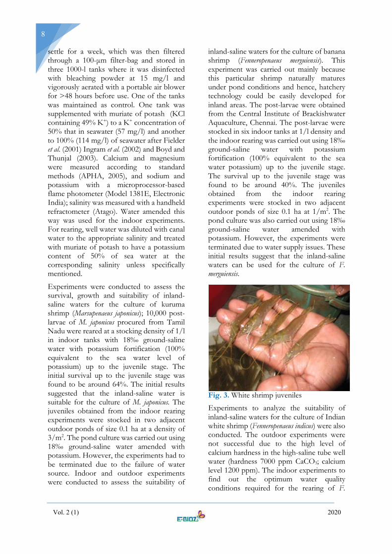
inland-saline waters for the culture of banana shrimp (Fenneropenaeus merguiensis). This experiment was carried out mainly because this particular shrimp naturally matures under pond conditions and hence, hatchery technology could be easily developed for inland areas. The post-larvae were obtained from the Central Institute of Brackishwater Aquaculture, Chennai. The post-larvae were stocked in six indoor tanks at 1/l density and the indoor rearing was carried out using 18‰ ground-saline water with potassium fortification (100% equivalent to the sea water potassium) up to the juvenile stage. The survival up to the juvenile stage was found to be around 40%. The juveniles obtained from the indoor rearing experiments were stocked in two adjacent outdoor ponds of size 0.1 ha at 1/m2. The pond culture was also carried out using 18‰ ground-saline water amended with potassium. However, the experiments were terminated due to water supply issues. These initial results suggest that the inland-saline waters can be used for the culture of F. merguiensis.
Fig. 3. White shrimp juveniles
Experiments to analyze the suitability of inland-saline waters for the culture of Indian white shrimp (Fenneropenaeus indicus) were also conducted. The outdoor experiments were not successful due to the high level of calcium hardness in the high-saline tube well water (hardness 7000 ppm CaCO3; calcium level 1200 ppm). The indoor experiments to find out the optimum water quality conditions required for the rearing of F.

Vol. 2 (1) 2020
9
indicus in inland-saline waters were taken up subsequently using water passed through ion-exchangers to reduce the calcium content. A total of 30,000 post-larvae (PL- 5) were procured from the Matsyafed Hatchery at Mopla Bay, Kannur. The post-larvae were initially conditioned using potassium supplemented inland-saline water of salinity 10‰ inside the wet laboratory for 15 days to attain a better size to stock into the outdoor ponds. After the attainment of the suitable size, around 18,000 post-larvae were stocked in a poly-lined pond of size 0.1 ha for the nursery rearing and 6,000 post-larvae maintained under indoor conditions. These animals were also reared using inland-saline water of salinity 10‰ and fed with commercial shrimp feed (CP brand). The animals had grown to an average size of 5 g in 2 months (Fig. 3) proving that indigenous species that mature under pond conditions can be cultivated using inland-saline water and that the production of seed using shrimp maintained entirely under captive conditions in such waters is possible sooner than later. This would do away with the need for seed production using wild-caught brood and transportation of theseed from the coastal areas to inland-saline areas.
4. Culture of tiger shrimp
Survival and growth of tiger shrimp (Penaeus monodon) in inland-saline water at three salinity levels 5, 10 and 15‰ were investigated with and without potassium supplementation (Raizada et al., 2015). Shrimps reared in potassium-supplemented media survived whereas, total mortality occurred in control water. Survival levels of 72.6% in 45 days and 63.3% in 60 days at salinity 5‰; 90.0% in 45 days and 88.0% in 60 days at salinity 10‰; and 81.3% in 45 days and 78.6% in 60 days at salinity 15‰ were recorded. Growth parameters indicated that there is significant difference in lengths and weights attained amongst the various treatments and growth was found to be the best at salinity 10‰. The study indicated that the inadequate level of potassium in inland-saline water is mainly responsible for the
mortality of shrimp, and that supplementation of potassium can raise the survival and growth of P. monodon to a level normally obtained in commercial production.
For commercial-level production of P. monodon, hatchery-produced post-larvae (PL-10) were procured from Kakinada, Andhra Pradesh. The PCR-screened post-larvae for white spot syndrome virus (WSSV) prior to dispatch by air to the experimental site at Rohtak were acclimatized to 10‰ salinity in natural seawater. The post-larvae (average weight 10±1 mg) were then acclimatized to artificial seawater of salinity 10‰ for 48 hours before randomly stocking at a rate of 50 post-larvae in each of the nine cylindro-conical FRP tanks of 300-l capacity for the survival trial. Stocked tanks were randomly assigned to one of the three treatments: control (ISW), potassium supplementation to 50% seawater (ISWK+50%) and potassium supplementation to 100% of that in seawater (ISWK+100%). Tanks were continuously aerated and ∼25% of water exchanged daily at the time of tank cleaning. Experimental animals were fed a commercial shrimp diet (CP Brand) ad-libitum. All the post-larvae died by the sixth day in ISW. In the other treatments, survival rates were 85.3% to 88.0% up to the sixtieth day. The post-larvae under potassium supplementation exhibited normal locomotion and body pigmentation; muscle tissue opaqueness, appendage deformity or untimely moulting were not observed. As there was no significant (p < 0.05) differences in mortality amongst the potassium treatments, pond trials were conducted at 50% potassium supplementation rate.
The post-larvae were acclimatized to pond water conditions for one hour in plastic tubs and at 4.4/m2 stocking rate in two earthen ponds of 56 x 46 m (0.25 ha) lined with LDPE geo-membrane (thickness 200 µm) with cement tiles on the sides and a soil bottom. These ponds were filled with ISW (salinity 10‰) to a depth of 120 cm and were supplemented with muriate of potash to get a K+ concentration equivalent to 50% of
Vol. 2 (1) 2020
10
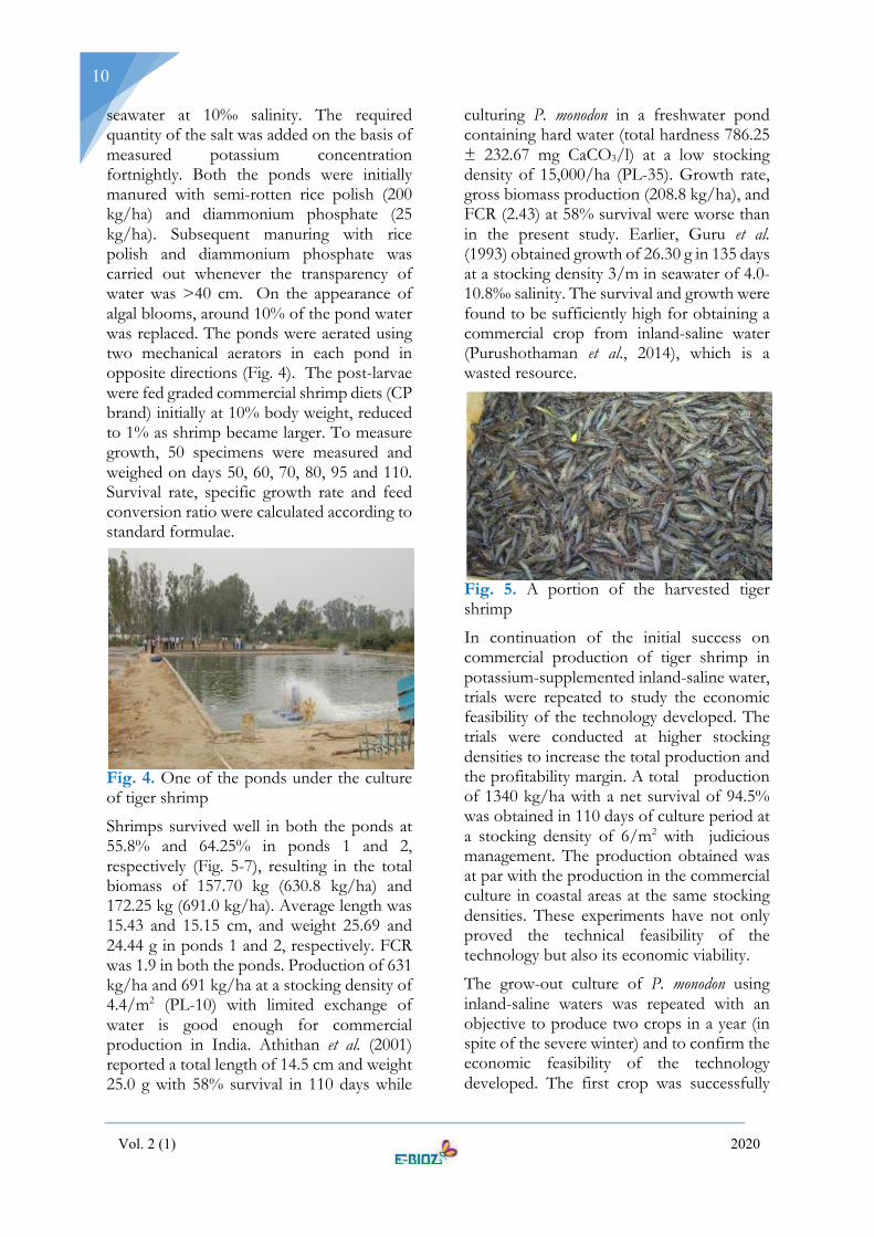
seawater at 10‰ salinity. The required quantity of the salt was added on the basis of measured potassium concentration fortnightly. Both the ponds were initially manured with semi-rotten rice polish (200 kg/ha) and diammonium phosphate (25 kg/ha). Subsequent manuring with rice polish and diammonium phosphate was carried out whenever the transparency of water was >40 cm. On the appearance of algal blooms, around 10% of the pond water was replaced. The ponds were aerated using two mechanical aerators in each pond in opposite directions (Fig. 4). The post-larvae were fed graded commercial shrimp diets (CP brand) initially at 10% body weight, reduced to 1% as shrimp became larger. To measure growth, 50 specimens were measured and weighed on days 50, 60, 70, 80, 95 and 110. Survival rate, specific growth rate and feed conversion ratio were calculated according to standard formulae.
Fig. 4. One of the ponds under the culture of tiger shrimp
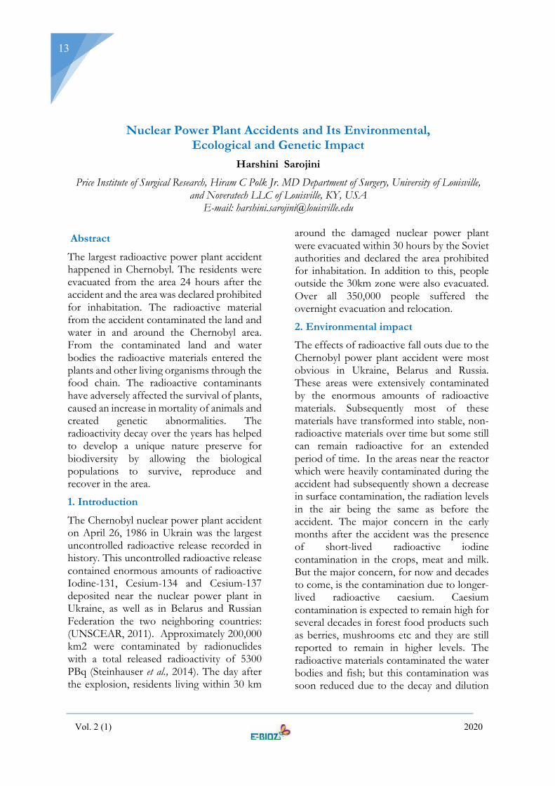
Shrimps survived well in both the ponds at 55.8% and 64.25% in ponds 1 and 2, respectively (Fig. 5-7), resulting in the total biomass of 157.70 kg (630.8 kg/ha) and 172.25 kg (691.0 kg/ha). Average length was 15.43 and 15.15 cm, and weight 25.69 and 24.44 g in ponds 1 and 2, respectively. FCR was 1.9 in both the ponds. Production of 631 kg/ha and 691 kg/ha at a stocking density of 4.4/m2 (PL-10) with limited exchange of water is good enough for commercial production in India. Athithan et al. (2001) reported a total length of 14.5 cm and weight 25.0 g with 58% survival in 110 days while
culturing P. monodon in a freshwater pond containing hard water (total hardness 786.25 ± 232.67 mg CaCO3/l) at a low stocking density of 15,000/ha (PL-35). Growth rate, gross biomass production (208.8 kg/ha), and FCR (2.43) at 58% survival were worse than in the present study. Earlier, Guru et al. (1993) obtained growth of 26.30 g in 135 days at a stocking density 3/m in seawater of 4.0-10.8‰ salinity. The survival and growth were found to be sufficiently high for obtaining a commercial crop from inland-saline water (Purushothaman et al., 2014), which is a wasted resource.
Fig. 5. A portion of the harvested tiger shrimp
In continuation of the initial success on commercial production of tiger shrimp in potassium-supplemented inland-saline water, trials were repeated to study the economic feasibility of the technology developed. The trials were conducted at higher stocking densities to increase the total production and the profitability margin. A total production of 1340 kg/ha with a net survival of 94.5% was obtained in 110 days of culture period at a stocking density of 6/m2 with judicious management. The production obtained was at par with the production in the commercial culture in coastal areas at the same stocking densities. These experiments have not only proved the technical feasibility of the technology but also its economic viability.
The grow-out culture of P. monodon using inland-saline waters was repeated with an objective to produce two crops in a year (in spite of the severe winter) and to confirm the economic feasibility of the technology developed. The first crop was successfully

Vol. 2 (1) 2020
11
harvested by the last week of June with a total production of 1680 kg/ha with a net survival of 82% in 96 days of culture period at a stocking density of 10/m2. As in the previous year, the production obtained was at par with the production obtained through commercial culture in coastal areas at the same stocking densities. The second crop was completed by the second week of November and a total production of 650 kg/ha was obtained with a net survival of 60% in 90 days of culture period at 6/m2 stocking density.
Fig. 6. Tiger shrimp harvest
Fig. 7. Specimens of harvested tiger shrimp
A repeat experiment completed by the second week of October resulted in a total production of 1280 kg/ha with a net survival of 70% in 110 days of culture duration at a stocking density of 10/m2.
Health monitoring of the shrimp in the farm trials revealed that the general health of the shrimp in potassium-amended inland-saline waters is as good as in any ideal coastal tiger shrimp culture operation. Although inland-
saline water is a novel habitat to the shrimp, it did not pose any disease susceptibility issues or health problems, provided water quality parameters are maintained. It was also found that the potassium level of inland-saline waters has direct correlation with disease susceptibility and the general health status of the shrimp. An optimum of 53 ppm potassium is required in 10‰ inland-saline water for the maintenance of ideal health conditions. As P. monodon cannot withstand the extreme low temperature at Rohtak, polyhouses (Fig. 8) were established for the overwintering of the stock and the results have been successful.
Fig. 8. Inside a poly-house fabricated for the overwintering of shrimp brood stock
Unlike coastal farms, inland farms need to be operated with either zero or very little exchange of water due to the difficulty of finding suitable places for the disposal of water with high salt content. In addition, since ISW is pumped from wells, the chances of pathogens are fairly remote, and the lack of naturally-occurring crustaceans that can serve as carriers of pathogens present a pathogen-free atmosphere for the rearing of brood stock and seed production. Both of these argue for the increased assessment of the potential of ISW for marine shrimp farming that could turn a waste resource into a viable industry. It is also expected to provide a sustainable means of livelihood to the target population.
5. Acknowledgements
The work was carried out under the Niche Area of Excellence Project on Utilization of

Vol. 2 (1) 2020
12
Inland-saline Resources for Aquaculture by the Indian Council of Agricultural Research. I sincerely acknowledge the scientists and co-workers in the project at Rohtak and Mumbai who made the studies possible.
6. References Allan, G. L., D. S. Fielder, K. M. Fitzsimmons, S.L. Applebaum and S. Raizada, (2009). Inland saline aquaculture. In: New Technologies in Aquaculture, Improving Production Efficiency, Quality and Environmental Management (ed., G. Burnell and G.Allan). Woodhead Publishing Limited/CRC Press, Boca Raton, 1119–1147.
APHA, (2005). Standard Methods for the Examination of Water and Wastewater. 21st ed. American Public Health Association. Washington, DC.
Applebaum, S., (1995). Technology for desert aquaculture. J. Arid Land Stud.,5S: 207–210.
Applebaum, S., (1998). Desert aquaculture: A new opportunity for world aquaculture production. J. Arid Land Stud.,7S:101–103.
Athithan, S., T. Francis, N. Ramanathan and V. Ramadhas, (2001). A note on monoculture of Penaues monodon in a hardwater seasonal pond. Naga,24(3 & 4): 14–15.
Boyd, C. E. andT. Thunjal, (2003). Concentrations of major ions in waters of inland shrimp farms in China, Ecuador, Thailand, and the United States. J. World Aquacult. Soc.,34(4): 524–532.
Collins, A. and B. Russell, (2003). Inland prawn farming trial in Australia. Pond study test P. monodon performance in low-salinity groundwater. Global Aquacult. Advocate, 6:84–85.
Doroudi, M. S., D. S. Fielder, G. L. Allan and G. K. Webster, (2006). Combined effects of salinity and potassium on juvenile mulloway, Argyrosomus japonicus (Temminck and Sehlegael) in inland saline groundwater. Aquacult. Res.,37:1034–1039.
Doroudi, M. S., G. Webster, G. L. Allan and D. S. Fielder, (2007). Survival and growth of silver perch, Bidyanusbidyanus, a salt-tolerant freshwater species, in inland saline groundwater from southwestern New South Wales, Australia. J. World Aquacult. Soc.,38(2): 314–317.
FAO, (1992). The Use of Saline Waters for Crop Production. Technical Report. Food and Agriculture Organization, Rome.
FAO, (2000). Extent and Causes of Salt-affected Soils in Participating Countries. Land and Plant Nutrition Management Service. Food and Agriculture Organization, Rome,http://www.fao/ag/agl/agll/spush.
Fielder, D. S., W. J. Bardsley and G. L. Allan, (2001). Survival and growth of Australian snapper, Pagrus auratus, in saline groundwater from inland New South Wales, Australia. Aquaculture,201:73–90.
Guru, B. C., V. C. Mohapatra, S. Pal and A. K. Mohanty, (1993). Observations in the growth of P. monodon in a low saline culture pond. Indian J. Fish.,40(4): 262–263.
Ingram, B. A., G. J. Gooley, I. J. McKinnon and S. S. De Silva,(2002). Aquaculture-agriculture systems integration: An Australian perspective. Fish. Manage. Ecol.,7:33–43. Khan, M. H. A., M. U. aleem, S. R. Ahmad, N. Ahmad, S. J.Sameeni, M.Akram and M. Farooq, (2017). Role of canal lining on groundwater fluctuations: A modelling simulation approach for Jaalwala Distributary, Bahawalnagar. Open J. Appl. Sci., 7(5):213–232.
Partridge, G. J. and J. Creeper, (2004). Skeletal myopathy in juvenile barramundi, Latescalcarifer (Bloch), cultured in potassium-deficient saline groundwater. J. Fish Dis.,27:523–530.
Partridge, G. J. and A. J. Kymbey, (2008). The effect of salinity on the requirement for potassium by barramundi (Latescalcarifer) in saline groundwater. Aquaculture, 278:164–170.
Pragnell, D. I. and R. Fotedar, (2005). The effect of potassium concentration in inland saline water on the growth and survival of the western king shrimp, Penaeus latisulcatusKishinouye, 1896. J. Appl.Aquacult., 17(2): 19–33.
Purushothaman, C. S., S. Raizada, V. K. Sharma, V. Harikrishna, G. Venugopal, R. K. Agrahari, M. Rahaman, J. Hasan and A. Kumar, (2014). Production of tiger shrimp (Penaeus monodon) in potassium supplemented inland saline sub-surface water. J. Appl. Aquacult., 26:84-93.
Rahman, S., A. K. Jain, A. K. Reddy, K. Girish and D. R. Koyya, (2005). Ionic manipulation of inland saline groundwater for enhancing survival and growth of Penaeus monodon (Fabricius). Aquacult. Res.,36:1149–1156.
Raizada, S., C. S. Purushothaman, V. K. Sharma, V. Harikrishna, M. Rahaman, R.K. Agrahari, J. Hasan, G. Venugopal and A. Kumar, (2015). Survival and growth of tiger shrimp (Penaeus monodon) in inland saline water supplemented with potassium. Proc. Nat. Acad. Sci. India Sec. B Biol. Sci.,85(2): 491–497.
Saoud, I. P., D. A. Davis and D. B. Rouse, (2003). Suitability studies of inland well waters for Litopenaeusvannamei culture. Aquaculture, 217:373–383.
Tantulo, U. and R. Fotedar, (2006). Comparison of growth, osmoregulatory capacity, ionic regulation and organosomatic indices of black tiger shrimp (Penaeus monodon Fabricius, 1798) juveniles reared in potassium fortified inland saline water and ocean water at different salinities. Aquaculture, 258(1-4): 594–605.
USGS,(2013). The World's Water. United States Geological Survey. http://ga.water.usgs.gov/ edu/earthwherewater.
Vol. 2 (1) 2020
13
Nuclear Power Plant Accidents and Its Environmental, Ecological and Genetic Impact
Harshini Sarojini
Price Institute of Surgical Research, Hiram C Polk Jr. MD Department of Surgery, University of Louisville, and Noveratech LLC of Louisville, KY, USA
E-mail: [email protected]
Abstract
The largest radioactive power plant accident happened in Chernobyl. The residents were evacuated from the area 24 hours after the accident and the area was declared prohibited for inhabitation. The radioactive material from the accident contaminated the land and water in and around the Chernobyl area. From the contaminated land and water bodies the radioactive materials entered the plants and other living organisms through the food chain. The radioactive contaminants have adversely affected the survival of plants, caused an increase in mortality of animals and created genetic abnormalities. The radioactivity decay over the years has helped to develop a unique nature preserve for biodiversity by allowing the biological populations to survive, reproduce and recover in the area.
1. Introduction
The Chernobyl nuclear power plant accident on April 26, 1986 in Ukrain was the largest uncontrolled radioactive release recorded in history. This uncontrolled radioactive release contained enormous amounts of radioactive Iodine-131, Cesium-134 and Cesium-137 deposited near the nuclear power plant in Ukraine, as well as in Belarus and Russian Federation the two neighboring countries: (UNSCEAR, 2011). Approximately 200,000 km2 were contaminated by radionuclides with a total released radioactivity of 5300 PBq (Steinhauser et al., 2014). The day after the explosion, residents living within 30 km
around the damaged nuclear power plant were evacuated within 30 hours by the Soviet authorities and declared the area prohibited for inhabitation. In addition to this, people outside the 30km zone were also evacuated. Over all 350,000 people suffered the overnight evacuation and relocation.
2. Environmental impact
The effects of radioactive fall outs due to the Chernobyl power plant accident were most obvious in Ukraine, Belarus and Russia. These areas were extensively contaminated by the enormous amounts of radioactive materials. Subsequently most of these materials have transformed into stable, non-radioactive materials over time but some still can remain radioactive for an extended period of time. In the areas near the reactor which were heavily contaminated during the accident had subsequently shown a decrease in surface contamination, the radiation levels in the air being the same as before the accident. The major concern in the early months after the accident was the presence of short-lived radioactive iodine contamination in the crops, meat and milk. But the major concern, for now and decades to come, is the contamination due to longer-lived radioactive caesium. Caesium contamination is expected to remain high for several decades in forest food products such as berries, mushrooms etc and they are still reported to remain in higher levels. The radioactive materials contaminated the water bodies and fish; but this contamination was soon reduced due to the decay and dilution

Vol. 2 (1) 2020
14
of the radioactive materials. Up till now some of the radioactive materials can be trapped in the soils surrounding the contaminated rivers and lakes. Recently, in most water bodies and fish less radioactivity levels were detected, while in some closed lakes the radioactivity still remains high. The heavy radioactive fallout affected numerous plants and animals within 30 km of the site. High mortality, reduced levels of reproduction and genetic abnormalities were reported in those plants and animals. Now 30 years later, the radioactivity levels decreased over the years allowing the biological populations to survive, reproduce and recover in the area and has become a unique nature preserve for biodiversity (UN Chernobyl Forum, 2006).
3. Ecological implication
Ionizing radiation from Chernobyl nuclear accidents has diminished the diversity and species abundance of the ecosystem. The resurgence in studies focusing on radiobiology and radioecology, specifically on the low dose effects of ionizing radiation emerged after Chernobyl nuclear power plant accident. The effects of low dose radiation on non-human biota are seldom studied. Møller, Mousseau and colleagues have conducted a significant amount of field studies on the effects of the low doses of radiation on non-human biota especially in Chernobyl birds. The emphasis on the study was based on high metabolic rates, high survival rates and a high diversity of bird species with variable life history, migratory propensity and dispersal. The richness, abundance and population density of breeding birds were reported to decrease with increasing radiation levels (Møller and Mousseau, 2007). Similar results were observed for the invertebrates’ in the uppermost soil layer of Chernobyl (Møller and Mousseau, 2009).Chernobyl birds were found to have impaired brain development interrelated to oxidative stress, subsequently resulting in smaller head volume (Møller et al., 2011). High frequency of cataracts independent of bird age was also reported (Mousseau and Møller, 2013). Elevated frequencies of abnormalities, such as partial
albinism, deformed toes, and tumors have been reported in barn swallows from Chernobyl (Møller et al., 2007), However, it has been debated that the ambient dose rates in the contaminated regions are too low to certify significant impacts (Baker and Chesser, 2000; Deryabina et al., 2015). Correspondingly no standardized census subsists for common animals in relation to the radiation (Møller and Mousseau, 2007). Hence, the query of the ecological effects of radiation essentially remains unresolved. Even though, the precise dose determination under field conditions can be a challenging, the maximum dose rate for terrestrial plants to reproduce and long-term survival in natural populations is considered to be 400 mGy/h (UNSCEAR, 2008). However, these lower doses of ionizing radiation can induce stress responses in an irradiated organism (Galvan et al., 2014; Volkova et al., 2017). The comprehensive molecular mechanisms of adaptation to chronic radiation exposure by plants still remain unresolved (Kovalchuk et al., 2003,2004; Boubriak et al., 2016; Møller and Mosseau, 2016).
The ionizing radiation exposure dose at which the response occurs depend on the species, age, plant morphology, physiology and genome organization (Holst and Nagel, 1997). Ionizing radiations have differential effects on plant growth and development; stimulatory effects at low doses, harmful effects for vegetative growth at medium levels, and pronounced reduction in reproduction and yield at higher radiation levels (Jan et al., 2012). In rapid growing plants the radionuclides were absorbed to by young leaves. This mechanism of active translocation and absorption of the water-soluble radioactive elements through the leaves were reported by Coughtrey and Thorne (1983). Conversely, plants also absorb radionuclides through their roots from the soil. Rain fall helped to clear the atmosphere from radionuclides, which were consequently transferred to the soil. Several studies have reported that rain can carry more radioactivity deposition than a

Vol. 2 (1) 2020
15
radioactive cloud settling (Papastefanou et al., 1988). Marine organisms have low radioactivity concentrations due to their dilution of radioactive fall outs in sea water. Mosses, fungi and lichens are reported to be collectors of a variety of heavy metals and toxic substances from their environment (Papastefanou et al., 1989). Finally, in fruit-bearing trees, higher radioactivity is clearly observed in the leaves than in the fruit, due to a greater surface exposed to contamination.
The biodiversity loss is a serious concern, since they play a vital role in long term ecosystem functioning (Groombridge and Jenkins, 2002). Hence, research focused on the biodiversity spatial distribution is essential.
4. Genetic significance
The genetic studies conducted in Chernobyl showed higher rates of genetic impairment and mutation. Ionizing radiation can induce diverse effects at the genetic level, and this can vary from simple base pair substitutions to single- or double-stranded DNA breaks (Grosovsky et al., 1988). The population sizes in highly radioactive parts of the Chernobyl Exclusion Zone were found to be reduced among most of the investigated taxonic groups (i.e., birds, bees, butterflies, grasshoppers, dragonflies, spiders, mammals). One of the initial tests for radiation on mutation rates at Chernobyl used microsatellite markers to examine de novo mutation rates in barn swall Hirundorustica (Ellegren et al., 1997). The mutation rates in this study were 2 to 10-fold higher for birds in Chernobyl than the control populations from Ukraine and Italy. Amid the earliest visible signs of radiation exposure were the appearance of white spots on the bird feathers and the mammalian furs. These “partial albinos” have been reported for Chernobyl barn swallows (Ellegrenet al. 1997; Møller and Mousseau2001) and other bird species (Møller, Bonisoli-Alquati, et al. 2013). Møller et al. (2004) reported that the frequency of abnormal sperms in barn swallows was up to 10 times higher for
Chernobyl birds as compared to sperms from males living in control areas. They also reported that the abnormality rates were interrelated to the reduced levels of antioxidants in the blood, liver, and eggs of these birds, thereby postulating the hypothesis that antioxidants play a significant role in DNA protection from the direct/indirect radionuclides exposure. Møller et al. (2008) found that sperm behavior was negatively affected by radiation levels while Bonisoli-Alquati et al. (2011) found that plasma oxidative status could predict sperm performance from the effects of ionizing radiation. Overall, these studies provide convincing evidence that low dose radiation results in male infertility and this may be the explanation for the smaller population sizes of many species in the Chernobyl region. The number of visible tumors on birds was significantly higher in radioactive areas. This could be due to the higher mutation rates in the somatic tissues (Møller, Bonisoli- Alquati, et al., 2013).
5. References Baker, R.J., Chesser, R.K., (2000). The Chornobyl nuclear disaster and subsequent creation of a wildlife preserve. Environ. Toxicol. Chem. 19, 1231–1232.
Bonisoli-Alquati, A., Møller, A.P., Rudolfsen, G., Saino, N., Caprioli, M., Ostermiller, S., and Mousseau, T.A. (2011) The effects of radiation on sperm swimming behavior depend on plasma oxidative status in the barn swallow (Hirundorustica). Comp. Biochem. Physiol, Part A, MolIntegr Physiol. 159:105–112.
Boubriak, I., Akimkina, T., Polischuk, V., Dmitriev, A., McCready, S.,and Grodzinsky, D., (2016). Long term effects of Chernobyl contamination on DNA repair function and plant resistance to different biotic and abiotic stress factors. Cytol. Genet. 50 (6),34-59. http://dx.doi.org/10.3103/S0095452716060049
Coughtrey, P. J. and Thorne, M.C (1983). Radionucleotide distribution and transport in terrestrial and aquatic ecosystems. A critical review of data, (Volume I). A.A. Balkema, Rotterdam.
Deryabina, T., Kuchmel, S., Nagorskaya, L., Hinton, T., Beasley, J., Lerebours, A., and Smith, J., (2015). Long-term census data reveal abundant wildlife populations at Chernobyl. Curr. Biol., 25, R824eR826.
Ellegren, H., Lindgren, G., Primmer, C.R., andMøller, A.P.,(1997). Fitness loss and germline mutations in

Vol. 2 (1) 2020
16
barn swallows breeding in Chernobyl. Nature, 389:593–596.
Galvan, I., Bonisoli-Alquati, A., Jenkinson, S., Ghanem, G., Wakamatsu, K., Mousseau, T., and Møller, A.P., (2014). Chronic exposure to low-dose radiation at Chernobyl favours adaptation to oxidative stress in birds. Funct. Ecol., 28,1387-1403. http://dx.doi.org/10.1111/1365-2435.12283
Groombridge, B. and Jenkins, M.D. (2002). World Atlas of Biodiversity. Prepared by the UNEP World Conservation Monitoring Centre, University of California Press, Berkeley.
Grosovsky, A.J., Deboer, J.G., Dejong, P.G., Drobetsky, E.A., and Glickman, B.W., (1988). Base substitutions, frame shifts, and small deletions constitute ionizing radiation induced point mutations in mammalian-cells. Proc. Natl. Acad. Sci. U. S. A, 85,185-188. PMID: 3422416.
Holst, R.W., and Nagel, D.J., (1997). Radiation effects on plants. In:Wang,W., Gorsuch, J.W., Hughes, J.S. (Eds.), Plants for Environmental Studies. CRC Press/Lewis Publishers, New York, pp. 37-79.
Jan, S., Parween, T., Siddiqi, T.O., and Mahmooduzzafar, (2012). Effect of gamma radiation on morphological, biochemical, and physiological aspects of plants and plantproducts. Environ. Rev.,20,17-39.
Kovalchuk, O., Burke, P., Arkhipov, A., Kuchma, N., James, S.J., Kovalchuk, I., and Pogribny, I., (2003). Genome hypermethylation in Pinussylvestris of Chernobylea mechanism for radiation adaptation? Mutat. Res. 529, 13e20.
Kovalchuk, I., Abramov, V., Pogribny, I., and Kovalchuk, O., (2004). Molecular aspects of plant adaptation to life in the Chernobyl zone. Plant Physiol.,135,357e363.http://dx.doi.org/10.1104/pp.104.040477
Møller, A.P., and Mousseau, T.A. (2001). Albinism and phenotype of barn swallows (Hirundorustica) from Chernobyl. Evolution. 55:2097–2104.
Møller, A.P., Surai, P., and Mousseau, T.A. (2004). Antioxidants, radiation and mutations in barn swallows from Chernobyl. Proc Roy Soc Lond., B.272:247–252.
Moller, A.P., and Mousseau, T.A. (2007a). Species richness and abundance of forest birds in relation to radiation at Chernobyl. Biol Lett., 3:483–486.
Møller, A.P., and Mousseau, T.A. (2007b). Determinants of interspecific variation in population declines of birds after exposure to radiation at Chernobyl. J. Appl. Ecol., 44: 909-919.
Møller, A.P., Mousseau, T.A., de Lope, F., and Saino, N. (2007). Elevated frequency of abnormalities in barn swallows from Chernobyl. Biol. Lett., 3:414-417.
Møller, A.P., Mousseau, T.A., Lynn, C., Ostermiller, S., and Rudolfsen, G. (2008). Impaired swimming behaviour and morphology of sperm from barnswallows Hirundorusticain Chernobyl. Mutat. Res., 650: 210-216.
Møller, A.P., and Mousseau, T.A. (2009). Reduced abundance of insects and spiders linked to radiation at Chernobyl 20 years after the accident. Biol. Lett., 5:356-359.
Møller, A.P., Bonisoli-Alquati, A., Rudolfsen, G., and Mousseau, T.A. (2011). Chernobylbirds have smaller brains. PLoS ONE. 6:16862.
Mousseau, T.A., Møller,A.P.,( 2013). Elevated frequency of cataracts in birds from Chernobyl. PLoS ONE. 8: 66939.
Møller, A.P., Bonisoli-Alquati, A., and Mousseau, T.A., (2013). High frequency of albinism and tumours in free-living birds around Chernobyl. Mutat Res., 757:52-59.
Møller, A.P., Mousseau, T.A., (2016). Are organisms adapting to ionizing radiation atChernobyl? Trends Ecol. Evol., 31 (4), 281-289.
Papastefanou, C, Manolopoulou, M and Charalambous, S. (1988). Radiation measurements and radioecological aspects of fallout from the Chernobyl. J. Environ. Radioactivity, 7, pp. 49-64
Papastefanou, C, Manolopoulou, M and Sawidis, T. (1989)Lichens and mosses: Biological monitors of radioactive fallout from the Chernobyl reactor accident. J. Environ. Radioactivity, 9(3), pp. 199-207
Steinhauser, G., Brandl, A. and Johnson, T.E. (2014) Comparison of the Chernobyl and Fukushima nuclear accidents: a review of the environmental impacts. Sci. Total Environ., 2014 Jul 15; 487-575.
UN Chernobyl Forum, (2006)Chernobyl's Legacy: Health, Environmental and Socioeconomic.
UNSCEAR (United Nation Scientific Committee on the Effects of Atomic Radiation), (2008). Effect of ionizing radiation on non-human biota. Report to the General Assembly with Scientific Annexes, Volume II. Scientific Annex E.
UNSCEAR, (2011). United Nations (UN) Report to the General Assembly, Scientific Annexes C, D and E, UNSCEAR 2008 Report. New York: United Nations.
Volkova, P., Yu., Geras’kin, S.A. and Kazakova, E.A., (2017). Radiation exposure in theremote period after the Chernobyl accident caused oxidative stress and geneticeffects in Scots pine populations. Sci. Rep., 7, 43009. http://dx.doi.org/10.1038/srep43009.
Vol. 2 (1) 2020
17
Rare diseases are not actually rare in India Binukumar, B.K.
CSIR-Institute of Genomics and Integrative Biology, New Delhi, India
E-mail: [email protected]
1. Introduction
Rare diseases pose clinical and economic burden as well as a significant challenge for health systems. About 6,000 to 7,000 patients are reported to be suffering from rare diseases, for most of which no specific treatment options are available. A disease or disorder is defined as rare in Europe when it affects less than 1 in 2000 in the population and in the USA it is set to be at 1 / 200,000 at any given time. A rare disease is often referred to as an orphan disease. They are characterized by a broad diversity of disorders and symptoms that vary not only from disease to disease but also from patient to patient suffering from the same disease. 80% of the rare diseases have genetic origins and others are the result of infections, allergies, and environmental causes. Most of them are degenerative and proliferate in nature and 50% of rare diseases affect children. Most common symptoms can hide underlying rare diseases, leading to misdiagnosis. In most of the cases, there may not be effective cure and can cause high level of pain and suffering to the patients as well as their families.
Early diagnosis is a major challenge in rare diseases and most cases remain undiagnosed for a long period of time. Majority of the cases take more than 7 years to diagnose. During this period the patients experience poor physical and mental health problems. They also try a variety of laboratory tests and often visit super-specialty doctors and hospitals. It is very important to put an end to this diagnostic dilemma. Once the patient receives a proper diagnosis, they can move to
next steps of disease management looking for different treatment options, lifestyle changes etc. More than 7000 rare diseases are identified globally and about 450 of them have been reported in India. For most diseases treatment is unavailable even after proper diagnosis, because only about 5 percent of rare diseases have a treatment approved by the Food and Drug Administration. Most of them are very expensive and unaffordable to common man. Insurance policies most of the time do not cover these ongoing (lifelong) treatment expenses.
2. Common Problems faced and International initiative
The major delay in rare disease diagnosis is due to the lack of quality of genomic information and scientific knowledge. The lack of appropriate quality health care engenders inequalities and difficulties in access to treatment and care. Most of the time, this situation leads to heavy social and financial burdens on patients and family. The initial misdiagnosis is common in most cases of rare diseases because of the board diversity and relatively common symptoms associated with other diseases. Due to the spatiality and verity of rare diseases, research needs to be global to ensure that policy-making experts, healthcare providers, basic researchers, and clinicians are connected. In addition to that clinical trials are multinational and that patients can benefit from the pooling of data and resources across borders. Initiatives such as the European Reference Networks, the International Rare Disease Research Consortium and the EU Framework

Vol. 2 (1) 2020
18
Programme for Research and Innovation Horizon 2020 support international level collaborative research on rare diseases.
3. The incidence of rare diseases globally and specific to India
The major concern in rare diseases is that it cannot be determined by a universal definition. However, the importance of having a consistent definition is well acknowledged. Adopting a standard definition of rare disease is a pre-requisite for public policy development. There are approximately 350 million patients affected by about 7000 known rare diseases. Rough estimates indicate that over 70 million people in India are affected by rare diseases, many of whom may still not have a diagnosis. Different rare diseases database including Orphanet database is updated every year to accommodate new diseases that are being reported. Orphanet database is maintained by the European Union. We do not yet have accurate statistics on the incidence or prevalence of rare diseases in India. This is mainly because of the lack of definition and more importantly, due to the lack of diagnostic tools and equipment and systematic data collection systems in India. Half of the rare diseases are early onset childhood diseases. About 80% of all rare diseases are genetic in origin, most of them monogenic. The US National Institutes of Health initiatives such as the Undiagnosed Diseases Network (UDN) and the international rare diseases research consortium (IRDiRC) aim to address this challenge by accelerating the speed of diagnosis. The goal is to bring the average time to diagnosis down to one year.
Time to time, scientific and patient communities have expressed the need for government initiatives towards rare diseases. The first attempt to bring together all experts of rare diseases under a common platform was initiated by Indian National Science Academy (INSA), which conducted the first of the kind rare disease workshop entitled “To Develop a Scientific Program for Research on Rare Diseases” in 2016, which
deliberated on issues such as definition of “Rare disease,” rare disease awareness, rare disease research avenues, policy framework for boosting and incentivizing research and development efforts and framing suitable legislation to ensure involvement of the State in fulfilling the special needs of rare diseases. In the INSA rare disease workshop (2016), the honourable Drug Controller General of India stated that a policy for accelerated clearance of orphan drugs and fast-track approval is not in place because government needs clear-cut recommendations regarding the definition of rare disease, mechanism for fast-track approval (e.g., waiver of a specific phase in orphan drug clinical trial). He again stated that genetic differences in Indian population warrant Indian-centered studies, rather than using data from studies in other countries. He also invited for expert suggestions on the need of changes in the drugs and cosmetic act to meet the requirements of research in rare disease.
The Genetic and Rare Diseases Information Center (GARD) is a program of the National Center for Advancing Translational Sciences (NCATS) and is funded by two associate institutions of the National Institutes of Health (NIH): NCATS and the National Human Genome Research Institute (NHGRI). GARD provides the public with access to current, reliable, and easy-to-understand information about rare or genetic diseases in English or Spanish. NORD’s database provides brief introductions for patients and their families about more than 1,200 rare diseases. This is not a comprehensive database considering the fact that there are nearly 7,000 diseases considered rare in the U.S.
4. RARE List™
The RARE List™ comprises approximately 7,000 different rare diseases and disorders affecting more than 300 million people worldwide. Some common diseases are included on the RARE List™ because in the United States one of the primary criteria for recognizing a disease as rare is the prevalence of the disease fewer than 200,000 cases.
Vol. 2 (1) 2020
19
Diseases such as malaria are quite common in some parts of the world but are considered rare in the United States. Other diseases such as Cancer or Alzheimer's are not rare diseases as a whole but certain form of these diseases are considered rare.
5. The Indian Council of Medical Research (ICMR) initiative
ICMR has been providing financial assistance to projects for orphan disease research and for sponsoring/organizing workshops /conferences /training programs on rare diseases. It has also taken the initiative in the preparation of a registry for rare disease. This is referred to as National Initiative for Rare Diseases (NIRD), organized jointly by ICMR, AIIMS, JNU, and PRESIDE. The first step in this endeavour is to identify patients with the rare disease. “Indian rare disease registry” was launched on April 27, 2017. This registry is intended to cover all rare and ultra-rare diseases prevalent in India. The registry is first intended to be hospital-based and later population based. The objectives of the registry include: to identify patients having rare diseases, to use that data for policy framing and to guide future research. The registry may also enable proper and easy monitoring of the diseases including their prevalence, incidence and natural history with regard to the Indian context.
6. Council of Scientific & Industrial Research (CSIR) and Institute of Genomics & Integrative Biology (IGIB) initiative
IGIB, New Delhi, has conducted a project funded by CSIR, named as “Genomics for Understanding Rare Diseases India Alliance Network (GUaRDIAN),” for the purpose to bring together and understand novel genetic variations to achieve translational applications by both clinicians and basic science researchers. GUaRDIAN, the pioneer and one of the largest networks of clinicians and researchers in India have been working on rare genetic diseases. The CSIR- IGIB offers a research oriented, well-
structured and carefully supervised training programme in rare diseases and related fields.
7. Indian Collaborative Research Network on Wilson's Disease (ICROWD)
ICROWD is also another programme supported by CSIR at IGIB for Wilson’s diseases (WD). WD, a classical monogenic disorder, is the commonest cause of paediatric chronic liver disease in Indian subcontinent and also, one of the few treatable causes of liver disease across all populations. We have already established a pilot network of over 40 clinicians and researchers from over 9 medical and research centres across the country working in the area of WD. The ICROWD has four major components encompassing the clinical areas of WD, the molecular genetic basis of WD, disease modelling and correction of gene defects. For each of the four areas, we have a consortium approach to deliver better diagnosis and personalized, precision medicine in WD clinical settings in India.
8. Nongovernmental organization initiative
Organization for Rare Diseases India (ORDI; www.ordindia.org) is a voluntary organization which was established to deal with the rare disease conditions in the Indian population. The ORDI team members belong to different disciplines and they need not have a science background. ORDI deals with the matters related to the rare disease such as unique challenges in dealing with rare diseases (Rajasimha et al.,2014). The Indian organization for rare diseases was conceived in 2005 and is incorporated as a not-for-profit organization in India as well as in the USA. It is the umbrella organization and represents interests of all rare diseases, individual patients, patient support groups, health policy advocates and health care provide for rare disease.
9. Judiciary initiative
In November 2016, the Delhi high court had ordered the government to finalize a policy

Vol. 2 (1) 2020
20
on rare diseases and consequently a draft policy was submitted by the Union Ministry of Health to the Delhi high court on May 25. The Delhi high court then directed the Centre to implement a National Policy for treatment of Rare Diseases without delay (Bhuyan, 2017).
10. Academic institutes
There are many ongoing research projects on various aspects of rare diseases undertaken by reputed institutes such as AIIMS, PGIMER Chandigarh, CMC Vellore, and SGPGI Lucknow. It is expected that the outcome of these projects would contribute significantly to solving the existing problems in dealing with rare diseases such as diagnosis, treatment protocols, causes and genetic basis of the diseases.
Rare Disease Day® is observed worldwide, typically on or near the last day of February each year, to create awareness among policymakers and the public about rare diseases and their impact on patients’ lives. Each year, NCATS and the NIH Clinical Centre (CC) sponsor Rare Disease Day at NIH as part of this global observance. The global theme for 2019 was “bridging health and social care.”
Recently, the Government of India has informed the Hounarable Madras High Court during the hearing of a Public Litigation Petition (PIL) filed by the Lysosomal Storage Disorders (LSD) Support Society of India, New Delhi, that the Government has publised a draft policy on rare diseases, seeking public opinion on it. It will be finalized only after considering the views of all stake holders. It is stated that the draft policy envisages a provision of Rs. 150000, for meeting medical expenses, to those suffering from LSD. The draft policy categorizes rare diseases amenable to one-time curative treatment such as Haemopoietic stem cell transplantation and those that require organ transplantation into one group. Patients suffering from these diseases are eligible for the relief under the umbrella scheme of Rastriya Arokya Nidhi from Government of India (The Hindu, dated 11.02.2020).
11. References
Bhuyan, A., (2017). Government Submits Rare Disease Policy to Delhi HC, Recommends Rs 100 Crore for Genetic Diseases. The Wire. [Last accessed on 2017 Sep 22]. Available from: https://www.thewire.in/140229/rare-disease-policy/.
Rajasimha, H.K., Shirol, P. B., Ramamoorthy, P., Hegde, M., Barde S., Chandru, V., (2014).
Organization for rare diseases India (ORDI) – Addressing the challenges and opportunities for the Indian rare diseases' community. Genet. Res. (Camb); 96:e009.
Websites
https://food.ndtv.com/health/rare-disease-day-7-most-rare-diseases-in-the-world-and-the importance-of-research-1664134.
https://health.economictimes.indiatimes.com› Latest Health News, Industry.
https://www.rarediseaseday.org/article/what-is-a-rare-disease.
Minutes of the Meeting. Meeting of Pharma Stakeholders with DCG (I) to Explore the Possibilities of Providing Cheaper Medicines,
Therapies for Treatment of Rare Disease. 2016. May 04, [Last accessed on 2017 Sep 22]. Availablefrom:http://www.cdsco.nic.in/writereaddata/Minutes%20Of%20Meeting%20Stakeholders%2004_05_2016.pdf.
ICMR Launches ‘Indian Rare Disease Registry’ to Address Unmet Needs of Patients with Rare Diseases. CheckOrphan. [Last accessed on 2017 Sep 22]. Available from:
http://www.checkorphan.org/news/icmr-launches-indian-rare-disease-registry-to-address-unmet-needs-of-patients-with-rare-diseases.
‘National Policy for Rare Diseases a Welcome Step, Challenges Ahead’ Sunday Guardian. 2017. [Last accessed on 2017 Sep 22]. Available from: http://www.sundayguardianlive.com/news/9732-national-policy-rare-diseases-welcome-step-challenges-ahead.
Implement National Policy on Rare Diseases: HC to Centre. Zee News. 2017. [Last accessed on 2017 Sep.22]. Available from: http://www.zeenews.india.com/delhi/implement-national -policy-on-rare-diseases-hc-to-centre-2009374.html.

Vol. 2 (1) 2020
21
Mass spectrometry, an analytic tool in biological research Pradeep Kumar, R.
Department of Zoology, Govt. College for Women, Thiruvananthapuram, India
E-mail: [email protected]
1. Introduction
The interaction between electromagnetic radiation and matter forms the basis for spectrometry. Mass Spectrometry is a potent tool for identifying unknown compounds and studying molecular structure based on the fundamental principles of chemistry. In it, the compound to be characterised is ionised and the ionic molecules are then separated based on their mass/charge ratio (m/z) and the number of ions representing each mass/charge unit is recorded as a spectrum. It has become an essential analytical tool in biological research and can be used to characterise a wide variety of biomolecules such as proteins, sugars and oligonucleotides. The ions formed are very reactive and short-lived. So, their formation and manipulation must be conducted in a vacuum to minimise ion-molecule reactions, scattering, and neutralisation of the ions.
2. Components
A mass spectrometer consists of the following components: the inlet system, ion source, mass analyser and detector (Fig. 1).
2.1. Inlet system
The function of an inlet system is to introduce a small amount of sample into the ion source with minimal loss of vacuum. The samples should be in the vapour phase prior to ionisation. Gases and samples with high vapour pressure are introduced directly into the source region of the mass spectrometer through a needle valve. Liquids and solids are usually heated to increase the vapour
pressure for analysis. Modern mass spectrometers are equipped with different kinds of inlet systems like batch inlets, direct probe inlets, chromatographic and capillary electrophoretic inlet systems. The batch inlet system is the conventional and simplest one where the sample is volatilised externally and then allowed to leak into the evacuated ionisation region. Direct probe inlets are used for the injection of solid and non-volatile liquid into the ionisation region by means of a sample probe. Chromatographic inlet systems in Gas chromatographic mass spectrometry (GC-MS) and Liquid chromatographic mass spectrometry (LC-MS), capillary electrophoretic units in Capillary electrophoresis mass spectrometry (CE-MS) are employed for the separation and identification of the components in the sample mixture by giving independent mass spectra. Gas chromatography is the commonly used technique for introducing volatile samples into a mass spectrometer while liquid chromatography is used for thermally labile compounds which are not easily separated by gas chromatography.
2.2. Ion source
A sample molecule for analysis by mass spectrometer must be converted to gas phase-charged particles by ionisation process. In the ion source region, neutral sample molecules are ionised and then accelerated into the mass analyser. The ions are generated by inducing either the loss or gain of charge. The various methods of ionisation include electron ionisation,

Vol. 2 (1) 2020
22
chemical ionisation, desorption ionisation and electrospray ionisation.
In electron ionisation, a beam of high energy electrons strikes the molecules. The electron molecule collision releases an electron from the molecule creating a cation or electron gain to produce anions as follows.
The product in both instances is a radical, and also an ion. Hence it is represented as M.+or M.-, + and – signs indicate ionic state and dot (.) represents a radical. It may be called a radical ion or molecular ion or parent ion. In chemical ionisation, the sample molecules are combined with an ionised reagent gas. On collision with ionised reagent gas (methane, ammonia, isobutene), sample molecules are ionised by proton transfer, electron transfer, and adduct formation. In desorption ionisation, the sample to be analysed is dissolved in a matrix and placed on a high energy beam of ions or high intensity photons. When high intensity photons are used it is termed MALDI (Matrix-assisted laser desorption/ionisation). The analyte is placed in a light absorbing solid matrix which protects the analyte from being destroyed by direct laser beam. The matrix is composed of energy absorbing molecules such as 2,5-dihydroxybenzoic acid or cyano-4-hydroxycinnamic acid. With a short pulse of laser light, the analytes are ionised into gas phase and desorbed from the matrix into the vacuum system. In electrospray ionisation (ESI), the samples are dissolved in a polar, volatile solvent and pumped through a narrow, stainless steel capillary. A high voltage of 3 or 4 kV is applied to the tip of the capillary. The sample emerging from the tip is dispersed into an aerosol of highly charged droplets. The solvent evaporates, ions are released from the droplets. It is one of the most important techniques for analysing biomolecules such as polypeptides, proteins, and oligonucleotides having molecular weights of 100,000 Da or more. It
produces multiply charged ions based on ion evaporation process.
2.3. Mass analysis
The mass analyser is the main component of the mass spectrometer. Once the sample has been ionised, the ions are accelerated by an electric field into mass analyser where the ions are separated based on their m/z ratio and finally detected. General types of mass analysers are quadrupole mass analyser, time of flight mass analyser (TOF), magnetic sector mass analyser, electrostatic sector mass analyser, quadrupole ion trap mass analysers and ion cyclotron resonance. Quadrupole mass analyser and time of flight mass analyser are the common mass analysers. In quadrupole mass analyser, ions are transmitted through an electric field created by an array of four parallel metal rods, the quadrupole. TOF mass analyser measures ion flight time. The arrival time to the detector is dependent upon mass, charge and kinetic energy of the ions. The velocity of two ions with the same kinetic energy will vary depending on their masses. The lighter ion will have higher velocity and reach the detector first. Ion trap mass analysers function to trap molecular ions in a 3-D electric field which increases sensitivity.
2.4. Detector
The ion collection system measures the relative abundance of ion fragments of each mass. Several types of detectors are available for mass spectrometers. Photographic plates are used in older instruments. Electron multiplier tube, Faraday Cup and Array Detectors are commonly used.
3. Interpretation
The m/z ratio is used to describe ions observed in mass spectrometry. A mass spectrum is represented as a vertical bar graph, in which each bar represents an ion having a specific m/z ratio and the length of the bar indicates the relative abundance of the ion (fig.2). “m” is the numerical value for the mass of the ion and “z” is the numerical
+M+e M 2e
M+e M

Vol. 2 (1) 2020
23
value for the charge of the ion which is equal to the number of electrons lost or gained. Mass of the ion is expressed in Dalton (Da) for polymers, peptides and other large molecules. Generally, one electron is lost during ionisation, so z is 1 and the m/z value is equivalent to the relative molecular mass of the ion. The mass fragmentation of ether is shown below:
Molecular ions are the intact ionised analyte molecule. The molecular ion provides the molecular mass of the analyte and it will be the highest mass in a spectrum (parent peak) and it is the first clue to interpret a mass spectrum (fig.2). Fragment ions are formed by subsequent fragmentation of molecular ions. The base peak is the tallest peak because it represents the commonest, stable fragment ion to be formed. This is usually given an arbitrary height of 100, and the height of other ions is relative to this. The nitrogen rule, illogical peaks, isotope effects, and their relative abundance are also to be considered for mass interpretation.
The structural information is obtained from the fragmentation patterns of the mass spectrum. Functional groups and overall structure determine how some portions of molecules will resist fragmenting, while other portions will fragment easily. The mass spectra of many compounds have been published and may be used to identify unknowns. Mass spectral libraries are also used for their identification.
4. Applications in biology
Mass spectrometry has become one of the most widely used analytical techniques in life sciences. It is widely used to measure the molecular mass of biomolecules such as
polypeptides and in nucleic acid sequencing and elucidation of protein structure. It is also used in the analysis of complex biological systems, drug metabolism, lipid analysis, metabolomics, quantitative proteomics, and clinical microbiology.
Tandem mass spectrometry or MS/MS is the combination of two mass analysers in one mass spectrometer instrument (fig.3). Tandem mass spectrometry is used to identify compounds in a mixture. The mass analysis of the mixture by the first analyser would give a mixed spectrum. In such instances, a particular peak is selected and then subjected to further degradation and analysis using a second analyser. For example, in protein identification, a protein is treated with protease and hydrolysed into a mixture of peptides. The mixture is then injected into the mass spectrometer. The first mass analyser sorts different peptides so that only one peptide is selected for further analysis. The selected peptide is further fragmented in the collision cell with a collision gas. CO-NH, CH-CO, NH-CH bonds are broken to produce neutral and charged species. The m/z of product ions is measured in the second mass analyser. This is usually done in a triple quadrupole MS or a Q-TOF. MALDI-TOF is commonly used for peptide mass fingerprinting, protein identification and in large scale proteomics work because of its speed and sensitivity. It also helps in qualitative and quantitative analysis of amino and organic acids.
Since MS enables accurate determination of molecular mass of a protein, it is the most efficient way to identify proteins and evaluate their purity based on comparison of the data obtained from the mass spectrometry with those predicted for all the proteins contained in a database. This could be extended for rapid verification of the fidelity and homogeneity of proteins produced by genetic engineering. Soft ionisation methods like ESI can be used for the study of the non-covalent interactions of proteins. MALDI-TOF is also used for the precise identification of genus and species of bacteria and analysis of

Vol. 2 (1) 2020
24
antibiotic resistance based on their protein profile, carbapenem hydrolysis product or antibiotic biomarkers.
The protein produced by the ribosome may undergo several post-translational modifications such as glycosylation, disulfide bridge formation, phosphorylation, sulfation, hydroxylation, carboxylation, acetylation etc. Mass spectrometry enables identification and localization of such modifications based on their mass differences.
Mass spectrometry allows precise determination of the molecular weight of oligonucleotides and their sequencing. They can be analysed in both positive and negative ion mode latter having with better sensitivity and resolution.
GC-MS has been used for the identification of monosaccharides and very small oligosaccharides, determination of their structure and quantification. Analysis of fatty acids, acylglycerols, bile acids, phospholipids, steroids, prostaglandins, sphingolipids and
leukotrienes can be done successfully by mass spectrometry.
Mass spectrometry and Nuclear magnetic resonance (NMR) spectroscopy are the most widely used techniques for metabolome analysis. In comparison with NMR, mass spectrometry is more sensitive and, thus, can also be used for compounds of lower concentration.
GC-MS and LC-MS are widely used to separate and identify a broader range of compounds with minimal sample preparation of pure compounds or compound mixtures of plant or animal origin. Mass fragmentation patterns produced by these compounds are specific and used for the elucidation of compound structure. They are also used in quantifying pesticides in water samples and identifying steroids in athletes. Along with various separation techniques, MS can be used for the isolation and structure elucidation of various bioactive compounds.

Vol. 2 (1) 2020
25
5. Conclusion
Several modifications and improvisations are being added in the field of spectroscopy. Hybrid mass spectrometers like Electromagnetic analysers coupled to quadrupoles or ion trap, Ion trap analyser combined with time-of-flight or ion cyclotron resonance are developed by using several types of analysers. It gives more sensitivity and accuracy in mass analysis of biomolecules. Knowledge of mass spectroscopy, its application and interpretation of the data have greatly improved the quality of research dealing with biomolecules, particularly phytocompounds.
6. References
Carmona-Téllez, S., Alarcón-Flores, G., Zaleta-Alejandre, E., Rivera-Alvarez, Z., Meza-Rocha, A.N., Martínez-Martínez, R., Murrieta, H.S., Aguilar-Frutis, M., Falcony, C.,(2015). Luminescent polystyrene films, a novel way to reduce styrofoam residues. Rev. Mex. Fis.
Carr, S., Fundamentals of Biological Mass Spectrometry and Proteomics, Broad Institute, U.S.
Finehout, E.J., Lee, K.H., (2004). An Introduction to Mass Spectrometry Applications in Biological Research. Biochem. Mol. Biol. Educ. 32, 93–100.
Hoffmann Edmond de, S. V, (2019). Mass Spectrometry- Principles and Applications, 3rd edn. John Wiley and Sons, England.
Hou, T.Y., Chiang-Ni, C., Teng, S.H., (2019). Current status of MALDI-TOF mass spectrometry in clinical microbiology. J. Food Drug Anal. 27, 404–414.
Koppenaal, D.W., Barinaga, C.J., Denton, M.B., Sperline, R.P., Hieftje, G.M., Schilling, G.D., Andrade, F.J., Barnes IV, J.H., (2005). MS detectors. Anal. Chem. 77. doi:10.1021/ac053495p
Matta, A., Ralhan, R., Desouza, L. V., Siu, K.W.M., (2010). Mass spectrometry-based clinical proteomics: Head-and-neck cancer biomarkers and drug-targets discovery, Mass Spectrometry Reviews. doi:10.1002/mas.20296
Perez, E.R., Knapp, J.A., Horn, C.K., Stillman, S.L., Evans, J.E., Arfsten, D.P., (2016). Comparison of LC-MS-MS and GC-MS analysis of benzodiazepine compounds included in the drug demand reduction urinalysis program. J. Anal. Toxicol. 40, 201–207.
Pramanik, B.N., Lee, M.S., Chen, G., (2011). Characterization of Impurities and Degradants Using Mass Spectrometry. Charact. Impurities Degrad. Using Mass Spectrom.
doi:10.1002/9780470921371.
Pranav Kumar, (2019). Biophysics and molecular biology-Fundamentals and Techniques, 3rd edn. Pathfinder publications, New Delhi.
University of Kentucky Discussion, n.d. Summary of the characteristics of different mass analyzers General : The effect of electromagnetic fields on ions.
Van Bramer, S.E., (1998). An Introduction to Mass Spectrometry, Widener University, Chester.

Vol. 2 (1) 2020
26
The effects of flood on ecosystems with special reference to August 2018 floods in Kerala: A report based on field observation
Cherian, T.
Former Faculty member, UAE University and Kuwait University
E-mail: [email protected]
Seasonal floods play an important role in maintaining natural ecosystem functions and may have some beneficial effects. The rivers, lakes, ponds, wet lands, paddy fields, marshy places and other extended water canals in an ecosystem are closely linked with the land surrounding it. Floods refill wetlands and thereby facilitate ground water recharging, increase the connectivity between aquatic habitats, and move both sediments and nutrients around the landscape. Many of our coastal resources, including fish and other marine organisms, are dependent on the nutrients supplied from the main land during floods. Several species take advantage of floods for breeding, migration and dispersal. The natural systems are resilient to the effects of usual floods; but not to the unusual heavy floods.
Kerala has been witnessing heavy rains and floods during monsoon, consecutively for the last two years. The floods of August 2018 have wreaked havoc in almost the entire state of Kerala, being the worst one after the great flood in 1924, popularly known as the 99 flood. Over 483 people died, 14 were missing and a million people were evacuated during the floods. 14 districts were affected and put on red alert and the satellite pictures show barely any land mass in this area during this time. Government had declared it a Level 3 Calamity. Due to heavy rains, the water levels in most dams reached almost close to the FRL (Full Reservoir Level) and this forced the KSEB who managed these dams to open shutters. Landslides that accompanied heavy rains further increased the severity of the disaster.
Cycling of sediments and nutrients is essential to a healthy ecosystem but too much sediments and nutrients entering a waterway may produce negative impacts on downstream water quality. Other negative effects include loss of habitat, dispersal of weed species (in Kerala 360 wild weeds were identified so far), the release and redistribution of pollutants, lower fish production and loss of biodiversity and disruption of the normal functioning of ecosystems. Modifications of both dry and wetlands by human activity have a detrimental effect on various ecosystems. During the last few decades there have been wide spread constructions particularly on river sides. These constructions block the natural flood water corridors and ultimately exerting too much pressure on the available passages for flood water. The clogging of water channels due to deposition of solid waste, bottle necks created as a result of constructions and encroachments etc have compounded this problem. Floods tend to further degrade the already degraded ecosystems. Destruction of natural barriers on the riversides which are the natural regulators of water flow have intensified the gravity of floods in neighboring areas. The negative effects of floodwaters on coastal marine environments are mainly due to the introduction of excess sediments, pollutants such as pesticides, detergents, a wide variety of harmful chemicals, heavy metals and plastics including micro plastics and debris (Vinuja, 2018). These can degrade aquatic habitats, lower water quality, reduce coastal production and contaminate food resources.

Vol. 2 (1) 2020
27
The rivers have become the dumping zones for all types of solid, chemical and organic wastes. During the last two decades this problem has increased tremendously due to the widespread use of plastic bags. Flood water collects plastics, organic wastes and other pollutants together with eroded soil and ultimately dumps everything collected on its course to the lakes and the sea. Water pollution is a major problem affecting the fauna and flora in fresh water bodies. Opening of sewerage systems into rivers and canals in many parts of Kerala have badly affected the quality of water. The Pampa Irrigation Canal network connecting the Pampa and its tributaries now appears dry and has become a dumping zone for waste as reported from many areas. The canal sides have an abundance of wild weeds and are not cleaned for years. The rain water collected in this area is contaminated and forms the breeding zones for mosquitoes and other pests (Fig.1). The rain water from these canals opens into the river beds, wetlands and marshy places contributing to the severity of water and soil pollution. The diverse fish fauna including Aplocheilus (Manathukanni), Macropodus, once abundant in almost all fresh water bodies including the drains, are no longer visible. Most of these feed on mosquito larvae and have been acting as natural biological control agents against mosquitoes. The composition of aquatic flora and associated plants has considerably changed due to pollution, paving the way for harmful algal blooms, bacterial and fungal biofilms, which poses health hazards to the local population.
It was observed that in some areas, particularly of the slopes, the fertile top soil was washed away by gushing waters. A study conducted by the Department of Soil Survey and Soil Conservation in 13 flood affected districts of the state revealed that floods impacted the soil quality by impairing its physical, chemical and biological properties
(Justin Mohan, 2019). The soil samples showed deficiency of potassium, calcium and magnesium. They had low carbon content and were acidic. The soil profiles also showed obvious changes characterized by the absence of certain horizons or by the presence of new ones. Inundation for a longer period may cause depletion of soil fauna comprising microorganisms, microarthropods like collembola, protura, diplura, pseudoscorpions, pseudocentipedes, mites, millipedes, and centipedes, nematodes and earth worms which play an important role in the functioning of the soil ecosystems. The very sustainability of the soil ecosystem will be in peril without these organisms. It is reported that flooding has caused widespread agricultural loss to farmers. Most of the crops such as rice, banana, tapioca and vegetables were affected. This is due to the fact that inundation for a longer period may cause water stress in plants and under extreme conditions they may even perish. In some instances, plants have shown stunted growth. Those plants that have survived inundation and excessive water stress are prone to microbial infection, mostly fungal. Sediments deposited by flood water are a source of plant propagules, in the form of seeds and vegetative fragments. Floods facilitate the dispersal of weeds and fungal spores. In wetlands wild weeds are a common sight. Some of these weeds are not native and they might have been brought by the flood waters from the forests or other sources. These wild weeds are growing uncontrollably affecting the local vegetation and agricultural crops (Nimmy Jose et al., 2019). This is evident from fig. 2 and 3 that show abundantly growing wild weeds like Pandanus fascicularis (kaitha), wild grasses, Sphagneticola trilobata (a plant with yellow flowers), Acacia varieties, and wild creepers, not seen before, in an abandoned marshy land, now deposited with silt and sediments after the floods, at Eraviperoor, Pathanamthitta District.
Floods and landslides bring several alien invasive plants to the water bodies, wet and dry lands posing great threat to biodiversity
Floods and landslides bring several alien invasive plants to the water bodies, wet and dry lands posing great threat to biodiversity

Vol. 2 (1) 2020
28
and the aquatic environment. This could be a reintroduction of the already eradicated wild species from the area or a new arrival. Such weed plants can disturb the ecological system of the area as they have competitive adaptive capability by eradicating the other species. The wild plants have the capability to grow and adapt to extreme environmental conditions using available nutrients and water. Non-native plants are more likely to become invasive when they possess biological traits that are different from those of native flora, which works to their competitive advantage. Invasive species transform the soil structure and microenvironment to their advantage by producing allelochemicals, which, however, cause the destruction of native species and local biodiversity. Invasive plants have the ability to adapt to environmental stress, a phenomenon referred to as phenotypic plasticity. They generally possess a wide variety of seed dispersal mechanisms and their seeds are more viable. The threat of invasive species should be addressed in the process of post-flood reconstruction in the State. Elimination of invasive plants is a Herculean task but their spread can be tackled only with the active participation of the public. Invasive species are introduced to native eco-systems by 'global transportation', as ornamental plants or for botanical gardens, either deliberately or inadvertently. They alter the environment they invade and are difficult and expensive to control once they colonize an area. Usually the invasion become uncontrollable in agricultural land and negatively impact native species and ecological balance. Similar is the situation with exotic fauna. Many exotic fishes kept in fish farms, aquaria have escaped to the natural systems during flooding. One such example is the illegally introduced alligator gar, Atractosteus spatula, a notorious predator on native fish. Other examples include Piaratcus (reported from Alappuzha), Pangas, Gaint gourami, Tilapia etc.
Fig. 1. The Pampa Irrigation Canal
Fig. 2. Growth of wild weeds
Fig. 3. Growth of wild weeds
Floods like other natural calamities are unpredictable and there is no way of preventing them. However, in some instances anthropogenic factors may also contribute to or accelerate natural disasters. Quarrying, unscientific and illegal constructions, large scale conversion of forest land into plantations and for housing and infrastructure development in ecologically sensitive and vulnerable areas have contributed to the present problem. The recent floods exposed our vulnerability to calamities of higher magnitude. According to a post flood report prepared by a team of experts consisting of geologists and soil scientists coordinated by Kerala Forest

Vol. 2 (1) 2020
29
research Institute (KFRI) landslides occurred mostly in slopes above 220, most commonly between 220-280(.Pramod, 2019). Fragmentation of forest land disrupting slope continuity is cited as the major causative factor of landslides.
As far as ecological damages are concerned, they are not often visible and the extent of damage can be evaluated only by scientific studies. The environmental issues caused by recent floods are multifaceted and hence require different strategies. The management strategies for river, forests, biodiversity, ecosystems, flood mitigation, all need revamping and must be dealt with an ecological perspective. Restoration of degraded ecosystems, natural as well as artificial, must be given due importance while implementing projects under the “Rebuild Kerala Development Programme” of the Government.
References
Justin Mohan, J. (2019). Effect of flood on soil properties and soil ecosystem. In:
Devastating Floods and defaced environment (Eds:S. Sreekumar and Hema Krishnkumar), Unizoa publication, Thiruvananthapuram, pp: 76-95.
Nimmy Jose., Surendran, M. and Lekshmi,S., (2019). Impact of flood in the wetland ecosystem of Kuttanad. In: Devastating Floods and defaced environment (Eds:S. Sreekumar and Hema Krishnkumar), Unizoa publication, Thiruvananthapuram, pp: 121-129.
Pramod,P.P. (2019). Flood and its impact on forests. In: Devastating Floods and defaced environment (Eds:S. Sreekumar and Hema Krishnkumar), Unizoa publication, Thiruvananthapuram, pp: 96- 106.
Vinuja, S, (2018). Microplastic pollution in Poonthura estuary, Thiruvananthapuram, Kerala, India: Before and after the cyclones of Ockhi. M.Phil. dissertation, Dept. of Aquatic Biology and Fisheries, university of Kerala.
"What we are today in Trivandrum." Chronicle of the London Missionary Society for the Year 1890. London: London Missionary Society, 1891. 99-104. Internet Archive. Contributed by Harvard University. Web. 1 May 2019.

Vol. 2 (1) 2020
30
Cocoon construction by larvae of Rhynchophorus ferrugineus (Coleoptera: Curculionidae)
Amrutha Kumari, Y.K. and Sreekumar, S.*
Department of Zoology, University College, Thiruvananthapuram *E-mail: [email protected]
The red palm weevil Rhynchophorus ferrugineus is one of the most injurious pests of coconut, date and oil palms. It is a holometabolous insect. The life cycle of Rhynchophorus ferrugineus comprises seven larval instars when reared in the laboratory on sugar cane ( Jaya et al., 2000). It takes 70-60 days to complete the larval life. When about to pupate, the larvae construct cocoons made of fibrous chewed materials of the host plant. The fibres are mostly oriented circularly and are packed tightly towards the interior. The inner surface is smooth and has a glistening appearance (Nirula, 1956). The fibres covering the top appear somewhat loosely packed which may facilitate the exit of the adult after eclosion. It is intriguing how the apodous larva, with no appendages except a pair of mandibles (Fig. 1), constructs a cocoon with such perfection. After cocoon construction the larva becomes the prepupa. The prepupal period ranges from 2 to 11 days and is terminated by the act of pupation. The pupal period lasts for 11 to 21 days (Nirula, 1955).
The cocoon construction by larvae of Rhynchophorus ferrugineus was observed in this study by introducing older larvae of the late instar into a glass tube (15 cm x 2.5 cm diameter) containing coconut husk fibres. The cocoons collected from the field were composed of fibres measuring 2-4 cm in length. Hence, coconut husk fibres cut into pieces, 2-4 cm long, were provided as the material for cocoon construction. The mouth of the tube was closed with cloth. The tubes with larvae and coconut husk fibres were kept undisturbed until they completed the cocoons.
The series of events associated with cocoon construction can be broadly recognized into 4 stages.
Fig. 1. Developmental stages of Rhynchophorus ferrugineus. 1.Egg, 2. Larva, 3 . Prepupa, 4. Pupa, 5. Adult
Wandering stage
The first stage is known as the wandering stage. The older larvae of the final instar consume little or no food prior to cocoon construction. The larva now enters the wandering stage during which it exhibits vigorous crawling movements and occupies the surface of the rearing medium. It lasts for about 3.25 ± 1.19 days. At this time a gradual decrease in body weight occurs from 5.03 ± 0.47 g to 4.40 ± 0.48 g. The wandering stage enables the larva to find a suitable site for cocoon construction, probably near the exit that may facilitate easy escape of the adult after eclosion. Detection of light and airflow
Vol. 2 (1) 2020
32
may provide this clue to the larva. It is observed in this study that in almost 95 % of instances, the larvae have constructed cocoons towards the mouth of the tube . The foregut and midgut of the larva contain a clear brown coloured fluid having sticky nature. At the end of the wandering stage, the larva purges the gut. It then starts packing the fibres in the form of a cylinder.
Packing stage
The larva takes 2.30 ± 1.03 days for packing the fibres. This is achieved by pulling the fibres with mandibles and pushing them with the head. The fibres are rolled into a cylinder with both ends open. It then presses the hind end of the body against the cylindrical mass of fibre (Fig. 2). The packing is usually carried out at night. After packing the larva enters a brief period of quiescence lasting for 5 to 10 min. It then starts to orient the fibres of the inner layer around its body by pulling the fibres with mandibles accompanied by random rotation of the body. During this time the larva discharges the gut contents to moisten the fibres. The secretion serves to glue the fibres. The larva then pushes the roll of fibres against the tube to make it more compact. The partially completed cocoon is now open at both ends. Packing is completed by mid or late night. After packing the larva becomes inactive and enters a resting period for a few hours or one or two days. Then it closes the two open ends of the cocoon. In 90% of the cases the larva closes the top end of the cocoon first. But very rarely, it closes the bottom first. Usually the bottom end is closed 1 to 2 hr after the closure of the top end. One of the stimuli for closing cocoon may be the light as it happens mostly at dawn. The larvae can be induced to close the cocoon by flashing a torch towards the open end. Closing is completed within a few minutes. The adult emerges out of the cocoon through the top end.
Mechanism of closing
The closing of the two open ends needs some expertise. The larva bites on the rim of the open end and holds 2-4 fibres with the
mandibles. It then strongly pulls these fibres towards the centre of the cocoon so that the circularly oriented fibres of the rim now become vertical in position. This process is repeated until the cocoon is closed with a dome of fibres. The larva then turns about by a somersault movement and strongly pushes the interior of the dome with the abdominal tip. By this action the fibres become compactly packed. The method of closing the open ends of the cocoon is simulated in fig. 3, using a pair of needles.
Plastering stage
It includes the finishing works such as pasting of the fibres and plastering the interior of the cocoon. The larva regurgitates the gut contents which by now have turned highly viscous probably due to the presence of disintegrated peritrophic membrane, for plastering the inner wall of the cocoon. The peritrophic membrane is made of chitin fibres set in a protein-carbohydrate matrix. It protects the midgut epithelium from mechanical damage caused by food particles and also serves as a barrier against the entry of microorganisms (Gillet, 1995). In many coleopterans, the peritrophic membrane is used to coat the pupal cocoon (Kenchington, 1976). It is observed in this study that the secretions of the mandibular glands are also added to the gut contents for plastering. The mandibular secretion is found to have antifungal properties. During plastering, the larva knocks on the wall of the cocoon 5-9 times at a stretch, probably to sense the sturdiness of the cocoon. The larva exhibits a variety of movements which include somersaults, wriggling and rotation for plastering the cocoon. The larva then becomes quiescent, settles down and becomes immobile for pupation. The entire process of cocoon construction takes 7.80 ± 2.48 days.
The optimum length of fibres for cocoon construction is found to be 2-4 cm. The larvae fail to construct cocoons when short fibres of 0.5 to 1 cm long are provided. However, they can utilize very long fibres (10

Vol. 2 (1) 2020
33
to 15 cm) after cutting them into pieces of varying length.
Fig. 2. A. Initial stage of packing; B. Final stage of packing.
Fig. 3. Mechanism of closing the open ends of the cocoon is demonstrated using a pair of needles
Fig. 4. Cocoons made of substitute materials; A. Used fibres, B. Jute fibres, C. Thin plastic fibres, D. Thick plastic fibres
When larvae were placed in a specially designed chamber with a temperature range between 260 and 320 C from bottom to top, the larvae preferred a temperature of 28- 300C for cocoon construction. The larvae maintained at total darkness required 7.80 ± 2.30 days for cocoon construction. Larvae exposed to continuous light showed random movements and they spent more time for wandering, packing and closing. These larvae closed the open ends incompletely and reopened it several times before making the final attempt. The duration was found to be 12.1 2.66 days.
It is observed in this study that the larvae can make use of a variety of substitute materials such as thick and thin plastic fibres, jute fibres and used fibres (fibres detached from completed cocoons) for cocoon construction (Fig. 4). No significant variation in duration for cocoon construction is observed with the above-mentioned materials. In the cocoon made of plastic, the fibres are not properly oriented especially those forming the inner layer of the cocoon. The fibres appear irregularly packed but strongly glued. During eclosion the adults make a hole near upper end of the cocoon with their mouth parts and snout to find a way out. It is known that the behaviour of the larva during cocoon construction and pupal moult are influenced by hormones. In Lepidopterans, a surge in ecdysone, called the commitment peak, switches on the behavioural responses from feeding to wandering activity and potentiates tissues for the pupal moult (Truman and

Vol. 2 (1) 2020
34
Dominic, 1983). The second much larger prepupal peak controls the pupal moulting.
The pupae of coleopterans are inactive forms and during this period locomotion ceases, feeding is suspended, respiration slows down and externally they look quiescent; but internally probably as active as any period subsequent to embryonic development. All available energy is used for establishing totally different adult morphology and anatomy with modifications in physiology. In other words, in the life cycle of insects, growth has all been relegated to the larval stage and transformation from larva to adult is greatly abbreviated into a single and short stage, the pupa (Metcalf and Flint, 1951). The pupa being the most vulnerable stage in the life cycle of the insect, it needs to be often protected by an outer casting, which are made of different materials in various groups of insects. For example, in more advanced group of flies, (Diptera) the skin of the last instar is hardened into a seed like case called ‘puparium’. The caterpillars of most Lepidoptera, Neuroptera, Trichoptera and some members of other orders often construct cocoon entirely by secreting silk. For providing strength, extra materials will be added from the surroundings such as bits of leaf, particles of sand or even faecal pellets. The majority of coleopterans, however, construct cocoons using extraneous materials such as soil or the food medium itself (e.g., Oryctes rhinoceros) or the chewed fibrous materials of host plant as in the case of Rhynchophorus ferrugineus.
References Gillet, C. (1995). Entomology, pp 470-480, Plenum Press , New York.
Jaya, S., Suresh, T., Sobhitha Rani. and Sreekumar , S. (2000). Evidence of seven larval instars in the red palm weevil, Rhynchophorus ferrugineus Oliv. reared in sugar cane, J. Ent. Res.,24 (1): 27-31.
Kenchington, W. (1976). Adaptations of insect peritrophic membrane to form cocoon fabrics. In: The Insect Integument (Ed. Hepburn, H.R.), Elsevier, Amsterdam.
Metcalf, C. L. and Flint, W.P. (1951). Destructive and useful insects: Their habits and Control. (Ed. R.
A. Brink), 146-805, Mc Graw Hill Book Company, New York.
Nirula, K. K. (1955). Investigations on the pest of coconut palm. Part I, Indian Cocon. J., 8:118-110.
Nirula, K.K. (1955). Investigations on the pest of coconut palm. Part IV. Rhynchophorus ferrugineus, Indian Cocon. J., 9:229-347.
Truman, J. W and Dominic, O.S. (1983). Endocrine mechanisms organizing inveretebrate behavior, Bioscience, 33: 546-551.

Vol. 2 (1) 2020
35
Stomatogastric Nervous System in the Larva of Oryctes rhinoceros (Coleoptera: Scarabaeidae)
Veena, O.,1* Susha Dayandan2 and Sreekumar, S.2 1Department of Biotechnology, University of Kerala, Karyavattom, Thiruvananthapuram, 2Department of zoology, University College, University of Kerala, Thiruvananthapuram
E-mail: [email protected]
1. Introduction
The sensory system in insects has been evolved to numerous specialisations to permit them to detect the features of external environment and to monitor constantly the internal state of an organism. Food selection and gustation are of primary importance to sustain growth and development in voracious immature stages of insects (Zacharuk and Shields, 1991). In most insects, among the external organs associated with feeding, the maxilla is the primary organ of sensory system with virtual tasting capability. This property of maxilla is due to the presence of variously modified integumentary sensory processes with rich nerve supply known as sensilla. In Oryctes rhinoceros two types of sensilla are predominantly present in the maxilla viz, trichoid and basiconic sensilla (Veena et al., 2011). The trichoid sensilla are further recognised into types viz pointed and blunt types. The basiconic sensilla are associated with gustatory reception. Among the trichoid sensilla, the blunt types may be chemosensory while the pointed ones may have a chemo-mechanosensory function. All the chemoreceptors appear to respond to phagostimulants and deterrents and the system appears to be both complex and plastic (Blaney, 1975; Haskell and Schoonhoven, 1969). Several studies have
reported changes in the number of chemosensory sensilla on mouth parts and antennae of grasshoppers with variation in the complexity of chemosensory environment. In insects the mouth parts including sensory structures, foregut and the anterior part of the midgut are associated with a network of neurons and ganglia and this part of the nervous system is referred to as stomatogastric nervous system (SGNS). It controls feeding as well as the movement of food through the foregut and midgut. SGNS is also a centre of neuroendocrine activity due to the presence of neurosecretory cells in one of its components, the frontal ganglion. The present report describes the stomatogastric nerve system in the larvae of Oryctes rhinoceros.
2. Dissection
Actively feeding larvae were ether anaesthetized and pinned dorsal side up on a wax layered petridish flooded with insect saline. The cuticle and head capsule of the larvae were cut open at mid dorsal line to expose the stomatogastric nervous system (SGNS). The fat body and muscles adhering to the SGNS were removed carefully using a pair of forceps and the parts of SGNS were observed under stereoscopic dissection microscope and a diagrammatic sketch was
made.
3. Observations
Stomatogastric nervous system in the final instar larvae of Oryctes rhinoceros is well
developed and found associated with brain lying on the dorsal side of oesophagus (Fig 1). The stomatogastric nervous system innervates anterior parts of the alimentary canal. The integral part of the system is the

Vol. 2 (1) 2020
36
median frontal ganglion. The frontal ganglion is visible as an intact pear shaped structure with apex pointing posteriorly, just in front of the brain lying above the oesophagus. The frontal ganglion gives out a single frontal nerve, which passes to clypeus and also innervates mouthparts. The frontal ganglion is connected to the tritocerebrum by a pair of frontal connectives which arises from the anterio-lateral portions of the frontal ganglion and runs through the sides of oesophagus to join the brain. On its way, it receives labral nerves. The labral nerves pass into pharyngeal musculature and to labrum. The frontal ganglion gives out single median recurrent nerve which passes backward along the mid dorsal line of the oesophagus and just behind the brain it expands to form hypo cerebral ganglion and ends in a single median ingluvial ganglion. The recurrent nerve also innervates pharynx. The hypocerebral ganglion is connected to brain via two connectives. The ingluvial ganglion innervates posterior as well as anterior midgut.
Fig 1. Stomatogastric nervous system in larvae of Oryctes rhinoceros
B: Brain
SG: Suboesophageal Ganglion
CA: Corpus allatum
CC: Corpus Cardiacum
DB: Dorsal Blood Vessel
FC: Frontal
FGN: Frontal ganglion
FG: Frontal Ganglion
HP: Hypocerebral Ganglion
IG: Ingluvian Ganglion
MG: Midgut
RN: Recurrent Nerve
LN: Labral Nerves
4. Discussion
The present study reveals that the final instar larva of Oryctes rhinoceros has a well-developed stomatogastric nervous system. It includes a median frontal ganglion on the dorsal surface of the oesophagus and it is in close proximity with the brain (Veena, 2013). This is in accordance with the earlier observations of Orlov (1924) in Oryctes nasicornis and for other insect orders (Ayali, 2004;). It is observed in the present study that in the larvae of Oryctes rhinoceros, the frontal ganglion is seen as a pear shaped intact structure with apex pointing posterior. From the anterio-lateral sides of frontal ganglion, a pair of frontal connectives arises and they run through sides of oesophagus to the brain. Ayali (2004) described that in Schistocera gregaria and Manduca sexta, frontal connectives emerging from the frontal ganglion remain connected to the tritocerebrum of the brain. The frontal connectives of Oryctes larvae are found to have innervations in the pharyngeal musculature. The frontal ganglion gives out a frontal nerve which innervates the clypeus and mandibles. In Manduca sexta, an anteriorly directed frontal nerve innervates buccal musculature (Ayali, 2004). The frontal ganglion posteriorly gives out a recurrent nerve which passes underneath the brain and enters into hypocerebral ganglion. The recurrent nerve on its way receives innervations from the anterior part of the oesophagus as in Calliphora and Drosophila (Roland spie et al., 2007), Periplaneta americana and larval Manduca sexta (Miles and booker, 1994).The recurrent nerve ends in the single stomachic ganglion. It innervates the posterior part of the foregut and anterior part of the midgut. These findings are in accordance with the earlier reports by Orlov (1924) in Oryctes nasicornis and Oryctes rhinoceros

Vol. 2 (1) 2020
37
and others (Kirby et al., 1984). Variable degree of modification from this basic Pterygota plan may be seen in other insect groups described so far by these authors. Thomsen (1951) has described varying degree of fusion in between hypocerebral ganglion, corpora cardiaca and corpora allata to form Weisman’s ring in Cyclorrampha (Diptera). In Ephemeroptera, Odonata, Blattoidea, Megaloptera and Isoptera, the frontal ganglion is connected with brain at pars intercerebralis or medialis region by a median connective. In Dictyoptera the hypocerebral ganglion gives out a median recurrent nerve which runs back to the crop and terminates in a single ventricular ganglion, whereas hypocerebral ganglion of Orthoptera sends out paired lateral nerves which end in ingluvial ganglion (Ayali et al., 2002, Zilberstein and Ayali, 2002). Apterygota represents an under developed stomato-gastric nervous system (Tembhare, 1997).
It was reported that frontal ganglion innervates foregut muscles and is necessary for producing motor patterns of foregut in Manduca sexta (Miles and booker, 1994) and adult locust (Ayali, 2004; Ayali et al., 2002; Zilberstein and Ayali, 2002). Miles and booker (1994) have found that in the adult Manduca sexta, the frontal ganglion was essential for the action of the cibarial pump during feeding. Cessation of feeding is observed in frontal ganglionectomised larvae of Oryctes rhinoceros (Veena, 2013), probably by exerting effects on movement of mandibles and foregut peristalsis. From these studies it can be deduced that, in insects, frontal ganglion is instrumental in passing food through foregut and crop emptying (Ayali, 2004). The regulatory action of gut muscles can be attributed to the sensory component of the stomatogastric nervous system. Sensory information mediated via the gut wall has shown to be instrumental in controlling feeding in fly (Gelperin, 1972). The volumetric feedback from the gut influences regulation of meal size in crickets (Mohl, 1972) and in locusts (Simpson, 1983).
Clark and Langley (1963) have reported that in Locusta migratoria the frontal ganglion forms a link in conduction of the nervous impulses originating from the stretch receptors of pharynx passing via posterior pharyngeal nerve and frontal connectives to brain. The present observations also agree well with these earlier reports.
The frontal ganglion of Oryctes rhinoceros has a critical role in feeding and moulting as reported in other insect orders (Ayali, 2004). Most of the previous studies and the present study on Oryctes rhinoceros indicate that the frontal ganglion plays an important role in growth, feeding and metamorphosis. Information on feeding and metamorphosis can be of great value in designing alternative insect pest management strategies (Ayali, 2004).
5. References
Ayali, A. (2004).The insect frontal ganglion and stomatogastric pattern generator networks. Neurosignals,13: 20-36.
Ayali, A., Zilberstein, Y. and Cohen, N. (2002). The locust frontal ganglion: A central pattern generator network controlling foregut rhythmic motor patterns. J. Exp. Biol. 205 : 2825-2832.
Blaney, V.M. (1975). Behavioural and electrophysiological studies of taste discrimination by the maxillary palps of Locusta migratoria (L). J. exp. Biol., 62: 555-569.
Clarke, K.U. and Langley, P.A. (1963). Studies on the initiation of growth and moulting in Locusta migratoria migratorioides. J. Insect Physiol., 9: 363-373.
Gelperin, A. (1972). Neural control systems underlying insect feeding behaviour. AM.Zoologist,12: 489-496.
Haskell, P.T. and Schoohoven, L.M. (1969). The function of certain mouth part receptors in relation to feeding in Schistocerca gregaria (Forsk) and Locusta migratorioides (R and F). Ent. exp. Appl .12: 423-440.
Kirby, P., Beck, R., and Clarke, K.U. (1984). The stomatogastric nervous system of the

Vol. 2 (1) 2020
38
house cricket Acheta domesticus L. I. The anatomy of the system and the innervation of the gut. Journal of Morphology, 180: 81-103.
Miles, C. I. and Booker, R. (1994). The role of the frontal ganglion in foregut movements of the moth, Manduca sexta. J. Comp. Physiol. 174A: 755-767.
Möhl, B. (1972). The control of foregut movements by the stomatogastric nervous system in the european house cricket Acheta domesticus L. J. Comp. Physiol. 80:1-28.
Orlov. J. (1924). Die Innervation des Darmes der in section (Larvden Von Lamelicorneiern). Zwiss Zool. 122: 425-502.
Roland Spie B, Andreas Schools and Hans-Georg Heinzel. (2007). Anatomy of the stomatogestric Nervous system associated with the foregut in Drosophila melanogaster and Calliphora vicina. Third Instar larvae, Journal of Morphology.
Simpson, S. J. (1983). The role of volumetric feedback from the hindgut in the regulation
of meal size in fifth-instar Locusta migratoria nymphs. Physiol. Entomol. 8, 451-467.
Tembhare, D.B. (1997). The nervous system. In: Modern Entomology. Himalaya Publishers, New Delhi. 165.
Thomsen, M. (1951). Weismann’s ring and related organs in larvae of Diptera. Dan. Biol. Skr. 6(5): 32.
Veena. O (2013) Regulation of feeding in coconut pest Oryctes rhinoceros (Coleoptera: carabaeidae) Ph.D Thesis. Kerala University.
Veena. O, SushaDayanandan and S.Sreekumar.(2011). Studies on the influence of frontal ganglionectomy on feeding and maxillar morphology of the final instar larvae of Oryctes rhinoceros (coleoptera: scarabaeidae), Entomon. 36:231-236.
Zacharuk, R.Y. and Shields, V.D. (1991).Sensilla of immature insects. Annu. Rev Entomol. 36:331–354.

POPULAR ARTICLES
Scientific calculators: How best to use in Statistical problem solving Balasubramanian, N.K.
Retired Professor of Biostatistics, Chennai – 600125 E-mail: [email protected]
1. Introduction
I have found from my teaching experience that students not only from biological science stream but from mathematical science stream as well are not using scientific calculators effectively. My aim through this is to enlighten how best and beneficial will be its use while attempting to solve statistical problems especially for students In display: you can see 1 2 3 Mc1 Mode All Press “3”, then = and Press “AC” Key. 2.2. Setting Calculator for Statistical Calculations
A. To calculate AM [or Mean] & SD [ Standard deviation]
This is done by setting “SD” Mode. For this, Click “MODE” once. Check the display. If SD is not seen, Click once more. Now you will see “SD”. You can also see a number below “SD”. Now Click that number. If the number is “2” Click number “2”. If the number is “1” Click number “1” in the number key. Now, in the display you will see “SD”. Also you will see “D”. Do not worry about this. Let it be there. Entering data As an example: Find SD of 2, 3, 4, 5, 6 Click “2” in the number key and then click “M+” key. Now the display will be “n = 1” Then, click “3” and Click “M+”. Now the display will be “n = 2” Go on entering like that. When you finish pressing “6” and “M+” key, the display will be “n = 5”. Now, data entry over. Now, you have entered all values in calculator memory. 2. Steps for Using Scientific Calculator 2.1. Erase Memory Storage This is done by Click “SHIFT KEY”. Then click “MODE / CLR” key Getting Σ x, Σ x2 for the calculations using the formulas. Press “Shift” { note the color } key and then Number “1” key[ you can see “S-SUM” above it { note the color }] key. The display will be

Vol. 2 (1) 2020
40
Σ x2Σ x n 1 2 2 Now, press “1” and then “=” key to get Σ x2 which will be = 90 in the display. Again, Press “Shift” and then Number “1” keys. Now, Press “2” and “=” to get Σ x which will be = 20. Press “shift” and Number “3” to get “n” which will be = 5 Calculating AM and SD Use the formula as follows:
AM = = `x = !"#
= 4
The SD formula used is from
SD =
( 1 )
S = SD =
= !$"#− (!"
#)² = √18 − 16 = √2 = 1.4142
If some one wish to use the formula for SD as
( 2 ),
Thencalculate it using S * ! %%&'
{ ‘S’ calculated
as per formula ( 1 ).
I would like to mention here that S * ! %%&'
is
the formula for 𝜎- [ Estimate of 𝜎, 𝑡ℎ𝑒𝑃𝑜𝑝𝑢𝑙𝑎𝑡𝑖𝑜𝑛𝑀𝑒𝑎𝑛 −𝑐𝑎𝑙𝑙𝑒𝑑𝑡ℎ𝑒𝑃𝑎𝑟𝑎𝑚𝑒𝑡𝑒𝑟] when the small sample is small { sample size ‘n’ < 29 }. This is referred as sample SD in textbooks. But I would advise to use calculate SD using formula ( 1 ). The divisor in formula ( 2 ) is nothing but degrees of freedom. Please note that Standard Error [ SE ] is used in applied statistics which will be same using the SD formulas ( 1 ) Or ( 2 ). Verification of answers of AM and SD { The advantage in Scientific calculators } For this,
Press “shift” and then “2” [ you can see “S-VAR” above it] keys The display will be 𝑥x σn x σn-1 1 2 3 Now, press “1” and “=” keys to get 𝑥which will be = 4 Note that the SD value we must take is xσn ( The result got using formula ( 1 ). If you take x σn-1, it is the result of formula using (2). In some calculators these options are a little different 𝑥 σxSx 1 2 3 Here also take option ‘2’ for SD value. Again, Press “Shift” and Number “2” keys. Now, Press “2” and “=” to get ‘S’ {xσn}which will be = 1.4142 The advantage in using scientific calculator is getting the σ x2σ x and n values quickly and verifying the answers Note: the students are unable to get any formulae by using a scientific calculator The calculator type is fx 991 ms Calculating Coefficient of Variation [CV]
CV = ()*+,%
* 100 = '..'.!.
* 100 = 35.355 = 35.36 CV value cannot be verified since the formula is not built in, in the calculator. B. To calculate Coefficient of correlation [
“r” value] This is done by setting “REG” Mode. For this, Click “MODE” once. Check the display. If “REG” is not seen, Click once more. Now you will see “REG”. You can also see a number below “REG”. Now Click that number. If the number is “2” Click number “2”. If the number is “1” Click number “1”. There will again a display where you can see the following. Lin Log Exp 1 2 3 Now, in the display you will have to select “Lin”. If the number is “1” Click number “1”.
nxå
nxxå - 2)(
( ) 22x xn n
ì üæ öï ï- ç ÷í ýç ÷è øï ïî þ
å å
1)( 2
-
-ån
xx

Vol. 2 (1) 2020
41
Now, in the display you can see “REG” Also you will see “D”. Do not worry about this. Let it be there. Now, the calculator is set for both “Correlation” and “Regression” calculations I. Entering data If the problem is Find Correlation coefficient of X: 2 3 4 6 8 Y: 5 7 8 9 10 Click “2”, then click “,“, then click “5” and Click “M+” key. Now the display will be “n = 1” Now, the first pair of values is entered. Now, click “3”, then click “, “, then click “7” and Click “M+” key. Now the display will be “n = 2” Now, the second pair of values is entered. Go on entering like that. When you finish the last pair, the display will be “n = 5”. Now, data entry over. II. Getting Σ x, Σ x2, Σ y, Σ y2, Σ xy and
“n”for the calculations using the formulas.
For this, Press “Shift” and Number “1” [ you can see “S-SUM” above it] keys. The display will be Σ x2Σ x n 1 2 2 You can see a “REPLAY” button in the middle of the calculator. Also you can see an: RIGHT ARROW [ like triangle]. Press on it and there will be display where you can see Σ y2Σ y Σ xy 1 2 3 Now, pressing the corresponding numbers and “=” keys, all the six values [Σ x, Σ x2, Σ y, Σ y2, Σ xy and “n”] can be got. Σ x2 = 129 Σ x = 23 n = 5 Σ y2 = 319 Σ y = 39 Σ xy = 197
The advantage in using scientific calculator is getting σ x, σ x2, σ y, σ y2, σ xy and “n” values quickly and verifying the answers The calculator type is fx 991 ms or similar III. Calculating Correlation coefficient and
Regression equation The following formulas will be the best for the Correlation and Regression calculations.
Sxx = = 23.2
Syy = = 14.8
Sxy =
= 17.6
Then, r = =
'/.0√!2.!∗'..4
= '/.0'4.#2
= 0.9498
Then the general form of the regression equation (linear) is given as
y = b x + a In this,
‘b’ is calculated as b = = '/.0!2.!
=
0.7586 and ‘a’ is given by a = `y – b.`x Also `x and `y, the Mean of ‘x’ and Mean of ‘y’ is to be obtained.
𝑥 = 56%
= !2#
= 4.6
and 𝑦 = 57%
= 2$#
= 7.8 a = 7.8 - 0.7586*4.6 = 4.3103 The Regression equation is Y = 0.7586x + 4.3103 IV. Verification of answers of Correlation
coefficient [r], Regression coefficient [b] and Y-intercept [a]
åå
úû
ùêë
é-
nxx2
2 )(
åå
úû
ùêë
é-
nyy2
2 )(
( )( )( )
x yxy
né ù
- ê úë û
å åå
)).(( SyySxx
Sxy
SxxSxy

Vol. 2 (1) 2020
42
For this Press “shift” and “2” [ you can see “S-VAR” above it] keys The display will be 𝑥x σn x σn-1 1 2 3 In the “REPLAY” button in the middle of the calculator Click RIGHT ARROW [ like triangle]. Now, there will be display where you can see 𝑦 y σn y σn-1 1 2 3 Again, click in the “REPLAY” button in the middle of the calculator Click RIGHT ARROW [ like triangle]. Now, there will be display where you can see A B r 1 2 3 ‘r, Correlation Coefficient ‘B’ [b] and ‘A’ [a] the coefficients in the Regression equation y = b x + a Pressing the corresponding numbers and pressing “=”, the corresponding values can be seen in the display. Calculated answers can be verified. C. ‘t’ test and paired ‘t’
For these calculations the above procedures can be used to get Mean and SD to apply in the respective formulas. The following formulas could possibly be advantageous in “t” test calculations. Significance Tests – Small Samples (n < 30) When samples are small , the test statistic follows Student’s ‘ t ‘ distribution Tests based on Student’s ‘t’ Distribution 1. Testing sample mean against
population mean ( One Sample ‘t’ Test ) Null hypothesis is set as H0: µ = µ0. Alternate hypothesis can be any one of the following.
(1) H1 : µ¹µ0 (2) H1 : µ<µ0 (3) H1 : µ>µ0
‘s’, the population SD is unknown, ‘t’ is calculated as
t = df = n-1
NOTE: The SD is calculated as S = SD =
, The divisor is ‘n’ only.
NOTE: If SD calculated using S = SD =
Then the formula used for the ‘t’ calculation will
be t = 6&8˳!√#
Students please, do not use this formula. 2. Testing of Two Means Based on ‘t’ Test (
Two independent samples ‘t’ test ) The test statistic is set as
t =
df = n1 + n2 – 2 It will be easy to use the formula given correlation coefficient calculation to get n1S12 and n2S22 Calculate and n2S22
Sxx = n1S12 = ; (åx)2
= (åx) (åx)
Syy = n2S22 = ; (åy)2
= (åy) (åy) Use symbol ‘x’ for Sample 1 and symbol ‘y’ for Sample 2. ÷÷
ø
öççè
æ-
-
1
0
nS
x µ
nxxå - 2)(
1)( 2
-
-ån
xx
2121
222
211
21
11.2 nnnnSnSn
xx
+-+
+
-
åå
úû
ùêë
é-
nxx2
2 )(
åå
úû
ùêë
é-
nyy2
2 )(

Vol. 2 (1) 2020
43
Biocontrol of Agricultural Pests
Chandrika Mohan and Josephrajkumar, A.
ICAR-Central Plantation Crops Research Institute, Regional Station, Kayamkulam – 690 533 e-mail: [email protected]
The central core to the performance of many ecosystem processes is the insects which in its dominance and long evolutionary background occupy every niche of the earth. Though many insects are beneficial to mankind some of them interfere with man and his properties aptly termed as “Pests” which could cause direct feeding injury (red palm weevil) or serve as vectors transmitting diseases (Banana aphid transmitting bunchy top disease). Biological control is one of the widely adopted tools in pest management programme that targets environmental and ecological stability by holding biodiversity together. Biological control involves suppression of pests using other living organism which includes two major categories such as (i) Entomophaga encompassing predators and parasitoids and (ii) Enomopathogens involving virus, bacteria, fungi, nematodes, protozoa etc.
Types of biological control
Biological control is one of the critical components of Integrated Pest Management programme and owing allegiance to environmental and human safety, this approach formed the centre stage of sustainable pest suppression these days. The word “Biological control” was first used by Prof. Harry Scott Smith during 1919, however its widespread usage was popularized by Prof. Paul H. Debach, a noted citrus entomologist. It could be classical biological control where introduction of natural enemies is usually undertaken from the centre of origin of the invasive pest through standard protocols and get systematically released in the new country of infestation. E.g., Bio-suppression of papaya mealy bug (Paracoccus marginatus) in India through introduction of the encyrtid parasitoid, Acerophagous papaya from Puerto
Rico. Under augmentative biological control, it could be either inoculative with one time release of natural enemies (E.g., Release of green lacewing, Chrysoperla zastrowii sillemi for the bio-suppression of sucking pests [Hopper, Amrasca devastans; aphid, Aphis gossypii] of cotton) or innundative with frequent release of natural enemies based on the pest incursion status (E.g., Frequent release of stage-specific parasitoids, Goniozus nephantidis or Bracon brevicornisin the bio-suppression of coconut black headed caterpillar, Opisina arenosella). Though conservatory biological control is not so prominent and successful in several cropping systems, its silent but significant role in the bio-suppression of two invasive whiteflies, viz., spiralling whitefly (Aleurodicus dispersus Russell) and rugose spiraling whitefly (Aleurodicus rugioperculatus Martin) by aphelinid parasitoids Encarsia dispersa and Encarsia guadeloupae is noteworthy.
Entomophaga
Predators
In general, predators kill the prey for itself and are usually larger than the prey. Predators are mainly free-living species that directly consume a large number of preys during their whole lifetime. The first international shipment of an insect as biological control agent was made by Charles V. Riley in 1873, delivering France the predatory mites Tyroglyphus phylloxera to help fight the grapevine phylloxera (Daktulosphaira vitifoliae) that was destroying grapevines in France. In 1888–1889 the vedalia beetle, Rodolia cardinalis, a lady beetle, was introduced from Australia to California to control the cottony cushion scale, Icerya purchasi. This had become a major problem for the newly developed

Vol. 2 (1) 2020
44
citrus industry in California, but by the end of 1889 the cottony cushion scale population had already declined and this successful bio-suppression strategy is termed as “Miracle of Entomology”.
For rodent pests, cats are natural predators and hence, they can be employed for rodent
control, in conjunction with reduction of "harborage"/hiding locations. Barn owls are also sometimes used as biological rodent control. Due to the absence of rat snakes and owls in Lakshadweep Islands, rats emerged as a major pest of coconut, reducing yield by about 40%.
The red spot reduviid predator, Platymeris laevicolis imported from Zanzibar was found to be an excellent predator of rhinoceros beetle, Oryctes rhinoceros (Fig.1.a). A wide array of spiders is recorded as predators of black headed caterpillar, Opisina arenosella of which Rhena, Sparassus and Cheiracanthium are the major ones. Many predatory mites including Amblyseius largoensis, Neoseiulus paspalivorus and Bdella distincta were reported as potential predators of coconut eriophyid mite, Aceria (Fig.1 b and c).
Parasites and parasitoids A parasite and its host belong to two different classes taxonomically and usually a parasite will never kill the host but could cause extreme level of parasitism. E.g., Head louse on humans is a parasite. On the other hand, parasitoid and the host belong to the
same class and usually a parasitoid kills the host. The bethylid larval parasitoid of Goniozus nephantidis parasitizes and kills the host, black headed caterpillar, O. arenosella (Fig.2.a). While a predator kills the host for itself, a parasitoid parasitizes the host for its progenies. The stage specific parasitoids of O. arenosella viz., the larval parasitoids Goniozus nephantidis (Bethylidae), Bracon brevicornis
(Braconidae) (Fig.2.b), the prepupal parasitoid, Elasmus nephantidis (Elasmidae), and the pupal parasitoid Brachymeria nosatoi (Chalcididae) are the most promising ones which are extensively used for augmentative releases for pest suppression (Fig.2.c). The major desirable attributes of these parasitoids are their greater host searching ability, production of higher proportion of females, occurrence throughout the year and their distribution in all pest infested areas. A large area field validation of the bio-suppression technology of coconut black headed caterpillar with regular monitoring and release of stage specific parasitoids viz., G. nephantidis, B.brevicornis, E. nephantidis and B. nosatoi was taken up during 1999-2002 in different geographic locations in coastal Karnataka and Kerala comprising of a total of 1,400 ha could achieve 93-100 per cent reduction in O. arenosella population in a period of two years. Very recently the natural bio-suppression of the rugose spiralling whitefly, Aleurodicus rugioperculatus by the aphelinid parasitoid, Encarsia guadeloupae (Fig.2.d) is a classical example of conservatory biological control

Vol. 2 (1) 2020
45
which could parasitize the pest to more than 85% in a pest prone area. In this backdrop, a pesticide holiday is advised for the natural buildup of the parasitoid, E. guadeloupae.
Entomopathogens
This category includes microbial pathogens such as virus, bacteria, fungi, protozoa and nematodes used in biological pest suppression.
Virus
Insect viruses (baculovirus) are obligate disease-causing organisms that can only reproduce within a host insect. They can provide safe, effective and sustainable control of a variety of insect pests, although they are most effective as part of a diverse integrated pest management program. They are broadly classified as occluded or non-occluded types based on the occlusion of virion particles. Virus infection begins in the insect’s digestive system but spreads throughout the whole body of the host in fatal infections. The body tissues of virus-killed insects are almost completely converted into virus particles. The digestive system is among the last internal organ system to be destroyed, so the insects usually continue to feed until they die.
The occluded viruses such as Spodoptera litura nuclear polyhedrosis virus, Helicoverpa armigera nuclear polyhedrosis virus, Cydia pomonella granulosis virus are very effective and commercially used in biological pest suppression. In coconut, the use of non-occluded virus, Oryctes rhinoceros nudivirus
(OrNV) is very effective and kills the O. rhinoceros grub in 15 -20 day’s time significantly suppressing the longevity and fecundity of adult beetles (Fig. 3). OrNV was first reported from Malaysia by Huger (1966), and named it as rhabdion virus of O. rhinoceros. In India, OrNV infected grubs after developing the symptoms could be stored in the deep freezer at -40oC indefinitely
retaining its virulence. Studies conducted at ICAR - CPCRI indicated that OrNV infected grubs become less active and stops active feeding. As a result of virus multiplication in the mid gut epithelium, fat body disintegrates and haemolymph content increases. This causes translucency in the abdominal region which is an important exopathological symptom of the OrNV infection and mortality in grubs. The infected beetles disseminate the virus through faecal matter into the surroundings after 3-9 days of inoculation at the rate of 0.3 mg virus adult-1 day1. Introduction of OrNV in Minicoy and Androth Islands of Lakshadweep, Chittilappilly, Thrissur, Kerala and Sipighat, Andaman Island successfully reduced the population of O. rhinoceros and its damage potential on coconut.
Bacteria
A small number of entomopathogenic bacteria have been commercially developed for control of insect pests. These include several Bacillus thuringiensis sub-species, Lysinibacillus (Bacillus) sphaericus, Paenibacillus spp. and Serratia entomophila. B. thuringiensis
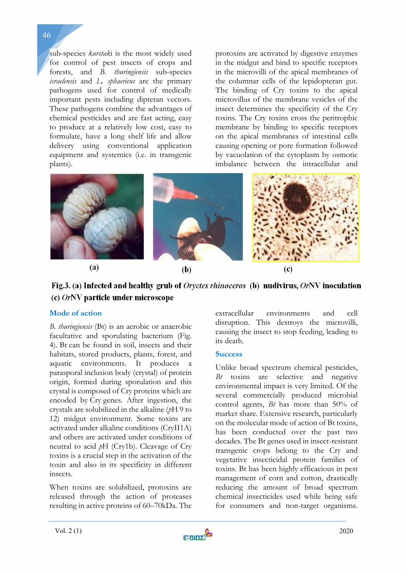
Vol. 2 (1) 2020
46
sub-species kurstaki is the most widely used for control of pest insects of crops and forests, and B. thuringiensis sub-species israelensis and L. sphaericus are the primary pathogens used for control of medically important pests including dipteran vectors. These pathogens combine the advantages of chemical pesticides and are fast acting, easy to produce at a relatively low cost, easy to formulate, have a long shelf life and allow delivery using conventional application equipment and systemics (i.e. in transgenic plants).
Mode of action
B. thuringiensis (Bt) is an aerobic or anaerobic facultative and sporulating bacterium (Fig. 4). Bt can be found in soil, insects and their habitats, stored products, plants, forest, and aquatic environments. It produces a parasporal inclusion body (crystal) of protein origin, formed during sporulation and this crystal is composed of Cry proteins which are encoded by Cry genes. After ingestion, the crystals are solubilized in the alkaline (pH 9 to 12) midgut environment. Some toxins are activated under alkaline conditions (CryII1A) and others are activated under conditions of neutral to acid pH (Cry1b). Cleavage of Cry toxins is a crucial step in the activation of the toxin and also in its specificity in different insects.
When toxins are solubilized, protoxins are released through the action of proteases resulting in active proteins of 60–70kDa. The
protoxins are activated by digestive enzymes in the midgut and bind to specific receptors in the microvilli of the apical membranes of the columnar cells of the lepidopteran gut. The binding of Cry toxins to the apical microvillus of the membrane vesicles of the insect determines the specificity of the Cry toxins. The Cry toxins cross the peritrophic membrane by binding to specific receptors on the apical membranes of intestinal cells causing opening or pore formation followed by vacuolation of the cytoplasm by osmotic imbalance between the intracellular and
extracellular environments and cell disruption. This destroys the microvilli, causing the insect to stop feeding, leading to its death.
Success
Unlike broad spectrum chemical pesticides, Bt toxins are selective and negative environmental impact is very limited. Of the several commercially produced microbial control agents, Bt has more than 50% of market share. Extensive research, particularly on the molecular mode of action of Bt toxins, has been conducted over the past two decades. The Bt genes used in insect-resistant transgenic crops belong to the Cry and vegetative insecticidal protein families of toxins. Bt has been highly efficacious in pest management of corn and cotton, drastically reducing the amount of broad spectrum chemical insecticides used while being safe for consumers and non-target organisms.

Vol. 2 (1) 2020
47
Despite successes, the adoption of Bt crops has not been without controversy. In addition to discovery of more efficacious isolates and toxins, an increase in the use of Bt products and transgenes will rely on innovations in formulation, better delivery systems and ultimately, wider public acceptance of transgenic plants expressing insect-specific Bt toxins.
Fungi
Entomopathogenic fungi do not require per os infection and could enter insect system through the cuticle. Examples include Metarhizium anisopliae, Verticillium lecanii, Beauveria bassiana, Nomuraea rileyietcwell exploited in pest management. Metarhizium anisopliae (Metchinkoff) Sorokin is an entomopathogenic fungus which kills O. rhinoceros in conditions of low temperature and high humidity. The fungus could be mass multiplied using cheaper substrates in both solid and liquid media and the spores could be harvested and treated in the breeding site @ 5 x 10¹¹ spores m-3. In vermicomposting sites, treatments with M. anisopliae spores killed all third - instar larvae, with the highest dose giving the fastest kill, taking eight days when favoured by high humidity (Fig. 5.a and b). This technology through farmer - participatory and women group approach has created a great impact on the long-term bio-suppression of the pest in farmer-participatory and community-mode. Mass production technology of M. anisopliae was
standardized in semi - cooked rice grains yielding a spore count of 3x10⁷cfu / g of the culture.
Searching for natural enemies of coconut eriophyid mite A. guerreronis, ICAR-CPCRI could collect more than 40 isolates of the acaropathogenic fungus, Hirsutella thompsonii (Fig. 5.c). Based on the bio-efficacy studies, one virulent isolate collected from Kayamkulam was characterized through molecular tools confirming species identity. Coconut water was found as an ideal medium for mass production of H. thompsonii. Talc-preparation of this H. thompsonii @ 20 g L-1 of water palm-1 containing 1.6 x 10⁸cfu with a frequency of three spraying per year resulted in 63-81 per cent reduction in mite incidence. It was found effective in many locations, though seasonal variation in efficacy existed.
Entomopathogenic nematodes (EPN)
The word ‘nematode’ means "thread-like” and they are also known as thread worms or round worms. Nematodes are numerically the most abundant metazoans on earth and second only to insects, in terms of diversity of forms (species) is concerned. Nematodes dwell in all types of habitats on the earth. Most of the nematodes are free living which feeds on microorganisms. Nematodes also parasitize plants, animals and human beings. There is hardly any animal on earth, which is free from one or the other kind of nematode infection.

Vol. 2 (1) 2020
48
Entomopathogenic nematodes (EPN) of the families Steinernematidae and Heterorhabditidae are soil inhabiting insect pathogens that possess potential as biological control agents. These nematodes, working with their symbiotic bacteria (Xenorhabdus for steinernematids and Photorhabdus for heterorhabditids), kill insects within 24 to 48 hour (Fig. 6). They are safe for the plant health, human health, soil and the environment. There is a huge potential for the utilization of these nematodes for the management of many coconut pests like white grub, rhinoceros beetle, red palm weevil etc.
Protozoa and Microsporidia
Protozoans are one-celled forms. One group the Microsporadia, contains many species that have promise for biological control. Microsporidian infections in insects are thought to be common and responsible for naturally occurring low to moderate insect mortality. But these are relatively slow acting organisms, taking days or weeks to debilitate their host. Infected insects may be sluggish and smaller than normal, sometimes with reduced feeding and reproduction, and molting problems. Death may follow if the level of infection is high. Nosema locustae is the only commercially available species of microsporidium, marketed under several labels for the control of grasshoppers and crickets. It is applied with insect-attractant
bait. Because of its slow mode of action, this product is better suited to long-term management of locusts than to the more intensive demands of commercial crop or even home garden production.
Bio-scavenging
When plants and animals die, the organic matter that makes their bodies possesses energy in the form of chemical bond. That energy is released by sequential breakdown of body tissue constituents. Insects, in feeding on dead plant and animal tissues, often carry out the first stage in the decomposition process by pre-disposing matter for
enhanced decay and ultimate break down by microorganisms. Some prominent examples of insect decomposers include termites that break down wood, spring tails that assist in the decomposition of dead leaves, carrion beetles and fly maggots that feed on dead animals aiding scavenging and enhanced disintegration.
Fig. 6. (a) Larvae killed by Steinernematid nematodes; (b) Infective juveniles of EPN
(b) (a)

Vol. 2 (1) 2020
49
Rugose spiraling whitefly
Emergence of the exotic rugose spiraling whitefly (RSW), Aleurodicus rugioperculatus (Fig. 7.a)on coconut palm during 2016 from Pollachi, Tamil Nadu and Palakkad, Kerala was the latest new addition of invasive pest
introduced into our country ably aided by inadequate quarantine approach. The pest could have been introduced from Florida, USA either through ornamental palms or RSW-infested coconut seedlings. Being non-native, RSW sounded alarm initially due to extensive feeding potential and high population build-up of the bug on coconut palms. RSW feeds from undersurface of palm leaflet and the enormous honey dew excreted get deposited on the upper surface of palm attracting the sooty mould, Leptoxyphium sp. Growth of sooty mould affects the photosynthetic efficiency of palms and served as a characteristic feature of pest identification (Fig. 7.c).
Farmers are worried due to black encrustation of sooty mould on palms and other under story intercrops in the system. RSW bred fast reaching as high as 15 egg spirals on a leaflet and with the white flocculent masses and sooty mould development could panic any farmer in the initial phase. Gradient outbreak of RSW could be realized interlinked with higher maximum temperature with lower humidity and rainfall as well. However, in a period of four to five months, there has been tremendous build-up of the aphelinid parasitoid, Encarsia guadeloupae (Fig.7. b)
which could bio-suppress RSW quite efficiently and significantly. From a modest 10% parasitism initially registered, natural parasitism rose as high as 82% in a period of four-five months. This is one of the classical success stories of conservatory biological control well documented and by this observation, ICAR-CPCRI has
recommended pesticide holiday to effectively conserve the natural enemy involved in RSW suppression. Though not well received, this strategy was well appreciated by the ecological benefit realized to nature as well as to human health. The Organic Policy of the Government of Kerala is another boost in this direction that may also encourage biodiversity preservation.
Sooty mould scavenger beetle
For the first time at the International level, scientists from ICAR-Central Plantation Crops Research Institute, and ICAR-National Research Centre on Banana, Trichy have reported the occurrence of a sooty mould scavenging beetle, Leiochrinus nilgirianus Kaszab (Coleoptera: Tenebrionidae) on coconut palms infested by the rugose spiraling whitefly (RSW) (Josephrajkumar et al., 2018). This beetle, which resembles the predatory lady beetle in appearance, are found to feed on the sooty mould deposits developed on the honey dew excreted by the rugose spiraling whitefly (Fig. 8). L. nilgirianus could be first located from sooty mould laden coconut palms at ICAR-CPCRI, Regional Station, Kayamkulam during July-August period which coincided with the south-west monsoon phase providing high humidity and

Vol. 2 (1) 2020
50
morning wetness, that favoured the accurate niche for enhanced survival and feeding.
All stages of the beetles viz., eggs, grubs, pupae and adult beetles were found on the palm leaflets and other intercrops with sooty mould.Generally, the grubs and adult L. nilgirianus beetles are restricted on the under surface of the leaves. But they move on to the upper surface of the leaves with sooty mould deposits during early morning hours with dew drops and feed on the sooty mould. Eggs of this beetleare purplish, laid in groups and emerging neonates are transparent and turn black, assume spherical and cylindrical shapes in movement due to the presence of elastic inter-segmental membrane. Creamish patches are developed during pre-pupating stages and pupae with distinct cream patches are also confined on the under surface of palm leaflet indicating its photo-sensitiveness. During our observations, more than five beetles (Average 2.07±0.9) and immature grubs could be noticed on sooty mould laden leaves. Bristle-like tarsomeres and tarsal claws of adult beetles encouraged effective adhesion, scooping of mould and swift movement on fungal growth. L. nilgirianus population was found very high and subsequently got reduced with the decline in RSW population and the sooty mould deposits. Mouth parts and gut lining of the beetle with fungal deposits could be located confirming the scavenging action of the beetle. Though ecosystem services provided by insects include food for wildlife, pest destruction, crop pollination, scavenging, etc. which is estimated at around $57 billion in the
United States of America alone, a thorough cleaning action on an economically significant crop like coconut is reported for the first time at global level. Habitat conservation of the sooty mould feeding scavenger beetle, L. nilgirianusin the palm ecosystem is very crucial and the conducive weather factors of high humidity and high rainfall prevailing in Kerala during monsoon phase are key factors for the survival and feeding efficiency of the beetle. Though report on scavenging action by insects such as termites disintegrating wood, springtail decomposing dead larvae and fly maggots feeding on dead animals exist in nature, the present instance appears to be unique in which sooty mould deposition is totally cleared by an insect scavenger on palms.
Conservation of E. guadeloupae by limited or zero pesticide usage coupled with in situ habitat preservation of scavenger beetles (L. nilgirianus) appears to be a very effective strategy that would help in controlling A. rugioperculatus and in clearing sooty mould from coconut palms at no cost in the most natural and eco-friendly manner, avoiding chemical management options and other expensive methods. Furthermore, the Organic Policy adopted by the State Government of Kerala could have encouraged the sustenance of the beetle and emerged in need for scavenging action.
Ecological Engineering
Ecological engineering defined first by Odum (1962) is a human activity that modifies the environment according to

Vol. 2 (1) 2020
51
ecological principles. Accordingly, it is a useful conceptual framework for considering the practice of habitat manipulation for arthropod pest management. Habitat manipulation aims to provide the natural enemies of pests with resources such as nectar, pollen, physical refugia, alternative prey, alternative hosts etc. The development of ecological engineering ranged from a simple first approximation that diversity is beneficial, to contemporary understanding that diversity can have adverse effects on pest management. In the recent era of agricultural intensification, the potential for using crop diversity to manage insect and microbial pests have not been extensively exploited.
Push-pull strategy
Push-pull strategies use a combination of behavior-modifying stimuli to manipulate the distribution and abundance of pest and/or beneficial insects for pest management. Strategies targeted against pests try to reduce their abundance on the protected resource, for example, a crop or farm animal. The pests are repelled or deterred away from this resource (push) by using stimuli that mask host apparency or are repellent or deterrent. The pests are
simultaneously attracted (pull), using highly apparent and attractive stimuli, to other areas such as traps or trap crops where they are concentrated, facilitating their elimination. The strategy is a useful tool for integrated pest management programs reducing pesticide input.
An agro-ecosystem based pest regression strategy by including compatible intercrops in coconut-based cropping system has registered lower pest incidence (fig. 9). The influence of mixed-volatile cues of crop plurality (rambutan, nut meg, curry leaf, banana, turmeric, red gram, papaya) inflicted lesser rhinoceros beetle damage (15.8%) compared to palms in outer whorls (30%). In
situ stimulo-deterrent diversionary strategy infused less pest damage and encouraged more defender population through eco-feast crops (Antigonon leptopus) and diversity in fruit crops. In addition to pest regression, continuous employment and income is generated fostering closer care to palms complementing the concept of an “inch of land and a bunch of crops”.
Agro-ecosystem based pest suppression
Diversity distraction; Stimulo-deterrant;Crop heterogeneity 140 nuts plam-1 year-1
Fig. 9. An agro-ecosystem based pest regression strategy for coconut cultivation by including compatible intercrops with the objective of achieving lower pest incidence

Vol. 2 (1) 2020
52
Plant health management
Plant health management (PHM) is the science and practice of understanding and overcoming the succession of biotic and abiotic factors that limit plants from achieving their full genetic potential as crops, ornamentals, timber trees, or other uses. Although practiced as long as agriculture itself, as a science-based concept, PHM is even younger than integrated pest management (IPM), and includes and builds upon IPM ; but is not a replacement for IPM. PHM is a moving target, like a football game, where one team is science and technology and the other is nature, where the S & T team is only beginning to know nature's rules while
playing itself with the three sets of rules
written to, respectively, satisfy the laws of economics, protect the environment, and gain social acceptance. In a nutshell, PHM includes integrated pest, disease and weed management, integrated nutrient management and soil health management in a holistic manner as system approach.
From the ecological engineering concept, health management is revered as holistic and at the same time sustainable providing continuous income and employment as well. A systematic care would thus make you cheer with coconut and empower your health infusing bio-happiness.

Vol. 2 (1) 2020
53
If you go with science, the tribes in Andaman Islands are the first Indians!!!
Biju Kumar, A.
Department of Aquatic Biology & Fisheries, University of Kerala, Thiruvananthapuram- 695581, Kerala E-mail: [email protected]
Exploring the roots of ancient tribes has been tenaciously neglected, if not ignored, in the contemporary Indian culturescape, though we always search for ‘avatars’ of our own creation. The recent killing of John Chau, a missionary who illegally trespassed on a secluded island to preach to the locals about Christianity, has created a lot of discussion for and against the tribes, including the human cry for bringing tribes to justice! Who cares about the human rights and right of tribes to live on their land? For the tribes of Andaman and Nicobar islands, it is the real story of our own (human) migration to explore the new pastures across the world, often driven by the innate ability to survive on changing environment!
Before we argue for or against the tribes in Andamans, who are also Indians, probably without Aadhar card and passport, it is always better to know their history told by science (especially in the light of recent developments in genomics) and look at the history of origin of extant humans of Indian mainland! Yet we continue to discuss in India, including within the ‘reformed’ and ‘educated’ Kerala society, the hegemony of caste system and social values created through the feudal system.
Going with genetics
Going with genetics, there are 4,635 well-defined populations in India, including 532 tribes (7.76%), and 72 primitive tribes (36 hunters and gatherers). When humans first ventured out of Africa as Homo sapiens around 60,000 years ago, they left genetic footprints through their pathways of migration. The migratory routes of ancient human beings is now relatively well drawn, through the
mapping of human genome (A genome is an organism’s complete set of its genetic material, Deoxyribonucleic acid or DNA, including all of its genes) or more precisely the genetic markers in DNA (Y chromosome and mitochondrial DNA).
The migration eventually helped the descendants of a small group of Africans to occupy even the farthest reaches of the Earth. Though the origin of modern man has happened in Africa and they have spent most of their life on earth in that piece of land, their journey outside Africa almost 60,000 -70,000 years ago, might have been triggered by major climatic shifts – may be a sudden cooling in the Earth’s climate during the last Ice Age. There is also an argument that the East African mega droughts during the time when the water volume of the lake Malawi was reduced by at least 95% could have been the reason for their migration out of Africa.
The earliest people to colonize the Eurasian landmass likely did so across the Bab-al-Mandab Strait separating present-day Yemen from Djibouti. These early beachcombers expanded rapidly along the coast to India, and reached Southeast Asia and Australia by 50,000 years ago (see the map). The first great foray of our species beyond Africa had led us all the way across the globe.
In recent years, scientists have determined that modern humans are not the only ancestors represented in our DNA. During your ancestors’ journey from our original African homeland, they might have mixed with ancient hominids who lived tens and even hundreds of thousands of years ago - our human cousins like the Neanderthals in

Vol. 2 (1) 2020
54
Europe and Asia. This means that you may find in you a small percentage (between 0 to 2 percent) of Neanderthal ancestry, even though those ancient species have long since gone extinct.
The genomics research suggested that mainland India's current population largely descended from Indo-European or Aryans (North India- Ancestral North Indians), Dravidians (South India-Ancestral South Indians) groups, Tibeto-Burman (North-East India) and Austro-Asiatic (fragmented in East and Central India) groups. The ‘Ancestral North Indians’ (ANI), is genetically close to Middle Easterners, Central Asians, and Europeans, whereas the other, the ‘Ancestral South Indians’ (ASI), is as distinct from ANI and East Asians as they are from each other. As far as the tribes of Andaman and Nicobar Islands are concerned, the Andaman tribes are Negrito (providing evidence for the Homo sapiens migration from East Africa some 60,000 years ago) and the Nicobar tribes are Mongoloid. The Jarawas and the Onges in Andamans share their ancestry with present-day Pacific islanders, implying a distinct ancestral population for these Andaman and Nicobar tribes. The studies further showed that two tribes in mainland India, Kurumba from Kerala and Rajbanshi from West Bengal were found to share some mutations (as indicated by DNA base sequence) with those of Andaman tribes.
These studies were done based on the Y chromosome which is inherited paternally and sequencing the complete mitochondrial genome (DNA in mitochondria) which is inherited maternally. The scientists compared the mitochondrial DNA sequences with that of the world population to arrive at conclusions. The studies proved the southern coastal route migration of African tribes to India. Earlier belief was that the tribes migrated to India through the northern route via Middle East, Europe, Southeast Asia and Australia.
According to a recent study by an Indo-Spanish group, the Onges and Jarawas were
believed to have descended from an unknown hominid (ancestor of all great apes and human) that went extinct. However, there is no fossil record yet to prove this argument. In short, the tribes living in the Andaman Islands have been recognized as belonging to the Negrito stock with dark copper colour complexion and woolly short hair growing in tufts. They are however completely distinct from the African tribes. The Nicobar Islands, on the other hand, are the abode of a race that have Mongolian characteristics and a mode of life entirely different from that of the Andamanese.
Gene exchange was widespread among the four ancestral groups of the present-day Indians, though the caste system later came into existence in India forced the population to live in relatively-isolated social groups. This was enhanced through endogamy (marriages limited within the caste system). With negligible proportion of marriages taking place between individuals belonging to different social groups, the four populations became genetically distinct.
A new paper authored by 92 scientists from around the globe shows that some sort of migration did indeed take place into India and that the Indus Valley civilisation is key to all South Asian populations. Further, there are three potential groupings that, when mixed in various combinations, could be responsible for the creation of the Ancestral North Indian and Ancestral South Indian Populations.
• The first are South Asian hunter-gatherers, described in this study as Ancient Ancestral South Indians or AASI, the oldest people of the subcontinent, related to modern-day Andaman islanders.
• Then there are Iranian agriculturists, who were known to have come to the subcontinent, possibly bringing certain forms of cultivation of wheat and barley with them.

Vol. 2 (1) 2020
55
• And finally, there are the Steppe pastoralists, the inhabitants of the vast Central Asian grasslands to the north of Afghanistan, who were previously known as ‘Aryans.’
There is another, important population with South Asian connections that sits somewhere amidst these three: the Indus Valley population. The mixing of Iranian agriculturists and South Asian hunter-gatherers first created the Indus Valley population. Then around the 2nd millennium BCE, Steppe pastoralists moved south towards the subcontinent encountering the Indus Valley population in a manner that was likely to have caused some amount of upheaval.
What appears to have happened afterwards is that some of the Indus Valley population moved further south, mixing more with South Asian hunter-gatherers to create the Ancestral South Indian population. Meanwhile, in the north, the Steppe pastoralists got mixed with the Indus Valley population to create the Ancestral North Indian grouping. Most subsequent South Asian populations were the result of further
mixing between Ancestral North Indians and Ancestral South Indians.
Tribes in Andaman and Nicobar islands
The Andaman and Nicobar Islands are
presently home to six major tribal groups - the Shompen and Nicobarese in the Nicobar Islands and the Jarawa, Onge, Great Andamanese and Sentinelese in the Andaman Islands. Andamanese are the descendants of the first modern human migrated out of Africa through Southern Coastal rout via India about 65,000 – 70,000 years ago; whereas the Nicobarese are the recent migrants from the Southeast Asia about 18,000 years ago.
The tribes of Andaman belong to the Negrito ethnicity, significant populations of which are spread across the Pacific Ocean in Indonesia, Papua New Guinea, and Solomon Islands to name a few. They are physically characterised by short stature, dark skin and peppercorn hair, largely resembling the African Pygmies.
The Great Andamanese Tribe
Thought to be the descendants of the Africans, they are known to have migrated out of Africa around 60,000 years ago. They
Fig. 1. The routes of human migration from Africa, as revealed by mitochondrial genetic analysis

Vol. 2 (1) 2020
56
are pygmies, divided into ten major tribes. Among the four tribes of Andaman Islands, the Andamanese paid the heaviest toll of their lives on account of their contact with the early settlers of these islands. In 1858, their number was conservatively estimated near about 3500. In 1901 their strength depleted to 625. They are now limited to a population of around 44. Much of their diet includes Indian food although they still go fishing, hunting, and food gathering. They also have farms now to practice agriculture and a little bit of animal husbandry. They are now settled and receive government support.
Jarawa Tribe
The Jarawa tribe is now confined to the forested areas along the west coast of South and Middle Andamans, which is known as the “Jarawa Reserve”. Jarawas were the earliest to be contacted by the colonizers and were reportedly the worst hit during the Japanese occupation as the Japanese bombed the Jarawa territory in their effort to drive away the British, since they considered the Jarawa area to be a hiding place for the British army. Jarawa have stayed totally out of contact of the outside world till 1998. They are believed to be the ascendants of the now extinct Jangil tribe.
Onge Tribe
They are presently concentrated in settlement namely the Dugong Creek in the Little Andaman Island. Being semi-nomadic, they
hunted for food instead of farming. The Onges also suffered grievously at the hands of the colonisers and early settlers. After the tsunami, only 101 of the Onge individuals are left who have taken shelter on the highland of the island.
Sentinelese Tribe
The term Sentinelese is derived from the name of the island they inhabit, viz. North Sentinel, which is located at a distance of 102 kms from Port Blair. The Sentinelese are believed to be an off-shoot of the Onge - Jarawa tribe but because they have remained isolated from other tribes for years, they have grown up as a distinct ethnic group. Sentinelese tribe is the most untouched tribe of Andaman Islands. They live on the North Sentinel Island, isolated completely from the other tribes. They are still leading the life of hunter and gatherer as they hunt and gather food, together with fishing. They don’t, however, know about how to produce fire and cook food. They are very hostile towards outsiders.
The Nicobarese Tribe
The Nicobarese tribe comprises of the dominant tribes of the Nicobar Islands. They alone have had a natural growth of population and have made headway on the road to civilization. They live in elivated huts having dome-shaped roofs and use ladders to

Vol. 2 (1) 2020
57
enter the house and to get down from there. These ladders are pulled up at night.
Shom Pen Tribe
The Shom Pen is mongoloid hunting-gatherer community of the Great Nicobar Islands in Andaman and Nicobar Islands. It is the first tribe that had arrived to the Nicobar Islands. After the tsunami, however, their population decreased considerably. They are the hunting-gathering type of tribe. They use tools like the bows and arrows, spears, fire drills and hatchet are the main kind of tools used by the Shompen. They hunt and gather food. They also depend on farming where they cultivate yams, roots, vegetables as well as tobacco.
Table 1. The tribe-wise population of Scheduled Tribes in Andaman and Nicobar Islands as per Census 2011
Tribes Population
Andamanese, Chariar, Chari, Kora, Tabo, Bo, Yere, Kede, Bea, Balawa, Bojigiya, Juwai, Kol
44
Jarawas 380
Nicobarese 27168
Onges 101
Sentinelese 15
Shom Pens 229
Based on information received from the Directorate of Tribal Welfare, Andaman and Nicobar (A&N) Administration, Nicobarese follow Islam and Christianity.
The earliest archaeological evidence that documents the ancestry of tribes is only a few thousands of years old. However, genetic and cultural studies suggest that the indigenous Andamanese people may have been isolated from other populations during the Middle Paleolithic, which ended 30,000 years ago. Since that time, the Andamanese have diversified into linguistically and culturally distinct, territorial groups.
What is happening to the tribes?
The inhabitants of these islands formed one of the most isolated groups of the world, until the British colonisation during the 19th and 20th century. The subsequent contact and settlement had devastating consequences on the local tribal population and radically altered the island’s ethnicity.
The Andaman Trunk Road or National Highway 223 is a 360 kilometre road, running south to north covering multiple towns and villages. The road passes through the Jarawa Reserve and the activists and anthropologists raised their concerns against the road construction. The Jarawas started making friendly contact with the modern society since 1998, while the government continue to implement restrictions in contacting them due to their low immunity and fear of their exploitation. The Jarawa habitat is protected as a tribal reserve notified under the Andaman and Nicobar Protection of Aboriginal Tribes Regulation (ANPATR) of 1956. Despite an interim order from the Supreme Court, prohibiting the road in 2002, it has not been closed. The apex court in 2012, prohibited tourism and commercial activities within a 5 km buffer area of the Jarawa reserve.
Of late, there is flourishing promotion of ‘tribal’ tourism, as the tourists wait in long queues to visit Jarawas and click photos with them! According to Survival International, a global tribal rights advocacy group,

Vol. 2 (1) 2020
58
unauthorised ‘human safaris’ are on the increase in the island and tourists often throw bananas and biscuits to the tribal people at the roadside, as they would do to animals in a safari park! Further, the ‘modern’ society is to be blamed for enticing them to bad habits of alcohol and marijuana.
Contact with the outsiders brought diseases to which the locals had no immunity, leading up to the complete extinction of the Jangil or Rutland Jarawa Tribe. Simultaneously, the rampant use of alcohol and opium in the early days was seen as a means of disrupting and removing the locals of the islands and caused friction between the British and the tribals, which ultimately saw the tribes being driven out of their traditional homes. The Jarawa have only had friendly interaction with their neighbours since 1998 and are highly vulnerable to exploitation, diseases, and dependency on goods such as alcohol brought in by outsiders. Survival International reports that poachers have been using Jarawa women illegally as aides or as a cover for hunting and gathering forest produce inside the tribe’s reserve. There are strong indications that the women are being lured by alcohol, and that sexual exploitation occurs on a regular basis. The tsunami of 2004 could be one reason which shattered these tribals and affected their habitation and demography.
Leave them alone!
Veteran anthropologist, T N Pandit is the only person who has visited the isolated Sentinelese several times in the last 50 years, and reported their culture. He says: “Of the four Andaman tribal communities, we have seen that those in close contact with the outside world have suffered the most. They have declined demographically and culturally. Their land is occupied. It has happened to the Great Andamanese, to the Onge, and even to the Shompen in Nicobar, who are also a hunter-gatherer people. Over the years, we have not been able to get the Jarawa any benefits. Rather in a number of cases, settlers are having liasons with their girls, however minimal. Their food supply like honey, crab
and fish are being taken away in exchange for biscuits. They don’t need biscuits. They have learned to smoke and drink”.
Unlike the Jarawa, who have much more resources to be able to withstand the pressures of civilisation, the Sentinelese are a highly vulnerable population and would disappear in an epidemic. The government’s responsibility should be to keep a watch over them in the sense no unauthorised people reach them and exploit them. Otherwise, just leave them alone.
References Barik, S.S. et al. (2008). Detailed mtDNA genotypes permit a reassessment of the settlement and population structure of the Andaman Islands. Am. J. Phys. Anthropol., doi: 10.1002/ajpa.20773
Basu, A. et al. (2016). Genomic reconstruction of the history of extant populations of India reveals five distinct ancestral components and a complex structure. Proc. Natl. Acad. Sci., doi: 10.1073/pnas.1513197113
Kumar, A. et al. (2015). Genome-wide signatures of male-mediated migration shaping the Indian gene pool. J. Human Genet., 60: 493-499. doi: 10.1038/jhg.2015.51
Reich, D. et al. (2009). Reconstructing Indian population istory. Nature , 461: 489-494. doi: 10.1038/nature08365
Sunita Sinh, Sanghamitra Sengupta, Kanchan M. Murhekar, ubhash C. Sehgal and Partha P. Majumder (2004). Single nucleotide polymorphisms in two genes among the Jarawa, a primitive tribe of the Andaman and Nicobar Islands. Current Science, 86: 326-328.
Thangaraj K, et al. (2005). Reconstructing the origin of Andaman Islanders, Science, 308: 996.
UNESCO. 2010. The Jarawa Tribal Reserve Dossier: Cultural & Biological Diversities in the Andaman Islands. Edited by Pankaj Sekhsaria and Vishvajit Pandya. 212pp. Paris: UNESCO.
Websites:
https://genographic.nationalgeographic.com/
https://www.survivalinternational.org/

Vol. 2 (1) 2020
59
DOCTORAL DEGREES AWARDED
Dr. Ayana Gayathri R. V., is an Assistant Professor in the Department of Zoology, University College, Thiruvananthapuram. She secured her Ph.D. in Zoology (2019) from University of Kerala under the supervision of Dr. Evans D. A., Associate Professor in Zoology (Rtd.), University College, Thiruvananthapuram.
Thesis Title
Physiological response of Culex quinquefasciatus larvae to selected stressors
Abstract: Culex quinquefasciatus Say, vector of lymphatic filariasis showed adaptive physiological response when subjected to physical and chemical stressors such as Temperature and xenobiotic, Bisphenol A (BPA). To counter the effect of stressors larvae increased the expression of Phospholipase A2 activating protein as revealed by MALDI TOF analysis. BPA elevated the titre levels of 20- hydroxyecdysone in 4th instar larvae facilitating rapid moulting. The present study identified Phospholipase A2 activating protein as a biochemical target for the control of filarial vector.
Publications Ayana Gayathri R.V. and Evans, D.A. (2018).Culex quinquefasciatus Say larva adapts to temperature shock through changes in protein turn over and amino acid catabolism. Journal of Thermal Biology, 74: 149-159.
Ayana Gayathri R.V. and Evans, D.A. (2018). Quantitative and qualitative changes in proteins and shift in the utilization of amino acids for cuticle sclerotisation and energy release during development in Culex quinquefasciatus. Entomon, 43(2): 85-94.
Dr. Bindhu, V.R. secured her Ph.D. in Zoology (2017) from University of Kerala under the supervision of Dr. Susha Dayanandan., Assistant Professor in Zoology (Rtd.), University College, Thiruvananthapuram.
Thesis Title
Efficacy of certain plant leaf extracts on the pulse beetle Callosobruchus chinensis. Coleoptera : Bruchida
Abstract: Effect of four medicinal plants; Boerhavia, Bacopa, Centella and Trichosanthes in controlling the stored product pest Callosobruchus
chinensis was analysed by using mortality studies, checking biochemical parameters, genomic studies and fecundity studies.
Publications Bindhu.V.R, Ganga, S. and Susha Dayanandan (2014). Efficacy of the plant Centella asiatica on Callosobruchus chinensis. Entomon 39(3): 135-142.
Bindhu.V.R,Ganga, S. and Susha Dayanandan (2015). Impact of partially purified compounds of Boerhavia diffusa on Callosobruchus chinensis (Coleoptera: Bruchidae). Journal of Entomological Research 40(1): 11-15.
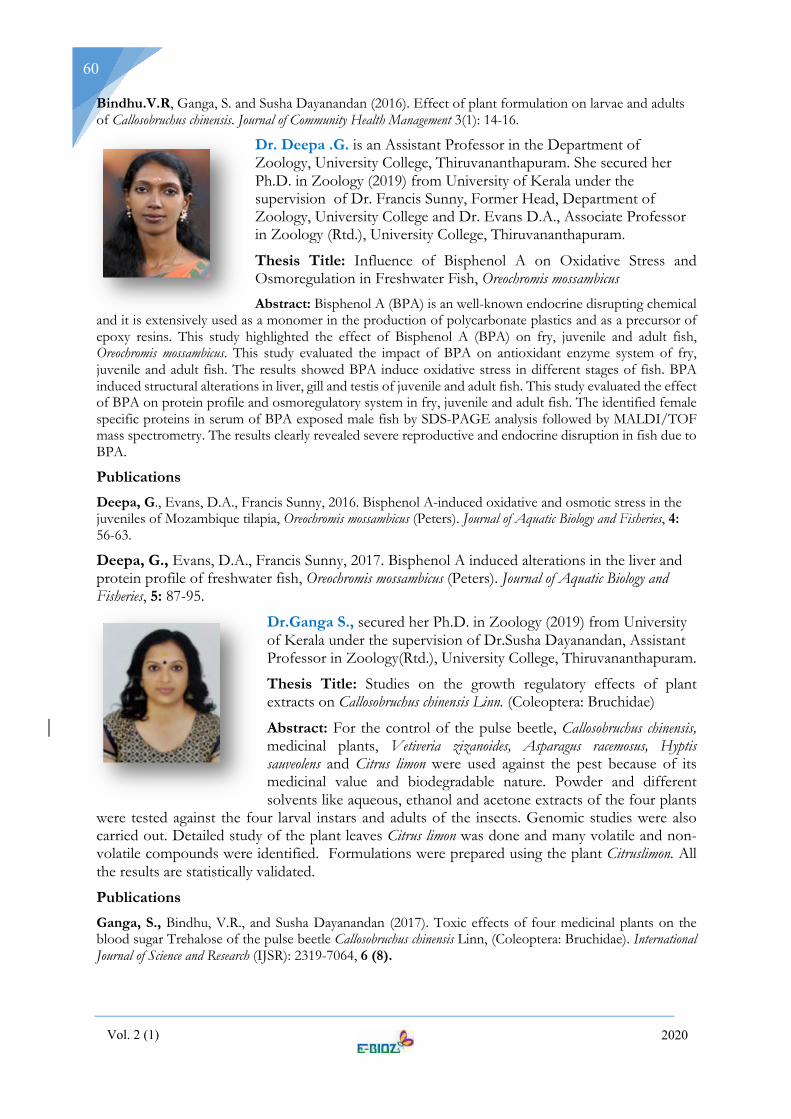
Vol. 2 (1) 2020
60
Bindhu.V.R, Ganga, S. and Susha Dayanandan (2016). Effect of plant formulation on larvae and adults of Callosobruchus chinensis. Journal of Community Health Management 3(1): 14-16.
Dr. Deepa .G. is an Assistant Professor in the Department of Zoology, University College, Thiruvananthapuram. She secured her Ph.D. in Zoology (2019) from University of Kerala under the supervision of Dr. Francis Sunny, Former Head, Department of Zoology, University College and Dr. Evans D.A., Associate Professor in Zoology (Rtd.), University College, Thiruvananthapuram.
Thesis Title: Influence of Bisphenol A on Oxidative Stress and Osmoregulation in Freshwater Fish, Oreochromis mossambicus Abstract: Bisphenol A (BPA) is an well-known endocrine disrupting chemical
and it is extensively used as a monomer in the production of polycarbonate plastics and as a precursor of epoxy resins. This study highlighted the effect of Bisphenol A (BPA) on fry, juvenile and adult fish, Oreochromis mossambicus. This study evaluated the impact of BPA on antioxidant enzyme system of fry, juvenile and adult fish. The results showed BPA induce oxidative stress in different stages of fish. BPA induced structural alterations in liver, gill and testis of juvenile and adult fish. This study evaluated the effect of BPA on protein profile and osmoregulatory system in fry, juvenile and adult fish. The identified female specific proteins in serum of BPA exposed male fish by SDS-PAGE analysis followed by MALDI/TOF mass spectrometry. The results clearly revealed severe reproductive and endocrine disruption in fish due to BPA.
Publications Deepa, G., Evans, D.A., Francis Sunny, 2016. Bisphenol A-induced oxidative and osmotic stress in the juveniles of Mozambique tilapia, Oreochromis mossambicus (Peters). Journal of Aquatic Biology and Fisheries, 4: 56-63.
Deepa, G., Evans, D.A., Francis Sunny, 2017. Bisphenol A induced alterations in the liver and protein profile of freshwater fish, Oreochromis mossambicus (Peters). Journal of Aquatic Biology and Fisheries, 5: 87-95.
Dr.Ganga S., secured her Ph.D. in Zoology (2019) from University of Kerala under the supervision of Dr.Susha Dayanandan, Assistant Professor in Zoology(Rtd.), University College, Thiruvananthapuram.
Thesis Title: Studies on the growth regulatory effects of plant extracts on Callosobruchus chinensis Linn. (Coleoptera: Bruchidae)
Abstract: For the control of the pulse beetle, Callosobruchus chinensis, medicinal plants, Vetiveria zizanoides, Asparagus racemosus, Hyptis sauveolens and Citrus limon were used against the pest because of its medicinal value and biodegradable nature. Powder and different solvents like aqueous, ethanol and acetone extracts of the four plants
were tested against the four larval instars and adults of the insects. Genomic studies were also carried out. Detailed study of the plant leaves Citrus limon was done and many volatile and non-volatile compounds were identified. Formulations were prepared using the plant Citruslimon. All the results are statistically validated.
Publications Ganga, S., Bindhu, V.R., and Susha Dayanandan (2017). Toxic effects of four medicinal plants on the blood sugar Trehalose of the pulse beetle Callosobruchus chinensis Linn, (Coleoptera: Bruchidae). International Journal of Science and Research (IJSR): 2319-7064, 6 (8).

Vol. 2 (1) 2020
61
Ganga, S, Bindhu, V.R, and Susha Dayanandan (2016). Insecticidal effect of medicinal plant on DNA fragmentation of the pulse beetle Callosobruchus chinensis Linn, (Coleoptera: Bruchidae). Journal of Entomological Research., 40 (4):365-368.
Dr. Indulekha R., secured her Ph.D. in Zoology (2019) from University of Kerala under the supervision of Dr. George Thomas., Associate Professor in Zoology (Rtd), University College, Thiruvananthapuram.
Thesis Title: Cytotoxic and genotoxic effects of metal oxide nanoparticles on a fresh water fish Rasbora daniconius.
Abstract: The study highlighted the effect of two metal oxide nanoparticles on a freshwater fish R. daniconius. Characterisation of nanoparticles was done using TEM, SEM and XRD. A comparison was also made between np and its bulk form in relation to its toxicity. Toxicological effects of TiO2 and CuO np and bulk forms were assessed by analysing the changes in haematological, histopathological, biochemical and genotoxic
effects in R. daniconius. NP exposure resulted in severe alterations in tissues and chromosomal damage and DNA stand breaks were investigated. The studied parameters were found to be excellent biomarkers of toxicity.
Publications Indulekha. R and George Thomas (2016), “Effect of TiO2 np on the histology of gill and liver tissue of a fresh water fish: Rasbora daniconius,” Journal of Aqua. Bio. and Fisheries. vol. 4. pp. 72-80.
Indulekha. R and George Thomas (2019), “Comparative toxicities of bulk and nanoforms of TiO2 and
CuO in the Fresh Water Fish: Rasbora daniconius,(Ham.1872)” International Journal of new innovations in Engg and Tech..vol.10(1).
Dr. Kavitha K J., Research Scholar in the Department of Zoology, University College, Thiruvananthapuram secured her Ph.D. in Zoology (2020) from University of Kerala under the supervision of Dr. Evans D. A., Associate Professor of Zoology (Rtd.), University College, Thiruvananthapuram.
Thesis Title: Allelopathic interactions of certain Musa cultivars against Odoiporus longicollis [Olivier] and the molecular mechanism of pest resistance.
Abstract: Diversity of Musa cultivars in relation to the pest infestation status of Odoiporus longicollis in Chittar Panchayat of Pathanamthitta district has resulted in the identification of 20 cultivars of Musa. Cultivars are grouped into Susceptible, Non preferred and Resistant on the basis of field
study and confirmed by laboratory study. Bioassay guided fractionation of resistant cultivar Thenkaali resulted in the identification of two larvicidal compounds Stigmasterol-3-O-glucoside and Sulfoquinovosyl diacylglycerol and are highly toxic to the holometabolous larvae affecting amino acid metabolism, protein profile, protein turnover and caused endocrine disruption. Comparative study on the mRNA expression of a susceptible and resistant cultivar showed difference in their DDRT profile.
Publications Kavitha, K.J., K. Murugan and D.A.Evans (2015). Allelopathic interactions of certain Musa cultivars against Odoiporus longicollis (Olivier), Entomon.40(4), Article No. ent.40404, 209-220.
Kavitha, K.J., D.A. Evans and K. Murugan (2015),Screening of Wild and Cultivars of Banana of Kerala,India using Score Card Method and its Resistance against Odoiporus longicollis Olivier, Phytomorphology,65(3&4),147-152.

Vol. 2 (1) 2020
62
Kavitha, K.J., K. Murugan and D.A. Evans (2016),Cytopathological and haematological changes in Odoiporus longicollis[Olivier] grub by Aattinkombu and Thekaali,the two pest resistant Musa cultivars identified in Kerala, Journal of Entomological Research, 40(1), 27-33.DOI:10.5958/0974-4576.2016.00005.0.
Kavitha, K.J., and D.A.Evans (2019). Sulfoquinovosyl diacylglycerol a larvicidal molecule in Aadinkombu an AA Musa cultivar induces allelopathy in odoiporus longicollis[Olivier], International Journal of Tropical Agriculture, 37(2): 0254-8755.
Kavitha, K.J., K. Shabithraj and D.A.Evans (2020), Allelopathic effects of high contents of phenols and flavonoids in Musa paradisiaca L.cultivars on banana borer Odoiporus longicollis (Olivier), Allelopathy Journal, 49(1), 99-112, https://doi. org/10.26651/allelo.j/2020-49-1-1257.
Kavitha, K.J., J. Anil John and D.A. Evans (2020), Stigmasterol-3-O-glucoside,an allelopathic molecule responsible for pest resistance of Thenkaali (AAB), a Musa cultivar against Odoiporus longicollis [Olivier]. Current Science,118(6): 946-953.
Dr. Kiran S Kumar is a Lecture in the PG & Research Department of Zoology, NSS College, Pandalam. He completed his Bachelors (2008) and Masters (2010) in Zoology from University college, Thiruvananthapuram and secured Ph.D. in Zoology (2019) from University of Kerala under the supervision of Dr. Sanil George., Scientist EII, Rajiv Gandhi Centre for Biotechnology, Thiruvananthapuram.
Thesis Title: Analysis of Genetic Variation in an Endemic Frog (Indirana semipalmata Boulenger, 1882) Population of the Western Ghats using Molecular Markers.
Abstract: We analyzed the genetic diversity of Indirana semipalmata frog species using different molecular markers such as mitochondrial, nuclear and microsatellite from their entire distribution range in the Western Ghats. The genetic analysis showed a high genetic variation coupled with the isolation by distance pattern of species distribution for the populations of I. semipalmata south of the Palghat Gap in the Western Ghats. The genetic differentiation existing between the communities were significantly small is an indication of habitat fragmentation. The population as a whole was experiencing a recent population expansion after a population decline in the past. Though the current analysis did not show any loss of genetic variation in I. semipalmata species, the decrease in effective population size, as well as the fragmentation of habitat may warrant separate management considerations for the I. semipalmata populations.
Publications Kumar, K. S., George, S. (2019). Development and characterization of ten polymorphic microsatellites of the Western Ghats frog Indirana semipalmata and cross amplification in related species. Molecular Biology Reports. doi.org/10.1007/s11033- 019-04854-z.
Kumar, K. S., Sivakumar, K. C., Reghunathan, D., Manoj, P., & George, S. (2016). The complete mitochondrial genome of Indirana semipalmata (Amphibia: Anura). Conservation Genetics Resources, 1(9), 83-86.
Kumar, K. S., Anoop, V. S., Sivakumar, K. C., Reghunathan, D., Manoj, P., Deuti, K., & George, S. (2017). An additional record of Fejervaryamanoharani Garg and Biju from the Western Ghats with a description of its complete mitochondrial genome. Zootaxa 4277 (4): 491–502.
Anoop, V. S., Kumar, K. S., Sivakumar, K. C., Reghunathan, D., Manoj, P., Deuti, K., & George, S. (2017). The complete mitochondrial genome of Euphlyctiskaraavali (Amphibia: Anura) with a note on its range expansion. Conservation Genetics Resources, 1-4.
Gopalan, S. V., Kumar, S. U., Kumar, K. S., & George, S. (2016). Genetic diversity of an endangered bush frog Pseudophilautuswynaadensis (Jerdon, 1854 “1853”) from the south of Palghat gap, Western Ghats, India. Mitochondrial DNA Part A, 27(6), 3846-3851.

Vol. 2 (1) 2020
63
Dr. Pradeep Kumar R., is an Assistant Professor in the Department of Zoology, Government. College for Women, Thiruvananthapuram. He secured his Ph.D. in Zoology (2020) from University of Kerala under the supervision of Dr. Evans D.A., Associate Professor of Zoology (Rtd.), University College, Thiruvananthapuram.
Thesis Title: Isolation, Identification, Structure Elucidation and Mode of Action of Phytocompounds From Adiantum latifolium Against The Coconut Pest Oryctes rhinoceros
Abstract: Ethyl acetate fraction of methanol extract of leaves of Adiantum latifolium exhibited larvicidal activity against the coconut pest, Oryctes rhinoceros, and antibacterial activity against its gut bacteria. Twelve phytocompounds were isolated and characterized from the bioactive fraction, adiantobischrysene and 22-hydroxyhopane as pure and novel compounds, a compound composition of ten compounds which includes terpenoids, phenolics and phenanthrenoids. Mode of action studies revealed 20-hydroxyecdysone titer in haemolymph, gut histolysis, decreased digestive enzyme secretion, changes in haemolymph profile of biochemical components and cellular changes in haemocytes of the larvae.
Publications Pradeep Kumar, R., John, A., Kumar, P., Dinesh Babu, K.V., Evans, D.A., (2018). Larvicidal efficacy of Adiantobischrysene from Adiantum latifolium against Oryctes rhinocerosthrough disrupting metamorphosis and impeding microbial mediated digestion. Pest Manag. Sci., 74, 1821–1828. doi:10.1002/ps.4880.
Pradeep Kumar, R., Dinesh Babu, K.V., Evans, D.A., (2019a). Isolation, characterization and mode of action of a larvicidal compound, 22-hydroxyhopane from Adiantum latifolium Lam. against Oryctes rhinoceros Linn. Pestic. Biochem. Physiol., 153, 161–170doi:10.1016/J.PESTBP.2018.11.018.
Pradeep Kumar, R., Evans, D.A., Dinesh Babu, K.V., (2019b). Characterization of Multipotent Compounds from Adiantum latifolium Leaves by Liquid Chromatography-Electrospray-Mass Spectroscopy. Anal. Chem. Lett., 9, 682–696. doi:10.1080/22297928.2019.1674185.
Dr. Remya Balan M., is a Guest Lecturer in the Department of Zoology, Christian College, Kattakada, Thiruvananthapuram. She secured her Ph.D. in Zoology (2019) from University of Kerala under the supervision of Dr. Susha Dayanandan, former Head of the Department of Zoology (Rtd.), University College, Thiruvananthapuram.
Thesis Title: Effect of plant extracts on feeding, development, reproduction and behaviour of the banana pest Odoiporus longicollis (olivier)
Abstract: Towards the increasing demand for food and the expectations imposed by a competitive market, farmers are compelled to use chemical pesticides and fertilizers which can cause hazardous problems upon health and environment. One of the most relevant and accessible manner of pest management is use of botanicals. So, the study concentrates on effect of botanicals on banana pseudostem weevil, Odoiporus longicollis. Leaf extracts and a formulation of Clerodendrum infortunatum showed significant changes on physiological, biochemical and molecular parameters of O. longicollis on laboratory studies and field trials. The phytochemical evaluations of the formulation results show clearly the presence of certain active compounds, which may be the reason for insect growth regulatory activity of C. infortunatum. This can act as an alternative for harmful chemical or synthetic pesticides and save the biota as well as nature.

Vol. 2 (1) 2020
64
Publications
Remya Balan, M. and Susha Dayanandan 2016. Biocidal activity of plant powders on the fourth instar larvae of Banana pseudostem borer Odoiporus longicollis (Olivier). International Journal of Applied and Pure Science and Agriculture, e-ISSN 2394-5532, 2 (6): 212-215.
Remya Balan, M. and Susha Dayanandan 2019. Bioactivity of selelcted medicnal plants on banana pseudostem weevil, Odoiporus longicollis. International Journal of Scientific Research and Review, 8, (1): 206 – 219.
Remya Balan, M. and Susha Dayanandan, 2019. Effect of leaf extracts of C. infortunatum on repellency of fourth instar larvae of Odoiporus longicollis. International Journal of Scientific Research and Review, 8 (11): 1056-1062.
Dr. Resmi S Nair, is an Assistant Professor in the Department of Zoology, N.S.S.College Nilamel, Kollam. She secured her Ph.D. in Zoology (2019) from University of Kerala under the supervision of Dr. Susha Dayanandan., Assistant Professor of Zoology (Rtd.), University College, Thiruvananthapuram.
Thesis Title: Biocidal Activity of Certain Plants On The Rice Weevil, Sitophilus Oryzae (L)
Abstract: Nine common medicinal plants- Glycosmis pentaphylla, Caesalpinia pulcherrima, Mentha arvensis, Coleus ambonicus, Annona reticulata,Myrstica fragrans, Ficus bengalensis, Emblica officinalis,and Hemidesmus indicus and three mangrove plants-Acanthus ilicifolius, Brugiera cylindrica and Avicennia officinalis were treated against Sitophilus oryzae(L). From the various bioassays it was clear that acetone extract of Glycosmis pentaphylla was most effective one. For the control of Sitophilus oryzae herbal insecticidal formulation were prepared using the acetone extracts of Glycosmis pentaphylla leaves, it could be integrated into pest management system
Publications Resmi S Nair, Susha Dayanandan and Beena Joy (2015). Bioefficacy of selected ecofriendly botanicals in the management of rice weevil, Sitophilus Oryzae (L.) (Coleoptera: Curculionidae). J. Scientia, 11 (1): 74-77.
Resmi S Nair, Susha Dayanandan and Beena Joy (2017). Efficacy of different extracts of Glycosmis pentaphylla on the rice weevil, Sitophilus oryzae (L.) (coleoptera: curculionidae). J. Scientia, 12 (1): 18-21.
Resmi S Nair, Susha Dayanandan, and Beena Joy (2017). Biocidal activity of different extract of Glycosmis pentaphylla against the rice weevil Sitophilus oryzae (L.) (Coleoptera: Curculionidae) International Journal of Recent Scientific Research. 8 (11): 21689-21692.
Resmi S Nair, Susha Dayanandan, and Beena Joy (2017). Insecticidal properties of Glycosmis pentaphylla against the rice weevil Sitophilus oryzae (L.) (Coleoptera: Curculionidae). Edu-vision, 126-137.
Resmi S Nair, Susha Dayanandan, and Beena Joy,(2018),“Expression of Heat Shock Protein (Hsp)Genes in the Stored Grain Pest, Sitophilus oryzae (L) (Coleoptera:Curculionidae)” International Journal of Advanced Scientific Research and Management, 3: 246 –251.
Resmi S Nair, Susha Dayanandan and Beena Joy (2018). A study on phytochemical screening and characterisation of acetone extracts of plant leaf Glycosmis pentaphylla. International Journal of Scientific Research and Reviews, 7 (3):

Vol. 2 (1) 2020
65
NEWS
Neandertal genes and the risk of COVID-19
Those people who have inherited genes from Neandertals may raise their risk of developing severe COVID-19. A stretch of DNA on human chromosome 3 was previously found to be associated with an increased risk of developing severe disease from coronavirus infection and of being hospitalized. Some genetic heirlooms passed down after humans interbred with Neanderals more than 50,000 years ago are known to affect
immune system function and other aspects of human health even today (SN: 2/11/16). About half of people whose ancestors hail from South Asia — particularly Bangladesh — and about 16 percent of people in The risky DNA was identified as a COVID-19 danger zone in genome-wide association studies, or GWAS, which use statistical methods to find genetic variants that show up more often in people with a particular disease than in those without the disease. In this case, the comparison was between people who have milder forms of COVID-19 and people who required hospitalization. Europe today harbour this gene, according to this study that appeared in the journal Nature. Zeberg, H., Pääbo, S. The major genetic risk factor for severe COVID-19 is inherited from Neanderthals. Nature (2020). https://doi.org/10.1038/s41586-020-2818-3
Gene-editing tool CRISPR wins the chemistry Nobel
Emmanuelle Charpentier (left) and Jennifer Doudna (right)
Researchers Emmanuelle Charpentier and Jennifer Doudna devised a powerful gene-editing tool called CRISPR/Cas9. The gene editor has revolutionized molecular genetics and gene therapy and netted the duo the 2020 Nobel Prize in chemistry. CRISPR stands for Clustered Regularly Interspaced Short Palindromic Repeats. CRISPR/Cas9 is a two-part gene editing tool made up of a guide RNA and an enzyme, Cas 9, that cuts DNA. A guide RNA brings the enzyme to a particular spot in an organism’s DNA that researchers wish to cut (the targeted sequence in this diagram). The spot is a chemical match for the RNA. Once ferried to the right spot, Cas9 snips the DNA. In essence, these short, repeating bits of DNA sandwich bacteria’s version of the FBI’s most

Vol. 2 (1) 2020
66
wanted list — invading viruses. Every time bacteria encounter a virus, they take a DNA mug shot of it and file it in between the repeats. The next time the bacteria encounters that virus, they make RNA copies of the mug shots. Those RNA photocopies then team up with another bit of RNA known as a trans-activating CRISPR RNA, or tracrRNA, to form an all-points bulletin known as a guide RNA. Guide RNAs shepherd the DNA-cutting enzyme Cas9 to the virus, where the enzyme chops and eliminates the threat. The ability to cut the DNA where you want has revolutionized the life sciences. As a result we can now easily edit genomes as desired — something that before was hard, or even impossible. This recent discovery has already benefited humankind greatly, particularly in curing genetic diseases. However, ethics and law are also important to determine what can and should be done with the tool, as some human gene editing is extremely controversial.
References: Nobelprize.org; M. Jinek et al. A programmable dual RNA-guided DNA endonuclease in adaptive bacterial immunity. Science. Vol. 337, August 17, 2012, p. 816. doi: 10.1126/science.1225829
Hepatitis C discoveries win 2020 Nobel Prize in physiology or medicine
Three virologists have won the Nobel Prize in physiology or medicine for the discovery of the hepatitis C virus. Harvey Alter, of the U.S. National Institutes of Health in Bethesda, Md., Michael Houghton, who is now at the University of Alberta in Edmonton, Canada, and Charles Rice (From left to right in photo), now of The Rockefeller University in New York City, have won the prize. About 71 million people worldwide have chronic hepatitis C infections. An estimated 400,000 people die each year of complications from the disease, which include cirrhosis and liver cancer. Today, the major way people get infected is through contaminated needles used for injecting intravenous drugs, but when the researchers made their discoveries in the 1970s, ’80s and ’90s, blood transfusions were an important source of hepatitis C infection. The trio of winners of this year’s Nobel Prize in physiology or medicine — Harvey Alter, Michael Houghton and Charles Rice (from left) — all played a role in discovering the hepatitis C virus, which can cause a silent but ultimately deadly disease. Over seven years, the researchers have tried 30 or 40 methods to extract, or clone, the virus and then they turned to a method using bacteriophage — viruses that infect bacteria, and confirmed that the small piece of genetic material that they had cloned was from the virus. The blood test Houghton and colleagues developed was used to screen blood all around the world and dramatically decreased hepatitis C infections. But a question still remained about whether the hepatitis C virus alone was responsible for the infection. Rice and colleagues working at Washington University in St. Louis stitched together genetic fragments of the virus pulled from the blood of infected chimpanzees into a working virus and demonstrated that it could cause hepatitis in animals. This provided conclusive evidence that the cloned hepatitis C alone could cause the disease.
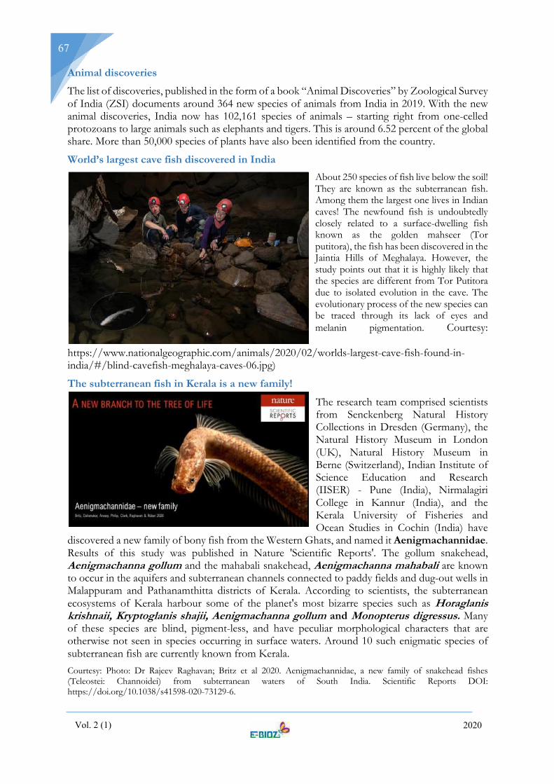
Vol. 2 (1) 2020
67
Animal discoveries
The list of discoveries, published in the form of a book “Animal Discoveries” by Zoological Survey of India (ZSI) documents around 364 new species of animals from India in 2019. With the new animal discoveries, India now has 102,161 species of animals – starting right from one-celled protozoans to large animals such as elephants and tigers. This is around 6.52 percent of the global share. More than 50,000 species of plants have also been identified from the country.
World’s largest cave fish discovered in India About 250 species of fish live below the soil! They are known as the subterranean fish. Among them the largest one lives in Indian caves! The newfound fish is undoubtedly closely related to a surface-dwelling fish known as the golden mahseer (Tor putitora), the fish has been discovered in the Jaintia Hills of Meghalaya. However, the study points out that it is highly likely that the species are different from Tor Putitora due to isolated evolution in the cave. The evolutionary process of the new species can be traced through its lack of eyes and melanin pigmentation. Courtesy:
https://www.nationalgeographic.com/animals/2020/02/worlds-largest-cave-fish-found-in-india/#/blind-cavefish-meghalaya-caves-06.jpg)
The subterranean fish in Kerala is a new family!
The research team comprised scientists from Senckenberg Natural History Collections in Dresden (Germany), the Natural History Museum in London (UK), Natural History Museum in Berne (Switzerland), Indian Institute of Science Education and Research (IISER) - Pune (India), Nirmalagiri College in Kannur (India), and the Kerala University of Fisheries and Ocean Studies in Cochin (India) have
discovered a new family of bony fish from the Western Ghats, and named it Aenigmachannidae. Results of this study was published in Nature 'Scientific Reports'. The gollum snakehead, Aenigmachanna gollum and the mahabali snakehead, Aenigmachanna mahabali are known to occur in the aquifers and subterranean channels connected to paddy fields and dug-out wells in Malappuram and Pathanamthitta districts of Kerala. According to scientists, the subterranean ecosystems of Kerala harbour some of the planet's most bizarre species such as Horaglanis krishnaii, Kryptoglanis shajii, Aenigmachanna gollum and Monopterus digressus. Many of these species are blind, pigment-less, and have peculiar morphological characters that are otherwise not seen in species occurring in surface waters. Around 10 such enigmatic species of subterranean fish are currently known from Kerala. Courtesy: Photo: Dr Rajeev Raghavan; Britz et al 2020. Aenigmachannidae, a new family of snakehead fishes (Teleostei: Channoidei) from subterranean waters of South India. Scientific Reports DOI: https://doi.org/10.1038/s41598-020-73129-6.
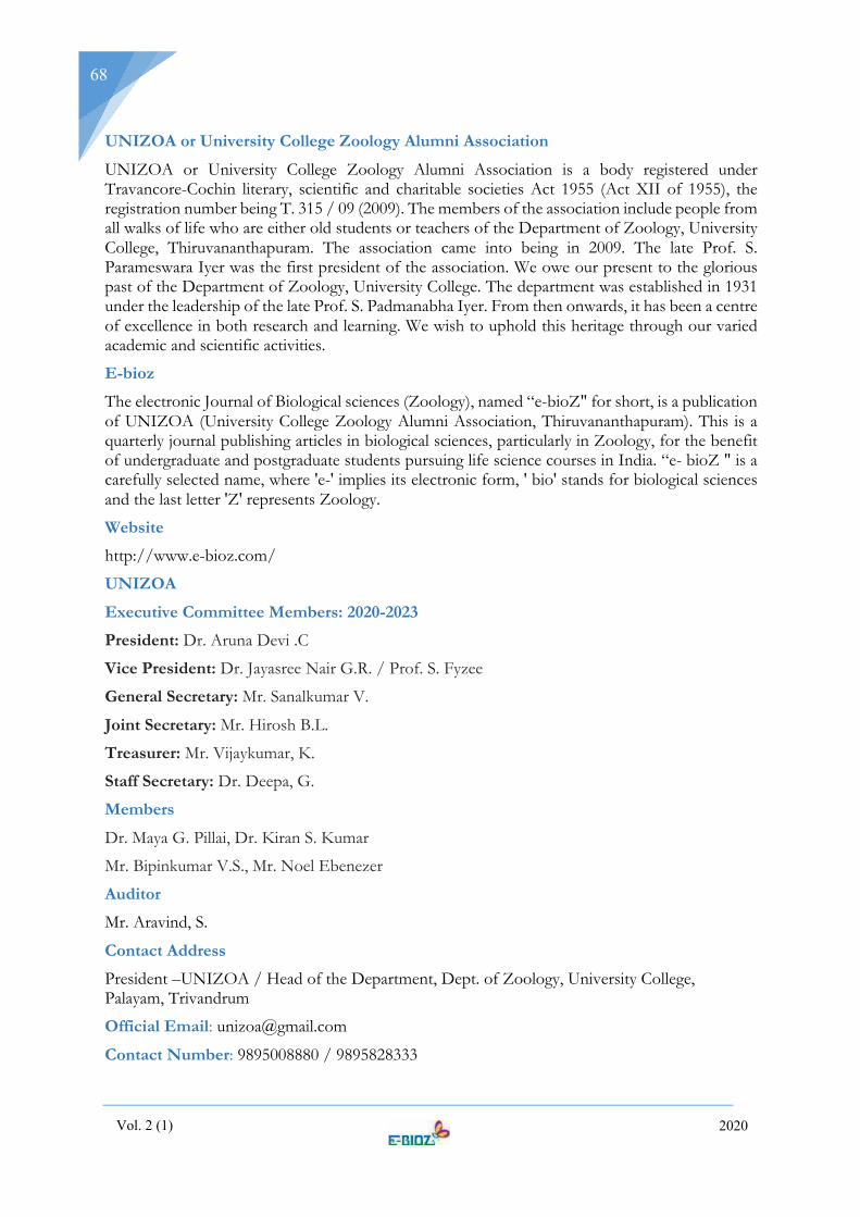
Vol. 2 (1) 2020
68
UNIZOA or University College Zoology Alumni Association
UNIZOA or University College Zoology Alumni Association is a body registered under Travancore-Cochin literary, scientific and charitable societies Act 1955 (Act XII of 1955), the registration number being T. 315 / 09 (2009). The members of the association include people from all walks of life who are either old students or teachers of the Department of Zoology, University College, Thiruvananthapuram. The association came into being in 2009. The late Prof. S. Parameswara Iyer was the first president of the association. We owe our present to the glorious past of the Department of Zoology, University College. The department was established in 1931 under the leadership of the late Prof. S. Padmanabha Iyer. From then onwards, it has been a centre of excellence in both research and learning. We wish to uphold this heritage through our varied academic and scientific activities.
E-bioz
The electronic Journal of Biological sciences (Zoology), named “e-bioZ" for short, is a publication of UNIZOA (University College Zoology Alumni Association, Thiruvananthapuram). This is a quarterly journal publishing articles in biological sciences, particularly in Zoology, for the benefit of undergraduate and postgraduate students pursuing life science courses in India. “e- bioZ " is a carefully selected name, where 'e-' implies its electronic form, ' bio' stands for biological sciences and the last letter 'Z' represents Zoology.
Website
http://www.e-bioz.com/
UNIZOA
Executive Committee Members: 2020-2023
President: Dr. Aruna Devi .C
Vice President: Dr. Jayasree Nair G.R. / Prof. S. Fyzee
General Secretary: Mr. Sanalkumar V.
Joint Secretary: Mr. Hirosh B.L.
Treasurer: Mr. Vijaykumar, K.
Staff Secretary: Dr. Deepa, G.
Members
Dr. Maya G. Pillai, Dr. Kiran S. Kumar
Mr. Bipinkumar V.S., Mr. Noel Ebenezer
Auditor
Mr. Aravind, S.
Contact Address
President –UNIZOA / Head of the Department, Dept. of Zoology, University College, Palayam, Trivandrum
Official Email: [email protected]
Contact Number: 9895008880 / 9895828333
Copyright © 2022 FDOKUMEN