
Arab Hussain Zoology 2020 icp peshwar.pdf
171
EPIDEMIOLOGY AND MOLECULAR CHARACTERIZATION OF TOXOPLASMA GONDII IN LIVESTOCK OF PESHAWAR VALLEY Submitted by ARAB HUSSAIN REG.NO.2015/ICP-119 Supervised by DR. MUHAMMAD ZAHID DEPARTMENT OF ZOOLOGY ISLAMIA COLLEGE PESHAWAR KHYBER PAKHTUNKHWA, PAKISTAN (2015-18)
-
Upload
khangminh22 -
Category
Documents
-
view
0 -
download
0
Transcript of Arab Hussain Zoology 2020 icp peshwar.pdf

EPIDEMIOLOGY AND MOLECULAR CHARACTERIZATION OF
TOXOPLASMA GONDII IN LIVESTOCK OF PESHAWAR VALLEY
Submitted by
ARAB HUSSAIN
REG.NO.2015/ICP-119
Supervised by
DR. MUHAMMAD ZAHID
DEPARTMENT OF ZOOLOGY
ISLAMIA COLLEGE PESHAWAR
KHYBER PAKHTUNKHWA, PAKISTAN
(2015-18)

EPIDEMIOLOGY AND MOLECULAR CHARACTERIZATION OF
TOXOPLASMA GONDII IN LIVESTOCK OF PESHAWAR VALLEY
Submitted by
ARAB HUSSAIN
REG.NO.2015/ICP-119
Supervised by
DR. MUHAMMAD ZAHID
Thesis submitted to the Department of Zoology Islamia College University Peshawar, for the
partial fulfillment of the requirement for the degree of
DOCTOR OF PHILOSOPY (Ph. D) IN ZOOLOGY
DEPARTMENT OF ZOOLOGY
ISLAMIA COLLEGE PESHAWAR
KHYBER PAKHTUNKHWA, PAKISTAN
(2015-18)

iii

iv
DEDICATION
This work is dedicated
To
My parent’s
Teachers
And friends

v
TABLE OF CONTENTS
DEDICATION.............................................................................................................................. iv
TABLE OF CONTENTS ............................................................................................................. v
LIST OF TABLES ....................................................................................................................... ix
LIST OF FIGURES ..................................................................................................................... xi
LIST OF ABBREVIATIONS ................................................................................................... xiii
ACKNOWLEDGMENTS ......................................................................................................... xiv
ABSTRACT ................................................................................................................................. xv
INTRODUCTION......................................................................................................................... 1
1.1 History ................................................................................................................................................ 1
1.2 Distribution (Epidemiology) ............................................................................................................... 5
1.3 Morphology......................................................................................................................................... 7
1.3.1 Tachyzoite: ................................................................................................................................... 7
1.3.2 Bradyzoites (Tissue cysts): .......................................................................................................... 8
1.3.3 Oocyst .......................................................................................................................................... 9
1.4 Life Cycle.......................................................................................................................................... 10
1.5 Toxoplasmosis and animal world ..................................................................................................... 12
1.5.1 Goats and Sheep ......................................................................................................................... 12
1.5.2 Sheep .......................................................................................................................................... 13
1.5.3 Pigs (Sus scrofa) ........................................................................................................................ 15
1.5.4 Domestic ruminants (Cattle) ...................................................................................................... 17
1.5.5 Birds ........................................................................................................................................... 17
1.5.6 Poultry ........................................................................................................................................ 18
1.5.7 Cats ............................................................................................................................................ 18
1.5.8 Dogs ........................................................................................................................................... 19
1.5.9 Deer ............................................................................................................................................ 19
1.5.10 Horses ...................................................................................................................................... 19
1.5.11 Rabbits ..................................................................................................................................... 19
1.5.12 Other species ............................................................................................................................ 19
1.5.13 Marine mammals...................................................................................................................... 19
1.6 Food studies ...................................................................................................................................... 20
1.6.1 Survival in foods ........................................................................................................................ 20

vi
1.6.2 Unsporulated oocysts ................................................................................................................. 20
1.6.3 Sporulated oocysts ..................................................................................................................... 21
1.6.3 Tissue cysts ................................................................................................................................ 21
1.6.4 Tachyzoites ................................................................................................................................ 21
1.7 Toxoplasmosis in humans ................................................................................................................. 22
1.7.1 Prevalence of T. gondii in Pakistan ............................................................................................ 24
1.8 Genome of Toxoplasma gondii ..................................................................................................... 24
1.9. Genetic Variation of T. gondii ......................................................................................................... 25
1.9.1 Main Lineages ............................................................................................................................ 25
1.9.2 Atypical lineages ........................................................................................................................ 26
1.10. Transmission .................................................................................................................................. 27
1.11 Pathogenesis. ................................................................................................................................... 28
1.11.1 Pathogenesis and Clinical Signs of Toxoplasmosis in Animals .............................................. 28
1.11.2 Pathogenesis and Clinical Signs of Toxoplasmosis in Humans ............................................... 28
1.12 Diagnosis......................................................................................................................................... 30
1.12.1 DAT (Direct Agglutination Test). ............................................................................................ 30
1.12.2 ELISA (Enzyme Linked Immunosorbent Assay) .................................................................... 31
1.12.3 Serology Using Meat Juice ...................................................................................................... 31
1.12.4 Detection of Parasites............................................................................................................... 31
1.12.5 Examination of cat faeces ........................................................................................................ 31
1.12.6 ELISA to measure IgG titers .................................................................................................... 32
1.12.7 PCR for detection of T. gondii B1 gene in blood ..................................................................... 32
1.12.8 Techniques for T. gondii strain genotyping ............................................................................. 33
1.12.9 Latex Agglutination Test ......................................................................................................... 34
1.12.10 Vaccination strategies ............................................................................................................ 34
1.13 Symptoms ....................................................................................................................................... 36
1.14 Risk factors and Transmission ........................................................................................................ 36
1.14.1 Horizontal transmission ........................................................................................................... 37
1.14.2. Vertical transmission............................................................................................................... 38
1.15 Treatment of Toxoplasmosis ........................................................................................................... 39
1.16 Prevention and Control ................................................................................................................... 39
1.16.1 Preventive Measures in Pregnancy .......................................................................................... 40
1.17 Aims and Objectives ....................................................................................................................... 40
MATERIALS AND METHODS ............................................................................................... 41

vii
2.1 Study area.......................................................................................................................................... 41
2.1.1 District Charsadda ...................................................................................................................... 41
2.1.2 District Mardan .......................................................................................................................... 43
2.1.3 District Swabi ............................................................................................................................. 44
2.1.4 District Nowshera ...................................................................................................................... 45
2.1.5 District Peshawar ....................................................................................................................... 46
2.4 Serological Examination ................................................................................................................... 49
2.4.1 Sample preparation ........................................................................................................................ 49
2.4.2 Techniques ..................................................................................................................................... 49
2.4.3 Interpretation of results .................................................................................................................. 49
2.5 Collection of fecal samples from stray cats ...................................................................................... 49
2.5.1 Materials needed and sample collection .................................................................................... 49
2.5.2 Direct smear formation .............................................................................................................. 50
2.5.3 Sheather’s Sugar Floatation Technique ..................................................................................... 50
2.6 DNA extraction from blood .............................................................................................................. 50
2.7 Isolation of DNA from Stool ............................................................................................................ 51
2.7.1 Procedure ................................................................................................................................... 51
2.8 Amplification of DNA samples .................................................................................................... 53
2.11 Statistical analysis ........................................................................................................................... 54
RESULTS .................................................................................................................................... 64
3.1 Overall seroprevalence of Toxoplasmosis in Livestock of Peshawar Valley ................................... 64
3.2 Overall District wise seroprevalence of Toxoplasmosis in livestock of Peshawar Valley. ........... 65
3.3 Percentage distribution of sample animals. ....................................................................................... 66
3.4 Comparative seroprevalence of T. gondii in Livestock of Peshawar Valley .................................... 67
Table. 3.4 Comparative seroprevalence of T. gondii in Livestock of Peshawar Valley ......................... 67
3.5 Seroprevalence of T. gondii in Livestock of Charsadda. .................................................................. 68
3.6 Seroprevalence of T. gondii in Livestock of district Peshawar. ........................................................ 69
3.7 Seroprevalence of T. gondii in Livestock of district Mardan. ........................................................... 70
3.8 Seroprevalence of T. gondii in domestic animals of district Nowshera ............................................ 71
3.9 Seroprevalence of T. gondii in Livestock of District Swabi. ............................................................ 72
3.10 Overall sex-wise seroprevalence of T. gondii in Livestock of Peshawar Valley ............................ 73
3.11 Sex-wise seroprevalence of T. gondii in Livestock of district Charsadda. ..................................... 74
3.12 Sex-wise seroprevalence of T. gondii in Livestock of district Peshawar ........................................ 76
3.13 Sex-wise seroprevalence of T. gondii in Livestock of district Mardan ........................................... 78

viii
3.14 Sex-wise seroprevalence of T. gondii in livestock of district Nowshera ........................................ 80
3.15 Sex-wise seroprevalence of T. gondii in livestock of District Swabi.............................................. 82
3.16 Age-wise Seroprevalence of T. gondii in Cows of district Charsadda, Peshawar and Mardan ...... 84
3.17 Age-wise Seroprevalence of T. gondii in Goats of District Charsadda, Peshawar and Mardan ..... 86
3.18 Age-wise prevalence of T. gondii in Sheep of Charsadda, Peshawar and Mardan ......................... 87
3.19 Age-wise Seroprevalence of T. gondii in buffaloes of District Charsadda, Peshawar and Mardan 88
3.20 Agewise prevalence of T. gondii in cows of district Nowshera and Swabi .................................... 89
3.21 Age-wise Seroprevalence of T. gondii in Goats of District Nowshera and Swabi ......................... 90
3.22 Age-wise Seroprevalence of T. gondii in Sheep Nowshera and Swabi .......................................... 91
3.23. Age-wise Seroprevalence of T. gondii in buffaloes of District Nowshera and Swabi ................... 92
3.24 Seroprevalence of T. gondii in livestock on the bases of type of feeding. ...................................... 92
3.25 Seroprevalence of T. gondii in livestock on the bases of contact with cat. ..................................... 94
B. Molecular Results ................................................................................................................... 95
3.1. Molecular results .............................................................................................................................. 95
3.2 DNA Sequencing .............................................................................................................................. 96
3.2.1 Sequence of the Toxo B1 gene .................................................................................................. 96
DISCUSSION .............................................................................................................................. 97
4.1 Epidemiology .................................................................................................................................... 97
4.2 T. gondii oocysts in fecal samples of stray cats .............................................................................. 100
4.3. Molecular characterization of T. gondii ......................................................................................... 101
CONCLUSION ......................................................................................................................... 102
RECOMMENDATIONS .......................................................................................................... 103
REFERENCES .......................................................................................................................... 104
Annexure (Author Published Articles on Toxoplasma gondii) ............................................. 136

ix
LIST OF TABLES
Table
No. Title
Page
No.
1.1 Summary of landmarks in the history of Toxoplasma gondii 3-5
1.2 Mn-PCR-RFLP primers and Marker and T. gondii chromosome number. 33
2.1 Preparation of Real-Time PCR Reaction Mix for DNA template 53
2.2 Reaction conditions for DNA templates 54
2.3 Detail of Primer 54
3.1 Overall seroprevalence of Toxoplasma gondii in Livestock of Peshawar
Valley 64
3.2 Overall District wise seroprevalence of T. gondii in Livestock of Peshawar
Valley 65
3.3 Percentage distribution of tested animals 66
3.4 Comparative seroprevalence of T. gondii in Livestock of Peshawar Valley 67
3.5 Seroprevalence of T. gondii in livestock of district Charsadda 68
3.6 Seroprevalence of T. gondii in Livestock of district Peshawar 69
3.7 Seroprevalence of T. gondii in Livestock of district Mardan 70
3.8 Seroprevalence of T. gondii in Livestock of district Nowshera 71
3.9 Seroprevalence of T. gondii in Livestock of district Swabi. 72
3.10 Overall sex-wise seroprevalence of T. gondii in Livestock of Peshawar Valley. 73
3.11 Sex-wise seroprevalence of T. gondii in Livestock of district Charsadda 75
3.12 Sex-wise seroprevalence of T. gondii in Livestock of district Peshawar 77
3.13 Sex-wise seroprevalence of T. gondii in Livestock of district Mardan 79
3.14 Sex-wise seroprevalence of T. gondii in livestock of district Nowshera 81
3.15 Sex-wise seroprevalence of T. gondii in livestock of district Swabi 83
3.16 Age-wise Seroprevalence of T. gondii in Cows of district Charsadda,
Peshawar and Mardan. 85

x
3.17 Age-wise Seroprevalence of T. gondii in Goats of district Charsadda,
Peshawar and Mardan 86
3.18 Age-wise prevalence of T. gondii in Sheep of Charsadda, Peshawar and
Mardan 87
3.19 Age-wise Seroprevalence of T. gondii in buffaloes of district Charsadda,
Peshawar and Mardan 88
3.20 Age wise prevalence of T. gondii in cows of district Nowshera and Swabi 89
3.21 Age-wise Seroprevalence of T. gondii in Goats of District Nowshera and
Swabi 90
3.22 Age-wise Seroprevalence of T. gondii in Sheep of Nowshera and Swabi 91
3.23 Age-wise Seroprevalence of T. gondii in buffaloes of District Nowshera and
Swabi 92
3.24 Seroprevalence of T. gondii in livestock on the bases of type of feeding. 93
3.25 Seroprevalence of T. gondii in livestock on the bases of contact with cat. 94

xi
LIST OF FIGURES
Figure
No. Title
Page
No.
1.1 Structure of T. gondii Tachyzoites 8
1.2 Schematic diagram showing Organelles of a Bradyzoite of T. gondii 9
1.3 Sporulated Oocyst of T. gondii 10
1.4 Life cycle of T. gondii. 12
1.5 Map showing worldwide seroprevalence of T. gondii. 22
1.6 Transmission of Toxoplasma gondii 38
2.1 Map of District Charsadda. 42
3.1 Overall seroprevalence of T. gondii in Livestock of Peshawar Valley. 64
3.2 Overall district wise seroprevalence of T. gondii in Livestock of Peshawar
Valley. 66
3.3 Percentage distribution of tested animals 67
3.4 Comparative seroprevalence of T. gondii in Livestock of Peshawar Valley 68
3.5 Seroprevalence of T. gondii in livestock of district Charsadda 69
3.6 Seroprevalence of T. gondii Livestock of district Peshawar. 70
3.7 Seroprevalence of T. gondii in Livestock of district Mardan. 71
3.8 Seroprevalence of T. gondii in Livestock of District Nowshera 72
3.9 Seroprevalence of T. gondii in Livestock of district Swabi 73
3.10 Prevalence of T. gondii in Male Livestock of Peshawar Valley 74
3.11 Prevalence of T. gondii in female Livestock of Peshawar Valley 74
3.12 Prevalence in male livestock of district Charsadda 75
3.13 Prevalence in female Livestock of district Charsadda 76
3.14 Prevalence in male livestock of district Peshawar 77
3.15 Prevalence in female livestock of district Peshawar 78

xii
3.16 Prevalence in male livestock of district Mardan 79
3.17 Prevalence in female livestock of district Mardan 80
3.18 Prevalence in male livestock of district Nowshera 81
3.19 Prevalence in female livestock of district Nowshera 82
3.20 Prevalence in male livestock of district Swabi 83
3.21 Prevalence in female livestock of district Swabi 84
3.22 Age-wise prevalence of T. gondii in Cows of Charsadda, Peshawar and
Mardan 85
3.23 Age-wise prevalence of T. gondii in Goats of Charsadda, Peshawar and
Mardan 86
3.24 Age-wise Seroprevalence of T. gondii in Sheep of district Charsadda,
Peshawar and Mardan 87
3.25 Age-wise Seroprevalence of T. gondii in buffaloes of district Charsadda,
Peshawar and Mardan 88
3.26 Age-wise Seroprevalence of T. gondii in Cows of district Nowshera and
Swabi 89
3.27 Age-wise Seroprevalence of T. gondii in Goats of district Nowshera and
Swabi 90
3.28 Age-wise Seroprevalence of T. gondii in Sheep of district Nowshera and
Swabi 91
3.29 Age-wise Seroprevalence of T. gondii in buffaloes of district Nowshera
and Swabi 93
3.30 Seroprevalence of T. gondii in livestock on the bases of type of feeding. 93
3.31 Seroprevalence of T. gondii in livestock on the bases of contact with cat. 94
3.32 Bands showing Toxo B1 gene (size of 420 bp) 95
3.33 Phylogenetic Tree of T. gondii 96

xiii
LIST OF ABBREVIATIONS
AIDS Acquired immunodeficiency syndrom
CFT Complement Fixation Test
CNS Central Nervous System
DSM Direct smear method
DT Dye Test
ELISA Enzyme Linked Immunosorbent Assay
HIV Human immunodeficiency virus
IFA Indirect Fluorescent antibody test
IgA Immunoglobulin antibody A
IgG Immunoglobulin antibody G
IgM Immunoglobulin antibody M
IHA Indirect Heamagglutination Test
LAT Latex Agglutination Test
MAT Modified Agglutination Test
PCR Polymerase Chain Reaction
SFDT Sabin-Feldman Dye Test
SOT Solid organ transplant
T. gondii Toxoplasma gondii
UK United Kingdom
US United States

xiv
ACKNOWLEDGMENTS
All praises to almighty “ALLAH” alone, the creator of heavens and earth, the most
merciful and most Compassionate, the most Benevolent, whose blessing and exaltations
flourished my thoughts and enabled me to improve my knowledge up to this stage. Who blessed
me to complete this task within specified time. I also offer the humble words of respect to the
Holy Prophet “MUHAMMAD” (Sallallaho Alaehe Wa Allehe Wasallam) the most perfect and
exalted among all the creatures born on surface of the earth and who is forever city of knowledge
for the whole humanity.
I wish to express my thanks and profound gratitude to my Supervisor Dr. Muhammad
Zahid, Associate Professor, Department of Zoology, Islamia College University, Peshawar, who
provided me very friendly environment during my stay at the Department and his continuous
encouragement, friendly approach and fatherly attitude, his meticulous approach and analytical
mind has always inspired me in dealing with problems instantly and with logical reasoning.
I would like to express my thanks and profound gratitude to Chairman, Professor Dr. Ali
Muhammad, Department of Zoology, Islamia College University, Peshawar, who provided me
very friendly environment during my stay at the Department.
I wish to extend my greatest gratitude to Mr. Mudassir Shah, and Mr Ahmad Yar for their
help during my field work.
Arab Hussain

xv
ABSTRACT
Toxoplasmosis is a zoonotic disease, caused by a protozoan coocidian unicellular parasite
Toxoplasma gondii which is cosmopolitan in distribution among the animals including domestic
animals and human beings. Very little information is available about the infection rate of
toxoplasmosis in livestock of Peshawar valley, Pakistan. Therefore, the study was carried in this
area with the aim to determine the infection rate in livestock and to aware the people about
adverse effects of the disease. A total of 2880 blood samples were collected from Cows,
Buffaloes, sheep and goats in which 1255 (43.6%) samples were positive. Out of 1058 (36.7%)
male 375 (35.4%) and out of 1822 (63.3%) females 880 (48.3%) were positive. The
seropositivity of the samples were determined by Latex agglutination test. In cows out of 261
males 106 (40.6%) were positive while in females out of 456 blood samples 254 (55.7%) were
seropositive. In goats out of 282 males 99 (35.1%) and in 549 females 287 (52.3%) were found
infective. Among sheep and buffaloes’ prevalence was found as 39.2% of 344 males and 56.3%
of 449 females, 20.5% of 171 males and 23.4% of 368 females respectively. Among cows high
prevalence 72.7% was found in age group 3-4 years while in goats high infection rate 61.8% was
detected in age group 2-3 years. Similarly, high prevalence 56% was found in sheep in age group
> 3 years and 27% in buffaloes of age group > 4 years. A high seroprevalence 54.8% was found
in those animals where cats were more frequent in their surrounding while a low prevalence
33.5% was found in those where contact with cats was not common. (P<0.05). Similarly, high
prevalence was found in grazing animals 53.5% as compared to non-grazing 34.6% (P<0.05).
Moreover, a total of 130 cat fecal samples were collected and observed for the detection of
oocysts through microscopy. Out of these 14(10.8 %) samples were found positive for T. gondii
oocysts whereas the prevalence rate in male was 7% as compared to female stray cats in which
the prevalence rate was 12.6%. The positive samples were then analyzed for further confirmation
through PCR and bands were visualized on 2% gel. The DNA of positive samples were further
processed for sequencing. This was the first ever study at molecular level in the area for the
detection of T. gondii. This study will provide an ample picture for controlling the disease in the
area.

1
CHAPTER 1
INTRODUCTION
Toxoplasmosis is a disease caused by an obligate intracellular coccidian protozoan
parasite, named Toxoplasma gondii, belongs to phylum Apicomplexa, widely spread
throughout the world. It has the ability of penetrating into many types of host cells (Levine,
1973). Its prevalence is based upon different elements like topographical conditions, social
activities, cultural aspects, age, food behavior, and interaction with native cats (Barbosa et
al., 2009). It is a protozoal parasite of warm-blooded animals and is capable of infecting
different species of vertebrates but cats are its main host (Daryani et al., 2010; Hamidinejat et
al., 2010; Edrisian et al., 2008). T. gondii occurs in brain cells, lungs, heart but mostly in
lymph nodes and is predominant in hot and humid environment. The parasite exists in
different forms tachyzoite, bradyzoite and oocyst (Evering and Weiss, 2006; Hegab and Al
Matawa, 2003; Lappalainen and Hedman, 2004; Suzuki et al., 2001). Due to its ability of
infecting all warm-blooded animals including human, it is considered one of the most
successful parasites. About half of the population suffer from toxoplasmosis all over the
world (Dubey, 2010; Romero et al., 2012). Toxoplasma gondii belongs to phylum
Apicomplexa, which also includes Neospora, Plasmodium and Sarcosystis. Apicomplexa is a
diverse and extremely large taxon of parasites which causes a variety of life-threatening
disease in humans and animals (Majumdar, 2010).
1.1 History
Toxoplasma gondii was first discovered by Charles Nicolle and Louis Manceaux in
1908 at Pasteur Institute in Tunis. They recognized it in the desert rodent with a common
name of “gundi” and scientific name Ctenodactylus gundi, and primarily presumed it as a
species of Leishmania. Later, in the same year Alfonso Splendore of Brazil discovered the
parasite in rabbit (Oryctolagus cuniculus) and again wrongly recognized it as a species of
Leishmania (Splendore, 1908). But in 1909 subsequent experimental infection and
microscopic analysis the parasite was retitled to Toxoplasma gondii as defined by Nicolle and
Manceaux (Nicolle and Manceaux, 1909) due to the bow shaped morphology of the
extracellular tachyzoite stage of the parasite; “Toxo” is derived from Greek for bow,
“plasma” meaning life and “gondii” after the original host it was reported in gundi
(Ctenodactylus gundi). In 1939 for the first-time congenital toxoplasmosis was reported in a
3-day old child with seizures (Wolf et al., 1939).

2
In 1939 the first identified case of congenital toxoplasmosis was reported from a 3-
day old child who had developed seizures (Wolf et al., 1939). The baby only survived for one
month and following post mortem cerebral calcification, retinochoroiditis, and hydrocephalus
were observed. This is now known as the classical triad of symptoms of congenital
toxoplasmosis (Sabin, 1942). In the 1950’s T. gondii parasites were discovered in enucleated
eyes (Wilder, 1952). It was not until 1970 that cats were identified as the definitive host for
the parasite, when the first description of the sexual development of T. gondii in the small
intestine of cats was published (Frenkel et al., 1970). Another important part in the history of
Toxoplasma was in the 1980’s when AIDS patients were found to develop clinical symptoms
of the parasite tunistic infection for these immuno- compromised patients (Luft and
Remington, 1988). Either newly acquired Toxoplasmosis or recrudescence of latent infection
would frequently cause Toxoplasmic encephalitis (Luft and Remington, 1992).
The most possible effect of T. gondii on behaviour in both humans and animals was
also studied. In 2000 a study showed that rats, which were infectedwith the T. gondii, were
less fearful of cats (Berdoy et al., 2000) and further research has also showed that mice
infected with the T. gondii are attracted to cat urine (Ingram et al., 2013). These behavioural
changes are assumed to increase thechance of cat predation by cats, and hence complete the
parasite life cycle. Although the T. gondii in animal and human hosts link to T. gondii
infection and behavioural problems in humans is not fully understood, several reports have
linked infection to schizophrenia (Torrey & Yolken, 2003), increased risk taking road traffic
accidents (Flegr et al., 2009) and anincreased risk of suicide (Lester, 2012). Similarly, the
emergence of genetically differentstrains (atypical strains) of the T. gondii have been linked
to several fatal cases of acquired infection in immuno-competent individuals (Carme et al.,
2002; Carme et al., 2009b).

3
Table.1.1 Summary of landmarks in the history of Toxoplasma gondii (Dubey, 2008).
Finding Reference
Etiologic agent
Protozoan found in Tunisia in the rodent, Ctenodactylus gundi. Nicolle and Manceaux (1908)
Protozoan found in a rabbit in Brazil. Splendore (1908)
proposed name Toxoplasma gondii (taxon bow, plasma image). Nicolle and Manceaux (1909)
T. gondii First viable isolate obtained from an animal. Sabin and Olitsky (1937)
First isolate of T. gondii from human Wolf et al. (1939)
Animal and humanT. gondii proven identical Sabin (1941)
T. gondii pathogenesis, including hydrocephalus Frenkel and Friedlander (1951),
Frenkel (1953,1956)
Parasite morphology and life cycle
Tachyzoite (feeding form, trophozoite, proliferative form and
endodyozoite)
proposed term tachyzoite (tachy-fast, zoite- life) Frenkel (1973)
Described Endodyogeny Goldman et al. (1958)
Ultrastructure Gustafson, Agar, and Cramer (1954),
Sheffield and Melton (1968)
Tissue cyst, cystozoite, bradyzoite,
Cyst identified Levaditi, Schoen, and Sanchis Bayarri
(1928)
Cyst cytologically described
Frenkel and Friedlander (1951),
Frenkel (1956)
Wanko et al. (1962), Ferguson and
Hutchison (1987)
Proposed term bradyzoite (bradys slow, zoon animal) Frenkel (1973)
Term tissue cyst proposed Dubey and Beattie (1988)
Identification of bradyzoite resistance to digestive enzymes Jacobs et al. (1960a, b)
Tissue cysts and bradyzoites development described Dubey and Frenkel (1976)
Complete biology of bradyzoites and tissue cysts reviewed Dubey et al. (1998)
Entroepithelial stages Feline
Coccidian stages described
Frenkel, Dubey, and Miller (1970),
Hutchison et al. (1970), Dubey and
Frenkel (1972), Sheffield and Melton
(1970)
Morphology described Oocyst Dubey et al. (1970b)

4
Five asexual T. gondii types (A–E) described Dubey and Frenkel (1972)
Ultrastructure of coccidian stages described
Sheffield (1970), Piekarski, Pelster,
and Witte (1971), Ferguson et al.
(1974, 1975, 1979a, b), Christie,
Pappas, and Dubey (1978), Speer,
Clark, and Dubey (1998), Speer and
Dubey (2005)
Transmission
Congenital
Transmission described in human Wolf et al. (1939)
Repeated transmission in house mouse found Beverley (1959)
Congenital transmission demonstrated in a large wild-animal
species, i.e white tailed deer Dubey et al. (2008)
Carnivorism, transmission of T. gondii by meat of intermediate hosts
Suggested carnivorous transmission Weinman and Chandler (1954)
Transmission through meat found in humans Desmonts et al. (1965)
Fecal—oral
Transmission of T. gondii by a resistant fecal form Demonstrated Hutchison (1965)
Coccidian phase recognized
Hutchison et al. (1970, 1971), Frenkel
et al. (1970), Dubey et al. (1970a, b),
Sheffield and Melton (1970),
Overdulve (1970).
intermediate and definitive hosts defined, including oocysts
shedding only by felids
Frenkel et al. (1970), Miller et al.
(1972), Jewell et al. (1972)
Human toxoplasmosis, first oocyst-inhaled/ingested outbreak
described Teutsch et al. (1979)
Genetics and genetically different T. gondii strains
Genetic and recombinants crosses produced Pfefferkorn and Pfefferkorn (1980)
Different Isoenzyme used to differentiate T. gondii strains Darde´ et al. (1987), Tibayrene et al.
(1991)
Restriction fragment length polymorphism (RFLP) used to group
T. gondiistrains into 3 Types (I, II, III)
Sibley et al. (1992), Howe and Sibley
(1995)
Continental, national, intercontinental, and Pandemic T. gondii
strains distinguished Lehmann et al. (2006)
T. gondii genome anointed Khan et al. (2005)
Immunity and protection
T. gondii neutralizing antibody recognized Sabin and Ruchman (1942)

5
Identification of antibodies to kill extracellular but not intracellular
T. gondii Sabin and Feldman (1948)
Protection transferred by immune lymphoid cells but not by
antibodies Frenkel (1967)
The main cytokine Interferon g found for protection Suzuki et al. (1988)
Role of CD81 and CD41cells in protection defined Gazzinelli et al. (1991)
Toxoplasmosis in humans
Congenital
Congenital toxoplasmosis first proven case Described Wolf et al. (1939)
Typical tetrad clinical signs described (chorioretinitis ydrocephalus
or microcephalus, intracerebral calcification) Sabin (1942)
1.2 Distribution (Epidemiology)
The distribution of Toxoplasmosis is related with environment and weather condition
of an area where the oocysts survive (Dubey, 2004). The use of infected undercooked meat is
considered as an important source of Toxoplasmosis for human infection (Cook et al., 2000).
Prevalence of Toxoplasmosis varies in different parts of the world and this variation is related
to climatic conditions, life style, age, nutritional habits and other socio-cultural factors
(Spalding et al., 2005). Africa and South America have a bigger variety of haplogroups of T.
gondii than Europe and North America (Khan et al., 2007), which indicate that in these areas
sexual reproduction of T. gondii is higher than in any other part of the world.
As Toxoplasma gondii is cosmopolitan in distribution (Zhou et al., 2011). To evaluate
the comparative significance of wide causes of T. gondii infection in humans,
epidemiological survey remains the main important approach. There have been a wide range
of serological surveys conducted in different countries to determine the prevalence of
toxoplasmosis in farm animals and humans; from north and South America (Dubey et al.,
2005; Anderlini et al., 2011), Europe (Acici et al., 2008; Gilot-Fromont et al., 2009), Africa
(Beidi et al., 1989, Bisson et al., 2000), Asia (Yang et al., 2000; Huang et al., 2010).
According to Australian Centre for International Agricultural Research (ACIAR,
2007) T. gondii is extensively spread among farm animals and humans with variable
seroprevalence rates of 11 to 61% in goats, less than 10% in cows, and 35 to 73% in cats,
75% in dogs, 11 to 36% in pigs and 35 to 73% in humans. Use of different serological tests
for the detection of T. gondii in sheep has been demonstrated in several countries. Using
indirect Fluorescent Antibody Test (IFAT), the prevalence of T. gondii was 55% in Swedish

6
pregnant ewes (Uggla et al., 1983) and 33% in Australian lambs (Munday et al., 1987). Using
enzymes linked immunosorbent assay (ELISA), the prevalence of infection was 62.5% in the
USA (Malik et al., 1990) and 57% in sheep of northwest Spain (Pandero et al., 2010), while
by modified agglutination test (MAT), the prevalence of infection was 64% in 4 to 6 year old
age and 80% in ewes of 6 year of age in the USA (Dubey and Jones 2008), while 13.9 and
28.5% in sheep kept under an intensive and extensive management system respectively, in
Uruguay (Savio and Nieto, 1995). Using Sabin-Feldman test (SFT) the prevalence of
infection was 33.2% in 0 to 12 months old sheep and 47% in sheep older than one year in
turkey (Aktas et al., 2000).
The only documented study on T. gondii Seroprevalence in small ruminants in
Zimbabwe was by (Pandey and Van knapen (1992) who reported seroprevalence rates of
9.2% and 10% in adult sheep and goats respectively, using an indirect ELISA test. In Egypt
in farm animals, anti-Toxoplasma gondii antibodies were detected in 10.8% of the cattle sera
tested by enzyme linked immunosorbent assay (ELISA) based on truncated surface antigen
2(TgSAG2t) (Ibrahim et al., 2009), and in 43.7% or 41.7% of sheep sera, when a modified
agglutination test or ELISA was used, respectively (shaapan et al., 2008), and in 98.4% of
sheep and 41.7% of goats when an ELISA was used (Ghoneim et al., 2010). A high
seroprevalence of 65.6% was recorded in donkeys (El-ghaysh, 1998) and 48.1 % in horses
(Ghazy et al., 2007). Anti-Toxoplasma gondii antibodies were detected in 17.4% of 166
camels (Hilali et al., 1998). When poultry were tested, 47.2% of chickens, 59.5% of turkeys,
and 50% of ducks were positive for anti-Toxoplasma gondii antibodies (Hilali et al., 1998).
Publications are not available on the T. gondii in Sudanese cattle, sheep, and goats.
The first study of Toxoplasmosis in camels was done by El Din et al. (1985) who reported an
infection rate of 54% from slaughter-camels. Bornstein and Musa (1987) accounted 22.5% by
using Sabin-Feldman test. Abbas et al. (1987) reported 12% via indirect Heamagglutination
test. (Elamin et al., 1992) in Butana plains via LAT reported 67%. (Khalil et al., 2007) in
three ecologically different areas reported prevalence 22.2% by using LAT. Antibodies to T.
gondii have been found in sheep worldwide and seroprevalence rate ranges from 6.7% to
84.5% (Kamaniet al., 2010; Klunet al., 2006). The seroprevalence of T. gondii infection in
flocks of sheep in Brazil ranges from 7.0% to 54.6% (Moura et al., 2007; Ogawa et al.,
2003). The only study in Bahia state with sheep was carried out in the metropolitan region of
Salvador and Recôncavo, with Prevalence of 18.75% (Gondim et al., 1999).20-30% in USA,
Netherland and Italy 60%, Finland 50%, Japan 25%, Indonesia 58% (Konishi et al., 2000)
and in India the prevalence of infection reached to 54-70% in women during pregnancy

7
(Yasodhara et al., 2004). Infection rate was higher than 80% in some countries (Montoya and
Liesenfeld, 2004).
Seroprevalence study has been conducted on different animal species in different
parts of the world (Bisson et al., 2000; Sharif et al., 2006). There are relatively fever reports
on the prevalence of T. gondii in different parts of Pakistan (Ramzan et al., 2009). The
previous study recorded 42.28% and 44.13% toxoplasmosis infection in goats and sheep,
respectively in district Mardan, Khyber Pakhtunkhwa. However overall infection recorded in
district Mardan was 43.12% (Shah et al., 2013a). Similarly, Toxoplasmosis was recorded in
Federally Administered Tribal Area, in Mohamand Agency with 32.29% in farm animals
(Shah et al., 2013b). Recently the Seroprevalence of T. gondii recorded in human population
of the nearby district Mardan with 28.44% infection (Shah et al., 2014). Toxoplasma gondii
varies in different countries. 17.4% was found in young school children in Islamabad,
Pakistan (Sadaruddin et al., 1991). The prevalence rate in Dera Ghazi Khan, Pakistan was
detected to be 29.5% (Tasawar et al., 2011), 63% in Punjab 48% in Azad Kashmir and 38%
in Khyber Pakhtunkhwa (Tenter et al., 2000).
1.3 Morphology
Toxoplasma gondii exist in three infectious stages: (1) tachyzoites, (2) tissue cysts
contain bradyzoites (3) Oocysts contain sporozoites.
1.3.1 Tachyzoite: The tachyzoite form which in early infection is 5mm long and 2mm wide
approximately. The tachyzoite is the rapidly growing form of parasite found during the acute
phase of toxoplasmosis and it takes 06 to 08 hours to replicate inside a host cell. It is found in
central nervous system and muscle tissues (Black and Boothroyd, 2000).
Tachyzoite is the rapidly multiplying stage of the parasiteafter primary infection
found in intermediate hosts, such as humans. Tachyzoitesare responsible for congenital
infection and may also be involved in infections acquired f rom transplants, blood products
that are high in white cell fractions, or in laboratory accidents and these sources are involved
in horizontal transmission of Toxoplasmosis. Hence the presence of tachyzoites in donor
tissue is the usual source of infection in bone marrow transplant recipients. In recipients of
solid organ grafts, life-threatening toxoplasmosis is most often acquired through the
reactivation of viable tissue cysts from the donor organ in a recipient with no prior infection
with the parasite. Tachyzoites are also present in milk from intermediate hosts, lik esheep,
cattle, and goats. Human Toxoplasmosis have only been linked directly to ingestion of goats
milk (Esteban and Innes, 1997; Elsheikha, 2008).

8
Tachyzoites are usually destroyed by gastric digestion because these are sensitive to
proteolytic enzymes and can survive for up to 2 hours in acid pepsin solutions and oral use of
high doses of tachyzoites has been shown to produce infection in cats and mice. It has also
been suggested that tachyzoites may enter the host by penetration of the mucosal tissue thus
by passing the stomach, and this route of transmission has been suggested for a case of
acquisition by a breast-fed infant. Tachyzoites have been found in saliva, tears, sputum, urine
and semen, in raw eggs from experimentally but not naturally infected hens, most horizontal
transmission is thus acquired by consumption of tissue cysts in meat and offal, or of oocysts
shed by cats into soil or water bodies (Jacobs and Melton, 1962)
Figure 1.1 Structure of T. gondii Tachyzoites (James et al., 2001).
1.3.2 Bradyzoites (Tissue cysts): Tachyzoites change into bradyzoites (1.5-7mm) and form
tissue cysts that first appear 07 to 10 days post infection (Dubey, 2004). Frenkel in 1973, was
the first who used the word ‘‘bradyzoite” derived from Greek word (Brady - slow) (Dubey,
2008).
Tissue cysts can be found in muscles, brain, heart and viscera. They contain
bradyzoites which are released from infected animals after consumption of tissue cysts in
meat. Bradyzoites are less resistant to environmental conditions than oocysts but more
resistant to digestive enzymes than tachyzoites. Theyr emain infectious for up to three weeks
at 1-4°C. Bradyzoites are normally killed by freezing at minus 12°C. Similarly, they may
survive curing, depending on the conditions used. They are killed by temperatures ≥67°C and
by gamma irradiation (Dubey, 2008).

9
Figure 1.2 Schematic diagram showing Organelles of a Bradyzoite of T. gondii
1.3.3 Oocyst The third form is the oocyst. Size of oocyst range from 10 to 12 mm. Oocyst are
ovoid structures which posses two sporocysts, and each of sporocyst retain further four
sporozoites. Oocyst is surrounded by an outer multilayer protective covering which is tough
and resist unfavorable conditions and is viable in moist conditions up to more than a year
(Gangneux and Darde, 2012).
Oocysts are shed by cats during primary infection. In the epithelial cells of the small
intestine male and female gametes are produced and, after fertilization of the female gamete
by the male gamete, a protective wall is formed producing the oocyst. Oocysts are non-
infective at the time of shedding, but they sporulate within 1-5 days on exposure to air.
Sporulation of oocysts is inhibited by anaerobic conditions, by temperatures of 4°C or lower
and by heat at approximately 50°C. Sporulated oocysts are very resistant to environmental
conditions, particularly in moist soil or sand, where they retain infectivity for up to 18
months. They can remain viable in surface water for longer periods. Oocysts lose infectivity
during drying condition, remaining infective for at least 30 days at 100% relative humidity
but for less than 3 days at 0-37% relative humidity. They are relatively resistant to freezing
although some killing is observed at minus 21°C. They are killed within 1-2 minutes by

10
heating to 55-60°C. They are very resistant to disinfectants because they are highly
impermeable. The ingestion of contaminated soil or raw fruit and vegetables with oocysts in
the environment may cause direct infection in humans. They also cause infection in
herbivores and hence may lead to formation of tissue cysts in farm animals. Flies and other
insects have also been shown to transmit oocysts to food. (Jacobs and Melton, 1962)
Figure 1.3 Sporulated Oocyst of T. gondii (Pappas and Wordrop, 2004)
1.4 Life Cycle
The T. gondii life cycle is completed in two hosts, Cats are the definitive host, and
warm-blooded animals as intermediate hosts (Dubey and Beltsville, 2010). The life cycle of
T. gondii is complex in the way that infection can be maintained in populations of
intermediate hosts without the presence of a definitive host. Intermediate hosts become
infected when oocysts, tachyzoites, or tissue cysts in another intermediate host are ingested
(Dubey, 1983). T. gondii replication occurs both sexually and asexually, the former is only
possible in the intestines of the feline definitive host. During sexual reproduction, male
microgametes merge with female macrogametes and oocysts are produced within the
enterocytes. This occurs after infection with any of the three infectious stages of the parasite:
the sporozoite within oocysts, the tachyzoite within many different host cells (or free), the
bradyzoite within intracellular tissue cysts. Ingestion of tissue cysts is much more likely to
cause production of oocysts than is ingestion of oocysts or tachyzoites, and the prepatent
period is shorter, about three to ten days. After infection, millions of oocysts are passed in the
faeces, and after 1 to 3-day sporulation time (depending on the environmental conditions),
these oocysts become infectious (Dubey et al., 1970).
11
Intermediate hosts become infected when oocysts, tachyzoites, or tissue cysts are
ingested by an intermediate host (Dubey, 1983). Upon this event, sporozoites or bradyzoites
are released in the gut, transformed into rapidly multiplying tachyzoites and the acute stage of
the infection is initiated. Tachyzoites are subsequently dispersed throughout the body via
blood and lymph, likely exploiting the host’s immune cells as a Trojan horse (Lambert et al.,
2006). After a few intracellular multiplication cycles, the tachyzoites develop further into
slowly multiplying bradyzoites, which are sheltered from the surrounding host immune
system by an impenetrable tissue-cyst wall (Dubey et al., 1998). This stage of infection is
referred to as the latent stage and T. gondii infection is presumed to be lifelong in most
species (Tenter et al., 2000). Even though T. gondii has the ability to infect and replicate in
almost any nucleated cell type, neural and muscular tissues are the predilection sites
(Jurankova et al., 2014).
During acute infection, tachyzoites may also pass the placental barrier (vertical
transmission) to cause infection in the unborn foetus (Jungersen et al., 2001). The consensus
view is that congenital transmission only occurs following primary infection during
pregnancy. A previous infection is generally thought to confer lifelong immunity, which is
presumed to prevent tachyzoites from crossing the placenta in subsequent gestations (Benard
et al., 2008). When infection was reactivated due to immunosuppression, in human
transmission to the foetus from chronically infected mothers has been documented in rare
cases (Montoya and Remington, 2008).

12
Figure 1.4 Life cycle of T. gondii. (Hunter and Sibley, 2012)
1.5 Toxoplasmosis and animal world
Toxoplasma gondii is found in every kind and type of animals. Cats and other felines
act as definitive hosts while other vertebrates act as intermediate host for example birds and
mammals. Cats and sheep are mostly infected while horses and dogs bears low infection rate.
The main sources of infection are meat, other foods, water and the environment of animals is
considered.
1.5.1 Goats and Sheep
The seroprevalnce of Toxoplasma gondii in herbivorous and meat producing animals,
such as goats, sheep and horses has found the same infection all the time, because pastures
provide a main source for their infection,while in farmed sheep, the prevalence in Europe is
related with age, increasing from lambs (17-22%) to adult (65 -89%) (Halos et al., 2010).
Viable T. gondii have been detected in about 67% of sheep samples. In Southern European

13
countries infected meat of sheep is the main source of infection. T. gondii is the second most
commonly diagnosed cause of abortion and economic loss to the UK sheep industry. It is
estimated that over 0.5 million lambs are lost each year due to toxoplasmosis which costs the
UK sheep industry £12-24 million.Thirty six Licensed vaccines for Toxoplasma are available
in the UK to control abortion in sheep but ratio of vaccinated animals reported very low
6.2%.37 Tissue cysts are mostly found in brain and skeletal muscle of infected sheep. Sheep
get infection through ingestion of oocysts in pasture or feeding on food contaminated by cat
faeces. Vertical transmission from ewe to lamb may play a role in the maintenance of
toxoplasma in a flock (Duncanson et al., 2001). It has been estimated that fewer than 2% of
sheep become congenitally infected. Highest seroprevalence for Toxoplasmosis in sheep up
to 90% generally have in some European countries and a recent large-scale screening of
sheep farms has shown that 3.4% of sheep were shedding toxoplasma in their milk (Dubey,
2009).
Seroprevalance for goats varies from 4 to 77% (Dubey, 2011), while lower in horses
(Dubey, 2010). Seroprevalence in some European countries up to 90% is reported (Fusco et
al., 2007). The seroprevalence of this infection in sheep in Newzeland is reported to be 30-
90% and in UK 77% prevalence is reported in goats while 29% is reported in sheep (De
Bhur, 2008). Sheep are the most sensitive to T. gondii infection amongst food producing
animals which results in abortions and stillbirths (Cenci Goga et al., 2011). The milk of some
intermediate hosts, like sheep, goats and cows has also revealed presence of tachyzoites.
(Tenter, 2009). If the environment is heavily contaminated with oocysts, more than 90%
seroprevalence has reported (Tenter et al., 2000). It affects reproductive system of sheep thats
why T. gondii is one of the serious problems for the sheep (Stormoen et al., 2012).
Seroprevalence increases with age, reaching up to 95% seroprevalence in 6-year-old sheep in
some flocks, and most sheep get infection before 4 years of age. However, it has been
described that one-third ewes remain still unaffected in highly seropositive flocks (Dubey and
Kirkbride, 1989).
In Pakistan there are fewer reports on seroprevelance of T. gondii in sheep and goats
i.e. 52% in Multan (Lashari and Tasawar, 2010). Toxoplasmosis accounts for approximately
5-10% of abortions in goats in France. Toxoplasma has been detected in milk of naturally
infected dairy goats. Seroprevalence in goats can be as high as 77% (Dubey, 2011).
1.5.2 Sheep
As sheep are herbivores, horizontal infection with T. gondii is acquired from the
consumption of oocysts in the environment, either during grazing on contaminated pasture,

14
by drinking contaminated water, or from feed which has been contaminated with oocysts due
to the presence of cats in the area (Innes et al., 2009a; Skjerve et al., 1998; Vesco et al.,
2007). Once the animal is infected it develops an effective immune response to control the
parasite, despite being infected for life (as also described for all other intermediate hosts,
including humans). Approximately 14 – 21 days after infection IgG antibody specific for T.
gondii are detectable. Initial infection in sheep is generally asymptomatic, with little or no
clinical symptoms. In these animals vertical transmission of the parasite across the placenta
occurs and the parasite passes to the foetus. Infections usually results in abortion in the first
trimester while infection during the second trimester can lead to the birth of a stillborn or
very weaker lamb and when infection occurs during the third trimester the lamb may appear
healthy when it is born, but it is likely to be persistently infected (Buxton, 1990).
Another cause of congenital transmission which is from the reactivation of the
parasite from a persistently infected ewe during pregnancy (Morley et al., 2008; Williams et
al., 2005), this route of transmission is thought to be a very infrequent event, and therefore
does not pose a significant risk (Buxton et al., 2007a; Buxton et al., 2007b; Rodger et al.,
2006). It has also been reported that abortion caused by ovine Toxoplasmosis affects
approximately 1-2% of the UK’s national flock (Blewett and Trees, 1987), results in yearly
losses of an estimated 0.5 million lambs, costing approximately £12 million annually in Great
Britain alone (Nieuwhof and Bishop, 2005). Losses in other countries are also estimated to be
high, for example in Uruguay the cost is predicted to be between Toxoplasma gondii in
animal and human hosts US$1.4 - 4.7 million (Freyre et al., 1999). Although congenital
infection results in neonatal loss for the pregnant ewe, sheep which are infected can
potentially pass on the infection to humans, due to the consumption of undercooked or
unfrozen meat harbouring infective T. gondii tissue cysts (Halos et al., 2010).
Approximately 14 – 21 days after infection IgG antibodies specific for T. gondii are
detectable and are thought to remain so throughout the lifetime of the animal, as with other
species (Dubey and Jones, 2008). Detection of T. gondii specific IgG by ELISA is frequently
used to assess the seroprevalence of the parasite, although latex agglutination is also another
option to test for the presence or absence of IgG antibodies against the parasite. The results of
such studies can give an indication of levels of infection.
The prevalence of T. gondii in sheep can vary from both different countries and
regions, but infection rate increases with age in majority of cases. A study examining 125
Scottish sheep flocks (equating to 3333 sheep) found an overall prevalence of 56.6%
(1619/3333) where an increase in seropositivity was related to age. This was particularly
evident in sheep which were over six years of age, where 73.8% tested IgG positive (Katzer
15
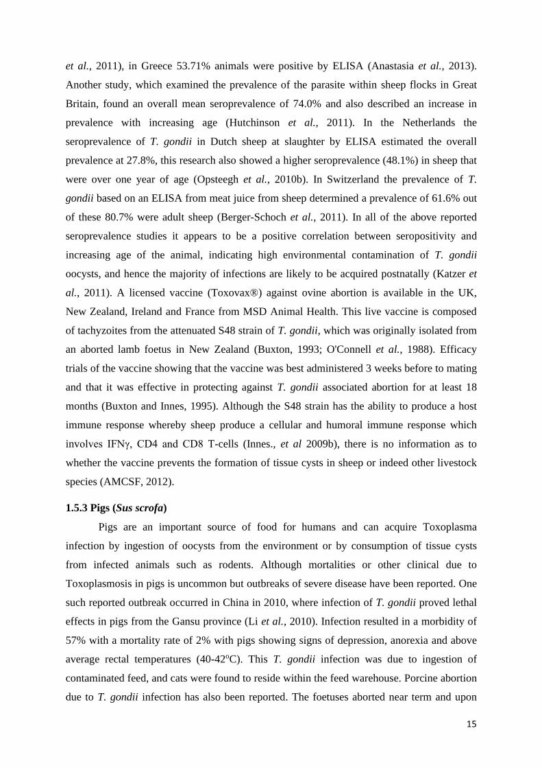
et al., 2011), in Greece 53.71% animals were positive by ELISA (Anastasia et al., 2013).
Another study, which examined the prevalence of the parasite within sheep flocks in Great
Britain, found an overall mean seroprevalence of 74.0% and also described an increase in
prevalence with increasing age (Hutchinson et al., 2011). In the Netherlands the
seroprevalence of T. gondii in Dutch sheep at slaughter by ELISA estimated the overall
prevalence at 27.8%, this research also showed a higher seroprevalence (48.1%) in sheep that
were over one year of age (Opsteegh et al., 2010b). In Switzerland the prevalence of T.
gondii based on an ELISA from meat juice from sheep determined a prevalence of 61.6% out
of these 80.7% were adult sheep (Berger-Schoch et al., 2011). In all of the above reported
seroprevalence studies it appears to be a positive correlation between seropositivity and
increasing age of the animal, indicating high environmental contamination of T. gondii
oocysts, and hence the majority of infections are likely to be acquired postnatally (Katzer et
al., 2011). A licensed vaccine (Toxovax®) against ovine abortion is available in the UK,
New Zealand, Ireland and France from MSD Animal Health. This live vaccine is composed
of tachyzoites from the attenuated S48 strain of T. gondii, which was originally isolated from
an aborted lamb foetus in New Zealand (Buxton, 1993; O'Connell et al., 1988). Efficacy
trials of the vaccine showing that the vaccine was best administered 3 weeks before to mating
and that it was effective in protecting against T. gondii associated abortion for at least 18
months (Buxton and Innes, 1995). Although the S48 strain has the ability to produce a host
immune response whereby sheep produce a cellular and humoral immune response which
involves IFNγ, CD4 and CD8 T-cells (Innes., et al 2009b), there is no information as to
whether the vaccine prevents the formation of tissue cysts in sheep or indeed other livestock
species (AMCSF, 2012).
1.5.3 Pigs (Sus scrofa)
Pigs are an important source of food for humans and can acquire Toxoplasma
infection by ingestion of oocysts from the environment or by consumption of tissue cysts
from infected animals such as rodents. Although mortalities or other clinical due to
Toxoplasmosis in pigs is uncommon but outbreaks of severe disease have been reported. One
such reported outbreak occurred in China in 2010, where infection of T. gondii proved lethal
effects in pigs from the Gansu province (Li et al., 2010). Infection resulted in a morbidity of
57% with a mortality rate of 2% with pigs showing signs of depression, anorexia and above
average rectal temperatures (40-42oC). This T. gondii infection was due to ingestion of
contaminated feed, and cats were found to reside within the feed warehouse. Porcine abortion
due to T. gondii infection has also been reported. The foetuses aborted near term and upon
16
histopathological examination Toxoplasma cysts were found in the alveolar macrophages of
the lung and intracellular parasites morphologically similar to Toxoplasma, were also
identified in myocardial cells (Hunter, 1979). The farm described was known to have a rodent
problem which can T. gondii in animal and human hosts increase the transmission of T.
gondii to pigs (Kijlstra et al., 2008).
Infection of sows with Toxoplasma have also resulted in the birth of still born piglets
(Thiptara et al., 2006), like clinical signs observed in cases of ovine toxoplasmosis. A more
commonly observed symptom is a slight increase in rectal temperature during the initial
stages of infection, which may persist for 1-2 days. Presentation of more acute clinical
symptoms (although rare) are likely to be linked to age, immune status, and even breed of the
animal (Dubey, 2009). Clinical symptoms in pigs which have been experimentally infected
with T. gondii are similar to those described above but can prove fatal when pigs are
inoculated with large numbers of oocysts (≥ 4x10⁴) (Dubey et al., 1998b; Garcia et al.,
2008).
To prevent or reduce the formation of T. gondii tissue cysts in pig species is to reduce
the number of parasites entering the human food chain and their consequent transmission.
The ability of the parasite to infect all warm-blooded mammals, combined with its cyst
forming nature, T. gondii is readily found within the muscles and organs of infected pigs
which are often used for human consumption (Bayarri et al., 2012; Dubey et al., 2005a;
Wang et al., 2012). By ingestion of T. gondii oocysts or bradyzoites, within 6-7 days
infective tissue cysts can develop in the host. The prevalence of T. gondii in pigs reared
indoors is likely to be lower than those animals which are outdoor reared, due to an increased
risk of exposure to environmental oocysts (Kijlstra et al., 2004; van der Giessen et al., 2007).
The infection of T. gondii in pigs increases with age (Dubey et al., 1995b), and this
seropositivity reflect the presence of infective tissue cysts within the animal (Dubey et al.,
1995a), it can be assumed that older pigs are more proven to infective tissues cysts.
The incidence of Toxoplasma in pigs has dropped in the past 30 years. This is most
likely due to a change in farming practices where intensive well managed indoor farming
methods have been introduced (Dubey, 2009; Edelhofer, 1994).
Transmission of T. gondii to pigs from the consumption of infected rodents has also
been shown to be a direct source of infection, particularly in animals reared outdoors (Kijlstra
et al., 2004), where outdoor reared and organically raised pigs have generally been shown to
have a higher prevalence of the parasite i.e Northern USA to be 90.9% (30/33), which is
significantly higher than reported for pigs raised in conventional indoor housing (2.7%) (Hill
et al., 2010a). Similarly, in Argentinean animals which had been bred indoors compared to

17
sows reared outdoors had a prevalence of 4.5% (4/88) and 40.2% (45/112) respectively
(Venturini et al., 2004).
The source of infection in pigs can either be via oocyst from contaminated soil or
feed, or from consumption of tissue cysts from infected rodents or other small mammals
harboring the tissue cysts stage of the parasite. Contamination of feed or soil with oocysts is
thought to be the main source of infection for pigs (Lehmann et al., 2003). However, outdoor
housing systems allow pigs to come into contact with rodents and other wildlife, and as pigs
are omnivores, they will consume rodents or rodent cadavers as well as other small mammals
and birds, which may be infected with T. gondii and harbour infective tissue cysts
(bradyzoites).
1.5.4 Domestic ruminants (Cattle)
Several epidemiological studies show that the use of raw or undercooked beef may be
responsible for T. gondii infection in humans (Cook et al., 2000), and bovines shows high
seroprevalence (up to 90%) (Tenter et al., 2009). In West Indies prevalence of 8.4%
(Chikweto et al., 2011), Brazil, 49.4% was recorded in cattle from a highly endemic area of
human toxoplasmosis (Frazao-Texeira and Oliveira, 2011). In Malaysia 7.9%
(Chandrawathani et al., 2008) and in Vietnam 10.5% seroprevalence was recorded in cattle
(Huong et al., 1998).
The seroprevalence of Toxoplasma infection in cattle ranges from 2 to 92% (Tenter et
al., 2000). High prevalence rates are found in calves during their first exposure to grazing,
which shows that calves become infected with T. gondii after exposure on pastures (Marty et
al., 1999). In dairy cows seroprevalence of 22.3% in Thailand, 3.2% in cattle in USA, 2.3%
in China (Yu et al., 2007), 6.6% in Ethopia (Bekele and Kasali, 1989), 9% in Indonesia was
reported (Matasuo and Husin, 1996).
1.5.5 Birds
T. gondii has been detected in tissues like spleen and lungs of dead pigeons (Johnson,
1943). Pigeons Infected with T. gondii were anorexic, dull, and having conjunctivitis (Carini,
1911). It has been shown that high infection rate of T. gondii oocysts was found with oral
infection (Biancifiori et al., 1986). Experimentally Sparrows indicated high resistant to T.
gondii but number of strains, and stage of T. gondii inoculated play an important role in
seroprevalence (Wallace, 1973; Literak et al., 1999). If numerous tachyzoites of mouse
virulence strain are injected in sparrows, they will die (Manwell et al., 1945; Drobeck et al.,
1953).

18
Severe toxoplasmosis with an unusual clinical symptoms (blindness) and in some
cases death has been reported in canaries (Serinus canarius) from Uruguay, Australia, Italy,
New Zealand, UK, and the US. Out of 18 birds 15 birds from a breeder house died within 15
days (Cassamagnaghi et al., 1952). While affected birds had lesions in the lungs and spleen,
enteritis, splenomegaly, necrotic, and hepatic degeneration (Parenti et al., 1986).
Toxoplasmosis has also been reported in 23 mynahs (Acridotheres spp.) imported from
Mexico. Lesions and parasites were seen in the liver, lungs, and spleen. Fatal toxoplasmosis
has been reported once in crows (Work et al., 2000).
1.5.6 Poultry
Toxoplasmosis in domestic chickens has been detected which indicated 65%
seropositivity and the presence of T. gondii in them could reach 81% of the seropositive
animals (Lehmann et al., 2006). Domestic chickens may be considered as an important
source of T. gondii infection to humans particularly in developing countries while in
developed countries seroprevalence of 1-10% have been reported. However, it can be
expected that high chance of poultry infection is possible when reared outside (Dubey et al.,
2008a). Antibodies to T. gondii reported in domestic chickens (Gallus domesticus), 12.5%
from Italy, 64% from Ghana, 30% from Poland, 24.4% in chickens from Indonesia, and
24.2% in chickens from Vietnam (Dubey et al., 2008a). In rural areas from Brazil, infection
higher than 50% reported in domestic chickens with T. gondii oocysts, indicating a
widespread contamination of rural environment (Oliveria et al., 2009). High susceptibility of
poultry species by oocysts is due to its feeding behavior and cysts are located mainly in heart
tissues and brain of poultry and rarely in muscles (Kijlstra and Jongert, 2008).
1.5.7 Cats
Cats which act as definitive host of the T. gondii may shed up to 10 million oocysts
per day for up to 14 days after primary infection. Shedding of oocyst depends on the source
of infection. Infected cats shed 30-50 of oocysts (Tenter et al., 2000). It is believed that about
1% of domestic cats excrete oocysts at any one time. Although shedding of oocysts is usually
suppressed by an active immune response, but this immunity may not be life-long, and cats
may shed further oocysts when again infected several years after primary infection (Innes et
al., 2009). The seroprevalence of T. gondii in cat’s ranges from 5% to 90% globally and is
more in wild and stray cats than domestic pets. Infection rate increases with age, although
due to immunity acquired after initial infection oocysts are excreted primarily by young
kittens (Dubey, 2008).

19
Cats which are the definitive host of T. gondii showed some extraordinary behavioral
characteristics of infection in old age like head pressing, teeth grinding, weakness in body
coordination, abnormal sounds and voice, circling and feelings uneasy, weak reflex actions
and paralysis were the signs of spinal cord. Granuloma formation and diarrhea indicated gut
infection. In Kittens respiratory system disorders were commonly observed and very little
cases have been reported of immature births and abortions. The ocular toxoplasmosis was
reported but fortunately very little damage was done to the nictitating membrane (Holzworth,
1987).
1.5.8 Dogs
Toxoplasmosis is also detected in dogs. Immune competent dogs remained generally
asymptomatic but immune compromised and old dogs, revealed symptoms of disease like
loss of canines, depression, and development of rigidity of the body especially pelvic
muscles. Younger dogs were found more seropositive than older ones. Acute stage was
usually associated with death occurred in most cases when symptoms like high temperature,
pain in abdomen, diarrhea, signs of CNS, lethargy, and vomiting, appeared (Aiello and Mays,
1998).
1.5.9 Deer
Toxoplasmosis in deer in Europe (involving 760 animals) indicated a seroprevalence
of 7.7% in Red deer compared to 34% in Roe deer (AFSSA, 2005).
1.5.10 Horses
T. gondii infection in horses in Europe suggested a prevalence of 1% by ELISA and
7.7% in Czech Republic by Sabin-Feldman dye test. Experimental studies have shown tissue
cysts persisting in horses for at least 15 months after inoculation (AFSSA, 2005).
1.5.11 Rabbits
Seroprevalence of T. gondii in wild rabbits vary greatly ranging from 5.9% in France
to 53% in the Czech Republic
1.5.12 Other species
Toxoplasmosis have also been detected in several other species examples include wild
boar 8-38%, and kangaroos 22% (Kijlstra and Jongert, 2008)
1.5.13 Marine mammals
Toxoplasmosis has been reported in a number of marine mammals’species, showing
that the organism has reached to the marine environment. According to several serological

20
studies carried out mainly in North America marine crustaceans and molluscs harbor the
parasite. Oocysts in soil or via domestic sewage are leached where they can survive and
sporulate. It has been proposed that marine mammals could act as sentinel species for T.
gomdii in the marine environment. In California a survey revealed toxoplasma infection in
52% of 305 freshly dead sea otters. Resistant oocysts concentrated in bivalve molluscs would
be most likely source of infection because sea otters do not feed on intermediate host
(Conrad, 2005).
1.6 Food studies
For detection of oocysts on or in food there is no standardised method, nor there is a
molecular method for detecting viable organisms in meats. Reports regarding the detection of
viable cysts are based mostly in vivo studies. T. gondii has been found in a variety of meat
from lambs, goats, pigs and game while beef appears less commonly contaminated, chicken
rarely contains viable cysts. The presence of cysts depends on time spent indoors, age farm
hygiene and the tissues concerned non-skeletal-muscle is more commonly infected than
skeletal muscle. PCR studiesin UK on a small number meat product indicated an overall
prevalence of 38%, including 25% of beef samples, 33% of pork samples and 67% of lamb
samples. Viable parasites were found in 1 sample (1.5%) out of 67 samples. 6/9 (66%)
samples of lamb meat from a butcher in Manchester tested positive for toxoplasma by PCR.
Bivalve molluscs are also a potential source of food borne T. gondii infection. (Aspinall et
al., 2002).
1.6.1 Survival in foods
There should be an understanding about the survival of infective stage of (oocysts,
tachyzoites and tissue cysts). Control strategies for food require an understanding of the
survival of the key infective stages of T. gondii (oocysts, tachyzoites and tissue cysts) under
different conditions. Generally, the principal hazard will be the tissue cysts in raw meats,
tachyzoites in milk, and oocysts may be a hazard on water and agriculture (Dubey, 2010).
1.6.2 Unsporulated oocysts
Sporulation of oocysts occurs within 1-5 days at ambient temperature and therefore
oocysts contaminating foods are most likely to be sporulated. Dubey reported that
unsporulated oocysts were killed by exposure to 37°C for 24 hours. Salt has not affected on
sporulation of oocysts with sporulation occurring in water at 1.5% and 3.2% NaCl.
Sporulation of oocysts appear to be inhibited by anaerobic conditions. (Dubey, 2010).

21
1.6.3 Sporulated oocysts
Sporulated oocysts show high resistant to adverse physico-chemical conditions.
Sporulated oocysts are less effected by low pH, in inorganic acids e.g. sulphuric acid (pH <1)
survive for over a year. Storage in alkali (sodium hydroxide, 6%, pH >/=12) inactivated a
suspension of oocysts in 24 hours and a 2-log reduction in infectivity was achieved after 1
hour (10% NaOH, pH>/= 12). In salt water oocysts survive for long periods (>6 months at
ambient temperature or at 4°C). Little or no effect on oocyst viability have been shown by
disinfectants for example chlorine of level 2, 20 and 200ppm had no effect on oocyst
viability. Ethanol (99%) for 24h kill oocysts, a 10% solution destroyeoocysts were by a 4-
dayexposure. (Dubey, 2010).
1.6.3 Tissue cysts
Tissue cysts can be inactivated by extremes in temperature because they are more
susceptible extreme temperature at low temperature they can survive for long period.
Relatively mild pasteurization is required to destroy tissue cysts and temperatures of 67°C
and above will reduce contaminated meat safe. Tissue cysts survival in the presence of salt
(sodium chloride) is variable and dependent on the salt concentration and storage
temperature. It has been found that concentrations of 6% salt is lethal to tissue cysts, although
at low temperatures (4°C) cysts shown survival, remaining viable in 0.85%, 2% and 3.3%
NaCl for 56 days, 49 days and 21 days respectively. Tissue cysts lost infectivity in pork
sausages made with 2% and 2.5% salt and stored under refrigeration for 48 hours but not
after 24 hours (Dubey, 2010).
Common curing salts can be effective at rendering tissue cysts non-infective based on
bioassays. When stored at 4°C, tissue cysts in pork became non infective in a number of
curing salt solutions including sodium tripolyphosphate (0.5%) sodium chloride (2%) (After
7, 28d and 45d), sodium chloride (1%) (After 45d but not after 7d or 28d) or sodium lactate
(2%) (After 7d, 28d and 45d). Infectivity of bradyzoites inside tissue cysts are lost after
storage at low pH (<1, HCl) for 2 hours (Dubey, 2010).
1.6.4 Tachyzoites
Limited information is available on the survival of tachyzoites although they are
reported to be susceptible to freezing and are destroyed by pasteurisation. Low pH (<1, HCl)
destroy tachyzoites after 25 minutes but survive for 7 days at 4°C in cow‟s milk (Dubey,
2010).

22
1.7 Toxoplasmosis in humans
Human Toxoplasmosis is a problem throughout world, with seroprevalence ranging
from 0 – 100% depending on the origin of the population studied, published the overall
global trends for the seroprevalence of T. gondii in women of child bearing age and pregnant
women for a period of ten years from January 1999 - December 2008. The seroprevalence of
T. gondii in human is shown in global map indicated by different colours (Tenter et al., 2000:
Pappas et al., 2009).
Figure 1.5 Map showing worldwide seroprevalence of T. gondii. Seroprevalence is
shown using the following different colours: Dark red above 60%, light red = 40 –60%,
yellow = 20 – 40%, blue = 10 – 20%, green = less than 20%, white = unknown. The map
shows a high prevalence in mainland Europe, South America and South East Asia
(Pappas et al., 2009).
The map shows high seroprevalence in humans in South American countries such as
Argentina and Brazil which is 53.4% and 77.5% respectively (Porto et al., 2008; Rickard et
al., 1999), while a lower prevalence was reported in most of European countries, such as
Denmark 27.8%, the United Kingdom 9.1% and the Czech Republic 19.8% (Kankova and
Flegr, 2007; Lebech et al., 1999; Nash et al., 2005). Seroprevalence of T. gondii infection in
Germany and France was higher 59.0% and 54.0% respectively compared to other European
countries (Fiedler et al., 1999; Tenter et al., 2000). Climatic condition, consumption of raw
meat and meat from animals or frozen meat may be factors that contribute to these variations.

23
Seroprevalance based on age have also been reported which shows variation in a number of
countries. For example, in the Netherlands at age 25 years it was found 20% and 60% at age
50 years. While in japan in age group 20-29 years lower seroprevalence was found as
compared to 40% in those over 70 years age. Similarly, seroprevalence in women of child
bearing age has also been detected which 10-22% in England as was compared to 50% in
above 50 years (Zadik et al., 1995).
In pregnant women the frequency of the parasite occurs differently, depend upon
geographic area (6.1 to 75.2), it was found as 21% in Pakistan and Bangladesh, 55% in
France, 31% in Ireland ,15% - 41% in Africa 46% in other European countries. Pregnant
women show 71% prevalence rate in France (Ancelle et al., 1996; jeannel et al., 1998) and
78% in Ibadan, Nigeria (Onadeko et al., 1992), and in people of Somalia it was 44, in south
delta Nigerian 83% was reported (Dubey and Beattie, 1988). In jimma (zemene et al., 2012)
central Ethiopia (Gebremedhin et al., 2013), Arba Minch. (Yohanes et al., 2014), womens at
child bearing age and HIV patients show prevalence of 83.6%, 81.4% and 88.2% (Zemene et
al., 2012). It was observed that in the past few years the prevalence of toxoplasmosis among
different countries including United Kingdom, France and Belgium goes on decreasing.
(Remingto et al., 2006; Montoya et al., 2004). Low level of seroprevalence in US as
compared to Western Europe also identified reduced seroprevalence in overall population
(Smith et al., 1996). Central America and South America, particularly in Costa Rica and
Brazil), have higher seroprevalence greater than 60% revealed by studies (Zapata et al.,
2005). Although it gained no importance as community health problem recommended in
Colombia (Gomezet al., 1995; Gomez 1997). In 1980, by means of indirect immune
fluorescence antibody test the Ministry of Public Health implemented serological study of
prevalence, it has concluded that 20% - 90% of world population exposed to toxoplasmosis
(Zemene et al., 2012).
Countries where usually uncooked food is used show high rate of prevalence (jeannel
et al., 1998; Ancelle et al., 1995) and also it was found in areas where cats are distributed
widely and environmental factors that supports existence of oocyst like in tropical areas of
Latin America and sub-Saharan Africa (Dubay and Beattie, 1998; Schwartzman, Maguire,
1999; Onadeko et al., 1992). Age wise its prevalence has been found to be 13% (6 years)
(Frenkal et al., 1995) and 90% (60 years) (Souse et al., 1998). Increase rate of seroprevalence
among children in panama where people used properly cooked food indicates their contact
with soil contain oocysts. (Souse et al., 1988). Rise in prevalence rate in different parts of the
world also associated with contaminated water (Ertug et al., 2005). T. gondii among pregnant
women is abundant in wet and hot areas. There is a higher risk of congenital toxoplasmosis in

24
people with anti-toxoplasma IgG antibodies between 25-80 percent, due to high flow of
parasite in blood and high ratio of sensitive pregnancy (Remington et al., 2006).
1.7.1 Prevalence of T. gondii in Pakistan
In district Dera Ghazi Khan (Pakistan), about 29.8% cases were reported (Tasawar et
al., 2011). Mostly human toxoplasmosis is without symptoms or show minor illness. Rise in
seroprevalence of infection noticed up to 100% in a population in last three years. (Flegr et
al., 2003). In Pakistan among pregnant womens, it has been found in Punjab, Kashmir and
Khyber Pahktunkhwa in the percentage of 63, 48, and 38 respectively. In district Kohat it has
been observed in pregnant women about 14.4%. Rural areas show higher percentage of
parasite then urban areas of district Kohat while 17% seroprevalence reported from
Islamabad and Rawalpindi.
1.8 Genome of Toxoplasma gondii
The genome of T. gondii is 65 Mega base pairs (Mbps) in size, which comprise of 14
chromosomes, the size of which ranges from 2 Mbp to 7.5 Mbps (Khan et al., 2005b). Its
genome is somewhat similar to that of Neospora caninum, which is another protozoan
parasite of the Apicomplexa group. It is assumed that the two parasites diverged from a
common ancestor about 28 million years ago. The reason for the divergence was the
speciation of the definitive hosts i.e. cats for T. gondii and dogs for N. caninum (Reid et al.,
2012). As compare to other Apicomplexan parasites for example Theileria parva and
Cyptosporidium parvum, the T. gondii genome is considerably larger, and have more introns,
has a lower gene density and has more predicted genes (Ajioka and Soldati, 2001). The
recommended reason for the difference in size of the genome compared to other
Apicomplexan parasites is the large number of secondary hosts which T. gondii can establish
within (Roos, 2005).
In 2005 a composite genome map was derived from the genetic crosses and analysis
of linkage in the three main archetypal lineages (I, II, III) of T. gondii (Khan et al., 2005b).
The results from this study helped the researchers engaged in mapping out of drug resistance
genes and aided the researchers that were studying the genes involved in infectivity and
transmission. The linkage map that was produced, identified 250 specific genetic markers, 12
of them are most commonly used for genotyping of the strain through PCR-RFLP. Some of
the markers are present on all the three archetypal strains.
The ToxoDB, an online genome database provides a more detailed information about
the structural and functional genomics of T. gondii. It also offers information about gene
expression genome sequence and proteomics data, that helps in supporting research on the

25
parasite (Gajria et al., 2008). The database also has data for genetic maps and single
nucleotide polymorphism, representing strains for three archetypal T. gondii lineages; GT-1
(Type-I), Me49 (Type-II) and VEG (Type-III). Identification of single nucleotide
polymorphism (SNPs) through PCR-RFLP are used to define the different strains of T. gondii
(Sibley et al., 1992; Ajioka et al., 1998).
1.9. Genetic Variation of T. gondii
1.9.1 Main Lineages
Three main lineages (Type I, II, III) were identified by the studies on the virulence of
of T. gondii in mice and molecular characterization of the parasite in humans and through
multilocus RFLP (Sibley and Boothroyd, 1992; Howe & Sibley, 1995). It has been
recognized by the research that type I isolates have limited genetic variability but have high
virulence in mice. The Type I lineage is isolated very rarely and only about 10 % of the
strains collected in USA and Europe. But this Type is related to the reactivation of the
parasite in immunocompromised persons (Khan et al., 2005a). Type II lineage isolate is
recognized as the predominant in human toxoplasmosis as well in sheep and pigs in Europe
and USA. However, it is less virulent in mice. Type III lineages isolates were initially
considered as non-virulent in mice. But new research studies reported that they are usually
more virulent than Type II isolates in mice (Darde, 2004). The Type III lineage is more
common in animal hosts and rare in humans. A research in chickens in North America
reported a higher prevalence of type III as compared to Type II (Dubey et al., 2003).
Although initial characterization of the parasite explored its virulence in mice (Sibley
and Boothroyd, 1992), the pathogenicity of the parasite can differ depending on its host
species. Most of infections in both humans and animals are without any symptoms, but some
of species of host can be vulnerable to initial infection, triggering clinical symptoms and even
death. For instance, Australian marsupials and lemurs are examples of vulnerable host
(Canfield et al., 1990; Dubey et al., 1988), and other animal species, such as lemurs, moved
into zoos or wildlife parks (Dubey et al., 2009; Hermosilla et al., 2010; Juan-Salles et al.,
2011). This susceptibility is supposed to occur due to susceptible animals being more
geographically isolated from the definitive host i.e. felids, where they have evolved in areas
where, until relatively recently, cats have been absent. Subsequently these animals could be
more sensitive to T. gondii oocysts, with the immune system incapable to deal with the
infection successfully (Innes, 1997).

26
1.9.2 Atypical lineages
Molecular characterization through multilocus RFLP has recognized strains which
have different unique genotypes as compared to three predominant lineages. At first most of
these atypical genotypes were isolated from exotic animals in geographically distant areas,
but more recently multilocus RFLP has emphasized the presence of atypical alleles in pigs
sheeps and poultry in Brazil (da Silva et al., 2011; Frazao-Teixeira et al., 2011). Initial
infection of these atypical strains in humans has been related to severe toxoplasmosis and eye
disease, with epidemics reported in Brazil, French Guiana and Suriname (Carme et al.,
2009a; Vaudaux et al., 2010).
The main features of atypical strains are that they comprise of novel alleles as well as
mixed combinations of alleles of three predominant lineages (Grigg and Suzuki, 2003).
Through molecular genotyping atypical strains shows great genetic diversity from those of
predominant lineages. For instance, atypical strains recognized in South America display a
greater genetic diversity as compared to the predominant lineages isolates found in North
America and Europe (Herrmann et al., 2010).
Other different strains that cannot be classified into the three main predominant
lineages also exist, which have been isolated on various occasions and seem to occur in
specific hosts or be associated to a specific geographical region. Type X is a genotype that
has been predominantly reported in Californian sea otters (Sundar et al., 2008). Microsatellite
genotyping of an immunocompromised toxoplasmosis patients in France recognized a new
genotype, which were later isolated from many patients. These genotypes were named Africa
I, Africa II and Caribbean I (Ajzenberg et al., 2009). Further research in Gabon has
recognized the similar African genotypes as well as an extra Africa III genotype (Mercier et
al., 2010)
Studies from North America has recognized a fourth clonal lineage, which was
initially classified as Type X and was first isolated from Californian sea otters (Sundar et al.,
2008). More lately the presence of this genotype has also been reported in wild animals and
in rare cases humans from North America, resulting in a definite, highly clonal, separate
lineage fitting in to haplogroup 12 (Khan et al., 2011). At the time of writing, T. gondii is
described to be composed of 15 different haplogroups, which defines six major clades this
definition is based on analysis of the genetic diversity of 950 T. gondii isolates which were
collected worldwide (Su et al., 2012).
The T. gondii strains which have endured sexual recombination to generate mixed
infections have also been reported by multilocus RFLP in both human and animal hosts
(Lindstrom et al., 2008). Sexual recombination can only occur in the gut of the felid host and

27
its development must be due to infection with two strains of the parasite at the same time. In
spite of the identification of atypical genotypes and mixed infections their occurrence is rare
compared to the identification of predominant lineages, but with the advancement of
molecular tools, the sensitivity of detection has increased and consequently the likelihood of
discovering these unusual strains is becoming more likely (Lindstrom et al., 2008).
1.10. Transmission
The infection of T. gondii follow various infection paths for felids and intermediate
hosts, including vertical and horizontal transmission routes. In humans the main concern is
the vertical route which involve congenital transmission from mother to fetus. (Dubey and
Beattie, 1988b). If a womanfor the first time is exposed to T. gondii during pregnancy,
complications in perinatal mortality or birth defectsmay result (Dubey and Beattie, 1988b).
Risk factors like contact with faeces of cat, cat ownership for human act for T. gondii
seropositivity (Hofhuis et al., 2011).
Possible reasons for transmission: When cats become infected byingesting oocysts in
the surroundingsor infected intermediate hosts for shedding oocysts to occur. 2) cats excrete
oocysts only for a short time after primary infection, 3) cats are usually resistant to re-
infection and protected against re-shedding of oocysts for several years after primary
infection, 4) Excreted oocysts takes 24 Hrs to sporulate to become infectious, faeces
deposited indoors is usually discarded before sporulation takes place, and 5) oocysts are
rarely found in the fur of cats (Dubey, 1995). Toxoplasmosis disease is horizontally
transmitted to humans by accidentally ingestion of raw or undercooked meat containing
tissue cysts, and ingestion of food or water contaminated with oocysts shed by cats. (Jones et
al., 2012). Transmission and spread of infection among humans is largely through inhalation
or ingestion of oocyst discharged in the faeces of infected cats, inoculation of trophozoites
through the skin, drinking raw cows milk or contaminated water, ingestion of birds’ eggs and
by eating raw or undercooked infected meat. (Sadaruddin et al., 1991). Among several
domestic animal’s cat is the definite host but pigs, cattle, sheep, goats and rodents may also
play role in its transmission. Rats and mice are thought to be persistent wildlife host
reservoirs of T. gondii. (Glazebrook et al., 1978; Webster, 1994). Properly cooked meat does
not involve a risk for infection, because T. gondii tissue cysts are killed at 65°C (Dubey et al.,
1990). Pet dogs are often regarded as closest faithful and intimate friends of humans. There
are also some reports of T. gondii oocysts shedding by dogs because oocysts ingested via
food and water in dogs can pass through intestinal tract and are excreted in the feces (Lindsay
et al., 1997). The presence of pet dog in household has been found to be a risk factor for T.

28
gondii infection in humans (Sroka et al., 2010). The oocyst stage of T. gondii has been found
in the environment, both in soil and in water, where they can survive for a long time,
depending on temperature and humidity. Contact with soil during gardening and consumption
of raw or poorly rinsed vegetables have been identified as significant risk factors for T. gondii
infection in Europe (Simon et al., 2013). Although beef is generally considered a less
important source of T. gondii infection because of relatively rare findings of viable parasites
in their tissues. It is unlikely that pork is a major source of human toxoplasmosis in countries
where the majority of fattening pigs are reared in intensive management systems. However,
pork from pigs reared in animal-friendly management systems is thought to be a significant
risk (Tenter et al., 2000).
1.11 Pathogenesis.
1.11.1 Pathogenesis and Clinical Signs of Toxoplasmosis in Animals
Feline toxoplasmosis is a multi-systemic disease which may cause clinical disease in
domestic and wild animals (Dubey, 2010). It is a major cause of abortion and mortality in
animals leading to economic losses as well as lowering the food basket with effects on family
health (Jadoon et al., 2009). In cats, which are the definitive hosts, toxoplasmosis is usually
asymptomatic (Darabus et al., 2011). In a study done in Iran on wild and domestic dogs, it
was found that dogs, unlike cats, are mechanical vectors and do not show specific clinical
signs (Shadfar et al., 2012). Toxoplasmosis has been known to be a major cause of abortion
and stillbirth in sheep and goats (Jadoon et al., 2009). Prevalence in herbivores has been seen
to be highest in spring while in autumn and winter the prevalence has been seen to be highest
in pigs (Darabus, et al., 2011). In swine, toxoplasmosis causes reproductive disorders such as
premature birth with pneumonia, myocarditis and encephalitis (EFSA, 2007). However, most
cases of Toxoplasmosis may be asymptomatic with mild non-specific symptoms such as
hyperthermia, anorexia and tachypnea (Dubey and Beattie, 1988). Infectivity rates of
toxoplasmosis in cattle are low with clinical signs not usually observed. In some rare
instances, however, T. gondii has been isolated in aborted fetuses (Dubey et al., 2012).
1.11.2 Pathogenesis and Clinical Signs of Toxoplasmosis in Humans
Globally, it infects a third of the total human population. However, local variations in
prevalence is a characteristic feature of T. gondii infection, and the seroprevalence varies
between 0% and 100% depending on the population investigated. The risk of encountering
the infection increases with time, and because it is a lifelong infection, the T. gondii
prevalence in a population is highly dependent on the age composition. (Tenter et al., 2000).
One of the first studies of T. gondii prevalence in Sweden was performed in 1951 by Zeipel

29
and Linder, who found a seroprevalence of 37% in 300 healthy blood donors. In 1953,
Hedqvist studied the frequency of T. gondii infection in different age groups in Eskilstuna, a
city located in the south-central part of Sweden. A difference in seroprevalence was found
between children 0-4 years old (0%) and adults 20-50 years old (40%). Thirty years later
(1982/1983), a similar overall seroprevalence of 40% was found in pregnant women in
Malmo (Ahlfors et al., 1989).
Human toxoplasmosis includes a broad spectrum of symptoms, usually grouped into:
1) mild and transient disease in immunocompetent individuals, 2) reactivation of latent
infection in immunocompromised patients, 3) congenital infection in newborns with
symptoms apparent either at birth or detected later in life, and 4) severe toxoplasmosis with
ocular, neurological, and generalised symptoms as a result of postnatally acquired infection in
immunocompetent persons (Montoya and Liesenfeld, 2004). In immunosuppressed patients,
such as those infected with human immunodeficiency virus (HIV), the cause of disease is
usually not a primary infection, but rather reactivation of a latent infection (Kodym et al.,
2015). It leads to abortions and neonatal problems in humans and exerts negative impact on
livestock production. Toxoplasma encephalitis has been reported as a cause of death in
immune-compromised individuals with AIDS (Dubey and Jones, 2008). During pregnancy T.
gondii leads to ocular and neurological impairment such as mental retardation, blindness,
epilepsy, seizures, microcephaly and hydrocephaly (Robert-Gangneux and Darde, 2012). T.
gondii has been associated with behavioural changes and psychiatric illness such as
schizophrenia, implying that latent toxoplasmosis may be a greater problem than previously
recognised (Sutterland et al., 2015).
More recently, the prevalence of HIV-associated toxoplasmosis has decreased
because of HAART (highly active antiretroviral therapy). However, other groups of
immunocompromised patients, such as organ or stem-cell transplant recipients and patients
undergoing cancer therapy, are at risk for reactivation of latent T. gondii infection (Hakko et
al., 2013). Approximately 90% of congenitally infected children are born asymptomatic, of
which many will develop clinical signs later in life (Guerina et al., 1994). A more recent
abortion outbreak occurred in Jeju Island, Korea, where pregnant woman showed clinical
signs which included: fever, anorexia, depression, recumbency, abortion and in a few cases,
death (Kim et al., 2009).

30
1.12 Diagnosis
Demonstration of anti-T. gondii antibodies is a useful indirect indication of T. gondii
infection, because the infection is assumed to be lifelong in most host species (Tenter et al.,
2000). The relationship between parasite abundance in muscles and specific antibodies in
blood seems to differ between species. For pigs, the two seem to correlate well, and it has
been shown that a higher antibody titre is associated with a higher probability of finding
viable parasites in tissues using bioassay (Dubey et al., 1995a).
Many different serological tests have been used for detection of T. gondii specific IgG
and/or IgM. Some of these are: the Sabin-Feldman dye test (DT), the indirect fluorescent
antibody test (IFAT), the direct agglutination test (DAT; also referred to as the modified
agglutination test, MAT), the enzyme-linked immunosorbent assay (ELISA), the latex
agglutination test (LAT), complement fixation (CF), and the indirect haemagglutination test
(IHAT). DAT and ELISA are widely used and have been evaluated for pig serum in many
studies (Hill et al., 2006).
1.12.1 DAT (Direct Agglutination Test).
In DAT, the antigen (whole killed T. gondii tachyzoites of the RH strain) coalesce into a
network in which the organisms are coupled by specific IgG, if present in the test serum. The
outcome of the test (the titre) is the highest dilution of the sample at which agglutination
occurs. The DAT for T. gondii was developed in 1959 by Fulton and Turk. Initially, a low
sensitivity (Se) and specificity (Sp) limited its use, until the test was further developed in
1980 (Desmonts and Remington, 1980). The Sp was improved by adding 2-mercaptoethanol
(2-ME) to denature interfering natural IgM antibodies and the Se was improved by modifying
the antigen production. Note that specific IgM activity is also inhibited by 2-ME, and
therefore, DAT only detects T. gondii specific IgG. DAT may thus result in false negative
results in recently infected individuals (Dubey et al., 1995b).
DAT is a simple and straightforward test; it does not require any specific equipment, and
results are read after 5-18 hours. Serum samples should be tested in at least two dilutions (one
dilution representing the decided cut-off and one higher dilution) to avoid false negative
results caused by a phenomenon known as the prozone effect (Seefeldt et al., 1989). This
effect is a result of excessive T. gondii specific IgG in the sample, which saturates binding
sites on the antigen and thus prevents cross-linking of the antigen (agglutination). A
disadvantage of DAT is that the results are read subjectively (Dubey et al., 1995b).

31
1.12.2 ELISA (Enzyme Linked Immunosorbent Assay)
In indirect ELISA tests, a solubilised antigen is absorbed to a plastic surface of high-
binding capacity. Test samples are added to allow specific antibody-antigen binding. If
present in the sample, specific antibodies are subsequently detected using an enzyme-linked
secondary antibody. As a final step, a substrate solution for the enzyme is added and the
interaction between the enzyme and the substrate generates a visible colour. The colour
intensity is quantified objectively using a spectrophotometer and the result is presented as an
optical density value (OD). Using a predefined cut-off value, samples are assigned as positive
or negative. ELISAs for T. gondii come in a variety of designs including different types of
antigens, conjugates and buffers etc., all of which affect the performance of the test.
1.12.3 Serology Using Meat Juice
Meat juice is a serological matrix extracted from muscle samples by freezing and
thawing (Nielsen et al., 1998). Although not very well characterised, meat juice consists of a
mixture of blood, lymph and extracellular and intracellular fluids. Therefore, meat juice
contains antibodies, originating in different amounts from the above-mentioned sources.
However, the relative contribution of these sources is not well defined. Because meat juice is
readily available from animals at slaughter and from hunted game, it has been used in several
serological assays to demonstrate antibodies directed at different pathogens (e.g., Trichinella,
Salmonella and T. gondii in pigs) (Felin et al., 2014).
1.12.4 Detection of Parasites
Other means than serology is necessary to directly determine if parasites are present
in the tissues of an animal. Investigations based on detection of parasite DNA (by the
polymerase chain reaction (PCR)) are common, but may underestimate disease prevalence
(Garcia et al., 2006a). T. gondii tissue cysts are distributed unevenly throughout the muscles
of the host, and very small amounts of tissue (a few milligrams) are usually used in the PCR
analysis. Thus, there is a risk that the sample does not contain tissue cysts. However, a
method to analyse larger samples (100 grams) by digestion followed by magnetic-capture
PCR – has recently been developed (Opsteegh et al., 2010a).
1.12.5 Examination of cat faeces
Collect fecal samples of cat from different localities in a particular period of time.
Examine cat faeces for the presence of T. gondii oocysts using sheather's sugar flotation as
described by (Dubey, 2009a). Briefly, (2-10 gm) faeces from each cat sample float in sucrose
solution (454 gm sugar, 355 ml water and 6 ml formalin; specific gravity, 1.203), filter

32
through gauze, and centrifuge at 1500 rpm for 10 min in a 15 ml tube. Now put a drop of the
float on glass slide and cover with cover slip and examine microscopically at 400X
magnification. Measure oocysts with an ocular micrometer. If oocyst size ranges from 9-12
μm, fecal floats are sediment in water and aerated in 2% sulfuric acid, for sporulation, on a
shaker at 22 ºC for 1 week and stored in a refrigerator at 4 ºC (Lindsay et al., 2002).
1.12.6 ELISA to measure IgG titers
ELISA is also used with little modifications for detection of Toxoplasmosis (Choi et
al., 1992). Briefly, out of 96-well micro titer plate each one is coated 200μl (TLA T. gondii
lysate antigen) (5 μg/ml) in 0.05 M carbonate-bicarbonate buffer with (pH 9.6). After
incubating plate at 4˚C plate is washed serum samples are reacted in each well and further
diluted 1:100 with 0.05% Tween 20. It is then incubated at 37˚C for one hour. After washing
for several times, o-phenylenediamine dihydrochloride which is freshly prepared is added. 8
N H2SO4 is added to stop the reaction. The IgG antibody titers at a density of 490 nm are
determined.
1.12.7 PCR for detection of T. gondii B1 gene in blood
Specific IgM and IgA antibodies of Toxoplasmosis can be diagnosed very
successfully in laboratory by using Polymerase Chain Reaction (PCR). It is an efficient and
important tool for diagnosis. Some infected cats may be asymptomatic but revealed large
amount of IgG antibodies in their bodies without excreting oocysts in faeces. Thus antibodies
(IgM) can be used to determine susceptibility of a healthy cat to T. gondii. After two weeks
of infection oocysts are shed by infected cats. The examination of faeces and presence of
oocysts in it is not a suitable technique for diagnosis of oocysts in faeces because oocysts of
T. gondii are similar to other pathogenic parasites. T. gondii B1 gene can be identified in
blood samples by PCR. To isolate DNA of T. gondii the DNeasy blood and tissue kit
(Qiagen, Hilden, Germany) is used. Primers like reverse Toxo-497 (5´-
CATGGTTTGCATTTTGTGG-3´) and forward Toxo-497 (5´-AGCAAACAC-
CGACGAACTCT-3´) are used to amplify the 497bp fragment of the gene, the smart 2X PCR
Pre-mix is used with the following conditions: 94 ˚C for 5 min, followed by 35 cycles of 94
˚C for 60 secs, 55 ˚C for 55 secs, and 72 ˚C for 60 sec. The conventional PCR products are
analyzed by 1% agarose gel electrophoresis and stained with ethidium bromide (Aldebert et
al., 2011).

33
1.12.8 Techniques for T. gondii strain genotyping
Different strains of T. gondii cause different clinical symptoms in both humans and
animals, there for genotyping is important to understand the epidemiology and population
genetics of the T. gondii (Grigg et al., 2001; Wang et al., 2013). To understand the virulence
and epidemiological impact, Identification and tracking of these strains is important. Initially
strain genotyping methodology was based on Multilocus Enzyme Electrophoresis (MLE)
(Darde et al., 1992). But current research has focused on two other typing methods; Multi-
locus Nested PCR-restriction fragment length polymorphism (Mn-PCR-RFLP) and
Microsatellite Sequence Typing (MLST), for epidemiological studies Mn-PCR-RFLP is most
commonly used. Currently the markers used for Mn-PCR-RFLP only identifies specific
regions of the T. gondii chromosome, therefore, using this technique alone there is possibility
of genetic diversity of the T. gondii. By using either of these methods directs equencing of
PCR products, will confirm the detection of SNP’s and will also identify deletions and
insertions which may provide additional information about genetic polymorphisms within an
isolate. However, these molecular technologies can be expensive and may not cost effective
option for large scale epidemiological studies (Sibley et al., 2009).
1.12.8.1 Mn-PCR-RFLP
Mn-PCR-RFLP is based on digestion of PCR products by specific restriction enzymes
which detect SNP’s. The digested PCR products are different in size according to the strain of
T. gondii and PCR marker used, result in bands of differentsizes which can be used to
identify a particular allele for the parasite. A series of 10 - 12 markers BTUB, GRA6, SAG2
5’, SAG2 3’, SAG3, SAG1, altSAG2, c22- 8, c29-2, L348, PK1, and Apico which
coverseight chromosomes and the plastid of T. gondii (see Table 1.2) are used to detect not
only clonal lineages (type I, II and III) atypical or mixed genotypes (Dubey et al., 2014; Pena
et al., 2013), but other studies shows the use of only five or six markers ,BTUB, GRA6,
SAG2 5’,SAG2 3’, SAG3 and Apico (Boughattas et al., 2010; Burrells et al., 2013; Khan et
al., 2005a).
Table 1.2 Mn-PCR-RFLP primers and Marker and T. gondii chromosome number.
SAG1, 5'SAG2, 5'SAG3, alt. SAG2 VIII
SAG3 XII
BTUBIX
GRA6X
c22-8Ib
c29-2III
L358 V

34
PK1 VI
Apico Plastid
1.12.8.2 MLST.
Genotyping by MLST is not as widely used as Mn-PCR-RFLP. The technique
identifies nucleotide polymorphisms, which differ between different T. gondii strains, and
generally incorporates five microsatellite markers (TUB2, W35, TgM-A, T. gondii in animal
and human hosts B18, and B17) across five different chromosomes of the parasite (Ajzenberg
et al., 2005). Primer pairs are used in a multiplex assay which incorporates fluorescent
labelling of the 5 ends with fluorescein. The PCR amplicons generated are accurately sized
using an automatic sequencer and Gene Scan analysis software (Fekkar et al., 2011).
1.12.9 Latex Agglutination Test
The latex reagent is a suspension of polystyrene particle which is sensitized with the
antigens of T. gondii. The distant agglutination pattern when observe after mixing the serum
will reflects formation of antigen-antibody complexes. When the organism has no infection,
then no agglutination will be observed. A greater than 4 IU/ml value is considered as standard
for positive result.
A Latex reagent is enough for the 50/ 100 tests. For ensuring homogeneity the latex
will be shaken. The positive control test will declare positive result indicates that the reagent
is functional, and the negative response of negative control will indicate that it is also
functional. The Pipette stirrer and re useable reaction slides are also used in the process.
Serological methods such as Sabin-Feldman Dye Test (SFDT), complement fixation test,
indirect and Modified Agglutination Test (MAT), intradermal skin test, are used for the
detection of toxoplasmosis (Goz et al., 2007). lymphoma and encephalitis techniques are
used as diagnostic procedures in immunocompromised patient (Gross et al., 2004).
ELISA is used for detection of IgG and IgM antibodies in human blood (Hasan,
2011). Latex Agglutination Test kit (LAT) is used for IgG antibodies commercially (Tasawar
et al., 2012).
1.12.10 Vaccination strategies
1.12.9.1 Cats
The definitive host cat shed oocysts which act as primary source of infection (Dubey
and Beattie, 1988), a vaccine which control oocysts shedding will prevent environmental
contamination with oocysts (Innes 2009), as a result of reduction in number of oocysts in
environment will reduce toxoplasmosis in humans and animals hosts. One of the most
successful vaccines T-263 shown successful results 84% (31/37) aginst oocysts shedding in

35
experimental cats (Frenkel et al., 1991). During a field trial with the same mutant strain (T-
263), results showed that vaccination of cats on 8 commercial swine farms resulted in an
overall reduction of the seroprevalence of the parasite in pigs (Mateus-Pinilla et al., 1999).
Despite the success of this vaccine it has not commercially been manufactured because it
requires a cold storage chain and bradyzoites which are used for vaccination are also infective
to humans, making administering the vaccine potentially hazardous. A more desirable
approach would be a vaccine which incorporates recombinant DNA, however studies using
this methodology are few in number and their success has been limited. For example,
research by (Mishima et al., 2002) using recombinant feline herpesvirus-1 expressing the
ROP2 antigen of T.gondii did not reduce oocyst shedding in cats noted partial protection
against oocyst shedding when cats were vaccinated intranasally with crude rhoptry proteins
of T. gondii combined with Quil- A as an adjuvant (Garcia et al,. 2007; Zulpo et al,. 2012)
1.12.10.2 Sheep
The main cause of ovine abortion in both UK and worldwide is ovine toxoplasmosis.
Toxovax is the only commercially available vaccine against toxoplasmosis which is licensed
by MSD Animal Health (Milton Keynes, UK). The vaccine S48 strain of T. gondii, consists
of the tachyzoite stage of which has lost the ability to differentiate into bradyzoites or
oocysts, and remains as the tachyzoite stage of the parasite (Buxton, 1993; O'Connell et al.,
1988).
1.12.10.3 Pigs
Various vaccination plans have been followed, but, as yet, no vaccine has been shown
to provide complete protection against tissue cyst formation in pigs however, following
mouse bioassay of porcine tissues, only partial protection from tissue cyst formation was
obtained (Dubey et al., 1994; Dubey et al., 1998b; Dubey et al., 1991; Pinckney et al., 1994).
In 2004, Kringel et al also used the RH strain of T. gondii for vaccinating pigs, two of the
dense granule proteins of T. gondii, GRA1 and GRA7, was shown to produce a strong
humoral and type I cellular immune response in pigs against T.gondii infection (Jongert et al.,
2008). A mouse bioassay on porcine heart tissue from the vaccinated pigs in this experiment
found two out of a total of three animalsto be tissue cyst free.
1.12.10.4 Humans
A vaccine for control of toxoplasmosis in humans is, as yet, unavailable. A vaccine in
risk groups like women, child bearing age and immune-comromised individuals would be
most beneficial.The only vaccine which is successful against toxoplasmosis is live
tachyzoites of the attenuated S48 strain of the parasite only in veterinary is now considered to

36
be beneficial in humans (Ivory and Chadee, 2004), however, these may not be as effective as
live vaccines (Innes et al., 2011).
From published research, it is clear that a vaccine which incorporates antigens from
the different stages of the life cycle is important. CD4+, CD8+ and IFNγ are involved in
protective immunity to T. gondii and if a greater number of antigens are present in a single
vaccine, it is more likely that they will be processed and presented to the immune system
(Innes et al., 2011).
1.13 Symptoms
Due to healthy immune system, 80 percent of primary toxoplasmosis show no
symptoms as determined by epidemological studies (Remington et al., 2006). Majority of
infected pregnant (more than 90%) women are asymptomatic (Boyer et al., 2005; Kravetz
and Federman, 2005). Only in few cases clinical symptoms are identified (De carlo et al.,
2008; Boyer et al., 2005). Appearance of clinical symptoms is same both in pregnant women
and non-pregnant women it includes flu like symptoms fever and enlargement of lymph
nodes, malaise (Jones et al., 2003; Boyer et al., 2005; Stray-pederson, 1993) and typically
around 2 % of people with good physical health show occular disorders (Holland, 2003).
Immunity developed against T. gondii in females are safe during pregnancy from congenital
toxoplasmosis, while females are in risk of congenital toxoplasmosis who lack such
immunity (Cunningham et al., 1997). Due to severe infection congenital infecion establish in
pregnant women and depends on period of infection. Symptoms of congenital infection may
be seen after few months of delievery or may be noted at birth time (Dubey et al., 1998;
Tenter et al., 2000). People with normal immune function in most cases of toxoplasmosis
show no symptoms (Kravetz and Federman, 2005), while immunocompromised inidividuals
show various disorders like fatal encephalitis, fever and lanphadenitis. (Shimelis et al., 2009).
It has been estimated that one-third cases of T. gondii during pregnancy leads to abortion,
loss of vision, accumulation of fluid in brain, microcephaly. (Dubey et al., 1998; Tenter etal.,
2000). Recrudescent infections happen as a result of bradyzoite dissemination, cyst rupture
and transformation into rapidly dividing tachyzoites. Clinical symptoms in cats involves
muscular hyper aesthesia, uveitis, dyspnoea, icterus, fever, diarrhoea, lethargy, nasal
secretions, weight loss and anorexia (Hartmann et al., 2013; Jokelainen et al., 2012).
1.14 Risk factors and Transmission
Various Studies have shown the causes of toxoplasmosis infection as keeping cats,
taking of raw meat, raw vegetables or raw fruits, unhygienic conditions like dirty hands (Baril
et al., 1999; Weigal et al., 1999; Kapperud et al., 1996; Cook et al., 2000), by having cats in

37
farming (Weigal et al., 1996). Unhygienic conditions like dirty hands, handling cat fecal
matter, using of unclean kitchen knifes,interaction with soil,low socio-economic status, low
education level, and Age are also some of the factors that increases the risk for infection
(Torgerson and Mastroiacovo, 2013; Pappas et al., 2009; Sroka et al., 2010).
Transmission may be horizontal or vertical. Horizontally it can be transmitted by
infected food, water, milk soil, cat litter boxes or different host species. Vertical transmission
involves congenital toxoplasmosis, transmit via blood (Dubey, 2010; Jones and Dubey,
2012).
1.14.1 Horizontal transmission
1.14.1 a. Tissue cyst transmission
Tissue cysts occur in the brain and muscles of the intermediate hosts. In Cats tissue
cysts transfer by consuming infected prey e.g rodents and birds. In cats sexual phase of
reproduction occurs and oocyst are formed during enteroepithelial cycle. (Dubeyet al., 1970).
Tissue cyst is considered as important infective source of transmission to humans, especially
in farm animals i.e. goat, sheep, pig and cattles (Tenter et al., 2000). Wild carnivorous
animals such as fox, bear, and raccon and can acquire toxoplasmosis by cosumption of tissue
cysts. Human can acquire the infection via ingestion of undercooked infected meat like lamb
or pork (Dubey et al., 2005; Hill et al., 2010). When a tissue cyst pass into stomach, the
proteolytic enzymes act on cyst wall and released bradyzoites, which initiates infection in
small intestine.
1.14.1.b Transmission by oocyst
The Infected cats sheds large number of oocysts in large numbers, up to 10 million
cysts during a single day. The sporulation of oocyst takes 1-5 days, which become virulent
and remain infective for more than a year in suitable condition like moist soil (Dubey et al.,
1998). Humans can acquire infection by consuming water and food, raw fruit and vegetables
containing oocysts (Dubey, 2010; Pereira et al., 2010). Oocyst can be carried byflies, such as
the common housefly Musca domestica and the oriental blowfly Chrysomya megacephala,
from cat faeces to food for one to two days (Wallace, 1971). Circumstantial evidence
recommends oocysts infection is more severe than tissue cysts (Dubey, 2010). The direct
interaction with cats with human is not consider as source of primary infection due to short
duration of releasing oocysts, shedding off non-infective oocyst (Elmoreet al., 2010). It has
been found that oocysts can survive in seawater for up to 6 months, suggesting that it could
be a source of infection in coastal marine environments by transport hosts (Lindsay and
Dubey, 2009). Recently it has been proved that oocysts can survive in filter feeders like
anchovies and sardines and cause infection inside their alimentary canal (Massie et al., 2010).

38
1.14.2. Vertical transmission
1.14.2. a Congenital transmission
Congenital toxoplasmosis occurs during acutephasein pregnant women when T.
gondii is able to cross placenta and cause infection to fetus (Duncanson et al., 2001).
Transmission through organ transplantation and blood transfusion is infrequent. The severity
and the transmission rate of congenital toxoplasmosis depends on the period of pregnancy in
which it is acquired. Transmission is comparatively lower (< 20%) in first trimester however
at the end of pregnancy it increases upto 80%. (Joneset al., 2003; Ortiz-Alegria et al., 2010).
Within first two trimester of pregnancy, T. gondii cause spontaneous abortion (Montoya and
Liesenfeld, 2004). In most inhabitants the overall frequency of congenital toxoplasmosis is
from 1 in 1000 to 1 in 10,000 live births (Tenteret al., 2000; Dubey and Jones, 2008). In
humans the vertical transmission rate of 19.8 was reported by using PCR at birth time (Hide
et al., 2007). In experimental mouse and sheeps it was found 75% and 65% respectively.
(Hide et al., 2009).
Figure 1.6 Transmission of Toxoplasma gondii

39
1.15 Treatment of Toxoplasmosis
In healthy individuals toxoplasmosis resolves without the need for drug intervention
(Muhie and Keskes, 2014). The need for possible treatment in immunocompetent individuals
with mild symptoms has not been demonstrated (EFSA, 2007). The treatment of choice in
immunocompromised individuals with toxoplasmosis is a combination of pyrmethamine and
Sulfadiazine (EFSA, 2007). This treatment should be given continually until there is
Improvement in their condition. For AIDS patients, the continuation of the medication for the
Rest of their life or while they are immunocompromised may be necessary (Nissapatorn et
al., 2004). Pyrimethamine and sulfadiazine are used after fetal infection has taken place.
These Drugs act as agents to produce a combined effect by blocking the pathway for cellular
Metabolism that involves p-aminobenzoic acid (Sulphonamides) and folic-folinic acid cycle
(Pyrimethamine) as reported (Caroline and Mark, 2013). However, Pyrimethamine and
sulphonamide therapy must not be administered during the first trimester of pregnancy due to
its potential teratogenicity (Montoya and Remington, 2008). Studies by Baron (1996)
reported that, development of Thrombocytopenia and/or leucopenia may occasionally occur
as a result of Sulphonamide or Pyrimethamine therapy. However, a combination therapy with
folic Acid reduces thrombocytopenia. Pregnant women, newborns and infants can be treated
with pyrmethamine and sulfadoxine although the infection is not eradicated completely
(Muhie and Keskes, 2014). Spiramycine (a Macrolide) is the antibiotic used for treatment of
toxoplasmosis for most pregnant women to Prevent infection to the unborn fetus (Overton
and Bennet, 2010). In latent infection, treatment is not effective as antibiotics are not able to
reach bradyzoites in sufficient concentrations (Muhie and Keskes, 2014; EFSA, 2007).
Atovaquone has been used for treatment against Toxoplasma Cysts in AIDS patients (Muhie
and Keskes, 2014).
1.16 Prevention and Control
Toxoplasmosis is a serious disease but treatable. Different methods are applied to
reduce T. gondii infection in human such as by keeping good hygienic condition. Stages of T.
gondii are sensitive to soap and water and easily destroyed with washing, so washing hands
with soap and water after contact with meat products reduce the chances of infection. Before
eating fruits and vegetables it should be washed thoroughly with water (Lopez et al., 2000).
Wash knifes and other kitchen tool that is used for handling and cutting meat. T. gondi
present in meat can be destroy by giving heat or cold T. gondii. Methods such as curing,
salting or microwave cooking, are not efficient to kill bradyzoites (Dubey, 2010), freezing
at -12 ˚Ϲ or lower for more than two days, acidity, high pressure and irradiation, high-

40
pressure are effective methods (Kijlstra and Jongert, 2008). Women in pregnancy should
avoid eating raw or undercooked meat, avoid interaction with cats , handling cat fecal matter
and with soil (Hill and Dubey, 2002; Lopez et al., 2000; Tenter et al., 2000). For blood
transfusion it has been recommended that individual with higher risk of toxoplasmosis like
immunocompetent and pregnant women to receive antibody-negative blood components.
Highly active and efficient antiviral drugs are given to individual with AIDS to minimize the
chances of T. gondii (Gagandeep and Sehgal, 2010). Pasteurization is the effective method of
destroying tachyzoites in milk (Dubey, 2010). On larger scale, the best wayto prevent human
toxoplasmosis is to keep animals (particularly domestic cats and livestock) free of T. gondii.
1.16.1 Preventive Measures in Pregnancy
Prevention is catergorized into primary, secondary and tertiary. Primary prevention
minimizes the firsttime infection up to 63%. Prevention of primary infection based on
educational and public health programs, avoiding them to eat raw food, handling of cat litter
boxes, when contact with soil use of gloves is also recommended (Foulon, 1992). Secondary
prevention involves early analysis of mother, foetus and newborn baby, preventing the
transmission of infection via placenta. Tertiary prevention consists of early analysis and of
specific IgA and IgM antibodies in blood of the newborn, to prevent or lower the risk of
sequels by therapeutic regime (Hall 1992).
1.17 Aims and Objectives
Prevalence of T. gondii in the livestock was poorly reported previously. The current
study was conducted to investigate the prevalence of toxoplasmosis. The association of
various risk factors with toxoplasmosis were also analyzed. Epidemiology and molecular
characterization were the main objectives of the present study in Peshawar valley KP
Pakistan.

41
CHAPTER 2
MATERIALS AND METHODS
2.1 Study area
Pakistan is geographically divided into three regions i.e. North, Northwestern and
Western high lands which cover nearly two third of the country. The Northern region is
divided into Himalayas, inner Himalayas and Trans Himalayas which is a vital source of
water for Indus River system (Khan, 1998). The research area Peshawar valley is located in
Khyber Pakhtunkhwa province of Pakistan. It is divided into five districts viz Charsadda,
Peshawar, Mardan Sawabi and Nowshera.
2.1.1 District Charsadda
The surrounding border of Charsadda is bounded by Mardan, Peshawar, Mohmand
Agency and Malakand Agency from all sides. Pushkalawati is the old name of Charsadda.
The word Pushkalawati means the city of lotus plants. It was the administrative district of
ghandara but Mahmoud Ghaznawi in 1026 A.D conquered this area. The name of Ghandara
disappeared and the name of Pushkalawati was given by Afghan commander owing to the
abundance of lotus plants in this area. The present name Charsadda was given by local people
due to the connections of four main routes entering to the area from Tangi, Nowshera,
Mardan and Peshawar. The historical and important tehsil of Charsadda is Tangi. The Tangi
name was given by the people of that area due to narrow ways and congested buildings.
Charsadda is located in the basin of Peshawar having distinctive geographical
characteristics. The valley is surrounded by mountains from all borders except the East,
which is an entrance way to Punjab province. The mountainous range of Malakand and Buner
lies to the North, Khyber and Mohmand hills to West, Khattak and Cherat ranges to the South
and South East. Besides the mountain series there are some important routes which link the
valey with surrounding areas and regions. These comprise Khyber Pass, Malakand Pass,
Lowari Pass and Kohat Pass which are used as a gate way to Afghanistan, Chitral, Swat, and
Kohat basin respectively (Coningham et al., 2007). The total area of Charsadda is about 996
square kilometers (243753 acres). Out of which 21055 acres is used for cultivation, whereas
180339 acres is used as irrigated area. There are three rivers flowing in Charsadda, the Kabul
River, the Swat River and River Jindi which are the core source of irrigation (SMEDA,
2009).

42
Figure 2.1 Map of District Charsadda.
2.1.1.1 Climate: Charsadda which is a part of valley of Peshawar, so it receives less rain
because of its relatively low laying location. The maximum rain fall is less than average as a
result it is considered as semiarid. Approximately 500 mm annual rain fall is recorded so far.
The area consists mainly of four seasons i.e. summer, winter, spring and autumn. Winter
interval is from December to March with 15 °C day time temperature, while the summer
season extends from July to September with an average day temperature of 40 °. Besides
from these, two more transitional periods also present one from April to June while the other
from September to November (Coningham et al., 2007).
2.1.1.2 Topography: The land of Charsadda is very fertile (SMEDA, 2009), so it hosts two
cropping seasons i.e. summer or Kharif and winter or Rabi.The summer crop is cultivated
from May to August and winter crop from October to January. About 95%of the people adopt
agriculture as a profession. Sugarcane is the chief cash crop. Gur is prepared from sugarcane
juice (Coningham et al., 2007).
2.1.1.3 Flora: Owing to its climatic condition flora consist of Acacia, mulberry, Olives,
shisham and Tamarisk (Coningham et al., 2007). Common crops of Charsadda are

43
Sugarcane, sugar beet, maize, wheat, and Rice while famous fruits include Orchards, Apricot,
Citrus, Musk, Strawberry, Mellon, Guava, peaches, plums and Pears (SMEDA, 2009).
2.1.1.4 Fauna: As a consequence of modern agriculture techniques most of the area which
provide a suitable habitat for wild animals is nowadays under cultivation which has adverse
effect on animal diversity. Presently few wild mammals have seen such as wild boar, jackal,
buffaloe, Porcupines, goats, humped cattle and sheep. Both wild boar and porcupine is pest
ofmaize and sugarcane respectively. About 75% farmers keep domestic animals.Among them
cattle and buffalo are specially kept for dragging cart and also fulfill the dairy products
requirmens. Chickens are kept in homes to accomplish meat requirement (Coningham et al.,
2007).
2.1.2 District Mardan
The literal meaning of Mardan is the land of courageous people. It lies from 34° 05' to
34° 32' latitudes in north, and 71" 48' to 72° 25' longitudes in east. On the north it is
surrounded by Buner district and protected area of Malakand Agency, on the east Swabi is
present, on south it is bounded by Nowshera district and on the West lies district Charsadda.
The area of the district Mardan is 1632 square km (Mardan district demographics, 2017).
2.1.2.1 Population According to 2017 consensus, the population of district Mardan is
2,373,061 (Pakistan tehsilwise census, 2017). The rural population of district Mardan in 1998
was 1,164,972 and urban population was 295,128. It was 357,455 in 1951 and in 1961, 1972,
1981, 1998 it was 481,297, 696,622, 881,465, 1,460,100 respectively (Censuses report,
2017).
2.1.2.2 Food Mostly people like to eat wheat flour bread while maize bread is also used as a
source of diet. People like spicy food commonly they use green leafy vegetables while people
are fond of beef 43 and meat cooked in different varieties. Black tea is used with milk where
green tea is also common in this area. The oranges are the popular fruit of this area grown in
this area. These are transported to other cities also. Here the roads are not properly
constructed mostly the roads are muddy.
2.1.2.3 Occupation Commonly the villagers are farmers as a profession with low
socioeconomic status. Now a day‟s Industrial labour has also increased due to some factories
established in the nearby areas whereas some people are also in Government service and
some are doing their business.

44
2.1.2.4 Physical features and topography: The district Mardan is mainly distributed into
two areas, north eastern hilly area and south western plain. The north side of the area is
covered by the hills. Whereas the southern part is covered by fertile plain area. This is
commonly known that once these plain areas where covered with lakes the river flowing from
the nearby hills gradually filled the lakes. Which ultimately steeps down to the river Kabul
(The Express tribune, 2017). The summer in this area is very severe the temperature rises
from May to September which shows quite high temperatures. In May and June at night dust
storm blows.in the month of June the temperature rises up to 43.5 °C (110.3 °F). Because of
high level of cultivation and artificial irrigation humidity is high and heat is oppressive.
However, the temperature changes from the month of October onwards. In the month of
December and January the temperature low down and reaches to 5 °C (32.9 °F). Which is the
minimum temperature recorded in the month of January. Commonly rainfall occurs in the
month of July, August, December and January. Maximum rainfall is recorded for the month
of August i.e. 12.8 mm. The relative humidity of this area is high in the whole year while
maximum humidity has been recorded in December i.e. 73.33% (Division, district and
tehsil/census district khyber Pakhtunkhwa province, 2017).
2.1.2.5 Administration The district Mardan has been divided into three tehsils. Takht Bhai
tehsil, Katlang tehsil and Mardan tehsil (www.pbscensus.gov.pk, 2017).
2.2 Study population
A total of 2880 blood samples from domestic animals (goat, cow,sheep and buffalo)
were randomly collected in sterilize collecting tubes having EDTA from September 2015 to
December 2017 in the study area.
2.1.3 District Swabi
Swabi district is one of the districtsin Khyber Pakhtunkhwa Province of Pakistan. It is
situated between the Kabul and Indus Rivers at 34° 7′ 0″ N, 72° 28′ 0″ E. Its inhabitants are
referred to as 'Swabiva'l. It is the fourth most populous district of the province. The
population of Swabi district, according to the 2017 census, is 1,624,616. It has a population
density of 1,100/km2 (2,700/sq mi) with Urban 275,925 and Rural 1,348,691. The
MandanrYusufzai subsection of the Yousafzai clan of the Pakhtunns form most of the district
population. Other clans include Razars/Rajars, Utman, Jadoon/Gadoon and Khattak. About
96% of the population have Pashto as their first language. Before raising to the level of
district in 1988, it was a tehsil within Mardan District. Swabi District is currently subdivided
into four Tehsils. Which are; Swabi Tehsil, Topi Tehsil, Lahor Tehsil and Razzar Tehsil.

45
With 56 union councils. It has a total area of 1,543 km2 (595.8 square miles) (Pakistan
Bureau of Statistics, 2018).
The climate of Swabi is warm and temperate. With mild winters and hot, humid
summers. Swabi characterizes a humid subtropical climate.The average temperature in
district Swabi is about 22.2 °C, while the precipitation annually averages 639 mm. August is
the wettest month with an average 137 mm of precipitation, while November is the driest
month with an average rainfall of 12 mm. June with an average temperature of 32.9 °C,
proves to be the hottest month of the year. While January appears to be the coldest month of
the year with an average temperature of 10.2 °C. (Wikipedia, "Climate: Swabi - Climate-
Data.org". Retrieved 18 March 2018.).
Swabi is home of many streams and two main rivers, the Indus and the Kabul. Both
these rivers meet at a place known as Kund, a major tourist attraction. The Indus river with its
remarkable blue colour and the Kabul river's muddy brown waters go side by side without
blending. There are several small streams in different locations of Swabi. A unique stream is
in village Maini. The water of the stream comes out in the centre of the village. Its water is
used for irrigation in the village. This stream is called china in local language which means
spring. The three villages namely Kotha, Topi and Maini are collectively called Utman. They
all have natural springs called china in Maini, chino in Kotha and bayin in Topi
(http://www.pakistan.web.pk/threads/swabi- district.2211).
2.1.4 District Nowshera
Nowshera is a district in the Psrovince Khyber Pakhtunkhwa of Pakistan. The capital
and district headquarter is Nowshera city. It is situated on the Eastern side of Peshawar with
coordinates of 34°N 71° 59°E. This district is surrounded by four other districts and two FR
Area, Namely, F.R.Peshawar to South West, Charsadda to North West, Mardan to North,
Swabi to East, Attockto South East and FR Kohat to South East side.Nowshera become tehsil
of District Peshawar in 1930 and remain as tehsil till 1988, after which it was raised to the
level of district.It was also a part of the Peshawar Division until the reforms of
The Government of Pakistan (The Nation. Retrieved 2017-12-20; Wikipedia)
Total area of Nowshera is 1,748 km². The population of Nowshera district, according
to the 2017 census, is 1,518,540 (Pakistan Tehsil Wise Census 2017]while the population,
according to the 1998 census, was 874,000. According to 1998 census the sex Ratio (males
per 100 females) was 108.8. the 26% of the population is urban while the rest 74.04% is
rural. The population density is 608 persons per square kilometre. The total agricultural area

46
is 52,540 hectares. The main source of income of the region is agriculture. The district is
divided into 3 Tehsils, which are: Nowshera Tehsil, Jehangira Tehsil and Pabbi Tehsil.
Further the district is divided into 47 Union Councils. Based on area Nizampur is the largest
union council and Pabbi appears to the smallest union council in the district Nowshera
(www.pbscensus.gov.pk. Retrieved 24 November 2017).
The Topography of Nowshera is plan in its Northern half while its southern part is
hilly. There are two rivers running through this district i.e. River Kabul and River Bara. River
Kabul is the main River running through Nowshera. It flows through out District Nowshera
from west to east from village Garhi Momin to village Khairabad where it enters river Indus
at Attock bridge. It has 07 streams entering it from various parts and making it a potential
source of flood. In 2010 flood 45 % of the whole district has been submerged and caused
colossal damages. A very small range of mountains known as Khattak Mountain Range,
spreading from south west to south east separating areas of FR-Kohat and FR- Peshawar from
District Nowshera. In south west it has areas of Cherat from there its runs down to Manki
Sharif till Nizampur in South East.
Pashto is predominant first language, spoken natively by 91% of the population (PCO,
1998). The census of 1981 reported that in the tehsil of Nowshera, Pashto was the language
of 88% of households, while Punjabi accounted for 4.3%, Hindko 4.2%, and Urdu 2.9%. The
58% population of the district Nowshera belongs to Khattak tribe, 12% belongs
to Gumoriani tribe, 10% belongs to Kheshgi (Batakzai included) tribe, 09% belongs
to Kakakhel tribe, 7% belongs to Babar tribe, 3% belongs to Awan tribe, 1% belongs
to Paracha tribe, 1% belongs to Afridi tribe and 1% belongs to Taizi tribe (PCO, 1981).
The usual climate in Nowshera is known as a local steppe climate. During the year,
there is little rainfall in Nowshera. The temperature here averages 22.4 °C. The average
annual rainfall is 532 mm. The driest month is October. There is 13 mm of precipitation in
October. Most precipitation falls in August, with an average of 112 mm. With an average of
33.6 °C, June is the warmest month. In January, the average temperature is 10.1 °C. It is the
lowest average temperature of the whole year. The precipitation varies 99 mm between the
driest month and the wettest month. The average temperatures vary during the year by 23.5
°C. (https://en.climate-data.org/location/1240/).
2.1.5 District Peshawar
Peshawar is a district in Khyber Pakhtunkhwa province of Pakistan. It is located about
160 km west of the Pakistan's capital Islamabad. It is located in Geo-strategically important

47
location and has an enriched history. This district and city have seen the rise and fall of many
civilisations. It was once the centre of Gandhara civilisation and has subsequently been ruled
by Persians, Greeks, Buddhists, Kushans, Afghans, Mughals, Sikhs and the British. The
original district of Peshawar was a district of the North-West Frontier Province of British
India (Imperial Gazetteer of India).
After independence in 1947, the old Peshawar District became Peshawar
Division containing the current districts of Peshawar District, Charsadda District and
Nowshera District. In July 1988, the former Charsadda tehsil was separated and
became Charsadda District while former Nowshera tehsil became Nowshera Districtin 1990.
(DCR, 1998). Under the latest revision of Pakistan's administrative structure, promulgated in
2001, Peshawar was also given the status of a city district.
The population of Peshawar district, according to the 2017 census, is 4,269,079. Area
of the Peshawar is 1,257 km2 (485 sq mi). There is only one tehsil in the district which is
named as Peshawar Tehsil. The district is divided into 4 towns, 279 mouzas (Revenue
Villages), out of which 236 are rural, 15 are urban and 28 mouzas are partly urban. There are
30 police stations in the district (Pakistan Bureau of Statistics, 2018)
Peshawar, the provincial capital of KP features a semi-arid climate. The city
experiences hot summers and relatively cold winters. There is not much rainfall in Peshawar
all year long. The temperature here averages 22.7 °C. Precipitation here averages 384 mm.
The least amount of rainfall occurs in June. The average in this month is 8 mm. In March, the
precipitation reaches its peak, with an average of 65 mm. The temperatures are highest on
average in June, at around 33.2 °C. At 10.7 °C on average, January is the coldest month of
the year. The variation in the precipitation between the driest and wettest months is 57 mm.
The variation in annual temperature is around 22.5°C(https://en.climate-
data.org/location/3624/).
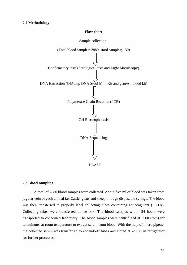
48
2.2 Methodology
Flow chart
Sample collection
(Total blood samples: 2880, stool samples; 130)
Confirmatory tests (Serological tests and Light Microscopy)
DNA Extraction (QIAamp DNA Stool Mini Kit and geneAll blood kit)
Polymerase Chain Reaction (PCR)
Gel Electrophoresis
DNA Sequencing
BLAST
2.3 Blood sampling
A total of 2880 blood samples were collected. About five ml of blood was taken from
jugular vein of each animal i.e. Cattle, goats and sheep through disposable syringe. The blood
was then transferred to properly label collecting tubes containing anticoagulant (EDTA).
Collecting tubes were transferred to ice box. The blood samples within 24 hours were
transported to concerned laboratory. The blood samples were centrifuged at 3500 (rpm) for
ten minutes at room temperature to extract serum from blood. With the help of micro pipette,
the collected serum was transferred to eppendorff tubes and stored at -20 °C in refrigerator
for further processes.

49
2.4 Serological Examination
2.4.1 Sample preparation
Fresh serum was used, obtained from centrifugation of blood. The haemolysed or
contaminated sera were discarded.
2.4.2 Techniques
According to the company (Toxocell Latex- Spain) standard procedure the test
procedure was performed at room temperature. The samples were diluted insaline NaCl 0.9
% solution. 50ul or one drop of diluted sera was taken with the help of micropippete and
mixed with one drop (25ul) of chemical placed on special black-ground slide using pipe
stirrers supplied with kitthen rotating the slide slowly for 5 minutes and observe any visible
agglutination.The result was classified into positive or negative on a slide.
2.4.3 Interpretation of results
A clear positive reaction shows the presence of toxoplasma antibodies while a
negative reaction was the indication of the absence of T. gondii antibodies.
2.5 Collection of fecal samples from stray cats
A total of 130 fecal samples were collected from stray cats from different sampling
sites of Peshawar valley.
2.5.1 Materials needed and sample collection
Plastic pots for faecal sample collection coded with a blue coloured lid, disposable
plastic spoon, Plastic gloves, and plastic zip-lock bag. Each container was properly labelled
with locality, cat's gender and age of the cat. Faecal samples were collected and treated with
great care because it may contain several infectious microorganisms. For personal hygiene
disposable plastic gloves were used. First of all, it was confirmed that fecal sample was not
too old. Then a specific amount of faecal sample (full table spoon) was taken with the help of
disposable plastic spoon and 90% ethanol was added to faecal sample to preserve it for
further analysis for DNA extraction and then put it in the plastic pot and then the faecal pot
was placed inside the zip-lock plastic bag and sealed.
2.5.2 Light Microscopy of cat faecal samples
The cat faecal samples which were collected from various stray parts of sampling
region were observed under light microscope by using direct smear and Sheather’s sugar
floatation technique.

50
2.5.2 Direct smear formation
The faecal samples were put in centrifuge tubes and a drop of 0.9% sodium chloride
was added. To homogenize the sample, it was centrifuged. Then a small amount of the
sample was placed on a clean slide and covered with a cover slip of 18-mm X 18mm size.
Now it was examined under light microscope (Olympia electrical light microscope from
Japan) of magnification 400X for the detection of oocysts of T. gondii. The size of the oocyst
of T. gondii under 400X magnification was 10µ-12µ.
2.5.3 Sheather’s Sugar Floatation Technique
By using this technique approximately 5 g of faeces was taken and mixed with 45 ml
of sugar solution and then centrifugedfor 10 minutes at 1000×g (Sheather, 1923). Now the
suspension was taken and put on a slide and was examined under microscope (Olympia
electrical light microscope from Japan) of magnification400X for the presence of unstained
oocysts of T. gondii having a size of 10-12µm.
2.6 DNA extraction from blood
Product: GeneAll ExgeneTM Blood SV, 250 preps), Cat No: 105-152. Lot No:
10516811007mini. 2015 GeneAll Biotechnology, co, itcl.138-859, Sangpa-gu, Seoul,
SOUTH KOREA. www.geneall.com
1. 20 ul of proteinase K solution (20 mg/ml) was put into the bottom of a 1.5 µl tube
through pipet. The protease K amount may be increased if the sample volume is larger
than 200 µl.
2. Then 200 ul of sample was transferred to the tube.
3. After that 200 µl of Buffer BL was added to the tube. For thorough mixing the tube
was vortexed. It was then incubated at 56 °C for 10 minutes. For the removal of any
drop from inside of the lid, it was spun down briefly.
4. Then 200 µl of absolute ethanol was added to the sample and was again vortexed for
thorough mixing of the sample. For the removal of any drops from inside of the lid it
was spun down.
5. The mixture was carefully transferred to the SV column and was centrifuged for 1
minute at 6,000 xg above (> 8,000 rpm). After centrifugation the collection tubes
were replaced by new one (already provided).
6. Buffer BW of 600 ul was then added and was then centrifuged for 1 minute at 6,000
xg above (> 8, 000 rpm) and then the collection tubes were replaced with new one
(already provided).

51
7. Buffer TW of 700 µl was then applied and was again centrifuged for 1 minute at
6,000 xg above (> 8,000 rpm) the pass-through was discarded and the SV column was
back inserted into the collection tube.
8. For the removal of residual wash buffer, it was centrifuged at full speed for 1 minute.
After centrifugation the SV column was placed in a fresh 1.5 ml tube. Great care was
taken at this step for the elimination of carryover of buffer TW. It was again
centrifuged for one minute at full speed for the removal of any carryover of buffer
TW. Centrifugation was carried out at room temperature.
9. About 200 µl of buffer AE or sterilized water was then added. It was then incubated
for one minute at room temperature and was centrifuged at full speed for one minute.
The elution step was repeated with fresh 200 µl elution buffer for the increase DNA
yield upto significant level.
2.7 Isolation of DNA from Stool
2.7.1 Procedure
1. 180–220 mg stool was weighed in a 2 ml micro-centrifuge tube.
2. In second step 1.4 ml Buffer ASL was added to each stool sample. After adding
Buffer ASL, the samples were Vortexed for 1 min until the sample were homogenized
thoroughly so that maximum DNA can be collected in the final eluate
3. The suspension was placed in a water bath to heat for 5 min at 70°C, so that to kill
parasites and bacteria and also to maximize the total yield of DNA 3-5 times.
4. After heating it was again vortexed for 15 seconds. When samples were vortexed, it
was then centrifuged at full speed for 1 min to pellet stool particles.
5. From the supernatant 1.2 ml was pipetted into a new 2 ml microcentrifuge tube and
the pellet was discarded. The 2 ml tubes were wide enough for accommodation of an
InhibitEX tablet.
6. Each sample was loaded with 1 InhibitEX Tablet. For the complete suspension of the
tablet it was vortexed immediately and continuously for 1 min. Suspension was
incubated for 1 min at room temperature for the adsorption of inhibitors to the
InhibitEX matrix.
7. After incubation the sample was centrifuged at full speed for 3 min to pellet
inhibitors bound to InhibitEX matrix.
8. New 1.5 ml microcentrifuge tube was taken and all the supernatant was pippeted into
that new tube and the pellet was discarded. After that the sample was centrifuged at

52
full speed for 3 min. There was no effect on procedure by the transfer of small amount
of pelleted material from step 7.
9. A new 1.5 ml microcentrifuge tube was taken and 15 µl proteinase K was pippeted
into that new 1.5 ml tube.
10. A total of 200 µl supernatant from step 8 was pippeted into the 1.5 ml microcentrifuge
tube in which already proteinase K was present
11. Then 200 µl Buffer AL was added and was vortexed for 15 s.
After being vortexed it was Incubated at 70°C for 10 min. it was again centrifuged and
drops from the inside of the tube lid were removed. This centrifugational step was
optional.
12. A 200 µl of ethanol (96–100%) was added to the lysate, and was mixed by vortexing.
It was then centrifuged briefly for the removal of drops.
13. 13.The lid of a new QIAamp spin column was labelled and was placed in a 2 ml
collection tube. Complete lysate was carefully applied from step 13 to the QIAamp
spin column without moistening the rim. The cap was closed and was centrifuged at
full speed for 1 min. The QIAamp spin column was placed in a new 2 ml collection
tube, and the tube having the filtrate was discarded. Each spin column was closed so
that to not allow aerosol formation during centrifugation.
14. The QIAamp spin column were carefully opend and 500 µl Buffer AW1 was added to
the spin column. The cap was closed and was centrifuged at full speed for 1 min. The
QIAamp spin column was placed in a new 2 ml collection tube. The collection tube
was discarded which had filtrate.
15. 15.The QIAamp spin column were carefully opened and 500 µl Buffer AW2 was
added. The cap was closed and was centrifuged at full speed for 3 min. the collection
tube containing the filtrate was discarded.
16. 16.The QIAamp spin column was placed in a new 2 ml collection tube whereas the
old collection tube having the filtrate was discarded. It was then centrifuged at full
speed for 1 min to eliminate the chance of possible Buffer AW2 carryover.
17. A new 1.5 ml microcentrifuge tube was labelled and the QIAamp spin column was
transferred into these new tubes. The QIAamp spin column were carefully opened and
200 µl Buffer AE was pipetted directly onto the QIAamp membrane. The cap was
closed and was incubated for 1 min at room temperature, then it was centrifuged at
full speed for 1 min to elute DNA. A sample of the eluted DNA was verified on 1%
gel.

53
2.8 Amplification of DNA samples
The PCR tubes for samples and controls were properly labeled. In case of
quantification experiments, tubes were also labeled for standards. The PCR reagents were
thawed and PCR reaction mixture was prepared. A generalized recipe of the PCR is given in
the following table. The volume of total reaction mixture required for the whole batch
including samples, controls and standards were calculated.
Aliquot the reaction mixture in the individual PCR reaction tubes/wells. Add the
template i.e; sample/control/ standards in the appropriate labeled tube. The volume of
template varies according to the protocol in use. A 2 µl DNA templates were added to each
tube so that 20 µl total reaction volume was produced. The Real-Time PCR machine’s
software was opened and run parameters were edited e.g, sample IDs, sample volume,
detection wavelengths and cycling conditions according to desired protocol. A generalized
cycling protocol for TaqMan assay is given below.
Table:2.1 Preparation of Real-Time PCR Reaction Mix for DNA template
Reagent Volume per Reaction
DNA template 2 µl
2x PCR Master Mix 10 µl
Forward Primer (100 µM stock) 2 µl
Reverse Primer (100 µM stock) 2 µl
DEPC H2O 4 µl
Total 20 µl

54
Table: 2.2. Reaction conditions for DNA templates
Step Temperature Duration No. of cycles
Initial Denaturation 95 degree C 5 min 1
Denaturation 95 degree C 30 sec 35
Annealing 52 degree C 30 sec 35
Extension 72 degree C 30 sec 35
Final extension 72 degree C 10 min 1
Table: 2.3 Detail of Primer
No Primer Sequence
P1
P2
TGExF
TGExtR
GAACTGCATCCGTTCATGAGTAT
GTTCCCTCCTCTTCGCGAAAC
The sample tubes/strips/plates were placed in the thermal cycler, lid was closed, and
program was run. After completion of PCR, open the analysis module of Real-Time PCR
machine’s software and analyzed the run file. The base line and outliers may be adjusted
before analysis.
2.9 Agarose gel
The PCR product was run on 2% agarose gel. A band of about 420 bp wasobserved.
2.10 Sequencing
The PCR product was sequenced by Sanger Method
2.11 Statistical analysis
The data collected was properly arranged in the form of tables and graphs and basic statistics
was applied. All the data was expressed in percentages and X2 and P values were calculated.
P value less than 0.05 was considered significant.

55
RESULTS
PART-A
PUBLISHED

56
Int. J. Biosci. 2019
International Journal of Biosciences | IJB |
ISSN: 2220-6655 (Print) 2222-5234 (Online)
http://www.innspub.net
Vol. 14, No. 2, p. 515-521, 2019
RESEARCH PAPER OPEN ACCESS
Seroprevalence of Toxoplasma gondii Infection in Domestic
Animals of District Charsadda, Khyber Pakhtunkhwa, Pakistan
Arab Hussain1,2,, Muhammad Zahid*1
1Department of Zoology, Islamia College University, Peshawar, KP, Pakistan 2Department of Zoology, Government post Graduate College Charsadda, KP, Pakistan
Key words: Toxoplasmosis, District Charsadda, Latex agglutination test, Domestic animals
http://dx.doi.org/10.12692/ijb/14.2.515-521 Article published on February 28, 2019 Abstract This study was conducted in to find out the seroprevalence of toxoplasmosis in domestic animals of District
Charsadda. Toxoplasmosis is an infection caused by a unicellular parasite Toxoplasma gondii which is
cosmopolitan in distribution among the animals including domestic animals and human beings. Very little
information is available about the infection rate of toxoplasmosis in domestic animals of District Charsadda,
Pakistan. A total of 374 blood samples were collected from cows, buffaloes, sheep and goats and were tested by
Latex agglutination test, out of which (79.7%) were found seropositive. Out of 260 females, (82.69%) were found
infected while in 114 males (78.80%) were found infective. A high prevalence of (81.65%) was detected in age
group above one year. Individual sex wise prevalence also revealed that females were more positive as compared
to males. In male buffaloes, (76.92%) were positive while in females, (75.67%) were detected positive. In male
goats (72.41%) were positive while in case of females, (84.78%) were found infective. The infection rate was
higher in female sheep (91.42%) as compared to male sheep (84.78%). In male cows (69.23%) were detected
seropositive while in females cows the infection rate was (73.77%). This increased rate of infection may be due to
free access of cat to drinking water and other food sources as well as warm and humid conditions of the area.
* Corresponding Author: Muhammad Zahid
515 Hussain and Zahid

57
Int. J. Biosci. 2019
Introduction Toxoplasma gondii which is an intracellular
parasitedistributed worldwide (Shah et al., 2013a)
and is responsible for toxoplasmosis disease (Aldebret
et al., 2011). The discovery of cats as definitive host
for T.gondii was done in 1960 (Innes, 2010). Life
cycle of T. gondii is completed in two stages, sexual
stage startsin definitive host, which include cats and
asexual stage is completed in warm blooded animals
(Afonso, 2008). Intermediate hosts are infected by
ingesting oocysts, which change into tachyzoites
through asexual reproduction and then change into
bradyzoites. Once again the felines are infected by
eating bradyzoites-infected meat (Webster, 2007).
About (33-60%) of the human worldwide population
have been infected by this parasite (Holliman, 1997;
Shah et al., 2014), but this rate changes according to
the geographical, climatic and nutritional factors,
socio- cultural habits and routes of transmission
(Shah et al., 2013b). Prevalence rate of T. gondii
varies in different countries.
The infection rate of (28.44%) was recorded in
Mohmand Agency, Pakistan (shah et al., 2014). The
seroprevelence of T. gondii was 63% in Punjab, 48%
in Azad Kashmir and 38% in Khyber Pukhtunkhwa
(Tenter et al., 2000). Prevalence rate of (17.4%) was
found in young school children in Islamabad,
Pakistan (Sadaruddin et al., 1991). The prevalence
rate in Dera Ghazi Khan, Pakistan was detected
(29.5%) (Tasawar et al., 2011), Infection rate has
declined recently due to awareness in people by not
using under cooked meat and advancement in animal
husbandry work (Shah et al., 2017). The
seroprevalence of T. gondii in domestic animals and
meat producing animals, such as goats, and sheep,
has found the same infection all the time. In District
Mardan (Pakistan) the prevalence rate of
toxoplasmosis in goats was recorded 42.28% whereas
in sheep the infection rate was 44.13% (Shah et al.,
2013b). In Mohmand Agency, Pakistan, the
prevalence rate of toxoplasmosis in goats was
recorded 53.84% while in sheep it was 36.00% (Shah
et al., 2013a). In farmed sheep, the prevalence
inEurope is related with age, increasing from lambs
(17-
22%) to adult (65 -89%) (Halos, 2010). Viable
T.gondii have been detected in about 67% of
sheepsamples. In Southern European countries
infected meat of Sheep is the main source of infection.
Seropositivity for goats varies from 4 to 77% (Dubey,
2011). The seroprevalence of this infection in sheep in
Newzeland was reported to be 30-90% and in UK 77%
prevalence was reported in goats while 29% was
reported in sheep (De Bhur, 2008). In Mohmand Agency, Pakistan, the seroprevalence of
toxoplasmosis reported in cows was 20% whereas
16% was recorded in buffaloes (Shah et al., 2013a).
T. gondii Infections show mild symptoms or
mostlywithout symptoms (Sarkar et al., 2012).
Immunocompetent individuals in acute infection are
generally asymptomatic or show some common
specific symptoms (Selseleh, 2012), often give flu-like
symptoms in the early acute stage (Menotti et al.,
2003). While in immunos up pressed patients
T.gondii causes encephalitis and leading illness
(jonesand Roberts, 2013) and infect brain, heart
which results in myocarditis, and infection of liver,
pancreas, bone marrow, bladder and lymph nodes,
kidneys, spleen, and skin (Arnold et al., 1997).
The placental barrier is very strong against the parasite
during the first three months of pregnancy, but
transmission rate increases as pregnancy proceeds which
results in abortions (Dunn, 1999). Congenital
toxoplasmosis also causes deafness, mental retardation,
micro cephalous, eye lesions, cataract, retinal necrosis
(Delair, 2011). Abortion or still birth are common during
first three months of pregnancy while Infection acquired
in later pregnancy the newborn is usually without
symptoms but may cause eye problems (shah et al.,
2014) Congenital toxoplasmosis leads to CNS damage,
blindness epilepsy and mental growth is retarded with
fetal death or spontaneous abortion in about 10% of
cases [23]. T. gondii causes abortions, stillbirth and
neonatal death in sheep and goats (Engeland et al.,
1998).
The distribution of T. gondii is related with weather
condition of an area and environment where the
oocysts survive (Dubey, 2004).
516 Hussain and Zahid

58
Int. J. Biosci. 2019
Prevalence of toxoplasmosis varies in different parts
of the world and this variation related to life style,
age, climatic conditions, nutritional habits and other
socio-cultural factors (shah et al., 2014). This study
was carried out in the study area with the aim to
determine the infection rate in domestic animals,
minimize the economic loss in domestic animals and
to aware the people about the adverse effects of
toxoplasmosis.
Materials and methods A total of 374 blood samples were collected from
domestic animals (cows, goats, sheep and buffaloes)
from District Charsadda, Pakistan. About five ml of
blood was collected from each animal. The samples
were centrifuged for extraction of serum.
Serological examination The latex regent is a suspension of polystyrene
particle which is sensitized with the antigens of
T.gondii. The distant agglutination pattern
whenobserved after mixing the serum reflects
formation of antigen-antibody complexes. When the
organism has no infection, then no agglutination is
observed. A greater than 4 IU/ml value was
considered as standard for positive result.
Test procedure According to the manufacturer standard protocol
(Toxocell Latex- Spain) the test procedure was
performed. The samples were diluted in NaCl 0.9%
saline solution. One drop or 50ul of diluted sera was
mixed with one drop (25ul) of chemical and mixed
well by sterile wood stick, then the slide was slowly
rotated for 5 minutes and visible agglutination was
observed. The result was classified into positive or
negative on a slide, using pip stirrers provided. A
negative reaction was the indication of the absence of
toxoplasma antibodies. A clear positive reaction
showed the presence of toxoplasma antibodies equal
or greater than 4 IU/ml which showed either an
evolving infection or a past infection.
Statistical analysis For simplification all the results were expressed in
percentages. The values between different sex and age
groups were recorded and relevantly expressed in
percentages. Microsoft Excel (version-10) was utilized
by windows-08, (Release 16.0 standard version).
Results A total of 374 blood samples were collected from
domestic animals of three Tehsils (Shabqadar, Tangi
and Charsadda) of District Charsadda and were tested
for the presence of T. gondii infection by using Latex
Agglutination Test. Out of these 374 blood samples,
298 (79.7%) were found positive for toxoplasmosis. A
total of 100 blood samples were collected from cows
in which 72 (72%) were found positive and 28 (28%)
were negative, out of 121 goats, 99 (81.8%) were
positive and 22 (18.2%) were negative. Similarly, 103
samples were collected from Sheep, out of which 89
(86.4%) were positive and 14(13.6%) were found
negative whereas out of 50 samples from buffalo,
38(76%) were found positive and 12(24%) were
detected negative (Table 1).
Table 1. Comparative seroprevalence ofT.
gondiiinfection in domestic animals.
Animals Samples Positive N Negative (N) (%) N (%) Cows 100 72 (72) 28 (28) Goats 121 99 (81.8) 22 (18.2) Sheep 103 89 (86.4) 14 (13.6) Buffaloes 50 38 (76) 12 (24)
Total 374 298 76
A total of 260 females and 114 males were tested for
T.gondii infection. Out of which 215 (82.69%) females
83(78.80%) males were found positive. This also
indicated that prevalence among the females was high
among these animals. Prevalence of toxoplasmosis was
also detected in different sex groups. The highest
prevalence of toxoplasmosis was found in females as
compared to males. Out of 13 male buffaloes, 10
(76.92%) were positive while out of 37 females, 28
(75.67%) were found positive. In goats out of 29 males,
21 (72.41%) were found infective whereas out of 92
females, 78 (84.78%) were positive. Out of 33 male sheep
25 (84.78%) were found seropositive and out of 70
female sheep (91.42%) were found infective. Out of 39
male cows, 27 (69.23%) were detected infective whereas
out of 61 females, 45 (73.77%) were found seropositive
(Table 2).
517 Hussain and Zahid

59
Int. J. Biosci. 2019
Table 2. Sex wise seroprevalence of toxoplasmosis among goats, sheep, cows and buffaloes.
Animals Samples Male Positive N (%) Female Positive N (%)
Buffaloes 50 13 10 (76.92) 37 28 (75.67)
Goats 121 29 21 (72.41) 92 78 (84.78)
Sheep 103 33 25 (84.78) 70 64 (91.42)
Cows 100 39 27 (69.23) 61 45 (73.77)
Total 374 114 87 260 215
Seroprevalence of toxoplasmosis was also detected in
different age groups. A total of 178 (81.7%) of age above
one year and 120 (76.9%) under one year were detected
seropositive. Seroprevalence of toxoplasmosis was higher
in age group above one year (Table 3).
Table 3. Age wise seroprevalence of
toxoplasmosisamong domestic animals.
Age Sample (N) Positive (%) Negative (%) > One year 218 178 (81.7) 40 (18.3) <One year 156 120 (76.9) 36 (23.1)
Total 374 298 76
Discussion Toxoplasmosis is a zoonotic disease arising from close
contact of human with felids (kravetz and federman,
2002). Domestic cats play a vital role in the spread of
toxoplasmosis because they are the definitive hosts
and play an important role in transmission of
T.gondii infection by shedding oocysts in their
faeces(Dubey, 1994).
It varies between, herds, countries and regions,
methods of diagnoses and even at different times in
the same herd. In our study, infection rate for
T.gondii in cattle, goats and sheep was extremely
high(79.7%) than 32.29% in domestic animals in
Mohmand Agency in Pakistan ((shah et al., 2013a)),
9.2% reported from Guangxi and 46.4% from
Xinjiang, China (Lv and Cui, 1994).
In present study 81.81% prevalence rate of T. gondii
was found in goats, which is higher than 51% in goat’s
population recorded in Saudi Arabia (Sanad and Al-
ghabban, 2007), in Brazil 28.9% (Bisson et al., 2000),
Thiland 27.9% (Jittapalapong et al., 2005), 25.4% in
Pakistan (Ramzan et al., 2009). Toxoplasmosis was
also detected in sheep population of Charsadda. The
present study, revealed that in sheep toxoplasmosis is
very high (86.40%) than reported from Brazil 46.2%
(Silva et al., 2013) and
Greece 48.6% (tzanidakis et al., 2012), 31% reported
in Turkey (oncel and Vural, 2006) and Northeastern
China 4.4% (Yang et al., 2013), 44.13% from Mardan.
Toxoplasma infection in cattle shows a high degree of
variation across the globe, ranging virtually from 0%
to 99% (Ivana et al., 2006). Seropositivity rate
recorded for T. gondii in the present study in cows is
extremely high (66%) as compared to 9% in Indonesia
(Matsuo and Husin, 1996), while seroprevalence for
T. gondii in Mohmand Agency, Pakistan was
found20% in cows and 16% in buffaloes (shah et al.,
2013a), which is lower than reported in the present
study but seroprevalence in present study (66%) is
lower than (76.3%) in cattle in Serbia (Ivana et al.,
2006).
The differences in prevalence reported by all these
studies could be accounted on host, age, breed, sex,
environmental conditions, farm size and number of
cats and management practices (Van Der et al.,
2000). During the present study, a high prevalence of
82.7% was reported in females as compared to males
(78%), indicating high prevalence among the females
as compared to 35.8% in female and 21.1% in male
sheep and goats (Ramzan et al., 2009; Van Der et al.,
2000). Different studies indicated that females are
more easily infected by protozoan parasites as
compared to males (Alexander and Stinson, 1998).
In female immunity can be broken due to various
factors e.g., nutrition, age, pregnancy and
environmental factors. Toxoplasmosis and age
relationship in the present study revealed that
seroprevalence is high in age group above one year
(81.65%) as compared to age group less than one year
(76.92%) which are in support with 77.7% in age
group of 60 to 75 months and lowest prevalence
39.3% in age group of 12 to 27 months (Jittapalapong
et al., 2005).
518 Hussain and Zahid

60
Int. J. Biosci.
Older goats were more seropositive as compared to
younger ones under one year old (Olivier et al., 2007).
The present work was an attempt to find out the
prevalence rate of toxoplasmosis in the study area.
The high prevalence rate of toxoplasmosis in the
study area may be due to warm and humid climatic
conditions of this area. The increased infection rate in
study area may also be due to unhygienic conditions
because T. gondii prevalence ranged from 0-100% in
different areas of the world and this variation is due
to the life styles of the inhabitant’s customs,
traditions, weather conditions, age of the animals and
husbandry practices (Olivier et al., 2007).
Acknowledgements We are thankful to the people who allowed us to
their animals for blood collection.
References Afonso E. 2008. Spatial distribution of
soilcontamination by T. gondii in relation tocat
defaecation behaviour in an urban area. International
Journal of Parasitology 38, 1017-1023. https://doi.
org/10.1016/j.ijpara. 2008.01.004.
Aldebret D, Hypolite M, Cavaillaes P, Touque
B, Flori P, Loeuillet C, Cesbron-Delauw MF. 2011. Development of High-Throughput methods to quantify cysts of T. gondii. Cytometry Part A 79(11), 952-958. https://doi.org/10.1002/cyto.a.21138.
Alexander J, Stinson WH. 1988. Sex hormones and the course of parasitic infection. Parasitology Today 4, 189-193. https://doi.org/10.1016/0169-4758(88)9
Arnold SJ, Kinney MC, Mccormick MS,
Dummer S, Scott MA. 1997.
Disseminatedtoxoplasmosis. Unusual presentations
in the immunocompromised host. Archives of
Pathology and Laboratory Medicine 12(8), 869-873.
Bisson A, Maley S, Rubaire-Akiiki CM,
Watling JM. 2000. The seroprevalence ofantibodies
to T. gondii in domestic goats in Uganda. Acta Tropica 76, 33-38.
2019
De Bhur K. 2008. T. gondii seroprevalence–
currentresults in German swine herds. Archive Für
Lebensmittelhygiene 59, 5-8.
Delair E. 2011. Clinical manifestations of ocular toxoplasmosis. Ocular Immunology and
Inflammation 19, 91-102.
Dubey JP. 1994. Toxoplasmosis. J.
AmericanVeterinary Medical Association 205, 1593-
1598.
Dubey JP. 2004. Toxoplasmosis–a
waterbornezoonosis. Veterinary Parasitology, 126,
57-72. https://doi.org/10.1016/j.vetpar.2004.09.005.
Dubey JP. 2011. High prevalence and genotypes
ofToxoplasma gondii isolated from goats, from a
retail meat store, destined for human consumption in
the USA. Internatinal Journal for Parasitology 41,
827-833. https ://doi.org/10.1016/j.
ijpara.2011.03.006.
Dunn D. 1999. Mother-to-child transmission of toxoplasmosis risk estimates for clinical counselling. Lancet 353, 1829-1833. https://doi.org/10.1016/S01.
Engeland IV, Waldeland H, Andresen O,
Løken T, Björkman C, Bjerkås I. 1998. Foetalloss
in dairy goats: an epidemiological study in 22 herds.
Small Ruminant Research 30, 37-48.
https://doi.org/10.1016/S0921-4488(98)00084-4.
Halos L. 2010. An innovative survey underlining
thesignificant level of contamination by T. gondii of
ovine meat consumed in France. International
Journal for Parasitology 40, 193-200. https://doi.
org/10.1016/j.ijpara.2009.06.009.
Holliman RE. 1997. Toxoplasmosis, behaviour
andpersonality. Journal of Infection 35, 105-110.
https://doi.org/10.1016/S0163-4453(97)91380-3.
Innes EA. 2010. A brief history and overview
ofT.gondii. Zoonoses and Public health 57, 1-7.
https://doi.org/10.1111/j.1863-2378.2009.01276.x
519 Hussain and Zahid

61
Int. J. Biosci. 2019
Ivana L, Olgica DD, Sofija KK, Aleksandra N. 2006. Cross sectional survey of T. gondii infection in
cattle, sheep and pigs in Serbia: seroprevalence and
risk factors. Veternary Parasitology 135, 121-131.
Jittapalapong S, Sangvaranond A,
Pinyopanuwat N, Chimnoi W, Khachaeram W, Koizumi S, Maruyama S. 2005. Seroprevalence of
T.gondii infection in domestic goats in Satun Province,
Thailand. Veternary Parasitology 127, 17-22.
Jones JL, Roberts JM. 2013.
Toxoplasmosishospitalizations in the United States.
2008, and Trends, 1993-2008. Clinical Infectious
Disease 54(7), e58-e61.
Khan SN, Khan S, Ayaz S, Jan AH, Jehangir S,
Attaullah S, Ali J, Shams S. 2011. Seroprevalance and risk factors of toxoplasmosis
among pregnant women in district Kohat, Khyber
Pakhtunkhwa Pakistan. World Applied Science
Journal 14(7),1032-1036.
Kravetz JD, Federman DG. 2002. Cat-
associatedzoonoses. Archives of international
Medicine 162, 1945-1952.
DOI: 10.1056/NEJMcpc059027.
Lilenbaum W. 2013.
Immunohistochemicalidentification of T. gondii in
tissues from Modified Agglutination Test positivesheep. Veterinary
Parasitology 191, 347-52.
Lv YC, Cui JZ. 1994. Survey of T. gondii infection
inpigs and cattle in Guangxi Province, China. Journal
of Animal Science and Veterinary Medicine 3, 26.
Matsuo K, Husin DA. 1996. Survey of T.
gondiiantibodies in goats and cattle in Lampung
Province, Indonesia.Southeast Asian. Journal of
Tropical Medicine and Public Health 27, 554-5.
Menotti, Gustavo Vilela, Stephane Romand,
Yves Jean-Francois Garin, Lionel Ades, Eliane
Gluckman, Francis Derouin, Patricia Ribaud. 2003. Comparison of PCR-Enzyme-linked
immunosorbent Assay and Real-Time PCR Assay for
diagnosis of an unusual case of cerebral
Toxoplasmosis in stem cell transplant recipient.
Journal of Clinical Microbiology 41, 5313-5316.
Olivier A, Herbert B, Sava B, Pierre C, John
DC, Aline DK. 2007. Surveillance and monitoring
ofToxoplasma in humans, food and animals: a
scientific opinion of the panel on biological hazards.
The European Food Safety Association Journal 583,
1-64.
Oncel, Vural T. 2006. Occurance of T.
gondiiantibodies in sheep in Istanbul, Turkey.
Veteninarski Arthiv 76, 547-557.
Ramzan M, Akhtar M, Muhammad F, Hussain I,
Hiszczyńska- Sawicka E, Haq AU, Mahmood MS,
Hafeez MA. 2009. Seroprevalence of T. gondii insheep
and goats in Rahim Yar Khan (Punjab), Pakistan.
Tropical.Animal Health and Production 41, 1225-9.
Sadaruddin A, Agha F, Anwar F, Ghafoor A. 1991. Seroepidemiology of T. gondii infection in
young school children in Islamabad. Journal of
Pakistan Medical Association 41, 131-134.
Sanad MM, Al-ghabban AJ. 2007.
Serologicalsurvey on toxoplasmosis among
slaughtered sheep and goats in Tabouk, Saudi Arabia.
Journal of Egyptian Society of Parasitology 37, 329-
340.
Sarkar MD, Anuradha B, Sharma N, Roy RN. 2012. Seropositivity of toxoplasmosis in antenatal
women with bad obstetric history in a tertiary-care
hospital of Andhra Pradesh, Indian Journal of Health
Population and Nutrition 30, 87-92.
Selseleh M, Modarressi MH, Ali MM, Shojaee S,
Eshragian MR, Selseleh M, Aziz E, Keshavarz H. 2012. Real-Time RT-PCR on SAG1 and BAG1 gene
expression during stage conversion in
immunosuppressed mice infected with T. gondii, Tehran.
Strain. Korean. Journal of Parasitology 3, 199-205.
https://dx.doi.org/10.3347%2Fkjp.2012.50.3.199.
Shah M, Zahid M, Asmat P, Sthanadar AA. 2013b. Seroprevalence of T. gondii in goats and sheep of district Mardan, Pakistan. International Journal of Bioscience 3, 90-97. http://dx.doi.org/10.12692/ ijb/3.7.90-97.
520 Hussain and Zahid

62
Int. J. Biosci. 2019
Shah M, Zahid M, Bibi B, Hussain A, Haroon
M, Ali B. 2017. Chromatographic
immunoassaybased detection of of human
toxoplasmosis in District Mardan, Khyber
Pakhtunkhawa, Pakistan. Pure and Applied Biology
6,1297-1305.
Shah M, Zahid M, Sthanadar AA, Ali PA.
2014.Seroprevalence of Toxoplasma gondii Infection
in Human Population of Mohmand Agency Khyber
Pakhtunkhwa, Pakistan. Pakistan Journal of Zoology
44, 1169-1172.
Shah M, Zahid M, Sthanadar AA, Pir A,
Kausar A, Jan AH. 2013a. Seroprevalence of
T.gondii infection in domestic animals of Mohmand
agency, Pakistan. Journal of Coastal Life Medicine 1,
70-73. DOI: 10.12980/JCLM.1.2013C254.
Silva AF, Oliveira FC, Leite JS, Mello MF,
Brandão FZ, Leite RI, Frazao-Teixeira E,
Tasawar Z, Nawaz S, Lashari MH, Aziz F,
Hayat CS. 2011. Seroprevalence of human Toxoplasmosis in Dera Ghazi Khan, Punjab. Gomal
Journal of Medical Sciences 9, 82-85.
Tenter AM, Heckeroth AR, Weiss LM. 2000.
T.gondii: from animals to humans. International
Journal of Parasitology 30, 1217-1258.
https://doi.org/10.1016/S0020-7519(00)00124-7.
521 Hussain and Zahid
Teshale S, Dumètre A, Dardé ML, Merga B,
Dorchies P. 2007. Serological survey of
caprinetoxoplasmosis in Ethiopia: prevalence and
risk factors. Parasite 14, 155-159.
https://doi.org/10.1051/ parasite/2007142155.
Tzanidakis N, Maksimov P, Conraths FJ,
Kiossis E, Brozos C, Sotiraki S, Schares G. 2012. T. gondii in sheep and goats: seroprevalence
and potential risk factors under dairy husbandry
practices. Veterinary Parasitology 190, 340-348.
https://doi.org/10.1016/j.vetpar.2012.07.020.
Van Der Puije Wna, Bosompem KM, Canacoo
EA, Wastling, Zakanmoribd JM. 2000.
Theprevalence of anti-T. gondii antibodies in
Ghanaiansheep and goats. Acta Tropica 76, 21-26.
https://doi.org/10.1016/S0001-706X(00)00084-X.
Webster JP. 2007. The effect of T. gondii on
animalbehavior: Playing cat and mouse.
Schizophrenia bulletin 33(3), 752-756.
Yang N, Li H, He J, Mu M, Yang S.
2013.Seroprevalence of T. gondii infection in
domestic sheep in Liaoning Province, northeastern
China. Journal of Parasitology 99, 174-5.

63
RESULTS
PART-B
UNPUBLISHED

64
CHAPTER 3
RESULTS
3.1 Overall seroprevalence of Toxoplasmosis in Livestock of Peshawar Valley.
Antibodies against T. gondii were detected in all 2880 blood samples collected from
livestock in Peshawar valley. Out of these 43.6% (1255) animals were found seropositive and
56.6% (1625) were negative for T. gondii antibodies.
Table 3.1 Overall seroprevalence of Toxoplasma gondii in Livestock of Peshawar Valley.
Samples Positive N (%) Negative N (%)
2880 1255(43.6) 1625(56.4)
Fig: 3.1 Overall seroprevalence of T. gondii in Livestock of Peshawar Valley.
2880
1255
1625
0
500
1000
1500
2000
2500
3000
3500
samples Positive Negative
Series1

65
3.2 Overall District wise seroprevalence of Toxoplasmosis in livestock of
Peshawar Valley.
Samples were collected from livestock from different districts of Peshawar valley. A total
of 734 blood samples were collected from district Charsadda, out of which 344 were found
positive and 390 were negative for toxoplasmosis. From Peshawar 615 blood samples were
collected in which 266 were positive and 349 were negative while in district Mardan out 511
samples 236 were positive and 275 were seronegative. From district Nowshera 500 blood
samples were collected from livestock out of which 236 samples were positive and 275 were
negative while from district Swabi a total of 520 blood samples 221 were positive and 299 were
negative
Table 3.2 Overall District wise seroprevalence of T. gondii in Livestock of Peshawar Valley.
Tehsil Samples Positive N (%) Negative N (%)
Charsadda 734 344(46.9)
390(53.1)
Peshawar 615 266(43.3)
349(56.7)
Mardan 511 236(46.2)
275(53.8)
Nowshera 500 188(37.6)
312(62.4)
Swabi 520 221(42.5)
299(57.5)
Total 2880 1255(43.6)
1625(56.4)
P = 0.00

66
Fig. 3.2 Overall district wise seroprevalence of T. gondii in Livestock of Peshawar Valley.
3.3 Percentage distribution of sample animals.
A total of 2880 blood samples were collected from different animals which includes, 717
(24.9%) cows, 831 (28.9%) goats, 793 (27.5%) sheep and 539 (18.7%) buffalos (Table 3.2).
Table 3.3 Percentage distribution of tested animals
Animals Sample size(N) %age
Cow
717 24.9
Goat
831 28.9
Sheep
793 27.5
Buffaloes 539 18.7
Total 2880 100%
734
615
511 500 520
344
266236
188221
390349
275312 299
0
100
200
300
400
500
600
700
800
Charsadda Peshawar Mardan Nowshehra Sawabi
Samples Positive Negative

67
Fig. 3.3 Percentage distribution of tested animals
3.4 Comparative seroprevalence of T. gondii in Livestock of Peshawar Valley.
A total of 717blood samples collected from cows in which 361(50.3%) were found
positive and 356 (49.7%) were negative, 831 from goats in which 384 (46.2%) were positive and
447(53.8%) were negative. Similarly, 793 samples were collected from Sheep out of which 389
(49.1%) were positive and 404 (50.9%) were negative and 539 samples from buffalo in which
121 (22.4%) positive and 418(56.4%) were negative (Table 3.4).
Table. 3.4 Comparative seroprevalence of T. gondii in Livestock of Peshawar Valley.
Animals Samples (N) Positive (%) Negative (%)
Cow 717 361(50.3) 356(49.7)
Goat 831 384(46.2) 447(53.8)
Sheep 793 389(49.1) 404(50.9)
Buffaloes 539 121(22.4) 418(77.6)
Total 2880 1255(43.6) 1625(56.4)
P = 0.00
Cow25%
Goat29%
Sheep27%
Buffaloe19%

68
Fig. 3.4 Comparative seroprevalence of T. gondii in Livestock of Peshawar Valley
3.5 Seroprevalence of T. gondii in Livestock of Charsadda.
Seroprevalence of T. gondii in domestic animals of Tehsil Charsadda is high. Out of 734
animals 351(47.8) were found positive and 383(52.2%) were negative. Among cows out of 186
cows 104 were found positive while out of 210 goats 108(51.4%) were detected positive. Sheep
and buffaloes showed high prevalence rate 53.8% and 25.7% respectively.
Table. 3.5 Seroprevalence of T. gondii in livestock of district Charsadda
Animal Sample Size (N) Positive (%) Negative%
Cow 186 104(55.9) 82(44.1)
Goat 210 108(51.4) 102(48.6)
Sheep 186 100(53.8) 86(46.2)
Buffaloes 152 39(25.7) 113(74.3)
Total 734 351(47.8) 383(52.2)
361384 389
121
356
447
404418
0
50
100
150
200
250
300
350
400
450
500
Cow Goat Sheep Bufaloe
Positive Negative

69
Fig. 3.5 Seroprevalence of T. gondii in livestock of district Charsadda
3.6 Seroprevalence of T. gondii in Livestock of district Peshawar.
A total of 615 samples were collected from district Peshawar, out of these 285(46.3%)
were positive and 330(53.7%) were negative. Among cows out of 160 cows 100(62.5%) were
positive and 60(37.5%) were negative while out of 204 goats 78(38.2%) were positive and
126(61.8%) were negative. Among 139 sheep 84(60.4%) were found positive and 55(39.6%)
were negative while out of 112 buffaloes 23(20.5%) were positive and 89(79.5%) were negative
for T. gondii antibodies.
Table. 3.6 Seroprevalence of T. gondii in Livestock of district Peshawar.
Animal Sample Size (N) Positive (%) Negative (%)
Cow
160 100(62.5) 60(37.5)
Goat
204 78(38.2) 126(61.8)
Sheep
139 84(60.4) 55(39.6)
Buffaloes 112 23(20.5) 89(79.5)
Total 615 285(46.3) 330(53.7)
186
210
186
152
104 108100
39
82
10286
113
0
50
100
150
200
250
Cow Goat Sheep Buffaloes
Sample Size (N) Positive (%) Negative%

70
Fig. 3.6 Seroprevalence of T. gondii Livestock of district Peshawar.
3.7 Seroprevalence of T. gondii in Livestock of district Mardan.
From district Mardan 511 blood samples were collected from different domestic animals,
out of these 210(41.8%) were positive and 301(58.9%) were negative. Out of 132 cow samples
60(45.5%) were positive and 72(54.5%) were negative while out of 126 goats 66(52.4%) were
positive and 60(47.6%) samples were negative. The seroprevalence among 167 sheep was
detected as 64(38.3%) positive and 103(61.7%) negative while among 86 buffaloes 20(23.3%)
were positive and 66(76.7%) were negative.
Table 3.7 Seroprevalence of T. gondii in Livestock of district Mardan.
Animal Sample Size (N) Positive (%) Negative (%)
Cow 132 60(45.5) 72(54.5)
Goat 126 66(52.4) 60(47.6)
Sheep 167 64(38.3) 103(61.7)
Buffaloes 86 20(23.3) 66(76.7)
Total 511 210(41.1) 301(58.9)
100
7884
23
60
126
55
89
0
20
40
60
80
100
120
140
cow goat sheep buffaloe
positive negative

71
Fig. 3.7 Seroprevalence of T. gondii in Livestock of district Mardan.
3.8 Seroprevalence of T. gondii in domestic animals of district Nowshera
A total of 500 blood samples were collected from district Nowshera in which out of 120
cows 45 (37.5%) were positive and 75 (62.5%) were negative while in Goats out of 140 samples
59 (42.1%) were seropositive and 81 (57.9%) were negative. In sheep out of 145 samples 65
(44.8%) were infected and 80 (55.2%) were negative. Similarly, out of 95 buffaloes 19 (20%)
were seropositive and 76 (80%) were negative for toxoplasmosis.
Table. 3.8 Seroprevalence of T. gondii in Livestock of district Nowshera
Animal Sample Size (N) Positive (%) Negative (%)
Cow 120 45(37.5) 75(62.5)
Goat 140 59(42.1) 81(57.9)
Sheep 145 65(44.8) 80(55.2)
Buffaloes 95 19(20) 76(80)
Total 500 188(37.6) 312(62.4)
6066 64
20
72
60
103
66
0
20
40
60
80
100
120
cow goat sheep buffaloe
positive negative

72
Fig. 3.8 Seroprevalence of T. gondii in Livestock of District Nowshera
3.9 Seroprevalence of T. gondii in Livestock of District Swabi.
Samples were collected from district Sawabi and tested for toxoplasmosis. 119 blood
samples of cows were tested in which 52(43.7%) were positive and 67(56.3%) were negative, in
Goats 72(47.7%) samples out of 151 were seropositive and 79(52.3%) were negative while in
sheep and buffaloes out of 156 and 94 blood samples 77(49.4%) and 20(21.3%) were found
positive respectively.
Table. 3.9 Seroprevalence of T. gondii in Livestock of district Swabi.
Animal Sample Size (N) Positive (%) Negative (%)
Cow 119 52(43.7) 67(56.3)
Goat 151 72(47.7) 79(52.3)
Sheep 156 77(49.4) 79(50.4)
Buffaloes 94 20(21.3) 74(78.8)
Total 520 221(42.5) 299(57.5)
45
5965
19
7581 80
76
0
10
20
30
40
50
60
70
80
90
Cow Goat Sheep Buffaloe
Positive Negative

73
Fig 3.9 Seroprevalence of T. gondii in Livestock of district Swabi.
3.10 Overall sex-wise seroprevalence of T. gondii in Livestock of Peshawar Valley
Sex wise seroprevalence for T. gondii antibodies was also detected in Livestock of
Peshawar Valley. Out of 2880 blood samples 1058 males 375(35.4%) and in females 880(48.3%)
out of 1822 samples were found positive. Among 717 cows 261 males 106(44.6%) and out of
456 females 254(55.7%) were positive while among 831 goats, out of 282 males 99(35.1%) and
out of 549 females 287(52.3%) were found positive. The seroprevalence in 793 samples of sheep
was 39.2% out of 344 males and 56.3% out of 449 females while among 549 buffaloes out 171
males 35(20.4%) and 86(23.4%) out of 368 females were positive.
Table. 3.10 Overall sex-wise seroprevalence of T. gondii in Livestock of Peshawar Valley.
Animal Type Gender Sample Size (N) Positive (%) Negative(%
Cow Male
Female
261
456
106(40.6)
254(55.7)
155(59.4)
202(44.3)
Goat
Male
Female
282
549
99(35.1)
287(52.3)
183(64.9)
262(47.7)
Sheep Male
Female
344
449
135(39.2)
253(56.3)
209(60.8)
196(43.6)
Buffaloe Male
Female
171
368
35(20.4%)
86(23.4%)
136(79.6%)
282(76.6%)
P= 0.00
52
7277
20
67
79 7974
0
20
40
60
80
100
Cow Goat Sheep Buffaloe
Positive Negative

74
Fig. 3.10 Prevalence of T. gondii in Male Livestock of Peshawar Valley
Fig. 3.11 Prevalence of T. gondii in female Livestock of Peshawar Valley
3.11 Sex-wise seroprevalence of T. gondii in Livestock of district Charsadda.
In district Charsadda a total of 734 blood samples were tested for Toxoplasma gondii
antibodies. Among 186 cows out of 63 males 31(49.2%) and out of 123 females 73(59.3%) were
positive while in 210 goats out of 71 males 26(36.6%) and in 139 females 82(59%) were
seropositive. Among 186 sheep out of 84 males 32(38.1%) and out of 102 females 68(66.7%)
were positive. In 152 samples of buffaloes out of 44 males 9(20.5%) and out of 108 females
30(27.8%) were positive.
106 99
135
35
155
183
209
136
0
50
100
150
200
250
Cow Goat Sheep Buffaloe
Positive Negative
254
287
253
86
202
262
196
282
0
50
100
150
200
250
300
350
Cow Goat Sheep Buffaloe
Positive Negative

75
Table 3.11 Sex-wise seroprevalence of T. gondii in Livestock of district Charsadda
Type of
animal
Total
Sampes
Gender Samples No. Positive (%) Negative (%)
Cow 186 Male
Female
63
123
31(49.2)
73(59.3)
32(50.8)
50(40.7)
Goat 210 Male
Female
71
139
26(36.6)
82(59)
45(63.4)
57(41)
Sheep 186 Male
Female
84
102
32(38.1)
68(66.7)
52(61.9)
34(33.3)
Buffaloes 152 Male
Female
44
108
9(20.5)
30(27.8)
35(79.5)
78(72.2)
Total 734 Male
Female
262
472
98(37.4)
253(53.6)
164(62.6)
219(46.4)
Fig. 3.12 Prevalence in male livestock of district Charsadda
31
26
32
9
32
45
52
35
0
10
20
30
40
50
60
cow goat sheep buffaloe
positive negative

76
Fig. 3.13 Prevalence in female Livestock of district Charsadda
3.12 Sex-wise seroprevalence of T. gondii in Livestock of district Peshawar
Antibodies against toxoplasmosis were also detected in livestock of district Peshawar.
Among 160 cows, out of 53 males 21(39.6%) and out of 107 females 79(73.8%) were found
positive while among 204 goats out of 70 males 18(25.7%) and out of 134 females 60(44.8%)
were found positive. In 1139 sheep out of 61 males 36(59%) and out of 78 females 48(61.5%)
were seropositive and in 112 buffaloes out of 39 males 9(23.1%) and out of 73 females
14(19.2%) were detected positive.
73
82
68
30
50
57
34
78
0
10
20
30
40
50
60
70
80
90
cow goat sheep buffaloe
positive negative

77
Table.3.12 Sex-wise seroprevalence of T. gondii in Livestock of district Peshawar
Type of
Animal
Total
samples Gender
No. of
Samples Positive (%) Negative (%)
Cow 160 Male
Female
53
107
21(39.6)
79(73.8)
32(60.4)
28(26.2)
Goat 204 Male
Female
70
134
18(25.7)
60(44.8)
52(74.3)
74(55.2)
Sheep 139 Male
Female
61
78
36(59)
48(61.5)
25(41)
30(38.5)
Buffaloes 112 Male
Female
39
73
9(23.1)
14(19.2)
30(76.9)
59(80.2)
Total 615 Male
Female
223
392
84(37.7)
201(51.3)
139(62.3)
191(48.7)
Fig. 3.14 Prevalence in male livestock of district Peshawar
2118
36
9
32
52
25
30
0
10
20
30
40
50
60
cow goat sheep buffaloe
animal negative

78
Fig 3.15 Prevalence in female livestock of district Peshawar
3.13 Sex-wise seroprevalence of T. gondii in Livestock of district Mardan
A total of 511 blood samples were collected from domestic animals. Among 132 cow
samples out of 56 males 25(44.6%) and out of 76 females 35(46%) were detected positive while
among 126 goats out of 31 males 11(35.5%) and in 95 females 56(58.9%) samples were positive.
In sheep and buffaloes 167 and 86 blood samples were tested for Toxoplasmosis respectively.
Out of 78 male sheep 26(33.3%) and out of 89 female sheep 46(51.7%) were seropositive while
among buffaloes out of 24 males 6(25%) and out of 66 females 14(22.6%) were detected
positive.
79
60
48
14
28
74
30
59
0
10
20
30
40
50
60
70
80
90
cow goat sheep buffaloe
animal negative

79
Table 3.13 Sex-wise seroprevalence of T. gondii in Livestock of district Mardan
Type of
Animal
Total
Samples
Gender No. of samples Positive (%) Negative (%)
Cow
132
Male
Female
56
76
25(44.6)
35(46.1)
31(55.4)
41(53.9)
Goat
126
Male
Female
31
95
11(35.5)
56(58.9)
20(64.5)
39(41.1)
Sheep 167 Male
Female
78
89
26(33.3)
46(51.7)
52(66.7)
43(48.3)
Buffaloes 86 Male
Female
24
62
6(25)
14(22.6)
18(75)
48(77.4)
Fig. 3.16 Prevalence in male livestock of district Mardan
25
11
26
6
31
20
52
18
0
10
20
30
40
50
60
cow goat sheep buffaloe
positive negative

80
Fig. 3.17 Prevalence in female livestock of district Mardan
3.14 Sex-wise seroprevalence of T. gondii in livestock of district Nowshera
Antibodies against toxoplasmosis were also detected in Livestock of district Nowshehra.
Among 120 cows, out of 49 males 15(30.6%) and out of 71 females 29(40.8%) were found
positive while among 140 goats out of 50 males 18(35.5%) and out of 90 females 43(47.8%)
were found positive. In 145 sheep out of 59 males 23(39%) and out of 86 females 41(47.7%)
were seropositive and in 95 buffaloes out of 34 males 6(17.6%) and out of 61 females 14(23%)
were detected positive.
35
56
4648
4139
43
48
0
10
20
30
40
50
60
cow goat sheep buffaloe
positive negative

81
Table 3.14 Sex-wise seroprevalence of T. gondii in livestock of district Nowshera
Type of
animal
Total
Samples
Gender Total samples Positive (%) Negative (%)
Cow 120 Male
Female
49
71
15(30.6)
29(40.8)
34(69.4)
42(59.2)
Goat 140 Male
Female
50
90
18(35.5)
43(47.8)
32(64.5)
47(52.2)
Sheep 145 Male
Female
59
86
23(39)
41(47.7)
36(61)
45(52.3)
Buffaloes 95 Male
Female
34
61
6(17.6)
14(23)
28(82.4)
47(77)
Total 500 Male
Female
192
308
62(32.3)
127(41.2)
130(67.7)
181(58.8)
Fig. 3.18 Prevalence in male livestock of district Nowshera
15
18
23
6
34
32
36
28
0
5
10
15
20
25
30
35
40
Cow Goat Sheep Buffaloe
Positive Negative

82
Fig. 3.19 Prevalence in female livestock of district Nowshera
3.15 Sex-wise seroprevalence of T. gondii in livestock of District Swabi
A total of 520 blood samples were collected from domestic animals. Among 119 cow
samples out of 40 males 14(35%) and out of 79 females 38(48.1%) were detected positive while
among 151 goats out of 60 males 26(43.3%) and in91 females 46(50.5%) samples were positive.
In sheep and buffaloes 156 and 94 blood samples were tested for Toxoplasmosis respectively.
Out of 63 male sheep 27(42.9%) and out of 93 female sheep 50(53.8%) were seropositive while
among buffaloes out of 28 males 5(17.9%) and out of 66 females 15(22.7%) were detected
positive.
29
4341
14
42
4745
47
0
5
10
15
20
25
30
35
40
45
50
Cow Goat Sheep Buffaloe
Positive Negative

83
Table 3.15 Sex-wise seroprevalence of T. gondii in livestock of district Swabi
Type of
animal
Total
Samples Gender
No. of
samples Positive (%) Negative (%)
Cow 119 Male
Female
40
79
14(35)
38(48.1)
26(65)
41(51.9)
Goat 151 Male
Female
60
91
26(43.3)
46(50.5)
34(56.7)
45(49.5)
Sheep 156 Male
Female
63
93
27(42.9)
50(53.8)
36(57.1)
43(46.2)
Buffaloes 94 Male
Female
28
66
5(17.9)
15(22.7)
23(82.1)
51(77.3)
Total 520 Male
Female
191
329
72(37.7)
149(45.3)
119(62.3)
180(54.7)
Fig.3.20 Prevalence in male livestock of district Swabi
14
26 27
5
26
3436
23
0
5
10
15
20
25
30
35
40
Cow Goat Sheep Buffaloe
Positive Negative

84
Fig.3.21 Prevalence in female livestock of district Swabi
3.16 Age-wise Seroprevalence of T. gondii in Cows of district Charsadda, Peshawar
and Mardan.
Age wise seroprevalence was also detected in cows of different age groups in three
District Peshawar Valley. In age group up to one year out of 48 samples 18(37.5%) and in age
group 1-2 years out of 68 samples 36(53%) were found positive while in age group 2-3 years in
118 samples 62(52.5%) and in age group 3-4 years out of 100 cow samples 80(80%) while in age
group 4-5 years out of 86 samples 44(51.2%)and in age group >5 years out of 58 24(41.4%)
were found positive. Highest prevalence 80% was detected in age group 2-3 years.
38
46
50
15
41
4543
51
0
10
20
30
40
50
60
Cow Goat Sheep Buffaloe
Positive Negative

85
Table. 3.16 Age-wise Seroprevalence of T. gondii in Cows of district Charsadda, Peshawar
and Mardan.
Age group Sample
Size Positive %age Negative %age
Up to 1 year 48 18 37.5 30 62.5
1-2 years 68 36 53 32 47
2 to 3 years 118 62 52.5 56 47.5
3 to 4 years 100 80 80 20 20
4 to 5 years 86 44 51.2 42 48.8
Above 5 years 58 24 41.4 34 58.6
Total 478 264 55.2 214 44.8
Fig. 3.22 Age-wise prevalence of T. gondii in Cows of Charsadda, Peshawar and Mardan.
18
36
62
80
44
24
30 32
56
20
42
34
0
10
20
30
40
50
60
70
80
90
lees than oneyear
1-2 years 2-3 years 3-4 years 4-5 years Above 5 years
positive negative

86
3.17 Age-wise Seroprevalence of T. gondii in Goats of District Charsadda, Peshawar
and Mardan
Age wise seroprevalence was also detected goats in different age groups in above three
districts. In age group up to one year out of 118 samples 30(36.4%) and in age group 1-2 years
out of 130 samples 40(30.8%) were found positive while in age group 2-3 years in 152 samples
94(61.2%) and in age group >3 years out of 140 cow samples 76(80%) were found positive.
Highest prevalence 80% was detected in age group >3 years.
Table 3.17 Age-wise Seroprevalence of T. gondii in Goats of district Charsadda, Peshawar
and Mardan
Age ofAnimal Sample Size Positive %age Negative %age
Up to 1 year 118 43 36.4 75 63.6
1 to 2 years 130 40 30.8 90 69.2
2 to 3 years 152 94 61.8 58 38.2
>3 years 140 76 54.3 64 45.7
Total 540 253 46.9 287 53.1
Fig. 3.23 Age-wise prevalence of T. gondii in Goats of Charsadda, Peshawar and Mardan
4340
94
7675
90
5864
0
10
20
30
40
50
60
70
80
90
100
lees than one year 1-2 years 2-3 years > 3 years
positive negative

87
3.18 Age-wise prevalence of T. gondii in Sheep of Charsadda, Peshawar and Mardan
T. gondii antibodies were also detected in 492 blood samples collected from various age
groups of Sheep. In age group up to one year out of 104 samples 46(44.2%) and in age group 1-2
years out of 136 samples 65(47.8%) were found positive. Among140 samples of age group 2-3
years 71(50.7%) and in age group >3 years out of 112 samples 65(58%) were seropositive. The
highest seroprevalence rate for T. gondii (58%) was found among sheep of age group >3 years
Table 3.18 Age-wise prevalence of T. gondii in Sheep of Charsadda, Peshawar and Mardan
Age ofAnimal Sample Size Positive %age Negative %age
Up to 1 year 104 46 44.2% 58 55.8
1 to 2 years 136 65 47.8% 71 52.2
2 to 3 years 140 71 50.7% 69 49.3
> 3years 112 65 58.% 47 42
Total 492 247 50.2% 245 49.8
Fig. 3.24 Age-wise Seroprevalence of T. gondii in Sheep of district Charsadda, Peshawar
and Mardan
46
65
71
65
58
71 69
47
0
10
20
30
40
50
60
70
80
lees than one year 1-2 years 2-3 years >3 years
positive negative

88
3.19 Age-wise Seroprevalence of T. gondii in buffaloes of District Charsadda,
Peshawar and Mardan
T. gondii antibodies were also detected in 350 blood samples collected from various age
groups of buffaloes. In age group up to one year out of 62 samples 12(19.4%) and in age group
1-2 years out of 72 samples 17(23.6%) were found positive. Among86 samples of age group 2-3
years 20(23.3%) and in age group 3-4 years out of 70 samples 16(22.9%) were seropositive.
Similarly, in age group >4 years out of 60 samples 17(28.3%) were found positive T. gondii
antibodies. The highest seroprevalence rate for T. gondii (28.3%) was found among buffaloes of
age group >4 years.
Table 3.19 Age-wise Seroprevalence of T. gondii in buffaloes of district Charsadda,
Peshawar and Mardan
Age of Animal Sample Size Positive %age Negative %age
Up to 1 year 62 12 19.4 50 80.6
1 to 2 years 72 17 23.6 55 76.4
2 to 3 years 86 20 23.3 66 76.7
3 to 4 years 70 16 22.9 54 77.1
Above 4 years 60 17 28.3 43 71.7
Total 350 82 23.4 268 76.6
Fig 3.25 Age-wise Seroprevalence of T. gondii in buffaloes of district Charsadda, Peshawar
and Mardan
1217 20
16 17
5055
66
54
43
0
10
20
30
40
50
60
70
lees than oneyear
1-2 years 2-3 years 3-4 years >4 years
positive negative

89
3.20 Agewise prevalence of T. gondii in cows of district Nowshera and Swabi
Age wise seroprevalence was also detected in cows of different age groups in District
Nowshera and Sawabi. In age group up to one year out of 24 samples 5(20.8%) and in age group
1-2 years out of 33 samples 11(33.3%) were found positive while in age group 2-3 years in 60
samples 26(43.3%) and in age group 3-4 years out of 50 cow samples 29(58%) while in age
group 4-5 years out of 43 samples 18(41.9%)and in age group >5 years out of 29 8(27.6%) were
found positive. Highest prevalence 58% was detected in age group 3-4years.
Table 3.20 Agewise prevalence of T. gondii in cows of district Nowshera and Swabi
Age group Sample Size positive %age Negative %age
Up to 1 year 24 5 20.8 19 79.2
1-2 years 33 11 33.3 22 66.7
2 to 3 years 60 26 43.3 34 56.7
3 to 4 years 50 29 58 21 42
4 to 5 years 43 18 41.9 25 58.1
Above 5 years 29 8 27.6 21 72.4
Total 239 97 40.6 142 59.4
Fig 3.26 Age-wise Seroprevalence of T. gondii in Cows of district Nowshera and Swabi
5
11
26
29
18
8
19
22
34
21
25
21
0
5
10
15
20
25
30
35
40
<1 year 1-2 years 2-3 years 3-4 years 4-5 years > 5 years
positive negative

90
3.21 Age-wise Seroprevalence of T. gondii in Goats of District Nowshera and Swabi
Age wise seroprevalence was also detected goats in different age groups in above two
districts. In age group up to one year out of 64 samples 20(31.2%) and in age group 1-2 years out
of 70 samples 21(30%) were found positive while in age group 2-3 years in 81 samples
50(61.7%) and in age group >3 years out of 76 cow samples 40(52.6%) were found positive.
Highest prevalence 61.7% was detected in age group 2-3 years.
Table 3.21 Age-wise Seroprevalence of T. gondii in Goats of District Nowshera and Swabi
Age of Animal Sample Size Positive %age Negative %age
Up to 1 year 64 20 31.2 44 68.8
1 to 2 years 70 21 30 49 70
2 to 3 years 81 50 61.7 31 38.3
>3 years 76 40 52.6 36 47.4
Total 291 131 45 160 55
Fig 3.27. Age-wise Seroprevalence of T. gondii in Goats of district Nowshera and Swabi
20 21
50
40
44
49
31
36
0
10
20
30
40
50
60
< 1 one year 1-2 years 2-3 years > 3 years
positive negative

91
3.22 Age-wise Seroprevalence of T. gondii in Sheep Nowshera and Swabi
Toxoplasma gondii antibodies were also detected in 301 blood samples collected from
various age groups of Sheep. In age group up to one year out of 64 samples 24(37.5%) and in
age group 1-2 years out of 83 samples 38(45.8%) were found positive. Among 86 samples of age
group 2-3 years 44(51.2%) and in age group >3 years out of 68 samples 36(52.9%) were
seropositive. The highest seroprevalence rate for Toxoplasma gondii (52%) was found among
sheep of age group >3 years.
Table.3.22. Age-wise Seroprevalence of T. gondii in Sheep of Nowshera and Swabi
Age of Animal Sample Size Positive %age Negative %age
Up to 1 year 64 24 37.5% 40 62.5
1 to 2 years 83 38 45.8 45 54.2
2 to 3 years 86 44 51.2 42 48.8
> 3 years 68 36 52.9 32 47.1
Total 301 142 47.2 159 52.8
Fig. 3.28. Age-wise Seroprevalence of T. gondii in Sheep of district Nowshera and Swabi
24
38
44
36
40
4542
32
0
5
10
15
20
25
30
35
40
45
50
< 1 one year 1-2 years 2-3 years > 3 years
positive negative

92
3.23. Age-wise Seroprevalence of T. gondii in buffaloes of District Nowshera and
Swabi
T. gondii antibodies were also detected in 189 blood samples collected from various age
groups of buffaloes. In age group up to one year out of 35 samples 5(14.3%) and in age group 1-
2 years out of 39 samples 7(17.9%) were found positive. Among 46 samples of age group 2-3
years 10(21.7%) and in age group 3-4 years out of 37 samples 9(24.3%) were seropositive.
Similarly, in age group >4 years out of 32 samples 8(25%) were found positive for T. gondii
antibodies. The highest seroprevalence rate for T. gondii (25%) was found among buffaloes of
age group >4 years
Table.3.23. Age-wise Seroprevalence of T. gondii in buffaloes of District Nowshera and
Swabi
Age of animal Sample Size Positive %age Negative %age
up to 1 year 35 5 14.3 30 85.7
1 to 2 years 39 7 17.9 32 82.1
2 to 3 years 46 10 21.7 36 78.3
3 to 4 years 37 9 24.3 28 75.7
Above 4 years 32 8 25 24 75
Total 189 39 20.6 150 79.4
3.24 Seroprevalence of T. gondii in livestock on the bases of type of feeding.
On the bases of type of feeding out 1366 grazing animals 731(53.5%) were found
positive and 635(46.5%) were found negative while out of 1514 non grazing animals 524(34.6)
were seropositive and 990(65.4%) were seronegative. This shows a high rate of infection in
grazing animals.

93
Fig 3.29. Age-wise Seroprevalence of T. gondii in buffaloes of district Nowshera and Swabi
Table 3.24 Seroprevalence of T. gondii in livestock on the bases of type of feeding.
Type of feeding Sample Positive (%) Negative(%)
Grazing 1366 731 (53.5) 635 (46.5)
Non-Grazing 1514 524 (34.6) 990 (65.4)
Total 2880 1255 (43.6) 1625 (56.4)
P=0.00
Fig. 3.30 Seroprevalence of T. gondii in livestock on the bases of type of feeding.
57
10 9 8
3032
36
28
24
0
5
10
15
20
25
30
35
40
< 1 year 1-2 years 2-3 years 3-4 years >4 years
positive negative
1363
747
616
1517
508
1009
0
200
400
600
800
1000
1200
1400
1600
Sample Positive Negative
yes No

94
3.25 Seroprevalence of T. gondii in livestock on the bases of contact with cat.
On the bases of contact with cats out 136 animals contact with cats 747(54.8%) were
found positive and 616(45.2%) were found negative while out of 1517 non contact animals
508(33.5) were seropositive and 1009(66.5%) were seronegative. This shows a high rate of
infection in contact animals with cat.
Table 3.25 Seroprevalence of T. gondii in livestock on the bases of contact with cat.
Type of feeding Sample Positive (%) Negative (%)
Yes 1363 747 (54.8) 616 (45.2)
No 1517 508 (33.5) 1009 (66.5)
Total 2880 1255 (43.6) 1625 (56.4)
P=0.00
Fig. 3.31 Seroprevalence of T. gondii in livestock on the bases of contact with cat.
1363
747
616
1517
508
1009
0
200
400
600
800
1000
1200
1400
1600
Sample Positive Negative
yes No

95
B. Molecular Results
3.1. Molecular results
The DNA from positive samples were extracted by DNA extraction kit (GeneAll Exgene
Kit and QIAamp stool Kit). The DNA extracted was amplified by Polymerase Chain Reaction.
Forward primer of sequence GAACTGCATCCGTTCATGAGTAT and reverse primer of sequence
GTTCCCTCCTCTTCGCGAAACwas used. After amplification of DNA it was run on gel
electrophoresis (2% gel) and bands were visualized on the gel. The DNA ladder of 100 bp was
used. The amplified DNA was put in the wells and was left for 40 minutes in the gel
electrophoresis it was then placed under UV light visual observation. Those DNA samples whose
bands were of the size of 420 bp were sent to find out the sequence of the Toxo B1 gene.
1 2 3 4
Fig: 3.32 Bands showing Toxo B1 gene (size of 420 bp)

96
3.2 DNA Sequencing
Non-radioactive dideoxy chain termination sequencing was carried out to characterize
Toxo B1 gene.
3.2.1 Sequence of the Toxo B1 gene
>1st_BASE_3012426_1_Toxo_F
5TATTGTGGTTGTGGGATGAGAGACGCTATGTATTTGCATAGGTTGCAGTCACT
GACGAGCTCCCCTCTGCTGGCGAAAAGTGAAATTCATGAGTATCTGTGCAACTT
TGGTGTATTCGCAGATTGGTCGCCTGCAATCGATAGTTGACCACGAACGCTTTA
AAGAACAGGAGAAGAAGATCGTGAAAGAATACGAGAAGAGGTACACAGAGATA
GAAGTCGCTGCGGAGACAGCGAAGACTGCGGATGACTTCACTCCCGTCGCACC
AGCAGCAGAGGAGTGCCGGGCAAGAAAATGAGATGCCTAGAGGAGACACAGC
GTGTTATGAACAAATCTATTGAGGTTTCGCGAAGAGGAGGGAACAGC
AF179871.1 Toxoplasma gondii USA
KX270373.1 Toxoplasma gondii Mexico
Toxoplasma gondii Peshawar valley
AB703302.1 Toxoplasma Gondii Iran
Fig: 3.33 Phylogenetic tree of T. gondii

97
CHAPTER 4
DISCUSSION
4.1 Epidemiology
Toxoplasmosis is a zoonotic disease arising from close contact of human with felids
(Kravetz and Federman, 2002). Toxoplasmosis in cats was reported 97.4% in Egypt (Al-
Kappany et al, 2010). That’s why domestic cats play a vital role in the spread of Toxoplasmosis
because they are the definitive hosts and play an important role in prevalence of T. gondii
through the faecal contamination of water, soil and food or by shedding oocysts in their faeces
(Dubey, 1994).
Toxoplasmosis in cattle, sheep and goats in different countries has been studied. It varies
between, herds, countriesand regions, methods of diagnoses and even at different times in the
same herd. The impact and prevalence of T. gondii on human health are highly variable
geographically. Depending on the differences in eating habits and culture, sources of infection
are different in human populations (Garcia, 2006; Gilot-Fromont, 2009). The source by which an
individual has become infected with T. gondii cannot be differentiated by diagnostic tests.
Several epidemiological studies in human shows a significant association between toxoplasmosis
and consumption of cattle meat (Baril et al., 1999). In Europe, up to 63% of human infections
are due to the consumption of undercooked or cured meat products (Cook et al., 2000). Several
studies have pinpointed uncooked meat as the most important risk factor for pregnant women.
(Baril et al., 1999; Cook et al., 2000, Kapperud, 1996). This stimulated me to conduct a survey
in livestock from Peshawar Valley to determine the seroprevalence of T. gondii infection in these
animal groups. Observation of cysts directly in tissues of live animals is not a suitable diagnostic
method and the fact that symptomatic toxoplasmosis is rare in cattle, the serological techniques
appear to be the methods of choice.
In our study the overall infection rate for T. gondii in live stock (cattle, goats and sheep)
was extremely high (43.6%) which is higher than 32.3% in domestic animals in Mohmand
Agency Pakistan (Shah et al., 2013.a), 9.2% reported from Guangxi China (Lv et al., 1994)
17% in Norway (Stormoen et al., 2012), 24% in Ethiopia (Negash et al., 2004), 25.1% and
28.9% in Brazil (Pita-Gondim et al., 1999; Cavalcante et al., 2008), 31% in Mexico (Alvarado-
Esquivel et al., 2011) and lower than 46.4% from Xinjiang, 52% in Pakistan (Tasawar et al.,
98
2011), 59.8% in Bulgaria (Prelezov et al., 2008), 66% in Czech Republic (Bartova and Sedlak,
2012), and 67.9% in Zimbabwe (Hove et al., 2005).
In present study 46.2% prevalence rate of T. gondii was found in goats of Peshawar
valley which is lower than 51% in goats population recorded in Saudi Arabia (Sanad and Al-
Ghaban, 2007) 67.9% level of infection found in Zimbabwe (Hove et al., 2005), 59.4% in Giza,
Egypt (Barakat et al., 2009), Romania 52.8 % (Iovu et al., 2012), Bangladesh 61.0 % (Rahman et
al., 2014) Pakistan 52 % (Zahida Tasawar et al., 2011), Stara Zagora Region 59.8 % (Prelezov et
al., 2008). In the present study, the seroprevalence in goats population is more than reported
from different parts of the world like in Brazil 28.9% (Bisson et al., 2000), in Satun Province,
Thailand 27.9 % (Jittapalapong et al., 2005), 25.4% in Pakistan (Ramzan et al., 2009), 35.5%
from Malaysia (Chandrawathani et al., 2008), Greece 30.7 % (Tzanidakis et al., 2012 ), Brazil
30.6 % (Neto et al., 2008), Mexico 31 % (Alvarado Esquival et al., 2011), Thailand 27.9 %
(Jittapalapong et al., 2005), Pakistan 14.32% (NisarAhmad et al., 2015) Egypt 28.7% (Ragab M.
Fereig et al., 2016), Bangladesh 32% (Shahiduzzaman et al., 2011), China 10% (HouqiangLuo et
al., 2017) China 14.1% (Guang-HuiZhao et al., 2011).
Sheep is a vital source of wool, milk and meat for humans in various countries, and
Toxoplsmosis causes abortion and great economic losses to sheep industry worldwide (Buxton et
al., 2007). Thus T. gondii play an important role in the epidemiology of toxoplasmosis through
infected meat to human especially in those parts of world where regularly goat meat and mutton
are eaten (Kijlstra and Jongert, 2008).
T. gondii was also detected in sheep population of Peshawar valley. Toxoplsmosis in
sheep is worldwide in distribution (Tenter et al., 2000). It is revealed by the present study, that in
sheep T. gondii infection is very high (49.1%) than reported from Brazil 46.2 % (Silva et al.,
2013), Greece 48.6 % (Tzanidakis et al., 2012), 31% reported in Turkey (Oncel et al., 2006) and
Northeastern China 4.4 % (Yang et al., 2013), but it is lower than 84.5% reported in sheep (Ivana
et al., 2006), 57.6% from Canada. In the present study seroprevalence of T. gondii antibodies in
sheep (49.1%) is higher than reported from other parts of Pakistan (Ramazan et al., 2009).
T. gondii infection in cattle shows a high degree of variation across the globe, ranging
virtually from 0% to 99% (Ivana et al., 2006).
Seropositivity in cows recorded for T. gondii in the present study is extremely high
(55.2%) as compared to 9% in Indonesia (Matsuo and Husin 1996), 10.5% in Vietnam (Huong et
al, 1998), 6.6%in Central Ethiopia (Bekele and Kasali, 1989), 2.3% in China (Yu et al, 2007). In

99
Montana 3.2% cattle were found seropositive for T. gondii antibodies (Dubey, 1985), in
Bangladesh, 16.1% (Samad et al., 1993) Bangladesh 27 % (Rahman et al., 2014), Iran 1.6 %
(Raeghi et al., 2011), Assam 26.66 % (Minakshi and Sarmah, 2015), Pakistan 19.75 % (Nisar
and Qayyum, 2014), While in Mohmand Agency, Pakistan seroprevalence for T. gondii was
found 20% in cows and 16% in buffalo (Shah et al., 2013) which is lower than reported in the
present study but seroprevalence in present study 55.2% is lower than 76.3% in cattle recorded in
Serbia (Ivana et al., 2006), Iran 71.3 % (Sanati et al., 2012), Brazil 83.40 % (da SilvaI et al.,
2015).
In present study seroprevalence of T. gondii in buffalo is 22.4%, which is less than
reported from southern Brazil 27.2% (Santose et al., 2013), but is Higher than reported from
Lahore Pakistan 22% (Choudhary et al., 2006) Trinidad 7.8% (Persad et al., 2011), from the
southern veitnam 3% (Huanga et al., 1998), from Bahia state Brazil 3.85% (Gondim et al.,
1999), from Punjab India 2.91% (Sharma et al., 2008), from south western china 11.14% (Zou et
al., 2015), and from Khoozestan province Iran 8.8% (Navidpoura et al., 1998).
During the present study, out of total 1058 male 375 (35.4%) were detected positive for
toxoplasmosis while out of 1822 females 880 (48.3%) were found seropositive, indicating high
prevalence among the females as compared to male and is higher than 35.8% in female and
21.1% in male sheep and goats (Ramzan et al., 2009; Van der Puije et al., 2000).
Different study indicates that females are more easily infected by protozoan parasites as
compared to males (Alexander and Stinson, 1988). Hormones play a vital role in males and
females response to parasitic infection. The immune system is directly affected by sex related
hormones (Roberts et al., 2001). It has also proved that production of antibodies can be enhanced
by estrogen (Da Silva, 1999), but in females immunity can be broken due to various factors e.g.,
nutrition, age, pregnancy and environmental factors. The high seroprevalence of Toxoplasmosis
in present study in females than males may be due to difference in hormonal activities in females
as compared to males and also because of large number of examined females as compared to
male.
Toxoplasmosis and age relationship in the present study revealed that seroprevalence is
high in age group above one year (45.8%) as compared to age group less than one year (33.3%).
Present results are in agreement with 77.7% in age group of 60 to 75 months and lowest
prevalence 39.3% in age group of 12 to 27 months (Jittapalapong et al., 2005; Ivana et al., 2006;
Sharif et al., 2006). The prevalence rate increases as the age of animal increases. Progressive

100
increase of T. gondii with age suggests regular exposure to the organism in the environment
(Sharif et al., 2010). Older goats were more seropositive as compared to younger ones under one
year old (Teshale et al., 2007). The older animals are less resistant to toxoplasmosis as compared
to younger animals because of the low immunity (Roberts et al., 2001).
In different age groups of goats and sheep, the prevalence of T. gondii infection found
different, ranging from 20-54.44 % and 13.33-66.66 %, respectively with the highest infection
rate of 54.44 % in goats and 66.66 % in sheep for the age of ≥ 2 years old. A positive correlation
between age and toxoplasmosis was observed as previously reported (Ramazan et al., 2009:
Kamani et al., 2010).
The differences in seroprevalence reported by all these studies could be accounted on the
basis of host, age, breed, sex, environmental conditions, farm size and number of cats and
management practices (Arko-Mensah et al., 2000; Van der Puije et al., 2000; Ghazaei, 2006).
The generally high prevalences in goats, sheep and other domestic animals are probably due to
overgrazing around households where there may be highest concentration of domestic cat faeces.
and also due to high humidity of the area because Toxoplasma oocysts only sporulate where
there is sufficient humidity (Dubey, 1986) and can only survive short periods of dehydration
(Frenkel, 2000).
The present work was an attempt to find out the prevalence rate of toxoplasmosis in the
study area. The high prevalence rate of toxoplasmosis in the study area may be due to warm and
humid climatic conditions of this area because toxoplasmosis is more common in those areas
where the environment is warm and humid as compared to dry and cold areas (Dubey, 2010).
The increased infection rate in study area may also be due to unhygienic conditions because T.
gondii prevalence ranged from 0-100 % in different areas of the world and this variation is due to
the life styles of the inhabitants customs, traditions, weather conditions, age of the animals and
husbandry practice (Olivier et al., 2007).
4.2 T. gondii oocysts in fecal samples of stray cats
Toxoplasma gondii infection is one of the most widespread infections in man and
animals. This may be considered first report on the prevalence of T. gondii in stray cats in
Peshawar valley and shows that 10.8% of the cats were infected in this area by T. gondii. These
results suggest that a considerable number of stray cats in Peshawar valley are infected. These
infected cats may play an important role in the transmission of T. gondii to other animals. The
climatic conditions in this area (warm and humid) appear to be suitable for the spread and

101
survival of the oocysts. A higher seroprevalence was observed in female stray than in male stray
cats.
A total of 130 cat fecal samples were collected and oocysts detected by microscopy. A
total of 14 (10.8%) were found positive for T. gondii oocysts whereas 114 (89.2%) were
observed negative for oocysts in its fecal material. There were 43 male cats in which the
prevalence rate was 03 (7%) whereas in 76 female stray cats the prevalence rate was 11 (12.6%).
There was a significant difference between the male and females. In a study carried out in
Malaysia out of total of 25 out 61 (40.98%) faecal samples were found positive for T. gondii by
modified Kato-katz and Sheather’s sugar methods. Similarly, in another study in Malaysia
toxoplasmosis in cats was found to be 14.55 (Chandarawathani et al., 2008) While a study
conducted in United States 6% of cats were shedding oocysts of T. gondii (Lily and Wortham,
2013)
In Pakistan a study conducted in Faisalabad, Pakistan revealed a higher seroprevalence of
toxoplasmosis (60%) in stray cats as compared to the present work. The present results showed a
higher prevalence rate in females which was also concluded from the study in Faisalabad in
which the prevalence rate in female cats was higher (70%) as compared to males (40% (Ahmad
et al., 2001)The prevalencerate in the present study was also lower (9.3%) than the overall
prevalence reported in other study in Pakistan in cats which was 26.43%. (Ahmad et al., 2014).
4.3. Molecular characterization of T. gondii
A total of 130 cat fecal samples were tested for oocysts of T. gondii. Only 14 samples
were found positive. DNA was extracted from the positive samples. The DNA was amplified
using the universal primers for Toxo B1 gene. Out of 14, only 4 were amplified by PCR
positively. The gene amplified was of 420 bp by size. The specified bands were purified from the
gel and were sequenced.

102
CONCLUSION
The seroprevalence of T. gondii infection in peshawar valley, Khyber Pakhtunkhwa
province of Pakistan revealed that the infection of T. gondii is very high among the livestock like
sheep, goats, cows and buffalo. Among these animals, goats and sheep showed high infection
rate of 46.2% and 49.1% as compared to cow and buffalo of 50.3% and 22.4% respectively. A
total of 1822 females and 1058 males were tested for T. gondii infection. Out of which 48.3%
females and 35.3% males were found positive. This also indicated that seroprevalence in females
was high among these animals. The infection rate was also detected in different age groups.
Among cows high prevalence 72.7% was found in age group 3-4 years while in goats high
infection rate 61.8% was detected in age group 2-3 years. Similarly, high prevalence 56% was
found in sheep in age group > 3 years and 27% in buffaloes of age group > 4 years. This study
indicated that livestock of Peshawar Valley, Pakistan are at high risk to toxoplasmosis.
Moreover, these animals are used by the people as a source of meat and milk and are also
transported to other parts of the country for the same purpose. Thus, these animals provide a
main source for the spread of infection in human population. Therefore, the government should
give due attention to this problem to minimize the spread of infection not only among the human
population but also in livestock where it causes severe damage to livestock. The infection is so
severe in this area because the people graze their animals in open places and provide drinking
water to their animals from rivers, streams, and ponds etc. which are usually contaminated with
oocysts directly or indirectly. Another reason for this high infection is the access of cats not only
to drinking water but also to other utensils used for the animals feeding. Therefore, Control and
prophylactic measures must be followed to enhance the rearing system and the implementation
of health improving programmes in a joint effort between veterinarians and sheep farmers
associations and to inform about the sources of spread of the infection and for a better
understanding of the disease.

103
RECOMMENDATIONS
In the light of the present study, the following recommendations are made:
o Proper seminars should be arranged to aware the people about toxoplasmosis.
o More anti- Toxoplasmosis drugs should be supply to the study area by the
government as soon as possible.
o Clean drinking watershould be provided to domestic animals .
o Keep cover the grasses and other food items to minimize free access of cat and its
oocysts
o Cat faeces should be disposed properly.
o Wash hands properly with soap and water after contact with domestic animals and
exposure to soil, sand and raw meat or unwashed vegetables.
o Cook meat completely to an internal temperature of 106oF.
o Cooking utensils shouldbe washed before cooking meal.
o Freeze meat for several days before cooking to reduce the chance of T. gondii
infection.
o All fruits and vegetables should be washed and/or peeled before eating them.
o Wear gloves when gardening or handling sand or soil wear gloves. Wash hands
well afterward. Keep sandboxes covered when not in use, to avoid contamination
by cats.
o Avoid drinking untreated water.
o Only immunocompetent, non-pregnant women should perform daily litter box
cleaning because infection is likely to be more severe in pregnant womenand
immunosuppressed individuals.
o Avoid keeping cats and dogs in homes to minimize the risk of infection.
o Contact with domestic animals with care is also recommended.
o Domestic animals should be kept outside the homes in separate place to reduce
the infection rate.
o The government should arrange proper education system in the study area about
Toxoplasmosis.
o The government should arrange the awareness programs through LHW (Lady
Health Workers).

104
REFERENCES
Acici, M., Babur, C., Kilic, S., Hokelek, M., & Kurt, M. (2008). Prevalence of antibodies to
Toxoplasma gondii infection in humans and domestic animals in Samsun province, Turkey.
Tropical animal health and production, 40(5), 311-315.
AFSSA. 2005. [Toxoplasmose: état des connaissances et evaluation du risque lié à
l’alimentation. agence francaise de securite sanitaire des aliments.] Available at
http://lesrapports.ladocumentationfrancaise.fr/BRP/064000311/0000.pdf
Ahlfors, K., Börjeson, M., Huldt, G., & Forsberg, E. (1989). Incidence of toxoplasmosis in
pregnant women in the city of Malmö, Sweden. Scandinavian journal of infectious
diseases, 21(3), 315-321.
Aiello, S. E., & Mays, A. (1998). The Merck Veterinary Manual, Whitehouse station, NJ, USA:
Merck & CO. Inc. and Merial Limited, 966-970.
Ajioka, J. W., Boothroyd, J. C., Brunk, B. P., Hehl, A., Hillier, L., Manger, I. D., & Waterston,
R. (1998). Gene discovery by EST sequencing in Toxoplasma gondii reveals sequences restricted
to the apicomplexa. Genome Research, 8(1), 18-28.
Ajzenberg, D., Dumètre, A., & Dardé, M. L. (2005). Multiplex PCR for typing strains of
Toxoplasma gondii. Journal of clinical microbiology, 43(4), 1940-1943.
Ajzenberg, D., Yera, H., Marty, P., Paris, L., Dalle, F., Menotti, J., ... & Pelloux, H. (2009).
Genotype of 88 Toxoplasma gondii isolates associated with toxoplasmosis in
immunocompromised patients and correlation with clinical findings. The Journal of infectious
diseases, 199(8), 1155-1167.
Aktaș, M., Babür, C., &Düzgün, A. (2000). Seroprevalence of Toxoplasma gondii in sheep in
Malatya and its vicinity. SağlıkBilimleriDergisi, FiratÜniversitesi, 14(1), 65-67.
Aldebert, D., Hypolite, M., Cavailles, P., Touquet, B., Flori, P., Loeuillet, C., &
Cesbron‐Delauw, M. F. (2011). Development of high‐throughput methods to quantify cysts of
Toxoplasma gondii. Cytometry Part A, 79(11), 952-958.

105
Anastasia, D., Elias, P., Nikolaos, P., Charilaos, K., & Nektarios, G. (2013). Toxoplasma gondii
and Neospora caninum seroprevalence in dairy sheep and goats mixed stock farming. Veterinary
parasitology, 198(3-4), 387-390.
Ancelle, T., Goulet, V., Tirard-Fleury, V., Baril, L., Du Mazaubrun, C., Thulliez, P. H., ... &
Carme, B. (1996). La Toxoplasmose chez la femme enceinte en France en 1995. Resultats d’une
enquete nationale perinatale. Bull Epidemiol Hebd, 51, 227-229.
Anderlini, G. A., Mota, R. A., Faria, E. B., Cavalcanti, E. F. T. S. F., Valença, R. M. B., Pinheiro
Júnior, J. W., ... & Souza Neto, O. L. D. (2011). Occurrence and risk factors associated with
infection by Toxoplasma gondii in goats in the State of Alagoas, Brazil. Revista da Sociedade
Brasileira de Medicina Tropical, 44(2), 157-162.
Aspinall, T. V., Marlee, D., Hyde, J. E., & Sims, P. F. (2002). Prevalence of Toxoplasma gondii
in commercial meat products as monitored by polymerase chain reaction–food for thought?.
International journal for parasitology, 32(9), 1193-1199.
Barbosa, P., Hines, J., Kaplan, I., Martinson, H., Szczepaniec, A., & Szendrei, Z. (2009).
Associational resistance and associational susceptibility: having right or wrong neighbors. annual
review of ecology, evolution, and systematics, 40, 1-20.
Baril, L., Ancelle, T., Goulet, V., Thulliez, P., Tirard-Fleury, V., & Carme, B. (1999). Risk
factors for Toxoplasma infection in pregnancy: a case-control study in France. Scandinavian
journal of infectious diseases, 31(3), 305-309.
Bayarri, S., Gracia, M. J., Perez-Arquillue, C., Lazaro, R., & Herrera, A. (2012). Toxoplasma
gondii in commercially available pork meat and cured ham: a contribution to risk assessment for
consumers. Journal of food Protection, 75(3), 597-600.
Bekele, T. and Kasali, O.B. (1989). Toxoplasmosis in sheep, goats and cattle in central Ethiopia.
Vet. Res.Communic., 13: 371-5.
Bénard, A., Petersen, E., Salamon, R., Chêne, G., Gilbert, R., Salmi, L. R., & European Toxo
Prevention Study Group. (2008). Survey of European programmes for the epidemiological
surveillance of congenital toxoplasmosis. Euro Surveillance, 13(15).

106
Berdoy, M., Webster, J.P. and Macdonald, D.W. (2000). Fatal attraction in rats infected with T.
gondii. Proc. Biol. Sci., 267(4): 1452-1591.
Berger-Schoch, A. E., Herrmann, D. C., Schares, G., Müller, N., Bernet, D., Gottstein, B., &
Frey, C. F. (2011). Prevalence and genotypes of Toxoplasma gondii in feline faeces (oocysts)
and meat from sheep, cattle and pigs in Switzerland. Veterinary parasitology, 177(3-4), 290-297.
Biancifiori, F., Rondini, C., Grelloni, V., & Frescura, T. (1986). Avian toxoplasmosis:
experimental infection of chicken and pigeon. Comparative Immunology, Microbiology and
Infectious Diseases, 9(4), 337-346.
Bisson, A., Maley, S., Rubaire-akiiki, C.M. and Watling, J.M. (2000). The seroprevalence of
antibodies to T. gondii in domestic goats in Uganda. Ac. Trop., 76: 33-38.
Black, M. W., & Boothroyd, J. C. (2000). Lytic cycle of Toxoplasma gondii. Microbiology and
Molecular Biology Reviews, 64(3), 607-623.
Blewett, D. A., & Trees, A. J. (1987). The epidemiology of ovine toxoplasmosis with especial
respect to control. British Veterinary Journal, 143(2), 128-135.
Bornstein, S., & Musa, B. E. (1987). Prevalence of antibodies to some viral pathogens, Brucella
abortus and Toxoplasma gondii in serum from camels (Camelus dromedarius) in Sudan. Journal
of Veterinary Medicine, Series B, 34(1‐10), 364-370.
Boughattas, S., Ben-Abdallah, R., Siala, E., Souissi, O., Aoun, K., & Bouratbine, A. (2010).
Direct genotypic characterization of Toxoplasma gondii strains associated with congenital
toxoplasmosis in Tunisia (North Africa). The American journal of tropical medicine and
hygiene, 82(6), 1041-1046.
Boughattas, S., Ben-Abdallah, R., Siala, E., Souissi, O., Aoun, K., & Bouratbine, A. (2010).
Direct genotypic characterization of Toxoplasma gondii strains associated with congenital
toxoplasmosis in Tunisia (North Africa). The American journal of tropical medicine and
hygiene, 82(6), 1041-1046.
Boyer, K. M., Holfels, E., Roizen, N., Swisher, C., Mack, D., Remington, J., ... & Toxoplasmosis
Study Group. (2005). Risk factors for Toxoplasma gondii infection in mothers of infants with

107
congenital toxoplasmosis: implications for prenatal management and screening. American
journal of obstetrics and gynecology, 192(2), 564-571.
Boyer, K.M. (2005). Risk factors for T. gondii infection in mothers of infants with congenital
toxoplasmosis: implications for prenatal management and screening. Am. J. Obstet. Gynecol.,
192: 564 –571.
Burrells, A., Bartley, P. M., Zimmer, I. A., Roy, S., Kitchener, A. C., Meredith, A., ... & Katzer,
F. (2013). Evidence of the three main clonal Toxoplasma gondii lineages from wild mammalian
carnivores in the UK. Parasitology, 140(14), 1768-1776.
Buxton, D. (1990). Ovine toxoplasmosis: a review. Journal of the Royal Society of Medicine,
83(8), 509-511.
Buxton, D. (1993). Toxoplasmosis: the first commercial vaccine. Parasitology today, 9(9), 335-
337.
Buxton, D. and Innes, E. A. (1995). A commercial vaccine for ovine toxoplasmosis.
Buxton, D., Maley, S. W., Wright, S. E., Rodger, S., Bartley, P., & Innes, E. A. (2007a).
Toxoplasma gondii and ovine toxoplasmosis: new aspects of an old story. Veterinary
parasitology, 149(1-2), 25-28.
Buxton, D., Maley, S. W., Wright, S. E., Rodger, S., Bartley, P., & Innes, E. A. (2007b). Ovine
toxoplasmosis: transmission, clinical outcome and control. Parassitologia, 49(4), 219-221.
Canfield, P. J., Hartley, W. J., & Dubey, J. P. (1990). Lesions of toxoplasmosis in Australian
marsupials. Journal of Comparative Pathology, 103(2), 159-167.
Carini, A. (1911). Infection spontanee du pigeon et du chien due au Toxoplasma cuniculi. Bull.
Soc. Pathol. Exot., 4: 518-519.
Carme, B., Bissuel, F., Ajzenberg, D., Bouyne, R., Aznar, C., Demar, M., ... & Neron, P. (2002).
Severe acquired toxoplasmosis in immunocompetent adult patients in French Guiana. Journal of
clinical microbiology, 40(11), 4037-4044.

108
Carme, B., Demar, M., Ajzenberg, D., & Dardé, M. L. (2009). Severe acquired toxoplasmosis
caused by wild cycle of Toxoplasma gondii, French Guiana. Emerging infectious diseases, 15(4),
656.
Cassamagnaghi, A., Bianchi Bazerque, A., Scelza, R., & Ferrando, H. (1952). La toxoplasmosis.
Su incorporación en la patologıa Uruguaya. Reconocimento de dos cepas en nuestras aves
domésticas. Su trasmisión y carácter infeccioso para los mamıferos. Boll. Ministerio de Agric. y
Ganaderia Montevideo, 33, 34-38.
Cenci-Goga, B. T., Rossitto, P. V., Sechi, P., McCrindle, C. M., & Cullor, J. S. (2011).
Toxoplasma in animals, food, and humans: an old parasite of new concern. Foodborne Pathogens
and Disease, 8(7), 751-762.
Chandrawathani, P., Nurulaini, R., Zanin, C., Premaalatha, B., Adnan, M., Jamnah, O., & Seah,
T. C. (2008). Research Note Seroprevalence of Toxoplasma gondii antibodies in pigs, goats,
cattle, dogs and cats in peninsular Malaysia. Trop Biomed, 25, 257-258.
Chikweto, A., Kumthekar, S., Tiwari, K., Nyack, B., Deokar, M. S., Stratton, G., ... & Dubey, J.
P. (2011). Seroprevalence of Toxoplasma gondii in pigs, sheep, goats, and cattle from Grenada
and Carriacou, West Indies. Journal of Parasitology, 97(5), 950-951.
Choi, W.Y., Nam, H.W., Youn, J.H., Kim, D.J., Kong, Y., Kang, S.Y., Cho, S.Y. 1992.
Detection of antibodies in serum and cerebrospinal fluid to Toxo¬plasma gondii by indirect latex
agglutination test and enzyme-linked immunosorbent assay. Korean J Parasitol., 30: 83-90.
Conrad, P. A., Miller, M. A., Kreuder, C., James, E. R., Mazet, J., Dabritz, H., ... & Grigg, M. E.
(2005). Transmission of Toxoplasma: clues from the study of sea otters as sentinels of
Toxoplasma gondii flow into the marine environment. International journal for
parasitology, 35(11-12), 1155-1168.
Cook, A. J. C., Holliman, R., Gilbert, R. E., Buffolano, W., Zufferey, J., Petersen, E., ... & Dunn,
D. T. (2000). Sources of toxoplasma infection in pregnant women: European multicentre case-
control studyCommentary: Congenital toxoplasmosis—further thought for food. Bmj,
321(7254), 142-147.

109
Cunningham Jr, E. T., & Margolis, T. P. (1998). Ocular manifestations of HIV infection. New
England Journal of Medicine, 339(4), 236-244.
da Silva, R. C., Langoni, H., Su, C., & da Silva, A. V. (2011). Genotypic characterization of
Toxoplasma gondii in sheep from Brazilian slaughterhouses: new atypical genotypes and the
clonal type II strain identified. Veterinary parasitology, 175(1-2), 173-177.
Dărăbuş, G., Afrenie, M., Olariu, R. T., Ilie, M. S., Balint, A., & Hotea, I. (2011).
Epidemiological remarks on Toxoplasma gondii infection in Timişoara Zoo. Ursus, 1(1), 100.
Dardé, M. L. (2004). Genetic analysis of the diversity in Toxoplasma gondii. Annali dell'Istituto
superiore di sanitÃ, 40(1), 57-63.
Daryani, A., Sharif, M., Hosseini, S. H., Karimi, S. A., & Gholami, S. (2010). Serological survey
of Toxoplasma gondii in schizophrenia patients referred to Psychiatric Hospital, Sari City,
Iran. Trop Biomed, 27(3), 476-482.
Daryani, A., Sharif, M., Hosseini, S. H., Karimi, S. A., & Gholami, S. (2010). Serological survey
of Toxoplasma gondii in schizophrenia patients referred to Psychiatric Hospital, Sari City, Iran.
Trop Biomed, 27(3), 476-482.
De Buhr, K., Ludewig, M., & FEHIHABER, K. (2008). Toxoplasma gondii-seroprevalence:
current results in German swine herds. Archiv für Lebensmittelhygiene, 59(1), 5-8.
De Oliveira, L. N., Costa Junior, L. M., De Melo, C. F., Ramos Silva, J. C., Bevilaqua, C. M. L.,
Azevedo, S. S. D., ... & Gennari, S. M. (2009). Toxoplasma gondii isolates from free-range
chickens from the northeast region of Brazil. Journal of Parasitology, 95(1), 235-237.
Delbac, F., Sänger, A., Neuhaus, E. M., Stratmann, R., Ajioka, J. W., Toursel, C., ... & Soldati,
D. (2001). Toxoplasma gondii myosins B/C: one gene, two tails, two localizations, and a role in
parasite division. The Journal of cell biology, 155(4), 613-624.
Desmonts, G. E. O. R. G. E. S., & Remington, J. S. (1980). Direct agglutination test for
diagnosis of Toxoplasma infection: method for increasing sensitivity and specificity. Journal of
clinical microbiology, 11(6), 562-568.

110
Di Carlo, P., Romano, A., Schimmenti, M. G., Mazzola, A., & Titone, L. (2008). Materno-fetal
Toxoplasma gondii infection: critical review of available diagnostic methods. Infez Med, 16(1),
28-32.
Drobeck, H. P., Manwell, E., Bernstein, B., & Dillon, E. (1953). Further studies of toxoplasmosis in
birds. American journal of hygiene, 58(3), 329-39.
Drobeck. H.P., Manwell. R.D., Bernstein. E. and Dillon, R.D. (1953). Further studies of
toxoplasmosis in birds. Americ. J. Hyg., 58: 329-339.
Dubey, J. P. (1983). Distribution of cysts and tachyczoites in calves and pregnant cows
inoculated with toxoplasma gondii oocysts. Veterinary Parasitology, 13(3), 199-211.
Dubey, J. P. (1995). Duration of immunity to shedding of Toxoplasma gondii oocysts by
cats. The Journal of parasitology, 410-415.
Dubey, J. P. (1995). Duration of immunity to shedding of Toxoplasma gondii oocysts by cats.
The Journal of parasitology, 410-415.
Dubey, J. P. (1998b). Toxoplasma gondii oocyst survival under defined temperatures. The
Journal of parasitology, 862-865.
Dubey, J. P. (2009). History of the discovery of the life cycle of Toxoplasma
gondii. International journal for parasitology, 39(8), 877-882.
Dubey, J. P. (2009). History of the discovery of the life cycle of Toxoplasma gondii.
International journal for parasitology, 39(8), 877-882.
Dubey, J. P. (2010). Toxoplasma gondii infections in chickens (Gallus domesticus): prevalence,
clinical disease, diagnosis and public health significance. Zoonoses and Public Health, 57(1), 60-
73.
Dubey, J. P., & Beattie, C. P. (1988). Toxoplasmosis of animals and man. CRC Press, Inc..
Dubey, J. P., & Jones, J. L. (2008). Toxoplasma gondii infection in humans and animals in the
United States. International journal for parasitology, 38(11), 1257-1278.

111
Dubey, J. P., & Jones, J. L. (2008). Toxoplasma gondii infection in humans and animals in the
United States. International journal for parasitology, 38(11), 1257-1278.
Dubey, J. P., Baker, D. G., Davis, S. W., Urban, J. J., & Shen, S. K. (1994). Persistence of
immunity to toxoplasmosis in pigs vaccinated with a nonpersistent strain of Toxoplasma
gondii. American journal of veterinary research, 55(7), 982-987.
Dubey, J. P., Davis, S. W., Speer, C. A., Bowman, D. D., De Lahunta, A., Granstrom, D. E., ... &
Suter, M. M. (1991). Sarcocystis neurona n. sp.(Protozoa: Apicomplexa), the etiologic agent of
equine protozoal myeloencephalitis. The Journal of parasitology, 212-218.
Dubey, J. P., Graham, D. H., Dahl, E., Hilali, M., El-Ghaysh, A., Sreekumar, C., ... & Lehmann,
T. (2003). Isolation and molecular characterization of Toxoplasma gondii from chickens and
ducks from Egypt. Veterinary parasitology, 114(2), 89-95.
Dubey, J. P., Hill, D. E., Jones, J. L., Hightower, A. W., Kirkland, E., Roberts, J. M., ... &
Sreekumar, C. (2005). Prevalence of viable Toxoplasma gondii in beef, chicken, and pork from
retail meat stores in the United States: risk assessment to consumers. Journal of
Parasitology, 91(5), 1082-1093.
Dubey, J. P., Hill, D. E., Jones, J. L., Hightower, A. W., Kirkland, E., Roberts, J. M., &
Sreekumar, C. (2005). Prevalence of viable Toxoplasma gondii in beef, chicken, and pork from
retail meat stores in the United States: risk assessment to consumers. Journal of Parasitology,
91(5), 1082-1093.
Dubey, J. P., Kotula, A. W., Sharar, A., Andrews, C. D., & Lindsay, D. S. (1990). Effect of high
temperature on infectivity of Toxoplasma gondii tissue cysts in pork. The Journal of
parasitology, 201-204.
Dubey, J. P., Lago, E. G., Gennari, S. M., Su, C., & Jones, J. L. (2012). Toxoplasmosis in
humans and animals in Brazil: high prevalence, high burden of disease, and epidemiology.
Parasitology, 139(11), 1375-1424.
Dubey, J. P., Lindsay, D. S., & Speer, C. A. (1998). Structures of Toxoplasma gondiitachyzoites,
bradyzoites, and sporozoites and biology and development of tissue cysts. Clinical microbiology
reviews, 11(2), 267-299.

112
Dubey, J. P., Lindsay, D. S., &Speer, C. A. (1998). Structures of Toxoplasma gondii tachyzoites,
bradyzoites, and sporozoites and biology and development of tissue cysts. Clinical microbiology
reviews, 11(2), 267-299.
Dubey, J. P., Miller, N. L., & Frenkel, J. K. (1970). Characterization of the new fecal form of
Toxoplasma gondii. The Journal of parasitology, 447-456.
Dubey, J. P., Ott-Joslin, J., Torgerson, R. W., Topper, M. J., & Sundberg, J. P. (1988).
Toxoplasmosis in black-faced kangaroos Macropus fuliginosus melanops. Veterinary
Parasitology, 30(2), 97-105.
Dubey, J. P., Speer, C. A., & Lindsay, D. S. (1998). Isolation of a third species of Sarcocystis in
immunodeficient mice fed feces from opossums (Didelphis virginiana) and its differentiation
from Sarcocystis falcatula and Sarcocystis neurona. The Journal of parasitology, 1158-1164.
Dubey, J. P., Sundar, N., Hill, D., Velmurugan, G. V., Bandini, L. A., Kwok, O. C. H., ... & Su,
C. (2008). High prevalence and abundant atypical genotypes of Toxoplasma gondii isolated from
lambs destined for human consumption in the USA. International journal for parasitology, 38(8-
9), 999-1006.
Dubey, J. P., Thulliez, P., & Powell, E. C. (1995). Toxoplasma gondii in Iowa sows: comparison
of antibody titers to isolation of T. gondii by bioassays in mice and cats. The Journal of
parasitology, 48-53.
Dubey, J. P., Van Why, K., Verma, S. K., Choudhary, S., Kwok, O. C. H., Khan, A., ... &
Weaver, M. (2014). Genotyping Toxoplasma gondii from wildlife in Pennsylvania and
identification of natural recombinants virulent to mice. Veterinary parasitology, 200(1-2), 74-84.
Dubey, J. P., Velmurugan, G. V., Alvarado-Esquivel, C., Alvarado-Esquivel, D., Rodríguez-
Peña, S., Martínez-García, S., ... & Su, C. (2009). Isolation of Toxoplasma gondii from animals
in Durango, Mexico. Journal of Parasitology, 95(2), 319-322.
Dubey, J.P, Kirkbride, C.A. 1989. Enzootic toxoplasmosis in sheep in North-Central United-
States. J. Parasitol., 75: 673-676.
Dubey, J.P. 2004. Toxoplasmosis – a waterborne zoonosis.Vet. Parasitol., 126:57–72.

113
Dubey, J.P. 2008. The history of T. gondii—The First 100 years. J. Euk. Microbiol., 55(6): 467–
475.
Dubey, J.P. 2009a. Toxoplasmosis of Animals andHumans. CRC Press Inc.,Boca Raton, New
York, 1-313.
Dubey, J.P. 2010. Toxoplasmosis of animals and humans, 2nd ed. CRC Press, Boca Raton, FL.
Dubey, J.P. 2011. High prevalence and genotypes of Toxoplasmagondii isolated from goats,
from a retail meat store, destined for humanconsumption in the USA. Int. J. Parasitol. 41: 827-
833.
Duncanson, P., Terry, R. S., Smith, J. E., & Hide, G. (2001). High levels of congenital
transmission of Toxoplasma gondii in a commercial sheep flock. International journal for
parasitology, 31(14), 1699-1703.
Edelhofer, R. (1994). Prevalence of antibodies againstToxoplasma gondii in pigs in Austria-an
evaluation of data from 1982 and 1992. Parasitology research, 80(8), 642-644.
Edrisian, G. H., Rezaeean, M., Ghorbani, M., Keshavarz, M., & Mohebali, M. (2008). Medical
protozoalogy. Tehran, Uni-versity of Sciences, First publication.
El Din, EA, Z. E., Elkhawad, S. E., & Kheir, H. S. M. (1985). A serological survey for
Toxoplasma antibodies in cattle, sheep, goats and camels (Camelus dromedarius) in the
Sudan. Revue d'élevage et de médecine vétérinaire des pays tropicaux, 38(3).
Elamin, E. A., Elias, S., Daugschies, A., & Rommel, M. (1992). Prevalence of Toxoplasma
gondii antibodies in pastoral camels (Camelus dromedarius) in the Butana plains, mid-Eastern
Sudan. Veterinary Parasitology, 43(3-4), 171-175.
Elamin, F. M., & Wilcox, C. J. (1992). Milk Composition of Majaheim Camels1. Journal of
dairy science, 75(11), 3155-3157.
El-Ghaysh, A. (1998). Seroprevalence of Toxoplasma gondii in Egyptian donkeys using ELISA.
Veterinary parasitology, 80(1), 71-73.

114
Elmore, S. A., Jones, J. L., Conrad, P. A., Patton, S., Lindsay, D. S., &Dubey, J. P. (2010).
Toxoplasma gondii: epidemiology, feline clinical aspects, and prevention. Trends in
parasitology, 26(4), 190-196.
Elsheikha, H. M. (2008). Congenital toxoplasmosis: priorities for further health promotion
action. Public health, 122(4), 335-353.
Ertug, S., Okyay, P., Turkmen, M., & Yuksel, H. (2005). Seroprevalence and risk factors for
toxoplasma infection among pregnant women in Aydin province, Turkey. BMC public health,
5(1), 66.
Esteban-Redondo, I., & Innes, E. A. (1997). Toxoplasma gondii infection in sheep and cattle.
Comparative immunology, microbiology and infectious diseases, 20(2), 191-196.
European Food Safety Authority (EFSA). (2007). Surveillance and monitoring of Toxoplasma in
humans, food and animals‐Scientific Opinion of the Panel on Biological Hazards. EFSA Journal,
5(12), 583.
Evering, T., & Weiss, L. M. (2006). The immunology of parasite infections in
immunocompromised hosts. Parasite immunology, 28(11), 549-565.
Fekkar, A., Ajzenberg, D., Bodaghi, B., Touafek, F., Le Hoang, P., Delmas, J., ... & Paris, L.
(2011). Direct genotyping of Toxoplasma gondii in ocular fluid samples from 20 patients with
ocular toxoplasmosis: predominance of type II in France. Journal of clinical microbiology, 49(4),
1513-1517.
Felin, E., Jukola, E., Raulo, S., & Fredriksson‐Ahomaa, M. (2015). Meat Juice Serology and
Improved Food Chain Information as Control Tools for Pork‐Related Public Health
Hazards. Zoonoses and public health, 62(6), 456-464.
Fiedler, K., Hülsse, C., Straube, W., & Briese, V. (1999). Toxoplasmosis-antibody
seroprevalence in Mecklenburg-Western Pomerania. Zentralblatt fur Gynakologie, 121(5), 239-
243.
Flegr, J., Klose, J., Novotná, M., Berenreitterová, M., & Havlíček, J. (2009). Increased incidence
of traffic accidents in Toxoplasma-infected military drivers and protective effect RhD molecule
revealed by a large-scale prospective cohort study. BMC infectious diseases, 9(1), 72.

115
Flegr, J., Preiss, M., Klose, J., Havlı́ček, J., Vitáková, M., & Kodym, P. (2003). Decreased level
of psychobiological factor novelty seeking and lower intelligence in men latently infected with
the protozoan parasite Toxoplasma gondii Dopamine, a missing link between schizophrenia and
toxoplasmosis?. Biological psychology, 63(3), 253-268.
Foulon, W. A. L. T. E. R. (1992). Congenital toxoplasmosis: is screening desirable. scandinavian
journal of infectious diseases supplement, 11-11.
Frazão-Teixeira, E., & de Oliveira, F. C. (2011). Anti–Toxoplasma gondii antibodies in cattle
and pigs in a highly endemic area for human toxoplasmosis in Brazil. The Journal of
parasitology, 97(1), 44-47.
Frazão-Teixeira, E., Sundar, N., Dubey, J. P., Grigg, M. E., & De Oliveira, F. C. R. (2011).
Multi-locus DNA sequencing of Toxoplasma gondii isolated from Brazilian pigs identifies
genetically divergent strains. Veterinary parasitology, 175(1-2), 33-39.
Frenkel, J. K. (1973). Toxoplasma in and around us. Bioscience, 23(6), 343-352.
Frenkel, J. K., Dubey, J. P., & Miller, N. L. (1970). Toxoplasma gondii in cats: fecal stages
identified as coccidian oocysts. Science, 167(3919), 893-896
Frenkel, J. K., Hassanein, K. M., Hassanein, R. S., Brown, E., Thulliez, P., & Quintero-Nunez,
R. (1995). Transmission of Toxoplasma gondii in Panama City, Panama: a five-year prospective
cohort study of children, cats, rodents, birds, and soil. The American journal of tropical medicine
and hygiene, 53(5), 458-468.
Fusco, G., Rinaldi, L., Guarino, A., Proroga, Y. T. R., Pesce, A., & Cringoli, G. (2007).
Toxoplasma gondii in sheep from the Campania region (Italy). Veterinary parasitology, 149(3-
4), 271-274.
Gajria, B., Bahl, A., Brestelli, J., Dommer, J., Fischer, S., Gao, X., ... & Pinney, D. F. (2007).
ToxoDB: an integrated Toxoplasma gondii database resource. Nucleic acids
research, 36(suppl_1), D553-D556.
Gangneux, F.R. and darde, M.L. 2012. Epidemiology of and diagnostic strategies for
toxoplasmosis. Clin. Microbiol. Rev., 25: 264-296.

116
Garcia, J. L., Gennari, S. M., Navarro, I. T., Machado, R. Z., Headley, S. A., Vidotto, O., ... &
Igarashi, M. (2008). Evaluation of IFA, MAT, ELISAs and immunoblotting for the detection of
anti-Toxoplasma gondii antibodies in paired serum and aqueous humour samples from
experimentally infected pigs. Research in veterinary science, 84(2), 237-242.
Gebremedhin, E. Z., Abebe, A. H., Tessema, T. S., Tullu, K. D., Medhin, G., Vitale, M., ... &
Dorny, P. (2013). Seroepidemiology of Toxoplasma gondii infection in women of child-bearing
age in central Ethiopia. BMC Infectious Diseases, 13(1), 101.
Ghazy, A. A., Shaapan, R. M., & Abdel-Rahman, E. H. (2007). Comparative serological
diagnosis of toxoplasmosis in horses using locally isolated Toxoplasma gondii. Veterinary
parasitology, 145(1-2), 31-36.
Ghoneim, N. H., Shalaby, S. I., Hassanain, N. A., Zeedan, G. S., Soliman, Y. A., & Abdalhamed,
A. M. (2010). Comparative study between serological and molecular methods for diagnosis of
toxoplasmosis in women and small ruminants in Egypt. Foodborne pathogens and disease, 7(1),
17-22.
Gilot-Fromont, E., Aubert, D., Belkilani, S., Hermitte, P., Gibout, O., Geers, R., &Villena, I.
(2009). Landscape, herd management and within-herd seroprevalence of Toxoplasma gondii in
beef cattle herds from Champagne-Ardenne, France. Veterinary parasitology, 161(1), 36-40.
Glazebrook, J. S., Campbell, R. S. F., Hutchinson, G. W., & Stallman, N. D. (1978). Rodent
zoonoses in North Queensland: the occurrence and distribution of zoonotic infections in North
Queensland rodents. Australian Journal of Experimental Biology and Medical Science, 56(2),
147-156.
Gomez-Marin, J. E., Montoya-de-Londono, M. T., & Castano-Osorio, J. C. (1997). A maternal
screening program for congenital toxoplasmosis in Quindio, Colombia and application of
mathematical models to estimate incidences using age-stratified data. The American journal of
tropical medicine and hygiene, 57(2), 180-186.
Gondim, L. P., Barbosa, H. V., Ribeiro Filho, C. H. A., & Saeki, H. (1999). Serological survey
of antibodies to Toxoplasma gondii in goats, sheep, cattle and water buffaloes in Bahia State,
Brazil. Veterinary parasitology, 82(4), 273-276.

117
Goz, Y., Babur, C., Aydin, A. and Kilic, S. 2007. Seroprevalence of toxoplasmosis, brucellosis
and lestriosis in horse in hakkari, eastern region of Turkey. Revue. Med. Vet., 158(11): 534-539.
Grigg, M. E., & Boothroyd, J. C. (2001). Rapid Identification of Virulent Type I Strains of the
Protozoan Pathogen Toxoplasma gondii by PCR-Restriction Fragment Length Polymorphism
Analysis at theB1 Gene. Journal of clinical microbiology, 39(1), 398-400.
Grigg, M. E., & Suzuki, Y. (2003). Sexual recombination and clonal evolution of virulence in
Toxoplasma. Microbes and infection, 5(7), 685-690.
Grigg, M. E., Bonnefoy, S., Hehl, A. B., Suzuki, Y., & Boothroyd, J. C. (2001). Success and
virulence in Toxoplasma as the result of sexual recombination between two distinct
ancestries. Science, 294(5540), 161-165.
Grigg, M. E., Ganatra, J., Boothroyd, J. C., & Margolis, T. P. (2001). Unusual abundance of
atypical strains associated with human ocular toxoplasmosis. The Journal of infectious
diseases, 184(5), 633-639.
Gross, U., Holpert, M. and Goebel, S. (2004). Impact of stage differentiation on diagnosis of
toxoplasmosis. Ann. Ist. Sup. Sanita, 40(1): 65-70.
Guerina, N. G., Hsu, H. W., Meissner, H. C., Maguire, J. H., Lynfield, R., Stechenberg, B., ... &
Grady, G. F. (1994). Neonatal serologic screening and early treatment for congenital
Toxoplasma gondii infection. New England Journal of Medicine, 330(26), 1858-1863.
Guerrant, R. L., Walker, D. H., & Weller, P. F. (2011). Tropical Infectious Diseases: Principles,
Pathogens and Practice E-Book. Elsevier Health Sciences.
Hakko, E., Ozkan, H. A., Karaman, K., & Gulbas, Z. (2013). Analysis of cerebral toxoplasmosis
in a series of 170 allogeneic hematopoietic stem cell transplant patients. Transplant Infectious
Disease, 15(6), 575-580.
Hall, S. M. (1992). Congenital toxoplasmosis. BMJ: British Medical Journal, 305(6848), 291.
Halos, L., Thébault, A., Aubert, D., Thomas, M., Perret, C., Geers, R., ... & Durand, B. (2010).
An innovative survey underlining the significant level of contamination by Toxoplasma gondii of
ovine meat consumed in France. International journal for parasitology, 40(2), 193-200.

118
Hamidinejat, H., Ghorbanpoor, M., Hosseini, H., Alavi, S. M., Nabavi, L., Jalali, M. H. R., ... &
Mohammadaligol, S. (2010). Toxoplasma gondii infection in first-episode and inpatient
individuals with schizophrenia. International journal of infectious diseases, 14(11), e978-e981.
Hartmann, S., Tousseyn, T., Döring, C., Flüchter, P., Hackstein, H., Herreman, A., ... & Küppers,
R. (2013). Macrophages in T cell/histiocyte rich large B cell lymphoma strongly express
metal‐binding proteins and show a bi‐activated phenotype. International journal of cancer,
133(11), 2609-2618.
Hassan, S.F. 2011. Seroprevalence of toxoplasmosis among comers to marriage in Kerbala
governorate. Ker. J. Pharmaceut. Sci., 2: 97-102.
Hegab, S. M., & Al-Mutawa, S. A. (2003). Immunopathogenesis of toxoplasmosis. Clinical and
experimental medicine, 3(2), 84-105.
Hermosilla, C., Pantchev, N., Gies, N., & Taubert, A. (2010). Presumptive acute neural
toxoplasmosis in a captive red-necked wallaby (Macropus rufogriseus). Veterinary medicine
international, 2010.
Herrmann, D. C., Pantchev, N., Vrhovec, M. G., Barutzki, D., Wilking, H., Fröhlich, A., ... &
Schares, G. (2010). Atypical Toxoplasma gondii genotypes identified in oocysts shed by cats in
Germany. International journal for parasitology, 40(3), 285-292.
Hide, G., Gerwash, O., Morley, E. K., Williams, R. H., Hughes, J. M., Thomasson, D., ... &
Smith, J. E. (2007). Does vertical transmission contribute to the prevalence of
toxoplasmosis?. Parassitologia, 49(4), 223-226.
Hide, G., Morley, E. K., Hughes, J. M., Gerwash, O., Elmahaishi, M. S., Elmahaishi, K. H., ... &
Smith, J. E. (2009). Evidence for high levels of vertical transmission in Toxoplasma gondii.
Parasitology, 136(14), 1877-1885.
Hilali, M., Romand, S., Thulliez, P., Kwok, O. C. H., & Dubey, J. P. (1998). Prevalence of
Neospora caninum and Toxoplasma gondii antibodies in sera from camels from Egypt.
Veterinary parasitology, 75(2-3), 269-271.

119
Hill, D. E., Chirukandoth, S., Dubey, J. P., Lunney, J. K., & Gamble, H. R. (2006). Comparison
of detection methods for Toxoplasma gondii in naturally and experimentally infected
swine. Veterinary parasitology, 141(1-2), 9-17.
Hill, D. E., Haley, C., Wagner, B., Gamble, H. R., & Dubey, J. P. (2010). Seroprevalence of and
risk factors for Toxoplasma gondii in the US swine herd using sera collected during the National
Animal Health Monitoring Survey (Swine 2006). Zoonoses and Public Health, 57(1), 53-59.
Hill, D., &Dubey, J. P. (2002). Toxoplasma gondii: transmission, diagnosis and prevention.
Clinical microbiology and infection, 8(10), 634-640.
Hofhuis, A., Van Pelt, W., Van Duynhoven, Y. T. H. P., Nijhuis, C. D. M., Mollema, L., Van der
Klis, F. R. M., ... & Kortbeek, L. M. (2011). Decreased prevalence and age-specific risk factors
for Toxoplasma gondii IgG antibodies in The Netherlands between 1995/1996 and 2006/2007.
Epidemiology & Infection, 139(4), 530-538.
Holland, G. N. (2003). Ocular toxoplasmosis: a global reassessment:. Part I: epidemiology and
course of disease. American journal of ophthalmology, 136(6), 973-988.
Holzworth, J. 1987. Diseases of the cat: toxoplasmosis. Philadelphia WB Saunders, 369-390.
Howe, D. K., & Sibley, L. D. (1995). Toxoplasma gondii comprises three clonal lineages:
correlation of parasite genotype with human disease. Journal of infectious diseases, 172(6),
1561-1566.
Huang, C. Q., Lin, Y. Y., Dai, A. L., Li, X. H., Yang, X. Y., Yuan, Z. G., & Zhu, X. Q. (2010).
Seroprevalence of Toxoplasma gondii infection in breeding sows in Western Fujian Province,
China. Tropical animal health and production, 42(1), 115.
Hughes, J. M., Colley, D. G., Lopez, A., Dietz, V. J., Wilson, M., Navin, T. R., & Jones, J. L.
(2000). Preventing congenital toxoplasmosis. Morbidity and Mortality Weekly Report:
Recommendations and Reports, 57-75.
Hunter, C. A., & Sibley, L. D. (2012). Modulation of innate immunity by Toxoplasma gondii
virulence effectors. Nature Reviews Microbiology, 10(11), 766.
120
Huong, L., Ljungstrom, T.T., Uggla, B.L.A. and Bjorkman, C. 1998. Prevalence of antibodies to
Neospora caninum and T. gondii in cattle and water buffaloes in Southern Vietnam. Vet
Parasitol., 75: 53-7
Hutchinson, J. P., Wear, A. R., Lambton, S. L., Smith, R. P., & Pritchard, G. C. (2011). Survey
to determine the seroprevalence of Toxoplasma gondii infection in British sheep
flocks. Veterinary Record, 169(22), 582-582.
Ingram, T., & Mahler, D. L. (2013). Surface: detecting convergent evolution from comparative
data by fitting Ornstein‐Uhlenbeck models with stepwise Akaike Information Criterion. Methods
in Ecology and Evolution, 4(5), 416-425.
Ingram, W. M., Goodrich, L. M., Robey, E. A., & Eisen, M. B. (2013). Mice infected with low-
virulence strains of Toxoplasma gondii lose their innate aversion to cat urine, even after
extensive parasite clearance. PloS one, 8(9), e75246.
Innes, E. A. (1997). Toxoplasmosis: comparative species susceptibility and host immune
response. Comparative immunology, microbiology and infectious diseases, 20(2), 131-138.
Innes, E. A., Bartley, P. M., Buxton, D., & Katzer, F. (2009). Ovine toxoplasmosis. Parasitology,
136(14), 1887-1894.
Innes, E. A., Bartley, P. M., Rocchi, M., Benavidas-Silvan, J., Burrells, A., Hotchkiss, E., ... &
Katzer, F. (2011). Developing vaccines to control protozoan parasites in ruminants: dead or
alive?. Veterinary parasitology, 180(1-2), 155-163.
Ivory, C., & Chadee, K. (2004). DNA vaccines: designing strategies against parasitic infections.
Genetic Vaccines and Therapy, 2(1), 17.
Jacobs, L., Melton, M. L., & Stanley, A. M. (1962). The isolation of Toxoplasma gondii from the
ovaries and oviducts of naturally infected hens. J. Parasitol, 48(2), 38.
Jadoon, A., Akhtar, T., Maqbool, A., Anjum, A. A., & Ajmal, A. (2009). Seroprevalence of
Toxoplasma gondii in Canines. Journal of Animal & Plant Sciences, 19(4), 179-181.

121
Jeannel, D., Fretz, C., Traore, Y., Kohdjo, N., Bigot, A., Jourdan, G., ... & Stuyver, L. (1998).
Evidence for high genetic diversity and long‐term endemicity of hepatitis C virus genotypes 1
and 2 in West Africa. Journal of medical virology, 55(2), 92-97.
Johnson, C.M. 1943. Immunological and epidemiological investigation. Annual Report of the
Gorgas Memorial Laboratory, 15-16.
Jokelainen, P., Simola, O., Rantanen, E., Näreaho, A., Lohi, H., & Sukura, A. (2012). Feline
toxoplasmosis in Finland: cross-sectional epidemiological study and case series study. Journal of
veterinary diagnostic investigation, 24(6), 1115-1124.
Jones-Brando, L., Torrey, E. F., & Yolken, R. (2003). Drugs used in the treatment of
schizophrenia and bipolar disorder inhibit the replication of Toxoplasma gondii. Schizophrenia
research, 62(3), 237-244.
Jongert, E., Melkebeek, V., De Craeye, S., Dewit, J., Verhelst, D., & Cox, E. (2008). An
enhanced GRA1–GRA7 cocktail DNA vaccine primes anti-Toxoplasma immune responses in
pigs. Vaccine, 26(8), 1025-1031.
Juan-Sallés, C., Mainez, M., Marco, A., & Malabia Sanchís, A. M. (2011). Localized
toxoplasmosis in a ring-tailed lemur (Lemur catta) causing placentitis, stillbirths, and
disseminated fetal infection. Journal of Veterinary Diagnostic Investigation, 23(5), 1041-1045.
Jungersen, G., Bille-Hansen, V., Jensen, L., & Lind, P. (2001). Transplacental transmission of
Toxoplasma gondii in minipigs infected with strains of different virulence. Journal of
Parasitology, 87(1), 108-113.
Juránková, J., Basso, W., Neumayerová, H., Baláž, V., Jánová, E., Sidler, X., ... & Koudela, B.
(2014). Brain is the predilection site of Toxoplasma gondii in experimentally inoculated pigs as
revealed by magnetic capture and real-time PCR. Food microbiology, 38, 167-170.
Kamani, J., Mani, A.U., Egwu, G.O. (2010). Seroprevalence of T. gondii infection in domestic
sheep and goats in Borno state, Nigeria. Tropic. Ani. He. Pro. 42(4): 793-7.
Kaňková, Š., & Flegr, J. (2007). Longer pregnancy and slower fetal development in women with
latent" asymptomatic" toxoplasmosis. BMC infectious diseases, 7(1), 114.

122
Kapperud, G., Jenum, P. A., Stray-Pedersen, B., Melby, K. K., Eskild, A., & Eng, J. (1996). Risk
factors for Toxoplasma gondii infection in pregnancy: results of a prospective case-control study
in Norway. American journal of epidemiology, 144(4), 405-412.
Katzer, F., Brülisauer, F., Collantes-Fernández, E., Bartley, P. M., Burrells, A., Gunn, G., ... &
Innes, E. A. (2011). Increased Toxoplasma gondii positivity relative to age in 125 Scottish sheep
flocks; evidence of frequent acquired infection. Veterinary research, 42(1), 121.
Khalil, K. M., Gadir, A. E. A., Rahman, M. M. A., Yassir, Mohammed, O., Ahmed, A. A.,
&Elrayah, I. E. (2007). Prevalence of Toxoplasma gondii antibodies in camels andtheirherders in
three ecologically different areas in Sudan. Journal of Camel Practice andResearch, 14(1), 11-13.
Khan, A., Fux, B., Su, C., Dubey, J. P., Dardé, M. L., Ajioka, J. W., & Sibley, L. D. (2007).
Recent transcontinental sweep of Toxoplasma gondii driven by a single monomorphic
chromosome. Proceedings of the National Academy of Sciences, 104(37), 14872-14877.
Khan, S.N., Khan, S., Ayaz, S., Jan, A.H., Jehangir, S., Attaullah, S., Ali, J. & Shams, S. 2011.
Seroprevalance and risk factors of toxoplasmosis among pregnant women in district Kohat,
Khyber Pakhtunkhwa Pakistan. World. Appli. Sci. J., 14(7):1032-1036.
Kijlstra, A., & Jongert, E. (2008). Control of the risk of human toxoplasmosis transmitted by
meat. International journal for parasitology, 38(12), 1359-1370.
Kijlstra, A., Eissen, O. A., Cornelissen, J., Munniksma, K., Eijck, I., & Kortbeek, T. (2004).
Toxoplasma gondii infection in animal-friendly pig production systems. Investigative
ophthalmology & visual science, 45(9), 3165-3169.
Klun, I., Djurković-Djaković, O., Katić-Radivojević, S., &Nikolić, A. (2006). Cross-sectional
survey on Toxoplasma gondii infection in cattle, sheep and pigs in Serbia: seroprevalenceand
risk factors. Veterinary Parasitology, 135(2), 121-131.
Kodym, P., Malý, M., Beran, O., Jilich, D., Rozsypal, H., Machala, L., & Holub, M. (2015).
Incidence, immunological and clinical characteristics of reactivation of latent Toxoplasma gondii
infection in HIV-infected patients. Epidemiology & Infection, 143(3), 600-607.
Konishi, E., Houki, Y., Harano, K., Mibawani, R. S., Marsudi, D., Alibasah, S. & Dachlan, Y.
P. 2000. JPn. J. Infect. Dis., 53: 238.

123
Kravetz, J. D., & Federman, D. G. (2005). Toxoplasmosis in pregnancy. The American journal
of medicine, 118(3), 212-216.
Lambert, H., Hitziger, N., Dellacasa, I., Svensson, M., & Barragan, A. (2006). Induction of
dendritic cell migration upon Toxoplasma gondii infection potentiates parasite
dissemination. Cellular microbiology, 8(10), 1611-1623.
Lappalainen, M., & Hedman, K. (2004). Serodiagnosis of toxoplasmosis. The impact of
measurement of IgG avidity. Ann Ist Super Sanita, 40(1), 81-8.
Lashari, M.H. & Tasawar, Z. 2010. Seroprevalence of toxoplasmosis in sheep in southern
Punjab, Pakistan. Pak. Vet. J., 30(2): 91-94.
Lehmann, T., Graham, D. H., Dahl, E., Sreekumar, C., Launer, F., Corn, J. L., ... & Dubey, J. P.
(2003). Transmission dynamics of Toxoplasma gondii on a pig farm. Infection, Genetics and
Evolution, 3(2), 135-141.
Lehmann, T., Marcet, P.L., Graham, D.H., Dahl, E.R., Dubey, J.P. 2006. Globalization and the
population structure of Toxoplasma gondii. Proc. Natl. Acad. Sci. 103: 11423–11428.
Lester, D. (2012). Toxoplasma gondii and homicide. Psychological reports, 111(1), 196-197.
Levine, B. F. (1973). Bond-charge calculation of nonlinear optical susceptibilities for various
crystal structures. Physical Review B, 7(6), 2600.
Li, X., Wang, Y., Yu, F., Li, T., & Zhang, D. (2010). An outbreak of lethal toxoplasmosis in pigs
in the Gansu province of China. Journal of veterinary diagnostic investigation, 22(3), 442-444.
Lindsay, D. S., & Dubey, J. P. (2009). Long-term survival of Toxoplasma gondii sporulated
oocysts in seawater. Journal of Parasitology, 95(4), 1019-1020.
Lindsay, D. S., Dubey, J. P., Butler, J. M., & Blagburn, B. L. (1997). Mechanical transmission of
Toxoplasma gondii oocysts by dogs. Veterinary parasitology, 73(1-2), 27-33.
Lindsay, D.S., Blagburn, B.L., Dubey, J.P. 2002. Survival of nonsporulated Toxoplasma gondii
oocysts under refrigerator conditions. Vet. Parasitol., 103: 309-313.

124
Lindström, I., Sundar, N., Lindh, J., Kironde, F., Kabasa, J. D., Kwok, O. C. H., ... & Smith, J. E.
(2008). Isolation and genotyping of Toxoplasma gondii from Ugandan chickens reveals frequent
multiple infections. Parasitology, 135(1), 39-45.
Literak, I., Sedlak, K., Juricova, Z. & Pavlasek, I. 1999. Experimental toxoplasmosis in house
sparrows.Avian. Pathol., 28: 363-368.
Luft, B. J., & Remington, J. S. (1988). Toxoplasmic encephalitis. Journal of Infectious Diseases,
157(1), 1-6.
Luft, B. J., & Remington, J. S. (1992). Toxoplasmic encephalitis in AIDS. Clinical Infectious
Diseases, 15(2), 211-222.
Malik, M. A., Dreesen, D. W., & Cruz, A. (1990). Toxoplasmosis in sheep in northeastern
United States. Journal of the American Veterinary Medical Association, 196(2), 263-265.
Manwell, R.D., Coulston, F., Binckley, E.C. & Jones, V.P.1945. Mammalian and avian
toxoplasmosis. J. Infect. Dis., 76: 1-14.
Marty P, Reynes J, Le Fichoux Y. Study of toxoplasmosis in pregnant women in Cameroon. Bull
Soc Pathol Exot Filiales. 1985;78:623–8.
Massie, G. N., Ware, M. W., Villegas, E. N., & Black, M. W. (2010). Uptake and transmission
of Toxoplasma gondii oocysts by migratory, filter-feeding fish. Veterinary parasitology, 169(3-
4), 296-303.
Massie, G. N., Ware, M. W., Villegas, E. N., & Black, M. W. (2010). Uptake and transmission
of Toxoplasma gondii oocysts by migratory, filter-feeding fish. Veterinary parasitology, 169(3-
4), 296-303.
Mateus-Pinilla, N. E., Dubey, J. P., Choromanski, L., & Weigel, R. M. (1999). A field trial of the
effectiveness of a feline Toxoplasma gondii vaccine in reducing T. gondii exposure for swine.
The Journal of parasitology, 855-860.
Matsuo k, Husin, D. A. 1996.Survey of T. gondii antibodies in goats and cattle in Lampung
Province, Indonesia.Southeast Asian. J. Trop. Med. Pub. He., 27: 554-5.

125
Mercier, A., Devillard, S., Ngoubangoye, B., Bonnabau, H., Bañuls, A. L., Durand, P., & Dardé,
M. L. (2010). Additional haplogroups of Toxoplasma gondii out of Africa: population structure
and mouse-virulence of strains from Gabon. PLoS neglected tropical diseases, 4(11), e876.
Mishima, M., Kaitna, S., & Glotzer, M. (2002). Central spindle assembly and cytokinesis require
a kinesin-like protein/RhoGAP complex with microtubule bundling activity. Developmental cell,
2(1), 41-54.
Montoya, J. G., & Remington, J. S. (2008). Management of Toxoplasma gondii infection during
pregnancy. Clinical Infectious Diseases, 47(4), 554-566.
Montoya, J. G., Huffman, H. B., & Remington, J. S. (2004). Evaluation of the immunoglobulin
G avidity test for diagnosis of toxoplasmic lymphadenopathy. Journal of clinical
microbiology, 42(10), 4627-4631.
Montoya, J.G., Liesenfeld, O. 2004. Toxoplasmosis. Lancet., 363: 1965-1976.
Morley, E. K., Williams, R. H., Hughes, J. M., Thomasson, D., Terry, R. S., Duncanson, P., ... &
Hide, G. (2008). Evidence that primary infection of Charollais sheep with Toxoplasma gondii
may not prevent foetal infection and abortion in subsequent lambings. Parasitology, 135(2), 169-
173.
Moura, F. E., de Mesquita, J. R., Portes, S. A., Ramos, E. A., & Siqueira, M. M. (2007).
Antigenic and genomic characterization of adenovirus associated to respiratory infections in
children living in Northeast Brazil. Memórias do Instituto Oswaldo Cruz, 102(8), 937-941.
Muhie, Y., & Keskes, S. (2014). Toxoplasmosis: emerging and re-emerging zoonosis. Afr. J.
App. Microbiol. Res, 3(1), 1-11.
Munday, B. L., Obendorf, D. L., Handlinger, J. H., & Mason, R. W. (1987). Diagnosis of
congenital toxoplasmosis in ovine foetuses. Australian veterinary journal, 64(9), 292-292.
Nash, J. Q., Chissel, S., Jones, J., Warburton, F., &Verlander, N. Q. (2005). Risk factors for
toxoplasmosis in pregnant women in Kent, United Kingdom. Epidemiology &Infection,133(3),
475-483.

126
Nicolle, M. M. C, and L. Manceaux. 1909. Sur un protozoaire nouveau du gondi (Toxoplasma
ng). Arch. Inst. Pasteur Tunis, 2, 97-103.
Nielsen, B., Ekeroth, L., Bager, F., & Lind, P. (1998). Use of muscle fluid as a source of
antibodies for serologic detection of Salmonella infection in slaughter pig herds. Journal of
Veterinary Diagnostic Investigation, 10(2), 158-163.
Nieuwhof, G. J., & Bishop, S. C. (2005). Costs of the major endemic diseases of sheep in Great
Britain and the potential benefits of reduction in disease impact. Animal Science, 81(1), 23-29.
Nissapatorn, V., & Abdullah, K. A. (2004). Review on human toxoplasmosis in Malaysia: the
past, present and prospective future. Southeast Asian journal of tropical medicine and public
health, 35, 24-30.
O'Connell, J. F., Hawkes, K., & Jones, N. B. (1988). Hadza hunting, butchering, and bone
transport and their archaeological implications. Journal of Anthropological research, 44(2), 113-
161.
Ogawa, L., Navarro, I.T., Freire, R.L., Oliveira, R.C., Vidotto, O., 2003. Ocorrência de
anticorpos anti-Toxoplasma gondii em ovinos da região de Londrina no Estado do Paraná.
Semina: Ci. Agr. 24, 57–62.
Onadeko, M. O., Joynson, D. H., & Payne, R. A. (1992). The prevalence of Toxoplasma
infection among pregnant women in Ibadan, Nigeria. The Journal of tropical medicine and
hygiene, 95(2), 143-145.
Opsteegh, M., Langelaar, M., Sprong, H., Den Hartog, L., De Craeye, S., Bokken, G., ... & Van
Der Giessen, J. (2010). Direct detection and genotyping of Toxoplasma gondii in meat samples
using magnetic capture and PCR. International journal of food microbiology, 139(3), 193-201.
Opsteegh, M., Teunis, P., Mensink, M., Züchner, L., Titilincu, A., Langelaar, M., & van der
Giessen, J. (2010). Evaluation of ELISA test characteristics and estimation of Toxoplasma
gondii seroprevalence in Dutch sheep using mixture models. Preventive veterinary
medicine, 96(3-4), 232-240.
Ortiz-Alegría, L. B., Caballero-Ortega, H., Cañedo-Solares, I., Rico-Torres, C. P., Sahagún-Ruiz,
A., Medina-Escutia, M. E., & Correa, D. (2010). Congenital toxoplasmosis: candidate host

127
immune genes relevant for vertical transmission and pathogenesis. Genes and immunity, 11(5),
363.
Overton, T. & Bennet, P., 2010. Toxoplasmosis in Pregnancy. Fetal and Maternal Medicine
Review, 8: 11-18.
Panadero, R., Painceira, A., López, C., Vázquez, L., Paz, A., Díaz, P., ... & Díez-Baños, P.
(2010). Seroprevalence of Toxoplasma gondii and Neospora caninum in wild and domestic
ruminants sharing pastures in Galicia (Northwest Spain). Research in Veterinary Science, 88(1),
111-115.
Pandey, V. S., & Van Knapen, F. (1992). The seroprevalence of toxoplasmosis in sheep, goats
and pigs in Zimbabwe. Annals of Tropical Medicine & Parasitology, 86(3), 313-315.
Pappas, G., Roussos, N., & Falagas, M. E. (2009). Toxoplasmosis snapshots: global status of
Toxoplasma gondii seroprevalence and implications for pregnancy and congenital
toxoplasmosis. International journal for parasitology, 39(12), 1385-1394.
Pappas, P. W. and S. M. Wordrop. (2004). Toxoplasma gondii WWW. Google . com .
Paquet, C., Yudin, M. H., Allen, V. M., Bouchard, C., Boucher, M., Caddy, S., ... & van
Schalkwyk, J. (2013). Toxoplasmosis in pregnancy: prevention, screening, and
treatment. Journal of obstetrics and gynaecology Canada, 35(1), 78-79.
Parenti, E., Sola, S. C., Turilli, C., & Corazzola, S. (1986). Spontaneous toxoplasmosis in
canaries (Serinus canaria) and other small passerine cage birds. Avian Pathology, 15(2), 183-
197.
Pena, H. F. J., Vitaliano, S. N., Beltrame, M. A. V., Pereira, F. E. L., Gennari, S. M., & Soares,
R. M. (2013). PCR-RFLP genotyping of Toxoplasma gondii from chickens from Espirito Santo
state, Southeast region, Brazil: new genotypes and a new SAG3 marker allele. Veterinary
parasitology, 192(1-3), 111-117.
Pereira, K. S., Franco, R. M., & Leal, D. A. (2010). Transmission of toxoplasmosis (Toxoplasma
gondii) by foods. In Advances in food and nutrition research (Vol. 60, pp. 1-19). Academic
Press.

128
Petersen, E., Nielsen, H. V., Christiansen, L., & Spenter, J. (1998). Immunization with E. coli
produced recombinant T. gondii SAG1 with alum as adjuvant protect mice against lethal
infection with Toxoplasma gondii. Vaccine, 16(13), 1283-1289.
Pinckney, R. D., Lindsay, D. S., Blagburn, B. L., Boosinger, T. R., McLaughlin, S. A., & Dubey,
J. P. (1994). Evaluation of the safety and efficacy of vaccination of nursing pigs with living
tachyzoites of two strains of Toxoplasma gondii. The Journal of parasitology, 438-448.
Porto, A. M. F., Amorim, M. M. R. D., Coelho, I. C. N., & Santos, L. C. (2008). Serologic
profile of toxoplasmosis in pregnant women attended at a teaching-hospital in Recife. Revista da
Associação Médica Brasileira, 54(3), 242-248.
Ramzan, M., Akhtar, M., Muhammad, F., Hussain, I., Hiszczyńska- Sawicka, E., Haq, A.U.,
Mahmood, M.S & Hafeez, M.A. 2009. Seroprevalence of T. gondii in sheep and goats in Rahim
Yar Khan (Punjab), Pakistan. Trop.Ani. Hea. Prod., 41(7): 1225-9.
Reid, A. J., Vermont, S. J., Cotton, J. A., Harris, D., Hill-Cawthorne, G. A., Könen-Waisman, S.,
... & Sanders, M. (2012). Comparative genomics of the apicomplexan parasites Toxoplasma
gondii and Neospora caninum: Coccidia differing in host range and transmission strategy. PLoS
pathogens, 8(3), e1002567.
Remington J. S., McLeod R., Thulliez P., Desmonts G. (2006). “Toxoplasmosis,” inInfectious
Diseases of the Fetus and Newborn Infant, 6th Edn eds Remington J. S., Klein J. O., Wilson C.
B., Baker C. J., editors. (Philadelphia, PA: Elsevier Saunders; ) 947–1091 10.1016/B0-72-
160537-0/50033-5
Rickard, E., Costagliola, M., Outen, E., Cicero, M., Garcia, G., Dieguez, N., & Pendiveni, M.
(1999). Toxoplasmosis antibody prevalence in pregnancy in Buenos Aires Province,
Argentina. Clin Microbiol Infec, 5, 171-1721.
Robert-Gangneux, F., & Dardé, M. L. (2012). Epidemiology of and diagnostic strategies for
toxoplasmosis. Clinical microbiology reviews, 25(2), 264-296.
Robert-Gangneux, F., & Dardé, M. L. (2012). Epidemiology of and diagnostic strategies for
toxoplasmosis. Clinical microbiology reviews, 25(2), 264-296.

129
Robert-Gangneuxa, F. & Darde, M.L. 2012. Epidemiology of and Diagnostic Strategies for
Toxoplasmosis. Clini. Microbio. Rev., 25(02): 264-296. doi:10.1128/CMR. 05013-11.
Rodger, S. M., Maley, S. W., Wright, S. E., Mackellar, A., Wesley, F., Sales, J., & Buxton, D.
(2006). Role of endogenous transplacental transmission in toxoplasmosis in sheep. Veterinary
Record, 159(23), 768-771.
Romero, R., Nicolaides, K., Conde-Agudelo, A., Tabor, A., O'brien, J. M., Cetingoz, E., ... &
Soma-Pillay, P. (2012). Vaginal progesterone in women with an asymptomatic sonographic short
cervix in the midtrimester decreases preterm delivery and neonatal morbidity: a systematic
review and metaanalysis of individual patient data. American journal of obstetrics and
gynecology, 206(2), 124-e1.
Roos, D. S. (2005). Themes and variations in apicomplexan parasite biology. Science,
309(5731), 72-73.
Sabin, A. B., & Warren, J. (1942). Therapeutic effectiveness of certain sulfonamides on infection
by an intracellular protozoon (Toxoplasma). Proceedings of the Society for Experimental
Biology and Medicine, 51(1), 19-23.
Sadaruddin, A., Agha, F., Anwar, F. A. R. Z. A. L., & Ghafoor, A. (1991). Seroepidemiology of
Toxoplasma gondii infection in young school children in Islamabad. J Pak Med Assoc, 41(6),
131-134.
Savio, E., & Nieto, A. (1995). Ovine toxoplasmosis: seroconversion during pregnancy and lamb
birth rate in Uruguayan sheep flocks. Veterinary parasitology, 60(3-4), 241-247.
Schwartzman J. D.; Maguire J. H.Toxoplasmosis. In Tropical Infectious Diseases: Principles,
Pathogens and Practice, 3rd ed.; Guerrant R. L., Walker D. H., Weller P. F., Eds.; Elsevier:
Edinburgh, U.K., 2011; pp 722–728.
Seefeldt, S. L., Kirkbride, C. A., & Dubey, J. P. (1989). Comparison of enzyme-linked
immunosorbent assay, indirect fluorescent antibody test, and direct agglutination test for
detecting Toxoplasma gondii antibodies in naturally aborted ovine fetuses. Journal of Veterinary
Diagnostic Investigation, 1(2), 124-127.

130
Shaapan, R. M., El-Nawawi, F. A., & Tawfik, M. A. A. (2008). Sensitivity and specificity of
various serological tests for the detection of Toxoplasma gondii infection in naturally infected
sheep. Veterinary parasitology, 153(3-4), 359-362.
Shadfar, S., Shabestari, A., Zendeh, M. B., Gasemi, B., & Zamzam, S. H. (2012). Evaluation of
Toxoplasma Gondii IgG antibodies in stray and household dogs by ELISA. Global Veterinaria,
9(1), 117-22.
Shah, M., Zahid, M., Alam Sthanadar, A., & Asmat Ali, P. (2014). Seroprevalence of
Toxoplasma gondii Infection in Human Population of Mohmand Agency Khyber Pakhtunkhwa,
Pakistan. Pakistan Journal of Zoology, 46(4).
Shah, M., Zahid, M., Asmat, P., Alam, A., & Sthanadar, A. (2013b). Seroprevalence of
Toxoplasma gondii in goats and sheep of district Mardan, Pakistan. Int J Biosci, 7(7), 90-97.
Shah, M., Zahid, M., Asmat, P., Sthanadar, A.A. 2013b. Seroprevalence of T. gondii in goats and
sheep of district Mardan, Pakistan. Int. J. Biosci., 3: 90-97.
Shah, M., Zahid, M., Sthanadar, A. A., Asmat, P., Kausar, A., & Jan, A. H. (2013a).
Seroprevalence of Toxoplasma gondii infection in domestic animals of. Journal of Coastal Life
Medicine, 1(1), 70-73.
Sharif, M., Gholami, S., Ziaei, H., Daryani, A., Laktarashi, B., Ziapour, S. P., ...&Vahedi, M.
(2006). Seroprevalence of Toxoplasma gondii in Cattle, Sheep and Goats Slaughtered for Food
in Mazandaran Province, Iran, 2005. Journal of Animal and Veterinary Advances.
Shimelis, T., Tebeje, M., Tadesse, E., Tegbaru, B., & Terefe, A. (2009). Sero-prevalence of
latent Toxoplasma gondii infection among HIV-infected and HIV-uninfected people in Addis
Ababa, Ethiopia: A comparative cross-sectional study. BMC Research Notes, 2(1), 213.
Sibley, L. D., & Boothroyd, J. C. (1992). Construction of a molecular karyotype for Toxoplasma
gondii. Molecular and biochemical parasitology, 51(2), 291-300.
Sibley, L. D., & Boothroyd, J. C. (1992). Virulent strains of Toxoplasma gondii comprise a
single clonal lineage. Nature, 359(6390), 82.

131
Sibley, L. D., LeBlanc, A. J., Pfefferkorn, E. R., & Boothroyd, J. C. (1992). Generation of a
restriction fragment length polymorphism linkage map for Toxoplasma gondii. Genetics, 132(4),
1003-1015.
Sibley, L.D., Khan, A., Ajioka, J.W. & Rosenthal, B.M. 2009. Genetic diversity of T. gondii in
animals and humans. Phil. Trans. R. Soc. B., 364: 2749-2761.
Simon, A., Poulin, M. B., Rousseau, A. N., & Ogden, N. H. (2013). Fate and transport of
Toxoplasma gondii Oocysts in seasonally snow covered watersheds: A conceptual framework
from a melting snowpack to the Canadian Arctic coasts. International journal of environmental
research and public health, 10(3), 994-1005.
Singh, G., & Sehgal, R. (2010). Transfusion-transmitted parasitic infections. Asian journal of
transfusion science, 4(2), 73.
Skjerve, E., Waldeland, H., Nesbakken, T., & Kapperud, G. (1998). Risk factors for the presence
of antibodies to Toxoplasma gondii in Norwegian slaughter lambs. Preventive veterinary
medicine, 35(3), 219-227.
Smith, K. L., Wilson, M., Hightower, A. W., Kelley, P. W., Struewing, J. P., Juranek, D. D., &
McAuley, J. B. (1996). Prevalence of Toxoplasma gondii antibodies in US military recruits in
1989: comparison with data published in 1965. Clinical infectious diseases, 23(5), 1182-1183.
Spalding, S.M., Amendoeira, M.R., Klein, C.H. & Ribeiro, L.C. 2005. Serological screening
and toxoplasmosis exposure factors among pregnant women in South of Brazil. Rev. Soc. Bras.
Med. Trop., 38 (2): 173-177.
Splendore, A. (1908). il vírus mixomatoso dei conigli: nota preventiva. Rev Soc Scient S Paulo,
3, 13-5.
Sroka, J., Wójcik-Fatla, A., Szymanska, J., Dutkiewicz, J., Zajac, V., & Zwolinski, J. (2010).
The occurrence of Toxoplasma gondii infection in people and animals from rural environment of
Lublin region-estimate of potential role of water as a source of infection. Annals of Agricultural
and Environmental Medicine, 17(1), 125-132.
Stormoen, M., Tharaldsen, J., & Hopp, P. (2012). Seroprevalence of Toxoplasma gondii
infection in Norwegian dairy goats. Acta Veterinaria Scandinavica, 54(1), 75.

132
Stray-Pedersen, B. (1993). 5 Toxoplasmosis in pregnancy. Baillière's clinical obstetrics and
gynaecology, 7(1), 107-137.
Su, C., Khan, A., Zhou, P., Majumdar, D., Ajzenberg, D., Dardé, M. L., ... & Sibley, L. D.
(2012). Globally diverse Toxoplasma gondii isolates comprise six major clades originating from
a small number of distinct ancestral lineages. Proceedings of the National Academy of Sciences,
201203190.
Sundar, N., Cole, R. A., Thomas, N. J., Majumdar, D., Dubey, J. P., & Su, C. (2008). Genetic
diversity among sea otter isolates of Toxoplasma gondii. Veterinary parasitology, 151(2-4), 125-
132.
Sutterland, A. L., Fond, G., Kuin, A., Koeter, M. W. J., Lutter, R., Van Gool, T., ... & De Haan,
L. (2015). Beyond the association. Toxoplasma gondii in schizophrenia, bipolar disorder, and
addiction: systematic review and meta‐analysis. Acta Psychiatrica Scandinavica, 132(3), 161-
179.
Suzuki, L. A., Rocha, R. J., & Rossi, C. L. (2001). Evaluation of serological markers for the
immunodiagnosis of acute acquired toxoplasmosis. Journal of medical microbiology, 50(1), 62-
70.
Tasawar, Z., Aziz, F., Lashari, M. H., Shafi, S., Ahmad, M., Lal, V., & Hayat, C. S. (2012).
Seroprevalence of Human toxoplasmosis in southern Punjab, Pakistan. Pak J Life Soc Sci, 10,
48-52.
Tasawar, Z., Nawaz, S., Lashari, M. H., Aziz, F., & Hayat, S. (2011). Seroprevalence of human
Toxoplasmosis in Dera Ghazi Khan, Punjab. Gomal Journal of Medical Sciences, 9(1).
Tasawar, Z., Raza, A. A., Aziz, F., & Lashari, M. H. (2012). Prevalence of human
Toxoplasmosis in District Muzaffargarh, Punjab, Pakistan. Gomal Journal of Medical Sciences,
10(2).
Tenter, A. M. (2009). Toxoplasma gondii in animals used for human consumption. Memórias do
Instituto Oswaldo Cruz, 104(2), 364-369.
Tenter, A. M., Heckeroth, A. R., & Weiss, L. M. (2000). Toxoplasma gondii: from animals to
humans. International journal for parasitology, 30(12), 1217-1258.

133
Thiptara, A., Kongkaew, W., Bilmad, U., Bhumibhamon, T., & Anan, S. (2006). Toxoplasmosis
in piglets. Annals of the New York Academy of Sciences, 1081(1), 336-338.
Torgerson, P. R., & Mastroiacovo, P. (2013). The global burden of congenital toxoplasmosis: a
systematic review. Bulletin of the World Health Organization, 91, 501-508.
Torrey, E.F. & Yolken, R.H. 2003. T. gondii and schizophrenia. Emerg. Infect. Dis., 9(11):
1375-1380.
Uggla, A., Beskow, P., Schwan, O., Bergquist, N. R., & Waller, T. (1983). Ovine toxoplasmosis
in Sweden. Acta Veterinaria Scandinavica, 24(1), 113-119.
van der Giessen, J., Fonville, M., Bouwknegt, M., Langelaar, M., & Vollema, A. (2007).
Seroprevalence of Trichinella spiralis and Toxoplasma gondii in pigs from different housing
systems in The Netherlands. Veterinary parasitology, 148(3-4), 371-374.
Vaudaux, J. D., Muccioli, C., James, E. R., Silveira, C., Magargal, S. L., Jung, C., & Belfort Jr,
R. (2010). Identification of an atypical strain of Toxoplasma gondii as the cause of a waterborne
outbreak of toxoplasmosis in Santa Isabel do Ivai, Brazil. The Journal of infectious diseases,
202(8), 1226-1233.
Venturini, M. C., Bacigalupe, D., Venturini, L., Rambeaud, M., Basso, W., Unzaga, J. M., &
Perfumo, C. J. (2004). Seroprevalence of Toxoplasma gondii in sows from slaughterhouses and
in pigs from an indoor and an outdoor farm in Argentina. Veterinary parasitology, 124(3-4), 161-
165.
Vesco, G., Buffolano, W., La Chiusa, S., Mancuso, G., Caracappa, S., Chianca, A., ... &
Petersen, E. (2007). Toxoplasma gondii infections in sheep in Sicily, southern Italy. Veterinary
parasitology, 146(1-2), 3-8.
Wallace, G. D. (1971). Isolation of Toxoplasma gondii from the feces of naturally infected cats.
Journal of Infectious Diseases, 124(2), 227-228.
Wallace, G. D. (1973). The role of the cat in the natural history of Toxoplasma gondii. The
American journal of tropical medicine and hygiene, 22(3), 313-322.

134
Wallace, G.D. 1973. Intermediate and transport hosts in the natural history of Toxoplasma
gondii. Americ. J. Trop. Med. Hyg., 22: 456-464.
Wang, H., Wang, T., Luo, Q., Huo, X., Wang, L., Liu, T., ... & Yu, L. (2012). Prevalence and
genotypes of Toxoplasma gondii in pork from retail meat stores in Eastern China. International
journal of food microbiology, 157(3), 393-397.
Wang, H., Yang, H., Shivalila, C. S., Dawlaty, M. M., Cheng, A. W., Zhang, F., & Jaenisch, R.
(2013). One-step generation of mice carrying mutations in multiple genes by CRISPR/Cas-
mediated genome engineering. cell, 153(4), 910-918.
Webster, J. P. (1994). Prevalence and transmission of Toxoplasma gondii in wild brown rats,
Rattusnorvegicus. Parasitology, 108(4), 407-411.
Weigel, R. M., Dubey, J. P., Dyer, D., & Siegel, A. M. (1999). Risk factors for infection with
Toxoplasma gondii for residents and workers on swine farms in Illinois. The American journal of
tropical medicine and hygiene, 60(5), 793-798.
Weigel, R. M., Dubey, J. P., Siegel, A. M., Kitron, U. D., Mannelli, A., Mitchell, M. A., ... &
Todd, K. S. (1995). Risk factors for transmission of Toxoplasma gondii on swine farms in
Illinois. The Journal of parasitology, 736-741.
Wilder, H. C. (1952). Toxoplasma chorioretinitis in adults. AMA archives of ophthalmology,
48(2), 127-136.
Williams, R. H., Morley, E. K., Hughes, J. M., Duncanson, P., Terry, R. S., Smith, J. E., & Hide,
G. (2005). High levels of congenital transmission of Toxoplasma gondii in longitudinal and
cross-sectional studies on sheep farms provides evidence of vertical transmission in ovine hosts.
Parasitology, 130(3), 301-307.
Wolf, A., Cowen, D., & Paige, B. (1939). Human toxoplasmosis: occurrence in infants as an
encephalomyelitis verification by transmission to animals. Science (Washington), 89(2306).
Work, T. M., Massey, J. G., Rideout, B. A., Gardiner, C. H., Ledig, D. B., Kwok, O. C. H., &
Dubey, J. P. (2000). Fatal toxoplasmosis in free-ranging endangered ‘Alala from Hawaii. Journal
of wildlife diseases, 36(2), 205-212.

135
Yang, H. J., Jin, K. N., Park, Y. K., Hong, S. C., Bae, J. M., Lee, S. H., ... & Nam, H. W. (2000).
Seroprevalence of toxoplasmosis in the residents of Cheju island, Korea. The Korean journal of
parasitology, 38(2), 91.
Yasodhara, P., Ramalakshmi, B. A., Lakshmi, V., & Krishna, T. P. (2004). Socioeconomic status
and prevalence of toxoplasmosis during pregnancy. Indian journal of medical microbiology,
22(4), 241.
Yohanes, T., Debalke, S., & Zemene, E. (2014). Latent Toxoplasma gondii infection and
associated risk factors among HIV-infected individuals at Arba Minch Hospital, South
Ethiopia. AIDS research and treatment, 2014.
Yu, J., Xia, Z., Liu, Q., Liu, J., Ding, J., & Zhang, W. (2007). Seroepidemiology of Neospora
caninum and Toxoplasma gondii in cattle and water buffaloes (Bubalus bubalis) in the People's
Republic of China. Veterinary Parasitology, 143(1), 79-85.
Zadik, P. M., Kudesia, G., & Siddons, A. D. (1995). Low incidence of primary infection with
toxoplasma among women in Sheffield: a seroconversion study. BJOG: An International Journal
of Obstetrics & Gynaecology, 102(8), 608-610.
Zapata, M., Reyes, L., & Holst, I. (2005). Disminución en la prevalencia de anticuerpos contra
Toxoplasma gondii en adultos del valle central de Costa Rica. Parasitología latinoamericana,
60(1-2), 32-37.
Zemene, E., Yewhalaw, D., Abera, S., Belay, T., Samuel, A., & Zeynudin, A. (2012).
Seroprevalence of Toxoplasma gondii and associated risk factors among pregnant women in
Jimma town, Southwestern Ethiopia. BMC Infectious Diseases, 12(1), 337.
Zhou, P., Chen, Z., Li, H. L., Zheng, H., He, S., Lin, R. Q., & Zhu, X. Q. (2011). Toxoplasma
gondii infection in humans in China. Parasites & vectors, 4(1), 165.
Zulpo, D. L., Headley, S. A., Biazzono, L., da Cunha, I. A. L., Igarashi, M., de Barros, L. D., ...
& Garcia, J. L. (2012). Oocyst shedding in cats vaccinated by the nasal and rectal routes with
crude rhoptry proteins of Toxoplasma gondii. Experimental parasitology, 131(2), 223-230.

136
Annexure (Author Published Articles on Toxoplasma gondii)
1. Chromatographic immunoassay-based detection of human toxoplasmosis in District
Mardan, Khyber Pakhtunkhawa, Pakistan.
2. Seroprevalence of Toxoplasma gondii Infection in Gallus domesticus ofDistrict Mardan,
Khyber Pakhtunkhwa, Pakistan.
3. Seroprevalence of toxoplasma gondii infection in cows and goats of district Charsadda,
Khyber Pakhtunkhwa, Pakistan

137
Pure Appl. Biol., 6(4):1297-1305, December, 2017 http://dx.doi.org/10.19045/bspab.2017.600138
Research Article
Chromatographic Immunoassay based
detection of human toxoplasmosis in
District Mardan, Khyber Pakhtunkhawa,
Pakistan
Mudassir Shah1,2
, Muhammad Zahid1*
, Basmina Bibi1, Arab
Hussain1,3
, Muhammad Haroon4 and Basit Ali
1
1 Department of Zoology, Islamia College Peshawar, Khyber Pakhtunkhwa (KPK)-Pakistan 2 Department of Zoology, Government Superior Science College Peshawar-Pakistan 3 Department of Zoology, Government Post Graduate College Charsadda-Pkistan 4 Khyber Teaching Hospital Khyber Pakhtunkhwa (KPK)-Pakistan
*Corresponding author’s email:[email protected] Citation Mudassir Shah, Muhammad Zahid, Basmina Bibi, Arab Hussain, Muhammad Haroon and Basit Ali. Chromatographic
immunoassay based detection of human toxoplasmosis in District Mardan, Khyber Pakhtunkhawa, Pakistan. Pure and
Applied Biology. Vol. 6, Issue 4, pp1297-1305. http://dx.doi.org/10.19045/bspab.2017.600138 Received: 30/06/2017 Revised: 05/10/2017 Accepted: 07/10/2017 Online First: 16/10/2017
Abstract This study was carried out to determine the sero-prevalence of human Toxoplasmosis in district Mardan, Pakistan. A total of 600 human blood samples were collected by random sampling method. Blood samples were analyzed for Toxoplasma gondii infection and anti-T. gondii IgM and IgG antibodies by using Latex agglutination test (LAT) and Chromatographic technique, respectively. Out of 600 individuals, 105 (17.5%) were sero-positive for T. gondii. Males showed slightly higher (18.4%) prevalence as compared to female (16%), but statistically the difference
was non-significant (X2= 0.394, P = 0.5302). Sero-prevalence was high (29.41%) in age group
31- 45 years and lowest (10%) in age group ≤1-15years. A non-significant difference (P > 0.05) of sero-positivity was observed between rural (18.06%) and urban (15.56%) areas. Sero-positivity for IgG was high (24%) in age group 31-45. IgM anti-body showed highest sero-positivity (14%) in age group 61-75. Of various risk factors, a significant association (P = 0.0001) was found between sero-positivity and no hand washing after contact with animal, soil or other contaminated objects. However, there was no significant relation between sero-positivity of T. gondii and educational status of subjects (p>0.05). Keywords: Chromatographic technique; Latex agglutination test; Human toxoplasmosis; Sero-prevalence; Mardan Introduction Toxoplasmosis is a widely prevalent,
zoonotic infection, caused by an obligate, intracellular protozoan parasite, commonly
cosmopolitan in its distribution across the
globe [1-3]. The disease condition is of considerable importance, particularly in hot
and humid climates [4, 5] infecting about one third of the human population at global
level [6, 7]. The sero-prevalence of

138
toxoplasmosis in human populations is quite high high in developing world, ranging up to
80 as compared to developed world [8]. Felines or cats are the definitive hosts as it
has been shedding the oocysts of T. gondii
in its feces in a routine excretion. Other domestic animals like cattle, goats, sheep,
pigs, mice, rats and rodents are also known for transmitting toxoplasmosis to human [3,
9,10]. Placental transmission is also evidentfrom mother to fetus whereas blood
transfusion is the other possible cause [3,
11,12]. However, the major risk factor
forhuman toxoplasmosis is the consumption
of undercooked meat or food stuffs contacted by any intermediate host (sheep,
rabbit, cat, mice and goat) containing tissue cysts [13,14]. However, contact with animal,
gardening and under cooked food and vegetables are the risk factors for human
toxoplasmosis [1, 15, 16]. Above all, cats possess high risk of toxoplasmosis for
human [10, 17, 18]. Toxoplasmosis is
known for causing life threatening
infections, including pneumonia,
chorioretinitis, and encephalitis [19]. During pregnancy, mother infection leads to mental
retardation, epilepsy, blindness and maternal death [13] or spontaneous abortion of the
fetus [8, 20-22]. The present study aimed to record the sero-prevalence of toxoplasmosis
in human population of District Mardan,
Khyber Pakhtunkhwa, Pakistan. This investigation will further open new gates for
the molecular level approaches to T. gondii infection in the region and will help in
general awareness of the subjects about the detrimental health hazards of toxoplasmosis. Materials and methods The present study investigated the sero-prevalence of T. gondii antibodies in human
population of District Mardan, Khyber
Pakhtunkhwa Pakistan. A total of 600 blood samples were collected by random
samplingmethod. The study area was enough vast, that is why random sampling
method was the best choice for conducting such a human oriented study. Blood samples
were
collected from male and female hosts by
visiting different urban and rural areas of the
district. Data was collected on properly
designed questionnaire. For the study,
ethical certificate was obtained from Islamia
College University Peshawar. The data
collectionquestionnaire included information
like host name, sex, age, location, animal
contact, educational level, use of washed or
unwashed fruits and hand washing after
animal sanitation or contact with soil. A
total of 3 ml blood was drawn from vein of
each host’s arm with disposable sterilized
syringes. Blood samples were immediately
transferred to laboratory for serum
separation by centrifugation at 4000 rpm
(Revolution perminute) for 10 minutes.
After processing, serum was transferred by
micropipette into clear and labeled tubes.
Serological tests All test samples were analyzed for T. gondii
infection by using the Latex agglutination test (LAT). The kit was used according to
the manufacturer protocol (Rosario, Argentina). Positive samples were preceded
for detection of particular IgM and IgG
antibodiesbychromatographic immunoassay.
For this purpose, OnSite Toxo IgG/IgM Rapid Test kits (CTK Biotech, Inc. USA)
were used. Chromatographic immunoassay The OnSite Toxo IgG/IgM Rapid Test kit (CTK Biotech, Inc. USA) was used which is alateral flow chromatographic immunoassay
for the immediate detection and demarcation of IgM and IgG anti-Toxoplasma gondii in
humans’ serum. It is asimple, rapid, one step screening test and requires no special
precautions. Assay procedure 2-3 drops of serum were dispensed into the sample vail after removing air bubbles. Single drop sample diluent (Phosphate-Saline buffer) was added. After timer setting, results were read after 15 minutes.
1298

139
Pure Appl. Biol., 6(4):1297-1305, December, 2017 http://dx.doi.org/10.19045/bspab.2017.600138
Positive results were noticeable in one minute. The manifestation of any reddish-purple color in the T bands, apart from intensity, was considered as presence of the band. Statistical analysis To consolidate the results and check its
statistical significance, chi square test was applied to calculate the P- value. The level
of significance was selected <0.05. Data was analyzed through computer software SPSS
(version 10.0-registered). Results Sex-wise prevalence There were 69 (18.40%) out of 375 infected in male and 36 (16.00%) out of 225 were found toxoplasma positive in female. Over
all sero-positivity for toxoplasmosis was high (18.40%) in male sex as compared to females (16.00 %) (Table 1). Chi square
(X2) value was 0.394 against P= 0.5302.
Although a difference was recorded in infection in both the sexes, but statistically the difference was non-significant. Male sex showed the highest sero-positivity for IgG 42 (61%) than IgM 21 (30%), and IgM than IgG+IgM 06 (09%). Female sero-positivity for IgG, IgM and IgG+IgM was 42%, 25% and 33%, respectively. Out of 600 tested samples for sero-prevalence of Toxoplasma gondii, 57 were IgG positive (10%), 30 were IgM positive (05%) and only 18(3%) were positive for both type of immunoglobulins.
Table 1. Gender wise T. gondii antibodies distribution in human population
Individual
Infected No; Percentage and Types
Gender
of Antibodies
Total Prevalence (%)
Examined
IgG IgM
IgG +Ig
Male 375 42(61%) 21(30%) 06(09%) 69 18.40%
Female 225 15(42%) 09(25%) 12(33%) 36 16.00%
Total% 600 57(9.5%) 30(05%) 18(03%) 105 17.5%
*P = 0.5302
**X2= 0.394
Level of significance= 0.05 Age-wise prevalence Sero-positivity of anti T. gondii IgG, IgM
and IgG+IgM was observed in relation to participant’s age. Individuals in age group,
01 to 15 years showed highest sero-positivity (05.00%) for IgM, 03.00% for IgG
and only 02.00% for IgG and IgM both.
Highest IgG sero-prevalence (24.00%) was
observed in 31-45 years old group. Whereas the highest sero-positivity of IgM
was 14.00% in the oldest group (61-75
years old) (Table 2). There was a difference in sero-positivity for IgG+IgM among
different age groups. However both age groups (31- 45 and 61-75 years) showed no
IgG+IgM positivity (Table 3).
Table 2. Age wise Sero-prevalence of human toxoplasmosis
Age group No of hosts Examined No of hosts infected Prevalence (%)
(years)
≤ 1-15 174 18 10.34
16-30 240 39 16.25
31-45 102 30 29.41
46-60 63 12 19.04
61-75 21 06 28.57
Total 600 105 17.5

140
Shah et al.
Table 3. Age wise Sero-prevalence of human toxoplasmosis by using IgG, IgM and IgG+Igm antibodies
Age Anti body IgG Anti body IgM Anti body IgG+ IgM Positive
groups
Negative Positive Negative Positive Negative Positive
≤ 1-15 168(97%) 6(3%) 165(95%) 9(5%) 171(98%) 3(2%) 174
16-30 222(92%) 18(8%) 231(96%) 9(4%) 228(95%) 12(5%) 240
31-45 78(76%) 24(24%) 96(94%) 6(6%) 102(100%) * 102
46-60 57(90%) 06(10%) 60(95%) 3(5%) 60(95%) 3(5%) 63
61-75 18(86%) 03(14%) 18(86%) 3(14%) 21(100%) * 21
Total 543(90%) 57(10%) 570(95%) 30(05%) 582(97%) 18(03%) 600
(%)
Area-wise prevalence To investigate toxoplasmosis in relation to
area, 465 samples were collected from rural areas and 135 samples from the urban areas
of the study population in Mardan district. The results were statistically tested to know
any significant difference in sero-prevalence across the rural and urban areas. The
difference was non-significant (P=0.5004,
significance level= 0.05). Toxoplasmosis was recoded 18.06 % (n= 84/465) in rural
and 15.56 % (n= 21/135) in urban areas.
Chi square Value was 0.454, as non-significant at 0.05 level of significance. 465
rural samples, 84 (18.06%) were found positive for anti T. gondii antibodies and
381 (81.94%) were negative and among 135 urban samples, 21 (15.56%) were
seropositive for T. gondii antibodies and 114 (84.44%) were sero-negative. The
prevalence was slightly higher in rural areas than urban areas (Table 4). However the
difference was statistically non- significant
(X2=0.454, P=0.5004).
Table 4. Area wise sero-prevalence of Toxoplasmosis
Test Result Rural % Urban % Total
Negative 381 81.94 114 84.44 495
Positive 84 18.06 21 15.56 105
Total 465 100 135 100 600
522 X2=0.454, P=0.5004
521 Level of significance= 0.05
Risk factors for transmission In relation to mode of transmission, there were 411 individuals noted for contact with animals. Out of 41, 19% were sero-positive for the parasite antibodies. Subjects having no association with the animals showed only 14% sero-positivity. It seems that animals’ contact is positively associated factor with the infection. But difference observed in both groups was statistically non-significant
(X2=1.46, P= 0.2269). People used to wash
their hands after animal and soils contact showed 10% sero-positivity and those not
washing their hands were with 28% sero-positive. An extremely significant difference
(X2=22.36, P = 0.0001) was observed
between the groups. Most of the infected individuals (18%) were unaware about the infection and mode of toxoplasmosis transmission. While those having knowledge about the transmission mode were with 14% sero-positivity. But the difference was again
statistically non-significant (X2= 0.491, P =
0.4835). There was also a slight difference
(X2=0.016, P = 0.8993) in the sero-
positivity of individuals using unwashed raw
1300

141
Pure Appl. Biol., 6(4):1297-1305, December, 2017 http://dx.doi.org/10.19045/bspab.2017.600138
vegetables and fruits. And those using
washed vegetables and fruits were having 18% and 17% ratios respectively for
toxoplasmosis. People with soil contact were with higher (08%) sero-positivity than those
having no contact with (4.75%) soil.
However the difference is statistically insignificant (Table 5). The result shows, the
prevalence of toxoplasmosis was higher
(20%) in individuals, having secondary
education than those having higher studies (18%) and none or primary education
(having 14% infection). However, there was no statistically significant relation
between sero-positivity to T. gondii and
educational standard of the study subjects (P > 0.05; P = 0.3848).
Table 5. Potential risk factors for T. gondii sero-positivity in human population
Risk Factor No.
No. positive (%) Chi-square P- value Significance
person’s
Animal
Contact
Yes 411 78 (18.97) 1.46 0.2269 non-significant
No 189 27 (14.28)
Hand
Washing
after animal
Contact
Yes 355 36 (10.14) 22.36 0.0001 highly significant
No 245 69 (28.16)
Knowledge
About
transmutation
Mode
Yes 90 13 (14.44) 0.491 0.4835 non-significant
No 510 92 (18.03)
Unwashed
Raw
Vegetables
Or fruits
consumption
Yes 75 14 (18.66) 0.016 0.8993 non-significant
No 525 91 (17.33)
Grading or
Contact
with soil
Yes 200 16 (8.00) 2.25 0.1336 non-significant
No 400 19 (4.75)
* Level of significance= 0.0
Discussion During the study, 600 samples having age range less than or equal to 01 to 75 years
were studied for Toxoplasma gondii sero-positivity. Out of total 600, 105 subjects were found seropositive for anti-T. gondii
1301

142
immunoglobulin. Overall sero-prevalence of human toxoplasmosis was found to be 17.5 %. The findings of Sadaruddin recorded
17.4% sero-prevalance of T. gondii in the capital territory of Pakistan in school going
children [23]. A study from Tehran, Iran reported 17.7% T. gondii infection [24].
However, the findings of other authors have also reported a high sero-prevalence rate in
China [25], Pakistan and Ethopia. Sero-
prevalence was recorded 37% in district Muzaffargarh, Punjab, Pakistan [16], 29.5%
in Dera Ghazi khan, Punjab [26] and 90% in African population of Ethiopia [27]. The
work done in Pakistan, Iran and china [23-
25] are in full agreement with our findings (Table 1). However, variations are evident in
the prevalence rate of T. gondii infection across different regions, societies and
countries. That might be due to different cultural activities, nutritional habits, socio-
economic condition, immunity, geographical region, animal contact, sample size, use of
raw or not properly cooked meat, unfiltered water, contaminated soil, hygienic conditions and climatic change among regions [28-30]. Regarding sero-prevalence in different sexes, our data recoded a slightly higher
prevalence in male sex as compared to
female sex. However, the difference was statistically non-significant (p> 0.05). The
possible explanation for the slight change may be the difference in human activities
and immune system [28], Klein had reported similar findings of higher occurrence of
infections in males as compared to females due to immune system differences [31]. The
intrinsic physiological changes between
females and males might be another reason in difference of frequency in different sexes
[32]. High rate ofT. gondiiinfection inmales was also reported from Punjab, Pakistan
[16] which is in agreement with this findings. Our findings (Table 1) arefurther
confirmed by the work of Moschen
Shah et al.
who also reported a slight dominance among males (18.2%) than females 17.5% in Italy [33]. Prevalence of toxoplasmosis varies in
different age groups. Lowest sero-prevalence (10%) was observed in the case
of subjects with age 01 year or below to 15 years, while in age group 31-45 years,
highest anti-T. gondii prevalence (29.41%) was recorded. Age group 61-75 years also
showed nearly same higher prevalence
(28.57%) of human toxoplasmosis (Table 2). Our data shows a higher infection in older
age group as compared to younger age groups which is in accordance with the work
of Tasawar who found 29.41% sero-prevalence in age group 51-60 years [16].
Work of earlier authors also supports the present findings of high rate of T. gondii
infection in old age groups. Likely, study
conducted in the neighboring province Lahore, Pakistan showed a high rate of
toxoplasmosis (28%) in 51-60 years age group [9]. Reason for higher infection rate
with the increasing age may increase the exposure time and the weakness of the
immune system in old age people. Table 3 shows that, highest IgG positivity (24%) in
age group 31-45. While IgM higest sero-
positivity (14%) is recorded in old age groups (61-75). On the other hand the
highest (05 %) sero-positivity for IgG+IgM is seen in age group16-30 and 46-60. The
work of Ngui is in agreement with the present results (Table 3), who also recorded
highest IgG sero-positivity than IgM and IgG+IgM [34]. While Mohan reported
highest (10%) IgM positivity in 6-12 years
age group in urban area and 17.7% highest IgM positivity in age less than or equal to 05
years in the rural area [35]. Statistically a non-significant (p > 0.05) difference was
noted in toxoplasmosis infection recorded in rural and urban areas in our findings (Table
4), which correlates with the earlier work
1302

143
Pure Appl. Biol., 6(4):1297-1305, December, 2017 http://dx.doi.org/10.19045/bspab.2017.600138
[36]. While some studies evaluated highersero-prevalence in rural areas [37]. The effects of various factors such as the
animal contact, hand washing after contact, knowledge about transmission modes,
unwashed raw vegetables or fruits consumption, gardening or contact with soil
were studied (Table 5). A significant association (P= 0.0001) was found between
hand washing after contact with soil, animal
or infection. Previous studies support the association between these factors and
toxoplasmosis infection [36]. In the current study, no significant connection (P>0.05)
was observed between the T. gondii sero-positivity and such factors due to small
sample size. Conclusions It is concluded that the risk of infection increases with the increase in age, non-
washing of hands after animal and soil
contact. Men remain slightly at higher risk as compared to females. Infection occurs in
urban areas as well as in rural. IgG sero-positivity is high in 31-45 age group, while
IgM sero-positivity recorded high in old age group (61-75). Public awareness is sought
through health programmes, as the infection is prevalent equally in both educated and
non-educated subjects. Preventive measures
should be taken to ensure public health and avoid its detrimental health effects. Hands
should properly be washed, undercooked meal should be avoided, proper hand
washing is required and animals contact should be avoided. Authors’ contributions
Conceived and designed the experiments: M
Zahid, Performed the experiments: M Shah
& S Attaullah, Analyzed the data: M Haroon & B Ali, Contributed reagents/ materials/ analysis tools: B Bibi & A Hussain, Wrote the paper: M Zahid, & M Shah. References 1. Dubey JP & Jones JL (2008). Toxoplasma
gondii infection in humans and animals
in the United States. InternationalJournal Parasitology 38: 1257-1278. 2. Aldebret D, Hypolite M, Cavaillaes
P, Touque B, Flori P, Loeuillet C & Cesbron-Delauw MF (2011). Development of High-Throughput methods to quantify cysts of Toxoplasma gondii. Cytometry Part A 79: 952-958.
3. Shah M, Zahid M., Sthanadar AA & Asmat P (2014). Seroprevalence of
Toxoplasma gondii infection in human
population of Mohmand Agency, Pakistan. Pakistan Journal of Zoology
46(4): 1169-1172. 4. Al-Qureshi AR, Ghandour AM, Obeid
OE, Al-Mulhim A & Makki SM (2001). Seroepidemiological study of Toxoplasma gondii infection in the human population in the Eastern region. Saudi Medical Journal 22(1): 13-8.
5. Shah M, Zahid M, Asmat P & Sthanadar AA (2013b). Seroprevalence of Toxoplasma gondii in goats and sheep of district Mardan, Pakistan. International Journal of Biosciences 3:90-97.
6. Jones JL, Kruszon-Moran D, Sanders-
Lewis K & Wilson M (2007).
Toxoplasma gondii infection in the United States, 1999 2004, decline from
the prior decade. The American JournalTropical Medicine and Hygiene
77:405-410. 7. Kijlstra A & Jongert E (2008). Control
of the risk of human toxoplasmosis transmitted by meat. InternationalJournal of Parasitology 38: 1359-1370.
8. Arevalo JF, Belfort RJR, Muccioli C & Espinoza JV (2010). Ocular Toxoplasmosis in the developing world. International Ophthalmology Clinical
50: 57-69. 9. Ahmad MS, Maqbool A, Mahmood-Ul-
Hassan M, Mushtaq-Ul Hassan M & ANJUM AA (2012). Prevalence of
1303

144
Toxoplasma gondii antibodies in human beings and commensal rodents trapped from Lahore. Pakistan Journal AnimalPlant Sciiences 22(1): 51-53.
10. Robert-Gangneuxa & Darde ML (2012). Epidemiology of and Diagnostic Strategies for Toxoplasmosis. Clini. Microbio. Rev 25(2): 264–296.
11. Hajsoleimani F, Ataeian A, Nourian AA & Mazloomzadeh S (2012).
Seroprevalence of Toxoplasma gondii in pregnant women and bioassay of IgM
positive cases in Zanjan, Northwest of Iran. Iranian Journal of Parasitology
7(2): 82–86. 12. Bodaghi B, Touitou V, Paris CFL &
Lehoang P (2012). Toxoplasmosis: new challenges for an old disease. Eye 26: 241-244.
13. Montoya JG & Liesenfeld O (2004). Toxoplasmosis. Lancet 363(9425): 1965 1976.
14. Shah M, Zahid M, Sthanadar AA, Pir A,
Kausar A & Jan AH (2013a). Seroprevalence of Toxoplasma gondii
infection in domestic animals of Mohmand agency, Pakistan Journal
ofCoastal Life Medicence 1(1): 70-73. 15. Dubey JP & Beattie CP (1988).
Toxoplasmosis of Animals and Man. CRC Press, Boca. Raton, Florida 1-220.
16. Tasawar Z, Raza AA, Aziz F & Lashari MH (2012). Prevalence of Human Toxoplasmosis in District Muzaffargarh, Punjab. Gomal Journalof Medical Sciences 10(1): 37-41.
17. Mcallister MM (2005). A decade of discoveries in veterinary protozoology changes our concept of "subclinical" toxoplasmosis. Veterinary parasitology 132: 241-247.
18. Kolbekova P, Kourbatova E, Novotnal M, Kodym P & Flegr J (2007). New and old risk-factors for Toxoplasma gondii infection: prospective cross sectional study among military personnel in the
Shah et al. Czech Republic. Clinical Microbiologyand Infection 13: 1012-1017. 19. Cantos GA, Prando MD, Siqueira MV &
Teixeira RM (2000). Toxoplasmosis: occurrence of antibodies anti-Toxoplasma gondii and diagnosis. Revista Da Associação Médica Brasileira 46(4): 335-41.
20. Martin S (2001). Congenital Toxoplasmosis. Neonatal Netw 20(4): 23-30.
21. Lopes-Mori RMF, Breganó RM, Capobiango JD, Inoue IT, Reiche EMV, Morimoto HK, Casella AMB, Bittencourt LHF, Freire RL & Navarro IT (2011). Programs for control of congenital toxoplasmosis. AssociaçãoMédica Brasileira 57(5): 581-586.
22. Cabanas RMP, Araujo EJA, Da-Silva AV & Santana DMG (2012). Myenteric neuronal plasticity induced by
Toxoplasma gondii (genotype III) onthe
duodenum of rats. Anais da Academia
Brasileira de Ciencias 84(3): 737-745.
23. Sadaruddin A, Agha F, Anwar F & Ghafoor A (1991). Seroepidemiology of
Toxoplasma gondii infection in young
school children in Islamabad. Journal ofPakistan Medical Association 41:
131-134. 24. Soleimani Z, Salekmoghadam A,
Shirzadi M & Pedram N (2003). Seroepidemiological study of
Toxoplasma gondii in high school girls in Robatkarim, district by IFA and
ELISA. Proceeding of 4th national
congress of parasitology and parasitic diseases, Mashhad. Iran 388.
25. Sun X, Lu H, Jia B, Chang Z, Peng S, Yin J, Chen Q & Jiang N (2013). A
comparative study of Toxoplasma gondii seroprevalence in three healthy
Chinese populations detected using native and recombinant antigens.
Parasite Vectors 6: 1-241.
1304

145
Pure Appl. Biol., 6(4):1297-1305, December, 2017 http://dx.doi.org/10.19045/bspab.2017.600138
26. Tasawar Z, Nawaz S, Lashari MH, Aziz F & Hayat CS (2011). Seroprevalence of human Toxoplasmosis in Dera Ghazi Khan, Punjab. Gomal Journal ofMedical Sciences 9(1): 82-85.
27. Shimelis T, Tebeje M, Tadesse E,
Tegbaru B & Terefe A (2009). Seroprevalence of latent T. gondii
infection among HIV-infected and HIV-uninfected people in Addis Ababa,
Ethiopia: A comparative cross-sectional study. BMC Research Notes 2: 213.
28. Ortiz-Alegria LB, Caballero-Ortega H, Canedo- Solares I, Rico-Torres CP, Sahagun-Ruiz A & Medina-Escutia ME (2010). Congenital toxoplasmosis: candidate host immune genes relevant for vertical transmission and pathogenesis. Genes and Immununity 11: 363-73.
29. Sroka S, Bartelheimer N, Winter A, Heukelbach J, Ariza L, Ribeiro H, Oliveira FA, Queiroz AJN, Alencar C & Liesenfeld O (2010). Prevalence and risk factors of toxoplasmosis among pregnant women in Fortaleza, Northeastern Brazil. The Americanjournal of tropical medicine and hygiene 83: 528-33.
30. Dhumne M, Sengupta C, Kadival G, Rathinaswamy A & Velumani A (2007). National Seroprevalence of T. gondii in India. Journal of Parasitolology 93: 1520-1.
31. Klein SL (2004). Hormonal and immunological mechanisms mediating sex differences in parasite infection. Parasite Immunology 26: 247-64.
32. Mcclelland EE & Smith JM (2011). Gender specific differences in the
immune response to infection. Archivum immunologiae et therapiae experimentalis 59: 203-13. 33. Moschen ME, Stroffolini T, Arista S,
Pistoia D, Giammanco A, Azara A, Mattia DE, Chiaramonte, M, Rigo G &
Scarpa B (1991). Prevalence of Toxoplasma gondii antibodies
amongchildren and teenagers in Italy. Microbiology 14: 229-34.
34. Ngui R, Yvonne AL, Lim Amir NF, Nissapatorn V & Mahmud R (2011). Seroprevalence and Sources of Toxoplasmosis among Orang Asli
(Indigenous) Communities in Peninsular Malaysia. The Americanjournal of tropical medicine and hygiene 85(4): 660-666.
35. Mohan B, Dubey ML, Malla N
&Kumar R (2002).
Seroepidemiological study of
toxoplasmosis in different sections of
population of Union Territory of
Chandigarh. The Journal of
communicable diseases 34(1): 15-22.
36. Studenicova C, Bencaiova G & Holkova R (2006). Seroprevalence of Toxoplasma gondii antibodies in a healthy population from Slovakia. European journal of internal medicine 17: 470-473. 37. Kawashima T, Win KS, Kawabata M,
Barzaga N, Matsuda H & Konishi E
(2000). Prevalence of antibodies to Toxoplasma gondii among urban and
rural residents in the Philippines.
TheSoutheast Asian journal of tropical medicine and public health 31(4): 742-7

146
Pakistan J. Zool., vol. 46(6), pp. 1705-1710, 2014.
Seroprevalence of Toxoplasma gondii Infection in Gallus domesticus
of District Mardan, Khyber Pakhtunkhwa, Pakistan
Zia Ullah Mahmood,1 Muhammad Zahid,1 Aftab Alam Sthanadar,1,2 Mudassir Shah1,3 and
Arab Hussain1
1Islamia College University, Peshawar, Khyber Pakhtunkhwa (KPK), Pakistan 2Post-Graduate College, Dargai, Malakand, KPK, Pakistan 3Government Degree College, Darra Adam kheil, FR Kohat, Pakistan
Abstract.- The aim of present study was to determine the rate of prevalence ofToxoplasma gondiiin
chickensand to apprise the community about the negative effects of the T. gondii in the study area. A total of 536
blood samples of caged and free range chicken were tested, of which 18.85% were found infected with T. gondii. In
64 blood samples from caged 5.90% were found sero-positive. While out of 468 free range chickens 20.70% were
found with anti-toxoplasma antibody. The seroprevalence rate was 22.20% detected in female birds of free range
chickens, while 17.80% were found sero-positive in male free range chickens. A greater number of samples were
found positive in free range chickens, while little number of caged chickens was with negative result. The present
research study shows that infection of toxoplasmosis is high in chickens in Mardan, Pakistan, which is a public health
threat in the study area.
Key Words: Toxoplasmosis, indirect hemagglutination antibody (IHA),Toxoplasma gondii, Chickens, Mardan
INTRODUCTION
Toxoplasma gondiiis an obligate, intracellular protozoan parasite, cosmopolitan in distribution and a causative agent of toxoplasmosis (Blader and Saeij, 2009; Aldebret et al., 2011; Shah et al., 2014). Its infection is generally caused byingesting the oocyst stage, reaching to body tissues via water and food contacted by cat’s faeces (Schlundt et al., 2004; Webster, 2007; Robert-Gangneuxa and Darde, 2012). The condition is also passed via placenta from mother to offspring while feeding the child during pregnancy (Sibley et al., 2009; Hajsoleimani et al., 2012). The other possible
cause of toxoplasmosis transmission occurs through blood or leucocytes from infected blood donors to patient (Zhou et al., 2011; Bodaghi et al., 2012).
Prevalence rate of T. gondii varies in different countries (Shah et al., 2014). Toxoplasmosis is reported 30% in Europe and generally 10% observed across USA (Hill and Dubey, 2002). A highest rate of toxoplasmosis is recorded across Serbia and Poland with 58% and 60% (Hasan, ___________________________
* Corresponding author: [email protected] 0030-9923/2014/0006-1705 $ 8.00/0 Copyright
2014 Zoological Society of Pakistan
2011). Estimatidly, about 33% of the human
population may harbour this parasite, however its
prevalence rate varies with climate, nutritional
factors, geographic factors, sociocultural habits and
transmission route (Shah et al., 2013b). Improved
animal husbandry practices and increased awareness
can well counter the possible risks of toxoplasmosis
across the globe (Weiss and Dubey, 2009; Shah
etal., 2013a). The previous studies recorded 42.28% and
44.13% toxoplasmosis infection in goats and sheep, respectively in district Mardan. However, overall infection recorded in district Mardan was 43.12 % (Shah et al., 2013a). Similarly toxoplasmosis was recoded in federally adminisrted tribal area, in Mohamand agency with 32.29% in farm animals (Shah et al., 2013b). Recently, the seroprevalence of
T. gondii recorded in human population of thenearby district Mardan with 28.44% infection (Shah et al., 2014).
Initially toxoplasmosis infection
asymptomatic or specified with only generally
symptomatic with mild flue (Goz et al., 2007;
Selseleh et al., 2012). Medically it leads to
encephalitis (Hill and Dubey, 2002; Blader and
Saeij, 2009; Jones and Roberts, 2013), abortions
(Mori et al., 2011) and other congenital defects in
humans (Chaudhary et al., 2006). Other conditions

147
1706 Z.U. MAHMOOD ET AL.
caused are lymphadenopathy, retino-choroiditis in
new borns which readily leads to blindness (Blader
and Saeij, 2009; Weiss and Dubey, 2009).
Toxoplasma gondii may also become the cause
ofdiseases like pericarditis, pneumonia, and other
neurologic disorders (Cabanas et al., 2012; Lee
etal., 2012), central nervous system damage, mental retardation, chorioretinitis, blindness, meningoencephalitis, hydrocephalus, intracranial calcifications and epilepsy with fetal results or spontaneous abortion in about 10% of cases (Khan et al., 2011; Mori et al., 2011; Cabanas et al., 2012).It also leads to ocular disease (Jones and Roberts, 2013). The congenital infection can form lesions in the retina of the eye which can result in
eye pain and might loss the vision power of the eye (Weiss and Dubey, 2009).
Toxoplasmosis occurs in goats, sheep
(Stormoen et al., 2012) and cats (Holzworth, 1987),
dogs (Aiello and Mays, 1998) and horses (Nicolle
and Manceaux, 1908). It also causes mortality in
birds of different species including domesticated
and commercially available chickens (Devada et al.,
1998; Murao et al., 2008; Wu et al., 2011). Other
birds are pigeons (Biancifiori et al., 1986), sparrows
(Pinowski et al., 1999). As the birds are the
intermediate hosts of T. gondii so chickens are also
victimized by T. gondii infection worldwide and can
create serious problems for humans (Devada et al.,
1998). Meat from T. gondii infected poultry
(chickens) is consumed widely in many countries
and is known to be the primary source of infection
for humans (Dubey et al., 2007) The present study aims to record the
incidence of toxoplasmosis in chickens of district Mardan Khyber Pakhtunkhwa province in Pakistan. This investigation will further open new gates for the molecular level findings of T. gondii infection in
the region and will help in general awareness of the locals using commercial chicken as a preferable source of protein source.
MATERIAL AND METHODS
Serum collection The present study was conducted to
determine the sero-prevalence of T. gondii in chickens including commercially available as well
as domesticated chickens in Mardan District of Khyber Pakhtunkhwa province in Pakistan. Samples were randomly selected across the study area with a
1632 km2 area. A total of 536 samples were
collected from 03 geographically different areas (tehsil Mardan, tehsil Katlong and tehsil Takht Bhai). About 02 mL of blood was collected from wings by puncturing through disposable needle in vein. Collected blood was immediately transferred to collecting tubes containing anticoagulant. The samples were centrifuged at 3500 rpm for 5 min at 25-30°C or room temperature for the extraction of serum. The serum collected was stored in eppendorff tubes at -20°C until further analysis to be carried out (Chumpolbanchorn et al., 2009).
Serological examination The commercial Indirect Hemagglutination
Antibody (IHA) test kits were used according to manufacturer protocol (SERFIB, France) for
detection of antibodies of T. gondii. A serial two fold dilution was prepared starting from 1:40, up to 6th dilution. A drop of sensitized red blood cells was poured in the each vile containing diluted serum. While un-sensitized and sensitized blood was used as positive and reagent control. The samples were properly mixed and the plate was allowed to stand for 02 hours before recording the reading. All sera reactivated at ≥1:80 were considered as positive (Shah et al., 2014).
Statistical analysis The results were expressed in percentages.
The values between different sex groups were analyzed by using Chi Square test for Windows (Release 16.0 standard version). The P value < 0.05 was considered as statistically significant.
RESULTS
The present study was conducted regarding “seroprevalence of Toxoplasma gondii infection in domesticated and caged chickens in District Mardan”. The study area was divided in three geographically different zones i.e. Tehsil Mardan, Tehsil Takht Bhai and Tehsil Katlang with a total
area of 1632 km2 area. A total of 536 blood samples
were collected in Mardan district. There were 101

148
PREVALENCE OF TOXOPLASMA IN CHICKEN 1707
positive samples (18.85%) and 435 (81.15) were
with no anti-toxoplasma antibody detection. Over
all a total of 18.85 % toxoplasma infection was
recorded across the study area (Table I).
Table I.- Toxoplasmosis in caged (briolers) and free or
domesticated chickens.
in Table II. The data showed that female have little
high rate of prevalence (22.20%) as compare to
male (17.80 %) domestic chicken.
DISCUSSION
Chickens N Positive Negative (%) (%)
Caged (Broiler) 68 4 (5.90) 64 (64.10) Free range (Domestic) 468 97 (20.70) 371 (79.30)
Total chickens 536 101 (18.85) 435 (81.5)
Table II.- Toxoplasmosis in caged (briolers) and free
or domesticated chickens according to sex.
Chickens N Positive Negative (%) (%)
Caged/Broiler
Male 10 - 10 (100.00) Female 58 4 (6.90) 54 (93.10)
Total 68 4 (5.90) 64 (94.10)
Uncaged/Domestic
Male uncaged 152 27 (17.80) 125 (82.20) Female uncaged 316 70 (22.20) 246 (77.80)
Total 468 97 (20.70) 371 (79.30)
Grand total 536 101 (18.85) 435 (81.15)
The data collected shows that 04 (5.90%)
out of 68 samples were found positive for
toxoplasma infection in caged chickens (broilers).
94. 10 % sample was recorded negative for
toxoplasmosis antibody in their blood (Table II).
However the rate of infection was quite similar in
both sexes of caged broilers. Table II, shows 468
blood samples were collected from free range
(domestic) chickens for the detection of anti-
toxoplasma antibody in district Mardan. 97
(20.70%) out of total 468 samples were found
positive (20.70%), while 371 (79.30%) samples
were recorded negative by testing for T.gondii
infection. Regarding sex, toxoplasmosis
wasrecorded high in female birds (22.20%) as
compared to male uncaged or domesticated
chickens, as seen
The birds are the important hosts of T. gondii
and also considered as epidemiologically important
for toxoplasmosis infection. Usually toxoplasma
infection is passed to ground-foraging birds when
coming in contact with soil contaminated with
toxoplasma oocysts. Cats are the commonly known
for frequent infection of T gondii. Previous studies
showed that T. gondii causes major mortalities in
wild birds of different species (Murao et al., 2008;
Wu et al., 2011). Among birds, chickens also serve
as the intermediate hosts of T. gondii, so chickens
are readily victimized across the globe and even
causing problems for human population as well
(Devada et al., 1998). Regarding prevalence,
Toxoplasma infection is very much low in
cagedbirds and chickens. However, the
seroprevalence of toxoplasmosis is significantly
high in free-range or backyards chickens. Due to
their habit of feeding close to the ground, free-range
chickens are usually counted as a good indicator of
environmental contamination by Toxoplasma
oocysts (Dubey, 2010; Shah et al., 2013a). Out of 536 sera samples, obtained from
Mardan district, Pakistan, 101 (18.85%) were found seropositive for T. gondii. This figure for seroprevalence is quite lower than 64% toxoplasma infection recorded in chickens from Ghana. About 24.4% toxoplasmosis was reported in Indonesia, 30% in Poland, and 24.2% in Vietnam (Dubey etal., 2008). However seroprevalence recoded in USA(12.5%) is quite lower than our figures (18.85%) (Dubey et al., 2008). A study conducted
in adjacent province, in Punjab (Pakistan) shows that there is no anti-toxoplasma antibody in chickens (Zaki, 1995). The difference in rate of prevalence may be due to the geography, climate socioeconomic conditions, traditions, and customs and due to life style of the people living at there.
The infection rate in the present study in caged and uncaged (free-range) chickens is 20.70% and 5.90% respectively, which is nearly similar to a

149
1708 Z.U. MAHMOOD ET AL.
study conducted in Beni Suef, Egypt, showing
seroprevalence of 20% and 9.6% in free-range and
farmed chickens, respectively (Aboelhadid et al.,
2013). Our investigations are in full commitment
with the findings of Xu et al. (2012) Liaoning
Province, China showing 18.8% and 5.6%
prevalence in free-range and caged respectively
with a significantly higher prevalence in free-range
chickens. The prevalence rate in present study is
higher than 11.4% (of 361) in free-range and 4.1%
(of 244) in caged chickens reported from Southern
China (Yan et al., 2009). Another study suggest that
prevalence in caged and free-range chickens is
6.23% and 10.19% respectively, the study was
conducted in North-West China (Cong et al., 2012).
A study from China recorded a total of seropositive
rate of 30.36% (of 1173) in free-range chickens
reported from 13 provinces/municipalities of China
(Zhao et al., 2012). A study conducted in Addis
Ababa, Ethiopia revealed that seroprevalence of T.
gondii antibodies in free-range chickens was high as
38.4% (Tilahun et al., 2013). A study from Egypt reveals high rate (38.1%)
of toxoplasmosis in Kafr El-Sheikh, Capital of Kafr El-Sheikh Governorate, Egypt (Harfoush and Tahoon, 2010). There occurs a variation between rate of prevalence in caged and free ranged chikens. The high prevalence in free ranged chickens as compared to caged chickens, is because of environmental contamination with oocysts. The free ranged chickens become mainly infected by feeding from ground or soil as readily contaminated with oocysts of T. gondii (Edelhofer and Prossinger,
2010; Tilahun et al., 2013). In the present study anti-toxoplasma
antibodies were found to be more common in
females (22.20) as compared to males (17.80%)
domestic free range chickens. A significant
difference was observed in male and female
infection in the present study. However, there are
limited studies worked out gender based studies of
toxoplasmosis infection in chickens. This was the ever first scientific investigation
conducted across the study area regarding toxoplasma infection in chickens. In fact, it will
pave a way for future line of action and will create a better awareness regarding the problem in locals of the study area.
CONCLUSIONS
This is the first ever study regarding
seroprevalence of T. gondii infection in chickens of
Mardan, Pakistan. The results of the present study
showed that chickens (caged and uncaged) are
commonly victimized by T. gondii infection. The
overall infection rate was 18.85 % in chickens of
study area. The infection rate was higher in
uncaged/free range (20.70%) as compared to caged
chickens (5.90 %). The infection rate was higher in
females as compared to males. Susceptibility to
toxoplasmosis is more in females as compared to
male chickens. The high prevalence in free ranged
chickens as compare to caged indicates the
environmental contamination with oocysts because
free ranged chickens become mainly infected by
feeding from ground or soil and the ground is
contaminated with oocysts of T. gondii. It was
concluded that the main reason of the prevalence of
the toxoplasmosis was the transmission may be due
to cats presence in homes or unhygienic condition
of the society. It is suggested that more research
work should be carried out to find out other possible
routes of transmission other than cats and chickens.
REFERENCES
BOELHADID,S.M., ABDEL-GHANY, A.E., IBRAHIM, M.A.
AND MAHRAN, H.A., 2013. Seroprevalence of
Toxoplasma gondii Infection in chickens and humans
inBeni Suef, Egypt. Glob. Vet.,11: 139-144. AIELLO, S.E. AND MAYS, A., 1998. Toxoplasmosis. Merck.
Vet. Manual, 336-337. ALDEBRET,D., HYPOLITE, M., CAVAILLAES, P.,
TOUQUE, B., FLORI, P., LOEUILLET, C. AND
CESBRON-DELAUW, M.F., 2011. Development of
High-Throughput methods to quantify cysts of
Toxoplasma gondii. Cytometry Part A, 79:952-958. BIANCIFIORI, F., RONDINI, C., GRELLONI, V. AND
FRESCURA, T., 1986. Avian toxoplasmosis:
experimental infection of chicken and pigeon.
Comp.Immunol. Microbiol. Infect. Dis.,9:337-346 BLADER,I.J. AND SAEIJ, J.P., 2009. Communication between
Toxoplasma gondii and its host: impact on parasite
growth, development, immune evasion, and virulence.
Acta Path. Microbiol. Immunol. Scanvid., 117: 458-476.
BODAGHI,B., TOUITOU, V., PARIS, C.F.L. AND
LEHOANG, P., 2012. Toxoplasmosis: new challenges
for an old disease. Eye, 26: 241-244.

150
PREVALENCE OF TOXOPLASMA IN CHICKEN 1709
CABANAS, R.M.P., ARAUJO, E.J.A., DA-SILVA, A.V.
AND SANTANA, D.M.G., 2012. Myenteric
neuronal plasticity induced by Toxoplasma gondii
(genotype III) on the duodenum of rats. Anais.
Acad. Brasil. Cienc., 84: 737-745. CHAUDHARY, Z.I., AHMED, R.S., HUSSAIN, S.M.I.
AND SHAKOORI, A.R., 2006. Detection of
Toxoplasmagondii infection in butchers and
buffaloes bypolymerase chain reaction and latex
agglutination test. Pakistan J. Zool.,38: 333-336. CHUMPOLBANCHORN,K., ANANKEATIKUL, P.,
RATANASAK, W., WIENGCHAROEN, J.,
THOMPSON, R.C. AND SUKTHANA, Y., 2009.
Prevalence of Toxoplasma gondii indirect
fluorescent antibodies in naturally- and
experimentally-infected chickens (gallus
domesticus) in Thailand. ActaParasitolgica, 54:
194-196
CONG, W., HUANG, S.Y., ZHOU, D.H., XU, M., WU,
S.M., YAN, C., ZHAO, Q., SONG, H.Q. AND
ZHU, X.Q., 2012. First report of Toxoplasma
gondii infection in market-sold adult chickens,
ducks and pigeons in northwest China. Parasitol.
Vec., 5:110. DEVADA, K., ANANDA, R. AND DUBEY, J. B., 1998.
Serologic prevalence of T. gondii chickens in
Madras, India. J. Parasitol.,84: 621-622. DUBEY, J.P., 2010. Toxoplasmosis of animals and
humans, 2nd ed. CRC Press, Boca Raton, FL. DUBEY, J.P., HUONG, L.T., LAWSON,B.W., SUBEKTI,
D.T., TASSI, P., CABAJ, W., SUNDAR, N.,
VELMURUGAN, G.V., KWOK, O.C. AND SU,
C., 2008. Seroprevalence and isolation of
Toxoplasmagondii from free-range chickens in
Ghana, Indonesia,Italy, Poland, and Vietnam. J.
Parasitol.,94: 68-71. DUBEY, J.P, SUNDAR, N., NOLDEN, C.A., SAMUEL,
M.D., VELMURUGAN, G.V., BANDINI, L.A.,
KWOK, O.C., BODENSTEIN, B. AND SU, C.,
2007. Characterization of Toxoplasma gondii from
raccoons (Procyon lotor), coyotes (Canis latrans),
and striped skunks (Mephitis mephitis) in
Wisconsin identified several atypical genotypes. J.
Parasitol., 93:1524-1527. EDELHOFER, R. AND PROSSINGER, H., 2010. Infection
with Toxoplasma gondii during pregnancy: seroepidemiological studies in Austria. Zoonos. Publ.Hlth.,57: 18-26
GOZ, Y., BABUR, C., AYDIN, A. AND KILIC, S., 2007.
Seroprevalence of toxoplasmosis, brucellosis and
lestriosis in horse in hakkari, eastern region of
Turkey. Rev. Med. Vet.,158: 534-539. HAJSOLEIMANI, F., ATAEIAN, A., NOURIAN, A.A.
AND MAZLOOMZADEH, S., 2012.
Seroprevalence of Toxoplasma gondii in pregnant
women and bioassay ofIgM positive cases in
Zanjan, Northwest of Iran. Iran. J.Parasitol.,7: 82–
86.
HARFOUSH, M. AND TAHOON, A.N., 2010 Seroprevalence of
Toxoplasma gondii antibodies in domestic ducks,free-range
chickens, turkeys and rabbits in Kafr El-Sheikh Governorate
Egypt. J. Egypt. Soc. Parasitol., 40: 295-302. HASSAN, S.F., 2011. Seroprevalence of toxoplasmosis among
comers to marriage in Kerbala governorate. Ker. J.Pharmaceut.
Sci.,2: 97-102. HILL,D. AND DUBEY, J.P., 2002. Toxoplasma gondii:
transmission, diagnosis and prevention. Clin.
Microbiol.Infect.,8: 634–640. HOLZWORTH, J. 1987. Caring for cat. J. Am. Vet. Med.Assoc.,191:
794-796 JONES, J.L. AND ROBERTS, J.M., 2013. Toxoplasmosis
hospitalizations in the United States. 2008, and Trends, 1993–
2008. Cent. Infect. Dis., 54: 58-61 KHAN, S.N., KHAN, S., AYAZ, S., JAN, A.H., JEHANGIR, S.,
ATTAULLAH, S., ALI, J. AND SHAMS, S., 2011.
Seroprevalance and risk factors of toxoplasmosis among
pregnant women in district Kohat, Khyber Pakhtunkhwa
Pakistan. World appl. Sci. J., 14: 1032-1036. LEE, S.E., HONG, S.E., LEE, S.E., JEONG, Y.I., LIM, S.U.,
KWON, O.W., KIM, S.H., YOU, Y.S., CHO, S.H. AND LEE,
W.J., 2012. Detection of ocular Toxoplasmagondii infection in
chronic irregular recurrent uveitis byPCR. Korean J. Parasitol.,
50: 229-231. MORI, F.M.R., BREGANÓ, R.M., CAPOBIANGO, J.D., INOUE,
I.T., REICHE, E.M.V., MORIMOTO, H.K., CASELLA,
A.M.B., BITTENCOURT, L.H.F., FREIRE, R.L. AND
NAVARRO, I.T., 2011. Programs for control of congenital
toxoplasmosis. Rev. Assoc.Med. Bras.,57: 581-586. MURAO, T., OMATA, Y., KANO, R., MURATA, S., OKADA, T.,
KONNAI, S., ASAKAWA, M., OHASHI, K. AND ONUMA,
M., 2008. Serological survey of Toxoplasma gondii in wild
waterfowl in Chukotka,Kamchatka, Russia and Hokkaido,
Japan. J. Parasitol., 94: 830–833. NICOLLE, C. AND MANCEAUX, L., 1908. Sur une infection a`
corps de Leishman (ou organismes voisins) du gondi. C. R.
Hebd. Sean. Acad. Sci., 147:763–766. PINOWSKI, J., NIEWIADÖWSKA, A., JURICOVÁ, Z.,
LITERÁK, I. AND ROMANOWSKI, J., 1999. Chlorinated
aromatic hydrocarbons in the brains and lipids of sparrows
(passer domesticus and passer montanus) from rural and
suburban areas near warsaw. Bull. environ. Contam.
Toxicol.,63:736-43. ROBERT-GANGNEUXA, F. AND DARDE, M.L., 2012.
Epidemiology of and Diagnostic Strategies for Toxoplasmosis.
Clini. Microbiol. Rev., 25: 264–296. doi:10.1128/CMR.05013-
11. SCHLUNDT,J., TOYOFUKU, H., JANSEN, J. AND HERBST, S.A.,
2004. Emerging food-borne diseases. Rev. Sci. Tech.,23: 513-
533. SHAH, M., ZAHID, M., STHANADAR, A.A., PIR, A., KAUSAR,
A. AND JAN, A.H., 2013a. Seroprevalenceof Toxoplasma
gondii infection in domestic animals of Mohmand agency,
Pakistan. J. Coast. Life. Med., 1: 70-73. SHAH, M., ZAHID, M., ASMAT, P. AND STHANADAR, A.A.,
2013b. Seroprevalence of Toxoplasma gondii in goats and
sheep of district Mardan, Pakistan. Int. J.Biosci.,3: 90-97.

151
SHAH, M., ZAHID, M., STHANADAR, A.A. AND ASMAT,
P., 2014. Seroprevalence of Toxoplasma gondii
infection in human population of Mohmand Agency,
Pakistan. Pakistan J. Zool., 46: 1169-1172. SELSELEH,M., MODARRESSI, M.H., ALI, M.M.,
SHOJAEE, S., ESHRAGIAN, M.R., SELSELEH, M.,
AZIZ, E. AND KESHAVARZ, H., 2012. Real-Time
RT-PCR on SAG1 and BAG1 gene expression during
stage conversion in immunosuppressed mice infected
with Toxoplasma gondii, Tehran. Strain. Korean.
J.Parasitol.,3: 199-205. SIBLEY,L.D., KHAN, A., AJIOKA, J.W. AND ROSENTHAL,
B.M., 2009. Genetic diversity of Toxoplasma gondii in
animals and humans. Phil. Trans. R. Soc. B.,364: 2749-2761.
STORMOEN, M., THARALDSEN, J. AND HOPP, P., 2012.
Seroprevalence of Toxoplasma gondii infection in
Norwegian dairy goats. Acta Vet. Scand., 54: 75. TILAHUN, G., TIAO, N., FERREIRA, L.R., CHOUDHARY,
S., OLIVEIRA, S., VERMA, S.K., KWOK, O.C.,
MOLLA, B., SAVILLE, W.J., MEDHIN, G., KASSA,
T., ALEME, H., GEBREYES, W.A., SU, C. AND
DUBEY, J.P., 2013. Prevalence of Toxoplasma gondii
from free-range chickens (Gallus domesticus) from
Addis Ababa, Ethiopia 2013. J. Parasitol., 99: 740-1. WEBSTER, J.P., 2007. The effect of Toxoplasma gondii
onanimal behavior: Playing cat and mouse. Schizophr. Bull.,33: 752–756.
WEISS, L.M. AND DUBEY, J.P., 2009. Toxoplasmosis: a history
of clinical observations. Int. J. Parasitol., 39: 895–901. WU, S.M., HUANG, S.Y., BQ, F.U., LIU, G.Y., CHEN, J.X.,
CHEN, M.X., YUAN, Z.G., ZHOU, D.H., WENG, Y.B.,
ZHU, X.Q. AND YE, D.H., 2011. Seroprevalence of
Toxoplasma gondii infection in pet dogs in Lanzhou,
Northwest China. Parasitol. Vect., 4:64. XU, P., SONG, X., WANG, W., WANG, F., CAO, L. AND LIU,
Q., 2012. Seroprevalence of Toxoplasma gondii infection
in chickens in Jinzhou, northeastern China. J.Parasitol.,
98: 1300-1 YAN, C., YUE, C.L., YUAN, Z.G., HE, Y., YIN, C.C., LIN, R.Q.,
DUBEY, J.P. AND ZHU, X.Q., 2009. Toxoplasma gondii
infection in domestic ducks, free-range and caged chickens
in southern China. Vet.Parasitol.,165: 337–340. ZAKI, M., 1995. Seroprevalence of Toxoplasma gondii in
domestic animals in Pakistan. J. Pak. med Assoc., 45: 4-5. ZHAO, G., SHEN, B., XIE, Q., XU, L.X., YAN, R.F., SONG,
X.K., HASSAN, I.A. AND LI, X.R., 2012. Detection of
Toxoplasma gondii in free-range chickens in Chinabased
on circulating antigens and antibodies. Vet.Parasitol.185:
72-77. ZHOU, P., CHEN, Z., LI, H.L., ZHENG, H.L., SHENYI, H.E.,
LIN, R.Q. AND HU, X.Q., 2011. Toxoplasma gondii
infection in humans in China. Parasit. Vect., 4: 165.
(Received 1 September 2014, revised 25 September 2014.

152
International Journal of Fauna and Biological Studies 2018; 5(6): 18-22
ISSN 2347-2677 IJFBS 2018; 5(6): 18-22 Received: 11-09-2018 Accepted: 15-10-2018 Muhammad Taimur Khan
Department of Zoology,
Government Post Graduate
College, Charsadda, Pakistan Jalal Ud Din Department of Biotechnology, Faculty of Chemical and Life Sciences, Abdul Wali Khan University Mardan (AWKUM), Mardan, Pakistan Sajid Ali Department of Biotechnology, Faculty of Chemical and Life Sciences, Abdul Wali Khan University Mardan (AWKUM), Mardan, Pakistan Abid kamal Department of Zoology, Government Post Graduate College Charsadda, Pakistan Arab Hussain Department of Zoology, Government Post Graduate College Charsadda, Pakistan Ahmad Yar Department of Zoology, Government Post Graduate College Charsadda, Pakistan Hira Bibi Department of Biochemistry, Abdul Wali Khan University Mardan, Pakistan Hasnainjan Department of Biotechnology Quaid-izam University Islamabad, Pakistan Shah Faisal Department of Biotechnology Bacha Khan University Charsadda, KPK, Pakistan
Correspondence Shah Faisal Department of Biotechnology
Bacha Khan University
Charsadda, KPK, Pakistan
Seroprevalence of toxoplasma gondii infection in cows
and goats of district Charsadda, Khyber Pakhtunkhwa,
Pakistan
Muhammad Taimur Khan, Jalal Ud Din, Sajid Ali, Abid kamal, Arab
Hussain, Ahmad Yar, Hira Bibi, Hasnainjan and Shah Faisal
Abstract Toxoplasma gondii is a globally distributed protozoan parasite. This study was performed to find out theseroprevalence of Toxoplasma gondii in Cows and Goats in District Charsadda. A total of 288
samples 139 from Cows and 149 from Goats were xi collected and examined by latex agglutination test.
Out of 139 cow 77(55.39%) were detected seropositive for Toxoplasmosis and out of 149 Goats 62(41.61%) were found seropositive. In Cow, a high seroprevalence rate of (80.00%) was obtained in age
group of above 03 to 4 years while in Goats; the highest seroprevalence rate of (57.14%) was detected in age group Two to three year. In Cow the seroprevalence rate was higher in female (61.95% as compared
to male (42.55%) In Goats the seroprevalence rate was higher in female (47.31%) as compared to male (32.14%). The present study shows that the prevalence in Cow and Goats is higher in District Charsadda,
which is a risk factor for human infection. Therefore, proper control measure should be taken to avoid infection of Toxoplasmosis. Keywords: toxoplasmosis,Toxoplasma gondii Seroprevalence, cowsandgoats 1. Introduction
Toxoplasma gondii is an intracellular protozoan parasite[1]
. Toxoplasma gondii is
globallydistributed and a causative agent of toxoplasmosis [2]
. Toxoplasmosis is a parasitic
zoonotic disease that causes serious reproductive and economic losses all over the world [3]
. It has been estimated that approximately one third of the world’s population has been infected
with toxoplasma gondii [4]
. Toxoplasma is capable of infecting almost all the warm blooded
animalsin most part of the world and is estimated to infect 4 to 77% of humans [5]
. It especially affects immunosuppressed individuals, causing serious damage to the central
nervous system and also has a clinical impact on the unborn foetus [6]
. Its infection is generally caused by ingesting the Oocyst stage, reaching to body tissues via water and food contacted by
cat’s faeces [7]
. The condition is also passed via placenta from mother to offspring while
feeding the child during pregnancy [8]
. The other routes of T. gondii infection are vertical
organ transplantation and blood transfusion [9]
. As toxoplasma gondii is cosmopolitan in
distribution [1]
. For evaluating the comparative significance of wide causes of toxoplasmosis in humans, epidemiological survey still remains the main important approach. There have been a wide range of serological surveys conducted in different countries to determine the prevalence of toxoplasmosis in farm animals and humans; from north and South America, Europe, Africa
and Asia [5, 10, 11, 12]
. Diagnosis of toxoplasmosis can be aided by serologic or histocytologic examination, but serologic test are the usual means for establishing the diagnosis. Clinical signs of toxoplasmosis are nonspecific and cannot be depended on for a definite diagnosis.
Toxoplasmosis clinically mimics several other infectious diseases [13]
. Parasites enter gastrointestinal cells, after their release from cysts or oocysts, where they multiply, disrupt
cells, and infect contiguous cells [14]
. Organisms may spread first to the mesenteric lymph nodes and then to distant organs by invasion of lymphatics and blood stream. T. gondii infects all cell types, and cell invasion occurs as an active process. Then it generates the formation of
a parasite- porous vacuole which does not fuse with intracellular organelles [14]
. The aim of the current study was to determine the seroprevalence of toxoplasmosis infection in cows and goats in Charsadda, KPK Pakistan.
~ 18 ~

153
International Journal of Fauna and Biological Studies
2. Material and Methods
2.1 Study area Area of the study is conducted in district Charsadda. Charsadda is a town and headquarters of Charsadda District, in the Khyber Pakhtunkwa province of Pakistan
2.2 Sample size A total of 288 samples, 139 samples of Cows and 149 samples of
Goats were collected from different parts of district Charsadda
and examined for seroprevalence of T. gondii
2.3 Collection of blood 3-5 ml of blood was collected from jugular vein by using 5 ml of clean syringe. Blood was then centrifuge at 3500 rpm and
for 10 minutes to collect the serum.
2.4 Test procedure The test procedure was performed according to standard protocol and manufacturer of the company Toxo latex
agglutination test is performed using “toxo latex kit” of “Spin
react” company “Spain”. After obtaining serum, 20 micro litre
serum is added to the glass slide, and then 20 micro litre of
toxo latex reagent is added to the slide and mix with the
helpof stirrer for three minutes. After mixing it is studied for
positive and negative.
3. Results 3.1 Overall prevalence of Toxoplasma gondii in Cows and
Goats A total of 288 animals including Goats and Cows from
different localities (Tehsil Charsadda, Tehsil Tangi, Tehsil
shabqadar) of District Charsadda, Pakistan were examined for
the presence of T. gondii antibodies. Out of 288 animals 77
(55.39%) were detected positive for T. gondii in Cows and
62(41.61%) were detected in Goats. A high percentage of
infection was found in Cows as compared to Goats which is
show in (Table 1).
Table 1: Overall prevalence oftoxoplasma gondiiinCowsandGoats
Animals Total no of samples Positive samples Negative sample Positive % Cows 139 77 62 55.39% Goats 149 62 87 41.61%
3.2 Prevalence of Toxoplasma gondii in Cows and Goats of
the three Tehsils As this study was conducted in different Tehsils of district
Charsadda (Tehsil Charsadda, Tehsil Tangi and Tehsil
shabqadar). A total of 58, 45 and 36 samples of Cows is collected from Tehsil Charsadda, Tehsil Tangi and Tehsil
shabqadar in which 31(53.44%), 30(66.66%) and 16(44.44%)
were seropositive respectively. A total of 65, 61 and 23
samples of Goats is collected from the three Tehsils of district
Charsadda in which 30 (46.15%), 20 (32.78%) and 12
(52.17%) were seropositive in Tehsil Charsadda, Tangi and
shabqadar respectively which is show in (Table 2, 3).
Table 2: Prevalence ofT. gondiiinCowsof the three Tehsils
Tehsil Total sample Positive sample Negative samples Negative samples Tehsil Charsadda 58 31 27 53.44%
Tehsil Tangi 45 30 14 66.66% Tehsil shabqadar 36 16 20 44.44%
Total samples 139 77 62 41.61%
Table 3: Prevalence ofT. gondiiinGoatsof the three Tehsils
Tehsil Total sample Positive sample Negative samples positive %
Tehsil Charsadda 65 30 35 46.15%
Tehsil Tangi 61 20 41 32.78% Tehsil shabqadar 23 12 11 52.17%
Total samples 149 62 87 41.61%
3.3 Sex wise prevalence of Toxoplasma gondii of Cows and
Goats Out of 47 male Cows, 20 (42.55%) were detected
seropositive. In 92 examined female Cows, 57 (61.95%) were
detected seropositive for T. gondii infection. Out of 56
examined male Goats 18 (32.14%) were detected seropositive
for T. gondii antibodies while 44 (47.31%) out of 93 female
Goats were detected seropositive for T. gondii antibodies.
High seroprevalence of toxoplasmosis was seen in female
Cows as compared to male Cows. A significant difference
wasfound in the male of Cows and Goats. Similar results were
observed for male and female of Cows and Goats. Although
the percentage of infection in female Goats was higher
(47.31%) as compared to male Goats (32.14%) which is show
in (Table 4, 5).
Table 4: Sex wise prevalence of Toxoplasmosis ofCowsof the three Tehsils
Tehsil Total sample Female sample Male sample Positive % +ive -ive total +ive -ive -ive total Male female
Tehsil Charsadda 58 23 17 40 8 10 18 44.44% 57.5% Tehsil Tangi 45 25 7 32 5 8 13 38.46% 78.12%
Tehsil shabqadar 36 9 11 20 7 9 16 43.75% 45.00% Total 139 57 35 92 20 27 47 42.55% 61.95%
~ 19 ~

154
International Journal of Fauna and Biological Studies
Table 5: Sex wise prevalence of Toxoplasmosis ofGoatsof the three Tehsils
Tehsil Total sample Female sample Male sample Positive % +ive -ive total +ive -ive total Male female
Tehsil Charsadda 65 22 18 40 8 17 25 32.00% 55.22% Tehsil Tangi 61 13 24 37 7 17 24 29.16% 35.13%
Tehsil shabqadar 239 9 7 16 3 4 7 42.85% 56.25% Total 149 44 49 93 38 56 56 32.14% 47.31%
3.4 Age wise prevalence of Toxoplasma gondii of
CowsToxoplasma gondii infection was also examined in
differentage groups of Cows. Out of 14 examined Cows whose age was up to one year 5 (35.71%) were detected
positive while 10 (52.63%) Cows were infected in age group
of 1 to 2 year in 19 examined samples. In age group of 2 to 3 year 18 (52.94%)
were seropositive in 34 examined samples. A high
seroprevalence 24 (80.00%) was found in age group of 3 to 4
year in 30 examined samples. In age group of 4 to 5 years 13
(52.00%) were seropositive in 25 examined samples, while 7
(41.17%) were seropositive in age group of above 5 years in
17 examined samples which is show in (Table 6).
Table 6: Age wise prevalence of toxoplasmosis amongCows
Age total sample positive sample negative sample positive % Up to one year 14 5 9 53.71%
One to two year 19 10 9 52.63% Two to three year 34 18 16 52.94% Three to four year 30 24 6 80.00% Four to five year 25 13 12 52.00% Above five year 17 7 10 41.17%
Total 139 77 62 55.39%
3.5 Age wise prevalence of Toxoplasma gondii of Goats.The prevalence also varied in different age groups of Goats
ranging from 30.00% to 57.14%. Out of 33 examined Goats
10 (30.30%) were seropositive in age group of upto 1 year. The T. gondii infection was found in 9 (25.71%) out of 35
samples in age group of 1 to 2 year. A high prevalence 24
(57.14%) was found in age group of 2 to 3 year out of 42
examined Goats. Out of 39 examined Goats 19 (48.71%)
were seropositive in age group of above three years which is
show in (Table 7).
Table 7: Age wise prevalence of toxoplasmosis amongGoats
Age Total sample Positive sample Negative sample Positive % Up to one year 33 10 23 30.30%
One to two year 33 9 26 25.71% Two to three year 42 24 18 57.14% Above three year 39 19 20 48.71%
Total 149 62 87 41.61%
4. Discussion The present study was conducted in order to know the seroprevalence of toxoplasma gondii infection. A total of 139 samples of Cows and 149 samples of Goats were collected and the result found were 55.39% and 41.61% respectively. Various studies carried out in other countries and other parts of Pakistan; have reported different contamination rates for T.gondii in Cows and Goats. This may be due to difference intime and season of sampling and also differences of sensitivities and specificities of assays used. T. gondii infection is widely distributed at a worldwide scale, with
incidences from zero to 100% in the different countries [15]
. The sero-prevalence rate of T. gondii was reported 17% in
Norway, 24% in Ethiopia 25.1% and 28.9% in Brazil [16, 17]
.
31% in Mexico, 52% in Pakistan, 59.8% in Bulgaria, 66% in
Czech Republic, and 67.9% in Zimbabwe [18, 19]
. Prevalence
of T. gondii infection of 41.61% found in sera of Goats
inpresent study is higher than 35.5% from Malaysia[20]
.
Greece 30.7 %, Brazil 30.6 % [21]
. Mexico 31 %, Thailand
27.9 %, Pakistan 14.32%, Egypt 28.7%, Bangladesh 32%, China 10%, China 14.1%, but is lower than that reported from Romania 52.8 %, Pakistan 53.84 %, Zimbabwe 67.9 %, Bangladesh 61.0 %, Pakistan 52 %, Stara Zagora Region 59,8%, but the results reported in Pakistan 42.28%, 42.8 % and West Indies 42.8 % Goats were reported from West Indies is
nearly same to the result of the present study
[27]. A high
prevalence of T. gondii infection was observed in females as compared to male Goats which are similar to the previously
conducted studies [22, 23]
. In Cows the prevalence of T. gondii
is greater in females as compared to male Cows as reported in
South East Iran [24]
. It is not possible to compare
prevalencedata of studies because of the use of different serological tests with variable specificity and sensitivity. Warm and humid environmental conditions are favourable for
the spread of toxoplasmosis [5]
. Prevalence of toxoplasma
gondii infection during this study in Cows is 55.39% which is higher than Egypt 23.6 %, Sudan 32 %, sudan 44.8 %, New Caledonia 33%, Southern China 5.7 %, Bangladesh 12 %, China 11 %, Bangladesh 27 %, Iran 1.6 %, Assam 26.66 %, Pakistan 19.75 %, Pakistan 20%, Algaria 3.92 %, Brazil 1.03 %, Somalia 7.1 %, But is lower than Iran 71.3 %, Brazil
83.40 % [25, 26]
. The differences in the results of
toxoplasmosis is due to various reasons, some factors depend on the climatic conditions, temperature, humidity and hygienic conditions hygienic condition is the major factor in the spread of toxoplasmosis. It is not possible to compare prevalence dataof studies because of the use of different serological tests with variable specificity and sensitivity. Warm and humid environmental conditions are favorable for the spread of toxoplasmosis. Toxoplasma gondii infection is high in regions
~ 20 ~

155
International Journal of Fauna and Biological Studies
where the people eat undercooked meat, unwashed vegetables
and fruits and the people who have contact with cats and dogs or other domestic animals or have direct contact with the soil.
Toxoplasmosis is more common in those areas where people drink municipal water.
5. Conclusion The aim of the present study is to know the seroprevalence of
toxoplasma gondii in district Charsadda Pakistan. This
studydemonstrated that toxoplasmosis is prevalent in both the
sexes (male and female) and all age groups of Cows and
Goats in District Charsadda, Pakistan. The percentage of
toxoplasmosis is greater in Cows as compared to Goats. In
Cows the percentage of toxoplasmosis is 55.39% in
139examined Cows and 41.61% in 149 examined Goats. In
Cows the prevalence is 53.44%, 66.66% and 44.44% in Tehsil
Charsadda, Tehsil Tangi, and Tehsil shabqadar respectively.
In Goats the prevalence recorded is 46.15%, 32.78% and
52.17% in in Tehsil Charsadda, Tehsil Tangi, and Tehsil
shabqadar respectively. In Cows the percentage is high in
female’s cows (61.95%) as compared to male (42.55%). In
Goats the percentage is also greater in females (47.31%)
thanmales (32.14%). In Cows the percentage is greater in age
group of three to four year (80.00%) in 30 examined Cows. In
goats the percentage is greater in age group of two to three
year (57.14%) in 42 examined Goats. The results found in this
study demonstrate that the prevalence rate is higher in
female’s Goats as compared to male Goats. It means that
older and female Goats possess low immunity to
toxoplasmosis. This study also demonstrates that infected
Cows and Goats may be a potential risk for human
toxoplasmosis. Therefore, proper measures should be taken
tocontrol and prevent toxoplasmosis in Goats and sheep in the
region.
6. Acknowledgment The authors are thankful to Mardan medical complex for providing us research facilities.
7. References 1. Zhou P, Chen Z, Li HL, Zheng H, He S, Lin RQ, et al. Toxoplasma gondii infection in humans in China. Parasites & vectors. 2011; 4(1):165. 2. Aspinall TV, Guy EC, Roberts KE, Joynson
DH, Hyde JE, Sims PF. Molecular evidence for multiple
Toxoplasmagondii infections in individual patients in
England and Wales: public health implications.
International journal for parasitology. 2003; 33(1):97-
103. 3. Hill DE, Chirukandoth S, Dubey JP. Biology and epidemiology of Toxoplasma gondii in man and animals. Animal Health Research Reviews. 2005;
6(1):41-61. 4. Wang S, Zhao GW, Wang W, Zhang ZC, Shen
B, Hassan IA, et al. Pathogenicity of five strains of Toxoplasma gondii from different animals to chickens.
The Korean journal of parasitology. 2015; 53(2):155. 5. Dubey JP, Hill DE, Jones JL, Hightower AW,
Kirkland E, Roberts JM, et al. Prevalence of viable
Toxoplasma gondii in beef, chicken, and pork from retail
meat stores in the United States: risk assessment to
consumers. Journal of Parasitology. 2005; 91(5):1082-
1093.
6. Khan A, Su C, German M, Storch GA,
Clifford DB,
Sibley LD. Genotyping of Toxoplasma gondii strains
from immunocompromised patients reveals high prevalence of type I strains. Journal of Clinical
Microbiology. 2005; 43(12):5881-5887. 7. Webster JP. Prevalence and transmission of Toxoplasma
gondii in wild brown rats, Rattusnorvegicus. Parasitology. 1994; 108(4):407-411.
8. Hajsoleimani F, Ataeian A, Nourian AA, Mazloomzadeh
S. Seroprevalence of Toxoplasma gondii in pregnant
women and bioassay of IgM positive cases in Zanjan,
Northwest of Iran. Iranian journal of parasitology. 2012;
7(2):82. 9. Foroutan-Rad M, Majidiani H, Dalvand S, Daryani A,
Kooti W, Saki J, et al. Toxoplasmosis in blood donors: a systematic review and meta-analysis. Transfusion
medicine reviews. 2016; 30(3):116-122. 10. Gilot-Fromont E, Aubert D, Belkilani S, Hermitte P,
Gibout O, Geers R, Villena I. Landscape, herd
management and within-herd seroprevalence of
Toxoplasma gondii in beef cattle herds from Champagne-
Ardenne, France. Veterinary parasitology. 2009; 161(1),
36-40. 11. Bisson A, Maley S, Rubaire-Akiiki CM, Wastling JM.
The seroprevalence of antibodies to Toxoplasma gondii in domestic goats in Uganda. Acta Tropica. 2000;
76(1):33-38. 12. Huang CQ, Lin YY, Dai AL, Li XH, Yang XY, Yuan
ZG, et al. Seroprevalence of Toxoplasma gondii infection
in breeding sows in Western Fujian Province, China.
Tropical animal health and production. 2010; 42(1):115.
13. Aresh N, Vishwa Bharathi HA, Chitra S. Serological
study for torch infection in women with bad obstetric
history by Nano Elisa method. History. 2015; 15(48):202-217.
14. Remington JS, Barnett CG, Meikel M, Lunde MN.
Toxoplasmosis and infectious mononucleosis. Archives of internal medicine. 1962; 110(5):744-753.
15. Prelezov P, Koinarski V, Georgieva D. Seroprevalence of
Toxoplasma gondii infection among sheep and goats in the Stara Zagora region. Bulgarian Journal of Veterinary
Medicine. 2008; 11(2):113-119. 16. Stormoen M, Tharaldsen J, Hopp P. Seroprevalence of
Toxoplasma gondii infection in Norwegian dairy goats. Acta Veterinaria Scandinavica, 2012; 54(1):75.
17. Cavalcante ACR, Carneiro M, Gouveia AMG, Pinheiro RR, Vitor RWA. Risk factors for infection by
Toxoplasma gondii in herds of goats in Ceará, Brazil. Arquivo Brasileiro de Medicina Veterinária e Zootecnia.
2008; 60(1):36-41. 18. Tasawar Z, Lashari MH, Hanif M, Hayat CS.
Seroprevalence of Toxoplasma gondii in domestic goats
in Multan, Punjab, Pakistan. Pak. J Life Soc. Sci. 2011; 9:24-7.
19. Hove T, Lind P, Mukaratirwa S. Seroprevalence of Toxoplasma gondii infection in goats and sheep in
Zimbabwe. Onder stepoort Journal of Veterinary
Research. 2005; 72(4):267-272. 20. Chandrawathani P, Nurulaini R, Zanin C, Premaalatha B,
Adnan M, Jamnah O, Seah TC. Research Note
Seroprevalence of Toxoplasma gondii antibodies in pigs,
goats, cattle, dogs and cats in peninsular Malaysia. Trop
Biomed. 2008; 25:257-258.

156
International Journal of Fauna and Biological Studies
21. Neto JQF, Bloemhof-Ruwaard JM, van Nunen JA, van
Heck E. Designing and evaluating sustainable logistics networks. International Journal of Production Economics.
2008; 111(2):195-208. 22. Iovu A, Györke A, Mircean V, Gavrea R, Cozma V.
Seroprevalence of Toxoplasma gondii and Neospora
caninum in dairy goats from Romania. Veterinary
Parasitology. 2012; 186(3-4):470-474. 23. Ramzan M, Akhtar M, Muhammad F, Hussain I,
Hiszczyńska-Sawicka E, Haq AU, Hafeez MA. Seroprevalence of Toxoplasma gondii in sheep and goats
in Rahim Yar Khan (Punjab), Pakistan. Tropical animal
health and production. 2009; 41(7):1225. 24. Tzanidakis N, Maksimov P, Conraths FJ, Kiossis E,
Brozos C, Sotiraki S, Schares G. Toxoplasma gondii in sheep and goats: seroprevalence and potential risk factors under dairy husbandry practices. Veterinary Parasitology. 2012; 190(3-4):340-348.
25. Khalil KM, Elrayah IE. Seroprevalence of Toxoplasma gondii antibodies in farm animals (camels, cattle, and
sheep) in Sudan. Journal of Veterinary Medicine and
Animal Health. 2011; 3(3):36-39. 26. Shahiduzzaman M, Islam MR, Khatun MM, Batanova
TA, Kitoh K, Takashima Y. Toxoplasma gondii
seroprevalence in domestic animals and humans in
Mymensingh District, Bangladesh. Journal of Veterinary Medical Science. 2011; 73(10):1375-1376.
27. Chikweto A, Kumthekar S, Tiwari K, Nyack B, Deokar
MS, Stratton G, Dubey JP. Seroprevalence of
Toxoplasma gondii in pigs, sheep, goats, and cattle from
Grenada and Carriacou, West Indies. Journal of
Parasitology. 2011; 97(5):950-951.
~ 22 ~