Practical Zoology - I - Index of
194
MZO-05 Vardhman Mahaveer Open University, Kota Practical Zoology - I
-
Upload
khangminh22 -
Category
Documents
-
view
0 -
download
0
Transcript of Practical Zoology - I - Index of
MZO-05
Vardhman Mahaveer Open University, Kota
Practical Zoology - I
MZO-05
Vardhman Mahaveer Open University, Kota
Practical Zoology - I
Course Development Committee Chair Person Prof. Vinay Kumar Pathak Vice-Chancellor Vardhman Mahaveer Open University, Kota
Coordinator and Members Convener SANDEEP HOODA Department of Zoology School of Science & Technology Vardhman Mahaveer Open University, Kota Members
Prof. L.R.Gurjar Director (Academic) Vardhman Mahaveer Open University, Kota
Dr. Anuradha Dubey Deputy Director School of Science & Technology Vardhman Mahaveer Open University, Kota
Dr. Arvind Pareek Director (Regional Centre) Vardhman Mahaveer Open University, Kota
Prof. K.K. Sharma MDSU,Ajmer
Prof. Maheep Bhatnagar MLSU, Udaipur
Prof. S.C. Joshi University of Rajasthan, Jaipur
Dr. Anuradha Singh Department of Zoology Govt. College, Kota
Dr.M.M.Ranga Department of Zoology Govt. College, Ajmer
Editing and Course Writing Editor Dr. Subhash Chandra, Director (Regional Centre) Vardhman Mahaveer Open University, Kota
Writing
Writer Name Unit No.
Writer Name Unit No.
Dr. S. Tayal Dept. of Zoology , Govt. College, Rajgarh, Alwar
1 Dr. Subhash Chandra Director (Regional Centre) Vardhman Mahaveer Open University,Kota
2
Dr. Rajendra Purohit Dept. of Zoology,Govt. Dungar College, Bikaner
3 Dr. Rajesh Yadav Dept. of Zoology, JECRC University Jaipur
4
Dr. B L Sharma Dept of Zoology, Biyani Girls College, Jaipur
5 Dr. Abhisekh Vashistha Dept. of Microbiology, Maharaja Ganga Singh University, Bikaner
6, 7, 8
Dr. Anima Sharma Dept. of Biotechnology, JECRC University, Jaipur
9 Mohammed Kasim Dept. of Zoology,JNVU Jodhpur
10
Academic and Administrative Management
Prof. Vinay Kumar Pathak Vice-Chancellor Vardhman Mahaveer Open University, Kota
Prof. L.R. Gurjar Director (Academic) Vardhman Mahaveer Open University, Kota
Prof. Karan Singh Director (MP&D) Vardhman Mahaveer Open University, Kota
Dr. Subodh Kumar Additional Director (MP&D) Vardhman Mahaveer Open University, Kota
ISBN : All Right reserved. No part of this Book may be reproduced in any form by mimeograph or any other means without permission in writing from V.M. Open University, Kota. Printed and Published on behalf of the Registrar, V.M. Open University, Kota. Printed by :

MZO-05
Vardhman Mahaveer Open University, Kota Index
Unit No. Unit Name Page No.
Unit -1 Study of Museum Specimens (Through
Specimens /Models / Diagrams) 1-47
Unit -2 Microscopy and Permanent Preprations 48-53
Unit -3 Study of Prepared Slides (Lower Invertebrates) 54-71
Unit -4 Study of Prepared Slides (Higher Invertebrates) 72-95
Unit -5 Blood Physiology 96-107
Unit -6 Cell Bilogy 108-119
Unit -7 Immunology 120-129
Unit -8 Biochemistry 130-142
Unit -9 Biostatistics Practical 143-149
Unit -10 Computer and Bioinformatics Experiments 150-188
MZO-05
Vardhman Mahaveer Open University, Kota
Preface
The present book entitled “Practical Zoology - I” has been designed so as to cover the unit-wise syllabus of MZO-05 course for M.Sc. Zoology (Previous) students of Vardhman Mahaveer Open University, Kota. The basic principles and theory have been explained in simple, concise and lucid manner. Adequate examples, diagrammes, photographs and self-learning exercises have also been included to enable the students to grasp the subject easily. The unit writers have consulted various standard books and internet on the subject and they are thankful to the authors of these reference books.
----------------

1
Unit - 1
Study of Museum Specimens (Through
Specimens /Models / Diagrams) Structure of the Unit
1.0 Objective
1.1 Introduction
1.2 Study of metazoan nonchordate animal types
1.3 Phylum-Porifera
1.3.1 Leucosolenia
1.3.2. Sycon
1.3.3.Euplectella
1.3.4.Spongilla
1.4 Phylum- Coelenterata
1.4.1 Hydra
1.4. 2 Physalia
1.4. 3 Aurellia
1.4.4 Alcyonium
1.5 Phylum- Platyhelminthes
1.5.1 Fasciola hepatica
1.5.2 Taenia solium
1.6 Phylum- Aschelminthes
1.6.1 Ascaris lumbricoides
1.7 Phylum Annelida
1.7.1 Nereis
1.7.2 Pheretima
1.7.3 Hirudinaria
1.8 Phylum-Mollusca
1.8.1 Chiton

2
1.8.2 Pila globosa
1.8.3 Loligo
1.8.4 Nautilus
1.9 Phylum-Arthropoda
1.9.1 Balauns
1.9.2 Palaemon
1.9.3 Periplanata
1.9.4 Queen termite
1.9.5 Apis indica
1.9 Phylum-Echinodermata
1.9 .1 Asterias
1.9 .2 Holothuria
1.0 Objective
The present unit has been designed with the objective to learn about the animals which are classified as metazoan nonchordates. The study of the habitat, form of life, the basic structure and the life activities of the representative nonchordates types would help you to have an understanding of the taxonomic concepts to a great extent.
1.1 Introduction
There are large number of animals included in each group/ taxon of the animal kingdom but it is not possible to study all the animals . For each class there is a representative animal which can be best studied by preserving them in formaline /alcohol/stuffing or if such animals is not available due to legal restrictions imposed by the government , being taxonomically significant, it should be studied in a model form or through the diagram for the practical exercises.
1.2 Study of metazoan nonchordate animal types
Metazoan nonchordates are multicellular animals without notochord.
1.3 Phylum: Porifera (L. Porus=pores,ferre+to bear)
1. The animals belonging to this phylum bear pores on the body and are commonly called sponges.

3
2. These are aquatic animals inhabiting all forms of water which remain attached to the substratum or to some objects.
3. They show cellular grade of organization . The body is composed merely by the aggregation of cells, which perform diversified functions.
4. The body is diploblastic made up of two embryonic layers - outer ectodermal epidermis and inner endodermal endodermis . In between two layers there is cementing mesenchyme.
5. The epidermis is made up of flattened cells –pinacocytes.
6. The endodermis is made up of cells called collar cells or the choanocytes or the flagellated cells, characteristic of the sponges.
7. The cavity in the body of sponges is spongocoel
8. Characteristic of sponges is the presence of canal system-asconoid/syconoid/ leuconoid /rhagon types.
9. Skeleton is made up of spicules (calcareous or silicious) and/or spongin fibres.
10. The animals lack any specialized digestive,excretory ,circulatory , respiratory and nervous system. The incurrent water brings in dissolved oxygen, food and excurrent water removes wastes.
11. Reproduce asexually and sexually.
12. Asexual reproduction is by exogenous budding under favourable conditions and by formation of gemmule or reduction bodies under unfavourable conditions.
13. They have great power of regeneration.
Phylum Porifera is divided into three classes:
1. Class- Calcarea-Spicules calcareous, canal system – asconoid/syconoid/ leuconoid types
2. Class- Hexactinellida-Spicules silicious and six rayed,canal system- syconoid/leuconoid type.
3. Class-Demospongiae-Skeleton of slicious spicules and/or spongin fibres or absent,canal system –rhagon type .

4
1.3.1. Leucolosenia
Classification :
Phylum- Porifera -Pore bearing animals,diploblastic with cavity spongocoel
Class- Calcarea -Spicules calcareous, canal system – asconoid/syconoid/leuconoid types
Order- Homocoela- canal system of asconoid type
Genus- Leucolosenia
Habitat- Marine, inhabit shallow water
Habit- Colonial,sessile
Characters-
1. Colony has anastomosing vase shaped cylindrical tubes. Tubes are attached horizontally at the base which in turn is attached to some rocks.
2. Osculum is present at the free distal end of each tube.
3. Simplest type of canal system-asconoid.Course of water current is ostia—spongocoel—osculum.
4. Skeleton of monoaxon or triaxon calcareous spicules.
5. Choanocytes line the spongocoel.
6. Asexual reproduction is by budding and sexual reproduction is by production of sperms and ova.
7. High power of regeneration is present.
Fig. 1.1 : Leucolosenia
5
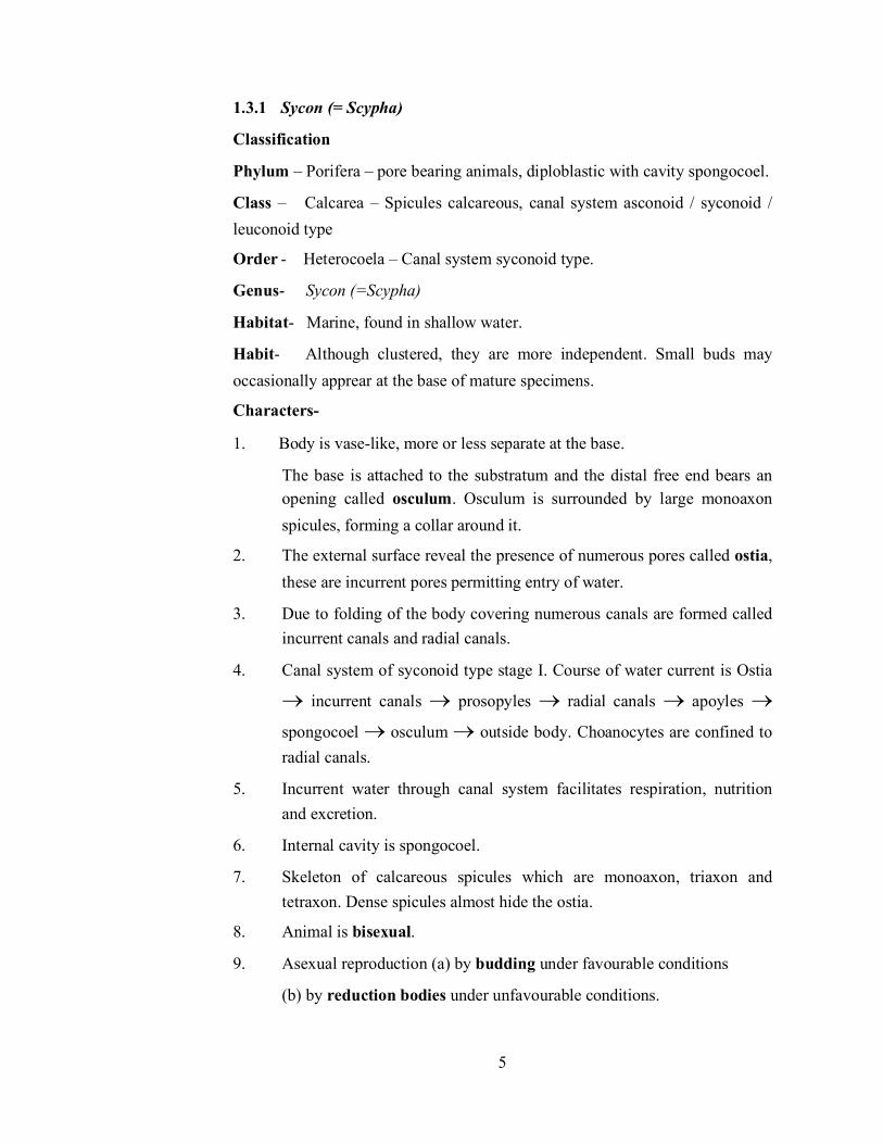
1.3.1 Sycon (= Scypha)
Classification
Phylum – Porifera – pore bearing animals, diploblastic with cavity spongocoel.
Class – Calcarea – Spicules calcareous, canal system asconoid / syconoid / leuconoid type
Order - Heterocoela – Canal system syconoid type.
Genus- Sycon (=Scypha)
Habitat- Marine, found in shallow water.
Habit- Although clustered, they are more independent. Small buds may occasionally apprear at the base of mature specimens.
Characters-
1. Body is vase-like, more or less separate at the base.
The base is attached to the substratum and the distal free end bears an opening called osculum. Osculum is surrounded by large monoaxon spicules, forming a collar around it.
2. The external surface reveal the presence of numerous pores called ostia, these are incurrent pores permitting entry of water.
3. Due to folding of the body covering numerous canals are formed called incurrent canals and radial canals.
4. Canal system of syconoid type stage I. Course of water current is Ostia
incurrent canals prosopyles radial canals apoyles
spongocoel osculum outside body. Choanocytes are confined to radial canals.
5. Incurrent water through canal system facilitates respiration, nutrition and excretion.
6. Internal cavity is spongocoel.
7. Skeleton of calcareous spicules which are monoaxon, triaxon and tetraxon. Dense spicules almost hide the ostia.
8. Animal is bisexual.
9. Asexual reproduction (a) by budding under favourable conditions
(b) by reduction bodies under unfavourable conditions.
6
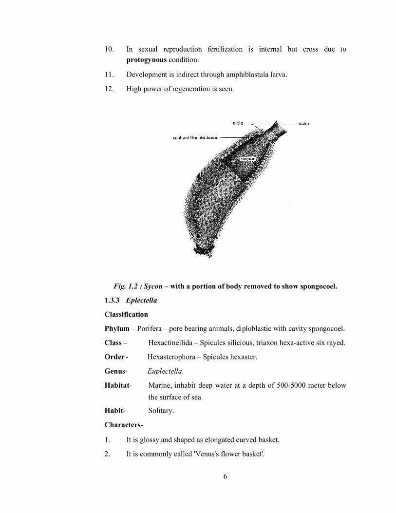
10. In sexual reproduction fertilization is internal but cross due to protogynous condition.
11. Development is indirect through amphiblastula larva.
12. High power of regeneration is seen.
Fig. 1.2 : Sycon – with a portion of body removed to show spongocoel.
1.3.3 Eplectella
Classification
Phylum – Porifera – pore bearing animals, diploblastic with cavity spongocoel.
Class – Hexactinellida – Spicules silicious, triaxon hexa-active six rayed.
Order - Hexasterophora – Spicules hexaster.
Genus- Euplectella.
Habitat- Marine, inhabit deep water at a depth of 500-5000 meter below the surface of sea.
Habit- Solitary.
Characters-
1. It is glossy and shaped as elongated curved basket.
2. It is commonly called 'Venus's flower basket'.

7
3. Six rayed silicious spicules are symmetrically arranged and join to form network with parietal gaps.
4. The basal part consists of long glossy spicules which collectively form the tuft. The tuft serves to fasten the sponge to mudy ooze of sea buttom.
5. The osculum is covered by an oscular sieve plate.
6. The canal system of syconoid type, choanocytes are restricted to flagellated chambers.
7. Special character - it shows commensalism with crustacean- Spongicola A pair of shrimp enters the body of Euplectella and is unable to escape out due to growth of sponge. Pair of shrimp passes whole life together. With this idea Euplectella is gifted to newly wedded couples in Japan.
Fig. 1.3 : Euplectella
1.3.4 Spongilla
Classification
Phylum – Porifera – pore bearing animals, diploblastic with cavity spongocoel.
Class – Demospongiae – Skeleton of silicious spicules and / or spongin fibres or absent.
Order - Monoaxonida – Spicules are monoaxon and silicious, spongin fibres may be present or absent.
Genus- Spongilla.
Habitat- Inhabits fresh water ponds and lakes and is commonly called "Fresh water sponge".

8
Habit- Colonial, profusely branched colony is associated with twigs and plant sticks.
Characters-
1. Body is covered with thin dermal membrane.
2. The body consists of several dermal ostia and osculum.
3. Skeleton consists of monoaxon silicious spicules embeded in the mesh work of spongin fibres.
4. Canal system of rhagon type. Course of water current: prosopyle
flagellated chambers apoplyle spongocoel osculum.
5. Asexual reproduction under unfavourable conditions by gemmule formation.
6. Free swimming larva characteristic of Spongilla settles to twigs.
7. The animal shows symbiotic association with unicellular green – Zoochlorella algae. Alage is found in the amoebocytes of sponge. The algae is passed to next generation through fresh infection.
Fig. 1.4 : Spongilla-colony
1.4 Phylum – Coelenterata (after coelenteron: coel = cavity, enteron= gut, meaning a cavity that acts as gut) 1. The animals of this phylum are aquatic dwelling in freshwater and
chiefly in marine water. Solitary or colonical forms.
2. The body shows tissue grade of organisation.

9
3. The body is diploblastic, made up of two embryonic germ layers, outer ectodermal-epidermis and inner endodermal - gastrodermis. In between two layers there is gelatinous mesoglea.
4. These are acoelomate. The body wall surrounds a single cavity called coelenteron or gastrovascular cavity (Note: gastrovascular means a cavity that assists is digestion and circulation).
5. The bodywall consists of specialised cells called nematocytes (=stinging cells) with a structure called nematocyst. These are characterstic of this phylum only. Due to presence of these structures the phylum is better known as Cnidaria (nidae=sting).
6. The animals are radially symmetrical.
7. The mouth is surrounded by hollow tentacles. Anus is absent.
8. This is the phylum in which nerve cell appeared first.
9. There are two stages in the life (i) polyp (ii) medusa.
10. Both the stages show polymorphism. The forms are called zooids.
11. Asexual reproduction in by budding.
12. Sexual reproduction by production of sex cells.
13. Development may be direct or indirect. Planula is the basic larva.
Phylum is divided into three classes-
1. Class – Hydrozoa – Both polyp and medusa are present.
2. Class- Scyphozoa (skyphos = cup. or saucer, zoon = animals) medusa stage is dominant.
3. Class- Anthozoa (=Actinozoa) – Only polyp stage is present.
1.4.1 Hydra
Classification
Phylum – Coelenterata – Presence of coelenteron, tissue grade of organisation, diploblastic, nematcyst present.
Class – Hydrozoa – Polyp and medusa are present.
Order - Hydroida- Polyp stage is dominant.
Suborder- Athecata – Covercing theca is absent.
Genus- Hydra.
Habitat- Fresh water form, inhabits ponds and lakes.

10
Habit- Solitary and sedentary, attached to substratum with its proximal or aboral end.
Characters-
1. Only polyp stage is present which has cylindrical body measuring 1.5cm in length showing radial symmetry.
2. The distal end or oral end bears a mouth. Located at this end is a raised structure called hypostome.
3. Hypostome is surrounded by 6-10 hollow tentacles.
4. Proximal end or abroal end has basal disc or pedal disc.
5. Diploblastic animal, nematocysts epidermal.
6. Body wall surrounds the single cavity coelenteron or gastrovascular cavity.
7. Carnivorous, digestion is both extra cellular and intracelluar.
8. Hydra may be unisexual or bisexual depending upon the species.
(a) 1-2 ovaries are formed near proximal end.
(b) Cone shaped testis (3-4) are produced near distal end.
9. Fertilization is cross due to protandrous conditoin. (Note: protandrous means male part matures first)
10. Asexual reproduction occurs by budding. Buds after development are detached from the parental body.
Fig. 1.5 : Hydra
11
1.4.2 Physalia
Classification
Phylum – Coelenterata – Presence of coelenteron, tissue grade of organisation, diploblastic, namatocysts present with radial symmetry.
Class – Hydrozoa – Polyp and medusa are present.
Order - Siphonophora – Polyp and medusa forms show polymorphism.
Suborder- Physophorida – Floating is assisted by the presence of pneumatophore.
Genus- Physalia
Habitat- Marine, found in warm waters.
Habit- Colonial, pelagic floating form.
Characters-
1. This is commonly named as "Portuguese – man – of – war", because they suddenly appear and disappear from water like the warrior ships of Portuguese.
2. Prominent feature is a large bladder like float – the pneumatophore, filled with gases oxygen, argon and nitrogen (80-90%). The filling of the float helps in floating while with the removal of gas the animal sinks down.
3. On the upper surface of pheunmatophore is a sail like crest.
4. To the lower surface of pneumatophore are attached zooids spread up amongst the fishing tentacles. Gastrozoids-feeding zooids, small and large dactylozooids – for defence, gonodendra for reproduction. Among the sessile gonophores are numerous gonopalpons. Gelatinous zooids are of unassigned function.
5. The nematocysts on tantacles are highly poisonous. Commensal small Nomeus fish lives among the tentacles without any harm.

12
Fig. 1.6 : Physalia (A) portion of colony showing zooids. (B) colony
1.4.3 Aurelia
Classification
Phylum – Coelenterata – Presence of coelenteron, tissue grade of organisation, diploblastic, namatocysts present with radial symmetry
Class – Scyphozoa – Medusa is dominant.
Order- -Semaeostomeae – Medusae saucer shaped and provided with oral lobes.
Genus- Aurelia
Habitat- Marine, living mosty in coastal warm waters.
Habit- Solitary, free swimming.
Characters-
1. Being transparent and gelatinous are commonly called 'Jelly fish'.
2. Body is saucer shaped or umbrella shaped with two surfaces (a) convex – exumbrellar surface (b) concave – subumbrellar surface.
3. The circular margin has eight notches.
4. Each notch has a pair of marginal lappets which encloses sense organs tentaculocyst or rhopallium.

13
5. In between notches there are numerous small marginal hollow tentacles.
6. Margin lacks muscles and nerve ring and does not form the velum and is called velarium. (Note: true velum is present in medusae of hydrozoans)
7. The subumbrellar surface bears the following – (a) four cornerd mouth, (b) four oral arms arising from each corner of mouth, (c) cilitated grooves in oral arms, (d) nematocytes on oral arms.
8. On the surface of umbrella are inter-radial, per-radial and ad-radial canals which open in circular canal.
9. The animal is carnivorous.
10. The animal swims by rhythmic contraction of muscular processes of cells of umbrellar surface.
11. Sexes are separate.
12. Four horse shoe shaped gonods are present on subumbrellar surface.
13. In the life cycle – alternation of generation is seen.
14. Fertilization is external.
15. Development indirect – ephyra larva is formed.
Fig. 1.7 : Aurelia-medusa showing oral surface.
1.4.4 Alcyonium
Classification

14
Phylum – Coelenterata – Presence of coelenteron, tissue grade of organisation, diploblastic, namatocysts present, with radial symmetry.
Class – Anthozoa (Actinozoa) – Only polyp stage.
Subclass- Octacorallia – Eight tentacles are present.
Order- Alcyonacea – Basal part forms fleshy mass. This order includes "Soft Coral"
Genus- Alcyonium.
Habitat- Marine, mostly found in the tidal zone at a depth of 200m in temperate and cold sea.
Habit- Colonial, sedentary.
Characters-
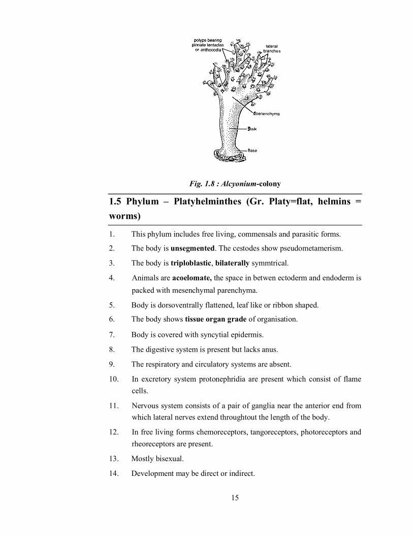
1. This is commonly called "Dead man's finger".
2. Base of the colony is meant for adherence to subtratum. At the free end of the stalk there are branched leathery lobes.
3. Over the stalk is fleshy collenchyma from which project out the polyp with oral end.
4. Skeleton of spicules provide rigidity to collenchyma.
5. Eight pinnate tentacles are present in each polyp.
6. The distal part of the body consists of anthocodia in scattered manner while the proximal part lacks anthocodia.
7. Fertilization occurs outside the body.
8. In life cycle free siwmming planula larva is formed which by budding gives rise to colony.
15
Fig. 1.8 : Alcyonium-colony
1.5 Phylum – Platyhelminthes (Gr. Platy=flat, helmins = worms)
1. This phylum includes free living, commensals and parasitic forms.
2. The body is unsegmented. The cestodes show pseudometamerism.
3. The body is triploblastic, bilaterally symmtrical.
4. Animals are acoelomate, the space in betwen ectoderm and endoderm is packed with mesenchymal parenchyma.
5. Body is dorsoventrally flattened, leaf like or ribbon shaped.
6. The body shows tissue organ grade of organisation.
7. Body is covered with syncytial epidermis.
8. The digestive system is present but lacks anus.
9. The respiratory and circulatory systems are absent.
10. In excretory system protonephridia are present which consist of flame cells.
11. Nervous system consists of a pair of ganglia near the anterior end from which lateral nerves extend throughtout the length of the body.
12. In free living forms chemoreceptors, tangoreceptors, photoreceptors and rheoreceptors are present.
13. Mostly bisexual.
14. Development may be direct or indirect.

16
Phylum is divided into following major classes.
1. Class - Turbellaria
2. Class – Trematoda
3. Class - Cestoda.
1.5.1 Fasciola hepatica
Classification
Phylum – Platyhelminthes – Acoelomate, tissue organ grade of organisation, triploblastic, bilaterally symmetrical, dorsoventrally flattened body.
Class – Trematoda – Parasites commonly called flukes due to leaf like body.
Order- Digenea – Life cycle includes two hosts.
Family- Fasciolidae
Genus- Fasciola (L. fasciola = small bandage) .
Species- hepatica (Gr. hepar = liver)
Habit- Endoparasite, found in the bile ducts of liver of sheep, human, monkey, horses and dogs.
Characters-
1. Being present in liver it is commonly called "liver fluke".
2. Body measures about 2-5cm in length and 0.5-1.5 cm in breadth.
3. Body is leaf like with anterior end shaped like a cone called cephalic cone.
4. At the end of the cephalic cone is a mouth encircled by sucker called oral sucker.
5. Midventrally about 3-4mm posterior to oral sucker there is large ventral sucker or acetabulum.
6. In front of vental sucker there is a small opening called gonopore.
7. At the extreme posterior end of the body there is single excretery pore.
8. Alimentary canal lacks anus.
9. In exretory system there is a branched system of excretory duct with flame cells or protonephridia at their ends.

17
10. Bisexual individuals, fertilization – cross.
11. Life cycle – digenetic- involves two host.
Adult in liver of primary host egg
Miracidium larva (ciliated free swimming)
Pierces shell of snail
Sporocyst larva
Redia larva
With Food
Vertebrate (Primary Host)
In Water Snail (Secondary host)
Metacercaria (Infective stage) secrets cyst
comes out of the body of snail
Ciracaria (Tailed larva)
Daughter Redia
Life cycle of Fasciola hepatica
12. Parasite is pathogenic to primary host and causes liver rot in sheep, The animal products – meat, wool and leather are adversely effected.
Fig. 1.9 : Fasciola hepatia-ventral view.
1.5.2 Taenia solium
Classification

18
Phylum – Platyhelminthes – Acoelomate, tissue grade of organisation, triploblastic, bilaterally symmetrical, dorsoventrally flattened body.
Class – Cestoda – Endoparasites commonly called tapewom due to ribbon like body.
Subclass- Eucestoda – Polyzoic (with many proglottids), larva is six hooked.
Order- Cyclophyllidea – scolex with four suckers (=acetabula)
Family- Taenidae – uterus is branched.
Genus- Taenia
Species- solium.
Characters-
1. Body is dorsoventrally flattened, long, ribbon like, measuring 2-3 metre in length. Commonly called "tapeworms".
2. Body shows pseudometamerism with segments called proglottids.
3. Body division –
(a) Scolex – with two circles of restellar hooks and four suckers – meant for adhesion to intestinal walls of host.
(b) Neck region – Zone of growth or zone of proglottization.
(c) Immature proglottids – About 200 proglottids.
(d) Mature proglottids – 500 in number with reproductive organs.
(e) Gravid proglottids – About 200 proglottids. They contain only branched uterus with fertilized eggs
4. The body wall is produced into microtriches.
5. The animal lacks digestive system, it absorbs digested food contents available in intestine of host through the body wall.
6. Respiration is anaerobic.
7. Excretory system contains flame cells.
8. Bisexual individuals.
9. Fertilization is cross due to protandrous condition.
10. Life cycle: Digenetic – involves two hosts.

19
(a) Primary host is man (b) secondary host is pig – found in meat called "measly pork". The animal is commonly called "Pork tapeworm of man"
Adult in intestine of man (Primary host)
Eggs
Sperms
Hexacanth Covered with embryophore
Comes out alongwith faceal matter of host
Caprophagous pig (Secondary host)
Life cycle of Taenia solium Hexacanth pierces the intestine of pig
inadeqauately cooked measly pork ingested by primary host
Muscles of pig changed to cysticercus larva or bladder worm (Infective stage)
Blood Vascular System of pig
11. Causes – anaemia, eosinophilia, diarrhoea and abdominal pain in man.
Fig. 1.10 : Taenia solium –(A) entire tapeworm (B) enlarged (proglottid)
20
1.6 Phylum Aschelminthes (Gr. Ascos= sac, helmins = worm)
1. This phylum include freeliving (aquatic or terrestrial) and parasitic animals measuring few millimetres to a metre in length.
2. These are unsegmented worms.
3. Triploblastic with organ system grade of organisation.
4. Bilaterlally symmetrical animals.
5. Animals are pseudocoelomate: coelom is not mesodermal but derived from embryonic blastacoel.
6. Cuticle covers the body wall.
7. Alimentary canal straight with mouth and anus.
8. Excretoy system has canals.
9. Nervous system has circumentric ring and longitudinal nerves.
10. Respiratory system and circulatory systems are wanting.
11. Sense organs are present.
12. Unisexual.
13. Life cycle simple or complicated. Eggs shelled, cleavage determinate and spiral.
14. Development direct or indirect.
1.6.1 Ascaris lumbricoides
Classification
Phylum – Aschelminthes – unsegmented, triploblastic, bilaterally symmetrical, pseudocoelomate.
Class – Nematoda (Gr. nema=thread, eidos=form)-this included thread like forms or round worms.
Order- Ascaroidea- mouth surrounded by three lips.
Genus- Ascaris
Species- lumbricoides (=suum)
Habit- Ascaris lumbricoides is endoparasite of intestine of pigs and man. The infection spreads by ingesting infective eggs.

21
Characters-
1. It is commonly called 'round worm'.
2. Animal is elongated cylindrical tapering at the end.
3. The animal is unisexual with sexual dimorphism.
4. Female is straight and longer 20-40cm in length and 4-5mm in diameter, male is curved at the posterior end and smaller 15-30cm in length and 2-4mm in diameter.
5. Body colour is yellowish white.
6. In both females and males four longitudinal lines are visible one ventral, one dorsal and two lateral.
7. Mouth is at the anterior end surrounded by three lips.
8. Lips have amphids and sensory papillae.
9. In female anus and in male cloaca is located near the porterior end midventrally.
10. In female the genital opening is midventral at a distance 1/3rd from the anterior end.
11. Syncytical epidemis is covered with cuticle.
12. The animal is pseudocoelomate.
13. Repiratory and circulatory systems are absent
14. Excretary pore midvental at a distance 2mm from anterior end.
15. Reproductive system is simple.
16. In male two copulatory spicules protrude out of cloaca.
17. Life cycle is monogenetic, completed in man.
18. Infection occurs by ingestion of contaminated food and water, development in direct.
19. The animal causes ascariasis in man.

22
Ascaris lumbricoides
1.7 Phylum – Annelida (L. Annelus= ring, edios=form)
1. The animals included in phylum annelida are free living aquatic (freshwater and marine), terrestrial and parasitic forms.
2. The body is metamerically segmented, the organs are repeated in number of segments.
3. The segmentation is true.
4. Body, shows organ grade of organisation and bilateral symmetry.
5. The animals are eucoelomate. The coelom develops from embryonic mesoderm. Eucoelom is schizocoelous-formed by splitting of mesodermal bands.
6. Chitinous setae assist in locomotion.
7. Alimentary canal is straight with month at anterior end and anus at the posterior end.
8. Circulatory system of closed type. The respiratory pigment – haemolgobin / erythrocruorin is present in plasma in dissolved state. In dorsal blood vessel flow of blood is from posterior to anterior and in ventral blood vessel flow is from anterior to posterior direction.

23
9. Excretion by means of nephridia.
10. In nervous system – nerve ring and ventral double solid, ganglionated nerve chord is present.
11. Sexes – may be separate or united.
12. Cleavage is determinate and spiral, Development- direct or indirect. If indirect ciliated trochophore larva is produced.
Classification –
Phylum is divided into
1. Class-Archiannelida (Archi=primitive, annelids) – includes primitive annelids.
2. Class- Polychaeta – (Poly=many, chaeta=setae)-with numerous setae, clitellum absent. Development with trochophore larva.
3. Class- Oligochaeta (Oligos=few, chaeta=setae)- Animals with: few setae, clitellium present. Development direct.
4. Class- Hirudinea – includes parasitic leeches, clitellum is temporary, development in direct.
1.7.1 Nereis
Classification
Phylum – Annelida (L. annelus=ring, edios=form) metameric segmentation, bilateral symmetry, eucoelom-schizocoelom present.
Class – Polychacta (Poly=many, chaeta=setae) – numerous setae are present, clitellum is absent, development indirect through trochophore larva.
Order- Errantia – Swarming animals, locomotion by parapodia.
Genus- Nereis (=Neanthes)
Habitat- Cosmopolitan, marine.
Habit- It is found burried in sand and found among clams though the animal has no ecological relationship with sand or clams. This animal is commonly called sandworn or clamworm or ragworm. The animal is nocturnal and carnivorous.
Characters-

24
1. The segmentation is external and internal.
2. Body division.
(i) Head – with prostomium and peristomium. The head has sensory structures like eyes, prostomial tentacles, palps and nuchal organs.
(ii) Trunk – with 80-120 segments, each with a pair of lateral parapodia. Each parapodium has numerous setae. Each para podium is divided into (a) dorsal notopodium and (b) ventral neuropodium.
(iii) Pygidium – Last segment with terminal anus and a pair of anal cirri.
3. Animal is carnivorous, filter feeder and raptorial.
4. Each segement has a pair of nephridium ment for excretion and osmoregulation.
5. Blood vascular system is of closed type. Blood with respiratory pigment erythrocruourin which is dissolved in plasma.
6. Sexes are separate with sexual dimorphism.
7. Sexual stage is heteronereis with anterior atoke or non sexual region and pasterior epitoke or sexual region. The sexual stage is actively swarming.
8. Development indirect through trochophore larve.
Fig. : Nereis-dorsal view

25
1.7.2 Pheretima
Classification
Phylum – Annelida (L-annelus=ring, edios=form) – metameric segmentation, bilateral symmetry, eucoelom-schizocoelom present.
Class – Oligochaeta (obligos=few, chaeta=setae) few setae are present in segments, clitellum is present, development is direct.
Order- Opisthopora – the organs are present posterior to pores or openings.
Genus- Pheretima
Habit- Inhabits moist soil, rich in decaying organic matter. Pheretima is commonly called 'earthworm'.
Habit- Burrowing forms, form pellets at the opening of the burrows, nocturnal.
Characters-
1. Body is cyclindrical, dark brown in colour with external and internal segmentation.
2. Number of segments in body is 100-120 and segments are homonomous (=all alike)
3. Body division into:
(a) Preclitellar region – 1 to 13 segment.
(b) Clitellar region – girdle like in 14,15,16 segments.
(c) Postclitellar region – 17 to last segment.
4. Each segment except first, clitellar and last segement has a ring of setae bearing 80-100 setae.
5. First segment – peristomium extends anteriorby and dorsally to form prostomium. Mouth is ventral.
6. On the ventrolateral side on intersegmental grooves-5/6, 6/7, 7/8, 8/9 each has a pair of spermathecal pores.
7. On ventral side 14th segment has a female genital pore.

26
8. On ventrolateral side in 17 and 19 segment there is a pair of raised genital papillae per segment and in 18th segment there is a pair of male genital opening.
9. The body bears numerous nephridiopores except few anterior segments.
10. Animal in bisexual.
11. Fertilization is cross, external in cocoons. Cocoon is formed by clitellum.
12. Development is direct in cocoon.
13. Animal is of great economic significance in agriculture, their excreta is used as fertilizer.
Fig. : External feature of Pheretima (a) dorsal view (b) ventral view.
1.7.3 Hirudinaria -
Phylum – Annelida (L-Annelus-ring, edios=form). metameric segmentation, bilateral symmetry, eucoelom-schizocoelom present.
Class – Hirudinea – includes parasitic leeches, clitellum in temporary, development is direct.

27
Order- Gnathobdellida - (Gnathos=jaws)-jaws are present, probosis is not protrusible.
Genus- Hirudinaria – 33 segments are present.
Habitat- Commonly known as "Indian common leech" is found in freshwater ponds, lakes, swamps and slow running streams.
Habit- Leech is ectoparaisite of fish, frog, cattles and man. It is sanguivorous (blood-sucking)
Characters-
1. Body is soft, elongated and vermiform, dorsoventrally flattened. The body has great power of contraction and expansion.
2. The body has 33 segments and each segment is divided into annuli. The number of annuli varies in segments.
3. The body bears two suckers. At the anterior end is oral sucker – it has ventral preoral chamber which leads to mouth. At the posterior end there is anal sucker. Suckers are used for adhesion to host and in locomotion.
4. Body division into.
(a) Cephalic region – of 5 segments
(b) Preclitellar region – 6th, 7th & 8th segment
(c) Clitellar region – 9th, 10th & 11th segment. Clitellum temporary develops during breeding season.
(d) Middle region – 11 complete somites from 12th to 22nd.
(e) Caudal region – from 23rd to 26th segment.
(f) Posterior sucker – from 27th to 33rd segment.
5. Mouth is triradiate, ventral.
6. Ocelli are five pairs located dorsally.
7. Nephridiopores are seventeen pairs on ventral surface one pair lies on the last annulus of each segment from 6th to 22nd.
8. The coelom is obliterated by the presence of botryoidal tissue. Haemocoelomic channels are present.
9. Bisexual animals. Male genital pore is or midventral on intersegmental groove of 2nd and 3rd annuli of 10th segement. Female – genital pore is located on 11th segement midventrally.

28
10. Fertilization in internal.
11. Development is direct in cocoon formed by clitellum.
Fig. : Hirudinaria (A) ventral view, (B) anterior dorsal view (C) posterior dorsal view.
1.8 Phylum – Mollusca (L-Mollis or Molluscum=soft) means phylum includes soft bodied animals
This is the second largest phylum including 80000-100000 species.
1. Inhabit aquatic or terrestrial environment.
2. Body unsegmented, triploblastic, bilaterally symmetrical.
3. Body division into:
(i) Head
(ii) Mantle – Soft skin like. Covers the visceral mass and secrets shell.
(iii) Foot – is ventral.
4. Haemocoelomic spaces do not form coelom. Coelom is present around heart, in kidney and in gonads.
5. Digestive system consists of masticatory radula. Alimentary canal opens in mantle cavity.

29
6. In circulatory system heart is dorsal. Circulatory system is of open type. Blood is colourless or green due to presence of haemocynin.
7. Respiration by skin, gills or lungs.
8. Excretion by metanephridia.
9. Head with eyes and tentacles.
10. Nervous system with 3 pairs of ganglia, connected by nerves.
11. Mostly unisexual.
12. There is always sexual reproduction. No asexual reproduction.
13. Development – Direct in pulmonate, indirect through trochophore / veliger or glochidium larva.
Phylum – Mollusca is divided into six classes.
i. Class – Monoplacophora – Neopilina – connecting link between annelida and mollusca.
ii. Class – Amphineura
iii. Class – Scaphopoda (Gr. Scaphos = boat, podos = foot)
iv. Class – Gastropoda (Gr. Gastral=belly, podos=foot)
v. Class – Cephalopoda (Gr. Kephale=head, podos=foot)
1.8.1 Chiton
Classification
Phylum – Mollusca – Body soft, unsegmented, triploblastic and bilaterally symmetrical.
Class – Amphineura – Reduced head lacks eyes and tentacles,
Order- Polyplacophora – Mantle secrets shell with many plates, foot is flattened.
Genus- Chiton
Habitat- Marine
Habit- Sluggish animal found attached to rocks, empty shells and corals, nocturnal, herbivorous.
Characters-
1. It is commonly called "Sea myca" or "Coat of mail shell"
2. Body is dersoventrally compressed.

30
3. Shell with 8 overlapping plates is present dorsally.
4. Foot is ventral and flat helps in creeping.
5. Mantle covers visceral mass.
6. Head is not distinct and lacks eyes and tentacles.
7. Mouth and anus at the opposite ends.
8. Numerous pairs of bipectinate ctenidia lie on either side of the body in mantle groove.
9. Sexes are separate. Gonod is single, gonoducts are paired.
10. Development indirect through trochophore larva.
Fig. : Chiton (A) dorsal surface (B) ventral surface.
1.8.2 Pila globosa
Classification
Phylum – Mollusca body soft, unsegmented, triploblastic and bilaterally symmetrical.
Class – Gastropoda (Gr. Gastral=belly, podos=foot)-foot over the belly- Snails and slugs are included. Body shows coiling called torsion.
Order- Mesogastropoda (=Pectinibranchia)- monopectinate ctenidium present.
Genus- Pila.
Species- globosa
Habitat- Fresh water form found in pools, ponds, lakes and marshes.
Habit- Feeds upon plant scrapping. It is adapted for amphibious life.
Characters-

31
1. Shape is globose with lemon yellow, brownish in colour commonly called "apple snail".
2. Shell is twisted spirally forming body whorl, penultimate whorl and apex. The shell shows dextral or right handed coiling.
3. Surface of the shell shows lines of growth or varices.
4. When the snail is inside the shell, it is covered with operculum. Operculum too has concentric lines of growth.
5. Body division into:
(i) Head – with one pair of eyes, two pairs of tentacles, nuchal lobes and ventral slit like mouth.
(ii) Foot – Ventral roughly triangular, broad, flat, to it is attached operculum.
(iii) Visceral mass in humplike on the dorsal side and in spirally coiled.
(iv)Mantle or pallium covers the visceral mass.
6. In buccal mass – radula is present.
7. Ospharidium is present for testing the quality of water.
8. Respiration by pulmonary sac and ctenidium.
9. Pericardium with heart and renal organ present.
10. Sexes are separate, fertilization is internal development is direct.
Pila globosa

32
1.8.3 Loligo
Classification
Phylum – Mollusca – Soft bodied, unsegmented, triploblastic and bilaterlaly symmetrical.
Class – Cephalopoda (Gr. Kephale=head, podos=foot). Head distinct surrounded by tentacular foot.
Subclass- Dibranchia – shell internal.
Order- Decapoda=(Deca=ten, podos=legs)
Genus- Loligo
Habitat- Marine, found in warmer seas.
Habit- Solitary, it is a fast swimmer in open water of sea.
Characters-
1. Commonly called "Squid" or "Sea arrow".
2. Body is flattened dorsoventally.
3. Body division.
(i) Head – surrounded by ten oral arms provided with suckers. Two of these arms are long called tentacles and bear suckers at the destal end. Tentacles are called hectocotylised arms. One pair of eyes are present, horny jaws are present.
(ii) Trunk or visceral hump.
(iii) Posterior end in equipped with arrow shaped lateral Fins or parapodium.
4. Shell is internal penlike which maintains buoyancy.
5. The animal contains 2 ctenicdia, 2 kidneys and 2 auricles and ink gland. Blood vascular system is closed type.
6. Sexes are separate. In male one of the arms acts as copulatory organ.
7. Development is direct.

33
Loligo
1.8.4 Nautilus
Classification
Phylum – Mollusca-Body soft, unegmented, triploblastic, bilaterally symmetrical.
Class – Cephalopoda (Gr. Kephale=head, podos=foot) head distinct, surrounded by tentacular foot.
Subclass- Tetrabranchia – 4 gills are present, shell in external.
Genus- Nautilus.
Habitat- Marine, found in deep water.
Habit- Nocturnal, carnviorous.
Characters-
1. The animal is commonly called: "pearly nautilus" because the inner layer of shell is pearly.
2. Shell in external and is coiled spirally.
3. Shell is internally divided into chambers by means of septa.
4. The septa are perforated in the middle. Through the performation passes a chord called "siphuncle" which is the extension of visceral mass. The gas secreted by animal is passed to the empty chamber through this chord. This helps in making shell buoyant.

34
5. Body division
(i) Head – with mouth and a pair of eyes surrounded by about 90 filliform tentacles without suckers.
(ii) Trunk is bag like with 4 gills, 4 kidneys, and 4 auricles. Ospharidia are present.
6. Ink gland and chromatophores are absent.
7. Sexes are separate.
8. Development is direct.
Fig. : Nautilus – (A) shell (B) section of the shell
1.9 Phylum – Arthropoda (Gr. Arthros=jointed,
podos=legs)
This is the largest phylum of animal kingdom which includes about 88 percent of known animals.
1. The phylum includes aquatic and terrestrial animals.
2. The animals may be solitary, colonial or social.
3. The animals are triploblastic, bilaterally symmetrical and segmented.
4. The body segments typically consist of jointed appendages.
5. Body division into head, thorax and abdomen.
6. The body is covered with chitinised cuticle.
7. In blood vascular system the blood vessel are modified and enclose the visceral organs. The cavity is filled with blood and is called haemocoel. The blood vascular system is of open type.

35
8. Blood may be colourless or with haemocynin pigment dissolved in plasma.
9. Heart is dorsal with flow of blood from posterior to anterior.
10. Respiration by gills (in aquatic forms) or trachae or book lungs or book gills or external body surface.
11. Excretion by coelomoducts or malpighian tubules or coxal glands, (Note: Onychophorans have nephridia).
12. Nervous system with nerve ring and ventral double, ganglionated nerve chord.
13. Eyes may be simple and compound.
14. Sexes are separate.
15. Development may be direct or indirect.
Classification – Living subphyla are as under:
1. Suphylum – Onchychophora – Peripatus is connecting link between phylum annelida and arthropoda.
2. Subphylum – Chlicerata – First pair of appendages are chilicerae, Antennae are lacking. Cephalothora with six pairs of appendages. Abdomen without appendage.
3. Subphylum – Pychogonida – includes marine spiders. Head with three pairs of appendages and abdomen degenerate.
4. Subphylum – Mandibulata – Mouth parts include one or two pairs of mandible. Head is six segmented. First cephalic segment lacks appendage, second has first pair of antennae, third has second pair of antennae (only in crustaceans), fourth has one pair of mandible, fifth and sixth each have a pair of maxillae.
1.9.1 Balanus
Classification
Phylum – Arthropoda – Animal bears jointed appendages, triploblastic, bilaterally symmetrical and segmented.
Subphylum –Mandibulata – Mouth parts with mandibles. Six segmented head with one or two pairs of antennae.
Class- Crustacea-head with two pairs of antennae.

36
Subclass- Cirripedia – Adults are sedentary, fixed, lack compound eyes and antennae, segmentation is poor. Thorax bears six pairs of biramous appendages, carapace forms paired fold.
Order- Thoracica – Non parasitic forms, carapace forms calcareous folds, alimentary canal is absent.
Genus- Balanus
Habitat- Marine found in between tide marks in shallow water.
Habit- Found attached to rocks and shells, so commonly called "Rock barnacle" or "Acorn barnacle"
Characters-
1. Peduncle is attached so that the shell is directly attached to the objects.
2. Cephalic part is short but broader.
3. Body is encircled by calcareous shell consisting of six plates – an unpaired carina, an unpaired rostrum and two pairs of lateral plates. The animal is easily identified by the presence of these plates.
4. The opening of the shell in provided with mobile fourfold operculum consisting of two scuta and two terga.
5. Six pairs of jointed thoracic limbs or cirri protrude out through the opening of shell which help to swipe the food particles.
6. Sexes – united or hermaphrodite.
7. Development – Indirect through nauplius larva.
Fig. : Balanus

37
1.9.2 Palaemon
Classification
Phylum – Arthropoda – Animal bears jointed appendages, triploblastic, bilaterally symmetrical and segmented.
Subphylum – Mandibulata – Mouth parts with mandibles. Six segmented head with one or two pairs of antennae.
Class- Crustacea-head with two pairs of antennae.
Subclass- Malacostraca – Cephalothoracic carapace is present.
Order- Decapoda – Five pairs of thoracic walking legs are present.
Genus- Palaemon (=Macrobrachium)
Habitat- Freshwater free moving form, inhabits streams, ponds, lakes, rivers
Habit- Nocturnal, omnivorous
Characters-
1. The animal is commonly called "prawn".
2. Colour is pale greenish with brown patches.
3. Body is spindle shaped, elongated, measuring 20m in length.
4. Body division into:
(i) Cephalothorax – formed by fusion of 6 segmented head and 8 segmented thorax, which are covered by a carapace. Carapace, is extended anteriorly and midodorsally into serratted rostrum. Cephalic part has two pairs of antennae, one pair each of mandibles, maxillulae and maxillae. One pair of pedunclate compound eyes are present.
Thoracic part has three pairs of maxillipedes and five pairs of walking legs.
(ii) Abdomen in laterally compressed and curved ventrally, the animal look like comma. It is five segmented, abdomen has five pairs pleopods or swimmerets and one pair of uropod. The abdomen ends in telson.
5. There are total 19 pairs of appendages.
6. Sexes are separate. In male the second pair of chelate leg is larger and powerful than in female. The second pair of pleopod of male differs

38
from female in having additional process called appendix masculina. Female carries eggs in between pleopods.
7. Fertilization is external.
8. Development is direct, hatched embyo looks like small prawn.
Fig. : Palaemon
1.9.3 Periplaneta
Classification
Phylum – Arthropoda – Animal bears jointed appendages, triploblastic, bilaterally symmetrical and segmented.
Subphylum – Mandibulata – Mouth parts with mandibles. Six segmented head with one or two pairs of antennae.
Class- Insecta or Hexpapoda – Thorax with 3 pairs of appendages.
Subclass- Pterygota (Pterygos=wings) – wings present
Division- Exopterygota – Wings develop externally
Order- Dictyoptera – All the legs are of similar size.
Genus- Periplaneta
Habitat- The animal inhabits dark damp places.
Habit- Nocturnal, omnivorous, cursorial-fast runner but can take nuptial flights.
Characters-
1. It is commonly called 'cockroach'

39
2. Length = 2.5cm, width – 1cm.
3. Body dorsoventrally flattened.
4. Body division:
(i) Head – Six segmented with single pair of antennae, 1 pair of mandlible, 1 pair of maxillae and labium. One pair of sessile compound eyes are present. Mouth parts are chewing and cutting type.
(ii) Thorax – divided into prothorax, mesothorax and metathorax. Prothorax is large with two dark specs. Mesothroax and metathroax each on their dorsal side has a pair of wings. Each thoracic segment on their ventral side has a pair of walking legs. All the legs are similar.
(iii) Abdomen – is ten segmented.
5. Each leg is divided into coxa, trochanter, femur, tibia, tarsus, claw and pulvillus.
6. Sexes are separate showing sexual dimorphism.
7. In abdomen of male and female attached to 10 tergum is a pair of anal cerci but in males only one pair of additional appendage attached to 9th sternum is present called analstyles which in absent from females. In female at the posterior end their are ovipositors.
8. Development – Direct in ootheca.
Periplaneta (a) dorsal view (b) ventral view
1.9.4 Queen Termite

40
Classification
Phylum – Arthropoda – Animal bears jointed appendages, triploblastic, bilaterally symmetrical and segmented.
Subphylum – Mandibulata – Mouth parts with mandible. Six segmented head with one or two pairs of antennae.
Class- Insecta or Hexpapoda – Thorax with 3 pairs of appendages.
Subclass- Plerygota (Pterygos=wings) – wings present.
Division- Exopterygota – Wings develop externally
Order- Isoptera – Wings are equal.
Genus- Odontotermes.
Caste- Queen termite
Habitat- Wood dwelling insects commonly called termites. The termite is a social insect forming large communities and well marked polymorphic individuals. The dwelling places are called "termitarium", The termite has caste system comprising 3 fertile reproductive castes and two sterile castes.
Habit- Termite bores the wood, dead trees and even the ground. Feeds upon wood, vegetation, faceal matter of termites.
Characters- Size – 5 to 7.5cm in length.
1. The body division into:
(i) Head – with a pair of antennae, a pair of compound eyes, and a pair of maxillary palps. Mouthparts biting type.
(ii) Thorax – 13 segmented prothorax, mesothorax and metathorax and contains 3 pairs of legs. Mesothorax bears a pair of reduced wing stubs on the dorsal side.
(iii) Abdomen is much elongated due to increase in size of ovaries and fat bodies. With the growth in the pleural membrane the queen becomes large and insert.
2. Queen is sexually mature female and lays eggs throughout life.
3. Life span is 6-15 years.
4. When the power of laying eggs ceases, female dies or is killed by other members.

41
Fig. : Queen Termite
1.9.5 Apis indica
Classification
Phylum – Arthropoda – Animal bears jointed appendages, triploblastic, bilaterally symmetrical and segmented.
Subphylum – Mandibulata – Mouth parts with mandibles. Six segmented head with one or two pairs of antennae.
Class- Insecta or Hexpapoda – Thorax with 3 pairs of appendages.
Subclass- Pterygota (Pterygos=wings) – wings present.
Division- Exopterygota – Wings develop externally
Order- Hymenoptera – Mouth parts biting, chewing, lapping and sucking type. Wings two pairs.
Genus- Apis
Species- indica.
Habitat- It lives in highly organised colony in bee hive.
Habit- It is colonial, social insect. It is polymorphic with workers (sterile female) – perform varied duties, queen (fertile female-lays eggs), drones – fertile males.
Characters-
1. Worker bees are present in largest number in the colony.
(i) Which are developed from fertilized eggs. Size is smallest.

42
(ii) Mouth parts are chewing and lapping type.
(iii) The prothoracic legs contain eye brush, fibula or velum, antennae comb and pollen brush.
(iv)Mesothoracic leg bears spur, pollen brush and pulvillus.
(v) Metathoracic leg has pollen basket in the tibia part.
(vi)Abdomen is 6 segmented. The wax glands are modified cells, present on the ventral surface of last four segments. The ovipositor of worker bee is modified into sting.
(vii) The worker bees perform duties like collection of food, secreting wax, building, repairing, cleaning, defence of hive, parental care of young ones etc.
2. The queen is larger in size having longer abdomen, mates once in life and lays eggs only.
3. The drones are fertile males, without sting, developed parthenogenetically, copulates with queen and then dies.
4. The honey bees are economically important insects produce-honey and bee wax and helpful in pollination of flowers.
Queen Bee
Other Castes
Apis indica (A) queen (B) various castes

43
1.10 Phylum – Echinodermata (Gr. Echinos=spines,
hedgehog; derma=skin) Animals bearing spines on
skin.
1. The animal belonging to this phylum are exclusively marine freeliving, nonmicroscopic.
2. Mostly benthonic forms, few remain attached with the help of pleduncle.
3. The adults show secondary pentamerous radial symmetry, though bilateral symmetry is present in larval forms. During development larval bilateral symmetry is transformed to radial symmetry.
4. The animals are triploblastic, unsegmented deuterostomes.
5. Body star shaped, ball like, cylindrical or disc shaped.
6. Distinct head is lacking. There are two ends of the body – oral end with mouth and opposite end is aboral end.
7. The body wall consists of calcareous plates equipped with spines. The body wall has protective pedicellariae.
8. The coelom is enterocoelous derived as lateral pouches from embryonic archenteron. The coelom is schizocoelous in the forms where the development is direct.
9. The body consists of ambulacral (=actinal) areas alternated by adambulacral (=abactinal) areas.
10. The animal bears ambulacral system or water vascular system characteristic of this phylum only. This system helps in locomotion.
11. At the ends of the canals present in water vascular system there are tubefeet or podia which assist in locomotion, food capturing and respiration.
12. Alimentary canal is straight or 'U' shaped. Mostly carnivorous.
13. Respiration by dermal branchiae, respiratory trees, burse or tubefeet.
14. Haemal and perihaemal systems are present. Heart is called axial gland.
15. Nervous system lacks brain. Pentagonal nerve ring and radial nerves are present.
16. Excretory system is lacking.

44
17. Sexes are separate, fertilization is external.
18. Development – Mostly indirect, diplurela is ancestral larva.
19. Power of regeneration is present.
Phylum is classified as under:
1. Supphylum – Pelmetozoa (Pelmetos=peduncle, zoon=animals)
Class – Crinoidea – includes sea lilies.
2. Subphylum – Eleutherozoa – Nonpedunclate.
(i) Class Asteroidea – includes star fishes
(ii) Class – Ophiuroidea – includes serpent stars or brittle stars.
(iii) Class – Echinoidea – includes sea urchin and sand dollars.
(iv)Class - Holothuroidea – includes sea cucumbers.
1.10.1 Asterias
Classification
Phylum – Echinodermata – Spines are present on skin, secondary pentamerous symmetry present, triploblastic, unsegmented, enterocoelomate, water vascular system present.
Subphylum – Eleutherozoa – without stalk and free living.
Class- Asteroidea – Central disc and arms are not distinct.
Order- Forcipulata – Forcep like pedicellariale present.
Genus- Asterias
Habitat- Asterias is marine, found below 200 fathoms
Habit- Animal is benthomic form, carnivorous.
Characters-
1. Body is star shaped hence the animal is commonly called 'star fish'.
2. Body shows pentamerous radial symmetry.
3. Body is distinguished as having two surfaces.
(a) Downwardly directed – oral surface with mouth.
(b) Upwardly directed – aboral surface.
4. From the pentagonal central disc radiate out five arms.
45
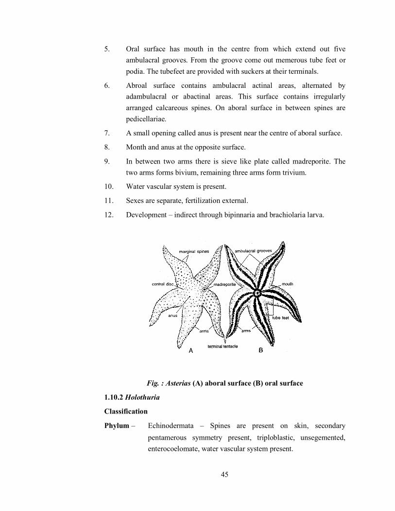
5. Oral surface has mouth in the centre from which extend out five ambulacral grooves. From the groove come out memerous tube feet or podia. The tubefeet are provided with suckers at their terminals.
6. Abroal surface contains ambulacral actinal areas, alternated by adambulacral or abactinal areas. This surface contains irregularly arranged calcareous spines. On aboral surface in between spines are pedicellariae.
7. A small opening called anus is present near the centre of aboral surface.
8. Month and anus at the opposite surface.
9. In between two arms there is sieve like plate called madreporite. The two arms forms bivium, remaining three arms form trivium.
10. Water vascular system is present.
11. Sexes are separate, fertilization external.
12. Development – indirect through bipinnaria and brachiolaria larva.
Fig. : Asterias (A) aboral surface (B) oral surface
1.10.2 Holothuria
Classification
Phylum – Echinodermata – Spines are present on skin, secondary pentamerous symmetry present, triploblastic, unsegemented, enterocoelomate, water vascular system present.

46
Subphylum –Eleutherozoa – without stalk and free living.
Class- Holothuroida – Body cylindrical, elongated on oral – aboral axis.
Order- Aspidochirota – tentacles leaf like.
Habitat- Marine, found near sea coasts.
Habit- Moves slowly on the sea bottom by the contraction of the body wall and assisted by tubefeet. They feed upon micro-organisms.
Characters-
1. The animal is commonly called 'sea cucumber'.
2. Body is cylindrical, elongated at the oral-aboral axis.
3. Oral end contains a mouth encircled by leaf like tentacles which help in collecting food.
4. Water vascular system has many pollian vesicles and internal madreporite. Tubefeet are provided with suckers.
5. Pedicellariae and spines are absent.
6. Ambulacral grooves are internal.
7. Respirating tubes and tubules of cuverian open in cloaca and can be thrown out in defence (=evisceration).
8. Sexes are separate.
9. Development indirect through auricularia and doliolaria larva.
Fig. : Holothuria

47
1.11 References
Invertebrate Zoology by Hegner and Engemann. Macmillan company, New York.
Invertebrate – Laboratory Work book – Zoology Elden Beck and Braithwaile, L.F. Burgess Publishing Company (1962)
Practical Zoology – Invertebrate, Lal S.S. Rastogi Publication, Meerut (2010)
Advanced Practical Zoology – by Verma PS and Srivastava P.C., S. Chand & Co. Ltd., New Delhi (1994)
Modern Text Book of Zoology – Invertebrates, Kotpal R.L. – Rastogi Publicaton Meerut (2011)

48
Unit - 2 Microscopy and Permanent Preprations
Structure of Unit
2.1 Aim: To study different type of Microscopes
2.2 Aim: To prepare permanent slides using the given sections like Stem, Root and Leaf
2.3 Aim: To prepare permanent slides using the given Animal Samples
2.1 Aim: To study different type of Microscopes Introductions- Cells are small and in almost all situations a microscope is needed to observe them and their sub-cellular components. In fact the invention of the microscope led to the discovery and description of cells (Hooke, 1655). The microscope is still an extremely important tool that is often overlooked in conducting research.
Light Microscopy
The light microscopy is to shine light through a specimen and examine it under magnification. The major optical parts of a microscope are the objective lens, the eyepiece, the condenser and the light source.
The objective lens functions to magnify the object. The high degree of magnification of the objective lens results in a small focal length and the magnified image actually appears directly behind the objective.

49
The eyepiece delivers this image to the eye.
The condenser focuses the light source on the specimen.
The specimen is illuminated from a lamp or other light source. The best light source is one in which the light intensity is controlled by adjusting the voltage.
Sample Preparation: Specimens can be examined by simply placing them on a glass microscope slide under a glass cover slip. However, it is necessary to prepare and stain the samples before examination by microscopy. Fixation is a process by which cells are preserved and stabilized. Common fixatives include: acids, organic solvents, formaldehyde and glutaraldehyde. These treatments affix macromolecules in position. Thick samples, such as tissues need to cut into thin sections. The sample or cells are embedded into a supporting medium such as Paraffin. Sectioning is carried out with a microtome. The microtome makes successive sections of a specified thickness. The image generated by microscopy depends upon different components in the sample interacting with and impeding the light waves differentially. Biological samples are fairly homogeneous (i.e., carbon-based polymers) and do not greatly impede light. Therefore, it is often necessary to stain cells with dyes. Different dyes have different affinities for different sub cellular components. The stained sub cellular components will differentially impede (i.e., absorb) the light waves and provide more contrast than unstained specimens. Dark-Field Microscopy: In this microscopy the specimen is illuminated from the side and only scattered light enters the objective lens which results in bright objects against dark background. This is accomplished through the use of an annular aperture that will produce a hollow cone of light that does not enter the objective lens. The images produced by dark-field microscopy are low resolution. Dark-field microscopy is especially useful for visualization of small particles such as bacteria. Phase Contrast Microscopy & Differential-Interference-Contrast Microscopy: these microcopies allow objects that differ slightly in refractive index or thickness to be distinguished within unstained or living cells. Differences in the thickness or refractive index of the specimen result in a differential retardation of light which shifts the phase. During phase contrast microscopy the phase differences are converted to intensity differences by special objectives and condensers. Normarski optics use special condensers and

50
objectives to recombine incident and refracted light waves from a single source at the plane of the image. The interference effects between the incident and refracted light enhance small differences in the refractive index or thickness of the specimen and leads to an increased resolution without staining. Fluorescence Microscopy: In this microscopy, a fluorochrome is used with ultraviolet light and the resulting visible fluorescence is observed. This produces a bright image in a dark background. Electron Microscopy: The Light microscopy exhibits a limit of resolution
which is generally defined as 0.61λ/NA, where NA (numerical aperture) is a property of the objective lens determined by its magnification, diameter and refractive index. Typical ranges for the NA are 0.25-1.32. Visible light has an average wavelength of approximately 0.5 µm making the maximum limit of resolution approximately 0.2 µm. The relationship between the limit of resolution and the wavelength of the illumination holds true for any form of radiation. Particles, such as electrons, travelling near the speed of light behave as a wave and their effective wavelength is inversely proportional to electron's velocity. There-fore increased resolution can be achieved by examining a specimen with high velocity electrons.
The principal for electron microscope is similar to the light microscope. The illumination source is a white-hot tungsten filament, which emits electrons. The electron beam is focused by a condenser lens onto the specimen. The condenser
Comparison of Microscope Optics

51
lens is an electromagnet instead of a glass. The electrons are differentially impeded by the specimen. The resulting electrons are focused with a series of magnetic objective lens on either a photographic plate or a fluorescent screen. Some of the electrons are scattered or absorbed by the atoms of the specimen. The loss of electrons generates an image in much the same way as the absorption of light creates an image in light microscopy.
2.2 Aim: To prepare permanent slides using the given sections like Stem, Root and Leaf Principle:
Dehydration preserves the cells and protects them from decaying. There are various dehydrating agents like ethyl alcohol. Specimens that are already dry (like paper or cloth) do not need dehydration. During dehydrating a specimen with alcohol; the objective is to slowly replace the water in the cell with alcohol. Since pure alcohol will harden the cell wall and make an impenetrable barrier, it must be done gradually. Materials and Reagents Required:
Pre-cleaned Glass slides
Glass cover slips
Lab brush
Dissecting needle
Compound Microscope
Ethyl alcohol
Nail polish (cementing agent) Methodology:
1. Prepare a thin section of specimen using new blade or scalpel without damaging the tissue.
2. Take a pre-cleaned watch glass with 3 droplets of distilled water and 1 drop of ethyl alcohol (3:1) and transfer the sections into watch glass and incubate for 15 minutes.
3. After incubation remove the object section and transfer to new watch glass containing 2 drops of ethyl alcohol and 2 drops of distilled water, leave it for 15 minutes.
4. Follows again transfer sections into new watch glass containing 3 drop of ethyl alcohol 1 drops of distilled water, leave it for 15 minutes.
52
5. Again transfer sections into another watch glass containing pure ethyl alcohol incubate for 15 minutes.
6. Eventually, transfer the dehydrated section into center of the pre-cleaned glass slide and add a drop of safranin and cover with cover slip then seal or cement the cover slip with nail polish.
Result: Permanent slide is ready for long term.
2.3 Aim: To prepare permanent slides using the given Animal Samples Permanent Mount: the slide is to be kept for long-term reference, for days or even years, it must be made as a permanent preparation. In order to mount most animals on slides they must be cleared dehydrated, embedded in a hardening resin and covered with a cover slip. As usual there are many techniques, some more permanent than others that can be used. This is achieved by:
1. Clearing: Opaque specimens must first be cleared to facilitate identification. For it Place specimens in a 10% Potassium Hydroxide or Sodium Hydroxide solution overnight. If the specimen is dry it must first be wetted by soaking it in a detergent solution before clearing. (If additional clearing is needed it may require physically cleaning out the specimen's internal contents using small needles and forceps.) After initial clearing it is placed into acetic acid to neutralize the alkali. Than it is transfer to oil of cloves to complete clearing. Keep in oil until specimen becomes transparent (up to 60 minutes)
2. Dehydration: The purpose of dehydration is to allow complete infiltration of tissues with Canada balsam. Unless all traces of water are removed, infiltration is incomplete It should be done gradually and sufficient time allowed for the complete extraction of water. Dehydration is commonly effected by the passage of the stained specimen or slide through successively stronger solutions of ethyl alcohol ending with immersion in absolute alcohol 100% ethanol.
3. Slide Mounting: Permanent preparations are prepared by enclosing tissues in solid, resiniferous media such as Canada balsam/DPX. After the tissues have been cleared and dehyderated, they are mounted in a semi-fluid 1, 2- dimethylbenzene (xylene) balsam mixture. For it, a drop of Canada balsam is placed on a clean glass slide and specimen is transfer with fine forceps or lifting pin to slide and arrange appendages to an extended position. Carefully place coverslip on slide. Avoid trapping air bubbles by

53
first placing the coverslip at an angle and then slowly lowering its other side. Label slide Place slide on a stable horizontal surface until the balsam dries.
Result:
Permanent slide is ready for long term study

54
Unit - 3 Study of Prepared Slides (Lower
Invertebrates)
Structure of Unit
3.1 Phylum – Protozoa
3.2 Phylum – Porifera
3.3 Phylum - Colelenterata
3.4 Phylum: Platyhelminthes
3.5 Phylum - Nemathelminthes
3.1 Phylum – Protozoa (i) Euglena
Phylum - Protozoa - Microscopic and Unicellular Sub Phylum- Plasmodroma - Cilia absent, locomotion by
pseudopodia or flagella Class - Mastigophora - One or more flagella Order - Euglenoidina - Body covered with pellicle Genus - Euglena Characters:
1. Euglena is found in fresh water ponds, ditches, lakes and slow running streams.
2. Body of the animal is simple fusiform, spindly shaped measuring from 40-100 microns in length.
3. Cytoplasm is differentiated into ectoplasm and endoplasm. 4. Body is covered by striated pellicle marked by spiral striations known as
myonema. 5. Anterior end of the body exhibits a funnel shaped cytosome which leads
into cytopharynx. 6. A photosensitive organ stigmata is also present on one side of the body. 7. A conspicuous nucleus is located at the posterior region of the body.

55
8. Two flagella arising each from a blehperoplast situated beneath the cytopharynx. Both the flagella unite to form a single long flagellum which projects from the cytosome.
9. Contractile vacuola is surrounded by tiny accessory vacuoles. 10. Type of nutrition is holophytic or saprophytic. 11. Asexual reproduction by longitudinal binary fission.
(ii) Volvox Classification:
Phylum - Protozoa - Characters same as in Euglena Sub Phylum- Plasmodroma -
Class - Mastigophora - Order - Phytomonadina - Body covered with cellulose Genus - Volvox

56
Characters
1. Volvox is a pelagic fresh water and large spherical colonial form found in ponds and ditches, lakes along with planktons.
2. A mature colony is hollow sphere consists of many somatic cells or daughter coenobium or zooids.
3. Zooids are differentiated into somatic and reproductive zooids.
4. Each zooid is composed of two flagella, two contractile vacuoles, chloroplast and a cup like nucleus.
5. Asexual reproduction by binary fission.
6. During sexual reproduction some specialized cells antheridia produce microgametes and other cells archegonia produce macrogametes.
7. Type of nutrition is holophytic.
(iii) Amoeba Classification:
Phylum - Protozoa - Unicellular Sub Phylum- Plasmodroma - Cilia absent, Locomotion by
pseudopodia Class - Rhizopoda - Locomotion and feeding by pseudopodia Order - Lobosa - Blunt or lobos pseudopodia with distinct

57
ectoplasm and endoplasm Genus - Amoeba Characters:
1. Commonly found in the mud in fresh water ponds, ditches or on the underside of the aquatic vegetation.
2. Body is irregular in shape exhibiting simple or branched pseudopodia. 3. Body is enclosed by a thin, delicate and permeable plasma membrane
known as plamalemma. 4. The nucleus, food vacuole, contractile vacuoles, water globules and
crystals are present in the endoplasm. 5. Permanent posterior end is called as Uroid. 6. Type of nutrition is holozoic. 7. Asexual reproduction by fission and encystment.
(iv) Paramecium Classification:
Phylum - Protozoa - Unicellular Sub Phylum- Ciliophora - Locomotion by Cilia
Class - Ciliata - Cilia persist throughout life Order - Holotricha - Uniformly distributed cilia
58
Genus - Paramecium
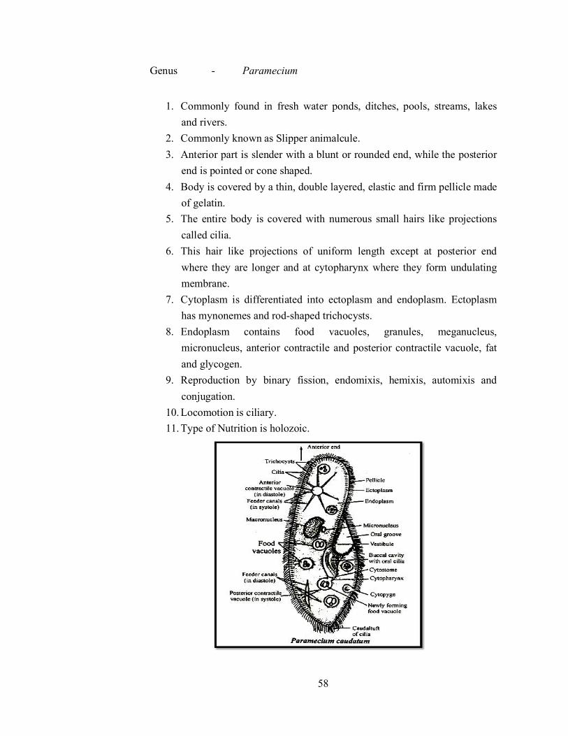
1. Commonly found in fresh water ponds, ditches, pools, streams, lakes
and rivers. 2. Commonly known as Slipper animalcule. 3. Anterior part is slender with a blunt or rounded end, while the posterior
end is pointed or cone shaped. 4. Body is covered by a thin, double layered, elastic and firm pellicle made
of gelatin. 5. The entire body is covered with numerous small hairs like projections
called cilia. 6. This hair like projections of uniform length except at posterior end
where they are longer and at cytopharynx where they form undulating membrane.
7. Cytoplasm is differentiated into ectoplasm and endoplasm. Ectoplasm has mynonemes and rod-shaped trichocysts.
8. Endoplasm contains food vacuoles, granules, meganucleus, micronucleus, anterior contractile and posterior contractile vacuole, fat and glycogen.
9. Reproduction by binary fission, endomixis, hemixis, automixis and conjugation.
10. Locomotion is ciliary. 11. Type of Nutrition is holozoic.
59
(v) Vorticella Classification:
Phylum - Protozoa - Unicellular Sub Phylum- Ciliophora - Locomotion by Cilia in all
stages
Class - Ciliata - Cilia persist throughout life Order - Peritricha - Body bell shaped attached
with a long stalk Genus - Vorticella Characters:
1. Vorticella is solitary animal found in rivers, ponds. It is also found attached to weeds. Stones, aquatic worms, fishes and amphibians with the help of its stalk.
2. Due to the bell shaped body it if often called bell animalcule. 3. The body consists of a thin pellicle and cytoplasm is differentiated
into ectoplasm and endoplasm. 4. A vestibule or infundibulum is found between the peristome and the
peristomal disc. 5. Mouth is situated at the bottom of vestibule leading into the
cytopharynx ending into protoplasm. 6. A ciliary disc is present in the peristome which consists of and outer
adoral cilia and an inner adoral cilia. 7. Endoplasm contains food particles, long and curved macronucleus
and small micronucleus and cytopyge. 8. It does not move freely because it is usually found fixed aborally by
its long highly contractile stalk. However, with the help of stalk and myonemes, the bell sways to and fro in the surrounding water like a flower in a breeze.
9. Nutrition is holozoic. 10. Reproduction is by binary fission and occasionally by conjugation.

60
3.2 Phylum – Porifera Sycon- L.S. Comments
1. Body wall consists of ectoderm, mesenchyme and endoderm. 2. Ectoderm bears small pores called dermal ostia. 3. Mesenchyme consists of amoebocytes, gelatinous transparent matrix and
spicules. 4. Endoderm consists of collar cells or choanocytes and forms the lining of
flagellated chambers. 5. Flagellated chambers open through apopyle in the spongocoel. 6. Incurrent canals and flagellated chambers are communicated by
prosopyles. 7. Spongocoel opens to the exterior by osculum.
8. The path of water circulation is Ostia → Incurrent
canal→prosopyle→radial
canal→apopyles→spongocoel→osculum→Exterior (out).

61
(ii) Sycon T.S. Comments:
1. Body diploblastic consisting of an outer ectoderm, an inner endoderm and a middle layer mesenchyme.
2. Ectoderm comprises a large number of perforations called dermal ostia. This layer is lined by pinacocytes.
3. Mesenchyme consists of gelatinous material and contains spicules,amoebocytes, collenocytes, scleroblasts and archaeocytes.
4. Endoderm consists of single layer of large flagellated collar cells or choanocytes. It forms the lining of radial canals.
5. Radial canals and incurrent canals are communicated by prosopyles. The radial canal opens into the spongocoel by apopyles.
6. In the syconoid type of canal system the flow of water is
Ostia→Incurrent
canal→prosopyle→radialcanal→apopyles→spongocoel→osculum
→ Exterior (out).

62
(iii) Spicules of Sponge Comments:
1. The body wall of sponge is exhibited by various minute, crystalline and calcareous spicules. These are secreted by scleroblasts in the mesenchyme.
2. Spicules have an axis of organic material around which is deposited the inorganic substances either calcium carbonate or hydrated silica.
3. Spicules are classified according to the main axis and rays: (i) Monoaxons- Large one rayed needle like monoaxon spicules are
arranged in a circlet around the osculum. (ii) Triaxons- consists of three axes crossing each other, having six
rays. (iii) Tetraxons- with four rays. (iv) Polyaxons- with many axes and several rays radiating from a
point. 4. Spicules are helpful in identification, classification and metabolism.
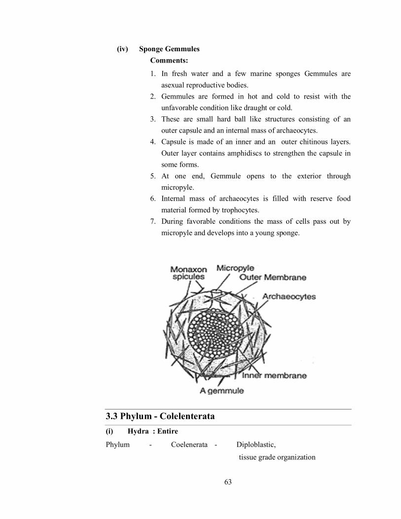
63
(iv) Sponge Gemmules Comments:
1. In fresh water and a few marine sponges Gemmules are asexual reproductive bodies.
2. Gemmules are formed in hot and cold to resist with the unfavorable condition like draught or cold.
3. These are small hard ball like structures consisting of an outer capsule and an internal mass of archaeocytes.
4. Capsule is made of an inner and an outer chitinous layers. Outer layer contains amphidiscs to strengthen the capsule in some forms.
5. At one end, Gemmule opens to the exterior through micropyle.
6. Internal mass of archaeocytes is filled with reserve food material formed by trophocytes.
7. During favorable conditions the mass of cells pass out by micropyle and develops into a young sponge.
3.3 Phylum - Colelenterata (i) Hydra : Entire
Phylum - Coelenerata - Diploblastic, tissue grade organization

64
Class - Hydrozoa - Hydroids bearing medusa with true velum
Order - Hydroida - Polypoid Sub order - Anthomedusae- Hydrotheca and Gonotheca wanting. Genus - Hydra Characters:
1. Body is cylindrical and tube like. 2. Anterior end of the body is called mouth or hypostome encircled by
tentacles. 3. Diploblastic body consisting of outer ectoplasm and an inner endoplasm.
In between these two layers a mesogloea is present. 4. Gonads may also be present on the body. The testes occur near the oral
end which are conical projections. 5. Ovaries are situated towards the proximal end as oval projections. 6. It reproduces asexually by budding and sexually by fusion of gametes.
(ii) Obelia
Phylum - Coelenerata - Diploblastic, tissue grade organization Class - Hydrozoa - Hydroids bearing medusa with true velum Order - Hydroida - Polypoid
Sub order - Leptomedusae- Hydrotheca and Gonotheca wanting. Genus - Obelia
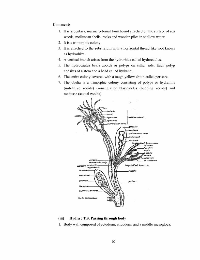
65
Comments
1. It is sedentary, marine colonial form found attached on the surface of sea weeds, molluscan shells, rocks and wooden piles in shallow water.
2. It is a trimorphic colony. 3. It is attached to the substratum with a horizontal thread like root knows
as hydrorhiza. 4. A vertical branch arises from the hydrorhiza called hydrocaulus. 5. The hydrocaulus bears zooids or polyps on either side. Each polyp
consists of a stem and a head called hydranth. 6. The entire colony covered with a tough yellow chitin called perisarc. 7. The obelia is a trimorphic colony consisting of polyps or hydranths
(nutrititive zooids) Gonangia or blastostyles (budding zooids) and medusae (sexual zooids).
(iii) Hydra : T.S. Passing through body 1. Body wall composed of ectoderm, endoderm and a middle mesogloea.

66
2. The epithelio- muscular cells, interstitial cells, nematoblasts, sensory and nerve cells constitute ectoderm.
3. Endoderm consists of nutritive cells, secretory cells, interstitial cells, sensory and gland cells.
4. The middle layer mesogloea composed of a thin, transparent, non-cellular gelatinous material.
5. The body cavity coelenteron lies in the centre.
3.4 Phylum: Platyhelminthes Classification:
(i) Fasciola
Phylum - Platyhelminthes - Organ Grade System, Acoelomate
Class - Trematoda - Flukes, ecto or endoparasites Order - Digenia - Two Suckers without hooks, endoparasitic Genus - Fasciola - Liver Flukes Species - Hepatica

67
Comments:
1. Fasciola is found as an endoparasitic form in the bile duct and biliary passages in the liver of sheep.
2. Commonly known as Sheep Liver Fluke. 3. Body is dorsovenrally flattened, leaf like, elongated and oval shape. 4. Two muscular suckers are found, oral and ventral suckers or acetabulam.
The latter forms adhesive organ with the tissues of the host. 5. Alimentary canal is incomplete consisting of mouth, pharynx, sucker,
oseophagus branched and intestine in diverticulate shape. 6. Hermaphrodite. Male reproductive system consists of 2 tandem dendritic
testes, vasa deferens, seminal vesicle, ejaculatory duct, cirrus, while female system comprises ovary, uterus and vitelline glands.
7. Digenetic life cycle and completed into two hosts. Definite host is Sheep and intermediate host is Limnaea.
(ii) Fasciola hepatica: T.S passing through testes Comments:
1. Body wall is consist of tegument musculature and mesenchyme. 2. Body outermost covering is made up of cuticle.

68
3. Spines and scales are also found on cuticle. 4. Circular longitudinal and oblique muscles constitute the
musculature. 5. Vitelline ducts and glands are visible. 6. Testes containing spermatozoa is clearly visible in the section.
(iv) Fasciola hepatica: T.S through uterus Comments:
1. The ody wall consists of cuticle and musculature. 2. Scales or Spinucles are present on the cuticle. 3. Circular longitudinal and oblique muscles are found. 4. Ovarian and uterine sections are seen in the centre. 5. In the uterine sections fertilized eggs are also observed. 6. Sections of the vitelline glands and ducts are visible. 7. Coelom is absent. Space between body wall and gut is filled with
mesenchyme.

69
(v) Taenia Solium: Scolex
Comments:
1. Scolex is the anterior knob like structure of the body. 2. It is exhibited by four cup like muscular suckers having radial muscles
and an anterior prominent rostellum. 3. Rostellum includes two rows of 22 to 32 curved chitinous hooks. 4. Inner circle with larger hooks and outer circle with smaller hooks. 5. The proximal part bears four adhesive suckers. 6. The scolex with its suckers and hooks is an organ of attachment to the
intestinal wall of the host and acts as holdfast organ.

70
3.5 Phylum - Nemathelminthes (i) Wuchereria
Phylum - Nemathelminthes - Pesudocoelomate, vermiform Class - Nematoda - Unsegmented body. Straight
alimentary canal Order - Filaroidea - Mouth with buccal capsule.
Genus - Wuchereria (Filaria) Species - bancrofti Characters:
1. It is a dreadful endoparasite of man. Adults harbouring the lymphatic vessels and lymph nodes.
2. Its life cycle is digenetic. It involves a secondary host, the blood sucking insects, the female culex, Aedes or Anophoeles.
3. Adult worms are long, hair like, transparent and often creamy white in colour.
4. Filiform in shape with tapering ends; the head end terminating in a slightly rounded swelling.
5. Sexes are separate and sexual dimorphism is distinct. 6. The male worm having a ventrally curved tail-end containing a number
of genital papillae and two spicules of unequal copulatory spicules.. 7. The female worm having a narrow and abruptly pointed tail. 8. The female genital pore or vulva is located ventrally in the pharyngeal
region and provided with a pyriform ovijector. 9. Its mouth is simple without lips. 10. Pharynx is divisible into an anterior muscular and a posterior glandular
parts. 11. The oesophageal bulb is absent. 12. It causes a serious disease known as Filariasis or Elephentiasis.

71
(ii) Dracunculus
Phylum - Nemathelminthes - Pesudocoelomate, vermiform Class - Nematoda - Unsegmented body.
Incomplete alimentary canal
Order - Dracunculoidea - Mouth with buccal capsule. Genus - Dracunculus Species - medinensis
Characters:
1. Dracunculus is commonly called as Guinea worm. 2. It is parasitic in the deeper subcutaneous tissues of human beings where
the long female lies in a loose coil under the skin. 3. The intermediate host is cyclops. 4. Sexual dimorphism is distinct. 5. The head of female if blunt and tail has sharp hooks. 6. The males are few, 20 to 30mmlong with a spirally coiling tail having 10
pairs of papillae and two large equal spicules. 7. A cuticular ring is present aroung the mouth at anterior end having six
labial papillae and four double papillae 8. It causes Guinea worm disease.

72
Unit - 4
Study of Prepared Slides (Higher
Invertebrates) Structure of the Unit
4.0 Objectives
4.1 Introduction
4.2 Study of slides
4.2.1 Phylum Annelida
4.2.1.1 Earthworm : T.S. passing though pharynx
4.2.1.2 Earthworm : Ovary 4.2.1.3 Earthworm: Nerve Ring 4.2.1.4 Leech : T.S. passing through stomach 4.2.1.5 Leech: Nephridium
4.2.2 Phylum Mollusca
4.2.2.1 Unio : cross-section of body 4.2.2.2 Unio : T.S. of shell 4.2.2.3 Pila : L.S. osphradium 4.2.2.4 Pila : radula
4.2.3 Phylum Arthropoda
4.2.3.1 Cyclops 4.2.3.2 Daphnia 4.2.3.3 Lucifer 4.2.3.4 Zoaea larva 4.2.3.5 Prawn : Hastate plate 4.2.3.6 Prawn : statocyst 4.2.3.7 Cockroach: Brain 4.2.3.8 Pediculus 4.2.3.9 Drosophila: Male & Female 4.2.3.10 Drosophila: Sex Comb
4.2.4 Phylum Echinodermata
73
4.2.4.1 Starfish : T.S. of arm 4.2.4.2 Starfish :Bipinnaria larva 4.2.4.3 Echinus : Ehinopluteus larva
4.3 Self-Learning Exercise
4.4 References
4.0 Objectives
After going through this unit you will be able to understand the structure of
tissue mounts and histology of representative higher invertebrate types.
Structure of whole mounts and sections of individuals of different phylum of
higher invertebrates like Annelida, Mollusca, Arthropoda and Echinodermata
with specific characters have been detailed in this unit.
4.1 Introduction
The larger specimens such as prawn, pila, cockroach etc. are preserved in
preservatives for the study of their external features but for the study of
microorganisms, smaller animals and histological studies of tissues or material
in the laboratory readymade pre-stained permanent slides are used. In this unit,
study of prepared slides of Higher Invertebrates ie annelids, molluscs,
arthropods and echinoderms have been elaborated in detail. For the easy
understanding well labeled histological diagrams have also been given.
4.2 Study of slides
4.2.1 Phylum Annelida
Annelids are truly coelomate animals. The coelom is present between body wall
and alimentary canal and is lined with visceral and parietal layers. The animals
are metamerically segmented showing serial repition of body parts.
4.2.1.1 Earthworm : T.S. passing though pharynx
Comments:
1. The section shows organ grade of body organization, triploblastic layeration and true coelom. Pharynx is clearly seen.
2. Pharynx is a wide pear-shaped and thick walled muscular chamber. 3. Pharyngeal cavity dorsoventrally compressed.

74
4. Body wall composed of cuticle, epidermis, musculature and parietal epithelial layer.
5. Cuticle is thin, non-cellular, double layered, iridescent and made up of collagen protein, gelatin and polysaccharide.
6. Epidermis is made up of single layered distinct columnar cells with gland cells, supporting cells, basal cells and receptor cells.
7. Musculature is made up of outer continuous circular muscle fibers and inner longitudinal muscle fibers cut in bundles.
8. The pharynx contains visceral epithelial layer, pharyngeal gland cells, musculo-vascular tissue and pharyngeal lumen.
9. From the pharyngeal wall radial muscle strand run outward up to body wall. The contractions of these radial muscle strands dilate the pharyngeal cavity which works as suction pump.
10. The pharyngeal lumen is divided by an incomplete horizontal shelf into an upper salivary chamber and a lower conducting or ventral chamber. The salivary chamber is dorsally lined with ciliated pharyngeal epithelium.
11. Besides this, other structures like dorsal blood vessel, ventral blood vessel, ventral nerve cord and muscle strands are also seen.
Fig: 4.1 Earthworm: T.S. passing through pharynx

75
4.2.1.2 Earthworm : Ovary Comments:
1. A pair of ovaries is present on the ventral side of the body.
2. The ovaries are pyriform, semi-transparent structures hanging freely into the coelom.
3. Ovaries are attached by their broad ends to the body septum of the 12th and 13th segments.
4. The ovaries lie on either sides of the ventral nerve cord.
5. Each ovary is a white compact mass made up of several finger-like processes.
6. Each ovary contains developing ova in a linear series.
7. Each ovum is large, having a distinct nucleus and a nucleolus.
Fig: 4.2 Earthworm : Ovary
4.2.1.3 Earthworm: Nerve Ring Comments:
1. The nerve ring is present between body segments 3rd & 4th and surrounds & encircles a groove between the buccal cavity and the pharynx.
2. It consists of a dorsal pair of supra-pharyngeal ganglia or cerebral ganglia and a ventral pair of fused sub-pharyngeal ganglia.

76
3. The two pair of ganglia is connected on either side by a pair of circum-pharyngeal connectives.
4. From the sub-pharyngeal ganglia arises a median ventral fused double nerve cord which gives off segmental ganglia from segment 5th to the last segment.
5. Small nerves from ganglia and nerve cord supply to various organs.
Fig: 4.3. Earthworm: Nerve ring
4.2.1.4 Leech : T.S. passing through stomach Comments:
The transverse section passing through stomach shows the following histological structures:
1. Coelomic cavity is filled with botryoidal tissue. 2. Body wall consists of thick cuticle, epidermis and fibrous and ramified
dermis. 3. Muscle layer is composed of thin circular, thick longitudinal, dorso-
ventral, oblique and vertical muscles. 4. Lumen of stomach is less wide than crop. 5. T.S. stomach consists of outer cuticle, longitudinal and circular muscle
layer and endodermal epithelial cells. 6. Folds of epithelial lining of stomach narrower than crop.

77
7. Besides this, a dorsal haemocoelomic vessel, ventral vessel having ventral nerve cord and two lateral vessels having nephridial vesicles are also seen.
Fig.4.4. Leech: T.S. passing through stomach
4.2.1.5 Leech: Nephridium
Comments:
1. The nephridium consists of initial lobe, main lobe and excretory vesicle.
2. The initial lobe is associated with testis sac.
3. The initial lobe leads into a horse shoe-shaped main lobe.
4. In front of the main lobe is an apical lobe and in the concavity of the main lobe is present an inner lobe.
5. From the main lobe a slender vesicle duct leads into a large bladder or vesicle.
6. The vesicle leads by a small excretory duct to a nephridiopore which opens externally.

78
Fig.4.5 Leech :Nephridium
4.2.2 Phylum Mollusca
All the molluscs have shell and mantle. For the first time animals have
developed hard shell for protection. The shell is secreted by mantle.
4.2.2.1 Unio : cross-section of body Comments:
In the cross sections of Unio body the following structures can be seen:
1. Section has shell, mantle, foot and other structures. 2. Mantle is associated with external and internal gill laminae. 3. Each gill lamina is made up of two folds called outer and inner gill
lamellae. 4. Foot and gonads are seen posteriorly. 5. Kidney or nephridium, suprabranchial chamber, pericardial cavity
enclosing ventricles and rectum with typhlosole are seen anteriorly. 6. On the sides of pericardium Keber’s organs are present. 7. Ligament, mantle cavity and muscle fibers are also seen.

79
Fig 4.6 Unio: cross-section of body
4.2.2.2 Unio : T.S. of shell Comments:
1. Shell is secreted by the mantle and is composed of organic and inorganic material.
2. The transverse section of shell shows four layers ie. periostracum, prismatic, lamellated and nacreous layers.
3. The outermost layer is called as the periostracum layer which is made up of organic matter consisting of translucent, chitin-like substance called as conchiolin.
4. Middle layer is called as the ostracum or prismatic layer which consists of tiny calcium carbonate prisms.
5. Prisms are separated by thin layers of conchiolin, arranged vertically. 6. Nacreous layer which is called as the mother-of-pearl is the innermost
layer. 7. Nacreous layer is the iridescent layer and consists of alternate layers of
calcium carbonate and conchiolin, arranged parallel to the surface of the shell.

80
Fig 4.7 Unio :T.S. of Shell
4.2.2.3 Pila : L.S. osphradium Comments:
1. Osphradium is a small lobe-like structure, which is attached to the mantle near the left nuchal lobe.
2. It is a rheoreceptor and chemoreceptor sense organ. 3. It tests the chemical nature of water current and also of the food particles
which enters the mantle cavity. 4. The longitudinal section of osphradium is an oval structure which is
composed of a central axis from which emerges 22-28 oblong fleshy leaflets on either side. This arrangement is called as bipectinate.
5. One end of the leaflet is attached to the mantle, while inner side is attached with the axis.
6. Side leaflets are smaller in size, while middle ones are larger. 7. Osphradium is covered with epithelium. 8. The epithelium contains ciliated supporting cells, non- ciliated
supporting cells, glandular cells, mucocytes and neurosensory cells.
Fig 4.8 Pila :L.S. Osphradium

81
4.2.2.4 Pila : radula Comments:
1. It is a masticatory apparatus and is a ribbon-shaped, chitinous plate. 2. It is present in a radular sac which lies in the buccal mass. 3. The wall of radular sac consists of odontoblast cells, which secrete teeth
of radula. 4. Each teeth row comprises of definitely arranged 7 teeth ie. 2 pairs of
marginals or uncini, one pair of lateral and one central or rachidium teeth.
5. Its teeth formula is 2:1:1:1:2 ie. 2(M):1(L):1(R):1(L):2(M). 6. The teeth are sharp with cutting edges and made up of chitin which is
reinforced by a hardened protein. 7. The radula with its sharp teeth acts like a file and shreds the vegetable
food of the animal.
Fig 4.9 Pila: (A.) Single row of teeth of radula, (B.) Complete Radula
4.2.3 Phylum Arthropoda
Arthropods possess higher grade of organization. For the first time
development of jointed appendages was observed in Arthropods. They also
developed hard body covering made up of chitin. Arthropods are economically
and socially most important animals.
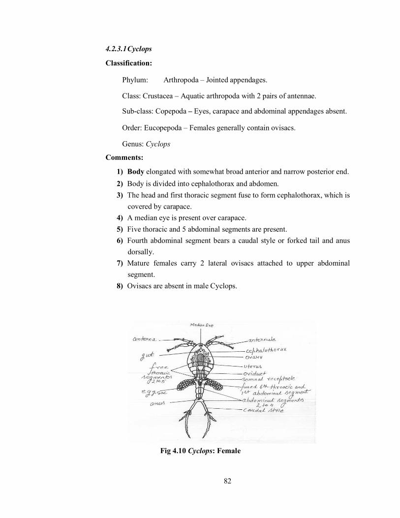
82
4.2.3.1 Cyclops
Classification:
Phylum: Arthropoda – Jointed appendages.
Class: Crustacea – Aquatic arthropoda with 2 pairs of antennae.
Sub-class: Copepoda – Eyes, carapace and abdominal appendages absent.
Order: Eucopepoda – Females generally contain ovisacs.
Genus: Cyclops
Comments:
1) Body elongated with somewhat broad anterior and narrow posterior end. 2) Body is divided into cephalothorax and abdomen. 3) The head and first thoracic segment fuse to form cephalothorax, which is
covered by carapace. 4) A median eye is present over carapace. 5) Five thoracic and 5 abdominal segments are present. 6) Fourth abdominal segment bears a caudal style or forked tail and anus
dorsally. 7) Mature females carry 2 lateral ovisacs attached to upper abdominal
segment. 8) Ovisacs are absent in male Cyclops.
Fig 4.10 Cyclops: Female

83
4.2.3.2 Daphnia
Classification:
Phylum: Arthropoda – Jointed appendages.
Class: Crustacea – Aquatic arthropods with 2 pairs of antennae.
Sub-class: Branchiopoda – Trunk appendages serve as gills.
Order: Cladocera – Four to six pairs of thoracic limbs.
Genus: Daphnia
Comments:
1) Commonly called as ‘water flea’. 2) Body is bilaterally compressed and enclosed in a vestigeal bivalve
carapace ending anteriorly into a backwardly directed rostrum and posteriorly into a spine.
3) Head is not separated from the body by a dorsal notch. 4) Head is rounded and bears large biramous antennae which help in
swimming. 5) Small unjointed antennules, mandibles, maxillulae, and large sessile
eyes are very distinct. 6) 5 pairs and leaf-like thoracic appendages are present. 7) Abdominal appendages are absent. 8) Posteriorly female carries a broad pouch containing various developing
embryos. 9) Brood pouch is absent in male. 10) Thoracic appendages form efficient food-catching organs. 11) Sexes are separate.
Fig.4.11. Daphnia

84
4.2.3.3 Lucifer
Classification:
Phylum: Arthropoda – Jointed appendages.
Class: Crustacea – Thick exoskeleton.
Sub-class: Malacostraca -- Body distinctly segmented.
Order: Decapoda – Ten legs.
Sub-order: Macrura – Abdomen well developed and hard.
Genus: Lucifer
Comments:
1. Animal is minute, slender and delicate. 2. Body divisible into cephalothorax, abdomen and telson. 3. Cephalothorax is smaller than abdomen. 4. Head is extremely elongated and bears long antennae and eyes with long
stalks. 5. Thoracic limbs are non-chelate and the last two thoracic somites are
without limbs. 6. Gills are absent. 7. Abdomen contains abdominal appendages, telson contains uropod.
Fig 4.12 Lucifer
4.2.3.4 Zoaea larva Comments:
1. It is a crustacean larva.
2. Nauplius, after metanauplius or protozoaea directly changes into zoaea larva.
3. Body is divisible into two parts –cephalothorax and abdomen.

85
4. Presence of an anterior rostrum and a median dorsal spine on cephalothorax or carapace.
5. Head bears a large compound eye, antennules, antennae, mandibles, maxillae and maxillipedes.
6. Presence of seven pairs of rudimentary thoracic appendages.
7. Abdomen is six segmented and bears a caudal fork on the last segment.
8. Zoaea larva changes to metazoaea or megalopa larva.
Fig 4.13 Zoaea larva
4.2.3.5 Prawn : Hastate plate Comments:
1. Hastate plate is present on the floor of the cardiac stomach of prawn.
2. It is spear-shaped chitinous plate, which is composed of a median hastate plate, combed plate and guiding ridges or lateral longitudinal folds.
3. Upper surface of hastate plate contains setae and a median ridge. 4. Posteriorly, the hastate plate communicates with cardio-pyloric
aperture. 5. Combed plate contains a row of delicate bristles. 6. Setae and chitinous bristles present on the hastate plate helps in
cutting the food into smaller fragments.

86
Fig 4.14 Prawn: Hastate plate
4.2.3.6 Prawn : Statocyst Comments:
1. Statocyst is the sense organ meant for maintaining the balance and equilibrium of the prawn.
2. One statocyst is present in each antennule in the basal precoxa. 3. It is a pedicellate, whitish and vescicle-like hollow chitinous structure
measuring about 1mm in diameter. 4. Inside the cavity of each statocyst, a cluster of sand grains termed
statolith is present in the centre. 5. The statolith is surrounded by a circlet of delicate and elongated sensory
setae. 6. Each setae are connected with the statocystic nerve which is a branch of
the antennulary nerve and functions as a receptor. 7. Any change in swimming position brings change in position of sand
particles, which stimulate setae and the animal comes in proper position.

87
Fig 4.15 Prawn: (A.) Statocyst in situ, (B) T.S. Of Statocyst
4.2.3.7 Cockroach: Brain Comments:
1. The brain in cockroach is also called as supra-oesophageal ganglion. 2. It is a large and bilobed mass located in head above oesophagus. 3. It represents 3 pairs of ganglia of head region fused together. 4. It is divided into three parts: protocerebrum, deuterocerebrum and
tritocerebrum. 5. Remaining three pairs of ganglia of head fuse to form the sub-
oesophageal ganglion which lies below the oesophagus. 6. Brain and sub-oesophageal ganglion are connected together, on either
side of oesophagus, by a circum-oesophageal commissure.
Fig 4.16 Cockroach: Brain

88
4.2.3.8 Pediculus
Classification:
Phylum: Arthropoda – Jointed appendages.
Class: Insecta – Abdominal appendages absent.
Sub-class: Pterygota – Winged insects with complete or incomplete
metamorphosis.
Division: Exopterygota- Wings externally originated with simple
metamorphosis.
Order: Anoplura–Mouth parts piercing and sucking, ectoparasites
without metamorphosis.
Genus: Pediculus
Comments:
1. Cosmopolitan ectoparasite on man. It is also present in apes and monkeys.
2. Body is dorsoventrally flattened and measures about 1-3mm in length.
3. Head is pointed with two lateral compound eyes and a pair of small antennae.
4. Highly modified mouth-parts for piercing and sucking blood.
5. Thorax is small, unsegmented and wingless.
6. Thorax contains three pairs of clawed legs which are strong and the movable claws helps for holding firmly the hair of the host.
7. The abdomen is narrow anteriorly but broadens out posteriorly.
8. The abdomen is nine segmented.
9. In males, posterior end is turned upwards and in females, it does not turn upwards.
10. They not only cause itching and falling of hair but are also carrier of diseases like typhus, relapsing fever, and trench fever, which is transmitted by their excreta into the host through the punctured skin.
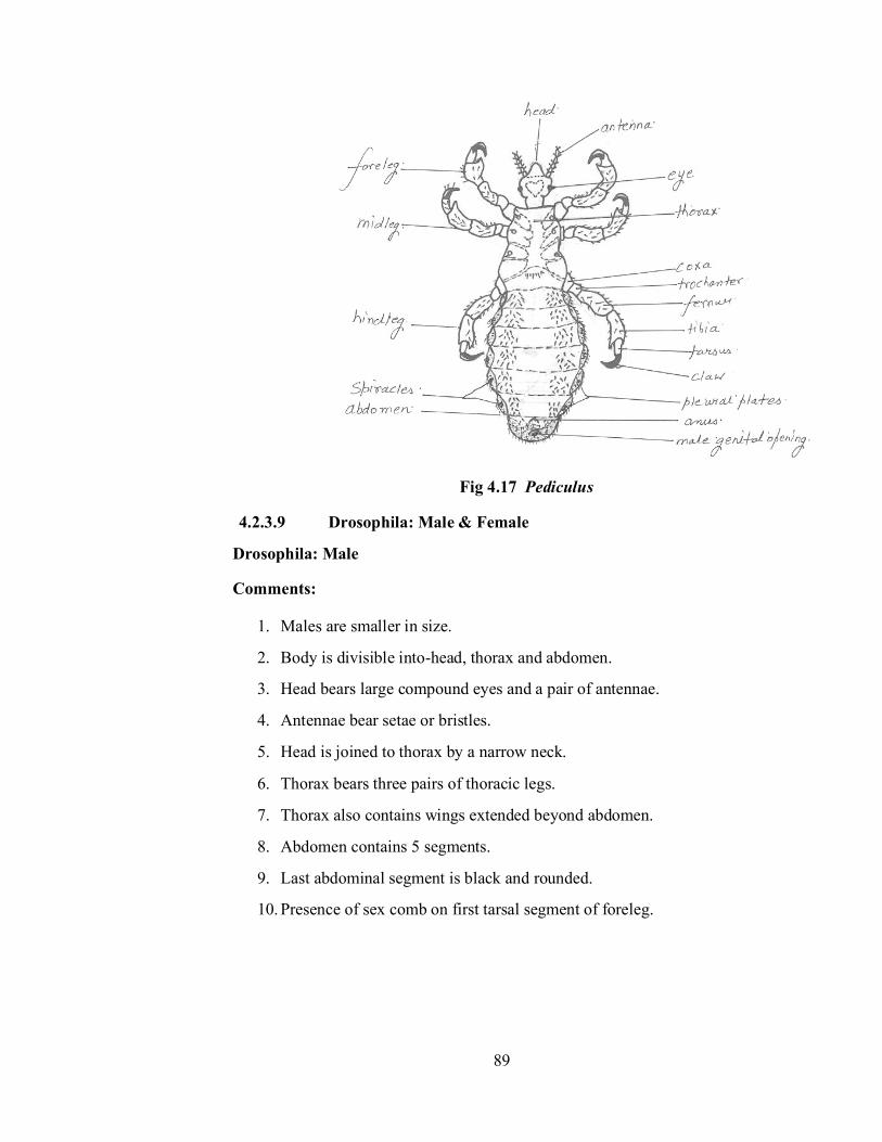
89
Fig 4.17 Pediculus
4.2.3.9 Drosophila: Male & Female
Drosophila: Male
Comments:
1. Males are smaller in size.
2. Body is divisible into-head, thorax and abdomen.
3. Head bears large compound eyes and a pair of antennae.
4. Antennae bear setae or bristles.
5. Head is joined to thorax by a narrow neck.
6. Thorax bears three pairs of thoracic legs.
7. Thorax also contains wings extended beyond abdomen.
8. Abdomen contains 5 segments.
9. Last abdominal segment is black and rounded.
10. Presence of sex comb on first tarsal segment of foreleg.
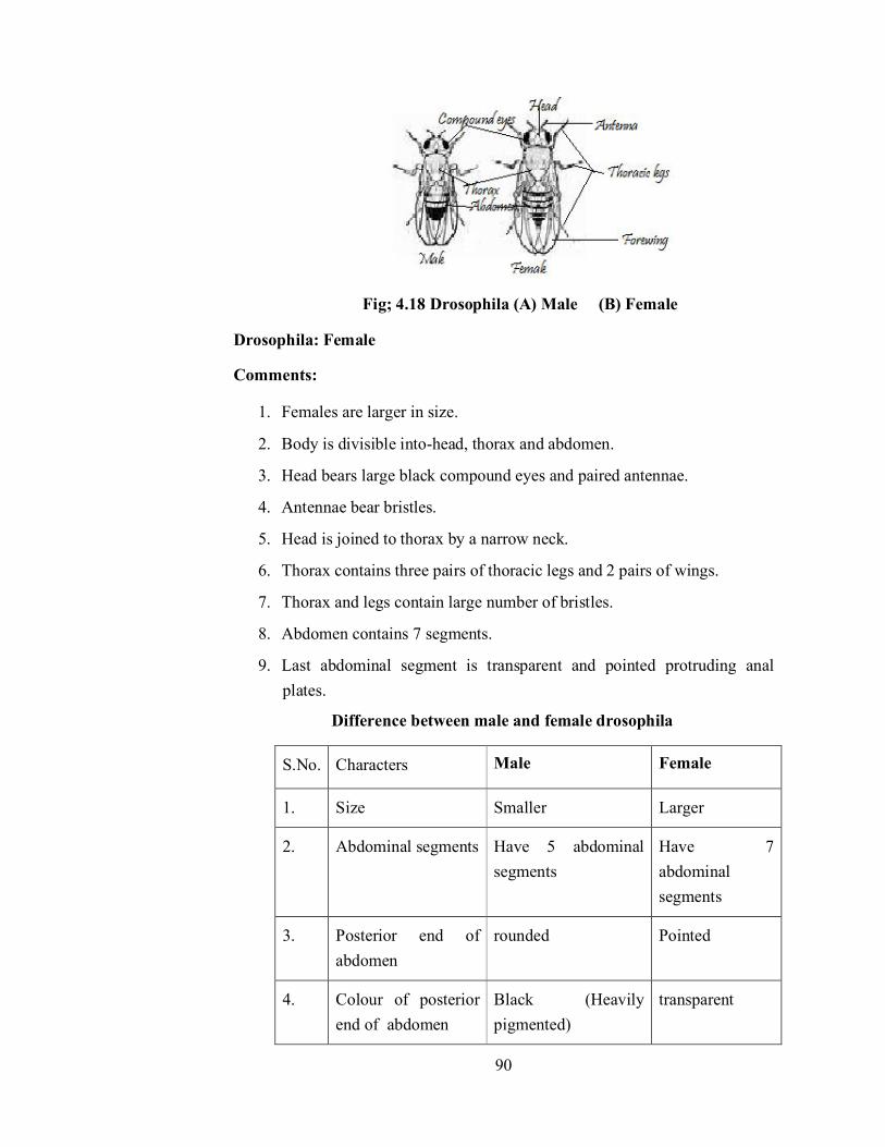
90
Fig; 4.18 Drosophila (A) Male (B) Female
Drosophila: Female
Comments:
1. Females are larger in size.
2. Body is divisible into-head, thorax and abdomen.
3. Head bears large black compound eyes and paired antennae.
4. Antennae bear bristles.
5. Head is joined to thorax by a narrow neck.
6. Thorax contains three pairs of thoracic legs and 2 pairs of wings.
7. Thorax and legs contain large number of bristles.
8. Abdomen contains 7 segments.
9. Last abdominal segment is transparent and pointed protruding anal plates.
Difference between male and female drosophila
S.No. Characters Male Female
1. Size Smaller Larger
2. Abdominal segments Have 5 abdominal segments
Have 7 abdominal segments
3. Posterior end of abdomen
rounded Pointed
4. Colour of posterior end of abdomen
Black (Heavily pigmented)
transparent

91
5. Sex comb Present on Ist tarsal segment of foreleg
Absent
4.2.3.10 Drosophila: Sex Comb Comments:
1. In Drosophila, a thoracic leg is composed of coxa, trochanter, femur, tibia and 5 jointed tarsus.
2. In male drosophila, the first tarsal segment of Ist thoracic leg (foreleg) contains a comb –like structure called as sex comb.
3. Sex comb consists of row of about 10 short thick, black bristles.
Fig 4.19 Drosophilia : Sex comb
4.2.4 Phylum Echinodermata Echinoderms reveal supreme example of invertebrate evolution being at top amongst the invertebrate and very close to chordates. For the first time echinoderms have developed mesodermal endoskeleton of calcium caronate in the form of ambulacral ossicles or dermal ossicles.
4.2.4.1 Starfish : T.S. of arm Comments:
1. T.S. of arm of a starfish includes tube-feet, nervous tissue, pyloric caeca and body wall appendages.
2. Body wall is composed of two layered cuticle, tall flagellated columnar epidermis, connective tissue, dermis and musculature.
3. Dermis contains dermal ossicles and haemal spaces.

92
4. Musculature comprises of outer circular and inner longitudinal muscle fibres.
5. Spines, pedicellariae and branchiae are present on the aboral surface.
6. Oral surface bears V-shaped ambulacral groove, which is occupied by tube feet.
7. Extension of pyloric caeca and gonads are present in the perivisceral coelom.
8. Besides this, other structures like radial nerve, radial canal, perihaemal canal, radial hyponeural sinus, septum dividing radial hyponeural sinus, lower ambulacral muscles, upper transverse ambulacral muscle, peribranchial sinus, longitudinal muscle thickening, radial water canal, ambulacral spines and radial haemal strand are also seen.
Fig 4.20 Starfish: T.S of arm
4.2.4.2 Star-fish : Bipinnaria larva
Comments:
1. In asteroids, the egg, after holoblastic unequal cleavage and embolic gastrulation, changes into a typical eleutherozoan larva ie. Bipinnaria larva.
2. The larva is formed within few weeks. Although a free-swimming larva is hatched within 24 hours, but it takes few days to develop into bipinnaria larva.
3. A newly hatched larva assumes angular form by the time coelomic chambers are separated.
4. Outgrowths from the principal parts of the elongated larval body form arms.
5. Arms are located at posterolateral, postoral, posterodorsal, anterodorsal, preoral, and dorsomedian positions.

93
6. Presence of preoral and postoral ciliary bands.
7. Preoral ciliated band surrounds the preoral lobe.
8. Postoral ciliated band is present between the mouth and the anus.
Fig 4.21 Starfish :Bipinnaria Larva
4.2.4.3 Echinus : Echinopluteus larva
Comments:
1. In echinoids, after gastrulation, egg hatches into first stage larva known as Echinopluteus larva.
2. Larva has conical shaped body.
3. Alimentary canal is differentiated into oesophagus, stomach and intestine.
4. Blastopore continues as larval anus.
5. Presence of five pairs of arms viz. preoral, anterolateral, postoral, anterodorsal and posterodorsal.
6. Posterolateral arms, if present, are directed backward and outward.
7. Ciliated bands are in the form of epaulettes.
8. Arms are supported by calcareous rods.
9. The echinopluteus larva is a microscopic, pelagic and planktonic larva and develops into adult after metamorphosis in a month or so.
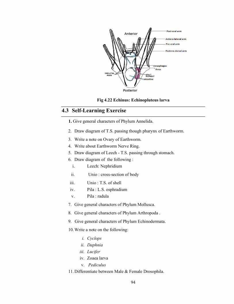
94
Fig 4.22 Echinus: Echinopluteus larva
4.3 Self-Learning Exercise
1. Give general characters of Phylum Annelida.
2. Draw diagram of T.S. passing though pharynx of Earthworm.
3. Write a note on Ovary of Earthworm. 4. Write about Earthworm Nerve Ring. 5. Draw diagram of Leech - T.S. passing through stomach. 6. Draw diagram of the following :
i. Leech: Nephridium
ii. Unio : cross-section of body
iii. Unio : T.S. of shell iv. Pila : L.S. osphradium v. Pila : radula
7. Give general characters of Phylum Mollusca.
8. Give general characters of Phylum Arthropoda .
9. Give general characters of Phylum Echinodermata.
10. Write a note on the following:
i. Cyclops ii. Daphnia
iii. Lucifer iv. Zoaea larva v. Pediculus
11. Differentiate between Male & Female Drosophila.

95
12. Write about Sex Comb of Drosophila. 13. Describe the Prawn: Hastate plate. 14. Write the structure of statocyst of Prawn. 15. Write the structure of Brain of Cockroach. 16. Draw diagram of the following:
i. Starfish : T.S. of arm ii. Starfish :Bipinnaria larva
iii. Echinus : Ehinopluteus larva
4.4 References
R.L.Kotpal : Modern Text Book of Zoology Invertebrates
S.S. Lal : Practical Zoology Invertebrate
P.S.Verma : A Manual of practical Zoology Non-Chordates
T.Poddar, S.Mukhopadhyay & S.K.Das : An Advanced Laboratory Manual of Zoology
96
Unit - 5
Blood Physiology Structure of the Unit
5.0 Objectives
5.1 Introduction
5.2 Study of Blood Physiology
5.2.1 Blood Group Identification
5.2.1.1 Object
5.2.1.2 Principle
5.2.1.3 Apparatus & Chemicals
5.2.1.4 Procedure
5.2.1.5 Observation
5.2.1.6 Result
5.2.2 Counting of Red and White Blood Corpuscles
5.2.2.1 Object
5.2.2.2 Apparatus & Chemicals
5.2.2.3 Counting of RBCs
5.2.2.4 Counting of WBCs
5.2.3 Haemoglobin Estimation
5.2.3.1 Object
5.2.3.2 Apparatus & Chemicals
5.2.3.3 Procedure
5.2.3.4 Result
5.2.4 Haematocrit Value
5.2.4.1 Object
5.2.4.2 Principle
5.2.4.3 Apparatus & Chemicals
5.2.4.4 Procedure

97
5.2.4.5 Observation
5.2.4.6 Calculation
5.2.4.7 Result
5.3 Self-Learning Exercise
5.4 References
5.0 Objectives
Analysis of the components and cells of the blood in this unit will make you understand the procedure in medical diagnosis to know the number, proportions and shape of the blood cells and the quantities of proteins which help the physician to draw a correct diagnosis. Analysis of blood is often carried out to understand the diseases and physiological processes, values and composition. The aim of the following exercises is to study some basic phenomena like Blood Group Identification, RBC & WBC Counting, Haemoglobin Estimation, Haematocrit Value of the human or students in the lab.
Blood is a liquid connective tissue that consists of cells and cell fragments surrounded by a liquid extracellular matrix (or blood plasma). Blood has three general functions: transport, homeostasis and protection. It transports respiratory gases (O2 and CO2) between lungs and body cells and nutrients, waste products and different regulatory molecules (like hormones) inside our body. Circulation of blood helps maintain the homeostasis of all body fluids, including the regulation of pH, osmolarity or temperature. Blood also protects our body either by clotting and thus, preventing excessive bleeding or can fight diseases or pathogenic intruders.
5.1 Introduction
Blood plasma is a straw-coloured (yellowish) liquid, which makes up about 55% of total blood volume after the sedimentation of formed elements in a blood sample. It contains approx. 91.5% water and 8.5% solutes, most of which are proteins like albumins, globulins or fibrinogen. Antibodies or immunoglobulins belong to the group of globulins, and play important role during certain immune responses. Formed elements include three principal components: red blood cells (RBCs or erythrocytes), white blood cells (WBCs or leukocytes) and platelets (thrombocytes). While RBCs and WBCs are whole cells, platelets are only cell fragments.
98
The percentage of total blood volume occupied by formed elements, mostly RBCs is called the haematocrit, its normal range is necessary to keep a person healthy. RBCs are biconcave discs. In their mature form, they lack a nucleus. The ruptured cells are removed and destroyed by macrophages in the spleen and liver. WBCs have nuclei and all cellular organelles and are classified as either granular or agralanular. Granular leukocytes include neutrophils, eosinophils and basophils, depending on the type of dyes staining their granules. Agranular leukocytes include lymphocytes and monocytes. Platelets are irregularly disc-shaped, 2-4 µm diameter cellular fragments of megakaryocytes. They have a short life span (4-9 days) in the circulation and then are removed by fixed macrophages in the spleen or liver. They have no nuclei but contain many vesicles which promote blood clotting upon the release of their content. Platelets additionally help stop blood loss by forming a platelet plug in the damaged vessels.
The diagnosis of blood parameters make aware of body physiology. The RBCs above the normal rang results in polycythemia whereas the lower counts result anaemia. The number of WBC more than normal results in leukocytosis whereas the decrease in number result in leukopenea. If the haemoglobin percentage is lower than normal then the person may be anaemic. Similarly if the measure of haematocrit value is more than normal it may lead to polycythemia and lower values indicate anaemia in the person.
5.2 Study of Blood Physiology
5.2.1 Blood Group Identification
5.2.1.1 Object: Identification of ABO blood groups and Rh factor in human blood.
5.2.1.2 Principle: According to Karl Landsteiner on basis of absence or presence of A or B antigens in Red blood cells, the blood is classified into 4 groups:
1. Blood group A having antigen-A in RBC and antibodies B
2. Blood group B having antigen-B in RBC and antibodies A
3. Blood group AB having antigen-A & B in RBC but no antibodies
4. Blood group O do not have any antigen in RBC but with antibodies A & B.

99
The serum antibodies of the same person or same blood do not agglutinate the Antigens (glycolipid or glycoprotein) present in RBC but when different blood is transfused then the agglutination takes place. The possible donor and recipients of blood are:
Blood Group Blood cell Antigen
Serum Antibody
Donor Recipient
A A B A, AB O, A
B B A B, AB O, B
AB A & B None AB Universal Recipient
O None A & B Universal Donor
O
Rhesus factor (Rh) is derived from Rhesus macaque monkeys. They are Rh+ and Rh- agglutinogen in RBC of human. When a person of Rh- blood group is frequently given transfusion of Rh+ blood the former develops rh-agglutinins. Because of rh- agglutinins of blood group become compatible to agglutinins of blood group A and B and they cause haemolysis of RBC Rh factor has great medical significance during pregnancy. With Rh+ father and Rh- mother foetus may be Rh+. Some RBC (with Rh+) may pass into the blood of mother due to which mother develops anti-Rh agglutinins. After the birth of first child mother becomes sensitized. In second pregnancy of sensitized mother some of rh- red blood corpuscle would reach through placenta into the foetus. The RBC of foetus are haemolysed by sensitized RBC of mother and RBC of foetus are destroyed resulting into anaemia in foetus and cause death of foetus. Above disease is called an erythroblast foetalis.
5.2.1.3 Apparatus & Chemicals: Cavity slides, blood sample vials or small glass tubes, Antiserum (A, B and D) and pathological binocular microscope.
5.2.1.4 Procedure:
1. Take sterilized cotton wet in 90% alcohol and sterilize the tip of your middle finger.

100
2. After the tip of the finger becomes dry, prick it with a sterilized needle.
3. Collect the blood in sample vial containing 1 ml of 0.85% sodium chloride (0.85gms of sodium chloride powder dissolved in 100cc of distilled water).
4. Take 2 cavity slides wash and clean them and let the slide become dry. 5. Add a drop of samples of blood in each cavity. 6. Now add a drop of antiserum (A, B and D) separately in cavities (Fig.:
5.1) and mix the blood sample with a sterilized needle. 7. Leave for 2 minutes and observe.
5.2.1.5 Observation:
Blood group/Sera
Anti-A Anti-B Anti-D
O- Normal Normal Normal
O+ Normal Normal Clumping

101
A- Clumping Normal Normal
A+ Clumping Normal Clumping
B- Normal Clumping Normal
B+ Normal Clumping Clumping
AB- Clumping Clumping Normal
AB+ Clumping Clumping Clumping
5.2.1.6 Result: Blood group is ……………
5.2.2 Counting of Red and White Blood Corpuscles
5.2.2.1 Object: Count Red Blood Corpuscles and White Blood Corpuscles in a sample of blood using haemocytometer.
5.2.2.2 Apparatus & Chemicals: Haemocytometer, Microscope, Pricking needle, EDTA or heparin, Hayem’s solution, WBC diluting fluid, Distilled water, Absolute alcohol and Blood.
Haemocytometer: In haemocytometer set of thick glass counting slide or Neubaur’s slide, one cover slip and two pipettes are present.
(a) Counting slide or Neubaur’s slide:
The Neubaur’s slide consists of a double-cell slide with two sunk platforms and a cover slip. Each platform has a ruling. When a special cover glass is kept in position, a depth of 1/10 mm is maintained over the rulings. The diluted blood is run on by capillary force after the cover glass has been fixed. The counting chambers are nine large squares having sides of 1 mm each. The central large square ABCD is subdivided in 25 medium squares and each medium square in turn contains 16 tiny squares having sides 0.05 mm long (Fig.: 5.2).

102
(b) RBC and WBC pipettes: There are two pipettes. One is for RBC and other is for WBC counting. The RBC pipette contains red bead in its bulb with red ring for sucking blood. Whereas in WBC pipette white bead is present in bulb and it is fitted with white ring at its terminal for sucking blood. RBC pipette is marked .05, 1.0 and 101 whereas the marking on WBC pipette is 0.5, 1.0 and 11 (Fig.: 5.3).

103
Hayem’s solution: Hayem’s solution or RBC diluting solution is prepared in laboratory by mixing:
HgCl2 - 0.5gm
Na2SO4 - 5.0gm
NaCl - 1.0gm
Distill water - 200ml
Mix above salts in 200ml distill water, filter and keep in a bottle. It is isotonic to blood hence prevent haemolysis in RBCs. HgCl2 prevents bacterial growth.
Turck’s fluid or WBC diluting fluid: Turck’s fluid or WBC diluting fluid is prepared in laboratory by mixing:
Gentian violet - 10gm
Glacial acetic acid - 1ml
Distill water - 100ml
Keep the solution in bottle. This solution destroy RBCs and stain nucleus of WBCs.
5.2.2.3 Counting of RBCs:
(a) Procedure:
1. Clean and dry the RBC pipette, Neubaur’s slide and coverslip with distil water. 2. Wipe finger with cotton soaked with alcohol and prick the tip with a
sterile needle.

104
3. Aspirate the oozed blood up to 0.5 mark in the RBC diluting pipette and immediately dilute the blood with Hayme’s solution or RBC diluting fluid 200 times up to 101 marks.
4. Hold the pipette horizontally and rotate it with both hands between finger and thumb so as to allow the blood to mix with diluting solution.
5. Discard the clear fluid in the capillary part of pipette as this fluid is not mixed with blood.
6. Place coverslip on the counting chamber and rapidly drop mixture to the edge of the coverslip.
7. Allow the corpuscles to settle down and make a count under the microscope.
8. Scan the counting area with objective lens. 9. Count cells in 16 squares in five different parts of the field i.e. 80
smallest squares.
10. Include all cells lying on the upper and left lines of any square, omit the cells on the lower and right- hand lines.
(b) Calculation: The number of erythrocytes per cubic mm is calculated as follows:
(c) Result: The number of RBCs in the given sample of blood are ……… per sq mm.
(Ideal number of RBCs in male are 55 lacs and in female are 45 lacs per sq mm of blood)
5.2.2.4 Counting of WBCs:
(a) Procedure:
1. Clean and dry the WBC pipette, Neubaur’s slide and coverslip with distil water.
2. Sterilize the tip of middle finger wiping with cotton soaked in alcohol and prick with a sterile needle.
3. Aspirate the oozed blood up to 0.5 mark in the WBC diluting pipette and immediately dilute the blood with Turk’s fluid or WBC diluting fluid 20 times up to 11 marks.
105
4. Hold the pipette horizontally and rotate it with both hands between finger and thumb so as to allow the blood to mix with diluting solution.
5. Discard the clear fluid in the capillary part of pipette as this fluid is not mixed with blood.
6. Place coverslip on the counting chamber and add immediately and rapidly a drop of mixture to the edge of the coverslip.
7. Allow the corpuscles to settle down and make a count under the microscope.
8. Scan the counting area with objective lens. 9. The white cells are recognized by their retractile appearance and by
slight colour given to them by the diluting fluid. The counting is performed in the four corners of 1 square millimeter.
10. Include all cells lying on the upper and left lines of any square, omit the cells on the lower and right- hand lines.
(b) Calculation: The number of leucocytes per cubic mm is calculated as follows:
(c) Result: The number of WBCs in the given sample of blood are
……………. per sq mm.
(Ideal number of WBCs are 6000 to 8000 per sq mm of blood)
5.2.3 Haemoglobin Estimation
5.2.3.1 Object: Estimation of haemoglobin percentage of blood.
5.2.3.2 Apparatus & Chemicals: Haemoglobinometer, N/10 HCI, H2O2, Absolute alcohol, Distill water and Blood.
Haemoglobinometer: Generally Sahli’s haemometer (Fig.: 5.4) is used. In this apparatus two lateral standards glass rods of brown colour are present. In center of brown rods empty measuring test tube is found. Where haemoglobin percentage is marked. N/10 HCI is used for hemolysis of blood. Due to HCl RBCs are damaged and the haemoglobin released is converted into hematin acid.

106
Haemoglobin pipette is also present with this apparatus. This capillary tube is of equal diameter marked 200µm or 20 sq mm. The other end of this pipette is fitted with rubber tube through which blood is sucked.
5.2.3.3 Procedure:
1. Clean the Haemoglobin pipette and measuring test tube in haemoglobinometer with H2O2 and then by absolute alcohol and dry.
2. Pour N/10 HCI in the measuring test tube up to the mark with the help of dropper.
3. Sterilize the tip of middle finger wiping with cotton soaked in alcohol and prick with a sterile needle.
4. Aspirate the oozed blood up to 0.2 ml mark in the pipette. 5. Transfer the blood carefully into the haemoglobinometer tube containing
small amount of N/10 HCI. 6. Add distilled water drop by drop into the tube and constantly match the
colour of the mixture with the colour of adjacent tubes of either side of the instrument. Continue this until the colour of the mixture is exactly matched with the colour of standards.
107
7. Find out the end point. It may be found out by noting the point where the colour of the solution becomes lighter by addition of very next drop. Take more than one reading to avoid error. By addition of N/I0 HCI, a definite quantity of haemoglobin is converted into acid Haematin.
5.2.3.4 Result: The percentage of haemoglobin in the blood is ……………. .
(Ideal haemoglobin %tage in female is 13.7 and in male is 15.8)
5.2.4 Haematocrit Value
5.2.4.1 Object: Estimation of haematocrit value in a blood sample.
5.2.4.2 Principle: Centrifugation of blood sample separates erythrocytes or RBCs (settle down) and Plasma. Plasma is seen as a clear liquid. The volume of settled down RBCs represent PCV (packed cell volume). PCV can be calculated by standard Wintrobe method. The haematocrit value is used to diagnose diseases. Excess values cause polycythemia and lower values cause anaemia.
5.2.4.3 Apparatus & Chemicals: High speed centrifuge, centrifugal tubes or Wintrob’s tube, EDTA (Ethylenediamine triacetic acid) or sodium citrate or sodium acetate as anticoagulant and Blood sample.
5.2.4.4 Procedure:
1. Wash the graduated centrifuge tube. 2. Dry the tubes in incubator. 3. Take a freshly chloroformed rat, dissect quickly and puncture the heart. 4. Add a little EDTA or sodium citrate or sodium acetate in centrifuge
tubes and collect 5cc of blood in each centrifuge tube. 5. Seal the end point of centrifuge/capillary tube in which blood is not
present. 6. Keep the blood containing centrifuge tubes in the centrifuge. Centrifuge
the blood sample at 3000 rpm (revolutions per minute) for 10 minutes for applying sufficient relative centrifugal force (RCF) for the separation of plasma and blood cells. The dark coloured settled blood cells and a clear yellowish plasma will be seen.
7. Measure the length of separated blood cells and total length (blood plasma + blood cells).
5.2.4.5 Observation: Observe the following:
1. Volume of blood = …………….

108
2. Volume of the blood corpuscles = …………….
5.2.4.6 Calculation: Calculate PCV by the following formula:
5.2.4.7 Result: Haematocrit value or PCV = ………….. %.
(Ideal value of PVC in man is 45%, in female is 42% and in newly born baby is 53-65%)
5.3 Self-Learning Exercise
1. Identification of your own blood groups.
2. Identify Rh factor in given sample of blood.
3. Count Red Blood Corpuscles in your own blood using haemocytometer.
4. Estimate White Blood Corpuscles in given sample of blood using haemocytometer.
5. Estimation of haemoglobin percentage in your own blood.
6. Estimation of haematocrit value in a given blood sample.
7. Estimate PCV in your own blood.
8. Write comments on:
1. Neubaur’s slide
2. RBC/WBC pipette
3. Haemocytometer
4. Haemoglobinometer
5. Rh factor
6. ABO blood groups
7. PCV
5.4 References
Animal Physiology and Biochemistry, Sastry K.V., Rastogi Publication, Meerut
Eckert Animal Physiology, Randall D., Burggren W., French K.W.H., Freeman and Company, New York
Practical Zoology Vertebrate, Lal S.S., Rastogi Publication, Meerut

109
Animal Physiology and Biotechnology, Bhatia A.L., and Kohali K.S. Ramesh Book Dpot, Jaipur

110
Unit - 6
Cell Bilogy
Structure of Unit
6.1 Aim : To study prepared slides of mitosis
6.2 Aim :To perform and identify different stages of mitosis in onion root tip.
6.3 Aim: Study of stages of meiosis using permanent slides
6.4 Aim :To study and identification of Meiosis in Grasshopper testis cells
6.1 Aim : To study prepared slides of mitosis
6.1.1 Objectives
After Going through this unit, you will be able to:
Explain the principle of this method
Write about the procedure of this method
6.1.2 Principle
Somatic growth in plants and animals takes place by the increase in the number of cells. A cell divides mitotically to form two daughter cells wherein the number of chromosomes remains the same (i.e., unchanged) as in the mother cell. In plants, such divisions rapidly take place in meristematic tissues of root and shoot apices, where the stages of mitosis can be easily observed. Mitosis, cell division which produces two daughter cells both containing a full set of chromosomes, is the natural beginning for a discussion of both cellular reproduction and the molecular basis of heredity. While mitosis itself is not present in all sets of state science standards, the fundamental nature of the concept makes it an important part of any general biology curriculum. An understanding of mitosis is required for a full understanding of meiosis, and meiosis is required for a deep understanding of heredity
6.1.3 Apparatus, Glasswares And Chemicals
Permanent slides of mitosis, Light microscope

111
6.1.4 Procedure
Examine prepared slides of both onion root tips. Locate the meristematic region of the onion with the 10X objective, and then use the 40X objective to study individual cells.
Identify and sketch one cell which clearly represents each phase. Use the spaces provided below.
1. The nondividing cell is in a stage called interphase, The nucleus may have one or more dark-stained nucleoli and is filled with a fine network of threads, the chromatin. During interphase, DNA replication occurs.
2. The first signs of a division is prophase, in which a thickening of the chromatin threads occurs. Thickening continues until it is evident that the chromatin has condensed into chromosomes. With somewhat higher magnification you may be able to see that each chromosome is composed of two chromatids joined at a centromere. As prophase continues, the chromatids continue to shorten and thicken. In late prophase the nuclear envelope and nucleoli are no longer visible, and the chromosomes are free in the cytoplasm. Just before this time the first sign of a spindle appears in the cytoplasm; the spindle apparatus is made up of microtubules, and it is thought that these microtubules may pull the chromosomes toward the poles of the cell where the two daughter nuclei will eventually form.
3. At metaphase, the chromosomes have moved to the center of the spindle. One particular portion of each chromosome, the centromere, attaches to the spindle. The centromeres of all the chromosomes lie at about the same level of the spindle, on an imaginary plane called the metaphase plate. At metaphase you should be able to observe the two chromatids of some chromosomes.
4. At the beginning of anaphase, the centromere regions of each pair of chromatids separate and are moved by the spindle fibers toward opposite poles of the spindle, dragging the rest of the chromatid behind them. Once the two chromatids separate, each is called a chromosome. The daughter chromosomes continue poleward movement until they form two compact clumps, one at each spindle pole.
5. Telophase, the last stage of division, is marked by a pronounced condensation of the chromosomes, followed by the formation of a new nuclear envelope around each group of chromosomes. The chromosomes gradually uncoil to form the fine chromatin network of

112
interphase, and the nucleoli and nuclear envelope reappear. Cytokinesis may occur. This is the division of the cytoplasm into two cells. In plants, a new cell wall is laid down between the daughter cells. In animal cells, the old cell will pinch off in the middle along a cleavage furrow to form two new daughter cells.
6.1.5 Observation and Calculation
Interphase Prophase Metaphase Anaphase
Observe the slide first under low power and then under high power, Find the stages of prophase, metaphase, anaphase and telophase and compare them with the given figures.
6.1.6 Precautions
Use freshly prepared chemicals.
Always use clean glassware in the experiment
6.1.7 Exercise
Write the procedure and result in your own words.
6.2 Aim To perform and identify different stages of mitosis in onion root tip. 6.2.1 Objectives
After Going through this unit, you will be able to:
Explain the principle of this method
Write about the procedure of this method
6.2.2 Principle
Onion root tip has meristematic tissue just behind the root cap, hence this serves as a good material for studying various stages of mitosis. The roots can be easily grown if an onion is placed on a water filled conical flask in such a way that the onion disc touches water or else making the onion to sit on wet sand in such a way that the bulb is buried partially in sand. In two days time nearly 1 cm long roots develop, which can then be cut, fixed and stored. Longer grown roots are not good as it will have more of non-dividing tissue.

113
A process by which a parent cell divides into two or more daughter cells is called cell division. Cell division is a small part of the cell cycle. In normal eukaryotic cells the type of cell division is known as mitosis. Another type of cell division is present in reproductive cells of eukaryotes known as meiosis.
The process of Mitosis is mainly divided into four stages: Prophase, Metaphase, Anaphase and Telophase.
Prophase: During this stage, the chromosomes are super coiled, condensed and become visible for first time during the cell cycle. The spindle fibers start forming. The nuclear membrane starts disintegrating.
Metaphase: During this stage, the spindle fibers reach and attach to centromere of each sister chromatids. The chromosomes align along the center plane of the cell. The nuclear membrane disintegrates completely.
Anaphase: During this stage, the centromeres start splitting and the sister chromatids begin migrating towards the opposite poles of the cell.
Telophase: During this stage, the chromosomes are clustered on the either ends of the cell. The nuclear membrane starts reforming. The cell plate (new cell wall) starts to form between the two daughter nuclei. This will be followed by cytokinesis.
6.2.3 Apparatus, Glasswares and Chemicals
Apparatus and Glasswares: Root tips of onion, Spirit lamp, needle, slide, cover slip, watch glass, forceps, droppers etc.
Chemicals: FAA solution, IN HCl, Acetocarmine stain
6.2.4 Pre-Prepration
To perform this experiment, first take 2-3 onion and place them on a glass of water without dipping in the water. Place the glass in slightly dark place and study it daily when the young roots are about 2-3 mm long, then cut 2-3 mm of the root apex and transfer in a small bottle in which FAA solution is present. FAA solution is made from formaline, acetic acid and 90% alcohol are mixed in equal proportions.
6.2.5 Procedure
1.Take out 2-3 piece of root tips kept in FAA, place them in watch glass and wash them with water. 2. Now place the root tips on a slide and add few drops of IN HCl on them. Heat the slide on a low flame with a needle, it can also be teased.

114
3.Now put a few drops of acetocarmine stain on the material and heat the slide. 4.After sometime cover the material with a cover slip. 5.Press the cover slip lightly.
6.Observe it under a compound microscope in 10x objective. Scan and narrow down to a region containing dividing cells and switch to 40x for a better view.
6.2.6 Observation And Calculation
Observe the slide first under low power and then under high power, find the stages of prophase, metaphase, anaphase and telophase and compare them with the given figures.
Different stages of Mitotic Cell Division
6.2.7 Precautions
Use freshly prepared chemicals.
Always use clean glassware in the experiment
6.2.8 EXERCISE
Write the procedure and result in your own words.
6.3 Aim: Study of stages of meiosis using permanent slides
6.3.1 Objectives
After Going through this unit, you will be able to:
Explain the principle of this method
Write about the procedure of this method
6.3.2 Principle
Meiosis is a type of cell division in which the number of chromosomes is

115
halved (from diploid to haploid) in the daughter cells, i.e., the gametes. The division is completed in two phases, meiosis I and meiosis II. Meiosis I is a reductional division in which the chromosomes of homologous pairs separate from each other. Meiosis II is equational division resulting in the formation of four daughter cells. Stages of meiosis can be observed in a cytological preparation of the cells of testis tubules or in the pollen mother cells of the anthers of flower buds.
6.3.3 Apparatus, Glasswares and Chemicals
Permanent slides of meiosis and compound microscope.
6.3.4 Procedure
Place the slide on the stage of the microscope and search for the dividing cells using lower magnification. When dividing cells are located observe them under higher magnification.
6.3.5 Observation and Calculation
Observe various stages of meiosis and identify them on the basis of the specific features:
MEIOSIS I
1. Prophase I: Unlike the prophase of mitosis, it is a comparatively complex phase characterised by a number of events. Five sub-phases can be identified in it.
(a) Leptotene (leptos = slender tene = band or thread) i. The nuclear membrane and nucleolus are not distinctly observable.
ii. Fine network of thin threads are seen uniformly distributed in the nucleus.These are chromatin threads, which may be observed as more prominent structures in the later stages.
(b) Zygotene (Zygon = paired) i. This stage is characterised by the pairing of the homologous
chromosomes, which can be seen as paired chromatin threads (bivalents).
(c) Pachytene (pachy = thick) The chromatin threads get condensed and appear shortened and thick. Pairs of homologous chromosomes can be seen. Each chromosome has two chromatids and thus each bivalent consists of four chromatids. This configuration is called tetrad.
(d) Diplotene (diplos = double)

116
The homologous chromosomes (each made up of two chromatids) show distinct separation from each other except at few regions where attachments are seen. These are chiasmata (sing. chiasma) representing the site of exchange of the parts between two homologous chromosomes (i.e. crossing over).
(e) Diakinesis (Dia = opposite; kinesis= separation or movement) i. The homologous pair of chromosomes appear more shortened, thick and
prominent. ii. Chiasmata can be still observed.
iii. All the homologous pairs appear scattered in the cell.
2. Metaphase I Homologous chromosomes are still in pairs, and are arranged along the equatorial plane of the cell. At this stage, the number of bivalents can be counted. Chiasmata may still be seen in a few bivalents.
3. Anaphase I The chromosome pairs appear to have moved towards the two opposite poles of the cell. At the later stage, the anaphase - I may show the assembly of chromosomes at two poles. This results into the reduction of number of chromosomes to half.
This stage can be identified by the presence of two chromatids in each chromosome.
4. Telophase I The chromosomes present at the two poles appear decondensed and form two distinct nuclei.
Note: After the telophase I stage there may or may not be cytokinesis. Thereafter the cell enters into the second meiotic division.
MEIOSIS II
1. Prophase II (i) Distinct thread- like chromatin fibres or rod- shaped chromosome
are seen.
2. Metaphase II This phase is similar to that of mitotic division
(i) The chromosomes having two chromatids attached at the centromere are observed arranged at the equatorial plane of the cell.
(ii) Each chromosome of metaphase II has two chromatid whereas in metaphase I these are paired homologous chromosomes each having two chromatids thus forming tetrad.

117
(iii) In the metaphase I of meiosis, a few chiasmata are observed, where as no chiasmata are observed, where as no chiasmata are observed during metaphase II.
3. Anaphase II- The two chromatids of each chromosome after separation appear to lie at the two poles of the cell.
4. Telophase II The separated chromosomes appear decondensed and form nuclei.
6.3.6 Precautions
Clean the lenses of Microscope.
6.3.7 Exercise
Write the procedure and result in your own words.
6.4 Aim To study identification of Meiosis in Grasshopper testis cells 6.4.1 Objectives
After Going through this unit, you will be able to:
Explain the principle of this method
Write about the procedure of this method
6.4.2 Principle
Grasshopper testis is an ideal material for studying various stages of meiosis. Grasshopper is of good choice because it is easily available in lawns and fields, males can be easily distinguished from female and testis is easy to dissect. In addition, it has fewer number of chromosomes (locally available species contain 17 or 19 or 21 chromosomes in males; odd number of chromosomes due to XX/XO sex chromosome system) and all chromosomes are of one type, i.e., acrocentric, facilitating unambiguous identification of division stages.
6.4.3 Apparatus, Glasswares and Chemicals
Male grasshopper, insect normal saline (0.67% NaCl), fixative (1:3 acetic acid-methanol), 60% acetic acid, Acetocarmine, slides, cover glasses.
6.4.4 Procedure
1. Identify the male grass hopper, which has lean and lengthy tail whereas, female has short and blunt tail.
2. Dissect out testes from male grasshopper.

118
3. Turn the dorsal side of grasshopper and count the 7th segment from the tail which is abdominal portion of the grasshopper.
4. Using sharp scalpel cut at the 7th segment and press gently to release a yellow fluid clumps (which is genital organ covered with yellow fat bodies).
5. Keeping the testes in normal saline, remove the yellow fat.
6. Shake gently with lab needle so the fat bodies and follicles (white fibers in elongated shape) get separated.
7. Transfer this follicle bunch to a Petri dish and separate it into individual follicles.
8. Take a pre-cleaned slide and transfer a follicle to the centre of the slide.
9. Add a drop of acetocarmine incubate it for 5minutes.
10. If it is dehydrated add another drop of acetocarmine and cover with cover slip.
11. Using your forefinger press gently and prepare squash carefully.
12. Remove the excess stain by blotting technique.
13. Focus at 10x and examine at 40x for sharp image using Light microscope.
14. The slide can be mounted with DPX before observing in oil immersion lens.
6.4.5 Observation
Different phases of meiosis can be observed as shown in the figures below:

119
Different stages of meiotic cell division could be visible.
Interphase - Condensed chromosome
Prophase - Thread like chromosome structure
Metaphase - Spindle fibers and distinct chromosome at center of the cell
Anaphase - Stretching of chromatids towards opposite poles
Telophase - cell undergoes equal division
6.4.6 Precautions
Use freshly prepared chemicals.
Always use clean glassware in the experiment
6.4.7 Exercise
Write the procedure and result in your own words.

120
Unit - 7
Immunology
Structure of the Unit
7.1 Aim : To learn the technique of Ouchterlony double diffusion. (The
reaction pattern of an antigen with a set of antibodies)
7.2 Aim : To perform dot ELISA test for the detection of antigen and
antibody
7.3 Aim : To perform sandwich ELISA Test
7.1 Aim : To learn the technique of Ouchterlony double
diffusion. (The reaction pattern of an antigen with a set of
antibodies)
7.1.1 Objectives
After Going through this unit, you will be able to:
Explain the principle of this method
Write about the procedure of this method 7.1.2 Principle
Immunodiffusion in gels encompasses a variety of techniques, which are useful
for the analysis of antigens and antibodies. An antigen reacts with a specific
antibody to form an antigen-antibody complex, the composition of which
depends on the nature, concentration and proportion of the initial reactants.
Gel immunodiffusion can be classified into two groups:
1. Single Immunodiffusion 2. Double Immunodiffusion
In ouchterlony double diffusion, both antigen and antibody are allowed
to diffuse into the gel. This assay is frequently used for comparing different
antigen preparation. In this test, different antigen preparations, each containing
single antigenic species are allowed to diffuse from separate wells against the
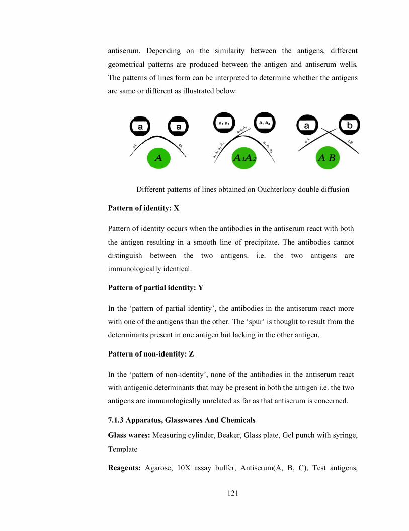
121
antiserum. Depending on the similarity between the antigens, different
geometrical patterns are produced between the antigen and antiserum wells.
The patterns of lines form can be interpreted to determine whether the antigens
are same or different as illustrated below:
Different patterns of lines obtained on Ouchterlony double diffusion
Pattern of identity: X
Pattern of identity occurs when the antibodies in the antiserum react with both
the antigen resulting in a smooth line of precipitate. The antibodies cannot
distinguish between the two antigens. i.e. the two antigens are
immunologically identical.
Pattern of partial identity: Y
In the ‘pattern of partial identity’, the antibodies in the antiserum react more
with one of the antigens than the other. The ‘spur’ is thought to result from the
determinants present in one antigen but lacking in the other antigen.
Pattern of non-identity: Z
In the ‘pattern of non-identity’, none of the antibodies in the antiserum react
with antigenic determinants that may be present in both the antigen i.e. the two
antigens are immunologically unrelated as far as that antiserum is concerned.
7.1.3 Apparatus, Glasswares And Chemicals
Glass wares: Measuring cylinder, Beaker, Glass plate, Gel punch with syringe,
Template
Reagents: Agarose, 10X assay buffer, Antiserum(A, B, C), Test antigens,

122
Assay buffer: PBS, Alcohol
Other requirements: Incubator (37oC), Microwave or Bunsen burner,
Vortex mixer, spatula, Micropipettes, Tips, Moist chamber (box with wet
cotton)
7.1.4 Prepration Of Solutions
Preparation of 1X Assay Buffer: To prepare 10 ml of 1X Assay Buffer, add 1 ml of 10X Assay buffer to 9 ml of sterile distilled water.
Preparation of 1% Agarose gel: To prepare 10 ml of agarose gel, add 0.1g of agarose powder to 10 ml of 1X Assay Buffer, boil to dissolve the agarose completely.
7.1.5 Procedure
1. 25 ml of 1.2% Agarose (0.3g/25ml) was prepared in 1X assay buffer and Agarose was dissolved completely by boiling.
2. The solutions were cooled at 50-60○c and pour 4ml/plate on to 5 grease free glass plates placed on a horizontal surface. The gel was allowed to set for 30minutes.
3. Wells were punched by keeping the glass plate on the template. 4. The wells were filled with 10 µl each of the antiserum and the
corresponding antigens.
5. The glass plates were kept in a moist chamber overnight at 37○c. 6. After incubation, opaque precipitin lines between the antigen and
antisera wells were observed.
Template for addition of antiserum and antigen to their respective wells
7.1.6 Observation
Observe for presence of precipitin lines between antigen and antisera wells.
Note the pattern of precipitin line observed in each case.

123
Diagram showing pattern of precipitin lines
7.1.7 Result
1. Reaction of identity: This occurs between identical antigenic determinants.
The line of precipitation is given as a continuous arc.
2. Reaction of non-identity: When they do not contain any common antigenic
determinant the two lines are found independently and were without any
interaction.
3. Reaction of partial-identity: This has 2 components: i. Those antigenic determinants which are common to both
give a continuous line of identity. ii. The unique determinant recognition is one of the antigen,
in addition a line of non-identity, so that a spur is formed. 7.1.8 Precaution
Drying the agarose gel during incubation
Always use clean glasswares.
7.1.9 Exercise
Write the procedure and result in your own words.
7.2 Aim : To perform dot ELISA test for the detection of
antigen and antibody 7.2.1 OBJECTIVES
After Going through this unit, you will be able to:
Explain the principle of this method
Write about the procedure of this method
124
7.2.2 Principle
DOT ELISA is an extensively used immunological tool in research, as well as
in analytical and diagnostic laboratory. In DOT ELISA antigen is coated on a
nitrocellulose membrane. After incubation unabsorbed antigen is washed out,
and then rest of the reaction sites are blocked on the membrane. Specific
antibody bind with antigen in the nitrocellulose membrane, unbounded
antibody get washed out. The antibody which are bound are detected by the
addition of enzyme labelled with secondary antibody. This complex appears as
a clear brown dot after addition of specific substrate. The enzyme activity is
indicated by the intensity of the spot which is directly proportional to the
antigen concentration.
In this method, ELISA strips are having three well defined zones:
1. Negative control zone that is blocked with an inert protein. 2. Test zone having an antibody immobilized on it and then blocked
with an inert protein. 3. Positive control zone having the antibody immobilized on it,
blocked with inert protein and has a specific antigen bound to the immobilized antibody. These strips will be used to detect the antigen in the test serum
samples by using a secondary antibody conjugated to Horse radish
peroxidase (HRP). HRP is then detected using hydrogen peroxide as
a substrate and Tetramethylbenzidine (TMB) as a chromogen. HRP
acts on hydrogen peroxide to release oxygen, which oxidizes the
TMB to TMB oxide. The TMB oxide is deposited wherever enzyme
is present and appears as a blue spot.
HRP
H2O2 H2O + [O]
TMB + [O] TMBO (Blue)
If the test sample does not contain the antigen specific to the
antibody, there will be no enzyme reaction to occur and no spot

125
develops.
Negative Control Zone: In this zone, immobilize antibody is not present and
hence, there is no reaction when the reagents are added.
Positive control zone: In this zone, antigen is bound to immobilize antibody.
The antigen binds to antibody enzyme conjugate and develops spot.
Test Zone: The result depends on the presence or absence of antigen and will
occur according to the test performed.
7.2.3 Apparatus, Glasswares And Chemicals
• 1X assay buffer • Test serum sample • Dot ELISA strip • Antibody - HRP • Substrates –TMB H2O2 7.2.4 Procedure
1. In a vial take 1ml of 1X assay buffer and 50μl of test serum sample. Mix thoroughly and insert a dot ELISA strip.
2. Allow the reaction to occur at room temperature for 20 minutes . 3. Wash the strip by dripping it in 1ml of 1X assay buffer for about 5
minutes. Repeat it thrice and replace the buffer each time.
4. Take 1ml of 1X assay buffer in a fresh vial and add 10μl antibody HRP conjugate.
5. Mix thoroughly. 6. Dip the strip and allow the reaction to take place for 20 minutes. 7. Wash the strip by dipping it in 1ml of 1X assay buffer for about 5 minutes.
Repeat it thrice and replace the buffer each time. 8. In a fresh vial take 0.1ml of 10X TMB or H2O2 and 0.9ml of distilled
water. 9. Dip the strip in the substrate solution . 10. Observe the strip after 10 to 20 minutes for the appearance of blue colour. 11. Rinse the strip with distilled water to stop the reaction. 7.2.5 Observation Ans Calculations
1. Spot in positive control zone and no spot in the negative control zone indicates proper performance of test.

126
2. Spot in test zone indicates presence of specific antigen in the sample. 3. No spot in the test zone indicates the absence of specific antigen in the
sample. 4. Look for the appearance of the blue dot as shown below:
Test Zone
Negative Zone
Positive Zone
Record your observations as follows:
ZONE SPOT
POSITIVE
ZONE
NEGATIVE
ZONE
TEST
ZONE
Denote +ve : on appearance of a blue spot and -ve : on absence of a blue spot.
7.2.6 Precautions
1. Always wear gloves while performing the experiment. 2. Never leave the reagents at room temperature.
7.2.7 Exercise
Write the procedure and result in your own words.
127
7.3 Aim : To perform sandwich ELISA Test.
7.3.1 Objectives
After Going through this unit, you will be able to:
Explain the principle of this method
Write about the procedure of this method 7.3.2 Principle
Sandwich ELISA is a less common variant of ELISA, but is highly efficient in sample antigen detection. Moreover, many commercial ELISA pair sets are built on this sandwich ELISA. The sandwich ELISA quantifies antigens between two layers of antibodies (i.e. capture and detection antibody). The antigen to be measured must contain at least two antigenic epitope capable of binding to antibody, since at least two antibodies act in the sandwich. Either monoclonal or polyclonal antibodies can be used as the capture and detection antibodies in Sandwich ELISA systems. Monoclonal antibodies recognize a single epitope that allows fine detection and quantification of small differences in antigen. A constant dilution of antibody is coated on a solid phase. Specific antigen will
bind to the specific antibody coated in the well. This complex then binds with
the antibody enzyme labelled conjugate and finally captured antigen is
sandwiched between two antibodies. Then by adding the substrate the colour
reaction will take place.
The amount of HRP labelled antigen bound will be estimated using H2O2 as the
substrate and tetramethylbenzidine (TMB) as chromogen.
HRP + H2O2 -----------> H2O + ½ O2
½ O2 oxidises TMB to TMB oxide
(blue coloured product)
The intensity of colour will be measured at 450 nm spectrophotometer
7.3.3 Apparatus, Glasswares And Chemicals

128
Phosphate buffered saline (PBS): 10 mM phosphate buffer, pH 7.4, 150 mM NaCl tablet and 0.1% sodium azide
Carbonate-Bicarbonate buffer capsule, pH 9.6
Washing buffer (PBS-T): 10 mM phosphate buffer pH 7.4, 150 mM NaCl, 0.05% Tween 20
Capture or coating antibody
Control antigen to be used for standard curve
Labeled detection antibody
Substrate. Typically the most sensitive substrate is used (such as TMB or OPD if using peroxidase as the enzyme label).
Stop solution (optional)
Microtiter plates 7.3.4 Procedure
Day 1: Coating of ELISA wells
1. Concentration of reconstituted antigen is 0.1 mg / ml, dilute it with coating buffer i.e., make 50µl of stock with 4.95 ml of coating buffer to get working concentration of 1µg / ml.
2. Pipette 200µl of diluted 1X antibody in each of microtitre well (24 wells). 3. Tap or shake well to ensure antigen solution is evenly distributed over the
bottom. 4. Incubate it overnight at 4°C ( 37°C for 1 hour) Day 2: Blocking
5. Discard the well content and rinse it with distilled water 3 times, draining out the water after each rinse.
6. Fill each well with 20µl of blocking buffer and incubate at room temperature for 1 hour.
7. Rinse the wells 3 times (as in step 6) with distilled water. Drain out the water completely by tapping the wells on blotting paper. Addition
8. Prepare standard antigen dilution as indicated in the table given. 9. Add 100µl of standard antigen, dilute test samples and PBST to the coated
wells as indicated in figure. 10. Incubate the plate for 1 hour at room temperature.

129
Elusion of HRP labelled antigen
11. Prepare 1X HRP labelled antigen using sample diluent i.e., mix 3µl of stock with 3ml of sample diluent.
12. Add 100µl of 1X HRP labelled antigen to all the wells. 13. Incubate at room temperature for 30 minutes. 14. Discard the well contents , fill the wells with 1X PBST allow it to stand for
3 minutes and discard the content. Repeat this step two more times. 7.3.4 Observation and Calculations
Measure the optical density of each well at wavelength of 492nm and reference
wavelength of 620nm in ELISA reader.
7.3.5 Precautions
1. Always wear gloves while performing the experiment. 2. Never leave the reagents at room temperature. 7.3.6 Exercise
Write the procedure and result in your own words.

130
Unit - 8
Biochemistry Structure of the Unit
8.1 Aim: Quantitative estimation of RNA by Orcinol method.
8.2 Aim: Quantitative estimation of DNA by the diphenylamine reaction.
8.3 Aim : Quantitative estimation of protein (Lowry’s Method)
8.4 Aim: Quantitative estimation of protein by Bradford method.
8.5 Aim : Quantitative estimation of carbohydrates by DNSA Method.
8.6 Aim: Quantitative estimation of carbohydrates by Anthrone's method
8.1 Aim: Quantitative estimation of RNA by Orcinol method.
8.1.1 Objectives
After Going through this unit, you will be able to:
Explain the principle of this method
Write about the procedure of this method 8.1.2 Principle
This is a general reaction for pentoses and depends on the formation of furfural.
When the pentose is heated with concentrated HCl, the Orcinol reacts with
furfural in the presence of ferric chloride as a catalyst, giving a green colour,
which can be measured at 665nm. Only the purine nucleotide gives significant
reactions.
8.1.3 Apparatus, Glasswares and Chemicals
Apparatus and Glasswares: Test tubes, Beaker, pipettes, burner, boiling
waterbath etc.
Chemicals: Ferric chloride, HCl, Alcohol, RNA, TCA, H2O
Prepration of Solutions
Orcinol reagent: Dissolve 1gm of Ferric chloride in 1 litre of
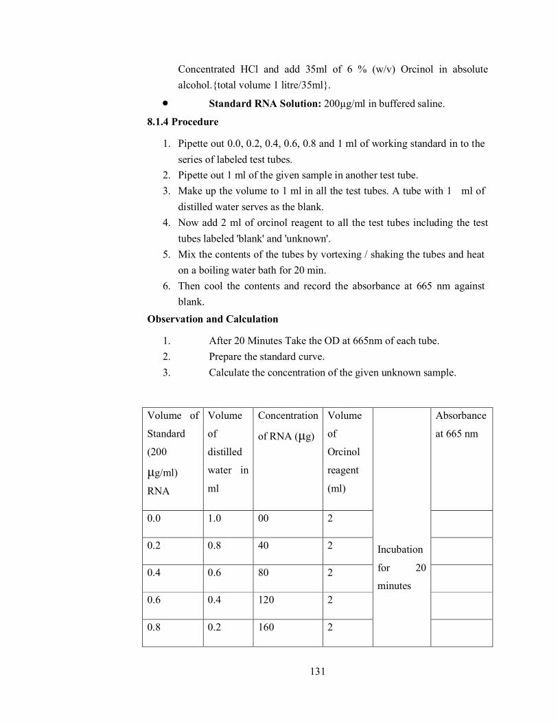
131
Concentrated HCl and add 35ml of 6 % (w/v) Orcinol in absolute alcohol.{total volume 1 litre/35ml}.
Standard RNA Solution: 200µg/ml in buffered saline.
8.1.4 Procedure
1. Pipette out 0.0, 0.2, 0.4, 0.6, 0.8 and 1 ml of working standard in to the series of labeled test tubes.
2. Pipette out 1 ml of the given sample in another test tube. 3. Make up the volume to 1 ml in all the test tubes. A tube with 1 ml of
distilled water serves as the blank. 4. Now add 2 ml of orcinol reagent to all the test tubes including the test
tubes labeled 'blank' and 'unknown'. 5. Mix the contents of the tubes by vortexing / shaking the tubes and heat
on a boiling water bath for 20 min. 6. Then cool the contents and record the absorbance at 665 nm against
blank. Observation and Calculation
1. After 20 Minutes Take the OD at 665nm of each tube. 2. Prepare the standard curve. 3. Calculate the concentration of the given unknown sample.
Volume of
Standard
(200
μg/ml)
RNA
Volume
of
distilled
water in
ml
Concentration
of RNA (μg)
Volume
of
Orcinol
reagent
(ml)
Incubation
for 20
minutes
Absorbance
at 665 nm
0.0 1.0 00 2
0.2 0.8 40 2
0.4 0.6 80 2
0.6 0.4 120 2
0.8 0.2 160 2

132
1.0 0.0 200 2
1.0
(Unknown)
0.0 To estimate 2
8.1.5 Precautions
Use freshly prepared chemicals.
Always use oven dry glassware in the experiment 8.1.6 Exercise
Write the procedure and result in your own words.
8.2 Aim: Quantitative estimation of DNA by the
diphenylamine reaction. 8.2.1 Objectives
After Going through this unit, you will be able to:
Explain the principle of this method
Write about the procedure of this method 8.2.2 Principle
DNA is a polymer of nucleotides. Each nucleotide is made up of a sugar and phosphate backbone connected to a nitrogen base. The sugar in DNA is 2-deoxyribose which is a type of 2-deoxypentose. 2-deoxypentoses in acidic
conditions form a highly reactive compound called β-hydroxy leavulinic aldehyde. This compound reacts with diphenylamine (DPA) to produce a bluish-green coloured complex. But only the deoxy riboses of purine nucleotide react. When DNA is treated with diphenylamine reaction under acid conditions, a blue compound is formed with a sharp absorption maximum at 595 nm. This reaction is given by 2-deoxypentoses in general and is not specific for DNA. In acid solution, the straight chain form of a deoxypentose is
converted to the highly reactive β-hydroxylevulinaldehyde that reacts with diphenylamine to give blue complex. In DNA, only the deoxyribose of the purine nucleotides reacts, so that the value obtained represents half of the total deoxyribose present.

133
8.2.3 Apparatus, Glasswares and Chemicals
Apparatus and Glasswares: Test tubes, Beaker, pipettes, burner, boiling
waterbath, Spectrophotometer, Vortex mixer etc.
Chemicals: Diphenyl amine, Alcohol, Glacial Acetic Acid, H2SO4, DNA, H2O
Prepration of Solutions
Stock standard solution: Weighed 100 mg of DNA and dissolved in 100 ml of distilled water.
Working standard solution: 10 ml of stock was diluted to 100 ml using distilled water.
Diphenyl amine reagent: It should be prepared freshly by dissolving 1 gm of diphenyl amine in 100 ml of glacial acetic acid and by adding 2.5 ml of concentrated sulphuric acid.
Buffered Saline ph 7.4: 0.14N Sodium chloride and 0.02M sodium citrate.
Unknown Solution: The given unknown solution is made up to 100 ml with distilled water.
Procedure
1. Pipette out 0.2 ml – 1.0 ml of DNA solution into a series of test tubes and make up the volume to 1.0 ml with distilled water.
2. 1.0 ml of unknown is taken. 3. Added 2.0 ml of diphenylamine reagent than mixed well and is heated in
a boiling water bath for 10 minutes. 4. Cooled and the colour developed is read at 595 nm.
8.2.4 Observation and Calculation
Volume of
Standard
(200
μg/ml)
Volume
of
distilled
water
(ml)
Concentration
of DNA (μg)
Volume
of DPA
reagent
in ml.
Incubation
Absorbance
at 595 nm
0.0 1.0 00 2

134
0.2 0.8 40 2 in Water
bath for 10
minutes
0.4 0.6 80 2
0.6 0.4 120 2
0.8 0.2 160 2
1.0 0.0 200 2
1.0
(unknown)
0.0 To estimate 2
1. Plot the standard curve by taking concentration of DNA along X-axis and absorbance at 595 nm along Y-axis.
2. Then from this standard curve calculate the concentration of DNA in the given sample.
3. The given unknown sample contains ----µg DNA/ml. 8.2.5 Precautions
Use freshly prepared chemicals.
Always use oven dry glassware in the experiment 8.2.6 Exercise
Write the procedure and result in your own words.
8.3 Aim : Quantitative estimation of protein (Lowry’s Method)
8.3.1 Objectives
After Going through this unit, you will be able to:
Explain the principle of this method
Write about the procedure of this method 8.3.2 Principle
The principle behind the Lowry method of determining protein concentrations
lies in the reactivity of the peptide nitrogens with the copper [II] ions under
alkaline conditions and the subsequent reduction of the Folin Ciocalteay
phosphomolybdic phosphotungstic acid to heteropolymolybdenum blue by the
copper-catalyzed oxidation of aromatic acids.
135
The phenolic group of tyrosine and trytophan residues ( amino acid) in a protein
will produce a blue purple color complex , with maximum absorption in the
region of 660 nm wavelength, with Folin- Ciocalteau reagent which consists of
sodium tungstate molybdate and phosphate. Thus the intensity of color depends
on the amount of these aromatic amino acids present and will thus vary for
different proteins. Most proteins estimation techniques use Bovin Serum
Albumin (BSA) universally as a standard protein, because of its low cost, high
purity and ready availability. The method is sensitive down to about 10 µg/ml
and is probably the most widely used protein assay despite its being only a
relative method , subject to interference from Tris buffer, EDTA, nonionic and
cationic detergents, carbohydrate, lipids and some salts. The incubation time is
very critical for a reproducible assay. The reaction is also dependent on pH and
a working range of pH 9 to 10.5 is essential.
8.3.3 Apparatus, Glasswares and Chemicals
Apparatus and Glasswares: Test tubes, Beaker, pipettes, burner, boiling
waterbath etc.
Chemicals: Bovine serum albumin, Folin’s phenol reagent, CuS04, Alcohol,
H2O, Sodium carbonate, Sodium Potassium tartrate, NaOH
8.3.4 Prepration of Solutions
Stock Solution: Bovine serum albumin of 100 mg is weighed accurately and dissolved in 100ml of distilled water in a standard flask. (Concentration:1mg/ml)
Working Standard: The stock solution of 10ml is diluted to 100ml with distilled water in a standard flask. (Concentration:100mg.ml)
Folin’s Phenol Reagent: Folin’s phenol reagent is mixed with distilled water in a the ratio 1:2
Alkaline CuS04 Reagent: Solution A: Sodium carbonate of 2% in 0.1N sodium hydroxide.
Solution B: Sodium Potassium tartrate of 1%
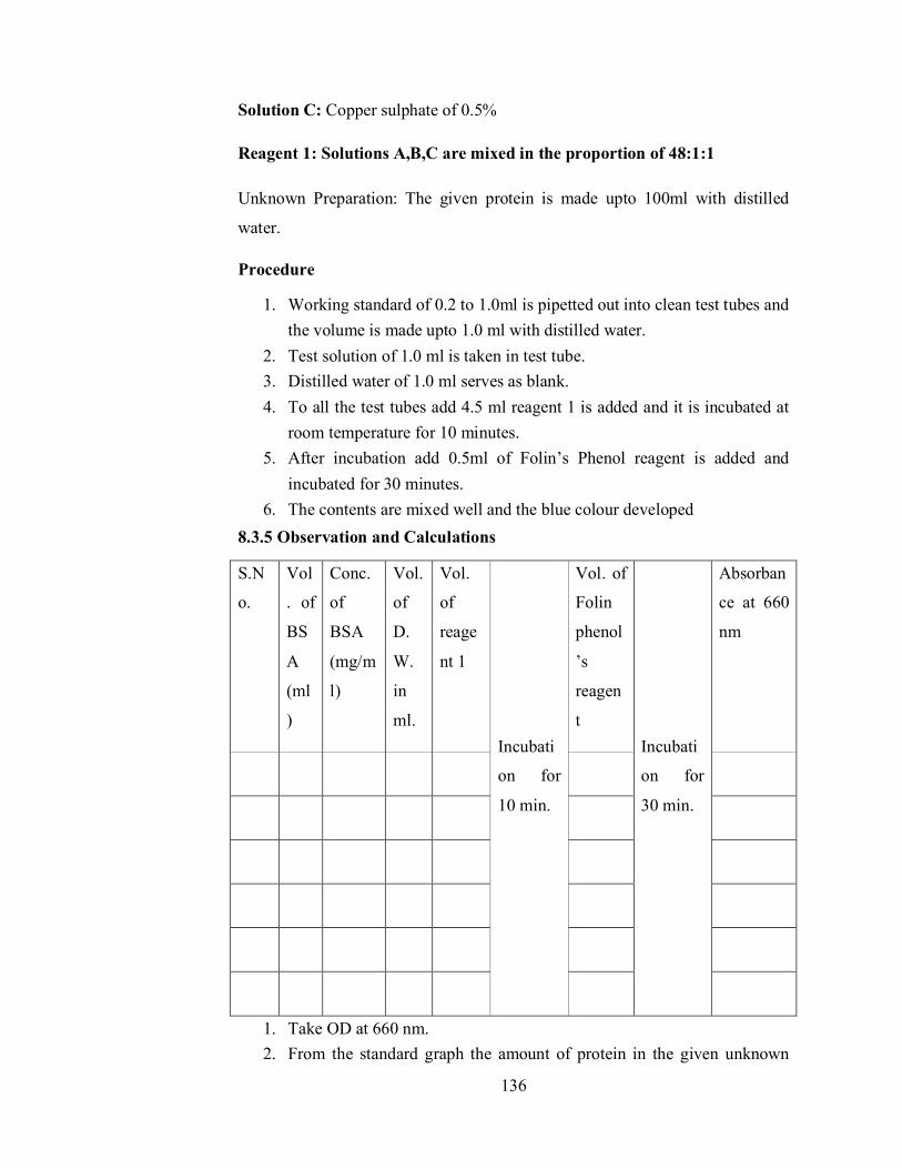
136
Solution C: Copper sulphate of 0.5%
Reagent 1: Solutions A,B,C are mixed in the proportion of 48:1:1
Unknown Preparation: The given protein is made upto 100ml with distilled
water.
Procedure
1. Working standard of 0.2 to 1.0ml is pipetted out into clean test tubes and the volume is made upto 1.0 ml with distilled water.
2. Test solution of 1.0 ml is taken in test tube. 3. Distilled water of 1.0 ml serves as blank. 4. To all the test tubes add 4.5 ml reagent 1 is added and it is incubated at
room temperature for 10 minutes. 5. After incubation add 0.5ml of Folin’s Phenol reagent is added and
incubated for 30 minutes. 6. The contents are mixed well and the blue colour developed
8.3.5 Observation and Calculations
S.N
o.
Vol
. of
BS
A
(ml
)
Conc.
of
BSA
(mg/m
l)
Vol.
of
D.
W.
in
ml.
Vol.
of
reage
nt 1
Incubati
on for
10 min.
Vol. of
Folin
phenol
’s
reagen
t
Incubati
on for
30 min.
Absorban
ce at 660
nm
1. Take OD at 660 nm. 2. From the standard graph the amount of protein in the given unknown

137
solution is calculated. 8.3.6 Precautions
Use freshly prepared chemicals.
Always use oven dry glassware in the experiment 8.3.7 Exercise
Write the procedure and result in your own words.
8.4 Aim: Quantitative estimation of protein by Bradford
method.
8.4.1 Objectives
After Going through this unit, you will be able to:
Explain the principle of this method
Write about the procedure of this method 8.4.2 Principle
The protein in solution can be measured quantitatively by different methods.
The methods described by Bradford uses a different concept-the protein‘s
capacity to bind to a dye, quantitatively. This method relies on the binding of
the dye Coomassie Brilliant Blue to protein. At low pH the free dye has
absorption maxima at 470 nm but when bound to protein has absorption
maxima at 595nm.
The amount of dye binding appears to vary with the content of basic amino
acids arginine & lysine in the protein.
8.4.3 Apparatus, Glasswares and Chemicals
Apparatus and Glasswares: Test tubes, Beaker, pipettes, burner,
Spectrophotometer etc.
Chemicals: Coomassie Brilliant Blue G250, phosphoric acid, bovine serum
albumin, Phosphate buffer, HCl, Alcohol, Protein Sample, H2O
8.4.4 Prepration of Solutions
Bradford reagent: dissolve 100mg of Coomassie Brilliant Blue G250 in

138
50ml of ethanol (C2H5OH). Add 100ml of 85% of phosphoric acid & make the volume to 1lt with water.
0.1M Phosphate buffer (pH 7.5): prepare as mention in experiment 3.
Standard protein solution: dissolve 5mg 0f bovine serum albumin in 50 ml of 0.1M Phosphate buffer. This solution contains 100µg protein/ml.
8.4.5 Procedure
1. Take 0.1ml of protein sample & Standard protein solution in separate test-tubes & make the volume to 1ml with 0.1M Phosphate buffer.
2. Add 5ml of Bradford reagent in all the test tubes. 3. Mix thoroughly & incubate in H2O-bath for 10 minute.
Observation and Calculation
Vol.
Conc of
Vol. of Vol. of
OD
Of Distilled Bradford
S.No BSA
at 595
BSA water reagent
min
(mg/ml) nm
(ml) (ml) (ml)
Incu
batio
n fo
r 10
139
1. Record the absorbance at 595nm against the reagent blank. 2. Plot the standard curve of A595 versus µg of protein in the standards. 3. Determine the protein in the sample extract from the standard curve
8.4.6 Precautions
Use freshly prepared chemicals.
Always use oven dry glassware in the experiment 8.4.7 Exercise
Write the procedure and result in your own words.
8.5 Aim : Quantitative estimation of carbohydrates by DNSA
Method. 8.5.1 Objectives
After Going through this unit, you will be able to:
Explain the principle of this method
Write about the procedure of this method 8.5.2 Principle
This method tests for the presence of free carbonyl group (C=O), present in the so-called reducing sugars. This involves the oxidation of the aldehyde functional group present in, for example, glucose and the ketone functional group in fructose. Simultaneously, 3,5-dinitrosalicylic acid (DNS) is reduced to 3-amino,5-nitrosalicylic acid under alkaline conditions which is red brown in colour with absorbance maxima at 540 nm oxidation aldehyde group ----------> carboxyl group reduction 3,5-dinitrosalicylic acid ----------> 3-amino,5-nitrosalicylic acid The above reaction scheme shows that one mole of sugar will react with one
mole of 3,5 dinitrosalicylic acid. Different reducing sugars generally yield

140
different colour intensities ;thus it is necessary to caliberate for each sugar. In
addition to the oxidation of the carbonyl groups in the sugar, other side
reactions such as the decomposition of sugar also competes for the availability
of 3,5 dinitrosalicylic acid.
Although this is a convenient and relatively inexpensive method, due to the
relatively Low specificity, one must run blanks diligently if the colorimetric
result are to be interpreted correctly and accurately.
When the effects of extraneous compounds are not known one can effectively
include a socalled internal standered by first fully developing the color for the
unknown sample, then a known amount of sugar is added to this sample. The
increase in the absorbance is equivalent to the incremental amount of sugar
added.
8.5.3 Apparatus, Glasswares and Chemicals
Equipment and glassware: Test tubes, pipette, spectrophotometer Reagents: NaOH, DNSA, Standerd glucose solution
8.5.4 Prepration of Solutions
Dinitrosalicylic Acid Reagent Solution (DNSA Solution): Mix 100 ml of 5% (w/v) 3,5-dinitrosalicylic acid in 2 M NaOH with 250 ml of 60% (w/v) sodium potassium tartrate and make the total volume up to 500 ml with distilled water.
8.5.6 Procedure
1. Prepare 100ml glucose stock solution of 10mg/ml concentration. 2. Prepare glucose standards ranging from 0.0 to 1.0 mg ml-1 (total sample
volume 3 ml) 3. Add 2 ml of DNS reagent to 3 ml of glucose standard solutions in a test
tube. 4. Take 3 ml of glucose solution of unknown concentration and add 2ml
DNS reagent to it. 5. Keep all the test tubes in boiling water bath for 5 minutes and let them
cool. 8.5.7 Observation and Calculations
1. Measure the absorbance at 540 nm against reagent blank.

141
2. Plot a graph of glucose concentration on x axes and absorbance at y axes. This is standard calibration curve.
3. Determine the value of glucose concentration in unknown samples using the standard calibration curve.
4. Concentration of unknown was found to be ___________mg/ml.
8.5.8 Pricautions
Use freshly prepared chemicals.
Always use oven dry glassware in the experiment
DNS reagent is corrosive, so handle it with care.
8.5.9 Exercise
Write the procedure and result in your own words.
8.6 Aim: Quantitative estimation of carbohydrates by
Anthrone's method 8.6.1 Objectives
After Going through this unit, you will be able to:
Explain the principle of this method
Write about the procedure of this method 8.6.2 Principle
Carbohydrates are first hydrolysed into simple sugars using dilute hydrochloric
acid. In hot acidic medium glucose is dehydrated to hydroxymethyl furfural.
This compound forms with anthrone a green coloured product with an
absorption maximum at 630 nm.
8.6.3 Apparatus, Chemicals and Glasswares
Equipment and glassware: Test tubes, pipette, spectrophotometer Reagents: Anthrone reagent, H2SO4, Standerd glucose solution, H2O, HCl
8.6.4 Prepration of Solutions
Anthrone reagent: Dissolve 200 mg anthrone in 100 mL of ice-cold 95% H2SO4. Prepare fresh before use.
142
Standard glucose: Dissolve 100 mg in 100 mL water. Working standard—10 mL of stock diluted to 100 mL with distilled water.
Procedure
1. Weigh 100 mg of the sample into a boiling tube. 2. Hydrolyse by keeping it in a boiling water bath for three hours with 5
mL of 2.5 N HCl and cool to room temperature. 3. Neutralise it with solid sodium carbonate until the effervescence ceases. 4. Make up the volume to 100 mL and centrifuge. 5. Collect the supernatant and take 0.5 and 1 mL aliquots for analysis. 6. Prepare the standards by taking 0, 0.2, 0.4, 0.6, 0.8 and 1 mL of the
working standard. ‘0’ serves as blank.
7. Make up the volume to 1 mL in all the tubes including the sample tubes by adding distilled water.
8. Then add 4 mL of anthrone reagent. 9. Heat for ten minutes in a boiling water bath. Cool rapidly.
8.6.5 Observation And Calculations
1. Read the green to dark green colour at 630 nm. 2. Draw a standard graph by plotting concentration of the standard on the
X-axis vs. absorbance on the Y-axis.
3. From the graph calculate the amount of carbohydrate present in the sample tube.
8.6.6 Precautions
Use freshly prepared chemicals.
Always use oven dry glassware in the experiment
Prepare and handle anthrone's reagent with care as sulfuric acid is highly corrosive.
8.6.7 Exercise
Write the procedure and result in your own words.
143
Unit - 9
Biostatistics Practical Structure of the Unit
9.0 Objectives 9.1 Introduction 9.2 Mean
9.2.1 Definition 9.2.2 Character statics 9.2.3 Advantages 9.2.4 Disadvantages
9.3 Mode 9.3.1 Definition
9.3.2 Character statics 9.3.3 Advantages 9.3.4 Disadvantages
9.4 Median 9.4.1 Definition 9.4.2 Character statics 9.4.3 Advantages 9.4.4 Disadvantages
9.5 Chi square test 9.5.1 Definition 9.5.2 Character statics
9.6 Reference
9.1 Introduction
The term statistics is used to mean either statistical data or statistical methods. Statistical data refers to numerical descriptions of things. These descriptions may take the form of counts or measurements.
Biostatistics is the branch of applied statistics directed toward applications in the health sciences and biology. Biostatistics is sometimes distinguished from the field of biometry based upon whether applications are in the health sciences (biostatistics) or in broader biology (biometry eg: agriculture, ecology, wildlife biology)

144
Other branches of applied statistics, psychometrics, econometrics, chemometrics, astrostatistics, environmetrics, etc.
Why biostatistics?
*Because some statistical methods are more heavily used in health applications than elsewhere (e.g. survival analysis, longitudinal data analysis)
*Because examples are drawn from health sciences:
Makes subject more appealing to those interested in health,
Illustrates how to apply methodology to similar problems encountered in real life.
9.0 Objectives
1. Define and understand the statistics and biostatistics 2. Understand the arithmetic mean or simple mean with their
advantages and disadvantages 3. Understand the mode with their advantages and disadvantages 4. Understand the median with their advantages and disadvantages 5. To understand chi square test 6. Understand the graphical representation of data
9.2 The Arithmetic Mean or simple Mean
Definition: The arithmetic mean of a set of data is found by taking the sum of the data, and then dividing the sum by the total number of values in the set. A mean is commonly referred to as an average.
It is usually denoted by x̅ or a
Characteristics:
1) The value of the arithmetic mean is determined by every item in the series.
2) It is greatly affected by extreme values.
3) The sum of the deviations about it is zero.
4) The sum of the squares of deviations from the arithmetic mean is less than of those computed from any other point.

145
Example: Find the mean driving speed for 6 different cars on the same highway?
66 mph, 57 mph, 71 mph, 54 mph, 69 mph, 58 mph
Solution: x̅ = 66+57+71+54+69+58
6
x̅ = 62.5 mph
The arithmetic mean is, in general, a very natural measure of central location. One of its principal limitations, however, is that it is overlysensitive to extreme values. In this instance it may not be representative of the location of the great majority of the sample points.
Advantages:
1) It is based on all values given in the distribution.
2) It is most early understood.
3) It is most amenable to algebraic treatment.
Disadvantages:
1) It may be greatly affected by extreme items and its usefulness as a “Summary of the whole” may be considerably reduced.
2) When the distribution has open-end classes, its computation would be based assumption, and therefore may not be valid.
9.3Mode
Definition:It is the value of the observation that occurs with the greatest frequency. A particular disadvantage is that, with a small number of observations, there may be no mode. In addition, sometimes, there may be more than one mode such as when dealing with a bimodal(two-peak) distribution. It is even less amenable (responsive) to mathematical treatment than the median. The mode is not often used in biological or medical data.
Characteristics:
1) It is an average of position
2) It is not affected by extreme values

146
3) It is the most typical value of the distribution
Example.1: Find the mode values for the following data
22, 66, 69, 70, 73.
Answer: no mode value
Example.2: Find the mode value for the following data
1.8, 3.0, 3.3, 2.8, 2.9, 3.6, 3.0, 1.9, 3.2, 3.5
Answer: mode value = 3.0 kg
Advantages:
1. Since it is the most typical value it is the most descriptive average
2. Since the mode is usually an “actual value”, it indicates the precise value of an important part of the series.
Disadvantages:
1. Unless the number of items is fairly large and the distribution reveals a distinct central tendency, the mode has no significance
2. It is not capable of mathematical treatment
3. In a small number of items the mode may not exist.
9.4 Median
Definition:An alternative measure of central location, perhaps second in popularity to the arithmetic mean, is the median.
Suppose there are n observations in a sample. If these observations are ordered from smallest to largest, then the median is defined as follows:
The sample median is
The rational for these definitions is to ensure an equal number of sample points on both sides of the sample median.

147
The median is defined differently when n is even and odd because it is impossible to achieve this goal with one uniform definition.
Characteristics:
1) It is an average of position.
2) It is affected by the number of items than by extreme values.
Example: Compute the sample median for the birth weight data
2581, 2834, 4146, 2838, 2841, 3031, 3245, 3248,
3260,2069, 3265, 3484, 3541, 3609, 3649, 2759,3323,3314,3200, 3101
Solution: First arrange the sample in ascending order
2069, 2581, 2759, 2834, 2838, 2841, 3031, 3101, 3200, 3245, 3248,
3260, 3265, 3314, 3323, 3484, 3541, 3609, 3649, 4146
Since n=20 is even,
Median = average of the 10th and 11th largest observation =
(3245 + 3248)/2 = 3246.5 g
The principal strength of the sample median is that it is insensitive to very large or very small values.
Advantages:
1) It is easily calculated and is not much disturbed by extreme values
2) It is more typical of the series
3) The median may be located even when the data are incomplete, e.g, when the class intervals are irregular and the final classes have open ends.
Disadvantages:
1. The median is not so well suited to algebraic treatment as the arithmetic, geometric and harmonic means.
2. It is not so generally familiar as the arithmetic mean
9.5 The Chi – square test
A chi square (χ2 ) distribution is a probability distribution. The chi-squareis useful in making statistical inferences about categorical data in which the categories are two and above
Definition A statistic which measures the discrepancy between K observed frequencies O1, O2, . Ok and the corresponding expected frequencies e1, e2 . ek.

148
Chi square = χ2 = Σ{ (Oi - ei)2 } / ei
The sampling distribution of the chi-square statistic is known as the chi square
distribution. As in t distributions, there is a different χ2 distribution for each different value of degrees of freedom, but all of them share the following characteristics.
Characteristics
1. Every χ2 distribution extends indefinitely to the right from 0.
2. Every χ2 distribution has only one (right ) tail.
3. As df increases, the χ2 curves get more bell shaped and approach the normal curve in appearance (but remember that a chi square curve starts at 0, not at -
∞ )
If the value of χ2 is zero, then there is a perfect agreement between the observed and the expected frequencies. The greater the discrepancy between the
observed and expected frequencies, the larger will be the value of χ2.
In order to test the significance of the χ2, the calculated value of χ2 is compared with the tabulated value for the given df at a certain level of significance.
Example1: In an experiment with peas one observed 360 round and yellow, 130 round and green, 118 wrinkled and yellow and 32 wrinkled and green. According to the Mendelian theory of heredity the numbers should be in the ratio 9:3:3:1. Is there any evidence of difference from the plants at 5% level of significance?
Solution
Hypothesis: HO : Ratio is 9:3:3:1
HA : Ratio is not 9:3:3:1
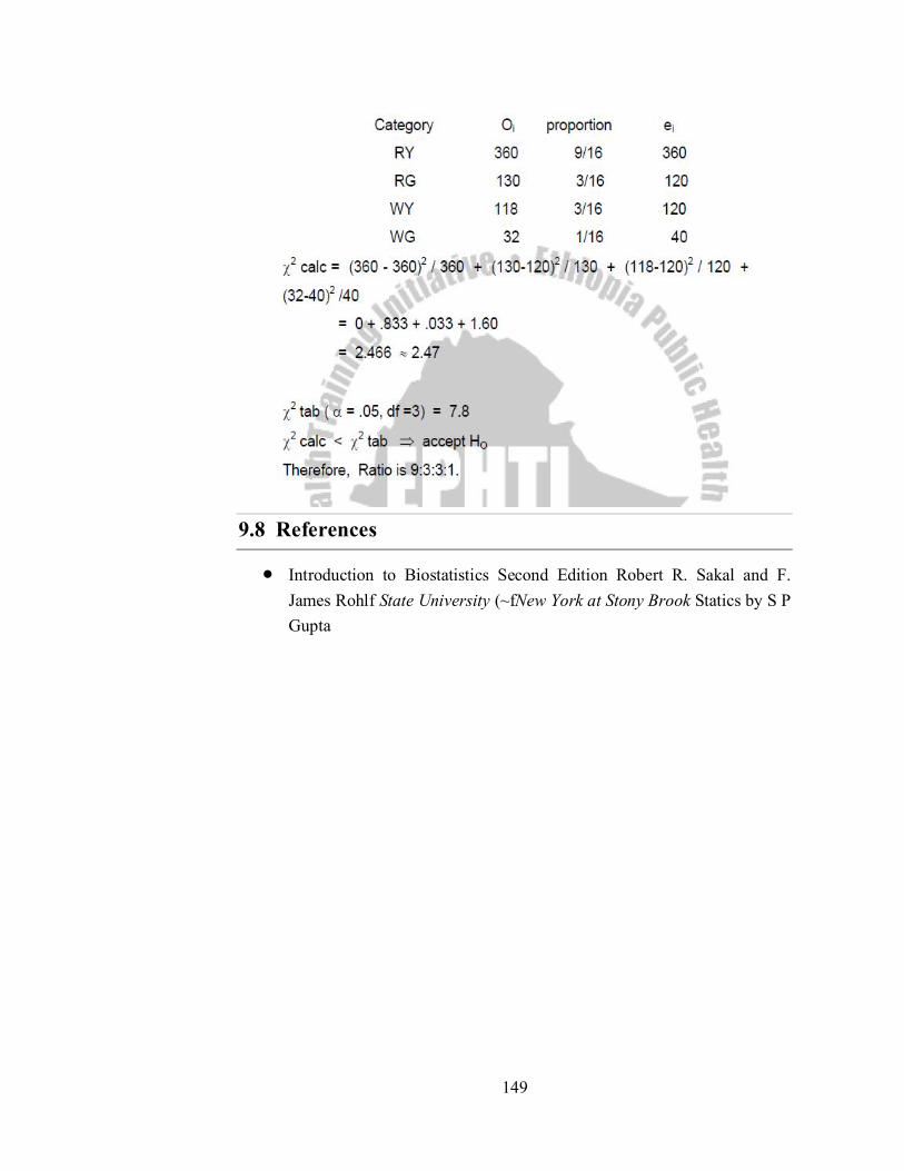
149
9.8 References
Introduction to Biostatistics Second Edition Robert R. Sakal and F. James Rohlf State University (~fNew York at Stony Brook Statics by S P Gupta

150
Unit - 10
Computer and Bioinformatics Experiments
Structure of the Unit
10.0 Objectives
10.1 Introduction
10.2 Computer Lab Experiments
10.2.1. Creation of presentation and formatting especial PPT
10.2.2. Different views of presentation
10.2.3. Layouts and templates
10.2.4. Master slides
10.2.5. Animation
10.2.6. Transition
10.3 Bioinformatics Lab Experiments
10.3.1. To study about the different biological databases in variable formats
10.3.1.1. Study about the NCBI
10.3.1.2. Review of the variety of entities of NCBI
10.3.1.3. Study of Genbank
10.3.1.4. Study of format
10.3.1.5. Study of SWISS PORT
10.3.1.6. SWISS PORT record format
10.3.2. To retrieve sequence from freely available databases
10.3.2.1. Retrieve the Protein molecule sequence from the available
protein domains.
10.3.2.2. Learn the storage of the protein sequence for the further
proceeding work.
10.4 Self-Learning Exercise
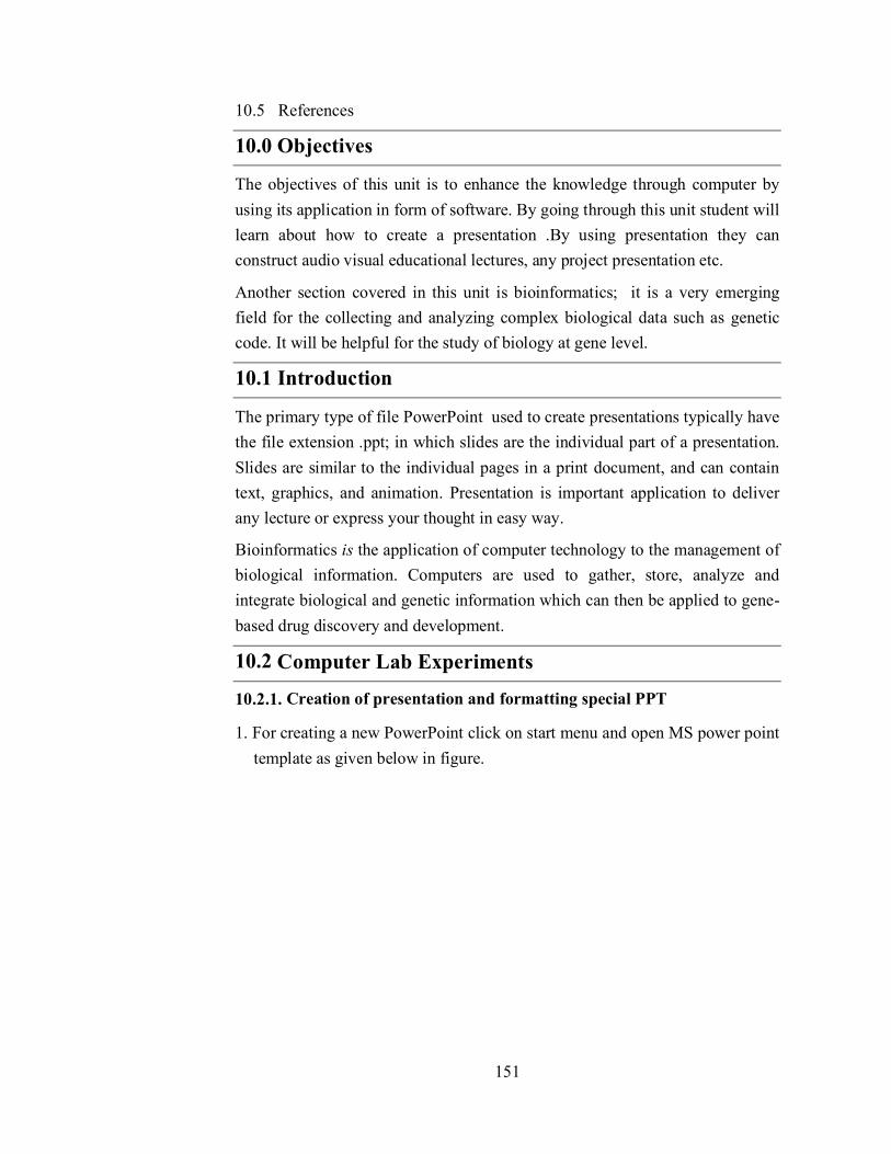
151
10.5 References
10.0 Objectives
The objectives of this unit is to enhance the knowledge through computer by using its application in form of software. By going through this unit student will learn about how to create a presentation .By using presentation they can construct audio visual educational lectures, any project presentation etc.
Another section covered in this unit is bioinformatics; it is a very emerging field for the collecting and analyzing complex biological data such as genetic code. It will be helpful for the study of biology at gene level.
10.1 Introduction
The primary type of file PowerPoint used to create presentations typically have the file extension .ppt; in which slides are the individual part of a presentation. Slides are similar to the individual pages in a print document, and can contain text, graphics, and animation. Presentation is important application to deliver any lecture or express your thought in easy way.
Bioinformatics is the application of computer technology to the management of biological information. Computers are used to gather, store, analyze and integrate biological and genetic information which can then be applied to gene-based drug discovery and development.
10.2 Computer Lab Experiments
10.2.1. Creation of presentation and formatting special PPT
1. For creating a new PowerPoint click on start menu and open MS power point template as given below in figure.

152
2. Insert a new slide or a blank presentation.
In formatting menu bar click “home” insert “new slide” by clicking on it as given below. The short cut Ctrl+M can be used to insert new slide. Knowing the short cut will save you a lot of frustration trying to figure out how to make a new slide or how to get the Common Task Window to display again.
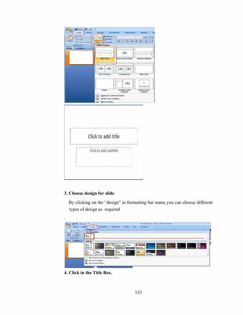
153
3. Choose design for slide
By clicking on the “design” in formatting bar menu you can choose different types of design as required
.
4. Click in the Title Box.

154
You can choose different fonts by selecting “Font” from the Format menu or by Pulling down on fonts in the Formatting Bar. You can also select the size and style.
Making PowerPoint Slides
VMOU KOTA
5. Inserting a Text box
Text box is required to enter text into the slide. To inset text box, click on “insert” on formatting bar then click “text box” as given below in figure.

155
6. Inserting a picture
Insert clip art on your slide by double clicking on the icon or selecting picture from clip art in the Insert menu as given below in figure.
7. Inserting bullets or numbering
Note the text box for bullets. This might be fine for listing items, but when you want to type a paragraph, this feature can be annoying. To eliminate bullets, click on the text box. Choose Bullets and Numbering from the Format menu. Click on None and then OK.

156
8. Inserting a Sound File
Click on New Slide in the Common Tasks window or New Slide from the Insert menu. Add a sound by selecting Movies and Sounds then Sound from Gallery in the Insert menu as given below. You can choose whether or not the sound to play automatically or when you click it. You can further customize the order in which objects are played or displayed on your slide by going to custom animation under the Slide Show Menu.
9. Inserting a Movie
Click on New Slide in the Common Tasks window or New Slide from the Insert menu. To insert a movie select “Movies and Sounds” from the Insert menu and choose Movies from Gallery.
10.2.2. Different views of presentation
PowerPoint gives you five views in which you create and organize your presentation. As you create a presentation, you can switch among the four

157
views as you work.
The five PowerPoint views are:
1. Slide View
To be in Slide view you click on the following button located in the bottom-left hand corner of any slide: ( ). The Slide view shows a single slide. In Slide view, you work on one slide at time. Here, you can type your slide title and body, add other text to the slide, draw shapes, add clip art, choose a color scheme, make a graph, etc. In Slide view, you have access to all the tools on the Tool Palette as well as buttons on the Toolbar.
2. Normal View
To be in normal view you click on the following button located in the bottom-left hand corner of any slide: ( ). Switches to normal view, where you can work on one slide at a time or organize the structure of all the slides in your presentation.
3. Outline View
To be in Outline view you click on the following button located in the bottom-left hand corner of any slide: ( ). The Outline view shows all the titles and body text in your presentation. In Outline view, you can move slides around within your presentation and also edit your text. Click on the Outline view button ( ), to view your title slide in this view.
4. Slide Sorter View
To be in Slide Sorter view you click on the following button located in the bottom-left hand corner of any slide: ( ). The Slide Sorter view shows you a miniature of each slide in your presentation. You can drag slides around on the screen to reposition them in this view. You can also select and copy multiple slides should you want to use them in other presentations. Click on the Slide Sorter view button ( ), to view your title slide in this view.
5. Slide Show View
There is one more button located in the bottom- left hand corner of any slide:( ). This is the Slide Show button. The slide show runs your slide show in a full screen, beginning with the current slide if you are in slide view or the selected slide if you are in slide sorter view.

158
If you simply want to view your show from the first slide:
1. Click Slide Show at the top of the screen
2. Select View Show
Figure shows the icon of different types of views
10.2.3. Layouts and templates
The New Slide layout screen appears. You can select from a variety of pre designed layouts. Choose Title Slide Click OK. A PowerPoint template provides the slide background designs for our presentations. In addition, a template file can contain other presentation style aspects, including specifications for the type and size of bullets and fonts, placeholder sizes and positions, and color schemes.

159
10.2.4. Master slides
A slide master is the top slide in a hierarchy of slides that stores information about the theme and slide layouts of a presentation, including the background, color, fonts, effects, placeholder sizes, and positioning.
Every presentation contains at least one slide master. The key benefit of using slide masters is that you can make universal style changes to every slide in your presentation, including ones added later to the presentation. When you use a slide master, you save time because you don't have to type the same information on more than one slide. The slide master especially comes in handy when you have extremely long presentations with lots of slides.
The easiest way to make the same change to all slides or slides that use a specific layout is to change the slide master, also called the master slide. For example, let's say you like a theme but want to change a specific design element of that theme. You may want a different bullet style, slide titles that are center-aligned instead of left-aligned, or an accent line under each slide title. You can quickly make these changes and more by modifying the slide master.
To view the slide master:
Select the View tab
Click the Slide Master view command in the Presentation Views group.
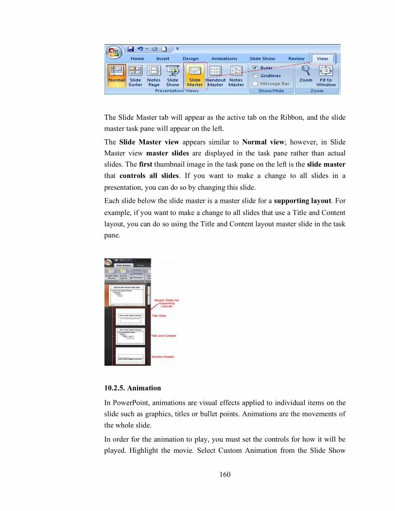
160
The Slide Master tab will appear as the active tab on the Ribbon, and the slide master task pane will appear on the left.
The Slide Master view appears similar to Normal view; however, in Slide Master view master slides are displayed in the task pane rather than actual slides. The first thumbnail image in the task pane on the left is the slide master that controls all slides. If you want to make a change to all slides in a presentation, you can do so by changing this slide.
Each slide below the slide master is a master slide for a supporting layout. For example, if you want to make a change to all slides that use a Title and Content layout, you can do so using the Title and Content layout master slide in the task pane.
10.2.5. Animation
In PowerPoint, animations are visual effects applied to individual items on the slide such as graphics, titles or bullet points. Animations are the movements of the whole slide.
In order for the animation to play, you must set the controls for how it will be played. Highlight the movie. Select Custom Animation from the Slide Show
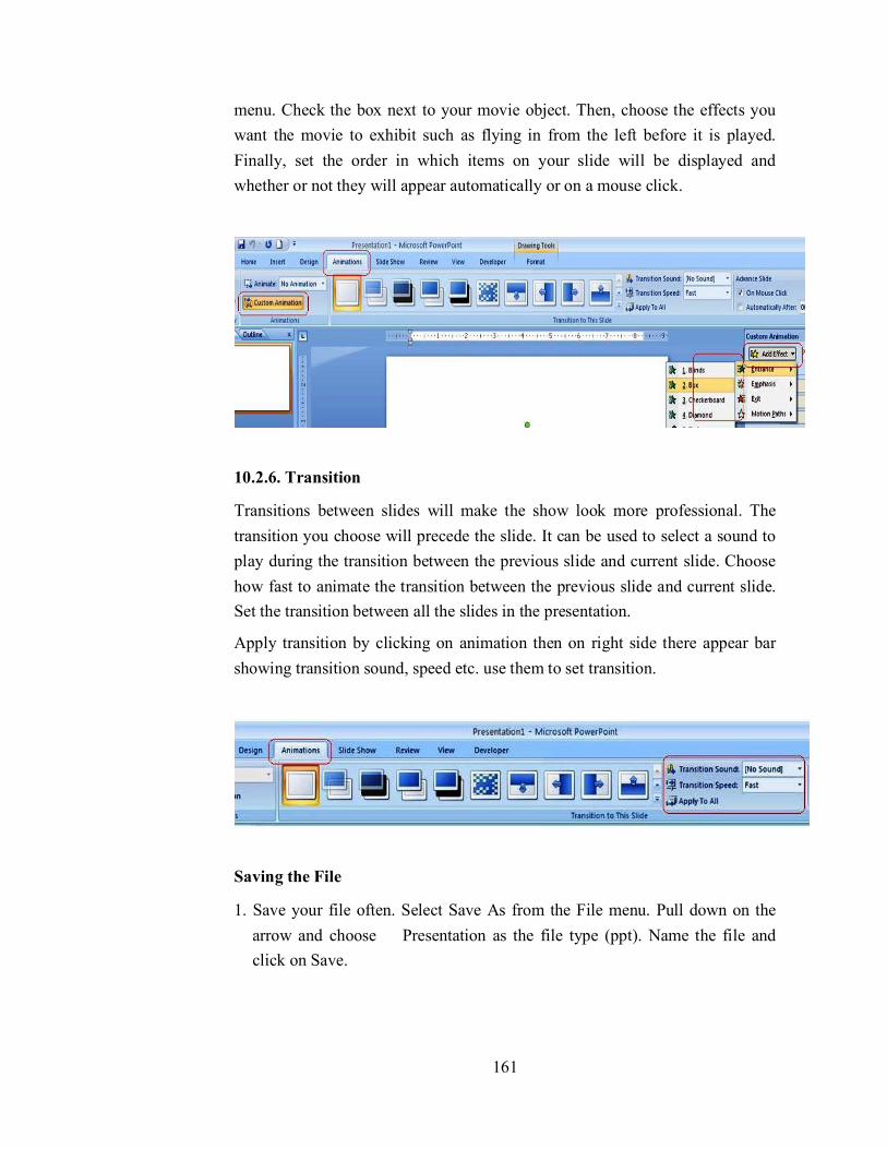
161
menu. Check the box next to your movie object. Then, choose the effects you want the movie to exhibit such as flying in from the left before it is played. Finally, set the order in which items on your slide will be displayed and whether or not they will appear automatically or on a mouse click.
10.2.6. Transition
Transitions between slides will make the show look more professional. The transition you choose will precede the slide. It can be used to select a sound to play during the transition between the previous slide and current slide. Choose how fast to animate the transition between the previous slide and current slide. Set the transition between all the slides in the presentation.
Apply transition by clicking on animation then on right side there appear bar showing transition sound, speed etc. use them to set transition.
Saving the File
1. Save your file often. Select Save As from the File menu. Pull down on the arrow and choose Presentation as the file type (ppt). Name the file and click on Save.

162
2. To save it as a slide show, select Save As from the File menu. Pull down on the arrow and choose PowerPoint Show as the file type (pps). Name the file and click on Save.
Viewing the Slide Show
Select View Show from the Slide Show menu in formatting bar.
10.3 Bioinformatics Lab Experiments
10.3.1. To study about the different biological databases in variable formats
10.3.1.1. Study about the NCBI
The National Center for Biotechnology Information (NCBI) is part of the United States National Library of Medicine (NLM), a branch of the National Institutes of Health. The NCBI is located in Bethesda, Maryland and was founded in 1988 through legislation sponsored by Senator Claude Pepper. The NCBI houses a series of databases relevant to biotechnology and biomedicine. Major databases include GeneBank for DNA sequences and PubMed, a bibliographic database for the biomedical literature. Other databases include the NCBI Epigenomics database. All these databases are available online through the Entrez search engine (http://www.ncbi.nlm.nih.gov).

163
10.3.1.2. Review of the variety of entities of NCBI
There are various kinds of tools; databases etc. that are parts of NCBI database. These are as follows:
Databases
Assembly
A database providing information on the structure of assembled genomes, assembly names and other meta-data, statistical reports, and links to genomic sequence data.
BioProject (formerly Genome Project)
A collection of genomics, functional genomics, and genetics studies and links to their resulting datasets. This resource describes project scope, material, and objectives and provides a mechanism to retrieve datasets that are often difficult to find due to inconsistent annotation, multiple independent submissions, and
164
the varied nature of diverse data types which are often stored in different databases.
BioSample
The BioSample database contains descriptions of biological source materials used in experimental assays.
BioSystems
Database that groups biomedical literature, small molecules, and sequence data in terms of biological relationships.
Bookshelf
A collection of biomedical books that can be searched directly or from linked data in other NCBI databases. The collection includes biomedical textbooks, other scientific titles, genetic resources such as GeneReviews, and NCBI help manuals.
ClinVar
A resource to provide a public, tracked record of reported relationships between human variation and observed health status with supporting evidence. Related information in the NIH Genetic Testing Registry (GTR), MedGen, Gene, OMIM, PubMed and other sources is accessible through hyperlinks on the records.
CloneDB (formerly Clone Registry)
A database that integrates information about clones and libraries, including sequence data, map positions and distributor information.
Computational Resources from NCBI's Structure Group
A centralized page providing access and links to resources developed by the Structure Group of the NCBI Computational Biology Branch (CBB). These resources cover databases and tools to help in the study of macromolecular structures, conserved domains and protein classification, small molecules and their biological activity, and biological pathways and systems.
Consensus CDS (CCDS)
A collaborative effort to identify a core set of human and mouse protein coding regions that are consistently annotated and of high quality.
Conserved Domain Database (CDD)
165
A collection of sequence alignments and profiles representing protein domains conserved in molecular evolution. It also includes alignments of the domains to known 3-dimensional protein structures in the MMDB database.
Database of Expressed Sequence Tags (dbEST)
A divison of GenBank that contains short single-pass reads of cDNA (transcript) sequences. dbEST can be searched directly through the Nucleotide EST Database.
Database of Genome Survey Sequences (dbGSS)
A division of GenBank that contains short single-pass reads of genomic DNA. dbGSS can be searched directly through the Nucleotide GSS Database.
Database of Genomic Structural Variation (dbVar)
The dbVar database has been developed to archive information associated with large scale genomic variation, including large insertions, deletions, translocations and inversions. In addition to archiving variation discovery, dbVar also stores associations of defined variants with phenotype information.
Database of Genotypes and Phenotypes (dbGaP)
An archive and distribution center for the description and results of studies which investigate the interaction of genotype and phenotype. These studies include genome-wide association (GWAS), medical resequencing, molecular diagnostic assays, as well as association between genotype and non-clinical traits.
Database of Major Histocompatibility Complex (dbMHC)
An open, publicly accessible platform where the HLA community can submit, edit, view, and exchange data related to the human major histocompatibility complex. It consists of an interactive Alignment Viewer for HLA and related genes, an MHC microsatellite database, a sequence interpretation site for Sequencing Based Typing (SBT), and a Primer/Probe database.
Database of Short Genetic Variations (dbSNP)
Includes single nucleotide variations, microsatellites, and small-scale insertions and deletions. dbSNP contains population-specific frequency and genotype data, experimental conditions, molecular context, and mapping information for both neutral variations and clinical mutations.
Epigenomics
166
This resource enables users to explore and visualize richly-annotated epigenomics datasets. It provides a unique interface to search and navigate epigenomic data in the context of biological sample information, as well as tools to select, download and view multiple sets of epigenomic data as tracks on genome browsers.
GenBank
The NIH genetic sequence database, an annotated collection of all publicly available DNA sequences. GenBank is part of the International Nucleotide Sequence Database Collaboration, which comprises the DNA DataBank of Japan (DDBJ), the European Molecular Biology Laboratory (EMBL), and GenBank at NCBI. These three organizations exchange data on a daily basis. GenBank consists of several divisions, most of which can be accessed through the Nucleotide database. The exceptions are the EST and GSS divisions, which are accessed through the Nucleotide EST and Nucleotide GSS databases, respectively.
Gene
A searchable database of genes, focusing on genomes that have been completely sequenced and that have an active research community to contribute gene-specific data. Information includes nomenclature, chromosomal localization, gene products and their attributes (e.g., protein interactions), associated markers, phenotypes, interactions, and links to citations, sequences, variation details, maps, expression reports, homologs, protein domain content, and external databases.
Gene Expression Omnibus (GEO) Database
A public functional genomics data repository supporting MIAME-compliant data submissions. Array- and sequence-based data are accepted and tools are provided to help users query and download experiments and curated gene expression profiles.
Gene Expression Omnibus (GEO) Datasets
Stores curated gene expression and molecular abundance DataSets assembled from the Gene Expression Omnibus (GEO) repository. DataSet records contain additional resources, including cluster tools and differential expression queries.
Gene Expression Omnibus (GEO) Profiles
Stores individual gene expression and molecular abundance Profiles assembled from the Gene Expression Omnibus (GEO) repository. Search for specific

167
profiles of interest based on gene annotation or pre-computed profile characteristics.
GeneReviews
A collection of expert-authored, peer-reviewed disease descriptions on the NCBI Bookshelf that apply genetic testing to the diagnosis, management, and genetic counseling of patients and families with specific inherited conditions.
Genes and Disease
Summaries of information for selected genetic disorders with discussions of the underlying mutation(s) and clinical features, as well as links to related databases and organizations.
Genetic Testing Registry (GTR)
A voluntary registry of genetic tests and laboratories, with detailed information about the tests such as what is measured and analytic and clinical validity. GTR is a nexus for information about genetic conditions and provides context-specific links to a variety of resources, including practice guidelines, published literature, and genetic data/information. The initial scope of GTR includes single gene tests for Mendelian disorders, as well as arrays, panels and pharmacogenetic tests.
Genome
Contains sequence and map data from the whole genomes of over 1000 organisms. The genomes represent both completely sequenced organisms and those for which sequencing is in progress. All three main domains of life (bacteria, archaea, and eukaryota) are represented, as well as many viruses, phages, viroids, plasmids, and organelles.
Genome Reference Consortium (GRC)
The Genome Reference Consortium (GRC) maintains responsibility for the human and mouse reference genomes. Members consist of The Genome Center at Washington University, the Wellcome Trust Sanger Institute, the European Bioinformatics Institute (EBI) and the National Center for Biotechnology Information (NCBI). The GRC works to correct misrepresented loci and to close remaining assembly gaps. In addition, the GRC seeks to provide alternate assemblies for complex or structurally variant genomic loci. At the GRC website (http://www.genomereference.org), the public can view genomic regions currently under review, report genome-related problems and contact the GRC.

168
HIV-1, Human Protein Interaction Database
A database of known interactions of HIV-1 proteins with proteins from human hosts. It provides annotated bibliographies of published reports of protein interactions, with links to the corresponding PubMed records and sequence data.
HomoloGene
A gene homology tool that compares nucleotide sequences between pairs of organisms in order to identify putative orthologs. Curated orthologs are incorporated from a variety of sources via the Gene database.
Influenza Virus
A compilation of data from the NIAID Influenza Genome Sequencing Project and GenBank. It provides tools for flu sequence analysis, annotation and submission to GenBank. This resource also has links to other flu sequence resources, and publications and general information about flu viruses.
Journals in NCBI Databases
Subset of the NLM Catalog database providing information on journals that are referenced in NCBI database records, including PubMed abstracts. This subset can be searched using the journal title, MEDLINE or ISO abbreviation, ISSN, or the NLM Catalog ID.
MeSH Database
MeSH (Medical Subject Headings) is the U.S. National Library of Medicine's controlled vocabulary for indexing articles for MEDLINE/PubMed. MeSH terminology provides a consistent way to retrieve information that may use different terminology for the same concepts.
MedGen
A portal to information about medical genetics. MedGen includes term lists from multiple sources and organizes them into concept groupings and hierarchies. Links are also provided to information related to those concepts in the NIH Genetic Testing Registry (GTR), ClinVar, Gene, OMIM, PubMed, and other sources.
NCBI C++ Toolkit Manual
A comprehensive manual on the NCBI C++ toolkit, including its design and development framework, a C++ library reference, software examples and

169
demos, FAQs and release notes. The manual is searchable online and can be downloaded as a series of PDF documents.
NCBI Education Page
Provides links to tutorials and training materials, including PowerPoint slides and print handouts.
NCBI Glossary
Part of the NCBI Handbook, this glossary contains descriptions of NCBI tools and acronyms, bioinformatics terms and data representation formats.
NCBI Handbook
An extensive collection of articles about NCBI databases and software. Designed for a novice user, each article presents a general overview of the resource and its design, along with tips for searching and using available analysis tools. All articles can be searched online and downloaded in PDF format; the handbook can be accessed through the NCBI Bookshelf.
NCBI Help Manual
Accessed through the NCBI Bookshelf, the Help Manual contains documentation for many NCBI resources, including PubMed, PubMed Central, the Entrez system, Gene, SNP and LinkOut. All chapters can be downloaded in PDF format.
NCBI Website Search
A database of static NCBI web pages, documentation, and online tools. These pages include such content as specialized online sequence analysis tools, back issues of newsletters, legacy resource description pages, sample code, and other miscellaneous resources. Searching this database is equivalent to a site search tool for the whole NCBI web site, with the exception of the FTP directories.
National Library of Medicine (NLM) Catalog
Bibliographic data for all the journals, books, audiovisuals, computer software, electronic resources and other materials that are in the library's holdings.
Nucleotide Database
A collection of nucleotide sequences from several sources, including GenBank, RefSeq, the Third Party Annotation (TPA) database, and PDB. Searching the Nucleotide Database will yield available results from each of its component databases.

170
Online Mendelian Inheritance in Man (OMIM)
A database of human genes and genetic disorders. NCBI maintains current content and continues to support its searching and integration with other NCBI databases. However, OMIM now has a new home at omim.org, and users are directed to this site for full record displays.
PopSet
Database of related DNA sequences that originate from comparative studies: phylogenetic, population, environmental and, to a lesser degree, mutational. Each record in the database is a set of DNA sequences. For example, a population set provides information on genetic variation within an organism, while a phylogenetic set may contain sequences, and their alignment, of a single gene obtained from several related organisms.
Probe
A public registry of nucleic acid reagents designed for use in a wide variety of biomedical research applications, together with information on reagent distributors, probe effectiveness, and computed sequence similarities.
Protein Clusters
A collection of related protein sequences (clusters), consisting of Reference Sequence proteins encoded by complete prokaryotic and organelle plasmids and genomes. The database provides easy access to annotation information, publications, domains, structures, external links, and analysis tools.
Protein Database
A database that includes protein sequence records from a variety of sources, including GenPept, RefSeq, Swiss-Prot, PIR, PRF, and PDB.
PubChem BioAssay
Consists of deposited bioactivity data and descriptions of bioactivity assays used to screen the chemical substances contained in the PubChem Substance database, including descriptions of the conditions and the readouts (bioactivity levels) specific to the screening procedure.
PubChem Compound
Contains unique, validated chemical structures (small molecules) that can be searched using names, synonyms or keywords. The compound records may link to more than one PubChem Substance record if different depositors supplied the same structure. These Compound records reflect validated chemical

171
depiction information provided to describe substances in PubChem Substance. Structures stored within PubChem Compounds are pre-clustered and cross-referenced by identity and similarity groups. Additionally, calculated properties and descriptors are available for searching and filtering of chemical structures.
PubChem Substance
PubChem Substance records contain substance information electronically submitted to PubChem by depositors. This includes any chemical structure information submitted, as well as chemical names, comments, and links to the depositor's web site.
PubMed
A database of citations and abstracts for biomedical literature from MEDLINE and additional life science journals. Links are provided when full text versions of the articles are available via PubMed Central (described below) or other websites.
PubMed Central (PMC)
A digital archive of full-text biomedical and life sciences journal literature, including clinical medicine and public health.
PubMed Health
A collection of clinical effectiveness reviews and other resources to help consumers and clinicians use and understand clinical research results. These are drawn from the NCBI Bookshelf and PubMed, including published systematic reviews from organizations such as the Agency for Health Care Research and Quality, The Cochrane Collaboration, and others .
RefSeqGene
A collection of human gene-specific reference genomic sequences. RefSeq gene is a subset of NCBI’s RefSeq database, and are defined based on review from curators of locus-specific databases and the genetic testing community. They form a stable foundation for reporting mutations, for establishing consistent intron and exon numbering conventions, and for defining the coordinates of other biologically significant variation. RefSeqGene is a part of the Locus Reference Genomic (LRG) Collaboration.
Reference Sequence (RefSeq)
A collection of curated, non-redundant genomic DNA, transcript (RNA), and protein sequences produced by NCBI. RefSeqs provide a stable reference for

172
genome annotation, gene identification and characterization, mutation and polymorphism analysis, expression studies, and comparative analyses. The RefSeq collection is accessed through the Nucleotide and Protein databases.
Retrovirus Resources
A collection of resources specifically designed to support the research of retroviruses, including a genotyping tool that uses the BLAST algorithm to identify the genotype of a query sequence; an alignment tool for global alignment of multiple sequences; an HIV-1 automatic sequence annotation tool; and annotated maps of numerous retroviruses viewable in GenBank, FASTA, and graphic formats, with links to associated sequence records.
SARS CoV
A summary of data for the SARS coronavirus (CoV), including links to the most recent sequence data and publications, links to other SARS related resources, and a pre-computed alignment of genome sequences from various isolates.
Sequence Read Archive (SRA)
The Sequence Read Archive (SRA) stores sequencing data from the next generation of sequencing platforms including Roche 454 GS System®, Illumina Genome Analyzer®, Life Technologies AB SOLiD System®, Helicos Biosciences Heliscope®, Complete Genomics®, and Pacific Biosciences SMRT®.
Structure (Molecular Modeling Database)
Contains macromolecular 3D structures derived from the Protein Data Bank, as well as tools for their visualization and comparative analysis.
Taxonomy
Contains the names and phylogenetic lineages of more than 160,000 organisms that have molecular data in the NCBI databases. New taxa are added to the Taxonomy database as data are deposited for them.
Third Party Annotation (TPA) Database
A database that contains sequences built from the existing primary sequence data in GenBank. The sequences and corresponding annotations are experimentally supported and have been published in a peer-reviewed scientific journal. TPA records are retrieved through the Nucleotide Database.
173
Trace Archive
A repository of DNA sequence chromatograms (traces), base calls, and quality estimates for single-pass reads from various large-scale sequencing projects.
UniGene
A database that provides sets of transcript sequences that appear to come from the same transcription locus (gene or expressed pseudogene), together with information on protein similarities, gene expression, cDNA clone reagents, and genomic location.
UniGene Library Browser
This database contains libraries of Expressed Sequence Tags (ESTs) organized by organism, tissue type and developmental stage.
Viral Genomes
A wide range of resources, including a brief summary of the biology of viruses, links to viral genome sequences in Entrez Genome, and information about viral Reference Sequences, a collection of reference sequences for thousands of viral genomes.
Virus Variation
An extension of the Influenza Virus Resource to other organisms, providing an interface to download sequence sets of selected viruses, analysis tools, including virus-specific BLAST pages, and genome annotation pipelines.
Downloads
BLAST (Stand-alone)
BLAST executables for local use are provided for Solaris, LINUX, Windows, and MacOSX systems. Pre-formatted databases for BLAST nucleotide, protein, and translated searches also are available for downloading under the db subdirectory.
FTP: BLAST Databases
Sequence databases for use with the stand-alone BLAST programs. The files in this directory are pre-formatted databases that are ready to use with BLAST.
FTP: CDD
This site provides full data records for CDD, along with individual Position Specific Scoring Matrices (PSSMs), mFASTA sequences and annotation data for each conserved domain. See the README file for full details.
174
FTP: ClinVar Data
This site provides full data extractions in XML and summary data in VCF format. It contains files with information about standard terms used in ClinVar, MedGen, and GTR.
FTP: FASTA BLAST Databases
Sequence databases in FASTA format for use with the stand-alone BLAST programs. These databases must be formatted using formatdb before they can be used with BLAST.
FTP: GenBank
This site contains files for all sequence records in GenBank in the default flat file format. The files are organized by GenBank division, and the full contents are described in the README.genbank file.
FTP: GenPept
The protein sequences corresponding to the translations of coding sequences (CDS) in GenBank are collected for each GenBank release.
FTP: Gene
This site contains three directories: DATA, GeneRIF and tools. The DATA directory contains files listing all data linked to GeneIDs along with subdirectories containing ASN.1 data for the Gene records. The GeneRIF (Gene References into Function) directory contains PubMed identifiers for articles describing the function of a single gene or interactions between products of two genes. Sample programs for manipulating gene data are provided in the tools directory.
FTP: Gene Expression Omnibus (GEO) Profiles and Datasets
This site contains GEO data in two formats: SOFT (Simple Omnibus in Text Format) and MINiML (MIAME Notation in Markup Language). Summary text files and supplementary data are also available.
FTP: Genome
This site contains genome sequence and mapping data for organisms in Entrez Genome. The data are organized in directories for single species or groups of species. Mapping data are collected in the directory MapView and are organized by species.
FTP: Genome Mapping Data

175
Contains directories for each genome that include available mapping data for current and previous builds of that genome.
FTP: HomoloGene
This site contains data for each build of HomoloGene, beginning with build 35. Complete data for each build are provided in XML, and a data summary is provided in tab-delimited text format.
FTP: NCBI Field Guide Manual
Downloadable material for NCBI's previously offered Field Guide training course.
FTP: NCBI Structure Course Materials
PowerPoint slides, handouts and exercises for the previously offered NCBI course "Exploring 3D Molecular Structures."
Submissions
BioProject Submission
An online form that provides an interface for researchers, consortia and organizations to register their BioProjects. This serves as the starting point for the submission of genomic and genetic data for the study. The data does not need to be submitted at the time of BioProject registration.
ClinVar Submissions
Guidelines and instructions for submitting assertions about the pathogenicity of human genetic variants. These submissions can include summary data about a variant (variant level/aggregate data); support for variants per case (case-level) is in development.
GenBank: BankIt
A web-based sequence submission tool for one or a few submissions to the GenBank database, designed to make the submission process quick and easy.
GenBank: Barcode
Tool for submission to the GenBank database of Barcode short nucleotide sequences from a standard genetic locus for use in species identification.
GenBank: Sequin
A stand-alone software tool developed by the NCBI for submitting and updating entries to public sequence databases (GenBank, EMBL, or DDBJ). It is capable of handling simple submissions that contain a single short mRNA

176
sequence, complex submissions containing long sequences, multiple annotations, segmented sets of DNA, as well as sequences from phylogenetic and population studies with alignments. For simple submission, use the online submission tool BankIt instead.
GenBank: tbl2asn
A command-line program that automates the creation of sequence records for submission to GenBank using many of the same functions as Sequin. It is used primarily for submission of complete genomes and large batches of sequences.
Submission Portal
A single entry point for submitters to link to and find information about all of the data submission processes at NCBI. Currently, this serves as an interface for the registration of BioProjects and BioSamples and submission of data for WGS and GTR. Future additions to this site are planned.
Tools
CDTree
A stand-alone application for classifying protein sequences and investigating their evolutionary relationships. CDTree can import, analyze and update existing Conserved Domain (CDD) records and hierarchies, and also allows users to create their own. CDTree is tightly integrated with Entrez CDD and Cn3D, and allows users to create and update protein domain alignments.
COBALT
COBALT is a protein multiple sequence alignment tool that finds a collection of pairwise constraints derived from conserved domain database, protein motif database, and sequence similarity, using RPS-BLAST, BLASTP, and PHI-BLAST.
Cn3D
A stand-alone application for viewing 3-dimensional structures from NCBI's Entrez retrieval service. Cn3D runs on Windows, Macintosh, and UNIX and can be configured to receive data from most popular web browsers. Cn3D simultaneously displays structure, sequence, and alignment, and has powerful annotation and alignment editing features.
Conserved Domain Search Service (CD Search)
Identifies the conserved domains present in a protein sequence. CD-Search uses RPS-BLAST (Reverse Position-Specific BLAST) to compare a query

177
sequence against position-specific score matrices that have been prepared from conserved domain alignments present in the Conserved Domain Database (CDD).
Sequence Viewer
Provides a configurable graphical display of a nucleotide or protein sequence and features that have been annotated on that sequence. In addition to use on NCBI sequence database pages, this viewer is available as an embeddable webpage component.
10.3.1.3. Study of Genbank
GenBank format (GenBank Flat File Format) consists of an annotation section and a sequence section. The start of the annotation section is marked by a line beginning with the word "LOCUS". The start of sequence section is marked by a line beginning with the word "ORIGIN" and the end of the section is marked by a line with only "//".
Example
LOCUS AF068625 200 bp mRNA linear ROD 06-DEC-1999
DEFINITION Mus musculus DNA cytosine-5 methyltransferase 3A (Dnmt3a) mRNA, complete cds.
ACCESSION AF068625 REGION: 1..200
VERSION AF068625.2 GI:6449467
KEYWORDS .
SOURCE Mus musculus (house mouse)
ORGANISM Mus musculus
Eukaryota; Metazoa; Chordata; Craniata; Vertebrata; Euteleostomi;
Mammalia; Eutheria; Euarchontoglires; Glires; Rodentia;
Sciurognathi; Muroidea; Muridae; Murinae; Mus.
REFERENCE 1 (bases 1 to 200)
AUTHORS Okano,M., Xie,S. and Li,E.
TITLE Cloning and characterization of a family of novel mammalian DNA
(cytosine-5) methyltransferases
JOURNAL Nat. Genet. 19 (3), 219-220 (1998)
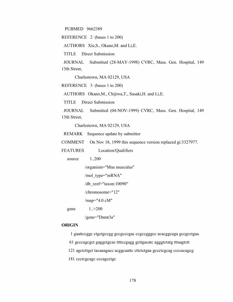
178
PUBMED 9662389
REFERENCE 2 (bases 1 to 200)
AUTHORS Xie,S., Okano,M. and Li,E.
TITLE Direct Submission
JOURNAL Submitted (28-MAY-1998) CVRC, Mass. Gen. Hospital, 149 13th Street,
Charlestown, MA 02129, USA
REFERENCE 3 (bases 1 to 200)
AUTHORS Okano,M., Chijiwa,T., Sasaki,H. and Li,E.
TITLE Direct Submission
JOURNAL Submitted (04-NOV-1999) CVRC, Mass. Gen. Hospital, 149 13th Street,
Charlestown, MA 02129, USA
REMARK Sequence update by submitter
COMMENT On Nov 18, 1999 this sequence version replaced gi:3327977.
FEATURES Location/Qualifiers
source 1..200
/organism="Mus musculus"
/mol_type="mRNA"
/db_xref="taxon:10090"
/chromosome="12"
/map="4.0 cM"
gene 1..>200
/gene="Dnmt3a"
ORIGIN
1 gaattccggc ctgctgccgg gccgcccgac ccgccgggcc acacggcaga gccgcctgaa
61 gcccagcgct gaggctgcac ttttccgagg gcttgacatc agggtctatg tttaagtctt
121 agctcttgct tacaaagacc acggcaattc cttctctgaa gccctcgcag ccccacagcg
181 ccctcgcagc cccagcctgc
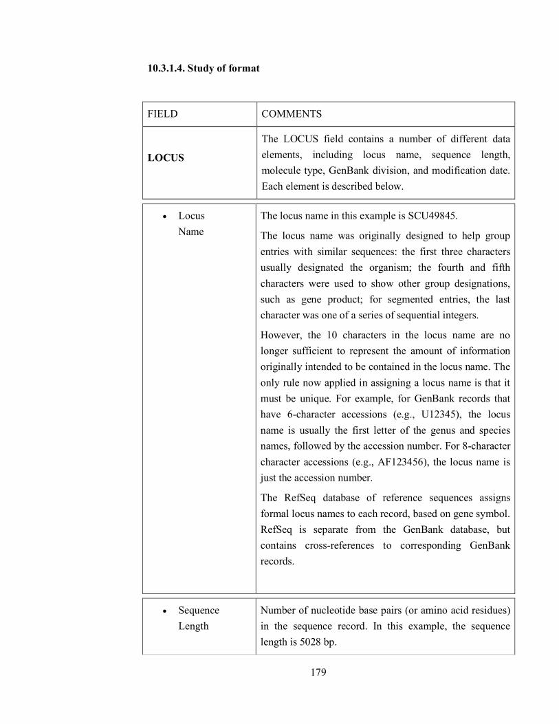
179
10.3.1.4. Study of format
FIELD COMMENTS
LOCUS
The LOCUS field contains a number of different data elements, including locus name, sequence length, molecule type, GenBank division, and modification date. Each element is described below.
Locus Name
The locus name in this example is SCU49845.
The locus name was originally designed to help group entries with similar sequences: the first three characters usually designated the organism; the fourth and fifth characters were used to show other group designations, such as gene product; for segmented entries, the last character was one of a series of sequential integers.
However, the 10 characters in the locus name are no longer sufficient to represent the amount of information originally intended to be contained in the locus name. The only rule now applied in assigning a locus name is that it must be unique. For example, for GenBank records that have 6-character accessions (e.g., U12345), the locus name is usually the first letter of the genus and species names, followed by the accession number. For 8-character character accessions (e.g., AF123456), the locus name is just the accession number.
The RefSeq database of reference sequences assigns formal locus names to each record, based on gene symbol. RefSeq is separate from the GenBank database, but contains cross-references to corresponding GenBank records.
Sequence Length
Number of nucleotide base pairs (or amino acid residues) in the sequence record. In this example, the sequence length is 5028 bp.

180
Molecule Type The type of molecule that was sequenced. In this example, the molecule type is DNA.
Each GenBank record must contain contiguous sequence data from a single molecule type. The various molecule types are described in the Sequin documentation and can include genomic DNA, genomic RNA, precursor RNA, mRNA (cDNA), ribosomal RNA, transfer RNA, small nuclear RNA, and small cytoplasmic RNA.
GenBank Division
The GenBank division to which a record belongs is indicated with a three letter abbreviation. In this example, GenBank division is PLN.
The GenBank database is divided into 18 divisions:
1. PRI - primate sequences 2. ROD - rodent sequences 3. MAM - other mammalian sequences 4. VRT - other vertebrate sequences 5. INV - invertebrate sequences 6. PLN - plant, fungal, and algal sequences 7. BCT - bacterial sequences 8. VRL - viral sequences 9. PHG - bacteriophage sequences 10. SYN - synthetic sequences 11. UNA - unannotated sequences 12. EST - EST sequences (expressed sequence tags) 13. PAT - patent sequences 14. STS - STS sequences (sequence tagged sites) 15. GSS - GSS sequences (genome survey sequences) 16. HTG - HTG sequences (high-throughput genomic sequences) 17. HTC - unfinished high-throughput cDNA sequencing 18. ENV - environmental sampling sequences
Some of the divisions contain sequences from specific groups of organisms, whereas others (EST, GSS, HTG, etc.) contain data generated by specific sequencing technologies from many different organisms.

181
Modification Date
The date in the LOCUS field is the date of last modification. The sample record shown here was last modified on 21-JUN-1999.
DEFINITION Brief description of sequence; includes information such as source organism, gene name/protein name, or some description of the sequence's function (if the sequence is non-coding). If the sequence has a coding region (CDS), description may be followed by a completeness qualifier, such as "complete cds".
ACCESSION The unique identifier for a sequence record. An accession number applies to the complete record and is usually a combination of a letter(s) and numbers, such as a single letter followed by five digits (e.g., U12345) or two letters followed by six digits (e.g., AF123456). Some accessions might be longer, depending on the type of sequence record.
VERSION A nucleotide sequence identification number that represents a single, specific sequence in the GenBank database. This identification number uses the accession.version format implemented by GenBank/EMBL/DDBJ in February 1999.
If there is any change to the sequence data (even a single base), the version number will be increased, e.g.,
→U12345.1 U12345.2, but the accession portion will remain stable.
The accession.version system of sequence identifiers runs parallel to the GI number system, i.e., when any change is made to a sequence, it receives a new GI number AND an increase to its version number.
GI "GenInfo Identifier" sequence identification number, in this case, for the nucleotide sequence. If a sequence changes in any way, a new GI number will be assigned.

182
KEYWORDS Word or phrase describing the sequence. If no keywords are included in the entry, the field contains only a period.
SOURCE Free-format information including an abbreviated form of the organism name, sometimes followed by a molecule type.
Organism The formal scientific name for the source organism (genus and species, where appropriate) and its lineage, based on the phylogenetic classification scheme used in the NCBI Taxonomy Database.
REFERENCE Publications by the authors of the sequence that discuss the data reported in the record. References are automatically sorted within the record based on date of publication, showing the oldest references first. Some sequences have not been reported in papers and show a status of "unpublished" or "in press". When an accession number and/or sequence data has appeared in print, sequence authors should send the complete citation of the article to [email protected] and the GenBank staff will revise the record.
AUTHORS List of authors in the order in which they appear in the cited article.
TITLE Title of the published work or tentative title of an unpublished work. Sometimes the words "Direct Submission" instead of an article title. This is usually true for the last citation in the REFERENCE field because it tends to contain information about the submitter of the sequence, rather than a literature citation. The last citation is therefore called the "submitter block".
JOURNAL MEDLINE abbreviation of the journal name. (Full spellings can be obtained from the Entrez Journals Database.)

183
PUBMED PubMed Identifier (PMID).
Direct Submission
Contact information of the submitter, such as institute/department and postal address. This is always the last citation in the References field. Some older records do not contain the "Direct Submission" reference. However, it is required in all new records.
FEATURES Information about genes and gene products, as well as regions of biological significance reported in the sequence. These can include regions of the sequence that code for proteins and RNA molecules, as well as a number of other features.
source Mandatory feature in each record that summarizes the length of the sequence, scientific name of the source organism, and Taxon ID number. Can also include other information such as map location, strain, clone, tissue type, etc., if provided by submitter.
Taxon A stable unique identification number for the taxon of the source oganism. A taxonomy ID number is assigned to each taxon (species, genus, family, etc.) in the NCBI Taxonomy Database. See also the Organism field, above.
CDS Coding sequence; region of nucleotides that corresponds with the sequence of amino acids in a protein (location includes start and stop codons). The CDS feature includes an amino acid translation. Authors can specify the nature of the CDS by using the qualifier "/evidence=experimental" or "/evidence=not_experimental".
<1..206 Base span of the biological feature indicated to the left, in this case, a CDS feature.
protein_id A protein sequence identification number, similar to the Version number of a nucleotide sequence. Protein IDs

184
consist of three letters followed by five digits, a dot, and a version number. If there is any change to the sequence data (even a single amino acid), the version number will be increased, but the accession portion will remain stable (e.g., AAA98665.1 will change to AAA98665.2).
translation The amino acid translation corresponding to the nucleotide coding sequence (CDS). In many cases, the translations are conceptual. Note that authors can indicate whether the CDS is based on experimental or non-experimental evidence.
gene A region of biological interest identified as a gene and for which a name has been assigned. The base span for the gene feature is dependent on the furthest 5' and 3' features. Additional examples of records that show the relationship between gene features and other features such as mRNA and CDS are AF165912 and AF090832.
complement Indicates that the feature is located on the complementary strand.
10.3.1.5. Study of SWISS PORT
UniProtKB/Swiss-Prot is the manually annotated and reviewed section of the UniProt Knowledgebase (UniProtKB). It is a high quality annotated and non-redundant protein sequence database, which brings together experimental results, computed features and scientific conclusions. Since 2002, it is maintained by the UniProt consortium and is accessible via the UniProt website.
10.3.1.6. SWISS PORT record format
Each sequence entry is composed of lines. Different types of lines, each with their own format, are used to record the various data which make up the entry.
Each line begins with a two-character line code, which indicates the type of data contained in the line. The current line types and line codes and the order in which they appear in an entry, are shown below:
ID - Identification.
AC - Accession number(s).
185
DT - Date.
DE - Description.
GN - Gene name(s).
OS - Organism species.
OG - Organelle.
OC - Organism classification.
RN - Reference number.
RP - Reference position.
RC - Reference comments.
RX - Reference cross-references.
RA - Reference authors.
RL - Reference location.
CC - Comments or notes.
DR - Database cross-references.
KW - Keywords.
FT - Feature table data.
SQ - Sequence header.
- (blanks) sequence data.
// - Termination line.
The program ignores all the description lines and uses only these line types: 'ID', 'DE', 'OS', 'SQ' and '//'.
The program uses the 'ENTRY_NAME' which is the first field of the ID line as the first line of the title
The data of the 'DE' and 'OS' lines are collected by the program and are used as the remaining lines of the title
The 'SQ' line is used to identify the beginning of the sequence. The program collect all the following lines until the termination line is found or end is reached
10.3.2. To retrieve sequence from freely available databases
186
10.3.2.1. Retrieve the Protein molecule sequence from the available protein
domains.
Conserved domains are functional units within a protein that act as building blocks in molecular evolution and recombine in various arrangements to make proteins with different functions. The Conserved Domain Database (CDD) brings together several collections of multiple sequence alignments representing conserved domains, in addition to NCBI-curated domains that use 3D-structure information explicitly to define domain boundaries and provide insights into sequence/structure/function relationships. The data are then used for putative functional annotation of protein query sequences based on matches to specific hits (illustrated example) or superfamilies, identification of proteins with similar domain architectures, and protein classification.
How to view a protein query sequence embedded within the multiple sequence alignment of a domain model
I. Use the CD-Search tool to compare a protein query sequence (either as raw sequence data in FASTA format, or as a GI or Accession) against the desired conserved domain data set, in order to identify functional units within the query sequence. Or, simply retrieve a sequence record of interest from Entrez Protein database, open its "Links" menu, and select "Conserved Domains".
II. The search results will be shown in the default Concise Display, which shows only the top scoring hits for each region of the query sequence. Change to the Full Display if you'd like to see all hits.
III. Click on a domain cartoon (colored bar representing a domain footprint) in the graphical display of the CD-search results to view the multiple sequence alignment for that domain model.
IV. Your query sequence will automatically be embedded in the alignment. If desired, modify the number and type of sequences shown as described in the help document section on CDD Record : multiple sequence alignment displays. It is also possible to view the query sequence embedded within a phylogenetic tree of the domain model's member sequences, if your hit was to an NCBI-curated domain.

187
10.3.2.2. Learn the storage of the protein sequence for the further
proceeding work.
Connect to UniProt (Universal Protein Resource) : http://www.uniprot.org/

188
Search in the Protein Knwoledgebase (UniPortKB) for “human calcitonin” (query)
How many results are given ?
Look at the 10 first ones. Why were they selected ?
Modify your query to get only human genes. A simple possibility is to use the restriction :
- Restrict term "human" to organism
- Restrict term "calcitonin" to protein family
How has the query been modified ?
How many protein sequence do you get ? Select them in the result table. A green banner opens at the bottom of your screen.
Click to “retrieve” in the green banner. You are switching from the “search” to the “retrieve” tab. Note that the UniProt identifiers are now written in the query frame. Several formats are proposed to open or download.
Open the data in the FASTA format. This is the standard format to exchange protein sequences. The information line begins with a ‘>’. The rest of the characters are interpreted as amino acids.
Save the sequences. You can either download the file and open it with a text editor such as Word or copy the sequences from the window and paste it to Word. Use a non-proportional font such as “courier” to visually maintain the alignment.
10.4 Self-Learning Exercise
1. Create a power point presentation and write down the steps to insert animation and transition in presentation.
2. What is the importance to create a presentation?
3. What is Slide master? Explain its steps.
4. Describe the different types of views of presentation.
5. What is Bioinformatics?
6. What is NCBI? Discuss about the different biological databases in variable formats.
7. What is SWISS PORT?