Inducible nitric oxide synthase is present in motor neuron mitochondria and Schwann cells and...
23
Inducible nitric oxide synthase is present in motor neuron mitochondria and Schwann cells and contributes to disease mechanisms in ALS mice Kevin Chen, Division of Neuropathology, Department of Pathology, Johns Hopkins University School of Medicine, 558 Ross Building, 720 Rutland Avenue, Baltimore, MA 21205-2196, USA Frances J. Northington, and Department of Pediatrics, Johns Hopkins University School of Medicine, Baltimore, MA, USA Lee J. Martin Division of Neuropathology, Department of Pathology, Johns Hopkins University School of Medicine, 558 Ross Building, 720 Rutland Avenue, Baltimore, MA 21205-2196, USA, Pathobiology Graduate Program, Johns Hopkins University School of Medicine, Baltimore, MA, USA, Department of Neuroscience, Johns Hopkins University School of Medicine, Baltimore, MA, USA Lee J. Martin: [email protected] Abstract Amyotrophic lateral sclerosis (ALS) is a fatal neurodegenerative disease of motor neurons (MNs). The molecular pathogenesis of ALS is not understood, thus effective therapies for this disease are lacking. Some forms of ALS are inherited by mutations in the superoxide dismutase-1 (SOD1) gene. Transgenic mice expressing human Gly93 → Ala (G93A) mutant SOD1 (mSOD1) develop severe MN disease, oxidative and nitrative damage, and mitochondrial pathology that appears to involve nitric oxide-mediated mechanisms. We used G93A-mSOD1 mice to test the hypothesis that the degeneration of MNs is associated with an aberrant up-regulation of the inducible form of nitric oxide synthase (iNOS or NOS2) activity within MNs. Western blotting and immunoprecipitation showed that iNOS protein levels in mitochondrial-enriched membrane fractions of spinal cord are increased significantly in mSOD1 mice at pre-symptomatic stages of disease. The catalytic activity of iNOS was also increased significantly in mitochondrial-enriched membrane fractions of mSOD1 mouse spinal cord at pre-symptomatic stages of disease. Reverse transcription-PCR showed that iNOS mRNA was present in the spinal cord and brainstem MN regions in mice and was increased in pre-symptomatic and early symptomatic mice. Immunohistochemistry showed that iNOS immunoreactivty was up-regulated first in spinal cord and brainstem MNs in pre-symptomatic and early symptomatic mice and then later in the course of disease in numerous microglia and few astrocytes. iNOS accumulated in the mitochondria in mSOD1 mouse MNs. iNOS immunoreactivity was also up-regulated in Schwann cells of peripheral nerves and was enriched particularly at the paranodal regions of the nodes of Ranvier. Drug inhibitors of iNOS delayed disease onset and significantly extended the lifespan of G93A- mSOD1 mice. This work identifies two new potential early mechanisms for MN degeneration in mouse ALS involving iNOS at MN mitochondria and Schwann cells and suggests that therapies targeting iNOS might be beneficial in treating human ALS. Correspondence to: Lee J. Martin, [email protected]. NIH Public Access Author Manuscript Brain Struct Funct. Author manuscript; available in PMC 2010 December 26. Published in final edited form as: Brain Struct Funct. 2010 March ; 214(2-3): 219–234. doi:10.1007/s00429-009-0226-4. NIH-PA Author Manuscript NIH-PA Author Manuscript NIH-PA Author Manuscript
-
Upload
independent -
Category
Documents
-
view
1 -
download
0
Transcript of Inducible nitric oxide synthase is present in motor neuron mitochondria and Schwann cells and...

Inducible nitric oxide synthase is present in motor neuronmitochondria and Schwann cells and contributes to diseasemechanisms in ALS mice
Kevin Chen,Division of Neuropathology, Department of Pathology, Johns Hopkins University School ofMedicine, 558 Ross Building, 720 Rutland Avenue, Baltimore, MA 21205-2196, USA
Frances J. Northington, andDepartment of Pediatrics, Johns Hopkins University School of Medicine, Baltimore, MA, USA
Lee J. MartinDivision of Neuropathology, Department of Pathology, Johns Hopkins University School ofMedicine, 558 Ross Building, 720 Rutland Avenue, Baltimore, MA 21205-2196, USA,Pathobiology Graduate Program, Johns Hopkins University School of Medicine, Baltimore, MA,USA, Department of Neuroscience, Johns Hopkins University School of Medicine, Baltimore, MA,USALee J. Martin: [email protected]
AbstractAmyotrophic lateral sclerosis (ALS) is a fatal neurodegenerative disease of motor neurons (MNs).The molecular pathogenesis of ALS is not understood, thus effective therapies for this disease arelacking. Some forms of ALS are inherited by mutations in the superoxide dismutase-1 (SOD1)gene. Transgenic mice expressing human Gly93 → Ala (G93A) mutant SOD1 (mSOD1) developsevere MN disease, oxidative and nitrative damage, and mitochondrial pathology that appears toinvolve nitric oxide-mediated mechanisms. We used G93A-mSOD1 mice to test the hypothesisthat the degeneration of MNs is associated with an aberrant up-regulation of the inducible form ofnitric oxide synthase (iNOS or NOS2) activity within MNs. Western blotting andimmunoprecipitation showed that iNOS protein levels in mitochondrial-enriched membranefractions of spinal cord are increased significantly in mSOD1 mice at pre-symptomatic stages ofdisease. The catalytic activity of iNOS was also increased significantly in mitochondrial-enrichedmembrane fractions of mSOD1 mouse spinal cord at pre-symptomatic stages of disease. Reversetranscription-PCR showed that iNOS mRNA was present in the spinal cord and brainstem MNregions in mice and was increased in pre-symptomatic and early symptomatic mice.Immunohistochemistry showed that iNOS immunoreactivty was up-regulated first in spinal cordand brainstem MNs in pre-symptomatic and early symptomatic mice and then later in the courseof disease in numerous microglia and few astrocytes. iNOS accumulated in the mitochondria inmSOD1 mouse MNs. iNOS immunoreactivity was also up-regulated in Schwann cells ofperipheral nerves and was enriched particularly at the paranodal regions of the nodes of Ranvier.Drug inhibitors of iNOS delayed disease onset and significantly extended the lifespan of G93A-mSOD1 mice. This work identifies two new potential early mechanisms for MN degeneration inmouse ALS involving iNOS at MN mitochondria and Schwann cells and suggests that therapiestargeting iNOS might be beneficial in treating human ALS.
Correspondence to: Lee J. Martin, [email protected].
NIH Public AccessAuthor ManuscriptBrain Struct Funct. Author manuscript; available in PMC 2010 December 26.
Published in final edited form as:Brain Struct Funct. 2010 March ; 214(2-3): 219–234. doi:10.1007/s00429-009-0226-4.
NIH
-PA Author Manuscript
NIH
-PA Author Manuscript
NIH
-PA Author Manuscript

KeywordsApoptosis-necrosis cell death continuum; Mitochondrial permeability transition pore; MutantSOD1; Nitration; Node of Ranvier; Schwann cell
IntroductionAmyotrophic lateral sclerosis (ALS) (also known in the US as Lou Gehrig’s disease) is aprogressive, severely disabling fatal neurological disease in humans characterized byweakness, spasticity, skeletal muscle wasting, and eventual paralysis of movement, speech,swallowing, and breathing; patients die generally within 3–5 years after symptoms begin(Rowland and Shneider 2001). The cause of the spasticity, paralysis, and morbidity isprogressive degeneration and loss of upper motor neurons (MNs) in cerebral cortex andlower MNs in brainstem and spinal cord (Rowland and Shneider 2001). ALS is the thirdmost common neurodegenerative disorder with an adult onset, affecting about 2–5 in every100,000 individuals (Rowland and Shneider 2001; Cozzolino et al. 2008). More than 5,600people in the US are diagnosed with ALS each year (ALS Association, www.alsa.org). Thedisease-causing events that trigger MN degeneration are not understood and why MNs areselectively vulnerable in ALS is unclear. Two forms of ALS exist: sporadic and familial(Rowland and Shneider 2001; Bendotti and Carrì 2004; Cozzolino et al. 2008). The majorityof ALS cases are sporadic with no known genetic component, except for missense mutationsin TAR-DNA binding protein (Kabashi et al. 2008). Aging is a strong risk factor for ALSbecause the average age of onset is 55 years (ALS Association, www.alsa.org). Familialforms of ALS (fALS) have autosomal dominant or autosomal recessive inheritance patternsand make up ~10% or less of all ALS cases (Schymick et al. 2007; Turner and Talbot 2008).ALS-linked mutations occur in the genes encoding SOD1 (ALS1), Alsin (ALS2), senataxin(ALS4), vesicle associated membrane protein (VAMP/synaptobrevin)-associated protein B(ALS8), dynactin, TAR-DNA binding protein, and fused in sarcoma (FUS, ALS6) (Martin2006; Schymick et al. 2007; Turner and Talbot 2008).
Mutations in the SOD1 gene account for ~20% of all fALS cases (~2% of all ALS cases)(Deng et al. 1993; Rosen et al. 1993). SOD1 (also known as copper/zinc SOD) is ametalloenzyme of 153 amino acids (~16 kDa) that binds one copper ion and one zinc ion persubunit. SOD1, functioning as a ~32 kDa non-covalently linked homodimer, is responsiblefor the detoxification and maintenance of intracellular superoxide anion (O2
−) concentrationin the low femtomolar range by catalyzing the dismutation of O2
− to molecular oxygen andhydrogen peroxide (O2
− + O2− + 2H+ → H2O2 + O2) (McCord and Fridovich 1969). SOD1
is ubiquitous (intracellular SOD concentrations are typically ~10–40 μM) in most tissuesand possibly greater in neurons (Rakhit et al. 2004). SOD1 mutants appear to gain a toxicproperty or function, rather than having diminished O2
− scavenging activity (Deng et al.1993; Borchelt et al. 1994; Yim et al. 1996), and this toxicity might involve nitric oxide(NO•) (Beckman et al. 1993, 2001). Cellular stresses resulting from reactive oxygen species(ROS) and reactive nitrogen species (RNS) have been implicated in human ALSpathogenesis, and in animal and cell models of ALS (Martin 2006). One particular pathwayfor MN toxicity involves NO•, which can be synthesized by three isoforms of nitric oxidesynthase (NOS) enzymes: neuronal or NOS1, inducible or NOS2, and endothelial or NOS3(Mungrue et al. 2003). Although NO• has many beneficial cellular functions, it can reactwith superoxide radical (O2 •) to form the potent oxidant peroxynitrite (ONOO−) that candamage protein, lipids, and nucleic acids (Pacher et al. 2007). Inducible NOS (iNOS) differsfrom NOS1 and NOS3 because it is active constitutively in a calcium-independent mannerand is active for extended periods yielding high-output NO• (MacMicking et al. 1997;Lowenstein and Padalko 2004). Although iNOS is studied most commonly in the context of
Chen et al. Page 2
Brain Struct Funct. Author manuscript; available in PMC 2010 December 26.
NIH
-PA Author Manuscript
NIH
-PA Author Manuscript
NIH
-PA Author Manuscript

the immune system, tissue inflammation, and macrophage function (MacMicking et al.1997; Lowenstein and Padalko 2004), iNOS is also present in the nervous system and isexpressed by subsets of glial cells and neurons (Heneka and Feinstein 2001). Interestingly,normal MNs neurons express constitutively iNOS at low levels (Martin et al. 2005), andafter axotomy iNOS is up-regulated in MNs and is involved directly in their apoptotic death(Martin et al. 2005; Martin and Liu 2002). Thus, a gain in the activity of iNOS in responseto certain signals can cause some forms of MN degeneration.
In the present experiments, we examined further the contribution of iNOS to thepathogenesis of ALS in a mutant SOD1 (mSOD1) mouse model. Our goals were to measurethe levels and activity of iNOS in the mSOD1 mouse nervous system, to determine thecellular and subcellular localizations of iNOS, and to determine if pharmacologicalinterventions using iNOS inhibitors could ameliorate disease. Our findings stronglyimplicate iNOS in the disease mechanisms of ALS in mice.
Materials and methodsAnimal model
A common mutation in human SOD1 is the substitution of glycine by alanine at position 93(G93A). Transgenic (tg) mice that express this mutant form of human SOD1 linked to fALS(Gurney et al. 1994; Dal Canto and Gurney 1994) are used widely as an animal model ofALS (Bendotti and Carrì 2004; Martin and Liu 2004; Cozzolino et al. 2008; Turner andTalbot 2008). The effects of this human mutant gene on mice are profound. Hemizygous tgmice expressing a high copy number of the G93A variant of mutant SOD1 (mSOD1)become completely paralyzed and die at ~16–18 weeks of age (Gurney et al. 1994). MNs inmice expressing G93Ahigh-mSOD1 undergo prominent degeneration; about 70% of lumbarMNs are eliminated by end-stage disease (Martin et al. 2007). Tg mice expressing either thehuman mutant SOD1 gene or the human wild-type (wt) human SOD1 gene (Gurney et al.1994) were used in this study. The mouse line (B6SJL-TgN[SOD1-G93A]1Gur, G1H,Jackson Laboratory, Bar Harbor, ME) with a high copy number of mutant allele (~20copies) and a rapid disease onset was used. Mice were studied throughout the course ofdisease generally from pre-symptomatic stages (<10 weeks) to early symptomatic stages(10–14 weeks) to late stages of disease (15–18 weeks). Mice were classified specifically byvisible neurologic signs as pre-symptomatic, early-stage disease, or late-stage disease bytheir hindlimb reflex appearance when held by the tail, gait and posture, and paralysis,respectively. Controls were tg mice expressing the normal allele of the human SOD1 gene(B6SJL-TgN[SOD1]2Gur, Jackson Laboratory) studied at the same ages as the mutants.Animal protocols were approved by the Johns Hopkins University Institutional Animal Careand Use Committee.
Western blots and immunoprecipitationTo determine the levels of iNOS protein in wtSOD1 and mSOD1 tg mice spinal cord,Western blotting (WB) and immunoprecipitation (IP) were used. mSOD1 mice at pre-symptomatic (n = 4–6) and early symptomatic (n = 4–6) stages of disease, along with age-matched and wtSOD1 tg mice (n = 4), were killed by isoflurane overdose and decapitated.Spinal cords were isolated and homogenized, followed by differential centrifugation intonuclear, soluble, and mitochondrial-enriched membrane fractions as described (Martin et al.2003). Spinal cord protein samples (10–20 μg) and purified recombinant mouse iNOS(Calbiochem) were subjected to SDS-PAGE, followed by electrophoretic transfer tonitrocellulose membranes. The quality of the SDS-PAGE and transfer was assessed byPonceau S staining. These membranes were blocked with 2.5% dry milk/0.1% Tween 20/Tris–buffered saline (TBS) and then incubated with antibody to iNOS. Four different
Chen et al. Page 3
Brain Struct Funct. Author manuscript; available in PMC 2010 December 26.
NIH
-PA Author Manuscript
NIH
-PA Author Manuscript
NIH
-PA Author Manuscript

antibodies to iNOS were used: mouse monoclonal C-11 antibody to mouse iNOS C-terminus (Santa Cruz Biotechnology), mouse monoclonal clone 6 antibody to mouse iNOSC-terminus (BD Biosciences), rabbit polyclonal antibody to mouse iNOS N-terminus(Upstate Biotechnology), and rabbit polyclonal antibody to mouse iNOS C-terminus(Sigma). Primary antibody binding was detected with HRP-conjugated secondary antibodies(BioRad), followed by a chemiluminescent HRP substrate (Pierce). Immunoblots wereexposed to X-ray film to visualize immunoreactivity. Blots were re-probed with monoclonalantibody 6C5 to glyceraldehyde phosphate dehydrogenase (GAPDH, Research DiagnosticsInternational) for a protein loading control. iNOS immunoreactivity levels relative toGAPDH levels on films were quantified using densitometry (Quantity One).
For IP, as described previously in more detail (Golden et al. 2003), samples of spinal cordprotein fractions (50 μg) were pre-cleared with Protein A/G Sepharose (Pierce), followed byincubation with iNOS primary antibody (Santa Cruz, sc-7271 or Upstate, 06-573). Sampleswere then incubated with Protein A/G Sepharose to form sepharose-antibody–iNOScomplexes. These complexes were captured by centrifugation. Precipitated proteins wereisolated by boiling samples in 4× treatment buffer and subjected to SDS-PAGE and WB.Films were quantified using computer densitometry program (Quantity One). IgG bandswere quantified as loading controls.
NOS enzyme activity assayMice classified as pre-symptomatic, early-stage disease, late-stage of disease, and controls(wtSOD1 tg) were used (n = 6 mice/group). Spinal cords were collected freshly and tissuehomogenates obtained as described above. A NOS biochemical assay kit (CaymanChemical, 781001) was used to measure enzymatic activity of iNOS in spinal cordpreparations of mSOD1 mice. L-arginine is consumed by NOS enzymes using a variety ofcofactors and electron carriers, producing NO· and L-citrulline (Mungrue et al. 2003); theNOS assay kit measures production of NO· in vitro by measuring conversion of [14C]radiolabeled L-arginine to L-citrulline using a modification of the method describedpreviously (Bhardwaj et al. 1997). To detect the calcium-independent enzymatic activity ofiNOS activity, the reaction mixtures omitted calcium ion and calmodulin, which wererequired for other NOS isoforms (e.g., nNOS). Alternatively, a highly specific inhibitor ofiNOS (S-methylisothiourea sulfate, SMT; Santa Cruz Biotechnology, sc-3566; Southan et al.1995) was included in the reaction mix to assay selectively for nNOS activity. Samples wereincubated with the kit’s reaction mix, which includes cofactors (e.g., FAD, NADPH) and[14C] radiolabeled L-arginine. After 1 h of reaction time, reactions were terminated using alow pH stop buffer included in the kit. A negatively charged resin was then suspended withthe reaction mixture, binding the positively charged, unreacted L-arginine. The solution wasplaced in spin cups containing filters, and the solutions centrifuged briefly. The filterretained the resin bound to leftover L-arginine, and a liquid solution was eluted thatcontained neutrally charged, reacted L-citrulline unbound by resin. The L-citrulline that waseluted corresponded to NO· produced, since the stoichiometry of NOS produced one L-citrulline molecule and one NO· molecule for every L-arginine molecule consumed. Theeluent was combined with 5 ml scintillation fluid and run through a scintillation counter.The efficacy of iNOS and nNOS selective conditions were confirmed by performing NOSactivity assay in the absence of inhibiting conditions (i.e., including Ca2+/calmodulin oromitting SMT). Activity was controlled against background reactivity (incubating [14C] L-arginine with purified iNOS) and converted from decompositions per minute to nmol/min ofNO·.
Chen et al. Page 4
Brain Struct Funct. Author manuscript; available in PMC 2010 December 26.
NIH
-PA Author Manuscript
NIH
-PA Author Manuscript
NIH
-PA Author Manuscript

ImmunohistochemistrymSOD1 and wtSOD1 tg mice at 7, 8, 9, 10, 11, 14, and 15 weeks of age (n = 6/geneotype/age) were anesthetized with an overdose of chloral hydrate and perfused intracardially withice-cold 100 mM phosphate-buffered saline (PBS; 0.9% NaCl, pH 7.4) followed by ice-cold4% paraformaldehyde. After perfusion fixation, spinal cords remained in situ for 2 h beforethey were removed from the vertebral column and then placed in 20% glycerol forcryoprotection. Transverse serial symmetrical sections of lumbar spinal cord (40 μm) wereobtained by frozen sectioning on a sliding microtome and stored in 96-well plates with onesection/per well.
Selected sections of lumbar spinal cord (every two in ten series) were immunostained foriNOS using the immunoperoxidase method as done before (Martin et al. 2007, 2009).Sections were first permeabilized by 0.4% triton-x/TBS and then blocked with a solution of4% normal goat serum/0.1% triton-x/TBS. Sections were then incubated in primary antibodyto iNOS. Two different antibodies to iNOS were used for immunohistochemistry: mousemonoclonal C-11 antibody to mouse iNOS C-terminus (Santa Cruz Biotechnology) andmouse monoclonal clone 6 antibody to mouse iNOS C-terminus (BD Biosciences). Afterincubation in primary antibody, affinity-purified goat anti-mouse secondary antibody wasapplied, followed by peroxidase anti-peroxidase. Antibody binding to iNOS was visualizedusing diaminobenzidine (DAB) as chromogen. Labeling intensity of individual MNs wasquantified by computer densitometry using IPLab Gel.
ImmunofluorescenceImmunohistochemistry using dual-label immunofluorescence was done as before (Martin etal. 2007, 2009) to identify iNOS at various organelles and in different types of cells. Lumbarspinal cord sections were permeabilized and blocked by incubation in 5% normal goat serumand 0.4% triton-x/TBS. iNOS was detected with mouse monoclonal primary antibody (SantaCruz) in combination with different rabbit or sheep second primary bodies for co-localization analyses. Mitochondria were identified by MnSOD (SOD2) by rabbit polyclonalantibody (Stressgen). Peroxisomes were identified using sheep antibody to catalase(BioDesign International). Microsomes were visualized by rabbit antibody to cytochromeP450 reductase (Upstate Biotechnology). Microglial cells were identified with rabbitpolyclonal antibodies (Affinity Bioreagents) to the integrin protein CD11b (Akiyama andMcGeer 1990). Astrocytes were detected with rabbit polyclonal antibodies (DAKO) to glialfibrillary acidic protein (GFAP). Schwann cells in the ventral roots/peripheral nerve wereidentified with rabbit antibodies to p75-low affinity neurotrophin receptor (Promega) andvimentin (Chemicon), as shown before to be markers for Schwann cells (Autilio-Gambetti etal. 1982; Johnson et al. 1988). Secondary antibodies conjugated with Alexa-488 orAlexa-594 were applied and sections were viewed using epifluorescence microscopy.
Reverse transcription-polymerase chain reaction (RT-PCR)To corroborate findings based on iNOS antibody approaches, RT-PCR was used to analyzemRNA expression for iNOS in mSOD1 mice. Total RNA was extracted using TRIzol(Invitrogen) from mouse whole cerebrum and from brainstem (medulla containing cranialnerve nuclei V and VII) and spinal cord ventral isolated freshly by micro-dissection andmicro-punching. cDNA synthesis was accomplished using SuperScript One-Step RT-PCRwith Platinum Taq (Invitrogen) followed by PCR. Two different sets of oligonucleotideprimer pairs were used to amplify mouse iNOS cDNA. One set of primers, commerciallyavailable (R&D Systems), amplifies a 513-bp product. Another set of primer pairs used werechosen from the published cDNA sequence of mouse iNOS (Lyons et al. 1992) and were 5′-AGCATCACCCCTGTGTTCCACC-3′(sense) and 5′-TGGGACAGTCTCCATTCCCA-3′(anti-sense). These primers amplify a 388-bp product (Wu et al. 2003). Oligonucleotide
Chen et al. Page 5
Brain Struct Funct. Author manuscript; available in PMC 2010 December 26.
NIH
-PA Author Manuscript
NIH
-PA Author Manuscript
NIH
-PA Author Manuscript

primers for mouse voltage-dependent anion channel-1 (VDAC1), used as an RNA controlfor each sample, were 5′-GCTAAGGATGACTCGGCTT TAAGG-3′ and 5′-AGGTTAAGTGATGGGCTAGGATG G-3′, which give a 335-bp amplification product(Massa et al. 2000). The PCR products were separated on a 1.0% agarose gel, stained withethidium bromide and imaged using a BioRad molecular imaging VersaDoc system.
Pharmacological interventionTo determine if the activity of iNOS participates directly the mechanisms of disease in ALSmice, we conducted pharmacological studies using iNOS inhibitors. Two different highlyspecific inhibitors of iNOS were used. A small-scale study was done on a cohort of mSOD1mice (n = 5) injected intraperitoneally with SMT (Santa Cruz Biotechnology), a potentcompetitive inhibitor of iNOS (Southan et al. 1995), at a dosage of 5 mg/body weight dailystarting at 9 weeks of age. mSOD1 mice receiving normal saline vehicle were controls. Alarger-scale study was done on a cohort of mSOD1 mice (n = 12) injected subcutaneouslywith 1400 W (Sigma), a slow tight-binding potent iNOS inhibitor (Garvey et al. 1997) at adosage of 3 mg/body weight on alternate days, starting at 6 weeks of age. mSOD1 micereceiving normal saline/cyclodextran vehicle were controls. Lifespan was the outcomemeasurement.
Data analysisGroup means and variances were evaluated statistically by one-way ANOVA and aNewman–Keuls post hoc test.
Photography and figure constructionThe original images of immunohistochemical preparations used for figure construction weregenerated using digital photography. Digital images were captured as TiFF files using aSPOT digital camera and SPOT Advanced software (Diagnostic Instruments) or a Nikondigital camera (DXM1200) and ACT-1 software. Images were altered slightly for brightnessand contrast using ArcSoft Photo-Studio 2000 or Adobe Photoshop software withoutchanging the content and actual result. Figure composition was done using CorelDraw 9software with final composite figures being converted to TiFF files. Files of compositefigures were adjusted for brightness and contrast in Adobe Photoshop.
ResultsiNOS protein levels are up-regulated in pre-symptomatic ALS mice
Western blots of wtSOD1 and mSOD1 tg mouse spinal cord extracts probed with iNOSantibodies showed a band of immunoreactivity at ~130 kDa (Fig. 1a), consistent with themolecular weight of full-length iNOS protein (Heneka and Feinstein 2001;Mungrue et al.2003). Corroboration that this immunoreactive band was indeed iNOS was based on thefindings that this band was strongly positive in lanes loaded with cell extracts oflipopolysaccharide-stimulated mouse macrophages (RAW 264.7 cells) or purified iNOSrecombinant protein and absent in lanes loaded with tissue extracts from iNOS−/− mice (datanot shown). iNOS was detected at near equivalent levels in soluble and mitochondria-enriched membrane fractions of wtSOD1 mouse spinal cord (Fig. 1a). In contrast, mSOD1mice had greater levels of iNOS in the mitochondria-enriched membrane fractions comparedto the soluble fraction, and the level of iNOS in the membranous fraction was significantlygreater than that found in wtSOD1 mice (Fig. 1a, b).
To detect and measure the full-length iNOS protein in its native state, IP followed by WBwas used (Fig. 1c). iNOS immunoprecipitated from wtSOD1 and mSOD1 tg mouse spinalcord migrated as a 130-kDa band corresponding to an immunoreactive band of
Chen et al. Page 6
Brain Struct Funct. Author manuscript; available in PMC 2010 December 26.
NIH
-PA Author Manuscript
NIH
-PA Author Manuscript
NIH
-PA Author Manuscript

immunoroprecipitated purified iNOS from mouse macrophage cells (Fig. 1c). Computerdensitometry of this 130-kDa band, controlled against the IgG heavy chain labeling,demonstrated a significant increase in the level of iNOS in pre-symptomatic mSOD1 mousespinal cord compared to wtSOD1 mouse spinal cord (Fig. 1d).
NOS activity is increased in pre-symptomatic and early symptomatic ALS miceTo determine the functional activity of iNOS in mSOD1 mice, a NOS biochemical assaywas employed to measure enzymatic conversion of radiolabeled L-arginine to L-citrulline.As negative controls, reactions were incubated with known inhibitors of iNOS and nNOSthat confirmed the assay to be effective and specific (Fig. 2a). Specific iNOS activity wasfound in nuclear-enriched, soluble, and mitochondrial membrane-enriched fractions ofmouse spinal cord (Fig. 2b). In the mitochondrial membrane-enriched fraction, iNOSactivity was increased significantly in mSOD1 mice compared to wtSOD1 mice at earlysymptomatic stages of disease (Fig. 2b). iNOS activity was not significantly different innuclear-enriched and soluble fractions of mSOD1 mice (Fig. 2b). nNOS activity wasmeasured to determine if the changes in iNOS activity were isoform specific. nNOS activitywas detected in soluble and mitochondrial subcellular compartments of spinal cord. nNOSactivity was increased significantly in the mitochondrial-enriched membrane compartmentof mSOD1 mice compared to wtSOD1 mice at the pre-symptomatic stages of the disease(Fig. 2c).
iNOS immunoreactivity is increased in mSOD1 MNs and microgliaImmunohistochemical staining of iNOS using specific antibodies confirmed by Westernblotting showed increases in iNOS immunoreactivity in motor neurons during theprogression of disease (Fig. 3a–d). iNOS immunoreactivity was seen as dot-like particlesand aggregates in the cytoplasm of the somatodendritic compartment and nuclearcompartment of MNs (Fig. 3a–c, h, i). MNs in wtSOD1 mice maintained a steadily low levelof iNOS immunoreactivity at 7 through 15 weeks of age (Fig. 3a), similar to that seen beforein non-transgenic mouse MNs (Martin et al. 2005); in contrast, mSOD1 mice showedprogressively increased immunoreactivity throughout this time course (Fig. 3b–d). Thelevels of iNOS immunoreactivity in symptomatic mSOD1 mice reached an optical densityof more than 3.3 times greater than the average optical density in wtSOD1 mice (Fig. 3d).The iNOS localization pattern in MNs of pre-symptomatic and symptomatic mSOD1 micediffered markedly from that seen in wtSOD1 tg mice. The increased iNOS immunoreactivityoccurred specifically in MNs in mice at the pre-symptomatic and early symptomatic stagesof disease and then later also in cells appearing as microglia (Fig. 3c). The rich iNOSimmunostaining in MNs of pre-symptomatic mSOD1 mice was confirmed byimmunofluoresence (Fig. 4g). iNOS immunoreactivity specifically in microglia wasdemonstrated by dual labeling for iNOS and CD11b (Fig. 4a–c). The co-localization ofiNOS and CD11b was common and robust (Fig. 4a–c). In advanced disease, iNOS-positivemicroglia and their processes were found closely associated with, and perhaps penetrating,iNOS-positive degenerating and remnant MNs (Fig. 4d–f). iNOS immunoreactivity in pre-symptomatic mSOD1 mouse spinal cord was not associated with astrocytes identified byGFAP immunostaining (Fig. 4g–i), but at end-stage disease some infrequent co-localizationof iNOS and GFAP was observed (Fig. 4j–k).
iNOS expression is increased in mSOD1 mouse brainstem motor nucleiThe pattern of increased iNOS immunoreactivity seen in spinal MNs was also seen incranial nerve MN nuclei in brainstem (Fig. 3e). This observation afforded an opportunity touse higher resolution micro-dissection of brainstem regions containing cranial nerve MNnuclei (CN5 and CN7) for RT-PCR. In normal mouse CNS, constitutive levels of iNOSmRNA were undetectable in cerebrum, low in brainstem, and highest in spinal cord (Fig. 3f)
Chen et al. Page 7
Brain Struct Funct. Author manuscript; available in PMC 2010 December 26.
NIH
-PA Author Manuscript
NIH
-PA Author Manuscript
NIH
-PA Author Manuscript

consistent with the constitutive expression of iNOS in MNs (Martin et al. 2005). Acomparison of iNOS mRNA expression in wtSOD1 and mSOD1 mouse brainstem revealedconsistently elevated levels of iNOS mRNA in pre-symptomatic mSOD1 mice (Fig. 3g).Surprisingly, mSOD1 mice generally expressed lower levels of iNOS mRNA at earlysymptomatic and end-stage disease compared to the pre-symptomatic stages (Fig. 3g).
iNOS immunoreactivity is localized to mSOD1 mouse MN mitochondriaiNOS immunoreactivity in MN cell bodies was seen in the cytoplasm as fine discrete dots,larger round or oval particles, and as diffuse labeling (Fig. 3a–c, h–j). Dual labeling foriNOS and organelle markers was done to identify the subcellular localization of iNOS inMNs. iNOS immunoreactivity was largely distinct from the peroxisomal compartmentidentified by catalase (Fig. 3h). In contrast, the fine diffusely particulate iNOSimmunoreactivity in MNs of mSOD1 mice showed registration with the microsomalcompartment identified by cytochrome p450 reductase (Fig. 3i) and the larger particulateiNOS immunoreactivity co-localized with mitochondrial marker SOD2 (Fig. 3j). In mSOD1mouse motor neurons with mitochondrial swelling, iNOS was consistently localized toswollen mitochondria while normal-sized mitochondria were mostly iNOS-negative (Fig.3j).
iNOS is expressed by Schwann cells in mSOD1 mice and is enriched at the paranodalregions of nodes of Ranvier
In the course of analyzing the cellular localization of iNOS in mSOD1 mouse spinal cord,we found serendipitously iNOS immunoreactivity enriched at the ventral root exit zones ofthe peripheral nerves (Fig. 5a). This iNOS immunoreactivity appeared to be associated withthe myelin sheaths of MN axons (Fig. 5a) and was enriched particularly at the node ofRanvier paranodal sites of peripheral nerves (Fig. 5a, lower left inset), suggesting theexpression of iNOS by Schwann cells. Many peripheral nerves of pre-symptomatic andsymptomatic mSOD1 mice contained subsets of iNOS-immunoreactive axons that wereensheathed by iNOS immunoreactivity (Fig. 5b). Dual immunofluorescence for iNOS andthe Schwann cell marker vimentin (Autilio-Gambetti et al. 1982) demonstrated thatSchwann cells were positive for iNOS (Fig. 5c–e). p75NTR staining was also used to identifyactivated Schwann cells in response to injured axons (Johnson et al. 1988), confirming iNOSexpression by Schwann cells (Fig. 5f–h). Dual immunofluorescence for iNOS and p75NTR
also confirmed the accumulation of iNOS in swollen, degenerating axons within peripheralnerves (Fig. 5f–h).
Inhibition of iNOS has beneficial effects on ALS miceWe studied in vivo the effects of drug inhibitors of iNOS activity on disease mSOD1 mice.In a small cohort study, ALS symptoms were delayed by administration of SMT starting at 9weeks of age (Fig. 6a). Nevertheless, after this temporary period of ALS-like symptoms,SMT-receiving mice quickly reached end-stage disease, like vehicle-treated mice, with noextension of lifespan (Fig. 6a). In contrast, treatment of mSOD1 mice with 1400 W startingat 6 weeks of age delayed the onset of disease (Fig. 6b) and significantly extended survival,as evidenced by the 23% increase in lifespan (Fig. 6c).
DiscussionThe disease mechanisms in mSOD1 mice are studied intensively, but clinically translatableeffective mechanism-based therapies have not yet been developed from work on this animalmodel or any other animal model of MN degeneration (Martin and Liu 2004; Bendotti andCarrì 2004; Cozzolino et al. 2008; Turner and Talbot 2008). The focus of this study was onthe role of iNOS in the pathobiology of ALS in mice. We found that iNOS mRNA, protein,
Chen et al. Page 8
Brain Struct Funct. Author manuscript; available in PMC 2010 December 26.
NIH
-PA Author Manuscript
NIH
-PA Author Manuscript
NIH
-PA Author Manuscript

and enzyme activity are up-regulated in mSOD1 mouse spinal cord and brainstem at pre-symptomatic and early symptomatic stages of disease. iNOS is expressed constitutively atlow levels in mouse MNs and an early pre-symptomatic up-regulation of iNOS occurs inMNs in mSOD1 mice. iNOS in MNs of mSOD1 mice associates with mitochondria andmicrosomes. Finally, pharmacological inhibition of iNOS has significant effects in ALSmice by delaying disease onset and extending survival. These observations demonstrate thatiNOS participates in the causal mechanisms of MN degeneration in mouse ALS.
This study is important because the role of NOS in the degeneration of MNs in mSOD1mice has been very controversial and most studies have focused only on the nNOS (NOS1)isoform (Martin 2006). nNOS isoform and super-oxide have been implicated in the death ofMNs induced by mSOD1 and Zn2+-deficient wild-type SOD1 in cell culture (Estévez et al.1999; Raoul et al. 2002). Cultured MNs from tg mouse embryos expressing G93A, G85R,and G37R variants of mSOD1 show enhanced sensitivity to Fas death receptor-triggered celldeath through a signaling pathway involving nNOS-mediated NO• production (Raoul et al.2002). In vivo, the nNOS inhibitor AR-R 17,477 prolonged the survival of mSOD1 mice,but other nNOS inhibitors were ineffective (Facchinetti et al. 1999; Upton-Rice et al. 1999).mSOD1 mice without nNOS (α isoform) do not have prolonged survival (Facchinetti et al.1999), but these mice still produce catalytically active β and γ isoforms of nNOS, utilizingalternate translation start sites that exclude the regions targeted by the knockout strategy(Mungrue et al. 2003), and these mice have two other NOS genes (iNOS and NOS3). iNOShas properties that are different from NOS1 and NOS3. Homodimeric iNOS is alwayscatalytically active when expressed, because it is calcium-independent and active forextended periods (hours to days) with a Vmax ~ tenfold greater that other NOS isoforms,yielding a very high output of NO• (MacMicking et al. 1997; Lowenstein and Padalko2004). We reported that G93Ahigh-mSOD1 mouse MNs have increased NO• production andthat these mice without iNOS have significantly prolonged survival (Martin et al. 2007).However, survival of G93A-mSOD1 mice expressing a low copy number of transgeneseems unaffected by iNOS gene deletion (Son et al. 2001), suggesting that the diseasemechanisms in G93Ahigh-and G93Alow-expressing mice are different. Here, we show thatdrugs that selectively inhibit iNOS have beneficial effects in mSOD1 mice with a rapiddisease onset. Thus, iNOS has a role in the pathobiology of ALS in this severe mousemodel.
This study expands on the idea that changes in NO• signaling pathways are causally relatedto the initiation or progression of ALS. We focused on iNOS because earlier studies on mice(Almer et al. 1999; Sasaki et al. 2001a, b; Martin et al. 2007) and humans (Phul et al. 2002)have indicated that this isoform of NOS could be important in ALS, and its role in thepathobiology of ALS has been under-appreciated. We used several different methods tointerrogate iNOS. iNOS was present constitutively in normal mouse spinal cord andbrainstem as seen at the mRNA level using RT-PCR and protein level using WB and IP,corroborating an earlier study demonstrating iNOS immunoreactivity and activity in MNs atlow levels (Martin et al. 2005). WB showed that iNOS was greater in membrane-enrichedsubcellular fractions than in cytoplasmic fractions, suggesting potential iNOS associationswith mitochondria and microsomes; these observations were confirmed subsequently byimmunohistochemical dual labeling. A salient finding was that iNOS mRNA expression andprotein levels were up-regulated highest in mSOD1 mice early in the course of disease. Up-regulated iNOS mRNA in spinal cord of early symptomatic mice has been found by others(Almer et al. 1999). We then used immunohistochemistry to show that iNOS was present innormal MNs at very low levels and was up-regulated dramatically in brainstem and spinalMNs of mSOD1 mice before a robust microglial up-regulation occurring at later stages ofdisease. Sasaki et al. (2001a, b) and Kiaei et al. (2005) also found iNOS immunoreactivity inspinal MNs of G93A-mSOD1 mice. In addition, Sasaki et al. (2001a, b) reported that
Chen et al. Page 9
Brain Struct Funct. Author manuscript; available in PMC 2010 December 26.
NIH
-PA Author Manuscript
NIH
-PA Author Manuscript
NIH
-PA Author Manuscript

reactive astrocytes were immunostained frequently with iNOS antibody in the spinal cord atearly symptomatic (32 weeks) and end-stage (35 weeks) disease in G93Alow-expressingmice, and they found in the autopsy of spinal cords of human ALS iNOS-positive MNs andastrocytes that were not observed in controls (Sasaki et al. 2000). We observed, usingdifferent antibodies, very occasional iNOS-positive astroglial elements in ALS mice withrapid disease, with the up-regulation of iNOS in microglia being much more prominentwhen mice were at end-stage disease. There are important distinctions between our studyand the previous work by Sasaki et al. (2001a, b), notably the use of different tg mouse linesexpressing very different transgene copy numbers and disease progressions, and we usedcomplementary quantitative approaches. Almer et al. (1999) stated that iNOS was identifiedonly in glial cells and not in neurons of G93Ahigh-mSOD1 mice, but their illustrationssuggest otherwise. It is possible that abnormalities in the spinal cord neuropilmicroenvironment in mSOD1 mice are responsible for the iNOS induction in MNs, becausemany pro-inflammatory cytokines can modulate iNOS gene expression through NF-κB,JAK/STAT, and HIF-1 (Lowenstein and Padalko 2004).
We also studied the biochemical activity of iNOS and nNOS. iNOS activity was detected innuclear, soluble, and mitochondrial-enriched membrane fractions in control and mSOD1 tgmice. nNOS activity was detected in soluble and mitochondrial-enriched membranefractions. The changes in activity in mSOD1 mice were very selective. iNOS and nNOSactivities were significantly increased only in mitochondrial-enriched membrane fractions ofmice at early symptomatic and pre-symptomatic stages of disease, respectively. Almer et al.(1999) found in total spinal cord extracts nNOS activity to be unaltered early in disease andiNOS activity increased in early symptomatic and end-stage mice. Our results show aninteresting disconnect between the robust microglial immunoreactivity for iNOS and lowenzyme activity and mRNA in mSOD1 mice at end-stage disease. This finding might meanthat our biochemical approach using subcellular fractions is disadvantageous in this regard,resulting in loss of activity, or the finding suggests that the presence of protein does notnecessarily mean catalytically active enzyme due to interactions with proteins such asNAP110 (Ratovitski et al. 1999).
MNs seem to be unique among neurons regarding NO• production because they expressconstitutively low levels of iNOS, and iNOS is strongly up-regulated in MNs in ALS mice;thus, iNOS is the likely source of NO• in MNs degenerating in ALS. The iNOS promoter isactivated by IRF-1 and NF-κB and is usually engaged by inflammation-mediated stimulation(Heneka and Feinstein 2001; Mungrue et al. 2003). This mechanism would be consistentwith the concept that mSOD1 has aberrant oxidative chemistry causing an oxidativemicroenvironment (Liochev and Fridovich 2003), and thus high levels of NO• would favorthe diffusion-limited reaction with superoxide to form peroxynitrite (Beckman et al. 1993).nNOS could also be a source of NO in degenerating MNs in mSOD1 mice (Sasaki et al.2002), but we showed previously that NADPH diaphorase activity and iNOSimmunoreactivity were induced in mSOD1 mouse MNs, but not nNOS immunoreactivity(Martin et al. 2007). Our new work here corroborates this finding with quantitative analysesusing different complementary methods. mSOD1 mice develop profound mitochondrialdamage in MNs (Wong et al. 1995; Kong and Xu 1998; Bendotti et al. 2001; Higgins et al.2002; Sasaki et al. 2004; Martin 2006; Martin et al. 2007). We found that iNOSimmunoreactivity becomes localized to mitochondria before and after they become swollen.Mitochondria produce NO through a reaction catalyzed by a mitochondrial form of NOS(mtNOS) with similar cofactor and substrate requirements as constitutive NOS, but mtNOScan cross-react immunologically with antibodies to iNOS (Lacza et al. 2003; Giulivi 2003).Our work extends this idea to ALS mice and demonstrates that iNOS is catalytically activein mitochondrial-enriched membrane fractions of mouse spinal cord at a time when iNOS
Chen et al. Page 10
Brain Struct Funct. Author manuscript; available in PMC 2010 December 26.
NIH
-PA Author Manuscript
NIH
-PA Author Manuscript
NIH
-PA Author Manuscript

protein accumulates in MNs but not in microglia. Our findings are consistent with an iNOSproduced NO-mediated mechanism for mitochondriopathy in MNs of mSOD1 mice.
It is not completely clear to us whether the NOS activity accumulating in MNs and theirmitochondria of mSOD1 mice should be called iNOS or mtNOS. True iNOS (NOS2) has ahigh NO output capacity and is Ca2+ independent, though it does bind calmodulin(MacMicking et al. 1997; Lowenstein and Padalko 2004); thus, its activity within MNsshould be insensitive to the abnormal increases in intracellular Ca2+ in mSOD1 mouse MNsobserved by us (Martin et al. 2007) and others (Siklos et al. 1998). But, if this NOS activityin MN mitochondria is indeed mtNOS, then intracellular Ca2+ fluxes could be importantpathophysiologically because mitochondrial Ca2+ uptake stimulates NO production inmitochondria (Dedkova et al. 2004). Regardless of the specific isoform of NOS, abnormalNO production could drive the formation of peroxynitrite in mitochondria and the nitrationof respiratory chain enzymes (e.g., cytochrome c oxidase subunit I) and mitochondrialantioxidant enzymes (e.g., SOD2) (Martin et al. 2007). Abnormal NO production inmitochondria could also explain the nitration of cyclophilin D (the ppif gene product) andthe nitration of adenine nucleotide translocase seen pre-symptomatically and the formationof the mitochondrial permeability transition pore that has a critical role in the diseasemechanisms of ALS mice, possibly by driving the apoptosisnecrosis cell death continuum(Martin et al. 2009; Martin 2009). Other studies have found increased protein nitration inanimal models of ALS (Martin et al. 1999; Casoni et al. 2005; Martin et al. 2005). Thus,iNOS or mtNOS might be a relevant new mechanism-based target for ALS treatment.
An early abnormality in human ALS patients seen by neurologists is skeletal muscledenervation (Rowland and Shneider 2001). Similar abnormalities occur in G93A-mSOD1mice (Fischer et al. 2004; Schaefer et al. 2005; Pun et al. 2006). These findings havefostered the proposal that MN distal axonopathy is an early initiating mechanism of ALS(Fischer et al. 2004). The possible mechanisms for this distal axonopathy could involvemitochondria. We have found that MNs in mSOD1 mice at pre-symptomatic diseaseaccumulate mitochondria from their distal axons/terminals (Martin et al. 2007, 2009). MNsin mSOD1 mice at pre-symptomatic disease also generate higher levels of superoxide, NO,and peroxynitrite than MNs in tg mice expressing human wtSOD1 (Martin et al. 2007). Weshow here that Schwann cells could be another source of NO through the catalytic activity ofiNOS. In peripheral nerve, Schwann cell paranodal regions and axon nodes of Ranvier havehigh local concentrations of mitochondria (Perkins et al. 2008), which could generatesuperoxide, and in combination with Schwann cell-produced NO, to form peroxynitritelocally. Moreover, we show that iNOS accumulation in peripheral nerve axons is associatedwith the accumulation of p75NTR. Copray et al. (2003) have also seen p75NTFR accumulatein degenerating axons and Schwann cells of ventral roots in G93A-mSOD1 mice.Interestingly, genetic deletion of p75NTR results in delayed disease onset and extendedlifespan in female, but not male, G93A-mSOD1 mice (Kust et al. 2003). In human ALS,p75NTR is also up-regulated in degenerating axons and surrounding Schwann cells(Kerkhoff et al. 1991). Thus, our study implicates for the first time Schwann cells in themechanisms of distal axonopathy in mouse ALS through their expression of iNOS, possiblytriggering MN axonal damage at the nodes of Ranvier.
The molecular pathogenesis of ALS is far from being understood completely, and thuseffective therapies for this disease are lacking. That is why the study of iNOS in ALS couldbe worthwhile. Currently, the only FDA-approved pharmaceutical to treat ALS is Riluzole, aNa+ channel blocking drug with an uncertain mechanism of action in ALS and conferringonly minimal improvement in patient quality of life (Cozzolino et al. 2008). In this study,we identify 1400W as a drug that has beneficial effects in mSOD1 mice with a rapid diseaseonset and is known to selectively inhibit iNOS. The treatment regimen used was
Chen et al. Page 11
Brain Struct Funct. Author manuscript; available in PMC 2010 December 26.
NIH
-PA Author Manuscript
NIH
-PA Author Manuscript
NIH
-PA Author Manuscript

conservative (low dose, every other day) and we did not observe overt side effects even withchronic treatment of tg and non-tg mice, although future studies should carefully monitorblood pressure. Another important consideration is that the post-transcriptional regulation ofiNOS expression is different in mouse and human cells (Pautz et al. 2009). Nevertheless,people who die from ALS also have an aberrant up-regulation of iNOS in the spinal cord(Phul et al. 2000) and importantly in MNs (Sasaki et al. 2000). Nitration of tyrosines, asignature of peroxynitrite-mediated damage, is also elevated in human ALS nervous tissues(Abe et al. 1995; Beal et al. 1997; Sasaki et al. 2000). Thus, data are available that supportthe involvement of iNOS in the pathobiology of human ALS. Our experiments here expandon previous work (Martin et al. 2009) demonstrating that iNOS might be a relevantmechanism-based target for human ALS treatment.
AcknowledgmentsThe authors thank Yan Pan, Ann Price, and Debora Flock for their technical assistance. This work was supportedby grants from the US Public Health Service, NIH-NINDS (NS065895, NS052098) and NIH-NIA (AG016282).
ReferencesAbe K, Pan L-H, Watanabe M, Kato T, Itoyama Y. Induction of nitrotyrosine-like immunoreactivity in
the lower motor neuron of amyotrophic lateral sclerosis. Neurosci Lett 1995;199:152–154.[PubMed: 8584246]
Akiyama H, McGeer PL. Brain microglia constitutively express β-2 integrins. J Neuroimmunol1990;30:81–93. [PubMed: 1977769]
Almer G, Vukosavic S, Romero N, Przedborski S. Inducible nitric oxide synthase up-regulation in atransgenic mouse model of familial amyotrophic lateral sclerosis. J Neurochem 1999;72:2415–2425. [PubMed: 10349851]
Autilio-Gambetti L, Sipple J, Sudilovsky O, Gambetti P. Intermediate filaments of Schwann cells. JNeurochem 1982;38:774–780. [PubMed: 7035618]
Beal MF, Ferrante RJ, Browne SE, Matthews RT, Kowall NW, Brown RH Jr. Increased 3-nitrotyrosine in both sporadic and familial amyotrophic lateral sclerosis. Ann Neurol 1997;42:644–654. [PubMed: 9382477]
Beckman JS, Carson M, Smith CD, Koppenol WH. ALS, SOD and peroxynitrite. Nature1993;364:548. [PubMed: 8393148]
Beckman JS, Estévez AG, Crow JP, Barbeito L. Superoxide dismutase and the death of motoneuronsin ALS. Trends Neurosci 2001;24:S15–S20. [PubMed: 11881740]
Bendotti C, Carrì MT. Lessons from models of SOD1-linked familial ALS. Trends Mol Med2004;10:393–400. [PubMed: 15310460]
Bendotti C, Calvaresi N, Chiveri L, Prelle A, Moggio M, Braga M, Silani V, De Biasi S. Earlyvacuolization and mitochondrial damage in motor neurons of FALS mice are not associated withapoptosis or with changes in cytochrome oxidase histochemical reactivity. J Neurol Sci2001;191:25–33. [PubMed: 11676989]
Bhardwaj A, Northington FJ, Martin LJ, Hanley DF, Traystman RJ, Koehler RC. Characterization ofmetabotropic glutamate receptor-mediated nitric oxide production in vivo. J Cereb Blood FlowMetab 1997;17:153–160. [PubMed: 9040494]
Borchelt DR, Lee MK, Slunt HH, Guarnieri M, Xu Z-S, Wong PC, Brown RH Jr, Price DL, SisodiaSS, Cleveland DW. Superoxide dismutase 1 with mutations linked to familial amyotrophic lateralsclerosis possesses significant activity. Proc Natl Acad Sci USA 1994;91:8292–8296. [PubMed:8058797]
Casoni F, Basso M, Massignam T, Gianazza E, Cheroni C, Salmona M, Bendotti C, Bonetto V. Proteinnitration in a mouse model of familial amyotrophic lateral sclerosis. J Biol Chem2005;280:16295–16304. [PubMed: 15699043]
Copray JCVM, Jaarsma D, Kunst BM, Bruggeman RWG, Mantingh I, Brouwer N, Boddeke HWGM.Expression of the low affinity neurotrophin receptor p75 in spinal motoneurons in a transgenic
Chen et al. Page 12
Brain Struct Funct. Author manuscript; available in PMC 2010 December 26.
NIH
-PA Author Manuscript
NIH
-PA Author Manuscript
NIH
-PA Author Manuscript

mouse model for amyotrophic lateral sclerosis. Neuroscience 2003;116:685–694. [PubMed:12573711]
Cozzolino M, Ferri A, Carri MT. Amyotrophic lateral sclerosis: from current developments in thelaboratory to clinical implications. Antioxid Redox Signal 2008;10:405–443. [PubMed: 18370853]
Dal Canto MC, Gurney ME. Development of central nervous system pathology in a murine transgenicmodel of human amyotrophic lateral sclerosis. Am J Pathol 1994;145:1271–1279. [PubMed:7992831]
Dedkova EN, Ji X, Lipsius SL, Blatter LA. Mitochondrial calcium uptake stimulates nitric oxideproduction in mitochondria of bovine vascular endothelial cells. Am J Physiol Cell Physiol2004;286:C406–C415. [PubMed: 14512291]
Deng H-X, Hentati A, Tainer JA, Iqbal Z, Cayabyab A, Hung W-Y, Getzoff ED, Hu P, Herzfeldt B,Roos RP, Warner C, Deng G, Soriano E, Smyth C, Parge HE, Ahmed A, Roses AD, HallewellRA, Pericak-Vance MA, Siddique T. Amyotrophic lateral sclerosis and structural defects in Cu,Zn superoxide dismutase. Science 1993;261:1047–1051. [PubMed: 8351519]
Estévez AG, Crow JP, Sampson JB, Reiter C, Zhuang Y, Richardson GJ, Tarpey MM, Barbeito L,Beckman JS. Induction of nitric oxide-dependent apoptosis in motor neurons by zinc-deficientsuperoxide dismutase. Science 1999;286:2498–2500. [PubMed: 10617463]
Facchinetti F, Sasaki M, Cutting FB, Zhai P, MacDonald JE, Reif D, Beal MF, Huang PL, DawsonTM, Gurney ME, Dawson VL. Lack of involvement of neuronal nitric oxide synthase in thepathogenesis of a transgenic mouse model of familial amyotrophic lateral sclerosis. Neuroscience1999;90:1483–1492. [PubMed: 10338314]
Fischer LR, Culver DG, Tennant P, Davis AA, Wang M, Castellano-Sanchez A, Khan J, Polak MA,Glass JD. Amyotrophic lateral sclerosis is a distal axonopathy: evidence in mice and man. ExpNeurol 2004;18:232–240. [PubMed: 14736504]
Garvey EP, Oplinger JA, Furfine ES, Kiff RJ, Laszlo F, Whittle BJR, Knowles RG. 1400 W is a slow,tight binding, and highly selective inhibitor of inducible nitric-oxide synthase in vitro and in vivo.J Biol Chem 1997;272:4959–4963. [PubMed: 9030556]
Giulivi C. Characterization and function of mitochondrial nitric oxide synthase. Free Radic Biol Med2003;34:397–408. [PubMed: 12566065]
Golden WC, Brambrink AM, Traystman RJ, Shaffner DH, Martin LJ. Nitration of the striatal NA, K-ATPase α3 isoform occurs in normal brain development but is not increased during hypoxia–ischemia in newborn piglets. Neurochem Res 2003;28:1883–1889. [PubMed: 14649731]
Gurney ME, Pu H, Chiu AY, Dal Canto MC, Polchow CY, Alexander DD, Caliendo J, Hentati A,Kwon YW, Deng HX, Chen W, Zhai P, Sufit RL, Siddique T. Motor neuron degeneration in micethat express a human Cu, Zn superoxide dismutase mutation. Science 1994;264:1772–1775.[PubMed: 8209258]
Heneka MT, Feinstein DL. Expression and function of inducible nitric oxide synthase in neurons. JNeuroimmunol 2001;114:8–18. [PubMed: 11240010]
Higgins CM, Jung C, Ding H, Xu Z. Mutant Cu, Zn superoxide dismutase that causes motoneurondegeneration is present in mitochondria in the CNS. J Neurosci 2002;22(6):RC215. [PubMed:11886899]
Johnson EM Jr, Taniuchi M, DiStefano PS. Expression and possible function of nerve growth factorreceptors on Schwann cells. Trends Neurosci 1988;11:299–304. [PubMed: 2465632]
Kabashi E, Valdmains PN, Dion P, Spiegelman D, McConkey BJ, Vande Velde C, Bouchard J-P,Lacomblez L, Pochigaeva K, Salachas F, Pradat P-F, Camu W, Meininger V, Dupre N, RouleauGA. TARDBP mutations in individuals with sporadic and familial amyotrophic lateral sclerosis.Nat Genet 2008;40:572–574. [PubMed: 18372902]
Kerkhoff H, Jennekens FG, Troost D, Veldman H. Nerve growth factor receptor immunostaining inthe spinal cord and peripheral nerves in amyotrophic lateral sclerosis. Acta Neuropathol1991;81:649–656. [PubMed: 1715633]
Kiaei M, Kipiani K, Chen J, Calingasan NY, Beal MF. Peroxisome proliferator-activated receptor-gamma agonist extends survival in transgenic mouse model of amyotrophic lateral sclerosis. ExpNeurol 2005;191:331–336. [PubMed: 15649489]
Chen et al. Page 13
Brain Struct Funct. Author manuscript; available in PMC 2010 December 26.
NIH
-PA Author Manuscript
NIH
-PA Author Manuscript
NIH
-PA Author Manuscript
Kong J, Xu Z. Massive mitochondrial degeneration in motor neurons triggers the onset of amyotrophiclateral sclerosis in mice expressing a mutant SOD1. J Neurosci 1998;18:3241–3250. [PubMed:9547233]
Kust BM, Brouwer N, Mantingh IJ, Boddeke HWGM, Copray JCVM. Reduced p75NTR expressiondelays disease onset only in female mice of a transgenic model of familial amyotrophic lateralsclerosis. ALS 2003;4:100–105.
Lacza Z, Snipes JA, Zhang J, Horváth EM, Figueroa JP, Szabó C, Busija DW. Mitochondrial nitricoxide synthase is not eNOS, nNOS or iNOS. Free Radic Biol Med 2003;35:1217–1228. [PubMed:14607521]
Liochev SI, Fridovich I. Mutant Cu, Zn superoxide dismutases and familial amyotrophic lateralsclerosis: evaluation of oxidative hypotheses. Free Radic Biol Med 2003;34:1383–1389. [PubMed:12757848]
Lowenstein CJ, Padalko E. iNOS (NOS2) at a glance. J Cell Sci 2004;117:2865–2867. [PubMed:15197240]
Lyons CR, Orloff GJ, Cunningham JM. Molecular cloning and functional expression of an induciblenitric oxide synthase from a murine macrophage cell line. J Biol Chem 1992;267:6370–6374.[PubMed: 1372907]
MacMicking J, Xie Q-w, Nathan C. Nitric oxide and macrophage function. Annu Rev Immunol1997;15:323–350. [PubMed: 9143691]
Martin LJ. Mitochondriopathy in Parkinson disease and amyotrophic lateral sclerosis. J NeuropatholExp Neurol 2006;65:1103–1110. [PubMed: 17146283]
Martin LJ. The mitochondrial permeability transition pore: a molecular target for ALS therapy.Biochim Biophys Acta. 2009 (in press).
Martin LJ, Liu Z. Injury-induced spinal motor neuron apoptosis is preceded by DNA single-strandbreaks and is p53- and bax-dependent. J Neurobiol 2002;5:181–197. [PubMed: 11810634]
Martin LJ, Liu Z. Opportunities for neuroprotection in ALS using cell death mechanism rationales.Drug Discov Today Dis Models 2004;1:135–143.
Martin LJ, Kaiser A, Price AC. Motor neuron degeneration after sciatic nerve avulsion in adult ratevolves with oxidative stress and is apoptosis. J Neurobiol 1999;40:185–201. [PubMed:10413449]
Martin LJ, Price AC, McClendon KB, Al-Abdulla NA, Subramaniam JR, Wong PC, Liu Z. Earlyevents of target deprivation/axotomy-induced neuronal apoptosis in vivo: oxidative stress, DNAdamage, p53 phosphorylation and subcellular redistribution of death proteins. J Neurochem2003;85:234–247. [PubMed: 12641745]
Martin LJ, Chen K, Liu Z. Adult motor neuron apoptosis is mediated by nitric oxide and Fas deathreceptor linked by DNA damage and p53 activation. J Neurosci 2005;25:6449–6459. [PubMed:16000635]
Martin LJ, Liu Z, Chen K, Price AC, Pan Y, Swaby JA, Golden WC. Motor neuron degeneration inamyotrophic lateral sclerosis mutant superoxide dismutase-1 transgenic mice: mechanisms ofmitochondriopathy and cell death. J Comp Neurol 2007;500:20–46. [PubMed: 17099894]
Martin LJ, Gertz B, Pan Y, Price AC, Molkentin JD, Chang Q. The mitochondrial permeabilitytransition pore in motor neurons: involvement in the pathobiology of ALS mice. Exp Neurol2009;218(2):333–346. [PubMed: 19272377]
Massa R, Marlier LN, Martorana A, Cicconi S, Pierucci D, Giacomini P, De Pinto V, Castellani L.Intracellular localization and isoform expression of the voltage-dependent anion channel (VDAC)in normal and dystrophic skeletal muscle. J Muscle Res Cell Motil 2000;21:433–442. [PubMed:11129434]
McCord JM, Fridovich I. Superoxide dismutase, an enzymic function for erythrocuprein(hemocuprein). J Biol Chem 1969;244:6049–6055. [PubMed: 5389100]
Mungrue IN, Bredt DS, Stewart DJ, Husain M. From molecules to mammals: what’s NOS got to dowith it? Acta Physiol Scand 2003;179:123–135. [PubMed: 14510775]
Pacher P, Beckman JS, Liaudet L. Nitric oxide and peroxynitrite in health and disease. Physiol Rev2007;87:315–424. [PubMed: 17237348]
Chen et al. Page 14
Brain Struct Funct. Author manuscript; available in PMC 2010 December 26.
NIH
-PA Author Manuscript
NIH
-PA Author Manuscript
NIH
-PA Author Manuscript

Pautz A, Linker K, Altenhofer S, Heil S, Schmidt N, Art J, Knauer S, Stauber R, Sadri N, Pont A,Schneider RJ, Kleinert H. Similar regulation of human inducible nitric-oxide synthase expressionby different isoforms of the RNA-binding protein AUF. J Biol Chem 2009;284:2755–2766.[PubMed: 19074427]
Perkins GA, Sosinsky GE, Ghassemzadeh S, Perez A, Jones Y, Ellisman MH. Electron tomographicanalysis of cytoskeletal cross-bridges in the paranodal region of the node of Ranvier in peripheralnerves. J Struct Biol 2008;161:469–480. [PubMed: 18096402]
Phul R, Shaw PJ, Ince PG, Smith ME. Expression of nitric oxide synthase isoforms in spinal cord inamyotrophic lateral sclerosis. ALS Other Motor Neuron Disord 2000;1:259–267.
Pun S, Santos AF, Saxena S, Xu L, Caroni P. Selective vulnerability and pruning of phasicmotoneuron axons in motoneuron disease alleviated by CNTF. Nat Neurosci 2006;3:408–419.[PubMed: 16474388]
Rakhit R, Crow JP, Lepock JR, Kondejewski LH, Cashman NR, Chakrabartty A. Monomeric Cu, Zn-superoxide dismutase is a common misfolding intermediate in the oxidation models of sporadicand familial amyotrophic sclerosis. J Biol Chem 2004;279:15499–15504. [PubMed: 14734542]
Raoul C, Estévez AG, Nishimune H, Cleveland DW, de Lapeyrière O, Henderson CE, Haase G,Pettmann B. Motoneuron death triggered by a specific pathway downstream of Fas: Potentiationby ALS-linked SOD1 mutations. Neuron 2002;35:1067–1083. [PubMed: 12354397]
Ratovitski E, Alam MR, Quick RA, McMillan A, Bao C, Kozlovsky C, Hand TA, Johnson RC, MainsRE, Eipper BA, Lowenstein CJ. Kalirin inhibition of inducible nitric-oxide synthase. J Biol Chem1999;274:993–999. [PubMed: 9873042]
Rosen DR, Siddique T, Patterson D, Figiewicz DA, Sapp P, Hentati A, Donalsson D, Goto J, O’ReganJP, Deng H-X, Rahmani Z, Krizus A, McKenna-Yasek D, Cayabyab A, Gaston AM, Berger R,Tanzi RE, Halperin JJ, Harzfeldt B, Van den Bergh R, Hung W-Y, Bird T, Deng G, Mulder DW,Smyth C, Laing NG, Soriano E, Pericak-Vance MA, Haines J, Rouleau GA, Gusella JS, HorvitzHR, Brown RH Jr. Mutations in Cu/Zn superoxide dismutase gene are associated with familialamyotrophic lateral sclerosis. Nature 1993;362:59–62. [PubMed: 8446170]
Rowland LP, Shneider NA. Amyotrophic lateral sclerosis. N Engl J Med 2001;344:1688–1700.[PubMed: 11386269]
Sasaki S, Shibata N, Komori T, Iwata M. iNOS and nitrotyrosine immunoreactivity in amyotrophiclateral sclerosis. Neurosci Lett 2000;291:44–48. [PubMed: 10962150]
Sasaki S, Shibata N, Iwata M. Neuronal nitric oxide synthase immunoreactivity in the spinal cord inamyotrophic lateral sclerosis. Acta Neuropathol 2001a;101:351–357. [PubMed: 11355306]
Sasaki S, Warita H, Abe K, Iwata M. Inducible nitric oxide synthase (iNOS) and nitrotyrosineimmunoreactivity in the spinal cords of transgenic mice with G93A mutant SOD1 gene. JNeuropath Exp Neurol 2001b;60:839–846. [PubMed: 11556540]
Sasaki S, Warita H, Abe K, Iwata M. Neuronal nitric oxide synthase (nNOS) immunoreactivity in thespinal cord of transgenic mice with G93A mutant SOD1 gene. Acta Neuropathol 2002;103:421–427. [PubMed: 11935256]
Sasaki S, Warita H, Murakami T, Abe K, Iwata M. Ultrastructural study of mitochondria in the spinalcord of transgenic mice with a G93A mutant SOD1 gene. Acta Neuropathol 2004;107:461–474.[PubMed: 15029445]
Schaefer AM, Sanes JR, Lichtman JW. A compensatory subpopulation of motor neurons in a mousemodel of amyotrophic lateral sclerosis. J Comp Neurol 2005;490:209–219. [PubMed: 16082680]
Schymick JC, Talbot K, Traynor GJ. Genetics of amyotrophic lateral sclerosis. Hum Mol Genet2007;16:R233–R242. [PubMed: 17911166]
Siklos L, Engelhardt JI, Alexianu ME, Gurney ME, Siddique T, Appel SH. Intracellular calciumparallels motoneuron degeneration in SOD-1 mutant mice. J Neuropathol Exp Neurol1998;57:571–587. [PubMed: 9630237]
Son M, Fathallah-Shaykh HM, Elliott JL. Survival in a transgenic model of FALS is independent ofiNOS expression. Ann Neurol 2001;50:273. [PubMed: 11506415]
Southan GJ, Szabo C, Thiemermann C. Isothioureas: potent inhibitors of nitric oxide synthases withvariable isoform selectivity. Br J Pharmacol 1995;114:510–516. [PubMed: 7533622]
Chen et al. Page 15
Brain Struct Funct. Author manuscript; available in PMC 2010 December 26.
NIH
-PA Author Manuscript
NIH
-PA Author Manuscript
NIH
-PA Author Manuscript

Turner BJ, Talbot K. Transgenics, toxicity and therapeutics in rodent models of mutant SOD1-mediated familial ALS. Prog Neurobiol 2008;85:94–134. [PubMed: 18282652]
Upton-Rice MN, Cudkowicz ME, Mathew RK, Reif D, Brown RH Jr. Administration of nitric oxidesynthase inhibitors does not alter disease course of amyotrophic lateral sclerosis SOD1 mutanttransgenic mice. Ann Neurol 1999;45:413–414. [PubMed: 10072062]
Wong PC, Pardo CA, Borchelt DR, Lee MK, Copeland NG, Jenkins NA, Sisodia SS, Cleveland DW,Price DL. An adverse property of a familial ALS-linked SOD1 mutation causes motor neurondisease characterized by vacuolar degeneration of mitochondria. Neuron 1995;14:1105–1116.[PubMed: 7605627]
Wu F, Wilson JX, Tyml K. Ascorbate inhibits iNOS expression and preserves vasoconstrictorresponsiveness in skeletal muscle of septic mice. Am J Physiol Regul Integr Comp Physiol2003;285:R50–R56. [PubMed: 12637347]
Yim MB, Kang J-H, Yim H-S, Kwak H-S, Chock PB, Stadtman ER. A gain-of-function of anamyotrophic lateral sclerosis-associated Cu, Zn-superoxide dismutase mutant: an enhancement offree radical formation due to a decrease in Km for hydrogen peroxide. Proc Natl Acad Sci USA1996;93:5709–5714. [PubMed: 8650157]
Chen et al. Page 16
Brain Struct Funct. Author manuscript; available in PMC 2010 December 26.
NIH
-PA Author Manuscript
NIH
-PA Author Manuscript
NIH
-PA Author Manuscript
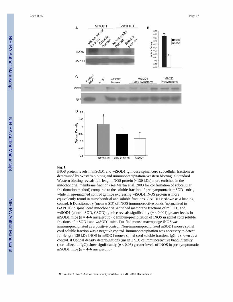
Fig. 1.iNOS protein levels in mSOD1 and wtSOD1 tg mouse spinal cord subcellular fractions asdetermined by Western blotting and immunoprecipitation-Western blotting. a StandardWestern blotting reveals full-length iNOS protein (~130 kDa) more enriched in themitochondrial membrane fraction (see Martin et al. 2003 for confirmation of subcellularfractionation method) compared to the soluble fraction of pre-symptomatic mSOD1 mice,while in age-matched control tg mice expressing wtSOD1 iNOS protein is moreequivalently found in mitochondrial and soluble fractions. GAPDH is shown as a loadingcontrol. b Densitometry (mean ± SD) of iNOS immunoreactive bands (normalized toGAPDH) in spinal cord mitochondrial-enriched membrane fractions of mSOD1 andwtSOD1 (control SOD, CSOD) tg mice reveals significantly (p < 0.001) greater levels inmSOD1 mice (n = 4–6 mice/group). c Immunoprecipitation of iNOS in spinal cord solublefractions of mSOD1 and wtSOD1 mice. Purified mouse macrophage iNOS wasimmunoprecipitated as a positive control. Non-immunoprecipitated mSOD1 mouse spinalcord soluble fraction was a negative control. Immunoprecipitation was necessary to detectfull-length 130 kDa iNOS in mSOD1 mouse spinal cord soluble fraction. IgG is shown as acontrol. d Optical density determinations (mean ± SD) of immunoreactive band intensity(normalized to IgG) show significantly (p < 0.05) greater levels of iNOS in pre-symptomaticmSOD1 mice (n = 4–6 mice/group)
Chen et al. Page 17
Brain Struct Funct. Author manuscript; available in PMC 2010 December 26.
NIH
-PA Author Manuscript
NIH
-PA Author Manuscript
NIH
-PA Author Manuscript

Fig. 2.NOS catalytic activity in mSOD1 and wtSOD1 tg mouse spinal cord subcellular fractions asdetermined by biochemical isotopic assay. a Use of specific inhibitors of recombinant iNOS(S-methylisothiourea sulfate) and nNOS (provided in kit) demonstrates the effectiveness andspecificity of the assay. b iNOS activity (mean ± SD) was detected in nuclear, soluble, andmitochondrial-enriched membrane fractions of in mSOD1 and wtSOD1 tg mouse spinalcord. iNOS activity was increased significantly (p < 0.05) in mitochondrial fractions ofmSOD1 mice at early stages of disease. c nNOS activity (mean ± SD) was detected insoluble and mitochondrial-enriched membrane fractions of in mSOD1 and wtSOD1 tgmouse spinal cord. nNOS activity was increased significantly (p < 0.01) in mitochondrialfractions of mSOD1 mice at pre-symptomatic stages of disease
Chen et al. Page 18
Brain Struct Funct. Author manuscript; available in PMC 2010 December 26.
NIH
-PA Author Manuscript
NIH
-PA Author Manuscript
NIH
-PA Author Manuscript

Fig. 3.Immunohistochemical localization of iNOS protein and RT-PCR analysis of iNOS mRNAin mSOD1 and wtSOD1 tg mice. Immunoperoxidase staining for iNOS in lumbar spinalcord sections of 14-week-old wtSOD1 tg mouse (a) and mSOD1 tg mice (b, c) at 11 weeksof age (early symptomatic) and 15 weeks old (end-stage). iNOS immunoreactivity is seen asbrown labeling. Insets show delineated MNs at higher magnification. Scale bars (in a) applyto b and c. d Single-cell computer densitometry (mean ± SD) of immunohistochemicallystained motor neurons (n = ~25–30 MNs/group) shows significantly (p <0.01) increasediNOS immunoreactivity in MNs of mSOD1 mice at 11 and 15 weeks of age (compared tobaseline at 7 weeks of age). e Immunoperoxidase staining for iNOS in brainstem sectionsfrom a 14-week-old wtSOD1 tg mouse (left) and a 9-week-old (pre-symptomatic) mSOD1tg mouse (right). iNOS immunoreactivity is seen as brown labeling. The facial motornucleus (arrow) shows high levels of iNOS immunoreactivity in mSOD1 mice. Scale bar (ine) = 130 μm. f RT-PCR analysis (see “Methods” for primers) of iNOS mRNA expression inbrain (cerebrum), brainstem, and spinal cord of control mouse. In normal CNS, iNOSmRNA levels are moderate in spinal cord, low in brainstem, and not detectable in cerebrum.RT-PCR for VDAC1 mRNA expression in the same samples is shown as a control. g RT-PCR analysis of iNOS mRNA expression in microdissected brainstem (medulla containing
Chen et al. Page 19
Brain Struct Funct. Author manuscript; available in PMC 2010 December 26.
NIH
-PA Author Manuscript
NIH
-PA Author Manuscript
NIH
-PA Author Manuscript

facial and trigeminal motor nuclei) of wtSOD1 and mSOD1 tg mice. iNOS expression ishighest at pre-symptomatic stages of disease. RT-PCR for VDAC1 mRNA expression in thesame samples is shown as a control. Dual-label immunofluorescence for iNOS (red) and theperoxisomal marker catalase (h), the microsomal marker cytochrome p450 reductase (i), andthe mitochondrial marker SOD2 (j). Asterisks identify nuclei. iNOS immunoreactivity (h,hatched arrow) is mostly distinct from catalase immunoreactivity (h, solid arrow) and thusthere is sparse yellow. iNOS and cytochrome p450 reductase robustly, but incompletely, co-localize (yellow) in the cytoplasm (i). iNOS robustly localizes to SOD2-positivemitochondria as revealed by the discrete merging of red and green (j, yellow). Small (~0.5μm in diameter), morphologically normal-appearing mitochondrial are single-labeled forgreen-SOD2 (j, lower left of cell, open arrows), while swollen mitochondria (doublearrows) are iNOS-positive (yellow). Other structures, probably microsomes, are red-iNOSsingle labeled (j, hatched arrows) Scale bars h 6.7 μm; i 4.5 μm; j 7 μm
Chen et al. Page 20
Brain Struct Funct. Author manuscript; available in PMC 2010 December 26.
NIH
-PA Author Manuscript
NIH
-PA Author Manuscript
NIH
-PA Author Manuscript

Fig. 4.iNOS immunoreactivity is found in microglia, but rarely in astrocytes, in symptomaticmSOD1 mice. Immunolocalization of iNOS (a, green) and the microglial cell markerCD11b (b, red) demonstrates the near complete localization of iNOS to microglia (c, yellow)in spinal cord ventral horn and white matter ventral funiculus at end-stage disease. Hatchedarrows identify selected microglial cells that are positive for iNOS; however, not all iNOSpositive structures (a, open arrow) are CD11b-positive microglia. There is also iNOSimmunoreactivity throughout the ventral horn parenchyma that appears to localize as finemicroglial processes. Scale bar (in a) = 25 μm. d–f At end-stage disease remnant MNs(delineated by oval) show green-iNOS particulate immunoreactivity in the cytoplasm, whileiNOS+/CD11b+ microglia (e, red at arrows) form satellites and infiltrate into thedegenerating MN. Scale bar (in d) = 2.5 μm. Immunolocalization of iNOS (g, red) and theastroglial marker GFAP (h, red) demonstrate the absence of co-localization of iNOS toastrocytes (i, no yellow, compare to Fig. 4C) in pre-symptomatic mSOD1 mouse spinal cordventral horn, but MNs are strongly positive for iNOS (g). Scale bar (in g, applies to h–l) =35 μm. j–l. iNOS immunoreactivity is found very rarely in some astrocyte processes insymptomatic mSOD1 mice
Chen et al. Page 21
Brain Struct Funct. Author manuscript; available in PMC 2010 December 26.
NIH
-PA Author Manuscript
NIH
-PA Author Manuscript
NIH
-PA Author Manuscript

Fig. 5.iNOS is present in Schwann cells and their paranodal zones of the nodes of Ranvier inmSOD1 mice. a iNOS immunoreactivity is present in early symptomatic (10-week-old)mSOD1 mice at the spinal nerve rootlets (upper right inset, open arrow), ventral root exitzones of the spinal nerves (hatched arrows), and nerve sheaths of peripheral nerves whereiNOS is enriched highly at the paranodes of some axons (lower left inset, hatched arrows).Scale bars: a and lower left inset, 3 μm; upper right inset, 16 μm. b In pre-symptomaticmSOD1 mice, iNOS accumulates in subsets of degenerating axons (open arrows) inperipheral nerve. Axons that appear normal are iNOS-negative (asterisks) but the sheaths areiNOS-positive (hatched arrows). Scale bar 2.4 μm. c–e iNOS is expressed in the ventralroot exit zone Schwann cells as demonstrated by the co-localization (yellow in e) of iNOSand the Schwann cell marker vimentin. Scale bar (in c) = 3.5 μm. f–h In symptomaticmSOD1 mice, the degenerating peripheral nerve axons and nerve interstitial components aremarked by p75 neurotrophin receptor (f, hatched arrows) and these axons are iNOS-positive(yellow in h). Nerve interstitial components are also marked by p75 and iNOS. Scale bar (inf) = 6.0 μm
Chen et al. Page 22
Brain Struct Funct. Author manuscript; available in PMC 2010 December 26.
NIH
-PA Author Manuscript
NIH
-PA Author Manuscript
NIH
-PA Author Manuscript

Fig. 6.iNOS inhibitors have beneficial effects in G93Ahigh-mSOD1 tg mice. a Kaplan–Meiersurvival curve for small cohort study using iNOS-specific inhibitor (SMT). Mice receivingSMT delayed symptom onset (age ~90–120 days) compared to vehicle-treated mice, but hadon effects on survival. b G93A mice treated with 1400W (3 mg/kg, sc every other day) haddelayed disease onset and an extended lifespan compared to mice treated with vehicle (n =10–14 mice/group). c Graph of the mean lifespan of vehicle- and 1400W-treated mSOD1mice. Values are mean ± SD. Significant difference (*p <0.05) from vehicle control
Chen et al. Page 23
Brain Struct Funct. Author manuscript; available in PMC 2010 December 26.
NIH
-PA Author Manuscript
NIH
-PA Author Manuscript
NIH
-PA Author Manuscript