Inducible heat shock protein 70 expression as a potential predictive marker of metastasis in breast...
10
34 Cell Stress & Chaperones (2006) 11 (1), 34–43 Q Cell Stress Society International 2006 Article no. csac. 2006.CSC-159R Inducible heat shock protein 70 expression as a potential predictive marker of metastasis in breast tumors Carolina Torronteguy, 1 Antonio Frasson, 2 Felipe Zerwes, 2 Erik Winnikov, 2 Vinicius Duval da Silva, 3 Antoine Me ´ noret, 4 and Cristina Bonorino 1 1 Faculdade de Biociencias e, Instituto de Pesquisas Biome ´ dicas, PUCRS, Avenida Ipiranga, 6690 2o andar, 90010, Porto Alegre, RS, Brazil 2 Centro de Mama, Hospital Sa ˜o Lucas, PUCRS, Porto Alegre, RS, Brazil 3 Departamento de Patologia, Faculdade de Medicina, PUCRS, Porto Alegre, RS, Brazil 4 University of Connecticut, Farmington, CT 06032, USA Abstract Heat shock protein (Hsp)–peptide complexes purified from tumors can prime the immune system against tumor antigens, but how they contribute to the generation of immune responses against naturally occurring tumors is unknown. Murine tumors expressing high amounts of Hsp70 are preferentially rejected by the immune system, sug- gesting that low Hsp70 expression is advantageous for tumor growth in the host. To determine whether Hsp70 was differentially expressed in human tumors, inducible Hsp70 expression was quantitatively (by Western blot) and quali- tatively (by immunohistology) analyzed in 53 biopsies of tumor and normal breast tissue. The mean expression of inducible Hsp70 was significantly higher in tumor compared with normal tissue (U 5 899.0; P 5 0.0033). However, a significant negative association of the amount of Hsp70 expressed by tumor tissue was found with metastasis (r 2 5 20.309; P 5 0.05). After 3 years, follow-up analysis determined that 7 of the 53 patients relapsed, and 5 died. Hsp70 expression in tumor (but not normal) cells was significantly lower in relapse patients and patients with metastatic disease than in patients with no relapse or metastasis. Together, these observations support the hypothesis that Hsp70 plays a role in tumor expansion in vivo, and tumors that downregulate it may be able to evade immunosurveillance and grow. INTRODUCTION The stress-inducible heat shock protein 70 (Hsp70) is the main protein produced during cellular responses to var- ied stresses such as heat shock, ischemia/reperfusion, and oxidative changes (Lindquist 1986; Subjeck and Shyy 1986). A cognate form, Hsc70, is constitutively expressed in cells not experiencing stress. Hsp70, but not Hsc70, is overexpressed in human and animal tumors of varied or- igins (Clark and Menoret 2001). Because Hsp70 confers cell protection against different stresses, it has been hy- Received 23 August 2005; Revised 16 September 2005; Accepted 4 Oc- tober 2005. Correspondence to: Cristina Bonorino, Tel: 55 51 3320 3000 ext 2725; Fax: 55 51 3320 3312; E-mail: [email protected]. Current address: Cristina Bonorino, Center for Immunology, University of Minnesota, 312 Church Street, BSBE 6th Floor, Minneapolis, MN 55455. pothesized that this protein plays a protective role in tu- mor growth in vivo (Barnes et al 2001; Ravagnan et al 2001). Indeed, CH1 lymphoma cells in culture methylate and silence their Hsp70 promoter, but upon injection into a mouse Hsp70 production in the CH1 cells is restored (Davidson et al 1995). These data suggest that Hsp70 is needed for in vivo tumor progression. One of the major stresses faced by a tumor in vivo is the pressure of an immune response (Dunn et al 2004). Interestingly, it has been demonstrated that Hsp70 and other heat shock proteins can interact with the immune system. HSPs purified from tumors, particularly Hsp70, HSP90, calreticulin, and gp96, promote the generation of CD8 1 T cell responses against tumor antigens, both in mice (Udono and Srivastava 1993; Janetzki et al 1998, 2000; Basu and Srivastava 1999) and humans (Parmiani et al 2004). HSPs carry tumor peptides to antigen-pre-
-
Upload
independent -
Category
Documents
-
view
0 -
download
0
Transcript of Inducible heat shock protein 70 expression as a potential predictive marker of metastasis in breast...
34
Cell Stress & Chaperones (2006) 11 (1), 34–43Q Cell Stress Society International 2006Article no. csac. 2006.CSC-159R
Inducible heat shock protein 70expression as a potential predictivemarker of metastasis inbreast tumorsCarolina Torronteguy,1 Antonio Frasson,2 Felipe Zerwes,2 Erik Winnikov,2Vinicius Duval da Silva,3 Antoine Menoret,4 and Cristina Bonorino1
1Faculdade de Biociencias e, Instituto de Pesquisas Biomedicas, PUCRS, Avenida Ipiranga, 6690 2o andar, 90010, Porto Alegre, RS, Brazil2Centro de Mama, Hospital Sao Lucas, PUCRS, Porto Alegre, RS, Brazil3Departamento de Patologia, Faculdade de Medicina, PUCRS, Porto Alegre, RS, Brazil4University of Connecticut, Farmington, CT 06032, USA
Abstract Heat shock protein (Hsp)–peptide complexes purified from tumors can prime the immune system againsttumor antigens, but how they contribute to the generation of immune responses against naturally occurring tumors isunknown. Murine tumors expressing high amounts of Hsp70 are preferentially rejected by the immune system, sug-gesting that low Hsp70 expression is advantageous for tumor growth in the host. To determine whether Hsp70 wasdifferentially expressed in human tumors, inducible Hsp70 expression was quantitatively (by Western blot) and quali-tatively (by immunohistology) analyzed in 53 biopsies of tumor and normal breast tissue. The mean expression ofinducible Hsp70 was significantly higher in tumor compared with normal tissue (U 5 899.0; P 5 0.0033). However, asignificant negative association of the amount of Hsp70 expressed by tumor tissue was found with metastasis (r2 520.309; P 5 0.05). After 3 years, follow-up analysis determined that 7 of the 53 patients relapsed, and 5 died. Hsp70expression in tumor (but not normal) cells was significantly lower in relapse patients and patients with metastaticdisease than in patients with no relapse or metastasis. Together, these observations support the hypothesis that Hsp70plays a role in tumor expansion in vivo, and tumors that downregulate it may be able to evade immunosurveillanceand grow.
INTRODUCTION
The stress-inducible heat shock protein 70 (Hsp70) is themain protein produced during cellular responses to var-ied stresses such as heat shock, ischemia/reperfusion,and oxidative changes (Lindquist 1986; Subjeck and Shyy1986). A cognate form, Hsc70, is constitutively expressedin cells not experiencing stress. Hsp70, but not Hsc70, isoverexpressed in human and animal tumors of varied or-igins (Clark and Menoret 2001). Because Hsp70 conferscell protection against different stresses, it has been hy-
Received 23 August 2005; Revised 16 September 2005; Accepted 4 Oc-tober 2005.
Correspondence to: Cristina Bonorino, Tel: 55 51 3320 3000 ext 2725; Fax:55 51 3320 3312; E-mail: [email protected].
Current address: Cristina Bonorino, Center for Immunology, University ofMinnesota, 312 Church Street, BSBE 6th Floor, Minneapolis, MN 55455.
pothesized that this protein plays a protective role in tu-mor growth in vivo (Barnes et al 2001; Ravagnan et al2001). Indeed, CH1 lymphoma cells in culture methylateand silence their Hsp70 promoter, but upon injection intoa mouse Hsp70 production in the CH1 cells is restored(Davidson et al 1995). These data suggest that Hsp70 isneeded for in vivo tumor progression.
One of the major stresses faced by a tumor in vivo isthe pressure of an immune response (Dunn et al 2004).Interestingly, it has been demonstrated that Hsp70 andother heat shock proteins can interact with the immunesystem. HSPs purified from tumors, particularly Hsp70,HSP90, calreticulin, and gp96, promote the generation ofCD81 T cell responses against tumor antigens, both inmice (Udono and Srivastava 1993; Janetzki et al 1998,2000; Basu and Srivastava 1999) and humans (Parmianiet al 2004). HSPs carry tumor peptides to antigen-pre-
Cell Stress & Chaperones (2006) 11 (1), 34–43
HSP70 in metastatic breast cancer 35
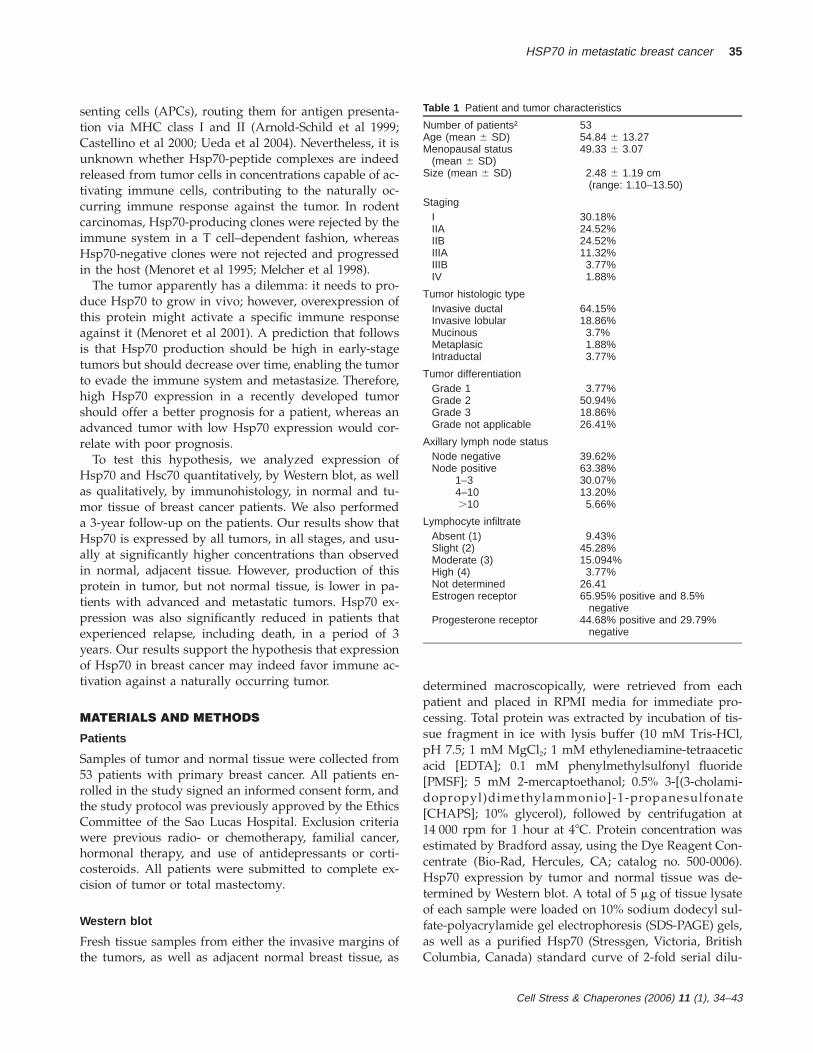
Table 1 Patient and tumor characteristics
Number of patients†Age (mean 6 SD)
5354.84 6 13.27
Menopausal status(mean 6 SD)
49.33 6 3.07
Size (mean 6 SD) 2.48 6 1.19 cm(range: 1.10–13.50)
StagingIIIAIIBIIIAIIIBIV
30.18%24.52%24.52%11.32%3.77%1.88%
Tumor histologic typeInvasive ductalInvasive lobularMucinousMetaplasicIntraductal
64.15%18.86%3.7%1.88%3.77%
Tumor differentiationGrade 1Grade 2Grade 3
3.77%50.94%18.86%
Grade not applicable 26.41%
Axillary lymph node statusNode negativeNode positive
39.62%63.38%
1–34–10.10
30.07%13.20%5.66%
Lymphocyte infiltrateAbsent (1)Slight (2)Moderate (3)High (4)Not determinedEstrogen receptor
Progesterone receptor
9.43%45.28%15.094%3.77%
26.4165.95% positive and 8.5%
negative44.68% positive and 29.79%
negative
senting cells (APCs), routing them for antigen presenta-tion via MHC class I and II (Arnold-Schild et al 1999;Castellino et al 2000; Ueda et al 2004). Nevertheless, it isunknown whether Hsp70-peptide complexes are indeedreleased from tumor cells in concentrations capable of ac-tivating immune cells, contributing to the naturally oc-curring immune response against the tumor. In rodentcarcinomas, Hsp70-producing clones were rejected by theimmune system in a T cell–dependent fashion, whereasHsp70-negative clones were not rejected and progressedin the host (Menoret et al 1995; Melcher et al 1998).
The tumor apparently has a dilemma: it needs to pro-duce Hsp70 to grow in vivo; however, overexpression ofthis protein might activate a specific immune responseagainst it (Menoret et al 2001). A prediction that followsis that Hsp70 production should be high in early-stagetumors but should decrease over time, enabling the tumorto evade the immune system and metastasize. Therefore,high Hsp70 expression in a recently developed tumorshould offer a better prognosis for a patient, whereas anadvanced tumor with low Hsp70 expression would cor-relate with poor prognosis.
To test this hypothesis, we analyzed expression ofHsp70 and Hsc70 quantitatively, by Western blot, as wellas qualitatively, by immunohistology, in normal and tu-mor tissue of breast cancer patients. We also performeda 3-year follow-up on the patients. Our results show thatHsp70 is expressed by all tumors, in all stages, and usu-ally at significantly higher concentrations than observedin normal, adjacent tissue. However, production of thisprotein in tumor, but not normal tissue, is lower in pa-tients with advanced and metastatic tumors. Hsp70 ex-pression was also significantly reduced in patients thatexperienced relapse, including death, in a period of 3years. Our results support the hypothesis that expressionof Hsp70 in breast cancer may indeed favor immune ac-tivation against a naturally occurring tumor.
MATERIALS AND METHODS
Patients
Samples of tumor and normal tissue were collected from53 patients with primary breast cancer. All patients en-rolled in the study signed an informed consent form, andthe study protocol was previously approved by the EthicsCommittee of the Sao Lucas Hospital. Exclusion criteriawere previous radio- or chemotherapy, familial cancer,hormonal therapy, and use of antidepressants or corti-costeroids. All patients were submitted to complete ex-cision of tumor or total mastectomy.
Western blot
Fresh tissue samples from either the invasive margins ofthe tumors, as well as adjacent normal breast tissue, as
determined macroscopically, were retrieved from eachpatient and placed in RPMI media for immediate pro-cessing. Total protein was extracted by incubation of tis-sue fragment in ice with lysis buffer (10 mM Tris-HCl,pH 7.5; 1 mM MgCl2; 1 mM ethylenediamine-tetraaceticacid [EDTA]; 0.1 mM phenylmethylsulfonyl fluoride[PMSF]; 5 mM 2-mercaptoethanol; 0.5% 3-[(3-cholami-dopropyl)dimethylammonio]-1-propanesulfonate[CHAPS]; 10% glycerol), followed by centrifugation at14 000 rpm for 1 hour at 48C. Protein concentration wasestimated by Bradford assay, using the Dye Reagent Con-centrate (Bio-Rad, Hercules, CA; catalog no. 500-0006).Hsp70 expression by tumor and normal tissue was de-termined by Western blot. A total of 5 mg of tissue lysateof each sample were loaded on 10% sodium dodecyl sul-fate-polyacrylamide gel electrophoresis (SDS-PAGE) gels,as well as a purified Hsp70 (Stressgen, Victoria, BritishColumbia, Canada) standard curve of 2-fold serial dilu-

Cell Stress & Chaperones (2006) 11 (1), 34–43
36 Torronteguy et al
Fig 1. Western blot analysis of Hsp70 expression in normal andtumor tissue. Tissue samples were analyzed by Western blot forHsp70 expression and compared with a standard curve of purifiedHsp70. (A) T, Tumor; N, normal. Numbers next to these letters de-note the patient. 1–4, Standard curve of Hsp70, in a serial 2-folddilution, starting (on 4) at 0.2 mg. (B) Tumor and normal sample ofthe same patients, blotted with both anti-Hsp70 and anti-actin anti-bodies. (C) Quantitation of Hsp70 expression in normal and tumortissue of breast cancer patients. Hsp70 expression was determinedin tumor and normal tissue by Western blot and quantitated by theImagemaster software (Pharmacia). Tumor tissue expresses signif-icantly more Hsp70 than normal tissue (Mann-Whitney U 5 924.0;P 5 0.0024).
Fig 2. Expression of Hsp70, but not Hsc70, is significantly higherin tumor compared with normal tissue. Tumor and normal samplesfrom 23 patients were screened for Hsp70 expression with either theC92 antibody (A), which recognizes exclusively the stress-inducedprotein, or the N27 (B) antibody, which recognizes both stress-in-duced and constitutive homologs. Hsp70 expression is expressed innanograms per microgram of total protein.
tions, starting at 0.2 mg. After electrophoresis, the gelswere transferred to nitrocellulose membranes and blottedwith anti-human Hsp70 antibody (SPA810; Stressgen) oranti-HSP/Hsc70 (SPA820; Stressgen). Actin expressionwas used as a control for basal constitutive protein syn-thesis, using an anti-human actin (A4700; Sigma, St Lou-is, MO) antibody. Samples in which actin was not de-tected were discarded. Secondary antibody was anti-mouse IgG conjugated with peroxidase (Invitrogen, SanDiego, CA), and the blots were developed using the ECLsystem (Pharmacia, Peapack, NJ). The concentration ofHsp70 in tumor and normal tissue bands were deter-mined according to the area of these bands in relation toa curve of areas plotted by the Imagemaster software
from Pharmacia. Briefly, the Western blot images werecaptured using a scanner, and saved in a scale of graywith a resolution of 1200 dpi. Concentration values wereassigned to Hsp70 standard curve bands according to theamount of protein applied to the respective gel lane.Quantitations were repeated for at least 3 times.
Histology
In tumor samples analyzed by immunohistochemistry,part of the tissue was fixed in paraffin and cut in sectionsof 4 mm and applied to histology slides. Antigen recoverywas performed in citrate buffer by microwave, in 3 cyclesof maximum power for 5 minutes. Endogenous peroxi-dase activity was blocked with 3% hydrogen peroxideand 0.1% sodium azide. Additional blocking was per-formed with normal mouse serum (Novastain UniversalDetection kit). Incubation with primary antibody (anti-Hsp70 from Stressgen or anti-IFN-g from R&D Systems[Minneapolis, MN]) was performed overnight at roomtemperature, followed by incubation with biotinylatedanti-mouse IgG and avidin-peroxidase (Novastain). Slides
Cell Stress & Chaperones (2006) 11 (1), 34–43
HSP70 in metastatic breast cancer 37
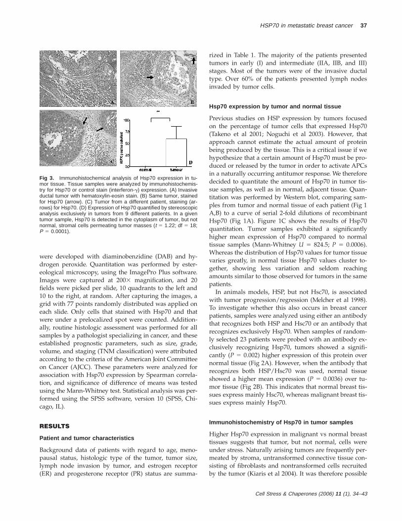
Fig 3. Immunohistochemical analysis of Hsp70 expression in tu-mor tissue. Tissue samples were analyzed by immunohistochemis-try for Hsp70 or control stain (interferon-g) expression. (A) Invasiveductal tumor with hematoxylin-eosin stain. (B) Same tumor, stainedfor Hsp70 (arrow). (C) Tumor from a different patient, staining (ar-rows) for Hsp70. (D) Expression of Hsp70 quantified by stereoscopicanalysis exclusively in tumors from 9 different patients. In a giventumor sample, Hsp70 is detected in the cytoplasm of tumor, but notnormal, stromal cells permeating tumor masses (t 5 1.22; df 5 18;P 5 0.0001).
were developed with diaminobenzidine (DAB) and hy-drogen peroxide. Quantitation was performed by ester-eological microscopy, using the ImagePro Plus software.Images were captured at 2003 magnification, and 20fields were picked per slide, 10 quadrants to the left and10 to the right, at random. After capturing the images, agrid with 77 points randomly distributed was applied oneach slide. Only cells that stained with Hsp70 and thatwere under a prelocalized spot were counted. Addition-ally, routine histologic assessment was performed for allsamples by a pathologist specializing in cancer, and theseestablished prognostic parameters, such as size, grade,volume, and staging (TNM classification) were attributedaccording to the criteria of the American Joint Committeeon Cancer (AJCC). These parameters were analyzed forassociation with Hsp70 expression by Spearman correla-tion, and significance of difference of means was testedusing the Mann-Whitney test. Statistical analysis was per-formed using the SPSS software, version 10 (SPSS, Chi-cago, IL).
RESULTS
Patient and tumor characteristics
Background data of patients with regard to age, meno-pausal status, histologic type of the tumor, tumor size,lymph node invasion by tumor, and estrogen receptor(ER) and progesterone receptor (PR) status are summa-
rized in Table 1. The majority of the patients presentedtumors in early (I) and intermediate (IIA, IIB, and III)stages. Most of the tumors were of the invasive ductaltype. Over 60% of the patients presented lymph nodesinvaded by tumor cells.
Hsp70 expression by tumor and normal tissue
Previous studies on HSP expression by tumors focusedon the percentage of tumor cells that expressed Hsp70(Takeno et al 2001; Noguchi et al 2003). However, thatapproach cannot estimate the actual amount of proteinbeing produced by the tissue. This is a critical issue if wehypothesize that a certain amount of Hsp70 must be pro-duced or released by the tumor in order to activate APCsin a naturally occurring antitumor response. We thereforedecided to quantitate the amount of Hsp70 in tumor tis-sue samples, as well as in normal, adjacent tissue. Quan-titation was performed by Western blot, comparing sam-ples from tumor and normal tissue of each patient (Fig 1A,B) to a curve of serial 2-fold dilutions of recombinantHsp70 (Fig 1A). Figure 1C shows the results of Hsp70quantitation. Tumor samples exhibited a significantlyhigher mean expression of Hsp70 compared to normaltissue samples (Mann-Whitney U 5 824.5; P 5 0.0006).Whereas the distribution of Hsp70 values for tumor tissuevaries greatly, in normal tissue Hsp70 values cluster to-gether, showing less variation and seldom reachingamounts similar to those observed for tumors in the samepatients.
In animals models, HSP, but not Hsc70, is associatedwith tumor progression/regression (Melcher et al 1998).To investigate whether this also occurs in breast cancerpatients, samples were analyzed using either an antibodythat recognizes both HSP and Hsc70 or an antibody thatrecognizes exclusively Hsp70. When samples of random-ly selected 23 patients were probed with an antibody ex-clusively recognizing Hsp70, tumors showed a signifi-cantly (P 5 0.002) higher expression of this protein overnormal tissue (Fig 2A). However, when the antibody thatrecognizes both HSP/Hsc70 was used, normal tissueshowed a higher mean expression (P 5 0.0036) over tu-mor tissue (Fig 2B). This indicates that normal breast tis-sues express mainly Hsc70, whereas malignant breast tis-sues express mainly Hsp70.
Immunohistochemistry of Hsp70 in tumor samples
Higher Hsp70 expression in malignant vs normal breasttissues suggests that tumor, but not normal, cells wereunder stress. Naturally arising tumors are frequently per-meated by stroma, untransformed connective tissue con-sisting of fibroblasts and nontransformed cells recruitedby the tumor (Kiaris et al 2004). It was therefore possible
Cell Stress & Chaperones (2006) 11 (1), 34–43
38 Torronteguy et al
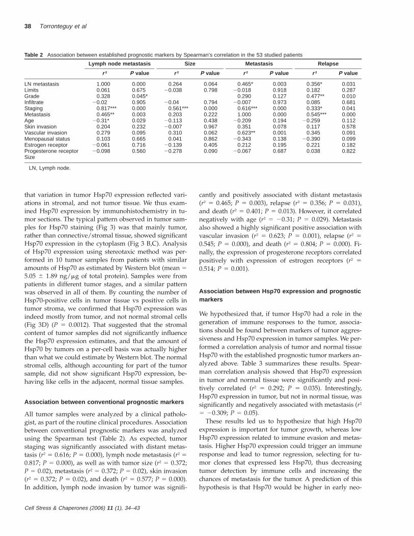
Table 2 Association between established prognostic markers by Spearman’s correlation in the 53 studied patients
Lymph node metastasis
r 2 P value
Size
r 2 P value
Metastasis
r 2 P value
Relapse
r 2 P value
LN metastasisLimitsGradeInfiltrateStagingMetastasisAgeSkin invasion
1.0000.0610.328
20.020.817***0.465**
20.31*0.204
0.0000.6750.045*0.9050.0000.0030.0290.232
0.26420.038
20.040.561***0.203
20.11320.007
0.0640.798
0.7940.0000.2220.4380.967
0.465*20.018
0.29020.007
0.616***1.000
20.2090.351
0.0030.9180.1270.9730.0000.0000.1940.078
0.356*0.1820.477**0.0850.333*0.545***
20.2590.117
0.0310.2870.0100.6810.0410.0000.1120.578
Vascular invasionMenopausal statusEstrogen receptorProgesterone receptorSize
0.2790.103
20.06120.098
0.0950.6650.7160.560
0.3100.041
20.13920.278
0.0620.8620.4050.090
0.623**20.343
0.21220.067
0.0010.1380.1950.687
0.34520.390
0.2210.038
0.0910.0990.1820.822
LN, Lymph node.
that variation in tumor Hsp70 expression reflected vari-ations in stromal, and not tumor tissue. We thus exam-ined Hsp70 expression by immunohistochemistry in tu-mor sections. The typical pattern observed in tumor sam-ples for Hsp70 staining (Fig 3) was that mainly tumor,rather than connective/stromal tissue, showed significantHsp70 expression in the cytoplasm (Fig 3 B,C). Analysisof Hsp70 expression using stereotaxic method was per-formed in 10 tumor samples from patients with similaramounts of Hsp70 as estimated by Western blot (mean 55.05 6 1.89 ng/mg of total protein). Samples were frompatients in different tumor stages, and a similar patternwas observed in all of them. By counting the number ofHsp70-positive cells in tumor tissue vs positive cells intumor stroma, we confirmed that Hsp70 expression wasindeed mostly from tumor, and not normal stromal cells(Fig 3D) (P 5 0.0012). That suggested that the stromalcontent of tumor samples did not significantly influencethe Hsp70 expression estimates, and that the amount ofHsp70 by tumors on a per-cell basis was actually higherthan what we could estimate by Western blot. The normalstromal cells, although accounting for part of the tumorsample, did not show significant Hsp70 expression, be-having like cells in the adjacent, normal tissue samples.
Association between conventional prognostic markers
All tumor samples were analyzed by a clinical patholo-gist, as part of the routine clinical procedures. Associationbetween conventional prognostic markers was analyzedusing the Spearman test (Table 2). As expected, tumorstaging was significantly associated with distant metas-tasis (r2 5 0.616; P 5 0.000), lymph node metastasis (r2 50.817; P 5 0.000), as well as with tumor size (r2 5 0.372;P 5 0.02), metastasis (r2 5 0.372; P 5 0.02), skin invasion(r2 5 0.372; P 5 0.02), and death (r2 5 0.577; P 5 0.000).In addition, lymph node invasion by tumor was signifi-
cantly and positively associated with distant metastasis(r2 5 0.465; P 5 0.003), relapse (r2 5 0.356; P 5 0.031),and death (r2 5 0.401; P 5 0.013). However, it correlatednegatively with age (r2 5 20.31; P 5 0.029). Metastasisalso showed a highly significant positive association withvascular invasion (r2 5 0.623; P 5 0.001), relapse (r2 50.545; P 5 0.000), and death (r2 5 0.804; P 5 0.000). Fi-nally, the expression of progesterone receptors correlatedpositively with expression of estrogen receptors (r2 50.514; P 5 0.001).
Association between Hsp70 expression and prognosticmarkers
We hypothesized that, if tumor Hsp70 had a role in thegeneration of immune responses to the tumor, associa-tions should be found between markers of tumor aggres-siveness and Hsp70 expression in tumor samples. We per-formed a correlation analysis of tumor and normal tissueHsp70 with the established prognostic tumor markers an-alyzed above. Table 3 summarizes these results. Spear-man correlation analysis showed that Hsp70 expressionin tumor and normal tissue were significantly and posi-tively correlated (r2 5 0.292; P 5 0.035). Interestingly,Hsp70 expression in tumor, but not in normal tissue, wassignificantly and negatively associated with metastasis (r2
5 20.309; P 5 0.05).These results led us to hypothesize that high Hsp70
expression is important for tumor growth, whereas lowHsp70 expression related to immune evasion and metas-tasis. Higher Hsp70 expression could trigger an immuneresponse and lead to tumor regression, selecting for tu-mor clones that expressed less Hsp70, thus decreasingtumor detection by immune cells and increasing thechances of metastasis for the tumor. A prediction of thishypothesis is that Hsp70 would be higher in early neo-

Cell Stress & Chaperones (2006) 11 (1), 34–43
HSP70 in metastatic breast cancer 39
Table 2 Continued
Death
r 2 P value
Infiltrate
r 2 P value
Skin invasion
r 2 P value
Estrogen receptor
r 2 P value
Vascular invasion
r 2 P value
0.40120.181
0.08420.007
0.577***0.804***
20.0240.351
0.013*0.2850.6650.9730.0000.0000.8820.078
20.02020.161
0.0601.000
20.155
20.1090.219
0.9050.3400.7640.0000.352
0.5200.253
0.2040.0700.1340.2190.2490.351
20.1801.000
0.2320.6880.5140.2530.1430.0780.3010.000
20.0610.0920.3250.2950.0000.212
20.1120.389
0.7160.5870.0850.1351.0000.1950.4900.055
0.2790.2660.107
20.0910.372*0.623**
20.0850.215
0.0950.1170.5940.6200.0230.0010.6210.238
0.24620.252
0.21220.067
0.2250.2840.1950.687
20.0910.2530.2950.130
20.044
0.6200.3630.1350.5180.794
0.2150.089
20.1610.240
20.007
0.2380.7520.4420.2490.967
20.16120.040
1.0000.514**
20.139
0.4420.8710.0000.0010.405
1.00020.07720.16120.050
0.310
0.00000.7940.4420.8110.062
* 5 p,0.05; ** 5 p,0.01; *** 5 p 5 0.001.
Table 3 Association between Hsp70 expression and established prognostic markers by Spear-man’s correlation
Hsp70 in tumor tissue
r 2 P value
Hsp 70 in normal tissue
r 2 P value
LN metastasisLimitsVascular invasionSizeGradeInfiltrateStagingMetastasisRelapseDeathHsp70 in normal tissueHsp70 in tumor tissue
20.05020.26620.302
0.03320.09120.33220.06420.309*20.20120.255
0.292*1
0.7290.0640.070.8220.5850.0420.6570.0500.2190.1130.0350.000
0.09120.18620.318
0.1270.086
20.3250.1860.0670.0520.16410.292*
0.5300.2020.0550.3790.6100.0470.1900.6830.7550.3130.0000.035
LN, Lymph node; r 2, correlation coefficient.
Fig 4. Comparison of mean inducible Hsp70 expression values be-tween tumors in different staging groups. Mean values of Hsp70(nanograms per microgram of total protein) in various stages of tu-mor growth: early (stage I tumors), intermediary (stages IIA, IIB, andIIIA), and advanced (stages IIIB, IIIC, and IV) were compared usingthe Mann-Whitney test. *, #0.05.
plasms and get lower with time under the selective pres-sure of the immune system.
To test this prediction, we investigated whether themean Hsp70 expression was significantly different in var-ious stages of tumor growth: early (stage I tumors), in-termediary (stages IIA, IIB, and IIIA), and advanced(stages IIIB, IIIC, and IV). The results are displayed inFigure 4. Whereas early tumors did not differ signifi-cantly from intermediary tumors regarding their meaninducible Hsp70 expression, advanced tumors expressedsignificantly lower Hsp70 than both early- and interme-diate-stage tumors.
Follow-up on breast cancer patients
After a 3-year period, the clinical status of the patients inthis study was determined. Of the 53 patients, 7 (13.2 %)presented some sort of relapse (Table 4). The Hsp70 val-ues displayed are the ones detected at diagnosis, becausewe did not have access to samples of the relapsing tu-
Cell Stress & Chaperones (2006) 11 (1), 34–43
40 Torronteguy et al
Tab
le4
Cha
ract
eris
tics
ofpa
tient
sw
ithre
laps
e(in
ape
riod
of2
to3
year
s)
Pat
ien
tn
o.
Dia
gn
osi
sA
ge
atd
iag
no
sis
Sta
gin
gTu
mo
rH
sp70
(ng
/mg
)N
orm
alH
sp79
0(n
g/m
g)
Infi
ltra
teF
ollo
w-u
p
B2
Aug
ust
2001
48N
D3.
360.
10M
ildB
ilate
ralb
reas
tca
ncer
B8
Janu
ary
2002
46III
A6.
933.
56M
ildB
one
met
asta
sis
(Aug
ust
2003
),liv
erm
etas
tasi
s,an
dde
ath
(May
2004
)B
12M
ay20
0270
IVA
0.10
28.4
2N
otav
aila
ble
Sub
mitt
edto
palli
ativ
ech
emot
hera
py(M
ay20
02)
due
tosu
prac
lavi
cula
rly
mph
node
met
asta
sis
atdi
agno
sis.
Neo
plas
iare
spon
ded,
and
subs
eque
ntm
amm
ogra
phy
show
edno
tum
orev
iden
ce(J
anua
ry20
03).
Sur
gery
was
perf
orm
ed(M
ay20
03),
and
ther
ew
asno
brea
stor
axill
ary
lym
phno
dene
opla
sia.
Sym
ptom
atic
cent
ral
nerv
ous
syst
emm
etas
tasi
soc
curr
edin
June
2003
,an
dpa
tient
died
.B
13Ju
ly20
0244
IIIB
0.10
11.2
8M
ildP
ositi
veno
deat
diag
nosi
s.R
elap
se(J
une
2003
),m
ultip
lebo
nem
etas
tasi
s(S
epte
mbe
r20
03),
and
deat
hby
ter-
min
alne
opla
sia
(Dec
embe
r20
03)
P14
July
2001
73IIB
6.92
4.46
Mild
Ano
ther
neop
lasi
a(g
astr
icca
ncer
with
liver
met
asta
sis—
Sep
tem
ber
2001
),an
dde
ath
byup
per
gast
riche
mor
-rh
age
(Jul
y20
04)
P16
Aug
ust
2001
41III
A4.
370.
54M
ildR
elap
seF
4Ju
ne20
0181
IN
DN
DA
bsen
tLi
ver
met
asta
sis
(Jun
e20
03)
and
deat
h(M
arch
2004
)
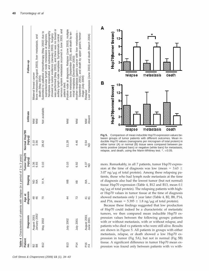
Fig 5. Comparison of mean inducible Hsp70 expression values be-tween groups of tumor patients with different outcomes. Mean in-ducible Hsp70 values (nanograms per microgram of total protein) ineither tumor (A) or normal (B) tissue were compared between pa-tients positive (striped bars) or negative (white bars) for metastasis,relapse, and death, using the Mann-Whitney test. *, #0.05.
mors. Remarkably, in all 7 patients, tumor Hsp70 expres-sion at the time of diagnosis was low (mean 5 3.63 63.07 ng/mg of total protein). Among these relapsing pa-tients, those who had lymph node metastasis at the timeof diagnosis also had the lowest tumor (but not normal)tissue Hsp70 expression (Table 4, B12 and B13, mean 0.1ng/mg of total protein). The relapsing patients with high-er Hsp70 values in tumor tissue at the time of diagnosisshowed metastasis only 1 year later (Table 4, B2, B8, P14,and P16, mean 5 5.395 6 1.8 ng/mg of total protein).
Because these findings suggested that low productionof Hsp70 could indeed be a characteristic of metastatictumors, we then compared mean inducible Hsp70 ex-pression values between the following groups: patientswith or without metastasis, with or without relapse, andpatients who died vs patients who were still alive. Resultsare shown in Figure 5. All patients in groups with eithermetastasis, relapse, or death showed a low Hsp70 ex-pression in tumor (Fig 5A), but not in normal (Fig 5B)tissue. A significant difference in tumor Hsp70 mean ex-pression was found only between patients with vs with-
Cell Stress & Chaperones (2006) 11 (1), 34–43
HSP70 in metastatic breast cancer 41
out metastasis (Fig 5A). No significant differences werefound between groups of each outcome for mean Hsp70expression in normal tissue.
DISCUSSION
Due to central immune tolerance, a lack of responseagainst self-tumor–associated antigens is expected. Pe-ripheral tolerance may explain the lack of responseagainst tumor-specific antigens. The immune system,however, is capable of responding to breast cancer as ev-idenced by systemic, regional, and intratumoral leukocyteactivation (Hamlin 1968; Kreider et al 1984). During tu-mor progression, tumors adopt immunosuppressive strat-egies, such as downregulation of HLA class I molecules(Lassam and Jay 1989) and secretion of anti-inflammatorycytokines such as TGF-b (Daughaday and Deuel 1991;Hazelbag et al 2002), leading to further tolerization ofnatural antitumor responses. Activation of dendritic cells,mainly via Toll-like receptor (TLR) ligands, seems to becrucial for the reversal of the tumor-induced state of tol-erance of the tumor-specific T cells (Yang et al 2004). HSP-rich tumor lysates activate dendritic cells (Somersan et al2001), suggesting that immune-mediated tumor regres-sion by Hsp70 occurs via dendritic cell activation.
In this study, we asked whether Hsp70 expression inspontaneously arising tumors would be correlated withtumor progression and therefore would be significant forpatient outcome. We observed that breast tumor tissueexpresses significantly more Hsp70 than the adjacent nor-mal, healthy tissue. Also, we showed that it is the neo-plastic cells within the tumor mass, and not the recruitedstroma, that express high amounts of this protein. Be-cause Hsp70 is synthesized upon stress situations in cellsof all living organisms, the fact that tumor, but not nor-mal, cells express Hsp70 suggests tumor cells are understress. Different hypotheses have been raised to explainHsp70 expression by tumors. HSP expression may pro-tect cells from apoptosis and toxic effects of tumor ne-crosis factor a (TNF-a) (Wang et al 1996; Van Molle et al2002). However, TNF also inhibits the Hsp70 stress re-sponse by blocking TNF receptor 1 (TNF-R1) and acti-vating protein phosphatases (Schett et al 2003). Cell sur-face–bound Hsp70 increases sensitivity to apoptosis in-duced by granzymes (Gross et al 2003). Nuclear Hsp70expression led to the suggestion that Hsp70 would pro-tect tumors from death by protecting transcription factorsfrom degradation (Ravagnan et al 2001; Gurbuxani et al2003). Together, these results suggest that high Hsp70 ex-pression is critical for tumor growth, but most impor-tantly is highly associated with, and modulated by, im-mune-induced apoptosis.
Tumors that arise from high Hsp70-expressing normalcells are likely to be high Hsp70 expressers. If the tumor
managed to progress even with low amounts of Hsp70,then immune activation by tumor Hsp70 would be less-ened. This situation would select for clones with down-regulated Hsp70 expression and enhanced potential forgrowth and metastasis. This hypothesis predicts thatHsp70 expression should be higher in early-stage tumors,which mainly need Hsp70 to grow, and should be lowunder the selective pressure of the immune system. In-deed, we found Hsp70 expression was significantly lowerin early- and intermediate-stage tumors compared withthe advanced-stage ones, and tumors that showed an ex-pressive cellular infiltrate were low in Hsp70. Support forthis comes from the striking and statistically consistentfinding that inducible Hsp70 expression in tumor, but notnormal tissue, is negatively associated with metastasis. Itsuggests that tumor clones with low Hsp70 expressionwere able to evade an immune response and invade othertissues. The fact that all of the patients that experiencedeither relapse, metastasis, and/or died in 2 years had tu-mors that were low in Hsp70 expression agrees with anantitumor immune-activating role for this protein. Con-sistent with this is the finding that, among the relapsingpatients, Hsp70 expression was very low in those withlymph node invasion at the time of diagnosis, comparedwith those with no signs of metastasis.
What is the mechanism that leads to tumor-specific ac-tivation by tumor Hsp70? Although a chaperone activityfor tumor peptides has been consistently observed forHsp70 purified from tumors, it is still not conclusive howand if this protein can bind to and activate APCs. Hsp70has been shown to activate NK cells, leading to the de-velopment of antitumor responses (Multhoff et al 1999;Multhoff 2002). Different studies refer to different cellsurface receptors, such as CD91 (Basu et al 2001), CD40(Becker et al 2002), or TLR2 (Vabulas et al 2002). Morerecent reports suggest a role for scavenger receptors suchas lox-1 (Delneste et al 2002) as a receptor for Hsp70. Anadditional mechanism has been reported (Faure et al2004) for Hsp70 as a tumor antigen itself, due to its in-tense production in tumor cells. Our findings demon-strate that, in breast cancer patients, Hsp70 expressioncan be significantly associated with tumor progression.Together, they suggest that inducible Hsp70 expression intumor tissue might be important as a prognostic factorfor patients with breast cancer.
ACKNOWLEDGMENTS
We thank the patients who consented to participate inthis study; Tiago Giuliane Lopes and Erlinda Furtado daSilva in the Pathology Department for the expert technicalsupport with the histology studies; clinical pathologistsAluisio de Castro Antunes Maciel, Josenel Barcelos Mar-cal, and Sidnei Pacheco Junior for excising samples of tu-
Cell Stress & Chaperones (2006) 11 (1), 34–43
42 Torronteguy et al
mor tissue; Carlos Luiz Reichel for sharing samples withus; and Pedro Pasetto for help in compiling patient chartinformation. This work was supported by FAPERGS andCNPq.
REFERENCES
Arnold-Schild D, Hanau D, Spehner D, Schmid C, Rammensee HG,de la Salle H, Schild H. 1999. Cutting edge: receptor-mediatedendocytosis of heat shock proteins by professional antigen-pre-senting cells. J Immunol 162: 3757–3760.
Barnes JA, Dix DJ, Collins BW, Luft C, Allen JW. 2001. Expressionof inducible Hsp70 enhances the proliferation of MCF-7 breastcancer cells and protects against the cytotoxic effects of hyper-thermia. Cell Stress Chaperones 6: 316–325.
Basu S, Binder RJ, Ramalingam T, Srivastava PK. 2001. CD91 is acommon receptor for heat shock proteins gp96, hsp90, hsp70,and calreticulin. Immunity 14: 303–313.
Basu S, Srivastava PK. 1999. Calreticulin, a peptide-binding chap-erone of the endoplasmic reticulum, elicits tumor- and peptide-specific immunity. J Exp Med 189: 797–802.
Becker T, Hartl FU, Wieland F. 2002. CD40, an extracellular receptorfor binding and uptake of Hsp70-peptide complexes. J Cell Biol158: 1277–1285.
Castellino F, Boucher PE, Eichelberg K, Mayhew M, Rothman JE,Houghton AN, Germain RN. 2000. Receptor-mediated uptakeof antigen/heat shock protein complexes results in major his-tocompatibility complex class I antigen presentation via twodistinct processing pathways. J Exp Med 191: 1957–1964.
Clark PR, Menoret A. 2001. The inducible Hsp70 as a marker oftumor immunogenicity. Cell Stress Chaperones 6: 121–125.
Daughaday WH, Deuel TF. 1991. Tumor secretion of growth factors.Endocrinol Metab Clin North Am 20: 539–563.
Davidson S, Hoj P, Gabriele T, Anderson RL. 1995. In vivo growthof a murine lymphoma cell line alters regulation of expressionof HSP72. Mol Cell Biol 15: 1071–1078.
Delneste Y, Magistrelli G, Gauchat J, et al. 2002. Involvement of LOX-1 in dendritic cell-mediated antigen cross-presentation. Immu-nity 17: 353–362.
Dunn GP, Old LJ, Schreiber RD. 2004. The immunobiology of cancerimmunosurveillance and immunoediting. Immunity 21: 137–148.
Faure O, Graff-Dubois S, Bretaudeau L, et al. 2004. Inducible Hsp70as target of anticancer immunotherapy: identification of HLA-A*0201-restricted epitopes. Int J Cancer 108: 863–870.
Gross C, Koelch W, DeMaio A, Arispe N, Multhoff G. 2003. Cellsurface-bound heat shock protein 70 (Hsp70) mediates perforin-independent apoptosis by specific binding and uptake of gran-zyme B. J Biol Chem 278: 41173–41181.
Gurbuxani S, Schmitt E, Cande C, et al. 2003. Heat shock protein 70binding inhibits the nuclear import of apoptosis-inducing fac-tor. Oncogene 22: 6669–6678.
Hamlin IM. 1968. Possible host resistance in carcinoma of the breast:a histological study. Br J Cancer 22: 383–401.
Hazelbag S, Gorter A, Kenter GG, van den Broek L, Fleuren G. 2002.Transforming growth factor-beta1 induces tumor stroma andreduces tumor infiltrate in cervical cancer. Hum Pathol 33: 1193–1199.
Janetzki S, Blachere NE, Srivastava PK. 1998. Generation of tumor-specific cytotoxic T lymphocytes and memory T cells by im-munization with tumor-derived heat shock protein gp96. J Im-munother 21: 269–276.
Janetzki S, Palla D, Rosenhauer V, Lochs H, Lewis JJ, Srivastava PK.
2000. Immunization of cancer patients with autologous cancer-derived heat shock protein gp96 preparations: a pilot study. IntJ Cancer 88: 232–238.
Kiaris H, Chatzistamou I, Kalofoutis C, Koutselini H, Piperi C, Kal-ofoutis A. 2004. Tumour-stroma interactions in carcinogenesis:basic aspects and perspectives. Mol Cell Biochem 261: 117–122.
Kreider JW, Bartlett GL, Butkiewicz BL. 1984. Relationship of tumorleucocytic infiltration to host defense mechanisms and prog-nosis. Cancer Metastasis Rev 3: 53–74.
Lassam N, Jay G. 1989. Suppression of MHC class I RNA in highlyoncogenic cells occurs at the level of transcription initiation. JImmunol 143: 3792–3797.
Lindquist S. 1986. The heat-shock response. Annu Rev Biochem 55:1151–1191.
Melcher A, Todryk S, Hardwick N, Ford M, Jacobson M, Vile RG.1998. Tumor immunogenicity is determined by the mechanismof cell death via induction of heat shock protein expression. NatMed 4: 581–587.
Menoret A, Li Z, Niswonger ML, Altmeyer A, Srivastava PK. 2001.An endoplasmic reticulum protein implicated in chaperoningpeptides to major histocompatibility of class I is an aminopep-tidase. J Biol Chem 276: 33313–33318.
Menoret A, Patry Y, Burg C, Le Pendu J. 1995. Co-segregation oftumor immunogenicity with expression of inducible but notconstitutive hsp70 in rat colon carcinomas. J Immunol 155: 740–747.
Multhoff G. 2002. Activation of natural killer cells by heat shockprotein 70. Int J Hyperthermia 18: 576–585.
Multhoff G, Mizzen L, Winchester CC, et al. 1999. Heat shock pro-tein 70 (Hsp70) stimulates proliferation and cytolytic activity ofnatural killer cells. Exp Hematol 27: 1627–1636.
Noguchi T, Wada S, Takeno S, Moriyama H, Kimura Y, Uchida Y.2003. Lymph node metastasis could be predicted by evaluationof macrophage infiltration and hsp70 expression in superficialcarcinoma of the esophagus. Oncol Rep 10: 1161–1164.
Parmiani G, Testori A, Maio M, et al. 2004. Heat shock proteins andtheir use as anticancer vaccines. Clin Cancer Res 10: 8142–8146.
Ravagnan L, Gurbuxani S, Susin SA, et al. 2001. Heat-shock protein70 antagonizes apoptosis-inducing factor. Nat Cell Biol 3: 839–843.
Schett G, Steiner CW, Xu Q, Smolen JS, Steiner G. 2003. TNFalphamediates susceptibility to heat-induced apoptosis by proteinphosphatase-mediated inhibition of the HSF1/hsp70 stress re-sponse. Cell Death Differ 10: 1126–1136.
Somersan S, Larsson M, Fonteneau JF, Basu S, Srivastava P, BhardwajN. 2001. Primary tumor tissue lysates are enriched in heat shockproteins and induce the maturation of human dendritic cells. JImmunol 167: 4844–4852.
Subjeck JR, Shyy TT. 1986. Stress protein systems of mammaliancells. Am J Physiol 250: C1–C17.
Takeno S, Noguchi T, Kikuchi R, Wada S, Sato T, Uchida Y. 2001.Immunohistochemical study of leukocyte infiltration and ex-pression of hsp70 in esophageal squamous cell carcinoma. On-col Rep 8: 585–590.
Udono H, Srivastava PK. 1993. Heat shock protein 70-associatedpeptides elicit specific cancer immunity. J Exp Med 178: 1391–1396.
Ueda G, Tamura Y, Hirai I, et al. 2004. Tumor-derived heat shockprotein 70-pulsed dendritic cells elicit tumor-specific cytotoxicT lymphocytes (CTLs) and tumor immunity. Cancer Sci 95: 248–253.
Vabulas RM, Ahmad-Nejad P, Ghose S, Kirschning CJ, Issels RD,Wagner H. 2002. HSP70 as endogenous stimulus of the Toll/

Cell Stress & Chaperones (2006) 11 (1), 34–43
HSP70 in metastatic breast cancer 43
interleukin-1 receptor signal pathway. J Biol Chem 277: 15107–15112.
Van Molle W, Wielockx B, Mahieu T, Takada M, Taniguchi T, Seki-kawa K, Libert C. 2002. HSP70 protects against TNF-inducedlethal inflammatory shock. Immunity 16: 685–695.
Wang D, McMillin JB, Bick R, Buja LM. 1996. Response of the neo-
natal rat cardiomyocyte in culture to energy depletion: effectsof cytokines, nitric oxide, and heat shock proteins. Lab Invest 75:809–818.
Yang Y, Huang CT, Huang X, Pardoll DM. 2004. Persistent Toll-likereceptor signals are required for reversal of regulatory T cell-mediated CD8 tolerance. Nat Immunol 5: 508–515.