Inactivation of the Lysyl Oxidase Gene Lox Leads to Aortic Aneurysms, Cardiovascular Dysfunction,...
8
Inactivation of the Lysyl Oxidase Gene Lox Leads to Aortic Aneurysms, Cardiovascular Dysfunction, and Perinatal Death in Mice Joni M. Mäki, PhD; Juha Räsänen, MD, PhD; Hilkka Tikkanen, BSc; Raija Sormunen, PhD; Kaarin Mäkikallio, MD, PhD; Kari I. Kivirikko, MD, PhD; Raija Soininen, PhD Background—The lysyl oxidases are extracellular copper enzymes that initiate the crosslinking of collagens and elastin, 5 human isoenzymes having been characterized so far. The crosslinks formed provide the tensile strength and elastic properties for various extracellular matrices, including vascular walls. We studied the role of the first described isoenzyme Lox by inactivating its gene in mice. Methods and Results—Murine Lox gene was disrupted by routine methods. Lox / mice died at the end of gestation or as neonates, necropsy of the live-born pups revealing large aortic aneurysms. In light microscopy, hazy and unruffled elastic lamellae in the Lox / aortas were observed, and electron microscopy of the aortic walls of the Lox / fetuses showed highly fragmented elastic fibers and discontinuity in the smooth muscle cell layers in Lox / fetuses. The wall of the aorta in the Lox / fetuses was significantly thicker, and the diameter of the aortic lumen was significantly smaller than that in the Lox / aortas. In Lox / fetuses, Doppler ultrasonography revealed increased impedance in the umbilical artery, descending aorta, and intracranial artery blood velocity waveforms, decreased mean velocities across cardiac inflow and outflow regions, and increased pulsatility in ductus venosus blood velocity waveforms. Conclusions—Lox has an essential role in the development and function of the cardiovascular system. Inactivation of the Lox gene causes structural alterations in the arterial walls, leading to abnormalities in the cardiovascular functions. Alterations in LOX activity may also play a critical role in certain human cardiovascular diseases. (Circulation. 2002; 106:2503-2509.) Key Words: aneurysm aorta crosslink lysyl oxidase L ysyl oxidases are extracellular copper enzymes that initiate the formation of the lysine and hydroxylysine- derived crosslinks in collagens and lysine-derived crosslinks in elastin. 1 These crosslinks are essential for the mechanical stability of the fibers and other supramolecular assemblies formed by these proteins and the elasticity of elastin. 2,3 Because collagens and elastin are important components of the extracellular matrix, abnormalities in their modification can be expected to affect many tissues, as seen in lathyrism, a connective tissue disorder caused by the administration of -aminopropionitrile, an irreversible inhibitor of lysyl oxi- dases. 4 The manifestations of lathyrism include kyphoscoli- osis, bone deformities, weakening of tendons and ligament attachments, dislocation of joints, weakening of skin and cartilage, hernias, and dissecting or saccular aneurysms of the aorta. 4 Five human lysyl oxidase isoenzymes, referred to as lysyl oxidase, LOX, and lysyl oxidase-like proteins, LOXL, LOXL2, LOXL3, and LOXL4, have been characterized so far, but little is known about their specific functions. 1,5–7 We inactivated the mouse Lox gene and report here that the isoenzyme Lox has an essential role in the development and functioning of the cardiovascular system. Methods Generation of a Mouse Line With an Inactivated Lox Gene A 10.2-kb genomic clone containing the 5 end of the murine Lox gene was isolated from the 129SV library using human LOX cDNA as a probe. The loxP sequence was inserted into a NsiI site located about 600 bp upstream of the translation start codon in front of the transcription start sites. 8 The HSV-tk-neo r selection cassette flanked by the loxP sites was inserted 90 bp downstream of the first exon into the BamHI site. The linearized targeting construct was electroporated into R1 embryonic stem (ES) cells. 9 Genomic DNA from the G418-resistant clones was digested with SpeI and hybridized with the B1/SpeI external probe. Cells from correctly targeted ES clones were then electroporated with the pIC-Cre plasmid (a gift from Dr Werner Müller, University of Cologne, Germany) and grown in the presence Received May 23, 2002; revision received August 15, 2002; accepted August 16, 2002. From the Collagen Research Unit, Biocenter Oulu and Department of Medical Biochemistry and Molecular Biology (J.M.M., H.T., K.I.K., R. Soininen), Department of Obstetrics and Gynecology (J.R., K.M.), and Biocenter Oulu and Department of Pathology (R. Sormunen), University of Oulu, Finland. Correspondence to Raija Soininen, PhD, Department of Medical Biochemistry and Molecular Biology, University of Oulu, PO Box 5000, 90014 Oulu, Finland. E-mail [email protected] © 2002 American Heart Association, Inc. Circulation is available at http://www.circulationaha.org DOI: 10.1161/01.CIR.0000038109.84500.1E 2503 by guest on September 22, 2015 http://circ.ahajournals.org/ Downloaded from
-
Upload
independent -
Category
Documents
-
view
1 -
download
0
Transcript of Inactivation of the Lysyl Oxidase Gene Lox Leads to Aortic Aneurysms, Cardiovascular Dysfunction,...

Inactivation of the Lysyl Oxidase Gene Lox Leads to AorticAneurysms, Cardiovascular Dysfunction, and Perinatal
Death in MiceJoni M. Mäki, PhD; Juha Räsänen, MD, PhD; Hilkka Tikkanen, BSc; Raija Sormunen, PhD;
Kaarin Mäkikallio, MD, PhD; Kari I. Kivirikko, MD, PhD; Raija Soininen, PhD
Background—The lysyl oxidases are extracellular copper enzymes that initiate the crosslinking of collagens and elastin,5 human isoenzymes having been characterized so far. The crosslinks formed provide the tensile strength and elasticproperties for various extracellular matrices, including vascular walls. We studied the role of the first describedisoenzyme Lox by inactivating its gene in mice.
Methods and Results—Murine Lox gene was disrupted by routine methods. Lox�/� mice died at the end of gestation or asneonates, necropsy of the live-born pups revealing large aortic aneurysms. In light microscopy, hazy and unruffledelastic lamellae in the Lox�/� aortas were observed, and electron microscopy of the aortic walls of the Lox�/� fetusesshowed highly fragmented elastic fibers and discontinuity in the smooth muscle cell layers in Lox�/� fetuses. The wallof the aorta in the Lox�/� fetuses was significantly thicker, and the diameter of the aortic lumen was significantly smallerthan that in the Lox�/� aortas. In Lox�/� fetuses, Doppler ultrasonography revealed increased impedance in the umbilicalartery, descending aorta, and intracranial artery blood velocity waveforms, decreased mean velocities across cardiacinflow and outflow regions, and increased pulsatility in ductus venosus blood velocity waveforms.
Conclusions—Lox has an essential role in the development and function of the cardiovascular system. Inactivation of theLox gene causes structural alterations in the arterial walls, leading to abnormalities in the cardiovascular functions.Alterations in LOX activity may also play a critical role in certain human cardiovascular diseases. (Circulation. 2002;106:2503-2509.)
Key Words: aneurysm � aorta � crosslink � lysyl oxidase
Lysyl oxidases are extracellular copper enzymes thatinitiate the formation of the lysine and hydroxylysine-
derived crosslinks in collagens and lysine-derived crosslinksin elastin.1 These crosslinks are essential for the mechanicalstability of the fibers and other supramolecular assembliesformed by these proteins and the elasticity of elastin.2,3
Because collagens and elastin are important components ofthe extracellular matrix, abnormalities in their modificationcan be expected to affect many tissues, as seen in lathyrism,a connective tissue disorder caused by the administration of�-aminopropionitrile, an irreversible inhibitor of lysyl oxi-dases.4 The manifestations of lathyrism include kyphoscoli-osis, bone deformities, weakening of tendons and ligamentattachments, dislocation of joints, weakening of skin andcartilage, hernias, and dissecting or saccular aneurysms of theaorta.4
Five human lysyl oxidase isoenzymes, referred to as lysyloxidase, LOX, and lysyl oxidase-like proteins, LOXL,
LOXL2, LOXL3, and LOXL4, have been characterized sofar, but little is known about their specific functions.1,5–7 Weinactivated the mouse Lox gene and report here that theisoenzyme Lox has an essential role in the development andfunctioning of the cardiovascular system.
MethodsGeneration of a Mouse Line With an InactivatedLox GeneA 10.2-kb genomic clone containing the 5� end of the murine Loxgene was isolated from the 129SV library using human LOX cDNAas a probe. The loxP sequence was inserted into a NsiI site locatedabout 600 bp upstream of the translation start codon in front of thetranscription start sites.8 The HSV-tk-neor selection cassette flankedby the loxP sites was inserted 90 bp downstream of the first exon intothe BamHI site.
The linearized targeting construct was electroporated into R1embryonic stem (ES) cells.9 Genomic DNA from the G418-resistantclones was digested with SpeI and hybridized with the B1/SpeIexternal probe. Cells from correctly targeted ES clones were thenelectroporated with the pIC-Cre plasmid (a gift from Dr WernerMüller, University of Cologne, Germany) and grown in the presence
Received May 23, 2002; revision received August 15, 2002; accepted August 16, 2002.From the Collagen Research Unit, Biocenter Oulu and Department of Medical Biochemistry and Molecular Biology (J.M.M., H.T., K.I.K., R.
Soininen), Department of Obstetrics and Gynecology (J.R., K.M.), and Biocenter Oulu and Department of Pathology (R. Sormunen), University of Oulu,Finland.
Correspondence to Raija Soininen, PhD, Department of Medical Biochemistry and Molecular Biology, University of Oulu, PO Box 5000, 90014 Oulu,Finland. E-mail [email protected]
© 2002 American Heart Association, Inc.
Circulation is available at http://www.circulationaha.org DOI: 10.1161/01.CIR.0000038109.84500.1E
2503 by guest on September 22, 2015http://circ.ahajournals.org/Downloaded from

of Gancyclovir. DNA from clones that had survived selection wasdigested with NsiI and hybridized with the B1-E1/E1 probe. Cells inwhich the selection cassette and the first exon of the Lox gene hadbeen deleted were injected into C57BL/6 blastocysts, which werethen implanted into pseudopregnant females. The resulting chimeraswere bred with C57BL/6 mice to produce heterozygous mutants thatwere identified by genotyping tail DNA.
Total RNA was extracted from whole E18.5 fetuses using TriPureIsolation Reagent (Roche Molecular Biochemicals), and Northernblots were prepared by routine methods. All animal care andexperimental procedures were approved by the Animal ResearchCommittee of University of Oulu, Finland.
Histological AnalysisTissue samples were fixed overnight in 10% buffered formalin andembedded in paraffin. Sections were stained with H&E or Masson’strichrome, the H&E-stained sections being examined under UV light.Processing and sectioning of Lox�/� and Lox�/� samples wereperformed parallel and were inspected without knowledge of thegenotype.
For evaluation of the cardiac chamber sizes, thickness of theventricular and aortic walls, and diameter of the aortic lumen, digitalimages were captured from paraffin sections at the atrioventricularvalve level. Measurements were carried out using an objectivemicrometer as a reference.
Transmission Electron MicroscopyBiopsies from the descending aorta of E18.5 fetuses were fixed in2.5% glutaraldehyde in 0.1 mol/L phosphate buffer, postfixed in 1%osmium tetroxide, dehydrated in acetone, and embedded in EponLX112. Thin sections were cut with a Reichert Ultracut ultramic-rotome and examined in a Phillips CM100 transmission electronmicroscope.
Ultrasonographic ExaminationE18.5 fetuses were examined using an Acuson Sequoia 512 Dopplerultrasonograph with a 13-MHz linear probe.10 The dam was anes-thestized, and the fetuses were located in each uterine horn. The fetalheart was identified by color Doppler, and the sample volume of thepulsed Doppler was placed over it to cover the entire heart. Thehigh-pass filter was set at its minimum. Different views of the heartwere examined to minimize the angle between the Doppler beam andthe inflow and outflow regions to obtain their maximal velocities.The descending aorta, intracranial arteries, umbilical artery, andductus venosus were located on the sagittal view of fetus with thehelp of color Doppler, and blood velocity waveforms were obtainedby the pulsed Doppler method. Immediately after examination, thedam was killed, the abdomen was opened, and fetuses were identi-fied according to their location in ultrasonography. The detectionrate of the fetuses by ultrasonography was 96.5% (n�85 of 88). Theultrasonographic examinations were videotaped and analyzed laterusing the cardiovascular measurement package included with theultrasound equipment. Time-velocity integrals (TVIs) were mea-sured from the inflow and outflow blood velocity waveforms byplanimetry of the area underneath the Doppler spectrum. The fetalheart rate (FHR) was obtained, and the inflow and outflow meanvelocities, which are directly proportional to volume blood flow,were calculated (Vmean�FHR�TVI). The presence of valve regurgi-tation was documented.10 Pulsatility index values [PI�(peak systolicvelocity�end-diastolic velocity)/time-averaged maximum velocityover the cardiac cycle] were obtained from the descending aorta,intracranial arteries, and umbilical artery blood velocity waveforms,and pulsatility index values for veins [PIV�(peak systolicvelocity�velocity during atrial contraction)/time-averaged maxi-mum velocity over the cardiac cycle] were calculated from theductus venosus blood velocity waveforms. Three consecutive cardiaccycles were analyzed for every measurement, and their means wereused for additional analysis. Intraobserver variability was analyzedin 15 fetuses from 3 litters by performing a second examination �30minutes later under the same anesthesia. The mean intraobserver
variability of the inflow and outflow TVI measurements ranged from6.0% to 6.5% (95% CI, 4.0 to 8.4), the corresponding variabilities inthe PI and PIV calculations being 3.6% and 6.7% (95% CI, 2.3 to8.8), this repeatability being similar to that reported in humanstudies.11
Statistical AnalysisComparisons between 3 groups were made by ANOVA, the ScheffeF-test being used for additional analysis. Comparisons between 2groups were made by Student’s t test.
ResultsGeneration of Mice With an Inactivated Lox GeneES cells carrying the inactivated Lox gene were generated bya two-step targeting method comprising homologous recom-bination followed by cre recombination (Figures 1A through1D). The targeted ES cells were used to generate chimeras,from which 1 male transmitted the mutation to his offspringwhen mated with wild-type females. Deletion of the first exonled to complete inactivation of the Lox gene, as seen in theNorthern blot (Figure 1E) and confirmed using probes fromseveral regions of the gene. Heterozygous mice, which wereindistinguishable from wild type, were crossbred, and theoffspring were genotyped at 2 weeks of age. From 111 pupsanalyzed, 32.4% were Lox�/� and 67.6% were Lox�/�, but noLox�/� pups were present.
Lox�/� Mice Develop to Full Term but AreNot ViableTo study the time of death of the Lox�/� mice, pregnantLox�/� females were killed at different times after breedingwith Lox�/� males, and the fetuses were genotyped. At theend of gestation, E18.5, a normal Mendelian ratio of all 3genotypes was obtained (26.7% Lox�/�, 45% Lox�/�, 28.3%Lox�/�, n�127), indicating loss of Lox�/� pups immediatelyafter birth. Pregnant females were then observed during theassumed day of delivery, and in this way, 4 Lox�/� pups wereobtained alive and 3 were found dead in a total number of 27pups in 5 litters. The gross appearance of the newborn Lox�/�
pups that were alive was normal, but they had cyanotic skinand poor condition. They were breathing but sluggish and didnot suck milk (Figure 2A) and would presumably have beencannibalized if left with the dams. Necropsy revealed largeaneurysms in the aortas of 3 of the 4 live-born pups (Figures2B through 2D). In addition, a narrowed lumen was observedin the abdominal aorta of all 4 pups (Figures 2C and 2D). The3 Lox�/� pups that had died immediately after birth had adiaphragmatic hernia, and 2 also had a large hemorrhage inthe upper chest region suggesting aortic rupture, but rupturesites could not be localized because of the tissue degenerationin the cadavers.
Structural Alterations in the Aortic Wall but Notin the Heart of Lox�/� FetusesBecause preliminary analyses indicated defects in bloodvessels, aortas from fetuses at different stages were taken fordetailed analysis. Alterations in the structure of the aortic wallwere seen at the earliest time point analyzed, E14.5, but weremost obvious at E18.5. Light microscopy revealed hazy andunruffled elastic lamellae in all Lox�/� samples, whereas the
2504 Circulation November 5, 2002
by guest on September 22, 2015http://circ.ahajournals.org/Downloaded from

lamellae in the wild-type aorta were well defined and ruffled(Figures 3A and 3B). Furthermore, the aortic wall in theLox�/� fetuses was thicker (P�0.005), and the diameter of theaortic lumen was smaller (P�0.001) than that in the Lox�/�
(Figures 2C and 2D, Figures 3A and 3B, Table 1). In contrastto the aorta, no major abnormalities were found in thesuperior vena cava. Aortic aneurysms were observed in 4 anddiaphragmatic hernias in 2 of 30 analyzed E18.5 Lox�/�
fetuses; however, the incidence of aneurysms is probably
higher, because some of the samples were not sectionedthrough the whole aorta length.
In electron microscopy of the Lox�/� aorta, the smoothmuscle cell layer appeared discontinuous, and elastic laminaewere fragmented. These findings differ distinctly from theuniform elastic lamina and smooth muscle cell layers seen inthe media of the wild-type E18.5 aorta (Figures 4A through4C). In the remnants of elastic fibers, amorphous elastin wasseen, but in fragments (Figure 4C). Most of the endothelial
Figure 1. Targeted inactivation of the Lox gene. A, Top, 5� end of the Lox gene. Exons are depicted as black boxes and numbered. Inthe targeting construct, the HSV-tk-neor selection cassette, flanked by loxP sequences (arrowheads), was inserted into the first intronand an additional loxP sequence in front of exon 1. B, Targeting events were identified using the B1/SpeI probe in Southern blots,where 7.7-kb wild-type and 4.9-kb targeted SpeI fragments are seen. C, ES cells with the targeted allele were treated with cre recom-binase and analyzed in Southern blots with the B1/E1-E1 probe that hybridizes to the 5.8-kb wild-type and 4.5-kb Cre-deleted NsiIfragments. D, Southern blot with genomic DNA from E18.5 fetuses digested with NsiI and hybridized with the B1/E1-E1 probe. E,Northern analysis of RNA from E18.5 fetuses using a 613-bp polymerase chain reaction fragment from exon 1 of the Lox gene as aprobe. The 4.5- and 6.0-kb mRNAs8 were observed in the Lox�/� and Lox�/� samples. An �-actin probe was used to verify the qualityand quantity of the RNA.
Mäki et al Cardiovascular Dysfunction in Lox�/� Mice 2505
by guest on September 22, 2015http://circ.ahajournals.org/Downloaded from
cells on the internal elastic lamellae in the Lox�/� aortic wallwere morphologically altered and detached from the elasticlamina (Figures 4A and 4B). No major differences in thenumber or diameter of collagen fibers in the aortic adventitia
were seen between the Lox�/� and Lox�/� fetuses by electronmicroscopy (not shown).
Fetal hearts were first inspected for gross morphology andsize and then by histology. Cardiac structure was indistin-guishable, and the chamber sizes and wall thicknesses weresimilar among E18.5 Lox�/� and Lox�/� fetuses (Table 1).
Ultrasonographic Signs ofCardiovascular DysfunctionCardiovascular functions of the fetuses were examined inutero by Doppler ultrasonography of anesthetized Lox�/�
females at day 18.5 after mating with Lox�/� males (Figure5A). All 3 genotypes could thus be examined under the sameconditions. No difference in FHR was found between the 3groups, but the PIs for the descending aorta, umbilical artery,and intracranial arteries were significantly higher, and theoutflow and inflow mean velocities were significantly lowerin the Lox�/� fetuses (Table 2). The ductus venosus PIVs weresignificantly higher in the Lox�/� fetuses (Table 2). Inaddition, fetuses with the highest PIs in the descending aortahad semilunar valve regurgitation (Figures 5A through 5D).
DiscussionAll 5 lysyl oxidase isoenzymes are likely to use both elastinand collagens as their substrates, as has been shown so far for3 of them.1,12,13 Therefore, the neonatal lethality associatedwith the lack of only 1 isoenzyme, Lox, was unexpected,especially because mice totally lacking elastin survive for upto 4 days,14 and mice totally lacking collagen III can survivefor up to 6 months, although most die at birth.15 Our studyshows that Lox cannot be compensated for by other isoen-zymes in terms of its contribution to the development andmaintenance of the structure and mechanical properties of theaortic wall. Besides the abnormalities in the aortic wall,diaphragmatic hernias were detected in a few Lox�/� fetuses,whereas no other major abnormalities were observed.
A deficiency in Lox activity is likely to affect several aorticwall components, mainly elastin and collagen types I and III.The early development of the aorta and the translation and
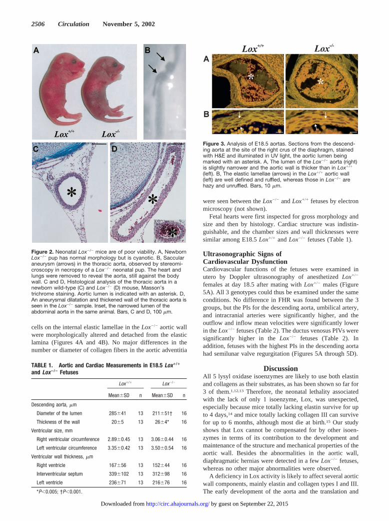
Figure 3. Analysis of E18.5 aortas. Sections from the descend-ing aorta at the site of the right crus of the diaphragm, stainedwith H&E and illuminated in UV light, the aortic lumen beingmarked with an asterisk. A, The lumen of the Lox�/� aorta (right)is slightly narrower and the aortic wall is thicker than in Lox�/�
(left). B, The elastic lamellae (arrows) in the Lox�/� aortic wall(left) are well defined and ruffled, whereas those in Lox�/� arehazy and unruffled. Bars, 10 �m.
Figure 2. Neonatal Lox�/� mice are of poor viability. A, NewbornLox�/� pup has normal morphology but is cyanotic. B, Saccularaneurysm (arrows) in the thoracic aorta, observed by stereomi-croscopy in necropsy of a Lox�/� neonatal pup. The heart andlungs were removed to reveal the aorta, still against the bodywall. C and D, Histological analysis of the thoracic aorta in anewborn wild-type (C) and Lox�/� (D) mouse, Masson’strichrome staining. Aortic lumen is indicated with an asterisk. D,An aneurysmal dilatation and thickened wall of the thoracic aorta isseen in the Lox�/� sample. Inset, the narrowed lumen of theabdominal aorta in the same animal. Bars, C and D, 100 �m.
TABLE 1. Aortic and Cardiac Measurements in E18.5 Lox�/�
and Lox�/� Fetuses
Lox�/� Lox�/�
Mean�SD n Mean�SD n
Descending aorta, �m
Diameter of the lumen 285�41 13 211�51† 16
Thickness of the wall 20�5 13 26�4* 16
Ventricular size, mm
Right ventricular circumference 2.89�0.45 13 3.06�0.44 16
Left ventricular circumference 3.35�0.42 13 3.50�0.54 16
Ventricular wall thickness, �m
Right ventricle 167�56 13 152�44 16
Interventricular septum 339�102 13 312�98 16
Left ventricle 236�71 13 216�76 16
*P�0.005; †P�0.001.
2506 Circulation November 5, 2002
by guest on September 22, 2015http://circ.ahajournals.org/Downloaded from

secretion of aortic wall components are likely to occurnormally in Lox�/� embryos, as demonstrated by the normalnumber of elastic lamellae and smooth muscle cell layers. Inelastin-null mice, smooth muscle cells proliferate and therebystabilize the arterial structure but finally obliterate the arteriallumen.14 No increase in smooth muscle cell proliferation wasobserved in the Lox�/� aortas, probably because normalamounts of elastin were produced, at least during the earlystages. Rupture of a large blood vessel is the major cause ofdeath in mice lacking collagen III.15 The arrangement ofelastic fibers and smooth muscle cells is normal in the aortasof these mice, but collagen fibrils are reduced or absent in themedia, and the fibrils in the adventitia are of variablediameter.15 No analogous changes were seen in the Lox�/�
aortas, where the collagen fibrils were of normal appearancein electron microscopy.
Elastin is present in elastic fibers as soluble monomers andas a highly crosslinked insoluble protein in the amorphous
component.2 Crosslinking has been suggested to be a criticalstep in the nucleation of elastin assembly.2 Amorphous elastinwas found in Lox�/� aortic walls, indicating that other lysyloxidase isoenzymes are also likely to function in elastincrosslinking, but the fragmentation of elastic fibers impliesthat these isoenzymes did not compensate completely for thedeficiency in Lox. The incomplete crosslinking may makeelastin more susceptible to proteolytic degradation,16 whichwould lead to fragmentation of the elastic fibers, so that theyare unable to bear the hemodynamic stress, which would leadin turn to dilatation of the aortic wall and probably alsorupture.
The increased PIs measured in the umbilical artery, de-scending aorta, and the intracranial arteries in Doppler ultra-sonography are likely to be attributable to abnormalities inthe arterial walls. Comparable increased PIs in the umbilicalartery and descending aorta are observed in human pregnan-cies with placental insufficiency, but they are connected with
Figure 4. Electron micrographs of Lox�/� (left) andLox�/� aortic wall (right). A, Smooth muscle cells(SMC) in the Lox�/� aortic wall are organizedbetween elastic fibers (arrows), whereas those inLox�/� are disorganized, and elastic fibers are frag-mented and endothelial cells (uppermost cell layer)are rounded. B, The endothelial cells (EC) in theLox�/� aortic wall are in contact with the intactelastic lamina (arrow), whereas those in Lox�/� arepoorly associated with the fragmented elastic lam-ina (arrow). C, In the Lox�/� aortic wall, amorphouselastin (aEL) is detected in a continuous elasticfiber, whereas in Lox�/�, the elastic fiber is frag-mented, with remnants of amorphous elastin(arrows). Original magnifications: A, �1600; B,�4400; and C, �8400.
Mäki et al Cardiovascular Dysfunction in Lox�/� Mice 2507
by guest on September 22, 2015http://circ.ahajournals.org/Downloaded from

a concomitant decrease in PI values in the cerebral circula-tion, so that an adequate oxygen supply to the coronary andcerebral circulation is maintained.17 Structural alterations arelikely to cause a lower relative pulse amplitude in the arteries,leading to an increased afterload of the heart. In addition, thedecreased mean velocities in the inflow and outflow regionssuggest a lower cardiac output than in Lox�/� fetuses. In-creased fetal cardiac afterload may lead to a drop in cardiac
output and valve regurgitation.18 In our series, fetuses withthe greatest increase in the afterload had semilunar valveregurgitation, most likely pulmonary valve regurgitation. It isnot possible to distinguish between the right and left ventric-ular inflow and outflow regions in mouse fetuses because ofthe small cardiac size, but both the ductus arteriosus and theforamen ovale are wide open in the fetal circulation, so thatthe fetal cardiac ventricles function in parallel and the
Figure 5. Doppler ultrasonography of E18.5 mouse fetuses. The waveforms shown were recorded from fetuses of the same dam. A,Color Doppler image of a mouse fetus in sagittal view, demonstrating the sites from which blood velocity waveforms were sampled bypulsed Doppler. UA indicates umbilical artery; ICA, intracranial artery; DV, ductus venosus; DAO, descending aorta; OF, outflow, and IF,inflow. B, Blood velocity waveforms in the umbilical artery, showing a continuous diastolic component in the Lox�/� fetus and areversed diastolic blood velocity pattern (arrow) in Lox�/�. C, Blood velocity waveforms of the semilunar valve. The valve click (arrow) atthe end of systole, showing valve closure in the Lox�/� fetus, while in the Lox�/� fetus regurgitation jet during diastole is present(arrow). D, Blood velocity waveforms in the ductus venosus. The Lox�/� fetus demonstrates a normal antegrade (toward the heart dur-ing the whole cardiac cycle) triphasic blood velocity waveform pattern. V indicates ventricular contraction; E, early passive filling duringthe diastole; and A, atrial contraction. Increased pulsatility is observed in the ductus venosus of the Lox�/� fetus, where the A wavedemonstrates a retrograde blood flow pattern.
2508 Circulation November 5, 2002
by guest on September 22, 2015http://circ.ahajournals.org/Downloaded from

pressure faced by the ventricles is equal. Thus, the pathophys-iological effects would be similar in both ventricles. In-creased PIVs in the ductus venosus implies an increase insystemic venous pressure, which, if detected in humanfetuses, is interpreted as a sign of congestive heart failure.11
The ventricular sizes and wall thicknesses in the Lox�/�
fetuses were similar to those in Lox�/�, suggesting that thechanges observed in the Doppler parameters merely reflectedfunctional rather than anatomical abnormality in the heart.
Low levels of lysyl oxidase activity secondary to abnor-malities in copper metabolism are found in mottled blotchymice.19 They are born without aneurysms or other cardiovas-cular abnormalities but die of aortic ruptures before 6 monthsof age.20 Our results show that when the activity of the Loxisoenzyme is completely abolished, aneurysms and cardio-vascular dysfunction already occur during fetal development,indicating that a certain level of Lox activity in the aortic wallis irreplaceable. Furthermore, the results underline the impor-tance of the structural and functional properties of the arteriesfor normal cardiovascular development.
In conclusion, the pathophysiological sequence of events inthe cardiovascular system of Lox�/� fetuses suggests thatdeficient crosslinking of elastin and collagens causes adecline in the resilience and tensile strength of the arterialwalls, giving rise to abnormalities in cardiovascular functionsand leading to aortic aneurysms. Our findings suggest thatalterations in LOX activity may also play a critical role inhuman cardiovascular diseases, and in this sense our mousemodel will be a useful tool for additional studies.
AcknowledgmentsThis work was supported by grants from the Health Sciences Councilof the Academy of Finland, from the Finnish Centre of ExcellenceProgramme 2000-2005 (44843), and from FibroGen Inc (South SanFrancisco).
References1. Csiszar K. Lysyl oxidases: a novel multifunctional amine oxidase family.
Prog Nucleic Acid Res Mol Biol. 2001;70:1–32.2. Vrhovski B, Weiss AS. Biochemistry of tropoelastin. Eur J Biochem.
1998;258:1–18.
3. Myllyharju J, Kivirikko KI. Collagens and collagen-related diseases. AnnMed. 2001;33:7–21.
4. Steinmann B, Royce PM, Superti-Furga A. The Ehlers-Danlos syndrome.In: Royce PM, Steinmann B, eds. Connective Tissue and Its HeritableDisorders: Molecular, Genetic and Medical Aspects. New York:Wiley-Liss; 1993:351–407.
5. Mäki JM, Kivirikko KI. Cloning and characterization of a fourth humanlysyl oxidase isoenzyme. Biochem J. 2001;355:381–387.
6. Asuncion L, Fogelgren B, Fong KS, et al. A novel human lysyloxidase-like gene (LOXL4) on chromosome 10q24 has an alteredscavenger receptor cysteine rich domain. Matrix Biol. 2001;20:487–491.
7. Mäki JM, Tikkanen H, Kivirikko KI. Cloning and characterization of afifth human lysyl oxidase isoenzyme: the third member of the lysyloxidase-related subfamily with four scavenger receptor cysteine richdomains. Matrix Biol. 2001;20:493–496.
8. Contente S, Csiszar K, Kenyon K, et al. Structure of the mouse lysyloxidase gene. Genomics. 1993;16:395–400.
9. Nagy A, Rossant J, Nagy R, et al. Derivation of completely cell culture-derived mice from early-passage embryonic stem cells. Proc Natl AcadSci U S A. 1993;90:8424–8428.
10. Gui YH, Linask KK, Khowsathit P, et al. Doppler echocardiography ofnormal and abnormal embryonic mouse heart. Pediatr Res. 1996;40:633–642.
11. Mäkikallio K, Vuolteenaho O, Jouppila P, et al. Ultrasonographic andbiochemical markers of human fetal cardiac dysfunction in placentalinsufficiency. Circulation. 2002;105:2058–2063.
12. Borel A, Eichenberger D, Farjanel J, et al. Lysyl oxidase-like proteinfrom bovine aorta: isolation and maturation to an active form by bonemorphogenetic protein-1. J Biol Chem. 2001;276:48944–48949.
13. Ito H, Akiyama H, Iguchi H, et al. Molecular cloning and biologicalactivity of a novel lysyl oxidase-related gene expressed in cartilage. J BiolChem. 2001;276:24023–24029.
14. Li DY, Brooke B, Davis EC, et al. Elastin is an essential determinant ofarterial morphogenesis. Nature. 1998;21:276–280.
15. Liu X, Wu H, Byrne M, et al. Type III collagen is crucial for collagen Ifibrillogenesis and for normal cardiovascular development. Proc NatlAcad Sci U S A. 1997;4:1852–1856.
16. Tinker D, Romero-Chapman N, Reiser K, et al. Elastin metabolismduring recovery from impaired crosslink formation. Arch BiochemBiophys. 1990;278:326–332.
17. Reed KL. Doppler-the fetal circulation. Clin Obstet Gynecol. 1997;40:750–754.
18. Tulzer G, Gudmundsson S, Rotondo KM, et al. Acute fetal ductalocclusion in lambs. Am J Obstet Gynecol. 1991;165:775–778.
19. Danks DM. Disorders of copper transport: Menkes disease and theoccipital horn syndrome. In: Royce PM, Steinmann B, eds. ConnectiveTissue and Its Heritable Disorders: Molecular, Genetic and MedicalAspects. New York: Wiley-Liss; 1993:487–506.
20. Brophy CM, Tilson JE, Braverman IM, et al. Age of onset, pattern ofdistribution, and histology of aneurysm development in a geneticallypredisposed mouse model. J Vasc Surg. 1988;8:45–48.
TABLE 2. Doppler Ultrasonographic Parameters of E18.5 Fetuses
Lox�/� Lox�/� Lox�/�
Mean�SD n Mean�SD n Mean�SD n
Fetal heart rate, bpm 333�71 19 285�18 10 304�69 11
Mean velocity, cm/s
Outflow 16.2�3.1 19 16.0�1.5 10 12.2�2.3* 11
Inflow 10.3�1.6 19 10.6�1.1 10 8.0�1.3* 11
Pulsatility index
Umbilical artery 1.90�0.22 19 1.84�0.13 10 2.85�0.70† 11
Descending aorta 2.09�0.21 19 1.95�0.19 10 2.87�0.64† 11
Intracranial arteries 1.90�0.31 18 1.78�0.17 10 2.28�0.23* 10
Pulsatility index for veins
Ductus venosus 0.89�0.09 19 0.88�0.07 10 1.30�0.34† 11
*P�0.01 vs Lox�/� and Lox�/�; †P�0.001 vs Lox�/� and Lox�/�.
Mäki et al Cardiovascular Dysfunction in Lox�/� Mice 2509
by guest on September 22, 2015http://circ.ahajournals.org/Downloaded from

Kivirikko and Raija SoininenJoni M. Mäki, Juha Räsänen, Hilkka Tikkanen, Raija Sormunen, Kaarin Mäkikallio, Kari I.
Dysfunction, and Perinatal Death in Mice Leads to Aortic Aneurysms, CardiovascularLoxInactivation of the Lysyl Oxidase Gene
Print ISSN: 0009-7322. Online ISSN: 1524-4539 Copyright © 2002 American Heart Association, Inc. All rights reserved.
is published by the American Heart Association, 7272 Greenville Avenue, Dallas, TX 75231Circulation doi: 10.1161/01.CIR.0000038109.84500.1E
2002;106:2503-2509Circulation.
http://circ.ahajournals.org/content/106/19/2503World Wide Web at:
The online version of this article, along with updated information and services, is located on the
http://circ.ahajournals.org//subscriptions/
is online at: Circulation Information about subscribing to Subscriptions:
http://www.lww.com/reprints Information about reprints can be found online at: Reprints:
document. Permissions and Rights Question and Answer this process is available in the
click Request Permissions in the middle column of the Web page under Services. Further information aboutOffice. Once the online version of the published article for which permission is being requested is located,
can be obtained via RightsLink, a service of the Copyright Clearance Center, not the EditorialCirculationin Requests for permissions to reproduce figures, tables, or portions of articles originally publishedPermissions:
by guest on September 22, 2015http://circ.ahajournals.org/Downloaded from