Inhibitor of Lysyl Oxidase in The Optic Nerve Head Complex ...
19
Page 1/19 Inhibitor of Lysyl Oxidase in The Optic Nerve Head Complex Imparts Partial Protection Against Injury in Experimental Glaucoma Carlos M Cordoba Universidad Nacional de Colombia - Sede Bogotá: Universidad Nacional de Colombia Magally Barrera Universidad Nacional de Colombia - Sede Bogotá: Universidad Nacional de Colombia Sandra Perdomo Signal Inc SAS Pedro Gabriel Franco Universidad Nacional de Colombia - Sede Bogotá: Universidad Nacional de Colombia Jaidy Acosta Alvarez Universidad Nacional de Colombia - Sede Bogotá: Universidad Nacional de Colombia Zulma Y Dueñas Universidad Nacional de Colombia - Sede Bogotá: Universidad Nacional de Colombia Marcel Y Avila ( [email protected] ) Universidad Nacional de Colombia https://orcid.org/0000-0001-7734-8177 Research article Keywords: glaucoma, BAPN, Genipin, optic nerve, cross-linking Posted Date: October 26th, 2021 DOI: https://doi.org/10.21203/rs.3.rs-968649/v1 License: This work is licensed under a Creative Commons Attribution 4.0 International License. Read Full License
-
Upload
khangminh22 -
Category
Documents
-
view
1 -
download
0
Transcript of Inhibitor of Lysyl Oxidase in The Optic Nerve Head Complex ...

Page 1/19
Inhibitor of Lysyl Oxidase in The Optic Nerve HeadComplex Imparts Partial Protection Against Injury inExperimental GlaucomaCarlos M Cordoba
Universidad Nacional de Colombia - Sede Bogotá: Universidad Nacional de ColombiaMagally Barrera
Universidad Nacional de Colombia - Sede Bogotá: Universidad Nacional de ColombiaSandra Perdomo
Signal Inc SASPedro Gabriel Franco
Universidad Nacional de Colombia - Sede Bogotá: Universidad Nacional de ColombiaJaidy Acosta Alvarez
Universidad Nacional de Colombia - Sede Bogotá: Universidad Nacional de ColombiaZulma Y Dueñas
Universidad Nacional de Colombia - Sede Bogotá: Universidad Nacional de ColombiaMarcel Y Avila ( [email protected] )
Universidad Nacional de Colombia https://orcid.org/0000-0001-7734-8177
Research article
Keywords: glaucoma, BAPN, Genipin, optic nerve, cross-linking
Posted Date: October 26th, 2021
DOI: https://doi.org/10.21203/rs.3.rs-968649/v1
License: This work is licensed under a Creative Commons Attribution 4.0 International License. Read Full License

Page 2/19
AbstractBackground: Glaucoma is a neurodegenerative disease with the progressive loss of retinal ganglion cellsand changes in the optic nerve head (ONH). These changes are exacerbated by an increase in intraocularpressure (IOP).
Methods: The effect of scleral and optic nerve softening with beta aminopropionitrile a lysyl oxidaseinhibitor (BAPN) and stiffening with genipin, in a model of chronic increase of IOP was evaluated.Changes in optic nerve and retina were evaluated. H&E, Bielschowsky's silver staining and glial �brillaryacid protein (GFAP) staining was performed on optic nerve, retina and scleral structures. Changes in theexpression of the Ywhab, Yhwaz (prosurvival genes), C3 complement (complement C3 in�ammatorymarker), CPG15 (neurite growth and neural survival gene) GFAP (glial activator marker) genes was carriedout in the different groups.
Results: Protective effect of BAPN was evident by the preservation of the optic nerve structure, and withthe conservation of the retinal structures, while deleterious changes were evident in the stiffening of ONHcomplex, characterized by the increase in the glia, changes in the optic nerve, and disorganization in theretina. BAPN induced a reduction in the expression of Ywhab, Yhwaz (prosurvival genes), C3 and GFAP(in�ammatory and glial marker) and CPG15.
Conclusions: These �ndings support the critical involvement of changes in the ONH stiffness in theprogression of glaucoma. The control of this variable as a regulatory mechanism in the progression ofneural glaucomatous damage must be considered and would be explored as a possible intervention inglaucoma management.
BackgroundGlaucoma is the leading cause of irreversible blindness worldwide; in the optic nerve there is a loss ofretinal ganglion cells with changes in the optic nerve head, inducing an increase of the cup /disc relation.[1] Increases in IOP induces deformation of the optic nerve head (ONH) and reduction in the retinalganglion cells by mechanisms such as a reduction in axoplasmic �ow [2], a reduction of ocular blood�ow in the ONH [3], and changes induced by mechanical strain [4]. Several authors have proposed thatcross-linking of the peripapillar sclera decreased the deformation of the ONH, thus reducing thedeleterious changes in this area, considering the cross-linking of ONH as a potential therapeutic strategyin the management of glaucoma [5–6]. However, other authors have proposed that the induction of cross-linking could reduce ONH compliance [7], reducing axonal �ow, leading to an increase in susceptibility toglaucomatous damage [8] in glaucoma models. To support this theory, noxious effects in the retinalganglion cells in a rat glaucoma model were exacerbated by cross-linking the ONH [9], such as anincrease in collagen and in peripapillary thickness in glaucoma rodent models [10]. In this work, weproposed evaluating the effect of softening the ONH via lysyl oxidase inhibition (LOX inhibition) usingbeta-aminopropionitrile (BAPN) to inhibit the collagen I and elastin cross-linking, in a glaucoma model

Page 3/19
and to induce stiffening of the ONH with genipin, thus We can con�rm the opposite effects in thechanges in the optic nerve and in the retina in this model.
Some authors have proposed that stiffening of the ONH may be related to several mechanisms involvedwith age, such as cross-linking, calcium, lipids and increases in advanced glycation end products (AGEs)affecting the collagenous and non-collagenous components of the extracellular matrix, as well aschanges in the phenotype of �broblasts to myo�broblasts, increasing the �brosis in the ONH, and leadingto a reduction of the ONH compliance [9–11]. It has also been found that glaucoma patients exhibited ahigher ocular stiffness than non-glaucoma subjects [12]. In this model, we used genipin, a naturalcrosslinker that has low toxicity and interesting properties, as a crosslinker agent; this agent inducescorneal [13] and scleral stiffening in ex vivo and in vivo models [14], with minimal damage to the cornealendothelial and retinal cells [5–15], and is useful in controlling eye growth in myopia models [16]. In aclinical setting, the uses of scleral strips treated with genipin have been proposed in the control of myopia[17]. Furthermore, the dosage of genipin in the rat eye has been previously characterized [18], showing agood effect as a cross-linker in the optic nerve complex; in the posterior sclera, with only one injectionwith no toxicity to retinal ganglion cells, there are no changes in gene expression in normal rat eyes [5].This type of cross-linking is de�ned as dark cross-linking since there is no need for an external lightsource in order to achieve the cross-linking effect [6].
In contrast, beta aminopropionitrile (BAPN) was used for the opposite reason; as an inhibitor of collagen-elastin cross-linking via lysyl oxidase (LOX) inhibitor, it prevents the cross-linking of collagen I and theelastin-induced softening of tissues and reverses myocardial �brosis in murine models [19]. The aim ofthis study was to determine whether cross-linking or softening of the ONH is detrimental to retinal cellsand the optic nerve in a model of glaucoma in rats. Intraocular pressure changes as well as changes inthe retina and ONH morphology and changes in the gene expression of the Ywhab, Yhwaz (prosurvivalgenes), C3 complement (complement C3 in�ammatory marker), CPG15 (neurite growth and neuralsurvival gene) and GFAP genes were evaluated by RT-PCR and compared in different groups (Glaucoma,Glaucoma-genipin, Glaucoma –BAPN, genipin and BAPN).
MethodsAnimals
White albino Wistar rats were used; half were female and half male (n=40). All rats were housed on a 12-hour light-dark cycle and were provided with food and water ad libitum. All procedures were approved bythe Institutional animal care and use committee from the Facultad de Medicina Universidad Nacional deColombia, under the approval 010-135-19 and followed the guidelines of ARVO, and Colombian regulationfor animals in experimentation. Surgical procedures as well as retrobulbar injections were performedunder general anesthesia using ketamine (60 mg/kg) and xylazine (7.5 mg/kg) complemented withtopical proparacaine (Alcon Fortworth TX). Retrobulbar injections were performed using a 31 g
Page 4/19
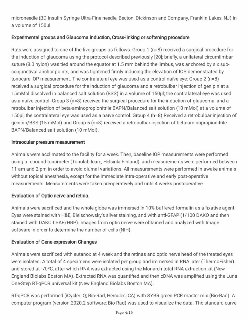
microneedle (BD Insulin Syringe Ultra-Fine needle, Becton, Dickinson and Company, Franklin Lakes, NJ) ina volume of 150µl.
Experimental groups and Glaucoma induction, Cross-linking or softening procedure
Rats were assigned to one of the �ve groups as follows. Group 1 (n=8) received a surgical procedure forthe induction of glaucoma using the protocol described previously [20]; brie�y, a unilateral circumlimbarsuture (8.0 nylon) was tied around the equator at 1.5 mm behind the limbus, was anchored by six sub-conjunctival anchor points, and was tightened �rmly inducing the elevation of IOP, demonstrated bytonocare IOP measurement. The contralateral eye was used as a control naïve eye. Group 2 (n=8)received a surgical procedure for the induction of glaucoma and a retrobulbar injection of genipin at a15mMol dissolved in balanced salt solution (BSS) in a volume of 150μl; the contralateral eye was usedas a naïve control. Group 3 (n=8) received the surgical procedure for the induction of glaucoma, and aretrobulbar injection of beta-aminopropionitrile BAPN/Balanced salt solution (10 mMol) at a volume of150μl; the contralateral eye was used as a naïve control. Group 4 (n=8) Received a retrobulbar injection ofgenipin/BSS (15 mMol) and Group 5 (n=8) received a retrobulbar injection of beta-aminopropionitrileBAPN/Balanced salt solution (10 mMol).
Intraocular pressure measurement
Animals were acclimated to the facility for a week. Then, baseline IOP measurements were performedusing a rebound tonometer (Tonolab Icare, Helsinki Finland), and measurements were performed between11 am and 2 pm in order to avoid diurnal variations. All measurements were performed in awake animalswithout topical anesthesia, except for the immediate intra-operative and early post-operativemeasurements. Measurements were taken preoperatively and until 4 weeks postoperative.
Evaluation of Optic nerve and retina.
Animals were sacri�ced and the whole globe was immersed in 10% buffered formalin as a �xative agent.Eyes were stained with H&E, Bielschowsky's silver staining, and with anti-GFAP (1/100 DAKO and thenstained with DAKO LSAB/HRP). Images from optic nerve were obtained and analyzed with Imagesoftware in order to determine the number of cells (NIH).
Evaluation of Gene expression Changes
Animals were sacri�ced with eutanox at 4 week and the retinas and optic nerve head of the treated eyeswere isolated. A total of 4 specimens were isolated per group and immersed in RNA later (ThermoFisher)and stored at -70ºC, after which RNA was extracted using the Monarch total RNA extraction kit (NewEngland Biolabs Boston MA). Extracted RNA was quanti�ed and then cDNA was ampli�ed using the LunaOne-Step RT-qPCR universal kit (New England Biolabs Boston MA).
RT-qPCR was performed (iCycler iQ; Bio-Rad, Hercules, CA) with SYBR green PCR master mix (Bio-Rad). Acomputer program (version:2020.2 software; Bio-Rad) was used to visualize the data. The standard curve
Page 5/19
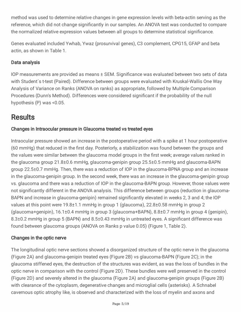
method was used to determine relative changes in gene expression levels with beta-actin serving as thereference, which did not change signi�cantly in our samples. An ANOVA test was conducted to comparethe normalized relative expression values between all groups to determine statistical signi�cance.
Genes evaluated included Ywhab, Ywaz (prosurvival genes), C3 complement, CPG15, GFAP and betaactin, as shown in Table 1.
Data analysis
IOP measurements are provided as means ± SEM. Signi�cance was evaluated between two sets of datawith Student´s t-test (Paired). Difference between groups were evaluated with Kruskal-Wallis One WayAnalysis of Variance on Ranks (ANOVA on ranks) as appropriate, followed by Multiple ComparisonProcedures (Dunn's Method). Differences were considered signi�cant if the probability of the nullhypothesis (P) was <0.05.
ResultsChanges in Intraocular pressure in Glaucoma treated vs treated eyes
Intraocular pressure showed an increase in the postoperative period with a spike at 1 hour postoperative(60 mmHg) that reduced in the �rst day. Posteriorly, a stabilization was found between the groups andthe values were similar between the glaucoma model groups in the �rst week; average values ranked inthe glaucoma group 21.8±0.6 mmHg, glaucoma-genipin group 25.5±0.5 mmHg and glaucoma-BAPNgroup 22.5±0.7 mmHg. Then, there was a reduction of IOP in the glaucoma-BPNA group and an increasein the glaucoma-genipin group. In the second week, there was an increase in the glaucoma-genipin groupvs. glaucoma and there was a reduction of IOP in the glaucoma-BAPN group. However, those values werenot signi�cantly different in the ANOVA analysis. This difference between groups (reduction in glaucoma-BAPN and increase in glaucoma-genipin) remained signi�cantly elevated in weeks 2, 3 and 4; the IOPvalues at this point were 19.8±1.1 mmHg in group 1 (glaucoma), 22.8±0.58 mmHg in group 2(glaucoma+genipin), 16.1±0.4 mmHg in group 3 (glaucoma+BAPN), 8.8±0.7 mmHg in group 4 (genipin),8.3±0.2 mmHg in group 5 (BAPN) and 8.5±0.43 mmHg in untreated eyes. A signi�cant difference wasfound between glaucoma groups (ANOVA on Ranks p value 0.05) (Figure 1, Table 2).
Changes in the optic nerve
The longitudinal optic nerve sections showed a disorganized structure of the optic nerve in the glaucoma(Figure 2A) and glaucoma-genipin treated eyes (Figure 2B) vs glaucoma-BAPN (Figure 2C); in theglaucoma stiffened eyes, the destruction of the structures was evident, as was the loss of bundles in theoptic nerve in comparison with the control (Figure 2D). These bundles were well preserved in the control(Figure 2D) and severely altered in the glaucoma (Figure 2A) and glaucoma-genipin groups (Figure 2B)with clearance of the cytoplasm, degenerative changes and microglial cells (asterisks). A Schnabelcavernous optic atrophy like, is observed and characterized with the loss of myelin and axons and

Page 6/19
preservation of septa with a spongiform appearance and was evident in the glaucoma and glaucoma-genipin groups. Glaucoma-BAPN showed organization of optic nerve bundles with minimal microglialcells (Figure 2C).
Changes in retina and nerve �ber evaluation
At week 4, retinal cell layers were evaluated using the retina cross sections stained with H&E and acomparison between all groups. The normal retina was observed in the control group (Figure 3d), andthinning of the retina was observed in glaucoma (Figure 3a) along with alterations in ONL, OPL, IPL and areduction in RGC with a loss of inner nuclear cells layer and gliosis-like shape; a loss of the retinalstructure was observed in the glaucoma-genipin group (Figure 3b) while a complete evaluation of alllayers is observed in the glaucoma-BAPN (Figure 3c), genipin (Figure 3e) and BAPN (Figure 3f).
Bielschowsky's silver staining shows preservation of the inter-neuronal unions and axonal staining in thenormal retina (Figure 4D), genipin alone (Figure 4E) and BAPN alone (Figure 4F), as well as in theglaucoma-BAPN group (Figure 4C); in the glaucoma group (Figure 4A) and glaucoma-genipin group(Figure 4B), severe �ber distortion and a loss of the axonal union was observed in the central area of theretina. Staining with GFAP antibody in the glaucoma-genipin (Figure 5B) and glaucoma (Figure 5A)groups was evident in the nerve �ber layer (NFL), ganglion cell layer (GCL), and projections to innerplexiform layer (IPL), while there was minimal staining in the NFL in the control group (Figure 5D) andglaucoma-BAPN group (Figure 5C).
Changes in Gene Expression and effects of BAPN and genipin in glaucoma eyes
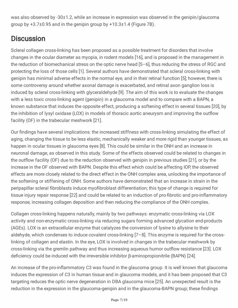
Gene expression of C3 complement was up-regulated in glaucoma +6.1±0.4 in comparison with thecontrol group; a signi�cative down-regulation observed in the BAPN/glaucoma group and in thegenipin/glaucoma group. No differences were noted in the BAPN and genipin group (Figure 6A).
The GFAP gene was up-regulated in glaucoma by +1.4±0.02 in comparison to the control group, as wellas in BAPN (+1.3±0.9) and was down-regulated in the glaucoma-genipin group (-19±0.9),BAPN/glaucoma group (-33±2.0) and in the genipin group (-16±1.4) (Figure 6B).
Cpg15 gene was down regulated in glaucoma by -1.8±0.7 in comparison with the control group, as wellas in the BAPN-glaucoma group by -20±2.0 and in the genipin-glaucoma group by -14±0.9; in the genipingroup, this was reduced by -2.7±1.4 and was increased in the BAPN group by +3.7±1.0 (Figure 6C).
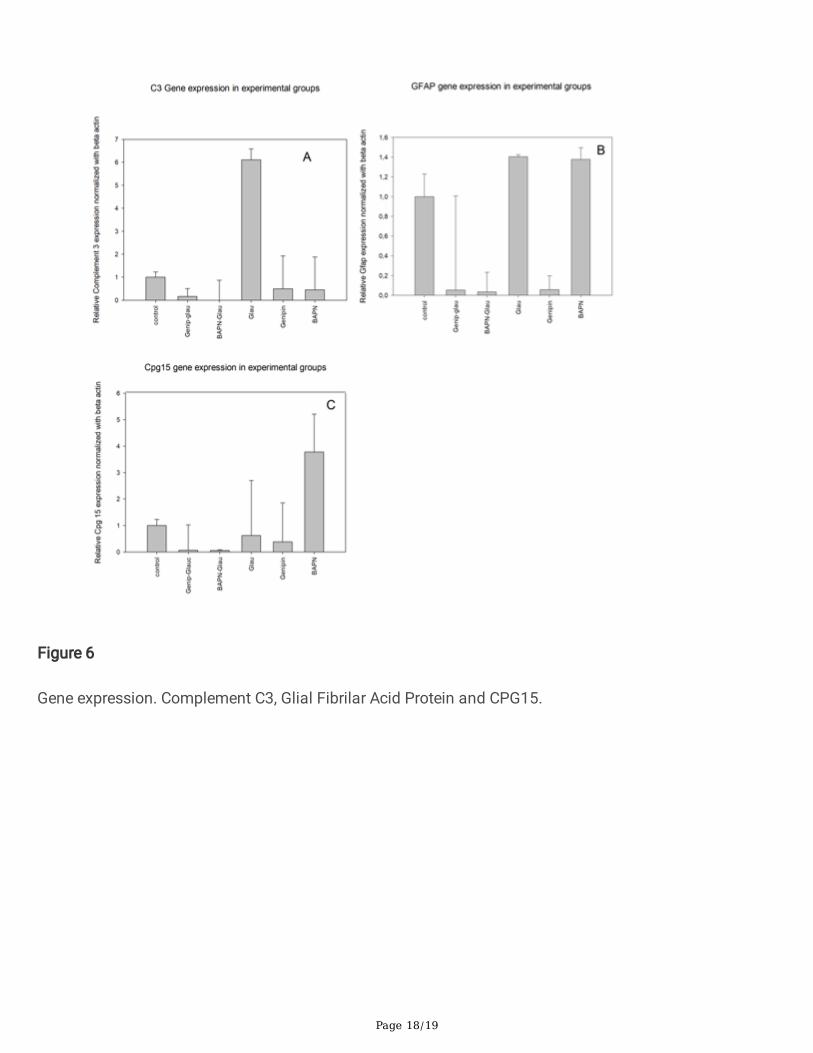
Ywhab gene was up-regulated in the glaucoma group by 1.8±0.27 and down-regulated in the glaucoma-genipin and glaucoma-BAPN group, as well as severely up-regulated in the genipin group (77±1.42)(Figure 7A).
The Ywhaz gene was down-regulated in glaucoma by -5.3±0.4 in comparison to the control group, andwas also down-regulated in the BAPN/glaucoma group -21±2.1; in BAPN alone, a signi�cant reduction
Page 7/19
was also observed by -30±1.2, while an increase in expression was observed in the genipin/glaucomagroup by +3.7±0.95 and in the genipin group by +10.3±1.4 (Figure 7B).
DiscussionScleral collagen cross-linking has been proposed as a possible treatment for disorders that involvechanges in the ocular diameter as myopia, in rodent models [16], and is proposed in the management inthe reduction of biomechanical stress on the optic nerve head [5–6], thus reducing the stress of RGC andprotecting the loss of those cells [1]. Several authors have demonstrated that scleral cross-linking withgenipin has minimal adverse effects in the normal eye, and in their retinal function [5]; however, there issome controversy around whether axonal damage is exacerbated, and retinal axon ganglion loss isinduced by scleral cross-linking with glyceraldehyde [9]. The aim of this work is to evaluate the changeswith a less toxic cross-linking agent (genipin) in a glaucoma model and to compare with a BAPN, aknown substance that induces the opposite effect, producing a softening effect in several tissues [20], bythe inhibition of lysyl oxidase (LOX) in models of thoracic aortic aneurysm and improving the out�owfacility (OF) in the trabecular meshwork [21].
Our �ndings have several implications: the increased stiffness with cross-linking simulating the effect ofaging, changing the tissue to be less elastic, mechanically weaker and more rigid than younger tissues, ashappen in ocular tissues in glaucoma eyes [8]. This could be similar in the ONH and an increase inneuronal damage, as observed in this study. Some of the effects observed could be related to changes inthe out�ow facility (OF) due to the reduction observed with genipin in previous studies [21], or by theincrease in the OF observed with BAPN. Despite this effect which could be affecting IOP, the observedeffects are more closely related to the direct effect in the ONH complex area, unlocking the importance ofthe softening or stiffening of ONH. Some authors have demonstrated that an increase in strain in theperipapillar scleral �broblasts induce myo�broblast differentiation; this type of change is required fortissue injury repair response [22] and could be related to an induction of pro-�brotic and pro-in�ammatoryresponse, increasing collagen deposition and then reducing the compliance of the ONH complex.
Collagen cross-linking happens naturally, mainly by two pathways: enzymatic cross-linking via LOXactivity and non-enzymatic cross-linking via reducing sugars forming advanced glycation end-products(AGEs). LOX is an extracellular enzyme that catalyzes the conversion of lysine to allysine to theiraldehyde, which condenses to induce covalent cross-linking [7–8]. This enzyme is required for the cross-linking of collagen and elastin. In the eye, LOX is involved in changes in the trabecular meshwork bycross-linking via the gremlin pathway and thus increasing aqueous humor out�ow resistance [23]. LOXde�ciency could be induced with the irreversible inhibitor β-aminopropionitrile (BAPN) [24].
An increase of the pro-in�ammatory C3 was found in the glaucoma group. It is well known that glaucomainduces the expression of C3 in human tissue and in glaucoma models, and it has been proposed that C3targeting reduces the optic nerve degeneration in DBA glaucoma mice [25]. An unexpected result is thereduction in the expression in the glaucoma-genipin and in the glaucoma-BAPN group; these �ndings

Page 8/19
could be explained by the anti-in�ammatory effect of genipin by itself [26], and by the preservation of theONH anatomy by BAPN, reducing the damage in the RGC and in the ONH complex, thus reducingin�ammation.
Changes in the Ywhaz gene expression with a function as a mitochondrial import stimulation factor, wasalso observed in this study. A down-regulation in glaucoma (as previously demonstrated) (27) and in theglaucoma-BAPN group was found in this work, and their increase in the glaucoma-genipin group and inthe genipin group is observed; this gene is considered an inductor of pro-survival pathways in RGC.Despite this, BAPN induced a reduction of this gene and genipin increased the expression of this gene.This effect could be related to the neuroprotective effects of genipin itself, as has been demonstrated inmodels of Alzheimer degeneration (26); despite the up-regulation, the net effect was neuronal damage,demonstrating that the effect of stiffening by itself is more injurious than the possible neuroprotectiveeffect of genipin. However, more studies are required to elucidate this change. Ywhab was also down-regulated in the treated glaucoma-BAPN and glaucoma-genipin group but was overexpressed in genipincontrol. This effect could also be explained by the neuroprotective effect of genipin mediated by thereduction of ROS species and RNS species [8–9]; as previously stated, stiffening induced moredeleterious changes than the effect of genipin as a neuroprotective agent.
GFAP gene expression was observed in the glaucoma group and a reduction in the glaucoma-BAPNgroup, glaucoma-genipin group and genipin group. GFAP is a complex system that is up-regulated inneurodegenerative diseases; its role could be related to a reduction of the neuronal damage, and anincrease in the affected area to form a physical barrier to isolate the damaged tissue [10]. A protectiveeffect in the treatment may explain the reduction in the expression of GFAP as well as the expression inthe Ganglion cell layer in glaucoma and in glaucoma-genipin retina.
Cpg15, a candidate plasticity gene that promotes neurite outgrowth, is down-regulated in glaucomamodels; this �nding was also present in our study, as well as an important down-regulation in treatedgroups. This down-regulation may be related to the reduction of ischemia in the ONH, as has beenproposed in models of transient global cerebral ischemia, where ischemia is required to induce theoverexpression of Cpg15 [11]; however, more studies are required to clarify this point.
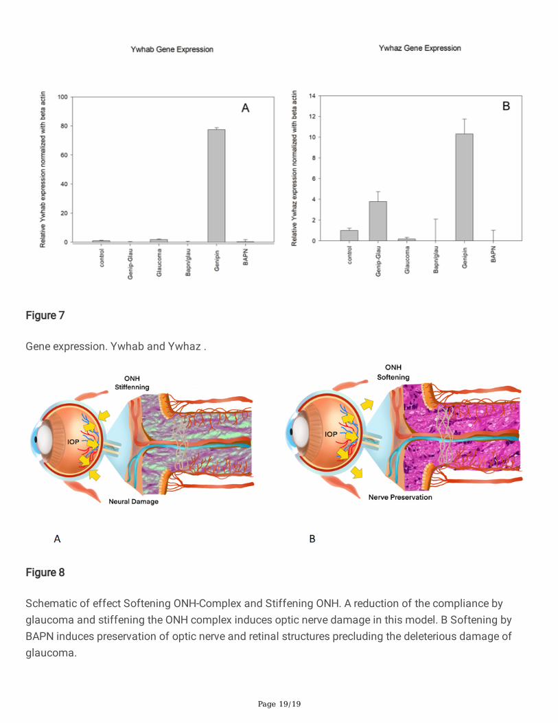
ConclusionsIn conclusion, our model con�rms the effects of changing the stiffness of the ONH complex in aglaucoma model, showing that this factor is crucial for the progression of glaucoma. A proposedmechanism is presented in Figure 8. Stiffening the ONH complex induces a reduction in scleralcompliance and induces axon damage in the ONH and in the retinal layers with changes in the opticnerve. There is also local degeneration, cavernous atrophy and the induction of pro-in�ammatorymolecules, as well as a reduction of prosurvival genes. In contrast, softening of the ONH induces thepreservation of axons and optic nerve structure as well as preservation of the retinal layers and a

Page 9/19
reduction in the induction of in�ammatory genes. ONH softening may represent a new target in themanagement of glaucoma and deserves more studies.
AbbreviationsONH Optic Nerve Head
IOP Intraocular Pressure
BAPN Beta aminopropionitrile
GFAP Glial �brillary acid protein
LOX Lysyl oxidase
OF Out�ow facility
AGEs Advanced glycation end-products
RGC Retinal ganglion cells
DeclarationsEthical Approval and Consent to participate: Ethical approval was obtained by the IRB 010-135-19Facultad de Medicina Universidad Nacional de Colombia.
Consent for publication: Non Aplicable
Availability of supporting data: Supporting data is available upon request to the Corresponding author asa PDF format
Competing interests: None of the Authors have any competing interest in the products and methodsdescribed in this work
Funding: Hermes Funding for Publications School of Medicine 43558
Authors' contributions : CMC.; Designed and performed experiments, analysed data and co-wrote thepaper. MB Performed experiments, SP Performed experiments and Performed bioinformatic analyses,PGF Performed pathologic analyses, JAA Performed experiments, ZD Supervised the research anddesigned experiments, MYA Designed experiments, analyzed data and co-wrote the paper and supervisedthe research. All authors read and approved the �nal manuscript.
Acknowledgements : To Susana Avila in the design and perform in the drafts and illustrations.

Page 10/19
Authors' information: CMC MD Ophthalmologist Universidad Nacional, MB Master of Science andphysiological Sciences at Universidad Nacional , SP is a BSc. PhD Universidad el Bosque and Director ofSignal Inc, PGF Associate Professor of Anatomy and Head of Laboratory of histology, School of MedicineUniversidad Nacional de Colombia, JAA BSc. Laboratory of histology, School of Medicine UniversidadNacional de Colombia, ZD MSC PhD UNAM, Postdoctoral Fellow University of Irvine CA ProfessorAssociate Department of Physiology School of Medicine Universidad Nacional de Colombia, MYA MDPhD Ophthalmologist. Post Doctoral Fellow University of Pensylvania,Professor Associate Department ofOphthalmology School of Medicine Universidad Nacional de Colombia.
References1. Tham YC, Li X, Wong TY, Quigley HA, Aung T, Cheng CY. Global prevalence of glaucoma and
projections of glaucoma burden through 2040: a systematic review and meta-analysis.Ophthalmology. 2014 Nov;121(11):2081–90. doi:10.1016/j.ophtha.2014.05.013. Epub 2014 Jun 26.PMID: 24974815.
2. Korneva A, Schaub J, Jefferys J, Kimball E, Pease ME, Nawathe M, Johnson TV, Pitha I, Quigley H. Amethod to quantify regional axonal transport blockade at the optic nerve head after short termintraocular pressure elevation in mice. Exp Eye Res. 2020 Jul;196:108035. doi:10.1016/j.exer.2020.108035. Epub 2020 Apr 27. PMID: 32353427; PMCID: PMC7335019.
3. 3. Nakazawa T. Ocular Blood Flow and In�uencing Factors for Glaucoma. Asia Pac J Ophthalmol(Phila). 2016 Jan-Feb;5(1):38-44. doi: 10.1097/APO.0000000000000183. PMID: 26886118.
4. Kirwan RP, Fenerty CH, Crean J, Wordinger RJ, Clark AF, O'Brien CJ. In�uence of cyclical mechanicalstrain on extracellular matrix gene expression in human lamina cribrosa cells in vitro. Mol Vis. 2005Sep 23;11:798–810. PMID: 16205625.
5. Hannon BG, Luna C, Feola AJ, Ritch MD, Read AT, Stinnett SS, Vo H, Pardue MT, Gonzalez P, EthierCR. Assessment of visual and retinal function following in vivo genipin-induced scleral cross-linking.Transl Vis Sci Technol. 2020 Sep 8;9(10):8. doi: 10.1167/tvst.9.10.8. PMID: 32974080; PMCID:PMC7488211.
�. Gerberich BG, Hannon BG, Hejri A, Winger EJ, Schrader Echeverri E, Nichols LM, Gersch HG, MacLeodNA, Gupta S, Read AT, Ritch MD, Sridhar S, Toothman MG, Gershon GS, Schwaner SA, Sánchez-Rodríguez G, Goyal V, Toporek AM, Feola AJ, Grossniklaus HE, Pardue MT, Ethier CR, Prausnitz MR.Transpupillary collagen photo-cross-linking for targeted modulation of ocular biomechanics.Biomaterials. 2021 Apr;271:120735. doi: 10.1016/j.biomaterials.2021.120735. Epub 2021 Feb 24.PMID: 33721571; PMCID: PMC8044034.
7. Rahman N, O'Neill E, Irnaten M, Wallace D, O'Brien C. Corneal Stiffness and Collagen Cross-LinkingProteins in Glaucoma: Potential for Novel Therapeutic Strategy. J Ocul Pharmacol Ther. 2020Oct;36(8):582–94. doi:10.1089/jop.2019.0118. Epub 2020 Jul 13. PMID: 32667842.
�. Liu B, McNally S, Kilpatrick JI, Jarvis SP, O'Brien CJ. Aging and ocular tissue stiffness in glaucoma.Surv Ophthalmol. 2018 Jan-Feb;63(1):56–74. doi:10.1016/j.survophthal.2017.06.007. Epub 2017

Page 11/19
Jun 27. PMID: 28666629.
9. Kimball EC, Nguyen C, Steinhart MR, Nguyen TD, Pease ME, Oglesby EN, Oveson BC, Quigley HA.Experimental scleral cross-linking increases glaucoma damage in a mouse model. Exp Eye Res. 2014Nov;128:129–40. doi:10.1016/j.exer.2014.08.016. Epub 2014 Oct 5. PMID: 25285424; PMCID:PMC4254118.
10. Cone-Kimball E, Nguyen C, Oglesby EN, Pease ME, Steinhart MR, Quigley HA. Scleral structuralalterations associated with chronic experimental intraocular pressure elevation in mice. Mol Vis.2013 Sep 26;19:2023–39. PMID: 24146537; PMCID: PMC3783364.
11. Liu B, Kilpatrick JI, Lukasz B, Jarvis SP, McDonnell F, Wallace DM, Clark AF, O'Brien CJ. Increasedsubstrate stiffness elicits a myo�broblastic phenotype in human lamina cribrosa cells. InvestOphthalmol Vis Sci. 2018 Feb 1;59(2):803-814. doi: 10.1167/iovs.17-22400. PMID: 29392327.
12. Coudrillier B, Tian J, Alexander S, Myers KM, Quigley HA, Nguyen TD. Biomechanics of the humanposterior sclera: age- and glaucoma-related changes measured using in�ation testing. InvestOphthalmol Vis Sci. 2012 Apr 2;53(4):1714-28. doi: 10.1167/iovs.11-8009. PMID: 22395883; PMCID:PMC3630906.
13. Avila MY, Navia JL. Effect of genipin collagen cross-linking on porcine corneas. J Cataract RefractSurg. 2010 Apr;36(4):659-64. doi: 10.1016/j.jcrs.2009.11.003. PMID: 20362860.
14. Wong FF, Lari DR, Schultz DS, Stewart JM. Whole globe in�ation testing of exogenously crosslinkedsclera using genipin and methylglyoxal. Exp Eye Res. 2012 Oct;103:17–21.doi:10.1016/j.exer.2012.06.010. Epub 2012 Aug 1. PMID: 22884564; PMCID: PMC3462300.
15. Avila MY, Gerena VA, Navia JL. Corneal cross-linking with genipin, comparison with UV-ribo�avin inex-vivo model. Mol Vis. 2012;18:1068–73. Epub 2012 Apr 27. PMID: 22605919; PMCID:PMC3351405.
1�. Wang M, Corpuz CC. Effects of scleral cross-linking using genipin on the process of form-deprivationmyopia in the guinea pig: a randomized controlled experimental study. BMC Ophthalmol. 2015Jul;29:15:89. doi:10.1186/s12886-015-0086-z. PMID: 26220299; PMCID: PMC4518847.
17. Zhu SQ, Zheng LY, Pan AP, Yu AY, Wang QM, Xue AQ. The e�cacy and safety of posterior scleralreinforcement using genipin cross-linked sclera for macular detachment and retinoschisis in highlymyopic eyes. Br J Ophthalmol. 2016 Nov;100(11):1470–5. doi:10.1136/bjophthalmol-2015-308087.Epub 2016 Feb 25. PMID: 26917677.
1�. Campbell IC, Hannon BG, Read AT, Sherwood JM, Schwaner SA, Ethier CR. Quanti�cation of thee�cacy of collagen cross-linking agents to induce stiffening of rat sclera. J R Soc Interface. 2017Apr;14(129):20170014. doi:10.1098/rsif.2017.0014. Erratum in: J R Soc Interface. 2017 May;14 (130): PMID: 28381643; PMCID:PMC5414911.
19. Rosin NL, Sopel MJ, Falkenham A, Lee TD, Légaré JF. Disruption of collagen homeostasis canreverse established age-related myocardial �brosis. Am J Pathol. 2015 Mar;185(3):631-42. doi:

Page 12/19
10.1016/j.ajpath.2014.11.009. PMID: 25701883.
20. Liu HH, Bui BV, Nguyen CT, Kezic JM, Vingrys AJ, He Z. Chronic ocular hypertension induced bycircumlimbal suture in rats. Invest Ophthalmol Vis Sci. 2015 May;56(5):2811-20. doi:10.1167/iovs.14-16009. PMID: 25829414.
21. Lv X, Hu Y, Chen X, Chen X, Chen L, Lin Y, Hou Y. Establishment and effect evaluation of an aorticdissection model induced by different doses of β-aminopropionitrile in rats. Vascular. 2020 Dec23:1708538120984056. doi: 10.1177/1708538120984056. Epub ahead of print. PMID: 33357159.
22. Yang YF, Sun YY, Acott TS, Keller KE. Effects of induction and inhibition of matrix cross-linking onremodeling of the aqueous out�ow resistance by ocular trabecular meshwork cells. Sci Rep. 2016 Jul28;6:30505. doi: 10.1038/srep30505. PMID: 27465745; PMCID: PMC4964656.
23. Qu J, Chen H, Zhu L, Ambalavanan N, Girkin CA, Murphy-Ullrich JE, Downs JC, Zhou Y. High-magnitude and/or high-frequency mechanical strain promotes peripapillary scleral myo�broblastdifferentiation. Invest Ophthalmol Vis Sci. 2015 Dec;56(13):7821–30. doi:10.1167/iovs.15-17848.PMID: 26658503; PMCID: PMC4682490.
24. Sethi A, Wordinger RJ, Clark AF. Gremlin utilizes canonical and non-canonical TGFβ signaling toinduce lysyl oxidase (LOX) genes in human trabecular meshwork cells. Exp Eye Res. 2013Aug;113:117–27. doi:10.1016/j.exer.2013.05.011. Epub 2013 Jun 5. PMID: 23748100; PMCID:PMC3809843.
25. El Hajj EC, El Hajj MC, Ninh VK, Gardner JD. Inhibitor of lysyl oxidase improves cardiac function andthe collagen/MMP pro�le in response to volume overload. Am J Physiol Heart Circ Physiol. 2018 Sep1;315(3):H463-H473. doi: 10.1152/ajpheart.00086.2018. Epub 2018 May 18. PMID: 29775412;PMCID: PMC6172641.
2�. Bosco A, Anderson SR, Breen KT, Romero CO, Steele MR, Chiodo VA, Boye SL, Hauswirth WW,Tomlinson S, Vetter ML. Complement C3-Targeted Gene Therapy Restricts Onset and Progression ofNeurodegeneration in Chronic Mouse Glaucoma. Mol Ther. 2018 Oct 3;26(10):2379-2396. doi:10.1016/j.ymthe.2018.08.017. Epub 2018 Aug 24. PMID: 30217731; PMCID: PMC6171099.
27. Li M, Cai N, Gu L, Yao L, Bi D, Fang W, Lin Z, Wu Y, Xu H, Li H, Hu Z, Xu X. Genipin Attenuates TauPhosphorylation and Aβ Levels in Cellular Models of Alzheimer's Disease. Mol Neurobiol. 2021 May4. doi: 10.1007/s12035-021-02389-8. Epub ahead of print. PMID: 33948899.
2�. Wang DY, Ray A, Rodgers K, Ergorul C, Hyman BT, Huang W, Grosskreutz CL. Global gene expressionchanges in rat retinal ganglion cells in experimental glaucoma. Invest Ophthalmol Vis Sci. 2010Aug;51(8):4084–95. doi:10.1167/iovs.09-4864. Epub 2010 Mar 24. PMID: 20335623; PMCID:PMC2910641.
TablesTable 1 Primers of genes evaluated in the Glaucoma model RT-qPCR

Page 13/19
Table 2 is not available with this version.
Figures

Page 14/19
Figure 1
Changes in Intraocular pressure in the groups. Glaucoma, Glaucoma-Genipin, Glaucoma-BAPN increasesIOP in the follow up a reduction of IOP was observed in the Glaucoma-BAPN at week 3 and 4. Controlgroups include Genipin, BAPN and untreated eyes with similar IOP.

Page 15/19
Figure 2
Longitudinal optic nerve changes. A glaucoma , B Glaucoma-Genipin, C BAPN-Glaucoma, D Control.Cavernous optic changes are observed in glaucoma as well as loss of nerve bundles (A), glaucoma –genipin(B) and minimally in glaucoma-BAPN (C) vs control (D). line 50 microns

Page 16/19
Figure 3
Retinal changes in treated vs non treated eyes. A Normal retina, B Glaucoma, C Glaucoma-Genipin, DGlaucoma-BAPN, E Genipin, F BAPN. (HE). GCL ganglional cell layer, IPL internal plexiform layer, INLInternal nuclear layer, OPL outer plexiform layer, ONL Outer nuclear layer. H&E line 50um
Figure 4
Bielschowsky's silver Staining retina. A Glaucoma, B Glaucoma-Genipin, C Glaucoma-BAPN, D Control, EGenipin, F BAPN .

Page 17/19
Figure 5
Glial Fibrilar Acid Protein in retina. A Glaucoma , B Glaucoma –Genipin , C Glaucoma-BAPN, D Control . Apositive staining was observed in Glaucoma and in Glaucoma-Genipin in the ganglionar cell layer, and inthe inner plexiform layer
Page 18/19
Figure 6
Gene expression. Complement C3, Glial Fibrilar Acid Protein and CPG15.
Page 19/19
Figure 7
Gene expression. Ywhab and Ywhaz .
Figure 8
Schematic of effect Softening ONH-Complex and Stiffening ONH. A reduction of the compliance byglaucoma and stiffening the ONH complex induces optic nerve damage in this model. B Softening byBAPN induces preservation of optic nerve and retinal structures precluding the deleterious damage ofglaucoma.