![Diffusion of 18 elements implanted into thermally grown SiO[sub 2]](https://static.fdokumen.com/doc/165x107/6335afedcd4bf2402c0b3112/diffusion-of-18-elements-implanted-into-thermally-grown-siosub-2.jpg)
Diffusion of 18 elements implanted into thermally grown SiO[sub 2]
Upload
independentCategory
view
1download
0
This journal is c The Royal Society of Chemistry 2013 Integr. Biol., 2013, 5, 889--898 889
Cite this: Integr. Biol.,2013,5, 889
In vivo screening of extracellular matrix componentsproduced under multiple experimental conditionsimplanted in one animal†
Gustavo A. Higuera,z*b Jeanine A. A. Hendriks,zc Joost van Dalum,a Ling Wu,a
Roka Schotel,c Liliana Moreira-Teixeira,a Mirella van den Doel,c Jeroen C. H. Leijten,a
Jens Riesle,c Marcel Karperien,a Clemens A. van Blitterswijka and Lorenzo Moronia
Animal experiments help to progress and ensure safety of an increasing number of novel therapies,
drug development and chemicals. Unfortunately, these also lead to major ethical concerns, costs and
limited experimental capacity. We foresee a coercion of all these issues by implantation of well systems
directly into vertebrate animals. Here, we used rapid prototyping to create wells with biomaterials to
create a three-dimensional (3D) well-system that can be used in vitro and in vivo. First, the well sizes
and numbers were adjusted for 3D cell culture and in vitro screening of molecules. Then, the
functionality of the wells was evaluated in vivo under 36 conditions for tissue regeneration involving
human mesenchymal stem cells (hMSCs) and bovine primary chondrocytes (bPCs) screened in one
animal. Each biocompatible well was controlled to contain ml-size volumes of tissue, which led to tissue
penetration from the host and tissue formation under implanted conditions. We quantified both
physically and biologically the amounts of extracellular matrix (ECM) components found in each well.
Using this new concept the co-culture of hMSCs and bPCs was identified as a positive hit for cartilage
tissue repair, which was a comparable result using conventional methods. The in vivo screening of
candidate conditions opens an entirely new range of experimental possibilities, which significantly
abates experimental animal use and increases the pace of discovery of medical treatments.
Insight, innovation, integrationWell-plates and their experimental throughput, as invented by Dr Gyula Takatsy in 1951, are indispensable tools in biological research, where biological datathroughput remains solely optimised in vitro. Here, we created implantable well-plates to increase the throughput of animal experiments. By creating wells withbiocompatible materials, it was possible to evaluate ml-size volumes of tissue in vitro and in vivo and to obtain quantitative correlations of multiple conditionsvs. biological markers directly in animal models. This approach embodies a practical strategy to reduce the numbers of experimental animals that rely onanalysing tissue biology in units (wells) that can be controlled and compared with other units of real tissue.
Introduction
New biomedical treatments spend years in the preclinical phasedue to the inherent difficulties in translating1,2 in vitro resultsinto in vivo settings and their subsequent rounds of decisive3,4
validation in ethically and financially costly animal models.
While novel in vitro methods5 have boosted the throughput of dataand predictability of treatments in animals, the benefit of in vitromodels to forecast in vivo effects has not been definitely proven.1,6
For example, novel drug screening methods7 do not considerphysical phenomena typical of three-dimensional (3D) tissues suchas molecular diffusion and gradient profiles. In such cases, in vitrovalidation for drug and treatment development depends on geneexpression profiles that usually differ from the effects of sharpmolecular gradients of the native 3D milieu.
Another approach to replacing animal experiments has seenthe rise of promising 3D8 and micro-fabricated systems9–13 thattry to mimic the cellular environment. These systems capturesome of the complexity of the 3D environment; that is, the roleof physics in tissue biology.14 However, most of these fall
a Department of Tissue Regeneration, MIRA Institute, University of Twente,
Drienerlolaan 5, Zuidhorst, 7522 NB Enschede, The Netherlands.
E-mail: [email protected]; Fax: +31 53489 2150; Tel: +31 53489 3400b Screvo Ltd, P.O. Box 217, 7500 AE, Enschede, The Netherlandsc CellCoTec, Prof. Bronkhorstlaan 10-48, 3723MB Bilthoven, The Netherlands
† Electronic supplementary information (ESI) available. See DOI: 10.1039/c3ib40023a‡ Both authors contributed equally.
Received 31st January 2013,Accepted 5th April 2013
DOI: 10.1039/c3ib40023a
www.rsc.org/ibiology
Integrative Biology
TECHNICAL INNOVATION
Ope
n A
cces
s A
rtic
le. P
ublis
hed
on 1
7 A
pril
2013
. Dow
nloa
ded
on 2
8/08
/201
3 01
:14:
43.
View Article OnlineView Journal | View Issue
890 Integr. Biol., 2013, 5, 889--898 This journal is c The Royal Society of Chemistry 2013
short of representing real tissue and replacing animal models.Inevitably, millions of animals are bred every year to evaluatethe impact of substances and treatments on the environmentand human health.
A notable example for optimizing animal experiments has beenachieved in evaluating biocompatibility,15 where 8 biomaterials canbe spatio-temporally assessed in one animal through fluorescenceimaging. Even though the physiological consequences forimplanting multiple conditions (e.g. materials, drugs or toxins)in one animal cannot be predicted, and must therefore beinvestigated, there is a tangible social and economic benefit ofincreasing the efficiency of experiments directly in animal models.
Here, we integrate methodology in rapid prototyping, 3D cellculture and ECM biology to present in vivo screening as aviable strategy for refining animal experiments. To make thispossible, we made an implantable well-system that enablesscreening in vitro and directly in an animal model for tissueregeneration. For cell culture, human mesenchymal stem cells(hMSCs) and bovine primary chondrocytes (bPCs) were chosen ascell sources for their extensively studied therapeutic potential. Wefocus on screening of ECM components as markers of the tissueformation potential of 36 cell conditions in one animal.
Results and discussion
To screen in vivo, that is, to implant multiple conditionsdirectly in one animal, conventional platforms and screeningmaterials such as polydimethylsiloxane (PDMS) would not be
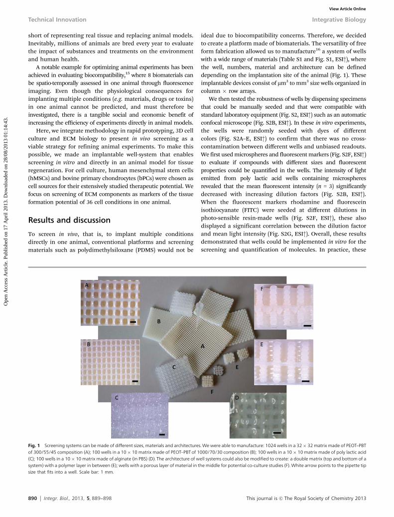
ideal due to biocompatibility concerns. Therefore, we decidedto create a platform made of biomaterials. The versatility of freeform fabrication allowed us to manufacture16 a system of wellswith a wide range of materials (Table S1 and Fig. S1, ESI†), wherethe well, numbers, material and architecture can be defineddepending on the implantation site of the animal (Fig. 1). Theseimplantable devices consist of mm3 to mm3 size wells organized incolumn � row arrays.
We then tested the robustness of wells by dispensing specimensthat could be manually seeded and that were compatible withstandard laboratory equipment (Fig. S2, ESI†) such as an automaticconfocal microscope (Fig. S2B, ESI†). In these in vitro experiments,the wells were randomly seeded with dyes of differentcolors (Fig. S2A–E, ESI†) to confirm that there was no cross-contamination between different wells and unbiased readouts.We first used microspheres and fluorescent markers (Fig. S2F, ESI†)to evaluate if compounds with different sizes and fluorescentproperties could be quantified in the wells. The intensity of lightemitted from poly lactic acid wells containing microspheresrevealed that the mean fluorescent intensity (n = 3) significantlydecreased with increasing dilution factors (Fig. S2B, ESI†).When the fluorescent markers rhodamine and fluoresceinisothiocyanate (FITC) were seeded at different dilutions inphoto-sensible resin-made wells (Fig. S2F, ESI†), these alsodisplayed a significant correlation between the dilution factorand mean light intensity (Fig. S2G, ESI†). Overall, these resultsdemonstrated that wells could be implemented in vitro for thescreening and quantification of molecules. In practice, these
Fig. 1 Screening systems can be made of different sizes, materials and architectures. We were able to manufacture: 1024 wells in a 32� 32 matrix made of PEOT–PBTof 300/55/45 composition (A); 100 wells in a 10 � 10 matrix made of PEOT–PBT of 1000/70/30 composition (B); 100 wells in a 10 � 10 matrix made of poly lactic acid(C); 100 wells in a 10 � 10 matrix made of alginate (in PBS) (D). The architecture of well systems could also be modified to create: a double matrix (top and bottom of asystem) with a polymer layer in between (E); wells with a porous layer of material in the middle for potential co-culture studies (F). White arrow points to the pipette tipsize that fits into a well. Scale bar: 1 mm.
Technical Innovation Integrative Biology
Ope
n A
cces
s A
rtic
le. P
ublis
hed
on 1
7 A
pril
2013
. Dow
nloa
ded
on 2
8/08
/201
3 01
:14:
43.
View Article Online

This journal is c The Royal Society of Chemistry 2013 Integr. Biol., 2013, 5, 889--898 891
results showed that up to 9 wells per screening device could beconsistently dispensed manually, and that the sensitivity ofresults was not compromised by the materials or the designs.
As a popular tissue regeneration cell source, hMSCs wereselected for testing the cell culture compatibility of wells (Fig. 2)made of PEOT–PBT, a biocompatible copolymer already used inskin and musculoskeletal regenerative medicine applications.8
The material was selected for its slow in vivo degradation,17 lowwater uptake (B5%) and high tensile strength (up to 23 MPa).18
In addition, attachment, proliferation and differentiation ofchondrocytes16 and hMSCs19 can be tuned by varying the PEOT/PBT ratio of the copolymer chains. A 2D film of the material itselfshowed that when hMSCs were cultured on 300PEOT55PBT45discs, cells organized in colony forming units (CFU’s) (Fig. 2A),which resembled their known monolayer behavior on tissueculture plastic. In contrast, hMSCs cultured on rectangularwells formed 3D aggregates conditioned by the number of cellsseeded (Fig. 2B). Remarkably, wells with the highest number ofhMSCs contained 3D aggregates of random shapes whereasdilutions of hMSCs yielded wells with monolayer-type cultures.We determined that 3D aggregates augmented in size in a time-dependent manner due to the non-monolayer aggregation ofhMSCs. To examine the effect of well-volume on hMSCs, thewell size was varied in width and length and 1.2 � 104 hMSCswere seeded in three different types of wells (Fig. 2C–E), which
effectively increases the cell concentration by decreasing the wellvolume instead of increasing the cell numbers. By adjusting thewell size, an inverse correlation between hMSCs organizationand well size was derived: hMSCs form 3D aggregates in the0.45 ml well (Fig. 2C), while in the 1.8 and 4 ml wells (Fig. 2Dand E) they organized into CFU’s, leading to the conclusion thatabove 5 � 104 cells per ml of well hMSCs readily assemble in 3Daggregates. When low numbers of hMSCs are deposited in thewell, these proliferate, increasing the cell concentration in time(Fig. 2F). From these data, we discovered that the 3D organiza-tion of hMSCs in the wells could be controlled through the wellvolume, cell number and culture time.
Based on molecular and cell culture results, we chose tofabricate PEOT–PBT screening systems with 9 wells and volumesof 1.8 ml per well. This meant that thirty six conditions could behoused in four subcutaneous sites in every immune-deficientmouse (Fig. S3, ESI†). The thirty six implanted conditionsconsisted of numbers of hMSCs, numbers of bovine primarychondrocytes (bPCs) and coculture ratios of hMSCs : bPCs thatwere screened for tissue regeneration markers (Table 1).
First, the systemic effect on the host tissue under thedifferent conditions confined in the screening device wasassessed in all twenty mice with markers for the cell nuclei,the cytoplasm of cells, keratin and collagen (Fig. 3). Interestingly,hair growth (Fig. 3A) was stimulated on the skin of nude mice
Fig. 2 Cell culture in the PEOT–PBT wells. 300 000 cells seeded on 2D PEOT–PBT colony forming units of methylene blue stained hMSCs. (A, top view), scale bar:0.2 mm. When hMSC dilutions were tested in the PEOT–PBT wells, 25 000 hMSCs per ml produced 3D aggregates (B, cross section), scale bar: 0.5 mm. Varying the size ofthe wells and seeding 12 000 hMSCs in all well volumes showed that 0.45 ml wells induce 3D aggregate formation whereas 1.8 and 4 ml wells do not (C–E, cross section,scale bar: 0.2 mm). hMSC concentrations increase in time due to proliferation (F). One-way ANOVA followed by Tukey’s post hoc test was used to evaluate statisticalsignificance, where bars and error bars represent the mean cell number (n = 9 wells) and their standard deviation, respectively (p o 0.01).
Integrative Biology Technical Innovation
Ope
n A
cces
s A
rtic
le. P
ublis
hed
on 1
7 A
pril
2013
. Dow
nloa
ded
on 2
8/08
/201
3 01
:14:
43.
View Article Online

892 Integr. Biol., 2013, 5, 889--898 This journal is c The Royal Society of Chemistry 2013
in wells containing hMSCs, indicating a systemic response totrophic factors.20 Upon histopathological examination of tissuemorphology in slides from all mice, there was no indication ofcross-contamination of conditions or inflammatory responses(Fig. 3A). Instead, the first evident effect was the penetration/formation of tissue in the wells (Fig. 3B).
To evaluate the presence of tissue in the wells, we quantifiedthe physical accumulation (tissue area/total well area) of tissuein each well for all mice (Fig. 4). Since the objective was tocompare tissue penetration in all wells vs. the total well area,the analysed tissue area included implanted cells, host tissueand new ECM. Then we proceeded to identify patterns in timeaccording to each condition. For example, the percent area of
tissue in a well increased gradually with increasing numbers ofhMSCs (Fig. 4A). Also bovine PCs exhibited an increase in tissuepercent areas with increasing cell numbers on week 4 (Fig. 4B),but not on week 2. Despite seeding a maximum of 25 000 cellsin all wells, co-cultures revealed growing tissue percent areasfor wells containing increasing numbers of hMSCs (Fig. 4C).We analyzed the means of the percent area of tissue todetermine whether there were significant differences betweenconditions. These led to variability of results between membersof the same species when comparing all mice with each other.In wells with hMSCs, half of the mice displayed a significantlyhigher percent area of tissue between 6000 cells vs. control (noseeded cells, week 2), and 25 000, 12 000 cells vs. control (week4) (Table S1, ESI†). Similarly, in wells with bPCs, half of themice presented a significantly higher percent area of tissuebetween 25 000 cells vs. 12 000 cells (weeks 2 and 4), 12 000 cellsvs. control (week 2) (Table S2, ESI†). Finally, in wells withco-cultures, 56% of the mice revealed significantly higher percentareas of tissue in the 20 000 hMSCs to 5000 bPCs (80 : 20) ratio vs.control (week 4) (Table S3, ESI†). Notably, there was no indicationthat the implantation site influenced the correlation between cellnumbers and tissue percent areas according to the randomdistribution of conditions (Fig. S2, ESI†).
Table 1 Cell numbers for human mesenchymal stem cells (hMSCs), bovineprimary chondrocytes (bPCs) and co-cultures screened with three replicates percell condition and 9 control (empty) wells for a total of 36 conditions in eachmouse
Dilutionfactor
hMSCs[cell #]
bPCs[cell #]
Co-cultureratios
hMSCs : bPCs[cell #]
1� 25 000 25 000 80 : 20 20 000 : 50002� 12 500 12 500 50 : 50 12 500 : 12 5004� 6000 6000 20 : 80 5000 : 20 000
Fig. 3 In vivo screening is performed on histological sections showing three wells of a well system with the subcutaneous host tissue located above the wells. Therepresentative staining of 80 screening devices is shown in this cross section of a 9-well device in 3 rows � 3 columns configuration. The red arrow points to hairgrowth on the skin of nude mice in wells containing hMSCs. The black arrow points to the area of the screening device stained with India ink to pinpoint the location ofconditions (rows and columns) after histological processing. Sections of screening devices were stained with haematoxylin and counterstained with eosin showing thecytoplasm (red) and nuclei (dark blue), where the well is delineated with black lines and the material of the screening device appears to be white and enveloped by thehost tissue (A). The dotted oval in (A) displays the area of interest for screening purposes, where implanted conditions interact with the host tissue. (B) The three wellsare shown after Masson’s tri-chrome staining: keratin (red), collagen (blue) and nuclei (dark brown) to evaluate the extracellular matrix in the wells. Notably, potentialdifferences in the tissue penetration/formation per well were observed in each well. Scale bar: 1 mm.
Technical Innovation Integrative Biology
Ope
n A
cces
s A
rtic
le. P
ublis
hed
on 1
7 A
pril
2013
. Dow
nloa
ded
on 2
8/08
/201
3 01
:14:
43.
View Article Online

This journal is c The Royal Society of Chemistry 2013 Integr. Biol., 2013, 5, 889--898 893
While in vivo cell proliferation was not investigated directly, thestatistics (Tables S1 and S2, ESI†) did not support a significantcorrelation between increasing cell numbers and increasing per-cent areas of tissue (Fig. 4A and B). Since not higher than 50% ofmice displayed a higher percent area of tissue under most condi-tions, it was concluded that increasing cell numbers were notsufficient to significantly increase the percent area of tissue in awell. Other effects such as the reverse trends in percent area oftissue for chondrocytes (week 2, Fig. 4B) were not significant. Themost significant effect (in 56% of the mice) detected by quantifyingthe amount of tissue in a well was found for co-cultures, suggestingthat co-cultures have the potential to induce higher percent areas oftissue when compared to the readings on empty wells (control),through ECM formation rather than cell numbers. Thus, it wasconcluded that the main contributor to the percent area of tissue ina well is the well itself probably due to tissue bulging21 orpenetration, which occurs spontaneously when host tissue is incontact with the well. In order of importance, tissue penetrationwas followed by tissue formation as the factor that increases theamount of tissue in a well as observed in 56% of the mice in wellswith cocultures of four hMSCs to one bPC (80 : 20).
Since hMSCs : bPCs cocultures have a known22,23 cartilageregeneration potential, slides under all conditions were stainedfor proteoglycans (Fig. 5). It was found that when compared toother conditions (Fig. 5B–D), the co-culture containing thehighest amount of hMSCs (80 : 20) was more strongly stainedfor proteoglycans (Fig. 5E). Upon histological grading24 (Fig. 5F)of the quality of neo-cartilage, it was confirmed that all hMSCs :bPCs combinations produced a cartilage matrix. The 80 : 20co-culture was the most promising condition tested in thisscreening showing in vivo cartilage regeneration as shown bythe significantly higher cartilage (Bern) score on week 4.
To evaluate the results obtained for co-culture ratios using aconventional method (Fig. S4, ESI†), conditions were seeded inporous 3D scaffolds16 and implanted subcutaneously in nudemice. These data also showed that proteoglycans increased inPEOT–PBT scaffolds containing cocultures with higher quantitiesof hMSCs (Fig. S4A, ESI†). The amount of GAG/DNA did notshow a significant difference between groups (Fig. S4B, ESI†).However, the efficiency of proteoglycans production augmentedwith increasing ratios of hMSCs (Fig. S4B, ESI†). Sulphated-proteoglycans, collagen types I, II and IX confirmed that hyaline(healthy) cartilage-like tissue was more abundant in scaffoldswith 80% and 90% hMSCs (Fig. S4C, ESI†).
Increasing the number of conditions that could be screenedsimultaneously in one animal improves the efficiency andspeed at which therapies and treatments can be identifiedand translated into clinical practice. In fact, evaluating thesame 36 in vivo conditions using conventional methods wouldhave required 9 times the number of mice (Table 2).
Cross talk between wells and their physiological effect iscondition-dependent. While wells in vitro can be independentof each other by adjusting the well volumes and the relativehumidity, wells in vivo can interact with each other throughsoluble factors. The degree of interaction between wells will bedependent on molecule diffusion dynamics and time. In thisstudy, ECM markers at 2 and 4 weeks did not indicate any crosstalk between wells. However, this will be relative to the markerof interest. Even though cell therapy in conventional scaffoldsystems (50 mm3) occurs in volumes over 25 times larger thanwell volumes (1.8 mm3), both systems are consistent with theregions, where ECM production occurs, which is in the pores inscaffolds or inside the wells. This indicates that by reducing thevolume of interest in the animal with wells, the extent of desiredand undesired effects such as cross contamination is identifiableand controllable. As shown here, control can be achievedthrough randomization of implanted conditions, which ensuresthe discrimination of relevant biological responses, such asECM production from irrelevant effects – for the purpose of thisstudy – such as host tissue penetration in the wells.
Albeit 36 wells were investigated in nude mice as a proof ofprinciple, the screening device has the potential to become ahigh throughput screening (HTS)25,26 system where hundredsof wells can be contained in one animal with one well (1.8 ml)for every 1.4 mm of an implantation site. Depending on the sizeof the animal, these platforms can be tailored to fit the requireddimensions of an implantation site, which defines the number
Fig. 4 Histological sections (>300 slides) contained tissue penetration/for-mation in wells which was evaluated through the percent area of tissue, definedas tissue area/well area � 100%. The means (n = 3 slides of a well) of the percenttissue area for each condition are graphically shown for two representative mice;one from week 2 and one from week 4. The percent tissue area increased as thenumber of cells in a well increased as depicted for hMSCs dilutions and control(empty well system) (A); bPCs dilutions and control (B). With a total of 25 000 cellsper well in cocultures (C), the percent tissue area increased with higher ratios ofhMSCs to bPCs.
Integrative Biology Technical Innovation
Ope
n A
cces
s A
rtic
le. P
ublis
hed
on 1
7 A
pril
2013
. Dow
nloa
ded
on 2
8/08
/201
3 01
:14:
43.
View Article Online

894 Integr. Biol., 2013, 5, 889--898 This journal is c The Royal Society of Chemistry 2013
of conditions that can be implanted (Movie S1, ESI†). Thus, thelarger the animal, the higher the number of conditions thatcan be investigated simultaneously. However, the seeding ofmacroscopic and large numbers (>100) of wells requires special
equipment to control the microenvironment while dispensingconditions in all wells with reproducibility. Using the wellsystem, the number of lives required to perform experimentsin vertebrate animals is inversely proportional to the size of the
Fig. 5 Proteoglycan production was screened as a marker for cartilage formation. Glycosaminoglycans (GAG) (blue) and cytoplasm (red) seen in three wells ofa screening device (A, scale bar: 1 mm). A representative slide of the control and three conditions expressing GAG are displayed: empty wells (B); 25 000 bPCs (C);co-culture of hMSCs : bPCs at a 20 : 80 ratio (D); co-culture of hMSCs : bPCs at a 80 : 20 ratio (E). Scale bars: 100 mm. Quantification of cartilage quality (F) fromthe screened conditions (mean Bern score, N = 10 mice) showed a significantly higher quality of cartilage for hMSCs : bPCs at a 80 : 20 ratio compared to control.Bern scores were analyzed with Kruskal–Wallis to evaluate significance followed by Bonferroni post hoc correction.
Technical Innovation Integrative Biology
Ope
n A
cces
s A
rtic
le. P
ublis
hed
on 1
7 A
pril
2013
. Dow
nloa
ded
on 2
8/08
/201
3 01
:14:
43.
View Article Online

This journal is c The Royal Society of Chemistry 2013 Integr. Biol., 2013, 5, 889--898 895
animal (Table 2), extending the possibility to assess multiplein vivo conditions. As shown here, standard histological meth-ods offer an extensive wealth of data, yet methods that avoidhistological processing15,27,28 would be convenient to furtheroptimize animal experiments, automatize the quantificationand imaging, and improve the sensitivity and throughput ofin vivo screening.
The efficiency of the developed animal-implantable wellsystem not only lies in optimizing animal experiments, butalso in bridging both in vitro and in vivo milieus. For example,the well system could be adapted to monitor biomechanics29
and to elucidate gene regulatory networks30,31 in live tissues.Multiple compounds such as nanoparticles, drugs or chemicalscould be covalently or non-covalently attached to wells toincrease the throughput of toxicity assessments and therebydrastically dwindle the use of vertebrate animal lives and costsin pharmaceutical, toxicological, chemical, and disease screen-ing, while allowing for the first time to execute such screeningin true three-dimensional tissues.
ExperimentalMesenchymal stem cell and chondrocyte isolation and culture
hMSCs and bPCs were isolated, cultured and cryopreserved asdescribed before.32 hMSCs were obtained from the acetabulum offive donors who were undergoing total hip replacement surgeryand gave informed consent for bone marrow biopsy, approved bythe medical ethical testing commission (METC) of the MedicalSpectrum Twente in Enschede, The Netherlands. Gender anddonor age were: Donor 1: female, 81 years. Donor 2: male, 65 years.Donor 3: female, 66 years. Donor 4: female, 66 years. Donors 1 to 3were cultured in the wells in vitro and only Donor 3 was implantedin mice. Donor 4 was cultured in the porous scaffolds.
Well system fabrication
A Bioplotter device (Envisiontec GmbH, Germany), which is aXYZ plotter, was used to make the wells (European PatentApplication: EP2404672A1) with a fabrication process adaptedfrom that for scaffold fabrication.16 To obtain well systems in amatrix form (columns � rows) (Fig. S1, ESI†), closed wells weremade by depositing fibers on top of each other at a 901 angle toobtain rectangular wells, where the first layer deposited wasalways closed. The well dimensions were defined by increasingor decreasing fiber distances in the XY axis and by the height ofthe cubical model. This was reproducible with four materialswith their respective parameters as shown in Table S1 (ESI†)and Fig. 1.
Different architecture designs were fabricated to maximizethe number of wells and to create co-culture systems as shownin Fig. 1E and F. For the design in Fig. 1E, first and second layerstrand distances were maintained as shown in Table S1 (ESI†)and a strand distance (0.5 mm) in the middle of the well height(at 2 mm) was introduced. The design in Fig. 1F did not have aclosed first layer where a constant strand distance (Table S1, ESI†)was maintained until deposition of a closed layer in the middle ofthe well height (at 2 mm). Subsequently, layer deposition with aconstant strand distance was resumed.
In vitro screening
Fluorescent microspheres (YG: l = 441–486 nm) (Polyscience,Germany), rhodamine (Sigma-Aldrich) and FITC (Sigma-Aldrich)were diluted in PBS before seeding 1 ml of solution per well ofphotopolymerizable resin (Envisiontec, Germany) and poly lacticacid (Purac, The Netherlands) screening devices. Fluorescencewas imaged in an automatic confocal light microscope (BDpathway, The Netherlands).
Well sterilization and conditioning for cell culture
Well systems were sterilized with 70% ethanol solution for15 min, washed three times and incubated at room tempera-ture for two hours with sterile PBS, and thereafter incubated inaMEM proliferation media overnight at 37 1C.
Well seeding
From a cell suspension of 5 � 106 cells per ml, hMSCs werecentrifuged at 200 Relative Centrifugal Force (RCF) andre-suspended in 0.2 ml of medium. From this cell suspension,dilutions were prepared with a maximum of 2.5 � 104 cells in1 ml volume seeded per well. Every condition (i.e. cell dilutions)was seeded in all the wells of a column. In a 3 � 3 well system,three replicates or three wells were seeded under the samecondition. Seeded wells were incubated for 20 min at 37 1C in ahumid atmosphere with 5% CO2. Non-detached cells in a wellwere removed by washing with culture medium, then thesewere placed in a well of a 25-well plate (Nunc), and covered withthe culture medium of hMSCs or bPCs and incubated at 37 1C.
In vitro cell culture
Cell numbers in wells were obtained using Cyquant DNA assay(Invitrogen). Cell numbers were calibrated for hMSCs bymeasuring the mg of DNA per cell of cell numbers from 1 � 103
to 3 � 105 cells against known DNA amounts. It was estimatedthat each hMSC contained 1.6 picograms of DNA, which made itpossible to quantify cell numbers in wells.
Table 2 Projection of animals saved with implantable wells vs. a conventional method in a similar experimental set-up
SpeciesExperimentalconditions
# Animals with conventionalmethods (n = 10 animals)
3D HTSwell matrix
Size[mm]
Conditionsper 3D HTS
Wells peranimal
# Animals with3D HTS
Animalssaved
Mouse 36 90 3 � 3 4.2 9 36 10 80Rats 576 1440 12 � 12 16.8 144 576 10 1430Rabbits 1600 4000 20 � 20 28 400 1600 10 3990Goats 4096 10 240 32 � 32 44.8 1024 4096 10 10 230
Integrative Biology Technical Innovation
Ope
n A
cces
s A
rtic
le. P
ublis
hed
on 1
7 A
pril
2013
. Dow
nloa
ded
on 2
8/08
/201
3 01
:14:
43.
View Article Online

896 Integr. Biol., 2013, 5, 889--898 This journal is c The Royal Society of Chemistry 2013
Co-culture preparation in wells
Human MSCs : bPCs were seeded in wells in aMEM prolifera-tion media. Each of the three ratios (0.8 : 0.2, 0.5 : 0.5, and0.2 : 0.8) was seeded in three wells of a 3 � 3 well system, whichcontained a maximum of 2.5 � 104 cells per well.
Implantations in immune-deficient mice
Nude mice studies were performed after consent fromthe ethical committee for animal studies (DEC-GDL Utrecht,The Netherlands). Six-week-old nude mice (Hdcpb : NMRI-nuHarlan, The Netherlands) were anaesthetized with 0.02 ml of a3.5 : 3 : 1 mixture of ketamine (100 mg ml�1) : xylazine (20 mg ml�1) :atropine (0.5 mg ml�1).
Four 3 � 3 well systems containing 36 conditions weresubcutaneously implanted per mouse in the posterior-lateralside of the back and sutured. The 36 conditions implanted weretriplicates of: three dilutions of hMSCs (25 000; 12 000; and6000 cells); three dilutions of bPCs (25 000; 12 000; and 6000cells); three hMSCs : bPCs ratios (0.8 : 0.2, 0.5 : 0.5, and 0.2 : 0.8)and empty wells with no cells. Conditions were randomized bydispensing them in different wells of screening devices andimplanting devices in different pockets from one mouse to another(Fig. S3A, ESI†). After 2 weeks (n = 10) and 4 weeks (n = 10) micewere euthanized via CO2 asphyxiation and screening systems wereexcised and processed for analysis.
Histology of screening devices
3 � 3 well systems were explanted, washed in PBS and fixed in10% formalin at 4 1C overnight. After washing in PBS, wellswere embedded in paraffin and cut into 5 mm sections using amicrotome. Slides containing sections of 80 well systems werestained with haematoxylin and counterstained with eosin(H&E); alcian blue and counterstained with nuclear red; andMasson’s tri-chrome. These samples were examined by GAH,LW, LMT and JL under direct supervision of CvB, LM and MKwith a proven track record in histological evaluation ofimplants of more than 20 years.
Tissue quantification in wells
Slides showing 3 � 3 well system’s rows were stained withH&E, Masson’s trichrome and alcian blue and imaged usinglight microscopy. Image analysis was performed for the areacontaining each condition on B350 representative slides,obtained from 80 screening devices and 3 representativesections per well. Images were processed by cutting the imagesof wells (Fig. S3B, ESI†) and converting them to a binary pixelimage (Fig. S3C, ESI†) using ImageJ software (open source).Black and white pixels within an image of a well were quanti-fied with the histogram function in ImageJ, where the mean(n = 3 slides) of the percent area of tissue was obtained for eachexplanted well system. Using a binary system, where blackrepresented tissue and white represented lack of tissue in a well,it was possible to avoid thresholding biases and to quantify thepixels in the wells. In this manner, nuclei, cytoplasm and ECM
(keratin, collagen and GAG) appeared as black pixels, and every-thing else as white pixels.
Bern score of cartilage in wells
The visual histological grading or the Bern score for theevaluation of neocartilage was performed by three independentobservers as previously shown24 for representative slides ofconditions showing cartilage formation with alcian blue andcounterstained with nuclear fast red. 170 images were printed,randomly organized and separately assessed by each independentobserver to obtain a score for each condition in 20 mice.
Two-dimensional PEOT–PBT discs
Poly(ethylene oxide terephthalate)–poly(butylene terephthalate)(PEOT–PBT) was used to make 2D substrates, following anaPEOTbPBTc nomenclature, a is the molecular weight ing mol�1 of the starting PEG block for copolymerization, whereasb and c are the respective weight fractions of the PEOT and PBTblocks. Specifically, 300PEOT55PBT45 was used to manufacture2D substrates by a hot-embossed compression molding tech-nique, where two silicon wafers served as a support and definedthe molded surface. A stainless steel mold with circular featuresthrough holes of 2.2 cm in diameter was placed in between themolds. Granules of 300PEOT55PBT45 were placed insidethe mold to fill up the mold cavities upon polymer melting.Silicon supports were cleaned by immersion in piranha solution(3 : 1 concentrated H2SO4–33% aqueous H2O2) for 15 min. Thesewere rinsed with water and dried in N2. Then, they were function-alized with 1H,1H,2H,2H-perfluorodecyltrichlorosilane (ABCR).After that, these were deposited in the gas phase that served asan anti-adhesion layer to ease the polymer–support separation.For the hot embossing process, a hydraulic press equipped with awater cooling system and temperature control (Fortune, Holland,The Netherlands) was used. The 300PEOT55PBT45 was placed ontop of the silicon support and in the aperture of the mold. Then,the system was heated up to a temperature of 180 1C and 10 barswere applied. After 5 minutes the system cooled down to 60 1Cand the pressure was released. The mold and the supports weremanually separated and the 2D disc released from the mold.
3D scaffold fabrication
The bioplotter device was also used to manufacture porousscaffolds. According to a previous study,16 co-polymers weremolten and deposited as successively layered fibers in a 0–90pattern from a 250 mm nozzle onto a computer aided x/y/z tablewith a layer thickness of 0.15 mm and a fiber spacing of0.6 mm. Cylindrical scaffolds (+4 � 4 mm) with constant poresize and 100% interconnecting pore volume were cored andsterilized in isopropyl alcohol.
Seeding and co-culture of bovine PCs and human MSCs inscaffolds
Cylindrical scaffolds were extensively rinsed in phosphatebuffered saline (PBS), incubated overnight in proliferationmedium and blotted dry prior to seeding. Bovine primarychondrocytes were mixed with hMSCs, respectively, at a
Technical Innovation Integrative Biology
Ope
n A
cces
s A
rtic
le. P
ublis
hed
on 1
7 A
pril
2013
. Dow
nloa
ded
on 2
8/08
/201
3 01
:14:
43.
View Article Online

This journal is c The Royal Society of Chemistry 2013 Integr. Biol., 2013, 5, 889--898 897
total of 3 million cells per scaffold, centrifuged at 300g for5 minutes and re-suspended in 54 ml of medium containing300 mg ml�1 fibronectin. Scaffolds were seeded statically with3 � 106 cells per scaffold for 1 day before subcutaneousimplantation in nude mice.
Implantation of bovine PCs and human MSCs in scaffolds
With 4 implants maximum per mouse, nine constructs ofbovine PCs : hMSCs (n = 9) per experimental group (7 groups =4 co-cultures plus 3 controls) were randomly implanted in 16nude mice.
Histology and immunohistochemistry with scaffolds
Three constructs per donor were explanted, washed in PBS,fixated in formalin at 4 1C overnight. After washing inPBS, constructs were cut into 2 halves, one half was embeddedin GMA and one half was embedded in Tissue-Tek forimmunohistochemistry. GMA embedded 3D scaffolds werecut into 5 mm sections which were treated with safranin-O(red) to stain sulphated-proteoglycans, hematoxylin (dark blue)to mark the nuclei and with fast green (light blue) to label thecytoplasm. Scaffolds (n = 3) were embedded and immediatelyfrozen at �80 1C in OCT compound (Tissue-Tek) for immuno-staining. Sections were cut using a cryo-microtome to a thick-ness of 5 mm and fixated with acetone for 10 minutes, stainedfor collagen type-I (1 : 1000, Ab-1, Calbiochem), type-II (1 : 100,DSHB II-II6B3), and type-IX (1 : 100, DSHB D1-9). Blocking wasachieved with 10% human serum and secondary antibody wasgoat anti-mouse (1 : 100, DAKO). Staining was visualized throughincubation of slides in 3,3-diaminobenzidine (DAB)–solution(DAKO) for 10–20 minutes. These samples were examined byJH, MvdD and RS under direct supervision of CvB, LM and MK.
Statistical analysis
Normality of the data was analyzed by determining skewnessbeing between �3 and 3 and showed normal distribution of thedata in all groups. In vitro fluorescent data were analyzed foreach condition as the mean (n = 12 wells) emission of wells withone-way Anova followed by Tukey’s honestly significant differ-ence criterion with statistical significance set at p o 0.01.Quantitative GAG and DNA data from scaffolds were analyzedfor differences of the means (n = 5 scaffolds) with two-wayAnova followed by Tukey’s honestly significant difference cri-terion, where statistical significance was set at p o 0.01.
In vivo screening of conditions was organized in replicateswith a multi-dimensional sample number, which was defined byN = 10 animals and n = 3 replicates per well for each condition ineach animal. So that n = 3 replicates per condition per mouseand the total sample number per condition is N � n = 30.
The mean (n0 = 3 histological slides) of the percent area oftissue was analyzed for each screening device in each statisti-cally independent mouse (n = 3 wells per condition in eachanimal) with one-way Anova followed by Tukey’s honestlysignificant difference criterion where statistical significance wasset at p o 0.05. So that for each mouse, there were n0 � n = 9images of the same condition available for analysis.
Since Bern score data are non-parametric, it was notnormally distributed data. To account for that, Bern scores ofdifferences of means (N = 10 mice) for glycosaminoglycansin the well system were analyzed with Kruskal–Wallis withBonferroni post hoc correction. Three independent observersassessed the Bern scores for each slide, where inter-observerreproducibility was evaluated with the hypothesis of a combinedeffect on Bern scores by both mean observers’ score for a slideand a mean score for a condition causing a statistical significantdifference set at p o 0.05.
Acknowledgements
The authors would like to acknowledge the financial supportfrom the Dutch SenterNovem research grant number 15044112,Pre-seed grant number 93611002 of the Netherlands GenomicsInitiative, and the TERM Smart Mix Program of the NetherlandsMinistry of Economic Affairs and the Netherlands Ministry ofEducation, Culture and Science. The authors would like tothank Susan Nijhuis and Tessa Hering of the Laboratory ofPathology of the East Netherlands and Ewart de Bruijn for theirhistological expertise, and Dr Maryana Escalante for her inputin the design of polymer discs.
References
1 K. Cheng, Y. Lai and W. S. Kisaalita, Three-dimensionalpolymer scaffolds for high throughput cell-based assaysystems, Biomaterials, 2008, 29, 2802–2812.
2 W. R. Inch, J. A. McCredie and R. M. Sutherland, Growth ofnodular carcinomas in rodents compared with multi-cellspheroids in tissue culture, Growth, 1970, 34, 271–282.
3 Comission, E. (ed Environment Directorate General) 19(2007).
4 T. Hartung and C. Rovida, Chemical regulators have over-reached, Nature, 2009, 460, 1080–1081.
5 F. Khan, R. S. Tare, J. M. Kanczler, R. O. C. Oreffo andM. Bradley, Strategies for cell manipulation and skeletaltissue engineering using high-throughput polymer blendformulation and microarray techniques, Biomaterials, 2010,31, 2216–2228.
6 A. Peters, D. M. Brey and J. A. Burdick, High-throughput andcombinatorial technologies for tissue engineering applica-tions, Tissue Eng., Part B, 2009, 15, 225–239.
7 P. B. Gupta, et al. Identification of selective inhibitors ofcancer stem cells by high-throughput screening, Cell, 2009,138, 645–659.
8 L. Moroni, J. R. de Wijn and C. A. van Blitterswijk, Integratingnovel technologies to fabricate smart scaffolds, J. Biomater.Sci., Polym. Ed., 2008, 19, 543–572.
9 D. R. Albrecht, V. L. Tsang, R. L. Sah and S. N. Bhatia, Photo-and electropatterning of hydrogel-encapsulated living cellarrays, Lab Chip, 2005, 5, 111–118.
10 Y. Ibold, et al. Development of a high-throughput screeningassay based on the 3-dimensional pannus model for rheumatoidarthritis, J. Biomol. Screening, 2007, 12, 956–965.
Integrative Biology Technical Innovation
Ope
n A
cces
s A
rtic
le. P
ublis
hed
on 1
7 A
pril
2013
. Dow
nloa
ded
on 2
8/08
/201
3 01
:14:
43.
View Article Online

898 Integr. Biol., 2013, 5, 889--898 This journal is c The Royal Society of Chemistry 2013
11 M. Schindler, et al. Living in three dimensions: 3D nanos-tructured environments for cell culture and regenerativemedicine, Cell Biochem. Biophys., 2006, 45, 215–227.
12 C. M. Yen, C. C. Chan and S. J. Lin, High-throughputreconstitution of epithelial-mesenchymal interaction in fol-liculoid microtissues by biomaterial-facilitated self-assem-bly of dissociated heterotypic adult cells, Biomaterials, 2010,31, 4341–4352.
13 S. I. Montanez-Sauri, K. E. Sung, E. Berthier and D. J. Beebe,Enabling screening in 3D microenvironments: probing matrixand stromal effects on the morphology and proliferation ofT47D breast carcinoma cells, Integr. Biol, 2013, 5, 631–640.
14 G. A. Higuera, A. van Boxtel, C. A. van Blitterswijk andL. Moroni, The physics of tissue formation with mesenchymalstem cells, Trends Biotechnol., 2012, 30, 583–590.
15 K. M. Bratlie, et al. Rapid biocompatibility analysis ofmaterials via in vivo fluorescence imaging of mouse models,PLoS One, 2010, 5, e10032.
16 L. Moroni, J. A. Hendriks, R. Schotel, J. R. de Wijn andC. A. van Blitterswijk, Design of biphasic polymeric3-dimensional fiber deposited scaffolds for cartilage tissueengineering applications, Tissue Eng., 2007, 13, 361–371.
17 A. A. Deschamps, et al. In vivo and in vitro degradation ofpoly(ether ester) block copolymers based on poly(ethyleneglycol) and poly(butylene terephthalate), Biomaterials, 2004,25, 247–258.
18 A. A. Deschamps, et al. Design of segmented poly(etherester) materials and structures for the tissue engineeringof bone, J. Controlled Release, 2002, 78, 175–186.
19 A. Nandakumar, et al. Combining technologies to createbioactive hybrid scaffolds for bone tissue engineering,Biomatter, 2013, 3, e23705.
20 A. I. Caplan and J. E. Dennis, Mesenchymal stem cells astrophic mediators, J. Cell. Biochem., 2006, 98, 1076–1084.
21 G. Lemon, et al. Growth of the chorioallantoic membraneinto a rapid-prototyped model pore system: experimentsand mathematical model, Biomech. Model. Mechanobiol.,2011, 10, 539–558.
22 J. Fischer, A. Dickhut, M. Rickert and W. Richter, Humanarticular chondrocytes secrete parathyroid hormone-relatedprotein and inhibit hypertrophy of mesenchymal stem cellsin coculture during chondrogenesis, Arthritis Rheum., 2010,62, 2696–2706.
23 X. Liu, et al. In vivo ectopic chondrogenesis of BMSCsdirected by mature chondrocytes, Biomaterials, 2010, 31,9406–9414.
24 S. P. Grogan, et al. Visual histological grading system for theevaluation of in vitro-generated neocartilage, Tissue Eng.,2006, 12, 2141–2149.
25 L. A. Kunz-Schughart, J. P. Freyer, F. Hofstaedter andR. Ebner, The use of 3-D cultures for high-throughputscreening: the multicellular spheroid model, J. Biomol.Screening, 2004, 9, 273–285.
26 E. Stokstad, Putting chemicals on a path to better riskassessment, Science, 2009, 325, 694–695.
27 R. Bhargava, D. C. Fernandez, S. M. Hewitt and I. W. Levin,High throughput assessment of cells and tissues: Bayesianclassification of spectral metrics from infrared vibrationalspectroscopic imaging data, Biochim. Biophys. Acta, 2006,1758, 830–845.
28 S. Judex, et al. Quantification of adiposity in small rodentsusing micro-CT, Methods, 2010, 50, 14–19.
29 R. La and M. F. Arnsdorf, Multidimensional atomic forcemicroscopy for drug discovery: a versatile tool for definingtargets, designing therapeutics and monitoring theirefficacy, Life Sci., 2010, 86, 545–562.
30 D. D. Licatalosi, et al. HITS-CLIP yields genome-wideinsights into brain alternative RNA processing, Nature,2008, 456, U464–U422.
31 T. Whitington, A. C. Perkins and T. L. Bailey, High-throughputchromatin information enables accurate tissue-specificprediction of transcription factor binding sites, Nucleic AcidsRes., 2009, 37, 14–25.
32 L. Wu, et al. Trophic effects of mesenchymal stem cellsincrease chondrocyte proliferation and matrix formation,Tissue Eng., Part A, 2011, 17, 1425–1436.
Technical Innovation Integrative Biology
Ope
n A
cces
s A
rtic
le. P
ublis
hed
on 1
7 A
pril
2013
. Dow
nloa
ded
on 2
8/08
/201
3 01
:14:
43.
View Article Online
Copyright © 2022 FDOKUMEN