Design and analysis of an innovative multifunctional cylinder
Upload
independentCategory
view
2download
0
BIGINI ET AL. VOL. XXX ’ NO. XX ’ 000–000 ’ XXXX
www.acsnano.org
A
CXXXX American Chemical Society
In Vivo Fate of Avidin-Nucleic AcidNanoassemblies as MultifunctionalDiagnostic ToolsPaoloBigini,†,# Sara Previdi,†,# Elisabetta Casarin,‡Davide Silvestri,‡MartinaBrunaViolatto,† Sonia Facchin,§
Leopoldo Sitia,† Antonio Rosato,^,O Gaia Zuccolotto,O Nicola Realdon,‡ Fabio Fiordaliso, ) Mario Salmona,†
and Margherita Morpurgo‡,*
†Department of Biochemistry and Molecular Pharmacology, IRCCS-Istituto di Ricerche Farmacologiche “Mario Negri”, Via La Masa 19, 20156 Milan, Italy,‡Pharmaceutical and Pharmaceutical and Pharmacological Sciences, Universtà di Padova, via Marzolo, 5, 35131 Padova, Italy, §ANANAS nanotech, s.r.l.,via Altinate 120, 35131 Padova, Italy, ^Istituto Oncologico Veneto IOVIRCCS, Via Gattamelata 64, 35128 Padova, Italy, ODepartment of Surgery Oncology andGastroenterology, University of Padova, Via Gattamelata 64, 35128 Padova, Italy, and )Department of Cardiovascular Research, IRCCS-Istituto di RicercheFarmacologiche “Mario Negri”, Via La Masa 19, 20156 Milan, Italy. #These authors contributed equally.
In recent years, the active search for noveltheranostic agents has led to the devel-opment of many kinds of nanoparticle-
based technologies. Since the food anddrug administration (FDA) approval of super paramagnetic iron-oxide NP in 1996,one of themain goals in clinical imaging hasbeen to develop innovative tracers capableof providing longitudinal tracking in pa-tients and improving diagnostics and prog-nostics by noninvasive procedures. Thisobjective has also called up the attentionof drug delivery systemdevelopers, with theambitious goal to generate multimodal de-vices capable of combining improved phar-macokinetics and targeted localization withan efficient modality of anatomical trackingby means of imaging instruments. As aconsequence, multifunctionality is becom-ing a key feature for the development of
novel theranostic nanosystems.1�9 Thechallenge in this area is related to thepossibility to not only load onto the nano-particle (NP) different bioactive/tracer func-tions, but also be able to tune and controltheir individual payload. In this way, it ispossible to establish a quantitative correla-tion between composition andbioactivity inorder to tune the final device propertiesaccording to specific needs.10 Therefore,the availability of reproducible and conve-nient standardized preparation methodsthat permit fine-tuning of each individualcomponent becomes an important add-onto favor clinical translation and meet indus-trial interest.In this context, the Avidin-Nucleic-Acids-
Nano-ASsembly (ANANAS) represents aninnovative NP platform11 characterized byeasy tuning of formulation composition and
* Address correspondence [email protected].
Received for review May 27, 2013and accepted December 5, 2013.
Published online10.1021/nn402669w
ABSTRACT This study describes the formulation optimization and
body-cell distribution and clearance in mice of a dually fluorescent biode-
gradable poly avidin nanoassembly based on the novel Avidin-Nucleic-
Acid-Nano-ASsembly (ANANAS) platform as a potential advancement of
classic avidin/biotin-based targeted delivery. The nanoformulation circulates
freely in the bloodstream; it is slowly captured by filter organs; it is effi-
ciently cleared within 24�48 h, and it is poorly immunogenic. The system
displays more favorable properties than its parent monomeric avidin and it
is a promising tool for diagnostic purposes for future translational aims, for
which free circulation in the bloodstream, safety, multifunctionality and high composition definition are all necessary requirements. In addition, the assembly
shows a time-dependent cell penetration capability, suggesting it may also function as a NP-dependent drug delivery tool. The ease of preparation together with
the possibility to fine-tune the surface composition makes it also an ideal candidate to understand if and how nanoparticle composition affects its localization.
KEYWORDS: avidin nucleic acid nanoassemblies . poly avidin . fluorescence optical imaging . in vivo diagnostics . nanomedicine .multifunctional
ARTIC
LE
BIGINI ET AL. VOL. XXX ’ NO. XX ’ 000–000 ’ XXXX
www.acsnano.org
B
surface chemistry properties. The term “ANANAS” re-fers to a series “poly-avidin” NP formulations originat-ing from the high affinity interaction of avidin with thenucleic acids.12 These buffer soluble NPs are formed byaNature-driven double self-assembly in which, initially,a nucleic acid molecule acts as a central elementaround which several avidins (1 each 14 base pairs)nucleate, leading to toroidal assemblies (diameterabout 120 nm);12 in a second self-assembly process, adefined amount of biotinylated poly(ethylene glycol)(PEG) is also added to permit buffer solubility andincrease stability.13 In the end, the ANANAS assemblyis a 'polymerized' form of avidin, one of the mostrenown proteins used in biomedicine,14�17 and canbe considered as a powerful alternative to it in many ofits applications. Importantly, the ANANAS assembliesare composed of soft biocompatible materials onlyand, as opposed to other poly avidins obtained bychemical cross-linking or nonspecific adsorption ontometal or polymer substrates,18�27 they are character-ized by high composition definition. In addition, avidinin the assembly maintains its full biotin (B) bindingcapacity11 so that the system carries a precisely definednumber of biotin binding sites (BBS), which are avail-able for docking additional biotinylated functionalmoieties.The ANANAS platform can be deployed into several
formulation compositions depending on the length ofthe nucleating nucleic acid and the kind and amount ofbiotinylated functions linked to the surface.13 For ex-ample, recently, several functionally active ANANASformulations generated through a “one-pot” proce-dure have been compared in a model of in vitro immu-nodetection, showing differently improved perfor-mance with respect to 'monomeric' avidin (namelyreducing to different extent the analyte limit of detec-tion down to amaximum500-fold).11 On top of itsmostrenown application as an in vitro diagnostics tool, theavidin (and its bacterial analogue steptavidin)/biotinsystem is also extensively investigated as a carrier fortargeted radio-diagnostics and therapy.17,28�32 How-ever, the success of (strept)avidin based targetingapproaches has been limited by both unfavorableproteins pharmacokinetics28 and, especially in the caseof streptavidin (StAv), high immunogenicity.17,33�35 Inthe context of targeted delivery, fast clearance of thenontargeted carrier is needed to avoid high back-ground or long-term exposure of nontarget compart-ments to the (toxic) carried drug. On the other hand, anexcessively fast elimination is also detrimental becauseit impedes the efficient tracking necessary in diagnos-tics and the (slow) diffusion processes necessary toreach target tissues necessary in drug delivery applica-tions. StAv exhibits high and prolonged (>96 h) per-manence in many tissues due to a RGD-like motifwithin its 58�62 amino acid sequence,28 while avidin,whose fate also depends on its surface charge and
varies with its degree of biotinylation,36,37 has lowerorgan retention than StAv, but it is cleared from thebloodstream too rapidly (within 10 min) .28
To improve the poor pharmacokinetics/immuno-genic profile of avidin/streptavidin, several proteinderivatives have been obtained throughout the years,which have been tested within many administrationprotocols, with some success.17,28�32,38 In the case ofavidin, improvement of plasma half-life and minimiza-tion of its interaction with the immune system wereobtained through its covalent conjugation with poly-(ethylene glycol) (PEG).38,39 However, the covalentattachment of PEG leads to partial loss of the proteinbiotin binding ability,40 thus reducing its activity. Pre-sently, the issue of the (strept)avidin unfavorable phar-macokinetics upon i.v. administration is not yet fullysolved so that the most current promising use of theavidin/biotin system in targeted radio-therapy involvesthe intraoperative local administration of an oxidizedprotein derivative.29
Another possibility tomodify the biodistribution andclearance of proteins characterized by poor pharma-cokinetics is to formulate them into NP preparations.The ANANAS assembly here investigated is indeed anovel form of PEG coated poly avidin nanoformulation,and therefore, it could represent an avidin-baseddelivery device alternative to the “monomeric” protein.In this work, we investigated the in vivo fate upon ivadministration of an ANANAS formulation, which wasmade dually fluorescent by using two biotin-labeledfluorophores (fluoresceine and alexa633). NP dual fluo-rescent labeling was performed to allow concurrentlyfine composition control and to enable in vivo biodis-tribution studies through noninvasive in vivo Fluores-cence optical imaging tools. The fluoresceine tag,which was linked at the ω-end of the biotin-PEGderivative introduced for surface protection, was usedto permit precise estimation of the number of polymerchains linked to the particle. It also complemented thein vivo and ex vivo biodistribution information, whichwasmainly obtained through the alexa633 fluorophore.
RESULTS AND DISCUSSION
Formulation Optimization. One of the unique proper-ties of the ANANAS platform is that it allows fine tuningof its surface tethered biotinylated moieties, permit-ting high control of the assembly composition. Thispermits an infinite number of surface compositioncombinations, the selection of which is to be chosenbeforehand or in parallel to any functional evaluation.In the specific context of this work, we carried outpreformulation studies to optimize the NP composi-tion on the basis of two main requirements: (a) max-imal PEG surface protection to favor stealthiness and(b) efficient alexa633 payload to allow in vivo near-infrared (NIR) visualization. It is known23,41,42 that thefate of a NP is influenced by its degree of PEG coating,
ARTIC
LE

BIGINI ET AL. VOL. XXX ’ NO. XX ’ 000–000 ’ XXXX
www.acsnano.org
C
the higher, the better is for the particle ability to freelycirculate in the bloodstream. In fact, the hydrophilicpolymer layer shields the particle from the bodydefenses and favors its stealth properties.
The preformulation studies weremeant to estimatethe maximal load possible for a 5 kDa PEG (PEG5kDa)molecule and to evaluate if the polymer shape affectsthis parameter. It was previously shown, using biotiny-lated proteins as bulky cargoes, that the ANANASloading capability is dictated by steric limitation ratherthan the available BBS.11 On the other hand, it has notbeen shown if a particle saturated with a large MWcargo is still able to accommodate small biotin deriva-tives that, in principle, could be able to diffuse throughsmall openings of the surface covering layer. A furthergoal of the preformulation studies was therefore to testif particles already saturated with a dense PEG layerwere still able to load lower molecular weight biotinderivatives.
To evaluate if the shape of the polymer has anyeffect on the particle loading ability, two ω-FITC la-beled biotin PEG derivatives were synthesized, namelya linear (5 kDa) one, and a branched one in which twoFITC-PEG5kDa chains are linked at the two primaryamines of a biotin�lysine conjugate (Compounds 4and 6, Figure 1, see also Supporting Information,Figure S-1).
A buffer stable 'core' formulation was prepared byusing the branched fluorescent derivative (com-pound 6) at 1:1 molar ratio with respect to avidin,according to what described by Pignatto.13 The num-ber of PEG5kDa chains linked to the resulting particlesurface was calculated to be about 430 (16% of thetheoretically available BBS), based on the amount ofFITC measured from the assemblies UV spectra re-corded after gel permeation purification.
After purification and characterization, this formu-lation was further added of oversaturating amounts ofeither the linear or branched derivative. After this sec-ond addition, about 700 PEG5kDa chains/NP (Figure 2B)were found, independently of the PEG derivative used,confirming that steric hindrance ismore important thatthe number of available BBS. Taking into account theANANAS surface area available (6000 nm2)11 and thefoot-print of a PEG5kDa chain in mushroom conforma-tion (25�30 nm2),43 the 700 PEG chains obtained atmaximal polymer load must be organized in a mixed
brush-mushroom conformation, which is ideal forminimizing undesired nonspecific interactions.44�46
The formulation obtained with the “Y” shaped deriva-tive (that contains one biotin every two PEG chains) ischaracterized by a higher number of free biotin bind-ing sites (about 75% of total) and was thereforeselected for further experiments. The ability of this for-mulation to accommodate additional smaller biotiny-lated compounds was verified. B�C6-alexa
633 (com-pound 7, Figure 2 and Supporting Information) wasadded in molar excess, and after purification, 280fluorophores were found to be stably linked. Thisformulation (PEG-FITC-ANANAS-alexa633, Figure 2C,D),containing 700 PEG5kDa chains deriving from thebranched polymer derivative and 280 alexa633 residues(final 46% of BBS occupancy), having an average size(by DLS) of about 130 nmand an almost neutral surfacecharge (ζ-potential ≈ �5 mV (Table S-2 in SupportingInformation)), was selected for the in vivo and ex vivo
experiments.Stability of the ANANAS Complex in Serum and Organ
Extracts. Another peculiar property of the ANANASformulation is that it is composed of soft biodegrad-able components only, and its assembly relies primarilyon Nature-driven high affinity interactions. On onehand, these properties may be useful because thesystem is likely to be well tolerated after parenteraladministration, on the other hand they make it poten-tially prone to biodegradation even in mild conditions,as for example, physiological buffers or body fluids.NP degradation, which would also impair in vivo data
Figure 1. The biotin fluorophores synthesized for this work.
Figure 2. Schematic representation of the preformulationstudies: (A) cartoon (not in scale) depicting the fluorescentbiotinylated elements used for the preformulation studies;(B) summary of the loading capability of the ANANASsystem with the fluorescent biotin derivatives; (C) cartoondepicting the PEG-FITC-ANANAS-alexa633 formulation se-lected for the in vivo studies; (D) UV spectra of the selectedPEG-FITC-ANANAS NP before and after the addition ofB�C6-alexa
633; (E) electron microscopy images, size bar,100 nm.
ARTIC
LE
BIGINI ET AL. VOL. XXX ’ NO. XX ’ 000–000 ’ XXXX
www.acsnano.org
D
analysis, could occur because of avidin disassemblyfrom the core and/or detachment of the biotinylatedelements (B-PEG-FITC and B�C6-alexa
633) upon eitherloss of biotin binding capability or endogenous enzy-matic biotinidase activity47,48 on the biotin-linked moi-eties. The avidin/biotin interaction is governed by a10�14�10�15 M�1 dissociation constant, and since theaffinity of avidin for free biotin is higher than for biotin-derivatives, detachment of the biotinylated moietiescan be slowly reversed by the presence of endogenousbiotin, or avidin denaturation. On the other hand,disassembly of the avidin-nucleic-acid core (avidin/DNA Kd ≈ 10�8�10�9 M�1),49 which is stabilized afterits formation by a mild and reversible cross-linking,may be driven by multiple phenomena, includingdegradation of the avidin or DNA components andhydrolysis of the reversible stabilization chemistry,followed by DNA/avidin disassociation according tothe equilibrium constants.
The robustness of the assembly to degradation inin vivo-simulating environments (namely plasma andmetabolic organ homogenates) was therefore verifiedprior to in vivo analyses, in order to confirm that anysignal later registered in animals was related to theoriginal assembly and not to its degradation products.Stability tests (Figure 3) were carried out on the duallyfluorescent NPs and on B�C6-alexa
633 when free insolution. More in detail, samples were incubated at37 �C with liver or spleen homogenates;50 plasma; or10 mM phosphate, 150 mM NaCl, pH 7.4 (PBS) as acontrol, and their degradation was evaluated at differ-ent time points using two gel permeation chromatog-raphy tools, namely a G25 medium in a gravitycolumn and Superose-6 (Superose) column on a FPLCapparatus. The first chromatography medium allowsdistinguishing between free or avidin-associated(NP-assembled or free in solution) low molecular weightfluorophores; it also allows distinguishing betweenB�C6-alexa
633 and its hydrolyzed alexa633-COOHderiv-ative (Figure S-8 in Supporting Information). The sec-ond medium allows differentiating between NP-boundavidin from its disassembled “monomeric” form.
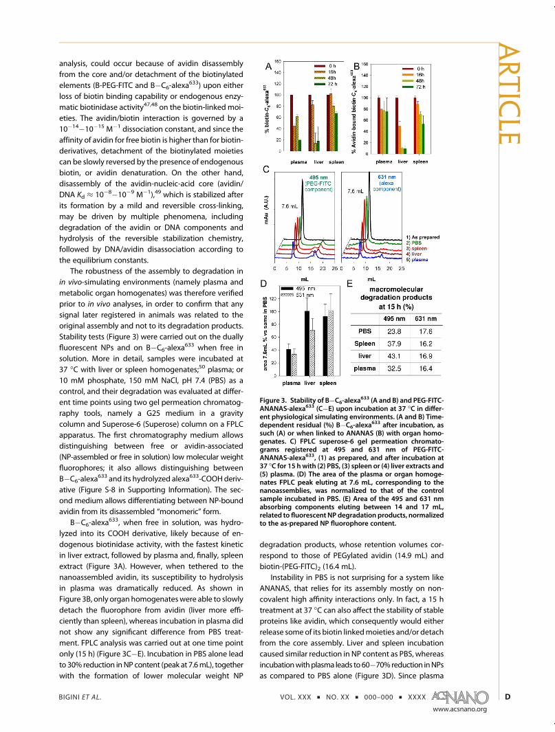
B�C6-alexa633, when free in solution, was hydro-
lyzed into its COOH derivative, likely because of en-dogenous biotinidase activity, with the fastest kineticin liver extract, followed by plasma and, finally, spleenextract (Figure 3A). However, when tethered to thenanoassembled avidin, its susceptibility to hydrolysisin plasma was dramatically reduced. As shown inFigure 3B, only organ homogenateswere able to slowlydetach the fluorophore from avidin (liver more effi-ciently than spleen), whereas incubation in plasma didnot show any significant difference from PBS treat-ment. FPLC analysis was carried out at one time pointonly (15 h) (Figure 3C�E). Incubation in PBS alone leadto 30% reduction inNP content (peak at 7.6mL), togetherwith the formation of lower molecular weight NP
degradation products, whose retention volumes cor-respond to those of PEGylated avidin (14.9 mL) andbiotin-(PEG-FITC)2 (16.4 mL).
Instability in PBS is not surprising for a system likeANANAS, that relies for its assembly mostly on non-covalent high affinity interactions only. In fact, a 15 htreatment at 37 �C can also affect the stability of stableproteins like avidin, which consequently would eitherrelease someof its biotin linkedmoieties and/or detachfrom the core assembly. Liver and spleen incubationcaused similar reduction in NP content as PBS, whereasincubationwithplasma leads to60�70%reduction inNPsas compared to PBS alone (Figure 3D). Since plasma
Figure 3. Stability of B�C6-alexa633 (A and B) and PEG-FITC-
ANANAS-alexa633 (C�E) upon incubation at 37 �C in differ-ent physiological simulating environments. (A and B) Time-dependent residual (%) B�C6-alexa
633 after incubation, assuch (A) or when linked to ANANAS (B) with organ homo-genates. C) FPLC superose-6 gel permeation chromato-grams registered at 495 and 631 nm of PEG-FITC-ANANAS-alexa633, (1) as prepared, and after incubation at37 �C for 15 h with (2) PBS, (3) spleen or (4) liver extracts and(5) plasma. (D) The area of the plasma or organ homoge-nates FPLC peak eluting at 7.6 mL, corresponding to thenanoassemblies, was normalized to that of the controlsample incubated in PBS. (E) Area of the 495 and 631 nmabsorbing components eluting between 14 and 17 mL,related to fluorescent NP degradation products, normalizedto the as-prepared NP fluorophore content.
ARTIC
LE
BIGINI ET AL. VOL. XXX ’ NO. XX ’ 000–000 ’ XXXX
www.acsnano.org
E
decrease was not accompanied with any increase inlow molecular weight derivatives, this result can beexplained only by the formation of large particleaggregates (still soluble), maybe secondary to opsoni-zation by plasma proteins. These aggregates are un-distinguishable from the original NPs using the largermesh G25 chromatography medium (Figure 3B), butthey are blocked by the superose column prefilter.Analysis of the NP degradation products eluting be-tween 14 and 16 mL shows slightly faster degradationin the biological samples as compared to PBS. The NPcomponent that ismostly susceptible to degradation isthe one linked to the Biotin-(PEG-FITC)2 moiety thatshowed a larger difference between PBS and homo-genate treatment (Figure 3E). On the contrary, nodetectable difference between PBS- and homogenate-treated samples was registered from the chromato-grams registered at 631 nm. Since the NP peak main-tains both FITC and alexa633 associated absorbance, wecan conclude that any fluorescence associated signalin vivo is mostly related to NP-associated avidin andnot to degradation products.
Immunogenicity. The response of the immune sys-tem is a fundamental parameter that needs to be takeninto consideration when investigating any novel su-pramolecular delivery tool intended for parenteraladministration. A strong immune reaction is not onlyrisky for the patient, but also can promote the earlysequestration of the delivered entity and reduce itsefficacy. In the case of the ANANAS system, which iscomposed for most part of the exogenous proteinavidin, an immunoresponse is indeed expected. How-ever, it was also shown in experiments in humans thatthe immunoresponse induced by the monomeric avi-din is not dangerous to the patients,33 making itpossible to envision its use also within the ANANASsupramolecular assembly.
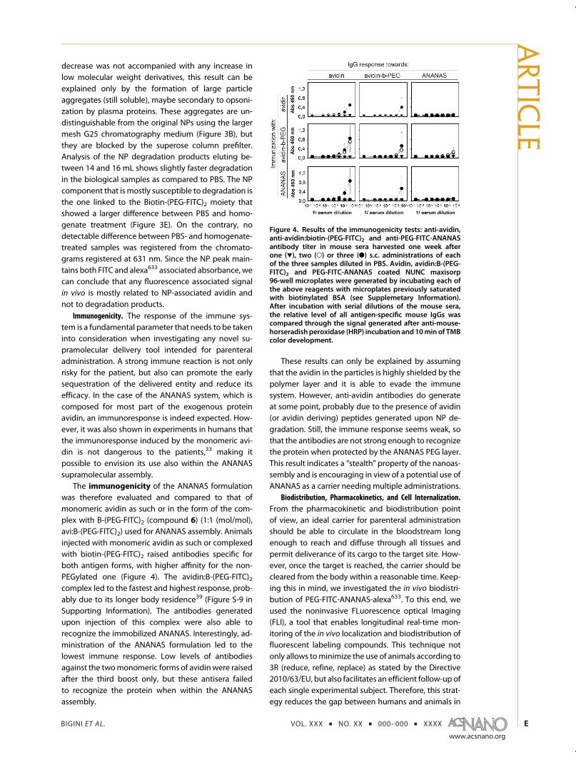
The immunogenicity of the ANANAS formulationwas therefore evaluated and compared to that ofmonomeric avidin as such or in the form of the com-plex with B-(PEG-FITC)2 (compound 6) (1:1 (mol/mol),avi:B-(PEG-FITC)2) used for ANANAS assembly. Animalsinjected with monomeric avidin as such or complexedwith biotin-(PEG-FITC)2 raised antibodies specific forboth antigen forms, with higher affinity for the non-PEGylated one (Figure 4). The avidin:B-(PEG-FITC)2complex led to the fastest and highest response, prob-ably due to its longer body residence39 (Figure S-9 inSupporting Information). The antibodies generatedupon injection of this complex were also able torecognize the immobilized ANANAS. Interestingly, ad-ministration of the ANANAS formulation led to thelowest immune response. Low levels of antibodiesagainst the twomonomeric forms of avidinwere raisedafter the third boost only, but these antisera failedto recognize the protein when within the ANANASassembly.
These results can only be explained by assumingthat the avidin in the particles is highly shielded by thepolymer layer and it is able to evade the immunesystem. However, anti-avidin antibodies do generateat some point, probably due to the presence of avidin(or avidin deriving) peptides generated upon NP de-gradation. Still, the immune response seems weak, sothat the antibodies are not strong enough to recognizethe protein when protected by the ANANAS PEG layer.This result indicates a “stealth” property of the nanoas-sembly and is encouraging in view of a potential use ofANANAS as a carrier needing multiple administrations.
Biodistribution, Pharmacokinetics, and Cell Internalization.From the pharmacokinetic and biodistribution pointof view, an ideal carrier for parenteral administrationshould be able to circulate in the bloodstream longenough to reach and diffuse through all tissues andpermit deliverance of its cargo to the target site. How-ever, once the target is reached, the carrier should becleared from the body within a reasonable time. Keep-ing this in mind, we investigated the in vivo biodistri-bution of PEG-FITC-ANANAS-alexa633. To this end, weused the noninvasive FLuorescence optical Imaging(FLI), a tool that enables longitudinal real-time mon-itoring of the in vivo localization and biodistribution offluorescent labeling compounds. This technique notonly allows tominimize the use of animals according to3R (reduce, refine, replace) as stated by the Directive2010/63/EU, but also facilitates an efficient follow-up ofeach single experimental subject. Therefore, this strat-egy reduces the gap between humans and animals in
Figure 4. Results of the immunogenicity tests: anti-avidin,anti-avidin:biotin-(PEG-FITC)2 and anti-PEG-FITC-ANANASantibody titer in mouse sera harvested one week afterone (1), two (O) or three (b) s.c. administrations of eachof the three samples diluted in PBS. Avidin, avidin:B-(PEG-FITC)2 and PEG-FITC-ANANAS coated NUNC maxisorp96-well microplates were generated by incubating each ofthe above reagents with microplates previously saturatedwith biotinylated BSA (see Supplemetary Information).After incubation with serial dilutions of the mouse sera,the relative level of all antigen-specific mouse IgGs wascompared through the signal generated after anti-mouse-horseradish peroxidase (HRP) incubation and 10min of TMBcolor development.
ARTIC
LE

BIGINI ET AL. VOL. XXX ’ NO. XX ’ 000–000 ’ XXXX
www.acsnano.org
F
terms of investigation procedures, thus increasing thetranslational level of the research. From a technicalpoint of view, the use of far red fluorophores, as thealexa633, permits relatively deep photon penetrationinto tissue, avoids the generation of background dueto the autofluorescence of tissues and generates a highoptical contrast.51
The reliability of the FLI based investigation wasverified in a preliminary experiment (Figure S-9 inSupporting Information), in which we followed thein vivo fate of the monomeric avidin:B�C6-alexa
633
(1:1 mol/mol) and avidin:B-(PEG-FITC)2:B�C6-alexa633
(1:1:1 mol/mol/mol) complexes. The results were in-deed similar to the pharmacokinetic data obtainedthrough other analytical tools.28,36,39
To investigate the pharmacokinetics and biodistri-bution of PEG-FITC-ANANAS-alexa633, NP solution wasadministered into the tail vein of 15 healthy NFR mice.Animals were scanned by FLI before and immediatelyafter (15 min) injection, and 2, 6, 24, and 48 h later, tomonitor the localization of ANANAS-related fluores-cent signal (Figure 5). At each time point, three animalswere sacrificed and ex vivo NIR analysis of liver, spleenand kidneys was also carried out (Figure 6).
The overall FLI pattern registered was nearly over-lapping among the three mice tested, indicating goodhomogeneity and reproducibility in the particle be-havior. The biodistribution information registered is inaccordance with the metabolic pattern of clearanceobserved with other biocompatible nanostructuresalready used at the clinical level for diagnostic
purposes:52,53 immediately after injection, the in vivo
fluorescent signal spread in thewhole toraco-abdominal
Figure 5. In vivo biodistribution of PEG-FITC-ANANAS-alexa633 after iv administration. Representative images acquiredbefore (PRE INJ) administration, and at scheduled time points (15 min, and 2, 6, 24, and 48) after injection. The fluorescencesignal intensity, measured as normalized photon counts (NC), is shown as a pseudo-color scale bar. For each mouse, thepseudo-color scale bar is consistent for all images, in order to show relative changes of biodistribution over time. As controls,five animals received the vehicle solution only. The FLI pattern registered in these controlswas the sameof that of preinjectedanimals (data not shown).
Figure 6. Ex vivooptical imaging analysis. (A) Threemice foreach time point (2�24�48 h) after PEG-FITC-ANANAS-alexa633 injection were sacrificed, the abdominal organs(L = liver, K = kidneys and S = spleen) were excised andscanned by optical imaging analysis. The intensity of thefluorescent signal, measured as normalized photon counts(NC), is shown as a pseud-ocolor scale bar that is consistentfor all images. (B) At each timepoint, the relativefluorescentintensity (FLI) signal registered from all excised organs wasquantified and expressed in the graph as relative FLI. Thedashed lines represent the average value of the vehicletreated animals normalized to 1. Data are presented asmean ( SE. The analysis was performed by unpairedStudent's t test, *P < 0.05, **P < 0.005, ***P < 0.0001 inrespect with vehicle-treated mice.
ARTIC
LE
BIGINI ET AL. VOL. XXX ’ NO. XX ’ 000–000 ’ XXXX
www.acsnano.org
G
area, with higher intensity levels concentrating in a
specific region, probably corresponding to the liver
and spleen. Two to six hours after injection, most of the
signal had moved toward consistent specific areas of
the abdominal region. After 24 h, a dramatic reduction
of the overall signal was registered and which was
reduced to almost preinjection levels at 48 h. Interest-
ingly, the fluorescent signal plasma permanence regis-
tered for the nanoassembly is similar to that observed
for the complex monomeric avidin:B-(PEG-FITC)2:
B�C6-alexa633 complex (Supporting Information,
Figure S-9). However, the pattern of distribution, accu-
mulation and clearance is slightly different be-
tween the two species (e.g., the avidin:B-(PEG-FITC)2:
B�C6-alexa633 complex did not show spleen localiza-
tion and its signal was eliminated through the kidneys,
whereas a much lower signal in the bladder was
recorded in NP treated animals), confirming the factthat they are indeed different species.
The relative photon counts measured from liver,spleen and kidneys explanted from the NP-adminis-tered animals (Figure 6) are consistent with the in vivo
analysis: strong signal intensity in organs was regis-tered 2 h after administration, with a peak of fluores-cence in the liver.
Twenty-four hours after injection, a marked signalreduction was observed in all organs analyzed, with akinetics that appears faster in liver and kidneys than inspleen. Two days after administration, the signal hadalmost completely disappeared from both liver andkidney whereas a lower, but still detectable fluores-cence was found in the spleen.
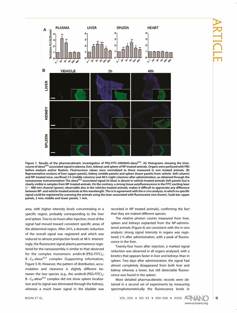
More detailed pharmacokinetic records were ob-tained in a second set of experiments by measuringspectrophotometrically the fluorescence levels in
Figure 7. Results of the pharmacokinetic investigation of PEG-FITC-ANANAS-alexa633. (A) Histograms showing the time-course of alexa633 associated signal in plasma, liver, kidneys and spleen of NP treated animals. Organswere perfusedwith PBSbefore analysis and/or fixation. Fluorescence values were normalized to those measured in non treated animals. (B)Representative sections of liver (upper panels), kidney (middle panels) and spleen (lower panels) from vehicle- (left column)and NP-treated mice, sacrificed 2 h (middle columns) and 48 h (right columns) after administration, as obtained through thenanozoomer instrumentation The alexa633-associated signal (in blue) is absent in vehicle-treated animals (left panels) but isclearly visible in samples fromNP-treated animals. On the contrary, a strong tissue autofluorescence in the FITC exciting laser(λ∼ 488 nm) channel (green), observable also in the vehicles-treated animals, makes it difficult to appreciate any differencebetweenNP- and vehicle-treated animals at thiswavelength. This is in agreementwith the in vivo analysis, inwhich no specificsignal could be registered by scanning the animals using the laser-associated with fluoresceine (not shown). Scale bar: upperpanels, 2 mm; middle and lower panels, 1 mm.
ARTIC
LE

BIGINI ET AL. VOL. XXX ’ NO. XX ’ 000–000 ’ XXXX
www.acsnano.org
H
plasma, liver, spleen and kidney homogenates at dif-ferent time-points after administration (Figure 7). A 30min time-point was included in this case, in order to im-prove the analysis of the early phase of the distributionand elimination processes.
The pharmacokinetics data registered from thesupernatants of centrifuged organ homogenates(Figure 7A) demonstrate the existence of NPs circulat-ing freely in the bloodstream for more than two hours.Between 30 to 120min, the plasma alexa633-associatedfluorescence decreased less than 40% and six hoursafter administration, the signal intensity was re-duced to almost preinjection levels. Concurrently, thealexa633-signal moved into the metabolic organs, witha t-max between 2 and 6 h in the liver andg6 h in thespleen. No changes in the fluorescence levels of hearthhomogenates, a well perfused nonmetabolic organ,were registered along the whole experiment, during
which the heart-associated signal remained similar tothat of pretreated mice.
To verify the pattern of signal associated with NPwithin each organ tissue, a large number of sectionsfrom liver, spleen and kidney obtained from animalssacrificed 2 and 48 h after administration was acquiredwith Nanozoomer HT 2.0 scanner with fluorescenceoptics (Figure 7B).
In agreement with the pharmacokinetic studies, theNP-associated signal was high 2h after administrationand markedly reduced after 48 h (even if still visible).Signal decay was less pronounced in spleen than inother organs, confirming the information gained bythe Optix instrumentation. The Nanozoomer analysisallowed a convenient and rapid scan of the organsin their whole volume, permitting to understandthe overall distribution of the particles in theorgans: heterogeneous accumulation of ANANAS was
Figure 8. Representative confocal images of HeLa cells incubated with PEG-FITC-ANANAS-alexa633 (30 μg/mL in 10% FCSculture medium). Similar results (not shown) were obtained using lower NP concentration (6 μg/mL). Panels A�E show themerge (red for alexa633, green for FITC, yellow for merged alexa633/FITC) among the fluorescent channels associated to theNPs and the nuclei (in blue after staining with Hoechst 33258, 2 μg/mL). A representative image of control cells (treated withPBS only) is reported in panel A. In panels B,�D, cells treatedwith NPs for 2 h (B), 6 h (C) and 24 h (D) can be observed. In panelE, a 3D reconstruction of 24 h treated cells is reported. Hoechst 33258 labeled nuclei are visualized in blue, while both green(FITC) and red (alexa633) signals are associatedwith ANANAS. Panels F�Kdisplay representative low (F, H, and J) and high (G, I,and K) magnification images of cells upon 24 h incubation with NPs in basal conditions (F�G), or in presence ofchloropromazine (20 μg/mL) (H and I) or amyloride 1 mM (J and K). Hoechst 33258 labeled nuclei are visualized in blue,while both green (FITC) and red (alexa633) signals are associated to ANANAS. In physiological conditions, the ANANAS spreadhomogenously in the cytoplasm of all cells (F), showing a punctuate pattern of vesicle-like staining (G). Treatment withchloropromazine leads to NP accumulation around the cell periphery (H) and themerge between fluorescence and nomarskiacquisitions reveals that almost all of the spots are confined in proximity of the plasma membrane (I). Amyloride treatmentdid not show relevant inhibition of NP uptake and significant entry of NP can be observed at this time-point (J and K). Scalebars: (A�D) 100 μm; (E) 10 μm; (F, H and J) 20 μm; (G, I and K) 5 μm.
ARTIC
LE

BIGINI ET AL. VOL. XXX ’ NO. XX ’ 000–000 ’ XXXX
www.acsnano.org
I
observed in kidney and spleen sections and observa-tional results suggest that in kidneys the NPs accumulateclose to the glomeruli endothelial structures, while inspleen they localize in the red pulp (likely in residentmarcrophages). On the other hand, in liver, thealexa633 signal was uniformly spread in the entireparenchyma and analysis at higher resolutionthrough confocal microscopy (Figure S-10 in Sup-porting Information) showed that the NPs were al-most homogeneously diffused around the nuclei in aspot-like staining. The peripheral distribution of se-parate spots and the presence of signal around thenuclei strongly suggests that an active process ofinternalization occurred by a mechanism of nanopar-ticle-dependent vesicles formation. The ability ofANANAS to internalize cells was further demon-strated in an in vitro test in which NP accumulationin HeLa cells upon incubation for 2, 6, and 24 h wasmonitored by confocal microscopy. As shown inFigure 8, cell association and internalization occurquite efficiently, even if at a relatively slow pace. Thekinetics observed along this experiment, which hasbeen carried out in the presence of serum, supportsthe in vivo observation that the ANANAS can effi-ciently internalize cells, provided they have the op-portunity to stay in contact with them for some time.Upon internalization, the NPs localize primarily with-in the cytoplasm (Figure 8E,G), where they appear asseparate spots, resembling clusters of internalizationvesicles. Treatment with chloropromazine, a selectiveinhibitor of clathrin-dependent endocytosis, stronglyreduces NP entry (Figure 8H,I), whereas incubationwith amyloride, which selectively inhibits macro-pinocytosis, does not lead to significant alterationon NP uptake for at least 24 h (Figure 8J,K). Thisstrongly suggests that a vesicle-dependent pro-cess of endocytosis occurs (see single fluorescentspots in Figure 8F,G), secondary to clathrin-dependentvesicle formation (see the lack of NP internalization inFigure 8H,I). This is in agreement with other studiesshowing that nanomaterials can be uptaken by a largevariety of cells through a “troyan horse”-like mech-anism.
CONCLUSIONS
The multimodal approach adopted in this studyallowed us to disclose quali-quantitatively the in vivo
fate of the dually fluorescent ANANAS formulationhere optimized. Altogether, the information obtainedconsistently indicate that, when mixed with biologicalfluids, the NPs maintain their assembled conformationand are slowly degraded by a combination of mecha-nisms, including avidin disassembly from the core andslow detachment of the biotinylated elements. In vivo,these phenomena occur concomitantly to NP accumu-lation in liver and spleen, probably also secondary toopsonization by plasma proteins. The formulation does
not have any special tropism other than that for normalscavenging organs and its fate is in line with thatregistered for other biocompatible NPs. Although theparticles have the ability to penetrate cells, this prop-erty neither induces their accumulation nor avoidstheir clearance and/or their degradation. Indeed, thein vitro tests showed that cell internalization occursrather slowly, so that some time of contact with thetarget cells is necessary. As a consequence, unless atargeting element is inserted in the NP composition,cell penetration in vivo can occur only in the scaveng-ing organs and no accumulation occurs elsewhere. Assuch, the lack of accumulation observed provides animportant suggestion for the development of thisnanodevice for diagnostic and/or drug-delivery appli-cation. On this matter, we would like to note that amajor reason for FDA approval of the first nanoliposo-mal doxorubicin (DOXIL) was its limited heart avail-ability/accumulation, with consequent reduced car-diotoxicity.The results show that the formulation here investi-
gated represents an advancement in avidin-basedtargeting technologies: with respect to monomericavidin, it displays more favorable pharmacokineticproperties, since circulating NPs can be detected formore than 2 h after iv administration. The NP plasmaresidence is similar to that of the PEGylated form of theprotein, with the important advantage of having amuch higher payload for biotinylated active moietiesand being less immunogenic. Since antibodies capableto recognize the assembled particles do not form atleast after two repeated administrations, this formula-tion could be greatly advantageous for treatments thatnecessitate multiple administrations.With respect to the NP plasma residence, it is
important to note that the carbohydrate portion inavidin is known to be responsible for increasing theprotein liver localization, but only when saturated withbiotin.28 Considering that most of the avidin in afunctional ANANAS formulation is biotinylated, it ispossible that the NP liver tropism here observed may,at least in part, be related to the protein glycosidicportion. If so, and if necessary, the NP plasma residencecould be further prolonged by replacing the nativeprotein with its nonglycosylated analogue.54
Overall, the results are encouraging toward a futureevaluation of the NP fate when in contact with leakyvascularized inflamed or cancer tissues, and/or to test iftargeted particles can localize into desired tissues orbody districts. Ultimately, the theranostic applicabilityof the system, once functionalized with targeting ele-ments, will depend on its ability to penetrate and beretained into target tissues. In this context, the cellinternalizing ability observed through both the ex vivoand in vitro confocal microscopy analyses suggeststhat it is possible to carry the bioactive cargoes alsoinside the cells. Nonetheless, since the biotinylated
ARTIC
LE

BIGINI ET AL. VOL. XXX ’ NO. XX ’ 000–000 ’ XXXX
www.acsnano.org
J
elements slowly detach from the core particles, simi-larly biotinylated bioactive agents (as, for example,drugs), if loaded onto a (targeted) ANANAS, wouldlikely detach from the particles, once these havereached their final body destination. In this respect,it has to be pointed out that if one would like to usethe ANANAS as a drug delivery system rather thana diagnostic tool, the linker between the bioactivecompound and the NP-tethering biotin should befurther engineered to favor local release of the carriedmoiety in its active form. For example, depending onthe biochemical properties of the target environ-ment, the use of pH, cathepsin or temperature sensi-tive bonds and/or self-immolative spacers can beenvisioned.55�58
Finally, it is worth mentioning that the biodistribu-tion and pharmacokinetic data were generated takingadvantage of the NIR fluorescence alexa633-relatedinformation. Although in the formulation investigatedthis fluorophore occupies less than 20% of the totalavailable BBS (andmuch less % of the total surface area
available for functionalization), the signal generatedwas high enough to allow quantitative analysis bymeans of the noninvasive Optix instrumentation, sug-gesting that, if necessary, the amount of fluorophorecould be further reduced without losing sensitivity.This permits a great formulation freedom when seek-ing more complex functional assemblies;containing,for example, drugs, targeting or cell internalizingmoieties;which, in principle, could still be testedthrough the same noninvasive tools as those used here.Last but not least, we would like to note the unique
mode of ANANAS assembly, which allows to generatefunctional formulations with stoichiometrically con-trolled composition in “one-pot” reactions. This per-mits envisioning the use of this technology not only asa per se theranostic tool, but also as a general platformto test the efficacy of functional elements (as, forexample, putative tissue-targeting or cell-internalizingmoieties) and to establish dose/response relationshipsthat are of general interest for the development of anynanoparticle-based biomedical tool.
METHODSMaterials and Instrumentation. Avidin was purchased from
Belovo chemical (Belgium). Plasmid pEGFP-C1, 4.7 kb (usedfor the ANANAS core preparation), was from Clontech; anti-mouse-HRP was from Millipore; 3,30 ,5,50-tetramethylbenzidine(TMB)was purchased fromKPL (Gaithersburg,MD, USA). Ninety-six-well High binding Corning EIA/RIA plates, and all otherreagents were from Sigma Aldrich (St. Louis, MO, USA). NAP10gravity G25 gel permeation columns were purchased fromGE Healthcare. Fast Permeation Liquid Chromatography(FPLC) analyses were performed using an AKTA purifier 10 (GEHealthcare) apparatus, equipped with a UV�vis detector and aSuperose 6 (10/300 GL) column. UV�vis analyses were per-formed on a Varian Cary 50 UV�vis spectrophotometer (Varian,Inc., Palo Alto, CA, USA). Dynamic light scatteringmeasurementswere performed using a Malvern Zetasizer NANO ZS (MalvernInstruments Ltd., Worcestershire, United Kingdom). ELISA Mi-croplate absorbance was measured with a Multiskan FC micro-plate reader (Thermo Fisher Scientific, Waltham, MA, USA).Fluorescence spectroscopy data were recorded using a JASCOFP 6200 fluorimeter. A 2300 Multilabel Plate Reader spectro-photofluorimeter (Perkin-Elmer, Boston, MA, USA) was used toquantify the ANANAS associated fluorescence in the pharma-cokinetics experiments. Electron microscopy images were ob-tained with an Energy Filter Transmission Electron Microscope(EFTEM, ZEISS LIBRA 120) equipped with YAG scintillator slowscanCCD camera. Before image acquisition, a PEG-FITC-ANANAS-alexa633 solution in ddH2O was placed to dry overnight on a100 mesh Formvar/carbon coated copper grid (EMS, Hatfield,PA, USA).
Nanoparticle Preparation and Characterization. The synthesis ofthe two ω-FITC labeled biotin-PEG derivatives (compounds 4and6 andof biotin-C6-alexa
633 (compound 7) is described in theSupporting Information. All NPs were prepared on the basisof a previously published procedure.13 In the preformulationstudies, 'Core' FITC-labeled ANANAS were prepared by mix-ing avidin, compound 6 (Figures 1 and 2) and plasmid DNAat selected molar ratios. In this preparation, the avidin BBS:biotin-PEG derivative molar ratio was 1:0.25. After stabilization,particles were purified from excess of avidin by ultrafiltration(Millipore PVDF membranes, cutoff 100 kDa) or gel permeationchromatography eluting the sample in 10 mM phosphate,150 mM NaCl, pH 7.4 buffer (PBS) using a Sepharose 6-FF
column on a FPLC system (AKTA purifier, GE Healthcare).The eluted particles were stored at 4 �C until further use. Theresulting NPs were characterized by dynamic light scattering,UV�vis and fluorescence spectroscopy. The number of fluo-rescein molecules attached to the purified particles was mea-sured from the UV�vis spectra, on the basis of the 280 nm(avidin, DNA and fluorescein) and 495 nm (fluorescein only)absoption values.
Maximal NP PEG loading capability was assessed bymixing the 'core' particles with molar excesses (with respectto the theoretically available BBS) of either compounds 4 or 6 inPBS. After 1 h incubation, particles were purified by size exclu-sion chromatography and the number of fluorophores linkedwas quantified by specrophotometry as above indicated.
The final B�C6‑alexa633-B-PEG-FITC labeled NP formula-
tion used for the in vivo studies (PEG-FITC-ANANAS-alexa633)was obtained from a highly FITC-PEGylated ANANAS NP pre-pared as above using an avidin-BBS:biotin PEG derivative molarratio of 1:0.46. After purification and UV�vis characterization,the nanoassembly was added of an excess of B�C6‑alexa
633
(compound 7). After 30 min incubation, nanoassemblies werepurified by gel permeation chromatography as described aboveand analyzed by DLS, UV�vis and fluorescence spectroscopy.The number of fluorophores attached to the purified particleswas measured from the UV�vis spectrum, on the basis of the280 nm (avidin, DNA and fluorescein) and 495 nm (fluoresceinonly) and 631 nm (alexa633) absorption values.
Stability of PEG-FITC-ANANAS-alexa633 in Serum and Organ Homoge-nates. Tissues homogenates and plasma were obtained accord-ing to the standardized procedures described by our group.50
Briefly, liver, spleen and plasma were harvested. Tissues werehomogenized in 5 mM PBS, pH 7.4, in a 1:4 (w:v) ratio andcentrifuged at 9000g for 10 min at 4 �C. The supernatant wascollected and stored at �20 �C until used.
Stability through Gravity G25 Chromatograpy Analysis. Solutionsof PEG-FITC-ANANAS-alexa633 (120 μg/mL in PBS) or B�C6-alexa633 weremixedwith 0.1 vol of plasma, organ homogenates(spleen and liver) obtained as above, or PBS as a control. Atscheduled times, samples were centrifuged (15 000 rpm(16 600g), 2 min, 4 �C) and the supernatants were either elutedthrough a gravity G25 medium (NAP10, GE Healthcare) oranalyzed by gel permeation chromatography using an Aktapurifier (GE) FPLC apparatus integrated with a Superose 6
ARTIC
LE

BIGINI ET AL. VOL. XXX ’ NO. XX ’ 000–000 ’ XXXX
www.acsnano.org
K
column, using PBS as eluant. In the case of the NAP analysis, thisprocedure was carried out on mixtures as prepared and afterincubation at 37 �C for 16, 48 and 72 h. The eluted fractions(0.5 mL) were analyzed by UV�vis and fluorescence Spectros-copy (λexc 632 nm, λem 647 nm, slidts 5 nm, high sensitivity). Forquantitative analysis, each sample elution profile was com-pared to that of PEG-FITC-ANANAS-alexa633, B�C6-alexa
633
and alexa633-COOH (Supporting Information, Figure S-7). As acontrol, B�C6-alexa
633 was also treated and analyzed in thesame way. FPLC analysis was carried out on mixtures asprepared and after 15 h incubation at 37 �C. Samples wereeluted with PBS buffer at 0.5 mL/min, and elution was mon-itored at 280, 495, and 631 nm.
In Vivo Experiments. Procedures involving animals and theircare were conducted in conformity with the institutional guide-lines that are in compliance with national (D.L. no. 116, G.U.,suppl. 40, 18 Febbraio 1992; Circolare no. 8, G.U., 14 Luglio 1994)and international (EEC Council Directive 86/609, OJL 358, 1, 12December 1987; Guide for the Care and Use of LaboratoryAnimals, U.S. National Research Council, 1996) laws and policies.
Immunogenicity. Repeated (three) subcutaneous injections ofPEG-FITC-ANANAS-alexa633, avidin, and avidin:B-(PEG-FITC)2(1:1) complex diluted in PBS (60 μg/mL) were administered inthree separate animal groups (N = 3) every two weeks. Plasmawas collected one week after each administration and stored at�20 �C until analysis. At the end of the experiment, the antibodytiter against each avidin form was measured in all samples. Aconjugate between BSA and biotin-PEG5kDa (B-PEG-BSA) wasobtained by classic bioconjugation procedures using biotin-PEG5kDa-NHS (laysan Bio). NUNC maxisorp 96-well microplateswere incubated overnight with B-PEG-BSA, (4 �C) (4 μg/mL) in0.05 M NaHCO3, pH 9.3. After blocking with PBS containing0.05% of Tween 20 (PBST) and 0.3% BSA, avidin, avidin�biotin-PEG and PEG-FITC-ANANAS coated wells were generated byincubating each of the above reagents (all 4 μg/mL in PBST, 2 h,37 �C) with the B-PEG-BSA-coated wells. Mice sera were seriallydiluted (from 1:100 to 1:1 000 000) with PBST þ 0.1% BSA) andwere incubated in the above wells for 1 h at 37 �C. The relativelevel of all antigen-specific mouse IgGs was compared by thesignal generated after anti-mouse-HRP incubation (diluted inPBST þ 0.1% BSA 1 h, 37 �C) and 10 min of TMB (50 μL/well)development, followed by stopping the reaction by the addi-tion of 50 μL/well of 0.5 M H2SO4 and absorbance readout at450 nm. All samples were tested in triplicate. A NP formulationcontaining slightly less PEG5kDa-FITC chains than the one in-jected in mice (600 instead of 700) was used in these experi-ments to ensure the assembly surface tethering through theB-PEG-BSA anchoring element).
In Vivo and ex Vivo Fluorescence Imaging. In vivowhole body fluo-rescence imagingwas performed longitudinally onmice injectedintravenously with PEG-FITC-ANANAS-alexa633 (140 μg/mL inPBS þ 0.05% Tween 20) using the Explore Optix System (ARTAdvanced Research Technologies, Montreal, Canada), and afixed pulsed laser diode as an illumination source. Beforeimaging, animals were anesthetized with a continuous flow of5% isoflurane/oxygen mixture and placed on their back on aheated (37 �C) pad inside the camera box and 2�3% isofluraneanesthesia was maintained using a nose cone delivery systemfor the duration of image acquisition. For each animal, a regionof interest (ROI) of the ventral whole body was chosen with astep size of 2 mm and a spot size of 1 mm. Excitation wasperformed with a 635 nm pulsing laser and emission wasdetected with a 650 nm long pass filter. Subsequently, a pho-tomultiplier tube (PMT) coupled to a time correlated singlephoton counting system collects the emitted light, allowingdetection of fluorescent signal. Light emission was recorded aspseudo-color images. Overlay of gray scale (body referencephotograph) and pseudo-color images allowed localizationof fluorescent signal. Mice injected with PEG-FITC-ANANAS-alexa633 were scanned before injection and immediately after15 min and 2, 6, 24, and 48 h later. For each time point, threeanimals were sacrificed, and their abdominal organs (liver,spleen and kidneys) were removed and scanned for theex vivo fluorescence imaging. A ROI was designed around eachorgan and light/photon emission in the selected ROI was
quantified as total photon counts using Optiview software(version 2.02.00; ART Advanced Research Technologies). At eachtime point, the mean FLI signal values detected around liver,spleen and kidneys were measured and expressed as relativevalues to those measured from PBS-treated animals.
Statistical Analysis. All data were analyzed with the use ofGraphPad Prism software (version 5.03). In particular, the differ-ences in fluorescent intensity between multiple groups havebeen evaluated using a one way ANOVA test followed by theBonferroni's test. Data were expressed as the mean ( standarddeviation (SD). The differences were considered statisticallysignificant at a level of P < 0.05.
Pharmacokinetic Study through Fluorescence Titration of Plasma andOrgan Homogenates. A total of 18animalswereused, and3of thesekept as control. The remaining 15 were intravenously treatedwith a solution of PEG-FITC-ANANAS-alexa633 (127.8 μg/mL (w/v)in PBS þ 0.05% Tween 20. At each time point (30 min, 2, 6, 24,and 48 h), three animals were sacrificed by cervical dislocation.Fresh blood was collected from the retroorbital plexus andkept into EDTA (0.5 M, pH 8) pretreated tubes at 4 �C for about30 min. To separate plasma from entire blood, the collectedsamples were centrifuged for 15 min at 1500g. The supernatant(plasma) was collected and aliquots were immediately frozen.Tissues were homogenized as described above. Before use,homogenates were diluted 1:10 (v/v) with distilled water,centrifuged at 1100g for 10 min at 25 �C and the supernatantrecovered in order to obtain transparent solutions. Spectro-photofluorimetric analyses were conducted to correlate thefluorescence intensity (FI) measured into the collected samplesto the presence of PEG-FITC-ANANAS-alexa633. FI was deter-mined at λexc = 470 nm and λem = 520 nm for FITC and at λexc =600 nm and λem = 670 nm for alexa633 using an 2300 MultilabelPlate Reader spectrophotofluorimeter (Perkin-Elmer, Boston,MA, USA) with a standard 96wells black plate with a transparentbottom (ProxiPlate-96, Perking Elmer). The fluorescence inten-sity of all samples was compared to that measured from blank(non treated) animals.
In Vitro Cell Internalization Assay. HeLa cells were seeded onround glass slides in 24-well plates at the concentration of10 000 cell/well in culture medium (DMEM, 10% fetal bovineserum, 2 mM L-glutamine, 100 U penicillin/0.1 mg/mL strep-tomycin) and incubated at 37 �C, 5% CO2, for 24 h. Culturemedium was then removed and replaced with a suspensionPEG-FITC-ANANAS-alexa633 (30 μg/mL) in culture medium for2, 6, and 24 h, respectively. At the end of incubation, cells werefixed with 4% paraformaldehyde in PBS for 40 min and nucleiwere stained with Hoechst 33258 (2 μg/mL in PBS, incubationfor 40min). Glass slidesweremounted on cover glass slideswith2�3 drops of Fluormont and stored at 4 �C. Samples were thenanalyzed with an Olympus Fluoview microscope BX61 withconfocal system FV500, equipped with specific lasers λexc =405 nm, λexc = 488 nm, and λexc = 635 nm to visualize Hoechst33258, FITC, and alexa633, respectively. Two dimensional andthree-dimensional images (z stack reconstruction of one focalplane acquisition every 0.2 μm on the z axis) were acquired.Images were pseudo-colored (blue for Hoechst 33258, green forFITC and AF 488, red for alexa633), and the signal obtained fromthe three channels was automatically merged by Olympusfluoview software. 3-D reconstruction was performed usingImaris 5.0 (Bitplane) software. In a separate experiment and inorder to investigate on the mechanism of NP uptake, cells wereco-incubated for 24 h as above with PEG-FITC-ANANAS-alexa633
(30 μg/mL) in the presence of chloropromazine (final concen-tration, 20 μg/mL) or amyloride (final concentration, 1 mM).Fixation and visualization were performed as above-described.
Conflict of Interest: The authors declare no competingfinancial interest.
Acknowledgment. This project was funded by the “Progettodi Ateneo” of the University of Padova n.CPDA072372. Authorsare greatly indebted to Dr. Rosanna Piccirillo for her criticalreading and her suggestions. A particular thanks to Dr. StefanoPezzati Eng. and Dr. Annamaria Mauro from Hamamtsu Photo-nics Italia for the support in acquiring images.
ARTIC
LE
BIGINI ET AL. VOL. XXX ’ NO. XX ’ 000–000 ’ XXXX
www.acsnano.org
L
Supporting Information Available: Synthesis and character-ization of biotinylated elements; DLS size and ζ-potential thefinal assemblies, biodistribution of individual NP componentsthrough FLI; ex vivo histological analyses. This material is avail-able free of charge via the Internet at http://pubs.acs.org.
REFERENCES AND NOTES1. Hrkach, J.; Von Hoff, D.; Ali, M. M.; Andrianova, E.; Auer, J.;
Campbell, T.; De Witt, D.; Figa, M.; Figueiredo, M.; Horhota,A.; et al. Preclinical Development and Clinical Translationof a PSMA-Targeted Docetaxel Nanoparticle with a Differ-entiated Pharmacological Profile. Sci. Transl. Med. 2012, 4,128–139.
2. Zamboni, W. C.; Torchilin, V.; Patri, A. K.; Hrkach, J.; Stern, S.;Lee, R.; Nel, A.; Panaro, N. J.; Grodzinski, P. Best Practices inCancer Nanotechnology: Perspective from Nci Nanotech-nology Alliance. Clin. Cancer Res. 2012, 18, 3229–3241.
3. Bourzac, K. Carrying Drugs. Nature 2012, 491, S58–60.4. Winter, P. M.; Neubauer, A. M.; Caruthers, S. D.; Harris, T. D.;
Robertson, J. D.; Williams, T. A.; Schmieder, A. H.; Hu, G.;Allen, J. S.; Lacy, E. K.; et al. Endothelial Alpha(V)Beta(3)Integrin-Targeted Fumagillin Nanoparticles Inhibit Angio-genesis in Atherosclerosis. Arterioscler., Thromb., Vasc. Biol.2006, 26, 2103–2109.
5. Zhou, H. F.; Yan, H.; Senpan, A.; Wickline, S. A.; Pan, D.;Lanza, G. M.; Pham, C. T. Suppression of Inflammation in aMouse Model of Rheumatoid Arthritis Using TargetedLipase-Labile Fumagillin Prodrug Nanoparticles. Biomater-ials 2012, 33, 8632–8640.
6. Sharrna, P.; Brown, S.; Walter, G.; Santra, S.; Moudgil, B.Nanoparticles for Bioimaging. Adv. Colloid Interface Sci.2006, 123, 471–485.
7. Mansilla, E.; Marin, G. H.; Nunez, L.; Drago, H.; Sturla, F.;Mertz, C.; Rivera, L.; Ichim, T.; Riordan, N.; Raimondi, C. TheLysosomotropic Agent, Hydroxychloroquine, Delivered ina Biodegradable Nanoparticle System, Overcomes DrugResistance of B-Chronic Lymphocytic Leukemia Cells inVitro. Cancer Biother. Radiopharm. 2010, 25, 97–103.
8. Islam, T.; Josephson, L. Current State and Future Applica-tions of Active Targeting in Malignancies Using Super-paramagnetic Iron Oxide Nanoparticles. Cancer Biomarkers2009, 5, 99–107.
9. Kelkar, S. S.; Reineke, T. M. Theranostics: Combining Imag-ing and Therapy. Bioconjugate Chem. 2011, 22, 1879–1903.
10. Stefanick, J. F.; Ashley, J. D.; Kiziltepe, T.; Bilgicer, B. ASystematic Analysis of Peptide Linker Length and Liposo-mal Polyethylene Glycol Coating on Cellular Uptake ofPeptide-Targeted Liposomes. ACS Nano 2013, 7, 2935–2947.
11. Morpurgo,M.; Facchin, S.; Pignatto,M.; Silvestri, D.; Casarin,E.; Realdon, N. Characterization of Multifunctional Nano-systems Based on the Avidin-Nucleic Acid Interaction asSignal Enhancers in Immuno-Detection. Anal. Chem. 2012,84, 3433–3439.
12. Morpurgo, M.; Radu, A.; Bayer, E. A.; Wilchek, M. DNACondensation by High-Affinity Interaction with Avidin.J. Mol. Recognit. 2004, 17, 558–566.
13. Pignatto, M.; Realdon, N.; Morpurgo, M. Optimized AvidinNucleic Acid Nanoassemblies by a Tailored PegylationStrategy and Their Application as Molecular Amplifiers inDetection. Bioconjugate Chem. 2010, 21, 1254–1263.
14. Wilchek, M.; Bayer, E. A. The Avidin-Biotin Complex in Bio-analytical Applications. Anal. Biochem. 1988, 171, 1–32.
15. Abelson, J.; Simon, M.; Wichek, M.; Bayer, E. A., Eds. Avidin-Biotin Technology; Methods in Enzymology; AcademicPress: San Diego, CA, 1990; Vol. 184.
16. Bratthauer, G. L. The Avidin-Biotin Complex (Abc) Methodand Other Avidin-Biotin Binding Methods. Methods Mol.Biol. 1999, 115, 203–214.
17. Lesch, H. P.; Kaikkonen, M. U.; Pikkarainen, J. T.; Yla-Herttuala, S. Avidin-Biotin Technology in Targeted Ther-apy. Expert Opin. Drug Delivery 2010, 7, 551–564.
18. Yan, H.; Jiang, W. M.; Zhang, Y. X.; Liu, Y.; Wang, B.; Yang, L.;Deng, L. H.; Singh, G. K.; Pan, J. Novel Multi-Biotin GraftedPoly(Lactic Acid) and Its Self-Assembling NanoparticlesCapable of Binding to Streptavidin. Int. J. Nanomed. 2012,7, 457–465.
19. Pulkkinen, M.; Pikkarainen, J.; Wirth, T.; Tarvainen, T.;Haapa-aho, V.; Korhonen, H.; Seppala, J.; Jarvinen, K.Three-Step Tumor Targeting of Paclitaxel Using Biotiny-lated Pla-Peg Nanoparticles and Avidin-Biotin Technol-ogy: Formulation Development and in Vitro AnticancerActivity. Eur. J. Pharm. Biopharm. 2008, 70, 66–74.
20. Ting, S. R.; Nguyen, T. L.; Stenzel, M. H. One Pot Synthesis ofSurface Pegylated Core-Shell Microparticles by Suspen-sion Polymerization with Surface Enrichment of Biotin/Avidin Conjugation. Macromol. Biosci. 2009, 9, 211–220.
21. Li, S.; Liu, H. N.; He, N. Y. Covalent Binding of Streptavidinon Gold Magnetic Nanoparticles for Bead Array Fabrica-tion. J. Nanosci. Nanotechnol. 2010, 10, 4875–4882.
22. Wang, Y.; Liu, X. R.; Nakamura, K.; Chen, L.; Rusckowski, M.;Hnatowich, D. J. In Vivo Delivery of Antisense Morf Oligo-mer by Morf/Carrier Streptavidin Nanoparticles. CancerBiother. Radiopharm. 2009, 24, 573–578.
23. Gbadamosi, J. K.; Hunter, A. C.; Moghimi, S. M. Pegylation ofMicrospheres Generates a Heterogeneous Population ofParticles with Differential Surface Characteristics and Bio-logical Performance. FEBS Lett. 2002, 532, 338–344.
24. Schiestel, T.; Brunner, H.; Tovar, G. E. M. Controlled SurfaceFunctionalization of Silica Nanospheres by Covalent Conju-gation Reactions and Preparation of High Density Streptavi-din Nanoparticles. J. Nanosci. Nanotechnol.2004, 4, 504–511.
25. Maeda, Y.; Yoshino, T.; Takahashi, M.; Ginya, H.; Asahina, J.;Tajima, H.; Matsunaga, T. Noncovalent immobilization ofstreptavidin on in vitro- and in vivo-biotinylated bacterialmagnetic particles. Appl. Environ. Microbiol. 2008, 74,5139–5145.
26. Chirra, H. D.; Sexton, T.; Biswal, D.; Hersh, L. B.; Hilt, J. Z.Catalase-Coupled Gold Nanoparticles: Comparison be-tween the Carbodiimide and Biotin-Streptavidin Methods.Acta Biomater. 2011, 7, 2865–2872.
27. Erdem, A.; Sayar, F.; Karadeniz, H.; Guven, G.; Ozsoz, M.;Piskin, E. Development of Streptavidin Carrying MagneticNanoparticles and Their Applications in ElectrochemicalNucleic Acid Sensor Systems. Electroanalysis 2007, 19,798–804.
28. Schechter, B.; Silberman, R.; Arnon, R.; Wilchek, M. TissueDistribution of Avidin and Streptavidin Injected to Mice -Effect of Avidin Carbohydrate, Streptavidin Truncationand Exogenous Biotin. Eur. J. Biochem. 1990, 189, 327–331.
29. De Santis, R.; Leoni, B.; Rosi, A.; Albertoni, C.; Forni, G.;Cojoca, R.; Iezzi, M.; Musiani, P.; Paganelli, G.; Chinol, M.;et al. Avidinox for Highly Efficient Tissue-PretargetedRadionuclide Therapy. Cancer Biother. Radiopharm.2010, 25, 143–148.
30. Paganelli, G.; De Cicco, C.; Ferrari, M. E.; Carbone, G.; Pagani,G.; Leonardi, M. C.; Cremonesi, M.; Ferrari, A.; Pacifici, M.; DiDia, A.; et al. Intraoperative Avidination for RadionuclideTreatment as a Radiotherapy Boost in Breast Cancer:Results of a Phase Ii Study with (90)Y-Labeled Biotin. Eur.J. Nucl. Med. Mol. Imaging 2010, 37, 203–211.
31. Paganelli, G.; Ferrari, M.; Cremonesi, M.; De Cicco, C.;Galimberti, V.; Luini, A.; Veronesi, P.; Fiorenza, M.; Carmi-nati, P.; Zanna, C.; et al. Iart: Intraoperative Avidination forRadionuclide Treatment. A New Way of Partial BreastIrradiation. Breast 2007, 16, 17–26.
32. Paganelli, G.; Ferrari, M.; Ravasi, L.; Cremonesi, M.; De Cicco,C.; Galimberti, V.; Sivolapenko, G.; Luini, A.; De Santis, R.;Travaini, L. L.; et al. Intraoperative Avidination for Radio-nuclide Therapy: A Prospective New Development toAccelerate Radiotherapy in Breast Cancer. Clin. CancerRes. 2007, 13, 5646s–5651s.
33. Petronzelli, F.; Pelliccia, A.; Anastasi, A. M.; Lindstedt, R.;Manganello, S.; Ferrari, L. E.; Albertoni, C.; Leoni, B.; Rosi, A.;D'Alessio, V.; et al. Therapeutic Use of Avidin Is NotHampered by Antiavidin Antibodies in Humans. CancerBiother. Radiopharm. 2010, 25, 563–570.
ARTIC
LE

BIGINI ET AL. VOL. XXX ’ NO. XX ’ 000–000 ’ XXXX
www.acsnano.org
M
34. Paganelli, G.; Chinol, M.; Maggiolo, M.; Sidoli, A.; Corti, A.;Baroni, S.; Siccardi, A. G. The Three-Step PretargetingApproach Reduces the Human Anti-Mouse Antibody Re-sponse in Patients Submitted to Radioimmunoscintigra-phy and Radioimmunotherapy. Eur. J. Nucl. Med. 1997, 24,350–351.
35. Bubb, M. O.; Green, F.; Conradie, J. D.; Tchernyshev, B.;Bayer, E. A.; Wilchek, M. Natural Antibodies to Avidin inHuman Serum. Immunol. lett. 1993, 35, 277–280.
36. Yao, Z. S.; Zhang, M. L.; Sakahara, H.; Nakamoto, Y.; Higashi,T.; Zhao, S.; Sato, N.; Arano, Y.; Konishi, J. The Relationshipof Glycosylation and Isoelectric Point with Tumor Accu-mulation of Avidin. J. Nucl. Med. 1999, 40, 479–483.
37. Yao, Z. S.; Zhang, M. L.; Sakahara, H.; Saga, T.; Arano, Y.;Konishi, J. Avidin Targeting of Intraperitoneal TumourXenografts. J. Natl. Cancer. Inst. 1998, 90, 25–29.
38. Chinol, M.; Casalini, P.; Maggiolo, M.; Canevari, S.; Omodeo,E. S.; Caliceti, P.; Veronese, F. M.; Cremonesi, M.; Chiolerio,F.; Nardone, E.; et al. Biochemical Modifications of AvidinImprove Pharmacokinetics and Biodistribution, and Re-duce Immunogenicity. Br. J. Cancer. 1998, 78, 189–197.
39. Caliceti, P.; Chinol, M.; Roldo,M.; Veronese, F.M.; Semenzato,A.; Salmaso, S.; Paganelli, G. Poly(ethylene glycol)-AvidinBioconjugates: Suitable Candidates for Tumor Pretargeting.J. Controlled Release 2002, 83, 97–108.
40. Salmaso, S.; Semenzato, A.; Bersania, S.; Chinol,M.; Paganelli,G.; Caliceti, P. Preparation and Characterization of ActiveSite Protected Poly(ethylene glycol)-Avidin Bioconjugates.Biochim. Biophys. Acta 2005, 1726, 57–66.
41. Moghimi, S. M.; Szebeni, J. Stealth Liposomes and LongCirculating Nanoparticles: Critical Issues in Pharmacoki-netics, Opsonization and Protein-Binding Properties. Prog.Lipid Res. 2003, 42, 463–478.
42. Cattel, L.; Ceruti, M.; Dosio, F. From Conventional to StealthLiposomes: A New Frontier in Cancer Chemotherapy.J. Chemother. 2004, 16 (Suppl. 4), 94–97.
43. Livnah, O.; Bayer, E. A.; Wilchek, M.; Sussman, J. L. Three-Dimensional Structures of Avidin and the Avidin-BiotinComplex. Proc. Natl. Acad. Sci. U.S.A. 1993, 90, 5076–5080.
44. Jeon, S. I.; Andrade, J. D. Protein Surface Interactions in thePresence of Polyethylene Oxide 0.2. Effect of Protein Size.J. Colloid Interface Sci. 1991, 142, 159–166.
45. Jeon, S. I.; Lee, J. H.; Andrade, J. D.; Degennes, P. G. ProteinSurface Interactions in the Presence of Polyethylene Oxide0.1. Simplified Theory. J. Colloid Interface Sci. 1991, 142,149–158.
46. Sofia, S. J.; Premnath, V.; Merrill, E. W. Poly(ethylene oxide)Grafted to Silicon Surfaces: Grafting Density and ProteinAdsorption. Macromolecules 1998, 31, 5059–5070.
47. Wilbur, D. S.; Chyan, M. K.; Pathare, P. M.; Hamlin, D. K.;Frownfelter, M. B.; Kegley, B. B. Biotin Reagents for Anti-body Pretargeting. 4. Selection of Biotin Conjugates for inVivo Application Based on Their Dissociation Rate fromAvidin and Streptavidin. Bioconjugate Chem. 2000, 11,569–583.
48. Wilbur, D. S.; Hamlin, D. K.; Chyan, M. K.; Kegley, B. B.;Pathare, P. M. Biotin Reagents for Antibody Pretargeting. 5.Additional Studies of Biotin Conjugate Design to ProvideBiotinidase Stability. Bioconjugate Chem. 2001, 12, 616–623.
49. Morpurgo, M. Investigation over the Affinity betweenAvidin and the Nucleic Acids. Manuscript in preparation,2013.
50. Lazzari, S.; Moscatelli, D.; Codari, F.; Salmona, M.; Morbidelli,M.; Diomede, L. Colloidal Stability of Polymeric Nanoparti-cles in Biological Fluids. J. Nanopart. Res., 14, 920.
51. Frangioni, J. V. In Vivo near-Infrared Fluorescence Imaging.Curr. Opin. Chem. Biol. 2003, 7, 626–634.
52. Ferrucci, J. T.; Stark, D. D. Iron-Oxide Enhanced MR-Imaging of the Liver and Spleen;Review of the first-5Years. Am. J. Roentgenol. 1990, 155, 943–950.
53. Patel, V.; Papineni, R. V. L; Gupta, S.; Stoyanova, R.; Ahmed,M. M. A Realistic Utilization of Nanotechnology in Molec-ular Imaging and Targeted Radiotherapy of Solid Tumors.Radiat. Res. 2012, 177, 483–495.
54. Hiller, Y.; Gershoni, J. M.; Bayer, E. A.; Wilchek, M. BiotinBinding to Avidin -Oligosaccharide Side-Chain Not Re-quired for Ligand Association. Biochem. J. 1987, 248, 167–171.
55. Achour, O.; Bridiau,N.; Kacem,M.;Delatouche, R.; Bordenave-Juchereau, S.; Sannier, F.; Thiery, V.; Piot, J. M.; Maugard, T.;Arnaudin, I. Cathepsin D Activity and Selectivity in theAcidic Conditions of a Tumor Microenvironment: Utiliza-tion in the Development of a Novel Cathepsin D Substratefor Simultaneous Cancer Diagnosis and Therapy. Biochi-mie 2013, 95, 2010–2017.
56. Blencowe, C. A.; Russell, A. T.; Greco, F.; Hayes, W.;Thornthwaite, D. W. Self-Immolative Linkers in PolymericDelivery Systems. Polym. Chem. 2011, 2, 773–790.
57. Hao, T. N.; Qiao, M. X.; Li, Z.; Chen, D. W. Progress in theStudy of Ph and Temperature Sensitive BiodegradableBlock Copolymers. Yaoxue Xuebao 2008, 43, 123–127.
58. Peterson, J. J.; Meares, C. F. Cathepsin Substrates asCleavable Peptide Linkers in Bioconjugates, Selected froma Fluorescence Quench Combinatorial Library. Bioconju-gate Chem. 1998, 9, 618–626.
ARTIC
LE
Copyright © 2022 FDOKUMEN