Additives for the crystallization of proteins and nucleic acids
Upload
plamtandfoodCategory
view
1download
0
BioMed CentralJournal of Nanobiotechnology
ss
Open AcceResearchSelf-assembly of proteins and their nucleic acidsGraham Fletcher, Sean Mason, Jon Terrett and Mikhail Soloviev*Address: Oxford GlycoSciences (UK) Ltd, Abingdon, Oxon OX14 3YS, United Kingdom
Email: Graham Fletcher - [email protected]; Sean Mason - [email protected]; Jon Terrett - [email protected]; Mikhail Soloviev* - [email protected]
* Corresponding author
self-assemblyproteinDNAmolecular engineeringmolecular interfacecloning expression
AbstractWe have developed an artificial protein scaffold, herewith called a protein vector, which allowslinking of an in-vitro synthesised protein to the nucleic acid which encodes it through the processof self-assembly. This protein vector enables the direct physical linkage between a functionalprotein and its genetic code. The principle is demonstrated using a streptavidin-based proteinvector (SAPV) as both a nucleic acid binding pocket and a protein display system. We have shownthat functional proteins or protein domains can be produced in vitro and physically linked to theirDNA in a single enzymatic reaction. Such self-assembled protein-DNA complexes can be used forprotein cloning, the cloning of protein affinity reagents or for the production of proteins which self-assemble on a variety of solid supports. Self-assembly can be utilised for making libraries of protein-DNA complexes or for labelling the protein part of such a complex to a high specific activity bylabelling the nucleic acid associated with the protein. In summary, self-assembly offers anopportunity to quickly generate cheap protein affinity reagents, which can also be efficientlylabelled, for use in traditional affinity assays or for protein arrays instead of conventional antibodies.
BackgroundThe 20th century has witnessed the birth of molecular bi-ology and an explosion in cloning applications, the num-bers of which exceeds hundreds of thousands. Traditionalmolecular cloning approaches are dependant on the abil-ity of cells to both synthesise proteins from DNA and toreplicate themselves and any exogenous DNA. This ena-bles the linkage, within an individual cell, of the informa-tion-carrying DNA to the encoded protein or the cellularphenotype. Viruses and phages are also used in molecularbiology and provide another means of "linking" protein(or protein function) to corresponding DNA but they areentirely dependent upon a host cell to replicate. Usingcell- or phage-based cloning systems resolves a number ofimportant problems. It allows the creation of a "one DNAvector per cell" system, which following a physical separa-
tion (by plating on a dish or through dilution) can be am-plified (through self-replication) into a macroscopiccolony which could then be catalogued, stored or grownfurther for preparative applications. However, the use ofliving cell-based systems has a number of disadvantages.Performing such experiments not only requires proper fa-cilities, but they are also lengthy processes. Bacterial orphage cloning takes about a day to go from a single bacte-ria to a clone; yeast takes days to grow; and mammaliancells take weeks to form a clone. An adequate amplifica-tion of DNA can be achieved by other means. For the lastdecade PCR has been widely used instead of cloning forthe production of large amounts of DNAs. However, noadequate system has so far been developed for linking theDNA, an information carrier, to its protein, a functioncarrier.
Published: 28 January 2003
Journal of Nanobiotechnology 2003, 1:1
Received: 25 November 2002Accepted: 28 January 2003
This article is available from: http://www.jnanobiotechnology.com/content/1/1/1
© 2003 Fletcher et al; licensee BioMed Central Ltd. This is an Open Access article: verbatim copying and redistribution of this article are permitted in all media for any purpose, provided this notice is preserved along with the article's original URL.
Page 1 of 16(page number not for citation purposes)

Journal of Nanobiotechnology 2003, 1 http://www.jnanobiotechnology.com/content/1/1/1
Direct linking of proteins to their DNAs or RNAs to bypassthe limitation of cellular systems has been attempted be-fore. One strategy has been to utilise components of thecellular protein synthesis machinery to transiently or per-manently link mRNAs and proteins. Protein synthesis inliving cells is a two-step process involving transcription,which is followed by translation. During transcription ofDNA, an mRNA is made and processed by RNA polymer-ases and spliceosome complexes. Translation involvesprotein synthesis on ribosomes using mRNA as a templatemolecule. If transcription termination is blocked, themRNA will remain in the complex with its DNA (and withthe enzymes responsible for the RNA synthesis and splic-ing). Similarly, if translation termination is prevented theribosome will remain associated with both the mRNA andthe nascent protein chain. The discovery that the processesof transcription and translation could be performed out-side the cell [1–3] has encouraged attempts to "link" suchin vitro synthesised proteins to their nucleic acid. Taussigand He have employed the transcription-translation ter-mination blockade to create transient {mRNA-ribosome-protein} complexes which physically crosslink the RNAwith the associated proteins [4,5]. Such a "ribosome dis-play" approach has a number of disadvantages, includingthe fact that the complexes obtained also include all ele-ments of the protein synthesis machinery, i.e. ribosomeswith all their associated RNAs and proteins. This not onlydepletes the translation reaction but also results in a veryhigh background and large number of unrelated proteinslinked to the mRNA. Xu et al [6] have produced interme-diate {mRNA-DNA-adapter-ribosome-Protein} complex-es where a puromycin-labelled DNA adapter, separatelyligated to RNA molecules, covalently links to a nascentprotein chain in a sequence-independent manner (an"mRNA display" approach, [6]). Such a modification re-sults in covalent {mRNA-protein} complexes, which lackbulky ribosomes, but involve a high degree of non-specif-ic crosslinking of the RNA to ribosomal proteins. Ligationof a puromycin-modified DNA to mRNA requires an ad-ditional step, which makes the whole procedure signifi-cantly longer especially if a few rounds of subsequentamplification and selection are required. A variation ofRNA-protein complex production using puromycin wasalso reported by Roberts and Szostak, and by Liu et al [7,8]respectively. All the methods reported so far result in theproduction of covalently crosslinked protein-RNA hybridsand/or complexes containing bulky ribosomes or requir-ing multi-step processes and excessive RNA handling inorder to make protein-DNA complexes. The use of mRNAin the techniques described above is disadvantageous be-cause of the instability of RNA and its fast degradationcompared to the more stable DNA molecules. Anotherdisadvantage is the requirement for the two additional en-zymatic steps, namely reverse transcription and cDNA
amplification, before sequence information can beextracted.
Using a molecular scaffold of a streptavidin protein wehave designed a protein vector – an interface synthesisedin vitro, which contains a nucleic acid assembly moduleand a protein sequence of interest, thus providing a directphysical link between the expressed protein feature and itsencoding DNA.
ResultsDesign of a protein vector based on the core protein se-quence of streptavidin (SA)Streptavidin (from Streptomyces avidinii) is a naturally oc-curring protein, which is able to bind biotin (Figure 1A)with high affinity. The nucleotide sequence of the strepta-vidin gene was reported in 1986 by Argarana et al [9]. Wehave used the Streptomyces avidinii gene for streptavidin(X03591, Figure 1C) as a scaffold for designing a strepta-vidin based protein vector (SAPV, Figure 1B). Full lengthnucleotide sequence coding for the SAPV (Figure 2) wasproduced using overlapping synthetic oligonucleotides(obtained from Sigma-Genosys) and several rounds ofPCR (for oligonucleotide primers and details of the syn-thesis see Materials and Methods). For efficient transcrip-tion by bacterial T7 polymerase, two T7 RNA polymerasebinding sites and a T7 terminator sequence were insertedinto the engineered SAPV DNA. It also contained a ribos-ome-binding site (RBS) – a signal necessary for efficienttranslation, see Figure 2. SAPV DNAs for use in the in vitroTranscription/Translation (T&T) were routinely obtainedby PCR (see Methods). To confirm efficient expression ofthe SAPV at the protein level, the SAPV was designed witha protein tag (autofluorescent protein AFP). The engi-neered nucleotide sequence of the tagged SAPV is shownin Figure 3. Tagged SAPV DNA was generated in the sameway as the untagged SAPV DNA. Tagged SAPV was detect-ed on Western blots with anti-GFP Rabbit polyclonal an-tibody, see Figure 4. The strong staining confirmedefficient synthesis of the SAPV-AFP. Based on the results ofthis experiment, the optimal experimental conditions forall subsequent T&T reactions included the use of 2 ugDNA per 20 ul of the in vitro T&T reaction, the synthesistemperature was maintained at 21°C.
To control whether SAPV protein vector is able to bind bi-otinylated DNA, a completed T&T reaction was incubatedwith either biotinylated or non-biotinylated DNA. Thelonger DNAs were chosen for assembly reactions to avoidnon-specific background due to the SAPV DNA used inthe in vitro T&T reaction. Protein-DNA complexes wereseparated from free DNAs by filtration through a protein-binding filter and the retained DNAs were detected byPCR. The amplified products were separated on agarosegels. Equal amounts of each PCR reaction were loaded
Page 2 of 16(page number not for citation purposes)

Journal of Nanobiotechnology 2003, 1 http://www.jnanobiotechnology.com/content/1/1/1
onto each lane (Figure 5). The absence of a signal in the4th wash (in both the biotinylated and non-biotinylatedDNA assemblies) confirms the absence of a non-specificbackground. The eluates from the biotinylated DNA ex-periments (Figure 5A,5C) contained large amounts of am-plified DNA, whilst the eluates from the non-biotinylatedDNA assemblies (Figure 5B,5D) did not. This clearly dem-onstrates that the designed SA-based tagged protein vectoris able to bind biotinylated DNAs.
Assembly and affinity precipitation of SAPVs displaying a BCMP84 peptideThe core protein sequence of streptavidin and the strepta-vidin-based SAPV contains a 9 amino acid long loop (GT-TEANAWK, Figures 6 and 7), which we predicted to bemost suitable for modifications, such as SAPV extension,modification, or for expressing other protein fragments,peptides and tags. This choice is based on the moleculararchitecture of streptavidin (Figure 6B). To illustrate the"display" capabilities of the SAPV, we have engineeredSAPV-Alb5 and SAPV-84 which display peptide fragmentsof Albumin and BCMP84 proteins, respectively (Table 1).
Figure 1Design of the SAPV (streptavidin based protein vector). Biotin (panel A) can routinely and cheaply be included in oli-gonucleotide primers and thus be easily introduced (in a fully controllable manner) into nucleic acids used for self-assembly. Schematic diagram showing a principle behind the SAPV (panel B). Part of the SAPV DNA (a "double spiral") encodes for a streptavidin protein domain (marked in red) which can bind its own DNA through binding to the biotin molecule (marked green). Protein fragments (and a corresponding DNA fragment) marked in blue – a protein of interest (e.g. displayed peptides or affinity reagents or cloned proteins etc.). Yellow denotes a linker region (both protein and DNA). Streptomyces avidinii gene for streptavidin (X03591) mRNA sequence (panel C). The corresponding deduced amino acid sequence of the streptavidin protein is available from the SwissProt database (P22629). Fragment of the coding region used in the design of the SAPV pro-tein vector is shaded grey.
Page 3 of 16(page number not for citation purposes)

Journal of Nanobiotechnology 2003, 1 http://www.jnanobiotechnology.com/content/1/1/1
The choice of the peptides was determined by theantibodies available (polyclonal anti-albumin antibody,which recognise the Albumin peptide, and polyclonalanti-BCMP84 anti-peptide antibody). DNAs encoding themodified SAPV (SAPV-Alb5 or SAPV-84) were obtainedby PCR. A co-immunoprecipitation system was designedto quickly separate different SAPVs. The protocol was test-ed using a recombinant BCMP84 protein. We separatelytested glass bead-based and nitrocellulose-based systems.Comparable amounts of BCMP84 protein were present inthe eluates from both the beads and the nitrocellulose, in-dicating that the protein was selectively retained (Figure8).
Assembled SAPV-84 protein-DNA complexes were immu-noprecipitated using either anti-BCMP84 or anti-albuminantibodies bound to nitrocellulose. Following a numberof washes, the SAPVs were eluted and the eluates assayedby PCR amplification of the SAPV-84 DNA. The results of5 independent measurements are presented in Figure 9.The results indicate that immunoprecipitation of SAPV-84on the anti-BCMP84 nitrocellulose is significantly higherthan on the control anti-Albumin nitrocellulose. The ap-proximately 2.5x fold difference cannot be taken as a fullyquantitative measurement as this assay employed an endpoint PCR detection, which may have gone out of the log-arithmic amplification phase. However, the clear predom-inance of the assembled SAPV-84 in the eluate from the
anti-BCMP84 nitrocellulose confirms that the BCMP84peptide was adequately displayed on the SAPV-84 proteinvector, which was assembled with the biotinylated SAPV-84 DNA and precipitated by anti-BCMP84 antibody.
Self-assembly of protein vectors with their DNAs and affin-ity separationCo-transcriptional and co-translational self-assembly ofSAPV protein vectors with their encoding DNAs is demon-strated using SAPV-84, SAPV-Alb5 and "empty" SAPV-only (unmodified) protein vectors. The in vitro synthe-sised and assembled SAPVs were incubated with eitheranti-BCMP84 or anti-Albumin antibodies, which wereimmobilised on beads. Following incubation and wash-ings, the co-immunoprecipitated SAPVs were eluted andassayed by PCR. Equal amounts of each PCR reaction wereanalysed by electrophoresis (see Figure 10). The figureclearly demonstrates that only correct self-assembledSAPVs are precipitated, i.e. SAPV-84 DNA is co-precipitat-ed on anti-BCMP84 beads and SAPV-Alb5 DNA is co-pre-cipitated on anti-Albumin beads.
DiscussionProtein vectorsWe have reported the design of protein vectors that are ca-pable of self-assembly with nucleic acids. The key princi-ple behind our design of the protein vectors is the use ofnucleic acids which encode proteins that contain, as part
Figure 2Full length engineered nucleotide sequence (466 b.p.) coding for the SAPV protein vector. Oligonucleotide primer sequences used to amplify the SAPV DNA are underlined (T7-F forward and T7TER-R reverse primers). The reverse oligonucleotide primer SA-7R was used to amplify SAPV lacking stop codons (to facilitate self-assembly by slowing down tran-scription and translation). Turquoise highlighting denotes T7 RNA polymerase binding sites, red highlighting – a ribosome bind-ing site, preceding the ATG start codon (light green). Sequence fragment within the SA-7R oligonucleotide highlighted in yellow codes for the amino acid loop within the Streptavidin sequence, which is suitable for modifications (see also Figures 6 and 7). Stop codons are highlighted in blue, the transcription termination site in pink.
Page 4 of 16(page number not for citation purposes)

Journal of Nanobiotechnology 2003, 1 http://www.jnanobiotechnology.com/content/1/1/1
of their protein sequence (or structure), a fragment (orfragments) which upon synthesis are able to bind the nu-cleic acids in either a sequence-specific or non-specificmanner. This self-assembly is achieved by labelling thenucleic acid with a ligand, which is then bound by thesynthesised protein vector, or may alternatively beachieved by utilising nucleotide sequence-specific interac-tors. Sequence independent recognition pairs can be ex-emplified by the following pairs of interactors: (i) biotinas nucleic acid label and avidin, streptavidin, related pro-teins or derivatives which bind biotin as part of a proteinvector which is encoded by the labelled nucleic acids; (ii)a small molecule ligand or ligands (for example glutha-tione), as a nucleic acid label, and an appropriate receptoror protein fragment which binds the ligand as part of theprotein vector and which is encoded by the labelled nucle-ic acid (i.e. GST protein or fragments); (iii) nucleic acids,which additionally encode stretches of Lysine or Argininewhich are inherently positively charged, and which uponsynthesis of protein vector will bind the nucleic acid(which is inherently negatively charged). If sequence-spe-cific recognition is sought, then nucleic acids should in-
clude binding sites (i.e. specific sequences) for nucleicacid-binding proteins and should also encode corre-sponding nucleic acid-binding proteins. The use of knownprotein transcription factors and their target DNA se-quences is a possibility. Sequence-specific interaction mayseem preferable to sequence-independent recognition.However, the low affinity of known DNA sequence-specif-ic recognition pairs and the limited number of such pairsavailable are clear disadvantages. On the other hand, se-quence-independent recognition, if performed co-tran-scriptionally and co-translationally whilst DNA, RNA andthe nascent protein are present in a single transient com-plex may be as effective in linking DNA with the encodedprotein as using the sequence specific interactors. Moreo-ver, it is possible to extend the life time of such DNA-mRNA-protein complexes or even to transiently blocktheir disassembly and thus to increase the chances of for-mation of the correct protein-DNA or protein-RNA pairs.We have designed our protein vectors using streptavidinas a scaffold due to its high affinity to biotin, which couldbe routinely and cheaply incorporated into nucleic acidsand primers.
Figure 3Nucleotide sequence of the tagged SAPV (1442 b.p.). A sequence coding for the autofluorescent protein (AFP, shaded grey) was fused C-terminal to SAPV coding sequence. The linker sequence is highlighted in dark green. See legend to Figure 2 for other details.
Page 5 of 16(page number not for citation purposes)

Journal of Nanobiotechnology 2003, 1 http://www.jnanobiotechnology.com/content/1/1/1
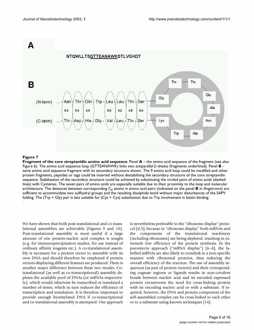
Display system based on the SAPV protein vectorWe have identified a loop in the amino acid sequence ofstreptavidin (Figures 6 and 7) which can be used as a sitefor SAPV extension, modification or for expressing otherprotein fragments, peptides or tags. We have illustratedhow our system can be used for displaying proteins andprotein fragments (Figures 9 and 10). Generally speaking,
however, "displaying" artificial sequences may change thefolding of the SAPV. To avoid this, the secondary structureelements of the SAPV could be additionally stabilised andpositioned by one or more disulphide bonds. Inparticular, one (or more) of the 8 amino acids of thestreptavidin core sequence, immediately preceding theloop (NTQWLLTS) and one (or more) of the respective 8
Figure 4Detection of the tagged SAPV on Western blots. SAPV protein vector was tagged with AFP sequence. In vitro T&T reactions were run either at different temperatures (left panel) or different amounts of DNA was added to the reactions (right panel). Detection of the tagged SAPV was done using anti-GFP Rabbit polyclonal antibody (from AbCam). The right most lane (right panel) represents T&T reaction containing 2 ug of unpurified PCR products. Second lane from the right – 2 ug of DNA was ethanol-precipitated prior to T&T, following lanes – 3 ug, 6 ug, 12 ug, 18 ug and 30 ug DNA, all were ethanol-precipitated prior to T&T.
Page 6 of 16(page number not for citation purposes)

Journal of Nanobiotechnology 2003, 1 http://www.jnanobiotechnology.com/content/1/1/1
amino acids C-terminal to the loop (STLVGHDT) couldbe substituted with Cysteine residues (Figure 7). This ispossible because the distances between respective pairs ofamino acids in these two antiparallel strands (the two 8amino acid stretches) and their orientation should allowpairwise Cysteine substitutions without major changes tothe streptavidin folding pattern (Figure 7).
Self-assemblyIf required, the efficiency of the self-assembly processcould be manipulated by regulating a processivity of thetranscription and/or translation reactions. This could beachieved by varying the concentration of the tRNAspresent in the reaction mixture and the use of respectivecodons in RNA (or DNA) sequences coding for the pro-teins processed. The translation reaction can be paused or
stopped if required tRNA(s) is not available. Protein syn-thesis will continue after the missing tRNAs are added tothe translation reaction. This could allow a user to manip-ulate the speed of synthesis and folding of the nascentprotein chains and also to regulate protein vector bindingto the nucleic acid molecules as well as protein-protein in-teractions (in protein complex formation). For exampletranslation can be paused or slowed down after the assem-bly domain of the protein vector is produced, to allowbinding to a nucleic acid or solid support or another pro-tein, before the complete protein is translated and re-leased from the ribosome. In vitro translation could alsobe slowed down by addition of a short complementarynucleic acid strand, the technique used in vivo and knownas the antisense approach [10–13].
Figure 5Assembly of the SAPV protein vector with biotinylated DNA. Panel A – biotinylated DNA was added to the SAPV vector. The assembled complexes were separated from the rest of the reaction components by filtration through protein-bind-ing filters. The four washes and the eluate were tested by PCR. Large amount of the DNA was eluted indicating that bioti-nylated DNA was retained by the SAPV vector. Panel B – same as in panel A, except that non-biotinylated DNA was added to the SAPV. Arrows on the left of both gels indicate the expected size (position) of the amplified products corresponding to the assembled DNAs. Panel – C, same as panel A, but data pooled from three experiments. The band intensities were determined using GeneSnap and fluorescent imager from SynGene (Cambridge, UK). All values shown were normalised to the DNA sam-ple from the 1st wash (which also contained a flow-through fraction of the total loaded DNA, marked by asterisk). Error bars represent standard deviation (n = 3). Large amounts of the DNA were eluted in all three experiments (the right most bar) con-firming that biotinylated DNA was retained by the SAPV vector. Panel D – same as panel B, but data pooled from three exper-iments. No biotinylated DNA was co-precipitated (the right most bar).
Page 7 of 16(page number not for citation purposes)

Journal of Nanobiotechnology 2003, 1 http://www.jnanobiotechnology.com/content/1/1/1
Figure 6Structure of the streptavidin protein. Panel A – A 3D structure of the streptavidin protein (PDP Acc.No. 1STP) showing a biotin binding site (left) and the side-most amino acid chain loop (both panels). Panel B – Visualisations of the crystal struc-ture of the Streptavidin protein, obtained from the PDB Protein Data Bank http://www.pdb.org/. Nine amino acids forming the loop, which can be modified or substituted are identified (bottom right corner). The loop (see also Figure 7) was used in the design of a display system based on the SAPV.
Page 8 of 16(page number not for citation purposes)
Journal of Nanobiotechnology 2003, 1 http://www.jnanobiotechnology.com/content/1/1/1
We have shown that both post-translational and co-trans-lational assemblies are achievable (Figures 9 and 10).Post-translational assembly is most useful if a largeamount of one protein-nucleic acid complex is sought(e.g. for immunoprecipitation studies, for use instead ofordinary affinity reagents etc.). A co-translational assem-bly is necessary for a protein vector to assemble with itsown DNA and should therefore be employed if proteinvectors displaying different features are produced. There isanother major difference between these two modes. Co-translational (as well as co-transcriptional) assembly de-pletes the available pool of DNAs (or mRNAs respective-ly), which would otherwise be transcribed or translated anumber of times, which in turn reduces the efficiency oftranscription and translation. It is therefore important toprovide enough biotinylated DNA if co-transcriptionaland co-translational assembly is attempted. Our approach
is nevertheless preferable to the "ribosome display" proto-col [4,5], because in "ribosome display" both mRNAs andthe components of the translational machinery(including ribosomes) are being depleted, resulting in ex-tremely low efficiency of the protein synthesis. In thepuromycin approach ("mRNA display") [6–8], the la-belled mRNAs are also likely to crosslink in a non-specificmanner with ribosomal proteins, thus reducing theoverall efficiency of the reaction. The use of assembly se-quences (as part of protein vectors) and their correspond-ing cognate regions or ligands results in non-covalentbonds between nucleic acid and its encoded expressedprotein circumvents the need for cross-linking proteinwith its encoding nucleic acid or with a substrate. If re-quired, however, the DNA and protein component of theself-assembled complex can be cross-linked to each otheror to a substrate using known techniques [14].
Figure 7Fragment of the core streptavidin amino acid sequence. Panel A – the amino acid sequence of the fragment (see also Figure 6). The amino acid sequence loop (GTTEANAWK) links two antiparallel β-sheets (fragments underlined). Panel B – same amino acid sequence fragment with its secondary structure shown. The 9 amino acid loop could be modified and other protein fragments, peptides or tags could be inserted without destabilising the secondary structure of the core streptavidin sequence. Stabilisation of the secondary structure could be achieved by substituting the circled pairs of amino acids (dashed lines) with Cysteines. The seven pairs of amino acids are especially suitable due to their proximity to the loop and molecular architecture. The distances between corresponding Cβ atoms in amino acid pairs (indicated on the panel B in Angstroms) are sufficient to accommodate two sulfhydryl groups and the resulting disulphide bond without major disturbances of the SAPV folding. The (Trp + Gly) pair is less suitable for (Cys + Cys) substitution due to Trp involvement in biotin binding.
Page 9 of 16(page number not for citation purposes)

Journal of Nanobiotechnology 2003, 1 http://www.jnanobiotechnology.com/content/1/1/1
ConclusionsProtein vectors and the principle of self-assembly de-scribed here provide new exciting possibilities in molecu-lar biology research. Because proteins can be directlylinked to their nucleic acids, such self-assembled com-
plexes can be used for cloning proteins or protein affinityreagents (antibody, their fragments or antibody mimics,etc.). The ability to quickly generate thousands of affinityreagents may be a crucial factor in the development ofprotein affinity arrays [15–17]. Also, the ability to quickly
Figure 8BCMP84 immunoprecipitation on protein A-conjugated glass beads and on nitrocellulose membrane. Recom-binant BCMP84 was incubated with beads or nitrocellulose that had BCMP84 antibody bound to them. Samples from the first wash, 4th wash and the eluate from these incubations were run as indicated. The washes and eluates from the beads are on the left and the washes and eluates from the filter paper are on the right. White asterisks denote immunoprecipitated and eluted BCMP84 protein, which is not detected in the last (4th) wash prior to elution (both blots). The 1st wash, as expected, includes recombinant BCMP84 as indicated by the band at approximately 40 kDa. This band is not present in the 4th wash. Comparable amounts of the BCMP84 protein are present in the eluate of both the beads and nitrocellulose (marked with asterisks), indicat-ing that it was selectively retained.
Page 10 of 16(page number not for citation purposes)

Journal of Nanobiotechnology 2003, 1 http://www.jnanobiotechnology.com/content/1/1/1
determine nucleic acid sequence and therefore to identifythe associated proteins could be extremely helpful forprotein affinity selection or directed protein evolution. Al-ternatively, proteins can be assembled with labelled nu-cleic acids, which can be done either co-translationally orpost-translationally; the proteins thus become labelled toa high specific activity through their association with theirnucleic acids, without being chemically modified. Thiscould result in not only a higher specific activity of label-ling but would also avoid the chemical modification that
takes place when proteins are labelled directly. Nucleicacid molecules associated with proteins could also im-prove sensitivity of detection of such proteins down to asingle molecule level, by enabling detection using PCR.This could surpass all other known protein detection tech-niques, of which only immunogold detection in combi-nation with electron microscopy is capable of detectingindividual protein molecules [18–20].
Table 1: Engineered SAPV displaying Albumin and BCMP84 peptides.
Construct: Sequence fragment*
unmodified SAPV* ...NTQWLLTSGTTEANAWKSTLVGHDT...SAPV-Alb5 ...NTQWLLTSGHPYFYAPELLFFAKSTLVGHDT...SAPV-84 ...NTQWLLTSGEGGKETLTPSELRDLVSTLVGHDT...
* Underlined in the SAPV sequence (only) is the fragment, which was modified.
Figure 9Co-immunoprecipitation of the assembled SAPV-84 Co-immunoprecipitation of the assembled SAPV-84 assayed by PCR amplification of the co-eluted SAPV-84 DNAs. Filled bar – normalised signal corresponding to the precipitation of the SAPV-84 on immobilised anti-BCMP84 antibody. Open bar – relative signal strength corresponding to the eluate from anti-Albumin antibodies. Error bar represents standard deviation (n = 5).
Page 11 of 16(page number not for citation purposes)

Journal of Nanobiotechnology 2003, 1 http://www.jnanobiotechnology.com/content/1/1/1
Materials and MethodsDesign of the SAPV protein vectorFull length nucleotide sequence coding for the SAPV wasproduced using overlapping synthetic oligonucleotides(Sigma-Genosys) and 3 rounds of PCR amplification. Firstround: a mixture of oligonucleotides (here and later seeTable 2 for sequences) SA-1F + SA-2R + SA-3R + SA-4R +SA-5R + SA-6R + SA-7R + SA-8R was used (20 ul of amixture, each oligo at 1.25 pmol/ul) plus 5 ul of 10x Pfxbuffer, plus 2 ul 50 mM MgSO4, plus 0.5 ul 20 mMdNTPs, plus 0.5 ul Pfx polymerase and H2O to a total of
50 ul. Both here and later, only proofreading DNApolymerase (Pfx polymerase, Invitrogen) was used. Cy-cling: following 5' at 96°C, 20 cycles were done as follows95°C for 30", 72°C (with a 2°C decrement per cycle) for30", 72°C for 30" (with 1" increment per cycle). Final in-cubation was for 1' at 72°C. Second round: the DNAswere re-amplified (5 ul of original PCR product per reac-tion was used as a template) using a mixture of oligonu-cleotides SA-1F + SA-5R + SA-6R + SA-7R + SA-8R (1 uleach at 10 pmol/ul each). Other PCR conditions were asdescribed above. Cycling: following 5' at 96°C, 15 cycles
Figure 10Immunoprecipitation of the self-assembled SAPV vectors Immunoprecipitation of the self-assembled SAPV vectors (SAPV-84, SAPV-Alb5 and unmodified SAPV). Panel A – The eluates from anti-Albumin beads (left) or from anti-BCMP84 beads (right) were assayed by PCR and equal amounts of the resultant products were run on 2% agarose gel containing ethid-ium bromide. The gel indicates that anti-Albumin beads retain exclusively SAPV-Alb5 construct, whilst anti-BCMP84 beads pre-cipitate SAPV-84 construct. The unmodified "empty" SAPV vector was retained by neither anti-Albumin nor anti-BCMP84 beads. Panel B – Immunoprecipitation of the self-assembled SAPV vectors (SAPV-84, SAPV-Alb5 and unmodified SAPV), data pooled from 3 experiments. The eluates from anti-Albumin beads (top panel) or from anti-BCMP84 beads (bottom panel) were assayed by PCR and quantified by agarose gel electrophoresis. Band intensities were determined using GeneSnap and flu-orescent imager from SynGene (Cambridge, UK). All values shown are normalised to the strongest DNA sample in each case (marked by asterisks). Error bars represent standard deviation (n = 3). Largest eluted amounts of DNA (and therefore the cor-responding SAPVs) were: SAPV-Alb5 (eluted from anti-Albumin beads) and SAPV-84 (eluted from anti-BCMP84 beads). The results confirm that anti-Albumin beads precipitate SAPV-Alb5 self-assembled construct, whilst anti-BCMP84 beads retain SAPV-84 construct.
Page 12 of 16(page number not for citation purposes)

Journal of Nanobiotechnology 2003, 1 http://www.jnanobiotechnology.com/content/1/1/1
were done as follows 96°C for 1', 55°C (with 1°C decre-ment per cycle) for 30", 72°C for 30" (with 1" incrementper cycle). Final incubation was 1' at 72°C. Third round:5 ul of previously obtained DNA was used as a templatefor a re-amplification using T7-F and T7TER-R primers.Cycling was as follows: 5' at 96°C, 25 cycles were done asfollows 96°C for 1', 40°C for 30", 72°C for 1'. Final incu-bation was 5' at 72°C. The amplified SAPV DNA fragmentwas cloned into the TOPO4BLUNT vector. Sequence ofthe final construct is shown on Figure 2.
Expression of the SAPV protein vector was performed us-ing bacterial coupled in-vitro Transcription/Translation(T&T) kit obtained from Roche (RTS 100 E. coli HY). Invitro T&T synthesis reaction were assembled according tomanufacturer recommendations unless stated otherwise.Amounts of the SAPV DNA sufficient for the in vitro T&Twere routinely obtained by PCR amplification using T7-F(forward) primer and T7TER-R reverse primer (see Figure2). Either biotinylated or unmodified primers were used.Cycling was typically as follows: 5' at 96°C, followed by20 cycles of 96°C for 1', 40°C for 30", 72°C for 1'. Finalincubation was 5' at 72°C.
Tagging of the SAPV protein vector with autofluorescent proteinTagged SAPV DNA was generated by PCR, similarly to theuntagged SAPV. In particular, a linear fragment of theSAPV was amplified using 3 ul of 16 pmol/ul M13F prim-er (part of the TOPO vector, upstream of the T7-F primerof the SAPV insert), 0.5 ul of 100 pmol/ul SA-11R primer
and 0.5 ul of the untagged SAPV DNA (0.5 ug/ul). Cyclingwas as follows: 6' at 96°C, and 15 cycles of 96°C for 1',40°C for 30", 72°C for 1'. Final incubation was 5' at72°C. Linear fragment of AFP was amplified using 0.5 ulof 100 pmol/ul AFP-1F primer, 0.5 ul of 100 pmol/ul AFP-3R primer and AFP DNA as a template. Cycling was as fol-lows: 7' at 96°C, 55°C for 30", 72°C for 1' following by15 cycles of 96°C for 1', 72°C for 1'30". Final incubationwas 5' at 72°C. Transcription terminator sequence wasadded to the linear AFP fragment by PCR using 0.5 ul of100 pmol/ul AFP-1F primer and 5 ul of 10 pmol/ul SA-8Rprimer and a linear APV fragment (see above) as a tem-plate. Cycling was as follows: 6' at 96°C, 2 cycles of of96°C for 1', 40°C for 30", 72°C for 1' and 15 cycles of96°C for 1', 72°C for 1'30". Final incubation was 5' at72°C. Full length tagged SAPV DNA was obtained byjoining the untagged SAPV and modified AFP by PCR us-ing primers T7-F and TFTER-R and a mixture of the SAPVfragment (M13F/SA-11R PCR fragment) and of the linearand modified AFP fragment (AFP-1F/SA-8R PCR frag-ment) as a template. Cycling was as follows: 5' at 96°C, 6cycles of 96°C for 1', 72°C (with decrement of 5°C per cy-cle) for 30", 72°C for 1' (with 5" increment per cycle) and15 cycles of 96°C for 1', 40°C for 30" and 72°C for 1'30".Final incubation was 5' at 72°C. The amplified DNA wascloned into the TOPO4BLUNT vector. A long 5' UTR wasadded to the SAPV DNA by PCR. Firstly, a tagged andcloned SAPV DNA was used as a template to amplify afragment lacking the T7 RNA polymerase binding site byPCR using primers RBS-1F and T3. Cycling was as follows:5' at 96°C, 15 cycles of 96°C for 1', 40°C for 30" and
Table 2: Oligonucleotide primers used in the design of the SAPV, tagged SAPV and SAPV display system.
SA-1F aaattaatacgactcactatagggagaaggagatataccatggaggccggcatcaccggcacctggtacaacSA-2R ttccggtcagggcgccgtcggcgcccgcggtcacgatgaaggtcgagccgagctggttgtaccaggtgccggtgatgSA-3R ggacgtagcggctctcggcgttgccgacggccgactcgtaggttccggtcagggcgccgtcSA-4R gtggccggggcgctgtcgtaacgaccggtcaggacgtagcggctctcggSA-5R tcgcggagtgggcgttgcggtagttattcttccaggccaccgtccaaccgagggcggtgccgctgccgtcggtggccggggcgctgtSA-6R gccggaggtcagcagccactgggtgttgatcctcgcctcggcgccgccgacgtactggccgctccacgtggtcgcggagtgggcgtSA-7R ttccaccttggtgaaggtgtcgtggccgaccagcgtggacttccaggcgttggcctcggtggtgccggaggtcagcaSA-8R aaaaaacccctcaagacccgtttagaggccccaaggggttatgctagttatcattcattcattccaccttggtgSA-10R aaaaaacccctcaagacccgtttagaggccccaaggggttatgctagttatcattcattcattccaccttggtgaaggtgtcgtggccgaccagcgtggaSA-11R tcgtggccgaccagcgtggaT7-F aaattaatacgactcactatT7TER-R aaaaaacccctcaagacccM13-F gtaaaacgacggccagM13-R caggaaacagctatgacAFP-1F tccacgctggtcggccacgaccgaattcggggaggcggaggtgAFP-3R ttcattccaccttggtgcacgggggaggggcaaacaacRBS-1F agaaggagatataccatRBS-2R catggtatatctccttctT3 attaaccctcactaaagggaLOOP-Alb5-R tcgtggccgaccagcgtggatttagcaaaaaataataattcaggagcataaaaataaggatggccggaggtcagcagccactggLOOP-84-1R tcgtggccgaccagcgtggaaactaaatctcttaattcagaaggagttaaagtttctttaccaccttcgccggaggtcagcagccactgg
Page 13 of 16(page number not for citation purposes)

Journal of Nanobiotechnology 2003, 1 http://www.jnanobiotechnology.com/content/1/1/1
72°C for 1'30". Final incubation was 5' at 72°C.Following that, a fragment of the pIVEX vector (ROCHE)was used as a template for PCR amplification using T7-Fand T7TER-R primers. Cycling was as follows: 5' at 96°C,15 cycles of 96°C for 1', 40°C for 30" and 72°C for 1'. Fi-nal incubation was 5' at 72°C. Amplified fragment of thepIVEX vector was cloned into TOPO4BLUNT vector and along 5' UTR was excised by PCR using primers RBS-2R andM13-R oligo. Cycling was as follows: 5' at 96°C, 15 cyclesof 96°C for 1', 40°C for 30" and 72°C for 30". Final incu-bation was 5' at 72°C. The amplified UTR fragment wasjoined with the linearised tagged SAPV DNA (RBS-1F/T3fragment, see above) by PCR using primers T7-F andT7TER-R and a mixture of the linearised tagged SAPV DNA(RBS-1F/T3 fragment) and the cloned re-amplified pIVEX(RBS-2R/M13-R) fragment as a template. Cycling was asfollows: 5' at 96°C, 10 cycles of of 96°C for 1', 72°C (withdecrement of 3°C per cycle) for 30", 72°C for 1'30" and10 cycles of 96°C for 1', 40°C for 30" and 72°C for 1'30".Final incubation was 5' at 72°C. The amplified DNA wascloned into the TOPO4BLUNT vector. Final sequence ofthe engineered tagged SAPV is shown on Figure 3.
In vitro coupled transcription and translation reactionsFor temperature optimisation the T&T reactions (20 ul fi-nal volume each) were assembled according to manufac-turer recommendations, but synthesis was done at a rangeof temperatures (2 ug of the SAPV-AFP DNA was added toeach tube, Figure 4, left panel). To optimise the amount ofDNA used for T&T reactions, different amounts of DNAwere added (by Ethanol precipitating the precalculatedamount of the PCR product prior to assembling the in vit-ro T&T reaction, Figure 4, right panel). Reactions were runovernight. 5 ul aliquots of each of the T&T reactions wereloaded onto a precast 4–12% NuPAGE gel (Invitrogen).The proteins were resolved by SDS-polyacrylimide gelelectrophoresis and transferred onto nitrocellulose (0.2uM pore size, Invitrogen) using an Xcell SureLock Minicelland Blot Module according to the manufacturer's instruc-tions. The blot was then blocked for 1 hour in TBST (TBSplus 0.1% tween-20) with 2% powdered milk and probedwith a 1:3000 dilution of AbCam anti-GPF Rabbit poly-clonal (1 hr room temp) in TBST/milk. After washing inTBST (3x, 5–10' each wash) the blot was probed with1:6000 HRP-labelled Anti-Rabbit IgG, (Amersham Phar-macia) in TBST/milk (1 hr room temp), washed again andthen developed using ECL (Amersham) according to themanufacturer's instructions and exposed to ECL Hyper-film (Amersham).
Assembly of the SAPV protein vector with biotinylated DNAUntagged SAPV was obtained by means of the in vitro T&Tas described above and using SAPV DNA lacking STOP co-dons (see legend to Figure 2). This DNA was obtained by
PCR using M13F and SA-7R primers and tagged SAPVDNA (Figure 3) as a template. Cycling was as follows: 6' at96°C, and 15 cycles of 96°C for 1', 40°C for 30", 72°C for1'. Final incubation was 5' at 72°C. SAPV DNA was usedfor the T&T reaction. Following an overnight incubation,the T&T reaction was spun for 3 min at 15,000 RPM in amicrocentrifuge to precipitate insoluble components ofthe in vitro reaction mixture. Clear supernatant was trans-ferred to fresh tube prior to adding DNAs for assembly. Bi-otinylated and non-biotinylated DNAs for assembly weregenerated by PCR using T7 forward primer (biotinylatedor non-biotinylated, respectively) and non-biotinylatedT7TER-R primers and the long DNA coding the taggedSAPV (Figure 3) as a template (all other conditions wereas described previously). The longer DNAs were chosenfor assembly reactions to avoid non-specific backgrounddue to SAPV DNA used for in vitro T&T. DNAs were etha-nol-precipitated prior to assembly and redissolved in wa-ter at 1 ug/ul. Cleared T&T supernatants were aliquoted(10 ul per tube) and DNAs (biotinylated/non-biotinylat-ed) were added (5 ug per tube). Assembly reactions wereallowed to run overnight at +4°C. Protein-DNA complex-es were separated from free DNAs by filtration throughprotein-binding microcentrifuge filters ("Ultrafree-MCProbind Units" modified PVDF, Millipore). After 4 wash-es (by flow through filtration) the retained materials wereeluted by incubation for 30' with gentle agitation in 50 ulvolume 0.1 × TAE. Eluted DNAs were detected by PCR asfollows: 10 ul of each of the wash through and eluatesfrom each assembly reaction were amplified in parallel us-ing primers T7-F and T7TER-R. 35 cycles of amplificationof 1' at 96°C, 30" at 40°C and 1'30" at 72°C were carriedout. Amplified products were separated on 2.5% agarosegels containing Ethidium Bromide. Equal amounts ofeach PCR reaction were loaded onto each lane (Figure5A,5B).
Display system based on the SAPV protein vectorTo illustrate the "display" capabilities of the SAPV, we en-gineered SAPV displaying peptide fragments of Albuminand BCMP84 proteins (Table 1). The DNA coding for themodified SAPV were obtained by PCR using SAPV DNA asa template and synthetic oligonucleotide primers M13Fplus loop-84-1R (to make SAPV-84 construct) or M13Fplus loop-Alb5-R (to make SAPV-Alb5 construct), see Ta-ble 1. Stop codons were added to both constructs by PCRusing M13F and SA-10R primers. Cycling was as follows:5' at 96°C, and 30 cycles of 96°C for 30", 54°C for 30",72°C for 30". Final incubation was 5' at 72°C. Largeamounts of the full length DNAs coding for all SAPVvariants (both biotinylated and non-biotinylated) wereproduced for in vitro T&T by PCR using T7-F forward andT7TER-R reverse primers as described earlier for SAPVvector.
Page 14 of 16(page number not for citation purposes)

Journal of Nanobiotechnology 2003, 1 http://www.jnanobiotechnology.com/content/1/1/1
Co-immunoprecipitation system for affinity separationsA co-immunoprecipitation system for affinity separationswas designed to quickly separate different SAPVs. To testthe system a recombinant BCMP84 was used. We testedglass bead-based and nitrocellulose-based systems sepa-rately as follows. Twenty microlitres of protein A-conju-gated glass beads (PROSEP-A, Millipore) were washed 3times in 1 ml PBS then incubated with 20 ul of anti-BCMP84 antibody (rabbit polyclonal, 110 ug/ml). Nitro-cellulose was wetted in deionised water for 5' then cut into3-mm squares and incubated with 20 ul of anti-BCMP84antibody. The beads and nitrocellulose squares werewashed twice in 1 ml PBS then blocked by incubation in3% powdered milk in PBS for 30'. Blocking buffer was re-moved and the beads and nitrocellulose squares wereincubated for 90' with 2.5 ug of recombinant BCMP84 inPBS with 0.5% powdered milk. Beads and proteinsolution were transferred to Vectaspin microcentrifugetubes containing a 0.2 um pore Anapore membrane(Millipore) and the beads were washed 3 times by resus-pension in 600 ul PBS followed by centrifugation. After afinal wash in 50 ul PBS, the beads were resuspended in 50ul elution buffer (100 mM glycine, pH 2.45), shaken peri-odically over 10' and then spun. The eluted sample wasneutralised by the addition of 13 ul 2 M NaOH. One mil-lilitre PBS was added to the incubations of the nitrocellu-lose squares and protein. The nitrocellulose squares werewashed 2 times by transferral to 1 ml fresh PBS followedby brief shaking. After a final wash in 50 ul PBS, 50 ul elu-tion buffer was added to the nitrocellulose which wasthen shaken periodically over 10' before the eluate was re-moved and then neutralised by the addition of 13 ul 2 MNaOH. 10 ul of the eluate, the final wash and the firstwash were run on a 4–12% NuPage 1D polyacrylymidegel (Invitrogen) under non-reducing conditions andtransferred to a nitrocellulose membrane (0.2 um poresize, Invitrogen) by western blotting. After blocking thenitrocellulose by incubation in 2% powdered milk inTBST (TBS with 0.1% tween-20) the blots were probed us-ing 0.5 ug/ml anti-BCMP84 in TBST plus 2% powderedmilk. The blots were washed extensively in TBST andprobed with an HRP-conjugated, anti-rabbit secondaryantibody (1:6000 dilution, Amersham) washed extensive-ly and then developed using ECL (Amersham PharmaciaBiotech) according to the manufacturer's instructions (seeFigure 8).
Affinity precipitation of the SAPVs displaying BCMP84 peptideIn vitro T&T (50 ul volume) was run as described aboveusing in-vitro coupled Transcription/Translation (T&T)kit from Roche (RTS 100 E. coli HY) and DNA coding forthe SAPV-84 (see Table 1). Following an overnight incu-bation at 21°C the reaction was spun down for 3' to sep-arate insoluble components of the T&T from the
synthesised SAPV. The cleared T&T supernatant was keptat 4°C until assembly reaction. 20 ul of the PCR-amplifiedunpurified biotinylated SAPV-84 DNA was added to 20 ulof the cleared supernatant and the assembly mixture wasincubated overnight at 4°C. The assembled SAPV-DNAcomplex was immunoprecipitated with either anti-BCMP84 or anti-albumin (control) antibodies as follows.Nitrocellulose was wetted in deionised water for 5' thencut into 3-mm squares and incubated with either 20 ul ofanti-albumin antibody or 20 ul of anti-BCMP84 antibody.The nitrocellulose squares were washed twice in 1 ml PBSthen blocked by incubation in 2% powdered milk and 0.1mg/ml tRNA in PBS for 30 minutes. Blocking buffer wasremoved and the nitrocellulose squares were incubatedfor 60' with the assembled SAPV-DNA complex. The ni-trocellulose was washed three times in wash buffer (1 mlPBS supplemented with 0.01 mg/ml tRNA and 2%powdered milk). After a final wash in 50 ul wash buffer,the nitrocellulose squares were incubated with in 50 ulelution buffer (80 mM glycine, pH 2.45) supplementedwith 0.4% powdered milk and 0.01 ug/ml tRNA. The elut-ed samples were neutralised by the addition of 11 ul 2 MNaOH. The presence of the SAPV-84 in the eluted sampleswas detected by PCR amplification of the SAPV-84 DNAsusing primers T7-F and T7TER-R. All reactions were run inparallel under identical conditions. 33 cycles of amplifica-tion of 30" at 96°C, 30" at 42°C and 30" at 72°C werecarried out. PCR products were run on a 2% gel and bandintensities determined using GeneSnap and fluorescentimager from SynGene. The results of 5 measurements arepresented on Figure 9.
Self-assembly of protein vectors with their DNAs and affin-ity separationIn vitro synthesis and self-assembly of the SAPV-84, SAPV-Alb5 and "empty" SAPV-only (unmodified) protein vec-tors were done using RTS 100 E. coli HY kit from Roche.Optimal reaction conditions were used (as determinedearlier, see Figure 4), except that each reaction contained~16 ug of biotinylated SAPV DNAs (SAPV-84, SAPV-Alb5and SAPV-only coding DNAs) and a total volume of eachreaction was 100 ul. The combined in vitro [T&T self-as-sembly] reactions were allowed to run at 21°C. Followingan overnight incubation the reactions were cleared byspinning for 3' at 15,000 RPM in a microcentrifuge andtwo 40 ul aliquots from each reaction were transferred tofresh tubes. The SAPVs were precipitated from these aliq-uots by incubation with either anti-BCMP84 or anti-Albu-min antibodies using protein A-conjugated glass beads(120 ul, PROSEP-A, Millipore). Beads were prepared bywashing 3 times in 1 ml PBS. They were then incubatedwith either 600 ul of anti-albumin antibody (rabbit poly-clonal, 2.8 mg/ml) supplemented with 0.1 mg/ml tRNAor 600 ul of anti-BCMP84 antibody (rabbit polyclonal,110 ug/ml) supplemented with 0.1 mg/ml tRNA for 90
Page 15 of 16(page number not for citation purposes)

Journal of Nanobiotechnology 2003, 1 http://www.jnanobiotechnology.com/content/1/1/1
Publish with BioMed Central and every scientist can read your work free of charge
"BioMed Central will be the most significant development for disseminating the results of biomedical research in our lifetime."
Sir Paul Nurse, Cancer Research UK
Your research papers will be:
available free of charge to the entire biomedical community
peer reviewed and published immediately upon acceptance
cited in PubMed and archived on PubMed Central
yours — you keep the copyright
Submit your manuscript here:http://www.biomedcentral.com/info/publishing_adv.asp
BioMedcentral
minutes. All subsequent washes, incubations and elutionswere carried out in the presence of 0.01 mg/ml tRNA.Both sets of beads were washed 3 times and then split intoseparate tubes each containing 20 ul of beads. Eachaliquot of beads was then incubated with 40 ul of super-natant from one of the self-assembled reactions for 90minutes. Each sample was then transferred to a macropo-rous Wizard® Minicolumn filtration unit (Promega). Thecolumn with beads was washed with 80 ul of PBS by cen-trifugation, following which each column was washedwith 40 ml of PBS using a 50 ml syringe. The filtrationunit was transferred into a microcentrifuge tube and thebeads were washed with 50 ul PBS by centrifugation. Fi-nally the beads were resuspended in 50 ul elution buffer(100 mM glycine, pH 2.45) and then the eluates werecollected by centrifugation and neutralised with 13 ul of 2M NaOH. The eluted SAPV-DNA complexes were ethanolprecipitated and assayed by PCR using T7-F and T7TER-Rprimers. Equal amounts of the PCR reactions were run on2% agarose gel (see Figure 10).
References1. Emel'ianov VV, Kalinin VN, Girdo BM and Tikhonenko TI Colicin El
synthesis in a cell-free system of paired transcription –translation. Biull Eksp Biol Med 1976, 82:1324-1326
2. Spirin AS Cell-free systems of polypeptide biosynthesis andapproaches to the evolution of translation apparatus. Orig Life1976, 7:109-118
3. Erickson AH and Blobel G Cell-free translation of messengerRNA in a wheat germ system. Methods Enzymol 1983, 96:38-50
4. He M and Taussig MJ Antibody-ribosome-mRNA (ARM) com-plexes as efficient selection particles for in vitro display andevolution of antibody combining sites. Nucleic Acids Res 1997,25:5132-5134
5. He M, Menges M, Groves MA, Corps E, Liu H, Bruggemann M andTaussig MJ Selection of a human anti-progesterone antibodyfragment from a transgenic mouse library by ARM ribosomedisplay. J Immunol Methods 1999, 231:105-117
6. Xu L, Aha P, Gu K, Kuimelis R, Kurz M, Lam T, Lim A, Liu H, LohseP, Sun L, Weng S, Wagner R and Lipovsek D Directed evolution ofhigh-affinity antibody mimics using mRNA display. Chem Biol2002, 9:933
7. Roberts RW and Szostak JW RNA-peptide fusions for the in vit-ro selection of peptides and proteins. Proc Natl Acad Sci U S A1997, 94:12297-12302
8. Liu R, Barrick JE, Szostak JW and Roberts RW Optimized synthe-sis of RNA-protein fusions for in vitro protein selection. Meth-ods Enzymol 2000, 318:268-293
9. Argarana CE, Kuntz ID, Birken S, Axel R and Cantor CR Molecularcloning and nucleotide sequence of the streptavidin gene. Nu-cleic Acids Res 1986, 14:1871-1882
10. Blake KR, Murakami A and Miller PS Inhibition of rabbit globinmRNA translation by sequence-specificoligodeoxyribonucleotides. Biochemistry 1985, 24:6132-6138
11. Haeuptle MT, Frank R and Dobberstein B Translation arrest by ol-igodeoxynucleotides complementary to mRNA coding se-quences yields polypeptides of predetermined length. NucleicAcids Res 1986, 14:1427-1448
12. Marcus-Sekura CJ, Woerner AM, Shinozuka K, Zon G and QuinnanGV Jr Comparative inhibition of chloramphenicol acetyl-transferase gene expression by antisense oligonucleotide an-alogues having alkyl phosphotriester, methylphosphonateand phosphorothioate linkages. Nucleic Acids Res 1987, 15:5749-5763
13. Vlasov VV and Iurchenko LV Mechanism for suppression mRNAtranslation with antisense oligonucleotides. Mol Biol (Mosk)1990, 24:1157-1161
14. Wong SS A Chemistry of Protein Conjugation and Cross-Linking. CRC Press 1993,
15. Soloviev M EuroBiochips: spot the difference. Drug Discov Today2001, 6:775-777
16. Barry R, Scrivener E, Soloviev M and Terrett J Chip-Based Pro-teomics Technologies. Int Genomic Proteomic Technology 2002,Feb:14-22
17. Scrivener E, Barry R, Platt A, Calvert R, Masih G, Hextall P, SolovievM and Terrett J Peptidomics: a new approach to affinity pro-tein microarrays. Proteomics 2003,
18. Nusser Z, Roberts JD, Baude A, Richards JG and Somogyi P Relativedensities of synaptic and extrasynaptic GABAA receptors oncerebellar granule cells as determined by a quantitative im-munogold method. J Neurosci 1995, 15:2948-2960
19. Baude A, Nusser Z, Molnar E, McIlhinney RA and Somogyi P High-resolution immunogold localization of AMPA type glutama-te receptor subunits at synaptic and non-synaptic sites in rathippocampus. Neuroscience 1995, 69:1031-1055
20. Bernard V, Somogyi P and Bolam JP Cellular, subcellular, and sub-synaptic distribution of AMPA-type glutamate receptor sub-units in the neostriatum of the rat. J Neurosci 1997, 17:819-833
Page 16 of 16(page number not for citation purposes)
Copyright © 2022 FDOKUMEN