
Citation: Molecular Therapy–Nucleic Acids Exploiting the Intron-splicing Mechanism of Insect Cells...
10
Citation: Molecular Therapy–Nucleic Acids (2012) 1, e57; doi: 10.1038/mtna.2012.48 © 2012 American Society of Gene & Cell Therapy All rights reserved 2158-3188/11 www.nature.com/mtna 1 Virovek, Inc., Hayward, California, USA. Correspondence: Haifeng Chen, R&D, Virovek, Inc., 3521 Investment Blvd. Suite 1, Hayward, California 94545, USA. E-mail: [email protected] Keywords: adeno-associated virus (AAV); baculovirus; cancer; gene therapy; toxin Received 13 August 2012; accepted 4 October 2012; advance online publication 27 November 2012. doi:10.1038/mtna.2012.48 METHODS: ORIGINAL ARTICLE Exploiting the Intron-splicing Mechanism of Insect Cells to Produce Viral Vectors Harboring Toxic Genes for Suicide Gene Therapy Haifeng Chen 1 Two mammalian introns, the human growth hormone intron and the Simian virus 40 large T antigen intron, were inserted into the coding sequences of diphtheria toxin fragment A (DT-A) and barnase (Bar), respectively, to disrupt their open-reading frames (ORFs). Expression of these two toxic proteins were totally abolished, which enabled the production of normal levels of recombinant baculoviral and adeno-associated viral (AAV) vectors in insect cells. When these viral vectors were introduced into mammalian cells, the introns were spliced out and the toxic proteins were expressed, which resulted in apoptosis in mammalian cells. This is the first report to show that viral vectors harboring toxin genes can be produced at normal levels by exploiting the intron-splicing mechanism of insect cells. Furthermore, viral vectors carrying the DT-A gene under control of tumor-specific promoters were able to exert tumor-specific cell killing. This novel method to produce viral vectors harboring toxic genes under control of tumor-specific promoter offers a powerful tool for further research, as well as for the development of toxin-based suicide gene therapy drugs. Molecular Therapy–Nucleic Acids (2012) 1, e57; doi:10.1038/mtna.2012.48; published online 27 November 2012 Subject Category: Methods section Introduction Toxic genes and proteins encoded by such genes have been used or proposed for use in disease therapies, such as in cancer therapies involving expression of genes that cause cell death (“suicide gene therapy”). 1 Certain proteins encoded by toxic genes can kill a host cell at very low levels of expres- sion. For example, a single molecule of diphtheria toxin frag- ment A (DT-A) is sufficient to kill a cell. 2 As a result, suicide gene therapy has become an attractive strategy for cancer treatment. It relies on the delivery of genes whose products are either toxic or can lead to the production of a toxic prod- uct in conjunction with prodrug administration. 3 DT is a potent cellular toxin that is believed to poison protein synthesis by catalyzing ADP-ribosylation of elongation factor 2. DT kills primarily by an apoptosis-mediated pathway. Furthermore, DT is believed to kill cells in a cell cycle-independent fash- ion. Taken together, these attributes make DT an attractive cancer therapeutic candidate, as some malignancies such as prostate cancer tend to have a very low mitotic index. Previous attempts at limiting toxicity of a toxic gene, such as DT-A by use of an inducible and/or tissue-specific pro- moter, have led to variable results. For example, Maxwell et al. 4 used a truncated form of the metallothionein pro- moter to demonstrate that basal expression of this promoter, even in the absence of heavy metals, resulted in substan- tial inhibition of protein synthesis. This inhibition could be augmented by the addition of an immunoglobulin enhancer element, but only minimally by cadmium. The authors were not able to demonstrate true specific cytotoxicity, but rather only a preferential cell susceptibility to DT-A–mediated cell death, presumably as a result of basal expression of this highly toxic gene. Subsequent efforts by this group and others concentrated on introducing an attenuated mutant of DT-A 5 or on tightly regulating gene expression using prokaryotic control elements. 6,7 In both cases, although preferential cell killing could be demonstrated, complete abolition of nonspecific cell killing was not achieved. Keyvani et al. 8 used a tet repressor-based system as they had reported previously with the wild-type DT-A gene with an attenuated DT-A mutant, but were still unable to dem- onstrate complete abolition of background expression and subsequent cell death. Wang et al. 9 described a recombi- nant baculovirus accommodating the transcriptional regula- tory sequence of glial fibrillary acidic acid protein (GFAP) to drive the expression of DT-A gene in glioma cells. Because GFAP promoter is inactive in insect cells, they were able to generate recombinant baculovirus carrying the DT-A toxic gene. However, a cell-type specific promoter such as GFAP displays relatively weak transcriptional activity compared with positive control sequences derived from viruses, e.g., the enhancer/promoter of human cytomegalovirus (CMV) immediate-early gene, and therefore, limits its applications. Li et al. 10 reported the generation of a DT-resistant HEK293 packaging cell line that can tolerate the toxin concentra- tion up to 10 −7 mol/l, and improved the adenovirus vector production yield. Kohlschütter et al. 11 described AAV vec- tors carrying the DT-A gene in which the TetR repressor
-
Upload
independent -
Category
Documents
-
view
0 -
download
0
Transcript of Citation: Molecular Therapy–Nucleic Acids Exploiting the Intron-splicing Mechanism of Insect Cells...

Citation: Molecular Therapy–Nucleic Acids (2012) 1, e57; doi:10.1038/mtna.2012.48© 2012 American Society of Gene & Cell Therapy All rights reserved 2158-3188/11
www.nature.com/mtna
1Virovek, Inc., Hayward, California, USA. Correspondence: Haifeng Chen, R&D, Virovek, Inc., 3521 Investment Blvd. Suite 1, Hayward, California 94545, USA. E-mail: [email protected]: adeno-associated virus (AAV); baculovirus; cancer; gene therapy; toxinReceived 13 August 2012; accepted 4 October 2012; advance online publication 27 November 2012. doi:10.1038/mtna.2012.48
METHODS: ORIGINAL ARTICLE
Exploiting the Intron-splicing Mechanism of Insect Cells to Produce Viral Vectors Harboring Toxic Genes for Suicide Gene TherapyHaifeng Chen1
Two mammalian introns, the human growth hormone intron and the Simian virus 40 large T antigen intron, were inserted into the coding sequences of diphtheria toxin fragment A (DT-A) and barnase (Bar), respectively, to disrupt their open-reading frames (ORFs). Expression of these two toxic proteins were totally abolished, which enabled the production of normal levels of recombinant baculoviral and adeno-associated viral (AAV) vectors in insect cells. When these viral vectors were introduced into mammalian cells, the introns were spliced out and the toxic proteins were expressed, which resulted in apoptosis in mammalian cells. This is the first report to show that viral vectors harboring toxin genes can be produced at normal levels by exploiting the intron-splicing mechanism of insect cells. Furthermore, viral vectors carrying the DT-A gene under control of tumor-specific promoters were able to exert tumor-specific cell killing. This novel method to produce viral vectors harboring toxic genes under control of tumor-specific promoter offers a powerful tool for further research, as well as for the development of toxin-based suicide gene therapy drugs.Molecular Therapy–Nucleic Acids (2012) 1, e57; doi:10.1038/mtna.2012.48; published online 27 November 2012Subject Category: Methods section
Introduction
Toxic genes and proteins encoded by such genes have been used or proposed for use in disease therapies, such as in cancer therapies involving expression of genes that cause cell death (“suicide gene therapy”).1 Certain proteins encoded by toxic genes can kill a host cell at very low levels of expres-sion. For example, a single molecule of diphtheria toxin frag-ment A (DT-A) is sufficient to kill a cell.2 As a result, suicide gene therapy has become an attractive strategy for cancer treatment. It relies on the delivery of genes whose products are either toxic or can lead to the production of a toxic prod-uct in conjunction with prodrug administration.3 DT is a potent cellular toxin that is believed to poison protein synthesis by catalyzing ADP-ribosylation of elongation factor 2. DT kills primarily by an apoptosis-mediated pathway. Furthermore, DT is believed to kill cells in a cell cycle-independent fash-ion. Taken together, these attributes make DT an attractive cancer therapeutic candidate, as some malignancies such as prostate cancer tend to have a very low mitotic index.
Previous attempts at limiting toxicity of a toxic gene, such as DT-A by use of an inducible and/or tissue-specific pro-moter, have led to variable results. For example, Maxwell et al.4 used a truncated form of the metallothionein pro-moter to demonstrate that basal expression of this promoter, even in the absence of heavy metals, resulted in substan-tial inhibition of protein synthesis. This inhibition could be augmented by the addition of an immunoglobulin enhancer element, but only minimally by cadmium. The authors were
not able to demonstrate true specific cytotoxicity, but rather only a preferential cell susceptibility to DT-A–mediated cell death, presumably as a result of basal expression of this highly toxic gene. Subsequent efforts by this group and others concentrated on introducing an attenuated mutant of DT-A5 or on tightly regulating gene expression using prokaryotic control elements.6,7 In both cases, although preferential cell killing could be demonstrated, complete abolition of nonspecific cell killing was not achieved. Keyvani et al.8 used a tet repressor-based system as they had reported previously with the wild-type DT-A gene with an attenuated DT-A mutant, but were still unable to dem-onstrate complete abolition of background expression and subsequent cell death. Wang et al.9 described a recombi-nant baculovirus accommodating the transcriptional regula-tory sequence of glial fibrillary acidic acid protein (GFAP) to drive the expression of DT-A gene in glioma cells. Because GFAP promoter is inactive in insect cells, they were able to generate recombinant baculovirus carrying the DT-A toxic gene. However, a cell-type specific promoter such as GFAP displays relatively weak transcriptional activity compared with positive control sequences derived from viruses, e.g., the enhancer/promoter of human cytomegalovirus (CMV) immediate-early gene, and therefore, limits its applications. Li et al.10 reported the generation of a DT-resistant HEK293 packaging cell line that can tolerate the toxin concentra-tion up to 10−7 mol/l, and improved the adenovirus vector production yield. Kohlschütter et al.11 described AAV vec-tors carrying the DT-A gene in which the TetR repressor
Molecular Therapy–Nucleic Acids
Production of Viral Vectors Harboring Toxic Genes Chen
2
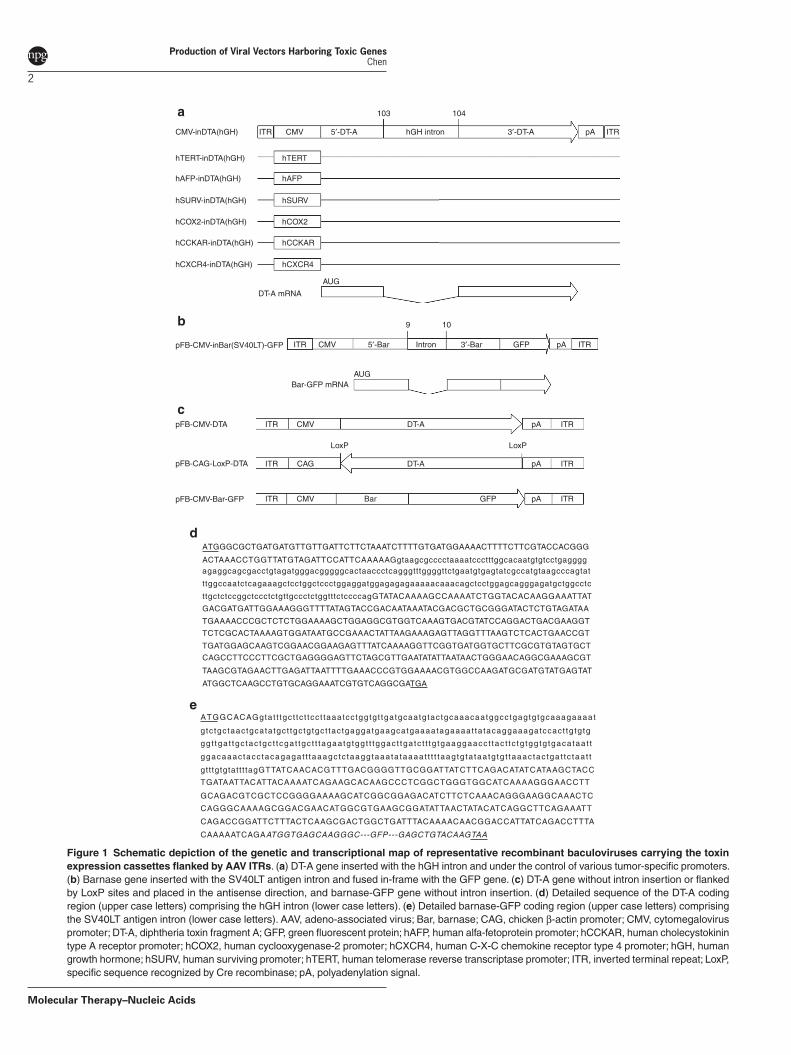
Figure 1 Schematic depiction of the genetic and transcriptional map of representative recombinant baculoviruses carrying the toxin expression cassettes flanked by AAV ITRs. (a) DT-A gene inserted with the hGH intron and under the control of various tumor-specific promoters. (b) Barnase gene inserted with the SV40LT antigen intron and fused in-frame with the GFP gene. (c) DT-A gene without intron insertion or flanked by LoxP sites and placed in the antisense direction, and barnase-GFP gene without intron insertion. (d) Detailed sequence of the DT-A coding region (upper case letters) comprising the hGH intron (lower case letters). (e) Detailed barnase-GFP coding region (upper case letters) comprising the SV40LT antigen intron (lower case letters). AAV, adeno-associated virus; Bar, barnase; CAG, chicken β-actin promoter; CMV, cytomegalovirus promoter; DT-A, diphtheria toxin fragment A; GFP, green fluorescent protein; hAFP, human alfa-fetoprotein promoter; hCCKAR, human cholecystokinin type A receptor promoter; hCOX2, human cyclooxygenase-2 promoter; hCXCR4, human C-X-C chemokine receptor type 4 promoter; hGH, human growth hormone; hSURV, human surviving promoter; hTERT, human telomerase reverse transcriptase promoter; ITR, inverted terminal repeat; LoxP, specific sequence recognized by Cre recombinase; pA, polyadenylation signal.
hTERT
ITRCMV-inDTA(hGH)
hTERT-inDTA(hGH)
hAFP-inDTA(hGH) hAFP
hSURV-inDTA(hGH) hSURV
hCOX2-inDTA(hGH) hCOX2
hCCKAR-inDTA(hGH) hCCKAR
hCXCR4-inDTA(hGH) hCXCR4
AUG
DT-A mRNA
CMV hGH intron
103a 104
pA ITR5′-DT-A 3′-DT-A
pFB-CAG-LoxP-DTA
pFB-CMV-Bar-GFP
pFB-CMV-DTA
c
LoxP LoxP
ITR CMV DT-A pA ITR
ITR CMV Bar GFP pA ITR
ITR CAG DT-A pA ITR
pFB-CMV-inBar(SV40LT)-GFP
Bar-GFP mRNAAUG
b
ITR CMV Intron pA ITR5′-Bar
9 10
3′-Bar GFP
ATGGGCGCTGATGATGTTGTTGATTCTTCTAAATCTTTTGTGATGGAAAACTTTTCTTCGTACCACGGG
ATGGCACAGgtat t tgc t tc t tcc t taaatcc tggtg t tgatgcaatg tac tgcaaacaatggcctgagtg tgcaaagaaaat
g tc tgc taactgcata tgc t tgc tg tgc t tac tgaggatgaagcatgaaaatagaaaat ta tacaggaaagatccact tg tg tg
gg t tga t tgc tac tgc t tcga t tgc t t tagaa tg tgg t t tggac t tga tc t t tg tgaaggaacc t tac t tc tg tgg tg tgaca taa t t
ggacaaac tacc tacagagat t taaagc tc taaggtaaa ta taaaat t t t taag tg ta taa tg tg t taaac tac tga t tc taa t t
gtt tgtgtatt t tagGTTATCAACACGTTTGACGGGGTTGCGGATTATCTTCAGACATATCATAAGCTACC
TGATAATTACATTACAAAATCAGAAGCACAAGCCCTCGGCTGGGTGGCATCAAAAGGGAACCTT
GCAGACGTCGCTCCGGGGAAAAGCATCGGCGGAGACATCTTCTCAAACAGGGAAGGCAAACTC
CAGGGCAAAAGCGGACGAACATGGCGTGAAGCGGATATTAACTATACATCAGGCTTCAGAAATT
CAGACCGGATTCTTTACTCAAGCGACTGGCTGATTTACAAAACAACGGACCATTATCAGACCTTTA
CAAAAATCAGAATGGTGAGCAAGGGC---GFP---GAGCTGTACAAGTAA
ACTAAACCTGGTTATGTAGATTCCATTCAAAAAGgtaagcgcccctaaaatccctttggcacaatgtgtcctgaggggagaggcagcgacctgtagatgggacgggggcactaaccctcagggtttggggttctgaatgtgagtatcgccatgtaagcccagtat
ttggccaatctcagaaagctcctggctccctggaggatggagagagaaaaacaaacagctcctggagcagggagatgctggcctc
ttgctctccggctccctctgttgccctctggtttctccccagGTATACAAAAGCCAAAATCTGGTACACAAGGAAATTAT
GACGATGATTGGAAAGGGTTTTATAGTACCGACAATAAATACGACGCTGCGGGATACTCTGTAGATAA
TGAAAACCCGCTCTCTGGAAAAGCTGGAGGCGTGGTCAAAGTGACGTATCCAGGACTGACGAAGGT
TCTCGCACTAAAAGTGGATAATGCCGAAACTATTAAGAAAGAGTTAGGTTTAAGTCTCACTGAACCGT
TGATGGAGCAAGTCGGAACGGAAGAGTTTATCAAAAGGTTCGGTGATGGTGCTTCGCGTGTAGTGCT
CAGCCTTCCCTTCGCTGAGGGGAGTTCTAGCGTTGAATATATTAATAACTGGGAACAGGCGAAAGCGT
TAAGCGTAGAACTTGAGATTAATTTTGAAACCCGTGGAAAACGTGGCCAAGATGCGATGTATGAGTAT
ATGGCTCAAGCCTGTGCAGGAAATCGTGTCAGGCGATGA
d
e
www.moleculartherapy.org/mtna
Production of Viral Vectors Harboring Toxic Genes Chen
3
system was used to decrease the expression of DT-A. Even though the authors managed to produce some AAV vectors carrying the DT-A gene in HEK293T cells, their AAV yields were generally 2–3 logs lower than AAV vectors that did not carry the DT-A gene, such as vectors carrying the PUMA gene (AAVTetO2-PUMA). The authors attributed the lower titer to residual expression of the DT-A protein. In general, the use of viral vectors, such as recombinant adenovirus, recombinant baculovirus, and AAV, for the delivery of toxic genes to a target cell has been largely hampered by the inherent toxicity of the toxic gene. As a result, viral vectors harboring a toxic gene must be grown under conditions that are not lethal to the host cell.
In the present study, two mammalian introns that are not functional in insect cells were inserted into the coding sequences of DT-A and barnase (Bar) to disrupt their open-reading frames (ORFs), which successfully abolished the expression of toxic proteins in insect cells. This enabled the production of baculoviral and AAV vectors harboring toxin genes at similar levels to those of baculoviral and AAV vec-tors carrying the green fluorescent protein (GFP) gene. Fur-thermore, these baculoviral or AAV vectors harboring the toxin genes were able to kill mammalian cells, demonstrating that the mammalian introns were spliced out properly and toxin proteins were expressed. Finally, tumor-specific promot-ers were cloned to drive the expression of the toxin genes, which resulted in tumor-specific cell killing. These findings, taken together, should pave the way for further research, as well as for the development of toxin-based suicide gene therapy drugs.
ResultsInsertion of mammalian introns to disrupt toxin gene ORFs and production of recombinant baculovirusesTwo mammalian introns and two toxin genes were chosen for these experiments. Since the consensus intron-splicing sequence, 5′-AG/GUAAGU-intron-YNCURAC-Y
nNAG/G-3′, requires a 5′-AG and a ′’-G nucleotides to flank the intron,12 the human growth hormone (hGH) intron was inserted into the DT-A ORF between nucleotides 103 and 104 (the first
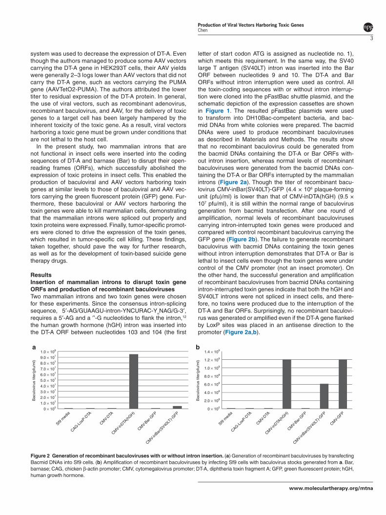
letter of start codon ATG is assigned as nucleotide no. 1), which meets this requirement. In the same way, the SV40 large T antigen (SV40LT) intron was inserted into the Bar ORF between nucleotides 9 and 10. The DT-A and Bar ORFs without intron interruption were used as control. All the toxin-coding sequences with or without intron interrup-tion were cloned into the pFastBac shuttle plasmid, and the schematic depiction of the expression cassettes are shown in Figure 1. The resulted pFastBac plasmids were used to transform into DH10Bac-competent bacteria, and bac-mid DNAs from white colonies were prepared. The bacmid DNAs were used to produce recombinant baculoviruses as described in Materials and Methods. The results show that no recombinant baculovirus could be generated from the bacmid DNAs containing the DT-A or Bar ORFs with-out intron insertion, whereas normal levels of recombinant baculoviruses were generated from the bacmid DNAs con-taining the DT-A or Bar ORFs interrupted by the mammalian introns (Figure 2a). Though the titer of recombinant bacu-lovirus CMV-inBar(SV40LT)-GFP (4.4 × 10⁶ plaque-forming unit (pfu)/ml) is lower than that of CMV-inDTA(hGH) (9.5 × 10⁷ pfu/ml), it is still within the normal range of baculovirus generation from bacmid transfection. After one round of amplification, normal levels of recombinant baculoviruses carrying intron-interrupted toxin genes were produced and compared with control recombinant baculovirus carrying the GFP gene (Figure 2b). The failure to generate recombinant baculovirus with bacmid DNAs containing the toxin genes without intron interruption demonstrates that DT-A or Bar is lethal to insect cells even though the toxin genes were under control of the CMV promoter (not an insect promoter). On the other hand, the successful generation and amplification of recombinant baculoviruses from bacmid DNAs containing intron-interrupted toxin genes indicate that both the hGH and SV40LT introns were not spliced in insect cells, and there-fore, no toxins were produced due to the interruption of the DT-A and Bar ORFs. Surprisingly, no recombinant baculovi-rus was generated or amplified even if the DT-A gene flanked by LoxP sites was placed in an antisense direction to the promoter (Figure 2a,b).
Figure 2 Generation of recombinant baculoviruses with or without intron insertion. (a) Generation of recombinant baculoviruses by transfecting Bacmid DNAs into Sf9 cells. (b) Amplification of recombinant baculoviruses by infecting Sf9 cells with baculovirus stocks generated from a. Bar, barnase; CAG, chicken β-actin promoter; CMV, cytomegalovirus promoter; DT-A, diphtheria toxin fragment A; GFP, green fluorescent protein; hGH, human growth hormone.
1.0 × 10⁸a b
9.0 × 10⁷
8.0 × 10⁷
Bac
ulov
irus
titer
(pfu
/ml)
7.0 × 10⁷
6.0 × 10⁷
5.0 × 10⁷
4.0 × 10⁷
3.0 × 10⁷
2.0 × 10⁷
1.0 × 10⁷
0 × 100
1.4 × 10⁹
1.2 × 10⁹
1.0 × 10⁹
Bac
ulov
irus
titer
(pfu
/ml)
8.0 × 10⁸
6.0 × 10⁸
4.0 × 10⁸
2.0 × 10⁸
0 × 100
Sf9 m
edia
CAG-Lox
P-DTA
CMV-
DTA
CMV-
inDTA
(hGH)
CMV-
Bar-G
FP
CMV-
inBar
(SV40
LT)-G
FP
Sf9 m
edia
CAG-Lox
P-DTA
CMV-
DTA
CMV-
inDTA
(hGH)
CMV-
Bar-G
FP
CMV-
inBar
(SV40
LT)-G
FP
CMV-
GFP
Molecular Therapy–Nucleic Acids
Production of Viral Vectors Harboring Toxic Genes Chen
4
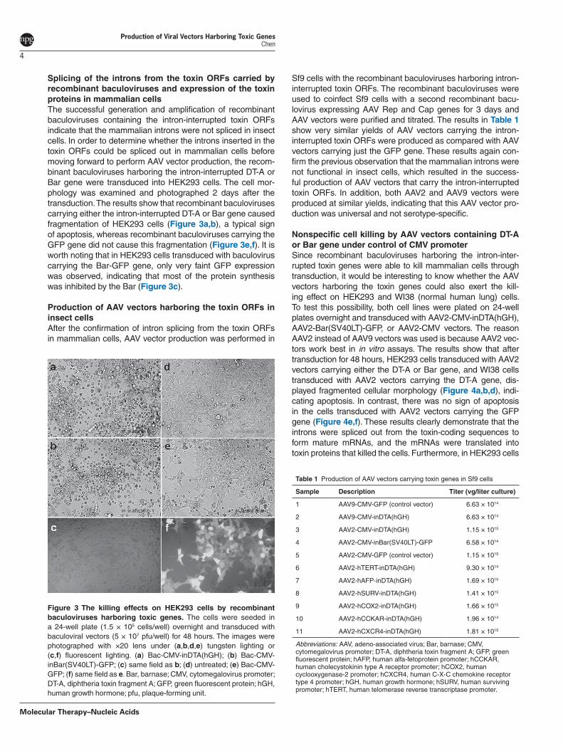
Splicing of the introns from the toxin ORFs carried by recombinant baculoviruses and expression of the toxin proteins in mammalian cellsThe successful generation and amplification of recombinant baculoviruses containing the intron-interrupted toxin ORFs indicate that the mammalian introns were not spliced in insect cells. In order to determine whether the introns inserted in the toxin ORFs could be spliced out in mammalian cells before moving forward to perform AAV vector production, the recom-binant baculoviruses harboring the intron-interrupted DT-A or Bar gene were transduced into HEK293 cells. The cell mor-phology was examined and photographed 2 days after the transduction. The results show that recombinant baculoviruses carrying either the intron-interrupted DT-A or Bar gene caused fragmentation of HEK293 cells (Figure 3a,b), a typical sign of apoptosis, whereas recombinant baculoviruses carrying the GFP gene did not cause this fragmentation (Figure 3e,f). It is worth noting that in HEK293 cells transduced with baculovirus carrying the Bar-GFP gene, only very faint GFP expression was observed, indicating that most of the protein synthesis was inhibited by the Bar (Figure 3c).
Production of AAV vectors harboring the toxin ORFs in insect cellsAfter the confirmation of intron splicing from the toxin ORFs in mammalian cells, AAV vector production was performed in
Sf9 cells with the recombinant baculoviruses harboring intron-interrupted toxin ORFs. The recombinant baculoviruses were used to coinfect Sf9 cells with a second recombinant bacu-lovirus expressing AAV Rep and Cap genes for 3 days and AAV vectors were purified and titrated. The results in Table 1 show very similar yields of AAV vectors carrying the intron-interrupted toxin ORFs were produced as compared with AAV vectors carrying just the GFP gene. These results again con-firm the previous observation that the mammalian introns were not functional in insect cells, which resulted in the success-ful production of AAV vectors that carry the intron-interrupted toxin ORFs. In addition, both AAV2 and AAV9 vectors were produced at similar yields, indicating that this AAV vector pro-duction was universal and not serotype-specific.
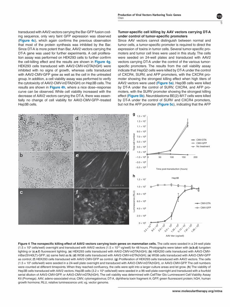
Nonspecific cell killing by AAV vectors containing DT-A or Bar gene under control of CMV promoterSince recombinant baculoviruses harboring the intron-inter-rupted toxin genes were able to kill mammalian cells through transduction, it would be interesting to know whether the AAV vectors harboring the toxin genes could also exert the kill-ing effect on HEK293 and WI38 (normal human lung) cells. To test this possibility, both cell lines were plated on 24-well plates overnight and transduced with AAV2-CMV-inDTA(hGH), AAV2-Bar(SV40LT)-GFP, or AAV2-CMV vectors. The reason AAV2 instead of AAV9 vectors was used is because AAV2 vec-tors work best in in vitro assays. The results show that after transduction for 48 hours, HEK293 cells transduced with AAV2 vectors carrying either the DT-A or Bar gene, and WI38 cells transduced with AAV2 vectors carrying the DT-A gene, dis-played fragmented cellular morphology (Figure 4a,b,d), indi-cating apoptosis. In contrast, there was no sign of apoptosis in the cells transduced with AAV2 vectors carrying the GFP gene (Figure 4e,f). These results clearly demonstrate that the introns were spliced out from the toxin-coding sequences to form mature mRNAs, and the mRNAs were translated into toxin proteins that killed the cells. Furthermore, in HEK293 cells
Figure 3 The killing effects on HEK293 cells by recombinant baculoviruses harboring toxic genes. The cells were seeded in a 24-well plate (1.5 × 10⁵ cells/well) overnight and transduced with baculoviral vectors (5 × 10⁷ pfu/well) for 48 hours. The images were photographed with ×20 lens under (a,b,d,e) tungsten lighting or (c,f) fluorescent lighting. (a) Bac-CMV-inDTA(hGH); (b) Bac-CMV-inBar(SV40LT)-GFP; (c) same field as b; (d) untreated; (e) Bac-CMV-GFP; (f) same field as e. Bar, barnase; CMV, cytomegalovirus promoter; DT-A, diphtheria toxin fragment A; GFP, green fluorescent protein; hGH, human growth hormone; pfu, plaque-forming unit.
Table 1 Production of AAV vectors carrying toxin genes in Sf9 cells
Sample Description Titer (vg/liter culture)
1 AAV9-CMV-GFP (control vector) 6.63 × 10¹⁴
2 AAV9-CMV-inDTA(hGH) 6.63 × 10¹⁴
3 AAV2-CMV-inDTA(hGH) 1.15 × 10¹⁵
4 AAV2-CMV-inBar(SV40LT)-GFP 6.58 × 10¹⁴
5 AAV2-CMV-GFP (control vector) 1.15 × 10¹⁵
6 AAV2-hTERT-inDTA(hGH) 9.30 × 10¹⁴
7 AAV2-hAFP-inDTA(hGH) 1.69 × 10¹⁵
8 AAV2-hSURV-inDTA(hGH) 1.41 × 10¹⁵
9 AAV2-hCOX2-inDTA(hGH) 1.66 × 10¹⁵
10 AAV2-hCCKAR-inDTA(hGH) 1.96 × 10¹⁴
11 AAV2-hCXCR4-inDTA(hGH) 1.81 × 10¹⁵
Abbreviations: AAV, adeno-associated virus; Bar, barnase; CMV, cytomegalovirus promoter; DT-A, diphtheria toxin fragment A; GFP, green fluorescent protein; hAFP, human alfa-fetoprotein promoter; hCCKAR, human cholecystokinin type A receptor promoter; hCOX2, human cyclooxygenase-2 promoter; hCXCR4, human C-X-C chemokine receptor type 4 promoter; hGH, human growth hormone; hSURV, human surviving promoter; hTERT, human telomerase reverse transcriptase promoter.
www.moleculartherapy.org/mtna
Production of Viral Vectors Harboring Toxic Genes Chen
5
transduced with AAV2 vectors carrying the Bar-GFP fusion cod-ing sequence, only very faint GFP expression was observed (Figure 4c), which again confirms the previous observation that most of the protein synthesis was inhibited by the Bar. Since DT-A is more potent than Bar, AAV2 vectors carrying the DT-A gene was used for further experiments. A cell prolifera-tion assay was performed on HEK293 cells to further confirm the cell-killing effect and the results are shown in Figure 4g. HEK293 cells transduced with AAV2-CMV-inDTA(hGH) were inhibited with no signs of growth, whereas cells transduced with AAV2-CMV-GFP grew as well as the cell in the untreated group. In addition, a cell viability assay was performed to verify the cytotoxicity of AAV2-CMV-inDTA(hGH) on Hep3B cells. The results are shown in Figure 4h, where a nice dose–response curve can be observed. While cell viability increased with the decrease of AAV2 vectors carrying the DT-A, there was essen-tially no change of cell viability for AAV2-CMV-GFP–treated Hep3B cells.
Tumor-specific cell killing by AAV vectors carrying DT-A under control of tumor-specific promotersSince AAV vectors cannot distinguish between normal and tumor cells, a tumor-specific promoter is required to direct the expression of toxins in tumor cells. Several tumor-specific pro-moters and tumor cell lines were used in this study. The cells were seeded on 24-well plates and transduced with AAV2 vectors carrying DT-A under the control of the various tumor-specific promoters. The results from the cell viability assay indicate that HepG2 cells were killed by DT-A under the control of CXCR4, SURV, and AFP promoters, with the CXCR4 pro-moter showing the strongest killing effect when high titers of AAV2 vectors were used (Figure 5a). Hep3B cells were killed by DT-A under the control of SURV, CXCR4, and AFP pro-moters, with the SURV promoter showing the strongest killing effect (Figure 5b). Neuroblastoma BE(2)-M17 cells were killed by DT-A under the control of SURV and CXCR4 promoters, but not the AFP promoter (Figure 5c), indicating that the AFP
1.5 × 10⁷
1.2 × 10⁷
1.0 × 10⁷
8.0 × 10⁶
6.0 × 10⁶
4.0 × 10⁶
Lum
ines
cenc
e (R
LU)
2.0 × 10⁶
0 × 100
AAV titer (vg/well)
1.3 × 10⁷
1.1 × 10⁷
9.0 × 10⁶
7.0 × 10⁶
5.0 × 10⁶
3.0 × 10⁶
Tota
l cel
l num
ber
1.0 × 10⁶
−1.0 × 10⁶ 0 50 100
Time post-transduction (hours)
Hep3Bh
g
150 200
CMV-DTACMV-GFP
CMV-DTA
CMV-GFP
No treatment
−3.0 × 10⁶
−5.0 × 10⁶
3.2
× 1010
8.0
× 109
2.0
× 109
5.0
× 10
8
1.25
× 10
8
3.13
× 10
7
7.81
× 106
1.95
× 10
6
Figure 4 The nonspecific killing effect of AAV2 vectors carrying toxin genes on mammalian cells. The cells were seeded in a 24-well plate (1.5 × 10⁵ cells/well) overnight and transduced with AAV2 vectors (1.5 × 10¹⁰ vg/well) for 48 hours. Photographs were taken with (a,b,d) tungsten lighting or (c,e,f) fluorescent lighting. (a) HEK293 cells transduced with AAV2-CMV-inDTA(hGH); (b) HEK293 cells transduced with AAV2-CMV-inBar(SV40LT)-GFP; (c) same field as b; (d) WI38 cells transduced with AAV2-CMV-inDTA(hGH); (e) WI38 cells transduced with AAV2-CMV-GFP as control; (f) HEK293 cells transduced with AAV2-CMV-GFP as control. (g) Proliferation of HEK293 cells transduced with AAV2 vectors. The cells (1.5 × 10⁵ cells/well) were seeded in a 24-well plate overnight and transduced with AAV2-CMV-inDTA(hGH), or AAV2-CMV-GFP. The cell numbers were counted at different timepoints. When they reached confluency, the cells were split into a larger culture areas and let grow. (h) The viability of Hep3B cells transduced with AAV2 vectors. Hep3B cells (3.2 × 10⁴ cells/well) were seeded in a 96-well plate overnight and transduced with a fourfold serial dilution of AAV2-CMV-GFP, or AAV2-CMV-inDTA(hGH). The cell viability was determined with CellTiter Glo Luminescent Cell Viability Assay Kit (Promega). AAV, adeno-associated virus; CMV, cytomegalovirus; DT-A, diphtheria toxin fragment A; GFP, green fluorescent protein; hGH, human growth hormone; RLU, relative luminescence unit; vg, vector genome.

Molecular Therapy–Nucleic Acids
Production of Viral Vectors Harboring Toxic Genes Chen
6
promoter was not active in neuroblastoma cells. A represen-tative result to show the morphologies of AAV2 vector-trans-duced cells is shown in Figure 6. The normal human lung cell line WI38 was not affected by DT-A under control of the SURV promoter (Figure 6b), whereas the three tumor cell lines, HepG2, Hep3B, and BE(2)-M17 were killed by DT-A under control of the SURV promoter (Figure 6d,f,h). The three tumor cell lines were also tested with DT-A under control of human COX2, CCKAR, and hTERT promoters, but no significant cell-killing effect was observed (data not shown), probably due to the weak promoter activities.
Discussion
Suicide gene therapy has been widely investigated for the treatment of HIV infection, for controlling graft-versus-host disease, and also for the treatment of cancer. While the pro-duction of viral vectors carrying suicide genes, such as the
herpes simplex virus thymidine kinase or cytosine deaminase are easily achievable, the generation of viral vectors contain-ing toxin genes, such as DT-A, Pseudomonas exotoxin, or Bar has always been a challenge due to the extreme toxicity of these toxins. The current study presents a novel method to circumvent this toxicity issue by exploiting the differences of intron-splicing machineries between mammalian and insect cells. Since some mammalian introns are not functional in insect cells,13,14 two commonly used mammalian introns, hGH and SV40LT introns, were selected and cloned to interrupt the ORFs of DT-A and Bar, respectively. The insertion sites were chosen between 5′-AG and 3′-G nucleotides accord-ing to intron-splicing consensus sequence.12 As predicted, the insertion of these mammalian introns in the toxin ORFs totally abolished their expression in insect cells and enabled the production of recombinant baculoviruses carrying these intron-interrupted toxin ORFs at normal levels. In contrast, no recombinant baculovirus was produced when the toxin ORFs
1,000,000HepG2 Hep3Ba b
6,000,000
5,000,000
4,000,000
3,000,000
2,000,000
1,000,000
0
900,000
800,000
700,000
600,000
500,000 AFP-DTA
SURV-DTA
CXCR4-DTA
CMV-GFP
AFP-DTA
SURV-DTA
CXCR4-DTA
CMV-GFP
Lum
ines
cenc
e (R
LU)
Lum
ines
cenc
e (R
LU)
400,000
300,000
200,000
100,000
AAV titer (vg/well)
0
AAV titer (vg/well)
BE(2)-M17c6,000,000
5,000,000
4,000,000
3,000,000
2,000,000
1,000,000
0
AFP-DTA
SURV-DTA
CXCR4-DTA
CMV-GFP
Lum
ines
cenc
e (R
LU)
AAV titer (vg/well)
3.2 ×
1010
8.0 ×
109
2.0 ×
109
5.0 ×
108
1.25 ×
108
3.13 ×
107
7.81 ×
106
1.95 ×
106
3.2 ×
1010
8.0 ×
109
2.0 ×
109
5.0 ×
108
1.25 ×
108
3.13 ×
107
7.81 ×
106
1.95 ×
106
3.2 ×
1010
8.0 ×
109
2.0 ×
109
5.0 ×
108
1.25 ×
108
3.13 ×
107
7.81 ×
106
1.95 ×
106
Figure 5 Cell viability assay of tumor cells transduced with AAV2 vectors carrying DT-A under the control of various human tumor-specific promoters. The cells were seeded on 96-well plates and transduced with AAV2 vectors at fourfold serial dilutions for 4 days, and cell viability was assayed with the CellTiter Glo Luminescent Cell Viability Assay kit. (a) HepG2 cells, (b) Hep3B cells, and (c) BE(2)-M17 were transduced with AAV2 vectors carrying DT-A under control of different tumor-specific promoters. AAV, adeno-associated virus; CMV, cytomegalovirus; DT-A, diphtheria toxin fragment A; GFP, green fluorescent protein; RLU, relative luminescence unit.

www.moleculartherapy.org/mtna
Production of Viral Vectors Harboring Toxic Genes Chen
7
were not interrupted by the introns (Figure 2). Furthermore, the recombinant baculoviruses carrying the intron-interrupted toxin ORFs were able to kill mammalian cells once they were introduced into the cells, demonstrating that the mammalian introns were spliced out, which allowed the toxin proteins to be expressed (Figure 3). This is the first report to show that mammalian introns can be used to interrupt the ORFs of toxin genes and abolish their expression in insect cells so that viral vectors (both recombinant baculoviral and AAV vectors) can be produced at normal levels. These viral vectors can then be introduced into mammalian cells for various purposes that require cell killing. This discovery is significant because it can be used to produce viral vectors carrying any toxic genes at normal levels and use them to kill mammalian cells, which should pave the way for further research, as well as for the development of toxin-based suicide gene therapy drugs.
DT-A is extremely toxic to mammalian cells, for as few as one single molecule is enough to kill a cell.2 DT-A is also toxic to insect cells and recombinant baculovirus could only be generated when the DT-A ORF was under control of the GFAP promoter, which is inactive in insect cells.9 This extreme
cytotoxicity was confirmed in the current study by the obser-vation that no recombinant baculovirus could be produced from bacmid DNA containing DT-A–coding sequence under control of the CMV promoter (Figure 2a). Though it is not an insect promoter, the CMV promoter has some activity in insect cells and is able to drive low level of hemagglutinin expression.15 This low level of promoter activity could drive some basal expression of DT-A, which kills any insect cells that express the toxin. It was also observed that recombinant baculovirus carrying the DT-A ORF flanked with LoxP sites in an antisense direction to the chicken β-actin promoter (CAG) promoter was not generated, indicating that even cryptic promoter TATA-like sequence of the LoxP site was able to drive trace amounts of DT-A expression that can kill insect cells and inhibit baculovirus production (Figure 2). The same phenomenon was observed for the Bar in which recombinant baculoviruses could be generated only when its ORF was interrupted by the SV40LT antigen intron, indicating that Bar is also toxic to insect cells. On the other hand, the successful generation of recombinant baculoviruses harboring either the intron-interrupted DT-A or the Bar-GFP gene indicated that hGH and SV40LT antigen introns were not spliced in insect cells, and that DT-A and Bar-GFP expressions were totally abolished by this intron interruption.
AAV vector is one of the most promising vectors for human gene therapy and has been used in numerous clinical trials.16 The development of AAV vectors for cancer therapy has focused on antiangiogenic, immunomodulatory, and suicide genes that require a prodrug, or on genes that mediate cell repair.17 It has been reported that AAV vectors carrying the DT-A gene under control of the Tet-repressor system can be produced in HEK293 cells with yields generally 2–3 logs lower than that of control AAV vectors due to the basal level expression of DT-A.11 This demonstrates that the poisonous effect of the toxin on the producer cells even at trace amounts is enough to limit AAV vector production. In the current study, the expression of DT-A or Bar toxins was totally abolished during AAV vector production process in insect cells, and their expression was restored when the AAV vectors were introduced into mammalian cells. This was accomplished by exploiting the differences in intron-splicing mechanisms between insect and mammalian cells. This novel method enables the high production yields of AAV vectors carrying the toxin gene (Table 1), and should provide abundant AAV vectors for further research and the potential development of cancer therapy drugs.
Specificity is paramount in cancer therapy when lethal toxins such as DT-A or Pseudomonas exotoxin are used. Several approaches, such as tumor-specific promoters driving toxic gene expression and engineered AAV capsids to target cancer cells, can be employed to restrict the toxin expression in cancer cells so that unwanted harm can be minimized. It has been reported that tumor-specific promoters can direct gene expression in tumor cells with limited expression in normal cells.17–20 In the present study, the human TERT, AFP, SURV, COX2, CCKAR, and CXCR4 promoters were cloned to drive the expression of DT-A gene, and their specific cell-killing effects were examined. The normal cell line WI38 was not affected by DT-A under control of any of the above mentioned tumor-specific promoters, but was killed by DT-A under control of the
Figure 6 Representative results of tumor-specific cell killing by AAV2 vectors carrying DT-A under the control of the survivin promoter. The cells were seeded in 24-well plate and transduced with (a,c,e,g) AAV2-CMV-GFP or (b,d,f,h) AAV2-hSURV-inDTA(hGH). Photographs were taken 3 days post-transduction. (a,b), WI38; (c,d), HepG2; (e,f), Hep3B, and (g,h), BE(2)-M17 cells. AAV, adeno-associated virus; CMV, cytomegalovirus; DT-A, diphtheria toxin fragment A; GFP, green fluorescent protein; hGH, human growth hormone.
Molecular Therapy–Nucleic Acids
Production of Viral Vectors Harboring Toxic Genes Chen
8
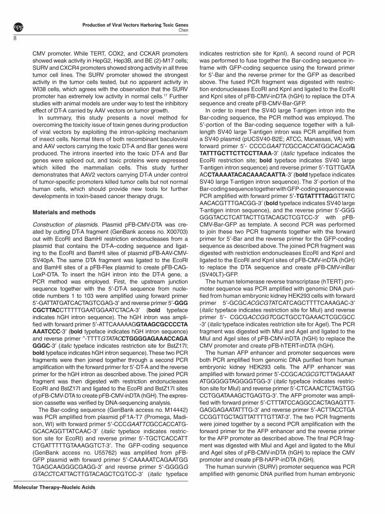
CMV promoter. While TERT, COX2, and CCKAR promoters showed weak activity in HepG2, Hep3B, and BE (2)-M17 cells; SURV and CXCR4 promoters showed strong activity in all three tumor cell lines. The SURV promoter showed the strongest activity in the tumor cells tested, but no apparent activity in WI38 cells, which agrees with the observation that the SURV promoter has extremely low activity in normal cells.17 Further studies with animal models are under way to test the inhibitory effect of DT-A carried by AAV vectors on tumor growth.
In summary, this study presents a novel method for overcoming the toxicity issue of toxin genes during production of viral vectors by exploiting the intron-splicing mechanism of insect cells. Normal titers of both recombinant baculoviral and AAV vectors carrying the toxic DT-A and Bar genes were produced. The introns inserted into the toxic DT-A and Bar genes were spliced out, and toxic proteins were expressed which killed the mammalian cells. This study further demonstrates that AAV2 vectors carrying DT-A under control of tumor-specific promoters killed tumor cells but not normal human cells, which should provide new tools for further developments in toxin-based cancer therapy drugs.
Materials and methods
Construction of plasmids. Plasmid pFB-CMV-DTA was cre-ated by cutting DT-A fragment (GenBank access no. X00703) out with EcoRI and BamHI restriction endonucleases from a plasmid that contains the DT-A–coding sequence and ligat-ing to the EcoRI and BamHI sites of plasmid pFB-AAV-CMV-SV40pA. The same DTA fragment was ligated to the EcoRI and BamHI sites of a pFB-Flex plasmid to create pFB-CAG-LoxP-DTA. To insert the hGH intron into the DT-A gene, a PCR method was employed. First, the upstream junction sequence together with the 5′-DT-A sequence from nucle-otide numbers 1 to 103 were amplified using forward primer 5′-GATTATGATCACTAGTCGAG-3′ and reverse primer 5′-GGG CGCTTACCTTTTTGAATGGAATCTACA-3′ (bold typeface indicates hGH intron sequence). The hGH intron was ampli-fied with forward primer 5′-ATTCAAAAAGGTAAGCGCCCCTAAAATCCC-3′ (bold typeface indicates hGH intron sequence) and reverse primer ′’-TTTTGTATACCTGGGGAGAAACCAGA GGGC-3′ (italic typeface indicates restriction site for BstZ17I; bold typeface indicates hGH intron sequence). These two PCR fragments were then joined together through a second PCR amplification with the forward primer for 5′-DT-A and the reverse primer for the hGH intron as described above. The joined PCR fragment was then digested with restriction endonucleases EcoRI and BstZ17I and ligated to the EcoRI and BstZ17I sites of pFB-CMV-DTA to create pFB-CMV-inDTA (hGH). The expres-sion cassette was verified by DNA-sequencing analysis.
The Bar-coding sequence (GenBank access no. M14442) was PCR amplified from plasmid pF1A-T7 (Promega, Madi-son, WI) with forward primer 5′-CCCGAATTCGCCACCATG-GCACAGGTTATCAAC-3′ (italic typeface indicates restric-tion site for EcoRI) and reverse primer 5′-TGCTCACCATTCTGATTTTTGTAAAGGTCT-3′. The GFP-coding sequence (GenBank access no. U55762) was amplified from pFB-GFP plasmid with forward primer 5′-CAAAAATCAGAATGGTGAGCAAGGGCGAGG-3′ and reverse primer 5′-GGGGGGTACCTCATTACTTGTACAGCTCGTCC-3′ (italic typeface
indicates restriction site for KpnI). A second round of PCR was performed to fuse together the Bar-coding sequence in-frame with GFP-coding sequence using the forward primer for 5′-Bar and the reverse primer for the GFP as described above. The fused PCR fragment was digested with restric-tion endonucleases EcoRI and KpnI and ligated to the EcoRI and KpnI sites of pFB-CMV-inDTA (hGH) to replace the DT-A sequence and create pFB-CMV-Bar-GFP.
In order to insert the SV40 large T-antigen intron into the Bar-coding sequence, the PCR method was employed. The 5′-portion of the Bar-coding sequence together with a full-length SV40 large T-antigen intron was PCR amplified from a SV40 plasmid (pUCSV40-B2E; ATCC, Manassas, VA) with forward primer 5′- CCCCGAATTCGCCACCATGGCACAGG TATTTGCTTCTTCCTTAAA-3′ (italic typeface indicates the EcoRI restriction site; bold typeface indicates SV40 large T-antigen intron sequence) and reverse primer 5′-TGTTGATAACCTAAAATACACAAACAATTA-3′ (bold typeface indicates SV40 large T-antigen intron sequence). The 3′-portion of the Bar-coding sequence together with GFP-coding sequence was PCR amplified with forward primer 5′-TGTATTTTAGGTTATCAACACGTTTGACGG-3′ (bold typeface indicates SV40 large T-antigen intron sequence), and the reverse primer 5′-GGG GGGTACCTCATTACTTGTACAGCTCGTCC-3′ with pFB-CMV-Bar-GFP as template. A second PCR was performed to join these two PCR fragments together with the forward primer for 5′-Bar and the reverse primer for the GFP-coding sequence as described above. The joined PCR fragment was digested with restriction endonucleases EcoRI and KpnI and ligated to the EcoRI and KpnI sites of pFB-CMV-inDTA (hGH) to replace the DTA sequence and create pFB-CMV-inBar (SV40LT)-GFP.
The human telomerase reverse transcriptase (hTERT) pro-moter sequence was PCR amplified with genomic DNA puri-fied from human embryonic kidney HEK293 cells with forward primer 5′-GCGCACGCGTATCATCAGCTTTTCAAAGAC-3′ (italic typeface indicates restriction site for MluI) and reverse primer 5′- CGCGACCGGTCGCTGCCTGAAACTCGCGCC -3′ (italic typeface indicates restriction site for AgeI). The PCR fragment was digested with MluI and AgeI and ligated to the MluI and AgeI sites of pFB-CMV-inDTA (hGH) to replace the CMV promoter and create pFB-hTERT-inDTA (hGH).
The human AFP enhancer and promoter sequences were both PCR amplified from genomic DNA purified from human embryonic kidney HEK293 cells. The AFP enhancer was amplified with forward primer 5′-CCGCACGCGTCTTAGAAATATGGGGGTAGGGGTGG-3’ (italic typeface indicates restric-tion site for MluI) and reverse primer 5′-CTCAAACTCTAGTGGCCTGGATAAAGCTGAGTG-3′. The AFP promoter was ampli-fied with forward primer 5′-CTTTATCCAGGCCACTAGAGTTT-GAGGAGAATATTTG-3′ and reverse primer 5′-ACTTACCTGACCGGTTGCTAGTTATTTTGTTAT-3′. The two PCR fragments were joined together by a second PCR amplification with the forward primer for the AFP enhancer and the reverse primer for the AFP promoter as described above. The final PCR frag-ment was digested with MluI and AgeI and ligated to the MluI and AgeI sites of pFB-CMV-inDTA (hGH) to replace the CMV promoter and create pFB-hAFP-inDTA (hGH).
The human survivin (SURV) promoter sequence was PCR amplified with genomic DNA purified from human embryonic
www.moleculartherapy.org/mtna
Production of Viral Vectors Harboring Toxic Genes Chen
9
kidney HEK293 cells with forward primer 5′-GGGGACTAGTCTGGCCATAGAACCAGAGAAGTGA-3′ (italic typeface indi-cates restriction site for SpeI) and reverse primer 5′-TTTTAC-CGGTCCACCTCTGCCAACGGGTCCCGCG-3′ (italic type-face indicates restriction site for AgeI). The PCR fragment was digested with SpeI and AgeI and ligated to the SpeI and AgeI sites of pFB-CMV-inDTA (hGH) to replace the CMV pro-moter and create pFB-hSURV-inDTA (hGH).
The human cyclooxygenase-2 (COX2) promoter was PCR amplified with genomic DNA purified from human embry-onic kidney HEK293 cells with forward primer 5′-GCCCAC-TAGTTGAGGTACCTGGTGTAGTTT-3′ (italic typeface indi-cates restriction site for SpeI) and reverse primer 5′-ATATACCGGTCAGCGGCGGGCAGGGCGCGG-3′ (italic typeface indicates restriction site for AgeI). The PCR fragment was digested with SpeI and AgeI and ligated to the SpeI and AgeI sites of pFB-CMV-inDTA (hGH) to replace the CMV promoter and create pFB-hCOX2-inDTA (hGH).
The human cholecystokinin type-A receptor (CCKAR) pro-moter sequence was PCR amplified with genomic DNA puri-fied from human embryonic kidney HEK293 cells with forward primer 5′-GCCCACTAGTACCCAGGTACCTATGTTCAAAAG-3′ (italic typeface indicates restriction site for SpeI) and reverse primer 5′-GCGCACCGGTTTGCCTGCTGCTTTCCACCAAG-3′ (italic typeface indicates restriction site for AgeI). The PCR fragment was digested with SpeI and AgeI and ligated to the SpeI and AgeI sites of pFB-CMV-inDTA (hGH) to replace the CMV promoter and create pFB-hCCKAR-inDTA (hGH).
The human C-X-C chemokine receptor type 4 (CXCR4) promoter sequence was PCR amplified with genomic DNA purified from human embryonic kidney HEK293 cells with forward primer 5′-GCCCACTAGTTACCGACCACCCGCAAA-CAG-3′ (italic typeface indicates restriction site for SpeI) and reverse primer 5′-GCGCACCGGTGTAACCGCTGGTTCTCCAGA-3′ (italic typeface indicates restriction site for AgeI). The PCR fragment was digested with SpeI and AgeI and ligated to the SpeI and AgeI sites of pFB-CMV-inDTA (hGH) to replace the CMV promoter and create pFB-hCXCR4-inDTA (hGH).
Cell culture. HEK293 cells were maintained at 37 °C in Dulbecco’s modified Eagle’s medium (Invitrogen, Carlsbad, CA) containing 100 U/ml of penicillin and 100 mg/ml of streptomycin, and supplemented with 10% fetal calf serum (Sigma-Aldrich, St Louis, MO). HepG2, Hep3B, and WI38 cells were maintained at 37 °C in Eagle’s minimal essential medium complete growth medium (ATCC) containing 100 U/ml of penicillin and 100 mg/ml of streptomycin, and supplemented with 10% fetal calf serum. BE (2) M17 cells (ATCC) were maintained in 50% Eagle’s minimal essential medium + 50% F12 complete growth media containing 100 U/ml of penicillin and 100 mg/ml of streptomycin, and supplemented with 10% fetal calf serum. Spodoptera frugiperda Sf9 cells (Invitrogen) were maintained at 28 °C in ESF921 serum-free medium (Expression Systems, Woodland, CA) containing 100 U/ml of penicillin and 100 mg/ml of streptomycin (Invitrogen).
Generation and titration of recombinant baculoviruses. Plasmids constructed as described above were used for transforming DH10Bac-competent cells to generate recombinant bacmids. The bacmids containing the target
genes were used for generating recombinant baculoviruses in accordance with the manufacturer’s protocol (Invitrogen), with minor modifications. Briefly, 2 ng of the plasmid DNA was used for transforming 20 µl of DH10Bac-competent cells. After 4 hours of incubation at 37 °C in 500 µl of Super Optimal broth with Catabolite repression medium, 25 and 2.5 µl of the culture were plated separately in selection plates and incubated for 48 hours to allow white/blue colonies to form. Generally, three white colonies were picked for each construct and miniprep bacmid DNAs were prepared. The bacmid DNAs were used for transfecting Sf9 cells to generate recombinant baculoviruses. The recombinant baculoviruses were amplified once and titers determined with real-time quantitative PCR assay and converted to pfu based on empirical studies in which 20 copies was converted into 1 pfu.
AAV vector production, purification, and titration. The methods for AAV vector production in Sf9 cells, purification, and titration have been described previously.21,22 Briefly, Sf9 cells were grown at 28 °C to ~1 × 10⁷ cells/ml in ESF921 serum-free medium con-taining 100 U/ml of penicillin and 100 µg/ml of streptomycin, and diluted to ~5 × 10⁶ cells/ml before infection. Double infection was employed to produce AAV vectors. Five multiplicity of infection of Bac-AAV-transgene, and 10 multiplicity of infection of Bac-inCap-inRep were used to infect Sf9 cells at 28 °C for 3 days to produce AAV vectors. After 3 days of infection, cell pellets were collected by centrifugation at 3,000 rpm for 15 minutes. The cell pellets were lysed in a modified Sf9 lysis buffer (50 mmol/l Tris-HCl, pH 8.0, 1% sarkosyl, 1% Triton X-100, 2 mmol/l MgCl2) and cellular nucleic acids (DNA and RNA) were digested using ben-zonase (Sigma-Aldrich). The cell lysates were cleared by cen-trifugation at 8,000 rpm for 20 minutes and supernatants were subjected to two rounds of cesium chloride ultracentrifugation to purify the AAV vectors. The AAV vectors were buffer-exchanged into phosphate-buffered saline containing 0.001% pluronic F-68 (Sigma-Aldrich) with 2 PD-10 desalting columns (GE Health-Care Bio-Science, Piscataway, NJ). After purification, the AAV vectors were titrated with real-time quantitative PCR method.
Cell proliferation assay. HEK293 cells were grown in 24-well plate (1.5 × 10⁵ cells/well) overnight and transduced with AAV2 vectors (1.5 × 10⁹ vector genome/well) for 48 hours. The cells were then trypsinized and cell number counted. The cells were further cultivated for another 48 hours until reach-ing confluency and trypsinized, cell number counted and then expanded into larger vessels. After another 72-hour cultiva-tion, the cells were trypsinized and counted. Viable cells were counted with trypan blue staining.
Cell viability assay. The CellTiter Glo Luminescent Cell Viability Assay kit was used to test the cell viability according to manu-facturer’s protocol (Promega). Briefly, the cells (WI38, HepG2, Hep3B, and BE (2) M17) were seeded on 96-well plates at 3.2 × 10⁴ cells/well overnight and transduced with AAV2 vectors at fourfold serial dilutions for 4 days. The reagent was reconsti-tuted and added to the cells. After mixing on an orbital shaker for 2 minutes to lyse the cells, the plate was incubated at room temperature for 10 minutes and then the luminescent signals were recorded.

Molecular Therapy–Nucleic Acids
Production of Viral Vectors Harboring Toxic Genes Chen
10
1. Rodriguez, R and Simons, JW (1999). Urologic applications of gene therapy. Urology 54: 401–406.
2. Yamaizumi, M, Mekada, E, Uchida, T and Okada, Y (1978). One molecule of diphtheria toxin fragment A introduced into a cell can kill the cell. Cell 15: 245–250.
3. Denning, C and Pitts, JD (1997). Bystander effects of different enzyme-prodrug systems for cancer gene therapy depend on different pathways for intercellular transfer of toxic metabolites, a factor that will govern clinical choice of appropriate regimes. Hum Gene Ther 8: 1825–1835.
4. Maxwell, IH, Maxwell, F and Glode, LM (1986). Regulated expression of a diphtheria toxin A-chain gene transfected into human cells: possible strategy for inducing cancer cell sui-cide. Cancer Res 46: 4660–4664.
5. Maxwell, F, Maxwell, IH and Glode, LM (1987). Cloning, sequence determination, and ex-pression in transfected cells of the coding sequence for the tox 176 attenuated diphtheria toxin A chain. Mol Cell Biol 7: 1576–1579.
6. Robinson, DF and Maxwell, IH (1995). Suppression of single and double nonsense muta-tions introduced into the diphtheria toxin A-chain gene: a potential binary system for toxin gene therapy. Hum Gene Ther 6: 137–143.
7. Paulus, W, Baur, I, Oberer, DM, Breakefield, XO and Reeves, SA (1997). Regulated ex-pression of the diphtheria toxin A gene in human glioma cells using prokaryotic transcrip-tional control elements. J Neurosurg 87: 89–95.
8. Keyvani, K, Baur, I and Paulus, W (1999). Tetracycline-controlled expression but not toxic-ity of an attenuated diphtheria toxin mutant. Life Sci 64: 1719–1724.
9. Wang, CY, Li, F, Yang, Y, Guo, HY, Wu, CX and Wang, S (2006). Recombinant baculovi-rus containing the diphtheria toxin A gene for malignant glioma therapy. Cancer Res 66: 5798–5806.
10. Li, Y, McCadden, J, Ferrer, F, Kruszewski, M, Carducci, M, Simons, J et al. (2002). Prostate-specific expression of the diphtheria toxin A chain (DT-A): studies of inducibility and specificity of expression of prostate-specific antigen promoter-driven DT-A adenoviral-mediated gene transfer. Cancer Res 62: 2576–2582.
11. Kohlschütter, J, Michelfelder, S and Trepel, M (2010). Novel cytotoxic vectors based on adeno-associated virus. Toxins (Basel) 2: 2754–2768.
12. Weaver, RF (2002). Molecular Biology. McGraw-Hill: Boston.
13. Urabe, M, Ding, C and Kotin, RM (2002). Insect cells as a factory to produce adeno-asso-ciated virus type 2 vectors. Hum Gene Ther 13: 1935–1943.
14. Jeang, KT, Holmgren-Konig, M and Khoury, G (1987). A baculovirus vector can express intron-containing genes. J Virol 61: 1761–1764.
15. He, F, Ho, Y, Yu, L and Kwang, J (2008). WSSV ie1 promoter is more efficient than CMV promoter to express H5 hemagglutinin from influenza virus in baculovirus as a chicken vaccine. BMC Microbiol 8: 238.
16. High, KA and Aubourg, P (2011). rAAV human trial experience. Methods Mol Biol 807: 429–457.
17. Zhu, ZB, Makhija, SK, Lu, B, Wang, M, Kaliberova, L, Liu, B et al. (2004). Transcriptional targeting of tumors with a novel tumor-specific survivin promoter. Cancer Gene Ther 11: 256–262.
18. Zhu, ZB, Makhija, SK, Lu, B, Wang, M, Kaliberova, L, Liu, B et al. (2004). Transcriptional targeting of adenoviral vector through the CXCR4 tumor-specific promoter. Gene Ther 11: 645–648.
19. Hallenbeck, PL, Chang, YN, Hay, C, Golightly, D, Stewart, D, Lin, J et al. (1999). A novel tumor-specific replication-restricted adenoviral vector for gene therapy of hepatocellular carcinoma. Hum Gene Ther 10: 1721–1733.
20. Gu, J, Andreeff, M, Roth, JA and Fang, B (2002). hTERT promoter induces tumor-specific Bax gene expression and cell killing in syngenic mouse tumor model and prevents sys-temic toxicity. Gene Ther 9: 30–37.
21. Chen, H (2008). Intron splicing-mediated expression of AAV Rep and Cap genes and production of AAV vectors in insect cells. Mol Ther 16: 924–930.
22. Merten, O-W and Al-Rubeai, M. Viral Vectors for Gene Therapy: Methods and Protocols. Humana Press: New York.
Molecular Therapy–Nucleic Acids is an open-access journal published by Nature Publishing Group. This work is
licensed under the Creative Commons Attribution-Noncommercial-No Derivative Works 3.0 Unported License. To view a copy of this license, visit http://creativecommons.org/licenses/by-nc-nd/3.0/