Antiparasitic Therapy in Children
32
Antiparasitic Therapy in Children Troy D. Moon, MD, MPH a, * , Richard A. Oberhelman, MD b a Department of Pediatrics, TB-8, Tulane University School of Medicine, 1430 Tulane Avenue, New Orleans, LA 70112, USA b Department of Tropical Medicine, SL-29, Tulane School of Public Health and Tropical Medicine, 1440 Canal Street, Suite 2210, New Orleans, LA 70112, USA Although parasitic infections are ubiquitous on a worldwide basis, with an estimated 1 billion persons infected with intestinal helminthes alone, physicians in the United States and other developed countries are often unfamiliar with the management of these diseases. Children are traveling internationally in larger numbers than ever before, however, and emigration from developing countries to the United States and other Western countries is increasing, so clinicians in these countries are confronted more frequently with parasitic diseases from the tropics. Treatment of parasitic infections presents many challenges for the clinician. One challenge is the markedly different therapy needed for some parasites that are genetically and morphologically similar. The coccidian protozoan, Cyclospora cayetanensis responds well to treatment with trimethoprim-sulfamethoxazole (TMP-SMX), whereas the morphologically similar protozoan, Cryptosporidium parvum, is resistant to most commonly used antimicrobial agents. Morphologi- cally similar Entamoeba species also can complicate decisions regarding treat- ment. Entamoeba histolytica often causes invasive disease requiring treatment, but Entamoeba dispar is a benign commensal that can be ignored. Another challenge is the need to treat some parasites, such as the trypanosomes, with prolonged courses of highly toxic drugs. Optimal treatment of other parasitic or- ganisms, such as malaria, requires an understanding of their complex life cycles. Finally, treatment of some parasitic infections requires special precautions be- cause of the potential for serious adverse clinical reactions. If cysticerci in the brain are treated with antiparasitic agents, without concurrent steroid therapy, the resulting inflammatory response can precipitate seizures. The therapy of 0031-3955/05/$ – see front matter D 2005 Elsevier Inc. All rights reserved. doi:10.1016/j.pcl.2005.02.012 pediatric.theclinics.com * Corresponding author. E-mail address: [email protected] (T.D. Moon). Pediatr Clin N Am 52 (2005) 917 – 948
-
Upload
vanderbilt -
Category
Documents
-
view
3 -
download
0
Transcript of Antiparasitic Therapy in Children

Pediatr Clin N Am 52 (2005) 917–948
Antiparasitic Therapy in Children
Troy D. Moon, MD, MPHa,*, Richard A. Oberhelman, MDb
aDepartment of Pediatrics, TB-8, Tulane University School of Medicine, 1430 Tulane Avenue,
New Orleans, LA 70112, USAbDepartment of Tropical Medicine, SL-29, Tulane School of Public Health and Tropical Medicine,
1440 Canal Street, Suite 2210, New Orleans, LA 70112, USA
Although parasitic infections are ubiquitous on a worldwide basis, with an
estimated 1 billion persons infected with intestinal helminthes alone, physicians
in the United States and other developed countries are often unfamiliar with the
management of these diseases. Children are traveling internationally in larger
numbers than ever before, however, and emigration from developing countries to
the United States and other Western countries is increasing, so clinicians in these
countries are confronted more frequently with parasitic diseases from the tropics.
Treatment of parasitic infections presents many challenges for the clinician. One
challenge is the markedly different therapy needed for some parasites that are
genetically and morphologically similar. The coccidian protozoan, Cyclospora
cayetanensis responds well to treatment with trimethoprim-sulfamethoxazole
(TMP-SMX), whereas the morphologically similar protozoan, Cryptosporidium
parvum, is resistant to most commonly used antimicrobial agents. Morphologi-
cally similar Entamoeba species also can complicate decisions regarding treat-
ment. Entamoeba histolytica often causes invasive disease requiring treatment,
but Entamoeba dispar is a benign commensal that can be ignored. Another
challenge is the need to treat some parasites, such as the trypanosomes, with
prolonged courses of highly toxic drugs. Optimal treatment of other parasitic or-
ganisms, such as malaria, requires an understanding of their complex life cycles.
Finally, treatment of some parasitic infections requires special precautions be-
cause of the potential for serious adverse clinical reactions. If cysticerci in the
brain are treated with antiparasitic agents, without concurrent steroid therapy,
the resulting inflammatory response can precipitate seizures. The therapy of
0031-3955/05/$ – see front matter D 2005 Elsevier Inc. All rights reserved.
doi:10.1016/j.pcl.2005.02.012 pediatric.theclinics.com
* Corresponding author.
E-mail address: [email protected] (T.D. Moon).

moon & oberhelman918
parasitic diseases requires careful attention to diagnostic studies and pathogen-
specific therapy.
Parasites are defined as eukaryotic single-celled or multicellular micro-
organisms that differ from fungi in cell membrane structure. Parasites often are
classified into two groups, single-celled parasites or protozoa and multicellular
parasites or helminthes, including parasitic ‘‘worms.’’ Parasites are often host
specific, and many parasites found in humans are nonpathogenic.
This article is organized into three main sections, based on parasite structure
and disease epidemiology: (1) protozoan infections found primarily in developing
countries, (2) protozoan infections distributed globally and infections in immuno-
compromised hosts, and (3) helminth infections. Drugs used in the treatment of
more than one type of parasite are presented once in detail, with reference to the
detailed description in subsequent sections.
Treatment of protozoan infections found primarily in developing countries
Table 1 provides a quick reference to drugs of choice and dosages.
Malaria
Malaria is one of the most prevalent parasitic infections worldwide, and it
is among the greatest health and development challenges facing developing
countries today [1]. Nearly 2 billion people, a third of the world’s population,
live in malaria-endemic areas [2]. Each year, approximately 100 million people
are infected with malaria, and mortality estimates range from 500,000 to 3 million
people annually [2–4]. Ninety percent of deaths occur in Africa, where severe
malaria and malaria-related mortality disproportionately affect children, pregnant
women, and immunocompromised persons. Travelers without prior immunity
visiting endemic areas also are at increased risk [5].
There are four Plasmodium species that cause human disease. Most cases of
severe disease and death are caused by P. falciparum, whereas P. vivax, P. ovale,
and P. malariae cause less severe disease [6]. Drug resistance is one of the major
obstacles to effective disease control. It is estimated that in some areas, resistance
to chloroquine exceeds 25%, and that other first-line drugs are losing their ef-
ficacy quickly [7]. It is estimated that chloroquine resistance results in a fourfold
to eightfold increase in mortality rate [8].
Strategies for effective treatment depend on the species of malaria, drug
resistance patterns where infection was acquired, and severity of disease [6,9].
Physicians should consult the Centers for Disease Control and Prevention website
(www.cdc.gov) or a travel medicine service to identify areas where chloroquine is
the recommended therapy. In the following sections, discussion is limited to
treatment of disease only. Malaria prophylaxis is beyond the scope of this article.

antiparasitic therapy 919
Plasmodium falciparum
A 3-day course of chloroquine is the recommended first-line treatment
of uncomplicated P. falciparum, in areas where sensitivity to chloroquine
predominates [6,9]. Chloroquine is relatively inexpensive and well tolerated.
Side effects include pruritus, dizziness, headache, diplopia, nausea, and malaise.
Chloroquine-induced pruritus is accentuated in patients with concomitant filarial
infection. These side effects are typically minor and transient. Serious side ef-
fects, such as hypotension and ECG abnormalities, can occur at high concen-
trations [6,7,9]. Chloroquine is the safest of the antimalarial drugs for use during
pregnancy [3].
One of the most confusing aspects of choloquine therapy is the frequent
reporting of dosages in terms of chloroquine ‘‘base’’ and chloroquine ‘‘salt.’’
Calculation of chloroquine doses in terms of milligrams of base is relevant only
when there are different salt preparations, as in some countries where there are
sulfate, phosphate, and hydrochloride salts available. When several chloroquine
salts are available, milligram dosages of these preparations providing equivalent
amounts of chloroquine base vary with the molecular weight of the compound.
Chloroquine phosphate (eg, Aralen) is the most common chloroquine salt prepa-
ration in pharmacies worldwide, and unless preparations other than choroquine
phosphate are available, dosage calculations should be made based on chloro-
quine phosphate salt. Dosages of chloroquine base should be multiplied by 1.6 to
determine the corresponding dose of chloroquine phosphate salt.
Quinine sulfate given three times daily and atovaquone-proguanil given once
daily are the drugs of choice in areas with chloroquine resistance. Quinine sulfate
can be used alone in a 7-day course or combined with doxycycline or clinda-
mycin for a 3-day course. Alternatively, pyrimethamine-sulfadoxine can be given
in one dose at the end of quinine treatment. Quinine combined with clindamycin
is the recommended first-line treatment regimen for pregnant women with
chloroquine-resistant malaria. Quinine sulfate is a relatively safe drug, although it
may produce a syndrome known as cinchonism (name derived from the cinchona
tree, from which quinine is extracted). Cinchonism is a symptom complex in-
cluding tinnitus, high-tone hearing impairment, nausea, and vomiting. These side
effects often interfere with completion of therapy. In large doses, side effects of
quinine include hypotension, arrhythmias, visual impairment, and seizures [3,7].
Hyperinsulinemic hypoglycemia is an important complication of quinine therapy
in pregnant women, who should have careful blood glucose monitoring dur-
ing treatment.
Atovaquone-proguanil (Malarone) is an alternative to quinine sulfate. It is
given once daily for 3 days. Absorption from the gastrointestinal tract increases
when taken with food. Atovaquone-proguanil is generally well tolerated. The
most common side effects include rash, fever, gastrointestinal upset, and CNS
disturbances. This drug is contraindicated during pregnancy (category C) [7].
Alternative drugs for uncomplicated P. falciparum include mefloquine alone
or in combination with one of the arteminisins—artesunate or artemether. Mef-
loquine is often used as a third-line drug because of its rare but significant

Table 1
Treatment of protozoan infections found primarily in developing countries
Parasite Drug Pediatric dosage Adult dosage
Malaria
P. falciparum (uncomplicated)
Chloroquine-sensitive
Drug of choice Chloroquine phosphate
(chloroquine salt, containing
60% chloroquine base by
weight)*
16 mg/kg salt (10 mg/kg base), then 8.3 mg/kg salt
(5 mg/kg base) at 6, 24, and 48 h
1 g salt (600 mg base), then 500 mg salt
(300 mg base) at 6, 24, and 48 h
Chloroquine-resistant
Drug of choice Quinine sulfate 10 mg/kg three times daily � 3–7 d 650 mg three times daily � 3–7 d
plus one of
doxycycline 2 mg/kg twice daily � 7 d 100 mg twice daily � 7 d
or
clindamycin 5 mg/kg three times daily � 7 d 300 mg four times daily � 7 d
or
pyrimethamine-sulfadoxine b5 kg: H tab once 3 tabs once on the last day of quinine
5–10 kg: O tab once
11–20 kg: 1 tab once
21–30 kg: 1O tab once
31–40 kg: 2 tabs once
N 40 kg: 3 tabs once on the last day of quinine
Alternatives Atovaquone-proguanil 5–8 kg: 2 peds tabs once daily � 3 d 4 adult tabs daily � 3 d
9–10 kg: 3 peds tabs once daily � 3 d
11–20 kg: 1 adult tabs once daily � 3 d
21–30 kg: 2 adult tabs once daily � 3 d
31–40 kg: 3 adult tabs once daily � 3 d
N 40 kg: 4 adult tabs once daily � 3 d
Mefloquine 15 mg/kg once, then 10 mg/kg after 12 h 750 mg once, then 500 mg after 12 h
moon
&oberhelman
920

Artesunate 4 mg/kg/d � 3 d 4 mg/kg/d � 3 d
plus mefloquine 15 mg/kg once, then 10 mg/kg after 12 h 750 mg once, then 500 mg after 12 h
P. falciparum (severe disease)
Drugs of choice Quinine sulfate 20 mg/kg load over 4 h, then 10 mg/kg over
2–4 h q8 h
20 mg/kg load over 4 h, then 10 mg/kg
over 2–4 hrs q8 h
Quinidine 10 mg/kg load over 1–2 h, then 0.02 mg/kg/min
continuous infusion
10 mg/kg load over 1–2 h, then 0.02 mg/kg/min
continuous infusion
Alternative Artemether 3.2 mg/kg IM, then 1.6 mg/kg daily � 7 d 3.2 mg/kg IM, then 1.6 mg/kg daily � 7 d
P. vivax/P. ovale
Drug of choice Chloroquine phosphate
(chloroquine salt, containing
60% chloroquine base by
weight)*
16 mg/kg salt (10 mg/kg base), then 8.3 mg/kg salt
(5 mg/kg base) at 6, 24, and 48 h
1 g salt (600 mg base), then 500 mg salt
(300 mg base) at 6, 24, and 48 h
plus primaquine 0.5 mg/kg � 14 d 30 mg once daily � 14 d
P. malariae
Drug of choice Chloroquine phosphate
(chloroquine salt, containing
60% chloroquine base by
weight)*
16 mg/kg salt (10 mg/kg base), then 8.3 mg/kg salt
(5 mg/kg base) at 6, 24, and 48 h
1 g salt (600 mg base), then 500 mg salt
(300 mg base) at 6, 24, and 48 h
Trypanosomiasis
T.cruzi (Chagas disease)
Drugs of choice Benznidazole b12 y: 10 mg/kg/day in twice daily � 30–90 d 5–7 mg/kg/d divided twice daily � 30–90 d
Nifurtimox 1–10 y: 15–20 mg/kg/d 4 times daily � 90 d 8–10 mg/kg/d 4 times daily � 90 d
11–16 y: 12.5–15 mg/kg/day 4 times daily � 90 d
T.brucei gambiense (sleeping sickness)
Hemolymphatic stage
Drugs of choice Pentamidine isethionate 4 mg/kg/day IM � 10 d 4 mg/kg/day � 10 d
Suramin sodium 5 mg/kg (test dose) IV, then after 48 h 20 mg/kg/day
on day 1, 3, 7, 14, and 21
100–200 mg (test dose) IV, then 1 g IV on day
1, 3, 7, 14, and 21
(continued on next page)
antiparasitictherapy
921
Table 1 (continued)
Parasite Drug Pediatric dosage Adult dosage
Trypanosomiasis
T.brucei gambiense (sleeping sickness)
CNS involvement
Drugs of choice Melarsoprol 2.2 mg/kg daily � 10 d 2.2 mg/kg daily � 10 d
Eflornithine 400 mg/kg 4 times daily � 14 d 400 mg/kg 4 times daily � 14 d
T.brucei rhodesiense
Hemolymphatic stage
Drug of choice Suramin sodium 5 mg/kg (test dose) IV, then after 48 h
20 mg/kg/day IV on day 1, 3, 7, 14, and 21
100–200 mg (test dose) IV, then 1 g IV on day
1, 3, 7, 14, and 21
CNS involvement
Drug of choice Melarsoprol 2–3.6 mg/kg/d � 3 d, then after 7 d 3.6 mg/kg/d � 3 d,
then repeat after 7 d
2–3.6 mg/kg/d � 3 d, then after 7 d 3.6 mg/kg/d
� 3 d, then repeat after 7 d
Leishmaniasis
Visceral
Drugs of choice Sodium stibogluconate 20 mg Sby /kg/d IV/IM � 28 d 20 mg Sby /kg/d IV/IM � 28 d
Meglumine antimonate 20 mg Sby /kg/d IV/IM � 28 d 20 mg Sby /kg/d IV/IM � 28 d
Amphotericin B 0.5–1 mg/kg IV daily or every other day for 8 wk 0.5–1 mg/kg IV daily or every other day for
8 wk
Liposomal amphotericin B 3 mg/kg/d IV for 1–5 d, then 3 mg/kg/d on day 14
and 21
3 mg/kg/d IV for 1–5 d, then 3/mg/kg/d on
day 14 and 21
Alternate Pentamidine 4 mg/kg IV/IM daily or every other day for 15–30 doses 4 mg/kg IV/IM daily or every other day for
15–30 doses
moon
&oberhelman
922

Cutaneous
Drugs of choice Sodium stibogluconate 20 mg Sby /kg/d IV/IM � 20 d 20 mg Sby /kg/d IV/IM � 20 d
Meglumine antimonate 20 mg Sby /kg/d IV/IM � 20 d 20 mg Sby /kg/d IV/IM � 20 d
Alternative Pentamidine 2–3 mg/kg IV/IM daily or every other day for 4–7 doses 2–3 mg/kg IV/IM daily or every other day for
4–7 doses
Paromomycin 2x/d topically � 10–20 d 2x/d topically � 10–20 d
Mucosal
Drugs of choice Sodium stibogluconate 20 mg Sby /kg/d IV/IM � 28 d 20 mg Sby /kg/d IV/IM � 28 d
Meglumine antimonate 20 mg Sby /kg/d IV/IM � 28 d 20 mg Sby /kg/d IV/IM � 28 d
Amphotericin B 0.5–1 mg/kg IV daily or every other day for 8 wk 0.5–1 mg/kg IV daily or every other day
for 8 wk
Amebiasis
Entomoeba histolytica
Drugs of choice Metronidazole 30–50 mg/kg/d 3 times daily � 7–10 d 500–750 mg 3 times daily � 7–10 d
Noninvasive disease Iodoquinol 30–40 mg/kg/d 3 times daily � 20 d 650 mg 3 times daily � 20 d
Paromomycin 25–35 mg/kg/d 3 times daily � 7 d 25–35 mg/kg/d 3 times daily � 7 d
Diloxanide furoate 20 mg/kg/d 3 times daily � 10 d 500 mg 3 times daily � 10 d
* Chloroquine salt (chloroquine phosphate) is the preparation available in pharmacies, and dosage calculations should be made based on chloroquine salt rather than
chloroquine base, even though the latter is often used for describing dosages.y Sb, antimony. Dosing of pentavalent antimonials should be done in consultation with infectious disease experts.
antiparasitictherapy
923

moon & oberhelman924
side effects, including life-threatening skin reactions, aplastic anemia, psychosis,
seizures, and encephalopathy [7]. Although children are more likely than adults
to vomit immediately after taking mefloquine, in general they tolerate the drug
better than adults. Self-limited neuropsychiatric reactions, such as convulsions
and psychosis, occur in about 1 in every 15,000 individuals receiving mefloquine
for malaria prophylaxis, but the rate of these reactions in patients receiving
antimalarial treatment dosages is about 10 times higher. Mefloquine should not
be used in conjunction with quinine or quinidine because it can potentiate their
cardiac toxicities, especially arrhythmias [9]. Mefloquine can be used during
pregnancy, but should be used with caution in the first trimester (category C) [3].
Side effects for the arteminisins include mild gastrointestinal upset and rash.
Serious CNS side effects, although rare, have been reported [7].
For the treatment of severe P. falciparum, intravenous quinine and quinidine
are the drugs of choice. Both should begin with a loading dose to achieve thera-
peutic concentrations quickly. Both agents should be diluted in a crystalloid
solution such as 5% dextrose. If intravenous administration is not possible,
quinine can be given intramuscularly. When the patient can swallow, he or she
should be switched to oral tablets to complete a 7-day course of therapy [7,9].
Another alternative drug for severe P. falciparum is intramuscular artemether.
A loading dose should be given followed by daily injections for 7 days. If tol-
erated, oral therapy can instituted after 3 days of parenteral treatment and con-
tinued to complete a 7-day course [7,9].
Plasmodium vivax and Plasmodium ovale
Neither P. vivax nor P. ovale typically causes severe disease. The drug of
choice for both is chloroquine for 3 days, followed by primaquine once daily for
14 days [6,9]. Primaquine is used to eradicate the dormant parasites within the
liver (hypnozoites), which could cause relapse. Although typically well tolerated,
primaquine is associated with severe hemolysis in patients with glucose-6 phos-
phate dehydrogenase (G6PD) deficiency. Screening for this deficiency, before
using primaquine therapy, is recommended.
Plasmodium malariae
P. malariae is treated with a 3-day course of chloroquine alone. Chloroquine
resistance has been reported in Indonesia [6].
Kinetoplastids (trypanosomiasis and leishmaniasis)
American Trypanosomiasis
American trypanosomiasis, also known as Chagas’ disease, is caused by the
flagellated protozoan Trypanosoma cruzi. Transmission is from the bite of a
triatomine bug, which contaminates abraded skin or mucous membranes with
feces containing trypomastigotes. Chagas’ disease is the most important parasitic
disease of Latin America, infecting roughly 10 million people [10,11]. The clini-
cal course is characterized by an acute phase, which is often asymptomatic, and

antiparasitic therapy 925
a chronic phase. Children are more likely than adults to exhibit symptoms.
Typically a chagoma, or red nodule, develops at the site of inoculation. Often
inoculation occurs at the eyelid, causing unilateral periorbital edema. This is
known as Romana’s sign, when accompanied by conjunctivitis and preauricular
lymphadenitis. Infection is followed by fever, malaise, and lymphadenopathy, and
complications include myocarditis, hepatosplenomegaly, and meningoencepha-
litis [10,12]. Manifestations of chronic Chagas’ disease, including cardiac aneu-
rysms, megaesophagus, and megacolon, are found almost exclusively in adults
with long-standing infections. These late manifestations do not respond to anti-
parasitic therapy.
Treatment for acute Chagas’ disease consists of benznidazole twice daily for
30 to 90 days or nifurtimox four times daily for 90 days. Benznidazole has greater
trypanocidal activity than nifurtimox and has been associated with greater im-
provement in ECG abnormalities. Side effects of both drugs are common. For
benznidazole, allergic dermopathy occurs in approximately 50% of patients.
Peripheral neuropathy and granulocytopenia also are frequent. Side effects tend
to disappear with interruptions in treatment. Patients receiving nifurtimox often
experience nausea, vomiting, and weakness. Therapy also can lead to toxic hepa-
titis and CNS symptoms, such as seizures [13,14].
African Trypanosomiasis
African trypanosomiasis, also known as sleeping sickness, is caused by two
morphologically identical protozoa—Trypanosoma brucei gambiense and Try-
panosoma brucei rhodesiense. Both protozoa are transmitted by the bites of
tsetse flies. T. brucei gambiense typically causes a mild chronic illness occurring
months to years after inoculation. Early manifestations of infection include in-
termittent fever, malaise, and lymphadenopathy, particularly in the posterior
cervical chain. Signs and symptoms of meningoencephalitis, including behav-
ior changes, somnolence, severe headaches, and coma, and death may follow.
T. brucei rhodesiense typically causes an acute, severe, often fatal, generalized
illness within weeks of inoculation; CNS symptoms are uncommon [15].
Treatment for sleeping sickness is highly toxic, and parasitic resistance is
common [12,15,16]. Four drugs are available to treat sleeping sickness, and
selection is based in part on CNS involvement. Suramin sodium can be used for
infection with either T. brucei gambiense or T. brucei rhodesiense, but because
it does not cross the blood-brain barrier, it is useful only during the hemolym-
phatic stages of infection. It is given intravenously in a test dose of 5 mg/kg,
followed 48 hours later by 20 mg/kg on days 1, 3, 7, 14, and 21 [12,17]. Se-
vere side effects, including anaphylaxis, neurotoxicity, and nephrotoxicity, have
been reported.
Pentamidine isethionate, given in daily intramuscular injections for 10 days, is
the recommended treatment for the hemolymphatic stage of T. brucei gambiense.
This treatment is typically well tolerated, but hypotension and hypoglycemia may
occur [15].

moon & oberhelman926
Melarsoprol is a highly toxic drug that is indicated for infections involving the
CNS. It contains arsenic and can only be given intravenously. For T. brucei
gambiense, the treatment course consists of daily intravenous injections for
10 days. The treatment course for T. brucei rhodesiense consists of a daily
infusion on 3 consecutive days, repeated three times, each separated by 1 week
[15]. Approximately 5% to 10% of patients develop an encephalopathic
syndrome requiring the coadministration of steroids [15,18]. Other side effects
reported include abdominal pain, vomiting, fever, and joint pain [19].
Eflornithine is the recommended drug of choice for patients who fail therapy
with melarsoprol. Eflornithine treatment requires four infusions daily for 14 days
followed by oral administration for 2 to 4 weeks [15,20]. Therapy is typically
well tolerated, but side effects include seizures, abdominal complaints, granulo-
cytopenia, and alopecia. Adverse reactions tend to be associated with length of
treatment and are reversible when treatment is completed [15,20].
Leishmaniasis
Leishmaniasis is caused by a variety of different species of Leishmania
parasites, which are transmitted by the bite of an infected sandfly. Infection is
characterized by three major clinical syndromes: cutaneous, mucocutaneous, and
visceral leishmaniasis [21,22]. Cutaneous disease is divided further into Old
World and New World disease by their differing causal species of parasite and
geographic distribution; however, the clinical manifestations are similar. Both
diseases consist of ulcerative lesions that present on exposed areas of the face and
extremities. Infection is often self-limited, and specific therapy is not required
[12,21]. Mucocutaneous disease is caused most often by infection from
L. braziliensis, presenting several months to years after an initial cutaneous le-
sion. Inflammation of mucosal tissue is followed by potentially disfiguring ul-
ceration and death if disease results in compromise of the respiratory system [21].
Visceral disease results when parasites spread from skin macrophages to local
lymph nodes and concentrate in the liver, spleen, and bone marrow. Illness is
characterized by fever, weight loss, marked hepatosplenomegaly, and anemia,
and death usually occurs within several years as a result of secondary bacterial
infections or progressive emaciation [23].
Antimonial drugs, such as sodium stibogluconate and meglumine antimonite,
are the mainstays of treatment for leishmaniasis, but the incidence of side effects
is high. Dosing is based on antimony concentration in each drug, and treatment
should be done in consultation with infectious disease experts. Currently, only
sodium stibogluconate is available in the United States from the Centers for
Disease Control and Prevention [24]. Treatment often requires a prolonged hos-
pital stay with daily intramuscular or intravenous infusions for 20 to 28 days
depending on location and species of leishmania [22]. Retreatment is commonly
necessary. Side effects include abdominal pain, nausea, and arthralgias [12].
Prolonged treatment courses can lead to ECG abnormalities, including fatal ar-
rhythmias. HIV-infected persons are prone to clinical pancreatitis [21,23,25,26].
Use of antimonials is becoming compromised because of parasitic resistance.

antiparasitic therapy 927
Reports from India show resistant disease in 65% of infections [27,28]. These
drugs are greater than 90% effective in children with Mediterranean visceral
leishmaniasis [25,26].
Amphotericin B is the drug of choice for treatment failures with antimonial
drugs and is now first-line therapy in areas with high rates of drug resistance,
such as India. Cure rates reach 97%, but cost is often a limiting factor. Side
effects are common and include hypokalemia; anemia; renal impairment; and
infusion-related side effects, such as fever, chills, bone pain, and thrombophle-
bitis. This regimen is given intravenously daily or every other day for 8 weeks.
Liposomal preparations of amphotericin B have been shown to be highly effec-
tive and have better tolerance [21,23,25,29].
Pentamidine is an alternative second-line treatment. It is given intravenously
or intramuscularly daily or every other day for 4 to 7 doses in cutaneous disease
and for 15 to 30 doses in visceral disease. The use of pentamidine is limited
because of side effects and the development of resistance [21,23].
Allopurinol in combination with antimonials has shown some usefulness when
traditional therapy has failed. It is not recommended currently, however, because
of lack of adequate clinical trials [23,30]. Topical paromomycin has shown bene-
fit in cutaneous disease, but should be used only in geographic areas where
mucocutaneous disease is rare [17].
Entamoeba histolytica
Entamoeba are pseudo–pod-forming, nonflagellated protozoa that can cause
gastrointestinal disease, including amebic dysentery. Most are commensal or-
ganisms that do not cause disease in humans. E. histolytica, the organism that
causes amebic colitis and liver abscess [31], is transmitted by the fecal-oral route.
E. histolytica is most prevalent in tropical and developing countries, and in the
United States it is most frequently found in travelers to endemic areas and recent
immigrants [32]. The clinical spectrum of illness in patients with amebic colitis
ranges from 1 to 3 weeks of mild diarrhea to grossly bloody dysentery with
abdominal pain and tenesmus [31,33]. Often, amebic colitis is mistaken for
inflammatory bowel disease [12]. The most common form of extraintestinal
disease resulting from E. histolytica infection is liver abscess.
Four drugs are useful for the therapy of amebiasis. The recommended man-
agement strategy is to treat the invasive disease first, followed by the eradication
of intestinal carriage of the organism with agents active in the intestinal lumen
[31]. Oral metronidazole, three times daily for 7 to 10 days, is the mainstay for
treatment of invasive disease. It is fairly well tolerated with common side effects,
including nausea, vomiting, diarrhea, and metallic taste. Less frequently, patients
experience neurotoxic effects, such as seizures, confusion, and irritability. Pa-
tients receiving metronidazole should avoid alcohol because of its disulfiram-
like intolerance [31,32]. Other nitroimidazoles, such as tinidazole and ornidazole,
seem to be as effective as metronidazole, but are unavailable in the United States
[31,34].

moon & oberhelman928
After completion of treatment for invasive disease, a luminal drug is recom-
mended for clearance of intestinal organisms. Three drugs are currently rec-
ommended: iodoquinol, paromomycin, and diloxanide furoate [31,32,35,36].
Iodoquinol is given orally three times daily for 20 days. Side effects include
nausea, vomiting, diarrhea, and abdominal pain; iodoquinol is contraindicated in
patients with allergy to iodine [32]. Paromomycin is given orally three times daily
for 7 days. Side effects include diarrhea and gastrointestinal upset. Diloxanide
furoate is given three times daily for 10 days. Side effects include gastrointestinal
symptoms, such as nausea, vomiting, and flatulence [31,32].
Drainage or surgical removal of amebic liver abscess generally is not rec-
ommended. Drainage may be indicated, however, when abscesses are sufficiently
large and rupture is of concern; in left lobe abscesses, which hold a higher risk
for mortality; and in persons who fail to respond to medical therapy within 5 to
7 days [32].
Treatment of protozoan infections distributed globally and infections in
immunocompromised hosts
Table 2 provides a quick reference to drugs of choice and dosages.
Lumenal Flagellates (Giardia and Trichomonas)
Giardiasis
Giardia lamblia, also known as Giardia intestinalis or Giardia duodenalis,
is a flagellated protozoan that infects the gastrointestinal tract. It is the most
frequent parasitic cause of enteritis in the United States and has a worldwide
distribution. In industrialized countries, Giardia has a prevalence of 2% to 5%,
and in developing countries prevalence is 20% to 30%. High-risk groups include
children, previously uninfected adults and travelers, and immunocompromised
persons. Rates of infection are highest in areas of poor sanitation and where water
is unfiltered [37–39]. Clinical presentations of Giardia have a bimodal dis-
tribution with peaks at 0 to 5 years and 30 to 40 years [39].
Several drugs are effective in the treatment of giardiasis. The drug of choice is
oral metronidazole. It usually is given three times daily for 5 to 7 days [40]. It has
a cure rate of 80% to 95% [37,40,41]. An oral formulation of metronidazole is not
marketed; however, a suspension can be prepared by thoroughly crushing the
tablet and suspending it in cherry syrup [40]. Nitazoxanide, which is available as
a tablet and an oral suspension, is approved by the Food and Drug Administration
for treatment of Giardia. Dosing is usually twice daily for 3 days. Nitazoxanide is
as effective as metronidazole for the treatment of Giardia and the treatment of
metronidazole-resistant Giardia [17,42,43]. Nitazoxanide is well tolerated
[44,45]. Alternative treatments include furazolidone, tinidazole, albendazole,
and paromomycin [37,38]. Furazolidone is given four times daily for 7 to 10 days
and is available in an oral solution, an advantage for pediatric patients. Side

antiparasitic therapy 929
effects include nausea, vomiting, and diarrhea. Cure rates are lower (about 70%)
than rates for other options. This drug should be avoided in patients with G6PD
deficiency because of hemolysis. Children younger than 1 month old also can
experience hemolytic anemia owing to glutathione instability [40]. Single-dose
tinidazole, a nitroimidazole, is another effective agent [37,40]. Albendazole has
been shown to be safe and effective in treatment of helminth infections (see
section on helminthes) and equally as effective as metronidazole in treating
giardiasis in children [46]. This broad activity makes it ideal for treating patients
with mixed infections [40]. Paromomycin, a poorly absorbed aminoglycoside, is
recommended for the treatment of pregnant women. It is given three times daily
for 7 days and has an efficacy of 50% to 70%. If systemically absorbed, it may
cause ototoxicity and nephrotoxicity, and it should be used with caution in pa-
tients with renal impairment [37,40].
Trichomonas
Trichomonas vaginalis is a sexually transmitted flagellated protozoan that
causes 3 to 4 million infections annually in the United States [47]. It is the most
common nonviral sexually transmitted disease worldwide [48]. Most men who
are infected are asymptomatic or have mild urethral discharge. Women often
experience symptoms characterized by a malodorous yellow-green vaginal
discharge with vulvar irritation [49]. The health consequences of these infections
are substantial and include complications of pregnancy, association with cervical
cancer, and predisposition to HIV infection [48]. Metronidazole is the drug of
choice, resulting in a cure rate of approximately 95%. Sexual partners should be
treated concurrently, even if asymptomatic. In older adolescents and adults,
treatment can be given as a single large dose or alternatively in a twice-daily
regimen for 7 days [48]. Children should receive three-times-daily dosing for
7 days [17]. Symptomatic pregnant women should be treated with the single-dose
regimen [49].
Apicomplexa Infections (Coccidians [including Cryptosporidium], Babesia, and
Toxoplasma)
Cryptosporidiosis
C. parvum is a coccidian parasite that infects the epithelial cells of the gas-
trointestinal and respiratory tracts of vertebrates [48,50]. Transmission is through
ingestion of fecally contaminated food and water and direct person-to-person or
animal-to-person spread [50]. This disease has been associated with diarrheal
illness worldwide with severity of symptoms dependent on the host character-
istics. High-risk populations include children in tropical developing areas and
immunocompromised individuals [48,50]. Outbreaks secondary to food-borne
transmission occur in more affluent countries. Cryptosporidiosis is characterized
by profuse watery diarrhea, fever, anorexia, abdominal cramps, and vomiting.
Infection is typically self-limited in immunocompetent hosts; diarrhea lasts ap-
proximately 10 to 14 days without therapy. Immunocompromised hosts often

Table
Treat nt of protozoan infections distributed globally and infections in immunocompromised hosts
Paras Drug Pediatric dosage Adult dosage
Lum l flagellates
Gi ia duodenalis
ugs of choice Metronidazole 15 mg/kg/d 3 times daily � 5–7 d 250 mg 3 times daily � 5–7 d
Nitazoxanide 1–3 y: 100 mg twice daily � 3 d 500 mg twice daily � 3 d
4–11 y: 200 mg twice daily � 3 d
ernatives: Furazolidone 6 mg/kg/d 4 times daily � 7–10 d 100 mg 4 times daily � 7–10 d
Tinidazole 50 mg/kg � 1 dose 2 g � 1 dose
Albendazole 15 mg/kg once daily � 5 d 400 mg once daily � 5 d
Paromomycin 25–35 mg/kg/d 3 times daily � 7 d 25–35 mg/kg/d 3 times daily � 7 d
Tr omonas vaginalis
ug of choice Metronidazole 15 mg/kg/d 3 times daily � 7 d 500 mg twice daily � 7 d; or 2 g � 1 dose
Apic plexa infections
Cr osporidium parvum
ug of choice Nitazoxanide 1–3 ys: 100 mg twice daily � 3 d 500 mg twice daily � 3 d
4–11 ys: 200 mg twice daily � 3 d
ernative Paromomycin
Iso ora belli
ug of choice Trimethoprim-
sulfmethoxazole
TMP 5 mg/kg, SMX 25 mg/kg twice
daily � 10 d
TMP 160 mg, SMX 800 mg twice daily � 10 d
phylaxis in AIDS TMP 5 mg/kg, SMX 25 mg/kg daily
3 times per wk
TMP 160 mg, SMX 800 mg daily 3 times per wk
Cy spora cayetanensis
ug of choice Trimethoprim-
sulfmethoxazole
TMP 5 mg/kg, SMX 25 mg/kg twice
daily � 10 d
TMP 160 mg, SMX 800 mg twice daily � 10 d
phylaxis in AIDS TMP 5 mg/kg, SMX 25 mg/kg daily
3 times per wk
TMP 160 mg, SMX 800 mg daily 3 times per wk
moon
&oberhelman
930
2
me
ite
ena
ard
Dr
Alt
ich
Dr
om
ypt
Dr
Alt
sp
Dr
Pro
clo
Dr
Pro

Babesia microti
Drug of choice Quinine plus
clindamycin
25 mg/kg/d 3 times daily � 7–10 d plus
20–40 mg/kg/d 3 times daily � 7–10 d
650 mg 3 times daily � 7–10 d plus 1.2 g IV
2 times daily or 600 mg 3 times daily � 7–10 d
Atovaquone plus
azithromycin
20 mg/kg twice daily � 7–10 d plus
12 mg/kg once daily � 7–10 d
750 mg twice daily � 7–10 d plus 600 mg once
daily � 7–10 d
Toxoplasma gondii
Pregnant female
Drug of choice Spiramycin 1 g three times daily until term or fetal infection
Alternative after first trimester
if in utero transmission
Pyrimethamine plus
sulfadiazine
50 mg twice daily � 2 d, then 50 mg once daily
plus 50 mg/kg twice daily until term
Congenital infection
Drugs of choice Pyrimethamine plus
sulfadiazine
2 mg/kg � 2 d; then 1 mg/kg � 6 mo, then
once every M, W, F � 1 y plus 50 mg/kg
twice daily � 1 y
Immunocompromised host
Drugs of choice Pyrimethamine plus
sulfadiazine
2 mg/kg � 3 d; then 1 mg/kg plus 50 mg/kg
twice daily � 4 wk
25–100 mg/d plus 1–1.5 g 4 times daily � 4 wk
Alternative Trimethoprim-
sulfamethoxazole
TMP 5 mg/kg, SMX 25 mg/kg twice daily
� 4 wk
TMP 160 mg, SMX 800 mg twice daily � 4 wk
AIDS-related pathogens
Pneumocyctis jiroveci (formerly P. carinii)
Drugs of choice Trimethoprim-
sulfamethoxazole
TMP 15 mg/kg/d, SMX 75 mg/kg/d IV/PO
4 times daily � 21 d
TMP 15 mg/kg/d, SMX 75 mg/kg/d IV/PO 4 times
daily � 21 d
Pentamidine 3–4 mg/kg/d IV once daily � 21 d 3–4 mg/kg/d IV once daily � 21 d
Prophylaxis
Drug of choice Trimethoprim-
sulfamethoxazole
TMP 15 mg/m2, SMX 750 mg/m2 twice daily
on 3 consecutive days per wk
1 tab (single or double strength) daily on 3
consecutive days per week
Alternatives Dapsone 2 mg/kg/d or 4 mg/kg each week 50 mg twice daily or 100 mg once daily
Pentamidine N5 y: 300 mg IV/inhaled monthly 300 mg IV/inhaled monthly
(continued on next page)
antiparasitictherapy
931

Table 2 (continued)
Parasite Drug Pediatric dosage Adult dosage
AIDS-related pathogens
Prophylaxis
Alternatives Atovaquone 1–3 mo: 30 mg/kg once daily 1500 mg once daily
4–24 mo: 45 mg/kg once daily
N 24 mo: 30 mg/kg once daily
Microsporidiosis
Drugs of choice Albendazole 400 mg twice daily � 21 d
Fumagillin 60 mg once daily � 14 d
Free-living ameba
Naegleria fowleri
Drug of choice Amphotericin B 1.5 mg/kg twice daily � 3 d, then 1/mg/kg
once daily � 6 d
1.5 mg/kg twice daily � 3 d, then 1/mg/kg once
daily � 6 d
Acanthamoeba
Drug of choice See text
moon
&oberhelman
932
antiparasitic therapy 933
have a prolonged course with chronic diarrhea and wasting and involvement of the biliary
and pancreatic ducts [50].
Because cryptosporidiosis is self-limiting in most cases, treatment consists
of maintaining adequate hydration and supportive care. In severe cases and in
immunocompromised patients, however, several treatment options could be con-
sidered. Nitazoxanide is the drug of choice. It is available as a tablet and oral
suspension and should be given twice daily for 3 days. In a study in Zambia,
malnourished children treated with nitazoxanide showed clinical and microbio-
logic improvements and improved survival [51]. Paromomycin, a nonabsorbed
aminoglycoside, has been shown to decrease stool excretion of oocytes in several
trials. There are conflicting results with regards to its efficacy in treatment of
cryptosporidiosis in patients with AIDS, however [52,53]. When used as a single
therapy, treatment regimens have included two to four doses daily from 14 to
28 days. Paromomycin also has been used in combination with azithromycin for
4 weeks, followed by paromomycin alone for another 8 weeks with some im-
provement in clinical symptoms and decrease in oocyte passage [54].
Isospora and Cyclospora
Isospora belli and C. cayetanensis are coccidian protozoa that can infect the
small intestines and cause human disease. Both cause diarrheal diseases similar to
cryptosporidiosis. Cyclospora has a worldwide distribution and is endemic in
Nepal, Peru, and Haiti. Both infections are a common source of travel-related
diarrhea, and both are spread by the fecal-oral routes through food and water [37].
In immunocompetent hosts, both produce self-limiting infections, but in immuno-
compromised hosts chronic diarrhea and anorexia can cause serious sequelae.
The treatment for both infections is TMP-SMX twice daily for 7 to 10 days. In
patients with AIDS, treatment should be the continuation of TMP-SMX three
times per week as prophylaxis [37,55]. Formulations of TMP-SMX include tablet
and oral suspension. Serious reactions include Stevens-Johnson syndrome, aplas-
tic anemia, anaphylactoid and allergic reactions, hepatotoxicity, and nephrotoxi-
city. Daily pyrimethamine, with or without folinic acid, is an effective alternative
for patients who cannot tolerate TMP-SMX [56].
Babesia
Babesia are tick-borne protozoa classified in the Apicomplexa phylum. Hu-
man disease is found almost exclusively in the United States and Europe. The
most common species in northeastern United States is B. microti, transmitted
mainly by Ixodes scapularis ticks, which are also the main vectors for Lyme
disease [57,58]. Babesia manifestations range from asymptomatic disease to mild
flulike symptoms to more severe symptoms mimicking malaria to death. The
most common symptoms include fever, malaise, night sweats, and headache.
The drugs of choice are either oral quinine three times daily plus intravenous/
oral clindamycin three times daily for 7 to 10 days or oral atovaquone twice daily

moon & oberhelman934
plus oral azithromycin once daily for 7 to 10 days [57]. In a study comparing the
two treatment regimens in adults, both were similar with regards to clearing
symptoms and parasitemia. Clindamycin and quinine were associated with a
higher rate of adverse events, however [59].
Toxoplasma
Toxoplasma gondii is an obligate intracellular protozoan with a worldwide
distribution. Cats are the definitive host, but T. gondii can infect most species
of warm-blooded animals. Transmission can occur in-utero, by ingestion of
food and water contaminated by cat feces, or by ingestion of undercooked
meats infected with T. gondii oocysts [48,60]. In most healthy, immunocom-
petent individuals, infection with T. gondii is asymptomatic and resolves
spontaneously without treatment. Treatment is indicated, however, for three popu-
lations of special concern: pregnant mothers, neonates, and immunocompro-
mised persons.
Infection acquired early in the pregnancy can result in severe congenital toxo-
plasmosis, in utero fetal demise, or spontaneous abortion. Maternal infections
contracted late in pregnancy are associated with a high frequency of vertical
transmission, but most resulting congenital infections are asymptomatic. Treat-
ment of pregnant women is aimed at decreasing vertical transmission and the
frequency and severity of adverse outcomes for the fetus [60]. The drug of choice
for acute toxoplasmosis in a pregnant woman is spiramycin three times a day.
If, after the first trimester, there is no evidence of transmission to the fetus,
spiramycin can be continued for the length of the pregnancy. If the fetus shows
evidence of infection, pyrimethamine and sulfadiazine should be initiated. Pyri-
methamine cannot be used in the first trimester because of its teratogenic effects
[48,60,61].
Neonates with congenital toxoplasmosis usually are asymptomatic at birth.
When present, clinical manifestations may include microcephaly, hydrocephalus,
seizures, blindness, petechiae, and anemia. Infected infants should be treated
with pyrimethamine once daily for 6 months, then three times weekly to complete
1 year, plus sulfadiazine twice daily for 1 year. While taking pyrimethamine,
patients should receive leucovorin three times daily to prevent bone marrow sup-
pression [60,62].
CNS disease is a common complication of toxoplasmosis in HIV-infected
adults and children. Focal neurologic deficits include seizures, hemiparesis, cra-
nial nerve palsies, and ataxia. Treatment consists of pyrimethamine plus sul-
fadiazine plus leucovorin acutely and for a minimum of 4 weeks after symptoms
have resolved [60,63]. Clindamycin may be substituted for sulfadiazine if the
patient is intolerant of sulfa drugs. TMP-SMX seems to have equal efficacy
to pyrimethamine plus sulfadiazine in patients with AIDS and represents an al-
ternative therapy [64]. When the acute therapy is complete, secondary pro-
phylaxis, usually at half the treating dose, should be continued until the patient is
no longer severely immunocompromised [60].

antiparasitic therapy 935
AIDS-related pathogens (Pneumocystis and Microsporidia)
Pneumocystis
Pneumocystis jiroveci, formerly known as Pneumocystis carinii, is the most
common opportunistic infection in children with advanced HIV infection. It is
classified as a fungus based on DNA sequence analysis, but retains several
morphologic and biologic similarities to protozoa [63]. P. jiroveci is ubiquitous in
mammals, and most humans have acquired antibody by 4 years of age. Most
cases in industrialized countries occur in persons lacking cell-mediated immunity,
especially HIV-infected persons. Pneumocystis is an extracellular parasite that
infects the lungs, resulting in the classic tetrad of symptoms: tachypnea, dyspnea,
cough, and fever. Rapidly progressing hypoxemia and subsequent respiratory
failure follow [63,65].
The treatment of choice of Pneumocystis in HIV-infected children is intra-
venous TMP-SMX, steroids, and respiratory support. TMP-SMX is given in
higher than normal dosages, divided into four daily doses for 21 days. Treatment
can be switched to oral formulations when the patient’s clinical status has
improved [66]. Rates of adverse reactions to TMP-SMX are generally higher for
HIV-infected children compared with normal children. Pentamidine, in a single
daily intravenous dose for 21 days, is an alternative for patients who are intol-
erant of TMP-SMX. Pentamidine is similar in efficacy to TMP-SMX. Adverse
effects of pentamidine include pancreatitis, hypoglycemia or hyperglycemia,
hypotension, fever, rash, and neutropenia [67]. Atovaquone has been approved
for the oral treatment of mild-to-moderate Pneumocystis in adult patients who
are intolerant to TMP-SMX. Experience with this agent in children is limited.
Common side effects include rash, fever, nausea, diarrhea, hyperglycemia, and
elevated amylase levels [68]. Several other regimens (clindamycin plus prima-
quine, dapsone plus trimethoprim, and trimetrexate plus leucovorin) have been
approved for use in adults, but have not been evaluated in children [69–71].
Guidelines for Pneumocystis prophylaxis in HIV-positive and HIV-exposed
children were revised in 1995 and are shown in Box 1 [72]. TMP-SMX is the
Box 1. Guidelines for Pneumocystis prophylaxis in HIV-positive andHIV-exposed children
1. All HIV-infected and indeterminate children from 4 weeks to12 months of life (prophylaxis can be stopped if HIV infectionhas been excluded after 4 months of age)
2. HIV-infected children aged:1–5 years: CD4+ count b500/ML, CD4 percentage b15%6–12 years: CD4+ count b200/ML, CD4 percentage b15%
3. All HIV-infected children treated for P. jiroveci pneumonia

moon & oberhelman936
prophylactic medication of choice, given once daily on 3 consecutive days per
week. For persons intolerant of TMP-SMX, alternatives include daily oral dap-
sone, monthly aerosolized or intravenous pentamidine, or daily atovaquone.
Dapsone has been associated with hemolytic anemia and is contraindicated in
persons with G6PD deficiency [66].
Microsporida
Microsporida are obligate, intracellular protozoa that are ubiquitous in nature
and infect numerous animals, including humans. Transmission occurs when
spores are ingested, then organisms disseminate into host tissue, such as liver and
kidneys, with excretion back into the environment through feces [73]. Before the
HIV epidemic, there were few reported human cases of infection. More recently,
the incidence of infection has increased dramatically, with most cases reported
in immunocompetent persons [74,75]. Clinical features of disease caused by
Microsporida include diarrhea, corneal infections, cholecystitis, hepatitis, ne-
phritis, and peritonitis [73,75–77]. The drugs of choice for treatment of Micro-
sporida are albendazole twice daily for 21 days and fumagillin once daily for
14 days. Albendazole has been shown to improve symptoms of diarrhea, but not
to eradicate the organism. Albendazole usually is effective against Encephalito-
zoon intestinalis, but infections with E. bienuesi are more difficult to treat [78].
Fumagillin was effective at alleviating symptoms and eliminating the organism
from stools in a study conducted in 10 patients with AIDS and 2 organ trans-
plant recipients. Severe neutropenia and thrombocytopenia occurred in several
patients [79].
Free-Living Amebae (Naegleria, Acanthamoeba)
Naegleria and Acanthamoeba
Naegleria fowleri and Acanthamoeba species are ‘‘free-living’’ amebic or-
ganisms because they do not need a secondary host to complete their life cycle.
These organisms have a worldwide distribution and are found in soil, freshwater
ponds, streams, rivers, and pools. Infection can result in primary amebic menin-
goencephalitis, an extremely rare and almost uniformly fatal infection [80].
N. fowleri causes an acute amebic meningoencephalitis, which initially is in-
distinguishable from primary bacterial meningitis, whereas Acanthamoeba causes
a more indolent and subacute granulomatous amebic encephalitis [81].
The drug of choice for treatment of N. fowleri is amphotericin B. There have
been reports of successful combinations of treatments with amphotericin B,
rifampin, and chloramphenicol; amphotericin B and rifampin; amphotericin B,
rifampin, and ketoconazole; and combinations of intravenous and intrathecal
amphotericin B [82–84]. Outcome of treatment of Acanthamoeba infection usu-
ally is poor, although several cases have been treated successfully with the
combination use of TMP-SMX, rifampin, and ketaconazole [85,86]. Other re-
ports describe use of fluconazole, sulfadiazine, and pyrimethamine in combina-
tion with surgical resection of the CNS lesion [87].

antiparasitic therapy 937
Treatment of helminthic infections
Table 3 provides a quick reference to drugs of choice and dosages.
Intestinal nematodes (Ascaris, Trichuris, Enterobius, and Hookworms) and
Strongyloides species
Helminth infections affect more than one quarter of the world’s population,
making them a major health priority. Campaigns for deworming, launched by the
World Health Organization, are targeting high-risk groups, such as school-age
children, preschool children, and women of childbearing age in the developing
world. In the United States, high-risk groups include international travelers,
refugees, recent immigrants, and international adoptees [88–90].
Five antihelminthic drugs are considered the drugs of choice against intestinal
nematodes. The benzamidazoles, such as albendazole (single dose) and meben-
dazole (twice a day for 3 days), are effective first-line treatments against Ascaris
lumbricoides (roundworm), Trichuris trichiura (whipworm), Ancylostoma duo-
denale, and Necator americanus (hookworms). Albendazole administered
twice a day for 2 days is the drug of choice against Strongyloides stercoralis.
Albendazole and mebendazole are available as chewable tablets, and both are
available as oral solutions [90,91]. Mebendazole is poorly absorbed by the gas-
trointestinal tract and exerts its action directly on the worms themselves. For
extraluminal infections, appropriate tissue levels can be attained if the drug is
taken with fatty foods. Side effects for both drugs are typically mild and transient.
In a few cases, gastrointestinal symptoms (epigastric pain, nausea, diarrhea, and
vomiting), CNS symptoms (dizziness, headache), migration of worms through
the mouth, and rare allergic conditions have been reported [90]. Because of their
teratogenic potential in animals, benzamidazoles are not recommended for chil-
dren younger than 2 years of age. Side effects in infants 12 months old are similar
to those of older children [92,93].
Pyrantel pamoate, available as an oral solution given as a single dose, is the
drug of choice for Enterobius vermicularis (pinworm). Single-dose albendazole
and mebendazole are effective alternatives. Regardless of the drug used, a second
dose is required after 2 weeks. Pyrantel pamoate, as a single dose, is an effective
alternative for A. lumbricoides, and once-daily dosing for 3 days is an alternative
for A. duodenale and N. americanus [90]. Pyrantel pamoate should be used with
caution in patients with hepatic dysfunction. No data exist for use in children
younger than 2 years of age, but no age-related problems have been reported [94].
Cutaneous larva migrans, or creeping eruption, usually is caused by the larvae
of Ancylostoma brasiliense and Uncinaria stenocephala (dog and cat hook-
worms). This infection can be treated topically with thiabendazole cream, two to
three times daily for 5 to 10 days. In most cases, pruritus and larval migration
resolve within 48 hours. Alternative treatments include albendazole (daily for
3 days) or ivermectin (daily for 1–2 days). Other topical treatments, such as

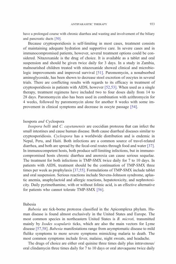
Table 3
Treatment of helminthic infections
Parasite Drug Pediatric dosage Adult dosage
Intestinal nematode
Roundworm Ascaris lumbricoides
Drugs of choice Albendazole 400 mg � 1 dose 400 mg � 1 dose
Mebendazole 100 mg twice daily � 3 d 100 mg twice daily � 3 d
Pyrantel pamoate 11 mg/kg once; repeat in 2 wk 11 mg/kg once; repeat in 2 wk
Ivermectin 150–200 mg/kg � 1 dose 150–200 mg/kg � 1 dose
Whipworm: Trichuris trichiura
Drugs of choice Albendazole 400 mg � 1 dose 400 mg � 1 dose
Mebendazole 100 mg twice daily � 3 d 100 mg twice daily � 3 d
Ivermectin 150–200 mg/kg � 1 dose 150–200 mg/kg � 1 dose
Hookworm: Ancylostoma duodenale and Necator americanus
Drugs of choice Albendazole 400 mg � 1 dose 400 mg � 1 dose
Mebendazole 100 mg twice daily � 3 d 100 mg twice daily � 3 d
Pyrantel pamoate 11 mg/kg once daily � 3 d 11 mg/kg once daily � 3 d
Pinworm: Enterobius vermicularis
Drugs of choice Pyrantel pamoate 11 mg/kg once; repeat in 2 wk 11 mg/kg once; repeat in 2 wk
Alternatives Albendazole 400 mg once; repeat in 2 wk 400 mg once; repeat in 2 wk
Mebendazole 100 mg once; repeat in 2 wk 100 mg once; repeat in 2 wk
Ivermectin 150–200 mg/kg � 1 dose 150–200 mg/kg � 1 dose
Strongyloides stercoralis
Drugs of choice Albendazole 400 mg twice daily � 2 d 400 mg twice daily � 2 d
Alternatives Ivermectin 200 mg/kg once daily � 2 d 200 mg/kg once daily � 2 d
Thiabendazole 50 mg/kg twice daily � 2 d 50 mg/kg twice daily � 2 d
Cutaneous larva migrans: Ancylostoma brasiliense and Uncinaria stenocephala
Drugs of choice Thiabendazole Topically 2–3 times daily for 5–10 d Topically 2–3 times daily for 5–10 d
Albendazole 400 mg once daily � 3 d 400 mg once daily � 3 d
Ivermectin 200 mg/kg once daily � 1–2 d 200 mg/kg once daily � 1–2 d
moon
&oberhelman
938
Blood and tissue nematodes
Filariasis: Onchocerca volvulus
Drug of choice Ivermectin 150 mg/kg once monthly � 6–12 mo 150 mg/kg once monthly � 6–12 mo
Lymphatic filariasis: Wuchereria bancrofti, Brugia malayi, Brugia timori
Drug of choice Diethylcarbamazine 6 mg/kg � 1 dose 6 mg/kg � 1 dose
Visceral larva migrans: Toxocara cari
Drug of choice Albendazole 400 mg twice daily � 5 d 400 mg twice daily � 5 d
Mebendazole 100–200 mg twice daily � 5 d 100–200 mg twice daily � 5 d
Cestodes
Tapeworms
Taeniasis: T.saginata/T. solium
Drugs of choice Niclosamide 50 mg/kg � 1 dose 2 g � 1 dose
Praziquantel 5–10 mg/kg � 1 dose 5–10 mg/kg � 1 dose
Cysticercosis: T.solium
Drug of choice Albendazole 15 mg/kg twice daily � 15–30 d 400 mg twice daily � 15–30 d
Alternative Praziquantel 50–100 mg/kg 3 times daily � 30 d 50–100 mg/kg 3 times daily � 30 d
Hydatid disease: Echinococcus granulosus and E. multilocularis
Drugs of choice Albendazole 15 mg/kg once daily for 1–6 mo 400 mg twice daily � 1–6 mo
Diphyllobothrium latum, Dipylidium caninum
Drug of choice Praziquantel 5–10 mg/kg � 1 dose 5–10 mg/kg � 1 dose
Hymenolepsis nana
Drug of choice Praziquantel 25 mg/kg � 1 dose 25 mg/kg � 1 dose
Trematodes
Schistosomiasis
Drug of choice Praziquantel 40–60 mg/kg 2–3 times daily � 1 dose 40–60 mg/kg 2–3 times daily � 1 dose
Liver flukes: Clonorchis sinesis, Opisthorchis viverrini, and Opisthorchis felineus
Drug of choice Praziquantel 75 mg/kg 3 times daily � 1 dose 75 mg/kg 3 times daily � 1 dose
Alternative: (C. sinesis) Albendazole 10 mg/kg once daily � 7 d 10 mg/kg once daily � 7 d
Lung fluke: Paragonimus westermani
Drug of choice Praziquantel 75 mg/kg 3 times daily � 2 d 75 mg/kg 3 times daily � 2 d
antiparasitictherapy
939

moon & oberhelman940
freezing the leading edge of the cutaneous trail, have been tried in the past, but
are no longer recommended because of blistering and ulceration [91,103].
Ivermectin (single dose for 1–2 days) and thiabendazole (twice daily for 2 days)
are acceptable alternatives to albendazole for the treatment of S. stercoralis [90,
95,96]. Ivermectin, as a single dose for the treatment of ascariasis, trichuriasis,
and enterobiasis, is equal in efficacy to other agents, but it has limited activity
against hookworms [94]. Studies suggest that giving a single combination dose of
ivermectin plus albendazole produces superior cure rates and egg reduction for
trichuriasis than with either drug used alone [97]. Neither ivermectin nor
thiabendazole has been studied extensively in children, and safety profiles have
not been established for children weighing less than 15 kg. Neither drug is
recommended during pregnancy, but if treatment of heavy worm burden during
pregnancy is required, ivermectin should be used because of its low risk of
adverse events [90]. Thiabendazole is available in chewable tablets and oral
solution. It is well absorbed and associated with side effects such as dizziness,
nausea, diarrhea, and anorexia [96,98].
Blood and tissue nematodes (filarial parasites, Toxocara, and visceral larva
migrans)
As with the intestinal nematodes, tissue and blood nematodes are a serious
global public health problem. Currently, several World Health Organization–
sponsored campaigns are geared toward the eradication of some severe nematode
infections. These campaigns are based on antivector measures to decrease en-
vironmental exposure and are supplemented with mass treatment campaigns
when appropriate.
Ivermectin, as a single dose repeated monthly for 6 to 12 months, is currently
recommended for international campaigns against Onchocerca volvulus (the
agent causing river blindness). It is microfilaricidal and results in approximately
95% reduction in dermal microfilariae after one dose. This drug has been shown
to reduce or limit dramatically the transmission of filarial disease when used in
community-based disease control programs. On an individual basis, this drug is
not completely curative, however, because of its lack of effect on the adult para-
site. Side effects of ivermectin are infrequent and often due to the inflammatory
responses to the dead microfilariae. The frequency of common symptoms
accompanying treatment, including rash, pruritus, and myalgias, is less with each
subsequent treatment as the number of microfilariae decreases [99,100].
Lymphatic filariasis is caused by three different filariae species (Wuchereria
bancrofti, Brugia malayi, and Brugia timori) and accounts for approximately
120 million infections per year globally [101]. Most cases of lymphatic filariasis
are asymptomatic. Children frequently present with lymphadenopathy secondary
to worm infestation of the lymph nodes, most commonly in the legs, arms, and
scrotum. Tropical pulmonary eosinophilia, thought to result from immune re-
sponses to filarial antigens, rarely occurs in children [102]. Single-dose iver-
mectin given annually is an effective supplement to community-based control

antiparasitic therapy 941
programs, but treatment is not curative [100]. The drug of choice for treatment
of lymphatic filariasis is single-dose diethylcarbamazine, which is available in
tablet form. A 21-day course of diethylcarbamazine may be required for patients
with tropical pulmonary eosinophilia. Although diethylcarbamazine is effective at
clearing infection; there is little evidence to suggest that it reverses lymphatic
damage or pulmonary fibrosis. Side effects of diethylcarbamazine include pru-
ritus, maculopapular rash, fever, edema, and headache. Data in children are
limited, but no other adverse events have been reported [94]. Diethylcarbama-
zine is effective in vitro against Onchocerca volvulus, but it cannot be used
clinically because of the intense inflammatory response it causes with rapid
killing of microfilariae.
Visceral larva migrans and ocular larva migrans usually are caused by in-
fection most commonly resulting from Toxocara cani. Often the disease course
for Toxocara is subclinical and self-limited, and treatment is controversial. For
symptomatic disease, either albendazole or mebendazole twice daily for 5 days is
recommended. For patients with ocular or neurologic manifestations, combina-
tion therapy with albendazole and corticosteroids is recommended [103,104].
Cestodes (tapeworms [including taeniasis and cysticercosis] and hydatid
disease)
Cestodes, or tapeworms, are segmented worms that have two life cycle stages,
the adult stage and larval stage, both of which cause disease in humans. Ingestion
of undercooked meats containing larvae of Taenia solium (pork) or Taenia
saginata (beef) results in taeniasis when the larvae mature into adult tapeworms.
Taeniasis is characterized by mild symptoms of abdominal pain, bloating, nausea,
and diarrhea [105]. Niclosamide and praziquantel are the drugs of choice for
therapy. Niclosamide, as a single dose, is preferred because it is not absorbed
from the intestinal tract, but it is currently not available in the United States.
Praziquantel, as a single dose, is available in a scored tablet form [105,106]. Side
effects of praziquantel include malaise, abdominal discomfort, headache, diz-
ziness, and rarely urticaria. Safety profiles have not been established in children
younger than 4 years old [94].
Cysticercosis and neurocysticercosis are caused by the larval stage of
T. solium, but not T. saginata. In adults, the disease is characterized by symp-
toms related to increased intracranial pressure and immune-mediated inflamma-
tion. The disease differs in children, with generalized seizures a common initial
sign, secondary to the cystic mass lesion itself or granuloma formation after
cyst destruction [105,107]. Albendazole is the drug of choice for therapy; it is
effective and relatively inexpensive. It is administered twice daily for 15 to
30 days and can be repeated as necessary. Praziquantel given three times daily
for 15 to 30 days is an alternative. Although effective anticysticercal treatment
is available, the decision to treat is controversial because symptoms related to
neurocysticercosis are thought to result from the inflammatory response accom-
panying the death of the organism [105,107]. Studies confirm that neurologic

moon & oberhelman942
symptoms increase early in the course of treatment. Persons who are not treated,
however, have a higher frequency and persistence of neurologic symptoms.
These neurologic symptoms can be ameliorated by the concomitant use of
dexamethasone and anticonvulsants [108].
Hydatid disease is caused by the larval forms of the dog tapeworms, Echino-
coccus granulosus and Echinococcus multilocularis [90,109]. In adults, dissemi-
nation of cysts to multiple different tissue sites, especially the liver and lungs, can
follow ingestion. Echinococcus is the most common cause of liver cysts world-
wide. Symptoms may be mild for many years or result in serious complications,
including death. Dissemination to the brain and eyes in more common in child-
hood. The mainstay of treatment, when possible, is surgical removal of any cysts.
In some cases, such as uncomplicated liver cysts, percutaneous aspiration and
injection of a protoscolicidal agent is effective. In other cases, either in con-
junction with surgery or when surgery is contraindicated, chemotherapy with oral
benzimadoles is warranted. Albendazole and mebendazole have been shown
to be beneficial, but albendazole is preferred because of poor tissue penetra-
tion of mebendazole. Treatment may need to continue for 6 months [17,110].
Diphyllobothrium species (fish tapeworm), Dipylidium caninum (dog and cat
tapeworm), Hymenolepsis nana (dwarf tapeworm), and Hymenolepsis diminuta
(rodent tapeworm) are other tapeworms that cause human disease, which can be
treated with single-dose praziquantel [111].
Trematodes (schistosomes, lung and liver flukes)
Schistosomiasis, also known as bilharziasis, is caused by the parasitic blood
flukes called schistosomes. The World Health Organization estimates that
approximately 200 million people are infested worldwide, ranking it second
to malaria in terms of global public health importance [112,113]. Numerous
schistosome species can affect many different animals, with almost all human
cases resulting from S. mansoni, S. haematobium, S. japonicum, S. mekongi, and
S. intercalatum. These different species have differing global distributions and
differing predilections for sites of residence within the host [112]. These parasites
have a complex life cycle that involves snails as the intermediate host. Prevention
efforts are geared toward mass chemotherapy, improved sanitation, and snail
control through environmental engineering or molluscacides [114,115].
The drug of choice for treatment of all schistosome species is praziquantel,
given in either two or three doses for 1 day [112,116]. Praziquantel is one of
the safest antihelminthic medications with minimal side effects. It has not been
tested in pregnant and lactating women, however, and is classified as Pregnancy
category B. Currently, countries such as Ghana, China, Egypt, and the Philip-
pines have adopted the routine treatment of pregnant women with praziquantel
because of a presumed disproportionate risk from infection compared with treat-
ment [116].
Clonorchis sinesis, Opisthorchis viverrini, and Opisthorchis felineus consti-
tute a group of trematodes termed liver flukes, which reside in the human biliary

antiparasitic therapy 943
tract. Infections are caused by ingestion of uncooked fish that have been infested
with larval cysts. Praziquantel, given in three doses for 1 day, is the drug of choice
for the three trematodes. Albendazole, given daily for 7 days, is an alternative
for C. sinesis [17].
Paragonimus westermani, the lung fluke, is a trematode commonly causing
human disease in eastern Asia. After ingestion of uncooked crabs or crayfish,
the larvae penetrate through the diaphragm into the pleural space and migrate
through lung tissue into the bronchi. Approximately 1% of infections result in
cerebral disease, which is more common among children [117]. Praziquantel,
given three times daily for 2 days, is the drug of choice. For patients developing
cerebral disease, corticosteroids given concurrently with praziquantel can reduce
symptoms of inflammation caused by dying flukes [17].
Summary
Parasitic infections in children present many challenges for the pediatrician.
These complex diseases are often difficult to diagnose and require pathogen-
specific treatment with drugs that are unfamiliar to many clinicians. International
travel and immunodeficiency states, such as AIDS, have been factors in the in-
creasing prevalence and clinical importance of these infections in children. As
in bacterial and viral infections, the emergence of drug resistance is a continuing
potential threat. From endemic malaria in persons in sub-Saharan Africa to giar-
diasis in children US daycare centers to Pneumocystis infections in AIDS pa-
tients, it is likely that parasitic infections will remain a persistent challenge for
public health and infectious disease specialists for many years to come.
References
[1] Sachs J. Helping the world’s poorest. Economist 1999;14:17–20.
[2] Guerin PJ, Olliaro P, Nosten F, et al. Malaria: current status of control, diagnosis, treatment,
and a proposed agenda for research and development. Lancet Infect Dis 2002;2:564–73.
[3] Alecrim WD, Espinosa FEM, Alecrim MGC. Emerging and re-emerging diseases in Latin
America: Plasmodium falciparum infection in the pregnant patient. Infect Dis Clin North Am
2000;14:83–95.
[4] Shann F. The management of severe malaria. Pediatr Crit Care Med 2003;4:489–90.
[5] World Health Organization. World malaria situation in 1994. Parts I–III. Wkly Epidemiol Rec
1997;72:269–90.
[6] Suh KN, Kain KC, Keystone JS. Malaria. Can Med Assoc J 2004;170:1693–702.
[7] Winstanley P. Modern chemotherapeutic options for malaria. Lancet Infect Dis 2001;1:242–50.
[8] Trape JF, Pison G, Preziosi MP, et al. Impact of chloroquine resistance on malaria mortality.
C R Acad Sci III 1998;321:689–97.
[9] John CC. Drug treatment of malaria in children. Pediatr Infect Dis J 2003;22:649–50.
[10] Miles MA. The discovery of Chagas disease: progress and prejudice. Infect Dis Clin North Am
2004;18:247–60.
[11] Prata A. Clinical and epidemiological aspects of Chagas disease. Lancet Infect Dis 2001;1:
92–100.

moon & oberhelman944
[12] American Academy of Pediatrics. Summaries of infectious diseases. In: Pickering LK, editor.
Red book: 2003 report of the Committee on Infectious Diseases. 26th ed. Elk Grove Village
(IL)7 American Academy of Pediatrics; 2003.
[13] Estani SS, Segura EL, Ruiz AM, Velazquez E, Porcel BM, Yampotis C. Efficacy of
chemotherapy with benznidazole in children in the indeterminate phase of Chagas’ disease.
Am J Trop Med Hyg 1998;59:526–9.
[14] Viotti R, Vigliano C, Armenti H, Segura E. Treatment of chronic Chagas’ disease with benz-
nidazole: clinical and serological evolution with long-term follow-up. Am Heart J 1994;127:
151–62.
[15] Legros D, Ollivier G, Gastellu-Etchegorry M, et al. Treatment of human African
trypanosomiasis—present situation and needs for research and development. Lancet Infect
Dis 2002;2:437–40.
[16] Burchman RJ, Ogbunude PO, Enanza B, Barrett MP. Chemotherapy of African trypanoso-
miasis. Curr Pharm Des 2002;8:256–67.
[17] Drugs for parasitic infections. Med Lett Drugs Ther. Available at: www.medletter.com/.
Accessed November 4, 2004.
[18] Pepin J, Milord F, Khonde AN, et al. Risk factors for encephalopathy and mortality during
melarsoprol treatment of trypanosoma brucei gambiense sleeping sickness. Trans R Soc Trop
Med Hyg 1995;89:92–7.
[19] Ortega-Barria E. Trypanosoma species (trypanosomiasis). In: Long SS, Pickering LK, Prober
CG, editors. Principles and practice of pediatric infectious diseases. 2nd ed. New York7
Churchill Livingstone; 2003. p. 1324–30.
[20] Burri C. Eflornithine for the treatment of human African trypanosomiasis. Parasitol Res 2003;
90:S49–52.
[21] Markle WH, Makhoul K. Cutaneous leishmaniasis: recognition and treatment. Clin Infect Dis
1997;25:677–84.
[22] Zuckerman A, Lainson R. Leishmania. In: Kreier JP, editor. Parasitic protozoa, vol 1.
New York7 Academic Press; 1977. p. 77–87.
[23] Guerin PJ, Olliero P, Sundar S, et al. Visceral leishmaniasis: current status of control, diagnosis
and treatment, and a proposed research and development agenda. J Infect Dis 1999;180:564–7.
[24] Kafetzis DA, Maltezou HC. Visceral leishmaniasis in paediatrics. Curr Opin Infect Dis 2002;
15:289–94.
[25] Kafetzis DA. An overview of paediatric leishmaniasis. J Postgrad Med 2003;49:31–8.
[26] Herwaldt BL, Berman JD. Recommendations for treating leishmaniasis with sodium stibo-
gluconate (Pentostam) and review of pertinent clinical studies. Am J Trop Med Hyg 1992;
46:296–306.
[27] Sundar S, Pai K, Kumar R, et al. Resistance to treatment in Kala-azar: speciation of isolates
from northeast India. Am J Trop Med Hyg 2001;65:193–6.
[28] Lira R, Sundar S, Makharia A, et al. Evidence that the high incidence of treatment failures
in Indian Kala-azar is due to the emergence of antimony-resistant strains of leishmania donovani.
J Infect Dis 1999;180:564–7.
[29] Meyerhoff A. US Food and Drug Administration approval of Ambisome (liposomal
amphoteracin B) for treatment of visceral leishmaniasis. Clin Infect Dis 1999;28:42–8.
[30] Di Martino L, Mantovani MP, Gradoni L. Low dosage combination of meglumine antimoniate
plus allopurinol as first choice treatment of infantile visceral leishmaniasis in Italy. Trans Soc
Trop Med Hyg 1990;84:534–5.
[31] Li E, Stanley SL. Protozoa: amebiasis. Gastroenterol Clin North Am 1996;25:471–92.
[32] Hughes MA, Petri WA. Amebic liver abscess. Infect Dis Clin North Am 2000;14:565–82.
[33] Adams EB, MacLeod IN. Invasive amebiasis: amebic dysentery and its complications.
Medicine 1977;56:315–24.
[34] Bassily S, Farid Z, El-Masry A, Mikhail EM. Treatment of intestinal E. histolytica and
G. lamblia with metronidazole, tinidazole, and ornidazole: a comparative study. J Trop Med
Hyg 1987;90:9–12.

antiparasitic therapy 945
[35] McAuley JB, Juranek DD. Paromomycin in the treatment of mild-to-moderate intestinal
amebiasis. Clin Infect Dis 1992;15:551–2.
[36] McAuley JB, Herwaldt BL, Stokes SL, et al. Diloxinide furoate for treating asymptomatic
Entamoeba histolytica cyst passers: 14 years’ experience in the United States. Clin Infect Dis
1992;15:464–8.
[37] Katz DE, Taylor DN. Parasitic infections of the gastrointestinal tract. Gastroenterol Clin North
Am 2001;30:797–815.
[38] Lebwohl B, Deckelbaum RJ, Green PH. Giardiasis. Gastrointest Endosc 2003;57:906–13.
[39] Procop GW. Gastrointestinal infections. Infect Dis Clin North Am 2001;15:1073–108.
[40] Gardner TB, Hill DR. Treatment of giardiasis. Clin Microbiol Rev 2001;14:114–28.
[41] Hill DR, Nash TE. Intestinal flagellate and ciliate infections. In: Guerrant RL, Walker DH,
Weller PF, editors. Tropical infectious diseases. Philadelphia7 Churchill Livingstone; 1999.
p. 703–12.
[42] Ortiz JJ, Ayoub A, Gargala G, Chegne NL, Favennec L. Randomized clinical study of
nitazoxanide compared to metronidazole in the treatment of symptomatic giardiasis in children
from northern Peru. Aliment Pharmacol Ther 2001;15:1409–15.
[43] Abboud B, Lemee V, Gargala G, et al. Successful treatment of metronidazole and albendazole
resistant giardiasis with nitazoxanide in a patient with acquired immunodeficiency syndrome.
Clin Infect Dis 2001;32:1792–4.
[44] Diaz E, Mondragon J, Ramirez E, Bernal R. Epidemiology and control of intestinal parasites
with nitazoxanide in children in Mexico. Am Trop Med Hyg 2003;68:384–5.
[45] White Jr AC. Nitazoxanide: an important advance in anti-parasitic therapy. Am Trop Med Hyg
2003;68:382–3.
[46] Hall A, Nahar Q. Albendazole as a treatment for infections with Giardia duodenalis in children
in Bangladesh. Trans R Soc Trop Med Hyg 1993;87:84–6.
[47] Lossick J. Epidemiology of urogenital trichomoniasis. In: Honinberg BM, editor. Trichomonads
parasitic in humans. New York7 Springer-Verlag; 1989. p. 311–23.
[48] Christie JD, Garcia LS. Emerging parasitic infections. Clin Lab Med 2004;24:737–72.
[49] Centers for Disease Control and Prevention. Sexually transmitted disease treatment guidelines.
MMWR Morb Mortal Wkly Rep 2002;51(RR06):1–80.
[50] Kosek M, Alcantara C, Lima A, Guerrant RL. Cryptosporidiosis: a review. Lancet Infect Dis
2001;1:262–9.
[51] Amadi B, Mwiya M, Musuku J, et al. Effect of nitazoxanide on morbidity and mortality in
Zambian children with cryptosporidiosis: a randomized controlled trial. Lancet 2002;360:
1375–80.
[52] White Jr AC, Chappell CL, Hayat CS, Kimball KT, Flanigan TP, Goodgame RW. Paromomycin
for cryptosporidiosis in AIDS: a prospective, double-blind, placebo-controlled trial. J Infect Dis
1994;170:419–24.
[53] Hewitt RG, Yainnoutsos CT, Higgs ES, Carey JT, Geiseler PJ, Soave R. Paromomycin: no
more effective than placebo for treatment of cryptosporidiosis in patients with advanced
human immunodeficiency virus infection. Clin Infect Dis 2000;31:1084–92.
[54] Smith NH, Cron S, Valdez LM, Chappell CL, White Jr AC. Combination drug therapy for
cryptosporidiosis in AIDS. J Infect Dis 1998;178:900–3.
[55] Pape JW, Verdier RI, Boncy M, Boncy J, Johnson Jr WD. Cyclospora infection in adults
infected with HIV: clinical manifestations, treatment and prophylaxis. Ann Intern Med 1994;
121:654–7.
[56] Weiss LM, Perlman DC, Sherman J, Tanowitz H, Wittner M. Isospora belli infection: treatment
with pryimethamine. Ann Intern Med 1988;109:474–5.
[57] Krause PJ. Babesiosis. Med Clin North Am 2002;86:361–73.
[58] Speilman A. Human babesiosis on Nantucket Island: transmission by nymphal Ixodes ticks.
Am J Trop Med Hyg 1976;25:784–7.
[59] Krause PJ, Lepore T, Sikand VJ, et al. Atovaquone and azithromycin for the treatment of
human babesiosis. N Engl J Med 2000;343:1454–8.

moon & oberhelman946
[60] Montoya JG, Liesenfeld O. Toxoplasmosis. Lancet 2004;363:1965–76.
[61] Bale JF. Congenital infections. Neurol Clin North Am 2002;20:1039–60.
[62] Sanchez PJ. Perinatal infections. Clin Perinatol 2002;29:799–826.
[63] Abrams EJ. Opportunistic infections and other clinical manifestations of HIV disease in
children. Pediatr Clin North Am 2000;47:79–108.
[64] Torre D, Casari S, Speranza F, et al. Randomized trial of trimethoprim-sulfamethoxazole vs.
pyrimethamine-sulfadiazine for therapy of toxoplasmic encephalitis in patients with AIDS.
Italian Collaborative Study Group. Antimicrob Agents Chemother 1998;42:1346–9.
[65] Bye MR, Bernstein LJ, Glaser J, Kleid D. Pneumocystis carinii pneumonia in young children
with AIDS. Pediatr Pulmonol 1990;9:251–3.
[66] Hughes WT. Pneumocystis carinii pneumonia: new approaches to diagnosis, treatment and
prevention. Pediatr Infect Dis J 1991;10:391–9.
[67] Rieder MJ, King SM, Read S. Adverse reaction to trimethoprim-sulfamethoxazole among
children with human immunodeficiency virus infection. Pediatr Infect Dis J 1997;16:1028.
[68] Dohn MN, Weinberg WG, Torres RA, et al. Oral atovaquone compared with intravenous
pentamidine for Pneumocystis carinii pneumonia in patients with AIDS. Atovaquone study
group. Ann Intern Med 1994;121:174–80.
[69] Toma E, Thorne A, Singer J, et al. Clindamycin with primiquine vs. trimethoprim-
sulfamethoxazole therapy for mild and moderately severe Pneumocystis carinii pneumonia
in patients with AIDS: a multicenter, double-blind, randomized trial. Clin Infect Dis 1998;27:
524–30.
[70] Black JR, Feinberg J, Murphy RL, et al. Clindamycin and primiquin therapy for mild-to-
moderate episodes of Pneumocystis carinii pneumonia in patients with AIDS. AIDS Clinical
Trials Group 044. Clin Infect Dis 1994;18:905–13.
[71] Sattler FR, Frame P, Davis R, et al. Trimetrexate with leucovorin versus trimethoprim-
sulfamethoxazole for moderate to severe episodes of Pneumocystis carinii pneumonia in
patients with AIDS: a prospective, controlled multicenter investigation of the AIDS Clinical
Trials Group Protocol 029/031. J Infect Dis 1994;170:165–72.
[72] Centers for Disease Control and Prevention. 1995 Revised guidelines for prophylaxis against
Pneumocystis carinii pneumonia for children infected with or perinatally exposed to human
immunodeficiency virus. MMWR Morb Mortal Wkly Rep 1995;44(RR-4):1–11.
[73] Bryan RT, Cali A, Owen RL, Spencer HC. Microsporidia: opportunistic pathogens in patients
with AIDS. Prog Clin Parasitol 1991;2:1–26.
[74] Weber R, Bryan RT. Microsporidial infections in immunodeficient and immunocompetent
patients. Clin Infect Dis 1994;19:517–21.
[75] Weber R, Bryan RT, Schwartz DA, Owen RL. Human microsporidial infections. Clin Microbiol
Rev 1994;7:426–61.
[76] Wittner M, Tanowitz HB, Weiss LM. Parasitic infections in AIDS patients: cryptosporidiosis,
isosporiasis, microsporidiosis, cyclosporiasis. Infect Dis Clin North Am 1993;7:569–86.
[77] Orenstein JM, Dieterich DT, Kotler DP. Systemic dissemination by a newly recognized
intestinal microsporidia species in AIDS. AIDS 1992;6:1143–50.
[78] Weber R, Sauer B, Spycher MA, et al. Detection of Septata intestinalis in stool specimens and
coprodiagnostic monitoring of successful treatment with albendazole. Clin Infect Dis 1994;
19:342–5.
[79] Molina JM, Tourneur M, Sarfati C, et al. Fumagillin treatment of intestinal microsporidiosis.
N Engl J Med 2002;346:1963–9.
[80] Jain R, Prabhakar S, Modi M, Bhatia R, Sehgal R. Naegleria meningitis: a rare survival. Neurol
India 2002;50:470–2.
[81] Galarza M, Cuccia V, Sosa FP, Monges JA. Pediatric granulomatous cerebral amebiasis:
a delayed diagnosis. Pediatr Neurol 2002;26:153–6.
[82] Wang A, Kay R, Poon WS. Successful treatment of amoebic meningoencephalitis in a Chinese
living in Hong Kong. Clin Neurol Neurosurg 1993;95:249–52.
[83] Poungvarin N, Jariya P. The fifth non-lethal case of primary amebic meningoencephalitis.
J Med Assoc Thai 1991;74:112.

antiparasitic therapy 947
[84] Brown RL. Successful treatment of primary amebic meningoencephalitis. Arch Intern Med
1991;151:1201–2.
[85] Singhal T, Bajpai A, Kalra V, et al. Successful treatment of Acantamoeba meningitis with
combination oral antimicrobials. Pediatr Infect Dis J 2001;20:623–7.
[86] Sison JP, Kemper CA, Loveless M, McShane D, Visvesvara GS, Deresinski SC. Disseminated
Acanthamoeba infection in patients with AIDS: case reports and reviews. Clin Infect Dis 1995;
20:1207–16.
[87] Seijo Martinez M, Gonzalez-Mediero G, Santiago P, et al. Granulomatous amebic encephalitis
in a patient with AIDS: isolation of Acanthamoeba sp. group II from brain tissue and success-
ful treatment with sulfadiazine and fluconazole. J Clin Microbiol 2000;38:3892–5.
[88] Muennig P, Pallin D, Sell R, Chan MS. The cost effectiveness of strategies for the treatment
of intestinal parasites in immigrants. N Engl J Med 1999;340:773–9.
[89] Crawford FG, Vermund SH. Parasitic infections in day care centers. Pediatr Infect Dis J
1987;6:744–9.
[90] Urbani C, Albonico M. Antihelminthic drug safety and drug administration in the control of
soil-transmitted helminthiasis in community campaigns. Acta Trop 2003;86:215–21.
[91] American Academy of Pediatrics. Drugs for parasitic infections. In: Pickering LK, editor. Red
book: 2003 report of the Committee on Infectious Diseases. 26th ed. Elk Grove Village (IL)7
American Academy of Pediatrics; 2003. p. 744–70.
[92] Montresor A, Stoltzfus RJ, Albonico M, et al. Is the exclusion of children under 24 months
from antihelmintic treatment justifiable? Trans R Soc Trop Med Hyg 2002;96:197–9.
[93] Montresor A, Awasthi A, Crompton DWT. Use of benzimidoles in children younger than
24 months for the treatment of soil-transmitted helminthiasis. Acta Trop 2003;86:223–32.
[94] Wilson CM, Freedman DO. Antiparasitic agents. In: Long SS, Pickering LK, Prober CG,
editors. Principles and practice of pediatric infectious diseases. 2nd ed. New York7 Churchill
Livingstone; 2003. p. 1547–58.
[95] Naquira C, Jimenez G, Guerra JG, et al. Ivermectin for human strongyloidiasis and other
intestinal helminths. Am J Trop Med Hyg 1989;40:304–9.
[96] Schaffel R, Nucci M, Portugal R, et al. Thiabendazole for the treatment of strongyloides in
patients with hematologic malignancies. Clin Infect Dis 2000;31:821–2.
[97] Belizario VY, Amarillo ME, de Leon WU, de los Reyes AE, Bugayong MG, Macatangay BJC.
A comparison of the efficacy of single doses of albendazole, ivermectin, and diethylcarbama-
zine alone or in combinations against Ascaris and Trichuris spp. Bull World Health Organ
2003;81:35–42.
[98] Grove DI. Treatment of strongyloidiasis with thiabendazole: an analysis of toxicity and
effectiveness. Trans R Soc Trop Med Hyg 1982;76:114–8.
[99] Elgart GW, Meinking TL. Ivermectin. Dermatol Clin 2003;21:277–82.
[100] Richard-Lenoble D, Chandenier J, Gaxotte P. Ivermectin and filariasis. Fundam Clin Pharm
2003;17:199–203.
[101] Michael E, Bundy DA, Grenfell BT. Re-assessing the global prevalence and distribution of
lymphatic filariasis. Parasitology 1996;112:409–28.
[102] Boggild AK, Keystone JS, Kain KC. Tropical pulmonary eosinophilia: a case series in a setting
of nonendemicity. Clin Infect Dis 2004;39:1123–8.
[103] Caumes E. Treatment of cutaneous larva migrans and toxocara infection. Fundam Clin Pharm
2003;17:213–6.
[104] Dinning WJ, Gillespie SH, Cooling RI, Maizels RM. Toxocariasis: a practical approach to
management of ocular disease. Eye 1988;2:580–2.
[105] Garcia HH, Gonzalez AE, Evans CA, Gilman RH. Taenia solium cysticercosis. Lancet 2003;
362:547–56.
[106] Flisser A, Sarti E, Sarti R, Schantz PM, Valencia S. Effect of praziquantel on protozoan
parasites. Lancet 1995;345:316–7.
[107] Maguire JH. Tapeworms and seizures-treatment and prevention. N Engl J Med 2004;350:215–7.
[108] Garcia HH, Pretell EJ, Gilman RH, et al. A trial of antiparasitic treatment to reduce the rate of
seizures due to cerebral cysticercosis. N Engl J Med 2004;350:249–58.

moon & oberhelman948
[109] Thompson RCA. Biology and systematics of Echinococcus. In: Thompson RCA, Lambery AJ,
editors. Echinococcus and hydatid disease. London7 CAB International; 1995. p. 1–37.
[110] Yorganci K, Sayek I. Surgical treatment of hydatid cysts of the liver in the era of percutaneous
treatment. Am J Surg 2002;184:63–9.
[111] Richards Jr FO. Diphyllobothrium, Dipylidium, and Hymenolepsis species. In: Long SS,
Pickering LK, Prober CG, editors. Principles and practice of pediatric infectious diseases.
2nd ed. New York7 Churchill Livingstone; 2003. p. 1351–4.
[112] Elliott DE. Schistosomiasis. Gastroenterol Clin North Am 1996;25:599–625.
[113] Sturruck R. The parasites and their life cycles. In: Jordan P, Webbe G, Sturruck R, editors.
Human schistosomiasis. Wallingford, UK7 CAB International; 1993. p. 1–31.
[114] Parraga IM, Assis AM, Prado MS, et al. Gender differences in growth of school-aged children
with schistosomiasis and geohelminth infection. Am J Trop Med Hyg 1996;55:150–6.
[115] Capron A. Schistosomiasis: forty years’ war on the worm. Parasitol Today 1998;14:379–84.
[116] Olds GR. Administration of praziquantal to pregnant and lactating women. Acta Trop 2003;86:
185–95.
[117] Harinasuta T, Pungpak S, Keystone J. Trematode infections—opisthorchiasis, clonorchiasis,
fascioliasis, and paragonimiasis. Infect Dis Clin North Am 1993;7:699–716.