Human CD14dim Monocytes Patrol and Sense Nucleic Acids and Viruses via TLR7 and TLR8 Receptors
22
Human CD14 dim Monocytes Patrol and Sense Nucleic Acids and Viruses via TLR7 and TLR8 Receptors Jérôme Cros 2,9 , Nicolas Cagnard 3,9 , Kevin Woollard 1,9 , Natacha Patey 2 , Shen-Ying Zhang 4 , Brigitte Senechal 2 , Anne Puel 4 , Subhra K. Biswas 5 , Despina Moshous 6 , Capucine Picard 6 , Jean-Philippe Jais 3 , David D'Cruz 7 , Jean-Laurent Casanova 4,6,8 , Céline Trouillet 1 , and Fréderic Geissmann 1,2,∗ 1 Centre for Molecular and Cellular Biology of inflammation, Division of Immunology Infection and Inflammatory Diseases, King's College London, SE1 1UL London, UK 2 U838 INSERM, Université Paris Descartes, 75015 Paris, France 3 Plateforme Bio-informatique & Département de biostatistiques AP-HP, Hôpital Necker - Enfants Malades, Université Paris Descartes, 75015 Paris, France 4 Laboratory of Human Genetics of Infectious Diseases, Rockefeller Branch, Rockefeller University, New York, NY 10065, USA 5 Singapore Immunology Network, SIgN (BMSI, A ∗ STAR), Immunos, 138648 Singapore 6 Pediatric Immuno-Hematology Unit, Hôpital Necker-Enfants Malades, Université Paris Descartes, 75015 Paris, France 7 Louise Coote Lupus Unit, St Thomas Hospital, King's College London, SE1 7EH London, UK 8 Laboratory of Human Genetics of Infectious Diseases, Necker Branch, INSERM U550 and University Paris Descartes, Necker Medical School, 75015 Paris, France Summary Monocytes are effectors of the inflammatory response to microbes. Human CD14 + monocytes specialize in phagocytosis and production of reactive oxygen species and secrete inflammatory cytokines in response to a broad range of microbial cues. Here, we have characterized the functions of human monocytes that lack CD14 (CD14 dim ) and express CD16. CD14 dim monocytes were genetically distinct from natural killer cells. Gene expression analyses indicated similarities with murine patrolling Gr1 dim monocytes, and they patrolled the endothelium of blood vessels after adoptive transfer, in a lymphocyte function-associated antigen-1-dependent manner. CD14 dim monocytes were weak phagocytes and did not produce ROS or cytokines in response to cell-surface Toll-like receptors. Instead, they selectively produced TNF-α, IL-1β, and CCL3 in response to viruses and immune complexes containing nucleic acids, via a proinflammatory TLR7-TLR 8-MyD88-MEK pathway. Thus, CD14 dim cells are bona fide monocytes involved in the innate local surveillance of tissues and the pathogenesis of autoimmune diseases. © 2010 ELL & Excerpta Medica. ∗ Corresponding author [email protected]. 9 These authors contributed equally to this work This document was posted here by permission of the publisher. At the time of deposit, it included all changes made during peer review, copyediting, and publishing. The U.S. National Library of Medicine is responsible for all links within the document and for incorporating any publisher-supplied amendments or retractions issued subsequently. The published journal article, guaranteed to be such by Elsevier, is available for free, on ScienceDirect. Sponsored document from Immunity Published as: Immunity. 2010 September 24; 33(3): 375–386. Sponsored Document Sponsored Document Sponsored Document
-
Upload
univ-paris7 -
Category
Documents
-
view
4 -
download
0
Transcript of Human CD14dim Monocytes Patrol and Sense Nucleic Acids and Viruses via TLR7 and TLR8 Receptors

Human CD14dim Monocytes Patrol and Sense Nucleic Acids andViruses via TLR7 and TLR8 Receptors
Jérôme Cros2,9, Nicolas Cagnard3,9, Kevin Woollard1,9, Natacha Patey2, Shen-YingZhang4, Brigitte Senechal2, Anne Puel4, Subhra K. Biswas5, Despina Moshous6, CapucinePicard6, Jean-Philippe Jais3, David D'Cruz7, Jean-Laurent Casanova4,6,8, CélineTrouillet1, and Fréderic Geissmann1,2,∗1Centre for Molecular and Cellular Biology of inflammation, Division of Immunology Infection andInflammatory Diseases, King's College London, SE1 1UL London, UK2U838 INSERM, Université Paris Descartes, 75015 Paris, France3Plateforme Bio-informatique & Département de biostatistiques AP-HP, Hôpital Necker - EnfantsMalades, Université Paris Descartes, 75015 Paris, France4Laboratory of Human Genetics of Infectious Diseases, Rockefeller Branch, RockefellerUniversity, New York, NY 10065, USA5Singapore Immunology Network, SIgN (BMSI, A∗STAR), Immunos, 138648 Singapore6Pediatric Immuno-Hematology Unit, Hôpital Necker-Enfants Malades, Université ParisDescartes, 75015 Paris, France7Louise Coote Lupus Unit, St Thomas Hospital, King's College London, SE1 7EH London, UK8Laboratory of Human Genetics of Infectious Diseases, Necker Branch, INSERM U550 andUniversity Paris Descartes, Necker Medical School, 75015 Paris, France
SummaryMonocytes are effectors of the inflammatory response to microbes. Human CD14+ monocytesspecialize in phagocytosis and production of reactive oxygen species and secrete inflammatorycytokines in response to a broad range of microbial cues. Here, we have characterized thefunctions of human monocytes that lack CD14 (CD14dim) and express CD16. CD14dim monocyteswere genetically distinct from natural killer cells. Gene expression analyses indicated similaritieswith murine patrolling Gr1dim monocytes, and they patrolled the endothelium of blood vesselsafter adoptive transfer, in a lymphocyte function-associated antigen-1-dependent manner.CD14dim monocytes were weak phagocytes and did not produce ROS or cytokines in response tocell-surface Toll-like receptors. Instead, they selectively produced TNF-α, IL-1β, and CCL3 inresponse to viruses and immune complexes containing nucleic acids, via a proinflammatoryTLR7-TLR 8-MyD88-MEK pathway. Thus, CD14dim cells are bona fide monocytes involved inthe innate local surveillance of tissues and the pathogenesis of autoimmune diseases.
© 2010 ELL & Excerpta Medica.∗Corresponding author [email protected] authors contributed equally to this workThis document was posted here by permission of the publisher. At the time of deposit, it included all changes made during peerreview, copyediting, and publishing. The U.S. National Library of Medicine is responsible for all links within the document and forincorporating any publisher-supplied amendments or retractions issued subsequently. The published journal article, guaranteed to besuch by Elsevier, is available for free, on ScienceDirect.
Sponsored document fromImmunity
Published as: Immunity. 2010 September 24; 33(3): 375–386.
Sponsored Docum
ent Sponsored D
ocument
Sponsored Docum
ent

AbstractGraphical Abstract—
Highlights—► Human CD14dim monocytes patrol the vasculature ► CD14dim monocytesrespond poorly to TLR1, TLR2, and TLR4 agonists ► CD14 dim monocytes respond to virusesand nucleic acids via TLR7-8-MyD88-MEK pathway
IntroductionBlood monocytes are bone marrow-derived phagocytes involved in the innate response tobacterial, fungal, parasitic, and viral infection (Barbalat et al., 2009; Gupta et al., 2001;Serbina et al., 2009; Serbina et al., 2008; Ströher et al., 2001). Nonetheless, the full spectrumof monocyte function, and in particular the specific functions of monocyte subsets, is not yetknown. Recent work has revealed heterogeneity of monocytes, with two distinct subsets inrodents as well as in humans (Ancuta et al., 2009; Auffray et al., 2009; Geissmann et al.,2003; Ingersoll et al., 2010; Sunderkötter et al., 2004; Zhao et al., 2009; Ziegler-Heitbrock,2007). Akin to other leucocytes, such as lymphocytes, evidence suggests that the diversity offunctions ascribed to monocytes might reflect cellular heterogeneity, with distinct subsetsendowed with specific functions.
On the basis of in vivo studies, murine monocytes have been separated into two subclasses.A major subset of “inflammatory” Gr1+ monocytes specializes in the production of tumornecrosis factor (TNF)-α, reactive oxygen species (ROS), and nitric oxide (NO) but littleinterleukin (IL)-10 upon in vivo infection with bacteria such as Listeria monocytogenes orparasites such as Toxoplasma gondii (Serbina et al., 2008). Gr1+ monocytes also producetype 1 interferon in response to viral ligands (Barbalat et al., 2009). In contrast, Gr1−monocytes patrol the blood vasculature, can differentiate into macrophages afterextravasation into tissues, and have been suggested to be associated with tissue repair(Auffray et al., 2007; Nahrendorf et al., 2007). Their role in response to infection is notdocumented.
Human CD14+ CD16− monocytes resemble Gr1+ cells based on surface marker expressionand gene expression arrays (Geissmann et al., 2003; Ingersoll et al., 2010; Strauss-Ayaliet al., 2007) and are reportedly producers of IL-10 and weak producers of TNF-α in vitro.Human CD16+ monocytes, which resemble Gr1− cells, are described as the main producersof inflammatory cytokines such as TNF-α and IL-1β in response to bacteriallipopolysaccharides (LPSs) (Belge et al., 2002). Recent work demonstrates distinctresponses of human monocyte subsets to Aspergillus fumigatus conidia (Serbina et al.,2009). Although both monocyte subsets efficiently phagocytose conidia, CD14+ CD16−monocytes inhibit conidial germination yet secrete little TNF-α, whereas CD14+ CD16+
Cros et al. Page 2
Published as: Immunity. 2010 September 24; 33(3): 375–386.
Sponsored Docum
ent Sponsored D
ocument
Sponsored Docum
ent

monocytes do not inhibit conidial germination but secrete large amounts of TNF-α (Serbinaet al., 2009). Of note, an additional degree of complexity was introduced by the work ofGrage-Griebenow (Skrzeczyńska-Moncznik et al., 2008) and Schäkel (Schäkel et al., 2002),indicating that human CD16+ monocytes are not a homogeneous cell population. Thus, onthe one hand it is increasingly clear that monocyte subsets exert critical specific functions inthe response to microbes, whereas on the other hand the reported differences in the behaviorof monocyte subsets in mice and human have hampered the description of potentialevolutionary conserved functions (Auffray et al., 2009; Skrzeczyńska-Moncznik et al., 2008;Ziegler-Heitbrock, 2007).
The murine population of Gr1− monocytes patrol the blood vasculature and is thereforeideally located to survey local tissue damage and infection (Auffray et al., 2007). Thepresent study was therefore designed to search for putative human functional homologs tomurine Gr1− patrolling monocytes and to characterize the full spectrum of their functions.
ResultsHuman Homologs to Murine Monocytes
Human monocytes can be distinguished from other cell types in the peripheral blood by flowcytometry (Figure 1 and Figure S1 available online). Large CD14+ CD16− monocytes (85%of blood monocytes) can be distinguished from a population of large CD14+CD16+
monocytes (5%), on the basis of the level of expression of CD16, CD62L, CD11c, CD163,and CX3CR1 (Figures 1A–1D and Figure S1). Monocytes expressing low amounts of CD14(CD14dim, 7% of monocytes) have a smaller size and granularity, express CD16, and havelower expression of CD11b and CD163 (Figures 1A–1D). These smallCD14dimCD16+monocytes can be further subdivided into CD14dimCD16+ 6-sulfo LacNAc(Slan)+ and CD14dimCD16+Slan− (Figure 1C). Blood monocytes Gr1+ and Gr1−, from C57/Bl6 mice (n = 3, samples Gr1+1, 2, and 3 and Gr1− 1, 2, and 3), were therefore compared tothe prospective human monocyte subsets from three healthy donors with whole-genomeexpression arrays (as described in the Experimental Procedures). For the three healthydonors, five samples were analyzed for each subset, including biological replicates fromdonor 1 (D1 samples 17–19, 19–21, 23–25, 26–28, and 29–31), and simplicates from donor2 (32, 33, 34, 35, and 36) and donor 3 (37, 38, 39, 40, and 41) (see also SupplementalExperimental Procedures). Common expressed genes between human and mouse chips wereselected with Ingenuity. Principal component analysis (PCA) on the human data setindicated that CD14+CD16−, CD14+CD16+, and CD14dimCD16+ subsets segregated inindependent clusters (Figure 1E). Expression of Slan did not allow discrimination of subsetsamong CD14dimCD16+cells (Figure 1E). Human and mouse data sets were then merged andsubmitted to a hierarchical clustering with the Spearman correlation similarity measure withaverage linkage that used “all expressed genes” and “fold 2 expressed” genes (Figure 1F andFigure S1), and submitted independently to a PCA with a covariance matrix (Figure S1).These two unsupervised analyses showed comparable results, summarized in Figure 1G andFigure S1. Results from the hierarchical clustering analysis using D1, D2, and D3 with allindividual D1 biological replicates (Figure 1F), with the mean of D1 replicates (Figure S1),or with individual replicates (not shown) were similar and led to an identical conclusion. Inall cases the samples were split into two main groups, each of which included human andmouse samples. Gr1− murine samples always segregated with the samples of CD14dim
CD16+ human monocytes samples, whereas Gr1+ murine monocyte samples segregated withall of the CD14+ human samples, independently of expression of CD16 (Figure 1F andFigure S1).
These results predicted that the absence of expression of the CD14 and CD163 antigensdistinguishes the putative human homologs of murine Gr1− “patrolling” monocytes, whereas
Cros et al. Page 3
Published as: Immunity. 2010 September 24; 33(3): 375–386.
Sponsored Docum
ent Sponsored D
ocument
Sponsored Docum
ent

both CD14+CD16− and CD14+CD16+ cells would resemble mouse Gr1+ “inflammatory”monocytes, in contrast to previous hypotheses (Geissmann et al., 2003; Ingersoll et al.,2010; Ziegler-Heitbrock, 2007).
“Patrolling” CD14dim MonocytesTo test this prediction, we purified human monocyte subsets from peripheral blood by flowcytometry (as described in Figure S1), labeled them with fluorescent probes, transferredthem intravenously into Rag2−/− Il2rg−/− Cx3cr1gfp mice, and examined them by intravitalmicroscopy (Figure 1H and Movies S1 and S2). Most human CD14dim monocytes attachedimmediately to the endothelium after intravenous transfer and crawled for an extendedperiod of time, whereas crawling was not observed after intravenous transfer of CD14+
monocytes (Figure 1I), further suggesting that CD14dim monocytes are the functionalhomologs of murine “patrolling” Gr1− monocytes. The ability of human monocytes to attachand crawl on murine endothelium was consistent with interspecies conservation of integrins,in particular lymphocyte function-associated antigen-1 (LFA-1) (Vidovic et al., 2003),which is critical for murine monocyte crawling (Auffray et al., 2007). To test thishypothesis, we therefore performed intravenous injection of human LFA1 blocking antibody(clone 38[Dransfield et al., 1992]), and observed that it abolished crawling by humanmonocytes inside mouse blood vessels (Figure 1I). These data indicated that humanCD14dim monocytes can patrol blood vessels in vivo, in a LFA1-dependent manner.
“Inflammatory” CD14+CD16− and CD14+CD16+ Monocytes Respond to Bacteria-Associated Signals
As expected, CD14+ monocytes were very efficient at phagocytosing latex beads(Figure 2A). CD14+CD16− monocytes produced high level of ROS (Figure 2B) and alsoexpressed more mRNA for myeloperoxidase and lysozyme (Figure S2). In the presence ofLPS, CD14+CD16− monocytes produced very high amounts of a distinct and limited set ofchemokines and cytokines, including IL-6, IL-8, CCL2, and CCL3 (Figure 2C).
The double-positive CD14+CD16+ monocyte population did not produce ROS (Figure 2B),expressed mRNA for myeloperoxidase and lysozyme at low levels (Figure S2), andproduced cytokines distinct from those produced by CD14+CD16− monocytes (Figure 2C).They were the main producers of IL-1β and TNF-α in response to LPS and additionallyproduced IL-6 and CCL3 (Figure 2C). CD14+CD16+ also accounted for the bulk of cytokineproduction in response to the Toll-like receptor (TLR) 2 agonist Pam3ck4 (data not shownand Figure S2). Both CD14+CD16− and CD14+CD16+ monocytes produced moderateamount of the anti-inflammatory cytokine IL-10, with slightly different kinetics (Figure S2)that may explain previous contradictory reports (Belge et al., 2002; Skrzeczyńska-Monczniket al., 2008).
Therefore, CD14+ monocytes as a population exhibited the basic characteristics of Gr1+
“inflammatory” TNF-α and ROS-producing murine monocytes (Serbina et al., 2008).Distinct cell types within this population, i.e., CD14+CD16− and CD14+CD16+, areresponsible for the bulk production of ROS, IL-6, IL-8, and CCL2 and of TNF-α and IL-1,respectively, in the presence of LPS. These observations are in line with the publishedevidence that LPS induces expression of different cytokines by CD14+ and CD16+
monocytes (Belge et al., 2002; Skrzeczyńska-Moncznik et al., 2008). The secretion of type IIFN, IL-2, IL-4, IL-5, IL-7, IL-9, IL-12, IL-13, IL-15, IL-17, CCL11, granulocyte colony-stimulating factor (G-CSF), granulocyte-macrophage colony-stimulating factor (GM-CSF),platelet-derived growth factor (PDGF), and vascular endothelial growth factor (VEGF) wasnot detected in monocytes in these experiments (data not shown).
Cros et al. Page 4
Published as: Immunity. 2010 September 24; 33(3): 375–386.
Sponsored Docum
ent Sponsored D
ocument
Sponsored Docum
ent

“Patrolling” CD14dim Monocytes Respond Poorly to Bacterial CuesCD14dim cells exhibited a limited ability to uptake latex beads in comparison to CD14+
monocytes (Figure 2A), did not produce ROS (Figure 2B), expressed little mRNA formyeloperoxidase (MPO) and lysozyme (Figure S2), and gave little response to LPS or toPAM3CK4 TLR1 and two agonists (Figure 2C and Figure S2). Thus, the production ofIL-1β and TNF-α in response to LPS, previously attributed to the CD16+ fraction ofmonocytes, is restricted to the double-positive CD14+ CD16+ cells in our experiments.
As expected from PCA and clustering analysis, CD14dim monocytes expressed a distinct setof genes in comparison to both CD14+CD16+ and CD14+CD16− monocytes, includingchemokine receptors, as well as different sets of apolipoproteins, scavenger receptors forlipids and dying cells, and cytokine receptors (Figure S2). CD14dim monocytes did notexpress inflammatory chemokine receptors such as CCR2 and expressed lower levels ofCCR1, CCR5, IL-17RA, apolipoprotein (Apo) B48, CD36, MPO, and lysozyme. In contrast,they expressed higher amounts of IL-10 receptor and CXCL16 scavenger receptor ApoAand ApoE (Figure S2) and produced high amounts of IL-1 receptor antagonist duringovernight culture (Figure 2D). Overall, the monocyte proinflammatory response, includingphagocytosis and the production of ROS, MPO, lysozyme, and cytokines in response towell-characterized TLR1, TLR2, and TLR4 signals, appeared to be mediated by CD14+
monocytes, whereas the profile of CD14dim monocytes was, if anything, anti-inflammatoryin this context.
Are CD14dim Cells Monocytes?We thus considered the possibility that CD14dim cells might not be monocytes. Cytologicexamination indicated that CD14dim cells had a morphology compatible with monocytes,and flow cytometric profiles indicated that they expressed the macrophage colony-stimulating factor (MCSF) receptor and CD68, but low CD11b and CD163 (see Figures 1B–1D). A subset of human natural killer (NK) lymphocytes express CD16 and HLA-DR, andrecent studies have revealed that NK cells can be confused for myeloid cells (Blasius et al.,2007; Caminschi et al., 2007; Vosshenrich et al., 2007). Therefore, we tested the hypothesisthat CD14dim cells might actually represent major histocompatibility complex (MHC) classII-positive NK cells by examining cells from patients deficient in the common cytokinereceptor gamma chain (γc); these patients lack all lymphoid lineages. We found normalnumbers of CD14dim CD16+ cells in three γc-deficient patients, whereas as expected CD16+
NK cells were absent from these patients (Figure 3A), indicating that CD14dim cells are notlymphoid.
Cells expressing HLA-DR, CD16, and Slan and negative for CD14 have also been describedas antigen-presenting dendritic cells (DCs) (Schäkel et al., 2006). Both CD14+ and CD14dim
monocytes stimulate allogenous mixed lymphocyte reaction at high effector/target ratio(Figures S2D and S2E). However, they do not process and present antigen (Figure 3B). Forinvestigating the ability of CD14dim CD16+ cells to present a recall protein antigen to T cellsin comparison to other monocyte subsets and blood DCs, tetanus toxoid was added tococulture of monocytes or DCs and T cells from autologous vaccinated donors (Sallusto andLanzavecchia, 1994). Freshly isolated monocytes from any subset did not promote anysignificant antigen-dependent T cell proliferation or IL-2 production ,whereas CD14−CD16−HLA-DR+ DCs are able to efficiently present tetanus toxoid to T cells and to trigger strongIL-2 production (Figure 3B). Therefore, CD14dim cells appeared to be unable to processantigen, and genetic and functional evidence indicated that they are a bona fide monocytesubset. Of note, in our PCA, CD14dimCD16+Slan+ cells clustered together withCD14dimCD16+Slan− cells and total CD14dimCD16+ cells (see Figure 1).
Cros et al. Page 5
Published as: Immunity. 2010 September 24; 33(3): 375–386.
Sponsored Docum
ent Sponsored D
ocument
Sponsored Docum
ent

CD14dim Monocytes Produce a Selected Set of Proinflammatory Cytokines in Response toViruses
The poor response via TLR1 and TLR 2 and via TLR4 does not preclude the involvement ofmonocytes in the inflammatory responses to other classes of pathogens. P. Ancuta andothers have proposed a role for CD16+ monocytes in the immune response to HIV (Ancutaet al., 2006) and monocytic cells have been shown to produce inflammatory cytokines in thepresence of viruses (Gupta et al., 2001; Ströher et al., 2001). A separate subset of whiteblood cells, plasmacytoid DCs, are known to be the main blood producers of type 1 IFN inresponse to viruses (Liu, 2005), although murine Gr1+ monocytes can contribute toproduction of type 1 IFN in response to viruses (Barbalat et al., 2009). Among TLRs thatrecognize nucleic acids and are involved in the immune response to viruses (TLR3, TLR7,TLR8, and TLR9), monocytes weakly express TLR3 and TLR9 and do not respond to TLR3and TLR9 agonists (Jarrossay et al., 2001; Kadowaki et al., 2001) (Figure S3). However,human monocytes express TLR7 and TLR8 (Supplemental figure S3), the two otherendosomal TLRs involved in the immune response to viruses (Diebold et al., 2004; Heilet al., 2004; Lund et al., 2004). We thus reasoned that human monocytes subsets may beinvolved in the pro-inflammatory response to viruses.
CD14+ monocytes produced very high amounts of the granulocyte, B cell, and T cell“helper” chemokines and cytokines IL-6, IL-8, and CCL2, in response to measles (ssRNA[−]) and herpes simplex virus 1 (HSV-1) (dsDNA) at a multiplicity of infection (MOI) of 1,similar to what was observed for LPS. In contrast, freshly purified CD16+ monocytes,especially CD14dim CD16+ monocytes, whether Slan+ or Slan−, selectively produced veryhigh amounts of the proinflammatory cytokines TNF-α, IL-1β, and of CCL3 in response tomeasles and HSV-1 (Figures 4A and 4B). Of note, secretion of type I IFN by humanmonocytes was very low (not shown).
CD14dim Monocytes Respond to Viruses via a Unique TLR7-TLR 8-MYD88-MEK-DependentPathway
To investigate the mechanisms of cytokine production by CD14dim and CD14+ monocytes,we examined their response to TLR7 and TLR8 agonists. The response of monocyte subsetsto stimulation with TLR8 agonists (3M2 and R848) and to a lesser extent to a TLR7 agonist(3M13) was similar to their response to intact viruses. CD14dim monocytes produced veryhigh amounts of proinflammatory cytokines, but little IL-6, IL-8, and CCL2, whereasCD14+ CD16− monocytes produced little proinflammatory cytokines, but high IL-6 andIL-8 (Figure 4C). As was true for responses to viruses, the population of CD14+ CD16+
monocytes displayed an intermediate phenotype (Figure S3).
To investigate whether cytokine production by monocytes required infectious virus, weincubated monocytes with either live, ultraviolet light (UV)-inactivated, or heat-inactivatedHSV1-green fluorescent protein (GFP) expressing virus. Control fibroblasts exhibited GFPfluorescence only when infected with live virus (Figure S3). In contrast, monocytesexhibited similar GFP fluorescence whether they were exposed to live, UV-inactivated, orheat-treated viruses (Figure S3), probably reflecting uptake rather than infection. Monocytesproduced cytokines in response to live virus and UV-inactivated virus, but not in response todenatured virus particles obtained by heat-inactivation (Figure S3). These data indicate thatmonocytes are activated by HSV-1 particles, in line with a TLR-mediated response, butactive HSV-1 replication may not be required for cytokine production at a MOI of 1.Similarly, replication of measles virus was not observed at a MOI of 1 (Figure S3).
To confirm the role of TLRs and specifically of TLR7 and TLR8 for cytokine production,we purified CD14dim and CD14+ CD16− monocytes from the peripheral blood of patients
Cros et al. Page 6
Published as: Immunity. 2010 September 24; 33(3): 375–386.
Sponsored Docum
ent Sponsored D
ocument
Sponsored Docum
ent
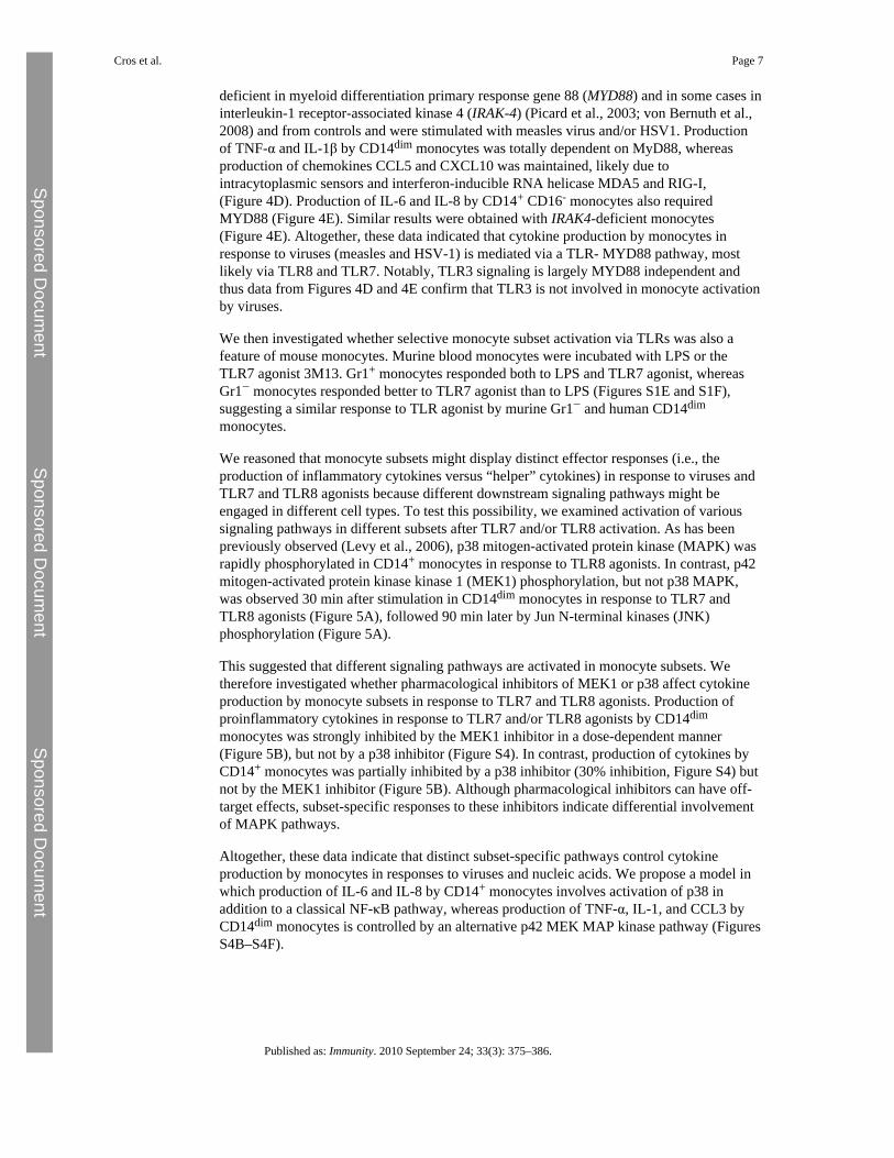
deficient in myeloid differentiation primary response gene 88 (MYD88) and in some cases ininterleukin-1 receptor-associated kinase 4 (IRAK-4) (Picard et al., 2003; von Bernuth et al.,2008) and from controls and were stimulated with measles virus and/or HSV1. Productionof TNF-α and IL-1β by CD14dim monocytes was totally dependent on MyD88, whereasproduction of chemokines CCL5 and CXCL10 was maintained, likely due tointracytoplasmic sensors and interferon-inducible RNA helicase MDA5 and RIG-I,(Figure 4D). Production of IL-6 and IL-8 by CD14+ CD16- monocytes also requiredMYD88 (Figure 4E). Similar results were obtained with IRAK4-deficient monocytes(Figure 4E). Altogether, these data indicated that cytokine production by monocytes inresponse to viruses (measles and HSV-1) is mediated via a TLR- MYD88 pathway, mostlikely via TLR8 and TLR7. Notably, TLR3 signaling is largely MYD88 independent andthus data from Figures 4D and 4E confirm that TLR3 is not involved in monocyte activationby viruses.
We then investigated whether selective monocyte subset activation via TLRs was also afeature of mouse monocytes. Murine blood monocytes were incubated with LPS or theTLR7 agonist 3M13. Gr1+ monocytes responded both to LPS and TLR7 agonist, whereasGr1− monocytes responded better to TLR7 agonist than to LPS (Figures S1E and S1F),suggesting a similar response to TLR agonist by murine Gr1− and human CD14dim
monocytes.
We reasoned that monocyte subsets might display distinct effector responses (i.e., theproduction of inflammatory cytokines versus “helper” cytokines) in response to viruses andTLR7 and TLR8 agonists because different downstream signaling pathways might beengaged in different cell types. To test this possibility, we examined activation of varioussignaling pathways in different subsets after TLR7 and/or TLR8 activation. As has beenpreviously observed (Levy et al., 2006), p38 mitogen-activated protein kinase (MAPK) wasrapidly phosphorylated in CD14+ monocytes in response to TLR8 agonists. In contrast, p42mitogen-activated protein kinase kinase 1 (MEK1) phosphorylation, but not p38 MAPK,was observed 30 min after stimulation in CD14dim monocytes in response to TLR7 andTLR8 agonists (Figure 5A), followed 90 min later by Jun N-terminal kinases (JNK)phosphorylation (Figure 5A).
This suggested that different signaling pathways are activated in monocyte subsets. Wetherefore investigated whether pharmacological inhibitors of MEK1 or p38 affect cytokineproduction by monocyte subsets in response to TLR7 and TLR8 agonists. Production ofproinflammatory cytokines in response to TLR7 and/or TLR8 agonists by CD14dim
monocytes was strongly inhibited by the MEK1 inhibitor in a dose-dependent manner(Figure 5B), but not by a p38 inhibitor (Figure S4). In contrast, production of cytokines byCD14+ monocytes was partially inhibited by a p38 inhibitor (30% inhibition, Figure S4) butnot by the MEK1 inhibitor (Figure 5B). Although pharmacological inhibitors can have off-target effects, subset-specific responses to these inhibitors indicate differential involvementof MAPK pathways.
Altogether, these data indicate that distinct subset-specific pathways control cytokineproduction by monocytes in responses to viruses and nucleic acids. We propose a model inwhich production of IL-6 and IL-8 by CD14+ monocytes involves activation of p38 inaddition to a classical NF-κB pathway, whereas production of TNF-α, IL-1, and CCL3 byCD14dim monocytes is controlled by an alternative p42 MEK MAP kinase pathway (FiguresS4B–S4F).
Cros et al. Page 7
Published as: Immunity. 2010 September 24; 33(3): 375–386.
Sponsored Docum
ent Sponsored D
ocument
Sponsored Docum
ent

CD14dim Monocyte-Specific Inflammatory Response to Self-Nucleic Acids in Patients withLupus
Intracellular TLRs activated by nucleic acids, and particularly RNA, have evolved understrong selection, suggesting an essential role in host survival (Barreiro et al., 2009).However, as a possible tradeoff mechanism, TLR7- and TLR9-dependent recognition ofendogenous nucleic acids is also a feature of autoimmune diseases such as systemic lupuserythematosus (SLE), a disease associated with autoantibodies to nucleosome andribonucleoproteins, and immune complex deposition in several organs (Rahman andIsenberg, 2008). Ribonucleoprotein-containing immune complexes stimulate immune cellsuch as plasmacytoid DCs and B cells via TLRs (Lau et al., 2005; Lövgren et al., 2006).
Although the mechanisms of Lupus-induced tissue damage, and in particularglomerulonephritis, are poorly understood, recent work in the mouse has proposed thatactivation of myeloid cells by glomerular immune complex, which requires both FcRγ andTLRs (Bergtold et al., 2006; Clynes et al., 1998), initiates an inflammatory response thatresults in glomerulonephritis. Murine Gr1− monocytes have recently been shown to beselectively expanded in nephritis, to accumulate inside the capillary vessels in the nephritickidney, and to be activated by immune complexes in an FcRγ-dependent manner (Bergtoldet al., 2006). In human, genetic studies studies have associated CD16 with lupus (Rhodesand Vyse, 2008). It was further shown that CD16+ monocytes adhere in vitro to endothelialcells better than CD14+ monocytes, a phenomenon in part dependent on the chemokinereceptor CX3CR1 (Ancuta et al., 2003), and human CD14dimCD16+ monocytes patrolcapillaries in vivo as shown in this report (see Figures 1H and 1I). Finally, elevated amountsof fractalkine (the CX3CR1 ligand) expression and accumulation of CD16+ monocytesinside glomerular blood vessels has been documented in human SLE (Hill et al., 2005;Yoshimoto et al., 2007). In accordance with the literature (Hill et al., 2005; Yoshimoto et al.,2007), we confirmed and observed a glomerular IgG deposition and monocytic infiltrate(CD68+ CD16+ CD14dim and CD15− CD3ɛ−) in glomerular vessels from patients with classIV-G lupus nephritis (Figure 6A).
We thus hypothesized that nucleic acid-containing immune complexes present in glomerularcapillaries activate CD14dim monocytes to produce TNF-α, IL-1, and CCL3 cytokines thatmay damage the endothelium. We compared the effects of serum from individuals with SLEand anti-ribonucleoprotein (RNP) antibodies (see Experimental Procedures) on monocyteactivation. Patient sera induced strong production of TNF-α and CCL3 by CD14dim
monocytes isolated from healthy donors, as well as the production of IL-6 and IL-8 byCD14+ monocytes, in comparison with sera from controls (Figure 6B). Sera were thenpretreated with RNase and DNase, and/or Ig depletion, and/or addition of ribonucleoprotein,before incubation with monocytes (Figures 6C and 6D). Both nucleic acids and Ig wereobserved to contribute 30%–50% of the production of CCL3 and TNF-α by CD14dim
monocytes; the depletion of nucleic acids and Ig together did not have an additive effect oninhibition. These results indicated that in addition to TLR7-TLR8 agonists, and viruses,ribonucleoprotein-containing immune complexes induce CCL3 and TNF-α production byCD14dim monocytes.
DiscussionThis study establishes CD14dim human monocytes as a bona fide myeloid immune cellpopulation that patrol blood vessels and exert specific effector functions in the inflammatoryresponse to viruses and nucleic acids. Genome-wide expression analysis (Figure 1) predictedthat these monocytes, which lack CD14 expression (CD14dim) and represent a minority(∼7%) of human blood monocytes, resemble mouse “patrolling” Gr1− monocytes and canbe distinguished from lymphoid cells, dendritic cells, and CD14+ CD16− and CD14+ CD16+
Cros et al. Page 8
Published as: Immunity. 2010 September 24; 33(3): 375–386.
Sponsored Docum
ent Sponsored D
ocument
Sponsored Docum
ent

monocytes. CD14dim monocytes patrol capillaries after adoptive transfer in vivo, do notproduce ROS, exhibit low or absent myeloperoxidase and lysozyme production in a steadystate, constitutively produce IL-1RA, and are weak producers of inflammatory cytokinesafter exposure to LPS or a TLR1 and TLR2 agonist. Nevertheless CD14dim cells belong tothe monocytic lineage, given that they develop in individuals lacking lymphoid precursorsdue to loss of function mutations in X-linked common γ chain, implying that they aredevelopmentally distinct from NK CD16+ lymphocytes. CD14dim monocytes exhibited ananti-inflammatory phenotype in a steady state and respond poorly to surface-associated TLRstimulation, but specialize in the production of the proinflammatory cytokines TNF-α,IL-1β, and of CCL3 in response to viruses and nucleic acids, via a unique TLR7-8, MyD88and MEK-dependent pathway. Of note, murine “patrolling” Gr1− monocytes also respondedbetter to TLR7 agonist than to LPS (Figure S1), suggesting a degree of functional similaritybetween human and murine patrolling monocytes.
Although they can stimulate the proliferation of allogenic T cells at high effector-to-targetratio (Sallusto and Lanzavecchia, 1994), blood monocytes, including the CD14dim subset, donot process and present a recall antigen to T cells, in contrast to blood DCs. This is incontrast to some previous reports that identified DCs as a population expressing CD16 andSlan (Schäkel et al., 2002). However, we observed that monocytes, in particular theCD14dim subset, can promote autologous T cell proliferation, in an antigen-independentmanner, and without detectable increase in IL-2 production. Therefore, although monocytesare clearly distinct from DCs, further studies will be needed to clarify the functionaloutcomes of their interaction with T cells (Evans et al., 2009).
Reported differences in the behavior of monocyte subsets in mice and human (Auffray et al.,2009; Skrzeczyńska-Moncznik et al., 2008; Ziegler-Heitbrock, 2007) have stimulated effortsto identify human homologs to monocyte subsets (Ingersoll et al., 2010). The PCA andhierachical clustering analysis reported here suggested that monocytes expressing CD14(CD14+), which represent more than 90% of blood monocytes, resemble mouse“inflammatory” Gr1+ monocytes, whether or not they express CD16. Indeed, they sharetheir key functional characteristics; they are responsible for phagocytosis, ROS production,and cytokine production in response to LPS via a classical MyD88-, p38-, and NF-κBdependent pathway. Within this subset, distinct cells, i.e., CD14+CD16− and CD14+CD16+
cells, are responsible for the bulk of ROS, IL-8, and IL-6 production and for TNF-α andIL-1 production respectively in response to LPS. This observation may appear different froma recent work that concluded that CD14+ CD16− monocytes were homologous to mouseGr1+ monocytes, whereas CD14+ CD16+ monocytes were homologous to mouse Gr1−monocytes (Ingersoll et al., 2010). However, this discrepancy can be accounted for by thefact that the Ingersoll study compared only two human subsets (CD14+ CD16− monocytesand human CD16+ monocytes) with two murine subsets (Gr1+ and Gr1− monocytes). TheirCD16+ monocyte population may have contained both the CD14dim subset and the CD14+
CD16− subset.
We therefore propose that CD14dim CD16+ monocytes represent a monocyte subset thatpatrol blood vessels and selectively detect virally infected and damaged cells to produceproinflammatory cytokines. This role of CD14dim CD16+ monocytes is reminiscent of themain function of plasmacytoid DCs (PDCs), which are also specialized in response to viralstimulation via TLR7 and TLR9 (Liu, 2005). However, PDCs secrete type I interferons,whereas CD14+ monocytes produce IL-6 and IL-8, and patrolling monocytes produce TNF-α, IL-1β, and CCL3, thus exhibiting complementary functions in the TLR-dependentantiviral response. Of note, we did not detect significant secretion of type I interferon byeither CD14+ or CD14dim monocytes in this study. In the murine system Gr1+ monocytes
Cros et al. Page 9
Published as: Immunity. 2010 September 24; 33(3): 375–386.
Sponsored Docum
ent Sponsored D
ocument
Sponsored Docum
ent

(which resemble CD14+ human monocytes) secrete IFN-β after activation by viruses(Barbalat et al., 2009).
During antiviral responses, a systemic type 1 interferon response is triggered by PDCs andGr1+ monocytes and a local type 1 interferon response by infected fibroblasts and othertissue cells. Our data suggest that in addition, monocytes and in particular patrollingCD14dim monocytes, participate in a regional antiviral response characterized by theproduction of proinflammatory cytokines such as TNF-α and IL-1β and of CCL3 and arepotentially involved in virus-induced immunopathology.
PCA and functional experiments indicated that CD14+CD16−, CD14dim and CD14+CD16+
monocytes represent distinct cell populations. The expression of CD16 by CD14+
monocytes may correspond to an activation and/or differentiation state of CD14+
monocytes, as suggested by several groups (Ziegler-Heitbrock, 2000). They both respond toextracellular TLR such as TLR4 and TLR2 and share a high phagocytic index.
In some donors the CD14+CD16+ population displayed an intermediate cytokine response toviruses and TLR7- TLR8 agonists between that of CD14dim and CD14+CD16− monocytes,and future studies will need to investigate potential heterogeneity of this “double positive”population. Although it is conceivable that CD14dim may also derive from CD14+
monocytes, there is no experimental evidence to support this claim, and results from thisstudy argue that, whether CD14dim arise from CD14+ monocytes or directly fromprogenitors, their distinct morphology, behavior in vivo, and signal transduction apparatusmake them two independent functional cell types.
Results presented here indicate that a TLR7-8, MyD88, and MEK pathway is critical for thesecretion of a distinct set of cytokines by CD14dim monocytes, whereas p38 is important inthe MyD88-dependent response of CD14+ monocytes. Activation of MAPK ERK1 andERK2 has been previously shown to play an important role in regulating cytokineproduction by murine myeloid cells (Agrawal et al., 2006), and the regulation of distinctcytokine genes by different members of the MAPK family (ERK versus p38 MAPK) hasbeen previously reported in DCs (Dillon et al., 2004). We used computational modeling toexamine the potential relevance of this model. Computational modeling of this forwardbiochemical cascades for TLR7 and TLR8 signaling in monocyte subsets simulates in silicoTNF-α, IL-1β, IL-8, and IL-6 expression profiles that are consistent with our experimentaldata. However, more investigations will be needed to define the consequences of distinctsignaling pathways being involved in the effector functions of monocyte subsets.
TLR7 and TLR8 have evolved under strong selection, indicating an essential role in hostsurvival (Barreiro et al., 2009). Our present identification of a specific human monocytesubset relying on TLR7 and TLR8 to detect viruses and nucleic acids for the production ofthe proinflammatory cytokines TNF-α and IL-1β further suggests that TLR7 and TLR8 areendowed with an essential physiological role. However, there have been no reports ofsusceptibility to viral infections in human patients with impaired MYD88 and IRAK-4signaling, who are unresponsive to TLR7, TLR8, and TLR9 (Ku et al., 2007; von Bernuthet al., 2008; Yang et al., 2005). Patients unresponsive to TLR3, TLR7, TLR8, and TLR9(UNC-93B deficiency) share the narrow viral phenotype of TLR3-deficient patients, withherpes simplex encephalitis (Casrouge et al., 2006; Zhang et al., 2007). Thus, TLR7 andTLR8 are apparently redundant for host defense against most common viruses, although it ispossible that TLR7 and TLR8 are required for the defense against yet unidentified virus(es).Alternatively, they may have been selected by virtue of their role in recognition ofendogenous nucleic acids.
Cros et al. Page 10
Published as: Immunity. 2010 September 24; 33(3): 375–386.
Sponsored Docum
ent Sponsored D
ocument
Sponsored Docum
ent

In support of a role in the sensing of endogenous nucleic acids for TLR7-TLR8 expressingmonocytes, our results indicate that activation of CD14dim monocytes by endogenousnucleic acids can contribute to the pathogenesis of inflammatory autoimmune diseases suchas SLE, during which immune complexes containing nucleoproteins accumulate in tissuesand in particular the glomeruli. Because glomerular damage associated with SLE leads tokidney failure, requiring dialysis or transplantation, CD14dim monocytes may thereforerepresent a potentially useful cellular therapeutic target in selected inflammatory diseases.
Experimental ProceduresMonocyte Phenotyping and Purification
Anticoagulated whole blood was collected from healthy volunteers, MYD88-deficient,IRAK-4-deficient, and γc-deficient patients after informed consent. Red blood cell lysis wasused to avoid activation of monocytes by ficoll density centrifugation. Monocytes(CD14+CD16−, CD14dimCD16+ and CD14+CD16+ cells) were analyzed and purified asdescribed in Figure S1 and the Supplemental Experimental Procedures.
Intravital Microscopy and Adoptive TransferIntravital microscopy and adoptive transfer were performed as described in theSupplemental Experimental Procedures.
Array AnalysisSamples—The human samples come from three different healthy donors (D1, D2, and D3)but the samples from donor D1 are in triplicate. Twenty-five “Whole Human Genome”chips were therefore obtained from five prospective subsets and three different donors,including triplicate samples from one donor (see Supplemental Experimental Procedures).Six “Whole Mouse Genome” chips from Gr1+ and Gr1− mouse monocyte subsets weregenerated as described (Auffray et al., 2007). MIAME data are available atwww.ebi.ac.uk/arrayexpress: Geissman_Miltenyi_Human, ArrayExpress accession: E-MEXP-2544 and Geissman_Miltenyi_Mouse: ArrayExpress accession: E-MEXP-2545.
We submitted this series of expression arrays to an unsupervised analysis in order to unveilpossible correspondences between samples from the two species. The two sets of data wereobtained and processed independently as described in the Supplemental ExperimentalProcedures.
Stimulation of Monocytes—CD14hiCD16lo, CD14loCD16hi, and CD14hiCD16hi cellswere sorted from the blood of healthy controls, and MyD88 and IRAK deficient patientswhen indicated, as described in the Supplemental Experimental Procedures.
TLR Agonist Stimulations. A total of 104 cells/well were laid in a 96-well plate in 100 μLand stimulated with PBS control or TLR agonists. Optimal concentration of TLR agonistswere determined in preliminary experiments. In the experiments reported in this paper, thefollowing concentration were used: LPS (TLR4 agonist) 1 ng/ml, Pam3ck4 (TLR2) 100 μg/mL, 3M2 (TLR8) 3 μg/ml, R838 (TLR8) 3 μg/ml, and 3M13 (TLR7) 3 μg/ml, in 10% FCS,1% P/S, and incubated for 6, 24, or 48 hr at 37°C and 5% CO2. When indicated, monocyteswere preincubated (30 min) with the MEK inhibitor PD98059 (10–100 μM), the P38inhibitor SB-203580 (50 μM), or DMSO vehicle control before exposure to TLR agonists.
Viral Stimulations. We stimulated 2 × 10⁴ monocytes or peripheral blood mononuclear cells(PBMCs) per well from controls and, when indicated, from MYD88-deficient and IRAK-4-deficient patients, with the indicated intact viruses (Yang et al., 2005) at a multiplicity of
Cros et al. Page 11
Published as: Immunity. 2010 September 24; 33(3): 375–386.
Sponsored Docum
ent Sponsored D
ocument
Sponsored Docum
ent

infection of 1 and placed on ice for 30 min to obtain a synchronous infection. Cellsupernatant was collected and analyzed for cytokine production with the Biorad multiplexassay or ELISA tests.
Human Serum. Human serum were obtained and treated as described in the SupplementalExperimental Procedures. ROS production, phagocytosis assay, antigen presentation assay,cytokine measurements, protein phosphorylation, and histological studies were performed asdescribed in the Supplemental Experimental Procedures.
ReferencesAgrawal A. Dillon S. Denning T.L. Pulendran B. ERK1-/- mice exhibit Th1 cell polarization and
increased susceptibility to experimental autoimmune encephalomyelitis. J. Immunol.. 2006;176:5788–5796. [PubMed: 16670284]
Ancuta P. Rao R. Moses A. Mehle A. Shaw S.K. Luscinskas F.W. Gabuzda D. Fractalkinepreferentially mediates arrest and migration of CD16+ monocytes. J. Exp. Med.. 2003; 197:1701–1707. [PubMed: 12810688]
Ancuta P. Kunstman K.J. Autissier P. Zaman T. Stone D. Wolinsky S.M. Gabuzda D. CD16+monocytes exposed to HIV promote highly efficient viral replication upon differentiation intomacrophages and interaction with T cells. Virology. 2006; 344:267–276. [PubMed: 16305804]
Ancuta P. Liu K.Y. Misra V. Wacleche V.S. Gosselin A. Zhou X. Gabuzda D. Transcriptionalprofiling reveals developmental relationship and distinct biological functions of CD16+ and CD16-monocyte subsets. BMC Genomics. 2009; 10:403. [PubMed: 19712453]
Auffray C. Fogg D. Garfa M. Elain G. Join-Lambert O. Kayal S. Sarnacki S. Cumano A. Lauvau G.Geissmann F. Monitoring of blood vessels and tissues by a population of monocytes with patrollingbehavior. Science. 2007; 317:666–670. [PubMed: 17673663]
Auffray C. Sieweke M.H. Geissmann F. Blood monocytes: Development, heterogeneity, andrelationship with dendritic cells. Annu. Rev. Immunol.. 2009; 27:669–692. [PubMed: 19132917]
Barbalat R. Lau L. Locksley R.M. Barton G.M. Toll-like receptor 2 on inflammatory monocytesinduces type I interferon in response to viral but not bacterial ligands. Nat. Immunol.. 2009;10:1200–1207. [PubMed: 19801985]
Barreiro L.B. Ben-Ali M. Quach H. Laval G. Patin E. Pickrell J.K. Bouchier C. Tichit M. Neyrolles O.Gicquel B. Evolutionary dynamics of human Toll-like receptors and their different contributions tohost defense. PLoS Genet.. 2009; 5:e1000562. [PubMed: 19609346]
Belge K.U. Dayyani F. Horelt A. Siedlar M. Frankenberger M. Frankenberger B. Espevik T. Ziegler-Heitbrock L. The proinflammatory CD14+CD16+DR++ monocytes are a major source of TNF. J.Immunol.. 2002; 168:3536–3542. [PubMed: 11907116]
Bergtold A. Gavhane A. D'Agati V. Madaio M. Clynes R. FcR-bearing myeloid cells are responsiblefor triggering murine lupus nephritis. J. Immunol.. 2006; 177:7287–7295. [PubMed: 17082647]
Blasius A.L. Barchet W. Cella M. Colonna M. Development and function of murine B220+CD11c+NK1.1+ cells identify them as a subset of NK cells. J. Exp. Med.. 2007; 204:2561–2568.[PubMed: 17923504]
Caminschi I. Ahmet F. Heger K. Brady J. Nutt S.L. Vremec D. Pietersz S. Lahoud M.H. Schofield L.Hansen D.S. Putative IKDCs are functionally and developmentally similar to natural killer cells,but not to dendritic cells. J. Exp. Med.. 2007; 204:2579–2590. [PubMed: 17923506]
Casrouge A. Zhang S.Y. Eidenschenk C. Jouanguy E. Puel A. Yang K. Alcais A. Picard C. MahfoufiN. Nicolas N. Herpes simplex virus encephalitis in human UNC-93B deficiency. Science. 2006;314:308–312. [PubMed: 16973841]
Clynes R. Dumitru C. Ravetch J.V. Uncoupling of immune complex formation and kidney damage inautoimmune glomerulonephritis. Science. 1998; 279:1052–1054. [PubMed: 9461440]
Diebold S.S. Kaisho T. Hemmi H. Akira S. Reis e Sousa C. Innate antiviral responses by means ofTLR7-mediated recognition of single-stranded RNA. Science. 2004; 303:1529–1531. [PubMed:14976261]
Cros et al. Page 12
Published as: Immunity. 2010 September 24; 33(3): 375–386.
Sponsored Docum
ent Sponsored D
ocument
Sponsored Docum
ent

Dillon S. Agrawal A. Van Dyke T. Landreth G. McCauley L. Koh A. Maliszewski C. Akira S.Pulendran B. A Toll-like receptor 2 ligand stimulates Th2 responses in vivo, via induction ofextracellular signal-regulated kinase mitogen-activated protein kinase and c-Fos in dendritic cells.J. Immunol.. 2004; 172:4733–4743. [PubMed: 15067049]
Dransfield I. Cabañas C. Craig A. Hogg N. Divalent cation regulation of the function of the leukocyteintegrin LFA-1. J. Cell Biol.. 1992; 116:219–226. [PubMed: 1346139]
Evans H.G. Gullick N.J. Kelly S. Pitzalis C. Lord G.M. Kirkham B.W. Taams L.S. In vivo activatedmonocytes from the site of inflammation in humans specifically promote Th17 responses. Proc.Natl. Acad. Sci. USA. 2009; 106:6232–6237. [PubMed: 19325128]
Geissmann F. Jung S. Littman D.R. Blood monocytes consist of two principal subsets with distinctmigratory properties. Immunity. 2003; 19:71–82. [PubMed: 12871640]
Gupta M. Mahanty S. Ahmed R. Rollin P.E. Monocyte-derived human macrophages and peripheralblood mononuclear cells infected with ebola virus secrete MIP-1alpha and TNF-alpha and inhibitpoly-IC-induced IFN-alpha in vitro. Virology. 2001; 284:20–25. [PubMed: 11352664]
Heil F. Hemmi H. Hochrein H. Ampenberger F. Kirschning C. Akira S. Lipford G. Wagner H. BauerS. Species-specific recognition of single-stranded RNA via toll-like receptor 7 and 8. Science.2004; 303:1526–1529. [PubMed: 14976262]
Hill G.S. Delahousse M. Nochy D. Bariéty J. Class IV-S versus class IV-G lupus nephritis: Clinicaland morphologic differences suggesting different pathogenesis. Kidney Int.. 2005; 68:2288–2297.[PubMed: 16221231]
Ingersoll M.A. Spanbroek R. Lottaz C. Gautier E.L. Frankenberger M. Hoffmann R. Lang R. HaniffaM. Collin M. Tacke F. Comparison of gene expression profiles between human and mousemonocyte subsets. Blood. 2010; 115:10–19.
Jarrossay D. Napolitani G. Colonna M. Sallusto F. Lanzavecchia A. Specialization andcomplementarity in microbial molecule recognition by human myeloid and plasmacytoid dendriticcells. Eur. J. Immunol.. 2001; 31:3388–3393. [PubMed: 11745357]
Kadowaki N. Ho S. Antonenko S. Malefyt R.W. Kastelein R.A. Bazan F. Liu Y.J. Subsets of humandendritic cell precursors express different toll-like receptors and respond to different microbialantigens. J. Exp. Med.. 2001; 194:863–869. [PubMed: 11561001]
Ku C.L. von Bernuth H. Picard C. Zhang S.Y. Chang H.H. Yang K. Chrabieh M. Issekutz A.C.Cunningham C.K. Gallin J. Selective predisposition to bacterial infections in IRAK-4-deficientchildren: IRAK-4-dependent TLRs are otherwise redundant in protective immunity. J. Exp. Med..2007; 204:2407–2422. [PubMed: 17893200]
Lau C.M. Broughton C. Tabor A.S. Akira S. Flavell R.A. Mamula M.J. Christensen S.R. ShlomchikM.J. Viglianti G.A. Rifkin I.R. Marshak-Rothstein A. RNA-associated autoantigens activate Bcells by combined B cell antigen receptor/Toll-like receptor 7 engagement. J. Exp. Med.. 2005;202:1171–1177. [PubMed: 16260486]
Levy O. Suter E.E. Miller R.L. Wessels M.R. Unique efficacy of Toll-like receptor 8 agonists inactivating human neonatal antigen-presenting cells. Blood. 2006; 108:1284–1290. [PubMed:16638933]
Liu Y.J. IPC: Professional type 1 interferon-producing cells and plasmacytoid dendritic cellprecursors. Annu. Rev. Immunol.. 2005; 23:275–306. [PubMed: 15771572]
Lövgren T. Eloranta M.L. Kastner B. Wahren-Herlenius M. Alm G.V. Rönnblom L. Induction ofinterferon-alpha by immune complexes or liposomes containing systemic lupus erythematosusautoantigen- and Sjögren's syndrome autoantigen-associated RNA. Arthritis Rheum.. 2006;54:1917–1927. [PubMed: 16729300]
Lund J.M. Alexopoulou L. Sato A. Karow M. Adams N.C. Gale N.W. Iwasaki A. Flavell R.A.Recognition of single-stranded RNA viruses by Toll-like receptor 7. Proc. Natl. Acad. Sci. USA.2004; 101:5598–5603. [PubMed: 15034168]
Nahrendorf M. Swirski F.K. Aikawa E. Stangenberg L. Wurdinger T. Figueiredo J.L. Libby P.Weissleder R. Pittet M.J. The healing myocardium sequentially mobilizes two monocyte subsetswith divergent and complementary functions. J. Exp. Med.. 2007; 204:3037–3047. [PubMed:18025128]
Cros et al. Page 13
Published as: Immunity. 2010 September 24; 33(3): 375–386.
Sponsored Docum
ent Sponsored D
ocument
Sponsored Docum
ent

Picard C. Puel A. Bonnet M. Ku C.L. Bustamante J. Yang K. Soudais C. Dupuis S. Feinberg J. FieschiC. Pyogenic bacterial infections in humans with IRAK-4 deficiency. Science. 2003; 299:2076–2079. [PubMed: 12637671]
Rahman A. Isenberg D.A. Systemic lupus erythematosus. N. Engl. J. Med.. 2008; 358:929–939.[PubMed: 18305268]
Rhodes B. Vyse T.J. The genetics of SLE: An update in the light of genome-wide association studies.Rheumatology (Oxford). 2008; 47:1603–1611. [PubMed: 18611920]
Sallusto F. Lanzavecchia A. Efficient presentation of soluble antigen by cultured human dendritic cellsis maintained by granulocyte/macrophage colony-stimulating factor plus interleukin 4 anddownregulated by tumor necrosis factor alpha. J. Exp. Med.. 1994; 179:1109–1118. [PubMed:8145033]
Schäkel K. Kannagi R. Kniep B. Goto Y. Mitsuoka C. Zwirner J. Soruri A. von Kietzell M. Rieber E.6-Sulfo LacNAc, a novel carbohydrate modification of PSGL-1, defines an inflammatory type ofhuman dendritic cells. Immunity. 2002; 17:289–301. [PubMed: 12354382]
Schäkel K. von Kietzell M. Hänsel A. Ebling A. Schulze L. Haase M. Semmler C. Sarfati M. BarclayA.N. Randolph G.J. Human 6-sulfo LacNAc-expressing dendritic cells are principal producers ofearly interleukin-12 and are controlled by erythrocytes. Immunity. 2006; 24:767–777. [PubMed:16782032]
Serbina N.V. Jia T. Hohl T.M. Pamer E.G. Monocyte-mediated defense against microbial pathogens.Annu. Rev. Immunol.. 2008; 26:421–452. [PubMed: 18303997]
Serbina N.V. Cherny M. Shi C. Bleau S.A. Collins N.H. Young J.W. Pamer E.G. Distinct responses ofhuman monocyte subsets to Aspergillus fumigatus conidia. J. Immunol.. 2009; 183:2678–2687.[PubMed: 19635902]
Skrzeczyńska-Moncznik J. Bzowska M. Loseke S. Grage-Griebenow E. Zembala M. Pryjma J.Peripheral blood CD14high CD16+ monocytes are main producers of IL-10. Scand. J. Immunol..2008; 67:152–159. [PubMed: 18201370]
Strauss-Ayali D. Conrad S.M. Mosser D.M. Monocyte subpopulations and their differentiationpatterns during infection. J. Leukoc. Biol.. 2007; 82:244–252. [PubMed: 17475785]
Ströher U. West E. Bugany H. Klenk H.D. Schnittler H.J. Feldmann H. Infection and activation ofmonocytes by Marburg and Ebola viruses. J. Virol.. 2001; 75:11025–11033. [PubMed: 11602743]
Sunderkötter C. Nikolic T. Dillon M.J. Van Rooijen N. Stehling M. Drevets D.A. Leenen P.J.Subpopulations of mouse blood monocytes differ in maturation stage and inflammatory response.J. Immunol.. 2004; 172:4410–4417. [PubMed: 15034056]
Vidovic D. Graddis T.J. Stepan L.P. Zaller D.M. Laus R. Specific stimulation of MHC-transgenicmouse T-cell hybridomas with xenogeneic APC. Hum. Immunol.. 2003; 64:238–244. [PubMed:12559626]
von Bernuth H. Picard C. Jin Z. Pankla R. Xiao H. Ku C.L. Chrabieh M. Mustapha I.B. Ghandil P.Camcioglu Y. Pyogenic bacterial infections in humans with MyD88 deficiency. Science. 2008;321:691–696. [PubMed: 18669862]
Vosshenrich C.A. Lesjean-Pottier S. Hasan M. Richard-Le Goff O. Corcuff E. Mandelboim O. DiSanto J.P. CD11cloB220+ interferon-producing killer dendritic cells are activated natural killercells. J. Exp. Med.. 2007; 204:2569–2578. [PubMed: 17923507]
Yang K. Puel A. Zhang S. Eidenschenk C. Ku C.L. Casrouge A. Picard C. von Bernuth H. Senechal B.Plancoulaine S. Human TLR-7-, -8-, and -9-mediated induction of IFN-alpha/beta and -lambda IsIRAK-4 dependent and redundant for protective immunity to viruses. Immunity. 2005; 23:465–478. [PubMed: 16286015]
Yoshimoto S. Nakatani K. Iwano M. Asai O. Samejima K. Sakan H. Terada M. Harada K. Akai Y.Shiiki H. Elevated levels of fractalkine expression and accumulation of CD16+ monocytes inglomeruli of active lupus nephritis. Am. J. Kidney Dis.. 2007; 50:47–58. [PubMed: 17591524]
Zhang S.Y. Jouanguy E. Ugolini S. Smahi A. Elain G. Romero P. Segal D. Sancho-Shimizu V.Lorenzo L. Puel A. TLR3 deficiency in patients with herpes simplex encephalitis. Science. 2007;317:1522–1527. [PubMed: 17872438]
Cros et al. Page 14
Published as: Immunity. 2010 September 24; 33(3): 375–386.
Sponsored Docum
ent Sponsored D
ocument
Sponsored Docum
ent

Zhao C. Zhang H. Wong W.C. Sem X. Han H. Ong S.M. Tan Y.C. Yeap W.H. Gan C.S. Ng K.Q.Identification of novel functional differences in monocyte subsets using proteomic andtranscriptomic methods. J. Proteome Res.. 2009; 8:4028–4038. [PubMed: 19514703]
Ziegler-Heitbrock H.W. Definition of human blood monocytes. J. Leukoc. Biol.. 2000; 67:603–606.[PubMed: 10810998]
Ziegler-Heitbrock L. The CD14+ CD16+ blood monocytes: Their role in infection and inflammation.J. Leukoc. Biol.. 2007; 81:584–592. [PubMed: 17135573]
Accession NumbersThe microarray data are available in the Gene Expression Omnibus (GEO) database(http://www.ncbi.nlm.nih.gov/gds) under the accession numbers E-MEXP-2544 and E-MEXP-2545.
Supplemental InformationRefer to Web version on PubMed Central for supplementary material.
AcknowledgmentsAuthors are indebted to members of the Geissmann Lab for support and critical discussions, to M. Bertolaccini forpreparation of patient serums, C. Wirrig for help with preparation of arrays and Facs analysis, to F. Rosenberg forHSV-1 GFP, to M. Dionne, A. Fischer, M. Marber, E. Zorn, and R. Medzhitov for helpful discussions and criticalreading of the manuscript. Work was supported by Arthritis Research UK, and EURYI and Medical ResearchCouncil (G0900867) Grants to FG. K.W. is a British Heart Foundation fellow. F.G., J.C., and K.W. designed thestudy and wrote the manuscript. N.C. and J.P.J. performed the bioinformatical analysis. J.C., K.W., C.T., N.P.,S.Y.Z., A.P., C.P., D.M., S.B., and D.D.'C. performed experiments and contributed to the analysis of results.
Cros et al. Page 15
Published as: Immunity. 2010 September 24; 33(3): 375–386.
Sponsored Docum
ent Sponsored D
ocument
Sponsored Docum
ent

Figure 1.Phenotype, Expression Arrays, and In Vivo Behavior of Human Monocyte Subsets(A–D) (A) Monocytes are contained within CD115+ cells, or DR+ cells, that do not expressB cell (CD19−), T cell (CD2−), NK cell (NKp46−), or granulocyte markers (CD15−). Withinthis gate, large CD14+CD16− and CD14+CD16+ monocytes, small CD14dimCD16+
monocytes, and CD14−CD16− dendritic cells can be separated. (A and B) The size,morphology, and (C and D) phenotype of monocytes subsets are shown. The scale bar in (B)represents 20 μm.(E) Principal component analysis (PCA), using probes with significant statistical variationbetween at least two groups.
Cros et al. Page 16
Published as: Immunity. 2010 September 24; 33(3): 375–386.
Sponsored Docum
ent Sponsored D
ocument
Sponsored Docum
ent

(F) Hierarchical clustering with the Spearman correlation similarity measure and averagelinkage in clustering algorithm with probes corresponding to the homologenes and filteringby fold two.(G) Hypothesis generated from biostatistical analysis. In (A) and (D)–(G), monocytespopulations are color coded. CD14+CD16− are yellow, CD14+CD16+ are red, and CD14dim
CD16− are dark blue. Within CD14dim CD16− cells, Slan+ cells are light blue and Slan−cells are magenta.(H) Intravital microscopy of the ear dermis from Cx3cr gfp/+ Rag2−/−Il2rg−/− mice. Bloodvessels are labeled by intravenous (i.v.) injection of fluorescent 70 kDa dextran (red), andmurine monocytes express gfp (green). CD14dim human monocytes are purified by flowcytometry labeled with DiD (cyan) and injected i.v.; images are stills from Movie S1. Thescale bar in (H) represents 50 μm.(I) Dot plots represent the length and duration of crawling CD14+ monocytes (circles) andCD14dimCD16+ monocytes before (squares) and after blocking human LFA-1 (triangles)from nine independent experiments.See also Figure S1 and Movies S1 and S2.
Cros et al. Page 17
Published as: Immunity. 2010 September 24; 33(3): 375–386.
Sponsored Docum
ent Sponsored D
ocument
Sponsored Docum
ent

Figure 2.Functional Analysis of Monocyte SubsetsHuman monocytes were obtained from whole blood from healthy donors and purified byflow cytometry as indicated in the Experimental Procedures (see Figure S1).(A) Percentage of cells that have phagocytosed 1 μm beads after the indicated time. Themean ± SD of triplicates is shown. A representative of five independent experiments isshown.(B) ROS production.(C) Cytokine production after overnight incubation with (+) or without (−) LPS (100 ng/ml).(D) Production of IL1-RA after 18 hr incubation. Bar graphs in (B)–(D) show mean ± SD oftriplicates, from experiments representative of three to five donors. See also Figure S2.
Cros et al. Page 18
Published as: Immunity. 2010 September 24; 33(3): 375–386.
Sponsored Docum
ent Sponsored D
ocument
Sponsored Docum
ent

Figure 3.Development of Monocytes in γc-Deficient Patient and Antigen Presentation(A) Phenotype of DR+ cells is analyzed by flow cytometry analysis of blood from γc-deficient patient (lacking T, B, and NK cells) versus controls. The bar graph represents thepercentage (mean ± SD) of CD14dim monocytes among DR+ cells in three patients and tencontrols.(B) Antigen presentation assay. Monocyte subsets and DCs, from a tetanus toxoid-immunized donor were incubated with autologous T cells and tetanus toxoid. Thymidineincorporation measured during the last 18 hr. IL-2 is measured after 4 days. Data in (B)represent the mean ± SD of triplicates, from experiments representative of three donors.
Cros et al. Page 19
Published as: Immunity. 2010 September 24; 33(3): 375–386.
Sponsored Docum
ent Sponsored D
ocument
Sponsored Docum
ent

Figure 4.Monocyte Subset-Specific Response to Viruses(A and B) Cytokine production from two different donors after overnight incubation withviruses (A and B) (herpes simplex virus-1 (dsDNA, HSV-1), vesicular stomatitis virus[ss(−)RNA, VSV], measles virus [ss(−)RNA, MV], and Encephalomyocarditis virus[ss(+)RNA, EMCV'.(C) Cytokine production after overnight incubation with TLR agonists.(D and E) Cytokine production by CD14dim monocytes (D) and CD14+ monocytes (E) fromcontrols, MYD88-deficient patients and IRAK-4-deficient patients after exposure to measlesvirus (MV) or herpes simplex virus-1 (HSV-1). Data are representative of at least threedonors. Bar graphs in (A)–(C) show mean (±SD) of triplicates from experimentsrepresentative of five healthy donors. Bar graphs in (D) and (E) show results representativeof two IRAK−/− patients, three MYD88−/− patients, and ten healthy donors.See also Figure S3.
Cros et al. Page 20
Published as: Immunity. 2010 September 24; 33(3): 375–386.
Sponsored Docum
ent Sponsored D
ocument
Sponsored Docum
ent

Figure 5.Monocyte Subset-Specific Activation Pathways(A) Phosphorylated Akt, p38-MAPK, MEK1, and JNK in CD14+(black bars) and CD14lo
monocytes (red bars) following treatment with TLR8 agonist (3M2, open bars) or TLR7agonist (3M13, closed bars) for 0, 30, and 120 min at 37°C. Data are presented as mean foldincrease from vehicle control treated cells from three independent experiments.(B) Cytokine production by CD14+ (open black bars) and CD14dim (closed red bars)monocytes treated with vehicle, 3M2, or 3M13 with or without MEK inhibitor PD98059.Data are presented as mean ± SD in expression (pg/ml) from three independent experiments.See also Figure S4.
Cros et al. Page 21
Published as: Immunity. 2010 September 24; 33(3): 375–386.
Sponsored Docum
ent Sponsored D
ocument
Sponsored Docum
ent

Figure 6.CD14dim Monocytes Detect Nucleic Acids and Mediate Inflammation in Lupus Patients(A) Immunostaining for IgG, CD68, CD16, CD14, CD15, and CD3ɛ, on tissue sectionsfrom kidney biopsy with IV-G lupus glomerulonephritis (left panels) and postinfectiousglomerulonephritis (right panels).(B) Cytokine production by CD14dim or CD14+ monocytes from healthy controls (n = 3)incubated with serum from Lupus patients with autoantibodies to RNPs (n = 3) and controls(n = 2).(C and D) Monocyte cytokine production after incubation with serum from patients withouttreatment (open bar), RNase+DNase treatment (red bar), addition of RNP (light yellow bar),depletion of immunoglobulins (green bar), depletion of immunoglobulins+RNP (blue bar),or depletion of immunoglobulins+RNase+DNase (orange bar). Two representative figuresfor each cytokine from at least three experiments are shown. ∗p < 0.01 for comparison RNPversus other treatments (one-way ANOVA).
Cros et al. Page 22
Published as: Immunity. 2010 September 24; 33(3): 375–386.
Sponsored Docum
ent Sponsored D
ocument
Sponsored Docum
ent