Carbon nanotube-mediated delivery of nucleic acids does not result in non-specific activation of B...
10
IOP PUBLISHING NANOTECHNOLOGY Nanotechnology 18 (2007) 365101 (10pp) doi:10.1088/0957-4484/18/36/365101 Carbon nanotube-mediated delivery of nucleic acids does not result in non-specific activation of B lymphocytes Dong Cai 1,4 , Cheryl A Doughty 1 , Terra B Potocky 1 , Fay J Dufort 1 , Zhongping Huang 2 , Derek Blair 1 , Krzysztof Kempa 3 , Z F Ren 3 and Thomas C Chiles 1 1 Department of Biology, Boston College, Chestnut Hill, MA 02467, USA 2 NanoLab, Incorporated, Newton, MA 02458, USA 3 Department of Physics, Boston College, Chestnut Hill, MA 02467, USA E-mail: [email protected] Received 12 March 2007, in final form 12 July 2007 Published 10 August 2007 Online at stacks.iop.org/Nano/18/365101 Abstract The efficient delivery of genes and proteins into primary mammalian cells and tissues has represented a formidable challenge. Recent advances in the research of carbon nanotubes (CNTs) offer much promise for their use as delivery platforms into mammalian cells. Ideally, CNT-mediated applications should not result in cellular toxicity nor perturb cellular homeostasis (e.g., result in non-specific activation of primary cells). It is therefore critical to evaluate the impact of CNT exposure on the cellular metabolism, proliferation and survival of primary mammalian cells. We investigated the compatibility of a recently developed CNT-mediated delivery method, termed nanospearing, with primary ex vivo cultures of B lymphocytes. Several parameters were evaluated to assess the impact of CNTs on na¨ ıve B lymphocytes, including cell survival, activation, proliferation and intracellular signal transduction. Our results indicate that nanospearing does not result in the activation of na¨ ıve primary B lymphocytes nor alter survival in ex vivo cultures. Herein, B cells exposed to CNTs were capable of responding to extrinsic pro-survival signals such as interleukin-4 and signaling by the B-cell antigen receptor in a manner similar to that of B cells cultured in the absence of CNTs. Our study demonstrates the biocompatibility of the CNT-mediated nanospearing procedure with respect to primary B lymphocytes. 1. Introduction The extraordinary properties of carbon nanotubes (CNTs) have stimulated extensive research activities across the world since their discovery in the early 1990s [1]. Various prospective biomedical applications of CNTs have been reported and proposed including biosensing, biointerfacing, molecular delivery and tissue engineering, etc. In the area of molecule delivery, CNTs with various di- ameters and length, morphologies, and surface modifications 4 Address for correspondence: Biology Department, Boston College, 410 Higgins Hall, Chestnut Hill, MA 02467, USA. have been used to demonstrate promising molecule introduc- tions into a spectrum of cell types [2–4]. We recently reported the development of a multi-step CNT-mediated delivery plat- form for plasmid DNA delivery into ex vivo mammalian cells and tumor cell lines [5]. The delivery platform, termed nanos- pearing, relies on CNTs that are synthesized by a plasma en- hanced chemical vapor deposition (PECVD) technique. Each CNT contains a Ni particle encapsulated in one end; they are uniform in length, and exhibit a linear morphology. The Ni particle in the presence of an applied magnetic field allows the CNTs to be driven into the cell membrane, thereby de- livering into cells the molecule plasmid DNA (coupled to the 0957-4484/07/365101+10$30.00 1 © 2007 IOP Publishing Ltd Printed in the UK
-
Upload
independent -
Category
Documents
-
view
0 -
download
0
Transcript of Carbon nanotube-mediated delivery of nucleic acids does not result in non-specific activation of B...

IOP PUBLISHING NANOTECHNOLOGY
Nanotechnology 18 (2007) 365101 (10pp) doi:10.1088/0957-4484/18/36/365101
Carbon nanotube-mediated delivery ofnucleic acids does not result in non-specificactivation of B lymphocytesDong Cai1,4, Cheryl A Doughty1, Terra B Potocky1, Fay J Dufort1,Zhongping Huang2, Derek Blair1, Krzysztof Kempa3, Z F Ren3 andThomas C Chiles1
1 Department of Biology, Boston College, Chestnut Hill, MA 02467, USA2 NanoLab, Incorporated, Newton, MA 02458, USA3 Department of Physics, Boston College, Chestnut Hill, MA 02467, USA
E-mail: [email protected]
Received 12 March 2007, in final form 12 July 2007Published 10 August 2007Online at stacks.iop.org/Nano/18/365101
AbstractThe efficient delivery of genes and proteins into primary mammalian cellsand tissues has represented a formidable challenge. Recent advances in theresearch of carbon nanotubes (CNTs) offer much promise for their use asdelivery platforms into mammalian cells. Ideally, CNT-mediated applicationsshould not result in cellular toxicity nor perturb cellular homeostasis (e.g.,result in non-specific activation of primary cells). It is therefore critical toevaluate the impact of CNT exposure on the cellular metabolism,proliferation and survival of primary mammalian cells. We investigated thecompatibility of a recently developed CNT-mediated delivery method, termednanospearing, with primary ex vivo cultures of B lymphocytes. Severalparameters were evaluated to assess the impact of CNTs on naı̈ve Blymphocytes, including cell survival, activation, proliferation andintracellular signal transduction. Our results indicate that nanospearing doesnot result in the activation of naı̈ve primary B lymphocytes nor alter survivalin ex vivo cultures. Herein, B cells exposed to CNTs were capable ofresponding to extrinsic pro-survival signals such as interleukin-4 andsignaling by the B-cell antigen receptor in a manner similar to that of B cellscultured in the absence of CNTs. Our study demonstrates thebiocompatibility of the CNT-mediated nanospearing procedure with respectto primary B lymphocytes.
1. Introduction
The extraordinary properties of carbon nanotubes (CNTs) havestimulated extensive research activities across the world sincetheir discovery in the early 1990s [1]. Various prospectivebiomedical applications of CNTs have been reported andproposed including biosensing, biointerfacing, moleculardelivery and tissue engineering, etc.
In the area of molecule delivery, CNTs with various di-ameters and length, morphologies, and surface modifications
4 Address for correspondence: Biology Department, Boston College, 410Higgins Hall, Chestnut Hill, MA 02467, USA.
have been used to demonstrate promising molecule introduc-tions into a spectrum of cell types [2–4]. We recently reportedthe development of a multi-step CNT-mediated delivery plat-form for plasmid DNA delivery into ex vivo mammalian cellsand tumor cell lines [5]. The delivery platform, termed nanos-pearing, relies on CNTs that are synthesized by a plasma en-hanced chemical vapor deposition (PECVD) technique. EachCNT contains a Ni particle encapsulated in one end; they areuniform in length, and exhibit a linear morphology. The Niparticle in the presence of an applied magnetic field allowsthe CNTs to be driven into the cell membrane, thereby de-livering into cells the molecule plasmid DNA (coupled to the
0957-4484/07/365101+10$30.00 1 © 2007 IOP Publishing Ltd Printed in the UK
Nanotechnology 18 (2007) 365101 D Cai et al
exterior surface of the CNT). We demonstrated efficient deliv-ery of plasmid DNA with accompanying gene expression indifficult-to-transfect primary mammalian cells, including pri-mary cultures of B lymphocytes and neurons [5]. This tech-nique holds promise for the unprecedented efficient deliveryof macromolecules into primary cells and tissues, both in vitroand in vivo. Exposure of mammalian cells to CNTs raises con-cerns about the biocompatibility, as with other CNT-based de-livery methods [2–4, 6]. In order to successfully manipulate thecell function following delivery of macromolecules into mam-malian cells by nanospearing, it is critical that exposure to theCNTs and/or the actual spearing process does not perturb thecellular homeostasis (i.e., result in non-specific cellular activa-tion or alter the growth and/or survival responses).
The impact of CNTs on human and animal modelshas been the subject of numerous investigations with regardto cellular toxicity of the physical and chemical nanotubestructure and contaminants resulting from CNT fabrication.Notably, pulmonary toxicity and inflammatory responseshave been observed upon intratracheal or intrapharyngealadministration of CNTs [7, 8]. At the cellular level,toxicity has been linked to oxidative stress in both invivo [9, 10] and in vitro [11] models. Interestingly, cytotoxicityassociated with exposure to single-walled carbon nanotubes(SWNTs) and multi-walled carbon nanotubes (MWNTs) canbe minimized by chemical modifications to the nanotubesurface [12–15]. Germane to these studies, there is evidencethat exposure to and/or uptake of MWNTs induces release ofthe pro-inflammatory cytokine, interleukin 8, from epithelialkeratinocytes [16]. Such cellular and metabolic responsesmay be cell-type specific insofar as a recent study found thatexposure of T cells to CNTs at low dosage (i.e. 40 μg) did notresult in altered survival [17], whereas macrophages exposed toan even lower amount of CNTs exhibited cellular necrosis [18].The same study also showed that SWNTs were more toxic thanMWNTs to macrophages [18].
Aberrant growth and survival responses can promote theclonal expansion of B lymphocytes, leading to malignanciesand autoimmune diseases. To understand the molecular basisunderlying dysregulated B-cell growth and/or survival andultimately to provide therapeutic intervention, B lymphocytesneed to be rendered amenable to the uptake of macromoleculessuch as recombinant proteins and genes. Nanospearing, theCNT-mediated delivery, allows for the efficient introduction ofplasmid DNA into naı̈ve B lymphocytes [5]. It is recognizedthat the CNTs and the actual spearing process must preservethe naı̈ve ‘resting’ state of ex vivo B cells and result in minimalcytotoxicity. The original procedure of the nanospearinginvolved multiple steps and, while there was no detectableimpact on cell cycle distribution induced by the nanospearingevent, we noted by phase contrast microscopy B-cell blasts andcellular aggregation, suggesting non-specific B-cell activation(i.e., increased cell size, activation of signal transductionpathways, increased protein and RNA content, normallyassociated with stimulation by extrinsic growth factors). Wehave therefore conducted a comprehensive characterizationof the potential effects of exposing ex vivo primary B cellsto CNTs as it pertains to cellular activation. Our previousmethod also exploited covalent linkage between plasmidDNA and CNTs. Although DNA immobilization through
this avenue has proved effective and has been characterizedrecently [6, 19], it may reduce the potential to generate stablecell lines with plasmid DNA and preclude effective geneknock-down with siRNA, because these applications requirethe release of molecules from the CNTs. Herein, we havemodified the original multistep nanospearing protocol usinga non-covalent DNA immobilization strategy. Furthermore,the compatibility of this delivery method was investigated inex vivo primary B cells with respect to cellular activation,survival, and signal transduction. The results indicate thatCNT-mediated nanospearing of primary B cells does not resultin non-specific activation of naı̈ve B lymphocytes. We believethat this technique could be used to introduce macromoleculesinto primary B lymphocytes in order to manipulate cellularsignaling, metabolic and growth responses.
2. Experimental details
2.1. Reagents
Poly(L-lysine) (PLL) (70 kD to 140 kD) and 1-ethyl-3-(3-dimethylaminopropyl) carbodiimide (EDC) were pur-chased from Sigma-Aldrich (Saint Louis, MO). Fluoresceinisothiocyanate (FITC) conjugated oligonucleotides (FITC-oligo) (FITC-5′CTTCTTCATGCTCAACCTGGTGCTC3′ ) ina scrambled sequence (i.e., random sequence that does not cor-respond to any known mammalian gene) were obtained fromSigma-Genosys (Woodlands, TX). 2.4G2 monoclonal antibody(mAb) was purchased from BD PharMingen (San Diego, CA).F(ab′)2 fragments of goat anti-mouse IgM (anti-IgM) werepurchased from Jackson ImmunoResearch Laboratories (WestGrove, PA). Recombinant mouse interleukin-4 (IL-4) was pur-chased from BioSource (Camarillo, CA). The anti-phospho(S217/221) MEK1/2 and anti-MEK1/2 antibodies (Abs) werepurchased from Cell Signaling Technology (Beverly, MA).Phycoerythrin (PE) conjugated anti-CD71 Ab was obtainedfrom BD Pharmingen (San Diego, CA). All other chemicalswere obtained from Sigma-Aldrich.
2.2. B-cell isolation
Balb/c mice were purchased from The Jackson Laborato-ries (Bar Harbor, ME) and housed at Boston College. Themice were cared for and handled at all times in accordancewith the National Institutes of Health and Boston CollegeInstitutional Animal Care Use Committee (IACUC) guide-lines. Splenic B cells were purified by depletion of Tcells with anti-Thy-1.2 plus rabbit complement; macrophages(and other adherent cells) were removed by plastic adher-ence [20]. Red blood cells and non-viable cells were re-moved by sedimentation on Lymphocyte-M gradients (Accu-rate Chemical and Scientific Co., Westbury, MA). The re-sulting B cells were then centrifuged through discontinu-ous Percoll gradients in order to isolate quiescent (naı̈ve)B cells [21]. The cells were then cultured in RPMI-1640medium supplemented with 10 mM HEPES (pH 7.5), 2 mML-glutamine, 50 μM β-mercaptoethanol, 100 U ml−1 peni-cillin, 100 μg ml−1 streptomycin, 0.25 μg ml−1 ampho-tericin B and 10% heat-inactivated fetal calf serum (AtlantaBiologicals, Lawrenceville, GA). B cells were cultured at3 × 106 cells ml−1 in a water-jacketed CO2 incubator.
2

Nanotechnology 18 (2007) 365101 D Cai et al
2.3. Immunoblotting
Lymphocytes were solubilized in lysis buffer (50 mM Tris-Cl pH 7.4, 150 mM NaCl, 20 mM EDTA, 0.5% Tween-20)containing 1 mM PMSF, 1 mM NaF, 1 mM Na3VO4, 10 mMβ-glycerophosphate and protease inhibitor cocktail (Sigma-Aldrich). Insoluble debris was removed by centrifugation(10 000 × g for 10 min, 4 ◦C). Whole cell lysate protein wasseparated by polyacrylamide SDS gel electrophoresis and thentransferred to Immobilon-P membrane (Millipore, Bedford,MA). Membranes were blocked by incubation in TBS-T(20 mM Tris, pH 7.6, 137 mM NaCl, and 0.05% Tween-20)containing 5% nonfat dry milk for 1 h, followed by incubationovernight with 2 μg ml−1 primary Ab. The membranewas then washed with TBS-T, incubated with secondary Abconjugated horseradish peroxidase (1/2500 dilution) for 1 h,and detected by enhanced chemiluminescence.
2.4. Flow cytometry
B lymphocytes (1–5 × 10⁶ cells) were incubated in 100 μlstaining buffer (1 X PBS, 1% FCS, and 0.1% sodium azide)containing 2.4G2 mAb (1:500 dilution) on ice (20 min) toblock Fcγ RIIb receptors. Anti-CD71 conjugated PE Ab (1:500dilution) was then added and incubated for 1 h (4 ◦C). ParallelB cells were stained with a control isotype conjugated PEantibody. The cells were washed three times with 100 μlof ice-cold staining buffer, resuspended in 500 μl of stainingbuffer, and analyzed by flow cytometry using a BD FACSCantoflow cytometer (BD Bioscience, San Jose, CA). The datawere acquired and analyzed with BD FACSDiva software.To monitor cell viability, B cells were resuspended in 1 mlof staining solution (10 μg ml−1 propidium iodide (PI) in1X PBS) for 5 min and then analyzed by flow cytometry.Bromodeoxyuridine (BrdU) incorporation was carried outaccording to the manufacturer’s instructions (BD Pharmingen).In brief, B lymphocytes were incubated with 20 μM BrdUduring the last 24 h of culture. The cells were fixed,permeabilized, and stained with FITC conjugated anti-BrdUAb (FITC BrdU flow kit). 7-AAD was added to each sampleto quantitate DNA content. The cells were analyzed by flowcytometry.
2.5. Microscopy
The cells were absorbed to poly-lysine-coated glass cover slips.The cells were fixed in PBS containing 4% paraformaldehydefor 30 min at room temperature and then stained with10 ng ml−1 PI for 15 min. The cells were then washedtwice with ice-cold PBS and analyzed with a Zeiss Axioplan2 microscope for phase contrast observation and OlympusFluoView FV1000 confocal microscope (Center Valley, PA)section analysis. The three-dimensional reconstruction isbased on a stack of 40 sections, and the section depth is 0.3 μm.
2.6. Nanotube preparation
A 2 cm × 2 cm silicon wafer was coated with chromium andnickel layers of 350 and 30 nm, respectively. The nanotubeswere grown in a hot filament PECVD system [5]. A basepressure of 10−6 Torr was used before the introduction ofacetylene and ammonia gases. The growth pressure was
10–20 Torr, and the growth time was varied between 1 and10 min. The substrate temperature was maintained below660 ◦C. The nanotubes were removed from the silicon waferand suspended in 5 ml ethyl alcohol at approximately 2 pM.Following centrifugation (10 000 × g, room temperature)for 10 min, the pellet containing nanotubes was resuspendedin 0.5 M HNO3 to a functionalized CNT with a carboxylgroup. The nanotubes were collected by placing a Nd–Fe–Bmagnet adjacent to the wall of the centrifuge tube (overnight).The nanotubes were then washed three times with deionizedwater and stored in 5 ml ethyl alcohol at room temperature.The procedure is identical to that previously described [5].Transmission electron microscopy characterization of the Ni-encapsulated CNT is provided in the supporting informationof [5].
2.7. DNA condensation
The PECVD CNTs containing functional carboxyl groupswere resuspended in 1 ml 0.1 M 2-[N -morpholino]ethanesulfonic acid (MES) buffer (pH 4.5) containing 10 μl 0.01%PLL and 10 mg EDC to couple the primary amine groupsin the PLL molecules to the carboxylic groups on carbonnanotubes. Nanotube PLL complexes (PLL–CNT) wereformed after incubation in dark at room temperature for 1 h.The complexes were washed four times with 1 ml MES buffer.The PLL–CNT was incubated with either pBluescript plasmid(pBT) or FITC-oligos. The PLL–CNT nucleic acid complexes(DNA–PLL–CNT) were then condensed [22]. Condensationefficiency was determined by measuring the residual DNA inthe extracted supernatant of the condensation mixture by agargel electrophoresis, ultraviolet–visible spectrometry (DU530,Beckman Coulter, Inc., Fullerton, CA) and fluorescenceemission reader (Spectramax plate reader, Molecular Devices,Sunnyvale, CA). The MES buffer was used as the blank. Therelative DNA adsorption (%) is calculated according to 100 ×(1 − Absn
Abs0)%, where Absn = O.D.n260 − O.D.n240−O.Dn
3002 , n denotes
the index of the samples, and n = 0 designates the samplewith DNA solution only. Similarly, the supernatant samplesof FITC-oligo–PLL–CNT were extracted in the fluorescencemeasurement.
2.8. Nanospearing
Single-step nanospearing was carried out with Bal17 B-lymphoma cells (3 × 10⁵) or primary murine B lymphocytes(1 × 10⁶) following attachment to PLL-coated cover slips(18 mm diameter). The DNA–PLL–CNTs were resuspendedat 2, 0.5, or 0.1 pM in serum-free RPMI-1640 medium. TheDNA–PLL–CNT (200 μl) suspension was then applied to eachcover slip. The cells were speared by positioning the coverslips directly over a Nd–Fe–B permanent magnet (15 min). Thecells were then placed back in the incubator and cultured for30 min before further assay.
3. Results and discussion
The cell membrane is a formidable barrier to the delivery oftherapeutic nucleic acids [23]. A majority of chemical-basedartificial DNA delivery systems involve charge neutralizationand condensation of DNA into nanometer-scale condensatesthat facilitate DNA entry into cells [24]. Cationic lipids, for
3
Nanotechnology 18 (2007) 365101 D Cai et al
Nanotube/DNA (fmol/µg)
Rel
ativ
e D
NA
Ads
orpt
ion
[CNT] (pM)
[FIT
C-o
ligo]
(nM
)
Nanotube/DNA (fmol/ µg)
(b)(a)
(c)
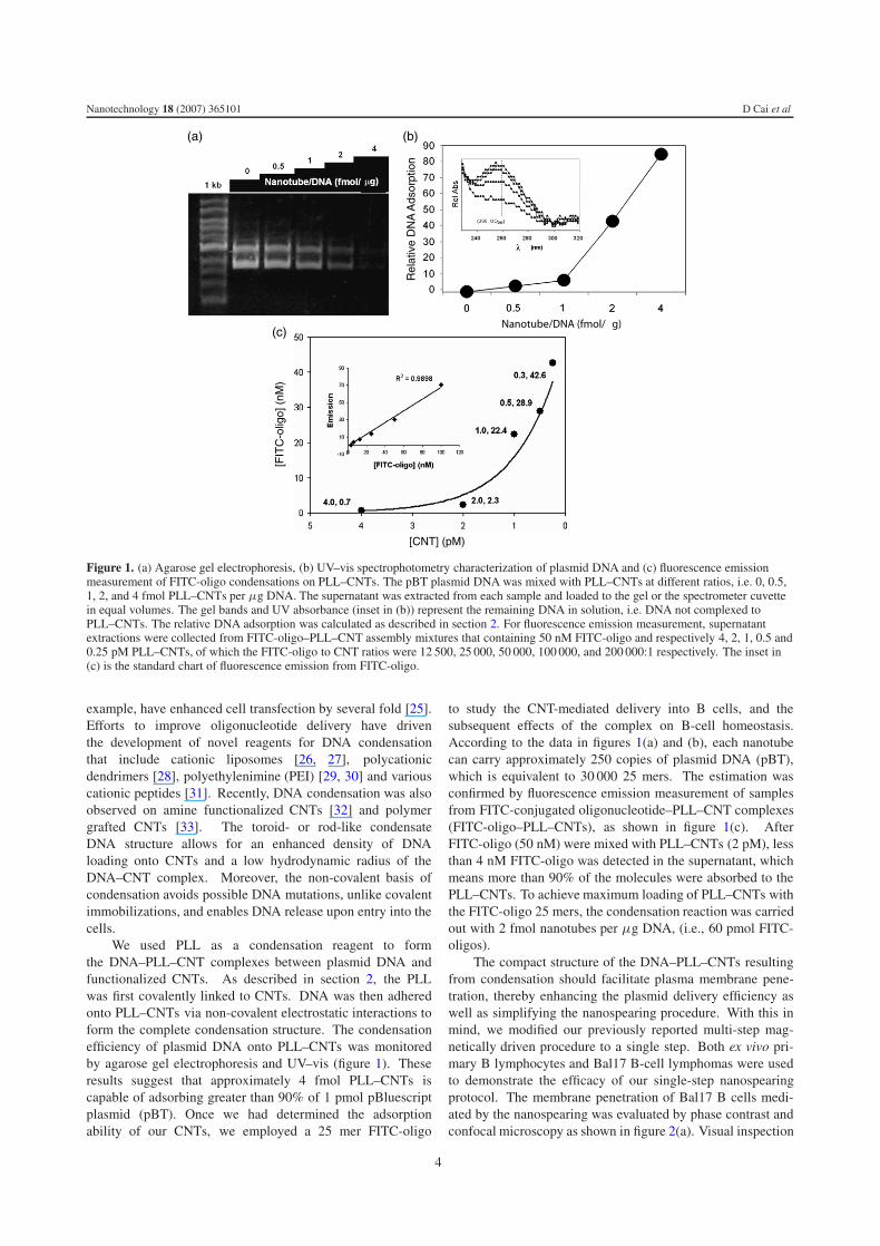
Figure 1. (a) Agarose gel electrophoresis, (b) UV–vis spectrophotometry characterization of plasmid DNA and (c) fluorescence emissionmeasurement of FITC-oligo condensations on PLL–CNTs. The pBT plasmid DNA was mixed with PLL–CNTs at different ratios, i.e. 0, 0.5,1, 2, and 4 fmol PLL–CNTs per μg DNA. The supernatant was extracted from each sample and loaded to the gel or the spectrometer cuvettein equal volumes. The gel bands and UV absorbance (inset in (b)) represent the remaining DNA in solution, i.e. DNA not complexed toPLL–CNTs. The relative DNA adsorption was calculated as described in section 2. For fluorescence emission measurement, supernatantextractions were collected from FITC-oligo–PLL–CNT assembly mixtures that containing 50 nM FITC-oligo and respectively 4, 2, 1, 0.5 and0.25 pM PLL–CNTs, of which the FITC-oligo to CNT ratios were 12 500, 25 000, 50 000, 100 000, and 200 000:1 respectively. The inset in(c) is the standard chart of fluorescence emission from FITC-oligo.
example, have enhanced cell transfection by several fold [25].Efforts to improve oligonucleotide delivery have driventhe development of novel reagents for DNA condensationthat include cationic liposomes [26, 27], polycationicdendrimers [28], polyethylenimine (PEI) [29, 30] and variouscationic peptides [31]. Recently, DNA condensation was alsoobserved on amine functionalized CNTs [32] and polymergrafted CNTs [33]. The toroid- or rod-like condensateDNA structure allows for an enhanced density of DNAloading onto CNTs and a low hydrodynamic radius of theDNA–CNT complex. Moreover, the non-covalent basis ofcondensation avoids possible DNA mutations, unlike covalentimmobilizations, and enables DNA release upon entry into thecells.
We used PLL as a condensation reagent to formthe DNA–PLL–CNT complexes between plasmid DNA andfunctionalized CNTs. As described in section 2, the PLLwas first covalently linked to CNTs. DNA was then adheredonto PLL–CNTs via non-covalent electrostatic interactions toform the complete condensation structure. The condensationefficiency of plasmid DNA onto PLL–CNTs was monitoredby agarose gel electrophoresis and UV–vis (figure 1). Theseresults suggest that approximately 4 fmol PLL–CNTs iscapable of adsorbing greater than 90% of 1 pmol pBluescriptplasmid (pBT). Once we had determined the adsorptionability of our CNTs, we employed a 25 mer FITC-oligo
to study the CNT-mediated delivery into B cells, and thesubsequent effects of the complex on B-cell homeostasis.According to the data in figures 1(a) and (b), each nanotubecan carry approximately 250 copies of plasmid DNA (pBT),which is equivalent to 30 000 25 mers. The estimation wasconfirmed by fluorescence emission measurement of samplesfrom FITC-conjugated oligonucleotide–PLL–CNT complexes(FITC-oligo–PLL–CNTs), as shown in figure 1(c). AfterFITC-oligo (50 nM) were mixed with PLL–CNTs (2 pM), lessthan 4 nM FITC-oligo was detected in the supernatant, whichmeans more than 90% of the molecules were absorbed to thePLL–CNTs. To achieve maximum loading of PLL–CNTs withthe FITC-oligo 25 mers, the condensation reaction was carriedout with 2 fmol nanotubes per μg DNA, (i.e., 60 pmol FITC-oligos).
The compact structure of the DNA–PLL–CNTs resultingfrom condensation should facilitate plasma membrane pene-tration, thereby enhancing the plasmid delivery efficiency aswell as simplifying the nanospearing procedure. With this inmind, we modified our previously reported multi-step mag-netically driven procedure to a single step. Both ex vivo pri-mary B lymphocytes and Bal17 B-cell lymphomas were usedto demonstrate the efficacy of our single-step nanospearingprotocol. The membrane penetration of Bal17 B cells medi-ated by the nanospearing was evaluated by phase contrast andconfocal microscopy as shown in figure 2(a). Visual inspection
4
Nanotechnology 18 (2007) 365101 D Cai et al
8060
4020
100
0
Cou
nts
FITC-A
Oligo
FITC-oligo
(a)
(b)
(c)
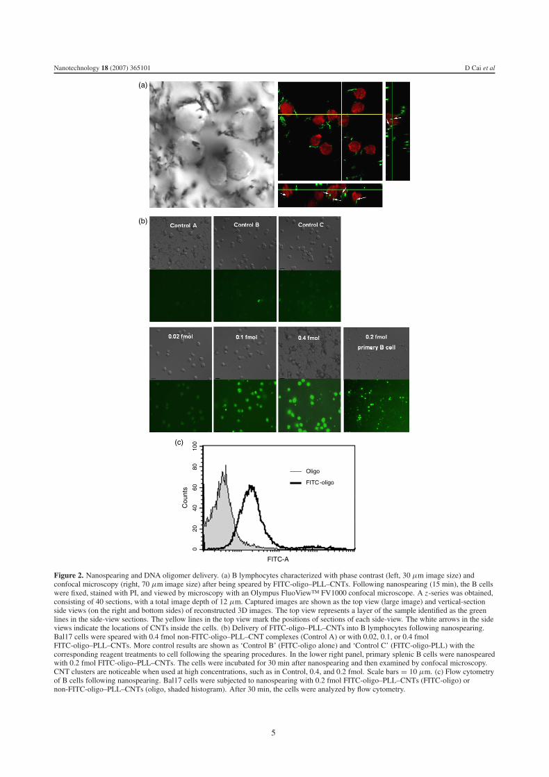
Figure 2. Nanospearing and DNA oligomer delivery. (a) B lymphocytes characterized with phase contrast (left, 30 μm image size) andconfocal microscopy (right, 70 μm image size) after being speared by FITC-oligo–PLL–CNTs. Following nanospearing (15 min), the B cellswere fixed, stained with PI, and viewed by microscopy with an Olympus FluoView™ FV1000 confocal microscope. A z-series was obtained,consisting of 40 sections, with a total image depth of 12 μm. Captured images are shown as the top view (large image) and vertical-sectionside views (on the right and bottom sides) of reconstructed 3D images. The top view represents a layer of the sample identified as the greenlines in the side-view sections. The yellow lines in the top view mark the positions of sections of each side-view. The white arrows in the sideviews indicate the locations of CNTs inside the cells. (b) Delivery of FITC-oligo–PLL–CNTs into B lymphocytes following nanospearing.Bal17 cells were speared with 0.4 fmol non-FITC-oligo–PLL–CNT complexes (Control A) or with 0.02, 0.1, or 0.4 fmolFITC-oligo–PLL–CNTs. More control results are shown as ‘Control B’ (FITC-oligo alone) and ‘Control C’ (FITC-oligo-PLL) with thecorresponding reagent treatments to cell following the spearing procedures. In the lower right panel, primary splenic B cells were nanospearedwith 0.2 fmol FITC-oligo–PLL–CNTs. The cells were incubated for 30 min after nanospearing and then examined by confocal microscopy.CNT clusters are noticeable when used at high concentrations, such as in Control, 0.4, and 0.2 fmol. Scale bars = 10 μm. (c) Flow cytometryof B cells following nanospearing. Bal17 cells were subjected to nanospearing with 0.2 fmol FITC-oligo–PLL–CNTs (FITC-oligo) ornon-FITC-oligo–PLL–CNTs (oligo, shaded histogram). After 30 min, the cells were analyzed by flow cytometry.
5
Nanotechnology 18 (2007) 365101 D Cai et al
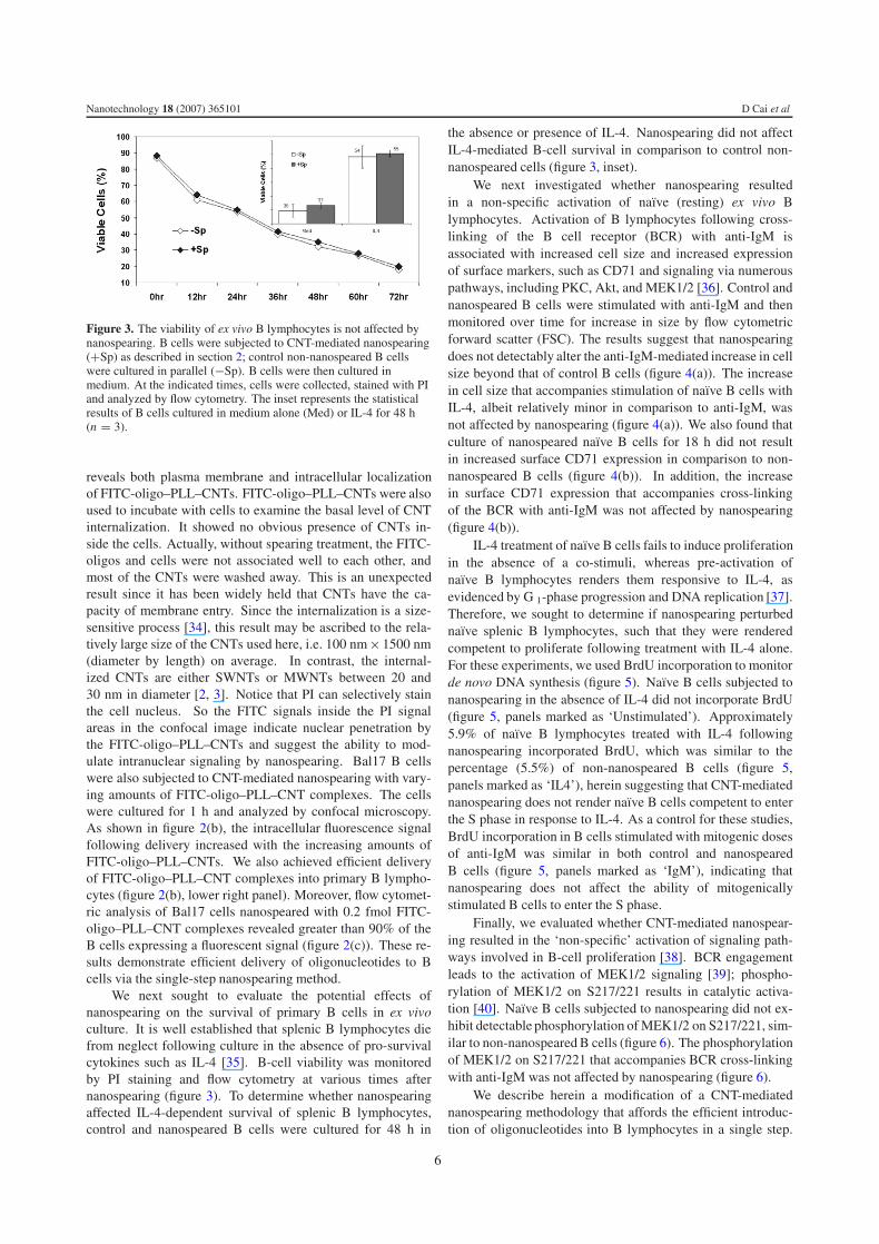
Figure 3. The viability of ex vivo B lymphocytes is not affected bynanospearing. B cells were subjected to CNT-mediated nanospearing(+Sp) as described in section 2; control non-nanospeared B cellswere cultured in parallel (−Sp). B cells were then cultured inmedium. At the indicated times, cells were collected, stained with PIand analyzed by flow cytometry. The inset represents the statisticalresults of B cells cultured in medium alone (Med) or IL-4 for 48 h(n = 3).
reveals both plasma membrane and intracellular localizationof FITC-oligo–PLL–CNTs. FITC-oligo–PLL–CNTs were alsoused to incubate with cells to examine the basal level of CNTinternalization. It showed no obvious presence of CNTs in-side the cells. Actually, without spearing treatment, the FITC-oligos and cells were not associated well to each other, andmost of the CNTs were washed away. This is an unexpectedresult since it has been widely held that CNTs have the ca-pacity of membrane entry. Since the internalization is a size-sensitive process [34], this result may be ascribed to the rela-tively large size of the CNTs used here, i.e. 100 nm× 1500 nm(diameter by length) on average. In contrast, the internal-ized CNTs are either SWNTs or MWNTs between 20 and30 nm in diameter [2, 3]. Notice that PI can selectively stainthe cell nucleus. So the FITC signals inside the PI signalareas in the confocal image indicate nuclear penetration bythe FITC-oligo–PLL–CNTs and suggest the ability to mod-ulate intranuclear signaling by nanospearing. Bal17 B cellswere also subjected to CNT-mediated nanospearing with vary-ing amounts of FITC-oligo–PLL–CNT complexes. The cellswere cultured for 1 h and analyzed by confocal microscopy.As shown in figure 2(b), the intracellular fluorescence signalfollowing delivery increased with the increasing amounts ofFITC-oligo–PLL–CNTs. We also achieved efficient deliveryof FITC-oligo–PLL–CNT complexes into primary B lympho-cytes (figure 2(b), lower right panel). Moreover, flow cytomet-ric analysis of Bal17 cells nanospeared with 0.2 fmol FITC-oligo–PLL–CNT complexes revealed greater than 90% of theB cells expressing a fluorescent signal (figure 2(c)). These re-sults demonstrate efficient delivery of oligonucleotides to Bcells via the single-step nanospearing method.
We next sought to evaluate the potential effects ofnanospearing on the survival of primary B cells in ex vivoculture. It is well established that splenic B lymphocytes diefrom neglect following culture in the absence of pro-survivalcytokines such as IL-4 [35]. B-cell viability was monitoredby PI staining and flow cytometry at various times afternanospearing (figure 3). To determine whether nanospearingaffected IL-4-dependent survival of splenic B lymphocytes,control and nanospeared B cells were cultured for 48 h in
the absence or presence of IL-4. Nanospearing did not affectIL-4-mediated B-cell survival in comparison to control non-nanospeared cells (figure 3, inset).
We next investigated whether nanospearing resultedin a non-specific activation of naı̈ve (resting) ex vivo Blymphocytes. Activation of B lymphocytes following cross-linking of the B cell receptor (BCR) with anti-IgM isassociated with increased cell size and increased expressionof surface markers, such as CD71 and signaling via numerouspathways, including PKC, Akt, and MEK1/2 [36]. Control andnanospeared B cells were stimulated with anti-IgM and thenmonitored over time for increase in size by flow cytometricforward scatter (FSC). The results suggest that nanospearingdoes not detectably alter the anti-IgM-mediated increase in cellsize beyond that of control B cells (figure 4(a)). The increasein cell size that accompanies stimulation of naı̈ve B cells withIL-4, albeit relatively minor in comparison to anti-IgM, wasnot affected by nanospearing (figure 4(a)). We also found thatculture of nanospeared naı̈ve B cells for 18 h did not resultin increased surface CD71 expression in comparison to non-nanospeared B cells (figure 4(b)). In addition, the increasein surface CD71 expression that accompanies cross-linkingof the BCR with anti-IgM was not affected by nanospearing(figure 4(b)).
IL-4 treatment of naı̈ve B cells fails to induce proliferationin the absence of a co-stimuli, whereas pre-activation ofnaı̈ve B lymphocytes renders them responsive to IL-4, asevidenced by G 1-phase progression and DNA replication [37].Therefore, we sought to determine if nanospearing perturbednaı̈ve splenic B lymphocytes, such that they were renderedcompetent to proliferate following treatment with IL-4 alone.For these experiments, we used BrdU incorporation to monitorde novo DNA synthesis (figure 5). Naı̈ve B cells subjected tonanospearing in the absence of IL-4 did not incorporate BrdU(figure 5, panels marked as ‘Unstimulated’). Approximately5.9% of naı̈ve B lymphocytes treated with IL-4 followingnanospearing incorporated BrdU, which was similar to thepercentage (5.5%) of non-nanospeared B cells (figure 5,panels marked as ‘IL4’), herein suggesting that CNT-mediatednanospearing does not render naı̈ve B cells competent to enterthe S phase in response to IL-4. As a control for these studies,BrdU incorporation in B cells stimulated with mitogenic dosesof anti-IgM was similar in both control and nanospearedB cells (figure 5, panels marked as ‘IgM’), indicating thatnanospearing does not affect the ability of mitogenicallystimulated B cells to enter the S phase.
Finally, we evaluated whether CNT-mediated nanospear-ing resulted in the ‘non-specific’ activation of signaling path-ways involved in B-cell proliferation [38]. BCR engagementleads to the activation of MEK1/2 signaling [39]; phospho-rylation of MEK1/2 on S217/221 results in catalytic activa-tion [40]. Naı̈ve B cells subjected to nanospearing did not ex-hibit detectable phosphorylation of MEK1/2 on S217/221, sim-ilar to non-nanospeared B cells (figure 6). The phosphorylationof MEK1/2 on S217/221 that accompanies BCR cross-linkingwith anti-IgM was not affected by nanospearing (figure 6).
We describe herein a modification of a CNT-mediatednanospearing methodology that affords the efficient introduc-tion of oligonucleotides into B lymphocytes in a single step.
6

Nanotechnology 18 (2007) 365101 D Cai et al
4030
2010
500
Cou
nts
100 101 102 103 104
PE-A
Med-Sp
Med+Sp
IgM-Sp
IgM+Sp
(a) (b)
Figure 4. Ex vivo B lymphocyte activation after nanospearing characterized by flow cytometry. (a) Increased B-cell size measured by flowcytometry and FSC. B cells were subjected to CNT-mediated nanospearing (+Sp). Control non-nanospeared B cells were cultured in parallel.B cells were then cultured in medium alone (Med), stimulated with 10 μg ml−1 anti-IgM (IgM), or 30 ng ml−1 IL-4. At the indicated times,cells were collected and analyzed for flow-cytometric FSC. The inset shows the average FSC values at 48 h (n = 3). (b) Surface expression ofCD71 on naı̈ve and anti-IgM-stimulated B cells after CNT-mediated nanospearing. B cells were subjected to CNT-mediated nanospearing(+Sp); control non-nanospeared B cells were cultured in parallel (-Sp). B cells were then cultured in medium alone (Med) or stimulated with10 μg ml−1 anti-IgM (IgM) for 18 h. The cells were collected and analyzed for CD71 expression by flow cytometry.
Unstimulated 10 µg/mL IgM 30 ng/mL IL4
Brd
U
7-AAD
Uns
pear
edS
pear
ed
Figure 5. Proliferative responses of primary B lymphocyte following CNT-mediated nanospearing. B cells were subjected to CNT-mediatednanospearing (speared) as described in section 2. Control non-nanospeared B cells were cultured in parallel (unspeared). B cells were thencultured in medium alone (unstimulated), stimulated with 10 μg ml−1 anti-IgM, or 30 ng ml−1 IL-4 for 48 h. BrdU incorporation was carriedout and analyzed by flow cytometry as described in section 2. The cells in the dot plot were gated into three populations that represented cellsin the G0/G1, G2/M , and S phase of the cell cycle, respectively. The percentage of cells in the S phase in each condition is show in the panel.
We observe that naı̈ve B cells exposed to CNT-mediated nanos-pearing in the absence of external stimuli (e.g., IL-4 or anti-IgM) do not exhibit a detectable increase in cell size, surfaceCD71 expression, or phosphorylation of MEK1/2; we alsodemonstrate that CNT-mediated nanospearing does not affectthe survival of naı̈ve B cells in long-term ex vivo cultures.
We believe the ability to maintain the ‘resting’ state of pri-mary B cells may be attributed to the chemical modification ofthe CNTs (i.e., the functionalization with carboxyl groups andcoating with PLL) as well as the low dosage of CNTs used fornanospearing. Indeed, functionalization has been shown to ren-der CNTs less cytotoxic [12–15], even in primary immune cells
7

Nanotechnology 18 (2007) 365101 D Cai et al
Figure 6. Phosphorylation of MEK1/2 in naı̈ve and anti-IgMstimulated B cells following CNT-mediated nanospearing. B cellswere subjected to CNT-mediated nanospearing (+Sp) as described insection 2. Control non-nanospeared B cells were cultured in parallel(−Sp). B cells were then cultured in medium alone (Med) orstimulated with 10 μg ml−1 anti-IgM (IgM) for 20 min. Thephosphorylation of endogenous MEK1/2 on S217/221 wasdetermined by Western blot analysis (upper panel). The blots werestripped and reprobed for total MEK1/2 protein (lower panel).
(This figure is in colour only in the electronic version)
with SWNTs [41]. We also observed that nanospearing didnot induce S-phase entry in naı̈ve B cells subsequently stimu-lated with IL-4. These findings suggest that the CNTs preparedas outlined herein and/or the actual spearing event do not re-sult in non-specific activation of naı̈ve primary B lymphocytes.Our findings demonstrate that subjecting B cells to nanospear-ing does not alter their ability to subsequently respond to BCRcross-linking. Although we note some blast-like cells and ag-gregated cells following nanospearing (by phase contrast mi-croscopy), a plausible explanation could be that the residualmagnetism of metal particles in the CNTs tends to generate amagnetic force and cluster the CNT-containing structures to-gether.
The nanospearing technique described here may serveas a platform for the introduction of recombinant proteins,siRNA, and plasmid DNA into primary B cells, withoutperturbing the ‘resting’ state. Current methods for theintroduction of macromolecules into primary lymphocytes,such as transfection and retroviral-mediated transduction,require prior stimulation with lipopolysaccharide (LPS). Inboth methods, the transduction efficiency is low (less than 15%of cells) and requires lymphocytes to be engaged in the cellcycle prior to transfection/transduction [42]. These techniquespreclude analysis of the cellular and molecular regulationof B-cell activation, proliferation, and cell cycle control.Moreover, these techniques tend to perturb numerous signaltransduction pathways and induce the expression of surfaceactivation markers [43]. By contrast, the single-step CNT-mediated spearing procedure described here should allow theintroduction of macromolecules into naı̈ve B cells without aprior need to activate or engage B cells in the cell cycle (bystimulation with LPS or the combination of phorbol diesterplus calcium ionophore) [44].
It has been previously reported that no appreciable celldeath was observed with 10 μg single-wall nanotubes incultures containing 3 × 10⁵ cells [3]. The amount of CNTsused in our method is only 2 μg,5 a concentration below the
5 Concentrate 10 ml CNT suspension in ethyl alcohol by centrifugation andvacuum dry the CNT pellet in a desiccator for 1 day. The weight of CNTswas obtained by measuring with AT261 delta range balance (Mettler-ToledoInternational Inc., Columbus, OH). Correspondingly, 20 fmol CNTs obtainedfrom 10 ml suspension is about 168 μg.
previously determined threshold. Nanospearing, therefore, hasthe potential for in vivo applications where cytotoxicty is apotential concern.
Finally, amine and lysine functionalized CNTs have beenshown to exhibit the ability to efficiently condense plasmidDNA [32, 33]. We achieved DNA condensation by covalentlylinking PLL to CNTs before DNA immobilization. Thefree amine groups render the complex positively charged,allowing for efficient adsorption of the negatively chargednucleic acid oligomer backbone. Our experiments indicatethat DNA condensation onto PLL–CNTs occurs in a similarratio to that seen for amine functionalized CNTs. Sincethe condensed plasmid DNA can resist disruptive sonicationand DNase I catalyzed hydrolysis [45, 46], we envision thatsiRNA immobilized by adsorption onto CNTs will remainstable during CNT-mediated delivery.
4. Conclusion
The oligo-PLL–CNT complex formed by the condensationstrategy is able to facilitate efficient delivery of nucleic acidsinto B cells through the single-step nanospearing procedure.Nanospearing does not result in the activation of naı̈ve exvivo primary B cells nor measurably impact subsequent B-cell stimulation via the BCR. These results indicate that CNT-mediated nanospearing can be used to deliver macromoleculesinto naı̈ve primary B cells in order to study metabolic andproliferative responses.
Acknowledgments
We thank NIBIB of NIH (IR43EB006249) (DC), DOE (DE-FG02-00ER45805) (ZFR) and NSF (NIRT0506830) (ZFR)for supporting the research. Ms Deborah Ritta (Departmentof Biology, Boston College) has contributed to the research.We thank the Department of Biology for use of the ConfocalMicroscopy Facility.
References
[1] Iijima S 1991 Helical microtubules of graphitic carbon Nature354 56
[2] Kostarelos K et al 2007 Cellular uptake of functionalizedcarbon nanotubes is independent of functional group and celltype Nat. Nanotechnol. 2 108
[3] Shi Kam N W, Jessop T C, Wender P A and Dai H 2004Nanotube molecular transporters: internalization of carbonnanotube–protein conjugates into mammalian cells J. Am.Chem. Soc. 126 6850
[4] Rojas-Chapana J, Troszczynska J, Firkowska I,Morsczeck C and Giersig M 2005 Multi-walled carbonnanotubes for plasmid delivery into Escherichia coli cellsLab Chip 5 536
[5] Cai D, Mataraza J M, Qin Z H, Huang Z, Huang J, Chiles T C,Carnahan D, Kempa K and Ren Z 2005 Highly efficientmolecular delivery into mammalian cells using carbonnanotube spearing Nat. Methods 2 449
[6] McKnight T E, Melechko A V, Griffin G D, Guillorn M A,Merkulov V I, Serna F, Hensley D K, Doktycz M J,Lowndes D H and Simpson M L 2003 Intracellularintegration of synthetic nanostructures with viable cells forcontrolled biochemical manipulation Nanotechnology14 551
[7] Lam C W, James J T, McCluskey R, Arepalli S andHunter R L A 2006 review of carbon nanotube toxicity andassessment of potential occupational and environmentalhealth risks Crit. Rev. Toxicol. 36 189
8

Nanotechnology 18 (2007) 365101 D Cai et al
[8] Muller J, Huaux F, Moreau N, Misson P, Heilier J F, Delos M,Arras M, Fonseca A, Nagy J B and Lison D 2005Respiratory toxicity of multi-wall carbon nanotubes Toxicol.Appl. Pharmacol. 207 221
[9] Donaldson K, Aitken R, Tran L, Stone V, Duffin R,Forrest G and Alexander A 2006 Carbon nanotubes:a review of their properties in relation to pulmonarytoxicology and workplace safety Toxicol. Sci. 92 5
[10] Shvedova A A et al 2005 Unusual inflammatory and fibrogenicpulmonary responses to single-walled carbon nanotubes inmice Am. J. Physiol. Lung Cell Mol. Physiol. 289 L698
[11] Manna S K, Sarkar S, Barr J, Wise K, Barrera E V, Jejelowo O,Rice-Ficht A C and Ramesh G T 2005 Single-walled carbonnanotube induces oxidative stress and activates nucleartranscription factor—kappaB in human keratinocytes NanoLett. 5 1676
[12] Sayes C M et al 2006 Functionalization density dependence ofsingle-walled carbon nanotubes cytotoxicity in vitroToxicol. Lett. 161 135
[13] Pantarotto D, Singh R, McCarthy D, Erhardt M, Briand J P,Prato M, Kostarelos K and Bianco A 2004 Functionalizedcarbon nanotubes for plasmid DNA gene delivery Angew.Chem. Int. Edn Engl. 43 5242
[14] Pantarotto D, Briand J P, Prato M and Bianco A 2004Translocation of bioactive peptides across cell membranesby carbon nanotubes Chem. Commun. (1) 16
[15] Singh R, Pantarotto D, Lacerda L, Pastorin G, Klumpp C,Prato M, Bianco A and Kostarelos K 2006 Tissuebiodistribution and blood clearance rates of intravenouslyadministered carbon nanotube radiotracers Proc. Natl Acad.Sci. USA 103 3357
[16] Monteiro-Riviere N A, Nemanich R J, Inman A O,Wang Y Y and Riviere J E 2005 Multi-walled carbonnanotube interactions with human epidermal keratinocytesToxicol. Lett. 155 377
[17] Bottini M, Bruckner S, Nika K, Bottini N, Bellucci S,Magrini A, Bergamaschi A and Mustelin T 2006Multi-walled carbon nanotubes induce T lymphocyteapoptosis Toxicol. Lett. 160 121
[18] Jia G, Wang H, Yan L, Wang X, Pei R, Yan T, Zhao Y andGuo X 2005 Cytotoxicity of carbon nanomaterials:single-wall nanotube, multi-wall nanotube, and fullereneEnviron. Sci. Technol. 39 1378
[19] Mann D G, McKnight T E, Melechko A V, Simpson M L andSayler G S 2006 Quantitative analysis of immobilized DNAon vertically aligned carbon nanofiber gene delivery arraysBiotechnol. Bioeng. 97 680
[20] Doughty C A, Bleiman B F, Wagner D J, Dufort F J,Mataraza J M, Roberts M F and Chiles T C 2006 Antigenreceptor-mediated changes in glucose metabolism in Blymphocytes: role of phosphatidylinositol 3-kinase signalingin the glycolytic control of growth Blood 107 4458
[21] Hodgkin P D, Yamashita L C, Coffman R L and Kehry M R1990 Separation of events mediating B cell proliferation andIg production by using T cell membranes and lymphokinesJ. Immunol. 145 2025
[22] Zauner W, Ogris M and Wagner E 1998 Polylysine-basedtransfection systems utilizing receptor-mediated deliveryAdv. Drug Delivery Rev. 30 97
[23] Zabner J, Fasbender A J, Moninger T, Poellinger K A andWelsh M J 1995 Cellular and molecular barriers to genetransfer by a cationic lipid J. Biol. Chem. 270 18997
[24] Mahato R I, Takakura Y and Hashida M 1997 Nonviral vectorsfor in vivo gene delivery: physicochemical andpharmacokinetic considerations Crit. Rev. Ther. Drug Carr.Syst. 14 133
[25] Gao X and Huang L 1996 Potentiation of cationicliposome-mediated gene delivery by polycationsBiochemistry 35 1027
[26] Capaccioli S, Dipasquale G, Mini E, Mazzei T andQuattrone A 1993 Cationic lipids improve antisenseoligonucleotide uptake and prevent degradation in cultured
cells and in human serum Biochem. Biophys. Res. Commun.197 818
[27] Lewis J G, Lin K Y, Kothavale A, Flanagan W M,Matteucci M D, DePrince R B, Mook R A,Hendren R W and Wagner R W 1996 A serum-resistantcytofectin for cellular delivery of antisenseoligodeoxynucleotides and plasmid DNA Proc. Natl Acad.Sci. USA 93 3176
[28] Yoo H and Juliano R L 2000 Enhanced delivery of antisenseoligonucleotides with fluorophore-conjugated PAMAMdendrimers Nucleic Acids Res. 28 4225
[29] Boussif O, Lezoualch F, Zanta M A, Mergny M D,Scherman D, Demeneix B and Behr J P 1995 A versatilevector for gene and oligonucleotide transfer into cells inculture and in vivo: polyethylenimine Proc. Natl Acad. Sci.USA 92 7297
[30] Robaczewska M et al 2001 Inhibition of hepadnaviralreplication by polyethylenimine-based intravenous deliveryof antisense phosphodiester oligodeoxynucleotides to theliver Gene Ther. 8 874
[31] Lochmann D, Jauk E and Zimmer A 2004 Drug delivery ofoligonucleotides by peptides Eur. J. Pharm. Biopharm.58 237
[32] Singh R, Pantarotto D, McCarthy D, Chaloin O, Hoebeke J,Partidos C D, Briand J P, Prato M, Bianco A andKostarelos K 2005 Binding and condensation of plasmidDNA onto functionalized carbon nanotubes: toward theconstruction of nanotube-based gene delivery vectors J. Am.Chem. Soc. 127 4388
[33] Liu Y, Wu D C, Zhang W D, Jiang X, He C B, Chung T S,Goh S H and Leong K W 2005 Polyethylenimine-graftedmultiwalled carbon nanotubes for secure noncovalentimmobilization and efficient delivery of DNA Angew. Chem.Int. Edn Engl. 44 4782
[34] Gao H, Shi W and Freund L B 2005 From the cover: mechanicsof receptor-mediated endocytosis Proc. Natl Acad. Sci. USA102 9469
[35] Broxmeyer H E, Lu L, Cooper S, Tushinski R, Mochizuki D,Rubin B Y, Gillis S and Williams D E 1988 Synergisticeffects of purified recombinant human and murine B cellgrowth factor—1/IL-4 on colony formation in vitro byhematopoietic progenitor cells J. Immunol. 141 3852
[36] Lok C N and Loh T T 1998 Regulation of transferrin functionand expression: review and update Biol. Signals Recept.7 157
[37] Oliver K, Noelle R J, Uhr J W, Krammer P H andVitetta E S 1985 B-cell growth factor (B-cell growth factor Ior B-cell-stimulating factor, provisional 1) is adifferentiation factor for resting B cells and may not inducecell growth Proc. Natl Acad. Sci. USA 82 2465
[38] Gold M R 2002 To make antibodies or not: signaling by theB-cell antigen receptor Trends Pharmacol. Sci. 23 316
[39] Chang F, Steelman L S, Lee J T, Shelton J G, Navolanic P M,Blalock W L, Franklin R A and McCubrey J A 2003 Signaltransduction mediated by the Ras/Raf/MEK/ERK pathwayfrom cytokine receptors to transcription factors: potentialtargeting for therapeutic intervention Leukemia 17 1263
[40] Jazirehi A R, Vega M I, Chatterjee D, Goodglick L andBonavida B 2004 Inhibition of the Raf-MEK1/2-ERK1/2signaling pathway, Bcl-xL down-regulation, andchemosensitization of non-Hodgkin’s lymphoma B cells byRituximab Cancer Res. 64 7117
[41] Dumortier H, Lacotte S, Pastorin G, Marega R, Wu W,Bonifazi D, Briand JP, Prato M, Muller S andBianco A 2006 Functionalized carbon nanotubes arenon-cytotoxic and preserve the functionality of primaryimmune cells Nano Lett. 6 1522
[42] Wilcox H M and Berg L J 2003 Itk phosphorylation sites arerequired for functional activity in primary T cells J. Biol.Chem. 278 37112
[43] McMahon S B, Norvell A, Levine K J and Monroe J G 1995Transient transfection of murine B lymphocyte blasts as a
9

Nanotechnology 18 (2007) 365101 D Cai et al
method for examining gene regulation in primary B cellsJ. Immunol. Methods 179 251
[44] DeFranco A L, Raveche E S and Paul W E 1985 Separatecontrol of B lymphocyte early activation and proliferation inresponse to anti-IgM antibodies J. Immunol. 135 87
[45] Kogurea K, Moriguchib R, Sasakib K, Uenoc M, Futakid S andHarashima H 2004 Development of a non-viral
multifunctional envelope-type nano device by a novel lipidfilm hydration method J. Control. Release 98 317
[46] Abdelhady H G, Allen S, Davies M C, Roberts C J,Tendler S J B and Williams P M 2003 Direct real-timemolecular scale visualisation of the degradation ofcondensed DNA complexes exposed to DNase I NucleicAcids Res. 31 4001
10