In vitro effects of selected medicinal - UCL Discovery
339
1 UCL SCHOOL OF PHARMACY In vitro effects of selected medicinal plants shortlisted for clinical use in the Brazilian public health system in CYP3A4 mRNA gene expression, glutathione levels, and P- glycoprotein activity and their implications for herb-drug interactions. By: André Luís Dias Araujo Mazzari A THESIS SUBMITTED IN FULFILMENT OF THE REQUIREMENTS FOR THE AWARD OF THE DEGREE OF DOCTOR OF PHILOSOPHY 2017
-
Upload
khangminh22 -
Category
Documents
-
view
2 -
download
0
Transcript of In vitro effects of selected medicinal - UCL Discovery
1
UCL SCHOOL OF PHARMACY
In vitro effects of selected medicinal plants shortlisted for clinical use in
the Brazilian public health system in CYP3A4 mRNA gene expression,
glutathione levels, and P-glycoprotein activity and their
implications for herb-drug interactions.
By: André Luís Dias Araujo Mazzari
A THESIS SUBMITTED IN FULFILMENT OF THE REQUIREMENTS FOR THE AWARD OF THE DEGREE OF DOCTOR OF PHILOSOPHY
2017

2
DECLARATION I, André Luís Dias Araujo Mazzari confirm that the work presented in this thesis is my own. Where information has been delivered from other sources, I confirm that this has been indicated in the thesis. Signed: ..........................................this day................................................2017
3
Abstract The Brazilian Unified Public Health System (SUS) shortlisted various plant species of interest (RENISUS) for future clinical use. However, very little is known about their effects on metabolic and transporter proteins, which could potentially lead to herb-drug interactions (HDI). To evaluate this, we conducted in vitro preclinical studies on twenty-four plant extracts to disclose their effects on CYP3A4 mRNA gene expression, intracellular glutathione (GSH) levels, inhibition of γ-glutamyl transferase (GGT) in HepG2 cells and P-glycoprotein (P-gp) activity in vincristine resistant Caco-2 (Caco-2 VCR) cells. We also investigated whether four Brazilian native species were able to activate the human pregnane X receptor (hPXR) in transiently co-transfected HeLa cells. This preclinical research showed that all but two plant extracts were able to modulate at least one of the selected targets. CYP3A4 mRNA gene expression in HepG2 cells was significantly affected by half of the extracts. The antagonistic effect of Solanum paniculatum L. on hPXR could explain its ability to inhibit CYP3A4. GSH levels were affected by 80% of the extracts. There was depletion of intracellular GSH levels by Cordia verbenacea A. DC., Costus spicatus (Jacq.) Sw., Persea americana Mill., Salix alba L., Schinus terebinthifolia Raddi and Syzygium jambolanum (Lam.) DC. accompanied because of the inhibition of GGT activity. P-gp activity was modulated in a significant manner by 17% of the extracts. The approaches used for the conduction of in vitro preclinical studies in herbal medicines revealed a series of challenges faced especially by academics in order to anticipate cases of HDI. Clinicians have also to consider the presence of intrinsic factors such as genetic polymorphisms in each patient. The possible presence of undesirable interactions between RENISUS herbal medicines and essential drugs in SUS need eventually be clinically confirmed to attest our observed in vitro effects.
4
Dedication
To dad, my big star. Thanks for inspiring me in each and every step I walk in my life. Thanks to mom (Graça), sister (Vanessa) and Laika for supporting me on this journey.
I love you all.
5
Acknowledgements I wish to express my sincere gratitude to my supervisor, Dr. Jose Prieto for his unwavering support throughout the course of this PhD. His willingness to go the extra mile for his students is very much appreciated. I would also like to thank Prof Michael Heinrich, Prof Simon Gibbons and especially Dr. Paul Stapleton for his advice and support both academically and otherwise during these past four years. The support of the different members of staff and colleagues at SOP who contributed to making my work a lot easier isn’t unrecognised. Thank you for making the completion of this PhD a reality. I would like to appreciate my fellow PhD students in Rooms 201, 203 and 211. Your friendship made student life a pleasurable one. I wouldn’t have wished for better companions to study with. I want to sincerely thank Fon, Mukrish, Maria, Sarah, Pedro, Gugu, Awo, Cynthia, Tariq, Stephen, Amaka, Johanna, Tony, Francesca and Jawharah for their priceless friendship. A special thanks to my beloved friend Hannah Jennings and Jen Datiles who proofread this PhD thesis. Thanks to Alan Kelly for always supporting me and encouraging me when I needed most. This work would not be possible to be conducted without the support of our Brazilian team from the University of Brasília and ANVISA. Thanks to Dr. Dâmaris Silveira, Prof. Francisco Neves, and Ana Cecília Carvalho. I am very grateful for all the help. I cannot fail to remember the support of various staff and members of the Goodenough community. It is truly a home away from home and so many people too numerous to mention were contributory to the success of this PhD for which I’m really grateful. Special thanks to Stefania, Ana Paula, Sergio, Bia, Mario, Ceci, Esteban, Marcelo, Larissa, Hylo, Malu, Luisa, and Marina for your friendship. My family both home and abroad have been of immeasurable emotional, spiritual and financial support at every step of the way. This PhD thesis is written in your honour. Finally, I would like to acknowledge the CNPq scholarship commission for funding this PhD. Thanks to our President Dilma Rousseff for encouraging many Brazilians to study abroad through Science without Borders scheme in order to collaborate with the advance of science in the country.
6
1 GENERAL INTRODUCTION ................................................................................ 21
1.1 Herbal medicines: traditional use and safety concerns ........................................................ 22
1.2 Evolution of herb-drug interactions research and the introduction of metabolic and transporter Experimental targets ......................................................................................................... 25
1.3 Drug metabolism and transport: general considerations ..................................................... 27 1.3.1 Phase I metabolism: cytochrome P450 ............................................................................... 28 1.3.2 Phase II metabolism (conjugation reactions) ....................................................................... 32 1.3.3 Types of phase II reactions ................................................................................................. 33 1.3.4 The drug transporter P-glycoprotein .................................................................................... 35
1.4 Induction and inhibition of drug metabolising enzymes ....................................................... 36 1.4.1 Induction of drug metabolising enzymes ............................................................................. 36 1.4.2 Inhibition of drug metabolising enzymes ............................................................................. 37
1.5 Factors that influence drug biotransformation ...................................................................... 38 1.5.1 Genetic Polymorphism ........................................................................................................ 38 1.5.2 Disease ................................................................................................................................ 40 1.5.3 Age ...................................................................................................................................... 40 1.5.4 Sex ....................................................................................................................................... 40 1.5.5 Environmental factors .......................................................................................................... 41
1.6 Herb-Drug interactions and adverse reactions ...................................................................... 41 1.6.1 Herb-drug interactions involving cytochrome P450 ............................................................. 41 1.6.2 Herb-drug interactions involving phase II metabolising enzymes ........................................ 43 1.6.3 Herb-drug interactions involving P-glycoprotein .................................................................. 44
1.7 Pharmacovigilance of herbal medicines ................................................................................. 46
1.8 Use of herbal medicines in Brazil and the risk of herb-drug interactions on patients of the public health system (SUS) .................................................................................................................. 47
1.9 Reported effects of renisus medicinal plants on main phase I and phase II metabolic mechanisms and the P-glycoprotein transporter. ............................................................................. 69
1.10 Aim of the thesis ........................................................................................................................ 79
2 MATERIALS AND METHODS ............................................................................. 80
7
2.1 Chemicals and reagents ........................................................................................................... 81
2.2 Preparation of plant materials .................................................................................................. 82 2.2.1 Selection criteria and ethical clearance ............................................................................... 82 2.2.2 Processing of plant materials .............................................................................................. 83
2.3 Phytochemical analysis ............................................................................................................ 84 2.3.1 HPTLC analysis ................................................................................................................... 84 2.3.2 HPLC analysis ..................................................................................................................... 84
2.4 In vitro studies ........................................................................................................................... 85 2.4.1 Mammalian cell culture techniques ..................................................................................... 85 2.4.2 Cell Viability Assays............................................................................................................. 88 2.4.3 CYP3A4 mRNA gene expression assay ............................................................................. 92 2.4.4 hPXR activation assay ......................................................................................................... 98 2.4.5 Intracellular GSH assay ..................................................................................................... 101 2.4.6 GGT activity assay ............................................................................................................. 109 2.4.7 Modulation of P-glycoprotein dependant Rh-123 efflux in Caco-2 cells ............................ 112 2.4.8 Statistical Analysis ............................................................................................................. 114
3 RESULTS ........................................................................................................... 115
3.1 Authorisation for accessing the Brazilian genetic heritage component from CGEN and yield of plant extracts ......................................................................................................................... 116
3.2 Phytochemical analysis .......................................................................................................... 118
3.3 SRB, Neutral Red and MTT assays for determination of cell viability/cytotoxicity ........... 121
3.4 CYP3A4 mRNA gene expression ........................................................................................... 123 3.4.1 Integrity and NanoDrop reading-assessment of the purity and quality of RNA ................. 123 3.4.2 Real-time qPCR efficiency ................................................................................................. 124 3.4.3 Polymerase chain reaction (PCR) ..................................................................................... 126 3.4.4 hPXR antagonistic effect of Solanum paniculatum L. ........................................................ 128 3.4.6 Erythrina mulungu Benth. displays a partial agonistic effect on hPXR luciferase reporter gene assay. ....................................................................................................................................... 131 3.4.7 Lippia sidoides Cham. does not display any effect on hPXR luciferase reporter gene assay. 131 3.4.8 Cordia verbenaceae A. DC. displays an antagonistic effect on both hPXR and TRb1 but not
CMV luciferase reporter gene assay. ................................................................................................ 132
8
3.5 Estimation of intracellular GSH levels in HepG2 cells ......................................................... 135
3.6 GGT activity ............................................................................................................................. 138
3.7 Modulation of P-Glycoprotein dependent Rh-123 efflux in Caco-2 VCR cells ................... 140
3.8 Summary of the effects of selected RENISUS medicinal plants in CYP3A4, hPXR, GSH, GGT and P-gp ...................................................................................................................................... 142
4 GENERAL DISCUSSION ................................................................................... 143
4.1 In vitro studies of RENISUS medicinal plants in the selected targets ............................... 144 4.1.1 Experimental approaches to the in vitro metabolism and transport studies ...................... 145
4.2 Implications of herbal medicines and intrinsic factors on the metabolism and efflux ..... 156 4.2.1 RENISUS herbal medicines as modulators of metabolism and efflux and potential HDI with RENAME essential medicines ........................................................................................................... 156 4.2.2 Intrinsic factors that impact in the regulation of metabolism and efflux ............................. 163 4.2.3 Active pharmacovigilance is needed for detection of HDI ................................................. 174
5 CONCLUSIONS ................................................................................................. 177
5.1 The way forward ...................................................................................................................... 178
6 REFERENCES ................................................................................................... 180
7 APPENDIX .......................................................................................................... 204
7.1 Interinstitutional agreement between UCL and UnB ............................................................ 205
7.2 Project sent to CGEN and IBAMA for requesting authorisation to access the Brazilian genetic heritage ................................................................................................................................... 210
7.3 Material transfer agreement ................................................................................................... 229
7.4 Authorisation to access the Brazilian genetic heritage given by CGEN/CNPq ................. 233
7.5 HPLC analysis .......................................................................................................................... 235
7.6 Tabular summary of CYP3A4 mRNA gene expression in HepG2 cells (results show mean ± SEM, n ≥ 3) ........................................................................................................................................ 312
9
7.7 Tabular summary of intracellular GSH concentrations in HepG2 cells (results show mean ± SEM, n ≥ 3) ........................................................................................................................................ 313
7.8 Tabular summary of GGT activity in terms of µM p-nitroaniline concentration in HepG2
cells (results show mean ± SEM, n ≥ 3) ............................................................................................. 314
7.9 Effect of plant extracts on intracellular Rh-123 accumulation in Caco-2 VCR cells (results show mean ± SEM, n ≥ 3) .................................................................................................................... 315
7.10 Publications ............................................................................................................................. 316 7.10.1 Herbal medicines in Brazil: pharmacokinetic profile and potential herb-drug interactions 316 7.10.2 Monitoramento de interações farmacocinéticas entre plantas medicinais e fitoterápicos e os medicamentos convencionais pelo sistema de farmacovigilância brasileiro ................................ 328 7.10.3 In vitro effects of four native Brazilian medicinal plants in CYP3A4 mRNA gene expression, glutathione levels, and P-glycoprotein activity ................................................................................... 334
10
List of figures Figure 1- 1 Number of publications per year using the keyword “herb-drug interactions”
on PubMed. Data was collected on 15 August 2016. .......................................... 26 Figure 1- 2 Scheme of sequential drug metabolism .................................................... 28 Figure 1- 3 Human liver CYPs represented by circles whose size and percentage
approximate to their level of expression in human liver. ...................................... 29 Figure 1- 4 Common reactions catalysed by CYP enzymes. ...................................... 30 Figure 1- 5 Conjugation of a glucuronic acid (GLU) to a carboxylic acid group. ......... 34 Figure 1- 6 Conjugation of glutathione (GSH) catalysed by the enzyme glutathione-S-
transferase. .......................................................................................................... 34 Figure 1- 7 Genetic polymorphisms found on CYP1A2, 2C9, 2C19, 2D6, and 3A5 among
the Brazilian population ........................................................................................ 39 Figure 2- 1 SRB chemical structure. ........................................................................... 88 Figure 2- 2 Chemical structure of neutral red dye. ...................................................... 90 Figure 2- 3 Cleavage of dsDNA by DNAse in presence of Mg2+ and Mn2+. ................ 93 Figure 2- 4 Graphical representation of cDNA synthesis from RNA template. After
extraction, RNA is mixed with a oligoprimer, dNTP's and reverse transcriptase enzyme (M-MLV). The oligoprimer is a poly(T) tail sequence, which will anneal to the poly(A) tail of the RNA molecule. When dNTPs, buffer and a reverse transcriptase enzyme (such as M-MLV) is added to the sample, a complementary strand of DNA (or cDNA) is synthesized. ............................................................. 94
Figure 2- 5 Graphical representation of the addition of a target primer sequence and a Master Mix containing SYBRâ Green dye and Taq polymerase enzyme. This reaction results in an increase in fluorescence signal during the PCR analysis. SYBRâ Green as a free dye displays very low fluorescence and it will not bind to the single stranded or denaturated DNA. At the annealing stage, when a double-stranded structure is formed, SYBRâ Green will bind to it and, as a result, an increase in fluorescent signal is observed. During the extension stage, which is conducted by the enzyme Taq polymerase, the fluorescent signal increases according to the number of SYBRâ Greendye molecules bound per double-stranded molecule. This process is repeated in each cycle until it reaches a total of 40 cycles. ............................................................................................................. 95
Figure 2- 6 Graphical representation of the activation of PXR nuclear receptor by a xenobiotic (drug), resulting in the expression of CYP in presence of coactivators. A, in absence of a ligand, the orphan nuclear receptors small heterodimer partner (SHP) and silencing mediator of retinoid and thyroid hormone receptor (SMRT) are transcribed in presence of the coactivators steroid receptor coactivator-1 (SRC-1), hepatocyte nuclear factor a (HNF4a) and peroxisome proliferator –activated receptor g coactivator-1a (PGC-1a). As a result, no CYP transcription will occur. B, in presence of a ligand (drug), the PXR receptor translocates to the nucleus of the cell and the ligand-PXR complex will form a heterodimer with RXR. CYP transcription will initiate upon the binding of the complex coactivators (SRC-1, HNF4a and PGC-1a)-ligand-PXR-RXR complex will bind to the xenobiotic responsive enhancer molecule (XREM)/PXRE region. ...................................... 100
11
Figure 2- 7 Chemical structures of the reduced GSH (A) and oxidized GSH (GSSG) (B). The tripeptide is formed by the amino acids glutamate, L-cysteine, and glycine. ........................................................................................................................... 102
Figure 2- 8 The two-step intracellular GSH synthesis. A, g-glutamylcysteine is formed by the conjugation of L-cysteine and glutamate, catalised by g-glutamylcysteine synthase. B, g-glutamylcysteine conjugates with glycine to generate g-glutamylcysteinilglycine (GSH). This step is catalised by GSH synthase. ......... 103
Figure 2- 9 Formation of mercapturic acid conjugate from a GSH conjugate. After the conjugation of GSH with the metabolite, the complex is released from the cell where is firstly borken down by a transpeptidase (g-glutamyl transferase) followed by the release of the amino acid glutamate. The remaining part of the complex is broken down once again by a dipeptidase, which will dissociate the amino acid glycine from the rest of the molecule to form the mercapturic acid. In order to improve the polarity of the mercapturic acid, an acetyl group is added to the molecule to form the N-acetylcystein mercapturic acid conjugate, which will be further excreted in the urine. ........................................................................................................................... 105
Figure 2- 10 N-acetylation of mercapturic acid. ......................................................... 105 Figure 2- 11 Enzymatic recycling of GSH from glutathione disulphide (GSSG) by
glutathione reductase (GR) in the presence of NADPH. GSH reacts with DTNB to GS-TNB and TNB, with subsequent reduction of the former to GSH. TNB can be measured at 405 nm. (Adapted from Rahman et al., 2007). .............................. 107
Figure 2- 12 The g-glutamyl cycle. When GSH is transported out of the cell, the enzyme gamma-glutamyl transferase (GGT) will break down the tripeptide and transfer the g-glutamyl moiety to an amino acid, forming g-glutamyl amino acid and cysteinylglycine. The g-glutamyl amino acid is transported back to the cell where it will be metabolized to release the amino acid and 5-oxoproline, which will be converted to glutamate and reincorporated into GSH. Dipeptidase will break down cysteinylglycine into glycine and cysteine. The latter will be transported back to the cell to be reincorporated into GSH. The amount of cysteine which is not used for GSH synthesis will be incorporated into newly synthesized proteins and/or broken down into sulphate and taurine. ......................................................................... 111
Figure 2- 13 Schematic representation of the inhibition of Rh-123 efflux by modulators of the P-glycoprotein transporter. Rh-123 is a fluorescent dye and a P-gp substrate. Upon cell uptake, Rh-123 will be effluxed out by P-gp in an ATP-requiring process. In presence of a p-glycoprotein inhibitor, the efflux in reduced allowing the accumulation of Rh-123 in the cell, which is detected and quantified by a spectrofluorometer. ............................................................................................ 112
Figure 3- 1 365nm after derivatization. ...................................................................... 118 Figure 3- 2 235nm after derivatization. ...................................................................... 119 Figure 3- 3 365nm before derivatization. ................................................................... 119 Figure 3- 4 Viability of (a) HepG2, (b) Caco-2 VCR and (c) HeLa cells in presence of
different concentrations of ethanol and H2O2 (Results show mean ± SEM, N³3). ........................................................................................................................... 121
Figure 3- 5 CYP3A4 primer calibration curve. ........................................................... 124 Figure 3- 6 CYP3A4 primer melting curve. ................................................................ 124 Figure 3- 7 b-actin primer calibration curve. .............................................................. 125 Figure 3- 8 b-actin primer melting curve. ................................................................... 125
12
Figure 3- 9 Effect of 24-hour incubation of extracts (100µg/mL) or the positive control rifampicin (50µM) and the negative control DMSO 1% in CYP3A4 mRNA gene expression in HepG2 cells. Thirteen extracts – CAS, EA1, RP1 (p<0.05), MI1, PI1, PA1 (p<0.01), AF1, SA1, ST1, SJ1 and VP1 (p<0.001) produced statistically significant increase in CYP3A4 mRNA gene expression in HepG2 cells based on one-way ANOVA test followed by Bonferroni correction. LA1 (p<0.05) and SP1 (0.01), on the other hand, were able to decrease CYP3A4 expression. ............ 127
Figure 3- 10 Antagonistic effect of Solanum paniculatum L. extract on RIF-induced hPXR activation. HeLa cells were co-transfected with expression vector pM-Gal4-PXR-LBD and Gal4 luciferase reporter and treated with increasing concentrations of Solanum paniculatum L. in the presence of RIF 1μM (EC50). Luciferase activity was measured after 24h and normalized as a percentage of RIF treated cells. 128
Figure 3- 11 Agonistic effect. HeLa cells were co-transfected with expression vector pM-Gal4-PXR-LBD and Gal4 luciferase reporter and treated with vehicle, rifampicin (RIF) or increasing concentrations of Solanum paniculatum L. Luciferase activity was measured after 24 hours and reported as fold induction compared to vehicle. Results represent data from three independent experiments performed in triplicate. *** p ≤ 0.001. ...................................................................................................... 129
Figure 3- 12 HeLa cells were co-transfected with expression vector pM-Gal4-dTRb1-LBD and Gal4 luciferase reporter and treated with vehicle, T3 or increasing concentrations of Solanum paniculatum L. Luciferase activity was measured after 24 hours and reported as fold induction compared to vehicle. **p≤0.01; ***p≤0.001. ........................................................................................................................... 129
Figure 3- 13 HeLa cells were transfected with expression vector CMV luciferase reporter and treated with vehicle, 1.6 or 2.0 mg/ mL of Solanum paniculatum L. Luciferase activity was measured after 24 hours and reported as fold induction compared to vehicle. ............................................................................................................... 130
Figure 3- 14 Agonistic effect. HeLa cells were co-transfected with expression vector pM-Gal4-PXR-LBD and Gal4 luciferase reporter and treated with vehicle, RIF or increasing concentrations of Erythrina mulungu Benth. Luciferase activity was measured after 24 hours and reported as fold induction compared to vehicle. *p ≤0.05; **p ≤ 0.01; ***p ≤ 0.001. .......................................................................... 131
Figure 3- 15 Agonistic effect. HeLa cells were co-transfected with expression vector pM-Gal4-PXR-LBD and Gal4 luciferase reporter and treated with vehicle, RIF or increasing concentrations of Lippia sidoides Cham. Luciferase activity was measured after 24 hours and reported as fold induction compared to vehicle. ***p ≤ 0.001. .............................................................................................................. 131
Figure 3- 16 Antagonistic effect. HeLa cells were co-transfected with expression vector pM-Gal4-PXR-LBD and Gal4 luciferase reporter and treated with vehicle, RIF without and with increasing concentrations of Lippia sidoides Cham. Luciferase activity was measured after 24 hours and reported as fold induction compared to vehicle. **p ≤ 0.01. ***p ≤ 0.001. ........................................................................ 132
Figure 3- 17 Agonistic effect. HeLa cells were co-transfected with expression vector pM-Gal4-PXR-LBD and Gal4 luciferase reporter and treated with vehicle, RIF or increasing concentrations of Cordia verbenaceae A. DC. Luciferase activity was measured after 24 hours and reported as fold induction compared to vehicle. ***p ≤ 0.001. .............................................................................................................. 132
13
Figure 3- 18 Antagonistic effect. HeLa cells were co-transfected with expression vector pM-Gal4-PXR-LBD and Gal4 luciferase reporter and treated with vehicle, RIF without or with increasing concentrations of C. verbenaceae. Luciferase activity was measured after 24 hours and reported as fold induction compared to vehicle. ***p ≤ 0.001. ....................................................................................................... 133
Figure 3- 19 Antagonistic effect on TRb1. HeLa cells were co-transfected with expression vector pM-Gal4-dTRb1-LBD and Gal4 luciferase reporter and treated with vehicle, T3 without and with increasing concentrations of Cordia verbenaceae A. DC. Luciferase activity was measured after 24 hours and reported as fold induction compared to vehicle. *p≤ 0.05; **p ≤ 0.01; ***p ≤ 0.001. .................................... 133
Figure 3- 20 HeLa cells were co-transfected with expression vector CMV luciferase reporter and treated with vehicle or with increasing concentrations of Cordia verbenaceae A. DC. Luciferase activity was measured after 24 hours and reported as fold induction compared to vehicle. ............................................................... 134
Figure 3- 21 Graph plot of the rate of change in TNB absorbance at the different GSH standard concentrations. .................................................................................... 136
Figure 3- 22 The F-slope is created according the values of each standard concentration obtained in figure 3-21, which is used to estimate the amount of GSH in each sample. ............................................................................................................... 136
Figure 3- 23 Effect of 24-hour incubation of extracts (100µg/mL) on the intracellular GSH levels in HepG2 cells. Twenty extracts (AF1, BP1, CAS, CV1, CS1, EA1, LA1, LS1, MSF, MSH, MI1, MN1, PI1, PA1, RP1, SA1, ST1, SP1, SM1, SJ1, TA1 and VP1 produced statistically significant (p£0.05, 0.01 and 0.001) decrease in GSH concentrations based on one-way ANOVA test followed by Bonferroni correction. Only one extract EM1 Was able to increase intracellular GSH levels. ............... 137
Figure 3- 24 Effect of 24-hour incubation of extracts (100µg/mL) on the GGT activity in HepG2 cells. Six extracts – CS1, ST1 (p<0.05), CV1, PA1, SA1, SJ1 (p<0.001) produced statistically significant decrease in GGT activity based on one-way ANOVA test followed by Bonferroni correction. PI1 (p<0.05), AF1 and RP1 (p<0.001), on the other hand, were able to increase GGT activity. .................... 139
Figure 3- 25 Increased intracellular Rh-123 concnetration in Caco-2 VCR cells in the presence of either extracts or verapamil (20µM). Four extracts – AF1, SA1, ST1 and SJ1 – were able to significantly increase Rh-123 concentration in the cells (p<0.001). One-way ANOVA folowed by Bonferroni’s post test was carried out to determine the significance of extracts. ............................................................... 141
14
List of tables Table 1- 1 The main phase II metabolic mechanisms and respective enzymes. ........ 33 Table 1- 2 RENISUS plant species, traditional uses and presence in the Brazilian
pharmacopoeia. ................................................................................................... 50 Table 1- 3 Medicinal plants listed in RENISUS with reported effects on CYP1A2 ...... 72 Table 1- 4 Medicinal plants listed in RENISUS with reported effects on CYP2C9 ...... 73 Table 1- 5 Medicinal plants listed in RENISUS with reported effects on CYP2C19 .... 74 Table 1- 6 Medicinal plants listed in RENISUS with reported effects on CYP2D6 ...... 74 Table 1- 7 Medicinal plants listed in RENISUS with reported effects on CYP2E1 ...... 75 Table 1- 8 Medicinal plants listed in RENISUS with reported effects on CYP3A ........ 76 Table 1- 9 Medicinal plants listed in RENISUS with reported effects on GSH levels .. 77 Table 1- 10 Medicinal plants listed in RENISUS with reported effects on UGT .......... 78 Table 1- 11 Medicinal plants listed in RENISUS with reported effects on P-gp efflux
activity .................................................................................................................. 78 Table 3- 1Tabular summary of plant samples - Collection & Extraction .................... 117 Table 3- 2 Tabular summary of phenolic compounds present in plant samples detected
by HPTLC and HPLC-DAD. ............................................................................... 120 Table 3- 3 Tabular summary of cytotoxicity of plant extracts in HepG2, Caco-2 VCR and
HeLa cells. ......................................................................................................... 122 Table 3- 4 Tabular summary of the purity and quality of RNA using NanoDrop. The ratio
of absorbance 260nm and 280nm is used to assess the purity of RNA. The ratio ±2.0 is generally accepted as “pure” for RNA. The ration 260/230 is used as a secondary mesure of nucleic acid purity and the ratio should also be ±2.0. ...... 123
Table 3- 5 Effects of RENISUS plant species in the in vitro studied targets. ............ 142
15
List of abbreviations Acetyl-CoA Acetyl-coenzyme A ADME Absorption, Distribution, Metabolism, and
Elimination ADR Adverse drug reaction AE Adverse events ALP Alkaline phosphatase ALT Alanine aminotransferase ANVISA Agência Nacional de Vigilância Sanitária
(National Health Surveillance Agency) APS Adenosine-5’-phosphosulphate AST Aspartate aminotransferase BBB Blood-brain barrier BCS Biopharmaceutics Classification System BP Brazilian Pharmacopoeia BSO Buthionine sulfoximine Caco-2 VCR Vincristine resistant Caco-2 cells CAR Constitutive androstane receptor cDNA Complementary DNA CGEN Conselho de Gestao do Patrimonio
Genetico (Genetic Heritage Council) CNMM Centro Nacional de Monitorização de
Medicamentos (National Centre for Drug Monitoring)
CNS Central nervous system CYP Cytochrome P450 DCIP Desmethylclomipramine DDI Drug-drug interactions DMSO Dimethyl sulfoxide DNA Deoxyribonucleic acid DNTB 5-5’-dithiobis(2-nitrobenzoic acid) dsDNA Double-stranded DNA EA Eventos adversos (adverse events) FBS Foetal bovine serum FDA Food and Drug Administration Fiocruz Fundação Oswaldo Cruz (Oswaldo Cruz
Foundation) GCL Glutamate-cysteine ligase GGT Gamma-glutamyl transferase GLU Glucuronic acid Gly-Gly Glycyl-glycine GpNA g-glutamyl-p-nitroanalide GPx Glutathione peroxidases GR Glutathione reductase GSH Glutathione GSSG Glutathione disulphide GST Glutathione-S-transferase
16
HCl Hydrochloric acid HDI Herb-drug interactions HDL High-density lipoprotein cholesterol HeLa Henrietta Lacks HH Human Hepatocytes HLM Human Liver Microsomes HNFa Hepatocyte nuclear factor a HPLC High performance liquid chromatography HPTLC High performance thin layer
chromatography hPXR Human nuclear pregnane X receptor IBAMA Instituto Brasileiro do Meio Ambiente e
dos Recursos Naturais Renováveis (Brazilian Institute of Environment and Renewable Natural Resources)
LA Liquirtin apioside LBD Ligand-binding domain LDC Lead Drug Candidate LDL Low-density lipoprotein cholesterol M-MLV Moloney murine Leukaemia Virus M-MLV RT Moloney murine Leukaemia Virus
Reverse Transcriptase MDR1 Multi-drug resistance 1 MNTC Maximum non-toxic concentration mRNA Messenger RNA MTA Material transfer agreement MTT 3-[4,5-dimethylthiazol-2-yl]-2,5 diphenyl
tetrazolium bromide NAPQI N-acetyl-p-benzoquinoneimine NAT N-acetyltransferate NOTIVISA Sistema de Notificações em Vigilância
Sanitária (National notification System for Adverse Events and Technical Complaints)
NPR Natural products reagent NR Neutral Red NSAID Nonsteroidal anti-inflammatory P-gp P-glycoprotein PAPS 3’-phosphoadenosine-5’-
phosphosulphate PBS Phosphate saline buffer PCR Polymerase chain reaction PD Pharmacodynamic PGC-1a Peroxisome proliferator-activated
receptor g coactivator-1-a PK Pharmacokinetic PNPIC Política Nacional de Práticas Integrativas
e Complementares (National Policy of
17
Integrative and Complementary Practices)
qPCR Quantitative PCR QT Queixas técnicas (Technical complaints) REFARGEN Rede Nacional de Farmacogenética
(Brazilian National Pharmacogenetics/Pharmacogenomics Network)
RENAME Relação Nacional de Medicamentos Essenciais (Brazilian national essential medicines list)
RENISUS Relação Nacional de Plantas Medicinais de Interesse ao SUS (Brazilian National List of Medicinal Plants of Interest to the Unified Health System)
Rf Retention factor Rh-123 Rhodamine 123 RIF Rifampicin RNA Ribonucleic acid ROS Reactive Oxygen Species RT-PCR Real-time polymerase chain reaction RT-qPCR Real-time quantitative PCR RXRa Retinid X receptora SAM S-adenosylmethionine SHP Small heterodimer partner SJW Saint John’s Wort SMRT Silencing mediator of retinoid and thyroid
hormone SRB Sulphorhodamine B SRC-1 Steroid receptor coactivator-1 SSA Sulfosalicylic acid SULT Sulphotransferases SUS Sistema Único de Saúde (the Brazilian
Unified Health System) T3 Triiodothyronine TC Technical complaints TCA Trichloroacetic acid TCM Traditional Chinese Medicine UCL University College London UDPGA Uridine 5’-diphosphate-glucuronic acid UGT UDP-glucuronosyltransferase UnB University of Brasília UV-Vis Ultraviolet/visible VLDL Very low-density lipoprotein cholesterol WHO World Health Organization XREM/PXRE Xenobiotic responsive enhancer
molecule/PXRE
18
β-NADPH β-Nicotinamide adenine dinucleotide 2’-phosphate reduced tetrasodium salt hydrate
19
Parts of this thesis have been accepted for publication as:
Mazzari, ALDA, Garcia, JP (2014). Herbal Medicines in Brazil: Pharmacokinetic Profile and Potential Herb-Drug Interactions. Frontiers in Pharmacology. 5: 162
DOI: 10.3389/fphar.2014.00162
Mazzari, ALDA, Garcia, JP (2014). Monitoramento de interações farmacocinéticas entre plantas medicinais e fitoterápicos e os medicamentos convencionais pelo sistema de farmacovigilância brasileiro. Infarma – Ciências Farmacêuticas. 26(3) 193-198 DOI: 10.14450/2318-9312 Mazzari ALDA, Milton F, Frangos S, Carvalho ACB, Silveira D, de Assis Rocha Neves F and Prieto JM (2016). In vitro effects of four native Brazilian medicinal plants in CYP3A4 mRNA gene expression, glutathione levels and P-glycoprotein activity. Frontiers in Pharmacology. 7: 265 DOI: 10.3389/fphar.2016.00265 Our publications are also available on Appendix 7.10.
20
Presented as a poster on the following international conferences:
• Pharmacokinetic Effects of Herbal Medicines in Brazil: Effects on
Glutathione Levels. The 15th International Congress of the International Society for Ethnopharmacology. May 5 – 8, 2015. Perta, Jordan.
• Pharmacokinetic Profile of Herbal Medicines in Brazil: Effects on Glutathione Levels. ULLA Summer School. July 4 – 11, 2015. Université Paris Sud. Paris, France.
• Contribution of Genetic Polymorphism to Potential Herb-Drug Interactions
Among the Brazilian Population. International Conference on Toxicogenomics and Drug Monitoring. August 25 – 27, 2015. Valencia, Spain. Abstract published in “Journal of Drug Metabolism & Toxicology”. DOI: 10.4172/2157-7609.S1.002
• Modulation of P-glycoprotein Efflux Pump Activity in Vincristine Resistant
Caco-2 cells by Brazilian Traditional Medicine. Asian Congress on Biotechnology. November 15 – 19, 2015. Kuala Lumpur, Malaysia.
• Preclinical Pharmacokinetic Profile of Four Traditional Medicinal Plants. 11th
International ISSX Meeting. June 12 – 16, 2016. Busan, South Korea. Abstract to be published in “Drug Metabolism Reviews”
• Preclinical Pharmacokinetic Profile of Four native Brazilian Medicinal Plants.
9th Joint Natural Products Conference 2016. July 24-27, 2016. Copenhagen, Denmark. Abstract to be published in “Planta Medica”

21
1 GENERAL INTRODUCTION
22
1.1 HERBAL MEDICINES: TRADITIONAL USE AND SAFETY
CONCERNS
According to the World Health Organization (WHO), it is estimated that up to four billion people depend on medicinal plants for their primary health care due to poverty or lack of access to modern medicine, this constitutes between 65 and 80% of the world’s population in developing countries (Silveira et al., 2008). In contrast, in the developed world, herbal medicines are mostly used due to the belief that they promote healthy living (Ekor, 2013).
The national policy on traditional medicines and regulation of herbal medicines report published in 2005 by WHO shows that up to 90% of the African population depend on traditional medicines, followed by India (70%) and China (40%) (WHO, 2005). Also, herbal medicines are used in primary health care in about 350 locations only in Brazil (Antonio et al., 2013). Neglected tropical diseases, such as Chagas disease and leishmaniasis, are endemic in many tropical countries. As such diseases are more likely to affect low-income populations private pharmaceutical companies do not invest in the development of new medications for such conditions. Therefore, in many developing countries, traditional knowledge on medicinal plants is a valuable resource to keep the population healthy (Ndjonka et al., 2013).
Although most of the herbal medicines traditionally used are known for their effectiveness on numerous conditions, safety concerns are rarely effectively disclosed to the population that uses them. Cases of acute toxicity caused by intake of herbal medicines, for example have led to acute hepatitis (Haller et al., 2002) and nephrotoxicity in patients (Asif, 2012). Also, chronic toxicity was found in herbal preparations such as the Nigerian DAS-77 (Afolabi et al., 2012). Cases of toxicity involving herbal medicines rarely impact the natural products industry. Despite the risks of consuming many herbal medicines (particularly with excessive chronic use), the general public continues to perceive such products as “natural” without being properly informed about the associated risks (Ekor, 2013).
23
The growth of manufactured drugs was accompanied by a series of events that questioned their actual safety; one of the most noted ones being the thalidomide case. When the pharmacological effects of thalidomide were first reported in 1956, regulations on the safety of new drugs were not as strict as they currently are. Cases of teratogenicity raised quickly due to the consumption of the drug by pregnant women, which led to the quick withdrawal of thalidomide from the market and also forced the UK and USA regulatory bodies to make changes in their drug safety regulations.
A remarkable case of the natural compound aristolochic acid led to a similar outcome as thalidomide. In the early 1990s the cases of nephropathy linked to Chinese herbs among Belgian women, this was due to the introduction of the Chinese species Aristolochia in a popular slimming regimen. Studies on aristolochic acid detected its potential nephrotoxic and carcinogenic effects in the body. Following these findings herbs containing aristolochic acid were banned for medicinal use in many other countries including Canada, Australia, Germany, and the UK (Cosyns, 2003). The thalidomide and aristolochic acid cases demonstrated the potential risks that a drug could have on a population. However, this is not the only concern of the pharmaceutical companies. The risks of adverse drug reactions (ADRs) due to drug interactions are also very relevant in terms of drug safety (Botting, 2002).
When drugs are taken in combination with other drugs, the chances of drug-drug interactions (DDI) are increased (Boobis et al., 2009). In addition to DDI several types of drug interactions have been identified and documented in the literature: drug-food, drug-chemical substance, drug-laboratory, and non-laboratory test (Mazzari et al., 2016). DDI occur at both pharmacodynamic (PD) and pharmacokinetic (PK) levels. PD interactions can be defined as how drugs influence each other’s effects directly by a synergistic or antagonistic effect. For example, nonsteroidal anti-inflammatory drugs (NSAIDs) and glucocorticoids lead to additive interactions and the possible PD interaction effect is an increased risk of gastric bleeding. PK interactions occur when a drug is able to alter the absorption, distribution, metabolism, and elimination (ADME) of another drug. An example of a PK interaction at the metabolic level is the
24
antidepressant fluvoxamine, which is a potent cytochrome P450 (CYP) 1A2 inhibitor. The drug can interact with theophylline, which is a substrate of this cytochrome isoform. As a consequence of their coadministration, the bioavailability of theophylline will be increased as well as its toxic effects (Cascorbi, 2012).
Drugs, as well as environmental pollutants, food additives and insecticides are exogenous substances to the body that are cleared by a series of chemical reactions to avoid their accumulation in the organism. Such substances are also called xenobiotics. Xenobiotics are defined as chemical entities to which an organism is exposed that are extrinsic to the normal metabolism of that organism. If xenobiotics are not metabolised, they can reach toxic concentrations in the body and thereby cause damage to tissues and organs (Croom, 2012).
Herbal medicines, like any other xenobiotic, are able to cause both PD and PK-ADME interactions if coadministered with conventional drugs, causing the so-called herb-drug interactions (HDI). PD interactions between Piper methysticum (Piperaceae) and the benzodiazepine alprazolam is just one of the numerous documented. Following the hospital admission of a 54-year-old man in a lethargic and disoriented state after being coadministered Piper methysticum and alprazolam, the case report published in 1996 suggested that Piper methysticum might have additive effects on benzodiazepines (Almeida and Grimsley, 1996). One of the most popular herbal medicines, St. John’s wort (SJW) – Hypericum perforatum L. (Hypericaceae) - has been extensively studied regarding its absorption and metabolic HDI with several drugs, including contraceptives. In this case, the intake of SJW was found to increase the expression of intestinal P-glycoprotein (P-gp) and CYP3A4 in the liver, therefore potentially interfering with the action of drugs that are also metabolised by CYP3A4 (Murphy et al., 2005).
Many of the documented metabolic HDI cases that were caused by traditional, the ones involving SJW were highly relevant in helping scientists to identify the experimental targets that identified most of the HDI events that are currently known.
25
1.2 EVOLUTION OF HERB-DRUG INTERACTIONS RESEARCH AND
THE INTRODUCTION OF METABOLIC AND TRANSPORTER
EXPERIMENTAL TARGETS
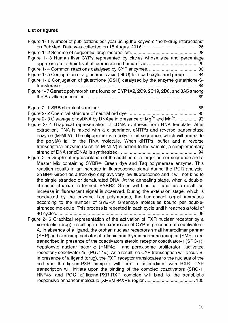
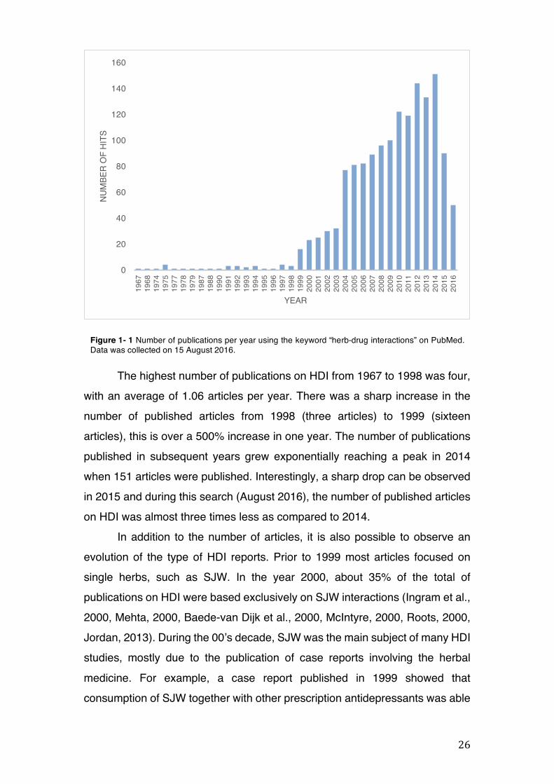
Cases of HDI are well documented in the literature. We conducted a search on PubMed database using the keywords “herb-drug interactions”. This helps to understand the beginning and the development of this type of research over the years.
The first record of a publication on HDI was in 1967 (Figure 1-1). This single work assessed potential interactions between herbal medicines and anaesthetic drugs (Pellerano, 1967). Subsequent years were marked by a few publications in diverse subjects where HDI were mentioned but were the main scope of the research. In 1974, a publication on the interaction between cannabinoids and phenytoin (Chesher and Jackson, 1974) triggered more interest in the subject. In 1975, two of the four HDI publications were about cannabinoids (Hine et al., 1975, Hollister and Gillespie, 1975) and in 1978 the single publication found was also about interactions with cannabis (Singh and Das, 1978).
In 1990, the first work on HDI involving numerous herbal medicines was published by a Chinese researcher who evaluated the potential interactions between Western antihypertensive drugs and Chinese herbal drugs (Wang, 1990). From 1990 to 1993 more articles involving drug interactions with various herbal medicines were published as review articles (D'Arcy, 1991, D'Arcy, 1992, D'Arcy, 1993).
Interactions involving Chinese herbal medicines were again the subject of the only article published in 1995 according to our PubMed search (Tam et al., 1995). Two years later, in 1997, grapefruit (Citrus paradisi Macfad. - Rutaceae) and Ginkgo biloba L. (Ginkgoaceae) were investigated for their potential interactions with synthetic drugs (Ameer and Weintraub, 1997, Chermat et al., 1997) and in 1998 two more research articles (one again about drug interactions with grapefruit) were published (Miller, 1998, Fuhr, 1998).
26
0
20
40
60
80
100
120
140
160
1967
1968
1974
1975
1977
1978
1979
1987
1988
1990
1991
1992
1993
1994
1995
1996
1997
1998
1999
2000
2001
2002
2003
2004
2005
2006
2007
2008
2009
2010
2011
2012
2013
2014
2015
2016
NUM
BER
OF
HITS
YEAR
The highest number of publications on HDI from 1967 to 1998 was four, with an average of 1.06 articles per year. There was a sharp increase in the number of published articles from 1998 (three articles) to 1999 (sixteen articles), this is over a 500% increase in one year. The number of publications published in subsequent years grew exponentially reaching a peak in 2014 when 151 articles were published. Interestingly, a sharp drop can be observed in 2015 and during this search (August 2016), the number of published articles on HDI was almost three times less as compared to 2014.
In addition to the number of articles, it is also possible to observe an evolution of the type of HDI reports. Prior to 1999 most articles focused on single herbs, such as SJW. In the year 2000, about 35% of the total of publications on HDI were based exclusively on SJW interactions (Ingram et al., 2000, Mehta, 2000, Baede-van Dijk et al., 2000, McIntyre, 2000, Roots, 2000, Jordan, 2013). During the 00’s decade, SJW was the main subject of many HDI studies, mostly due to the publication of case reports involving the herbal medicine. For example, a case report published in 1999 showed that consumption of SJW together with other prescription antidepressants was able
Figure 1- 1 Number of publications per year using the keyword “herb-drug interactions” on PubMed. Data was collected on 15 August 2016.
27
to cause severe ADRs due to HDI, particularly in elderly people (Lantz et al., 1999). However, it was not clear how SJW could be causing such effects.
The first attempt to elucidate one of the possible interaction mechanisms of SJW was published in 2000, suggesting that the herbal medicine was able to induce CYP3A4 and consequently reduce the efficacy of certain drugs such as oral contraceptives (Roby et al., 2000). This was the starting point of the increased number of research articles evaluating how SJW could potentially cause interactions with various synthetic drugs, as most of them were known to be metabolised mainly by CYP3A4.
In addition to CYP3A4, other phase I metabolising enzymes such as the CYP isoforms CYP1A2, CYP2C9, CYP2C19, CYP2D6, and CYP2E1 were becoming the subject of metabolic studies involving herbal medicines. The effects of SJW on P-gp and phase II metabolic enzymes, such as glutathione-S-transferase (GST) started to be also investigated. Herbal medicines have increasingly become the subject of metabolic and transporter studies using similar molecular targets as the ones used for synthetic drugs but with only one difference: such studies are not a regulatory requirement to market traditional herbal medicines in many countries (Tsay et al., 2016).
1.3 DRUG METABOLISM AND TRANSPORT: GENERAL
CONSIDERATIONS
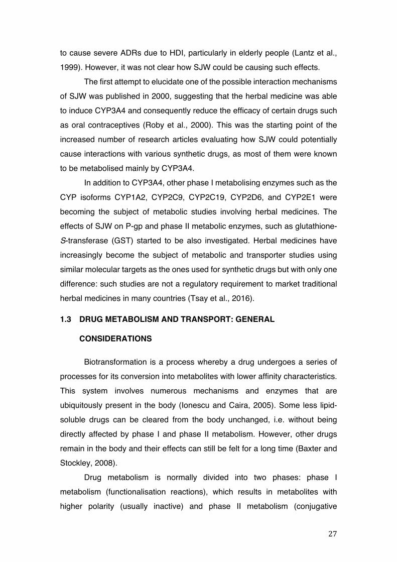
Biotransformation is a process whereby a drug undergoes a series of processes for its conversion into metabolites with lower affinity characteristics. This system involves numerous mechanisms and enzymes that are ubiquitously present in the body (Ionescu and Caira, 2005). Some less lipid-soluble drugs can be cleared from the body unchanged, i.e. without being directly affected by phase I and phase II metabolism. However, other drugs remain in the body and their effects can still be felt for a long time (Baxter and Stockley, 2008).
Drug metabolism is normally divided into two phases: phase I metabolism (functionalisation reactions), which results in metabolites with higher polarity (usually inactive) and phase II metabolism (conjugative
28
PD
Lipophilic parent drug
PD PHASE I PM PHASE II SM EXCRETION SM
Primary metabolite Secondary metabolite
Hydrophilic metabolite
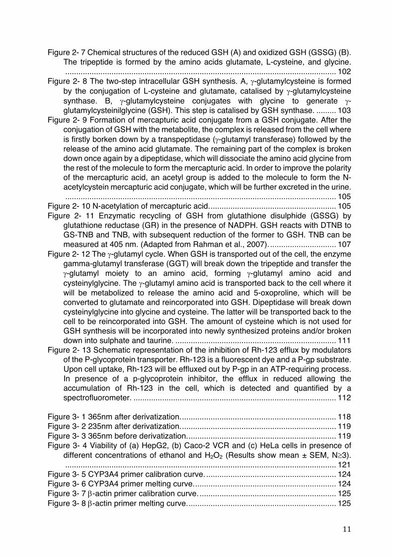
reactions), which results in the phase I metabolites becoming even more polar (Figure 1-2). Phase I reactions prepare the drug for phase II metabolism by adding polar functional groups to the xenobiotic (Ionescu and Caira, 2005).
1.3.1 Phase I metabolism: cytochrome P450
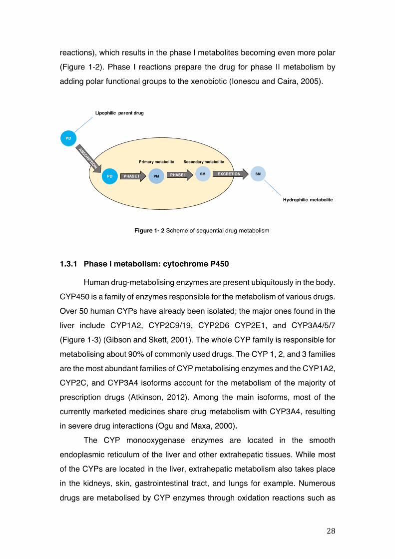
Human drug-metabolising enzymes are present ubiquitously in the body. CYP450 is a family of enzymes responsible for the metabolism of various drugs. Over 50 human CYPs have already been isolated; the major ones found in the liver include CYP1A2, CYP2C9/19, CYP2D6 CYP2E1, and CYP3A4/5/7 (Figure 1-3) (Gibson and Skett, 2001). The whole CYP family is responsible for metabolising about 90% of commonly used drugs. The CYP 1, 2, and 3 families are the most abundant families of CYP metabolising enzymes and the CYP1A2, CYP2C, and CYP3A4 isoforms account for the metabolism of the majority of prescription drugs (Atkinson, 2012). Among the main isoforms, most of the currently marketed medicines share drug metabolism with CYP3A4, resulting in severe drug interactions (Ogu and Maxa, 2000).
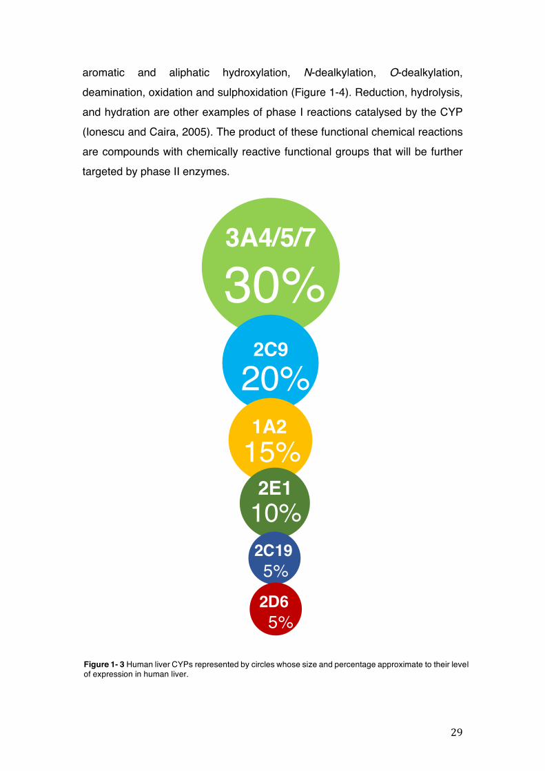
The CYP monooxygenase enzymes are located in the smooth endoplasmic reticulum of the liver and other extrahepatic tissues. While most of the CYPs are located in the liver, extrahepatic metabolism also takes place in the kidneys, skin, gastrointestinal tract, and lungs for example. Numerous drugs are metabolised by CYP enzymes through oxidation reactions such as
Figure 1- 2 Scheme of sequential drug metabolism
29
3A4/5/7
30%2C9
20%1A215%2E110%2C195%
2D65%
aromatic and aliphatic hydroxylation, N-dealkylation, O-dealkylation, deamination, oxidation and sulphoxidation (Figure 1-4). Reduction, hydrolysis, and hydration are other examples of phase I reactions catalysed by the CYP (Ionescu and Caira, 2005). The product of these functional chemical reactions are compounds with chemically reactive functional groups that will be further targeted by phase II enzymes.
Figure 1- 3 Human liver CYPs represented by circles whose size and percentage approximate to their level of expression in human liver.
30
R CH3 R CH2 + H+OH[OH]
NH C CH3
O
NH C CH3
O
OH
[OH]
R NH CH3 [R NH CH2 OH] R NH2 + CH2O[OH]
R O CH3 [R O CH2 OH] R OH + CH2O[OH]
RHC
NH2
CH3[OH]
R C
NH2
CH3
OH
R C CH3
O
+ NH3
H3C N
CH3
CH3
[OH]H3C N
CH3
CH3
OH[ ]+ H3C NO
CH3
CH3
+ H+
R S R'[OH] [ R
HS R'
OH
] R S R'
O
+ H+
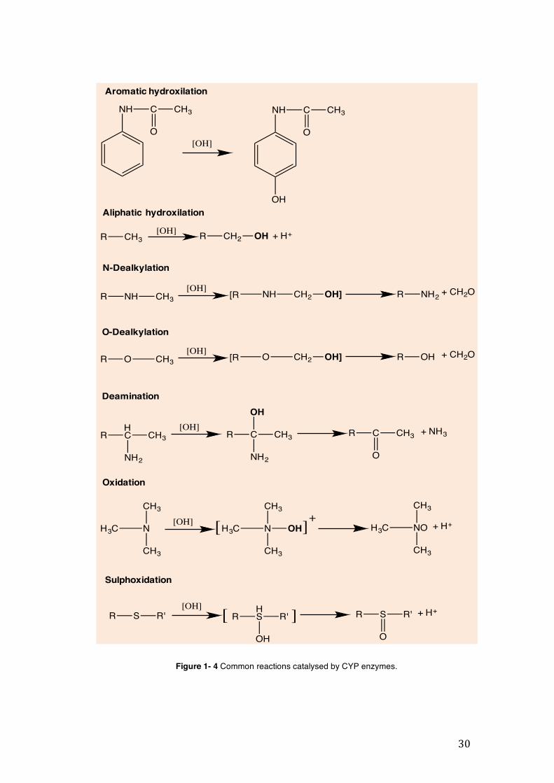
Aromatic hydroxilation
Aliphatic hydroxilation
N-Dealkylation
O-Dealkylation
Deamination
Oxidation
Sulphoxidation
Figure 1- 4 Common reactions catalysed by CYP enzymes.
31
1.3.1.1 Main CYP isoforms
1.3.1.1.1 CYP1A2
This CYP isoform is mainly found in the liver (15%) and it metabolises almost 20% of the current therapeutic arsenal (Wang and Zhou, 2009). Examples of synthetic drugs, which are substrates for the CYP1A2, include the highly popular painkiller acetaminophen, the beta-blocker propranolol, the antidepressant clomipramine and the anticoagulant warfarin.
1.3.1.1.2 CYP2C9
CYP2C9 is the major CYP2C isoform found in the human liver (Mo et al., 2009) and it metabolises approximately 15% of clinical drugs, including the nonsteroidal anti-inflammatory (NSAID) ibuprofen, the antihypertensive losartan, the antidepressant fluoxetine, the antiepileptic phenytoin, and the anti-hypercholesteraemic fluvastatin (Baxter and Stockley, 2008, Berka et al., 2011). Certain endogenous bioactive substances such as steroids, melatonin, retinoids, and arachidonic acid are also metabolised by this CYP isoform (Mo et al., 2009).
1.3.1.1.3 CYP2C19
CYP2C19 is not only involved in the metabolism of a range of drugs but it also plays a crucial role in the detoxification and inactivation of some potential carcinogens (Wang et al., 2013). CYP2C19 is responsible for the metabolism of about 10% of prescribed drugs, including the proton pump inhibitor omeprazole, the tricyclic antidepressant amitriptyline, the selective serotonin reuptake inhibitor fluoxetine, the benzodiazepine diazepam, and the barbiturate phenobarbital (Zhou et al., 2009).
1.3.1.1.4 CYP2D6
Although the level of expression of CYP2D6 in the human liver is only about 5%, this CYP isoform metabolises about 25% of all medications in the human liver (Ionescu and Caira, 2005). The beta-blockers propafenone and timolol, the antidepressant amitriptyline, the antipsychotics haloperidol and
32
risperidone, and the antihistamine chlorphenamine are examples of drugs that are metabolised by this CYP isoform (Zhou et al., 2009).
1.3.1.1.5 CYP2E1
CYP2E1 represents 10% of the total CYPs expressed in the human liver and it is well known for its involvement in the metabolism of ethanol to acetaldehyde, and accordingly it is rapidly induced after ethanol ingestion (Anzenbacher and Anzenbacherova, 2001). This CYP isoform is responsible for the activation of some carcinogens, procarcinogens, and toxicants and it metabolises mainly low-molecular-weight compounds. CYP2E1 also has the ability to produce reactive intermediates, leading to the formation of free radicals such as superoxide, hydroxyl radical, and lipid peroxides (Neafsey et al., 2009). Acetaminophen and the anaesthetic halothane are examples of CYP2E1 substrates (Tateishi et al., 1998, Wolf et al., 2007).
1.3.1.1.6 CYP3A
The most abundant subfamily of CYPs is CYP3A. It represents about 30% of the entire CYP450 enzymes in the liver and it is responsible for the biotransformation of more than 50% of therapeutic drugs. CYP3A’s main isoforms found in the body are 3A4, 3A5, and 3A7. CYP3A5 is more often detected in adolescents than in adults, where it is hardly inducible. CYP3A4 is mostly glucocorticoid-inducible and CYP3A7 (found only in fetal livers) has a role in the hydroxylation of allylic and benzylic carbon atoms (Ionescu and Caira, 2005). Macrolide antibiotics, antiarrhythmics, benzodiazepines, immune modulators, HIV antivirals, antihistamines, calcium channel blockers, and HMG-CoA reductase inhibitors are examples of classes of medications metabilised by the CYP3A subfamily (Zhou et al., 2009). The probability of HDI with this isoform is high and therefore a particular attention to all CYP3A substrates should be given in order to avoid such interactions.
1.3.2 Phase II metabolism (conjugation reactions)
Phase II metabolism reactions (or conjugation reactions) occur when metabolic enzymes react with functional groups of a drug that were formed
33
during the phase I process. Endogenous species, such as a sugar or an amino acid, are added to the drug in order to increase the polarity to allow its elimination. The two main phase II biotransformation reactions are glutathione (GSH) conjugation and glucuronidation. However, the other conjugative reactions such as sulphation, methylation, and acetylation are also relevant (Atkinson, 2012). The main metabolising enzymes involved in phase II metabolism are listed in table 1-1. Table 1- 1 The main phase II metabolic mechanisms and respective enzymes.
1.3.3 Types of phase II reactions
1.3.3.1 Glucuronidation
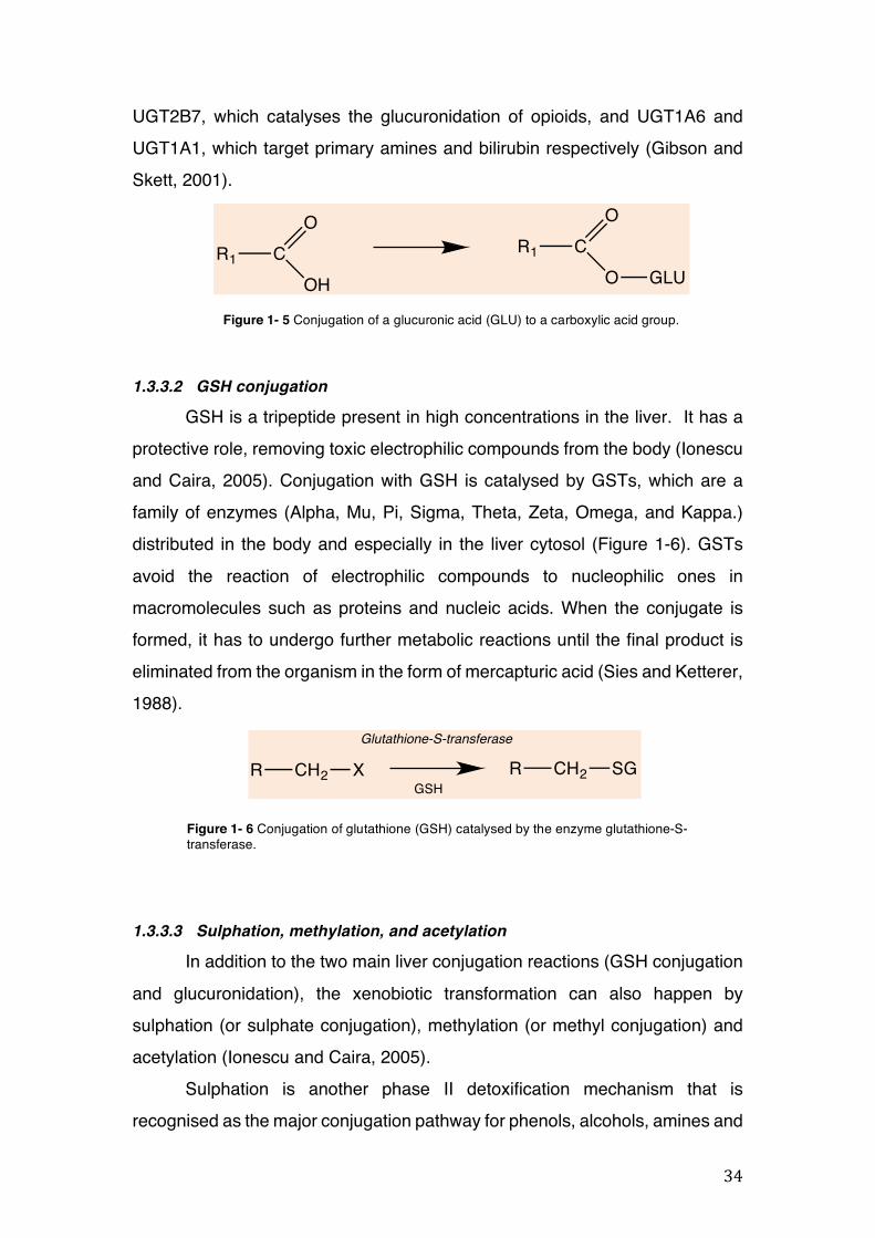
Glucuronidation is a mechanism in which a glucuronide is formed by the reaction between the electrophilic C-1 atom of the pyranose acid ring of the co-factor UDPGA (uridine 5’-diphosphate-glucuronic acid) and the substrate catalysed by UGTs. UGTs are the most important phase II enzymes, and out of all the conjugation enzymes in the liver, they constitute the highest amount (35%) (Ionescu and Caira, 2005, Caira and Ionescu, 2006). The enzyme can be found in the kidneys, small intestine, lung, skin, adrenals, and spleen. This is the most important form of conjugation of xenobiotics with chemical groups such as alcohols, phenols, hydroxylamines, carboxylic acids (Figure 1-5), amines, sulphonamides, and thiols (Gibson and Skett, 2001).
UGTs exists in multiple isoforms (1A1, 1A3, 1A4, 1A6, 2B4, 2B7, 2B10, 2B11, and 2B15) and they are able to metabolise a wide range of compounds, hence providing the liver with the extensive capacity to glucuronidate numerous endogenous and xenobiotic compounds. An example of this process is:
Phase II mechanism Enzyme involved Glucuronidation UDP-glucuronosyltransferase (UGT) Glutathione (GSH) conjugation Glutathione-S-transferase (GST) Sulphation Sulphotransferases (SULT) Methylation Methyltransferases Acetylation CoA-S-acetyltransferase
34
R CH2 X R CH2 SGGlutathione-S-transferase
GSH
R1 CO
OH
R1 CO
O GLU
UGT2B7, which catalyses the glucuronidation of opioids, and UGT1A6 and UGT1A1, which target primary amines and bilirubin respectively (Gibson and Skett, 2001).
1.3.3.2 GSH conjugation
GSH is a tripeptide present in high concentrations in the liver. It has a protective role, removing toxic electrophilic compounds from the body (Ionescu and Caira, 2005). Conjugation with GSH is catalysed by GSTs, which are a family of enzymes (Alpha, Mu, Pi, Sigma, Theta, Zeta, Omega, and Kappa.) distributed in the body and especially in the liver cytosol (Figure 1-6). GSTs avoid the reaction of electrophilic compounds to nucleophilic ones in macromolecules such as proteins and nucleic acids. When the conjugate is formed, it has to undergo further metabolic reactions until the final product is eliminated from the organism in the form of mercapturic acid (Sies and Ketterer, 1988).
1.3.3.3 Sulphation, methylation, and acetylation
In addition to the two main liver conjugation reactions (GSH conjugation and glucuronidation), the xenobiotic transformation can also happen by sulphation (or sulphate conjugation), methylation (or methyl conjugation) and acetylation (Ionescu and Caira, 2005). Sulphation is another phase II detoxification mechanism that is recognised as the major conjugation pathway for phenols, alcohols, amines and
Figure 1- 5 Conjugation of a glucuronic acid (GLU) to a carboxylic acid group.
Figure 1- 6 Conjugation of glutathione (GSH) catalysed by the enzyme glutathione-S-transferase.
35
thiols (Ionescu and Caira, 2005). Before the phase I metabolites undergo sulphate conjugation, the inorganic sulphate has to be activated via ATP to form adenosine-5’-phosphosulphate (APS) and consequently 3’-phosphoadenosine-5’-phosphosulphate (PAPS). SULTs will then catalyse the detoxification of essential drugs, such as salbutamol and acetaminophen by transferring a sulphuryl group from PAPS to an acceptor molecule (Gibson and Skett, 2001).
Methylation is the mainly metabolic pathway for endogenous compounds, but it can also be the route for many drugs and xenobiotics in general (Weinshilboum, 1988). Methyl conjugate reactions are only possible in the presence of the cofactor S-adenosylmethionine (SAM) and will result in the formation of O-methylated, N-methylated, and S-methylated products (Gibson and Skett, 2001).
The liver is the primary site for acetylation reactions, but the reaction can also happen in some extra hepatic sites, such as the spleen, lungs, and gut. To summarise, the N-acetyltransferases will catalyse the transferring of the co-factor acetyl-coenzyme A (acetyl-CoA) to aromatic amines and sulphonamides and form the polar metabolites (Ionescu and Caira, 2005).
1.3.4 The drug transporter P-glycoprotein
Drug-transporter proteins are known to allow xenobiotics to cross biological membranes, the most well-known one being P-gp. P-gp was first discovered in 1986 as a product of the multi-drug resistance gene (MDRI) in cancer cells, therefore reducing the intracellular accumulation of drugs (Watanabe et al., 2012).
This protein plays a role as an efflux pump, it pushes metabolites and drugs out of the cells which can result in absorption alterations (Williamson et al., 2009). P-gp contains two ATP-binding sites, where ATP will bind in presence of a P-gp substrate. As a consequence, ATPase is activated in order to hydrolyse ATP. The energy produced by the ATP hydrolysis will allow the P-gp to transport numerous substrates across cellular membranes (Watanabe et al., 2012). In the intestine, P-gp can be found on the apical surface of epithelial
36
cells. When a drug is taken up by an enterocyte, the substance can be either metabolised by CYP3A4 or pumped back into the lumen (Butterweck and Derendorf, 2008). Hence, oral delivery can be compromised (Butterweck et al., 2004).
1.4 INDUCTION AND INHIBITION OF DRUG METABOLISING ENZYMES
1.4.1 Induction of drug metabolising enzymes
As previously stated in section 1.2, studies on SJW demonstrate how the herbal medicine can act as a potent CYP3A4 inducer. As a consequence, drugs like oral contraceptives, which are metabolised by the same enzyme, will have their pharmacological effects altered. The increased metabolism of the drug due to high CYP3A4 activity will lead to a decreased blood concentration of the oral contraceptive and low pharmacological activity (Murphy et al., 2005). Consumption of metabolic enzyme inducers like SJW will, therefore, increase the chances of inefficacy of the coadministered drug in usual therapeutic doses due to HDI (Tarirai et al., 2010).
Another example of metabolic HDI involving CYP was published as a case report in 2015. The authors showed that the intake of noni juice, which is an herbal remedy made from the fruit of Morinda citrifolia L. (Rubiaceae), is able to induce CYP2C9 and interact with the anti-epileptic drug phenytoin by decreasing its bioavailability in a 49-year-old man (Kang et al., 2015).
The main types of drug induction are substrate-dependant induction, receptor-mediated and inhibitor-mediated interaction. Substrate-dependant induction is where the herbal drug influences the metabolism and duration of action of numerous other drugs. Receptor-mediated is characterized by interactions with important regulator pathways, which is the case of the human nuclear pregnane X (hPXR) and the constitutive androstane (CAR) receptors. These are well known also as xenobiotic sensors, which are activated by numerous compounds leading to the activation of their downstream target genes (Xie, 2009).
The binding of xenobiotics to the receptor directly affects the clearance of those compounds, and consequently, protects the body from foreign
37
chemicals. Many drug metabolising enzymes involved in the metabolism of endogenous cellular regulators (steroids, eicosanoids) can be induced by hormones. For example, the growth hormone has been also proven to alter CYP expression (Mode et al., 1992, Cheung et al., 2006).
Inhibitor-mediated interaction involves a stabilisation mechanism, i.e. the drug decreases the degradation of CYP and therefore the concentration of the enzyme is increased (Watkins et al., 1986).
Phase II metabolising enzymes such as UGTs and GSTs can also be inducible. Induction of UGTs is highly dependent on the nature of the inducer. Ethanol and phenobarbital are able to induce UGTs, as well as the endogenous compound bilirubin (Soars et al., 2004, Hiroshi and Shigeko Shinkai and Takaharu, 2008). GSTs can also be induced by phenobarbital and other xenobiotics such as dithiolethiones (Zhang and Munday, 2008, Sasaki et al., 1989).
1.4.2 Inhibition of drug metabolising enzymes
In contrast to the effects observed in the induction of drug metabolic enzymes, inhibition delays xenobiotic biotransformation resulting in a higher concentration of the compound in the bloodstream. The consequence of such inhibition is the increased adverse reactions due to exacerbated pharmacological and toxicological effects (Kobayashi et al., 2002).
An inhibitor is a substance that interferes with the action of the enzyme, which will slow down the speed of the reaction. A reversible inhibitor will bind to an enzyme and will subsequently be released, whereas an irreversible inhibitor reacts with the enzyme producing a protein that will not be enzymatically active anymore and the original enzyme cannot be regenerated (Ring et al., 2014).
The two main types of CYP inhibition are called competitive and non-competitive inhibition. The former happens when an inhibitor competes with the substrate to bind to the active site of the enzyme. In this case, the inhibitor will block the access of the substrate to the enzyme. On the other hand, in the case of a non-competitive inhibition, both substrate and inhibitor will bind to different
38
sites of the enzyme. However, the enzyme cannot catalyse the reaction in as an efficient manner as it can in the absence of the inhibitor (Campbell and Farrell, 2012). Drugs like indinavir and saquinavir are competitive inhibitors, whereas ketoconazole and fluconazole can act as non-competitive inhibitors of CYP3A (Thummel and Wilkinson, 1998).
Like phase I enzymes, phase II enzymes can also display inhibitory activity. A study recently performed by Liu and coworkers (2016) demonstrated that UGT isoforms can be inhibited by vitamin A (Liu et al., 2016), whereas Chang and coworkers (1998) showed that oestrogens are able to inhibit GSTs (Chang et al., 1998).
1.5 FACTORS THAT INFLUENCE DRUG BIOTRANSFORMATION
In addition to the induction and inhibition of drug metabolising enzymes, several factors can contribute to the variability in biotransformation. They include:
• Genetic polymorphism • Disease • Age • Sex • Environmental factors
1.5.1 Genetic Polymorphism
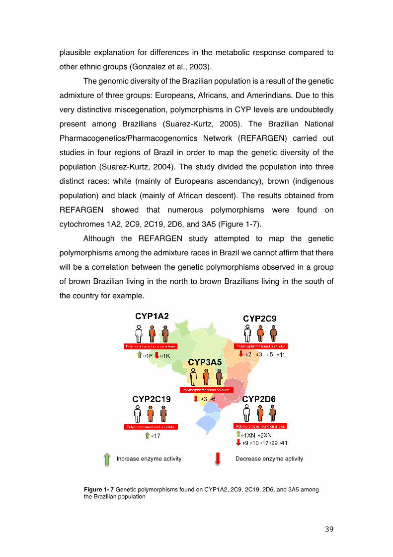
Responses to drug consumption differ between individuals due to the variability of CYP content. A predefined dosage of a medicine might be enough to exert a pharmacological effect in one patient while needing to be adjusted to achieve the same effect in another patient. This can be explained by genetic polymorphisms within CYPs that can affect the metabolism of xenobiotics in general, leading to changes in drug responses and increased risk of ADRs (Zhou et al., 2009). For example, the bioavailability of omeprazole, which is a drug that is metabolised by both CYP3A4 and CYP2C19, has been demonstrated to be slower in Mexicans when compared with Caucasians but faster than that of Asians. The ethnic mixture of the Mexican population is a
39
Increase enzyme activity Decrease enzyme activity
plausible explanation for differences in the metabolic response compared to other ethnic groups (Gonzalez et al., 2003).
The genomic diversity of the Brazilian population is a result of the genetic admixture of three groups: Europeans, Africans, and Amerindians. Due to this very distinctive miscegenation, polymorphisms in CYP levels are undoubtedly present among Brazilians (Suarez-Kurtz, 2005). The Brazilian National Pharmacogenetics/Pharmacogenomics Network (REFARGEN) carried out studies in four regions of Brazil in order to map the genetic diversity of the population (Suarez-Kurtz, 2004). The study divided the population into three distinct races: white (mainly of Europeans ascendancy), brown (indigenous population) and black (mainly of African descent). The results obtained from REFARGEN showed that numerous polymorphisms were found on cytochromes 1A2, 2C9, 2C19, 2D6, and 3A5 (Figure 1-7).
Although the REFARGEN study attempted to map the genetic polymorphisms among the admixture races in Brazil we cannot affirm that there will be a correlation between the genetic polymorphisms observed in a group of brown Brazilian living in the north to brown Brazilians living in the south of the country for example.
Figure 1- 7 Genetic polymorphisms found on CYP1A2, 2C9, 2C19, 2D6, and 3A5 among the Brazilian population
40
1.5.2 Disease
Diseases affecting the liver, such as cirrhosis, alcoholic liver disease, cholestatic jaundice, and liver carcinoma can alter organ function, and consequently, reduce the capacity of the body to clear drugs. Drug metabolism can also be affected by endocrine disorders such as diabetes mellitus, hypo-and hyperthyroidism, pituitary disorders and bacterial and viral infections in general. For example, in the case of a hepatic cirrhosis, the CYP2A6 can be overexpressed. This isoform catalyses the bioactivation of many drugs and also carcinogens. Therefore, CYP2A6 is a major liver catalyst in pathological conditions (Ionescu and Caira, 2005).
Malaria, which is one of the most common tropical diseases, can also affect the enzymes involved in the biotransformation of drugs. An impairment of microsomal drug metabolising activities as a result of the malaria infection reduces the oxidation of drugs by decreasing hepatic CYP activity (Mansor et al., 1991).
1.5.3 Age
Biotransformation contrasts can be found between newborns, the young and elderly due to the types and the amount of enzymes that are activated and expressed among those groups. Newborns are more sensitive to drug reactions because of the low level of development of their metabolising capacity. The sensitiveness of newborns lowers significantly over time until they reach adulthood when normal levels of enzyme activity are reached. In the elderly population, the biotransformation capacity may be reduced up to 30% due to several reasons. Reasons include decreased hepatic blood flow and mass, decreased absorption surface and gastrointestinal mobility and increased gastric pH (Klotz, 2009).
1.5.4 Sex
Sex is another factor that contributes to differences in the expression of drug metabolising enzymes. Initial findings on this subject were made in the 1930s when researchers treated rats with barbiture and observed that the
41
female ones required only half the dose to induce sleep compared to male rats (Gibson and Skett, 1986).
Another study discovered that the growth hormone may be the main hormonal factor that dictates the differences in the expression of CYPs and other drug metabolising enzymes between males and females (Waxman and Holloway, 2009).
1.5.5 Environmental factors
Heavy metals, industrial pollutants, pesticides and other chemical substances that are spread out in the air can potentially alter the activity of CYP enzymes in liver microsomes (Gillette, 1976). The activity of some drug metabolising enzymes may be induced if the individual is exposed to exogenous chemicals, including environmental pollutants. For example, most industrial pollutants are typically aromatic or aromatic polycyclic compounds and polychlorinated biophenols, which are known to cause inductive enzyme effects in various CYP isoforms (O'Mahony and Woodhouse, 1994, Kietz and Fischer, 2003).
1.6 HERB-DRUG INTERACTIONS AND ADVERSE REACTIONS
As previously discussed in section 1.2, reports on HDI are dated back to 1967 and since then over 1,500 articles have been published according to PubMed. The documented interactions involve well-known herbs from Traditional Chinese Medicine (TCM), Ayurveda and many other traditional herbal medicines commonly used by the population of many countries and their biotransformation involves many metabolic and transporter mechanisms.
1.6.1 Herb-drug interactions involving cytochrome P450
In section 1.3.1 we showed that the CYP family is responsible for the metabolism of most xenobiotics. Among the various CYP isoforms, CYP1A2, 2C9, 2C19, 2D6, 2E1, and 3A(4/5/7) are the most relevant ones because altogether they are responsible for the biotransformation of the majority of the currently marketed drugs (Mazzari and Prieto, 2014a).
42
Due to the increased popularity of SJW as an antidepressant, numerous interactions with CYP3A4 have been detected in vitro and in vivo, the interaction with oral contraceptives being one of the most remarkable cases of HDI (Murphy et al., 2005). As the interaction with CYP3A4 became more evident among researchers, the effects of SJW in other metabolic targets, such as CYP1A2, 2C9, 2C19, 2D6, 2E1 were also assessed. The numerous publications alerting to the potential interactions of SJW involving CYPs due to enzyme induction and/or inhibition made it possible to discuss the potential effects that other herbal medicines could cause on drug metabolising enzymes.
Besides SJW, popular herbal medicines such as Ginkgo biloba L. and milk thistle (Silybum marianum (L.) Gaertn. - Compositae) have been extensively studied over the past years in order to reveal their potential to cause HDI due to their effects on CYP activity and/or expression. Published studies showed that Ginkgo biloba and milk thistle are able to modulate the effect of the main CYP isoforms due to the inducible and/or inhibitory effects of their constituents (von Moltke et al., 2004, Venkataramanan et al., 2000).
A study published by Sugiyama and coworkers (2004) showed that simultaneous administration of Ginkgo biloba extract with the oral anti-diabetic tolbutamide potentiated the hypoglycaemic action of this drug in rats. Tolbutamine is metabolised by CYP2C9 and the inhibitory effect caused by the Ginkgo biloba extract increased both the plasma concentrations and the bioavailability of the drug. Hence, blood glucose levels dramatically dropped in rats treated with the drugs (Sugiyama et al., 2004).
Silybin, a constituent of milk thistle, show inhibitory effects on CYP2D6, 2E1, and 3A4 (Zuber et al., 2002). The ginsenosides Rb1, Rb2, Rc, Rd, Re, Rf, and Rg1 from Panax ginseng C.A.Mey. (Araliaceae) display inductive and inhibitory effects on CYP 1A2, 2C9, 2C19, 2D6, and 3A4 (Henderson et al., 1999). A drug interaction study demonstrated that silymarin (140mg) given three times a day inhibited hepatic clearance of losartan, which is a drug used to treat high blood pressure, in Chinese subjects. The active metabolite of losartan (E-3174) is formed by CYP2C9. Due to the inhibition of the enzyme caused by the milk thistle constituent silymarin, the amount of E-3174 found in
43
the subjects treated also with the herbal medicine decreased compared to the control group (Brantley et al., 2014).
A plethora of data on HDI involving CYP has been published demonstrating that herbal medicines are able to affect CYP and therefore their consumption should be avoided with a synthetic drug. Nonetheless, evaluation of this subject is very controversial. The fact that most of the published research on HDI is either in vitro, performed in animal models and done with fractions (not whole herbs) at non-physiological concentrations, explains why most of the ‘theoretical’ HDI have never been clinically observed. Preclinical data is very important because it serves as a guide for clinical studies. Therefore, it is important that such studies are fully performed and are not abandoned in their initial or middle stages, which has been the case in much research on herbal medicines that has never reached the clinical trials stage of development (Brantley et al., 2014).
1.6.2 Herb-drug interactions involving phase II metabolising enzymes
Glucuronidation is a major route of phase II xenobiotic biotransformation and elimination in the body. Classes of drugs such as narcotic analgesics, anxiolytics, and NSAIDs are examples of therapeutic agents metabolised via glucuronidation mechanism. Due to the high number of exogenous compounds that are metabolised through this route, the characterisation of the effects of herbal medicines on the activity of UGTs is very important to reveal potential HDI (Ionescu and Caira, 2005).
In vitro studies demonstrated that valerian extract (Valeriana officinalis L. - Caprifoliaceae) - used at clinical dosages are able to produce significant reductions in the glucuronidation of conventional drugs like acetaminophen and morphine, possibly caused by the inhibition of several UGTs such as UGT1A6/9, UGT1A1, and UGT2B7 (Alkharfy and Frye, 2007).
Echinacea, ginkgo, milk thistle, soy, and SJW are other extracts that have been assessed for their effects on UGT in vitro. The extracts display inhibitory effects on UGT1A1, UGT 1A9, and UGT2B15. Constituents of several herbal medicines, such as silymarin and silybin (milk thistle), genistein (soy),
44
hypericin (SJW) and valerianic acid (valerian) are also able to modulate glucuronidation due to the inhibition and/or induction of various UGTs isoforms (Mohamed and Frye, 2011).
GSH conjugation is also affected by xenobiotics due to the modulatory effects on GSTs. A study conducted by Yang and coworkers (2003) showed that Ginkgo biloba extract down-regulates the GST activity in microsomes from rat kidney (Yang et al., 2003). Lower GST activity can lead to potential adverse reactions caused by toxic metabolites that are detoxified by conjugation with GSH such as N-acetyl-p-benzoquinoneimine (NAPQI) (Henderson et al., 2000). GSH conjugation is the detoxification mechanism that various drugs undergo, including anticancer agents. Even though elevated levels of GSH are considered positive due to its strong antioxidant effects in the cells, the high activity of GSTs can weaken the effectiveness of certain anticancer drugs. GST P1-1, for example, is particularly induced in certain types of cancer, such as lung, colon, and stomach cancers. Because of its increased activity, the GST P1-1 enzyme is implicated in cellular resistance to chemotherapeutic agents (Ramsay and Dilda, 2014).
Many other HDI involving phase II metabolising enzymes have been reported in the literature. As some conjugation mechanisms are the major detoxifying route of several important classes of drugs, such as the anticancer agents, more interaction studies (especially clinical studies) need to be carried out on herbal medicines that could potentially affect phase II enzymes and reactions.
1.6.3 Herb-drug interactions involving P-glycoprotein
Modulation of drug transporters like P-gp by herbal medicines has been the target of a plethora of studies. Due to its high expression and activity in several tissues (kidney, liver, intestine, and blood-brain barrier (BBB)), which are pharmacologically important epithelial barriers, the bioavailability of the drug could be compromised if P-gp activity is affected (Li et al., 2014).
Inhibition or induction of P-gp activity results in altered absorption and bioavailability of P-gp substrates (Zhou et al., 2004). Studies have shown that
45
polyphenols from green tea (Camellia sinensis (L.) Kuntze – Theaceae) are able to inhibit P-gp activity, which is bolstered by the intracellular accumulation of the P-gp substrate rhodamine 123 (Rh-123) in Caco-2 cells (Jodoin et al., 2002).
SJW displays inductive effects in P-gp efflux activity. Chronic treatment with SJW increases the P-gp efflux in healthy subjects, which can also be observed in vitro by a reduced intracellular accumulation of the P-gp substrate Rh-123 (Hennessy et al., 2002). This inductive effect on P-gp has been demonstrated to attenuate the efficacy of the potent P-gp inhibitor drug ritonavir, which potentially leads to treatment failure (Drewe et al., 1999).
Ezuruike and Prieto (2014) showed that some herbal medicines used in Nigeria for diabetes management can cause HDI at P-gp level. This is the case of the coadministration of medicinal plants such as Acacia nilotica (L.) Delile (Leguminosae), Annona senegalensis Pers. (Annonaceae), Bauhinia thonningii Schum. (Leguminosae), Bridelia ferruginea Benth. (Phyllanthaceae), Carica papaya L. (Caricaceae) and Morinda lucida Benth. (Rubiaceae) with the antidiabetic drug glibenclamide. According to the authors, the herbal medicines which exhibit inhibitory effects on P-gp and their concomitant use with glibenclamide increases the plasma concentration of the synthetic drug (Ezuruike and Prieto, 2014).
Some examples of potential HDI previously mentioned involving metabolising enzymes and P-gp were conducted using in vitro and in vivo models in order to either predict or attest an interaction that has been already reported by other researchers. However, HDI of the most of the marketed herbal medicines have not been evaluated using the usual methods of clinical pharmacology, that is, preclinical metabolic studies have not been conducted (Skalli and Soulaymani Bencheikh, 2012). Therefore, pharmacovigilance plays a very important role in the detection of HDI. Unfortunately, in many cases, an alert about possible HDI is only made after a reported case of death believed to be due to treatment failure or increased toxicity of a synthetic drug.
46
1.7 PHARMACOVIGILANCE OF HERBAL MEDICINES
The pharmacological and PK metabolic knowledge on herbal drugs accumulated over the last decades (summarised in the previous sections) prompted WHO to make an alert as to the potential risks of uncontrolled use of herbal medicines in conjunction with other synthetic drugs. In 2004, WHO issued the “Guidelines on safety monitoring of herbal medicines in pharmacovigilance systems”. These guidelines indicate how member countries should include herbal medicines within existent pharmacovigilance systems in order to facilitate the exchange of information on adverse reactions and HDI between WHO member countries (WHO, 2004).
The WHO traditional medicine strategy, which was published by WHO in 2013, showed that the regulatory status of herbal medicines is quite diverse among WHO member countries. A medicinal plant can be registered as a food, a functional food, a dietary supplement, a traditional medicine, or a full licensed herbal medicine depending on the country’s own needs, cultural background, history of use of medicines, and regulations. This can potentially cause difficulties to pharmacovigilance systems in collecting HDI data, as for example a food interaction would be practically ignored and hardly detected by health professionals (WHO, 2013, SUS, 2006).
In addition to the differences in the regulatory status of herbal medicines between many countries, other challenges can be highlighted in terms of pharmacovigilance of herbal medicines. The presence of numerous active compounds in herbal extracts, the low standardisation of herbal products, the problems with the nomenclature of herbs and the many sources of the herbal medicines used by the patients are also examples that make the detection of HDI by health professionals more difficult. As a consequence, HDI are likely to be underreported to the pharmacovigilance systems (Shaw et al., 2012).
47
1.8 USE OF HERBAL MEDICINES IN BRAZIL AND THE RISK OF HERB-
DRUG INTERACTIONS ON PATIENTS OF THE PUBLIC HEALTH
SYSTEM (SUS)
Brazil is the most biodiverse country on the planet and a valuable traditional knowledge associated with the use of medicinal plants. The total biodiversity of the country corresponds to between 15 and 20% of the plant species that are found in the world, which also encompasses medicinal plants. Although the country has about 60,000 catalogued plant species, only 8% have been studied for their bioactive compounds and 1,100 have been assessed for their medicinal properties (Dutra et al., 2016). The vast genetic heritage found in the country gives to Brazil the opportunity to be a leading country in natural products research that could potentially benefit millions of people who still have no access to costly treatments with synthetic drugs and are affected by neglected tropical conditions, such as dengue fever, chikungunya and the most recent threat, the zika fever (SUS, 2006).
Agriculture in Brazil is the main source of income for millions of families in the country. Over the years, the Brazilian government has made expressive investments in this field in order to develop this government sector and to improve the research into the cultivation of plants (especially medicinal plants) (SUS, 2006).
Therefore, Ministry of Agriculture and Ministry of Health have stimulated the development of the medicinal plants field in Brazil. They aim to integrate the traditional knowledge of both cultivation and medicinal use of Brazil’s biodiversity in order to offer alternative treatments to the entire population of the country by increasing the availability of herbal medicines in the Brazilian Unified Health System (SUS) (SUS, 2006).
The SUS was created by the Brazilian Federal Constitution of 1988. According to the document, SUS has to guarantee full and free access to various health procedures and treatments for the entire population of Brazil. Currently, 180 million people rely exclusively on the health services provided by SUS. All medications available at SUS have been put together in a list of
48
essential medicines called RENAME. The list was created in 1975, before the creation of the SUS, and is based on the WHO recommendations to the member countries to develop a list of medicines aimed to attend to the health needs of the population. Since then the list has been revised and updated according to newly available treatments for the patients of SUS. At the moment, RENAME contains 440 drugs that are prescribed to treat most of the conditions that affect the Brazilian population (Brasil, 2014).
Between 2013 and 2015, the search for treatments using herbal medicines in SUS grew 161% and over the last 3 years at least 6,000 Brazilians went to local pharmacies seeking the use herbal drugs (Brasil, 2016). Currently, only 12 herbal medicines are listed in RENAME, therefore limiting the prescription of those drugs to the patients of SUS (Figueiredo et al., 2014).
In 2008, the Ministry of Health of Brazil issued a list of medicinal plants of interest of SUS (RENISUS). This list contains medicinal plants that have the potential to generate products for SUS (Table 1-2). As part of the selection process of the medicinal plants for the list, the Ministry of Health coordinated regular meetings with Brazilian researchers, representatives of the Brazilian Pharmacopoeia (BP) and the National Health Surveillance Agency (ANVISA). The work started in 2005 with a list of 237 plant species. The preliminary list considered plant species that were already in use in health services of the states and municipalities, the traditional and popular knowledge, the available chemical and pharmacological studies, and other plants that were in use by other health programs already established throughout the country. After several meetings, RENISUS was created containing various medicinal plant species that, eventually, should be available for the patients of SUS as part of RENAME (Brasil, 2009).
Some of RENISUS species are widely used worldwide, for example Cynara scolymus L. (Compositae), Allium sativum L. (Amaryllidaceae) and Uncaria tomentosa (Wild. Ex Schult.) DC. (Rubiaceae). Numerous medicinal plants that are traditionally used in the country are also present in the BP. Monographs of medicinal plant species have been included in the BP since the launch of its first edition in 1926. From this date, four more editions have been
49
published, dating: 1959, 1977, 1996, and 2010 with a special edition issued in 2011. More than 1000 monographs of medicinal plants, including native and non-native plants to Brazil, have been included in the editions (Brandão et al., 2006, ANVISA, 2011).
More than 500 herbal medicines were registered at ANVISA by 2008 and this number has certainly increased as the number of users grew as well. Ginkgo biloba L., Aesculus hippocastanum L. (Sapindaceae), Cynara scolymus L., Hypericum perforatum L., Glycine max (L.) Merr. (Leguminosae), Valeriana officinalis L., Panax ginseng C.A.Mey., Cassia angustifolia M.Vahl. (Leguminosae), Cimicifuga racemosa (L.) Nutt. (Ranunculaceae), Mikania glomerata Spreng., Maytenus ilicifolia Mart. ex Reissek (Celastraceae) and Peumus boldus Molina (Monimiaceae) are listed as the most used medicinal plant species by the Brazilian population. The native plants Mikania glomerata Spreng., Maytenus ilicifolia Mart. ex Reissek and Paullinia cupana Kunth (Sapindaceae) are widely used in the country as a bronchodilator, adjuvant in the treatment of gastric ulcers and as a stimulator of the nervous system respectively (Carvalho et al., 2008).
Due to the high number of synthetic drugs available in SUS, cases of interactions are likely to occur when the patient has been prescribed a treatment with more than one drug. As a consequence, the number of drug interactions (especially cases of HDI at the absorption and metabolic levels) may increase with the implementation of RENISUS. Thus, studies on the effects of those herbal medicines in metabolic and transporter mechanisms are highly required in order to anticipate HDI that could affect the patients of SUS.
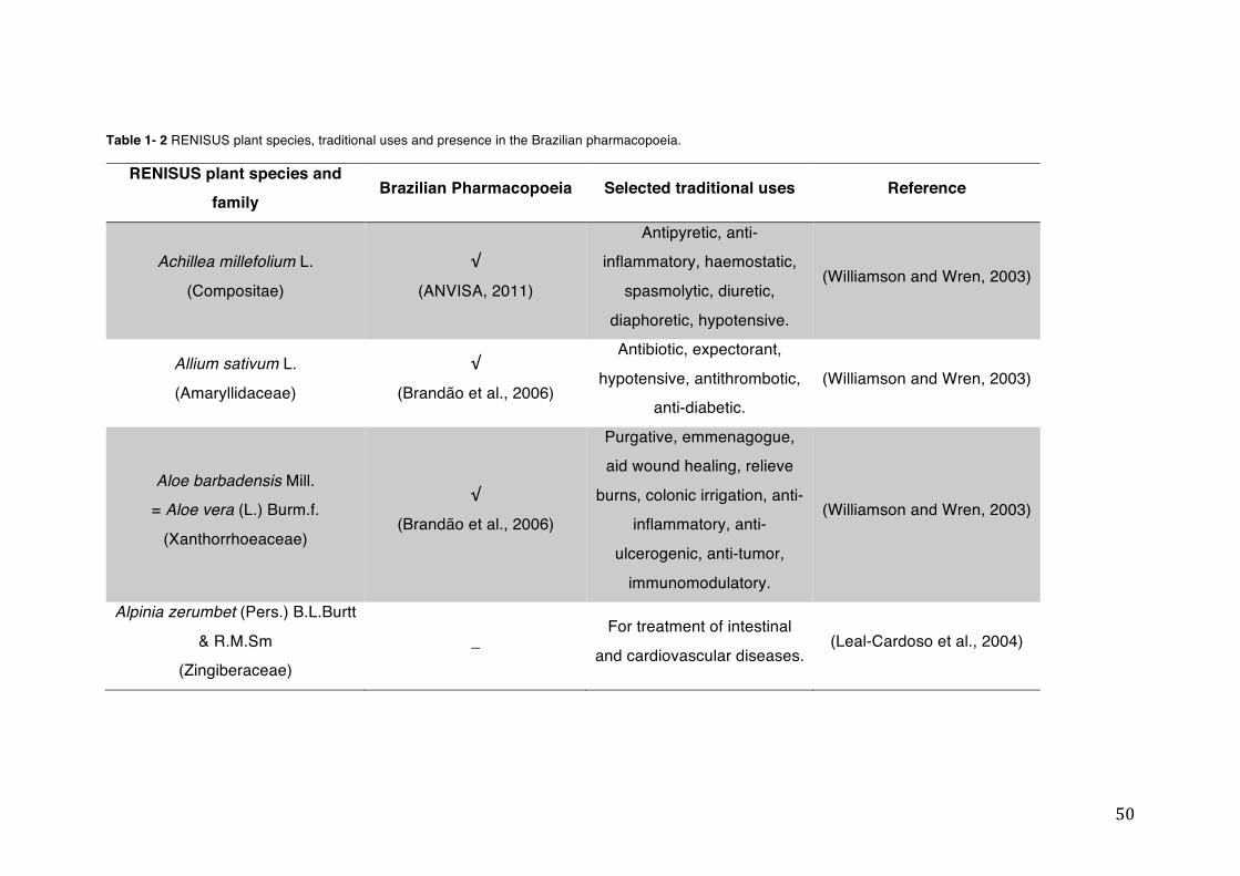
50
Table 1- 2 RENISUS plant species, traditional uses and presence in the Brazilian pharmacopoeia.
RENISUS plant species and
family Brazilian Pharmacopoeia Selected traditional uses Reference
Achillea millefolium L.
(Compositae)
√
(ANVISA, 2011)
Antipyretic, anti-inflammatory, haemostatic,
spasmolytic, diuretic,
diaphoretic, hypotensive.
(Williamson and Wren, 2003)
Allium sativum L.
(Amaryllidaceae)
√
(Brandão et al., 2006)
Antibiotic, expectorant,
hypotensive, antithrombotic, anti-diabetic.
(Williamson and Wren, 2003)
Aloe barbadensis Mill. = Aloe vera (L.) Burm.f.
(Xanthorrhoeaceae)
√
(Brandão et al., 2006)
Purgative, emmenagogue, aid wound healing, relieve
burns, colonic irrigation, anti-inflammatory, anti-
ulcerogenic, anti-tumor,
immunomodulatory.
(Williamson and Wren, 2003)
Alpinia zerumbet (Pers.) B.L.Burtt
& R.M.Sm (Zingiberaceae)
_ For treatment of intestinal
and cardiovascular diseases. (Leal-Cardoso et al., 2004)
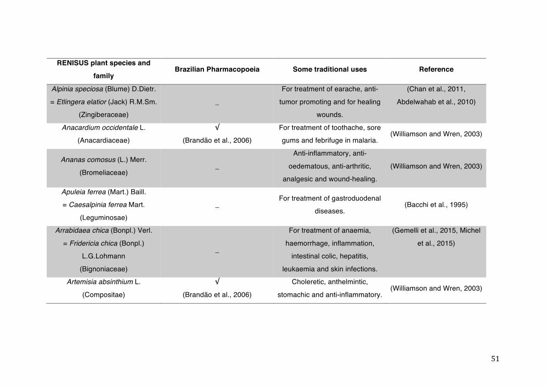
51
RENISUS plant species and
family Brazilian Pharmacopoeia Some traditional uses Reference
Alpinia speciosa (Blume) D.Dietr.
= Etlingera elatior (Jack) R.M.Sm. (Zingiberaceae)
_
For treatment of earache, anti-
tumor promoting and for healing wounds.
(Chan et al., 2011,
Abdelwahab et al., 2010)
Anacardium occidentale L. (Anacardiaceae)
√
(Brandão et al., 2006) For treatment of toothache, sore gums and febrifuge in malaria.
(Williamson and Wren, 2003)
Ananas comosus (L.) Merr.
(Bromeliaceae) _
Anti-inflammatory, anti-
oedematous, anti-arthritic, analgesic and wound-healing.
(Williamson and Wren, 2003)
Apuleia ferrea (Mart.) Baill. = Caesalpinia ferrea Mart.
(Leguminosae)
_ For treatment of gastroduodenal
diseases. (Bacchi et al., 1995)
Arrabidaea chica (Bonpl.) Verl.
= Fridericia chica (Bonpl.) L.G.Lohmann
(Bignoniaceae)
_
For treatment of anaemia,
haemorrhage, inflammation, intestinal colic, hepatitis,
leukaemia and skin infections.
(Gemelli et al., 2015, Michel
et al., 2015)
Artemisia absinthium L.
(Compositae)
√
(Brandão et al., 2006)
Choleretic, anthelmintic,
stomachic and anti-inflammatory. (Williamson and Wren, 2003)
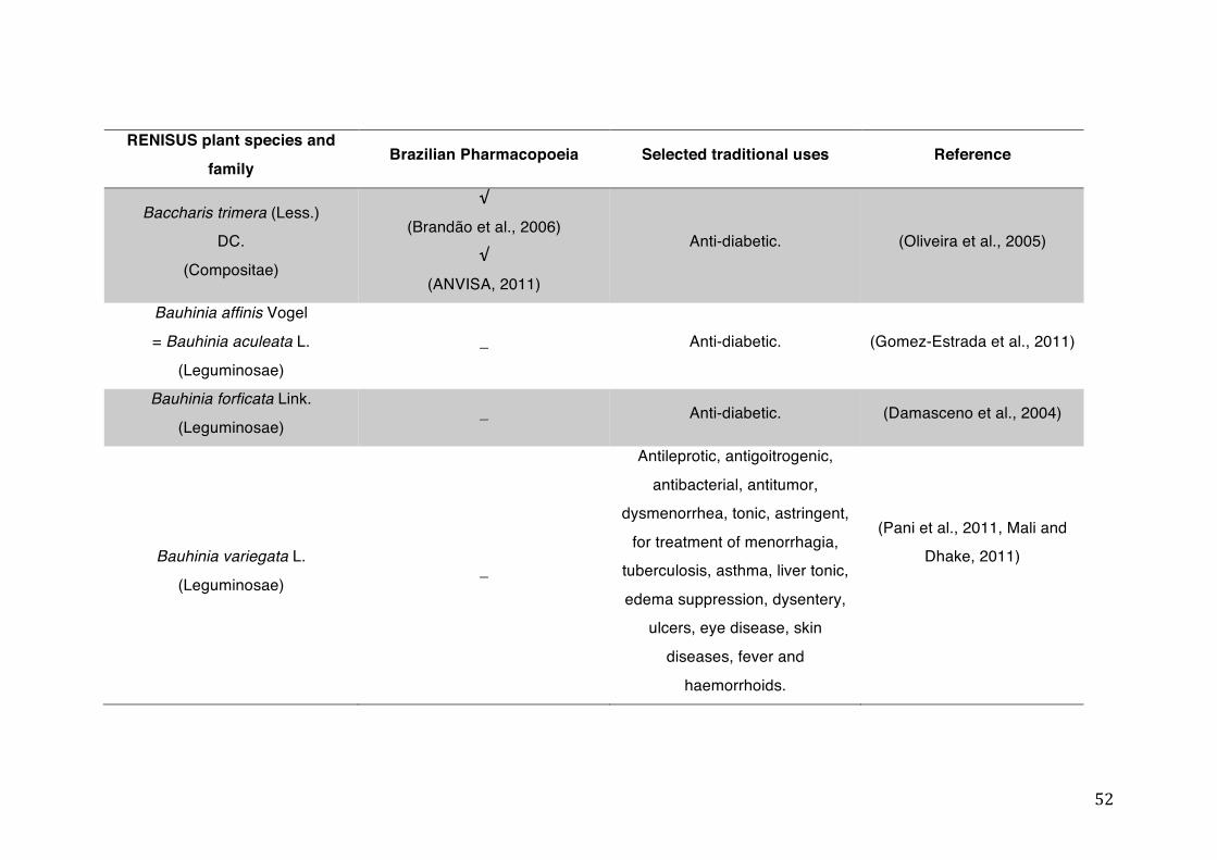
52
RENISUS plant species and
family Brazilian Pharmacopoeia Selected traditional uses Reference
Baccharis trimera (Less.) DC.
(Compositae)
√
(Brandão et al., 2006) √
(ANVISA, 2011)
Anti-diabetic. (Oliveira et al., 2005)
Bauhinia affinis Vogel
= Bauhinia aculeata L.
(Leguminosae)
_ Anti-diabetic. (Gomez-Estrada et al., 2011)
Bauhinia forficata Link.
(Leguminosae) _ Anti-diabetic. (Damasceno et al., 2004)
Bauhinia variegata L.
(Leguminosae) _
Antileprotic, antigoitrogenic,
antibacterial, antitumor, dysmenorrhea, tonic, astringent,
for treatment of menorrhagia, tuberculosis, asthma, liver tonic,
edema suppression, dysentery, ulcers, eye disease, skin
diseases, fever and
haemorrhoids.
(Pani et al., 2011, Mali and Dhake, 2011)
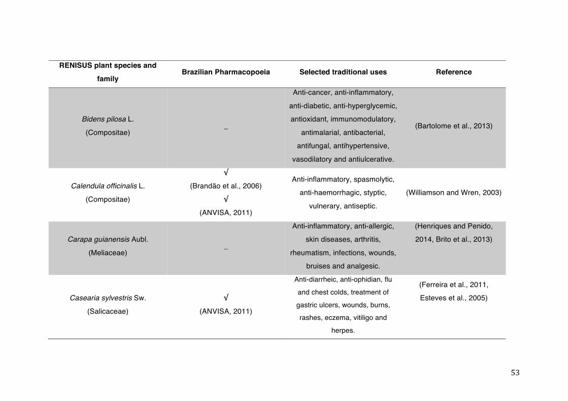
53
RENISUS plant species and
family Brazilian Pharmacopoeia Selected traditional uses Reference
Bidens pilosa L.
(Compositae) _
Anti-cancer, anti-inflammatory,
anti-diabetic, anti-hyperglycemic, antioxidant, immunomodulatory,
antimalarial, antibacterial, antifungal, antihypertensive,
vasodilatory and antiulcerative.
(Bartolome et al., 2013)
Calendula officinalis L.
(Compositae)
√
(Brandão et al., 2006)
√
(ANVISA, 2011)
Anti-inflammatory, spasmolytic, anti-haemorrhagic, styptic,
vulnerary, antiseptic.
(Williamson and Wren, 2003)
Carapa guianensis Aubl.
(Meliaceae) _
Anti-inflammatory, anti-allergic, skin diseases, arthritis,
rheumatism, infections, wounds, bruises and analgesic.
(Henriques and Penido, 2014, Brito et al., 2013)
Casearia sylvestris Sw.
(Salicaceae)
√
(ANVISA, 2011)
Anti-diarrheic, anti-ophidian, flu and chest colds, treatment of gastric ulcers, wounds, burns, rashes, eczema, vitiligo and
herpes.
(Ferreira et al., 2011, Esteves et al., 2005)
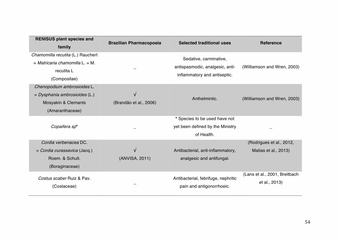
54
RENISUS plant species and
family Brazilian Pharmacopoeia Selected traditional uses Reference
Chamomilla recutita (L.) Rauchert
= Matricaria chamomilla L. = M. recutita L
(Compositae)
_
Sedative, carminative,
antispasmodic, analgesic, anti-
inflammatory and antiseptic.
(Williamson and Wren, 2003)
Chenopodium ambrosioides L.
= Dysphania ambrosioides (L.) Mosyakin & Clemants
(Amaranthaceae)
√
(Brandão et al., 2006) Anthelmintic. (Williamson and Wren, 2003)
Copaifera sp* _ * Species to be used have not
yet been defined by the Ministry
of Health.
_
Cordia verbenacea DC.
= Cordia curassavica (Jacq.) Roem. & Schult.
(Boraginaceae)
√
(ANVISA, 2011) Antibacterial, anti-inflammatory,
analgesic and antifungal.
(Rodrigues et al., 2012,
Matias et al., 2013)
Costus scaber Ruiz & Pav. (Costaceae)
_ Antibacterial, febrifuge, nephritic
pain and antigonorrhoeic.
(Lans et al., 2001, Breitbach
et al., 2013)
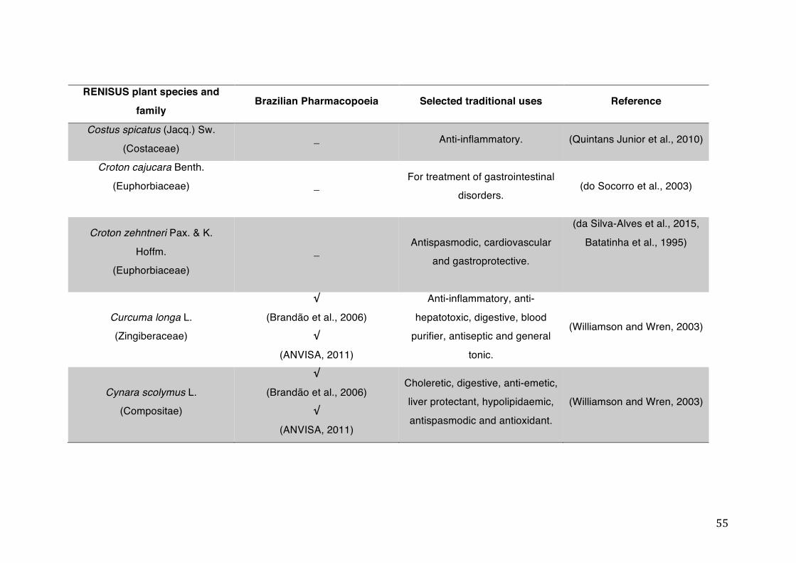
55
RENISUS plant species and
family Brazilian Pharmacopoeia Selected traditional uses Reference
Costus spicatus (Jacq.) Sw.
(Costaceae) _ Anti-inflammatory. (Quintans Junior et al., 2010)
Croton cajucara Benth.
(Euphorbiaceae)
_ For treatment of gastrointestinal
disorders. (do Socorro et al., 2003)
Croton zehntneri Pax. & K.
Hoffm. (Euphorbiaceae)
_ Antispasmodic, cardiovascular
and gastroprotective.
(da Silva-Alves et al., 2015,
Batatinha et al., 1995)
Curcuma longa L. (Zingiberaceae)
√
(Brandão et al., 2006) √
(ANVISA, 2011)
Anti-inflammatory, anti-
hepatotoxic, digestive, blood purifier, antiseptic and general
tonic.
(Williamson and Wren, 2003)
Cynara scolymus L. (Compositae)
√
(Brandão et al., 2006) √
(ANVISA, 2011)
Choleretic, digestive, anti-emetic, liver protectant, hypolipidaemic,
antispasmodic and antioxidant.
(Williamson and Wren, 2003)
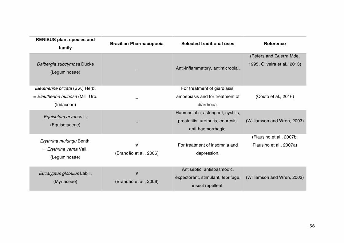
56
RENISUS plant species and
family Brazilian Pharmacopoeia Selected traditional uses Reference
Dalbergia subcymosa Ducke (Leguminosae)
_ Anti-inflammatory, antimicrobial.
(Peters and Guerra Mde,
1995, Oliveira et al., 2013)
Eleutherine plicata (Sw.) Herb.
= Eleutherine bulbosa (Mill. Urb.
(Iridaceae)
_
For treatment of giardiasis,
amoebiasis and for treatment of
diarrhoea.
(Couto et al., 2016)
Equisetum arvense L.
(Equisetaceae) _
Haemostatic, astringent, cystitis,
prostatitis, urethritis, enuresis, anti-haemorrhagic.
(Williamson and Wren, 2003)
Erythrina mulungu Benth.
= Erythrina verna Vell. (Leguminosae)
√
(Brandão et al., 2006)
For treatment of insomnia and
depression.
(Flausino et al., 2007b, Flausino et al., 2007a)
Eucalyptus globulus Labill.
(Myrtaceae)
√
(Brandão et al., 2006)
Antiseptic, antispasmodic, expectorant, stimulant, febrifuge,
insect repellent.
(Williamson and Wren, 2003)
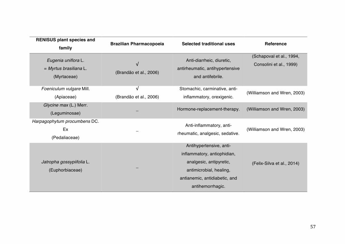
57
RENISUS plant species and
family Brazilian Pharmacopoeia Selected traditional uses Reference
Eugenia uniflora L. = Myrtus brasiliana L.
(Myrtaceae)
√
(Brandão et al., 2006)
Anti-diarrheic, diuretic, antirheumatic, antihypertensive
and antifebrile.
(Schapoval et al., 1994,
Consolini et al., 1999)
Foeniculum vulgare Mill.
(Apiaceae)
√
(Brandão et al., 2006)
Stomachic, carminative, anti-
inflammatory, orexigenic. (Williamson and Wren, 2003)
Glycine max (L.) Merr. (Leguminosae)
_ Hormone-replacement-therapy. (Williamson and Wren, 2003)
Harpagophytum procumbens DC. Ex
(Pedaliaceae)
_ Anti-inflammatory, anti-
rheumatic, analgesic, sedative. (Williamson and Wren, 2003)
Jatropha gossypiifolia L.
(Euphorbiaceae) _
Antihypertensive, anti-
inflammatory, antiophidian, analgesic, antipyretic,
antimicrobial, healing, antianemic, antidiabetic, and
antihemorrhagic.
(Felix-Silva et al., 2014)
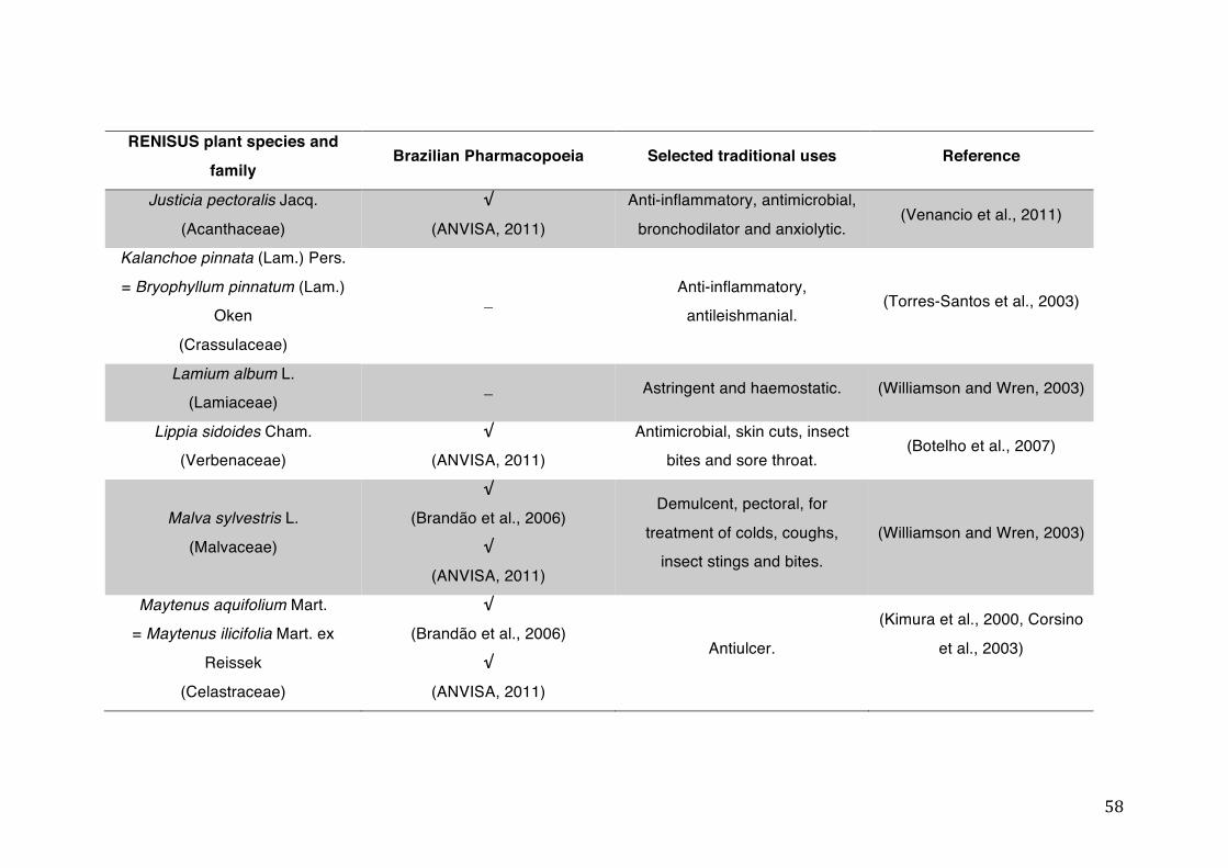
58
RENISUS plant species and
family Brazilian Pharmacopoeia Selected traditional uses Reference
Justicia pectoralis Jacq.
(Acanthaceae)
√
(ANVISA, 2011)
Anti-inflammatory, antimicrobial,
bronchodilator and anxiolytic. (Venancio et al., 2011)
Kalanchoe pinnata (Lam.) Pers.
= Bryophyllum pinnatum (Lam.) Oken
(Crassulaceae)
_ Anti-inflammatory,
antileishmanial. (Torres-Santos et al., 2003)
Lamium album L. (Lamiaceae)
_ Astringent and haemostatic. (Williamson and Wren, 2003)
Lippia sidoides Cham. (Verbenaceae)
√
(ANVISA, 2011) Antimicrobial, skin cuts, insect
bites and sore throat. (Botelho et al., 2007)
Malva sylvestris L.
(Malvaceae)
√
(Brandão et al., 2006)
√
(ANVISA, 2011)
Demulcent, pectoral, for
treatment of colds, coughs, insect stings and bites.
(Williamson and Wren, 2003)
Maytenus aquifolium Mart. = Maytenus ilicifolia Mart. ex
Reissek
(Celastraceae)
√
(Brandão et al., 2006)
√
(ANVISA, 2011)
Antiulcer.
(Kimura et al., 2000, Corsino
et al., 2003)
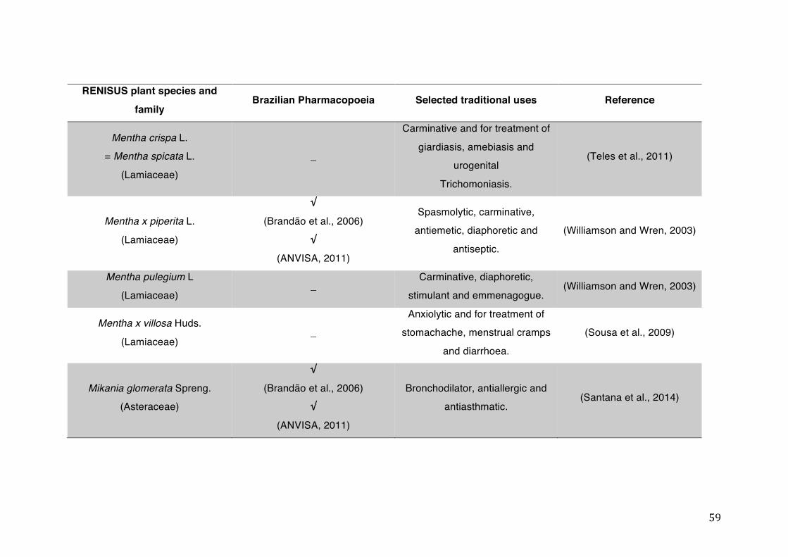
59
RENISUS plant species and
family Brazilian Pharmacopoeia Selected traditional uses Reference
Mentha crispa L. = Mentha spicata L.
(Lamiaceae)
_
Carminative and for treatment of
giardiasis, amebiasis and urogenital
Trichomoniasis.
(Teles et al., 2011)
Mentha x piperita L.
(Lamiaceae)
√
(Brandão et al., 2006)
√
(ANVISA, 2011)
Spasmolytic, carminative, antiemetic, diaphoretic and
antiseptic.
(Williamson and Wren, 2003)
Mentha pulegium L (Lamiaceae)
_ Carminative, diaphoretic,
stimulant and emmenagogue. (Williamson and Wren, 2003)
Mentha x villosa Huds.
(Lamiaceae) _
Anxiolytic and for treatment of stomachache, menstrual cramps
and diarrhoea.
(Sousa et al., 2009)
Mikania glomerata Spreng. (Asteraceae)
√
(Brandão et al., 2006) √
(ANVISA, 2011)
Bronchodilator, antiallergic and antiasthmatic.
(Santana et al., 2014)
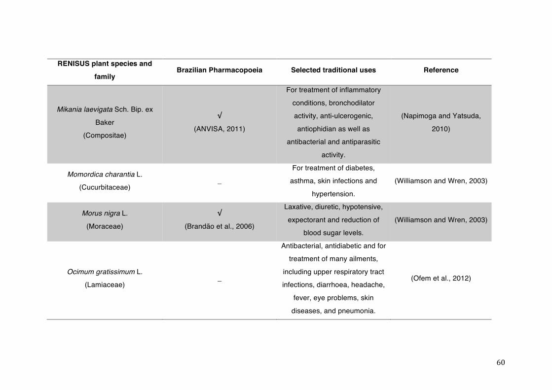
60
RENISUS plant species and
family Brazilian Pharmacopoeia Selected traditional uses Reference
Mikania laevigata Sch. Bip. ex
Baker (Compositae)
√
(ANVISA, 2011)
For treatment of inflammatory
conditions, bronchodilator activity, anti-ulcerogenic,
antiophidian as well as antibacterial and antiparasitic
activity.
(Napimoga and Yatsuda,
2010)
Momordica charantia L. (Cucurbitaceae)
_ For treatment of diabetes,
asthma, skin infections and
hypertension.
(Williamson and Wren, 2003)
Morus nigra L.
(Moraceae)
√
(Brandão et al., 2006)
Laxative, diuretic, hypotensive,
expectorant and reduction of blood sugar levels.
(Williamson and Wren, 2003)
Ocimum gratissimum L. (Lamiaceae)
_
Antibacterial, antidiabetic and for treatment of many ailments,
including upper respiratory tract infections, diarrhoea, headache,
fever, eye problems, skin
diseases, and pneumonia.
(Ofem et al., 2012)
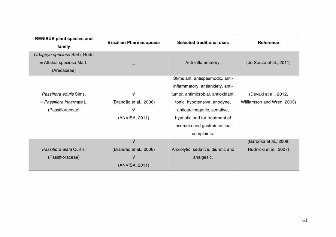
61
RENISUS plant species and
family Brazilian Pharmacopoeia Selected traditional uses Reference
Orbignya speciosa Barb. Rodr.
= Attalea speciosa Mart. (Arecaceae)
_ Anti-inflammatory. (de Souza et al., 2011)
Passiflora edulis Sims.
= Passiflora incarnata L. (Passifloraceae)
√
(Brandão et al., 2006) √
(ANVISA, 2011)
Stimulant, antispasmodic, anti-inflammatory, antianxiety, anti-
tumor, antimicrobial, antioxidant,
tonic, hypotensive, anodyne, anticarcinogenic, sedative,
hypnotic and for treatment of insomnia and gastrointestinal
complaints.
(Devaki et al., 2012,
Williamson and Wren, 2003)
Passiflora alata Curtis. (Passifloraceae)
√
(Brandão et al., 2006) √
(ANVISA, 2011)
Anxiolytic, sedative, diuretic and analgesic.
(Barbosa et al., 2008,
Rudnicki et al., 2007)
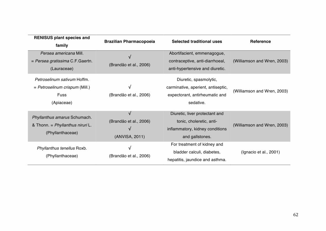
62
RENISUS plant species and
family Brazilian Pharmacopoeia Selected traditional uses Reference
Persea americana Mill.
= Persea gratissima C.F.Gaertn. (Lauraceae)
√
(Brandão et al., 2006)
Abortifacient, emmenagogue,
contraceptive, anti-diarrhoeal, anti-hypertensive and diuretic.
(Williamson and Wren, 2003)
Petroselinum sativum Hoffm.
= Petroselinum crispum (Mill.) Fuss
(Apiaceae)
√
(Brandão et al., 2006)
Diuretic, spasmolytic,
carminative, aperient, antiseptic, expectorant, antirheumatic and
sedative.
(Williamson and Wren, 2003)
Phyllanthus amarus Schumach.
& Thonn. = Phyllanthus niruri L. (Phyllanthaceae)
√
(Brandão et al., 2006) √
(ANVISA, 2011)
Diuretic, liver protectant and
tonic, choleretic, anti-inflammatory, kidney conditions
and gallstones.
(Williamson and Wren, 2003)
Phyllanthus tenellus Roxb. (Phyllanthaceae)
√
(Brandão et al., 2006)
For treatment of kidney and
bladder calculi, diabetes, hepatitis, jaundice and asthma.
(Ignacio et al., 2001)
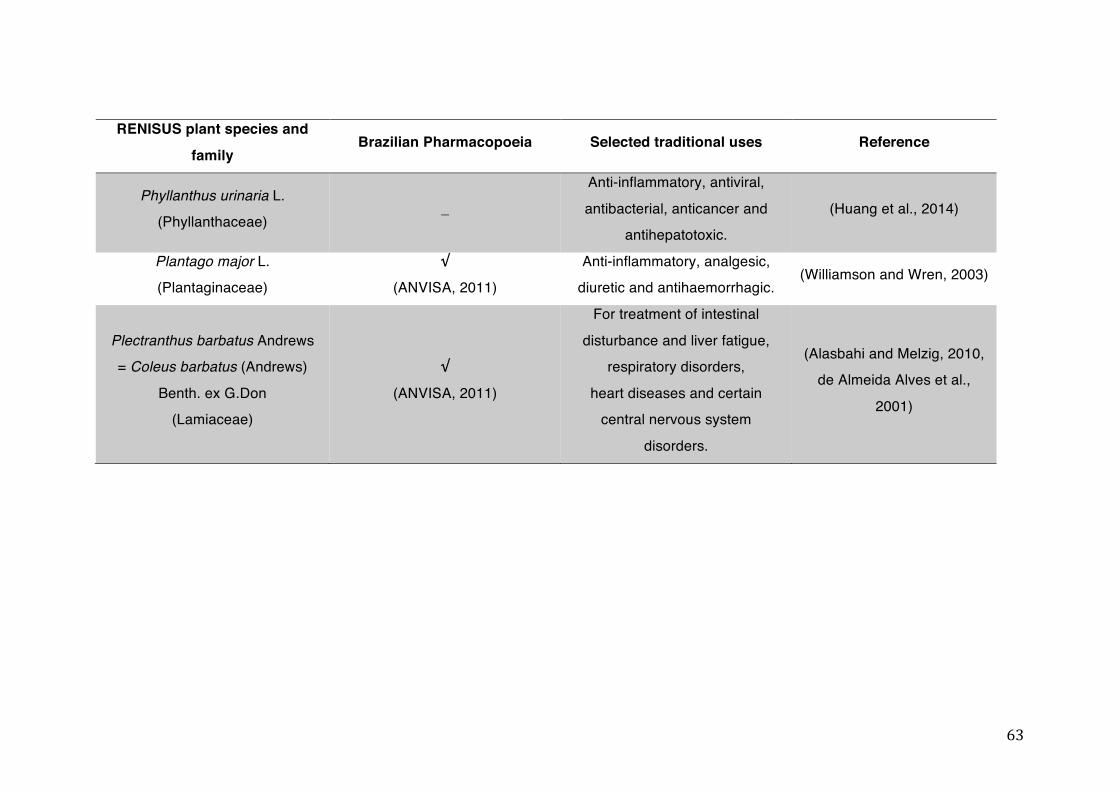
63
RENISUS plant species and
family Brazilian Pharmacopoeia Selected traditional uses Reference
Phyllanthus urinaria L. (Phyllanthaceae)
_
Anti-inflammatory, antiviral,
antibacterial, anticancer and antihepatotoxic.
(Huang et al., 2014)
Plantago major L. (Plantaginaceae)
√
(ANVISA, 2011) Anti-inflammatory, analgesic,
diuretic and antihaemorrhagic. (Williamson and Wren, 2003)
Plectranthus barbatus Andrews = Coleus barbatus (Andrews)
Benth. ex G.Don (Lamiaceae)
√
(ANVISA, 2011)
For treatment of intestinal
disturbance and liver fatigue, respiratory disorders,
heart diseases and certain central nervous system
disorders.
(Alasbahi and Melzig, 2010, de Almeida Alves et al.,
2001)
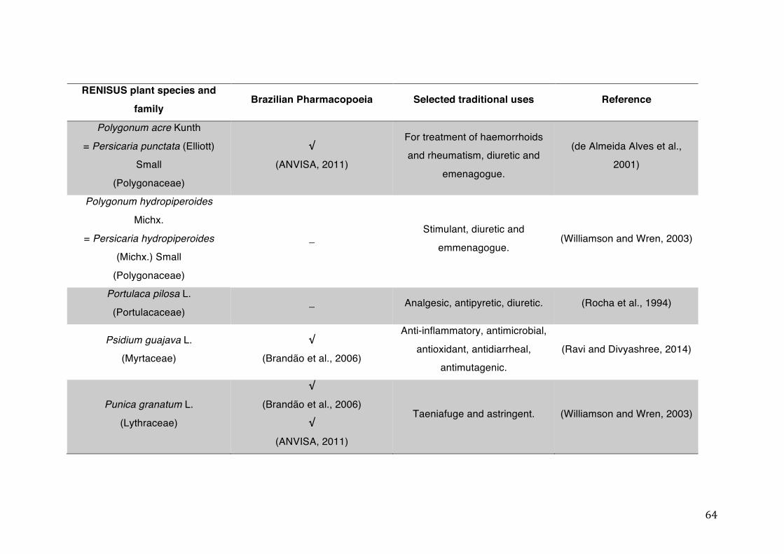
64
RENISUS plant species and
family Brazilian Pharmacopoeia Selected traditional uses Reference
Polygonum acre Kunth
= Persicaria punctata (Elliott) Small
(Polygonaceae)
√
(ANVISA, 2011)
For treatment of haemorrhoids and rheumatism, diuretic and
emenagogue.
(de Almeida Alves et al., 2001)
Polygonum hydropiperoides
Michx.
= Persicaria hydropiperoides (Michx.) Small
(Polygonaceae)
_ Stimulant, diuretic and
emmenagogue. (Williamson and Wren, 2003)
Portulaca pilosa L.
(Portulacaceae) _ Analgesic, antipyretic, diuretic. (Rocha et al., 1994)
Psidium guajava L. (Myrtaceae)
√
(Brandão et al., 2006)
Anti-inflammatory, antimicrobial,
antioxidant, antidiarrheal, antimutagenic.
(Ravi and Divyashree, 2014)
Punica granatum L.
(Lythraceae)
√
(Brandão et al., 2006)
√
(ANVISA, 2011)
Taeniafuge and astringent. (Williamson and Wren, 2003)
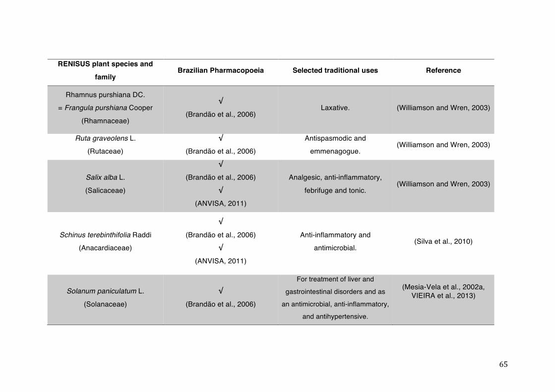
65
RENISUS plant species and
family Brazilian Pharmacopoeia Selected traditional uses Reference
Rhamnus purshiana DC. = Frangula purshiana Cooper
(Rhamnaceae)
√
(Brandão et al., 2006) Laxative. (Williamson and Wren, 2003)
Ruta graveolens L. (Rutaceae)
√
(Brandão et al., 2006) Antispasmodic and
emmenagogue. (Williamson and Wren, 2003)
Salix alba L.
(Salicaceae)
√
(Brandão et al., 2006)
√
(ANVISA, 2011)
Analgesic, anti-inflammatory,
febrifuge and tonic. (Williamson and Wren, 2003)
Schinus terebinthifolia Raddi (Anacardiaceae)
√
(Brandão et al., 2006) √
(ANVISA, 2011)
Anti-inflammatory and antimicrobial.
(Silva et al., 2010)
Solanum paniculatum L.
(Solanaceae)
√
(Brandão et al., 2006)
For treatment of liver and gastrointestinal disorders and as
an antimicrobial, anti-inflammatory, and antihypertensive.
(Mesia-Vela et al., 2002a, VIEIRA et al., 2013)
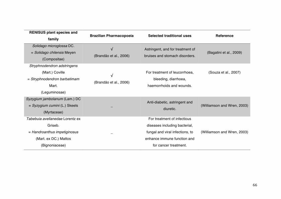
66
RENISUS plant species and
family Brazilian Pharmacopoeia Selected traditional uses Reference
Solidago microglossa DC.
= Solidago chilensis Meyen (Compositae)
√
(Brandão et al., 2006) Astringent, and for treatment of bruises and stomach disorders.
(Bagatini et al., 2009)
Stryphnodendron adstringens (Mart.) Coville
= Stryphnodendron barbatimam
Mart. (Leguminosae)
√
(Brandão et al., 2006)
For treatment of leucorrhoea,
bleeding, diarrhoea,
haemorrhoids and wounds.
(Souza et al., 2007)
Syzygium jambolanum (Lam.) DC = Syzygium cumini (L.) Skeels
(Myrtaceae)
_ Anti-diabetic, astringent and
diuretic. (Williamson and Wren, 2003)
Tabebuia avellanedae Lorentz ex
Griseb. = Handroanthus impetiginosus
(Mart. ex DC.) Mattos (Bignoniaceae)
_
For treatment of infectious
diseases including bacterial, fungal and viral infections, to
enhance immune function and for cancer treatment.
(Williamson and Wren, 2003)
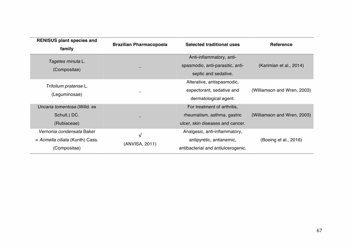
67
RENISUS plant species and
family Brazilian Pharmacopoeia Selected traditional uses Reference
Tagetes minuta L. (Compositae)
_
Anti-inflammatory, anti-
spasmodic, anti-parasitic, anti-septic and sedative.
(Karimian et al., 2014)
Trifolium pratense L.
(Leguminosae) _
Alterative, antispasmodic, expectorant, sedative and
dermatological agent.
(Williamson and Wren, 2003)
Uncaria tomentosa (Willd. ex Schult.) DC.
(Rubiaceae)
_ For treatment of arthritis,
rheumatism, asthma, gastric
ulcer, skin diseases and cancer.
(Williamson and Wren, 2003)
Vernonia condensata Baker
= Acmella ciliata (Kunth) Cass. (Compositae)
√
(ANVISA, 2011)
Analgesic, anti-inflammatory,
antipyretic, antianemic, antibacterial and antiulcerogenic.
(Boeing et al., 2016)
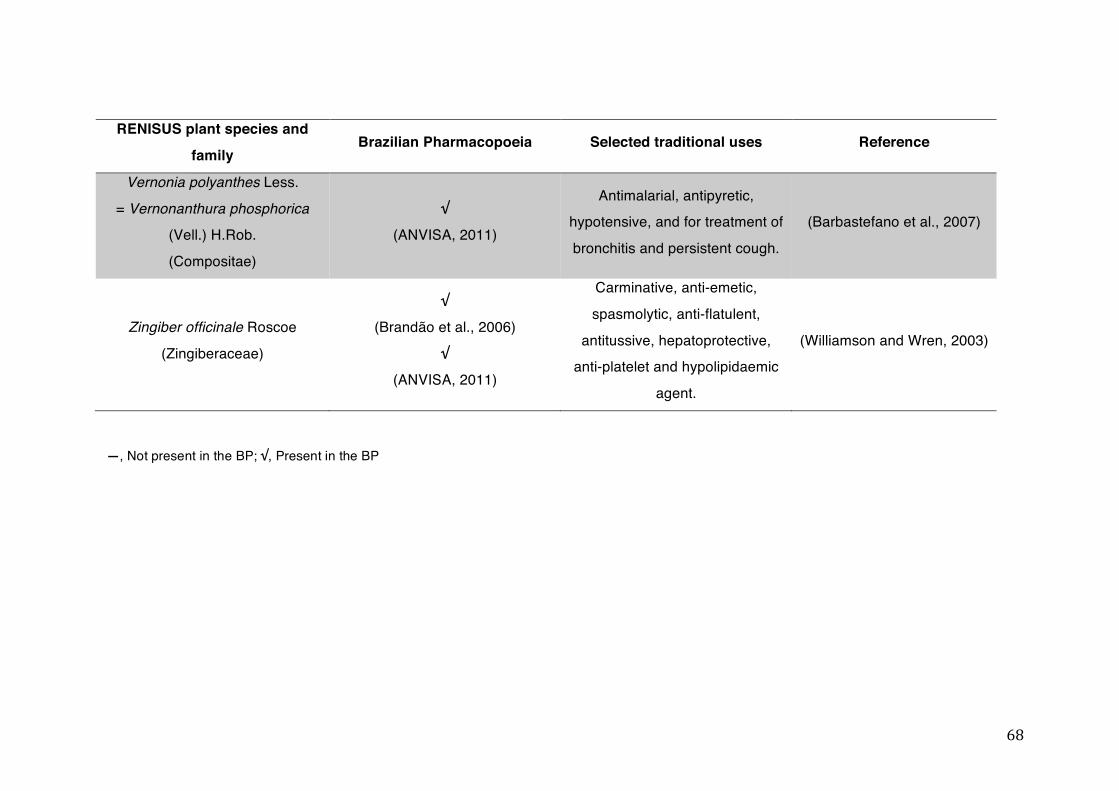
68
RENISUS plant species and
family Brazilian Pharmacopoeia Selected traditional uses Reference
Vernonia polyanthes Less.
= Vernonanthura phosphorica (Vell.) H.Rob.
(Compositae)
√
(ANVISA, 2011)
Antimalarial, antipyretic, hypotensive, and for treatment of
bronchitis and persistent cough.
(Barbastefano et al., 2007)
Zingiber officinale Roscoe
(Zingiberaceae)
√
(Brandão et al., 2006)
√
(ANVISA, 2011)
Carminative, anti-emetic,
spasmolytic, anti-flatulent,
antitussive, hepatoprotective, anti-platelet and hypolipidaemic
agent.
(Williamson and Wren, 2003)
—, Not present in the BP; √, Present in the BP

69
1.9 REPORTED EFFECTS OF RENISUS MEDICINAL PLANTS ON MAIN
PHASE I AND PHASE II METABOLIC MECHANISMS AND THE P-
GLYCOPROTEIN TRANSPORTER.
In this chapter, we showed that herbal medicines are able to modulate metabolic and transporter proteins and, as a consequence, cause HDI at both the absorption and metabolic levels if they are coadministered with synthetic drugs. Also, we highlighted that the number of users of herbal medicines in SUS has considerably increased over the years. Statistics indicate that about 66% of the Brazilian population have no access to commercial medicines (Trojan-Rodrigues et al., 2012). Even when access is granted, popular use of herbal medicines is often due to poor medical and pharmaceutical assistance and the high cost of treatment with conventional drugs (Silveira et al., 2008). Brazilians are also becoming more interested in “safe” and “natural” treatments aimed to promote healthier living. A consequence of this increased use of phytotherapy has been a higher number of case reports of adverse reactions caused by the uncontrolled consumption of herbal medicines (Silveira et al., 2008).
The ANVISA is in charge of the regulation of herbal medicines in the country and since its creation in 1999, many advances have been made in order to control and ensure their efficacy and safety (Balbino and Dias, 2010). In order to guarantee the safety of the consumption of phytomedicinal candidates by humans, ANVISA established a requirement that at least 20 years of prior traditional use must be attested. In the absence of this evidence the efficacy and safety of the candidate need to be demonstrated by a point based system according to literature data, clinical evidence or an indication that the herb is already included in the list of simplified registration of herbal medicines (ANVISA, 2010). Although these regulations are relevant to improve the safety and efficacy of herbal medicines, PK metabolic studies on the traditional medicinal plants are not yet a regulatory requirement. As a
70
consequence, there is a scarcity of this kind of data which is regarded as an oversight in terms of safety (Ribeiro et al., 2005).
As previously stated, the SUS is one of the biggest public health systems in the world and is responsible for the health care of 180 million Brazilian citizens (Mendes, 2013). In order to establish which medicines should be provided by SUS, the Brazilian Health Ministry produced a list of essential drugs (RENAME) according to the International Classification of Diseases (ICD) and epidemiological studies conducted nationwide (Brasil, 2014). Several herbal medicines are already part of this list, such as Cynara scolymus L., Schinus
terebinthifolia Raddi and Rhamnus purshiana DC. However, due to the extensive number of plant species possessing pharmacological activity used in Brazil and the necessity for increased availability of herbal medicines in SUS, the Health Ministry determined that more herbal medicines should be provided by the System to the population, as part of one of the aims of the National Policy of Integrative and Complementary Practices (PNPIC). Consequently, RENISUS was published in 2008 and, from this point on, efforts have been concentrated on elucidating the efficacy and safety of the plant species present on the list (Brasil, 2009, Feijó et al., 2012).
The profile of pharmaceutical drugs at both the absorption and metabolic levels is essential to determine whether or not they will interact with other therapeutic interventions (Ionescu and Caira, 2005). However, studies on metabolic and transporter targets using plant extracts are very difficult to carry out because of their chemical complexity (Simões and Mariot, 2003). As a result, there is very little data for numerous native and exotic adapted medicinal plants that are traditionally used in Brazil (He et al., 2010). The in vitro profiles of some herbal medicines in metabolic and transporter mechanisms can be found in the literature and, although these data are important, they are seldom used for the prediction of potential HDI. Thus, we will provide an overview of the effects of medicinal plants to be used in the SUS in the main phase I and phase II metabolism targets and the P-gp transporter.
A literature search was conducted using the Library of Medicine’s PubMed database between May and June 2013. Included studies consisted of
71
the reported effects of the Brazilian medicinal plants of RENISUS list on the main liver metabolic enzymes, which are involved in phase I (functionalization reactions mediated by CYP) and phase II (GSH conjugation, glucuronidation, sulphation, methylation and acetylation) metabolism and also the assessment of their effects on P-gp activity. The following combinations of keywords were used: ‘Plant name AND, 1A2, 3A4, 3A5, 3A7, 2C9, 2C19, 2D6, 2E1’, ‘Plant name AND glutathione’ or ‘Plant name AND GSH’, ‘Plant name AND glucuronidation’ or ‘Plant name AND UGT’, ‘Plant name AND sulphation’, ‘Plant name AND sulphate conjugation’, ‘Plant name AND sulphotransferase’, ‘Plant name AND methylation’, ‘Plant name AND methyltransferase’, ‘Plant name AND acetylation’, ‘Plant name AND n-acetyltransferase’, ‘Plant name AND P-glycoprotein’ or ‘Plant name AND P-gp” (Tables 1-3 to 1-11). EndNote web was the citation tool used to manage and organize all the references collected.
It is important to note that RENISUS encompasses native and exotic adapted medicinal plant species and that we are aware of the chemical variability of the materials, which eventually may be harmonised by pharmacopoeial monographs of their respective countries.
Also, same plant species can present different results in the literature. This could be explained by the using plants collected in different regions, places and time of the year for example. Also, observed effects may vary due to the extraction procedure used for the conduction of the experiment, as well as the models selected for the study.
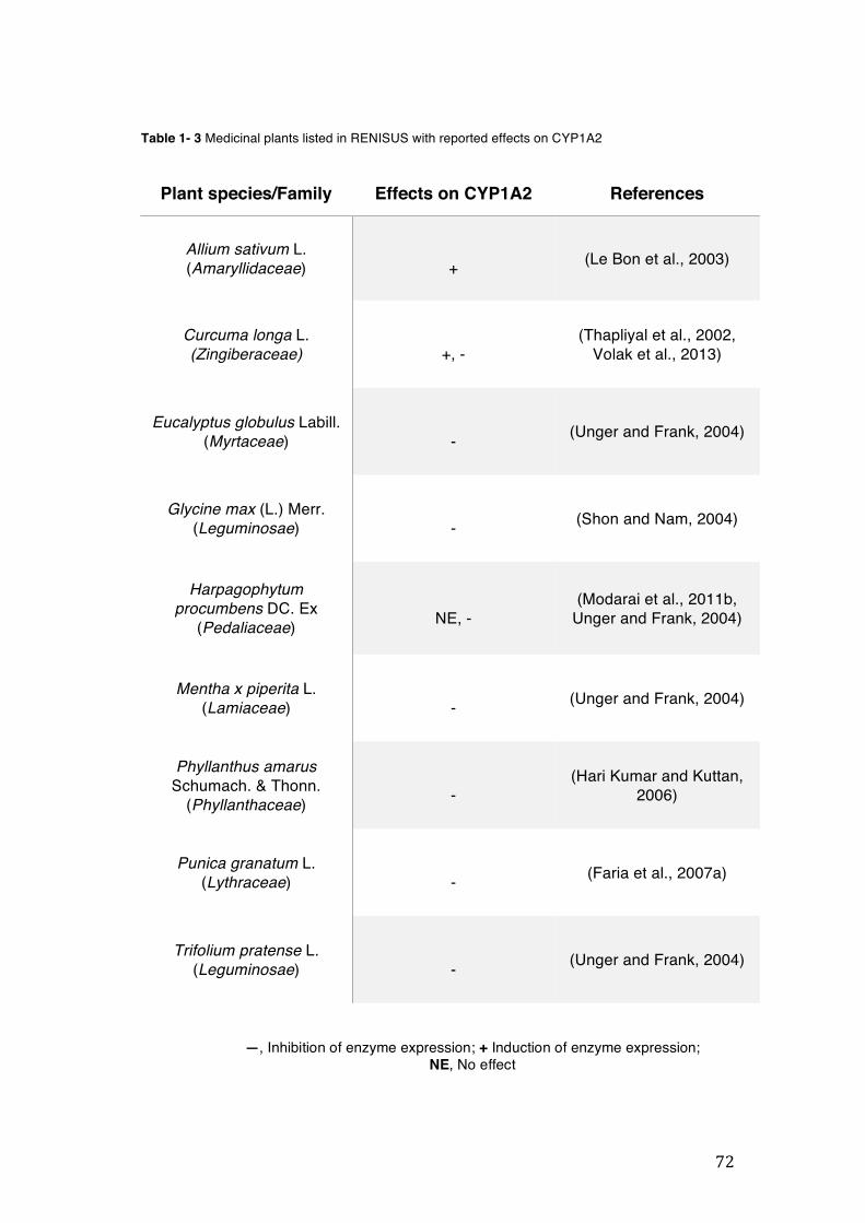
72
Table 1- 3 Medicinal plants listed in RENISUS with reported effects on CYP1A2
Plant species/Family Effects on CYP1A2 References
Allium sativum L. (Amaryllidaceae)
+ (Le Bon et al., 2003)
Curcuma longa L. (Zingiberaceae)
+, -
(Thapliyal et al., 2002, Volak et al., 2013)
Eucalyptus globulus Labill. (Myrtaceae)
- (Unger and Frank, 2004)
Glycine max (L.) Merr. (Leguminosae)
- (Shon and Nam, 2004)
Harpagophytum procumbens DC. Ex
(Pedaliaceae)
NE, - (Modarai et al., 2011b,
Unger and Frank, 2004)
Mentha x piperita L. (Lamiaceae)
- (Unger and Frank, 2004)
Phyllanthus amarus Schumach. & Thonn.
(Phyllanthaceae)
-
(Hari Kumar and Kuttan, 2006)
Punica granatum L. (Lythraceae)
- (Faria et al., 2007a)
Trifolium pratense L. (Leguminosae)
- (Unger and Frank, 2004)
—, Inhibition of enzyme expression; + Induction of enzyme expression; NE, No effect
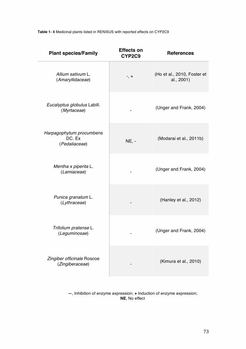
73
Table 1- 4 Medicinal plants listed in RENISUS with reported effects on CYP2C9
Plant species/Family Effects on CYP2C9 References
Allium sativum L. (Amaryllidaceae)
-, + (Ho et al., 2010, Foster et
al., 2001)
Eucalyptus globulus Labill. (Myrtaceae)
- (Unger and Frank, 2004)
Harpagophytum procumbens DC. Ex
(Pedaliaceae)
NE, - (Modarai et al., 2011b)
Mentha x piperita L. (Lamiaceae)
- (Unger and Frank, 2004)
Punica granatum L. (Lythraceae)
- (Hanley et al., 2012)
Trifolium pratense L. (Leguminosae)
- (Unger and Frank, 2004)
Zingiber officinale Roscoe (Zingiberaceae)
- (Kimura et al., 2010)
—, Inhibition of enzyme expression; + Induction of enzyme expression; NE, No effect
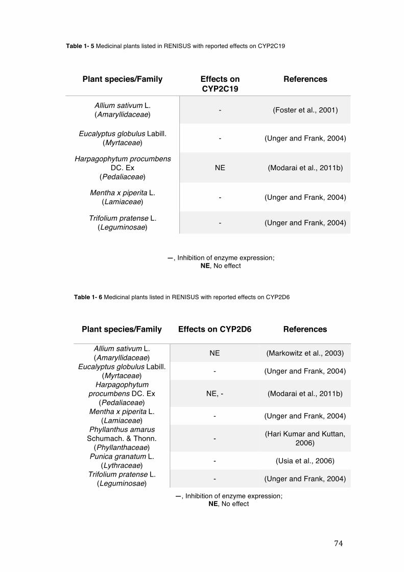
74
Table 1- 5 Medicinal plants listed in RENISUS with reported effects on CYP2C19
Plant species/Family Effects on CYP2C19
References
Allium sativum L. (Amaryllidaceae) - (Foster et al., 2001)
Eucalyptus globulus Labill. (Myrtaceae) - (Unger and Frank, 2004)
Harpagophytum procumbens DC. Ex
(Pedaliaceae) NE (Modarai et al., 2011b)
Mentha x piperita L. (Lamiaceae) - (Unger and Frank, 2004)
Trifolium pratense L. (Leguminosae) - (Unger and Frank, 2004)
Plant species/Family
Effects on CYP2D6 References
Allium sativum L. (Amaryllidaceae) NE (Markowitz et al., 2003)
Eucalyptus globulus Labill. (Myrtaceae) - (Unger and Frank, 2004)
Harpagophytum procumbens DC. Ex
(Pedaliaceae) NE, - (Modarai et al., 2011b)
Mentha x piperita L. (Lamiaceae) - (Unger and Frank, 2004)
Phyllanthus amarus Schumach. & Thonn.
(Phyllanthaceae) - (Hari Kumar and Kuttan,
2006)
Punica granatum L. (Lythraceae) - (Usia et al., 2006)
Trifolium pratense L. (Leguminosae) - (Unger and Frank, 2004)
Table 1- 6 Medicinal plants listed in RENISUS with reported effects on CYP2D6
—, Inhibition of enzyme expression; NE, No effect
—, Inhibition of enzyme expression; NE, No effect
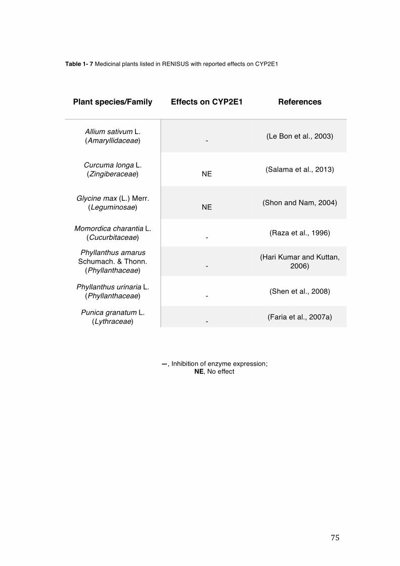
75
—, Inhibition of enzyme expression; NE, No effect
Plant species/Family Effects on CYP2E1 References
Allium sativum L. (Amaryllidaceae)
- (Le Bon et al., 2003)
Curcuma longa L. (Zingiberaceae)
NE (Salama et al., 2013)
Glycine max (L.) Merr. (Leguminosae)
NE (Shon and Nam, 2004)
Momordica charantia L. (Cucurbitaceae)
- (Raza et al., 1996)
Phyllanthus amarus Schumach. & Thonn.
(Phyllanthaceae) -
(Hari Kumar and Kuttan, 2006)
Phyllanthus urinaria L. (Phyllanthaceae)
- (Shen et al., 2008)
Punica granatum L. (Lythraceae)
- (Faria et al., 2007a)
Table 1- 7 Medicinal plants listed in RENISUS with reported effects on CYP2E1
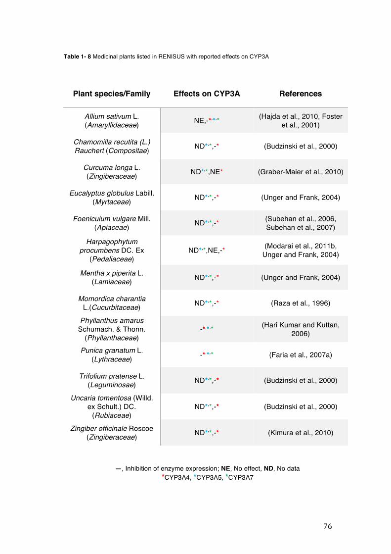
76
—, Inhibition of enzyme expression; NE, No effect, ND, No data *CYP3A4, *CYP3A5, *CYP3A7
Table 1- 8 Medicinal plants listed in RENISUS with reported effects on CYP3A
Plant species/Family
Effects on CYP3A References
Allium sativum L. (Amaryllidaceae) NE,-*,*,* (Hajda et al., 2010, Foster
et al., 2001)
Chamomilla recutita (L.) Rauchert (Compositae) ND*,*,-* (Budzinski et al., 2000)
Curcuma longa L. (Zingiberaceae) ND*,*,NE* (Graber-Maier et al., 2010)
Eucalyptus globulus Labill. (Myrtaceae) ND*,*,-* (Unger and Frank, 2004)
Foeniculum vulgare Mill. (Apiaceae) ND*,*,-* (Subehan et al., 2006,
Subehan et al., 2007)
Harpagophytum procumbens DC. Ex
(Pedaliaceae) ND*,*,NE,-* (Modarai et al., 2011b,
Unger and Frank, 2004)
Mentha x piperita L. (Lamiaceae) ND*,*,-* (Unger and Frank, 2004)
Momordica charantia L.(Cucurbitaceae) ND*,*,-* (Raza et al., 1996)
Phyllanthus amarus Schumach. & Thonn.
(Phyllanthaceae) -*,*,* (Hari Kumar and Kuttan,
2006)
Punica granatum L. (Lythraceae) -*,*,* (Faria et al., 2007a)
Trifolium pratense L. (Leguminosae) ND*,*,-* (Budzinski et al., 2000)
Uncaria tomentosa (Willd. ex Schult.) DC.
(Rubiaceae) ND*,*,-* (Budzinski et al., 2000)
Zingiber officinale Roscoe (Zingiberaceae) ND*,*,-* (Kimura et al., 2010)
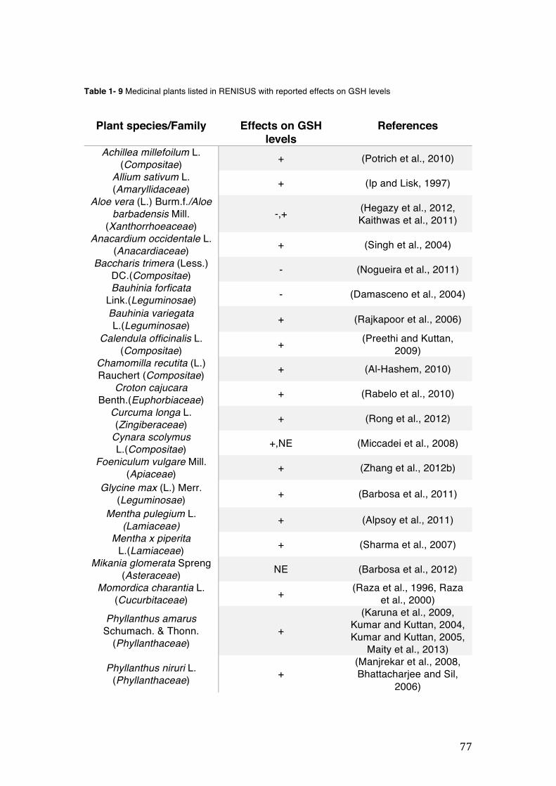
77
Table 1- 9 Medicinal plants listed in RENISUS with reported effects on GSH levels
Plant species/Family
Effects on GSH
levels References
Achillea millefoilum L. (Compositae) + (Potrich et al., 2010)
Allium sativum L. (Amaryllidaceae) + (Ip and Lisk, 1997)
Aloe vera (L.) Burm.f./Aloe barbadensis Mill.
(Xanthorrhoeaceae) -,+ (Hegazy et al., 2012,
Kaithwas et al., 2011) Anacardium occidentale L.
(Anacardiaceae) + (Singh et al., 2004) Baccharis trimera (Less.)
DC.(Compositae) - (Nogueira et al., 2011) Bauhinia forficata
Link.(Leguminosae) - (Damasceno et al., 2004)
Bauhinia variegata L.(Leguminosae) + (Rajkapoor et al., 2006)
Calendula officinalis L. (Compositae) + (Preethi and Kuttan,
2009) Chamomilla recutita (L.) Rauchert (Compositae) + (Al-Hashem, 2010)
Croton cajucara Benth.(Euphorbiaceae) + (Rabelo et al., 2010)
Curcuma longa L. (Zingiberaceae) + (Rong et al., 2012)
Cynara scolymus L.(Compositae) +,NE (Miccadei et al., 2008)
Foeniculum vulgare Mill. (Apiaceae) + (Zhang et al., 2012b)
Glycine max (L.) Merr. (Leguminosae) + (Barbosa et al., 2011)
Mentha pulegium L. (Lamiaceae) + (Alpsoy et al., 2011)
Mentha x piperita L.(Lamiaceae) + (Sharma et al., 2007)
Mikania glomerata Spreng (Asteraceae) NE (Barbosa et al., 2012)
Momordica charantia L. (Cucurbitaceae) + (Raza et al., 1996, Raza
et al., 2000)
Phyllanthus amarus Schumach. & Thonn.
(Phyllanthaceae) +
(Karuna et al., 2009, Kumar and Kuttan, 2004, Kumar and Kuttan, 2005,
Maity et al., 2013)
Phyllanthus niruri L. (Phyllanthaceae) +
(Manjrekar et al., 2008, Bhattacharjee and Sil,
2006)
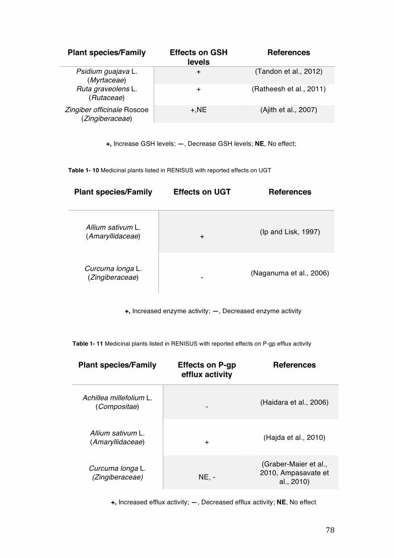
78
+, Increase GSH levels; —, Decrease GSH levels; NE, No effect;
+, Increased enzyme activity; —, Decreased enzyme activity
+, Increased efflux activity; —, Decreased efflux activity; NE, No effect
Plant species/Family Effects on GSH levels
References
Psidium guajava L. (Myrtaceae)
+ (Tandon et al., 2012)
Ruta graveolens L. (Rutaceae)
+ (Ratheesh et al., 2011)
Zingiber officinale Roscoe (Zingiberaceae)
+,NE (Ajith et al., 2007)
Table 1- 10 Medicinal plants listed in RENISUS with reported effects on UGT
Plant species/Family Effects on UGT References
Allium sativum L. (Amaryllidaceae)
+ (Ip and Lisk, 1997)
Curcuma longa L. (Zingiberaceae)
- (Naganuma et al., 2006)
Plant species/Family Effects on P-gp efflux activity
References
Achillea millefolium L. (Compositae)
- (Haidara et al., 2006)
Allium sativum L. (Amaryllidaceae)
+ (Hajda et al., 2010)
Curcuma longa L. (Zingiberaceae)
NE, -
(Graber-Maier et al., 2010, Ampasavate et
al., 2010)
Table 1- 11 Medicinal plants listed in RENISUS with reported effects on P-gp efflux activity
79
1.10 AIM OF THE THESIS
As highlighted earlier the SUS aims to include more herbal medicines to RENAME within the coming years. However, the intake of those herbal medicines by the patients of SUS could potentially cause HDI if they are coadministered with essential medicines. Due to the types of effects that herbal medicines could cause in phase I and phase II metabolising enzymes and P-gp, the present study aims to answer whether selected RENISUS herbal medicines are able to modulate designated metabolic and transporter targets in vitro in order to forestall HDI with some essential medicines available in SUS.
To achieve this, we carried out an experimental evaluation of the in vitro effects of twenty-four plant extracts from twenty-three RENISUS medicinal plant species selected based on data collected in our literature analysis on different cell detoxification and transport mechanisms; as a means of identifying potential HDI. Parts of this experimental evaluation have been previously published as an original research article (Mazzari et al., 2016).

80
2 MATERIALS AND METHODS
81
2.1 CHEMICALS AND REAGENTS
Cell Culture. HepG2 and Caco-2 cell lines were from Sigma Aldrich at passages 100 and 43 respectively. HeLa cells were donated by Dr. Paul Webb from the Houston Methodist Institute for Technology at passage 20. All cell culture reagents were from Gibco® Invitrogen unless otherwise stated. Vincristine 2mg/mL was purchased from Hospira Ltd. HPTLC analysis. Water, dichloromethane (>99.8%, contains amylene as a stabilizer), ethyl acetate (>99.7%) and methanol (>99.9%) all ChromasolvPlus for HPLC provided by Sigma Aldrich. Acetic acid (glacial) analytical reagent grade provided by Fisher Scientific. Formic Acid 98% provided by Rectapur® VWR. Caffeic acid was from Kotch-light Laboratories LTD. Rutin hydrate, quercetin dehydrate and gallic acid all from Sigma Aldrich. Diphenylborinic acid 2-aminoethyl ester (98%) and coumarin laser grade (99% UV-Vis) were provided by ACROS organics. Luteolin (HPLC grade) was from Extrasynthése. Chlorogenic acid provided by Cayman Chemical Co. Polyethylene glycol 4000 grade was from Fisher Scientific. HPLC analysis. All solvents (water HPLC gradient grade, glacial acetic acid and methanol) were purchased from Fisher Scientific UK. Caffeic acid, catechin, rutin, quercetin, and chlorogenic acid were from Sigma Aldrich. Cell viability assays. Sulphorhodamine B (SRB), neutral red (NR), (3-[4,5-dimethylthiazol-2-yl]-2,5 diphenyl tetrazolium bromide) (MTT), trichloroacetic acid (TCA), glacial acetic acid, 96% ethanol, hydrochloric acid (HCl), Isopropanol and Tris base were from Sigma Aldrich. CYP3A4 mRNA gene expression assay. Oligonucleotide primers were custom-synthesized by Invitrogen Life Technologies and Sigma Aldrich. TRIzol® (Total RNA Isolation Reagent), Oligo (dT) 12-18 primers, M-MLV Reverse Transcriptase, RNAseOUT, DNAse I Amplification Grade and 100mM dNTP set were purchased from Invitrogen Life Technologies. SYBR Green (2xqPCR Master Mix premixed with SYBRgreen) was obtained from Amethyst reagents (Cambridge Bioscience). Rifampicin (RIF) and DMSO were purchased from Sigma Aldrich.
82
hPXR assay. Lipofectamine® 2000 was purchased from Invitrogen. RIF and Luciferase assay system were obtained from Sigma Aldrich and Promega respectively. Intracellular Glutathione assay. Buthionine sulfoximine (BSO), L-glutathione reduced, glutathione reductase (GR), 5-5’-dithiobis(2-nitrobenzoic acid) (DTNB), β-Nicotinamide adenine dinucleotide 2’-phosphate reduced tetrasodium salt hydrate (β-NADPH), Triton-X and sulfosalicylic acid (SSA) were from Sigma Aldrich. GGT activity assay. g-glutamyl-p-nitroanalide (GpNA), p-nitroaniline, glycyl-glycine (Gly-Gly), Tris base and acivicin were from Sigma Aldrich. Rh-123 uptake assay. Rh-123 was from Sigma Aldrich. Verapamil (Securon IV 2.5mg/mL) was from Abbott Laboratories Ltd.
2.2 PREPARATION OF PLANT MATERIALS
2.2.1 Selection criteria and ethical clearance
The medicinal plants used for our in vitro preclinical studies were selected according to the results found in the literature review (chapter 1). Originally, we planned to work with all plant species where no preclinical published data had been found. However, it was not possible to obtain all the plant species from the main natural products suppliers in Brazil. Therefore we conducted our work with 23 plant species, which were readily available for purchasing and collection with three different suppliers in Brazil: Santosflora Comércio de Ervas (Mairiporã, São Paulo, Brazil), Florien (Piracicaba, São Paulo, Brazil) and “Farmácia Viva Brasília” (Brasília, DF – Brazil), via UnB.
In order to conduct our research, we had to first to deal with a series of regulations to access the Brazilian genetic heritage ‘component’, which is controlled by the Conselho de Gestão do Patrimônio Genético (Genetic Heritage Council) or CGEN. According to CGEN, in order to purchase and/or collect medicinal plants in Brazil for research purposes and to transport them to the UK, we needed to first to establish an institutional agreement between UCL and a Brazilian institution that would be legally responsible for the whole process (Appendix 7.1). We contacted several Brazilian universities exploring
83
a possible collaboration. We eventually received a positive feedback from Dr. Damaris Silveira from the Faculty of Life Sciences of the University of Brasília (UnB) (Brasília – Distrito Federal) who was willing to help us with all the bureaucratic processes.
The second step was to elaborate the project and send a clear plan of it to CGEN and to the Instituto Brasileiro do Meio Ambiente e dos Recursos
Naturais Renováveis (Brazilian Institute of Environment and Renewable Natural Resources) or IBAMA. The plan had details of the type of research to be carried out with the selected plant extracts and included materials, methodologies and a timetable for the execution of the entire project (Appendix 7.2).
The third (and last) step was the production of a material transfer agreement (MTA) document, which was countersigned by the legal representatives of University College London (UCL) and UnB (Appendix 7.3). All documents were prepared by us and sent to Brazil between November 2013 and March 2014.
2.2.2 Processing of plant materials
The plants were grown and processed according to good manufacturing practices established by ANVISA, ensuring validity and quality (Carvalho et al., 2014). All whole plant handling and extraction were performed at UnB. All used parts of the plants were subjected to a 20-minute infusion in order to mimic traditional use (Academia Nacional de Medicina (Madrid Spain), 1954). For infusion, boiled distilled water (300mL) was added to the plant material (20g) with continuous stirring for 15 minutes on a laboratory hot plate magnetic stirrer (IKA WERKE labortechnik electric stirrer). All extracted plant samples were allowed to stand until cold before filtering with a Buchner flask. All the filtered extracts were frozen in round bottom flasks and then freeze dried (Edwards Pirani 501 Savant super modulo freeze drier) to obtain the dried extract. The lyophilised extracts were immediately sent to UCL School of Pharmacy and stored at -18°C throughout the studies.
84
2.3 PHYTOCHEMICAL ANALYSIS
2.3.1 HPTLC analysis
Extracts were diluted to a concentration of 50mg/mL in methanol. Control compounds were made at a concentration of 1mg/mL, also diluted in methanol. A CAMAG Linomat 5 was used to apply 5µL of the samples to TLC silica gel 60 F254 aluminium sheets. The plates were developed using a CAMAG ADC2 automatic developing chamber. The method included 30-second pre-drying, 10 minutes humidity control using magnesium chloride to 48.3% relative humidity and 20 minutes saturation time, using saturation pads all done at 25.2°C. The mobile phase used was ethyl acetate: formic acid: water (82:9:9). During development, the solvent front was allowed to migrate 80mm before a drying time of 5 minutes. For derivatization, we used natural products reagent (NPR) followed by PEG 4000 (Reich and Schibli, 2007). All visualization and analysis were done using CAMAG TLC visualizer both before and after derivatization.
2.3.2 HPLC analysis
Phytochemical analysis of all samples of the freeze-dried plant extracts was carried out using a high-performance liquid chromatography (HPLC) system- Agilent 1200 series equipped with an ultraviolet/visible (UV-Vis) diode array detector to obtain a ‘fingerprint’ of the samples. The conditions used for the analysis have previously been used for the detection of phenolic compounds in plant samples (Giner et al., 1993).
The stationary phase was a Phenomenex® synergi Polar-RP80A column (250mm x 4.6mm, I.D = 4µm) maintained at 40°C and protected with a Nova-Pak® C18 guard column. The mobile phase was a binary system consisting of (A) water-glacial acetic acid (99.8:0.2, v/v) and (B) methanol-glacial acetic acid (99.8:0.2, v/v). The gradient elution program for the mobile phase was 10-20% B (0-5 minutes), 20-50% B (5-65 minutes), 50-80% B (65-75 minutes) and 80-100% B (75-80 minutes) at a flow rate of 1mL/minute. The injected volume of the sample was 10µL and the UV-Vis spectra were recorded
85
in the range 190 to 400nm, while chromatographic peaks were monitored at 254 and 360nm.
The plant extracts were prepared as 25mg/mL solutions in either micro-filtered double distilled water or HPLC-grade methanol. Five plant phenolic compounds: caffeic acid, catechin, rutin, quercetin, and chlorogenic acid were used as standards at concentrations between 0.5 and 1mg/mL in methanol, depending on the intensity of the peak. Samples were filtered through a 0.45µm syringe filter (Millipore Millex® PTFE) before the analysis.
2.4 IN VITRO STUDIES
2.4.1 Mammalian cell culture techniques
2.4.1.1 Human liver hepatocellular carcinoma (HepG2), colorectal carcinoma cells (Caco-2) and cervix adenocarcinoma (HeLa) cells
Human liver hepatocellular carcinoma, or simply HepG2, is an accepted cell line model commonly used for in vitro drug metabolism studies as they keep some specialised functions when compared to human hepatocytes (HH). HepG2 also preserves the activity of several phase I, phase II, and antioxidant enzymes (Vidyashankar et al., 2010). Besides, HepG2 cells have the advantage of ready availability and assurance in reproducibility of experiments (Hewitt and Hewitt, 2004). HepG2 cells were used in our study to evaluate the CYP3A4 mRNA gene expression, intracellular GSH levels, and GGT activity.
Caco-2 cells are widely used for in vitro drug transport studies. The capacity of Caco-2 to form monolayers with morphological and functional similarities to the human intestinal epithelium, meant the cell line received recognition from the Food and Drug Administration (FDA) as a viable model that replicates human intestinal absorption (Chen et al., 2016). Our drug transport study is focused on the P-gp efflux activity, and we therefore used vincristine resistant Caco-2 cells (Caco-2 VCR) model, which displays a more pronounced efflux activity mechanism than normal Caco-2 (Eneroth et al., 2001).
Henrietta Lacks (HeLa) cervix adenocarcinoma cells were discovered in 1951 and became the first human cancer cell line immortalised in tissue culture
86
(Lucey et al., 2009). HeLa is one of the most versatile cancer cell lines and it is widely used in molecular biology, especially due to its easy and efficient transfection with plasmids using Lipofectamine (Nikcevic et al., 2003). Among some uses in molecular biology, transfected HeLa cells have been the choice of researchers for evaluating hPXR-dependent gene expression, including proteins like CYPs (Lemaire et al., 2006). HeLa cells were therefore our choice for studying the effect of four RENISUS plant extracts on hPXR.
2.4.1.2 Resuscitation of frozen cell stocks
Cell lines are usually stored in frozen form (in liquid nitrogen) when not in use. The cells used in this study were in this form when purchased. To resuscitate them, the cells were thawed rapidly at 37°C by placing the vial of cells in a pre-heated water bath. The thawed cells were then diluted slowly into a centrifuge tube containing the appropriate warm growth media to eliminate DMSO, which is toxic to cells. DMSO is used as a cryoprotectant to prevent crystallisation, which will otherwise cause cells to lyse during cryopreservation. The tubes were then centrifuged (Biofuge primo R Heraeus centrifuge) for 5 minutes at 1000rpm to separate the cell pellet from the culture media. Inside the laminar flow cabinet, the media was removed from the centrifuge tube and the cells were properly resuspended in 15mL of pre-warmed complete media. They were then transferred to a culture flask pre-labelled with the name of the cell line, passage number, and date. An inverted microscope (Nikon TMS 10/0.25 lens) was used to check the morphology of the cells before placing in an incubator (Ezuruike, 2015).
2.4.1.3 Subculture/maintenance
HepG2, Caco-2, and HeLa cells were stocked in DMSO and frozen in liquid nitrogen until they were used. When the cells were needed they were taken out of the liquid nitrogen and defrosted in the water bath (37°C) for approximately 4 minutes and then transferred to a flask containing 15mL of pre-warmed media and centrifuged at 1000rpm for 5 minutes at 18°C. The media (supernatant) was removed and the cells were re-suspended with 5mL of the same media and then transferred to a cell culture flask containing 15mL of pre-

87
warmed media and incubated. The cells were checked regularly in order to observe their growing. Cells were incubated in culture flasks (TPP, surface area 75cm2) containing 20mL of HepG2 medium (MEM Alpha + GlutaMax 1x) or Caco-2 and HeLa medium (DMEM Alpha + Glutamax 1x), 1% (v/v) penicillin-streptomycin antibiotic (10,000 units/mL penicillin and 10,000 µg/mL streptomycin) and 10% heat-inactivated foetal bovine serum-EU. Caco-2 cells were also incubated with vincristine (20nM). The media was kept in the fridge (2°C - 8°C) and prior to its use in cells, the media was placed for 30 minutes in the water bath (37°C) (Ezuruike, 2015).
2.4.1.4 Cell passage (propagation)
When the cells reached around 70% of confluence the cell passing procedure was performed. The old media was aspirated from the cell culture flask and the cells were washed with 5mL of phosphate saline buffer (PBS) to remove the remaining media. After removing the PBS, 1.2mL of 0.25% (w/v) trypsin/EDTA was added to the flask, it was then placed in the incubator until the cells detached. Afterwards, the volume of media was added according to the split ratio desired and the new flasks were then placed in the incubator again allowing the cells to grow (Ezuruike, 2015).
2.4.1.5 Cell counting
The confluence of the flask was checked and then the media was aspirated. The cells were then rinsed with 5mL of PBS and 1.2mL of trypsin/EDTA was added to detach the cells. The flask was put in the incubator for approximately 3 minutes. Afterwards, the flask was placed in the laminar flow cabinet and 190µL of trypan blue stain 0.4% was placed in an eppendorf plus 10µL of the cell suspension. The microcentrifuge tube was mixed on a rotamixer for a few seconds and 10µL of the trypan blue cell suspension were added to each side of a Neubauer haemocytometer that was placed in an inverted microscope (Nikon TMS). The cells were then counted and plated out according to the number of cells desired per well (Ezuruike, 2015).
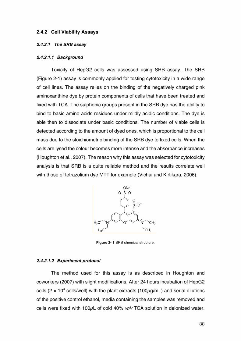
88
2.4.2 Cell Viability Assays
2.4.2.1 The SRB assay
2.4.2.1.1 Background
Toxicity of HepG2 cells was assessed using SRB assay. The SRB (Figure 2-1) assay is commonly applied for testing cytotoxicity in a wide range of cell lines. The assay relies on the binding of the negatively charged pink aminoxanthine dye by protein components of cells that have been treated and fixed with TCA. The sulphonic groups present in the SRB dye has the ability to bind to basic amino acids residues under mildly acidic conditions. The dye is able then to dissociate under basic conditions. The number of viable cells is detected according to the amount of dyed ones, which is proportional to the cell mass due to the stoichiometric binding of the SRB dye to fixed cells. When the cells are lysed the colour becomes more intense and the absorbance increases (Houghton et al., 2007). The reason why this assay was selected for cytotoxicity analysis is that SRB is a quite reliable method and the results correlate well with those of tetrazolium dye MTT for example (Vichai and Kirtikara, 2006).
2.4.2.1.2 Experiment protocol
The method used for this assay is as described in Houghton and coworkers (2007) with slight modifications. After 24 hours incubation of HepG2 cells (2 × 10⁴ cells/well) with the plant extracts (100µg/mL) and serial dilutions of the positive control ethanol, media containing the samples was removed and cells were fixed with 100µL of cold 40% w/v TCA solution in deionized water.
Figure 2- 1 SRB chemical structure.
89
The plates were incubated at 4°C for 1 hour and then immersed five times in tap water. The TCA-fixed cells were stained by adding 100µL of SRB solution (0.4 % SRB in 0.1% glacial acetic acid) and left at room temperature for 1 hour. Afterwards, the plates were quickly rinsed four times with 1% acetic acid and flicked to remove unbound dye and then left to air-dry overnight. After drying completely, the protein bound SRB was solubilised by adding 100µL of Tris base buffer solution (10mM) to each well. The plates were agitated in an orbital shaker for 30 minutes (IKA MS3 basic shaker). The optical density was measured at 492nm by using a microplate plate reader Infinite M200 (Tecan Trading AG, Switzerland) and the viability was calculated as follows: % viability = (O.DS-O.DB/O.DM-O.DB) x 100; where O.DS is the optical density of the sample (either drug or plant extract), O.DB is the optical density of the blank wells without cells, O.DM is the optical density of the wells of the positive control containing only complete media (Houghton et al., 2007).
2.4.2.2 Neutral Red Uptake assay
2.4.2.2.1 Background
Toxicity of Caco-2 VCR cells was determined using the neutral red assay. The assay was based on the uptake of neutral red (3-amino-m-dimethylamino-2-methylphenazine hydrochloride) (Figure 2-2), a supravital dye, and its accumulation in the lysosomes of viable uninjured cells. The uptake of the dye depends on the cell’s capacity to maintain pH gradients, through the production of ATP. The dye is weakly cationic and at physiological pH, presents a net charge close to zero, enabling it to penetrate the cell membranes by non-ionic passive diffusion and concentrates in the lysosomes. Inside the lysosome, there is a proton gradient to maintain a pH lower than that of the cytoplasm. Thus the dye becomes charged and is retained, where it binds to anionic and/or phosphate groups of the lysosomal matrix (Repetto et al., 2008).
Extraction of the dye from the viable cells was done using an acidified ethanol solution, and the absorbance of the solubilised dye is quantified spectrophotometrically at 540nm. Quantification of the dye extracted from the cells have been shown to be linear with cell numbers, both by direct cell counts
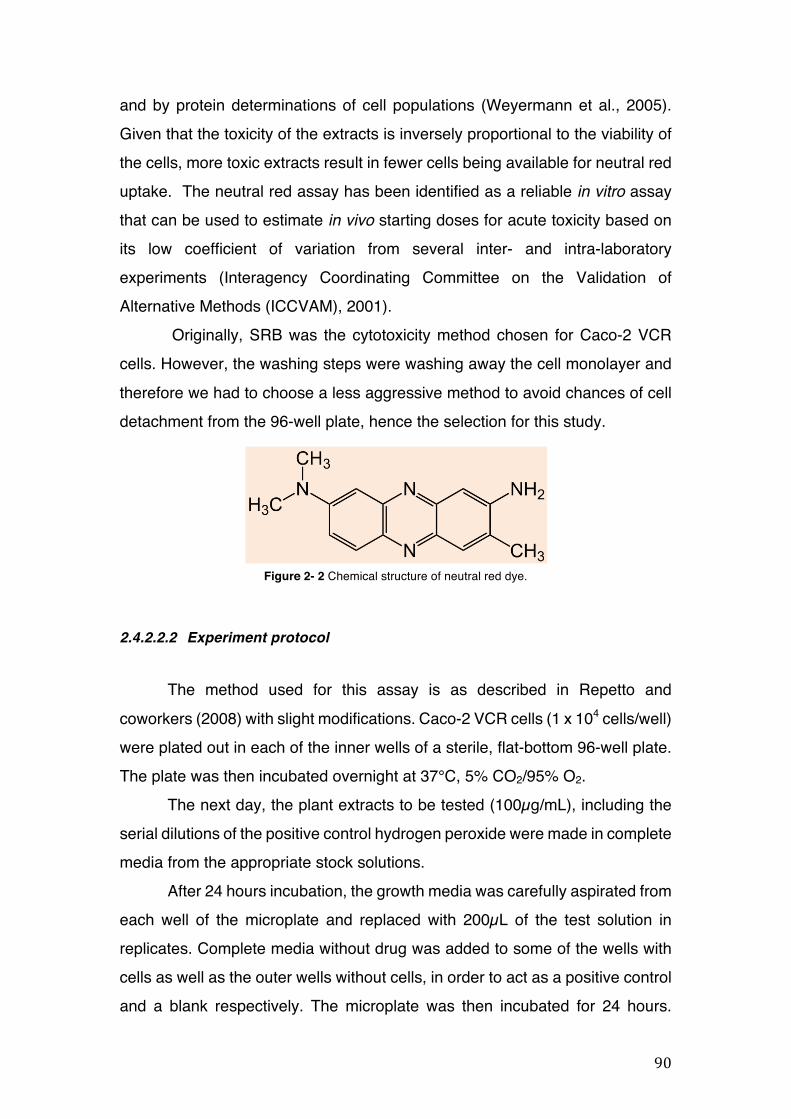
90
and by protein determinations of cell populations (Weyermann et al., 2005). Given that the toxicity of the extracts is inversely proportional to the viability of the cells, more toxic extracts result in fewer cells being available for neutral red uptake. The neutral red assay has been identified as a reliable in vitro assay that can be used to estimate in vivo starting doses for acute toxicity based on its low coefficient of variation from several inter- and intra-laboratory experiments (Interagency Coordinating Committee on the Validation of Alternative Methods (ICCVAM), 2001).
Originally, SRB was the cytotoxicity method chosen for Caco-2 VCR cells. However, the washing steps were washing away the cell monolayer and therefore we had to choose a less aggressive method to avoid chances of cell detachment from the 96-well plate, hence the selection for this study.
Figure 2- 2 Chemical structure of neutral red dye.
2.4.2.2.2 Experiment protocol
The method used for this assay is as described in Repetto and coworkers (2008) with slight modifications. Caco-2 VCR cells (1 x 10⁴ cells/well) were plated out in each of the inner wells of a sterile, flat-bottom 96-well plate. The plate was then incubated overnight at 37°C, 5% CO2/95% O2.
The next day, the plant extracts to be tested (100µg/mL), including the serial dilutions of the positive control hydrogen peroxide were made in complete media from the appropriate stock solutions.
After 24 hours incubation, the growth media was carefully aspirated from each well of the microplate and replaced with 200µL of the test solution in replicates. Complete media without drug was added to some of the wells with cells as well as the outer wells without cells, in order to act as a positive control and a blank respectively. The microplate was then incubated for 24 hours.
91
Neutral red (40µg/mL) in complete media was prepared in a centrifuge tube from a stock solution of 4mg/mL in PBS and incubated overnight, wrapped in foil paper at the same temperature as the cells.
Subsequently, the neutral red solution was centrifuged for 10 minutes at ≈ 1800 rpm to remove precipitated dye crystals. The microplate was then removed from the incubator and the media in each of the wells was gently aspirated off and replaced with 100µL of the neutral red solution in each well of the plate and then incubated for another 2 hours. After incubation, each well of the plate was gently rinsed with PBS (150µL) and the washing solution removed by decanting or gently tapping the plate (Ezuruike, 2015).
150µL of neutral red destain solution (96% ethanol, deionised water, glacial acetic acid; 50%:49%:1%) was added to each well of the plate after the washing step. The plate was immediately shaken on a microplate shaker for at least 10 minutes until the neutral red had been completely extracted from the cells and formed a homogenous solution. The optical density (O.D)/absorbance of the neutral red extract was read out in the plate reader at 540nm. The percentage viability for each of the concentrations of the drug/extract was estimated as previously stated in section 2.4.2.1.2 (Repetto et al., 2008).
2.4.2.3 The MTT assay
2.4.2.3.1 Background
Toxicity of HeLa cells was measured using MTT assay. When the yellow water soluble tetrazolium salt reaches the mitochondria, the succinate dehydrogenase enzyme cleaves the tetrazolium ring, converting the salt into insoluble purple formazan crystals. As the reaction is mitochondria-dependant, the formazan crystals will only be formed in viable cells and will remain there due to the impermeability of the compound to the cell membranes. Only after the addition of an acidified isopropanol solution the product is liberated and can readily be detected and quantified by a colorimetric method (Fotakis and Timbrell, 2006). MTT was chosen for cytotoxicity analysis of the HeLa cells because it is one of the most standard and popular methods for such
92
assessment in cells that are submitted to transfection (Bednarek et al., 2008, Li et al., 2006, Cheng et al., 2007).
2.4.2.3.2 Experiment protocol
The method used for this assay is as described in José Ruiz and coworkers (2006) with some modifications. After 24 hours incubation of HeLa cells (2 × 10⁴ cells/well) with the plant extracts (100µg/mL) and serial dilutions of the positive control ethanol, media containing the samples was removed and 200µL of fresh media containing 50µL of MTT (1mg/mL) were added to each well. After 4 hours incubation, the solution was carefully aspirated and replaced by 150µL of MTT solubilisation solution (0.04M HCl in isopropanol). Plates were shaken until crystals were completely dissolved and absorbance was measured at 570nm in the plate reader and the viability for each of the concentrations of the drug/extract was estimated as previously described in section 2.4.2.1.2 (José Ruiz et al., 2006).
2.4.3 CYP3A4 mRNA gene expression assay
2.4.3.1 Background
Changes in mRNA expression in a cell caused by a xenobiotic can be measured several ways, but quantitative PCR (qPCR) is the “gold standard” technology to quantify nucleic acids. qPCR is a very sensitive and robust technique that collects fluorescent signals from one or more polymerase chain reactions over a range of cycles (Dorak, 2007). Briefly; after the mRNA is extracted from a tissue or cell, it needs to be retro-transcribed into complementary DNA (cDNA) and this is accomplished by using a DNAse, reverse transcriptase enzyme, an oligoprimer, dNTPs (dATP, gGTP, dCTP, and dTTP), RT buffer and RNase inhibitor. Before the beginning of the cDNA synthesis, mRNA is treated with a DNase in order to digest single – and double (dsDNA) – stranded DNA, by the cleavage of each strand of dsDNA independently. In the presence of Mg2+, DNase cleaves each strand of dsDNA independently and randomly. In the presence of Mn2+, the enzyme cleaves both DNA strands at approximately the same site (Figure 2-3).
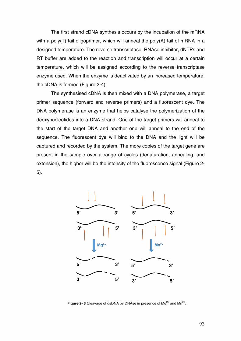
93
5’ 3’
3’ 5’
5’ 3’
3’ 5’
Mg2+
5’ 3’
3’ 5’
Mn2+
5’ 3’
3’ 5’
The first strand cDNA synthesis occurs by the incubation of the mRNA with a poly(T) tail oligoprimer, which will anneal the poly(A) tail of mRNA in a designed temperature. The reverse transcriptase, RNAse inhibitor, dNTPs and RT buffer are added to the reaction and transcription will occur at a certain temperature, which will be assigned according to the reverse transcriptase enzyme used. When the enzyme is deactivated by an increased temperature, the cDNA is formed (Figure 2-4).
The synthesised cDNA is then mixed with a DNA polymerase, a target primer sequence (forward and reverse primers) and a fluorescent dye. The DNA polymerase is an enzyme that helps catalyse the polymerization of the deoxynucleotides into a DNA strand. One of the target primers will anneal to the start of the target DNA and another one will anneal to the end of the sequence. The fluorescent dye will bind to the DNA and the light will be captured and recorded by the system. The more copies of the target gene are present in the sample over a range of cycles (denaturation, annealing, and extension), the higher will be the intensity of the fluorescence signal (Figure 2-5).
Figure 2- 3 Cleavage of dsDNA by DNAse in presence of Mg2+ and Mn2+.
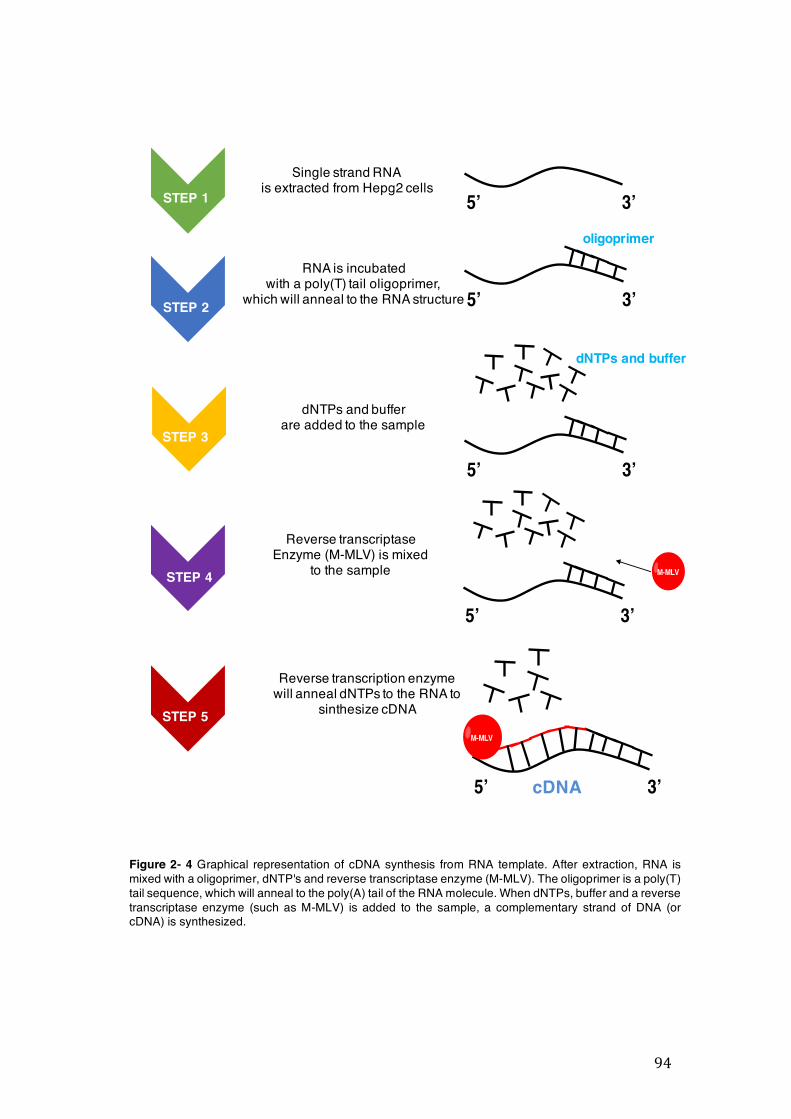
94
5’ 3’
5’ 3’
5’ 3’
5’ 3’
M-MLV
5’ 3’
M-MLV
Single strand RNAis extracted from Hepg2 cells
RNA is incubatedwith a poly(T) tail oligoprimer,
which will anneal to the RNA structure
dNTPs and bufferare added to the sample
Reverse transcriptaseEnzyme (M-MLV) is mixed
to the sample
Reverse transcription enzymewill anneal dNTPs to the RNA to
sinthesize cDNA
cDNA
oligoprimer
dNTPs and buffer
STEP 4
STEP 1
STEP 2
STEP 3
STEP 5
Figure 2- 4 Graphical representation of cDNA synthesis from RNA template. After extraction, RNA is mixed with a oligoprimer, dNTP's and reverse transcriptase enzyme (M-MLV). The oligoprimer is a poly(T) tail sequence, which will anneal to the poly(A) tail of the RNA molecule. When dNTPs, buffer and a reverse transcriptase enzyme (such as M-MLV) is added to the sample, a complementary strand of DNA (or cDNA) is synthesized.
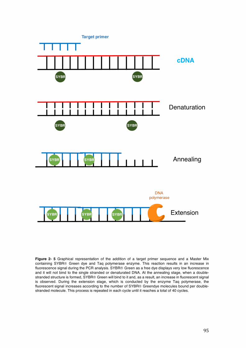
95
Denaturation
SYBR SYBR
AnnealingSYBR SYBR
SYBR SYBR ExtensionSYBR
DNA polymerase
cDNA
SYBR SYBR
Target primer
Figure 2- 5 Graphical representation of the addition of a target primer sequence and a Master Mix containing SYBRâ Green dye and Taq polymerase enzyme. This reaction results in an increase in fluorescence signal during the PCR analysis. SYBRâ Green as a free dye displays very low fluorescence and it will not bind to the single stranded or denaturated DNA. At the annealing stage, when a double-stranded structure is formed, SYBRâ Green will bind to it and, as a result, an increase in fluorescent signal is observed. During the extension stage, which is conducted by the enzyme Taq polymerase, the fluorescent signal increases according to the number of SYBRâ Greendye molecules bound per double-stranded molecule. This process is repeated in each cycle until it reaches a total of 40 cycles.
96
2.4.3.2 Experiment protocol
2.4.3.2.1 Total RNA extraction from HepG2 cells
After exposing HepG2 cells (5 × 10⁵ cells/well) to plant extracts (100µg/mL) or the CYP3A4 inducer RIF (50µM) or the CYP 3A4 inhibitor DMSO 1% for 96 hours, total RNA extraction started by lysing the cells with TRIzol® Reagent (1mL). The homogenised sample was transferred to 1.5mL RNAse/DNAse free microfuge tubes and incubated for 5 minutes at room temperature to permit complete dissociation of the nucleoprotein complex. Chloroform (200µL) was added to each sample and the microfuge tubes were centrifuged at 12,000 x g for 15 minutes at 4°C. The mixture separated into a lower red phenol-chloroform phase, an interphase, and a colourless upper aqueous phase, where the RNA remains. The aqueous phase of the samples was transferred to new 1.5mL RNAse/DNAse free microfuge tubes and 100% isopropanol (500µL) was added in order to precipitate the RNA. The tubes were let at room temperature for 10 minutes and centrifuged at 12,000 x g for 10 minutes at 4°C. The supernatant was removed and the RNA pellet was left in the tubes for washing. The washing step was conducted by adding 1mL of 75% ethanol to each tube and the centrifuged at 12,000 x g for 10 minutes at 4°C. This step was repeated twice and after the removal of the ethanol solution, the RNA pellet was air dried for 30 minutes. The RNA was resuspended with RNAse free water (100µL) and the quantity, quality and integrity of the samples were assessed by agarose gel electrophoresis and NanoDrop, respectively.
2.4.3.2.2 Agarose gel electrophoresis-assessment of the integrity of RNA
The integrity of total RNA from HepG2 cells was assessed by visual inspection of the two rRNAs 28s and 18s on agarose gels. Ethidium bromide (1µL) was added to 1% agarose gel solution and the cooled molten agarose solution was poured into the gel tray containing a 10-well foaming comb. After 30 minutes, the gel was set and the tank was filled with 1 x TAE buffer. Gel loading dye (1µL) was added to 5µL total RNA and this was loaded into each lane.
97
2.4.3.2.3 NanoDrop reading-assessment of the purity and quality of RNA
The quantity and quality of RNA were determined by adding 1.5µL of the RNA sample to the Nanodrop 2000 (Thermo Scientific). Differential readings were made at 230, 260, and 280nm.
2.4.3.2.4 cDNA synthesis
Prior to the reverse transcription, RNA samples were treated with DNase I (1U/µL) to avoid genomic contamination. cDNA synthesis started by adding 1µg of total RNA to 0.5mL RNAse/DNAse free microfuge tubes containing 10mM (1µL) dNTP Mix (10mM each dATP, dGTP, dCTP and dTTP at neutral pH, Oligo (dT)12-18 (500 µg/mL) (1µL), and sterile distilled water up to 12µL volume. The samples were heated at 65°C for 5 minutes and chilled at 4°C for 2 minutes to let the primer anneal to the RNA in an MJ Mini Personal Thermal Cycler (Bio-Rad). 5X First-Strand Buffer (4µL), 0.1 DDT (2µL) and RNase OUTä Recombinant Ribonuclease Inhibitor (40 units/ µL) (1µL) were added to the samples tubes and the mixture was incubated at 37°C for 2 minutes. The Moloney murine Leukaemia Virus Reverse Transcriptase (M-MLV RT) (200U/µL) (1µL) was added to each sample and the tubes were incubated for 50 minutes at 37°C. The reaction was then inactivated by heating the samples at 70°C for 15 minutes.
2.4.3.2.5 RT-qPCR conditions and analysis
CYP3A4 sense strand primer sequence was 5’- CAAGGACAACATAGATCGTTACATATACACACCCTTTGGAAG-3’ and the antisense strand primer was 5’-AGCTCAATGCATGTACAGAATCCCCGGTTA-3’. The β-actin gene was used to control for variations in RNA loading within the experimental conditions. The sense strand primer sequence was 5’- CGTACCACTGGCATCGTGAT-3’and the antisense strand primer was 5’- GTGTTGGCGTACAGGTCTTTG-3’. The RT-qPCR was carried out in 96-well plates using a Pikoreal™ Real-Time PCR detection system (Thermo Scientific). Each well contained a final reaction
98
volume of 10µL (5.0µL MasterMix with SYBR Green, 2.0µL cDNA template diluted appropriately, 0.5µL of each primer at a final concentration 0.3mM and 2.0µL of RNAse/DNAse free distilled water). The PCR reaction was performed under the following schema: initial denaturation at 95°C for 2 minutes, then 40 cycles of denaturation at 95°C for 15 seconds, annealing at 55°C (β-actin) or 60°C (CYP3A4) for 30 seconds, and extension at 72°C for 30 seconds.
At the end of the run, a melting curve was generated by heating the amplicon from 60 to 95◦C in order to confirm the specificity of the amplification
of each primer pair. All samples were run in quadruplicates. Standard curves were produced to check the PCR efficiency using a five-fold dilution series of cDNA. Efficiency (E) of primer pairs was obtained from the slope of the calibration curve generated. The relative expression was calculated on the basis of ‘delta delta Ct’ (DDCt) values. Normalisation of the target gene was achieved by using β-actin as a reference gene.
2.4.4 hPXR activation assay
2.4.4.1 Background
Regulation of CYP3A4 mRNA gene expression is mostly related to the binding of xenobiotics to nuclear receptors, such as the hPXR, which are DNA binding and ligand-regulated transcriptional factor. An interesting feature of hPXR is its ligand-binding domain (LBD) (Xie, 2009). hPXR LBD is hydrophobic and it is able to change its shape in order to better accommodate numerous xenobiotics, such as antibiotics, calcium channel blockers, dietary supplements, environmental pollutants and herbal medicines (Chang, 2009, Shukla et al., 2011).
In the case that no ligand binds to hPXR, CYP3A4 transcription is inhibited by orphan nuclear receptors, such as the small heterodimer partner (SHP) and the silencing mediator of retinoid and thyroid hormone receptors (SMRT). These receptors are transcribed by coactivators, such as steroid receptor coactivator-1 (SRC-1), hepatocyte nuclear factor a (HNFa), and peroxisome proliferator-activated receptor g coactivator-1-a (PGC-1a) (Figure 2-6A).
99
Upon xenobiotic binding, hPXR translocates into the nucleus and blocks the transcription of SHP/SMRT. The ligand-PXR then forms a heterodimer complex with the retinoid X receptora (RXRa), which will then bind to the xenobiotic responsive enhancer molecule/PXRE (XREM/PXRE) region of the CYP3A4 promoter to initiate the expression of the enzyme (Figure 2-6B) (Xie, 2009).
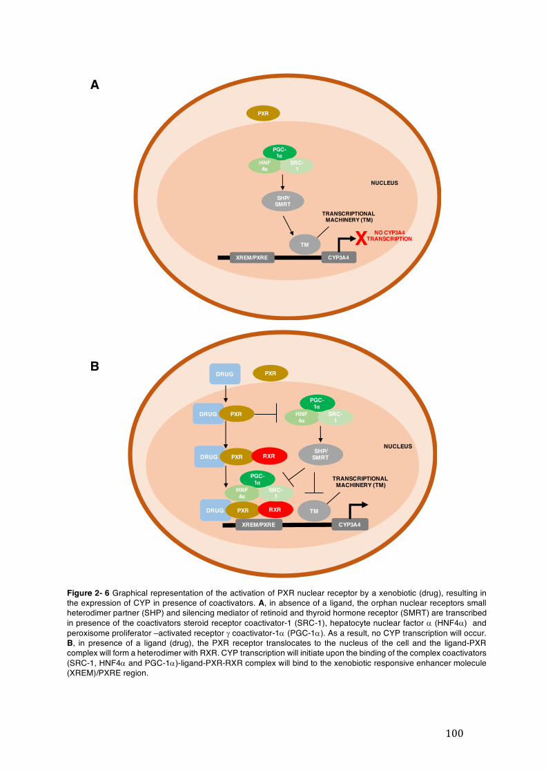
100
PXR
PGC-1⍺
HNF4⍺
SRC-1
SHP/SMRT
XREM/PXRE
TM
TRANSCRIPTIONAL MACHINERY (TM)
CYP3A4
NUCLEUS
X NO CYP3A4 TRANSCRIPTION
PXRDRUG
DRUG PXR
DRUG PXR RXR
PGC-1⍺
HNF4⍺
SRC-1
SHP/SMRT
XREM/PXRE
DRUG PXR RXR
PGC-1⍺
HNF4⍺
SRC-1
TM
TRANSCRIPTIONAL MACHINERY (TM)
CYP3A4
NUCLEUS
A
B
Figure 2- 6 Graphical representation of the activation of PXR nuclear receptor by a xenobiotic (drug), resulting in the expression of CYP in presence of coactivators. A, in absence of a ligand, the orphan nuclear receptors small heterodimer partner (SHP) and silencing mediator of retinoid and thyroid hormone receptor (SMRT) are transcribed in presence of the coactivators steroid receptor coactivator-1 (SRC-1), hepatocyte nuclear factor a (HNF4a) and peroxisome proliferator –activated receptor g coactivator-1a (PGC-1a). As a result, no CYP transcription will occur. B, in presence of a ligand (drug), the PXR receptor translocates to the nucleus of the cell and the ligand-PXR complex will form a heterodimer with RXR. CYP transcription will initiate upon the binding of the complex coactivators (SRC-1, HNF4a and PGC-1a)-ligand-PXR-RXR complex will bind to the xenobiotic responsive enhancer molecule (XREM)/PXRE region.
101
2.4.4.2 Experiment protocol
Four RENISUS plant extracts (Erythrina mulungu Benth., Cordia
verbenaceae A. DC., Solanum paniculatum L. and Lippia sidoides Cham.) were subject to hPXR activation assays. The experiment protocol for hPXR activation assay required first the adding of a luciferase reporter gene, which is a downstream of the PXRE. Once the ligand binds to the PXRE, the amount of luciferase enzyme is quantified by the addition of luciferin substrate and detection after cell lysis.
After 24 hours seeding, HeLa cells (4 × 10⁴ cells/well) were transiently co-transfected with 60ng of pM-Gal4-PXR-LBD and 240ng of Gal4 luciferase reporter using lipofectamine 2000 reagent according to the manufacturer’s protocol. Transfected cells were treated with increasing concentrations of plant extract and/or rifampicin 1µM (EC50). Luciferase activity was measured after 24 hours, according to the manufacturer’s protocol in a 20/20n Glomax luminometer and reported as a response (%) compared to cells treated only with RIF.
2.4.5 Intracellular GSH assay
2.4.5.1 Background
GSH is a ubiquitous and unusual tripeptide thiol (L-γ-glutamyl-L-cysteinylglycine) or α-amino acid found in the cortex, medulla, cytosol, mitochondria, molecules, blood and most frequently in the liver of various mammalian cells in millimolar range (0.5-10mM). It is known to function either directly or indirectly in many important biological phenomena, including the synthesis of proteins and DNA, transport, enzyme activity, metabolism and protection of cells (Meister and Anderson, 1983). Out of all the functions of GSH, two are relevant in the process of cell detoxification. They are: (1) its role as an antioxidant by removing potentially toxic electrophilic compounds such as free radicals, peroxides, and alkylating agent (Ionescu and Caira, 2005, Allen et al., 2000) and (2) its role in phase II metabolism of xenobiotics. In general, both roles relate to the general function of GSH to protect cells against oxygen toxicity (Fahey and Sundquist, 2006).
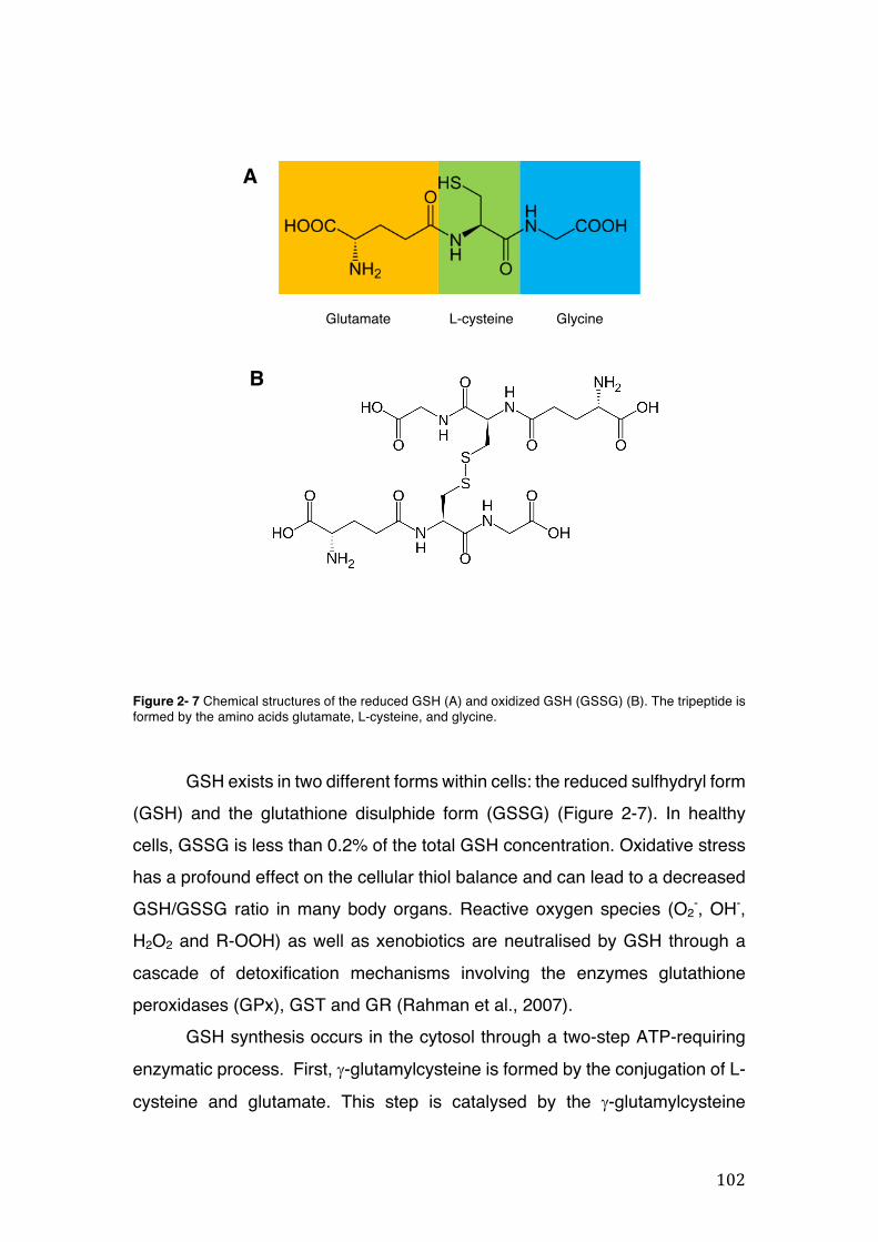
102
Glutamate L-cysteine Glycine
A
B
Figure 2- 7 Chemical structures of the reduced GSH (A) and oxidized GSH (GSSG) (B). The tripeptide is formed by the amino acids glutamate, L-cysteine, and glycine.
GSH exists in two different forms within cells: the reduced sulfhydryl form (GSH) and the glutathione disulphide form (GSSG) (Figure 2-7). In healthy cells, GSSG is less than 0.2% of the total GSH concentration. Oxidative stress has a profound effect on the cellular thiol balance and can lead to a decreased GSH/GSSG ratio in many body organs. Reactive oxygen species (O2
-, OH-, H2O2 and R-OOH) as well as xenobiotics are neutralised by GSH through a cascade of detoxification mechanisms involving the enzymes glutathione peroxidases (GPx), GST and GR (Rahman et al., 2007).
GSH synthesis occurs in the cytosol through a two-step ATP-requiring enzymatic process. First, g-glutamylcysteine is formed by the conjugation of L-cysteine and glutamate. This step is catalysed by the g-glutamylcysteine
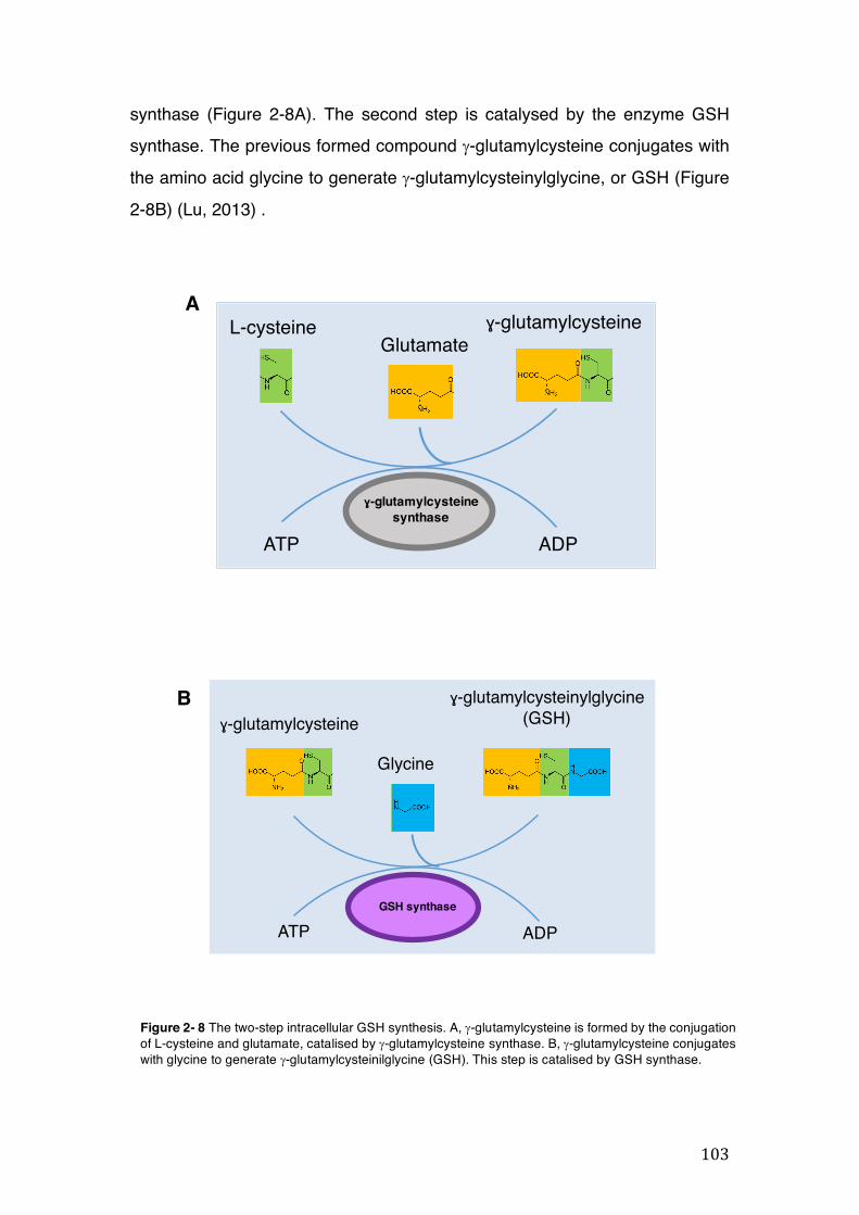
103
Glutamate L-cysteine Glycine
Glutamate L-cysteine Glycine
ɣ-glutamylcysteinesynthase
ATP ADP
Glutamate L-cysteine Glycine
GlutamateL-cysteine ɣ-glutamylcysteine
GSH synthase
Glutamate L-cysteine Glycine
Glutamate L-cysteine Glycine
ATP ADP
Glycine
ɣ-glutamylcysteinylglycine(GSH)
Glutamate L-cysteine Glycine
ɣ-glutamylcysteine
A
B
synthase (Figure 2-8A). The second step is catalysed by the enzyme GSH synthase. The previous formed compound g-glutamylcysteine conjugates with the amino acid glycine to generate g-glutamylcysteinylglycine, or GSH (Figure 2-8B) (Lu, 2013) .
Figure 2- 8 The two-step intracellular GSH synthesis. A, g-glutamylcysteine is formed by the conjugation of L-cysteine and glutamate, catalised by g-glutamylcysteine synthase. B, g-glutamylcysteine conjugates with glycine to generate g-glutamylcysteinilglycine (GSH). This step is catalised by GSH synthase.
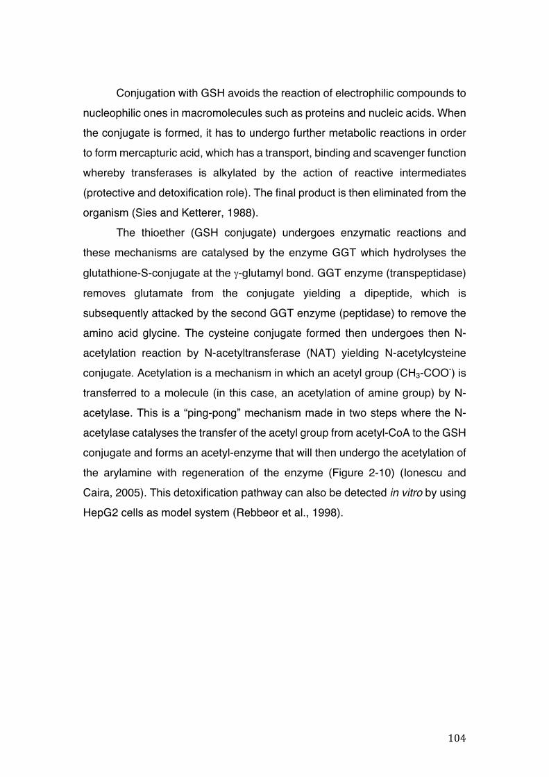
104
Conjugation with GSH avoids the reaction of electrophilic compounds to
nucleophilic ones in macromolecules such as proteins and nucleic acids. When the conjugate is formed, it has to undergo further metabolic reactions in order to form mercapturic acid, which has a transport, binding and scavenger function whereby transferases is alkylated by the action of reactive intermediates (protective and detoxification role). The final product is then eliminated from the organism (Sies and Ketterer, 1988).
The thioether (GSH conjugate) undergoes enzymatic reactions and these mechanisms are catalysed by the enzyme GGT which hydrolyses the glutathione-S-conjugate at the g-glutamyl bond. GGT enzyme (transpeptidase) removes glutamate from the conjugate yielding a dipeptide, which is subsequently attacked by the second GGT enzyme (peptidase) to remove the amino acid glycine. The cysteine conjugate formed then undergoes then N-acetylation reaction by N-acetyltransferase (NAT) yielding N-acetylcysteine conjugate. Acetylation is a mechanism in which an acetyl group (CH3-COO-) is transferred to a molecule (in this case, an acetylation of amine group) by N-acetylase. This is a “ping-pong” mechanism made in two steps where the N-acetylase catalyses the transfer of the acetyl group from acetyl-CoA to the GSH conjugate and forms an acetyl-enzyme that will then undergo the acetylation of the arylamine with regeneration of the enzyme (Figure 2-10) (Ionescu and Caira, 2005). This detoxification pathway can also be detected in vitro by using HepG2 cells as model system (Rebbeor et al., 1998).
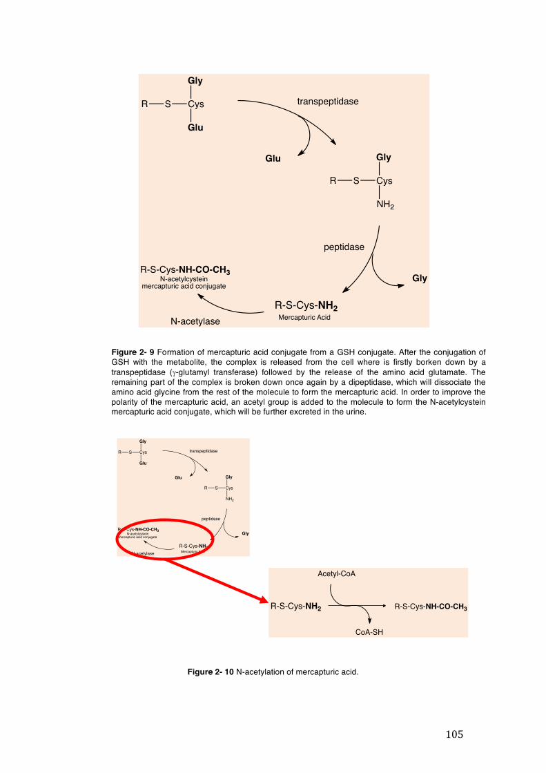
105
R S Cys
Gly
Glu
Glu
R S Cys
Gly
NH2
Gly
R-S-Cys-NH2
R-S-Cys-NH-CO-CH3
transpeptidase
peptidase
Mercapturic AcidN-acetylase
N-acetylcysteinmercapturic acid conjugate
R-S-Cys-NH2 R-S-Cys-NH-CO-CH3
Acetyl-CoA
CoA-SH
Figure 2- 10 N-acetylation of mercapturic acid.
R S Cys
Gly
Glu
Glu
R S Cys
Gly
NH2
Gly
R-S-Cys-NH2
R-S-Cys-NH-CO-CH3
transpeptidase
peptidase
Mercapturic AcidN-acetylase
N-acetylcysteinmercapturic acid conjugate
Figure 2- 9 Formation of mercapturic acid conjugate from a GSH conjugate. After the conjugation of GSH with the metabolite, the complex is released from the cell where is firstly borken down by a transpeptidase (g-glutamyl transferase) followed by the release of the amino acid glutamate. The remaining part of the complex is broken down once again by a dipeptidase, which will dissociate the amino acid glycine from the rest of the molecule to form the mercapturic acid. In order to improve the polarity of the mercapturic acid, an acetyl group is added to the molecule to form the N-acetylcystein mercapturic acid conjugate, which will be further excreted in the urine.
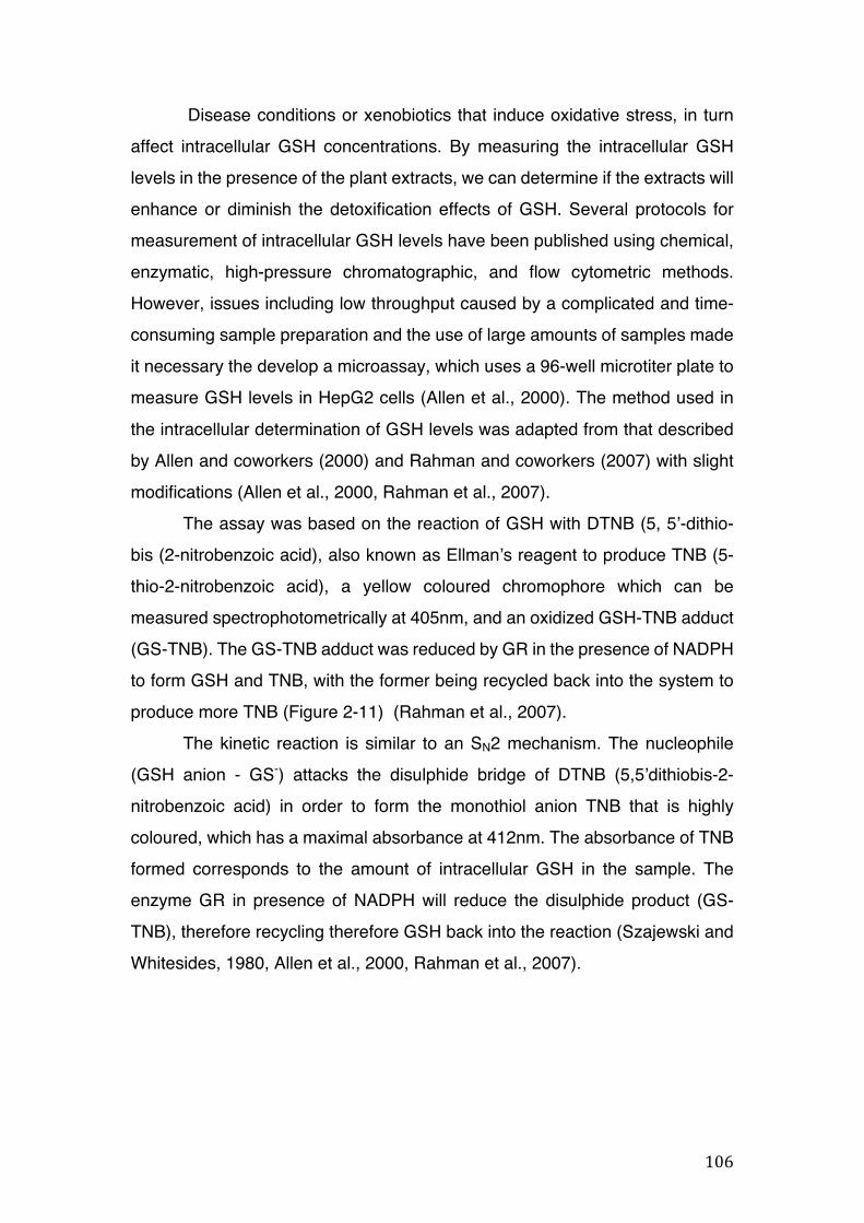
106
Disease conditions or xenobiotics that induce oxidative stress, in turn affect intracellular GSH concentrations. By measuring the intracellular GSH levels in the presence of the plant extracts, we can determine if the extracts will enhance or diminish the detoxification effects of GSH. Several protocols for measurement of intracellular GSH levels have been published using chemical, enzymatic, high-pressure chromatographic, and flow cytometric methods. However, issues including low throughput caused by a complicated and time-consuming sample preparation and the use of large amounts of samples made it necessary the develop a microassay, which uses a 96-well microtiter plate to measure GSH levels in HepG2 cells (Allen et al., 2000). The method used in the intracellular determination of GSH levels was adapted from that described by Allen and coworkers (2000) and Rahman and coworkers (2007) with slight modifications (Allen et al., 2000, Rahman et al., 2007).
The assay was based on the reaction of GSH with DTNB (5, 5’-dithio-bis (2-nitrobenzoic acid), also known as Ellman’s reagent to produce TNB (5-thio-2-nitrobenzoic acid), a yellow coloured chromophore which can be measured spectrophotometrically at 405nm, and an oxidized GSH-TNB adduct (GS-TNB). The GS-TNB adduct was reduced by GR in the presence of NADPH to form GSH and TNB, with the former being recycled back into the system to produce more TNB (Figure 2-11) (Rahman et al., 2007).
The kinetic reaction is similar to an SN2 mechanism. The nucleophile (GSH anion - GS-) attacks the disulphide bridge of DTNB (5,5’dithiobis-2-nitrobenzoic acid) in order to form the monothiol anion TNB that is highly coloured, which has a maximal absorbance at 412nm. The absorbance of TNB formed corresponds to the amount of intracellular GSH in the sample. The enzyme GR in presence of NADPH will reduce the disulphide product (GS-TNB), therefore recycling therefore GSH back into the reaction (Szajewski and Whitesides, 1980, Allen et al., 2000, Rahman et al., 2007).
107
GSH
GSSGGR
+NADPH
S-
O2N
HOOC
S-
O2N
HOOC
NO2
COOHSSHOOC
O2N
DTNB
TNB
TNB
GS-TNB
Figure 2- 11 Enzymatic recycling of GSH from glutathione disulphide (GSSG) by glutathione reductase (GR) in the presence of NADPH. GSH reacts with DTNB to GS-TNB and TNB, with subsequent reduction of the former to GSH. TNB can be measured at 405 nm. (Adapted from Rahman et al., 2007).
The GSSG present in the sample is also reduced by GR to form two
molecules of GSH. Thus, the amount of GSH measured represented the sum of reduced and oxidised GSH in the sample. The rate of change in absorbance of TNB at 405nm is linearly proportional to the total concentration of GSH in the sample. The concentration of an unknown sample can therefore be determined by calculating from the linear equation or the regression curve generated from several standard concentrations of GSH.
108
2.4.5.2 Experiment protocol
HepG2 cells (4 x 10⁴ cells per well) were seeded out in sterile, flat bottom 96-well plates and incubated overnight to allow for cell attachment. The next day, the cells were treated with the plant extracts (100µg/mL) to be tested in triplicates. Three wells were not treated with any drug and served as the media control, while three other wells were treated with buthionine sulphoximine (BSO) (10µM) as the positive control. All drug/extract incubations were done for 24 hours. Prior to commencing the GSH assay, the following solutions were prepared: phosphate buffer solution containing 168mL of a 286mM solution of sodium phosphate dibasic in distilled water, 32mL of a 286mM solution of sodium phosphate monobasic and 6.3mM EDTA; 5% SSA in double distilled water; 0.1% Triton X-100; 1mM stock solution of reduced glutathione in 5% SSA solution; and 10mM DTNB in phosphate buffer solution. Apart from the DTNB, all the other solutions were able to be stored in the fridge at 2 - 4 °C for up to a week. 2.39mM or 2mg/mL β-NADPH in phosphate buffer solution was freshly prepared on the day of the assay. 24 hours after incubation of the extract/drug, the cells were washed with PBS (200µL per well) and the PBS was aspirated off. 0.1% Triton X-100 (60µL) was added to each well of the plate containing cells and then shaken on a microplate shaker for 2 minutes to lyse the cells. The UV light of the laminar flow hood was turned off for the rest of the assay to minimise GSH oxidation. Subsequently, 5% SSA solution (25µL) was added and the plate shaken for another 2 minutes. After shaking, the lysed cell solution (25µL) was transferred to the wells of the next three inner columns of the plate. 5% SSA solution (25µL) was added to each well of the first column of the plate as a blank. A double dilution of a standard solution of reduced glutathione in 5% SSA (25µL), starting from 100µM GSH as the highest concentration was then prepared. The GSH reaction buffer needed for the assay was prepared by adding the freshly prepared β-NADPH (1mL) and GR (3µL) (equivalent to 2 units of GR) to the phosphate buffer solution (7.5mL) for each 96-well plate. This was
109
vortexed for 5 seconds and then the DTNB solution (1mL) was immediately thawed and added to the reaction buffer and then vortexed for another 5 minutes. All solutions to be used were kept on ice until needed. The reaction buffer (125µL) was added to each well of the 96-well plate containing either the blank SSA solution (25µL), the GSH standards (25µL) or the cell lysate solution (25µL). The microplate was immediately covered and then wrapped in foil paper and then shaken in the microplate for 15 seconds. Immediately after, the absorbance of the wells of the microplate was read in a kinetic cycle in the TECAN plate reader every 30 seconds for 5 minutes at 405nm (11 readings). The slope of the curve obtained from the 11 readings of each of the GSH standard concentrations (otherwise known as the I-slope), which represents the rate of change of GSH levels at that concentration plotted against the GSH concentration to give a regression curve (otherwise known as the F-slope curve). The concentration of GSH in the samples from the cell lysate solutions was then estimated by substituting the average I-slope values for each of the extract/drug into the equation of the F-slope curve (Ezuruike, 2015).
2.4.6 GGT activity assay
2.4.6.1 Background
GGT is a membrane-localised enzyme that plays a key role in the maintenance of GSH at normal levels in the body. The enzyme provides the aminoacid cysteine, which is the precursor for GSH synthesis. As GSH cannot be hydrolysed by general peptidases due to its unusual peptide bond linking glutamate and cysteine through the g-carboxyl group of glutamate instead of the conventional a-carboxyl group, GGT has the crucial function of breaking
down that bond. The enzyme catalyses the cleavage of the g-glutamyl bond of GSH and the transfer of the g-glutamyl group to water, amino acids or peptides.
The amino acid cysteine is fundamental for GSH synthesis in the cell. In this cycle, GSH is transported out of the cell and it is first broken-down by GGT, form g-glutamyl aminoacid and cysteinylglycine. g-glutamyl aminoacid is transported back into the cell and metabolised to release the amino acid and 5-
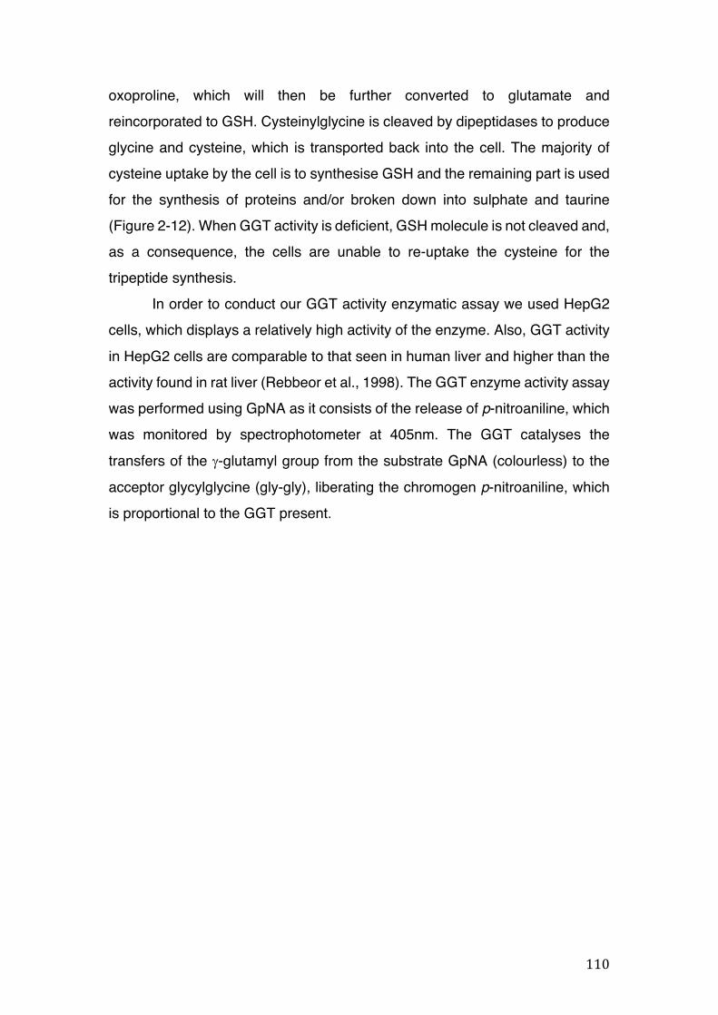
110
oxoproline, which will then be further converted to glutamate and reincorporated to GSH. Cysteinylglycine is cleaved by dipeptidases to produce glycine and cysteine, which is transported back into the cell. The majority of cysteine uptake by the cell is to synthesise GSH and the remaining part is used for the synthesis of proteins and/or broken down into sulphate and taurine (Figure 2-12). When GGT activity is deficient, GSH molecule is not cleaved and, as a consequence, the cells are unable to re-uptake the cysteine for the tripeptide synthesis. In order to conduct our GGT activity enzymatic assay we used HepG2 cells, which displays a relatively high activity of the enzyme. Also, GGT activity in HepG2 cells are comparable to that seen in human liver and higher than the activity found in rat liver (Rebbeor et al., 1998). The GGT enzyme activity assay was performed using GpNA as it consists of the release of p-nitroaniline, which was monitored by spectrophotometer at 405nm. The GGT catalyses the transfers of the g-glutamyl group from the substrate GpNA (colourless) to the acceptor glycylglycine (gly-gly), liberating the chromogen p-nitroaniline, which is proportional to the GGT present.
111
aminoacid
gamma-glutamylaminoacid
cysteinylglycine
glycinecysteine
Gamma-glutamylaminoacid
5-oxoproline
glutamatecysteine
sulfate + taurine
protein
Large subunit
SmallsubunitGGT
Dipeptidase
Glutamate L-cysteine GlycineGlutathione
Glutamate L-cysteine GlycineGlutathione
Nucleus
Figure 2- 12 The g-glutamyl cycle. When GSH is transported out of the cell, the enzyme gamma-glutamyl transferase (GGT) will break down the tripeptide and transfer the g-glutamyl moiety to an amino acid, forming g-glutamyl amino acid and cysteinylglycine. The g-glutamyl amino acid is transported back to the cell where it will be metabolized to release the amino acid and 5-oxoproline, which will be converted to glutamate and reincorporated into GSH. Dipeptidase will break down cysteinylglycine into glycine and cysteine. The latter will be transported back to the cell to be reincorporated into GSH. The amount of cysteine which is not used for GSH synthesis will be incorporated into newly synthesized proteins and/or broken down into sulphate and taurine.
2.4.6.2 Experiment protocol
GGT activity assay was conducted according Rebbeor and coworkers (1998) with slight modifications. Briefly, after 24 hours of incubation of HepG2 cells (1 × 10⁶ cells/well) with the GGT inhibitor acivicin (5µM) or plant extracts (100µg/mL), media was aspirated and cells were washed with PBS. 4mL of pre-warmed Gly-Gly buffer (115mM Tris, 138mM Gly-Gly) and 400µL of the substrate GpNA (29.6mg/mL of HCl 0.5mmol/L) were added to the wells and plates were incubated for 10 minutes. Subsequently, 500µL of the content of each well were transferred to 24-well plates and absorbance was measured in the plate reader (405nm). The absorbances were converted into absolute amounts by means of a calibration line using p-nitroaniline (Rebbeor et al., 1998).
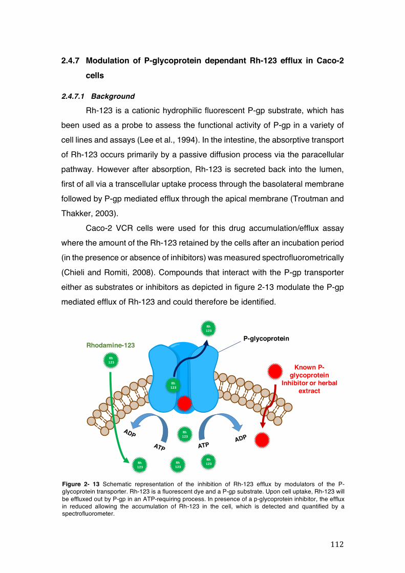
112
Rh123
Rh123
Rh123
Rh123
Rh123
Rh123
Rh123
P-glycoproteinRhodamine-123
Known P-glycoprotein
Inhibitor or herbal extract
2.4.7 Modulation of P-glycoprotein dependant Rh-123 efflux in Caco-2 cells
2.4.7.1 Background
Rh-123 is a cationic hydrophilic fluorescent P-gp substrate, which has been used as a probe to assess the functional activity of P-gp in a variety of cell lines and assays (Lee et al., 1994). In the intestine, the absorptive transport of Rh-123 occurs primarily by a passive diffusion process via the paracellular pathway. However after absorption, Rh-123 is secreted back into the lumen, first of all via a transcellular uptake process through the basolateral membrane followed by P-gp mediated efflux through the apical membrane (Troutman and Thakker, 2003).
Caco-2 VCR cells were used for this drug accumulation/efflux assay where the amount of the Rh-123 retained by the cells after an incubation period (in the presence or absence of inhibitors) was measured spectrofluorometrically (Chieli and Romiti, 2008). Compounds that interact with the P-gp transporter either as substrates or inhibitors as depicted in figure 2-13 modulate the P-gp mediated efflux of Rh-123 and could therefore be identified.
Figure 2- 13 Schematic representation of the inhibition of Rh-123 efflux by modulators of the P-glycoprotein transporter. Rh-123 is a fluorescent dye and a P-gp substrate. Upon cell uptake, Rh-123 will be effluxed out by P-gp in an ATP-requiring process. In presence of a p-glycoprotein inhibitor, the efflux in reduced allowing the accumulation of Rh-123 in the cell, which is detected and quantified by a spectrofluorometer.
113
2.4.7.2 Experiment protocol
The protocol used for this assay is as described in Chieli and coworkers (2009), with slight modifications (Chieli et al., 2009). Caco-2 VCR cells to be used were routinely grown in the appropriate complete growth media in 75cm2 tissue culture flasks as described in section 2.4.1.3. The culture media of the cells were usually changed a day before plating out to ensure that the cells were in their exponential stage of growth. The media of Caco-2 VCR cells was replaced with complete media without vincristine at this point.
80 to 90% confluent culture flask of cells were trypsinised and then plated out in the inner wells of a sterile 96-well plate at a density of 1.5 x 10⁴ cells/well in full culture media (without vincristine). The cells were then placed in the incubator overnight to allow for attachment. The next day, the growth media of the cells was changed to serum free media (without 10% FBS). This was to ensure a more uniform condition for the majority of the cell population and to prevent adsorption of drugs to the serum proteins. The microplates were then replaced back in the incubator for 24 hours.
On the day of the assay, Rh-123 (5µg/mL) solution in serum free media sufficient for the assay was prepared from a stock solution in DMSO (5mg/mL) (solution A). All the extracts to be tested were then prepared (100µg/mL) in solution A (solution B), while 20µM verapamil (Securon® I.V. 2.5mg/mL, Abbot Laboratories Limited) was also prepared in solution A as positive control (solution C). The media in each of the wells of the microplate was then aspirated off and then replaced with either solution A, B or C in replicates. The microplates were placed back in the incubator for 2 hours to allow for intracellular Rh-123 accumulation within the cells. During this time, 20µM verapamil in PBS (solution D) was also prepared and stored in the fridge at 4°C.
After the two hours of incubation, the media in the wells were all aspirated off and each well was washed three times, gently but quickly with the ice cold solution D (200µL) to stop the P-gp efflux activity. 0.1% Triton X-100 in PBS (100µL) was then added to each well and then placed in the incubator for 15 minutes to lyse the cells and completely solubilise the intracellular Rh-123. Cell lysates (80µL) were transferred to a 96-well black plate and the
114
fluorescence intensity of each well was measured in a Tecan® plate reader (Exc-485nm, Em-525nm). The cellular accumulation of Rh-123 for each of the extracts was expressed as the percentage of the accumulation measured for Rh-123 only (solution A), that is under control conditions.
2.4.8 Statistical Analysis
Collected data were analysed as means ± SD of at least three independent experiments. Statistical significance was measured by student t-test and one-way ANOVA followed by Bonferroni correction using GraphPad InStat (GraphPad Software Inc., La Jolla, CA, USA). Results with a value of p < 0.05 were considered significant.

115
3 RESULTS
116
3.1 AUTHORISATION FOR ACCESSING THE BRAZILIAN GENETIC
HERITAGE COMPONENT FROM CGEN AND YIELD OF PLANT
EXTRACTS
The authorisation from CGEN for the initiation of our research with the Brazilian plant extracts was issued in May 2014 (Appendix 7.4). A total number of twenty-four plant samples comprising twenty-three species from nineteen families were collected from either the “Farmácias vivas” or bought from the herbal suppliers in Brazil in 2014.
All samples were extracted by infusing with hot water. The yields of the different plant samples varied from as low as 5.03% w/w (Artemisia absinthium) to as high as over 14.95% w/w (Cordia verbenaceae) (Table 3-1).
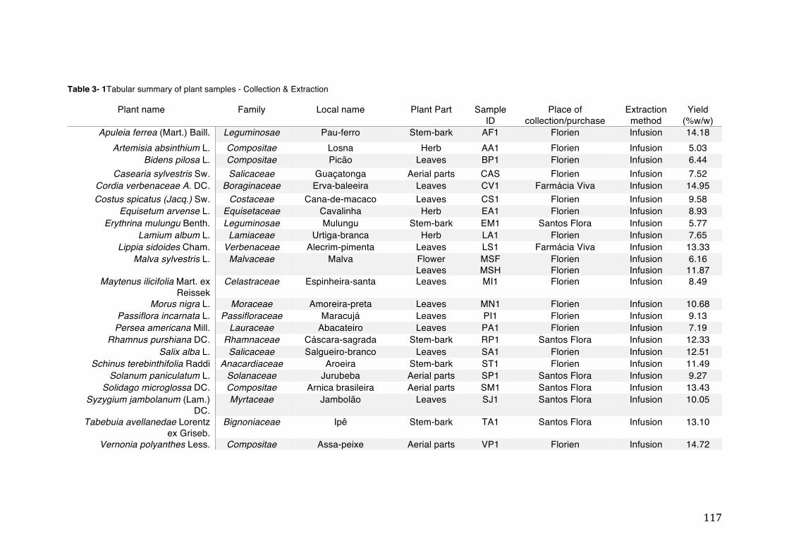
117
Table 3- 1Tabular summary of plant samples - Collection & Extraction
Plant name Family Local name Plant Part Sample ID
Place of collection/purchase
Extraction method
Yield (%w/w)
Apuleia ferrea (Mart.) Baill. Leguminosae Pau-ferro Stem-bark AF1 Florien Infusion 14.18 Artemisia absinthium L. Compositae Losna Herb AA1 Florien Infusion 5.03
Bidens pilosa L. Compositae Picão Leaves BP1 Florien Infusion 6.44 Casearia sylvestris Sw. Salicaceae Guaçatonga Aerial parts CAS Florien Infusion 7.52
Cordia verbenaceae A. DC. Boraginaceae Erva-baleeira Leaves CV1 Farmácia Viva Infusion 14.95 Costus spicatus (Jacq.) Sw. Costaceae Cana-de-macaco Leaves CS1 Florien Infusion 9.58
Equisetum arvense L. Equisetaceae Cavalinha Herb EA1 Florien Infusion 8.93 Erythrina mulungu Benth. Leguminosae Mulungu Stem-bark EM1 Santos Flora Infusion 5.77
Lamium album L. Lamiaceae Urtiga-branca Herb LA1 Florien Infusion 7.65 Lippia sidoides Cham. Verbenaceae Alecrim-pimenta Leaves LS1 Farmácia Viva Infusion 13.33
Malva sylvestris L. Malvaceae Malva Flower Leaves
MSF MSH
Florien Florien
Infusion Infusion
6.16 11.87
Maytenus ilicifolia Mart. ex Reissek
Celastraceae Espinheira-santa Leaves MI1 Florien Infusion 8.49
Morus nigra L. Moraceae Amoreira-preta Leaves MN1 Florien Infusion 10.68 Passiflora incarnata L. Passifloraceae Maracujá Leaves PI1 Florien Infusion 9.13
Persea americana Mill. Lauraceae Abacateiro Leaves PA1 Florien Infusion 7.19 Rhamnus purshiana DC. Rhamnaceae Cáscara-sagrada Stem-bark RP1 Santos Flora Infusion 12.33
Salix alba L. Salicaceae Salgueiro-branco Leaves SA1 Florien Infusion 12.51 Schinus terebinthifolia Raddi Anacardiaceae Aroeira Stem-bark ST1 Florien Infusion 11.49
Solanum paniculatum L. Solanaceae Jurubeba Aerial parts SP1 Santos Flora Infusion 9.27 Solidago microglossa DC. Compositae Arnica brasileira Aerial parts SM1 Santos Flora Infusion 13.43
Syzygium jambolanum (Lam.) DC.
Myrtaceae Jambolão Leaves SJ1 Santos Flora Infusion 10.05
Tabebuia avellanedae Lorentz ex Griseb.
Bignoniaceae Ipê Stem-bark TA1 Santos Flora Infusion 13.10
Vernonia polyanthes Less. Compositae Assa-peixe Aerial parts VP1 Florien Infusion 14.72
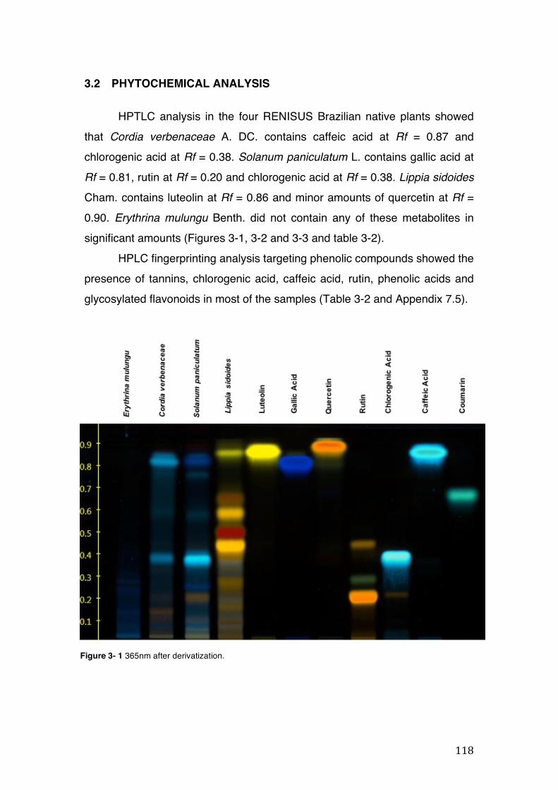
118
3.2 PHYTOCHEMICAL ANALYSIS
HPTLC analysis in the four RENISUS Brazilian native plants showed
that Cordia verbenaceae A. DC. contains caffeic acid at Rf = 0.87 and
chlorogenic acid at Rf = 0.38. Solanum paniculatum L. contains gallic acid at
Rf = 0.81, rutin at Rf = 0.20 and chlorogenic acid at Rf = 0.38. Lippia sidoides
Cham. contains luteolin at Rf = 0.86 and minor amounts of quercetin at Rf =
0.90. Erythrina mulungu Benth. did not contain any of these metabolites in
significant amounts (Figures 3-1, 3-2 and 3-3 and table 3-2).
HPLC fingerprinting analysis targeting phenolic compounds showed the
presence of tannins, chlorogenic acid, caffeic acid, rutin, phenolic acids and
glycosylated flavonoids in most of the samples (Table 3-2 and Appendix 7.5).
Figure 3- 1 365nm after derivatization.
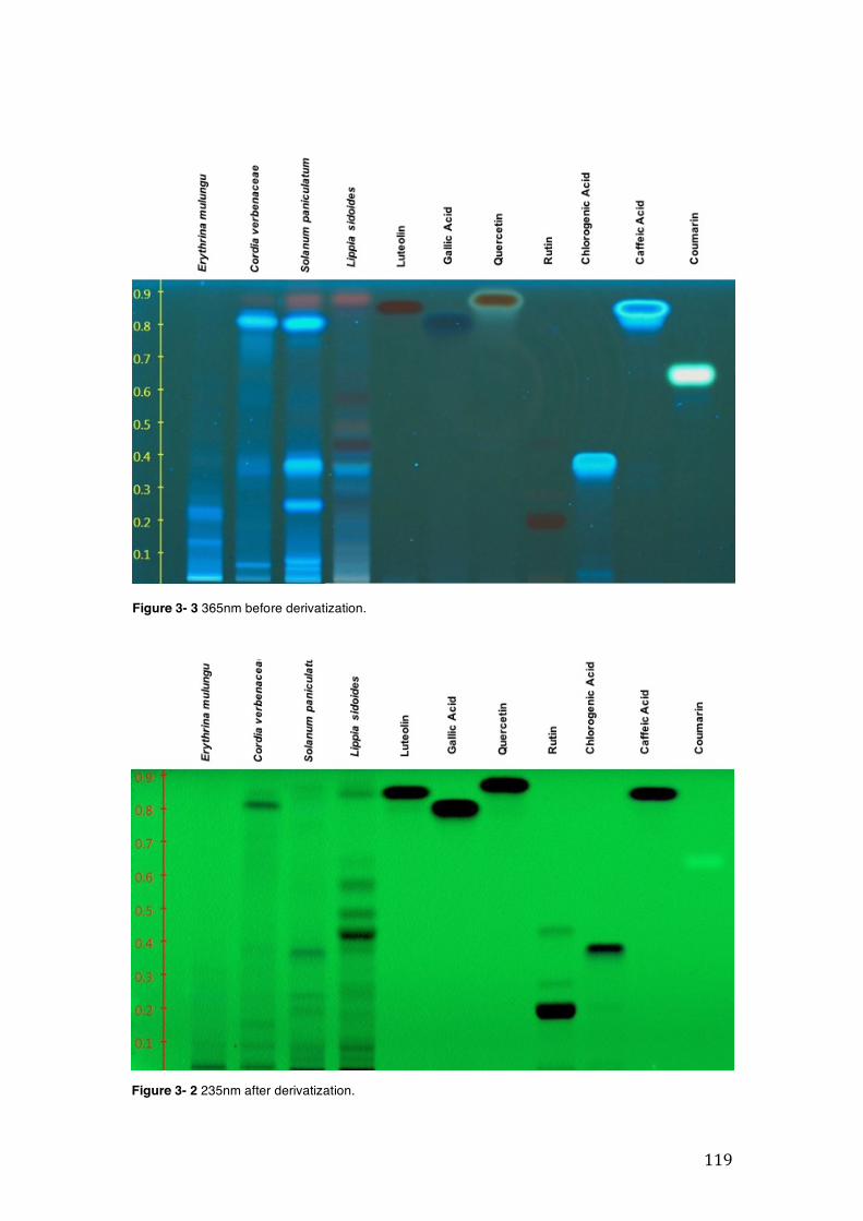
119
Figure 3- 3 365nm before derivatization.
Figure 3- 2 235nm after derivatization.
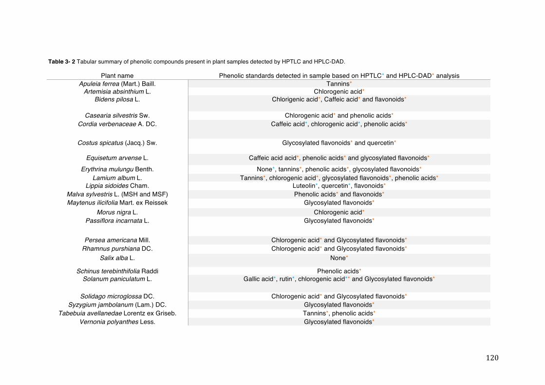
120
Table 3- 2 Tabular summary of phenolic compounds present in plant samples detected by HPTLC and HPLC-DAD.
Plant name Phenolic standards detected in sample based on HPTLC* and HPLC-DAD* analysis Apuleia ferrea (Mart.) Baill. Tannins*
Artemisia absinthium L. Chlorogenic acid* Bidens pilosa L. Chlorigenic acid*, Caffeic acid* and flavonoids*
Casearia silvestris Sw. Chlorogenic acid* and phenolic acids* Cordia verbenaceae A. DC. Caffeic acid*, chlorogenic acid*, phenolic acids*
Costus spicatus (Jacq.) Sw. Glycosylated flavonoids* and quercetin*
Equisetum arvense L. Caffeic acid acid*, phenolic acids* and glycosylated flavonoids* Erythrina mulungu Benth. None*, tannins*, phenolic acids*, glycosylated flavonoids*
Lamium album L. Tannins*, chlorogenic acid*, glycosylated flavonoids*, phenolic acids* Lippia sidoides Cham. Luteolin*, quercetin*, flavonoids*
Malva sylvestris L. (MSH and MSF) Phenolic acids* and flavonoids* Maytenus ilicifolia Mart. ex Reissek Glycosylated flavonoids*
Morus nigra L. Chlorogenic acid* Passiflora incarnata L. Glycosylated flavonoids*
Persea americana Mill. Chlorogenic acid* and Glycosylated flavonoids* Rhamnus purshiana DC. Chlorogenic acid* and Glycosylated flavonoids*
Salix alba L. None*
Schinus terebinthifolia Raddi Phenolic acids* Solanum paniculatum L. Gallic acid*, rutin*, chlorogenic acid** and Glycosylated flavonoids*
Solidago microglossa DC. Chlorogenic acid* and Glycosylated flavonoids* Syzygium jambolanum (Lam.) DC. Glycosylated flavonoids*
Tabebuia avellanedae Lorentz ex Griseb. Tannins*, phenolic acids* Vernonia polyanthes Less. Glycosylated flavonoids*
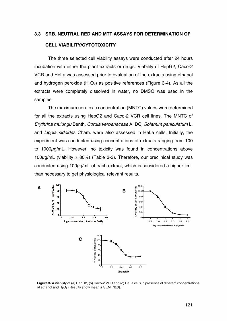
121
0 50 100 150 200 250 3000
20
40
60
80
100
120
[H2O2] uM
Cell
via
bilit
y (%
)
2.52.42.32.22.01.7log concentration of H2O2 (mM)
% V
iabi
lity
of C
aco-
2 VC
R c
ells
A
A
0.0 0.2 0.4 0.6 0.80
20
40
60
80
100
120
[Etanol] M
Cell
via
bilit
y (%
)%
Via
bility
of H
eLa
cells
3.3 SRB, NEUTRAL RED AND MTT ASSAYS FOR DETERMINATION OF
CELL VIABILITY/CYTOTOXICITY
The three selected cell viability assays were conducted after 24 hours incubation with either the plant extracts or drugs. Viability of HepG2, Caco-2 VCR and HeLa was assessed prior to evaluation of the extracts using ethanol and hydrogen peroxide (H2O2) as positive references (Figure 3-4). As all the extracts were completely dissolved in water, no DMSO was used in the samples.
The maximum non-toxic concentration (MNTC) values were determined for all the extracts using HepG2 and Caco-2 VCR cell lines. The MNTC of Erythrina mulungu Benth, Cordia verbenaceae A. DC, Solanum paniculatum L. and Lippia sidoides Cham. were also assessed in HeLa cells. Initially, the experiment was conducted using concentrations of extracts ranging from 100 to 1000µg/mL. However, no toxicity was found in concentrations above 100µg/mL (viability ³ 80%) (Table 3-3). Therefore, our preclinical study was conducted using 100µg/mL of each extract, which is considered a higher limit than necessary to get physiological relevant results. B
C
Figure 3- 4 Viability of (a) HepG2, (b) Caco-2 VCR and (c) HeLa cells in presence of different concentrations of ethanol and H2O2 (Results show mean ± SEM, N³3).
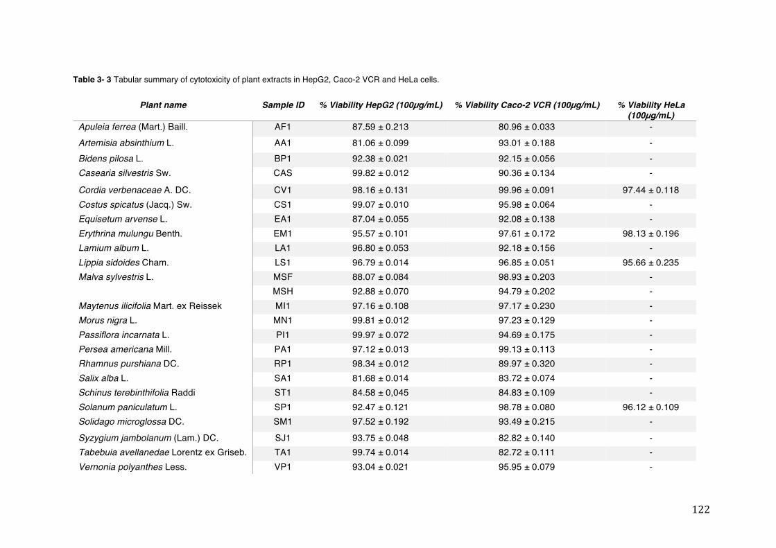
122
Plant name Sample ID % Viability HepG2 (100µg/mL) % Viability Caco-2 VCR (100µg/mL) % Viability HeLa (100µg/mL)
Apuleia ferrea (Mart.) Baill. AF1 87.59 ± 0.213 80.96 ± 0.033 - Artemisia absinthium L. AA1 81.06 ± 0.099 93.01 ± 0.188 - Bidens pilosa L. BP1 92.38 ± 0.021 92.15 ± 0.056 - Casearia silvestris Sw. CAS 99.82 ± 0.012 90.36 ± 0.134 -
Cordia verbenaceae A. DC. CV1 98.16 ± 0.131 99.96 ± 0.091 97.44 ± 0.118 Costus spicatus (Jacq.) Sw. CS1 99.07 ± 0.010 95.98 ± 0.064 - Equisetum arvense L. EA1 87.04 ± 0.055 92.08 ± 0.138 - Erythrina mulungu Benth. EM1 95.57 ± 0.101 97.61 ± 0.172 98.13 ± 0.196 Lamium album L. LA1 96.80 ± 0.053 92.18 ± 0.156 - Lippia sidoides Cham. LS1 96.79 ± 0.014 96.85 ± 0.051 95.66 ± 0.235 Malva sylvestris L. MSF 88.07 ± 0.084 98.93 ± 0.203 -
MSH 92.88 ± 0.070 94.79 ± 0.202 - Maytenus ilicifolia Mart. ex Reissek MI1 97.16 ± 0.108 97.17 ± 0.230 - Morus nigra L. MN1 99.81 ± 0.012 97.23 ± 0.129 - Passiflora incarnata L. PI1 99.97 ± 0.072 94.69 ± 0.175 - Persea americana Mill. PA1 97.12 ± 0.013 99.13 ± 0.113 - Rhamnus purshiana DC. RP1 98.34 ± 0.012 89.97 ± 0.320 - Salix alba L. SA1 81.68 ± 0.014 83.72 ± 0.074 - Schinus terebinthifolia Raddi ST1 84.58 ± 0,045 84.83 ± 0.109 - Solanum paniculatum L. SP1 92.47 ± 0.121 98.78 ± 0.080 96.12 ± 0.109 Solidago microglossa DC. SM1 97.52 ± 0.192 93.49 ± 0.215 -
Syzygium jambolanum (Lam.) DC. SJ1 93.75 ± 0.048 82.82 ± 0.140 - Tabebuia avellanedae Lorentz ex Griseb. TA1 99.74 ± 0.014 82.72 ± 0.111 - Vernonia polyanthes Less. VP1 93.04 ± 0.021 95.95 ± 0.079 -
Table 3- 3 Tabular summary of cytotoxicity of plant extracts in HepG2, Caco-2 VCR and HeLa cells.
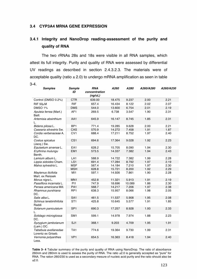
123
Table 3- 4 Tabular summary of the purity and quality of RNA using NanoDrop. The ratio of absorbance 260nm and 280nm is used to assess the purity of RNA. The ratio ±2.0 is generally accepted as “pure” for RNA. The ration 260/230 is used as a secondary mesure of nucleic acid purity and the ratio should also be ±2.0.
3.4 CYP3A4 MRNA GENE EXPRESSION
3.4.1 Integrity and NanoDrop reading-assessment of the purity and quality of RNA
The two rRNAs 28s and 18s were visible in all RNA samples, which attest its full integrity. Purity and quality of RNA were assessed by differential UV readings as described in section 2.4.3.2.3. The materials were of acceptable quality (ratio ± 2.0) to undergo mRNA amplification as seen in table 3-4.
Samples Sample ID
RNA concentration
(ng/uL)
A260 A280 A260/A280 A260/A230
Control (DMSO 0.2%) CTR 639.00 18.475 9.237 2.00 2.21 RIF 50µM RIF 657.4 16.434 8.122 2.02 2.07 DMSO 1% DMS 544.0 13.600 6.754 2.01 2.19 Apuleia ferrea (Mart.) Baill.
AF1 269.5 6.738 3.547 1.90 2.31
Artemisia absinthium L.
AA1 645.9 16.147 8.745 1.85 2.31
Bidens pilosa L. BP1 771.4 19.285 9.628 2.00 2.21 Casearia silvestris Sw. CAS 570.9 14.272 7.458 1.91 1.87 Cordia verbenaceae A. DC.
CV1 688.4 17.211 8.752 1.97 2.40
Costus spicatus (Jacq.) Sw.
CS1 694.6 17.364 9.028 1.92 2.23
Equisetum arvense L. EA1 628.2 15.705 8.090 1.94 2.30 Erythrina mulungu Benth.
EM1 573.5 14.337 7.382 1.94 2.43
Lamium album L. LA1 588.9 14.722 7.382 1.99 2.28 Lippia sidoides Cham. LS1 691.4 17.284 8.792 1.97 2.19 Malva sylvestris L. MSF 567.4 14.184 7.210 1.97 2.30
MSH 628.8 15.721 8.200 1.92 2.29 Maytenus ilicifolia Mart. ex Reissek
MI1 597.1 14.926 7.861 1.90 2.28
Morus nigra L. MN1 452.8 11.321 5.913 1.91 2.19 Passiflora incarnata L. PI1 747.9 18.696 10.089 1.86 2.30 Persea americana Mill. PA1 568.7 14.217 7.206 1.97 2.38 Rhamnus purshiana DC.
RP1 638.3 15.957 8.066 1.98 2.05
Salix alba L. SA1 461.5 11.537 5.908 1.95 2.08 Schinus terebinthifolia Raddi
ST1 425.8 10.645 5.577 1.91 1.85
Solanum paniculatum L.
SP1 690.3 17.257 8.928 1.93 2.33
Solidago microglossa DC.
SM1 599.1 14.978 7.974 1.88 2.23
Syzygium jambolanum (Lam.) DC.
SJ1 368.1 9.203 4.709 1.95 1.91
Tabebuia avellanedae Lorentz ex Griseb.
TA1 774.6 19.364 9.730 1.99 2.31
Vernonia polyanthes Less.
VP1 654.5 16.363 8.418 1.94 2.40
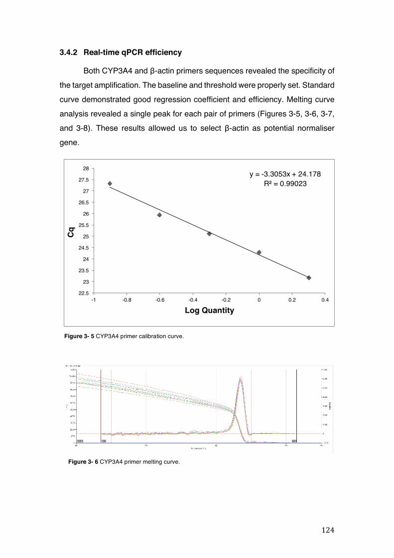
124
y = -3.3053x + 24.178R² = 0.99023
22.5
23
23.5
24
24.5
25
25.5
26
26.5
27
27.5
28
-1 -0.8 -0.6 -0.4 -0.2 0 0.2 0.4
Cq
Log Quantity
3.4.2 Real-time qPCR efficiency
Both CYP3A4 and β-actin primers sequences revealed the specificity of the target amplification. The baseline and threshold were properly set. Standard curve demonstrated good regression coefficient and efficiency. Melting curve analysis revealed a single peak for each pair of primers (Figures 3-5, 3-6, 3-7, and 3-8). These results allowed us to select β-actin as potential normaliser gene.
Figure 3- 5 CYP3A4 primer calibration curve.
Figure 3- 6 CYP3A4 primer melting curve.

125
y = -3.3219x + 16.863R² = 0.94656
15
16
17
18
19
20
21
-1 -0.8 -0.6 -0.4 -0.2 0 0.2 0.4
Cq
Log Quantity
Figure 3- 8 b-actin primer calibration curve.
Figure 3- 7 b-actin primer melting curve.
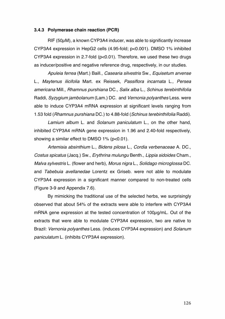
126
3.4.3 Polymerase chain reaction (PCR)
RIF (50µM), a known CYP3A4 inducer, was able to significantly increase CYP3A4 expression in HepG2 cells (4.95-fold; p<0.001). DMSO 1% inhibited CYP3A4 expression in 2.7-fold (p<0.01). Therefore, we used these two drugs as inducer/positive and negative reference drug, respectively, in our studies.
Apuleia ferrea (Mart.) Baill., Casearia silvestris Sw., Equisetum arvense
L., Maytenus ilicifolia Mart. ex Reissek, Passiflora incarnata L., Persea
americana Mill., Rhamnus purshiana DC., Salix alba L., Schinus terebinthifolia
Raddi, Syzygium jambolanum (Lam.) DC. and Vernonia polyanthes Less. were able to induce CYP3A4 mRNA expression at significant levels ranging from 1.53 fold (Rhamnus purshiana DC.) to 4.88-fold (Schinus terebinthifolia Raddi).
Lamium album L. and Solanum paniculatum L., on the other hand, inhibited CYP3A4 mRNA gene expression in 1.96 and 2.40-fold respectively, showing a similar effect to DMSO 1% (p<0.01).
Artemisia absinthium L., Bidens pilosa L., Cordia verbenaceae A. DC., Costus spicatus (Jacq.) Sw., Erythrina mulungu Benth., Lippia sidoides Cham., Malva sylvestris L. (flower and herb), Morus nigra L., Solidago microglossa DC. and Tabebuia avellanedae Lorentz ex Griseb. were not able to modulate CYP3A4 expression in a significant manner compared to non-treated cells (Figure 3-9 and Appendix 7.6).
By mimicking the traditional use of the selected herbs, we surprisingly observed that about 54% of the extracts were able to interfere with CYP3A4 mRNA gene expression at the tested concentration of 100µg/mL. Out of the extracts that were able to modulate CYP3A4 expression, two are native to Brazil: Vernonia polyanthes Less. (induces CYP3A4 expression) and Solanum
paniculatum L. (inhibits CYP3A4 expression).
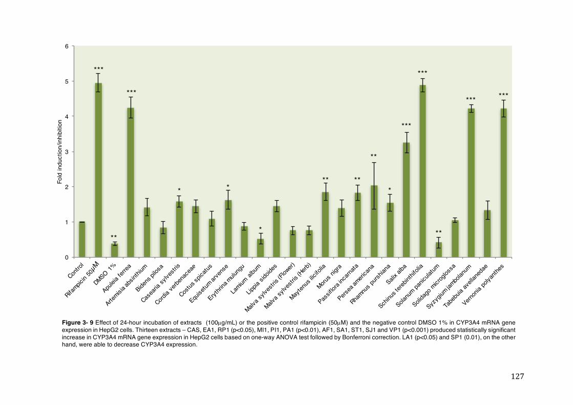
127
0
1
2
3
4
5
6
Fold
indu
ctio
n/in
hibi
tion
***
**
***
* *
*
** **
**
*
***
***
**
*** ***
Figure 3- 9 Effect of 24-hour incubation of extracts (100µg/mL) or the positive control rifampicin (50µM) and the negative control DMSO 1% in CYP3A4 mRNA gene expression in HepG2 cells. Thirteen extracts – CAS, EA1, RP1 (p<0.05), MI1, PI1, PA1 (p<0.01), AF1, SA1, ST1, SJ1 and VP1 (p<0.001) produced statistically significant increase in CYP3A4 mRNA gene expression in HepG2 cells based on one-way ANOVA test followed by Bonferroni correction. LA1 (p<0.05) and SP1 (0.01), on the other hand, were able to decrease CYP3A4 expression.
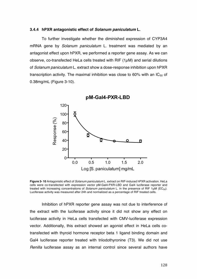
128
3.4.4 hPXR antagonistic effect of Solanum paniculatum L.
To further investigate whether the diminished expression of CYP3A4 mRNA gene by Solanum paniculatum L. treatment was mediated by an antagonist effect upon hPXR, we performed a reporter gene assay. As we can observe, co-transfected HeLa cells treated with RIF (1µM) and serial dilutions of Solanum paniculatum L. extract show a dose-response inhibition upon hPXR transcription activity. The maximal inhibition was close to 60% with an IC50 of 0.38mg/mL (Figure 3-10).
Figure 3- 10 Antagonistic effect of Solanum paniculatum L. extract on RIF-induced hPXR activation. HeLa cells were co-transfected with expression vector pM-Gal4-PXR-LBD and Gal4 luciferase reporter and treated with increasing concentrations of Solanum paniculatum L. in the presence of RIF 1μM (EC50). Luciferase activity was measured after 24h and normalized as a percentage of RIF treated cells.
Inhibition of hPXR reporter gene assay was not due to interference of
the extract with the luciferase activity since it did not show any effect on luciferase activity in HeLa cells transfected with CMV-luciferase expression vector. Additionally, this extract showed an agonist effect in HeLa cells co-transfected with thyroid hormone receptor beta 1 ligand binding domain and Gal4 luciferase reporter treated with triiodothyronine (T3). We did not use Renilla luciferase assay as an internal control since several authors have
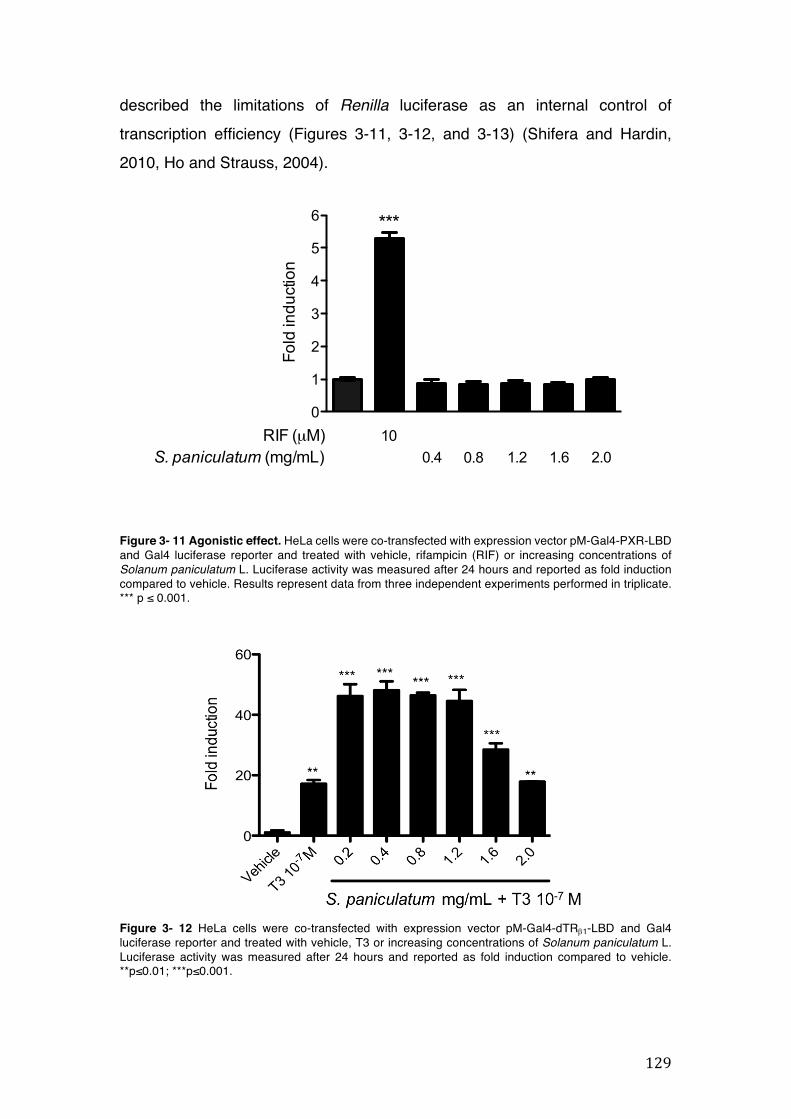
129
pM-Gal4-PXR-LBD
0
1
2
3
4
5
6
RIF (µM)S. paniculatum (mg/mL) 0.80.4 1.2 1.6 2.0
10
***
HeLa cells were co-transfected with expression vector pM-Gal4-PXR-LBD and Gal4 luciferase reporter and treated with vehiche, Rifampicin (RIF) or increasing concentrations of E. mulung. Cells were lysed after 24 hours and luciferase activity was measured and reporter as fold induction compared to vehiche.
Fold
indu
ctio
n
described the limitations of Renilla luciferase as an internal control of transcription efficiency (Figures 3-11, 3-12, and 3-13) (Shifera and Hardin, 2010, Ho and Strauss, 2004).
Figure 3- 11 Agonistic effect. HeLa cells were co-transfected with expression vector pM-Gal4-PXR-LBD and Gal4 luciferase reporter and treated with vehicle, rifampicin (RIF) or increasing concentrations of Solanum paniculatum L. Luciferase activity was measured after 24 hours and reported as fold induction compared to vehicle. Results represent data from three independent experiments performed in triplicate. *** p ≤ 0.001.
Figure 3- 12 HeLa cells were co-transfected with expression vector pM-Gal4-dTRb1-LBD and Gal4 luciferase reporter and treated with vehicle, T3 or increasing concentrations of Solanum paniculatum L. Luciferase activity was measured after 24 hours and reported as fold induction compared to vehicle. **p≤0.01; ***p≤0.001.
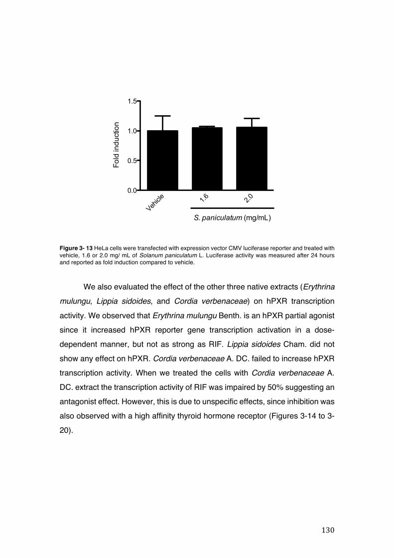
130
pCMV-Luc
Vehicl
e 1.6 2.00.0
0.5
1.0
1.5
S. paniculatum (mg/mL)
Fold
indu
ctio
n
Figure 3- 13 HeLa cells were transfected with expression vector CMV luciferase reporter and treated with vehicle, 1.6 or 2.0 mg/ mL of Solanum paniculatum L. Luciferase activity was measured after 24 hours and reported as fold induction compared to vehicle.
We also evaluated the effect of the other three native extracts (Erythrina
mulungu, Lippia sidoides, and Cordia verbenaceae) on hPXR transcription activity. We observed that Erythrina mulungu Benth. is an hPXR partial agonist since it increased hPXR reporter gene transcription activation in a dose-dependent manner, but not as strong as RIF. Lippia sidoides Cham. did not show any effect on hPXR. Cordia verbenaceae A. DC. failed to increase hPXR transcription activity. When we treated the cells with Cordia verbenaceae A. DC. extract the transcription activity of RIF was impaired by 50% suggesting an antagonist effect. However, this is due to unspecific effects, since inhibition was also observed with a high affinity thyroid hormone receptor (Figures 3-14 to 3-20).

131
Mazzari et al. 2016 Supplementary Data
8
HeLa Cells
0
1
2
3
4
5
6
7
RIF (µM)E. mulungu (mg/mL) 0.1 0.2 0.4 0.8 1.2 2.0
***
*
HeLa cells were co-transfected with expression vector pM-Gal4-PXR-LBD and Gal4 luciferase reporter and treated with vehiche, Rifampicin (RIF) or increasing concentrations of E. mulungu. Cells were lysed after 24 hours and luciferase activity was measured and reporter as fold induction compared to vehiche.
101.6
Fold
indu
ctio
n
* ** **
CMV-Luc
Vehicl
e 1.6 2.00.0
0.5
1.0
1.5
[S. paniculatum] mg/mL
Fold
indu
ctio
n
Figure S 10 HeLa cells were transfected with expression vector CMV luciferase reporter and treated with S. paniculatum. Lucifearase activity was measured after 24 hours and reporter as fold induction compared to vehicle.
4.2 ErythrinamulungudisplaysapartialagonisteffectonhPXR
Figure S 11 Agonism effect assay. HeLa cells were co-transfected with expression vector pM-Gal4-PXR-LBD and Gal4 luciferase reporter and treated with vehicle, Rifampicin or increasing concentrations of E. mulungu. Lucifearase activity was measured after 24 hours and reporter as fold induction compared to vehicle. **p ≤ 0.01; ***p ≤ 0.001.
E. mulungu (mg/mL)
Vehicl
e
RIF 10-5 M
0.10
0.15
0.20
0.25
0.30
0
1
2
3
4
5
6
***
L. sidoides (mg/mL)
pM-Gal4-PXR-LBD
Fold
indu
ctio
n
3.4.6 Erythrina mulungu Benth. displays a partial agonistic effect on hPXR luciferase reporter gene assay.
Figure 3- 14 Agonistic effect. HeLa cells were co-transfected with expression vector pM-Gal4-PXR-LBD and Gal4 luciferase reporter and treated with vehicle, RIF or increasing concentrations of Erythrina mulungu Benth. Luciferase activity was measured after 24 hours and reported as fold induction compared to vehicle. *p ≤0.05; **p ≤ 0.01; ***p ≤ 0.001.
3.4.7 Lippia sidoides Cham. does not display any effect on hPXR luciferase reporter gene assay.
Figure 3- 15 Agonistic effect. HeLa cells were co-transfected with expression vector pM-Gal4-PXR-LBD and Gal4 luciferase reporter and treated with vehicle, RIF or increasing concentrations of Lippia sidoides Cham. Luciferase activity was measured after 24 hours and reported as fold induction compared to vehicle. ***p ≤ 0.001.
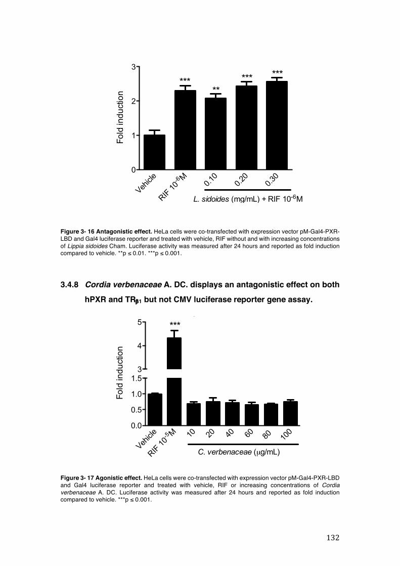
132
Vehicl
e
RIF 10
-6 M0.1
00.2
00.3
00
1
2
3
L. sidoides (mg/mL) + RIF 10-6M
*****
*** ***
pM-Gal4-PXR-LBD
Fold
indu
ctio
n
Vehicl
e
RIF 10
-5 M 10 20 40 60 80
100
0.0
0.5
1.0
1.53
4
5pMGal4PXRLBD
***
C. verbenaceae (µg/mL)
Fold
indu
ctio
n
Figure 3- 16 Antagonistic effect. HeLa cells were co-transfected with expression vector pM-Gal4-PXR-LBD and Gal4 luciferase reporter and treated with vehicle, RIF without and with increasing concentrations of Lippia sidoides Cham. Luciferase activity was measured after 24 hours and reported as fold induction compared to vehicle. **p ≤ 0.01. ***p ≤ 0.001.
3.4.8 Cordia verbenaceae A. DC. displays an antagonistic effect on both hPXR and TRb1 but not CMV luciferase reporter gene assay.
Figure 3- 17 Agonistic effect. HeLa cells were co-transfected with expression vector pM-Gal4-PXR-LBD and Gal4 luciferase reporter and treated with vehicle, RIF or increasing concentrations of Cordia verbenaceae A. DC. Luciferase activity was measured after 24 hours and reported as fold induction compared to vehicle. ***p ≤ 0.001.
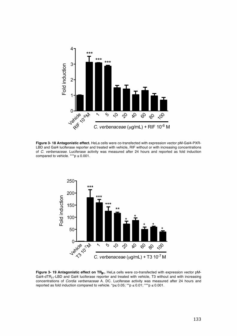
133
Vehicl
e
RIF 10
-6 M 1 5 10 20 40 60 80
100
0
1
2
3
4
pMGal4PXRLBD
C. verbenaceae (µg/mL) + RIF 10-6 M
****** ***
Fold
indu
ctio
n
Figure 3- 18 Antagonistic effect. HeLa cells were co-transfected with expression vector pM-Gal4-PXR-LBD and Gal4 luciferase reporter and treated with vehicle, RIF without or with increasing concentrations of C. verbenaceae. Luciferase activity was measured after 24 hours and reported as fold induction compared to vehicle. ***p ≤ 0.001.
Figure 3- 19 Antagonistic effect on TRb1. HeLa cells were co-transfected with expression vector pM-Gal4-dTRb1-LBD and Gal4 luciferase reporter and treated with vehicle, T3 without and with increasing concentrations of Cordia verbenaceae A. DC. Luciferase activity was measured after 24 hours and reported as fold induction compared to vehicle. *p≤ 0.05; **p ≤ 0.01; ***p ≤ 0.001.
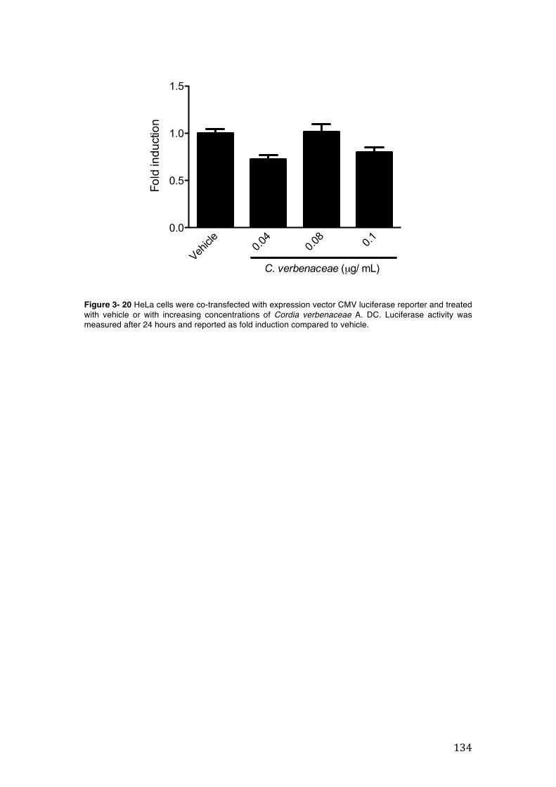
134
CMV-Luc
Vehicl
e0.0
40.0
8 0.10.0
0.5
1.0
1.5
C. verbenaceae (µg/ mL)
Fold
indu
ctio
n
Figure 3- 20 HeLa cells were co-transfected with expression vector CMV luciferase reporter and treated with vehicle or with increasing concentrations of Cordia verbenaceae A. DC. Luciferase activity was measured after 24 hours and reported as fold induction compared to vehicle.
135
3.5 ESTIMATION OF INTRACELLULAR GSH LEVELS IN HEPG2 CELLS
The rate of change in the kinetic absorbance of TNB for the different GSH standard concentrations is linearly proportional to the amount of GSH in each sample. Thus, a graph plot of the slopes of each GSH standard concentration against the GSH concentration, otherwise known as the F-slope curve can be used to estimate the amount of GSH in the treated wells given their individual slopes (Figures 3-21 and 3-22).
The GSH concentration obtained for the control cells was 29.91 ± 0.36µM. Twenty extracts- Apuleia ferrea (Mart.) Baill., Bidens pilosa L., Casearia silvestris Sw., Cordia verbenaceae A. DC., Costus spicatus (Jacq.) Sw., Equisetum arvense L., Lamium album L., Lippia sidoides Cham., Malva
sylvestris L. (flower and herb), Maytenus ilicifolia Mart. Ex Reissek, Passiflora
incarnata L., Persea americana Mill., Rhamnus purshiana DC., Salix alba L., Schinus terebinthifolia Raddi, Solanum paniculatum L., Solidago microglossa
DC., Tabebuia avellanedae Lorentz ex Griseb. and Vernonia polyanthes Less. - decreased the intracellular GSH concentration beyond levels seen in the control cells. This decrease was found to be statistically significant (P£0.05, 0.01 and 0.001). The majority of the extracts decreased GSH levels at a comparable level to that produced by the positive control BSO, which decreased intracellular GSH levels to 17.78 ± 1.70µM (P£0.001). Only one extract - Erythrina mulungu Benth. - was able to increase GSH levels at a significant level 47.05 ± 2.0 (Figure 3-23 and Appendix 7.7).
Similar to what has been observed on CYP3A4 mRNA gene expression, native Brazilian extracts were also able to cause changes in GSH levels. Cordia
verbenaceae A. DC., Lippia sidoides Cham., and Solanum paniculatum L. depleted GSH levels whereas Erythrina mulungu Benth. displayed the opposite effect.
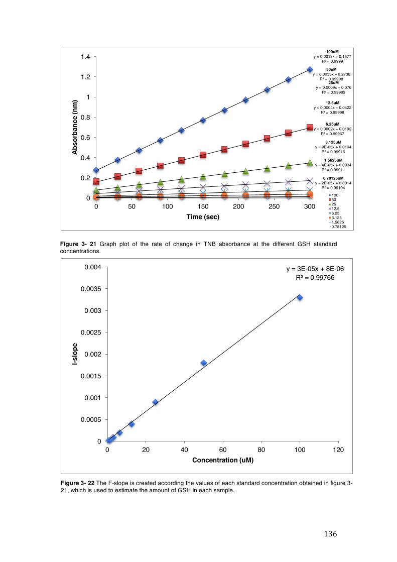
136
50uMy = 0.0033x + 0.2738
R² = 0.99998
100uMy = 0.0018x + 0.1577
R² = 0.9999
25uMy = 0.0009x + 0.076
R² = 0.99989
12.5uMy = 0.0004x + 0.0422
R² = 0.99998
6.25uMy = 0.0002x + 0.0192
R² = 0.99967
3.125uMy = 9E-05x + 0.0104
R² = 0.99916
1.5625uMy = 4E-05x + 0.0034
R² = 0.99911
0.78125uMy = 2E-05x + 0.0014
R² = 0.99104
0
0.2
0.4
0.6
0.8
1
1.2
1.4
0 50 100 150 200 250 300
Abso
rban
ce (n
m)
Time (sec)
100502512.56.253.1251.56250.78125
y = 3E-05x + 8E-06R² = 0.99766
0
0.0005
0.001
0.0015
0.002
0.0025
0.003
0.0035
0.004
0 20 40 60 80 100 120
i-slo
pe
Concentration (uM)
Figure 3- 22 The F-slope is created according the values of each standard concentration obtained in figure 3-21, which is used to estimate the amount of GSH in each sample.
Figure 3- 21 Graph plot of the rate of change in TNB absorbance at the different GSH standard concentrations.
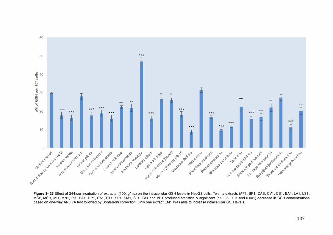
137
0
10
20
30
40
50
60µM
of G
SH p
er 1
05ce
lls
*** *** *** ******
** **
***
***
* *
***
***
***
******
**
*** ***
**
***
***
Figure 3- 23 Effect of 24-hour incubation of extracts (100µg/mL) on the intracellular GSH levels in HepG2 cells. Twenty extracts (AF1, BP1, CAS, CV1, CS1, EA1, LA1, LS1, MSF, MSH, MI1, MN1, PI1, PA1, RP1, SA1, ST1, SP1, SM1, SJ1, TA1 and VP1 produced statistically significant (p£0.05, 0.01 and 0.001) decrease in GSH concentrations based on one-way ANOVA test followed by Bonferroni correction. Only one extract EM1 Was able to increase intracellular GSH levels.
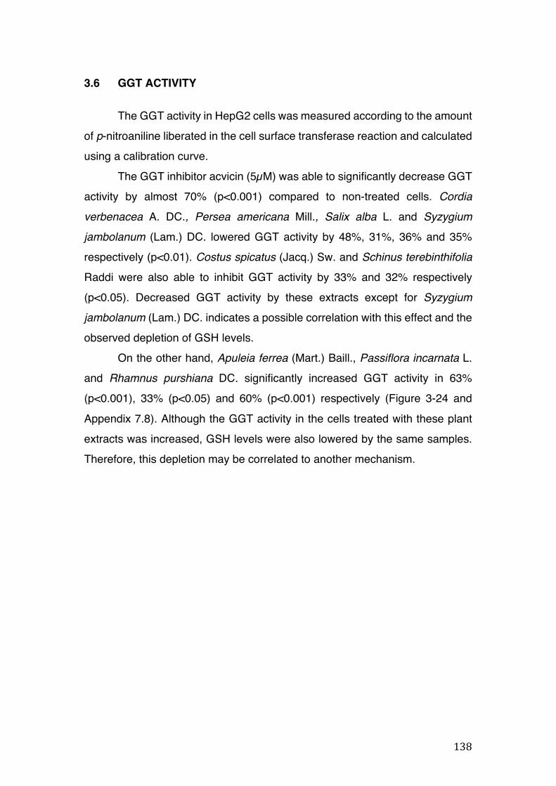
138
3.6 GGT ACTIVITY
The GGT activity in HepG2 cells was measured according to the amount of p-nitroaniline liberated in the cell surface transferase reaction and calculated using a calibration curve.
The GGT inhibitor acvicin (5µM) was able to significantly decrease GGT activity by almost 70% (p<0.001) compared to non-treated cells. Cordia
verbenacea A. DC., Persea americana Mill., Salix alba L. and Syzygium
jambolanum (Lam.) DC. lowered GGT activity by 48%, 31%, 36% and 35% respectively (p<0.01). Costus spicatus (Jacq.) Sw. and Schinus terebinthifolia Raddi were also able to inhibit GGT activity by 33% and 32% respectively (p<0.05). Decreased GGT activity by these extracts except for Syzygium
jambolanum (Lam.) DC. indicates a possible correlation with this effect and the observed depletion of GSH levels.
On the other hand, Apuleia ferrea (Mart.) Baill., Passiflora incarnata L. and Rhamnus purshiana DC. significantly increased GGT activity in 63% (p<0.001), 33% (p<0.05) and 60% (p<0.001) respectively (Figure 3-24 and Appendix 7.8). Although the GGT activity in the cells treated with these plant extracts was increased, GSH levels were also lowered by the same samples. Therefore, this depletion may be correlated to another mechanism.
139
0
20
40
60
80
100
120
140
160
µM p
-nitr
oani
line
per 1
06ce
lls
**
***
***
*
*
**
***
*** **
Figure 3- 24 Effect of 24-hour incubation of extracts (100µg/mL) on the GGT activity in HepG2 cells. Six extracts – CS1, ST1 (p<0.05), CV1, PA1, SA1, SJ1 (p<0.001) produced statistically significant decrease in GGT activity based on one-way ANOVA test followed by Bonferroni correction. PI1 (p<0.05), AF1 and RP1 (p<0.001), on the other hand, were able to increase GGT activity.
140
3.7 MODULATION OF P-GLYCOPROTEIN DEPENDENT RH-123 EFFLUX
IN CACO-2 VCR CELLS
The intracellular Rh-123 fluorescence in the cells incubated with Apuleia
ferrea (Mart.) Baill., Salix alba L., Schinus terebinthifolia Raddi and Syzygium
jambolanum (Lam.) DC. was significantly higher than cells treated only with Rh-
123. This indicates increased efflux inhibition by these extracts.
The effect of Salix alba L. (192.5%) and Syzygium jambolanum (Lam.) DC.
(182.39%) in the efflux activity was relatively similar to the one found in the
positive control verapamil (181.78%). The Rh-123 fluorescence in Caco-2 VCR
cells was higher in Apuleia ferrea (Mart.) Baill. (412.72%) and Schinus
terebinthifolia Raddi (442.61%), indicating strong efflux inhibition caused by
these extracts (Figure 3-25 and Appendix 7.9).
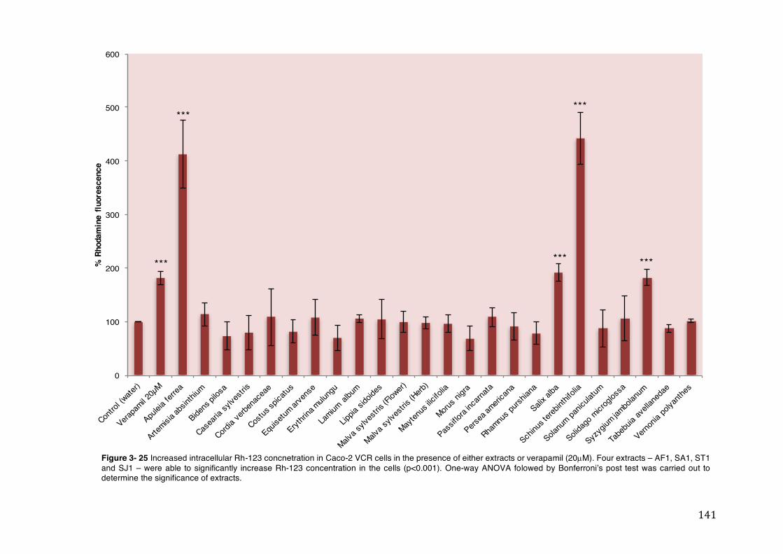
141
0
100
200
300
400
500
600
% R
hoda
min
e flu
ores
cenc
e***
*** ***
***
***
Figure 3- 25 Increased intracellular Rh-123 concnetration in Caco-2 VCR cells in the presence of either extracts or verapamil (20µM). Four extracts – AF1, SA1, ST1 and SJ1 – were able to significantly increase Rh-123 concentration in the cells (p<0.001). One-way ANOVA folowed by Bonferroni’s post test was carried out to determine the significance of extracts.
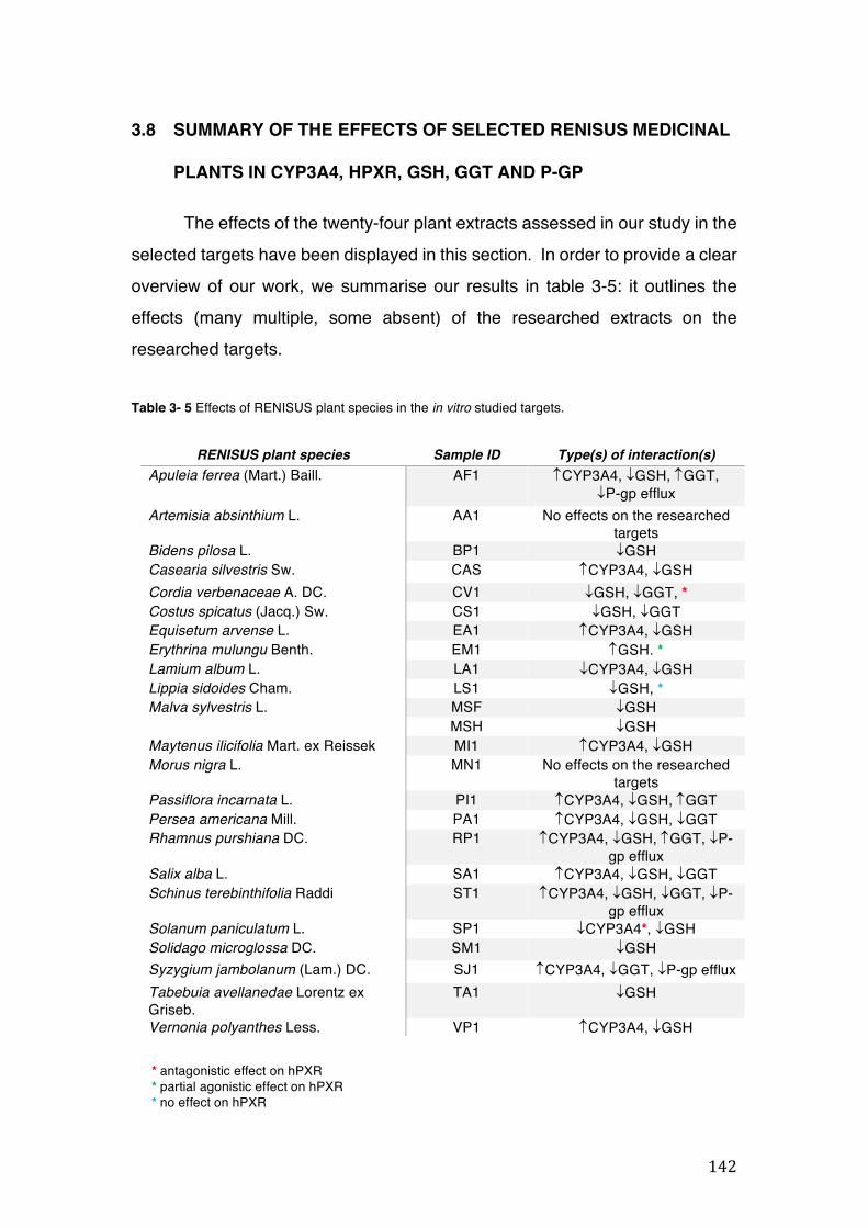
142
3.8 SUMMARY OF THE EFFECTS OF SELECTED RENISUS MEDICINAL
PLANTS IN CYP3A4, HPXR, GSH, GGT AND P-GP
The effects of the twenty-four plant extracts assessed in our study in the selected targets have been displayed in this section. In order to provide a clear overview of our work, we summarise our results in table 3-5: it outlines the effects (many multiple, some absent) of the researched extracts on the researched targets.
Table 3- 5 Effects of RENISUS plant species in the in vitro studied targets.
RENISUS plant species Sample ID Type(s) of interaction(s) Apuleia ferrea (Mart.) Baill. AF1 CYP3A4, ¯GSH, GGT,
¯P-gp efflux Artemisia absinthium L. AA1 No effects on the researched
targets Bidens pilosa L. BP1 ¯GSH Casearia silvestris Sw. CAS CYP3A4, ¯GSH Cordia verbenaceae A. DC. CV1 ¯GSH, ¯GGT, * Costus spicatus (Jacq.) Sw. CS1 ¯GSH, ¯GGT Equisetum arvense L. EA1 CYP3A4, ¯GSH Erythrina mulungu Benth. EM1 GSH. * Lamium album L. LA1 ¯CYP3A4, ¯GSH Lippia sidoides Cham. LS1 ¯GSH, * Malva sylvestris L. MSF ¯GSH
MSH ¯GSH Maytenus ilicifolia Mart. ex Reissek MI1 CYP3A4, ¯GSH Morus nigra L. MN1 No effects on the researched
targets Passiflora incarnata L. PI1 CYP3A4, ¯GSH, GGT Persea americana Mill. PA1 CYP3A4, ¯GSH, ¯GGT Rhamnus purshiana DC. RP1 CYP3A4, ¯GSH, GGT, ¯P-
gp efflux Salix alba L. SA1 CYP3A4, ¯GSH, ¯GGT Schinus terebinthifolia Raddi ST1 CYP3A4, ¯GSH, ¯GGT, ¯P-
gp efflux Solanum paniculatum L. SP1 ¯CYP3A4*, ¯GSH Solidago microglossa DC. SM1 ¯GSH Syzygium jambolanum (Lam.) DC. SJ1 CYP3A4, ¯GGT, ¯P-gp efflux Tabebuia avellanedae Lorentz ex Griseb.
TA1 ¯GSH
Vernonia polyanthes Less. VP1 CYP3A4, ¯GSH
* antagonistic effect on hPXR * partial agonistic effect on hPXR * no effect on hPXR

143
4 GENERAL DISCUSSION
144
4.1 IN VITRO STUDIES OF RENISUS MEDICINAL PLANTS IN THE
SELECTED TARGETS
We present for the first time in vitro preclinical data for a selection of RENISUS medicinal plants. These plants were assessed for their effects on selected metabolic and transporter targets as a step towards producing their potential HDI profile. While some of these plants are native to Brazil, the majority are consumed all around the world by many people on a daily basis; these results, therefore, not only may help Brazil to better integrate the use of these plants into clinical practice, but will also certainly prompt further research in Europe, North America and worldwide.
It was necessary to balance a number of factors in the experimental design. These factors included the nature of the samples (herbal extracts), their number, as well as a lack of consensus between the academy, regulatory bodies, and industry.
Our results show that 22 out of the 24 plant extracts screened have a significant effect with at least one of the selected targets and therefore have the potential to interact with essential drugs prescribed in Brazil. These metabolic HDI may also be complicated by intrinsic factors such as genetic polymorphisms, disease, age, and sex.
Taking into account all these aspects, we will discuss in the following pages the rationale behind our experimental approach, the interpretation of our results within the context of HDI, and their potential clinical impact in the case of Brazil.
145
4.1.1 Experimental approaches to the in vitro metabolism and transport studies
Drug metabolism studies are an essential step in the preclinical development of medicines. To this end, in vitro models must provide vital information on the effects of the lead drug candidate (LDC) upon drug metabolising enzymes, such as the CYP, as a step towards predicting potential DDI. Many in vitro experimental models of drug metabolism and disposition make use of cells; HH or their microsomes (HLM), for example, are considered the best models for enzyme inhibition, whereas HLM is the preferred test system for enzyme induction. Caco-2 cells are a common choice for transporter studies (Zhang et al., 2012a). Several other models have been successfully developed to replace the use of cells, and have been validated for predicting the effects of single drug entities in the metabolism with similar correlation to HLM. Such is the case of characterised metabolic recombinant enzymes (CYP1A2, 2C9, 2C19, 2D6, and 3A4) expressed in Escherichia coli, which has been developed by the pharmaceutical company AstraZeneca (McGinnity and Riley, 2001). The use of in silico models has become more common in the pharmaceutical industry in recent years to speed up the selection of the best lead candidates at this stage of drug development (Steinmetz and Spack, 2009).
In order to be accepted by regulatory bodies, drug metabolism models must be robust, validated and reproducible. However, unlike the requirements for synthetic drug studies, there is no regulatory requirement for preclinical metabolism studies of traditional herbal medicines and no substantial industrial activity exists on this side. Metabolism studies on traditional herbal medicines are normally only initiated upon the receipt of case reports documenting a supposed interaction, which trigger warnings from the regulators. Otherwise, such studies originate from purely academic-driven programmes focused on demonstrating the potential effects of traditional herbal medicines on metabolic and transporter targets (Brantley et al., 2014); these studies are generally carried out by adapting protocols to their own needs, thus leading to an array of different experimental approaches.
146
The established principles and models used for metabolism studies in synthetic drugs are directly applicable to herbal medicines (He et al., 2010). However, they would require significantly high budgets and infrastructure. Due to the particular scope of the academic activities, a case can thus be made regarding the choice of cell lines. HH are generally replaced by other cell models like HepG2 and HepaRG cells for determination of metabolic activities. The hepatoma cell line HepG2 is one of the most popular models for pharmacological and toxicological in vitro studies in academia despite having lost some liver functions, and its supply is readily available from any public cell bank (Guo et al., 2011). The HepaRG cell line is derived from another hepatocellular carcinoma cell and was selected for our project because it maintains important metabolic activities lost in HepG2 cells. But it is significantly more expensive and only available through a private company and still has lost many functions when compared to HH (Szabo et al., 2013).
The high costs of HepaRG and HH cells forces many researchers to use the cheaper HepG2 cell line. A vial containing 2-3 x 10⁶ HepG2 cells costs around 330 GBP and stocks can be made for further use whenever they are needed. On the other hand, a vial containing 10 million HepaRG cells would cost nearly double at about 600 GBP, and cells would have to be plated out in 24-well plates for immediate use. Furthermore, HepG2 is an immortal cell line and therefore it is suitable for long-term maintenance. It generally reaches 70 – 80% confluence within 48 hours, which allows the conduction of medium and high throughput screenings. Long-term maintenance of HepaRG and HH with high availability, on the other hand, could be very challenging in traditional two-dimensional monolayer cultivation systems (Klein et al., 2014).
Caco-2 cells are one of the most used in vitro models for the assessment of P-gp transporter activity within most pharmaceutical industries (Eneroth et al., 2001) and reports of use of Caco-2 for transporter studies in herbal medicines have been also well documented (Li et al., 2014, Wu et al., 2016, Yoshida et al., 2006). The reasons for choosing this cell model for efflux studies are similar to those that were applied to HepG2: the reduced costs and rapid availability for immediate use.
147
The budget available for preclinical studies and the number of samples to be analysed, therefore, limit the approaches that researchers may select in terms of cell models and methodologies applied to them. In our case, the annual budget of 5,000 GBP per year (3 years) restricted us to the screening of a maximum of twenty-four plant extracts in six different cell-based assays using both HepG2 and Caco-2 cells. Samples in triplicates and quadruplicates had to be run at least three times to optimise experiments. With the exception of real-time polymerase chain reaction (RT-PCR), our experiments were carried out using more affordable colorimetric and fluorimetric assays in 96-well plates in order to maximise the number of samples analysed per time.
GSH is unique among the phase II conjugation mechanisms due to its role in the conjugation of phase I metabolites and in the protection of the body against several diseases. This was determinant for the selection of this target in our studies. P-gp was chosen due to its well-known efflux activity that can affect the absorption of many drugs; indeed, this is an important studied target in the industry during drug discovering stage.
Conduction of CYP3A4 gene expression was made using RT-PCR, which is the most used technique for such a study. Due to the costs of RT-PCR reagents, it was necessary to focus our phase I metabolism study on the expression of CYP3A4 despite other CYPs because it is responsible for metabolising more than 50% of the currently marketed drugs. The fundamentals of RT-PCR have been previously covered in section 2.4.3. Despite the availability of several protocols for HH, the numerous steps and reagents used to produce the cDNA and to amplify the CYP3A4 gene had to be all optimised as this technique is highly dependent on the instrumentation and the cell line.
The studies found in the literature made use of different concentrations of the gene inducer, total RNA and time of the treatment to achieve significant levels of CYP3A4 expression with different cell lines. For instance, Li and coworkers (2013) used RT-PCR for determination of the CYP expression in HepG2 cells by using 1µg of total RNA. The amount of RIF used to achieve 2.5 fold induction was 10µM for 48 hours treatment (Li et al., 2013). Modarai and
148
coworkers (2011), on the other hand, needed to use 2.5µg of total RNA and RIF 50µM treatment over a period of 96 hours of exposition to achieve 3.83-fold induction also in HepG2 cells (Modarai et al., 2011a). Lau and coworkers (2013) conducted a research on the effects of ethanol extracts of the Chinese herbs Oldenlandia diffusa (Willd.) Roxb. (Rubiaceae), Codonopsis tangshen Oliv. (Campanulaceae), Rehmannia glutinosa (Gaertn.) DC. (Plantaginaceae) and Astragalus propinquus Schischkin (Leguminosae) on CYP3A4 expression using human colon adenocarcinoma-derived LS180 cells. In that experiment, the amount of RNA used for cDNA synthesis was not disclosed but a 24 hours treatment with RIF 10µM was reportedly enough to achieve 8-fold induction (Lau et al., 2013). Likewise, Liu and coworkers (2012) did not indicate the amount of total RNA used but their treatment of the colon adenocarcinoma LS174T cells with RIF 20µM for 24 hours resulted in 3-fold induction (Liu et al., 2012).
Contrary to the study conducted by Li and coworkers (2013), the treatment of our HepG2 cells using RIF 10µM for 48 hours did not significantly change the CYP3A4 expression using 1µg of total RNA. In order to increase CYP3A4 expression using this inducer, some researchers may decide to increase the amount of total RNA amplified, as was the case in Modarai and coworkers (2011). However, we managed to achieve almost 5-fold induction by treating our HepG2 cells using the same RIF concentration for the same time treatment as Modarai and coworkers (2011) and the same total RNA concentration used by Li and coworkers (2013).
The achievement of the optimum protocol for CYP3A4 expression using HepG2 allowed the assessment of the twenty-four plant extracts. Solanum paniculatum L. and Lamium album L. were the only herbal extracts that decreased CYP3A4 gene expression. In section 3.4.4 we demonstrated that this down-regulation in Solanum paniculatum L. is due - at least in part - to an antagonistic effect of the plant extract on hPXR (Mazzari et al., 2016).
Although the method proved to be robust and reproducible, our final results must be put into the context of the limitations of HepG2 as a model for induction studies (Gerets et al., 2012): the poor response to induction was
149
overcome by the sensitivity of RT-PCR and to the adjustment of the concentration of the controls (Mazzari et al., 2016).
Herbal medicines, like any other xenobiotic, are also able to act as agonists or antagonists of hPXR and consequently modulate the expression of drug metabolising enzymes and transporters. A study published by Awortwe and coworkers (2015) demonstrated that the crude extract and fractions of Echinacea purpurea (L.) Moench (Compositae) up-regulated CYP1A2, CYP3A4 and MDR1 gene expression by activation of hPXR in HepG2 cells (Awortwe et al., 2015). Yeung and coworkers (2008) also demonstrated that Ginkgo biloba extract was able to induce CYP3A4, CYP3A5 and ABCB1 gene expression by activating the hPXR in LS180 cells (Yeung et al., 2008).
As previously mentioned in sections 1.4.1 and 2.4.4.1, the nuclear receptor hPXR is responsible for the transcription of numerous metabolising enzymes. However, in some cases, the transcription of enzymes may be initiated via the activation of another receptor. For example, a study conducted by Westerink and Schoonen (2007) found that the level of expression of CYP3A4 increased after the treatment of HH and HepG2 cells with hPXR activators but this was not observed for CYP2C8. Using hPXR agonists such as RIF, the expression of CYP3A4 was increased in both HH and HepG2. On the other hand, overexpression of CYP2C8 after hPXR agonist treatment was detectable in HH but not in HepG2 cells (Westerink and Schoonen, 2007). Therefore, expression of CYP2C8 in HepG2 cells might happen via activation of a different receptor, such as CAR.
Several techniques such as flow cytometry and colorimetric assays have been used and published by academic researchers for the measurement of intracellular GSH levels. For example, Staal and coworkers (1992) demonstrated that it is possible to measure the amount of intracellular GSH in T cells in HIV-infected individuals using a flow cytometry protocol (Staal et al., 1992). GSH levels have been also assessed using flow cytometry in cell lines like human acute lymphoblastoid leukaemia cells (Morales et al., 2005), MCF-7 (mammary carcinoma), HCT-8 (colon carcinoma) and MGH-U1 (bladder carcinoma) (Hedley and Chow, 1994). The sensitivity of the technique is the
150
biggest advantage of flow cytometry. However, the operational costs and the expenses involving the purchasing of materials are the biggest drawbacks.
For the analysis of the effects of twenty-four plant extracts on intracellular GSH levels, it was necessary to find protocols that would allow the assessment of multiple samples at the same time and at a low cost. Among the protocols for determination of intracellular GSH levels using colorimetric assays, we found a suitable kinetic assay published by Allen and coworkers (2000) wherein intracellular GSH levels were analysed in 96-well microtiter plates. This protocol allowed us to work with six different samples at the same time in triplicates. Another similar protocol published by Rahman and coworkers (2007) was used to compare the results found with the Allen and coworkers (2000) protocol. Both procedures were merged into a single method, which was used by our group.
Recycling methods have been previously used by academic researchers to evaluate the effects of herbal medicines on GSH levels. Guan and coworkers (2012) for instance used the recycling assay to assess the effects of liquirtin apioside (LA), the main flavonoid component from Glycyrrhiza uralensis Fisch. (Leguminosae) on adenocarcinomic human alveolar epithelial cells (Guan et al., 2012). Yoon and coworkers (2012) conducted a study in rat liver homogenates previously treated with the water extract of Agrimonia eupatoria L. (Rosaceae) by using the recycling method (Yoon et al., 2012). The HepG2 cell line has also been used by Kang and coworkers (2011), who performed the recycling kinetic assay to quantify the amount of GSH in HepG2 cells treated with the wild vegetable Youngia denticula Kitam (Kang et al., 2011).
The implementation of the GSH recycling method in our study showed that, with the exception of Apuleia ferrea (Mart.) Baill., Erythrina mulungu Benth., Morus nigra L. and Syzygium jambolanum (Lam.) DC., all the remaining tested extracts were able to lower intracellular GSH levels, an important metabolite involved in the clearance of xenobiotics and the detoxification of reactive species (Xie, 2009). The variations of intracellular GSH may be related to different effects:
151
(a) the inhibition/induction of enzymes involved in its biosynthesis, such as glutamate-cysteine ligase (GCL) and glutathione synthase (Lu, 2013);
(b) the increase (or the inhibition) of GSH efflux from cells (Aw et al., 1986, Lu et al., 1990, Fernandez-Checa et al., 1988) which may also be associated with cell death (De Nicola and Ghibelli, 2014);
(c) the formation (and the possible release) of GSH-adducts which could also cause a reduction of the detectable intracellular GSH (if it is not compensated by a new GSH synthesis) (Blair, 2010);
(d) a modulation of GST activity which could also explain changes in both intra- and extracellular GSH levels (Tolson and Wang, 2010).
Regarding the first possibility (a), the presence of active compounds in the extracts could be depleting GSH in a similar manner BSO does, that is, by inhibiting the GCL enzyme (Marengo et al., 2008). In order to identify these active compounds, a bioguided isolation strategy would be needed. Literature data reports that polyphenols such as gallic acid and derivatives, flavonoids such as luteolin, chrysin, and apigenin, among others, induce GCL expression rather than inhibiting its activity (Panich et al., 2012, Huang et al., 2013). The second possibility (b) is less likely to happen due to cell death, as we were working with concentrations of plant extracts more than ten times lower than their maximum non-toxic concentration (>1000µg/mL). More refined experiments would be needed to attest the formation of GSH-adducts, as stated in the third possibility (c). As for the fourth and final possibility, (d) is another target that should be studied if the mechanism of GSH depletion needs to be unveiled.
Erythrina mulungu Benth. was the only extract found to increase GSH levels. Although this effect is not harmful in itself, the plant’s ability to cause HDI cannot be ruled out. Safety warnings are still necessary regarding these other types of interactions that may be important in this case.
Changes in the intracellular GSH levels could also be linked to a non-functional γ-glutamyl cycle. This cycle serves as a continuous source of cysteine, which plays a key role in GSH synthesis (section 2.4.5.1). Deficiency of GGT activity could potentially lower GSH levels due to loss of cysteine (as
152
glutathione) in the urine. Therefore, it is possible that GSH synthesis could be impaired due to the absence of this amino acid (Lu, 2013, Chevez-Barrios et al., 2000).
In the present study, we hypothesised that reduction of GSH levels in HepG2 cells caused by the herbal extracts could be correlated with an inhibition of GGT activity. This hypothesis was also the subject of an investigation made by Jean and coworkers (2002). In their experiment, deficiency in GGT activity detected in the lung of mice resulted in lower GSH levels and, consequently, increased oxidative stress to the organ (Jean et al., 2002). On the other hand, another study suggests that depletion of GSH levels would cause an increased GGT activity as an adaptive response in order to provide more cysteine for GSH synthesis (Chinta et al., 2006).
The evaluation of GGT activity may mechanistically clarify the variation of intracellular GSH levels in HepG2 cells. The standard enzymatic assay for determination of the GGT activity is carried out by quantifying the amount of p-nitroaniline produced by the breakdown of GpNA in the sample. However, another enzymatic method published by Wickham and coworkers (2011) has been proposed. This new method assesses the hydrolytic activity of GGT measured by a coupled assay in which the glutamate released by the hydrolysis of the g-glutamyl bond is oxidised by glutamate dehydrogenase, reducing NAD+ to NADH. In presence of diaphorase, the produced NADH reduces iodonitrotetrazolium to INT-formazan, which is a coloured product that can be detected spectrophotometrically (Wickham et al., 2011). The standard method was chosen for our in vitro GGT activity analysis because it is the most commonly assay used among researchers and it is not a coupled experiment like the one reported by Wickham and coworkers (2011), which is more time- and reagent-consuming.
Indeed, the reduction of GSH levels found in HepG2 cells treated with Cordia verbenacea A. DC., Costus spicatus (Jacq.) Sw., Persea americana Mill., Salix alba L., Schinus terebinthifolia Raddi and Syzygium jambolanum (Lam.) DC. turned out to be due - at least in part- by its ability to lower GGT activity. However, Bidens pilosa L., Casearia silvestris Sw., Equisetum arvense
153
L., Lamium album L., Lippia sidoides Cham., Malva sylvestris L., Maytenus ilicifolia Mart. Ex Reissek, Solanum paniculatum L., Solidago microglossa DC., Tabebuia avellanedae Lorentz ex Griseb., and Vernonia polyanthes Less, did not affect GGT activity in a significant way, so GSH levels in those cases may have been affected via other mechanisms.
We attempted the dereplication of potential GGT inhibitors in several Brazilian native species using HPTLC analysis. The flavonoid luteolin was found in Lippia sidoides Cham. and gallic acid was found in Solanum paniculatum L. Those compounds have been reported to deplete intracellular GSH levels. A study published by Balyan and coworkers (2015) demonstrated that intracellular GSH levels were significantly depleted by luteolin in the human melanoma SK-MEL-28 cell line. This was explained by an inhibition of GST activity via competitive reversible and irreversible mixed mechanisms (Balyan et al., 2015). Another study published by Locatelli and coworkers (2009) showed that gallic acid was able to deplete intracellular GSH levels in melanoma cells through inhibition of the activity of γ-Glutamyl-cysteine synthetase (Locatelli et al., 2009). Gallic acid has been also proven to inhibit GGT activity in mice (Mahajan and Mahmood, 2009).
Some herbal medicines that have been reported to affect both phase I and phase II metabolisms are also known to influence the bioavailability of drugs by modulating other targets such as P-gp. For example, the efflux of xenobiotics mediated by P-gp in intestinal cells has been reported to be affected by herbal drugs like SJW. This is due to a substrate overlap at receptor sites of P-gp caused by the consumption of herbal medicines and synthetic drugs with similar functional groups (Awortwe et al., 2014).
Several in vitro preclinical studies are available in the existing literature to determine if a xenobiotic is a P-gp substrate or inhibitor. Among them, the bidirectional transport assay, ATPase activity assay, and uptake/efflux assay are the most commonly used, and for such studies, Caco-2 cells have been extensively used in both the industry and in academia. Several advantages can bolster the use of this cell line for efflux studies. First, Caco-2 resembles the human intestinal barrier in morphology and polarity. It expresses numerous
154
proteins, including P-gp, and it is one of the most employed models in preclinical investigation to predict the gastrointestinal permeability. Second, Caco-2 is recommended by the US FDA (Food and Drug Administration) as an integral component of the Biopharmaceutics Classification System (BCS). Third, it is the most commonly used cell model in the industry to screen new chemical entities and for the prediction of DDI and HDI (Awortwe et al., 2014). The screening of in vitro P-gp activity with Caco-2 cells is not devoid of drawbacks. One is the reported downregulation of the P-gp expression during cell culturing process. In addition, the P-gp expression is quite variable in this cell line which seems to be correlated with its origin and passage number (Shirasaka et al., 2008). In order to overcome these issues, several authors have proposed new ways to induce P-gp expression in Caco-2 cells.
For instance, Eneroth and coworkers (2001) overexpressed P-gp in Caco-2 cells prior to applying their use in a calcein AM extrusion screening assay. To do so, they cultured normal Caco-2 cells in media containing several concentrations of vincristine. The group concluded that an optimal level of P-gp was achieved using vincristine 25nM and that it would be suitable for their calcein AM extrusion method (Eneroth et al., 2001).
Shirasaka and coworkers (2006, 2008) managed to increase the expression of P-gp by culturing Caco-2 cells in media containing vinblastine 50nM. The mRNA concentration of P-gp increased in more than 4-fold compared to normal Caco-2 cells. This group proposed some possible reasons for the overexpression of P-gp in Caco-2 cells treated with vinca alkaloids. One possibility was that the overexpression of P-gp was mediated by the activation of hPXR. Another proposed explanation was that P-gp expression in Caco-2 is not uniform and that the vinca alkaloids treatment would have probably killed the cells with low P-gp expression, therefore acting as a selection process instead of induction. No conclusion has been reached about the correct P-gp overexpression mechanism yet (Shirasaka et al., 2006, Shirasaka et al., 2008).
To complicate matters further, studies on P-gp efflux inhibition may use different substrates. A commonly used substrate probe is digoxin, however concerns have been raised about the use of this drug due to the high variability
155
in the results. Jouan and coworkers (2016) carried out an in vitro evaluation of P-gp efflux inhibition comparing Rh-123 vs. digoxin transport. The authors concluded that Rh-123 is the most convenient substrate for characterising the effects of several P-gp inhibitors, including verapamil. Importantly, the inter-laboratory reproducibility of digoxin transport experiments was lower when compared to the results found using Rh-123. Therefore, the predictability of drug interactions could be questioned using this substrate (Jouan et al., 2016).
Thus, in our study we followed the protocol involving the overexpression of P-gp as reported by Eneroth and coworkers (2001). This resulted in an increased P-gp activity of Caco-2 cells cultured with vincristine compared to normal Caco-2, which made the conduction of our preclinical study possible. Despite this, the IC50 of verapamil – a reference for P-gp inhibition – in normal Caco-2 cells is not significantly different than in overexpressing Caco-2 cells after vincristine treatment (Mazzari et al., 2016).
These considerations involving cell models, suitable techniques and methodologies were crucial for the execution of the present study. As academic researchers continue to improve these aspects in order to provide better techniques for more reliable results, prediction of HDI made in early development stages will become more reliable and feasible for both industry and academia. Although most models and techniques used in the private sector are still out of reach of the average academic group, researchers in academia have been applying cutting-edge techniques to overcome the limitations of less sensitive models, thus making such studies accessible to all levels of research investment.
This is probably one of the reasons why research on HDI has considerably improved over the years (Figure 1-1), even as funding is becoming very limited in many countries. The in vitro assessment of the effects of herbal medicines in metabolic and transporter targets is an important step for prediction of HDI. Some examples involving our findings and the influence of intrinsic factors in the metabolism will be further discussed.
156
4.2 IMPLICATIONS OF HERBAL MEDICINES AND INTRINSIC FACTORS
ON THE METABOLISM AND EFFLUX
In chapter 1, we showed that metabolism studies involving herbal medicines have been widely reported, especially since 1999. Since then, a plethora of herbal medicines has been the subject of studies for several targets, mainly CYP and P-gp. We also mentioned in section 1.4 that drug metabolising enzymes can be induced and/or inhibited by xenobiotics, which also encompasses herbal medicines. Some examples of documented HDI were provided in section 1.6 and, in order to map the potential HDI of the medicinal plants from RENISUS, we conducted a literature review of their effects on main metabolic and transporter targets (Table 1-2).
Besides xenobiotics, some intrinsic factors such as genetic polymorphism, disease, age, and sex could cause alterations in the expression and activity of those targets. We will integrate here these factors together with the findings of our literature review and the results of our preclinical study. We will also take into account the mix of races in Brazil which contribute to a variety of genetic polymorphisms in the country, and have been partially revealed by REFARGEN. We sought in this way to theoretically predict HDI between RENISUS medicinal plants and RENAME essential medicines. Hence, consumption of those herbal medicines could be carefully evaluated by the Brazilian physicians in the light of these considerations and the treatment that the patient is being submitted to.
4.2.1 RENISUS herbal medicines as modulators of metabolism and efflux and potential HDI with RENAME essential medicines
At this stage of our work, we can affirm that various herbal medicines have been found to affect both the metabolism and the efflux in vitro and in vivo in certain cases. Our literature review disclosed the effects of various medicinal plants that comprise RENISUS in the selected research targets. We previously mentioned in section 1.8 that in 1975 Brazil adopted a list of essential medicines named RENAME and that physicians in SUS should only prescribe medications
157
present in that list to the patients. Nevertheless, due to the future inclusion of more herbal medicines (apart from the 12 that are already in RENAME), the likelihood of HDI has increased.
As a step towards the implementation of herbal medicines in SUS and their safe consumption with synthetic drugs, we have thus provided some potential HDI based both on the literature review and original laboratory research findings.
4.2.1.1 Potential HDI between RENISUS herbal medicines and RENAME drugs
As previously mentioned, the expression of the main CYP isoforms found in the liver (CYP1A2, CYP2C9, CYP2C19, CYP2D6, CYP2E1, and CYP3A4/5/7) can be induced and/or inhibited by xenobiotics. Indeed, various RENISUS and RENAME drugs have been identified as inhibitors and/or inducers of those CYP isoforms. Therefore, it is possible that HDI could occur with the concomitant use of those drugs. We highlight here that our in vitro evaluation must now be clinically assessed in order to infer whether the following suggested interactions are likely to happen or not.
Beginning with CYP1A2, our literature review revealed that nine RENISUS medicinal plants were found to interfere with the activity of CYP1A2. The beta-blocker propranolol and the tricyclic antidepressant clomipramine are essential drugs extensively prescribed in Brazil. The biotransformation of propranolol and clomipramine starts with the N-dealkylation, governed by S-mephenytoin (CYP1A2) for the former and the N-demethylation of the side chain of the molecule of the latter to form the active metabolite desmethylclomipramine (DCIP) (Nielsen et al., 1996). Therefore, plant species that inhibit CYP1A2 such as Eucalyptus globulus, Glycine max, Harpagophytum procumbens, Mentha piperita, Phyllanthus amarus, and Punica granatum must be avoided during the treatment period with these drugs. This is, however, theoretical and in this case no reports have thus far been found in the literature.
The anticoagulant warfarin is prescribed for the treatment of venous thrombosis and pulmonary embolism. The metabolism of this drug can easily be altered by CYP1A2 inhibitors. A rare case of interaction of warfarin with
158
garlic (Allium sativum) supplements has been reported, leading to excessive bleeding (Baxter and Stockley, 2008). Other cases of HDI were detected after coadministration of Curcuma longa, Glycine max, Harpagophytum procumbens, Punica granatum and Trifolium pratense with Warfarin, resulting in metabolic alterations (Liu et al., 2013, Cambria-Kiely, 2002, Ramsay et al., 2005, Komperda, 2009, Heck et al., 2000).
Regarding the enzyme CYP2C9, HDI involving RENISUS and RENAME could also happen. The active metabolites E-3174, norfluoxetine, 4’-HPPH and 6-hydroxy fluvastatin, are formed through the action of CYP2C9 on the essential drugs losartan, fluoxetine, phenytoin, and fluvastatin, respectively (von Moltke et al., 1997, Scripture and Pieper, 2001, Joy et al., 2009). Concomitant consumption of those drugs along with the RENISUS medicinal plants Allium sativum, Eucalyptus globulus, Harpagophytum procumbens, Mentha piperita, Punica granatum, Trifolium pratense and Zingiber officinale could affect the formation of these substances because of their inhibitory effects on the enzyme.
Ibuprofen is a common medication prescribed by SUS to combat pain and inflammation. The drug is also included in the WHO Model List of Essential Medicines as a drug for pain and palliative care and as an antimigraine medicine (WHO, 2012, WHO, 2013). Ibuprofen is mostly metabolised by CYP2C9 leading to the formation of the active metabolite (S)-ibuprofen. Herbal species that are substrates for CYP2C9 may inhibit the formation of (S)-ibuprofen, potentially leading to therapeutic failure (Mo et al., 2009).
Metabolism studies with the plant species Allium sativum, Eucalyptus globulus, Mentha piperita and Trifolium pratense revealed that they are able to inhibit CYP2C19 (Foster et al., 2001, Unger and Frank, 2004). Due to the extensive use of these herbal medicines that are metabolised by CYP2C19, HDI at this level could be very frequent but so far no clinical report has been found.
Regarding CYP2D6, the popular medicinal plants Eucalyptus globulus, Harpagophytum procumbens, Mentha piperita, Phylanthus amarus, Punica granatum and Trifolium pratense are shown to inhibit the activity of the enzyme
159
in liver cells (Mazzari and Prieto, 2014a). The intake of these herbal drugs may increase chances of ADRs, although no reports on HDI involving those medicinal plants and essential drugs were found in the literature.
As well as CYP2D6, our study showed that several plants like Allium sativum, Momordica charantia, Phyllanthus amarus, Phyllanthus urinaria and Punica granatum were able to decrease the levels and the activity of CYP2E1 in the liver (Taubert et al., 2006, Le Bon et al., 2003, Raza et al., 1996, Hari Kumar and Kuttan, 2006, Shen et al., 2008, Faria et al., 2007b, Faria et al., 2007a). As acetaminophen is also metabolised by CYP2E1, NAPQI can be also formed at this metabolic route. Therefore, consumption of these medicinal plants could reduce the formation of the toxic metabolite. For example, species of the Phyllanthus genus are traditionally used for conditions such as jaundice, gonorrhea, frequent menstruation, diabetes and as a pain killer (Naaz et al., 2007, Patel et al., 2011). A study revealed that Phyllanthus urinaria inhibits CYP2E1 activity in hepatocytes and it also attenuates acetaminophen-induced hepatotoxicity in mice. The experiment was conducted by treating a total of 37 mice with acetaminophen at a dose of 550mg/kg of body weight on day one in order to induce liver injury. The mice were then divided into two groups: the first group was treated with Phyllanthus urinaria extract from day two to four whereas the second group just received water. The final results indicated that the herbal drug was able to inhibit the formation of NAPQI and, consequently, prevent liver failure (Hau et al., 2009).
Among the studied CYPs, CYP3A is the most relevant family of phase I metabolism (Wienkers and Heath, 2005). Some HDI cases involving CYP3A and RENISUS medicinal plants have been described in the literature. For example, HIV-positive patients are commonly treated with the essential drug saquinavir. At the same time, dietary supplements such as garlic and/or immune system boosters such as cat’s claw (Uncaria tomentosa) can be used to help prevent, combat and improve health. In 1998, a case report was published demonstrating that saquinavir is a substrate of CYP3A4 and that garlic (an inhibitor of CYP3A) was able to interfere with the metabolism of the
160
drug, leading to failures in therapy and possible drug resistance (Chen et al., 2011).
Another example of a CYP3A substrate is the HMG-CoA reductase inhibitor atorvastatin. This essential medicine is prescribed to patients with dyslipidemia to reduce levels of total cholesterol, low-density lipoprotein cholesterol (LDL), triglycerides, and very low-density lipoprotein cholesterol (VLDL) and to increase high-density lipoprotein cholesterol (HDL) levels. A study conducted in 2012 showed that the half-life of the drug was increased in rats treated with Allium sativum L. due to inhibition of CYP3A4 by the herbal medicine (Reddy et al., 2012).
We report for the first time the following herbal drugs to cause changes in CYP3A4 gene expression in vitro: Apuleia ferrea (Mart.) Baill., Casearia silvestris Sw., Equisetum arvense L., Lamium album L., Maytenus ilicifolia Mart. Ex Reissek, Passiflora incarnata L., Persea americana Mill, Rhamnus purshiana DC., Salix alba L., Schinus terebinthifolia Raddi, Solanum paniculatum L., Syzygium jambolanum (Lam.) DC. and Vernonia polyanthes Less. These will need to be clinically investigated to prevent such negative interactions in patients as those discussed above.
Those cases of potential HDI involving CYPs can also be explored in phase II conjugation mechanisms. According to our literature review, twenty-three RENISUS plant species were found to affect the intracellular GSH levels. Our preclinical study showed that twenty-one other plant extracts are able to significantly modulate the intracellular GSH levels in HepG2 cells.
The metabolite NAPQI, generated by the metabolism of acetaminophen, undergoes GSH conjugation which is then metabolised and eliminated from the body (Baxter and Stockley, 2008). When production of NAPQI exceeds liver stores of GSH, however, the organ is damaged due to the attachment of NAPQI to liver proteins (Alipour et al., 2013). Thus, a combined therapy of acetaminophen and herbal species that deplete GSH levels should be monitored for these herbs.
A study demonstrated that consumption of garlic extract protects hepatocytes against acetaminophen-induced GSH depletion. In order to
161
measure GSH levels, hepatocytes were isolated from male Sprague-Dawley rats and incubated with different concentrations of garlic extract at three different times: before, at the same time and 30 minutes after addition of acetaminophen. The results were collected at different time points (from 0 to 150 minutes) and it was observed that the intake of garlic extract is able to protect hepatocytes against acetaminophen-induced toxicity by increasing intracellular GSH levels (Anoush et al., 2009).
Consumption of garlic also seems to decrease CYP3A4 activity, thereby increasing the half-life of drugs like acetaminophen. However, GSH synthesis is stimulated by this herbal medicine (Mazzari and Prieto, 2014a). Consequently, the generated NAPQI would be promptly neutralised by GSH and its toxic effects may be unlikely to happen.
In our study, the P-gp efflux activity was also modulated by certain herbal medicines. Achillea millefolium was shown to inhibit P-gp whereas Allium sativum was shown to activate the transporter (Mazzari and Prieto, 2014a). Our preclinical study revealed that P-gp activity was affected by only four extracts, Apuleia ferrea (Mart.) Baill., Rhamnus purshiana DC., Schinus terebinthifolia Raddi, and Syzygium jambolanum (Lam.) DC.. The clinical implications associated with these findings would theoretically affect the bioavailability of drugs coadministered with those P-gp substrates, but it would need to be confirmed through future clinical studies.
HDI cases have already been reported on the P-gp level. For example, the HIV-protease inhibitor saquinavir (substrate of CYP3A4) is absorbed in the intestine via P-gp. As garlic extracts may induce P-gp efflux activity, the outcome of concomitant consumption of the herbal medicine with the drug could potentially reduce the bioavailability of the latter (Williamson et al., 2009). On the other hand, we found that Apuleia ferrea (Mart.) Baill., Rhamnus purshiana DC., Schinus terebinthifolia Raddi and Syzygium jambolanum (Lam.) DC. could successfully increase the Rh-123 concentration in Caco-2 VCR cells, an effect directly correlated to an inhibition of the P-gp efflux activity. Thus, it may be possible to increase the bioavailability of saquinavir if the drug is coadministered with these herbal extracts.
162
While our overview of HDI focuses on the potential harm and health risks to patients who use these herbal drugs, other studies have attempted to provide evidence of the potential benefits of HDI. For the most part, such beneficial cases are related to the effects of plant extracts on P-gp efflux activity, which could result in increased bioavailability of prescription drugs with poor oral bioavailability.
Zhang and coworkers (2007), for example, showed that curcumin from turmeric (Curcuma longa L.) is capable of down-regulating intestinal P-gp expression, which could then increase the bioavailability of the beta blocker celiprolol (Zhang et al., 2007). Bansal and coworkers (2008) also found that the flavonoid quercetin could increase the Cmax of the anticancer drug irinotecan, which could in turn benefit the patients being treated (Bansal et al., 2008).
While such findings seem to be very promising examples for improved therapy using herbal medicines, each HDI requires careful consideration and a thorough review. Data collected from our literature review, for example, suggests that consumption of Curcuma longa L. could also decrease the expression of CYP1A2. According to Pavithra and coworkers (2009), when taken alone the drug norfloxacin has limited absorption in the gastrointestinal tract, whereas after intake of curcumin the plasma concentration of norfloxacin increases (Pavithra et al., 2009). However, norfloxacin is a well-known CYP1A2 inhibitor (Kobayashi et al., 2011). While the absorption of norfloxacin could be improved by coadministration of curcumin, therefore, their concomitant use could drastically reduce the CYP1A2 expression and compromise the metabolism of some other CYP1A2 substrates that may also have been prescribed to the patient, such as acetaminophen or propranolol. Considering these factors, it remains unclear whether such an HDI would actually benefit a patient.
The beneficial effect of the flavonoid quercetin on irinotecan can be also questioned. Although irinotecan is also metabolised by CYP3A4 (Santos et al., 2000), Choi and coworkers (2011) showed that quercetin is able to inhibit CYP3A4 activity in a concentration-dependant manner (Choi et al., 2011). As a consequence, although the plasma concentration of irinotecan is increased by
163
the HDI with P-gp, the effects of the drug could be exacerbated by decreased CYP3A4 activity caused by quercetin.
The suggested benefits of HDI should thus be carefully assessed. As herbal extracts contain numerous pharmacological active substances, they could affect multiple metabolic and transporter targets at the same time. Hence, one beneficial effect could be masking an undesirable one that imposes risk to the patient. Indeed, our preclinical study showed that most of the herbal medicines were able to affect more than one target. Apuleia ferrea (Mart.) Baill., Rhamnus purshiana DC., Schinus terebinthifolia Raddi and Syzygium jambolanum (Lam.) DC., for example, were able to inhibit the P-gp activity in Caco-2 VCR cells. But they were also able to modulate the CYP3A4 expression and GSH levels. Thus, it is not clear whether the use of these drugs would ultimately prove an advantage with regards to enhancing drug bioavailability, as this benefit could be compromised by the effect of herbal medicines on metabolic enzymes and other detoxification mechanisms. Nevertheless, as research produces greater knowledge of the herbal medicines’ metabolic profiles, combination therapies to improve their therapeutic outcomes will be possible in the foreseeable future.
4.2.2 Intrinsic factors that impact in the regulation of metabolism and efflux
In the risk-benefit analysis of HDI, other key factors must also be taken in consideration. This includes, of course, the intrinsic factors that each patient will present. All the efforts made by using herbal medicines to improve the absorption of drugs with poor oral bioavailability could be jeopardised due to the presence of genetic polymorphisms and other factors such as disease, age, and sex of the patient. We will cover those cases and their impact on the metabolism in the following sections below.
4.2.2.1 Genetic polymorphism
Metabolism of drugs is not only affected by the coadministration of herbal medicines or other drugs. In many cases, the effects of a drug vary among subjects and these variations may not be directly correlated to the
164
patient’s treatment. It has been found that people from different races around the globe may have a variant allele of a single gene that is responsible for producing a metabolising enzyme. This allele will codify the expression of variants of the drug metabolising enzyme, resulting in differences in the enzyme expression. For example, Caucasian and Negro populations have a proportional expression of fast and slow acetylators, whereas Asian groups have 90% fast acetylators (Shenfield, 2004).
Cases of genetic polymorphism involving metabolising enzymes and proteins have been reported. For instance, Wei-lin and coworkers (2006) reported a case of dose adjustment of the immunosuppressive drug tacrolimus because of genetic polymorphism. Tacrolimus dose regulation was required in Chinese liver transplant patients due to the presence of polymorphisms in CYP3A5, which was found to decrease the bioavailability of the drug (Wei-lin et al., 2006).
ADRs caused by genetic polymorphism involving the expression of P-gp have been also reported in the literature. Charles and coworkers (2012) reported that a patient who was receiving amlodipine for controlling a cardiovascular disorder developed a gingival hyperplasia as a result of inflammation and fibrosis, which could be correlated with the use of anticonvulsants, antihypertensive calcium antagonists, and the immunosuppressant cyclosporine. DNA analysis from a blood sample revealed that the patient presented an MDR1 3435C/T gene polymorphism, which may have modified the P-gp expression in the endothelial layers of blood vessels in the gingiva. As a consequence, this genetic polymorphism could have worsened the inflammatory response to the drug and may thus be the reason of this side effect (Charles et al., 2012).
Cases of genetic polymorphism were found among the Brazilian population, which is comprised of a plethora of ethnic groups. As stated in section 1.5.1, the assessment made by REFARGEN in the Brazilian population demonstrated that genetic polymorphism could cause some ethnic subgroups to present ADRs, leading to increased or decreased levels of drug metabolising enzymes such as CYPs and transporters.
165
CYP1A2, for instance, is considered to be highly inducible and polymorphic. Two polymorphisms have been found to be common among South Brazilians: the CYP1A2*1F and *1K alleles, which cause higher and decreased enzyme expression, respectively (Kohlrausch et al., 2014). Their high frequency of both alleles in Southern Brazilians, therefore, suggests that this ethnic subgroup possesses a higher risk of ADRs.
The CYP2C9*2 and *11 alleles are found in higher frequency in white Brazilians, whereas CYP2C9*3 is mostly found in browns and CYP2C9*5 in the black Brazilian population. These polymorphisms decrease the enzyme activity in vitro (Zhou et al., 2009), and for that reason consumption of medicinal plants such as garlic, eucalyptus, devil’s claw, mentha, pomegranate, red clover, and ginger may further inhibit their function or expression, potentially causing HDI within these ethnic groups.
Approximately twenty-four variants of CYP2C19 are known. Three of them have been found by REFARGEN to exist in the Brazilian population: CYP2C19*2, CYP2C19*3, and CYP2C19*17. The alleles *2 and *3 appear not to affect the enzyme activity, whereas CYP2C19*17, which is the most prevalent, increases in vitro activity thereby potentially decreasing the pharmacological effects of CYP2C19 drug substrates, such as diazepam and phenobarbital (Mazzari and Prieto, 2014a).
Among the CYPs studied by REFARGEN, polymorphisms on CYP2D6 are the most present in the Brazilian population. To date, seventy-two variants of this isoform have been found in humans and, according to REFARGEN sixteen were detected in Brazilians from all researched ethnics groups: CYP2D6*1, *2, *3, *4, *5, *9, *10, *17, *29, *34, *35, *39, *41, *1XN, *2XN, and *4XN. An increase in enzyme activity was found on the alleles *1XN and *2XN, whereas a decrease of activity was found on *9, *10, *17, *29, and *41 (Cai et al., 2006). The chances of undesirable effects caused by disturbances in the bioavaioability of CYP2D6 drug substrates, such as the essential medicines atenolol and propranolol for example, are therefore elevated in these groups.
Polymorphisms on CYP2E1 in the Brazilian population have not yet been mapped by REFARGEN. CYP3A5 polymorphisms among Brazilians have
166
been reported with the most frequent alleles being CYP3A5*1, *3, *6 and *7. The alleles *3 and *6 decrease enzyme activity in vitro, whereas *1 and *7 showed no effects (Zhou et al., 2009). Interestingly, the allele *3 is the most common variation of this isoform in all groups in Brazil (Refargen, 2014). Since it reduces the enzyme activity, intake of herbal medicines that exert the same effect should be strongly avoided in order to prevent HDI.
Similar studies on polymorphisms in the Brazilian population of enzymes involving GSH conjugation, such as GST, have not been carried out by REFARGEN. Some reports, however, point out its existence and frequencies among this population. Rossini and coworkers (2002), for instance, reported the existence of GST polymorphisms in a group of 519 Brazilians from Rio de Janeiro. GSTM1 is involved in the detoxification of polycyclic aromatic hydrocarbons and some mutagens, whereas GSTT1 catalyses the metabolism of halomethanes by human erythrocytes. Rossini and coworkers (2002) found that the null allele, that is, no expression of the enzyme, was detected in approximately 10% of the studied population (Rossini et al., 2002). Null individuals are generally more susceptible to DNA damage by the action of GST polymorphisms like GSTM1 and GSTT1. The existence of these polymorphisms is thought to be common among multi-ethnic populations such as in Brazil. Therefore, the extensive list of plant species found to affect the GSH conjugation mechanism plus the presence of such polymorphisms could potentially increase chances of HDI among Brazilians.
REFARGEN has not yet published data about polymorphisms on UDP- glucuronosyltransferase enzymes. However, information about polymorphic UGTs and their impact on adverse drug reactions and cancer susceptibility has been reported (Guillemette, 2003). Also, no data was found about genetic polymorphisms in the Brazilian population on enzymes responsible for sulphation, methylation and acetylation conjugation mechanisms to date.
Studies have shown that the gene ABCB1, which encodes P-gp, is very polymorphic and that the pharmacokinetics of several P-gp substrates could be significantly altered (Scheiner et al., 2010). REFARGEN has identified three ABCB1 polymorphisms in the Brazilian population: 1236T (rs1128503),
167
2677nonG (rs2032582) and 3435T (rs1045642). Further studies on drugs that affect the regulation of ABCB1 are needed to prevent a HDI on that level in the future.
4.2.2.2 Disease
In section 1.5.2, we briefly showed that malaria-infected people could present decreased hepatic CYP activity compared to healthy subjects. Indeed, diseases can have a direct effect on both expression and activity of metabolic enzymes and transporters. For instance, a report published by Morgan (2009) showed that inflammation associated with chronic diseases can lead to the down-regulation of CYP3A4. Also, patients with secondary hepatic cancer showed lower levels of CYP1A2, CYP2C and CYP2E1 compared to the control group (Morgan, 2009).
A case report published by Lopez Galera and coworkers (2008) showed that a 45-year-old, HIV-positive woman with cirrhosis caused by a hepatitis C infection was not responding satisfactorily to the treatment of the anti-HIV drugs. The patient had no good adherence to the HIV treatment, so she was then asked whether she was making use of any other medicines, including herbal medicines. Surprisingly, the patient had been taking Uncaria tomentosa preparation for 2 months, probably to enhance her immune system. She was asked to cease taking the herbal medicine, and after 15 days the Cmin values of the anti-HIV drugs were normalised. Uncaria tomentosa has a high inhibitory capability to CYP3A4, which causes an increase in the Cmin values for antiretroviral agents leading to an increased risk of toxicity. The combined therapy with these two agents has been shown to be a potential risk for HIV patients (Lopez Galera et al., 2008).
Uncaria tomentosa is one of the RENISUS herbal medicines and the case report by Lopez Galera and coworkers (2008) correlates to the in vitro inhibitory effect in CYP3A4 reported by Budzinski and coworkers (2000). Furthermore, two herbal medicines from our preclinical study, Lamium album L. and Solanum paniculatum L., were also able to decrease CYP3A4 expression. Consumption of these herbal medicines by HIV-positive patients should, therefore, be avoided until clinical evidence proves its safety.
168
Besides phase I, phase II mechanisms like GSH conjugation could be impaired due to lower levels of GSH found in people with certain health conditions. A study conducted by Droge and Breitkreutz (2000) showed that the immune system works best when the GSH levels of lymphoid cells are kept within optimal levels. The study showed that some HIV-infected patients lose the GSH precursor cysteine at more levels than a non-infected person. GSH deficiency was thus observed to result in low protection of the cells against ROS, but the GSH levels in the lymphoid cells were restored with the administration of NAC (Droge and Breitkreutz, 2000). Low intracellular GSH levels have been correlated to a wide array of health conditions including neurodegenerative diseases such as Parkinson’s and Alzheimer, eye diseases such as cataracts, glaucoma and macular degeneration, osteoporosis, cancer cardiovascular diseases, pulmonary diseases, cystic fibrosis and inflammatory diseases (Lomaestro and Malone, 1995, Ballatori et al., 2009).
In another relevant case report published by Senadhi and coworkers (2012), a 26-year-old man recently diagnosed with HIV was admitted to hospital presenting some symptoms that included yellow eye discoloration, dark coloured urine, and nausea. A liver function exam revealed elevated levels of the liver enzymes aspartate aminotransferase (AST), alanine aminotransferase (ALT), alkaline phosphatase (ALP) and serum bilirubin. Mild hepatomegaly was also detected by an abdominal computed tomography, and no signs of acute hepatitis (A, B, and C) were detected. Serum acetaminophen levels were also unremarkable. After a few appointments, the patient disclosed he was making use of the herbal medicines fucoisan, maya nut, and finger millet, which are traditionally used in some parts of Africa for HIV treatment. He was asked to discontinue the herbal HIV medications, and after 2 weeks his liver function went back to normal, and his symptoms also disappeared. The authors suggested that the GSH levels of the patient were already lowered due to the HIV infection and that the herbal medicines may have lowered those levels even further, and the self-administered herbal medicines were thus the likely cause of the patient’s increased susceptibility to drug-induced hepatitis (Senadhi et al., 2012).
169
Such case reports raise concerns about the intake of most of the herbal medicines of our preclinical study. Herbal medicines like Passiflora incarnata L. or Salix alba L., for example, are commonly used by the Brazilian population for controlling anxiety and as anti-inflammatory respectively. Consumption of the herbal medicines that were found to deplete GSH levels in our study could, therefore, cause the same effects as those of the African ones used by the patient in Senadhi and coworkers (2012).
GSH deficiency has been associated with defects of some enzymes that take part of GSH synthesis, such as g-glutamylcysteine ligase, glutathione synthase, GGT, dipeptidase, and 5-oxoprolinase. GGT disorders, for example, can cause central nervous system abnormalities, and cases of GGT deficiency are commonly present in cases of increased GSH concentration in the urine (glutathionuria) and in the blood plasma will be found. Glutathionuria is caused by the inability of GGT to break down GSH that is filtered and secreted in the renal tubular fluid. A study conducted with mice revealed that problems associated with GGT deficiency may cause not only glutathionuria, but also glutathionemia, growth failure, cataracts, lethargy, shortened life span and infertility (Ballatori et al., 2009).
The importance of GSH in the maintenance of the optimum state of the cells and its pivotal role in xenobiotic detoxification makes it the most important phase II conjugation mechanism. It is essential, therefore, that prescription of herbal medicines be made carefully by the physician in order to avoid more complications with the patient’s health.
HDI may increase health risks among patients with preexisting diseases, such as those which may affect the P-gp expression in organs and tissues. For example, Jeynes and Provias (2013) showed that brain samples from patients with Alzheimer’s disease present significant differences in P-gp expression in both the hippocampal and superior temporal parts of the brain, as compared to a normal brain (Jeynes and Provias, 2013). Jeynes and Provias (2013) also found decreased P-gp expression in the patient’s inflamed intestinal epithelium, which has been previously reported to aggravate Alzheimer’s disease (Blokzijl et al., 2007).
170
Di Rosa and Di Rosa (2014) published a case report suggesting that low P-gp levels in the BBB could be correlated to catatonia, which is a subtype of schizophrenia. Their report showed that a 20-year-old man experienced many symptoms of catatonia, such as immobility, negativism, and sweating after an overdose of loperamide. Loperamide is a P-gp substrate and has an agonistic effect on the opioid receptors. In clinical doses, loperamide is safe and P-gp prevents the drug from crossing the BBB (Di Rosa and Di Rosa, 2014).
In these cases we have described, it is possible that inhibition of the P-gp efflux activity could have also been caused by consumption of herbal medicines. In section 1.9, we showed that Achillea milefolium L. and Curcuma longa L. have been reported to inhibit the P-gp activity, and in our preclinical study, Apuleia ferrea (Mart.) Bail., Rhamnus purshiana DC., Schinus terebinthifolia Raddi, and Syzygium jambolanum (Lam.) DC. presented the same inhibitory effect. Although no clinical evidence of P-gp inhibition caused by these four has been reported yet, based on the current evidence we report here, prescription of these herbal medicines should be avoided to patients with the above conditions.
4.2.2.3 Age
Age-related changes in some metabolic enzymes and transporters have also been identified by researchers, which would contribute to possible HDI in patients with certain conditions. Ku and coworkers (2010) reported that the levels of CYPs varied in male rats aged 3, 12, 26 and 104 weeks. CYP1A1 was only detected in 3-week-old-rats, whereas CYP1A2, CYP2B1, and CYP2E1 had their maximum expression in same-age rats, although the levels significantly decreased at 12 and 26 weeks. Other CYPs like CYP2C11 and CYP3A4 have been found at low levels in aged rats (Yun et al., 2010).
Aging can also affect GSH levels. Neurodegenerative diseases like Parkinson’s and Alzheimer’s disease have been associated with the generation of ROS and low levels of GSH in the brain. This was demonstrated by a case study (Zhu et al., 2006) of two groups of rat subjects, one of whom was young and the other aged. The results showed that GSH was significantly reduced in the aged rat group due to the diminished activity of g-glutamylcysteine
171
synthase, which is one of the enzymes responsible for GSH synthesis. From the evidence, the study concluded that the aged animals could therefore be more vulnerable to oxidative stress and, consequently, to age-related neurodegeneration diseases (Zhu et al., 2006).
Erden-Inal and coworkers (2002) also showed a correlation between aging and GSH depletion. They investigated the GSH levels in plasma collected from 176 healthy individuals, which were split into five groups according to their age: G1 (0.2 -1 years old), G2 (2-11 years old), G3 (12-24 years old), G4 (25-40 years old) and G5 (41-69 years old). The GSH levels in G1 and G5 were considerably lower than in the remaining groups (Erden-Inal et al., 2002). In the case of newborns, vulnerability to pathologies such as distress syndrome, bronchopulmonary dysplasia and periventricular leukomalacia may be a potential consequence of free radical-induced damage caused by oxidative stress (Ozsurekci and Aykac, 2016). Older people, on the other hand, could be more susceptible to previously mentioned conditions like Parkinson and Alzheimer’s disease, liver disease, cancer and diabetes (Pandey and Rizvi, 2010).
An example of drug toxicity due to low GSH levels in children has been published by Sia and Chan (2006). The case report shows a situation where a 2-year-old child suffered acute liver failure due to paracetamol poisoning. The girl was admitted to hospital, presenting a high body temperature, decreased consciousness and a high heartrate. Liver function exam found the AST, ALT and ALP levels to be abnormally high, and viral studies excluded the presence of hepatitis B, C, and HIV. However, the toxicology test revealed high concentrations of paracetamol in her blood circulation. After the administration of NAC, the paracetamol level and liver enzymes began to decrease, and after 10 days of treatment, she was discharged from the hospital (Sia and Chan, 2006).
The diagnostic in Sia and Chan (2006)’s report revealed that the girl was suffering from paracetamol overdose. In section 4.2.1.1 we showed that paracetamol (acetaminophen) is first metabolised by CYPs in the liver, resulting in the formation of the toxic metabolite NAPQI, which is neutralised by GSH.
172
We also mentioned that some RENISUS herbal medicines may decrease both GSH levels and CYP expression, such as Lamium album L. and Solanum paniculatum L. (Table 3-5). According to Suzuki and coworkers (2010), one of the CYPs that metabolise paracetamol (CYP1A2) only reaches 80% of its normal activity in 3-year-old children or more (Suzuki et al., 2010). Therefore, use of herbal medicines in children up to 3 years old should be carefully assessed, especially since their levels of metabolising enzymes are distinct from adults, and misapplication could lead to serious health consequences.
Use of herbal medicines among children is becoming more frequent in some countries. In Germany, for example, a study published by Du and coworkers (2014) detected the use of herbal medicines among 17,450 German subjects. Approximately 5.8% were children who were using at least one herbal medicinal product, and that the highest consumption was made by children younger than 6 years old (Du et al., 2014).
Conversely, de Souza Silva and coworkers (2014) reviewed the use of herbal medicines by elderly people in North America, Europe, and Asia. The study showed that the elderly population of those areas are active costumers of herbal medicines with the most popular being chamomile, garlic, and ginger (de Souza Silva et al., 2014). Our literature review confirms that some species of chamomile, garlic, and ginger are CYP3A4 inhibitors (Mazzari and Prieto, 2014a).
Although it is not known whether herbal medicines may have a higher impact on the expression and activity of drug-metabolising enzymes and transporters among children and the elderly, consumption should be accompanied by the advice and recommendation of a health professional. More active pharmacovigilance is strongly advised for these groups.
A study on aging-related P-gp expression was conducted by Wu and coworkers (2009). They demonstrated that older mice presented increased P-gp expression; this could be a response of their organism to prevent the accumulation of toxic substances in the body (Wu et al., 2009).
Another study conducted by Warrington and coworkers (2004) suggested that age-related change in the P-gp expression is tissue-specific. In
173
this rat study, hepatic P-gp expression increased with age whereas renal P-gp levels were lowered in the old kidneys (Warrington et al., 2004).
Unfortunately, case reports demonstrating problems with drug therapies due to aging-related P-gp expression remain scarce and more research is needed. Although our preclinical study found that only four herbs were able to inhibit the efflux activity, their consumption by both children and elderly people should be carefully monitored.
4.2.2.4 Sex
Another aspect that must be considered in drug metabolism is the gender-related differences in the expression of CYPs, GSH, and P-gp. Sex-related changes in CYP1A2 activity in the Chinese population was investigated by Ou-Yang and coworkers (2000). Their study had 229 healthy subjects (120 men and 109 women), and the results showed that CYP1A2 activity is significantly higher in Chinese men than in women (Ou-Yang et al., 2000). As a consequence, a higher dose of the CYP1A2 drug substrate acetaminophen, for example, would be required in Chinese women in order to obtain a pharmacological effect.
Variation in GSH levels in males and females was the subject of a study published by van Lieshout and Peters (1998). Their research was conducted by collecting blood samples from 124 healthy subjects (61 females and 63 males) from different age groups. When stratified according to age group, their results did not show differences between the GSH levels between males and females (van Lieshout and Peters, 1998).
We previously mentioned in section 1.5.4 that hormones could be the reason for different levels of metabolic enzymes between men and women. Suzuki and coworkers (2006) showed that sex hormones may also be the reason for the gender-related difference in P-gp expression that was observed in rats (Suzuki et al., 2006). Also, it has been shown that female steroid hormones like oestrogens and progesterone are able to modulate the expression levels of CYP2E1 in female mouse liver, affecting the metabolism of numerous drugs (Konstandi et al., 2013).
174
The aforementioned study conducted by Du and coworkers (2014) also showed that consumption of herbal medicines is higher among boys aged 0 – 2 years. Those figures change in children between the ages 3 – 17, where girls are shown to use more herbal medicines than boys (Du et al., 2014).
Another study published by Stjernberg and coworkers (2006) showed the profile of herbal medicines users in the Swedish municipality of Karlskrona. In total, 1380 people were surveyed (576 males and 804 females). Approximately 20.7% of the female group admitted to using herbal medicinal products while this figure was only 16.8% among the male group (Stjernberg et al., 2006).
The lack of published studies regarding the impact of gender differences on HDI limits our discussion on this matter, but there is evidence that it is possible. Future research should be encouraged to further investigate the potential associations between HDI, patient health outcomes, and the intrinsic factors discussed (genetic polymorphism, disease, age and sex) (Sultan et al., 2015). Due to the scarcity of existing published data, the pharmacovigilance system plays an important role in collecting and providing information of patients presenting ADRs due to HDI to regulatory agencies to better identify whether any of those intrinsic factors could be contributing to the side effects.
4.2.3 Active pharmacovigilance is needed for detection of HDI
All the factors discussed here represent obstacles for an optimum drug therapy, one that is effective and with no undesirable side effects. Despite all the intrinsic factors a patient will present, the risks of intoxication or therapy failure caused by consumption of a herbal medicine could be considerably avoided if its effects on the metabolism and efflux activity were anticipated. Notwithstanding, due to the lack of regulation in this matter, HDI is mostly found at market stages and the pharmacovigilance systems are thus more likely to detect and report those cases.
Pharmacovigilance systems of many countries have been reporting cases of HDI to the WHO monitoring centre over the years. Countries like the United States, Germany, Australia, Canada, Switzerland and the United
175
Kingdom, for instance, were responsible for the highest number of HDI reports until 2012. In total, 27 countries reported cases of HDI to the WHO. However, there have been no cases of HDI reported from the Brazilian pharmacovigilance to the WHO to date (Mazzari and Prieto, 2014b).
Brazil implemented its pharmacovigilance system in 1999, immediately after the creation of ANVISA. Currently, the Centro Nacional de Monitorização de Medicamentos (National Centre for Drug Monitoring), or CNMM, uses the electronic platform Sistema de Notificações em Vigilância Sanitária (National Notification System for Adverse Events and Technical Complaints), or NOTIVISA, for the collection of ADRs in the country. Created in 2009, the NOTIVISA aims to receive notifications of adverse events (AE) and technical complaints cases (TC) of various products approved by ANVISA, including herbal medicines. By means of this system, this information is received by health professionals in hospitals, clinics, and laboratories, by ANVISA and other local health surveillance units, and also by professionals working in drug stores and local pharmacies. The NOTIVISA system also allows Brazilian citizens to notify any case of AE and TC, enabling a higher provision of information related to the safety of the drug (Mazzari and Prieto, 2014a).
A study published in 2010 showed that 165 AE herbal medicines were received by the pharmacovigilance centre between 1999 and 2009. Among them, only two might have occurred as a result of HDI: Atropa belladonna L. (Solonaceae), which was administered with acriflavine, methenamine and methylthioninium causing hepatitis in the patient, and Pygeum africanum Hook.f. (Rosaceae), which caused a headache due to its use in conjunction with rupatadine fumarate (Balbino and Dias, 2010). Neither Atropa belladonna nor Pygeum africanum are RENISUS species, and those HDI cases involving the plants were not reported to the WHO.
The scarcity of HDI reports in the country may be related to factors such as the lack of promotion of rational use of medicinal plants and herbal medicines as well as insufficient training of health professionals who are in the best position to detect these cases. Currently, there is a high number of professionals who can legally prescribe medicinal plants and herbal medicines
176
to the Brazilian population. In addition to doctors, dentists, and nurses, pharmacists, and nutritionists have the authorisation of their respective Federal Councils to prescribe these products (Camargo and Barros de Leça Pereira, 2013). However, because many of these professionals are not properly trained to safely indicate a combined herbal-synthetic drug therapy, detection of HDI by these health professionals becomes more challenging (Rates, 2001).
Detection of HDI by pharmacovigilance systems remains difficult, and in the case of the Brazilian system the few cases recorded in the country have not been properly reported to the WHO. The line between what should and should not be reported to the WHO is not well defined. For example, the Atropa belladonna case which resulted in hepatitis (Balbino and Dias, 2010) could be considered a case of high HDI risk when compared to the observed interaction of rupatadine fumarate with Pygeum africanum, which resulted in a headache. Although hepatitis is far more severe ADR than a headache, both cases should have been transmitted to the WHO. Both cases were published by Balbino and Dias (2010), but according to the report published by Skalli and Bencheikh (2012), no HDI report from Brazil was received until then (Skalli and Soulaymani Bencheikh, 2012).
It is not clear what HDI risk level the Brazilian pharmacovigilance currently deems severe enough to a formalise into a report to the WHO. Thus, more attention should be given to that aspect in order to improve HDI information in the country.
177
5 CONCLUSIONS
In 2008, Brazil became a model country for the field of traditional medicines by defining a list of beneficial herbal medicines considered ‘essential’, and implementing an agenda to make the complete implementation of herbal medicines at a clinical level possible in the foreseeable future. This action taken by Brazil is a leading example of the current global trend towards the integration of herbal medicines into the healthcare system.
Our literature review has highlighted that little is yet known about native Brazilian plants. Most of the data relates to herbal medicines used worldwide, such as garlic, mint and devil’s claw. We have interpreted this data within the context of the unique range of intrinsic factors including the genetic make-up of the Brazilian population and how this may cause interactions with essential drugs prescribed within the Brazilian healthcare system. It is evident that an enormous task must be undertaken to understand the effects of most local plant species in both metabolic and transporter mechanisms.
The present preclinical study provides evidence that the administration of some of RENISUS herbal medicines may possibly cause in vitro disturbances to metabolic mechanisms. More refined studies would be necessary to ascertain the in vivo and/or clinical significance of such interactions. At this point, we can suggest that active pharmacovigilance is recommended especially for the plants that affected multiple targets, such as Apuleia ferrea (Mart.) Baill., Rhamnus purshiana DC. and Schinus terebinthifolia Raddi, which were able to modulate all targets in our preclinical study.
Finally, it is our opinion that the Brazilian pharmacovigilance system may be underreporting HDI, and that there is a need to review the threshold of HDI risk level for reports being passed onto the WHO. Another important factor for the prevention or reduction of HDI cases is the awareness of not only health professionals, but also the regulatory authorities and consumers themselves. All these groups must realise the issues that medicinal plants and herbal medicines can cause when coadministered with synthetic drugs. The increasing cases of self-medication with medicinal plants and herbal medicines
178
endanger the patient's health, especially when there is no medical monitoring. Therefore, healthcare professionals including doctors, pharmacists, and nurses should encourage their patients to disclose the use of either medicinal plants or herbal medicines to prevent cases of HDI. In case of a suspected HDI, health professionals must not hesitate to report it immediately to the pharmacovigilance system. This will enable ANVISA to post a warning about risks of interactions between these drugs, which must be sent to the WHO in a more consistent manner (Skalli and Soulaymani Bencheikh, 2012).
5.1 THE WAY FORWARD
Our research group conducted for the first time some in vitro preclinical research using the twenty-four plant extracts from RENISUS medicinal plants to evaluate their effects in CYP3A4 mRNA gene expression, GSH levels, and P-glycoprotein activity. However, clinical studies must be carried out in order to confirm the potential HDI of RENISUS medicinal plants. According to PNPIC, Oswaldo Cruz Foundation (Fiocruz) is the Brazilian institution responsible for the creation of toxicology research centres that could be used for conducting the clinical trials with those plants.
Besides Fiocruz, numerous institutions in Brazil are involved in the implementation of herbal medicines in the SUS. A better integration of those organisations would promote a better and safer usage of herbal medicines in the country. For instance, ANVISA could develop a regulation policy to improve research on the safety of traditional herbal medicines, which could be achieved with the participation of academia in the experimental process.
REFARGEN has been doing remarkable work by mapping the genetic polymorphisms in the Brazilian population. This study should be now expanded to other groups of Brazilian living in different parts of the country in order to have a better understanding of the impact of the genetic polymorphism among the entire population.
Continued education of health professionals about the prescription of herbal medicines is strongly needed to keep the professional updated, and this could be achieved by local campaigns made by the SUS. Better awareness of
179
HDI could certainly help those professionals in the moment of prescription and in the identification of a potential interaction in the presence of undesirable ADRs during the patient treatment.
With these changes, it is expected that herbal medicines will be used in a more effective and rational manner, avoiding incorrect coadministration with synthetic drugs and thus helping to prevent possible cases of HDI, both in Brazil and among populations around the world.
180
6 REFERENCES
ABDELWAHAB, S. I., ZAMAN, F. Q., MARIOD, A. A., YAACOB, M., ABDELMAGEED,
A. H. & KHAMIS, S. 2010. Chemical composition, antioxidant and antibacterial properties of the essential oils of Etlingera elatior and Cinnamomum pubescens Kochummen. J Sci Food Agric, 90, 2682-8.
ACADEMIA NACIONAL DE MEDICINA (MADRID SPAIN) 1954. Farmacopea oficial española, Madrid, Estades.
AFOLABI, S. O., AKINDELE, A. J., AWODELE, O., ANUNOBI, C. C. & ADEYEMI, O. O. 2012. A 90 day chronic toxicity study of Nigerian herbal preparation DAS-77 in rats. BMC Complement Altern Med, 12, 79.
AJITH, T. A., HEMA, U. & ASWATHY, M. S. 2007. Zingiber officinale Roscoe prevents acetaminophen-induced acute hepatotoxicity by enhancing hepatic antioxidant status. Food Chem Toxicol, 45, 2267-72.
AL-HASHEM, F. H. 2010. Gastroprotective effects of aqueous extract of Chamomilla recutita against ethanol-induced gastric ulcers. Saudi Med J, 31, 1211-6.
ALASBAHI, R. H. & MELZIG, M. F. 2010. Plectranthus barbatus: a review of phytochemistry, ethnobotanical uses and pharmacology - Part 1. Planta Med, 76, 653-61.
ALIPOUR, M., BUONOCORE, C., OMRI, A., SZABO, M., PUCAJ, K. & SUNTRES, Z. E. 2013. Therapeutic effect of liposomal-N-acetylcysteine against acetaminophen-induced hepatotoxicity. J Drug Target, 21, 466-73.
ALKHARFY, K. M. & FRYE, R. F. 2007. Effect of valerian, valerian/hops extracts, and valerenic acid on glucuronidation in vitro. Xenobiotica, 37, 113-23.
ALLEN, S., SHEA, J. M., FELMET, T., GADRA, J. & DEHN, P. F. 2000. A kinetic microassay for glutathione in cells plated on 96-well microtiter plates. Methods Cell Sci, 22, 305-12.
ALMEIDA, J. C. & GRIMSLEY, E. W. 1996. Coma from the health food store: interaction between kava and alprazolam. Ann Intern Med, 125, 940-1.
ALPSOY, L., SAHIN, H. & KARAMAN, S. 2011. Anti-oxidative and anti-genotoxic effects of methanolic extract of Mentha pulegium on human lymphocyte culture. Toxicol Ind Health, 27, 647-54.
AMEER, B. & WEINTRAUB, R. A. 1997. Drug interactions with grapefruit juice. Clin Pharmacokinet, 33, 103-21.
AMPASAVATE, C., SOTANAPHUN, U., PHATTANAWASIN, P. & PIYAPOLRUNGROJ, N. 2010. Effects of Curcuma spp. on P-glycoprotein function. Phytomedicine, 17, 506-12.
ANOUSH, M., EGHBAL, M. A., FATHIAZAD, F., HAMZEIY, H. & KOUZEHKONANI, N. S. 2009. The protective effects of garlic extract against acetaminophen-induced oxidative stress and glutathione depletion. Pak J Biol Sci, 12, 765-71.
ANTONIO, G. D., TESSER, C. D. & MORETTI-PIRES, R. O. 2013. Contribuições das plantas medicinais para o cuidado e a promoção da saúde na atenção primária. Interface - Comunicação, Saúde, Educação, 17, 615-633.
ANVISA 2010. Resolução-RDC 14, de 31 de Março de 2010.Estabelece os requisitos mínimos para o registro de medicamentos fitoterápicos. In: SANITÁRIA, A. N. D. V. (ed.). Brasília-DF.
ANVISA. 2011. Formulário de Fitoterápicos - Farmacopéia Brasileira [Online]. Brasila - DF - Brazil: Agencia Nacional de Vigilancia Sanitaria. Available: http://www.anvisa.gov.br/hotsite/farmacopeiabrasileira/conteudo/Formulario_de_Fitoterapicos_da_Farmacopeia_Brasileira.pdf [Accessed June 16 2013].
181
ANZENBACHER, P. & ANZENBACHEROVA, E. 2001. Cytochromes P450 and metabolism of xenobiotics. Cell Mol Life Sci, 58, 737-47.
ASIF, M. 2012. A brief study of toxic effects of some medicinal herbs on kidney. Adv Biomed Res, 1, 44.
ASSELAH, T. 2010. Genetic polymorphism and response to treatment in chronic hepatitis C: the future of personalized medicine. J Hepatol, 52, 452-4.
ATKINSON, A. J. 2012. Principles of clinical pharmacology, San Diego, Academic Press, Elsevier.
AW, T. Y., OOKHTENS, M., REN, C. & KAPLOWITZ, N. 1986. Kinetics of glutathione efflux from isolated rat hepatocytes. Am J Physiol, 250, G236-43.
AWORTWE, C., FASINU, P. S. & ROSENKRANZ, B. 2014. Application of Caco-2 cell line in herb-drug interaction studies: current approaches and challenges. J Pharm Pharm Sci, 17, 1-19.
AWORTWE, C., MANDA, V. K., AVONTO, C., KHAN, S. I., KHAN, I. A., WALKER, L. A., BOUIC, P. J. & ROSENKRANZ, B. 2015. Echinacea purpurea up-regulates CYP1A2, CYP3A4 and MDR1 gene expression by activation of pregnane X receptor pathway. Xenobiotica, 45, 218-29.
BACCHI, E. M., SERTIE, J. A., VILLA, N. & KATZ, H. 1995. Antiulcer action and toxicity of Styrax camporum and Caesalpinia ferrea. Planta Med, 61, 204-7.
BAEDE-VAN DIJK, P. A., VAN GALEN, E. & LEKKERKERKER, J. F. 2000. [Drug interactions of Hypericum perforatum (St. John's wort) are potentially hazardous]. Ned Tijdschr Geneeskd, 144, 811-2.
BAGATINI, M. D., FACHINETTO, J. M., SILVA, A. C. F. D. & TEDESCO, S. B. 2009. Cytotoxic effects of infusions (tea) of Solidago microglossa DC. (Asteraceae) on the cell cycle of Allium cepa. Revista Brasileira de Farmacognosia, 19, 632-636.
BALBINO, E. E. & DIAS, M. F. 2010. Farmacovigilância: um passo em direção ao uso racional de plantas medicinais e fitoterápicos. Revista Brasileira de Farmacognosia, 20, 992-1000.
BALLATORI, N., KRANCE, S. M., NOTENBOOM, S., SHI, S., TIEU, K. & HAMMOND, C. L. 2009. Glutathione dysregulation and the etiology and progression of human diseases. Biol Chem, 390, 191-214.
BALYAN, R., KUDUGUNTI, S. K., HAMAD, H. A., YOUSEF, M. S. & MORIDANI, M. Y. 2015. Bioactivation of luteolin by tyrosinase selectively inhibits glutathione S-transferase. Chem Biol Interact, 240, 208-18.
BANSAL, T., AWASTHI, A., JAGGI, M., KHAR, R. K. & TALEGAONKAR, S. 2008. Pre-clinical evidence for altered absorption and biliary excretion of irinotecan (CPT-11) in combination with quercetin: possible contribution of P-glycoprotein. Life Sci, 83, 250-9.
BARBASTEFANO, V., COLA, M., LUIZ-FERREIRA, A., FARIAS-SILVA, E., HIRUMA-LIMA, C. A., RINALDO, D., VILEGAS, W. & SOUZA-BRITO, A. R. 2007. Vernonia polyanthes as a new source of antiulcer drugs. Fitoterapia, 78, 545-51.
BARBOSA, A. C., LAJOLO, F. M. & GENOVESE, M. I. 2011. Effect of free or protein-associated soy isoflavones on the antioxidant status in rats. J Sci Food Agric, 91, 721-31.
BARBOSA, L. C., DIAS DE MORAIS, M., DE PAULA, C. A., DA SILVA FERREIRA, M. C., JORDAO, A. A., ANDRADE E SILVA, M. L., KENUPP BASTOS, J., DA SILVA FILHO, A. A. & DE OLIVEIRA CECCHI, A. 2012. Mikania glomerata Sprengel (Asteraceae) influences the mutagenicity induced by doxorubicin without altering liver lipid peroxidation or antioxidant levels. J Toxicol Environ Health A, 75, 1102-9.
182
BARBOSA, P. R., VALVASSORI, S. S., BORDIGNON, C. L., JR., KAPPEL, V. D., MARTINS, M. R., GAVIOLI, E. C., QUEVEDO, J. & REGINATTO, F. H. 2008. The aqueous extracts of Passiflora alata and Passiflora edulis reduce anxiety-related behaviors without affecting memory process in rats. J Med Food, 11, 282-8.
BARTOLOME, A. P., VILLASEÑOR, I. M. & YANG, W.-C. 2013. Bidens pilosa L. (Asteraceae): Botanical Properties, Traditional Uses, Phytochemistry, and Pharmacology. Evidence-based Complementary and Alternative Medicine : eCAM, 2013, 340215.
BATATINHA, M. J., DE SOUZA-SPINOSA, H. & BERNARDI, M. M. 1995. Croton zehntneri: possible central nervous system effects of the essential oil in rodents. J Ethnopharmacol, 45, 53-7.
BAXTER, K. & STOCKLEY, I. H. 2008. Stockley's drug interactions : a source book of interactions, their mechanisms, clinical importance, and management, London ; Chicago, Pharmaceutical Press.
BEDNAREK, I., SYPNIEWSKI, D., SOLARZ, J., MACHNIK, G., GALKA, S. & LOCH, T. 2008. [Changes in etoposide-induced apoptosis of HeLa tumor cells transfected with antisense oligonucleotide for BCL-2 mRNA]. Wiad Lek, 61, 97-106.
BERKA, K., HENDRYCHOVA, T., ANZENBACHER, P. & OTYEPKA, M. 2011. Membrane position of ibuprofen agrees with suggested access path entrance to cytochrome P450 2C9 active site. J Phys Chem A, 115, 11248-55.
BHATTACHARJEE, R. & SIL, P. C. 2006. The protein fraction of Phyllanthus niruri plays a protective role against acetaminophen induced hepatic disorder via its antioxidant properties. Phytother Res, 20, 595-601.
BLAIR, I. A. 2010. Analysis of endogenous glutathione-adducts and their metabolites. Biomed Chromatogr, 24, 29-38.
BLOKZIJL, H., VANDER BORGHT, S., BOK, L. I., LIBBRECHT, L., GEUKEN, M., VAN DEN HEUVEL, F. A., DIJKSTRA, G., ROSKAMS, T. A., MOSHAGE, H., JANSEN, P. L. & FABER, K. N. 2007. Decreased P-glycoprotein (P-gp/MDR1) expression in inflamed human intestinal epithelium is independent of PXR protein levels. Inflamm Bowel Dis, 13, 710-20.
BOEING, T., DA SILVA, L. M., SOMENSI, L. B., CURY, B. J., MICHELS COSTA, A. P., PETREANU, M., NIERO, R. & DE ANDRADE, S. F. 2016. Antiulcer mechanisms of Vernonia condensata Baker: A medicinal plant used in the treatment of gastritis and gastric ulcer. J Ethnopharmacol, 184, 196-207.
BOOBIS, A., WATELET, J. B., WHOMSLEY, R., BENEDETTI, M. S., DEMOLY, P. & TIPTON, K. 2009. Drug interactions. Drug Metab Rev, 41, 486-527.
BOTELHO, M. A., NOGUEIRA, N. A., BASTOS, G. M., FONSECA, S. G., LEMOS, T. L., MATOS, F. J., MONTENEGRO, D., HEUKELBACH, J., RAO, V. S. & BRITO, G. A. 2007. Antimicrobial activity of the essential oil from Lippia sidoides, carvacrol and thymol against oral pathogens. Braz J Med Biol Res, 40, 349-56.
BOTTING, J. 2002. The History of Thalidomide. Drug News Perspect, 15, 604-611. BRANDÃO, M. G. L., COSENZA, G. P., MOREIRA, R. A. & MONTE-MOR, R. L. M.
2006. Medicinal plants and other botanical products from the Brazilian Official Pharmacopoeia. Revista Brasileira de Farmacognosia, 16, 408-420.
BRANTLEY, S. J., ARGIKAR, A. A., LIN, Y. S., NAGAR, S. & PAINE, M. F. 2014. Herb-drug interactions: challenges and opportunities for improved predictions. Drug Metab Dispos, 42, 301-17.
BRASIL. 2009. Relação Nacional de Plantas Medicinais de Interesse ao SUS (RENISUS) [Online]. Brasilia - DF - Brasil: Ministerio da Saude. Available:
183
http://bvsms.saude.gov.br/bvs/sus/pdf/marco/ms_relacao_plantas_medicinais_sus_0603.pdf [Accessed February 22 2016].
BRASIL. 2014. Relação de Medicamentos Essenciais (RENAME) [Online]. Brasília - DF - Brasil: Ministerio da Saude. Available: http://www.saude.pr.gov.br/arquivos/File/0DAF/RENAME2014ed2015.pdf [Accessed July 17 2016].
BRASIL. 2016. Uso de plantas medicinais e fitoterapicos sobe 161% [Online]. Portal Brasil. Available: http://www.brasil.gov.br/saude/2016/06/uso-de-plantas-medicinais-e-fitoterapicos-sobe-161 [Accessed August 24 2016].
BREITBACH, U. B., NIEHUES, M., LOPES, N. P., FARIA, J. E. & BRANDAO, M. G. 2013. Amazonian Brazilian medicinal plants described by C.F.P. von Martius in the 19th century. J Ethnopharmacol, 147, 180-9.
BRITO, N. B., DE SOUZA JUNIOR, J. M., LEAO, L. R., BRITO, M. V., REGO, A. C. & MEDEIROS, A. C. 2013. Effects of andiroba (Carapa guianensis) oil on hepatic function of rats subjected to liver normothermic ischemia and reperfusion. Rev Col Bras Cir, 40, 476-9.
BUDZINSKI, J. W., FOSTER, B. C., VANDENHOEK, S. & ARNASON, J. T. 2000. An in vitro evaluation of human cytochrome P450 3A4 inhibition by selected commercial herbal extracts and tinctures. Phytomedicine, 7, 273-82.
BUTTERWECK, V. & DERENDORF, H. 2008. Potential of pharmacokinetic profiling for detecting herbal interactions with drugs. Clin Pharmacokinet, 47, 383-97.
BUTTERWECK, V., DERENDORF, H., GAUS, W., NAHRSTEDT, A., SCHULZ, V. & UNGER, M. 2004. Pharmacokinetic herb-drug interactions: are preventive screenings necessary and appropriate? Planta Med, 70, 784-91.
CAI, W. M., NIKOLOFF, D. M., PAN, R. M., DE LEON, J., FANTI, P., FAIRCHILD, M., KOCH, W. H. & WEDLUND, P. J. 2006. CYP2D6 genetic variation in healthy adults and psychiatric African-American subjects: implications for clinical practice and genetic testing. Pharmacogenomics J, 6, 343-50.
CAIRA, M. & IONESCU, C. 2006. Drug Metabolism: Current Concepts, Springer. CAMARGO, S. & BARROS DE LEÇA PEREIRA, V. 2013. A prática da fitoterapia pelo
nutricionista - algumas reflexões. Revista da Associação Brasileira de Nutrição, 5, 69-72.
CAMBRIA-KIELY, J. A. 2002. Effect of soy milk on warfarin efficacy. Ann Pharmacother, 36, 1893-6.
CAMPBELL, M. K. & FARRELL, S. O. 2012. Biochemistry, [Pacific Grove, Calif.?], Brooks/Cole, Cengage Learning.
CARVALHO, A. C., RAMALHO, L. S., MARQUES, R. F. & PERFEITO, J. P. 2014. Regulation of herbal medicines in Brazil. J Ethnopharmacol, 158 Pt B, 503-6.
CARVALHO, A. C. B., BALBINO, E. E., MACIEL, A. & PERFEITO, J. P. S. 2008. Situação do registro de medicamentos fitoterápicos no Brasil. Revista Brasileira de Farmacognosia, 18, 314-319.
CASCORBI, I. 2012. Drug interactions--principles, examples and clinical consequences. Dtsch Arztebl Int, 109, 546-55; quiz 556.
CHAN, E. W., LIM, Y. Y. & TAN, S. P. 2011. Standardised herbal extract of chlorogenic acid from leaves of Etlingera elatior (Zingiberaceae). Pharmacognosy Res, 3, 178-84.
CHANG, M., ZHANG, F., SHEN, L., PAUSS, N., ALAM, I., VAN BREEMEN, R. B., BLOND, S. Y. & BOLTON, J. L. 1998. Inhibition of glutathione S-transferase activity by the quinoid metabolites of equine estrogens. Chem Res Toxicol, 11, 758-65.
CHANG, T. K. H. 2009. Activation of Pregnane X Receptor (PXR) and Constitutive Androstane Receptor (CAR) by Herbal Medicines. Aaps j. Boston.
184
CHARLES, N., RAMESH, V., BABU, K. & PREMALATHA, B. 2012. Gene Polymorphism in Amlodipine Induced Gingival Hyperplasia: A Case Report. J Young Pharm. India.
CHEN, L., LU, X., LIANG, X., HONG, D., GUAN, Z., GUAN, Y. & ZHU, W. 2016. Mechanistic studies of the transport of peimine in the Caco-2 cell model. Acta Pharm Sin B.
CHEN, X. W., SERAG, E. S., SNEED, K. B., LIANG, J., CHEW, H., PAN, S. Y. & ZHOU, S. F. 2011. Clinical herbal interactions with conventional drugs: from molecules to maladies. Curr Med Chem, 18, 4836-50.
CHENG, Y. X., PU, D. M., LIU, R., LI, T., YIN, L. & MA, D. 2007. [Influence of hypoxia inducible factor-1alpha on cervical cancer cell line HeLa in vitro]. Zhonghua Fu Chan Ke Za Zhi, 42, 551-4.
CHERMAT, R., BROCHET, D., DEFEUDIS, F. V. & DRIEU, K. 1997. Interactions of Ginkgo biloba extract (EGb 761), diazepam and ethyl beta-carboline-3-carboxylate on social behavior of the rat. Pharmacol Biochem Behav, 56, 333-9.
CHESHER, G. B. & JACKSON, D. M. 1974. Anticonvulsant effects of cannabinoids in mice: drug interactions within cannabinoids and cannabinoid interactions with phenytoin. Psychopharmacologia, 37, 255-64.
CHEUNG, C., YU, A. M., CHEN, C. S., KRAUSZ, K. W., BYRD, L. G., FEIGENBAUM, L., EDWARDS, R. J., WAXMAN, D. J. & GONZALEZ, F. J. 2006. Growth hormone determines sexual dimorphism of hepatic cytochrome P450 3A4 expression in transgenic mice. J Pharmacol Exp Ther, 316, 1328-34.
CHEVEZ-BARRIOS, P., WISEMAN, A. L., ROJAS, E., OU, C. N. & LIEBERMAN, M. W. 2000. Cataract development in gamma-glutamyl transpeptidase-deficient mice. Exp Eye Res, 71, 575-82.
CHIELI, E. & ROMITI, N. 2008. Kidney proximal human tubule HK-2 cell line as a tool for the investigation of Pglycoprotein modulation by natural compounds. Boletín Latinoamericano y del Caribe de Plantas Medicinales y Aromáticas, 7, 282-295.
CHIELI, E., ROMITI, N., RODEIRO, I. & GARRIDO, G. 2009. In vitro effects of Mangifera indica and polyphenols derived on ABCB1/P-glycoprotein activity. Food and Chemical Toxicology, 47, 2703-2710.
CHINTA, S. J., KUMAR, J. M., ZHANG, H., FORMAN, H. J. & ANDERSEN, J. K. 2006. Up-regulation of gamma-glutamyl transpeptidase activity following glutathione depletion has a compensatory rather than an inhibitory effect on mitochondrial complex I activity: implications for Parkinson's disease. Free Radic Biol Med, 40, 1557-63.
CHOI, J. S., PIAO, Y. J. & KANG, K. W. 2011. Effects of quercetin on the bioavailability of doxorubicin in rats: role of CYP3A4 and P-gp inhibition by quercetin. Arch Pharm Res, 34, 607-13.
CONSOLINI, A. E., BALDINI, O. A. & AMAT, A. G. 1999. Pharmacological basis for the empirical use of Eugenia uniflora L. (Myrtaceae) as antihypertensive. J Ethnopharmacol, 66, 33-9.
CORSINO, J., SILVA, D. H., ZANONI, M. V., DA SILVA BOLZANI, V., FRANCA, S. C., PEREIRA, A. M. & FURLAN, M. 2003. Antioxidant flavan-3-ols and flavonol glycosides from Maytenus aquifolium. Phytother Res, 17, 913-6.
COSYNS, J. P. 2003. Aristolochic acid and 'Chinese herbs nephropathy': a review of the evidence to date. Drug Saf, 26, 33-48.
COUTO, C., MORAES, D., CARTAGENES, M., AMARAL, F. & GUERRA, R. 2016. Eleutherine bulbous (Mill.) Urb.: A review study. Journal of Medicinal Plants Research, 10, 286-297.
185
CROOM, E. 2012. Metabolism of xenobiotics of human environments. Prog Mol Biol Transl Sci, 112, 31-88.
D'ARCY, P. F. 1991. Adverse reactions and interactions with herbal medicines. Part 1. Adverse reactions. Adverse Drug React Toxicol Rev, 10, 189-208.
D'ARCY, P. F. 1992. [Drug reactions and interactions]. J Pharm Belg, 47, 154-60. D'ARCY, P. F. 1993. Adverse reactions and interactions with herbal medicines. Part
2--Drug interactions. Adverse Drug React Toxicol Rev, 12, 147-62. DA SILVA-ALVES, K. S., FERREIRA-DA-SILVA, F. W., COELHO-DE-SOUZA, A. N.,
ALBUQUERQUE, A. A., DO VALE, O. C. & LEAL-CARDOSO, J. H. 2015. Essential oil of Croton zehntneri and its main constituent anethole block excitability of rat peripheral nerve. Planta Med, 81, 292-7.
DAMASCENO, D. C., VOLPATO, G. T., CALDERON IDE, M., AGUILAR, R. & RUDGE, M. V. 2004. Effect of Bauhinia forficata extract in diabetic pregnant rats: maternal repercussions. Phytomedicine, 11, 196-201.
DE ALMEIDA ALVES, T. M., RIBEIRO, F. L., KLOOS, H. & ZANI, C. L. 2001. Polygodial, the fungitoxic component from the Brazilian medicinal plant Polygonum punctatum. Mem Inst Oswaldo Cruz, 96, 831-3.
DE NICOLA, M. & GHIBELLI, L. 2014. Glutathione depletion in survival and apoptotic pathways. Front Pharmacol, 5.
DE SOUZA, P. A., PALUMBO, A., JR., ALVES, L. M., DE SOUZA, V. P., CABRAL, L. M., FERNANDES, P. D., TAKIYA, C. M., MENEZES, F. S. & NASCIUTTI, L. E. 2011. Effects of a nanocomposite containing Orbignya speciosa lipophilic extract on Benign Prostatic Hyperplasia. J Ethnopharmacol, 135, 135-46.
DE SOUZA SILVA, J. E., SANTOS SOUZA, C. A., DA SILVA, T. B., GOMES, I. A., BRITO GDE, C., DE SOUZA ARAUJO, A. A., DE LYRA-JUNIOR, D. P., DA SILVA, W. B. & DA SILVA, F. A. 2014. Use of herbal medicines by elderly patients: A systematic review. Arch Gerontol Geriatr, 59, 227-33.
DEVAKI, K., BEULAH, U., AKILA, G. & GOPALAKRISHNAN, V. K. 2012. Effect of Aqueous Extract of Passiflora edulis on Biochemical and Hematological Parameters of Wistar Albino Rats. Toxicol Int, 19, 63-7.
DI ROSA, E. & DI ROSA, A. E. 2014. Loperamide overdose-induced catatonia: potential role of brain opioid system and P-glycoprotein. Acta Neuropsychiatr, 26, 58-60.
DO SOCORRO, S. R. M. S., MENDONCA-FILHO, R. R., BIZZO, H. R., DE ALMEIDA RODRIGUES, I., SOARES, R. M., SOUTO-PADRON, T., ALVIANO, C. S. & LOPES, A. H. 2003. Antileishmanial activity of a linalool-rich essential oil from Croton cajucara. Antimicrob Agents Chemother, 47, 1895-901.
DORAK, M. T. 2007. Real-time PCR, Taylor & Francis. DREWE, J., GUTMANN, H., FRICKER, G., TOROK, M., BEGLINGER, C. &
HUWYLER, J. 1999. HIV protease inhibitor ritonavir: a more potent inhibitor of P-glycoprotein than the cyclosporine analog SDZ PSC 833. Biochem Pharmacol, 57, 1147-52.
DROGE, W. & BREITKREUTZ, R. 2000. Glutathione and immune function. Proc Nutr Soc, 59, 595-600.
DU, Y., WOLF, I.-K., ZHUANG, W., BODEMANN, S., KNÖSS, W. & KNOPF, H. 2014. Use of herbal medicinal products among children and adolescents in Germany. BMC Complementary and Alternative Medicine, 14, 218.
DUTRA, R. C., CAMPOS, M. M., SANTOS, A. R. & CALIXTO, J. B. 2016. Medicinal plants in Brazil: Pharmacological studies, drug discovery, challenges and perspectives. Pharmacol Res.
EKOR, M. 2013. The growing use of herbal medicines: issues relating to adverse reactions and challenges in monitoring safety. Front Pharmacol, 4.
186
ENEROTH, A., ASTROM, E., HOOGSTRAATE, J., SCHRENK, D., CONRAD, S., KAUFFMANN, H. M. & GJELLAN, K. 2001. Evaluation of a vincristine resistant Caco-2 cell line for use in a calcein AM extrusion screening assay for P-glycoprotein interaction. Eur J Pharm Sci, 12, 205-14.
ERDEN-INAL, M., SUNAL, E. & KANBAK, G. 2002. Age-related changes in the glutathione redox system. Cell Biochem Funct, 20, 61-6.
ESTEVES, I., SOUZA, I. R., RODRIGUES, M., CARDOSO, L. G., SANTOS, L. S., SERTIE, J. A., PERAZZO, F. F., LIMA, L. M., SCHNEEDORF, J. M., BASTOS, J. K. & CARVALHO, J. C. 2005. Gastric antiulcer and anti-inflammatory activities of the essential oil from Casearia sylvestris Sw. J Ethnopharmacol, 101, 191-6.
EZURUIKE, U. F. A. 2015. Evaluation of herb-drug interactions in Nigeria with a focus on medicinal plants used in diabetes management. Thesis (Ph.D.), University College London (University of London).
EZURUIKE, U. F. & PRIETO, J. M. 2014. The use of plants in the traditional management of diabetes in Nigeria: pharmacological and toxicological considerations. J Ethnopharmacol, 155, 857-924.
FAHEY, R. C. & SUNDQUIST, A. R. 2006. Evolution of Glutathione Metabolism. Advances in Enzymology and Related Areas of Molecular Biology. John Wiley & Sons, Inc.
FARIA, A., MONTEIRO, R., AZEVEDO, I. & CALHAU, C. 2007a. Pomegranate juice effects on cytochrome P450S expression: in vivo studies. J Med Food, 10, 643-9.
FARIA, A., MONTEIRO, R., MATEUS, N., AZEVEDO, I. & CALHAU, C. 2007b. Effect of pomegranate (Punica granatum) juice intake on hepatic oxidative stress. Eur J Nutr, 46, 271-8.
FEIJÓ, A. M., BUENO, M. E. N., CEOLIN, T., LINCK, C. L., SCHWARTZ, E., LANGE, C., MEINCKE, S. M. K., HECK, R. M., BARBIERI, R. L. & HEIDEN, G. 2012. Plantas medicinais utilizadas por idosos com diagnóstico de Diabetes mellitus no tratamento dos sintomas da doença. Revista Brasileira de Plantas Medicinais, 14, 50-56.
FELIX-SILVA, J., GIORDANI, R. B., SILVA-JR, A., DA, A. A., ZUCOLOTTO, S. M. & FERNANDES-PEDROSA, M. D. F. 2014. Jatropha gossypiifolia L. (Euphorbiaceae): A Review of Traditional Uses, Phytochemistry, Pharmacology, and Toxicology of This Medicinal Plant. Evidence-Based Complementary and Alternative Medicine, 2014, 32.
FERNANDEZ-CHECA, J. C., REN, C., AW, T. Y., OOKHTENS, M. & KAPLOWITZ, N. 1988. Effect of membrane potential and cellular ATP on glutathione efflux from isolated rat hepatocytes. Am J Physiol, 255, G403-8.
FERREIRA, P. M., COSTA-LOTUFO, L. V., MORAES, M. O., BARROS, F. W., MARTINS, A. M., CAVALHEIRO, A. J., BOLZANI, V. S., SANTOS, A. G. & PESSOA, C. 2011. Folk uses and pharmacological properties of Casearia sylvestris: a medicinal review. An Acad Bras Cienc, 83, 1373-84.
FIGUEIREDO, T. A., SCHRAMM, J. M. D. A. & PEPE, V. L. E. 2014. Seleção de medicamentos essenciais e a carga de doença no Brasil. Cadernos de Saúde Pública, 30, 2344-2356.
FLAUSINO, O., JR., SANTOS LDE, A., VERLI, H., PEREIRA, A. M., BOLZANI VDA, S. & NUNES-DE-SOUZA, R. L. 2007a. Anxiolytic effects of erythrinian alkaloids from Erythrina mulungu. J Nat Prod, 70, 48-53.
FLAUSINO, O. A., JR., PEREIRA, A. M., DA SILVA BOLZANI, V. & NUNES-DE-SOUZA, R. L. 2007b. Effects of erythrinian alkaloids isolated from Erythrina
187
mulungu (Papilionaceae) in mice submitted to animal models of anxiety. Biol Pharm Bull, 30, 375-8.
FOSTER, B. C., FOSTER, M. S., VANDENHOEK, S., KRANTIS, A., BUDZINSKI, J. W., ARNASON, J. T., GALLICANO, K. D. & CHOUDRI, S. 2001. An in vitro evaluation of human cytochrome P450 3A4 and P-glycoprotein inhibition by garlic. J Pharm Pharm Sci, 4, 176-84.
FOTAKIS, G. & TIMBRELL, J. A. 2006. In vitro cytotoxicity assays: comparison of LDH, neutral red, MTT and protein assay in hepatoma cell lines following exposure to cadmium chloride. Toxicol Lett, 160, 171-7.
FUHR, U. 1998. Drug interactions with grapefruit juice. Extent, probable mechanism and clinical relevance. Drug Saf, 18, 251-72.
GEMELLI, T. F., PRADO LDA, S., SANTOS, F. S., DE SOUZA, A. P., GUECHEVA, T. N., HENRIQUES, J. A., FERRAZ ADE, B., CORREA, D. S., DIHL, R. R. & PICADA, J. N. 2015. Evaluation of Safety of Arrabidaea chica Verlot (Bignoniaceae), a Plant with Healing Properties. J Toxicol Environ Health A, 78, 1170-80.
GERETS, H. H., TILMANT, K., GERIN, B., CHANTEUX, H., DEPELCHIN, B. O., DHALLUIN, S. & ATIENZAR, F. A. 2012. Characterization of primary human hepatocytes, HepG2 cells, and HepaRG cells at the mRNA level and CYP activity in response to inducers and their predictivity for the detection of human hepatotoxins. Cell Biol Toxicol, 28, 69-87.
GIBSON, G. G. & SKETT, P. 1986. Factors affecting drug metabolism: internal factors. Introduction to Drug Metabolism. Boston, MA: Springer US.
GIBSON, G. G. & SKETT, P. 2001. Introduction to drug metabolism, London, Nelson Thornes.
GILLETTE, J. R. 1976. Environmental factors in drug metabolism. Fed Proc, 35, 1142-7.
GINER, R. M., RECIO, M. C., CUELLAR, M. J., MÁÑEZ, S., PERIS, MATEU, I. & RÍOS, J.-L. 1993. A taxonomical study of the subtribe leontodontinae based on the distribution of phenolic compounds. Biochemical Systematics and Ecology, 21, 613-616.
GOMEZ-ESTRADA, H., DIAZ-CASTILLO, F., FRANCO-OSPINA, L., MERCADO-CAMARGO, J., GUZMAN-LEDEZMA, J., MEDINA, J. D. & GAITAN-IBARRA, R. 2011. Folk medicine in the northern coast of Colombia: an overview. J Ethnobiol Ethnomed, 7, 27.
GONZALEZ, H. M., ROMERO, E. M., PEREGRINA, A. A., DE, J. C. T., ESCOBAR-ISLAS, E., LOZANO, F. & HOYO-VADILLO, C. 2003. CYP2C19- and CYP3A4-dependent omeprazole metabolism in West Mexicans. J Clin Pharmacol, 43, 1211-5.
GRABER-MAIER, A., BUTER, K. B., AESCHLIMANN, J., BITTEL, C., KREUTER, M., DREWE, J. & GUTMANN, H. 2010. Effects of Curcuma extracts and curcuminoids on expression of P-glycoprotein and cytochrome P450 3A4 in the intestinal cell culture model LS180. Planta Med, 76, 1866-70.
GUAN, Y., LI, F. F., HONG, L., YAN, X. F., TAN, G. L., HE, J. S., DONG, X. W., BAO, M. J. & XIE, Q. M. 2012. Protective effects of liquiritin apioside on cigarette smoke-induced lung epithelial cell injury. Fundam Clin Pharmacol, 26, 473-83.
GUILLEMETTE, C. 2003. Pharmacogenomics of human UDP-glucuronosyltransferase enzymes. Pharmacogenomics J, 3, 136-58.
GUO, L., DIAL, S., SHI, L., BRANHAM, W., LIU, J., FANG, J. L., GREEN, B., DENG, H., KAPUT, J. & NING, B. 2011. Similarities and differences in the expression of drug-metabolizing enzymes between human hepatic cell lines and primary human hepatocytes. Drug Metab Dispos, 39, 528-38.
188
HAIDARA, K., ZAMIR, L., SHI, Q. W. & BATIST, G. 2006. The flavonoid Casticin has multiple mechanisms of tumor cytotoxicity action. Cancer Lett, 242, 180-90.
HAJDA, J., RENTSCH, K. M., GUBLER, C., STEINERT, H., STIEGER, B. & FATTINGER, K. 2010. Garlic extract induces intestinal P-glycoprotein, but exhibits no effect on intestinal and hepatic CYP3A4 in humans. Eur J Pharm Sci, 41, 729-35.
HALLER, C. A., DYER, J. E., KO, R. & OLSON, K. R. 2002. Making a diagnosis of herbal-related toxic hepatitis. West J Med, 176, 39-44.
HANLEY, M. J., MASSE, G., HARMATZ, J. S., COURT, M. H. & GREENBLATT, D. J. 2012. Pomegranate juice and pomegranate extract do not impair oral clearance of flurbiprofen in human volunteers: divergence from in vitro results. Clin Pharmacol Ther, 92, 651-7.
HARI KUMAR, K. B. & KUTTAN, R. 2006. Inhibition of drug metabolizing enzymes (cytochrome P450) in vitro as well as in vivo by Phyllanthus amarus SCHUM & THONN. Biol Pharm Bull, 29, 1310-3.
HAU, D. K., GAMBARI, R., WONG, R. S., YUEN, M. C., CHENG, G. Y., TONG, C. S., ZHU, G. Y., LEUNG, A. K., LAI, P. B., LAU, F. Y., CHAN, A. K., WONG, W. Y., KOK, S. H., CHENG, C. H., KAN, C. W., CHAN, A. S., CHUI, C. H., TANG, J. C. & FONG, D. W. 2009. Phyllanthus urinaria extract attenuates acetaminophen induced hepatotoxicity: involvement of cytochrome P450 CYP2E1. Phytomedicine, 16, 751-60.
HE, S. M., LI, C. G., LIU, J. P., CHAN, E., DUAN, W. & ZHOU, S. F. 2010. Disposition pathways and pharmacokinetics of herbal medicines in humans. Curr Med Chem, 17, 4072-113.
HECK, A. M., DEWITT, B. A. & LUKES, A. L. 2000. Potential interactions between alternative therapies and warfarin. Am J Health Syst Pharm, 57, 1221-7; quiz 1228-30.
HEDLEY, D. W. & CHOW, S. 1994. Evaluation of methods for measuring cellular glutathione content using flow cytometry. Cytometry, 15, 349-58.
HEGAZY, S. K., EL-BEDEWY, M. & YAGI, A. 2012. Antifibrotic effect of aloe vera in viral infection-induced hepatic periportal fibrosis. World J Gastroenterol, 18, 2026-34.
HENDERSON, C. J., WOLF, C. R., KITTERINGHAM, N., POWELL, H., OTTO, D. & PARK, B. K. 2000. Increased resistance to acetaminophen hepatotoxicity in mice lacking glutathione S-transferase Pi. Proceedings of the National Academy of Sciences, 97, 12741-12745.
HENDERSON, G. L., HARKEY, M. R., GERSHWIN, M. E., HACKMAN, R. M., STERN, J. S. & STRESSER, D. M. 1999. Effects of ginseng components on c-DNA-expressed cytochrome P450 enzyme catalytic activity. Life Sci, 65, Pl209-14.
HENNESSY, M., KELLEHER, D., SPIERS, J. P., BARRY, M., KAVANAGH, P., BACK, D., MULCAHY, F. & FEELY, J. 2002. St Johns wort increases expression of P-glycoprotein: implications for drug interactions. Br J Clin Pharmacol, 53, 75-82.
HENRIQUES, M. & PENIDO, C. 2014. The therapeutic properties of Carapa guianensis. Curr Pharm Des, 20, 850-6.
HEWITT, N. J. & HEWITT, P. 2004. Phase I and II enzyme characterization of two sources of HepG2 cell lines. Xenobiotica. England: 2004 Taylor and Francis Ltd.
HINE, B., TORRELIO, M. & GERSHON, S. 1975. Interactions between cannabidiol and delta9-THC during abstinence in morphine-dependent rats. Life Sci, 17, 851-7.
189
HIROSHI, T. & SHIGEKO SHINKAI AND TAKAHARU, M. 2008. Induction of Human UGT1A1 by Bilirubin Through AhR Dependent Pathway. Drug Metabolism Letters, 2, 231-237.
HO, B. E., SHEN, D. D., MCCUNE, J. S., BUI, T., RISLER, L., YANG, Z. & HO, R. J. 2010. Effects of Garlic on Cytochromes P450 2C9- and 3A4-Mediated Drug Metabolism in Human Hepatocytes. Sci Pharm, 78, 473-81.
HO, C. K. & STRAUSS, J. F., 3RD 2004. Activation of the control reporter plasmids pRL-TK and pRL-SV40 by multiple GATA transcription factors can lead to aberrant normalization of transfection efficiency. BMC Biotechnol, 4, 10.
HOLLISTER, L. E. & GILLESPIE, H. 1975. Interactions in man of delta-9-tetrahydrocannabinol. II. Cannabinol and cannabidiol. Clin Pharmacol Ther, 18, 80-3.
HOUGHTON, P., FANG, R., TECHATANAWAT, I., STEVENTON, G., HYLANDS, P. J. & LEE, C. C. 2007. The sulphorhodamine (SRB) assay and other approaches to testing plant extracts and derived compounds for activities related to reputed anticancer activity. Methods, 42, 377-387.
HUANG, C. S., LII, C. K., LIN, A. H., YEH, Y. W., YAO, H. T., LI, C. C., WANG, T. S. & CHEN, H. W. 2013. Protection by chrysin, apigenin, and luteolin against oxidative stress is mediated by the Nrf2-dependent up-regulation of heme oxygenase 1 and glutamate cysteine ligase in rat primary hepatocytes. Arch Toxicol, 87, 167-78.
HUANG, S. T., BI, K. W., KUO, H. M., LIN, T. K., LIAO, P. L., WANG, P. W., CHUANG, J. H. & LIOU, C. W. 2014. Phyllanthus urinaria induces mitochondrial dysfunction in human osteosarcoma 143B cells associated with modulation of mitochondrial fission/fusion proteins. Mitochondrion, 17, 22-33.
IGNACIO, S. R., FERREIRA, J. L., ALMEIDA, M. B. & KUBELKA, C. F. 2001. Nitric oxide production by murine peritoneal macrophages in vitro and in vivo treated with Phyllanthus tenellus extracts. J Ethnopharmacol, 74, 181-7.
INGRAM, K. D., DRAGOSAVAC, G. B., BENNER, K. G. & FLORA, K. D. 2000. Risks of drug interactions with St. John's Wort. Am J Gastroenterol. United states.
INTERAGENCY COORDINATING COMMITTEE ON THE VALIDATION OF ALTERNATIVE METHODS (ICCVAM) 2001. Guidance Document on Using In Vitro Data to Estimate In Vivo Starting Doses for Acute Toxicity NIH Publication No. 01-4500. In: SCIENCES, N. I. O. E. H. (ed.). Research triangle park, North Carolina.
IONESCU, C. & CAIRA, M. R. 2005. Drug metabolism : current concepts, Dordrecht, Springer.
IP, C. & LISK, D. J. 1997. Modulation of phase I and phase II xenobiotic-metabolizing enzymes by selenium-enriched garlic in rats. Nutr Cancer, 28, 184-8.
JEAN, J. C., LIU, Y., BROWN, L. A., MARC, R. E., KLINGS, E. & JOYCE-BRADY, M. 2002. Gamma-glutamyl transferase deficiency results in lung oxidant stress in normoxia. Am J Physiol Lung Cell Mol Physiol, 283, L766-76.
JEYNES, B. & PROVIAS, J. 2013. P-Glycoprotein Altered Expression in Alzheimer's Disease: Regional Anatomic Variability. J Neurodegener Dis, 2013, 257953.
JODOIN, J., DEMEULE, M. & BELIVEAU, R. 2002. Inhibition of the multidrug resistance P-glycoprotein activity by green tea polyphenols. Biochim Biophys Acta, 1542, 149-59.
JORDAN, M. A. 2013. Interactions with Drugs and Dietary Supplements Used For Weight Loss, INTECH Open Access Publisher.
JOSÉ RUIZ, M., FESTILA, L. E. & FERNÁNDEZ, M. 2006. Comparison of basal cytotoxicity of seven carbamates in CHO-K1 cells. Toxicological & Environmental Chemistry, 88, 345-354.
190
JOUAN, E., LE VEE, M., MAYATI, A., DENIZOT, C., PARMENTIER, Y. & FARDEL, O. 2016. Evaluation of P-Glycoprotein Inhibitory Potential Using a Rhodamine 123 Accumulation Assay. Pharmaceutics, 8.
JOY, M. S., DORNBROOK-LAVENDER, K., BLAISDELL, J., HILLIARD, T., BOYETTE, T., HU, Y., HOGAN, S. L., CANDIANI, C., FALK, R. J. & GOLDSTEIN, J. A. 2009. CYP2C9 genotype and pharmacodynamic responses to losartan in patients with primary and secondary kidney diseases. Eur J Clin Pharmacol, 65, 947-53.
KAITHWAS, G., DUBEY, K. & PILLAI, K. K. 2011. Effect of aloe vera (Aloe barbadensis Miller) gel on doxorubicin-induced myocardial oxidative stress and calcium overload in albino rats. Indian J Exp Biol, 49, 260-8.
KANG, K., JHO, E. H., LEE, H. J., OIDOVSAMBUU, S., YUN, J. H., KIM, C. Y., YOO, J. H., KIM, Y. J., KIM, J. H., AHN, S. Y. & NHO, C. W. 2011. Youngia denticulata protects against oxidative damage induced by tert-butylhydroperoxide in HepG2 cells. J Med Food, 14, 1198-207.
KANG, Y. C., CHEN, M. H. & LAI, S. L. 2015. Potentially Unsafe Herb-drug Interactions Between a Commercial Product of Noni Juice and Phenytoin- A Case Report. Acta Neurol Taiwan, 24, 43-6.
KARIMIAN, P., KAVOOSI, G. & AMIRGHOFRAN, Z. 2014. Anti-oxidative and anti-inflammatory effects of Tagetes minuta essential oil in activated macrophages. Asian Pac J Trop Biomed, 4, 219-27.
KARUNA, R., REDDY, S. S., BASKAR, R. & SARALAKUMARI, D. 2009. Antioxidant potential of aqueous extract of Phyllanthus amarus in rats. Indian J Pharmacol, 41, 64-7.
KIETZ, S. & FISCHER, B. 2003. Polychlorinated biphenyls affect gene expression in the rabbit preimplantation embryo. Mol Reprod Dev, 64, 251-60.
KIMURA, E., ALBIERO, A. L., CUMAN, R. K., CAPARROZ-ASSEF, S. M., OGA, S. & BERSANI-AMADO, C. A. 2000. Effect of Maytenus aquifolium extract on the pharmacokinetic and antiinflammatory effectiveness of piroxicam in rats. Phytomedicine, 7, 117-21.
KIMURA, Y., ITO, H. & HATANO, T. 2010. Effects of mace and nutmeg on human cytochrome P450 3A4 and 2C9 activity. Biol Pharm Bull, 33, 1977-82.
KLEIN, S., MUELLER, D., SCHEVCHENKO, V. & NOOR, F. 2014. Long-term maintenance of HepaRG cells in serum-free conditions and application in a repeated dose study. J Appl Toxicol, 34, 1078-86.
KLOTZ, U. 2009. Pharmacokinetics and drug metabolism in the elderly. Drug Metab Rev, 41, 67-76.
KOBAYASHI, T., HOMMA, M., MOMO, K., KOBAYASHI, D. & KOHDA, Y. 2011. A simple chromatographic method for determining norfloxacin and enoxacin in pharmacokinetic study assessing CYP1A2 inhibition. Biomed Chromatogr, 25, 435-8.
KOBAYASHI, Y., SUZUKI, M., OHSHIRO, N., SUNAGAWA, T., SASAKI, T., OGURO, T., TOKUYAMA, S., YAMAMOTO, T. & YOSHIDA, T. 2002. Induction and inhibition of cytochrome P450 and drug-metabolizing enzymes by climbazole. Biol Pharm Bull, 25, 53-7.
KOHLRAUSCH, F. B., CARRACEDO, A. & HUTZ, M. H. 2014. Characterization of CYP1A2, CYP2C19, CYP3A4 and CYP3A5 polymorphisms in South Brazilians. Mol Biol Rep, 41, 1453-60.
KOMPERDA, K. E. 2009. Potential interaction between pomegranate juice and warfarin. Pharmacotherapy, 29, 1002-6.
191
KONSTANDI, M., CHENG, J. & GONZALEZ, F. J. 2013. Sex steroid hormones regulate constitutive expression of Cyp2e1 in female mouse liver. Am J Physiol Endocrinol Metab, 304, E1118-28.
KUMAR, K. B. & KUTTAN, R. 2004. Protective effect of an extract of Phyllanthus amarus against radiation-induced damage in mice. J Radiat Res, 45, 133-9.
KUMAR, K. B. & KUTTAN, R. 2005. Chemoprotective activity of an extract of Phyllanthus amarus against cyclophosphamide induced toxicity in mice. Phytomedicine, 12, 494-500.
LANS, C., HARPER, T., GEORGES, K. & BRIDGEWATER, E. 2001. Medicinal and ethnoveterinary remedies of hunters in Trinidad. BMC Complement Altern Med, 1, 10.
LANTZ, M. S., BUCHALTER, E. & GIAMBANCO, V. 1999. St. John's wort and antidepressant drug interactions in the elderly. J Geriatr Psychiatry Neurol, 12, 7-10.
LAU, C., MOOIMAN, K. D., MAAS-BAKKER, R. F., BEIJNEN, J. H., SCHELLENS, J. H. & MEIJERMAN, I. 2013. Effect of Chinese herbs on CYP3A4 activity and expression in vitro. J Ethnopharmacol, 149, 543-9.
LE BON, A. M., VERNEVAUT, M. F., GUENOT, L., KAHANE, R., AUGER, J., ARNAULT, I., HAFFNER, T. & SIESS, M. H. 2003. Effects of garlic powders with varying alliin contents on hepatic drug metabolizing enzymes in rats. J Agric Food Chem, 51, 7617-23.
LEAL-CARDOSO, J. H., MOREIRA, M. R., DA CRUZ, G. M., DE MORAIS, S. M., LAHLOU, M. S. & COELHO-DE-SOUZA, A. N. 2004. Effects of essential oil of Alpinia zerumbet on the compound action potential of the rat sciatic nerve. Phytomedicine, 11, 549-53.
LEE, J. S., PAULL, K., ALVAREZ, M., HOSE, C., MONKS, A., GREVER, M., FOJO, A. T. & BATES, S. E. 1994. Rhodamine efflux patterns predict P-glycoprotein substrates in the National Cancer Institute drug screen. Molecular Pharmacology, 46, 627-638.
LEMAIRE, G., MNIF, W., PASCUSSI, J. M., PILLON, A., RABENOELINA, F., FENET, H., GOMEZ, E., CASELLAS, C., NICOLAS, J. C., CAVAILLES, V., DUCHESNE, M. J. & BALAGUER, P. 2006. Identification of new human pregnane X receptor ligands among pesticides using a stable reporter cell system. Toxicol Sci, 91, 501-9.
LI, M., CHEN, P. Z., YUE, Q. X., LI, J. Q., CHU, R. A., ZHANG, W. & WANG, H. 2013. Pungent ginger components modulates human cytochrome P450 enzymes in vitro. Acta Pharmacol Sin, 34, 1237-42.
LI, R., CHENG, Y. & GAO, C. 2006. Enhancing radiosensitivity of Hela cells by combining transfection of bcl-2 c-myc ASODNs. Bull Cancer, 93, E119-25.
LI, X., HU, J., WANG, B., SHENG, L., LIU, Z., YANG, S. & LI, Y. 2014. Inhibitory effects of herbal constituents on P-glycoprotein in vitro and in vivo: herb-drug interactions mediated via P-gp. Toxicol Appl Pharmacol, 275, 163-75.
LIAO, W. L. & TSAI, F. J. 2014. Personalized medicine in Type 2 Diabetes. Biomedicine (Taipei). Taichung.
LIU, A. C., ZHAO, L. X. & LOU, H. X. 2013. Curcumin alters the pharmacokinetics of warfarin and clopidogrel in Wistar rats but has no effect on anticoagulation or antiplatelet aggregation. Planta Med, 79, 971-7.
LIU, C. L., LIM, Y. P. & HU, M. L. 2012. Fucoxanthin attenuates rifampin-induced cytochrome P450 3A4 (CYP3A4) and multiple drug resistance 1 (MDR1) gene expression through pregnane X receptor (PXR)-mediated pathways in human hepatoma HepG2 and colon adenocarcinoma LS174T cells. Mar Drugs, 10, 242-57.
192
LIU, X., CAO, Y. F., DONG, P. P., ZHU, L. L., ZHAO, Z., WU, X., FU, Z. W., HUANG, C. T., FANG, Z. Z. & SUN, H. Z. 2016. The inhibition of UDP-glucuronosyltransferases (UGTs) by vitamin A. Xenobiotica, 1-6.
LOCATELLI, C., LEAL, P. C., YUNES, R. A., NUNES, R. J. & CRECZYNSKI-PASA, T. B. 2009. Gallic acid ester derivatives induce apoptosis and cell adhesion inhibition in melanoma cells: The relationship between free radical generation, glutathione depletion and cell death. Chemico-Biological Interactions, 181, 175-184.
LOMAESTRO, B. M. & MALONE, M. 1995. Glutathione in health and disease: pharmacotherapeutic issues. Ann Pharmacother, 29, 1263-73.
LOPEZ GALERA, R. M., RIBERA PASCUET, E., ESTEBAN MUR, J. I., MONTORO RONSANO, J. B. & JUAREZ GIMENEZ, J. C. 2008. Interaction between cat's claw and protease inhibitors atazanavir, ritonavir and. Eur J Clin Pharmacol, 64, 1235-6.
LU, S. C. 2013. Glutathione synthesis. Biochim Biophys Acta, 1830, 3143-53. LU, S. C., GARCIA-RUIZ, C., KUHLENKAMP, J., OOKHTENS, M., SALAS-PRATO,
M. & KAPLOWITZ, N. 1990. Hormonal regulation of glutathione efflux. J Biol Chem, 265, 16088-95.
LUCEY, B. P., NELSON-REES, W. A. & HUTCHINS, G. M. 2009. Henrietta Lacks, HeLa Cells, and Cell Culture Contamination. Archives of Pathology & Laboratory Medicine, 133, 1463-1467.
MAHAJAN, N. & MAHMOOD, A. 2009. Effect of gallic acid on alkaline phosphatase and peptidase activities in rat intestine. Indian J Biochem Biophys, 46, 378-82.
MAITY, S., NAG, N., CHATTERJEE, S., ADHIKARI, S. & MAZUMDER, S. 2013. Bilirubin clearance and antioxidant activities of ethanol extract of Phyllanthus amarus root in phenylhydrazine-induced neonatal jaundice in mice. J Physiol Biochem.
MALI, R. G. & DHAKE, A. S. 2011. Evaluation of effects of Bauhinia variegata stem bark extracts against milk-induced eosinophilia in mice. J Adv Pharm Technol Res, 2, 132-4.
MANJREKAR, A. P., JISHA, V., BAG, P. P., ADHIKARY, B., PAI, M. M., HEGDE, A. & NANDINI, M. 2008. Effect of Phyllanthus niruri Linn. treatment on liver, kidney and testes in CCl4 induced hepatotoxic rats. Indian J Exp Biol, 46, 514-20.
MANSOR, S. M., EDWARDS, G., ROBERTS, P. J. & WARD, S. A. 1991. The effect of malaria infection on paracetamol disposition in the rat. Biochem Pharmacol. England.
MARENGO, B., DE CIUCIS, C., VERZOLA, D., PISTOIA, V., RAFFAGHELLO, L., PATRIARCA, S., BALBIS, E., TRAVERSO, N., COTTALASSO, D., PRONZATO, M. A., MARINARI, U. M. & DOMENICOTTI, C. 2008. Mechanisms of BSO (L-buthionine-S,R-sulfoximine)-induced cytotoxic effects in neuroblastoma. Free Radical Biology and Medicine, 44, 474-482.
MARKOWITZ, J. S., DEVANE, C. L., CHAVIN, K. D., TAYLOR, R. M., RUAN, Y. & DONOVAN, J. L. 2003. Effects of garlic (Allium sativum L.) supplementation on cytochrome P450 2D6 and 3A4 activity in healthy volunteers. Clin Pharmacol Ther, 74, 170-7.
MATIAS, E. F., ALVES, E. F., SANTOS, B. S., SOBRAL DE SOUZA, C. E., DE ALENCAR FERREIRA, J. V., SANTOS DE LAVOR, A. K., FIGUEREDO, F. G., FERREIRA DE LIMA, L., VIEIRA DOS SANTOS, F. A., NEVES PEIXOTO, F. S., VIANA COLARES, A., AUGUSTI BOLIGON, A., SARAIVA RDE, A., ATHAYDE, M. L., DA ROCHA, J. B., ALENCAR MENEZES, I. R., DOUGLAS MELO COUTINHO, H. & DA COSTA, J. G. 2013. Biological Activities and
193
Chemical Characterization of Cordia verbenacea DC. as Tool to Validate the Ethnobiological Usage. Evid Based Complement Alternat Med, 2013, 164215.
MAZZARI, A. L., MILTON, F., FRANGOS, S., CARVALHO, A. C., SILVEIRA, D., NEVES, F. & PRIETO, J. 2016. In vitro effects of four native Brazilian medicinal plants in CYP3A4 mRNA gene expression, glutathione levels and P-glycoprotein activity. Frontiers in Pharmacology, 7.
MAZZARI, A. L. D. A. & PRIETO, J. M. 2014a. Herbal Medicines in Brazil: Pharmacokinetic Profile and Potential Herb-Drug Interactions. Frontiers in Pharmacology, 5, 1-12.
MAZZARI, A. L. D. A. & PRIETO, J. M. 2014b. Monitoramento de interações farmacocinéticas entre plantas medicinais e fitoterápicos e os medicamentos convencionais pelo sistema de farmacovigilância brasileiro. Infarma, 26, 193-198.
MCGINNITY, D. F. & RILEY, R. J. 2001. Predicting drug pharmacokinetics in humans from in vitro metabolism studies. Biochem Soc Trans, 29, 135-9.
MCINTYRE, M. 2000. A review of the benefits, adverse events, drug interactions, and safety of St. John's Wort (Hypericum perforatum): the implications with regard to the regulation of herbal medicines. J Altern Complement Med, 6, 115-24.
MEHTA, U. 2000. Potentially serious drug interactions between St John's Wort and other medicines. S Afr Med J, 90, 698.
MEISTER, A. & ANDERSON, M. E. 1983. Glutathione. Annual Review of Biochemistry, 52, 711-760.
MENDES, E. V. 2013. 25 anos do Sistema Único de Saúde: resultados e desafios. Estudos Avançados, 27, 27-34.
MESIA-VELA, S., SANTOS, M. T., SOUCCAR, C., LIMA-LANDMAN, M. T. & LAPA, A. J. 2002a. Solanum paniculatum L. (Jurubeba): potent inhibitor of gastric acid secretion in mice. Phytomedicine, 9, 508-14.
MICCADEI, S., DI VENERE, D., CARDINALI, A., ROMANO, F., DURAZZO, A., FODDAI, M. S., FRAIOLI, R., MOBARHAN, S. & MAIANI, G. 2008. Antioxidative and apoptotic properties of polyphenolic extracts from edible part of artichoke (Cynara scolymus L.) on cultured rat hepatocytes and on human hepatoma cells. Nutr Cancer, 60, 276-83.
MICHEL, A. F., MELO, M. M., CAMPOS, P. P., OLIVEIRA, M. S., OLIVEIRA, F. A., CASSALI, G. D., FERRAZ, V. P., COTA, B. B., ANDRADE, S. P. & SOUZA-FAGUNDES, E. M. 2015. Evaluation of anti-inflammatory, antiangiogenic and antiproliferative activities of Arrabidaea chica crude extracts. J Ethnopharmacol, 165, 29-38.
MILLER, L. G. 1998. Herbal medicinals: selected clinical considerations focusing on known or potential drug-herb interactions. Arch Intern Med, 158, 2200-11.
MO, S. L., ZHOU, Z. W., YANG, L. P., WEI, M. Q. & ZHOU, S. F. 2009. New insights into the structural features and functional relevance of human cytochrome P450 2C9. Part I. Curr Drug Metab, 10, 1075-126.
MODARAI, M., SILVA, E., SUTER, A., HEINRICH, M. & KORTENKAMP, A. 2011a. Safety of Herbal Medicinal Products: Echinacea and Selected Alkylamides Do Not Induce CYP3A4 mRNA Expression. Evid Based Complement Alternat Med, 2011, 213021.
MODARAI, M., SUTER, A., KORTENKAMP, A. & HEINRICH, M. 2011b. The interaction potential of herbal medicinal products: a luminescence-based screening platform assessing effects on cytochrome P450 and its use with devil's claw (Harpagophyti radix) preparations. J Pharm Pharmacol, 63, 429-38.
194
MODE, A., TOLLET, P., STROM, A., LEGRAVEREND, C., LIDDLE, C. & GUSTAFSSON, J. A. 1992. Growth hormone regulation of hepatic cytochrome P450 expression in the rat. Adv Enzyme Regul, 32, 255-63.
MOHAMED, M. E. & FRYE, R. F. 2011. Effects of herbal supplements on drug glucuronidation. Review of clinical, animal, and in vitro studies. Planta Med, 77, 311-21.
MORALES, M. C., PEREZ-YARZA, G., NIETO-REMENTERIA, N., BOYANO, M. D., JANGI, M., ATENCIA, R. & ASUMENDI, A. 2005. Intracellular glutathione levels determine cell sensitivity to apoptosis induced by the antineoplasic agent N-(4-hydroxyphenyl) retinamide. Anticancer Res, 25, 1945-51.
MORGAN, E. T. 2009. Impact of infectious and inflammatory disease on cytochrome P450-mediated drug metabolism and pharmacokinetics. Clin Pharmacol Ther, 85, 434-8.
MOSMANN, T. 1983. Rapid colorimetric assay for cellular growth and survival: application to proliferation and cytotoxicity assays. J Immunol Methods, 65, 55-63.
MURPHY, P. A., KERN, S. E., STANCZYK, F. Z. & WESTHOFF, C. L. 2005. Interaction of St. John's Wort with oral contraceptives: effects on the pharmacokinetics of norethindrone and ethinyl estradiol, ovarian activity and breakthrough bleeding. Contraception, 71, 402-8.
NAAZ, F., JAVED, S. & ABDIN, M. Z. 2007. Hepatoprotective effect of ethanolic extract of Phyllanthus amarus Schum. et Thonn. on aflatoxin B1-induced liver damage in mice. J Ethnopharmacol, 113, 503-9.
NAGALLA, S. & BRAY, P. F. 2016. Personalized medicine in thrombosis: back to the future. Blood, 127, 2665-71.
NAGANUMA, M., SARUWATARI, A., OKAMURA, S. & TAMURA, H. 2006. Turmeric and curcumin modulate the conjugation of 1-naphthol in Caco-2 cells. Biol Pharm Bull, 29, 1476-9.
NAPIMOGA, M. H. & YATSUDA, R. 2010. Scientific evidence for Mikania laevigata and Mikania glomerata as a pharmacological tool. J Pharm Pharmacol, 62, 809-20.
NDJONKA, D., RAPADO, L. N., SILBER, A. M., LIEBAU, E. & WRENGER, C. 2013. Natural Products as a Source for Treating Neglected Parasitic Diseases. Int J Mol Sci.
NEAFSEY, P., GINSBERG, G., HATTIS, D., JOHNS, D. O., GUYTON, K. Z. & SONAWANE, B. 2009. Genetic polymorphism in CYP2E1: Population distribution of CYP2E1 activity. J Toxicol Environ Health B Crit Rev, 12, 362-88.
NIELSEN, K. K., FLINOIS, J. P., BEAUNE, P. & BROSEN, K. 1996. The biotransformation of clomipramine in vitro, identification of the cytochrome P450s responsible for the separate metabolic pathways. J Pharmacol Exp Ther, 277, 1659-64.
NIKCEVIC, G., KOVACEVIC-GRUJICIC, N. & STEVANOVIC, M. 2003. Improved transfection efficiency of cultured human cells. Cell Biology International, 27, 735-737.
NOGUEIRA, N. P., REIS, P. A., LARANJA, G. A., PINTO, A. C., AIUB, C. A., FELZENSZWALB, I., PAES, M. C., BASTOS, F. F., BASTOS, V. L., SABINO, K. C. & COELHO, M. G. 2011. In vitro and in vivo toxicological evaluation of extract and fractions from Baccharis trimera with anti-inflammatory activity. J Ethnopharmacol, 138, 513-22.
O'MAHONY, M. S. & WOODHOUSE, K. W. 1994. Age, environmental factors and drug metabolism. Pharmacology & Therapeutics, 61, 279-287.

195
OFEM, O., ANI, E. & ENO, A. 2012. Effect of aqueous leaves extract of Ocimum gratissimum on hematological parameters in rats. Int J Appl Basic Med Res, 2, 38-42.
OGU, C. C. & MAXA, J. L. 2000. Drug interactions due to cytochrome P450. Proc (Bayl Univ Med Cent).
OLIVEIRA, A. A., SEGOVIA, J. F., SOUSA, V. Y., MATA, E. C., GONCALVES, M. C., BEZERRA, R. M., JUNIOR, P. O. & KANZAKI, L. I. 2013. Antimicrobial activity of amazonian medicinal plants. Springerplus, 2, 371.
OLIVEIRA, A. C., ENDRINGER, D. C., AMORIM, L. A., DAS GRACAS, L. B. M. & COELHO, M. M. 2005. Effect of the extracts and fractions of Baccharis trimera and Syzygium cumini on glycaemia of diabetic and non-diabetic mice. J Ethnopharmacol, 102, 465-9.
OU-YANG, D. S., HUANG, S. L., WANG, W., XIE, H. G., XU, Z. H., SHU, Y. & ZHOU, H. H. 2000. Phenotypic polymorphism and gender-related differences of CYP1A2 activity in a Chinese population. Br J Clin Pharmacol, 49, 145-51.
OZSUREKCI, Y. & AYKAC, K. 2016. Oxidative Stress Related Diseases in Newborns. Oxid Med Cell Longev, 2016, 2768365.
PANDEY, K. B. & RIZVI, S. I. 2010. Markers of oxidative stress in erythrocytes and plasma during aging in humans. Oxid Med Cell Longev, 3, 2-12.
PANI, S. R., MISHRA, S., SAHOO, S. & PANDA, P. K. 2011. Nephroprotective effect of Bauhinia variegata (Linn.) whole stem extract against cisplatin-induced nephropathy in rats. Indian J Pharmacol, 43, 200-2.
PANICH, U., ONKOKSOONG, T., LIMSAENGURAI, S., AKARASEREENONT, P. & WONGKAJORNSILP, A. 2012. UVA-induced melanogenesis and modulation of glutathione redox system in different melanoma cell lines: the protective effect of gallic acid. J Photochem Photobiol B, 108, 16-22.
PATEL, J. R., TRIPATHI, P., SHARMA, V., CHAUHAN, N. S. & DIXIT, V. K. 2011. Phyllanthus amarus: ethnomedicinal uses, phytochemistry and pharmacology: a review. J Ethnopharmacol, 138, 286-313.
PAVITHRA, B. H., PRAKASH, N. & JAYAKUMAR, K. 2009. Modification of pharmacokinetics of norfloxacin following oral administration of curcumin in rabbits. J Vet Sci, 10, 293-7.
PELLERANO, P. 1967. [Pharmacologic interactions of anesthesiologic importance. I. Interference between preoperative pharmacotherapy and anesthetic drugs]. Acta Anaesthesiol, 18, 905-11.
PETERS, V. M. & GUERRA MDE, O. 1995. Effects of Dalbergia subcymosa Ducke decoction on rats and their offspring during pregnancy. J Ethnopharmacol, 46, 161-5.
POTRICH, F. B., ALLEMAND, A., DA SILVA, L. M., DOS SANTOS, A. C., BAGGIO, C. H., FREITAS, C. S., MENDES, D. A., ANDRE, E., WERNER, M. F. & MARQUES, M. C. 2010. Antiulcerogenic activity of hydroalcoholic extract of Achillea millefolium L.: involvement of the antioxidant system. J Ethnopharmacol, 130, 85-92.
PREETHI, K. C. & KUTTAN, R. 2009. Hepato and reno protective action of Calendula officinalis L. flower extract. Indian J Exp Biol, 47, 163-8.
QUINTANS JUNIOR, L. J., SANTANA, M. T., MELO, M. S., DE SOUSA, D. P., SANTOS, I. S., SIQUEIRA, R. S., LIMA, T. C., SILVEIRA, G. O., ANTONIOLLI, A. R., RIBEIRO, L. A. & SANTOS, M. R. 2010. Antinociceptive and anti-inflammatory effects of Costus spicatus in experimental animals. Pharm Biol, 48, 1097-102.
RABELO, A. F., GUEDES, M. M., TOME ADA, R., LIMA, P. R., MACIEL, M. A., LIRA, S. R., CARVALHO, A. C., SANTOS, F. A. & RAO, V. S. 2010. Vitamin E
196
ameliorates high dose trans-dehydrocrotonin-associated hepatic damage in mice. Nat Prod Commun, 5, 523-8.
RAHMAN, I., KODE, A. & BISWAS, S. K. 2007. Assay for quantitative determination of glutathione and glutathione disulfide levels using enzymatic recycling method. Nature Protocols, 1, 3159-3165.
RAJKAPOOR, B., JAYAKAR, B., MURUGESH, N. & SAKTHISEKARAN, D. 2006. Chemoprevention and cytotoxic effect of Bauhinia variegata against N-nitrosodiethylamine induced liver tumors and human cancer cell lines. J Ethnopharmacol, 104, 407-9.
RAMSAY, E. E. & DILDA, P. J. 2014. Glutathione S-conjugates as prodrugs to target drug-resistant tumors. Front Pharmacol, 5.
RAMSAY, N. A., KENNY, M. W., DAVIES, G. & PATEL, J. P. 2005. Complimentary and alternative medicine use among patients starting warfarin. Br J Haematol, 130, 777-80.
RATES, S. M. K. 2001. Promoção do uso racional de fitoterápicos: uma abordagem no ensino de Farmacognosia. Revista Brasileira de Farmacognosia, 11, 57-69.
RATHEESH, M., SHYNI, G. L., SINDHU, G. & HELEN, A. 2011. Inhibitory effect of Ruta graveolens L. on oxidative damage, inflammation and aortic pathology in hypercholesteromic rats. Exp Toxicol Pathol, 63, 285-90.
RAVI, K. & DIVYASHREE, P. 2014. Psidium guajava: A review on its potential as an adjunct in treating periodontal disease. Pharmacogn Rev, 8, 96-100.
RAZA, H., AHMED, I., JOHN, A. & SHARMA, A. K. 2000. Modulation of xenobiotic metabolism and oxidative stress in chronic streptozotocin-induced diabetic rats fed with Momordica charantia fruit extract. J Biochem Mol Toxicol, 14, 131-9.
RAZA, H., AHMED, I., LAKHANI, M. S., SHARMA, A. K., PALLOT, D. & MONTAGUE, W. 1996. Effect of bitter melon (Momordica charantia) fruit juice on the hepatic cytochrome P450-dependent monooxygenases and glutathione S-transferases in streptozotocin-induced diabetic rats. Biochem Pharmacol, 52, 1639-42.
REBBEOR, J. F., WANG, W., CLIFTON, D. & BALLATORI, N. 1998. Glutathione S-conjugate formation and metabolism in HepG2 cells: a cell model of mercapturic acid biosynthesis. J Toxicol Environ Health A, 53, 651-63.
REDDY, G. D., REDDY, A. G., RAO, G. S. & KUMAR, M. V. 2012. Pharmacokinetic interaction of garlic and atorvastatin in dyslipidemic rats. Indian J Pharmacol, 44, 246-52.
REFARGEN. 2014. Rede nacional de farmacogenetica [Online]. Available: http://www.refargen.org.br/rubrique.php3?id_rubrique=45 [Accessed 23 March 2014].
REPETTO, G., PESO, A. D. & ZURITA, L. J. 2008. Neutral red uptake assay for the estimation of cell viability/cytotoxicity. Nature protocols, 3, 1125-1131.
RIBEIRO, A. Q., LEITE, J. P. V. & DANTAS-BARROS, A. M. 2005. Perfil de utilização de fitoterápicos em farmácias comunitárias de Belo Horizonte sob a influência da legislação nacional. Revista Brasileira de Farmacognosia, 15, 65-70.
RING, B., WRIGHTON, S. A. & MOHUTSKY, M. 2014. Reversible mechanisms of enzyme inhibition and resulting clinical significance. Methods Mol Biol, 1113, 37-56.
ROBY, C. A., ANDERSON, G. D., KANTOR, E., DRYER, D. A. & BURSTEIN, A. H. 2000. St John's Wort: effect on CYP3A4 activity. Clin Pharmacol Ther, 67, 451-7.
ROCHA, M. J., FULGENCIO, S. F., RABETTI, A. C., NICOLAU, M., POLI, A., SIMOES, C. M. & RIBEIRO-DO-VALLE, R. M. 1994. Effects of hydroalcoholic
197
extracts of Portulaca pilosa and Achyrocline satureioides on urinary sodium and potassium excretion. J Ethnopharmacol, 43, 179-83.
RODRIGUES, F. F., OLIVEIRA, L. G., SARAIVA, M. E., ALMEIDA, S. C., CABRAL, M. E., CAMPOS, A. R. & COSTA, J. G. 2012. Chemical composition, antibacterial and antifungal activities of essential oil from Cordia verbenacea DC leaves. Pharmacognosy Res, 4, 161-5.
RONG, S., ZHAO, Y., BAO, W., XIAO, X., WANG, D., NUSSLER, A. K., YAN, H., YAO, P. & LIU, L. 2012. Curcumin prevents chronic alcohol-induced liver disease involving decreasing ROS generation and enhancing antioxidative capacity. Phytomedicine, 19, 545-50.
ROOTS, I. 2000. [Drug interactions with Hypericum. Is nature not so mild after all?. Interview by Petra Eiden]. MMW Fortschr Med, 142, 14.
ROSSINI, A., RAPOZO, D. C., AMORIM, L. M., MACEDO, J. M., MEDINA, R., NETO, J. F., GALLO, C. V. & PINTO, L. F. 2002. Frequencies of GSTM1, GSTT1, and GSTP1 polymorphisms in a Brazilian population. Genet Mol Res, 1, 233-40.
RUDNICKI, M., SILVEIRA, M. M., PEREIRA, T. V., OLIVEIRA, M. R., REGINATTO, F. H., DAL-PIZZOL, F. & MOREIRA, J. C. 2007. Protective effects of Passiflora alata extract pretreatment on carbon tetrachloride induced oxidative damage in rats. Food Chem Toxicol, 45, 656-61.
SALAMA, S. M., ABDULLA, M. A., ALRASHDI, A. S., ISMAIL, S., ALKIYUMI, S. S. & GOLBABAPOUR, S. 2013. Hepatoprotective effect of ethanolic extract of Curcuma longa on thioacetamide induced liver cirrhosis in rats. BMC Complement Altern Med, 13, 56.
SANTANA, L. C., BRITO, M. R., OLIVEIRA, G. L., CITO, A. M., ALVES, C. Q., DAVID, J. P., DAVID, J. M. & DE FREITAS, R. M. 2014. Mikania glomerata: Phytochemical, Pharmacological, and Neurochemical Study. Evid Based Complement Alternat Med, 2014, 710410.
SANTOS, A., ZANETTA, S., CRESTEIL, T., DEROUSSENT, A., PEIN, F., RAYMOND, E., VERNILLET, L., RISSE, M. L., BOIGE, V., GOUYETTE, A. & VASSAL, G. 2000. Metabolism of irinotecan (CPT-11) by CYP3A4 and CYP3A5 in humans. Clin Cancer Res, 6, 2012-20.
SASAKI, H., MIYAURA, S., HORIE, K. & ISONO, H. 1989. INDUCTION OF GLUTATHIONE S-TRANSFERASE BY PHENOBARBITAL IN RAT HEPATOCYTE CULTURE. Journal of Pharmacobio-Dynamics, 12, 775-780.
SCHAPOVAL, E. E., SILVEIRA, S. M., MIRANDA, M. L., ALICE, C. B. & HENRIQUES, A. T. 1994. Evaluation of some pharmacological activities of Eugenia uniflora L. J Ethnopharmacol, 44, 137-42.
SCHEINER, M. A., DAMASCENO, A. M. & MAIA, R. C. 2010. ABCB1 single nucleotide polymorphisms in the Brazilian population. Mol Biol Rep, 37, 111-8.
SCRIPTURE, C. D. & PIEPER, J. A. 2001. Clinical pharmacokinetics of fluvastatin. Clin Pharmacokinet, 40, 263-81.
SENADHI, V., ARORA, D., ARORA, M. & MARSH, F. 2012. A rare cause of drug-induced hepatitis in an immunocompromised patient and the role of glutathione. World J Hepatol, 4, 248-51.
SHARMA, A., SHARMA, M. K. & KUMAR, M. 2007. Protective effect of Mentha piperita against arsenic-induced toxicity in liver of Swiss albino mice. Basic Clin Pharmacol Toxicol, 100, 249-57.
SHAW, D., GRAEME, L., PIERRE, D., ELIZABETH, W. & KELVIN, C. 2012. Pharmacovigilance of herbal medicine. Journal of Ethnopharmacology, 140, 513-518.
198
SHEN, B., YU, J., WANG, S., CHU, E. S., WONG, V. W., ZHOU, X., LIN, G., SUNG, J. J. & CHAN, H. L. 2008. Phyllanthus urinaria ameliorates the severity of nutritional steatohepatitis both in vitro and in vivo. Hepatology, 47, 473-83.
SHENFIELD, G. M. 2004. Genetic Polymorphisms, Drug Metabolism and Drug Concentrations. Clin Biochem Rev.
SHIFERA, A. S. & HARDIN, J. A. 2010. Factors modulating expression of Renilla luciferase from control plasmids used in luciferase reporter gene assays. Anal Biochem, 396, 167-72.
SHIRASAKA, Y., KAWASAKI, M., SAKANE, T., OMATSU, H., MORIYA, Y., NAKAMURA, T., SAKAEDA, T., OKUMURA, K., LANGGUTH, P. & YAMASHITA, S. 2006. Induction of human P-glycoprotein in Caco-2 cells: development of a highly sensitive assay system for P-glycoprotein-mediated drug transport. Drug Metab Pharmacokinet, 21, 414-23.
SHIRASAKA, Y., SAKANE, T. & YAMASHITA, S. 2008. Effect of P-glycoprotein expression levels on the concentration-dependent permeability of drugs to the cell membrane. J Pharm Sci, 97, 553-65.
SHON, Y. H. & NAM, K. S. 2004. Inhibition of cytochrome P450 isozymes and ornithine decarboxylase activities by polysaccharides from soybeans fermented with Phellinus igniarius or Agrocybe cylindracea. Biotechnol Lett, 26, 159-63.
SHUKLA, S. J., SAKAMURU, S., HUANG, R., MOELLER, T. A., SHINN, P., VANLEER, D., AULD, D. S., AUSTIN, C. P. & XIA, M. 2011. Identification of clinically used drugs that activate pregnane X receptors. Drug Metab Dispos, 39, 151-9.
SIA, J. & CHAN, Y. 2006. Case report: paracetamol poisoning in a 2-year-old child - from international overview to the role of the Hong Kong Poison Information Centre. Hong Kong Journal of Emergency Medicine, 13, 225-231.
SIES, H. & KETTERER, B. 1988. Glutathione conjugation, Academic Press. SILVA, A. G., ALMEIDA, D. L., RONCHI, S. N., BENTO, A. C., SCHERER, R.,
RAMOS, A. C. & CRUZ, Z. M. A. 2010. The essential oil of Brazilian pepper, Schinus terebinthifolia Raddi in larval control of Stegomyia aegypti (Linnaeus, 1762). Parasites & Vectors, 3, 79.
SILVEIRA, P. F., BANDEIRA, M. A. M. & ARRAIS, P. S. D. 2008. Farmacovigilância e reações adversas às plantas medicinais e fitoterápicos: uma realidade. Revista Brasileira de Farmacognosia, 18, 618-626.
SIMÕES, C. U. M. O. & MARIOT, A. 2003. Farmacognosia : da planta ao medicamento, Florianópolis, SC; Porto Alegre, RS, Editora da UFSC; Editora da UFRGS.
SINGH, B., KALE, R. K. & RAO, A. R. 2004. Modulation of antioxidant potential in liver of mice by kernel oil of cashew nut (Anacardium occidentale) and its lack of tumour promoting ability in DMBA induced skin papillomagenesis. Indian J Exp Biol, 42, 373-7.
SINGH, P. P. & DAS, P. K. 1978. Studies on the interactions of copper and cannabis. Psychopharmacology (Berl), 56, 309-16.
SKALLI, S. & SOULAYMANI BENCHEIKH, R. 2012. Safety monitoring of herb-drug interactions: a component of pharmacovigilance. Drug Saf, 35, 785-91.
SOARS, M. G., PETULLO, D. M., ECKSTEIN, J. A., KASPER, S. C. & WRIGHTON, S. A. 2004. An assessment of udp-glucuronosyltransferase induction using primary human hepatocytes. Drug Metab Dispos, 32, 140-8.
SOUSA, P. J., LINARD, C. F., AZEVEDO-BATISTA, D., OLIVEIRA, A. C., COELHO-DE-SOUZA, A. N. & LEAL-CARDOSO, J. H. 2009. Antinociceptive effects of the essential oil of Mentha x villosa leaf and its major constituent piperitenone oxide in mice. Braz J Med Biol Res, 42, 655-9.
199
SOUZA, T. M., MOREIRA, R. R. D., PIETRO, R. C. L. R. & ISAAC, V. L. B. 2007. Avaliação da atividade anti-séptica de extrato seco de Stryphnodendron adstringens (Mart.) Coville e de preparação cosmética contendo este extrato. Revista Brasileira de Farmacognosia, 17, 71-75.
STAAL, F. J., ROEDERER, M., ISRAELSKI, D. M., BUBP, J., MOLE, L. A., MCSHANE, D., DERESINSKI, S. C., ROSS, W., SUSSMAN, H., RAJU, P. A. & ET AL. 1992. Intracellular glutathione levels in T cell subsets decrease in HIV-infected individuals. AIDS Res Hum Retroviruses, 8, 305-11.
STEINMETZ, K. L. & SPACK, E. G. 2009. The basics of preclinical drug development for neurodegenerative disease indications. BMC Neurol.
STJERNBERG, L., BERGLUND, J. & HALLING, A. 2006. Age and gender effect on the use of herbal medicine products and food supplements among the elderly. Scand J Prim Health Care, 24, 50-5.
SUAREZ-KURTZ, G. 2004. Pharmacogenomics in admixed populations: the Brazilian pharmacogenetics/pharmacogenomics network--REFARGEN. Pharmacogenomics J. United States.
SUAREZ-KURTZ, G. 2005. Pharmacogenomics in admixed populations. Trends Pharmacol Sci, 26, 196-201.
SUBEHAN, USIA, T., IWATA, H., KADOTA, S. & TEZUKA, Y. 2006. Mechanism-based inhibition of CYP3A4 and CYP2D6 by Indonesian medicinal plants. J Ethnopharmacol, 105, 449-55.
SUBEHAN, ZAIDI, S. F., KADOTA, S. & TEZUKA, Y. 2007. Inhibition on human liver cytochrome P450 3A4 by constituents of fennel (Foeniculum vulgare): identification and characterization of a mechanism-based inactivator. J Agric Food Chem, 55, 10162-7.
SUGIYAMA, T., KUBOTA, Y., SHINOZUKA, K., YAMADA, S., WU, J. & UMEGAKI, K. 2004. Ginkgo biloba extract modifies hypoglycemic action of tolbutamide via hepatic cytochrome P450 mediated mechanism in aged rats. Life Sciences, 75, 1113-1122.
SULTAN, S., VIGAR, M., ALI, R., TAJIK, A. J. & JAHANGIR, A. 2015. Essentials of Herb-Drug Interactions in the Elderly With Cardiovascular Disease. J Patient Cent Res Rev., 2, 174-191.
SUS. 2006. Política Nacional de Práticas Integrativas e Complementares no SUS [Online]. Available: http://189.28.128.100/dab/docs/publicacoes/geral/ pnpic .pdf [Accessed November 2 2016].
SUZUKI, S., MURAYAMA, Y., SUGIYAMA, E., HIRUNPANICH, V., SAITO, K., SEKIYAMA, M. & SATO, H. 2010. Estimating pediatric doses of drugs metabolized by cytochrome P450 (CYP) isozymes, based on physiological liver development and serum protein levels. Yakugaku Zasshi, 130, 613-20.
SUZUKI, T., ZHAO, Y. L., NADAI, M., NARUHASHI, K., SHIMIZU, A., TAKAGI, K. & HASEGAWA, T. 2006. Gender-related differences in expression and function of hepatic P-glycoprotein and multidrug resistance-associated protein (Mrp2) in rats. Life Sci, 79, 455-61.
SZABO, M., VERES, Z., BARANYAI, Z., JAKAB, F. & JEMNITZ, K. 2013. Comparison of Human Hepatoma HepaRG Cells with Human and Rat Hepatocytes in Uptake Transport Assays in Order to Predict a Risk of Drug Induced Hepatotoxicity. In: DELI, M. A. (ed.) PLoS One. San Francisco, USA.
SZAJEWSKI, R. P. & WHITESIDES, G. M. 1980. Rate constants and equilibrium constants for thiol-disulfide interchange reactions involving oxidized glutathione. Journal of the American Chemical Society, 102, 2011-2026.
200
TAM, L. S., CHAN, T. Y., LEUNG, W. K. & CRITCHLEY, J. A. 1995. Warfarin interactions with Chinese traditional medicines: danshen and methyl salicylate medicated oil. Aust N Z J Med, 25, 258.
TANDON, N., ROY, M., ROY, S. & GUPTA, N. 2012. Protective Effect of Psidium guajava in Arsenic-induced Oxidative Stress and Cytological Damage in Rats. Toxicol Int, 19, 245-9.
TARIRAI, C., VILJOEN, A. M. & HAMMAN, J. H. 2010. Herb-drug pharmacokinetic interactions reviewed. Expert Opin Drug Metab Toxicol, 6, 1515-38.
TATEISHI, T., WATANABE, M., NAKURA, H., TANAKA, M., KUMAI, T., AOKI, T. & KOBAYASHI, S. 1998. A comparison of the inhibitory effects of four volatile anaesthetics on the metabolism of chlorzoxazone, a substrate for CYP2E1, in rabbits. Acta Anaesthesiol Scand, 42, 1028-32.
TAUBERT, D., GLOCKNER, R., MULLER, D. & SCHOMIG, E. 2006. The garlic ingredient diallyl sulfide inhibits cytochrome P450 2E1 dependent bioactivation of acrylamide to glycidamide. Toxicol Lett, 164, 1-5.
TELES, N. S., FECHINE, F. V., VIANA, F. A., VIANA, I. O., NASCIMENTO, D. F., LEITE, A. L., BEZERRA, F. A., MORAES, M. O. & MORAES, M. E. 2011. Evaluation of the therapeutic efficacy of Mentha crispa in the treatment of giardiasis. Contemp Clin Trials, 32, 809-13.
THAPLIYAL, R., DESHPANDE, S. S. & MARU, G. B. 2002. Mechanism(s) of turmeric-mediated protective effects against benzo(a)pyrene-derived DNA adducts. Cancer Lett, 175, 79-88.
THUMMEL, K. E. & WILKINSON, G. R. 1998. In vitro and in vivo drug interactions involving human CYP3A. Annu Rev Pharmacol Toxicol, 38, 389-430.
TOLSON, A. H. & WANG, H. 2010. Regulation of drug-metabolizing enzymes by xenobiotic receptors: PXR and CAR. Adv Drug Deliv Rev, 62, 1238-49.
TORRES-SANTOS, E. C., DA SILVA, S. A., COSTA, S. S., SANTOS, A. P., ALMEIDA, A. P. & ROSSI-BERGMANN, B. 2003. Toxicological analysis and effectiveness of oral Kalanchoe pinnata on a human case of cutaneous leishmaniasis. Phytother Res, 17, 801-3.
TROJAN-RODRIGUES, M., ALVES, T. L., SOARES, G. L. & RITTER, M. R. 2012. Plants used as antidiabetics in popular medicine in Rio Grande do Sul, southern Brazil. J Ethnopharmacol, 139, 155-63.
TROUTMAN, M. & THAKKER, D. 2003. Rhodamine 123 Requires Carrier-Mediated Influx for Its Activity as a P-Glycoprotein Substrate in Caco-2 Cells. Pharmaceutical Research, 20, 1192-1199.
TSAY, H. S., SHYUR, L. F., AGRAWAL, D. C., WU, Y. C. & WANG, S. Y. 2016. Medicinal Plants - Recent Advances in Research and Development, Springer Singapore.
UNGER, M. & FRANK, A. 2004. Simultaneous determination of the inhibitory potency of herbal extracts on the activity of six major cytochrome P450 enzymes using liquid chromatography/mass spectrometry and automated online extraction. Rapid Commun Mass Spectrom, 18, 2273-81.
USIA, T., IWATA, H., HIRATSUKA, A., WATABE, T., KADOTA, S. & TEZUKA, Y. 2006. CYP3A4 and CYP2D6 inhibitory activities of Indonesian medicinal plants. Phytomedicine, 13, 67-73.
VAN LIESHOUT, E. M. & PETERS, W. H. 1998. Age and gender dependent levels of glutathione and glutathione S-transferases in human lymphocytes. Carcinogenesis, 19, 1873-5.
VENANCIO, E. T., ROCHA, N. F., RIOS, E. R., FEITOSA, M. L., LINHARES, M. I., MELO, F. H., MATIAS, M. S., FONSECA, F. N., SOUSA, F. C., LEAL, L. K. & FONTELES, M. M. 2011. Anxiolytic-like effects of standardized extract of
201
Justicia pectoralis (SEJP) in mice: Involvement of GABA/benzodiazepine in receptor. Phytother Res, 25, 444-50.
VENKATARAMANAN, R., RAMACHANDRAN, V., KOMOROSKI, B. J., ZHANG, S., SCHIFF, P. L. & STROM, S. C. 2000. Milk thistle, a herbal supplement, decreases the activity of CYP3A4 and uridine diphosphoglucuronosyl transferase in human hepatocyte cultures. Drug Metab Dispos, 28, 1270-3.
VICHAI, V. & KIRTIKARA, K. 2006. Sulforhodamine B colorimetric assay for cytotoxicity screening. Nat Protoc, 1, 1112-6.
VIDYASHANKAR, S., MITRA, S. K. & NANDAKUMAR, K. S. 2010. Liv. 52 protects HepG2 cells from oxidative damage induced by tert-butyl hydroperoxide. Mol Cell Biochem [Online], 333.
VIEIRA, P. M., MARINHO, L. P. M., FERRI, S. C. S. & CHEN-CHEN, L. 2013. Protective effects of steroidal alkaloids isolated from Solanum paniculatum L. against mitomycin cytotoxic and genotoxic actions. Anais da Academia Brasileira de Ciências, 85, 553-560.
VOLAK, L. P., HANLEY, M. J., MASSE, G., HAZARIKA, S., HARMATZ, J. S., BADMAEV, V., MAJEED, M., GREENBLATT, D. J. & COURT, M. H. 2013. Effect of a herbal extract containing curcumin and piperine on midazolam, flurbiprofen and paracetamol (acetaminophen) pharmacokinetics in healthy volunteers. Br J Clin Pharmacol, 75, 450-62.
VON MOLTKE, L. L., GREENBLATT, D. J., DUAN, S. X., SCHMIDER, J., WRIGHT, C. E., HARMATZ, J. S. & SHADER, R. I. 1997. Human cytochromes mediating N-demethylation of fluoxetine in vitro. Psychopharmacology (Berl), 132, 402-7.
VON MOLTKE, L. L., WEEMHOFF, J. L., BEDIR, E., KHAN, I. A., HARMATZ, J. S., GOLDMAN, P. & GREENBLATT, D. J. 2004. Inhibition of human cytochromes P450 by components of Ginkgo biloba. J Pharm Pharmacol, 56, 1039-44.
WALTRICH, K. K., HOSCHEID, J. & PROCHNAU, I. S. 2015. Antimicrobial activity of crude extracts and fractions of Vernonia polyanthes Less (assa-peixe) flowers. Revista Brasileira de Plantas Medicinais, 17, 909-914.
WANG, B. & ZHOU, S. F. 2009. Synthetic and natural compounds that interact with human cytochrome P450 1A2 and implications in drug development. Curr Med Chem, 16, 4066-218.
WANG, H., SONG, K., CHEN, Z. & YU, Y. 2013. Poor metabolizers at the cytochrome P450 2C19 loci is at increased risk of developing cancer in Asian populations. PLoS One, 8, e73126.
WANG, W. 1990. [Interactions of Western antihypertensive drugs and Chinese drugs]. Zhong Xi Yi Jie He Za Zhi, 10, 303-4.
WARRINGTON, J. S., GREENBLATT, D. J. & VON MOLTKE, L. L. 2004. The effect of age on P-glycoprotein expression and function in the Fischer-344 rat. J Pharmacol Exp Ther, 309, 730-6.
WATANABE, Y., IKARASHI, N., SATOH, T., ITO, K., OCHIAI, W. & SUGIYAMA, K. 2012. Inhibitory effects of daiokanzoto (da-huang-gan-cao-tang) on p-glycoprotein. Evid Based Complement Alternat Med, 2012, 361516.
WATKINS, P. B., WRIGHTON, S. A., SCHUETZ, E. G., MAUREL, P. & GUZELIAN, P. S. 1986. Macrolide antibiotics inhibit the degradation of the glucocorticoid-responsive cytochrome P-450p in rat hepatocytes in vivo and in primary monolayer culture. J Biol Chem, 261, 6264-71.
WAXMAN, D. J. & HOLLOWAY, M. G. 2009. Sex differences in the expression of hepatic drug metabolizing enzymes. Mol Pharmacol, 76, 215-28.
WEI-LIN, W., JING, J., SHU-SEN, Z., LI-HUA, W., TING-BO, L., SONG-FENG, Y. & SHENG, Y. 2006. Tacrolimus dose requirement in relation to donor and
202
recipient ABCB1 and CYP3A5 gene polymorphisms in Chinese liver transplant patients. Liver Transpl, 12, 775-80.
WEINSHILBOUM, R. 1988. Pharmacogenetics of methylation: relationship to drug metabolism. Clin Biochem, 21, 201-10.
WESTERINK, W. M. & SCHOONEN, W. G. 2007. Cytochrome P450 enzyme levels in HepG2 cells and cryopreserved primary human hepatocytes and their induction in HepG2 cells. Toxicol In Vitro, 21, 1581-91.
WEYERMANN, J., LOCHMANN, D. & ZIMMER, A. 2005. A practical note on the use of cytotoxicity assays. International Journal of Pharmaceutics, 288, 369-376.
WHO 2004. Guidelines on safety monitoring of herbal medicines in pharmacovigilance systems, Geneva, World Health Organization.
WHO 2013. WHO traditional medicine strategy, Geneva, World Health Organization. WHO 2012. The selection and use of essential medicines : report of the WHO Expert
Committee, 2011 (including the 17th WHO Model List of Essential Medicines and the 3rd WHO Model List of Essential Medicines for Children), Geneva, World Health Organization.
WHO. 2013. WHO Model List of Essential Medicines [Online]. Available: http://www.who.int/medicines/publications/essentialmedicines/18th_EML_Final_web_8Jul13.pdf [Accessed 02 September 2013 2013].
WICKHAM, S., WEST, M. B., COOK, P. F. & HANIGAN, M. H. 2011. Gamma-glutamyl compounds: substrate specificity of gamma-glutamyl transpeptidase enzymes. Anal Biochem, 414, 208-14.
WIENKERS, L. C. & HEATH, T. G. 2005. Predicting in vivo drug interactions from in vitro drug discovery data. Nat Rev Drug Discov, 4, 825-33.
WILLIAMSON, E. M., DRIVER, S. & BAXTER, K. 2009. Stockley's herbal medicines interactions : a guide to the interactions of herbal medicines, dietary supplements and nutraceuticals with conventional medicines [Online]. London ; Chicago: Pharmaceutical Press,. [Accessed].
WILLIAMSON, E. M. & WREN, R. C. 2003. Potter's herbal cyclopaedia : the authoritative reference work on plants with a known medical use, Saffron Walden, C.W. Daniel Company Ltd.
WOLF, K. K., WOOD, S. G., ALLARD, J. L., HUNT, J. A., GORMAN, N., WALTON-STRONG, B. W., SZAKACS, J. G., DUAN, S. X., HAO, Q., COURT, M. H., VON MOLTKE, L. L., GREENBLATT, D. J., KOSTRUBSKY, V., JEFFERY, E. H., WRIGHTON, S. A., GONZALEZ, F. J., SINCLAIR, P. R. & SINCLAIR, J. F. 2007. Role of CYP3A and CYP2E1 in alcohol-mediated increases in acetaminophen hepatotoxicity: comparison of wild-type and Cyp2e1(-/-) mice. Drug Metab Dispos, 35, 1223-31.
WU, B., UENO, M., ONODERA, M., KUSAKA, T., HUANG, C. L., HOSOMI, N., KANENISHI, K. & SAKAMOTO, H. 2009. Age-related changes in P-glycoprotein expression in senescence-accelerated mouse. Curr Aging Sci, 2, 187-92.
WU, J., LIN, N., LI, F., ZHANG, G., HE, S., ZHU, Y., OU, R., LI, N., LIU, S., FENG, L., LIU, L., LIU, Z. & LU, L. 2016. Induction of P-glycoprotein expression and activity by Aconitum alkaloids: Implication for clinical drug-drug interactions. Sci Rep, 6, 25343.
XIE, W. 2009. Nuclear receptors in drug metabolism, Oxford, Wiley. YANG, X. F., WANG, N. P., LU, W. H. & ZENG, F. D. 2003. Effects of Ginkgo biloba
extract and tanshinone on cytochrome P-450 isozymes and glutathione transferase in rats. Acta Pharmacol Sin, 24, 1033-8.
203
YEUNG, E. Y., SUEYOSHI, T., NEGISHI, M. & CHANG, T. K. 2008. Identification of Ginkgo biloba as a novel activator of pregnane X receptor. Drug Metab Dispos, 36, 2270-6.
YOON, S. J., KOH, E. J., KIM, C. S., ZEE, O. P., KWAK, J. H., JEONG, W. J., KIM, J. H. & LEE, S. M. 2012. Agrimonia eupatoria protects against chronic ethanol-induced liver injury in rats. Food Chem Toxicol, 50, 2335-41.
YOSHIDA, N., KOIZUMI, M., ADACHI, I. & KAWAKAMI, J. 2006. Inhibition of P-glycoprotein-mediated transport by terpenoids contained in herbal medicines and natural products. Food and Chemical Toxicology, 44, 2033-2039.
YUN, K. U., OH, S. J., OH, J. M., KANG, K. W., MYUNG, C. S., SONG, G. Y., KIM, B. H. & KIM, S. K. 2010. Age-related changes in hepatic expression and activity of cytochrome P450 in male rats. Arch Toxicol, 84, 939-46.
ZHANG, D., LUO, G., DING, X. & LU, C. 2012a. Preclinical experimental models of drug metabolism and disposition in drug discovery and development. Acta Pharmaceutica Sinica B, 2, 549-561.
ZHANG, W., TAN, T. M. & LIM, L. Y. 2007. Impact of curcumin-induced changes in P-glycoprotein and CYP3A expression on the pharmacokinetics of peroral celiprolol and midazolam in rats. Drug Metab Dispos, 35, 110-5.
ZHANG, Y. & MUNDAY, R. 2008. Dithiolethiones for Cancer Chemoprevention: Where Do We Stand? Mol Cancer Ther, 7, 3470-9.
ZHANG, Z. G., LU, X. B., XIAO, L., TANG, L., ZHANG, L. J., ZHANG, T., ZHAN, X. Y., MA, X. M. & ZHANG, Y. X. 2012b. [Antioxidant effects of the Uygur herb, Foeniculum Vulgare Mill, in a rat model of hepatic fibrosis]. Zhonghua Gan Zang Bing Za Zhi, 20, 221-6.
ZHOU, S., LIM, L. Y. & CHOWBAY, B. 2004. Herbal modulation of P-glycoprotein. Drug Metab Rev, 36, 57-104.
ZHOU, S. F., LIU, J. P. & CHOWBAY, B. 2009. Polymorphism of human cytochrome P450 enzymes and its clinical impact. Drug Metab Rev, 41, 89-295.
ZHU, Y., CARVEY, P. M. & LING, Z. 2006. Age-related changes in glutathione and glutathione-related enzymes in rat brain. Brain Res, 1090, 35-44.
ZUBER, R., MODRIANSKY, M., DVORAK, Z., ROHOVSKY, P., ULRICHOVA, J., SIMANEK, V. & ANZENBACHER, P. 2002. Effect of silybin and its congeners on human liver microsomal cytochrome P450 activities. Phytother Res, 16, 632-8.

204
7 APPENDIX
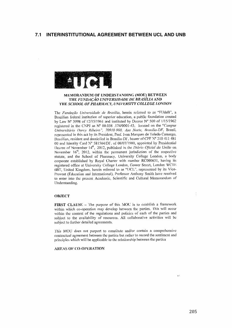
205
7.1 INTERINSTITUTIONAL AGREEMENT BETWEEN UCL AND UNB
206
207

208
209
210
7.2 PROJECT SENT TO CGEN AND IBAMA FOR REQUESTING
AUTHORISATION TO ACCESS THE BRAZILIAN GENETIC
HERITAGE
211
212
213
214
215
216
217
218
219
220
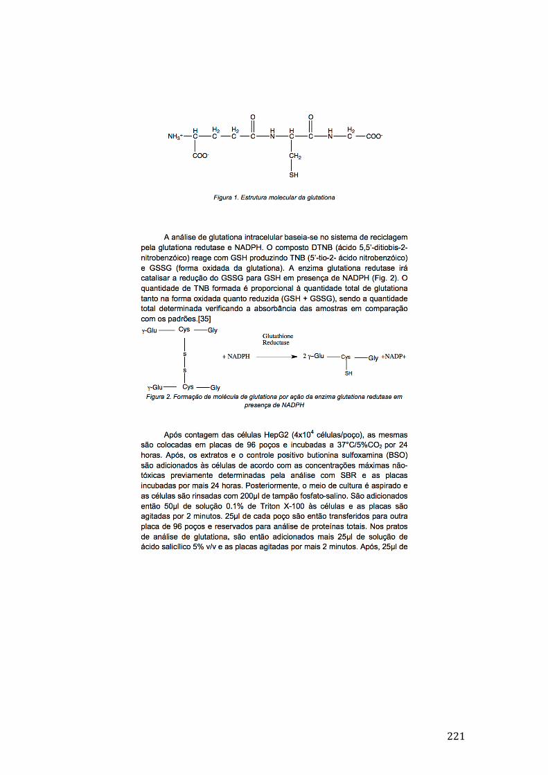
221
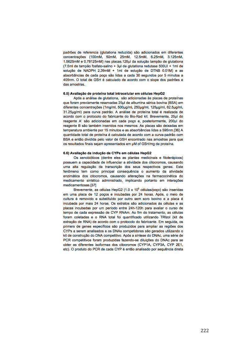
222
223
224
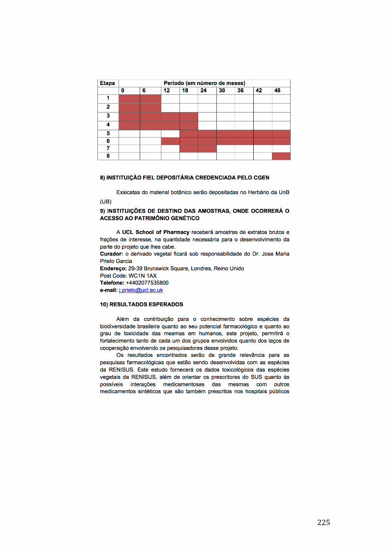
225
226
227
228
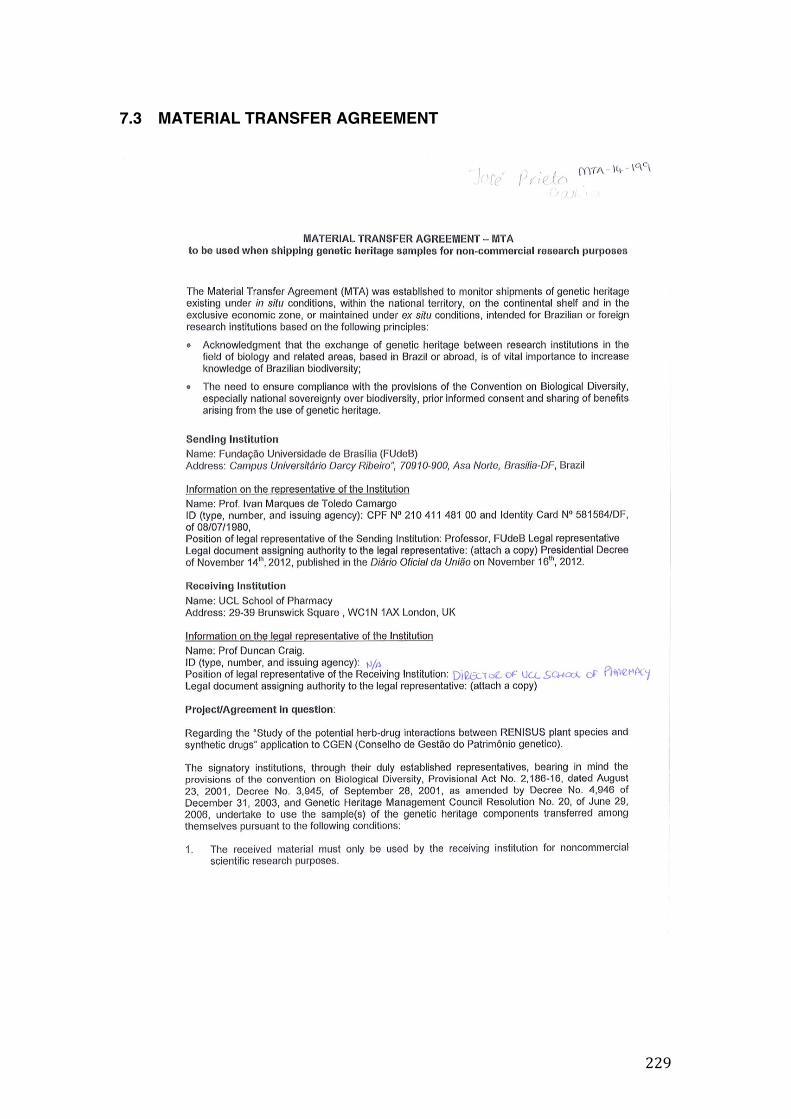
229
7.3 MATERIAL TRANSFER AGREEMENT
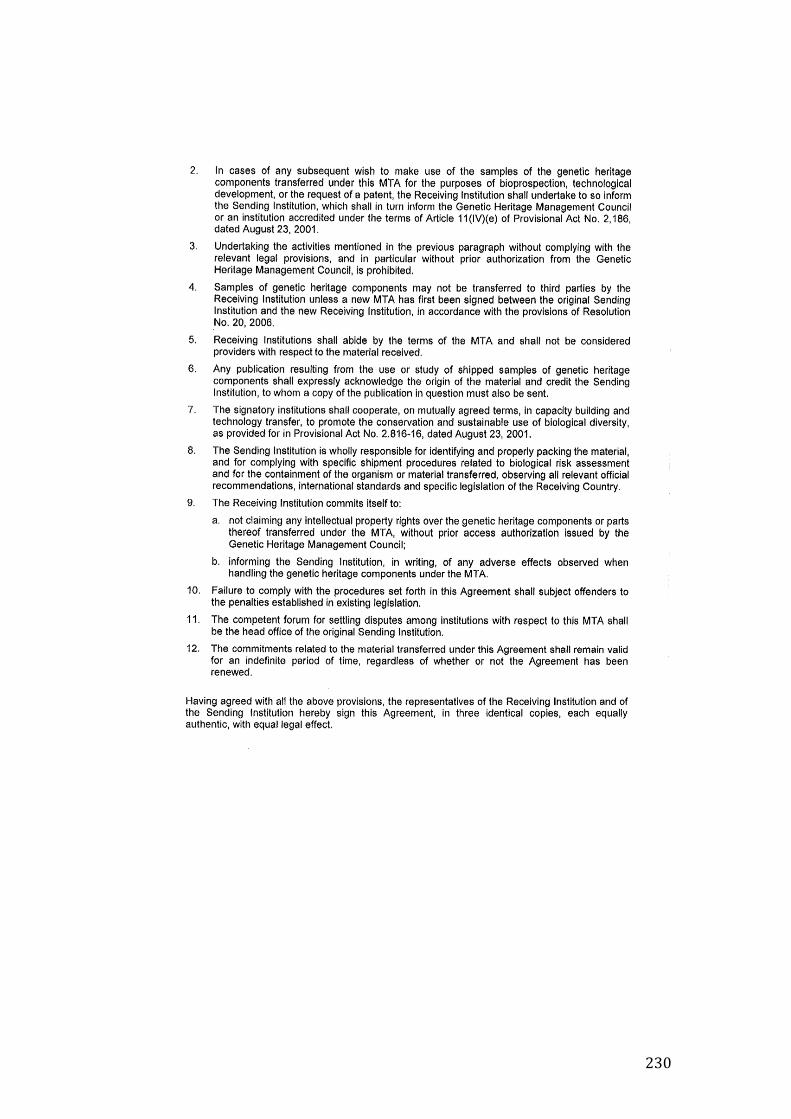
230
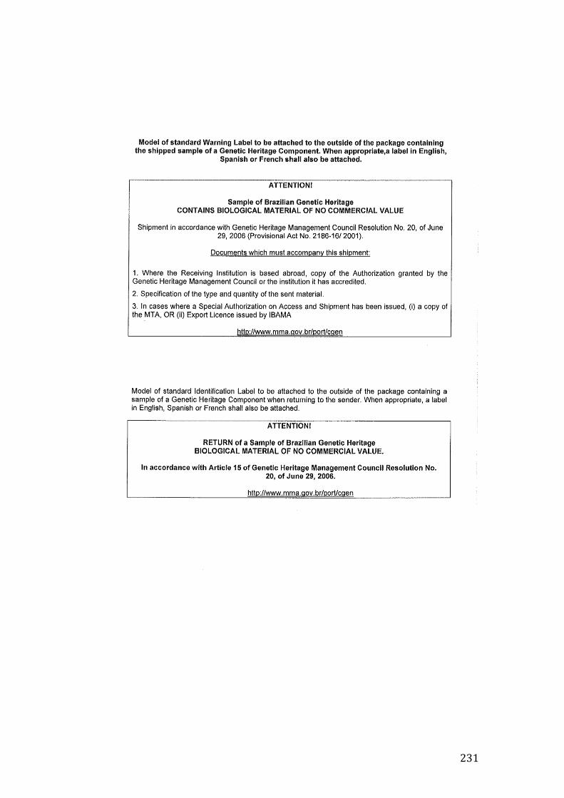
231
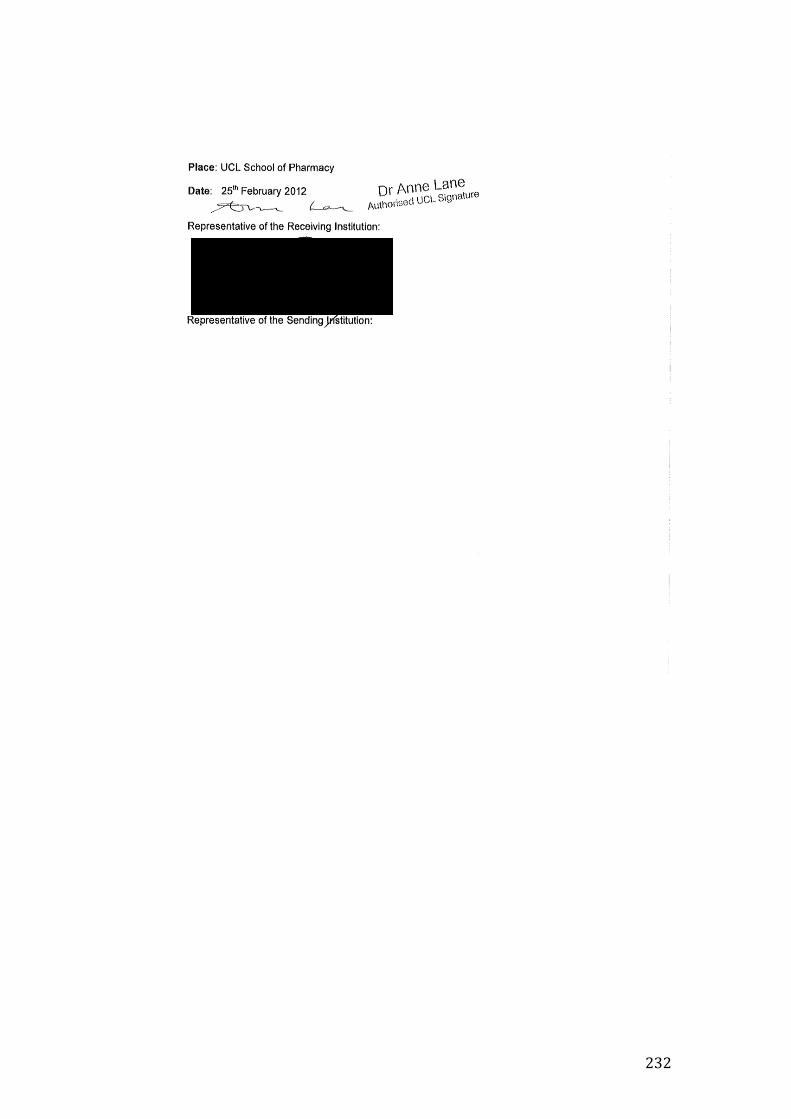
232
233
7.4 AUTHORISATION TO ACCESS THE BRAZILIAN GENETIC
HERITAGE GIVEN BY CGEN/CNPQ
6223768071168241
AUTORIZAÇÃO DE ACESSO E DE REMESSA DE AMOSTRA DE COMPONENTE DOPATRIMÔNIO GENÉTICO nº 010295/2014-3
O CONSELHO NACIONAL DE DESENVOLVIMENTO CIENTÍFICO E TECNOLÓGICO - CNPq, credenciado peloConselho de Gestão do Patrimônio Genético (CGEN/MMA), por meio da Deliberação CGEN n° 246, de 27 de agosto de2009, para autorizar instituições nacionais, públicas ou privadas, que exerçam atividades de pesquisa e desenvolvimentonas áreas biológicas e afins, a acessar e remeter amostras de componente do patrimônio genético para fins de pesquisacientífica sem potencial de uso econômico, neste ato representado pelo seu Diretor de Ciências Agrárias, Biológicas e daSaúde, nos termos da Portaria CNPq nº 104/2011, autoriza a instituição abaixo qualificada a acessar e remeter amostrasde componentes do patrimônio genético.
Instituição: UNIVERSIDADE DE BRASILIA - UNBCNPJ: 000.381.740/0001-43Representante Legal: JAIME MARTINS DE SANTANACargo/Função: Decano (Pró-Reitor) de Pesquisa e Pós-GraduaçãoCPF: 308.595.651-49 RG: 358372
Projeto: ESTUDO DE INTERAÇÕES MEDICAMENTOSAS ENTRE AS ESPÉCIES VEGETAIS CONSTANTES NARELAÇÃO NACIONAL DE PLANTAS MEDICINAIS DE INTERESSE DO SUS (RENISUS) E MEDICAMENTOSCONVENCIONAIS
Coordenador do Projeto: Damaris SilveiraCPF: 464.405.696-00 RG: 2430447 - SSP / DF
Finalidade do projeto: A RENISUS (Relação Nacional de Plantas de Interesse para o SUS) foi lançada em 2008 peloMinistério da Saúde com o objetivo de selecionar espécies vegetais com atividades medicinais que possam ser integradasà Relação Nacional de Plantas Medicinais (RENAFITO), sendo disponibilizada à população pelo Sistema Único de Saúde(SUS). Para que esta meta seja alcançada, estudos farmacológicos e toxicológicos devem ser realizados para que plantasmedicinais e fitoterápicos possam ser prescritos aos pacientes de forma segura. Este projeto tem, como principal objetivo,fazer uma avaliação in vitro das 71 espécies vegetais presentes na RENISUS em células hepáticas e avaliar suasegurança quando a planta medicinal ou o fitoterápico for administrado com algum medicamento sintético. Estudos destanatureza são muito relevantes, uma vez que o uso de produtos naturais vem crescendo muito nos últimos anos no Brasil eno mundo. Os conceitos errôneos de que a administração de plantas medicinais é segura e isenta de efeitos colateraispodem trazer sérias consequências para a saúde pública. Um xenobiótico de qualquer natureza (como uma plantamedicinal ou fitoterápico, por exemplo), uma vez administrado, será metabolizado pelas enzimas do fígado. A(s)substância(s) química(s) presente(s) naquele composto podem agir aumentando ou diminuindo a atividade ou aexpressão das enzimas presentes no metabolismo de fase I (citocromo P-450) e fase II (glutationa S-transferases,UDP-glucuroniltransferases, Sulfotransferases, etc.) que podem também impactar a metabolização de medicamentossintéticos quando estes foram concomitantemente administrados. O resultado desta interação se dá por alterações noprocesso farmacocinético do medicamento sintético, podendo afetar qualquer etapa do ADME (absorção, distribuição,metabolização e excreção), aumentando a toxicidade ou diminuindo e eficácia do medicamento dependendo da ação doproduto natural. Portanto, devido à necessidade de se conhecer mais profundamente a ação das espécies vegetais daRENISUS no mecanismo metabólico e identificar possíveis interações medicamentosas entre plantas medicinais emedicamentos sintéticos, este estudo toxicológico previamente aprovado e financiado pelo CNPq se faz de grandeimpacto para o Ministério da Saúde no processo de implementação da RENISUS e para futuros projetos envolvendoplantas medicinais e fitoterápicos no SUS.
Amostras a serem acessadas:Grupos Taxonômicos: Todos as espécies contidas na Relação Nacional de Plantas de Interesse ao SUS (RENISUS):Achillea millefolium, Allium sativum, Aloe spp (Aloe vera ou Aloe barbadensis), Alpinia (Alpinia zerumbet ou Alpiniaspeciosa), Anacardium occidentale, Ananas comosus, Apuleia ferrea = Caesalpinia ferrea, Arrabidaea chica, Artemisiaabsinthium, Baccharis trimera, Bauhinia spp (Bauhinia affinis, Bauhinia forficata ou Bauhinia variegata), Bidens pilosa,Calendula officinalis, Carapa guianensis, Casearia sylvestris, Chamomilla recutita = Matricaria chamomilla, Chenopodiumambrosioides, Copaifera spp, Cordia spp (Cordia curassavica ou Cordia verbenacea), Costus spp (Costus scaber ouCostus spicatus), Croton spp (Croton cajucara ou Croton zehntneri), Curcuma longa, Cynara scolymus, Dalbergiasubcymosa, Eleutherine plicata, Equisetum arvense, Erythrina mulungu, Eucalyptus globulus, Eugenia uniflora ou Myrtusbrasiliana Foeniculum vulgare, Glycine max, Harpagophytum procumbens, Jatropha gossypiifolia, Justicia pectoralis,Kalanchoe pinnata = Bryophyllum calycinum, Lamium album, Lippia sidoides, Malva sylvestris, Maytenus spp (Maytenusaquifolium ou Maytenus ilicifolia),Mentha pulegium, Mentha spp (M. crispa, M. piperita ou M. villosa), Mikania spp (Mikaniaglomerata ou Mikania laevigata), Momordica charantia, Morus spp., Ocimum gratissimum, Orbignya speciosa, Passiflora
Página 1 de 3
234
spp (Passiflora alata, Passiflora edulis ou Passiflora incarnata), Persea spp (P. gratissima ou P. americana), Petroselinumsativum, Phyllanthus spp (P. amarus, P.niruri, P. tenellus e P. urinaria), Plantago major, Plectranthus barbatus = Coleusbarbatus, Polygonum spp (Polygonum acre ou Polygonum hydropiperoides), Portulaca pilosa, Psidium guajava, Punicagranatum, Rhamnus purshiana, Ruta graveolens, Salix alba, Schinus terebinthifolius = Schinus aroeira, Solanumpaniculatum, Solidago microglossa, Stryphnodendron adstringens = Stryphnodendron barbatimam, Syzygium spp (S.jambolanum ou S. cumini), Tabebuia avellanedae, Tagetes minuta, Trifolium pratense, Uncaria tomentosa, Vernoniacondensata, Vernonia spp (Vernonia ruficoma ou Vernonia polyanthes), Zingiber officinaleTipo de material/quantidade de amostras: O tipo de amostra dependerá da espécie. A quantidade aproximada será:folhas,sumidades floridas ou partes aéreas: 500 g; caule, raiz ou bulbos: 800 g; frutos ou sementes: 600 g
Local de depósito de subamostra: UNIVERSIDADE DE BRASILIA
Equipe do projeto: DAMARIS SILVEIRA / CPF 464.405.696-00ANDRE LUIS DIAS ARAUJO MAZZARI / CPF 090.900.047-69YANNA KARLA DE MEDEIROS NOBREGA / CPF 020.423.464-62YRIS MARIA FONSECA / CPF 045.789.276-52PEROLA DE OLIVEIRA MAGALHAES / CPF 248.940.408-32LUIZ ALBERTO SIMEONI / CPF 043.388.018-08FRANCISCO DE ASSIS ROCHA NEVES / CPF 186.580.591-20RICCARDO PRATESI / CPF 000.079.941-68CHRISTOPHER WILLIAM FAGG / CPF 694.231.131-68JOSE M PRIETO / CPF ..-
Validade da Autorização: 15/05/2014 a 01/05/2018
A instituição acima qualificada deverá enviar ao CNPq, por meio do Coordenador do Projeto, relatório anual sobre oandamento do projeto de pesquisa, nos termos do Decreto nº. 4.946/2003. O roteiro para confecção do relatório estádisponível em http://www.cnpq.br/web/guest/relatorio-de-atividades. Os relatórios devem ser enviados ao CNPq em meioeletrônico, para o endereço [email protected] e, preferencialmente, em formato .pdf.
Esta autorização está vinculada às informações, declarações e termos de compromisso firmados pelo coordenador doprojeto e pelo representante legal, constantes do Processo n° 010295/2014-3. Atividades de acesso aos conhecimentostradicionais associados, de acesso e de remessa de componente do patrimônio genético com finalidade comercial,aplicação industrial, bioprospecção ou desenvolvimento tecnológico não estão autorizadas.
Caso seja identificado uso econômico de produto ou processo, passível ou não de proteção intelectual, originado dasamostras de componente do patrimônio genético acessado no âmbito desta autorização, a instituição beneficiada secompromete a adotar as providências cabíveis, nos termos da legislação vigente, junto ao CGEN/MMA.
Se ocorrer coleta de espécie não autorizada ou não identificada, deverá ser observado o que consta no Decreto nº 6.514,de 22/07/2008, no que refere à flora e fauna, e em particular sobre espécies ameaçadas de extinção ou de endemismoestrito.
A remessa de amostra de componente do patrimônio genético deverá ser precedida da assinatura do Termo deTransferência de Material (TTM) ou do Termo de Responsabilidade para Transporte de Amostra de Componente doPatrimônio Genético (TRTM). A remessa para instituições nacionais está isenta de autorização prévia. Contudo, aremessa para instituições sediadas no exterior depende de autorização prévia do CNPq, nos termos das resoluções doCGEN 15/2004 e 20/2006. Os modelos dos termos, assim como as citadas resoluções, estão disponíveis emhttp://www.cnpq.br/web/guest/remessa-e-transporte e devem ser enviados ao CNPq em meio eletrônico para o endereç[email protected], preferencialmente em formato .pdf. Ainda, para a remessa de componente do patrimônio genético parainstituição sediada no exterior, deverá ser solicitada ao Instituto Brasileiro do Meio Ambiente e dos Recursos NaturaisRenováveis - IBAMA, por meio de formulário específico e mediante a apresentação de TTM ou TRTM, licença deexportação complementar a autorização de remessa, especialmente quando se tratar de remessa de espécies constantesnos Anexos da Convenção sobre o Comércio Internacional das Espécies da Flora e Fauna Selvagens em Perigo deExtinção (Cites).
Brasília, 15 de Maio de 2014
Marcelo Marcos MoralesDiretor de Ciências Agrárias, Biológicas e da Saúde
Para visualizar a versão digital da Autorização de Acesso e de Remessa de Amostra de Componente do PatrimônioGenético, V.Sa. poderá utilizar a ferramenta disponibilizada pelo CNPq para esse fim na páginahttp://servicosweb.cnpq.br/visualizador/</a> e informar o número do protocolo 6223768071168241 para recuperá-la dobanco de dados do CNPq, ou poderá selecionar o arquivo salvo em seu computador (em formato PKCS7). V.Sa. podetambém usar outro aplicativo disponível no mercado capaz de reconhecer arquivos no padrão PKCS7 para fazer a
Página 2 de 3

235
Catechin7.5 HPLC ANALYSIS
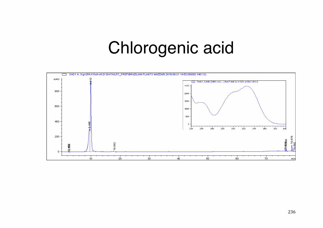
236
Chlorogenic acid
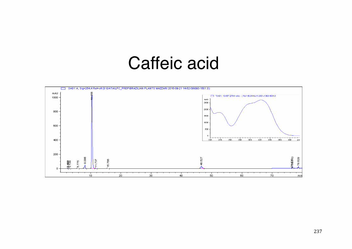
237
Caffeic acid
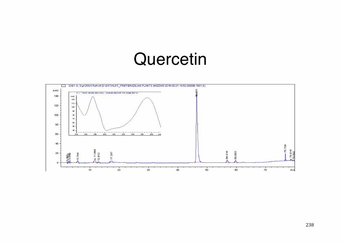
238
Quercetin
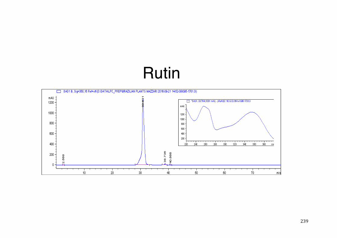
239
Rutin
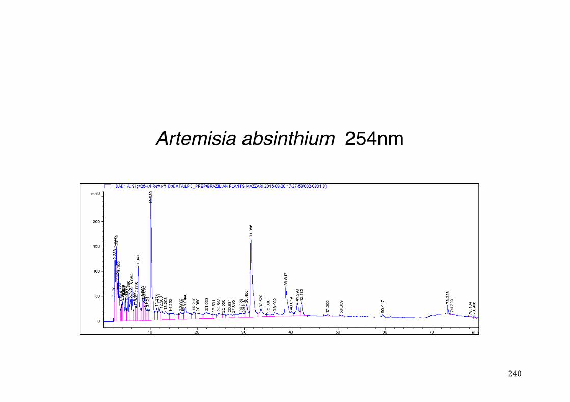
240
Artemisia absinthium 254nm
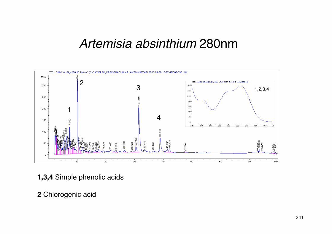
241
Artemisia absinthium 280nm
1,3,4 Simple phenolic acids
2 Chlorogenic acid
1
1,2,3,42 3
4
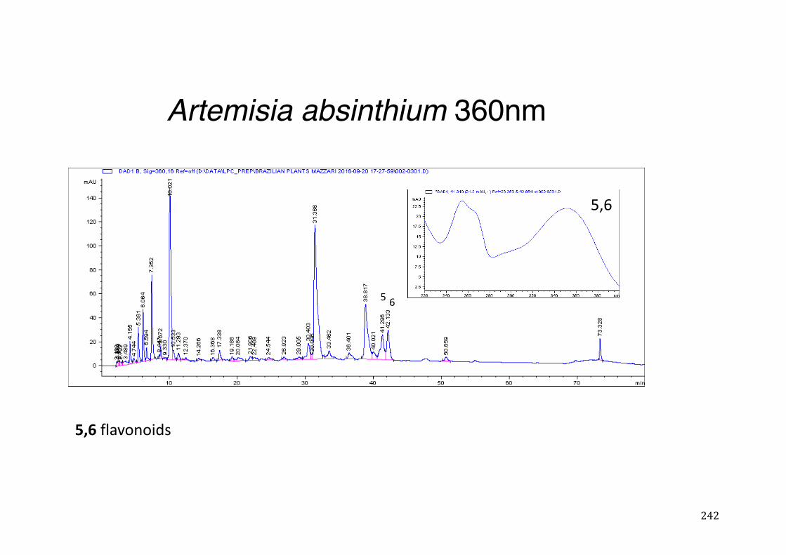
242
Artemisia absinthium 360nm
5 6
5,6
5,6flavonoids
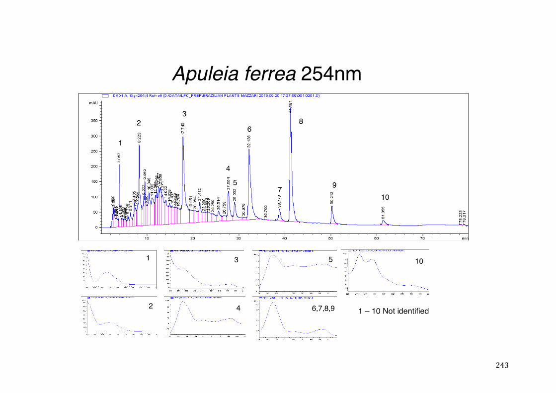
243
Apuleia ferrea 254nm
1
2
3
4
5
6,7,8,9
1
23
4
5
6
7
8
910
10
1 – 10 Not identified
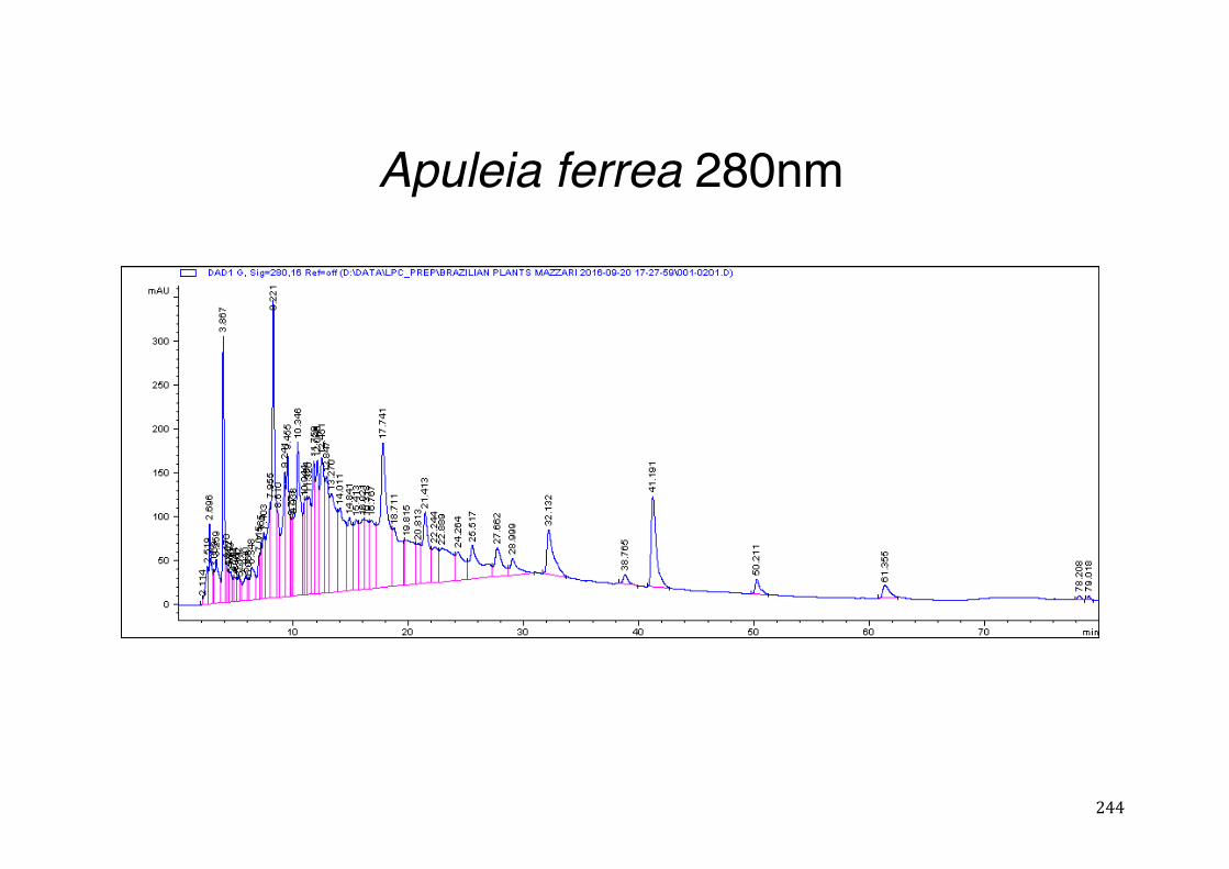
244
Apuleia ferrea 280nm
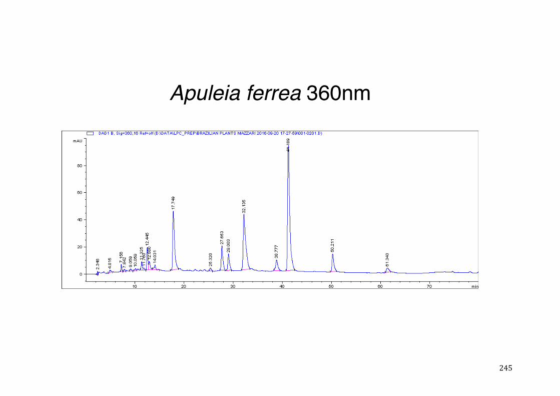
245
Apuleia ferrea 360nm
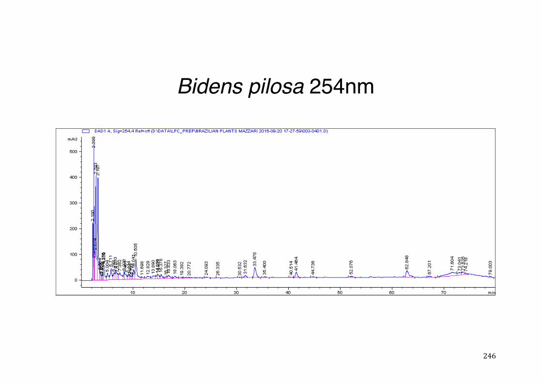
246
Bidens pilosa 254nm
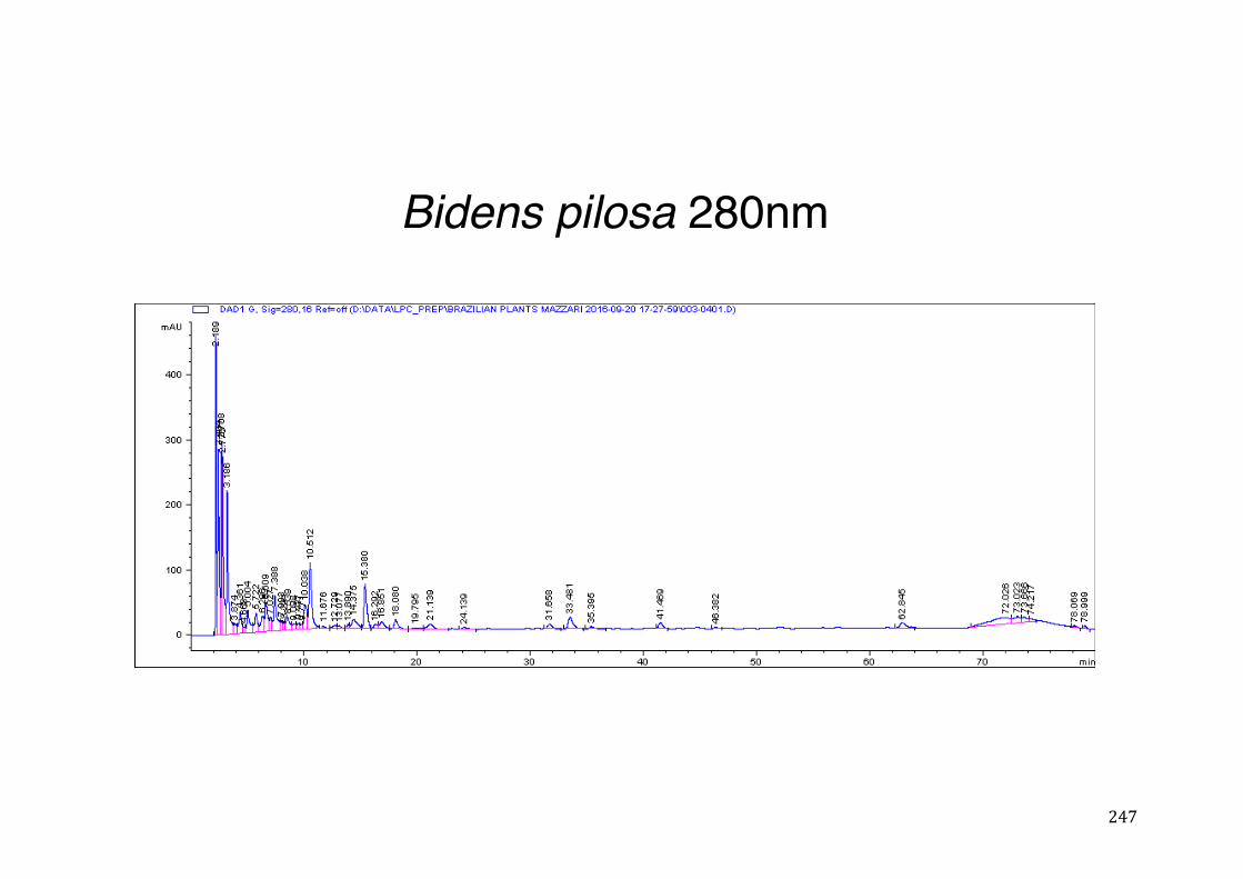
247
Bidens pilosa 280nm
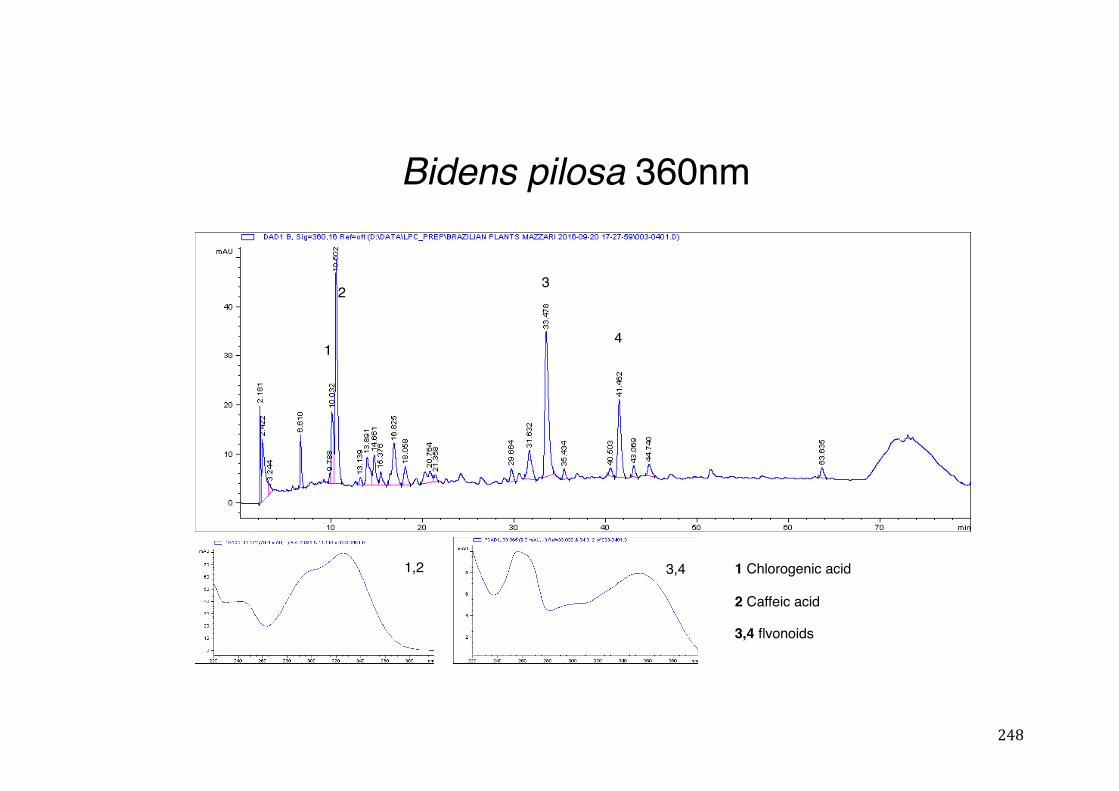
248
Bidens pilosa 360nm
3,4 flvonoids
1 Chlorogenic acid
2 Caffeic acid
1
3
4
2
1,2 3,4
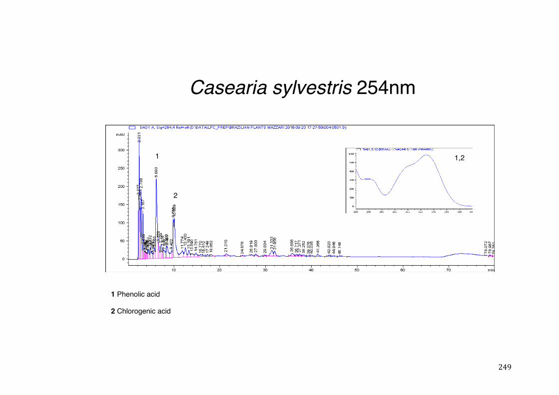
249
Casearia sylvestris 254nm
1 Phenolic acid
2 Chlorogenic acid
1
2
1,2
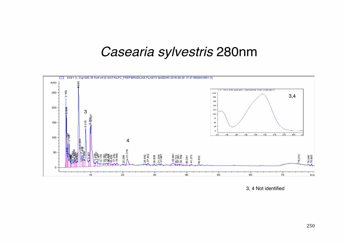
250
Casearia sylvestris 280nm
3
4
3,4
3, 4 Not identified
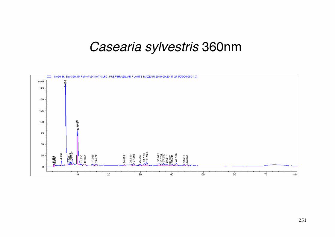
251
Casearia sylvestris 360nm
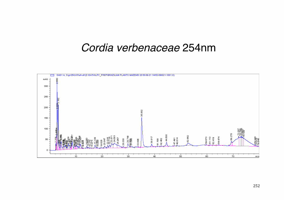
252
Cordia verbenaceae 254nm
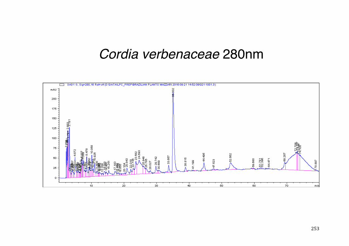
253
Cordia verbenaceae 280nm
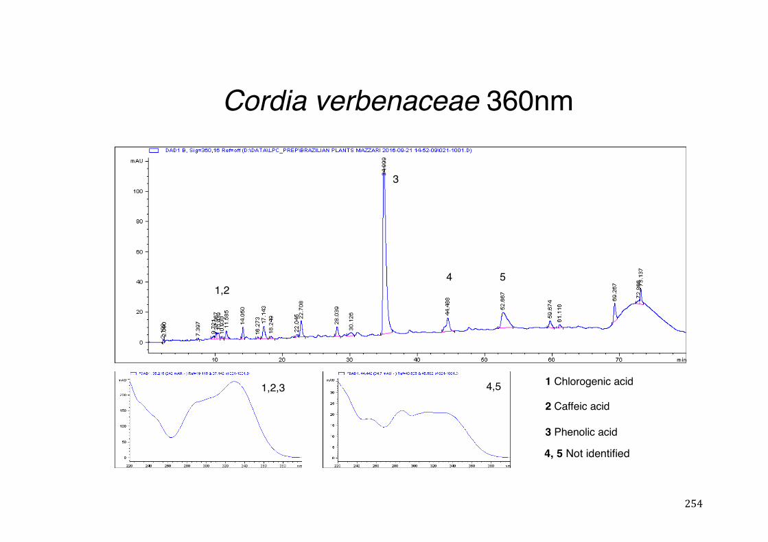
254
Cordia verbenaceae 360nm
4,5 1 Chlorogenic acid
2 Caffeic acid
3 Phenolic acid
4
3
51,2
1,2,3
4, 5 Not identified
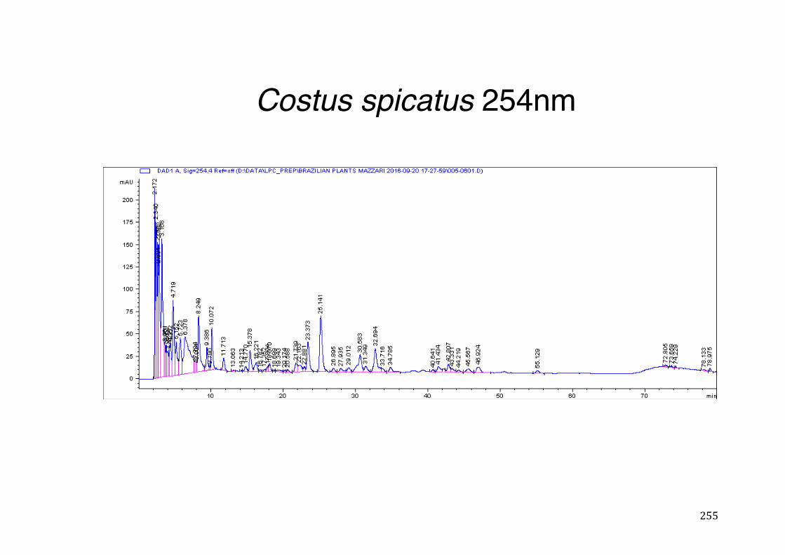
255
Costus spicatus 254nm
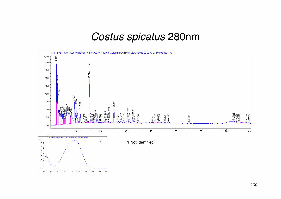
256
Costus spicatus 280nm
1
1 1 Not identified
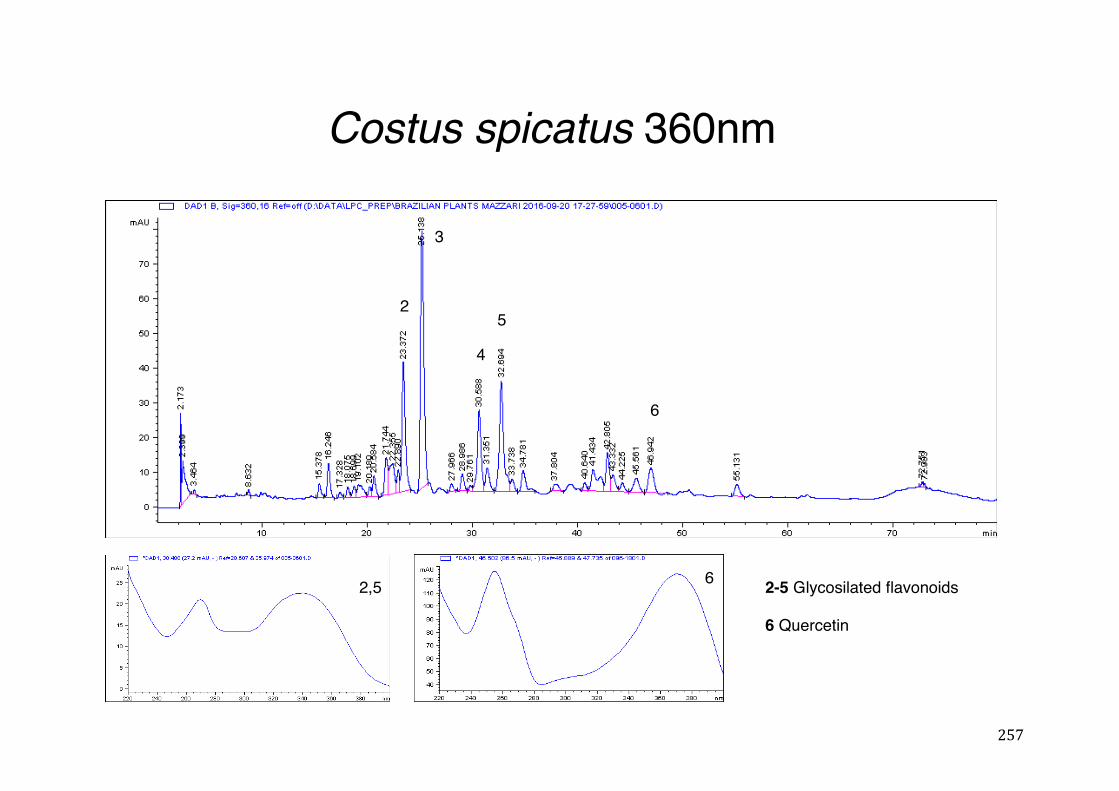
257
Costus spicatus 360nm
2
3
5
4
6
2-5 Glycosilated flavonoids
6 Quercetin
2,5 6
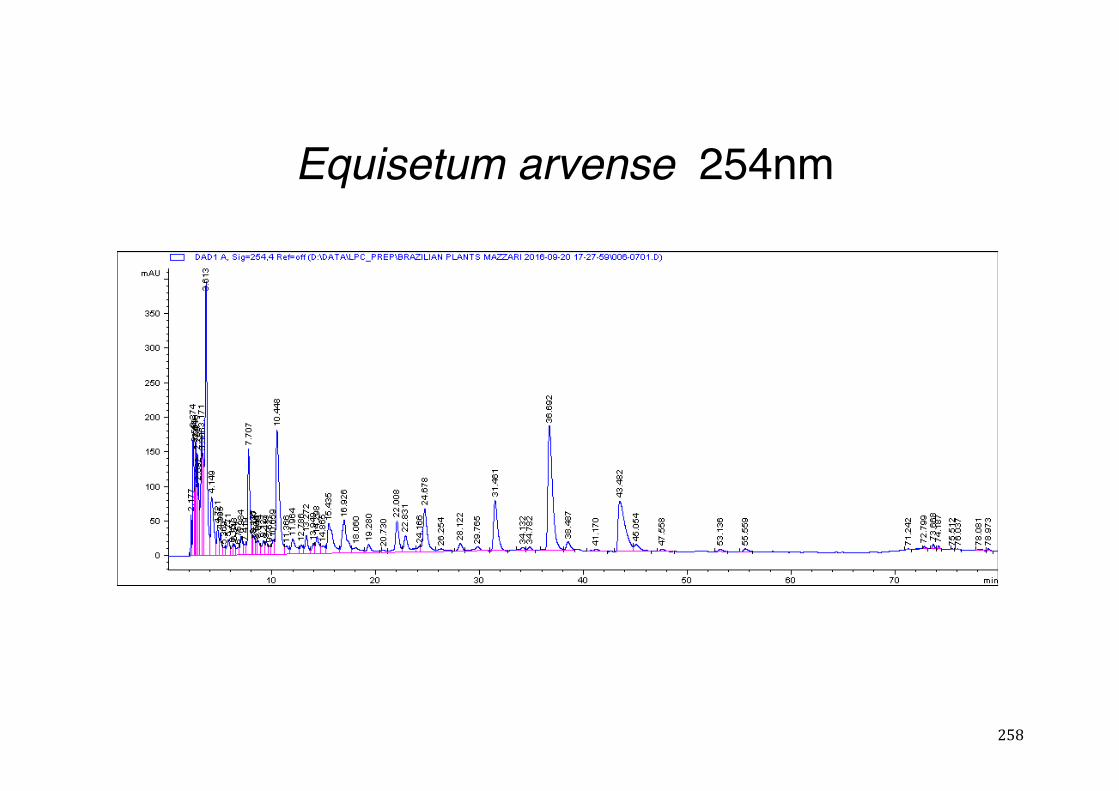
258
Equisetum arvense 254nm
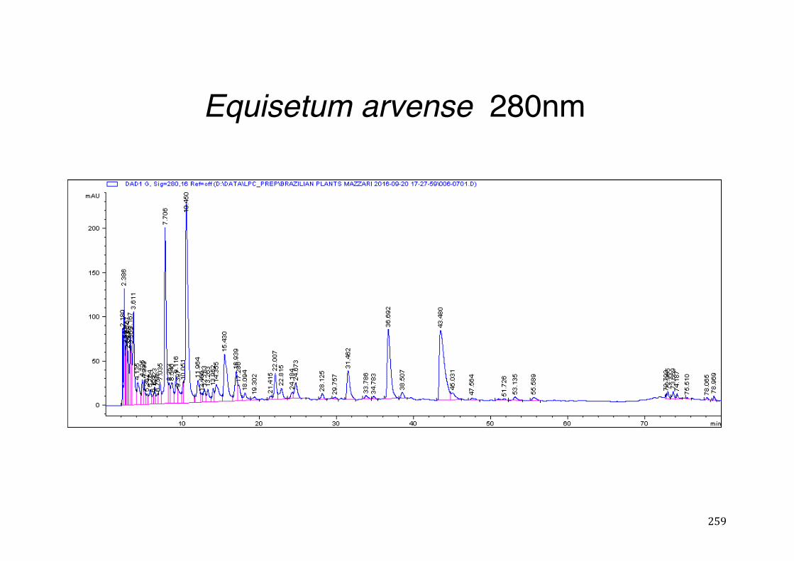
259
Equisetum arvense 280nm
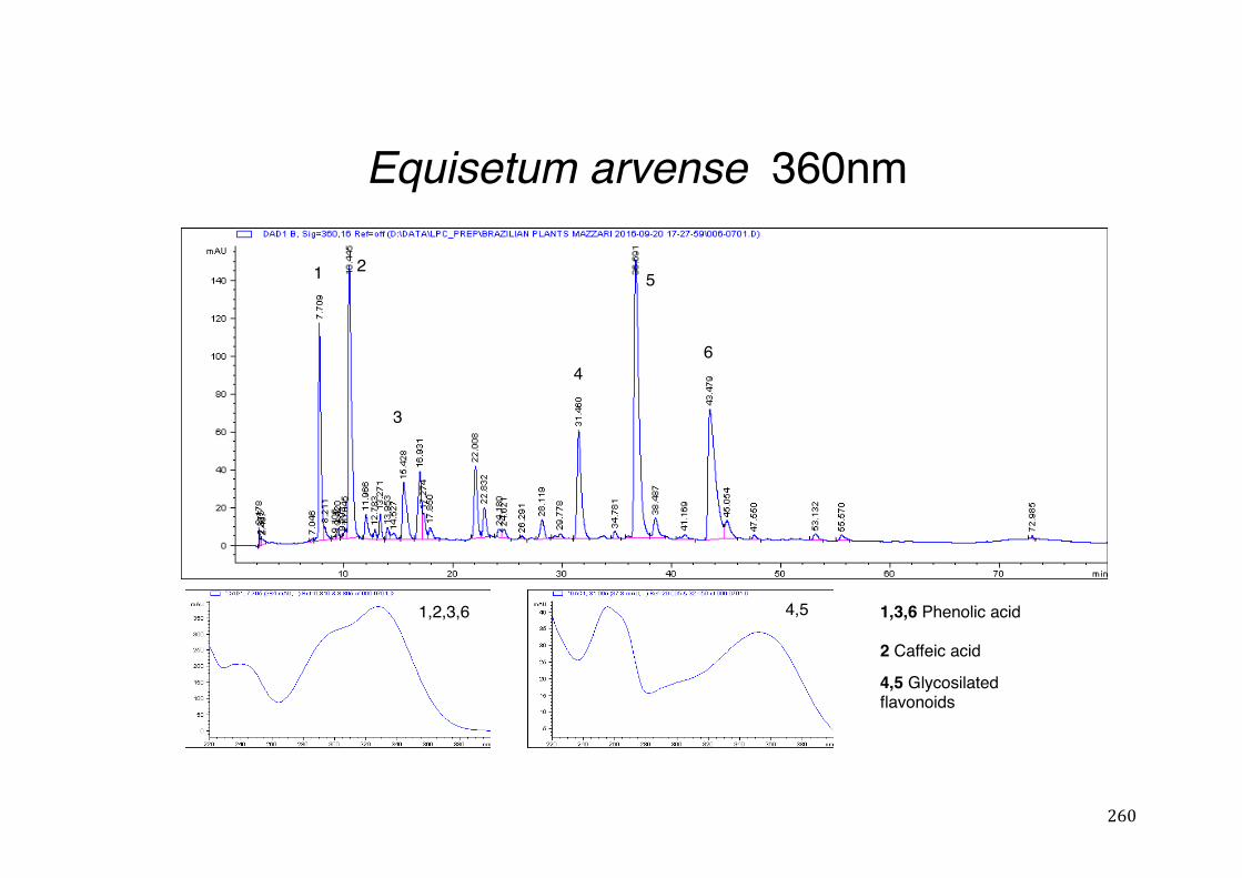
260
Equisetum arvense 360nm
1,3,6 Phenolic acid
2 Caffeic acid
4,5 Glycosilatedflavonoids
1 2
3
4
5
6
1,2,3,6 4,5
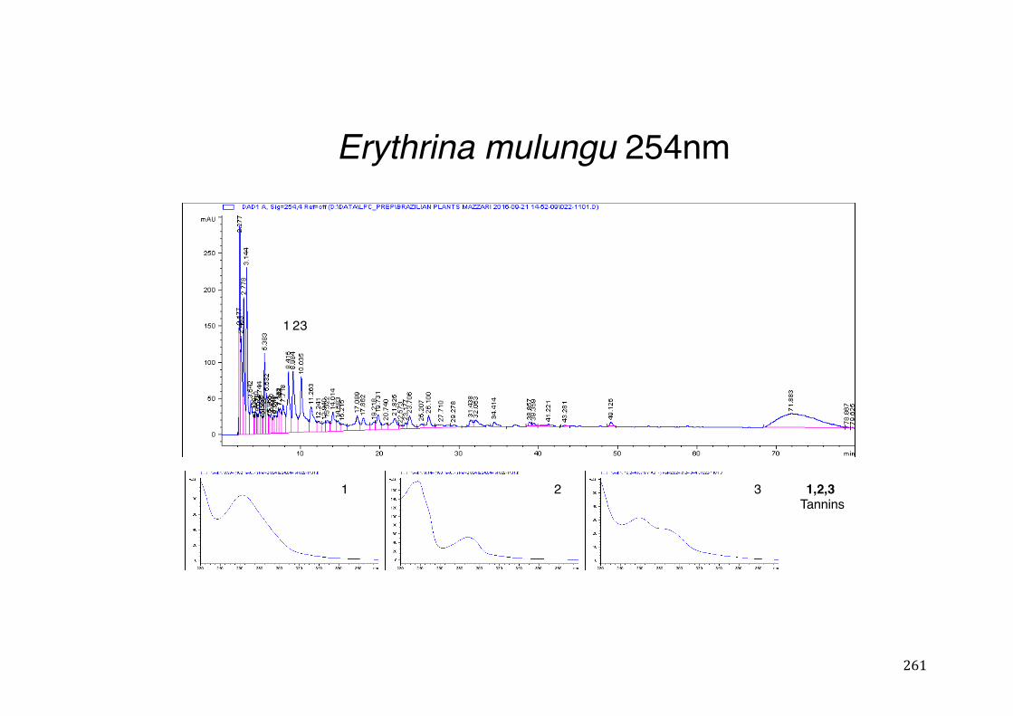
261
Erythrina mulungu 254nm
123
1 2 3 1,2,3Tannins
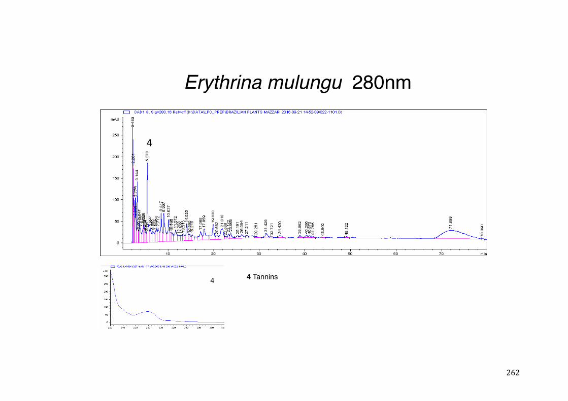
262
Erythrina mulungu 280nm
4
4 Tannins4
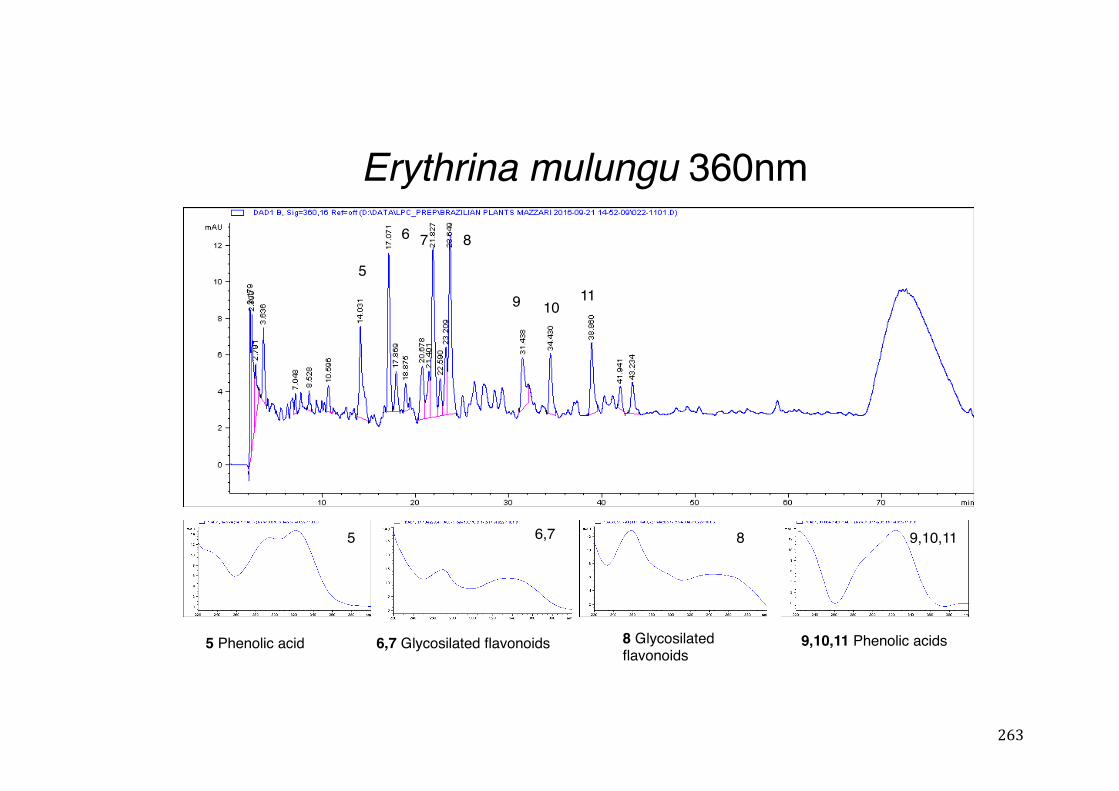
263
Erythrina mulungu 360nm
5 Phenolic acid 6,7 Glycosilated flavonoids 8 Glycosilatedflavonoids
9,10,11 Phenolic acids
5
6 7 8
9 1011
5 6,7 8 9,10,11
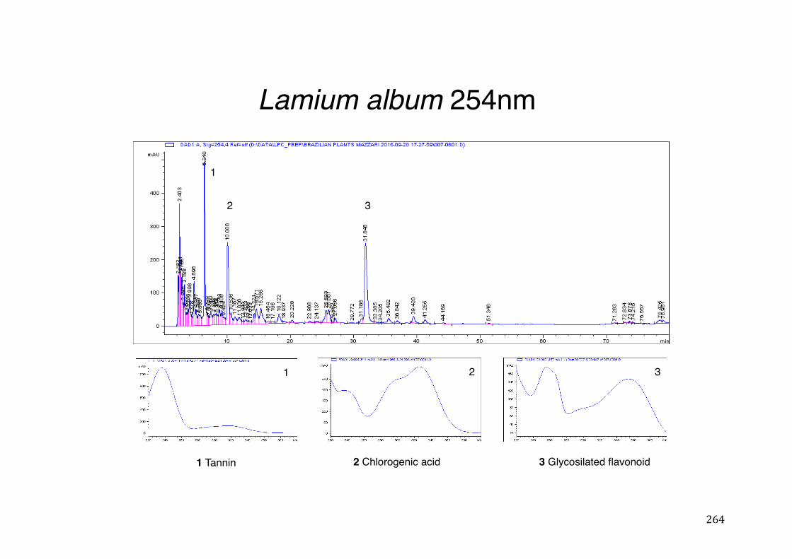
264
Lamium album 254nm
1 Tannin 2 Chlorogenic acid
1
2 3
3 Glycosilated flavonoid
1 2 3
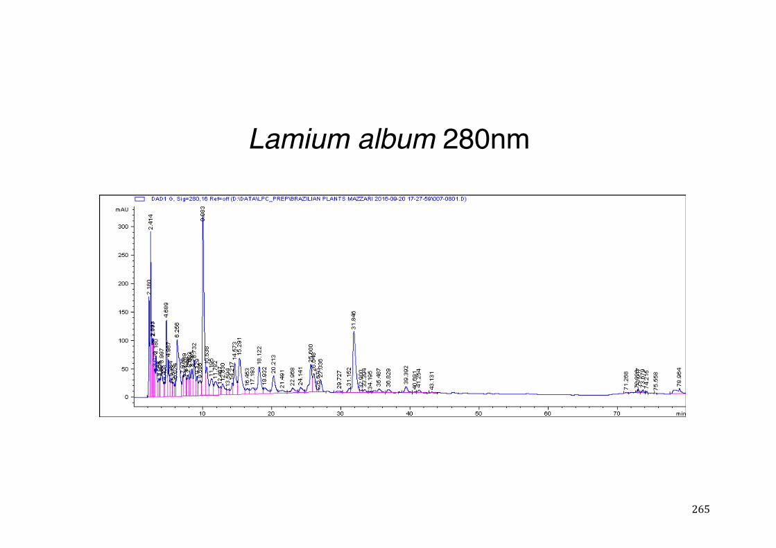
265
Lamium album 280nm
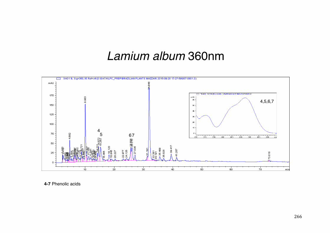
266
Lamium album 360nm
4-7 Phenolic acids
45 67
4,5,6,7
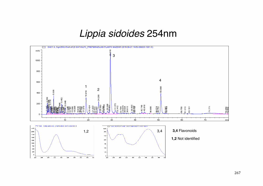
267
Lippia sidoides 254nm
3,4 Flavonoids1,2
3
41
2
3,4
1,2 Not identified
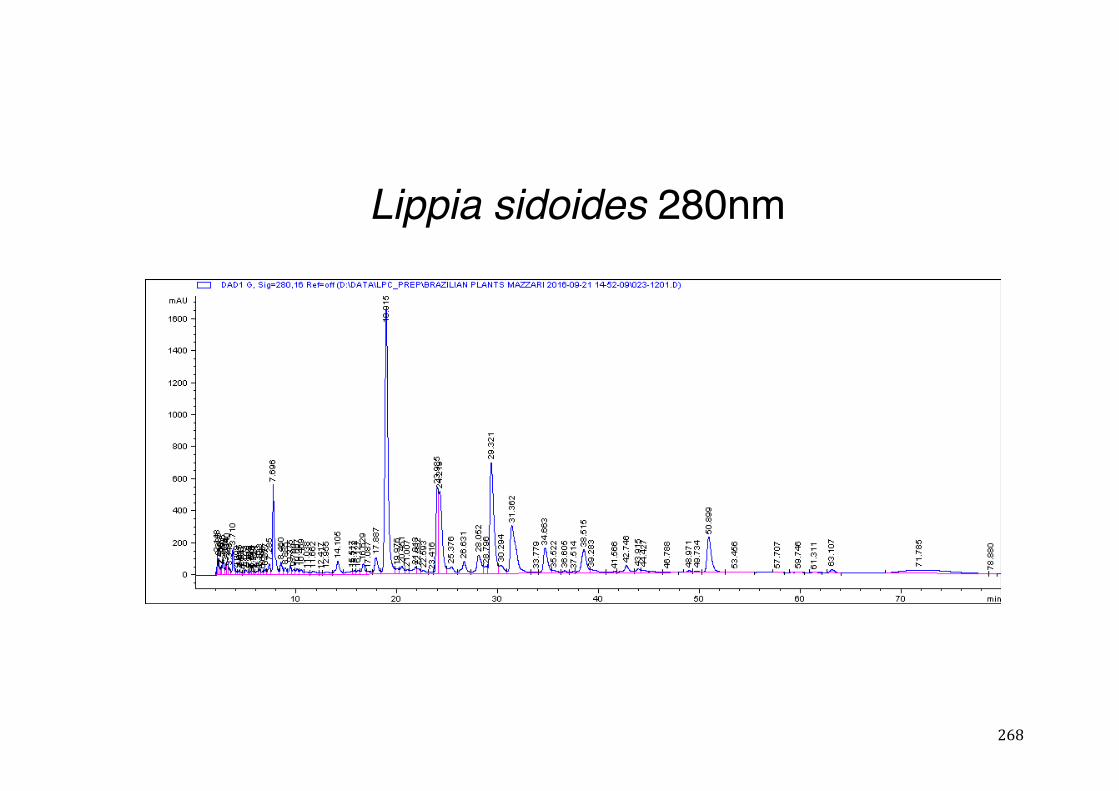
268
Lippia sidoides 280nm
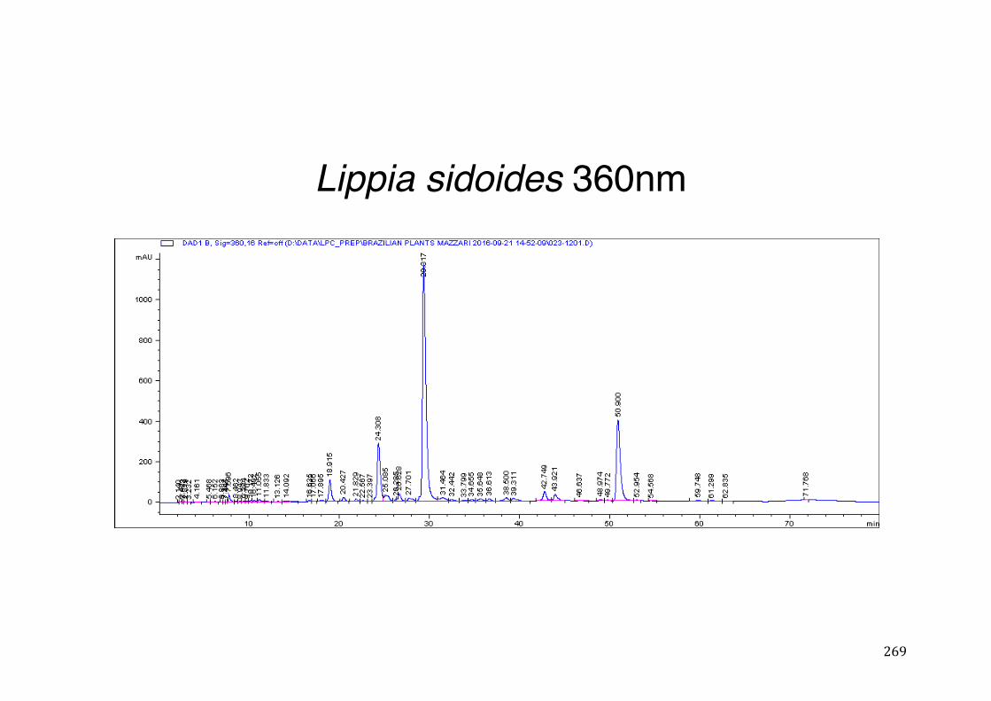
269
Lippia sidoides 360nm

270
Malva sylvestris (flower) 254nm
3 Phenolic acid 4,5 Flavonoids
1,2 6,7,8
34 5
12
6
7
8
3 4,5
1,2 Not identified 6,7,8 Not identified
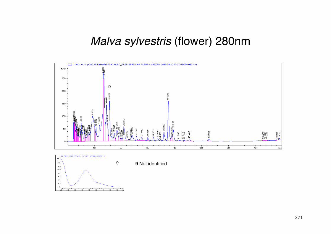
271
Malva sylvestris (flower) 280nm
9
9 9 Not identified
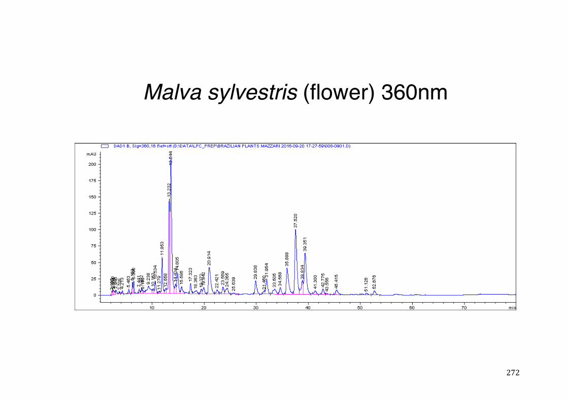
272
Malva sylvestris (flower) 360nm
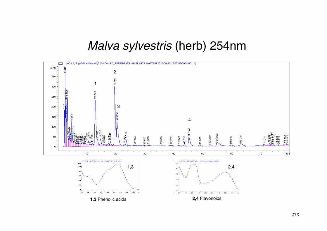
273
Malva sylvestris (herb) 254nm
1,3
1
2
3
4
2,4 Flavonoids1,3 Phenolic acids
2,4
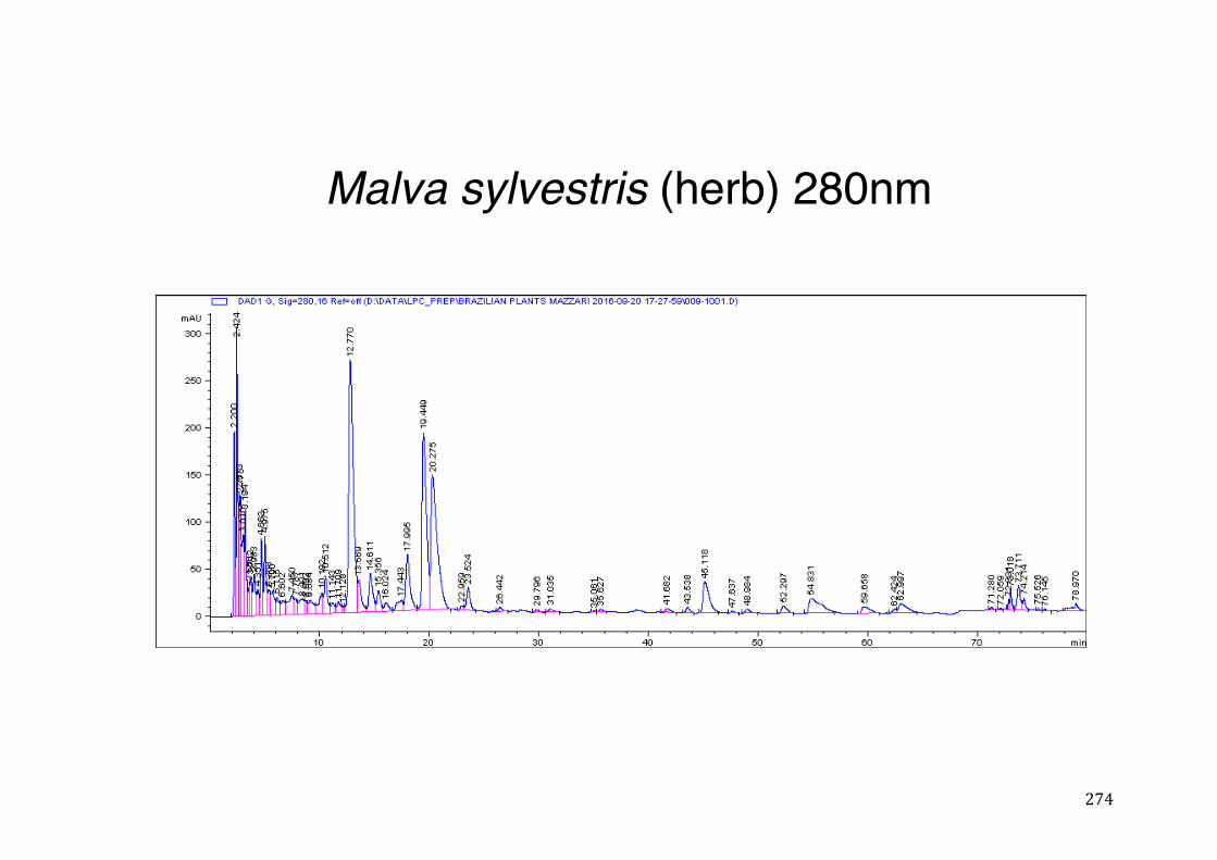
274
Malva sylvestris (herb) 280nm
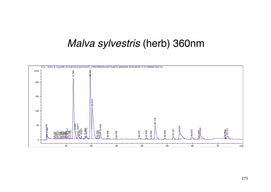
275
Malva sylvestris (herb) 360nm
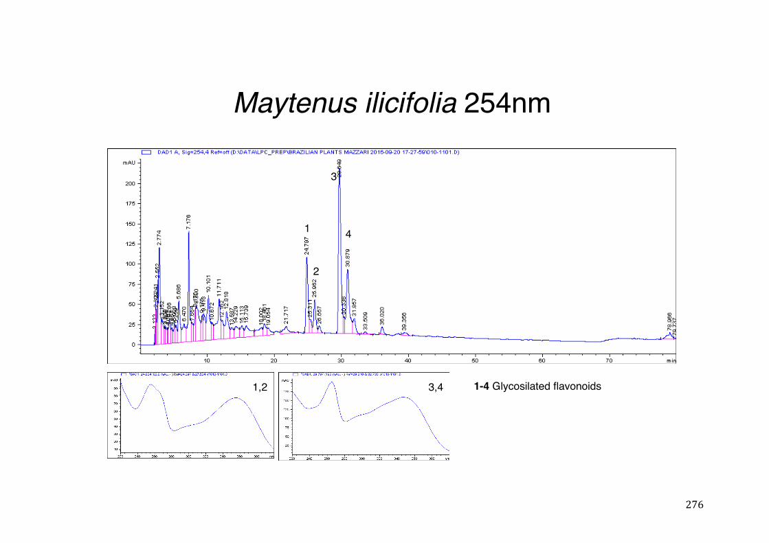
276
Maytenus ilicifolia 254nm
1
2
1,2 3,4
1
2
3
4
1-4 Glycosilated flavonoids
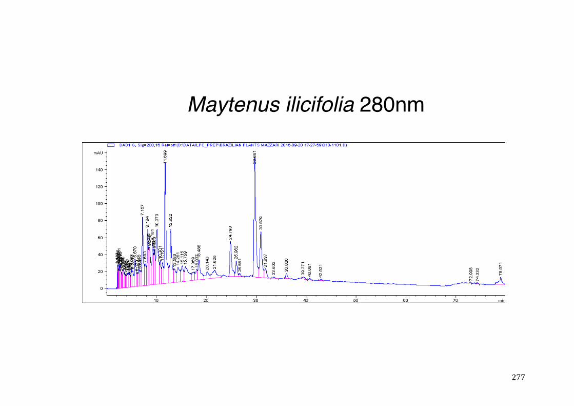
277
Maytenus ilicifolia 280nm
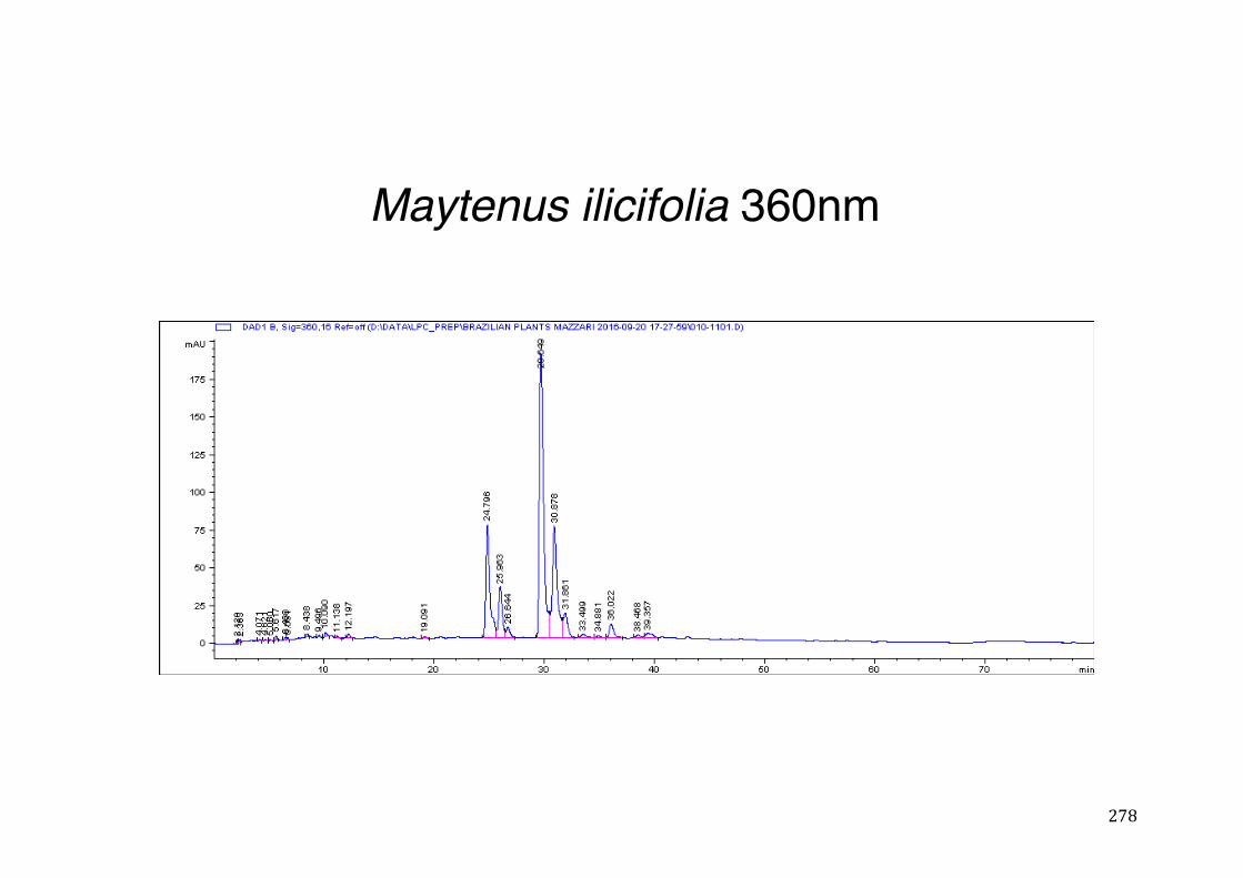
278
Maytenus ilicifolia 360nm
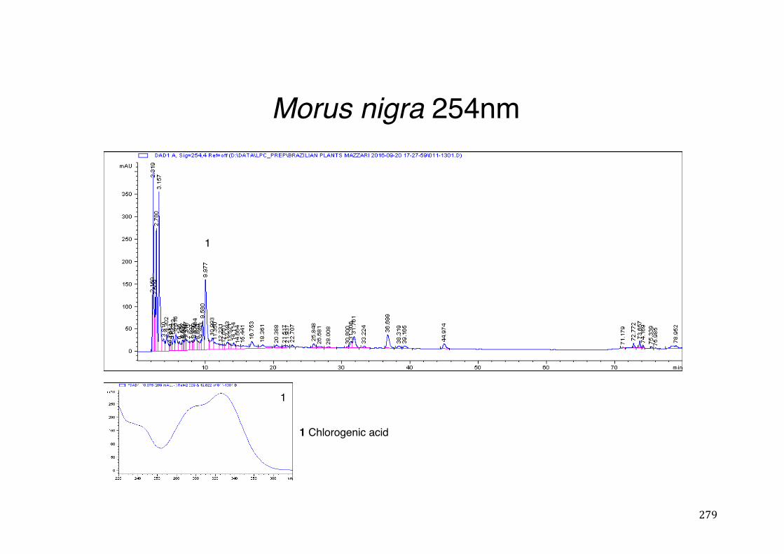
279
Morus nigra 254nm
1
1 Chlorogenic acid
1
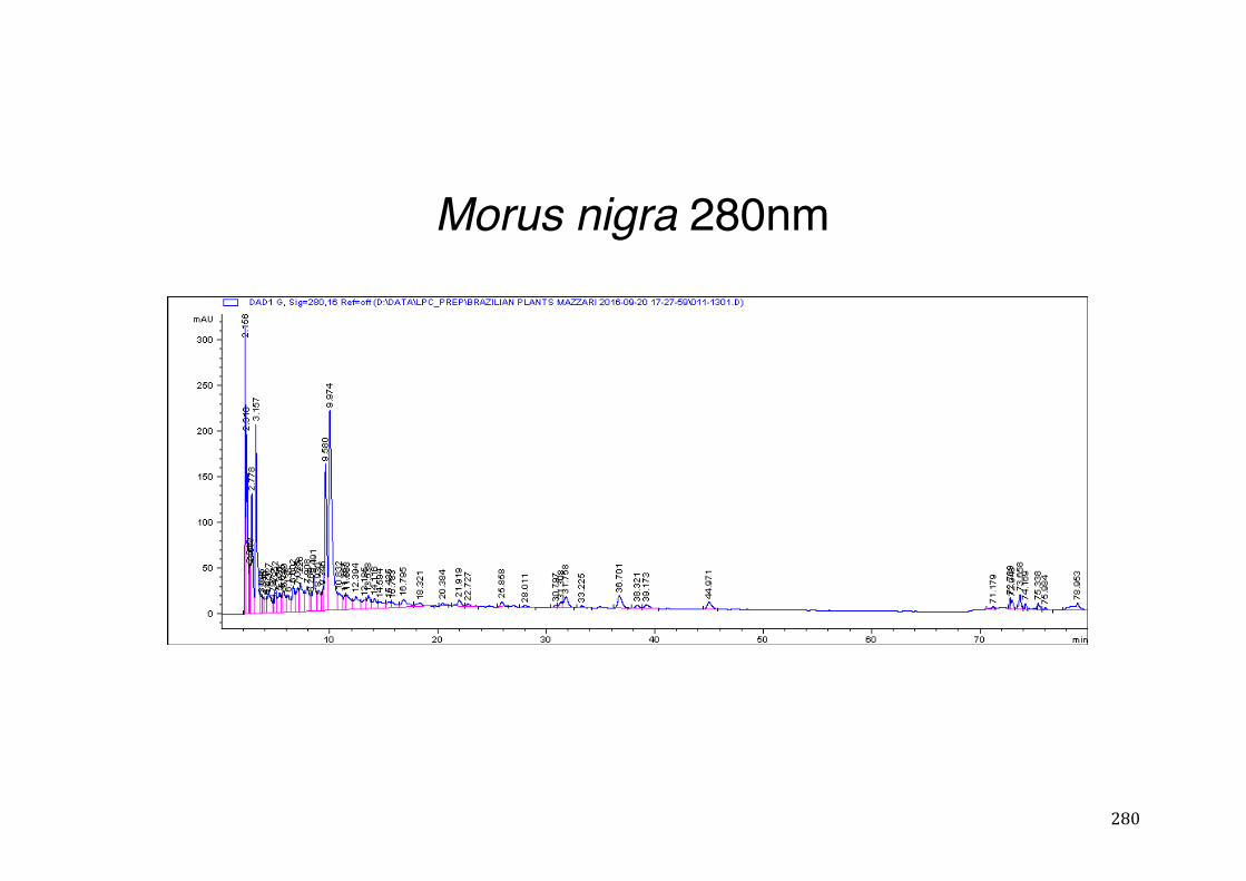
280
Morus nigra 280nm
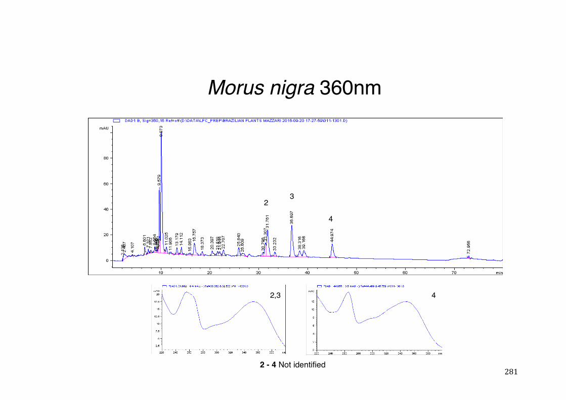
281
Morus nigra 360nm
23
4
2,3 4
2 - 4 Not identified
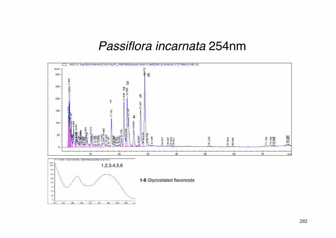
282
Passiflora incarnata 254nm
1,2,3,4,5,6
1
23
4
5
6
1-6 Glycosilated flavonoids
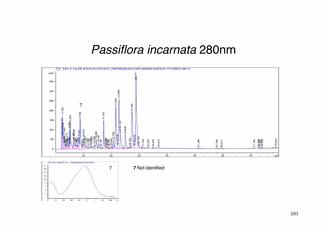
283
Passiflora incarnata 280nm
7
7 7 Not identified
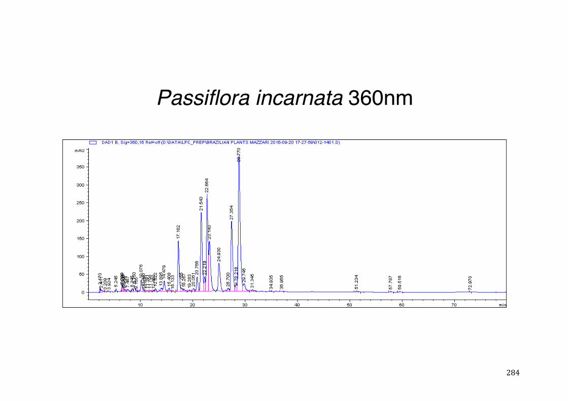
284
Passiflora incarnata 360nm
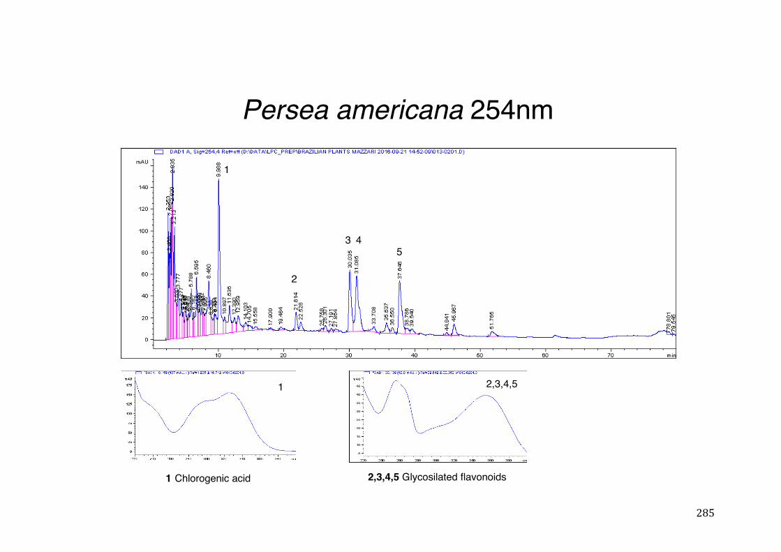
285
Persea americana 254nm
1 Chlorogenic acid
2,3,4,5
1
2
3 45
2,3,4,5 Glycosilated flavonoids
1
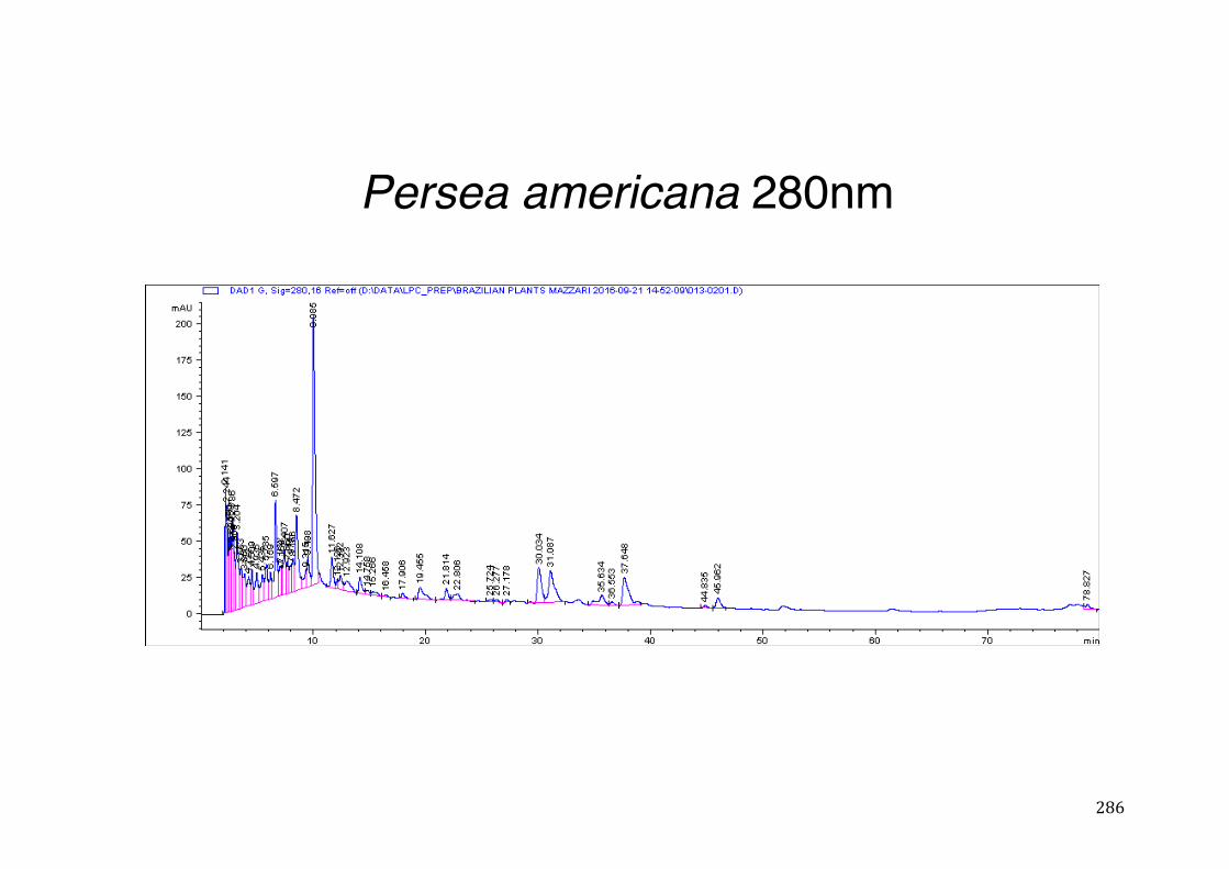
286
Persea americana 280nm
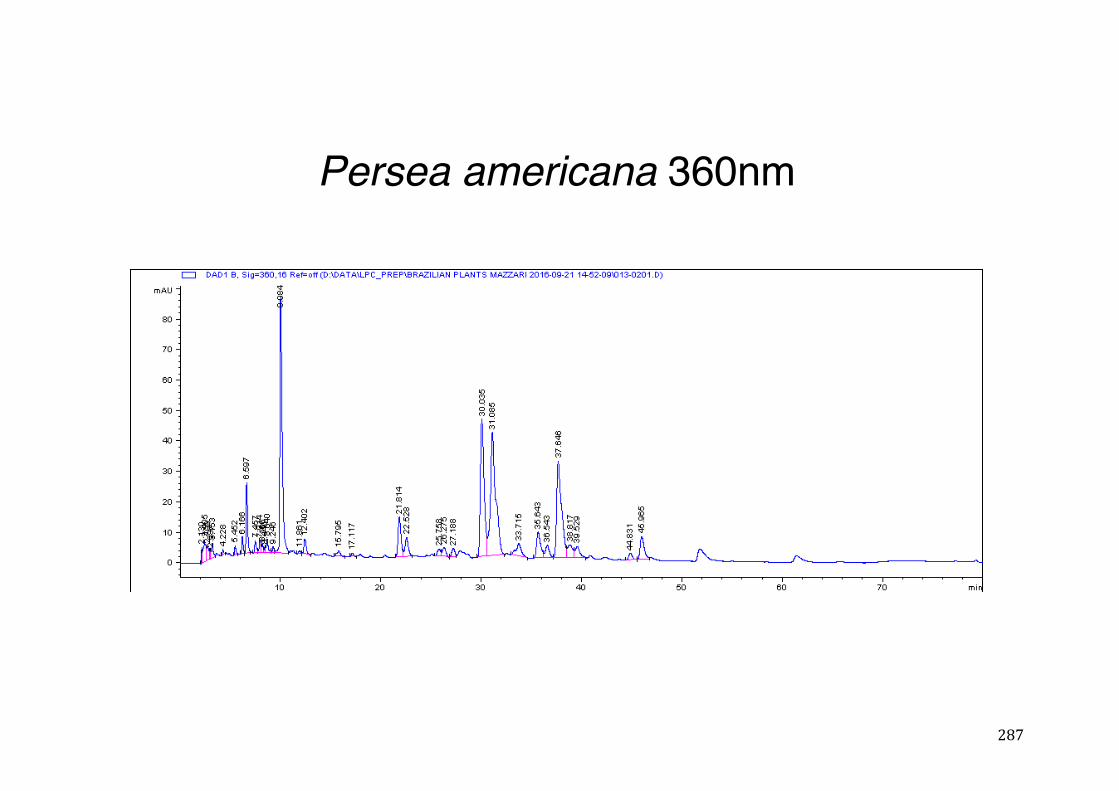
287
Persea americana 360nm
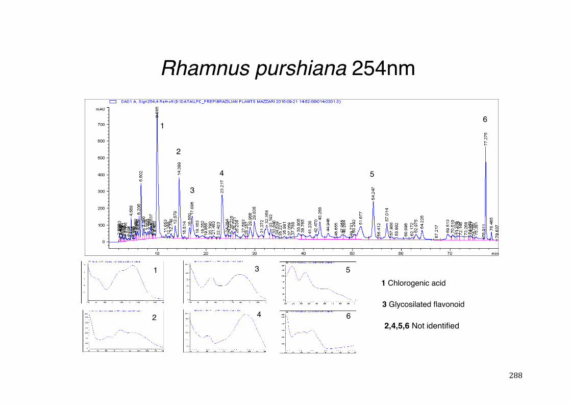
288
Rhamnus purshiana 254nm
1 Chlorogenic acid
2
3
4
1
2
3
4 5
6
5
63 Glycosilated flavonoid
1
2,4,5,6 Not identified
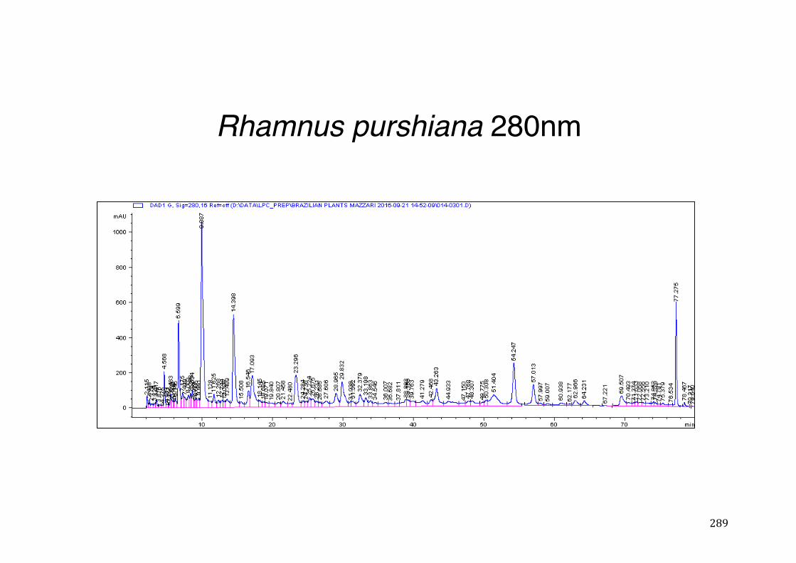
289
Rhamnus purshiana 280nm
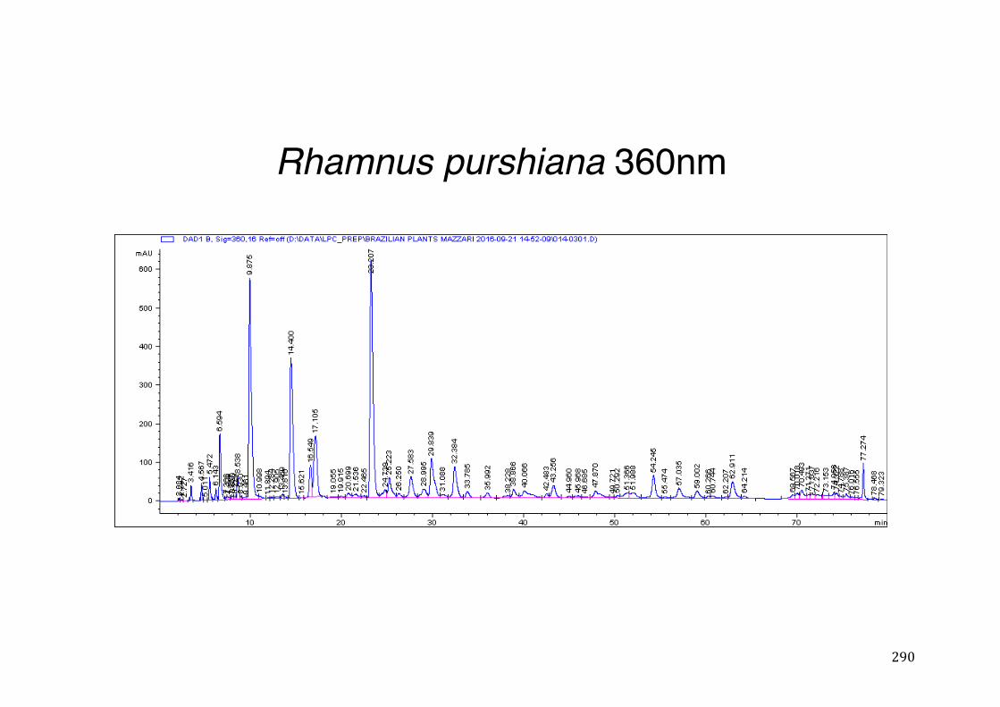
290
Rhamnus purshiana 360nm
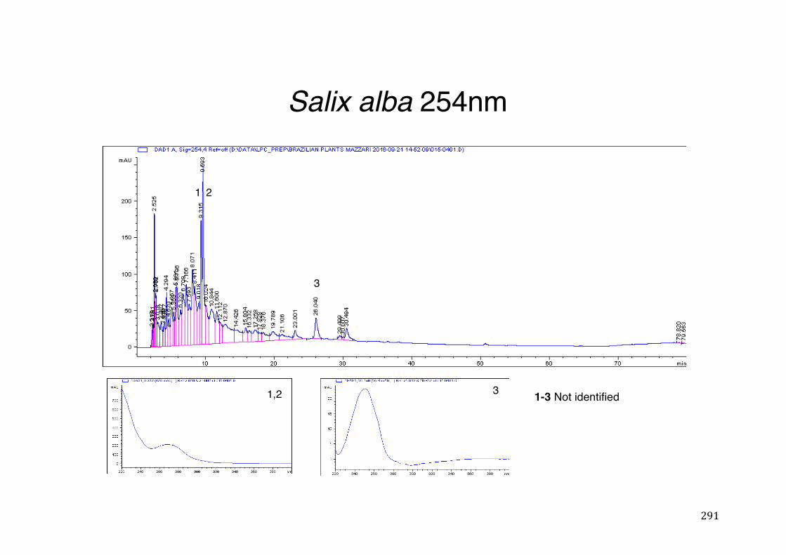
291
Salix alba 254nm
1,2
1 2
3
3 1-3 Not identified
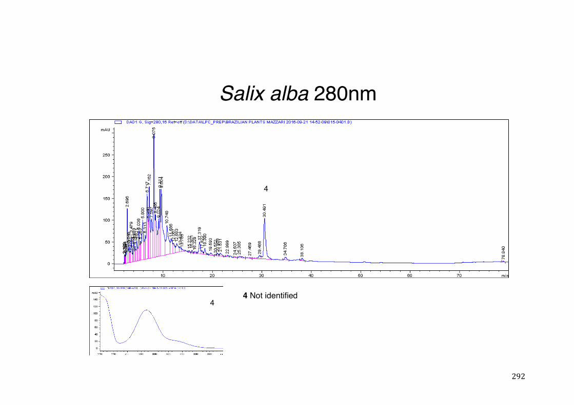
292
Salix alba 280nm
4
44 Not identified
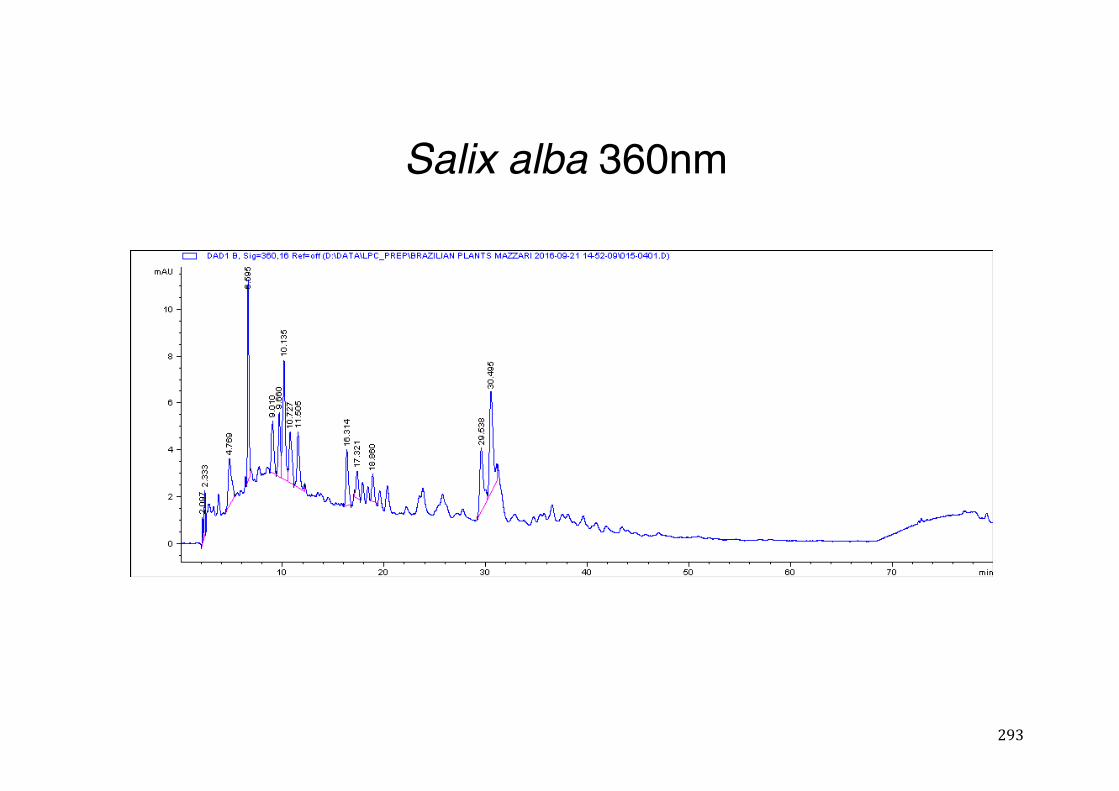
293
Salix alba 360nm
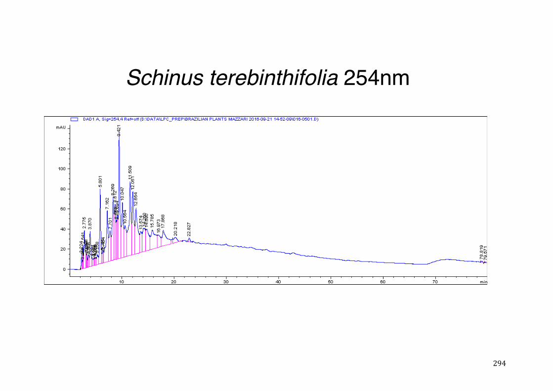
294
Schinus terebinthifolia 254nm
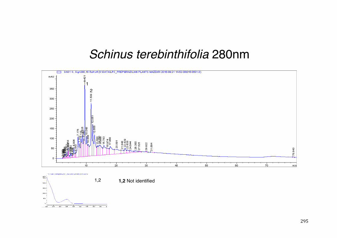
295
Schinus terebinthifolia 280nm
12
1,2 1,2 Not identified
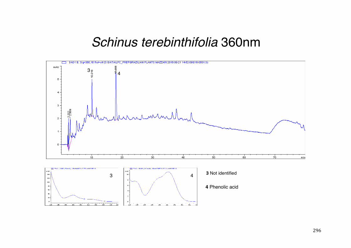
296
Schinus terebinthifolia 360nm
3 4
4 Phenolic acid
3 4 3 Not identified
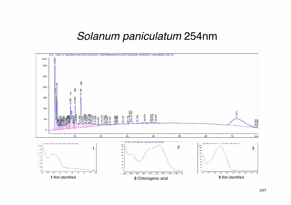
297
Solanum paniculatum 254nm
21
3
1 2 3
2 Chlorogenic acid1 Not identified 3 Not identified
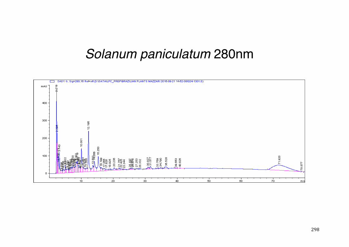
298
Solanum paniculatum 280nm
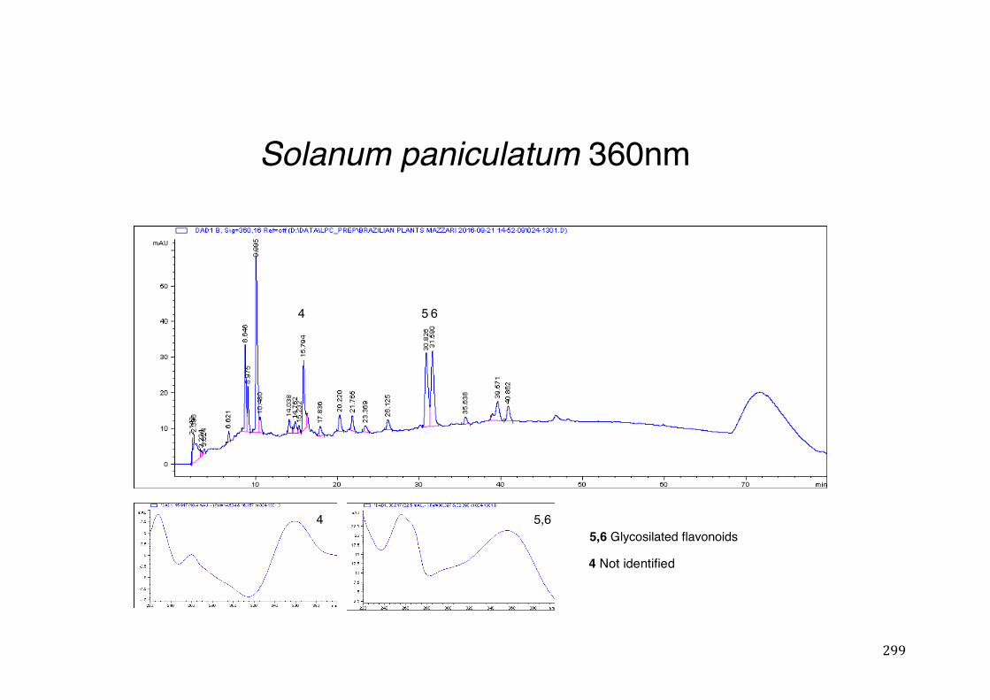
299
Solanum paniculatum 360nm
4 5,6
4 5 6
5,6 Glycosilated flavonoids
4 Not identified
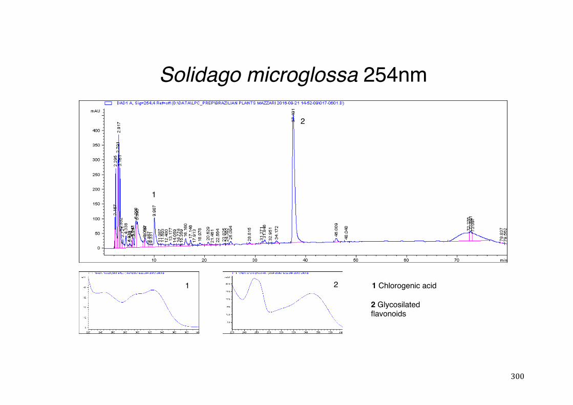
300
Solidago microglossa 254nm
1
2
2 1 Chlorogenic acid
2 Glycosilatedflavonoids
1
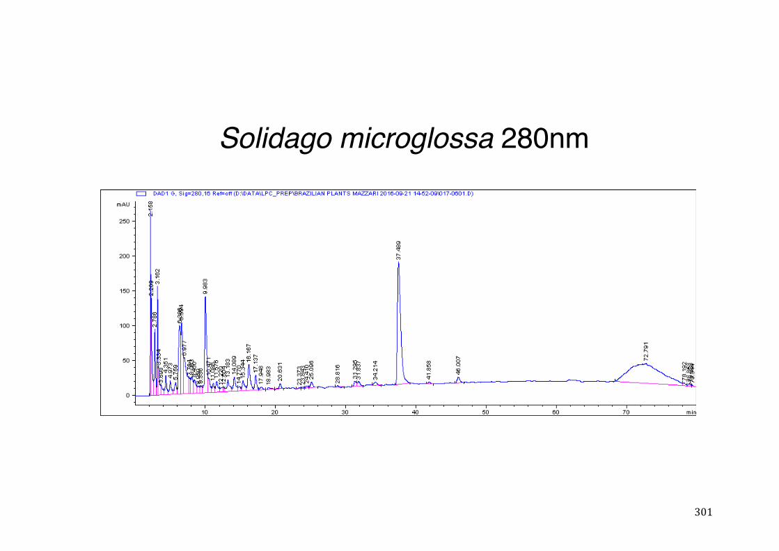
301
Solidago microglossa 280nm
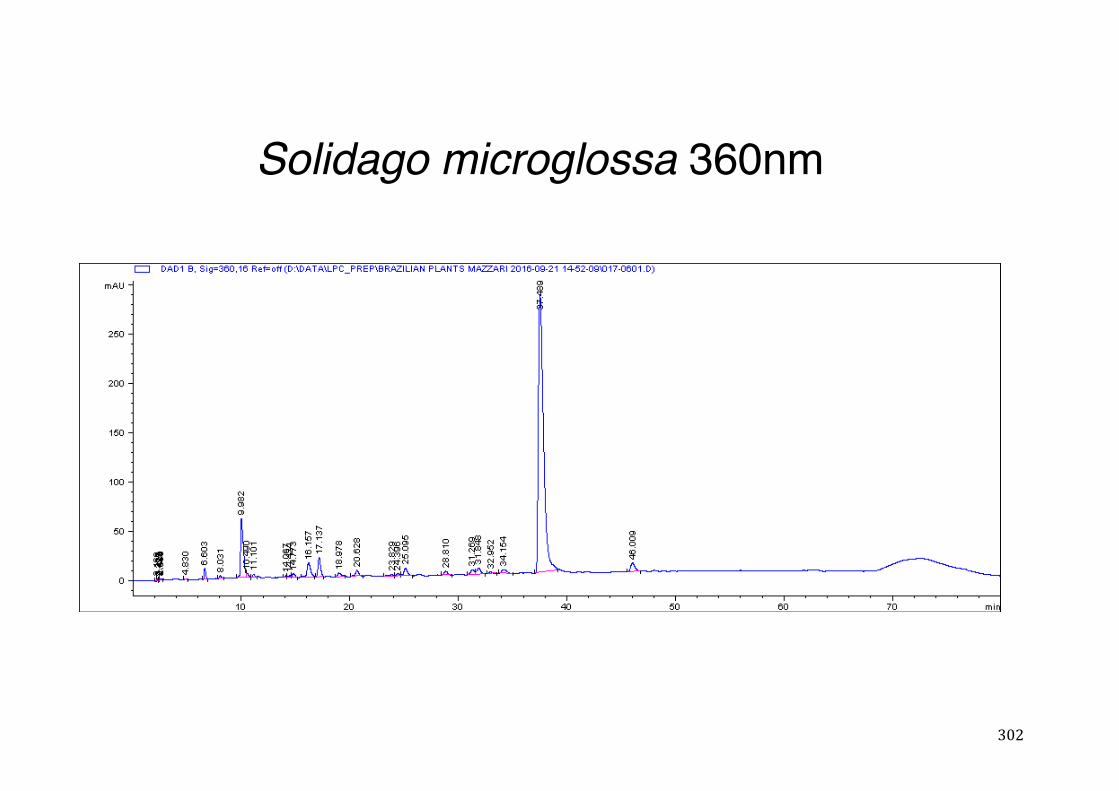
302
Solidago microglossa 360nm
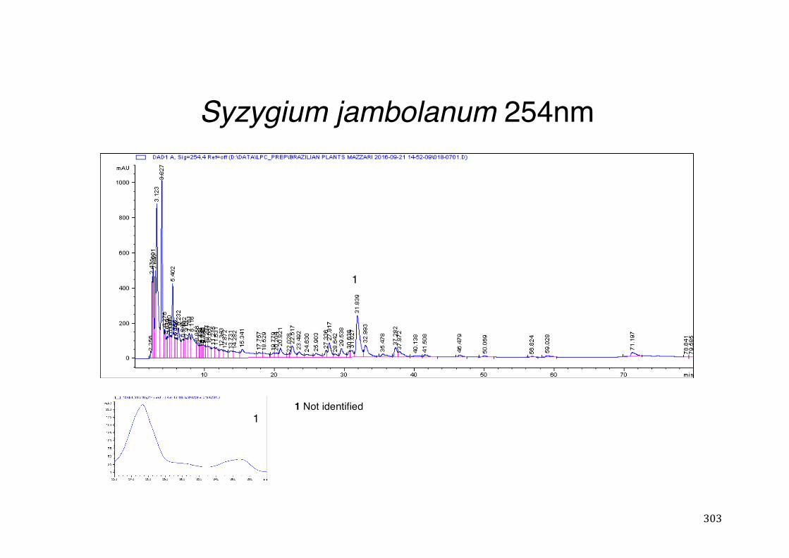
303
Syzygium jambolanum 254nm
1
11 Not identified
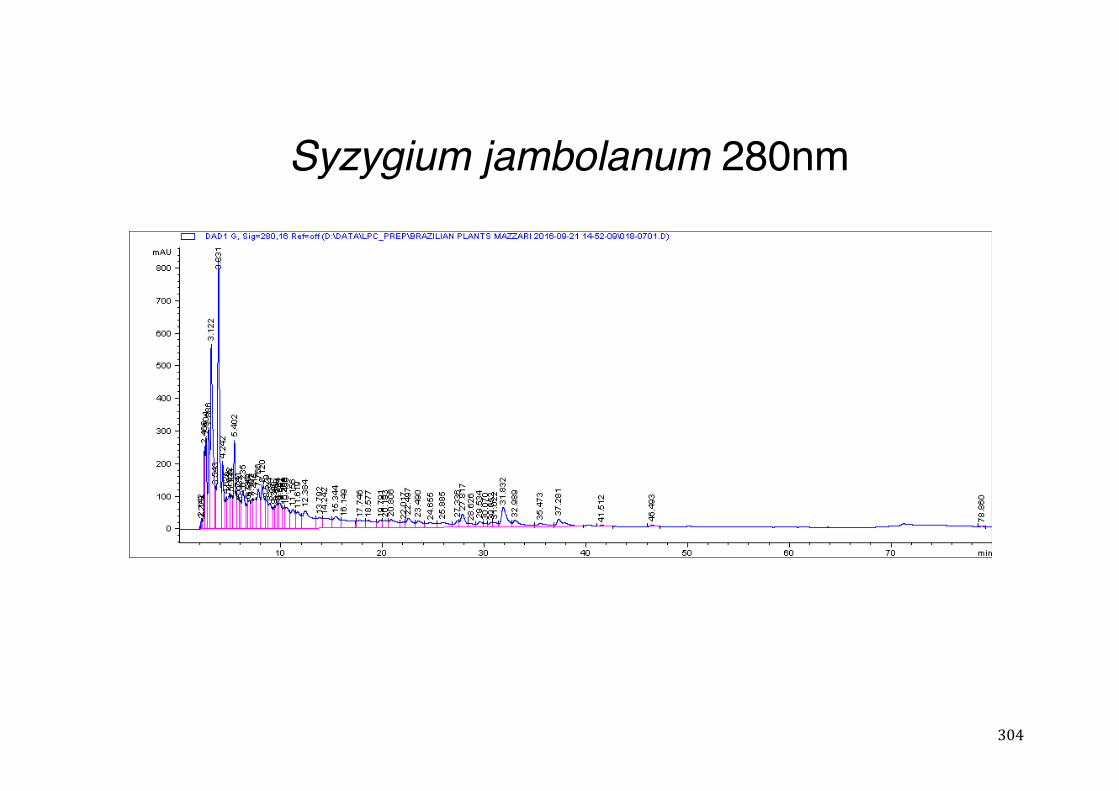
304
Syzygium jambolanum 280nm
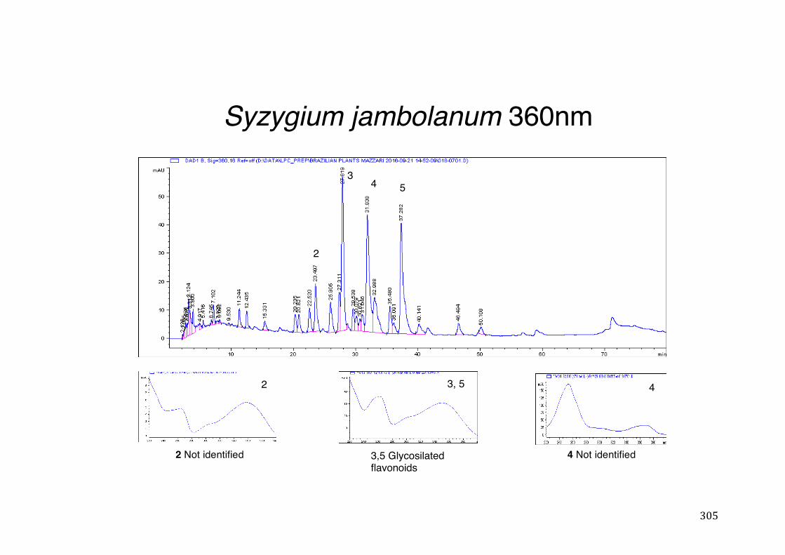
305
Syzygium jambolanum 360nm
2 3, 5
3,5 Glycosilatedflavonoids
2
34 5
4
2 Not identified 4 Not identified
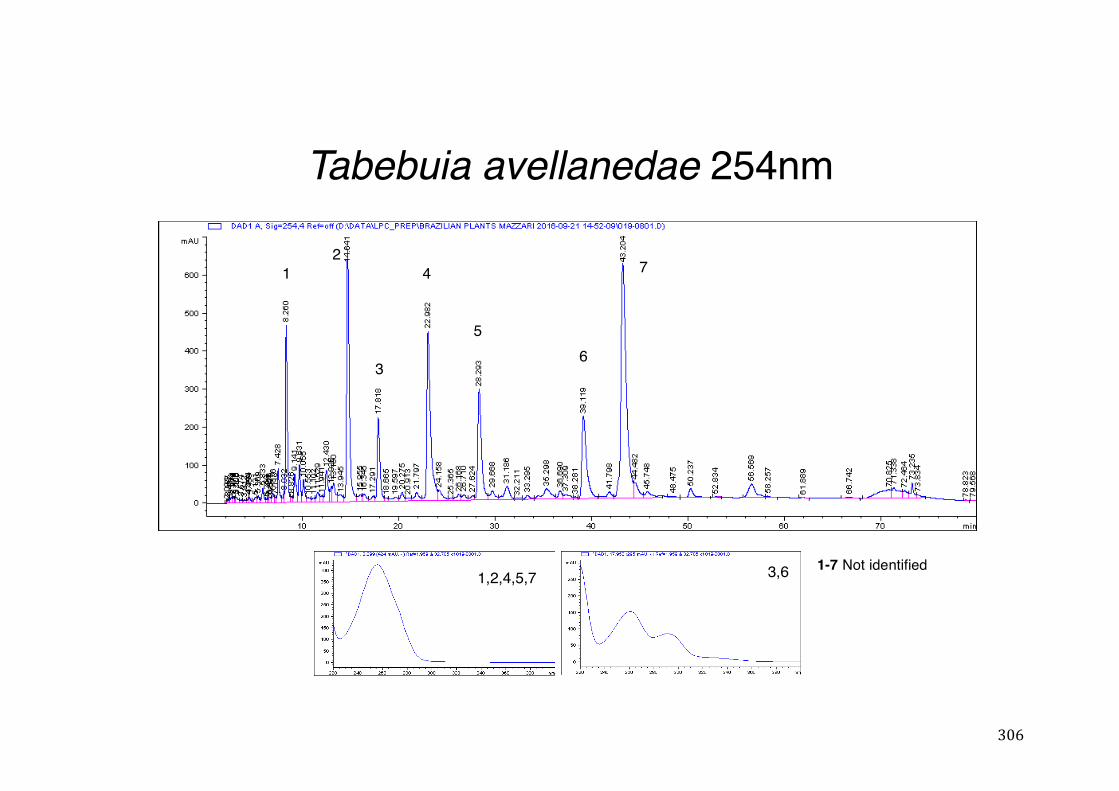
306
Tabebuia avellanedae 254nm
1,2,4,5,7 3,6
12
3
4
56
7
1-7 Not identified
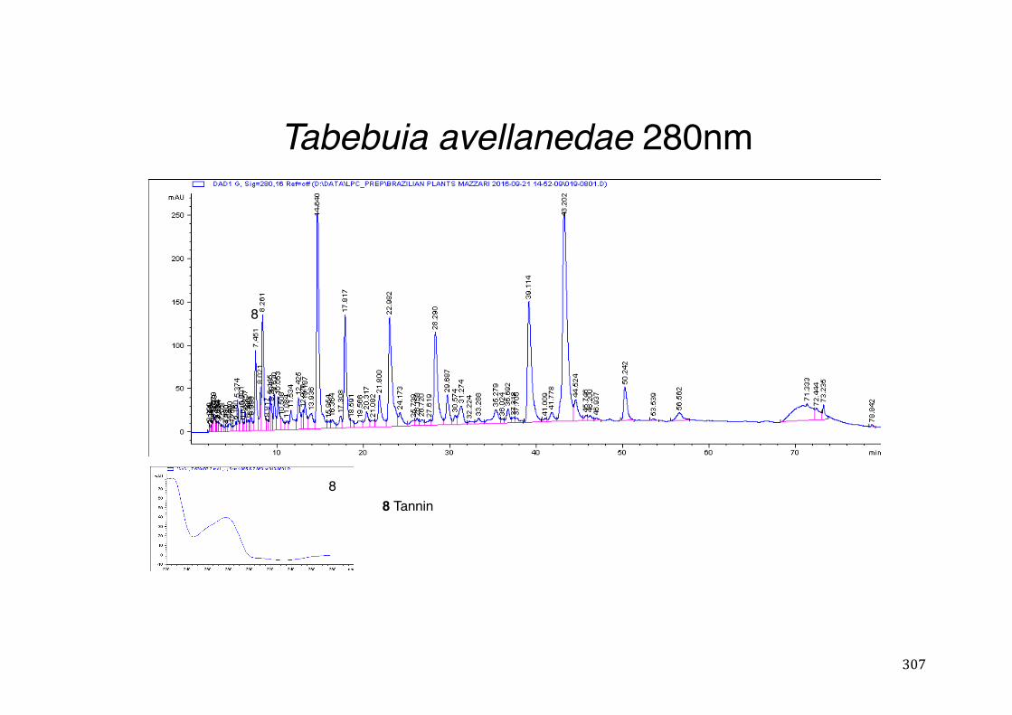
307
Tabebuia avellanedae 280nm
8
8 Tannin8
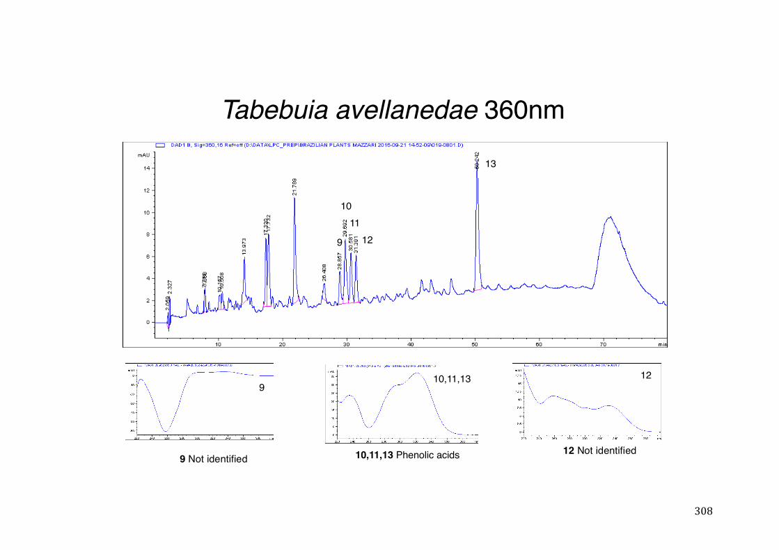
308
Tabebuia avellanedae 360nm
9
10,11,13 Phenolic acids
12
9
1011
12
13
10,11,13
9 Not identified 12 Not identified
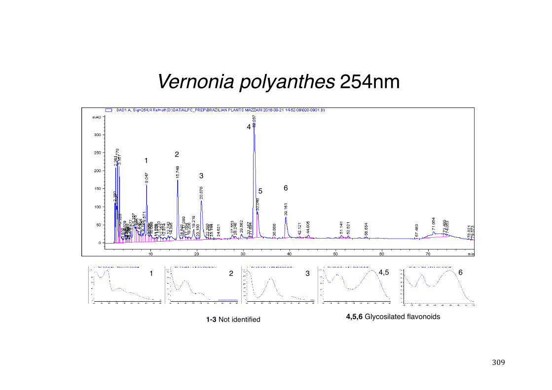
309
Vernonia polyanthes 254nm
12
3
4
5 6
1 2 3 4,5 6
4,5,6 Glycosilated flavonoids1-3 Not identified

310
Vernonia polyanthes 280nm
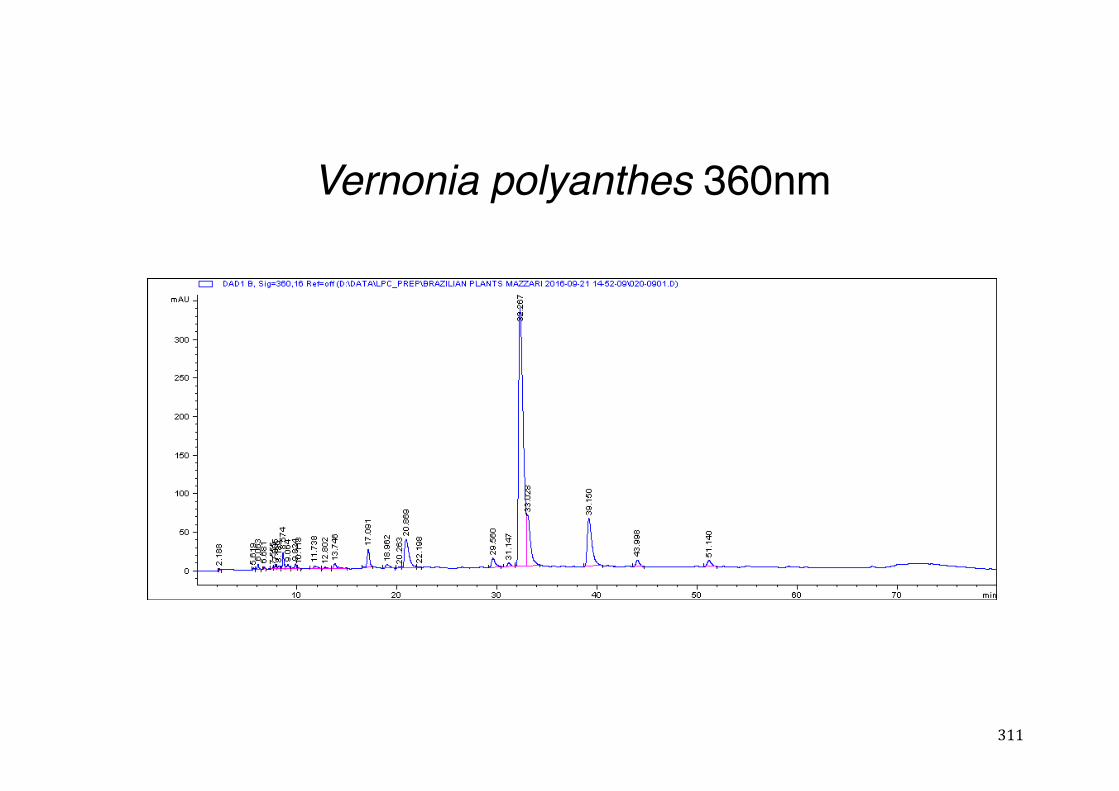
311
Vernonia polyanthes 360nm
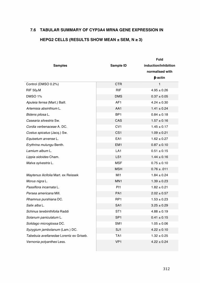
312
7.6 TABULAR SUMMARY OF CYP3A4 MRNA GENE EXPRESSION IN
HEPG2 CELLS (RESULTS SHOW MEAN ± SEM, N ≥ 3)
Samples
Sample ID
Fold induction/inhibition
normalised with b-actin
Control (DMSO 0.2%) CTR 1 RIF 50µM RIF 4.95 ± 0.26
DMSO 1% DMS 0.37 ± 0.05 Apuleia ferrea (Mart.) Baill. AF1 4.24 ± 0.30 Artemisia absinthium L. AA1 1.41 ± 0.24 Bidens pilosa L. BP1 0.84 ± 0.18 Casearia silvestris Sw. CAS 1.57 ± 0.16 Cordia verbenaceae A. DC. CV1 1.45 ± 0.17 Costus spicatus (Jacq.) Sw. CS1 1.09 ± 0.21 Equisetum arvense L. EA1 1.62 ± 0.27 Erythrina mulungu Benth. EM1 0.87 ± 0.10 Lamium album L. LA1 0.51 ± 0.15 Lippia sidoides Cham. LS1 1.44 ± 0.16 Malva sylvestris L. MSF 0.75 ± 0.10
MSH 0.76 ± .011 Maytenus ilicifolia Mart. ex Reissek MI1 1.84 ± 0.24 Morus nigra L. MN1 1.39 ± 0.23 Passiflora incarnata L. PI1 1.82 ± 0.21 Persea americana Mill. PA1 2.02 ± 0.57 Rhamnus purshiana DC. RP1 1.53 ± 0.23 Salix alba L. SA1 3.25 ± 0.29 Schinus terebinthifolia Raddi ST1 4.88 ± 0.19 Solanum paniculatum L. SP1 0.41 ± 0.15 Solidago microglossa DC. SM1 1.05 ± 0.06
Syzygium jambolanum (Lam.) DC. SJ1 4.22 ± 0.10 Tabebuia avellanedae Lorentz ex Griseb. TA1 1.32 ± 0.25 Vernonia polyanthes Less. VP1 4.22 ± 0.24
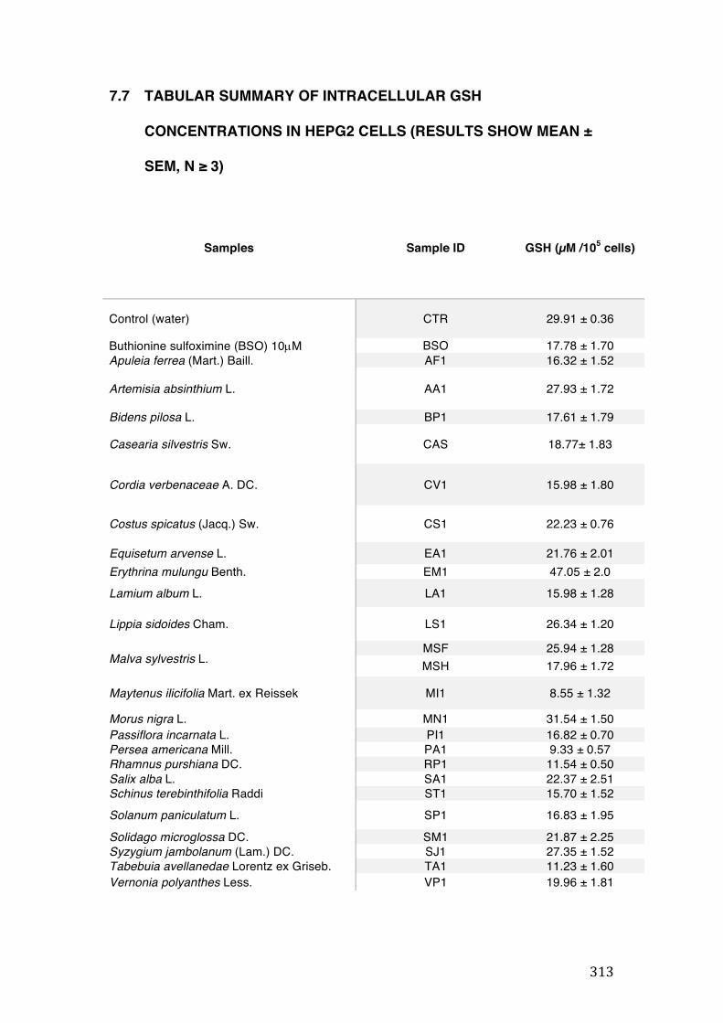
313
7.7 TABULAR SUMMARY OF INTRACELLULAR GSH
CONCENTRATIONS IN HEPG2 CELLS (RESULTS SHOW MEAN ±
SEM, N ≥ 3)
Samples
Sample ID GSH (µM /105 cells)
Control (water) CTR 29.91 ± 0.36
Buthionine sulfoximine (BSO) 10µM BSO 17.78 ± 1.70 Apuleia ferrea (Mart.) Baill. AF1 16.32 ± 1.52
Artemisia absinthium L. AA1 27.93 ± 1.72
Bidens pilosa L. BP1 17.61 ± 1.79
Casearia silvestris Sw. CAS 18.77± 1.83
Cordia verbenaceae A. DC. CV1 15.98 ± 1.80
Costus spicatus (Jacq.) Sw. CS1 22.23 ± 0.76
Equisetum arvense L. EA1 21.76 ± 2.01 Erythrina mulungu Benth. EM1 47.05 ± 2.0 Lamium album L. LA1 15.98 ± 1.28
Lippia sidoides Cham. LS1 26.34 ± 1.20
Malva sylvestris L. MSF 25.94 ± 1.28 MSH 17.96 ± 1.72
Maytenus ilicifolia Mart. ex Reissek MI1 8.55 ± 1.32
Morus nigra L. MN1 31.54 ± 1.50 Passiflora incarnata L. PI1 16.82 ± 0.70 Persea americana Mill. PA1 9.33 ± 0.57 Rhamnus purshiana DC. RP1 11.54 ± 0.50 Salix alba L. SA1 22.37 ± 2.51 Schinus terebinthifolia Raddi ST1 15.70 ± 1.52 Solanum paniculatum L. SP1 16.83 ± 1.95 Solidago microglossa DC. SM1 21.87 ± 2.25 Syzygium jambolanum (Lam.) DC. SJ1 27.35 ± 1.52 Tabebuia avellanedae Lorentz ex Griseb. TA1 11.23 ± 1.60 Vernonia polyanthes Less. VP1 19.96 ± 1.81
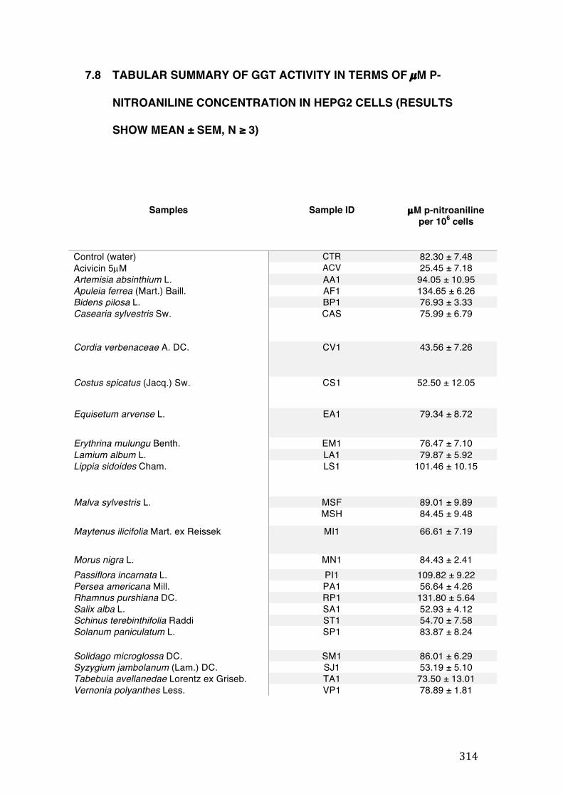
314
7.8 TABULAR SUMMARY OF GGT ACTIVITY IN TERMS OF µM P-
NITROANILINE CONCENTRATION IN HEPG2 CELLS (RESULTS
SHOW MEAN ± SEM, N ≥ 3)
Samples Sample ID µM p-nitroaniline per 106 cells
Control (water) CTR 82.30 ± 7.48 Acivicin 5µM ACV 25.45 ± 7.18 Artemisia absinthium L. AA1 94.05 ± 10.95 Apuleia ferrea (Mart.) Baill. AF1 134.65 ± 6.26 Bidens pilosa L. BP1 76.93 ± 3.33 Casearia sylvestris Sw. CAS 75.99 ± 6.79
Cordia verbenaceae A. DC. CV1 43.56 ± 7.26
Costus spicatus (Jacq.) Sw. CS1 52.50 ± 12.05
Equisetum arvense L. EA1 79.34 ± 8.72
Erythrina mulungu Benth. EM1 76.47 ± 7.10 Lamium album L. LA1 79.87 ± 5.92 Lippia sidoides Cham. LS1 101.46 ± 10.15
Malva sylvestris L. MSF 89.01 ± 9.89 MSH 84.45 ± 9.48
Maytenus ilicifolia Mart. ex Reissek MI1 66.61 ± 7.19
Morus nigra L. MN1 84.43 ± 2.41 Passiflora incarnata L. PI1 109.82 ± 9.22 Persea americana Mill. PA1 56.64 ± 4.26 Rhamnus purshiana DC. RP1 131.80 ± 5.64 Salix alba L. SA1 52.93 ± 4.12 Schinus terebinthifolia Raddi ST1 54.70 ± 7.58 Solanum paniculatum L. SP1 83.87 ± 8.24
Solidago microglossa DC. SM1 86.01 ± 6.29 Syzygium jambolanum (Lam.) DC. SJ1 53.19 ± 5.10 Tabebuia avellanedae Lorentz ex Griseb. TA1 73.50 ± 13.01 Vernonia polyanthes Less. VP1 78.89 ± 1.81
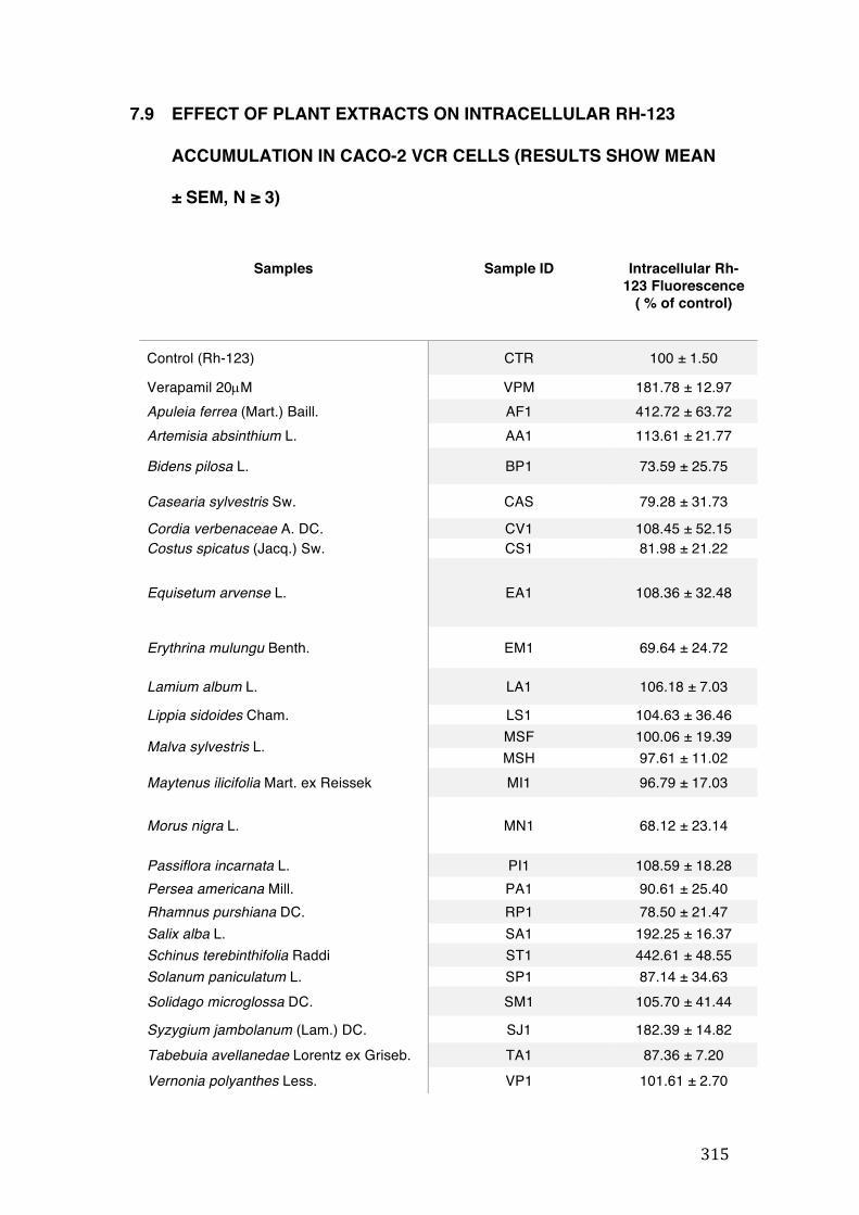
315
7.9 EFFECT OF PLANT EXTRACTS ON INTRACELLULAR RH-123
ACCUMULATION IN CACO-2 VCR CELLS (RESULTS SHOW MEAN
± SEM, N ≥ 3)
Samples Sample ID Intracellular Rh-123 Fluorescence
( % of control)
Control (Rh-123) CTR 100 ± 1.50
Verapamil 20µM VPM 181.78 ± 12.97 Apuleia ferrea (Mart.) Baill. AF1 412.72 ± 63.72 Artemisia absinthium L. AA1 113.61 ± 21.77
Bidens pilosa L. BP1 73.59 ± 25.75
Casearia sylvestris Sw. CAS 79.28 ± 31.73
Cordia verbenaceae A. DC. CV1 108.45 ± 52.15 Costus spicatus (Jacq.) Sw. CS1 81.98 ± 21.22
Equisetum arvense L. EA1 108.36 ± 32.48
Erythrina mulungu Benth. EM1 69.64 ± 24.72
Lamium album L. LA1 106.18 ± 7.03
Lippia sidoides Cham. LS1 104.63 ± 36.46
Malva sylvestris L. MSF 100.06 ± 19.39 MSH 97.61 ± 11.02
Maytenus ilicifolia Mart. ex Reissek MI1 96.79 ± 17.03
Morus nigra L. MN1 68.12 ± 23.14
Passiflora incarnata L. PI1 108.59 ± 18.28 Persea americana Mill. PA1 90.61 ± 25.40 Rhamnus purshiana DC. RP1 78.50 ± 21.47 Salix alba L. SA1 192.25 ± 16.37 Schinus terebinthifolia Raddi ST1 442.61 ± 48.55 Solanum paniculatum L. SP1 87.14 ± 34.63 Solidago microglossa DC. SM1 105.70 ± 41.44
Syzygium jambolanum (Lam.) DC. SJ1 182.39 ± 14.82 Tabebuia avellanedae Lorentz ex Griseb. TA1 87.36 ± 7.20 Vernonia polyanthes Less. VP1 101.61 ± 2.70
316
7.10 PUBLICATIONS
7.10.1 Herbal medicines in Brazil: pharmacokinetic profile and potential herb-drug interactions
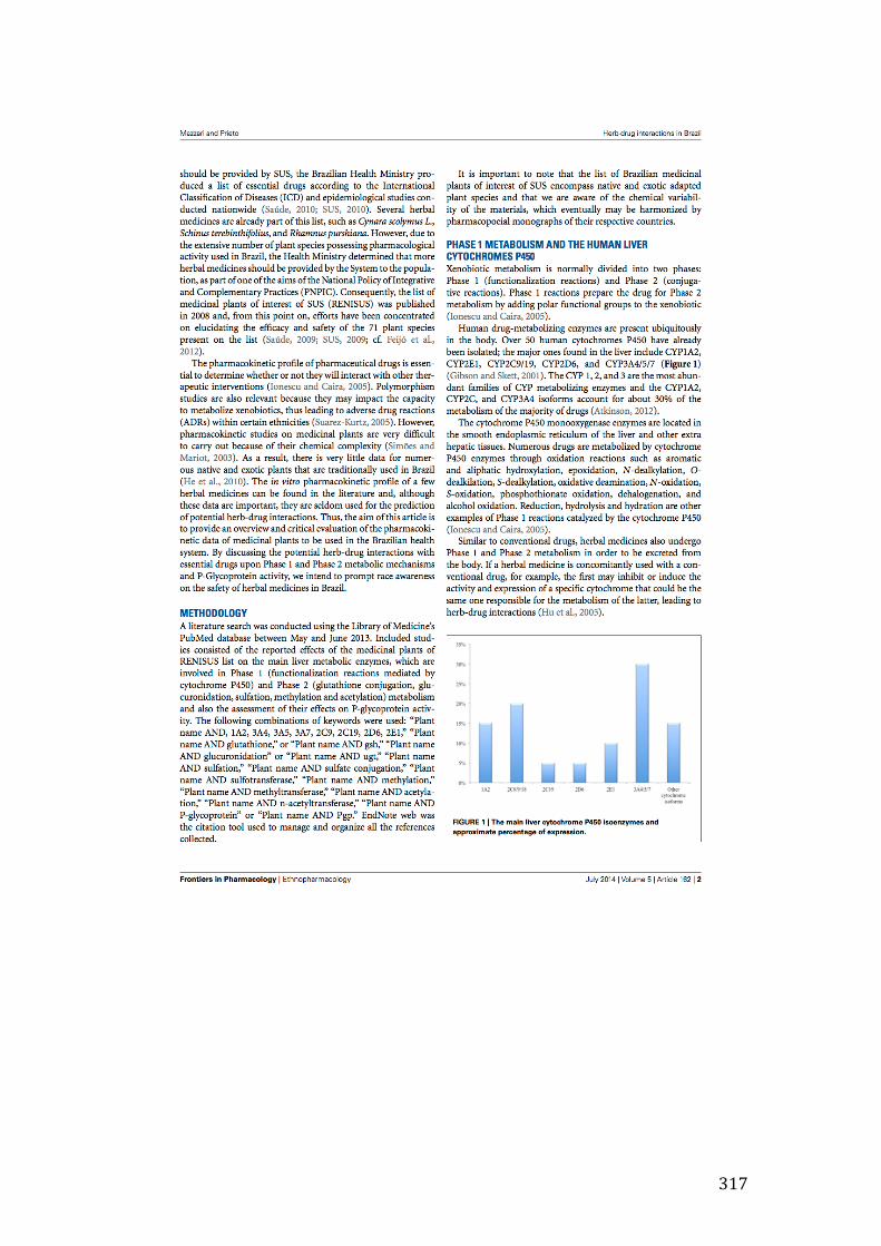
317
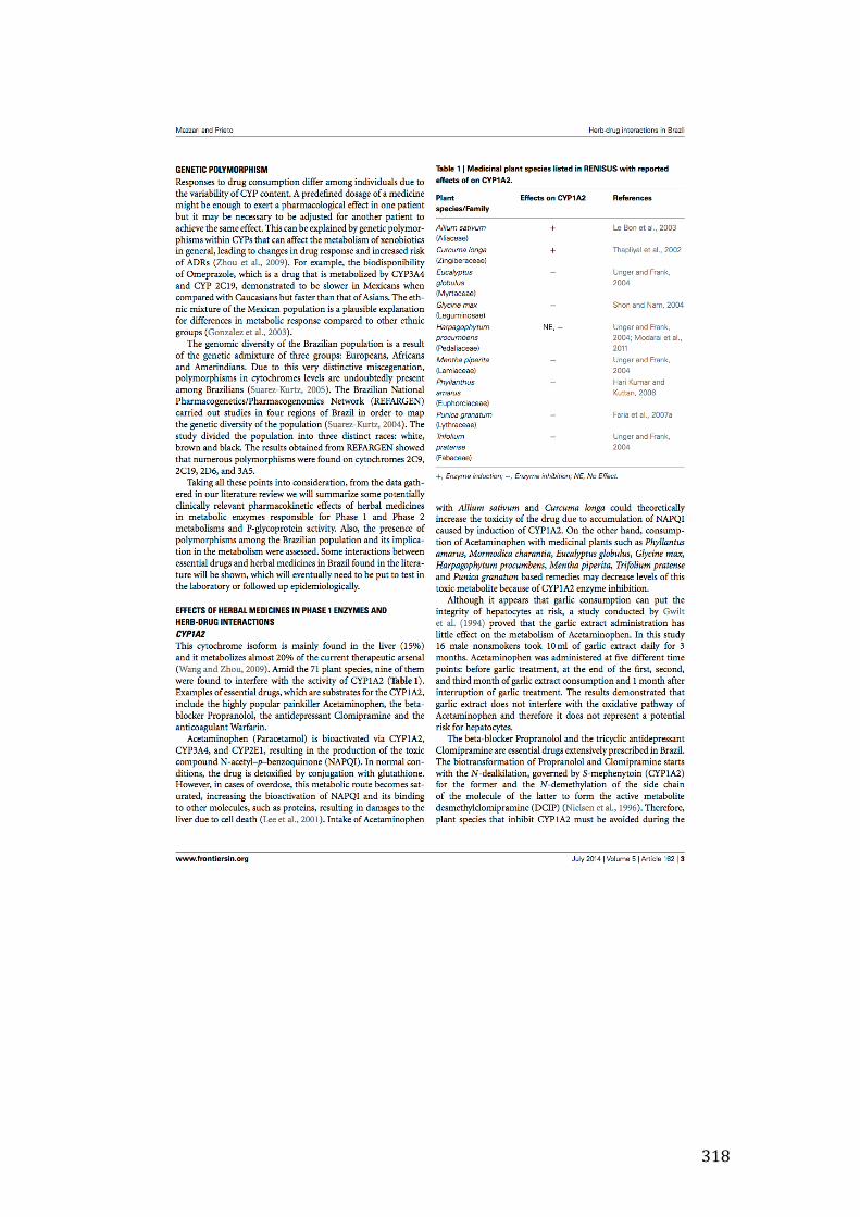
318
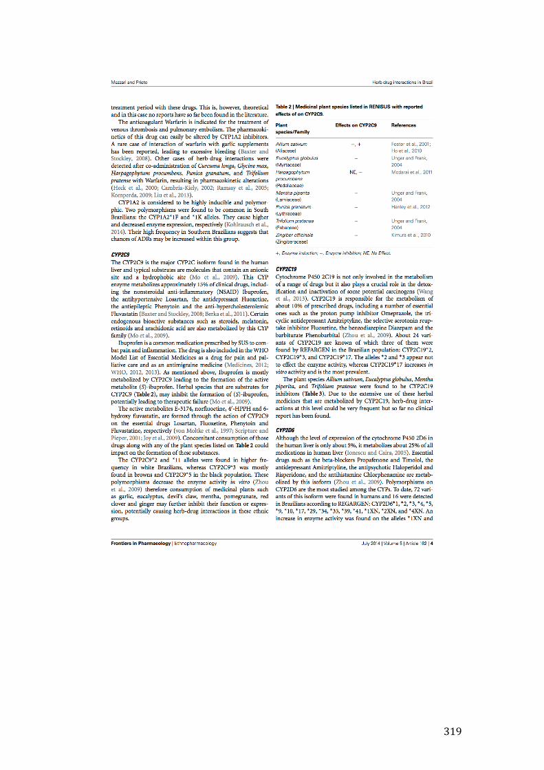
319
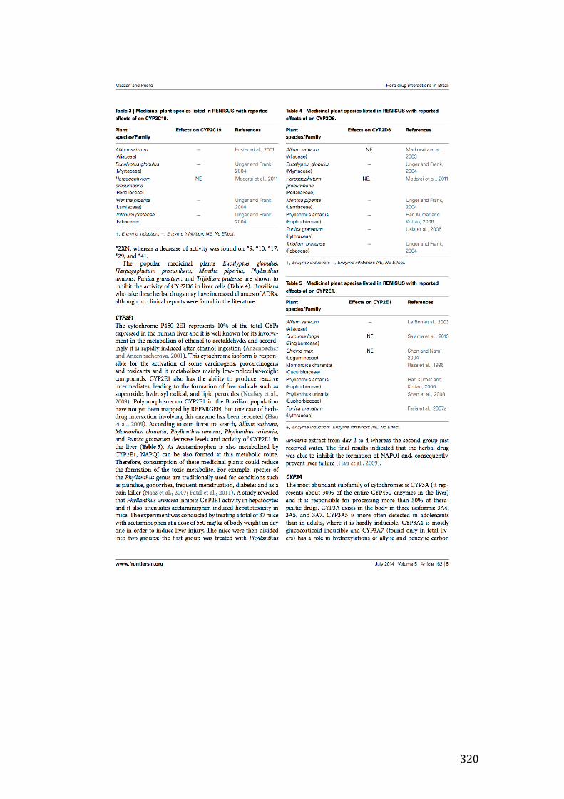
320
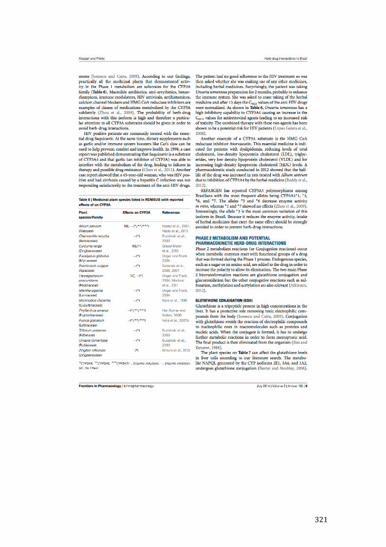
321
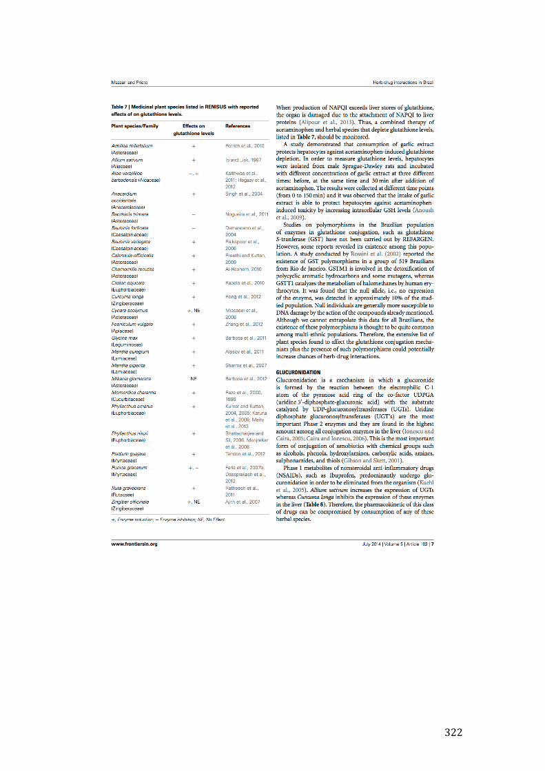
322
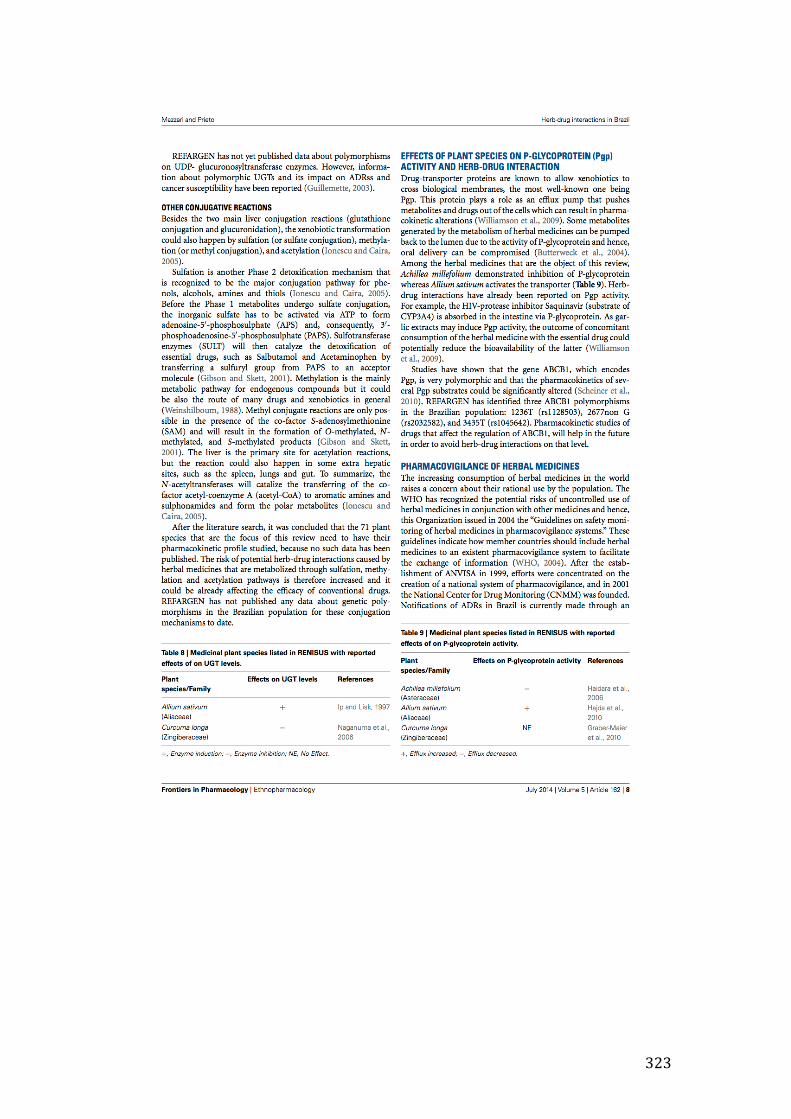
323
324
325
326
327
334
7.10.3 In vitro effects of four native Brazilian medicinal plants in CYP3A4 mRNA gene expression, glutathione levels, and P-glycoprotein activity
335
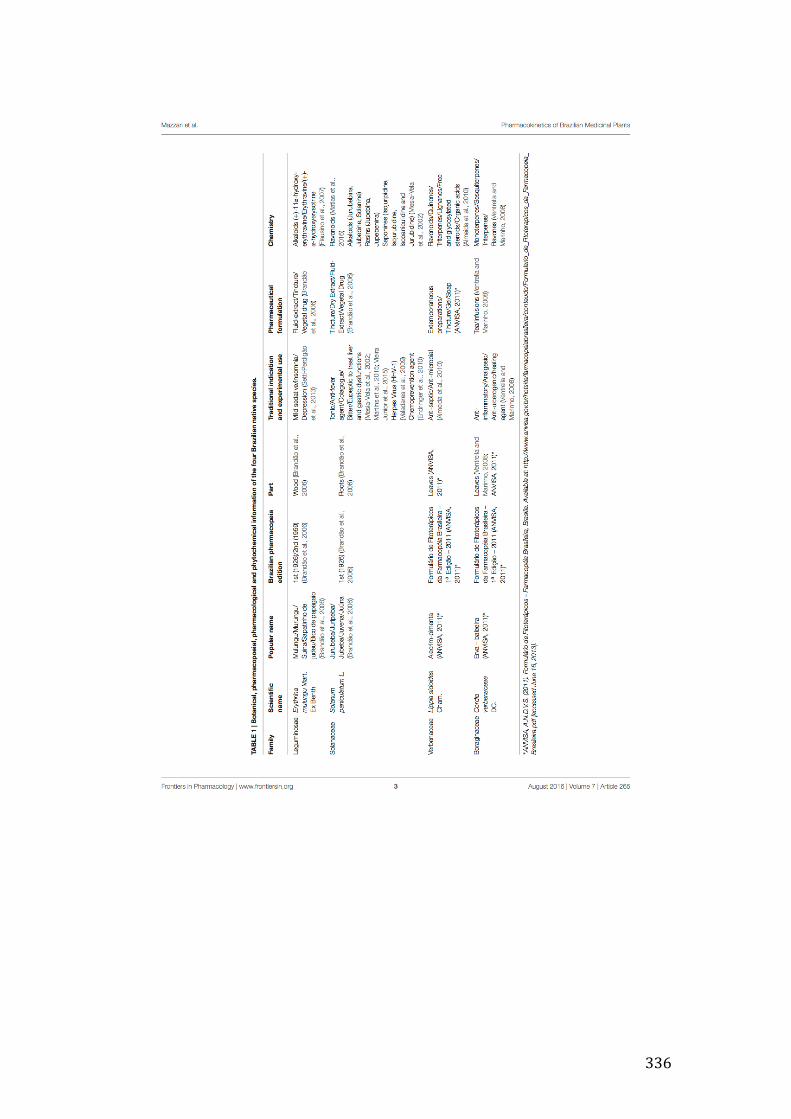
336
337
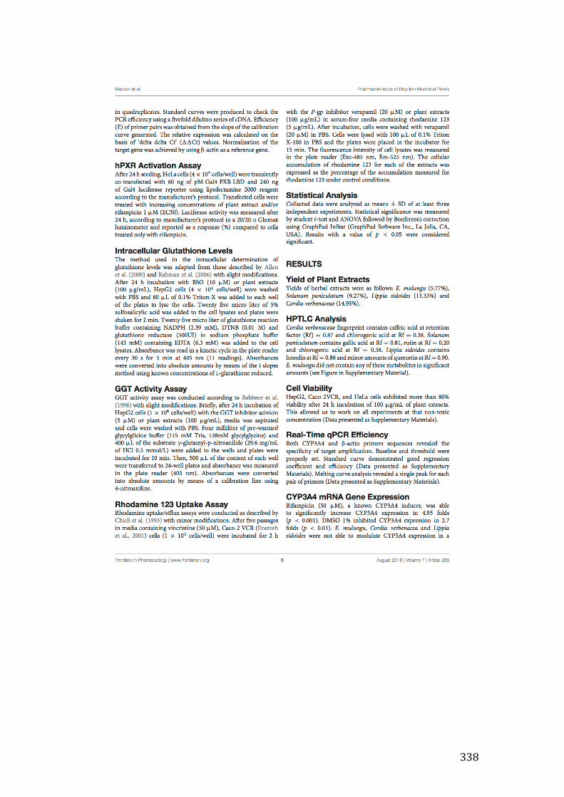
338
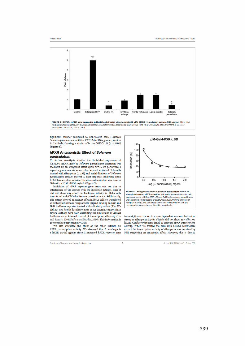
339
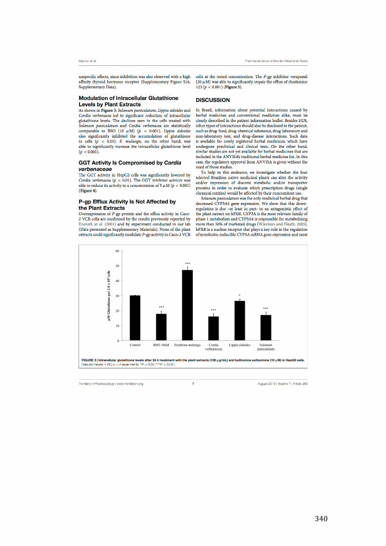
340
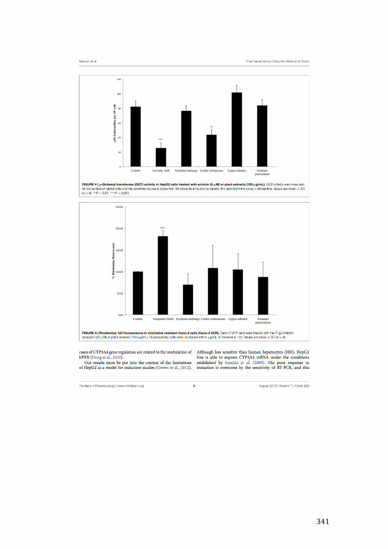
341
342
343
344
345