WHO MONOGRAPH ON SELECTED MEDICINAL PLANTS VOL 1
654
Transcript of WHO MONOGRAPH ON SELECTED MEDICINAL PLANTS VOL 1


Bulbus Allii Cepae
i
WHOmonographson selected
medicinal plantsVOLUME 1
World Health OrganizationGeneva
1999

WHO monographs on selected medicinal plants
ii
WHO Library Cataloguing in Publication DataWHO monographs on selected medicinal plants.—Vol. 1.1.Plants, Medicinal 2.Herbs 3.Traditional medicineISBN 92 4 154517 8 (NLM Classification: QV 766)
The World Health Organization welcomes requests for permission to reproduce or translate itspublications, in part or in full. Applications and enquiries should be addressed to the Office ofPublications, World Health Organization, Geneva, Switzerland, which will be glad to provide thelatest information on any changes made to the text, plans for new editions, and reprints andtranslations already available.
© World Health Organization 1999
Publications of the World Health Organization enjoy copyright protection in accordance with theprovisions of Protocol 2 of the Universal Copyright Convention. All rights reserved.The designations employed and the presentation of the material in this publication do not imply theexpression of any opinion whatsoever on the part of the Secretariat of the World Health Organiza-tion concerning the legal status of any country, territory, city or area or of its authorities, orconcerning the delimitation of its frontiers or boundaries.The mention of specific companies or of certain manufacturers’ products does not imply that theyare endorsed or recommended by the World Health Organization in preference to others of asimilar nature that are not mentioned. Errors and omissions excepted, the names of proprietaryproducts are distinguished by initial capital letters.
Designed by WHO GraphicsTypeset in Hong Kong
Printed in Malta97/11795-Best-set/Interprint-6500

Bulbus Allii Cepae
iii
Contents
Acknowledgements vIntroduction 1
Monographs (in alphabetical order of plant name)
Bulbus Allii Cepae 5Bulbus Allii Sativi 16Aloe 33Aloe Vera Gel 43Radix Astragali 50Fructus Bruceae 59Radix Bupleuri 67Herba Centellae 77Flos Chamomillae 86Cortex Cinnamomi 95Rhizoma Coptidis 105Rhizoma Curcumae Longae 115Radix Echinaceae 125Herba Echinaceae Purpureae 136Herba Ephedrae 145Folium Ginkgo 154Radix Ginseng 168Radix Glycyrrhizae 183Radix Paeoniae 195Semen Plantaginis 202Radix Platycodi 213Radix Rauwolfiae 221Rhizoma Rhei 231Folium Sennae 241Fructus Sennae 250Herba Thymi 259

WHO monographs on selected medicinal plants
iv
Radix Valerianae 267Rhizoma Zingiberis 277
AnnexParticipants in the WHO Consultation on Selected MedicinalPlants 288
Contents

Bulbus Allii Cepae
v
Acknowledgements
Special acknowledgement is due to Professors Norman R. Farnsworth, HarryH. S. Fong, and Gail B. Mahady of the WHO Collaborating Centre for Tradi-tional Medicine, College of Pharmacy, University of Illinois at Chicago, USA,for drafting and revising the monographs.
WHO also acknowledges with thanks the members of the advisory groupthat met in Beijing, China, in 1994, to draw up a list of medicinal plants forwhich monographs should be prepared, the more than 100 experts who pro-vided comments and advice on the draft texts, and those who participated inthe WHO Consultation held in Munich, Germany, in 1996 to review themonographs (see Annex). Finally, WHO would like to thank the Food andAgriculture Organization of the United Nations and the United Nations Indus-trial Development Organization for their contributions and all those whosubmitted comments through the World Self-Medication Industry, a nongov-ernmental organization in official relations with WHO.

WHO monographs on selected medicinal plants
vi

Bulbus Allii Cepae
1
Introduction
During the past decade, traditional systems of medicine have become a topic ofglobal importance. Current estimates suggest that, in many developing coun-tries, a large proportion of the population relies heavily on traditional practi-tioners and medicinal plants to meet primary health care needs. Althoughmodern medicine may be available in these countries, herbal medicines(phytomedicines) have often maintained popularity for historical and culturalreasons. Concurrently, many people in developed countries have begun to turnto alternative or complementary therapies, including medicinal herbs.
Few plant species that provide medicinal herbs have been scientificallyevaluated for their possible medical application. Safety and efficacy data areavailable for even fewer plants, their extracts and active ingredients, and thepreparations containing them. Furthermore, in most countries the herbal medi-cines market is poorly regulated, and herbal products are often neither regis-tered nor controlled. Assurance of the safety, quality, and efficacy of medicinalplants and herbal products has now become a key issue in industrialized and indeveloping countries. Both the general consumer and health-care professionalsneed up-to-date, authoritative information on the safety and efficacy of medici-nal plants.
During the fourth International Conference of Drug Regulatory Authorities(ICDRA) held in Tokyo in 1986, WHO was requested to compile a list ofmedicinal plants and to establish international specifications for the mostwidely used medicinal plants and simple preparations. Guidelines for the as-sessment of herbal medicines were subsequently prepared by WHO andadopted by the sixth ICDRA in Ottawa, Canada, in 1991.1 As a result ofICDRA’s recommendations and in response to requests from WHO’s MemberStates for assistance in providing safe and effective herbal medicines for usein national health-care systems, WHO is now publishing this first volumeof 28 monographs on selected medicinal plants; a second volume is inpreparation.
Preparation of the monographsThe medicinal plants featured in this volume were selected by an advisorygroup in Beijing in 1994. The plants selected are widely used and important in
1 Guidelines for the assessment of herbal medicines. In: Quality assurance of pharmaceuticals: acompendium of guidelines and related materials. Volume 1. Geneva, World Health Organization,1997:31–37.

WHO monographs on selected medicinal plants
2
all WHO regions, and for each sufficient scientific information seemed availableto substantiate safety and efficacy. The monographs were drafted by the WHOCollaborating Centre for Traditional Medicine at the University of Illinois atChicago, United States of America. The content was obtained by a systematicreview of scientific literature from 1975 until the end of 1995: review articles;bibliographies in review articles; many pharmacopoeias—the International,African, British, Chinese, Dutch, European, French, German, Hungarian, Indian,and Japanese; as well as many other reference books.
Draft monographs were widely distributed, and some 100 experts inmore than 40 countries commented on them. Experts included members ofWHO’s Expert Advisory Panels on Traditional Medicine, on the InternationalPharmacopoeia and Pharmaceutical Preparations, and on Drug Evaluation andNational Drug Policies; and the drug regulatory authorities of 16 countries.
A WHO Consultation on Selected Medicinal Plants was held in Munich,Germany, in 1996. Sixteen experts and drug regulatory authorities fromMember States participated. Following extensive discussion, 28 of 31 draftmonographs were approved. The monograph on one medicinal plant was re-jected because of the plant’s potential toxicity. Two others will be reconsideredwhen more definitive data are available. At the subsequent eighth ICDRA inBahrain later in 1996, the 28 model monographs were further reviewed andendorsed, and Member States requested WHO to prepare additional modelmonographs.
Purpose and content of the monographsThe purpose of the monographs is to:
• provide scientific information on the safety, efficacy, and quality control/quality assurance of widely used medicinal plants, in order to facilitate theirappropriate use in Member States;
• provide models to assist Member States in developing their own mono-graphs or formularies for these or other herbal medicines; and
• facilitate information exchange among Member States.
Readers will include members of regulatory authorities, practitioners of ortho-dox and of traditional medicine, pharmacists, other health professionals, manu-facturers of herbal products, and research scientists.
Each monograph contains two parts. The first part consists of phar-macopoeial summaries for quality assurance: botanical features, distribution,identity tests, purity requirements, chemical assays, and active or major chemi-cal constituents. The second part summarizes clinical applications, pharmacol-ogy, contraindications, warnings, precautions, potential adverse reactions, andposology.
In each pharmacopoeial summary, the Definition section provides the Latinbinomial pharmacopoeial name, the most important criterion in quality assur-ance. Latin pharmacopoeial synonyms and vernacular names, listed in the
Introduction

Bulbus Allii Cepae
3
sections Synonyms and Selected vernacular names, are those names used in com-merce or by local consumers. The monographs place outdated botanical no-menclature in the synonyms category, based on the International Rules ofNomenclature.
For example, Aloe barbadensis Mill. is actually Aloe vera (L.) Burm. Cassiaacutifolia Delile and Cassia angustifolia Vahl., often treated in separate mono-graphs, are now believed to be the same species, Cassia senna L. Matricariachamomilla L., M. recutita L., and M. suaveolens L. have been used for many yearsas the botanical name for camomile. However, it is now agreed that the nameChamomilla recutita (L.) Rauschert is the legitimate name.
The vernacular names listed are a selection of names from individual coun-tries worldwide, in particular from areas where the medicinal plant is in com-mon use. The lists are not complete, but reflect the names appearing in theofficial monographs and reference books consulted during preparation of theWHO monographs and in the Natural Products Alert (NAPRALERT) database (adatabase of literature from around the world on ethnomedical, biological andchemical information on medicinal plants, fungi and marine organisms, locatedat the WHO Collaborating Centre for Traditional Medicine at the University ofIllinois at Chicago).
A detailed botanical description (under Description) is intended for qualityassurance at the stages of production and collection, whereas the detaileddescription of the drug material (under Plant material of interest) is for the samepurpose at the manufacturing and commerce stages. Geographical distribution isnot normally found in official compendia, but it is included here to provideadditional quality assurance information.
General identity tests, Purity tests, and Chemical assays are all normalcompendial components included under those headings in these monographs.Where purity tests do not specify accepted limits, those limits should be set inaccordance with national requirements by the appropriate Member Stateauthorities.
Each medicinal plant and the specific plant part used (the drug) containactive or major chemical constituents with a characteristic profile that can beused for chemical quality control and quality assurance. These constituents aredescribed in the section Major chemical constituents.
The second part of each monograph begins with a list of Dosage forms and ofMedicinal uses categorized as those uses supported by clinical data, those usesdescribed in pharmacopoeias and in traditional systems of medicine, and thoseuses described in folk medicine, not yet supported by experimental or clinicaldata.
The first category includes medical indications that are well established insome countries and that have been validated by clinical studies documented inthe world’s scientific literature. The clinical trials may have been controlled,randomized, double-blind studies, open trials, or well-documented observa-tions of therapeutic applications. Experts at the Munich Consultation agreed toinclude Folium and Fructus Sennae, Aloe, Rhizoma Rhei, and Herba Ephedrae
Introduction

WHO monographs on selected medicinal plants
4
in this category because they are widely used and their efficacy is well docu-mented in the standard medical literature.
The second category includes medicinal uses that are well established inmany countries and are included in official pharmacopoeias or national mono-graphs. Well-established uses having a plausible pharmacological basis andsupported by older studies that clearly need to be repeated are also included.The references cited provide additional information useful in evaluating specificherbal preparations. The uses described should be reviewed by local expertsand health workers for their applicability in the local situation.
The third category refers to indications described in unofficial pharma-copoeias and other literature, and to traditional uses. The appropriateness ofthese uses could not be assessed, owing to a lack of scientific data to support theclaims. The possible use of these remedies must be carefully considered in thelight of therapeutic alternatives.
The final sections of each monograph cover Pharmacology (both experimentaland clinical); Contraindications such as sensitivity or allergy; Warnings; Precautions,including discussion of drug interactions, carcinogenicity, teratogenicity andspecial groups such as children and nursing mothers; Adverse reactions; andPosology.
Use of the monographsWHO encourages countries to provide safe and effective traditional remediesand practices in public and private health services.
This publication is not intended to replace official compendia such aspharmacopoeias, formularies, or legislative documents. The monographs areintended primarily to promote harmonization in the use of herbal medicineswith respect to levels of safety, efficacy, and quality control. These aspects ofherbal medicines depend greatly on how the individual dosage form is pre-pared. For this reason, local regulatory authorities, experts, and health workers,as well as the scientific literature, should be consulted to determine whether aspecific herbal preparation is appropriate for use in primary health care.
The monographs will be supplemented and updated periodically as newinformation appears in the literature, and additional monographs will beprepared. WHO would be pleased to receive comments and suggestions, to thisend, from readers of the monographs.
Finally, I should like to express our appreciation of the support providedfor the development of the monographs by Dr H. Nakajima and Dr F. S.Antezana during their time as Director-General and Assistant Director-General,respectively, of WHO.
Dr Xiaorui ZhangMedical OfficerTraditional MedicineWorld Health Organization
Introduction

Bulbus Allii Cepae
5
Bulbus Allii Cepae
DefinitionBulbus Allii Cepae is the fresh or dried bulbs of Allium cepa L. (Liliaceae) or itsvarieties and cultivars.
SynonymsAllium esculentum Salisb., Allium porrum cepa Rehb. (1).
Selected vernacular namesIt is most commonly known as “onion”. Basal, basl, cebolla, cebolla morada,cepa bulb, cepolla, cipolla, common onion, cu hanh, hom hua yai, hom khaao,hom yai, hu-t’sung, hu t’sung t’song, hua phak bhu, i-i-bsel, kesounni, khtim,Küchenzwiebel, l’oignon, loyon, Madras oignon, oignon, palandu, piyaj, piyaz,pyaz, pyaaz, ralu lunu, red globe onion, sibuyas, Spanish onion, tamanegi, umbibawang merah, vengayan, yellow Bermuda onion, white globe onion, Zwiebel(1–5).
DescriptionA perennial herb, strong smelling when crushed; bulbs vary in size and shapefrom cultivar to cultivar, often depressed-globose and up to 20cm in diameter;outer tunics membranous. Stem up to 100cm tall and 30mm in diameter,tapering from inflated lower part. Leaves up to 40cm in height and 20mm indiameter, usually almost semicircular in section and slightly flattened on upperside; basal in first year, in second year their bases sheathing the lower sixth ofthe stem. Spathe often 3-valved, persistent, shorter than the umbel. Umbel 4–9cm in diameter, subglobose or hemispherical, dense, many-flowered; pedicelsup to 40mm, almost equal. Perianth stellate; segments 3–4.5 � 2–2.5mm,white, with green stripe, slightly unequal, the outer ovate, the inner oblong,obtuse or acute. Stamens exserted; filaments 4–5mm, the outer subulate, theinner with an expanded base up to 2mm wide and bearing short teeth on eachside. Ovary whitish. Capsule about 5mm, 2n � 16 (6).
Plant material of interest: fresh or dried bulbsGeneral appearanceMacroscopically, Bulbus Allii Cepae varies in size and shape from cultivar tocultivar, 2–20cm in diameter; flattened, spherical or pear-shaped; white orcoloured (7 ).

WHO monographs on selected medicinal plants
6
Organoleptic propertiesOdour strong, characteristic alliaceous; taste strong; crushing or cutting the bulbstimulates lachrymation.
Microscopic characteristicsThe external dried leaf scales of the bulbs show a large-celled epidermis withlightly spotted cell walls; the cells are elongated longitudinally. The underlyinghypodermis runs perpendicular to the epidermis and contains large calciumoxalate crystals bordering the cell walls. The epidermis of the fleshy leaf scalesresembles that of the dried leaf scales, and the epidermal cells on the dorsal sideare distinctly longer and more elongated than the epidermal cells on the ventralside. Large calcium oxalate crystals are found in the hypodermis; stomata rare;large cell nuclei conspicuous; and spiral vessel elements occur in the leaf meso-phyll (8).
Powdered plant materialContains mainly thin-walled cells of the mesophyll with broken pieces of spiralvessel elements; cells containing calcium oxalate crystals are scarce (8).
Geographical distributionBulbus Allii Cepae (“onion”) is probably indigenous to western Asia, but it iscommercially cultivated worldwide, especially in regions of moderate climate(1).
General identity testsMacroscopic inspection, microscopic characteristics and microchemical exami-nation for organic sulfur compounds (9); and thin-layer chromatographic analy-sis for the presence of cysteine sulfoxides (10, 11).
Purity testsMicrobiologyThe test for Salmonella spp. in Bulbus Allii Cepae products should be negative.The maximum acceptable limits of other microorganisms are as follows (12–14). Preparations for oral use: aerobic bacteria—not more than 105/g or ml;fungi—not more than 104/g or ml; enterobacteria and certain Gram-negativebacteria—not more than 103/g or ml; Escherichia coli—0/g or ml.
Total ashNot more than 6% (3).

Bulbus Allii Cepae
7
Acid-insoluble ashNot more than 1.0% (3).
Water-soluble extractiveNot more than 5.0% (3).
Alcohol-soluble extractiveNot more than 4.0% (3).
Pesticide residuesTo be established in accordance with national requirements. Normally, themaximum residue limit of aldrin and dieldrin for Bulbus Allii Cepae is not morethan 0.05mg/kg (14). For other pesticides, see WHO guidelines on qualitycontrol methods for medicinal plants (12) and guidelines for predicting dietaryintake of pesticide residues (15).
Heavy metalsRecommended lead and cadmium levels are no more than 10 and 0.3mg/kg,respectively, in the final dosage form of the plant material (12).
Radioactive residuesFor analysis of strontium-90, iodine-131, caesium-134, caesium-137 andplutonium-239, see WHO guidelines on quality control methods for medicinalplants (12).
Other purity testsChemical, foreign organic matter, and moisture tests to be established in accor-dance with national requirements.
Chemical assaysAssay for organic sulfur constituents, cysteine sulfoxides and sulfides by meansof high-performance liquid chromatographic (16, 17) or gas–liquid chromato-graphic (18) methods, respectively. Quantitative levels to be established byappropriate national authority.
Major chemical constituentsSulfur- and non-sulfur-containing chemical constituents have been isolatedfrom Bulbus Allii Cepae; the sulfur compounds are the most characteristic (1, 4,7).
The organic sulfur compounds of Bulbus Allii Cepae, including thethiosulfinates, thiosulfonates, cepaenes, S-oxides, S,S�-dioxides, monosulfides,

WHO monographs on selected medicinal plants
8
disulfides, trisulfides, and zwiebelanes occur only as degradation products ofthe naturally occurring cysteine sulfoxides (e.g. (�)-S-propyl-L-cysteine sulfox-ide). When the onion bulb is crushed, minced, or otherwise processed, thecysteine sulfoxides are released from compartments and contact the enzymealliinase in adjacent vacuoles. Hydrolysis and immediate condensation of thereactive intermediate (sulfenic acids) form the compounds as indicated below(1). The odorous thiosulphonates occur (in low concentrations) only in freshlychopped onions, whereas the sulfides accumulate in stored extracts or steam-distilled oils. Approximately 90% of the soluble organic-bound sulfur is presentas γ-glutamylcysteine peptides, which are not acted on by alliinase. Theyfunction as storage reserve and contribute to the germination of seeds. How-ever, on prolonged storage or during germination, these peptides are acted onby γ-glutamyl transpeptidase to form alk(en)yl-cysteine sulfoxides, which inturn give rise to other volatile sulfur compounds (1).

Bulbus Allii Cepae
9
Dosage formsFresh juice and 5% and 50% ethanol extracts have been used in clinical studies(1). A “soft” extract is marketed in France but is not recognized as a drug byFrench authorities (7 ). Dried Bulbus Allii Cepae products should be stored inwell-closed containers, protected from light, moisture, and elevated tempera-ture. Fresh bulbs and juice should be refrigerated (2–10 °C).
Medicinal usesUses supported by clinical dataThe principal use of Bulbus Allii Cepae today is to prevent age-dependentchanges in the blood vessels, and loss of appetite (19).
Uses described in pharmacopoeias and in traditional systems ofmedicineTreatment of bacterial infections such as dysentery, and as a diuretic (2, 7). Thedrug has also been used to treat ulcers, wounds, scars, keloids (3), and asthma(20, 21). Bulbus Allii Cepae has also been used as an adjuvant therapy fordiabetes (4, 22, 23).
Uses described in folk medicine, not supported by experimental orclinical dataAs an anthelminthic, aphrodisiac, carminative, emmenagogue, expectorant, andtonic (3), and for the treatment of bruises, bronchitis, cholera, colic, earache,fevers, high blood pressure, jaundice, pimples, and sores (3).
PharmacologyExperimental pharmacologyAn aqueous extract or the juice of Bulbus Allii Cepae inhibited the in vitrogrowth of Escherichia coli, Serratia marcescens, Streptococcus species, Lactobacillusodontolyticus, Pseudomonas aeruginosa, and Salmonella typhosa (24–28). A petro-leum ether extract of Bulbus Allii Cepae inhibited the in vitro growth ofClostridium paraputrificum and Staphylococcus aureus (24). The essential oil hasactivity against a variety of fungi including Aspergillus niger, Cladosporiumwerneckii, Candida albicans, Fusarium oxysporium, Saccharomyces cerevisiae,Geotrichum candidum, Brettanomyces anomalus, and Candida lipolytica (5, 29).
The hypoglycaemic effects of Bulbus Allii Cepae have been demonstrated invivo. Intragastric administration of the juice, a chloroform, ethanol, petroleumether (0.25g/kg) or water extract (0.5ml), suppressed alloxan-, glucose- andepinephrine-induced hyperglycaemia in rabbits and mice (30–35).
Inhibition of platelet aggregation by Bulbus Allii Cepae has been demon-strated both in vitro and in vivo. An aqueous extract inhibited adenosinediphosphate-, collagen-, epinephrine- and arachidonic acid-induced platelet

WHO monographs on selected medicinal plants
10
aggregation in vitro (36, 37). Platelet aggregation was inhibited in rabbits afteradministration of the essential oil, or a butanol or chloroform extract of thedrug (38–40). An ethanol, butanol or chloroform extract or the essential oil(10–60µg/ml) of the drug inhibited aggregation of human platelets in vitro (41,42) by decreasing thromboxane synthesis (39). Both raw onions and the essen-tial oil increased fibrinolysis in ex vivo studies on rabbits and humans (1). Anincrease in coagulation time was also observed in rabbits (1).
Intragastric administration of the juice or an ether extract (100mg/kg) of thedrug inhibited allergen- and platelet activating factor-induced allergic reactions,but not histamine- or acetylcholine-induced allergenic responses in guinea-pigs(43). A water extract of the drug was not active (43). A chloroform extract ofBulbus Allii Cepae (20–80mg/kg) inhibited allergen- and platelet aggregationfactor-induced bronchial obstruction in guinea-pigs (44). The thiosulphinatesand cepaenes appear to be the active constituents of Bulbus Allii Cepae (1).
Both ethanol and methanol extracts of Bulbus Allii Cepae demonstrateddiuretic activity in dogs and rats after intragastric administration (45, 46).
Antihyperlipidaemic and anticholesterolaemic activities of the drug wereobserved after oral administration of minced bulbs, a water extract, the essen-tial oil (100mg/kg), or the fixed oil to rabbits or rats (47–52). However, onestudy reported no significant changes in cholesterol or lipid levels of the eye inrabbits, after treatment of the animals for 6 months with an aqueous extract(20% of diet) (53).
Oral administration of an ethanol extract of the drug to guinea-pigs inhibitedsmooth muscle contractions in the trachea induced by carbachol and inhibitedhistamine-, barium chloride-, serotonin-, and acetylcholine-induced contrac-tions in the ileum (20).
Topical application of an aqueous extract of Bulbus Allii Cepae (10% in agel preparation) inhibited mouse ear oedema induced by arachidonic acid (54).The active antiallergic and anti-inflammatory constituents of onion are theflavonoids (quercetin and kaempferol) (55). The flavonoids act as anti-inflammatory agents because they inhibit the action of protein kinase, phos-pholipase A2, cyclooxygenase, and lipoxygenase (56), as well as the release ofmediators of inflammation (e.g. histamine) from leukocytes (57).
In vitro, an aqueous extract of Bulbus Allii Cepae inhibited fibroblast prolif-eration (58). A 0.5% aqueous extract of onion inhibited the growth of humanfibroblasts and of keloidal fibroblasts (enzymically isolated from keloidal tis-sue) (59). In a comparative study, an aqueous extract of Bulbus Allii Cepae (1–3%) inhibited the proliferation of fibroblasts of varying origin (scar, keloid,embryonic tissue). The strongest inhibition was observed with keloid fibro-blasts (65–73%) as compared with the inhibition of scar and embryonicfibroblasts (up to 50%) (59). In human skin fibroblasts, both aqueous andchloroform onion extracts, as well as thiosulfinates, inhibited the platelet-derived growth factor-stimulated chemotaxis and proliferation of these cells(60). In addition, a protein fraction isolated from an onion extract exhibitedantimitotic activity (61).

Bulbus Allii Cepae
11
Clinical pharmacologyOral administration of a butanol extract of Bulbus Allii Cepae (200mg) tosubjects given a high-fat meal prior to testing suppressed platelet aggregationassociated with a high-fat diet (62).
Administration of a butanol extract to patients with alimentary lipaemiaprevented an increase in the total serum cholesterol, �-lipoprotein cholesterol,and �-lipoprotein and serum triglycerides (63, 64). A saponin fraction (50mg) orthe bulb (100mg) also decreased serum cholesterol and plasma fibrinogen levels(65, 66). However, fresh onion extract (50g) did not produce any significanteffects on serum cholesterol, fibrinogen, or fibrinolytic activity in normal sub-jects (67, 68).
Antihyperglycaemic activity of Bulbus Allii Cepae has been demonstrated inclinical studies. Administration of an aqueous extract (100mg) decreased glu-cose-induced hyperglycaemia in human adults (69). The juice of the drug(50mg) administered orally to diabetic patients reduced blood glucose levels(22). Addition of raw onion to the diet of non-insulin-dependent diabetic sub-jects decreased the dose of antidiabetic medication required to control thedisease (70). However, an aqueous extract of Bulbus Allii Cepae (200mg) wasnot active (71).
The immediate and late cutaneous reactions induced by injection of rabbitanti-human IgE-antibodies into the volar side of the forearms of 12 healthyvolunteers were reduced after pretreatment of the skin with a 50% ethanolonion extract (1). Immediate and late bronchial obstruction owing to allergeninhalation was markedly reduced after oral administration of a 5% ethanolonion extract 1 hour before exposure to the allergen (1).
In one clinical trial in 12 adult subjects, topical application of a 45%ethanolic onion extract inhibited the allergic skin reactions induced by anti-IgE(72).
ContraindicationsAllergies to the plant. The level of safety of Bulbus Allii Cepae is reflected by itsworldwide use as a vegetable.
WarningsNo warnings have been reported.
PrecautionsCarcinogenesis, mutagenesis, impairment of fertilityBulbus Allii Cepae is not mutagenic in vitro (73).
Other precautionsNo general precautions have been reported, and no precautions have beenreported concerning drug interactions, drug and laboratory test interactions,

WHO monographs on selected medicinal plants
12
nursing mothers, paediatric use, or teratogenic or non-teratogenic effects onpregnancy.
Adverse reactionsAllergic reactions such as rhinoconjunctivitis and contact dermatitis have beenreported (74).
PosologyUnless otherwise prescribed: a daily dosage is 50g of fresh onion or 20g of thedried drug; doses of preparations should be calculated accordingly (14).
References1. Breu W, Dorsch W. Allium cepa L. (Onion): Chemistry, analysis and pharmacology.
In: Wagner H, Farnsworth NR, eds. Economic and medicinal plants research, Vol. 6.London, Academic Press, 1994:115–147.
2. Kapoor LD. Handbook of Ayurvedic medicinal plants, Boca Raton, FL, CRC Press, 1990.3. Materia medika Indonesia, Jilid VI. Jakarta, Departemen Kesehatan, Republik
Indonesia, 1995.4. Wagner H, Wiesenauer M. Phytotherapie. Stuttgart, Gustav Fischer, 1995.5. Farnsworth NR, ed. NAPRALERT database. Chicago, University of Illinois at
Chicago, IL, August 8, 1995 production (an on-line database available directlythrough the University of Illinois at Chicago or through the Scientific and TechnicalNetwork (STN) of Chemical Abstracts Services).
6. Tutin TG et al., eds. Flora Europea, Vol. 5. Cambridge, Cambridge University Press,1980.
7. Bruneton J. Pharmacognosy, phytochemistry, medicinal plants. Paris, Lavoisier, 1995.8. Gassner G. Mikroskopische Untersuchung pflanzlicher Lebensmittel. Stuttgart, Gustav
Fischer, 1973.9. African pharmacopoeia, Vol. 1, 1st ed. Lagos, Organization of African Unity, Scientific,
Technical & Research Commission, 1985.10. Wagner H, Bladt S, Zgainski EM. Plant drug analysis. Berlin, Springer-Verlag, 1984.11. Augusti KT. Chromatographic identification of certain sulfoxides of cysteine present
in onion (Allium cepa Linn.) extract. Current science, 1976, 45:863–864.12. Quality control methods for medicinal plant materials. Geneva, World Health Organiza-
tion, 1998.13. Deutsches Arzneibuch 1996. Vol. 2. Methoden der Biologie. Stuttgart, Deutscher
Apotheker Verlag, 1996.14. European pharmacopoeia, 3rd ed. Strasbourg, Council of Europe, 1997.15. Guidelines for predicting dietary intake of pesticide residues, 2nd rev. ed. Geneva,
World Health Organization, 1997 (unpublished document WHO/FSF/FOS/97.7;available from Food Safety, WHO, 1211 Geneva 27, Switzerland).
16. Bayer T. Neue schwefelhaltige Inhaltsstoffe aus Allium Cepa L. mit antiasthmatischer undantiallergischer Wirkung [Thesis]. Germany, University of Munich, 1988.
17. Breu W. Analytische und pharmakologische Untersuchungen von Allium Cepa L. undneue 5-Lipoxygenase-Inhibitoren aus Arzneipflanzen [Thesis]. Germany, University ofMunich, 1991.
18. Brodnitz MH, Pollock CL. Gas chromatographic analysis of distilled onion oil. Foodtechnology, 1970, 24:78–80.

Bulbus Allii Cepae
13
19. German Commission E Monograph, Allii cepae bulbus. Bundesanzeiger, 1986, 50:13March.
20. Dorsch W, Wagner H. New antiasthmatic drugs from traditional medicine? Interna-tional archives of allergy and applied immunology, 1991, 94:262–265.
21. Sharma KC, Shanmugasundram SSK. Allium cepa as an antiasthmatic. RRL jammunewsletter, 1979:8–10.
22. Sharma KK et al. Antihyperglycemic effect of onion: Effect on fasting blood sugar andinduced hyperglycemia in man. Indian journal of medical research, 1977, 65:422–429.
23. Mathew PT, Augusti KT. Hypoglycemic effects of onion, Allium cepa Linn. ondiabetes mellitus: a preliminary report. Indian journal of physiology and pharmacology,1975, 19:213–217.
24. Didry N, Pinkas M, Dubreuil L. Activité antibactérienne d’espèces du genre Allium.Pharmazie, 1987, 42:687–688.
25. Arunachalam K. Antimicrobial activity of garlic, onion, and honey. Geobios, 1980,7:46–47.
26. Elnima EI et al. The antimicrobial activity of garlic and onion extracts. Pharmazie,1983, 38:747–748.
27. Sangmachachai K. Effect of onion and garlic extracts on the growth of certain bacteria[Thesis]. Bangkok, Chiangmai University, 1978.
28. Abou IA et al. Antimicrobial activities of Allium sativum, Allium cepa, Raphanus sativus,Capsicum frutescens, Eruca sativa, Allium kurrat on bacteria. Qualitas plantarum etmateriae vegetabiles, 1972, 22:29–35.
29. Conner DE, Beuchat LR. Effects of essential oils from plants on growth of foodspoilage yeasts. Journal of food science, 1984, 49:429–434.
30. El-Ashwah ET et al. Hypoglycemic activity of different varieties of Egyptian onion(Allium cepa) in alloxan diabetic rats. Journal of drug research (Egypt), 1981, 13:45–52.
31. Karawya MS et al. Diphenylamine, an antihyperglycemic agent from onion and tea.Journal of natural products, 1984, 47:775–780.
32. Mossa JS. A study on the crude antidiabetic drugs used in Arabian folk medicine.International journal of crude drug research, 1985, 23:137–145.
33. Augusti KT. Studies on the effects of a hypoglycemic principal from Allium cepa Linn.Indian journal of medical research, 1973, 61:1066–1071.
34. Jain RC, Vyas CR. Hypoglycaemic actions of onion on rabbits. British medical journal,1974, 2:730.
35. Gupta RK, Gupta S. Partial purification of the hypoglycemic principle of onion. IRCSmedical science library compendium, 1976, 4:410.
36. Srivastava KC. Effects of aqueous extracts of onion, garlic and ginger on plateletaggregation and metabolism of arachidonic acid in the blood vascular system: an invitro study. Prostaglandins and leukotrienes in medicine, 1984, 13:227–235.
37. Srivastava KC. Aqueous extracts of onion, garlic and ginger inhibit platelet aggrega-tion and alter arachidonic acid metabolism. Biomedica biochimica acta, 1984, 43:S335–S346.
38. Chauhan LS et al. Effect of onion, garlic and clofibrate on coagulation andfibrinolytic activity of blood in cholesterol fed rabbits. Indian medical journal, 1982,76:126–127.
39. Makheja AN, Vanderhoek JY, Bailey JM. Inhibition of platelet aggregation andthromboxane synthesis by onion and garlic. Lancet, 1979, i:781.
40. Ariga T, Oshiba S. Effects of the essential oil components of garlic cloves on rabbitplatelet aggregation. Igaku to seibutsugaku, 1981, 102:169–174.
41. Vanderhoek JY, Makheja AN, Bailey JM. Inhibition of fatty acid oxygenases byonion and garlic oils. Evidence for the mechanism by which these oils inhibit plateletaggregation. Biochemical pharmacology, 1980, 29:3169–3173.
42. Weissenberger H et al. Isolation and identification of the platelet aggregation inhibi-tor present in onion. Allium cepa. FEBS letters, 1972, 26:105–108.

WHO monographs on selected medicinal plants
14
43. Dorsch W et al. Antiasthmatic effects of onion extracts—detection of benzyl- andother isothiocyanates (mustard oils) as antiasthmatic compounds of plant origin.European journal of pharmacology, 1985, 107:17–24.
44. Dorsch W et al. Anti-asthmatic effects of onions. Alk(en)ylsufinothioc acid al(en)yl-esters inhibit histamine release, leukotriene and thromboxane biosynthesis in vitroand counteract PAF and allergen-induced bronchial spasm in vivo. Biochemical pharma-cology, 1988, 37:4479–4486.
45. Kaczmarek F et al. Preparation of a diuretic fraction from dried onion scales. Bulletinof the Institute of Roslin Leczniczych, 1961, 7:157–166.
46. De A, Ribeiro R et al. Acute diuretic effects in conscious rats produced by somemedicinal plants in the state of São Paulo, Brazil. Journal of ethnopharmacology, 1988,24:19–29.
47. Sharma KK, Chowdhury NK, Sharma AL. Studies on hypocholesterolaemic activityof onion. II. Effect on serum cholesterol in rabbits maintained on high cholesteroldiet. Indian journal of nutrition and diet, 1975:388–391.
48. Vatsala TM, Singh M. Effects of onion in induced atherosclerosis in rabbits. 2.Reduction of lipid levels in the eye. Current science, 1982, 51:230–232.
49. Ahluwalia P, Mohindroo A. Effect of oral ingestion of different fractions of Alliumcepa on the blood and erythrocyte membrane lipids and certain membrane-boundenzymes in rats. Journal of nutrition science and vitaminology, 1989, 35:155–161.
50. Sebastian KL et al. The hypolipidemic effect of onion (Allium cepa Linn.) in sucrosefed rabbits. Indian journal of physiology and pharmacology, 1979, 23:27–29.
51. Adamu I, Joseph PK, Augusti KT. Hypolipidemic action of onion and garlic unsatur-ated oils in sucrose fed rats over a two-month period. Experimentia, 1982, 38:899–901.
52. Bobboi A, Augusti KT, Joseph PK. Hypolipidemic effects of onion oil and garlic oilin ethanol-fed rats. Indian journal of biochemistry and biophysics, 1984, 21:211–213.
53. Vatsala TM, Singh M. Effects of onion in atherosclerosis in rabbits. 4. Maintenanceof normal activity of aortic enzymes. Current science, 1982, 51:276–278.
54. Untersuchung von Contractubex® auf antiphlogistische Wirkung. Münster, Merz, 1989(internal research report).
55. Alcaraz MJ, Jimenez MJ. Flavonoids as antiinflammatory agents. Fitoterapia, 1988,59:25–38.
56. Middleton E. The flavonoids. Trends in pharmacological sciences (TIPS), 1984, 5:335–338.
57. Amellal M et al. Inhibition of mast cell histamine release by flavonoids andbioflavonoids. Planta medica, 1985:16–20.
58. Majewski S, Chadzynska M. Effects of heparin, allantoin and Cepae Extract on theproliferation of keloid fibroblasts and other cells in vitro. Dermatologische Monatsschrift,1988, 174:106–129.
59. Untersuchung der Contractubex®-Inhaltsstoffe auf anti-proliferative Wirkung von humanenHautfibroblasten. Münster, Merz, 1989 (internal research report).
60. Dorsch W. Effect of onion extract and synthetic thiosulfinates on chemotaxis and proliferationof human fibroblasts. Münster, Merz, 1994 (internal research report).
61. Avuso MJ, Saenz MT. Antimitotic activity of a protein fraction isolated fromviscum-cruciatum on the root meristems of Allium cepa. Fitoterapia, 1985, 56:308–311.
62. Doutremepuich C et al. Action de l’oignon, Allium cepa L., sur l’hémostase primairechez le volontaire sain avant et après absorption d’un repas riche en lipides. [Effectsof onion, Allium cepa L., on primary haemostasis in healthy voluntary person beforeand after high fat meal absorption.] Annales pharmaceutiques françaises, 1985, 43:273–280.
63. Jain RC, Vyas CR. Onion and garlic in atherosclerotic heart disease. Medikon, 1977,6:12–14.

Bulbus Allii Cepae
15
64. Singhvi S et al. Effect of onion and garlic on blood lipids. Rajasthan medical journal,1984, 23:3–6.
65. Sainani GS et al. Effect of garlic and onion on important lipid and coagulationparameters in alimentary hyperlipidemia. Journal of the Association of Physicians inIndia, 1979, 27:57–64.
66. Sharma KK, Gupta S, Dwivedi KK. Effect of raw and boiled onion on the alterationsof blood cholesterol, fibrinogen and fibrinolytic activity in man during alimentarylipaemia. Indian medical gazette, 1977, 16:479–481.
67. Sharma KK, Sharma SP. Effect of onion and garlic on serum cholesterol on normalsubjects. Mediscope, 1979, 22:134–136.
68. Sharma KK, Sharma SP. Effect of onion on blood cholesterol, fibrinogen andfibrinolytic activity in normal subjects. Indian journal of pharmacology, 1976, 8:231–233.
69. Jain RC, Vyas CR, Mahatma OP. Hypoglycaemic action of onion and garlic. Lancet,1973, ii:1491.
70. Bhushan S et al. Effect of oral administration of raw onion on glucose tolerance testof diabetics: a comparison with tolbutamide. Current medical practice, 1984, 28:712–715.
71. Sharma KK et al. Antihyperglycemic effects of onion: Effect on fasting blood sugarand induced hyperglycemia in man. Indian journal of medical research, 1977, 65:422–429.
72. Dorsch W, Ring J. Suppression of immediate and late anti-IgE-induced skin reactionsby topically applied alcohol/onion extract. Allergy, 1984, 39:43–49.
73. Rockwell P, Raw I. A mutagenic screening of various herbs, spices, and food addi-tives. Nutrition and cancer, 1979, 1:10–15.
74. Valdivieso R et al. Bronchial asthma, rhinoconjunctivitis, and contact dermatitiscaused by onion. Journal of allergy and clinical immunology, 1994, 94:928–930.

WHO monographs on selected medicinal plants
16
Bulbus Allii Sativi
DefinitionBulbus Allii Sativi consists of the fresh or dried bulbs of Allium sativum L.(Liliaceae) (1, 2).
SynonymsPorvium sativum Rehb. (1, 3).
Selected vernacular namesIt is most commonly known as “garlic”. Ail, ail commun, ajo, akashneem,allium, alubosa elewe, ayo-ishi, ayu, banlasun, camphor of the poor, daitóan, dasuan, dawang, dra thiam, foom, Gartenlauch, hom khaao, hom kía,hom thiam, hua thiam, kesumphin, kitunguu-sumu, Knoblauch, kra thiam,krathiam, krathiam cheen, krathiam khaao, l’ail, lahsun, lai, lashun, lasan, lasun,lasuna, Lauch, lay, layi, lehsun, lesun, lobha, majo, naharu, nectar of the gods,ninniku, pa-se-waa, poor man’s treacle, rason, rasonam, rasun, rustic treacles,seer, skordo, sluôn, stinking rose, sudulunu, ta-suam, ta-suan, tafanuwa,tellagada, tellagaddalu, thiam, toi thum, tum, umbi bawang putih, vallaip-pundu, velluli, vellulli (1–13).
DescriptionA perennial, erect bulbous herb, 30–60cm tall, strong smelling when crushed.The underground portion consists of a compound bulb with numerous fibrousrootlets; the bulb gives rise above ground to a number of narrow, keeled, grass-like leaves. The leaf blade is linear, flat, solid, 1.0–2.5cm wide, 30–60cm long,and has an acute apex. Leaf sheaths form a pseudostem. Inflorescences areumbellate; scape smooth, round, solid, and coiled at first, subtended bymembraneous, long-beaked spathe, splitting on one side and remainingattached to umbel. Small bulbils are produced in inflorescences; flowersare variable in number and sometimes absent, seldom open and may wither inbud. Flowers are on slender pedicels; consisting of perianth of 6 segments,about 4–6mm long, pinkish; stamens 6, anthers exserted; ovary superior,3-locular. Fruit is a small loculicidal capsule. Seeds are seldom if ever produced(8, 9).

Bulbus Allii Sativi
17
Plant material of interest: fresh or dried bulbsGeneral appearanceBulbus Allii Sativi consists of several outer layers of thin sheathing protectiveleaves which surround an inner sheath. The latter enclose the swollen storageleaves called “cloves”. Typically, the bulb possesses a dozen sterile sheathingleaves within which are 6–8 cloves bearing buds making a total of 10–20 clovesand 20–40 well-developed but short and embedded roots. The cloves are asym-metric in shape, except for those near the centre (1).
Organoleptic propertiesOdour strong, characteristic alliaceous (1, 6, 8); taste very persistently pungentand acrid (1, 6, 8).
Microscopic characteristicsThe bulbs show a number of concentric bulblets; each is 5–10mm in diameterand consists of an outer scale, an epidermis enclosing a mesophyll free fromchlorophyll, a ground tissue and a layer of lower epidermal cells. Dry scalesconsist of 2 or 3 layers of rectangular cells having end walls with a broadlyangular slant. These cells contain many rhomboid crystals of calcium oxalate.The upper epidermal cells next to the dry scale layer consist of a single layer ofrectangular to cubical cells next to which are several layers of large parenchyma-tous cells. Among these cells are interspaced many vascular bundles, each ofwhich consists of xylem and phloem arranged alternately. Lower epidermisconsists of cubical cells which are much smaller than the upper epidermal cells.The same arrangement of tissues is met within different bulblets, 2 or 3 ofwhich are arranged concentrically (1, 6).
Powdered plant materialPale buff to greyish or purplish white, with characteristic aromatic alliaceousodour and taste. It is characterized by the presence of sclereids of the epidermisof protective leaves, thin epidermis of storage cells, latex tubes, swollen paren-chyma cells with granular contents, and lignified narrow spiral and annularvessels (1).
Geographical distributionBulbus Allii Sativi is probably indigenous to Asia (1, 7 ), but it is commerciallycultivated in most countries.

WHO monographs on selected medicinal plants
18
General identity testsMacroscopic and microscopic examinations and microchemical analysis areused to identify organic sulfur compounds (1), thin-layer chromatographicanalysis to determine the presence of alliin (14).
Purity testsMicrobiologyThe test for Salmonella spp. in Bulbus Allii Sativi products should be negative.The maximum acceptable limits of other microorganisms are as follows (2, 15,16). Preparations for internal use: aerobic bacteria—not more than 105/g or ml;fungi—not more than 104/g or ml; enterobacteria and certain Gram-negativebacteria—not more than 103/g or ml; Escherichia coli—0/g or ml.
Total ashNot more than 5.0% (2).
Acid-insoluble ashNot more than 1.0% (4).
Water-soluble extractiveNot less than 5.0% (4).
Alcohol-soluble extractiveNot less than 4.0% (4).
MoistureNot more than 7% (2).
Pesticide residuesTo be established in accordance with national requirements. Normally, themaximum residue limit of aldrin and dieldrin for Bulbus Allii Sativi is not morethan 0.05mg/kg (2). For other pesticides, see WHO guidelines on quality controlmethods for medicinal plants (15) and guidelines for predicting dietary intake ofpesticide residues (17).
Heavy metalsRecommended lead and cadmium levels are no more than 10 and 0.3mg/kg,respectively, in the final dosage form of the plant material (15).
Radioactive residuesFor analysis of strontium-90, iodine-131, caesium-134, caesium-137, andplutonium-239, see WHO guidelines on quality control methods for medicinalplants (15).

Bulbus Allii Sativi
19
Other purity testsChemical tests and tests for foreign organic matter to be established in accor-dance with national requirements.
Chemical assaysQualitative and quantitative assay for sulfur constituents (alliin, allicin etc.)content by means of high-performance liquid chromatography (18–22) or gaschromatography–mass spectroscopy (23) methods.
Major chemical constituentsThe most important chemical constituents reported from Bulbus Allii Sativi arethe sulfur compounds (7, 9, 24, 25). It has been estimated that cysteine sulfox-ides (e.g. alliin [1]) and the non-volatile γ-glutamylcysteine peptides make upmore than 82% of the total sulfur content of garlic (25).
The thiosulfinates (e.g. allicin [2]), ajoenes (e.g. E-ajoene [3], Z-ajoene [4]),vinyldithiins (e.g. 2-vinyl-(4H)-1,3-dithiin [5], 3-vinyl-(4H)-1,2-dithiin [6]), andsulfides (e.g. diallyl disulfide [7], diallyl trisulfide [8]), however, are not naturallyoccurring compounds. Rather, they are degradation products from the naturallyoccurring cysteine sulfoxide, alliin [1]. When the garlic bulb is crushed, minced,or otherwise processed, alliin is released from compartments and interacts withthe enzyme alliinase in adjacent vacuoles. Hydrolysis and immediate condensa-tion of the reactive intermediate (allylsulfenic acid) forms allicin [2]. One milli-gram of alliin is considered to be equivalent to 0.45mg of allicin (26). Allicinitself is an unstable product and will undergo additional reactions to form otherderivatives (e.g. products [3]–[8]), depending on environmental and processingconditions (24–26). Extraction of garlic cloves with ethanol at �0 °C gave alliin[1]; extraction with ethanol and water at 25°C led to allicin [2] and no alliin; andsteam distillation (100 °C) converted the alliin totally to diallyl sulfides [7], [8](24, 25). Sulfur chemical profiles of Bulbus Allii Sativi products reflected theprocessing procedure: bulb, mainly alliin, allicin; dry powder, mainly alliin,allicin; volatile oil, almost entirely diallyl sulfide, diallyl disulfide, diallyl trisul-fide, and diallyl tetrasulfide; oil macerate, mainly 2-vinyl-[4H]-1,3-dithiin, 3-vinyl-[4H]-1,3-dithiin, E-ajoene, and Z-ajoene (18–22, 24). The content of alliin

WHO monographs on selected medicinal plants
20
was also affected by processing treatment: whole garlic cloves (fresh) contained0.25–1.15% alliin, while material carefully dried under mild conditions con-tained 0.7–1.7% alliin (18–21).
Gamma-glutamylcysteine peptides are not acted on by alliinase. On pro-longed storage or during germination, these peptides are acted on by γ-glutamyltranspeptidase to form thiosulfinates (25).
Dosage formsFresh bulbs, dried powder, volatile oil, oil macerates, juice, aqueous or alcoholicextracts, aged garlic extracts (minced garlic that is incubated in aqueous alcohol(15–20%) for 20 months, then concentrated), and odourless garlic products(garlic products in which the alliinase has been inactivated by cooking; or inwhich chlorophyll has been added as a deodorant; or aged garlic preparationsthat have low concentrations of water-soluble sulfur compounds) (18, 24).
The juice is the most unstable dosage form. Alliin and allicin decomposerapidly, and those products must be used promptly (18).
Dried Bulbus Allii Sativi products should be stored in well-closed containers,protected from light, moisture, and elevated temperature.
Medicinal usesUses supported by clinical dataAs an adjuvant to dietetic management in the treatment of hyperlipidaemia,and in the prevention of atherosclerotic (age-dependent) vascular changes (5,27–31). The drug may be useful in the treatment of mild hypertension (11, 28).
Uses described in pharmacopoeias and in traditional systems ofmedicineThe treatment of respiratory and urinary tract infections, ringworm and rheu-matic conditions (1, 4, 7, 9, 11). The herb has been used as a carminative in thetreatment of dyspepsia (32).
Uses described in folk medicine, not supported by experimental orclinical dataAs an aphrodisiac, antipyretic, diuretic, emmenagogue, expectorant, and seda-tive, to treat asthma and bronchitis, and to promote hair growth (6, 9, 13).
PharmacologyExperimental pharmacologyBulbus Allii Sativi has a broad range of antibacterial and antifungal activity (13).The essential oil, water, and ethanol extracts, and the juice inhibit the in vitrogrowth of Bacillus species, Staphylococcus aureus, Shigella sonnei, Erwiniacarotovora, Mycobacterium tuberculosis, Escherichia coli, Pasteurella multocida, Proteus

Bulbus Allii Sativi
21
species, Streptococcus faecalis, Pseudomonas aeruginosa, Candida species, Cryptococ-cus species, Rhodotorula rubra, Toruloposis species, Trichosporon pullulans, andAspergillus niger (33–40). Its antimicrobial activity has been attributed to allicin,one of the active constituents of the drug (41). However, allicin is a relativelyunstable and highly reactive compound (37, 42) and may not have antibacterialactivity in vivo. Ajoene and diallyl trisulfide also have antibacterial and antifun-gal activities (43). Garlic has been used in the treatment of roundworm (Ascarisstrongyloides) and hookworm (Ancylostoma caninum and Necator americanus) (44,45). Allicin appears to be the active anthelminthic constituent, and diallyldisulfide was not effective (46).
Fresh garlic, garlic juice, aged garlic extracts, or the volatile oil all loweredcholesterol and plasma lipids, lipid metabolism, and atherogenesis both in vitroand in vivo (18, 43, 47–64). In vitro studies with isolated primary rat hepato-cytes and human HepG2 cells have shown that water-soluble garlic extractsinhibited cholesterol biosynthesis in a dose-dependent manner (48–50).Antihypercholesterolaemic and antihyperlipidaemic effects were observed invarious animal models (rat, rabbit, chicken, pig) after oral (in feed) orintragastric administration of minced garlic bulbs; water, ethanol, petroleumether, or methanol extracts; the essential oil; aged garlic extracts and the fixedoil (51–64). Oral administration of allicin to rats during a 2-month periodlowered serum and liver levels of total lipids, phospholipids, triglycerides, andtotal cholesterol (65). Total plasma lipids and cholesterol in rats were reducedafter intraperitoneal injection of a mixture of diallyl disulfide and diallyltrisulfide (66). The mechanism of garlic’s antihypercholesterolaemic andantihyperlipidaemic activity appears to involve the inhibition of hepatichydroxymethylglutaryl-CoA (HMG-CoA) reductase and remodelling of plasmalipoproteins and cell membranes (67). At low concentrations (�0.5mg/ml),garlic extracts inhibited the activity of hepatic HMG-CoA reductase, but athigher concentrations (�0.5mg/ml) cholesterol biosynthesis was inhibited inthe later stages of the biosynthetic pathway (68). Alliin was not effective, butallicin and ajoene both inhibited HMG-CoA reductase in vitro (IC50 � 7 and9mmol/l respectively) (49). Because both allicin and ajoene are converted toallyl mercaptan in the blood and never reach the liver to affect cholesterolbiosynthesis, this mechanism may not be applicable in vivo. In addition to allicinand ajoene, allyl mercaptan (50mmol/l) and diallyl disulfide (5mmol/l) en-hanced palmitate-induced inhibition of cholesterol biosynthesis in vitro (50). Itshould be noted that water extracts of garlic probably do not contain any ofthese compounds; therefore other constituents of garlic, such as nicotinic acidand adenosine, which also inhibit HMG-CoA reductase activity and cholesterolbiosynthesis, may be involved (69, 70).
The antihypertensive activity of garlic has been demonstrated in vivo. Oral orintragastric administration of minced garlic bulbs, or alcohol or water extractsof the drug, lowered blood pressure in dogs, guinea-pigs, rabbits, and rats (52,71–73). The drug appeared to decrease vascular resistance by directly relaxingsmooth muscle (74). The drug appears to change the physical state functions of

WHO monographs on selected medicinal plants
22
the membrane potentials of vascular smooth muscle cells. Both aqueous garlicand ajoene induced membrane hyperpolarization in the cells of isolated vesselstrips. The potassium channels opened frequently causing hyperpolarization,which resulted in vasodilation because the calcium channels were closed (75,76). The compounds that produce the hypotensive activity of the drug areuncertain. Allicin does not appear to be involved (43), and adenosine has beenpostulated as being associated with the activity of the drug. Adenosine enlargesthe peripheral blood vessels, allowing the blood pressure to decrease, and is alsoinvolved in the regulation of blood flow in the coronary arteries; however,adenosine is not active when administered orally. Bulbus Allii Sativi mayincrease production of nitric oxide, which is associated with a decrease in bloodpressure. In vitro studies using water or alcohol extracts of garlic or garlic powderactivated nitric-oxide synthase (77 ), and these results have been confirmed byin vivo studies (78).
Aqueous garlic extracts and garlic oil have been shown in vivo to alter theplasma fibrinogen level, coagulation time, and fibrinolytic activity (43). Serumfibrinolytic activity increased after administration of dry garlic or garlic extractsto animals that were artificially rendered arteriosclerotic (79, 80). Althoughadenosine was thought to be the active constituent, it did not affect wholeblood (43).
Garlic inhibited platelet aggregation in both in vitro and in vivo studies. Awater, chloroform, or methanol extract of the drug inhibited collagen-, ADP-,arachidonic acid-, epinephrine-, and thrombin-induced platelet aggregation invitro (81–87). Prolonged administration (intragastric, 3 months) of the essentialoil or a chloroform extract of Bulbus Allii Sativi inhibited platelet aggregation inrabbits (88–90). Adenosine, alliin, allicin, and the transformation products ofallicin, the ajoenes; the vinyldithiins; and the dialkyloligosulfides are respon-sible for inhibition of platelet adhesion and aggregation (4, 42, 91–93). Inaddition methyl allyl trisulfide, a minor constituent of garlic oil, inhibitedplatelet aggregation at least 10 times as effectively than allicin (94). Inhibition ofthe arachidonic acid cascade appears to be one of the mechanisms by which thevarious constituents and their metabolites affect platelet aggregation. Inhibitionof platelet cyclic AMP phosphodiesterase may also be involved (91).
Ajoene, one of the transformation products of allicin, inhibited in vitro plate-let aggregation induced by the platelet stimulators—ADP, arachidonic acid,calcium ionophore A23187, collagen, epinephrine, platelet activating factor, andthrombin (95, 96). Ajoene inhibited platelet aggregation in cows, dogs, guinea-pigs, horses, monkeys, pigs, rabbits, and rats (95, 96). The antiplatelet activityof ajoene is potentiated by prostacyclin, forskolin, indometacin, anddipyridamole (95). The mechanism of action involves the inhibition of themetabolism of arachidonic acid by both cyclooxygenase and lipoxygenase,thereby inhibiting the formation of thromboxane A2 and 12-hydroxyeicosatetraenoic acid (95). Two mechanisms have been suggested forajoene’s antiplatelet activity. First, ajoene may interact with the primaryagonist–receptor complex with the exposure of fibrinogen receptors through

Bulbus Allii Sativi
23
specific G-proteins involved in the signal transduction system on the plateletmembrane (92). Or it may interact with a haemoprotein involved in plateletactivation that modifies the binding of the protein to its ligands (96).
Hypoglycaemic effects of Bulbus Allii Sativi have been demonstrated in vivo.Oral administration of an aqueous, ethanol, petroleum ether, or chloroformextract, or the essential oil of garlic, lowered blood glucose levels in rabbits andrats (24, 97–104). However, three similar studies reported negative results (105–107). In one study, garlic bulbs administered orally (in feed) to normal orstreptozotocin-diabetic mice reduced hyperphagia and polydipsia but had noeffect on hyperglycaemia or hypoinsulinaemia (107). Allicin administered orallyto alloxan-diabetic rats lowered blood glucose levels and increased insulinactivity in a dose-dependent manner (24). Garlic extract’s hypoglycaemic actionappears to enhance insulin production, and allicin has been shown to protectinsulin against inactivation (108).
Intragastric administration of an ethanol extract of Bulbus Allii Sativi de-creased carrageenin-induced rat paw oedema at a dose of 100mg/kg. The anti-inflammatory activity of the drug appears to be due to its antiprostaglandinactivity (109, 110).
A water or ethanol extract of the drug showed antispasmodic activity againstacetylcholine, prostaglandin E2 and barium-induced contractions in guinea-pigsmall intestine and rat stomach (111). The juice of the drug relaxed smoothmuscle of guinea-pig ileum, rabbit heart and jejunum, and rat colon and fun-dus (112, 113). The juice also inhibited norepinephrine-, acetylcholine- andhistamine-induced contractions in guinea-pig and rat aorta, and in rabbit trachea(112, 113).
Clinical pharmacologyThe efficacy of Bulbus Allii Sativi as a carminative has been demonstratedin human studies. A clinical study of 29 patients taking two tablets daily(~1000mg/day) of a dried garlic preparation demonstrated that garlic relievedepigastric and abdominal distress, belching, flatulence, colic, and nausea, ascompared with placebo (32). It was concluded that garlic sedated the stomachand intestines, and relaxed spasms, retarded hyperperistalsis, and dispersed gas(32).
A meta-analysis of the effect of Bulbus Allii Sativi on blood pressure re-viewed a total of 11 randomized, controlled trials (published and unpublished)(113, 114). Each of the trials used dried garlic powder (tablets) at a dose of 600–900mg daily (equivalent to 1.8–2.7g/day fresh garlic). The median duration ofthe trials was 12 weeks. Eight of the trials with data from 415 subjects wereincluded in the analysis; three trials were excluded owing to a lack of data. Onlythree of the trials specifically used hypertensive subjects, and many of thestudies suffered from methodological flaws. Of the seven studies that comparedgarlic with placebo, three reported a decrease in systolic blood pressure, andfour studies reported a decrease in diastolic blood pressure (115). The results of

WHO monographs on selected medicinal plants
24
the meta-analysis led to the conclusion that garlic may have some clinicalusefulness in mild hypertension, but there is still insufficient evidence to recom-mend the drug as a routine clinical therapy for the treatment of hypertension(115).
A meta-analysis of the effects of Bulbus Allii Sativi on serum lipids andlipoproteins reviewed 25 randomized, controlled trials (published and unpub-lished) (116) and selected 16 with data from 952 subjects to include in theanalysis. Fourteen of the trials used a parallel group design, and the remainingtwo were cross-over studies. Two of the studies were conducted in an open-label fashion, two others were single-blind, and the remainder were double-blind. The total daily dose of garlic was 600–900mg of dried garlic powder, or10g of raw garlic, or 18mg of garlic oil, or aged garlic extracts (dosage notstated). The median duration of the therapy was 12 weeks. Overall, the subjectsreceiving garlic supplementation (powder or non-powder) showed a 12% re-duction (average) in total cholesterol, and a 13% reduction (powder only) inserum triglycerides. Meta-analysis of the clinical studies confirmed the lipid-lowering action of garlic. However, the authors concluded that the overallquality of the clinical trials was poor and that favourable results of better-designed clinical studies should be available before garlic can be routinelyrecommended as a lipid-lowering agent. However, current available data sup-port the hypothesis that garlic therapy is at least beneficial (116). Another meta-analysis of the controlled trials of garlic effects on total serum cholesterolreached similar conclusions (117). A systematic review of the lipid-loweringpotential of a dried garlic powder preparation in eight studies with 500 subjectshad similar findings (118). In seven of the eight studies reviewed, a daily doseof 600–900mg of garlic powder reduced serum cholesterol and triglyceridelevels by 5–20%. The review concluded that garlic powder preparations dohave lipid-lowering potential (118).
An increase in fibrinolytic activity in the serum of patients suffering fromatherosclerosis was observed after administration of aqueous garlic extracts, theessential oil, and garlic powder (119, 120). Clinical studies have demonstratedthat garlic activates endogenous fibrinolysis, that the effect is detectable forseveral hours after administration of the drug, and that the effect increases asthe drug is taken regularly for several months (43, 121). Investigations of theacute haemorheological (blood flow) effect of 600–1200mg of dry garlic pow-der demonstrated that the drug decreased plasma viscosity, tissue plasminogenactivator activity and the haematocrit level (118).
The effects of the drug on haemorheology in conjunctival vessels wasdetermined in a randomized, placebo-controlled, double-blind, cross-over trial.Garlic powder (900mg) significantly increased the mean diameter of the arteri-oles (by 4.2%) and venules (by 5.9%) as compared with controls (122). Inanother double-blind, placebo-controlled study, patients with stage II periph-eral arterial occlusive disease were given a daily dose of 800mg of garlic powderfor 4 weeks (123, 124). Increased capillary erythrocyte flow rate and decreasedplasma viscosity and plasma fibrinogen levels were observed in the group

Bulbus Allii Sativi
25
treated with the drug (123, 124). Determinations of platelet aggregation ex vivo,after ingestion of garlic and garlic preparations by humans, suffers from meth-odological difficulties that may account for the negative results in some studies(24). In one study in patients with hypercholesterolinaemia treated with agarlic–oil macerate for 3 months, platelet adhesion and aggregation decreasedsignificantly (125). In a 3-year intervention study, 432 patients with myocardialinfarction were treated with either an ether-extracted garlic oil (0.1mg/kg/day,corresponding to 2g fresh garlic daily) or a placebo (126). In the group treatedwith garlic, there were 35% fewer new heart attacks and 45% fewer deathsthan in the control group. The serum lipid concentrations of the treated patientswere also reduced (126).
The acute and chronic effects of garlic on fibrinolysis and platelet aggrega-tion in 12 healthy patients in a randomized, double-blind, placebo-controlledcross-over study were investigated (30). A daily dose of 900mg of garlic powderfor 14 days significantly increased tissue plasminogen activator activity ascompared with placebo (30). Furthermore, platelet aggregation induced byadenosine diphosphate and collagen was significantly inhibited 2 and 4 hoursafter garlic ingestion and remained lower for 7 to 14 days after treatment (30).Another randomized, double-blind, placebo-controlled study investigated theeffects of garlic on platelet aggregation in 60 subjects with increased risk ofjuvenile ischaemic attack (29). Daily ingestion of 800mg of powdered garlicfor 4 weeks significantly decreased the percentage of circulating platelet aggre-gates and spontaneous platelet aggregation as compared with the placebo group(29).
Oral administration of garlic powder (800mg/day) to 120 patients for 4weeks in a double-blind, placebo-controlled study decreased the average bloodglucose by 11.6% (30). Another study found no such activity after dosing non-insulin-dependent patients with 700mg/day of a spray-dried garlic preparationfor 1 month (127).
ContraindicationsBulbus Allii Sativi is contraindicated in patients with a known allergy to thedrug. The level of safety for Bulbus Allii Sativi is reflected by its worldwide useas a seasoning in food.
WarningsConsumption of large amounts of garlic may increase the risk of postoperativebleeding (128, 129).
PrecautionsDrug interactionsPatients on warfarin therapy should be warned that garlic supplements mayincrease bleeding times. Blood clotting times have been reported to double inpatients taking warfarin and garlic supplements (130).

WHO monographs on selected medicinal plants
26
Carcinogenesis, mutagenesis, impairment of fertilityBulbus Allii Sativi is not mutagenic in vitro (Salmonella microsome reversionassay and Escherichia coli) (131, 132).
Pregnancy: non-teratogenic effectsThere are no objections to the use of Bulbus Allii Sativi during pregnancy andlactation.
Nursing mothersExcretion of the components of Bulbus Allii Sativi into breast milk and its effecton the newborn has not been established.
Other precautionsNo general precautions have been reported, and no precautions have beenreported concerning drug and laboratory test interactions, paediatric use, orteratogenic or non-teratogenic effects on pregnancy.
Adverse reactionsBulbus Allii Sativi has been reported to evoke occasional allergic reactions suchas contact dermatitis and asthmatic attacks after inhalation of the powdereddrug (133). Those sensitive to garlic may also have a reaction to onion or tulip(133). Ingestion of fresh garlic bulbs, extracts, or oil on an empty stomach mayoccasionally cause heartburn, nausea, vomiting, and diarrhoea. Garlic odourfrom breath and skin may be perceptible (7 ). One case of spontaneous spinalepidural haematoma, which was associated with excessive ingestion of freshgarlic cloves, has been reported (134).
PosologyUnless otherwise prescribed, average daily dose is as follows (7): fresh garlic,2–5g; dried powder, 0.4–1.2g; oil, 2–5mg; extract, 300–1000mg (as solidmaterial). Other preparations should correspond to 4–12mg of alliin or about2–5mg of allicin).
Bulbus Allii Sativi should be taken with food to prevent gastrointestinalupset.
References1. African pharmacopoeia, Vol. 1, 1st ed. Lagos, Organization of African Unity, Scien-
tific, Technical & Research Commission, 1985.2. European pharmacopoeia, 3rd ed. Strasbourg, Council of Europe, 1997.3. Iwu MM. Handbook of African medicinal plants. Boca Raton, FL, CRC Press,
1993:111–113.4. Materia medika Indonesia, Jilid VI. Jakarta, Departemen Kesehatan, Republik
Indonesia, 1995.
Bulbus Allii Sativi
27
5. British herbal pharmacopoeia, Vol. 1. London, British Herbal Medicine Association.1990.
6. The Indian pharmaceutical codex. Vol. I. Indigenous drugs. New Delhi, Council ofScientific & Industrial Research, 1953:8–10.
7. Bradley PR, ed. British herbal compendium, Vol. 1. Bournemouth, British HerbalMedicine Association, 1992.
8. Youngken HW. Textbook of pharmacognosy, 6th ed. Philadelphia, Blakiston, 1950:182–183.
9. Farnsworth NR, Bunyapraphatsara N, eds. Thai medicinal plants. Bangkok,Prachachon, 1992:210–287.
10. Kapoor LD. Handbook of Ayurvedic medicinal plants. Boca Raton, FL, CRC Press,1990:26.
11. Hsu HY. Oriental materia medica, a concise guide. Long Beach, CA, Oriental HealingArts Institute, 1986:735–736.
12. Olin BR, ed. Garlic. In: The Lawrence review of natural products. St. Louis, MO, Factsand Comparisons, 1994:1–4.
13. Medicinal plants in Viet Nam. Manila, World Health Organization, 1990 (WHORegional Publications, Western Pacific Series, No. 3).
14. Wagner H, Bladt S, Zgainski EM. Plant drug analysis. Berlin, Springer-Verlag,1984:253–257.
15. Quality control methods for medicinal plant materials. Geneva, World Health Organiza-tion, 1998.
16. Deutsches Arzneibuch 1996. Vol. 2. Methoden der Biologie. Stuttgart, DeutscherApotheker Verlag, 1996.
17. Guidelines for predicting dietary intake of pesticide residues, 2nd rev. ed. Geneva, WorldHealth Organization, 1997 (unpublished document WHO/FSF/FOS/97.7; availablefrom Food Safety, WHO, 1211 Geneva 27, Switzerland).
18. Lawson LD et al. HPLC analysis of allicin and other thiosulfinates in garlic clovehomogenates. Planta medica, 1991, 57:263–270.
19. Iberl B et al. Quantitative determination of allicin and alliin from garlic by HPLC.Planta medica, 1990, 56:320–326.
20. Ziegler SJ, Sticher O. HPLC of S-alk(en)yl-L-cysteine derivatives in garlic includingquantitative determination of (�)-S-allyl-L-cysteine sulfoxide (alliin). Planta medica,1989, 55:372–378.
21. Mochizuki E et al. Liquid chromatographic determination of alliin in garlic andgarlic products. Journal of chromatography, 1988, 455:271–277.
22. Freeman F, Kodera Y. Garlic chemistry: Stability of S-(2-propenyl)-2-propene-1-sulfinothioate (allicin) in blood, solvents and simulated physiological fluids. Journalof agriculture and food chemistry, 1995, 43:2332–2338.
23. Weinberg DS et al. Identification and quantification of organosulfur compliancemarkers in a garlic extract. Journal of agriculture and food chemistry, 1993, 41:37–41.
24. Reuter HD, Sendl A. Allium sativum and Allium ursinum: Chemistry, pharmacology,and medicinal applications. In: Wagner H, Farnsworth NR, eds. Economic and medici-nal plants research, Vol. 6. London, Academic Press, 1994:55–113.
25. Sendl A. Allium sativum and Allium ursinum, Part 1. Chemistry, analysis, history,botany. Phytomedicine, 1995, 4:323–339.
26. Block E. The chemistry of garlic and onions. Scientific American, 1985, 252:94–99.27. German Commission E Monograph, Allii sativi bulbus. Bundesanzeiger, 1988, 122:6
June.28. Auer W, Eiber A, Hertkorn E. Hypertension and hyperlipidemia: garlic helps in
mild cases. British journal of clinical practice, 1990, 44:3–6.29. Kiesewetter H et al. Effect of garlic on platelet aggregation in patients with in-
creased risk of juvenile ischaemic attack. European journal of clinical pharmacology,1993, 45:333–336.

WHO monographs on selected medicinal plants
28
30. Kiesewetter H et al. Effect of garlic on thrombocyte aggregation, microcirculation,and other risk factors. International journal of clinical pharmacology, therapy and toxicol-ogy, 1991, 29:151–155.
31. Legnani C et al. Effects of dried garlic preparation on fibrinolysis and plateletaggregation in healthy subjects. Arzneimittel-Forschung, 1993, 43:119–121.
32. Damrau F, Ferguson EA. The modus operandi of carminatives. Review of gastroenter-ology, 1949, 16:411–419.
33. Fitzpatrick FK. Plant substances active against Mycobacterium tuberculosis. Antibioticsand chemotherapy, 1954, 4:528–529.
34. Sharma VD et al. Antibacterial property of Allium sativum. In vivo and in vitro studies.Indian journal of experimental biology, 1980, 15:466–469.
35. Arunachalam K. Antimicrobial activity of garlic, onion and honey. Geobios, 1980,71:46–47.
36. Moore GS, Atkins RD. The antifungistatic effects of an aqueous garlic extract onmedically important yeast-like fungi. Mycologia, 1977, 69:341–345.
37. Caporaso N, Smith SM, Eng RHK. Antifungal activity in human urine and serumafter ingestion of garlic (Allium sativum). Antimicrobial agents and chemotherapy, 1983,5:700–702.
38. Abbruzzese MR, Delaha EC, Garagusi VF. Absence of antimycobacterial synergismbetween garlic extract and antituberculosis drugs. Diagnosis and microbiology ofinfectious diseases, 1987, 8:79–85.
39. Chaiyasothi T, Rueaksopaa V. Antibacterial activity of some medicinal plants.Undergraduate special project report, 1975, 75:1–109.
40. Sangmahachai K. Effect of onion and garlic extracts on the growth of certain bacteria[Thesis]. Thailand, University of Bangkok, 1978:1–88.
41. Farbman et al. Antibacterial activity of garlic and onions: a historical perspective.Pediatrics infectious disease journal, 1993, 12:613–614.
42. Lawson LD, Hughes BG. Inhibition of whole blood platelet-aggregation by com-pounds in garlic clove extracts and commercial garlic products. Thrombosis research,1992, 65:141–156.
43. Koch HP, Lawson LD, eds. Garlic, the science and therapeutic application of Alliumsativum l. and related species. Baltimore, Williams and Wilkins, 1996.
44. Kempski HW. Zur kausalen Therapie chronischer Helminthen-Bronchitis.Medizinische Klinik, 1967, 62:259–260.
45. Soh CT. The effects of natural food-preservative substances on the developmentand survival of intestinal helminth eggs and larvae. II. Action on Ancylostomaduodenale larvae. American journal of tropical medicine and hygiene, 1960, 9:8–10.
46. Araki M et al. Anthelminthics. Yakugaku zasshi, 1952, 72:979–982.47. Mader FH. Treatment of hyperlipidemia with garlic-powder tablets. Evidence from
the German Association of General Practitioner’s multicentric placebo-controlled,double-blind study. Arzneimittel-Forschung, 1990, 40:1111–1116.
48. Gebhardt R. Multiple inhibitory effects of garlic extracts on cholesterol biosynthe-sis in hepatocytes. Lipids, 1993, 28:613–619.
49. Gebhardt R, Beck H, Wagner KG. Inhibition of cholesterol biosynthesis by allicinand ajoene in rat hepatocytes and HepG2 cells. Biochimica biophysica acta, 1994,1213:57–62.
50. Gebhardt R. Amplification of palmitate-induced inhibition of cholesterol biosyn-thesis in cultured rat hepatocytes by garlic-derived organosulfur compounds.Phytomedicine, 1995, 2:29–34.
51. Yeh YY, Yeh SM. Garlic reduces plasma lipids by inhibiting hepatic cholesterol andtriacylglycerol synthesis. Lipids, 1994, 29:189–193.
52. Petkov V. Pharmacological and clinical studies of garlic. Deutsche Apotheker Zeitung,1966, 106:1861–1867.

Bulbus Allii Sativi
29
53. Jain RC. Onion and garlic in experimental cholesterol induced atherosclerosis.Indian journal of medical research, 1976, 64:1509–1515.
54. Qureshi AA et al. Inhibition of cholesterol and fatty acid biosynthesis in liverenzymes and chicken hepatocytes by polar fractions of garlic. Lipids, 1983, 18:343–348.
55. Thiersch H. The effect of garlic on experimental cholesterol arteriosclerosis ofrabbits. Zeitschrift für die gesamte experimentelle Medizin, 1936, 99:473–477.
56. Zacharias NT et al. Hypoglycemic and hypolipidemic effects of garlic in sucrose fedrabbits. Indian journal of physiology and pharmacology, 1980, 24:151–154.
57. Gupta PP, Khetrapal P, Ghai CL. Effect of garlic on serum cholesterol and electro-cardiogram of rabbit consuming normal diet. Indian journal of medical science, 1987,41:6–11.
58. Mand JK et al. Role of garlic (Allium sativum) in the reversal of atherosclerosis inrabbits. In: Proceedings of the Third Congress of the Federation of Asian and OceanianBiochemists. Bangkok, 1983:79.
59. Sodimu O, Joseph PK, Angusti KT. Certain biochemical effects of garlic oil on ratsmaintained on high fat–high cholesterol diet. Experientia, 1984, 40:78–79.
60. Kamanna VS, Chandrasekhara N. Effect of garlic (Allium sativum Linn.) on serumlipoproteins and lipoprotein cholesterol levels in albino rats rendered hyper-cholesteremic by feeding cholesterol. Lipids, 1982, 17:483–488.
61. Kamanna VS, Chandrasekhara N. Hypocholesterolic activity of different fractionsof garlic. Indian journal of medical research, 1984, 79:580–583.
62. Chi MS. Effects of garlic products on lipid metabolism in cholesterol-fedrats. Proceedings of the Society of Experimental Biology and Medicine, 1982, 171:174–178.
63. Qureshi AA et al. Influence of minor plant constituents on porcine hepatic lipidmetabolism. Atherosclerosis, 1987, 64:687–688.
64. Lata S et al. Beneficial effects of Allium sativum, Allium cepa, and Commiphora mukulon experimental hyperlipidemia and atherosclerosis: a comparative evaluation.Journal of postgraduate medicine, 1991, 37:132–135.
65. Augusti KT, Mathew PT. Lipid lowering effect of allicin (diallyl disulfide-oxide) onlong-term feeding to normal rats. Experientia, 1974, 30:468–470.
66. Pushpendran CK et al. Cholesterol-lowering effects of allicin in suckling rats. Indianjournal of experimental biology, 1980, 18:858–861.
67. Brosche T, Platt D. Garlic. British medical journal, 1991, 303, 785.68. Beck H, Wagnerk G. Inhibition of cholesterol biosynthesis by allicin and ajoene in
rat hepatocytes and Hep62 cells. Biochimica biophysica acta, 1994, 1213:57–62.69. Platt D, Brosche T, Jacob BG. Cholesterin-senkende Wirkung von Knoblauch?
Deutsche Medizinische Wochenschrift, 1992, 117:962–963.70. Grünwald J. Knoblauch: Cholesterinsenkende Wirkung doppelblind nachgewiesen.
Deutsche Apotheker Zeitung, 1992, 132:1356.71. Ogawa H et al. Effect of garlic powder on lipid metabolism in stroke-prone
spontaneously hypertensive rats. Nippon eiyo, shokuryo gakkaishi, 1993, 46:417–423.
72. Sanfilippo G, Ottaviano G. Pharmacological investigations on Allium sativum. I.General action. II. Action on the arterial pressure and on the respiration. BollettinoSocieta Italiana Biologia Sperimentale, 1944, 19:156–158.
73. Foushee DB, Ruffin J, Banerjee U. Garlic as a natural agent for the treatment ofhypertension: A preliminary report. Cytobios, 1982:145–152.
74. Ozturk Y et al. Endothelium-dependent and independent effects of garlic on rataorta. Journal of ethnopharmacology, 1994, 44:109–116.
75. Siegel G et al. Potassium channel activation, hyperpolarization, and vascular relax-ation. Zeitschrift für Kardiologie, 1991, 80:9–24.

WHO monographs on selected medicinal plants
30
76. Siegel G et al. Potassium channel activation in vascular smooth muscle. In: FrankGB, ed. Excitation-contraction coupling in skeletal, cardiac, and smooth muscle. New York,Plenum Press, 1992:53–72.
77. Das I, Khan NS, Sooranna SR. Nitric oxide synthetase activation is a uniquemechanism of garlic action. Biochemical Society transactions, 1995, 23:S136.
78. Das I, Khan NS, Sooranna SR. Potent activation of nitric oxide synthetase by garlic:a basis for its therapeutic applications. Current medical research opinion, 1995, 13:257–263.
79. Bordia A et al. Effect of essential oil of onion and garlic on experimental atheroscle-rosis in rabbits. Atherosclerosis, 1977, 26:379–386.
80. Bordia A, Verma SK. Effect of garlic on regression of experimental atherosclerosisin rabbits. Artery, 1980, 7:428–437.
81. Mohammad SF et al. Isolation, characterization, identification and synthesis of aninhibitor of platelet function from Allium sativum. Federation proceedings, 1980,39:543A.
82. Castro RA et al. Effects of garlic extract and three pure components from it onhuman platelet aggregation, arachidonate metabolism, release reaction and plateletultrastructure. Thrombosis research, 1983, 32:155–169.
83. Srivastava KC. Aqueous extracts of onion, garlic and ginger inhibit platelet aggre-gation and alter arachidonic acid metabolism. Biomedica biochimica acta, 1984,43:S335–S346.
84. Makheja AN, Bailey JM. Antiplatelet constituents of garlic and onion. Agents andactions, 1990, 29:360–363.
85. Srivastava KC. Effects of aqueous extracts of onion, garlic and ginger on plateletaggregation and metabolism of arachidonic acid in the blood vascular system: invitro study. Prostaglandins and leukotrienes in medicine, 1984, 13:227–235.
86. Srivastava KC, Justesen U. Isolation and effects of some garlic components onplatelet aggregation and metabolism of arachidonic acid in human blood platelets.Wiener Klinische Wochenschrift, 1989, 101:293–299.
87. Sendl A et al. Comparative pharmacological investigations of Allium ursinum andAllium sativum. Planta medica, 1992, 58:1–7.
88. Chauhan LS et al. Effect of onion, garlic and clofibrate on coagulation and fibrinolyticactivity of blood in cholesterol fed rabbits. Indian medical journal, 1982, 76:126–127.
89. Makheja AN, Vanderhoek JY, Bailey JM. Inhibition of platelet aggregation andthromboxane synthesis by onion and garlic. Lancet, 1979, i:781.
90. Ariga T, Oshiba S. Effects of the essential oil components of garlic cloves on rabbitplatelet aggregation. Igaku to seibutsugaku, 1981, 102:169–174.
91. Agarwal KC. Therapeutic actions of garlic constituents. Medical research reviews,1996, 16:111–124.
92. Jain MK, Apitz-Castro R. Garlic: A product of spilled ambrosia. Current science,1993, 65:148–156.
93. Mohammad SM, Woodward SC. Characterization of a potent inhibitor of plateletaggregation and release reaction isolated from Allium sativum (garlic). Thrombosisresearch, 1986, 44:793–806.
94. Ariga T, Oshiba S, Tamada T. Platelet aggregation inhibitor in garlic. Lancet, 1981,i:150–151.
95. Srivastava KC, Tyagi OD. Effects of a garlic-derived principal (ajoene) on aggrega-tion and arachidonic acid metabolism in human blood platelets. Prostaglandins,leukotrienes, and essential fatty acids, 1993, 49:587–595.
96. Jamaluddin MP, Krishnan LK, Thomas A. Ajoene inhibition of platelet aggregation:possible mediation by a hemoprotein. Biochemical and biophysical research communi-cations, 1988, 153:479–486.
97. Jain RC, Konar DB. Blood sugar lowering activity of garlic (Allium sativum Linn.).Medikon, 1977, 6:12–18.

Bulbus Allii Sativi
31
98. Jain RC, Vyas CR, Mahatma OP. Hypoglycaemic action of onion and garlic. Lancet,1973, ii:1491.
99. Jain RC, Vyas CR. Garlic in alloxan-induced diabetic rabbits. American journal ofclinical nutrition, 1975, 28:684–685.
100. Osman SA. Chemical and biological studies of onion and garlic in an attempt toisolate a hypoglycemic extract. In: Proceedings of the fourth Asian Symposium ofMedicinal Plants and Spices. Bangkok, 1980:117.
101. Zacharias NT et al. Hypoglycemic and hypolipidemic effects of garlic in sucrose fedrats. Indian journal of physiology and pharmacology, 1980, 24:151–154.
102. Srivastana VK, Afao Z. Garlic extract inhibits accumulation of polyols and hydra-tion in diabetic rat lens. Current science, 1989, 58:376–377.
103. Farva D et al. Effects of garlic oil on streptozotocin-diabetic rats maintainedon normal and high fat diets. Indian journal of biochemistry and biophysics, 1986, 23:24–27.
104. Venmadhi S, Devaki T. Studies on some liver enzymes in rats ingesting ethanol andtreated with garlic oil. Medical science research, 1992, 20:729–731.
105. Kumar CA et al. Allium sativum: effect of three weeks feeding in rats. Indian journalof pharmacology, 1981, 13:91.
106. Chi MS, Koh ET, Stewart TJ. Effects of garlic on lipid metabolism in rats fedcholesterol or lard. Journal of nutrition, 1982, 112:241–248.
107. Swanston-Flatt SK et al. Traditional plant treatments for diabetes. Studies in normaland streptozotocin diabetic mice. Diabetologia, 1990, 33:462–464.
108. Mathew PT, Augusti KT. Studies on the effects of allicin (diallyl disulfide-oxide) onalloxan diabetes. Part I. Hypoglycemic action and enhancement of serum insulineffect and glycogen synthesis. Indian journal of biochemistry and biophysics, 1973,10:209–221.
109. Mascolo N et al. Biological screening of Italian medicinal plants for anti-inflammatory activity. Phytotherapy research, 1987, 1:28–31.
110. Wagner H, Wierer M, Fessler B. Effects of garlic constituents on arachidonatemetabolism. Planta medica, 1987, 53:305–306.
111. Gaffen JD, Tavares IA, Bennett A. The effect of garlic extracts on contractions of ratgastric fundus and human platelet aggregation. Journal of pharmacy and pharmacology,1984, 36:272–274.
112. Aqel MB, Gharaibah MN, Salhab AS. Direct relaxant effects of garlic juice onsmooth and cardiac muscles. Journal of ethnopharmacology, 1991, 33:13–19.
113. Rashid A, Hussain M, Khan HH. Bioassay for prostaglandin-like activity of garlicextract using isolated rat fundus strip and rat colon preparation. Journal of thePakistan Medical Association, 1986, 36:138–141.
114. Neil HA, Silagy CA. Garlic: its cardioprotectant properties. Current opinions inlipidology, 1994, 5:6–10.
115. Silagy CA, Neil A. A meta-analysis of the effect of garlic on blood pressure. Journalof hypertension, 1994, 12:463–468.
116. Silagy CA, Neil A. Garlic as a lipid lowering agent: a meta-analysis. Journal of theRoyal College of Physicians of London, 1994, 28:39–45.
117. Warshafsky S, Kamer RS, Sivak SL. Effect of garlic on total serum cholesterol. Ameta-analysis. Annals of internal medicine, 1993, 119:599–605.
118. Brosche T, Platt D. Garlic as a phytogenic lipid lowering drug: a review of clinicaltrials with standardized garlic powder preparation. Fortschritte der Medizin, 1990,108:703–706.
119. Harenberg J, Giese C, Zimmermann R. Effects of dried garlic on blood coagulation,fibrinolysis, platelet aggregation, and serum cholesterol levels in patients withhyperlipoproteinemia. Atherosclerosis, 1988, 74:247–249.
120. Bordia A et al. Effect of essential oil of garlic on serum fibrinolytic activity inpatients with coronary artery disease. Atherosclerosis, 1977, 26:379–386.

WHO monographs on selected medicinal plants
32
121. Chutani SK, Bordia A. The effect of fried versus raw garlic on fibrinolytic activityin man. Atherosclerosis, 1981, 38:417–421.
122. Wolf S, Reim M. Effect of garlic on conjunctival vessels: a randomised, placebo-controlled, double-blind trial. British journal of clinical practice, 1990, 44:36–39.
123. Kiesewetter H, Jung F. Beeinflusst Knoblauch die Atherosklerose? MedizinischeWelt, 1991, 42:21–23.
124. Jung H, Kiesewetter H. Einfluss einer Fettbelastung auf Plasmalipide und kapillareHautdurchblutung unter Knoblauch. Medizinische Welt, 1991, 42:14–17.
125. Bordia A. Klinische Untersuchung zur Wirksamkeit von Knoblauch. Apotheken-Magazin, 1986, 6:128–131.
126. Bordia A. Knoblauch und koronare Herzkrankheit: Wirkungen einer dreijährigenBehandlung mit Knoblauchextrakt auf die Reinfarkt- und Mortalitätsrate. DeutscheApotheker Zeitung, 1989, 129:16–17.
127. Sitprija S et al. Garlic and diabetes mellitus phase II clinical trial. Journal of theMedical Association of Thailand, 1987, 70:223–227.
128. Burnham BE. Garlic as a possible risk for postoperative bleeding. Plastic and recon-structive surgery, 1995, 95:213.
129. Petry JJ. Garlic and postoperative bleeding. Plastic and reconstructive surgery, 1995,96:483–484.
130. Sunter WH. Warfarin and garlic. Pharmaceutical journal, 1991, 246:722.131. Schimmer O et al. An evaluation of 55 commercial plant extracts in the Ames
mutagenicity test. Pharmazie, 1994, 49:448–451.132. Zhang YS, Chen XR, Yu YN. Antimutagenic effect of garlic (Allium sativum) on
4NQO-induced mutagenesis in Escherichia coli WP2. Mutation research, 1989,227:215–219.
133. Siegers CP. Allium sativum. In: De Smet PA et al., eds. Adverse effects of herbal drugs,Vol. 1. Berlin, Springer-Verlag, 1992:73–76.
134. Rose KD et al. Spontaneous spinal epidural hematoma with associated plateletdysfunction from excessive garlic ingestion: A case report. Neurosurgery, 1990,26:880–882.

Aloe
33
Aloe
DefinitionAloe is the dried juice of the leaves of Aloe vera (L.) Burm. f. or of A. ferox Mill.and its hybrids with A. africana Mill. and A. spicata Baker (Liliaceae) (1–6).
SynonymsAloe vera (L.) Burm. f.Aloe barbadensis Mill., Aloe chinensis Bak., A. elongata Murray, A. indica Royle, A.officinalis Forsk., A. perfoliata L., A. rubescens DC, A. vera L. var. littoralis König exBak., A. vera L. var. chinensis Berger, A. vulgaris Lam. (7).
In most formularies and reference books, Aloe barbadensis Mill. is regarded asthe correct species name, and Aloe vera (L.) Burm. f. is considered a synonym.However, according to the International Rules of Botanical Nomenclature, Aloevera (L.) Burm. f. is the legitimate name for this species (8–10). The genus Aloehas also been placed taxonomically in a family called Aloeaceae.
Aloe ferox Mill.Aloe horrida Haw., A. perfoliata Thunberg., A. pseudoferox Salm. Dyck, A. socotrinaMasson., A. supralaevis Haw., Pachydendron ferox Humb. & Bonpl., P. supralaeveHaw. (7).
Selected vernacular namesAloe capensis, aloe curacao, aloe vera, aloes, aloès, aloès du Cape, aloèsfèroce, aloes vrai, aloès vulgaire, alovis, Barbadoes aloe, Barbadoes aloes,Barbados aloe, Bergaalwyn, Bitteraalwyn, Cape aloe, chirukattali, Curacaoaloe, Curacao aloes, Curacao alos, Echte Aloe, ghai kunwar, ghai kunwrar,gheekuar, ghikanvar, ghikuar, ghikumar, ghikumari, ghikwar, ghiu kumari,ghrita kumari, ghritakumari, grahakanya, gwar-patha, haang takhe, hlaba,Indian aloe, jadam, korphad, kumari, kumaro, kunvar pata, kunwar, laloi,laluwe, lo-hoei, lo-hoi, lou-houey, lu wei, luchuy, manjikattali, Mediterraneanaloe, murr sbarr, musabar, rokai, sabbara, saber, sábila, sabilla, sabr, saibr,savila, savilla, semper vivum, shubiri, sibr, siang-tan, star cactus, tuna, umhlaba,waan haang charakhe, wan-hangchorakhe, yaa dam, yadam, zábila, zambila (1,7, 11).

WHO monographs on selected medicinal plants
34
DescriptionAloe vera (L.) Burm. f.Succulent, almost sessile perennial herb; leaves 30–50cm long and 10cm broadat the base; colour pea-green (when young spotted with white); bright yellowtubular flowers 25–35cm in length arranged in a slender loose spike; stamensfrequently project beyond the perianth tube (12).
Aloe ferox Mill.Arborescent perennial shrub with a single stem of 2–3m in height, crowned bya large rosette of numerous leaves which are glaucous, oval-lanceolate, 40–60cm in length, thorny on the ridge and the edges; inflorescence an erectraceme 60cm in height; flowers with perianth 2.5cm in length, red, yellow, ororange (2).
Plant material of interest: dried juiceSolidified juice originating in the cells of the pericycle and adjacent leaf paren-chyma, and flowing spontaneously from the cut leaf, allowed to dry with orwithout the aid of heat.
It is not to be confused with Aloe Vera Gel, which is the colourless mucilagi-nous gel obtained from the parenchymatous cells in the leaves of Aloe vera (L.)Burm. f. (13).
General appearanceCuracao or Barbados Aloe, derived from Aloe vera (L.) Burm. f.The dried juice occurs in dark chocolate-brown usually opaque masses; frac-ture, dull waxy, uneven, and frequently conchoidal (2, 6).
Cape Aloe, derived from A. ferox Mill. and its hybrids withA. africana Mill. and A. spicata BakerThe dried juice occurs in dark brown or greenish brown glassy masses, oftencovered with a yellowish powder; in thin fragments it is transparent andexhibits a yellowish, reddish brown or greenish tinge; fracture, smooth, even,and glassy (2, 6).
Organoleptic propertiesAloe is marketed as opaque masses that range from reddish black to brownishblack to dark brown in colour. Odour, characteristic and disagreeable; taste,somewhat sour, nauseating and very bitter (2, 7, 12).
Aloe
35
Microscopic characteristicsSee “Powdered plant material” below.
Powdered plant materialPowdered aloes are yellowish brown to dark reddish brown. Microscopically,Cape Aloe appears as transparent brown or greenish brown irregular andangular fragments; Curacao Aloe shows fragments with numerous minuteacicular crystals embedded in an amorphous matrix (1–3, 12, 14).
Geographical distributionNative to southern and eastern Africa, and subsequently introduced into north-ern Africa, the Arabian peninsula, China, Gibraltar, the Mediterranean coun-tries, and the West Indies (15). It is commercially cultivated in Aruba, Bonaire,Haiti, India, South Africa, the United States of America, and Venezuela (2, 7, 12,14, 15).
General identity testsMacroscopic and microscopic examinations (1–3, 7, 12, 14); solvent solubility(hot alcohol, boiling water, and ether) determination (2, 4–6); chemical reac-tions (1–6, 8, 12–14); and thin-layer chromatographic analysis employingbarbaloin as the reference standard (4–7).
Purity testsMicrobiologyThe test for Salmonella spp. in aloe products should be negative. The maximumacceptable limits of other microorganisms are as follows (16–18). For prepara-tion of decoction: aerobic bacteria—not more than 107/g; fungi—not morethan 105/g; Escherichia coli—not more than 102/g. Preparations for internal use:aerobic bacteria—not more than 105/g or ml; fungi—not more than 104/g or ml;enterobacteria and certain Gram-negative bacteria—not more than 103/g or ml;Escherichia coli—0/g or ml.
Foreign organic matterAdulterants: Aloe in commerce may sometimes be adulterated with blackcatechu, pieces of iron, and stones. These can be detected by examining alco-hol-soluble extracts under ultraviolet light which gives a deep brown colourwith aloe and a black colour with catechu (14).
Total ashNot more than 2% (3–5).

WHO monographs on selected medicinal plants
36
Water-soluble extractsNot less than 50% (1, 2, 14).
Alcohol-insoluble extractsNot more than 10% (1–3, 14).
MoistureNot more than 10% for Cape Aloe (6), and not more than 12% for Curacao orBarbados Aloe (2–6, 14).
Pesticide residuesTo be established in accordance with national requirements. Normally,the maximum residue limit of aldrin and dieldrin for Aloe is not more than0.05mg/kg (18). For other pesticides, see the WHO guidelines on quality controlmethods for medicinal plants (16) and guidelines for predicting dietary intake ofpesticide residues (19).
Heavy metalsRecommended lead and cadmium levels are not more than 10 and 0.3mg/kg,respectively, in the final dosage form of the plant material (16).
Radioactive residuesFor analysis of strontium-90, iodine-131, caesium-134, caesium-137, andplutonium-239, see WHO guidelines on quality control methods for medicinalplants (16).
Other testsAcid-insoluble ash and chemical tests to be established in accordance withnational requirements.
Chemical assaysThin-layer chromatography and microchemical analyses are employed for thequalitative analysis for the presence of anthracene glycosides (1–7, 12, 14).Quantitative analysis of total anthracene glycosides, calculated as barbaloin, isperformed by spectrophotometry (4, 5).
Curacao or Barbados Aloe, derived from Aloe vera (L.) Burm. f.Contains not less than 28% of hydroxyanthracene derivatives, expressed asbarbaloin (4–6).

Aloe
37
Cape Aloe, derived from A. ferox Miller and its hybrids withA. africana Mill. and A. spicata BakerContains not less than 18% of hydroxyanthracene derivatives, expressed asbarbaloin (4, 5).
Major chemical constituentsAloe contains as its major and active principles hydroxyanthrone derivatives,mainly of the aloe-emodin-anthrone 10-C-glucoside type. The major constitu-ent is known as barbaloin (aloin) (15–40%) (8, 13). It also contains hydroxyaloin(about 3%). Barbaloin (�aloin) is in fact a mixture of aloin A (10S) [1] and B(10R) [2]. A. ferox also contains aloinoside A [3] and B [4]. Aloin A and Binterconvert through the anthranol form as do aloinoside A and B (13).
Dosage formsPowdered, dried juice and preparations thereof for oral use.
Medicinal usesUses supported by clinical dataShort-term treatment of occasional constipation (2, 12, 13, 15).
Uses described in pharmacopoeias and in traditional systems ofmedicineNone.
Uses described in folk medicine, not supported by experimental orclinical dataTreatment of seborrhoeic dermatitis, peptic ulcers, tuberculosis, and fungalinfections, and for reduction of blood sugar (glucose) levels (11, 20).
OH
CH 3
OH O
OH
α-L-rhamnopyranosylor
6-deoxy-α-L-mannopyranosyl
10
O
OH
OH
OH
CH2OH
OOH OH
CH2OR
H
[1] R = H
[3] R = α-L-rhamnopyranosyl
10
O
OH
OH
OH
CH2OH
OOH OH
CH2OR
H
[2] R = H
[4] R = α-L-rhamnopyranosyl

WHO monographs on selected medicinal plants
38
PharmacologyExperimental pharmacologyAs shown for senna, Aloe’s mechanism of action is twofold. It stimulatescolonic motility, augmenting propulsion and accelerating colonic transit, whichreduces fluid absorption from the faecal mass. It also increases paracellularpermeability across the colonic mucosa probably owing to an inhibition of Na�,K�-adenosine triphosphatase or to an inhibition of chloride channels (8, 21, 22),which results in an increase in the water content in the large intestine (21).
Clinical pharmacologyThe laxative effects of Aloe are due primarily to the 1, 8-dihydroxyanthraceneglycosides, aloin A and B (formerly designated barbaloin) (23, 24). After oraladministration aloin A and B, which are not absorbed in the upper intestine, arehydrolysed in the colon by intestinal bacteria and then reduced to the activemetabolites (the main active metabolite is aloe-emodin-9-anthrone) (25, 26),which like senna acts as a stimulant and irritant to the gastrointestinal tract (27).The laxative effect of Aloe is not generally observed before 6 hours after oraladministration, and sometimes not until 24 or more hours after.
ToxicityThe major symptoms of overdose are griping and severe diarrhoea with conse-quent losses of fluid and electrolytes. Treatment should be supportive withgenerous amounts of fluid. Electrolytes, particularly potassium, should bemonitored in all recipients, especially in children and the elderly (28).
ContraindicationsAs with other stimulant laxatives, products containing Aloe should not be usedin patients with intestinal obstruction or stenosis, atony, severe dehydrationwith electrolyte depletion, or chronic constipation (28). Aloe should not beadministered to patients with inflammatory intestinal diseases, such as appen-dicitis, Crohn disease, ulcerative colitis, irritable bowel syndrome, ordiverticulitis, or to children under 10 years of age. Aloe should not be usedduring pregnancy or lactation except under medical supervision after evaluatingbenefits and risks. Aloe is also contraindicated in patients with cramps, colic,haemorrhoids, nephritis, or any undiagnosed abdominal symptoms such aspain, nausea, or vomiting (28, 29).
WarningsAloe-containing products should be used only if no effect can be obtainedthrough a change of diet or use of bulk-forming products. Stimulant laxativeproducts should not be used when abdominal pain, nausea, or vomiting arepresent. Rectal bleeding or failure to have a bowel movement within 24 hours

Aloe
39
after use of a laxative may indicate a serious condition. Chronic use may causedependence and need for increased dosages, disturbances of water and electro-lyte balance (e.g. hypokalaemia), and an atonic colon with impaired function(28).
The use of stimulant laxatives for more than 2 weeks requires medicalsupervision.
Chronic abuse with diarrhoea and consequent fluid and electrolyte losses(mainly hypokalaemia) may cause albuminuria and haematuria, and may resultin cardiac and neuromuscular dysfunction, the latter particularly in the case ofconcomitant use of cardiac glycosides (digoxin), diuretics, corticosteroids, orliquorice root (see Precautions below).
PrecautionsGeneralLaxatives containing anthraquinone glycosides should not be used continuouslyfor longer than 1–2 weeks, owing to the danger of electrolyte imbalance.
Drug interactionsDecreased intestinal transit time may reduce absorption of orally administereddrugs (30).
Existing hypokalaemia resulting from long-term laxative abuse can poten-tiate the effects of cardiotonic glycosides (digitalis, strophanthus) and anti-arrhythmic drugs such as quinidine (30). The induction of hypokalaemia bydrugs such as thiazide diuretics, adrenocorticosteroids, and liquorice root maybe enhanced, and electrolyte imbalance may be aggravated (31).
Drug and laboratory test interactionsStandard methods may not detect anthranoid metabolites, so measurements offaecal excretion may not be reliable (26).
Urinary excretion of certain anthranoid metabolites may discolour the urine,which is not clinically relevant but which may cause false positive results forurinary urobilinogen, and for estrogens when measured by the Kober procedure(30).
Carcinogenesis, mutagenesis, impairment of fertilityData on the carcinogenicity of Aloe are not available. While chronic abuse ofanthranoid-containing laxatives was hypothesized to play a role in colorectalcancer, no causal relationship between anthranoid laxative abuse and colorectalcancer has been demonstrated (32–35).
In vitro (gene mutation and chromosome aberration tests) and in vivo (micro-nucleus test in murine bone marrow) genotoxicity studies, as well as humanand animal pharmacokinetic data, indicate no genotoxic risk from Cape Aloe(36–38).

WHO monographs on selected medicinal plants
40
Pregnancy: teratogenic effectsNo teratogenic or fetotoxic effects were seen in rats after oral treatment withaloe extract (up to 1000mg/kg) or aloin A (up to 200mg/kg) (39).
Pregnancy: non-teratogenic effectsAloe should not be used during pregnancy except under medical supervisionafter benefits and risks have been evaluated (40).
Nursing mothersAnthranoid metabolites appear in breast milk. Aloe should not be used duringlactation except under medical supervision, as there are insufficient data avail-able to assess the potential for pharmacological effects in the breast-fed infant(30, 40).
Paediatric useOral use of Aloe in children under 10 years old is contraindicated.
Adverse reactionsAbdominal spasms and pain may occur after even a single dose. Overdose canlead to colicky abdominal spasms and pain, as well as the formation of thin,watery stools (28).
Chronic abuse of anthraquinone stimulant laxatives can lead to hepatitis(41). Long-term laxative abuse may lead to electrolyte disturbances(hypokalaemia, hypocalcaemia), metabolic acidosis, malabsorption, weightloss, albuminuria, and haematuria (30, 42, 43). Weakness and orthostatichypotension may be exacerbated in elderly patients when stimulant laxativesare repeatedly used (31). Secondary aldosteronism may occur owing to renaltubular damage after aggravated use. Steatorrhoea and protein-losinggastroenteropathy with hypoalbuminaemia have also been observed, as haveexcessive excretion of calcium in the stools and osteomalacia of the vertebralcolumn (44, 45). Melanotic pigmentation of the colonic mucosa (pseudo-melanosis coli) has been observed in individuals taking anthraquinone laxativesfor extended time periods (29, 42). The pigmentation is clinically harmless andusually reversible within 4 to 12 months after the drug is discontinued (29, 42).Conflicting data exist on other toxic effects such as intestinal-neuronal damageafter long-term use (42, 46).
PosologyThe correct individual dose is the smallest amount required to produce asoft-formed stool (26). As a laxative for adults and children over 10 years old,0.04–0.11g (Curacao or Barbados Aloe) or 0.06–0.17g (Cape Aloe) of the driedjuice (6, 14), corresponding to 10–30mg hydroxyanthraquinones per day, or0.1g as a single dose in the evening.

Aloe
41
References1. The United States pharmacopeia XXIII. Rockville, MD, US Pharmacopeial Convention,
1996.2. African pharmacopoeia, Vol. 1, 1st ed. Lagos, Organization of African Unity, Scientific,
Technical & Research Commission, 1985.3. The Japanese pharmacopoeia XIII. Tokyo, The Society of Japanese Pharmacopoeia,
1996.4. Pharmacopée française. Paris, Adrapharm, 1996.5. European pharmacopoeia, 2nd ed. Strasbourg, Council of Europe, 1995.6. British pharmacopoeia. London, Her Majesty’s Stationery Office, 1993.7. Hänsel R et al., eds. Hagers Handbuch der Pharmazeutischen Praxis, Vol. 6, 5th ed.
Berlin, Springer, 1994.8. Bradley PR, ed. British herbal compendium, Vol. 1. Bournemouth, British Herbal
Medicine Association, 1992:199–203.9. Newton LE. In defence of the name Aloe vera. The cactus and succulent journal of Great
Britain, 1979, 41:29–30.10. Tucker AO, Duke JA, Foster S. Botanical nomenclature of medicinal plants. In:
Cracker LE, Simon JE, eds. Herbs, spices and medicinal plants, Vol. 4. Phoenix, AR, OryxPress, 1989:169–242.
11. Farnsworth NR, ed. NAPRALERT database. Chicago, University of Illinois atChicago, IL, August 8, 1995 production (an on-line database available directlythrough the University of Illinois at Chicago or through the Scientific and TechnicalNetwork (STN) of Chemical Abstracts Services).
12. Youngken HW. Textbook of pharmacognosy, 6th ed. Philadelphia, Blakiston, 1950.13. Bruneton J. Pharmacognosy, phytochemistry, medicinal plants. Paris, Lavoisier, 1995.14. The Indian pharmaceutical codex. Vol. I. Indigenous drugs. New Delhi, Council of
Scientific & Industrial Research, 1953.15. Haller JS. A drug for all seasons, medical and pharmacological history of aloe. Bulletin
of the New York Academy of Medicine, 1990, 66:647–659.16. Quality control methods for medicinal plant materials. Geneva, World Health Organiza-
tion, 1998.17. Deutsches Arzneibuch 1996. Vol. 2. Methoden der Biologie. Stuttgart, Deutscher
Apotheker Verlag, 1996.18. European pharmacopoeia, 3rd ed. Strasbourg, Council of Europe, 1997.19. Guidelines for predicting dietary intake of pesticide residues, 2nd rev. ed. Geneva,
World Health Organization, 1997 (unpublished document WHO/FSF/FOS/97.7;available from Food Safety, WHO, 1211 Geneva 27, Switzerland).
20. Castleman M. The healing herbs. Emmaus, PA, Rodale Press, 1991:42–44.21. de Witte P. Metabolism and pharmacokinetics of anthranoids. Pharmacology, 1993,
47(Suppl. 1):86–97.22. Ishii O, Tanizawa H, Takino Y. Studies of Aloe III. Mechanism of laxative effect.
Chemical and pharmaceutical bulletin, 1990, 38:197–200.23. Tyler VE, Bradley LR, Robbers JE, eds. Pharmacognosy, 9th ed. Philadelphia, Lea &
Febiger, 1988:62–63.24. Tyler VE. Herbs of choice. New York, Pharmaceutical Products Press, 1994:155–
157.25. Che QM et al. Isolation of human intestinal bacteria capable of transforming
barbaloin to aloe-emodin anthrone. Planta medica, 1991, 57:15–19.26. Aloe capensis, Cape Aloes: proposal for the summary of product characteristics. Elburg,
Netherlands, European Scientific Committee of Phytotherapy, 1995.27. Reynolds JEF, ed. Martindale, the extra pharmacopoeia, 30th ed. London, Pharmaceuti-
cal Press, 1993:903.

WHO monographs on selected medicinal plants
42
28. Goodman and Gilman’s the pharmacological basis of therapeutics, 8th ed. New York,McGraw Hill, 1990.
29. Bisset NG. Sennae folium. In: Max Wichtl’s herbal drugs & phytopharmaceuticals. BocaRaton, FL, CRC Press, 1994:463–469.
30. American Hospital Formulary Service. Bethesda, MD, American Society of HospitalPharmacists, 1990.
31. United States pharmacopeia, drug information. Rockville, MD, United States Pharma-copeial Convention, 1992.
32. Siegers CP et al. Anthranoid laxative abuse—a risk for colorectal cancer. Gut, 1993,34:1099–1101.
33. Siegers CP. Anthranoid laxatives and colorectal cancer. Trends in pharmacologicalsciences, 1992, 13:229–231.
34. Patel PM et al. Anthraquinone laxatives and human cancer. Postgraduate medicaljournal, 1989, 65:216–217.
35. Loew D. Pseudomelanosis coli durch Anthranoide. Zeitschrift für Phytotherapie, 1994,16:312–318.
36. Lang W. Pharmacokinetic–metabolic studies with 14C-aloe emodin after oral admin-istration to male and female rats. Pharmacology, 1993, 47(Suppl. 1):73–77.
37. Brown JP. A review of the genetic effects of naturally occurring flavonoids, an-thraquinones and related compounds. Mutation research, 1980, 75:243–277.
38. Westendorf J et al. Genotoxicity of naturally occurring hydroxyanthraquinones.Mutation research, 1990, 240:1–12.
39. Bangel E et al. Tierexperimentelle pharmakologische Untersuchungen zur Frage derabortiven und teratogenen Wirkung sowie zur Hyperämie von Aloe. Steiner-Informationsdienst, 1975, 4:1–25.
40. Lewis JH, Weingold AB. The use of gastrointestinal drugs during pregnancy andlactation. American journal of gastroenterology, 1985, 80:912–923.
41. Beuers U, Spengler U, Pape GR. Hepatitis after chronic abuse of senna. Lancet, 1991,337:472.
42. Muller-Lissner SA. Adverse effects of laxatives: facts and fiction. Pharmacology, 47,1993, (Suppl. 1):138–145.
43. Godding EW. Therapeutics of laxative agents with special reference to the an-thraquinones. Pharmacology, 1976, 14(Suppl. 1):78–101.
44. Heizer WD et al. Protein-losing gastroenteropathy and malabsorption associatedwith factitious diarrhoea. Annals of internal medicine, 1968, 68:839–852.
45. Goodman and Gilman’s the pharmacological basis of therapeutics, 9th ed. New York,McGraw Hill, 1996.
46. Kune GA. Laxative use not a risk for colorectal cancer: data from the Melbournecolorectal cancer study. Zeitschrift für Gasteroenterologie, 1993, 31:140–143.

Aloe Vera Gel
43
Aloe Vera Gel
DefinitionAloe Vera Gel is the colourless mucilaginous gel obtained from the parenchy-matous cells in the fresh leaves of Aloe vera (L) Burm. f. (Liliaceae) (1, 2).
SynonymsAloe barbadensis Mill., Aloe chinensis Bak., A. elongata Murray, A. indica Royle, A.officinalis Forsk., A. perfoliata L., A. rubescens DC, A. vera L. var. littoralis König exBak., A. vera L. var. chinensis Berger, A. vulgaris Lam. (2–5). Most formularies andreference books regard Aloe barbadensis Mill. as the correct species name, andAloe vera (L.) Burm. f. as a synonym. However, according to the InternationalRules of Botanical Nomenclature, Aloe vera (L.) Burm. f. is the legitimate namefor this species (2–4). The genus Aloe has also been placed taxonomically in afamily called Aloeaceae.
Selected vernacular namesAloe vera gel, aloe gel.
DescriptionSucculent, almost sessile perennial herb; leaves 30–50cm long and 10cm broadat the base; colour pea-green (when young spotted with white); bright yellowtubular flowers 25–35cm in length arranged in a slender loose spike; stamensfrequently project beyond the perianth tube (6).
Plant material of interest: liquid gel from the fresh leafAloe Vera Gel is not to be confused with the juice, which is the bitter yellowexudate originating from the bundle sheath cells of the leaf. The drug Aloeconsists of the dried juice, as defined on page 33.
General appearanceThe gel is a viscous, colourless, transparent liquid.
Organoleptic propertiesViscous, colourless, odourless, taste slightly bitter.

WHO monographs on selected medicinal plants
44
Microscopic characteristicsNot applicable.
Geographical distributionProbably native to north Africa along the upper Nile in the Sudan, and subse-quently introduced and naturalized in the Mediterranean region, most of thetropics and warmer areas of the world, including Asia, the Bahamas, CentralAmerica, Mexico, the southern United States of America, south-east Asia, andthe West Indies (2).
General identity testsTo be established in accordance with national requirements.
Purity testsMicrobiologyThe test for Salmonella spp. in Aloe Vera Gel should be negative. Acceptablemaximum limits of other microorganisms are as follows (7–9). For external use:aerobic bacteria—not more than 102/ml; fungi—not more than 102/ml; entero-bacteria and certain Gram-negative bacteria—not more than 101/ml; Staphylo-coccus spp.—0/ml. (Not used internally.)
MoistureContains 98.5% water (10).
Pesticide residuesTo be established in accordance with national requirements. For guidance, seeWHO guidelines on quality control methods for medicinal plants (7) and guide-lines on predicting dietary intake of pesticide residues (11).
Heavy metalsRecommended lead and cadmium levels are not more than 10 and 0.3mg/kg,respectively, in the final dosage form (7).
Radioactive residuesFor analysis of strontium-90, iodine-131, caesium-134, caesium-137, andplutonium-239, see WHO guidelines on quality control methods for medicinalplants (7).
Other testsChemical tests for Aloe Vera Gel and tests for total ash, acid-insoluble ash,alcohol-soluble residue, foreign organic matter, and water-soluble extracts to beestablished in accordance with national requirements.

Aloe Vera Gel
45
Chemical assaysCarbohydrates (0.3%) (12), water (98.5%) (10). Polysaccharide compositionanalysis by gas–liquid chromatography (13).
Major chemical constituentsAloe Vera Gel consists primarily of water and polysaccharides (pectins, hemi-celluloses, glucomannan, acemannan, and mannose derivatives). It also containsamino acids, lipids, sterols (lupeol, campesterol, and �-sitosterol), tannins, andenzymes (1). Mannose 6-phosphate is a major sugar component (14).
Dosage formsThe clear mucilaginous gel. At present no commercial preparation has beenproved to be stable. Because many of the active ingredients in the gel appear todeteriorate on storage, the use of fresh gel is recommended. Preparation of freshgel: harvest leaves and wash them with water and a mild chlorine solution.Remove the outer layers of the leaf including the pericyclic cells, leaving a“fillet” of gel. Care should be taken not to tear the green rind which cancontaminate the fillet with leaf exudate. The gel may be stabilized by pasteur-ization at 75–80 °C for less than 3 minutes. Higher temperatures held for longertimes may alter the chemical composition of the gel (2).
Medicinal usesUses supported by clinical dataNone.
Uses described in pharmacopoeias and in traditional systems ofmedicineAloe Vera Gel is widely used for the external treatment of minor wounds andinflammatory skin disorders (1, 14–17). The gel is used in the treatment ofminor skin irritations, including burns, bruises, and abrasions (1, 14, 18). The gelis further used in the cosmetics industry as a hydrating ingredient in liquids,creams, sun lotions, shaving creams, lip balms, healing ointments, and facepacks (1).
Aloe Vera Gel has been traditionally used as a natural remedy for burns (18,19). Aloe Vera Gel has been effectively used in the treatment of first- andsecond-degree thermal burns and radiation burns. Both thermal and radiationburns healed faster with less necrosis when treated with preparations contain-ing Aloe Vera Gel (18, 19). In most cases the gel must be freshly preparedbecause of its sensitivity to enzymatic, oxidative, or microbial degradation.Aloe Vera Gel is not approved as an internal medication, and internal adminis-tration of the gel has not been shown to exert any consistent therapeutic effect.

WHO monographs on selected medicinal plants
46
Uses described in folk medicine, not supported by experimental orclinical dataThe treatment of acne, haemorrhoids, psoriasis, anaemia, glaucoma, petit ulcer,tuberculosis, blindness, seborrhoeic dermatitis, and fungal infections (2, 6, 19).
PharmacologyWound healingClinical investigations suggest that Aloe Vera Gel preparations acceleratewound healing (14, 18). In vivo studies have demonstrated that Aloe Vera Gelpromotes wound healing by directly stimulating the activity of macrophagesand fibroblasts (14). Fibroblast activation by Aloe Vera Gel has been reported toincrease both collagen and proteoglycan synthesis, thereby promoting tissuerepair (14). Some of the active principles appear to be polysaccharides com-posed of several monosaccharides, predominantly mannose. It has been sug-gested that mannose 6-phosphate, the principal sugar component of Aloe VeraGel, may be partly responsible for the wound healing properties of the gel (14).Mannose 6-phosphate can bind to the growth factor receptors on the surface ofthe fibroblasts and thereby enhance their activity (14, 15).
Furthermore, acemannan, a complex carbohydrate isolated from Aloeleaves, has been shown to accelerate wound healing and reduce radiation-induced skin reactions (20, 21). The mechanism of action of acemannan ap-pears to be twofold. First, acemannan is a potent macrophage-activating agentand therefore may stimulate the release of fibrogenic cytokines (21, 22).Second, growth factors may directly bind to acemannan, promoting theirstability and prolonging their stimulation of granulation tissue (20).
The therapeutic effects of Aloe Vera Gel also include prevention of progres-sive dermal ischaemia caused by burns, frostbite, electrical injury and intra-arterial drug abuse. In vivo analysis of these injuries demonstrates that Aloe VeraGel acts as an inhibitor of thromboxane A2, a mediator of progressive tissuedamage (14, 17). Several other mechanisms have been proposed to explain theactivity of Aloe Vera Gel, including stimulation of the complement linked topolysaccharides, as well as the hydrating, insulating, and protective propertiesof the gel (1).
Because many of the active ingredients appear to deteriorate on storage, theuse of fresh gel is recommended. Studies of the growth of normal human cellsin vitro demonstrated that cell growth and attachment were promoted by expo-sure to fresh Aloe vera leaves, whereas a stabilized Aloe Vera Gel preparationwas shown to be cytotoxic to both normal and tumour cells. The cytotoxiceffects of the stabilized gel were thought to be due to the addition of othersubstances to the gel during processing (23).
Anti-inflammatoryThe anti-inflammatory activity of Aloe Vera Gel has been revealed by a numberof in vitro and in vivo studies (14, 17, 24, 25). Fresh Aloe Vera Gel significantly

Aloe Vera Gel
47
reduced acute inflammation in rats (carrageenin-induced paw oedema), al-though no effect on chronic inflammation was observed (25). Aloe Vera Gelappears to exert its anti-inflammatory activity through bradykinase activity(24) and thromboxane B2 and prostaglandin F2 inhibition (18, 26). Furthermore,three plant sterols in Aloe Vera Gel reduced inflammation by up to 37% incroton oil-induced oedema in mice (15). Lupeol, one of the sterol compoundsfound in Aloe vera, was the most active and reduced inflammation in a dose-dependent manner (15). These data suggest that specific plant sterols may alsocontribute to the anti-inflammatory activity of Aloe Vera Gel.
Burn treatmentAloe Vera Gel has been used for the treatment of radiation burns (27–30).Healing of radiation ulcers was observed in two patients treated with Aloe veracream (27), although the fresh gel was more effective than the cream (29, 30).Complete healing was observed, after treatment with fresh Aloe Vera Gel, intwo patients with radiation burns (30). Twenty-seven patients with partial-thickness burns were treated with Aloe Vera Gel in a placebo-controlled study(31). The Aloe Vera Gel-treated lesions healed faster (11.8 days) than the burnstreated with petroleum jelly gauze (18.2 days), a difference that is statisticallysignificant (t-test, P � 0.002).
ContraindicationsAloe Vera Gel is contraindicated in cases of known allergy to plants in theLiliaceae.
WarningsNo information available.
PrecautionsNo information available concerning general precautions, or precautions deal-ing with carcinogenesis, mutagenesis, impairment of fertility; drug and labora-tory test interactions; drug interactions; nursing mothers; paediatric use; orteratogenic or non-teratogenic effects on pregnancy.
Adverse reactionsThere have been a few reports of contact dermatitis and burning skin sensationsfollowing topical applications of Aloe Vera Gel to dermabraded skin (18, 32).These reactions appeared to be associated with anthraquinone contaminants inthis preparation (33). A case of disseminated dermatitis has been reportedfollowing application of Aloe Vera Gel to a patient with stasis dermatitis (34).An acute bullous allergic reaction and contact urticaria have also been reportedto result from the use of Aloe Vera Gel (35).

WHO monographs on selected medicinal plants
48
PosologyFresh gel or preparations containing 10–70% fresh gel.
References1. Bruneton J. Pharmacognosy, phytochemistry, medicinal plants. Paris, Lavoisier, 1995.2. Grindlay D, Reynolds T. The Aloe vera phenomenon: a review of the properties and
modern uses of the leaf parenchyma gel. Journal of ethnopharmacology, 1986, 16:117–151.
3. Newton LE . In defence of the name Aloe vera. The cactus and succulent journal of GreatBritain, 1979, 41:29–30.
4. Tucker AO, Duke JA, Foster S. Botanical nomenclature of medicinal plants. In:Cracker LE, Simon JE, eds. Herbs, spices and medicinal plants, Vol. 4. Phoenix, AR, OryxPress, 1989:169–242.
5. Hänsel R et al., eds. Hagers Handbuch der Pharmazeutischen Praxis, Vol. 6, 5th ed.Berlin, Springer, 1994.
6. Youngken HW. Textbook of pharmacognosy, 6th ed. Philadelphia, Blakiston, 1950.7. Quality control methods for medicinal plant materials. Geneva, World Health Organiza-
tion, 1998.8. Deutsches Arzneibuch 1996. Vol. 2. Methoden der Biologie. Stuttgart, Deutscher
Apotheker Verlag, 1996.9. European pharmacopoeia, 3rd ed. Strasbourg, Council of Europe, 1997.
10. Rowe TD, Park LM. Phytochemical study of Aloe vera leaf. Journal of the AmericanPharmaceutical Association, 1941, 30:262–266.
11. Guidelines for predicting dietary intake of pesticide residues, 2nd rev. ed. Geneva,World Health Organization, 1997 (unpublished document WHO/FSF/FOS/97.7;available from Food Safety, WHO, 1211 Geneva 27, Switzerland).
12. Pierce RF. Comparison between the nutritional contents of the aloe gel from conven-tional and hydroponically grown plants. Erde international, 1983, 1:37–38.
13. Hart LA et al. An anti-complementary polysaccharide with immunological adjuvantactivity from the leaf of Aloe vera. Planta medica, 1989, 55:509–511.
14. Davis RH et al. Anti-inflammatory and wound healing of growth substance in Aloevera. Journal of the American Pediatric Medical Association, 1994, 84:77–81.
15. Davis RH et al. Aloe vera, hydrocortisone, and sterol influence on wound tensilestrength and anti-inflammation. Journal of the American Pediatric Medical Association,1994, 84:614–621.
16. Heggers JP, Pelley RP, Robson MC. Beneficial effects of Aloe in wound healing.Phytotherapy research, 1993, 7:S48–S52.
17. McCauley R. Frostbite—methods to minimize tissue loss. Postgraduate medicine,1990, 88:67–70.
18. Shelton RM. Aloe vera, its chemical and therapeutic properties. International journal ofdermatology, 1991, 30:679–683.
19. Haller JS. A drug for all seasons, medical and pharmacological history of aloe. Bulletinof New York Academy of Medicine, 1990, 66:647–659.
20. Tizard AU et al. Effects of acemannan, a complex carbohydrate, on wound healingin young and aged rats. Wounds, a compendium of clinical research and practice, 1995,6:201–209.
21. Roberts DB, Travis EL. Acemannan-containing wound dressing gels reduceradiation-induced skin reactions in C3H mice. International journal of radiation oncology,biology and physiology, 1995, 15:1047–1052.
22. Karaca K, Sharma JM, Norgren R. Nitric oxide production by chicken macrophages

Aloe Vera Gel
49
activated by acemannan, a complex carbohydrate extracted from Aloe vera. Interna-tional journal of immunopharmacology, 1995, 17:183–188.
23. Winters WD, Benavides R, Clouse WJ. Effects of aloe extracts on human normal andtumor cells in vitro. Economic botany, 1981, 35:89–95.
24. Fujita K, Teradaira R. Bradykininase activity of aloe extract. Biochemical pharmacology,1976, 25:205.
25. Udupa SI, Udupa AL, Kulkarni DR. Anti-inflammatory and wound healing proper-ties of Aloe vera. Fitoterapia, 1994, 65:141–145.
26. Robson MC, Heggers J, Hagstrom WJ. Myth, magic, witchcraft or fact? Aloe verarevisited. Journal of burn care and rehabilitation, 1982, 3:157–162.
27. Collin C. Roentgen dermatitis treated with fresh whole leaf of Aloe vera. Americanjournal of roentgen, 1935, 33:396–397.
28. Wright CS. Aloe vera in the treatment of roentgen ulcers and telangiectasis. Journal ofthe American Medical Association, 1936, 106:1363–1364.
29. Rattner H. Roentgen ray dermatitis with ulcers. Archives of dermatology and syphilogy,1936, 33:593–594.
30. Loveman AB. Leaf of Aloe vera in treatment of roentgen ray ulcers. Archives ofdermatology and syphilogy, 1937, 36:838–843.
31. Visuthikosol V et al. Effect of Aloe vera gel on healing of burn wounds: a clinical andhistological study. Journal of the Medical Association of Thailand, 1995, 78:403–409.
32. Hormann HP, Korting HC. Evidence for the efficacy and safety of topical herbaldrugs in dermatology: Part 1: Anti-inflammatory agents. Phytomedicine, 1994, 1:161–171.
33. Hunter D, Frumkin A. Adverse reactions to vitamin E and Aloe vera preparations afterdermabrasion and chemical peel. Cutis, 1991, 47:193–194.
34. Horgan DJ. Widespread dermatitis after topical treatment of chronic leg ulcers andstasis dermatitis. Canadian Medical Association Journal, 1988, 138:336–338.
35. Morrow DM, Rappaport MJ, Strick RA. Hypersensitivity to aloe. Archives of derma-tology, 1980, 116:1064–1065.

WHO monographs on selected medicinal plants
50
Radix Astragali
DefinitionRadix Astragali is the dried root of Astragalus membranaceus (Fisch.) Bunge andAstragalus mongholicus Bunge (Fabaceae) (1, 2).
SynonymsFabaceae are also known as Leguminosae.
Astragalus membranaceus (Fisch.) BungeA. propinguus B. Schischk. (3).
Astragalus mongholicus BungeA. membranaceus (Fisch.) Bunge var. mongholicus (Bunge) Hsiao (3).
Selected vernacular namesAstragalus root, hoàng ky, huang-chi, huangoi, huangqi, huángqi, hwanggi,membranous milkvetch, milkvetch, Mongolian milk-vetch, neimeng huangqi,ogi, ougi, zhongfengnaomaitong (1, 3–9).
DescriptionAstragalus membranaceus (Fisch.) BungePerennial herb, 25–40cm tall. Leaves 3–6cm long; petiole obsolete; stipules free,cauline, green, triangular ovate, sparingly vested on the outside with white hair.Leaflets oblong-obovate, oval or oblong-oval. Racemes oblong-ovoid to ovoid,4–5cm long, 10–15 flowers; bracts lanceolate. Calyx 8–9mm long, campanu-late, strongly oblique, glabrous. Corolla yellowish, 18–20mm long. Ovary gla-brous (4). Root cylindrical or nearly cylindrical with small bases of lateral rootdispersed on the surface, and usually not branched; greyish yellow to yellowishbrown epidermis and fibrous fracture (2, 5).
Astragalus mongholicus BungePerennial herb, 60–150cm tall. Leaves pinnate, leaflets broadly elliptical.Raceme axillary. Calyx tubular 5mm long. Corolla yellowish; pod ovate-

Radix Astragali
51
oblong, glabrous, reticulate. The root is flexible and long and covered with atough, wrinkled, yellowish brown epidermis, which has a tendency to break upinto woolly fibres. The woody interior is yellowish white (6).
Plant material of interest: rootGeneral appearanceRadix Astragali is cylindrical, some upper branches relatively thick, 30–90cmlong, 1–3.5cm in diameter. Externally pale brownish yellow or pale brown,with irregular, longitudinal wrinkles or furrows. Texture hard and tenacious,broken with difficulty, fracture highly fibrous and starchy, bark yellowishwhite, wood pale yellow, with radiate striations and fissures, the centre part ofold root occasionally looking like rotten wood, blackish brown or hollowed (1).
Organoleptic propertiesColour, pale yellow to yellow-brown; taste, slightly sweet; odour, slight (1, 2,4, 7).
Microscopic characteristicsThe transverse section shows cork consisting of many rows of cells. Phello-derm, 3–5 rows of collenchymatous cells. Outer part of phloem rays oftencurved and fissured, fibres in bundles, walls thickened and lignified or slightlylignified, arranged alternately with sieve tube groups; stone cells sometimesvisible near phelloderm. Cambium in a ring. Xylem vessels scattered singly or2 or 3 aggregated in groups; wood fibres among vessel stone cells singly or 2–4 in groups, sometimes visible in rays. Parenchymatous cells contain starchgranules (1).
Powdered plant materialYellowish white. Fibres in bundles or scattered, 8–30µm in diameter, thick-walled, with longitudinal fissures on the surface, the primary walls often sepa-rated from the secondary walls, both ends often tassel-like, or slightlytruncated. Bordered-pitted vessels colourless or orange, bordered pits arrangedclosely. Stone cells occasionally visible, rounded, oblong or irregular, slightlythick-walled (1).
Geographical distributionIndigenous to China, the Democratic People’s Republic of Korea, Mongolia, andSiberia (5, 6). Commercially cultivated in northern China and the DemocraticPeople’s Republic of Korea (5).

WHO monographs on selected medicinal plants
52
General identity testsMacroscopic and microscopic examination and thin-layer chromatographicanalysis for the presence of triterpene saponins (astragaloside I as referencestandard) (1).
Purity testsMicrobiologyThe test for Salmonella spp. in Radix Astragali products should be negative. Themaximum acceptable limits of other microorganisms are as follows (10, 11). Forpreparation of decoction: aerobic bacteria—not more than 107/g; fungi—notmore than 105/g; Escherichia coli—not more than 102/g. Preparations for internaluse: aerobic bacteria—not more than 105/g or ml; fungi—not more than 104/g orml; enterobacteria and certain Gram-negative bacteria—not more than 103/g orml; Escherichia coli—0/g or ml.
Total ashNot more than 5.0% (1, 2).
Acid-insoluble ashNot more than 1.0% (1, 2).
Water-soluble extractiveNot less than 17.0% (1).
MoistureNot more than 13.0% (2).
Pesticide residuesTo be established in accordance with national requirements. Normally, themaximum residue limit of aldrin and dieldrin in Radix Astragali is not more than0.05mg/kg (11). For other pesticides, see WHO guidelines on quality controlmethods for medicinal plants (10) and WHO guidelines on predicting dietaryintake of pesticide residues (12).
Heavy metalsRecommended lead and cadmium levels are not more than 10 and 0.3mg/kg,respectively, in the final dosage form of the plant material (10).
Radioactive residuesFor analysis of strontium-90, iodine-131, caesium-134, caesium-137, andplutonium-239, see WHO guidelines on quality control methods for medicinalplants (10).

Radix Astragali
53
Other testsChemical tests and tests for alcohol-soluble extractive and foreign organicmatter are to be established in accordance with national requirements.
Chemical assaysDetermination of triterpene saponins (astragalosides I–X) by thin-layer chro-matographic analysis (1). Concentration limits and quantitative methods needto be established for the triterpene saponins (e.g. astragalosides), as well as forthe polysaccharides.
Major chemical constituentsMajor chemical constituents are triterpene saponins (astragalosides I–X andisoastragalosides I–IV), and polysaccharides (e.g. astragalan, astraglucanAMem-P) (3, 13).
Dosage formsCrude plant material; extracts. Store in a dry environment protected frommoisture and insects (1).
OR1
O
O
O
O
O
R4
R3
R5
CH3
O
HCH
3CH
3
OH
CH3
CH3
H
H
H
H
CH3
H
O
HCH3
R2
* glc = β-D-glucopyranosyl
astragaloside I
astragaloside II
astragaloside III
astragaloside IV
astragaloside V
astragaloside VI
astragaloside VII
isoastragaloside I
isoastragaloside II
R1 R2 R3 R4 R5
glc *
H
glc *
glc *
glc *
glc *
glc *
glc *
glc *
glc *
glc *
glc *
glc *
H
H
H
H H
H
CH3CO
CH3CO
CH3CO
H
H
H
H
CH3CO
CH3CO
CH3CO
H
H
H
H
H
H
H
H
H
H
H
H
H
H
H
H
astragaloglucans
OH
OO
OH
OH
CH2OH
OO
OH
OH
CH2
OO
OH
OH
CH2OH
OH
OO
OH
OH
CH2OH
OO
OH
OH
CH2
OO
OH
OH
CH2OH
HOO
OH
OH
CH2OH
HO
n

WHO monographs on selected medicinal plants
54
Medicinal usesUses supported by clinical dataNone.
Uses described in pharmacopoeias and in traditional systems ofmedicineAs adjunctive therapy in the treatment of colds and influenza (1). The herbis used to enhance the immune system and to increase stamina and endurance(1).
Also in the treatment of chronic diarrhoea, oedema, abnormal uterine bleed-ing, and diabetes mellitus (1, 4, 14, 15), and as a cardiotonic agent (6).
Uses described in folk medicine, not supported by experimental orclinical dataTreatment of nephritis, chronic bronchitis, postpartum urine retention, leprosy,and the sequelae of cerebrovascular accidents (4).
PharmacologyExperimental pharmacologyEffect on the immune systemBoth in vitro and in vivo investigations have confirmed that Astragalusmembranaceus enhances the immune system (14–18). In vitro studies have shownthat, at concentrations of 10mg/ml, polysaccharides isolated from the plantincreased the blastization index in mixed lymphocyte cultures and thegranulopexis of macrophages or polymorphonucleates (16). Using the localxenogenic graft-versus-host reaction (assessed in cyclophosphamide-treatedrats) as a model assay for T-cell function, investigators found that mononuclearcells, derived from cancer patients, that were preincubated with a polysaccha-ride fraction from A. membranaceus had significant immunopotentiating activity,and they fully corrected in vitro T-cell function deficiency found in cancerpatients (14). Further investigations of this extract established that the polysac-charide fraction enhanced interleukin-2 activity in the in vitro generation oflymphokine-activated killer cell activity (17). Intravenous injection of thispolysaccharide fraction also reversed cyclophosphamide-induced immunosup-pression in rats (18).
A decoction of A. membranaceus given to mice by gastric lavage, daily or onalternate days for 1–2 weeks, increased the phagocytic activity of the reticu-loendothelial system (4, 5). The phagocytic index was significantly enhancedeven when the rehabilitation of the mouse reticuloendothelial system wasdisrupted by injection of carbon particles before the A. membranaceus extractwas administered (4, 5). Extracts of the crude drug enhanced antibody responseto a T-dependent antigen in vivo. Intravenous administration of a crude drug
Radix Astragali
55
extract to normal mice, or mice immunosuppressed by cyclophosphamide,radiation treatment, or ageing, induced the antibody response to a T-dependentantigen (19). Enhancement of this response is associated with an increase in T-helper cell activity in both normal and immunosuppressed mice (19). Other invivo studies performed on cyclophosphamide-immunosuppressed mice havefurther suggested that A. membranaceus root extracts may modulate the immunesystem by activation of macrophages and splenic lymphocytes (20).
The immunostimulant activity of A. membranaceus has been associated withthe polysaccharide fractions of the root extract (4, 13, 19, 21). The immune-enhancing polysaccharide molecules have relative molecular masses of ap-proximately 25000 (14, 18, 19). A polysaccharide fraction isolated from A.membranaceus reportedly antagonized the effect of cobra venom on the immunefunction of treated mice and guinea-pigs (22). The venom-treated guinea-pigshad decreased levels of complement and neutrophil phagocytotic activity, aswell as increased levels of neutrophil granular substances. Treatment of theanimals with the polysaccharides antagonized these changes in the venom-treated animals but had no effect in the normal group (22). Recently, a newglycan, named AMem-P, isolated from the roots of A. membranaceus, was shownby use of an in vivo carbon clearance test to significantly potentiate reticuloen-dothelial system activity in mice (13).
Radix Astragali is reported to have cardiovascular activity. Alcohol extractsof the drug enhanced both the contractility and contraction amplitude of iso-lated frog or toad hearts (4). Intraperitoneal injection of the drug to dogs did notproduce any immediate effect on heart rate, but 3–4 hours after administrationinverted and biphasic T waves and prolonged S–T intervals were noted (4).Intravenous administration of the drug produced hypotension in rabbits, dogs,and cats (4). Furthermore, saponins isolated from the drug were reportedto exert a positive inotropic effect on isolated rat hearts (23). The saponinsalso decreased the resting potential of cultured rat myocardial cells, suggestingthat they may exert an inotropic effect through the modulation of Na�/K�-exchanging ATPase (23).
ToxicologyNo adverse effects were observed in mice after oral administration of up to100g/kg, a dose several hundred times as high as the effective oral dose inhumans (4).
Clinical pharmacologyOral or intranasal administration of an aqueous A. membranaceus extract to 1000human subjects decreased the incidence and shortened the course of the com-mon cold (4). Two months of oral administration of the herb significantlyincreased the levels of IgA and IgG in the nasal secretions of patients susceptibleto the common cold (4). Details of these studies were not available.
A hot water extract of A. membranaceus root taken by human subjects was

WHO monographs on selected medicinal plants
56
reported to have a pronounced immunostimulant effect (24). Human adultstreated with an oral dose of Astragalus root (15.6g per person per day for 20days) significantly increased serum IgM, IgE, and cyclic AMP concentrations(24). Extracts of A. membranaceus have been further reported to stimulate theproduction of interferon, a protein with antiviral activity, in both animals andhumans in response to viral infections (21, 25). A hot water extract of the drugadministered intramuscularly for 3–4 months to patients with coxsackievirus Bmyocarditis enhanced natural killer cells, a response which was mediatedthrough interferon induction (15). Furthermore, both natural and recombinantinterferons enhanced the antiviral activity of an A. membranaceus extract (26).
ContraindicationsNo information available.
WarningsNo information available.
PrecautionsCarcinogenesis, mutagenesis, impairment of fertilityExtracts of A. membranaceus root were not mutagenic in a modified Ames testusing Salmonella typhimurium TA 98 and TA 100 (27). Furthermore, an aqueousextract of A. membranaceus was reported to be antimutagenic in that it inhibitedbenzo[a]pyrene-induced mutagenesis in Salmonella typhimurium TA 100 (28,29).
Pregnancy: non-teratogenic effectsNo data available; therefore Radix Astragali should not be administered duringpregnancy.
Nursing mothersExcretion of the drug into breast milk and its effects on the newborn infant havenot been established; therefore the use of the drug during lactation is notrecommended.
Other precautionsNo information available describing general precautions or precautions relatedto drug interactions, drug and laboratory test interactions, paediatric use, orteratogenic effects during pregnancy.
Adverse reactionsNo information available.

Radix Astragali
57
PosologyRoot: 9–30g/day for oral use (1).
References1. Pharmacopoeia of the People’s Republic of China (English ed.). Guangzhou, Guangdong
Science and Technology Press, 1992.2. The pharmacopoeia of Japan XII. Tokyo, The Society of Japanese Pharmacopoeia,
1991.3. Leung A, Foster S. Encyclopedia of common natural ingredients used in food, drugs, and
cosmetics, 2nd ed. New York, John Wiley, 1996.4. Chang HM, But PPH, eds. Pharmacology and applications of Chinese materia medica, Vol.
2. Singapore, World Scientific Publishing, 1987.5. Morazzoni P, Bombardelli E. Astragalus membranaceus (Fish.) Bge. Milan, Indena,
1994.6. Medicinal plants in China. Manila, World Health Organization, 1989 (WHO Regional
Publications, Western Pacific Series, No.2).7. Hsu HY. Oriental materia medica, a concise guide. Long Beach, CA, Oriental Healing
Arts Institute, 1986.8. Vietnam materia medica. Hanoi, Ministry of Health, 1972.9. Farnsworth NR, ed. NAPRALERT database. Chicago, University of Illinois at
Chicago, IL, August 8, 1995 production (an on-line database available directlythrough the University of Illinois at Chicago or through the Scientific and TechnicalNetwork (STN) of Chemical Abstracts Services).
10. Quality control methods for medicinal plant materials. Geneva, World Health Organiza-tion, 1998.
11. European pharmacopoeia, 3rd ed. Strasbourg, Council of Europe, 1997.12. Guidelines for predicting dietary intake of pesticide residues, 2nd rev. ed. Geneva,
World Health Organization, 1997 (unpublished document WHO/FSF/FOS/97.7;available from Food Safety, WHO, 1211 Geneva 27, Switzerland).
13. Tomoda M et al. A reticuloendothelial system-activating glycan from the roots ofAstragalus membranaceus. Phytochemistry, 1992, 31:63–66.
14. Chu DT, Wong WL, Mavligit GM. Immunotherapy with Chinese medicinal herbs I.Immune restoration of local xenogeneic graft-versus-host reactions in cancer pa-tients by fractionated Astragalus membranaceus in vitro. Journal of clinical laboratoryimmunology, 1988, 25:119–123.
15. Yang YZ et al. Effect of Astragalus membranaceus on natural killer cell activity andinduction of alpha- and gamma-interferon in patients with coxsackie B viralmyocarditis. Chung-hua i hseuh tsa chih (English Edition), 1990, 103:304–307.
16. Bombardelli E, Pozzi R. Polysaccharides with immunomodulating properties fromAstragalus membranaceus. Europe patent, 1991, 441:278.
17. Chu DT et al. Fractionated extract of Astragalus membranaceus, a Chinese medicinalherb, potentiates LAK cell cytotoxicity generated by a low dose of recombinantinterleukin-2. Journal of clinical laboratory immunology, 1988, 26:183–187.
18. Chu DT, Wong WL, Mavligit GM. Immunotherapy with Chinese medicinal herbs II.Reversal of cyclophosphamide-induced immune suppression by administration offractionated Astragalus membranaceus in vivo. Journal of clinical laboratory immunology,1988, 25:125–129.
19. Zhou KS, Mancini C, Doria G. Enhancement of the immune response in mice byAstragalus membranaceus extracts. Immunopharmacology, 1990, 20:225–233.
20. Jin R et al. Immunomodulative effects of Chinese herbs in mice treated with anti-tumor agent cyclophosphamide. Yakugaku zasshi, 1994, 114:533–538.

WHO monographs on selected medicinal plants
58
21. Hou YD et al. Effect of Radix Astragali seu hedysari on the interferon system.Chinese medical journal, 1981, 94:35–40.
22. Zhuang MX et al. The effects of polysaccharides of Astragalus membranaceus,Codonopsis pilosula and Panax ginseng on some immune functions in guinea-pigs.Zhongguo yaoxue zazhi, 1992, 27:653–655.
23. Wang QL et al. Inotropic action of Astragalus membranaceus Bge. saponins and itspossible mechanism. Zhongguo zhongyao zazhi, 1992, 17:557–559.
24. Institute of Basic Medical Sciences, The Chinese Academy of Medical Sciences.Immunity parameters and blood cAMP changes in normal persons after ingestion ofRadix Astragali. Chung hua i hsueh t’sa chih, 1979, 59:31–34.
25. Finter NB. Interferons and interferon-inducers. Amsterdam, North Holland, 1973:363.26. Peng JZ et al. Inhibitory effects of interferon and its combination with antiviral drugs
on adenovirus multiplication. Zhongguo yixue kexueyuan xuebao, 1984, 6:116–119.27. Yamamoto H, Mizutani T, Nomura H. Studies on the mutagenicity of crude drug
extracts. I. Yakugaku zasshi, 1982, 102:596–601.28. Wong BY, Lau BH, Teel RW. Chinese medicinal herbs modulate mutagenesis, DNA
binding and metabolism of benzo[a]pyrene. Phytotherapy research, 1992, 6:10–14.29. Liu DX et al. Antimutagenicity screening of water extracts from 102 kinds of
Chinese medicinal herbs. Chung-kuo chung yao tsa chi li, 1990, 15:617–620.

Fructus Bruceae
59
Fructus Bruceae
DefinitionFructus Bruceae consists of the dried ripe fruits of Brucea javanica (L.) Merr.(Simaroubaceae) (1, 2).
SynonymsBrucea amarissima Desv. ex Gomes, B. sumatrana Roxb., Gonus amarissimus Lour.,Lussa amarissima O. Ktze (2, 3).
Selected vernacular namesBiji makassar, bulah makassar, Java brucea, k’u-shen-tzu, kho sam, ko-sam, ku-sheng-tzu, nha dàm tùr, raat cha dat, raat dat, ratchadat, sàu dau rùng, xoanrùng, ya tan tzu, ya-dan-zi, yadãnzi (1–7).
DescriptionA shrub or small tree, 1–3m high; younger parts softly pubescent. Leavescompound-paripinnate; leaflets 5–11, oval-lanceolate, 5–10cm long by 2–4cmwide; apex acuminate, base broadly cuneate and often somewhat oblique;margin serrate; both surfaces densely pubescent, especially the underside. Flow-ers minute, purple, in numerous small cymes or clusters collected into axillarypanicles. Sepals 4, connate at the base. Petals 4, villous, glandular at the tips.Male flowers, stamens 4, pistil reduced to a stigma; female flowers, stamens 4,much reduced. Ovary with 4 free carpels. Fruit and drupe ovoid, black whenripe. Seeds, compressed, rugose, blackish brown (3–5).
Plant material of interest: dried ripe fruit or seedFruit also refers to the kernel or seed with the pulp removed (3, 4).
General appearanceThe fruit is ovoid, 6–10mm long by 4–7mm in diameter. Externally black orbrown, with raised reticulate wrinkles, the lumen irregularly polygonal, obvi-ously ribbed at both sides. Apex acuminate, base having a dented fruit stalkscar, shell hard and brittle. Seeds ovoid, 5–6mm long by 3–5mm in diameter,externally yellowish white, reticulate; testa thin, cotyledons milky white andoily (1, 3, 4).

WHO monographs on selected medicinal plants
60
Organoleptic propertiesOdour slight; taste, very bitter (1, 4).
Microscopic characteristicsThe pulverized pericarp is brown. Epidermal cells polygonal, with brown cellu-lar contents; parenchymatous cells polygonal, containing clusters of calciumoxalate prisms, up to 30mm in diameter. Stone cells subrounded or polygonal,14–38mm in diameter (1).
Powdered plant materialPowdered seeds yellowish white. Testa cells polygonal and slightly elongated.Endosperm and cotyledon cells contain aleurone grains (1).
Geographical distributionIndigenous to China, India, Indonesia, and Viet Nam (3, 4).
General identity testsMacroscopic and microscopic examinations (1, 3, 4).
Purity testsMicrobiologyThe test for Salmonella spp. in Fructus Bruceae products should be negative. Themaximum acceptable limits of other microorganisms are as follows (8–10). Forpreparation of decoction: aerobic bacteria—not more than 107/g; fungi—notmore than 105/g; Escherichia coli—not more than 102/g. Preparations (capsules)for internal use: aerobic bacteria—not more than 105/g; fungi—not more than104/g; enterobacteria and certain Gram-negative bacteria—not more than 103/g;Escherichia coli—0/g.
Foreign organic matterNot more than 2% (2).
Total ashNot more than 6% (2).
Acid-insoluble ashNot more than 0.6% (2).

Fructus Bruceae
61
Water-soluble extractiveNot less than 18% (2).
Dilute ethanol-soluble extractiveNot less than 26% (2).
Pesticide residuesTo be established in accordance with national requirements. Normally, themaximum residue limit of aldrin and dieldrin in Fructus Bruceae is not morethan 0.05mg/kg (10). For other pesticides, see WHO guidelines on qualitycontrol methods for medicinal plants (8) and guidelines on predicting dietaryintake of pesticide residues (11).
Heavy metalsRecommended lead and cadmium levels are no more than 10.0 and 0.3mg/kg,respectively, in the final dosage form of the plant material (8).
Radioactive residuesFor analysis of strontium-90, iodine-131, caesium-134, caesium-137, andplutonium-239, see WHO guidelines on quality control methods for medicinalplants (8).
Other purity testsChemical and moisture tests to be established in accordance with nationalrequirements.
Chemical assaysContains bruceosides and related quassinoids. Quantitative content require-ment to be established. Quantitative determination of quassinoid triterpenes bya high-performance liquid chromatographic method developed for the determi-nation of bruceoside A (12).
Major chemical constituentsQuassinoid triterpenes, including bruceantin, bruceantinol, bruceantinoside A,bruceins A–G and Q, brucein E 2-O-�-D-glucoside, bruceolide, bruceosides A–C, brusatol, dehydrobruceantinol, dehydrobruceins A and B, dehydrobrusatol,dihydrobrucein A, yadanzigan, yadanziolides A–D, and yadanziosides A–Ppredominate as the secondary metabolite constituents (13, 14). Representativequassinoid structures are presented in the figure.

WHO monographs on selected medicinal plants
62
Dosage formsSeeds for decoction, or capsules (1, 3, 4). Store in airtight container, protectedfrom light and moisture (1).
Medicinal usesUses supported by clinical dataNone.
Uses described in pharmacopoeias and in traditional systems ofmedicineTreatment of amoebic dysentery and malaria (1, 3, 14, 15).
Uses described in folk medicine, not supported by experimental orclinical dataAs a poultice on boils, to treat ringworm, whipworm, roundworm and tape-worm, scurf, centipede bites, haemorrhoids, and enlarged spleen (3–6). Theseed and seed oil have been used in the treatment of warts and corns (1, 4).Fructus Bruceae has been used in the treatment of trichomoniasis, corns andverrucae (6).
OH
H
O
OCH
3
O
O
CH3
OH
O
OH
CH3
H
O R
O
H
H
H
OH
H
R- = brucein B CH3
brucein C
OH CH3
CH3
CH 3
brusatolCH
3
CH3
bruceantinCH
3
CH3
CH3
brucein ACH
3
CH3
O
CH3
O
CH3
CH3
H
H
H
H
OO
CH3
H
OCH3
H3CO quassin
H
OH
O
CH 3
O
CH3
O
OH
CH3
H
OHOH
H
H
OH
H
HOH
brucein D
O
OH
O
CH3
O
CH3
O
OH
CH3
H
OH
H
H
H
H
HOH
O
R
H
ailanthinoneCH3
CH3
R- =
OH CH3
CH3
glaucarubinone

Fructus Bruceae
63
PharmacologyExperimental pharmacologyAmoebicidal and antibacterial activityA number of in vitro studies have indicated that extracts of Brucea javanicakernels are effective amoebicides. In one such study, a crude butanol extract ofB. javanica was highly active against Entamoeba histolytica (16). This amoebicidalactivity was associated with two polar compounds isolated from the extract,bruceantin and brucein C, which are quassinoid constituents (16). (Bruceaquassinoids were active against E. histolytica and other protozoa in vitro (17, 18).)The quassinoids were potent inhibitors of protein synthesis both in mammaliancells and in malaria parasites, and it has been suggested that this effect accountsfor their amoebicidal activity (17). In one other investigation, brusatol, anotherquassinoid isolated from the seeds of B. javanica, was also reported to beeffective in the treatment of dysentery (19). Extracts from the kernels of B.javanica have also been reported to possess antibacterial activity against Shigellashiga, S. flexneri, S. boydii, Salmonella lexington, Salmonella derby, Salmonella typhitype II, Vibrio cholerae inaba and Vibrio cholerae ogawa (20).
Antimalarial activityNumerous in vitro and in vivo studies have demonstrated the antiplasmodialactivity of Fructus Bruceae extracts. In vitro studies have determined thatbruceantin, a quassinoid constituent of the drug, exhibited significant anti-plasmodial activity against Plasmodium falciparum (21, 22). Extracts of the drugwere also active in vitro against chloroquine-resistant P. falciparum (23, 24) and invivo against P. berghei (mice) (23, 25). Nine quassinoid constituents of the drughad in vitro IC50 values of 0.046–0.0008mg/ml against chloroquine-resistant P.falciparum strain K-1 (23). Four of these compounds were also active in vivoagainst P. berghei infections in mice after oral dosing (23), and three of thecompounds, bruceins A–C, had in vitro activity comparable to that of theantimalarial drug mefloquine (24). Bruceolide, another quassinoid constituent ofB. javanica, was also effective in vivo (mice) against P. berghei, and was reportedto be more effective than chloroquine (25). A recent in vitro screening ofquassinoids against various protozoa showed that brucein D and brusatol havevery selective inhibitory activity against P. falciparum (17).
Quassinoids isolated from B. javanica are reported to have cytotoxic activityin vitro (17, 26, 27). Bruceantin was tested in phase I clinical cancer trials, but notumour regression was observed (28, 29).
Clinical pharmacologyBrucea javanica fruit extracts have been used clinically in the treatment ofamoebic dysentery (14, 15). These investigations indicated that theantidysenteric activity of the Brucea extract was less effective than that ofemetine (14, 15).

WHO monographs on selected medicinal plants
64
ContraindicationsFructus Bruceae should not be administered to children or pregnant women(6).
WarningsNo information available.
PrecautionsPregnancy: teratogenic and non-teratogenic effectsNo data available. Preparations containing Fructus Bruceae must not be admin-istered to pregnant women (6).
Nursing mothersExcretion of Fructus Bruceae into breast milk and its effects on infants have notbeen established; therefore this drug should not be administered to nursingwomen.
Paediatric useFructus Bruceae should not be administered to young children (6).
Other precautionsNo information available about general precautions or precautions concerningcarcinogenesis, mutagenesis, or impairment of fertility; drug interactions; ordrug and laboratory test interactions.
Adverse reactionsSome cases of anaphylaxis have been reported after external applications of thefruits of B. javanica (30).
PosologyDaily dose to treat amoebiasis, 4–16g as a decoction or powder in three divideddoses for 3–7 days (3); to treat malaria, 3–6g in three divided doses after mealsfor 4 or 5 days (3).
References1. Pharmacopoeia of the People’s Republic of China (English ed.). Guangzhou, Guangdong
Science and Technology Press, 1992.2. Materia medika Indonesia, Jilid I. Jakarta, Departemen Kesehatan, Republik Indonesia,
1977.

Fructus Bruceae
65
3. Medicinal plants in Viet Nam. Manila. World Health Organization Regional Office forthe Western Pacific, 1990 (WHO Regional Publications, Western Pacific Series, No.3).
4. Medicinal plants in China. Manila, World Health Organization, 1989 (WHO RegionalPublications, Western Pacific Series, No. 2).
5. Keys JD. Chinese herbs, their botany, chemistry and pharmacodynamics. Rutland, VT, CETuttle, 1976.
6. Hsu HY. Oriental materia medica, a concise guide. Long Beach, CA, Oriental HealingArts Institute, 1986.
7. Farnsworth NR, ed. NAPRALERT database. Chicago, University of Illinois atChicago, IL, August 8, 1995 production (an on-line database available directlythrough the University of Illinois at Chicago or through the Scientific and TechnicalNetwork (STN) of Chemical Abstracts Services).
8. Quality control methods for medicinal plant materials. Geneva, World Health Organiza-tion, 1998.
9. Deutsches Arzneibuch 1996. Vol. 2. Methoden der Biologie. Stuttgart, DeutscherApotheker Verlag, 1996.
10. European Pharmacopoeia, 3rd ed. Strasbourg, Council of Europe, 1997.11. Guidelines for predicting dietary intake of pesticide residues, 2nd rev. ed. Geneva, World
Health Organization, 1997 (unpublished document WHO/FSF/FOS/97.7; availablefrom Food Safety, WHO, 1211 Geneva 27, Switzerland).
12. Chi H, Wang YP, Zhou TH. Determination of the anticancer drug bruceoside A inthe Chinese drug, Yadanzi (Brucea javanica Merr.). Journal of chromatography, 1991,543:250–256.
13. Polonsky J. Quassinoid bitter principles, II. In: Herz W et al., eds. Progress in thechemistry of organic natural products, Vol. 47. Berlin, Springer-Verlag, 1972.
14. Tang W, Eisenbrand G. Chinese drugs of plant origin, chemistry, pharmacology and use intraditional and modern medicine. Berlin, Springer-Verlag, 1992:207–222.
15. Steak EA. The chemotherapy of protozoan diseases, Vol. 1. Washington, DC, US Govern-ment Printing Office, 1972.
16. Keene AT et al. In vitro amoebicidal testing of natural products, Part I. Methodology.Planta medica, 1986, 52:278–285.
17. Wright CW et al. Quassinoids exhibit greater selectivity against Plasmodiumfalciparum than against Entamoeba histolytica, Giardia intestinalis or Toxoplasma gondii invitro. Journal of eukaryotic microbiology, 1993, 40:244–246.
18. Wright CW et al. Use of microdilution to assess in vitro antiamoebic activities ofBrucea javanica fruit, Simarouba amara stem, and a number of quassinoids. Antimicro-bial agents and chemotherapy, 1988, 32:1725–1729.
19. Sato Y, Hasegawa M, Suto N. Identity of brusatol and yatansin, an antidysentericagent. Agricultural and biological chemistry, 1980, 44:951–952.
20. Wasuwat S et al. Study on antidysentery and antidiarrheal properties of extracts ofBrucea amarissima. Bangkok, Applied Science Research Center of Thailand, 1971:14(Research Project Report 17/10, 2).
21. O’Neill MJ et al. Plants as sources of antimalarial drugs: in vitro antimalarialactivities of some quassinoids. Antimicrobial agents and chemotherapy, 1986, 30:101–104.
22. Ayudhaya T et al. Study on the in vitro antimalarial activity of some medicinal plantsagainst Plasmodium falciparum. Bulletin of the Department of Medical Sciences (India),1987, 9:33–38.
23. O’Neill MJ. Plants as sources of antimalarial drugs, Part 4. Activity of Brucea javanicafruits against chloroquine-resistant Plasmodium falciparum in vitro and against Plasmo-dium berghei in vivo. Journal of natural products, 1987, 50:41–48.
24. Pavanand K et al. In vitro antimalarial activity of Brucea javanica against multi-drugresistant Plasmodium falciparum. Planta medica, 1986, 2:108–111.

WHO monographs on selected medicinal plants
66
25. Ngo VT et al. Effectiveness of Brucea sumatrana plant against malaria. Duoc hoc, 1979,4:15–17.
26. Darwish FA, Evan FJ, Phillipson JD. Cytotoxic bruceolides from Brucea javanica.Journal of pharmacy and pharmacology, 1979, 31:10.
27. Ohnishi S et al. Bruceosides D, E and F, three new cytotoxic quassinoid glycosidesfrom Brucea javanica. Journal of natural products, 1995, 58:1032–1038.
28. Liesmann J et al. Phase I study on Bruceantin administered on a weekly schedule.Cancer treatment report, 1981, 65:883–885.
29. Bedikian AY et al. Initial clinical studies with bruceantin. Cancer treatment report,1979, 63:1843–1847.
30. Zheng GQ et al. A report on three cases of anaphylaxis caused by externalapplication of the fruit of Brucea javanica. Bulletin of the Chinese materia medica,1986:11–12.

Radix Bupleuri
67
Radix Bupleuri
DefinitionRadix Bupleuri consists of the dried root of Bupleurum falcatum L. or B. falcatumL. var. scorzonerifolium (Willd.) Ledeb. (Apiaceae) (1, 2).
SynonymsBupleurum chinense D.C. and B. scorzonerifolium Willd. have been treated asdifferent species (1) but are actually synonyms of B. falcatum L. var.scorzonerifolium (3). Apiaceae are also referred to as Umbelliferae.
Selected vernacular namesBeichaihu, bupleurum root, ch’ai hu, chaifu, chaihu, chaiku-saiko, Chinesethorowax root, juk-siho, kara-saiko, mishima-saiko, nanchaihu, northern Chi-nese thorowax root, radix bupleur, saiko, shi ho, shoku-saiko, wa-saiko, Yama-saiko (1–5).
DescriptionA perennial herb up to 1m tall; base woody and the rhizome branching. Stemslender, flexuous, branches spreading. Basal leaves lanceolate, upper laminabroad, lower narrowed into a petiole, veins 7, apex acute, mucronate; middleand upper leaves linear to lanceolate, gradually shorter, falcate, veins 7–9, baseslightly amplexicaul, apex acuminate. Involucre of 1–3 minute bracts or lacking.Rays 5–8. Involucel of 5 minute, 3-veined bractlets, shorter than the floweringumbellet. Pedicels shorter than the fruits. Fruit oblong, 3–4mm long; furrows 3-vittate (4, 6).
Plant material of interest: dried rootsGeneral appearanceSingle or branched root, of long cone or column shape, 10–20cm in length, 0.5–1.5cm in diameter; occasionally with remains of stem on crown; externally lightbrown to brown and sometimes with deep wrinkles; easily broken, and frac-tured surface somewhat fibrous (2).

WHO monographs on selected medicinal plants
68
Organoleptic propertiesOdour, characteristic, slightly aromatic to rancid; taste, slightly bitter (1, 2).
Microscopic characteristicsTransverse section reveals often tangentially extended clefts in cortex, thethickness reaching a third to a half of the radius, and cortex scattered with agood many intercellular schizogenous oil canals 1.5–3.5cm in diameter; vesselslined radially or stepwise in xylem, with scattered fibre groups; in the crownpith also contains oil canals; parenchyma cells filled with starch grains and someoil drops. Starch grains composed of simple grains, 2–10µm in diameter, orcompound grains (2).
Powdered plant materialInformation not available. Description to be established by appropriate nationalauthorities.
Geographical distributionIndigenous to northern Asia, northern China, and Europe (4, 6).
General identity testsMacroscopic and microscopic examinations (1, 2), microchemical detection forsaponins (1, 2), and thin-layer chromatographic analysis for triterpene saponinswith reference to saikosaponins (2).
Purity testsMicrobiologyThe test for Salmonella spp. in Radix Bupleuri should be negative. The maxi-mum acceptable limits of other microorganisms are as follows (7–9). For prepa-ration of decoction: aerobic bacteria—not more than 107/g; fungi—not morethan 105/g; Escherichia coli—not more than 102/g.
ChemicalContains triterpene saponins (saikosaponins). Quantitative level to be estab-lished by appropriate national authorities, but should be not less than 1.5%according to literature data.
Foreign organic matterNot more than 10% of stems and leaves (2). No roots of B. longiradiantumTurcz., which is toxic (1, 5). Not more than 1% of other foreign matter (2).

Radix Bupleuri
69
Total ashNot more than 6.5% (2).
Acid-insoluble ashNot more than 2% (2).
Dilute ethanol-soluble extractiveNot less than 11% (2).
Pesticide residuesTo be established in accordance with national requirements. Normally, themaximum residue limit of aldrin and dieldrin for Radix Bupleuri is not morethan 0.05mg/kg (9). For other pesticides, see WHO guidelines on quality controlmethods for medicinal plants (7) and WHO guidelines for predicting dietaryintake of pesticide residues (10).
Heavy metalsRecommended lead and cadmium levels are no more than 10 and 0.3mg/kg,respectively, in the final dosage form of the plant material (7).
Radioactive residuesFor analysis of strontium-90, iodine-131, caesium-134, caesium-137, andplutonium-239, see WHO guidelines on quality control methods for medicinalplants (7).
Other testsTests for moisture and for water-soluble extractive to be established by nationalauthorities.
Chemical assaysTotal saikosaponins determination by colorimetric analysis (11), and high-performance liquid chromatography analysis for saikosaponins A, B1, B2, and D(12, 13).
Major chemical constituentsThe major constituents are triterpene saponins, including saikosaponins A,B1–4, D, E, F and H and related compounds including saikogenins A–G (5, 14).Two biologically active polysaccharides, bupleurans 2IIb and 2IIc, have alsobeen isolated from the roots of B. falcatum (15, 16). Representative structures ofsaikosaponins are presented in the figure.

WHO monographs on selected medicinal plants
70
Dosage formsDecoction (5). Store crude plant material in a dry environment protected frommoths, light, and moisture (1, 2).
Medicinal usesUses supported by clinical dataNone.
Uses described in pharmacopoeias and in traditional systems ofmedicineTreatment of fever, pain, and inflammation associated with influenza, and thecommon cold (1, 2, 5). The drug is also used as an analgesic for the treatment ofdistending pain in the chest and hypochondriac regions, and for amenorrhoea(1). Extracts have been used for the treatment of chronic hepatitis, nephroticsyndrome, and autoimmune diseases (1, 5).
R1R2
CH3
CH3
H
OH
CH2OHCH
3
H
CH2OHCH 3 CH3
CH3
HR1
R2
CH3
CH3
H
OH
CH2OHCH
3
H
CH 3 CH3
CH3
H
O
H
saikogenin G
saikogenin F R1 = OH , R2 = H
R1 = H , R2 = OH
3-O-β-D-glucopyranosyl-β-D-fucopyranosyl
or
3-O-β-D-glucopyranosyl-6-deoxy-β-D-galactopyranosyl
=osyl
O
OH
OH
OH
CH 2OHO
CH 3
OH O
OH
O
R1R2
CH3 CH 3
HCH 2OHCH3
H
CH2OHCH3 CH3
CH3
H
osyl
saikosaponin B1
saikosaponin B2 R1 = H , R2 = OH
O
R1R2
CH3 CH3
HCH 2OHCH3
H
CH3 CH3
CH3
H
O
H
osyl
O
R1R2
CH3 CH3
HCH2OHCH 3
H
CH2OHCH 3 CH 3
CH3
H
HH
3CO
osyl
H
R1 = OH , R2 = H
R1 = H , R2 = OH
saikosaponin B 3
saikosaponin B 4
saikosaponin A
saikosaponin D
R1 = OH , R2 = H
R1 = H , R2 = OH
saikogenin A
saikogenin D
R1 = OH , R2 = H
R1 = H , R2 = OH
R1 = OH , R2 = H

Radix Bupleuri
71
Uses described in folk medicine, not supported by experimental orclinical dataTreatment of deafness, dizziness, diabetes, wounds, and vomiting (5).
PharmacologyExperimental pharmacologyAntipyretic and analgesic activityA number of in vivo studies have confirmed the antipyretic activity of RadixBupleuri in the treatment of induced fevers in animals. Oral administration ofa Bupleurum decoction (5g/kg) to rabbits with a heat-induced fever decreasedbody temperature to normal levels within 1.5 hours (5). Subcutaneous injectionof an aqueous ethanol extract of Bupleurum roots (2.2ml/kg, 1.1g crude drug/ml)significantly reduced fevers in rabbits injected with Escherichia coli (17).
Oral administration of saikosaponins to rats produced hypothermic andantipyretic effects (5). Furthermore, intraperitoneal injection of the volatile oil(300mg/kg) or saponins (380 and 635mg/kg) isolated from B. chinense (B.falcatum) roots effectively decreased fever in mice induced by yeast injections(18). Oral administration of 200–800mg/kg of a crude saponin fraction tomice produced sedative, analgesic, and antipyretic effects, but no anticon-vulsant effect or reduction in muscle tone was observed (14). Saikosaponins arebelieved to be the major active antipyretic constituents in Radix Bupleuriextracts.
Analgesic activity of Bupleurum extracts is also supported by in vivo studies.Injections of a crude Bupleurum extract or purified sapogenin A inhibitedwrithing induced by intraperitoneal injection of acetic acid in mice (5). Thesaikosaponins appear to be the active analgesic constituents of the drug. Intra-peritoneal injection of mice with a total saponin fraction derived from B.chinense (B. falcatum) produced a marked analgesic effect on the pain induced byelectroshock (5). Moreover, orally administered saikosaponins were reported tohave an analgesic effect in mice (tail pressure test) (5).
Sedative effectsIn vivo studies have also confirmed the sedative effects of Radix Bupleuri. Boththe crude saikosaponin fraction and saikogenin A are reported to have signifi-cant sedative effects (5). In vivo studies, using the rod climbing test, demon-strated that the sedative effect of the saikosaponins (200–800mg/kg) in micewas similar to that of meprobamate (100mg) (5). Oral administration ofsaikosides extracted from B. chinense (B. falcatum) or saikosaponin A has alsobeen reported to prolong cyclobarbital sodium-induced sleep (5). Furthermore,intraperitoneal injection of saikogenin A inhibited rod climbing in mice andantagonized the stimulant effects of metamfetamine and caffeine (5).

WHO monographs on selected medicinal plants
72
Anti-inflammatory activityAnti-inflammatory activity of Radix Bupleuri has been demonstrated byin vivo studies. Intraperitoneal injection of the saponin fraction, the volatileoil, or a crude extract from B. chinense (B. falcatum) significantly inhibitedcarrageenin-induced rat paw oedema (5). The saikosaponins are the active anti-inflammatory constituents of the drug (19, 20). Oral administration of a crudesaikosaponin fraction (2g/kg) from B. falcatum inhibited dextran-, serotonin-, orcroton oil-induced rat paw oedema (5, 21). Structure–activity correlations haverevealed that saikosaponins A and D both have anti-inflammatory activity,while saikosaponin C does not (22). The potency of anti-inflammatory activityof the saikosaponins is similar to that of prednisolone (5).
Immune regulation activityIn vitro studies have demonstrated that a hot-water extract from the root of B.falcatum enhanced the antibody response and inhibited mitogen-induced lym-phocyte transformation (23). An acidic pectic polysaccharide, bupleuran 2IIb,isolated from the roots of B. falcatum, was found to be a potent enhancer ofimmune complex binding to macrophages (24). The activity of this polysaccha-ride appeared to be due to its ability to enhance the Fc receptor function ofmacrophages. This study has shown that the binding of glucose oxidase–antiglucose oxidase complexes (a model of immune complexes) to murineperitoneal macrophages was stimulated by treatment with the polysaccharide(24). Bupleuran 2IIb appears to up-regulate both FcRI and FcRII receptor expres-sion on the macrophage surface in a dose-dependent manner (25). The up-regulation of the Fc receptor by bupleuran 2IIb depends on an increase inintracellular calcium and activation of calmodulin (25). Only saikosaponin Dhas been shown to enhance Fc receptor expression of thioglycollate-elicitedmurine peritoneal macrophages in vitro (26). This activity appears to be due tothe translocation of FcR from the internal pool to the cell surface. In vitro studieswith saikosaponin D have shown that this compound was able to controlbidirectionally the growth response of T lymphocytes stimulated by con-canavalin A, anti-CD3 monoclonal antibody, and calcium ionophore A23187plus phorbol 12-myristate 13-acetate (27). Saikosaponin D also promotedinterleukin-2 production and receptor expression, as well as c-fos gene tran-scription (28). The results of this study suggest that saikosaponin D exerts itsimmunostimulant effects by modification of T lymphocyte function (28).
Antiulcer activityAntiulcer activity of Radix Bupleuri has been demonstrated both in vivo and invitro. A polysaccharide fraction of a hot-water extract of the root of B. falcatumwas reported to inhibit significantly hydrochloric acid- or ethanol-inducedulcerogenesis in mice (15). The polysaccharide fraction (BR-2, 100mg/kg) hadpotent antiulcer activity, and its activity was similar to that of sucralfate(100mg/kg) (29). BR-2 significantly protected against a variety of gastric lesions,

Radix Bupleuri
73
water-immersion stress ulcer and pylorus-ligation ulcer in mice and rats (29). Byoral, intraperitoneal, or subcutaneous administration, BR-2 was further found tobe effective against hydrochloric acid- or ethanol-induced gastric lesions sug-gesting that BR-2 acted both locally and systemically (29). The mechanism ofantiulcer action appears to be due to a reinforcement of the protective mucosalbarrier as well as an antisecretory action on acid and pepsin (30). Saponinsisolated from B. falcatum root have also been reported to have weak antiulceractivity in the pylorus-ligation ulcer model (30).
Hepatoprotectant activityCrude saponins of B. falcatum, administered orally to rats at a daily dose of500mg/kg for 3 days, normalized liver functions as determined by serumalkaline phosphatase levels in rats treated with carbon tetrachloride (31). Treat-ment of rats with saikosaponins 2 hours before treatment with D-galactosamineinhibited the increase in serum aspartate aminotransferase and alanineaminotransferase levels produced by damage of liver tissues (31). Conversely,saikosaponins did not affect an increase in serum alanine aminotransferase andexperimental cirrhosis in rats caused by carbon tetrachloride intoxication (32).
Clinical pharmacologyAntipyretic activityThe antipyretic activity of B. chinense (B. falcatum) has been investigated inpatients with fevers caused by the common cold, influenza, malaria, and pneu-monia (5). In one clinical study of 143 patients treated with the herb, feverssubsided within 24 hours in 98.1% of all cases of influenza, and in 87.9% of allcases of the common cold (5, 33). In another study, 40 patients with fever ofpathological origin had a significant reduction in fever (1–2°C), but theantipyretic effect of Radix Bupleuri in these patients was transient unless com-bined with antibiotic therapy (5, 34).
ContraindicationsNo information available.
WarningsRadix Bupleuri causes sedation when used in large doses (5); therefore, patientsshould be cautious when operating a motor vehicle or hazardous machinery.
PrecautionsDrug interactionsThe use of alcohol, sedatives and other central nervous system depressants inconjunction with Radix Bupleuri may cause synergistic sedative effects. Noclinical studies have evaluated this possible interaction; however, patients

WHO monographs on selected medicinal plants
74
should be cautioned about taking the drug with alcohol, sedatives, or otherdrugs known to cause depression of the central nervous system.
Carcinogenesis, mutagenesis, impairment of fertilityMethanolic extracts of B. chinense (B. falcatum) were not mutagenic in themodified Ames test using Salmonella typhimurium TA 98 and TA 100, in thepresence or absence of rat liver S-9 mix (35, 36). Furthermore, hot-water extractsof Bupleurum were shown to have antimutagenic activity in AFB1-inducedmutagenesis in the mouse Salmonella typhi/mammalian microsomal system(Ames test) (strain TA 98) and in the in vivo mouse bone marrow cell chromo-some aberration and mouse bone marrow eosinophil micronucleus test (37).There is one report that a hot-water extract of B. falcatum enhanced the mu-tagenic activity of Trp-P-1 with S9 mix in Salmonella typhimurium (38).
Pregnancy: teratogenic and non-teratogenic effectsNo data available; therefore, B. falcatum should not be administered duringpregnancy.
Nursing mothersExcretion of the drug into breast milk and its effects on the newborn infant havenot been established; therefore, Bupleurum should not be administered to nurs-ing women.
Paediatric useGuidelines for the administration of the drug to children are not available.
Other precautionsNo information available concerning general precautions or drug and laboratorytest interactions.
Adverse reactionsMild lassitude, sedation, and drowsiness have been reported as frequent side-effects (5). Large doses have also been reported to decrease appetite and causepronounced flatulence and abdominal distension. Three incidents of allergicreactions were reported in patients given intramuscular injections of the drug(5).
PosologyGenerally, doses of 3–9g/day (1).
References1. Pharmacopoeia of the People’s Republic of China (English ed.). Guangzhou, Guangdong
Science and Technology Press, 1992.

Radix Bupleuri
75
2. The Pharmacopoeia of Japan XII. Tokyo, The Society of Japanese Pharmacopoeia,1991.
3. Wolf H. Umbelliferae—Apioideae—Bupleurum, Trinia et reliqceae Ammineaehecteroclitae. In: Engler A, ed. Pflanzenreich IV. Leipzig, Verlag von Wilhelm Engel-mann, 1910.
4. Keys JD. T, Chinese herbs, their botany, chemistry and pharmacodynamics. Rutland, VT,CE Tuttle, 1976.
5. Chang HM, But PPH, eds. Pharmacology and applications of Chinese materia medica, Vol.2. Singapore, World Scientific Publishing, 1987.
6. Nasir E. Umbelliferae. In: Nasir E, Ali SI, eds. Flora of West Pakistan. Karachi,Pakistan, Stewart Herbarium, 1972:60.
7. Quality control methods for medicinal plant materials. Geneva, World Health Organiza-tion, 1998.
8. Deutsches Arzneibuch 1996. Vol. 2. Methoden der Biologie. Stuttgart, DeutscherApotheker Verlag, 1996.
9. European Pharmacopoeia, 3rd ed. Strasbourg, Council of Europe, 1997.10. Guidelines for predicting dietary intake of pesticide residues, 2nd rev. ed. Geneva, World
Health Organization, 1997 (unpublished document WHO/FSF/FOS/97.7; availablefrom Food Safety, WHO, 1211 Geneva 27, Switzerland).
11. Hiai S et al. A simultaneous colorimetric estimation of biologically active andinactive saikosaponins in Bupleurum falcatum extracts. Planta medica, 1976, 29:247–257.
12. Shimizu K, Amagaya S, Ogihara Y. Separation and quantitative analysis ofsaikosaponins by high-performance liquid chromatography. Journal of chromatogra-phy, 1986, 268:85–91.
13. Han DS, Lee DK. Separation and determination of saikosaponins in Bupleuri Radixwith HPLC. Korean journal of pharmacognosy, 1985, 16:175–179.
14. Tang W, Eisenbrand G, eds. Chinese drugs of plant origins, chemistry, pharmacology anduse in traditional and modern medicine. Berlin, Springer-Verlag, 1992.
15. Yamada H. Purification of anti-ulcer polysaccharides from the roots of Bupleurumfalcatum. Planta medica, 1991, 57:555–559.
16. Yamada H, Hirano M, Kiyohara H. Partial structure of an anti-ulcer pectic polysac-charide from the roots of Bupleurum falcatum L. Carbohydrate research, 1991, 219:173–192.
17. Zhu Y. Pharmacology and applications of Chinese medicinal materials. Beijing, People’sMedical Publishing House, 1958.
18. Zhou ZC et al. Chinese pharmaceutical bulletin, 1979, 14:252 (article in Chinese).19. Yamamoto M, Kumagai A, Yamamura Y. Structure and actions of saikosaponins
isolated from Bupleurum falcatum L. I. Anti-inflammatory action of saikosaponins.Arzneimittel-Forschung, 1974, 25:1021–1023.
20. Abe H et al. Pharmacological actions of saikosaponins isolated from Bupleurumfalcatum. 1. Effects of saikosaponins on liver function. Planta medica, 1980, 40:366–372.
21. Shibata M et al. Pharmacological studies on the Chinese crude drug saiko, Bupleurumfalcatum. Hoshi yakka daigaku kiyo, 1974, 16:77.
22. Shibata S. Medicinal chemistry of triterpenoid saponins and sapogenins. Proceedingsof the 4th Asian Symposium on Medicinal Plants and Spices. Bangkok, Mahidol Univer-sity, 1981:59–70.
23. Mizoguchi Y et al. Effects of saiko on antibody response and mitogen-inducedlymphocyte transformation in vitro. Journal of medical and pharmaceutical society forWAKAN-YAKU, 1985, 2:330–336.
24. Matsumoto T et al. The pectic polysaccharide from Bupleurum falcatum L. enhancesimmune-complexes binding to peritoneal macrophages through Fc receptor expres-sion. International journal of immunopharmacology, 1993, 15:683–693.

WHO monographs on selected medicinal plants
76
25. Yamada H. Pectic polysaccharides from Chinese herbs—structure and biologicalactivity. Carbohydrate polymers, 1994, 25:269–276.
26. Matsumoto T, Yamada H. Regulation of immune complex binding of macrophagesby pectic polysaccharide from Bupleurum falcatum L.—pharmacological evidence forthe requirement of intracellular calcium/calmodulin on Fc receptor up-regulation bybupleuran 2iib. Journal of pharmacy and pharmacology, 1995, 47:152–156.
27. Ushio Y, Abe H. Effects of saikosaponin-D on the functions and morphology ofmacrophages. International journal of immunopharmacology, 1991, 13:493–499.
28. Kato M et al. Characterization of the immunoregulatory action of saikosaponin D.Cellular immunology, 1994, 159:15–25.
29. Sun XB, Matsumoto T, Yamada H. Effects of a polysaccharide fraction from theroots of Bupleurum falcatum L. on experimental gastric ulcer models in rats and mice.Journal of pharmacy and pharmacology, 1991, 43:699–704.
30. Shibata M et al. Some pharmacological studies on the crude drugs possessing anti-inflammatory properties of the Bupleurum and the leaves of fig. Shoyakugaku zasshi,1976, 30:62–66.
31. Arichi S, Konishi H, Abe H. Studies on the mechanism of action of saikosaponin. I.Effects of saikosaponin on hepatic injury induced by D-galactosamine. Kanzo, 1978,19:430–435.
32. Zhao MQ et al. Preventive and therapeutic effects of glycyrrhizin, glycyrrhetic acidand saikosides on experimental cirrhosis in rats. Yao hsueh hsueh pao, 1983, 18:325–331.
33. Nanjing Medical College. Encyclopedia of Chinese materia medica, Vol. 2. Shanghai,Shanghai People’s Publishing House, 1978:3763.
34. Wuxi First People’s Hospital. Wuxi yiyao [Wuxi medical journal], 1973, 1:42 (article inChinese).
35. Yamamoto H, Mizutani T, Nomura H. Studies on the mutagenicity of crude drugextracts. I. Yakugaku zasshi, 1982, 102:596–601.
36. Sakai Y et al. Effects of medicinal plant extracts from Chinese herbal medicines onthe mutagenic activity of benzo[a]pyrene. Mutation research, 1988, 206:327–334.
37. Liu DX. Antimutagenicity screening of water extracts from 102 kinds of Chinesemedicinal herbs. Chung-kuo tung yao tsa chih, 1990, 15:640–642.
38. Niikawa M et al. Enhancement of the mutagenicity of TRP-P-1, TRP-P-2 andbenzo[alpha]pyrene by Bupleuri radix extract. Chemical and pharmaceutical bulletin,1990, 38:2035–2039.

Herba Centellae
77
Herba Centellae
DefinitionHerba Centellae consists of the dried aerial parts or the entire plant of Centellaasiatica (L.) Urban. (Apiaceae) (1–5).
SynonymsCentella coriacea Nannfd., Hydrocotyle asiatica L., Hydrocotyle lunata Lam. andTrisanthus cochinchinensis Lour. (1, 3, 6). Apiaceae are also known asUmbelliferae.
Selected vernacular namesArtaniyae-hindi, Asiatic pennywort, barmanimuni, barmi, bhram buti, boa-bok, bodila-ba-dinku, bokkudu, brahma manduki, brahmi ghi, brahmi-buti,brahmi, bua bok, bua-bok, centella, chhota mani-muni, chi-hsueh-ts’ao, ghibrahmi, ghod tapre, ghodtapre, ghortapre, gotu kola, gotukola, herba pegagan,herba kakikuda, hydrocotyle, hydrocotyle asiatique, idrocotile, imsen korokla,Indian pennywort, Indian water navelwort, Indischer Wassernabel, karinga,karivana, kudangal, luei gong gen, lièn tièn tháo, mandooka parni,mandukaparni, mandukparni, manimuni, marsh pepperwort, matoyahuho,matoyahuhu, mrang-khua, mtwigahuwu, pa-na-e-khaa-doh, phác chèn,phaknok, phalwaen, rau má, saraswathiaaku, takip-kohol, thalkuri, thankuni,thol-kuri, tilkushi, titjari, tono’itahi, tsubo-kusa, tungchian, vallari, vallarei,vitovitolenge, water pennywort, waternavel, yahon-yahon, yerba de chavos(3–11).
DescriptionA slender trailing herb, rooting at the nodes. Leaves 1.3–6.3cm diameter, or-bicular reniform, more or less cupped, entire, crenate or lobulate, glabrous; leafstalks 2–5cm long; peduncle about 6mm, often 2–3 nates; pedicels nil; bractssmall, embracing the flowers; inflorescence in single umbel, bearing 1–5flowers, sessile, white or reddish; fruit small, compressed, 8mm long, mericarpslonger than broad, curved, rounded at top, 7–9-ridged, secondary ridges asprominent as the primary, reticulate between them; pericarp much thickened;seed compressed laterally (1, 4, 7 ).

WHO monographs on selected medicinal plants
78
Plant material of interest: aerial part or entire plantGeneral appearanceA slender herb. Stems long, prostrate, emerging from the leaf-axils of a verticalrootstock, filiform, often reddish, with long internodes and rooting at the nodes;leaves thin, long-petioled, several from the rootstock and 1–3 from each node ofthe stems, 1.3–6.3cm diameter, orbicular reniform, more or less cupped, entire,crenate or lobulate, glabrous; petioles very variable in length, 7.5–15cm long ormore, channelled; stipules short, adnate to the petioles forming a sheathing base(4, 5).
Organoleptic propertiesColour, greyish green; odour, characteristic; taste, slightly bittersweet (4, 5).
Microscopic characteristicsGreyish green with stomata on both surfaces of the leaf, 30 by 28µm, mostlyrubiaceous type. Palisade cells differentiated into 2 layers of cells, 45 by 25µm;spongy parenchyma of about 3 layers of cells with many intercellular spaces,some with crystals of calcium oxalate; midrib region shows 2 or 3 layers ofparenchymatous cells without chloroplastids; petiole shows epidermis withthickened inner walls; collenchyma of 2 or 3 layers of cells; a broad zone ofparenchyma; 7 vascular bundles within parenchymatous zone, 2 in projectingarms and 5 forming the central strand; vessels 15–23µm in diameter. Someparenchymatous cells contain crystals of calcium oxalate. Fruits, epidermis ofpolygonal cells, trichomes similar to the leaves, sheets of elongated parquetrylayer cells, bundles of narrow annular vessels, and parenchymatous cells containsingle large prisms of calcium oxalate (4).
Geographical distributionThe plant is indigenous to the warmer regions of both hemispheres, includingAfrica, Australia, Cambodia, Central America, China, Indonesia, the LaoPeople’s Democratic Republic, Madagascar, the Pacific Islands, South America,Thailand, southern United States of America, and Viet Nam. It is especiallyabundant in the swampy areas of India, the Islamic Republic of Iran, Pakistan,and Sri Lanka up to an altitude of approximately 700m (1, 4, 6, 8, 10, 11).
General identity testsMacroscopic and microscopic examinations; and microchemical tests for thepresence of triterpenes and reducing sugars (1, 4).

Herba Centellae
79
Purity testsMicrobiologyThe test for Salmonella spp. in Herba Centellae products should be negative. Themaximum acceptable limits of other microorganisms are as follows (12–14). Forpreparation of decoction: aerobic bacteria—not more than 107/g; fungi—notmore than 105/g; Escherichia coli—not more than 102/g. Preparations for internaluse: aerobic bacteria—not more than 105/g or ml; fungi—not more than 104/g orml; enterobacteria and certain Gram-negative bacteria—not more than 103/g orml; Escherichia coli—0/g or ml.
Foreign organic matterNot more than 2% (4).
Total ashNot more than 19% (2, 3).
Acid-insoluble ashNot less than 6% (2).
Water-soluble extractiveNot less than 6% (2, 3).
Alcohol-soluble extractiveNot less than 9.5% (2, 3).
Pesticide residuesTo be established in accordance with national requirements. Normally, themaximum residue limit of aldrin and dieldrin in Herba Centellae is not morethan 0.05mg/kg (14). For other pesticides, see WHO guidelines on qualitycontrol methods for medicinal plants (12) and guidelines for predicting dietaryintake of pesticide residues (15).
Heavy metalsRecommended lead and cadmium levels are not more than 10 and 0.3mg/kg,respectively, in the final dosage form of the plant material (12).
Radioactive residuesFor analysis of strontium-90, iodine-131, caesium-134, caesium-137, andplutonium-239, see WHO guidelines on quality control methods for medicinalplants (12).

WHO monographs on selected medicinal plants
80
Other purity testsChemical tests, and tests for drug interactions and moisture to be established bynational authorities.
Chemical assaysContains not less than 2% triterpene ester glycosides (asiaticoside andmadecassoside) (10). Determination of asiaticoside and related triterpene esterglycosides by thin-layer chromatography (16) and spectroscopic analysis (17).
Major chemical constituentsThe major principles in Herba Centellae are the triterpenes asiatic acid andmadecassic acid, and their derived triterpene ester glycosides, asiaticoside andmadecassoside (8, 10, 11).
Dosage formsDried drug for infusion (18); galenic preparations for oral administration (10).Powder or extract (liquid or ointment) for topical application (1, 4). Package inwell-closed, light-resistant containers.
Medicinal usesUses supported by clinical dataTreatment of wounds, burns, and ulcerous skin ailments, and prevention ofkeloid and hypertrophic scars (10, 18–21). Extracts of the plant have beenemployed to treat second- and third-degree burns (19). Extracts have been usedtopically to accelerate healing, particularly in cases of chronic postsurgical andpost-trauma wounds (19). Extracts have been administered orally to treat stress-induced stomach and duodenal ulcers (10).
Uses described in pharmacopoeias and in traditional systems ofmedicineHerba Centellae is reported to be used in the treatment of leprous ulcers andvenous disorders (5, 6, 8, 10, 22).
H
CH3
CH3
CH3
H
H
RCH2OH
HCH 3
OH
OH
H
H
O
O
CH 3
CH3
H
H
R2
asiatic acid
madecassic acid R1 = OH , R2 = OH
R1 = H , R2 = OH asiaticoside
madecassoside R1 = OH , R2 = rha-glc-glc-
R1 = H , R2 = rha-glc-glc-
O-α-L-rhamnopyranosyl-(1→4)-O-β-D-glucopyranosyl-(1→6)-
β-D-glucopyranosylor
O-6-deoxy-α-L-mannopyranosyl-(1→4)-O-β-D-glucopyranosyl-
(1→6)-β-D-glucopyranosyl
rha-glc-glc- =
O
OH
OH
OH
O
O
CH3
OH
OH O
OH
O
OH
OH
CH2OH

Herba Centellae
81
Studies suggest that extracts of Centella asiatica cause regression of inflam-matory infiltration of the liver in cirrhosis patients (10, 23). Further experimen-tation is needed to confirm these findings.
Uses described in folk medicine, not supported by experimental orclinical dataTherapy of albinism, anaemia, asthma, bronchitis, cellulite, cholera, measles,constipation, dermatitis, diarrhoea, dizziness, dysentery, dysmenorrhoea,dysuria, epistaxis, epilepsy, haematemesis, haemorrhoids, hepatitis, hyperten-sion, jaundice, leukorrhoea, nephritis, nervous disorders, neuralgia, rheuma-tism, smallpox, syphilis, toothache, urethritis, and varices; and as an antipyretic,analgesic, anti-inflammatory, and “brain tonic” agent (4, 5, 7). Poultices havebeen used to treat contusions, closed fractures, sprains, and furunculosis (7).
PharmacologyExperimental pharmacologyThe pharmacological activity of Centella asiatica is thought to be due to severalsaponin constituents, including asiaticoside, asiatic acid, and madecassic acid(10). In vitro, each of these compounds stimulated the production of humancollagen I, a protein involved in wound healing (24). Stimulation of collagensynthesis in foreskin fibroblast monolayer cultures by an extract from HerbaCentellae has also been reported (25). Asiaticoside accelerated the healing ofsuperficial postsurgical wounds and ulcers by accelerating cicatricial action (21).Asiaticoside stimulates the epidermis by activating the cells of the malpighianlayer in porcine skin, and by keratinization in vitro (26). Topical application ofasiaticoside promoted wound healing in rats and significantly increased thetensile strength of newly formed skin (21, 27).
Extracts of C. asiatica, and in particular its major triterpene ester glycoside,asiaticoside, are valuable in the treatment of hypertrophic scars and keloids (21).Asiaticoside has been reported to decrease fibrosis in wounds, thus preventingnew scar formation (21). The mechanism of action appears to be twofold: byincreasing the synthesis of collagen and acidic mucopolysaccharides, and byinhibiting the inflammatory phase of hypertrophic scars and keloids. It hasfurther been proposed that asiaticoside interferes with scar formation by in-creasing the activity of myofibroblasts and immature collagen (21).
Extract of Herba Centellae effectively treated stress-induced stomach andduodenal ulcers in humans (10, 28). Oral administration of C. asiatica extract torats produced a dose-dependent reduction in stress-induced gastric ulceration,and the antiulcer activity was similar to that of famotidine (29). The mechanismof action appears to be associated with a central nervous system-depressantactivity of C. asiatica, owing to an increase in the concentration of GABA(γ-aminobutyric acid) in the brain (29).

WHO monographs on selected medicinal plants
82
A 70% ethanol extract of the drug administered intraperitoneally to miceproduced anticonvulsant activity (30).
Clinical pharmacologyIn clinical trials, an extract of C. asiatica in a 1% salve or 2% powder acceleratedhealing of wounds (31). A formulation containing asiaticoside as the mainingredient healed 64% of soiled wounds and chronic or recurrent atony thatwas resistant to usual treatment (21). In an open clinical study, treatment of 20patients with soiled wounds and chronic or recurrent atony with a galenicalformulation containing 89.5% C. asiatica healed 64% and produced improve-ment in another 16% of the lesions studied (20). Local application of an extractof the drug to second- and third-degree burns expedited healing, prevented theshrinking and swelling caused by infection, and further inhibited hypertrophicscar formation (11).
Twenty-two patients with chronic infected skin ulcers were treated with acream containing a 1% extract of C. asiatica (32). After 3 weeks of treatment,17 of the patients were completely healed and the ulcer size in the remaining5 patients was decreased (32). Another trial using the same cream pre-paration demonstrated similar results (33). A standardized extract of HerbaCentellae was reported to treat ulcus cruris (indolent leg ulcers) effectively inclinical trials (34, 35). In a double-blind study, no significant effect on healingwas observed in patients with ulcus cruris after oral treatment with asiaticoside(36).
Oral administration of C. asiatica or asiaticoside and potassium chloridecapsules was reported to be as effective as dapsone therapy in patients withleprosy (37). In a controlled study of 90 patients with perforated leg lesionsowing to leprosy, application of a salve of the plant produced significantlybetter results than a placebo (11, 22, 38).
Clinical trials of the drug have demonstrated its antiulcer activity after oraladministration (28, 39, 40). Fifteen patients with peptic or duodenal ulcer weretreated with a titrated extract of Herba Centellae (60.0mg/person). Approxi-mately 93% of the patients exhibited a definite improvement in subjectivesymptoms and 73% of the ulcers were healed as measured by endoscopic andradiological observations (28).
Clinical studies of Herba Centellae in the treatment of various venousdisorders has demonstrated a positive therapeutic effect (11). In patients suffer-ing from venous insufficiency who were treated with a titrated extract of thedrug, venous distension and oedema improved significantly, as compared withcontrols (41).
ContraindicationsAllergy to plants of the Apiaceae family.

Herba Centellae
83
WarningsNo information available.
PrecautionsCarcinogenesis, mutagenesis, impairment of fertilityAsiaticoside has been implicated as a possible skin carcinogen in rodents afterrepeated topical application (42). Further experimentation is needed to substan-tiate this claim.
Other precautionsNo information was available concerning drug interactions, drug and laboratorytest interactions, teratogenic or non-teratogenic effects on pregnancy, nursingmothers, or paediatric use.
Adverse reactionsAllergic contact dermatitis has been associated with topical application ofC. asiatica (21, 43, 44). However, further testing revealed that these reactionsmay be due to other ingredients in the preparations (45).
PosologyOral dose: 0.33–0.68g or by oral infusion of a similar amount three times daily(4–6).
References1. African pharmacopoeia, 1st ed. Lagos, Organization of African Unity, Scientific, Tech-
nical & Research Commission, 1985.2. Materia medika Indonesia, Jilid I. Jakarta, Departemen Kesehatan, Republik Indonesia,
1977.3. Vietnam materia medica. Hanoi, Ministry of Health, 1972.4. The Indian pharmaceutical codex. Vol. I. Indigenous drugs. New Delhi, Council of Scien-
tific & Industrial Research, 1953.5. British herbal pharmacopoeia, Part 2. London, British Herbal Medicine Association,
1979.6. Iwu MM. Handbook of African medicinal plants. Boca Raton, FL, CRC Press, 1993.7. Medicinal plants in Viet Nam. Manila, World Health Organization, 1990 (WHO
Regional Publications, Western Pacific Series, No. 3).8. Tyler VE, Brady LR, Robbers JE, eds. Pharmacognosy, 9th ed. Philadelphia, Lea &
Febiger, 1988.9. Medicinal plants of India, Vol. 1. New Delhi, Indian Council of Medical Research,
1976.10. Kartnig T. Clinical applications of Centella asiatica (L.) Urb. In: Craker LE, Simon JE,
eds., Herbs, spices, and medicinal plants: recent advances in botany, horticulture, andpharmacology, Vol. 3. Phoenix, AZ, Oryx Press, 1988:145–173.

WHO monographs on selected medicinal plants
84
11. Farnsworth NR, Bunyapraphatsara N, eds. Thai medicinal plants. Bangkok,Prachachon, 1992.
12. Quality control methods for medicinal plant materials. Geneva, World Health Organiza-tion, 1998.
13. Deutsches Arzneibuch 1996. Vol. 2. Methoden der Biologie. Stuttgart, DeutscherApotheker Verlag, 1996.
14. European pharmacopoeia, 3rd ed. Strasbourg, Council of Europe, 1997.15. Guidelines for predicting dietary intake of pesticide residues, 2nd rev. ed. Geneva,
World Health Organization, 1997 (unpublished document WHO/FSF/FOS/97.7;available from Food Safety, WHO, 1211 Geneva 27, Switzerland).
16. Meng ZM, Zheng YN. Determination of asiaticoside contained in sanjinplan.Zhongguo yaoke daxue xuebao, 1988, 19:205–206.
17. Castellani C, Marai A,Vacchi P. The Centella asiatica. Bolletin chimica farmacia, 1981,120:570–605.
18. Reynolds JEF, ed. Martindale, the extra pharmacopoeia, 30th ed. London, Pharmaceuti-cal Press, 1993:756.
19. Gravel JA. Oxygen dressings and asiaticoside in the treatment of burns. Lavalmedicine, 1965, 36:413–415.
20. Bosse JP et al. Clinical study of a new antikeloid agent. Annals of plastic surgery, 1979,3:13–21.
21. Morisset R et al. Evaluation of the healing activity of Hydrocotyle tincture in thetreatment of wounds. Phytotherapy research, 1987, 1:117.
22. Chaudhuri S et al. Use of common Indian herb Mandukaparni in the treatment ofleprosy (preliminary report). Journal of the Indian Medical Association, 1978, 70:177–180.
23. Darnis F et al. Use of a titrated extract of Centella asiatica in chronic hepatic disorders.Semaine hospitaux de Paris, 1979, 55:1749–1750.
24. Bonte F et al. Influence of asiatic acid, madecassic acid, and asiaticoside on humancollagen I synthesis. Planta medica, 1994, 60:133–135.
25. Maquart FX et al. Stimulation of collagen synthesis in fibroblast cultures bytriterpene extracted from Centella asiatica. Connective tissue research, 1990, 24:107–120.
26. May A. The effect of asiaticoside on pig skin in organ culture. European journal ofpharmacology, 1968, 4:177–181.
27. Rosen H, Blumenthal A, McCallum J. Effect of asiaticoside on wound healing in therat. Proceedings of the Society of Experimental Biology and Medicine, 1972, 125:279.
28. Shin HS et al. Clinical trials of madecassol (Centella asiatica) on gastrointestinal ulcerpatients. Korean journal of gastroenterology, 1982, 14:49–56.
29. Chatterjee TK, Chakraborty A, Pathak M. Effects of plant extract Centella asiatica L.on cold restraint stress ulcer in rats. Indian journal of experimental biology, 1992,30:889–891.
30. Adesina SK. Studies on some plants used as anticonvulsants in Amerindian andAfrican traditional medicine. Fitoterapia, 1982, 53:147–162.
31. Kiesewetter H. Erfahrungsbericht über die Behandlung von Wunden mit Asiaticosid(Madecassol). Wiener medizinische Wochenschrift, 1964, 114:124–126.
32. Boiteau P, Ratsimamanga AR. Asiaticoside extracted from Centella asiatica, its thera-peutic uses in healing of experimental or refractory wounds, leprosy, skin tubercu-losis, and lupus. Therapie, 1956, 11:125–149.
33. Boiteau P, Ratsimamanga AR. Cicatrizants of vegetable origin and the biostimulins.Bulletin de la Société Scientifique de CASSI, 1957, 32:28.
34. Huriez C. Action of the titrated extract of Centella asiatica in the cicatrization of legulcers (10 mg tablets). Apropos of 50 cases. Lille medicale, 1972, 17(Suppl. 3):574–579.
35. Bourde C, Bourde J. The place of cicatrizing agents in leg ulcers. Semaine des hôpitauxde Paris, 1952, 2:105–113.

Herba Centellae
85
36. Mayall RC et al. U’lceras troficas-Acbo cicatricial do extrato titulad da Centellaasiatica. Review of Brasilian medicine, 1975, 32:26–29.
37. Chakrabarty T, Deshmukh S. Centella asiatica in the treatment of leprosy. Science andculture, 1976, 42:573.
38. Nebout M. Résultats d’un essai controlé de l’extrait titre de Centella asiatica(E.T.C.A.) (I) dans une population lepreuse presentant des maux perforantsplantaires. Bulletin de la Société de Pathologie exotique, 1974, 67:471–478.
39. Rhee JC, Choi KW. Clinical effect of the titrated extract of Centella asiatica(madecassol) on peptic ulcer. Korean journal of gastroenterology, 1981, 13:35–40.
40. Cho KH et al. Clinical experiences of madecassol (Centella asiatica) in the treatmentof peptic ulcer. Korean journal of gastroenterology, 1981, 13:49–56.
41. Lythgoe B, Trippett S. Derivatives of Centella asiatica used against leprosy.Centelloside. Nature, 1949, 163:259–260.
42. Laerum OD, Iversen OH. Reticuloses and epidermal tumors in hairless mice aftertopical skin applications of cantharidin and asiaticoside. Cancer research, 1972,32:1463–1469.
43. Izu R et al. Allergic contact dermatitis from a cream containing Centella asiaticaextract. Contact dermatitis, 1992, 26:192–193.
44. Danese P, Carnevali C, Bertazzoni MG. Allergic contact dermatitis due to Centellaasiatica extract. Contact dermatitis, 1994, 31:201.
45. Hausen BM. Centella asiatica (Indian pennywort), an effective therapeutic but a weaksensitizer. Contact dermatitis, 1993, 29:175–179.

WHO monographs on selected medicinal plants
86
Flos Chamomillae
DefinitionFlos Chamomillae consists of the dried flowering heads of Chamomilla recutita(L.) Rauschert (Asteraceae) (1–4).
SynonymsMatricaria chamomilla L., M. recutita L., M. suaveolens L. (3).
In most formularies and reference books, Matricaria chamomilla L. is regardedas the correct species name. However, according to the International Rules ofBotanical Nomenclature, Chamomilla recutita (L.) Rauschert is the legitimatename for this species (5). Asteraceae are also known as Compositae.
Selected vernacular namesBaboonig, babuna, babunah camomile, babunj, bunga kamil, camamilla, camo-mile, chamomile, camomilla, chamomille allemande, campomilla, chamomillecommune, camomille sauvage, fleurs de petite camomille, flos chamomillae,german chamomile, hungarian chamomile, Kamille, Kamillen, kamitsure,kamiture, manzanilla, manzanilla chiquita, manzanilla comun, manzanilladulce, matricaire, matricaria flowers, pin heads, sweet false chamomille, sweetfeverfew, wild chamomile (3, 6–9).
DescriptionHerbaceous annual; 10–30cm in height, with erect, branching stems and alter-nate, tripinnately divided leaves below and bipinnately divided leaves above,both types having almost filiform lobes; the capitulum (to 1.5cm in diameter)comprises 12–20 white ligulate florets surrounding a conical hollow receptacleon which numerous yellow tubular (disk) florets are inserted; the inflorescenceis surrounded by a flattened imbricated involucre; fruit small, smooth, yellow-ish (3, 7, 10).
Plant material of interest: flower headsGeneral appearanceFlos Chamomillae consists of conical flower heads, each bearing a few whiteligulate florets and numerous yellowish orange to pale yellow tubular or diskflorets on conical, narrow hollow receptacles with a short peduncle; disk florets

Flos Chamomillae
87
perfect and without a pappus; ray florets pistillate, white, 3-toothed and 4-veined; involucre hemispherical, composed of 20–30 imbricate, oblanceolateand pubescent scales; peduncles weak brown to dusky greenish yellow, longi-tudinally furrowed, more or less twisted and up to 2.5cm long; achenes more orless obovoid and faintly 3- to 5-ribbed; pappus none, or slightly membranouscrown (7, 11).
Organoleptic propertiesOdour, pleasant, aromatic; taste, aromatic and slightly bitter (1–3).
Microscopic characteristicsReceptacle and bracteoles with schizogenous secretory ducts; vascular bundleswith phloem fibres; spiral, annular and reticulate but pitted vessels; lignifiedcells at the bases of the ovaries absent; nearly all parts of florets bear composite-type glandular hairs with short, biseriate stalk and enlarged head, formed ofseveral tiers, each of two cells; ovary with longitudinal bands of small mucilagecells; stigma with elongated papillae at the apex; pollen grains, spherical ortriangular, with numerous short spines (3).
Powdered plant materialPowdered Flos Chamomillae is greenish yellow to yellowish brown; spinypollen grains numerous, 18–25µm in diameter; fragments of yellow or whitecorolla, with polygonal, small epidermal cells having straight or slightly wavywalls, sometimes papillosed, and sometimes bearing glandular hairs of com-posite type; fragments of the fibrous layer of anther; fragments from ovary,with glandular hairs and rows of small mucilage cells; green fragments ofparenchyma of involucre; stigma with papillae; cells of the achenes withsclariform perforations in walls; fragments of fibrovascular bundles with spiral,annular and reticulate vessels and sclerenchyma fibres; fragments of involucralbracts with epidermis having elliptical stomata up to 30µm in length, alsovessels and fibres; occasional fibre from the stems; minute cluster crystals ofcalcium oxalate, up to 10µm in diameter; fragments of lignified parenchyma ofthe filaments and occasional fragments of vessels (3, 7, 10).
Geographical distributionThe plant is indigenous to northern Europe and grows wild in central Europeancountries; it is especially abundant in eastern Europe. Also found in westernAsia, the Mediterranean region of northern Africa, and the United States ofAmerica. It is cultivated in many countries (3, 7–13).
General identity testsThe drug is identified by its macroscopic and microscopic characteristics, andby thin-layer chromatography (1–3).

WHO monographs on selected medicinal plants
88
Purity testsMicrobiologyThe test for Salmonella spp. in Flos Chamomillae products should be negative.The maximum acceptable limits of other microorganisms are as follows (1, 14,15). For preparation of decoction: aerobic bacteria—not more than 107/g;fungi—not more than 105/g; Escherichia coli—not more than 102/g. Preparationsfor internal use: aerobic bacteria—not more than 105/g or ml; fungi—not morethan 104/g or ml; enterobacteria and certain Gram-negative bacteria—not morethan 103/g or ml; Escherichia coli—0/g or ml. Preparations for external use:aerobic bacteria—not more than 102/g or ml; fungi—not more than 102/g or ml;enterobacteria and certain Gram-negative bacteria—not more than 101/g or ml.
Foreign organic matterNot more than 10% stems and not more than 2% foreign organic matter (3). Noflowering heads of Anthemis cotula L. or A. nobilis L. (7).
Total ashNot more than 13% (2).
Acid-insoluble ashNot more than 4% (11).
MoistureNot more than 12% (12).
Pesticide residuesTo be established in accordance with national requirements. Normally, themaximum residue limit of aldrin and dieldrin for Flos Chamomillae is not morethan 0.05mg/kg (1). For other pesticides, see WHO guidelines on quality controlmethods for medicinal plants (14) and guidelines for predicting dietary intake ofpesticide residues (16).
Heavy metalsRecommended lead and cadmium levels are no more than 10 and 0.3mg/kg,respectively, in the final dosage form of the plant material (14).
Radioactive residuesFor analysis of strontium-90, iodine-131, caesium-134, caesium-137, andplutonium-239, see WHO guidelines on quality control methods for medicinalplants (14).

Flos Chamomillae
89
Other testsChemical, dilute ethanol-soluble extractive, and water-soluble extractive teststo be established in accordance with national requirements.
Chemical assaysContains not less than 0.4% v/w of essential oil (1–3). Total volatile oil contentis determined by pharmacopoeial methods (1–3).
Thin-layer (1, 2) and gas–liquid (17) chromatography for volatile oil constitu-ents, and high-performance liquid chromatography for flavonoids (18, 19).
Major chemical constituentsFlos Chamomillae contains an essential oil (0.4–1.5%), which has an intenseblue colour owing to its chamazulene content (1–15%). Other major constitu-ents include α-bisabolol and related sesquiterpenes (up to 50% of the oil).Apigenin and related flavonoid glycosides constitute up to 8% (dry weight) ofthe drug (10, 18).
Dosage formsDried flower-heads, liquid extract (1 :1 in 45% alcohol), tinctures and othergalenicals (11). Store in well-closed containers, protected from light (1–3).
Medicinal usesUses supported by clinical dataInternal useSymptomatic treatment of digestive ailments such as dyspepsia, epigastricbloating, impaired digestion, and flatulence (3, 7, 8, 10, 11, 20, 21). Infusions ofcamomile flowers have been used in the treatment of restlessness and in mildcases of insomnia due to nervous disorders (21, 22).
External useInflammation and irritations of the skin and mucosa (skin cracks, bruises,frostbite, and insect bites) (10, 23), including irritations and infections of themouth and gums, and haemorrhoids (10, 11, 20, 21, 23).
chamazulene
CH3
CH3
CH3
(�) α bisabolol
CH 3OH
CH3H
CH3
CH3
O
O
OH
OH
OH
apigenin- -

WHO monographs on selected medicinal plants
90
InhalationSymptomatic relief of irritations of the respiratory tract due to the common cold(24).
Uses described in pharmacopoeias and in traditional systemsof medicineAdjuvant in the treatment of minor inflammatory conditions of the gastrointes-tinal tract (24).
Uses described in folk medicine, not supported by experimental orclinical dataAs an antibacterial and antiviral agent, an emetic, and an emmenagogue. It isalso used to relieve eye strain, and to treat urinary infections and diarrhoea (13).
PharmacologyExperimental pharmacologyBoth camomile extract and (�)-α-bisabolol demonstrated antipeptic activity invitro (25, 26). A hydroalcoholic extract of camomile inhibited the growth ofStaphylococcus aureus, Streptococcus mutans, group B Streptococcus, and Streptococcussalivarius, and it had a bactericidal effect in vitro on Bacillus megatherium andLeptospira icterohaemorrhagiae (27). In vitro, the volatile oil of camomile alsoinhibited Staphylococcus aureus and Bacillus subtilis (28). In vitro, camomile extractsinhibited both cyclooxygenase and lipoxygenase (29), and thus the productionof prostaglandins and leukotrienes, known inducers of inflammation. Bothbisabolol and bisabolol oxide have been shown to inhibit 5-lipoxygenase, butbisabolol was the more active of the two compounds (30). Numerous in vivostudies have demonstrated the anti-inflammatory effects of the drug. The anti-inflammatory effects of camomile extract, the essential oil, and the isolatedconstituents have been evaluated in yeast-induced fever in rats and againstultraviolet radiation-induced erythema in guinea-pig models (31). The principalanti-inflammatory and antispasmodic constituents of camomile appear to bethe terpene compounds matricin, chamazulene, (�)-α-bisabololoxides A and B,and (�)-α-bisabolol (32–39). While matricin and (�)-α-bisabolol have beenisolated from the plant, chamazulene is actually an artefact formed during theheating of the flowers when an infusion or the essential oil is prepared (10). Theanti-inflammatory effects of these compounds in various animal models, suchas inhibition of carrageenin-induced rat paw oedema, have been demonstrated(30), although their activity was somewhat less than that of salicylamide (39).In the mouse model for croton oil-induced dermatitis, topical application ofeither the total camomile extract, or the flavonoid fraction only, was veryeffective in reducing inflammation (34). Apigenin and luteolin were more activethan indometacin and phenylbutazone (34). Activity decreased in the following

Flos Chamomillae
91
order: apigenin � luteolin � quercetin � myricetin � apigenin-7-glucoside �rutin (34). The spasmolytic activity of camomile has been attributed toapigenin, apigenin-7-O-glucoside (10, 36) and (�)-α-bisabolol, which haveactivity similar to papaverine (10, 35).
Intradermal application of liposomal apigenin-7-glucoside inhibited, in adose-dependent manner, skin inflammations induced in rats by xanthineoxidase and cumene hydroperoxide (38).
Intraperitoneal administration to mice of a lyophilized infusion of camomiledecreased basal motility, exploratory and motor activities, and potentiatedhexobarbital-induced sleep (40). These results demonstrated that in mice camo-mile depresses the central nervous system (40).
Clinical pharmacologyA double-blind study of the therapeutic effects of a camomile extract on re-epithelialization and drying of wound weeping after dermabrasion demon-strated a statistically significant decrease in the wound size and drying tendency(41).
In clinical trials, topical application of a camomile extract in a cream basewas found to be superior to hydrocortisone 0.25% for reducing skin inflamma-tion (42). In an international multicentre trial camomile cream was comparedwith hydrocortisone 0.25%, fluocortin butyl ester 0.75% and bufexamac 5%in the treatment of eczema of the extremities (42). The camomile cream wasshown to be as effective as hydrocortisone and superior to the other twotreatments, but no statistical analysis was performed. Camomile preparationshave also been found to be beneficial in the treatment of radiation mucositisowing to head and neck radiation and systemic chemotherapy (43).
ContraindicationsCamomile is contraindicated in patients with a known sensitivity or allergy toplants of the Asteraceae (Compositae) such as ragweed, asters, and chrysanthe-mums (21).
WarningsNo information available.
PrecautionsCarcinogenesis, mutagenesis, impairment of fertilityNo mutagenic effects were found in Salmonella typhimurium strains TA 97a, TA98, TA 100 and TA 104, with or without metabolic activation (44).
Pregnancy: teratogenic effectsNo adverse effects reported in vivo (45).

WHO monographs on selected medicinal plants
92
Other precautionsNo information available concerning general precautions, drug interactions,drug and laboratory test interactions, non-teratogenic effects on pregnancy,nursing mothers, or paediatric use.
Adverse reactionsThe presence of lactones in Flos Chamomillae-based preparations may causeallergic reactions in sensitive individuals and there have been reports of contactdermatitis due to camomile preparations (46–48). It should be noted that veryfew cases of allergy were specifically attributed to German camomile (49). Afew cases of anaphylactic reactions to the ingestion of Flos Chamomillae havealso been reported (50–52).
PosologyInternal useAdult dose of flower head: average daily dose 2–8g, 3 times a day (7, 8, 11); offluid extract 1 :1 in 45% ethanol: dose 1–4ml, 3 times a day (6, 11). Child doseof flower head: 2g, 3 times daily; of fluid extract (ethanol 45–60%): single dose0.6–2ml (11). Should not be used by children under 3 years old.
External useFor compresses, rinses or gargles: 3–10% (30–100g/l) infusion or 1% fluidextract or 5% tincture (11). For baths: 5g/l of water or 0.8g/l of alcoholicextract. For semisolid preparations: hydroalcoholic extracts corresponding to 3–10% (30–100g/kg) of the drug. For vapour inhalation: 6g of the drug or 0.8g ofalcoholic extract per litre of hot water (11).
References1. European pharmacopoeia, 3rd ed. Strasbourg, Council of Europe, 1997.2. Pharmacopée française. Paris, Adrapharm, 1996.3. African pharmacopoeia, 1st ed. Lagos, Organization of African Unity, Scientific, Tech-
nical & Research Commission, 1985.4. Estra farmakope Indonesia. Jakarta, Cetakan Kedua, Hal 152, Departemen Kesehatan,
Republik Indonesia, 1974.5. Rauschert S. Nomenklatorische Probleme in der Gattung Matricaria L. Folia
geobotanica phytotaxonomica, 1990, 9:249–260.6. Farnsworth NR, ed. NAPRALERT database. Chicago, University of Illinois at
Chicago, IL, August 8, 1995 production (an on-line database available directlythrough the University of Illinois at Chicago or through the Scientific and TechnicalNetwork (STN) of Chemical Abstracts Services).
7. Youngken HW. Textbook of pharmacognosy, 6th ed. Philadelphia, Blakiston, 1950.8. The Indian Pharmaceutical Codex. Vol. I. Indigenous drugs. New Delhi, Council of
Scientific & Industrial Research, 1953.9. Leung A, Foster S. Encyclopedia of common natural ingredients used in food, drugs, and
cosmetics, 2nd ed. New York, John Wiley, 1996.
Flos Chamomillae
93
10. Bruneton J. Pharmacognosy, phytochemistry, medicinal plants. Paris, Lavoisier, 1995.11. British herbal pharmacopoeia. London, British Herbal Medicine Association, 1990.12. Polish pharmacopoeia. Warsaw, 1965.13. Tyler VE, Brady LR, Robbers JE, eds. Pharmacognosy, 9th ed. Philadelphia, Lea &
Febiger, 1988.14. Quality control methods for medicinal plant materials. Geneva, World Health Organiza-
tion, 1998.15. Deutsches Arzneibuch 1996. Vol. 2. Methoden der Biologie. Stuttgart, Deutscher
Apotheker Verlag, 1996.16. Guidelines for predicting dietary intake of pesticide residues, 2nd rev. ed. Geneva,
World Health Organization, 1997 (unpublished document WHO/FSF/FOS/97.7;available from Food Safety, WHO, 1211 Geneva 27, Switzerland).
17. Carle R, Fleischhauer I, Fehr D. Qualitätsbeurteilung von Kamillenölen. DeutscheApotheker Zeitung, 1987, 127:2451–2457.
18. Dölle B, Carle R, Müller W. Flavonoidbestimmung in Kamillenextraktpräparaten.Deutsche Apotheker Zeitung, 1985, 125(Suppl. I):14–19.
19. Redaelli C, Formentini L, Santaniello E. Reversed-phase high-performance liquidchromatography analysis of apigenin and its glucosides in flowers of Matricariachamomilla and chamomille extracts. Planta medica, 1981, 42:288–292.
20. Carle R, Isaac O. Die Kamille—Wirkung and Wirksamkeit. Zeitschrift fürPhytotherapie, 1987, 8:67–77.
21. Carle R, Gomaa K. Chamomile: a pharmacological and clinical profile. Drugs of today,1992, 28:559–565.
22. Gould L, Reddy CVR, Gomprecht RF. Cardiac effect of chamomile tea. Journal ofclinical pharmacology, 1973, 13:475–479.
23. Hormann HP, Korting HC. Evidence for the efficacy and safety of topical herbaldrugs in dermatology. Part 1. Anti-inflammatory agents. Phytomedicine, 1994, 1:161–171.
24. Weiß RF. Kamille—“Heilpflanze 1987”. Kneipp-Blätter, 1987, 1:4–8.25. Thiemer VK, Stadler R, Isaac O. Biochemische Untersuchungen von Kamillen-
inhaltsstoffen. Arzneimittel-Forschung, 1972, 22:1086–1087.26. Isaac O, Thiemer K. Biochemische Untersuchungen von Kamilleninhaltsstoffen.
Arzneimittel-Forschung, 1975, 25:1086–1087.27. Cinco M et al. A microbiological survey on the activity of a hydroalcoholic extract
of chamomile. International journal of crude drug research, 1983, 21:145–151.28. Aggag ME, Yousef RT. Study of antimicrobial activity of chamomile oil. Planta
medica, 1972, 22:140–144.29. Wagner H, Wierer M, Bauer R. In vitro inhibition of prostaglandin biosynthesis by
essential oils and phenolic compounds. Planta medica, 1986:184–187.30. Ammon HPT, Kaul R. Pharmakologie der Kamille und ihrer Inhaltsstoffe. Deutsche
Apotheker Zeitung, 1992, 132(Suppl. 27):3–26.31. Jakovlev V et al. Pharmacological investigations with compounds of chamomile. II.
New investigations on the antiphlogistic effects of (�)-α-bisabolol and bisabololoxides. Planta medica, 1979, 35:125–240.
32. Jakovlev V, Isaac O, Flaskamp E. Pharmakologische Untersuchungen von Kamillen-inhaltsstoffen. VI. Untersuchungen zur antiphlogistischen Wirkung von Chama-zulen und Matricin. Planta medica, 1983, 49:67–73.
33. Tubaro A et al. Evaluation of anti-inflammatory activity of chamomile extract aftertopical application. Planta medica, 1984, 51:359.
34. Della Loggia R. Lokale antiphlogistische Wirkung der Kamillen-Flavone. DeutscheApotheker Zeitung, 1985, 125(Suppl. 1):9–11.
35. Della Loggia R et al. Evaluation of the anti-inflammatory activity of chamomilepreparations. Planta medica, 1990, 56:657–658.

WHO monographs on selected medicinal plants
94
36. Lang W, Schwandt K. Untersuchung über die glykosidischen Bestandteile derKamille. Deutsche Apotheker Zeitung, 1957, 97:149–151.
37. Mann C, Staba J. The chemistry, pharmacology, and commercial formulations ofchamomile. In: Craker LE, Simon JE, eds., Herbs, spices, and medicinal plants: recentadvances in botany, horticulture and pharmacology, Vol. I. Phoenix, AZ, Oryx Press,1986:233–280.
38. Fuchs J, Milbradt R. Skin anti-inflammatory activity of apigenin-7-glucoside in rats.Arzneimittel-Forschung, 1993, 43:370–372.
39. Albring M et al. The measuring of the anti-inflammatory effect of a compound onthe skin of volunteers. Methods and findings in experimental and clinical pharmacology,1983, 5:75–77.
40. Della Loggia R et al. Depressive effects of Chamomilla recutita (L.) Rausch. tubularflowers, on central nervous system in mice. Pharmacological research communications,1982, 14:153–162.
41. Glowania HJ, Raulin C, Svoboda M. The effect of chamomile on wound healing—a controlled clinical-experimental double-blind study. Zeitschrift für Hautkrankheiten,1986, 62:1262–1271.
42. Aertgeerts P et al. Vergleichende Prüfung von Kamillosan® Creme gegenübersteroidalen (0.25% Hydrocortison, 0.75% Fluocortinbutylester) und nichtsteroidalen(5% Bufexamac) Externa in der Erhaltungstherapie von Ekzemerkrankungen.Zeitschrift für Hautkrankheiten, 1985, 60:270–277.
43. Carl W, Emrich LS. Management of oral mucositis during local radiation and sys-temic chemotherapy: a study of 98 patients. Journal of prosthetic dentistry, 1991,66:361–369.
44. Rivera IG et al. Genotoxicity assessment through the Ames test of medicinal plantscommonly used in Brazil. Environmental toxicology and water quality, 1994, 9:87–93.
45. Leslie GB, Salmon G. Repeated dose toxicity studies and reproductive studies onnine Bio-Strath herbal remedies. Swiss medicine, 1979, 1:1–3.
46. Dstychova E, Zahejsky J. Contact hypersensitivity to camomile. Ceskoslovenskadermatologie, 1992, 67:14–18.
47. Subiza J et al. Allergic conjunctivitis to chamomile tea. Annals of allergy, 1990,65:127–132.
48. Paulsen E, Andersen KE, Hausen BM. Compositae dermatitis in a Danish dermatol-ogy department in one year. Contact dermatitis, 1993, 29:6–10.
49. Hausen BM, Busker E, Carle R. Über das Sensibilisierungsvermögen vonCompositenarten. VII. Experimentelle Untersuchungen mit Auszügen undInhaltsstoffen von Chamomilla recutita (L.) Rauschert und Anthemis cotula L. Plantamedica, 1984:229–234.
50. Benner MH, Lee HJ. Anaphylactic reaction to chamomile tea. Journal of allergy andclinical immunology, 1973, 52:307–308.
51. Casterline CL. Allergy to chamomile tea. Journal of the American Medical Association,1980, 244:330–331.
52. Subiza J et al. Anaphylactic reaction after the ingestion of chamomile tea: a study ofcross-reactivity with other composite pollens. Journal of allergy and clinical immunol-ogy, 1989, 84:353–358.

Cortex Cinnamomi
95
Cortex Cinnamomi
DefinitionCortex Cinnamomi consists of the dried inner bark of the shoots grown on cutstock of Cinnamomum verum J.S. Presl. (1–5) or of the trunk bark, freed of cork,of Cinnamomum cassia Blume (6–8) (Lauraceae).
SynonymsCinnamomum verum J.S. Presl.Cinnamomum zeylanicum Nees (9–11), Laurus cinnamomum L. (4).
Cinnamomum verum J.S. Presl. is the correct botanical name according to theInternational Rules of Botanical Nomenclature (11).
Cinnamomum cassia BlumeCinnamomum aromaticum Nees (7, 12, 13).
Selected vernacular namesCinnamomum verum J.S. Presl.Abdalasini, blood-giving drops, canela, canela en raja, cannalavanga pattai,cannelle de ceylan, cannelle dite de Ceylan, cannelier, Ceylon celonzimi cinna-mon, Ceylon cinnamon, cinnamon, cinnamon bark, cinnamon tree, cortexcinnamomi ceylanici, dalchini, dalochini, dar sini quirfa, darchini, daruchini,darusila, ecorce de cannelier de Ceylan, echter Kanel, gujerati-dalchini, kannel,kuei-pi, kurundu, kurundu-potu, kulit kayumanis, ob choei, tamalpatra, wildcinnamon, Zimtrinde (2–4, 10, 14, 15).
Cinnamomum cassia BlumeAnnan cinnamon, cassia, cassia bark, cassia bark tree, cassia lignea, chinazimt,Chinese cassia, Chinese cinnamon, ching hua yu-kuei, cinnamomi cassiae cor-tex, cinnamon, cinnamon bark, dalchini, guipi, guizhi, kannan keihi, keihi,keishi, kuei-chíi, lavanga-pattai, lavanga-patti, lurundu, macrophyllos cassiabark tree, rou gui, róugì, Saigon cinnamon, saleekha, taj, toko keihi, Viet Namcinnamon (6, 7, 12–17).

WHO monographs on selected medicinal plants
96
DescriptionCinnamomum verum J.S. Presl.A moderate-sized evergreen tree; bark rather thick, smooth, pale; twigs oftencompressed; young parts glabrous except the buds which are finely silky.Leaves opposite or subopposite (rarely alternate), hard and coriaceous, 7.5–20by 3.8–7.5cm, ovate or ovate-lanceolate, subacute or shortly acuminate, gla-brous and shining above, slightly paler beneath, base acute or rounded; mainnerves 3–5 from the base or nearly so, strong, with fine reticulate venationbetween; petioles 1.3–2.5cm long, flattened above. Flowers numerous, in silkypubescent, lax panicles usually longer than the leaves; peduncles long, oftenclustered, glabrous or pubescent; pedicels long. Perianth 5–6mm long; tube2.5mm long; segments pubescent on both sides, oblong or somewhat obovate,usually obtuse. Fruit 1.3–1.7cm long, oblong or ovoid-oblong, minutely apicu-late, dry or slightly fleshy, dark purple, surrounded by the enlarged campanulateperianth that is 8mm in diameter (14).
Cinnamomum cassia BlumeAn evergreen tree, up to 10m high. Leaves alternate, coriaceous, petiolate,oblong, elliptical-oval or oblong-lanceolate, 8–15cm long by 3–4cm wide, tipacuminate, base rounded, entire, 3-nerved; glabrous or underside lightly pubes-cent; petiole 10mm long, lightly pubescent. Inflorescence a densely hairypanicle as long as the leaves; panicles cymose, terminal and axillary. Flowersyellowish white, small, in cymes of 2–5. Perianth 6-lobed. No petals. Stamens6, pubescent. Ovary free, 1-celled. Fruit a globular drupe, 8mm long, red. Thebark is used in either channelled pieces or simple quills, 30–40cm long by 3–10cm wide and 0.2–0.8cm in thickness. The surface is greyish brown, slightlycoarse, with irregularly fine wrinkles and transverse lenticels. Here and there arefound scars of holes, indicating the insertion of leaves or lateral shoots; the innersurface is rather darker than the outer, with fine longitudinal striae. The fractureis short, the section of the thicker pieces showing a faint white line (pericyclicsclerenchyma) sometimes near the centre, sometimes near and parallel to theouter margin (14).
Plant material of interest: dried bark, free from theouter corkGeneral appearanceCinnamomum verum J.S. Presl.The bark is about 0.2–0.8mm thick and occurs in closely packed compoundquills made up of single or double quills. The outer surface is smooth,yellowish brown with faint scars marking the positions of leaves and axillarybuds and has fine, whitish and wavy longitudinal striations. The inner surface isslightly darker and longitudinally striated. The fracture is short and fibrous (1).

Cortex Cinnamomi
97
Cinnamomum cassia BlumeThe drug is channelled or quilted, 30–40cm long, 3–10cm in diameter, 2–8mmthick. Outer surface greyish brown, slightly rough, with irregular fine wrinklesand transverse raised lenticels, some showing greyish white streaks; innersurface reddish brown, with fine longitudinal striations and exhibiting oily traceon scratching. Texture hard and fragile, easily broken, fracture uneven, outerlayer brown and relatively rough, inner layer reddish brown and oily andshowing a yellowish brown line between two layers (6).
Organoleptic propertiesOdour, characteristic and aromatic (2, 3, 4, 6); taste, characteristic, slightlysweet and fragrant (3, 4, 6).
Microscopic characteristicsCinnamomum verum J.S. Presl.The outside shows a few discontinuous layers of cortical parenchyma withinwhich is a wide, continuous layer of pericyclic sclerenchyma composed ofgroups of isodiametric or tangentially elongated sclereids with thickened andpitted walls, and occasional groups of fibres. The phloem is composed of sievetissue and parenchyma with large secretion cells containing essential oil ormucilage and phloem fibres occurring singly or in small groups, individual fibres15–25µm in diameter with thickened walls; medullary rays uniseriate orbiseriate. Some of the cells contain small acicular crystals of calcium oxalate; theremainder, together with the phloem parenchyma, contain starch granules,simple or 2–4 compound, rarely more than 10µm in diameter (1, 3).
Cinnamomum cassia BlumeThe transverse section shows the cork being composed of several layers of cells,the innermost layer with thickened and lignified outer walls. Cortex scatteredwith stone cells and secretory cells. Pericycle stone cells in groups arranged in aninterrupted ring, accompanied by fibre bundles at outer side, the outer walls ofstone cells usually thinner. Phloem rays 1 or 2 rows of cells wide, containingminute needle crystals of calcium oxalate; usually 2 or 3 fibres in bundles; oilcells scattered throughout. Parenchymatous cells contain starch granules (6).
Powdered plant materialCinnamomum verum J.S. Presl.The powdered drug is yellowish to reddish brown and consists of groups ofrounded sclereids with pitted, channelled and moderately thickened walls;numerous colourless fibres, often whole with narrow lumen and thickened,lignified walls and few pits; rarely small acicular crystals of calcium oxalate;abundant starch granules. Cork fragments are absent or very rare (1, 3).

WHO monographs on selected medicinal plants
98
Cinnamomum cassia BlumeReddish brown. Most fibres singly scattered, long fusiform, 195–920µm long,up to 50µm in diameter, with thickened and lignified wall, pits indistinct. Stonecells subsquare or sub-rounded, 32–88µm in diameter, the walls thickened,some thin at one side. Oil cells sub-rounded or oblong, 45–108µm in diameter.Needle crystals minute, scattered in ray cells. Cork cells polygonal, containingreddish brown contents (1).
Geographical distributionCinnamomum verum J.S. Presl.Native to India and Sri Lanka (4, 11, 14); cultivated in parts of Africa, south-eastern India, Indonesia, the Seychelles, South America, Sri Lanka, and the WestIndies (4, 10, 11).
Cinnamomum cassia BlumeFound in China, Indonesia, the Lao People’s Democratic Republic, and VietNam, (12, 13, 16); mostly cultivated (12).
General identity testsMacroscopic and microscopic examinations (1–6); and thin-layer chromato-graphic analysis for the presence of cinnamaldehyde (1–6, 8).
Purity testsMicrobiologyThe test for Salmonella spp. in Cortex Cinnamomi products should benegative.The maximum acceptable limits of other microorganisms are as fol-lows (18–20). For preparation of decoction: aerobic bacteria—not more than107/g; fungi—not more than 105/g; Escherichia coli—not more than 102/g. Prepa-rations for internal use: aerobic bacteria—not more than 105/g or ml; fungi—notmore than 104/g or ml; enterobacteria and certain Gram-negative bacteria—notmore than 103/g or ml; Escherichia coli—0/g or ml.
Foreign organic matterC. verum: not more than 2% (4, 14). C. cassia: not more than 1% (16).
Total ashC. verum: not more than 6% (2). C. cassia: not more than 5% (6, 8, 14, 16).
Acid-insoluble ashC. verum: not more than 4% (4). C. cassia: not more than 2% (14, 16).

Cortex Cinnamomi
99
Sulfated ashC. verum: not more than 6% (1, 3). C. cassia: to be established in accordancewith national requirements.
Alcohol (90%)-soluble extractiveC. verum: 14–16% (4). C. cassia: to be established in accordance with nationalrequirements.
Pesticide residuesTo be established in accordance with national requirements. Normally, themaximum residue limit of aldrin and dieldrin for Cortex Cinnamomi is notmore than 0.05mg/kg (21). For other pesticides, see WHO guidelines on qualitycontrol methods for medicinal plants (18) and guidelines for predicting dietaryintake of pesticide residues (20).
Arsenic and heavy metalsRecommended lead and cadmium levels are not more than 10mg/kg and0.3mg/kg, respectively, in the final dosage form of the plant material (18).
Radioactive residuesFor analysis of strontium-90, iodine-131, caesium-134, caesium-137, andplutonium-239, see WHO guidelines on quality control methods for medicinalplants (18).
Other testsChemical tests to be established in accordance with national requirements.
Chemical assaysNot less than 1.2% v/w of volatile oil derived from C. verum (1–3) and 1–2%v/w of volatile oil derived from C. cassia (16), containing 60–80% w/w alde-hydes calculated as cinnamaldehyde (3, 16).
Assay for cinnamaldehyde content by means of thin-layer (1–4, 6) or high-performance liquid chromatographic (21, 22) methods.
Major chemical constituentsThe major constituent in both C. verum and C. cassia is cinnamaldehyde, atconcentrations of 65–80% (9, 10) and 90% (9) of the volatile oil, respectively.
cinnamaldehyde
CHO
eugenol
OH
CH 2 OCH3
O O
coumarin

WHO monographs on selected medicinal plants
100
Cinnamomum verum also contains o-methoxycinnamaldehyde (10). Cinnamomumverum differs from C. cassia in its eugenol and coumarin content. Cinnamomumverum volatile oil contains 10% eugenol, whereas in C. cassia, only a tracequantity of this compound is found (9). Coumarin is present in C. cassia(0.45%), but not in C. verum (21).
Dosage formsCrude plant material, powder, volatile oil, other galenic preparations. Store in awell-closed glass or metal container (do not use plastic), protected from lightand moisture (1–6, 10).
Medicinal usesUses supported by clinical dataNone.
Uses described in pharmacopoeias and in traditional systems ofmedicineThe treatment of dyspeptic conditions such as mild spastic conditions of thegastrointestinal tract, fullness and flatulence, and loss of appetite (4, 6, 7, 12).Also used to treat abdominal pain with diarrhoea, and pain associated withamenorrhoea and dysmenorrhoea (6, 12).
Uses described in folk medicine, not supported by experimental orclinical dataThe treatment of impotence, frigidity, dyspnoea, inflammation of the eye,leukorrhoea, vaginitis, rheumatism, neuralgia, wounds, and toothache (15).
PharmacologyExperimental pharmacologyAntibacterial and antifungal activities of the essential oil have been demon-strated in vitro (10). The essential oil of C. verum is active in vitro against thefollowing bacteria: Bacillus subtilis (23, 24), Escherichia coli, Staphylococcus aureus(24, 25), Salmonella typhimurium (26), and Pseudomonas aeruginosa (24). It was alsoactive in vitro against the following fungi: Aspergillus spp., Cladosporium werneckii(27), Geotrichum candidum, Kloeckera apivulata, Candida lipolytica and C. albicans(23, 28). The antibacterial and fungicidal effects have been attributed to o-methoxycinnamaldehyde (9).
The essential oil of C. verum has carminative activity (29) and decreasessmooth muscle contractions in guinea-pig trachea and ileum (30), and in dogileum, colon and stomach (31). The active antispasmodic constituent of thedrug is cinnamaldehyde. A reduction of stomach motility in rats and dogs and

Cortex Cinnamomi
101
intestinal motility in mice and a decrease in the number of stress- and serotonin-induced ulcers in mice have been described (32–36). An ethanol extract of thedrug inhibits histamine- and barium-induced contractions in guinea-pig ileum;the hot-water extract was not active (36).
ContraindicationsThe drug is contraindicated in cases of fever of unknown origin, pregnancy,stomach or duodenal ulcers (7, 9, 12), and in patients with an allergy to cinna-mon or Peru balsam (9).
WarningsNo information available.
PrecautionsDrug interactionsCinnamomum cassia bark extract (2g in 100ml) markedly decreased the in vitrodissolution of tetracycline hydrochloride (37). In the presence of C. cassia bark,only 20% of tetracycline was in solution after 30 minutes, in contrast to 97%when only water was used (37). However, the clinical significance of thisinteraction has not been established. The drug is reported to be incompatiblewith Halloysitum rubrum (6).
Carcinogenesis, mutagenesis, impairment of fertilityThere are insufficient data to evaluate the carcinogenic potential of CortexCinnamomi (35). Reports concerning the mutagenicity of the drug are contra-dictory. Extracts of the plant and cinnamaldehyde have been reported to beboth mutagenic and non-mutagenic in Salmonella typhimurium (Ames assay) andin assays using Bacillus subtilis (38, 39). However, the results of these in vitromutagenicity studies are difficult to assess because, at the doses given, theeffects may have been due to the antimicrobial effects of the drug (35). CortexCinnamomi and cinnamaldehyde gave positive results in chromosomal aberra-tion tests using Chinese hamster cell cultures (35), and in Drosophila testsystems (40–43). An aqueous extract of the drug was also negative in theDrosophila test system (35).
Pregnancy: teratogenic effectsAvailable data are not sufficient for an adequate benefit/risk assessment. There-fore, Cortex Cinnamomi should not be used during pregnancy. There is onereport of teratogenicity of cinnamaldehyde in chick embryos (35), but studies ofteratogenicity in chick embryos are of limited usefulness when evaluating theteratogenic potential for humans (35). A methanol extract of the drug given bygastric intubation was not teratogenic in rats (44, 45).

WHO monographs on selected medicinal plants
102
Pregnancy: non-teratogenic effectsCortex Cinnamomi should not be used during pregnancy. See Contra-indications.
Nursing mothersAvailable data are not sufficient for an adequate benefit/risk assessment. There-fore, Cortex Cinnamomi should not be used during lactation.
Paediatric useThe safety and efficacy of the drug in children have not been established.
Other precautionsNo information available concerning general precautions, or drug and labora-tory test interactions.
Adverse reactionsAllergic reactions of the skin and mucosa have been reported (7, 46–49).
PosologyCrude drug—average daily dose, 2–4g (7); volatile oil—average daily dose,0.05–0.2g (7); other preparations—average daily dose as above (7).
References1. European pharmacopoeia, 3rd ed. Strasbourg, Council of Europe, 1997.2. Pharmacopée française. Paris, Adrapharm, 1996.3. British pharmacopoeia. London, Her Majesty’s Stationery Office, 1988.4. African pharmacopoeia, 1st ed. Lagos, Organization of African Unity, Scientific, Tech-
nical & Research Commission, 1985.5. Deutsches Arzneibuch 1996. Stuttgart, Deutscher Apotheker Verlag, 1996.6. Pharmacopoeia of the People’s Republic of China (English ed.). Guangzhou, Guangdong
Science and Technology Press, 1992.7. German Commission E Monograph, Cinnamomi cassiae cortex. Bundesanzeiger,
1990, 22: 1 February.8. The pharmacopoeia of Japan XIII. Tokyo, The Society of Japanese Pharmacopoeia,
1996.9. Bisset NG. Max Wichtl’s herbal drugs & phytopharmaceuticals. Boca Raton, FL, CRC
Press, 1994:148–150.10. Bruneton J. Pharmacognosy, phytochemistry, medicinal plants. Paris, Lavoisier, 1995:451–
453.11. Klostermans AJGH. Miscellaneous botanical notes. Herbarium Bogoriense, 1965:141–
146.12. Medicinal plants in China. Manila, World Health Organization, 1989:78–79 (WHO
Regional Publications, Western Pacific Series, No. 2).13. Keys JD. Chinese herbs, their botany, chemistry and pharmacodynamics. Rutland, VT, CE
Tuttle, 1976:111.

Cortex Cinnamomi
103
14. Mukerji B. In: The Indian Pharmaceutical Codex, Vol. I. Indigenous drugs. New Delhi,Council of Scientific & Industrial Research, 1953:70–72.
15. Farnsworth NR, ed. NAPRALERT database. Chicago, University of Illinois atChicago, IL, August 8, 1995 production (an on-line database available directlythrough the University of Illinois at Chicago or through the Scientific and TechnicalNetwork (STN) of Chemical Abstracts Services).
16. British herbal pharmacopoeia, Part 2. London, British Herbal Medicine Association,1979:55–57.
17. Chang HM, But PPH, eds. Pharmacology and applications of Chinese materia medica, Vol.2. Singapore, World Scientific Publishing, 1987:949–951.
18. Quality control methods for medicinal plant materials. Geneva, World Health Organiza-tion, 1998.
19. Deutsches Arzneibuch 1996. Vol. 2. Methoden der Biologie. Stuttgart, DeutscherApotheker Verlag, 1996.
20. Guidelines for predicting dietary intake of pesticide residues, 2nd rev. ed. Geneva,World Health Organization, 1997 (unpublished document WHO/FSF/FOS/97.7;available from Food Safety, WHO, 1211 Geneva 27, Switzerland).
21. Archer AW. Determination of cinnamaldehyde, coumarin and cinnamyl alcoholin cinnamon and Cassia by high-performance liquid chromatography. Journal ofchromatography, 1988, 447:272–276.
22. Sagara K et al. Determination of Cinnamomi Cortex by high-performance liquidchromatography. Journal of chromatography, 1987, 409:365–370.
23. Raharivelomanana PJ et al. Study of the antimicrobial action of various essential oilextracts from Madagascan plants. II. The Lauraceae. Archives of the Institute of PasteurMadagascar, 1989, 56:261–271.
24. Janssen AM et al. Screening for antimicrobial activity of some essential oils by theagar overlay technique. Pharmaceutisch Weekblad (Sci. ed.), 1986, 8:289–292.
25. George M, Pandalai KM. Investigations on plant antibiotics. Part IV. Further searchfor antibiotic substances in Indian medicinal plants. Indian journal of medical research,1949, 37:169–181.
26. Sivaswamy SN et al. Mutagenic activity of south Indian food items. Indian journal ofexperimental biology, 1991, 29:730–737.
27. Morozumi S. A new antifungal agent in cinnamon. Shinkin to shinkinsho, 1978,19:172–180.
28. Conner DE, Beuchat LR. Effects of essential oils from plants on growth of foodspoilage yeasts. Journal of food science, 1984, 49:429–434.
29. Harries N, James KC, Pugh WK. Antifoaming and carminative actions of volatile oils.Journal of clinical pharmacology, 1978, 2:171–177.
30. Reiter M, Brandt W. Relaxant effects on tracheal and ileal smooth muscles of theguinea pig. Arzneimittel-Forschung, 1985, 35:408–414.
31. Plant OH, Miller GH. Effects of carminative volatile oils on the muscular activity ofthe stomach and colon. Journal of pharmacology and experimental therapeutics, 1926,27:149.
32. Harada M, Yano S. Pharmacological studies on Chinese cinnamon. II. Effects ofcinnamaldehyde on the cardiovascular and digestive systems. Chemical and pharma-ceutical bulletin, 1975, 23:941–947.
33. Plant OH. Effects of carminative volatile oils on the muscular movements ofthe intestine. Journal of pharmacology and experimental therapeutics, 1921, 22:311–324.
34. Akira T, Tanaka S, Tabata M. Pharmacological studies on the antiulcerogenic activ-ity of Chinese cinnamon. Planta medica, 1986, 52:440–443.
35. Keller K. Cinnamomum Species. In: DeSmet PAGM, Keller K, Hänsel R, Chandler RF,eds., Adverse reactions of herbal drugs. Berlin, Springer-Verlag, 1992:105–114.
36. Itokawa H et al. Studies on the constituents of crude drugs having inhibitory activity

WHO monographs on selected medicinal plants
104
against contraction of the ileum caused by histamine or barium chloride. Screeningtest for the activity of commercially available crude drugs and the related plantmaterials. Shoyakugaku zasshi, 1983, 37:223–228.
37. Miyazaki S, Inoue H, Nadai T. Effect of antacids on the dissolution behavior oftetracycline and methacycline. Chemical and pharmaceutical bulletin, 1977, 27:2523–2527.
38. Mahmoud I, Alkofahi A, Abdelaziz A. Mutagenic and toxic activities of severalspices and some Jourdanian medicinal plants. International journal of pharmacognosy,1992, 30:81–85.
39. Kasamaki A et al. Genotoxicity of flavouring agents. Mutation research, 1982,105:387–392.
40. Ishidate M. Primary mutagenicity screening of food additives currently used inJapan. Food chemistry and toxicology, 1984, 22:623–636.
41. Venkatasetty R. Genetic variation induced by radiation and chemical agents inDrosophila melanogaster. Dissertation abstracts international B, 1972, 32:5047–5048.
42. Woodruff RC, Manson JM, Valencia R, Zimmering S. Chemical mutagenesis testingin Drosophila. Results of 53 coded compounds tested for the National ToxicologyProgram. Environmental mutagenesis, 1985, 7:677–702.
43. Abraham SK, Kesavan PC. A preliminary analysis of the genotoxicity of a fewspecies in Drosophila. Mutation research, 1985, 143:219–224.
44. Abramovici A, Rachmuth-Roizman P. Molecular structure–teratogenicity relation-ships of some fragrance additives. Toxicology, 1983, 29:143–156.
45. Lee EB. Teratogenicity of the extracts of crude drugs. Korean journal of pharmacognosy,1982, 13:116–121.
46. Nixon R. Vignette in contact dermatology. Cinnamon allergy in bakers. Australianjournal of dermatology, 1995, 36:41.
47. Hausen BJM. Allergiepflanzen-Pflanzenallergene. Landsberg, Ecomed, 1988:95–96.48. Calnan CD. Cinnamon dermatitis from an ointment. Contact dermatitis, 1976, 2:167–
170.49. Drake TE, Maibach HI. Allergic contact dermatitis and stomatitis caused by cin-
namic aldehyde-flavored toothpaste. Archives of dermatology, 1976, 112:202–203.

Rhizoma Coptidis
105
Rhizoma Coptidis
DefinitionRhizoma Coptidis is the dried rhizome of Coptis chinensis Franch, Coptis deltoidesC.Y. Cheng et Hsiao, Coptis japonica Makino (Ranunculaceae), or otherberberine-containing species of the same genus (1, 2).
SynonymsNone.
Selected vernacular namesCoptis chinensis FranchChinese goldthread, ch’uan-lien, coptis, coptis rhizome, gold thread, huanglian, huang-lien, huánglián, oren, Perlenschnur, weilian (1–6).
Coptis deltoides C.Y. Cheng et HsiaoCoptis, gold thread, huang lian, huang-lien, huánglián, yalian (1, 4, 7).
Coptis japonica MakinoCoptis, coptis rhizome, oren (2, 5).
DescriptionCoptis chinensis FranchA perennial stemless herb, 20–50cm high. Leaves basal, long petiolate; bladetriangular-ovate, 3–8cm long by 2.5–7cm wide, ternatisect; leaflets pinnatifid,lobes incised, the terminal leaflet longer than the others. Peduncles 1–2, 12–25cm long, bracts resembling leaves. Inflorescence a terminal cyme with 3–8whitish green flowers; sepals narrow-ovate, 9–12mm long; petals small, oblan-ceolate, 5–7mm long; stamens numerous, 3–6mm long; carpels 8–12, withcarpophores, follicles many-seeded. Seeds with black crustaceous testa. Rhi-zome shaped like a cockspur, 5–6cm long, brownish yellow, densely coveredwith numerous nodes and often with rootlets; interior yellow-orange; in trans-verse section, the central pith deeper in colour (4).

WHO monographs on selected medicinal plants
106
Coptis deltoides C.Y. Cheng et Hsiao and Coptis japonicaMakinoDescriptions to be established by appropriate national authorities.
Plant material of interest: dried rhizomeGeneral appearanceCoptis chinensis FranchThe rhizome is curved, gathered in a cluster and resembles “chicken feet”, 3–6cm long and 3–8mm in diameter. Rough, greyish yellow or yellowish brownsurface, bearing irregular protrusions, rootlets, and rootlet remnants. Apex oftenbearing remains of stem or petiole. Texture is hard and fracture uneven. Bark isorange-red or dark brown; wood brightly yellow or orange-yellow. Pith, some-times hollowed (1).
Coptis deltoides C.Y. Cheng et HsiaoFrequently single, somewhat cylindrical, slightly curved, 4–8cm long and 0.5–1cm in diameter. Internodes smooth and relatively long. Apex with some stemremains (1).
Coptis japonica MakinoIrregular, cylindrical rhizome, 2–4cm, rarely up to 10cm in length, 0.2–0.7cm indiameter, slightly curved and often branched; externally greyish yellow-brown,with ring nodes, and with numerous remains of rootlets; generally remains ofpetiole at one end; fractured surface rather fibrous; cork layer light greyishbrown, cortex yellow-brown, xylem yellow, and pith yellow-brown in colour(2).
Organoleptic propertiesOdour, slight; taste, very bitter; colour, greyish yellow to yellowish brown,drug when chewed colours saliva yellow (1, 2).
Microscopic characteristicsCoptis chinensis FranchIn transverse section cork cells occupy several layers. Cortex broader thanothers; stone cells singly or grouped together; pericycle fibres yellow, in bundlesor accompanied by stone cells; collateral vascular bundles arranged in a ring.Interfascicular cambium indistinct. Xylem yellow, lignified with well developedfibres. Pith consisting of parenchyma cells and devoid of stone cells (1).
Coptis deltoides C.Y. Cheng et HsiaoTransverse section shows pith with stone cells (1).

Rhizoma Coptidis
107
Coptis japonica MakinoTransverse section reveals a cork layer composed of thin-walled cork cells;cortex parenchyma usually contains groups of stone cells near the cork layerand yellow phloem fibres near the cambium; xylem consists chiefly of vessels,tracheae and wood fibres; medullary ray distinct; pith large; in pith, stone cellsor sometimes stone cells with thick and lignified cells are recognized; paren-chyma cells contain minute starch grains (2).
Powdered plant materialCoptis japonica MakinoAlmost all elements are yellow. The powder shows mainly fragments of ves-sels, tracheids, and xylem fibres; parenchyma cells containing starch grains;polygonal cork cells. Usually, round to obtuse polygonal stone cells and theirgroups, and phloem fibres, 10–20µm in diameter, and fragments of theirbundles. Occasionally, polygonal and elongated epidermal cells, originatingfrom the petiole, having characteristic thickened membranes. Starch grains aresingle grains 1–7µm in diameter (2).
Coptis chinensis Franch and Coptis deltoides C.Y. Cheng et HsiaoDescriptions to be established by appropriate national authorities.
Geographical distributionCoptis chinensis Franch. and Coptis deltoides C.Y. Cheng etHsiaoChina (3, 4).
Coptis japonica MakinoJapan (2).
Coptis teeta Wall.Indigenous in India, where it is considered an endangered species (7). Coptisteeta Wall. has compendial status in China (1), where it is cultivated commer-cially (2).
General identity testsMacroscopic, microscopic, and microchemical examinations; thin-layer chro-matographic analysis for the presence of berberine (1, 2).

WHO monographs on selected medicinal plants
108
Purity testsMicrobiologicalThe test for Salmonella spp. in Rhizoma Coptidis products should be negative.The maximum acceptable limits of other microorganisms are as follows (8–10).For preparation of decoction: aerobic bacteria—not more than 107/g; fungi—notmore than 105/g; Escherichia coli—not more than 102/g. Preparations for internaluse: aerobic bacteria—not more than 105/g or ml; fungi—not more than 104/g orml; enterobacteria and certain Gram-negative bacteria—not more than 103/g orml; Escherichia coli—0/g or ml.
Total ashNot more than 5.0% (1, 2).
Pesticide residuesTo be established in accordance with national requirements. Normally, themaximum residue limit of aldrin and dieldrin for Rhizoma Coptidis is not morethan 0.05mg/kg (10). For other pesticides, see WHO guidelines on qualitycontrol methods for medicinal plants (8) and guidelines for predicting dietaryintake of pesticide residues (11).
Heavy metalsRecommended lead and cadmium levels are no more than 10 and 0.3mg/kg,respectively, in the final dosage form of the plant material (8).
Radioactive residuesFor analysis of strontium-90, iodine-131, caesium-134, caesium-137, andplutonium-239, see WHO guidelines on quality control methods for medicinalplants (8).
Other purity testsChemical tests and tests for acid-insoluble ash, dilute ethanol-soluble extrac-tive, foreign organic matter, moisture and water-soluble extractive are to beestablished in accordance with national requirements.
Chemical assaysShould contain not less than 4.2% of berberine, calculated as berberine chloride,assayed by means of thin-layer chromatography or high-performance liquidchromatography (2).
Major chemical constituentsThe major constituents are berberine and related protoberberine alkaloids (3, 8,10). Berberine occurs in the range of 4–8% ( C. chinensis: 5–7%; C. deltoides: 4–

Rhizoma Coptidis
109
8%; C. japonica: 7–9%), followed by palmatine (C. chinensis: 1–4%; C. deltoides:1–3%; C. japonica: 0.4–0.6%), coptisine (C. chinensis: 0.8–2%; C. deltoides:0.8–1%; C. japonica: 0.4–0.6%), berberastine (C. chinensis: 1%; C. deltoides: 1%;C. japonica: trace) among others (12).
Dosage formsCrude plant material, decoction, and powder. Store in a well-ventilated dryenvironment protected from light (1).
Medicinal usesUses supported by clinical dataNone.
Uses described in pharmacopoeias and in traditional systems ofmedicineTo manage bacterial diarrhoeas (1, 4). The drug is also used in the treatment ofacute conjunctivitis, gastroenteritis, boils, and cutaneous and visceral leish-maniasis (“oriental sore”) (1, 4, 13, 14).
Uses described in folk medicine, not supported by experimental orclinical dataTreatment of arthritis, burns, diabetes, dysmenorrhoea, toothache, malaria,gout, and renal disease (13).
PharmacologyExperimental pharmacologyNumerous reports support the antimicrobial activity of Rhizoma Coptidis. Invitro studies have shown that the crude drug and its active constituent, ber-berine, have a similar spectrum of antibacterial action (3, 15). Both inhibit the invitro growth of staphylococci, streptococci, pneumococci, Vibrio cholerae, Bacillusanthracis, and Bacillus dysenteriae, but they do not inhibit Escherichia coli, Proteusvulgaris, Salmonella typhi, S. paratyphi, Pseudomonas aeruginosa, and Shigella sonnei(3). Berberine was also active in vitro against Entamoeba histolytica, Giardia lamblia,and Trichomonas vaginalis (16).
N+
O
O
H3CO
OCH3
berberine

WHO monographs on selected medicinal plants
110
In vitro studies have demonstrated that V. cholerae can grow in a mediumcontaining berberine, but it fails to produce toxins (17). It has been hypoth-esized that the antidysenteric activity of berberine is due to local effects on theintestinal tract and not due to its bactericidal activity. The mechanism bywhich berberine exerts its antidiarrhoeal effects is thought to be activation ofα2-adrenoceptors and inhibition of cyclic AMP accumulation (18), which in turndecrease intestinal motility (19). However, in vitro studies of the drug on guinea-pig ileum contractility have demonstrated that berberine (≥1µmol/l) inhibitsacetylcholinesterase, which decreases the breakdown of acetylcholine andincreases the contractility of the ileum (20). This study suggests that theantidiarrhoeal activity of berberine may be due to its antisecretory (21) as wellas its antimicrobial actions (20). Berberine inhibits in vivo and in vitro intestinalsecretions induced by cholera toxin (22–24). In addition, berberine reducesintestinal secretion induced by the heat-labile toxin of Escherichia coli in rabbitileal loop by 70% and it markedly inhibits the secretory response of the heat-stable toxin of E. coli in rats (25, 26).
Intragastric administration of berberine to mice produces hypoglycaemiceffects with doses of 50–100mg/kg (27–29).
Local injection of berberine into lesions caused by Leishmania braziliensispanamensis in hamsters reduced lesion size by approximately 50% (30).
Clinical pharmacologyDespite the large number of published clinical studies, only two have examinedthe effect of berberine in comparison with a positive control, such as tetracy-cline, on fluid loss caused by diarrhoea in patients with cholera or in non-cholera diarrhoea (14, 31–33). In the first study, berberine chloride 100mgwas administered orally four times daily. The alkaloid did not have any signifi-cant vibriostatic effect; instead it only slightly reduced stool volume, and possi-bly reduced the vibriostatic effect of tetracycline (32). Berberine or tetracyclinewas no better than a placebo in patients with non-cholera diarrhoea of unspeci-fied etiologies (32). A randomized controlled trial of 165 patients utilized a400mg single-bolus dose of berberine sulfate for enterotoxigenic Escherichiacoli-induced diarrhoea and either 400mg as a single oral dose or 1200mg ofberberine sulfate (400mg every 8 hours) for the treatment of cholera (33).Berberine significantly reduced stool volume during enterotoxigenic E. coli(ETEC) diarrhoea regardless of strain and had a slight antisecretory activity inpatients with cholera. No adverse effects were observed in the patients receiv-ing berberine. The results of this study indicated that berberine was an effectiveand safe antisecretory drug for ETEC diarrhoea, but that it had only a modestantisecretory effect in cholera patients, where the activity of tetracycline alonewas superior (33).
Berberine has been used therapeutically in the treatment of cutaneousleishmaniasis (“oriental sore”) by direct injection of the drug into local lesions.

Rhizoma Coptidis
111
In humans, injection of a preparation containing 2% berberine into lesionscaused by Leishmania tropica was an effective treatment (34–36).
ContraindicationsThe safety of berberine or extracts of Rhizoma Coptidis in pregnancy has notbeen established (14). Therefore, until such data are available the use of ber-berine during pregnancy is contraindicated.
WarningsNo information available.
PrecautionsCarcinogenesis, mutagenesis, impairment of fertilityThe safety of berberine or extracts of Rhizoma Coptidis has not been estab-lished with respect to fertility (14). There are conflicting reports as to themutagenicity of Rhizoma Coptidis and berberine (37–43).
Pregnancy: non-teratogenic effectsThe safety of berberine or extracts of Rhizoma Coptidis has not been estab-lished with respect to pregnancy. See Contraindications, above.
Nursing mothersExcretion of berberine or Rhizoma Coptidis into breast milk, and its effects onthe newborn have not been established; therefore, use of the herb duringlactation is not recommended.
Paediatric useThe safety and efficacy of Rhizoma Coptidis or berberine in children have notbeen established.
Other precautionsNo information available concerning general precautions, drug interactions,drug and laboratory test interactions, or teratogenic effects on pregnancy.
Adverse reactionsBerberine was reported to be well tolerated in therapeutic doses of 500mg, andno serious intoxication was reported in humans (44). One report of nausea,vomiting, enterocinetic sound, abdominal distortion, diarrhoea, polyuria, and

WHO monographs on selected medicinal plants
112
erythropenia after administration of oral Rhizoma Coptidis to human adults(45) does not state the dosage used. No systematic studies have assessed organfunction during acute or chronic administration of berberine salts or extracts ofRhizoma Coptidis (14).
PosologyMaximum daily oral dosage of crude plant material: 1.5–6g (1, 3).
References1. Pharmacopoeia of the People’s Republic of China (English ed.). Guangzhou, Guangdong
Science and Technology Press, 1992.2. The pharmacopoeia of Japan XII. Tokyo, The Society of Japanese Pharmacopoeia,
1991.3. Chang HM, But PPH, eds. Pharmacology and applications of Chinese materia medica, Vol.
2. Singapore, World Scientific Publishing, 1987.4. Medicinal plants in China. Manila, World Health Organization, 1989 (WHO Regional
Publications, Western Pacific Series, No. 2).5. Hsu HY. Oriental materia medica, a concise guide. Long Beach, CA, Oriental Healing
Arts Institute, 1986.6. Farnsworth NR, ed. NAPRALERT database. Chicago, University of Illinois at Chi-
cago, IL, March 15, 1995 production (an on-line database available directly throughthe University of Illinois at Chicago or through the Scientific and Technical Network(STN) of Chemical Abstracts Services).
7. Pandit MK, Babu CR. Cytology and taxonomy of Coptis teeta Wall. (Ranunculaceae).Botanical journal of the Linnean Society, 1993, 111:371–378.
8. Quality control methods for medicinal plant materials. Geneva, World Health Organiza-tion, 1998.
9. Deutsches Arzneibuch 1996. Vol. 2. Methoden der Biologie. Stuttgart, DeutscherApotheker Verlag, 1996.
10. European pharmacopoeia, 3rd ed. Strasbourg, Council of Europe, 1997.11. Guidelines for predicting dietary intake of pesticide residues, 2nd rev. ed. Geneva,
World Health Organization, 1997 (unpublished document WHO/FSF/FOS/97.7;available from Food Safety, WHO, 1211 Geneva 27, Switzerland).
12. Ikuta A, Kobayashi A, Itokawa H. Studies on the quantitative analysis ofprotoberberine alkaloids in Japanese, Chinese and other countries Coptis rhizomesby thin-layer chromatography-densitometry. Shoyakugaku zasshi, 1984, 38:279–282.
13. Bruneton J. Pharmacognosy, phytochemistry, medicinal plants. Paris, Lavoisier, 1995.14. Lampe KF, Berberine. In: De Smet PAGM et al., eds. Adverse effects of herbal drugs, Vol.
1. Berlin, Springer-Verlag, 1992:97–104.15. Simeon S, Rios JL, Villar A. Pharmacological activities of protoberberine alkaloids.
Plantes médicinales et phytothérapie, 1989, 23:202–250.16. Kaneda Y et al. In vitro effects of berberine sulfate on the growth and structure of
Entamoeba histolytica, Giardia lamblia, and Trichomonas vaginalis. Annals of tropicalmedicine and parasitology, 1991, 85:417–425.
17. Hah FE, Ciak J. Berberine. Antibiotics, 1975, 3:577.18. Uebaba K et al. Adenylate cyclase inhibitory activity of berberine. Japanese journal of
pharmacology, 1984, 36(Suppl. 1):352.

Rhizoma Coptidis
113
19. Hui KK et al. Interaction of berberine with human platelet alpha-2 adrenoceptors.Life sciences, 1989, 49:315–324.
20. Shin DH et al. A paradoxical stimulatory effect of berberine on guinea-pig ileumcontractility: possible involvement of acetylcholine release from the postganglionicparasympathetic nerve and cholinesterase inhibition. Life sciences, 1993, 53:1495–1500.
21. Sack RB, Froehlich JL. Berberine inhibits intestinal secretory response of Vibriocholerae and Escherichia coli enterotoxins. Infection and immunity, 1989, 35:471–475.
22. Gaitonde BB, Marker PH, Rao NR. Effect of drugs on cholera toxin induced fluid inadult rabbit ileal loop. Progress in drug research, 1975, 19:519–526.
23. Sabir M, Akhter MH, Bhide NK. Antagonism of cholera toxin by berberine in thegastrointestinal tract of adult rats. Indian journal of medical research, 1977, 65:305–313.
24. Swabb EA, Tai YH, Jordan L. Reversal of cholera toxin-induced secretion in rat ileumby luminal berberine. American journal of physiology, 1981, 241:G248–G252.
25. Tai YH et al. Antisecretory effects of berberine in rat ileum. American journal ofphysiology, 1981, 241:G253–G258.
26. Guandalini S et al. Berberine effects on ion transport in rabbit ileum. Pediatricresearch, 1983, 17:423.
27. Shen ZF, Xie MZ. Determination of berberine in biological specimens by highperformance TLC and fluoro-densitometric method. Yao hsueh hsueh pao, 1993,28:532–536.
28. Chen QM, Xie MZ. Studies on the hypoglycemic effect of Coptis chinensis andberberine. Yao hsueh hsueh pao, 1986, 21:401–406.
29. Chen QM, Xie MZ. Effect of berberine on blood glucose regulation of normal mice.Yao hsueh hsueh pao, 1987, 22:161–165.
30. Vennerstrom JL et al. Berberine derivatives as antileishmanial drugs. Antimicrobialagents and chemotherapy, 1990, 34:918–921.
31. Lahiri SC, Dutta NK. Berberine and chloramphenicol in the treatment of cholera andsevere diarrhea. Journal of the Indian Medical Association, 1967, 48:1–11.
32. Khin-Maung U et al. Clinical trial of berberine in acute watery diarrhoea. Britishmedical journal, 1986, 291:1601–1605.
33. Rabbani GH et al. Randomized controlled trial of berberine sulfate therapy fordiarrhea due to enterotoxigenic Escherichia coli and Vibrio cholerae. Journal of infectiousdiseases, 1987, 155:979–984.
34. Devi AL. Berberine sulfate in oriental sore. Indian medical gazette, 1929, 64:139.35. Das Gupta BM. The treatment of oriental sore with berberine acid sulfate. Indian
medical gazette, 1930, 65:683.36. Das Gupta BM, Dikshit BB. Berberine in the treatment of Oriental boil. Indian medical
gazette, 1929, 67:70.37. Lee HK et al. Effect of bacterial growth-inhibiting ingredients on the Ames mutage-
nicity of medicinal herbs. Mutation research, 1987, 192:99–104.38. Pasqual MS et al. Genotoxicity of the isoquinoline alkaloid berberine in prokaryotic
and eukaryotic organisms. Mutation research, 1993, 286:243–252.39. Faddejeva MD et al. Possible intercalative bindings of alkaloids sanguinarine and
berberine to DNA. IRCS medical science and biochemistry, 1980, 8:612.40. Nozaka T et al. Mutagenicity of isoquinoline alkaloids, especially the aporphine
type. Mutation research, 1990, 240:267–279.41. Morimoto I et al. Mutagenicity screening of crude drugs with Bacillus subtilis
Rec-assay and Salmonella/microsome reversion assay. Mutation research, 1982, 97:81–102.
42. Yamamoto K, Mizutani T, Nomura H. Studies on the mutagenicity of crude drugextracts. I. Yakugaku zasshi, 1982, 102:596–601.
43. Watanabe F et al. Mutagenicity screening of hot water extracts from crude drugs.Shoyakugaku zasshi, 1983, 37:237–240.

WHO monographs on selected medicinal plants
114
44. Roth L, Daunderer M, Kormann K. Giftpflanzen. Pflanzengifte, 3rd ed. Landsberg,Ecomed, 1988:145–146, 810.
45. Bao Y. Side effects of Coptis chinensis and berberine. Chinese journal of integrated andtraditional western medicine, 1983, 3:12–13.

Rhizoma Curcumae Longae
115
Rhizoma Curcumae Longae
DefinitionRhizoma Curcumae Longae is the dried rhizome of Curcuma longa L.(Zingiberaceae) (1).
Dried rhizomes of Curcuma wenyujin Y.H. Lee et C. Ling, C. kwangsiensis S.Lee et C.F. Liang. and C. phaeocaulis Val. are also official sources of RadixCurcumae or Turmeric Root-Tuber in China (2).
SynonymsCurcuma domestica Valeton., C. rotunda L., C. xanthorrhiza Naves, Amomum cur-cuma Jacq. (3–5).
Selected vernacular namesAcafrao, arqussofar, asabi-e-safr, avea, cago rerega, chiang-huang, commontumeric, curcum, curcuma, dilau, dilaw, Gelbwurzel, gezo, goeratji, haladi,haldi, haldu, haku halu, hardi, haridra, huang chiang, hsanwen, hurid, Indiansaffron, jiânghuang, kaha, kakoenji, kalo haledo, khamin chan, khaminchan,kilunga kuku, kitambwe, kiko eea, koening, koenit, koenjet, kondin, kooneit,kunyit, kurcum, kurkum, Kurkumawurzelstock, luyang dilaw, mandano,manjano, manjal, nghe, nisha, oendre, pasupu, rajani, rame, renga, rhizome decurcuma, saffran vert, safran, safran des indes, skyer-rtsa, tumeric, tumeric root,tumeric rhizome, turmeric, ukon, ul gum, wong keong, wong keung, yellowroot, yii-chin, zardchob (1–3, 6–14).
DescriptionPerennial herb up to 1.0m in height; stout, fleshy, main rhizome nearly ovoid(about 3cm in diameter and 4cm long). Lateral rhizome, slightly bent (1cm �2–6cm), flesh orange in colour; large leaves lanceolate, uniformly green, up to50cm long and 7–25cm wide; apex acute and caudate with tapering base,petiole and sheath sparsely to densely pubescent. Spike, apical, cylindrical, 10–15cm long and 5–7cm in diameter. Bract white or white with light green upperhalf, 5–6cm long, each subtending flowers, bracteoles up to 3.5cm long. Paleyellow flowers about 5cm long; calyx tubular, unilaterally split, unequallytoothed; corolla white, tube funnel shaped, limb 3-lobed. Stamens lateral,petaloid, widely elliptical, longer than the anther; filament united to anther

WHO monographs on selected medicinal plants
116
about the middle of the pollen sac, spurred at base. Ovary trilocular; styleglabrous. Capsule ellipsoid. Rhizomes orange within (1, 4, 6, 15).
Plant material of interest: dried rhizomeGeneral appearanceThe primary rhizome is ovate, oblong or pear-shaped round turmeric, while thesecondary rhizome is often short-branched long turmeric; the round form isabout half as broad as long; the long form is from 2–5cm long and 1–1.8cmthick; externally yellowish to yellowish brown, with root scars and annula-tions, the latter from the scars of leaf bases; fracture horny; internally orange-yellow to orange; waxy, showing a cortex separated from a central cylinder bya distinct endodermis (1, 9, 13).
Organoleptic propertiesOdour, aromatic; taste, warmly aromatic and bitter (1, 9, 13). Drug whenchewed colours the saliva yellow (9).
Microscopic characteristicsThe transverse section of the rhizome is characterized by the presence ofmostly thin-walled rounded parenchyma cells, scattered vascular bundles, defi-nite endodermis, a few layers of cork developed under the epidermis andscattered oleoresin cells with brownish contents. The cells of the ground tissueare also filled with many starch grains. Epidermis is thin walled, consisting ofcubical cells of various dimensions. The cork cambium is developed from thesubepidermal layers and even after the development of the cork, the epidermisis retained. Cork is generally composed of 4–6 layers of thin-walled brick-shaped parenchymatous cells. The parenchyma of the pith and cortex containscurcumin and is filled with starch grains. Cortical vascular bundles are scatteredand are of collateral type. The vascular bundles in the pith region are mostlyscattered and they form discontinuous rings just under the endodermis. Thevessels have mainly spiral thickening and only a few have reticulate and annularstructure (1, 8, 9).
Powdered plant materialColoured deep yellow. Fragments of parenchymatous cells contain numerousaltered, pasty masses of starch grains coloured yellow by curcumin, fragmentsof vessels; cork fragments of cells in sectional view; scattered unicellular tri-chomes; abundant starch grains; fragments of epidermal and cork cells in sur-face view; and scattered oil droplets, rarely seen (1, 13).
Geographical distributionCambodia, China, India, Indonesia, Lao People’s Democratic Republic,Madagascar, Malaysia, the Philippines, and Viet Nam (1, 13, 16). It is exten-

Rhizoma Curcumae Longae
117
sively cultivated in China, India, Indonesia, Thailand and throughout the trop-ics, including tropical regions of Africa (1, 7, 13, 16).
General identity testsMacroscopic and microscopic examinations; test for the presence ofcurcuminoids by colorimetric and thin-layer chromatographic methods (1).
Purity testsMicrobiologyThe test for Salmonella spp. in Rhizoma Curcumae Longae products should benegative. The maximum acceptable limits of other microorganisms are as fol-lows (17–19). For preparation of decoction: aerobic bacteria—not more than107/g; fungi—not more than 105/g; Escherichia coli—not more than 102/g. Prepa-rations for internal use: aerobic bacteria—not more than 105/g or ml; fungi—notmore than 104/g or ml; enterobacteria and certain Gram-negative bacteria—notmore than 103/g or ml; Escherichia coli—0/g or ml.
Foreign organic matterNot more than 2% (1, 9).
Total ashNot more than 8.0% (1, 15).
Acid-insoluble ashNot more than 1% (1, 9, 15).
Water-soluble extractiveNot less than 9.0% (1).
Alcohol-soluble extractiveNot less than 10% (1).
MoistureNot more than 10% (1).
Pesticide residuesTo be established in accordance with national requirements. Normally, themaximum residue limit of aldrin and dieldrin in Rhizoma Curcumae Longae isnot more than 0.05mg/kg (19). For other pesticides, see WHO guidelines onquality control methods for medicinal plants (17) and guidelines for predictingdietary intake of pesticide residues (20).

WHO monographs on selected medicinal plants
118
Heavy metalsRecommended lead and cadmium levels are not more than 10 and 0.3mg/kg,respectively, in the final dosage form of the plant material (17).
Radioactive residuesFor analysis of strontium-90, iodine-131, caesium-134, caesium-137, andplutonium-239, see WHO guidelines on quality control methods for medicinalplants (17).
Other purity testsChemical tests to be established in accordance with national requirements.
Chemical assaysNot less than 4.0% of volatile oil, and not less than 3.0% of curcuminoids (1).Qualitative analysis by thin-layer and high-performance liquid chromatography(1, 21) and quantitative assay for total curcuminoids by spectrophotometric (1,22) or by high-performance liquid chromatographic methods (23, 24).
Major chemical constituentsPale yellow to orange-yellow volatile oil (6%) composed of a number ofmonoterpenes and sesquiterpenes, including zingiberene, curcumene, α- and �-turmerone among others. The colouring principles (5%) are curcuminoids, 50–60% of which are a mixture of curcumin, monodesmethoxycurcumin andbisdesmethoxycurcumin (1, 6, 25). Representative structures of curcuminoidsare presented below.
Dosage formsPowdered crude plant material, rhizomes (1, 2), and corresponding preparations(25). Store in a dry environment protected from light. Air dry the crude drugevery 2–3 months (1).
α-turmerone
CH3
CH3
CH3
O
CH3
β-turmerone
CH3
CH3
OCH2
CH3
ar-turmerone
CH3
CH3
O
H CH3
CH3
CH3
CH3
H CH3
CH3
H
zingiberene
OH
OO
R R'
OH
curcumin
desmethoxycurcumin
bisdesmethoxycurcumin
OCHOCH , R' = 33R =
R = OCH3, R' = H
R = H, R' = H

Rhizoma Curcumae Longae
119
Medicinal usesUses supported by clinical dataThe principal use of Rhizoma Curcumae Longae is for the treatment of acid,flatulent, or atonic dyspepsia (26–28).
Uses described in pharmacopoeias and in traditional systems ofmedicineTreatment of peptic ulcers, and pain and inflammation due to rheumatoidarthritis (2, 11, 14, 29, 30) and of amenorrhoea, dysmenorrhoea, diarrhoea,epilepsy, pain, and skin diseases (2, 3, 16).
Uses described in folk medicine, not supported by experimental orclinical dataThe treatment of asthma, boils, bruises, coughs, dizziness, epilepsy,haemorrhages, insect bites, jaundice, ringworm, urinary calculi, and slow lacta-tion (3, 7, 8–10, 14).
PharmacologyExperimental pharmacologyAnti-inflammatory activityThe anti-inflammatory activity of Rhizoma Curcumae Longae has been demon-strated in animal models (3, 30–32). Intraperitoneal administration of the drugin rats effectively reduced both acute and chronic inflammation in carrageenin-induced paw oedema, the granuloma pouch test, and the cotton pellet granu-loma test (32, 33). The effectiveness of the drug in rats was reported to besimilar to that of hydrocortisone acetate or indometacin in experimentallyinduced inflammation (31, 32). Oral administration of turmeric juice or powderdid not produce an anti-inflammatory effect; only intraperitoneal injection waseffective (33). The volatile oil has exhibited anti-inflammatory activity in ratsagainst adjuvant-induced arthritis, carrageenin-induced paw oedema, andhyaluronidase-induced inflammation (32). The anti-inflammatory activity ap-pears to be mediated through the inhibition of the enzymes trypsin and hyalu-ronidase (33). Curcumin and its derivatives are the active anti-inflammatoryconstituents of the drug (34 –40). After intraperitoneal administration, curcuminand sodium curcuminate exhibited strong anti-inflammatory activity in thecarrageenin-induced oedema test in rats and mice (41). Curcumin was alsofound to be effective after oral administration in the acute carrageenin-inducedoedema test in mice and rats (41). The anti-inflammatory activity of curcuminmay be due to its ability to scavenge oxygen radicals, which have been impli-cated in the inflammation process (42). Furthermore, intraperitoneal injectionof a polysaccharide fraction, isolated from the drug, increased phagocytosiscapacity in mice in the clearance of colloidal carbon test (43).

WHO monographs on selected medicinal plants
120
Activity against peptic ulcer and dyspepsiaOral administration to rabbits of water or methanol extracts of the drug signifi-cantly decreased gastric secretion (44) and increased the mucin contents ofgastric juice (45). Intragastric administration of an ethanol extract of the drug torats effectively inhibited gastric secretion and protected the gastroduodenalmucosa against injuries caused by pyloric ligation, hypothermic-restraint stress,indometacin, reserpine, and mercaptamine administration, and cytodestructiveagents such as 80% methanol, 0.6mol/l hydrochloric acid, 0.2mol/l sodiumhydroxide and 25% sodium chloride (30, 46). The drug stimulated the produc-tion of gastric wall mucus, and it restored non-protein sulfides in rats (46, 47).Curcumin, one of the anti-inflammatory constituents of the drug, has beenshown to prevent and ameliorate experimentally induced gastric lesions inanimal models by stimulation of mucin production (48). However, there areconflicting reports regarding the protective action of curcumin againsthistamine-induced gastric ulceration in guinea-pigs (41). Moreover, both intra-peritoneal and oral administration of curcumin (100mg/kg) have been reportedto induce gastric ulceration in rats (41, 49–51).
Non-specific inhibition of smooth muscle contractions in isolated guinea-pigileum by sodium curcuminate has been reported (41).
The effect of curcumin on intestinal gas formation has been demonstrated invitro and in vivo. Addition of curcumin to Clostridium perfringens of intestinalorigin in vitro and to a chickpea flour diet fed to rats led to a gradual reductionin gas formation (41).
Both the essential oil and sodium curcuminate increase bile secretion afterintravenous administration to dogs (41). In addition, gall-bladder muscles werestimulated (39).
Clinical pharmacologyOral administration of the drug to 116 patients with acid dyspepsia, flatulentdyspepsia, or atonic dyspepsia in a randomized, double-blind study resulted ina statistically significant response in the patients receiving the drug (27). Thepatients received 500mg of the powdered drug four times daily for 7 days (27).Two other clinical trials which measured the effect of the drug on peptic ulcersshowed that oral administration of the drug promoted ulcer healing and de-creased the abdominal pain involved (28, 29).
Two clinical studies have shown that curcumin is an effective anti-inflammatory drug (52, 53). A short-term (2 weeks) double-blind, crossoverstudy of 18 patients with rheumatoid arthritis showed that patients receivingeither curcumin (1200mg/day) or phenylbutazone (30mg/day) had significantimprovement in morning stiffness, walking time and joint swelling (52). In thesecond study, the effectiveness of curcumin and phenylbutazone on postopera-tive inflammation was investigated in a double-blind study (53). Both drugsproduced a better anti-inflammatory response than a placebo (53), but the

Rhizoma Curcumae Longae
121
degree of inflammation in the patients varied greatly and was not evenlydistributed among the three groups.
ContraindicationsObstruction of the biliary tract. In cases of gallstones, use only after consulta-tion with a physician (26). Hypersensitivity to the drug.
WarningsNo information available.
PrecautionsCarcinogenesis, mutagenesis, impairment of fertilityRhizoma Curcumae Longae is not mutagenic in vitro (54–56).
Pregnancy: teratogenic effectsOrally administered Rhizoma Curcumae Longae was not tetratogenic in miceor rats (34, 57, 58).
Pregnancy: non-teratogenic effectsThe safety of Rhizoma Curcumae Longae during pregnancy has not beenestablished. As a precautionary measure the drug should not be used duringpregnancy except on medical advice (59).
Nursing mothersExcretion of the drug into breast milk and its effects on the newborn have notbeen established. Until such data are available, the drug should not be usedduring lactation except on medical advice.
Paediatric useThe safety and effectiveness of the drug in children has not been established.
Other precautionsNo information on drug interactions or drug and laboratory test interactionswas found.
Adverse reactionsAllergic dermatitis has been reported (60). Reactions to patch testing occurredmost commonly in persons who were regularly exposed to the substance orwho already had dermatitis of the finger tips. Persons who were not previouslyexposed to the drug had few allergic reactions (60).

WHO monographs on selected medicinal plants
122
PosologyCrude plant material, 3–9g daily (5, 6); powdered plant material, 1.5–3.0g daily(9, 19); oral infusion, 0.5–1g three times per day; tincture (1 :10) 0.5–1ml threetimes per day.
References1. Standard of ASEAN herbal medicine, Vol. 1. Jakarta, ASEAN Countries, 1993.2. Pharmacopoeia of the People’s Republic of China (English ed.). Guangzhou, Guangdong
Science and Technology Press, 1992.3. Chang HM, But PPH, eds. Pharmacology and applications of Chinese materia medica, Vol.
1. Singapore, World Scientific Publishing, 1986.4. Keys JD. Chinese herbs, their botany, chemistry and pharmacodynamics. Rutland, VT, CE
Tuttle, 1976.5. Wren RC. Potter’s new cyclopedia of botanical drugs and preparations. Saffron Walden,
C.W. Daniel, 1988.6. Bisset NG. Max Wichtl’s herbal drugs & phytopharmaceuticals. Boca Raton, FL, CRC
Press, 1994.7. Ghazanfar SA. Handbook of Arabian medicinal plants. Boca Raton, FL, CRC Press,
1994.8. Kapoor LD. Handbook of Ayurvedic medicinal plants. Boca Raton, FL, CRC Press, 1990.9. The Indian pharmaceutical codex, Vol. I. Indigenous drugs. New Delhi, Council of Scien-
tific & Industrial Research, 1953.10. Cambie RC, Ash J. Fijian medicinal plants. CSIRO, Australia, 1994.11. Iwu MM. Handbook of African medicinal plants. Boca Raton, FL, CRC Press, 1993.12. Hsu HY. Oriental materia medica, a concise guide. Long Beach, CA, Oriental Healing
Arts Institute, 1986.13. Youngken HW. Textbook of pharmacognosy, 6th ed. Philadelphia, Blakiston, 1950.14. Medicinal plants in Viet Nam. Manila, World Health Organization, 1990 (WHO
Regional Publications, Western Pacific Series, No. 3).15. Japanese standards for herbal medicines. Tokyo, Yakuji Nippon, 1993.16. Medicinal plants in China. Manila, World Health Organization, 1989 (WHO Regional
Publications, Western Pacific Series, No. 2).17. Quality control methods for medicinal plant materials. Geneva, World Health Organiza-
tion, 1998.18. Deutsches Arzneibuch 1996. Vol. 2. Methoden der Biologie. Stuttgart, Deutscher
Apotheker Verlag, 1996.19. European pharmacopoeia, 3rd ed. Strasbourg, Council of Europe, 1997.20. Guidelines for predicting dietary intake of pesticide residues, 2nd rev. ed. Geneva,
World Health Organization, 1997 (unpublished document WHO/FSF/FOS/97.7;available from Food Safety, WHO, 1211 Geneva 27, Switzerland).
21. Taylor SJ, McDowell IJ. Determination of curcuminoid pigments in turmeric (Cur-cuma domestica Val) by reversed-phase high-performance liquid chromatography.Chromatographia, 1992, 34:73–77.
22. International Organization for Standardization. Turmeric—Determination ofcolouring power—Spectrophotometric method. ISO 5566, 1982.
23. König WA et al. Enantiomeric composition of the chiral constituents of essential oils.Part 2: Sesquiterpene hydrocarbon. Journal of high resolution chromatography, 1994,17:315–320.
24. Zhao DY, Yang MK. Separation and determination of curcuminoids in Curcuma longaL. and its preparation by HPLC. Yao hsueh hsueh pao, 1986, 21:382–385.
25. Bruneton J. Pharmacognosy, phytochemistry, medicinal plants. Paris, Lavoisier, 1995.

Rhizoma Curcumae Longae
123
26. German Commission E Monograph, Curcumae longae rhizoma. Bundesanzeiger,1985, 223:30 November.
27. Thamlikitkul V et al. Randomized double blind study of Curcuma domestica Val. fordyspepsia. Journal of the Medical Association of Thailand, 1989, 72:613–620.
28. Intanonta A et al. Treatment of abdominal pain with Curcuma longa L. (Report submittedto Primary Health Care Office, Ministry of Public Health, Thailand, 1986.)
29. Prucksunand C et al. Effect of the long turmeric (Curcuma longa L.) on healing pepticulcer: A preliminary report of 10 case studies. Thai journal of pharmacology, 1986,8:139–151.
30. Masuda T et al. Anti-oxidative and anti-inflammatory curcumin-related phenolicsfrom rhizomes of Curcuma domestica. Phytochemistry, 1993, 32:1557–1560.
31. Arora RB et al. Anti-inflammatory studies on Curcuma longa (turmeric). Indian journalof medical research, 1971, 59:1289–1295.
32. Yegnanarayan R, Saraf AP, Balwani JH. Comparison of antiinflammatory activityof various extracts of Curcuma longa (Linn). Indian journal of medical research, 1976,64:601–608.
33. Permpiphat U et al. Pharmacological study of Curcuma longa. In: Proceedings of theSymposium of the Department of Medical Science, Mahidol University, Bangkok, Thailand,Dec 3–4, 1990.
34. Gupta SS, Chandra D, Mishra N. Anti-inflammatory and antihyaluronidase activityof volatile oil of Curcuma longa (Haldi). Indian journal of physiology and pharmacology,1972, 16:254.
35. Chandra D, Gupta SS. Anti-inflammatory and antiarthritic activity of volatile oil ofCurcuma longa. Indian journal of medical research, 1972, 60:138–142.
36. Tripathi RM, Gupta SS, Chandra D. Anti-trypsin and antihyaluronidase activityof volatile oil of Curcuma longa (Haldi). Indian journal of pharmacology, 1973, 5:260–261.
37. Ghatak N, Basu N. Sodium curcuminate as an effective antiinflammatory agent.Indian journal of experimental biology, 1972, 10:235–236.
38. Srimal RC, Dhawan BN. Pharmacology of diferuloyl methane (curcumin), a non-steroidal anti-inflammatory agent. Journal of pharmacy and pharmacology, 1973,25:447–452.
39. Mukhopadhyay A et al. Antiinflammatory and irritant activities of curcumin analogsin rats. Agents and actions, 1982, 12:508–515.
40. Rao TS, Basu N, Siddiqui HH. Anti-inflammatory activity of curcumin analogs.Indian journal of medical research, 1982, 75:574–578.
41. Ammon HP, Wahl MA. Pharmacology of Curcuma longa. Planta medica, 1991, 57:1–7.
42. Kunchandy E, Rao MN. Oxygen radical scavenging activity of curcumin. Internationaljournal of pharmacognosy, 1990, 58:237–240.
43. Kinoshita G, Nakamura F, Maruyama T. Immunological studies on polysaccharidefractions from crude drugs. Shoyakugaku zasshi, 1986, 40:325–332.
44. Sakai K et al. Effects of extracts of Zingiberaceae herbs on gastric secretion in rabbits.Chemical and pharmaceutical bulletin, 1989, 37:215–217.
45. Muderji B, Zaidi SH, Singh GB. Spices and gastric function. Part I. Effect of Curcumalonga on the gastric secretion in rabbits. Journal of scientific and industrial research, 1981,20:25–28.
46. Rafatullah S et al. Evaluation of turmeric (Curcuma longa) for gastric and duodenalantiulcer activity in rats. Journal of ethnopharmacology, 1990, 29:25–34.
47. Bhatia A, Singh GB, Khanna NM. Effect of curcumin, its alkali salts and Curcumalonga oil on histamine-induced gastric ulceration. Indian journal of experimental biology,1964, 2:158–160.
48. Sinha M et al. Study of the mechanism of action of curcumin: an antiulcer agent.Indian journal of pharmacy, 1975, 7:98–99.

WHO monographs on selected medicinal plants
124
49. Prasad DN et al. Studies on ulcerogenic activity of curcumin. Indian journal of physi-ology and pharmacology, 1976, 20:92–93.
50. Gupta B et al. Mechanisms of curcumin induced gastric ulcers in rats. Indian journalof medical research, 1980, 71:806–814.
51. Srimal RC, Dhawan BN. In: Arora BA, ed. Development of Unani drugs from herbalsources and the role of elements in their mechanism of action. New Delhi, HamdardNational Foundation Monograph, 1985.
52. Deodhar SD, Sethi R, Srimal RC. Preliminary study on anti-rheumatic activity ofcurcumin (diferuloyl methane). Indian journal of medical research, 1980, 71:632–634.
53. Satoskar RR, Shah Shenoy SG. Evaluation of antiinflammatory property of curcumin(diferuloyl methane) in patient with postoperative inflammation. International journalof clinical pharmacology, therapy and toxicology, 1986, 24:651–654.
54. Rockwell P, Raw I. A mutagenic screening of various herbs, spices and food addi-tives. Nutrition and cancer, 1979, 1:10–15.
55. Yamamoto H, Mizutani T, Nomura H. Studies on the mutagenicity of crude drugextracts. I. Yakugaku zasshi, 1982, 102:596–601.
56. Nagabhushan M, Bhide SV. Nonmutagenicity of curcumin and its antimutagenicaction versus chili and capsaicin. Nutrition and cancer, 1986, 8:201–210.
57. Garg SK. Effect of Curcuma longa (rhizomes) on fertility in experimental animals.Planta medica, 1974, 26:225–227.
58. Vijayalaxmi. Genetic effects of tumeric and curcumin in mice and rats. Mutationresearch, 1980, 79:125–132.
59. Farnsworth NF, Bunyapraphatsara N, eds. Thai medicinal plants, recommended for aprimary health care system. Bangkok, Prachachon, 1992.
60. Seetharam KA, Pasricha JS. Condiments and contact dermatitis of the finger-tips.Indian journal of dermatology, venereology and leprology, 1987, 53:325–328.

Radix Echinaceae
125
Radix Echinaceae
DefinitionRadix Echinaceae consists of the fresh or dried roots of Echinacea angustifoliaD.C. var. angustifolia or its variety strigosa McGregor, or E. pallida (Nutt.) Nutt.(Asteraceae) (1–3).
SynonymsEchinacea angustifolia D.C. var. angustifoliaBrauneria angustifolia Heller, Echinacea pallida var. angustifolia (D.C.) Cronq. (4, 5).
Echinacea pallida (Nutt.) Nutt.Echinacea angustifolia Hook, Rudbeckia pallida Nutt., Brauneria pallida Britt.,Echinacea pallida f. albida Steyerm (4, 5).
E. angustifolia and E. pallida were regarded as varieties of the same species oreven identical plants. However, in a revision of the genus Echinacea in 1968,McGregor (4) classified them as two distinct species with E. angustifolia furtherdivided into two varieties (4, 5). A considerable amount of commercial “E.angustifolia” cultivated in Europe was, in fact, E. pallida. Data on E. angustifoliapublished prior to 1987 and based on material of commerce from Europe shouldbe reviewed with caution (5).
Current commercial preparations are derived primarily from E. angustifoliaand E. pallida roots; the preparation of a monograph on E. purpurea root awaitsfurther data.
Asteraceae are also known as Compositae.
Selected vernacular namesEchinacea angustifolia D.C. var. angustifoliaAmerican coneflower, black sampson, cock up head, coneflower, echinacearoot, Igelkopf, Indian head, Kansas snakeroot, Kegelblume, narrow-leavedpurple coneflower root, purple coneflower, Sonnenhut, racine d’echinacea (5–10).
Echinacea pallida (Nutt.) Nutt.Blasser Igelkopf, blasse Kegelblume, blasser Sonnenhut, pale coneflower root,pale purple coneflower root, pallida root (8, 10).

WHO monographs on selected medicinal plants
126
DescriptionEchinacea species are hardy, herbaceous perennials with either simple orbranched stems. The terminal single flowering heads have fertile disc floretsthat terminate in spines (paleae). These are surrounded by infertile drooping orspreading ray flowers that have 2 or 3 teeth at each end. The leaf shape variesfrom lanceolate to ovate, its margin may be dentate and the leaf may bepubescent or smooth. Roots are either single taproot or fibrous in form (6–11).
Echinacea angustifolia D.C. var. angustifoliaStems simple or occasionally branched, 10–50cm high, smooth or hirsute be-low, hirsute or tuberculate-hispid above; leaves oblong-lanceolate to elliptical,entire, dark green tuberculate-hirsute to tuberculate-hispid; basal leaves short-to long-petiolate, 5–27cm long, 1–4cm broad, lower cauline leaves petiolate, 4–15cm long, 0.5–3.8cm broad, upper cauline leaves sessile, acute; heads 1.5–3cmhigh, 1.5–2.5cm broad exclusive of ligules, phyllaries in three or fourseries, lanceolate, acute, entire, 6–11mm long, 2–3mm wide, tuberculate-hirsute or tuberculate-hispid; rays spreading, 2–3.8cm long, 5–8mm wide,white, pinkish or purplish; disc corollas 6–8.5mm long, lobes 1.2–2mm long;achenes 4–5mm long, pappus a toothed crown; pollen grains yellow, 19–26µmin diameter; haploid chromosome number n � 11 (4).
Echinacea pallida (Nutt.) Nutt.Stems simple, rarely branched, 40–90cm high, sparsely hirsute below, moredensely so above; leaves oblong-lanceolate to long-elliptical, entire, dark green,hirsute on both surfaces, triple-veined; basal leaves 10–35cm long, 1–4cmbroad, the cauline leaves 10–25cm long, 1–2.5cm broad, acute, petiolate belowto sessile above; phyllaries lanceolate to narrowly oblong, 8–17mm long, 2–4mm broad, hirsute, ciliate, three or four series gradually passing into theechinaceous pales; rays reflexed, 4–9cm long, 5–8mm broad, purplish, pink, orwhite; pales 1–1.3cm long, body 8–10mm long, awn 2.5–3.5mm long; discfloret 8–10mm long, lobes 2–3mm long, achenes 3.7–5mm long, glabrous,pappus a toothed crown, teeth about even, longest 1mm; pollen grains white,24–28.5µm in diameter; haploid chromosome number n � 22 (4).
Plant material of interest: fresh or dried rootsGeneral appearanceEchinacea angustifolia D.C. var. angustifoliaCylindrical or slightly tapering and sometimes spirally twisted, passing imper-ceptibly into a rhizome in the upper part; rhizome up to about 15mm indiameter, roots 4–10mm in diameter; outer surface pale brown to yellowishbrown; rhizomes crowned with remains of the aerial stem and sometimesshowing surface annulations; roots longitudinally wrinkled and deeply fur-

Radix Echinaceae
127
rowed; fracture short when dry but becoming tough and pliable on exposure toair (12).
Echinacea pallida (Nutt.) Nutt.Similar in appearance to E. angustifolia (5–7).
Organoleptic propertiesOdour, mild, aromatic; taste, sweet initially but quickly becoming bitter fol-lowed by a tingling sensation on the tongue (12).
Microscopic characteristicsThe roots of the two species are very similar. The transverse section shows athin outer bark separated by a distinct cambium line from a wide xylem; a smallcircular pith in the rhizome. Cork composed of several rows of thin-walled cellscontaining yellowish brown pigment; cortex parenchymatous; rhizome withoccasional small groups of thick-walled, lignified fibres in the pericycle; phloemand xylem composed of very narrow strands of vascular tissue separated bywide, non-lignified medullary rays; xylem vessels lignified, 25–75µm in diam-eter, usually reticulate thickening but occasionally with spiral or annular thick-ening; stone cells, occurring singly or in small groups, varying considerably insize and shape from rounded to rectangular to elongated and fibre-like, up to300µm long and 20–40µm wide, with intercellular spaces containing a denseblack deposit; schizogenous oleoresin canals; spherocrystalline masses ofinulin occur throughout the parenchymatous tissue. In E. angustifolia oleoresincanals, 80–150µm in diameter and containing yellowish orange oleoresin, arepresent only outside the central cylinder, but in E. pallida they are present bothinside and outside. In E. angustifolia the narrow, 300–800µm long, lignified fibresare in scattered groups usually surrounded by phytomelanin deposits, while inE. pallida they are present only in the periphery of the cortex and they aremostly single, wider, and shorter, 100–300µm, and phytomelanin is oftenabsent (9, 12).
Powdered plant materialE. angustifoliaPowdered rhizome and roots are brown with a slight aromatic odour andinitially a sweet taste, quickly becoming bitter and leaving a tingling sensationon the tongue. Thin-walled polygonal cork cells with red-brown contents;lignified reticulately thickened vessels; abundant stone cells of various shapes;fragments of oleoresin canals with reddish brown contents; abundant thin-walled parenchyma with spherocrystalline masses of inulin (12).
E. pallidaDescriptions of powdered E. pallida are currently unavailable.

WHO monographs on selected medicinal plants
128
Geographical distributionEchinacea species are native to the Atlantic drainage area of the United Statesof America and Canada, but not Mexico. Their distribution centres are inArkansas, Kansas, Missouri, and Oklahoma in the United States of America (4).E. pallida was cultivated in Europe for a number of years and was mistaken forE. angustifolia (9).
General identity testsMacroscopic and microscopic examinations (5–7, 9, 12). Chemical finger-printsof lipophilic constituents, echinacosides, and other caffeic acid derivatives inmethanol extracts can be obtained by thin-layer chromatography and high-performance liquid chromatography (5, 13, 14).
Purity testsMicrobiologyThe test for Salmonella spp. in Radix Echinaceae products should be negative.The maximum acceptable limits of other microorganisms are as follows (15–17). For preparation of decoction: aerobic bacteria—not more than 107/g;fungi—not more than 105/g; Escherichia coli—not more than 102/g. Preparationsfor internal use: aerobic bacteria—not more than 105/g or ml; fungi—not morethan 104/g or ml; enterobacteria and certain Gram-negative bacteria—not morethan 103/g or ml; Escherichia coli—0/g or ml.
Foreign organic matterNot more than 3% (2, 3, 12). Does not contain roots of Parthenium integrifoliumL., commonly known as “American feverfew”, which have been found to beadulterants of or substitutes for Radix Echinaceae (5, 6, 9, 13).
Total ashNot more than 9% (12).
Acid-insoluble ashNot more than 3% (12).
Water-soluble extractiveNot less than 15% (12).
MoistureNot more than 10% (3).
Pesticide residuesTo be established in accordance with national requirements. Normally, themaximum residue limit of aldrin and dieldrin in Radix Echinaceae is not more

Radix Echinaceae
129
than 0.05mg/kg (17). For other pesticides, see WHO guidelines on qualitycontrol methods for medicinal plants (15) and guidelines for predicting dietaryintake of pesticide residues (18).
Heavy metalsRecommended lead and cadmium levels are no more than 10 and 0.3mg/kg,respectively, in the final dosage form of the plant material (15).
Radioactive residuesFor analysis of strontium-90, iodine-131, caesium-134, caesium-137, andplutonium-239, see WHO guidelines on quality control methods for medicinalplants (15).
Other purity testsChemical tests and tests of dilute ethanol-soluble extractive to be established inaccordance with national requirements.
Chemical assaysEssential oil (0.2–2%) and echinacoside (0.4–1.7%) in both E. angustifolia and E.pallida roots (5).
Quantitative analysis of echinacoside, cynarin, chicoric acid, chlorogenicacid derivatives, and other constituents by high-performance liquid chroma-tography (5, 19).
Major chemical constituentsA number of chemical entities have been identified and reported to be biologi-cally active, including a volatile oil, alkamides, polyalkenes, polyalkynes, caffeicacid derivatives, and polysaccharides (5–7, 9–11).
The volatile oil contains, among other compounds, pentadeca-(1,8-Z)-diene (44%), 1-pentadecene, ketoalkynes and ketoalkenes.
More than 20 alkamides, mostly isobutylamides of C11–C16 straight-chainfatty acids with olefinic or acetylenic bonds, or both, are found in the roots; thehighest concentration is in E. angustifolia, followed by E. purpurea, and thelowest is in E. pallida. The main alkamide is a mixture of isomeric dodeca-2,4,8,10-tetraenoic acid isobutylamides.
Caffeic acid ester derivatives present include echinacoside, cynarin, andchicoric acid. Cynarin is present only in E. angustifolia, thus distinguishing itfrom the closely related E. pallida.
Polysaccharide constituents are of two types: a heteroxylan of relative mo-lecular mass about 35000 and an arabinorhamnogalactan of relative molecularmass about 45000.
Other constituents include trace amounts of pyrrolizidine alkaloids(tussilagine (0.006%) and isotussilagine). At these concentrations, the alkaloids

WHO monographs on selected medicinal plants
130
Dosage formsPowdered roots, and galenics and preparations thereof for internal use (9).
are considered to be non-toxic (7, 20), and since they lack the 1,2-unsaturatednecine ring of alkaloids such as senecionine (structure in box) from Seneciospecies, they are considered to have no hepatotoxic potential (5).
Structures of representative constituents are presented below.
O
O
CH3
OH
OH O
OH
O
O
OH
OH O
OH
O
OH
O
OH
OH
CH2OHOH
OH
echinacoside
Necine ring pyrrolizidine alkaloids :
1,2-saturated 1,2-unsaturated
senecionine
12
34
5
67 7a OH
CH3
H
N
H
OOCH 3
1
2
34
56
77a CH
3
OH
H
N
H
OOCH
3
tussilagine isotussilagine
1
2
34
5
6
7 8
OH CH 3
OH
CH3
O
N
OO
HH
CH3
(2E,4E,8Z ,10E )- and (2E,4E,8Z ,10Z)-dodeca-2,4,8,10-tetraenoic acid isobutylamides
CH3NH
O
CH3
CH3
CH3
NH
O
CH3
CH3
+
O
O
CO2H
HO2C
O
O
OH
OHH
HOH
OH
chicoric acid
HOH
OHH
OH
OH
O
O
OH
O
O
OH
H
CO2H
cynarin (1,5-dicaffeylquinic acid)echinolone
OH CH3
CH2
CH3
CH3
O
or enantiomer [(+)-isomer]

Radix Echinaceae
131
Medicinal usesUses supported by clinical dataPreparations of Radix Echinaceae are administered orally in supportive therapyfor colds and infections of the respiratory and urinary tract (1, 5–7, 9, 11, 21–23).Beneficial effects in the treatment of these infections are generally thought to bebrought about by stimulation of the immune response (5, 6, 9, 10).
Uses described in pharmacopoeias and in traditional systems ofmedicineNone.
Uses described in folk medicine, not supported by experimental orclinical dataTreatment of yeast infections, side-effects of radiation therapy, rheumatoidarthritis, and food poisoning (1, 5, 6, 9, 24).
PharmacologyExperimental pharmacologyCurrent claims for the effectiveness of Radix Echinaceae as a stimulator of theimmune system are based on over 350 scientific studies in the past 50 years.Numerous in vitro and in vivo studies have documented the activation of animmune response after treatment with Radix Echinaceae extracts. Theimmunostimulant effect is brought about by three mechanisms: activation ofphagocytosis and stimulation of fibroblasts; increasing respiratory activity; andcausing increased mobility of the leukocytes (5, 9, 11). Chemically standardizedextracts, derived from roots and aerial parts from the three Echinacea species,have been assessed for their phagocytotic potential. All ethanolic root extractsincreased phagocytosis in vitro (25). Inhibition of hyaluronidase activity, stimu-lation of the activity of the adrenal cortex, stimulation of the production ofproperdin (a serum protein which can neutralize bacteria and viruses), andstimulation of interferon production have also been reported after Echinaceatreatments (26). The pharmacological activity of Echinacea spp. has been attrib-uted to five component fractions in addition to the essential oil, namely thealkylamides, caffeic acid derivatives, polyalkynes, polyalkenes and polysaccha-rides (6). The lipophilic amides, alkamides and caffeic acid derivatives appear tocontribute to the immunostimulant activity of the alcoholic Echinacea extractsby stimulating phagocytosis of polymorphonuclear neutrophil granulocytes (5,23, 27). High molecular weight polysaccharides, including heteroxylan, whichactivates phagocytosis, and arabinogalactan, which promotes the release oftumour necrosis factor and the production of interleukin-1 and interferon beta(24, 26), have also been implicated in the activity of the aqueous extracts andthe powdered drug when taken orally. The overall immunostimulant activity of

WHO monographs on selected medicinal plants
132
the alcoholic and aqueous Echinacea extracts appears to depend on the com-bined effects of several constituents (5, 9, 27).
Echinacea extracts inhibit streptococcal and tissue hyaluronidase (28). Inhibi-tion of tissue and bacterial hyaluronidase is thought to localize the infection andprevent the spread of causative agents to other parts of the body. In addition tothe direct antihyaluronidase activity, an indirect effect on the hyaluronic acid–hyaluronidase system has been reported (29, 30). Stimulation of new tissueproduction by increasing the activity of fibroblasts, and stimulation of bothblood- and tissue-produced phagocytosis, appear to be involved in this mecha-nism (29).
Echinacea extracts have anti-inflammatory activity. An alkylamide fractionfrom Echinacea roots markedly inhibited activity in vitro in the 5-lipoxygenasemodel (porcine leukocytes) (31). Topical application of a crude polysaccharideextract from E. angustifolia has been reported to reduce inflammation in the ratpaw oedema model (32, 33).
Clinical pharmacologyOne placebo-controlled clinical study of 160 patients with infections of theupper respiratory tract has been performed (34). Significant improvement wasobserved after patients were treated with an aqueous-alcoholic tincture (1 :5) at90 drops/day (900mg roots). The duration of the illness decreased from 13 to9.8 days for bacterial infections, and from 12.9 to 9.1 days for viral infections(34).
ContraindicationsExternal useAllergy to plants in the Asteraceae.
Internal useShould not be used in serious conditions such as tuberculosis, leukosis,collagenosis, multiple sclerosis, AIDS, HIV infection and autoimmune disor-ders. Echinacea preparations should not be administered to people with a knownallergy to any plant of the Asteraceae (1). Parenteral administration is rarelyindicated owing to potential adverse side-effects (see Adverse reactions).
WarningsNone.
PrecautionsGeneralInternal use should not exceed a period of 8 successive weeks (1).

Radix Echinaceae
133
Carcinogenesis, mutagenesis, impairment of fertilityMutagenicity and carcinogenicity tests were negative (5, 9, 35). Doses up to apolysaccharide concentration of 500µg/ml caused no increase in sister chroma-tid exchange or structural chromosome aberrations (35).
Pregnancy: teratogenic effectsThere are no reliable studies on this subject. Therefore, administration of RadixEchinaceae during pregnancy is not generally recommended (1).
Nursing mothersThere are no reliable studies on this subject. Therefore, nursing mothers shouldnot take Radix Echinaceae without consulting a physician (1).
Paediatric useOral administration of Echinacea preparations is not recommended for children,except on the advice of a physician.
Other precautionsNo information was available concerning drug interactions, drug and laboratorytest interactions, and non-teratogenic effects on pregnancy.
Adverse reactionsExternal useAllergic reactions.
Internal useAllergic reactions, shivering, fever, and headache.
PosologyE. angustifolia rootUnless otherwise prescribed, hot water (about 150ml) is poured over about 0.5teaspoon (about 1g) of powdered plant material, allowed to steep for 10 min-utes, passed through a strainer, and taken orally three times a day betweenmeals (7).
Liquid extract (1 :5, 45% ethanol), 0.5–1ml three times daily (7). Tincture(1 :5, 45% ethanol), 2–5ml three times daily (7).
E. pallida rootUnless otherwise prescribed: daily dose, tincture (1 :5 with 50% ethanol byvolume) from original dry extract (50% ethanol), corresponding to 900mg ofroot (9).

WHO monographs on selected medicinal plants
134
References1. German Commission E Monograph, Echinaceae angustifoliae radix; Echinaceae
pallidae radix. Bundesanzeiger, 1992, 162:29 August.2. National formulary IX. Washington, DC, American Pharmaceutical Association, 1950.3. Deutsches Arzneibuch 1996. Stuttgart, Deutscher Apotheker Verlag, 1996.4. McGregor RL. The taxonomy of the genus Echinacea (Compositae). University of
Kansas science bulletin, 1968, 48:113–142.5. Bauer R, Wagner H. Echinacea species as potential immunostimulatory drugs. In:
Wagner H, Farnsworth NR, eds. Economic and medicinal plants research, Vol. 5. London,Academic Press, 1991:253–321.
6. Awang DVC, Kindack DG. Herbal medicine, Echinacea. Canadian pharmaceuticaljournal, 1991, 124:512–516.
7. Bradley PR, ed. British herbal compendium, Vol. 1. Bournemouth, British Herbal Medi-cine Association, 1992.
8. Hänsel R et al., eds. Hagers Handbuch der pharmazeutischen Praxis, 5th ed., Vol. 6.Berlin, Springer, 1994
9. Bisset NG. Max Wichtl’s herbal drugs & phytopharmaceuticals. Boca Raton, FL, CRCPress, 1994.
10. Foster S. Echinacea, the purple coneflowers. Austin, TX, The American Botanical Coun-cil, 1991 (Botanical Series, 301).
11. Bruneton J. Pharmacognosy, phytochemistry, medicinal plants. Paris, Lavoisier, 1995.12. British herbal pharmacopoeia. London, British Herbal Medicine Association, 1990.13. Bauer R, Khan IA, Wagner H. Echinacea-Drogen, Standardisierung mittels HPLC und
DC. Deutsche Apotheker Zeitung, 1986, 126:1065–1070.14. Bauer R, Khan IA, Wagner H. Echinacea: Nachweis einer Verfälschung von Echinacea
purpurea (L.) Moench. mit Parthenium integrifolium L. Deutsche Apotheker Zeitung, 1987,127:1325–1330.
15. Quality control methods for medicinal plant materials. Geneva, World Health Organiza-tion, 1998.
16. Deutsches Arzneibuch 1996, Vol. 2. Methoden der Biologie. Stuttgart, DeutscherApotheker Verlag, 1996.
17. European Pharmacopoeia, 3rd ed. Strasbourg, Council of Europe, 1997.18. Guidelines for predicting dietary intake of pesticide residues, 2nd rev. ed. Geneva,
World Health Organization, 1997 (unpublished document WHO/FSF/FOS/97.7;available from Food Safety, WHO, 1211 Geneva 27, Switzerland).
19. Bauer R, Remiger P, Wagner H. Echinacea—Vergleichende DC- und HPLC-Analyseder Herba-Drogen von Echinacea purpurea, E. pallida und E. angustifolia (3. Mitt.).Deutsche Apotheker Zeitung, 1988, 128:174–180.
20. Röder E, Wiedenfeld H, Hille T, Britz-Kirstgen R. Pyrrolizidine in Echinaceaangustifolia DC and Echinacea purpurea M. Isolation and analysis. Deutsche ApothekerZeitung, 1984, 124:2316–2317.
21. Iwu MM. Handbook of African medicinal plants. Boca Raton, FL, CRC Press, 1993.22. Schöneberger D. The influence of immune-stimulating effects of pressed juice from
Echinacea purpurea on the course and severity of colds. Forum immunologie, 1992, 8:2–12.
23. Melchart D et al. Immunomodulation with Echinacea: a systematic review of con-trolled clinical trials. Phytomedicine, 1994, 1:245–254.
24. Viehmann P. Results of treatment with an Echinacea-based ointment.Erfahrungsheilkunde, 1978, 27:353–358.
25. Bauer R et al. Immunological in vivo examinations of Echinacea extracts. Arzneimittel-Forschung, 1988, 38:276–281.
26. Haas H. Arzneipflanzenkunde. Mannheim, BI Wissenschaftsverlag, 1991:134–135.

Radix Echinaceae
135
27. Bauer R, Wagner H. Echinacea. Handbuch für Apotheker und andere Naturwissenschaftler.Stuttgart, Wissenschaftliche Verlagsgesellschaft, 1990.
28. Büsing KH. Hyaluronidase inhibition by Echinacin. Arzneimittel-Forschung, 1952,2:467–469.
29. Koch FE, Haase H. A modification of the spreading test in animal assays. Arzneimittel-Forschung, 1952, 2:464–467.
30. Koch FE, Uebel H. The influence of Echinacea purpurea upon the hypohyseal-adrenalsystem. Arzneimittel-Forschung, 1953, 3:133–137.
31. Wagner H et al. In vitro inhibition of arachidonate metabolism by some alkamidesand prenylated phenols. Planta medica, 1988, 55:566–567.
32. Tubaro A et al. Anti-inflammatory activity of a polysaccharidic fraction of Echinaceaangustifolia. Journal of pharmacy and pharmacology, 1987, 39:567–569.
33. Tragni E et al. Anti-inflammatory activity of Echinacea angustifolia fractions separatedon the basis of molecular weight. Pharmaceutical research communications, 1988,20(Suppl. V):87–90.
34. Bräunig B, Knick E. Therapeutische Erfahrungen mit Echinaceae pallidae beigrippalen Infekten. Ergebnisse einer plazebokontrollierten Doppelblindstudie.Naturheilpraxis, 1993, 46:72–75.
35. Kraus C, Abel G, Schimmer O. Untersuchung einiger Pyrrolizidinalkaloide aufchromosomenschädigende Wirkung in menschlichen Lymphocyten in vitro. Plantamedica, 1985, 51:89–91.

WHO monographs on selected medicinal plants
136
Herba Echinaceae Purpureae
DefinitionHerba Echinaceae Purpureae consists of the fresh or dried aerial parts ofEchinacea purpurea (L.) Moench harvested in full bloom (Asteraceae) (1).
SynonymsBrauneria purpurea (L.) Britt., Echinacea intermedia Lindl., E. purpurea (L.) Moenchf., E. purpurea (L.) Moench var. arkansana Steyerm., E. speciosa Paxt., Rudbeckiapurpurea L., R. hispida Hoffm., R. serotina Sweet (2, 3).
Asteraceae are also known as Compositae.
Selected vernacular namesConeflower, purple coneflower herb, purpurfarbener Igelkopf, purpurfarbeneKegelblume, purpurfarbener Sonnenhut, red sunflower, roter Sonnenhut (4–8).
DescriptionA hardy, herbaceous perennial. Stems erect, stout, branched, hirsute or gla-brous, 60–180cm high; basal leaves ovate to ovate-lanceolate, acute, coarsely orsharply serrate, petioles up to 25cm long, blades to 20cm long and 15cm wide,blade abruptly narrowing to base, often cordate, decurrent on petiole, 3–5veined; cauline leaves petiolate below, sessile above, 7–20cm long, 1.5–8cmbroad, coarsely serrate to entire, rough to the touch on both surfaces; phyllarieslinear-lanceolate, attenuate, entire, pubescent on outer surface, ciliate, passinginto the chaff; heads 1.5–3cm long and 5–10mm broad, purplish; pales 9–13mm long, awn half as long as body; disc corollas 4.5–5.5mm long, lobes1mm long; achene 4–4.5mm long, pappus a low crown of equal teeth; pollengrains yellow, 19–21µm in diameter; haploid chromosome number n � 11(2).
Plant material of interest: fresh or dried aerial partsGeneral appearanceThe macroscopic characteristics of Herba Echinaceae Purpureae are as de-scribed above under Description. An abbreviated description is currentlyunavailable.

Herba Echinaceae Purpureae
137
Organoleptic propertiesMild, aromatic odour; initially sweet taste that quickly becomes bitter.
Microscopic characteristicsA description of the microscopic characteristics of a cross-section of the aerialparts of the plant is currently unavailable.
Powdered plant materialA description of the powdered plant material is currently unavailable.
Geographical distributionEchinacea purpurea is native to the Atlantic drainage area of the United States ofAmerica and Canada, but not Mexico. Its distribution centres are in Arkansas,Kansas, Missouri, and Oklahoma in the United States of America (2). Echinaceapurpurea has been introduced as a cultivated medicinal plant in parts of northand eastern Africa and in Europe (9).
General identity testsMacroscopic examination (2) and thin-layer chromatography and high-performance liquid chromatography (4, 10–13) of the lipophilic constituentsand chicoric acid in methanol extracts.
Purity testsMicrobiologyThe test for Salmonella spp. in Herba Echinaceae Purpureae should be negative.The maximum acceptable limits of other microorganisms are as follows (14–16). For preparation of decoction: aerobic bacteria—not more than 107/g;fungi—not more than 105/g; Escherichia coli—not more than 102/g. Preparationsfor internal use: aerobic bacteria—not more than 105/g or ml; fungi—not morethan 104/g or ml; enterobacteria and certain Gram-negative bacteria—not morethan 103/g or ml; Escherichia coli—0/g or ml. Preparations for external use:aerobic bacteria—not more than 102/g or ml; fungi—not more than 102/g or ml;enterobacteria and certain Gram-negative bacteria—not more than 101/g or ml.
Pesticide residuesTo be established in accordance with national requirements. Normally, themaximum residue limit of aldrin and dieldrin in Herba Echinaceae Purpureae isnot more than 0.05mg/kg (16). For other pesticides, see WHO guidelines onquality control methods for medicinal plants (14) and guidelines for predictingdietary intake of pesticide residues (17).

WHO monographs on selected medicinal plants
138
Heavy metalsRecommended lead and cadmium levels are no more than 10 and 0.3mg/kg,respectively, in the final dosage form of the plant material (14).
Radioactive residuesFor analysis of strontium-90, iodine-131, caesium-134, caesium-137, andplutonium-239, see WHO guidelines on quality control methods for medicinalplants (14).
Other purity testsChemical tests and tests for acid-insoluble ash, dilute ethanol-soluble extrac-tive, foreign organic matter, moisture, total ash, and water-soluble extractive tobe established in accordance with national requirements.
Chemical assaysFor essential oil (0.08–0.32%); chicoric acid (1.2–3.1%) (4). Quantitative anal-ysis of echinacoside, chicoric acid, isobutylamides, and other constituentsby high-performance liquid chromatography (4). Quantitative analysis ofalkamides and caffeic acid derivatives by thin-layer chromatography and high-performance liquid chromatography (4, 12).
Major chemical constituentsA number of chemical entities have been identified, including alkamides,polyalkenes, polyalkynes, caffeic acid derivatives, and polysaccharides (3, 5–9).
The volatile oil contains, among other compounds, borneol, bornyl acetate,pentadeca-8-(Z)-en-2-one, germacrene D, caryophyllene, and caryophylleneepoxide.
Isobutylamides of C11–C16 straight-chain fatty acids with olefinic or acetyl-enic bonds (or both) are found in the aerial parts of Herba EchinaceaePurpureae, with the isomeric dodeca-(2E,4E,8Z,10E/Z)-tetraenoic acidisobutylamides.
The caffeic acid ester derivative chicoric acid is the major active compoundof this class found in the aerial parts of Echinacea purpurea, with a concentrationrange of 1.2–3.1%. Chicoric acid methyl ester and other derivatives are alsopresent.
Polysaccharide constituents from Herba Echinaceae Purpureae are of twotypes: a heteroxylan of average relative molecular mass about 35000 (e.g. PS-I),and an arabinorhamnogalactan of average relative molecular mass about 45000(e.g. PS-II).
Other constituents include trace amounts of pyrrolizidine alkaloids(tussilagine (0.006%) and isotussilagine). At these concentrations, the alkaloids

Herba Echinaceae Purpureae
139
are considered to be non-toxic (8). Furthermore, because these alkaloids lack the1,2-unsaturated necine ring of alkaloids such as senecionine (structure in box)from Senecio species, they are considered to be non-hepatotoxic (3).
Structures of representative constituents are presented below.
Dosage formsPowdered aerial part, pressed juice and galenic preparations thereof for internaland external use (1, 3).
Medicinal usesUses supported by clinical dataHerba Echinaceae Purpureae is administered orally in supportive therapy forcolds and infections of the respiratory and urinary tract (1, 3, 5, 7, 8, 18).Beneficial effects in the treatment of these infections are generally thought to bebrought about by stimulation of the immune response (3, 5, 7). External usesinclude promotion of wound healing and treatment of inflammatory skin con-ditions (1, 3, 5, 7, 8, 9, 19).
Uses described in pharmacopoeias and in traditional systems ofmedicineNone.
O
O
CO2H
HO2C
O
O
OH
OHH
HOH
OH
chicoric acid
Necine ring pyrrolizidine alkaloids :
1,2-saturated 1,2-unsaturated
senecionine
12
34
5
6
77a OH
CH3
H
N
H
OOCH 3
1
2
34
5
6
77a CH
3
OH
H
N
H
OOCH
3
tussilagine isotussilagine
1
2
34
5
67
8
OH CH 3
OH
CH3
O
N
OO
HH
CH3

WHO monographs on selected medicinal plants
140
Uses described in folk medicine, not supported by experimental orclinical dataOther medical uses claimed for Herba Echinaceae Purpureae include treatmentof yeast infections, side-effects of radiation therapy, rheumatoid arthritis, bloodpoisoning, and food poisoning (1, 5, 7, 9).
PharmacologyExperimental pharmacologyCurrent claims of the effectiveness of Echinacea purpurea as a stimulator ofthe immune system are based on numerous scientific studies. Theimmunostimulant effect is brought about by three mechanisms: activation ofphagocytosis and stimulation of fibroblasts; increasing respiratory activity; andincreased mobility of the leukocytes (3, 5, 8). Phagocytic activity of standard-ized extracts of the aerial parts of E. purpurea has been determined. Alyophylisate of the expressed juice of Herba Echinaceae Purpureae significantlyincreased the percentage of phagocytizing human granulocytes and stimulatedthe phagocytosis of yeast particles in vitro (20, 21). Inhibition of hyaluronidaseactivity, stimulation of the activity of the adrenal cortex, stimulation of theproduction of properdin (a serum protein which can neutralize bacteria andviruses), and stimulation of interferon production have also been reported afterEchinacea treatments (22). The pharmacological activity of Echinacea spp. hasbeen attributed to five component fractions in addition to the essential oil,namely the alkylamides, caffeic acid derivatives, polyalkynes, polyalkenes,and polysaccharides (7). The lipophilic amides, alkamides, and caffeic acidderivatives appear to contribute to the immunostimulant activity of the alco-holic Echinacea extracts by stimulating phagocytosis of polymorphonuclearneutrophil granulocytes (3, 23, 24). High molecular weight polysaccharides,including heteroxylan, which activates phagocytosis, and arabinogalactan,which promotes the release of tumour necrosis factor and the production ofinterleukin-1 and interferon beta (19, 22), have also been implicated in theactivity of the aqueous extracts and the powdered drug when taken orally. Theoverall immunostimulant activity of the alcoholic and aqueous Echinacea ex-tracts appears to depend on the combined effects of several constituents (3, 5,23).
Topical applications of Echinacea extracts have been traditionally used topromote wound healing. The first published work on the mechanism of thisaction was by Büsing (25), who investigated the effect of Echinacea spp. onstreptococcal and tissue hyaluronidase. Inhibition of tissue and bacterial hyalu-ronidase is thought to localize the infection and prevent the spread of causativeagents to other parts of the body. In addition to the direct antihyaluronidaseactivity, an indirect effect on the hyaluronic acid–hyaluronidase system hasbeen reported (26). Stimulation of new tissue production by increasingfibroblast activity, and stimulation of both blood- and tissue-produced phago-cytosis, appear to be involved in this mechanism (26). The polysaccharide

Herba Echinaceae Purpureae
141
fraction (echinacin B) appears to promote wound healing by forming a hyalu-ronic acid–polysaccharide complex that indirectly leads to the inhibition ofhyaluronidase (27 ).
In in vitro experiments, an ethanol extract (65% by volume) of HerbaEchinaceae Purpureae inhibited the contraction of collagen by mousefibroblasts, measured by the collagen lattice diameter (28).
Mouse macrophages pretreated with polysaccharides that were isolatedfrom the supernatant of Herba Echinaceae Purpureae cell culture increasedproduction of tumour necrosis factor alpha, interleukin-1, and interferon beta-2 and increased cytotoxicity against tumour cells and microorganisms (Leishma-nia enreittii ) (29–31).
Purified polysaccharides isolated from large-scale cell cultures of E. purpureaenhanced the spontaneous motility of human polymorphonuclear leukocytesunder soft agar and increased the ability of these cells to kill Staphylococcusaureus. Human monocytes were activated to secrete tumour necrosis factoralpha, interleukin-1, and interleukin-6 while the expression of class II humanleukocyte antigens was unaffected (32).
For purified caffeic acid derivatives, antiviral activities have been demon-strated (33). Incubation of vesicular stomatitis virus (VSV) with 125µg/ml ofchicoric acid for 4 hours reduced the number of viral particles in mouse L-929murine cells by more than 50% (34).
Clinical pharmacologyRecently 26 controlled clinical trials (18 randomized, 11 double-blind) weresystematically reviewed in Germany (24), Nineteen trials studied the prophy-laxis or curative treatment of infections, four trials studied the reduction of side-effects of chemotherapy, and three investigated the modulation of specificimmune parameters. The review concluded that Echinacea-containing prepara-tions are efficacious immunomodulators (24). However, it also concluded thatthere was insufficient evidence for clear therapeutic recommendations as towhich preparation or dosage to use for a specific indication (24).
A large-scale longitudinal trial (4598 patients) studied the effects of anointment containing a lyophylisate of the expressed juice of Herba EchinaceaePurpureae. The ointment was used to treat inflammatory skin conditions,wounds, eczema, burns, herpes simplex, and varicose ulcerations of the legs(19). Therapeutic benefit from the ointment was observed in 85.5% of thecases. The treatment periods ranged from 7.1 to 15.5 days (19).
ContraindicationsExternal useAllergy to the plant.
Internal useShould not be used in serious conditions such as tuberculosis, leukosis, col-lagenosis, multiple sclerosis, AIDS, HIV infection, and autoimmune disorders.

WHO monographs on selected medicinal plants
142
Echinacea preparations should not be administered to people with a knownallergy to any plant of the Asteraceae (1).
WarningsNo information available.
PrecautionsGeneralInternal or external use should not exceed a period of 8 successive weeks (1).
Carcinogenesis, mutagenesis, impairment of fertilityMutagenicity and carcinogenicity test results were negative (3, 5, 35). Doses upto a polysaccharide concentration of 500mg/ml caused no increase in sisterchromatid exchange or structural chromosome aberrations (35).
Pregnancy: teratogenic effectsThere are no reliable studies on this subject. Therefore, administration of thedrug during pregnancy is not recommended (1).
Nursing mothersThere are no reliable studies on this subject. Nursing mothers should not takethe drug without consulting a physician (1).
Paediatric useOral administration of Echinacea preparations is not recommended for smallchildren, except on the advice of a physician. Herba Echinaceae Purpureae maybe used for external treatment of small superficial wounds.
Other precautionsNo information available concerning drug interactions, drug and laboratory testinteractions, or non-teratogenic effects on pregnancy.
Adverse reactionsOccasionally allergic reactions may occur owing to allergy to plants in theAsteraceae (Compositae).
PosologyOral daily dosage of Herba Echinaceae Purpureae, 6–9ml expressed juice (1) forno longer than 8 successive weeks (1). External use of semisolid preparationscontaining at least 15% pressed juice (1) for no longer than 8 successive weeks(1). Information on dosages for children is not available (7).

Herba Echinaceae Purpureae
143
References1. German Commission E Monograph, Echinaceae purpureae radix. Bundesanzeiger,
1992, 162:29 August.2. McGregor RL. The taxonomy of the genus Echinacea (Compositae). University of
Kansas science bulletin, 1968, 48:113–142.3. Bauer R, Wagner H. Echinacea species as potential immunostimulatory drugs. In:
Wagner H, Farnsworth NR, eds. Economic and medicinal plants research. Vol. 5. London,Academic Press, 1991:253–321.
4. Hänsel R et al., eds. Hagers Handbuch der pharmazeutischen Praxis, Vol. 6, 5th ed.Berlin, Springer, 1994.
5. Bisset NG. Max Wichtl’s herbal drugs & phytopharmaceuticals. Boca Raton, FL, CRCPress, 1994.
6. Farnsworth NR, ed. NAPRALERT database. Chicago, University of Illinois at Chi-cago, IL, March 15, 1995 production (an on-line database available directly throughthe University of Illinois at Chicago or through the Scientific and Technical Network(STN) of Chemical Abstracts Services).
7. Awang DVC, Kindack DG. Herbal medicine, Echinacea. Canadian pharmaceuticaljournal, 1991, 124:512–516.
8. Bruneton J. Pharmacognosy, phytochemistry, medicinal plants. Paris, Lavoisier, 1995.9. Iwu MM. Handbook of African medicinal plants. Boca Raton, FL, CRC Press, 1993.
10. Bauer R, Khan IA, Wagner H. Echinacea-Drogen Standardisierung mittels HPLC undDC. Deutsche Apotheker Zeitung, 1986, 126:1065–1070.
11. Bauer R, Khan IA, Wagner H. Echinacea: Nachweis einer Verfälschung von Echinaceapurpurea (l.) Moench. mit Parthenium integrifolium L. Deutsche Apotheker Zeitung, 1987,127:1325–1330.
12. Bauer R, Remiger P, Wagner H. Echinacea—Vergleichende DC- und HPLC-Analyseder Herba-drogen von Echinacea purpurea, E. pallida und E. angustifolia (3. Mitt.).Deutsche Apotheker Zeitung, 1988, 128:174–180.
13. Bauer R, Wagner H. Echinacea—Der Sonnenhut—Stand der Forschung. Zeitschrift fürPhytotherapie, 1988, 9:151.
14. Quality control methods for medicinal plant materials. Geneva, World Health Organiza-tion, 1998.
15. Deutsches Arzneibuch 1996. Vol. 2. Methoden der Biologie. Stuttgart, DeutscherApotheker Verlag, 1996.
16. European pharmacopoeia, 3rd ed. Strasbourg, Council of Europe, 1997.17. Guidelines for predicting dietary intake of pesticide residues, 2nd rev. ed. Geneva,
World Health Organization, 1997 (unpublished document WHO/FSF/FOS/97.7;available from Food Safety, WHO, 1211 Geneva 27, Switzerland).
18. Schöneberger D. The influence of immune-stimulating effects of pressed juice fromEchinacea purpurea on the course and severity of colds. Forum immunologie, 1992, 8:2–12.
19. Viehmann P. Results of treatment with an Echinacea-based ointment.Erfahrungsheilkunde, 1978, 27:353–358.
20. Stotzem CD, Hungerland U, Mengs U. Influence of Echinacea purpurea on the phago-cytosis of human granulocytes. Medical science research, 1992, 20:719–720.
21. Bittner E. Die Wirkung von Echinacin auf die Funktion des Retikuloendothelialen Systems[Dissertation]. Freiburg, University of Freiburg, 1969.
22. Haas H. Arzneipflanzenkunde. Mannheim, BI Wissenschaftsverlag, 1991:134–135.
23. Bauer R, Wagner H. Echinacea. Handbuch für Apotheker und andere Naturwissenschaftler.Stuttgart, Wissenschaftliche Verlagsgesellschaft, 1990.
24. Melchart D et al. Immunomodulation with Echinacea—a systematic review of con-trolled clinical trials. Phytomedicine, 1994, 1:245–254.

WHO monographs on selected medicinal plants
144
25. Büsing KH. Hyaluronidase inhibition by Echinacin. Arzneimittel-Forschung, 1952,2:467–469.
26. Koch FE, Haase H. A modification of the spreading test in animal assays. Arzneimittel-Forschung, 1952, 2:464–467.
27. Bonadeo I, Bottazzi G, Lavazza M. Essenze-Profumi-Piante. Officin–Aromi-Saponi-Cosmetici-Aerosol, 1971, 53:281–295.
28. Zoutewelle G, van Wijk R. Effects of Echinacea purpurea extracts on fibroblast popu-lated collagen lattice contraction. Phytotherapy research, 1990, 4:77–81.
29. Steinmüller C et al. Polysaccharides isolated from plant cell cultures of Echinaceapurpurea enhance the resistance of immunosuppressed mice against systemic infec-tions with Candida albicans and Listeria monocytogenes. International journal forimmunopharmacology, 1993, 15:605–614.
30. Stempel M et al. Macrophage activation and induction of macrophage cytotoxicityby purified polysaccharide fractions from the plant Echinacea purpurea. Infection andimmunity, 1984:845–849.
31. Luettig B et al. Macrophage activation by polysaccharide arabinogalactan isolatedfrom plant cell cultures of Echinacea purpurea. Journal of the National Cancer Institute,1989, 81:669–675.
32. Roesler J et al. Application of purified polysaccharides from cell cultures of the plantEchinacea purpurea to test subjects mediates activation of the phagocyte system.International journal for immunopharmacology, 1991, 13:931–941.
33. Cheminat A et al. Caffeoyl conjugates from Echinacea species: structures and biologi-cal activity. Phytochemistry, 1988, 27:2787–2794.
34. Müller-Jakic B et al. In vitro inhibition of cyclooxygenase and 5-lipoxygenase byalkamides from Echinacea and Achillea species. Planta medica, 1993:37–42.
35. Kraus C, Abel G, Schimmer O. Untersuchung einiger Pyrrolizidinalkaloide aufchromosomenschädigende Wirkung in menschlichen Lymphocyten in vitro. Plantamedica, 1985, 51:89–91.

Herba Ephedrae
145
Herba Ephedrae
DefinitionHerba Ephedrae consists of the dried stem or aerial part of Ephedra sinica Stapfor other ephedrine-containing Ephedra species (Ephedraceae) (1–5).
SynonymsNone.
Selected vernacular namesAmsania, budshur, chewa, Chinese ephedra, ephédra, horsetail, hum, huma,joint fir, khama, ma hoàng, ma huang, máhuáng, mao, maoh, maou, mao-kon,môc tac ma hoàng, mu-tsei-ma-huang, phok, san-ma-huang, shrubby, soma,song tuê ma hoàng, trung aa hoàng, tsao-ma-huang, tutgantha (4–10).
DescriptionErect or prostrate, green, almost leafless shrub, 20–90cm high. Branches erect,short, glaucous green, somewhat flat, 1.0–1.5mm in diameter, with small sparselongitudinal striae, fasciated at the nodes; nodes reddish brown; internode 2.5–5.5cm long � 2mm in diameter. Small triangular leaves opposite, reduced toscales, barely 2mm. Flowers in summer, unisexual, dioecious; male flowerspedunculate or nearly sessile, grouped in catkins composed of 4 to 8 pairs offlowers with about 8 anthers; female flowers biflorous, pedunculate with 3 or 4pairs of bracts, the naked ovule surrounded by an urn-shaped perianth sheath,fruiting with often fleshy red succulent bracts, 2-seeded (4, 7, 11).
Plant material of interest: stem or aerial partGeneral appearanceMacroscopically, Herba Ephedrae occurs as thin cylindrical or ellipsoidal cylin-der, 1–2mm in diameter; 3.5–5.5cm in length of internode; light green toyellow-green; numerous parallel vertical furrows on the surface; scaly leaves atthe node portion; leaves, 2–4mm in length, light brown to brown in colour,usually opposite at every node, adhering at the base to form a tubular sheatharound the stem. Under a magnifying glass, the transverse section of the stemappears as circle and ellipse, the outer portion greyish green to yellow-green in

WHO monographs on selected medicinal plants
146
colour, and the centre filled with a red-purple substance or hollow. Whenfractured at an internode, the outer part is fibrous and easily split vertically (1).
Organoleptic propertiesOdour, slight; taste, slightly bitter and astringent, giving a slight sensation ofnumbness on the tongue (1).
Microscopic characteristicsThe epidermal cells of the stem are covered with a moderately thick granu-lar cuticle; the cells are polygonal or subrectangular, axially elongated, havingstraight anticlinal walls. The stomata are few and are of the ranunculaceoustype with lignified appendages. The epidermis of the scaly leaf is covered withsmooth (upper) or warty (lower) cuticle and consists of subrectangular to po-lygonal cells, having straight or sometimes slightly beaded anticlinal walls; fewstomata are present resembling those of stem. The epidermis of the apical andmarginal regions of the scaly leaf shows short papillae-like outgrowths.Chlorenchymatous palisade-like cells form the outer zone of the cortex;rounded ordinary parenchymatous cells form the inner zone of the cortex.Cortical parenchyma and pith cells contain an amorphous reddish brown sub-stance. Non-lignified or lignified hypodermal and pericyclic fibres, which havethick walls, bear slit-like pits and blunt, slightly tapering, occasionally forkedends. The vessels of the secondary xylem of the stem are lignified with bor-dered pits, having rounded or oval apertures. The vessel segments have muchinclined end walls, bearing foraminate perforation plates. The tracheids andfibrous tracheids of secondary xylem of the stem are lignified with bordered pitshaving oval or slit-like apertures. The fibres of the scaly leaf are lignified, usuallyirregular or nearly straight, having moderately thick walls and blunt or some-times forked ends. Few, small, rounded, simple and compound starch granuleswith indistinct hilum are present in cortical parenchyma, pith, and medullaryray cells. Few, small prisms of calcium oxalate are present in the corticalparenchyma (4).
Powdered plant materialPowdered Herba Ephedrae is greyish green. Numerous thick fragments ofcutinized outer walls of epidermis vary from colourless to varying shades ofbrown or red; numerous fragments of sclerenchyma fibres with extremelythickened, non-lignified to lignified walls, narrow, frequently indistinct luminaand sharp pointed ends; fragments of vascular tissue showing tracheids withbordered pores and occasional spiral and pitted tracheae; numerous chloren-chyma cells; starch grains simple, spheroidal to occasionally ovate, averaging upto 1.2µm but occasionally up to 20µm; fragments of epidermis with rectangularcells and granular contents, some with sunken elliptical stomata; fragments of

Herba Ephedrae
147
lignified or non-lignified pith parenchyma, some of the cells showing mucilagesacs; papillae; granules of calcium oxalate (4, 6).
Geographical distributionEphedra species are found in Afghanistan, Central America, China, India, re-gions of the Mediterranean, Mongolia, and North America (4, 6–12).
General identity testsMacroscopic and microscopic examinations and microchemical tests for thepresence of alkaloids with Mayer’s reagent (1–5, 7 ).
Purity testsMicrobiologyThe test for Salmonella spp. in Herba Ephedrae products should be negative. Themaximum acceptable limits for other microorganisms are as follows (13–15).For preparation of decoction: aerobic bacteria—not more than 107/g; fungi—notmore than 105/g; Escherichia coli—not more than 102/g. Preparations for internaluse: aerobic bacteria—not more than 105/g or ml; fungi—not more than 104/g orml; enterobacteria and certain Gram-negative bacteria—not more than 103/g orml; Escherichia coli—0/g or ml.
Foreign organic matterWoody stems, not more than 5% (1). Does not contain stems of Equisetaceaeor Gramineae plants, nor any other foreign matter (1).
Total ashNot more than 9% (3).
Acid-insoluble ashNot more than 2% (1).
MoistureNot more than 9% (3).
Pesticide residuesTo be established in accordance with national requirements. Normally, themaximum residue limit of aldrin and dieldrin for Herba Ephedrae is not morethan 0.05mg/kg (15). For other pesticides, see WHO guidelines on qualitycontrol methods for medicinal plants (13) and guidelines for predicting dietaryintake of pesticide residues (16).

WHO monographs on selected medicinal plants
148
Heavy metalsRecommended lead and cadmium levels are no more than 10 and 0.3mg/kg,respectively, in the final dosage form of the plant material (13).
Radioactive residuesFor analysis of strontium-90, iodine-131, caesium-134, caesium-137, andplutonium-239, see WHO guidelines on quality control methods for medicinalplants (13).
Other purity testsChemical, dilute ethanol-soluble extractive, and water-soluble extractive teststo be established in accordance with national requirements.
Chemical assaysContains not less than 0.7% total alkaloids, calculated as ephedrine by high-performance liquid chromatography in the Japanese pharmacopoeia; or notless than 0.8% of total alkaloids, calculated as ephedrine in the Chinesepharmacopoeia (1, 2).
Thin-layer (17), gas–liquid (18) or high-performance liquid (19) chromato-graphic analysis for ephedrine and related alkaloids are available.
Major chemical constituentsThe major active principle found in Herba Ephedrae is (�)-ephedrine inconcentrations of 40–90% of the total alkaloid fraction, accompanied by(�)-pseudoephedrine. Other trace alkaloids in the alkaloid complex include(�)-norephedrine, (�)-norpseudoephedrine, (�)-methylephedrine and (�)-methylpseudoephedrine. The total alkaloid content can exceed 2% dependingon the species (20). Not all Ephedra species contain ephedrine or alkaloids.
NH
H
H OH
CH3
CH3
( )-ephedrine ( )-methylephedrine
NH
H OH
CH3
CH3
CH3
NH2
H
H OH
CH3
( )-norephedrine
NH
H
H OH
CH3
CH3
( )-pseudoephedrine
NH
H OH
CH3
CH3
CH 3
( )-methylpseudoephedrine
NH2
H
H OH
CH3
( )-norpseudoephedrine
� � �
� � �
Herba Ephedrae
149
Dosage formsPowdered plant material; extracts and other galenicals. Store in well closed,light-resistant containers.
Medicinal usesUses supported by clinical dataHerba Ephedrae preparations are used in the treatment of nasal congestion dueto hay fever, allergic rhinitis, acute coryza, common cold, and sinusitis. Thedrug is further used as a bronchodilator in the treatment of bronchial asthma (4,8, 10, 21–23).
Uses described in pharmacopoeias and in traditional systems ofmedicineHerba Ephedrae has been used for the treatment of urticaria, enuresis,narcolepsy, myasthenia gravis, and chronic postural hypotension (4, 8, 22, 23).
Uses described in folk medicine, not supported by experimental orclinical dataOther medical uses claimed for Herba Ephedrae preparations include its use asan analgesic, an antiviral agent, an antitussive and expectorant, an antibacterial,and an immune stimulant (10, 24, 25).
Clinical pharmacologyTwo of the main active constituents of Herba Ephedrae, ephedrine andpseudoephedrine, are potent sympathomimetic drugs that stimulate α-,�1- and �2- adrenoceptors (22, 23). Pseudoephedrine’s activity is similarto ephedrine, but its hypertensive effects and stimulation of the central nervoussystem are somewhat weaker. Part of ephedrine’s peripheral action is due to therelease of norepinephrine, but the drug also directly affects receptors.Tachyphylaxis develops to its peripheral actions, and rapidly repeated dosesbecome less effective owing to the depletion of norepinephrine stores (22).
Cardiovascular actionsLike epinephrine (adrenaline), ephedrine excites the sympathetic nervous sys-tem, causing vasoconstriction and cardiac stimulation. Ephedrine differs fromepinephrine in that it is orally active, has a much longer duration of action, andhas more pronounced activity in the central nervous system, but is much lesspotent (22, 23). The drug stimulates the heart rate, as well as cardiac output, andincreases peripheral resistance, thereby producing a lasting rise in blood pres-sure. The cardiovascular effects of ephedrine persist up to ten times as long as

WHO monographs on selected medicinal plants
150
those of epinephrine (22). Ephedrine elevates both the systolic and diastolicpressures and pulse pressure. Renal and splanchnic blood flows are decreased,while coronary, cerebral, and muscle blood flows are increased (22, 23).
Bronchodilator and nasal decongestantEphedrine, like epinephrine, relaxes bronchial muscles and is a potentbronchodilator owing to its activation of the �-adrenoceptors in the lungs (22,23). Bronchial muscle relaxation is less pronounced but more sustained withephedrine than with epinephrine. As a consequence, ephedrine should be usedonly in patients with mild cases of acute asthma and in chronic cases thatrequire maintenance medication. Ephedrine, like other sympathomimetics withα-receptor activity, causes vasoconstriction and blanching when applied topi-cally to nasal and pharyngeal mucosal surfaces (22, 23). Continued, prolongeduse of these preparations (�3 days) may cause rebound congestion and chronicrhinitis (26). Both ephedrine and pseudoephedrine are useful orally as nasaldecongestants in cases of allergic rhinitis, but they may not be very effective forthe treatment of nasal congestion due to colds.
Central nervous systemMydriasis occurs after local application of ephedrine (3–5%) to the eye, but theeffect lasts for only a few hours (22). Ephedrine is of little value as a mydriaticin the presence of inflammation. The activity of the smooth muscles of theuterus is usually reduced by ephedrine; consequently, the drug has been used torelieve the pain of dysmenorrhoea (22).
Ephedrine is a potent stimulator of the central nervous system. The effectsof the drug may last for several hours after oral administration (23). Thus,preparations containing Herba Ephedrae have been promoted for use in weightreduction and thermogenesis (fat burning) (27, 28). The safety and effectivenessof these preparations is currently an issue of debate and requires further inves-tigation (29).
Ephedrine stimulates the α-adrenoceptors of the smooth muscle cells of thebladder base, which increases the resistance to the outflow of urine (23). ThusHerba Ephedrae has been used in the treatment of urinary incontinence andnocturnal enuresis.
ContraindicationsHerba Ephedrae should not be administered to patients with coronarythrombosis, diabetes, glaucoma, heart disease, hypertension, thyroid disease,impaired circulation of the cerebrum, phaeochromocytoma, or enlarged pros-tate (10, 21, 23). Co-administration of Herba Ephedrae preparations withmonoamine oxidase inhibitors is contraindicated as the combination may causesevere, possibly fatal, hypertension (23).

Herba Ephedrae
151
WarningsDosage should be reduced or treatment discontinued if nervousness, tremor,sleeplessness, loss of appetite or nausea occurs. Not for children under 6 yearsof age. Keep out of the reach of children (30). Continued, prolonged use maycause dependency.
PrecautionsGeneralInsomnia may occur with continued use of Herba Ephedrae preparations (23).
Drug interactionsIn combination with cardiac glycosides or halothane, may cause heart rhythmdisturbances (21); with guanethidine, may cause an enhancement of sympatho-mimetic effect (21); with monoamine oxidase inhibitors, can cause severe,possibly fatal, hypertension (26); with ergot alkaloid derivatives or oxytocin,may increase risk of high blood pressure (21).
Carcinogenesis, mutagenesis, impairment of fertilityExtracts of Ephedra sinica are not mutagenic in the Salmonella/microsome rever-sion assay (31).
Pregnancy: teratogenic effectsEphedra sinica did not have any teratogenic effects in vivo (32).
Pregnancy: nonteratogenic effectsEphedra sinica is not abortifacient in rats (32). Clinical studies in humans arenot available; therefore, use of the drug during pregnancy is not generallyrecommended.
Nursing mothersThere are no reliable studies on this subject. Therefore, nursing mothers shouldnot take Herba Ephedrae without consulting a physician.
Paediatric useHerba Ephedrae should not be administered to children under 6 years of age.
Other precautionsNo information available concerning drug and laboratory test interactions.
Adverse reactionsIn large doses Herba Ephedrae products can cause nervousness, headaches,insomnia, dizziness, palpitations, skin flushing and tingling, and vomiting (21).

WHO monographs on selected medicinal plants
152
The principal adverse effects of ephedrine and Herba Ephedrae are stimulationof the central nervous system, nausea, tremors, tachycardia, and urine retention(24). Continued, prolonged use (�3 days) of topical preparations containingHerba Ephedrae, for the treatment of nasal congestion, may cause reboundcongestion and chronic rhinitis (26). Continued prolonged use of oral prepara-tions may cause dependency (21).
PosologyCrude plant material: 1–6g for decoction daily (8, 21). Liquid extract (1 :1 in45% alcohol): 1–3ml daily (21). Tincture (1 :4 in 45% alcohol): 6–8ml daily (21).
References1. The pharmacopoeia of Japan XII. Tokyo, The Society of Japanese Pharmacopoeia,
1991.2. Pharmacopoeia of the People’s Republic of China (English ed.). Guangzhou, Guangdong
Science and Technology Press, 1992.3. Deutsches Arzneibuch 1996. Stuttgart, Deutscher Apotheker Verlag, 1996.4. African pharmacopoeia, 1st ed. Lagos, Organization of African Unity, Scientific, Tech-
nical & Research Commission, 1985.5. Vietnam materia medica. Hanoi, Ministry of Health, 1972.6. Hsu HY. Oriental materia medica, a concise guide. Long Beach, CA, Oriental Healing
Arts Institute, 1986.7. Youngken HW. Textbook of pharmacognosy, 6th ed. Philadelphia, Blakiston, 1950.8. Medicinal plants in China. Manila, World Health Organization, 1989 (WHO Regional
Publications, Western Pacific Series, No. 2).9. The Indian pharmaceutical codex. Vol. I. Indigenous drugs. New Delhi, Council of Scien-
tific & Industrial Research, 1953.10. Farnsworth NR, ed. NAPRALERT database. Chicago, University of Illinois at Chi-
cago, IL, March 15, 1995 production (an on-line database available directly throughthe University of Illinois at Chicago or through the Scientific and Technical Network(STN) of Chemical Abstracts Services).
11. Tyler VE, Brady LR, Robbers JE, eds. Pharmacognosy, 9th ed. Philadelphia, Lea &Febiger, 1988.
12. Morton JF. Major medicinal plants: botany, culture and use. Springfield, IL, Charles CThomas, 1977.
13. Quality control methods for medicinal plant materials. Geneva, World Health Organiza-tion, 1998.
14. Deutsches Arzneibuch 1996. Vol. 2. Methoden der Biologie. Stuttgart, DeutscherApotheker Verlag, 1996.
15. European pharmacopoeia, 3rd ed. Strasbourg, Council of Europe, 1997.16. Guidelines for predicting dietary intake of pesticide residues, 2nd rev. ed. Geneva,
World Health Organization, 1997 (unpublished document WHO/FSF/FOS/97.7;available from Food Safety, WHO, 1211 Geneva 27, Switzerland).
17. Zhang JS, Tian Z, Lou ZC. Detection and identification of the alkaloids in HerbaEphedra (Ma huang) by chemical tests and HPTLC. Yaowu fenxi zazhi, 1992, 12:38–41.
18. Cui JF et al. Analysis of alkaloids in Chinese Ephedra species by gas chromatographicmethods. Phytochemical analysis, 1991, 2:116–119.

Herba Ephedrae
153
19. Zhang JS, Tian Z, Lou ZC. Simultaneous determination of six alkaloids in EphedraHerba by high performance liquid chromatography. Planta medica, 1988, 54:69–70.
20. Bruneton J. Pharmacognosy, phytochemistry, medicinal plants. Paris, Lavoisier, 1995.21. German Commission E Monograph, Ephedrae herba. Bundesanzeiger, 1991, 11:17
January.22. Goodman and Gilman’s the pharmacological basis of therapeutics, 6th ed. New York,
MacMillan, 1985:169–170.23. Goodman LS et al. Goodman and Gilman’s the pharmacological basis of therapeutics, 8th
ed. New York, MacMillan, 1993:213–214.24. Kim TH, Yang KS, Hwang EZ, Park SB. Effect of Ephedrae Herba on the immune
response in mice. Korean journal of pharmacognosy, 1991, 22:183–191.25. Konno C et al. Ephedroxane, anti-inflammatory principal of Ephedra herbs. Phy-
tochemistry, 1979, 18:697–698.26. Handbook of non-prescription drugs, 8th ed. Washington, DC, American Pharmaceuti-
cal Association, 1986.27. Daley PA et al. Ephedrine, caffeine and aspirin: safety and efficacy for the treatment
of human obesity. International journal of obesity, 1993, 17(Suppl. 1):S73–S78.28. Pardoe AU, Gorecki DKJ, Jones D. Ephedrine alkaloid patterns in herbal products
based on Ma Huang (Ephedra sinica). International journal of obesity, 1993, 17(Suppl.1):S82.
29. Adverse events with Ephedra and other botanical dietary supplements. FDA medicalbulletin, 1994, 24:3.
30. Policy Statement on Ephedra sinica (Ma huang). Austin, TX, American Herbal Prod-ucts Association 1994.
31. Morimoto I et al. Mutagenicity screening of crude drugs with Bacillus subtilis rec-assay and Salmonella/microsome reversion assay. Mutation research, 1982, 97:81–102.
32. Lee EB. Teratogenicity of the extracts of crude drugs. Korean journal of pharmacognosy,1982, 13:116–121.

WHO monographs on selected medicinal plants
154
Folium Ginkgo
DefinitionFolium Ginkgo consists of the dried whole leaf of Ginkgo biloba L.(Ginkgoaceae).
SynonymsPterophyllus salisburiensis Nelson, Salisburia adiantifolia Smith, Salisburiamacrophylla C. Koch (1–4).
Selected vernacular namesEun-haeng, gin-nan, ginkgo, ginkgo balm, ginkgo leaves, ginkyo, ginan, icho,ityo, kew tree, maidenhair tree, pei-wen, temple balm, yin guo, yinhsing (1–5).
DescriptionA monotypic dioecious plant that is the only living representative of theGinkgoales. It has a grey bark, reaches a height of 35m and a diameter of 3–4m(sometimes up to 7m), and has fan-like leaves that are deciduous, alternate,lengthily petiolate, bilobate, base wedge-shaped, 6–9cm broad (sometimes upto 15–20cm), turning yellow in autumn. Venation dichotomously branching,seemingly parallel. Staminate and ovulate strobili borne on separate trees;staminate strobili consisting of naked pairs of anthers in catkin-like clusters;ovulate strobili in the form of long, slender, fused stalks bearing a single nakedovule which is fertilized by motile sperm cells, developing into 2 seeds. Seedsyellow when mature, foul-smelling, drupe-like, the middle layer of integumentbecoming hard or stone-like, the outer layer fleshy (3, 4).
Plant material of interest: dried leafThe kernel (nut, seed) is used in Chinese medicine (6, 7).
General appearanceThe leaves are green, grey-yellow, brown or blackish; the upper side of a leafmay be somewhat darker than the underside. The leaves are fan-shaped, long-petioled and have two lobes with forked veins radiating from the petiole end (2,4, 8).

Folium Ginkgo
155
Organoleptic propertiesGinkgo leaves have a weak characteristic odour (2, 4, 8).
Microscopic characteristicsYoung leaves have abundant trichomes that become confined to the petiolebase as the leaf ages. While the leaves have no midrib, dichotomous venationwith regular, numerous branching parallel veins arises from two vascularstrands within the petiole. Stomata occur almost exclusively on the lowersurface of the leaf. The epidermis of the upper and underside of the leaf consistsof undulated, irregular, mostly long extended cells. In the cross-section, theepidermal cells appear nearly isodiametric and from above appear to be slightlyundulated, with the upper cells appearing larger. The outer walls of the epider-mal cells are covered with a more or less thin layer of cuticle. In the area ofvascular bundles there are remarkable long extended narrow cells with slightlyundulated walls. Numerous druses of calcium oxalate occur near the vascularbundles (2, 4).
Powdered plant materialThe colour of the powder agrees with that of the leaves. The powder showsfragments of the epidermis with wavelike indentations irregular in form withgenerally elongated cells; large stomal openings of the anisocytic type; mark-edly elongated, narrow cells with only weakly undulated walls in the vascularareas and without marked indentations. The equifacial mesophyll comprisesexcretory vesicles, secretory cells, and idioblasts, as well as intermittent calciumoxalate druses, in the region of the vascular fascicles (2, 8).
Geographical distributionNative to China, but grown as an ornamental shade tree in Australia, south-eastAsia, Europe, Japan, and the United States of America (1–3, 6). It is commer-cially cultivated in France and the United States of America (2).
General identity testsMacroscopic and microscopic examinations (2, 8). Thin-layer chromatographicanalysis for the presence of the characteristic flavonoids, ginkgolides, andbilobalide (9); high-performance liquid chromatographic analysis for flavonoids(10), ginkgolides, and bilobalide (2); and gas–liquid chromatographic evaluationof ginkgolides and bilobalide (11).
Purity testsMicrobiologyThe test for Salmonella spp. in Folium Ginkgo should be negative. The maxi-mum acceptable limits of other microorganisms are as follows (12–14). Forpreparation of decoction: aerobic bacteria—not more than 107/g; fungi—not

WHO monographs on selected medicinal plants
156
more than 105/g; Escherichia coli—not more than 102/g. Preparations for internaluse: aerobic bacteria—not more than 105/g or ml; fungi—not more than 104/g orml; enterobacteria and certain Gram-negative bacteria—not more than 103/g orml; Escherichia coli—0/g or ml.
Foreign organic matterNot more than 5% of twigs and not more than 2% of other foreign matter (15).
Total ashNot more than 11% (15).
Pesticide residuesTo be established in accordance with national requirements. Normally, themaximum residue limit of aldrin and dieldrin in Folium Ginkgo is not more than0.05mg/kg (14). For other pesticides, see WHO guidelines on quality controlmethods for medicinal plants (12), and guidelines for predicting dietary intakeof pesticide residues (16).
Heavy metalsRecommended lead and cadmium levels are not more than 10 and 0.3mg/kg,respectively, in the final dosage form of the plant material (12).
Radioactive residuesFor analysis of strontium-90, iodine-131, caesium-134, caesium-137, andplutonium-239, see WHO guidelines on quality control methods for medicinalplants (12).
Other purity testsAcid-insoluble ash, acid-insoluble extractive, chemical, and moisture tests to beestablished in accordance with national requirements.
Chemical assaysFlavonoids not less than 0.5% calculated as flavonol glycosides or 0.2–0.4%calculated as aglycones (17); also contains ginkgolides (0.06–0.23%) andbilobalide (up to 0.26%) (2, 17).
Qualitative and quantitative determination of flavonoid glycosides is carriedout after hydrolysis to the aglycones kaempferol, quercetin, and isorhamnetin.The qualitative presence or absence of biflavones (17) is determined by high-performance liquid chromatography; and qualitative and quantitative deter-mination of the diterpene ginkgolides and sesquiterpene bilobalide byhigh-performance liquid chromatography (2, 18) or gas–liquid chromatography(11).
Certain commercial products used for clinical and experimental biologicalstudies, e.g. EGb 761 and LI 1370, do not contain biflavones.

Folium Ginkgo
157
Major chemical constituentsFolium Ginkgo contains a wide variety of phytochemicals, including alkanes,lipids, sterols, benzenoids, carotenoids, phenylpropanoids, carbohydrates, fla-vonoids, and terpenoids (18, 19). The major constituents are flavonoids ofwhich mono-, di-, and tri-glycosides and coumaric acid esters that are based onthe flavonols kaempferol and quercetin dominate. Lesser quantities of glyco-sides are derived from isorhamnetin, myricetin, and 3�-methylmyricetin. Non-glycosidic biflavonoids, catechins, and proanthocyanidins are also present (15).Characteristic constituents of this plant material are the unique diterpene lac-tones ginkgolides A, B, C, J, and M and the sesquiterpene lactone bilobalide(17). Representative structures of the major and characteristic constituents arepresented below.
OO
OH
OH
OH
O
OH
O
O
CH3
OH
O
O
OOH
OH
OH
R
O
OOH
OH
OH
R
O sugar
(R = H , OH , O C3)
flavonoid glycosides
quercetin derivative
R = OH
kaempferol derivativeR = H
kaempferol derivative
quercetin derivative
flavonoid glycosides
O
R1
R4
O
R3
OOH
OH
O
OH
R2
amentoflavone
bilobetin
ginkgetin
sciadopitysin
5'-methoxybilobetin
isoginkgetin
OH H
OH
OCH3 OCH3
O
O OO
H
R3C H3
H HO
OHO
O
R'R
R2H H C(CH
3)3
H
ginkgolide A
ginkgolide B
ginkgolide C
ginkgolide J
ginkgolide M
R R2 R3R' O OH
O
OH
OH C(CH3) 3
HO
O
OH
bilobalide
R1 R2 R3 R4
O HOH OH
OCH3 OH H
HOCH3OHOCH3
OCH3OCH3 OHOH
HOCH3 OCH3 OCH3
OHH HH
H OHH OH
OHOH OHH
H OH OHH
HOHOH H

WHO monographs on selected medicinal plants
158
Dosage formsStandardized extracts (dry extracts from dried leaves, extracted with acetoneand water, drug:extract ratio 35–67:1) contain 22–27% flavone glycosidesand 5–7% terpene lactones, of which approximately 2.8–3.4% consists ofginkgolides A, B, and C and 2.6–3.2% bilobalide. The level of ginkgolic acids isbelow 5mg/kg. Coated tablets and solution for oral administration are preparedfrom standardized purified extracts (20, 21).
Medicinal usesUses supported by clinical dataExtracts as described above (Dosage forms) have been used for symptomatictreatment of mild to moderate cerebrovascular insufficiency (demential syn-dromes in primary degenerative dementia, vascular dementia, and mixed formsof both) with the following symptoms: memory deficit, disturbance in concen-tration, depressive emotional condition, dizziness, tinnitus, and headache (1, 3,20–22). Such extracts are also used to improve pain-free walking distance inpeople with peripheral arterial occlusive disease such as intermittentclaudication, Raynaud disease, acrocyanosis, and post-phlebitis syndrome, andto treat inner ear disorders such as tinnitus and vertigo of vascular andinvolutive origin (20, 23–27). Extracts and doses other than those described inDosage forms and Posology are used for similar but milder indications (28, 29).
Uses described in pharmacopoeias and in traditional systems ofmedicineNone.
Uses described in folk medicine, not supported by experimental orclinical dataAs a vermifuge, to induce labour, for the treatment of bronchitis, chronicrhinitis, chilblains, arthritis, and oedema (3, 5).
PharmacologyExperimental pharmacologyCerebrovascular insufficiency and peripheral vascular diseasesIn vitro studies. A standardized extract of Ginkgo biloba (100µg/ml) did notproduce isometrically recordable contractions in isolated rabbit aorta but didpotentiate the contractile effect of norepinephrine (30). Higher concentrations(EC50 � 1.0mg/ml) produced a concentration-dependent contraction that couldbe antagonized by the α-adrenoceptor-blocking agent phentolamine (30). Bothcocaine and desipramine, inhibitors of catecholamine re-uptake, potentiatedthe contractile effect of norepinephrine but inhibited the contractile effects of a

Folium Ginkgo
159
standardized extract of G. biloba and tyramine (30). The results of these experi-ments indicate that the contractile action of G. biloba may be due to the releaseof catecholamines from endogenous tissue reserves, and this activity mayexplain some of the therapeutic effects of the drug in humans (e.g. improve-ment in cerebrovascular and peripheral vascular insufficiency) (1, 30). On thebasis of experiments comparing the effects of an extract of G. biloba,phentolamine, propranolol, gallopamil, theophylline, and papaverine on thebiphasic contractile response of norepinephrine in isolated rat aorta, researchersconcluded that G. biloba had musculotropic action similar to that of papaverine(31). This activity was previously reported for the flavonoids quercetin,kaempferol, and isorhamnetin, isolated from the leaves of G. biloba (32). Theflavonoids and papaverine both inhibit 3�,5�-cyclic-GMP phosphodiesterase,which in turn induces endothelium-dependent relaxation in isolated rabbitaorta by potentiating the effects of endothelium-derived relaxing factors (1).
In vitro studies have demonstrated that G. biloba extracts scavenge freeradicals (33–37). Ginkgo biloba extracts have been reported to reduce free radical-lipid peroxidation induced by NADPH-Fe3� systems in rat microsomes (33), andto protect human liver microsomes from lipid peroxidation caused byciclosporin A (34). The extract also inhibits the generation of reactive oxygenradicals in human leukocytes treated with phorbol myristate acetate (35). Theantioxidant action of G. biloba extract may prolong the half-life of endothelium-derived relaxing factor by scavenging superoxide anions (36, 37). Both theflavonoid and terpenoid constituents of G. biloba appear to aid the free-radicalscavenging activity of the drug (37).
Ginkgo biloba extract protected against brain tissue hypoxic damage in vitro.The ginkgolides and bilobalide were responsible for the antihypoxic activity ofthe extract (38, 39). Ginkgolides A and B have been shown to protect rathippocampal neurons against ischaemic damage, which may be due to theirability to act as antagonists to receptors for platelet-activating factor (PAF) (40–42).
In vivo studies. Oral administration of G. biloba extract protected rats againstinduced cerebral ischaemia (43–45). Intravenous perfusion of a G. biloba extractprevented the development of multiple cerebral infarction in dogs injected withfragments of an autologous clot into a common carotid artery (46). These datasuggest that G. biloba extract, administered after clot formation, may have somebeneficial effects on acute cerebral infarction or ischaemia caused by embolism(1). Other experiments demonstrated that animals treated with G. biloba extractsurvived under hypoxic conditions longer than did untreated controls (47, 48).Longer survival was due not only to significant improvements in cerebral bloodflow, but also to an increase in the level of glucose and ATP (44, 48–50). Otherstudies have shown that a G. biloba extract devoid of ginkgolides but containingbilobalide had protective activity when administered intraperitoneally to micewith induced hypobaric hypoxia (51, 52). Intravenous infusion of G. bilobaextract significantly increased pial arteriolar diameter in cats (53) and improved

WHO monographs on selected medicinal plants
160
cerebral blood flow in rats (53). The active constituents of G. biloba responsiblefor increasing cerebral blood flow appeared to be the non-flavonoid compounds(54); ginkgolide B may be responsible for this action owing to its PAF-antagonistactivity (55, 56). Furthermore, intravenous administration of a standardizedG. biloba extract and ginkgolide B to rats showed that the extract, but notginkgolide B, decreased the brain’s use of glucose (57).
The constituents of G. biloba responsible for its anti-ischaemic activity re-main undefined. The flavonoids, ginkgolides, and bilobalide have all been sug-gested, but it is possible that other constituents may be responsible.
An extract of G. biloba was effective in the in vivo treatment of cerebraloedema, a condition of excessive hydration of neural tissues owing to damageby neurotoxic agents (such as triethyltin) or trauma (58–60). Bilobalide appearedto play a significant role in the antioedema effect (61, 62). Oral or subcutaneousadministration of an extract of G. biloba to rats with acute and chronic phasesof adriamycin-induced paw inflammation partially reversed the increase inbrain water, sodium, and calcium and the decrease in brain potassium associ-ated with sodium arachidonate-induced cerebral infarction (63).
Mice treated with a standardized extract of G. biloba (100mg/kg, orally for4–8 weeks) showed improved memory and learning during appetitive operantconditioning (64).
Vestibular and auditory effectsGinkgo biloba extract improved the sum of action potentials in the cochlea andacoustic nerve in cases of acoustically produced sound trauma in guinea-pigs (1,65). The mechanism reduced the metabolic damage to the cochlea. Oral orparenteral administration of a standardized G. biloba extract to mice (2mg/kg)improved the ultrastructure qualities of vestibular sensory epithelia when thetissue was fixed by vascular perfusion (66). Improvement was due to the effectsof the drug on capillary permeability and general microcirculation (1, 66).
Positive effects on vestibular compensation were observed after administra-tion of G. biloba extract (50mg/kg intraperitoneally) to rats and cats that hadundergone unilateral vestibular neurectomy (67, 68).
Antagonism of platelet-activating factor (PAF)The ginkgolides, and in particular ginkgolide B, are known antagonists of PAF(69–73). PAF is a potent inducer of platelet aggregation, neutrophil degranu-lation, and oxygen radical production leading to increased microvascular per-meability and bronchoconstriction. Intravenous injections of PAF inducedtransient thrombocytopenia in guinea-pigs, which was accompanied by non-histamine-dependent bronchospasm (69, 70). Ginkgolide B has been shown tobe a potent inhibitor of PAF-induced thrombocytopenia and bronchocon-striction (71, 72). PAF or ovalbumin-induced bronchoconstriction in sensitizedguinea-pigs was inhibited by an intravenous injection of ginkgolide B (1–3mg/kg) 5 minutes prior to challenge (73).

Folium Ginkgo
161
Clinical pharmacologyCerebral insufficiencyCerebral insufficiency is an inexact term to describe a collection of symptomsassociated with dementia (21, 22). In dementia owing to degeneration withneuronal loss and impaired neurotransmission, decline of intellectual function isassociated with disturbances in the supply of oxygen and glucose. In clinicalstudies G. biloba effectively managed symptoms of cerebral insufficiency includ-ing difficulty in concentration and memory, absent-mindedness, confusion, lackof energy, tiredness, decreased physical performance, depressive mood, anxi-ety, dizziness, tinnitus, and headache (20–22). Several mechanisms of action ofG. biloba have been described: effects on blood circulation such as thevasoregulating activity of arteries, capillaries, veins (increased blood flow);rheological effects (decreased viscosity, by PAF-receptor antagonism); meta-bolic changes such as increased tolerance to anoxia; beneficial influence onneurotransmitter disturbances; and prevention of damage to membranes byfree radicals (22). Treatment of humans with G. biloba extract has been shownto improve global and local cerebral blood flow and microcirculation (74–76), toprotect against hypoxia (77), to improve blood rheology, including inhibition ofplatelet aggregation (74, 78–81), to improve tissue metabolism (82), and toreduce capillary permeability (83).
A critical review of 40 published clinical trials (up to the end of 1990) usingan orally administered G. biloba extract in the treatment of cerebral insufficiencyconcluded that only eight of the studies were well performed (21, 22). Almostall trials reported at least a partially positive response at dosages of 120–160mga day (standardized extract) and treatment for at least 4–6 weeks (21, 22).In a comparison of G. biloba with published trials using co-dergocrine(dihydroergotoxine), a mixture of ergoloid mesilates used for the same purpose,both G. biloba extract and co-dergocrine showed similar efficacy. A directcomparison of 120mg of G. biloba standardized extract and 4.5mg co-dergocrine showed similar improvements in both groups after 6 weeks (84).
A meta-analysis of 11 placebo-controlled, randomized double-blind studiesin elderly patients given G. biloba extract (150mg orally per day) for cerebralinsufficiency concluded that eight studies were well performed (85). Significantdifferences were found for all analysed single symptoms, indicating the superi-ority of the drug in comparison with the placebo. Analysis of the total score ofclinical symptoms indicated that seven studies confirmed the effectiveness of G.biloba extract, while one study was inconclusive (85).
Peripheral arterial occlusive diseaseThe effectiveness of G. biloba extract in the treatment of intermittentclaudication (peripheral arterial occlusive disease Fontaine stage II), as com-pared with a placebo, was demonstrated in placebo-controlled, double-blindclinical trials by a statistically significant increase in walking distance (1, 23, 24).Sixty patients with peripheral arterial occlusive disease in Fontaine stage IIb

WHO monographs on selected medicinal plants
162
who were treated with the drug (120–160mg for 24 weeks) and underwentphysical training also clearly increased their walking distance (25).
Out of 15 controlled trials (up to the end of 1990) only two (23, 24) were ofacceptable quality (22–24). The results of both studies were positive andshowed an increase in walking distance in patients with intermittentclaudication after 6 months (23), and an improvement of pain at rest in patientstreated with 200mg of G. biloba extract for 8 weeks (24).
After meta-analysis of five placebo-controlled clinical trials (up to the end of1991) of G. biloba extract in patients with peripheral arterial disease, investiga-tors concluded that the extract exerted a highly significant therapeutic effect(26).
Vertigo and tinnitusGinkgo biloba extracts have been used clinically in the treatment of inner eardisorders such as hearing loss, vertigo, and tinnitus. In a placebo-controlled,double-blind study of 68 patients with vertiginous syndrome of recent onset,treatment with G. biloba extract (120–160mg daily, for 4–12 weeks) produceda statistically significant improvement as compared with the placebo group(27).
The results of clinical studies on the treatment of tinnitus have been contra-dictory. At least six clinical studies have assessed the effectiveness of G. bilobaextract for the treatment of tinnitus. Three studies reported positive results (86,87, 88). One multicentre, randomized, double-blind, 13-month study of 103patients with tinnitus showed that all patients improved, irrespective of theprognostic factor, when treated with G. biloba extract (160mg/day for 3months) (86). Three other clinical trials reported negative outcomes (89–91).Statistical analysis of an open study (80 patients) without placebo, coupled witha double-blind, placebo-controlled part (21 patients), demonstrated that a con-centrated G. biloba extract (29.2mg/day for 2 weeks) had no effect on tinnitus(91).
ContraindicationsHypersensitivity to G. biloba preparations (20).
WarningsNo information available.
PrecautionsCarcinogenesis, mutagenesis, impairment of fertilityInvestigations with G. biloba extracts have shown no effects that were mu-tagenic, carcinogenic, or toxic to reproduction (20).

Folium Ginkgo
163
Pregnancy: non-teratogenic effectsThe safety of Folium Ginkgo for use during pregnancy has not been established.
Nursing mothersExcretion of Folium Ginkgo into breast milk and its effects on the newbornhave not been established.
Other precautionsNo information is available concerning general precautions or drug interactions,drug and laboratory test interactions, teratogenic effects on pregnancy, orpaediatric use.
Adverse reactionsHeadaches, gastrointestinal disturbances, and allergic skin reactions are possibleadverse effects (20).
PosologyDried extract (as described in Dosage forms), 120–240mg daily in 2 or 3 divideddoses (2); 40mg extract is equivalent to 1.4–2.7g leaves (20). Fluid extract (1 :1),0.5ml 3 times a day (1, 2).
References1. DeFeudis FV. Ginkgo biloba extract (egb 761): pharmacological activities and clinical
applications. Paris, Elsevier, Editions Scientifiques, 1991:1187.2. Hänsel R et al., eds. Hagers Handbuch der pharmazeutischen Praxis, Vol. 6, 5th ed.
Berlin, Springer-Verlag, 1994.3. Squires R. Ginkgo biloba. Australian traditional medicine society (ATOMS), 1995:9–14.4. Huh H, Staba EJ. The botany and chemistry of Ginkgo biloba L. Journal of herbs, spices
and medicinal plants, 1992, 1:91–124.5. Farnsworth NR, ed. NAPRALERT database. University of Illinois at Chicago, IL,
August 8, 1995 production (an on-line database available directly through theUniversity of Illinois at Chicago or through the Scientific and Technical Network(STN) of Chemical Abstracts Services).
6. Keys JD. Chinese herbs, their botany, chemistry and pharmacodynamics. Rutland,VT, CE Tuttle, 1976:30–31.
7. Pharmacopoeia of the People’s Republic of China (English ed.). Guangzhou, GuangdongScience and Technology Press, 1992:64.
8. Melzheimer V. Ginkgo biloba L. aus Sicht der systematischen und angewandtenBotanik. Pharmazie in unserer Zeit, 1992, 21:206–214.
9. Van Beek TA, Lelyveld GP. Thin layer chromatography of bilobalide and ginkgolidesA, B, C and J on sodium acetate impregnated silica gel. Phytochemical analysis, 1993,4:109–114.
10. Hasler A, Meier B, Sticher O. Identification and determination of the flavonoids fromGinkgo biloba by HPLC. Journal of chromatography, 1992, 605:41–48.
11. Hasler A, Meier B. Determination of terpenes from Ginkgo biloba by GLC. Pharmacyand pharmacology letters, 1992, 2:187–190.

WHO monographs on selected medicinal plants
164
12. Quality control methods for medicinal plant materials. Geneva, World Health Organiza-tion, 1998.
13. Deutsches Arzneibuch 1996. Vol. 2. Methoden der Biologie. Stuttgart, DeutscherApotheker Verlag, 1996.
14. European pharmacopoeia, 3rd ed. Strasbourg, Council of Europe, 1997.15. Sticher O. Biochemical, pharmaceutical and medical perspectives of Ginkgo prepara-
tions. In: New Drug Development from Herbal Medicines in Neuropsychopharmacology.Symposium of the XIXth CINP Congress, Washington, DC, June 27–July 1, 1994.
16. Guidelines for predicting dietary intake of pesticide residues, 2nd rev. ed. Geneva,World Health Organization, 1997 (unpublished document WHO/FSF/FOS/97.7;available from Food Safety, WHO, 1211 Geneva 27, Switzerland).
17. Sticher O. Quality of Ginkgo preparations. Planta medica, 1993, 59:2–11.18. Van Beek TA et al. Determination of ginkgolides and bilobalide in Ginkgo biloba
leaves and phytochemicals. Journal of chromatography, 1991, 543:375–387.19. Hasler A et al. Complex flavonol glycosides from the leaves of Ginkgo biloba.
Phytochemistry, 1992, 31:1391.20. German Commission E monograph, Trockenextrakt (35–67 : 1) aus Ginkgo-biloba-
Blättern Extrakt mit Aceton-Wasser. Bundesanzeiger, 1994, 46:7361–7362.21. Kleijnen J, Knipschild P. Ginkgo biloba. Lancet, 1992, 340:1136–1139.22. Kleijnen J, Knipschild P. Ginkgo biloba for cerebral insufficiency. British journal of
clinical pharmacology, 1992, 34:352–358.23. Bauer U. Six month double-blind randomized clinical trial of Ginkgo biloba extract
versus placebo in two parallel groups in patients suffering from peripheral arterialinsufficiency. Arzneimittel-Forschung, 1984, 34:716–720.
24. Saudreau F, Serise JM, Pillet J. Efficacité de l’extrait de Ginkgo biloba dans letraitement des artériopathies obliterantes chroniques des membres inferieurs austade III de la classification de Fontaine. Journal malade vasculare, 1989, 14:177–182.
25. Blume J et al. Placebokontrollierte Doppelblindstudie zur Wirksamkeit von Ginkgobiloba-Spezialextrakt EGb 761 bei austrainierten Patienten mit Claudicatiointermittens. VASA, 1996, 2:1–11.
26. Schneider B. Ginkgo biloba Extrakt bei peripheren arteriellen Verschlußkrankheiten.Arzneimittel-Forschung, 1992, 42:428–436.
27. Haguenauer JP et al. Traitement des troubles de l’equilibre par l’extrait de Ginkgobiloba. Presse medicale, 1986, 15:1569–1572.
28. Coeur et circulation, 02.97.0 Troubles de l’artériosclérose. IKS monthly bulletin, 1994,6:532–533.
29. Kade F, Miller W. Dose-dependent effects of Ginkgo biloba extraction on cerebral,mental and physical efficiency: a placebo controlled double blind study. Britishjournal of clinical research, 1993, 4:97–103.
30. Auguet M, DeFeudis FV, Clostre F. Effects of Ginkgo biloba on arterial smooth muscleresponses to vasoactive stimuli. General pharmacology, 1982, 13:169–171, 225–230.
31. Auguet M, Clostre F. Effects of an extract of Ginkgo biloba and diverse substances onthe phasic and tonic components of the contraction of an isolated rabbit aorta.General pharmacology, 1983, 14:277–280.
32. Peter H, Fisel J, Weisser W. Zur Pharmakologie der Wirkstoffe aus Ginkgo biloba.Arzneimittel-Forschung, 1966, 16:719–725.
33. Pincemail J et al. In: Farkas L, Gabor M, Kallay F, eds. Flavonoids and bioflavonoids.Szeged, Hungary, 1985:423.
34. Barth SA et al. Influences of Ginkgo biloba on cyclosporin induced lipid peroxidationin human liver microsomes in comparison to vitamin E, glutathione and N-acetylcysteine. Biochemical pharmacology, 1991, 41:1521–1526.
35. Pincemail J et al. Ginkgo biloba extract inhibits oxygen species production generated

Folium Ginkgo
165
by phorbol myristate acetate stimulated human leukocytes. Experientia, 1987,43:181–184.
36. Pincemail J, Dupuis M, Nasr C. Superoxide anion scavenging effect andsuperoxide dismutase activity of Ginkgo biloba extract. Experientia, 1989, 45:708–712.
37. Robak J, Gryglewski RJ. Flavonoids are scavengers of superoxide anions. Biochemicalpharmacology, 1988, 37:837–841.
38. Oberpichler H et al. Effects of Ginkgo biloba constituents related to protection againstbrain damage caused by hypoxia. Pharmacological research communications, 1988,20:349–352.
39. Krieglstein J. Neuroprotective effects of Ginkgo biloba constituents. European journal ofpharmaceutical sciences, 1995, 3:39–48.
40. Braquet P. The ginkgolides: potent platelet-activating factor antagonists isolatedfrom Ginkgo biloba L.: chemistry, pharmacology and clinical application. Drugs of thefuture, 1987, 12:643–648.
41. Oberpichler H. PAF-antagonist ginkgolide B reduces postischemic neuronal damagein rat brain hippocampus. Journal of cerebral blood flow and metabolism, 1990, 10:133–135.
42. Prehn JHM, Krieglstein J. Platelet-activating factor antagonists reduce excitotoxicdamage in cultured neurons from embryonic chick telencephalon and protect the rathippocampus and neocortex from ischemic injury in vivo. Journal of neuroscienceresearch, 1993, 34:179–188.
43. Larssen RG, Dupeyron JP, Boulu RG. Modèles d’ischémie cérébrale expérimentalepar microsphères chez le rat. Étude de l’effet de deux extraits de Ginkgo biloba et dunaftidrofuryl. Thérapie, 1978, 33:651–660.
44. Rapin JR, Le Poncin-Lafitte M. Consommation cérébrale du glucose. Effet de l’extraitde Ginkgo biloba. Presse medica, 1986, 15:1494–1497.
45. Le Poncin-Lafitte MC, Rapin J, Rapin JR. Effects of Ginkgo biloba on changes inducedby quantitative cerebral microembolization in rats. Archives of internationalpharmacodynamics, 1980, 243:236–244.
46. Cahn J. Effects of Ginkgo biloba extract (GBE) on the acute phase of cerebralischaemia due to embolisms. In: Agnoli A et al., eds. Effects of Ginkgo biloba extract onorganic cerebral impairment. London, John Libbey, 1985:43–49.
47. Chatterjee SS. Effects of Ginkgo biloba extract on cerebral metabolic processes. In:Agnoli A et al., eds. Effects of Ginkgo biloba extract on organic cerebral impairment.London, John Libby, 1985:5–14.
48. Karcher L, Zagermann P, Krieglstein J. Effect of an extract of Ginkgo biloba on ratbrain energy metabolism in hypoxia. Naunyn-Schmiedeberg’s archives of pharmacology,1984, 327:31–35.
49. Le Poncin-Lafitte M et al. Ischémie cérébrale après ligature non simultanèe desartères carotides chez le rat: effet de l’extrait de Ginkgo biloba. Semaine hopitale Paris,1982, 58:403–406.
50. Iliff LD, Auer LM. The effect of intravenous infusion of Tebonin (Ginkgo biloba) onpial arteries in cats. Journal of neurosurgical science, 1982, 27:227–231.
51. Duverger D. Anoxie hypobare chez la souris avec les différents extraits de Ginkgobiloba. Le Plessis Robinson, France, Institut Henri-Beaufour, 1989 (Report no. 1116/89/DD/HK).
52. Duverger D. Anoxie hypobare chez la souris avec l’un des constituants de l’EGB:leHE 134. Le Plessis Robinson, France, Institut Henri-Beaufour, 1990 (Report no. 1182/90/DD/HK).
53. Krieglstein J, Beck T, Seibert A. Influence of an extract of Ginkgo biloba on cerebralblood flow and metabolism. Life sciences, 1986, 39:2327–2334.
54. Beck T et al. Comparative study on the effects of two extract fractions of Ginkgobiloba on local cerebral blood flow and on brain energy metabolism in the rat under

WHO monographs on selected medicinal plants
166
hypoxia. In: Krieglstein J, ed. Pharmacology of cerebral ischemia. Amsterdam, Elsevier,1986:345–350.
55. Krieglstein J, Oberpichler H. Ginkgo biloba und Hirnleistungsstörungen.Pharmazeutische Zeitung, 1989, 13:2279–2289.
56. Oberpichler H et al. Effects of Ginkgo biloba constituents related to protection againstbrain damage caused by hypoxia. Pharmacology research communications, 1988, 20:349–352.
57. Lamor Y et al. Effects of ginkgolide B and Ginkgo biloba extract on local cerebralglucose utilization in the awake adult rat. Drug development research, 1991, 23:219–225.
58. Chatterjee SS, Gabard B. Effect of an extract of Ginkgo biloba on experimentalneurotoxicity. Archives of pharmacology, 1984, 325(Suppl.), Abstr. 327.
59. Otani M et al. Effect of an extract of Ginkgo biloba on triethyltin-induced cerebraloedema. Acta neuropathology, 1986, 69:54–65.
60. Borzeix MG. Effects of Ginkgo biloba extract on two types of cerebral oedema. In:Agnoli A et al., eds. Effects of Ginkgo biloba extract on organic cerebral impairment.London, John Libbey, 1985:51–56.
61. Chatterjee SS, Gabard BL, Jaggy HEW. Pharmaceutical compositions containingbilobalide for the treatment of neuropathies. US Patent no. 4,571,407 (Feb 18, 1986).
62. Sancesario G, Kreutzberg GW. Stimulation of astrocytes affects cytotoxic brainoedema. Acta neuropathology, 1986, 72:3–14.
63. DeFeudis FV et al. Some in vitro and in vivo actions of an extract of Ginkgo biloba (GBE761). In: Agnoli A et al., eds. Effects of Ginkgo biloba extract on organic cerebral impair-ment. London, John Libbey, 1985:17–29.
64. Winter E. Effects of an extract of Ginkgo biloba on learning and memory in mice.Pharmacology, biochemistry and behavior, 1991, 38:109–114.
65. Stange VG et al. Adaptationsverhalten peripherer und zentraler akustischerReizantworten des Meerschweinchens unter dem Einfluss verschiedener Fraktioneneines Extraktes aus Ginkgo biloba. Arzneimittel-Forschung, 1976, 26:367–374.
66. Raymond J. Effets de l’extrait de Ginkgo biloba sur la préservation morphologique desépithéliums sensoriels vestibulaires chez la souris. Presse médicale, 1986, 15:1484–1487.
67. Denise P, Bustany P. The effect of Ginkgo biloba (EGb 761) on central compensationof a total unilateral peripheral vestibular deficit in the rat. In: Lacour M et al., eds.Vestibular compensation: facts, theories and clinical perspectives. Paris, Elsevier, 1989:201–208.
68. Lacour M, Ez-Zaher L, Raymond J. Plasticity mechanisms in vestibular compensa-tion in the cat are improved by an extract of Ginkgo biloba (EGb 761). Pharmacology,biochemistry and behavior, 1991, 40:367–379.
69. Vargaftig BB et al. Platelet-activating factor induces a platelet-dependentbronchoconstriction unrelated to the formation of prostaglandin derivatives.European journal of pharmacology, 1982, 65:185–192.
70. Vargaftig BB, Benveniste J. Platelet-activating factor today. Trends in pharmacologicalsciences, 1983, 4:341–343.
71. Desquand S et al. Interference of BN 52021 (ginkgolide B) with the broncho-pulmonary effects of PAF-acether in the guinea-pig. European journal of pharmacology,1986, 127:83–95.
72. Desquand S, Vargaftig BB. Interference of the PAF-acether antagonist BN 52021 inbronchopulmonary anaphylaxis. Can a case be made for a role for PAF-acether inbronchopulmonary anaphylaxis in the guinea-pig? In: Braquet P, ed. Ginkgolides, Vol.1. Barcelona, JR Prous, 1988:271–281.
73. Braquet P et al. Involvement of platelet activating factor in respiratory anaphylaxis,demonstrated by PAF-acether inhibitor BN 52021. Lancet, 1985, i:1501.
74. Költringer P et al. Die Mikrozirkulation und Viskoelastizität des Vollblutes unter

Folium Ginkgo
167
Ginkgo biloba extrakt. Eine plazebokontrollierte, randomisierte Doppelblind-Studie.Perfusion, 1989, 1:28–30.
75. Költringer P et al. Mikrozirkulation unter parenteraler Ginkgo biloba Extrakt-Therapie. Wiener Medizinische Wochenschrift, 1989, 101:198–200.
76. Jung F et al. Effect of Ginkgo biloba on fluidity of blood and peripheral microcircula-tion in volunteers. Arzneimittel-Forschung, 1990, 40:589–593.
77. Schaffler K, Reeh PW. Doppelblindstudie zur hypoxieprotektiven Wirkung einesstandardisierten Ginkgo-biloba-Präparates nach Mehrfachverabreichung an gesundenProbanden. Arzneimittel-Forschung, 1985, 35:1283–1286.
78. Hofferberth B. Simultanerfassung elektrophysiologischer, psychometrischer undrheologischer Parameter bei Patienten mit hirnorganischem Psychosyndrom underhöhtem Gefässrisiko—Eine Placebo-kontrollierte Doppelblindstudie mit Ginkgobiloba-Extrakt EGB 761. In: Stodtmeister R, Pillunat LE, eds. Mikrozirkulation in Gehirnund Sinnesorganen. Stuttgart, Ferdinand Enke, 1991:64–74.
79. Witte S. Therapeutical aspects of Ginkgo biloba flavone glucosides in the context ofincreased blood viscosity. Clinical hemorheology, 1989, 9:323–326.
80. Artmann GM, Schikarski C. Ginkgo biloba extract (EGb 761) protects red blood cellsfrom oxidative damage. Clinical hemorheology, 1993, 13:529–539.
81. Ernst E, Marshall M. Der Effekt von Ginkgo-biloba-Spezialextrakt EGb 761 auf dieLeukozytenfilterabilität—Eine Pilotstudie. Perfusion, 1992, 8:241–244.
82. Rudofsky G. Wirkung von Ginkgo-biloba-extrakt bei arterieller Verschlusskrankheit.Fortschritte der Medizin, 1987, 105:397–400.
83. Lagrue G, et al. Oedèmes cycliques idiopathiques. Rôle de l’hyperperméabilitécapillaire et correction par l’extrait de Ginkgo biloba. Presse médicale, 1986, 15:1550–1553.
84. Gerhardt G, Rogalla K, Jaeger J. Medikamentöse Therapie von Hirnleistungsstö-rungen. Randomisierte Vergleichsstudie mit Dihydroergotoxin und Ginkgo biloba-Extrakt. Fortschritte der Medizin, 1990, 108:384–388.
85. Hopfenmüller W. Nachweis der therapeutischen Wirksamkeit eines Ginkgo bilobaSpezialextraktes. Arzneimittel-Forschung, 1994, 44:1005–1013.
86. Meyer B. Etude multicentrique randomisée a double insu face au placebo dutraitement des acouphènes par l’extrait de Ginkgo biloba. Presse medicale, 1986,15:1562–1564.
87. Sprenger FH. Gute Therapieergebnisse mit Ginkgo biloba. Ärztliche Praxis, 1986,12:938–940.
88. Witt U. Low power laser und Ginkgo-Extrakte als Kombinationstherapie. Hamburg,Germany (unpublished document; available through NAPRALERT, see reference 5).
89. Coles RRA. Trial of an extract of Ginkgo biloba (EGB) for tinnitus and hearing loss.Clinical otolaryngology, 1988, 13:501–504.
90. Fucci JM et al. Effects of Ginkgo biloba extract on tinnitus: a double blind study. St.Petersberg, FL, Association for Research in Otolaryngology, 1991.
91. Holgers KM, Axelson A, Pringle I. Ginkgo biloba extract for the treatment of tinnitus.Audiology, 1994, 33:85–92.

WHO monographs on selected medicinal plants
168
Radix Ginseng
DefinitionRadix Ginseng is the dried root of Panax ginseng C.A. Meyer (Araliaceae) (1–5).1
SynonymsPanax schinseng Nees (2).
Other Panax species, including P. quinquefolius L. (American ginseng), P.notoginseng Burk. (San-chi ginseng), P. pseudoginseng Wall. ssp. japonicus Hara �P. japonicus C.A. Meyer (Japanese chikutsu ginseng) and P. notoginseng ssp.himalaicus (Himalayan ginseng) have also been referred to as “ginseng” and usedmedically (6, 7). However, scientific documentation of these species is insuffi-cient to justify the preparation of a monograph at this time.
Selected vernacular namesChosen ninjin, ginseng, Ginsengwurzel, hakusan, hakushan, higeninjin,hongshen, hungseng, hungshen, hunseng, jenseng, jenshen, jinpi, kao-li-seng,korean ginseng, minjin, nhan sam, ninjin, ninzin, niuhuan, Oriental ginseng,otane ninjin, renshen, san-pi, shanshen, sheng-sai-seng, shenshaishanshen,shengshaishen, t’ang-seng, tyosenninzin, yakuyo ninjin, yakuyo ninzin, yeh-shan-seng, yuan-seng, yuanshen (1, 2, 4–10).
DescriptionA perennial herb with characteristic branched roots extending from the middleof the main root in the form of a human figure. Stem erect, simple, and notbranching. Leaves verticillate, compound, digitate, leaflets 5, with the 3 termi-nal leaflets larger than the lateral ones, elliptical or slightly obovate, 4–15cmlong by 2–6.5cm wide; apex acuminate; base cuneate; margin serrulate or finelybidentate. In general, 1 leaf in the first year with 1 leaflet added annually untilthe sixth year. Inflorescence a small terminal umbel, hemispherical in earlysummer. Flowers polygamous, pink. Calyx vaguely 5-toothed. Petals 5, stamens5. Fruit a small berry, nearly drupaceous, and red when ripe in autumn (8).
1 Steamed Panax ginseng root is listed in the Japanese pharmacopoeia as “Red Ginseng (GinsengRadix Rubra)” (2).

Radix Ginseng
169
Plant material of interest: dried rootGeneral appearanceThe main root is fusiform or cylindrical, 2.5–20cm long by 0.5–3.0cm indiameter; externally greyish yellow; upper part or entire root exhibiting sparse,shallow, interrupted, and coarse transverse striations and distinct longitudinalwrinkles; lower part bearing 2–5 branching lateral roots and numerous slenderrootlets with inconspicuous minute tubercles. Rhizomes 1–4cm long by 0.3–1.5cm in diameter, mostly constricted and curved, bearing adventitious rootsand sparse depressed circular stem scars. Texture relatively hard, fractureyellowish white, cambium ring brownish yellow, starchy (1–5).
Organoleptic propertiesColour, greyish white to amber-yellow; odour, characteristic; taste, slightlysweet at first, followed by a slight bitterness (1, 2).
Microscopic characteristicsThe transverse section shows cork consisting of several rows of cells; cortexnarrow; phloem showing clefts in the outer part, and parenchymatous cellsdensely arranged and scattered with resin canals containing yellow secretions inthe inner part; cambium in a ring; xylem rays broad, vessels singly scattered orgrouped in an interrupted radial arrangement, and occasionally accompanied bynon-lignified fibres; parenchyma cells containing abundant starch grains and afew clusters of calcium oxalate (1, 3–5).
Powdered plant materialYellowish white; fragments of resin canals containing yellow secretions; clus-ters of calcium oxalate (20–68µm in diameter), few, with acute angles; cork cellssubsquare or polygonal, with thin and sinuous walls; reticulate and scalariformvessels 10–56µm in diameter; starch granules fairly abundant, simple,subspheroidal, semicircular, or irregular polygonal (4–30µm in diameter), singlyor in groups of two to four (1–5).
Geographical distributionMountain regions of China (Manchuria), the Democratic People’s Republic ofKorea, Japan, the Republic of Korea, and the Russian Federation (eastern Sibe-ria) (7, 8). It is commercially produced mainly by cultivation (6).
General identity testsMacroscopic and microscopic examinations, microchemical tests, and thin-layer chromatographic analysis (1–5).

WHO monographs on selected medicinal plants
170
Purity testsMicrobiologyThe test for Salmonella spp. in Radix Ginseng products should be negative. Themaximum acceptable limits of other microorganisms are as follows (11–13). Forpreparation of decoction: aerobic bacteria—not more than 107/g; fungi—notmore than 105/g; Escherichia coli—not more than 102/g. Preparations for internaluse: aerobic bacteria—not more than 105/g or ml; fungi—not more than 104/g orml; enterobacteria and certain Gram-negative bacteria—not more than 103/g orml; Escherichia coli—0/g or ml.
Foreign organic matterNot more than 2% (2, 3).
Total ashNot more than 4.2% (2).
Acid-insoluble ashNot more than 1% (4).
Sulfated ashNot more than 12% (5).
Alcohol-soluble extractiveNot less than 14.0% (2).
Pesticide residuesTo be established in accordance with national requirements. Normally, themaximum residue limit of aldrin and dieldrin for Radix Ginseng is not morethan 0.05mg/kg (13). For other pesticides, see WHO guidelines on qualitycontrol methods for medicinal plants (11) and guidelines for predicting dietaryintake of pesticide residues (14).
Heavy metalsRecommended lead and cadmium levels are no more than 10 and 0.3mg/kg,respectively, in the final dosage form of the plant material (11).
Radioactive residuesFor analysis of strontium-90, iodine-131, caesium-134, caesium-137, andplutonium-239, see WHO guidelines on quality control methods for medicinalplants (11).

Radix Ginseng
171
Other purity testsChemical and water-soluble extractive tests to be established in accordancewith national requirements.
Chemical assaysMicrochemical, thin-layer chromatographic, and spectrophotometric methodsare used for the qualitative and quantitative analysis of ginsenosides (1–5).High-performance liquid chromatography (15–17) and liquid chromatography–mass spectrometry (18) methods are also available.
Characteristic saponins known as ginsenosides, not less than 1.5%calculated as ginsenoside Rg1 (D-glucopyranosyl-6�-glucopyranosyl-20S-protopanaxatriol, relative molecular mass 800) (3, 5).
Major chemical constituentsThe major chemical constituents are triterpene saponins. More than 30 arebased on the dammarane structure, and one (ginsenoside Ro) is derived fromoleanolic acid (6, 7, 17, 19). The dammarane saponins are derivatives ofeither protopanaxadiol or protopanaxatriol. Members of the former group in-clude ginsenosides Ra1�3, Rb1�3, Rc, Rc2, Rd, Rd2, and Rh2; (20S)-ginsenosideRg3; and malonyl ginsenosides Rb1, Rb2, Rc, and Rd. Examples ofprotopanaxatriol saponins are ginsenosides Re2, Re3, Rf, Rg1, Rg2, and Rh1; 20-gluco-ginsenoside Rf; and (20R)-ginsenosides Rg2 and Rh1. Those consideredmost important are ginsenosides Rb1, Rb2, Rc, Rd, Rf, Rg1, and Rg2; Rb1, Rb2, andRg1 are the most abundant. Representative structures are presented below.
CH 3CH
3
O
CH3
CH 3
CH3
H
CH3
H
CH3CH3
OR1
OHH
HH
H
R2panaxadiols
CH3CH
3
O
CH3
CH 3
CH 3
H
CH3
H
CH 3CH 3
OH
OHH
HH
HO
R3
H
R2panaxatriols
ginsenoside Re
ginsenoside Rf
ginsenoside Rg1
ginsenoside Rg2
→
→
→
R2 R3ginsenoside Rb1
ginsenoside Rb2ginsenoside Rc
ginsenoside Rd
→
→→
→
→
O →
R1 R2
β-D-glucopyranosyl
glc-O
OH
OH
OH
CH2OH
O-glc-(1 2)-glc- O-glc-(1 6)-glc-
O-glc-(1 2)-glc- -ara(p)-(1 6)-glc-
O-glc-(1 2)-glc- O-ara(f)-(1→6)-glc-
O-glc-(1 2)-glc- glc-
glc- O-rha-(1 2)-glc-
O-glc-(1 2)-glc-H-
glc- glc-
H- O-rha-(1 2)-glc-
-rhamnopyranosylα-L
CH 3
OH
OH O
OH
rha-
α-L-arabinopyranosyl
ara(p)-
OH O
OH
OH
α-L-arabinofuranosyl
ara(f)-
O
OH
OH
CH2OH

WHO monographs on selected medicinal plants
172
Dosage formsCrude plant material, capsules and tablets of powdered drugs, extracts, tonicdrinks, wines, and lozenges. Store in a cool, dry place in well-sealed containers(20).
Medicinal usesUses supported by clinical dataRadix Ginseng is used as a prophylactic and restorative agent for enhancementof mental and physical capacities, in cases of weakness, exhaustion, tiredness,and loss of concentration, and during convalescence (21–29).
Uses described in pharmacopoeias and in traditional systems ofmedicineRadix Ginseng has been used clinically in the treatment of diabetes (1), butfurther clinical studies are needed. The drug is also used in the treatment ofimpotence, prevention of hepatotoxicity, and gastrointestinal disorders such asgastritis and ulcers (1, 7).
Uses described in folk medicine, not supported by experimental orclinical dataTreatment of liver disease, coughs, fever, tuberculosis, rheumatism, vomiting ofpregnancy, hypothermia, dyspnoea, and nervous disorders (7).
PharmacologyExperimental pharmacologyThe suggested mode of action of Radix Ginseng is twofold. First, the drug hasan “adaptogenic” effect (30), which produces a non-specific increase in thebody’s own defences against exogenous stress factors and noxious chemicals(31). Secondly, the drug promotes an overall improvement in physical andmental performance (30–33).
Treatment of cultured mammalian cells, isolated organs, and animal models(primarily mice and rats) with Radix Ginseng before or during exposure tophysical, chemical, or psychological stress increased the ability of the respectivemodel systems to resist the damaging effects of various stressors (31). Theseresults were demonstrated in cases of radiation poisoning (34–36), viral infec-tion and tumour load (37, 38), alcohol or carbon tetrachloride poisoning (39–41),oxygen deprivation and hypobaric pressure (42, 43), light or temperature stress,emotional stress, and electrical shock or restricted movement (44, 45, 46). Themechanism by which the drug exerts its activity is most likely through thehypothalamus–pituitary–adrenal axis (47–49) and through its immunostimulanteffect (50).

Radix Ginseng
173
Intraperitoneal administration to rats of ginseng saponin fractions or theginsenosides Rb1, Rb2, Rc, Rd, and Re elevated serum levels of adrenocorti-cotropic hormone (ACTH) and corticosterone (51, 52). Pretreatment withdexamethasone, which blocks hypothalamus and pituitary functions, pre-vented ginseng saponin-mediated release of ACTH and corticosterone, andthereby demonstrated that the increase in serum corticosterone by ginsengoccurs indirectly through release of ACTH from the pituitary (51, 52).
The immunomodulatory activity of ginseng appears to be at least partlyresponsible for its adaptogenic effect (50, 53, 54). Alcohol extracts of RadixGinseng stimulated phagocytosis in vitro, were mitogenic in cultured humanlymphocytes, stimulated the production of interferon, and enhanced the activ-ity of natural killer cells (55, 56). Intraperitoneal administration of an extract ofthe drug to mice stimulated cell-mediated immunity against Semliki Forestvirus, elevated antibody levels against sheep red blood cells and natural killercells (57), and stimulated the production of interferon (58).
Improvement in physical and mental performance has been observed inmice and rats after oral or intraperitoneal administration of the drug (59–63).Oral administration of ginseng saponin fractions to mice increased enduranceand prolonged swimming time in swimming tests (63). However, two studiesconcluded that ginseng had no positive effects on the physical performance inmice and rats (64, 65). The adaptogenic effects of Radix Ginseng are generallyattributed to the ginsenosides (66, 67). The ginsenosides have been shown toalter mechanisms of fuel homeostasis during prolonged exercise, by increasingthe capacity of skeletal muscle to oxidize free fatty acids in preference toglucose for cellular energy production (59). Other constituents of Radix Gin-seng, such as vanillic and salicylic acid, have also been reported to have “anti-fatigue” activity in rats (68). Furthermore, the antioxidant activity of ginsengwas associated with both the ginsenosides and the flavonoid constituents (31,69). The ginsenosides protected pulmonary vascular endothelium against free-radical-induced injury (69).
Mice given ginseng extract or ginsenosides Rb1 and Rg2 orally during passiveavoidance response tests showed an improvement in learning ability which wasnegatively influenced by stress (30), and rats showed improved retention oflearned behaviour (70). Ginsenosides Rg1 and Rb1 are the active nootropicconstituents of the drug (66), and improve memory and learning in normal aswell as cognition-impaired animals. The mode of action involves an increase inthe synthesis and release of acetylcholine, and a decrease of brain serotoninlevels (66). In cerebral and coronary blood vessels, extracts of Radix Ginsengproduced vasodilatation, which improved brain and coronary blood flow (71).The vasodilatory activity of the ginsenosides appears to be primarily due torelaxation of vascular smooth muscles. The ginsenosides block the constrictingeffects of norepinephrine in isolated aorta strips, and inhibit the uptake of45Ca2� in the membrane and sarcolemma of rabbit heart tissue. Inhibition ofCa2� uptake in the muscle membrane contributes to the mechanism of vasodi-latation (71).

WHO monographs on selected medicinal plants
174
A number of polypeptides and glycans isolated from Radix Ginseng,named GP and panaxans A–E, respectively, have demonstrated hypoglycaemicactivity when given intraperitoneally to mice (72, 73). Two of the glycans,panaxans A and B, have been shown to stimulate hepatic glucose utilization byincreasing the activity of glucose-6-phosphate 1-dehydrogenase, phosphorylasea, and phosphofructokinase (72). Panaxan A did not affect plasma insulin levelsor insulin sensitivity, but panaxan B elevated the plasma insulin level by stimu-lating insulin secretion from pancreatic islets, and further enhanced insulinsensitivity by increasing insulin binding to receptors (72). The panaxans are notactive after oral administration. Administration of GP (intravenously or subcu-taneously) to mice or rats decreased blood glucose and liver glycogen levels (73).Radix Ginseng also contains a number of other constituents with hypo-glycaemic activity (72, 74). Adenosine, isolated from a water extract of RadixGinseng, enhanced lipogenesis and cyclic AMP accumulation of adipocytes, andsome of the ginsenosides inhibited ACTH-induced lipolysis, suppressedinsulin-stimulated lipogenesis, and stimulated the release of insulin from cul-tured islets (72).
Subcutaneous administration of a ginseng extract enhanced the matingbehaviour of male rats (75). The drug further stimulated spermatogenesis in rat(76), and rabbit testes, and increased the motility and survival of rabbit spermoutside the body (75).
Intragastric or intradermal administration of an ethanol extract of the drug torats decreased histamine-, pentagastrin-, carbachol- and vagal stimulation-induced gastric secretion, and inhibited gastric ulcers induced by stress or bypyloric ligation (77–79).
Liver-protectant activity of ginseng has been demonstrated in vitro and in vivo(80, 81). Intraperitoneal administration of Radix Ginseng extracts to normal anddexamethasone-treated rats did not influence the blood chemistry of normalrats, but it decreased aspartate aminotransferase and alanine aminotransferaselevels in dexamethasone-treated animals, thereby demonstrating a liver-protectant effect (81). However, another study demonstrated that an intraperi-toneal injection of a methanol extract of Radix Ginseng had no protectiveactivity against carbon tetrachloride-induced hepatotoxicity in rats (82).
Clinical pharmacologyAntifatigue activityThe results of clinical studies measuring increased performance and antifatigueeffects of ginseng extracts are conflicting and, in general, most studies sufferfrom poor methodology, lack of proper controls, and no standardization of theginseng extracts used. The influence of chronic Radix Ginseng administration(2g/day orally for 4 weeks) on substrate utilization, hormone production, en-durance, metabolism, and perception of effort during consecutive days of ex-haustive exercise in 11 naval cadets was reported. No significant differences

Radix Ginseng
175
were observed between the control group and the group receiving the ginsengsupplementation (83). Another clinical trial with eight participants reported nosignificant difference between placebo and ginseng administration during ex-haustive exercise after 7 days of treatment (84). A randomized, double-blind,cross-over study sought the effects of ginseng on circulatory, respiratory, andmetabolic functions during maximal exercise in 50 men (21–47 years old) (24).Total tolerated workload and maximal oxygen uptake were significantly higherfollowing ginseng administration than with placebo. At the same workload,oxygen consumption, plasma lactate levels, ventilation, carbon dioxide produc-tion, and heart rate during exercise were all lower in the ginseng treatmentgroup. The results indicated that the ginseng preparations effectively increasedthe work capacity of the participants by improving oxygen utilization (24). Aplacebo-controlled, cross-over study determined the effects of ginseng on thephysical fitness of 43 male triathletes (25). The participants received 200mg ofa ginseng preparation twice daily for two consecutive training periods of 10weeks. No significant changes were observed during the first 10-week period,but ginseng appeared to prevent the loss of physical fitness (as measured byoxygen uptake and oxygen pulse) during the second 10-week period (25). Twofurther studies with athletes given 100mg of a standardized ginseng extracttwice daily for 9 weeks reported significant improvement in aerobic capacityand reduction in blood lactate and heart rates (26, 27), but placebos or controlswere not used in either of the two studies. Further extension of these studiesusing placebo-controlled, double-blind trials demonstrated significant improve-ment in the ginseng group as compared with the placebo group (28). Similarresults were reported in another study on athletes, and the differences betweenthe ginseng and placebo groups lasted for approximately 3 weeks after the lastginseng dose (29). The effects of 1200mg of Radix Ginseng in a placebo-controlled, double-blind cross-over study in fatigued night nurses were assessedand the results were compared with placebo and with effects on nursesengaged in daytime work (22). Ginseng restored ratings on tests of mood,competence, and general performance, and the study concluded that ginsenghad anti-fatigue activity (22).
Aqueous and standardized ginseng extracts were tested in a placebo-controlled, double-blind study for immunomodulatory actions (85). Sixtyhealthy volunteers were divided into three groups of 20 each and were giveneither a placebo or 100mg of aqueous ginseng extract or 100mg of standardizedginseng extract, every 12 hours for 8 weeks. Blood samples drawn from thevolunteers revealed an increase in chemotaxis of polymorphonuclear leuko-cytes, the phagocytic index, and the total number of T3 and T4 lymphocytesafter 4 and 8 weeks of ginseng therapy, as compared with the placebo group.The group receiving the standardized extract also increased their T4 :T8 ratioand the activity of natural killer cells. The conclusion of this study was thatginseng extract stimulated the immune system in humans, and that the stan-dardized extract was more effective than the aqueous extract (85).

WHO monographs on selected medicinal plants
176
Psychomotor activityA double-blind, placebo-controlled clinical study assessed the effect of stan-dardized ginseng extract (100mg twice daily for 12 weeks) on psychomotorperformance in 16 healthy individuals (23). Various tests of pyschomotor per-formance found a favourable effect on attention, processing, integratedsensory–motor function, and auditory reaction time. The study concluded thatthe drug was superior to the placebo in improving certain psychomotor func-tions in healthy subjects (23).
Antidiabetic activityRadix Ginseng has been shown in clinical studies to have beneficial effects inboth insulin-dependent and non-insulin-dependent diabetic patients (86, 87).Oral administration of ginseng tablets (200mg daily for 8 weeks) to 36 non-insulin-dependent patients elevated mood, improved physical performance,reduced fasting blood glucose and serum aminoterminal propeptide of type IIIprocollagen concentrations, and lowered glycated haemoglobin (87).
ImpotenceGinseng extracts improved sperm production in men and may have someusefulness in treating impotence (32). The ginsenosides, which appear to be theactive components, are thought to depress blood prolactin levels, thereby in-creasing libido (32). In one clinical study, 90 patients with erectile dysfunctionwere treated with ginseng saponins (600mg orally per day). Treatment im-proved rigidity, tumescence, and libido, but not the frequency of coitus (88).
ContraindicationsNone (21, 50, 89, 90).
WarningsNo information available.
PrecautionsGeneralDiabetic patients should consult a physician prior to taking Radix Ginseng, asginseng intake may slightly reduce blood glucose levels (86, 87).
Drug interactionsThere are two reports of an interaction between Radix Ginseng and phenelzine,a monoamine oxidase inhibitor (91, 92). The clinical significance of this interac-tion has not been evaluated.

Radix Ginseng
177
Drug and laboratory test interactionsNone reported.
Carcinogenesis, mutagenesis, impairment of fertilityRadix Ginseng is not carcinogenic or mutagenic in vitro, and does not have anyeffect on fertility (90).
Pregnancy: teratogenic effectsRadix Ginseng is not teratogenic in vivo (90).
Pregnancy: non-teratogenic effectsThe safety of Radix Ginseng for use in pregnancy has not been established.
Nursing mothersExcretion of Radix Ginseng compounds into breast milk and its effects on thenewborn have not been established.
Paediatric useThe safety and efficacy of Radix Ginseng use in children have not beenestablished.
Adverse reactionsVarious researchers who studied Radix Ginseng extracts using conventionaltoxicological methods in five different animal models reported no acute orchronic toxicity of the extract (89, 90, 93).
On the basis of Radix Ginseng’s long use, and the relative infrequency ofsignificant demonstrable side-effects, it has been concluded that the use ofRadix Ginseng is not associated with serious adverse effects if taken at therecommended dose (90, 93). However, in Siegel’s open study of 133 patientsingesting large quantities, ginseng was reported to result in hypertension, ner-vousness, irritability, diarrhoea, skin eruptions, and insomnia, which werecollectively called ginseng abuse syndrome (GAS) (94). Critical analysis of thisreport has shown that there were no controls or analyses to determine the typeof ginseng being ingested or the constituents of the preparation taken, and thatsome of the amounts ingested were clearly excessive (as much as 15g per day,where the recommended daily dose is 0.5–2g) (50, 90, 95). When the dose wasdecreased to 1.7g/day the symptoms of the “syndrome” were rare. Thus theonly conclusion that can be validly extracted from the Siegel study is that theexcessive and uncontrolled intake of ginseng products should be avoided (90).One case of ginseng-associated cerebral arteritis has been reported in a patientconsuming a high dose of an ethanol extract of ginseng root (approximately 6gin one dose) (96). However, again the type and quantity of ginseng extract were

WHO monographs on selected medicinal plants
178
not reported. Two cases of mydriasis and disturbance in accommodation, aswell as dizziness have been reported after ingestion of large doses (3–9g) of anunspecified type of ginseng preparation (97).
Estrogenic-like side-effects have been reported in both premenopausal andpostmenopausal women following the use of ginseng. Seven cases of mastalgia(98–100) and one case of vaginal bleeding in a postmenopausal woman (101)were reported after ingestion of unspecified ginseng products. An increasedlibido in premenopausal women has also been reported (100). Specific studieson the possible hormonal side-effects of ginseng have been carried out with astandardized ginseng extract (102–104). Under physiological conditions, there isno interaction of the ginseng extract with either cytosolic estrogen receptorsisolated from mature rat uterus or progesterone receptors from humanmyometrium (102). Furthermore, clinical studies have demonstrated that astandardized ginseng extract does not cause a change in male and femalehormonal status (103, 104).
PosologyUnless otherwise prescribed, daily dose (taken in the morning): dried root0.5–2g by decoction; doses of other preparations should be calculated accord-ingly (21, 23, 89).
References1. Pharmacopoeia of the People’s Republic of China (English ed.). Guangzhou, Guangdong
Science and Technology Press, 1992.2. The pharmacopoeia of Japan XII. Tokyo, The Society of Japanese Pharmacopoeia,
1991.3. Pharmacopée française. Paris, Adrapharm, 1996.4. Deutsches Arzneibuch 1996. Stuttgart, Deutscher Apotheker Verlag, 1996.5. Pharmacopoeia helvetica VII. Berne, Département fédéral de l’intérieur, 1994.6. Shibata S et al. Chemistry and pharmacology of Panax. In: Wagner H, Farnsworth
NR, Hikino H, eds. Economic and medicinal plants research, Vol. 1. London, AcademicPress, 1985.
7. Bruneton J. Pharmacognosy, phytochemistry, medicinal plants. Paris, Lavoisier, 1995.8. Medicinal plants in China. Manila, World Health Organization, 1989 (WHO Regional
Publications, Western Pacific Series, No. 2).9. Hsu HY. Oriental materia medica, a concise guide. Long Beach, CA, Oriental Healing
Arts Institute, 1986.10. Farnsworth NR, ed. NAPRALERT database. Chicago, University of Illinois at
Chicago, IL, August 8, 1995 production (an on-line database available directlythrough the University of Illinois at Chicago or through the Scientific and TechnicalNetwork (STN) of Chemical Abstracts Services).
11. Quality control methods for medicinal plant materials. Geneva, World Health Organiza-tion, 1998.
12. Deutsches Arzneibuch 1996. Vol. 2. Methoden der Biologie. Stuttgart, DeutscherApotheker Verlag, 1996.
13. European pharmacopoeia, 3rd ed. Strasbourg, Council of Europe, 1997.14. Guidelines for predicting dietary intake of pesticide residues, 2nd rev. ed. Geneva,

Radix Ginseng
179
World Health Organization, 1997 (unpublished document WHO/FSF/FOS/97.7;available from Food Safety, WHO, 1211 Geneva 27, Switzerland).
15. Sticher O, Soldati F. HPLC separation and quantitative determination ofginsenosides from Panax ginseng, Panax quinquefolium and from ginseng drug prepa-rations. 1. Planta medica, 1979, 36:30–42.
16. Sticher O, Soldati F. HPLC separation and quantitative determination ofginsenosides from Panax ginseng, Panax quinquefolium and from ginseng drug prepa-rations. 2. Planta medica, 1979, 39:348–357.
17. Cui JF. Identification and quantification of ginsenosides in various commercialginseng preparations. European journal of pharmaceutical sciences, 1995, 3:77–85.
18. van Breemen RB et al. Electrospray liquid chromatography/mass spectrometry ofginsenosides. Analytical chemistry, 1995, 67:3985–3989.
19. Sprecher E. Ginseng: miracle drug or phytopharmacon? Deutsche Apotheker Zeitung,1987, 9:52–61.
20. British herbal pharmacopoeia. London, British Herbal Medicine Association, 1990.21. German Commission E Monograph, Ginseng radix. Bundesanzeiger, 1991, 11:17
January.22. Hallstrom C, Fulder S, Carruthers M. Effect of ginseng on the performance of
nurses on night duty. Comparative medicine East and West, 1982, 6:277–282.23. D’Angelo L et al. Double-blind, placebo-controlled clinical study on the effect of a
standardized ginseng extract on psychomotor performance in healthy volunteers.Journal of ethnopharmacology, 1986, 16:15–22.
24. Pieralisi G, Ripari P, Vecchiet L. Effects of a standardized ginseng extract combinedwith dimethylaminoethanol bitartrate, vitamins, minerals, and trace elements onphysical performance during exercise. Clinical therapeutics, 1991, 13:373–382.
25. Van Schepdael P. Les effets du ginseng G115 sur la capacité physique de sportifsd’endurance. Acta therapeutica, 1993, 19:337–347.
26. Forgo I, Kirchdorfer AM. The effect of different ginsenoside concentrations onphysical work capacity. Notabene medici, 1982, 12:721–727.
27. Forgo I, Kirchdorfer AM. On the question of influencing the performance of topsportsmen by means of biologically active substances. Ärztliche Praxis, 1981,33:1784–1786.
28. Forgo I. Effect of drugs on physical performance and hormone system of sports-men. Münchener Medizinische Wochenschrift, 1983, 125:822–824.
29. Forgo I, Schimert G. The duration of effect of the standardized ginseng extract inhealthy competitive athletes. Notabene medici, 1985, 15:636–640.
30. Wagner H, Norr H, Winterhoff H. Plant adaptogens. Phytomedicine, 1994, 1:63–76.
31. Sonnenborn U, Proppert Y. Ginseng (Panax ginseng C.A. Meyer). British journal ofphytotherapy, 1991, 2:3–14.
32. Owen RT. Ginseng: A pharmacological profile. Drugs of today, 1981, 17:343–351.33. Phillipson JD, Anderson LA. Ginseng-quality, safety and efficacy? Pharmaceutical
journal, 1984, 232:161–165.34. Takeda A, Yonezawa M, Katoh N. Restoration of radiation injury by ginseng. I.
Responses of X-irradiated mice to ginseng extracts. Journal of radiation research, 1981,22:323–335.
35. Yonezawa M, Katoh N, Takeda A. Restoration of radiation injury by ginseng. IV.Stimulation of recoveries in CFUs and megakaryocyte counts related to the preven-tion of occult blood appearance in X-irradiated mice. Journal of radiation research,1985, 26:436–442.
36. Zhang JS et al. Modification of radiation response in mice by fractionated extractsof Panax ginseng. Radiation research, 1987, 112:156–163.
37. Qian BC et al. Effects of ginseng polysaccharides on tumor and immunologicalfunction in tumor-bearing mice. Yao hsueh hsueh pao, 1987, 8:277–280.

WHO monographs on selected medicinal plants
180
38. Yun TK, Yun YS, Han IW. An experimental study on tumor inhibitory effect of redginseng in mice and rats exposed to various chemical carcinogens. In: Proceedings ofthe third International Ginseng Symposium. Seoul, Korean Ginseng Research Institute,1980:87–113.
39. Choi CW, Lee SI, Huk K. Effect of ginseng on hepatic alcohol metabolizing enzymesystem activity in chronic alcohol-treated mouse. Korean journal of pharmacognosy,1984, 20:13–21.
40. Hikino H et al. Antihepatotoxic actions of ginsenosides from Panax ginseng roots.Planta medica, 1985, 51:62–64.
41. Nakagawa S et al. Cytoprotective activity of components of garlic, ginseng andciwujia on hepatocyte injury induced by carbon tetrachloride in vitro. Hiroshimajournal of medical science, 1985, 34:303–309.
42. Chen X et al. Protective effects of ginsenosides on anoxia/reoxygenation ofcultured rat monocytes and on reperfusion injuries against lipid peroxidation.Biomedica biochimica acta, 1987, 46:646–649.
43. Lu G, Cheng XJ, Yuan WX. Protective action of ginseng root saponins on hypobarichypoxia in animals. Yao hsueh hsueh pao, 1988, 9:391–394.
44. Banerjee U, Izquierdo JA. Anti-stress and antifatigue properties of Panax ginseng:Comparison with piracetam. Acta physiologica et therapeutica Latinoamericana, 1982,32:277–285.
45. Cheng XJ et al. Protective effects of ginsenosides on anoxia/reoxygenation ofcultured rat myocytes and on reperfusion injuries against lipid peroxidation.Biomedica biochimica acta, 1987, 46:646–649.
46. Saito H. Neuropharmacological studies on Panax ginseng. In: Chang HM et al., eds.Advances in Chinese medicinal materials research. Singapore, World ScientificPublishing, 1974:509–518.
47. Filaretov AA et al. Effect of adaptogens on the activity of the pituitary-adrenocor-tical system in rats. Bulletin of experimental biology and medicine, 1986, 101:627–629.
48. Lu G, Cheng XJ, Yuan WX. Effects of the ginseng root saponins on serum corticos-terone and brain neurotransmitters of mice under hypobaric and hypoxic environ-ment. Yao hsueh hsueh pao, 1988, 9:489–492.
49. Ng TB, Li WW, Yeung HW. Effects of ginsenosides, lectins, and Momordica charantiainsulin-like peptides on corticosterone production by isolated rat adrenal cells.Journal of ethnopharmacology, 1987, 21:21–29.
50. Sonnenborn U. Ginseng-Nebenwirkungen: Fakten oder Vermutungen? MedizinischeMonatsschrift für Pharmazeuten, 1989, 12:46–53.
51. Hiai S et al. Stimulation of pituitary-adrenocortical system by ginseng saponin.Endocrinology Japan, 1979, 26:661.
52. Hiai S, Sasaki S, Oura H. Effects of Ginseng saponin on rat adrenal cyclic AMP.Planta medica, 1979, 37:15–19.
53. Singh VK, Agarwal SS, Gupta BM. Immunomodulatory activity of Panax ginsengextract. Planta medica, 1984, 50:462–465.
54. Sonnenborn U. Ginseng—neuere Untersuchungen immunologischer, undendokrinologischer Aktivitäten einer alten Arzneipflanze. Deutsche ApothekerZeitung, 1987, 125:2052–2055.
55. Fulder S. The growth of cultured human fibroblasts treated with hydrocortisoneand extracts of the medicinal plant Panax ginseng. Experimental gerontology, 1977,12:125–131.
56. Gupta S et al. A new mitogen and interferon inducer. Clinical research, 1980,28:504A.
57. Singh VK, Agarwal SS, Gupta BM. Immunomodulatory effects of Panax ginsengextract. Planta medica, 1984, 50:459.
58. Jie YH, Cammisuli S, Baggiolini M. Immunomodulatory effects of Panax ginsengC. A. Meyer in the mouse. Agents and actions, 1984, 15:386–391.

Radix Ginseng
181
59. Avakian EV et al. Effect of Panax ginseng on energy metabolism during exercise inrats. Planta medica, 1984, 50:151–154.
60. Brekhman II, Dardymov IV. Pharmacological investigation of glycosides from gin-seng and Eleutherococcus. Journal of natural products, 1969, 32:46–51.
61. Hassan Samira MM et al. Effect of the standardized ginseng extract G 115 on themetabolism and electrical activity of the rabbit’s brain. Journal of international medicalresearch, 1985, 13:342–348.
62. Petkov V. Effect of ginseng on the brain biogenic monoamines and 3�,5�-AMPsystem. Experiments on rats. Arzneimittel-Forschung, 1978, 28:338–339.
63. Bombardelli E, Cristoni A, Lietti A. The effect of acute and chronic ginseng sa-ponins treatment on adrenals function: biochemistry and pharmacological aspects.In: Proceedings of the third International Ginseng Symposium. Seoul, Korean GinsengResearch Institute, 1980:9–16.
64. Lewis WH, Zenger VE, Lynch RG. No adaptogen response of mice to ginseng andEleutherococcus infusions. Journal of ethnopharmacology, 1983, 8:209–214.
65. Martinez B, Staba EJ. The physiological effects of Aralia, Panax and Eleutherococcuson exercised rats. Japanese journal of pharmacology, 1984, 35:79–85.
66. Liu CX, Xiao PG. Recent advances in ginseng research in China. Journal ofethnopharmacology, 1992, 36:27–38.
67. Yang ZW. Renshen. In: Chang HM, But PPH, eds., Pharmacology and applications ofChinese materia medica, Vol. 1. Singapore, World Scientific Publishing, 1986:17–31.
68. Han BH, Han YN, Park MH. Chemical and biochemical studies on antioxidantcomponents of ginseng. In: Chang HM, Tso WW, Koo A. Advances in Chinesemedicinal materials research. World Scientific Publishing, Singapore, 1985:485–498.
69. Kim H et al. Ginsenosides protect pulmonary vascular endothelium against radical-induced injury. Biochemical and biophysical research communications, 1992, 189, 670–676.
70. Petkov VD et al. Memory effects of standardized extracts of Panax ginseng (G115),Ginkgo biloba (GK501) and their combination Gincosan (PHL00701). Planta medica,1993, 59:106–114.
71. Huang KC. Herbs with multiple actions. In: The pharmacology of Chinese herbs. BocaRaton, FL, CRC Press, 1993:21–48.
72. Marles R, Farnsworth NR. Antidiabetic plants and their active constituents.Phytomedicine, 1995, 2:137–189.
73. Wang BX et al. Studies on the mechanism of ginseng polypeptide inducedhypoglycemia. Yao hsueh hsueh pao, 1989, 25:727–731.
74. Davydov VV, Molokovsky A, Limarenko AY, Efficacy of ginseng drugs in experi-mental insulin-dependent diabetes and toxic hepatitis. Patologichezkaia Fiziologiia IEksperimentalkaia Terapiia, 1990, 5:49–52.
75. Kim C. Influence of ginseng on mating behavior in male rats. American journal ofChinese medicine, 1976, 4:163–168.
76. Yamamoto M. Stimulatory effect of Panax ginseng principals on DNA and proteinsynthesis in rat testes. Arzneimittel-Forschung, 1977, 27:1404–1405.
77. Suzuki Y et al. Effects of tissue cultured ginseng on the function of the stomach andsmall intestine. Yakugaku zasshi, 1991, 111:765–769.
78. Suzuki Y et al. Effects of tissue cultured ginseng on gastric secretion and pepsinactivity. Yakugaku zasshi, 1991, 111:770–774.
79. Matsuda H, Kubo M. Pharmacological study on Panax ginseng C.A. Meyer. II. Effectof red ginseng on the experimental gastric ulcer. Yakugaku zasshi, 1984, 104:449–453.
80. Hikino H. Antihepatotoxic activity of crude drugs. Yakugaku zasshi, 1985, 105:109–118.
81. Lin JH et al. Effects of ginseng on the blood chemistry profile of dexamethasone-treated male rats. American journal of Chinese medicine, 1995, 23:167–172.

WHO monographs on selected medicinal plants
182
82. Kumazawa N et al. Protective effects of various methanol extracts of crude drugson experimental hepatic injury induced by carbon tetrachloride in rats. Yakugakuzasshi, 1990, 110:950–957.
83. Knapik JJ, Wright JE, Welch MJ. The influence of Panax ginseng on indices ofsubstrate utilization during repeated, exhaustive exercise in man. Federation proceed-ings, 1983, 42:336.
84. Morris AC, Jacobs I, Kligerman TM. No ergogenic effect of ginseng extract afteringestion. Medical science of sports exercise, 1994, 26:S6.
85. Scaglione F et al. Immunomodulatory effects of two extracts of Panax ginseng C.A.Meyer. Drugs, experimental and clinical research, 1990, 26:537–542.
86. Kwan HJ, Wan JK. Clinical study of treatment of diabetes with powder of thesteamed insam (ginseng) produced in Kaesong, Korea. Technical information, 1994,6:33–35.
87. Sotaniemi EA, Haapakoski E, Rautio A. Ginseng therapy in non-insulin-dependentdiabetic patients. Diabetes care, 1995, 18:1373–1375.
88. Choi HK, Seong DW. Effectiveness for erectile dysfunction after the administrationof Korean red ginseng. Korean journal of ginseng science, 1995, 19:17–21.
89. Bradley PR, ed. British herbal compendium, Vol. 1. Guildford UK, British HerbalMedicine Association, 1992:115–118.
90. Sonnenborn U, Hänsel R. Panax ginseng. In: De Smet PAGM et al., eds. Adversereactions of herbal drugs. Springer-Verlag, Berlin, 1992:179–192.
91. Jones BD, Runikis AM. Interaction of ginseng with phenelzine. Journal of clinicalpsychopharmacology, 1987, 7:201–202.
92. Shader RI, Greenblatt DJ. Phenelzine and the dream machine-ramblings and reflec-tions. Journal of clinical psychopharmacology, 1985, 5:67.
93. Soldati F. Toxicological studies on ginseng. Proceedings of the fourth InternationalGinseng Symposium. Daejeon, Republic of Korea, Korean Ginseng and TobaccoResearch Institute, 1984.
94. Siegel RK. Ginseng abuse syndrome: problems with the panacea. Journal of theAmerican Medical Association, 1979, 241:1614–1615.
95. Tyler V. Performance and immune deficiencies. In: Herbs of choice. New York,Pharmaceutical Products Press, 1994:155–157.
96. Ryu SJ, Chien YY. Ginseng-associated cerebral arteritis. Neurology, 1995, 45:829–830.
97. Lou BY et al. Eye symptoms due to ginseng poisoning. Yen ko hsueh pao, 1989, 5:96–97.
98. Palmer BV, Montgomery AC, Monteiro JC. Gin Seng and mastalgia. British medicaljournal, 1978, 279:1284.
99. Koriech OM. Ginseng and mastalgia. British medical journal, 1978, 297:1556.100. Punnonen R, Lukola A. Oestrogen-like effect of ginseng. British medical journal, 1980,
281:1110.101. Hopkins MP, Androff L, Benninghoff AS. Ginseng face cream and unexplained
vaginal bleeding. American journal of obstetrics and gynecology, 1988, 159:1121–1122.102. Buchi K, Jenny E. On the interference of the standardized ginseng extract G115 and
pure ginsenosides with agonists of the progesterone receptor of the humanmyometrium. Phytopharm, 1984:1–6.
103. Forgo I, Kayasseh L, Staub JJ. Effect of a standardized ginseng extract on generalwell-being, reaction capacity, pulmonary function and gonadal hormones.Medizinische Welt, 1981, 19:751–756.
104. Reinhold E. Der Einsatz von Ginseng in der Gynäkologie. Natur- und GanzheitsMedizin, 1990, 4:131–134.

Radix Glycyrrhizae
183
Radix Glycyrrhizae
1 Glycyrrhiza inflata Bat. is listed in the Chinese pharmacopoeia (6). However, literature referencesto botanical, chemical, and biological studies on this species are rare. Therefore, it has not beenincluded in this monograph.
DefinitionRadix Glycyrrhizae consists of the dried roots and rhizomes of Glycyrrhizaglabra L. and its varieties (1–7) or of Glycyrrhiza uralensis Fisch. (6, 7) (Fabaceae).1
SynonymsLiquiritae officinalis Moench is a synonym of Glycyrrhiza glabra L. (1).
Selected vernacular namesGlycyrrhiza glabra L. and its varietiesAdimaduram, akarmanis, asloosoos, aslussos, athimaduram, athimaduramu,athimathuram, bekh-e-mahak, bois doux, cha em thet, estamee, gancao,glycyrrhiza, herbe aux tanneurs, hsi-pan-ya-kan-tsao, irk al hiel, irk al hilou,irksos, jakyakgamcho-tang, jashtimadhu, jethimadh, jethimadha, kanpo, kanzo,kan-ts’ao, kum cho, Lakritzenwurzel, licorice, licorice root, liquiritiae radix,liquorice, liquorice root, madhuyashti, madhuyashti rasayama, mulathee,muleti, mulhatti, neekhiyu, Persian licorice, racine de reglisse, racine douce,reglisse, reglisse officinalis, rhizoma glycyrrhizae, Russian licorice, Russianliquorice, Russisches Süssholz, si-pei, sinkiang licorice, Spanish licorice, Spanishliquorice, Spanisches Süssholz, Süssholzwurzel, sweet root, sweetwood, ud alsus, velmi, walmee, welmii, xi-bei, yashti, yashtimadhu, yashtimadhukam,yashtomadhu (1–15).
Glycyrrhiza uralensis Fisch.Chinese licorice, Chinese liquorice, gancao, kan-ts’ao, kanzo, kanzoh, licoriceroot, liquiritiae radix, north-eastern Chinese licorice, saihokukanzoh,tohoku kanzo, tongpei licorice, tung-pei-kan-tsao, Ural liquorice, uraru-kanzo(14–17).

WHO monographs on selected medicinal plants
184
DescriptionGlycyrrhiza glabra L. and its varietiesA perennial plant, up to more than 1m in height, erect, with highly developedstoloniferous roots. Leaves compound, 9–17 alternate imparipinnate leaflets,oblong to elliptical-lanceolate, acute or obtuse; racemes loose, shorter than theleaves or a little longer. Flowers 1cm long. Flat pods oblong to linear, 1–3cmlong by 6mm wide, more or less densely echinate glandular, many-seeded orabbreviated, 2- or 3-seeded (1, 11).
Glycyrrhiza uralensis Fisch.A perennial glandular herb, 30–100cm high. Stem erect, with short whitishhairs and echinate glandular hairs; the lower part of the stem is woody. Leavesalternate, imparipinnate; leaflets 7–17, ovate-elliptical, 2–5.5cm long by 1–3cmwide; apex obtuse-rounded; base rounded; both surfaces covered with glandu-lar hairs and short hairs. Stipules lanceolate. Inflorescence an axillary cluster.Flowers purplish, papilionaceous; calyx villous. Fruit a flat pod, oblong, some-times falcate, 6–9mm wide, densely covered with brownish echinate glandularhairs. Seeds 2–8. The root is cylindrical, fibrous, flexible, 20–22cm long and15mm in diameter, with or without cork, cork reddish, furrowed, light yellowinside (16).
Plant material of interest: dried root and rhizomeGeneral appearanceGlycyrrhiza glabra L. and its varietiesThe commercial variety, G. glabra var. typica Regel & Herd, known as Spanishliquorice, consists generally of roots and rhizomes in nearly cylindrical pieces,up to 1m long and 5–20mm in diameter; externally, the bark is brownish greyto dark brown, longitudinally wrinkled, occasionally bearing small dark buds inrhizomes or small circular or transverse rootlet-scars in roots. The peeled root isyellow, smooth, fibrous, finely striated; fracture, fibrous in the bark and splin-tery in the wood; internally, bright yellow. A distinct cambium ring separatesthe yellowish grey bark from the finely radiate yellow wood; central pith, onlyin rhizomes (1, 2, 7).
The commercial variety, G. glabra var. glandulifera (Wald et Kit) Regel &Herd, known as Russian liquorice, consists mainly of roots, in cylindrical piecessomewhat tapering and sometimes longitudinally split; 15–40cm long, 1–5cmin diameter. The enlarged crown of the root may attain up to 10cm in diameter;externally, the unpeeled root purplish brown, somewhat scaly, with stem scarsat the top; the peeled root yellowish, coarsely striated; fracture as for Spanishtype; internally, yellow, radiating (1).

Radix Glycyrrhizae
185
Glycyrrhiza uralensis Fisch.The roots and rhizomes are cylindrical, fibrous, flexible, 20–100cm long, 0.6–3.5cm in diameter, with or without cork. Externally reddish brown or greyishbrown, longitudinally wrinkled, furrowed, lenticellate, and with sparse rootletscars. Texture compact, fracture slightly fibrous, yellowish white, starchy; cam-bium ring distinct, rays radiate, some with clefts. Rhizomes cylindrical, exter-nally with bud scars, pith present in the centre of fracture (6, 7, 16, 17).
Organoleptic propertiesOdour slight and characteristic (1, 6, 7); taste, very sweet (1, 6, 7, 13, 15, 17).
Microscopic characteristicsIn transverse section the cork is thick, brown or purplish brown, formed ofseveral layers of flattened polygonal thin-walled cells; cortex of phelloderm inroot somewhat narrow, yellow fibres of parenchyma cells contain isolatedprisms of calcium oxalate; phloem, wide, yellow, traversed by numerous wavyparenchymatous medullary rays, 1–8 cells wide and consisting of numerousradial groups of fibres, each surrounded by a crystal sheath of parenchyma cells.Each cell usually contains a prism of calcium oxalate and layers of parenchymaalternating with sieve tissue, the latter occasionally obliterated, appearing asrefractive irregular structures; phloem fibres, very long, with very narrow lumenand strongly thickened stratified walls which are cellulosic in the inner part ofthe phloem and slightly lignified in the outer; xylem, yellow, distinctly radiate;xylem rays, consisting of small pale yellow parenchyma, groups of fibres similarto those of the phloem but more lignified, and surrounded by crystal-sheath,tracheids, and large wide lumen vessels, 80–200 µm in diameter, with thickyellow reticulate walls or with numerous oval bordered pits with slit-shapedopenings. Other parenchyma cells contain small round or oval starch granules.Pith, only in rhizome, dark yellow, parenchymatous. Root, with 4-arch primaryxylem, no pith and shows 4 broad primary medullary rays, radiating from thecentre at right angles to one another. In peeled liquorice, the cork, cortex, andsometimes part of the phloem are absent (1).
Powdered plant materialLight yellow in the peeled or brownish yellow or purplish brown in theunpeeled root. Characterized by the numerous fragments of the fibres accom-panied by crystal-sheath, the fibres 8–25µm, mostly 10–15µm, in diameter;dark yellow fragments of vessels, 80–200µm in diameter, containing soli-tary prismatic crystals of calcium oxalate, free or in cells 10–35µm (mostly15–25µm) long; numerous simple oval, round or fusiform starch granules, freeor in parenchyma cells, with no striation but occasionally showing hilum, 2–20 µm (mostly about 10µm) in diameter; cork may be present (1, 2, 7).

WHO monographs on selected medicinal plants
186
Geographical distributionGlycyrrhiza glabraNative to central and south-western Asia and the Mediterranean region (11, 12,13). It is cultivated in the Mediterranean basin of Africa, in southern Europe,and in India (1, 11, 12, 13).
Glycyrrhiza uralensisNorthern China, Mongolia, and Siberia (16, 17).
General identity testsMacroscopic, microscopic, and microchemical examinations (1–7); and thin-layer chromatographic analysis for the presence of glycyrrhizin (2–7).
Purity testsMicrobiologyThe test for Salmonella spp. in Radix Glycyrrhizae products should be negative.The maximum acceptable limits of other microorganisms are as follows (18, 19,20). For preparation of decoction: aerobic bacteria—not more than 107/g;fungi—not more than 105/g; Escherichia coli—not more than 102/g. Preparationsfor internal use: aerobic bacteria—not more than 105/g or ml; fungi—not morethan 104/g or ml; enterobacteria and certain Gram-negative bacteria—not morethan 103/g or ml; Escherichia coli—0/g or ml.
Total ashNot more than 7% (6, 7).
Acid-insoluble ashNot more than 2% (1–3, 6, 7).
Sulfated ashNot more than 10% (2).
Water-soluble extractiveNot less than 20% (8).
Dilute alcohol-soluble extractiveNot less than 25% (7).
Pesticide residuesTo be established in accordance with national requirements. Normally, themaximum residue limit of aldrin and dieldrin for Radix Glycyrrhizae is not

Radix Glycyrrhizae
187
more than 0.05mg/kg (20). For other pesticides, see WHO guidelines on qualitycontrol methods for medicinal plants (18) guidelines for predicting dietaryintake of pesticide residues (21).
Heavy metalsRecommended lead and cadmium levels are no more than 10 and 0.3mg/kg,respectively, in the final dosage form of the plant material (18).
Radioactive residuesFor analysis of strontium-90, iodine-131, caesium-134, caesium-137, andplutonium-239, see WHO guidelines on quality control methods for medicinalplants (18).
Other purity testsAlcohol-soluble extractive, chemical, and foreign organic matter tests to beestablished in accordance with national requirements.
Chemical assaysAssay for glycyrrhizin (glycyrrhizic acid, glycyrrhizinic acid) content (at least4%) by means of spectrophotometric (1, 2), thin-layer chromatographic–densitometric (22, 23) or high-performance liquid chromatographic (24–26)methods.
Major chemical constituentsThe major constituents are triterpene saponins. Glycyrrhizin (glycyrrhizic acid,glycyrrhizinic acid) is the major component (2–9%); minor components occurin proportions that vary depending on the species and geographical location(24–27). Glycyrrhizin occurs as a mixture of potassium and calcium salts (9).It is a monodesmoside, which on hydrolysis releases two molecules of
O
OR
O
OH
H
liquiritinliquiritigenin
OH
OR
O
OH
isoliquiritinisoliquiritigenin
glycyrrhizinic acidglycyrrhizin glycyrrhizic acidor or
glycyrrhetic acid glycyrrhetinic acidaglycone =
O
OO
OH
OH
OH
O
OH
OH
CO2H
CO2H
CH3
HCH
3CH3
H
CH3
CH3 CH3
CH3
H
CO2H
OH
or
R = HR = β-D-glucopyranosyl
R = HR = β-D-glucopyranosyl

WHO monographs on selected medicinal plants
188
D-glucuronic acid and the aglycone glycyrrhetic (glycyrrhetinic) acid(enoxolone) (28). Glycyrrhizin is generally regarded as the active principle ofRadix Glycyrrhizae and is responsible for its sweetness, which is 50 timesthat of sucrose (27). Flavonoid constituents include liquiritigenin andisoliquiritigenin.
Dosage formsCrude plant material, dried extract and liquid extract. Store in a well-closedcontainer, protected from light and moisture (1, 3).
Medicinal usesUses supported by clinical dataNone.
Uses described in pharmacopoeias and in traditional systems ofmedicineAs a demulcent in the treatment of sore throats, and as an expectorant in thetreatment of coughs and bronchial catarrh. Also in the prophylaxis and treat-ment of gastric and duodenal ulcers, and dyspepsia (1, 6, 8, 27–29). As ananti-inflammatory agent in the treatment of allergic reactions (27), rheumatismand arthritis (9), to prevent liver toxicity, and to treat tuberculosis andadrenocorticoid insufficiency (9, 30).
Uses described in folk medicine, not supported by experimental orclinical dataAs a laxative, emmenagogue, contraceptive, galactagogue, antiasthmatic drug,and antiviral agent (15). In the treatment of dental caries, kidney stones, heartdisease (15), “consumption”, epilepsy, loss of appetite, appendicitis, dizziness,tetanus, diphtheria, snake bite, and haemorrhoids (11, 13).
PharmacologyExperimental pharmacologyThe demulcent action of the drug is due primarily to glycyrrhizin (27). Theantitussive and expectorant properties of the drug have also been attributed toglycyrrhizin, which accelerates tracheal mucus secretion (27).
The antiulcer activity of Radix Glycyrrhizae has been demonstrated bothexperimentally and clinically. Intraperitoneal, intraduodenal, or oral administra-tion of aqueous or alcoholic extracts of Radix Glycyrrhizae reduced gastricsecretions in rats, and it inhibited the formation of gastric ulcers induced bypyloric ligation, aspirin, and ibuprofen (27, 31–32). Glycyrrhizin and its agly-

Radix Glycyrrhizae
189
cone (glycyrrhetic acid, enoxolone), two of the active constituents of RadixGlycyrrhizae, both have antiphlogistic activity and increase the rate of mucussecretion by the gastric mucosa (9). Deglycyrrhizinated liquorice (97% ofglycyrrhizin is removed) effectively treated stress-induced ulcers in animalmodels (31–34). The mechanism of antiulcer activity involves acceleration ofmucin excretion through increasing the synthesis of glycoprotein at the gastricmucosa, prolonging the life of the epithelial cells, and antipepsin activity (32).
The spasmolytic activity of Radix Glycyrrhizae has been demonstrated invivo (guinea-pig, rabbit, and dog) (35–37), and appears to be due to theflavonoids liquiritigenin and isoliquiritigenin (38).
Glycyrrhizin reduces the toxic action of carbon tetrachloride- andgalactosamine-induced cytotoxicity in cultured rat hepatocytes, through itsantioxidant activity (9, 27). Glycyrrhizin inhibited histamine release from ratmast cells and prevented carbon tetrachloride-induced liver lesions andmacrophage-mediated cytotoxicity (27). Intragastric administration of a fla-vonoid fraction isolated from Radix Glycyrrhizae to mice protected againstcarbon tetrachloride hepatotoxicity (39). Glycyrrhizin protected the liver appar-ently through its membrane stabilization effects (27).
The anti-inflammatory and antiallergic actions of the drug have been attrib-uted to the corticosteroid-like activity of glycyrrhizin and glycyrrhetic acid(enoxolone). These compounds act indirectly by potentiating the activity ofcorticosteroids. In vitro, glycyrrhetic acid inhibits ∆4 �-reductase, an enzymethat competitively inactivates steroid hormones, and 11�-hydroxysteroid dehy-drogenase, the enzyme that deactivates cortisol (27). Glycyrrhizin given intra-peritoneally suppressed contact dermatitis in mice, and was more effective thanprednisolone, but no effects were observed after oral administration (9).
In vitro, the drug inhibits the growth of Bacillus subtilis (40), Mycobacteriumtuberculosis (41), Aspergillus spp. (42), Staphylococcus aureus, Mycobacteriumsmegmatis, and Candida albicans (43).
Clinical pharmacologyOral administration of Radix Glycyrrhizae to 15 patients with peptic ulcerreduced symptoms and improved healing in 75% of the cases (44). Glycyrrheticacid (enoxolone), the active constituent, produced its antiulcer activity by inhib-iting 15-hydroxyprostaglandin dehydrogenase and ∆13-prostaglandin reductase(45). Inhibition of these two enzymes stimulated an increase in the concentra-tion of prostaglandins E and F2α in the stomach, which promoted the healing ofpeptic ulcers owing to a cytoprotective effect on the gastric mucosa (45).Carbenoxolone, a derivative of glycyrrhetic acid, has been used clinically foryears in the treatment of gastric and duodenal ulcers (46).
Oral administration of deglycyrrhizinated liquorice (380mg, 3 times daily)to 169 patients with chronic duodenal ulcers was as effective as antacidor cimetidine treatments (47). These results indicate that, in addition to

WHO monographs on selected medicinal plants
190
glycyrrhetic acid, other unidentified constituents of Radix Glycyrrhizae contrib-ute to its antiulcer activity.
Reports on the usefulness of liquorice extracts on body fluid homeostasis inpatients with Addison disease are contradictory. One study found no positiveeffects (48), while three other studies noted an increase in weight gain andsodium retention (49–51).
ContraindicationsRadix Glycyrrhizae is contraindicated in patients with hypertension, cholestaticdisorders or cirrhosis of the liver, hypokalaemia, or chronic renal insufficiency,and during pregnancy (9, 29).
WarningsProlonged use of large doses (�50g/day) of the drug for extended periods (�6weeks) may increase water accumulation, causing swelling of the hands andfeet. Sodium excretion is reduced and potassium excretion is increased. Bloodpressure may rise.
PrecautionsGeneralRadix Glycyrrhizae should not be taken concurrently with corticosteroid treat-ment. If sore throat or cough persists for more than 3 days, the patient shouldconsult a physician.
Drug interactionsBecause it increases potassium loss, Radix Glycyrrhizae should not be adminis-tered for prolonged use with thiazide and loop diuretics or cardiac glycosides(29). Because it reduces sodium and water excretion, the effectiveness of drugsused in the treatment of hypertension may be reduced. Radix Glycyrrhizaeshould not be administered in conjunction with spironolactone or amiloride(52).
Carcinogenesis, mutagenesis, impairment of fertilityRadix Glycyrrhizae is not mutagenic in vitro (53–55).
Pregnancy: teratogenic effectsThe drug is not teratogenic in animal models (56).
Pregnancy: non-teratogenic effectsThe safety of Radix Glycyrrhizae preparations during pregnancy has not beenestablished. As a precautionary measure the drug should not be used duringpregnancy.

Radix Glycyrrhizae
191
Nursing mothersThe safety of Radix Glycyrrhizae preparations during lactation has not beenestablished. As a precautionary measure the drug should not be used duringlactation except on medical advice.
Paediatric useThe safety and effectiveness of the drug in children have not been established.
Other precautionsNo information available about drug and laboratory test interactions.
Adverse reactionsNo adverse reactions have been associated with the drug when used within therecommended dosage and treatment period.
Prolonged use (�6 weeks) of excessive doses (�50g/day) can lead topseudoaldosteronism, which includes potassium depletion, sodium retention,oedema, hypertension, and weight gain (9, 57, 58). In rare cases, myoglobinuriaand myopathy can occur (59).
PosologyUnless otherwise prescribed, average daily dose of crude plant material, 5–15g,corresponding to 200–800mg of glycyrrhizin. Doses of other preparationsshould be calculated accordingly (29). Radix Glycyrrhizae should not be usedfor longer than 4–6 weeks without medical advice.
References1. African pharmacopoeia, Vol. 1, 1st. ed. Lagos, Organization of African Unity, Scientific
Technical & Research Commission, 1985:131–134.2. European pharmacopoeia, 2nd ed. Strasbourg, Council of Europe, 1995.3. British pharmacopoeia. London, Her Majesty’s Stationery Office, 1988.4. Deutsches Arzneibuch 1996. Stuttgart, Deutscher Apotheker Verlag, 1996.5. Pharmacopoeia helvetica VII. Berne, Département fédéral de l’intérieur, 1994.6. Pharmacopoeia of the People’s Republic of China (English ed.). Guangzhou, Guangdong
Science and Technology Press, 1992.7. The pharmacopoeia of Japan XII. Tokyo, The Society of Japanese Pharmacopoeia,
1991.8. Farmakope Indonesia, 4th ed. Jakarta, Departemen Kesehatan, Republik Indonesia,
1995.9. Bradley PR, ed. British herbal compendium, Vol. 1. Bournemouth, British Herbal Medi-
cine Association, 1992:145–148.10. Kapoor LD. Handbook of Ayurvedic medicinal plants. Boca Raton, FL, CRC Press,
1990:194–195.11. The Indian pharmaceutical codex. Vol. I. Indigenous drugs. New Delhi, Council of
Scientific & Industrial Research, 1953:112–113.

WHO monographs on selected medicinal plants
192
12. Ghazanfar SA. Handbook of Arabian medicinal plants. Boca Raton, FL, CRC Press,1994:110–111.
13. Chin WY, Keng H. An illustrated dictionary of Chinese medicinal herbs. Singapore, CRCSPublications, 1992.
14. Hsu HY. Oriental materia medica, a concise guide. Long Beach, CA, Oriental HealingArts Institute, 1986:532–535.
15. Farnsworth NR, ed. NAPRALERT database. University of Illinois at Chicago, IL,August 21, 1995 production (an on-line database available directly through theUniversity of Illinois at Chicago or through the Scientific and Technical Network(STN) of Chemical Abstracts Services).
16. Medicinal plants in China. Manila, World Health Organization, 1989 (WHO RegionalPublications, Western Pacific Series, No. 2).
17. Keys JD. Chinese herbs, their botany, chemistry and pharmacodynamics. Rutland,VT, CE Tuttle, 1976:120–121.
18. Quality control methods for medicinal plant materials. Geneva, World Health Organiza-tion, 1998.
19. Deutsches Arzneibuch 1996. Vol. 2. Methoden der Biologie. Stuttgart, DeutscherApotheker Verlag, 1996.
20. European pharmacopoeia, 3rd ed. Strasbourg, Council of Europe, 1997.21. Guidelines for predicting dietary intake of pesticide residues, 2nd rev. ed. Geneva,
World Health Organization, 1997 (unpublished document WHO/FSF/FOS/97.7;available from Food Safety, WHO, 1211 Geneva 27, Switzerland).
22. Takino Y et al. Quantitative determination of glycyrrhizic acid in liquorice roots byTLC-densitometry studies on the evaluation of crude drugs. VI. Planta medica, 1979,36:74–78.
23. Vanhaelen M, Vanhaelen-Fastré R. Quantitative determination of biologically activeconstituents in medicinal plant crude extracts by thin-layer chromatography densi-tometry. Journal of chromatography, 1983, 281:263–271.
24. Sticher O, Soldati F. Glycyrrhizinsäure-Bestimmung in Radix Liquiritiae mitHochleistungs-flüssigkeitschromatographie (HPLC). Pharmaceutica acta Helvetica,1978, 53:46–52.
25. Sagara K. Determination of glycyrrhizin in pharmaceutical preparations by ion-pair high-performance liquid chromatography. Shoyakugaku zasshi, 1986, 40:77–83.
26. Okada K et al. High-speed liquid chromatographic analysis of constituents in licoriceroot. I. Determination of glycyrrhizin. Yakugaku zasshi, 1981, 101:822–828.
27. Hikino H. Recent research on Oriental medicinal plants. In: Wagner H, Hikino H,Farnsworth NR, eds. Economic and medicinal plant research. Vol. 1. London, AcademicPress, 1985:53–85.
28. Bruneton J. Pharmacognosy, phytochemistry, medicinal plants. Paris, Lavoisier, 1995:549–554.
29. German Commission E Monograph, Liquiritiae radix. Bundesanzeiger, 1985, 90:15May
30. Schambelan M. Licorice ingestion and blood pressure regulating hormones. Steroids,1994, 59:127–130.
31. Dehpour AR et al. The protective effect of liquorice components and their deriva-tives against gastric ulcer induced by aspirin in rats. Journal of pharmacy and pharma-cology, 1994, 46:148–149.
32. Dehpour AR et al. Antiulcer activities of liquorice and its derivatives in experimentalgastric lesion induced by ibuprofen in rats. International journal of pharmaceutics, 1995,119:133–138.
33. Morgan RJ et al. The protective effect of deglycyrrhinized liquorice againstaspirin and aspirin plus bile acid-induced gastric mucosal damage, and its influence

Radix Glycyrrhizae
193
on aspirin absorption in rats. Journal of pharmacy and pharmacology, 1983, 35:605–607.
34. Russell RI, Morgan RJ, Nelson LM. Studies on the protective effect ofdeglycyrrhinized liquorice against aspirin (ASA) and ASA plus bile acid-inducedgastric mucosal damage, and ASA absorption in rats. Scandinavian journal of gastroen-terology, 1984, 19(Suppl.):97–100.
35. Takagi K, Harada M. Pharmacological studies on herb Peony root. III. Effects ofpeoniflorin on circulatory and respiration system and isolated organs. Yakugakuzasshi, 1969, 89:893–896.
36. Wrocinski T. Determination of the activity of spasmolytic drugs with reference tothe papaverine standard. Biuletyn Instytutu Roslin Leczniczych, 1960, 6:236.
37. Shihata M, Elghamry MI. Experimental studies in the effect of Glycyrrhiza glabra.Planta medica, 1963, 11:37.
38. Chandler RF. Licorice, more than just a flavour. Canadian pharmaceutical journal, 1985,118:420–424.
39. Wang GS, Han ZW. The protective action of Glycyrrhiza flavonoids against tetra-chloride hepatotoxicity in mice. Yao hsueh hsueh pao, 1993, 28:572–576.
40. Sabahi T et al. Screening of plants from the southeast of Iran for antimicrobialactivity. International journal of crude drug research, 1987, 25:72–76.
41. Grange JM, Davey RW. Detection of antituberculous activity in plant extracts.Journal of applied bacteriology, 1990, 68:587–591.
42. Toanun C, Sommart T, Rakvidhyasastra V. Effect of some medicinal plants andspices on growth of Aspergillus. Proceedings of the 11th Conference of Science andTechnology. Bangkok, Kasetsart University, 1985:364–365.
43. Mitscher LA et al. Antimicrobial agents from higher plants. Antimicrobialisoflavonoids and related substances from Glycyrrhiza glabra L. var. typica. Journal ofnatural products, 1980, 43:259–269.
44. Chaturvedi GN. Some clinical and experimental studies on whole root of Glycyrrhizaglabra L. (Yashtimadhu) in peptic ulcer. Indian medical gazette, 1979, 113:200–205.
45. Baker ME, Fanestil DD. Liquorice as a regulator of steroid and prostaglandinmetabolism. Lancet, 1991, 337:428–429.
46. Rask-Madsen J et al. Effect of carbenoxolone on gastric prostaglandin E2 levels inpatients with peptic ulcer disease following vagal and pentagastrin stimulation.European journal of clinical investigation, 1983, 13:875–884.
47. Kassir ZA. Endoscopic controlled trial of four drug regimens in the treatment ofchronic duodenal ulceration. Irish medical journal, 1985, 78:153–156.
48. Molhuysen JA et al. A liquorice extract with deoxycortone-like action. Lancet, 1950,ii:381–386.
49. Groen J et al. Extract of licorice for the treatment of Addison’s disease. New Englandjournal of medicine, 1951, 244:471–475.
50. Card WI et al. Effects of liquorice and its derivatives on salt and water metabolism.Lancet, 1953, i:663–667.
51. Groen J et al. Effect of glycyrrhizinic acid on the electrolyte metabolism in Addison’sdisease. Journal of clinical investigation, 1952, 31:87–91.
52. Doll R. Treatment of gastric ulcer with carbenoxolone: antagonistic effect ofspironolactone. Gut, 1968, 9:42–45.
53. Sakai Y et al. Effects of medicinal plant extracts from Chinese herbal medicines onthe mutagenic activity of benzo[a]pyrene. Mutation research, 1988, 206:327–334.
54. Lee HK et al. Effect of bacterial growth-inhibiting ingredients on the Ames muta-genicity of medicinal herbs. Mutation research, 1987, 192:99–104.
55. Yamamoto H, Mizutani T, Nomura H. Studies on the mutagenicity of crude drugextracts. I. Yakugaku zasshi, 1982, 102:596–601.

WHO monographs on selected medicinal plants
194
56. Leslie GB, Salmon G. Repeated dose toxicity studies and reproductive studies onnine Bio-Strath herbal remedies. Swiss medicine, 1979, 1:1–3.
57. Epistein MT et al. Effects of eating liquorice on the renin-angiotensin aldosteroneaxis in normal subjects. British medical journal, 1977, 1:488–490.
58. Stewart PM et al. Mineralocorticoid activity of liquorice: 11-� hydroxysteroid dehy-drogenase deficiency comes of age. Lancet, 1987, ii:821–824.
59. Caradonna P et al. Acute myopathy associated with chronic licorice ingestion:Reversible loss of myoadenylate deaminase activity. Ultrastructural pathology, 1992,16:529–535.

Radix Paeoniae
195
Radix Paeoniae
DefinitionRadix Paeoniae is the dried root of Paeonia lactiflora Pallas (Paeonaceae) (1, 2).1
SynonymsPaeonia albiflora Pallas., P. edulis Salisb., P. officinalis Thunb. (5, 6).
Selected vernacular namesBáisháo, bo-báisháo, chuan-báisháo, hang-báisháo, mu-shaoyao, mudan,paeoniae alba, paeony, pai shao yao, pe-shou, peony, peony root, Pfingstrose,shakuyaku, shaoyao, syakuyaku, white peony, white-flowered peony (2, 4,6–8).
DescriptionPaeonia lactiflora Pallas is a perennial herb, 50–80cm high, with a stout branchedroot. Leaves alternate and biternately compound, the ultimate segments red-veined, oblong-elliptical. The leaflets are narrow-ovate or elliptical, 8–12cmlong and 2–4cm wide. The petioles are 6–10cm long. Flowers large (5–10cm indiameter), solitary, and red, white, or purple. Sepals 4, herbaceous, persistent.Petals 5–10, larger than sepals. Stamens numerous and anthers yellow; carpels3–5, many-seeded. Fruit, 3–5 coriaceous few-seeded follicles. Seeds large,subglobose; testa thick (4, 6).
Plant material of interest: dried rootGeneral appearanceRadix Paeoniae is cylindrical, straight or slightly curved, two ends truncate, 5–20cm long and 1–2.5cm in diameter; externally light greyish brown to reddishbrown, glossy or with longitudinal wrinkles, rootlet scars and occasionalremains of brown cork, and with laterally elongated lenticels; texture compact,easily broken, fracture relatively even, internally whitish or pale brownish red.Cambium ring distinct and rays radial (1, 2).
1 Paeoniae veitchii is described in the monograph “Radix Paeoniae Rubra” in the Chinesepharmacopoeia (2). Moutan Cortex, the root bark of Paeonia moutan Sims. (� P. suffruticosa Andr.)is also used in traditional medicine (3–5), and is listed as “Moutan Bark” in the Japanesepharmacopoeia (1).

WHO monographs on selected medicinal plants
196
Organoleptic propertiesOdour, slight; taste, slightly sweet at first, followed by a sour or astringent tasteand a slight bitterness (1, 2).
Microscopic characteristicsLiterature description not available; to be established in accordance with na-tional requirements.
Powdered plant materialLight greyish brown powder; masses of gelatinized starch granules fairly abun-dant, 5–25µm in diameter; clusters of calcium oxalate 11–35µm in diameter,packed in parenchyma cells in rows or singly; bordered, pitted, or reticulatevessels 20–65µm in diameter, walls thickened and slightly lignified (1, 2).
Geographical distributionChina, India, and Japan (6).
General identity testsMacroscopic, microscopic, and microchemical examinations; thin-layerchromatographic analysis for the presence of the monoterpene glycosidepaeoniflorin (1, 2).
Purity testsMicrobiologyThe test for Salmonella spp. in Radix Paeoniae products should be negative. Themaximum acceptable limits of other microorganisms are as follows (9–11). Forpreparation of decoction: aerobic bacteria—not more than 107/g; fungi—notmore than 105/g; Escherichia coli—not more than 102/g. Preparations for internaluse: aerobic bacteria—not more than 105/g or ml; fungi—not more than 104/g orml; enterobacteria and certain Gram-negative bacteria—not more than 103/g orml; Escherichia coli—0/g or ml.
Total ashNot more than 6.5% (1, 2).
Acid-insoluble ashNot more than 0.5% (1).
Pesticide residuesTo be established in accordance with national requirements. Normally, themaximum residue limit of aldrin and dieldrin for Radix Paeoniae is not more

Radix Paeoniae
197
than 0.05mg/kg (11). For other pesticides, see WHO guidelines on qualitycontrol methods for medicinal plants (9) and guidelines for predicting dietaryintake of pesticide residues (12).
Heavy metalsRecommended lead and cadmium levels are not more than 10 and 0.3mg/kg,respectively, in the final dosage form of the plant material (9).
Radioactive residuesFor analysis of strontium-90, iodine-131, caesium-134, caesium-137, andplutonium-239, see WHO guidelines on quality control methods for medicinalplants (9).
Other purity testsAlcohol-soluble extractive, chemical, foreign organic matter, moisture andwater-soluble extractive tests to be established in accordance with nationalrequirements.
Chemical assaysContains not less than 2.0% of paeoniflorin (1, 2), assayed by a combination ofthin-layer chromatographic–spectrophotometric methods (2) or by high-performance liquid chromatography (1).
Major chemical constituentsPaeoniflorin, a monoterpene glycoside that is the major active constituent(5, 13), is present in the range of 0.05–6.01% (14, 15).
Dosage formsCrude plant material, powder, and decoction. Store in a ventilated dry environ-ment protected from light (2).
O
O
OO
OH
OH
OH
CH2OH O
O
OH
CH3
H
H
paeoniflorin

WHO monographs on selected medicinal plants
198
Medicinal usesUses supported by clinical dataNone.
Uses described in pharmacopoeias and in traditional systems ofmedicineAs an analgesic, anti-inflammatory and antispasmodic drug in the treatment ofamenorrhoea, dysmenorrhoea, and pain in the chest and abdomen (2). RadixPaeoniae is also used to treat dementia, headache, vertigo, spasm of the calfmuscles (2, 4, 5), liver disease, and allergies, and as an anticoagulant (8, 13).
Uses described in folk medicine, not supported by experimental orclinical dataThe treatment of atopic eczema, boils, and sores (5); to reduce fevers, inducesterility, and treat burns (8).
PharmacologyExperimental pharmacologyThe primary pharmacological effects of Radix Paeoniae are antispasmodic, anti-inflammatory, and analgesic. A decoction of the drug had antispasmodic effectson the ileum and uterus when administered orally to mice, rabbits, and guinea-pigs (13). Similar effects were observed with a methanol extract in rat uterus(16), but an ethanol extract had uterine stimulant activity in rabbits (17). RadixPaeoniae extracts tested in vitro relaxed smooth muscles in both rat stomach anduterine assays (13).
Intragastric administration of a hot-water extract of Radix Paeoniae to ratsinhibited inflammation in adjuvant-induced arthritis (18) and carrageenin-induced paw oedema (19). The major active constituent of the drug,paeoniflorin, a monoterpenoid glycoside, has sedative, analgesic, antipyretic,anti-inflammatory and vasodilatory effects in vivo. Hexobarbital-induced hyp-nosis was potentiated and acetic acid-induced writhing was inhibited in miceafter intragastric administration of paeoniflorin (20, 21).
Intragastric administration of hot-water or ethanol extracts of RadixPaeoniae to rats inhibited ADP-, arachidonic acid- and collagen-induced plateletaggregation, as well as endotoxin-induced disseminated intravascular coagula-tion (22–24). Similar effects were observed in rabbits and mice after intraperito-neal administration of the drug (25). When tested by the standard fibrin platemethod, ethanol and hot-water extracts of the drug had antifibrinolytic activityin vitro (26). Paeoniflorin had anticoagulant activity both in vitro (24), and in vivo(in mice) (27).
Intragastric administration of extracts of Radix Paeoniae protected the liveragainst carbon tetrachloride-induced hepatotoxicity in mice and rats (28).
Oral administration of water extracts of Radix Paeoniae or its major con-

Radix Paeoniae
199
stituent, paeoniflorin, attenuated the scopolamine-induced impairment of radialmaze performance in rats (29, 30). Paeoniflorin prevented the scopolamine-induced decrease in acetylcholine content in the striatum, but not in the hippo-campus or cortex (30). Oral administration of paeoniflorin further attenuatedlearning impairment of aged rats in operant brightness discrimination tasks (31).The results of this study suggest that further research to explore the therapeuticpotential of paeoniflorin in cognitive disorders such as senile dementia may bepromising (31).
ContraindicationsReports of traditional use indicate that Radix Paeoniae may have abortifacientactivity; therefore, the use of Radix Paeoniae in pregnancy is contraindicated(32).
WarningsNo information available.
PrecautionsDrug interactionsRadix Paeoniae should not be combined with Fritillaria verticillata, Cuscutajaponica, and Rheum officinale (7).
Carcinogenesis, mutagenesis, impairment of fertilityHot-water or methanol extracts of Radix Paeoniae are not mutagenic in vitro(33, 34).
Pregnancy: non-teratogenic effectsSee Contraindications.
Nursing mothersExcretion of the drug into breast milk and its effects on the newborn havenot been established; therefore, use of the drug during lactation is notrecommended.
Paediatric useNo information available; therefore, use of Radix Paeoniae in children is notrecommended.
Other precautionsNo information available about general precautions, drug and laboratory testinteractions, or teratogenic effects on pregnancy.

WHO monographs on selected medicinal plants
200
Adverse reactionsNo information available.
PosologyMaximum daily oral dose of crude plant material, 6–15g (2), standardized forpaeoniflorin.
References1. The pharmacopoeia of Japan XII. Tokyo, The Society of Japanese Pharmacopoeia,
1996.2. Pharmacopoeia of the People’s Republic of China (English ed.). Guangzhou, Guangdong
Science and Technology Press, 1992.3. Hsu HY. Oriental materia medica, a concise guide. Long Beach, CA, Oriental Healing
Arts Institute, 1986:144–145.4. National Institute for the Control of Pharmaceutical and Biological Products, ed.
Color atlas of Chinese traditional drugs, Vol. 1. Beijing, Science Press, 1987:88–91; 131–133.
5. Bruneton J. Pharmacognosy, phytochemistry, medicinal plants. Paris, Lavoisier, 1995:400–404.
6. Medicinal plants in China. Manila, World Health Organization, 1989 (WHO RegionalPublications, Western Pacific Series, No. 2).
7. Keys JD. Chinese herbs, their botany, chemistry and pharmacodynamics. Rutland,VT, CE Tuttle, 1976.
8. Farnsworth NR, ed. NAPRALERT database. Chicago, University of Illinois atChicago, IL, March 15, 1995 production (an on-line database available directlythrough the University of Illinois at Chicago or through the Scientific and TechnicalNetwork (STN) of Chemical Abstracts Services).
9. Quality control methods for medicinal plant materials. Geneva, World Health Organiza-tion, 1998.
10. Deutsches Arzneibuch 1996. Vol. 2. Methoden der Biologie. Stuttgart, DeutscherApotheker Verlag, 1996.
11. European pharmacopoeia, 3rd ed. Strasbourg, Council of Europe, 1997.12. Guidelines for predicting dietary intake of pesticide residues, 2nd rev. ed. Geneva,
World Health Organization, 1997 (unpublished document WHO/FSF/FOS/97.7available from Food Safety, WHO, 1211 Geneva 27, Switzerland).
13. Hikino H. Oriental medicinal plants. In: Wagner H, Hikino H, Farnsworth NR, eds.Economic and medicinal plant research Vol. 1. London, Academic Press, 1985.
14. He LY. Assay of paeoniflorin. Yao hsueh t’ung pao, 1983, 18:230–231.15. Yamashita Y et al. Studies on the good varieties of Paeoniae Radix. I. Yield of root,
paeoniflorin and tannin contents in Paeoniae Radix. Shoyakugaku zasshi, 1993,47:434–439.
16. Lee EB. The screening of biologically active plants in Korea using isolated organpreparation. IV. Anticholinergic and oxytocic actions in rat’s ileum and uterus.Korean journal of pharmacognosy, 1982, 13:99–101.
17. Harada M, Suzuki M, Ozaki Y. Effect of Japanese Angelicia root and Paeonia root onuterine contraction in rabbit in situ. Journal of pharmacobiological dynamics, 1984, 7:304–311.
18. Cho S, Takahashi M, Toita S, Cyong JC. Suppression of adjuvant arthritis on rat byOriental herbs. Shoyakugaku zasshi, 1982, 36:78–81.

Radix Paeoniae
201
19. Arichi S et al. Studies on Moutan Cortex. III. On anti-inflammatory activities. Part I.Shoyakugaku zasshi, 1979, 33:178–184.
20. Takagi K, Harada M. Pharmacological studies on herb Peony root. I. Central effectsof paeoniflorin and combined effects with licorice component FM 100. Yakugakuzasshi, 1969, 89:879.
21. Sugishita E, Amagaya S, Ogihara Y. Studies on the combination of GlycyrrhizaeRadix in Shakuyakukanzo-to. Journal of pharmacobiological dynamics, 1984, 7:427–435.
22. Kim JH et al. Effects of some combined crude drug preparations against plateletaggregations. Korean journal of pharmacognosy, 1990, 21:126–129.
23. Kubo M, Matsuda H, Matsuda R. Studies on Moutan Cortex VIII. Inhibitory effectson the intravascular coagulation (Part II). Shoyakugaku zasshi, 1984, 38:307–312.
24. Kubo M et al. Studies on Moutan Cortex VI. Inhibitory effects on the intravascularcoagulation (Part I). Shoyakugaku zasshi, 1982, 36:70–77.
25. Wang HF et al. Radiation-protective and platelet aggregation inhibitory effects offive traditional Chinese drugs and acetylsalicylic acid following high-dose gamma-irradiation. Journal of ethnopharmacology, 1991, 34:215–219.
26. Kawashiri N et al. Effects of traditional crude drugs on fibrinolysis by plasmin:antiplasmin principles in eupolyphaga. Chemical and pharmaceutical bulletin, 1986,34:2512–2517.
27. Ishida H et al. Studies on active substances in herbs used for Oketsu (Stagnant Blood)in Chinese medicine. VI. On the anticoagulative principle in Paeoniae Radix. Chemi-cal and pharmaceutical bulletin, 1987, 35:849–852.
28. Yun HS, Chang IM. Liver protective activities of Korean medicinal plants. Koreanjournal of pharmacognosy, 1980, 11:149–152.
29. Ohta H et al. Peony and its major constituent, paeoniflorin, improve radial mazeperformance impaired by scopolamine in rats. Pharmacology, biochemistry and behavior,1993, 45:719–723.
30. Ohta H et al. Involvement of α1- but not α2-adrenergic systems in the antagonizingeffect of paeoniflorin on scopolamine-induced deficit in radial maze performance inrats. Japan journal of pharmacology, 1993, 62:199–202.
31. Ohta H et al. Paeoniflorin attenuates learning impairment of aged rats in operantbrightness discrimination task. Pharmacology, biochemistry and behavior, 1994, 49:213–217.
32. Woo WS et al. A review of research on plants for fertility regulation in Korea. Koreanjournal of pharmacognosy, 1981, 12:153–170.
33. Chang IM et al. Assay of potential mutagenicity and antimutagenicity of Chineseherbal drugs by using SOS Chromotest (E. coli PQ37) and SOS UMU test (S.typhimurium TA 1535/ PSK 1002). Proceedings of the first Korea–Japan Toxicology Sympo-sium, Safety Assessment of Chemicals in Vitro, 1989:133–145.
34. Morimoto I et al. Mutagenicity screening of crude drugs with Bacillus subtilis rec-assay and Salmonella/microsome reversion assay. Mutation research, 1982, 97:81–102.

WHO monographs on selected medicinal plants
202
Semen Plantaginis
DefinitionSemen Plantaginis is the dried, ripe seed of Plantago afra L., P. indica L., P. ovataForsk., or P. asiatica L. (Plantaginaceae) (1–4).
SynonymsPlantago afra L.P. psyllium L. (2).
Plantago asiaticaNone.
Plantago indica L.P. arenaria Waldstein et Kitaibel, P. ramosa Asch. (1, 2, 5).
Plantago ovata Forsk.P. ispaghula Roxb. (4).
Selected vernacular namesPsyllium seed, plantain seed, flea seed, Flohsamen, semences de psyllium (6).
P. afra L.Flohsamen, Spanish psyllium (6).
P. asiaticaShazen-shi, Che-qian-zi.
P. indica L.Flashsamen, fleavort plantago, French psyllium, Spanish psyllium seed, whorledplantago (6).
P. ovata Forsk.Ashwagolam, aspaghol, aspagol, bazarqutuna, blond psyllium, ch’-ch’ientzu,ghoda, grappicol, Indian plantago, Indische Psylli-samen, isabgol, isabgul,

Semen Plantaginis
203
isabgul gola, ispaghula, isphagol, vithai, issufgul, jiru, obeko, psyllium, plantain,spogel seeds (1, 6–9).
DescriptionPlantago afra L.An annual, erect, glandular-hairy caulescent herb, with an erect branching stem(0.2–0.4m in height); it possesses whorls of flattened linear to linear-lanceolateleaves from the upper axils of which flowering stalks as long as the leaves arise.The stalks terminate in ovate-elliptical spikes up to 12mm long. The upperbracts ovate-lanceolate up to 4mm in length and somewhat similar in characterto the lower bracts, but with chloroplastids fewer in the midrib of the proximalportion. The flowers are tetramerous with a calyx of 4 similar persistent,lanceolate sepals, each with green midrib and hyaline lamina, a hypo-crateriform corolla of 4 gamopetalous hyaline petals inserted below the ovary,the tube surrounding the ovary and a portion of the filiform, hairy style, thelimb with 4-lanceolate, acuminate lobes. The fruit is membranous, 2-celled and2-seeded (6).
Plantago asiatica L.Usually wrinkled and contracted leaf and spike, greyish green to dark yellow-green in colour; when soaked in water and smoothed out, the lamina is ovateor orbicular-ovate, 4–15cm in length, 3–8cm in width; apex acute, and basesharply narrowed; margin slightly wavy, with distinct parallel veins; glabrous ornearly glabrous; petiole is rather longer than the lamina, and its base is slightlyexpanded with thin-walled leaf-sheath; scape is 10–50cm in length, one-thirdto one-half of the upper part forming the spike, with dense florets; the lowerpart of inflorescence often shows pyxidia; roots usually removed, but, if any,fine roots are closely packed (6).
Plantago indica L.An annual caulescent herb attaining a height of 0.3–0.5m with an erect ordiffuse, hairy, frequently branched stem with whorls of linear to filiformleaves, from the axils of the upper ones of which spring peduncles, which arelonger than the leaves and more or less umbellate. The lower bracts are trans-versely obovate below, lanceolate above, with a herbaceous midrib and hyalinemargin, glandular hairy; the upper bracts broadly ovate with obtuse summitsand also have herbaceous midribs and hyaline margins. The calyxis persistent, hairy, of 2 large spatulate anterior segments and 2 smaller,lateroposterior, lanceolate segments. The corolla is hypocrateriform of 4 petals,the limbs oblong with acute to mucronate summits; the tube of the corollacovering the pyxis and portions of the style. The pyxis is membranous, 2-celled,2-seeded, and dehisces about or slightly below the middle (6).

WHO monographs on selected medicinal plants
204
Plantago ovata Forsk.An annual, acaulescent herb, the stem of which is very ramified and bears linearleaves that are lanceolate, dentate, and pubescent. The flowers are white andgrouped into cylindrical spikes. The sepals are characterized by a distinct midribextending from the base to the summit; the petal lobes are oval with a muc-ronate summit. The seeds are oval and clearly carinate, measure 2–3mm,and are a light grey-pink with a brown line running along their convex side (6,7).
Plant material of interest: seedsGeneral appearancePlantago afra L.Hemianatropous, silky to the touch; ovate to ovate-elongate, larger at one endthan the other; concavo-convex; light to moderate brown, dark brown along themargin, very glossy. Length 1.3–2.7mm, rarely up to 3mm, and width 0.6–1.1mm; the convex dorsal surface somewhat transparent, exhibiting a longitu-dinal brown area extending nearly the length of the seed and representing theembryo lying beneath the seed coat, and a transverse groove nearer the broaderthan the narrower end and over the point of union of the hypocotyl andcotyledons; the concave ventral surface with a deep excavation, in the centre ofthe base of which is an oval yellowish white hilum (1, 6).
Plantago asiatica L.Flattened ellipsoidal seed, 2–2.25mm in length, 0.7–1mm in width, 0.3–0.5mmin thickness; externally brown to yellow-brown and lustrous. Under a magnify-ing glass, the surface of the seed is practically smooth; the dorsal side protrudeslike a bow and the ventral side is somewhat dented; micropyle and raphe notobservable. A hundred seeds weigh about 0.05g (3).
Plantago indica L.Ovate-oblong to elliptical; dark brown to maroon, often dull, rough and reticu-late, 1.6–3.0mm in length and 1.0–1.5mm in width; concavo-convex, the dorsalsurface has a longitudinal light brown area extending lengthwise along thecentre and beneath the seed coat and has a median transverse groove, dent, orfissure; the ventral surface with a deep concavity, the edge of which is some-what flattened and frequently forms a sharp indented angle with the base ofthe cavity, the latter showing a pale brown to occasionally whitish oval hilum(1, 6).
Plantago ovata Forsk.Boat-shaped with ovate outline, pinkish grey to brown in colour along themargin with opaque reticulate surface, 2–2.3mm long, 1–1.5mm wide and

Semen Plantaginis
205
1mm thick, usually with central reddish brown oval patch extending about athird of the length of the seed. The convex dorsal surface has a longitudinalbrown area extending nearly along the length of the seed that represents theposition of the embryo lying beneath the seed-coat, and a transverse groovenearer to the broader than to the narrower extremity and over the points ofunion of the hypocotyl and cotyledons. The ventral surface shows a deepbrown furrow that does not reach to either end of the seeds, in the centre ofwhich is an oval yellowish white hilum, from which extends to the chalazal enda slightly elevated dark brown raphe. The seed is albuminous with oily en-dosperm; the embryo is straight, formed of two large plano-convex cotyledonsand a small radicle in the narrow end and directed towards the micropyle. Theseed is mucilaginous and upon soaking in water, the seed-coat swells and theseed becomes enveloped with a colourless mucilage. The weight of 100 seedsis about 0.1g. A longitudinal cut, perpendicular to the ventral surface andpassing through the hilum, shows a thin dark brown testa within which is anarrow endosperm surrounding a large oval lanceolate cotyledon and largepyramidal radicle directed towards the micropyle (1, 4, 6).
Organoleptic propertiesOdourless with mucilage-like taste.
Microscopic characteristicsPlantago afra L.The transverse sections of the seed cut through the central region possess areniform outline and present for examination a spermoderm, endosperm, andembryo. The spermoderm shows an outer epidermis of mucilaginous epidermalcells with more or less obliterated walls in glycerine mounts; the radial andinner walls swell and disintegrate to form a clear mucilage upon irrigation of themount with water; and a pigment layer with brown amorphous content. Theendosperm composed of irregular-shaped, thick-walled cells with walls of re-serve cellulose. The outer layer of this region consists of palisade cells 15–40 µmin height. Aleurone grains and fixed oils are found in the endosperm cells(5).
Plantago asiatica L.Transverse section reveals a seed-coat consisting of three layers of epidermiscomposed of cells containing mucilage, a vegetative layer, and a pigment layerof approximately equidiameter cells; in the interior, endosperm thicker thanseed-coat, enclosing 2 cotyledons (6).
Plantago indica L.The transverse section of the seed shows a similar structure to that describedabove for P. afra, but the palisade cells of the endosperm are up to 52µm inheight (6).
WHO monographs on selected medicinal plants
206
P. ovata Forsk.The transverse cut through the central region possesses a reniform or a concave-convex outline and shows a testa, an endosperm, and 2 plano-convex cotyle-dons. Each cotyledon shows aleurone strands. On the convex surface a smallraphe. The testa formed of one integument showing outer epidermis consistingof polygonal tabular cells with straight thin anticlinal walls covered withsmooth cuticle. They are 52–68µm long, 30–52µm wide and 27–32µm thick.The middle (nutrient) layer is formed of collapsed thin cellulosic parenchyma,usually more than one layer, about 5 or 6 rows. The inner epidermis consists ofpolygonal cells with straight thin anticlinal walls, containing reddish browncontents; they are 16–38µm long, and 11–20µm wide and 2–3µm thick. Theendosperm is formed of irregularly shaped thick cellulosic parenchyma show-ing an epidermis which is palisade-like, cells containing aleurone grains withoutinclusion, and fixed oil. The embryo formed of thin-walled cellulosic paren-chyma containing fixed oil and aleurone grains. Each cotyledon shows 3pleurone strands (4).
Powdered plant materialThe most commonly used P. ovata powder is greyish brown showing glossyparticles, colourless and with mucilage-like taste, characterized by fragments ofepidermis formed of thin-walled polygonal cells with smooth cuticle and con-taining mucilage in the outer tangential and anticlinal walls, staining red withruthenium red and blue with methylene blue; fragments of the pigment layerwhich is formed of polygonal cells with thin straight anticlinal walls withbrown content traversed by collapsed colourless parenchyma; abundant frag-ments of endosperm with aleurone grains which are free of content and fixedoil; fragments of embryo tissues showing thin-walled parenchyma containingfixed oil and aleurone grains; few fragments showing spiral vessels attaining 11–15 µm width and few fibres which are elongated with thin pitted walls andpointed ends attaining 80–180µm in length and 8–12µm in width (4).
Geographical distributionP. afra and P. indica, west Mediterranean countries (6); P. asiatica, Japan (3). P.ovata, Asia and the Mediterranean countries; the plant is cultivated extensivelyin India and Pakistan and adapts to western Europe and subtropical regions (4,6, 8–10).
General identity testsMacroscopic and microscopic examination (1–4); determining the swellingindex (1–4); and test for reducing sugars (3, 4).

Semen Plantaginis
207
Purity testsMicrobiologyThe test for Salmonella spp. in Semen Plantaginis products should be negative.The maximum acceptable limits of other microorganisms are as follows (11–13). Preparations for internal use: aerobic bacteria—not more than 105/g;fungi—not more than 104/g; enterobacteria and certain Gram-negative bacte-ria—not more than 103/g; Escherichia coli—0/g.
ChemicalSwelling index of P. afra and P. ovata, not less than 10 (2); of P. indica, notless than 8 (1); of P. asiatica, to be established in accordance with nationalrequirements.
Foreign organic matterNot more than 0.5% (1).
Total ashNot more than 4.0% (1).
Acid-insoluble ashNot more than 1.0% (1).
MoistureNot more than 14% (2).
Pesticide residuesTo be established in accordance with national requirements. Normally, themaximum residue limit of aldrin and dieldrin in Semen Plantaginis is not morethan 0.05mg/kg (2). For other pesticides, see WHO guidelines on quality controlmethods for medicinal plants (11) and guidelines for predicting dietary intake ofpesticide residues (13).
Heavy metalsRecommended lead and cadmium levels are not more than 10 and 0.3mg/kg,respectively, in the final dosage form of the plant material (11).
Radioactive residuesFor analysis of strontium-90, iodine-131, caesium-134, caesium-137, andplutonium-239, see WHO guidelines on quality control methods for medicinalplants (11).

WHO monographs on selected medicinal plants
208
Other purity testsTests for water-soluble extractive to be established in accordance with nationalrequirements.
Chemical assaysMucilage (10–30%) (14). Plantago products can be assayed for their fibre contentby the method described by the Association of Official Analytical Chemists(14).
Major chemical constituentsPlantago seeds contain 10–30% mucilaginous hydrocolloid, which is localizedin the outer seed-coat (husk) and is the major, active principle. The mucilage iscomposed of a soluble polysaccharide fraction containing mainly arabinoxylans(85%). The polymer backbone is a xylan with 1 → 3 and 1 → 4 linkages, withno apparent regularity in their distribution. The monosaccharides in this mainchain are substituted on C-2 or C-3 by L-arabinose, D-xylose, and α-D-galacturonyl-(1 → 2)-L-rhamnose. In addition, secondary metabolites in the seedinclude sterols, triterpenes, and aucubin glycosides (4–7, 15).
Dosage formsSeeds, powder, and granules. Store in well-closed containers, in a cool dry place,protected from light (1–4).
Medicinal usesUses supported by clinical dataAs a bulk-forming laxative used to restore and maintain regularity (2, 4, 16–20).Semen Plantaginis is indicated in the treatment of chronic constipation, tempo-rary constipation due to illness or pregnancy, irritable bowel syndrome, consti-pation related to duodenal ulcer or diverticulitis (17–22). It is also used to softenthe stools of those with haemorrhoids, or after anorectal surgery (16, 17).
Uses described in pharmacopoeias and in traditional systems ofmedicineWhile Semen Plantaginis is primarily used in the treatment of constipation, ithas also been used effectively in the short-term symptomatic treatment ofdiarrhoea of various etiologies (23, 24).
→4)- D-xylopyranosyl-(1→4)-D-xylopyranosyl-(1→3)- D-xylopyranosyl-(1→4)-D-xylopyranosyl-(1→4)- D-xylopyranosyl-(1→
O-D-galactopyranuronosyl-(1→2)-O-L-rhamnopyranosyl-(1→3)
O-L-arabinofuranosyl-(1→3)
L-arabinofuranosyl-(1→3)
D-xylopyranosyl-(1→3)

Semen Plantaginis
209
Uses described in folk medicine, not supported by experimental orclinical dataOther medical uses claimed for Semen Plantaginis include use as an expectorantand antitussive, an antibacterial agent, and a diuretic and in the treatment ofrheumatic and gouty afflictions, glandular swelling, and bronchitis (8).
PharmacologyClinical pharmacologyConstipationSemen Plantaginis increases the volume of the faeces by absorbing water in thegastrointestinal tract, which stimulates peristalsis (25, 26). The intraluminalpressure is decreased, colon transit is increased, and the frequency of defecationis increased (15, 16, 25).
When mixed with water, the therapeutic efficacy of the drug is due to theswelling of the mucilaginous seed coat which gives bulk and lubrication (7).Semen Plantaginis increases stool weight and water content owing to thewater-bound fibre residue and an increased faecal bacterial mass. Clinical stud-ies have demonstrated that ingestion of 18g of Semen Plantaginis significantlyincreases faecal fresh and dry weights as compared with weights obtained withplacebo (15).
Antidiarrhoeal activityThe antidiarrhoeal effects of Semen Plantaginis have been extensively investi-gated in patients with acute and chronic diarrhoea (23, 24). An increase in theviscosity of the intestinal contents due to the binding of fluid and an increasedcolonic transit time (decreased frequency of defecation) were observed in pa-tients treated with the drug (23, 24).
ContraindicationsKnown hypersensitivity or allergy to the plant; faecal impaction or intestinalobstruction; diabetes mellitus where insulin adjustment is difficult (27).
WarningsSemen Plantaginis products should always be taken with sufficient amounts ofliquid, and at least half an hour after other medications to prevent delayedabsorption of the latter. If bleeding or no response occurs after ingesting thedrug, or if abdominal pain occurs 48 hours after treatment, treatment should bestopped and medical advice sought. If diarrhoea persists longer than 3 or 4 days,medical attention should be sought (28).
WHO monographs on selected medicinal plants
210
To prevent the generation of airborne dust, users should spoon the productfrom the container directly into a drinking glass and then add liquid (28). Tominimize the potential for allergic reaction, health professionals who frequentlydispense powdered Semen Plantaginis should avoid inhaling airborne dustwhile handling these products.
PrecautionsGeneralSemen Plantaginis should be taken with adequate volumes of fluid. It shouldnever be taken orally as the dried powder, because of the possibility of bowelobstruction. In patients who are confined to bed or do little physical exercise, amedical examination may be necessary prior to treatment with the drug.
Drug interactionsBulking agents have been reported to diminish the absorption of some minerals(calcium, magnesium, copper, and zinc), vitamin B12, cardiac glycosides, andcoumarin derivatives (29–31). The co-administration of Semen Plantaginis withlithium salts has been reported to reduce the plasma concentrations of thelithium salts and may inhibit their absorption from the gastrointestinal tract(32). Semen Plantaginis has also been reported to decrease both the rate andextent of carbamazepine absorption, inducing subclinical levels of the drug (33).Therefore, ingestion of lithium salts or carbamazepine and Semen Plantaginisshould be separated in time as far as possible (33). Individual monitoring ofthe plasma levels of the drug in patients taking Semen Plantaginis products isalso recommended. Insulin-dependent diabetic people may require less insulin(27).
Other precautionsNo information available concerning carcinogenesis, mutagenesis, impairmentof fertility; drug and laboratory test interactions; nursing mothers, paediatricuse, or teratogenic or non-teratogenic effects on pregnancy.
Adverse reactionsSudden increases in dietary fibre may cause temporary gas and bloating. Theseside-effects may be reduced by gradually increasing fibre intake, starting at onedose per day and gradually increasing to three doses per day (28). Occasionalflatulence and bloating may be reduced by decreasing the amount of SemenPlantaginis taken for a few days (28).
Allergic reactions to Plantago products in response to ingestion or inhalationhave been reported, especially after previous occupational exposure to theseproducts (34–36). These reactions range from urticarial rashes to anaphylacticreactions (rare). One case of fatal bronchospasm has been reported in a Plantago-sensitive patient with asthma (34).

Semen Plantaginis
211
PosologyThe suggested average dose is 7.5g dissolved in 240ml water or juice takenorally 1–3 times daily depending on the individual response. The recommendeddose for children aged 6–12 years is one-half the adult dose. For childrenunder 6 years, a physician should be consulted. An additional glass of liquid isrecommended after ingestion of the drug and generally provides an optimalresponse. Continued use for 2 or 3 days is needed for maximum laxativebenefit.
References1. The United States pharmacopeia XXIII. Rockville, MD, US Pharmacopeial Convention,
1995.2. European pharmacopoeia, 3rd ed. Strasbourg, Council of Europe, 1997.3. The pharmacopoeia of Japan XIII. Tokyo, The Society of Japanese Pharmacopoeia,
1996.4. African pharmacopoeia, 1st ed. Lagos, Organization of African Unity, Scientific, Tech-
nical & Research Commission, 1985.5. Bruneton J. Pharmacognosy, phytochemistry, medicinal plants. Paris, Lavoisier, 1995.6. Youngken HW. Textbook of pharmacognosy, 6th ed. Philadelphia, Blakiston, 1950.7. Tyler VE, Brady LR, Robbers JE, eds. Pharmacognosy, 9th ed. Philadelphia, Lea &
Febiger, 1988:52–53.8. Kapoor LD. Handbook of Ayurvedic medicinal plants. Boca Raton, FL, CRC Press,
1990:267.9. Farnsworth NR, ed. NAPRALERT database. Chicago, University of Illinois at
Chicago, IL, August 8, 1995 production (an on-line database available directlythrough the University of Illinois at Chicago or through the Scientific and TechnicalNetwork (STN) of Chemical Abstracts Services).
10. Mossa JS, Al-Yahya MA, Al-Meshal IA. Medicinal plants of Saudi Arabia, Vol. 1.Riyadh, Saudi Arabia, King Saud University Libraries, 1987:262–265.
11. Quality control methods for medicinal plant materials. Geneva, World Health Organiza-tion, 1998.
12. Deutsches Arzneibuch 1996. Vol. 2. Methoden der Biologie. Stuttgart, DeutscherApotheker Verlag, 1996.
13. Guidelines for predicting dietary intake of pesticide residues, 2nd rev. ed. Geneva,World Health Organization, 1997 (unpublished document WHO/FSF/FOS/97.7;available from Food Safety, WHO, 1211 Geneva 27, Switzerland).
14. Prosky L et al. Determination of total dietary fiber in food and food products:collaborative study. Journal of the Association of Official Analytical Chemists, 1985,68:677–679.
15. Marteau P et al. Digestibility and bulking effect of ispaghula husks in healthyhumans. Gut, 1994, 35:1747–1752.
16. Sölter H, Lorenz D. Summary of clinical results with Prodiem Plain, a bowel regulat-ing agent. Today’s therapeutic trends, 1983, 1:45–59.
17. Marlett JA et al. Comparative laxation of psyllium with and without senna in anambulatory constipated population. American journal of gastroenterology, 1987, 82:333–337.
18. Lennard-Jones JE. Clinical management of constipation. Pharmacology, 1993, 47:216–223.
19. Reynolds JEF, ed. Martindale, the extra pharmacopoeia, 30th ed. London, Pharmaceuti-cal Press, 1993.

WHO monographs on selected medicinal plants
212
20. Goodman and Gilman’s the pharmacological basis of therapeutics, 8th ed. New York,Pergamon Press, 1996.
21. Edwards C. Diverticular disease of the colon. European journal of gastroenterology andhepatology, 1993, 5:583–586.
22. Ligny G. Therapie des Colon irritabile; Kontrollierte Doppelblindstudie zur Prüfungder Wirksamkeit einer hemizellulosehaltigen Arzneizubereitung. Therapeutikon,1988, 7:449–453.
23. Qvitzau S, Matzen P, Madsen P. Treatment of chronic diarrhea: loperamide versusispaghula husk and calcium. Scandinavian journal of gastroenterology, 1988, 23:1237–1240.
24. Harmouz W. Therapy of acute and chronic diarrhea with Agiocur ®. MedizinischeKlinik, 1984, 79:32–33.
25. Read NW. Dietary fiber and bowel transit. In: Vahouny GV, Kritchevsky D, eds.Dietary fiber. Basic and clinical aspects. New York, Plenum Press, 1986.
26. Stevens J et al. Comparison of the effects of psyllium and wheat bran on gastrointes-tinal transit time and stool characteristics. Journal of the American Dietetic Association,1988, 88:323–326.
27. Bradley PR, ed. British herbal compendium, Vol. 1. Bournemouth, British Herbal Medi-cine Association, 1983:199–203.
28. Physicians’ desk reference, 45th ed. Montvale, NJ, Medical Economics Company,1991:1740–1741.
29. Gattuso JM, Kamm MA. Adverse effects of drugs used in the management ofconstipation and diarrhea. Drug safety, 1994, 10:47–65.
30. Hänsel R et al., eds. Hagers Handbuch der Pharmazeutischen Praxis, Vol. 6, 5th ed.Berlin, Springer-Verlag, 1994.
31. Drews L, Kies C, Fox HM. Effect of dietary fiber on copper, zinc, and magnesiumutilization by adolescent boys. American journal of clinical nutrition, 1981, 32:1893–1897.
32. Pearlman BB. Interaction between lithium salts and ispaghula husks. Lancet, 1990,335:416.
33. Etman MA. Effect of a bulk forming laxative on the bioavailability of carbamazepinein man. Drug development and industrial pharmacy, 1995, 21:1901–1906.
34. Hubert DC et al. Fatal bronchospasm after oral ingestion of ispaghula. Postgraduatemedical journal, 1995, 71:305–306.
35. Freeman GL. Psyllium hypersensitivity. Annals of allergy, 1994, 73:490–492.36. Knutson TW et al. Intestinal reactivity in allergic and nonallergic patients; an ap-
proach to determine the complexity of the mucosal reaction. Journal of allergy andclinical immunology, 1993, 91:553–559.

Radix Platycodi
213
Radix Platycodi
DefinitionRadix Platycodi is the root of Platycodon grandiflorum ( Jacq.) A. DC.(Campanulaceae) (1, 2).
SynonymsPlatycodon chinensis Lindl, P. autumnalis Decne., P. sinensis Lem., P. stellatum,Campanula grandiflora Jacq., Campanula glauca Thunb., Campanula gentianoidesLam. (3, 4).
Selected vernacular namesBalloon-flower, chieh keng, Chinese bell flower, gil gyeong, Japanese bell-flower, jiegeng, jieseng, kikiyou, kikyo, kikyokon, kikyou, platycodon radix(3–8).
DescriptionPerennial herb wholly glabrous, slightly glaucescent; root white, fleshy, radish-shaped, finger-thick, with abundant milky juice; stems ascending from base orstraight, simple, 40–50cm, herbaceous, glabrous or smooth, longitudinally stri-ate in lower part; radical leaves alternate or sometimes nearly opposite, ar-ranged along the lower half of stem or even higher, ovate-lanceolate, sessile,tapering at base, 2.5–3.4cm long, 2–3cm wide, rather large-toothed, pale be-neath, glaucescent, upper leaves reduced. Flowers usually 1, sometimes 2, large,lengthily pedunculate, broadly campanulate or deeply saucer-shaped; calyx in 5segments; corolla 5-lobed, violet-blue, 4cm long; stamens 5; ovary many-celled.Fruit an ovoid capsule dehiscent at the top; seeds ovoid, compressed, obtuse,first violet then brown; albumen fleshy (3, 9).
Plant material of interest: dried rootGeneral appearanceThe root is irregular, somewhat thin and long fusiform, tapering, conical, oftenbranched; externally greyish brown, light brown, or white; main root 10–15cmin length, 1–3cm in diameter at the upper end, with dented scars of removedstems, fine lateral wrinkles and longitudinal furrows, and slightly constricted;

WHO monographs on selected medicinal plants
214
the remaining part of the root, except the crown, covered with coarse longitu-dinal wrinkles, lateral furrows and lenticel-like lateral lines; hard in texture, butbrittle; fractured surface not fibrous, often with cracks. Under a magnifyingglass, a transverse section reveals cambium and its neighbourhood often brownin colour; cortex slightly thinner than xylem, almost white and with scatteredcracks; xylem white to light brown and the tissue slightly denser than cortex(2).
Organoleptic propertiesOdour, odourless; taste, tasteless at first, later bittersweet and pungent; colour,greyish brown (1, 2).
Microscopic characteristicsIn transverse section of whole peeled root, cork cells occasionally remain;unpeeled roots show cork layers. Cork cells contain calcium oxalate prisms.Cortex narrow, often with clefts. Phloem scattered with laticiferous tubegroups, walls somewhat thickened; contains yellowish brown granules. Cam-bium in a ring. Xylem vessels singly scattered or aggregated in groups arrangedradially. Parenchymatous cells contain inulin (1).
Powdered plant materialLight greyish yellow to light greyish brown powder containing numerousfragments of colourless parenchyma cells; fragments of reticulate vessels andscalariform vessels; fragments of sieve tubes and lactiferous tubes; fragments ofcork layer are sometimes observed. Starch grains are not usually observed, butvery rarely simple grains are present, ellipsoid to irregular spheroid, 12–25 µmin diameter (2).
Geographical distributionNorthern Asia, China, the Democratic People’s Republic of Korea, Japan, theRepublic of Korea, the Russian Federation (east Siberia) (3, 7, 9).
General identity testsMacroscopic and microscopic examinations; microchemical tests for saponins(1, 2), thin-layer chromatographic analysis for characteristic saponin profile (10).
Purity testsMicrobiologyThe test for Salmonella spp. in Radix Platycodi should be negative. The maxi-mum acceptable limits of other microorganisms are as follows (11–13). Forpreparation of decoction: aerobic bacteria—not more than 107/g; fungi—not

Radix Platycodi
215
more than 105/g; Escherichia coli—not more than 102/g. Preparations for internaluse: aerobic bacteria—not more than 105/g or ml; fungi—not more than 104/g orml; enterobacteria and certain Gram-negative bacteria—not more than 103/g orml; Escherichia coli—0/g or ml.
Total ashNot more than 4.0% (2).
Acid-insoluble ashNot more than 1.0% (2).
Alcohol-soluble extractiveNot less than 25% (2).
Pesticide residuesTo be established in accordance with national requirements. Normally, themaximum residue limit of aldrin and dieldrin in Radix Platycodi is not morethan 0.05mg/kg (13). For other pesticides, see WHO guidelines on qualitycontrol methods for medicinal plants (11) and guidelines for predicting dietaryintake of pesticide residues (14).
Heavy metalsRecommended lead and cadmium levels are not more than 10 and 0.3mg/kg,respectively, in the final dosage form of the plant material (11).
Radioactive residuesFor analysis of strontium-90, iodine-131, caesium-134, caesium-137, andplutonium-239, see WHO guidelines on quality control methods for medicinalplants (11).
Other purity testsChemical, foreign organic matter, moisture, and water-soluble extractive teststo be established in accordance with national requirements.
Chemical assaysTriterpene saponins, not less than 2% (6). Saponin content of the root can beevaluated by thin-layer chromatography–densitometry (10).
Major chemical constituentsThe major chemical constituents of Radix Platycodi root are triterpene saponinsbased on the sapogenins platycodigenin and polygalacic acid; examples areplatycodins A–I and polygalacins D and D2 (6, 15).

WHO monographs on selected medicinal plants
216
Dosage formsDried roots, extracts, and other preparations.
Medicinal usesUses supported by clinical dataNone.
Uses described in pharmacopoeias and in traditional systems ofmedicineAs an expectorant and antitussive (1, 3–5) used to treat coughs, colds, upperrespiratory infections, sore throats, tonsillitis, and chest congestion (1, 7). InChinese traditional medicine, Radix Platycodi has been used to treat cough withsputum, tonsillitis, pertussis, and asthma (16). Also used to treat stomatitis,peptic ulcers, and chronic inflammatory diseases (17, 18).
Uses described in folk medicine, not supported by experimental orclinical dataOther medical uses for Radix Platycodi include the treatment of viral infectionsand high blood pressure (6).
PharmacologyExperimental pharmacologyAnti-inflammatory activityThe anti-inflammatory activity of Radix Platycodi has been attributed to theplatycodins (17, 19, 20). In vivo studies have shown that intragastric administra-tion of the drug antagonized carrageenin- and acetic acid-induced swelling ofrat paws, and oral administration markedly inhibited cotton pledget-inducedgranulation in rats (21). Platycodins also effectively inhibited adjuvant-inducedarthritis in rats (22). Researchers investigating some Japanese Kampo medicines
OH
OO
OH
O
CH2OH
R1
CH3
CH3
CH2OHR2
H
CH3
CH3
CH3H
OH
H
H
O
O
CH 2OH
O
OH
O
OH
O
OH
OH
O
CH3
O
O O
O
OH O
OH
R4R3
OHH
H platycodin A
platycodin D
platycodin C
platycodin D2
polygalacin D
R1 R2 R3 R4
polygalacin D2
glc *
H
glc *
H
H
H
H
H
H
* glc = β-D-glucopyranosyl
H H
H CH2OH H
H CH2OH HH
CH2OH
CO- 3CH
CO-CH3
CH2OH
CH3
CH3

Radix Platycodi
217
concluded that Radix Platycodi was at least partly responsible for the anti-inflammatory activity of these preparations (17).
Expectorant and antitussive activityRadix Platycodi has both antitussive and expectorant activities (18, 20). Theexpectorant effects include the promotion of salivary and bronchial secretions(6). Oral administration of a decoction of the drug (1g/kg) to anaesthetized dogsincreased mucus secretions in the respiratory tract with a potency similar tothat of ammonium chloride (23). A similar response was observed in cats (24).The platycodins are believed to be the active components. Oral doses ofplatycodins irritated the pharyngeal and gastric mucosa, increasing mucosalsecretions in the respiratory tract and diluting sputum for easy expectoration(25).
In vivo studies have demonstrated the effectiveness of platycodins as anantitussive drug. When administered to guinea-pigs, platycodins reduced thefrequency of coughing; the median effective dose was 6.4mg/kg given intra-peritoneally (5, 26). A 20% decoction of Radix Platycodi was also effective intreating coughing induced by ammonia in mice (6).
Antipeptic ulcer activityPlatycodins have been reported to inhibit gastric secretion and prevent pepticulcer in rats (5). A dose of 100mg/kg inhibited gastric secretion in rats inducedby ligation of the pylorus and stress ulceration (18).
Antihypercholesterolaemic and antihyperlipidaemic activityAn effect of Radix Platycodi on serum and liver lipid concentrations has beendemonstrated. Rats with diet-induced hyperlipidaemia were fed diets contain-ing 5% and 10% Radix Platycodi. The rats fed with the 5% diet had signifi-cantly lower concentrations of total cholesterol and triglycerides in serum andof liver lipids than did controls (27).
ToxicityThe median lethal dose of a decoction of Radix Platycodi given orally was 24g/kg in mice (5). The median lethal doses of platycodins in mice and rats were 420and 800mg/kg (oral), or 22.3 and 14.1mg/kg (intraperitoneal), respectively (5).Crude platycodins have been reported to have sedative side-effects in mice,such as inhibition of movement and a decrease in respiration after both intrap-eritoneal and oral administration (18). These side-effects were less pronouncedafter oral administration, suggesting that platycodins are poorly absorbedthrough the gastrointestinal tract (18).
Crude platycodins have a highly haemolytic effect in mice, of which thehaemolytic index is 1.2 times that of a commercial reagent-grade saponin usedas a reference (5, 18). Radix Platycodi preparations should therefore be given
WHO monographs on selected medicinal plants
218
only orally, after which the drug loses its haemolytic effect owing to decompo-sition in the alimentary tract (18).
Clinical pharmacologyCrude powdered drug or decoctions of Radix Platycodi have been used to treatthe symptoms of lung abscesses, lobar pneumonia, and pharyngitis with re-ported success (5). However, the details of these clinical studies were notavailable.
ContraindicationsNo information available.
WarningsPlaytcodon extracts have a very pronounced haemolytic effect, and therefore thedrug should not be administered by injection (5).
PrecautionsGeneralRadix Platycodi reportedly depresses central nervous system (CNS) activity (5).Patients should avoid using alcohol or other CNS depressants in conjunctionwith this drug. Patients should be cautioned that the combination of the drugand alcohol may impair their ability to drive a motor vehicle or operate hazard-ous machinery.
Drug interactionsBecause of the CNS depressant activity (5), Radix Platycodi may act synergisti-cally with other CNS depressants such as alcohol, tranquillizers, and sleepingmedications. Radix Platycodon is also reported to be incompatible withGentiana scabra and Bletilla hyacinthina (5).
Carcinogenesis, mutagenesis, impairment of fertilityTo date, no genotoxic effects have been reported. Platycodon root extracts werenot mutagenic in the Bacillus subtilis rec-assay or the Salmonella/microsomereversion assay (28). Nor were they mutagenic in the SOS chromotest (E. coliPQ37) and in the SOS umu test (S. typhimurium TA 1535/pSK 1002) (29).
Pregnancy: teratogenic effectsPlatycodon extracts are not teratogenic in vivo (30).
Pregnancy: non-teratogenic effectsNo data available; therefore Radix Platycodi should not be administered duringpregnancy.

Radix Platycodi
219
Nursing mothersExcretion of the drug into breast milk and its effects on the newborn infant havenot been established; therefore the use of the drug during lactation is notrecommended.
Other precautionsNo information available on drug and laboratory test interactions or onpaediatric use.
Adverse reactionsNo information available.
PosologyThe usual dose range is 2–9g daily (1, 3, 6).
References1. Pharmacopoeia of the People’s Republic of China (English ed.). Guangzhou, Guangdong
Science and Technology Press, 1992.2. The pharmacopoeia of Japan XII. Tokyo, The Society of Japanese Pharmacopoeia,
1991.3. Keys JD. Chinese herbs, their botany, chemistry and pharmacodynamics. Rutland,
VT, CE Tuttle, 1976.4. Bailey LH, Lawrence GHM. The garden of bellflowers in North America. New York,
MacMillan, 1953.5. Chang HM, But PPH, eds. Pharmacology and applications of Chinese materia medica, Vol.
2. World Scientific Publishing, Singapore, 1987.6. Hsu H-Y. Oriental materia medica, a concise guide. Long Beach, CA, Oriental Healing
Arts Institute, 1986.7. Medicinal plants in China. Manila, World Health Organization, 1989 (WHO Regional
Publications, Western Pacific Series, No. 2).8. Farnsworth NR, ed. NAPRALERT database. University of Illinois at Chicago, IL,
March 15, 1995 production (an on-line database available directly through theUniversity of Illinois at Chicago or through the Scientific and Technical Network(STN) of Chemical Abstracts Services).
9. Shishkin BK, ed. Flora of the USSR, Vol. XXIV. Dipsacaceae, Cucurbitaceae,Campanulaceae. Jerusalem, Israel Program for Scientific Translation, 1972 (publishedfor the Smithsonian Institution and the National Science Foundation, Washington,DC).
10. Hosoda K et al. Studies on the cultivation and preparation of Platycodon root. III.Effect of picking flower and fruit on the quality of skin peeled root. Chemical andpharmaceutical bulletin, 1992, 40:1946–1947.
11. Quality control methods for medicinal plant materials. Geneva, World HealthOrganization, 1998.
12. Deutsches Arzneibuch 1996. Vol. 2. Methoden der Biologie. Stuttgart, DeutscherApotheker Verlag, 1996.
13. European pharmacopoeia, 3rd ed. Strasbourg, Council of Europe, 1997.14. Guidelines for predicting dietary intake of pesticide residues, 2nd rev. ed. Geneva, World

WHO monographs on selected medicinal plants
220
Health Organization, 1997 (unpublished document WHO/FSF/FOS/97.7; avai-lable from Food Safety, WHO, 1211 Geneva 27, Switzerland).
15. Tada A et al. Studies on the saponins of the root of Platycodon grandiflorum A. DeCandolle. I. Isolation and the structure of platycodin-D. Chemical and pharmaceuticalbulletin, 1975, 23:2965–2972.
16. Lee EB. Pharmacological studies on Platycodi radix. Korean journal of pharmacognosy,1974, 5:49–60.
17. Ozaki Y. Studies of antiinflammatory effect of Japanese oriental medicines (Kampomedicines) used to treat inflammatory diseases. Biological and pharmaceutical bulletin,1995, 18:559–562.
18. Lee EB. Pharmacological activities of crude platycodin. In: Woo ES, ed. TerpenoidsSymposium proceedings. Seoul, Natural Products Research Institute, Seoul NationalUniversity, 1975:52–64.
19. Kakimoto M et al. Anti-inflammatory and anti-allergic effects of a preparation ofcrude drugs, a remedy for nasal disease (fujibitol). Pharmacometrics, 1984, 28:555–565.
20. Shibata S. Medicinal chemistry of triterpenoid saponins and sapogenins. In:Proceedings of the 4th Asian Symposium on Medicinal Plants and Spices. Bangkok, 1981.
21. Takagi T. Metabolism and disease. In: Foreign references on Chinese Materia Medica.Hunan, Hunan Institute of Medical and Pharmaceutical Industry, 1975, 10:474.
22. Takagi K, Lee EB. Pharmacological studies on Platycodon grandiflorum A.DC. II.Anti-inflammatory activity of crude platycodin, its activities on isolated organs andother pharmacological activities. Journal of the Pharmaceutical Society of Japan (Tokyo),1972, 92:961–968.
23. Tang RY et al. Chinese medical journal, 1952, 38:4–5.24. Gao YD et al. Chinese medical journal, 1954, 46:331.25. Zhu Y. Pharmacology and applications of Chinese medicinal materials. Beijing, China,
People’s Medical Publishing House, 1958.26. Takagi KJ, Lee EB. Pharmacological studies on Platycodon grandiflorum A.DC. III.
Activities of crude platycodin, on respiratory and circulatory systems and otherpharmacological activities. Pharmaceutical Society of Japan (Tokyo), 1972, 92:969–973.
27. Kim K et al. Effects of Platycodon grandiflorum feeding on serum and liver lipidconcentrations in rats with diet-induced hyperlipidemia. Journal of nutritional scienceand vitaminology, 1995, 41:485–491.
28. Morimoto I et al. Mutagenicity screening of crude drugs with Bacillus subtilis rec-assay and Salmonella/microsome reversion assay. Mutation research, 1982, 97:81–102.
29. Chang IM et al. Assay of potential mutagenicity and antimutagenicity of Chineseherbal drugs by using SOS Chromotest (E. coli PQ37) and SOS UMU test (S.typhimurium TA 1535/pSK 1002). Proceedings of the first Korea–Japan ToxicologySymposium, Safety and Assessment of Chemicals in Vitro. The Korean Society of Toxicol-ogy, 1989.
30. Lee EB. Tetratogenicity of the extracts of crude drugs. Korean journal ofpharmacognosy, 1982, 13:116–121.

Radix Rauwolfiae
221
Radix Rauwolfiae
DefinitionRadix Rauwolfiae is the dried root of Rauvolfia serpentina (L.) Benth. ex Kurz(Apocynaceae) (1–4).
SynonymsOphioxylon obversum Miq., O. sautiferum Salisb., O. serpentinum L., Rauvolfiaobversa (Miq.) Baill., R. trifoliata (Gaertn.) Baill. (3–5).
Selected vernacular namesMost commonly called “rauwolfia”. Acawerya, aika-wairey, akar-tikos, arsol,bhudra, bongmaiza, chandmaruwa, chandra, chandrika, chotachand, chota-chard, chundrika, chundrooshoora, churmuhuntree, chuvannayilpuri,covanamilpori, covannamipori, dhanbarua, dhannerna, dogrikme, eiya-kunda,ekaweriya, garudpathal, hadki, harkai, harkaya, ichneumon plant, Indian snake-root, indojaboku, karai, karavi, karuvee, makeshwar chadrika, makeshwarchurna, matavi-aloos, nogliever, nundunee, pagla-ka-dawa, palalganni, patala-agandhi, poelé pandak, poeleh pandak, pushoomehnunkarika, ra-yom, radixmungo, radix mustelae, raiz de mongo alba, rametul, ratekaweriya, rayom noi,rauvolfia, rauwalfia, rauwolfia, Rauwolfiawurzel, sanochado, sapasan,sarpagandha, sarpgandha, serpentina, sjouanna-amelpodi, snakeroot, sung,suvapaval-amepodi, talona, vasoopooshpa, vasura (5–8).
DescriptionSmall, erect, glabrous shrub, 30–60cm high. Leaves whorled, 7.5–17.5cm long,lanceolate or oblanceolate, acute or acuminate, tapering gradually into thepetiole, thin. Flowers white or pinkish; peduncles 5.0–7.5cm long; pedicels andcalyx red. Calyx lobes 2.5mm long, lanceolate. Corolla about 1–1.3cm long;tube slender; inflated slightly above middle; lobes much shorter than tube,obtuse. Drupes about 6mm (diameter), single or didymous and more or lessconnate, purplish black when ripe (1).

WHO monographs on selected medicinal plants
222
Plant material of interest: rootGeneral appearanceThe root occurs as segments 5–15cm in length and 3–20mm in diameter,subcylindrical to tapering, tortuous or curved, rarely branched, occasionallybearing twisted rootlets, which are larger, more abundant, and more rigid andwoody on the thicker parts of the roots. Externally light brown to greyishyellow to greyish brown, dull, rough or slightly wrinkled longitudinally, yetsmooth to the touch, occasionally showing rootlet scars on the larger pieces,with some exfoliation of the bark in small areas that reveals the paler woodbeneath. Bark separates easily from the wood on scraping. Fracture short butirregular, the longer pieces readily breaking with a snap, slightly fibrous margin-ally. The freshly fractured surfaces show a rather thin layer of greyish yellowbark, and the pale yellowish white wood constitutes about 80% of the radius.The smooth transverse surface of larger pieces shows a finely radiate stele withthree or more clearly marked growth rings; a small knob-like protuberance isfrequently noticeable in the centre. The wood is hard and of relatively lowdensity (1).
Organoleptic propertiesRoot odour is indistinct, earthy, reminiscent of stored white potatoes, and thetaste is bitter (1).
Microscopic characteristicsA transverse section of the root shows externally 2–8 alternating strata of corkcells, the strata with larger cells alternating with strata made up of markedlysmaller cells. Each stratum composed of smaller cells includes 3–5 tangentiallyarranged cell layers. In cross-sectional view, the largest cells of the larger cellgroup measure 40–90µm radially and up to 75µm tangentially, while the cellsof the smaller group measure 5–20µm radially and up to 75µm tangentially.The walls are thin and suberized. The secondary cortex consists of several rowsof tangentially elongated to isodiametric parenchyma cells, most densely filledwith starch grains; others (short latex cells) occur singly or in short series andcontain brown resin masses. The secondary phloem is relatively narrow and ismade up of phloem parenchyma (bearing starch grains and less commonlytabular to angular calcium oxalate crystals up to 20µm in length; also, occasion-ally, with some brown resin masses in outer cells and phloem rays) interlaidwith scattered sieve tissue and traversed by phloem rays 2–4 cells in width.Sclerenchyma cells are absent in root (a distinction from other Rauvolfia species).Cambium is indistinct, narrow, dark, and wavering. The secondary xylemrepresents the large bulk of the root and shows one or more prominent annualrings with a dense core of wood about 500µm across at the centre. The xylemis composed of many wood wedges separated by xylem rays, and on closerexamination reveals vessels in interrupted radial rows, much xylem paren-

Radix Rauwolfiae
223
chyma, many large-celled xylem rays, few wood fibres, and tracheids, all withlignified walls. The xylem fibres occur in both tangential and radial rows. Thexylem rays are 1–12, occasionally up to 16 cells in width (1, 3).
Powdered plant materialPowdered Radix Rauwolfiae is brownish to reddish grey. Numerous starchgrains (simple, 2- to 3-compound, occasionally 4-compound) present; simplegrains spheroid, ovate, plano- to angular-convex, or irregular; hilum simple, Y-shaped, stellate, or irregularly cleft; unaltered grains 6–34µm in diameter; al-tered grains up to about 50µm; large unaltered grains clearly show polarizationcross; calcium oxalate prisms and cluster crystals scattered, about 10–15µm insize; brown resin masses and yellowish granular secretion masses occur occa-sionally; isolated cork cells elongated, up to 90µm in length; phelloderm andphloem parenchyma cells similar in appearance; vessels subcylindrical, up to360µm in length and about 20–57µm in diameter, the vessel end walls obliqueto transverse, generally with openings in the end walls, some vessels showingtyloses; tracheids pitted, with moderately thick, tapering, beaded walls, withrelatively broad lumina, polygonal in cross-section; xylem parenchyma cellswith moderately thick walls with simple circular pits, cells polygonal in cross-section, bearing much starch, sometimes with brown resin masses; xylem fibreswith thick heavily lignified walls showing small transverse and oblique linearpits and pointed simple to bifurcate ends, measuring about 200–750µm inlength. No phloem fibres or sclereids are present in root (colourless non-lignifiedpericycle or primary phloem fibres, single or in small groups, may be presentfrom rhizome or stem tissues) (1).
Geographical distributionThe plant is found growing wild in the sub-Himalayan tracts in India and is alsofound in Indonesia, Myanmar, and Thailand (3).
Overcollection of Radix Rauwolfiae in India has significantly diminishedsupply and since 1997 there has been an embargo on export of this drug fromIndia. Reserpine is currently either extracted from the roots of Rauvolfia vomitoriaof African origin or produced by total synthesis.
General identity testsMacroscopic and microscopic examinations (1–3) and thin-layer chromato-graphic analysis for the presence of characteristic indole alkaloids (2, 3).
Purity testsMicrobiologyThe test for Salmonella spp. in Radix Rauwolfiae products should be negative.The maximum acceptable limits of other microorganisms are as follows (9–11).

WHO monographs on selected medicinal plants
224
For preparation of decoction: aerobic bacteria—not more than 107/g; moulds andyeast—not more than 104/g; Escherichia coli—not more than 102/g; other entero-bacteria—not more than 104/g. Preparations for internal use: aerobic bacteria—not more than 105/g; moulds and yeast—not more than 103/g; Escherichiacoli—not more than 101/g; other enterobacteria—not more than 103/g.
Foreign organic matterNot more than 2.0% of stems, and not more than 3.0% of other foreign organicmatter (1).
Total ashNot more than 10% (2).
Acid-insoluble ashNot more than 2.0% (1, 2).
MoistureNot more than 12% (2).
Pesticide residuesTo be established in accordance with national requirements. Normally, themaximum residue limit of aldrin and dieldrin in Radix Rauwolfiae is not morethan 0.05mg/kg (11). For other pesticides, see WHO guidelines on qualitycontrol methods for medicinal plants (9) and guidelines for predicting dietaryintake of pesticide residues (12).
Heavy metalsRecommended lead and cadmium levels are no more than 10 and 0.3mg/kg,respectively, in the final dosage form of the plant material (9).
Radioactive residuesFor analysis of strontium-90, iodine-131, caesium-134, caesium-137, andplutonium-239, see WHO guidelines on quality control methods for medicinalplants (9).
Other purity testsChemical, alcohol-soluble extractive and water-soluble extractive tests to beestablished in accordance with national requirements.
Chemical assaysContains not less than 1% total alkaloids (2, 3); and a minimum of 0.1%alkaloids of the reserpine–rescinnamine group (3).

Radix Rauwolfiae
225
Thin-layer chromatography to detect the presence of the reserpine–rescinnamine group of alkaloids (2, 3, 13). Quantitative analysis of totaland reserpine–rescinnamine group of alkaloids can be performed by spectro-photometric analysis (2, 3) or by high-performance liquid chromatography(14, 15).
Major chemical constituentsRadix Rauwolfiae contains more than 60 indole alkaloids; the principalhypotensive alkaloids are identified as reserpine and rescinnamine (1, 6).
Dosage formsCrude drug and powder. Package in well-closed containers and store at 15–25 °C (9) in a dry place, secure against insect attack (1).
Medicinal usesUses supported by clinical dataThe principal use today is in the treatment of mild essential hypertension (16–22). Treatment is usually administered in combination with a diuretic agent tosupport the drug’s antihypertensive activity, and to prevent fluid retentionwhich may develop if Radix Rauwolfiae is given alone (18).
Uses described in pharmacopoeias and in traditional systems ofmedicineAs a tranquillizer for nervous and mental disorders (4, 5).
Uses described in folk medicine, not supported by experimental orclinical dataAs a tonic in states of asthenia, a cardiotonic and antipyretic; against snake andinsect bites; and for constipation, liver diseases, flatulence, insomnia, andrheumatism (8).
H H
H
HH
NNH
OCH3
O
H3CO
H
OOCH3
R
reserpine OCH3
OCH3
OCH3
O
R =
rescinnamine OCH3
OCH3
OCH3O
R =

WHO monographs on selected medicinal plants
226
PharmacologyExperimental pharmacologyIt is well accepted that the pharmacological effects of Radix Rauwolfiae are dueto its alkaloids, especially the reserpine–rescinnamine group. The experimentalpharmacology of reserpine and related compounds has been well documented(5, 16–18, 23). Powdered Radix Rauwolfiae, as well as various forms of extracts(ethanolic, dried), has been reported to lower the blood pressure of experimen-tal animals (dogs or cats) by various routes of administration (5).
Clinical pharmacologyRadix Rauwolfiae and its major alkaloids probably lower high blood pressureby depleting tissue stores of catecholamines (epinephrine and norepinephrine)from peripheral sites. By contrast, their sedative and tranquillizing propertiesare thought to be related to depletion of catecholamines and serotonin (5-hydroxytryptamine) from the brain. Following absorption from the gastrointes-tinal tract the active alkaloids concentrate in tissues with high lipid content.They pass the blood–brain barrier and the placenta. Radix Rauwolfiae productsare characterized by slow onset of action and sustained effect. Both the cardio-vascular and central nervous system effects may persist following withdrawalof the drug. The active alkaloids are metabolized in the liver to inactive com-pounds that are excreted primarily in the urine. Unchanged alkaloids are ex-creted primarily in the faeces (16).
ContraindicationsRadix Rauwolfiae products are contraindicated in patients who have previouslydemonstrated hypersensitivity to the plant or its alkaloids. They are alsocontraindicated in patients with a history of mental depression (especially thosewith suicidal tendencies) during or shortly after therapy with monoamineoxidase inhibitors; active peptic ulcer, sinus node disorders, ulcerative colitis;epilepsy; or decreased renal function; and in patients receiving electroconvul-sive therapy (16, 18).
WarningsRadix Rauwolfiae products may cause mental depression (24). Recognition ofdepression may be difficult because this condition may often be disguised bysomatic complaints (masked depression). The products should be discontinuedat first signs of depression such as despondency, early morning insomnia, lossof appetite, impotence, or self-deprecation. Drug-induced depression may per-sist for several months after drug withdrawal and may be severe enough toresult in suicide. Sensitivity reactions may occur in patients with or without ahistory of allergy or bronchial asthma. The use of Radix Rauwolfiae productsmay impair alertness and make it inadvisable to drive or operate heavy machin-ery (16, 18).

Radix Rauwolfiae
227
PrecautionsGeneralBecause Radix Rauwolfiae preparations increase gastrointestinal motility andsecretion, they should be used cautiously in persons with a history of pepticulcer, ulcerative colitis, or gallstones where biliary colic may be precipitated.Persons on high doses should be observed carefully at regular intervals to detectpossible reactivation of peptic ulcer (16).
Caution should be exercised when treating hypertensive patients with renalinsufficiency since they adjust poorly to lowered blood-pressure levels (16).
Drug interactionsWhen administered concurrently, the following drugs may interact with orpotentiate Radix Rauwolfiae and its alkaloids (16, 18): alcohol or other centralnervous system depressants, other antihypertensives or diuretics, digitalisglycosides or quinidine, levodopa, levomepromazine, monoamine oxidaseinhibitors, sympathomimetics (direct-acting) and tricyclic antidepressants.
Concomitant use of Radix Rauwolfiae products and anaesthetics may pro-voke a fall in blood pressure (4, 17, 25) and add to the �-adrenoceptor–blockingactivity of propranolol (25).
Drug and laboratory test interactionsChronic administration of Radix Rauwolfiae preparations may increase serumprolactin levels and decrease excretion of urinary catecholamines andvanilmandelic acid. Therefore, any diagnostic tests performed for these deter-minations should be interpreted with caution (16).
Radix Rauwolfiae preparations slightly decrease absorbance readings ob-tained on urinary steroid colorimetric determinations (e.g. modified Glenn–Nelson technique or Holtorff Koch modification of Zimmermann reaction), andthus false low results may be reported (16).
Preoperative withdrawal of Radix Rauwolfiae products does not necessarilyensure circulatory stability during the procedure, and the anaesthetist must beinformed of the patient’s drug history (4, 17, 25).
Caution is indicated in elderly patients and also in those suffering fromcoronary and cerebral arteriosclerosis. Administration of products includingRadix Rauwolfiae preparations at doses that might precipitate a sharp decreasein blood pressure should be avoided (17).
Carcinogenesis, mutagenesis, impairment of fertilityAnimal carcinogenicity studies using reserpine at doses 50 times as high as theaverage human dose have been conducted with rats and mice. Carcinogeniceffects associated with the administration of reserpine include an increasedincidence of adrenal medullary phaeochromocytomas in male rats, unidentifiedcarcinomas of the seminal vesicles in male mice, and mammary cancer infemale mice; carcinogenic effects were not seen in female rats (14, 23, 26).

WHO monographs on selected medicinal plants
228
Bacteriological studies to determine mutagenicity using reserpine showed nega-tive results (16). The extent of risk to humans is uncertain (16, 26–28).
Pregnancy: teratogenic effectsReserpine, the major active alkaloid in Radix Rauwolfiae, administeredparenterally has been shown to be teratogenic in rats at doses up to 2mg/kg andto have an embryocidal effect in guinea-pigs at 0.5mg daily (27). There are noadequate and well-controlled studies in pregnant women.
Pregnancy: non-teratogenic effectsIncreased respiratory secretions, nasal congestion, cyanosis, hypothermia, andanorexia have occurred in neonates of mothers treated with Radix Rauwolfiae(16, 28, 29). Therefore, the use of Radix Rauwolfiae is not recommended duringpregnancy.
Nursing mothersRauwolfia alkaloids are excreted in human milk. Because of the potential forserious adverse reactions in nursing infants, use of Radix Rauwolfiae duringlactation is not recommended.
Paediatric useSafety and effectiveness in children have not been established (16).
Adverse reactionsThe following adverse reactions have been observed, but there are insufficientdata to support an estimate of their frequency. The reactions are usually revers-ible and disappear when the Radix Rauwolfiae preparations are discontinued(16, 18).
Cardiovascular system: bradycardia, arrhythmias, particularly when usedconcurrently with digitalis or quinidine, angina-like symptoms. Water retentionwith oedema in persons with hypertensive vascular disease may occur rarely,but the condition generally clears with cessation of therapy, or the administra-tion of a diuretic agent. Vasodilation produced by rauwolfia alkaloids mayresult in nasal congestion, flushing, a feeling of warmth, and conjunctivalcongestion.
Central nervous system: sensitization of the central nervous system mani-fested by optic atrophy, glaucoma, uveitis, deafness, and dull sensorium. Otherreactions include depression, paradoxical anxiety, nightmares, nervousness,headache, dizziness, drowsiness. Large doses have produced parkinsonian syn-drome, other extrapyramidal reactions, and convulsions.
Gastrointestinal system: hypersecretion and increased intestinal motility,diarrhoea, vomiting, nausea, anorexia, and dryness of mouth. Gastrointestinalbleeding has occurred in isolated cases.
Radix Rauwolfiae
229
Respiratory system: dyspnoea, epistaxis, nasal congestion.Hypersensitivity: purpura, pruritus, rash.Other: dysuria, muscular aches, weight gain, breast engorgement,
pseudolactation, impotence or decreased libido, gynaecomastia.
PosologyPowder, 200mg daily in divided doses for 1–3 weeks; maintenance 50–300mgdaily (1). Doses of other preparations should be calculated accordingly. Doses ofRadix Rauwolfiae should be based on the recommended dosage of rauwolfiaalkaloids, which must be adjusted according to the patient’s requirements andtolerance in small increments at intervals of at least 10 days. Debilitated andgeriatric patients may require lower dosages of rauwolfia alkaloids than doother adults (18). Rauwolfia alkaloids may be administered orally in a singledaily dose or divided into two daily doses (18).
References1. National formulary XIV. Washington, DC, National Formulary Board, American
Pharmaceutical Association, 1975.2. Deutsches Arzneibuch 1996. Stuttgart, Deutscher Apotheker Verlag, 1996.3. Pharmacopée française. Paris, Adrapharm, 1996.4. Reynolds JEF, ed. Martindale, the extra pharmacopoeia, 30th ed. London, Pharmaceuti-
cal Press, 1993.5. Hänsel R, Henkler G. Rauwolfia. In: Hänsel R et al., eds. Hagers Handbuch der
Pharmazeutischen Praxis, Vol. 6, 5th ed. Berlin, Springer-Verlag, 1994:361–384.6. Monachino J. Rauwolfia serpentina: Its history, botany and medical use. Economic
botany, 1954, 8:349–365.7. The Indian pharmaceutical codex. Vol. I. Indigenous drugs. New Delhi, Council of Scien-
tific & Industrial Research, 1953.8. Farnsworth NR, ed. NAPRALERT database. Chicago, University of Illinois at Chi-
cago, IL, March 15, 1995 production (an on-line database available directly throughthe University of Illinois at Chicago or through the Scientific and Technical Network(STN) of Chemical Abstracts Services).
9. Quality control methods for medicinal plant materials. Geneva, World Health Organiza-tion, 1998.
10. Deutsches Arzneibuch 1996. Vol. 2. Methoden der Biologie. Stuttgart, DeutscherApotheker Verlag, 1996.
11. European pharmacopoeia, 3rd ed. Strasbourg, Council of Europe, 1997.12. Guidelines for predicting dietary intake of pesticide residues, 2nd rev. ed. Geneva,
World Health Organization, 1997 (unpublished document WHO/FSF/FOS/97.7;available from Food Safety, WHO, 1211 Geneva 27, Switzerland).
13. Clarke’s isolation and identification of drugs in pharmaceuticals, body fluids, and post-mortemmaterial, 2nd ed. London, Pharmaceutical Press, 1986.
14. Cieri UR. Identification and estimation of the alkaloids of Rauwolfia serpentina byhigh performance liquid chromatography and thin layer chromatography. Journal ofthe Association of Official Analytical Chemists, 1983, 66:867–873.
15. Cieri UR. Determination of reserpine and rescinnamine in Rauwolfia serpentina prepa-rations by liquid chromatography with fluorescence detection. Journal of the Associa-tion of Official Analytical Chemists, 1987, 70:540–546.

WHO monographs on selected medicinal plants
230
16. Physicians’ desk reference. 45th ed. Montvale, NJ, Medical Economics Company, 1991.17. Goodman and Gilman’s the pharmacological basis of therapeutics, 8th ed. New York,
Pergamon Press, 1990.18. American Hospital Formulary Service drug information 94. Bethesda, MD, American
Society of Health System Pharmacists, 1994.19. Bein HJ. The pharmacology of Rauwolfia. Pharmacology review, 1956, 8:435–483.20. Vakil RJ. A clinical trial of Rauwolfia serpentina in essential hypertension. British heart
journal, 1949, 11:350–355.21. Wilkins RW, Judson WE. The use of Rauwolfia serpentina in hypertensive patients.
New England journal of medicine, 1953, 248:48–53.22. Kline NS. Use of Rauwolfia serpentina Benth. in neuropsychiatric conditions. Annals of
the New York Academy of Science, 1954, 59:107–132.23. Rand MJ, Jurevics H. The pharmacology of Rauwolfia alkaloids. In: Gross F, ed.
Antihypertensive agents. New York, Springer-Verlag, 1977:77–159.24. Howes LG, Louis WJ. Rauwolfia alkaloide (reserpine). In: Ganten D, Mulrow PJ, eds.
Pharmacology of antihypertensive therapeutics. Berlin, Springer-Verlag, 1990:263–276.25. Physicians’ desk reference, 49th ed. Montvale, NJ, Medical Economics Company, 1995.26. Shapiro S et al. Risk of breast cancer in relation to the use of Rauwolfia alkaloids.
European journal of clinical pharmacology, 1984, 26:143–146.27. Weiss RF. Herbal medicine. Gothenburg, Sweden, AB Arcanum, 1988.28. Budnick IS et al. Effect in the new-born infant of reserpine administrated ante
partum. American journal of diseases of children, 1955, 90:286–289.29. Rogers SF. Reserpine and the new-born infant. Journal of the American Medical
Association, 1956, 160:1090.

Rhizoma Rhei
231
Rhizoma Rhei
DefinitionRhizoma Rhei consists of the underground parts (rhizome and root) of Rheumofficinale Baill., or R. palmatum L. (Polygonaceae) (1–7).1
SynonymsNone.
Selected vernacular namesAkar kalembak, Chinese rhubarb, chuòng diêp dai hoàng, dai hoàng, daioh,daiou, kot nam tao, rawind, Rhabarberwurzel, rhabarbarum, rhubarb, rhubardde Chine, rhubarb root, turkey rhubarb, ta-huang (8–10).
DescriptionRheum species are perennial herbs resembling the common garden rhubarbexcept for their lower growth and shape of their leaf blades; the undergroundportion consists of a strong vertical rhizome with fleshy, spreading roots; theportion above ground consists of a number of long petioled leaves that arisefrom the rhizome in the spring, and flower shoots bearing elongated leafypanicles that are crowded with greenish white, white, to dark purple flowers;the lamina is cordate to somewhat orbicular, entire or coarsely dentate (Rheumofficinale) or palmately lobed (R. palmatum). The fruit is an ovoid-oblong ororbicular achene bearing 3 broad membranous wings and the remains of theperianth at the base (9, 11).
Plant material of interest: rhizomes and rootsGeneral appearanceThe appearance of the rhizomes and roots varies according to the plant’sgeographical origin (12). They occur on the market in subcylindrical, barrel-shaped, plano-convex or irregularly formed pieces, frequently showing a perfo-
1 Rheum tangutium Max., R. coreanum Nakai, R. palmatum L., and R. officinale Baillow, or theirinterspecific hybrids, are also listed in the Japanese pharmacopoeia (1). R. emodi (“Indian rhubarb”)is listed in the Indian pharmacopoeia (7).
WHO monographs on selected medicinal plants
232
ration, or in cubes or rectangular pieces, the last commonly known as “rhubarbfingers”. They are hard and moderately heavy. The outer surfaces are smooth,longitudinally wrinkled or sunken, yellowish brown and mottled with alternat-ing striae of greyish white parenchyma and brownish or reddish medullaryrays, while here and there may be seen brown cork patches and branched scars,“star spots”, of leaf trace fibrovascular bundles. The fracture is uneven andgranular, the fractured surface pinkish brown. The smooth transverse surface ofthe rhizome exhibits a cambium line near the periphery traversed by radial linesthat represent medullary rays that project for a short distance within it. Thelarge area within this circle of medullary rays contains stellate vascular bundles2–4mm in diameter that are arranged in a more or less continuous circle in R.palmatum or scattered irregularly in R. officinale (9).
Organoleptic propertiesOdour, characteristic aromatic; taste, slightly astringent and bitter; whenchewed, gritty between the teeth; colour, yellow-brown to light brown (1, 2).
Microscopic characteristicsThe transverse section of the rhizome shows wavy medullary rays, 2–4 cells inwidth; the xylem consists of a matrix of wood parenchyma and resembles thephloem and cortex regions in that the cells possess either starch, tannin, or largecluster crystals of calcium oxalate. Large, reticulately thickened vessels occursingly or in small groups. Embedded in the parenchyma near the cambium lineand mostly in the pith are a number of compound (“stellate”) fibrovascularbundles, each of which consists of a small circle of open collateral bundlesseparated from each other by yellowish brown medullary rays containinganthraquinone derivatives. The bundles differ from the ordinary open collateralbundle in showing phloem inside and xylem outside the cambium. In R.officinale the compound bundles (“stellate spots”) are scattered through the pith,whereas in R. palmatum they are mostly arranged in a ring, the remainder beingscattered on either side of the ring (1, 2, 9, 13).
Powdered plant materialPowdered Rhizoma Rhei is dusky yellowish orange to moderate yellowishbrown, and coloured red in the presence of alkali. Under the microscope, itshows numerous starch grains, spherical, single or 2–4-compound, 2–25µmin diameter; fragments of non-lignified, reticulate and spiral tracheae, vessels,parenchyma cells containing starch grains or tannin masses; large rosette aggre-gates of calcium oxalate, 30–60µm, frequently over 100µm, and occasionallyattaining a diameter of 190µm; and medullary-ray cells containing an amor-phous yellow substance, insoluble in alcohol but soluble in ammonia testsolution with a reddish or pink colour; cork, sclerenchymatous cells, and fibresabsent (1, 2, 9, 10).

Rhizoma Rhei
233
Geographical distributionRheum officinale and R. palmatum are cultivated in China (Gansu, Sichuan, andQinghai provinces), the Democratic People’s Republic of Korea and the Repub-lic of Korea. There are several commercial grades (rhizome with or withoutrootlets, peeled or unpeeled, in transverse or longitudinal cuts) (9, 12, 14).
General identity testsMacroscopic and microscopic examinations; microchemical colour tests andthin-layer chromatographic analysis for the presence of anthraquinones (1–7).
Purity testsMicrobiologyThe test for Salmonella spp. in Rhizoma Rhei products should be negative. Themaximum acceptable limits of other microorganisms are as follows (15–17). Forpreparation of decoction: aerobic bacteria—not more than 107/g; fungi—notmore than 105/g; Escherichia coli—not more than 102/g. Preparations for internaluse: aerobic bacteria—not more than 105/g or ml; fungi—not more than 104/g orml; enterobacteria and certain Gram-negative bacteria—not more than 103/g orml; Escherichia coli—0/g or ml.
Foreign organic matterNot more than 1.0% (2–7).
Total ashNot more than 12% (2, 3).
Acid-insoluble ashNot more than 2.0% (2, 3).
Dilute ethanol-soluble extractiveNot less than 30% (1).
MoistureNot more than 12% (2, 3).
Pesticide residuesTo be established in accordance with national requirements. Normally, themaximum residue limit of aldrin and dieldrin in Rhizoma Rhei is not more than0.05mg/kg (17). For other pesticides, see WHO guidelines on quality controlmethods for medicinal plants (15) and guidelines for predicting dietary intake ofpesticide residues (18).

WHO monographs on selected medicinal plants
234
Heavy metalsRecommended lead and cadmium levels are no more than 10 and 0.3mg/kg,respectively, in the final dosage form of the plant material (15).
Radioactive residuesFor analysis of strontium-90, iodine-131, caesium-134, caesium-137, andplutonium-239, see WHO guidelines on quality control methods for medicinalplants (15).
Other purity testsChemical and water-soluble extractive tests to be established in accordancewith national requirements.
Chemical assaysContains not less than 2.2% hydroxyanthracene derivatives calculated as rhein(2, 3). Quantitative analysis of total hydroxyanthracene glycosides, calculatedas rhein, performed by spectrophotometric analysis (2–7). High-performanceliquid chromatography is also available (19) for quantitative analysis.
Thin-layer chromatography is employed for the qualitative analysis forthe presence of emodin, physcione (emodin 3-methyl ether), chrysophanol(chrysophanic acid), rhein, and aloe-emodin (2, 3).
Major chemical constituentsThe major constituents are hydroxyanthracene derivatives (2–5%) includingemodin, physcione, aloe-emodin, and chrysophanol glycosides, along withdi-O, C-glucosides of the monomeric reduced forms (rheinosides A–D), anddimeric reduced forms (sennosides A–F). The level of the oxidized forms ismaximal in the summer and almost nil in the winter (12). Until the 1950s,chrysophanol and other anthraquinones were considered to be the constituentsproducing the purgative action of rhubarb. Current evidence indicates that themajor active principles are the dimeric sennosides A–F (20).
1
10
8
O
O
OH OH
R1R2
chrysophanol
emodin
physcione
rhein *
aloe-emodin
R1 R2
* same numbering as rheinosides
O
OH
OH
OH
CH2OH
β-D
rheinoside A
rheinoside B
rheinoside C
rheinoside D
R1 R2
10
2
45
9
R2R1
OO OH
CO2H
glc
CH3 H
CHOH 3
CH3OCH3
2CH OH H
CO2H H
glc **OH
** glc = -glucopyranosylOHglc **
H glc **
Hglc **

Rhizoma Rhei
235
Dosage formsDried plant material and preparations standardized to contain 10–30mg ofhydroxyanthracene derivatives per dose (21, 22). Package in well-closed, light-resistant containers (9, 11).
Medicinal usesUses supported by clinical dataShort-term treatment of occasional constipation (20, 23, 24).
Uses described in pharmacopoeias and in traditional systems ofmedicineNone.
Uses described in folk medicine, not supported by experimental orclinical dataTo treat hypotension, increase peripheral vasodilation, and inhibit blood coagu-lation (8, 20).
PharmacologyExperimental pharmacologyAs shown for senna, the mechanism of action is twofold: (1) stimulation ofcolonic motility, which augments propulsion and accelerates colonic transit(which in turn reduces fluid absorption from the faecal mass); and (2) anincrease in the paracellular permeability across the colonic mucosa probablyowing to an inhibition of Na�/K�-exchanging ATPase or to an inhibition ofchloride channels (25, 26), which results in an increase in the water content inthe large intestine (27). Purgation is followed by an astringent effect owing tothe tannins present (11, 12).
Clinical pharmacologyThe active constituents of Rhizoma Rhei are the anthraquinone glycosides,sennosides A–F and rheinosides A–D (20). The rheinosides are similar to aloinA and B, the main cathartic principles of aloe. The cathartic action of both thesennosides and rheinosides is limited to the large intestine, where they directlyincrease motor activity in the intestinal tract (20, 23). Consequently, they areseldom effective before 6 hours after oral administration, and they sometimesdo not act before 24 hours.
The mechanism of action is similar to that of other anthraquinone stimulantlaxatives. Both the sennosides and rheinosides are hydrolysed by intestinalbacteria and then reduced to the active anthrone metabolite, which acts as astimulant and irritant to the gastrointestinal tract (28). Preparations of rhubarbare suitable as an occasional aperient, but should not be used in chronic consti-

WHO monographs on selected medicinal plants
236
pation. A variable amount is absorbed and imparts a yellowish brown colourto the urine, which is changed to a purplish red on the addition of alkali (11).Rhizoma Rhei preparations have been employed occasionally for their astrin-gent after effects, to check the diarrhoea produced by irritating substances inthe intestines (11).
ToxicityThe major symptoms of overdose are griping and severe diarrhoea with conse-quent losses of fluid and electrolytes (29). Treatment should be supportivewith generous amounts of fluid. Electrolytes, particularly potassium, should bemonitored, especially in children and the elderly.
ContraindicationsAs with other stimulant laxatives, products containing Rhizoma Rhei shouldnot be administered to patients with intestinal obstruction and stenosis, atony,severe dehydration states with water and electrolyte depletion, or chronicconstipation. Rhizoma Rhei should not be used in patients with inflammatoryintestinal diseases, such as appendicitis, Crohn disease, ulcerative colitis, orirritable bowel syndrome, or in children under 10 years of age. Rhizoma Rheishould not be used during pregnancy or lactation except under medical super-vision after respective benefits and risks have been considered. As with otherstimulant laxatives, Rhizoma Rhei is contraindicated in patients with cramps,colic, haemorrhoids, nephritis, or any undiagnosed abdominal symptoms suchas pain, nausea, or vomiting (23, 24).
WarningsProducts containing Rhizoma Rhei should be used only if no effect can beobtained through a change of diet or use of bulk-forming laxatives. Stimulantlaxatives should not be used when abdominal pain, nausea, or vomiting arepresent. Rectal bleeding or failure to have a bowel movement after the use of alaxative may indicate a serious condition (29). Use of stimulant laxatives forlonger than the recommended short-term application may increase intestinalsluggishness (28).
The use of stimulant laxatives for more than 2 weeks requires medicalsupervision.
Chronic use may lead to pseudomelanosis coli (harmless) and to an aggrava-tion of constipation with dependence and possible need for increased dosages.
Chronic abuse with diarrhoea and consequent fluid and electrolyte losses(mainly hypokalaemia) may cause albuminuria and haematuria, and it mayresult in cardiac and neuromuscular dysfunction, the latter particularly in case ofconcomitant use of cardiac glycosides (digoxin), diuretics, corticosteroids, orliquorice root (see below, Precautions).

Rhizoma Rhei
237
PrecautionsGeneralLaxatives containing anthraquinone glycosides should not be used for periodslonger than 1–2 weeks continually, owing to the danger of electrolyte imbal-ance (29).
Drug interactionsDecreased intestinal transit time may reduce absorption of orally administereddrugs (30).
Electrolyte imbalances such as increased loss of potassium may potentiatethe effects of cardiotonic glycosides (digitalis, strophanthus). Existinghypokalaemia resulting from long-term laxative abuse can also potentiate theeffects of antiarrhythmic drugs, such as quinidine, which affect potassiumchannels to change sinus rhythm. Simultaneous use with other drugs or herbswhich induce hypokalaemia, such as thiazide diuretics, adrenocorticosteroids,or liquorice root, may exacerbate electrolyte imbalance (22).
Drug and laboratory test interactionsAnthranoid metabolites may not be detectable with standard methods.Thus results of measuring faecal excretion may not be reliable (31). Urinaryexcretion of certain anthranoid metabolites may discolour the urine, which isnot clinically relevant but may cause false positive results for urinaryurobilinogen and for estrogens when measured by the Kober procedure (30).
Carcinogenesis, mutagenesis, impairment of fertilityData on the carcinogenicity of Rhizoma Rhei are not available. While chronicabuse of anthranoid-containing laxatives was hypothesized to play a role incolorectal cancer, no causal relationship between anthranoid laxative abuse andcolorectal cancer has been demonstrated (32, 33).
Pregnancy: teratogenic effectsThe teratogenic effects of Rhizoma Rhei have not been evaluated.
Pregnancy: non-teratogenic effectsProducts containing Rhizoma Rhei should not be used by pregnant womenbecause they have a pronounced action on the large intestine and have notundergone sufficient toxicological investigation (28).
Nursing mothersAnthranoid metabolites appear in breast milk. Rhizoma Rhei should not beused during lactation as there are insufficient data available to assess the poten-tial for pharmacological effects in the breast-fed infant (28).

WHO monographs on selected medicinal plants
238
Paediatric useUse of Rhizoma Rhei for children under 10 years of age is contraindicated.
Adverse reactionsSingle doses may cause cramp-like discomfort of the gastrointestinaltract, which may require a reduction of dosage. Overdoses can lead to colickyabdominal spasms and pain and the formation of thin, watery stools(31).
Chronic abuse of anthraquinone stimulant laxatives can lead to hepatitis(34). Long-term laxative abuse may lead to electrolyte disturbances(hypokalaemia, hypocalcaemia), metabolic acidosis, malabsorption, weightloss, albuminuria, and haematuria (31, 35, 36). Weakness and orthostatichypotension may be exacerbated in elderly patients when stimulant laxativesare repeatedly used (31). Secondary aldosteronism may occur due to renaltubular damage after aggravated use. Steatorrhoea and protein-losinggastroenteropathy with hypoalbuminaemia have also been reported in laxativeabuse (37). Melanotic pigmentation of the colonic mucosa (pseudomelanosiscoli) has been observed in individuals taking anthraquinone laxatives for ex-tended time periods (29, 35). The pigmentation is clinically harmless and usu-ally reversible within 4–12 months after the drug has been discontinued (30,35). Conflicting data exist on other toxic effects such as intestinal-neuronaldamage after long-term use (35).
PosologyThe individually correct dosage is the smallest dosage necessary to maintain asoft stool. The average dose is 0.5–1.5g of dried plant material or in decoction;preparations standardized to contain 10–30mg of hydroxyanthracene deriva-tives, usually taken at bedtime (21, 22, 28).
References1. The pharmacopoeia of Japan XII. Tokyo, The Society of Japanese Pharmacopoeia,
1991.2. European pharmacopoeia, 2nd ed. Strasbourg, Council of Europe, 1995.3. Pharmacopée française. Paris, Adrapharm, 1996.4. British pharmacopoeia. London, Her Majesty’s Stationery Office, 1988.5. Deutsches Arzneibuch 1996. Stuttgart, Deutscher Apotheker Verlag, 1996.6. Pharmacopoeia helvetica VII. Berne, Département fédéral de l’intérieur, 1994.7. Pharmacopoeia of India. New Delhi, The Controller of Publications, 1985.8. Farnsworth NR, ed. NAPRALERT database. Chicago, University of Illinois at Chi-
cago, IL, March 15, 1995 production (an on-line database available directly throughthe University of Illinois at Chicago or through the Scientific and Technical Network(STN) of Chemical Abstracts Services).
9. Youngken HW. Textbook of pharmacognosy 6th ed. Philadelphia, Blakiston,1950.

Rhizoma Rhei
239
10. Vietnam materia medica. Hanoi, Ministry of Health, 1972.11. The Indian pharmaceutical codex. Vol. I. Indigenous drugs. New Delhi, Council of
Scientific & Industrial Research, 1953.12. Bruneton J. Pharmacognosy, phytochemistry, medicinal plants. Paris, Lavoisier,
1995.13. Jackson BP, Snowden DW. Atlas of microscopy of medicinal plants, culinary herbs and
spices. Boca Raton, FL, CRC Press, 1990.14. Tyler VE, Brady LR, Robbers JE, eds. Pharmacognosy, 9th ed. Philadelphia, Lea &
Febiger, 1988.15. Quality control methods for medicinal plant materials. Geneva, World Health
Organization, 1998.16. Deutsches Arzneibuch 1996. Vol. 2. Methoden der Biologie. Stuttgart, Deutscher
Apotheker Verlag, 1996.17. European pharmacopoeia, 3rd ed. Strasbourg, Council of Europe, 1997.18. Guidelines for predicting dietary intake of pesticide residues, 2nd rev. ed. Geneva,
World Health Organization, 1997 (unpublished document WHO/FSF/FOS/97.7;available from Food Safety, WHO, 1211 Geneva 27, Switzerland).
19. Sagara K, Oshima T, Yoshida T. Rapid and simple determination of sennosides Aand B in Rhei Rhizoma by ion-pair high-performance liquid chromatography. Journalof chromatography, 1987, 403:253–261.
20. Nishioka I. Biological activities and the active components of rhubarb. Internationaljournal of Oriental medicine, 1991, 16:193–212.
21. Bradley PR, ed. British herbal compendium, Vol. 1. Bournemouth, British HerbalMedicine Association, 1992.
22. German Commission E monograph, Rhei radix. Bundesanzeiger, 1993, 133:21July.
23. Reynolds JEF, ed. Martindale, the extra pharmacopoeia, 30th ed. London, Pharmaceuti-cal Press, 1993:903.
24. Bisset NG. Max Wichtl’s herbal drugs & phytopharmaceuticals. Boca Raton, FL, CRCPress, 1994.
25. Leng-Peschlow E. Dual effect of orally administered sennosides on large intestinetransit and fluid absorption in the rat. Journal of pharmacy and pharmacology, 1986,36:230–236.
26. Yamauchi K et al. Suppression of the purgative action of rhein anthrone, the activemetabolite of sennosides A and B, by calcium channel blockers, calmodulin antago-nists and indomethacin. Pharmacology, 1993, 47(Suppl. 1):22–31.
27. de Witte P. Metabolism and pharmacokinetics of anthranoids. Pharmacology, 1993,47(Suppl. 1):86–97.
28. Physicians’ desk reference, 49th ed., Montvale, NJ, Medical Economics Company,1995.
29. Goodman and Gilman’s the pharmacological basis of therapeutics, 8th ed. New York,McGraw Hill, 1990.
30. United States pharmacopeia, drug information. Rockville, MD, US Pharmacopeial Con-vention, 1992.
31. American hospital formulary service. Bethesda, MD, American Society of HospitalPharmacists, 1990.
32. Siegers CP. Anthranoid laxatives and colorectal cancer. Trends in pharmacologicalsciences, 1992, 13:229–231.
33. Patel PM et al. Anthraquinone laxatives and human cancer. Postgraduate medicaljournal, 1989, 65:216–217.
34. Beuers U, Spengler U, Pape GR. Hepatitis after chronic abuse of senna. Lancet, 1991,337:472.
35. Muller-Lissner SA. Adverse effects of laxatives: facts and fiction. Pharmacology, 1993,47(Suppl. 1):138–145.
WHO monographs on selected medicinal plants
240
36. Godding EW. Therapeutics of laxative agents with special reference to the an-thraquinones. Pharmacology, 1976, 14(Suppl. 1):78–101.
37. Heizer WD et al. Protein-losing gastroenteropathy and malabsorption associatedwith factitious diarrhoea. Annals of internal medicine, 1968, 68:839–852.

Folium Sennae
241
Folium Sennae
DefinitionFolium Sennae consists of the dried leaflets of Cassia senna L. (Fabaceae).1
SynonymsFabaceae are also referred to as Leguminosae.
Although recognized as two distinct species in many pharmacopoeias (1–8),Cassia acutifolia Delile and C. angustifolia Vahl. are considered botanically to besynonyms of the single species Cassia senna L. (9).
Selected vernacular namesAlexandria senna, Alexandrian senna, cassia, eshrid, falajin, fan xie ye, filaskonmaka, hindisana, illesko, Indian senna, ma khaam khaek, makhaam khaek,mecca senna, msahala, nelaponna, nelatangedu, nilavaka, nilavirai, nubia senna,rinji, sanai, sand hijazi, sanjerehi, sen de alejandria, sen de la india, senna makki,senna, senamikki, sennae folium, sona-mukhi, Tinnevelly senna, true senna (3,10–14).
DescriptionLow shrubs, up to 1.5m high, with compound paripinnate leaves, having 3–7pairs of leaflets, narrow or rounded, pale green to yellowish green. Flowers,tetracyclic, pentamerous, and zygomorphic, have quincuncial calyx, a corolla ofyellow petals with brown veins, imbricate ascendent prefloration, and a par-tially staminodial androeceum. The fruit is a broadly elliptical, somewhat reni-form, flattened, parchment-like, dehiscent pod, 4–7cm long by 2cm wide, with6 to 10 seeds (11, 14, 15).
1 C. italica Mill. is listed in the Malian pharmacopoeia.

WHO monographs on selected medicinal plants
242
Plant material of interest: leafletsGeneral appearanceMacroscopically, the leaflets are lanceolate or lanceolate-ovate, unequal atthe base, with entire margin, acute-mucronate apex and short, stout petioles;sometimes broken; 1.5–5cm in length and 0.5–1.5cm in width, bearing afine pubescence of appressed hairs, more numerous on the lower surface(1–7).
Organoleptic propertiesThe colour is weak yellow to pale olive (1, 2). The odour is characteristic, andthe taste is mucilage-like and then slightly bitter (1, 3).
Microscopic characteristicsEpidermis with polygonal cells containing mucilage; unicellular thick-walledtrichomes, length, up to 260µm, slightly curved at the base, warty; paracyticstomata on both surfaces; under the epidermal cells a single row of palisadelayer; cluster crystals of calcium oxalate distributed throughout the lacunosetissue; on the adaxial surface, sclerenchymatous fibres and a gutter-shapedgroup of similar fibres on the abaxial side containing prismatic crystals ofcalcium oxalate (1).
Powdered plant materialLight green to greenish yellow. Polygonal epidermal cells showing paracyticstomata. Unicellular trichomes, conical in shape, with warty walls, isolated orattached to fragments of epidermis. Fragments of fibrovascular bundles with acrystal sheath containing calcium oxalate prisms. Cluster crystals isolated or infragments of parenchyma (2, 3).
Geographical distributionThe plant is indigenous to tropical Africa. It grows wild near the Nile river fromAswan to Kordofan, and in the Arabian peninsula, India and Somalia (15). It iscultivated in India, Pakistan, and the Sudan (11, 12, 14, 15).
General identity testsMacroscopic, microscopic examinations, and microchemical analysis (1–6),and thin-layer chromatographic analysis for the presence of characteristicsennosides (sennosides A–D) (3–5).
Purity testsMicrobiologyThe test for Salmonella spp. in Folium Sennae products should be negative. Themaximum acceptable limits of other microorganisms are as follows (16–18). For
Folium Sennae
243
preparation of decoction: aerobic bacteria—107/g; moulds and yeast—105/g;Escherichia coli—102/g; other enterobacteria—104/g. Preparations for internaluse: aerobic bacteria—105/g; moulds and yeast—104/g; Escherichia coli—0/g;other enterobacteria—103/g.
Foreign organic matterNot more than 2.0% of stems (1) and not more than 1.0% of other foreignorganic matter (1, 4, 8).
Total ashNot more than 12% (5).
Acid-insoluble ashNot more than 2.0% (1, 8).
Water-soluble extractiveNot less than 3% (1).
MoistureNot more than 10% (6).
Pesticide residuesTo be established in accordance with national requirements. Normally, themaximum residue limit of aldrin and dieldrin in Folium Sennae is not more than0.05mg/kg (18). For other pesticides, see WHO guidelines on quality controlmethods for medicinal plants (16) and guidelines for predicting dietary intake ofpesticide residues (19).
Heavy metalsRecommended lead and cadmium levels are not more than 10 and 0.3mg/kg,respectively, in the final dosage form of the plant material (16).
Radioactive residuesFor analysis of strontium-90, iodine-131, caesium-134, caesium-137, andplutonium-239, see WHO guidelines on quality control methods for medicinalplants (16).
Other purity testsChemical tests and tests of alcohol-soluble extractive are to be established inaccordance with national requirements.

WHO monographs on selected medicinal plants
244
Chemical assaysContains not less than 2.5% of hydroxyanthracene glycosides, calculated assennoside B (1, 4, 5). Quantitative analysis is performed by spectrophotometry(1, 4–8) and by high-performance liquid chromatography (20).
Thin-layer chromatography is employed for qualitative analysis for thepresence of sennosides A and B (3–5).
Major chemical constituentsFolium Sennae contains a family of hydroxyanthracene glycosides, the mostplentiful of which are sennosides A and B. There are also small amounts of aloe-emodin and rhein 8-glucosides, mucilage, flavonoids, and naphthalene precur-sors (15).
Dosage formsCrude plant material, powder, oral infusion, and extracts (liquid or solid) stan-dardized for content of sennosides A and B (15, 21, 22). Package in well-closedcontainers protected from light and moisture (1–8).
Medicinal usesUses supported by clinical dataShort-term use in occasional constipation (21–25).
Uses described in pharmacopoeias and in traditional systems ofmedicineNone.
9
9'
2
O
OH
OH
OH
CH2OH
OO
OH
OH
OH
O OH
CO2H
OO OH
R2
O
R1
R1 R2
sennoside A
sennoside B
sennoside C
sennoside D
sennoside E
sennoside F
9-9'
R*,S* (erythro)H CO2H
CH2OHH R*,R* (threo)
H CH2OH R*,S* (erythro)
CO-CO2H CO2H R*,R* (threo)
CO-CO2H CO2H R*,S* (erythro)
CO2HH R*,R* (threo)

Folium Sennae
245
Uses described in folk medicine, not supported by experimental orclinical dataAs an expectorant, a wound dressing, an antidysenteric, and a carminativeagent; and for the treatment of gonorrhoea, skin diseases, dyspepsia, fever, andhaemorrhoids (11, 23, 25).
PharmacologyExperimental pharmacologyThe effects of Folium Sennae are due primarily to the hydroxyanthraceneglucosides, especially sennosides A and B. These �-linked glucosides are secre-tagogues that increase net secretion of fluids and specifically influence colonicmotility and enhance colonic transit. They are not absorbed in the upperintestinal tract; they are converted by the bacteria of the large intestine intothe active derivatives (rhein-anthrone). The mechanism of action is twofold: (1)effect on the motility of the large intestine (stimulation of peristaltic contrac-tions and inhibition of local contractions), resulting in an accelerated colonictransit, thereby reducing fluid absorption, and (2) an influence on fluid andelectrolyte absorption and secretion by the colon (stimulation of mucus andactive chloride secretion), increasing fluid secretion (24, 25).
Clinical pharmacologyThe time of action of senna is usually 8–10 hours, and thus the dose should betaken at night (24). The action of the sennosides augments, without disrupting,the response to the physiological stimuli of food and physical activity (24). Thesennosides abolish the severe constipation of patients suffering from severeirritable bowel syndrome (26). In therapeutic doses, the sennosides do notdisrupt the usual pattern of defecation times and markedly soften the stool (24).Sennosides significantly increase the rate of colonic transit (27) and increasecolonic peristalsis, which in turn increase both faecal weight and dry bacterialmass (24, 28). Due to their colonic specificity, the sennosides are poorly ab-sorbed in the upper gastrointestinal tract (29).
ToxicityThe major symptoms of overdose are griping and severe diarrhoea with conse-quent losses of fluid and electrolytes. Treatment should be supportive withgenerous amounts of fluid. Electrolytes, particularly potassium, should bemonitored, especially in children and the elderly.
ContraindicationsAs with other stimulant laxatives, the drug is contraindicated in persons withileus, intestinal obstruction, and stenosis, atony, undiagnosed abdominal symp-toms, inflammatory colonopathies, appendicitis, abdominal pains of unknown

WHO monographs on selected medicinal plants
246
cause, severe dehydration states with water and electrolyte depletion, orchronic constipation (21, 30). Folium Sennae should not be used in childrenunder the age of 10 years.
WarningsStimulant laxative products should not be used when abdominal pain, nausea,or vomiting are present. Rectal bleeding or failure to have a bowel movementafter use of a laxative may indicate a serious condition (31). Chronic abuse, withdiarrhoea and consequent fluid electrolyte losses, may cause dependence andneed for increased dosages, disturbance of the water and electrolyte balance(e.g. hypokalaemia), atonic colon with impaired function, albuminuria andhaematuria (29, 32).
The use of stimulant laxatives for more than 2 weeks requires medicalsupervision.
Chronic use may lead to pseudomelanosis coli (harmless).Hypokalaemia may result in cardiac and neuromuscular dysfunction, espe-
cially if cardiac glycosides (digoxin), diuretics, corticosteroids, or liquorice rootare taken (29).
PrecautionsGeneralUse for more than 2 weeks requires medical attention (21, 31).
Drug interactionsDecreased intestinal transit time may reduce absorption of orally administereddrugs (32, 33).
The increased loss of potassium may potentiate the effects of cardiotonicglycosides (digitalis, strophanthus). Existing hypokalaemia resulting from long-term laxative abuse can also potentiate the effects of antiarrhythmic drugs, suchas quinidine, which affect potassium channels to change sinus rhythm. Simul-taneous use with other drugs or herbs which induce hypokalaemia, such asthiazide diuretics, adrenocorticosteroids, or liquorice root, may exacerbate elec-trolyte imbalance (21, 22).
Drug and laboratory test interactionsUrine discoloration by anthranoid metabolites may lead to false positive testresults for urinary urobilinogen, and for estrogens measured by the Koberprocedure (32).
Carcinogenesis, mutagenesis, impairment of fertilityNo in vivo genotoxic effects have been reported to date (34–37). Althoughchronic abuse of anthranoid-containing laxatives was hypothesized to play a

Folium Sennae
247
role in colorectal cancer, no causal relationship between anthranoid laxativeabuse and colorectal cancer has been demonstrated (38–40).
Pregnancy: non-teratogenic effectsUse during pregnancy should be limited to conditions in which changes in dietor fibre laxatives are not effective (41).
Nursing mothersUse during breast-feeding is not recommended owing to insufficient data on theexcretion of metabolites in breast milk (21). Small amounts of active metabo-lites (rhein) are excreted into breast milk, but a laxative effect in breast-fedbabies has not been reported (21).
Paediatric useContraindicated for children under 10 years of age (21).
Other precautionsNo information available on teratogenic effects in pregnancy.
Adverse reactionsSenna may cause mild abdominal discomfort such as colic or cramps (21, 22,33). A single case of hepatitis has been described after chronic abuse (42).Melanosis coli, a condition which is characterized by pigment-loaded macro-phages within the submucosa, may occur after long-term use. This condition isclinically harmless and disappears with cessation of treatment (33, 43, 44).
Long-term laxative abuse may lead to electrolyte disturbances(hypokalaemia, hypocalcaemia), metabolic acidosis or alkalosis, malabsorption,weight loss, albuminuria, and haematuria (21, 22, 33). Weakness and orthostatichypotension may be exacerbated in elderly patients when stimulant laxativesare repeatedly used (21, 33). Conflicting data exist on other toxic effects such asintestinal-neuronal damage due to long-term misuse (45–54).
PosologyThe correct individual dose is the smallest required to produce a comfortable,soft-formed motion (21). Powder: 1–2g of leaf daily at bedtime (11). Adults andchildren over 10 years: standardized daily dose equivalent to 10–30mgsennosides (calculated as sennoside B) taken at night.
References1. The international pharmacopoeia, 3rd ed. Vol. 3. Quality specifications. Geneva, World
Health Organization, 1988.2. The United States Pharmacopeia XXIII. Rockville, MD, US Pharmacopeial Convention,
1996.

WHO monographs on selected medicinal plants
248
3. African pharmacopoeia, 1st ed. Lagos, Organization of African Unity, Scientific, Tech-nical & Research Commission, 1985.
4. British pharmacopoeia. London, Her Majesty’s Stationery Office, 1988.5. European pharmacopoeia, 2nd ed. Strasbourg, Council of Europe, 1995.6. Pharmacopoeia of the People’s Republic of China (English ed.). Guangzhou, Guangdong
Science and Technology Press, 1992.7. Deutsches Arzneibuch 1996. Stuttgart, Deutscher Apotheker Verlag, 1996.8. Pharmacopée française. Paris, Adrapharm, 1996.9. Brenan JPM. New and noteworthy Cassia from tropical Africa. Kew bulletin, 1958,
13:231–252.10. Farnsworth NR, ed. NAPRALERT database. Chicago, University of Illinois at Chi-
cago, IL, March 15, 1995 production (an on-line database available directly throughthe University of Illinois at Chicago or through the Scientific and Technical Network(STN) of Chemical Abstracts Services).
11. Youngken HW. Textbook of pharmacognosy, 6th ed. Philadelphia, Blakiston, 1950.12. Medicinal plants of India, Vol. 1. New Delhi, Indian Council of Medical Research,
1976.13. Huang KC. The pharmacology of Chinese herbs. Boca Raton, FL, CRC Press, 1994.14. Farnsworth NR, Bunyapraphatsara N, eds. Thai medicinal plants. Bangkok,
Prachachon, 1992.15. Bruneton J. Pharmacognosy, phytochemistry, medicinal plants. Paris, Lavoisier, 1995.16. Quality control methods for medicinal plant materials. Geneva, World Health Organiza-
tion, 1998.17. Deutsches Arzneibuch 1996. Vol. 2. Methoden der Biologie. Stuttgart, Deutscher
Apotheker Verlag, 1996.18. European pharmacopoeia, 3rd ed. Strasbourg, Council of Europe, 1997.19. Guidelines for predicting dietary intake of pesticide residues, 2nd rev. ed. Geneva.
World Health Organization, 1997 (unpublished document WHO/FSF/FOS/97.7;available from Food Safety, WHO, 1211 Geneva 27, Switzerland).
20. Duez P et al. Comparison between high-performance thin-layer chromatography-fluorometry and high-performance liquid chromatography for the determination ofsennosides A and B in Senna (Cassia spp.) pods and leaves. Journal of chromatography,1984, 303:391–395.
21. Core-SPC for Sennae Folium. Coordinated review of monographs on herbal remedies.Brussels, European Commission, 1994.
22. German Commission E Monograph, Senna folium. Bundesanzeiger, 1993, 133:21July.
23. Leng-Peschlow E. Dual effect of orally administered sennosides on large intestinetransit and fluid absorption in the rat. Journal of pharmacy and pharmacology, 1986,38:606–610.
24. Godding EW. Laxatives and the special role of Senna. Pharmacology, 1988, 36(Suppl.1):230–236.
25. Bradley PR, ed. British herbal compendium, Vol. 1. Bournemouth, British Herbal Medi-cine Association, 1992.
26. Waller SL, Misiewicz JJ. Prognosis in the irritable-bowel syndrome. Lancet, 1969,ii:753–756.
27. Ewe K, Ueberschaer B, Press AG. Influence of senna, fibre, and fibre � senna oncolonic transit in loperamide-induced constipation. Pharmacology, 47(Suppl. 1):242–248.
28. Stephen AM, Wiggins HS, Cummings JH. Effect of changing transit time on colonicmicrobial metabolism in man. Gut, 1987, 28:610.
29. Goodman and Gilman’s the pharmacological basis of therapeutics, 9th ed. New York,McGraw-Hill, 1996.

Folium Sennae
249
30. Physicians’ desk reference, 49th ed. Montvale, NJ, Medical Economics Company, 1995.31. American hospital formulary service. Bethesda, MD, American Society of Hospital
Pharmacists, 1990.32. United States pharmacopeia, drug information. Rockville, MD, US Pharmacopeial Con-
vention, 1992.33. Martindale, the extra pharmacopoeia, 30th ed. London, Pharmaceutical Press, 1993.34. Heidemann A, Miltenburger HG, Mengs U. The genotoxicity of Senna. Pharmacol-
ogy, 1993, 47(Suppl. 1):178–186.35. Tikkanen L et al. Mutagenicity of anthraquinones in the Salmonella preincubation
test. Mutation research, 1983, 116:297–304.36. Westendorf et al. Mutagenicity of naturally occurring hydroxyanthraquinones.
Mutation research, 1990, 240:1–12.37. Sanders D et al. Mutagenicity of crude Senna and Senna glycosides in Salmonella
typhimurium. Pharmacology and toxicology, 1992, 71:165–172.38. Lyden-Sokolowsky A, Nilsson A, Sjoberg P. Two-year carcinogenicity study with
sennosides in the rat: emphasis on gastrointestinal alterations. Pharmacology, 1993,47(Suppl. 1):209–215.
39. Kune GA. Laxative use not a risk for colorectal cancer: data from the Melbournecolorectal cancer study. Zeitschrift für Gasteroenterologie, 1993, 31:140–143.
40. Siegers CP. Anthranoid laxatives and colorectal cancer. Trends in pharmacologicalsciences, 1992, 13:229–231.
41. Lewis JH et al. The use of gastrointestinal drugs during pregnancy and lactation.American journal of gastroenterology, 1985, 80:912–923.
42. Beuers U, Spengler U, Pape GR. Hepatitis after chronic abuse of Senna. Lancet, 1991,337:472.
43. Loew D. Pseudomelanosis coli durch Anthranoide. Zeitschrift für Phytotherapie, 1994,16:312–318.
44. Müller-Lissner SA. Adverse effects of laxatives: facts and fiction. Pharmacology, 1993,47(Suppl. 1):138–145.
45. Godding EW. Therapeutics of laxative agents with special reference to the an-thraquinones. Pharmacology, 1976, 14(Suppl. 1):78–101.
46. Dufour P, Gendre P. Ultrastructure of mouse intestinal mucosa and changes ob-served after long term anthraquinone administration. Gut, 1984, 25:1358–1363.
47. Dufour P et al. Tolérance de la muqueuse intestinale de la souris à l’ingestionprolongée d’une poudre de sené. Annales pharmaceutiques françaises, 1983, 41(6):571–578.
48. Kienan JA, Heinicke EA. Sennosides do not kill myenteric neurons in the colon of therat or mouse. Neurosciences, 1989, 30(3):837–842.
49. Riemann JF et al. Ultrastructural changes of colonic mucosa in patients with chroniclaxative misuse. Acta hepato-gastroenterology, 1978, 25:213–218.
50. Smith BA. Effect of irritant purgatives on the myenteric plexus in man and themouse. Gut, 1968, 9:139–143.
51. Riemann JF et al. The fine structure of colonic submucosal nerves in patients withchronic laxative abuse. Scandinavian journal of gastroenterology, 1980, 15:761–768.
52. Rieken EO et al. The effect of an anthraquinone laxative on colonic nerve tissue: acontrolled trial in constipated women. Zeitschrift für Gasteroenterologie, 1990, 28:660–664.
53. Riemann JF, Schmidt H. Ultrastructural changes in the gut autonomic nervoussystem following laxative abuse and in other conditions. Scandinavian journal ofgastroenterology, 1982, 71(Suppl.):111–124.
54. Krishnamurti S et al. Severe idiopathic constipation is associated with a distinctiveabnormality of the colonic myenteric plexus. Gastroenterology, 1985, 88:26–34.

WHO monographs on selected medicinal plants
250
Fructus Sennae
DefinitionFructus Sennae consists of the dried ripe fruit of Cassia senna L. (Fabaceae).1
SynonymsFabaceae are also referred to as Leguminosae.
Cassia acutifolia Delile and Cassia angustifolia Vahl. (1) are recognized as twodistinct species in a number of pharmacopoeias as Alexandrian senna fruit andTinnevelly senna fruit (2–7). Botanically, however, they are considered to besynonyms of the single species Cassia senna L. (1).
Selected vernacular namesAlexandria senna, Alexandrian senna, cassia, eshrid, falajin, fan xie ye, filaskonmaka, hindisana, illesko, Indian senna, ma khaam khaek, makhaam khaek,Mecca senna, msahala, nelaponna, nelatangedu, nilavaka, nilavirai, nubiasenna, rinji, sanai, sand hijazi, sanjerehi, sen de Alejandria, sen de la India, sennamakki, senna, senna pod, senamikki, sona-mukhi, Tinnevelly senna, true senna(8–11).
DescriptionLow shrubs, up to 1.5m high, with compound paripinnate leaves, having 3–7pairs of leaflets, narrow or rounded, pale green to yellowish green. Flowers,tetracyclic, pentamerous and zygomorphic, have quincuncial calyx, a corolla ofyellow petals with brown veins, imbricate ascendent prefloration, and a par-tially staminodial androeceum. The fresh fruit is a broadly elliptical, somewhatreniform, flattened, parchment-like, dehiscent pod, 4–7cm long by 2cm wide,with 6–10 seeds (9, 12, 13).
Plant material of interest: dried ripe fruitGeneral appearanceFructus Sennae is leaf-like, has flat and thin pods, yellowish green to yellowishbrown with a dark brown central area, oblong or reniform. Fruit is pale togreyish green, 3.5–6.0cm in length, 1.4–1.8cm in width; stylar point at one end,
1 Cassia italica Mill. is listed in the Malian pharmacopoeia.

Fructus Sennae
251
containing 6–10 obovate green to pale brown seeds with longitudinal promi-nent ridges on the testa (2).
Organoleptic propertiesColour is pale green to brown to greyish black (2, 3); odour, characteristic; taste,mucilaginous and then slightly bitter (2).
Microscopic characteristicsEpicarp with very thick cuticularized isodiametrical cells, occasionalanomocytic or paracytic stomata, and very few unicellular and warty tri-chomes; hypodermis with collenchymatous cells; mesocarp with parenchyma-tous tissue containing a layer of calcium oxalate prisms; endocarp consisting ofthick-walled fibre, mostly perpendicular to the longitudinal axis of the fruit, butthe inner fibres running at an oblique angle or parallel to the longitudinal axis.Seeds, subepidermal layer of palisade cells with thick outer walls; the en-dosperm has polyhedral cells with mucilaginous walls (2).
Powdered plant materialBrown; epicarp with polygonal cells and a small number of conical wartytrichomes and occasional anomocytic or paracytic stomata; fibres in twocrossed layers accompanied by a crystal sheath of calcium oxalate prisms;characteristic palisade cells in the seeds and stratified cells in the endosperm;clusters and prisms of calcium oxalate (4).
Geographical distributionThe plant is indigenous to tropical Africa. It grows wild near the Nile river fromAswan to Kordofan, and in the Arabian peninsula, India, and Somalia (12, 13).It is cultivated in India, Pakistan, and the Sudan (8, 9, 11–14).
General identity testsMacroscopic, microscopic, and microchemical examinations (2–7), and thin-layer chromatographic analysis for the presence of characteristic sennosides(sennosides A–D).
Purity testsMicrobiologyThe test for Salmonella spp. in Fructus Sennae products should be negative. Themaximum acceptable limits of other microorganisms are as follows (15–17). Forpreparation of decoction: aerobic bacteria—107/g; moulds and yeast—105/g;Escherichia coli—102/g; other enterobacteria—104/g. Preparations for internal use:aerobic bacteria—105/g or ml; moulds and yeast—104/g or ml; Escherichia coli—0/g or ml; other enterobacteria—103/g or ml.

WHO monographs on selected medicinal plants
252
Foreign organic matterNot more than 1.0% (2).
Total ashNot more than 6% (3).
Acid-insoluble ashNot more than 2.0% (2, 4, 5).
Water-soluble extractiveNot less than 25% (2).
MoistureNot more than 12% (5).
Pesticide residuesTo be established in accordance with national requirements. Normally, themaximum residue limit of aldrin and dieldrin in Fructus Sennae is not more than0.05mg/kg (17). For other pesticides, see WHO guidelines on quality controlmethods for medicinal plants (15) and guidelines for predicting dietary intake ofpesticide residues (18).
Heavy metalsRecommended lead and cadmium levels are not more than 10 and 0.3mg/kg,respectively, in the final dosage form of the plant material (15).
Radioactive residuesFor analysis of strontium-90, iodine-131, caesium-134, caesium-137, andplutonium-239, see WHO guidelines on quality control methods for medicinalplants (15).
Other purity testsChemical tests and tests of alcohol-soluble extractive to be established inaccordance with national requirements.
Chemical assaysContains not less than 2.2% of hydroxyanthracene glycosides, calculated assennoside B (2–7). Quantitative analysis is performed by spectrophotometry (2,5–7) or by high-performance liquid chromatography (19).
The presence of sennosides A and B (3–5) can be determined by thin-layerchromatography.

Fructus Sennae
253
Major chemical constituentsFructus Sennae contains a family of hydroxyanthracene glycosides, the mostplentiful of which are sennosides A and B (for structures, see page 244). Thereare also small amounts of aloe-emodin and rhein 8-glucosides, mucilage,flavonoids, and naphthalene precursors (12, 13, 20).
Dosage formsCrude plant material, powder, oral infusion, and extracts (liquid or solid,standardized for content of sennosides A and B) (12, 20, 21). Package in well-closed containers protected from light and moisture (2–7).
Medicinal usesUses supported by clinical dataShort-term use in occasional constipation (21–25).
Uses described in pharmacopoeias and in traditional systems ofmedicineNone.
Uses described in folk medicine, not supported by experimental orclinical dataAs an expectorant, a wound dressing, an antidysenteric, and a carminativeagent; and for the treatment of gonorrhoea, skin diseases, dyspepsia, fever, andhaemorrhoids (11, 23, 25).
PharmacologyExperimental pharmacologyThe effects of Fructus Sennae are due primarily to the hydroxyanthraceneglucosides, especially sennosides A and B. These �-linked glucosides are secre-tagogues that induce net secretion of fluids, and specifically influence colonicmotility and enhance colonic transit. They are not absorbed in the upperintestinal tract; they are converted by the bacteria of the large intestine into theactive derivatives (rhein-anthrone). The mechanism of action is twofold: aneffect on the motility of the large intestine (stimulation of peristaltic contrac-tions and inhibition of local contractions), which accelerates colonic transit,thereby reducing fluid absorption; and an influence on fluid and electrolyteabsorption and secretion by the colon (stimulation of mucus and active chloridesecretion), which increases fluid secretion (24, 25).
Clinical pharmacologyThe time of action of Senna is usually 8–10 hours, and thus the dose should betaken at night (24). The action of the sennosides augments, without disrupting,the response to the physiological stimuli of food and physical activity (24). The

WHO monographs on selected medicinal plants
254
sennosides abolish the severe constipation of patients suffering from severeirritable bowel syndrome (26). In therapeutic doses, the sennosides do notdisrupt the usual pattern of defecation times and markedly soften stools (24).Sennosides significantly increase the rate of colonic transit (27) and increasecolonic peristalsis, which in turn increases both faecal weight and dry bacterialmass (24, 28). Due to their colonic specificity, the sennosides are poorly ab-sorbed in the upper gastrointestinal tract (29).
ToxicityThe major symptoms of overdose are griping and severe diarrhoea with conse-quent losses of fluid and electrolytes. Treatment should be supportive withgenerous amounts of fluid. Electrolytes, particularly potassium, should bemonitored, especially in children and the elderly.
ContraindicationsAs with other stimulant laxatives, the drug is contraindicated in cases of ileus,intestinal obstruction, stenosis, atony, undiagnosed abdominal symptoms, in-flammatory colonopathies, appendicitis, abdominal pains of unknown cause,severe dehydration states with water and electrolyte depletion, or chronicconstipation (20, 21, 30). Fructus Sennae should not be used in children underthe age of 10 years.
WarningsStimulant laxative products should not be used when abdominal pain, nausea,or vomiting are present. Rectal bleeding or failure to have a bowel movementafter use of a laxative may indicate a serious condition (31). Chronic abuse withdiarrhoea and consequent fluid and electrolyte losses may cause dependenceand need for increased dosages, disturbance of the water and electrolyte bal-ance (e.g. hypokalaemia), atonic colon with impaired function and albuminuriaand haematuria (21, 32).
The use of stimulant laxatives for more than 2 weeks requires medicalsupervision.
Chronic use may lead to pseudomelanosis coli (harmless).Hypokalaemia may result in cardiac and neuromuscular dysfunction, espe-
cially if cardiac glycosides (digoxin), diuretics, corticosteroids, or liquorice rootare taken (29).
PrecautionsGeneralUse for more than 2 weeks requires medical attention (21, 31).
Drug interactionsDecreased intestinal transit time may reduce absorption of orally administereddrugs (32, 33).

Fructus Sennae
255
The increased loss of potassium may potentiate the effects of cardiotonicglycosides (digitalis, strophanthus). Existing hypokalaemia resulting from long-term laxative abuse can also potentiate the effects of antiarrhythmic drugs, suchas quinidine, which affect potassium channels to change sinus rhythm. Simul-taneous use with other drugs or herbs which induce hypokalaemia, such asthiazide diuretics, adrenocorticosteroids, or liquorice root, may exacerbate elec-trolyte imbalance (20, 21).
Drug and laboratory test interactionsUrine discoloration by anthranoid metabolites may lead to false positive testresults for urinary urobilinogen and for estrogens measured by the Koberprocedure (32).
Carcinogenesis, mutagenesis, impairment of fertilityNo in vivo genotoxic effects have been reported to date (34–37). Althoughchronic abuse of anthranoid-containing laxatives was hypothesized to play arole in colorectal cancer, no causal relationship between anthranoid laxativeabuse and colorectal cancer has been demonstrated (38–40).
Pregnancy: non-teratogenic effectsUse during pregnancy should be limited to conditions in which changes in dietor fibre laxatives are not effective (41).
Nursing mothersUse during breast-feeding is not recommended owing to insufficient availabledata on the excretion of metabolites in breast milk (21). Small amounts of activemetabolites (rhein) are excreted into breast milk, but a laxative effect in breast-fed babies has not been reported (21).
Paediatric useContraindicated for children under 10 years of age (21).
Other precautionsNo information available concerning teratogenic effects on pregnancy.
Adverse reactionsSenna may cause mild abdominal discomfort such as colic or griping (21, 22,33). A single case of hepatitis has been described after chronic abuse (42).Melanosis coli, a condition which is characterized by pigment-loaded macro-phages within the submucosa, may occur after long-term use. This condition isclinically harmless and disappears with cessation of treatment (33, 43, 44).
Long-term laxative abuse may lead to electrolyte disturbances (hypo-kalaemia, hypocalcaemia), metabolic acidosis or alkalosis, malabsorption,

WHO monographs on selected medicinal plants
256
weight loss, albuminuria, and haematuria (21, 22, 33). Weakness and orthostatichypotension may be exacerbated in elderly patients who repeatedly use stimu-lant laxatives (21, 33). Conflicting data exist on other toxic effects such asintestinal-neuronal damage after long-term misuse (45–54).
PosologyThe correct individual dose is the smallest required to produce a comfortable,soft-formed motion (21). Powder, 1–2g of fruit daily at bedtime (8, 19, 20).Adults and children over 10 years: standardized daily dose equivalent to 10–30mg sennosides (calculated as sennoside B) taken at night.
References1. Brenan JPM. New and noteworthy Cassia from tropical Africa. Kew bulletin, 1958,
13:231–252.2. The international pharmacopoeia, 3rd ed. Vol. 3. Quality specifications. Geneva, World
Health Organization, 1988.3. African pharmacopoeia, 1st ed. Lagos, Organization of African Unity, Scientific, Tech-
nical & Research Commission, 1985.4. British pharmacopoeia. London, Her Majesty’s Stationery Office, 1993.5. European pharmacopoeia, 2nd ed. Strasbourg, Council of Europe, 1995.6. Deutsches Arzneibuch 1996. Stuttgart, Deutscher Apotheker Verlag, 1991.7. Pharmacopée française. Paris, Adrapharm, 1996.8. Farnsworth NR, ed. NAPRALERT database. Chicago, University of Illinois at Chi-
cago, IL, March 15, 1995 production (an on-line database available directly throughthe University of Illinois at Chicago or through the Scientific and Technical Network(STN) of Chemical Abstracts Services).
9. Youngken HW. Textbook of pharmacognosy, 6th ed. Philadelphia, Blakiston, 1950.10. Medicinal plants of India, Vol. 1. New Delhi, Indian Council of Medical Research,
1976.11. Huang KC. The pharmacology of Chinese herbs. Boca Raton, FL, CRC Press, 1994.12. Farnsworth NR, Bunyapraphatsara N, eds. Thai medicinal plants. Bangkok,
Prachachon, 1992.13. Bruneton J. Pharmacognosy, phytochemistry, medicinal plants. Paris, Lavoisier, 1995.14. Tyler VE, Brady LR, Robbers JE, eds. Pharmacognosy, 9th ed. Philadelphia, Lea &
Febiger, 1988.15. Quality control methods for medicinal plant materials. Geneva, World Health Organiza-
tion, 1998.16. Deutsches Arzneibuch 1996. Vol. 2. Methoden der Biologie. Stuttgart, Deutscher
Apotheker Verlag, 1996.17. European pharmacopoeia, 3rd ed. Strasbourg, Council of Europe, 1997.18. Guidelines for predicting dietary intake of pesticide residues, 2nd rev. ed. Geneva,
World Health Organization, 1997 (unpublished document WHO/FSF/FOS/97.7;available from Food Safety, WHO, 1211 Geneva 27, Switzerland).
19. Duez P et al. Comparison between high-performance thin-layer chromatography-fluorometry and high-performance liquid chromatography for the determination ofsennosides A and B in Senna (Cassia spp.) pods and leaves. Journal of chromatography,1984, 303:391–395.

Fructus Sennae
257
20. Bisset NG. Max Wichtl’s herbal drugs and phytopharmaceuticals. Boca Raton, FL, CRCPress, 1994.
21. Core-SPC for Sennae Fructus Acutifoliae/Fructus Angustifoliae. Coordinated review ofmonographs on herbal remedies. Brussels, European Commission, 1994.
22. German Commission E Monograph, Senna fructus. Bundesanzeiger, 1993, 133:21July.
23. Leng-Peschlow E. Dual effect of orally administered sennosides on large intestinetransit and fluid absorption in the rat. Journal of pharmacy and pharmacology, 1986,38:606–610.
24. Godding EW. Laxatives and the special role of Senna. Pharmacology, 1988, 36(Suppl.1):230–236.
25. Bradley PR, ed. British herbal compendium, Vol. 1. Bournemouth, British Herbal Medi-cine Association, 1992.
26. Waller SL, Misiewicz JJ. Prognosis in the irritable-bowel syndrome. Lancet, 1969,ii:753–756.
27. Ewe K, Ueberschaer B, Press AG. Influence of senna, fibre, and fibre � senna oncolonic transit in loperamide-induced constipation. Pharmacology, 47(Suppl. 1):242–248.
28. Stephen AM, Wiggins HS, Cummings JH. Effect of changing transit time on colonicmicrobial metabolism in man. Gut, 1987, 28:610.
29. Goodman and Gilman’s the pharmacological basis of therapeutics, 9th ed. New York,McGraw-Hill, 1996.
30. Physicians’ desk reference, 49th ed. Montvale, NJ, Medical Economics Company, 1995.31. American hospital formulary service. Bethesda, MD, American Society of Hospital
Pharmacists, 1990.32. United States pharmacopeia, drug information. Rockville, MD, US Pharmacopeial Con-
vention, 1992.33. Reynolds JEF, ed. Martindale, the extra pharmacopoeia, 30th ed. London, Pharmaceuti-
cal Press, 1993.34. Heidemann A, Miltenburger HG, Mengs U. The genotoxicity of Senna. Pharmacol-
ogy, 1993, 47(Suppl. 1):178–186.35. Tikkanen L et al. Mutagenicity of anthraquinones in the Salmonella preincubation
test. Mutation research, 1983, 116:297–304.36. Westendorf et al. Mutagenicity of naturally occurring hydroxyanthraquinones. Mu-
tation research, 1990, 240:1–12.37. Sanders D et al. Mutagenicity of crude Senna and Senna glycosides in Salmonella
typhimurium. Pharmacology and toxicology, 1992, 71:165–172.38. Lyden-Sokolowsky A, Nilsson A, Sjoberg P. Two-year carcinogenicity study with
sennosides in the rat: emphasis on gastrointestinal alterations. Pharmacology, 1993,47(Suppl. 1):209–215.
39. Kune GA. Laxative use not a risk for colorectal cancer: data from the Melbournecolorectal cancer study. Zeitschrift für Gasteroenterologie, 1993, 31:140–143.
40. Siegers CP. Anthranoid laxatives and colorectal cancer. Trends in pharmacologicalsciences (TIPS), 1992, 13:229–231.
41. Lewis JH et al. The use of gastrointestinal drugs during pregnancy and lactation.American journal of gastroenterology, 1985, 80:912–923.
42. Beuers U, Spengler U, Pape GR. Hepatitis after chronic abuse of Senna. Lancet, 1991,337:472.
43. Loew D. Pseudomelanosis coli durch Anthranoide. Zeitschrift für Phytotherapie, 1994,16:312–318.
44. Müller-Lissner SA. Adverse effects of laxatives: facts and fiction. Pharmacology, 1993,47(Suppl. 1):138–145.
45. Godding EW. Therapeutics of laxative agents with special reference to the an-thraquinones. Pharmacology, 1976, 14(Suppl. 1):78–101.
WHO monographs on selected medicinal plants
258
46. Dufour P, Gendre P. Ultrastructure of mouse intestinal mucosa and changes ob-served after long term anthraquinone administration. Gut, 1984, 25:1358–1363.
47. Dufour P et al. Tolérance de la muqueuse intestinale de la souris à l’ingestionprolongée d’une poudre de sené. Annales pharmaceutiques françaises, 1983, 41(6):571–578.
48. Kienan JA, Heinicke EA. Sennosides do not kill myenteric neurons in the colon of therat or mouse. Neurosciences, 1989, 30(3):837–842.
49. Riemann JF et al. Ultrastructural changes of colonic mucosa in patients with chroniclaxative misuse. Acta hepato-gastroenterology, 1978, 25:213–218.
50. Smith BA. Effect of irritant purgatives on the myenteric plexus in man and themouse. Gut, 1968, 9:139–143.
51. Riemann JF et al. The fine structure of colonic submucosal nerves in patients withchronic laxative abuse. Scandinavian journal of gastroenterology, 1980, 15:761–768.
52. Rieken EO et al. The effect of an anthraquinone laxative on colonic nerve tissue: Acontrolled trial in constipated women. Zeitschrift für Gasteroenterologie, 1990, 28:660–664.
53. Riemann JF, Schmidt H. Ultrastructural changes in the gut autonomic nervoussystem following laxative abuse and in other conditions. Scandinavian journal ofgastroenterology, 1982, 71(Suppl.):111–124.
54. Krishnamurti S et al. Severe idiopathic constipation is associated with a distinctiveabnormality of the colonic myenteric plexus. Gastroenterology, 1985, 88:26–34.

Herba Thymi
259
Herba Thymi
DefinitionHerba Thymi is the dried leaves and flowering tops of Thymus vulgaris L. or ofThymus zygis L. (Lamiaceae) (1, 2).
SynonymsLamiaceae are also known as Labiatae.
Selected vernacular namesCommon thyme, farigola, garden thyme, herba timi, herba thymi, mother ofthyme, red thyme, rubbed thyme, ten, thick leaf thyme, thym, Thymian,thyme, time, timi, tomillo, za’ater (1, 3–7).
DescriptionAn aromatic perennial sub-shrub, 20–30cm in height, with ascending, quadran-gular, greyish brown to purplish brown lignified and twisted stems bearingoblong-lanceolate to ovate-lanceolate greyish green leaves that are pubescenton the lower surface. The flowers have a pubescent calyx and a bilobate,pinkish or whitish, corolla and are borne in verticillasters. The fruit consists of4 brown ovoid nutlets (5, 8, 9).
Plant material of interest: dried leaves and flowering topsGeneral appearanceThymus vulgarisLeaf 4–12mm long and up to 3mm wide; it is sessile or has a very short petiole.The lamina is tough, entire, lanceolate to ovate, covered on both surfaces by agrey to greenish grey indumentum; the edges are markedly rolled up towardsthe abaxial surface. The midrib is depressed on the adaxial surface and is veryprominent on the abaxial surface. The calyx is green, often with violet spots,and is tubular; at the end are 2 lips of which the upper is bent back and has 3lobes on its end; the lower is longer and has 2 hairy teeth. After flowering, thecalyx tube is closed by a crown of long, stiff hairs. The corolla, about twiceas long as the calyx, is usually brownish in the dry state and is slightly bilabiate(1).

WHO monographs on selected medicinal plants
260
Thymus zygisLeaf 1.7–6.5mm long and 0.4–1.2mm wide; it is acicular to linear-lanceolateand the edges are markedly rolled toward the abaxial surface. Both surfaces ofthe lamina are green to greenish grey and the midrib is sometimes violet; theedges, in particular at the base, have long, white hairs. The dried flowers arevery similar to those of Thymus vulgaris (1).
Organoleptic propertiesOdour and taste aromatic (1–3, 5).
Microscopic characteristicsIn leaf upper epidermis, cells tangentially elongated in transverse section with athick cuticle and few stomata, somewhat polygonal in surface section withbeaded vertical walls and striated cuticle, the stoma being at a right angle to the2 parallel neighbouring cells. Numerous unicellular, non-glandular hairs up to30 µm in length with papillose wall and apical cell, straight, or pointed, curved,or hooked. Numerous glandular hairs of two kinds, one with a short stalkembedded in the epidermal layer and a unicellular head, the other with an 8- to12-celled head and no stalk. Palisade parenchyma of 2 layers of columnar cellscontaining many chloroplastids; occasionally an interrupted third layer ispresent. Spongy parenchyma of about 6 layers of irregular-shaped chloren-chyma cells and intercellular air-spaces (5).
Powdered plant materialGrey-green to greenish brown powder; leaf fragments, epidermal cells pro-longed into unicellular pointed, papillose trichomes, 60µm long; trichomes ofthe lower surface uniseriate, 2–3 celled, sharp pointed, up to 300µm in diam-eter, numerous labiate trichomes with 8–12 secretory cells up to 80µm indiameter; broadly elliptical caryophyllaceous stomata. Six- to 8-celled uniseriatetrichomes from the calyx up to 400µm long; pollen grains spherical; pericyclicfibres of the stem (1–3).
Geographical distributionIndigenous to southern Europe. It is a pan-European species that is cultivated inEurope, the United States of America and other parts of the world (2, 3, 5, 10).
General identity testsMacroscopic and microscopic examinations (1, 5), and chemical and thin-layer chromatography tests for the characteristic volatile oil constituent, thymol[1].

Herba Thymi
261
Purity testsMicrobiologyThe test for Salmonella spp. in Herba Thymi products should be negative. Themaximum acceptable limits of other microorganisms are as follows (11–13). Forpreparation of infusion: aerobic bacteria—not more than 107/g; fungi—notmore than 105/g; Escherichia coli—not more than 102/g. Preparations for oral use:aerobic bacteria—not more than 105/ml; fungi—not more than 104/ml; entero-bacteria and certain Gram-negative bacteria—not more than 103/ml; Escherichiacoli—0/ml.
Foreign organic matterNot more than 10% of stem having a diameter up to 1mm. Leaves with longtrichomes at their base and with weakly pubescent other parts not allowed (1).The leaves and flowering tops of Origanum creticum or O. dictamnus are consid-ered adulterants (3, 5). Other foreign organic matter, not more than 2% (2).
Total ashNot more than 15% (1).
Acid-insoluble ashNot more than 2.0% (1).
MoistureNot more than 10% (1).
Pesticide residuesTo be established in accordance with national requirements. Normally, themaximum residue limit of aldrin and dieldrin in Herba Thymi is not more than0.05mg/kg (13). For other pesticides, see WHO guidelines on quality controlmethods for medicinal plants (11) and guidelines for predicting dietary intake ofpesticide residues (14).
Heavy metalsRecommended lead and cadmium levels are not more than 10 and 0.3mg/kg,respectively, in the final dosage form of the plant material (11).
Radioactive residuesFor analysis of strontium-90, iodine-131, caesium-134, caesium-137, andplutonium-239, see WHO guidelines on quality control methods for medicinalplants (11).
Other purity testsChemical, alcohol-soluble extractive, and water-soluble extractive tests to beestablished in accordance with national requirements.

WHO monographs on selected medicinal plants
262
Chemical assaysHerba Thymi contains not less than 1.0% volatile oil (2, 3), and not less than0.5% phenols. Volatile oil is quantitatively determined by water/steam distilla-tion (1), and the percentage content of phenols expressed as thymol is deter-mined by spectrophotometric analysis (1). Thin-layer chromatographic analysisis used for thymol, carvacrol, and linalool (1, 15).
Major chemical constituentsHerba Thymi contains about 2.5% but not less than 1.0% of volatile oil.The composition of the volatile oil fluctuates depending on the chemotypeunder consideration. The principal components of Herba Thymi are thymol[1] and carvacrol [2] (up to 64% of oil), along with linalool, p-cymol, cymene,thymene, α-pinene, apigenin, luteolin, and 6-hydroxyluteolin glycosides, aswell as di-, tri- and tetramethoxylated flavones, all substituted in the 6-position (for example 5,4�-dihydroxy-6,7-dimethoxyflavone, 5,4�-dihydroxy-6,7,3�-trimethoxyflavone and its 8-methoxylated derivative 5,6,4�-trihydroxy-7,8,3�-trimethoxyflavone) (1, 3–6, 9).
Dosage formsDried herb for infusion, extract, and tincture (1).
Medicinal usesUses supported by clinical dataNone.
Uses described in pharmacopoeias and in traditional systems ofmedicineThyme extract has been used orally to treat dyspepsia and other gastrointestinaldisturbances; coughs due to colds, bronchitis and pertussis; and laryngitis andtonsillitis (as a gargle). Topical applications of thyme extract have been used inthe treatment of minor wounds, the common cold, disorders of the oral cavity,and as an antibacterial agent in oral hygiene (3, 5, 8, 15, 16). Both the essentialoil and thymol are ingredients of a number of proprietary drugs includingantiseptic and healing ointments, syrups for the treatment of respiratory disor-ders, and preparations for inhalation. Another species in the genus, T. serpyllumL., is used for the same indications (8).
OH
CH3
CH3
CH3
OH
CH3
CH3
CH3
[2]
carvacrol
[1]
thymol

Herba Thymi
263
Uses described in folk medicine, not supported by experimental orclinical dataAs an emmenagogue, sedative, antiseptic, antipyretic, to control menstruationand cramps, and in the treatment of dermatitis (7).
PharmacologyExperimental pharmacologySpasmolytic and antitussive activitiesThe spasmolytic and antitussive activity of thyme has been most often attrib-uted to the phenolic constituents thymol and carvacrol, which make up a largepercentage of the volatile oil (17). Although these compounds have been shownto prevent contractions induced in the ileum and the trachea of the guinea-pig,by histamine, acetylcholine and other reagents, the concentration of phenolicsin aqueous preparations of the drug is insufficient to account for this activity(18, 19). Experimental evidence suggests that the in vitro spasmolytic activity ofthyme preparations is due to the presence of polymethoxyflavones (10). In vitrostudies have shown that flavones and thyme extracts inhibit responses toagonists of specific receptors such as acetylcholine, histamine and L-norepi-nephrine, as well as agents whose actions do not require specific receptors, suchas barium chloride (10). The flavones of thyme were found to act as non-competitive and non-specific antagonists (10); they were also shown to be Ca2�
antagonists and musculotropic agents that act directly on smooth muscle (10).
Expectorant and secretomotor activitiesExperimental evidence suggests that thyme oil has secretomotoric activity (20).This activity has been associated with a saponin extract from T. vulgaris (21).Stimulation of ciliary movements in the pharynx mucosa of frogs treated withdiluted solutions of thyme oil, thymol or carvacrol has also been reported (22).Furthermore, an increase in mucus secretion of the bronchi after treatment withthyme extracts has been observed (23).
Antifungal and antibacterial activitiesIn vitro studies have shown that both thyme essential oil and thymol haveantifungal activity against a number of fungi, including Cryptococcus neoformans,Aspergillus, Saprolegnia, and Zygorhynchus species (24–27). Both the essential oiland thymol had antibacterial activity against Salmonella typhimurium, Staphylo-coccus aureus, Escherichia coli, and a number of other bacterial species (28, 29). Asan antibiotic, thymol is 25 times as effective as phenol, but less toxic (30).
ContraindicationsPregnancy and lactation (See Precautions, below).

WHO monographs on selected medicinal plants
264
WarningsNo information available.
PrecautionsGeneralPatients with a known sensitivity to plants in the Lamiaceae (Labiatae) shouldcontact their physician before using thyme preparations. Patients sensitive tobirch pollen or celery may have a cross-sensitivity to thyme (31).
Carcinogenesis, mutagenesis, impairment of fertilityThyme essential oil did not have any mutagenic activity in the Bacillus subtilisrec-assay or the Salmonella/microsome reversion assay (32, 33). Recent investi-gations suggest that thyme extracts are antimutagenic (34) and that luteolin, aconstituent of thyme, is a strong antimutagen against the dietary carcinogenTrp-P-2 (35).
Pregnancy: non-teratogenic effectsThe safety of Herba Thymi preparations during pregnancy or lactation has notbeen established. As a precautionary measure, the drug should not be usedduring pregnancy or lactation except on medical advice. However, widespreaduse of Herba Thymi has not resulted in any safety concerns.
Nursing mothersSee Pregnancy: non-teratogenic effects, above.
Other precautionsNo information available concerning drug interactions, drug and laboratory testinteractions, paediatric use, or teratogenic effects on pregnancy.
Adverse reactionsContact dermatitis has been reported. Patients sensitive to birch pollen or celerymay have a cross-sensitivity to thyme (31).
PosologyAdults and children from 1 year: 1–2g of the dried herb or the equivalentamount of fresh herb as an oral infusion several times a day (30, 36); children upto 1 year: 0.5–1g (36). Fluid extract: dosage calculated according to the dosageof the herb (37). Tincture (1 :10, 70% ethanol): 40 drops up to 3 times daily (38).Topical use: a 5% infusion as a gargle or mouth-wash (30, 38).

Herba Thymi
265
References1. European pharmacopoeia, 2nd ed. Strasbourg, Council of Europe, 1995.2. Materia medika Indonesia, Jilid. Jakarta, IV Departemen Kesehatan, Republik Indone-
sia, 1980.3. British herbal phamacopoeia, Part 2. London, British Herbal Medicine Association,
1979.4. Deutsches Arzneibuch 1996. Stuttgart, Deutscher Apotheker Verlag, 1996.5. Youngken HW. Textbook of pharmacognosy, 6th ed. Philadelphia, Blakiston, 1950.6. Ghazanfar SA. Handbook of Arabian medicinal plants. Boca Raton, FL, CRC Press,
1994:128.7. Farnsworth NR, ed. NAPRALERT database. Chicago, University of Illinois at
Chicago, IL, March 15, 1995 production (an on-line database available directlythrough the University of Illinois at Chicago or through the Scientific and TechnicalNetwork (STN) of Chemical Abstracts Services).
8. Bruneton J. Pharmacognosy, phytochemistry, medicinal plants. Paris, Lavoisier, 1995.9. Mossa JS, Al-Yahya MA, Al-Meshal IA. Medicinal plants of Saudi Arabia, Vol. 1.
Riyadh, Saudi Arabia, King Saud University Libraries, 1987.10. Van den Broucke CO, Lemli JA. Spasmolytic activity of the flavonoids from Thymus
vulgaris. Pharmaceutisch Weekblad, scientific edition, 1983, 5:9–14.11. Quality control methods for medicinal plant materials. Geneva, World Health Organiza-
tion, 1998.12. Deutsches Arzneibuch 1996. Vol. 2. Methoden der Biologie. Stuttgart, Deutscher
Apotheker Verlag, 1996.13. European pharmacopoeia, 3rd ed. Strasbourg, Council of Europe, 1997.14. Guidelines for predicting dietary intake of pesticide residues, 2nd rev. ed. Geneva,
World Health Organization, 1997 (unpublished document WHO/FSF/FOS/97.7;available from Food Safety, WHO, 1211 Geneva 27, Switzerland).
15. Twetman S, Hallgren A, Petersson LG. Effect of antibacterial varnish on mutansStreptococci in plaque from enamel adjacent to orthodontic appliances. Caries research,1995, 29:188–191.
16. Petersson LG, Edwardsson S, Arends J. Antimicrobial effect of a dental varnish, invitro. Swedish dental journal, 1992, 16:183–189.
17. Reiter M, Brandt W. Relaxant effects on tracheal and ileal smooth muscles of theguinea pig. Arzneimittel-Forschung, 1985, 35:408–414.
18. Van Den Broucke CO. Chemical and pharmacological investigation on Thymi herbaand its liquid extracts. Planta medica, 1980, 39:253–254.
19. Van Den Broucke CO, Lemli JA. Pharmacological and chemical investigation ofthyme liquid extracts. Planta medica, 1981, 41:129–135.
20. Gordonoff T, Merz H. Über den Nachweis der Wirkung der Expektorantien.Klinische Wochenschrift, 1931, 10:928–932.
21. Vollmer H. Untersuchungen über Expektorantien und den Mechanismus ihrerWirkung. Klinische Wochenschrift, 1932, 11:590–595.
22. Freytag A. Über den Einfluß von Thymianöl, Thymol und Carvacrol auf dieFlimmerbewegung. Pflügers Archiv, European journal of physiology, 1933, 232:346–350.
23. Schilf F. Einfluss von Azetylcholin, Adrenalin, Histamin und Thymianextrakt aufdie Bronchialschleimhautsekretion; zugleich ein Beitrag zur Messung derBronchialschleimhautsekretion. Naunyn-Schmiedebergs Archiv für Pharmakologie, 1932,166:22–25.
24. Vollon C, Chaumont JP. Antifungal properties of essential oils and theirmain components upon Cryptococcus neoformans. Mycopathology, 1994, 128:151–153.

WHO monographs on selected medicinal plants
266
25. Perrucci S et al. In vitro antimycotic activity of some natural products againstSaprolegnia ferax. Phytotherapy research, 1995, 9:147–149.
26. Pasteur N et al. Antifungal activity of oregano and thyme essential oils applied asfumigants against fungi attacking stored grain. Journal of food protection, 1995, 58:81–85.
27. Tantaouielaraki A, Errifi A. Antifungal activity of essential oils when associated withsodium chloride or fatty acids. Grasas-y-aceites, 1994, 45:363–369.
28. Janssen AM, Scheffer JJC, Baerheim-Svendsen A. Antimicrobial activity of essentialoils: A 1976–1986 literature review. Aspects of the test methods. Planta medica, 1987,53:395–398.
29. Juven BJ, Kanner J, Schved F, Weisslowicz H. Factors that interact with the antibac-terial action of thyme essential oil and its active constituents. Journal of appliedbacteriology, 1994, 76:626–631.
30. Czygan C-F. Thymian, Thymi Herba. In: Wichtl M. ed. Teedrogen, 2nd ed. Stuttgart,Wissenschaftliche Verlagsgesellschaft, 1989:498–500.
31. Wüthrich B, Stäger P, Johannson SGO. Rast-specific IGE against spices in patientssensitized against birch pollen, mugwort pollen and celery. Allergologie, 1992,15:380–383.
32. Zani F et al. Studies on the genotoxic properties of essential oils with Bacillus subtilisrec-assay and Salmonella/microsome reversion assay. Planta medica, 1991, 57:237–241.
33. Azizan A, Blevins RD. Mutagenicity and antimutagenicity testing of six chemicalsassociated with the pungent properties of specific spices as revealed by the AmesSalmonella microsomal assay. Archives of environmental contamination and toxicology,1995, 28:248–258.
34. Natake M et al. Herb water-extracts markedly suppress the mutagenicity of Trp-P-2. Agricultural and biological chemistry, 1989, 53:1423–1425.
35. Samejima K et al. Luteolin, a strong antimutagen against dietary carcinogen, Trp-P-2, in peppermint, sage, and thyme. Journal of agricultural and food chemistry, 1995,43:410–414.
36. Dorsch W et al. In: Empfehlungen zu Kinderdosierungen von monographiertenArzneidrogen und ihren Zubereitungen. Bonn, Kooperation Phytopharmaka, 1993:100–101.
37. Hochsinger K. Die Therapie des Krampf- und Reizhustens. Wiener MedizinischeWochenschrift, 1931, 13:447–448.
38. Van Hellemont J. Fytotherapeutisch compendium, 2nd ed. Bonn, Scheltema & Holkema,1988:599–605.

Radix Valerianae
267
Radix Valerianae
DefinitionRadix Valerianae consists of the subterranean parts of Valeriana officinalis L.(sensu lato) (Valerianaceae)1 including the rhizomes, roots, and stolons, carefullydried at a temperature below 40°C (1–6).
SynonymsValeriana alternifolia Ledeb., Valeriana excelsa Poir., Valeriana sylvestris Grosch.(1).
Selected vernacular namesAll heal, akar pulepandak, amantilla, balderbrackenwurzel, baldrian,Baldrianwurzel, cat’s love, cat’s valerian, fragrant valerian, garden heliotrope,great wild valerian, ka-no-ko-so, Katzenwurzel, kesso root, kissokon,kuanyexiccao, luj, nard, ntiv, racine de valeriane, St. George’s herb, setwall,txham laaj, valerian fragrant, valerian, valeriana, valeriana extranjera, valerianarhizome, valeriane, vandal root, waliryana, wild valerian (8–11).
DescriptionsA tall perennial herb whose underground portion consists of a vertical rhizomebearing numerous rootlets and one or more stolons. The aerial portion consistsof a cylindrical hollow, channelled stem attaining 2m in height, branched in theterminal region, bearing opposite exstipulate, pinnatisect, cauline leaves withclasping petioles. The inflorescence consists of racemes of cymes whose flowersare small, white, or pink. The fruits are oblong-ovate, 4-ridged, single-seededachenes (1, 9).
Valeriana officinalis (sensu lato) is an extremely polymorphous complex ofsubspecies. The basic type is diploid, 2n � 14, (V. officinalis) and other subspe-cies have very similar characteristics: V. officinalis ssp. collina (Wallr.) Nyman
1 Approximately 200 Valeriana species are available, but only a few are or were used medicinally,such as V. fauriei Briquet (Japanese Valerian) (7), V. wallichii DC (Indian Valerian) and V. edulis Nuttex. Torr. & Gray (8). In commerce, V. edulis Nutt. ex Torr. & Gray is known as “Valerianamexicana”. Plants bearing this common name should not be confused with V. mexicana DC., whichis in fact V. sorbifolia H.B.K. var. mexicana (DC) F.G. Mey.

WHO monographs on selected medicinal plants
268
(2n � 28) has leaves with 15–27 folioles, all of the same width, and V. officinalisssp. sambucifolia (Mikan f.) Celak, V. excelsa Poiret (2n � 56) has leaves with 5–9 folioles, with the apical one clearly larger than the others. In contrast to theother subspecies, the rhizome of the latter is clearly stoloniferous (epigenousand hypnogenous stolons). V. repens Host. (equivalent to V. procurrens Wallr.)could be considered a fourth species, according to the Flora Europaea. Oftenappended to this species are taxonomic groups of uncertain status and limiteddistribution (e.g. V. salina Pleigel or V. versifolia Brügger) (12).
Plant material of interest: dried roots, rhizomes andstolonsGeneral appearanceRhizome, erect, entire or usually cut into 2–4 longitudinal pieces, 2–5cm long,1–3cm thick; externally, dull yellowish brown or dark brown, sometimescrowned by the remains of stem bases and scale leaves, and bears occasional,short, horizontal branches (stolons), and numerous rootlets or their circularscars; fracture, short and horny. Internally, whitish, with an irregular outline,occasionally hollow and exhibiting a comparatively narrow ark traversed, hereand there, by root-traces, and separated by a dark line, the cambium, from aring, small xylem bundles surrounding a central pith. Roots, numerous, slender,cylindrical, usually plump; 2–12cm but mostly 8–10cm long, 0.5–2mm indiameter; externally, greyish brown to brownish yellow, longitudinally striated,with fibrous lateral rootlets; brittle; internally, showing a wide bark and anarrow central stele (1, 9).
Organoleptic propertiesOdour, characteristic, penetrating valeric acid-like, becoming stronger onaging; taste, sweetish initially, becoming camphoraceous and somewhat bitter(1–5, 9).
Microscopic characteristicsRhizome, with epidermis of polygonal cells, having the outer walls slightlythickened; cork, immediately below the epidermis, of up to 7 layers of slightlysuberized, brownish, large polygonal cells; cortex, parenchymatous with ratherthick-walled parenchyma, containing numerous starch granules and traversedby numerous root-traces; endodermis of a single layer of tangentially elongatedcells containing globules of volatile oil; pericycle, parenchymatous; vascularbundles, collateral, in a ring and surrounding a very large parenchymatous pith,containing starch granules and occasional scattered groups of sclereids withthick pitted walls and narrow lumen; xylem, with slender, annular, spiral, andpitted vessels, in small numbers. Branches similar to rhizome but with a promi-nent endodermis and a well-defined ring of vascular bundles, showing second-ary thickening.

Radix Valerianae
269
Root, with piliferous layer, of papillosed cells, some developed into roothairs; exodermis, or a single layer of quadrangular to polygonal cells, withsuberized walls, and containing globules of volatile oil; cortex, parenchyma-tous, with numerous starch granules, the outermost cells containing globules ofvolatile oil; endodermis, of 1 layer of cells with thickened radial walls; primaryxylem, of 3–11 arches surrounding a small central parenchymatous pith con-taining starch granules, 5–15µm in diameter, sometimes showing a cleft orstellate hilum; the compound granules, with 2–6 components, up to 20µm indiameter. Older roots show a pith of starch-bearing parenchyma, vascularbundles with secondary thickening and a periderm originating in the piliferouslayer (1, 4, 9, 13).
Powdered plant materialLight brown and characterized by numerous fragments of parenchyma withround or elongated cells and containing starch granules, 5–15µm in diameter,sometimes showing a cleft or stellate hilum, the compound granules, with 2–6components, up to 20µm in diameter; cells containing light brown resin;rectangular sclereids with pitted walls, 5–15µm thick; xylem, isolated or innoncompact bundles, 10–50µm in diameter; some absorbing root hairs andcork fragments are also present (4).
Geographical distributionValeriana officinalis (sensu lato) is an extremely polymorphous complex of sub-species with natural populations dispersed throughout temperate and sub-polarEurasian zones. The species is common in damp woods, ditches, and alongstreams in Europe, and is cultivated as a medicinal plant, especially in Belgium,England, eastern Europe, France, Germany, the Netherlands, the Russian Fed-eration, and the United States of America (1, 9, 10, 12).
General identity testsMacroscopic, microscopic, organoleptic, and microchemical examination (1–6,9, 13); and by thin-layer chromatography for the presence of valerenic acid,acetoxyvalerenic acid, valtrate, and isovaltrate (1–5).
Purity testsMicrobiologyThe test for Salmonella spp. in Radix Valerianae products should be negative.The maximum acceptable limits of other microorganisms are as follows (14–16). For preparation of decoction: aerobic bacteria—not more than 107/g;fungi—not more than 105/g; Escherichia coli—not more than 102/g. Preparationsfor internal use: aerobic bacteria—not more than 105/g or ml; fungi—not more

WHO monographs on selected medicinal plants
270
than 104/g or ml; enterobacteria and certain Gram-negative bacteria—not morethan 103/g or ml; Escherichia coli—0/g or ml.
Foreign organic matterNot more than 5% (1).
Acid-insoluble ashNot more than 7% (1–5).
Dilute ethanol-soluble extractiveNot less than 15% (2–5).
Pesticide residuesTo be established in accordance with national requirements. Normally, themaximum residue limit of aldrin and dieldrin for Radix Valerianae is not morethan 0.05mg/kg (16). For other pesticides, see WHO guidelines on qualitycontrol methods for medicinal plants (14) and guidelines for predicting dietaryintake of pesticide residues (17).
Heavy metalsRecommended lead and cadmium levels are no more than 10 and 0.3mg/kg,respectively, in the final dosage form of the plant material (14).
Radioactive residuesFor analysis of strontium-90, iodine-131, caesium-134, caesium-137, andplutonium-239, see WHO guidelines on quality control methods for medicinalplants (14).
Other purity testsChemical, moisture, total ash and water-soluble extractive tests are to be estab-lished in accordance with national standards.
Chemical assaysContains not less than 0.5% v/w of essential oil (3–5), quantitatively deter-mined by distillation (2–5). Content of individual constituents includingvalepotriates, valerenic acids and valerenal, determined by high-performanceliquid (18, 19) or gas–liquid (20) chromatographic methods.
Major chemical constituentsThe chemical composition of Radix Valerianae varies greatly depending on thesubspecies, variety, age of the plant, growing conditions, and type and age ofthe extract. The volatile oil (ranges 0.2–2.8%) contains bornyl acetate and

Radix Valerianae
271
bornyl isovalerate as the principal components. Other significant constituentsinclude �-caryophyllene, valeranone, valerenal, valerenic acid, and othersesquiterpenoids and monoterpenes (12, 21). The co-occurrence of threecyclopentane-sesquiterpenoids (valerenic acid, acetoxyvalerenic acid, andvalerenal) is confined to V. officinalis and permits its distinction from V. edulisand V. wallichii (12). The various subspecies of V. officinalis have differentcompositions of volatile oil and, for example, average bornyl acetate contentvaries from 35% in V. officinalis ssp. pratensis to 0.45% in V. officinalis ssp. illyrica(12).
A second important group of constituents (0.05–0.67% range) is a series ofnon-glycosidic bicyclic iridoid monoterpene epoxy-esters known as thevalepotriates. The major valepotriates are valtrate and isovaltrate (which usu-ally represent more than 90% of the valepotriate content). Smaller amounts ofdihydrovaltrate, isovaleroxy-hydroxydihydrovaltrate, 1-acevaltrate or othersare present (8, 12). The valepotriates are rather unstable owing to their epoxidestructure, and losses occur fairly rapidly on storage or processing, especially ifthe drug is not carefully dried. Principal degradation products are baldrinal,homobaldrinal, and valtroxal (8).
Dosage formsInternal use as the expressed juice, tincture, extracts, and other galenical prepa-rations (8, 22). External use as a bath additive (22). Store in tightly closedcontainers, in a cool dry place, protected from light (1–6).
valerenal
valerenic acid
acetoxyvalerenic acid
R1 R
H CHO
R1H
H
CH 3H
CH3
H
R2
CH3
OR
H
CH3
CH3
CH 3
bornyl acetate
bornyl isovalerate
O
O
OH
O
R2H
O
R1
R3
HO
O
O
O
CH3
CH3
CH3
O
OHC OH
valtroxal
valtrate
isovaltrate
acevaltrate
R1 R2 R3 O
OR
OHC
baldrinal
homobaldrinal
acetyl
Ac =
CH 3
O
β-acetoxyisovalerylor
3-(acetyloxy)-3-methylbutanoyl
AcOiVal =
O CH 3O
CH3
CH3
O
isovalerylor
3-methylbutanoyl
iVal =
O
CH 3
CH3
valerylor
pentanoyl
Val =
O
CH3
R = Ac
R = iVal
R = Ac
R = iVal
H CO2H
HOAc CO2
iVal iVal Ac
AciVal iVal
AcOiValiVal Ac

WHO monographs on selected medicinal plants
272
Medicinal usesUses supported by clinical dataAs a mild sedative and sleep-promoting agent (8, 12, 22–25). The drug is oftenused as a milder alternative or a possible substitute for stronger syntheticsedatives, such as the benzodiazepines, in the treatment of states of nervousexcitation and anxiety-induced sleep disturbances (22–25).
Uses described in pharmacopoeias and in traditional systems ofmedicineAs a digestive aid, and an adjuvant in spasmolytic states of smooth muscle andgastrointestinal pains of nervous origin (8, 12). When associated with papaver-ine, belladonna, and other spasmolytics, Radix Valerianae has been shown to beuseful as an adjuvant in spastic states of smooth muscle such as spastic colitis(8).
Uses described in folk medicine, not supported by experimental orclinical dataTo treat epilepsy, gum sores, headaches, nausea, sluggish liver, urinary tractdisorders, vaginal yeast infections, and throat inflammations; and as anemmenagogue, antiperspirant, antidote to poisons, diuretic, anodyne, and adecoction for colds (5, 8).
PharmacologyExperimental pharmacologyThe sedative activity of V. officinalis has been demonstrated both in vitro and invivo. In vitro studies have demonstrated the binding of valerian extracts to GABA(γ-aminobutyric acid) receptors, adenosine receptors and the barbiturate andbenzodiazepine receptors (8, 26). Both hydroalcoholic and aqueous total ex-tracts show affinity for the GABA-A receptors, but there is no clear correlationbetween any of the known chemical components isolated from RadixValerianae and GABA-A binding activity (8). Aqueous extracts of the roots ofV. officinalis inhibit re-uptake and stimulate the release of radiolabelled GABAin the synaptosomes isolated from rat brain cortex (27, 28). This activity mayincrease the extracellular concentration of GABA in the synaptic cleft, andthereby enhance the biochemical and behavioural effects of GABA (8, 27).Interestingly, GABA has been found in extracts of V. officinalis and appears to beresponsible for this activity (29). The valtrates, and in particular dihydrovaltrate,also show some affinity for both the barbiturate receptors and the peripheralbenzodiazepine receptors (8).
In vivo studies suggest that the sedative properties of the drug may be due tohigh concentrations of glutamine in the extracts (29). Glutamine is able to crossthe blood–brain barrier, where it is taken up by nerve terminals and subse-

Radix Valerianae
273
quently metabolized to GABA (29). The addition of exogenous glutaminestimulates GABA synthesis in synaptosomes and rat brain slices (29).
The spasmolytic activity of the valepotriates is principally due to valtrate ordihydrovaltrate (30). These agents act on centres of the central nervous systemand through direct relaxation of smooth muscle (31), apparently by modulatingCa2� entry into the cells or by binding to smooth muscle (8, 32).
Clinical pharmacologyA number of clinical investigations have demonstrated the effectiveness ofRadix Valerianae as a sleep aid and minor sedative (8, 22–25). In a double-blindstudy, valerian (450mg or 900mg of an aqueous root extract) significantlydecreased sleep latency as compared with a placebo (23). The higher dose ofvalerian did not further decrease sleep latency (23). Additional clinical studieshave demonstrated that an aqueous extract of valerian root significantly in-creased sleep quality, in poor and irregular sleepers, but it had no effect on nightawakenings or dream recall (24). The use of Radix Valerianae appears to in-crease slow-wave sleep in patients with low baseline values, without alteringrapid eye movement (REM) sleep (24).
While extracts of the drug have been clearly shown to depress centralnervous system activity, the identity of the active constituents still remainscontroversial. Neither the valepotriates, nor the sesquiterpenes valerenic acidand valeranone, nor the volatile oil alone can account for the overall sedativeactivity of the plant (8, 33). It has been suggested that the baldrinals, degrada-tion products of the valepotriates, may be responsible (26). Currently, it is stillnot known whether the activity of Radix Valerianae extracts resides in onecompound, a group of compounds, or some unknown compound, or is due toa synergistic effect.
ContraindicationsRadix Valerianae should not be used during pregnancy or lactation (31, 34).
WarningsNo information available.
PrecautionsGeneralMay cause drowsiness. Those affected should not drive or operate machinery.Although no interaction between valerian and alcohol has been demonstratedclinically, as a precautionary measure patients should avoid consumingalcoholic beverages or other sedatives in conjunction with Radix Valerianae(31).

WHO monographs on selected medicinal plants
274
Carcinogenesis, mutagenesis, impairment of fertilitySome concern has been expressed over the cytotoxicity of the valepotriates.Cytotoxicity has been demonstrated in vitro but not in vivo, even in doses of1350mg/kg (35). Some of the valepotriates demonstrate alkylating activity invitro. However, because the compounds decompose rapidly in the stored drug,there is no cause for concern (35). The valepotriates are also poorly absorbedand are rapidly metabolized to the baldrinals (26), which have better sedatingeffects. In vitro, the baldrinals are less toxic than the valepotriates, but in vivothey are more cytotoxic because they are more readily absorbed by the intes-tine. Baldrinals have been detected at levels up to 0.988mg/dose in commercialpreparations standardized with respect to the concentration of valepotriatesand may be of cytotoxic concern (36).
Pregnancy: teratogenic effectsProlonged oral administration of valepotriates did not produce any teratogeniceffects (8, 37).
Pregnancy: non-teratogenic effectsThe safety of Radix Valerianae during pregnancy has not been established;therefore it should not be administered during pregnancy.
Nursing mothersExcretion of Radix Valerianae into breast milk and its effects on the newborninfant have not been established; therefore it should not be administered duringlactation.
Paediatric useRadix Valerianae preparations should not be used for children less than 12 yearsof age without medical supervision (34).
Other precautionsNo information on general precautions or drug interactions or drug and labora-tory test interactions was found.
Adverse reactionsMinor side-effects have been associated with chronic use of Radix Valerianaeand include headaches, excitability, uneasiness, and insomnia. Very large dosesmay cause bradycardia and arrhythmias, and decrease intestinal motility (38).The recommended first aid is gastric lavage, charcoal powder, and sodiumsulfate (38). Doses up to 20 times the recommended therapeutic dose have beenreported to cause only mild symptoms which resolved within 24h (38). Fourcases of liver damage have been associated with use of preparations containing

Radix Valerianae
275
Radix Valerianae (39). However, in all cases the patients were taking a combi-nation herbal product containing four different plant species and thus a causalrelationship to the intake of valerian is extremely doubtful.
PosologyDried root and rhizome, 2–3g drug per cup by oral infusion, 1–5 times per day,up to a total of 10g and preparations correspondingly (6, 22). Tincture (1 :5,70% ethanol), 0.5–1 teaspoon (1–3ml), once to several times a day. Externaluse, 100g drug for a full bath (22).
References1. African pharmacopoeia, 1st ed. Lagos, Organization of African Unity, Scientific, Tech-
nical & Research Commission, 1985.2. British pharmacopoeia. London, Her Majesty’s Stationery Office, 1988.3. Deutsches Arzneibuch 1996. Stuttgart, Deutscher Apotheker Verlag, 1996.4. European pharmacopoeia, 2nd ed. Strasbourg, Council of Europe, 1995.5. Pharmacopée française. Paris, Adrapharm, 1996.6. Pharmacopoea hungarica VII. Budapest, Medicina konyvkiado, 1986.7. The Japanese pharmacopoeia XIII. Tokyo, Ministry of Health and Welfare, 1996.8. Morazzoni P, Bombardelli E. Valeriana officinalis: traditional use and recent evalua-
tion of activity. Fitoterapia, 1995, 66:99–112.9. Youngken HW. Textbook of pharmacognosy, 6th ed. Philadelphia, Blakiston, 1950.
10. Bisset NG. Max Wichtl’s herbal drugs & phytopharmaceuticals. Boca Raton, FL, CRCPress, 1994.
11. Farnsworth, NR. ed. NAPRALERT database. Chicago, University of Illinois at Chi-cago, IL, March 15, 1995 production (an on-line database available directly throughthe University of Illinois at Chicago or through the Scientific and Technical Network(STN) of Chemical Abstracts Services).
12. Bruneton J. Pharmacology, phytochemistry, medicinal plants. Paris, Lavoisier, 1995.13. Jackson BP, Snowden DW. Atlas of microscopy of medicinal plants, culinary herbs and
spices. Boca Raton, FL, CRC Press, 1990.14. Quality control methods for medicinal plant materials. Geneva, World Health Organiza-
tion, 1998.15. Deutsches Arzneibuch 1996. Vol. 2. Methoden der Biologie. Stuttgart, Deutscher
Apotheker Verlag, 1996.16. European pharmacopoeia, 3rd ed. Strasbourg, Council of Europe, 1997.17. Guidelines for predicting dietary intake of pesticide residues, 2nd rev. ed. Geneva,
World Health Organization, 1997 (unpublished document WHO/FSF/FOS/97.7;available from Food Safety, WHO, 1211 Geneva 27, Switzerland).
18. Feytag WE. Bestimmung von Valerensäuren und Valerenal neben Valepotriaten inValeriana officinalis durch HPLC. Pharmazeutische Zeitung, 1983, 128:2869–2871.
19. van Meer JH, Labadie RP. Straight-phase and reverse phase high-performance liquidchromatographic separations of valepotriate isomers and homologues. Journal ofchromatography, 1981, 205:206–212.
20. Graf E, Bornkessel B. Analytische und pharmazeutisch-technologische Versuche mitBaldrian. Deutsche Apotheker Zeitung, 1978, 118:503–505.
21. Hänsel R, Schultz J. Valerensäuren und Valerenal als Leitstoffe des offizinellenBaldrians. Bestimmung mittels HPLC-Technik. Deutsche Apotheker Zeitung, 1982,122:333–340.

WHO monographs on selected medicinal plants
276
22. Leathwood PD, Chauffard F. Quantifying the effects of mild sedatives. Journal ofpsychological research, 1982/1983, 17:115.
23. Leathwood PD, Chauffard F. Aqueous extract of valerian reduces latency to fallasleep in man. Planta medica, 1985, 2:144–148.
24. Schultz H, Stolz C, Muller J. The effect of valerian extract on sleep polygraphy inpoor sleepers: a pilot study. Pharmacopsychiatry, 1994, 27:147–151.
25. Balderer G, Borbely A. Effect of valerian on human sleep. Psychopharmacology, 1985,87:406–409.
26. Wagner H, Jurcic K, Schaette R. Comparative studies on the sedative action ofValeriana extracts, valepotriates and their degradation products. Planta medica, 1980,37:358–362.
27. Santos MS et al. Synaptosomal GABA release as influenced by valerian root extract,involvement of the GABA carrier. Archives of international pharmacodynamics, 1994,327:220–231.
28. Santos MS et al. An aqueous extract of valerian influences the transport of GABA insynaptosomes. Planta medica, 1994, 60:278–279.
29. Santos MS et al. The amount of GABA present in the aqueous extracts of valerian issufficient to account for 3H-GABA release in synaptosomes. Planta medica, 1994,60:475–476.
30. Wagner H, Jurcic K. On the spasmolytic activity of Valeriana extracts. Planta medica,1979, 37:84–89.
31. Houghton P. Herbal products: valerian. Pharmacy journal, 1994, 253:95–96.32. Hazelhoff B, Malingre TM, Meijer DKF. Antispasmodic effects of Valeriana com-
pounds: An in vivo and in vitro study on the guinea pig ileum. Archives of internationalpharmacodynamics, 1982, 257:274–278.
33. Krieglstein J, Grusla D. Zentraldämpfende Inhaltsstoffe im Baldrian. Valepotriate,Valerensäure, Valeranon und ätherisches Öl sind jedoch unwirksam. DeutscheApotheker Zeitung, 1988, 128:2041–2046.
34. German Commission E Monograph, Valerianae radix. Bundesanzeiger, 1985, 90:15May.
35. Tortarolo M et al. In vitro effects of epoxide-bearing valepotriates on mouse earlyhematopoietic progenitor cells and human T-lymphocytes. Archives of toxicology,1982, 51:37–42.
36. Braun R. Valepotriates with an epoxide structure-oxygenating alkylating agents.Planta medica, 1982, 41:21–28.
37. Tufik S. Effects of a prolonged administration of valepotriates in rats on the mothersand their offspring. Journal of ethnopharmacology, 1985, 87:39–44.
38. Willey LB et al. Valerian overdose: a case report. Veterinary and human toxicology,1995, 37:364–365.
39. MacGregor FB. Hepatotoxicity of herbal remedies. British medical journal, 1989,299:1156–1157.

Rhizoma Zingiberis
277
Rhizoma Zingiberis
DefinitionRhizoma Zingiberis is the dried rhizome of Zingiber officinale Roscoe(Zingiberaceae) (1–5)
SynonymsAmomum zingiber L. (1, 6), Zingiber blancoi Massk. (6).
Selected vernacular namesAda, adrak, adu, African ginger, ajenjibre, ale, alea, allam, allamu, ardak,ardraka, ardrakam, ardrakamu, asunglasemtong, ata-le jinja, baojiang, beuing,chiang, citaraho, cochin ginger, common ginger, djae, gember, gengibre,gingembre, ginger, ginger root, gnji, gung, halia bara, halia, halija, hli, inchi,Ingberwurgel, inguere, inguru, Ingwer, jahe, Jamaica ginger, janzabeil, kallamu,kan chiang, kanga, kerati, khenseing, khiang, khing, khing-daeng, khing klaeng,khing phueak, khuong, kintoki, jion, konga, lahja, lei, luya, mangawizi,ngesnges, niamaku, oshoga, palana, palu, rimpang jahe, sa-e, sakanjabir, sge u-gser, shengiang, shenjing, shoga, shonkyoh, shokyo, shouhkyoh, tangawizi,wai, zanjabeel, zangabil ee-e-tar, zingabil urratab, zingibil, zingiberis rhizoma,zinjabil, zingiber, zinam (1, 4, 6–13).
DescriptionA perennial herb with a subterranean, digitately branched rhizome producingstems up to 1.50m in height with linear lanceolate sheathing leaves (5–30cmlong and 8–20mm wide) that are alternate, smooth and pale green. Flowerstems shorter than leaf stems and bearing a few flowers, each surrounded by athin bract and situated in axils of large, greenish yellow obtuse bracts, which areclosely arranged at end of flower stem forming collectively an ovate-oblongspike. Each flower shows a superior tubular calyx, split part way down oneside; an orange yellow corolla composed of a tube divided above into 3 linear-oblong, blunt lobes; 6 staminodes in 2 rows, the outer row of 3 inserted atmouth of corolla; the posterior 2, small, horn-like; the anterior petaloid, purpleand spotted and divided into 3 rounded lobes; an inferior, 3-celled ovary withtufted stigma. Fruit a capsule with small arillate seeds (1, 7, 8).

WHO monographs on selected medicinal plants
278
Plant material of interest: dried rhizomeGeneral appearanceGinger occurs in horizontal, laterally flattened, irregularly branching pieces; 3–16cm long, 3–4cm wide, up to 2cm thick; sometimes split longitudinally; paleyellowish buff or light brown externally, longitudinally striated, somewhatfibrous; branches known as “fingers” arise obliquely from the rhizomes, areflattish, obovate, short, about 1–3cm long; fracture, short and starchy withprojecting fibres. Internally, yellowish brown, showing a yellow endodermisseparating the narrow cortex from the wide stele, and numerous scatteredfibrovascular bundles, abundant scattered oleoresin cells with yellow contentsand numerous larger greyish points, vascular bundles, scattered on the wholesurface (1–5).
Organoleptic propertiesOdour, characteristic aromatic; taste, pungent and aromatic (1–5); colour, inter-nally pale yellow to brown (1, 4).
Microscopic characteristicsCortex of isodiametric, thin-walled parenchyma cells contains abundant starchgranules, each with a pointed hilum up to 50µm long and 25µm wide and 7µmthick, and showing scattered secretion cells with suberized walls and yellowishbrown oleoresinous content, and scattered bundles of the leaf-traces accompa-nied by fibres; endodermis, of pale brown, thin-walled cells with suberizedradial walls; stele, with parenchymatous ground tissue, numerous yellow oleo-resin secretion cells and numerous scattered, closed collateral vascular bundleswith nonlignified, reticulate, scalariform, and spiral vessels, often accompaniedby narrow cells; containing a dark brown pigment, and supported by thin-walled fibres with wide lumen, small oblique slit-like pits, and lignified middlelamella; some of the fibres are septate (1, 3, 4).
Powdered plant materialPowdered ginger is yellowish white to yellowish brown; characterized bynumerous fragments of thin-walled parenchyma cells containing starch gran-ules; fragments of thin-walled septate fibres with oblique slit-like pits; frag-ments of nonlignified scalariform, reticulate, and spiral vessels, oftenaccompanied by dark pigment cells; oleoresin in fragments or droplets with oilcells and resin cells scattered in parenchyma; numerous starch granules, simple,flat, oval, oblong with terminal protuberance, in which the hilum is pointed, 5–60 µm usually 15–30µm long, 5–40µm (usually 18–25µm) wide, 6–12µm (usu-ally 8–10 µm) thick with somewhat marked fine transverse striations (1–4).
Geographical distributionThe plant is probably native to south-east Asia and is cultivated in the tropicalregions in both the eastern and western hemispheres. It is commercially grown

Rhizoma Zingiberis
279
in Africa, China, India, and Jamaica; India is the world’s largest producer (1, 4,6, 7, 10, 14).
General identity testsRhizoma Zingiberis is identified by its macroscopic and organoleptic character-istics, including its characteristic form, colour, pungent taste, and volatile oilcontent; and by microchemical tests (1–5).
Purity testsMicrobiologyThe test for Salmonella spp. in Rhizoma Zingiberis products should be negative.The maximum acceptable limits of other microorganisms are as follows (15–17). For preparation of decoction: aerobic bacteria—not more than 107/g;fungi—not more than 105/g; Escherichia coli—not more than 102/g. Preparationsfor internal use: aerobic bacteria—not more than 105/g or ml; fungi—not morethan 104/g or ml; enterobacteria and certain Gram-negative bacteria—not morethan 103/g or ml; Escherichia coli—0/g or ml.
Foreign organic matterNot more than 2.0% (1). Powdered ginger is frequently adulterated with ex-hausted ginger (8).
Total ashNot more than 6.0% (2, 3).
Acid-insoluble ashNot more than 2.0% (5).
Water-soluble extractiveNot less than 10% (3, 4).
Alcohol-soluble extractiveNot less than 4.5% (3).
Pesticide residuesTo be established in accordance with national requirements. Normally, themaximum residue limit of aldrin and dieldrin in Rhizoma Zingiberis is not morethan 0.05mg/kg (17). For other pesticides, see WHO guidelines on qualitycontrol methods for medicinal plants (15) and guidelines for predicting dietaryintake of pesticide residue (18).
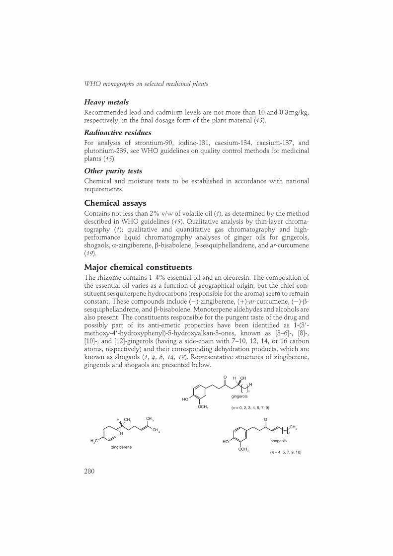
WHO monographs on selected medicinal plants
280
Heavy metalsRecommended lead and cadmium levels are not more than 10 and 0.3mg/kg,respectively, in the final dosage form of the plant material (15).
Radioactive residuesFor analysis of strontium-90, iodine-131, caesium-134, caesium-137, andplutonium-239, see WHO guidelines on quality control methods for medicinalplants (15).
Other purity testsChemical and moisture tests to be established in accordance with nationalrequirements.
Chemical assaysContains not less than 2% v/w of volatile oil (1), as determined by the methoddescribed in WHO guidelines (15). Qualitative analysis by thin-layer chroma-tography (1); qualitative and quantitative gas chromatography and high-performance liquid chromatography analyses of ginger oils for gingerols,shogaols, α-zingiberene, �-bisabolene, �-sesquiphellandrene, and ar-curcumene(19).
Major chemical constituentsThe rhizome contains 1–4% essential oil and an oleoresin. The composition ofthe essential oil varies as a function of geographical origin, but the chief con-stituent sesquiterpene hydrocarbons (responsible for the aroma) seem to remainconstant. These compounds include (�)-zingiberene, (�)-ar-curcumene, (�)-�-sesquiphellandrene, and �-bisabolene. Monoterpene aldehydes and alcohols arealso present. The constituents responsible for the pungent taste of the drug andpossibly part of its anti-emetic properties have been identified as 1-(3�-methoxy-4�-hydroxyphenyl)-5-hydroxyalkan-3-ones, known as [3–6]-, [8]-,[10]-, and [12]-gingerols (having a side-chain with 7–10, 12, 14, or 16 carbonatoms, respectively) and their corresponding dehydration products, which areknown as shogaols (1, 4, 6, 14, 19). Representative structures of zingiberene,gingerols and shogaols are presented below.
gingerols
(n = 0, 2, 3, 4, 5, 7, 9)
H OH
H
OH
O
OCH3
n
zingiberene
H CH3
CH3
CH3
CH3
H
shogaols
(n = 4, 5, 7, 9, 10)
CH3
OH
O
OCH3
n

Rhizoma Zingiberis
281
Dosage formsDried root powder, extract, tablets and tincture (2, 14). Powdered ginger shouldbe stored in well-closed containers (not plastic) which prevent access of mois-ture. Store protected from light in a cool, dry place (4, 5).
Medicinal usesUses supported by clinical dataThe prophylaxis of nausea and vomiting associated with motion sickness (20–23), postoperative nausea (24), pernicious vomiting in pregnancy (25), andseasickness (26, 27).
Uses described in pharmacopoeias and in traditional systems ofmedicineThe treatment of dyspepsia, flatulence, colic, vomiting, diarrhoea, spasms, andother stomach complaints (1, 2, 4, 9, 21). Powdered ginger is further employedin the treatment of colds and flu, to stimulate the appetite, as a narcoticantagonist (1, 2, 4, 6, 11, 12, 21), and as an anti-inflammatory agent in thetreatment of migraine headache and rheumatic and muscular disorders (9, 11,12, 28).
Uses described in folk medicine, not supported by experimental orclinical dataTo treat cataracts, toothache, insomnia, baldness, and haemorrhoids, and toincrease longevity (9, 10, 12).
PharmacologyExperimental pharmacologyCholagogic activityIntraduodenal administration of an acetone extract (mainly essential oils) ofginger root to rats increased bile secretion for 3 hours after dosing, while theaqueous extract was not active (29). The active constituents of the essential oilwere identified as [6]- and [10]-gingerol (29).
Oral administration of an acetone extract of ginger (75mg/kg), [6]-shogaol(2.5mg/kg), or [6]-, [8]-, or [10]-gingerol enhanced gastrointestinal motility inmice (30), and the activity was comparable to or slightly weaker than that ofmetoclopramide (10mg/kg) and domperidone (30). The [6]-, [8]-, or [10]-gingerols are reported to have antiserotoninergic activity, and it has been sug-gested that the effects of ginger on gastrointestinal motility may be due to thisactivity (30, 31). The mode of administration appears to play a critical role instudies on gastrointestinal motility. For example, both [6]-gingerol and [6]-shogaol inhibited intestinal motility when administered intravenously but ac-centuated gastrointestinal motility after oral administration (6, 12, 32).

WHO monographs on selected medicinal plants
282
Antiemetic activityThe emetic action of the peripherally acting agent copper sulfate was inhibitedin dogs given an intragastric dose of ginger extract (33), but emesis in pigeonstreated with centrally acting emetics such as apomorphine and digitalis couldnot be inhibited by a ginger extract (34). These results suggest that ginger’santiemetic activity is peripheral and does not involve the central nervous sys-tem (11). The antiemetic action of ginger has been attributed to the combinedaction of zingerones and shogaols (11).
Anti-inflammatory activityOne of the mechanisms of inflammation is increased oxygenation of arachi-donic acid, which is metabolized by cyclooxygenase and 5-lipoxygenase,leading to prostaglandin E2 and leukotriene B4, two potent mediators of inflam-mation (28). In vitro studies have demonstrated that a hot-water extract of gingerinhibited the activities of cyclooxygenase and lipoxygenase in the arachidonicacid cascade; thus its anti-inflammatory effects may be due to a decrease in theformation of prostaglandins and leukotrienes (35). The drug was also a potentinhibitor of thromboxane synthase, and raised prostacyclin levels without aconcomitant rise in prostaglandins E2 or F2α (36). In vivo studies have shown thatoral administration of ginger extracts decreased rat paw oedema (37, 38). Thepotency of the extracts was comparable to that of acetylsalicylic acid. [6]-Shogaol inhibited carrageenin-induced paw oedema in rats by inhibitingcyclooxygenase activity (39). Recently, two labdane-type diterpene dialdehydesisolated from ginger extracts have been shown to be inhibitors of human 5-lipoxygenase in vitro (40).
Clinical pharmacologyAntinausea and antiemetic activitiesClinical studies have demonstrated that oral administration of powdered gingerroot (940mg) was more effective than dimenhydrinate (100mg) in preventingthe gastrointestinal symptoms of kinetosis (motion sickness) (22). The results ofthis study further suggested that ginger did not act centrally on the vomitingcentre, but had a direct effect on the gastrointestinal tract through its aromatic,carminative, and absorbent properties, by increasing gastric motility and ad-sorption of toxins and acids (22).
In clinical double-blind randomized studies, the effect of powdered gingerroot was tested as a prophylactic treatment for seasickness (26, 27). The resultsof one study demonstrated that orally administered ginger was statisticallybetter than a placebo in decreasing the incidence of vomiting and cold sweating4 hours after ingestion (27). The other investigation compared the effects ofseven over-the-counter and prescription antiemetic drugs on prevention ofseasickness in 1489 subjects. This study concluded that ginger was as effectiveas the other antiemetic drugs tested (26).

Rhizoma Zingiberis
283
At least eight clinical studies have assessed the effects of ginger root on thesymptoms of motion sickness. Four of these investigations showed that orallyadministered ginger root was effective for prophylactic therapy of nausea andvomiting. The other three studies showed that ginger was no more effectivethan a placebo in treating motion sickness (23, 41, 42). The conflicting resultsappear to be a function of the focus of these studies. Clinical studies thatfocused on the gastrointestinal reactions involved in motion sickness recordedbetter responses than those studies that concentrated primarily on responsesinvolving the central nervous system.
The hypothesis that an increase in gastric emptying may be involved in theantiemetic effects of ginger has recently come under scrutiny. Two clinicalstudies demonstrated that oral doses of ginger did not affect the gastric empty-ing rate, as measured by sequential gastric scintigraphy (43) or the paracetamolabsorption technique (44).
In a double-blind, randomized, cross-over trial, oral administration of pow-dered ginger (250mg, 4 times daily) effectively treated pernicious vomiting inpregnancy (25). Both the degree of nausea and the number of vomiting attackswere significantly reduced (25). Furthermore, in a prospective, randomized,double-blind study, there were statistically significantly fewer cases of postop-erative nausea and vomiting in 60 patients receiving ginger compared to aplacebo (24). The effect of ginger on postoperative nausea and vomiting wasreported to be as good as or better than that of metoclopramide (24, 45). Incontrast, another double-blind randomized study concluded that orally admin-istered ginger BP (prepared according to the British Pharmacopoeia) was ineffec-tive in reducing the incidence of postoperative nausea and vomiting (46).
Anti-inflammatory activityOne study in China reported that 113 patients with rheumatic pain and chroniclower back pain, injected with a 5–10% ginger extract into the painful points orreaction nodules, experienced full or partial relief of pain, decrease in jointswelling, and improvement or recovery in joint function (11). Oral administra-tion of powdered ginger to patients with rheumatism and musculoskeletaldisorders has been reported to provide varying degrees of relief from pain andswelling (28).
ContraindicationsNo information available.
WarningsNo information available.

WHO monographs on selected medicinal plants
284
PrecautionsGeneralPatients taking anticoagulant drugs or those with blood coagulation disordersshould consult their physician prior to self-medication with ginger. Patientswith gallstones should consult their physician before using ginger preparations(21).
Drug interactionsGinger may affect bleeding times and immunological parameters owing to itsability to inhibit thromboxane synthase and to act as a prostacyclin agonist (47,48). However, a randomized, double-blind study of the effects of dried ginger(2g daily, orally for 14 days) on platelet function showed no differences inbleeding times in patients receiving ginger or a placebo (49, 50). Large doses(12–14g) of ginger may enhance the hypothrombinaemic effects of anticoagu-lant therapy, but the clinical significance has yet to be evaluated.
Carcinogenesis, mutagenesis, impairment of fertilityThe mutagenicity of ginger extracts is a controversial subject. A hot-waterextract of ginger was reported to be mutagenic in B291I cells and Salmonellatyphimurium strain TA 100, but not in strain TA 98 (51). A number of constitu-ents of fresh ginger have been identified as mutagens. Both [6]-gingerol andshogaols have been determined to be mutagenic in a Salmonella/microsomeassay (52), and increased mutagenesis was observed in an Hs30 strain ofEscherichia coli treated with [6]-gingerol (53). However, the mutagenicity of [6]-gingerol and shogaols was suppressed in the presence of various concentrationsof zingerone, an antimutagenic constituent of ginger (52). Furthermore, gingerjuice was reported to be antimutagenic and suppressed the spontaneousmutations induced by [6]-gingerol, except in cases where the mutagenic chemi-cals 2-(2-furyl)-3-(5-nitro-2-furyl)acryl amide and N-methyl-N�-nitro-N-nitroso-guanidine were added to [6]-gingerol (54). Other investigators have alsoreported that ginger juice is antimutagenic (54, 55).
Pregnancy: teratogenic effectsIn a double-blind randomized cross-over clinical trial, ginger (250mg by mouth,4 times daily) effectively treated pernicious vomiting in pregnancy (25). Noteratogenic aberrations were observed in infants born during this study, and allnewborn babies had Apgar scores of 9 or 10 after 5 minutes (25).
Paediatric useNot recommended for children less than 6 years of age.
Other precautionsNo information available concerning drug and laboratory test interactions, ornon-teratogenic effects on pregnancy or nursing mothers.

Rhizoma Zingiberis
285
Adverse reactionsContact dermatitis of the finger tips has been reported in sensitive patients (56).
PosologyFor motion sickness in adults and children more than 6 years: 0.5g, 2–4 timesdaily. Dyspepsia, 2–4g daily, as powdered plant material or extracts (21).
References1. Standard of ASEAN herbal medicine, Vol. I. Jakarta, ASEAN Countries, 1993.2. Pharmacopoeia of the People’s Republic of China (English ed.). Guangzhou, Guangdong
Science and Technology Press, 1992.3. British pharmacopoeia. London, Her Majesty’s Stationery Office, 1993.4. African pharmacopoeia, Vol. 1. 1st ed. Lagos, Organization of African Unity, Scientific,
Technical & Research Commission, 1985.5. The Japanese pharmacopoeia XIII. Tokyo, Ministry of Health and Welfare, 1996.6. Bisset NG. Max Wichtl’s herbal drugs & phytopharmaceuticals. Boca Raton, FL, CRC
Press, 1994.7. Keys JD. Chinese herbs, their botany, chemistry and pharmacodynamics. Rutland, VT, CE
Tuttle, 1976.8. Youngken HW. Textbook of pharmacognosy, 6th ed. Philadelphia, Blakiston, 1950.9. Farnsworth NR, ed. NAPRALERT database. Chicago, University of Illinois at
Chicago, IL, March 15, 1995 production (an on-line database available directlythrough the University of Illinois at Chicago or through the Scientific and TechnicalNetwork (STN) of Chemical Abstracts Services).
10. Kapoor LD. Handbook of Ayurvedic medicinal plants. Boca Raton, FL, CRC Press, 1990.11. Ghazanfar SA. Handbook of Arabian medicinal plants. Boca Raton, FL, CRC Press, 1994.12. Chang HM, But PPH, eds. Pharmacology and applications of Chinese materia medica, Vol.
1. Singapore, World Scientific Publishing, 1986.13. Farnsworth NR, Bunayapraphatsara N, eds. Thai medicinal plants. Bangkok,
Prachachon, 1992.14. Awang DVC. Ginger. Canadian pharmaceutical journal, 1982, 125:309–311.15. Quality control methods for medicinal plant materials. Geneva, World Health Organiza-
tion, 1998.16. Deutsches Arzneibuch 1996. Vol. 2. Methoden der Biologie. Stuttgart, Deutscher
Apotheker Verlag, 1996.17. European pharmacopoeia, 3rd ed. Strasbourg, Council of Europe, 1997.18. Guidelines for predicting dietary intake of pesticide residues, 2nd rev. ed. Geneva, World
Health Organization, 1997 (unpublished document WHO/FSF/FOS/97.7; availablefrom Food Safety, 1211 Geneva 27, Switzerland).
19. Yoshikawa M et al. Qualitative and quantitative analysis of bioactive principles inZingiberis rhizoma by means of high performance liquid chromatography and gasliquid chromatography. Yakugaku zasshi, 1993, 113:307–315.
20. Reynolds JEF, ed. Martindale, the extra pharmacopoeia, 30th ed. London, Pharmaceuti-cal Press, 1993:885.
21. German Commission E Monograph, Zingiberis rhizoma. Bundesanzeiger, 1988, 85:5May.
22. Mowrey DB, Clayson DE. Motion sickness, ginger, and psychophysics. Lancet, 1982,i:655–657.
23. Holtmann S et al. The anti-motion sickness mechanism of ginger. A comparativestudy with placebo and dimenhydrinate. Acta otolaryngology, 1989, 108:168–174.
WHO monographs on selected medicinal plants
286
24. Bone ME et al. Ginger root, a new antiemetic. The effect of ginger root on postop-erative nausea and vomiting after major gynaecological surgery. Anaesthesia, 1990,45:669–671.
25. Fischer-Rasmussen W et al. Ginger treatment of hyperemesis gravidarum. Europeanjournal of obstetrics, gynecology and reproductive biology, 1991, 38:19–24.
26. Schmid R et al. Comparison of seven commonly used agents for prophylaxis ofseasickness. Journal of travel medicine, 1994, 1:203–206.
27. Grontved A et al. Ginger root against seasickness. A controlled trial on the open sea.Acta otolaryngology, 1988, 105:45–49.
28. Srivastava KC, Mustafa T. Ginger (Zingiber officinale) in rheumatism and musculosk-eletal disorders. Medical hypotheses, 1992, 39:342–348.
29. Yamahara J et al. Cholagogic effect of ginger and its active constituents. Journal ofethnopharmacology, 1985, 13:217–225.
30. Yamahara J et al. Gastrointestinal motility enhancing effect of ginger and its activeconstituents. Chemical and pharmaceutical bulletin, 1991, 38:430–431.
31. Yamahara J et al. Inhibition of cytotoxic drug-induced vomiting in suncus by aginger constituent. Journal of ethnopharmacology, 1989, 27:353–355.
32. Suekawa M et al. Pharmacological studies on ginger. I. Pharmacological actions ofpungent components, (6)-gingerol and (6)-shogaol. Journal of pharmacobio-dynamics,1984, 7:836–848.
33. Japan centra revuo medicina, 1954, 112:669.34. Zhou JG. Tianjin medical journal, 1960, 2:131.35. Mustafa T, Srivastava KC, Jensen KB. Drug development report 9. Pharmacology of
ginger, Zingiber officinale. Journal of drug development, 1993, 6:25–39.36. Srivastava KC. Aqueous extracts of onion, garlic and ginger inhibit platelet aggrega-
tion and alter arachidonic acid metabolism. Biomedica biochimica acta, 1984, 43:335–346.
37. Mascolo N et al. Ethnopharmacologic investigation of ginger (Zingiber officinale).Journal of ethnopharmacology, 1989, 27:129–140.
38. Sharma JN, Srivastava KC, Gan EK. Suppressive effects of eugenol and ginger oil onarthritic rats. Pharmacology, 1994, 49:314–318.
39. Suekawa M, Yuasa K, Isono M. Pharmacological studies on ginger: IV. Effects of (6)-shogaol on the arachidonic cascade. Folia pharmacologia Japan, 1986, 88:236–270.
40. Kawakishi S, Morimitsu Y, Osawa T. Chemistry of ginger components and inhibi-tory factors of the arachidonic acid cascade. American Chemical Society Symposiumseries, 1994, 547:244–250.
41. Stott JR, Hubble MP, Spencer MB. A double-blind comparative trial of powderedginger root, hyosine hydrobromide, and cinnarizine in the prophylaxis of motionsickness induced by cross coupled stimulation. Advisory Group for AerospaceResearch Development conference proceedings, 1984, 39:1–6.
42. Wood CD et al. Comparison of the efficacy of ginger with various antimotionsickness drugs. Clinical research practice and drug regulatory affairs, 1988, 6:129–136.
43. Stewart JJ et al. Effects of ginger on motion sickness susceptibility and gastricfunction. Pharmacology, 1991, 42:111–120.
44. Phillips S, Hutchinson S, Ruggier R. Zingiber officinale does not affect gastric emptyingrate. Anaesthesia, 1993, 48:393–395.
45. Phillips S, Ruggier R, Hutchinson SE. Zingiber officinale (Ginger), an antiemetic for daycase surgery. Anaesthesia, 1993, 48:715–717.
46. Arfeen Z et al. A double-blind randomized controlled trial of ginger for the preven-tion of postoperative nausea and vomiting. Anaesthesia and intensive care, 1995,23:449–452.
47. Backon J. Ginger: inhibition of thromboxane synthetase and stimulation ofprostacyclin; relevance for medicine and psychiatry. Medical hypotheses, 1986,20:271–278.

Rhizoma Zingiberis
287
48. Backon J. Ginger as an antiemetic: possible side effects due to its thromboxanesynthetase activity. Anaesthesia, 1991, 46:705–706.
49. Srivastava KC. Isolation and effects of some ginger components on platelet aggrega-tion and eicosanoid biosynthesis. Prostaglandins and leukotrienes in medicine, 1986,25:187–198.
50. Lumb AB. Effect of ginger on human platelet function. Thrombosis and haemostasis,1994, 71:110–111.
51. Yamamoto H, Mizutani T, Nomura H. Studies on the mutagenicity of crude drugextracts. Yakugaku zasshi, 1982, 102:596–601.
52. Nagabhushan M, Amonkar AJ, Bhide SV. Mutagenicity of gingerol and shogoal andantimutagenicity of zingerone in Salmonella/microsome assay. Cancer letters, 1987,36:221–233.
53. Nakamura H, Yamamoto T. Mutagen and anti-mutagen in ginger, Zingiber officinale.Mutation research, 1982, 103:119–126.
54. Kada T, Morita M, Inoue T. Antimutagenic action of vegetable factor(s) on themutagenic principle of tryptophan pyrolysate. Mutation research, 1978, 53:351–353.
55. Morita K, Hara M, Kada T. Studies on natural desmutagens: screening for vegetableand fruit factors active in inactivation of mutagenic pyrolysis products from aminoacids. Agricultural and biological chemistry, 1978, 42:1235–1238.
56. Seetharam KA, Pasricha JS. Condiments and contact dermatitis of the finger tips.Indian journal of dermatology, venereology and leprology, 1987, 53:325–328.
WHO monographs on selected medicinal plants
288
AnnexParticipants in the WHO Consultation on Selected
Medicinal Plants
Munich, Germany, 8–10 July 1996Dr Keita Arouna, National Institute for Research in Public Health, Bamako, MaliProfessor Elaine Elisabetsky, Department of Pharmacology, Federal University of Rio
Grande do Sul, Porto Alegre, BrazilProfessor Norman Farnsworth, University of Illinois at Chicago, College of Pharmacy,
Chicago, IL, USAProfessor Harry Fong, University of Illinois at Chicago, College of Pharmacy, Chicago,
IL, USADr Abdel-Azim M. Habib, Professor of Pharmacognosy, Faculty of Pharmacy, University
of Alexandria, Alexandria, EgyptDr Djoko Hargono, Former Head, Directorate General of Drugs and Food Control,
Ministry of Health, Jakarta, IndonesiaDr Konstantin Keller, Director, Federal Institute of Drug and Medicinal Products, Berlin,
GermanyProfessor Fritz H. Kemper, Umweltprobenbanken für Human-Organproben, University
of Münster, Münster, GermanyMr Eftychios Kkolos, Director, Pharmaceutical Services, Ministry of Health, Nicosia,
CyprusDr Mamadou Koumaré, School of Medicine and Pharmacy, Bamako, MaliDr Gail Mahady, University of Illinois at Chicago, College of Pharmacy, Chicago, IL,
USADr Satish Mallya, Representative, Bureau of Pharmaceutical Assessment, Health Protec-
tion Branch, Drugs Directorate, Ottawa, Ontario, CanadaProfessor Tamas Paal, National Institute of Pharmacy, Budapest, HungaryDr Tharnkamol Reancharoen, Food and Drug Administration, Ministry of Public Health,
Bangkok, ThailandDr Gillian Scott, National Botanical Institute, Conservation Biology Research Unit, Cape
Town, South AfricaDr Geoffrey N. Vaughan, National Manager, Therapeutic Goods Administration, Com-
monwealth Department of Health, Housing and Community Service, Woden,Australian Capital Territory, Australia
Mr Tuley De Silva, Special Technical Adviser, United Nations Industrial DevelopmentOrganization, Vienna, Austria
WHO SecretariatDr Mary Couper, Medical Officer, Division of Drug Management and Policies, World
Health Organization, Geneva, SwitzerlandDr Martijn ten Ham, Chief, Drug Safety, Division of Drug Management and Policies,
World Health Organization, Geneva, Switzerland

Annex
289
Dr Jutta Schill, Technical Officer, Traditional Medicine Programme, Action Programmeon Essential Drugs, World Health Organization, Geneva, Switzerland
Dr Xiaorui Zhang, Medical Officer, Traditional Medicine Programme, ActionProgramme on Essential Drugs, World Health Organization, Geneva, Switzerland


WHO monographs
A A
on selected medicinal plants
VOLUME 2
World Health Organization Geneva
2002

WHO Library Cataloguing-in-Publication Data WHO monographs on selected medicinal p1ants.-Vol. 2. l.Plants, Medicinal 2.Herbs ISBN 92 4 154537 2 (NLM Classification: QV 766)
The World Health Organization welcomes requests £or permission to reproduce or translate its publications, in part or in £ull. Applications and enquiries should be addressed to the Ofhce of Publications, World Health Organization, Geneva, Switzerland, which will be glad to provide the latest information on any changes made to the text, plans £or new editions, and reprints and translations already available.
O World Health Organization 2002
Publications of the World Health Organization enjoy copyright protection in accordance with the provisions o£ Protoco12 o£ the Universal Copyright Convention. Al1 rights reserved. The designations employed and the presentation of the material in this publication do not imply the expression o£ any opinion whatsoever on the part o£ the Secretariat o£ the World Health Organization concerning the legal status o£ any country, territory, city or area or oF its authorities, or concerning the delimitation of its Frontiers or boundaries. The mention o£ specific companies or of certain manu£acturersl products does not imply that they are endorsed or recommended by the World Health Organization in preferente to others of a similar nature that are not mentioned. Errors and omissions excepted, the names of proprietay products are distinguished by initial capital letters.
Designed 'by WHO Graphics
Typeset in Hong IZong
Printed in Malta 2001/13613-SNPBest-set/Interprint-7700

Contents
Acknowledgements Introduction General technical notices
Monographs (in alphnbeticnl order o f planr narne)
Radix Althaeae Herba Andrographidis Radix Angelicae Sinensis Flos Calendulae Flos Caryophylli Rhizoma Cimicifugae Racemosae Folium cum Flore Crataegi Radix Eleutherococci Aetheroleum Eucalypti Folium Eucalypti Cortex Frangulae Folium et Cortex Hamamelidis Semen Hippocastani Herba Hyperici Aetheroleum Melaleucae Alternifoliae Folium Melissae Aetheroleum Menthae Piperitae Folium Menthae Piperitae Folium Ocimi Sancti Oleum Oenotherae Biennis Rhizoma Piperis Methystici Cortex Pruni Africanae Cortex Rhamni Purshianae Flos Sambuci Radix Senegae Fructus Serenoae Repentis

Contents
Fructus Silybi Mariae Herba Tanaceti Parthenii Radix Urticae Folium Uvae Ursi
Annex 1 Participants in the Second WHO Consultation on Selected Medicinal Plants 352
Annex 2 Cumulative index (in alphabetical order of plant narne) 354
Annex 3 Cumulative index (in alphabetical order of plant material of interest) 356

Acknowledgements
Special acknowledgement is due to Professors Norman R. Famsworth, Harry H.S. Fong and Gail B. Mahady of the WHO Collaborating Centre for Tradi- tional Medicine, College of Pharmacy, University of Illinois at Chicago, Chicago, E, USA, for drafting and revising the monographs. Similarly, special acknowledgement is also due to Dr Raymond Boudet-Dalbin of the Labora- toire de Chimie thérapeutique at the University René Descartes, Paris, France, for drawing the chemical structures for both volumes 1 and 2. The photograph for the front cover was kindly provided by Professor Icurt Hostettmann of the Institut de Pharmacognosie et Phytochimie at the University of Lausanne, Lausanne, Switzerland.
WHO also acknowledges with thanks the valuable work of the approxi- mately 120 experts in more than 50 countries who provided comrnents and advice on the draft texts; those who submitted cornrnents through the World Self-Medication Industry (a nongovernmental organization in official relations with WHO); and those who participated in the Second WHO Consultation on Selected Medicinal Plants held in Ravello-Salerno, Italy, in March 1999 to review the monographs (see Annex 1).
Finally, WHO would like to thank the Ministry of Health of Italy; the Governrnent of the Province of Salerno, Italy; the WHO Collaborating Centre for Traditional Medicine at the Centre of Research in Bioclimatology, Biotech- nologies and Natural Medicines of the State University of Milan, Italy; and the State University of Salerno, Italy, who hosted and supported the Second WHO Consultation.

Role of the WHO monographs on selected medicinal plantsThe first volume of the WHO monographs on selected medicinal plants, containing28 monographs, was published in 1999. It is gratifying that the importance ofthe monographs is already being recognized. For example, the European Com-mission has recommended volume 1 to its Member States as an authoritativereference on the quality, safety and efficacy of medicinal plants. The CanadianGovernment has also made a similar recommendation. Furthermore, as hoped,some of WHO’s Member States, such as Benin, Mexico, South Africa and VietNam, have developed their own monographs based on the format of the WHOmonographs.
The monographs are not only a valuable scientific reference for healthauthorities, scientists and pharmacists, but will also be of interest to the generalpublic. There can be little doubt that the WHO monographs will continue toplay an important role in promoting the proper use of medicinal plants through-out the world.
Preparation of monographs for volume 2At the eighth International Conference on Drug Regulatory Authorities(ICDRA) held in Manama, Bahrain, in 1996, WHO reported the completion ofvolume 1 of the WHO monographs. Member States requested WHO to con-tinue to develop additional monographs. As a consequence, preparation of thesecond volume began in 1997.
During the preparation, the number of experts involved, in addition tomembers of WHO’s Expert Advisory Panel on Traditional Medicine, signifi-cantly increased compared to that for volume 1. Similarly, the number of national drug regulatory authorities who participated in the preparation also greatly increased. This global network of active collaborators facilitatedwider access to the scientific references and information, thus increasing boththe quality and quantity of the monographs. These combined efforts greatlyimproved the efficiency of the preparation. As for volume 1, the monographswere drafted by the WHO Collaborating Centre for Traditional Medicine at theUniversity of Illinois at Chicago, United States of America.
The Second WHO Consultation on Selected Medicinal Plants was held inRavello-Salerno, Italy, in March 1999 to review and finalize the draft mono-graphs. Twenty experts and drug regulatory authorities from WHO Member
1
Introduction
States participated (see Annex 1). Following extensive discussion, 30 of 31 draftmonographs were approved for volume 2. At the subsequent ninth ICDRA inBerlin, Germany in April 1999, the 30 draft monographs were presented, andMember States requested WHO to publish them as soon as possible.
Purpose and content of the monographsThe purpose of the monographs was clearly explained in the introduction tovolume 1, and it is unnecessary to repeat it here. However, it is important toemphasize that the word “monograph”, as appears in the title, is used as a tech-nical term only. These monographs are not intended to be official pharma-copoeial monographs.
It should also be stressed that this publication is not intended to replace offi-cial compendia such as pharmacopoeias, formularies or legislative documents.Furthermore, the descriptions included in the section on medicinal uses shouldnot be taken as implying WHO’s official endorsement or approval. They merelyrepresent the systematic collection of scientific information available at the timeof preparation, for the purpose of facilitating information exchange.
A description of selected sections of the monographs is given in the Generaltechnical notices. For easy reference, two cumulative indexes are also providedas annexes. Annex 2 lists the monographs in alphabetical order of the plantname, while Annex 3 is according to the plant material of interest.
Dr Xiaorui ZhangActing CoordinatorTraditional MedicineDepartment of Essential Drugs and Medicines PolicyWorld Health Organization
Introduction
2

General technical notices
3
These WHO monographs are not pharmacopoeial monographs. Their pur-pose is to provide scientific information on the safety, efficacy and qualitycontrol/quality assurance of widely used medicinal plants, in order to facilitatetheir appropriate use in WHO’s Member States; to provide models to assistWHO’s Member States in developing their own monographs or formularies forthese and other herbal medicines; and to facilitate information exchange amongWHO’s Member States.
The format used for volume 2 essentially follows that of volume 1.However, to keep relevant sections together, Geographical distribution now pre-cedes Description; and Dosage forms appears before Posology.
The Definition describes the identity of the plant material of interest and theLatin binomial name of the source plant, the binomial name being the mostimportant criterion in quality assurance of the crude drug. Latin pharma-copoeial synonyms and vernacular names, listed in the sections Synonyms andSelected vernacular names, respectively, are those names used in commerce or bylocal consumers. The monographs place outdated botanical nomenclature inthe synonyms category, based on the International rules of nomenclature.
The vernacular names listed are a selection of names from individual coun-tries worldwide, in particular from areas where the medicinal plant is incommon use. The lists are not complete, but reflect the names found at thetime of preparation in official monographs, reference books and the NaturalProducts Alert (NAPRALERT) database (a database of literature from aroundthe world on ethnomedical, biological and chemical information on medicinalplants, fungi and marine organisms, located at the WHO Collaborating Centrefor Traditional Medicine at the University of Illinois at Chicago, USA).
A detailed botanical description (in Description) is intended for quality assur-ance at the stages of production and collection of the source plant, whereas thedetailed description of the specific plant part used (the crude drug)—in Plantmaterial of interest—is for quality assurance at the manufacturing and commer-cial stages. Geographical distribution is not normally found in official compendia,but it is included here to provide additional quality assurance information.
General identity tests, Purity tests and Chemical assays are all normal compen-dial components included under those headings in these monographs. Wherepurity tests do not specify accepted limits, those limits should be set in accor-dance with requirements of the respective national health authorities.
Each medicinal plant and crude drug contains active or major chemical con-stituents with a characteristic profile that can be used for chemical quality

control and quality assurance. These constituents are described in the sectionMajor chemical constituents.
Descriptions included in the section on Medicinal uses should not be takenas implying WHO’s official endorsement or approval. They merely representthe systematic collection of scientific information available at the time of prepa-ration, for information exchange. Medicinal uses are categorized as uses sup-ported by clinical data; uses described in pharmacopoeias and in traditionalsystems of medicine; and uses described in folk medicine, not yet supportedby experimental or clinical data.
The first category includes medicinal indications that are well established in some countries and have been validated by clinical studies documented inthe scientific literature. The clinical trials may have been controlled, random-ized, double-blind studies, trials without controls, cohort studies, or well-documented observations of therapeutic applications.
The second category includes medicinal uses that are well established inmany countries and are included in official pharmacopoeias or national mono-graphs. Well-established uses having a plausible pharmacological basis and sup-ported by older studies that clearly need to be repeated are also included. Thereferences cited provide additional information useful in evaluating specificherbal preparations. The uses described should be reviewed by local expertsand health workers for their applicability in the local situation.
The third category refers to indications described in unofficial pharma-copoeias and other literature, and to traditional uses. The appropriateness ofthese uses could not be assessed, owing to a lack of scientific data to supportthe claims. The possible uses of these remedies must be carefully consideredin the light of therapeutic alternatives.
The Experimental pharmacology section includes only the results of investiga-tions that prove or disprove the cited medicinal uses. Abbreviated details of thebest-performed studies have been included in this section. Other publishedexperimental data that are not associated with the medicinal uses have not beenincluded to avoid confusion.
The details included in the section on References have been checked againstthe original sources wherever possible. However, in some cases, details aremissing as the original sources were not available. For non-English language ref-erences, the title is given in the original language, except in cases where anEnglish summary is available.
General technical notices
4

DefinitionRadix Althaeae consists of the dried roots of Althaea officinalis L. (Malvaceae)(1–4).
SynonymMalva officinalis L. (5).
Selected vernacular namesAltea, altee, althea, bardul khatmi, benefischi, bismalva-hibiscus, blanca malva, bon visclo, bourdon de St Jacques, Eibisch, Eibischwurzel, erva molle,guimauve, Heilwurz, hobbiza, Ibischwurz, khairi, khatmi, korzén prawó-slazu, marshmallow, marshmallow root, malvaccioniu, malvavisco, marmo-lone, molotta, Moorish mallow, orvosiziliz gyökér, racine d’althée, racine deguimauve, Sammetpappel, sauvage, Schleimwurzel, suzmool, sweet weed,white mallow, wymote (3, 6–8).
Geographical distributionIndigenous to western Asia and Europe, and is naturalized in the United Statesof America (9, 10). Roots are obtained from commercially cultivated plants thatare at least 2 years old and harvested in the autumn (6, 10).
DescriptionA perennial herb with erect, woody stems, 60–120cm high. Leaves alternate,ovate to slightly cordate, serrate, velvety, large, occasionally 3-lobed. Flowerspale pink, axillary, the calyx of each surrounded by a 6–9 cleft involucre. Fruita set of cocci united into a ring (11).
Plant material of interest: dried rootsGeneral appearanceCylindrical or tapering, slightly twisted roots, up to 2cm thick, with deep longitudinal furrows. Outer surface greyish-brown, bearing numerous rootletscars. Fracture externally fibrous, internally rugged and granular; section shows
5
Radix Althaeae

a thick, whitish bark with brownish periderm, separated by a well-marked,brownish cambium from the white xylem; stratified structure of the bark andradiate structure of xylem become more distinct when moist. Peeled root has greyish-white finely fibrous outer surface; cork and external corticalparenchyma absent (2).
Organoleptic propertiesOdour: faint, aromatic; taste: mucilaginous (1).
Microscopic characteristicsPhloem with numerous long, thin-walled, non-lignified fibres arranged in tan-gential groups alternating with groups of sieve tissue, with a ground tissue ofthin-walled parenchyma; xylem containing reticulate or scalariform thickeningand bordered pits accompanied by lignified tracheids, a small amount of ligni-fied parenchyma and occasional small groups of fibres with only the middlelamella lignified; xylem and phloem transversed by numerous non-lignifiedmedullary rays, mostly uniseriate; majority of parenchyma cells of the phloemand medullary rays contain abundant small starch grains which are mostlysimple, spherical to ovoid, occasionally 2–3 compound, with a well-marked circular or slit-shaped hilum; some of these parenchyma cells contain clustercrystals of calcium oxalate 20–40mm in diameter, while others exist asidioblasts containing mucilage (1).
Powdered plant materialBrownish-grey (unpeeled root) or whitish (peeled root). Fragments of colour-less, mainly unlignified, thick-walled fibres with pointed or split ends; frag-ments of reticulate or scalariform thickening and bordered pits; cluster crystalsof calcium oxalate about 20–35mm, mostly 25–30mm, in diameter; parenchymacells containing mucilage; fragments of cork with thin-walled, tabular cells inthe powdered material from the unpeeled root. Numerous starch grains, 3–25mm in diameter, with occasionally a longitudinal hilum; starch grains mostlysimple, a few being 2–4 compound (2).
General identity testsMacroscopic and microscopic examinations (1, 2).
Purity testsMicrobiologyTests for specific microorganisms and microbial contamination limits are asdescribed in the WHO guidelines on quality control methods for medicinalplants (12).
WHO monographs on selected medicinal plants
6

Foreign organic matterNot more than 2% of brown, deteriorated drug and not more than 2% of corkin the peeled root (2).
Total ashNot more than 6% in the peeled root and not more than 8% in the unpeeledroot (2).
Acid-insoluble ashNot more than 3% in the peeled root (1).
Water-soluble extractiveNot less than 22% (1).
Loss on dryingNot more than 12% (2).
Swelling indexNot less than 10 (2).
Pesticide residuesThe recommended maximum limit of aldrin and dieldrin is not more than 0.05mg/kg (2). For other pesticides, see the European pharmacopoeia (2), and theWHO guidelines on quality control methods for medicinal plants (12) and pesticide residues (13).
Heavy metalsFor maximum limits and analysis of heavy metals, consult the WHO guidelineson quality control methods for medicinal plants (12).
Radioactive residuesWhere applicable, consult the WHO guidelines on quality control methods formedicinal plants (12) for the analysis of radioactive isotopes.
Other purity testsChemical, sulfated ash and alcohol-soluble extractive tests to be established inaccordance with national requirements.
Chemical assaysNot less than 10% total mucilage in the peeled root as determined by gravi-metric analysis (14).
Radix Althaeae
7

Major chemical constituentsThe mucilage content ranges from 10 to 20% and consists of a mixture of acidicgalacturonorhamnans, neutral glucans and neutral arabinogalactans (6, 8, 9,15–17).
Medicinal usesUses supported by clinical dataNone.
Uses described in pharmacopoeias and in traditional systems of medicineAs a demulcent for symptomatic treatment of dry irritable coughs and irrita-tions of oral and pharyngeal mucosa and as an emollient for wounds and dryskin (8, 18–23). Also used in cough mixtures to mask the bitter or pungent tasteof other drugs (16).
Uses described in folk medicine, not supported by experimental or clinical dataTreatment of asthma, cystitis, dysentery and irritations of the gastric mucosa(7).
PharmacologyExperimental pharmacologyThe demulcent effects of Radix Althaeae are due to its high content of poly-saccharide hydrocolloids, which form a protective coating on the oral and pharyngeal mucosa, soothing local irritation and inflammation (24).
Anti-inflammatory activityA polysaccharide fraction (500mg/ml) isolated from a root extract had anti-complement activity in human serum in vitro (25). Aqueous extracts of the roots stimulated phagocytosis, and the release of oxygen radicals andleukotrienes from human neutrophils in vitro (26). The aqueous extract alsoinduced the release of cytokines, interleukin-6 and tumour necrosis factor from human monocytes in vitro, thereby exhibiting anti-inflammatory andimmunostimulant activity (26). Intraperitoneal administration of isolatedmucilage polysaccharides to mice (10mg/kg body weight) induced a 2.2-foldincrease in the phagocytic activity of macrophages as measured by the colloidalcarbon clearance test (27). However, intragastric administration of an 80%ethanol extract of the roots to rats (100mg/kg body weight) did not inhibit carrageenan-induced footpad oedema (28).
WHO monographs on selected medicinal plants
8

Weak inhibition (17%) of mucociliary transport in isolated, ciliated epithe-lium of the frog oesophagus was demonstrated after treatment of the isolatedtissues with 200 ml of an aqueous root macerate (6.4g/140ml) (29).
Antitussive activityIntragastric administration of a polysaccharide fraction, isolated from anaqueous root extract, to cats (50mg/kg body weight) suppressed the intensityand the frequency of coughs induced by mechanical irritation of laryngo-pharyngeal and tracheobronchial mucosa (30). The antitussive activity of this polysaccharide fraction (50mg/kg body weight) was as effective as SyrupusAlthaeae (1.0g/kg body weight), and more effective than prenoxdiazine (30mg/kg body weight) (30).
Clinical pharmacologyNone.
ContraindicationsNo information available.
WarningsNo information available.
PrecautionsDrug interactionsSimultaneous administration of Radix Althaeae may delay the absorption ofother drugs (8).
Other precautionsNo information available on general precautions or precautions concerning drugand laboratory test interactions; carcinogenesis, mutagenesis, impairment offertility; teratogenic and non-teratogenic effects in pregnancy; nursing mothers;or paediatric use. Therefore, Radix Althaeae should not be administered during pregnancy or lactation or to children without medical supervision.
Adverse reactionsNo information available.
Radix Althaeae
9

Dosage formsPeeled or unpeeled, broken, chopped or powdered crude drug (1, 2) and galen-ical preparations thereof. Store in a well-closed container, protected from light(2).
Posology(Unless otherwise indicated)For dry cough, oral or pharyngeal irritation: 0.5–3.0g of crude drug as anaqueous, cold macerate (14, 19, 20, 31) or 2–8ml of syrup (20, 22, 32), whichmay be repeated up to a daily dose of 15g of crude drug. For gastric irritation:3–5g of crude drug as an aqueous, cold macerate up to three times daily (19,20, 31).
References1. British herbal pharmacopoeia. London, British Herbal Medicine Association, 1996.2. European pharmacopoeia, 3rd ed. Strasbourg, Council of Europe, 1996.3. Farmakopea Polska V, Suplement I. Warsaw, Polskie Towarzystwo Farmaceutyczne,
1995.4. Pharmacopoeia Hungarica, 7th ed. Budapest, Hungarian Pharmacopoeia Commission,
Medicina Konyvkiado, 1986.5. Hooker JD, Jackson BD. Index Kewensis. Vol. 1. Oxford, Clarendon Press, 1895.6. Bisset NG. Herbal drugs and phytopharmaceuticals. Boca Raton, FL, CRC Press, 1994.7. Farnsworth NR, ed. NAPRALERT database. Chicago, University of Illinois at
Chicago, IL, February 9, 1998 production (an online database available directlythrough the University of Illinois at Chicago or through the Scientific and Techni-cal Network [STN] of Chemical Abstracts Services).
8. Hänsel R et al., eds. Hagers Handbuch der pharmazeutischen Praxis. Bd. 6: Drogen P–Z,5th ed. Berlin, Springer-Verlag, 1994.
9. Leung AY, Foster S. Encyclopedia of common natural ingredients used in food, drugs, andcosmetics, 2nd ed. New York, NY, John Wiley & Sons, 1996.
10. Leung AY. Encyclopedia of common natural ingredients. New York, NY, John Wiley &Sons, 1980.
11. Youngken HW. Textbook of pharmacognosy, 6th ed. Philadelphia, PA, Blakiston, 1950.12. Quality control methods for medicinal plant materials. Geneva, World Health Organiza-
tion, 1998.13. Guidelines for predicting dietary intake of pesticide residues, 2nd rev. ed. Geneva, World
Health Organization, 1997 (document WHO/FSF/FOS/97.7).14. Pharmacopée française. Paris, Adrapharm, 1996.15. Blaschek W, Franz G. A convenient method for the quantitative determination of
mucilage polysaccharides in Althaeae radix. Planta Medica, 1986, 52:537.16. Samuelsson G, ed. Drugs of natural origin, a textbook of pharmacognosy. Stockholm,
Swedish Pharmaceutical Press, 1992.17. Tomoda M et al. The structural features of Althaea-mucilage representative mucous
polysaccharide from the roots of Althaea officinalis. Chemical and Pharmaceutical Bulletin, 1980, 28:824–830.
18. Bone K. Marshmallow soothes cough. British Journal of Phytotherapy, 1993/1994, 3:93.19. Marshmallow root. In: Bradley PR, ed. British herbal compendium. Vol. 1. Bourne-
mouth, British Herbal Medicine Association, 1992:151–153.
WHO monographs on selected medicinal plants
10

20. ESCOP monographs on the medicinal uses of plant drugs. Fascicule 1. Elberg, EuropeanScientific Cooperative on Phytotherapy, 1996.
21. Blumenthal M et al., eds. The complete German Commission E monographs. Austin, TX,American Botanical Council, 1998.
22. Reynolds JEF, ed. Martindale, the extra pharmacopoeia, 29th ed. London, Pharmaceu-tical Press, 1989.
23. Weiss RF. Lehrbuch der Phytotherapie, 7th ed. Stuttgart, Hippokrates Verlag, 1991.24. Franz G. Polysaccharides in pharmacy: current applications and future concepts.
Planta Medica, 1989, 55:493–497.25. Yamada H et al. Relationship between chemical structure and anti-complementary
activity of plant polysaccharides. Carbohydrate Research, 1985, 144:101–111.26. Scheffer J et al. Radix althaeae und Flores chamomillae Extrakte auf Entzün-
dungsreaktionen humaner neutrophiler Granulozyten, Monozyten und Ratten-mastzellen. In: Abstracts of the Third Phytotherapy Congress. Lübeck-Travemünde, 1991:Abstract P9.
27. Wagner H, Proksch A. Immunostimulatory drugs of fungi and higher plants. In:Wagner H, Hikino H, Farnsworth NR, eds. Economic and medicinal plant research. Vol.1. Orlando, FL, Academic Press, 1985:111–153.
28. Mascolo N et al. Biological screening of Italian medicinal plants for anti-inflammatory activity. Phytotherapy Research, 1987, 1:28–31.
29. Müller-Limmroth W, Fröhlich HH. Wirkungsnachweis einiger phytotherapeutischerExpektorantien auf den mukoziliaren Transport. Fortschritte der Medizin, 1980, 98:95–101.
30. Nosal’ova G et al. Antitussive efficacy of the complex extract and the polysaccha-ride of marshmallow (Althaea officinalis L. var. Robusta). Pharmazie, 1992, 47:224–226.
31. Wichtl M. Eibischwurzel. In: Wichtl M, ed. Teedrogen, 2nd ed. Stuttgart, Wis-senschaftliche Verlagsgesellschaft, 1989:146–147.
32. British pharmaceutical codex. London, Pharmaceutical Press, 1934.
Radix Althaeae
11
12
DefinitionHerba Andrographidis consists of the dried aerial parts of Andrographis panicu-lata (Burm. f.) Nees (Acanthaceae) (1–3).
SynonymsJusticia latebrosa Russ., J. paniculata Burm. f., J. stricta Lam. ex Steud. (3, 4).
Selected vernacular namesAkar cerita bidara, alui, Andrographidis Kraut, bidara, bhoonimba, bhuinimo,bhulimb, bhuninba, charayeta, charayetha, charita, cheranta, cherota, chiraita,chiretta, chuan-hsin-lien, chuan-xın-lián, công công, faathalaaichon, fathalaai,fathalaichon, fathalaijone, halviva, herba sambiloto, hinbinkohomba, I-chien-hsi, kalafath, kalmegh, kan-jang, kariyat, khee-pang-hee, king of bitters,kiriathu, kirta, kiryata, kiryato, lanhelian, mahatikta, mahatita, naelavemu, nay-nahudandi, nelavemu, quasab-uz-zarirah, rice bitters, sambilata, sambiloto,senshinren, sinta, xuyên tâm liên, yaa kannguu yijianxi (1, 2, 5–11).
Geographical distributionWidely found and cultivated in tropical and subtropical Asia, south-east Asiaand India (6, 8, 10).
DescriptionA herbaceous annual, erect, up to 1m high; stem acutely quadrangular, muchbranched. Leaves simple, opposite, lanceolate, glabrous, 2–12cm long, 1–3cmwide; apex acute; margin entire, slightly undulate, upper leaves often bracti-form; petiole short. Inflorescence patent, terminal and axillary in panicle, 10–30mm long; bract small; pedicel short. Calyx 5-particle, small, linear. Corollatube narrow, about 6mm long; limb longer than the tube, bilabiate; upper lipoblong, white with a yellowish top; lower lip broadly cuneate, 3-lobed, whitewith violet markings. Stamens 2, inserted in the throat and far exserted; antherbasally bearded. Superior ovary, 2-celled; style far exserted. Capsule erect,linear-oblong, 1–2cm long and 2–5mm wide, compressed, longitudinally fur-rowed on broad faces, acute at both ends, thinly glandular-hairy. Seeds small,subquadrate (1–3, 5, 10).
Herba Andrographidis

Plant material of interest: dried aerial partsGeneral appearanceMixture of broken, crisp, mainly dark green lanceolate leaves and quadrangu-lar stems; capsule fruit and small flowers occasionally found (1, 3). Stem texturefragile, easily broken; leaves simple, petiole short or nearly sessile, lanceolateor ovate-lanceolate, with acuminate apex and cuneate-decurrent base, laminacrumpled and easily broken (2).
Organoleptic propertiesOdour: slight, characteristic; taste: intensely bitter (1–3, 9).
Microscopic characteristicsLeaf upper epidermis: stomata absent, glandular trichomes present, unicellularand multicellular trichomes rare, cystoliths fairly large; lithocysts large (27–30mm thick, 96–210mm long and up to 49mm wide); columnar palisade cells;collenchyma in midrib beneath epidermis; parenchyma cells spongy; vascularbundles of lignified xylem in the upper part and lignified phloem in the lowerpart; spiral, scalariform and reticulate vessels. Leaf lower epidermis: a layer ofwavy-walled cells; stomata diacytic; trichomes up to 36mm in diameter and 180mm long, and cystoliths present. Stem: epidermis has glandular and non-glan-dular trichomes. Collenchyma dense at the corners of stems; parenchyma con-tains chloroplastids. Endodermis composed of a layer of thick-walled cells.Wood with spiral, scalariform and pitted xylem vessels; pith composed of largeparenchyma cells. Small acicular crystals of calcium oxalate occur in the pithand cortical cells of stem and leaf (1–3, 8).
Powdered plant materialLeaf fragments in surface view show upper epidermis with underlying palisadeand cystoliths, lower epidermis with underlying palisade cells with stomata,cystoliths and glandular trichomes. Leaf fragments in sectional view showupper epidermis with palisade cells, spongy parenchyma cells, vascular bundles;and lower epidermis with bundles of xylem associated with fibres; fragmentsof spiral, scalariform, reticulate and pitted vessels; fragments of epidermal cellsfrom midrib; fragments of parenchyma cells in transverse and longitudinal sections. Bundles of fibres. Fragments of epidermal cells from stem withstomata, cystoliths and glandular trichomes. Scattered cystoliths; scattered uni-cellular and multicellular trichomes, mostly from epidermal cells in fruit walls;scattered glandular trichomes from bundles of fibres in fruit wall; scatteredpollen grains (1).
General identity testsMacroscopic and microscopic examinations, chemical tests, and thin-layerchromatography for the presence of diterpene lactones (1–3).
Herba Andrographidis
13

Purity testsMicrobiologicalTests for specific microorganisms and microbial contamination limits are asdescribed in the WHO guidelines on quality control methods for medicinalplants (12).
ChemicalNot less than 6% of total diterpene lactones, calculated as andrographolide (1, 3).
Foreign organic matterNot more than 2% (1, 3).
Acid-insoluble ashNot more than 2% (1, 3).
Water-soluble extractiveNot less than 18% (1, 3).
Alcohol-soluble extractiveNot less than 13% using 85% ethanol (1, 3).
Loss on dryingNot more than 10% (1).
Pesticide residuesThe recommended maximum limit of aldrin and dieldrin is not more than 0.05mg/kg (13). For other pesticides, see the European pharmacopoeia (13), andthe WHO guidelines on quality control methods for medicinal plants (12) andpesticide residues (14).
Heavy metalsFor maximum limits and analysis of heavy metals, consult the WHO guidelineson quality control methods for medicinal plants (12).
Radioactive residuesWhere applicable, consult the WHO guidelines on quality control methods formedicinal plants (12) for the analysis of radioactive isotopes.
Other purity testsTotal ash test to be established in accordance with national requirements.
WHO monographs on selected medicinal plants
14

Chemical assaysChemical and thin-layer chromatography methods are used for qualita-tive analysis of andrographolide diterpene lactones (1, 2). Titrimetric (1) andhigh-performance liquid chromatography (15) methods are available for quan-titative analysis of total diterpene lactones.
Major chemical constituentsThe major constituents are diterpene lactones (free and in glycosidic forms)including andrographolide, deoxyandrographolide, 11,12-didehydro-14-deoxy-andrographolide, neoandrographolide, andrographiside, deoxyandrographisideand andropanoside (1, 3, 6, 7, 9, 16). The structures of andrographolide andrelated diterpene lactones are presented below.
Herba Andrographidis
15
O
OH
HO
CH2HH3C
H
HOH
CH3O
R
11 12
14O
O
CH2HH3C
H
RH
CH3O
Glc
3
OOH
HO
OHHO
Glc =
andrographolide R = H neoandrographolide R = H
andrographiside R = Glc andropanoside R = OH
3
18
b-D-glucopyranosyl
Medicinal usesUses supported by clinical dataProphylaxis and symptomatic treatment of upper respiratory infections, suchas the common cold and uncomplicated sinusitis (17–19), bronchitis (6, 9) andpharyngotonsillitis (20), lower urinary tract infections (21) and acute diarrhoea(22, 23).
Uses described in pharmacopoeias and in traditional systems of medicineTreatment of bacillary dysentery, bronchitis, carbuncles, colitis, coughs, dyspepsia, fevers, hepatitis, malaria, mouth ulcers, sores, tuberculosis and venomous snake bites (1, 2, 6, 7, 10, 16, 24–27).
Uses described in folk medicine, not supported by experimental or clinical dataTreatment of colic, otitis media, vaginitis, pelvic inflammatory disease, chicken-pox, eczema and burns (6, 7).

PharmacologyExperimental pharmacologyAntibacterial activityAn ethanol extract of the leaves inhibited the growth in vitro of Escherichia coliand Staphylococcus aureus (28). A 50% methanol extract of the leaves inhibitedgrowth in vitro of Proteus vulgaris (29). However, no in vitro antibacterial activ-ity was observed when dried powder from the aerial parts was tested againstE. coli, Staphylococcus aureus, Salmonella typhi or Shigella species (30).
Anti-human immunodeficiency virus (HIV) activityAqueous extracts of the leaves inhibited HIV-1 infection and replication in thelymphoid cell line MOLT-4 (31). A hot aqueous extract of the aerial partsreduced the percentage of HIV antigen-positive H9 cells (32). Dehydroandro-grapholide inhibited HIV-1 and HIV-1 (UCD123) infection of H9 cells at 1.6mg/ml and 50mg/ml, respectively, and also inhibited HIV-1 infection ofhuman lymphocytes at 50mg/ml (33). A methanol extract of the leaves sup-pressed syncytia formation in co-cultures of uninfected and HIV-1-infectedMOLT cells (median effective dose [ED50] 70mg/ml) (34).
Immunostimulatory activity Intragastric administration of an ethanol extract of the aerial parts (25mg/kgbody weight) or purified andrographolides (1mg/kg body weight) to mice stimu-lated antibody production and the delayed-type hypersensitivity response tosheep red blood cells (35). The extract also stimulated a non-specific immuneresponse in mice, measured by macrophage migration index, phagocytosis of[14C]leucine-labelled E. coli, and proliferation of splenic lymphocytes (35). Theextract was more effective than either andrographolide or neoandrographolidealone, suggesting that other constituents may be involved in the immuno-stimulant response (35).
Antipyretic activityIntragastric administration of an ethanol extract of the aerial parts (500mg/kgbody weight) to rats decreased yeast-induced pyrexia (36). The extract wasreported to be as effective as 200mg/kg body weight of aspirin, and no toxicity was observed at doses up to 600mg/kg body weight (36). Intragastricadministration of andrographolide (100mg/kg body weight) to mice decreasedbrewer’s yeast-induced pyrexia (37). Intragastric administration of deoxy-andrographolide, andrographolide, neoandrographolide or 11,12-didehydro-14-deoxyandrographolide (100mg/kg body weight) to mice, rats or rabbitsreduced pyrexia induced by 2,4-dinitrophenol or endotoxins (6, 38).
Antidiarrhoeal activityHerba Andrographidis has antidiarrhoeal activity in situ (39, 40). An ethanol,chloroform or 1-butanol extract of the aerial parts (300mg/ml) inhibited the
WHO monographs on selected medicinal plants
16

E. coli enterotoxin-induced secretory response—which causes a diarrhoeal syn-drome—in the rabbit and guinea-pig ileal loop assay (39, 40). However, anaqueous extract of the aerial parts was not active (40). The constituent diter-pene lactones, andrographolide and neoandrographolide, exhibited potent antisecretory activity in vivo against E. coli enterotoxin-induced diarrhoea (40).Andrographolide (1mg per loop) was as active as loperamide when testedagainst heat-labile E. coli enterotoxin-induced diarrhoea and more effective thanloperamide when tested against heat-stable E. coli enterotoxin-induced diar-rhoea (40). Neoandrographolide (1mg per loop) was as effective as loperamidewhen tested against heat-labile E. coli enterotoxin-induced diarrhoea andslightly less active than loperamide when tested against heat-stable E. colienterotoxin-induced diarrhoea (40). The mechanism of action involves inhibi-tion of the intestinal secretory response induced by heat-labile E. coli entero-toxins, which are known to act through the stimulation of adenylate cyclase,and by inhibition of the secretion induced by heat-stable E. coli enterotoxins,which act through the activation of guanylate cyclase (39). Incubation of murine macrophages with andrographolide (1–50mmol/l) inhibited bacterial endotoxin-induced nitrite accumulation in a concentration- and time-dependent manner. Western blot analysis demonstrated that andrographolideinhibited the expression of an inducible isoform of nitric oxide synthase linkedto endotoxin-induced circulatory shock (41).
Anti-inflammatory activityIntragastric administration of deoxyandrographolide, andrographolide, neo-andrographolide or 11,12-didehydrodeoxyandrographolide to mice inhibitedthe increase in cutaneous or peritoneal capillary permeability induced by xyleneor acetic acid, and reduced acute exudation in Selye granulocysts treated withcroton oil. 11,12-Didehydrodeoxyandrographolide had the most potent anti-inflammatory activity in vivo (6).
Antimalarial activityA 50% ethanol extract of the aerial parts inhibited the growth of Plasmodiumberghei both in vitro (100mg/ml) and in mice after intragastric administration (1g/kg body weight) (42). Intragastric administration of a 1-butanol, chloroformor ethanol–water extract of the aerial parts to Mastomys natalensis inhibited thegrowth of P. berghei at doses of 1–2g/kg body weight (43). Andrographolide (5mg/kg body weight) and neoandrographolide (2.5mg/kg body weight) werealso effective when administered by gastric lavage (43).
Antivenom activityIntraperitoneal injection of an ethanol extract of the aerial parts (25g/kg bodyweight) to mice poisoned with cobra venom markedly delayed the occurrenceof respiratory failure and death (6, 44). The same extract induced contractionsin guinea-pig ileum at concentrations of 2mg/ml. The contractions were
Herba Andrographidis
17

enhanced by physostigmine and blocked by atropine, but were unchanged byantihistamines (44). These data suggest that extracts of the aerial parts do notmodify the activity of the nicotinic receptors but produce significant muscarinicactivity, which accounts for its antivenom effects (6, 44).
Antihepatotoxic activityThe aerial parts and their constituent andrographolides have antihepatotoxicactivity in vitro and in vivo (45–54). Intraperitoneal administration of a meth-anol extract of the aerial parts (861.3mg/kg body weight) to mice reduced hepa-totoxicity induced by carbon tetrachloride (CCl4), and reversed CCl4-inducedhistopathological changes in the liver (52). Intraperitoneal administration ofandrographolide (100mg/kg body weight) to mice inhibited the CCl4-inducedincrease in the activity of serum glutamate oxaloacetate transaminase, serumglutamate pyruvate transaminase, alkaline phosphatase, bilirubin and hepatictriglycerides (52). Intraperitoneal administration of a methanol extract of theaerial parts (500mg/kg body weight) to rats also suppressed the CCl4-inducedincrease in the activity of serum glutamate oxaloacetate transaminase, serumglutamate pyruvate transaminase, alkaline phosphatase and bilirubin (51). Intra-gastric administration of an aqueous extract of the aerial parts (500mg/kg bodyweight) to ethanol-treated rats decreased the activity of serum transaminasesand suppressed histopathological changes in the liver (49). Andrographolide,the major antihepatotoxic component of the plant, exerted a pronounced protective effect in rats against hepatotoxicity induced by CCl4 (47), D-galactosamine (54), paracetamol (48) and ethanol (49). Andrographolide wasmore effective than silymarin, the standard hepatoprotective agent (47, 48).
Clinical pharmacologyThe common coldHerba Andrographidis has been used clinically for symptomatic treatment ofthe common cold and uncomplicated sinusitis, pharyngotonsillitis, pneumoniaand bronchitis (6, 17, 18, 20). A placebo-controlled, double-blind clinical trialassessed the efficacy of a standardized extract of the aerial parts (containing4% andrographolides) for treatment of the common cold in 61 adult patients.A significant reduction (P < 0.0001) in clinical symptoms such as sore throat,tiredness, muscular ache and malaise was observed on day 4 in the groupreceiving 1200mg extract daily, as compared with the placebo group. Noadverse reactions were reported in either group (17).
A randomized, placebo-controlled, double-blind pilot trial was conductedto evaluate the efficacy of a standardized extract of the aerial parts (containing4% andrographolides) on the initial symptoms of the common cold and un-complicated sinusitis. Fifty adult patients received either 1020mg extract or aplacebo daily for 5 days. The results demonstrated that patients in the treatedgroup took less sick leave than those in the placebo group (0.21 day comparedto 0.96 day). Furthermore, 68% of treated patients felt totally recovered, as
WHO monographs on selected medicinal plants
18
compared with 36% of the placebo group. Also 55% of the treated patientsthought that the course of illness was much easier than normal, as comparedwith 19% of the placebo group (18).
A randomized, placebo-controlled, double-blind study evaluated a stan-dardized extract of the aerial parts (containing 4% andrographolides) in the pro-phylaxis of the common cold in 107 schoolchildren during the winter season.The children received either 200mg extract or a placebo daily for 3 months andwere evaluated weekly by a physician. There was no difference in the occur-rence of colds between the two groups during the first 2 months of treatment.However, after the third month of treatment, there was a significant difference(P < 0.05) in the occurrence of the common cold in the treated group (30%) ascompared with the placebo group (62%) (19).
A randomized, double-blind comparison study of 152 adult patients withpharyngotonsillitis evaluated the efficacy of powdered aerial parts (6g daily)and paracetamol (1 capsule of 325mg as needed) for improving symptomatol-ogy. Baseline evaluation showed no significant difference between the twogroups. The crude drug was as effective as paracetamol in reducing the inci-dence of sore throat and fever after 3 days of treatment (20). In a study withoutcontrols, treatment of patients with a standardized extract of A. paniculata (con-taining 4% andrographolides) reduced the incidence of fever associated withthe common cold. The body temperature of patients treated with the extractwas lowered in less than 48 hours after treatment (55). This finding was con-firmed in a later study (17).
Urinary infectionsA clinical trial compared the efficacy of Herba Andrographidis, co-trimoxazole(sulfamethoxazole + trimethoprim) and norfloxacin in the prevention of urinarytract infections after extracorporeal shock wave lithotripsy. Patients received a5-day course of either Herba Andrographidis (4 tablets of 250mg, three timesdaily) or co-trimoxazole (2 tablets of 25mg, twice daily) or norfloxacin (1 tabletof 200mg, twice daily). After 1 month of treatment, urinalysis results of 100patients demonstrated that pyuria, haematuria and proteinuria were reducedin all treatment groups, and there was no significant difference between thethree treatments (21).
DysenteryThe aerial parts have been used for the treatment of acute bacillary dysenteryand enteritis (2, 6, 22, 23). In clinical studies, the combination of andro-grapholide and neoandrographolide was reported to be more effective thaneither furazolidine or chloramphenicol in the treatment of bacillary dysentery(6). A randomized, double-blind clinical study of 200 patients compared theefficacy of the powdered aerial parts with tetracycline in the treatment of acutediarrhoea and bacillary dysentery (22, 23). Patients received capsules of eitherthe aerial parts or tetracycline (both 500mg, four times daily) for 3 days. Com-pared with tetracycline, the aerial parts decreased the diarrhoea (both the fre-
Herba Andrographidis
19

quency and amount of discharge) (22). Furthermore, the aerial parts were moreeffective in treating diarrhoea resulting from shigellosis than from cholera (22).
Infectious hepatitisAdministration of a decoction of the aerial parts to patients with infectioushepatitis was reported to provide symptomatic relief (24).
ContraindicationsHerba Andrographidis should not be used during pregnancy or lactation. Herba Andrographidis is contraindicated in cases of known allergy to plants ofthe Acanthaceae family.
WarningsDue to potential anaphylactic reactions, crude extracts of Herba Andrographidisshould not be injected (6, 56).
PrecautionsDrug interactionsExtracts of Herba Andrographidis may have a synergistic effect with isoniazid(6).
Carcinogenesis, mutagenesis, impairment of fertilityHerba Andrographidis extracts are not mutagenic in vitro (57) and have anti-mutagenic activity (58). A standardized extract of A. paniculata did not producereproductive toxicity in male rats after 60 days of intragastric administration of20–1000mg/kg body weight daily (59).
Pregnancy: teratogenic effectsSee Contraindications.
Pregnancy: non-teratogenic effectsIn vivo studies in mice and rabbits suggest that Herba Andrographidis may haveabortifacient activity (6, 60). Conversely, no interruption of pregnancy, fetalresorption or decrease in the number of live offspring was observed in preg-nant rats after intragastric administration of an extract of the aerial parts at 2g/kg body weight during the first 9 days of gestation (61). Since potentialantagonism exists between Herba Andrographidis and endogenous proges-terone, Herba Andrographidis should not be used during pregnancy (2, 61).
Nursing mothersSee Contraindications.
WHO monographs on selected medicinal plants
20

Other precautionsNo information available on general precautions or precautions concerning drug and laboratory test interactions; or paediatric use. Therefore, HerbaAndrographidis should not be administered to children without medical supervision.
Adverse reactionsLarge oral doses of Herba Andrographidis may cause gastric discomfort, vom-iting and loss of appetite (6). These side-effects appear to be due to the bittertaste of andrographolide (6). Anaphylactic reactions may occur if the crude drugextract is injected (6, 56). Two cases of urticaria have been reported (18).
Dosage formsCrude drug, capsules, tablets and pills (1, 2, 6). Store in a well-closed container,protected from light and moisture.
Posology (Unless otherwise indicated)For pyrexia: a decoction from 3g crude drug, twice daily (1, 5). For the commoncold: 1.5–3.0g powdered crude drug three times daily, after meals and atbedtime (1). For diarrhoea: a decoction from 3–9g crude drug as a single doseas needed (1, 5), or two tablets of 500mg four times daily, after meals and atbedtime (5).
References1. Standard of ASEAN herbal medicine. Vol. 1. Jakarta, ASEAN Countries, 1993.2. Pharmacopoeia of the People’s Republic of China. Vol. 1 (English ed.). Beijing, Chemical
Industry Press, 1997.3. Thai herbal pharmacopoeia. Vol. 1. Bangkok, Prachachon Co., 1995.4. Hooker JD, Jackson BD. Index Kewensis. Vol. 1. Oxford, Clarendon Press, 1895.5. Manual for cultivation, production and utilization of herbal medicines in primary healthcare.
Nonthaburi, Department of Medical Sciences, Ministry of Public Health, 1990.6. Chang HM, But PPH, eds. Pharmacology and applications of Chinese materia medica.
Vol. 1. Singapore, World Scientific, 1986:918–928.7. Farnsworth NF, ed. NAPRALERT database. Chicago, University of Illinois at Chicago,
IL, January 28, 1998 production (an online database available directly through theUniversity of Illinois at Chicago or through the Scientific and Technical Network[STN] of Chemical Abstracts Services).
8. Kapoor LD. Handbook of Ayurvedic medicinal plants. Boca Raton, FL, CRC Press, 1990.9. Hsu HY. Oriental materia medica, a concise guide. Long Beach, CA, Oriental Healing
Arts Institute, 1986.10. Medicinal plants in Viet Nam. Manila, World Health Organization, 1990 (WHO
Regional Publications, Western Pacific Series, No. 3).11. Materia Medika Indonesia. Jilid III. Jakarta, Departemen Kesehatan, Republik
Indonesia, 1979.
Herba Andrographidis
21

12. Quality control methods for medicinal plant materials. Geneva, World Health Organiza-tion, 1998.
13. European pharmacopoeia, 3rd ed. Strasbourg, Council of Europe, 1996.14. Guidelines for predicting dietary intake of pesticide residues, 2nd rev. ed. Geneva, World
Health Organization, 1997 (document WHO/FSF/FOS/97.7).15. Sharma A, Lai K, Handa SS. Standardization of Indian crude drug kalmegh by high-
performance liquid chromatographic determination of andrographolide. Phyto-chemical Analysis, 1992, 3:3219.
16. Blaschek W et al., eds. Hagers Handbuch der pharmazeutischen Praxis. Folgeband 2:Drogen A–K, 5th ed. Berlin, Springer-Verlag, 1998.
17. Hancke J et al. A double-blind study with a new monodrug kan jang: decrease ofsymptoms and improvement in the recovery from common colds. PhytotherapyResearch, 1995, 9:559–562.
18. Melchior J et al. Controlled clinical study of standardized Andrographis paniculataextract in common cold—a pilot trial. Phytomedicine, 1997, 3:315–318.
19. Cáceres DD et al. Prevention of common colds with Andrographis paniculata driedextract. A pilot double-blind study. Phytomedicine, 1997, 4:101–104.
20. Thamlikitkui V et al. Efficacy of Andrographis paniculata Nees for pharyngotonsilli-tis in adults. Journal of the Medical Association of Thailand, 1991, 74:437–442.
21. Muangman V et al. The usage of Andrographis paniculata following extracorporealshock wave lithotripsy (ESWL). Journal of the Medical Association of Thailand, 1995,78:310–313.
22. Chaichantipyuth C, Thanagkul B. Andrographis paniculata Nees as antidiarrhoeal and antidysentery drug in Thailand. Asian Journal of Pharmacy, 1986, 6 (Suppl.):59–60.
23. Thanagkul B, Chaichantipayut C. Double-blind study of Andrographis paniculataNees and tetracycline in acute diarrhoea and bacillary dysentery. Ramathibodi MedicalJournal, 1985, 8:57–61.
24. Chaturvedi GN. Clinical studies on kalmegh (Andrographis paniculata) in infectioushepatitis. Journal of the International Institute of Ayurveda, 1983, 2:208–211.
25. Burkill IH. Dictionary of the economic plants of the Malay peninsula. Vol. 1. Kuala Lumpur,Ministry of Agriculture and Cooperatives, 1966.
26. Singh VK, Ali ZA. Folk medicines in primary health care: common plants used forthe treatment of fevers in India. Fitoterapia, 1994, 65:68–74.
27. Siddiqui MB, Husain W. Traditional antidotes of snake poison. Fitoterapia, 1990,61:41–44.
28. George M, Pandalai KM. Investigations on plant antibiotics. Part IV. Further searchfor antibiotic substances in Indian medicinal plants. Indian Journal of Medical Research,1949, 37:169–181.
29. Nakanishi K et al. Phytochemical survey of Malaysian plants: preliminary chemicaland pharmacological screening. Chemical and Pharmaceutical Bulletin, 1965, 13:882–890.
30. Leelarasamee A et al. Undetectable antibacterial activity of Andrographis paniculata(Burm) Wall. ex Nees. Journal of the Medical Association of Thailand, 1990, 73:299–304.
31. Yao XJ et al. Mechanism of inhibition of HIV-1 infection in vitro by a purified extractof Prunella vulgaris. Virology, 1992, 187:56–62.
32. Chang RS, Yeung HW. Inhibition of growth of human immunodeficiency virus in vitro by crude extracts of Chinese medicinal herbs. Antiviral Research, 1988, 9:163–175.
33. Chang RS et al. Dehydroandrographolide succinic acid monoester as an inhibitoragainst the human immunodeficiency virus (43225). Proceedings of the Society ofExperimental Biology and Medicine, 1991, 197:59–66.
34. Otake T et al. Screening of Indonesian plant extracts for anti-human immunodefi-ciency virus type 1 (HIV-1) activity. Phytotherapy Research, 1995, 9:6–10.
WHO monographs on selected medicinal plants
22

35. Puri A et al. Immunostimulant agents from Andrographis paniculata. Journal of NaturalProducts, 1993, 56:995–999.
36. Vedavathy S, Rao KN. Antipyretic activity of six indigenous medicinal plants ofTirumala Hills, Andhra Pradesh, India. Journal of Ethnopharmacology, 1991, 33:193–196.
37. Madav S et al. Analgesic and antiulcerogenic effects of andrographolide. IndianJournal of Pharmaceutical Science, 1995, 57:121–125.
38. Deng W et al. Comparison of pharmacological effect of four andrographolides.Chinese Pharmaceutical Bulletin, 1982, 17:195–198.
39. Gupta S et al. Antisecretory (antidiarrhoeal) activity of Indian medicinal plantsagainst Escherichia coli enterotoxin-induced secretion in rabbit and guinea-pig ilealloop models. International Journal of Pharmacognosy, 1993, 31:198–204.
40. Gupta S et al. Antidiarrhoeal activity of diterpenes of Andrographis paniculata (kal-megh) against Escherichia coli enterotoxin in in vivo models. International Journal ofCrude Drug Research, 1990, 28:273–283.
41. Chiou W-F, Lin J-J, Chen C-F. Andrographolide suppresses the expression ofinducible nitric oxide synthase in macrophages and restores the vasoconstriction in rat aorta treated with lipopolysaccharide. British Journal of Pharmacology, 1998,125:327–334.
42. Misra P et al. Antimalarial activity of traditional plants against erythrocytic stagesof Plasmodium berghei. International Journal of Pharmacognosy, 1991, 29:19–23.
43. Misra P et al. Antimalarial activity of Andrographis paniculata (kalmegh) against Plas-modium berghei NK 65 in Mastomys natalensis. International Journal of Pharmacognosy,1992, 30:263–274.
44. Nazimudeen SK et al. Effect of Andrographis paniculata on snake venom-induceddeath and its mechanism. Indian Journal of Pharmaceutical Sciences, 1978, 40:132–134.
45. Chander R et al. Antihepatotoxic activity of diterpene of Andrographis paniculata(kalmegh) against Plasmodium berghei-induced hepatic damage in Mastomys natalen-sis. International Journal of Pharmacognosy, 1995, 33:135–138.
46. Bhaumik A, Sharma MC. Therapeutic effect of two herbal preparations in inducedhepatopathy in sheep. Journal of Research in Indian Medicine, 1993, 12:33–42.
47. Kapil A. Antihepatotoxic effects of major diterpenoid constituents of Andrographispaniculata. Biochemical Pharmacology, 1993, 46:182–185.
48. Visen PKS et al. Andrographolide protects rat hepatocytes against paracetamol-induced damage. Journal of Ethnopharmacology, 1993, 40:131–136.
49. Pramyothin P et al. Hepatoprotective effect of Andrographis paniculata and its con-stituent, andrographolide, on ethanol hepatotoxicity in rats. Asia Pacific Journal ofPharmacology, 1993, 9:73–78.
50. Choudhury B, Poddar MK. Andrographolide and kalmegh (Andrographis paniculata)extract: effect on rat liver and serum transaminases. IRCS Medical Sciences, 1984,12:466–467.
51. Sharma A et al. Antihepatotoxic activity of some plants used in herbal formulations.Fitoterapia, 1991, 22:131–138.
52. Handa SS, Sharma A. Hepatoprotective activity of andrographolide from Andro-graphis paniculata against carbon tetrachloride. Indian Journal of Medical Research,1990, 92:276–283.
53. Rana AC, Avadhoot Y. Hepatoprotective effects of Andrographis paniculata againstcarbon tetrachloride-induced liver damage. Archives of Pharmacy Research, 1991, 14:93–95.
54. Saraswat B et al. Effect of andrographolide against galactosamine-induced hepato-toxicity. Fitoterapia, 1995, 66:415.
55. Pharmacology department, Sichuan Institute of Chinese Materia Medica. Primarystudy on the treatment of epidemic cold with Andrographis paniculata Nees A, B,
Herba Andrographidis
23

C. Sichuan Communications on Chinese Traditional Medicine and Herbal Drugs, 1975,1:21.
56. Yin XJ et al. A study on the mutagenicity of 102 raw pharmaceuticals used inChinese traditional medicine. Mutation Research, 1991, 260:73–82.
57. Liu DX et al. Antimutagenicity screening of water extracts from 102 kinds ofChinese medicinal herbs. Chung-kuo Chung Yao Tsa Chi Li, 1990, 15:617–622.
58. Burgos RA et al. Testicular toxicity assessment of Andrographis paniculata driedextract in rat. Journal of Ethnopharmacology, 1997, 58:219–224.
59. Shamsuzzoha M et al. Antifertility effect in mice of medicinal plant family Acanthaceae. Lancet, 1978, ii:900.
60. Hancke J. Reproductive toxicity study of Andrographis paniculata extract by oral adminis-tration to pregnant Sprague-Dawley rats. Santiago, Pontifica Universidad Catolica deChile, 1997.
61. Panossian A et al. Effect of Andrographis paniculata extract on progesterone in bloodplasma of pregnant rats. Phytomedicine, 1999, 6:157–161.
WHO monographs on selected medicinal plants
24
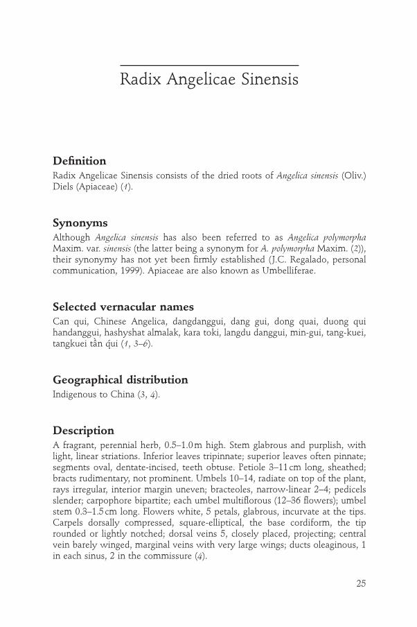
DefinitionRadix Angelicae Sinensis consists of the dried roots of Angelica sinensis (Oliv.)Diels (Apiaceae) (1).
SynonymsAlthough Angelica sinensis has also been referred to as Angelica polymorphaMaxim. var. sinensis (the latter being a synonym for A. polymorpha Maxim. (2)),their synonymy has not yet been firmly established ( J.C. Regalado, personalcommunication, 1999). Apiaceae are also known as Umbelliferae.
Selected vernacular namesCan qui, Chinese Angelica, dangdanggui, dang gui, dong quai, duong qui handanggui, hashyshat almalak, kara toki, langdu danggui, min-gui, tang-kuei,tangkuei tân qui (1, 3–6).
Geographical distributionIndigenous to China (3, 4).
DescriptionA fragrant, perennial herb, 0.5–1.0m high. Stem glabrous and purplish, withlight, linear striations. Inferior leaves tripinnate; superior leaves often pinnate;segments oval, dentate-incised, teeth obtuse. Petiole 3–11cm long, sheathed;bracts rudimentary, not prominent. Umbels 10–14, radiate on top of the plant,rays irregular, interior margin uneven; bracteoles, narrow-linear 2–4; pedicelsslender; carpophore bipartite; each umbel multiflorous (12–36 flowers); umbelstem 0.3–1.5cm long. Flowers white, 5 petals, glabrous, incurvate at the tips.Carpels dorsally compressed, square-elliptical, the base cordiform, the tiprounded or lightly notched; dorsal veins 5, closely placed, projecting; centralvein barely winged, marginal veins with very large wings; ducts oleaginous, 1in each sinus, 2 in the commissure (4).
25
Radix Angelicae Sinensis

Plant material of interest: dried rootsGeneral appearanceSomewhat cylindrical, 3–5 or more branches at the lower part, 15–25cm long.Externally yellowish-brown to brown, longitudinally wrinkled and transverselylenticellate. Root stocks 1.5–4cm in diameter, annulated, apex obtuse, showingpurple or yellowish-green remains of stems and leaf sheaths; main roots lumpyon the surface, branching roots 0.3–1.0cm in diameter, upper portion thick andlower portion thin, mostly twisted, with a few rootlet scars. Texture flexible,fracture yellowish-white or yellowish-brown, thick epidermis, showing someclefts and numerous brown spotted secretory cavities; wood paler in colourthan the bark, cambium ring yellowish-brown (1).
Organoleptic propertiesOdour: strongly aromatic; taste: sweet, pungent, slightly bitter (1).
Microscopic characteristicsCork cells in several layers. Cortex narrow, with a few scattered oil cavities.Phloem cleft, broad (25–160mm in diameter), relatively large on outer side, gradually becoming smaller, surrounded by 6–9 secretory cells, oil cavities andoil tubes. Cambium in a ring. Xylem rays, 3–5 cells wide; vessels scatteredsingly or in groups of 2–3, arranged radially; parenchymatous cells containstarch grains (1).
Powdered plant materialYellowish-brown; parenchymatous cells in phloem are fusiform, with slightlythickened walls, very oblique criss-cross striations, thin transverse septa sometimes visible; scalariform and reticulate vessels frequent, up to 80mm indiameter; fragments of oil cavities sometimes visible (1).
General identity testsMacroscopic and microscopic examinations (1).
Purity testsMicrobiologicalTests for specific microorganisms and microbial contamination limits are asdescribed in the WHO guidelines on quality control methods for medicinalplants (7).
Foreign organic matterFree of foreign matter (1).
WHO monographs on selected medicinal plants
26

Total ashNot more than 7.0% (1).
Acid-insoluble ashNot more than 2.0% (1).
Alcohol-soluble extractiveNot less than 45% using 70% ethanol (1).
Pesticide residuesThe recommended maximum limit of aldrin and dieldrin is not more than 0.05mg/kg (8). For other pesticides, see the European pharmacopoeia (8), and the WHO guidelines on quality control methods for medicinal plants (7) andpesticide residues (9).
Heavy metalsFor maximum limits and analysis of heavy metals, consult the WHO guidelineson quality control methods for medicinal plants (7).
Radioactive residuesWhere applicable, consult the WHO guidelines on quality control methods formedicinal plants (7 ) for the analysis of radioactive isotopes.
Other purity testsChemical, water-soluble extractive and loss on drying tests to be established inaccordance with national requirements.
Chemical assaysMethods for both qualitative and quantitative determination of the alkylphthalide components by high-performance liquid chromatography have beendeveloped (10, 11). National requirements for quantitative criteria should beestablished with respect to the concentration ranges reported for the essentialoil (0.4–0.7%) (4) and ligustilide (0.5–5.0%) (10).
Major chemical constituentsThe characteristic components are the simple alkyl phthalides (ligustilide, (Z)-ligustilide, (Z)-6,7-epoxyligustilide, angelicide, (Z)-butylidenephthalide, butyl-phthalide, 2,4-dihydrophthalic anhydride), which are the major components ofthe essential oil fraction of the roots. Other characteristic components of theoil have been identified as terpenes (b-cadinene, carvacrol and cis-b-ocimene).The non-volatile constituents reported are phenylpropanoids ((E)-ferulic acid,
Radix Angelicae Sinensis
27

coniferyl ferulate); benzenoids (valerophenone-o-carboxylic acid and vanillicacid); and coumarins (angelol G, angelicone and umbelliferone) (3, 4, 10, 11). Ithas been shown by high-performance liquid chromatography that the majorchemical constituent of the roots is ligustilide, which can account for over 5%(10). Polysaccharide fractions of low relative molecular mass have also beenreported (12, 13). The structures of the characteristic constituents are presentedbelow.
WHO monographs on selected medicinal plants
28
(Z )-ligustilide(Z )-butylidenephthalide((Z )-ligusticum lactone)
O
CH3
OO
CH3
O
O
O
O
O
H3C
angelicide CH3
67
H3C CH3
CH2CH3
cis-b-ocimeneb-cadinene
OH
CH3
H3C
CH3
carvacrolCH3
H3C
H3C CH3
H
H
H
OCH3
HO CH3
CO2H
O
valerophenone-o-carboxylic acid(ligusticumic acid)
CO2H
OCH3
HO
vanillic acid
O
O
R
O O
R5
R6
O
R8
umbelliferone
angelicone
angelol G
H
H
H H
Sen
Ang
H
H
H
R7
O
CO
CH3
HO CH3
H
HO
CH3
CH3
H
Ang = angeloyl
COH3C
CH3
Sen = senecioylCon = (E )-coniferyl
H2C
OH
OCH3
(E )-ferulic acidconiferyl ferulate
R = HR = Con
R5
OCH3 CH3
CH3
R6 R7 R8

Medicinal usesUses supported by clinical dataNone. Although Radix Angelicae Sinensis has been alleged to be useful for thetreatment of menopausal symptoms, a randomized, placebo-controlled clinicaltrial concluded that 4.5g of the root daily for 24 weeks did not alleviatemenopausal symptoms, such as hot flushes (14).
Uses described in pharmacopoeias and in traditional systems of medicineTreatment of menstrual disorders such as irregular menstruation, amenorrhoeaand dysmenorrhoea (1, 3, 15–19). As an analgesic for symptomatic treatmentof rheumatic arthralgia, abdominal pain and in the management of post-operative pain (1, 20). Treatment of constipation (1), anaemia (1, 20), chronichepatitis and cirrhosis of the liver (20).
Uses described in folk medicine, not supported by experimental or clinical dataTreatment of dehydration, lumbago, abnormal menstruation, menopausalsymptoms (including hot flushes), hypertonia and nervous disorders (18, 21).
PharmacologyExperimental pharmacologySmooth muscle contractionHot aqueous extracts of Radix Angelicae Sinensis stimulated smooth musclecontractions of the bladder, intestine and uterus when administered intra-venously to dogs (10g/kg body weight) (22). Intravenous administration of anaqueous or 95% ethanol extract of the roots to cats, rats and rabbits increasedthe strength of the contractions and tone of uterine smooth muscles (4). In vitroassays demonstrated that a decoction of the roots stimulated the H1 receptorof mouse uterus (23). The active constituent responsible for this activity is anaqueous- and alcohol-soluble, non-volatile component, the structure of whichis unknown (4). Conversely, ligustilide, a constituent of the essential oil of the roots, inhibited contractions of isolated uteri from various animal models(20, 24). Intraperitoneal administration of ligustilide (0.14ml/kg body weight)to guinea-pigs inhibited asthmatic reactions induced by acetylcholine and his-tamine (25). Ligustilide (32.5–130.0ml/ml) inhibited smooth muscle contractionsinduced by barium sulfate, acetylcholine and histamine in isolated guinea-pigtrachea (25).
Antihepatotoxic activityIntraperitoneal administration of a decoction of the roots (11ml/kg bodyweight) ameliorated galactosamine-induced hepatotoxicity in rats (26). Ferulic
Radix Angelicae Sinensis
29

acid, a constituent of the roots, protected rat liver mitochondria against damageinduced by oxygen free radicals (27). Intragastric pretreatment of mice with so-dium ferulate (100mg/kg body weight) daily for 10 days alleviated liver toxi-city induced by paracetamol (28) and prednisolone (29), and bromobenzene-induced liver injury (30).
Cardiovascular activityCardiac haemodynamic studies demonstrated that intravenous administra-tion of an aqueous root extract (2g/kg body weight) to anaesthetized dogsincreased coronary blood flow from 88ml before administration to 128ml (per100g cardiac muscle/minute post-injection). Coronary vascular resistance andmyocardial oxygen consumption also decreased, while the heart rate decreasedor remained unchanged (31). An extract of the roots increased coronary bloodflow in isolated guinea-pig hearts (32).
In animal models, both aqueous and ethanol extracts of the roots had an effecton arrhythmias induced by epinephrine, barium chloride and digitalis (32, 33).Intravenous administration of an ethanol extract of the roots (4g/kg body weight)antagonized chloroform- and epinephrine-induced arrhythmias in cats (34).Ethanol extracts of the roots and ferulic acid restored normal sinus rhythm afterouabain-induced arrhythmia in isolated ventricular muscle from cats (20).Aqueous extracts of the roots reduced the action potential amplitude andmaximal upstroke velocity of the Q phase, and prolonged the effective refractoryperiod and the duration of the action potential in guinea-pig myocardium (35).Intravenous administration of an aqueous extract of the roots (50mg/kg bodyweight) to rabbits with ligation of the left anterior descending coronary arteryprovided protection against ischaemia- and reperfusion-induced myocardial dys-function and injury (36). An aqueous extract of the roots bound to nitrendipineand diltiazem receptors, thereby demonstrating calcium channel blocking activ-ity (37). A ligustilide dimer, isolated from the roots, inhibited [3H]nitrendipinebinding to dihydropyridine-sensitive calcium channels (inhibitory concentrationof 50% [IC50] 0.4mmol/l) (38). Since calcium channel blockers are known to havepronounced effects on the cardiovascular system, this activity may explain someof the reported effects of root extracts on the cardiovascular system.
Antithrombotic activityIn vitro and in vivo studies have shown that extracts of the roots inhibit plateletaggregation and have antithrombotic activity (20). Aqueous extracts of the roots(200mg/ml) or ferulic acid (0.4mg/ml) inhibited platelet aggregation inducedby ADP or collagen in vitro (39). A hot aqueous extract of the roots (500mg/ml)or ferulic acid (1mg/ml) inhibited thrombin-induced platelet aggregation andrelease of [3H]5-hydroxytryptamine from labelled platelets in vitro (39). Anaqueous extract of the roots inhibited both ADP- and collagen-induced plateletaggregation when administered intravenously to rats (200mg/ml) (20, 39). Themechanism of action appears to be via inhibition of cyclooxygenase and throm-
WHO monographs on selected medicinal plants
30

boxane A2 synthase by ferulic acid, leading to decreased production of throm-boxane A2 (40). The antithrombotic activity of the drug is associated with inhi-bition of platelet aggregation, reduction in the concentration of plasmafibrinogen, changes in cell surface charge and a decrease in blood viscosity (20).
Intraperitoneal administration of polysaccharides isolated from the rootsincreased haematopoiesis in mouse bone marrow, as determined by an increasein colony-forming units in the marrow cells (12, 41). The polysaccharides promoted the proliferation and differentiation of haematopoietic progenitorcells in healthy and anaemic mice (13). Results of this study indicated that thepolysaccharides may enhance haematopoiesis by stimulating macrophages,fibroblasts and lymphocytes in haematopoietic and muscle tissue to secretehaematopoietic growth factor (13).
Clinical pharmacologyMenstrual disordersAlthough there are a number of case reports concerning the clinical use of Radix Angelicae Sinensis in the treatment of amenorrhoea and dysmenorrhoea,these studies were published between 1899 and 1910 (15–18). Randomized,controlled clinical trials are needed to confirm these observations. In these early case studies, female patients were treated with 5ml of a fluidextract ofthe roots three times daily before meals for 1 week before menstruation. The treatment relieved premenstrual pain and induced menstrual flow in most cases.No abortifacient activity was observed in two pregnant women treated withthe same fluidextract (15). In other studies, the fluidextract was used for thetreatment of dysmenorrhoea in nulliparous women, and of severe bleeding inmultiparous women. Administration of 5ml of the fluidextract three times daily for 1 week before menstruation was effective in decreasing menstrual painand chronic endometritis (16). Successful treatment of amenorrhoea and dys-menorrhoea in female patients was further reported after administration of thesame fluidextract (5ml, three times daily) (17, 18). In another report, 112women with dysmenorrhoea were treated for 3–7 days with ligustilide dimerisolated from the roots. The efficacy rate was 77%. Minor side-effects werenausea and dizziness, which disappeared after the treatment stopped (42).
Smooth muscle contractionDecoctions of the roots reportedly stimulated uterine smooth muscle in femalepatients, but the doses used and the conditions being treated were not stated(19). A decoction of the roots lowered whole blood viscosity after administra-tion to six patients (11).
ContraindicationsRadix Angelicae Sinensis should not be administered to children or patientswith diarrhoea, haemorrhagic diseases or hypermenorrhoea, and should not beused during pregnancy or lactation (4).
Radix Angelicae Sinensis
31

WarningsNo information available.
PrecautionsDrug interactionsDecreased prothrombin times were reported in rabbits that received both asingle subcutaneous dose of warfarin (2mg/kg body weight) and a repeatedoral dose of Radix Angelicae Sinensis (2g/kg body weight twice daily for 3 days) (43). Therefore, patients receiving anticoagulant therapy should beadvised against taking Radix Angelicae Sinensis without medical supervision.
Pregnancy: teratogenic effectsSee Contraindications.
Pregnancy: non-teratogenic effectsSee Contraindications.
Nursing mothersSee Contraindications.
Paediatric useSee Contraindications.
Other precautionsNo information available on general precautions or precautions concerning drug and laboratory test interactions; or carcinogenesis, mutagenesis andimpairment of fertility.
Adverse reactionsOral administration of Radix Angelicae Sinensis is generally regarded as hav-ing few side-effects; however, headaches may occur in sensitive individuals (14, 19). No adverse reactions were reported in 40 people who received anaqueous root extract by intravenous administration (240ml/person) for 30 days(19).
Dosage formsPowdered crude drug and fluidextracts (4). Store in an airtight container in acool, dry place protected from moisture (1).
WHO monographs on selected medicinal plants
32

Posology(Unless otherwise indicated)Daily dosage: 4.5–9g crude drug (1).
References1. Pharmacopoeia of the People’s Republic of China. Vol. I (English ed.). Beijing, Chemical
Industry Press, 1997.2. Hiroe M. Umbelliferae of Asia. Kyoto, Eikodo, 1958.3. Hsu HY. Oriental materia medica, a concise guide. Long Beach, CA, Oriental Healing
Arts Institute, 1986.4. Zhu DPQ. Dong quai. American Journal of Chinese Medicine, 1987, 15:117–125.5. Farnsworth NR, ed. NAPRALERT database. Chicago, University of Illinois at Chicago,
IL, January 1, 1998 production (an online database available directly through the University of Illinois at Chicago or through the Scientific and Technical Network[STN] of Chemical Abstracts Services).
6. Medicinal plants in Viet Nam. Manila, World Health Organization, 1990 (WHORegional Publications, Western Pacific Series, No. 3).
7. Quality control methods for medicinal plant materials. Geneva, World Health Organiza-tion, 1998.
8. European pharmacopoeia, 3rd ed. Strasbourg, Council of Europe, 1996.9. Guidelines for predicting dietary intake of pesticide residues, 2nd rev. ed. Geneva, World
Health Organization, 1997 (document WHO/FSF/FOS/97.7).10. Lin LZ et al. Liquid chromatographic–electrospray mass spectrometric study of
the phthalides of Angelica sinensis and chemical changes of Z-ligustilide. Journal ofChromatography A, 1998, 810:71–79.
11. Terasawa K et al. Chemical and clinical evaluation of crude drugs derived fromAngelica acutiloba and A. sinensis. Fitoterapia, 1985, 56:201–208.
12. Ma LF et al. The effect of Angelica sinensis polysaccharides on mouse bone marrowhematopoiesis. Zhonghua Xinxueguanbing Zazhi, 1988, 9:148–149.
13. Wang Y, Zhu B. The effect of Angelica polysaccharide on proliferation and dif-ferentiation of hematopoietic progenitor cells. Chung Hua I Hsueh Tsa Chih, 1996,76:363–366.
14. Hirata JD et al. Does dong quai have estrogenic effects in postmenopausal women?A double-blind, placebo-controlled trial. Fertility and Sterility, 1997, 68:981–986.
15. Mueller A. Versuche über die Wirkungsweise des Extrakts des chinesischen Emmenagogon Tang-kui (Man-mu) oder Eumenol-Merek. Münchener MedizinischeWochenschrift, 1899, 46:796–798.
16. Langes H. Beobachtungen bei der Verwendung einiger neuer Medikamente.Eumenol, Dionin und Stypticin. Therapeutische Monatshefte, 1901, 7:363.
17. Palm R. Erfahrungen mit Eumenol. Münchener Medizinische Wochenschrift, 1910, 1:23–25.
18. Buck P. Un nouveau remède spécifique contre la dysmenorrhée: l’eumenol. Belgiquemédicale, 1899, 2:363–365.
19. Chang HM, But PPH, eds. Pharmacology and applications of Chinese materia medica. Vol. 1. Philadelphia, PA, World Scientific Publishing, 1986.
20. Mei QB, Tao JY, Cui B. Advances in the pharmacological studies of Radix Angelicasinensis (Oliv.) Diels (Chinese danggui). Chinese Medical Journal, 1991, 104:776–781.
21. Duke JA, Ayensu ES. Medicinal plants of China. Vol. 1. Algonac, MI, Reference Publications, 1985.
22. Schmidt CF et al. Experiments with Chinese drugs. 1. Tang-kuei. Chinese MedicalJournal, 1924, 38:362.
Radix Angelicae Sinensis
33

23. Shi M, Chang L, He G. Stimulating action of Carthamus tinctorius L., Angelica sinensis (Oliv.) Diels and Leonurus sibiricus L. on the uterus. Chung Kuo Chung Yao TsaChih, 1995, 20:173–175.
24. Pi XP. Effects of Angelica sinensis on uterus. National Medical Journal of China, 1955,40:967.
25. Tao JY et al. Studies on the antiasthmatic action of ligustilide of dang-gui, Angelicasinensis (Oliv.) Diels. Yao Hsueh Hsueh Pao, 1984, 198:561–565.
26. Xiong X et al. The protective effect of Radix Angelicae Sinensis against acute liverdamage by D-galactosamine in rats: a histochemical study. Wu-han I Hsueh YuanHsueh Pao, 1982, 11:68–72.
27. Lin YH et al. Protective effect of sodium ferulate on damage of the rat liver mito-chondria induced by oxygen free radicals. Yao Hsueh Hsueh Pao, 1994, 29:171–175.
28. Wang H, Peng RX. Sodium ferulate alleviated paracetamol-induced liver toxicity inmice. Yao Hsueh Hsueh Pao, 1994, 15:81–83.
29. Wu DF et al. Sodium ferulate alleviates prednisolone-induced liver toxicity in mice.Acta Pharmaceutica Sinica, 1988, 30:801–805.
30. Wu DF, Peng RX. The effect of sodium ferulate on bromobenzene-induced liverinjury in mice. Zhongguo Yaoxue Zazhi, 1995, 30:597–599.
31. Chou YP. The effect of Angelica sinensis on hemodynamics and myocardiac oxygenconsumption in dogs. Acta Pharmaceutica Sinica, 1979, 14:156–160.
32. Pen RX. Pharmacological effects of danggui (Angelica sinensis) on cardiovascularsystem. Chinese Traditional Herb Drugs, 1981, 12:321.
33. Cha L. Effects of Angelica sinensis on experimental arrhythmias. Chinese Pharmaceu-tical Bulletin, 1981, 16:259.
34. Cha L, Chien CC, Lu FH. Antiarrhythmic effect of Angelica sinensis root, tetrandrineand Sophora flavescens root. Chinese Pharmaceutical Bulletin, 1981, 16:53–54.
35. Wei ZM et al. A study on the electrophysiology in antiarrhythmia effect of Angelica sinensis. Journal of Beijing College of Traditional Chinese Medicine, 1985, 8:40.
36. Chen SG et al. Protective effects of Angelica sinensis on myocardial ischemia/reperfusion injury in rabbits. Chung-kuo Chung His I Chieh Ho Tsa Chih, 1995,15:486–488.
37. Hon PM. A ligustilide dimer from Angelica sinensis. Phytochemistry, 1990, 29:1189–1191.
38. Han GQ. The screening of Chinese traditional drugs by biological assay and the iso-lation of some active components. International Journal of Chinese Medicine, 1991,16:1–17.
39. Yin ZZ. The effect of danggui (Angelica sinensis) and its ingredient ferulic acid on ratplatelet aggregation and release of 5-HT. Acta Pharmaceutica Sinica, 1980, 15:321.
40. Xu LN. Effect of sodium ferulate on arachidonic acid metabolism. Acta Pharmaceu-tica Sinica, 1990, 25:412.
41. Chen YC, Gao YQ. Research on the mechanism of blood-tonifying effect of dang-gui buxue decoction. Chung Kuo Chung Yao Tsa Chih, 1994, 19:43–45, 63.
42. Compendium of materia medica. Shanghai, State Administration of Traditional ChineseMedicine, Shanghai Science and Technical Press, 1996:1341–1355.
43. Lo A et al. Danggui (Angelica sinensis) affects the pharmacodynamics but not thepharmacokinetics of warfarin in rabbits. European Journal of Drug Metabolism andPharmacokinetics, 1995, 20:55–60.
WHO monographs on selected medicinal plants
34

DefinitionFlos Calendulae consists of the dried ligulate florets or composite flowers ofCalendula officinalis L. (Asteraceae) (1–3).
SynonymsAsteraceae are also known as Compositae.
Selected vernacular namesAtunjaq, calendula, Chinese safflower, cuc kim tiên, djamir, djomaira, feminell,flamenquilla, fleur de calandule, fleur de souci, fleur de souci officinal, fleurs de tous les mois, garden marigold, gold-bloom, Goldblume, gole hamishehbahar, hen and chickens, Körömvirag, lellousha, maravilla, marigold, mary-bud,ok-hhawan, pot marigold, qaraqus, qawqhan, quaqahan, ringflower, Ringelblüten, saialill, sciure’e Sant’antonio, souci, souci des jardins, tabsoult,toukinsenka, tousslat, uchu k’aspa, virreina, xu xi, zergul zerzira, zobeida,zubaydah (4–7).
Geographical distributionIndigenous to central, eastern and southern Europe. Cultivated commerciallyin North America, the Balkans, Eastern Europe and Germany (6, 8).
DescriptionAn annual herb, much branched from the base, very aromatic, up to 0.3–0.6mhigh; stem angular, hairy and solid. Leaves sessile, light green, with semi-amplexicaul base; entire, undulate or remotely denticulate; glandular hairs onboth surfaces; lower leaves spatulate, obtuse, sometimes acute at the apex,10–20cm long and 1–4cm wide; higher leaves oblong and mucronate, 4–7cmlong. Involucral bracts 7–15mm long, covered with long, glandular hairs; innerinvolucral bracts with pellucid, scarious margin; marginal flowers in cultivatedplants often multi-seriate; corolla oblong-spatulate, bright yellow or orange,15–25mm long and 3mm wide, 1–3-toothed with 4 or 5 veins, marginallyentire, covered at the base with patent, long, thick hairs; corolla of disc flowersrounded, 3-dentate top, 1.5–2.5cm long and 4–7mm in diameter, 5mm long
35
Flos Calendulae

tube and moderately widened limb. Stigma short, thick, hairy; ovary oblong,0.5mm in length, pubescent, shrivelling after anthesis. Achenes narrowlyoblong, strongly curved, faintly ribbed, thinly pubescent or glabrous, 10–12mmlong, outer achenes warty-ribbed outside, inner achenes prickly-warty, oftenwith broad, thick margins (2, 7, 9).
Plant material of interest: dried ligulate florets andcomposite flowersGeneral appearanceLigulate florets consist of a yellow, orange or orange-yellow ligule, 3–5mmwide and about 7mm in the middle part, with 3-toothed apex and hairy, partlysickle-shaped, yellowish-brown to orange-brown tube with projecting style and 2-lobed stigma; occasionally with a partly bent yellowish-brown toorange-brown ovary. Tubular florets about 5mm long, consist of yellow,orange-red or red-violet 5-lobed corolla and yellowish-brown or orange-browntube, hairy in its lower part, mostly with a bent yellowish-brown to orange-brown ovary (1).
Organoleptic propertiesOdour: faint, pleasantly aromatic (10, 11); taste: bitter (2).
Microscopic characteristicsInner epidermal cells of ray floret elongated, rectangular and almost straight-walled, cuticle faintly striated; stomata absent; outer epidermal cells similar, butwith 3 or 4 anomocytic stomata; trichomes very numerous on the tube, bi-seriate; stigma epidermal cells straight-walled, polygonal. In disc floret, outerepidermal cells elongated, straight or slightly sinuous-walled, stomata absent;abundant trichomes on area below point of insertion of the stamens, mainlyglandular, uniseriate or biseriate. Within the upper part of the anthers, a layerof isodiametric to elongated, moderately thick-walled, lignified and pitted cells;pollen grains spherical, up to 45mm in diameter, with 3 germinal pores, exinefinely granular with numerous short spines; apex of stigma covered by short,bulbous papillae (2).
Powdered plant materialYellow-green; fragments of corollas containing light yellow oil droplets; somecorollas with fairly large anomocytic stomata, others containing prismatic andvery small clusters of calcium oxalate crystals. Covering trichomes biseriate,multicellular and conical; glandular trichomes with a uniseriate or biseriate,multicellular stalk and a large, ovoid, biseriate, multicellular head. Spherical
WHO monographs on selected medicinal plants
36

pollen grains up to 45mm in diameter, exine finely granular with numerousshort spines and with 3 germinal pores; occasional fragments of stigmas withshort, bulbous papillae (1).
General identity testsMacroscopic and microscopic examinations, and thin-layer chromatography forflavonoid content (1, 2).
Purity testsMicrobiologicalTests for specific microorganisms and microbial contamination limits are asdescribed in the WHO guidelines on quality control methods for medicinalplants (12).
Foreign organic matterNot more than 5% bracts and not more than 2% other foreign matter (1, 2).
Total ashNot more than 10% (1, 2).
Acid-insoluble ashNot more than 2% (2).
Water-soluble extractiveNot less than 20% (2).
Loss on dryingNot more than 10% (1).
Pesticide residuesThe recommended maximum limit of aldrin and dieldrin is not more than 0.05mg/kg (13). For other pesticides, see the European pharmacopoeia (13), andthe WHO guidelines on quality control methods for medicinal plants (12) andpesticide residues (14).
Heavy metalsFor maximum limits and analysis of heavy metals, consult the WHO guidelineson quality control methods for medicinal plants (12).
Flos Calendulae
37

Radioactive residuesWhere applicable, consult the WHO guidelines on quality control methods formedicinal plants (12) for the analysis of radioactive isotopes.
Other purity testsChemical, sulfated ash and alcohol-soluble extractive tests to be established inaccordance with national requirements.
Chemical assaysContains not less than 0.4% flavonoids, calculated as hyperoside, by spectro-photometry (1). A high-performance liquid chromatography method is alsoavailable (15).
Major chemical constituentsThe major constituents are triterpene saponins (2–10%) based on oleanolic acid (i.e. calendulosides) and flavonoids (3-O-glycosides of isorhamnetin andquercetin), including astragalin, hyperoside, isoquercitrin and rutin. Other con-stituents include essential oil, sesquiterpenes (e.g. caryophyllene) and triter-penes (e.g. a- and b-amyrins, lupeol and lupenone) (5, 6, 16). Polysaccharideshave also been reported (17). The structures of the characteristic triterpenesaponins and flavonoids are presented below.
WHO monographs on selected medicinal plants
38
OO
O
R5
OH
R3
O
R4
CH3
H3C CH3
H
H
H
CH3
CH3
H
O
O
R
CH3H3C
OOH
OH
HO
HO
Galp = OH
OH
Glcf =
OH
HHO
O OOH
OH
Glcp =
HO
HO
calendulosides
A
E
F
H
R
H
H
Glcp
Glcp
R3
H
H
H
Galp
R4
Galp
H
H
H
R5
CH2OH
CO2H
CO2H
CO2H
b-D-galactopyranosyl b-D-glucofuranosyl b-D-glucopyranosyl

Medicinal usesUses supported by clinical dataNone.
Uses described in pharmacopoeias and in traditional systems of medicineExternal treatment of superficial cuts, minor inflammations of the skin and oralmucosa, wounds and ulcus cruris (2, 18, 19).
Uses described in folk medicine, not supported by experimental or clinical dataTreatment of amenorrhoea, angina, fevers, gastritis, hypotension, jaundice,rheumatism and vomiting (2, 5, 6).
PharmacologyExperimental pharmacologyPhagocytosisThree polysaccharides isolated from an aqueous extract of Flos Calendulaeenhanced phagocytosis in human granulocytes in vitro in the colloidal car-bon clearance test (17). Intraperitoneal injection of a polysaccharide fraction iso-lated from an aqueous extract of the flowers to mice (10mg/kg body weight)enhanced phagocytosis (20). Intraperitoneal administration of an unsaponifiablefraction (0.5ml) of a hydroalcoholic extract of the flowers weakly stimulatedphagocytosis in mice inoculated with Escherichia coli. However, the hydroalco-holic extract was not active (21).
Flos Calendulae
39
O
OH
HO
O
O
R
OH
X
astragalin
hyperoside
isoquercitrin
rutin
R
H
OH
OH
OH
X
Glcp
Galp
Glcf
Rhap-(1 6)-Glcp
O
OH
OH
HO
OO
HOHO
CH3
HO
Glcp =
Rhap-(1 6)-
O-6-deoxy-a-L-mannopyranosyl-(1 6)-b-D-glucopyranosyl
Antimicrobial activityThe essential oil of the flowers inhibited the growth in vitro of Bacillus subtilis,Escherichia coli, Staphylococcus aureus, Pseudomonas aeruginosa and Candida albi-cans (22). A flavonoid fraction isolated from the flowers inhibited the growthin vitro of S. aureus, Sarcina lutea, E. coli, Klebsiella pneumoniae and Candidamonosa (23). However, chloroform, ethanol, methanol or water extracts of theflowers did not inhibit bacterial growth in vitro (24–26). Acetone, ethanol orwater extracts inhibited the growth in vitro of the fungus Neurospora crassa (27).Extracts of the flowers inhibited the growth in vitro of Trichomonas vaginalis (28).Oxygenated terpenes appear to be responsible for the antimicrobial activity(29).
Antiviral activityA tincture of the flowers suppressed the replication of herpes simplex, influenzaA2 and influenza APR-8 viruses in vitro (30). However, an aqueous extract ofthe flowers was not active (31). A chloroform extract of the flowers inhibitedthe replication of HIV-1 in acutely infected lymphocytic MOLT-4 cells in vitro(IC50 0.4mg/ml) (32). A chloroform extract also inhibited HIV-1 reverse tran-scriptase activity in a dose-dependent manner (ED50 51.0mg/ml) (32). A 5% hotaqueous extract of the flowers (2ml) inhibited the replication of encephalitisvirus after intraperitoneal administration to mice (33).
Anti-inflammatory activityTopical application of a 70% ethanol extract of the flowers to mice at a doseof 1.2mg/ear (corresponding to 4.16mg crude drug) reduced croton oil-inducedear oedema by 20% (34). External application of a carbon dioxide extract of theflowers (300mg/cm2) suppressed croton oil-induced ear oedema in mice (34). The triterpene fraction of an extract of the flowers had marked anti-inflammatory activity in mice (1mg/ear) against ear oedema induced by 12-O-tetradecanoylphorbol-13-acetate (35). Faradiol esters isolated from the flowers(240mg/cm2) inhibited croton oil-induced ear oedema in mice (36). Intragastricadministration of an aqueous extract of the flowers (100mg/kg body weight)inhibited carrageenan-induced footpad oedema in rats (37). However, an 80%ethanol extract of the flowers was weakly active (11% inhibition) at a con-centration of 100mg/kg body weight administered orally 1 hour prior to induc-tion of oedema (38). Isorhamnetin glycosides isolated from the flowersinhibited rat lung lipoxygenase in vitro (39).
Wound-healing activityExternal application of a hydroalcoholic extract accelerated the rate of con-traction and epithelialization of excision wounds in rats (40). A 3% freeze-driedaqueous extract of the flowers induced vascularization in the chick chorio-allantoic membrane assay. Histological sections of the treated chorioallantoic
WHO monographs on selected medicinal plants
40
membranes also indicated the presence of hyaluronan, a tissue glycosamino-glycan associated with neovascularization (41).
Clinical pharmacologyAlthough no randomized, controlled clinical trials have been performed, twocase reports in the early medical literature support the traditional use of FlosCalendulae. The reports describe the use of a strong tincture of the flowersapplied on compresses to reduce inflammation and suppuration, and to acceler-ate the healing of wounds (42, 43). These reports may be of historical value only.
ContraindicationsFlos Calendulae is contraindicated in cases of known allergy to plants of theAsteraceae (Compositae) family (18).
WarningsNo information available.
PrecautionsCarcinogenesis, mutagenesis, impairment of fertilitySaponins isolated from Flos Calendulae were not mutagenic at a concentrationof 400mg/ml in the Salmonella/microsome assay using S. typhimurium strainTA98, with or without S9 metabolic activation (44). Extracts of the flowers werenot carcinogenic after daily intragastric administration of 0.15g/kg body weightto rats (for 22 months) or hamsters (for 18 months) (45). Mutagenicity testing ofthe fluidextract in the Salmonella/microsome assay (using S. typhimurium strainsTA98, TA100, TA1535 and TA1537) was negative at concentrations of up to 5mg/plate. The mouse bone marrow micronucleus test was also negative afterdaily administration of up to 1g/kg body weight for 2 days (46). A fluidextractof the flowers (100mg/ml, 60% ethanol) was genotoxic in both mitotic crossing-over and chromosome segregation when assayed for mitotic segrega-tion in the heterozygous diploid D-30 of Aspergillus nidulans (46).
Other precautionsNo information available on general precautions or precautions concerning drug interactions; drug and laboratory test interactions; teratogenic and non-teratogenic effects in pregnancy; nursing mothers; or paediatric use. Therefore,Flos Calendulae should not be administered during pregnancy or lactation orto children without medical supervision.
Adverse reactionsWeak skin-sensitization has been reported (47).
Flos Calendulae
41

Dosage formsInfusion for topical use; aqueous and alcohol extracts, tinctures and ointmentfor external use (2, 18, 19). Store in a well-closed container, protected from light(1).
Posology(Unless otherwise indicated)Topical application: an infusion of 1–2g/150ml (18). External use: a 40%alcohol extract (1 :1), or tincture (1 :5) in 90% alcohol (2). For the treatment ofwounds, the tincture is applied undiluted; for compresses, the tincture is usuallydiluted at least 1 :3 with sterile water (18, 48, 49). Ointment: 2–5% (48, 50).
References1. European pharmacopoeia, 3rd ed., Suppl. 2000. Strasbourg, Council of Europe,
1999.2. British herbal pharmacopoeia. London, British Herbal Medicine Association, 1996.3. Pharmacopoeia Hungarica, 7th ed. Budapest, Hungarian Pharmacopoeia Commission,
Medicina Konyvkiado, 1986.4. Boulos L. Medicinal plants of North Africa. Cairo, Reference Publications, 1983.5. Farnsworth NR, ed. NAPRALERT database. Chicago, University of Illinois
at Chicago, IL, January 28, 1998 production (an online database available directly through the University of Illinois at Chicago or through the Scientific andTechnical Network [STN] of Chemical Abstracts Services).
6. Bisset NG. Herbal drugs and phytopharmaceuticals. Boca Raton, FL, CRC Press, 1994.7. Youngken HW. Textbook of pharmacognosy, 6th ed. Philadelphia, PA, Blakiston,
1950.8. Leung AY, Foster S. Encyclopedia of common natural ingredients used in food, drugs, and
cosmetics, 2nd ed. New York, NY, John Wiley & Sons, 1996.9. Backer CA, Van den Brink B. Flora of Java. Vol. 2. Noordfog-Groningen, NVP, 1965:
574.10. Pharmacopoea helvetica, 8th ed. Berne, Département fédéral de l’intérieur, 1997.11. Pharmacopée française. Paris, Adrapharm, 1996.12. Quality control methods for medicinal plant materials. Geneva, World Health Organiza-
tion, 1998.13. European pharmacopoeia, 3rd ed. Strasbourg, Council of Europe, 1996.14. Guidelines for predicting dietary intake of pesticide residues, 2nd rev. ed. Geneva, World
Health Organization, 1997 (document WHO/FSF/FOS/97.7).15. Vidal-Ollivier E et al. Dosage par CLHP. Des flavonoides majoritaires de Calendula
officinalis L. En fonction de la variété culturale et de la date de récolte. PharmaceuticaActa Helvetiae, 1991, 66:318–320.
16. Bruneton J. Pharmacognosy, phytochemistry, medicinal plants. Paris, Lavoisier, 1995.17. Varljen J, Lipták A, Wagner H. Structural analysis of a rhamnoarabinogalactan
and arabinogalactans with immunostimulating activity from Calendula officinalis.Phytochemistry, 1989, 28:2379–2383.
18. ESCOP monographs on the medicinal uses of plant drugs. Fascicule 1. Elburg, EuropeanScientific Cooperative on Phytotherapy, 1996.
19. Blumenthal M et al., eds. The complete German Commission E monographs. Austin, TX,American Botanical Council, 1998.
WHO monographs on selected medicinal plants
42

20. Wagner H et al. Immunstimulierend wirkende Polysaccharide (Heteroglykane) aushöheren Pflanzen. Arzneimittel-Forschung, 1985, 7:1069–1075.
21. Delaveau P et al. Drogues végétales stimulant l’activité phagocytaire du systèmeréticulo-endothélial. Planta Medica, 1980, 40:49–54.
22. Janssen AM et al. Screening for antimicrobial activity of some essential oils by theagar overlay technique. Pharmaceutisch Weekblad, 1986, 8:289–292.
23. Tarle D, Dvorzak I. Antimicrobial substances in Flos Calendulae. Farmacevtski Vestnik(Ljubljana), 1989, 40:117–120.
24. Rios JL, Recio MC, Villar A. Antimicrobial activity of selected plants employed in the Spanish Mediterranean area. Journal of Ethnopharmacology, 1987, 21:139–152.
25. Dornberger K, Lich H. Screening for antimicrobial and presumed cancerostatic plantmetabolites. Pharmazie, 1982, 37:215–221.
26. Acevedo JG, Lopez JL, Cortes GM. In vitro antimicrobial activity of various plantextracts used by purepecha against some Enterobacteriaceae. International Journal ofPharmacognosy, 1993, 31:61–64.
27. Kubas J. Investigations on known or potential antitumoral plants by means of micro-biological tests. Part III. Activity of some cultivated plant species in Neurospora crassatest. Acta Biologica Cracoviensia Series Botanica, 1972, 15:87–100.
28. Racz G et al. Trichomonicidal and anthelmintic activity of Roumanian folkloricplants. Planta Medica, 1980, 39:257A.
29. Gracza L. Oxygen-containing terpene derivatives from Calendula officinalis. PlantaMedica, 1987, 53:227.
30. Bogdanova NS et al. Study of antiviral properties of Calendula officinalis. FarmakolToksikol (Moscow), 1970, 33:349.
31. May G, Willuhn G. Antiviral activity of aqueous extracts from medicinal plants intissue cultures. Arzneimittel-Forschung, 1978, 28:1–7.
32. Kalvatchev Z et al. Anti-HIV activity of extracts from Calendula officinalis flowers.Biomedicine and Pharmacotherapy, 1997, 51:176–180.
33. Fokina GI et al. Experimental therapy of tick-borne encephalitis. Soviet Progress inVirology, 1991, 1:27–31.
34. Della-Loggia R et al. The role of triterpenoids in the topical anti-inflammatory activity of Calendula officinalis flowers. Planta Medica, 1994, 60:516–520.
35. Akihisa T et al. Triterpene alcohols from the flowers of Compositae and their anti-inflammatory effects. Phytochemistry, 1996, 43:1255–1260.
36. Zitterl-Eglseer K et al. Anti-oedematous activities of the main triterpendiol esters ofmarigold (Calendula officinalis L.). Journal of Ethnopharmacology, 1997, 57:139–144.
37. Peyroux J et al. Anti-oedemic and anti-hyperhaemic properties of Calendula offici-nalis L. Plantes médicinales et Phytotherapie, 1981, 15:210–216.
38. Mascolo N et al. Biological screening of Italian medicinal plants for anti-inflammatory activity. Phytotherapy Research, 1987, 1:20–31.
39. Bezakova L et al. Inhibitory activity of isorhamnetin glycosides from Calendula offic-inalis L. on the activity of lipoxygenase. Pharmazie, 1996, 51:126–127.
40. Rao SG et al. Calendula and Hypericum: two homeopathic drugs promoting woundhealing in rats. Fitoterapia, 1991, 62:508.
41. Patrick KFM et al. Induction of vascularisation by an aqueous extract of the flowersof Calendula officinalis L., the European marigold. Phytomedicine, 1996, 3:11–18.
42. Livezey A. Some observations on our indigenous medical flora. Medical and Surgi-cal Reporter, 1868, 19:85.
43. Reynolds RG. Calendula. Pacific Medical and Surgical Journal, 1886, 29:720.44. Elias R et al. Antimutagenic activity of some saponins isolated from Calendula
officinalis L., C. arvensis L. and Hedera helix L. Mutagenesis, 1990, 5:327–331.45. Avramova S et al. Source of new products for the cosmetic industry. Medical and
Biological Information, 1988, 4:28–32.
Flos Calendulae
43

46. Ramos A et al. Genotoxicity of an extract of Calendula officinalis L. Journal ofEthnopharmacology, 1998, 61:49–55.
47. Bruynzeel DP et al. Contact sensitization by alternative topical medicaments con-taining plant extracts. Contact Dermatitis, 1992, 27:278–279.
48. Willuhn G. Pflanzliche Dermatika, eine kritische Übersicht. Deutsche ApothekerZeitung, 1992, 132:1873–1883.
49. Van Hellemont J. Fytotherapeutisch compendium, 2nd ed. Utrecht, Bohn, Scheltema &Holkema, 1988:113–114.
50. Hänsel R et al., eds. Hagers Handbuch der pharmazeutischen Praxis. Bd. 4: Drogen A–K,5th ed. Berlin, Springer-Verlag, 1994.
WHO monographs on selected medicinal plants
44

DefinitionFlos Caryophylli consists of the dried flower buds of Syzygium aromaticum (L.)Merrill et L.M. Perry (Myrtaceae) (1–5).
SynonymsCaryophyllus aromaticus L., Eugenia aromatica (L.) Baill., E. caryophylla Thunb., E. caryophyllus (C. Spreng.) Bull. et Harr., Jambosa caryophyllus (Spreng.) Nied.,Myrtus caryophyllus Spreng. (1, 5–8).
Selected vernacular namesBenefundi, choji, choko, chouji, choukou, clavero, clavo de olor, clous de girofle,clove, cloves, colve, ding huong, dingxiang, flores caryophylli, Gewürznelken,girofle, giroflier, glove, gurunful, harilik negipuu, kaan phluu, kaan pluu, kade,kanumfari, karafwu, karanho, kau-phlu, konofuru, koronfol, lauang, laung,lawang, Nägelein, osaragbogo-eze, qaranfal, qoranful, qronfel, szegfüszeg, ud-nuwwar (1, 6–9).
Geographical distributionIndigenous to the Moluccas and southern Philippines, but currently culti-vated in many tropical areas including Africa (e.g. Madagascar and UnitedRepublic of Tanzania), South America, Indonesia, Malaysia and Sri Lanka (7,8).
DescriptionSmall evergreen trees, 10–20m high. Leaves opposite, petiolate, lanceolate,pinkish to dark green, with translucent, aromatic glands, have a pungent odourwhen young. Inflorescence occurs as racemose panicles and bears buds thattake on the form of nails before blossoming. Flowers red with 4 concave, over-lapping petals that drop off as soon as the flower opens; stamens numerous; 4calyx lobes. Fruit dark red, fleshy drupe. Buds readily exude oil when pressedor scratched with a fingernail (7).
45
Flos Caryophylli

Plant material of interest: dried flower budsGeneral appearanceFlower bud 10–20mm long, bright reddish-brown to dark brown; lower part (the hypanthium) solid, cylindrical, somewhat flattened, 4-sided, taperingtowards the base and bearing at the apex 4 thick, triangular, divergent sepals,alternating with 4 rounded, fragile, unexpanded, membranous, imbricatedpetals forming a pale, nearly spherical head that encloses numerous stamens,curved inward and inserted on a small disc, and a stiff, slender, erect, singlestyle arising from a depression in the centre. Externally wrinkled; internally,hypanthium contains in its upper portion a 2-celled inferior ovary with numer-ous ovules attached to the axile placenta; has very large outer zone with numer-ous shining, oval oil glands near the periphery, numerous vascular bundles inthe centre and a dark, lacunose layer abutting on the central zone and columella(1).
Organoleptic propertiesOdour: characteristic, strongly aromatic; taste: pungent, spicy, followed byslight numbness (1, 3, 5).
Microscopic characteristicsHypanthium epidermis of small, thick-walled isodiametric cells with very thickcuticle, with stomata with no special subsidiary cells. Parenchymatous layercontaining numerous large (up to about 200mm long), oval, radially elongated,schizo-lysigenous oil glands, arranged in 2 or 3 more or less intermixed layers.Layer of parenchyma and collenchyma containing clusters of calcium oxalatecrystals, and traversed by small, irregularly arranged vascular bundles consist-ing of delicate, spiral vessels (up to 20mm in diameter), usually accompaniedby isolated fusiform, pericyclic fibres (200–650mm long and up to 40mm indiameter), having strongly thickened lignified walls. Lacunous layer formed ofthin-walled parenchyma. The columella consists of a parenchymatous strandwith numerous closely arranged, small vascular bundles. Sepals, with epider-mis resembling that of hypanthium and having numerous stomata on outersurface; mesophyll with rounded or stellate cells, numerous ovoid oil glandsand clusters of calcium oxalate crystals, and traversed by a few slender vascu-lar bundles. Petals, with epidermis formed of cells with straight, thin walls;stomata, absent; mesophyll, undifferentiated, containing oil glands and cellswith clusters of calcium oxalate crystals, and traversed by small vascularbundles. Stamens, with filaments having a central vascular strand and oil glandsbeneath the epidermis; connective tissue, with a large oil gland in the apex ofanther walls, with fibrous layer and minute clusters of calcium oxalate crystalsalong the line of dehiscence. Pollen grains, triangular, tricolpate, 10–20mm indiameter. Style, with epidermis similar to that of hypanthium, and consistingof small collenchyma cells, with clusters of calcium oxalate crystals, radiallyelongated oil glands, and traversed by 2 narrow vascular strands (1).
WHO monographs on selected medicinal plants
46

Powdered plant materialDark brown; abundant fragments of collenchyma and parenchyma with clusters of calcium oxalate crystals, fragments of epidermis with thick-walledcells and few stomata; fragments of vascular or parenchyma tissue showingbroken or entire oil glands; numerous fragments of vascular bundles with delicate spiral vessels, ranging from 6 to 45mm in diameter, mostly 6–10mm;occasional fusiform, rather thick-walled fibres, 4–20mm wide; numerous pollen grains, appearing either as equilateral triangular, with truncated, emar-ginate apices, or oval in outline, 10–20mm in diameter; fragments of the fibrouslayer of anther wall; clusters of calcium oxalate crystals, 10–15mm in diameter(1, 5).
General identity testsMacroscopic and microscopic examinations, and thin-layer chromatography forthe presence of eugenol and b-caryophyllene (1, 3–5, 10).
Purity testsMicrobiologicalTests for specific microorganisms and microbial contamination limits are asdescribed in the WHO guidelines on quality control methods for medicinalplants (11).
Foreign organic matterNot more than 4% open buds, peduncles and fruits; not more than 2% deteriorated buds; not more than 0.5% other foreign matter (5).
Total ashNot more than 7% (4, 5).
Acid-insoluble ashNot more than 0.5% (4).
Sulfated ashNot more than 8% (12).
Loss on dryingNot more than 12% (3).
Pesticide residuesThe recommended maximum limit of aldrin and dieldrin is not more than 0.05mg/kg (5). For other pesticides, see the European pharmacopoeia (5), and the
Flos Caryophylli
47

WHO guidelines on quality control methods for medicinal plants (11) and pesticide residues (13).
Heavy metalsFor maximum limits and analysis of heavy metals, consult the WHO guidelineson quality control methods for medicinal plants (11).
Radioactive residuesWhere applicable, consult the WHO guidelines on quality control methods formedicinal plants (11) for the analysis of radioactive isotopes.
Other purity testsChemical, water-soluble extractive and alcohol extractive tests to be establishedin accordance with national requirements.
Chemical assaysContains not less than 15% (v/w) essential oil (1, 12), determined by distilla-tion (5).
Major chemical constituentsThe major constituent (up to 20%) is an essential oil, which is characterizedby the presence of eugenol (60–95%), eugenol acetate (2–27%), and a- and b-caryophyllene (5–10%) (6, 8, 9, 14, 15). The structures of the major con-stituents are presented below.
WHO monographs on selected medicinal plants
48
OCH3
OR
H2C
CH3
H3C
CH3
CH3H
H3CCH3
CH2
CH3
H
eugenoleugenol acetate
R = HR = CO-CH3 a-caryophyllene b-caryophyllene
Medicinal usesUses supported by clinical dataNone.
Uses described in pharmacopoeias and in traditional systems of medicineExternal or local applications for the treatment of toothache, and minor infec-tions of the mouth and skin (7, 14, 16). Also used as an antiseptic for dressing

of minor wounds, and, in the form of lozenges, for sore throats and coughsassociated with the common cold (7). The essential oil (1–5%) is used in mouth-washes (16).
Uses described in folk medicine, not supported by experimental or clinical dataTreatment of asthma, bleeding gums, dyspepsia, fevers and morning sickness(9).
PharmacologyExperimental pharmacologyAntimicrobial activityEthanol (95%) or aqueous extracts of Flos Caryophylli inhibited the growth invitro of Staphylococcus aureus (17). The juice of the flower bud inhibited thegrowth in vitro of Mycobacterium tuberculosis (minimal inhibitory concentration[MIC] 1:160) (18). The powdered crude drug inhibited the growth in vitro ofYersinia enterolitica when added to the medium at a concentration of 1–3%(w/w) (19, 20). An aqueous extract of the flower buds inhibited the growth invitro of Bacillus subtilis (21). A chloroform extract of the flower buds inhibitedthe growth in vitro of Cladosporium werneckii (22). A 50% ethanol extract of theflower buds inhibited the growth of Aspergillus fumigatus, Aspergillus niger, Botrytis cinerea, Fusarium oxysporum, Penicillium digitatum, Rhizopus nigricans, Trichophyton mentagrophytes, Candida albicans and Saccharomyces pastorianus at aconcentration of 500mg/ml (23).
Eugenol, one of the active constituents of the flower buds, inhibited thegrowth in vitro of Staphylococcus aureus, Propionibacterium acnes and Pseudomonasaeruginosa, with an MIC of 0.05, 0.05 and 0.80mg/ml, respectively (24, 25). Inother studies, eugenol had a broad spectrum of antibacterial activity in vitro,inhibiting the growth of Clostridium sporogenes, Enterobacter aerogenes, Escherichiacoli, Klebsiella pneumoniae, Proteus vulgaris, Pseudomonas aeruginosa, Salmonella pullorum, Staphylococcus aureus, Streptococcus faecalis and Comamonas terrigena atvarious concentrations (26, 27). Eugenol also had a broad spectrum of anti-fungal activity in vitro, inhibiting the growth of Alternaria alternata, Aspergillusfumigatus, Aspergillus niger, Aspergillus flavus, Cladosporium werneckii, Cladosporiumcucumerinum, Colletotrichum capsici, Helminthosporium oryzae, Microsporum canis,Penicillium expansum, Phytophthora parasitica, Rhizopus nodosus, Trichophyton mentagrophytes and T. rubum at various concentrations (27–30).
Antiviral activityAn aqueous extract of the flower buds suppressed the replication of herpessimplex virus (HSV) in vitro at a concentration of 50mg/ml (31). An aqueousextract of the flower buds had antiviral activity against HSV-1 in vitro (IC50
60mg/ml), and in mice (250mg/kg body weight by gastric lavage) (32). A hot
Flos Caryophylli
49

aqueous extract of the flower buds suppressed the replication of HSV-1, measlesvirus and poliovirus-1 in Vero cells in vitro at a concentration of 0.5mg/ml (33).Intragastric administration of a decoction of the flower buds (750mg/kg bodyweight) decreased HSV-1 genome titres and the severity of HSV infection inmice with recurring herpetic lesions induced by ultraviolet light (34). Eugenolat a concentration of 0.1–10mg/ml demonstrated antiviral activity against HSVand adenovirus-6 in vitro (35). Eugeniin isolated from the flower buds exhib-ited anti-HSV-1 activity in mice (36).
Anti-inflammatory activityTopical application of a methanol extract of the flower buds (2mg/ear) suppressed ear oedema in mice induced by 12-O-tetradecanoylphorbol-13-acetate (37). A methanol extract of the flower buds inhibited interleukin-8production induced by lipopolysaccharide in rat macrophages in vitro at a con-centration of 0.1mg/ml (38). Administration of eugenol (100mg/kg bodyweight by gastric lavage or 50mg/kg body weight intraperitoneally) inhibitedcarageenan-induced footpad oedema in rats (39–41). Intragastric administrationof eugenol to rats (33mg/kg body weight) suppressed footpad and knee oedemainduced by Mycobacterium tuberculosis (42). Administration of eugenol to rats (50mg/kg body weight intraperitoneally or 100mg/kg body weight by gastriclavage) inhibited carrageenan-induced footpad oedema (39, 41). Topical appli-cation of eugenol to mice and rats at a dose of 0.2–2.0mg/ear suppressed earoedema induced by 12-O-tetradecanoylphorbol-13-acetate and ethyl phenyl-propiolate (43–45). Topical application of eugenol inhibited carrageenan-induced footpad oedema in rats and reversed passive Arthus reaction in rabbits(46). Eugenol inhibited the activities of cyclooxygenase (IC50 12–82mmol/l) andlipoxygenase (IC50 20–100mmol/l) in vitro (41, 46–48). Eugenol also inhibitedthe biosynthesis of prostaglandin and thromboxane in various biologicalsystems (27, 44, 49–51) and both eugenol and isoeugenol inhibited plateletaggregation (IC50 1.8mmol/l) (46).
Antioxidant activityA petroleum ether or ethylene chloride extract of the flower buds exhibitedstrong antioxidant activity in vitro at a concentration of 0.1% (52, 53). Amethanol extract of the flower buds inhibited lipid peroxidation induced bycarbon tetrachloride, ADP plus arachidonic acid, and ADP plus NADPH (IC50
1.7, 2.6 and 6.4mg/ml, respectively) (54). The antioxidant activity of eugenolhas been demonstrated in a wide range of in vitro systems (55–59).
Miscellaneous activitiesThe essential oil had spasmolytic activity in vitro on isolated guinea-pig tracheaand intestine (60, 61). Eugenol and caryophyllene had a narcotic effect afterintravenous administration of high doses (200–400mg/kg body weight) (27, 62),and a sedative effect after intragastric administration of low doses (1–100mg/kgbody weight) to mice (60).
WHO monographs on selected medicinal plants
50

Clinical pharmacologyNone.
ContraindicationsFlos Caryophylli is contraindicated in cases of known allergy to plants of theMyrtaceae family.
WarningsNo information available.
PrecautionsCarcinogenesis, mutagenesis, impairment of fertilityAn aqueous or chloroform–methanol extract of the crude drug was not muta-genic in the Salmonella/microsome assay at concentrations up to 100mg/ml (63,64). A hot aqueous extract was not mutagenic in the Salmonella/microsomeassay using S. typhimurium strains TA98 or TA100 at a concentration of 50mg/disk (63, 65). However, a 95% ethanol extract was mutagenic in the Salmonella/microsome assay using S. typhimurium strain TA102 at a concentra-tion of 10mg/plate (66). Eugenol was not mutagenic in vitro (Salmonella/microsome assay; up to 600mg/plate) or in vivo (in mice; 200mg/kg bodyweight, by intramuscular injection) (67–69). Local application of eugenolreduced the carcinogenic activity of benzopyrene (70).
Other precautionsNo information available on general precautions or precautions concerning drug interactions; drug and laboratory test interactions; teratogenic and non-teratogenic effects in pregnancy; nursing mothers; or paediatric use. Therefore,Flos Caryophylli should not be administered during pregnancy or lactation orto children without medical supervision.
Adverse reactionsAllergic contact dermatitis has been reported in patients who were regularlyexposed to Flos Caryophylli or who already had dermatitis of the fingertips (71).
Dosage formsCrude drug, extracts, tincture (1:5, 25% ethanol), lozenges and mouthwash.Store in a well-closed container, protected from light (1, 5).
Posology(Unless otherwise indicated)
Flos Caryophylli
51

Daily dosage: crude drug 3–5g as an infusion (preferably taken hot), three timesdaily; 25% ethanol extract (1:1) 3–5ml; tincture (1:5, 25% ethanol) 10–25ml (2).
References1. African pharmacopoeia. Vol. 1, 1st ed. Lagos, Organization of African Unity, Scientific
Technical & Research Commission, 1985.2. British herbal pharmacopoeia. London, British Herbal Medicine Association, 1996.3. Pharmacopoeia of the People’s Republic of China. Vol. I (English ed.). Beijing, Chemical
Industry Press, 1997.4. The Japanese pharmacopoeia, 13th ed. (English ed.). Tokyo, Ministry of Health and
Welfare, 1996.5. European pharmacopoeia, 3rd ed. Strasbourg, Council of Europe, 1996.6. Blaschek W et al., eds. Hagers Handbuch der pharmazeutischen Praxis. Folgeband 2:
Drogen A–K, 5th ed. Berlin, Springer-Verlag, 1998.7. Iwu MM. Handbook of African medicinal plants. Boca Raton, FL, CRC Press, 1993.8. Bisset NG. Herbal drugs and phytopharmaceuticals. Boca Raton, FL, CRC Press, 1994.9. Farnsworth NR, ed. NAPRALERT database. Chicago, University of Illinois at
Chicago, IL, February 9, 1998 production (an online database available directlythrough the University of Illinois at Chicago or through the Scientific and Techni-cal Network [STN] of Chemical Abstracts Services).
10. Wagner H, Bladt S. Plant drug analysis. Berlin, Springer-Verlag, 1996.11. Quality control methods for medicinal plant materials. Geneva, World Health Organiza-
tion, 1998.12. Pharmacopée française. Paris, Adrapharm, 1996.13. Guidelines for predicting dietary intake of pesticide residues, 2nd rev. ed. Geneva, World
Health Organization, 1997 (document WHO/FSF/FOS/97.7).14. Bruneton J. Pharmacognosy, phytochemistry, medicinal plants. Paris, Lavoisier, 1995.15. Leung AY, Foster S. Encyclopedia of common natural ingredients used in food, drugs, and
cosmetics, 2nd ed. New York, NY, John Wiley & Sons, 1996:174–177.16. Blumenthal M et al., eds. The complete German Commission E monographs. Austin, TX,
American Botanical Council, 1998.17. Perez C, Anesini C. Antibacterial activity of alimentary plants against Staphylococ-
cus aureus growth. American Journal of Chinese Medicine, 1994, 22:169–174.18. Fitzpatrick FK. Plant substances active against Mycobacterium tuberculosis. Antibiotics
and Chemotherapy, 1954, 4:528.19. Al-Khayat MA, Blank G. Phenolic spice constituents sporostatic to Bacillus subtilis.
Journal of Food Science, 1985, 50:971–980.20. Bara MTF, Vanetti MCD. Antimicrobial effect of spices on the growth of Yersinia
enterocolitica. Journal of Herbs, Spices and Medicinal Plants, 1995, 3:51–58.21. Ungsurungsie M et al. Mutagenicity screening of popular Thai spices. Food and
Chemical Toxicology, 1982, 20:527–530.22. Sharma A et al. Microbiological status and antifungal properties of irradiated spices.
Journal of Agricultural Food and Chemistry, 1984, 32:1061–1063.23. Guerin JC, Reveillere HP. Antifungal activity of plant extracts used in therapy. II.
Study of 40 plant extracts against 9 fungi species. Annales de Pharmacie française, 1985,43:77–81.
24. Himejima A, Kubo I. Antimicrobial agents from Licaria puchuri-major and their synergistic effects with polygodial. Journal of Natural Products, 1992, 55:620–625.
25. Kubo I et al. Naturally occurring anti-acne agents. Journal of Natural Products, 1994,57:9–17.
WHO monographs on selected medicinal plants
52

26. Deans SG, Svoboda KP. Antibacterial activity of French tarragon (Artemisia dracun-culus L.) essential oil and its constituents during ontogeny. Journal of HorticulturalScience, 1988, 63:503–508.
27. Laekeman GM et al. Eugenol, a valuable compound for in vitro experimentalresearch and worthwhile for further in vivo investigation. Phytotherapy Research,1990, 4:90–96.
28. Garg SC, Siddiqui N. Antifungal activity of some essential oil isolates. Pharmazie,1992, 47:467–468.
29. Rahalison L et al. Antifungal tests in phytochemical investigations: comparison ofbioautographic methods using phytopathogenic and human pathogenic fungi. PlantaMedica, 1994, 60:41–44.
30. Thompson DP. Fungitoxic activity of essential oil components on food storage fungi.Mycologia, 1989, 81:151–153.
31. Takechi M, Tanaka Y. Purification and characterization of antiviral substance fromthe bud Syzygium aromatica. Planta Medica, 1981, 42:69–74.
32. Kurokawa M et al. Efficacy of traditional herbal medicines in combination with acy-clovir against herpes simplex 1 infection in vitro and in vivo. Antiviral Research, 1995,27:19–37.
33. Kurokawa M et al. Antiviral traditional medicines against herpes simplex virus(HSV-1), poliovirus, and measles virus in vitro and their therapeutic efficacy for HSV-1 infection in mice. Antiviral Research, 1993, 22:175–188.
34. Kurokawa M et al. Prophylactic efficacy of traditional herbal medicines againstrecurrent herpes simplex virus type 1 infection from latently infected ganglia inmice. Journal of Dermatological Sciences, 1997, 14:76–84.
35. Lembke A, Deininger R. Wirkung von Bestandteilen ätherischer Öle auf Bakterien, Pilze und Viren. In: Reuter HD, Deininger R, Schulz V, eds. Phytotherapie,Grundlagen-Klinik-Praxis. Stuttgart, Hippokrates Verlag, 1988.
36. Kurokawa M et al. Purification and characterization of eugenin as an anti-herpesvirus compound from Geum japonicum and Syzygium aromaticum. Journal of Pharma-cology and Experimental Therapeutics, 1998, 284:728–735.
37. Yasukawa K et al. Inhibitory effect of edible plant extracts on 12-O-tetradecanoylphorbol-13-acetate-induced ear oedema in mice. Phytotherapy Research,1993, 7:185–187.
38. Lee GI et al. Inhibitory effects of oriental herbal medicines on IL-8 induction inlipopolysaccharide-activated rat macrophages. Planta Medica, 1995, 61:425–428.
39. Bennett A et al. The biological activity of eugenol, a major constituent of nutmeg(Myristica fragrans): studies on prostaglandins, the intestine and other tissues. Phytotherapy Research, 1988, 2:124–130.
40. Reddy ACP, Lokesh BR. Studies on anti-inflammatory activity of spice principlesand dietary N-2 polyunsaturated acids on carrageenan-induced inflammation in rats.Annals of Nutrition and Metabolism, 1994, 38:349–358.
41. Saeed SA et al. Eugenol: a dual inhibitor of platelet-activating factor and arachidonicacid metabolism. Phytomedicine, 1995, 2:23–28.
42. Sharma JN et al. Suppressive effects of eugenol and ginger oil on arthritic rats. Pharmacology, 1994, 49:314–318.
43. Pongprayoon U. Pharmacognostic studies on the Thai medicinal plant Ipomoea pes-caprae (L.) R.Br. (Pak bung ta lae). Acta Pharmaceutica Nordica, 1991, 3:184–186.
44. Pongprayoon U et al. Compounds inhibiting prostaglandin synthesis isolated fromIpomoea pes-caprae. Planta Medica, 1991, 57:515–518.
45. Pongprayoon U. Inhibition of ethyl phenylpropiolate-induced rat-ear oedema bycompounds isolated from Ipomoea pes-caprae (L.) R.Br. Phytotherapy Research, 1992,6:104–107.
46. Dewhirst FE. Structure/activity relationship for inhibition of prostaglandincyclooxygenase by phenolic compounds. Prostaglandins, 1980, 20:209–222.
Flos Caryophylli
53

47. Dohi T et al. Inhibition of lipoxygenase by phenolic compounds. Japanese Journal ofPharmacology, 1991, 55:547–550.
48. Naidu KA. Eugenol—an inhibitor of lipoxygenase-dependent lipid peroxidation.Prostaglandins, Leukotrienes and Essential Fatty Acids, 1995, 53:381–383.
49. Chen SJ et al. Antiplatelet and calcium inhibitory properties of eugenol and sodiumeugenol acetate. General Pharmacology, 1996, 27:629–633.
50. Srivastava KC. Antiplatelet principles from a food spice clove (Syzygium aromaticumL.). Prostaglandins, Leukotrienes and Essential Fatty Acids, 1993, 48:363–372.
51. Wagner H et al. In vitro inhibition of prostaglandin biosynthesis by essential oilsand phenolic compounds. Planta Medica, 1986, 3:184–187.
52. Saito Y et al. The antioxidant effects of petroleum ether-soluble and -insoluble fractions from spices. Eiyo To Shokuryo, 1976, 29:505–510.
53. Kramer RE. Antioxidants in clove. Journal of the American Oil and Chemical Society,1985, 62:111–113.
54. Kumazawa N et al. Protective effects of various methanol extracts of crude drugson experimental hepatic injury induced by carbon tetrachloride in rats. YakugakuZasshi, 1990, 110:950–957.
55. Aruoma OI et al. Commentary reaction of plant-derived and synthetic antioxidantswith trichloromethylperoxyl radicals. Free Radical Research, 1995, 22:187–190.
56. Davcheva Y et al. Study of the inhibiting activity of eugenol and isoeugenol bychemiluminescence. Oxidation Communications, 1995, 18:250–255.
57. Kumaravelu P et al. The antioxidant effect of eugenol on CCl4-induced erythrocytedamage in rats. Nutritional Biochemistry, 1996, 7:23–28.
58. Uchida M et al. Antioxidative effect of sesamol and related compounds on lipid peroxidation. Biological and Pharmaceutical Bulletin, 1996, 19:623–626.
59. Wie MB et al. Eugenol protects neuronal cells from excitotoxic and oxidative injuryin primary cortical cultures. Neuroscience Letters, 1997, 225:93–96.
60. Wagner H, Sprinkmeyer L. Über die pharmakologische Wirkung von Melissengeist.Deutsche Apotheker Zeitung, 1973, 113:1159–1166.
61. Reiter M, Brandt W. Erschlaffende Wirkung auf die glatte Muskulatur von Tracheaund Ileum des Meerschweinchens. Arzneimittel-Forschung, 1985, 35:408–414.
62. Sell AB, Carlini EA. Anesthetic action of methyleugenol and other eugenol deriva-tives. Pharmacology, 1976, 14:367–377.
63. Morimoto I et al. Mutagenicity screening of crude drugs with Bacillus subtilis rec-assay and Salmonella/microsome reversion assay. Mutation Research, 1982, 97:81–102.
64. Rockwell P, Raw I. A mutagenic screening of various herbs, spices and food addi-tives. Nutrition and Cancer, 1979, 1:10–15.
65. Yamamoto H et al. Studies on the mutagenicity of crude drug extracts. I. YakugakuZasshi, 1982, 102:596–601.
66. Mahmoud I et al. Mutagenic and toxic activities of several spices and some Jordanian medicinal plants. International Journal of Pharmacognosy, 1992, 30:81–85.
67. Green NR, Savage JR. Screening of safrole, eugenol, their ninhydrin-positivemetabolites and selected secondary amines for potential mutagenicity. MutationResearch, 1978, 57:115–121.
68. Sekizawa J et al. Genotoxicity of safrole-related chemicals in microbial test systems.Mutation Research, 1982, 101:127–140.
69. Amonkar AJ et al. Hydroxychavicol: a new phenolic antimutagen from betel leaf.Food Chemistry and Toxicology, 1986, 24:1321–1324.
70. Merek K, Junginger H, Thiele B. Einige pflanzliche Substanzen als antikanzerogenePhytotherapie. In: Reuter HD, Deininger R, Schulz V, eds. Phytotherapie, Grundlagen-Klinik-Praxis. Stuttgart, Hippokrates Verlag, 1988.
71. Seetharam KA, Pasricha JS. Condiments and contact dermatitis of the fingertips.Indian Journal of Dermatology, Venereology and Leprology, 1987, 53:325–328.
WHO monographs on selected medicinal plants
54

DefinitionRhizoma Cimicifugae Racemosae consists of the dried rhizomes and roots ofCimicifuga racemosa (L.) Nutt. (Ranunculaceae) (1)1.
SynonymsActaea gyrostachya Wender, A. orthostachya Wender, A. monogyna Walt., A. race-mosa L., Bortrophis actaeoides Raf., B. serpentaria Raf., Christophoriana canadensisracemosa Gouan, Cimicifuga racemosa (Torr) Bart., C. serpentaria Pursh, Macrotisracemosa Sweet, M. serpentaria Raf., Macrotrys actaeiodes Raf. (4–6).
Selected vernacular namesActée à grappes, black cohosh, black root, black snakeroot, bugbane, bug-wort, bugwort rattleroot, cimicifuga, cohosh bugbane, Frauen Wurzel, herbeaux punaises, macrotnys, macrotys, macroty’s, natsushirogiku, Qatil el baq,racine d’actée à grappes, rattle root, rattle snake root, rattle top, rattleweed, richweed, schwarze Schlangenwurzel, squaw root, squawroot, Traubensilberkerze,Wanzenkraut, zilberkaars (7–9).
Geographical distributionIndigenous to eastern North America (9).
DescriptionA perennial herb, up to 1–2.5m high; subterranean part consists of a thick,knotted rhizome system. Leaves compound, pinnate, up to 7cm long; leafletsserrate along the margin, subcordate to subcuneate at the base. Inflorescence along, wand-like raceme of white flowers with numerous stamens (9, 10).
55
Rhizoma Cimicifugae Racemosae
1 Rhizomes and roots of Cimicifuga heracleifolia Kom., C. dahurica (Turcz.) Maxim. or C. foetida L.are referred to as Rhizoma Cimicifugae in the Pharmacopoeia of the People’s Republic of China (2). Rhizomes and roots of C. simplex Warm. and related species are referred to under the same namein The Japanese pharmacopoeia (3).

Plant material of interest: dried rhizomes and rootsGeneral appearanceMixture of entire and broken dried rhizomes and roots. Rhizome dark-brown,hard, sub-cylindrical and somewhat knotted; 1–2.5cm in diameter and 2–15cmlong, with numerous, closely-arranged, upright or curved branches, each ter-minating in the remains of a bud or a circular, cup-shaped scar; branches about 1cm in diameter and up to 3cm long, marked with distinct, encircling leaf scars;fracture horny; transverse surface showing a thin outer bark surrounding a ring of numerous pale, narrow wedges of vascular tissue alternating with darkmedullary rays; a large central pith.
Roots attached to under surface of the rhizome or, more usually, broken off leaving circular scars. Roots dark brown, 1–3mm in diameter, brittle, nearlycylindrical or obtusely quadrangular, longitudinally wrinkled; fracture short;transverse surface showing a distinct cambium line separating the wide outerbark from the central region composed of 3–6 wedges of lignified xylem tissueunited at their apices and separated by broad, non-lignified medullary rays (1, 9).
Organoleptic propertiesOdour: slight; taste: slightly bitter (1, 9).
Microscopic characteristicsRhizome: yellowish-brown, suberized epidermis, several layers of starch- andresin-containing cortical parenchyma, 2 circles of open, collateral, fibrovascularbundles, the outer bundles being smaller than the inner; medullary rays sepa-rate the bundles and contain starch grains, spherical or polygonal, simple or 2–3or even up to 6 compound; individual grains 3–15mm in diameter with centralslit-shaped hilum. Xylem contains tracheae with bordered pits and numerousstrongly lignified wood fibres; and a central pith with cells resembling those ofthe cortex.Root: thin epidermis, a cortex, separated into 2 zones by a distinct endoder-mis, and 4–6, occasionally 3, open, collateral fibrovascular bundles separatedby broad, wedge-shaped medullary rays (1, 9).
Powdered plant materialLight brown, odourless with a bitter taste; abundant starch grains, often occur-ring in masses in numerous fragments of thin-walled parenchyma; groups ofsmall, lignified vessels with closely arranged bordered pits or, less frequently,with reticulate thickening; lignified thin-walled fibres and xylem parenchyma;fragments of brown suberized cells with thickened walls (1).
WHO monographs on selected medicinal plants
56

General identity testsMacroscopic and microscopic and microchemical examinations (1, 9), and thin-layer chromatography for the presence of characteristic flavonoids and pheno-lic acids (1, 11).
Purity testsMicrobiologicalTests for specific microorganisms and microbial contamination limits are asdescribed in the WHO guidelines on quality control methods for medicinalplants (12).
Foreign organic matterNot more than 5% stem bases and not more than 2% other foreign matter (1).
Total ashNot more than 10% (1).
Acid-insoluble ashNot more than 4% (1).
Water-soluble extractiveNot less than 10% (1).
Loss on dryingNot more than 12% (5).
Pesticide residuesThe recommended maximum limit of aldrin and dieldrin is not more than 0.05mg/kg (13). For other pesticides, see the European pharmacopoeia (13), andthe WHO guidelines on quality control methods for medicinal plants (12) andpesticide residues (14).
Heavy metalsFor maximum limits and analysis of heavy metals, consult the WHO guidelineson quality control methods for medicinal plants (12).
Radioactive residuesWhere applicable, consult the WHO guidelines on quality control methods formedicinal plants (12) for the analysis of radioactive isotopes.
Other purity testsChemical, sulfated ash and alcohol-soluble extractive tests to be established inaccordance with national requirements.
Rhizoma Cimicifugae Racemosae
57

Chemical assaysQualitative assessments may be based on the triterpene and isoflavonoidcontent. Concentration ranges and quantitative methods need to be established.A high-performance liquid chromatography method is available for the quan-titative analysis of flavones (15).
Major chemical constituentsThe major and characteristic constituents include the cycloartanol-based triter-penes acteol, acetylacteol, 26-deoxyacteol, cimigenol, actein, 26-deoxyacteinand cimicifugoside. (E)-Isoferulic acid and the isoflavone formononetin are alsofound (4, 15–17). However, the latter compound could not be detected inalcohol extracts of the root (15). The structures of the representative con-stituents are presented below.
WHO monographs on selected medicinal plants
58
O O
H
HO
CH3H3C
H
OHHH3C H
HOH
H
CH3
CH3
CH3
H
HO
2612
acteol
H
OOH
O
OHHO
O O
H
H3C CH3
H
CH3
CH3HO
H
H3C HH3C
OO
CH3
OH26
actein
H
O
H
HO
CH3H3C
H
CH3
CH3 OHH
H
H3C H
H
H CH3
HO CH3O
cimigenol
H
OOH
O
OHHO
O O
H
H3CCH3
CH3
CH3HO
H
H3C HH3C
OO
CH3
OH
cimicifugoside
H
H
CO2H
OH
H3CO
(E )-isoferulic acid
OHO
OOCH3
formononetin

Medicinal usesUses supported by clinical dataTreatment of climacteric symptoms such as hot flushes, profuse sweating,sleeping disorders and nervous irritability (18–26).
Uses described in pharmacopoeias and in traditional systems of medicineTreatment of premenstrual syndrome and dysmenorrhoea (27, 28).
Uses described in folk medicine, not supported by experimental or clinical dataTreatment of coughs, dyspepsia, epilepsy, intercostal myalgia, rheumatoidarthritis, sciatica, snake bites, tinnitus and whooping cough (1, 8, 9, 28, 29).
PharmacologyExperimental pharmacologyEstrogenic activityThe estrogenic effects of Rhizoma Cimicifugae Racemosae are controversial,and have been assessed both in vitro and in vivo. The in vitro proliferation ofhuman mammary carcinoma cells (cell line 435) was measured after treatmentwith an isopropyl alcohol extract of the rhizome. Treatment using concentra-tions below 2.5mg/ml did not enhance growth of the cells. However, concen-trations of 2.5mg/ml and above significantly inhibited cell proliferation (30).Similar results were obtained using the estrogen receptor-positive humanmammary carcinoma cell line MCF-7. When these cells were treated with a40% isopropyl alcohol extract of the rhizome at concentrations ranging from1ng/ml to 100mg/ml, the extract induced a dose-dependent inhibition of cellproliferation and also augmented the antiproliferative effects of tamoxifen (31).An extract of the rhizome (extract not specified) was tested in vivo for possi-ble estrogenic effects in female rats. The extract was added to a standard liquiddiet and fed to ovariectomized rats daily for 3 weeks. An increase in uterineweight was observed, along with an increase in serum ceruloplasmin levels,suggesting estrogenic activity of the extract (32). However, in a short-termstudy, intragastric or subcutaneous administration of a 50% ethanol extract ofthe rhizome (30, 300 or 3000mg/kg body weight) to immature mice daily for3 days did not have any estrogenic effects, as assessed by changes in uterineweight and vaginal cytology (33). Constituents of a chloroform fraction, iso-lated from a methanol extract of the rhizome, bound to the estrogen receptorsof isolated rat uteri in vitro. Formononetin, a minor constituent of the extract,showed a low binding affinity to the estrogen receptor (11.5mmol/l) (34). Theeffects of formononetin and a dichloromethane extract of the rhizome onluteinizing hormone secretion were tested in vivo (35). Ovariectomized ratsreceived nine intraperitoneal injections over 5 days (equivalent to a total
Rhizoma Cimicifugae Racemosae
59

dose of 10mg formononetin or 108mg extract). The extract, but not for-mononetin, reduced the serum concentration of luteinizing hormone (34, 35). Intraperitoneal (but not intragastric) administration of a chloroform(140mg), 60% ethanol (0.3ml) or dichloromethane (27mg) extract of therhizome reduced the serum concentration of luteinizing hormone in ovari-ectomized rats after 3–3.5 days of treatment (34, 36, 37). Serum follicle-stimulating hormone and prolactin levels, however, were not affected (34). Intragastric administration of a 95% ethanol extract of the rhizome (0.05ml/animal daily) had no effect on genital functions in female mice (38).
The effects of estradiol on estrogen-dependent brain and uterine functionswere compared with those of a dichloromethane fraction of a hydroalcoholicrhizome extract. Daily injection of the extract (60mg/rat) or estradiol for 3weeks reduced serum luteinizing hormone levels, but only estradiol increaseduterine weight. Up-regulation of estrogen receptor-a gene expression wasobserved in MCF-7 mammary carcinoma cells treated with either the extract(35mg/ml) or estradiol. The results suggest that the dichloromethane fractionof the extract may act as a selective modulator of the estrogen receptor (39).
Anti-inflammatory activitySubcutaneous injection of an ethanol extract of the crude drug (100mg/kg bodyweight) reduced carrageenan-induced footpad oedema in rats by 32% (40).
Clinical pharmacologyClimacteric symptomsThe following studies were all performed using oral administration of either a 40% isopropyl alcohol or 60% ethanol extract of Rhizoma Cimicifugae Racemosae.
In a placebo-controlled clinical trial, 110 women with climacteric symptomswere treated with the ethanol extract (8mg daily) for 2 months. Although a sig-nificant reduction in serum luteinizing hormone levels was observed in thetreated group (P < 0.01), there was no effect on follicle-stimulating hormonelevels (37).
A 12-week double-blind, placebo-controlled study of 80 women (aged45–58 years) compared the efficacy of the rhizome extract (8mg daily) witheither conjugated estrogens (0.625mg daily) or placebo for the treatment of cli-macteric symptoms and vaginal atrophy. The group treated with the extractshowed a greater reduction in climacteric symptoms than groups treated witheither conjugated estrogens or placebo, as demonstrated by a significant re-duction in both the Kupperman Index and Hamilton Rating Scale for Anxiety(Hamilton Anxiety Rating Scale), and by the proliferative status of the vaginalepithelium (P < 0.001) (23).
The efficacy of the isopropyl alcohol extract for the treatment of climactericsymptoms induced by hysterectomy was assessed in a randomized com-parison trial without controls. Sixty women under the age of 40, who had
WHO monographs on selected medicinal plants
60

undergone a hysterectomy, but retained one ovary, were treated daily witheither the extract (8mg), estriol (1mg), conjugated estrogens (1.25mg) or anestrogen–progesterone combination. After 4, 8, 12 and 24 weeks of treatment,a significant decrease in climacteric symptoms was reported by the patients inall treatment groups (P < 0.01). This was verified by a reduction in a modifiedKupperman index. Conjugated estrogens or the estrogen–progesterone combi-nation appeared to be slightly more effective than the extract; however, no sig-nificant difference between the three treatments was observed. Serum levels ofluteinizing hormone and follicle-stimulating hormone did not change signifi-cantly in any of the groups (P > 0.05) (20).
In a study without controls of 50 women with climacteric complaints, afteradministration of the ethanol extract (40 drops twice daily for 12 weeks),patients with moderate symptoms required no further treatment (25).
A randomized controlled trial involving 60 women aged 45–60 years com-pared the efficacy of the ethanol extract with hormone replacement therapy(0.6mg conjugated estrogens) or 2mg diazepam for the treatment of climac-teric symptoms. Clinical assessment of the patients was based on three indi-cators: the menopause index (for hot flushes, nocturnal sweating, nervousness,headache and palpitations), and the Hamilton Anxiety Rating Scale and self-assessment depression scale (for psychological symptoms). Patients weretreated with either the extract (40 drops twice daily), conjugated estrogens(0.625mg daily) or diazepam (2mg daily) for 12 weeks. All three forms oftherapy reduced all three indicators. The extract and conjugated estrogens alsoreduced atrophic changes in the vaginal mucosa (26).
In a study without controls, 36 women with climacteric symptoms weretreated with the ethanol extract (40 drops) twice daily for 12 weeks. A sig-nificant decrease in the average values of the Kupperman index was reported(P < 0.001), and an increase in the values of the Clinical Global Impression scalewas observed (18).
A placebo-controlled clinical trial assessed the efficacy of a rhizome extractfor the treatment of 82 women with climacteric symptoms. In the group treatedwith the extract, 31 women reported a considerable decrease in symptoms,while 10 women with severe climacteric symptoms did not show improve-ment. In the placebo group, a reduction of symptoms was seen in four women;symptoms were unchanged in 37 women (19).
In a study without controls, 50 women with climacteric symptoms, who hadreceived at least one or two intramuscular injections of estradiol valerate (4mg)and prasterone enantate (200mg) during 1–2 months prior to the trial, were treatedwith the isopropyl alcohol extract (2 tablets twice daily) for 6 months. The thera-peutic results were rated as good to very good in 41 of the patients: during the treat-ment period 28 required no further injections, 21 patients required one injectionand one patient required two injections. The Kupperman index decreased signifi-cantly (P < 0.001), indicating successful treatment of symptoms (22).
A multicentre, drug-monitoring study without controls of 629 women withclimacteric symptoms assessed the efficacy of the ethanol extract (40 drops
Rhizoma Cimicifugae Racemosae
61
twice daily) for 8 weeks. Symptoms improved in over 80% of all patients after6–8 weeks of treatment (24).
A 6-month randomized, double-blind clinical trial compared two differentdoses of the isopropyl alcohol extract (40 and 127mg daily) in 152 women withclimacteric symptoms. A decrease in the Kupperman index was observed after2 weeks in both treatment groups. Both dosages showed similar levels of effi-cacy and safety. After 6 months, approximately 90% of patients had respondedto the treatment. No effects on vaginal cytology or the levels of luteinizinghormone, follicle-stimulating hormone, sex hormone binding-globulin, pro-lactin and estradiol were observed (21, 41, 42).
A review of eight clinical trials assessed the efficacy of extracts of the crudedrug for the alleviation of climacteric symptoms in women. It concluded thatpreparations of the rhizome may be a safe and effective alternative to estrogenreplacement therapy for patients for whom the replacement therapy is con-traindicated or refused (43).
General gynaecological disordersFive case studies have described the successful use of a 40% isopropyl alcoholor 60% ethanol extract of the rhizome in the treatment of a total of 833 womenwith gynaecological disorders (e.g. climacteric symptoms) and menstrual dis-orders (e.g. primary or secondary amenorrhoea, and premenstrual disorders)(44–48).
ContraindicationsOwing to its potential estrogenic effects (39) and the lack of data on its safety,Rhizoma Cimicifugae Racemosae should not be used during pregnancy or lactation, or in children under the age of 12 years.
WarningsNo information available.
PrecautionsCarcinogenesis, mutagenesis, impairment of fertilityA 40% isopropyl alcohol extract of the crude drug was not mutagenic in the Salmonella/microsome assay using S. typhimurium strains TA98 or TA100(16).
Pregnancy: teratogenic effectsIntragastric administration of up to 2g/kg body weight of the crude drug, as acomponent of two traditional Chinese medicines, to pregnant rats daily on days7–17 of gestation was not teratogenic (49, 50). (See also Contraindications.)
Pregnancy: non-teratogenic effectsSee Contraindications.
WHO monographs on selected medicinal plants
62

Nursing mothersSee Contraindications.
Paediatric useSee Contraindications.
Other precautionsNo information available on general precautions or precautions concerning druginteractions or drug and laboratory test interactions.
Adverse reactionsMinor gastrointestinal upset and headache (19, 23–25).
Dosage formsCrude drug, and isopropyl alcohol or ethanol extracts (16). Store in a well-closed container, protected from light and moisture.
Posology(Unless otherwise indicated)Daily dosage: 40–60% isopropyl alcohol or ethanol extracts of the crude drug(18–20, 22–26, 37, 42), corresponding to 40mg drug (27).
References1. British herbal pharmacopoeia. London, British Herbal Medicine Association, 1996.2. Pharmacopoeia of the People’s Republic of China (English ed.). Guangzhou, Guangdong
Science and Technology Press, 1997.3. The Japanese pharmacopoeia, 13th ed. (English ed.). Tokyo, Ministry of Health and
Welfare, 1996.4. Bruneton J. Pharmacognosy, phytochemistry, medicinal plants. Paris, Lavoisier, 1995.5. Blaschek W et al., eds. Hagers Handbuch der pharmazeutischen Praxis. Folgeband 2:
Drogen A–K, 5th ed. Berlin, Springer-Verlag, 1998.6. Hooker JD, Jackson BD. Index Kewensis. Vol 1. Oxford, Clarendon Press, 1895.7. Bedevian AK. Illustrated polyglottic dictionary of plant names in Latin, Arabic, Armenian,
English, French, German, Italian and Turkish languages. Cairo, Argus & Papazian Press,1936.
8. Farnsworth NR, ed. NAPRALERT database. Chicago, University of Illinois atChicago, IL, February 9, 1998 production (an online database available directlythrough the University of Illinois at Chicago or through the Scientific and Technical Network [STN] of Chemical Abstracts Services).
9. Youngken HW. Textbook of pharmacognosy, 6th ed. Philadelphia, PA, Blakiston, 1950.10. Talalaj S, Czokowicz AS. Herbal remedies: harmful and beneficial effects. Melbourne,
Hill of Content, 1989.11. Wagner H, Bladt S. Plant drug analysis, 2nd ed. Berlin, Springer-Verlag, 1996.12. Quality control methods for medicinal plant materials. Geneva, World Health Organiza-
tion, 1998.13. European pharmacopoeia, 3rd ed. Strasbourg, Council of Europe, 1996.
Rhizoma Cimicifugae Racemosae
63

14. Guidelines for predicting dietary intake of pesticide residues, 2nd rev. ed. Geneva, WorldHealth Organization, 1997 (document WHO/FSF/FOS/97.7).
15. Struck D, Tegtmeier M, Harnischfeger G. Flavones in extracts of Cimicifuga racemosa.Planta Medica, 1997, 63:289.
16. Beuscher N. Cimicifuga racemosa L.—black cohosh. Zeitschrift für Phytotherapie, 1995,16:301–310.
17. Wagner H, Wiesenauer M. Phytotherapie. Stuttgart, Gustav Fischer Verlag, 1995.18. Daiber W. Klimakterische Beschwerden: ohne Hormone zum Erfolg! Ärztliche Praxis,
1983, 35:1946–1947.19. Földes J. Die Wirkungen eines Extraktes aus Cimicifuga racemosa. Ärztliche Forschung,
1959, 13:623–624.20. Lehmann-Willenbrock E, Riedel HH. Klinische und endokrinologische Unter-
suchungen zur Therapie ovarieller Ausfallerscheinungen nach Hysterektomie unterBelassung der Adnexe. Zentralblatt Gynäkologie, 1988, 110:611–618.
21. Liske E, Wüstenberg P, Boblitz N. Human-pharmacological investigations duringtreatment of climacteric complaints with Cimicifuga racemosa (Remifemin®): noestrogen-like effects. In: Proceedings of the Fifth International ESCOP Symposium.London, European Scientific Community on Phytotherapy, 1998.
22. Pethö A. Klimakterische Beschwerden. Umstellung einer Hormonbehandlung aufein pflanzliches Gynäkologikum möglich? Ärztliche Praxis, 1987, 38:1551–1553.
23. Stoll W. Phytotherapeutikum beeinflusst atrophisches Vaginalepithel: Doppelblind-versuch Cimicifuga vs. Östrogenpräparat. Therapeutikon, 1987, 1:23–31.
24. Stolze H. Der andere Weg, klimakterische Beschwerden zu behandeln. Gyne, 1982,1:14–16.
25. Vorberg G. Therapie klimakterischer Beschwerden. Erfolgreiche hormonfreie Ther-apie mit Remifemin®. Zeitschrift für Allgemeinmedizin, 1984, 60 (Suppl.):626–629.
26. Warnecke G. Influencing menopausal symptoms with a phytotherapeutic agent. Die Medizinische Welt, 1985, 36:871–874.
27. Blumenthal M et al., eds. The complete German Commission E monographs. Austin, TX,American Botanical Council, 1998.
28. Liske E. Therapeutic efficacy and safety of Cimicifuga racemosa for gynecological disorders. Advances in Therapy, 1998, 15:45–53.
29. Newall CA, Anderson LA, Phillipson JD. Herbal medicines, a guide for healthcare professionals. London, Pharmaceutical Press, 1996.
30. Nesselhut T et al. Untersuchungen zur proliferativen Potenz von Phytopharmakamit östrogenähnlicher Wirkung bei Mammakarzinomzellen. Archives of Gynecologyand Obstetrics, 1993, 253:817–818.
31. Freudenstein J, Bodinet C. Influence of an isopropanolic aqueous extract of Cimicifugae racemosae rhizoma on the proliferation of MCF-7 cells. In: Proceedingsof the Twenty-third International LOF-Symposium of Phyto-estrogens. Ghent, 1999.
32. Elm CL et al. Medicinal botanicals: estrogenicity in rat uterus and liver. Proceedingsof the American Association for Cancer Research, 1997, 38:293.
33. Einer-Jensen N et al. Cimicifuga and Melbrosia lack oestrogenic effects in mice andrats. Maturitas, 1996, 25:149–153.
34. Jarry H, Harnischfeger G, Düker E. Studies on the endocrine efficacy of the con-stituents of Cimicifuga racemosa. 2. In vitro binding of compounds to estrogen recep-tors. Planta Medica, 1985, 4:316–319.
35. Jarry H et al. Treatment of menopausal symptoms with extracts of Cimicifuga race-mosa: in vivo and in vitro evidence for estrogenic activity. In: Loew D, Rietbrock N,eds. Phytopharmaka in Forschung und klinischer Anwendung. Darmstadt, Steinkopff,1995:99–112.
36. Jarry H, Harnischfeger G. Studies on the endocrine effects of the contents of Cimicifuga racemosa. 1. Influence on the serum concentration of pituitary hormonesin ovariectomized rats. Planta Medica, 1985, 1:46–49.
WHO monographs on selected medicinal plants
64

37. Düker EM et al. Effects of extracts from Cimicifuga racemosa on gonadotropin releasein menopausal women and ovariectomized rats. Planta Medica, 1991, 57:420–424.
38. Siess M, Seybold G. Untersuchungen über die Wirkung von Pulsatilla pratensis, Cimicifuga racemosa und Aristolochia clematitis auf den Östrus infantiler und kastrierterweisser Mäuse. Arzneimittel-Forschung, 1960, 10:514–520.
39. Jarry H et al. Organ-specific effects of Cimicifuga racemosa (CR) in brain and uterus.In: Proceedings of the Twenty-third International LOF-Symposium of Phyto-estrogens.Ghent, 1999.
40. Benoit PS et al. Biological and phytochemical evaluation of plants. XIV. Anti-inflammatory evaluation of 163 species of plants. Lloydia, 1976, 39:160–171.
41. Liske E, Wüstenberg P. Efficacy and safety of phytomedicines with particular reference to Cimicifuga racemosa. Journal of the Medical Association of Thailand, 1998,81 (Suppl. 1):S108.
42. Liske E, Wüstenberg P. Therapy of climacteric complaints with Cimicifuga racemosa:herbal medicine with clinically proven evidence [Abstract]. Menopause, 1998, 5:250.
43. Lieberman S. A review of the effectiveness of Cimicifuga racemosa (black cohosh) forthe symptoms of menopause. Journal of Women’s Health, 1998, 7:525–529.
44. Görlich N. Behandlung ovarieller Störungen in der Allgemeinpraxis. Ärztliche Praxis,1962, 14:1742–1743.
45. Heizer H. Kritisches zur Cimicifuga-Therapie bei hormonalen Störungen der Frau.Medizinische Klinik, 1960, 55:232–233.
46. Schotten EW. Erfahrungen mit dem Cimicifuga-Präparat Remifemin. Der Landarzt,1958, 11:353–354.
47. Starfinger W. Therapie mit östrogenwirksamen Pflanzenextrakten. Medizin Heute,1960, 9:173–174.
48. Stiehler K. Über die Anwendung eines standardisierten Cimicifuga-Auszuges in derGynäkologie. Ärztliche Praxis, 1959, 26:916–917.
49. Fukunishi K et al. Teratology study of hochu-ekki-to in rats. Pharmacometrics, 1997,53:293–297.
50. Sakaguchi Y et al. Teratology study of otsuji-to in rats. Pharmacometrics, 1997,53:287–292.
Rhizoma Cimicifugae Racemosae
65

66
DefinitionFolium cum Flore Crataegi consists of the dried flower-bearing branches ofCrataegus monogyna Jacq. (Lindm), C. laevigata (Poir.) DC, their hybrids or, morerarely, other Crataegus species (Rosaceae).1
SynonymsCrataegus monogyna Jacq. (Lindm): C. apiifolia Medik. non Michx., C. oxyacan-tha L. ssp. monogyna Lev., Mespilus elegans Poir., M. monogyna All., M. monogynaEhrh. (3).
Crataegus laevigata (Poir.) DC: C. oxyacantha L., C. oxyacantha L. ssp. polygalaLev., C. oxyacanthoides Thuill, Mespilus oxyacantha (Gartn.) Crantz. (1, 3, 4).
Selected vernacular namesAubeline, aubepine, biancospino, calabrice, calavrice, eenarijlige meidorn, een-stijlige meidorn, eingriffeliger Weissdorn, Einkern-Weissdorn, épine blanche,espinero, espino blanco, espino majuelo, galagonya virágzó ágvég, hagdorn,hagedorn, harthorne, haw, hawthorn, hedge thorn, majuelo, may, May thorn,Mehlbeerbaum, Mehldorn, seiyosanzashi, shanzha, sorkh valik, spina, Stumpfgelappter Weissdorn, Weissdorn, whitethorn, za bur, zu’rurr el awdiyah,zweigriffeliger Weissdorn, Zweikern-Weissdorn (1, 3, 5–8).
Geographical distributionCommon to the temperate areas of the northern hemisphere, including easternareas of North America, parts of South America, east Asia and Europe (9, 10).
DescriptionCrataegus monogyna: a thorny shrub; leaves bright green with 3 or 5 acute lobes,deeper and further apart than those of C. laevigata. Flowers, grouped into
Folium cum Flore Crataegi
1 Fructus Crataegi is included in the European pharmacopoeia (1) and in the pharmacopoeia of the People’s Republic of China (2). However, clinical and pharmacological data for this plant partare insufficient to justify monographing at this time.

branchy corymbs, have 5 triangular sepals, 5 white petals, and an androeciumof 15–20 stamens inserted on the edge of a monocarpellate, brownish-greenreceptacle; floral peduncles and sepals pubescent, stamen with black anthersand 1 style (1, 9).
Crataegus laevigata: a thorny shrub; twigs glabrescent, brown; leaves brightgreen, obovate, with 3, 5 or 7 shallow, obtuse lobes. Flowers, grouped intobranchy corymbs, have 5 triangular sepals, 5 white petals, and an androeciumof 15–20 stamens inserted on the edge of a bi- or tricarpellate receptacle; floralpeduncles and sepals glabrous, stamens with red anthers and 2–3 styles; fruitsdeep red, globose or ellipsoid (9, 11).
Plant material of interest: dried leaf with flowerGeneral appearanceCrataegus monogyna: leaves bright green with 3 or 5 acute lobes, deeper andfurther apart than those of C. laevigata, with secondary venation curved out-wards. Flowers, grouped into branchy corymbs, have 5 triangular sepals, 5white petals, and an androecium of 15–20 stamens inserted on the edge of amonocarpellate, brownish-green receptacle; floral peduncles and sepals pubes-cent, anthers black with 1 style; sepals lanceolate, acuminate, falling over theovary after flowering (1, 9).
Crataegus laevigata: leaves bright green with 3, 5 or 7 shallow, obtuse, con-verging lobes, with secondary venation curved inward. Flowers, grouped intobranchy corymbs, have 5 triangular sepals, 5 white petals, and an androeciumof 15–20 stamens inserted on the edge of a bi- or tricarpellate receptacle; floralpeduncles and sepals glabrous, stamens with red anthers and 2–3 styles.
Organoleptic propertiesOdour: characteristic, faint; taste: slightly bitter-sweet, astringent (12–15).
Microscopic characteristicsLeaf dorsoventral; cells of upper epidermis polygonal, straight-walled with stri-ated cuticle, those of lower epidermis more sinuous; anomocytic stomata onlower epidermis only; covering trichomes on both epidermises but morenumerous on the lower, which are long, tapering, unicellular or very occasion-ally uniseriate with 2 cells, walls moderately thickened; cluster crystals orgroups of small prismatic crystals of calcium oxalate in the cells along the veins.Epidermis of floral pedicel and receptacle contain abundant covering trichomessimilar to those on the leaf, but longer and more undulating; calyx with numer-ous anomocytic stomata on the outer epidermis, inner epidermis with a stri-ated cuticle; epidermal cells of corolla distinctly papillose; fibrous layer of antherwith characteristic thickenings; pollen grains spherical to elliptical, up to 45mmin diameter, with 3 germinal pores and faintly granular exine. Epidermal cells
Folium cum Flore Crataegi
67

of stem have thickened anticlinal outer walls; cortex parenchymatous with pris-matic and cluster crystals of calcium oxalate; dense groups of small, tightlypacked pericyclic fibres with much thickened and lignified walls; xylem com-pletely lignified, composed of scattered vessels, thick-walled fibres andparenchyma separated by distinct medullary rays containing brown-colouredmatter; larger vessels with bordered pits, smaller elements with annular orspiral thickening; central pith parenchymatous and lignified, cells with moder-ately thickened walls and numerous pits (12, 15).
Powdered plant materialYellowish-green. Unicellular covering trichomes, usually with a thick wall andwide lumen, almost straight or slightly curved, pitted at the base; fragments ofleaf epidermis with cells which have sinuous to polygonal anticlinal walls andlarge anomocytic stomata surrounded by 4–7 subsidiary cells; parenchymatouscells of mesophyll containing cluster crystals of calcium oxalate, usually 10–20mm in diameter; cells associated with veins contain groups of small pris-matic crystals. Petal fragments showing rounded polygonal epidermal cells,strongly papillose, thick walls with clearly visible wavy striations in the cuticle;anther fragments showing endothecium with an arched and regularly thickenedmargin. Stem fragments containing collenchymatous cells, bordered, pittedvessels and groups of lignified sclerenchymatous fibres with narrow lumina.Numerous spherical to elliptical or triangular pollen grains up to 45mm in diameter, with 3 germinal pores and a faintly granular exine (1).
General identity testsMacroscopic and microscopic examinations, thin-layer chromatography (1, 7),and microchemical test for the presence of procyanidins (7).
Purity testsMicrobiologicalTests for specific microorganisms and microbial contamination limits are asdescribed in the WHO guidelines on quality control methods for medicinalplants (16).
Foreign organic matterNot more than 8% lignified branches with a diameter greater than 2.5mm (1)and not more than 2% other foreign matter (1, 15).
Total ashNot more than 10% (1).
Loss on dryingNot more than 10% (1).
WHO monographs on selected medicinal plants
68

Pesticide residuesThe recommended maximum limit of aldrin and dieldrin is not more than 0.05mg/kg (17). For other pesticides, see the European pharmacopoeia (17), andthe WHO guidelines on quality control methods for medicinal plants (16) andpesticide residues (18).
Other purity testsChemical, acid-insoluble ash, sulfated ash, water-soluble extractive andalcohol-soluble extractive tests to be established in accordance with nationalrequirements.
Heavy metalsFor maximum limits and analysis of heavy metals, consult the WHO guidelineson quality control methods for medicinal plants (16).
Radioactive residuesWhere applicable, consult the WHO guidelines on quality control methods formedicinal plants (16) for the analysis of radioactive isotopes.
Chemical assaysContains not less than 1.5% of flavonoids, calculated as hyperoside (1), and notless than 0.6% of flavone C-glycosides, calculated as vitexin (14), determinedby spectrophotometry at 410 and 336nm, respectively (1). A high-performanceliquid chromatography method is also available (19).
Major chemical constituentsThe major constituents are flavonoids (rutin, hyperoside, vitexin, vitexin-2≤rhamnoside, acetylvitexin-2≤ rhamnoside) and related proanthocyanidins (19,20). In the inflorescence, flavonol glycosides, mainly in the form of hyperoside, spiraeoside and rutin, are present. The primary flavonoid derivatives in the leaves are epi-catechin (epi-catechol) and/or catechin (ca-techol), and the related procyanidins formed during condensation of 2–8monomeric units of the above catechins (19–22), together with oligomeric procyanidins (23). The presence of simple phenolic acids (e.g. chlorogenic andcaffeic acids) has also been reported. Of the non-phenolic constituents, penta-cyclic triterpenes (e.g. ursolic and oleanolic acids) and the 2-a-hydroxy deriva-tive of oleanolic acid, known as crataegolic acid, are among the characteristiccomponents (4). The structures of the characteristic constituents are presentedbelow.
Folium cum Flore Crataegi
69

WHO monographs on selected medicinal plants
70
O
OH
HO
O
O
OH
O
X
R
hyperoside
spiraeoside
rutin
R
H
Glc
H
X
Gal
H
Rha-Glc
OOH
OHO
HO
O
O OH
OH
R
vitexinR = H
vitexin 2≤-rhamnosideR = Rha
HO
OOH
HO
OH
Gal =
b-D-galactopyranosyl
HO O
OH
Rha =
a-L-rhamnopyranosyl = 6-deoxy-a-L-mannopyranosyl
CH3
HO
HO
OOH
HO
OHHO
Glc =
b-D-glucopyranosyl
6-deoxy-a-L-mannopyranosyl-(1 6)-b-D-glucopyranosyl
=Rha-Glc
O
HO
HO
HO
CH3 O
OH
OH
HO
O
OH
H
H
OH
R
catechin(catechol)
epi-catechin(epi-catechol)
R¢
O
OH
OH
OH
HO
R'
R
H
procyanidin
B1
B2
B3
B4
2
a-(R )
a-(R )
a-(R )
a-(R )
2¢
a-(R )
a-(R )
a-(R )
a-(R )
3
a-(R )
a-(R )
b-(S )
b-(S )
4
b-(R )
b-(R )
a-(S )
a-(S )
3¢
b-(S )
a-(R )
b-(S )
a-(R )
OHO
OH
OH
OH
OH
OHO
OH
OH
OH
OH
H
H
2
34
2'
3'

Medicinal usesUses supported by clinical dataTreatment of chronic congestive heart failure stage II, as defined by the NewYork Heart Association (24–34).
Uses described in pharmacopoeias and in traditional systems of medicineSupport of cardiac and circulatory functions (35).
Uses described in folk medicine, not supported by experimental or clinical dataAs an antispasmodic agent in the treatment of asthma, diarrhoea, gall bladderdisease and uterine contractions, and as a sedative for the treatment of insomnia (5).
PharmacologyExperimental pharmacologyInotropic effectsPositive inotropic effects of Folium cum Flore Crataegi and its constituents havebeen demonstrated both in vitro and in vivo. These effects are generally attrib-uted to the flavonoid and procyanidin constituents of the leaves with flowers(3, 36–38). A hydroalcoholic extract of the flowers with leaves, flavonoid andprocyanidin fractions of the extract, and isolated constituents (e.g. biogenicamines, crataegolic acid, epi-catechin, hyperoside, luteolin 7-glucoside, rutinand vitexin) all have positive inotropic effects, and prolong the refractory periodin cardiac myocytes, isolated papillary muscles and isolated guinea-pig hearts(36–48). In isolated guinea-pig hearts perfused at constant pressure, 3mg/ml ofa standardized extract increased the contractility of the heart by 9.5% (40). In isolated, electrically stimulated strips of failing human left ventricularmyocardium, a standardized extract (18.75% oligomeric procyanidins)increased the force of contraction at concentrations higher than 10mg/ml; a 100mg/ml extract improved the force–frequency relationship (39). A standard-ized extract of the leaves and flowers increased the contractility of myocardialcells by 153%, at a concentration of 120mg/ml (44). An aqueous extract of theleaves with flowers, two proanthocyanidin fractions and two flavonoid frac-tions of the extract dilated coronary blood vessels, and had positive inotropiceffects on isolated guinea-pig hearts (extract or fraction: 0.05mg/ml) (41).
Chronotropic effectsIntragastric administration of a macerate or fluidextract of the shoots, flowersor leaves to rats (12.5–25.0mg/kg body weight) significantly inhibited arrhyth-mias induced by aconitine, calcium chloride or chloroform/epinephrine
Folium cum Flore Crataegi
71

(P < 0.05) (49, 50). The extracts also reduced blood pressure in rats at the same dosage (49, 50). Aconitine-induced arrythmias were also inhibited after intra-venous administration of a 95% ethanol extract of the bark and leaves (50mg/kg body weight) to rabbits (51). Intravenous administration of aflavonoid-enriched extract of the leaves and flowers to rabbits (20mg/kg bodyweight) or rats (2mg/kg body weight) inhibited barium chloride-inducedarrhythmias (52, 53). Intravenous administration of a standardized extract (containing 18.75% oligomeric procyanidins) to anaesthetized dogs (7.5–30.0mg/kg body weight) increased maximum left ventricular contraction velocity by 16.8–31.1% (54).
An aqueous extract improved cardiac performance during reperfusion,reduced lactate levels and accelerated energy metabolism in reperfusedischaemic rat heart. No increase in coronary blood flow was observed (55).Intragastric administration of single doses of a standardized extract (containing18.75% oligomeric procyanidins) of the leaves with flowers (100mg/kg bodyweight) or an oligomeric procyanidin-enriched fraction (20mg/kg body weight)daily to rats protected against perfusion-induced arrhythmias, hypotensivecrisis and mortality (56, 57). The oligomeric procyanidin-enriched fraction did not decrease the reperfusion-induced elevation of creatine kinase plasmalevels (57). Administration of powdered leaves and flowers to rats (2% of diet)reduced the release of lactate dehydrogenase after perfusion-induced heartischemia (58).
Effect on coronary blood flowIntragastric administration of an oligomeric procyanidin fraction of a stan-dardized leaf and flower extract to dogs at a dose of 12–70mg/kg body weight,three times daily for 60 days, increased myocardial blood flow (59, 60). Intra-venous injection of an aqueous or 95% ethanol extract of the flowers increasedcoronary blood flow and cardiac output, and decreased peripheral resistance inboth dogs and guinea-pigs (61–63). Administration of a flavonoid-enrichedextract to cats and rabbits increased coronary blood flow by 48% and 163%,respectively, and reduced pituitrin-induced coronary insufficiency in rabbits(52). Intravenous administration of a leaf with flower extract to cats (10mg/kgbody weight) or rabbits (20mg/kg body weight) dilated coronary blood vessels,and improved coronary blood flow (53).
Effect on action potentialA leaf preparation (10mg/l) prolonged the duration of the action potential anddelayed the recovery of Vmax in isolated guinea-pig papillary muscle (42). Theelectrophysiological correlation between the increase in the contraction ampli-tude of isolated canine papillary muscles, and vasodilation in isolated humancoronary arteries, was measured after application of an extract of the leaveswith flowers. The cardiac action potential significantly increased in durationand overshoot, and maximal depolarization (P < 0.001). Hyperpolarization of
WHO monographs on selected medicinal plants
72

the resting membrane of normal and arteriosclerotic vascular smooth musclecells of the human coronary artery was observed after treatment with fla-vonoids isolated from the extract (0.1 and 100mmol/l). The isometric wall ten-sion decreased in both normal and arteriosclerotic vessels. The increase ofpeak-to-plateau repolarization in cardiac action potential and hyperpolarizationof vascular smooth muscle suggest that the extract acts as a potassium channelagonist (64, 65).
Antihypertensive effectsIn various animal models, a decrease in peripheral vascular resistance andhypertension occurred after treatment with leaf and/or flower extracts (50, 54,66–69). Intravenous administration of a standardized fluidextract of the leaveswith flowers (equivalent to 6mg of procyanidins/kg body weight) to anaes-thetized normotensive dogs decreased norepinephrine-induced elevation ofblood pressure. The extract (equivalent to 0.03mg procyanidins/ml) also hadb-blocking activity and inhibited epinephrine-induced tachycardia in isolatedfrog hearts (69). Hyperoside, isolated from an extract of the leaves and flowers,administered either intravenously at a dose of 1mg/kg body weight or by in-fusion at 0.1mg/kg body weight/min for 30min, decreased blood pressure inanaesthetized dogs (68). Intravenous administration of an aqueous extract ofthe leaves (average dose 31mg/kg body weight) decreased the systolic, dias-tolic and mean blood pressure in normotensive anaesthetized rats (66). Acuteor chronic intragastric administration of a fluidextract or a glycerol/ethanolextract reduced arterial blood pressure in normotensive rats and in rats withdesoxycorticosterone acetate-induced hypertension (50). Intragastric adminis-tration of a standardized extract (300mg/kg body weight daily) decreased bloodpressure by 9mmHg (1.20kPa) (67). Intravenous administration of a standard-ized extract (containing 18.75% oligomeric procyanidins) to anaesthetized rats(30mg/kg body weight) or dogs (15mg/kg body weight) decreased total periph-eral resistance and arterial blood pressure (54).
Anti-inflammatory effectsBoth free radical production and lipid peroxidation are involved in variouspathological processes, including cardiac ischaemia. As determined by in vitrostudies, Folium cum Flore Crataegi has free radical scavenging and antioxidantactivities. A standardized extract (containing 18.75% oligomeric procyanidins)and an oligomeric procyanidin-fraction of the extract inhibited lipid peroxida-tion (IC50 0.48mg/ml (extract), 0.3mg/ml (fraction)), and the activity of humanneutrophil elastase (IC50 4.75mg/ml (extract), 0.84mg/ml (fraction)) (56). A 70%methanol extract of the flower buds inhibited lipid peroxidation in rat livermicrosomes (IC50 23mg/l) (70, 71). Both phenolic and flavonoid-enriched frac-tions of extracts of the leaves and flowers had antioxidant activity in vitro(70–72).
Folium cum Flore Crataegi
73

Effect on signal transductionAn aqueous or methanol extract of the leaves with flowers, as well as hyper-oside, vitexin and vitexin rhamnoside, inhibited the activity of cyclic AMP-dependent phosphodiesterase isolated from guinea-pig or rat heart (73, 74).Both luteolin 7-glucoside and rutin were also active (75). Hydroalcoholicextracts of the flowers and flower heads inhibited the formation of thrombox-ane A2 and prostaglandin I2 in rabbit cardiac tissues in vitro, thus indicating ananti-inflammatory effect of the extracts (76, 77). A standardized extract (con-taining 18.75% oligomeric procyanidins) displaced 3H-ouabain bound tosodium- and potassium-activated adenosine triphosphatase (39).
Anticontractile effectsAn aqueous extract of the flowers inhibited barium chloride-induced contrac-tions in rabbit intestine in vitro (78). A flavonoid-enriched extract of the leaveswith flowers inhibited both histamine- and nicotine-induced contractions inrabbit intestine in vitro and partially inhibited contractions induced by bariumchloride, acetylcholine or serotonin (ED50 0.02mg/ml) (52). Intravenous admin-istration of a flavonoid-enriched extract of the leaves with flowers to cats (20mg/kg body weight) inhibited contractions in intestinal smooth muscle, andintraperitoneal injection (400mg/kg body weight) inhibited acetic acid-inducedwrithing in mice (52).
Sedative effectsSedative effects have been observed in various animal models after intragastricadministration of leaf with flower extracts (79, 80). A 60% ethanol extract ofthe flowers increased hexobarbital-induced sleeping times, and decreased spon-taneous motility and exploratory behaviour in female mice (800mg/kg bodyweight) (80).
Diuretic effectsA flavonoid-enriched fraction of a flower extract had diuretic activity in dogs(50mg/kg body weight) (81).
ToxicologySingle-dose toxicity studies have demonstrated that rats and mice tolerate 3g/kg body weight, by gastric lavage, of a standardized hydroalcoholic extractof the leaves with flowers (containing 18.75% oligomeric procyanidins)without any clinical symptoms of toxicity. The intraperitoneal median lethal dose (LD50) was 1.17g/kg body weight in rats and 750mg/kg body weightin mice. No toxic effects were observed in a repeat-dose toxicity study in whichrats and dogs were given a standardized extract (containing 18.75% oligomericprocyanidins) at doses of 30, 90 and 300mg/kg body weight daily by the intra-gastric route for 26 weeks (82).
WHO monographs on selected medicinal plants
74

Clinical pharmacologyCardiac insufficiencyReview of the pharmacological and clinical data indicates that standardizedextracts of Folium cum Flore Crataegi increase myocardial performance,improve myocardial circulatory perfusion and tolerance in cases of oxygen defi-ciency, have antiarrhythmic effects and reduce afterload (29). Positive thera-peutic effects of Folium cum Flore Crataegi in patients with characteristicsymptoms of an activated sympathoadrenergic system, such as hypertension,tachycardia and arrhythmia (also characteristic of cardiac insufficiency stage II,as defined by the New York Heart Association (25–34)), have also been demon-strated (30). Furthermore, numerous clinical trials with and without controlshave assessed the therapeutic efficacy of Folium cum Flore Crataegi extracts forthe treatment of cardiac insufficiency stage II (25–34). The investigations wereperformed with a dried 70% methanol or 45% ethanol standardized extract(containing 2.2% flavonoids or 18.75% oligomeric procyanidins, respectively)of the leaves with flowers (30). The dosage ranged from 160 to 900mg extractdaily for 4–8 weeks. Evaluation of efficacy of the extracts was based on the fol-lowing criteria: anaerobic threshold (27); Clinical Global Impression Scale (31,32); exercise tolerance (25, 26, 28, 31, 32, 34); ventricular ejection fraction (26,33); quality of life and improvement of subjective symptoms (defined by theNew York Heart Association) (26–28, 31–34) and pressure/rate product (26, 28,31, 32, 34). Although improvements were seen, no long-term trials haveassessed the effects of Folium Cum Flore Crataegi on mortality rates in patientswith chronic congestive heart failure.
Exercise toleranceA randomized, double-blind, placebo-controlled trial assessed the efficacy ofthe extract containing 2.2% flavonoids on exercise-induced anaerobic thresh-old, as measured by spiroergometry, in 72 patients. Patients were administeredan oral dose of 900mg extract or placebo daily for 8 weeks. After treatment,oxygen uptake increased significantly in the treated group (P < 0.05), and exer-cise time to anaerobic threshold increased by 30 seconds in the treated group,but by only 2 seconds in the placebo group. Significant improvements in sub-jective symptoms were also noted in the treated group, as compared with theplacebo group (P < 0.01) (27).
The efficacy of the extract containing 2.2% flavonoids on the improvementof exercise tolerance was assessed by bicycle ergometry in patients with cardiacinsufficiency stage II, in three clinical trials. In a double-blind, placebo-controlled trial of 85 patients, oral administration of 300mg extract daily for4–8 weeks improved working capacity; however, the difference was not sig-nificant when compared with the placebo (25). A double-blind, placebo-controlled trial assessed the efficacy of oral administration of 600mg extractdaily for 8 weeks in 78 patients. Patients in the treatment group had a signifi-cant improvement in exercise tolerance as compared with the placebo group
Folium cum Flore Crataegi
75

(P < 0.001). Patients who received the extract also had lower blood pressureand heart rate during exercise, and had fewer overall symptoms, such as dys-pnoea and fatigue (31). In the third trial, 132 patients were treated orally with900mg extract or 37.5mg captopril daily for 8 weeks in a double-blind com-parative study. Exercise tolerance, measured after 56 days of treatment,improved significantly in both groups (P < 0.001). In addition, the pressure/rateproduct was reduced, and the incidence and severity of symptoms such as dys-pnoea and fatigue decreased by approximately 50% (32).
Pressure/rate productTwo double-blind, placebo-controlled trials assessed the efficacy of the ex-tract containing 18.75% oligomeric procyanidins in a total of 156 patients withstage II cardiac insufficiency. Patients were treated orally with 160mg extractor placebo daily for 8 weeks. The main parameters measured were the pressure/rate product using a bicycle ergometer, and the score of subjec-tive symptom status. Patients treated with the extract exhibited a signifi-cant improvement in exercise tolerance, as compared with the placebo group(P < 0.05), and also a decrease in subjective complaints (28, 34). In addition, aslight reduction in the systolic and diastolic blood pressure was noted in bothgroups (28).
Ventricular ejection fractionIn a trial without controls involving seven patients with stages II and III cardiacinsufficiency, with an angiographically determined left ventricular ejection frac-tion of less than 55% over a period of 4 weeks, oral administration of 240mgextract containing 18.75% oligomeric procyanidins daily for 4 weeks increasedthe ventricular ejection fraction from 29.80 to 40.45%, as measured by angio-graphy. Symptomatic complaints (Complaint List as defined by von Zerssen)also showed improvements (33). The effects of the extract containing 18.75%oligomeric procyanidins on haemodynamics were also investigated by radio-nuclide angiocardiography in a study without controls. Twenty patients withstage II cardiac insufficiency, with an angiographically determined left ventric-ular ejection fraction of less than 55% over a period of 4 weeks, were treatedwith 480mg extract. After treatment, the ejection fraction increased from 40.18to 43.50% at rest, and from 41.51 to 46.56% under exercise conditions. Ergo-metric tolerance to exercise improved, blood pressure decreased and subjectivecomplaints were reduced (26).
PharmacokineticsAbsorption of a 14C-labelled oligomeric procyanidin fraction of standardizedextracts of leaves with flowers was measured in mice after intragastric admin-istration (0.6mg). The results demonstrated that 20–30% of the total fraction,40–81% of the trimeric procyanidins and 16–42% of the oligomeric procyani-
WHO monographs on selected medicinal plants
76

Folium cum Flore Crataegi
77
dins were absorbed within 1–7 hours after administration. After 7 hours, 0.6%of the radioactivity of the total fraction was eliminated by expiration and 6.4%was eliminated in the urine. Daily intragastric administration of 0.6mg of aradiolabelled oligomeric procyanidin fraction to mice for 7 days led to an accu-mulation of radioactivity that was 2–3 times that in mice given a single dose(83).
ContraindicationsNone (84).
WarningsAccurate diagnosis of stage II congestive heart failure should be obtained priorto use of Folium cum Flore Crataegi. Consult a physician if symptoms worsen,remain unchanged for longer than 6 weeks, or if water accumulates in the legs.Medical attention is absolutely necessary if pain occurs in the region of theheart, spreading out to the arms, upper abdomen or neck area, or in cases ofrespiratory distress (e.g. dyspnoea) (84).
PrecautionsDrug interactionsNone (84).
Drug and laboratory test interactionsNo effects in laboratory tests (i.e. serum levels of sodium chloride, potassiumchloride, calcium chloride, serum glutamate oxaloacetate transaminase, serumglutamate pyruvate transaminase, g-glutamyl transpeptidase, total bilirubin,cholesterol and creatinin, and blood glucose levels) were observed (34).
Carcinogenesis, mutagenesis, impairment of fertilityA standardized extract of Folium cum Flore Crataegi (containing 18.75%oligomeric procyanidins) was not mutagenic or clastogenic in the Salmo-nella/microsome assay, mouse lymphoma test, cytogenetic analysis in culturedhuman lymphocytes or in the mouse bone marrow micronucleus test (82). Afluidextract was moderately active in the Salmonella/microsome assay in S.typhimurium strain TA98 only after metabolic activation. The mutagenic activ-ity appeared to be due to the quercetin content of the extract; however, theamount of quercetin ingested in a normal daily diet is higher than would beobtained from the extract (85). Intragastric administration of up to 1.6g/kg bodyweight had no effect on the fertility of female and male rats or the F1 genera-tion (86).

WHO monographs on selected medicinal plants
78
Pregnancy: teratogenic effectsIntragastric administration of up to 1.6g/kg body weight of a standardizedextract of Folium cum Flore Crataegi to rats and rabbits was not teratogenic(86).
Pregnancy: non-teratogenic effectsNo peri- or postnatal toxicity was observed in rats treated intragastrically witha standardized extract of Folium cum Flore Crataegi (1.6g/kg body weight) (86).
Other precautionsNo information available on general precautions or precautions concerningnursing mothers or paediatric use. Therefore, Folium cum Flore Crataegi shouldnot be administered during lactation or to children without medical supervision.
Adverse reactionsNone (84).
Dosage formsCrude drug for infusion and hydroalcoholic extracts (35). Store in a well-closedcontainer, protected from light and moisture (1).
Posology(Unless otherwise indicated)Daily dosage: 160–900mg dried 45% ethanol or 70% methanol extract (drug:extract ratio 4–7 :1) standardized to contain 18.75% oligomeric procyanidins(calculated as epi-catechin) or 2.2% flavonoids (calculated as hyperoside),respectively (26–29, 31–34, 84); 1.0–1.5g comminuted crude drug as an infu-sion 3–4 times daily (35). Therapeutic effects may require 4–6 weeks of continuous therapy (84).
References1. European pharmacopoeia, 3rd ed., Suppl. 2000. Strasbourg, Council of Europe,
1999.2. Pharmacopoeia of the People’s Republic of China. Vol. I (English ed.). Beijing, Chemical
Industry Press, 1997.3. Hänsel R et al., eds. Hagers Handbuch der pharmazeutischen Praxis. Bd. 4: Drogen A–D,
5th ed. Berlin, Springer-Verlag, 1994.4. Steinegger E, Hänsel R. Pharmakognosie. Berlin, Springer-Verlag, 1996:580–584.5. Farnsworth NR, ed. NAPRALERT database. Chicago, University of Illinois at
Chicago, IL, February 9, 1998 production (an online database available directlythrough the University of Illinois at Chicago or through the Scientific and Techni-cal Network [STN] of Chemical Abstracts Services).

6. Newall CA, Anderson LA, Phillipson JD. Herbal medicines, a guide for healthcare pro-fessionals. London, Pharmaceutical Press, 1996.
7. Pharmacopoeia Hungarica, 7th ed. Budapest, Hungarian Pharmacopoeia Commission,Medicina Konyvkiado, 1986.
8. Ahumada C. The effects of a triterpene fraction isolated from Crataegus monogynaJacq. on different acute inflammation models in rats and mice. Leucocyte migrationand phospholipase A2 inhibition. Journal of Pharmacy and Pharmacology, 1997,49:329–331.
9. Bruneton J. Pharmacognosy, phytochemistry, medicinal plants. Paris, Lavoisier, 1995.10. Pizarro CM. Sinopsis de la Flora Chilena. Santiago, Ediciones de la Universidad de
Chile, 1966.11. Tutin TG, ed. Flora Europea. Vol. 4. Cambridge, Cambridge University Press, 1976.12. Deutsches Arzneibuch. Stuttgart, Deutscher Apotheker Verlag, 1998.13. Pharmacopée française. Paris, Adrapharm, 1996.14. Pharmacopoea helvetica, 8th ed. Berne, Département fédéral de l’intérieur, 1997.15. British herbal pharmacopoeia. London, British Herbal Medicine Association, 1996.16. Quality control methods for medicinal plant materials. Geneva, World Health Organiza-
tion, 1998.17. European pharmacopoeia, 3rd ed. Strasbourg, Council of Europe, 1996.18. Guidelines for predicting dietary intake of pesticide residues, 2nd rev. ed. Geneva, World
Health Organization, 1997 (document WHO/FSF/FOS/97.7).19. Rehwald A, Meier B, Sticher O. Qualitative and quantitative reversed-phase high-
performance liquid chromatography of flavonoids in Crataegus leaves and flowers.Journal of Chromatography A, 1994, 677:25–33.
20. Krawczyk U, Pteri G, Kery A. HPLC analysis of procyanidins in Crataegus extract.Archive der Pharmazie, 1991, 324:97–99.
21. Ficarra P et al. High-performance liquid chromatography and diffuse reflectancespectroscopy of flavonoids in Crataegus oxycantha L. III. Analysis of 2-phenyl-chroman derivatives and caffeic acid. Il Farmaco, 1990, 45:237–255.
22. Kolodziej H, Ferreira D, Roux DG. Synthesis of condensed tannins. Part 12. Directaccess to [4,6]- and [4,8]-all-2,3-cis procyanidin derivatives from (-)-epicathechin:assessment of bonding positions in oligomeric analogues from Crataegus oxycanthaL. Journal of the Chemical Society Perkin Transactions I, 1984:343–350.
23. Thompson RS et al. Plant proanthocyanidins. Part 1. Introduction: the isolation,structure, and distribution in nature of plant procyanidins. Journal of the ChemicalSociety Perkin Transactions I, 1972:1387–1399.
24. American Heart Association Medical/Scientific Statement. 1994 revisions to classi-fication of functional capacity and objective assessment of patients with heart diseases. Circulation, 1994, 90:644–645.
25. Bödigheimer K, Chase D. Wirksamkeit von Weissdorn-Extrakt in der Dosierung 3¥ 100 mg täglich. Multizentrische Doppelblind-Studie mit 85 herzinsuffizientenPatienten im Stadium NYHA II. Münchener Medizinische Wochenschrift, 1994, 136(Suppl. 1):18–21.
26. Eichstädt H et al. Crataegus-Extrakt hilft dem Patienten mit NYHA-II Herzinsuf-fizienz. Untersuchung der myokardialen und hämodynamischen Wirkung einesstandardisierten Crataegus-Präparates mit Hilfe computergestützter Radionuklid-ventrikulographie. Therapiewoche, 1989, 39:3288–3296.
27. Förster A et al. Crataegus bei mässig reduzierter linksventrikulärer Auswurffraktion.Münchener Medizinische Wochenschrift, 1994, 136 (Suppl. 1):21–26.
28. Leuchtgens H. Crataegus-Spezialextrakt WS 1442 bei Herzinsuffizienz NYHA II.Fortschritte der Medizin, 1993, 111:352–354.
29. Loew D. Crataegus-Spezialextrakte bei Herzinsuffizienz. Gesicherte pharmakolo-gische und klinische Ergebnisse. Der Kassenarzt, 1994, 15:43–52.
30. Loew D. Phytotherapy in heart failure. Phytomedicine, 1997, 4:267–271.
Folium cum Flore Crataegi
79

31. Schmidt U et al. Effect of hawthorne (Crataegus) preparation LI 132 in 78 patientswith chronic congestive heart failure defined as NYHA functional class II. Phy-tomedicine, 1994, 1:17–24.
32. Tauchert M et al. Wirksamkeit des Weissdorn-Extraktes LI 132 im Vergleich mitCaptopril. Münchener Medizinische Wochenschrift, 1994, 136 (Suppl. 1):27–33.
33. Weikl A, Noh HS. Der Einfluss von Crataegus bei globaler Herzinsuffizienz. Herzund Gefässe, 1992:516–524.
34. Weikl A et al. Crataegus-Spezialextrakt WS 1442. Fortschritte der Medizin, 1996,114:291–296.
35. Wichtl M. Crataegi folium cum flore. In: Wichtl, M ed. Teedrogen und Phytophar-maka, 3rd ed. Stuttgart, Wissenschaftliche Verlagsgesellschaft, 1997:168–172.
36. Ammon HPT, Kaul R. Crataegus, Herz-Kreislauf-Wirkungen von Crataegusextrak-ten, Flavonoiden und Procyanidinen. Teil 1: Historisches und Wirkstoffe. DeutscheApotheker Zeitung, 1994, 134:2433–2436.
37. Ammon HPT, Kaul R. Crataegus, Herz-Kreislauf-Wirkungen von Crataegusextrak-ten, Flavonoiden und Procyanidinen. Teil 2: Wirkungen auf das Herz. DeutscheApotheker Zeitung, 1994, 134:2521–2535.
38. Ammon HPT, Kaul R. Crataegus, Herz-Kreislauf-Wirkungen von Crataegusextrak-ten, Flavonoiden und Procyanidinen. Teil 3: Wirkungen auf den Kreislauf. DeutscheApotheker Zeitung, 1994, 134:2631–2636.
39. Brixius K et al. WS 1442 (Crataegus-Spezialextrakt) wirkt am insuffizienten men-schlichen Myokard Kontraktionskraft-steigernd. Herz Kreislauf, 1998, 30:28–33.
40. Joseph G, Zhao Y, Klaus W. Comparative studies on the effect of Crataegus extractand different positive inotropic substances on the effective refractory period of ventricular myocardium. Naunyn-Schmiedeberg’s Archives of Pharmacology, 1995,351:R103.
41. Leukel A et al. Studies on the activity of Crataegus compounds upon the isolatedguinea-pig heart. Planta Medica, 1986, 53:545–546.
42. Müller A et al. Crataegus extract prolongs action potential duration in guinea-pigpapillary muscle. Phytomedicine, 1996, 3:257–261.
43. Occhiuto F et al. Étude comparée de l’activité cardiovasculaire des jeunes pousses,des feuilles et des fleurs de Crataegus oxyacantha L. II. Action de préparations extrac-tives et de principes actifs pur isolés sur le coeur isolés de lapin. Plantes médicinaleset Phytothérapie, 1986, 20:52–63.
44. Pöpping S et al. Crataegus-Wirkung auf Kontraktion und O2-Verbrauch isolierterHerzzellen. Münchener Medizinische Wochenschrift, 1994, 136 (Suppl. 1):39–46.
45. Pöpping S et al. Effect of a hawthorn extract on contraction and energy turnover ofisolated rat cardiomyocytes. Arzneimittel-Forschung, 1995, 45:1157–1161.
46. Schüssler M et al. Effects of flavonoids from Crataegus species in Langendorff perfused isolated guinea pig hearts. Planta Medica, 1992, 58 (Suppl. 1):A646–A647.
47. Schüssler M et al. Myocardial effects of flavonoids from Crataegus species. Arzneimit-tel-Forschung, 1995, 45:842–845.
48. Wagner H, Grevel J. Herzwirksame Drogen IV; Kardiotone Amine aus Crataegus oxyacantha. Planta Medica, 1982, 45:98–101.
49. Costa R et al. Étude comparée de l’activité cardiovasculaire des jeunes pousses, desfeuilles et des fleurs de Crataegus oxyacantha L. III. Action protectrice sur le coeurisolés de rat vis-à-vis des agents arythmogènes et dans les arythmies par reperfu-sions. Plantes médicinales et Phytothérapie, 1986, 20:115–128.
50. Occhiuto F et al. Étude comparée de l’activité cardiovasculaire des jeunes pousses,des feuilles et des fleurs de Crataegus oxyacantha L. I. Activité electrique et tensionarterielle chez le rat. Plantes médicinales et Phytothérapie, 1986, 20:37–51.
51. Thompson EB et al. Preliminary study of potential antiarrhythmic effects of Cratae-gus monogyna. Journal of Pharmaceutical Sciences, 1974, 63:1936–1937.
WHO monographs on selected medicinal plants
80

52. Manolov P, Nikolov KT. Crataemon experimental and clinical studies. Bulletin Pharmachim, 1969, 1:1.
53. Petkov V, Manolov P. Pharmacological studies on substances of plant origin withcoronary dilating and antiarrhythmic action. Comparative Medicine East and West,1978, 6:123–130.
54. Gabard B, Trunzler G. Zur Pharmakologie von Crataegus. In: Reitbrock N et al., eds.Wandlungen in der Therapie der Herzinsuffizienz. Braunschweig, Friedrich Vieweg undSohn, 1983:43–53.
55. Nasa Y et al. Protective effect of Crataegus extract on the cardiac mechanical dysfunction in isolated perfused working rat heart. Arzneimittel-Forschung, 1993,43:945–949.
56. Chatterjee SS et al. In vitro und in vivo-Untersuchungen zur kardioprotektivenWirkung von oligomeren Procyanidinen in einem Crataegus-Extrakt aus Blättern mitBlüten. Arzneimittel-Forschung, 1997, 47:821–825.
57. Krzeminski T, Chatterjee SS. Ischemia and early reperfusion-induced arrhythmias:beneficial effects of an extract of Crataegus oxyacantha L. Pharmacy and Pharmacolog-ical Letters, 1993, 3:45–48.
58. Makdessi SA et al. Myocardial protection by pretreatment with Crataegus oxyacantha. An assessment by means of the release of lactate dehydrogenase by theischemic and reperfused Langendorff heart. Arzneimittel-Forschung, 1996, 46:25–27.
59. Mävers WH, Hensel H. Veränderungen der lokalen Myokarddurchblutung nachoraler Gabe eines Crataegus-Extraktes bei nichtnarkotisierten Hunden. Arzneimittel-Forschung, 1974, 24:783–785.
60. Roddewig C, Hensel H. Reaktion der lokalen Myokarddurchblutung von wachenHunden und narkotisierten Katzen auf orale und parenterale Applikation einerCrataegus Fraktion (oligomere Procyanidine). Arzneimittel-Forschung, 1977, 27:1407–1410.
61. Hockerts T, Mülke G. Beitrag zur Frage einer Coronarwirkung von wässrigenExtrakten aus Crataegus-Droge. Arzneimittel-Forschung, 1955, 5:755–757.
62. Jacobi H et al. Studies on the coronary effect of Crataegus extracts. Arzneimittel-Forschung, 1956, 6:98–99.
63. Kovach AGB et al. Effects of an extract from Crataegus oxyacantha on coronary bloodflow in dogs. Arzneimittel-Forschung, 1958, 9:378–379.
64. Siegel G et al. Weissdorn-Extrakt LI 132. Münchener Medizinische Wochenschrift, 1994,136 (Suppl. 1):47–56.
65. Siegel G et al. Molecular physiological effector mechanisms of hawthorn extract incardiac papillary muscle and coronary vascular smooth muscle. PhytotherapyResearch, 1996, 10 (Suppl. 1):S195–S198.
66. Abdul-Ghani AS et al. Hypotensive effect of Crataegus oxyacantha. InternationalJournal of Crude Drug Research, 1987, 25:216–220.
67. Fehri B et al. Valeriana officinalis et Crataegus oxyacantha toxicité par administrationsréitérées et investigations pharmacologiques. Journale de Pharmacie de Belgique, 1991,46:165–176.
68. Liévre M et al. Étude des effets cardiovasculaires de l’hyperoside extrait del’Aubépine chez le chien anesthésié. Annales pharmaceutiques françaises, 1985,5:471–477.
69. Rácz-Kotilla E et al. Hypotensive and beta-blocking effect of procyanidins of Cratae-gus monogyna. Planta Medica, 1980, 39:239.
70. Bahorun T et al. Antioxidant activities of Crataegus monogyna extracts. Planta Medica,1994, 60:323–328.
71. Bahorun T et al. Oxygen species scavenging activity of phenolic extracts fromhawthorn fresh plant organs and pharmaceutical preparations. Arzneimittel-Forschung,1996, 46:1086–1089.
Folium cum Flore Crataegi
81

72. Rakotoarison DA et al. Antioxidant activities of polyphenolic extracts from flowers,in vitro callus and cell suspension culture of Crataegus monogyna. Pharmazie, 1997,52:60–64.
73. Petkov E et al. Inhibitory effect of some flavonoids and flavonoid mixtures on cyclicAMP phosphodiesterase activity of rat heart. Planta Medica, 1981, 43:183–186.
74. Schüssler M et al. Comparison of the flavonoids occurring in Crataegus species and inhibition of 3¢,5¢-cyclic adenosine monophosphate phosphodiesterase. PlantaMedica, 1991, 57 (Suppl. 2):A133.
75. Schüssler M et al. Cardiac effects of flavonoids from Crataegus species. PlantaMedica, 1993, 59 (Suppl. 2):A688.
76. Vibes J et al. Inhibition of thromboxane A2 biosynthesis in vitro by the main components of Crataegus oxyacantha (hawthorn) flower heads. Prostaglandins,Leukotrienes and Essential Fatty Acids, 1994, 50:173–175.
77. Vibes J et al. Effects of a methanolic extract from Crataegus oxyacantha blossoms onTXA2 and PGI2 synthesising activities of cardiac tissue. Medical Science Research,1993, 21:435–436.
78. Wrocinski T. Determination of the activity of spasmolytic drugs with reference tothe papaverine standard. Biuletyn Instytutu Roslin Lecznniczych, 1960, 6:236.
79. Beretz A et al. Choix de méthodes pharmacologiques pour l’étude des activités del’aubépine. Plantes médicinales et Phytothérapie, 1978, 12:305–314.
80. Della Loggia R et al. Depressive effect of Crataegus oxyacantha L. on central nervoussystem in mice. Science and Pharmacy, 1983, 51:319–324.
81. Borkowski B. Diuretic action of several flavone drugs. Planta Medica, 1960, 8:95–104.82. Schlegelmilch R, Heywood R. Toxicity of Crataegus (hawthorne) extract (WS 1442).
Journal of the American College of Toxicology, 1994, 13:103–111.83. Hecker-Niediek AE. Untersuchungen zur Biogenese, Markierung und Pharmakokinetik der
Procyanidine aus Crataegus-Species [Dissertation]. Marburg, University of Marburg,1983.
84. Blumenthal M et al., eds. The complete German Commission E monographs. Austin, TX,American Botanical Council, 1998.
85. Schimmer O et al. The mutagenic potencies of plant extracts containing quercetinin Salmonella typhimurium TA98 and TA100. Mutation Research, 1988, 206:201–208.
86. Albrecht A, Juretzek W. Weissdorn (Crataegus laevigata, Crataegus monogyna), Weiss-dornblätter mit Blüten (Crataegi folium cum flore). Berlin, Springer: Loseblatt SystemNaturheilverfahren, 1995.
WHO monographs on selected medicinal plants
82

DefinitionRadix Eleutherococci consists of the dried roots and rhizomes of Eleutherococ-cus senticosus (Rupr. and Maxim.) Maxim. (Araliaceae) (1–3).1
SynonymsAcanthopanax senticosus (Rupr. et Maxim.) Harms., Hedera senticosa (1, 4, 6).
Selected vernacular namesBuisson du diable, chi wu cha, ciwujia, devil’s bush, devil’s shrub, eleuthero,eleutherococc, eleutherococoque, eleutherokokk koljucij, ezoukogi, gashiohgap, hongmao-wujiapi, many prickle acanthopanax, pai wu cha pi, prickly eleutherococc, prickly eleutherococcus, shigoka, Siberian ginseng,Stachelkraftwurz, Stachelpanax, taiga root, Taigawurzel, thorny ginseng,thorny Russian pepperbush, touch-me-not, tsu wu cha, wild pepper, wu cha sang, wu cha seng, wu jia pi (2, 7).
Geographical distributionIndigenous to south-east Asia, northern China, the Democratic People’s Republic of Korea, Japan and the south-eastern part of the Russian Federation(4, 5).
DescriptionA prickly shrub, up to 4–6m high, usually with several mostly unbranchedstems; oldest stems may be unarmed, while the youngest are densely coveredwith flexible prickles. Palmate leaves, on long, often reddish stalks, usually com-posed of 5 elliptical leaflets with serrate margins. Flowers small, polygamous,occurring toward the tips of stems in single or paired umbels that have longpeduncles. Floral parts are in groups of 5, including the epigynous ovary surrounded by a nectar-secreting disc. Fruit, a drupe, contains the same numberof kernels as carpels. Flower and fruit resemble those of ivy (Hedera helix) (8).
83
Radix Eleutherococci
1 A 33% ethanol extract of Radix Eleutherococci is listed as Extractum Radicis et RhizomatisEleutherococcus in the Russian Pharmacopoeia (4, 5).

Plant material of interest: dried roots and rhizomesRoots from the unrelated plant Periploca sepium Bunge (Asclepiadaceae)(Chinese silk vine) have been surreptitiously used as a substitute for RadixEleutherococci in commerce. To a lesser extent, roots from the related Acan-thopanax species and Kalopanax septemlobus (Thunb.) Koidz. (Araliaceae) havealso been so used (9, 10).
General appearanceRoots: cylindrical, up to 0.5cm in diameter, straight, occasionally branched,dark brown, smooth surface with bark adhering closely to the xylem. Rhi-zomes: up to 4cm thick, pale brown, longitudinally wrinkled, showing rootscars and traces of aerial stems; fracture somewhat fibrous; fractured surfacepale yellow (1).
Organoleptic propertiesOdour: faint, aromatic; taste: bitter, acrid, persistent (1).
Microscopic characteristicsRoots: rows (5–7) of brown cork cells; secondary phloem containing secretorycanals in groups of 4 or 5, up to 20mm in diameter, with brown contents;phloem fibres with thick, lignified walls occurring singly or in small groups;cluster crystals of calcium oxalate in phloem parenchyma; parenchymatouscells surrounding secretory cells and the medullary ray cells containing smallstarch grains; xylem of reticulate and bordered, pitted vessels. Rhizomes:similar to the roots except for larger secretory canals, up to 25mm in diameter,and presence of parenchymatous pith containing starch grains (1).
Powdered plant materialYellowish; numerous groups of thick-walled, lignified fibres; fragments of retic-ulate and bordered, pitted vessels with a wide lumen; groups of secretorycanals, up to 20mm in diameter, with brown contents; parenchymatous cellscontaining cluster crystals of calcium oxalate 10–50mm in diameter; small starch grains, rounded to slightly angular in outline, single or in groups of 2 or 3 (3).
General identity testsMacroscopic and microscopic examinations (1–3), thin-layer chromatography(2, 3) and high-performance liquid chromatography (11–13).
Purity testsMicrobiologicalTests for specific microorganisms and microbial contamination limits are asdescribed in the WHO guidelines on quality control methods for medicinalplants (14).
WHO monographs on selected medicinal plants
84
Foreign organic matterNot more than 3% (3). Must be free of Periploca sepium and other foreign plantmaterials.
Total ashNot more than 6% (1).
Acid-insoluble ashNot more than 1.5% (1).
Water-soluble extractiveNot less than 4% (1).
Alcohol-soluble extractiveNot less than 6% using 75% ethanol (3).
Loss on dryingNot more than 10% (3).
Pesticide residuesThe recommended maximum limit of aldrin and dieldrin is not more than 0.05mg/kg (15). For other pesticides, see the European pharmacopoeia (15), andthe WHO guidelines on quality control methods for medicinal plants (14) andpesticide residues (16).
Heavy metalsFor maximum limits and analysis of heavy metals, consult the WHO guidelineson quality control methods for medicinal plants (14).
Radioactive residuesWhere applicable, consult the WHO guidelines on quality control methods formedicinal plants (14) for the analysis of radioactive isotopes.
Other purity testsChemical tests to be established in accordance with national requirements.
Chemical assaysSeveral methods based on high-performance liquid chromatography are avail-able for quantitative determination of syringaresinol-diglucoside (eleutherosideE) and syringin (eleutheroside B) (11–13).
Radix Eleutherococci
85

Major chemical constituentsThe constituents responsible for the characteristic biological effects of Radix Eleutherococci appear to be a complex mixture of phenylpropane deri-vatives of diverse structure, and various sugar polymers (4, 6, 11). The princi-pal components of the former group are the lignans, (+)-sesamin (eleutherosideB4), (+)-syringaresinol and its monoglucoside (eleutheroside E1) and digluco-side (eleutherosides D and E); the simple phenylpropanes, syringenin and its monoglucoside (eleutheroside B); and the coumarins isofraxidin and its monoglucoside (eleutheroside B1). An immunostimulant polysaccha-ride complex and a glycan series (eleutherans A–G) have also been isolated from the drug (17). b-Sitosterol and daucosterol (eleutheroside A) are the majorsterols. Eleutheroside E has been found in all samples regardless of geographi-cal origin, whereas eleutheroside B is present in all samples, except those fromplants grown in the Democratic People’s Republic of Korea (11, 12). The struc-tures of the representative constituents are presented below.
WHO monographs on selected medicinal plants
86
eleutheroside D1-epi = eleutheroside E
H3CO
OCH3
O
O
OCH3
OCH3HH
H
H
O
O
O
OH
HH
H(+)-sesamin
H3CO
OCH3
O
O
OCH3
OCH3H
HH
H
O
HO
O
O
O
O
R
Glc
Glc(+)-syringaresinoleleutheroside E1
R = HR = Glc
1
O
H3CO
OCH3
OHO
OCH3
H3CO
OOO
OH
OH
HO
HO
=
b-D-glucopyranosyl
GlcR
R
O
OH
OH
HO
eleutheroside C
HO
O CH3
CH3
HCH3
H
H3C
CH3
H3C
H
H
H
CH3H
O
R
H
syringenineleutheroside B
R = HR = Glc
isofraxidineleutheroside B1
R = HR = Glc
daucosterolb-sitosterol
R = GlcR = H

Medicinal usesUses supported by clinical dataAs a prophylactic and restorative tonic for enhancement of mental and physi-cal capacities in cases of weakness, exhaustion and tiredness, and during con-valescence (4, 18–20).
Uses described in pharmacopoeias and in traditional systems of medicineTreatment of rheumatoid arthritis, insomnia and dream-disturbed sleep (2).
Uses described in folk medicine, not supported by experimental or clinical dataAs a carminative in the treatment of acute and chronic gastritis, as a diuretic,to treat impotence and to regulate blood pressure (7).
PharmacologyExperimental pharmacologyAdaptogenic/antistress activityThe mechanism of the antistress or adaptogenic activities of Radix Eleuthero-cocci appears to be threefold. Extracts of the roots have an adaptogenic effectthat produces a non-specific increase in the body’s defence against exogenousstress factors and noxious chemicals (4, 21, 22). The roots also stimulate theimmune system, and promote an overall improvement in physical and mentalperformance (4).
Numerous in vivo studies have demonstrated the pharmacological activityof a 33% ethanol extract of the roots in a variety of animal models (4, 23–29).Most of these investigations were designed to analyse the adaptogenic responseto a variety of adverse conditions (stress, immobilization or chemical challenge)(4, 24, 25, 28, 30). An increase in the resistance of rats to the toxic effects ofnoxious chemicals such as alloxan, cyclophosphan, ethymidine and benzo-tepawas observed after oral administration of a 33% ethanol extract of the roots(1–5ml/kg body weight) (24, 25, 28). Intragastric administration of a 33%ethanol extract of the roots to mice (10ml/kg body weight) decreased the tox-icity of diethylglycolic acid, but did not reduce the severity of electroshock-induced convulsions (31). Administration of a 10% decoction of the roots tofrogs’ ventral lymph sac (0.1ml) protected them against injection of lethal dosesof cardiac glycosides (32). Intragastric administration of a 33% ethanol extractof the roots (1.0ml/kg body weight) daily for 21–23 days increased the resis-tance of rats to the toxic effects of alloxan, but did not lower alloxan-inducedhyperglycaemia (33). Intragastric administration of a freeze-dried extract of theroots (80 or 320mg/kg body weight) daily for 3 days decreased blood glucoselevels of mice by 35% and 60%, respectively, compared with placebo treat-
Radix Eleutherococci
87

ment (34). Reduction of blood glucose levels may be due partially to enhancedsynthesis of glycogen and high-energy phosphate compounds (35).
Investigations to elucidate the adaptogenic effect on the lymphatic systemassessed the ability of root extracts to inhibit cortisone-induced weightdecreases of the thymus and spleen in rats (4). Intraperitoneal administrationof a 33% ethanol extract of the roots (1.0ml/kg body weight) daily for 8 days prevented a decrease in spleen and thymus weight due to cortisone ad-ministration (22). A 33% ethanol root extract had normalizing effects on experimentally induced hypothermia when administered intragastrically to ratsand mice (0.1 or 1.0ml/kg body weight) daily for 12–14 days (36). Intragastricadministration of a 33% ethanol extract of the roots to rats and mice normal-ized experimentally induced hypothermia, and acted as a sedative (37).
Intragastric administration of an aqueous extract of the roots to mice (500mg/kg body weight) decreased stress-induced enlargement of the adrenalgland, normalized a decrease in rectal temperature due to chronic stress, andenhanced sexual behaviour (26). Intragastric administration of an aqueousextract of the roots (500mg/kg body weight) daily for 15 days prolonged theswimming times of rats (38). Intragastric administration of an aqueous orbutanol extract of the roots to rats (500mg/kg body weight) suppressed gastriculcer formation induced by stress (immersion in cold water) (39). Intragastricadministration of an aqueous extract of the roots (500mg/kg body weight) torats suppressed the decrease in locomotor activity induced by exposure to light,indicating a reduction in the anxiety levels of the animals (40).
The antistress or adaptogenic effects of Radix Eleutherococci are producedthrough metabolic regulation of energy, nucleic acids and proteins of thetissues. Under stress, a b-lipoprotein–glucocorticoid complex is generated in theblood. This complex inhibits permeation of cell membranes by sugars and alsoinhibits hexokinase activity in vivo and in vitro (4). The root extracts increasethe formation of glucose-6-phosphate, which in turn decreases the competitionbetween the different pathways of its utilization. In animal tissues deficient in ATP, glucose-6-phosphate is oxidized via the pentose phosphate pathway,yielding substrates for the biosynthesis of nucleic acids and proteins (4). Theconstituents syringin (eleutheroside B) and (-)-syringaresinol-4,4¢-O-b-D-diglucoside (eleutheroside E) are thought to be responsible for the adaptogenicactivity (21). Intraperitoneal administration of total eleutherosides isolated fromthe roots to rats (5.0mg/kg body weight) partially reversed the decrease in thelevels of muscle ATP, glycogen, creatine phosphate, lactic acid and pyruvic acidinduced by 2 hours of swimming. The same treatment also increased the workcapacity of mice (41). Intraperitoneal administration of total eleutherosides torats (15mg/kg body weight), 1 hour prior to 15 minutes of forced swimming,delayed the inhibition of RNA polymerase. The same treatment also increasedthe activity of this enzyme during rest periods (42). Intragastric administrationof a butanol extract of the roots to mice (170mg/kg body weight, daily, 6 daysa week for 6 weeks) enhanced the activities of oxidative enzymes and super-oxide dismutase in skeletal muscle, resulting in improved aerobic metabolic
WHO monographs on selected medicinal plants
88
rates (43). Intragastric administration of an aqueous extract of the roots to mice(170mg/kg body weight, daily for 9 weeks) increased the activity of succinatedehydrogenase and malate dehydrogenase in skeletal muscle (44).
Intraperitoneal administration of an aqueous root extract to mice (40–320mg/kg body weight) increased sleeping times up to 228% compared to con-trols treated with hexobarbital, and decreased sleep latency when given in con-junction with hexobarbital (45).
Intraperitoneal administration of an aqueous extract of the roots to rats (3mg/kg body weight) caused a significant increase in corticosterone levels 3 hours after injection, whereas adrenocorticotropic hormone levels remainedunchanged (40). Intraperitoneal administration of a fluidextract of the roots (1.0ml/kg body weight) increased anabolic activity in male rats (46). Oraladministration of a glycoside fraction isolated from an ethanol root extract torats (5.0mg/kg body weight) increased the body weight and RNA content ofthe prostate and seminal vesicles, and inhibited atrophy of the prostate andseminal vesicles in castrated animals (47). A 30% ethanol extract of the rootswas shown to bind in vitro to the estrogen receptor in rat uterus, and the gluco-corticoid and mineralocorticoid receptors in rat kidney, but not to the andro-gen receptor in rat kidney (48).
Antimicrobial activityParenteral administration of a 33% ethanol extract of the roots (dose not speci-fied) increased the resistance of mice and rabbits to listeriosis when adminis-tered for 15 days prior to infection (49, 50). However, administration of theextract simultaneously with the bacteria increased the severity of the infection(49). Intragastric administration of the same extract (1ml daily) for 15 days stimulated specific antiviral immunity in guinea-pigs and mice (51). A poly-saccharide fraction of the roots (0.01mg/ml) increased the activities of lymphokine-activated killer (LAK) cells and enhanced the activities of inter-leukin 2-stimulated LAK cells in vitro (52). A 95% ethanol extract of the roots(1ml daily) increased phagocytosis of Candida albicans by human granulocytesand monocytes in vitro by 30–45% (53). Intraperitoneal administration of apolysaccharide fraction isolated from an aqueous root extract (10mg/kg bodyweight) had immunostimulant activity in mice, as demonstrated by the col-loidal carbon clearance test (17). A pyrogen-free polysaccharide fraction of theroots stimulated lymphocyte phagocytosis and T-cell-dependent functions ofB-cells in vitro, as determined by plaque-forming cell stimulation assays andthe production of anti-bovine serum albumin antibodies. Intraperitoneal administration of the same polysaccharide fraction to mice (100mg/kg bodyweight daily for 7 days) significantly increased plaque-forming cell counts, anti-bovine serum albumin antibody levels and the phagocytic activity of lympho-cytes (54). Intraperitoneal administration of a polysaccharide fraction of anaqueous extract of the roots to mice (125mg/kg body weight) markedlyincreased the serum levels of anti-bovine serum albumin IgA and total anti-bovine serum albumin immunoglobulins, but not total IgA (55).
Radix Eleutherococci
89

Inhibition of platelet aggregationA 100% methanol extract of the root inhibited ADP-induced platelet aggrega-tion in blood samples from rats and humans in vitro (56).
Clinical pharmacologyAdaptogenic/antistress activityNumerous clinical studies, designed to measure the adaptogenic effects ofRadix Eleutherococci, were performed in Russia during the 1960s and 1970s(reviewed in Farnsworth et al., 1985 [4]). In 35 clinical trials without controls,involving over 2100 healthy subjects (4–1000 per study), oral administration ofa 33% ethanol root extract (2.0–20.0ml, daily for up to 60 days) improved phys-ical and mental work performance under stress conditions, and reduced audi-tory disorders and the incidence of illness (4, 30).
In another 35 clinical trials without controls, the effects of a 33% ethanolextract of the roots were assessed in 2200 patients (5–1200 per study) withvarious disorders, such as arteriosclerosis, acute pyelonephritis, diabetes,hypertension, hypotension, chronic bronchitis and rheumatic heart disease.Patients received 0.5–6.0ml extract orally 1–3 times daily for up to eight coursesof 35 days each, each course being separated by 2–3 weeks without treatment.The overall results were generally positive: for example, blood pressure wasnormalized, serum prothrombin and cholesterol levels were reduced, andoverall well-being and physical work performance improved (4). It should benoted, however, that these trials lacked good methodology (for example, theyused only a small number of patients, lacked proper controls and randomiza-tion, and were not double-blind).
A single-blind, placebo-controlled clinical trial in six baseball playersassessed the effects of a 33% ethanol root extract on maximal work capacity.Three maximal work tests using a bicycle ergometer were performed on 3 con-secutive days prior to treatment, and two tests were carried out after treatmentwith either 2ml extract (containing 0.53mg syringin (eleutheroside B) and 0.12mg syringaresinol-4,-4¢-O-b-diglucoside (identified here as eleutherosideD)) or placebo orally twice daily for 8 days. After each work test, maximaloxygen uptake, oxygen pulse, total work time and exhaustion time were mea-sured. A significant improvement in all four parameters was observed in sub-jects treated with the extract (P < 0.01), including a 23.3% increase in total worktime as compared with only a 7.5% increase following placebo treatment (18).A randomized, double-blind, placebo-controlled study measured the effect ofan ethanol extract of the roots (standardized to contain 0.2% w/v syringin) onthe immune system, using quantitative multiparameter flow cytometry withmonoclonal antibodies directed against specific surface markers of human lym-phocyte subsets to determine cellular immune status. Thirty-six healthy sub-jects were treated orally with either 10ml extract or placebo three times dailyfor 4 weeks. Subjects treated with the extract had a significant increase in thetotal number of immunocompetent cells (P < 0.0001), including lymphocytes
WHO monographs on selected medicinal plants
90

(predominantly T-cells, T-helper/inducer cells and natural killer cells). A signifi-cant increase in activated T-cells was also observed (P < 0.01) (19). A random-ized, double-blind, placebo-controlled study examined the effect of the crudedrug on submaximal and maximal exercise performance. Twenty highly traineddistance runners received either a 30–34% ethanol extract of the roots (3.4ml)or placebo daily for 8 weeks, during which they completed five trials of both10 minute and maximal treadmill tests. Heart rate, oxygen consumption,expired minute volume, ventilatory equivalent for oxygen, respiratoryexchange ratio and rating of perceived exertion were measured during bothtests. Serum lactate levels were analysed in blood samples. No significant dif-ferences were observed in any of the measured parameters between theplacebo and treatment groups (57). A randomized, placebo-controlled,crossover study of 30 healthy volunteers compared the effects of Radix Eleuthe-rococci, Panax ginseng and placebo on maximal oxygen uptake, using a bicycleergometer. After 6 weeks of treatment, maximal oxygen uptake increased sig-nificantly only in subjects who had received P. ginseng (58). A comparative studyassessed the ability of tinctures of Radix Eleutherococci and Leuzea carthamoides(containing eleutherosides and ecdysones, respectively) to decrease blood co-agulation in highly trained athletes. Athletes treated with a 20-day course ofthe Radix Eleutherococci tincture showed a decrease in blood coagulation, andthe activity of blood coagulation factors induced by intensive training (59).
ContraindicationsRadix Eleutherococci should not be used during pregnancy or lactation, or bypatients with blood pressure in excess of 180/90mmHg (24/12kPa) (4). RadixEleutherococci is also contraindicated in cases of known allergy to plants of theAraliaceae family.
WarningsNo information available.
PrecautionsDrug interactionsThere is one case report of an increased level of serum digoxin due to the con-comitant use of digoxin and Radix Eleutherococci (60). However, the identityof the plant material as Eleutherococcus senticosus was not established, and it isbelieved that it may have been Periploca sepium, which contains cardiac glyco-sides (9, 10).
Carcinogenesis, mutagenesis, impairment of fertilityNo carcinogenicity was observed in rats (61). No mutagenic activities wereobserved in the Salmonella/microsome assay using S. typhimurium strains TA100
Radix Eleutherococci
91

and TA98, in the mouse bone marrow micronucleus test, or in rats in vivo (61).Desmutagenic effects were observed in Drosophila (62, 63).
Pregnancy: teratogenic effectsNo teratogenic effects were observed in the offspring of rats administered totaleleutherosides intragastrically (10mg/kg body weight) daily for 16 days, or inpregnant rats given 13.5ml/kg body weight fluidextract of Radix Eleutherococcidaily during days 6–15 of gestation (64, 65). No teratogenic effects wereobserved in the offspring of sheep or mink when an ethanol extract of the rootswas added to the diet (4). (See also Contraindications.)
Pregnancy: non-teratogenic effectsSee Contraindications.
Nursing mothersSee Contraindications.
Other precautionsNo information available on general precautions or precautions concerning drugand laboratory test interactions or paediatric use. Therefore, Radix Eleuthero-cocci should not be used in children without medical supervision.
Adverse reactionsA few cases of insomnia, arrhythmia (including tachycardia), extrasystole andhypertonia were reported in a clinical study involving 64 patients with athero-sclerosis, who received a 33% ethanol extract of the crude drug at a dose of4.5–6.0ml daily for 6–8 cycles of treatment (lasting 25–35 days) (66). In anotherstudy of 55 patients with rheumatic heart lesions, two patients experiencedhypertension, pericardial pain and palpitations, and pressure headaches afteringesting 3ml of a 33% ethanol extract of the roots daily for 28 days (67).Insomnia has also been reported as a side-effect in other clinical trials (4). Inone case report, neonatal androgenization was tentatively associated with theingestion of Radix Eleutherococci tablets during pregnancy (68, 69). However,analysis of the raw materials used in the preparation of the tablets indicatedthat they were probably from Periploca sepium (70). Furthermore, intragastricadministration of either Radix Eleutherococci or P. sepium to rats (1.5g/kg bodyweight) did not demonstrate any androgenization potential, indicating that theneonatal androgenization was probably not due to the plant material (71).
Dosage formsPowdered crude drug or extracts in capsules, tablets, teas, syrups, fluidextracts(63). Store in a well-closed container, protected from light (3).
WHO monographs on selected medicinal plants
92

Posology(Unless otherwise indicated)Daily dosage: 2–3g powdered crude drug or equivalent preparations (20).
References1. British herbal pharmacopoeia. London, British Herbal Medicine Association, 1996.2. Pharmacopoeia of the People’s Republic of China. Vol. I (English ed.). Beijing, Chemical
Industry Press, 1997.3. European pharmacopoeia, 3rd ed., Suppl. 2000. Strasbourg, Council of Europe, 1999.4. Farnsworth NR et al. Siberian ginseng (Eleutherococcus senticosus): current status as an
adaptogen. In: Wagner H, Hikino H, Farnsworth NR, eds. Economic and medicinal plantresearch. Vol. 1. London, Academic Press, 1985:217–284.
5. Steinegger E, Hänsel R. Lehrbuch der Pharmakognosie und Phytopharmazie. 4. Auflage.Berlin, Springer-Verlag, 1988.
6. Bruneton J. Pharmacognosy, phytochemistry, medicinal plants. Paris, Lavoisier, 1995.7. Farnsworth NR, ed. NAPRALERT database. Chicago, University of Illinois at
Chicago, IL, February 9, 1998 production (an online database available directlythrough the University of Illinois at Chicago or through the Scientific and Techni-cal Network [STN] of Chemical Abstracts Services).
8. Collisson RJ. Siberian ginseng (Eleutherococcus senticosus Maxim.). British Journal of Phytotherapy, 1991, 2:61–71.
9. Awang D. Eleuthero. Canadian Pharmaceutical Journal, 1996, 129:52–54.10. Awang D. Siberian ginseng toxicity may be a case of mistaken identity. Canadian
Medical Association Journal, 1996, 155:1237.11. Bladt S, Wagner H, Woo WS. Taiga-Wurzel. DC- und HPLC-Analyse von Eleuthe-
rococcus-bzw. Acanthopanax-Extrakten und diese enthaltenden Phytopräparaten.Deutsche Apotheker Zeitung, 1990, 130:1499–1508.
12. Slacanin I et al. The isolation of Eleutherococcus senticosus constituents by centri-fugal partition chromatography and their quantitative determination by high-performance liquid chromatography. Phytochemical Analysis, 1991, 2:137–142.
13. Yat Y et al. An improved extraction procedure for the rapid quantitative HPLC esti-mation of the main eleutherosides in Eleutherococcus senticosus. Phytochemical Analy-sis, 1998, 9:291–295.
14. Quality control methods for medicinal plant materials. Geneva, World Health Organiza-tion, 1998.
15. European pharmacopoeia, 3rd ed. Strasbourg, Council of Europe, 1996.16. Guidelines for predicting dietary intake of pesticide residues, 2nd rev. ed. Geneva,
World Health Organization, 1997 (document WHO/FSF/FOS/97.7).17. Wagner H et al. Immunstimulierend wirkende Polysaccharide (Heteroglykane) aus
höheren Pflanzen. Arzneimittel-Forschung, 1984, 345:659–661.18. Asano K et al. Effect of Eleutherococcus senticosus extracts on human physical working
capacity. Planta Medica, 1986, 4:175–177.19. Bohn B et al. Flow-cytometric studies with Eleutherococcus senticosus extract as an
immunomodulatory agent. Arzneimittel-Forschung, 1987, 37:1193–1196.20. Blumenthal M et al., eds. The complete German Commission E monographs. Austin, TX,
American Botanical Council, 1998.21. Brekhman II, Dardymov JV. Pharmacological investigation of glycosides from
ginseng and Eleutherococcus. Lloydia, 1969, 31:46–51.22. Kirillov OI. Opyt farmakologicheskoy reguljacii stressa. Vladivostok, 1966:106.23. Brekhman II, Kirillov OI. Effect of Eleutherococcus on alarm-phase of stress. Life
Sciences, 1969, 8:113–121.
Radix Eleutherococci
93

24. Monakhov BV. Influence of the liquid extract from the roots of Eleutherococcus senticosus Maxim. on toxicity and antitumor activity of cyclophosphan. VoprosyOnkologii, 1965, 11:60–63.
25. Monakhov BV. The effect of Eleutherococcus senticosus on the therapeutic activity ofcyclophosphan, ethymidine or benzo-tepa. Voprosy Onkologii, 1967, 13:94–97.
26. Nishiyama N et al. Effect of Eleutherococcus senticosus and its components on sex- and learning behaviour and tyrosine hydroxylase activities of adrenal gland andhypothalamic regions in chronic stressed mice. Shoyakugaku Zasshi, 1985, 39:238–242.
27. Singh N et al. Antistress activity in a muramyl dipeptide. Indian Journal of Experi-mental Biology, 1990, 28:686–687.
28. Stukov AN. The influence of Eleutherococcus on the leukemogenic activity of indole.Voprosy Onkologii, 1967, 13:94–95.
29. Takasugi M et al. Effect of Eleutherococcus senticosus and its components on rectal temperature, body and grip tones, motor coordination, and exploratory and spon-taneous movements in acute stressed mice. Shoyakugaku Zasshi, 1985, 39:232–237.
30. Halstead BW, Hood LL. Eleutherococcus senticosus, Siberian ginseng: an introduction to theconcept of adaptogenic medicine. Long Beach, CA, Oriental Healing Arts Institute, 1984.
31. Kolla VF, Ovodenko IA. Lekarstrennye Sredstva Dal’nego Vostoka, 1966, 7:33.32. Golotkin GF, Bojko SN. On the treatment of atherosclerosis with Eleutherococcus.
In: Brekhman II, ed. Eleutherococcus and other adaptogens among the Far Eastern Plants.Vladivostok, Far Eastern Publishing House, 1966:213–220.
33. Bezdetko GN. The prophylactic and curative effects of Eleutherococcus on the courseof alloxan-induced diabetes. In: Brekhman II, ed. Eleutherococcus and other adaptogensamong the Far Eastern plants. Vladivostok, Far Eastern Publishing House, 1966.
34. Medon PJ et al. Hypoglycemic effect and toxicity of Eleutherococcus senticosus fol-lowing acute and chronic administration in mice. Acta Pharmacologica Sinica, 1981,2:281.
35. Brekhman II. Eleutherococcus, 1st ed. Leningrad, Nauka Publishing House, 1968.36. Abramova ZI et al. Stimulation of catecholamine and serotonin circulation caused
by Eleutherococcus and dibazole. Lekarstvennye Sredstva Dal’nego Vostoka, 1972, 11:106–108.
37. Rusin IY. Resistance of animals to unfavorable effects increased by Eleutherococcus.In: Proceedings of the Symposium on Eleutherococcus and ginseng. Vladivostok, TheAcademy of Sciences, 1962.
38. Nishibe S et al. Phenolic compounds from the stem bark of Acanthopanax senticosusand their pharmacological effect in chronic swimming stressed rats. Chemical andPharmaceutical Bulletin, 1990, 38:1763–1765.
39. Fujikawa T et al. Protective effects of Acanthopanax senticosus HARMS fromHokkaido and its components on gastric ulcer in restrained cold-water-stressed rats.Biological and Pharmaceutical Bulletin, 1996, 19:1227–1230.
40. Winterhoff H et al. Effects of Eleutherococcus senticosus on the pituitary–adrenalsystem of rats. Pharmaceutical and Pharmacological Letters, 1993, 3:95–98.
41. Brekhman II, Dardymov JV. Eleutherococcus. Sbornik Rabot Instituta Tsitologii AkademiyaNauk USSR, 1971, 14:82.
42. Bezdetko GN et al. Voprosy Meditsinskoi Khimii, 1973, 19:245.43. Sugimura H et al. Effects of Eleutherococcus extracts on oxidative enzyme activity
in skeletal muscle, superoxide dismutase activity and lipid peroxidation in mice.Japanese Journal of Fitness and Sports Medicine, 1992, 41:304–312.
44. Sugimura H et al. Effects of aqueous extracts from Eleutherococcus on the oxidativeenzyme activities in mouse skeletal muscle. Annual Proceedings of the Gifu Pharma-ceutical University, 1989, 38:38–48.
45. Medon PJ et al. Effects of Eleutherococcus senticosus extracts on hexobarbital metabo-lism in vivo and in vitro. Journal of Ethnopharmacology, 1984, 10:235–241.
WHO monographs on selected medicinal plants
94

46. Dardymov IV, Kirillov OI. Differences in the weight of some internal organs ofimmature rats given Eleutherococcus and testosterone at dosages causing the samegain in weight of the animals. Lekarstvennye Sredstva Dal’nego Vostoka, 1966, 7:43–47.
47. Dardymov IV. Gonadotropic effect of Eleutherococcus glycosides. Lekarstvennye Sredstva Dal’nego Vostoka, 1972, 11:60–65.
48. Pearce PT et al. Panax ginseng and Eleutherococcus senticosus extracts—in vitro studieson binding to steroid receptors. Endocrinologia Japonica, 1982, 29:567–573.
49. Cherkashin GV. The effect of an extract of Eleutherococcus senticosus and a prepara-tion of roseroot sedium (rhodosine) on the severity of experimental listeriosis.Central Nervous System Stimulants, 1966:91–96.
50. Cherkashin GV. The effects of Eleutherococcus and rhodosine preparations on theresistance of animals to experimental listeriosis. Izvestiya Sibirskogo OtdeleniyaAkademii Nauk USSR, Seriya Biologo Meditsinkikh Nauk, 1968, 1:116.
51. Fedorov Yu et al. Effect of some stimulants of plant origin on the development ofantibodies and immunomorphological reactions during acarid-borne encephalitis.Central Nervous System Stimulants, 1966:99–105.
52. Cao GW et al. Influence of four kinds of polysaccharides on the induction of lymphokine-activated killer cells in vivo. Journal of the Medical College of PLA, 1993,8:5–11.
53. Wildfeuer A et al. Study of the influence of phytopreparations on the cellular func-tion of body defence. Arzneimittel-Forschung, 1994, 44:361–366.
54. Shen ML et al. Immunopharmacological effects of polysaccharides from Acan-thopanax senticosus on experimental animals. International Journal of Immunopharma-cology, 1991, 13:549–554.
55. Zhu C et al. Effect of polysaccharide from Acanthopanax senticosus on mouse serumtype-specific antibodies. Yao Hsueh T’ung Pao, 1982, 17:178–180.
56. Yun-Choi HS, Kim JH, Lee JR. Potential inhibitors of platelet aggregation from plantsources, III. Journal of Natural Products, 1987, 50:1059–1064.
57. Dowling EA et al. Effect of Eleutherococcus senticosus on submaximal and maximalexercise performance. Medicine and Science in Sports and Exercise, 1995, 28:482–489.
58. McNaughton L et al. A comparison of Chinese and Russian ginseng as ergogenicaids to improve various facets of physical fitness. International Clinical NutritionReviews, 1989, 9:32–35.
59. Azizov AP. Effects of Eleutherococcus, elton, leuzea, and leveton on the blood coagulation system during training in athletes. Eksperimentalnaia I Klinicheskaia Farmakologiia, 1997, 60:58–60.
60. McRae S. Elevated serum digoxin levels in a patient taking digoxin and Siberianginseng. Canadian Medical Association Journal, 1996, 155:293–295.
61. Hirosue T et al. Mutagenicity and subacute toxicity of Acanthopanax senticosusextracts in rats. Journal of the Food Hygiene Society of Japan, 1986, 27:380–386.
62. Sakharova TA et al. The effect of Eleutherococcus extract on the induction of reces-sive lethal mutations by cyclophosphane and N-nitrosomorpholine in Drosophila.Khimiko Farmatseevticheskii Zhurnal, 1985, 19:539–540.
63. Sonnenborn U, Hänsel R. Eleutherococcus senticosus. In: De Smet PAGM et al., eds.Adverse effects of herbal drugs. Vol. 2. Berlin, Springer-Verlag, 1993:159–169.
64. Curtze A. Die Arzneipflanze Eleutherococcus senticosus Maxim. in der Bundesrepub-lik Deutschland. Der Kassenarzt, 1980, 20:497–503.
65. Dardymov IV et al. Absence of toxicity of Eleutherococcus glycosides during admin-istration for two months. Lekarstvennye Sredstva Dal’nego Vostoka, 1972, 11:66–69.
66. Golikov AP. Cholesterol synthesis in the small intestine of rabbits and the effect of Eleutherococcus during a five-day cholesterol load. Lekarstvennye Sredstva Dal’negoVostoka, 1966, 7:63–65.
Radix Eleutherococci
95

67. Mikunis RI et al. The effect of Eleutherococcus on some biochemical parameters ofthe blood in the combined treatment of patients with rheumatic lesions of the heart.Lekarstvennye Sredstva Dal’nego Vostoka, 1966, 7:227–230.
68. Koren G et al. Maternal ginseng use associated with neonatal androgenization.Journal of the American Medical Association, 1990, 264:1866.
69. Koren G et al. Maternal ginseng use and neonatal androgenization. Journal of theAmerican Medical Association, 1991, 265:1828.
70. Awang D. Maternal use of ginseng and neonatal androgenization. Journal of the American Medical Association, 1991, 264:2865.
71. Waller DP et al. Lack of androgenicity of Siberian ginseng. Journal of the AmericanMedical Association, 1991, 265:1826.
WHO monographs on selected medicinal plants
96

DefinitionAetheroleum Eucalypti is the essential oil obtained by steam distillation andrectification of the fresh leaves or terminal branchlets of Eucalyptus globulusLabill (Myrtaceae) or other Eucalyptus species rich in 1,8-cineole (1–3).
SynonymsEucalyptus cordata Miq., E. diversifolia Miq., E. gigantea Dehnh., E. glauca D.C.,E. globulus St Lag., E. pulverulenta Link (4).
Selected vernacular namesAceite de eucalipto, esencia de eucalipto, essence d’eucalyptus rectifiée, euca-lipto essenza, eucalyptus oil, eucalyptus olie, Eucalyptusöl, huile essentielled’eucalyptus, klei de eucalipt, minyak ekaliptus, oleo de eucalipto, Oleum euca-lypti, tinh dâu Bach dan (1–7).
Geographical distributionIndigenous to Australia, cultivated in subtropical regions of the world includ-ing Africa, South America (e.g. Argentina, Brazil and Paraguay), Asia (e.g.China, India and Indonesia), southern Europe and the United States of America(4, 7–11).
DescriptionA large tree with smooth bark, very pale or ash-grey, up to 3–20m high. Branch-lets quadrangular, glaucous. Leaves of young trees and first leaves of youngshoots opposite, sessile, oval-oblong, with a cordate base, farinaceous-glaucous; older leaves dangling, spirally arranged, lanceolate-falcate, up to 30cm long. Flowers with very short pedicels, mostly umbellate, sometimes 2–3in a fascicle. Calyx-tube double: outer tube drops early, smooth, inner tubesemi-persistent and warty. Stamens about 1.5cm long; fruit turbinate, angular,2.0–2.5cm in diameter (12, 13).
97
Aetheroleum Eucalypti

Plant material of interest: essential oilGeneral appearanceA colourless or pale yellow liquid that darkens slightly on long storage (1, 2).
Organoleptic propertiesOdour: aromatic, camphoric; taste: pungent, camphoric, followed by a sensa-tion of cold (1–3).
Microscopic characteristicsNot applicable.
Powdered plant materialNot applicable.
General identity testsThin-layer and gas chromatography (1–3).
Purity testsMicrobiologicalTests for specific microorganisms and microbial contamination limits are asdescribed in the WHO guidelines on quality control methods for medicinalplants (14).
ChemicalRefractive index: 1.458–1.470 (1–3); specific gravity: 0.906–0.925 (2); opticalrotation: 0° to +10° (2); solubility in ethanol: soluble in 5 volumes of 70%ethanol (2, 5). Methods to detect the presence of aldehyde and phellendreneare available (2).
Pesticide residuesThe recommended maximum limit of aldrin and dieldrin is not more than 0.05mg/kg (15). For other pesticides, see the European pharmacopoeia (15), andthe WHO guidelines on quality control methods for medicinal plants (14) andpesticide residues (16).
Heavy metalsFor maximum limits and analysis of heavy metals, consult the WHO guidelineson quality control methods for medicinal plants (14).
Radioactive residuesWhere applicable, consult the WHO guidelines on quality control methods formedicinal plants (14) for the analysis of radioactive isotopes.
WHO monographs on selected medicinal plants
98

Chemical assaysContains not less than 70% (w/w) 1,8-cineole (also known as cineol, cineoleor eucalyptol) (1, 2). Quantitative analysis according to the method describedfor 1,8-cineole (1–3).
Major chemical constituentsThe major constituent is 1,8-cineole (54–95%). In addition, there are moderateamounts of a-pinene (2.6%), p-cymene (2.7%), aromadendrene, cuminalde-hyde, globulol and pinocarveol (11, 13). The structure of 1,8-cineole is pre-sented below.
Aetheroleum Eucalypti
99
O
CH3
CH3
CH3
1,8-cineole (eucalyptol)
Medicinal usesUses supported by clinical dataNone.
Uses described in pharmacopoeias and in traditional systems of medicineSymptomatic treatment of catarrh and coughs (17, 18). As a component ofcertain dental root canal sealers; topically as a rubefacient for treatment ofrheumatic complaints (18, 19).
Uses described in folk medicine, not supported by experimental or clinical dataTreatment of cystitis, diabetes, gastritis, kidney disease (unspecified), neuralgia, laryngitis, leukorrhoea, malaria, pimples, ringworm, sinusitis,wounds, ulcers of the skin, urethritis and vaginitis (4, 6).
PharmacologyExperimental pharmacologyAntimicrobial activityAetheroleum Eucalypti inhibited the growth in vitro of Staphylococcus aureus,Pseudomonas aeruginosa, Bacillus subtilis, Enterococcus faecalis and Escherichia coli(20–25), but not of Bacillus cereus, Penicillium cyclopium or Aspergillus aegyptiacus(22, 25). Intramuscular injection of the essential oil (500mg/kg body weight)inhibited the growth of Mycobacterium tuberculosis in guinea-pigs, and enhancedthe efficacy of streptomycin and isoniazid (26).

Anti-inflammatory activityThe essential oil inhibited prostaglandin biosynthesis in vitro at a concentra-tion of 37mmol/l (27).
Respiratory tract effectsIntragastric administration of the essential oil increased respiratory tract secre-tions in cats (100mg/kg body weight), guinea-pigs (50mg/kg body weight),rabbits (100mg/kg body weight) and rats (100mg/kg body weight) (28). Admin-istration of non-lethal doses of the essential oil by steam inhalation to urethane-treated rabbits did not enhance the output of respiratory tract fluid(29).
Antitussive effectsThe antitussive effect of the essential oil was compared to that of codeine inguinea-pigs in which coughs were induced by mechanical stimulation. Inhala-tion of the essential oil (5% emulsified in normal saline) had a significant anti-tussive effect relative to codeine (15mg/kg body weight) of 0.68 (P < 0.05).When the essential oil was administered by intraperitoneal injection (50mg/kgbody weight), the antitussive effect relative to codeine was 0.57, which wasalso significant (P < 0.001) (30).
Clinical pharmacologyNasal decongestant activityA clinical trial without controls assessed the effects of Aetheroleum Eucalyptias a nasal decongestant in 31 healthy volunteers. Inhalation of the essential oil (10ml) over a period of 5 minutes had no effect on nasal resistance to airflow.However, the oil had a stimulant or sensitizing effect on nasal cold receptors,and the majority of subjects reported a sensation of increased airflow (31). Asingle-blind, parallel clinical trial assessed the efficacy of vaporized essential oil,camphor, menthol or steam in reducing nasal congestion in 234 patients withacute respiratory tract infections. The essential oil was significantly more effec-tive in reducing nasal congestion only during the first hour following treatment(P < 0.02) (32). In other clinical studies of patients with acute common colds,no significant differences in nasal decongestant activity were reported betweenthe essential oil (1.3%) in petrolatum and a petrolatum placebo (32).
Analgesic activityA randomized, double-blind, placebo-controlled, crossover study assessed theefficacy of a combination product of the essential oil (eucalyptus oil) andAetheroleum Menthae Piperitae (peppermint oil) for headache relief in 32patients. Five different preparations were used (all in 90% ethanol, to a finalweight of 100g): 10g peppermint oil and 5g eucalyptus oil; 10g peppermint oil and traces of eucalyptus oil; traces of peppermint oil and 5g eucalyptus oil; traces of both peppermint oil and eucalyptus oil; or a placebo. The test
WHO monographs on selected medicinal plants
100

preparations or placebo were applied topically to large areas of the foreheadand temples, and the effects on neurophysiological, psychological and experi-mental algesimetric parameters were measured. All test preparations improvedcognitive performance, and induced muscle and mental relaxation compared tothe placebo, but had no effect on sensitivity to headache (33).
ContraindicationsPreparations of Aetheroleum Eucalypti should not be administered internallyto children (34), or patients with inflammation of the gastrointestinal tract, gall bladder disease or impaired liver function (4, 17, 34). Aetheroleum Euca-lypti should not be taken internally during pregnancy (35), see Precautions.
WarningsAetheroleum Eucalypti preparations should not be applied to the face, espe-cially the nose, of infants or young children (17). Keep out of reach of children.
PrecautionsGeneralOily vehicles for the essential oil are unsuitable for use in nasal sprays as thevehicle inhibits ciliary movement and may cause lipid pneumonia (19).
Drug interactionsAlthough no published drug interactions were found, a number of animalstudies indicate possible concern that the essential oil may induce liver enzymesinvolved in drug metabolism. Therefore, the effects of other drugs may bedecreased following concomitant administration (17, 36).
Carcinogenesis, mutagenesis, impairment of fertilityThe essential oil was a weak promoter of papilloma formation by 9, 10-dimethyl-12-benzanthracene in mice. However, the development of tumoursin mice after intragastric administration of 8 or 32mg 1,8-cineole per kg bodyweight daily for 80 weeks was similar to that in mice treated with vehicle controls (37).
Pregnancy: teratogenic effectsThe essential oil was not teratogenic when administered subcutaneously topregnant mice (135mg/kg body weight) daily on days 6–15 of gestation (38).
Pregnancy: non-teratogenic effectsEucalyptol (500mg/kg body weight, administered subcutaneously) has beenreported to penetrate the placenta in rodents and reach concentrations in the
Aetheroleum Eucalypti
101

fetal blood which are sufficient to stimulate hepatic enzyme activity (39).Therefore, Aetheroleum Eucalypti should not be taken internally during preg-nancy (35).
Paediatric useSee Contraindications and Warnings.
Other precautionsNo information available on precautions concerning drug and laboratory testinteractions or nursing mothers. Therefore, Aetheroleum Eucalypti should notbe administered during lactation without medical supervision.
Adverse reactionsTopical applications of Aetheroleum Eucalypti are generally non-irritating, non-sensitizing and non-phototoxic (40). However, one case of systemic toxicity ina 6-year-old girl (41), and several cases of urticaria, contact dermatitis and skinirritation (42) have been reported.
Between 1981 and 1992, the clinical effects of poisoning were observed in 59% of 109 children after accidental ingestion of the essential oil (2–10ml)(43, 44). The symptoms included depression of conscious state (28% of cases),drowsiness (25% of cases) and unconsciousness (3% of cases), and were dose-dependent (43). Other reported symptoms included epigastric burning, nausea,vomiting, dizziness, muscular weakness, miosis, a feeling of suffocation,cyanosis, delirium and convulsions (8, 18, 45). Allergic reactions have beenreported after ingestion of 20 lozenges containing the essential oil (46).
Between 1889 and 1922, 17 cases of fatal poisoning due to ingestion of theessential oil were reported (36). A dose of as little as 3.5ml was fatal (47).However, these data are old and the purity of the oil used is unknown.
Dosage formsEssential oil in solid, semisolid or liquid preparations (1) and galenical prepara-tions (17). Store in a well-filled, tightly closed container, protected from heatand light (1, 2).
Posology(Unless otherwise indicated)
Internal useDaily dosage: 0.3–0.6ml essential oil or equivalent preparations (17). Capsules:1 capsule of 100–200mg, 2–5 times daily (48, 49). Lozenges: 1 lozenge of0.2–15.0mg dissolved slowly in the mouth, every 30–60 minutes (32). Mouth-
WHO monographs on selected medicinal plants
102

wash: 20ml of a 0.91mg/ml solution, gargled twice daily (32). Inhalation: 12drops/150ml boiling water (49).
External useDaily dosage: several drops (17) or 30ml essential oil in 500ml lukewarm water(35) rubbed into the skin for local application; 5–20% essential oil in liquid andsemisolid preparations; 5–10% in hydroalcoholic preparations.
References1. Pharmacopoeia of the People’s Republic of China (English ed.). Guangzhou, Guangdong
Science and Technology Press, 1997.2. European pharmacopoeia, 3rd ed., Suppl. 2000. Strasbourg, Council of Europe, 1999.3. The Japanese pharmacopoeia, 13th ed. (English ed.). Tokyo, Ministry of Health and
Welfare, 1996.4. Blaschek W et al., eds. Hagers Handbuch der pharmazeutischen Praxis. Folgeband 2:
Drogen A–K, 5th ed. Berlin, Springer-Verlag, 1998.5. Ekstra Farmakope Indonesia. Jakarta, Departemen Kesehatan, Republik Indonesia,
1974.6. Farnsworth NR, ed. NAPRALERT database. Chicago, University of Illinois at
Chicago, IL, August 8, 1998 production (an online database available directlythrough the University of Illinois at Chicago or through the Scientific and Techni-cal Network [STN] of Chemical Abstracts Services).
7. Youngken HW. Textbook of pharmacognosy, 6th ed. Philadelphia, PA, Blakiston, 1950.8. African pharmacopoeia. Vol. 1, 1st ed. Lagos, Organization of African Unity, Scientific,
Technical & Research Commission, 1985.9. Heyne K. De nuttige planten van Indonesie, 3rd ed. Wageningen, H. Veenman & Konen,
1950.10. Iwu MM. Handbook of African medicinal plants. Boca Raton, FL, CRC Press, 1993.11. Bisset NG. Herbal drugs and phytopharmaceuticals. Boca Raton, FL, CRC Press, 1994.12. Backer CA, van den Brink B. Flora of Java. Vol. 2. Groningen, Netherlands, NVP
Noordhof, 1965.13. Bruneton J. Pharmacognosy, phytochemistry, medicinal plants. Paris, Lavoisier, 1995.14. Quality control methods for medicinal plant materials. Geneva, World Health Organiza-
tion, 1998.15. European pharmacopoeia, 3rd ed. Strasbourg, Council of Europe, 1996.16. Guidelines for predicting dietary intake of pesticide residues, 2nd rev. ed. Geneva, World
Health Organization, 1997 (document WHO/FSF/FOS/97.7).17. Blumenthal M et al., eds. The complete German Commission E monographs. Austin, TX,
American Botanical Council, 1998.18. Reynolds JEF, Prasad AB. Martindale, the extra pharmacopoeia, 30th ed. London,
Pharmaceutical Press, 1996.19. Leung AY, Foster S. Encyclopedia of common natural ingredients used in food, drugs, and
cosmetics, 2nd ed. New York, NY, John Wiley & Sons, 1996.20. Benouda A et al. Les propriétés antiséptiques des huiles essentielles in vitro, testées
contre des germes pathogènes hospitaliers. Fitoterapia, 1988, 59:115–119.21. Brantner A, Grein E. Antibacterial activity of plant extracts used externally in tradi-
tional medicine. Journal of Ethnopharmacology, 1994, 44:35–40.22. El-Keltawi NEM et al. Antimicrobial activity of some Egyptian aromatic plants.
Herba Polonica, 1980, 26:245–250.
Aetheroleum Eucalypti
103

23. Janssen AM et al. Screening for antibacterial activity of some essential oils by theagar overlay technique. Pharmaceutisch Weekblad, 1986, 8:289–292.
24. Ontengco DC et al. Screening for the antibacterial activity of essential oils fromsome Philippine plants. Acta Manilana, 1995, 43:19–23.
25. Ross SA et al. Antimicrobial activity of some Egyptian plants. Fitoterapia, 1980, 51:201–205.
26. Kufferath F, Mundualdo GM. The activity of some preparations containing essen-tial oils in tuberculosis. Fitoterapia, 1954, 25:483–485.
27. Wagner H et al. In vitro inhibition of prostaglandin biosynthesis by essential oilsand phenolic compounds. Planta Medica, 1986, 3:184–187.
28. Boyd EM, Pearson GL. On the expectorant action of volatile oils. American Journalof Medical Science, 1946, 211:602–610.
29. Boyd EM, Sheppard EP. The effect of steam inhalation of volatile oils on the outputand composition of respiratory tract fluid. Journal of Pharmaceutical and ExperientialPractice, 1968, 163:250–256.
30. Misawa M , Kizawa M. Antitussive effects of several volatile oils especially of cedarleaf oil in guinea pigs. Pharmacometrics, 1990, 39:81–87.
31. Burrows A et al. The effects of camphor, eucalyptus and menthol vapour on nasal resistance to airflow and nasal sensation. Acta Otolaryngology, 1983, 96:157–161.
32. Food and Drug Administration. Over-the-counter drugs. Final monograph for OTCnasal decongestant drug products. Federal Register, 1994, 41:38408–38409.
33. Göbel H et al. Essential plant oils and headache mechanisms. Phytomedicine, 1995,2:93–102.
34. Eucalyptol preparation (paediatric)—suspended. WHO Pharmaceuticals Newsletter,1994, 10:2.
35. Newell CA, Anderson LA, Phillipson JD. Herbal medicines: a guide for healthcare pro-fessionals. London, Pharmaceutical Press, 1996.
36. Corrigan D. Eucalyptus species. In: DeSmet PAGM et al., eds. Adverse reactions ofherbal drugs. Berlin, Springer-Verlag, 1992:125–133.
37. Roe FCJ, Field WEH. Chronic toxicity of essential oils and certain other products ofnatural origin. Food, Cosmetics and Toxicology, 1965, 3:311–342.
38. Pages N et al. Les huiles essentielles et leurs propriétés tératogènes potentielles:exemple de l’huile essentielle d’Eucalyptus globulus, étude préliminaire chez la souris.Plantes médicinales et Phytotherapie, 1990, 24:21–26.
39. Jori A, Briatico G. Effects of eucalyptol on microsomal enzyme activity of foetal andnewborn rats. Biochemical Pharmacology, 1973, 22:543–544.
40. Opdyke DLJ. Eucalyptus oil. Food, Cosmetics and Toxicology, 1975, 13:107–108.41. Darben T et al. Topical eucalyptus oil poisoning. Australas Journal of Dermatology,
1998, 39:265–267.42. Mitchell J, Rook A. Botanical dermatology. Vancouver, Greengrass, 1979.43. Tibballs J. Clinical effects and management of eucalyptus oil ingestion in infants and
young children. Medical Journal of Australia, 1995, 163:177–180.44. Day LM et al. Eucalyptus oil poisoning among young children: mechanisms of
access and potential for prevention. Australian and New Zealand Journal of PublicHealth, 1997, 21:297–301.
45. Hindle RC. Eucalyptus oil ingestion. New Zealand Medical Journal, 1994, 107:185.
46. Oppenheim M. Exanthema produced by eucalyptus cough drops. Zentralblatt fürBiochemie und Biophysik, 1912, 13:128.
47. MacPherson J. The toxicology of eucalyptus oil. Medical Journal of Australia, 1925,2:108–110.
WHO monographs on selected medicinal plants
104

48. ESCOP Monographs on the medicinal uses of plant drugs. Fascicule 6. Devon, EuropeanScientific Cooperative on Phytotherapy, 1999.
49. Van Hellemont J. In: Fytotherapeutisch Compendium, 2nd ed. Utrecht, Scheltema &Holkema, 1988:232.
Aetheroleum Eucalypti
105
106
DefinitionFolium Eucalypti consists of the dried leaves of Eucalyptus globulus Labill (Myrtaceae) (1–3).
SynonymsEucalyptus cordata Miq., E. diversifolia Miq., E. gigantea Dehnh., E. glauca D.C.,E. globulus St Lag., E. pulverulenta Link (4).
Selected vernacular namesAlcanfor, arbre à la fièvre, Australian fever tree, bach dan xanh, Blaugum-mibaum, bluegum tree, calibtus, calipso, daun ekaliptus, eucalipus, eucalypto,eucalyptus, Eucalyptusblätter, feuilles d’eucalyptus, fevertree, Fieberbaum,Fieberhilbaum, gigante, gommier bleu, gommier bleu de Tasmania, gum tree,iron bark tree, kalatus, kaphur, khuynh diep, mtiulaya, nkwu-ishi, oykaliptus,Tasmanian bluegum, yukari (1, 4–8).
Geographical distributionIndigenous to Australia, cultivated in subtropical regions of the world in-cluding Africa, South America (e.g. Argentina, Brazil and Paraguay), Asia (e.g.China, India and Indonesia), southern Europe and the United States of America(1, 4, 6, 8–10).
DescriptionA large tree with smooth bark, very pale or ash-grey, up to 3–20m high. Branch-lets quadrangular, glaucous. Leaves of young trees and first leaves of youngshoots opposite, sessile, oval-oblong, with a cordate base, farinaceous-glaucous; older leaves dangling, spirally arranged, lanceolate-falcate, up to 30cm long. Flowers with very short pedicels, mostly umbellate, sometimes 2–3in a fascicle. Calyx tube double: outer tube drops early, smooth; inner tubesemipersistent and warty. Stamens about 1.5cm long. Fruit turbinate, angular,2.0–2.5cm in diameter (11, 12).
Folium Eucalypti

Plant material of interest: dried leavesGeneral appearanceLeaf lanceolate-falcate, bifacial, 8–30cm long, 2–7cm wide; petiole twisted,strongly wrinkled, 2–3cm, occasionally 5cm, in length; apex, when pre-sent, acute or acuminate; base unequal, obtuse or somewhat rounded, marginuneven, revolute; ventral and dorsal surfaces greyish-green to pale yellowish-green, coriaceous, glaucous, glabrous, glandular-punctate, with numeroussmall, rounded, brown dots of cork; venation pinnate-reticulate, veins of thefirst order running to a short distance from margin where they are anastomosedand form a vein nearly parallel with the margin (1–3, 8).
Organoleptic propertiesOdour: aromatic, camphoric; taste: aromatic, pungent, bitter (1, 3, 8).
Microscopic characteristicsUpper and lower epidermis composed of clear, polygonal cells with thickcutinized outer walls; both layers possess sunken stomata. Chlorenchyma dif-ferentiated into 2 palisade regions: both regions composed of 3–4 (usually 4)rows of cells which face each epidermis; in each region large, subglobular inter-nal glands occur, lined with secretory epithelium and containing yellow oil.Parenchyma spongy, a narrow zone of loosely arranged cells between the 2 palisade regions; its cells contain rosette aggregates or monoclinic prisms ofcalcium oxalate crystals. Fibrovascular bundles throughout the spongyparenchyma; in midrib and petiole, interrupted arc of slightly lignified pericyclicfibres occurs just outside these bundles (8).
Powdered plant materialGreyish-green; fragments of chlorenchyma with numerous embedded, broken,yellow, schizogenous oil glands; calcium oxalate crystals in rosette aggregatesor monoclinic prisms; fragments of epidermis with polygonal cells having verythick cuticle, numerous anomocytic stomata of more than 80mm in diameter,fragments of sclerenchyma fibres; fragments of cork, tracheids, vessels andfibres (1, 3, 8).
General identity testsMacroscopic and microscopic examinations, microchemical analysis and thin-layer chromatography for 1,8-cineole (1–3, 8, 13).
Purity testsMicrobiologicalTests for specific microorganisms and microbial contamination limits are asdescribed in the WHO guidelines on quality control methods for medicinalplants (14).
Folium Eucalypti
107

Foreign organic matterNot more than 1% fruits, and not more than 2% stems and other foreign matter(1–3).
Total ashNot more than 6% (2, 3).
Acid-insoluble ashNot more than 0.2% (8).
Loss on dryingNot more than 10% (3).
Pesticide residuesThe recommended maximum limit of aldrin and dieldrin is not more than 0.05mg/kg (15). For other pesticides, see the European pharmacopoeia (15), andthe WHO guidelines on quality control methods for medicinal plants (14) andpesticide residues (16).
Heavy metalsFor maximum limits and analysis of heavy metals, consult the WHO guidelineson quality control methods for medicinal plants (14).
Radioactive residuesWhere applicable, consult the WHO guidelines on quality control methods formedicinal plants (14) for the analysis of radioactive isotopes.
Other purity testsChemical, sulfated ash, water-soluble extractive and alcohol-soluble extractivetests to be established in accordance with national requirements.
Chemical assaysContains not less than 2% (v/w) essential oil, consisting of not less than 70%(w/w) 1,8-cineole (also known as cineol, cineole or eucalyptol) (1, 3). A thin-layer chromatography method is available for qualitative determination, using1,8-cineole as a reference standard (3).
Major chemical constituentsDried leaves contain 1–3% (v/w) essential oil (fresh leaves contain 0.4–1.6%),the major constituent of which is 1,8-cineole (54–95%). In addition, there are moderate amounts of other monoterpenes, including a-pinene (2.6%),
WHO monographs on selected medicinal plants
108

Folium Eucalypti
109
p-cymene (2.7%), aromadendrene, cuminaldehyde, globulol and pinocarveol.Gas chromatography and gas chromatography–mass spectroscopy of the oilindicated the presence of more than 70 components, 48 of which were identi-fied. The concentration of a-terpeneol was estimated to be 28% (17). Theleaves are rich in tannins and ellagitannins, and also contain 2–4% triterpenes(ursolic acid derivatives), a series of phloroglucinol-sesquiterpene-coupledderivatives (macrocarpals B, C, D, E, H, I and J) and flavonoids (rutin, quercetin,quercitrin and hyperoside) (5, 7, 10, 12, 17–19). The structure of the majormonoterpene, 1,8-cineole, is presented below.
O
CH3
CH3
CH3
1,8-cineole (eucalyptol)
Medicinal usesUses supported by clinical dataNone.
Uses described in pharmacopoeias and in traditional systems of medicineAs an expectorant for symptomatic treatment of mild inflammation of the res-piratory tract and bronchitis (20). Also for symptomatic treatment of asthma,fever and inflammation of the throat (1).
Uses described in folk medicine, not supported by experimental or clinical dataTreatment of cystitis, diabetes, gastritis, kidney disease (unspecified), laryngi-tis, leukorrhoea, malaria, pimples, ringworm, wounds, ulcers of the skin, urethritis and vaginitis (5).
PharmacologyExperimental pharmacologyAntibacterial and antifungal activityAn ethanol–water extract of Folium Eucalypti inhibited the growth in vitro ofStaphylococcus aureus at a concentration of 25mg/ml (21). An aqueous leaf extractinhibited the growth in vitro of Escherichia coli, Staphylococcus aureus, Bacillus sub-tilis and Enterococcus faecalis (MIC 0.07–1.30mg/ml) (22). A methanol extract ofthe leaves inhibited the growth in vitro of Escherichia coli, Staphylococcus aureus,Pseudomonas aeruginosa and Candida albicans (MIC 1.25–10.00mg/ml) (23). A

fluidextract of the leaves inhibited the growth in vitro of Mycobacterium tuber-culosis (MIC 6.25mg/ml) (24). A methanol–water extract of the leaves inhibitedthe growth in vitro of Candida albicans (25).
Antiviral activityAn aqueous leaf extract inhibited the replication of influenza virus A2
(Mannheim 57), vaccinia virus and herpes simplex virus type 2 in vitro at a concentration of 0.1% (26).
Antimalarial activityIntragastric administration of a hexane leaf extract to mice (100mg/kg bodyweight) did not inhibit the growth of Plasmodium berghei (27). Furthermore,administration of an aqueous (3.48g/kg body weight) or chloroform (264mg/kgbody weight) leaf extract to chickens by gastric lavage did not inhibit thegrowth of P. gallinaceum (28). An ethanol–water extract of the leaves inhibitedthe growth in vitro of P. falciparum at a concentration of 75mg/ml (21).
Antidiabetes activityA hot aqueous extract of the leaves suppressed streptozocin-induced hyper-glycaemia in mice when added to the diet (6.25%) and drinking-water (0.25%).The same extract did not stimulate insulin production by the pancreas (29).However, intragastric administration of aqueous or ethanol extracts of theleaves at a dose of 1g/kg body weight did not suppress alloxan-induced hyper-glycaemia in mice and rabbits (30, 31).
Clinical pharmacologyNone.
ContraindicationsPreparations of Folium Eucalypti should not be administered internally to chil-dren or patients with inflammation of the gastrointestinal tract, gall bladderdisease or impaired liver function (4, 20).
WarningsFolium Eucalypti preparations should not be applied to the face, especially thenose, of infants or young children (20). Keep out of reach of children.
PrecautionsDrug interactionsAlthough no published drug interactions were found, a number of animalstudies indicate possible concern that the leaf essential oil may induce liver
WHO monographs on selected medicinal plants
110

enzymes involved in drug metabolism. Therefore, the effects of other drugsmay be decreased following concomitant administration (20, 32 ).
Carcinogenesis, mutagenesis, impairment of fertilityA tincture of the leaves was not mutagenic in the Salmonella/microsome assayusing S. typhimurium strains TA100 and TA98 (33).
Pregnancy: teratogenic effectsThe leaf essential oil was not teratogenic when administered subcutaneouslyto pregnant mice (135mg/kg body weight) daily on days 6–15 of gestation (34).
Pregnancy: non-teratogenic effectsEucalyptol (500mg/kg body weight, administered subcutaneously) has beenreported to penetrate the placenta in rodents, and reach concentrations in the fetal blood which are sufficient to stimulate hepatic enzyme activity (35). Therefore, Folium Eucalypti should not be administered during pregnancywithout medical supervision.
Paediatric useSee Contraindications and Warnings.
Other precautionsNo information available on general precautions or precautions concerning drugand laboratory test interactions or nursing mothers. Therefore, Folium Euca-lypti should not be used during lactation without medical supervision.
Adverse reactionsExcessive ingestion of Folium Eucalypti can cause nausea, vomiting and diar-rhoea (20). Several cases of urticaria, contact dermatitis and skin irritation havebeen reported after therapeutic doses (36).
Dosage formsCrude drug (1, 20). Store in a tightly closed container, protected from light (3).
Posology(Unless otherwise indicated)Daily dosage: 4–6g crude drug or equivalent preparations. Infusion: pour 150ml of hot water over a half teaspoon of the leaves, allow them to stand for 10 minutes, then remove the leaves with a strainer (10, 20). One cup (240ml)of the freshly prepared infusion is drunk slowly three times daily. The vapourof the hot infusion is inhaled deeply (10).
Folium Eucalypti
111

References1. African pharmacopoeia. Vol. 1, 1st ed. Lagos, Organization of African Unity, Scientific,
Technical & Research Commission, 1985.2. British herbal pharmacopoeia. London, British Herbal Medicine Association, 1996.3. European pharmacopoeia, 3rd ed., Suppl. 2000. Strasbourg, Council of Europe, 1999.4. Blaschek W et al., eds. Hagers Handbuch der pharmazeutischen Praxis. Folgeband 2:
Drogen A–K, 5th ed. Berlin, Springer-Verlag, 1998.5. Farnsworth NR, ed. NAPRALERT database. Chicago, University of Illinois at
Chicago, IL, February 14, 1998 production (an online database available directlythrough the University of Illinois at Chicago or through the Scientific and Techni-cal Network [STN] of Chemical Abstracts Services).
6. Iwu MM. Handbook of African medicinal plants. Boca Raton, FL, CRC Press, 1993.7. Newall CA, Anderson LA, Phillipson JD. Herbal medicines, a guide for health-care
professionals. London, The Pharmaceutical Press, 1996.8. Youngken HW. Textbook of pharmacognosy, 6th ed. Philadelphia, PA, Blakiston, 1950.9. Heyne K. De nuttige planten van Indonesie, 3rd ed. Wageningen, H. Veenman & Konen,
1950.10. Bisset NG. Herbal drugs and phytopharmaceuticals. Boca Raton, FL, CRC Press, 1994.11. Backer CA, van den Brink B. Flora of Java. Vol. 2. Groningen, Netherlands, NVP
Noordhof, 1965.12. Bruneton J. Pharmacognosy, phytochemistry, medicinal plants. Paris, Lavoisier, 1995.13. Wagner H, Bladt S. Plant drug analysis, 2nd ed. Berlin, Springer-Verlag, 1996.14. Quality control methods for medicinal plant materials. Geneva, World Health Organiza-
tion, 1998.15. European pharmacopoeia, 3rd ed. Strasbourg, Council of Europe, 1996.16. Guidelines for predicting dietary intake of pesticide residues, 2nd rev. ed. Geneva, World
Health Organization, 1997 (document WHO/FSF/FOS/97.7).17. Zhao ZD et al. Gas chromatography of residue from fractional distillation of
Eucalyptus globulus leaf oil. Linchan Huaxue Yu Gongye, 1997, 17:37–40.18. Nishizawa M et al. Macrocarpals: HIV-RTase inhibitors of Eucalyptus globulus.
Tetrahedron Letters, 1992, 33:2983–2986.19. Osawa K et al. Macrocarpals H, I, and J from the leaves of Eucalyptus globulus. Journal
of Natural Products, 1996, 59:823–827.20. Blumenthal M et al., eds. The complete German Commission E monographs. Austin,
TX, American Botanical Council, 1998.21. Aswal BS et al. Screening of Indian plants for biological activity. Part X. Indian Journal
of Experimental Biology, 1984, 22:312–322.22. Brantner A, Grein E. Antibacterial activity of plant extracts used externally in
traditional medicine. Journal of Ethnopharmacology, 1994, 44:35–40.23. Navarro V et al. Antimicrobial evaluation of some plants used in Mexican traditional
medicine for the treatment of infectious diseases. Journal of Ethnopharmacology, 1996,53:143–147.
24. Fitzpatrick FK. Plant substances active against Mycobacterium tuberculosis. Antibioticsand Chemotherapy, 1954, 4:528.
25. Cacerea A et al. Plants used in Guatemala for the treatment of dermatophytic infec-tions. Screening for antimycotic activity of 44 plant extracts. Journal of Ethnophar-macology, 1991, 31:263–276.
26. May G, Willuhn G. Antiviral activity of aqueous extracts from medicinal plants intissue cultures. Arzneimittel-Forschung, 1978, 28:1–7.
27. Brandao M et al. Antimalarial experimental chemotherapy using natural products.Ciência e Cultura Sociedad Brasileira para o Progresso da Ciência, 1985, 37:1152–1163.
28. Spencer CF et al. Survey of plants for antimalarial activity. Lloydia, 1947, 10:145–174.
WHO monographs on selected medicinal plants
112

29. Swanson-Flatt SK et al. Traditional plant treatments for diabetes. Studies in normaland streptozotocin-diabetic mice. Diabetologia, 1990, 33:462–464.
30. Lin YC et al. Studies on the hypoglycemic activity of the medical herbs. FormosanMedical Association, 1964, 63:400–404.
31. Perez RM et al. A study of the hypoglycemic effect of some Mexican plants. Journalof Ethnopharmacology, 1984, 12:253–262.
32. Corrigan D. Eucalyptus species. In: DeSmet PAGM et al., eds. Adverse reactions ofherbal drugs. Berlin, Springer-Verlag, 1992:125–133.
33. Schimmer O et al. An evaluation of 55 commercial plant extracts in the Ames muta-genicity test. Pharmazie, 1994, 49:448–451.
34. Pages N et al. The essential oils and their potential teratogenic properties: exampleof the essential oils of Eucalyptus globulus preliminary study with mice. Plantes médicinales et Phytotherapie, 1990, 24:21–26.
35. Jori A, Briatico G. Effects of eucalyptol on microsomal enzyme activity of foetal andnewborn rats. Biochemical Pharmacology, 1973, 22:543–544.
36. Mitchell J, Rook J. Botanical dermatology. Vancouver, Greengrass, 1979:484–486.
Folium Eucalypti
113

114
DefinitionCortex Frangulae consists of the dried bark of the stem and branches ofRhamnus frangula L. (Rhamnaceae) (1–3).
SynonymsFrangula alnus Mill., F. frangula (L.) Karst., F. vulgaris Borgh., Rhamnus alnus Mill.,R. korolkowii Hort. Rehd., R. nemoralis Salisb., R. pentapetala Gilib. Ortega (1, 2, 4).
Selected vernacular namesAlder buck, alder buckthorn, alder dogwood, alno nero, alqueshra almoqadassa,amieiro preto, Amselbaum, arrow-wood, awsag aswad, bird cherry, black alder, black alder bark, black dog wood, bois à poudre, bois noir, bourdaine,Brechwegdorn, buckthorn, buckthorn bark, casca de amiero, corteccia di frangola, cortex frangulae, Cortex rhamni frangulae, corteza de arraclau, cortezade frangula, dog wood, écorce d’aune noir, écorce de bourdaine, écorce de frangule, Faulbaum, frangola, frangula, Gelbholzrinde, Glatter Wegdorn, glossy buckthorn, Grindholz, krusinnik, kulit frangula, kutyabengekéreg, Pulverholz, Pulverholzrinde, purging buckthorn, quishrul awsagel aswad,rhamnusbast, Schwarzholz, seyah-tusseh, shagrat hhabb esh shung, siâh-touseh, Spillbaum, sporkenhoutbast, vuilboombast, Zapfenholz, Zweckenholz(1, 4–7).
Geographical distributionIndigenous to Mediterranean countries and temperate regions of Africa,western Asia and Europe (1, 8).
DescriptionA shrub, 3–5m high with non-thorny stalks and dark-red to purplish-blue young branches spotted with greenish lenticels. Leaves alternate andovate, entire or slightly sinuate along the margin, and have parallel second-ary veins which curve as they meet the edge of the blade. Flowers small, greenish-white, hermaphrodite, pentamerous, arranged in axillary clusters of
Cortex Frangulae

2–3. Fruit a drupe, red at first, then black at maturity, with 2 or 3 seeds (1, 9,10).
Plant material of interest: dried barkThe fresh bark contains free anthrones and must be stored for at least 1 year or artificially aged by heat or aeration before therapeutic use (1, 11, 12).
General appearanceSingle or double quills, rarely in channelled pieces; usually 15cm long, 0.5–2cm wide and extremely thin (not more than 2mm thick). Outer surfacegreyish-brown or purplish-black, wrinkled, with numerous transversely elon-gated whitish lenticels; sometimes bearing patches of foliaceous lichen, withsmall black apothecia; when gently scratched, crimson colour of inner layers ofcork becomes evident. Inner surface reddish-yellow to dark brown; fine longi-tudinal striations, becoming red when moistened with dilute solutions of alkali(Bornträger’s test). Fracture, short in the outer part and slightly fibrous in theinner part (1, 2).
Organoleptic propertiesOdour: characteristic; taste: sweetish then slightly bitter and astringent;mucilaginous (1, 8).
Microscopic characteristicsCortex yellowish-brown, consisting of thin-walled parenchyma, containingscattered cluster crystals of calcium oxalate and few small starch grains, andshowing large cells filled with mucilage and few groups of slightly lignifiedfibres, each up to 40mm wide. Phloem yellowish-brown, traversed by numer-ous, somewhat wavy medullary rays, 1–3 cells wide and 10–25 cells high, andshowing numerous tangential groups of strongly lignified bast fibres, accom-panied by prismatic crystals of calcium oxalate, forming a crystal sheath aroundeach group; individual fibres 12–24mm in diameter (1).
Powdered plant materialYellowish-brown. Fragments of reddish-brown cork; fragments of groups of lignified bast fibres, accompanied by a calcium oxalate crystal sheath; occa-sional fragments of slightly lignified fibres; fragments showing cells ofmedullary rays, with yellow contents which turn red with solutions of alkalior with sodium hypochlorite solution; cluster crystals of calcium oxalate, 10–25mm in diameter; prismatic crystals of calcium oxalate, 7–15mm long; fewstarch grains 3–10mm in diameter; sclereid cells absent (1, 2).
Cortex Frangulae
115

General identity testsMacroscopic, microscopic and microchemical (Bornträger’s test) examinationsand thin-layer chromatography for characteristic hydroxyanthracene glycosides(1, 2).
Purity testsMicrobiologicalTests for specific microorganisms and microbial contamination limits are asdescribed in the WHO guidelines on quality control methods for medicinalplants (13).
Foreign matterNot more than 1% (2).
Total ashNot more than 6% (2).
Acid-insoluble ashNot more than 2% (1).
Loss on dryingNot more than 10% (2).
Pesticide residuesThe recommended maximum limit of aldrin and dieldrin is not more than 0.05mg/kg (14). For other pesticides, see the European pharmacopoeia (14), andthe WHO guidelines on quality control methods for medicinal plants (13) andpesticide residues (15).
Heavy metalsFor maximum limits and analysis of heavy metals, consult the WHO guidelineson quality control methods for medicinal plants (13).
Radioactive residuesWhere applicable, consult the WHO guidelines on quality control methods formedicinal plants (13) for the analysis of radioactive isotopes.
Other purity testsChemical, sulfated ash, water-soluble extractive and alcohol-soluble extractivetests to be established in accordance with national requirements.
WHO monographs on selected medicinal plants
116

Cortex Frangulae
117
Chemical assaysContains not less than 7.0% of glucofrangulins, calculated as glucofrangulin A,determined by spectrophotometry at 515nm (2). The high-performance liquidchromatography method reported for quantitative analysis of cascarosides (16)can also be considered.
Major chemical constituentsThe active constituents are hydroxyanthraquinone glycosides (3–8%) consist-ing of monoglycosides and diglycosides of frangula emodin, with the diglyco-sides, glucofrangulins A and B, being the major compounds. The majormonoglucosides are frangulins A and B (17). Other anthranoid derivativespresent include emodin anthrone-6-O-rhamnoside (franguloside), as well asphyscion and chrysophanol in glycosidic and aglycone forms (17, 18). In thefresh bark, anthraquinones are not present, but exist as their reduced anthroneand dianthrone glycosides, which are converted by oxidation during drying andstorage, or by accelerated heat and air treatment (4, 6, 8, 9, 17). The structuresof the major anthraquinone glycosides, free anthraquinones and frangulaemodin anthrone are presented below.
OH O O
O
H3C O
R2
R1
frangula emodinfrangulin Aglucofrangulin Afrangulin Bglucofrangulin B
R1
HH
GlcH
Glc
R2
HRhaRhaApiApi
OH
Api =
OH
HO O
OH
Glc = OH
HO
HO
O
OH
Rha = CH3
HO
HO
D-apio -β -D-furanosyl β -D-glucopyranosyl 6-deoxy- α -L-mannopyranosyl
O
H3C
OH O OH
OH
frangula emodin anthrone

Medicinal usesUses supported by clinical dataShort-term treatment of occasional constipation (1, 9–11). As a single dose, fortotal intestinal evacuation before X-rays and other diagnostic examinationswhen electrolyte solutions alone are insufficient for adequate evacuation or theuse of electrolyte solutions is not possible (11).
Uses described in pharmacopoeias and in traditional systems of medicineAs a cathartic (1).
Uses described in folk medicine, not supported by experimental or clinical dataInternally for treatment of diabetes and externally for skin irritations (6).
Experimental pharmacologyLaxative effectsThe pharmacological effects of Cortex Frangulae are associated with thehydroxyanthraquinone glycosides, glucofrangulins A and B, and frangulins Aand B (17). After oral administration of Cortex Frangulae, the hydroxyan-thracene glycosides are not absorbed in the upper intestine, but are hydrolysedin the colon by intestinal bacteria to form the pharmacologically active metabo-lites. These metabolites are partially absorbed in the colon and act as a stimu-lant and irritant to the gastrointestinal tract, as does senna (11, 18, 19, 20). Themechanism of action, similar to that of senna, is twofold. Firstly, there is stimu-lation of colonic motility, resulting in augmented propulsion, and acceleratedcolonic transit (which reduces fluid absorption from the faecal mass). Secondly,there is an increase in paracellular permeability across the colonic mucosa,probably due to inhibition of sodium/potassium-transporting adenosinetriphosphatase or inhibition of chloride channels (18, 21). The increased per-meability results in increased water content in the colon (11, 21).
The laxative effect of Cortex Frangulae is not generally observed until 6–8hours after oral administration. Hydroxyanthracene glycosides are excretedpredominantly in the faeces but are also excreted to some extent in urine, pro-ducing an orange colour; anthrones and anthranols will also pass into breastmilk (18).
Toxicity and overdoseAs with other anthraquinone laxatives, the major symptoms of overdose aregripes and severe diarrhoea with consequent loss of fluid and electrolytes (22).Treatment of overdose should be supportive with generous amounts of fluid.Electrolyte levels should be monitored, particularly those of potassium. This isespecially important in children and the elderly (22).
WHO monographs on selected medicinal plants
118

Cortex Frangulae
119
Clinical pharmacologyNone.
ContraindicationsCortex Frangulae should not be administered to patients with intestinalobstruction and stenosis, atony, inflammatory diseases of the colon (such asulcerative colitis, irritable bowel syndrome, Crohn disease), appendicitis, severedehydration with water and electrolyte depletion, or chronic constipation (9,19, 23). As with other stimulant laxatives, Cortex Frangulae is contraindicatedin patients with cramps, colic, haemorrhoids, nephritis, or any undiagnosedabdominal symptoms such as abdominal pain, nausea or vomiting (22). CortexFrangulae and other anthranoid laxatives are contraindicated during preg-nancy because of their pronounced action on the large intestine and the lack ofdata on their toxicology (24, 25). As anthranoid metabolites may appear inbreast milk, Cortex Frangulae should not be used during lactation, since thereare insufficient data to assess the potential for pharmacological effects in thebreastfed infant (25). Use of Cortex Frangulae for children under the age of 12years is contraindicated (11).
WarningsCortex Frangulae should only be used if no effect can be obtained through achange of diet or by the use of bulk-forming laxatives. Patients should also bewarned that certain constituents of the bark are excreted by the kidney andmay colour the urine orange, which is harmless. Cortex Frangulae and otherstimulant laxatives should not be used by patients with abdominal pain, nauseaor vomiting. The use of stimulant laxatives for longer than 2 weeks requiresmedical supervision. Rectal bleeding or failure to have a bowel movement aftertaking a laxative may indicate a serious condition. Chronic use may result inaggravation of constipation with laxative dependence and need for increaseddosages, and disturbances of water and electrolyte balance (e.g. hypokalaemia).Chronic use may also lead to colonic dysfunction (atonicity) and melanotic pig-mentation of the colonic mucosa (pseudomelanosis coli), which is harmless(22). Laxative abuse resulting in diarrhoea and consequent fluid and electrolytelosses (mainly of potassium) may cause albuminuria, haematuria, and cardiacand neuromuscular dysfunction. Neuromuscular dysfunction may arise partic-ularly in the case of concomitant use of cardiotonic glycosides (e.g. digoxin,digitalis and strophanthin), diuretics, corticosteroids or liquorice root (22).
PrecautionsGeneralCortex Frangulae and other laxatives containing anthraquinone glycosidesshould not be used continuously for longer than 1–2 weeks, because of the possibility of electrolyte imbalance (22).

Drug interactionsIncreased intestinal transit time may result in reduced absorption of orallyadministered drugs (26). Electrolyte imbalances, such as hypokalaemia, maypotentiate the effects of cardiotonic glycosides (e.g. digoxin, digitalis and stro-phanthus). Hypokalaemia resulting from long-term laxative abuse can alsopotentiate the effects of antiarrhythmic drugs (e.g. quinidine) that change sinusrhythm by affecting potassium channels. Hypokalaemia caused by drugs suchas thiazide diuretics, adrenocorticosteroids or liquorice root may be exacer-bated, and imbalance of other electrolytes may be aggravated (11).
Drug and laboratory test interactionsAnthranoid metabolites may not be detectable in faeces or urine by standardmethods. Thus faecal excretion measurements may not be reliable (26). Urinaryexcretion of certain anthranoid metabolites may cause discoloration of the urinewhich is not clinically relevant, but may cause false positives in urinary uro-bilinogen tests and in estrogen measurements using the Kober procedure (27).
Carcinogenesis, mutagenesis, impairment of fertilityAlthough chronic use of anthranoid-containing laxatives has been hypothe-sized to play a role in colorectal cancer, no causal relationship has been demonstrated (28–31).
Various Cortex Frangulae extracts have been shown to be genotoxic inseveral in vitro systems, resulting in bacterial mutation, and chromosomal aber-ration and DNA-repair defects in mammalian cells. However, no mutagenicitywas observed in a gene mutation assay in mammalian cells (23). Frangulaemodin was mutagenic in the Salmonella/microsome assay with S. typhimuriumstrain TA1537 only, but gave inconsistent results in gene mutation assays inmammalian cells. Frangula emodin was also a strong inducer of unscheduledDNA synthesis in primary rat hepatocytes, but gave negative results in thesister chromatid exchange assay (18, 23, 32, 33).
Pregnancy: teratogenic effectsThe teratogenic effects of Cortex Frangulae have not been evaluated. (See alsoContraindications.)
Pregnancy: non-teratogenic effectsSee Contraindications.
Nursing mothersSee Contraindications.
Paediatric useSee Contraindications.
WHO monographs on selected medicinal plants
120

Adverse reactionsSingle doses of Cortex Frangulae may result in cramp-like discomfort of thegastrointestinal tract, which may require a reduction of dosage (11). Overdosecan lead to colicky abdominal spasms and pain, as well as the formation ofthin, watery stools.
Long-term laxative abuse may lead to electrolyte imbalance (hypokalaemiaand hypocalcaemia being the most important), metabolic acidosis, malabsorp-tion of nutrients, weight loss, albuminuria and haematuria (34, 35). Weaknessand orthostatic hypotension may be exacerbated in elderly patients when stim-ulant laxatives are repeatedly used. Secondary aldosteronism may occur due torenal tubular damage after prolonged use. Steatorrhoea and protein-losing gastroenteropathy with hypoalbuminaemia have also been reported in laxa-tive abuse (36). Pseudomelanosis coli has been observed in individuals takinganthraquinone laxatives for extended time periods (22, 35). The pigmentationis harmless and usually reversible within 4–12 months after the drug is dis-continued (35). Conflicting data exist on other toxic effects after long-term usesuch as damage to the autonomous nervous system of the colon (35, 37). Inincontinent patients using anthranoid laxatives, prolonged exposure of the skinto faeces may cause skin damage (38).
Use of the fresh bark of Rhamnus frangula may cause severe vomiting, withpossible abdominal spasms (18).
Dosage formsFinely cut and powdered crude drug, powder, dried extract, liquid and solidpreparations (8). Store in a tightly closed, light-resistant container for amaximum of 3 years (1, 2).
Posology(Unless otherwise indicated)The correct dosage for the treatment of occasional constipation is the smallestdosage necessary to maintain a soft stool. Daily dosage: 0.5–2.5g crude drugtaken directly or in a decoction; 0.5–2.5ml 25% ethanol extract (18); all pre-parations standardized to contain 20–30mg hydroxyanthracene derivatives cal-culated as glucofrangulin A (11); taken at bedtime, or in two divided doses, onein the morning and one at bedtime.
References1. African pharmacopoeia. Vol. 1, 1st ed. Lagos, Organization of African Unity, Scientific
Technical & Research Commission, 1985.2. European pharmacopoeia, 3rd ed., Suppl. 2000. Strasbourg, Council of Europe, 1999.3. Pharmacopoeia Hungarica, 7th ed. Budapest, Hungarian Pharmacopoeia Commission,
Medicina Konyvkiado, 1986.
Cortex Frangulae
121

4. Blaschek W et al., eds. Hagers Handbuch der pharmazeutischen Praxis. Folgeband 2:Drogen A–K, 5th ed. Berlin, Springer-Verlag, 1998.
5. Bedevian AK. Illustrated polyglottic dictionary of plant names in Latin, Arabic, Armenian,English, French, German, Italian and Turkish languages. Cairo, Argus & Papazian Press,1936.
6. Farnsworth NR, ed. NAPRALERT database. Chicago, University of Illinois atChicago, IL, July 8, 1998 production (an online database available directly throughthe University of Illinois at Chicago or through the Scientific and Technical Network[STN] of Chemical Abstracts Services).
7. Leung AY, Foster S. Encyclopedia of common natural ingredients used in food, drugs, andcosmetics, 2nd ed. New York, NY, John Wiley & Sons, 1996.
8. Bisset NG. Herbal drugs and phytopharmaceuticals. Boca Raton, FL, CRC Press,1994:463–469.
9. Bruneton J. Pharmacognosy, phytochemistry, medicinal plants. Paris, Lavoisier, 1995.10. Youngken HW. Textbook of pharmacognosy, 6th ed. Philadelphia, PA, Blakiston, 1950.11. Blumenthal M et al., eds. The complete German Commission E monographs. Austin, TX,
American Botanical Council, 1998.12. Tyler VE, Bradley LR, Robbers JE, eds. Pharmacognosy, 9th ed. Philadelphia, PA, Lea
and Febiger, 1988:62–63.13. Quality control methods for medicinal plant materials. Geneva, World Health Organiza-
tion, 1998.14. European pharmacopoeia, 3rd ed. Strasbourg, Council of Europe, 1996.15. Guidelines for predicting dietary intake of pesticide residues, 2nd rev. ed. Geneva,
World Health Organization, 1997 (document WHO/FSF/FOS/97.7).16. De Witte P, Cuveele J, Lemli J. Determination of bicascarosides in cascara fluid
extract by high-performance liquid chromatography. Journal of Liquid Chromatogra-phy, 1991, 14:2201–2206.
17. Westendorf J. Anthranoid derivatives—Rhamnus species. In: De Smet PAGM et al.,eds. Adverse effects of herbal drugs. Vol. 2. Heidelberg, Springer-Verlag, 1993:129–131.
18. Bradley PR, ed. British herbal compendium. Vol. 1. Bournemouth, British Herbal Medi-cine Association, 1992.
19. Reynolds JEF, ed. Martindale, the extra pharmacopoeia, 30th ed. London, The Phar-maceutical Press, 1996.
20. WHO monographs on selected medicinal plants. Vol. I. Geneva, World Health Organi-zation, 1999.
21. De Witte P. Metabolism and pharmacokinetics of the anthranoids. Pharmacology,1993, 47 (Suppl. 1):86–97.
22. Hardman JG, Limbird LE, eds. Goodman and Gilman’s The pharmacological basis of therapeutics, 9th ed. New York, McGraw–Hill, 1996.
23. ESCOP monographs on the medicinal uses of plant drugs. Fascicule 5. Devon, EuropeanScientific Cooperative on Phytotherapy, 1997.
24. Lewis JH, Weingold AB. The use of gastrointestinal drugs during pregnancy and lactation. American Journal of Gastroenterology, 1985, 80:912–923.
25. Physician’s Desk Reference. Montvale, NJ, Medical Economics, 1998.26. American Hospital Formulary Service. Bethesda, MD, American Society of Hospital
Pharmacists, 1990.27. The United States pharmacopeia: dispensing information. Rockville, MD, The United
States Pharmacopeia Convention, 1992.28. Loew D. Pseudomelanosis coli durch Anthranoide. Zeitschrift für Phytotherapie, 1994,
16:312–318.29. Patel PM et al. Anthraquinone laxatives and human cancer. Postgraduate Medical
Journal, 1989, 65:216–217.30. Siegers CP. Anthranoid laxatives and colorectal cancer. Trends in Pharmaceutical Sci-
ences, 1992, 13:229–231.
WHO monographs on selected medicinal plants
122

31. Siegers CP et al. Anthranoid laxative abuse—a risk for colorectal cancer? Gut, 1993,34:1099–1101.
32. Westendorf J et al. Possible carcinogenicity of anthraquinone-containing medicalplants. Planta Medica, 1988, 54:562.
33. Westendorf J et al. Genotoxicity of naturally occurring hydroxyanthraquinones.Mutation Research, 1990, 240:1–12.
34. Godding EW. Therapeutics of laxative agents with special reference to theanthraquinones. Pharmacology, 1976, 14 (Suppl. 1):78–101.
35. Muller-Lissner SA. Adverse effects of laxatives: facts and fiction. Pharmacology, 1993,47 (Suppl. 1):138–145.
36. Heizer WD et al. Protein-losing gastroenteropathy and malabsorption associatedwith factitious diarrhoea. Annals of Internal Medicine, 1968, 68:839–852.
37. Kune GA. Laxative use not a risk for colorectal cancer: data from the Melbournecolorectal cancer study. Zeitschrift für Gastroenterologie, 1993, 31:140–143.
38. Helwig H, Mund P. Akute Hautschädigung durch “X-Prep”. Monatsschrift Kinder-heilkunde, 1986, 134:164.
Cortex Frangulae
123

124
DefinitionFolium et Cortex Hamamelidis consists of the dried or fresh leaves and/or thedried bark of Hamamelis virginiana L. (Hamamelidaceae).
Folium Hamamelidis consists of the dried (1, 2) or fresh leaves (3), andCortex Hamamelidis consists of the dried bark of the trunk and twigs ofHamamelis virginiana L. (2, 4).
SynonymsHamamelis androgyna Walt., H. caroliniana Walt., H. corylifolia Moench., H. dentataMoench., H. dioica Walt., H. estivalis Raf., H. macrophylla Pursh., H. nigra Raf.,H. parvifolia Raf., H. rotundifolia Raf., H. virginata sic, H. virginiae L., H. virginianassp. parvifolia Nutt., H. virginica L., Trilopus dentata Raf., T. estivalis Raf., T. nigraRaf., T. parvifolia Raf., T. rotundifolia Raf., T. virginica Raf. (5, 6).
Selected vernacular namesAmamelide, Amerikamansaku, cortice de hamamelis, feuilles d’hamamélis,feuilles du noisetier de la sorcière, folhas de hamamelis, hamamelis, hamamélisde virginie, Hexenhasel, magician’s rod, noisetier de sorcière, oczar, pistachionut, snapping hazelnut, spotted alders, striped alder, tobacco wood, varázsdiólevél és kéreg, vilin virginsky, virginische Zaubernuss, virginischer Zauber-strauch, white hazel, winter bloom, witch hazel, Zauberhasel, Zaubernuss(5–8).
Geographical distributionIndigenous to the Atlantic coast of North America, found in damp woodsranging from Nova Scotia to Florida and as far west as Texas (6, 8, 9).
DescriptionA tall shrub or small tree, up to 4.6m high. Branches highly branched. Leavesalternate, stipulate, short-petioled, unequilaterally ovate or rhomboid-ovate,with oblique base and sinuate or sinuate-dentate margin. Flowers thread-like,golden-yellow; appear in axillary clusters as leaves fall in autumn and at aboutthe same time as fruits ripen from blossoms of the previous year. Fruit a
Folium et Cortex Hamamelidis

2-beaked, 2-celled, woody capsule dehiscing loculicidally from the top, eachcell containing a single black seed (8, 10, 11).
Plant material of interest: dried and fresh leaves, driedbarkGeneral appearanceFoliumGreen or greenish-brown, often broken, crumpled and compressed into moreor less compact masses. Lamina 5–12cm long, 3–8cm wide, broadly ovate toobovate; base oblique and asymmetric; apex acute or, rarely, obtuse; marginsof lamina roughly crenate or dentate. Venation pinnate and prominent on theabaxial surface; usually 4–6 pairs of secondary veins attached to main vein,leaving at an acute angle and curving gently to marginal points where there arefine veins often at right angles to secondary veins (1).
CortexChannelled, seldom quilled or in strips, up to 3cm wide and 2mm thick. Outersurface light yellowish-brown or reddish-brown, has thin, whitish or greyish-brown cork with numerous lenticels; inner surface yellowish-brown to reddish-brown, longitudinally striated. Fracture splintery and fibrous (9).
Organoleptic propertiesFoliumOdour: slight; taste: astringent, slightly aromatic, bitter (8).
CortexOdourless; taste: strongly astringent, slightly bitter (2, 9).
Microscopic characteristicsFoliumUpper epidermis of leaf composed of slightly elongated cells with straight toslightly sinuous walls; walls moderately and sometimes unevenly thickened;no stomata; underlying palisade cells fairly small and distinct. Lower epidermiscomposed of polygonal cells with very sinuous outline; walls thinner and moreuniform than those of upper epidermis; paracytic stomata fairly numerous butrather faint and indistinct; underlying cells of spongy mesophyll frequentlybrown, appear as clearly defined honeycomb network. Covering trichomescharacteristic, stellate, found fragmented, occasionally entire, composed of4–12 elongated, conical cells united at their bases to form a radiating structure;each cell has moderately and slightly unevenly thickened wall which is slightlylignified. Linear idioblasts, composed of lignified cells, found scattered across
Folium et Cortex Hamamelidis
125

entire thickness of lamina. Prismatic calcium oxalate crystals scattered, occa-sionally found in clusters, as well as forming a sheath (12).
CortexSclereids abundant, vary considerably in size, are of 2 types: rounded to oval,or subrectangular; heavily thickened, usually in groups of just 2 or 3 cells, butsmaller cells often form larger groups; walls have numerous, conspicuousbranched pits and striations, particularly in the larger cells; other type of scle-reids more regular in size and form, frequently found associated with the cork,occurring as a layer of small, polygonal cells with no intercellular spaces. Fibresoccur in groups surrounded by a sheath of prismatic calcium oxalate crystals;individual fibres very thick-walled and lignified with indistinct lumen withcalcium oxalate prismatic crystals scattered as well as in the parenchyma sur-rounding the fibres. Crystals also occasionally found associated with thicker-walled sclereids; crystals fairly uniform in size, although a few very large prismsmay occur. Parenchyma cells thin-walled, several filled with dark brown con-tents. Medullary rays uniseriate, composed of rounded cells with slightly thick-ened walls. Cork cells thin-walled and polygonal. Fragments of lignified xylemtissue from adherent wood infrequent and consist of narrow tracheids withconspicuous bordered pits, accompanied by thin-walled fibres and pittedmedullary ray cells. Starch grains rare; a few small, spherical grains may befound in some parenchymatous cells (12).
Powdered plant materialFoliumBrownish-green; fragments of adaxial epidermis with wavy anticlinal walls;abaxial epidermis with stomata, some paracytic, others atypical; covering tri-chomes, stellate, either entire or broken, composed of 4–12 cells united at theirbases; cells elongated and conical, usually up to 250mm long, thick-walled withclearly visible lumen with often brown contents. Fibres lignified and thick-walled, isolated or in groups; accompanied by sheath of prismatic calciumoxalate crystals. Parenchymatous palisade cells small and cylindrical; irregular-shaped cells of spongy mesophyll; sclereids, frequently enlarged at one or bothends, 150–180mm long, whole or fragmented; fragments of annular or spiralvessels; isolated prismatic calcium oxalate crystals (1).
CortexMasses of brownish or yellowish cork cells, some lignified; groups ofparenchyma cells with tannin or small starch grains; strands of lignified bast;tracheae with bordered pores; strongly lignified wood fibres with slit-like orbordered pores; crystal fibres containing monoclinic prismatic calcium oxalatecrystals (up to 40mm in length) (13).
WHO monographs on selected medicinal plants
126

General identity testsMacroscopic and microscopic examinations (1), thin-layer chromatography (1,2) and high-performance liquid chromatography (5) for characteristic tanninconstituents.
Purity testsMicrobiologicalTests for specific microorganisms and microbial contamination limits are asdescribed in the WHO guidelines on quality control methods for medicinalplants (14).
Foreign organic matterFoliumNot more than 7% stems, and not more than 2% other foreign matter (1).
CortexNot more than 2% foreign matter (2, 4).
Total ashFoliumNot more than 7% (1).
CortexNot more than 6% (2).
Acid-insoluble ashFoliumNot more than 2% (1).
CortexNot more than 1.5% (2).
Alcohol-soluble extractiveFoliumTo be established in accordance with national requirements.
CortexNot less than 20% using 45% alcohol (2).
Folium et Cortex Hamamelidis
127

WHO monographs on selected medicinal plants
128
Loss on dryingFoliumNot more than 10% (1).
CortexNot more than 12% (4).
Pesticide residuesThe recommended maximum limit of aldrin and dieldrin is not more than 0.05mg/kg (15). For other pesticides, see the European pharmacopoeia (15), andthe WHO guidelines on quality control methods for medicinal plants (14) andpesticide residues (16).
Heavy metalsFor maximum limits and analysis of heavy metals, consult the WHO guidelineson quality control methods for medicinal plants (14).
Radioactive residuesWhere applicable, consult the WHO guidelines on quality control methods formedicinal plants (14) for the analysis of radioactive isotopes.
Other purity testsFolium and CortexChemical, sulfated ash and water-soluble extractive tests to be established inaccordance with national requirements.
FoliumAlcohol-soluble extractive test to be established in accordance with nationalrequirements.
Chemical assaysFolium: contains not less than 3% tannins (1). Cortex: contains not less than4% tannins (4). Thin-layer chromatography is used for qualitative and quanti-tative analysis of tannins (1). A high-performance liquid chromatographymethod for quantitative analysis of condensed and hydrolysable tannins hasbeen developed (17, 18).
Major chemical constituentsThe major constituents of the dried leaf and bark are tannins (up to 10%). Bothhydrolysable and condensed tannins are present, with the latter predominating(9, 11, 19). Folium tannins are a mixture of gallic acid (10%), hydrolysable

Folium et Cortex Hamamelidis
129
hamamelitannin (1.5%) and condensed proanthocyanidins (88.5%) (17). Cortextannins are similar qualitatively, but have a much higher hamamelitannin level(up to 65% of a hydroalcoholic extract) (11).
The structures of gallic acid, hamamelitannin and condensed proantho-cyanidins are presented below.
gallic acid
hamamelitannin
O
OH OH
O
O
O
O
OH
OH
OHHO
HO
OH
CO2H
OH
HO
HO
O
HO
HO
OH
OH
OH
R
O
HO
HO
OH
OH
OH
R
O
HO
HO
OH
OH
OH
R
n
OH
H
H
H
proanthocyanidins procyanidin prodelphinidin
n = 0, R = Hn = 0, R = OH
Medicinal usesUses supported by clinical dataTopically for minor skin lesions, bruises and sprains (3, 5, 20), local inflamma-tion of the skin and mucous membranes (3, 5, 20–24), haemorrhoids (3, 5, 20,25–28) and varicose veins (3).
Uses described in pharmacopoeias and in traditional systems of medicineTopically as a haemostat (27).
Uses described in folk medicine, not supported by experimental or clinical dataTreatment of colitis, diarrhoea, dysentery, dysmenorrhoea, eye inflammations,haematuria, kidney pains, neuralgia, nosebleeds and excessive menstruation.Also as a tonic (6, 7, 19).

WHO monographs on selected medicinal plants
130
PharmacologyExperimental pharmacologyAstringent activityThe phenolic constituents of Folium et Cortex Hamamelidis, particularly the tannins (e.g. hamamelitannin), aldehydes and oligomeric proanthocyani-dins, are responsible for its astringent activity (6, 18, 29, 30). Similar to otherastringent drugs, application of Hamamelidis1 preparations to the skin andmucosa in low concentrations sealed cell membranes and reduced capillary permeability (6, 30). Higher concentrations precipitated proteins and thickenedcolloidal tissue, forming a thin membrane in the wound region, and slightlycompressed the skin tissue beneath it (6). Alcohol extracts of Hamamelidis had strong astringent action, with the bark extract being slightly superior to theleaf extract (31).
The healing effect of Hamamelidis distillate was compared with hydrogenperoxide on skin damaged by application of dichlorodiethyl sulfide (mustardgas) in various animal models. The distillate was more effective than hydrogenperoxide in reducing the occurrence of pus in the affected skin areas. Further-more, subsequent treatment of the purulent skin areas with a 20% Hamamel-idis ointment reduced the incidence of suppuration as compared with hydrogen peroxide treatment (6, 32).
Venotonic activityThe venotonic effects of leaf preparations (steam distillate, tincture or alcoholextract) were tested by measuring the blood supply to the rear paw of rabbits(33). A decrease in blood supply was observed after intra-arterial administra-tion of the distillate. This effect was not influenced by concomitant adminis-tration of adrenergic, adrenolytic or myotonic drugs (33–35).
Antibacterial activityAn aqueous extract of the leaves inhibited the growth in vitro of Escherichia coli (MIC 0.4mg/ml), Staphylococcus aureus (MIC 0.4mg/ml), Bacillus subtilis (MIC1.1mg/ml) and Enterococcus faecalis (MIC 3.0mg/ml). Aqueous extracts of thebark inhibited the growth in vitro of Escherichia coli, Staphylococcus aureus, Bacil-lus subtilis and Enterococcus faecalis (MIC for all 10.0mg/ml) (36).
Antioxidant activityHamamelitannin inhibited the production of superoxide anion radicals (IC50
1.38mmol/l) and hydroxyl radicals (IC50 5.46mmol/l), as measured by electronspin resonance spectrometry (37, 38). Hamamelitannin also suppressed thedepolymerization of hyaluronic acid and protected human dermal fibroblastsagainst damage induced by superoxide anion radicals (at concentrations of
1 Refers to Folium et Cortex Hamamelidis.

1mmol/l and 10mmol/l, respectively) (37). Hamamelitannin and gallic acid pro-tected murine dermal fibroblasts against damage induced by superoxide anionradicals (IC50 1.31mmol/l and 1.01mmol/l, respectively) (38). Both tannins hadfree radical scavenging activity. For superoxide anion scavenging, the IC50 was1.31mmol/l for hamamelitannin and 1.01mmol/l for gallic acid, compared with23.31mmol/l for ascorbic acid. For hydroxyl radical scavenging, the IC50 was5.46mmol/l for hamamelitannin and 78.04mmol/l for gallic acid. For singletoxygen scavenging, the IC50 was 45.51mmol/l for hamamelitannin and 69.81mmol/l for gallic acid (39).
Anti-inflammatory activityHamamelidis extracts and isolated chemical constituents have anti-inflammatory activity both in vitro and in vivo. Intraperitoneal administrationof a 70% ethanol extract of the leaves (200mg/kg body weight) significantlyinhibited the chronic phase of carrageenan-induced rat footpad oedema (40).Hamamelitannin and galloylated proanthocyanidins isolated from Hamame-lidis are potent inhibitors of 5-lipoxygenase (IC50 range 1.0–18.7mg/ml). Topicalapplication of a hydroalcoholic extract of the bark (250 mg/ml) inhibited crotonoil-induced ear oedema in mice. In addition to anti-inflammatory activity, thisstudy demonstrated that the proanthocyanidin fraction of the hydroalcoholicextract was active against herpes simplex virus type 1 (ED50 11mg/ml), and alsoinhibited a-glucosidase (ED50 0.35mg/ml) and human leukocyte elastase (ED50
1.4mg/ml) (41).
Clinical pharmacologyAnorectal complaintsThe astringent properties of Hamamelidis extracts have led to their use in ointments and suppositories for the treatment of anorectal complaints, such ashaemorrhoids (25–27). In a clinical study without controls of 75 patients withacute stage 1 haemorrhoidal symptoms, the efficacy of rectal ointments containing either a Hamamelidis fluidextract or bismuth subgallate was assessed.After application of either ointment twice daily for 3 days, significant improvement was observed in pruritus, burning sensation and pain (P < 0.001).Marked recovery was noted after 3 weeks of therapy (25). A randomized,double-blind trial compared the efficacy of rectal ointments containing either aHamamelidis fluidextract, bismuth subgallate or a local anaesthetic in the treat-ment of 90 patients with acute stage 1 haemorrhoidal symptoms. The localanaesthetic was present in two control ointments which also contained eitherpolicresulen or fluocinolone acetonide. After 21 days of treatment, all four oint-ments were equally effective in improving pruritus, bleeding, burning sensa-tion and pain (26).
The efficacy of a Hamamelidis ointment containing 25g aqueous distil-late/100g ointment base (equivalent to about 4g drug) was compared to aHamamelidis reference preparation in a study without controls of 70 patients
Folium et Cortex Hamamelidis
131

with various anorectal complaints. Preparations were applied to the affectedskin or transitional mucosa three times daily either alone or in combinationwith sclerotherapy. After 4 weeks of treatment, symptoms such as pruritus,burning sensation and pain were eliminated in 60% of the patients treated withthe Hamamelidis ointment (28).
Anti-inflammatory activityThe anti-inflammatory efficacy of an aftersun lotion containing 10% AquaHamamelidis was compared with that of two Hamamelidis-free aftersunlotions in 30 healthy volunteers. Each volunteer received four doses of ultra-violet B in a modified ultraviolet B erythema test. Chromametry and visualscoring were used to determine the degree of erythema at 7, 24 and 48 hoursafter irradiation. The lotion containing Hamamelidis suppressed erythema by20% at 7 hours and by 27% at 48 hours, whereas the degree of suppressionseen with the Hamamelidis-free lotions was 11% and 15%, respectively (42).
A randomized, double-blind study of 48 patients assessed the anti-inflammatory efficacy of topical application of a Hamamelidis distillate in a phospholipid-containing vehicle, hydrocortisone, camomile and four drug-freevehicle-based preparations. Erythema induced by ultraviolet light or repeatedstripping of the skin with adhesive tape was suppressed only by the Hamam-elidis preparation (0.64mg or 2.5mg Hamamelidis ketone per 100g vehicle) andhydrocortisone cream (1%). However, the hydrocortisone cream was superiorto all other preparations tested (21).
VasoconstrictionA randomized, placebo-controlled study assessed the vasoconstrictive effectsof an aqueous propylene glycol extract of Hamamelidis in 30 healthy volun-teers. The extract produced a reduction in skin temperature as compared withthe placebo (6, 43). The anti-inflammatory effects of a Hamamelidis ointment containing 25g aqueous distillate/100g ointment base (about 4g drug) wereanalysed in five patients with dermatoses and 22 healthy volunteers. Fluvogra-phy measurements indicated that in both groups the ointment reduced thethermal conductivity of the skin due to vasoconstriction, suggesting a mild anti-inflammatory activity. These data were confirmed by transcutaneous oxygenmeasurements (44).
EczemaA randomized, double-blind, placebo-controlled trial compared the efficacy ofthree creams containing either a Hamamelidis distillate, 0.5% hydrocortisone ora drug-free vehicle in the symptomatic treatment of 72 patients with moderatelysevere atopic eczema. All treatments reduced the incidence of itching, scalingand erythema after 1 week of treatment: the cream containing Hamamelidis dis-tillate was no more effective than that containing the placebo (45).
WHO monographs on selected medicinal plants
132

The efficacy of two Hamamelidis ointments (differing only in the ointmentbase), containing 25g aqueous distillate/100g ointment base (equivalent toabout 4g drug), for the treatment of endogenous eczema (neurodermatitis) and toxic degenerative eczema (attrition eczema) was compared in a placebo-controlled, double-blind study (the placebo was not described). Symptomaticimprovements in itching, redness, burning sensation and desquamation of theskin were observed in the 36 patients with endogenous eczema (neuroder-matitis) with both Hamamelidis preparations after treatment for 39 days.Eighty patients with toxic degenerative eczema (attrition eczema) treated with the Hamamelidis ointments showed improvements in rough skin, redness, burning sensation and desquamation of the skin after 28 days of treatment (23).
A randomized, double-blind comparison study assessed the efficacy ofointments containing either a standardized extract of the dried leaves or bufexamac in the treatment of 22 patients with bilateral, moderately severe endogenous eczema (neurodermatitis). Patients were treated three times daily for an average of 17 days. Comparison of the patients’ forearmsshowed that both treatments reduced the severity of symptoms such as desqua-mation of the skin, redness, itching and lichenification, with desquamationshowing the highest reduction (55%). No differences were observed in theglobal assessment of the therapy or the severity of symptoms between treatments (24).
In a pilot study of 37 patients with endogenous eczema (neurodermatitis), a cream containing a Hamamelidis leaf extract was applied twice daily for 2 weeks. Following treatment, considerable improvement in symptoms such asinflammation and itching was noted in 24 patients (46).
Analgesic activityIn a randomized clinical trial involving 266 patients undergoing episiotomy, theefficacy of three analgesic treatments was investigated to determine theireffects on pain, bruising and oedematous swelling. The treatments tested werelocal application of: a cream containing Hamamelidis water BPC 1973; a refer-ence cream containing 1% hydrocortisone and a local anaesthetic; and icepacks. Oral paracetamol and salt baths were also used as needed. The efficacyof all three analgesic treatments appeared to be equal (22).
Antiviral activityThe efficacy and safety of an ointment prepared with a special extract from thebark was assessed in a randomized, double-blind, placebo-controlled study forthe treatment of herpes labialis infection. Thirty-four patients were treatedwithin 48 hours of recurrence of symptoms, and treatment (daily) lasted for 8days. By the end of the therapy, the size of the inflamed area was significantlyreduced in patients treated with the Cortex Hamamelidis ointment as com-pared with placebo treatment (P = 0.022) (47).
Folium et Cortex Hamamelidis
133

ContraindicationsNo information available.
WarningsNo information available.
PrecautionsCarcinogenesis, mutagenesis, impairment of fertilityAqueous extracts of the dried leaves were not carcinogenic when administeredsubcutaneously to rodents (10mg/animal) (48).
Other precautionsNo information available on general precautions or precautions concerning drug interactions; drug and laboratory test interactions; teratogenic and non-teratogenic effects in pregnancy; nursing mothers; or paediatric use. Therefore,Folium et Cortex Hamamelidis should not be administered during pregnancyor lactation or to children without medical supervision.
Adverse reactionsAllergic contact dermatitis may occur in sensitive individuals (49, 50).
Dosage formsDried leaves and bark for decoctions; steam distillate, ointment and supposi-tories (3, 9). Fresh leaves and twigs are collected in the spring and early summerto make a steam distillate (3). Store in a well-closed container, protected fromlight (19).
Posology(Unless otherwise indicated)External use: steam distillate, undiluted or diluted 1 :3 with water to make poul-tices; 20–30% in semisolid preparations (3). Extracts: in semisolid and liquidpreparations corresponding to 5–10% of the crude drug (3, 5). Crude drug:decoctions from 5–10g to 1 cup (250ml) water for poultices and wound irrigation (3, 5). Rectal suppositories: 1–3 times daily the quantity of a prepa-ration corresponding to 0.1–1.0g crude drug, use Hamamelidis water undilutedor diluted 1 :3 with water (3, 5). Other preparations: several times daily, corre-sponding to 0.1–1.0g drug in preparations, or witch hazel water undiluted ordiluted with water (3).
References1. European pharmacopoeia, 3rd ed., Suppl. 2000. Strasbourg, Council of Europe,
1999.
WHO monographs on selected medicinal plants
134

2. British herbal pharmacopoeia. London, British Herbal Medicine Association, 1996.
3. Blumenthal M et al., eds. The complete German Commission E monographs. Austin, TX,American Botanical Council, 1998.
4. Deutscher Arzneimittel-Codex. Stuttgart, Govi-Verlag, 1998.5. Blaschek W et al., eds. Hagers Handbuch der pharmazeutischen Praxis. Folgeband 2:
Drogen A–K, 5th ed. Berlin, Springer-Verlag, 1998.6. Laux P, Oschmann R. Die Zaubernuss—Hamamelis virginiana L. Zeitschrift für Phy-
totherapie, 1993, 14:155–166.7. Farnsworth NR, ed. NAPRALERT database. Chicago, University of Illinois at
Chicago, IL, February 9, 1998 production (an online database available directlythrough the University of Illinois at Chicago or through the Scientific and Techni-cal Network [STN] of Chemical Abstracts Services).
8. Youngken HW. Textbook of pharmacognosy, 6th ed. Philadelphia, PA, Blakiston, 1950.
9. Bisset NG. Herbal drugs and phytopharmaceuticals. Boca Raton, FL, CRC Press, 1994.10. Tyler VE. The honest herbal. New York, NY, Pharmaceutical Product Press, 1993.11. Bruneton J. Pharmacognosy, phytochemistry, medicinal plants. Paris, Lavoisier, 1995.12. Jackson BP, Snowdon DK. Atlas of microscopy of medicinal plants, culinary herbs and
spices. Boca Raton, FL, CRC Press, 1990.13. Gathercoal EN, Wirth EH, eds. Pharmacognosy. Philadelphia, Lea & Febiger, 1936.14. Quality control methods for medicinal plant materials. Geneva, World Health Organiza-
tion, 1998.15. European pharmacopoeia, 3rd ed. Strasbourg, Council of Europe, 1996.16. Guidelines for predicting dietary intake of pesticide residues, 2nd rev. ed. Geneva,
World Health Organization, 1997 (document WHO/FSF/FOS/97.7).17. Vennat B et al. Tannins from Hamamelis virginiana: identification of proanthocyani-
dins and hamamelitannin quantification in leaf, bark, and stem extracts. PlantaMedica, 1988, 54:454–457.
18. Vennat B et al. Hamamelis virginiana: identification and assay of proanthocyanidins,phenolic acids and flavonoids in leaf extracts. Pharmaceutica Acta Helvetiae, 1992,67:11–14.
19. Newall CA, Anderson LA, Phillipson JD. Herbal medicines: a guide for healthcare pro-fessionals. London, The Pharmaceutical Press, 1996.
20. ESCOP monographs on the medicinal uses of plant drugs. Fascicule 5. Devon, EuropeanScientific Cooperative on Phytotherapy, 1997.
21. Korting HC et al. Anti-inflammatory activity of Hamamelis distillate applied topi-cally to the skin. European Journal of Clinical Pharmacology, 1993, 44:315–318.
22. Moore W, James DK. A random trial of three topical analgesic agents in the treatment of episiotomy pain following instrumental vaginal delivery. Journal ofObstetrics and Gynaecology, 1989, 10:35–39.
23. Pfister R. Zur Problematik der Behandlung und Nachbehandlung chronischer Der-matosen. Eine klinische Studie über Hametum Salbe. Fortschritte der Medizin, 1981,99:1264–1268.
24. Swoboda M, Meurer J. Therapie von Neurodermitis mit Hamamelis virginiana Extraktin Salbenform. Zeitschrift für Phytotherapie, 1991, 12:114–117.
25. Knoch HG. Hämorrhoiden ersten Grades: Wirksamkeit einer Salbe auf pflanzlicherBasis. Münchener Medizinische Wochenschrift, 1991, 133:481–484.
26. Knoch HG et al. Salbenbehandlung von Hämorrhoiden ersten Grades. Fortschritte derMedizin, 1992, 110:135–138.
27. Reynolds JEF, Prasad AB. Martindale, the extra pharmacopoeia, 30th ed. London, ThePharmaceutical Press, 1996.
28. Steinhart GP. Anorektale Beschwerden: viele Symptome und was tun? ÄrztlichePraxis, 1982, 34:963–964.
Folium et Cortex Hamamelidis
135

29. Hänsel R. Phytopharmaka, Grundlagen und Praxis. Vol. 2. Berlin, Springer-Verlag, 1991.30. Steinegger E, Hansel R. Pharmakognosie. Berlin, Springer, 1992.31. Grascza L. Adstringierende Wirkung von Phytopharmaka. Deutsche Apotheker
Zeitung, 1987, 44:2256–2258.32. Kesser E et al. Therapie von Senfgasschädigungen der Haut. Archives of Experimental
Pathology and Pharmacy, 1936, 180:557.33. Bernard P et al. Valeur pharmacodynamique toniveineuse des préparations
galéniques à base de feuilles d’Hamamelis. Journal de Pharmacie de Belgique, 1972,4:505–512.
34. Balansard P et al. Méthode d’étude quantitative de l’action veinotrope. Thérapie,1970, 25:675–682.
35. Balansard P et al. Action toniveineuse d’un extrait purifié d’Hamamelis virginiana.Thérapie, 1972, 27:793–799.
36. Brantner A, Grein E. Antibacterial activity of plant extracts used externally in tradi-tional medicine. Journal of Ethnopharmacology, 1994, 44:35–40.
37. Masaki H et al. Evaluation of superoxide scavenging activities of Hamamelis extractand hamamelitannin. Free Radical Research Communications, 1993, 19:333–340.
38. Masaki H et al. Hamamelitannin as a new potent active oxygen scavenger. Phyto-chemistry, 1994, 37:337–343.
39. Masaki H et al. Protective activity of hamamelitannin on cell damage induced bysuperoxide anion radicals in murine dermal fibroblasts. Biological and PharmaceuticalBulletin, 1995, 18:59–63.
40. Duwiejua M et al. Anti-inflammatory activity of Polygonum bistorta, Guaiacum offici-nale and Hamamelis virginiana in rats. Journal of Pharmacy and Pharmacology, 1993,46:286–290.
41. Erdelmeier CAJ et al. Antiviral and antiphlogistic activities of Hamamelis virginianabark. Planta Medica, 1996, 62:241–245.
42. Hughes-Formella BJ et al. Anti-inflammatory effect of Hamamelidis lotion in a UVBerythema test. Dermatology, 1998, 196:316–322.
43. Diemunsch AM, Mathis C. S.T.P. Effet vasoconstricteur de l’hamamélis en applica-tion externe. Pharma, 1987, 3:111–114.
44. Sorkin B. Hametumsalbe, eine kortikoidfreie antiinflammatorische Salbe. Physikalis-che Medizin und Rehabilitation, 1980, 21:53–57.
45. Korting HC et al. Comparative efficacy of Hamamelis distillate and hydrocorti-sone cream in atopic eczema. European Journal of Clinical Pharmacology, 1995, 48:461–465.
46. Wokalek H. Zur Bedeutung epidermaler Lipide und des Arachidonsäurestoff-wechsels bei feuilles d’hamamelis. Journal de Pharmacie de Belgique, 1993, 27:498–506.
47. Baumgärtner M et al. Hamamelis-Spezialextrakt zur lokalen Behandlung des Herpes labialis, eine plazebokontrollierte Doppelblindstudie. Zeitschrift für AllgemeineMedizin, 1998, 74:158–161.
48. Kapadia GJ et al. Carcinogenicity of some folk medicinal herbs in rats. Journal of theNational Cancer Institute, 1978, 60:683–686.
49. Bruynzeel DP et al. Contact sensitization by alternative topical medicaments con-taining plant extracts. Contact Dermatitis, 1992, 27:278–279.
50. Granlund H. Contact allergy to witch hazel. Contact Dermatitis, 1994, 31:195.
WHO monographs on selected medicinal plants
136

DefinitionSemen Hippocastani consists of the dried ripe seeds of Aesculus hippocastanumL. (Hippocastanaceae) (1, 2).
SynonymsAesculus castanea Gilib., A. procera Salisb., Castanea equina, Hippocastanum vulgareGaertner (3). Not to be confused with the common chestnut, Castanea dentata(Marshall) Burkh. (Fagaceae) (4) or related Castanea species (5).
Selected vernacular namesAbu farwat el hhussan, castagna amare, castagna cavallina, castagna di cavalle,castagno d’India, castan, castandas da India, castanheiro da India, castaño deIndias, chata, châtaignier de cheval, châtaignier de mer, common horse chest-nut, conqueror tree, custul, gemeine Kastanie, gemeine Rosskastanie, hip-pocastani semen, horse chestnut, karu, marronier d’Inde, naru, paardekas-tanje, Pferdekastanie, qastanah baria, Rosskastanie, seiyo-tochinoki, seiyou-tochinoki, semen castaneae equinae, shahbalout-e hendi, vadgesztenyemag,weisse Rosskastanie, wilde kastanje, wilde kest (3, 6).
Geographical distributionIndigenous to western Asia, is now widely cultivated in parks, gardens andalong city streets of many countries worldwide, including those in Europe, andthe United States of America (7).
DescriptionA tree, up to 30m high and 2m in circumference, with large sticky buds anddense, broad, usually orbicular, or occasionally pyramidal, crown. Leaves up to20cm long and 10cm wide, with 15–20cm long petioles; composed of 5–7 largesessile leaflets, median leaflet largest, outer leaflets much smaller. Bladesobovate or oblong, tapering at the base, abruptly mucronate, irregularly serrateat the margin; dorsal side glabrous; ventral side with soft hairs. Flowers have5 petals with an orbicular limb, imbricate at the margins, white, with yellowspot at base which later turns pink; arranged in erect dense panicles up to 20–
137
Semen Hippocastani

30cm long; rachis and pedicel with reddish-brown hairs; calyx cylindrical tocampanulate and pubescent; stamens hairy at the base; ovary covered with softhairs and prickles. Capsules spiny, usually with 1 large seed (7).
Plant material of interest: dried ripe seedsGeneral appearanceGlobulous or ovoid, 2–4cm in diameter. The 2 large cotyledons fleshy, oily andstarchy, often connate with a line of suture more or less visible; covered by ashiny dark-brown tegument with a large whitish spot corresponding to thehilum; tegument creamy white in the immature seed, takes on a brown tingeduring maturation, becoming dark brown when mature. Curved radicle occu-pies a depression either on the commissure of the cotyledons or on the dorsalside of 1 of the cotyledons (1, 2).
Organoleptic propertiesOdour: slight; taste: bitter, acrid (1).
Microscopic characteristicsSeed envelope made up of polygonal cells radially oriented in a transversesection of the seed. Underneath the envelope are numerous layers of scle-renchyma cells with dense, roughly mottled, yellowish-brown thick walls;loose parenchyma, colourless, consisting of a few layers of cells, with rigidwalls; sparse annulate or spiral vessels. Tissue of the cotyledons made up ofcells with thin, colourless walls, full of starch and lipids. Characteristic starchgrains found singly, either spherical (15–25mm in diameter) or irregular (pear-or kidney-shaped); also numerous small (5–10mm in diameter), spherical starchgrains and a few grains clustered into groups of 2–4 (1, 2).
Powdered plant materialYellowish-grey. Characteristic starch grains found singly, either spherical (15–25mm in diameter) or irregular (pear- or kidney-shaped); also numerous small(5–10mm in diameter), spherical starch grains and a few grains clustered intogroups of 2–4. Oil droplets of different sizes; fine fragments of colourless cellwalls from the cotyledons; fragments of seed envelope brownish-yellow; andparenchyma and roughly mottled sclerenchyma cells (1).
General identity testsMacroscopic and microscopic examinations, and thin-layer chromatography forthe characteristic triterpene saponin, aescin (also known as escin) (1, 2).
WHO monographs on selected medicinal plants
138

Purity testsMicrobiologicalTests for specific microorganisms and microbial contamination limits are asdescribed in the WHO quality control methods for medicinal plants (8).
Foreign organic matterNot more than 2% (1).
Total ashNot more than 4% (1, 2).
Loss on dryingNot more than 10% (2).
Pesticide residuesThe recommended maximum limit of aldrin and dieldrin is not more than 0.05mg/kg (9). For other pesticides, see the European pharmacopoeia (9), and theWHO guidelines on quality control methods for medicinal plants (8) and pes-ticide residues (10).
Heavy metalsFor maximum limits and analysis of heavy metals, consult the WHO guidelineson quality control methods for medicinal plants (8).
Radioactive residuesWhere applicable, consult the WHO guidelines on quality control methods formedicinal plants (8) for the analysis of radioactive isotopes.
Other purity testsChemical, acid-insoluble ash, sulfated ash, water-soluble extractive andalcohol-soluble extractive tests to be established in accordance with nationalrequirements.
Chemical assaysContains not less than 3.0% triterpene saponins, calculated as aescin (escin),determined by spectrophotometry at 540nm (1, 2). High-performance liquidchromatography (11) and thin-layer chromatography–densitometry (11, 12)procedures for the quantitative analysis of triterpene saponins have also beendeveloped.
Semen Hippocastani
139

Major chemical constituentsThe major constituents are triterpene saponins (up to 10%), collectively referredto as aescin (also known as escin), and are considered the active therapeuticprinciples. Aescin exists in three forms, a-aescin, b-aescin and cryptoaescin,which are differentiated by their physical properties. b-aescin is a mixture ofmore than 30 different glycosides derived from the triterpene aglycones pro-toaescigenin (also known as protoescigenin) and barringtogenol C. Other con-stituents include flavonoids (e.g. quercetin, kaempferol and their glycosylderivatives) (3, 5, 7). The structures of barringtogenol C and protoaescigeninare presented below.
WHO monographs on selected medicinal plants
140
HHO
CH3H
R
CH3 CH3
H
H
HOH
CH2OHOHHHOH
H3C CH3
CH3
barringtogenol C R = H
protoaescigenin R = OH
Medicinal usesUses supported by clinical dataInternally, for treatment of symptoms of chronic venous insufficiency, includ-ing pain, feeling of heaviness in the legs, nocturnal calf-muscle spasms, itchingand oedema (13–21). Externally, for the symptomatic treatment of chronicvenous insufficiency, sprains and bruises (22–24).
Uses described in pharmacopoeias and in traditional systems of medicineTreatment of coronary heart disease (25).
Uses described in folk medicine, not supported by experimental or clinical dataTreatment of bacillary dysentery and fevers. Also as a haemostat for excessivemenstrual or other gynaecological bleeding, and as a tonic (6).
PharmacologyExperimental pharmacologyAnti-inflammatory activityIntravenous administration of a 95% ethanol extract of Semen Hippocastani(0.2–0.4ml/kg body weight) decreased histamine-induced erythema in guinea-pigs (26). Intragastric administration of a 30% ethanol extract of the seeds sup-

pressed carrageenan-induced footpad oedema and adjuvant-induced arthritis in rats (at doses of 0.6 and 1.5ml/kg body weight, respectively) (27). Intra-peritoneal administration of a saponin fraction isolated from a seed extractexhibited analgesic, anti-inflammatory and antipyretic activities in vivo; thesaponin fraction also inhibited prostaglandin synthetase activity in vitro (28).Intragastric administration of a hydroalcoholic extract of the seeds to rats (200–400mg/kg body weight) suppressed footpad oedema induced by peroxide andcarrageenan (29). Intravenous or oral administration of aescin to rats (0.5–120mg/kg body weight) inhibited footpad oedema induced by dextran, andgranuloma induced by cotton pellet and formalin paper (30–32). Intravenousadministration of aescin to rats reduced footpad oedema induced by ovalbu-min, formalin and dextran (33, 34).
Vasoactive effectsA hydroalcoholic extract of the seeds induced contractions in canine saphenousveins in vitro, and an intravenous bolus (25–50mg) increased venous pressurein perfused canine saphenous veins in vivo (29).
Cutaneous capillary hyperpermeability induced by chloroform, serotoninand histamine also decreased in rats and rabbits after intragastric administra-tion of a hydroalcoholic extract of the seeds (50–400mg/kg body weight) (29).Aescin (5–10mg/ml) increased the tension of isolated human saphenous veinsand rabbit portal veins in vitro. The effect was due to preferential formation ofprostaglandin F2a and was reversed by treatment with indometacin (35). Thevasoactive effects of aescin were investigated in isolated peripheral bloodvessels, isolated arteries and veins (constant-flow perfused cat rear paw, iso-lated perfused carotid artery of the guinea-pig and iliac veins of the pig). Aescinhad a biphasic effect on blood vessels: initial transient dilation was followedby increased tone, which was long lasting in isolated arteries and veins, but was transient in isolated peripheral blood vessels (36). Aescin has also beenshown to inhibit hyaluronidase activity in vitro (IC50 149.9mmol/l) (37). Ahydroalcoholic extract of the seeds (250mg/ml) reduced lipid peroxidation andhad radical-scavenging properties (IC50 0.24mg/ml for superoxide dismutaseradicals) (38).
Clinical pharmacologyChronic venous insufficiency and related conditionsNine placebo-controlled clinical trials (eight double-blind, one single-blind,seven with crossover design) assessed the efficacy of oral administration of a standardized Semen Hippocastani extract (250–600mg, equivalent to 100–150mg aescin daily) in a sustained-release form for the symptomatic treatmentof patients with chronic venous insufficiency (CVI) (13–21). In one study, 96patients with CVI received the extract over two treatment periods of 20 dayseach. Symptomatic improvement in skin colour, venous prominence, oedema, dermatosis, and pain, itching and feeling of heaviness in the legs were observed
Semen Hippocastani
141
in the treated patients (13). However, the methodology of this study was poor,and no statistical analysis was performed. Two later studies assessed the effi-cacy of the extract in 212 patients (19) and 95 patients (17) with CVI, using anumerical scale to rate the severity of symptoms. A significant symptomaticimprovement (P < 0.01–0.05) in oedema, calf-muscle spasms, pain and feelingof heaviness in the legs was observed in patients treated with the extract(during two treatment periods of 20 days each) (17, 19). The efficacy of theextract was assessed in a double-blind study of 20 female patients (13 withpregnancy-related varicose veins and seven with CVI) during two treatmentperiods of 14 days each. A significant reduction in leg volume (114ml in patientswith varicose veins and 126ml in patients with CVI, P < 0.01) was demon-strated by water plethysmography in patients treated with the extract (21).Another double-blind study assessed the efficacy of the extract in the treatmentof 74 patients with CVI and lower-leg oedema. In patients treated with theextract, the leg volume following induction of oedema was reduced from 32mlto 27ml (determined by water plethysmography); in the placebo group the legvolume increased from 27ml to 31ml (18).
Two further studies investigated the effects of the extract on the intravas-cular volume of the lower-extremity veins and on interstitial filtration (mea-sured indirectly by venous-occlusion or water plethysmography) in patientswith CVI (14, 20). In one of the studies, after a single dose of 600mg extract(equivalent to 100mg aescin), the transcapillary filtration coefficient decreasedby 22%, as compared with a slight increase in the coefficient of the placebogroup. This study demonstrated that the extract exerted its action primarily byreducing capillary permeability (14). In the other study, patients treated dailywith 600mg extract (equivalent to 100mg aescin) for 28 days showed a signif-icant reduction in extravascular volume of the foot and ankle (P < 0.01), as wellas a significant improvement in oedema, and feelings of tension, pain, fatigueand itching of the legs (P < 0.05). However, no changes in venous capacity orcalf-muscle spasms were observed (20).
The efficacy of the extract was assessed in a randomized, parallel, double-blind study of 40 patients with venous oedema due to chronic deep-vein incom-petence stage II. Patients received 369–412mg extract (equivalent to 75mgaescin) twice daily for 6 weeks. A significant reduction was observed in legvolume (measured by water plethysmography after oedema induction) and legcircumference in the treated group (P < 0.01) (15). A randomized, single-blind, parallel study compared the efficacy and safety of class II compres-sion stockings with the extract or placebo in 240 patients with CVI. Patients inthe treatment group received 300mg extract (equivalent to 50mg aescin) twicedaily for 12 weeks. The lower-leg volume of the affected limbs decreased by an average of 43.8ml in patients treated with the extract and by 46.7ml inpatients wearing compression stockings. In the placebo group, the lower-legvolume increased by 9.8ml. Thus, treatment with the extract or wearing class II compression stockings resulted in similar decreases in lower-leg volume(16).
WHO monographs on selected medicinal plants
142

A randomized, double-blind trial compared the efficacy of a standardizedextract (360–412mg, equivalent to 75mg aescin, twice daily) and oxerutins(1000mg O-(b-hydroxyethyl)-rutinosides twice daily) in 40 patients with CVIand peripheral venous oedema. A reduction in oedema (based on measurementof leg circumference) was observed in both treatment groups (39). Another ran-domized, double-blind study compared the efficacy of a standardized seedextract with oxerutins in the treatment of 137 postmenopausal women withchronic deep-vein incompetence stage II. Following a 1-week placebo run-in,patients were treated daily with either 600mg extract (equivalent to 100mgaescin) or 1000mg oxerutins for 12 weeks, or 100mg oxerutins for 4 weeks followed by 500mg oxerutins for 12 weeks. Patients were observed for 6 weeksafter treatment; the group treated with 1000mg oxerutins had the greatestdecrease in leg volume (40).
A placebo-controlled, double-blind crossover study assessed the effect of astandardized seed extract in the symptomatic treatment of 52 pregnant womenwith venous insufficiency. Patients were treated with either one capsule con-taining 300mg extract (equivalent to 50mg aescin) or a placebo twice daily for2 weeks. The extract was superior to the placebo in reducing oedema andsymptoms such as leg pain, fatigue and itching. Patients treated with the extractalso showed a greater resistance to oedema induction (41). The ability of a stan-dardized seed extract to reduce oedema was investigated in a randomized,double-blind, placebo-controlled trial of 30 patients with CVI. A significantreduction in leg circumference was found in the treatment group (P < 0.05) ascompared to the placebo group (P < 0.05) (42).
A double-blind placebo-controlled study investigated the effect of a stan-dardized seed extract (one dose of 600mg, equivalent to 100mg aescin) on vas-cular capacity and filtration in the arms and legs of 12 healthy volunteers. Usingvein plethysmography, the study showed a decrease in both vascular capacityand filtration coefficient in subjects treated with the extract (43). The effect ofa standardized seed extract (one dose of 1800mg) on the flow velocity ofvenous blood between the instep and the groin was quantitatively determinedin 30 patients with varicose veins by the xenon-133 appearance method. Bloodflow increased by >30%, with a lasting effect observed after 12 days of treat-ment. Blood viscosity was also reduced and there was a 73% improvement insubjective complaints (44). A randomized double-blind study assessed theeffect of a standardized seed extract on lower-leg oedema in 10 healthy vol-unteers during a 15-hour airflight. A single dose of the extract (600mg, equiva-lent to 100mg aescin) completely prevented or significantly reduced theincrease in ankle and foot oedema (P < 0.05), determined by measuring the cir-cumference of the ankle and heel before and after flying (45). A post-marketing surveillance study of over 5000 patients suffering from CVI demon-strated that treatment with a standardized seed extract (equivalent to 75mgaescin) twice daily for 4–10 weeks reduced the symptoms of leg pain, fatigue,oedema and itching (46). In a multicentre study without controls, 71 patientswith CVI were treated daily with a topical gel containing 2% aescin. After
Semen Hippocastani
143

6 weeks of treatment, a significant reduction in ankle oedema (reduction of 0.7cm in the ankle circumference, P < 0.001) and a significant reduction in thesymptom score (60%, P < 0.001) was reported (24). In a postmarketing sur-veillance study involving over 4000 patients with CVI, treatment with a stan-dardized extract of the crude drug (equivalent to 50mg aescin) twice dailyimproved typical symptoms in more than 85% of patients (47).
A criteria-based systematic review assessed the randomized, double-blind,placebo-controlled trials of standardized seed extracts for symptomatic treat-ment of CVI. The data were extracted from the trials in a standardized manner,and the methodological quality and outcome of each trial were assessed by twoindependent reviewers. In all trials, the extract was shown to be superior to theplacebo. Use of the extract was associated with a decrease in lower-leg oedema,and a reduction in the circumference of the calf and ankle. Other symptoms suchas leg pain, itching and fatigue were reduced. Results from five comparative trialsdemonstrated that the extract was as effective as oxerutins, and one of the fivetrials showed that the extract was as effective as compression therapy (48).
BruisesThe efficacy of a topically applied gel containing 2% aescin in reducing the ten-derness to pressure haematoma (experimentally induced by injection) wastested in a randomized, placebo-controlled, single-dose study involving 70healthy volunteers. Based on tonometric sensitivity measurements, the aescingel significantly reduced the tenderness to pressure haematoma (P < 0.001). Thiseffect was seen 1 hour after treatment and lasted for 9 hours (49).
Other trials have assessed the efficacy and safety of a topically applied gelcontaining 2% aescin for the treatment of bruises and sprains (22, 23).
ContraindicationsSemen Hippocastani is contraindicated in cases of known allergy to plants ofthe Hippocastanaceae family.
WarningsNo information available.
PrecautionsDrug interactionsTwo suspected cases of toxic nephropathy probably due to very high doses ofaescin were reported (50). Therefore, Semen Hippocastani should not beadministered with other drugs known to be nephrotoxic, such as gentamicin.
Carcinogenesis, mutagenesis, impairment of fertilityA 30% ethanol extract of Semen Hippocastani was not mutagenic in the Salmonella/microsome assay using S. typhimurium strains TA98 and TA100
WHO monographs on selected medicinal plants
144

(200ml/ml) (51). Sodium aescinate had no effect on the fertility of male rats (52).
Pregnancy: teratogenic effectsA 40% ethanol extract of Semen Hippocastani was not teratogenic or embryo-toxic in rats or rabbits following intragastric administration of 1.6ml/kg bodyweight (53). Intragastric administration of a 40% ethanol extract of the seedsto rats (100–300mg/kg body weight) or rabbits (100mg/kg body weight) wasnot teratogenic. However, when pregnant rabbits were given 300mg/kg bodyweight, a reduction in birth weight was observed (54).
Pregnancy: non-teratogenic effectsSemen Hippocastani has been used in clinical trials involving pregnant womenwith no ill effects (21, 41). However, the drug should not be administeredduring pregnancy without medical supervision.
Paediatric useThere is no therapeutic rationale for the use of Semen Hippocastani in children.
Other precautionsNo information available on general precautions or precautions concerning drugand laboratory test interactions; or nursing mothers. Therefore, Semen Hippocastani should not be administered during lactation without medicalsupervision.
Adverse reactionsCase reports have indicated gastrointestinal side-effects such as nausea and stomach discomfort (47, 55). Allergic reactions have also been reported (56).
Dosage formsCrude drug and extracts (7). Store away from light and humidity (1).
Posology(Unless otherwise indicated)Daily dosage: 250.0–312.5mg twice daily of a standardized powdered extractof the crude drug (equivalent to 100mg aescin) containing 16–20% triterpeneglycosides, calculated as aescin (55); topical gels containing 2% aescin (22–24,49).
Semen Hippocastani
145

References1. Pharmacopée française. Paris, Adrapharm, 1996.2. Deutsches Arzneibuch. Stuttgart, Deutscher Apotheker Verlag, 1998.3. Blaschek W et al., eds. Hagers Handbuch der pharmazeutischen Praxis. Folgeband 2:
Drogen A–K, 5th ed. Berlin, Springer-Verlag, 1998.4. Youngken HW. Textbook of pharmacognosy, 6th ed. Philadelphia, PA, Blakiston, 1950.5. Bruneton J. Pharmacognosy, phytochemistry, medicinal plants. Paris, Lavoisier, 1995.6. Farnsworth NR, ed. NAPRALERT database. Chicago, University of Illinois at
Chicago, IL, February 9, 1998 production (an online database available directlythrough the University of Illinois at Chicago or through the Scientific and Techni-cal Network [STN] of Chemical Abstracts Services).
7. Bombardelli E, Morazzoni P. Aesculus hippocastanum L. Fitoterapia, 1996, 67:483–510.8. Quality control methods for medicinal plant materials. Geneva, World Health Organiza-
tion, 1998.9. European pharmacopoeia, 3rd ed. Strasbourg, Council of Europe, 1996.
10. Guidelines for predicting dietary intake of pesticide residues, 2nd rev. ed. Geneva, WorldHealth Organization, 1997 (document WHO/FSF/FOS/97.7).
11. Kockar OM et al. Quantitative determination of escin. A comparative study of HPLCand TLC-densitometry. Fitoterapia, 1994, 65:439–443.
12. Vanhaelen M, Vanhaelen-Fastre R. Quantitative determination of biologically activeconstituents in medicinal plant crude extracts by thin-layer chromatography–densitometry. Journal of Chromatography, 1983, 281:263–271.
13. Alter H. Zur medikamentösen Therapie der Varikosis. Zeitschrift für AllgemeineMedizin, 1973, 49:1301–1304.
14. Bisler H et al. Wirkung von Rosskastaniensamenextrakt auf die transkapilläre Fil-tration bei chronischer venöser Insuffizienz. Deutsche Medizinische Wochenschrift,1986, 111:1321–1328.
15. Diehm C et al. Medical edema protection—clinical benefit in patients with chronicdeep vein incompetence. A placebo-controlled double-blind study. Vasa, 1992, 21:188–192.
16. Diehm C et al. Comparison of leg compression stocking and oral horse-chestnutseed extract therapy in patients with chronic venous insufficiency. Lancet, 1996, 347:292–294.
17. Friederich HC et al. Ein Beitrag zur Bewertung von intern wirksamen Venenphar-maka. Zeitschrift für Hautkrankheiten, 1978, 53:369–374.
18. Lohr E et al. Ödemprotektive Therapie bei chronischer Veneninsuffizienz mitÖdemneigung. Münchener Medizinische Wochenschrift, 1986, 128:579–581.
19. Neiss A, Böhm C. Zum Wirksamkeitsnachweis von Rosskastaniensamenextraktbeim varikösen Symptomenkomplex. Münchener Medizinische Wochenschrift, 1976:213–216.
20. Rudofsky G et al. Ödemprotektive Wirkung und klinische Wirksamkeit von Rosskas-taniensamenextrakt im Doppelblindversuch. Phlebologie und Proktologie, 1986, 15:47–54.
21. Steiner M, Hillemanns HG. Untersuchung zur ödemprotektiven Wirkung einesVenentherapeutikums. Münchener Medizinische Wochenschrift, 1986, 31:551–552.
22. Götz AK, Giannetti BM. Naturstoffe in der Therapie stumpfer Sportverletzungen—heute noch zeitgemäss? Erfahrungsheilkunde, 1990, 6:362–371.
23. Calabrese C, Preston P. Äscin bei der Behandlung von Hämatomen—eine ran-domisierte doppelblind-Studie. Zeitschrift für Phytotherapie, 1994, 60:112.
24. Geissbühler S, Degenring FH. Behandlung von chronisch venöser Insuffizienz mitAesculaforce Venengel. Schweizerische Zeitschrift für Ganzheits Medizin, 1999, 11:82–87.
WHO monographs on selected medicinal plants
146
25. Materia medica of Chinese herbology. Shanghai, State Administration of TraditionalChinese Medicine, Shanghai Scientific and Technical Press, 1996.
26. Arnold M, Przerwa M. Die therapeutische Beeinflussbarkeit experimentell erzeugterÖdeme. Arzneimittel-Forschung, 1976, 26:402–409.
27. Leslie GB. A pharmacometric evaluation of nine Bio-Strath herbal remedies. Medita,1978, 8:3–19.
28. Cebo B et al. Pharmacological properties of saponin fractions obtained from domes-tic crude drugs: Saponaria officinalis, Primula officinalis and Aesculus hippocastanum.Herba Polonica, 1976, 22:154–162.
29. Guillaume M, Padioleau F. Veinotonic effect, vascular protection, anti-inflammatoryand free-radical scavenging properties of horse chestnut extract. Arzneimittel-Forschung, 1994, 44:25–35.
30. Aizawa Y et al. Anti-inflammatory action of aescin. Intravenous injection. OyoYakuri, 1974, 8:211–213.
31. Damas P et al. Anti-inflammatory activity of escin. Bulletin de la Société royale des Sci-ences de Liège, 1976, 45:436–440.
32. Tarayre JP et al. Pharmacological study of some capillary-acting substances. Annalesde Pharmacie française, 1975, 33:467–469.
33. Girerd RJ et al. Archives for International Pharmacodynamics, 1961, 133:127–130.34. Preziosi P, Manca P. The anti-edematous and anti-inflammatory effects of aescin and
its relation to the hypophyseal–adrenal system. Arzneimittel-Forschung, 1965, 15:404–413.
35. Longiave D et al. The mode of action of aescin on isolated veins: relationship withPGF2a. Pharmacological Research Communications, 1978, 10:145–152.
36. Felix W et al. Vasoaktive Wirkungen von alpha-Aescin. Ergebnisse der Angiologie, 1984,30:93–105.
37. Facino RM et al. Anti-elastase and anti-hyaluronidase activities of saponins andsapogenins from Hedera helix, Aesculus hippocastanum, and Ruscus aculeatus: factorscontributing to their efficacy in the treatment of venous insufficiency. Archiv der Phar-mazie (Weinheim), 1995, 328:720–724.
38. Masaki H et al. Active-oxygen scavenging activity of plant extracts. Biological andPharmaceutical Bulletin, 1995, 18:162–166.
39. Erler M. Rosskastaniensamenextrakt bei der Therapie peripherer venöser Ödeme.Die Medizinische Welt, 1991, 42:593–596.
40. Rehn D et al. Comparative clinical efficacy and tolerability of oxerutins and horsechestnut extract in patients with chronic venous insufficiency. Arzneimittel-Forschung,1996, 46:483–487.
41. Steiner M, Hillemanns HG. Venostatin retard in the management of venous prob-lems during pregnancy. Phlebology, 1990, 5:41–44.
42. Pilz E. Ödeme bei Venenerkrankungen. Die Medizinische Welt, 1990, 41:1143–1144.43. Pauschinger P. Neuere Untersuchungen zur Wirkung von Venostasin retard auf die
kapilläre Funktion. Ergebnisse der Angiologie, 1984, 30:129–137.44. Klemm J. Strömungsgeschwindigkeit von Blut in varikösen Venen der unteren
Extremitäten. Münchener Medizinische Wochenschrift, 1982, 124:579–582.45. Marshall M, Dormandy JA. Oedema of long-distance flights. Phlebology, 1987, 2:
123–124.46. Greeske K, Pohlmann BK. Rosskastaniensamenextrakt—ein wirksames Thera-
pieprinzip in der Praxis. Fortschritte der Medizin, 1996, 114:196–200.47. Masuhr T et al. Nutzen-Risiko-Bewertung von Venoplant® retard, einem auf Aescin
standardisierten Präparat aus Rosskastaniensamenextrakt, bei Patienten mit chro-nischer Veneninsuffizienz. Top Medizin, 1994, 8:21–24.
48. Pittler MH, Ernst E. Horse-chestnut seed extract for chronic venous insufficiency. A criteria-based systematic review. Archives of Dermatology, 1998, 134:1356–1360.
Semen Hippocastani
147

49. Calabrese C, Preston P. Report on the results of a double-blind, randomized, single-dose trial of a topical 2% aescin gel versus placebo in the acute treatment of experi-mentally induced hematoma in volunteers. Planta Medica, 1993, 59:394–397.
50. Grasso A, Corvaglia E. Due casi di sospetta tubulonefrosi tossica da escina. GazzettaMedica Italiana, 1976, 135:581–584.
51. Schimmer O et al. An evaluation of 55 commercial plant extracts in the Ames muta-genicity test. Pharmazie, 1994, 49:448–451.
52. Kreybig H, Prechtel K. Toxizitäts- und Fertilitätsstudien mit Aescin bei der Ratte.Arzneimittel-Forschung, 1977, 7:1465–1466.
53. Leslie GB, Salmon G. Repeated dose toxicity studies and reproductive studies onnine Bio-Strath herbal remedies. Swiss Medicine, 1979, 1:1–3.
54. Liehn HD et al. A toxicological study of extractum Hippocastani seed (EHS). Pan-minerva Medicine, 1972, 14:84–91.
55. Blumenthal M et al., eds. The complete German Commission E monographs. Austin, TX,American Botanical Council, 1998.
56. Escribano MM et al. Contact urticaria due to aescin. Contact Dermatitis, 1997,37:233–253.
WHO monographs on selected medicinal plants
148

DefinitionHerba Hyperici consists of the dried flowering tops or aerial parts of Hypericumperforatum L. (Clusiaceae) (1–3).
SynonymsHypericum officinarum Crantz, Hypericum officinale Gater ex. Steud., Hypericumvulgare Lam. (4). Clusiaceae is also referred to as Guttiferae or Hypericaceae.
Selected vernacular namesBalsana, bassan, bossant, common St John’s Wort, corazoncillo, dendlu, devil’sscourge, echtes Johanniskraut, Eisenblut, erba di San Giovanni, flor de sao joao,fuga daemonum, hardhay, Hartheu, herbe à mille trous, herbe de millepertuis,Herrgottsblut, Hexenkraut, hierba de San Juan, hiperico, hipericon, hou-farighoun, iperico, Jageteufel, Johannisblut, Johanniskraut, John’s wort, Jottan-nesort, klamath weed, Konradskraut, Liebeskraut, Lord God’s wonder plant,Mannskraft, millepertuis, pelicao, perforata, perforate St John’s wort, pinillo deoro, quian-ceng lou, St Jan’s kraut, St John’s Wort, seiyouotogiri, sint janskruid,tenturotou, Teufelsflucht, Tüpfelhartheu, witches’s herb, zwieroboij (2, 4–7).
Geographical distributionIndigenous to northern Africa, South Africa, South America, Asia, Australia,Europe and New Zealand, and is naturalized in the United States of America(2, 7, 8). The plant material is harvested at flowering time (1).
DescriptionA herbaceous, aromatic perennial plant, up to 1m high; glabrous throughout,green or sometimes glaucous. Stems rounded, 2-winged, erect and branched attop. Leaves oval, linear-oblong, broadly elliptic, subcordate, flat or more or less revolute-marginated with pellucid glands and sometimes a number ofbrown-black glandular dots. Flowers numerous, forming a broadly paniculate,compound cymose inflorescence. Petals oblong to oblong-elliptic, inequilateralwith numerous glandular dots. Seed 1mm long, cylindrical, brown, minutelypitted longitudinally (2, 8, 9).
149
Herba Hyperici

Plant material of interest: dried flowering tops or aerialpartsGeneral appearanceStem glabrous greenish-yellow to brownish-yellow branching, 2-winged,cylindrical with 2 equidistant longitudinal bands. Leaves glabrous, generallysessile, opposite, greenish-grey, oval, 8–35mm long, with entire margins;laminal margin often more or less revolute-marginated. Brown-black glandulardots sometimes present along the edges; numerous pellucid glands on the entiresurface. Flowers, 2cm in diameter, regular, forming a broadly paniculate, com-pound cymose inflorescence at top of stem, composed of: 5 green, lanceolatesepals, containing punctiform, black glandular dots on the edges; 5 golden-yellow petals, with numerous glandular dots along margins; and 3 staminalblades, each divided into multiple golden-yellow stamens. Anthers with single,terminal, dark pigment dot. Ovary elongated and conical, parietal placentation,carries 3 styles. Fruits trilocular capsules containing numerous brown, triangu-lar seeds (1–3, 9).
Organoleptic propertiesOdour: weak, aromatic, balsamic; taste: bitter, acrid (9–11).
Microscopic characteristicsTransverse section of the stem circular and presents 2 lateral edges corre-sponding to the 2 longitudinal bands. From the exterior inwards are seen: epi-dermal layer formed of large polygonal cells; continuous collenchymal layer,slightly more developed at the 2 lateral edges; a cortical parenchyma contain-ing crystals of calcium oxalate in the shape of a sea urchin; a ring of continu-ous phloem, distinct from the xylem, which consists of large vessels and a lignified parenchyma with a visible cambium; and a lacunose medullaryparenchyma. Secretory pockets, almost invisible, rarely present in the endo-derm. Upper surface of leaf section shows polygonal cells with sinuous, slightlybeaded, anticlinal walls; cells of lower surface smaller, anticlinal walls more wavy with frequent paracytic, sometimes anomocytic, stomata; smoothcuticle, thicker on upper surface; straight-walled, elongated epidermal cells of veins occasionally beaded. Dorsoventral surface of leaf consists of a singlepalisade layer and large oil glands. Midrib shows single, collateral bundle withsmall area of lignified xylem. Microscopic characteristics of the sepal resemble those of the leaf. Petal narrow, elongated, thin-walled, epidermal cells with straight anticlinal walls on outer surface and wavy on inner surface.Stamen lignified fibrous layer of anther wall; elongated, thin-walled cells of filament with striated cuticle. Pollen grains spherical or elliptical, 20–28mm indiameter, with 3 germinal pores and smooth exine. Ovary small polygonal cells
WHO monographs on selected medicinal plants
150

with underlying oil glands. Seed testa brown, thick-walled hexagonal cells (2,3, 9).
Powdered plant materialYellowish-green or brownish-green. Leaf fragments abundant, most containinglarge characteristic hypericin oil glands with brown to red contents. Fragmentsof leaf epidermis, the adaxial side with thick-walled punctate, slightly sinuatecells, and abaxial side with sinuate cells and paracytic stomata; mesophyll frag-ments with large secretory pockets which are spherical, bright, containingstrongly refractive oil droplets; fragments of palisade parenchyma; stem frag-ments with reticulate spiral vessels, areolate punctation, long fibres with thickwalls, ligneous parenchyma, and small number of thick-walled, characteristi-cally punctate medullary cells; fragments of petals made of elongated rectan-gular cells with irregular nodulous thickenings, containing numerous yellowdroplets and large, round to oval secretory pockets; fragments of anthers; pollengrains 20–28mm in diameter, smooth spherical or elliptical with 3 germinalpores; clusters of calcium oxalate crystals (1, 2).
General identity testsMacroscopic and microscopic examinations and thin-layer chromatography forthe presence of characteristic compounds (hypericin, pseudohypericin, chloro-genic acid, hyperoside) (1, 9–11). Additionally, a liquid chromatography–massspectrometry method is available (12). The presence of hyperforin and rutin inHerba Hyperici is used to differentiate Hypericum perforatum from other Hyper-icum species (2).
Purity testsMicrobiologicalTests for specific microorganisms and microbial contamination limits are asdescribed in the WHO guidelines on quality control methods for medicinalplants (13).
Foreign organic matterNot more than 3% stems with a diameter greater than 5mm (1) and not morethan 2% other foreign matter (1, 3).
Total ashNot more than 7% (1).
Acid-insoluble ashNot more than 2.5% (9).
Herba Hyperici
151

Sulfated ashNot more than 2.5% (9).
Water-soluble extractiveNot less than 12% (9).
Loss on dryingNot more than 10% (1, 3).
Pesticide residuesThe recommended maximum limit of aldrin and dieldrin is not more than 0.05mg/kg (14). For other pesticides, see the European pharmacopoeia (14), andthe WHO guidelines on quality control methods for medicinal plants (13) andpesticide residues (15).
Heavy metalsFor maximum limits and analysis of heavy metals, consult the WHO guidelineson quality control methods for medicinal plants (13).
Radioactive residuesWhere applicable, consult the WHO guidelines on quality control methods formedicinal plants (13) for the analysis of radioactive isotopes.
Other purity testsChemical and alcohol-soluble extractive tests to be established in accordancewith national requirements.
Chemical assaysContains not less than 0.08% hypericins calculated as hypericin, as determinedby spectrophotometry (1). Quantitation can also be obtained by high-performance liquid chromatography (2, 16).
Major chemical constituentsThe major characteristic constituents include 0.05–0.30% naphthodianthrones(hypericin, pseudohypericin, hyperforin, adhyperforin); 2–4% flavonoids(hyperoside, quercitrin, isoquercitrin, rutin); and 7–15% catechin tannins (2, 4,7, 17). The structures of the representative constituents are presented below.
WHO monographs on selected medicinal plants
152

Medicinal usesUses supported by clinical dataSymptomatic treatment of mild and moderate depressive episodes (classifiedas F32.0 and F32.1, respectively, in the International statistical classification of dis-eases and related health problems, Tenth revision (ICD-10) (18)) (19–31).
Uses reported in pharmacopoeias and in traditional systems of medicineExternally for the treatment of minor cuts, burns and skin ulcers (8, 32). Topi-cally for viral infections (33).
Herba Hyperici
153
O
H3C CH3
OH
CH3
H3C
CH3
CH3
O
H
H3C
CH3
H3CR
CH3
H
O *
and epimer at C*
O
O
OH
OH
OH
OHOH
OH
H3C
R
O
OH
HO
O
O
OH
OH
R
O
OH
OH
HO
Galp Rhap
HO
=
b-D-galactopyranosyl
=
6-deoxy-a-L-mannopyranosyl
O
OH
HO
HO
CH3OH
OH
Glcf =
b-D-glucofuranosyl
O
OHHOH
O
OH
OH
HO
O
=
O -6-deoxy-a-L-mannopyranosyl-(1Æ6)-b-D-glucopyranosyl
Rhap-(1Æ6)-Glcp
O
HO
HO
HO
CH3
hypericinpseudohypericin
R = HR = OH
hyperforinadhyperforin
R = HR = CH3
quercitrinhyperosideisoquercitrinrutin
R = RhapR = GalpR = GlcfR =Rhap-(1Æ6)-Glcp
Rhap =
O
OH
HO
HO
CH3

Uses described in folk medicine, not supported by experimental or clinical dataAs an antiphlogistic agent in the treatment of inflammation of the bronchi and urogenital tract; treatment of biliary disorders, bladder irritation, thecommon cold, diabetes mellitus, dyspepsia, haemorrhoids, neuralgia, migraineheadaches, sciatica and ulcers (5, 8). Also used as a diuretic, an emmenagogueand an antimalarial agent (5, 8).
PharmacologyExperimental pharmacologyAntidepressant activityBehavioural studies, performed primarily in rodents, have demonstrated theantidepressant activity of Herba Hyperici by measuring the exploratory andlocomotor activities of animals in an unknown environment (34, 35). Intragas-tric administration of a 95% ethanol extract of the herb to male gerbils (2mg/kgbody weight) suppressed clonidine-induced depression. Intragastric adminis-tration of the extract to male mice (5mg/kg body weight) enhanced exploratoryactivity in a foreign environment and significantly prolonged narcotic-inducedsleeping time in a dose-dependent manner; the treated mice also exhibitedreserpine antagonism. Similar to standard antidepressant drugs, the extract (6mg/kg body weight) increased the activity of mice in the waterwheel testfollowing a single dose; prolonged administration (6mg/kg body weight, dailyfor 3 weeks) decreased aggressiveness in socially isolated male mice (35).Intraperitoneal administration of a 50% ethanol extract of the herb to mice (250mg/kg body weight) reduced the tail flick response to radiant heat, and significantly decreased swimming time in the forced swimming test (P < 0.05)and walking time on a rotating rod (P < 0.005), as well as exploratory activity(P < 0.05) (36). Significant, dose-dependent, antidepressant activities wereobserved in the behavioural despair test and the learned helplessness paradigmin rats treated intragastrically with a carbon dioxide extract of the crude drugcontaining 38.8% hyperforin (30mg/kg body weight) or an ethanol extract con-taining 4.5% hyperforin (300mg/kg body weight) (P < 0.001). The results werecomparable to those obtained following intraperitoneal administration ofimipramine (10mg/kg body weight) (37). Intragastric administration of anethanol extract containing 4.5% hyperforin (50, 150 and 300mg/kg bodyweight, daily for 3 days) or a carbon dioxide extract devoid of hypericin butcontaining 38.8% hyperforin (5, 15 and 30mg/kg body weight, daily for 3 days)had similar antidepressant activity in rodents (rats and mice) (38, 39). In thesame dosage range, the ethanol extract potentiated dopaminergic behaviouralresponses, whereas these effects were either absent or less pronounced inrodents treated with the carbon dioxide extract. In contrast, serotoninergiceffects of the carbon dioxide extract were more pronounced than those of theethanol extract (38). Intragastric administration of a methanol extract contain-
WHO monographs on selected medicinal plants
154

ing both hypericin and pseudohypericin (500mg/kg body weight) to mice pro-duced a dose-dependent increase in ketamine-induced sleeping time and alsoincreased body temperature. The extract also decreased immobility time in thetail suspension test and forced swimming tests, which are both regarded asindicative of antidepressant activity (40). Intragastric administration of a 50%ethanol extract of the herb prolonged pentobarbital-induced sleeping time(13.25mg/kg body weight) and depressed the central nervous system in malemice (25.50mg/kg body weight). The observed effects were similar to thoseseen in mice treated with diazepam (2mg/kg body weight) (41). Measurementof some metabolites of biological amines in the urine of various animal modelshas established a correlation between the excretion in the urine of 3-methoxy-4-hydroxyphenylglycol, the main metabolite of noradrenaline, with the start ofthe therapeutic antidepressant activity (42).
A hydroalcoholic extract of the herb inhibited serotonin (5-hydroxy-tryptamine, 5-HT) receptor expression in mouse brain synaptosome prepara-tions in vitro (50mmol/l), and similar effects were observed during ex vivoexperiments (43). In other studies, hydroalcoholic extracts of the herb inhib-ited serotonin reuptake (IC50 6.2–25.0mg/ml) (44, 45), and inhibited both g-aminobutyric acid (GABA) reuptake (IC50 1mg/ml) and GABA type A receptorbinding (IC50 3mg/ml) in vitro (46).
A hydroalcoholic extract of the fresh flowers and buds of H. perforatum (con-taining 0.1% hypericin) was subjected to a series of assays involving 39 recep-tor types and two enzymes. Receptor assays exhibiting at least 50% radioliganddisplacement or 50% inhibition of monamine oxidase (MAO) were consideredto be active. The extract demonstrated specific affinity for the GABA (types Aand B), serotonin, benzodiazepine and inositol triphosphate receptors, non-specific affinity for adenosine receptors and inhibited MAO types A and B. Puri-fied hypericin lacked any significant MAO (type A or B)-inhibitory activity atconcentrations up to 10mmol/l, and had affinity only for N-methyl-D-aspartate(NMDA) receptors in rat forebrain membranes (47).
An ethanol extract of the herb inhibited radioligand binding to the NMDA,GABA type A and GABA type B receptors (IC50 7.025, 3.240 and 3.310mg/ml,respectively). The extract also inhibited synaptosomal GABA and L-glutamateuptake in vitro (IC50 1.11 and 21.25mg/ml, respectively) (48).
A methanol or carbon dioxide extract of the herb, and pure hyperforin significantly inhibited synaptosomal uptake of serotonin, noradrenaline,dopamine, L-glutamate and GABA in vitro (49). The carbon dioxide extract (con-taining 38.8% hyperforin) was more active than the methanol extract (contain-ing 4.5% hyperforin), due to the higher hyperforin concentration. Inhibitionwas most pronounced with purified hyperforin, showing the following order ofaffinity: noradrenaline ≥ dopamine > GABA ≥ serotonin >> glutamate (IC50
0.043–0.445mg/ml) (49, 50). Neither hyperforin nor the carbon dioxide extractinhibited the activity of MAO type A or B at concentrations up to 50mg/ml (49).
A methanol extract of dried H. perforatum flowers inhibited radiolabelled-flumazenil binding to the benzodiazepine sites of the GABA receptor in rat brain
Herba Hyperici
155

preparations in vitro (IC50 6.83mg/ml) (51). The number of serotonergic 5-HT1 Aand 5-HT2 A receptors significantly increased in the brains of rats treated withan ethanol extract of the herb (2.7g/kg body weight) daily for 26 weeks, whereasthe affinity of both serotonergic receptors remained unaltered. These datasuggest that prolonged administration of the extract induced upregulation of the5-HT1 A and 5-HT2 A receptors (52). The affinity of hypericin for 30 types ofreceptor and reuptake sites was determined in vitro. At 1mmol/l, hypericininhibited less than 40% specific radioligand binding at all sites tested, exceptbinding at the acetylcholine and sigma receptors (53).
The mechanism of the antidepressant effect of Herba Hyperici is not wellunderstood. Early studies focused on the inhibition of MAO and catechol-O-methyltransferase (COMT), the two enzymes responsible for the catabolismof biological amines, such as serotonin. Initial investigations analysed the in vitro inhibition of MAO using a series of xanthones isolated from extractsof the herb (54, 55). In later studies, hypericin was reported to inhibit MAO type A (IC50 6.8 ¥ 10-5 mol/l) and type B (IC50 4.2 ¥ 10-5 mol/l) in rat brainmitochondria in vitro (56). However, analysis of the hypericin fraction used in these experiments revealed that at least 20% of the extract was composedof other constituents, including some flavonoid derivatives (8). Xanthone-containing fractions, free of hypericin and tannins, of a hydroalcoholic extractof H. perforatum showed significant inhibition in vitro of MAO type A (whichis specific for serotonin) (57). In other investigations, only the flavone aglycone,quercitrin, and the xanthone derivative, norethyriol, showed significant inhibi-tion of MAO type A (57–59). Hypericin was excluded as the active constituent,and the flavonols and 1,3,6,7-tetrahydroxyxanthone were reported to be theactive constituents of a crude extract of the herb (57–59). Molecular modellingstudies of the constituents of the herb also indicated that the flavonoids maybe the most likely candidates for inhibitors of MAO, as their structures aresimilar to those of known MAO type A inhibitors, toloxotone and brofaromine(60).
The MAO-inhibiting activity of six fractions of a hydroalcoholic extract ofthe herb was determined in vitro and ex vivo. In vitro inhibition of MAO typeA in rat brain homogenates could only be shown at a concentration of 1–10mmol/l of a crude extract or a flavonoid-rich fraction. In ex vivo studies usingalbino rats, neither the crude extract nor the xanthone-containing fractionsinhibited MAO type A or MAO type B after intraperitoneal administration of300mg/kg body weight of the extract or 1–10nmol/l of the fractions. In addi-tion, purified hypericin did not inhibit MAO type A either in vitro or ex vivo(61).
The in vitro effects of hypericin, an ethanol extract, and fractions of the extract were tested for inhibition of MAO and COMT obtained from pigliver. Inhibition of MAO was seen with hypericin (1mmol/l),1 ethanol extract (0.1mmol/l),1 and a fraction containing hypericins and flavonols (0.01mmol/l).1
WHO monographs on selected medicinal plants
156
1 Molar concentrations were based on a mean molar mass of 500 (62).

Weak inhibition of COMT was observed with hypericin and the ethanol extract(both at a concentration of 1mmol/l),1 whereas two fractions, containingflavonols and xanthones, inhibited COMT to a greater extent at 0.1mmol/l1
(62). However, the inhibitory concentrations observed during this study appearto be too high to be of any clinical significance.
Other possible mechanisms of the antidepressant effect of Herba Hypericiinclude its ability to modulate the production of mediators of inflammationsuch as cytokines, particularly interleukins. Strong suppression of interleukin-6 (IL-6) release was observed in blood samples from depressed patients treatedwith H. perforatum extract (63). IL-6 is involved in the modulation of the hypothalamic–pituitary–adrenal (HPA) axis within the nervous/immunesystem. Elevated IL-6 levels activate the HPA axis, thus increasing levels ofadrenal hormones that play a role in depression.
Effect on smooth muscle contractionA 95% ethanol extract or tincture of the herb (200mg/ml) inhibited barium-and histamine-induced smooth muscle contractions of guinea-pig ileum in vitro(64), and contractions of cat and mouse intestine (65). An ethyl acetate extractof the herb (0.1mg/ml) inhibited potassium chloride- and histamine-inducedcontractions in pig coronary artery in vitro (66).
Antibacterial and antiviral activityA methanol extract of Herba Hyperici inhibited the growth in vitro ofEscherichia coli, Proteus vulgaris, Streptococcus mutans, Streptococcus sanguis, Staphy-lococcus oxford and Staphylococcus aureus (MIC 0.31–1.25mg/ml) (67). An acetone,hot aqueous or ethyl acetate extract of the herb was active against influenzavirus A2 (Mannheim 57), herpes simplex virus 2, poliovirus II and vaccinia virusin vitro (68, 69). However, a decoction or hydroalcoholic extract of H. perfora-tum dried stem was not active against herpes simplex virus 1 or 2, or HIV invitro (100mg/ml) (70). In vitro activity of hypericin has been demonstratedagainst Friend murine leukaemia virus, hepatitis B virus, murine cytome-galovirus, human cytomegalovirus (Davis strain), parainfluenza 3 virus, Sindbisvirus, vaccinia virus, vesicular stomatitis virus and equine infectious anaemiavirus (71–77). Hypericin and pseudohypericin also inhibited herpes simplexvirus types 1 and 2, and HIV-1 in vitro (75, 77–83). Hypericin inhibited the activity of HIV reverse transcriptase in vitro (IC50 0.77mmol/l) (74, 80, 84), andinhibited herpes simplex virus, Rauscher murine leukaemia and Friend murineleukaemia viruses in mice after intravenous, intraperitoneal or intragastricadministration (80–82). Intraperitoneal administration of a 5% aqueous extractof the herb to mice resulted in viricidal activity against tick-borne encephalitisvirus (85). Hypericin displayed marginal activity in vitro against Molony murineleukaemia virus and did not show selective activity against herpes simplex
Herba Hyperici
157
1 Molar concentrations were based on a mean molar mass of 500 (62).

virus, influenza virus A, adenovirus or poliovirus (82). However, incubation ofthe virus with hypericin prior to infection resulted in viricidal activity againstall enveloped viruses tested (IC50 1.56–25mg/ml), but not against non-envelopedviruses (82). The antiviral activity of hypericin appears to involve a photoacti-vation process that forms a singlet oxygen and inactivates both viral fusion andsyncytia formation (72, 75, 86).
Protein kinase C inhibitionNumerous in vitro studies have demonstrated that hypericin is a potentinhibitor of protein kinase C (87–92). Hypericin treatment of glioma cell linesinhibited growth and also induced cell death due to protein kinase C (93).Receptor tyrosine kinase activity of epidermal growth factor is also inhibitedby hypericin and may be linked to its antiviral and antineoplastic effects (89, 94). The inhibition of protein kinase C may contribute to the anti-inflammatory effects of Herba Hyperici, as hypericin also inhibited the releaseof arachidonic acid and leukotriene B4 (94).
Wound healingExternal application of a 20% aqueous extract of the crude drug to the skin ofguinea-pigs and rabbits accelerated healing of experimentally induced wounds(95, 96). Intragastric administration of a 60% ethanol extract of the dried leavesto rats (0.1ml/animal) accelerated healing of experimentally induced wounds byenhancing the strength and rate of wound contraction and epithelialization (97).
Clinical pharmacologyAntidepressant activityClinical trials without controlsThe safety and efficacy of oral administration of Herba Hyperici has beenassessed in more than 5000 patients in numerous case reports and studies (22,23, 31, 98). In a drug-monitoring study involving 3250 patients, 49% wereassessed as being mildly depressed, 46% as moderately depressed and 3% asseverely depressed at the beginning of the trial. The patients were treated with300mg of a dried 80% methanol extract of the herb three times daily, and evalu-ated after 2 and 4 weeks of therapy. After treatment, 80% of patients hadimproved or were symptom-free, while 13–16% remained unchanged or wereworse. Minor adverse reactions were reported in 2.4% of patients (31). A post-marketing trial was performed with 2404 patients with symptoms of mild tomoderate depression who were treated with 2–4 capsules of an ethanol extractof the herb (equivalent to 0.6–1.8mg total hypericin) daily for 4–6 weeks.Symptomatic improvement was evaluated as good to very good in 77% ofpatients and satisfactory in 15% (99).
The effects of an ethanol extract of the herb on the electroencephalogram(EEG) of 40 patients with depression were determined following administra-
WHO monographs on selected medicinal plants
158

tion of the extract (equivalent to 0.5mg total hypericin or 1.4g crude drug) dailyfor 4 weeks. An increase in theta-activity, a decrease in alpha-activity and nochange in beta-activity were observed, indicating the induction of relaxation(100). A significant increase in nocturnal melatonin plasma concentration wasobserved in 13 healthy subjects treated with a hydroethanolic extract of theherb (equivalent to 0.53mg total hypericin) daily for 3 weeks (101). A signifi-cant increase in the concentration of neurotransmitters in the urine wasobserved 2 hours after administration of a standardized ethanol extract of thecrude drug to six women with symptoms of depression (42).
Reviews of clinical trialsThe results from over 28 controlled clinical trials involving oral administrationof Herba Hyperici have been published. Twelve of the trials, involving 950patients, were conducted using an ethanol extract of the herb, while the other16 trials of 1170 patients used a dried 80% methanol extract (26). A systematicreview and meta-analysis of 23 of the randomized clinical trials involving 1757 patients assessed the efficacy of the herb in the symptomatic treatmentof mild to moderate depression. Twenty trials were double-blind, one wassingle-blind and two were open studies. Fifteen of the trials involving 1008patients were placebo-controlled and eight studies of 749 patients were com-parison trials with other antidepressant drugs. With the exception of two trials,all studies had treatment periods of 4–8 weeks. The daily dosage ranged from0.4 to 2.7mg hypericin in 300–1000mg of a standardized extract of the herb.Seventeen trials used the Hamilton Rating Scale for Depression (HamiltonDepression Rating Scale), which focuses primarily on somatic symptoms, tomeasure effectiveness, while 12 trials used the Clinical Global Impression Scale.The latter involves observer-rated analysis of severity of illness, global improve-ment and efficacy. The meta-analysis concluded that the herb was significantlysuperior to the placebo and was as effective as standard antidepressants suchas maprotiline or imipramine (75mg three times daily). Fewer side-effects wereseen in the herb-treated patients (19.8%) than in those receiving standard anti-depressants (52.8%) (21).
A systematic, criteria-based review of 18 controlled clinical trials using eitherethanol or methanol extracts of the herb as a treatment for depression wascarried out. Twelve of the trials (nine placebo-controlled and three comparisontrials) met the methodological inclusion criteria and were included in thereview. The results of the cumulative data show that extracts of the herb weresuperior to the placebo for the symptomatic treatment of depression as mea-sured by the Hamilton Depression Rating Scale. Results of the comparisonstudies with maprotiline (75mg daily) and imipramine (50–75mg daily) andother standard antidepressants suggest that extracts of the herb have a similartherapeutic profile. Some flaws in the reported studies included no intention totreat analysis, lack of control over compliance, and insufficient description ofthe extract or placebo used (19).
Herba Hyperici
159

A review of 12 double-blind, placebo-controlled and three comparison clini-cal trials assessed the efficacy of the herb for the treatment of mild to moder-ate depression, and the methodology used to perform the studies. The review concluded that the antidepressant activity of a standardized extract of the herb(300mg standardized to contain 0.9mg hypericin three times daily for 4–8weeks) was sufficiently documented. However, it also concluded that no dose-finding studies had been conducted, and that studies on inpatients with severedepression and endogenously depressed patients were lacking. In the threecomparison studies, the daily dose of 75mg maprotiline or 30mg amitriptylinewas viewed as too low. The review concluded that further trials of longer dura-tion in comparison with higher doses of standard antidepressants are warranted(27).
A double-blind, randomized, multicentre study was performed to evaluatethe efficacy, safety and tolerability of a daily dose of 900mg hydroalcoholicextract of the herb or 75mg amitriptyline. After a 1-week placebo run-in phase,156 patients were treated with 300mg extract or 25mg amitriptyline, threetimes daily for 6 weeks. The patients were assessed before and after treatment.The Hamilton Depression Rating Scale changed from 20 to 10 in the extract-treated patients and from 21 to 6 in the amitriptyline-treated patients (P < 0.05).The Montgomery-Asberg Rating Scale for Depression changed from 27 to 13in the extract-treated patients, and from 26 to 6.5 in the amitriptyline-treatedpatients (P < 0.05). Similar scores in the Clinical Global Impression Scale wereobserved in both groups (29). In a randomized, double-blind, multicentre trialthe effectiveness of a standardized dried 80% methanol extract of the herb(containing 0.3% hypericin) was compared with that of imipramine in 209patients with recurrent depressive disorder, current episode severe without psy-chotic symptoms (18). Patients were treated daily with 1800mg extract or 150mg imipramine for 6 weeks. Assessment of patients before and after treatment revealed the following changes. In the Hamilton Depression RatingScale: from 25.3 to 14.4 in the extract-treated patients, and from 26.1 to 13.4in the imipramine-treated patients (P < 0.021). In the von Zerssen DepressionScale: from 28.9 to 13.6 in the extract-treated patients, and from 26 to 6.5 inthe imipramine-treated patients (P < 0.05). Results in the Clinical GlobalImpression Scale showed a trend in favour of imipramine. Although the effi-cacy of the extract was not significantly different from that of imipramine,analysis of the patient subgroups showed that it was most effective in patientswith moderately severe depression (28).
A prospective, randomized, double-blind, placebo-controlled, multicentrestudy assessed the safety and efficacy of a standardized ethanol extract of theherb for the treatment of 151 patients with mild and moderate depressiveepisodes (classified as F32.0 and F32.1, respectively, in 1CD-10 (18)). Patientsreceived either one 250mg tablet of the extract (equivalent to 1mg hypericin)or a placebo twice daily for 6 weeks. The primary efficacy variable was theHamilton Depression Rating Scale, and secondary variables were therisk–benefit Clinical Global Impression Scales I–III and Visual Analogue Scale
WHO monographs on selected medicinal plants
160

(a validated, patient self-assessment test). Decreases were seen in the Hamil-ton Depression Rating Scale in 56% of patients treated with the extract,whereas decreases were seen in only 15% of patients who received the placebo(24). A randomized, double-blind, placebo-controlled, multicentre studyassessed the clinical efficacy and safety of two extracts of the herb differing intheir hyperforin content (0.5% or 5.0% hyperforin) in 147 patients sufferingfrom mild to moderate depression as classified in the Diagnostic and statisticalmanual of mental disorders, 4th ed. (DSM-IV) of the American Psychiatric Asso-ciation (102). The patients received either 900mg of one of the extracts or aplacebo daily for 42 days. The patients who received the extract containing 5%hyperforin showed the largest decrease in the Hamilton Depression RatingScale (a reduction of 10.3; P = 0.004, compared to the placebo). A reduction of8.5 following treatment with the extract containing 0.5% hyperforin and of 7.9in the placebo-treated group was seen (20).
In a double-blind, placebo-controlled, crossover study, 12 healthy volunteerstreated with a dried hydromethanolic extract of the herb (300mg three timesdaily for 4 weeks) showed improved sleep quality with an increase in deep-sleep phases (25). A randomized, double-blind, placebo-controlled study of 54healthy volunteers evaluated the central pharmacodynamic effects of twoextracts of the herb with different hyperforin contents (0.5% or 5.0%) but iden-tical hypericin content. Healthy volunteers received either 900mg (300mg threetimes daily) of one of the extracts or a placebo daily for 8 days. A quantitativetopographic electroencephalogram (qEEG) was performed on days 1 and 8 asan indicator of drug-induced pharmacological activity. In both treatmentgroups, reproducible central pharmacodynamic effects were observed between4 and 8 hours after administration, and were confirmed on day 8. The extractcontaining 5% hyperforin showed a marked tendency to produce greaterincreases in qEEG baseline power performances than the extract containing0.5% hyperforin. Higher baseline outputs were observed on day 8 in the delta-, theta- and alpha-1 frequencies. Patients treated with the extract con-taining 5% hyperforin had an increase in qEEG power performance in the delta-frequency after a single dose and in the theta- and alpha-1 frequencies after 8 days of treatment, when compared with placebo treatment (103).
In a double-blind, placebo-controlled, crossover study, 12 healthy volun-teers were treated with 900mg (300mg three times daily) of a driedhydromethanolic extract of the herb for 6 weeks, and the effects on the EEGwere assessed. A reduction in alpha-activity and audiovisual latencies in evoked potentials and an increase in beta- and theta-activities were demon-strated (104). Another randomized, double-blind, clinical trial of 24 healthy vol-unteers compared the effects of a dried hydromethanolic extract of the herbwith those of maprotiline on the resting EEG and audio-visual latencies inevoked potentials. After 4 weeks of treatment, an increase in theta- and beta-2 activity was observed in patients treated with 900mg of a standardizedhydroalcoholic extract (300mg three times daily), while a decrease in theta-activity was seen in patients treated with 30mg maprotiline (10mg three times
Herba Hyperici
161

daily) (105). The extract also induced an increase of deep sleep as demonstratedby visual analysis of the sleeping phases and automatic analysis of slow-waveEEG activities. Rapid eye movement sleep was not influenced (25).
A randomized, single-blind study evaluated the efficacy of the herb for thetreatment of seasonal affective disorders (SAD) in conjunction with lighttherapy. Twenty patients with SAD were treated with 900mg (300mg threetimes daily) of a hydroalcoholic extract of the herb daily for 4 weeks, combinedwith either bright (3000lux) or dim light (<300lux) conditions. Light therapywas carried out for 2 hours daily. A significant reduction of the HamiltonDepression Rating Scale in both groups, but no statistically significant differ-ence between the two groups, was observed (106, 107).
Photodynamic effectsThe photodynamic effects of hypericin, incorporated into a non-ionichydrophilic ointment base, were assessed after external application to the skinof patients with herpes communis. The infected dermal surface of treatedpatients recovered rapidly and the effects lasted in most cases (33).
PharmacokineticsSingle-dose pharmacokinetics of hypericin and pseudohypericin were deter-mined in 12 healthy male volunteers. After a single dose of 300, 900 or 1800mg extract (equivalent to 250, 750 or 1500mg hypericin, respectively, and526, 1578 or 3156mg pseudohypericin, respectively), plasma levels of the hyper-icins were measured by high-performance liquid chromatography for up to 3days. The median plasma levels were 1.5, 4.1 and 14.2ng/ml for hypericin, and2.7, 11.7 and 30.6ng/ml for pseudohypericin, for the three stated doses, respec-tively. The median half-life of hypericin was 24.8–26.5 hours and 16.3–36.0hours for pseudohypericin. The median lag-time of absorption was 2.0–2.6hours for hypericin and 0.3–1.1 hours for pseudohypericin. During long-termdosing (900mg daily), a steady state was reached after 4 days. The meanmaximum plasma level during the steady state was 8.5ng/ml for hypericin and5.8ng/ml for pseudohypericin (108).
A randomized, placebo-controlled clinical trial was performed to evaluatethe pharmacokinetics and dermal photosensitivity of hypericin and pseudo-hypericin in 13 healthy volunteers after administration of a single dose of eithera placebo or 900, 1800 or 3600mg of the extract (equivalent to 0.00, 2.81, 5.62and 11.25mg total hypericin [combined hypericin and pseudohypericin],respectively). The maximum total hypericin plasma levels observed at 4 hoursafter administration were 0, 28, 61 and 159ng/l, respectively. Before and 4 hoursafter drug intake, the subjects were exposed to increasing doses of solar-simulated irradiation on small areas of their backs. No dose-related increase inlight sensitivity was observed. In the multiple-dose analysis, 50 healthy volun-teers received 600mg extract of the herb three times during 1 day only. A slight increase in solar-simulated irradiation sensitivity was observed (109).
WHO monographs on selected medicinal plants
162

In a randomized, four-way crossover study without controls involving six healthy volunteers, the pharmacokinetics of hyperforin were determinedafter administration of single doses of 300, 600, 900 or 1200mg of an alcoholextract containing 5% hyperforin. The maximum plasma level of hyperforin(150ng/ml) was reached 3.5 hours after administration of 300mg of the extract.The hyperforin half-life and mean residence time were 9 and 12 hours, respec-tively. The pharmacokinetics were linear up to 600mg of the extract. Increas-ing the dose to 900 or 1200mg of extract resulted in values for maximumclearance and area under the curve lower than those expected from linearextrapolation of data from the lower doses (110). The pharmacokinetics ofhyperforin were studied in nine healthy volunteers, as part of a double-blind,randomized, placebo-controlled study of 54 subjects. The subjects receivedeither a single dose of 900mg of an alcohol extract containing 5% hyperforin,or 300mg of an alcohol extract containing 5% hyperforin three times daily for8 days. No accumulation of hyperforin in the plasma was observed. On thebasis of the area under the curve values from the multiple-dose study, the esti-mated steady-state plasma concentration of hyperforin was approximately 100ng/ml (110).
ContraindicationsHerba Hyperici is contraindicated in cases of known allergy to plants of theClusiaceae family.
WarningsAs with other antidepressant drugs, observation of the therapeutic effects ofHerba Hyperici may require 2–4 weeks of therapy. If a significant antidepres-sant effect is not observed after 6 weeks of treatment, a physician should beconsulted.
PrecautionsGeneralUltraviolet treatments or prolonged exposure to direct sunlight should beavoided when Herba Hyperici is used, as photosensitization may occur in light-sensitive individuals (32). (See Adverse reactions.)
Drug interactionsAlthough the ingestion of foods containing high concentrations of tyraminesuch as pickled or smoked foods and cheese, and selective serotonin reuptakeinhibitors such as fluoxetine are contraindicated with MAO inhibitors, in vivodata linking Herba Hyperici to MAO inhibition are lacking (111, 112). The com-
Herba Hyperici
163

bination of Herba Hyperici with other standard antidepressant drugs, such astricyclic antidepressants or fluoxetine, is not recommended, unless undermedical supervision.
There are now numerous reports in the medical literature indicating thatHerba Hyperici extracts induce hepatic enzymes that are responsible for drugmetabolism and may reduce the serum levels and therapeutic efficacy of drugs(113–117). Coadministration of theophylline with a Herba Hyperici extractlowered the serum level of theophylline in a patient previously stabilized,requiring an increase in the theophylline dose (113). Coadministration of HerbaHyperici and digoxin reduced serum digoxin concentrations after 10 days oftreatment (114). A decrease in serum cyclosporin, warfarin and phenpro-coumon concentrations was seen in patients after they had additionally takenHerba Hyperici extracts (115). Concomitant use of Herba Hyperici in fivepatients previously stabilized on serotonin-reuptake inhibitors resulted insymptoms of central serotonin excess (116). The United States Food and DrugAdministration has publicized a report concerning a significant drug interactionbetween Herba Hyperici and indinavir, a protease inhibitor used to treat HIVinfections (117). Herba Hyperici substantially reduced indinavir plasma con-centrations, due to induction of the cytochrome P450 metabolic pathway. Asa consequence, the concomitant use of Herba Hyperici and protease inhibitorsor non-nucleoside reverse transcriptase inhibitors is not recommended and mayresult in suboptimal antiretroviral drug concentrations, leading to a loss of viru-cidal activity and the development of resistance (117).
Carcinogenesis, mutagenesis, impairment of fertilityThe mutagenicity of hydroalcoholic extracts of Herba Hyperici containing0.2–0.3% hypericin and 0.35mg/g quercetin has been studied in various in vitroand in vivo systems (118–121). The in vitro studies were performed using theSalmonella/microsome assay, hypoxanthine guanidine phosphoribosyl trans-ferase test (up to 4ml/ml), unscheduled DNA synthesis test (up to 1.37ml/ml),cell transformation test in Syrian hamster embryo cells (up to 10ml/ml) and spottest in mice (up to 10ml/ml). The in vivo tests included the chromosome aber-ration test with bone marrow cells of Chinese hamsters (10ml/kg body weight,gastric lavage) and the micronucleus test in rodent bone marrow (2g/kg bodyweight, gastric lavage). Although some positive results were observed in vitroin the Salmonella/microsome assay (119, 121), all the in vivo tests were nega-tive, indicating that the hydroalcoholic extract was not mutagenic in animals.In a 26-week study, intragastric administration of the hydroalcoholic extract torats and dogs (900 and 2700mg/kg body weight) had no effect on fertility,development of the embryo, or pre- or postnatal development (122).
Other precautionsNo information available on precautions concerning drug and laboratory testinteractions; teratogenic and non-teratogenic effects in pregnancy; nursing
WHO monographs on selected medicinal plants
164

mothers; or paediatric use. Therefore, Herba Hyperici should not be adminis-tered during pregnancy or lactation or to children without medical supervision.
Adverse reactionsPhototoxicity has been reported in cattle after ingestion of H. perforatum duringgrazing. However, the doses were estimated to be approximately 30–50 timeshigher than normal therapeutic doses (123). Photosensitization in light-sensitive individuals has been demonstrated in a controlled clinical trial involv-ing metered doses of hypericin and exposure to ultraviolet A and B irradiation.Patients were treated with 600mg of a hydroalcoholic extract of the herb (con-taining 0.24–0.32% total hypericin) three times daily for 15 days. A measur-able increase in erythema in light-sensitive individuals was observed afterultraviolet A irradiation. The plasma concentration of hypericin and pseudo-hypericin in these subjects was double that seen during normal therapeutictreatment of depression (124). A single case of reversible erythema after expo-sure to ultraviolet B has been reported in one patient who had been taking theherb for 3 years (125). A single case of acute neuropathy after exposure to sun-light has been reported in one patient taking the herb (126). Drug-monitoringstudies indicate that side-effects of the herb are rare and mild, and includeminor gastrointestinal irritations, allergic reactions, tiredness and restlessness.However, these studies did not last longer than 8 weeks (21, 24, 31). Clinicalstudies have suggested that the use of the herb does not affect general perfor-mance or the ability to drive (127, 128).
Dosage formsDried crude drug for decoction, powdered drug or extracts in capsules, tablets,tinctures and drops (2, 7, 32). Topical preparations include the oil, infusions,compresses, gels and ointments. Store in a well-closed container, protectedfrom light (10, 11).
Posology(Unless otherwise indicated)Daily dosage: 2–4g crude drug (32). Internal use: standardized tinctures or fluidextracts (23, 98, 100), or standardized hydroethanolic or dried hydro-methanolic extracts, up to a daily dose of 900mg extract (equivalent to 0.2–2.7mg total hypericin) (19, 21, 22, 27, 31).
References1. European pharmacopoeia, 3rd ed., Suppl. 2000. Strasbourg, Council of Europe, 1999.2. American herbal pharmacopeia and therapeutic compendium. Santa Cruz, CA, American
Herbal Pharmacopeia, 1997.3. St John’s wort. In: The United States pharmacopeia 24: national formulary 19. Rockville,
MD, United States Pharmacopeial Convention, 2000:2509–2510.
Herba Hyperici
165

4. Blaschek W et al., eds. Hägers Handbuch der pharmazeutischen Praxis. Folgeband 2:Drogen A–K, 5th ed. Berlin, Springer-Verlag, 1998.
5. Farnsworth NR, ed. NAPRALERT database. Chicago, University of Illinois atChicago, IL, February 9, 1998 production (an online database available directlythrough the University of Illinois at Chicago or through the Scientific and Techni-cal Network [STN] of Chemical Abstracts Services).
6. Pignatti S. Flora Italica. Torino, Unione Tipografico Editrice Torinese, 1982.7. Bisset NG. Herbal drugs and phytopharmaceuticals. Boca Raton, FL, CRC Press,
1994.8. Bombardelli E, Morazzoni P. Hypericum perforatum. Fitoterapia, 1995, 66:43–68.9. British herbal pharmacopoeia. London, British Herbal Medicine Association, 1996.
10. Pharmacopoea helvetica, 8th ed. Berne, Département fédéral de l’intérieur, 1997.11. Pharmacopée française. Paris, Adrapharm, 1996.12. Piperopoulos G et al. Determination of naphthodianthrones in plant extracts from
Hypericum perforatum L. by liquid chromatography–electrospray mass spectrometry.Journal of Chromatography B: Biomedical Applications, 1997, 695:309–316.
13. Quality control methods for medicinal plant materials. Geneva, World Health Organi-zation, 1998.
14. European pharmacopoeia, 3rd ed. Strasbourg, Council of Europe, 1996.15. Guidelines for predicting dietary intake of pesticide residues, 2nd rev. ed. Geneva, World
Health Organization, 1997 (document WHO/FSF/FOS/97.7).16. Brolis M et al. Identification by high-performance liquid chromatography–diode
array detection–mass spectrometry and quantification by high-performance liquidchromatography–UV absorbance detection of active constituents of Hypericum perforatum. Journal of Chromatography A, 1998, 825:9–16.
17. Nahrstedt A, Butterweck V. Biologically active and other chemical constituents ofthe herb of Hypericum perforatum L. Pharmacopsychiatry, 1997, 30:129–134.
18. International statistical classification of diseases and related health problems, Tenth revision(ICD-10), Volume 1. Geneva, World Health Organization, 1992.
19. Ernst E. St John’s wort, an antidepressant? A systematic, criteria-based review. Phytomedicine, 1995, 2:67–71.
20. Laakmann G et al. St John’s wort in mild to moderate depression: the relevance ofhyperforin for the clinical efficacy. Pharmacopsychiatry, 1998, 31 (Suppl. 1):S54–S59.
21. Linde K et al. St John’s wort for depression—an overview and meta-analysis ofrandomized clinical trials. British Medical Journal, 1996, 313:253–258.
22. Maisenbacher HJ et al. Therapie von Depressionen in der Praxis. Ergebnisse einerAnwendungsbeobachtung mit Hyperici herba. Natura Medica, 1992, 7:394–399.
23. Pieschl D et al. Zur Behandlung von Depressionen. Verblindstudie mit einem pflanzlichen Extrakt Johanniskraut. Therapiewoche, 1989, 39:2567–2571.
24. Schrader E et al. Hypericum treatment of mild–moderate depression in a placebo-controlled study. A prospective, double-blind, randomized, placebo-controlled,multicentre study. Human Psychopharmacology, 1998, 13:163–169.
25. Schultz H, Jobert M. Effects of Hypericum extract on the sleep EEG in older volun-teers. Journal of Geriatric Psychiatry and Neurology, 1994, 7 (Suppl. 1):S39–S43.
26. Schultz H et al. Clinical trials with phyto-psychopharmacological agents. Phytomedicine, 1997, 4:379–387.
27. Volz HP. Controlled clinical trials of Hypericum extracts in depressed patients—anoverview. Pharmacopsychiatry, 1997, 30:72–76.
28. Vorbach EU, Arnoldt KH, Hübner W-D. Efficacy and tolerability of St John’s wortextract LI 160 versus imipramine in patients with severe depressive episodesaccording to ICD-10. Pharmacopsychiatry, 1997, 30:81–85.
29. Wheatley D. LI 160, an extract of St John’s wort, versus amitriptyline in mildly tomoderately depressed outpatients—a controlled 6-week clinical trial. Pharmacopsy-chiatry, 1997, 30:77–80.
WHO monographs on selected medicinal plants
166

30. Wheatley D. Hypericum extract: potential in the treatment of depression. CNSDrugs, 1998, 9:431–440.
31. Woelk H et al. Benefits and risks of the Hypericum extract LI 160: drug-monitoring study with 3250 patients. Journal of Geriatric Psychiatry and Neurology,1994, 7 (Suppl. 1):S34–S38.
32. Blumenthal M et al., eds. The complete German Commission E monographs. Austin,TX, American Botanical Council, 1998.
33. Ivan H. Preliminary investigations on the application of Hypericum perforatum inherpes therapy. Gyogyszereszet, 1979, 23:217–218.
34. Öztürk Y. Testing the antidepressant effects of Hypericum species on animal models.Pharmacopsychiatry, 1997, 30:125–128.
35. Okpanyi SN, Weischer ML. Tierexperimentelle Untersuchungen zur psychotropenWirksamkeit eines Hypericum-Extraktes. Arzneimittel-Forschung, 1987, 37:10–13.
36. Öztürk Y et al. Effects of Hypericum perforatum L. and Hypericum calycinum L. extractson the central nervous system in mice. Phytomedicine, 1996, 3:139–146.
37. Chatterjee SS et al. Hyperforin as a possible antidepressant component of Hyper-icum extracts. Life Sciences, 1998, 63:499–510.
38. Bhattacharya SK, Chakrabaarti A, Chatterjee SS. Activity profiles of two hyperforin-containing Hypericum extracts in behavioral models. Pharmacopsychiatry,1998, 31 (Suppl. 1):S22–S29.
39. Dimpfel W et al. Effects of a methanolic extract and a hyperforin-enriched CO2
extract of St John’s wort (Hypericum perforatum) on intracerebral field potentials inthe freely moving rat. Pharmacopsychiatry, 1998, 31 (Suppl. 1):S30–S35.
40. Butterweck V et al. Effects of the total extract and fractions of Hypericum perfora-tum in animal assays for antidepressant activity. Pharmacopsychiatry, 1997, 30:117–124.
41. Girzu M et al. Sedative activity in mice of a hydroalcohol extract of Hypericum perforatum L. Phytotherapy Research, 1997, 11:395–397.
42. Mülder H, Zöller M. Antidepressive Wirkung eines auf den WirkstoffkomplexHypericin standardisierten Hypericum-Extrakts. Biochemische und klinische Unter-suchungen. Arzneimittel-Forschung, 1984, 34:918–920.
43. Müller WE, Rossol R. Effects of Hypericum extract on the expression of serotoninreceptors. Journal of Geriatric Psychiatry and Neurology, 1994, 7 (Suppl. 1):S63–S64.
44. Neary JT, Bu YR. Hypericum LI 160 inhibits uptake of serotonin and norepineph-rine in astrocytes. Brain Research, 1999, 816:358–363.
45. Perovic S, Müller WEG. Pharmacological profile of Hypericum extract: effect on serotonin uptake by postsynaptic receptors. Arzneimittel-Forschung, 1995,45:1145–1148.
46. Müller WE et al. Effects of Hypericum extract LI 160 on neurotransmitter uptakesystems and adrenergic receptor density. Second International Congress on Phytomedi-cine, Munich, 1996.
47. Cott J, Misra R. Medicinal plants: a potential source for new psychotherapeuticdrugs. In: Kanba S et al., eds. New drug development from herbal medicines in neuropsy-chopharmacology. New York, Brunner/Mazel Inc., 1997.
48. Wonnemann M et al. Effects of Hypericum extract on glutamatergic and gabamin-ergic receptor systems. Pharmazie, 1998, 53:38.
49. Chatterjee SS et al. Hyperforin inhibits synaptosomal uptake of neurotrans-mitters in vitro and shows antidepressant activity in vivo. Pharmazie, 1998, 53(Suppl. 1):9.
50. Müller WE et al. Hyperforin represents the neurotransmitter reuptake inhibit-ing constituent of Hypericum extract. Pharmacopsychiatry, 1998, 31 (Suppl. 1):16–21.
Herba Hyperici
167

51. Baureithel KH et al. Inhibition of benzodiazepine binding in vitro by amento-flavone, a constituent of various species of Hypericum. Pharmaceutica Acta Helvetiae,1997, 72:153–157.
52. Teufel-Mayer R, Gleitz J. Effects of long-term administration of Hypericum extractson the affinity and density of the central serotonergic 5-HT1 A and 5-HT2 A recep-tors. Pharmacopsychiatry, 1997, 30:113–116.
53. Raffa RB. Screen of receptor and uptake-site activity of hypericin component of StJohn’s wort reveals sigma receptor binding. Life Sciences, 1998, 62:PL265–PL270.
54. Suzuki O et al. Inhibition of monoamine oxidase by isogentisin and its 3-O-glucoside. Biochemical Pharmacology, 1978, 27:2075–2078.
55. Suzuki O et al. Inhibition of type A and type B monoamine oxidase by naturallyoccurring xanthones. Planta Medica, 1981, 42:17–21.
56. Suzuki O et al. Inhibition of monoamine oxidase by hypericin. Planta Medica, 1984,50:272–274.
57. Hölzl J et al. Investigation about antidepressive and mood-changing effects ofHypericum perforatum. Planta Medica, 1989, 55:643.
58. Demisch L et al. Identification of selective MAO type A inhibitors in Hypericumperforatum L. (Hyperforat®). Pharmacopsychiatry, 1989, 22:194.
59. Sparenberg B et al. Untersuchungen über antidepressive Wirkstoffe von Johan-niskraut. Pharmazie Zeitschrift Wissenschaften, 1993, 138:50.
60. Höltje HD, Walper A. Molecular modeling of the antidepressive mechanism ofHypericum ingredients. Nervenheilkunde, 1993, 12:339–340.
61. Bladt S, Wagner H. Inhibition of MAO by fractions and constituents of Hypericumextract. Journal of Geriatric Psychiatry and Neurology, 1994, 7 (Suppl. 1):S57–S59.
62. Thiede HM, Walper A. Inhibition of MAO and COMT by Hypericum extracts andhypericin. Journal of Geriatric Psychiatry and Neurology, 1994, 7 (Suppl. 1):S54–S56.
63. Thiele B et al. Modulation of cytokine expression by Hypericum extract. Journal ofGeriatric Psychiatry and Neurology, 1994, 7 (Suppl. 1):S60–S62.
64. Itokawa H et al. Studies on the constituents of crude drugs having inhibitory activ-ity against contraction of the ileum caused by histamine or barium chloride. I.Screening test for the activity of commercially available crude drugs and the relatedplant materials. Shoyakugaku Zasshi, 1983, 37:223–228.
65. Zaitseva IM. The effect of common St John’s wort on the gastrointestinal system.Zdravookhr Beloruss, 1966, 12:23.
66. Melzer R et al. Vasoactive properties of procyanidins from Hypericum perforatum L.in isolated porcine coronary arteries. Arzneimittel-Forschung, 1991, 41:481–483.
67. Barbagallo C, Chisari G. Antimicrobial activity of three Hypericum species. Fitoter-apia, 1987, 58:175–177.
68. May G, Willuhn G. Antiviral activity of aqueous extracts from medicinal plants intissue cultures. Arzneimittel-Forschung, 1978, 28:1–7.
69. Mishenkova EL et al. Antiviral properties of St John’s wort and preparations produced from it. Trudy S’ezda mikrobiologii Ukrainskoi, 1975, 4:222.
70. Pacheco P et al. Antiviral activity of Chilean medicinal plant extracts. PhytotherapyResearch, 1993, 7:415–418.
71. Anderson DO et al. In vitro virucidal activity of selected anthraquinones andanthraquinone derivatives. Antiviral Research, 1991, 162:185–196.
72. Carpenter S, Kraus GA. Photosensitization is required for inactivation of equineinfectious anemia virus by hypericin. Photochemistry and Photobiology, 1991, 53:169–174.
73. Hudson JB et al. Antiviral assays on phytopharmaceuticals: the influence of reac-tion parameters. Planta Medica, 1994, 604:329–332.
74. Lavie G et al. Hypericin as an antiretroviral agent. Mode of action and related analogues. Annals of the New York Academy of Sciences, 1992:556–562.
WHO monographs on selected medicinal plants
168

75. Lopez-Bazzocchi I et al. Antiviral activity of the photoactive plant pigment hyper-icin. Photochemistry and Photobiology, 1991, 54:95–98.
76. Moraleda G et al. Inhibition of duck hepatitis B virus replication by hypericin.Antiviral Research, 1993, 20:223–247.
77. Wood S et al. Antiviral activity of naturally occurring anthraquinones and anthra-quinone derivatives. Planta Medica, 1990, 56:651–652.
78. Cohen PA et al. Antiviral activities of anthraquinones, bianthrones and hypericinderivatives from lichens. Experientia, 1996, 523:180–183.
79. Degar S et al. Inactivation of the human immunodeficiency virus by hypericin: evi-dence for photochemical alterations of p24 and a block in uncoating. AIDS Researchand Human Retroviruses, 1992, 8:1929–1936.
80. Lavie G et al. Studies of the mechanisms of action of the antiretroviral agentshypericin and pseudohypericin. Proceedings of the National Academy of Sciences of theUnited States of America, 1989, 86:5963–5967.
81. Meruelo D et al. Therapeutic agents with dramatic antiretroviral activity and little toxicity at effective doses: aromatic polycyclic diones hypericin and pseudo-hypericin. Proceedings of the National Academy of Sciences of the United States of America,1988, 85:5230–5234.
82. Tang J et al. Virucidal activity of hypericin against enveloped and non-envelopedDNA and RNA viruses. Antiviral Research, 1990, 136:313–325.
83. Weber ND et al. The antiviral agent hypericin has in vitro activity against HSV-1through non-specific association with viral and cellular membranes. Antiviral Chem-istry and Chemotherapy, 1994, 5:83–90.
84. Schinazi RF et al. Anthraquinones as a new class of antiviral agents against humanimmunodeficiency virus. Antiviral Research, 1990, 135:265–272.
85. Fokina GI et al. Experimental phytotherapy of tick-borne encephalitis. Soviet Pro-gress in Virology, 1991, 1:27–31.
86. Lenard J et al. Photodynamic inactivation of infectivity of human immunodefi-ciency virus and other enveloped viruses using hypericin and Rose bengal: inhibi-tion of fusion and syncytia formation. Proceedings of the National Academy of Sciencesof the United States of America, 1993, 901:158–162.
87. Agostinis P et al. Photosensitized inhibition of growth factor-regulated proteinkinases by hypericin. Biochemical Pharmacology, 1995, 4911:1615–1622.
88. Agostinis P et al. A comparative analysis of the photosensitized inhibition ofgrowth factor-regulated protein kinases by hypericin derivatives. Biochemical andBiophysical Research Communications, 1996, 2203:613–617.
89. De Witte PA et al. Inhibition of epidermal growth factor receptor tyrosine kinaseactivity by hypericin. Biochemical Pharmacology, 1993, 46:1929–1936.
90. Lavie G et al. The chemical and biological properties of hypericin A compoundwith a broad spectrum of biological activities. Medical Research Reviews, 1995,152:111–119.
91. Samel D, De Witte P. Selective inhibition of PK-C activity by Fagopyrum esculentumextract. Phytotherapy Research, 1996, 10 (Suppl. 1):S156–S158.
92. Zhang W et al. Enhancement of radiosensitivity in human malignant glioma cellsby hypericin in vitro. Clinical Cancer Research, 1996, 25:843–846.
93. Couldwell WT et al. Hypericin: a potential antiglioma therapy. Neurosurgery, 1994,35:705–710.
94. Panossian AG et al. Immunosuppressive effects of hypericin on stimulated humanleucocytes: inhibition of the arachidonic acid release, leukotriene B4 and interleukin-1a production and activation of nitric oxide formation. Phytomedicine,1996, 3:19–28.
95. Fedorchuk AM. Effect of Hypericum perforatum on experimentally infected wounds.Mikrobiologichnii Zhurnal (Kiev), 1964, 26:32.
Herba Hyperici
169

96. Lazareva KN et al. The results of a study of some drug plants of the Bashkir USSR. Sbornik Nauchnykh Trudov Bashkir Gosudarstvennogo Meditsinskii Institut, 1968,17:54.
97. Rao SG et al. Calendula and Hypericum: two homeopathic drugs promoting woundhealing in rats. Fitoterapia, 1991, 62:508–510.
98. Daniel K. Kurze Mitteilung über 12 jährige therapeutische Erfahrungen mit Hypericin. Klinische Wochenschrift, 1951, 29:260–262.
99. Schakau D et al. Risk/benefit profile of St John’s wort extract. Psychopharmako-therapie, 1996, 3:116–122.
100. Kugler J et al. Therapie depressiver Zustände. Hypericum-Extrakt Steigerwald alsAlternative zur Benzodiazepin-Behandlung. Zeitschrift für Allgemeine Medizin, 1990,66:21–29.
101. Demisch L et al. Einfluss einer subchronischen Gabe von Hyperforat auf dienächtliche Melatonin- und Kortisolsekretion bei Probanden. Nünberg, Arbeits-gemeinschaft für Neuropsychopharmakologie und Pharmakopsychiatrie Sympo-sium Abstract 1991.
102. Diagnostic and statistical manual of mental disorders, 4th ed. Washington, DC, American Psychiatric Association, 1994.
103. Schellenberg R et al. Pharmacodynamic effects of two different Hypericum extractsin healthy volunteers measured by quantitative EEG. Pharmacopsychiatry, 1998, 31(Suppl. 1):S44–S53.
104. Johnson D. Neurophysiologische Wirkungen von Hypericum im Doppelblindver-such mit Probanden. Nervenheilkunde, 1991, 10:316–317.
105. Johnson D et al. Effects of Hypericum extract LI 160 compared with maprotiline onresting EEG and evoked potentials in 24 volunteers. Journal of Geriatric Psychiatryand Neurology, 1994, 7 (Suppl. 1):S44–S46.
106. Martinez B et al. Hypericum in the treatment of seasonal affective disorders. Journalof Geriatric Psychiatry and Neurology, 1994, 7 (Suppl. 1):S29–S33.
107. Kasper S. Treatment of seasonal affective disorder (SAD) with Hypericum extract.Pharmacopsychiatry, 1997, 30 (Suppl. 1):S89–S93.
108. Staffeldt B et al. Pharmacokinetics of hypericin and pseudohypericin after oralintake of the Hypericum perforatum extract LI 160 in healthy volunteers. Journal ofGeriatric Psychiatry and Neurology, 1994, 7 (Suppl. 1):S47–S53.
109. Brockmöller J et al. Hypericin and pseudohypericin: pharmacokinetics and effectson photosensitivity in humans. Pharmacopsychiatry, 1997, 30:94–101.
110. Biber A et al. Oral bioavailability of hyperforin from Hypericum extracts in rats andhuman volunteers. Pharmacopsychiatry, 1998, 31 (Suppl. 1):S36–S43.
111. Konig CD. Hypericum perforatum L. (gemeines Johanniskraut) als Therapeutikum beidepressiven Verstimmungszuständen—eine Alternative zu synthetischen Arzneimitteln?[Dissertation]. Basel, University of Basel, 1993.
112. Cott JM. In vitro receptor binding and enzyme inhibition by Hypericum perforatumextract. Pharmacopsychiatry, 1997, 30 (Suppl. 1):S108–S112.
113. Nebel A et al. Potential metabolic interaction between St John’s wort and theo-phylline. Annals of Pharmacotherapy, 1999, 33:502.
114. Johne A et al. Interaction of St John’s wort extract with digoxin. In: Jahreskongressfür klinische Pharmakologie. Berlin, 1999.
115. Ernst E. Second thoughts about safety of St John’s wort. Lancet, 1999, 354:2014–2016.
116. Lantz MS, Buchalter E, Giambanco V. St John’s wort and antidepressant drug inter-actions in the elderly. Journal of Geriatric Psychiatry and Neurology, 1999, 12:7–10.
117. Piscitelli SC et al. Indinavir concentrations and St John’s wort. Lancet, 2000,355:547–548.
118. Okpanyi SN et al. Genotoxizität eines standardisierten Hypericum Extrakts.Arzneimittel–Forschung, 1990, 40:851–855.
WHO monographs on selected medicinal plants
170
119. Poginsky B et al. Johanniskraut (Hypericum perforatum L.). Genotoxizität bedingtdurch den Quercetingehalt. Deutsche Apotheker Zeitung, 1988, 128:1364–1366.
120. Schimmer O et al. The mutagenic potencies of plant extracts containing quercetinin Salmonella typhimurium TA 98 and TA 100. Mutation Research, 1988, 206:201–208.
121. Schimmer O et al. An evaluation of 55 commercial plant extracts in the Amesmutagenicity test. Pharmazie, 1994, 49:448–451.
122. Leuschner J. Preclinical toxicological profile of Hypericum extract LI 160. SecondInternational Congress on Phytomedicine. Munich, 1996.
123. Siegers CP et al. Zur Frage der Phototoxizität von Hypericum. Nervenheilkunde, 1993,12:320–322.
124. Roots I et al. Evaluation of photosensitization of the skin upon single and multipledose intake of Hypericum extract. In: Second International Congress on Phytomedicine.Munich, 1996.
125. Golsch S et al. Reversible Erhöhung der Photosensitivität im UV-B-Bereich durchJohanniskrautextrakt-Präparate. Hautarzt, 1997, 48:249–252.
126. Bove GM. Acute neuropathy after exposure to sun in a patient treated with StJohn’s wort. Lancet, 1998, 352:1121.
127. Herberg KW. Psychotrope Phytopharmaka im Test. Alternative zu synthetischenPsychopharmaka? Therapiewoche, 1994, 44:704–713.
128. Schmidt U et al. Johanniskraut-Extrakt zur ambulanten Therapie der Depression.Aufmerksamkeit und Reaktionsvermögen bleiben erhalten. Fortschritt der Medizin,1993, 111:339–342.
Herba Hyperici
171

172
DefinitionAetheroleum Melaleucae Alternifoliae is the essential oil obtained by steam dis-tillation of the leaves and terminal branchlets of Melaleuca alternifolia (Maidenand Betche) Cheel (Myrtaceae) (1–3).
SynonymsNo information available.
Selected vernacular namesAustralian tea tree, tea tree (1–5).
Geographical distributionIndigenous to Australia, where it is grown commercially (6, 7).
DescriptionA narrow-leaved tree not exceeding 6m. Entire plant glabrous; leaves alternate.Flowers scattered in an interrupted spike; stamens more than 12mm longunited at their bases to form 5 distinct bundles; capsule persisting within fruit-ing hypanthium (6–8).
Plant material of interest: essential oilGeneral appearanceA colourless to pale-yellow liquid (1–3).
Organoleptic propertiesOdour: myristic (1, 2).
Microscopic characteristicsNot applicable.
Aetheroleum Melaleucae Alternifoliae

Powdered plant materialNot applicable.
General identity testsPhysico-chemical properties, thin-layer and gas chromatography (1, 2).
Purity testsMicrobiologicalTests for specific microorganisms and microbial contamination limits are asdescribed in the WHO guidelines on quality control methods for medicinalplants (9).
ChemicalRefractive index: 1.475–1.482 (1–3);Optical rotation: +5° to +15° (1–3);Relative density: 0.885–0.906 (1–3);Solubility in alcohol: soluble in two volumes of 85% ethanol at 20°C (1–3).
Pesticide residuesThe recommended maximum limit of aldrin and dieldrin is not more than 0.05mg/kg (10). For other pesticides, see the European pharmacopoeia (10), andthe WHO guidelines on quality control methods for medicinal plants (9) andpesticide residues (11).
Heavy metalsFor maximum limits and analysis of heavy metals, consult the WHO guidelineson quality control methods for medicinal plants (9).
Radioactive residuesWhere applicable, consult the WHO guidelines on quality control methods formedicinal plants (9) for the analysis of radioactive isotopes.
Chemical assaysContains not less than 30% (w/w) of terpinen-4-ol (4-terpineol) and not morethan 15% (w/w) of 1,8-cineole (also known as cineol, cineole or eucalyptol) (1,2). The oil must contain: not less than 3.5% sabine; 1–6% a-terpinene; 10–28%g-terpinene; 0.5–12.0% p-cymene; not less than 30% terpinen-4-ol; and1.5–8.0% a-terpineol, as measured by gas chromatography (1–3).
Major chemical constituentsThe major constituents are terpinen-4-ol (29–45%), g-terpinene (10–28%), a-terpinene (2.7–13.0%) and 1,8-cineole (4.5–16.5%) (8, 12–15). Other mono-
Aetheroleum Melaleucae Alternifoliae
173

WHO monographs on selected medicinal plants
174
terpenes present in significant quantities (1–5%) include a-pinene, limonene,p-cymene and terpinolene. The structures of the major monoterpenes are pre-sented below.
O
CH3
CH3
CH3
1,8-cineole(eucalyptol)
CH3
CH3OH
H3C
g-terpinene
CH3
CH3
H3C
a-terpinene
CH3
CH3
H3Cand enantiomer
terpinen-4-ol(4-terpineol)
Medicinal usesUses supported by clinical dataTopical application for symptomatic treatment of common skin disorders suchas acne, tinea pedis, bromidrosis, furunculosis, and mycotic onychia (ony-chomycosis), and of vaginitis due to Trichomonas vaginalis or Candida albicans,cystitis and cervicitis (16–23).
Uses described in pharmacopoeias and in traditional systems of medicineAs an antiseptic and disinfectant for the treatment of wounds (5).
Uses described in folk medicine, not supported by experimental or clinical dataSymptomatic treatment of burns, colitis, coughs and colds, gingivitis, impetigo,nasopharyngitis, psoriasis, sinus congestion, stomatitis and tonsillitis (24, 25).
PharmacologyExperimental pharmacologyAntimicrobial activityAetheroleum Melaleucae Alternifoliae inhibited the growth in vitro ofEscherichia coli, vancomycin-resistant Enterococcus faecium, Staphylococcus aureus,metacillin-resistant Staphylococcus aureus, and a variety of Streptomyces species(MIC 0.04–0.50%) (26–30). It also inhibited the growth in vitro of Trichophytonmentagrophytes, Trichophyton rubrum, Microsporum canis, Malassezia furfur, Candidaalbicans, Cryptococcus neoformans, Pityrospermum ovale and Trichosporon cutaneum(MIC 1.1–2.2mg/ml) (31–35). The susceptibility of 32 strains of Propionibac-terium acnes to the essential oil was determined using a broth dilution method.The MIC was 0.25% for five strains, and 0.50% for the other strains (36).Several chemical constituents of the oil, linalool, terpinen-4-ol, a-terpineol,

a-terpinene, terpinolene and 1,8-cineole, inhibited the growth in vitro of a widevariety of microorganisms, including Candida albicans, Escherichia coli andStaphylococcus aureus (MIC 0.06–0.50% v/v) (27).
ToxicologyThe dermal median lethal dose (LD50) of the essential oil in rabbits is >5.0mg/kgbody weight, since 5.0mg/kg resulted in the deaths of two out of 10 treatedrabbits (37). The oral LD50 in rats is 1.9g/kg body weight (range of doses 1.4–2.7g/kg) (24, 25, 37, 38). The signs of severe toxicity are respiratory distress,and coma with diarrhoea (26, 38). A few cases of toxicosis after topical appli-cation of high doses of the essential oil to dogs and cats have been reported.Symptoms included central nervous system depression, weakness, and lack ofcoordination and muscle tremors that were resolved within 2–3 days after sup-portive treatment (39).
Clinical pharmacologyVaginitis and cervicitisA study without controls assessed the safety and efficacy of a 40% emulsifiedsolution of Aetheroleum Melaleucae Alternifoliae in 13% isopropyl alcohol inthe treatment of 130 women with cervicitis or vaginitis due to Trichomonas vagi-nalis or vaginitis due to Candida albicans. Intravaginal application of tamponssaturated with a 20% emulsified solution healed cervicitis due to Trichomonasvaginalis after four weekly treatments. In patients with vaginitis due to Trichomonas vaginalis, intravaginal application of a 1% emulsified solution usinga saturated tampon, as well as vaginal douching, resulted in clinical cures andrestoration of the cervix (21). In another study without controls, 28 womenwith vaginitis due to Candida albicans were treated with vaginal pessaries (con-taining 0.2g essential oil) every night for 90 days. After 30 days of treatment,24 patients were already free of symptoms such as leukorrhoea and burningsensation, and 21 were free of Candida albicans (17).
CystitisA randomized, double-blind, placebo-controlled trial assessed the efficacy ofthe essential oil in the treatment of 26 women with chronic ideopathic colibacilli cystitis. Patients were treated with 8mg essential oil, in an entericcapsule form, orally three times daily for 6 months. After treatment, 54% ofthe essential oil-treated group were free of symptoms, compared with only15% in the placebo group. However, approximately 50% of the asymptomaticpatients still showed evidence of colibacilli and leukocytes in their urine (17).
AcneA randomized, single-blind, comparison trial evaluated the safety and efficacyof topical application of a gel containing either 5% essential oil or 5% benzoyl
Aetheroleum Melaleucae Alternifoliae
175

peroxide in the treatment of mild to moderate acne in 119 patients. The resultsdemonstrated that both preparations significantly reduced the number ofinflamed and non-inflamed lesions (open and closed comedones) after 3 monthsof daily treatment (P < 0.001), although the onset of action of the gel contain-ing the essential oil was slower than that of the gel containing benzoyl per-oxide. Patients treated with the oil-containing gel reported fewer side-effectsthan those treated with the benzoyl peroxide-containing gel (16).
Foot problemsA randomized double-blind, placebo-controlled clinical trial evaluated the effi-cacy of a cream containing either 10% (w/w) essential oil, 1% tolnaftate or aplacebo in the treatment of 104 patients with tinea pedis due to Trichophytonrubrum, T. mentagrophytes and Epidermophyton floccosum. After application of thecream twice daily for 4 weeks, 30% of the essential oil-treated patients, 85%of the tolnaftate-treated patients and 21% of the placebo-treated patientsshowed conversion to a negative culture (P < 0.001). Both the essential oil-treated group and tolnaftate-treated group demonstrated significant improve-ment in the clinical symptoms of scaling, inflammation, itching and burningsensation, compared with the placebo group (P < 0.001). The cream containingthe essential oil reduced symptomatology of tinea pedis as effectively as thatcontaining tolnaftate, but was no better than the placebo in achieving a myco-logical cure (22). A study without controls assessed the efficacy of three prod-ucts in the treatment of 60 patients with tinea pedis due to Trichophyton rubrum,T. mentagrophytes and Epidermophyton floccosum, as well as other conditions suchas bromidrosis, inflamed corns, calluses, bunions, fissures and mycotic onychia(onychomycosis) of the toenails. Eight patients were treated with 100% essen-tial oil, 40 patients were treated with a 40% emulsified solution of the essen-tial oil in 13% isopropyl alcohol and 12 were treated with an ointmentcontaining 8% essential oil, twice daily for 3 weeks to 4 years. The 100% essen-tial oil was assessed as fair to poor in the treatment of mycotic onychia. The40% emulsified solution reduced the symptoms of bromidrosis, and inflam-mation of corns, calluses and bunions. The 8% ointment was effective in thesymptomatic treatment of tinea pedis due to T. mentagrophytes and Epidermo-phyton floccosum, but was less effective against T. rubrum (23).
A randomized, double-blind, multicentre comparison trial assessed the effi-cacy of 100% essential oil or 1% clotrimazole in the treatment of 117 patientswith distal subungual mycotic onychia. Patients received twice-daily applica-tions for 6 months, and debridement and clinical assessment were performedat 0, 1, 3 and 6 months. After 3 months, approximately 50% of each groupreported improvements. After 6 months, clinical assessment showed partial orfull resolution in approximately 60% of each group (19).
The efficacy of the essential oil was assessed in an open study of 35 patientswith furuncles on the axilla, back, ear, face, forearm, neck and scalp. The furun-cles were painted with the essential oil two or three times daily, after thorough
WHO monographs on selected medicinal plants
176

cleaning. In the group treated with the essential oil, only one furuncle requiredincision, and in 15 patients, the furuncles were completely cured after 8 daysof treatment. The only adverse reaction was slight temporary stinging reportedby three patients. In the untreated control group, furuncles on five of the 10patients required incision and the infected site of the furuncles was still appar-ent after 8 days (20).
ContraindicationsAetheroleum Melaleucae Alternifoliae is contraindicated in cases of knownallergy to plants of the Myrtaceae family.
WarningsNot for internal use. Keep out of reach of children (see Adverse reactions).
PrecautionsNo information available on general precautions or precautions concerning druginteractions; drug and laboratory test interactions; carcinogenesis, mutagenesis,impairment of fertility; teratogenic and non-teratogenic effects in pregnancy;nursing mothers; or paediatric use. Therefore, Aetheroleum Melaleucae Alternifoliae should not be administered during pregnancy or lactation or tochildren without medical supervision.
Adverse reactionsAllergic contact dermatitis after external application and ingestion ofAetheroleum Melaleucae Alternifoliae has been reported (26, 40–44). Noadverse reactions were reported in two patch tests using preparations contain-ing up to 5% essential oil (45, 46). Accidental ingestion of 10ml essential oilcaused confusion, disorientation and loss of coordination in a 23-month-oldchild (47). Ingestion of 2.5ml essential oil by a 60-year-old man resulted in asevere rash and a general feeling of malaise (48). Induction of a comatose statelasting 12 hours, followed by 36 hours of a semi-conscious state accompaniedby hallucinations, was reported in one patient after ingestion of approximatelyhalf a cup (120ml) of the essential oil. Abdominal pain and diarrhoea lastingup to 6 weeks were also reported (38).
Dosage formsEssential oil (1, 2). Store in a well-filled, airtight container, protected from heatand light.
Posology(Unless otherwise indicated)
Aetheroleum Melaleucae Alternifoliae
177

WHO monographs on selected medicinal plants
178
External application of the essential oil at concentrations of 5–100%, depend-ing on the skin disorder being treated (16–23).
References1. Essential oils—oil of Melaleuca, terpinen-4-ol type. AS 2782–1997. Sydney, Standards
Association of Australia, 1997.2. Deutscher Arzneimittel-Codex, Suppl. 8. Stuttgart, Govi-Verlag, 1996.3. Oil of Melaleuca, terpinen-4-ol type (tea tree oil). ISO 4730:1996(E). Geneva, Interna-
tional Organization for Standardization, 1996.4. Farnsworth NR, ed. NAPRALERT database. Chicago, University of Illinois at
Chicago, IL, March 17, 1998 production (an online database available directlythrough the University of Illinois at Chicago or through the Scientific and Techni-cal Network [STN] of Chemical Abstracts Services).
5. Osborne F, Chandler F. Australian tea tree oil. Canadian Pharmacy Journal, 1998,131:42–46.
6. Cribb AB, Cribb JW. Useful wild plants in Australia. Sydney, Fontana/Collins, 1981.7. Penfold AR, Morrison FR. Tea tree oils. In: Guenther E, ed. The essential oils. Vol. IV.
New York, NY, D. Van Norstrand Co., 1950:60–72.8. Southwell I, Lowe R, eds. Tea tree. The genus Melaleuca. Sydney, Harwood Academic
Publishers, 1999.9. Quality control methods for medicinal plant materials. Geneva, World Health Organiza-
tion, 1998.10. European pharmacopoeia, 3rd ed. Strasbourg, Council of Europe, 1996.11. Guidelines for predicting dietary intake of pesticide residues, 2nd rev. ed. Geneva, World
Health Organization, 1997 (document WHO/FSF/FOS/97.7).12. Guenther E. Australian tea tree oils. Perfumery and Essential Oils Record, 1968:642–644.13. Swords G, Hunter GLK. Composition of Australian tea tree oil (Melaleuca alternifo-
lia). Journal of Agricultural and Food Chemistry, 1978, 26:734–737.14. Brophy JJ et al. Gas chromatographic quality control for oil of Melaleuca terpinen-
4-ol type (Australian tea tree). Journal of Agricultural and Food Chemistry, 1989, 37:1330–1335.
15. Verghese et al. Indian tea tree (Melaleuca alternifolia Cheel) essential oil. Flavour andFragrance Journal, 1996, 11:219–221.
16. Bassett IB et al. A comparative study of tea tree oil versus benzoyl peroxide in thetreatment of acne. Medical Journal of Australia, 1990, 153:455–458.
17. Belaiche P. Letter to the editor. Phytotherapy Research, 1988, 2:157.18. Blackwell AL. Tea tree oil and anaerobic (bacterial) vaginosis. Lancet, 1991, 337:300.19. Buck DS et al. Comparison of two topical preparations for the treatment of ony-
chomycosis: Melaleuca alternifolia (tea tree) oil and clotrimazole. Journal of PharmacyPractice, 1994, 38:601–605.
20. Feinblatt HM. Cajeput-type oil for the treatment of furunculosis. Journal of theNational Medical Association, 1960, 52:32–34.
21. Pena EF. Melaleuca alternifolia oil. Its use for trichomonal vaginitis and other vaginalinfections. Obstetrics and Gynecology, 1962, 19:793–795.
22. Tong MM et al. Tea tree oil in the treatment of tinea pedis. Australas Journal of Der-matology, 1992, 33:145–149.
23. Walker M. Clinical investigation of Australian Melaleuca alternifolia oil for a varietyof common foot problems. Current podiatry, 1972, 18:7–15.
24. Altman PM. Australian tea tree oil. Australian Journal of Pharmacy, 1988, 69:276–278.25. Altman PM. Australian tea tree oil: a natural antiseptic. Australian Journal of Biotech-
nology, 1989, 3:247–248.

26. Carson CF, Riley TV. Toxicity of the essential oil of Melaleuca alternifolia or tea treeoil. Journal of Toxicology (Clinical Toxicology), 1995, 32:193–194.
27. Carson CF, Riley TV. Antimicrobial activity of the major components of the essen-tial oils of Melaleuca alternifolia. Journal of Applied Bacteriology, 1995, 78:264–269.
28. Carson CF, Riley TV. In vitro activity of the essential oil of Melaleuca alternifoliaagainst Streptococcus spp. Journal of Antimicrobial Chemotherapy, 1996, 37:1177–1178.
29. Chan CH, Loudon KW. Activity of tea tree oil on methacillin-resistant Staphylococ-cus aureus (MRSA). Journal of Hospital Infection, 1998, 39:244–245.
30. Nelson RRS. In vitro activities of five plant essential oils against methacillin-resistant Staphylococcus aureus and vancomycin-resistant Enterococcus faecium. Journalof Antimicrobial Chemotherapy, 1997, 40:305–306.
31. Concha JM et al. Antifungal activity of Melaleuca alternifolia (tea tree) oil againstvarious pathogenic organisms. Journal of the American Podiatry Medical Association,1998, 88:489–492.
32. Hammer KA et al. In vitro activity of essential oils, in particular Melaleuca alternifo-lia (tea tree) and tea tree oil products, against Candida spp. Journal of AntimicrobialChemotherapy, 1998, 42:591–595.
33. Nenoff P et al. Antifungal activity of the essential oil of Melaleuca alternifolia (tea treeoil) against pathogenic fungi in vitro. Skin Pharmacology, 1996, 9:388–394.
34. Viollon C, Chaumont JP. Antifungal properties of essential oils and their main com-ponents upon Cryptococcus neoformans. Mycopathologia, 1994, 128:151–153.
35. Williams LR et al. Therapeutic use for tea tree oil. Australian Journal of Pharmacy,1997, 78:285–287.
36. Carson CF, Riley TV. Susceptibility of Propionibacterium acnes to the essential oil ofMelaleuca alternifolia. Letters in Applied Microbiology, 1994, 19:24–25.
37. Carson CF et al. Efficacy and safety of tea tree oil as a topical antimicrobial agent.Journal of Hospital Infection, 1998, 40:175–178.
38. Seawright A. Tea tree oil poisoning (comment). Medical Journal of Australia, 1993,159:831.
39. Villar D et al. Toxicity of Melaleuca oil and related essential oils applied topically ondogs and cats. Veterinary and Human Toxicology, 1994, 36:139–142.
40. Apted JH. Contact dermatitis associated with the use of tea tree oil. Australas journalof dermatology, 1991, 32:177.
41. De Groot AC et al. Systemic contact dermatitis from tea tree oil. Contact Dermatitis,1992, 27:279–280.
42. Knight TE, Hausen BM. Melaleuca oil (tea tree oil) dermatitis. Journal of the AmericanAcademy of Dermatology, 1994, 30:423–427.
43. Selvaag E et al. Contact allergy to tea tree oil and cross-sensitization to colophony.Contact Dermatitis, 1994, 31:124–125.
44. Van der Valk PGM et al. Allergisch contacteczeem voor “tea tree” olie. NederlandsTijdschrift voor Geneeskunde, 1994, 138:823–825.
45. De Groot AC. Airborne allergic contact dermatitis from tea tree oil. Contact Der-matitis, 1996, 35:304–305.
46. Bhushan M, Beck MH. Allergic contact dermatitis from tea tree oil in a wart paint.Contact Dermatitis, 1997, 36:117–118.
47. Jacobs MR, Hornfeldt CS. Melaleuca oil poisoning. Journal of Toxicology (Clinical Toxicology), 1994, 32:461–464.
48. Elliott C. Tea tree oil poisoning. Medical Journal of Australia, 1993, 159:830–831.
Aetheroleum Melaleucae Alternifoliae
179

180
DefinitionFolium Melissae consists of the dried leaves of Melissa officinalis L. (Lamiaceae,Labiatae) (1, 2).
SynonymsCalamintha officinalis Moench. (3), Melissa graveolens Host, Thymus melissa E.H.L.Krause (4). Lamiaceae is also referred to as Labiatae.
Selected vernacular namesAlahana, appiastro, badarendjabouya, badranjbuyeh, balm, balm mint, beebalm, blue balm, cedronella, citromfülevél, citronelle, citrounado, citrounela,citrounelo, common balm, cure-all, dropsy plant, erva-cidreira-miuda-de-folha,folia citronellae, franjmeshk, garden-balm, Herzkraut, hhashyshat ennahhl,honey plant, lemon balm, limiera, limouna, limounneta, mallisa, melissa,Melisse, Melissenblätter, Melissenkraut, melisso, melliss, ponciarada, pounci-nado, sidrunmeliss, sweet balm, toronjil, toronjil-cidrado, touroudjan, turungan,Zitronenkraut, Zitronenmelisse (4–8).
Geographical distributionIndigenous to western Asia and the eastern Mediterranean region, and is culti-vated in central, eastern and western Europe, and the United States of America(4, 7, 8).
DescriptionAn odorous perennial herb, 0.3–0.9m high, usually with several stems, lemon-scented on bruising. Stems obtusely quadrangular, furrowed pubescent. Leaves2–9cm long and 1–5cm wide, ovate to obovate-oval, base cuneate truncate orcordate at the base, densely pilose on both surfaces, petiole 0.2–3.5cm long.Corolla white or pinkish; infundibuliform tube 8–12mm long; stamens inserteddeep in the tube; bracteoles oval-oblong, about 1.5cm long, pubescent; calyx5–9mm long, pubescent outside, pubescent inside (with very short hairs),densely pilose in the middle (4, 8, 9).
Folium Melissae

Plant material of interest: dried leavesGeneral appearanceLeaves oval, cordate, up to about 8cm long and 5cm wide, with more or lesslong petioles; lamina thin, lower surface has conspicuous, raised, reticulatevenation; margins roughly dentate or crenate; upper surface bright green, lowersurface lighter in colour (1).
Organoleptic propertiesOdour: aromatic, lemon-like; taste: aromatic, lemon-like (1).
Microscopic characteristicsDorsoventral epidermal cells with sinuous walls and diacytic stomata on lowersurface only; very short, conical, unicellular covering trichomes with a finelystriated cuticle occur abundantly, especially over the veins on the lower surface;also uniseriate, multicellular (2–5 cells) covering trichomes, wide at the baseand narrowing rapidly toward the tip, with slightly thickened, warty walls;secretory trichomes also very abundant, some small with unicellular stalk andunicellular or bicellular head, others large, of laminaceous type, with unicellu-lar stalk and spherical to ovoid head composed of 8 cells (5).
Powdered plant materialGreenish. Fragments of the leaf epidermis with sinuous walls; short, conical,unicellular covering trichomes with a finely striated cuticle; uniseriate, multi-cellular covering trichomes; 8-celled secretory trichomes of laminaceous type,others with unicellular to tricellular stalks and unicellular or, more rarely, bicel-lular heads; diacytic stomata, on the lower surface only (1).
General identity testsMacroscopic and microscopic examinations, and thin-layer chromatography forrosmarinic, chlorogenic and caffeic acids (1).
Purity testsMicrobiologicalTests for specific microorganisms and microbial contamination limits are asdescribed in the WHO guidelines on quality control methods for medicinalplants (10).
Foreign organic matterNot more than 2% total foreign matter and not more than 10% of stem frag-ments with a diameter greater than 1mm (1).
Folium Melissae
181

Total ashNot more than 12% (1).
Loss on dryingNot more than 10% (1).
Pesticide residuesThe recommended maximum limit of aldrin and dieldrin is not more than 0.05mg/kg (11). For other pesticides, see the European pharmacopoeia (11), andthe WHO guidelines on quality control methods for medicinal plants (10) andpesticide residues (12).
Heavy metalsFor maximum limits and analysis of heavy metals, consult the WHO guidelineson quality control methods for medicinal plants (10).
Radioactive residuesWhere applicable, consult the WHO guidelines on quality control methods formedicinal plants (10) for the analysis of radioactive isotopes.
Other purity testsChemical, acid-insoluble ash, sulfated ash, water-soluble extractive andalcohol-soluble extractive tests to be established in accordance with national requirements.
Chemical assaysContains not less than 4.0% total hydroxycinnamic acids calculated as ros-marinic acid (1). Quantitative analysis is performed by spectrophotometry at505nm (1). Essential oil analysis is carried out according to the methoddescribed in the European pharmacopoeia (1).
Major chemical constituentsThe major characteristic constituents are the hydroxycinnamic acids (ros-marinic [up to 6%], p-coumaric, caffeic and chlorogenic acids), and an essen-tial oil (0.02–0.37%) composed of more than 40% monoterpenes and more than35% sesquiterpenes. The most significant terpenoid components are citral (amixture of the isomers neral and geranial), citronellal, geraniol, nerol, linalool,farnesyl acetate, humulene (a-caryophyllene), b-caryophyllene and eremophi-lene. Other constituents include flavonoids, tannins and acidic triterpenes (e.g.ursolic and oleanolic acids) (4, 6, 7, 13–15). The structures of the major com-pound, rosmarinic acid, and terpenoid components are presented below.
WHO monographs on selected medicinal plants
182

Folium Melissae
183
Medicinal usesUses supported by clinical dataExternally, for symptomatic treatment of herpes labialis (16–18).
Uses described in pharmacopoeias and in traditional systems of medicineOrally as a carminative for gastrointestinal disorders, and as a sedative for treat-ment of nervous disturbances of sleep (5, 15).
rosmarinic acid
CO2H
O
OOH
HO
OH
OH
H
H3C
CH3 CH3
geranial
H3C
CH3 CH3
O
H3C
CH3 CH3H
and enantiomercitronellal
geraniol
H3C OH
CH3 CH3 H3C
CH3 CH3
OH
H3CCH2
OHH3CCH3
and enantiomerlinalool
farnesyl acetate
H3C
CH3CH3CH3
CH3
O
b-caryophyllene
H3CCH3
CH2
CH3
H
H
humulene(a-caryophyllene)
H3C
CH3
CH3
CH3
eremophilene
CH3CH3
CH3
CH2H H
O
O
O
neral
nerol

Uses described in folk medicine, not supported by experimental or clinical dataTreatment of amenorrhoea, asthma, bee stings, coughs, dizziness, dysmenor-rhoea, migraine headaches, tachycardia, toothache, tracheobronchitis andurinary incontinence (6, 19).
PharmacologyExperimental pharmacologyAntiviral activityAqueous extracts of Folium Melissae inhibited the replication in vitro of herpessimplex virus type 2, influenza virus A2 (Mannheim 57) and vaccinia virus at aconcentration of 10% (20). A dried aqueous extract of the leaves inhibited thereplication of herpes simplex viruses in vitro at a concentration of 200mg/ml(18). A condensed tannin isolated from an aqueous extract of the leaves inhib-ited haemagglutination induced by Newcastle disease virus or mumps virus;protected eggs and chick cell cultures from infection by Newcastle disease virus;and prevented haemagglutination by Newcastle disease, mumps and parainfluenza viruses 1, 2 and 3, but not by influenza viruses A and B (21). A tannin-free polyphenol fraction of an aqueous extract of the leaves was active against herpes simplex and vaccinia viruses in egg and cell culturesystems (22). Aqueous extracts of the leaves have also been reported to haveactivity against Semliki Forest virus, influenza viruses and myxoviruses in vitro(23, 24).
Antispasmodic activityAn ethanol extract of the leaves inhibited histamine- and barium-induced con-tractions of guinea-pig ileum in vitro (200mg/ml), while an aqueous extract was inactive (25). A 30% ethanol extract did not inhibit acetylcholine- and histamine-induced contractions in guinea-pig ileum in vitro at concentrationsup to 10ml/ml (26). The essential oil inhibited contractions in guinea-pig ileum,rat duodenum and vas deferens, and rabbit jejunum and aorta in vitro (27, 28).The essential oil also exhibited smooth muscle relaxant activity in guinea-pigtracheal muscle (ED50 22mg/ml) and in an electrically stimulated ileum myen-teric plexus/longitudinal muscle preparation (ED50 7.8mg/ml) (29).
Behavioural effectsInhalation of the essential oil had a weak tranquillizing effect in mice (30).
Clinical pharmacologyAn open multicentre study of 115 patients with herpes simplex infections ofthe skin and transitional mucosa demonstrated that external applications of a1% lyophilized aqueous extract of Folium Melissae in a cream base reduced
WHO monographs on selected medicinal plants
184

the healing time of herpetic lesions from 10–14 days to 6–8 days (18). Treat-ment with the cream also prolonged the recidivation-free intervals, as com-pared with other topical virustatic preparations containing idoxuridine andtromantidine hydrochloride (16, 18). A subsequent randomized, double-blind,placebo-controlled study of 116 patients with herpes simplex infections of theskin and transitional mucosa demonstrated a significant reduction in the sizeof herpetic lesions within 5 days in patients treated with the same cream (P = 0.01), as compared with placebo treatment (17, 18).
ContraindicationsExternal use: none. Internal use: see Precautions.
WarningsNo information available.
PrecautionsCarcinogenesis, mutagenesis, impairment of fertilityA tincture of Folium Melissae was not mutagenic in vitro (31) and alcoholextracts had antimutagenic activity in vitro (32).
Pregnancy: teratogenic effectsInternal use: no information available. Therefore, Folium Melissae should notbe administered internally during pregnancy without medical supervision.
Pregnancy: non-teratogenic effectsInternal use: no information available. Therefore, Folium Melissae should notbe administered internally during pregnancy without medical supervision.
Nursing mothersInternal use: no information available. Therefore, Folium Melissae should notbe administered internally during lactation without medical supervision.
Paediatric useInternal use: no information available. Therefore, Folium Melissae should notbe administered internally to children without medical supervision.
Other precautionsNo information available on general precautions or precautions concerning drug interactions; or drug and laboratory test interactions; pregnancy.
Adverse reactionsNo information available.
Folium Melissae
185

Dosage formsComminuted crude drug; powder, tea bags, dried and fluidextracts for infusionsand other galenical preparations (7, 14, 15). Store in a tightly closed container,protected from light (1). Do not store in plastic containers (7).
Posology(Unless otherwise indicated)Daily dosage for oral administration (for gastrointestinal disorders and as asedative for nervous disturbances of sleep).
Infusion: 1.5–4.5g crude drug per cup several times daily as needed (15);45% alcohol extract (1 :1): 2–4ml three times daily (5); tincture (1 :5 in 45%alcohol): 2–6ml three times daily (14).
Daily dosage for topical application (for herpes labialis).Cream containing 1% of a lyophilized aqueous extract applied 2–4 times
daily from the appearance of prodromal signs to a few days after the healingof the lesions, for a maximum of 14 days (14, 18).
References1. European pharmacopoeia, 3rd ed., Suppl. 2000. Strasbourg, Council of Europe, 1999.2. Pharmacopoeia Hungarica, 7th ed. Budapest, Hungarian Pharmacopoeia Commission,
Medicina Kanyvkiado, 1986.3. Bedevian AK. Illustrated polyglottic dictionary of plant names in Latin, Arabic, Armenian,
English, French, German, Italian and Turkish languages. Cairo, Argus & Papazian Press,1936.
4. Hänsel R et al., eds. Hagers Handbuch der pharmazeutischen Praxis. Bd. 6: Drogen P–Z,5th ed. Berlin, Springer-Verlag, 1994.
5. British herbal pharmacopoeia. London, British Herbal Medicine Association, 1996.6. Farnsworth NR, ed. NAPRALERT database. Chicago, University of Illinois at
Chicago, IL, February 9, 1998 production (an online database available directlythrough the University of Illinois at Chicago or through the Scientific and Techni-cal Network [STN] of Chemical Abstracts Services).
7. Bisset NG. Herbal drugs and phytopharmaceuticals. Boca Raton, FL, CRC Press, 1994.8. Youngken HW. Textbook of pharmacognosy, 6th ed. Philadelphia, PA, Blakiston, 1950.9. Backer CA, Backhuisen van den Brink RC, eds. Flora of Java. Vol. 2. Noordhof, NVP,
1965.10. Quality control methods for medicinal plant materials. Geneva, World Health Organiza-
tion, 1998.11. European pharmacopoeia, 3rd ed. Strasbourg, Council of Europe, 1996.12. Guidelines for predicting dietary intake of pesticide residues, 2nd rev. ed. Geneva, World
Health Organization, 1997 (document WHO/FSF/FOS/97.7).13. Bruneton J. Pharmacognosy, phytochemistry, medicinal plants. Paris, Lavoisier, 1995.14. ESCOP monographs on the medicinal use of plant drugs. Fascicule 1. Elburg, European
Scientific Cooperative on Phytotherapy, 1996.15. Blumenthal M et al., eds. The complete German Commission E monographs. Austin, TX,
American Botanical Council, 1998.16. Wölbling RH, Milbradt R. Klinik und Therapie des Herpes simplex. Der Allge-
meinarzt. Vorstellung eines neuen phytotherapeutischen Wirkstoffes. Therapiewoche,1984, 34:1193–1200.
WHO monographs on selected medicinal plants
186

17. Vogt HJ et al. Melissenextrakt bei Herpes simplex. Allgemeinarzt, 1991, 13:832–841.18. Wölbling RH, Leonhardt K. Local therapy of herpes simplex with dried extract from
Melissa officinalis. Phytomedicine, 1994, 1:25–31.19. Boulos L. Medicinal plants of North Africa. Algonac, MI, Reference Publications Inc.,
1983.20. May G, Willuhn G. Antiviral activity of aqueous extracts from medicinal plants in
tissue cultures. Arzneimittel-Forschung, 1978, 28:1–7.21. Kucera LS, Herrmann EC. Antiviral substances in plants of the mint family
(Labiatae). II. Tannin of Melissa officinalis. Proceedings of the Society of ExperimentalBiology and Medicine, 1967, 124:865–869.
22. Herrmann EC, Kucera LS. Antiviral substances in plants of the mint family (Labiatae). II. Nontannin polyphenol of Melissa officinalis. Proceedings of the Society ofExperimental Biology and Medicine, 1967, 124:869–874.
23. Van den Berghe DA et al. Present status and prospects of plant products as antivi-ral agents. In: Vlietinck AJ, Dommisse RA, eds. Advances in medicinal plant research.Stuttgart, Wissenschaftliche Verlagsgesellschaft, 1985:47–99.
24. Konig B, Dustmann JH. The caffeoylics as a new family of natural compounds.Naturwissenschaften, 1985, 72:659–661.
25. Itokawa H et al. Studies on the constituents of crude drugs having inhibitory activ-ity against contraction of the ileum caused by histamine or barium chloride. I.Screening test for the activity of commercially available crude drugs and the relatedplant materials. Shoyakugaku Zasshi, 1983, 37:223–228.
26. Forster HB, Niklas H, Lutz S. Antispasmodic effects of some medicinal plants. PlantaMedica, 1980, 40:309–312.
27. Wagner H, Sprinkmeyer L. Über die pharmakologische Wirkung von Melissengeist.Deutsche Apotheker Zeitung, 1973, 113:1159–1166.
28. Debelmas AM, Rochat J. Étude pharmacologique des huiles essentielles. Activitéantispasmodique etudiée sur une cinquantaine d’échantillons differents. Plantesmédicinales et Phytothérapie, 1967, 1:23–27.
29. Reiter M, Brandt W. Relaxant effects on tracheal and ileal smooth muscles of theguinea-pig. Arzneimittel-Forschung, 1985, 35:408–414.
30. Buchbauer G et al. Fragrance compounds and essential oils with sedative effectsupon inhalation. Journal of Pharmaceutical Sciences, 1993, 82:660–664.
31. Schimmer O et al. An evaluation of 55 commercial plant extracts in the Ames mutagenicity test. Pharmazie, 1994, 49:448–451.
32. Saigusa S et al. Antimutagenic activity of herbal extracts. II. Mechanism and DNA-repair enhancement. Mutation Research, 1982, 182:375.
Folium Melissae
187

188
DefinitionAetheroleum Menthae Piperitae is the essential oil obtained by steam distilla-tion of the fresh overground parts of Mentha ¥ piperita L. (Lamiaceae) (1–4).
SynonymsMentha piperita (L.) Huds., M. piperita Stokes, M. balsamea Willd. (5, 6).
Selected vernacular namesAmentha, american mint, balm mint, brandy mint, cabra-caa, curled mint, dounmenta piperita, hierbabuena, hortela pimenta, Katzenkraut, lamb mint, lamenta, lamint, menta piemonte, mentea peperina, mentha pepe, menthe,menthe anglaise, menthe poivrée, moto yuyo, nána, ni naa, ni’na el fulfully,pepermin, pepper mint, peppermint, Pfefferminze, Pfefferminzblätter, piperita,pudeena, pum hub, yerba mota (5–7).
Geographical distributionCommercially cultivated in eastern and northern Europe and the United Statesof America, and is found in Africa (1, 5, 8, 9).
DescriptionA perennial herb, 30–90cm high. Stems square erect or ascending, branched,the upper portion always quadrangular. Leaves opposite, petiolate, ovate-oblong to oblong-lanceolate, serrate, pointed; dark green on the upper surface. Flowers purplish, occur in thick, terminal, spicoid racemes of verticillasters; each flower shows a tubular calyx with 5 sharp, hairy teeth, a purplish, irregular, 4-cleft corolla, 4 short stamens, a 4-celled ovary and a projecting style ending in a bifid stigma. Fruit consists of 4 ellipsoidal nutlets(5, 8, 10).
Plant material of interest: essential oilGeneral appearanceA colourless, pale yellow or pale greenish-yellow liquid (1, 2).
Aetheroleum Menthae Piperitae

Organoleptic propertiesOdour: characteristic, penetrating; taste: characteristic, pungent, followed by asensation of cold (1, 2).
Microscopic characteristicsNot applicable.
Powdered plant materialNot applicable.
General identity testsThin-layer and gas chromatography for characteristic monoterpene profiles (1, 2).
Purity testsMicrobiologicalTests for specific microorganisms and microbial contamination limits are asdescribed in the WHO guidelines on quality control methods for medicinalplants (11).
ChemicalAcid value: not more than 1.4 (1, 2).Relative density: 0.900–0.916 (1–3).Refractive index: 1.457–1.467 (1–3).Optical rotation: -10° to -30° (1–3).Solvent solubility: miscible with ethanol (96%), ether and methylene chloride(1, 2).
Pesticide residuesThe recommended maximum limit of aldrin and dieldrin is not more than 0.05mg/kg (2). For other pesticides, see the European pharmacopoeia (2), and the WHO guidelines on quality control methods for medicinal plants (11) andpesticide residues (12).
Heavy metalsFor maximum limits and analysis of heavy metals, consult the WHO guidelineson quality control methods for medicinal plants (11).
Radioactive residuesWhere applicable, consult the WHO guidelines on quality control methods formedicinal plants (11) for the analysis of radioactive isotopes.
Aetheroleum Menthae Piperitae
189

WHO monographs on selected medicinal plants
190
Chemical assaysThe monoterpene content determined by gas chromatography should be 1,8-cineole (6–14%), limonene (1–5%), menthone (14–32%), menthofuran (1–9%),isomenthone (2–10%), menthyl acetate (3–5%), menthol (30–55%), pulegone(not more than 4.0%) and carvone (not more than 1.0%). The ratio of 1,8-cineole to limonene should be greater than 2.0 (1, 2).
Major chemical constituentsThe major constituents are menthol (30–55%) and menthone (14–32%).Menthol occurs mostly in the free alcohol form, with small quantities as the acetate (3–5%) and valerate esters. Other monoterpenes present include isomenthone (2–10%), 1,8-cineole (6–14%), a-pinene (1.0–1.5%), b-pinene(1–2%), limonene (1–5%), neomenthol (2.5–3.5%) and menthofuran (1–9%) (2,6, 9, 13, 14). The structures of the major monoterpenes, menthol and men-thone, are presented below.
CH3
OHH3C
H
H
HCH3menthol
CH3
OH3C
H
HCH3menthone
Medicinal usesUses supported by clinical dataInternally for symptomatic treatment of irritable bowel syndrome (15–20), anddigestive disorders such as flatulence and gastritis (21–23). Externally for treat-ment of myalgia and headache (21, 24–27).
Uses described in pharmacopoeias and in traditional systems of medicineInternally and externally for the symptomatic treatment of catarrh and coughs(21, 22).
Uses described in folk medicine, not supported by experimental or clinical dataTreatment of dysentery, diabetes, dysmenorrhoea, fevers, jaundice and urinaryinfections (7).
PharmacologyExperimental pharmacologyAntimicrobial activityAetheroleum Menthae Piperitae inhibited the growth in vitro of Staphylococcusaureus, Pseudomonas aeruginosa, Bacillus subtilis, Enterococcus faecalis and Escherichia

coli (28–30), but did not affect the growth of Bacillus cereus, Penicillium cyclopiumor Aspergillus aegyptiacus (28, 30). The essential oil inhibited the growth in vitroof Trichophyton equinum and T. rubrum (at a concentration of 0.4mg/ml) (31),Aspergillus flavus, A. fumigatus and A. niger (32).
Antispasmodic activityThe essential oil had smooth muscle relaxant activity in guinea-pig ileum (ED50
26.0mg/l) and trachea (ED50 87.0mg/l) in vitro (33), and inhibited electricallyinduced contractions of guinea-pig ileum (IC50 0.176mg/ml) in vitro (34). Theessential oil decreased both the number and amplitude of spontaneous con-tractions, and inhibited spasms induced by barium chloride, pilocarpine andphysostigmine in isolated segments of rabbit and cat ileum (inhibitory con-centrations 0.05mg/ml) (35). The essential oil (0.5mmol/l) inhibited smoothmuscle contractions of guinea-pig ileum in vitro induced by barium chloride,carbachol, histamine and potassium chloride (36). Both the essential oil andmenthol act as calcium antagonists, since they inhibited the influx of calciumions through smooth muscle of guinea-pig ileum and taenia coli isolated fromhumans (36–39). The essential oil and menthol inhibited smooth muscle con-tractions of guinea-pig ileum induced by potassium chloride (IC50 28.1 and 21mg/ml, respectively) and induced electrically (11.5 and 7.7mg/ml, respec-tively) (40). Both also inhibited 45Ca2+ uptake induced by potassium ion-depen-dent depolarization in brain synaptosomes and retinal neurons, and inhibitedspecific binding of [3H]nitrendipine to ileal smooth muscle, synaptosomes andretinal neurons (40). The essential oil relaxed carbachol-contracted guinea-pigtaenia coli (IC50 22.1mg/ml), and inhibited spontaneous contractions in isolatedguinea-pig colon (IC50 25.9mg/ml) and rabbit jejunum (IC50 15.2mg/ml) (41). Theessential oil also attenuated contractile responses in guinea-pig taenia coliinduced by acetylcholine, histamine, serotonin (5-hydroxytryptamine) and sub-stance P (41). Contraction of Oddi’s sphincter induced by morphine wasreversed after intravenous administration of the essential oil to guinea-pigs (1.0mg/kg body weight). However, intravenous injection of the essential oil toguinea-pigs (25mg/kg body weight) was found to increase spasms of thesphincter (42). Intragastric administration of the essential oil exhibited chola-gogic activity in rats. This activity was attributed to (-)-menthol, a major con-stituent of the essential oil (43).
Antifoaming activityThe essential oil (0.1%) had antifoaming and carminative activity in vitro;however, the antifoaming effect was less than that observed with a combina-tion of dimethicone and silica (44).
ToxicologyIntragastric administration of the essential oil (100mg/kg body weight) to ratsdaily for 28 days induced histopathological changes (scattered cyst-like spaces)
Aetheroleum Menthae Piperitae
191

in the white matter of the cerebellum. No behavioural or clinical symptomsdue to the encephalopathy were observed (45).
Clinical pharmacologyAntispasmodic activityIrritable bowel syndromeAetheroleum Menthae Piperitae is a carminative with antispasmodic activitythat reduces intracolonic pressure (22). In an open study of 20 patients, anaqueous suspension of peppermint oil (British Pharmacopoeia Standard)injected along the biopsy channel of a colonoscope relieved colonic spasmswithin 30 seconds, allowing easier passage of the instrument or facilitatingpolypectomy (16). The essential oil relaxed the oesophageal sphincter whenadministered orally (15 drops [about 0.88ml] oil in 30ml water), decreasing thepressure differential between the stomach and oesophagus, and allowing refluxto occur (46).
In a double-blind, placebo-controlled, crossover clinical trial, 18 patients withsymptoms of irritable bowel syndrome were treated daily with three enteric-coated gelatin capsules, each containing either 0.2ml essential oil or a placebo for 3 weeks. Patients reported feeling significantly better while taking capsulescontaining the essential oil than when taking those containing placebo (P <0.01) and considered the essential oil significantly better than the placebo inrelieving abdominal symptoms (P < 0.005) (19). These results were confirmed in a later study (15). A matched-pair, placebo-controlled trial assessed the effi-cacy of the essential oil in the treatment of 40 patients with symptoms of irri-table bowel syndrome. After 14 days of treatment with 1–2 enteric-coatedgelatin capsules containing either 0.2ml essential oil or a placebo three timesdaily, patients treated with the essential oil showed an increase in intestinaltransit time, and subjective improvement in the feeling of fullness, bloating,bowel noises and abdominal pain, as compared with patients who received theplacebo (20).
Administration of the essential oil to patients undergoing barium enemasrelieved the associated colonic spasms (47, 48). However, two earlier trialsfailed to confirm the antispasmodic and analgesic activity of the essential oil inthe treatment of irritable bowel syndrome (49, 50). A double-blind, placebo-controlled trial assessed the effects of peppermint oil in 34 patients with symp-toms of irritable bowel syndrome. After 4 weeks of treatment with twocapsules containing either 0.2ml essential oil or a placebo three times daily,patients treated with the essential oil showed no significant difference in theiroverall symptoms, as compared with those who received the placebo treatment(49).
A prospective, randomized double-blind, placebo-controlled trial assessedthe efficacy and safety of enteric-coated capsules containing 0.2ml essential oil (one capsule 3–4 times daily for 1 month) for the symptomatic treatment of 110 patients with irritable bowel syndrome. After treatment, 79% of patients
WHO monographs on selected medicinal plants
192

in the treatment group and 43% of those in the placebo group experienced alle-viation of severe abdominal pain; 83% of the treated group and 32% of theplacebo group had reduced abdominal distention and a reduced stool fre-quency; 73% of the treated group and 31% of the placebo group had fewerbowel noises; and 79% of the treated group and 22% of the placebo group hadless flatulence (17).
A review of five randomized, double-blind, placebo-controlled clinical trialsassessed the efficacy of the essential oil in the symptomatic treatment of irri-table bowel syndrome (18). By measuring the improvement of symptoms, themeta-analysis showed that two of the trials (49, 51) did not show a significantdifference between the essential oil and the placebo. However, three of thetrials demonstrated significant improvements in symptoms after treatment withthe essential oil (15, 19, 52). Although there were methodological flaws in mostof the trials analysed, the analysis suggested that there was a significant positive effect of the essential oil (P < 0.001) on the symptomatic treatment ofirritable bowel syndrome, as compared with the placebo (18).
DyspepsiaA double-blind, placebo-controlled multicentre study involving 45 patientswith non-ulcer dyspepsia assessed the change in pain intensity and ClinicalGlobal Impression Scale after treatment with an enteric-coated capsule con-taining a combination of the essential oil (90mg) and caraway oil (50mg). After4 weeks of treatment with the essential oil/caraway oil capsules (one capsulethree times daily), 63% of patients were free of pain; 89.5% had less pain; and94.5% showed improvements in the Clinical Global Impression Scale (23). Inanother study, oral administration of the essential oil (0.2ml) delayed the gastricemptying time in healthy volunteers and in patients with dyspepsia (53).
Analgesic activityA randomized, double-blind, placebo-controlled, crossover study assessed theefficacy of a combination product of the essential oil (peppermint oil) andAetheroleum Eucalypti (eucalyptus oil) for headache relief in 32 patients. Fivedifferent preparations were used (all in 90% ethanol, to a final weight of 100g): 10g peppermint oil and 5g eucalyptus oil; 10g peppermint oil and tracesof eucalyptus oil; traces of peppermint oil and 5g eucalyptus oil; and traces ofboth peppermint oil and eucalyptus oil; or a placebo. The test preparations orplacebo were applied topically to large areas of the forehead and temples, andthe effects on neurophysiological, psychological and experimental algesimetricparameters were measured. The preparations improved cognitive performance,and induced muscle relaxation and mental relaxation, but had no effect on sen-sitivity to headache (27). A randomized, double-blind, placebo-controlled studyassessed the efficacy of the essential oil in the treatment of 41 patients suffer-ing from chronic tension headache. At each headache episode, patients weretreated orally with two capsules of either paracetamol (1g) or placebo, or exter-
Aetheroleum Menthae Piperitae
193

nal application of 10% essential oil in ethanol, or a placebo solution. Comparedwith the placebo solution, the 10% essential oil preparation produced a signif-icant (P < 0.05) reduction in headache intensity within 15 minutes. Paraceta-mol was also more effective than the oral placebo but did not differ significantlyfrom topical treatment with the essential oil (54).
ContraindicationsPreparations of Aetheroleum Menthae Piperitae should not be used internallyby patients with inflammation of the gastrointestinal tract or gall bladder, orwith impaired liver function (21). Hypersensitivity to the essential oil has beenreported (55–57).
WarningsAetheroleum Menthae Piperitae preparations should not be applied to the face,especially the nose, of infants or young children (21, 22). Keep out of reach ofchildren.
PrecautionsGeneralPatients with achlorhydria (due to medication with histamine H2 receptorantagonists) should only use enteric-coated preparations (19, 58).
Carcinogenesis, mutagenesis, impairment of fertilityAetheroleum Menthae Piperitae was not mutagenic in the Salmonella/micro-some assay using S. typhimurium strains TA98 and TA1535 (59).
Paediatric useNo information available. Therefore, Aetheroleum Menthae Piperitae shouldnot be administered to children without medical supervision. (See also Contraindications and Warnings.)
Other precautionsNo information available on precautions concerning drug interactions; drug andlaboratory test interactions; teratogenic and non-teratogenic effects in preg-nancy; or nursing mothers. Therefore, Aetheroleum Menthae Piperitae shouldnot be administered during pregnancy or lactation without medical supervision.
Adverse reactionsFollowing internal administration of Aetheroleum Menthae Piperitae, gastriccomplaints have been reported in individuals sensitive to the essential oil (21).The use of non-enteric-coated essential oil preparations has occasionally caused
WHO monographs on selected medicinal plants
194

heartburn, especially in patients suffering from reflux oesophagitis (58). Skinrashes, headache, heartburn, perianal burning, bradycardia, muscle tremors andataxia have been reported as rare side-effects, usually associated with overdose(18, 56, 60–65). Recurrent muscle pain has been associated with the ingestionof the essential oil (66). Following external administration of AetheroleumMenthae Piperitae, skin irritation has been reported (58).
Dosage formsEssential oil, concentrated peppermint emulsion, peppermint spirit and othergalenic preparations (1, 21). Store in a well-closed container, protected fromlight (1, 2).
Posology(Unless otherwise indicated)
Internal useFor digestive disorders, daily dosage: 0.2–0.4ml essential oil three times dailyin dilute preparations (58, 67) or suspensions (19). By inhalation: 3–4 dropsessential oil in hot water (21). Lozenges: 2–10mg essential oil per lozenge (58).
For irritable bowel syndrome, daily dosage: 0.2–0.4ml essential oil threetimes daily in enteric-coated capsules (21, 58).
External use5–20% essential oil in dilute, semisolid or oily preparations; 5–10% essentialoil in aqueous-ethanol; nasal ointments containing 1–5% crude drug (21).
References1. British pharmacopoeia. Vol. I (International edition and addendum). London, Her
Majesty’s Stationery Office, 1995.2. European pharmacopoeia, 3rd ed. Strasbourg, Council of Europe, 1996.3. Farmakope Indonesia Edisi Ketiga. Jakarta, Departemen Kesehatan, Republik Indone-
sia, 1979.4. Pharmacopoeia Hungarica, 7th ed. Budapest, Hungarian Pharmacopoeia Commission,
Medicina Konyvkiado, 1986.5. African pharmacopoeia. Vol. 1, 1st ed. Lagos, Organization of African Unity, Scientific
Technical & Research Commission, 1985.6. Blaschek W et al., eds. Hagers Handbuch der pharmazeutischen Praxis. Folgeband 2:
Drogen A–K, 5th ed. Berlin, Springer-Verlag, 1998.7. Farnsworth NR, ed. NAPRALERT database. Chicago, University of Illinois at
Chicago, IL, February 9, 1998 production (an online database available directlythrough the University of Illinois at Chicago or through the Scientific and Techni-cal Network [STN] of Chemical Abstracts Services).
8. Youngken HW. Textbook of pharmacognosy. Philadelphia, PA, Blakiston, 1950.9. Bisset NG. Herbal drugs and phytopharmaceuticals. Boca Raton, FL, CRC Press, 1994.
Aetheroleum Menthae Piperitae
195

10. Evans WC. Pharmacognosy, 14th ed. London, WB Saunders Co., 1996.11. Quality control methods for medicinal plant materials. Geneva, World Health Organiza-
tion, 1998.12. Guidelines for predicting dietary intake of pesticide residues, 2nd rev. ed. Geneva,
World Health Organization, 1997 (document WHO/FSF/FOS/97.7).13. Bruneton J. Pharmacognosy, phytochemistry, medicinal plants. Paris, Lavoisier, 1995.14. Samuelsson G. Drugs of natural origin, a textbook of pharmacognosy. Stockholm, Swedish
Pharmaceutical Press, 1992.15. Dew MJ, Evans BK, Rhodes J. Peppermint oil for the irritable bowel syndrome: a
multicentre trial. British Journal of Clinical Practice, 1984, 38:394, 398.16. Leicester RJ, Hunt RH. Peppermint oil to reduce colonic spasm during endoscopy.
Lancet, 1982, 2:989.17. Liu JH et al. Peppermint oil and irritable bowel syndrome. Journal of Gastroenterol-
ogy, 1997, 32:765–768.18. Pittler MH, Ernest E. Peppermint oil for irritable bowel syndrome: a critical review
and meta-analysis. American Journal of Gastroenterology, 1998, 93:1131–1135.19. Rees WDW, Evans BK, Rhodes J. Treating irritable bowel syndrome with pepper-
mint oil. British Medical Journal, 1979, 280:835–836.20. Wildgrube HJ. Untersuchungen zur Wirksamkeit von Pfefferminzöl auf Besch-
werdebild und funktionelle Parameter bei Patienten mit Reizdarm-Syndrom(Studie). Naturheilpraxis, 1988, 41:591–596.
21. Blumenthal M et al., eds. The complete German Commission E monographs. Austin, TX,American Botanical Council, 1998.
22. Reynolds JEF, ed. Martindale, the extra pharmacopoeia, 30th ed. London, Pharmaceu-tical Press, 1996.
23. May B et al. Efficacy of a fixed peppermint oil/caraway oil combination in non-ulcerdyspepsia. Arzneimittel-Forschung, 1996, 46:1149–1153.
24. Bromm B et al. Effects of menthol and cold on histamine-induced itch and skin reac-tions in man. Neuroscience Letters, 1995, 187:157–160.
25. Göbel H et al. Effect of peppermint and eucalyptus oil preparations on neuro-physiological and experimental algesimetric headache parameters. Cephalalgia,1994, 14:228–234.
26. Göbel H, Schmidt G. Effekt von Pfefferminz- und Eukalyptusölpräparationen inexperimentellen Kopfschmerzmodellen. Zeitschrift für Phytotherapie, 1995, 16:23–33.
27. Göbel H et al. Essential plant oils and headache mechanisms. Phytomedicine, 1995,2:93–102.
28. El-Keltawi NEM et al. Antimicrobial activity of some Egyptian aromatic plants.Herba Polonica, 1980, 26:245–250.
29. Janssen AM et al. Screening for antibacterial activity of some essential oils by theagar overlay technique. Pharmaceutisch Weekblad (Scientific Edition), 1986, 8:289–292.
30. Ross SA et al. Antimicrobial activity of some Egyptian plants. Fitoterapia, 1980, 51:201–205.
31. Begum J et al. Studies on essential oils for their antibacterial and antifungal proper-ties. Part I. Preliminary screening of 35 essential oils. Bangladesh Journal of Science andIndustry Research, 1993, 28:25–34.
32. Leifertova I, Lisa M. The antifungal properties of higher plants affecting somespecies of the genus Aspergillus. Folia Pharmacie (Prague), 1979, 2:29–54.
33. Reiter M, Brandt W. Relaxant effects on tracheal and ileal smooth muscles of theguinea-pig. Arzneimittel-Forschung, 1985, 35:408–414.
34. Taddei I et al. Spasmolytic activity of peppermint, sage and rosemary essences andtheir major constituents. Fitoterapia, 1988, 59:463–468.
35. Gunn JWC. The carminative action of volatile oils. Journal of Pharmacology and Experi-mental Therapeutics, 1920, 16:93–143.
WHO monographs on selected medicinal plants
196

Aetheroleum Menthae Piperitae
197
36. Taylor BA, Duthie HL, Luscombe DK. Inhibitory effect of peppermint oil on gas-trointestinal smooth muscle. Gut, 1983, 24:A992.
37. Taylor BA, Duthie HL, Luscombe DK. Inhibitory effect of peppermint and mentholon human isolated coli. Gut, 1984, 25:A1168–A1169.
38. Taylor BA, Duthie HL, Luscombe DK. Calcium antagonist activity of menthol on smooth gastrointestinal muscle. British Journal of Clinical Pharmacology, 1985,20:293P–294P.
39. Taylor BA et al. Mechanism by which peppermint oil exerts its relaxant effect on gastrointestinal smooth muscle. Journal of Pharmacy and Pharmacology, 1985, 37(Suppl. 1):104.
40. Triggle DJ et al. Peppermint oil as a calcium channel antagonist in intestinal smooth muscle and neuronal preparations. Gastroenterology, 1988, 94:A465.
41. Hills JM, Aaronson PI. The mechanism of action of peppermint oil on gastroin-testinal smooth muscle. An analysis using patch clamp electrophysiology and isolated tissue pharmacology in rabbit and guinea-pigs. Gastroenterology, 1991,101:55–65.
42. Giachetti D, Taddei E, Taddei I. Pharmacological activity of essential oils on Oddi’ssphincter. Planta Medica, 1988, 54:389–392.
43. Yamahara J et al. Cholagogic substances in Menthae Herba. Japanese Journal of Pharmacology, 1985, 39:280.
44. Harries N, James KC, Pugh WK. Antifoaming and carminative actions of volatileoils. Journal of Clinical Pharmacy, 1978, 2:171–177.
45. Thorup I et al. Short-term toxicity in rats dosed with peppermint oil. ToxicologyLetters, 1983, 19:207–210.
46. Sigmund CJ, McNally EF. The action of a carminative on the lower esophagealsphincter. Gastroenterology, 1969, 56:13–18.
47. Kingham JGC. Peppermint oil and colonic spasm. Lancet, 1995, 346:986.48. Sparks MJW et al. Does peppermint oil relieve spasm during barium enema? British
Journal of Radiology, 1995, 68:841–843.49. Nash P et al. Peppermint oil does not relieve the pain of irritable bowel syndrome.
British Journal of Clinical Practice, 1986, 40:292–293.50. Rogers J, Tay HH, Misiewicz JJ. Peppermint oil. Lancet, 1988, ii:98–99.51. Carling L, Svedberg L-E, Hulten S. Short-term treatment of the irritable bowel syn-
drome: a placebo-controlled trial of peppermint oil against hyoscyamine. OpusculaMedica, 1989, 34:55–57.
52. Lech AY et al. Behandling af colon irritabile med pebermynteolie. Ugeskrift for Laeger,1988, 150:2388–2389.
53. Dalvi SS et al. Effect of peppermint oil on gastric emptying in man: a preliminarystudy using a radiolabelled solid test meal. Indian Journal of Physiology and Pharma-cology, 1991, 35:212–214.
54. Göbel H et al. Oleum menthae piperitae: Wirkmechanismen und klinische Effektivität bei Kopfschmerz vom Spannungstyp. In: Loew D, Rietbrock N, eds. Phytopharmaka in Forschung und klinischer Anwendung. Darmstadt, Steinkopff Verlag,1995:817–824.
55. Dooms-Goossens A et al. Turpentine-induced hypersensitivity to peppermint oil.Contact Dermatitis, 1977, 3:304–308.
56. Fisher A. Reactions to menthol. Cutis, 1986, 38:17–18.57. Saito F, Oka K. Allergic contact dermatitis due to peppermint oil. Skin Research, 1990,
32 (Suppl. 9):161–167.58. ESCOP monographs on the medicinal uses of plant drugs. Fascicule 3. Devon, European
Scientific Cooperative on Phytotherapy, 1997.59. Andersen PH, Jensen NJ. Mutagenic investigation of peppermint oil in the Salmo-
nella/mammalian microsome test. Mutation Research, 1984, 138:17–20.

60. Mintec capsules. Pharmaceutical Journal, 1986, 237:355.61. Burr ML et al. Food allergic asthma in general practice. Human Nutrition and Applied
Nutrition, 1985, 39A:349–355.62. Lubow RM et al. Plasma-cell gingivitis: report of a case. Journal of Periodontology, 1984,
55:235–241.63. Luke E. Addiction to mentholated cigarettes. Lancet, 1962, i:110.64. Moller NE et al. Allergic and pseudo-allergic reactions caused by penicillins, cocoa
and peppermint additives in penicillin factory workers examined by basophil histamine release. Acta Pharmacologia Toxicologia, 1984, 55:139–144.
65. Parys BT. Chemical burns resulting from contact with peppermint oil. Burns includ-ing Thermal Injuries, 1983, 9:374–375.
66. Williams B. Palindromic rheumatism. Medical Journal of Australia, 1972, 2:390.67. Hänsel R. Phytopharmaka, 2nd ed. Berlin, Springer-Verlag, 1991.
WHO monographs on selected medicinal plants
198

DefinitionFolium Menthae Piperitae consists of the dried leaves of Mentha ¥ piperita L.(Lamiaceae) (1–3).
SynonymsMentha piperita (L.) Huds., M. piperita Stokes, M. balsamea Willd. (1, 4).
Selected vernacular namesAmentha, american mint, balm mint, brandy mint, cabra-caa, curled mint, doun menta piperita, hierbabuena, hortela pimenta, Katzenkraut, lamb mint, la menta, lamint, menta piemonte, mentea peperina, mentha pepe, menthe,menthe anglaise, menthe poivrée, moto yuyo, nána, ni naa, ni’na el fulfully,pepermin, pepper mint, peppermint, Pfefferminze, Pfefferminzblätter, piperita,pudeena, pum hub, yerba mota (1, 4, 5).
Geographical distributionCommercially cultivated in eastern and northern Europe and the United Statesof America, and is found in Africa (1, 3, 6, 7).
DescriptionA perennial herb, 30–90cm high. Stems square erect or ascending, branched,the upper portion always quadrangular. Leaves opposite, petiolate, ovate-oblong to oblong-lanceolate, serrate, pointed; dark green on the upper surface.Flowers purplish, occur in thick, terminal, spicoid racemes of verticillasters;each flower shows a tubular calyx with 5 sharp, hairy teeth, a purplish, irregu-lar, 4-cleft corolla, 4 short stamens, a 4-celled ovary and a projecting style ending in a bifid stigma. Fruit consists of 4 ellipsoidal nutlets (1, 7, 8).
Plant material of interest: dried leavesGeneral appearanceGreen to greenish-brown. Leaves whole, broken or cut; thin, fragile; whole leaf3–9cm long and 1–3cm wide, often crumpled. Lamina oval or lanceolate; apex acuminate; margin sharply dentate; base asymmetrical. Venation pinnate,
199
Folium Menthae Piperitae

prominent on the lower surface, with lateral veins leaving the midrib at an angleof about 45°. Lower surface slightly pubescent and secretory trichomes visibleunder a hand lens as bright yellowish points. Petiole grooved, usually up to 1mm in diameter and up to 1cm long (2).
Organoleptic propertiesOdour: characteristic, penetrating; taste: characteristic, aromatic (2).
Microscopic characteristicsUpper epidermis composed of large, clear epidermal cells with sinuous, verticalwalls and possessing few or no stomata, few glandular trichomes present; palisade parenchyma, comprising a layer of columnar cells rich in chloroplasts;spongy parenchyma, of 4–6 layers of irregularly shaped chloroplastid-containing cells and intercellular air-spaces. Lower epidermis of small epidermalcells with sinuous, vertical walls and numerous diacytic stomata; in the region ofveins and midrib, exhibits non-glandular and glandular trichomes as outgrowths;non-glandular trichomes uniseriate, papillose, 1–8-celled; glandular trichomeshave 1–2-celled stalk and 1–8-celled glandular head containing the essential oil.Calcium oxalate crystals absent; pollen grains spheroidal and smooth (1, 4, 7, 8).
Powdered plant materialBrownish-green. Fragments of leaf tissue with cells of epidermis having sinuous walls, cuticle striated over the veins, diacytic stomata present predominantly on the lower epidermis; epidermis fragments from near leafmargin with isodiametric cells showing distinct beading and pitting in anticli-nal walls; covering trichomes short, conical, unicellular, bicellular or elongated,uniseriate multicellular (3–8 cells) with striated cuticle. Glandular trichomes of 2 types: either with unicellular base with small, rounded, unicellular head15–25mm in diameter; or with unicellular base with enlarged, oval multicellu-lar head 55–70mm in diameter composed of 8 radiating cells; dorsoventral mesophyll fragments with a single palisade layer and 4–6 layers of spongyparenchyma; yellowish crystals of menthol under the cuticle of secretory cells.Calcium oxalate crystals absent (1, 2).
General identity testsMacroscopic and microscopic examinations, and thin-layer chromatography (1, 2). Gas chromatography of the steam-distilled essential oil (9).
Purity testsMicrobiologicalTests for specific microorganisms and microbial contamination limits are as de-scribed in the WHO guidelines on quality control methods for medicinal plants(10).
WHO monographs on selected medicinal plants
200

Foreign organic matterNot more than 5% stems, the diameter of which must be not more than 1.5mm; not more than 8% leaves showing brown stains due to Puccinia menthae(2); not more than 2% other foreign matter (2).
Total ashNot more than 15% according to the European pharmacopoeia (2); not more than12% according to the African pharmacopoeia (1).
Acid-insoluble ashNot more than 1.5% (2).
Pesticide residuesThe recommended maximum limit of aldrin and dieldrin is not more than 0.05mg/kg (2). For other pesticides, see the European pharmacopoeia (2), and theWHO guidelines on quality control methods for medicinal plants (10) and pes-ticide residues (11).
Heavy metalsFor maximum limits and analysis of heavy metals, consult the WHO guidelineson quality control methods for medicinal plants (10).
Radioactive residuesWhere applicable, consult the WHO guidelines on quality control methods formedicinal plants (10) for the analysis of radioactive isotopes.
Other purity testsSulfated ash, water-soluble extractive, alcohol-soluble extractive, and loss ondrying tests to be established in accordance with national requirements.
Chemical assaysWhole and cut leaves contain not less than 1.2% and 0.9% (v/w) essential oil,respectively, determined as described in the European pharmacopoeia (2).
Major chemical constituentsThe major constituent of the leaves is the essential oil (0.5–4%), which contains menthol (30–55%) and menthone (14–32%). Menthol occurs mostlyin the free alcohol form, with small quantities as the acetate (3–5%) and valerate esters. Other monoterpenes present include isomenthone (2–10%), 1,8-cineole (6–14%), a-pinene (1.0–1.5%), b-pinene (1–2%), limonene (1–5%), neomenthol (2.5–3.5%) and menthofuran (1–9%) (2, 4, 6, 12, 13).
Folium Menthae Piperitae
201

The structures of the major monoterpenes, menthol and menthone, are pre-sented below.
WHO monographs on selected medicinal plants
202
CH3
OHH3C
H
H
HCH3menthol
CH3
OH3C
H
HCH3menthone
Medicinal usesUses supported by clinical dataNone.
Uses described in pharmacopoeias and in traditional systems of medicineSymptomatic treatment of dyspepsia, flatulence and intestinal colic (1, 3, 14,15).
Uses described in folk medicine, not supported by experimental or clinical dataAs an emmenagogue, vermifuge, lactation enhancer and sedative. Also used totreat bronchitis, bacillary dysentery, diabetes, diarrhoea, dysmenorrhoea, fevers,hypertension, jaundice, nausea, pain, and respiratory and urinary tract infections(5).
PharmacologyExperimental pharmacologyAntimicrobial activityExtracts of Folium Menthae Piperitae have antibacterial and antiviral activity invitro. Addition of ground leaves to the agar medium inhibited the growth ofSalmonella typhimurium, Staphylococcus aureus and Vibrio parahaemolyticus at con-centrations of 0.1–2.0% (w/v) (16). Aqueous and ethanol extracts of the leavesreduced the number of plaques of the rinderpest virus at concentrations of 4–8mg/ml (17). Aqueous extracts of the leaves demonstrated activity againstthe following viruses in egg and cell culture: Newcastle disease, herpes simplex,vaccinia, Semliki Forest and West Nile (18).
Smooth muscle contractionA 31% ethanol extract of the leaves inhibited both acetylcholine- and histamine-induced smooth muscle contractions in guinea-pig ileum in vitro ata concentration of 10ml/l (19, 20). The results were similar to those obtainedwith 0.13mg atropine (19). An aqueous flavonoid fraction isolated from a leaf

extract inhibited barium chloride-induced muscle contractions of guinea-pigileum in vitro at a concentration corresponding to 0.5g leaves/ml (21).
Choleretic activityInjection of a leaf infusion (0.5ml) or a flavonoid fraction (equivalent to 3.3gleaves/kg body weight) increased the amount of bile acids in cannulated ratsand dogs (dose 0.4mg/kg body weight) (21, 22). A mixture of flavonoids, iso-lated from the leaves, had choleretic activity in dogs (2mg/kg body weight)(23). Flavomentin, a flavonoid isolated from the leaves, stimulated bile secre-tion and the synthesis of bile acids in dogs (2mg/kg body weight) (24). Intra-gastric administration of a 30% ethanol extract of the leaves to rats (1ml/kgbody weight) increased bile flow by 43%. The extract did not induce sedationin mice at doses up to 10ml/kg body weight (20).
Anti-oedema activityTopical application of a methanol leaf extract to mice (2.0mg/ear) inhibited earoedema induced by 12-O-tetradecanoylphorbol-13-acetate (25).
Analgesic activityIntragastric administration of a 30% ethanol extract of the leaves inhibitedphenylbenzoquinone-induced writhing in mice (ED50 2.1ml/kg body weight)(20).
ToxicologyIntragastric administration of a leaf extract (50g leaves infused with 500ml hotwater for 10 minutes, then spray-dried) to 12 mice (4g/kg body weight as asingle dose) did not result in central nervous system depression, toxic effectsor mortality (26).
Clinical pharmacologyNone.
ContraindicationsNo information available.
WarningsNo information available.
PrecautionsGeneralPatients with gallstones should not use Folium Menthae Piperitae unless undermedical supervision (15).
Folium Menthae Piperitae
203

Other precautionsNo information available on precautions concerning drug interactions; drug andlaboratory test interactions; carcinogenesis, mutagenesis, impairment of fertil-ity; teratogenic and non-teratogenic effects in pregnancy; nursing mothers; orpaediatric use. Therefore, Folium Menthae Piperitae should not be administeredduring pregnancy or lactation or to children without medical supervision.
Adverse reactionsNo information available.
Dosage formsDried leaves (2, 3). Tincture and infusions (6). Store in a well-closed container,protected from light (2).
Posology(Unless otherwise indicated)Daily dosage: 1–3g crude drug three times daily (14, 27). Infusion: pour 150mlhot water over 1.5–3.0g (one tablespoon) dried leaves, steep for 10 minutes,strain and drink three times daily between meals (6, 15, 28). Tincture: 2–3ml(1 :5, 45% ethanol) three times daily (14).
References1. African pharmacopoeia. Vol. 1, 1st ed. Lagos, Organization of African Unity, Scientific
Technical & Research Commission, 1985.2. European pharmacopoeia, 3rd ed. Strasbourg, Council of Europe, 1996.3. British herbal pharmacopoeia. London, British Herbal Medicine Association, 1996.4. Blaschek W et al., eds. Hagers Handbuch der pharmazeutischen Praxis. Folgeband 2:
Drogen A–K, 5th ed. Berlin, Springer-Verlag, 1998.5. Farnsworth NR, ed. NAPRALERT database. Chicago, University of Illinois at
Chicago, IL, February 9, 1998 production (an online database available directlythrough the University of Illinois at Chicago or through the Scientific and Technical Network [STN] of Chemical Abstracts Services).
6. Bisset NG. Herbal drugs and phytopharmaceuticals. Boca Raton, FL, CRC Press, 1994.7. Youngken HW. Textbook of pharmacognosy, 6th ed. Philadelphia, PA, Blakiston, 1950.8. Evans WC. Pharmacognosy, 14th ed. London, WB Saunders Co., 1996.9. Pharmacopoeia Hungarica, 7th ed. Budapest, Hungarian Pharmacopoeia Commission,
Medicina Konyvkiado, 1986.10. Quality control methods for medicinal plant materials. Geneva, World Health Organiza-
tion, 1998.11. Guidelines for predicting dietary intake of pesticide residues, 2nd rev. ed. Geneva,
World Health Organization, 1997 (document WHO/FSF/FOS/97.7).12. Bruneton J. Pharmacognosy, phytochemistry, medicinal plants. Paris, Lavoisier, 1995.13. Samuelsson G. Drugs of natural origin, a textbook of pharmacognosy. Stockholm,
Swedish Pharmaceutical Press, 1992.
WHO monographs on selected medicinal plants
204

14. Bradley PR, ed. British herbal compendium. Vol. 1. Bournemouth, British Herbal Medicine Association, 1992.
15. Blumenthal M et al., eds. The complete German Commission E monographs. Austin, TX,American Botanical Council, 1998.
16. Aktug SE, Karapinar M. Sensitivity of some common food-poisoning bacteria to thyme, mint and bay leaves. International Journal of Food Microbiology, 1986,3:349–354.
17. Alwan AH et al. Antiviral activity of some Iraqi indigenous plants. InternationalJournal of Crude Drug Research, 1988, 2:107–111.
18. Herrmann EC Jr, Kucera LS. Antiviral substances in plants of the mint family (Labiatae). III. Peppermint (Mentha piperita) and other mint plants. Proceedings of theSociety for Experimental Biology and Medicine, 1967:874–878.
19. Forster HB et al. Antispasmodic effects of some medicinal plants. Planta Medica,1980, 40:309–319.
20. Leslie GB. A pharmacometric evaluation of nine Bio-Strath herbal remedies. Medita,1978, 8:3–19.
21. Lallement-Guilbert N, Bézanger-Beauquesne L. Recherches sur les flavonoidesquelques Labiees médicinales (romarin, menthe poivrée, suage officinale). Plantesmédicinales et Phytothérapie, 1970, 4:92–107.
22. Steinmetzer K. Experimentelle Untersuchungen über Cholagoga. Wiener KlinischeWochenschrift, 1926, 39:1418–1422, 1455–1457.
23. Pasechnik IK. Study of choleretic properties specific to flavonoids from Menthapiperita leaves. Farmakologiia Toksikologiia, 1966, 21:735–737.
24. Pasechnik IK, Gella EV. Choleretic preparation from peppermint. FarmatsevtychnyiZhurnal (Kiev), 1966, 21:49–53.
25. Yasukawa K et al. Inhibitory effect of edible plant extracts on 12-O-tetradecanoylphorbol-13-acetate-induced ear edema in mice. Phytotherapy Research,1993, 7:185–189.
26. Della Loggia R et al. Evaluation of some pharmacological activities of a peppermintextract. Fitoterapia, 1990, 61:215–221.
27. Wichtl M. Pfefferminzblätter. In: Wichtl M, ed. Teedrogen, 2nd ed. Stuttgart, Wissenschaftliche Verlägsgesellschaft, 1989:372–374.
28. ESCOP monographs on the medicinal uses of plant drugs. Fascicule 3. Devon, EuropeanScientific Cooperative on Phytotherapy, 1997.
Folium Menthae Piperitae
205

206
DefinitionFolium Ocimi Sancti consists of the fresh or dried leaves of Ocimum sanctum L.(Lamiaceae) (1–3).
SynonymsMoschosma tenuiflorum (L.) Heynhold, Ocimum album Blanco, O. anisodorumMuell., O. brachiatum Hasskarl, O. flexuosum Blanco, O. frutescens Burm., O.gratissimum Lour., O. inodorum Burm., O. monachorum L., O. nelsonii Zipp exSpan., O. tenuiflorum L., O. virgatum Blanco (4).
Selected vernacular namesBadrooj, basilic des moines, bazsalikom levél, daun lampes, garden balsam,green tulsi, holy basil, huong nhu t¡ia, jagu lu myah, kamimebouki, kaphrao,kaprao, kemangi, kemangi laki, kra phrao, lampas, monk’s basil, peihan,rayhhan, reihan, sacred basil, saling-kugon, saling-kugon ma, selaseh puteh,solasi, sulasi, sursa, tamole, thulasi, tjlsi, tulashi, tulasi, tulsi (1, 4–9).
Geographical distributionIndigenous to India and parts of north and eastern Africa, Hainan Island andTaiwan, China. It is cultivated in south-east Asia (6, 8, 10).
DescriptionA herb or shrub, up to 1m high, often much branched. Stem square, lower partssub-serrate, higher parts slightly furrowed and more densely pubescent or sub-glabrous. Leaves simple, opposite, oblong, ovate or oval-oblong, 2.7–7.5cmlong, 1–3cm wide, with acute top, cuneate, obtuse to rounded base, marginentire, undulate or serrate, both surfaces thinly pubescent and dotted; petiole0.2–3.0cm long. Calyx 0.2–0.4cm long, with or without long or short hairs,ciliate, densely glandulose; upper lip 2.0–3.5mm long, oval short-acuminate;lower lip 1.0–2.5mm long, dentate, teeth linear-acuminate from an equal- orunequal-sided triangular to ovate base, 2 anterior teeth equalling or slightly surpassing the upper lip; fruiting calyx not completely closed by teeth. Upperpart of the corolla villous and glandulose in the upper part; lobes of upper lip
Folium Ocimi Sancti

rounded, lobes of lower lip obtuse to rounded. Nutlets obovoid, dark brownor black, 1–2mm long; pericarp swells into a slimy mass when moistened (6,8, 11, 12).
Plant material of interest: fresh or dried leavesGeneral appearanceLeaves green to greenish-brown, 2.5–7.5cm long, 1–3cm wide, oblong, ovateor oval-oblong, with acute top, cuneate, obtuse to rounded base, pinnate veins,serrate or entire and undulate margin; thin but fleshy, both surfaces thinlypubescent; petiole cylindrical, 1–2cm long, thinly pubescent (1).
Organoleptic propertiesOdour: characteristic, aromatic; taste: slightly pungent (1, 2).
Microscopic characteristicsTransverse section of the leaf through its midrib: upper epidermis consists of alayer of small, quadrangular transparent cells with thin walls and thin smoothcuticle. On tangential view, these cells are polygonal with straight or wavywalls. Lower epidermis consists of a layer of small, quadrangular transparentcells with thin walls and thin smooth cuticle. Trichomes bent, consisting of 2–6cells; glandular trichomes short, Lamiaceae type, consisting of 1 stalk cell and2–4 cells with rounded heads. Palisade parenchyma consists of layer of long cylindrical cells containing chlorophyll; spongy parenchyma consists of poly-gonal cells with thin, straight or slightly wavy side walls. Vascular bundles col-lateral type with collenchyma cells. Stomata diacytic, on upper and lowerepidermis (1).
Powdered plant materialUpper epidermis with diacytic stomata, glandular trichomes and palisade cells;lower epidermis with diacytic stomata and underlying spongy cells; 2- and 4-celled glandular trichomes; uniseriate, multicellular trichomes with collapsedcells; lignified fibres; spiral vessels; pollen grains rare; parenchyma and col-lenchyma from petioles (2).
General identity testsMacroscopic and microscopic examinations (1), and thin-layer chromatography(2).
Purity testsMicrobiologicalTests for specific microorganisms and microbial contamination limits are asdescribed in the WHO guidelines on quality control methods for medicinalplants (13).
Folium Ocimi Sancti
207

Total ashNot more than 13% (1).
Acid-insoluble ashNot more than 1% (1).
Sulfated ashNot more than 20% (2).
Water-soluble extractiveNot less than 5% (1).
Alcohol-soluble extractiveNot less than 5.0% (2).
Loss on dryingNot more than 14% (2).
Pesticide residuesThe recommended maximum limit of aldrin and dieldrin is not more than 0.05mg/kg (14). For other pesticides, see the European pharmacopoeia (14), andthe WHO guidelines on quality control methods for medicinal plants (13) andpesticide residues (15).
Heavy metalsFor maximum limits and analysis of heavy metals, consult the WHO guidelineson quality control methods for medicinal plants (13).
Radioactive residuesWhere applicable, consult the WHO guidelines on quality control methods formedicinal plants (13) for the analysis of radioactive isotopes.
Other purity testsChemical and foreign organic matter tests to be established in accordance withnational requirements.
Chemical assaysContains not less than 0.5% essential oil (3). Gas chromatography and gas chromatography–mass spectroscopy methods are available for qualitative andquantitative determination of volatile constituents (16).
WHO monographs on selected medicinal plants
208

Folium Ocimi Sancti
209
Major chemical constituentsThe main components are tannins (4.6%) and essential oil (up to 2%) (1). Theamounts of the primary constituents of the essential oil vary according to thegeographical distribution and variety of the source plant material: eugenol (upto 62%), methyleugenol (up to 86%), and a- and b-caryophyllene (up to 42%).Also present are methylchavicol, linalool and 1,8-cineole (4, 16–19). The struc-tures of the major constituents are presented below.
OCH3
OR
H2C
CH3
H3C
CH3
CH3H
H3CCH3
CH2
CH3
H
eugenolmethyleugenol
R = HR = CH3 a-caryophyllene b-caryophyllene
Medicinal usesUses supported by clinical dataNone. Although there are some preliminary clinical data supporting the use ofFolium Ocimi Sancti for the treatment of diabetes, further trials are needed tosubstantiate the data.
Uses described in pharmacopoeias and in traditional systems of medicineTreatment of arthritis, asthma, bronchitis, common cold, diabetes, fever,influenza, peptic ulcer and rheumatism (1, 8, 20).
Uses described in folk medicine, not supported by experimental or clinical dataTreatment of earache, epilepsy, heart disease, malaria, sinusitis, snake bites,stomach ache and vomiting. Also as an anthelminthic, to stimulate lactation,to prevent hair loss, and as a tonic (7).
PharmacologyExperimental pharmacologyAnalgesic activityIntraperitoneal or intragastric administration of the fixed oil to mice (3ml/kgbody weight) significantly inhibited writhing induced by acetic acid (P < 0.01)(21). Intragastric administration of an aqueous suspension or a methanol extractof the leaves to mice (100mg/kg body weight) showed analgesic activity in thehot-plate test (22).

Antispasmodic activityA 50% ethanol extract of the leaves inhibited histamine-induced bron-chospasms and pre-convulsive dyspnoea in guinea-pigs when administered by gastric lavage (200mg/kg body weight) (23, 24). Intragastric administrationof the leaf essential oil or fixed oil to guinea-pigs (0.5ml/kg body weight) inhibited bronchospasms induced by both histamine and acetylcholine, andpre-convulsive dyspnoea (23–25).
A hydroalcoholic extract of the leaves inhibited muscle spasms induced byhistamine in guinea-pig ileum, and muscle spasms induced by acetylcholine,barium and histamine in guinea-pig small intestine in vitro (26). However, anaqueous extract showed no activity in either test system (27). In another study,aqueous extracts of the leaves inhibited muscle spasms induced by acetyl-choline, histamine and carbachol in rabbit intestine in vitro (28).
Antimicrobial activityAn ether or 95% ethanol extract of the leaves inhibited the growth in vitro ofStaphylococcus aureus and S. citreus (29, 30) and of Mycobacterium tuberculosis (29,31). A hot aqueous extract of the leaves inhibited the growth in vitro of Tri-chophyton mentagrophytes (32), and the growth of Aspergillus fumigatus and A. nigerwas inhibited in vitro when grown on agar plates containing the powdered leaves (33).
Anti-inflammatory activityIntragastric administration of a hydroalcoholic extract of the leaves or theessential oil to rats and guinea-pigs (10ml/kg body weight) inhibited footpadoedema induced by histamine, serotonin and carrageenan (23, 24). Intragastricadministration of the fixed oil and linolenic acid extracted from the leaf to ratsinhibited footpad oedema induced by prostaglandin E2, leukotriene, car-rageenan and arachidonic acid (34). Intragastric administration of an aqueousleaf extract to rats (100mg/kg body weight) inhibited footpad oedema inducedby croton oil and carrageenan (22). Intraperitoneal administration of an aqueousleaf extract to rats (100mg/kg body weight) also inhibited carrageenan-inducedfootpad oedema (35). A hydroalcoholic extract of the leaves inhibited the activ-ity of prostaglandin synthetase by 88% in vitro at a concentration of 750mg/ml(36). An aqueous leaf extract exhibited anticholinergic and antihistamine activ-ity in guinea-pig ileum and small intestine in vitro (0.15mg) (27).
Antipyretic activityIntragastric administration of a methanol leaf extract to rats (250mg/kg bodyweight) suppressed fever induced by typhoid vaccine (35). However, intragas-tric administration of a hydroalcoholic extract of the leaves to rabbits (10mg)did not suppress fever induced by yeast (37).
WHO monographs on selected medicinal plants
210

Effect on sleeping timeIntraperitoneal administration of an aqueous or 70% ethanol extract (30–40mg/kg body weight) of the leaves to mice potentiated sleeping time inducedby hexobarbital and pentobarbital (28, 38).
Immunostimulatory activityIntragastric administration of an aqueous or methanol extract of the leaves to rats (100–500mg/kg body weight) increased antibody titre in both sheep erythrocyte and Widal agglutination tests, thus demonstrating stimulation of the humoral immune response. The cellular immune response was also stimu-lated, as an increase in lymphocytosis and E-rosette formation was also seen(39). Intragastric administration of a leaf essential oil to rats (100mg/kg bodyweight) enhanced the titres of both anti-sheep red blood cell and IgE anti-bodies (40).
Endocrinological effectsThe effects of a leaf extract on changes in serum triiodothyronine, thyroxineand cholesterol concentrations have been investigated in mice. After 15 daysof treatment (0.5g/kg body weight, by gastric lavage), significant decreases were observed in serum thyroxine concentration, hepatic lipid peroxidation and hepatic glucose-6-phosphate activities. No marked change in serum tri-iodothyronine levels was noted. The activities of superoxide dismutase andsuperoxide catalase were increased (41).
Antiulcer activityIntragastric administration of an ethanol extract of the leaves to rats reducedthe concentration of plasma corticosterone, which had risen following 30minutes of noise (100dB), to normal levels (42). An organic solvent extract ofthe leaves had significant antioxidant activity in a variety of in vitro systems(43). Intragastric administration of a 70% ethanol extract of the leaves to rats (100mg/kg body weight) prevented ulcers induced by acetylsalicylic acid andstress (44). Administration of the dried leaves to rats similarly prevented ulcersinduced by cold and acetylsalicylic acid (45). However, intragastric administra-tion of a methanol extract of dried leaves to mice (2g/kg body weight) did notprevent stress-induced ulcers (46).
Hypoglycaemic activityIntragastric administration of a 50% ethanol extract of the leaves (250mg/kgbody weight) to albino rats with experimentally induced hyperglycaemiareduced blood glucose levels by 30% (26, 47). Intragastric administration of the leaves (50–400mg/kg body weight) to rats with diabetes induced bystreptozocin resulted in a reduction in blood glucose levels measured afterfasting (48).
Folium Ocimi Sancti
211

ToxicityIntragastric administration of eugenol (400–600mg/kg body weight) has beenreported to produce liver damage in mice, whose livers were experimentallydepleted of glutathione (49). It was also cytotoxic in isolated rat hepatocytes(50). However, no generalized toxicity was reported in mice after a 50% ethanolextract of the leaves was injected either intraperitoneally (1g/kg body weight)(26) or intradermally (10g/kg body weight) (51).
Clinical pharmacologyAsthmaIn a study without controls, oral administration of an aqueous extract of driedFolium Ocimi Sancti to 20 patients with asthma increased lung vital capacityand relieved laboured breathing (52).
Glucose and cholesterol levelsA randomized, placebo-controlled, single-blind, crossover study assessed theeffects of the dried leaves on the levels of blood glucose and serum cholesterolin 40 non-insulin-dependent diabetic patients. Patients received orally 2.5gleaves daily for 4 weeks. Blood glucose levels, measured after fasting and eating,decreased by 17.6% and 7.3% respectively. Mean total cholesterol levels alsodecreased slightly (by 6.5%) during the treatment period (20). No adverseeffects were observed.
ContraindicationsThere are conflicting reports on the embryotoxicity of Folium Ocimi Sancti (53,54). The use of Folium Ocimi Sancti is therefore contraindicated during preg-nancy and lactation.
WarningsNo information available.
PrecautionsDrug interactionsOne study has shown that eugenol may be hepatotoxic in mice with glu-tathione-depleted livers (49). Therefore, Folium Ocimi Sancti should be usedwith caution in patients taking drugs such as paracetamol (acetaminophen) thatdeplete glutathione.
Carcinogenesis, mutagenesis, impairment of fertilityA hot aqueous extract of fresh Folium Ocimi Sancti was not mutagenic in Bacillus subtilis H-17 (rec+) and M-45(rec-) at a concentration of 0.5ml/disc (55).
WHO monographs on selected medicinal plants
212

Intragastric administration of the leaves prevented implantation of the embryoin various animal models (54, 56). Intragastric administration of the leaves (10% of feed) to male mice inhibited spermatogenesis (57, 58).
Pregnancy: teratogenic effectsThere are conflicting reports on the embryotoxicity of Folium Ocimi Sancti. Inone study, a benzene leaf extract was neither teratogenic nor embryotoxicwhen administered intragastrically to rats (200mg/kg body weight) (53).However, another study demonstrated that aqueous or benzene extracts of theleaves were embryotoxic when administered intragastrically to rats (100–200mg/kg body weight) (54). (See also Contraindications.)
Pregnancy: non-teratogenic effectsSee Contraindications.
Nursing mothersSee Contraindications.
Other precautionsNo information available on general precautions or precautions concerning drugand laboratory test interactions or paediatric use. Therefore, Folium OcimiSancti should not be administered to children without medical supervision.
Adverse reactionsNo adverse reactions have been reported in clinical trials (20, 52).
Dosage formsCrude drug and preparations thereof (1).
Posology(Unless otherwise indicated)Daily dosage: 6–12g crude drug as a decoction (8).
References1. Materia medika Indonesia, Jilid VI. Jakarta, Departemen Kesehatan, Republik
Indonesia, 1995.2. Thai herbal pharmacopoeia. Vol. 1. Bangkok, Prachachon Company, 1995.3. Vietnamese pharmacopoeia, 1st ed. Hanoi, Nha Xuat Ban Y Hoc, 1983.4. Blaschek W et al., eds. Hagers Handbuch der pharmazeutischen Praxis. Folgeband 2:
Drogen A–K, 5th ed. Berlin, Springer-Verlag, 1998.5. Cambie RC, Ash J. Fijian medicinal plants. Australia, Commonwealth Scientific and
Industrial Research Organisation, 1994.
Folium Ocimi Sancti
213

6. Manual for cultivation, production and utilization of herbal medicines in primary health care.Nonthaburi, Department of Medical Science, Ministry of Health, 1990.
7. Farnsworth NR, ed. NAPRALERT database. Chicago, University of Illinois atChicago, IL, January 28, 1998 production (an online database available directlythrough the University of Illinois at Chicago or through the Scientific and Techni-cal Network [STN] of Chemical Abstracts Services).
8. Medicinal plants in Viet Nam. Manila, WHO Regional Office for the Western Pacific,1990 (WHO Regional Publications, Western Pacific Series, No. 3).
9. Pharmacopoeia Hungarica, 7th ed. Budapest, Hungarian Pharmacopoeia Commission,Medicina Konyvkiado, 1986.
10. Iwu MM. Handbook of African medicinal plants. Boca Raton, FL, CRC Press, 1993.11. Backer CA, Backhuisen van den Brink RC, eds. Flora of Java. Vol. 2. Noordhof,
NVP, 1965.12. Medicinal plants in the South Pacific. Manila, WHO Regional Office for the Western
Pacific, 1990 (WHO Regional Publications, Western Pacific Series, No. 19).13. Quality control methods for medicinal plant materials. Geneva, World Health Organiza-
tion, 1998.14. European pharmacopoeia, 3rd ed. Strasbourg, Council of Europe, 1996.15. Guidelines for predicting dietary intake of pesticide residues, 2nd rev. ed. Geneva, World
Health Organization, 1997 (document WHO/FSF/FOS/97.7).16. Sukari MA, Takahashi S. Biological activity of some Malaysian plant extracts.
Pertaniks, 1988, 11:249–253.17. Brophy J, Jogia MK. Essential oils from two varieties of Fijian Ocimum sanctum
(Tulsi). Fiji Agricultural Journal, 1984, 46:21–26.18. Maheshwari ML et al. Essential oil of sacred basil (Ocimum sanctum). Indian Perfumer,
1987, 31:137–145.19. Lal RN, Sen TK, Nigam MC. Gas chromatography of the essential oil of Ocimum
sanctum L. Parfümerie und Kosmetiks, 1978, 59:230–231.20. Agrawal P, Rai V, Singh RB. Randomized, placebo-controlled, single-blind trial of
holy basil leaves in patients with noninsulin-dependent diabetes mellitus. Interna-tional Journal of Clinical Pharmacology and Therapeutics, 1996, 34:406–409.
21. Singh S, Majumdar DK, Analgesic activity of Ocimum sanctum and its possible mechanism of action. International Journal of Pharmacognosy, 1995, 33:188–192.
22. Godhwani S et al. Ocimum sanctum: an experimental study evaluating its anti-inflammatory, analgesic and antipyretic activity in animals. Journal of Ethnopharma-cology, 1987, 21:153–163.
23. Singh S, Agrawal SS. Anti-asthmatic and anti-inflammatory activity of Ocimumsanctum L. Journal of Research and Education in Indian Medicine, 1991, 10:23–28.
24. Singh S, Agrawal SS. Anti-asthmatic and anti-inflammatory activity of Ocimumsanctum. International Journal of Pharmacognosy, 1991, 29:306–310.
25. Singh S, Majumdar DK, Yadav MR. Chemical and pharmacological studies on fixed oil of Ocimum sanctum. Indian Journal of Experimental Biology, 1996, 34:1212–1215.
26. Dhar ML et al. Screening of Indian plants for biological activity: Part 1. Indian Journalof Experimental Biology, 1968, 6:232–247.
27. Ketusinh O et al. Smooth muscle actions of some Thai herbal carminatives. ThaiJournal of Pharmacology, 1984, 6:11–19.
28. Singh TJ et al. Preliminary pharmacological investigations of Ocimum sanctum, Linn.Indian Journal of Pharmacy, 1970, 32:92–94.
29. Gupta KC, Viswanathan R. A short note on antitubercular substance from Ocimumsanctum. Antibiotics and Chemotherapy, 1955, 5:22–23.
30. Phadke SA, Kulkarni SD. Screening of in vitro antibacterial activity of Terminaliachebula, Eclipta alba and Ocimum sanctum. Indian Journal of Medical Science, 1989, 45:113–117.
WHO monographs on selected medicinal plants
214

31. Reddi GS et al. Chemotherapy of tuberculosis—antitubercular activity of Ocimumsanctum leaf extract. Fitoterapia, 1986, 57:114–116.
32. Rai MK, Upadhyay S. Screening of medicinal plants of Chindwara district againstTrichophyton mentagrophytes: a causal organism of Tinea pedis. Hindustan Antibiotic Bulletin, 1988, 30:33–36.
33. Saksena N, Tripathi HHS. Plant volatiles in relation to fungistasis. Fitoterapia, 1985,56:243–244.
34. Singh S, Majumdar DK. Evaluation of antiinflammatory activity of fatty acids of Ocimum sanctum fixed oil. Indian Journal of Experimental Biology, 1997, 35:380–383.
35. Chattopadhyay RR et al. A comparative evaluation of some anti-inflammatoryagents of plant origin. Fitoterapia, 1994, 65:146–148.
36. Tseng CF et al. Inhibition of in vitro prostaglandin and leukotriene biosynthesis bycinnamoyl-b-phenethylamine and N-acyldopamine derivatives. Chemical and Phar-maceutical Bulletin, 1992, 40:396–400.
37. Mokkhasmit M et al. Pharmacological evaluation of Thai medicinal plants. Journalof the Medical Association of Thailand, 1971, 54:490–503.
38. Sakina MR et al. Preliminary psychopharmacological evaluation of Ocimum sanctumleaf extract. Journal of Ethnopharmacology, 1990, 28:143–150.
39. Godhwani S et al. Ocimum sanctum: a preliminary study evaluating its immuno-regulatory profile in albino rats. Journal of Ethnopharmacology, 1988, 65:301–302.
40. Mediratta PK et al. Effect of Ocimum sanctum Linn. on humoral immune responses.Indian Journal of Medical Research, 1988, 4:384–386.
41. Panda S, Kar A. Ocimum sanctum leaf extract in the regulation of thyroid function inthe male mouse. Pharmacology Research, 1998, 38:107–110.
42. Sembulingam K et al. Effect of Ocimum sanctum Linn. on noise-induced changes inplasma corticosterone levels. Indian Journal of Physiology and Pharmacology, 1997, 41:139–143.
43. Maulik G et al. Evaluation of antioxidant effectiveness of a few herbal plants. FreeRadical Research, 1997, 27:221–228.
44. Bhargava KP, Singh N. Anti-stress activity of Ocimum sanctum Linn. Indian Journalof Medical Research, 1981, 73:443–451.
45. Singh N et al. Indian plants as anti-stress agents. In: Proceedings of the InternationalCongress of Natural Products. Chapel Hill, NC, 1988, Abstract 202.
46. Yamazaki M et al. Studies on pharmacologically active principles from Indonesiancrude drugs. I. Principle prolonging pentobarbital-induced sleeping time fromCurcuma xanthorrhiza RoxB. Chemical and Pharmaceutical Bulletin, 1988, 36:2070–2074.
47. Giri JP et al. Effect of Tulsi (Ocimum sanctum) on diabetes mellitus. Indian Journal ofNutrition and Dietetics, 1987, 24:337–341.
48. Chattopadhyay RR. Hypoglycemic effect of Ocimum sanctum leaf extract in normaland streptozocin-diabetic rats. Indian Journal of Experimental Biology, 1993, 31:891–893.
49. Mizutani T et al. Hepatotoxicity of eugenol and related compounds in mice depletedof glutathione: structural requirements for toxic potency. Research Communications inChemical Pathology and Pharmacology, 1991, 73:87–95.
50. Thompson DC et al. Metabolism and cytotoxicity of eugenol in isolated rat hepa-tocytes. Chemico–biological Interactions, 1991, 77:137–147.
51. Mokkhasmit M et al. Toxicity study of some Thai medicinal plants. Bulletin of theDepartment of Medical Sciences of Thailand, 1971, 12:36–65.
52. Sharma G. Antiasthmatic effect of Ocimum sanctum. Sacitra Ayurveda, 1983, 35:665–668.
53. Batta SK, Santhakumari G. The antifertility effect of Ocimum sanctum and Hibiscusrosa sinensis. Indian Journal of Medical Research, 1970, 59:777–781.
Folium Ocimi Sancti
215

54. Vohora SB et al. Antifertility screening of plants. Part III. Effect of six indigenousplants on early pregnancy in albino rats. Indian Journal of Medical Research, 1969, 57:893–899.
55. Ungsurungsie M et al. Mutagenicity screening of popular Thai spices. Food and Cos-metic Toxicology, 1982, 20:527–530.
56. Kamboj VP. A review of Indian medicinal plants with interceptive activity. IndianJournal of Medical Research, 1988, 81:336–355.
57. Kashnathan S et al. Antifertility effects of Ocimum sanctum L. Indian Journal of Experi-mental Biology, 1972, 10:23–25.
58. Seth SD et al. Antispermatogenic effect of Ocimum sanctum. Indian Journal of Experi-mental Biology, 1981, 19:975–976.
WHO monographs on selected medicinal plants
216

DefinitionOleum Oenotherae Biennis is the fixed oil obtained from the seeds of Oenotherabiennis L. (Onagraceae).
SynonymsOenothera communis Léveillé, Oenothera graveolens Gilib., Onagra biennis Scop.,Onagra vulgaris Spach. (1).
Selected vernacular namesEnotera, evening primrose, hhashyshat el hhimar, king’s cureall, la belle de nuit, ligetszépeolaj, mematsuyoigusa, Nachtkerzenöl, onagre, raghan-e golemagrebi, teunisbloem (1–7).
Geographical distributionIndigenous to Europe and is naturalized in North America (7, 8).
DescriptionA biennial or occasionally an annual, up to 1.25m high. Thick yellowish conicalroot produces compressed rosettes of obtuse basal leaves, from which arisemuch-branched reddish, rough stems; stems bear alternate, lanceolate to ovate,entire, 4cm long, short petioled leaves. Flowers very fragrant, 3–5cm in diam-eter, yellow, erect on spikes, 4-petalled; open in the evening and wilt after 1night. Seed pods contain many small reddish-brown seeds. Plant hybridizeseasily (2, 9).
Plant material of interest: fixed oil obtained from the seedsGeneral appearanceA light-amber liquid.
Organoleptic propertiesOdourless; taste: oily.
217
Oleum Oenotherae Biennis

General identity testsStandard methods for analysis of fatty acids (1, 9).
Purity testsMicrobiologicalTests for specific microorganisms and microbial contamination limits are asdescribed in the WHO guidelines on quality control methods for medicinalplants (10).
ChemicalRefractive index: 1.476–1.480 (5).Specific gravity: 0.920–0.930 (5).
Pesticide residuesThe recommended maximum limit of aldrin and dieldrin is not more than 0.05mg/kg (11). For other pesticides, see the European pharmacopoeia (11), andthe WHO guidelines on quality control methods for medicinal plants (10) andpesticide residues (12).
Heavy metalsFor maximum limits and analysis of heavy metals, consult the WHO guidelineson quality control methods for medicinal plants (10).
Radioactive residuesWhere applicable, consult the WHO guidelines on quality control methods formedicinal plants (10) for the analysis of radioactive isotopes.
Other purity testsForeign organic matter acid values to be established in accordance with nationalrequirements.
Chemical assaysConcentration limits of linoleic acid (cis-linoleic acid) and g-linolenic acid (cis-g-linolenic acid) need to be established. However, based on literature data, valuesof not less than 60% and 7%, respectively, may be considered. A gas chro-matography method is available for quantitative analysis (13).
Major chemical constituentsThe major constituents are linoleic acid (cis-linoleic acid) (65–80%), g-linolenicacid (cis-g-linolenic acid) (8–14%), oleic acid (6–11%), palmitic acid (7–10%)and stearic acid (1.5–3.5%). Other constituents include sterols and triterpenealcohols (1, 3, 6, 14, 15). The structures of linoleic acid and g-linolenic acid arepresented below.
WHO monographs on selected medicinal plants
218

Medicinal usesUses supported by clinical dataInternally for symptomatic treatment of atopic eczema (2, 16–21), diabetic neuropathy (22, 23), and mastalgia (24–26). Clinical evidence for its use in thetreatment of rheumatoid arthritis (27–30) is conflicting, as are the results oftrials in women with premenstrual syndrome (31–35). Further well-designedclinical trials are needed to clarify these data. The results from clinical trials donot support the use of Oleum Oenotherae Biennis for the treatment of climac-teric symptoms or psoriasis (36, 37).
Uses described in pharmacopoeias and in traditional systems of medicineTopical use for the treatment of minor bruises and wounds (2).
Uses described in folk medicine, not supported by experimental or clinical dataTaken internally for the treatment of asthma, coughs, gastrointestinal disorders,pain and whooping cough (2, 9, 38).
PharmacologyExperimental pharmacologyAnti-allergic activityOleum Oenotherae Biennis was added to the diet (1g/kg body weight, for 5days) of guinea-pigs sensitized to ovalbumin prior to sequential allergen inhala-tion challenges. Treatment with the fixed oil reduced the severity of bronchialreactions following allergen challenge; the reactions were less severe in theanimals challenged 80 minutes after treatment (86% reduction) than in thosechallenged after 10 minutes (33% reduction) (39).
Effects on cholesterol and triglyceride levelsAdministration of the fixed oil to rabbits (15% of a high-cholesterol diet) for 6weeks reduced total serum cholesterol and triglyceride levels, and increasedhigh-density lipoprotein levels (40). A decrease in cholesterol and triglyceridelevels in plasma and liver was observed in rats fed a diet containing the fixedoil (10% of a high-cholesterol diet) for 5 weeks (41). The fixed oil also increasedthe levels of high-density lipoprotein, IgG and leukocytes in the serum of micefed a regular diet containing 10% fixed oil for 6 weeks (42). Levels of total serum
Oleum Oenotherae Biennis
219
linoleic acidCO2H
H3C
g-linolenic acid CO2HH3C

cholesterol and very-low-density lipoprotein were consistently lower in rats feda high-cholesterol diet supplemented with 10% fixed oil for 13 weeks, afterhaving been fed a regular diet for 8 weeks (since birth) (43).
Inhibition of platelet aggregationAdministration of the fixed oil to rats (5ml/kg body weight) inhibited adenosine diphosphate-induced platelet aggregation ex vivo (14). However, inanother study no effect on adenosine diphosphate-induced platelet aggregationex vivo was observed in rats fed a diet containing the fixed oil (10% of diet)(41). Administration of the fixed oil to rabbits (15% of a high-cholesterol diet)reduced platelet hyperaggregation ex vivo (40).
Antihypertensive activityRats fed a diet containing 11% fixed oil for 7 weeks showed a decrease in thespontaneous development of hypertension (41, 44). However, the vascularresponse to the vasoconstrictor hormones norepinephrine, angiotensin II or thecalcium channel blocker verapamil remained unchanged (44). In another study,however, intragastric administration of the fixed oil to rats (1ml daily for 3months) significantly reduced the vascular response to renin and angiotensin II(P < 0.05), and significantly increased the formation of vascular prostacyclin-like activity (P < 0.05), compared with control rats which received olive oil (14).Intragastric administration of the fixed oil to rats enhanced the hypotensiveeffects of dihydralazine, clonidine and captopril (45).
Administration of the fixed oil (147nmol/hour via an osmotic pump for 8weeks) to male rats fed a fat-free diet attenuated the cardiovascular responses(increased heart rate and blood pressure) to chronic isolation stress (46). Admin-istration of g-linolenic acid (0.4mg/kg body weight/hour, via an osmotic pumpfor 8 weeks) also attenuated the cardiovascular responses to chronic isolationstress in male rats genetically predisposed to hypertension (47). Administrationof the fixed oil to rats (9% of diet) decreased cardiac arrhythmias induced byischaemia (48).
Antiulcer activityIntragastric administration of the fixed oil to rats (10ml/kg body weight) inhib-ited gastric mucosal damage resulting from ulcers induced by pylorus ligation,non-steroidal anti-inflammatory drugs, and hypothermic restraint. The samedose of the fixed oil also protected gastric mucosa from damage by necrotiz-ing agents (0.6mol/l hydrochloric acid, 0.2mol/l sodium hydroxide and 80%ethanol) (49).
Antiarthritic activitySubcutaneous administration of the fixed oil to rats (4mg/kg body weight) suppressed adjuvant-induced arthritis when administered 1–15 days after ad-juvant injection (50). Intragastric administration of the fixed oil (0.2ml/kg body
WHO monographs on selected medicinal plants
220

weight) stimulated phagocytosis, T-lymphocyte production and natural killercell activity in cyclophosphamide-induced immune suppression in mice (51).Daily topical application of the fixed oil (10%) to the skin of pigs for 6 weeksenhanced cell proliferation (52).
Nerve functionAdministration of the fixed oil (10% of diet) to rats with streptozocin-induceddiabetes corrected a decrease in nerve conduction velocity, but did not reducethe prolonged hypoxic time to conduction failure after 1 month of treatment.Capillary density of the endoneurium also increased. Treatment of the animalswith flurbiprofen, a cyclooxygenase inhibitor, reduced the effect of the fixedoil (53). Intragastric administration of the fixed oil (1g/kg body weight) for 6 weeks to rats with streptozocin-induced diabetes improved conduction veloc-ity in the sciatic motor nerve and increased sciatic endoneural blood flow (54). In another study, administration of the fixed oil (5% of diet) to ratswith streptozocin-induced diabetes prevented the decrease in nerve conductionvelocity without affecting the levels of nerve sorbitol, fructose and myo-inositol, or the decrease in axonal transport of substance P (55).
Anti-embryotoxic activityIntragastric administration of the fixed oil (0.6ml daily) to pregnant rats on days 4–8 of gestation significantly reduced the embryotoxic effects of ethanol(56).
Clinical pharmacologyAtopic eczemaA meta-analysis of nine placebo-controlled clinical trials (four parallel studiesand five with crossover design) of Oleum Oenotherae Biennis in the symp-tomatic treatment of 311 patients with atopic eczema concluded that the fixedoil was more effective than placebo (20). However, two double-blind, placebo-controlled studies that were not included in the meta-analysis, one crossovertrial of 123 patients (2–4g for children, 6–8g for adults, daily for 4 weeks) (57),and one parallel trial of 102 patients (dosage not stated) (58), reported negativeresults. In another double-blind, placebo-controlled study, the efficacy of thefixed oil in the treatment of 39 patients with chronic dermatitis of the handswas assessed. Patients received 6g fixed oil or placebo daily for 16 weeks.Improvements were observed in both groups, but there was no significant dif-ference between the two groups (59). A randomized double-blind, placebo-controlled crossover trial of 99 patients assessed the efficacy of oral adminis-tration of the fixed oil for symptomatic treatment of atopic eczema. Patientstreated with 2–4g fixed oil daily for 12 weeks showed a 30–45% improvementin the overall severity of the eczema, including a significant decrease in itchingand scaling (P < 0.002), as compared with those that received the placebo (21).Similar results were reported in a multicentre study (60). In a double-blind,
Oleum Oenotherae Biennis
221

parallel trial, oral administration of 430mg oil daily to 37 patients with psori-asis resulted in no significant improvement in symptoms (37).
A double-blind, placebo-controlled study tested two doses of the fixed oil inthe treatment of 51 children with atopic dermatitis. Patients were treated for 8weeks with either a placebo, the fixed oil, or a combination of 50% placebo and50% fixed oil (daily dose of 0.5g/kg body weight, for all treatments). A signifi-cant improvement in the overall severity of the clinical symptoms was observedin patients treated with the fixed oil alone (P = 0.046). This treatment alsoincreased the concentration of omega-6 fatty acids in the erythrocyte cell mem-branes (17). In a study without controls, oral administration of the fixed oil (3g)to 12 children daily for 4–20 weeks improved the symptoms of atopic eczema(2, 16, 18). In a double-blind, placebo-controlled, parallel trial, 58 children withatopic dermatitis were treated daily with either placebo or the fixed oil (2–4g)for 16 weeks. Plasma concentrations of essential fatty acids increased in thegroup treated with the fixed oil. Symptomatic improvements occurred in bothgroups, but there was no significant difference between the two treatments (61).The major difficulty with this study was the use of a placebo containing sun-flower oil, which has a similar spectrum of essential fatty acids to the fixed oil.
PharmacokineticsThe serum concentration of eight fatty acids over time was measured after oral administration of the fixed oil to six healthy volunteers. Six capsules of thefixed oil (500mg each) were administered in both the morning and evening.The fatty acid concentrations in the serum were determined after each admin-istration of the fixed oil as their methyl esters by gas chromatography–massspectrometry. After administration of the fixed oil, g-linolenic acid showed anabsorption–elimination pattern, and its area under the curve at 24 hours andmaximum concentration (Cmax) were significantly increased over baselinevalues. The half-life of g-linolenic acid was shorter after the evening dose (2.7hours) than after the morning treatment (4.4 hours). Serum levels of dihomo-g-linolenic acid and arachidonic acid did not increase after administration of thefixed oil (62).
Rheumatoid arthritisFour clinical trials have assessed the efficacy of the fixed oil for the treatmentof rheumatoid arthritis in small numbers of patients (27–30). Three of the trialswere unable to establish a significant benefit of using the fixed oil (27, 28).
A 12-week prospective trial involving 20 patients with rheumatoid arthritisassessed the effects of the fixed oil (4.8ml, equivalent to 360mg g-linolenic acid,daily). Prior to the study, all patients discontinued their pharmacological treat-ments (at least 4 weeks before the start) and non-steroidal anti-inflammatorydrugs (4 days before the start). In addition to the fixed oil, patients receivedvitamin E daily and a product containing zinc, ascorbic acid, niacin and pyridoxine. The symptoms of rheumatoid arthritis, such as joint tenderness,swollen joints, morning stiffness and pain, were assessed at the beginning of the
WHO monographs on selected medicinal plants
222

trial and at 2-week intervals during treatment. Although three patients reportedimprovements in symptoms during treatment, the study concluded that therewas no significant impact on the symptoms of rheumatoid arthritis (27). Another12-week prospective study involving 20 patients with rheumatoid arthritisassessed the efficacy of 20ml fixed oil daily (equivalent to 750mg g-linolenic aciddaily) (28). The placebo group received olive oil (20ml daily). All patients dis-continued anti-inflammatory medications 7–10 days prior to the study. Althoughthe plasma concentrations of prostaglandin E2 decreased in four of the patientstreated with the fixed oil, no statistically significant changes in symptoms wereobserved in either group (27). The third study was a 6-month, prospective,double-blind, placebo-controlled clinical trial involving 40 patients with rheuma-toid arthritis and upper gastrointestinal lesions associated with the use of non-steroidal anti-inflammatory drugs. Nineteen patients received 6g fixed oiland 120mg vitamin E daily, while 21 patients in the placebo group received olive oil (6g daily). Although all patients continued to take non-steroidal anti-inflammatory drugs, three in each group reduced their dosage by one tablet perday. The results of this trial showed a significant reduction in morning stiffnessin patients receiving the fixed oil after 3 months of therapy, which was also seenafter 6 months of treatment. A significant reduction (P = 0.04) in pain and articu-lar index was seen only in patients treated with olive oil (29).
A significant benefit of the fixed oil was seen in a double-blind, placebo-controlled study which assessed the efficacy of the fixed oil, alone or in com-bination with fish oil, for the treatment of rheumatoid arthritis in 34 patientstaking non-steroidal anti-inflammatory drugs. Following 12 months of treat-ment, a significant subjective improvement was observed in patients receivingeither the fixed oil (540mg daily) or the fixed oil and fish oil (450mg and 240mg daily, respectively), as compared with the placebo group. In addition,these patients had markedly reduced their intake of non-steroidal anti-inflammatory drugs (30).
Premenstrual syndromeA review of four clinical studies (three with crossover design) reported improve-ments in the symptoms of premenstrual syndrome (PMS) following treatmentwith the fixed oil (31–33). One of these, a double-blind, placebo-controlledcrossover study, assessed the efficacy of the fixed oil in women with PMS. After8 weeks, improvements were seen in all the major clinical symptoms of PMSin both groups. Symptoms improved by 60% in patients treated with the fixedoil and by 40% in the placebo group. Irritability and depression were notablyimproved in the group treated with the fixed oil (31). In a study without con-trols, 196 women with PMS received two capsules of the fixed oil (500mg each)twice daily during the luteal phase of the menstrual cycle. The women scoredtheir symptoms during the cycle before treatment and for two cycles after treat-ment. During the two cycles after treatment, irritability decreased by 77%,depression by 74%, breast tenderness and pain by 76%, headache by 71% andankle swelling by 63%. These improvements were highly significant (P < 0.001)
Oleum Oenotherae Biennis
223

(31). Another study without controls assessed the efficacy of the fixed oil in 68women with severe PMS, who had failed to respond to at least one other thera-peutic regime. Patients were treated with a graduated dosage of the fixed oil,starting with two 500mg capsules twice daily in the luteal phase only, goingup to four capsules twice daily during the whole cycle if there was no responseto treatment. Total remission of symptoms was seen in 61% of patients; 23%had partial remission. Of the 36 women who had also experienced breast painas part of PMS, 26 had total relief from breast pain, five had partial relief andfive showed no improvement (33).
More recent reviews (63, 64) have assessed the clinical trials: seven placebo-controlled trials were identified, only five of which were randomized. Five ofthe seven trials (three of which were randomized) reported improvements inthe symptoms of PMS. However, two of the best-performed studies, both randomized, double-blind, placebo-controlled crossover studies, failed to showany beneficial effects of the fixed oil (34, 35). In one study, 27 women withPMS received 12 capsules of the fixed oil (500mg each) or placebo daily. Treat-ment with the fixed oil did not reduce either the magnitude or the cyclicity ofsymptoms (34). The other study of 38 women with PMS found no differencebetween the fixed oil (6g daily for six cycles) and placebo in alleviating symp-toms (35).
In a study without controls of 19 women with PMS, patients were treatedwith four capsules of the fixed oil (500mg each) twice daily for five cycles. Areduction in the scores of individual symptoms (irritability, swollen abdomen,breast discomfort, depression, anxiety, fatigue and general oedema) and totalPMS scores was observed after one cycle, and improvements continued overall five cycles (65). The clinical and biochemical effects of the fixed oil wereinvestigated in 30 women with severe, incapacitating PMS. The patients were treated with 3g fixed oil or placebo daily, beginning on day 15 of the cycleuntil the next menstrual period. Treatment with the fixed oil alleviated PMSsymptoms, as compared with treatment with the placebo. No changes werefound in the plasma levels of 6-keto-prostaglandin F1a, follicle-stimulatinghormone, luteinizing hormone, prolactin, progesterone, estradiol or testos-terone (66).
MastalgiaThe effect of the fixed oil on mastalgia, one of the symptoms of PMS, wasassessed in a randomized, double-blind, placebo-controlled crossover study.Seventy-three women were treated with the fixed oil or placebo for 3 months. Inpatients with both cyclical and non-cyclical mastalgia, treatment with the fixedoil significantly reduced breast pain and tenderness (P < 0.02–0.05) (25). Anotherdouble-blind, placebo-controlled clinical trial assessed the efficacy of the fixed oilin 42 women with cyclic breast pain and tenderness. Patients were treated witheight capsules (500mg each) daily for 12 weeks. The fixed oil was significantlymore effective than placebo in reducing nodularity, breast tenderness and irri-tability, as well as promoting a feeling of well-being (P < 0.05) (67).
WHO monographs on selected medicinal plants
224

A review of the randomized trials and studies without controls involving291 women with severe persistent mastalgia was performed. Patients weretreated with either the fixed oil (six capsules of 500mg), bromocriptine (5mg)or danazol (200mg) daily for 3–6 months. In patients with cyclical mastalgia,good responses were obtained in 45% of patients treated with the fixed oil, in47% treated with bromocriptine and in 70% treated with danazol. Theresponse rate in patients with non-cyclical mastalgia was 27%, 20% and 31%,respectively. Adverse reactions were reported in 2% of patients treated withthe fixed oil, in 33% of patients treated with bromocriptine and in 22% of thosetreated with danazol (26). A review of 17 years of drug treatment at a mastal-gia clinic described the efficacy of daily administration of danazol (200mg),bromocriptine (5mg) and the fixed oil (six capsules of 500mg) in 414 patients(324 with cyclical and 90 with non-cyclical mastalgia). Treatment with danazolwas most effective (in 79% of patients); the fixed oil and bromocriptine wereeffective in 58% and 54% of patients, respectively. However, the rates ofadverse reactions were higher in patients treated with danazol and bromocrip-tine (30% and 35%, respectively) than in those treated with the fixed oil (4%)(24).
Diabetic neuropathyDietary supplementation with the fixed oil was associated with a clinical, neu-rophysiological and quantitative sensory improvement in 22 male and femalepatients with diabetic polyneuropathy (22). After a preliminary trial in 22patients with diabetes, positive effects were also reported in many neurologi-cal and neurophysiological parameters in a parallel double-blind study of 111male and female patients with mild diabetic neuropathy (23).
Oral administration of the fixed oil to male patients with diabetes andhealthy male volunteers (20g, enriched with vitamin E) daily for 1 weekenhanced erythropoiesis and changed the serum fatty acid profiles in bothgroups. Inhibition of platelet-activating factor 4 and plasma b-thromboglobu-lin was also observed in both groups (68).
Menopausal flushingThe efficacy of the fixed oil was evaluated in a randomized, double-blind,placebo-controlled study of 35 women with hot flushes. The women weretreated with either four capsules of the fixed oil (500mg each, supplementedwith 10mg natural vitamin E) or placebo twice daily for 6 months. No signifi-cant improvement in menopausal flushing was observed in women treatedwith the oil, as compared with the placebo (36).
Uraemic skin disordersThe effects of oral administration of the fixed oil on plasma fatty acid concen-trations and the symptoms of uraemic skin disorders (dryness, pruritus and erythema) were evaluated in a double-blind study of haemodialysis patients.
Oleum Oenotherae Biennis
225

Patients treated with the fixed oil (2g daily) for 6 weeks showed a significantincrease in plasma dihomo-g-linolenic acid (P < 0.05) and a significant decreasein uraemic pruritus (P < 0.05) (69).
ContraindicationsNo information available.
WarningsOleum Oenotherae Biennis may precipitate symptoms of undiagnosed tempo-ral lobe epilepsy, particularly in schizophrenic patients or patients taking epilep-togenic drugs such as phenothiazines (70–72).
PrecautionsGeneralOleum Oenotherae Biennis should be used with caution in patients with ahistory of epilepsy, particularly those with schizophrenia, or those takingepileptogenic drugs such as phenothiazines (19, 70).
Drug interactionsOleum Oenotherae Biennis inhibited platelet aggregation in animals (14, 40)and inhibited platelet-activating factor in humans (68). Therefore, patientstaking anticoagulant drugs in conjunction with the fixed oil should be closelymonitored.
Other precautionsNo information available on general precautions or precautions concerning drugand laboratory test interactions; carcinogenesis, mutagenesis, impairment of fer-tility; teratogenic and non-teratogenic effects in pregnancy; nursing mothers; orpaediatric use. Therefore, Oleum Oenotherae Biennis should not be adminis-tered during pregnancy or lactation or to children without medical supervision.
Adverse reactionsHeadaches, nausea, loose stools and diarrhoea following treatment with OleumOenotherae Biennis have been reported (2). Administration of the fixed oil pre-cipitated symptoms of undiagnosed temporal lobe epilepsy in schizophrenicpatients taking epileptogenic drugs, in particular phenothiazines (72).
Dosage formsFixed oil, neat or in capsule form (1, 13). Store in a well-filled, airtight glass container, protected from heat and light.
WHO monographs on selected medicinal plants
226

Posology(Unless otherwise indicated)Daily dosage: 320–480mg fixed oil (calculated as g-linolenic acid) in divideddoses for atopic eczema, and 240–320mg in divided doses for mastalgia (19).
References1. Von Bruchhausen F et al., eds. Hagers Handbuch der pharmazeutischen Praxis, 8th ed.
Berlin, Springer-Verlag, 1998.2. Briggs CJ. Evening primrose. Canadian Pharmaceutical Journal, 1986, 119:249–254.3. Bruneton J. Pharmacognosy, phytochemistry, medicinal plants. Paris, Lavoisier, 1995.4. Pharmacopoeia Hungarica, 7th ed. Budapest, Hungarian Pharmacopoeia Commission,
Medicina Konyvkiado, 1986.5. Japanese standards of bulk quasi-drug ingredients. Tokyo, Yakuji Nippo, 1991.6. Newall CA, Anderson LA, Phillipson JD. Herbal medicines, a guide for healthcare
professionals. London, Pharmaceutical Press, 1996.7. Pignatti S. Flora d’Italia. Vol. II. Bologna, Edagricole, 1982.8. Mabberley DJ. The plant book, 2nd ed. Cambridge, Cambridge University Press,
1997.9. Stuart M, ed. The encyclopedia of herbs and herbalism. London, Orbis Publishing, 1979.
10. Quality control methods for medicinal plant materials. Geneva, World Health Organiza-tion, 1998.
11. European pharmacopoeia, 3rd ed. Strasbourg, Council of Europe, 1996.12. Guidelines for predicting dietary intake of pesticide residues, 2nd rev. ed. Geneva, World
Health Organization, 1997 (document WHO/FSF/FOS/97.7).13. Gibson R, Lines DR, Neumann MA. Gamma-linolenic acid (GLA) content of encap-
sulated evening primrose oil products. Lipids, 1992, 27:82–84.14. Schölkens BA et al. Evening primrose oil, a dietary prostaglandin precursor, dimin-
ishes vascular reactivity to renin and angiotensin II in rats. Prostaglandins, Leukotrienesand Medicine, 1982, 8:273–285.
15. Dombek C, ed. The Lawrence review of natural products: facts and comparisons. St Louis,MO, Walters Kluwer Co., 1993.
16. Biagi PL et al. A long-term study on the use of evening primrose oil (Efamol) in atopic children. Drugs under Experimental and Clinical Research, 1988, 14:285–290.
17. Biagi PL et al. The effect of gamma-linolenic acid on clinical status, red cell fattyacid composition and membrane microviscosity in infants with atopic dermatitis.Drugs under Experimental and Clinical Research, 1994, 20:77–84.
18. Bordoni A et al. Evening primrose oil (Efamol) in the treatment of children withatopic eczema. Drugs under Experimental and Clinical Research, 1987, 14:291–297.
19. Reynolds JEF, ed. Martindale, the extra pharmacopoeia, 30th ed. London, Pharmaceu-tical Press, 1996.
20. Morse PF et al. Meta-analysis of placebo-controlled studies of the efficacy of Epogamin the treatment of atopic eczema. Relationship between plasma essential fatty acidchanges and clinical response. British Journal of Dermatology, 1989, 121:75–90.
21. Wright S, Burton JL. Oral evening primrose seed oil improves atopic eczema. Lancet,1982, ii:1120–1122.
22. Jamal GA et al. The effect of gamma-linolenic acid on human diabetic peripheralneuropathy: a double-blind placebo-controlled trial. Diabetic Medicine, 1990, 7:319–323.
23. Keen H et al. Treatment of diabetic neuropathy with gamma-linolenic acid. Thegamma-Linolenic Acid Multicenter Trial Group. Diabetes Care, 1993, 16:8–15.
Oleum Oenotherae Biennis
227
24. Gateley CA et al. Drug treatments for mastalgia: 17 years’ experience in the Cardiffmastalgia clinic. Journal of the Royal Society of Medicine, 1992, 85:12–15.
25. Pashby NL et al. A clinical trial of evening primrose oil in mastalgia. British Journalof Surgery, 1981, 68:801–824.
26. Pye JK, Mansel RE, Hughes LE. Clinical experience of drug treatments for mastal-gia. Lancet, 1985, ii:373–377.
27. Hansen TM et al. Treatment of rheumatoid arthritis with prostaglandin E1 precur-sors cis-linolenic acid and gamma-linolenic acid. Scandinavian Journal of Rheumatol-ogy, 1983, 12:85–88.
28. Jantti J et al. Evening primrose oil and olive oil in the treatment of rheumatoid arthri-tis. Clinical Rheumatology, 1989, 8:238–244.
29. Brzeski M et al. Evening primrose oil in patients with rheumatoid arthritis and side-effects of non-steroidal anti-inflammatory drugs. British Journal of Rheumatology, 1991,30:370–372.
30. Belch JJ et al. Effects of altering dietary essential fatty acids on requirements for non-steroidal anti-inflammatory drugs in patients with rheumatoid arthritis: adouble-blind placebo-controlled study. Annals of Rheumatic Diseases, 1988, 47:96–104.
31. Horrobin DF. The role of essential fatty acids and prostaglandins in the premen-strual syndrome. Journal of Reproductive Medicine, 1983, 28:465–468.
32. O’Brian PMS et al. Premenstrual syndrome: clinical studies on essential fatty acids.In: Horrobin DF, ed. Omega-6-essential fatty acids. Pathophysiology and roles in clinicalmedicine. New York, NY, Wiley-Liss, 1990:523–545.
33. Brush MG. Evening primrose oil in the treatment of premenstrual syndrome. In:Horrobin DF, ed. Clinical uses of essential fatty acids. Montreal, Eden Press, 1983.
34. Collins A et al. Essential fatty acids in the treatment of premenstrual syndrome.Obstetrics and Gynecology, 1993, 81:93–98.
35. Khoo SK et al. Evening primrose oil and treatment of premenstrual syndrome.Medical Journal of Australia, 1990, 153:189–192.
36. Chenoy R et al. Effect of oral gamolenic acid from evening primrose oil onmenopausal flushing. British Medical Journal, 1994, 308:501–503.
37. Oliwiecki S, Burton JL. Evening primrose oil and marine oil in the treatment of psoriasis. Clinical and Experimental Dermatology, 1994, 19:127–129.
38. Farnsworth NR, ed. NAPRALERT database. Chicago, University of Illinois atChicago, IL, November 6, 1998 production (an online database available directlythrough the University of Illinois at Chicago or through the Scientific and Techni-cal Network [STN] of Chemical Abstracts Services).
39. Dorsch W, Schmidt O. Antiasthmatic effects of gamma-linolenic acid—high-dose evening primrose oil and borage oil stimulate allergen tachyphylaxis of sensitized guinea pigs and prevent allergen sensitization. Phytomedicine, 1995,4:271–275.
40. De La Cruz JP. Effect of evening primrose oil on platelet aggregation in rabbits fedan atherogenic diet. Thrombosis Research, 1997, 87:141–149.
41. Sugano M et al. Influence of Korean pine (Pinus koraiensis)-seed oil containing cis-5, cis-9, cis-12-octadecatrienoic acid on polyunsaturated fatty acid metabolism,eicosanoid production and blood pressure of rats. British Journal of Nutrition, 1994,72:775–783.
42. Hong JT et al. Effects of evening primrose oil on serum lipoproteins and immuneresponses. Food, Agriculture and Immunology, 1991, 3:37–42.
43. Fukushima M et al. Comparative hypocholesterolemic effect of six dietary oils incholesterol-fed rats after long-term feeding. Lipids, 1997, 32:1069–1074.
44. Engler MM. Comparative study of diets enriched with evening primrose, blackcur-rant, borage, or fungal oils on blood pressure and pressor responses in spontaneously
WHO monographs on selected medicinal plants
228

hypertensive rats. Prostaglandins, Leukotrienes and Essential Fatty Acids, 1993, 49:809–814.
45. Hoffmann P et al. Cardiovascular effects of antihypertensive drugs as affected bydietary polyunsaturates. Biomedica Biochimia Acta, 1984, 43:195–198.
46. Mills DE, Ward RP. Effects of eicosapentaenoic acid (20:5w3) on stress reactivity in rats. Proceedings of the Society for Experimental Biology and Medicine, 1986, 182:127–131.
47. Mills DE et al. Gamma-linolenic acid attenuates cardiovascular responses to stressin borderline hypertensive rats. Lipids, 1985, 20:573–577.
48. Charnock JS et al. Gamma-linolenic acid, blackcurrant seed and evening primroseoil in the prevention of cardiac arrhythmia in aged rats. Nutrition Research, 1994,14:1089–1099.
49. Al-Shabanah OA et al. Effect of evening primrose oil on gastric ulceration and secretion induced by various ulcerogenic and necrotizing agents in rats. Food andChemical Toxicology, 1997, 35:769–775.
50. Delbarre F, De Gery A. Immunomoderated effect of lipids from Oenothera seedextracts on adjuvant polyarthritis in the rat. Rhumatologie, 1980, 10:361–363.
51. Ahn YK et al. Effects of evening primrose oil on the immune responses in mice.Yakhak Hoe Chi, 1992, 36:93–109.
52. Morris GM et al. Modulation of the cell kinetics of pig skin by the topical applica-tion of evening primrose oil or lioxasol. Cell Proliferation, 1997, 30:311–323.
53. Cameron NE et al. The effects of evening primrose oil on nerve function and capillarization in streptozotocin-diabetic rats: modulation by the cyclo-oxygenaseinhibitor flurbiprofen. British Journal of Pharmacology, 1993, 109:972–979.
54. Dines KC et al. Comparison of the effects of evening primrose oil and triglyceridescontaining gamma-linolenic acid on nerve conduction and blood flow in diabeticrats. Journal of Pharmacology and Experimental Therapeutics, 1995, 273:49–55.
55. Tomlinson DR et al. Essential fatty acid treatment—effects on nerve conduction,polyol pathway and axonal transport in streptozotocin-diabetic rats. Diabetologia,1989, 32:655–659.
56. Varma PK et al. Protection against ethanol-induced embryonic damage by adminis-tering gamma-linolenic and linoleic acids. Prostaglandins, Leukotrienes and Medicine,1982, 8:641–645.
57. Bamford JTM et al. Atopic eczema unresponsive to evening primrose oil (linoleicand gamma-linolenic acids). Journal of the American Academy of Dermatology, 1985, 13:959–965.
58. Berth-Jones J, Graham-Brown RAC. Placebo-controlled trial of essential fatty acidsupplementation in atopic dermatitis. Lancet, 1993, 341:1557–1560.
59. Whitaker DK et al. Evening primrose oil (Epogam) in the treatment of chronic handdermatitis: disappointing therapeutic results. Dermatology, 1996, 193:115–120.
60. Stewart JCM et al. Treatment of severe and moderately severe atopic dermatitiswith evening primrose oil (Epogam), a multicenter study. Journal of Nutritional Medicine, 1991, 2:9–15.
61. Hederos CA, Berg A. Epogam evening primrose oil treatment in atopic dermatitisand asthma. Archives of Disease in Childhood, 1996, 75:494–497.
62. Martens-Lobenhoffer J, Meyer FP. Pharmacokinetic data of gamma-linolenic acid inhealthy volunteers after the administration of evening primrose oil (Epogam). Inter-national Journal of Clinical Pharmacology and Therapeutics, 1998, 36:363–366.
63. Budeiri D et al. Is evening primrose oil of value in the treatment of premenstrualsyndrome? Controlled Clinical Trials, 1996, 17:60–68.
64. Kleijnen J. Evening primrose oil. British Medical Journal, 1994, 309:824–825.65. Larsson B et al. Evening primrose oil in the treatment of premenstrual syndrome.
Current Therapeutic Research, 1989, 46:58–63.
Oleum Oenotherae Biennis
229

66. Puolakka J, Mansel RE, Hughes LE. Biochemical and clinical effects of treating the premenstrual syndrome with prostaglandin synthesis precursors. Journal ofReproductive Medicine, 1985, 30:149–153.
67. Mansel RE et al. The use of evening primrose in mastalgia. In: Horrobin DF, ed. Clinical uses of essential fatty acids. Montreal, Eden Press, 1983.
68. Van Doormaal JJ et al. Effects of short-term high-dose intake of evening primroseoil on plasma and cellular fatty acid compositions, alpha-tocopherol levels, and erythropoiesis in normal and type 1 (insulin-dependent) diabetic men. Diabetologia,1988, 31:576–584.
69. Yoshimoto-Furuie K et al. Effects of oral supplementation with evening primrose oil for six weeks on plasma essential fatty acids and uremic skin symptoms inhemodialysis patients. Nephron, 1999, 81:151–159.
70. Dukes MNG, ed. Meyler’s side effects of drugs, 13th ed. Amsterdam, Elsevier, 1996.71. Holman CP et al. A trial of evening primrose oil in the treatment of chronic schiz-
ophrenia. Journal of Orthomology and Psychiatry, 1983, 12:302–304.72. Vaddadi KS. The use of gamma-linolenic acid and linoleic acid to differentiate be-
tween temporal lobe epilepsy and schizophrenia. Prostaglandins and Medicine, 1981,6:375–379.
WHO monographs on selected medicinal plants
230

DefinitionRhizoma Piperis Methystici consists of the dried rhizomes of Piper methysticumG. Forst. (Piperaceae) (1–3).
SynonymsMacropiper latifolium Miq., M. methystiscum (G. Forst.) Hook. et Arnott, Piper inebrians Soland (3).
Selected vernacular namesAva, ava root, awa, gea, gi, kao, kava, kavakava, kava-kava, kava-kava root,kavapipar, kawa, kawa kawa, kawa pepper, Kawapfeffer, malohu, maluk,maori kava, meruk, milik, racine de poivre enivrant, Rauschpfeffer, rhizoma dekava-kava, rhizoma di kava-kava, yagona, yaqona (3–5).
Geographical distributionIndigenous to and cultivated in the islands of Oceania, from Hawaii to PapuaNew Guinea, with the notable exception of New Caledonia, New Zealand andmost of the Solomon Islands (5).
DescriptionA perennial shrub up to 7m high, robust and fairly succulent. Leaves cordate,pointed, smooth and green on both sides, up to 25cm long. Root can reach 60cm in length and 8cm in diameter; may eventually become a heavy knottedmass, 8–25cm wide. Petioles up to 6cm long; flowers in irregular spadices withlateral root up to 3m long (5).
Plant material of interest: dried rhizomeGeneral appearanceIrregular, transverse and longitudinal pieces, varying considerably in size andshape: 3–20cm long and 1–5cm in diameter. Outer surface light yellowish orgreyish-brown, longitudinally wrinkled, with large whitish circular root scars.
231
Rhizoma Piperis Methystici

Fracture coarsely fibrous, inner surface yellow-white; bark thin; xylem distinctly radiate; pith large (1, 2, 6, 7).
Organoleptic propertiesOdour: slight, agreeable; taste: sweetish, pungent, sometimes slightly bitter, followed by slight numbness (1, 2, 7).
Microscopic characteristicsTransverse section through the xylem shows small channels with vascularbundles; cross section through the xylem shows narrow vessels, which arelocated around the pith and alternate with large pith rays. Additional vesselsacross the pith; xylem has tracheid-like elements; phloem has fewer andthinner-walled cells. Secretory canals contain a fine, brown resinous mass.Unpeeled rhizome has a narrow cork-layer. Primary bark contains rays of col-lenchyma, tissues, numerous resin and storage cells around the phloem (1–3).
Powdered plant materialLight yellow-brown. Contains large oval pith cells. Secretion canals containingyellow to red-brown masses of resin; elongated cells of the medullary raysporous and slightly lignified. Vessels lignified and reticulate; fibres slightly lig-nified, large lumen and occasionally branched oval ends. Xylem parenchyma,cells lignified and slightly elongated. Numerous simple or 2–3 compound starchgrains, the individual grains being spheroidal or planoconvex, 10–30mm andsometimes up to 45mm in diameter, many showing radial or triangular centralclefts. Calcium oxalate crystals absent (1, 2, 7).
General identity testsMacroscopic, microscopic and microchemical examinations (1, 2, 7), and thin-layer chromatography for the presence of characteristic unsaturated a-pyronesknown as kava pyrones (1, 2, 8).
Purity testsMicrobiologicalTests for specific microorganisms and microbial contamination limits are asdescribed in the WHO guidelines on quality control methods for medicinalplants (9).
Foreign organic matterNot more than 2% (1, 2).
Total ashNot more than 8% (1, 2).
WHO monographs on selected medicinal plants
232
Acid-insoluble ashNot more than 1.5% (1).
Water-soluble extractiveNot less than 5% (1).
Loss on dryingNot more than 12% (2, 3).
Pesticide residuesThe recommended maximum limit of aldrin and dieldrin is not more than 0.05mg/kg (10). For other pesticides, see the European pharmacopoeia (10), andthe WHO guidelines on quality control methods for medicinal plants (9) andpesticide residues (11).
Heavy metalsFor maximum limits and analysis of heavy metals, consult the WHO guidelineson quality control methods for medicinal plants (9).
Radioactive residuesWhere applicable, consult the WHO guidelines on quality control methods formedicinal plants (9) for the analysis of radioactive isotopes.
Other purity testsChemical, sulfated ash and alcohol-soluble extractive tests to be established inaccordance with national requirements.
Chemical assaysContains not less than 3.5% kava pyrones, as determined by infrared absorp-tion spectroscopy at 1705 ± 5cm-1 (2). Complete qualitative analytical profilescan be obtained by high-performance liquid chromatography–electrospraymass spectrometry (12). A high-performance liquid chromatography method isalso available for quantitative analysis (3).
Major chemical constituentsThe major constituents are kava lactones (also known as kava pyrones) withthe major lactones being kawain (1.8%), methysticin (1.2%), dihydromethy-sticin (0.5%), demethoxyyangonin (1.0%), yangonin (1.0%) and dihydro-kawain (1.0%). At least 13 other lactones, two chalcones and a number of freearomatic acids are known (3–5, 13). The structures of the representative lac-tones are presented below.
Rhizoma Piperis Methystici
233

Medicinal usesUses supported by clinical dataShort-term symptomatic treatment of mild states of anxiety or insomnia, dueto nervousness, stress or tension (14–24).
Uses described in pharmacopoeias and in traditional systems of medicineTo induce relaxation, reduce weight and treat fungal infections (5).
Uses described in folk medicine, not supported by experimental or clinical dataTreatment of asthma, common cold, cystitis, gonorrhoea, headaches, menstrualirregularities, urinary infections and warts (4, 5).
PharmacologyExperimental pharmacologyBehavioural effectsIntraperitoneal administration of an aqueous extract of Rhizoma PiperisMethystici (62.5mg/kg body weight) decreased the spontaneous activity ofmice. This effect lasted for 2 hours without loss of muscle tone (25). The sameextract, however, was not active in mice or rats when administered orally insingle doses of 0.5–2.5g/kg body weight. A dichloromethane extract of therhizome (150mg/kg body weight, administered intraperitoneally) decreasedspontaneous motility by 46%, and markedly reduced motor control (by 50%)
WHO monographs on selected medicinal plants
234
R¢
R
O
O
HOCH3
R Hkawain
methysticin
= R¢ =
R, R¢ = O-CH2-O
R¢
R
O
O
HOCH3
R Hdihydrokawain
dihydromethysticin
= R¢ =
R, R¢ = O-CH2-O
R
O
O
OCH3
Ryangonin
demethoxyyangonin
=
R = H
OCH3

in mice (25, 26). At this dose, the extract also induced hypnosis and anal-gesia (25). Intraperitoneal administration of aqueous, dichloromethane andlyophilized aqueous extracts of the rhizome (62.5–250mg/kg body weight)reduced spontaneous activity in mice and rats (27, 28). Intraperitoneal admin-istration of an aqueous or dichloromethane extract of the rhizome (120mg/kgbody weight) suppressed apomorphine-induced hyperactivity in rats (25).Intraperitoneal administration of a lipid-soluble fraction of an aqueous rhizomeextract (doses up to 300mg/kg body weight) decreased the conditioned avoid-ance response in rats. An aqueous extract, however, was inactive at doses upto 500mg/kg body weight (27). Intraperitoneal administration of an extract of the rhizome (equivalent to 50–100mg kava pyrones/kg body weight) or (±)-kawain, a synthetic kava lactone (10–50mg/kg body weight), reducedmuscle tone in cats (29).
Analgesic activityIntraperitoneal administration of a dichloromethane extract of the rhizome (150mg/kg body weight) produced analgesia in mice (25). Intraperitoneal orintragastric administration of an aqueous or lipid extract of the rhizome(150–250mg/kg body weight) produced analgesia in mice, as measured by tail-flick reaction times and suppression of acetic acid-induced writhing (30). Bothdihydrokawain and dihydromethysticin exhibited analgesic effects whenadministered intraperitoneally to rats (140mg/kg body weight), as determinedby an increase in tail-flick reaction times (31).
Neurological effectsDepression of the central nervous system was observed in rodents afterintraperitoneal administration of an aqueous rhizome extract (50–170mg/kgbody weight) (32). Intraperitoneal administration of an aqueous extract (300mg/kg body weight) or a chloroform extract (140mg/kg body weight) ofthe rhizome depressed the central nervous system and potentiated the effectsof barbiturates in mice. Administration of dihydromethysticin to mice poten-tiated pentobarbital-induced sleeping time by 400%, while dihydrokawain,yangonin and kawain were only moderately active (150–235%) (33). Adichloromethane extract of the rhizome administered intraperitoneally to mice(150mg/kg body weight) induced hypnosis (25). The hypnotic and sedativeeffects of a dichloromethane rhizome extract (300mg/kg body weight, admin-istered intraperitoneally) were significantly prolonged in mice by the concur-rent administration of ethanol (2g/kg body weight; P < 0.001) (30). A salineextract of the rhizome had an effect on crayfish abdominal ganglia in vitro (0.05g/ml) (34). Intraperitoneal administration of an extract of the rhizome(equivalent to 50–100mg kava pyrones/kg body weight) to cats had a signifi-cant effect on EEG recordings, inducing high-amplitude delta waves, spindle-like formation, and continuous alpha- or beta-synchronization in amygdalarecordings (P < 0.001). Hippocampal responses, following stimulation of the
Rhizoma Piperis Methystici
235
amygdala nucleus, increased significantly in amplitude in cats treated intraperi-toneally with the rhizome extract (equivalent to 100mg kava pyrones/kg bodyweight; P < 0.01) or (±)-kawain (50mg/kg body weight; P < 0.05) (29).
The neuroprotective effects of an acetone extract of the rhizome and kavapyrones have been demonstrated both in vivo and in vitro. A standardizedacetone extract of the rhizome, methysticin and dihydromethysticin protectedrodents against hypoxia or ischaemia-induced cerebral damage (35). The stan-dardized extract also protected against neuronal damage in cultured neuronsfrom chick embryo cerebral hemispheres (36).
Although the neuroprotective mechanisms of the rhizome are not well understood, recent investigations have indicated that kava pyrones mayexert their effects by activating several neurotransmitter systems, such as theadrenergic (37), mesolimbic dopaminergic (38), gabaminergic (39), glutamater-gic (40, 41), and serotonergic receptor systems (42, 43). An extract of therhizome containing 58% kava pyrones enhanced the binding of [3H]muscimolto g-aminobutyric acid-A receptors in a concentration-dependent manner in rathippocampus, amygdala and medulla oblongata in vitro (ED50 200–300mmol/l)(39). However, another study found no significant interaction in vitro or in vivoof a dichloromethane rhizome extract or kava pyrones with g-aminobutyricacid (A and B) or benzodiazepine receptor binding sites (44). Both kawain anddihydromethysticin (10–100mmol/l) reduced the field potential changesinduced by the serotonin-1A agonist, ipsapirone, in the CA1 and CA3 areas ofguinea-pig hippocampal slices in vitro. These results suggest that both com-pounds may modulate serotonin-1A receptor activity (43). Methysticin andkawain inhibited the uptake of 3H-labelled norepinephrine, but not of 3H-labelled serotonin, in synaptosomes prepared from the cerebral cortex and hip-pocampus of rats (37). Intragastric administration of (+)-dihydromethysticin ina single dose (100mg/kg body weight), or chronic intragastric administrationof (±)-kawain (10.8mg/kg body weight) daily for 78 days to rats did not alterdopamine or serotonin levels in the striatal or cortical brain regions (45).
Anticonvulsant activityIntraperitoneal administration of an aqueous extract (300mg/kg body weight)or a chloroform extract (140mg/kg body weight) of the rhizome to mice inhib-ited strychnine-induced convulsions (33). The anticonvulsant activity ofmethysticin and other kava pyrones against electroshock- and chemically-induced seizures has been demonstrated in mice and rats (46–48). Intraperi-toneal administration of dihydromethysticin and dihydrokawain inhibitedelectroshock-induced seizures at doses of 25 and 60mg/kg body weight, respec-tively, in mice and rats (47). Methysticin (10–100mmol/l) was also active in dif-ferent in vitro models of seizure-like events using extracellular recordings in rattemporal cortex slices containing the hippocampus and entorhinal cortex.Methysticin suppressed epileptiform activity independent of the stimulus (lowcalcium or magnesium, or high potassium perfusion medium), suggesting a
WHO monographs on selected medicinal plants
236

direct effect of the compound on neuron membranes, thus inhibiting neuronexcitability (40). Other studies have demonstrated that (+)-kawain and (±)-kawain inhibited voltage-dependent calcium and sodium channels of rat cere-brocortical synaptosomes (41, 49, 50). In these synaptosomes, it was alsoshown that (±)-kawain inhibited the increase in intracellular calcium and glu-tamate release induced by veratridine and potassium chloride (49). Both (±)-kawain and methysticin inhibited voltage-dependent sodium channels in ratCA1 hippocampal neurons in vitro (1–400mmol/l) (51).
Antispasmodic activityAn aqueous rhizome extract, kawain, dihydrokawain, methysticin and dihy-dromethysticin inhibited serotonin and nicotine-induced contractions ofguinea-pig ileum in vitro (52, 53). The antispasmodic effects were attributed toa direct musculotropic action. Dihydromethysticin also inhibited contractionsof rat colon and uterus in vitro induced by serotonin, acetylcholine and barium (53). Desmethoxyyangonin, dihydromethysticin and kawain inhibited serotonin-induced contractions of rat uterus in vitro at concentrations of 3.2, 7.5 and 10.0mg/ml, respectively (54). Aqueous, dichloromethane andlyophilized extracts of the rhizome induced relaxation of rat uterus in vitro(ED50 22.5mg/ml) (28). The effects of an aqueous extract of the rhizome onmuscle contractility and neuromuscular transmission were investigated inmouse hemidiaphragms and frog sartorius muscles in vitro using twitch tensionand intracellular recording techniques. The extract (2–5mg/ml) induced musclerelaxation by direct action on muscle contractility rather than by inhibition ofneuromuscular transmission (55).
Antimicrobial activityA hydroalcoholic extract of the rhizome inhibited the growth in vitro ofAspergillus fumigatus, A. niger, Penicillium digitatum, Rhizopus nigricans, Trichophy-ton mentagrophytes, Candida albicans and Saccharomyces pastorianus (56). However, an aqueous extract of the rhizome did not inhibit the growth in vitro of Trichophyton rubrum, Microsporum canis or Epidermophyton floccosum (57).
Clinical pharmacologyAnxietyAt least seven double-blind, controlled clinical studies have assessed the effi-cacy of two extracts of Rhizoma Piperis Methystici for symptomatic treatmentof anxiety (17, 18, 21–24, 58). Two of these studies were performed with ahydroalcoholic extract standardized to contain 15% kava pyrones (22, 58),while the other studies used an extract standardized to contain 70% kavapyrones (17, 18, 21, 23, 24).
Two placebo-controlled trials investigated the effect of both standardizedextracts in women with climacteric psychosomatic disturbances. In the firststudy, 40 such women were treated with either a placebo or 200–400mg extract
Rhizoma Piperis Methystici
237

(30–60mg kava pyrones) daily for 8–12 weeks. Using the Kuppermann Index andAnxiety Status Index, the extract was found to be superior to the placebo (22).In the second study, a further 40 such women were treated with 300mg extract(210mg kava pyrones) daily for 8 weeks in a randomized, placebo-controlled, double-blind study. The outcome was assessed using the HamiltonAnxiety Rating Scale; the Depression Status Inventory and the KuppermannIndex were also used. The total score on the Hamilton Anxiety Rating Scaledecreased after 1 week of treatment with the extract, and reached a plateau at 4weeks. The therapeutic response to the extract was significant, as compared withthe response to the placebo (P < 0.001) (23). After 8 weeks of treatment with theextract, the mean score on the Hamilton Anxiety Rating Scale decreased from31.1 to 5.5. In the group which received the placebo, the mean score decreasedfrom 30.15 to 22.50. The mean score on the Depression Status Inventorydecreased significantly from 42.5 to 24.8 (P < 0.01). The mean score on the Kup-permann Index also decreased significantly from 20.35 to 3.60 (P < 0.01) (23).
A double-blind, placebo-controlled study of 58 patients with symptoms ofanxiety, tension or agitation of non-psychotic origin assessed the effectivenessof the extract containing 70% kava pyrones (equivalent to 210mg kava pyrones)daily for 4 weeks. The outcome was assessed using the total score on the Hamil-ton Anxiety Rating Scale, and other rating scales (the Erlanger Scale for Anxiety,Clinical Global Impressions and the Fischer Somatic Symptoms). After 1 week,patients treated with the extract showed a reduction in the total score on theHamilton Anxiety Rating Scale as compared with the placebo group. The dif-ference between the scores of the two groups increased after 4 weeks of treat-ment (17).
A randomized double-blind comparative study assessed the efficacy of theextract containing 70% kava pyrones in 172 patients with symptoms of anxiety,tension and agitation of non-psychotic origin. Patients received either 300mgextract (210mg kava pyrones), 15mg oxazepam or 9mg bromazepam daily for6 weeks. The main criterion for assessment was the total score on the HamiltonAnxiety Rating Scale. No significant difference was observed between the treat-ments (24). In another randomized study which involved several centres, the effi-cacy of the extract containing 70% kava pyrones was tested in 100 patients withanxiety of non-psychotic origin (as defined in the Diagnostic and statistical manualof mental disorders, 3rd ed. (59)). Patients were treated with either a placebo or300mg extract (equivalent to 210mg kava pyrones) daily for 24 weeks and theoutcome was assessed using the Hamilton Anxiety Rating Scale. Adjunct ratingscales were the Clinical Global Impressions and Von Zerssen mood scale. Inpatients treated with the extract, the decrease in the Hamilton Anxiety RatingScale (mean scores of 30.7 and 9.7 at weeks 0 and 24, respectively) was signifi-cant as compared with the placebo group (P < 0.005). The scores on the ClinicalGlobal Impressions and Von Zerssen mood scale also improved after 24 weeksof treatment with the extract (21). A randomized study of 58 patients alsoassessed the efficacy of the extract containing 70% kava pyrones for the treat-ment of anxiety of non-psychotic origin. Patients were treated with either a
WHO monographs on selected medicinal plants
238

placebo or 300mg extract (equivalent to 210mg kava pyrones) daily for 4 weeksand therapeutic efficacy was assessed using the Hamilton Anxiety Rating Scale.After 1 week, there was a significant reduction in the scores (mean scores of 25.6and 16.2 at weeks 0 and 1, respectively) in the treated group as compared withthe placebo group (P = 0.004) (18).
A randomized, double-blind pilot study investigated the effects of the extractcontaining 15% kava pyrones in 59 patients with pre-operative anxiety (58).Although improvements in mood were observed using a psychostatus score,only two doses of the extract (equivalent to 60mg kava pyrones daily) wereadministered, and thus the clinical significance of this study is questionable.
An additional nine double-blind studies have been performed with (±)-kawain (60, 61). Two of the studies were comparative studies and sevenwere placebo-controlled. Therapeutic anxiolytic activity was achieved withdoses of 200–600mg (±)-kawain daily (60). Kawain is available in Germany andSwitzerland as an over-the-counter medication.
InsomniaTwo single-blind and four double-blind, placebo-controlled clinical trials inves-tigated the effect of a rhizome extract standardized to contain 70% kavapyrones on EEG recordings, and intellectual and motor functions of healthy vol-unteers (14, 16, 62–65). Changes in EEG recordings and psychomotor testresults showed no evidence of a decrease in vigilance or responsiveness in vol-unteers treated with 600mg extract (equivalent to 420mg kava pyrones) dailyfor 5 days (64, 65). Examination of EEG recordings during sleep of healthy vol-unteers given a single dose of 300mg extract (equivalent to 210mg kavapyrones) showed an increased sleep spindle density of 20% and an increase inslow-wave sleep (i.e. deep sleep), but the rapid eye movement phase was notsuppressed (14). Daily doses of 300 or 600mg extract (equivalent to 210 or 420mg kava pyrones), respectively, for 1 week increased the beta/alpha indextypical for the pharmaco-EEG profile of anxiolytics. The increase in beta activ-ity was most marked in the beta2 range (16). In two studies, administration of300mg extract (equivalent to 210mg kava pyrones) daily for either 8 or 14 days,taken with or without ethanol, had no influence on the safety-related perfor-mance of healthy volunteers (62, 63).
In a randomized, double-blind crossover study involving 12 healthy volun-teers, administration of daily single doses of a rhizome extract standardized tocontain 30% kava pyrones (400mg extract containing 120mg kava pyrones)was compared with daily single doses of diazepam (10mg) or a placebo in a 7-day trial. Changes in EEG recordings and psychometric test results showed noevidence of a decrease in vigilance in the group treated with the extract (66).Safety-related performance was assessed in another study after administrationof an extract standardized to contain 30% kava pyrones, bromazepam or acombination of extract and bromazepam. Safety-related performance remainedunaffected in healthy volunteers treated daily with 400mg extract (equivalent
Rhizoma Piperis Methystici
239

to 120mg kava pyrones for 14 days), whereas it was impaired after treatmentwith bromazepam (9mg daily) or the extract/bromazepam combination. Nodifferences were observed following treatment with bromazepam or the com-bination, indicating that the extract did not have an additive effect when givenin combination with bromazepam (67).
ContraindicationsDuring pregnancy and lactation, and in patients with endogenous depression(15) or liver disease.1
WarningsRhizoma Piperis Methystici should not be taken for more than 3 monthswithout medical advice. Even when administered within the recommendeddosage range, motor reflexes and the ability to drive or operate heavy machin-ery may be adversely affected (15).
PrecautionsDrug interactionsThe effectiveness of centrally acting drugs such as alcohol, barbiturates andother psychopharmacological agents may be potentiated (15). One case of pos-sible drug interaction between Rhizoma Piperis Methystici, alprazolam, cime-tidine and terazosin has been reported (69). The clinical significance of thisinteraction has not yet been established.
Carcinogenesis, mutagenesis, impairment of fertilityOral administration of up to 600mg/kg body weight of a standardized extractcontaining 70% kava pyrones did not increase the formation of micronucleatedpolychromatic erythrocytes and did not lead to any change in the ratio of poly-chromatic to normochromatic erythrocytes. There was no increase in thenumber of revertants in Salmonella typhimurium strains TA98, TA100, TA1535,TA1537 and TA1538 with or without metabolic activation, at doses up to 2.5mg/plate in the Salmonella/microsome assay (3).
Pregnancy: teratogenic effectsSee Contraindications.
Pregnancy: non-teratogenic effectsSee Contraindications.
WHO monographs on selected medicinal plants
240
1 Several cases of liver toxicity have been reported in Europe following use of herbal products con-taining extracts of Rhizoma Piperis Methystici (68).

Nursing mothersSee Contraindications.
Other precautionsNo information available on general precautions or precautions concerning druginteractions; drug and laboratory test interactions; or paediatric use. Therefore,Rhizoma Piperis Methystici should not be administered to children withoutmedical supervision.
Adverse reactionsIn a surveillance study involving 4049 patients who received a standardizedextract of Rhizoma Piperis Methystici containing 70% kava pyrones (150mgextract, equivalent to 105mg kava pyrones) orally daily for 7 weeks, adversereactions were reported in 61 patients (1.5%). The major reactions were gas-trointestinal complaints or allergic skin reactions (3, 20). In a study of 3029patients given a standardized extract of the rhizome containing 30% kavapyrones (800mg extract, equivalent to 240mg kava pyrones) orally daily for 4weeks, adverse reactions were reported in 2.3% of patients. Nine cases of aller-gic reactions, 31 cases of gastrointestinal complaints, 22 cases of headache ordizziness, and 11 cases of other undefined problems were reported (3, 70).Chronic administration of the rhizome or preparations thereof may cause atransient, yellow discoloration of the skin and nails, which is reversible upondiscontinuation of the drug (15). Excessive, chronic abuse of infusions of therhizome has been historically associated with a scaly, eruptive dermopathy ofunknown etiology (71). Allergic skin reactions and ichthyosis have also beenreported (72–74). In two patients, a reaction was seen in areas rich in sebaceousglands following 3 weeks of systemic antidepressant therapy with the rhizome.The reaction resulted in the formation of papules and plaques on the face, andventral and dorsal thorax (75). One study in an Australian aboriginal commu-nity found that chronic abuse of the rhizome led to malnutrition and weightloss, increased levels of g-glutamyltransferase, decreased levels of plasmaprotein, and reduced platelet volume and lymphocyte numbers (76). In ahealthy volunteer, disturbances of visual accommodation, such as enlargementof the pupils, and disturbances in oculomotor equilibrium, were reported fol-lowing the ingestion of large doses of kava (77). Chronic consumption (6months) of large quantities of an infusion of the rhizome (5–6 cups daily) hasbeen reported to cause anorexia, diarrhoea and visual disturbances (73). A singlecase report of athetosis involving the limbs, trunk, neck and facial musculature,with marked athetosis of the tongue, was associated with chronic consump-tion of large quantities of the rhizome (78).
There is one report of acute hepatitis in a 39-year-old woman followingingestion of a rhizome preparation (79). However, the identity of the materialwas not authenticated.
Rhizoma Piperis Methystici
241

Dosage formsComminuted crude drug and extracts for oral use (15). Store in a tightly closedcontainer, away from light.
Posology(Unless otherwise indicated)Daily dosage: crude drug and extracts equivalent to 60–210mg kava pyrones(15, 18, 21–24).
References1. British herbal pharmacopoeia. London, British Herbal Medicine Association, 1996.2. Deutscher Arzneimittel-Codex. Stuttgart, Govi-Verlag, 1998.3. Blaschek W et al., eds. Hagers Handbuch der pharmazeutischen Praxis. Folgeband 2:
Drogen A–K, 5th ed. Berlin, Springer-Verlag, 1998.4. Farnsworth NR, ed. NAPRALERT database. Chicago, University of Illinois at
Chicago, IL, February 9, 1998 production (an online database available directlythrough the University of Illinois at Chicago or through the Scientific and Techni-cal Network [STN] of Chemical Abstracts Services).
5. Singh YN. Kava: an overview. Journal of Ethnopharmacology, 1992, 37:13–45.6. Gathercoal EN, Wirth EH. Pharmacognosy. Philadelphia, Lea & Febiger, 1947.7. Youngken HW. Textbook of pharmacognosy. Philadelphia, PA, Blakiston, 1950.8. Wagner H, Bladt S. Plant drug analysis, 2nd ed. Berlin, Springer, 1996.9. Quality control methods for medicinal plant materials. Geneva, World Health Organiza-
tion, 1998.10. European pharmacopoeia, 3rd ed. Strasbourg, Council of Europe, 1996.11. Guidelines for predicting dietary intake of pesticide residues, 2nd rev. ed. Geneva,
World Health Organization, 1997 (document WHO/FSF/FOS/97.7).12. He X-G, Lin L-Z, Lian L-Z. Electrospray high-performance liquid chromatography–
mass spectrometry in phytochemical analysis of kava (Piper methysticum) extract.Planta Medica, 1997, 63:70–74.
13. Bruneton J. Pharmacognosy, phytochemistry, medicinal plants. Paris, Lavoisier, 1995.14. Emser W, Bartylla K. Verbesserung der Schlafqualität. Zur Wirkung von Kava-
Extrakt WS 1490 auf das Schlafmuster bei Gesunden. Neurologie/Psychiatrie, 1991,5:636–642.
15. Blumenthal M et al., eds. The complete German Commission E monographs. Austin, TX,American Botanical Council, 1998.
16. Johnson D et al. Neurophysiologisches Wirkprofil und Verträglichkeit von Kava-Extrakt WS 1490. Eine Pilotstudie mit randomisierter Auswertung. Neurologie/Psy-chiatrie, 1991, 5:349–354.
17. Kinzler E, Kromer J, Lehmann E. Wirksamkeit eines Kava-Spezialextraktes beiPatienten mit Angst-, Spannungs- und Erregungszuständen nicht-psychotischerGenese. Doppelblind-Studie gegen Plazebo über 4 Wochen. Arzneimittel-Forschung,1991, 41:584–588.
18. Lehmann E et al. Efficacy of a special kava extract (Piper methysticum) in patients withstates of anxiety, tension and excitedness of non-mental origin—a double-blindplacebo-controlled study of four weeks’ treatment. Phytomedicine, 1996, 3:113–119.
19. Schulz V, Hübner W-D, Ploch M. Clinical trials with phyto-psychopharmacologicalagents. Phytomedicine, 1997, 4:379–387.
20. Siegers SP et al. Ergebnisse der Anwendungsbeobachtung L 1090 mit Laitan Kapseln.Ärztliche Forschung, 1992, 39:6–11.
WHO monographs on selected medicinal plants
242

21. Volz HP, Kieser M. Kava-kava extract WS 1490 versus placebo in anxiety disorders—a randomized placebo-controlled 25-week outpatient trial. Pharmacopsychiatry, 1997,30:1–5.
22. Warnecke G et al. Wirksamkeit von Kawa-Kawa-Extrakt beim klimakterischenSyndrom. Klinische Wirksamkeit und Verträglichkeit von Kawa-Extrakt WS 1490.Zeitschrift für Phytotherapie, 1990, 11:81–86.
23. Warnecke G. Psychosomatische Dysfunktionen im weiblichen Klimakterium.Fortschritte der Medizin, 1991, 109:119–122.
24. Woelk H et al. Behandlung von Angst-Patienten. Zeitschrift für Allgemeine Medizin,1993, 69:271–277.
25. Jamieson DD et al. Comparison of the central nervous system activity of theaqueous and lipid extract of kava (Piper methysticum). Archives internationales de Phar-macodynamie et de Thérapie, 1989, 301:66–80.
26. Duffield PH, Jamieson D. Development of tolerance to kava in mice. Clinical andExperimental Pharmacology and Physiology, 1991, 18:571–578.
27. Duffield PH, Jamieson DD, Duffield AM. Effect of aqueous and lipid-soluble extractsof kava on the conditioned avoidance response in rats. Archives internationales de Phar-macodynamie et de Thérapie, 1989, 301:81–90.
28. O’Hara MJ et al. Preliminary characterization of aqueous extracts of Piper methys-ticum (kava, kawa kawa). Journal of Pharmaceutical Sciences, 1965, 54:1021–1025.
29. Holm E et al. Untersuchungen zum Wirkungsprofil von D,L-Kavain. ZerebraleAngriffsorte und Schlaf-Wach-Rhythmus im Tierexperiment. Arzneimittel-Forschung,1991, 41:673–683.
30. Jamieson DD, Duffield PH. The antinociceptive actions of kava components in mice.Clinical and Experimental Pharmacology and Physiology, 1990, 17:495–507.
31. Brüggemann F, Meyer HJ. Die analgetische Wirkung der Kawa-Inhaltsstoffe Dihydrokawain und Dihydromethysticin. Arzneimittel-Forschung, 1962, 12:407–409.
32. Furgiuele AR et al. Central activity of aqueous extracts of Piper methysticum (kava).Journal of Pharmaceutical Sciences, 1965, 54:247–252.
33. Klohs MW et al. A chemical and pharmacological investigation of Piper methysticumForst. Journal of Medicinal and Pharmaceutical Chemistry, 1959, 1:95–103.
34. Rechnitz GA et al. Sensing neuroactive agents in Hawaiian plants. Analytica ChimicaActa, 1997, 337:297–303.
35. Backhauss C, Krieglstein J. Extract of kava (Piper methysticum) and its methysticinconstituents protect brain tissue against ischemic damage in rodents. EuropeanJournal of Pharmacology, 1992, 215:265–269.
36. Backhauss C, Krieglstein J. Neuroprotectant activity of kava extract (Piper methys-ticum) and its methysticin constituents in vivo and in vitro. Pharmacology of CerebralIschemia, 1992:501–507.
37. Seitz U et al. [3H]Monoamine uptake inhibition properties of kava pyrones. PlantaMedica, 1997, 63:548–549.
38. Baum SS, Hill R, Rommelspacher H. Effect of kava extract and individual kavapyrones on neurotransmitter levels in the nucleus accumbens of rats. Progressin Neuropsychopharmacology and Biological Psychiatry, 1998, 22:1105–1120.
39. Jussofie A, Schmiz A, Hiemke C. Kava pyrone-enriched extract from Piper methys-ticum as modulator of the GABA binding site in different regions of rat brain. Psy-chopharmacology, 1994, 116:469–474.
40. Schmitz D et al. Effects of methysticin on three different models of seizure-like events studied in rat hippocampal and entorhinal cortex slices. Naunyn-Schmiedeberg’s Archives of Pharmacology, 1995, 351:348–355.
41. Gleitz J, Beile A, Peters T. (±)-Kavain inhibits the veratridine- and KCl-inducedincrease in intracellular Ca2+ and glutamate-release of rat cerebrocortical synapto-somes. Neuropharmacology, 1996, 35:179–186.
Rhizoma Piperis Methystici
243

42. Walden J et al. Effects of kawain and dihydromethysticin on field potential changesin the hippocampus. Progress in Neuropsychopharmacology and Biological Psychiatry,1997, 21:697–706.
43. Walden J et al. Actions of kavain and dihydromethysticin on ipsapirone-inducedfield potential changes in the hippocampus. Human Psychopharmacology, 1997,12:265–270.
44. Davies LP et al. Kava pyrones and resin: studies on GABAA, GABAB and benzodi-azepine binding sites in rodent brain. Pharmacology and Toxicology, 1992, 71:120–126.
45. Boonen G et al. In vivo effects of the kavapyrones (+)-dihydromethysticin and (±)-kavain on dopamine, 3,4-dihydroxyphenylacetic acid, serotonin, and 5-hydroxyindoleacetic acid levels in striatal and cortical brain regions. Planta Medica,1998, 64:507–510.
46. Keller F, Klohs MW. A review of the chemistry and pharmacology of the constituentsof Piper methysticum. Lloydia, 1963, 26:1–15.
47. Meyer HJ, Meyer-Burg J. Hemmung des Elektrokrampfes durch die Kawa-PyroneDihydromethysticin und Dihydrokawain. Archives internationales de Pharmacodynamieet de Thérapie, 1964, 148:97–110.
48. Kretzschmar R, Meyer HJ, Tschendorf HJ. Strychnine antagonistic potency ofpyrone compounds of the kava root (Piper methysticum Forst). Experientia, 1970,26:283–284.
49. Gleitz J et al. Anticonvulsive action of (±)-kavain estimated from its properties onstimulated synaptosomes and Na+ channel receptor sites. European Journal of Phar-macology, 1996, 315:89–97.
50. Gleitz J et al. Kavain inhibits non-stereospecifically veratridine-activated Na+ chan-nels. Planta Medica, 1996, 62:580–581.
51. Magura EI et al. Kava extract ingredients, (+)-methysticin and (±)-kavain inhibitvoltage-operated Na+ channels in rat CA1 hippocampal neurons. Neuroscience, 1997,81:345–351.
52. Kretzschmar R et al. Spasmolytische Wirksamkeit von aryl-substituierten a-Pyronenund wässrigen Extrakten aus Piper methysticum Forst. Archives internationales de Phar-macodynamie et de Thérapie, 1969, 180:475–491.
53. Meyer HJ. Spasmolytische Effekte von Dihydromethysticin, einem Wirkstoff ausPiper methysticum Forst. Archives internationales de Pharmacodynamie et de Thérapie, 1965,154:449–467.
54. Buckley JP, Furgiuele AR, O’Hara MJ. Pharmacology of kava. In: Efron DH, ed.Ethnopharmacologic search for psychoactive drugs. Washington, DC, United States PublicHealth Service, 1967 (United States Public Health Service Publication No. 1645).
55. Singh YN. Effects of kava on neuromuscular transmission and muscle contractility.Journal of Ethnopharmacology, 1983, 7:267–276.
56. Guérin J-C, Réveillère H-P. Activité antifongique d’extraits végétaux à usagethérapeutique. I. Étude de 41 extraits sur 9 souches fongiques. Annales pharmaceu-tiques françaises, 1984, 42:553–559.
57. Locher CP et al. Anti-microbial activity and anti-complement activity of extractsobtained from selected Hawaiian medicinal plants. Journal of Ethnopharmacology, 1995,49:23–32.
58. Bhate H et al. Orale Prämedikation mit Zubereitungen aus Piper methysticum bei operativen Eingriffen in Epiduralanästhesie. Erfahrungsheilkunde, 1989, 6:339–345.
59. Diagnostic and statistical manual of mental disorders, 3rd ed. rev. Washington, DC,American Psychiatric Association, 1987.
60. Volz HP, Hänsel R. Kava-Kava und Kavain in der Psychopharmakotherapie. Psy-chopharmakotherapie, 1994, 1:33–39.
61. Klimke A et al. Effectivity of Kavain in tranquilizer indication. Psychopharmacology,1988, 96 (Suppl. 1):34.
WHO monographs on selected medicinal plants
244

62. Herberg KW. Fahrtüchtigkeit nach Einnahme von Kava-Spezialextrakt WS 1490.Zeitschrift für Allgemeine Medizin, 1991, 67:842–846.
63. Herberg KW. Zum Einfluss von Kava-Spezialextrakt WS 1490 in Kombination mitEthylalkohol auf sicherheitsrelevante Leistungsparameter. Blutalkohol, 1993, 30:96–105.
64. Heinze HJ et al. Pharmacopsychological effects of oxazepam and kava extract in avisual search paradigm assessed with event-related potentials. Pharmacopsychiatry,1994, 27:224–230.
65. Münte TF et al. Effects of oxazepam and an extract of kava roots (Piper methysticum)on event-related potentials in a word recognition task. Neuropsychobiology, 1993, 27:46–53.
66. Gessner B, Cnota P. Untersuchung der Vigilanz nach Applikation von Kava-KavaExtrakt, Diazepam oder Placebo. Zeitschrift für Phytotherapie, 1994, 15:30–37.
67. Herberg, KW. Alltagssicherheit unter Kava-Kava-Extrakt, Bromazepam und derenKombination. Zeitschrift für Allgemeinmedizin, 1996, 72:973–977.
68. Blumenthal M. Editorial comments. Herbalgram, 2002, 54:5.69. Almeida JC, Grimsley EW. Coma from the health food store: interaction between
kava and alprazolam. Annals of Internal Medicine, 1996, 125:940–941.70. Hoffmann R, Winter U. Therapeutische Möglichkeiten mit einem hochdosierten standardi-
sierten Kava-Kava Präparat (Antares 120) bei Angsterkrankungen. V. PhytotherapieKongress. Bonn, 1993.
71. Norton SA, Ruze P. Kava dermopathy. Journal of the American Academy of Dermatol-ogy, 1994, 31:89–97.
72. Ruze P. Kava-induced dermopathy: a niacin deficiency? Lancet, 1990, 335:1442–1445.73. Siegel RK. Herbal intoxication. Psychoactive effects from herbal cigarettes, tea, and
capsules. Journal of the American Medical Association, 1976, 236:473–476.74. Süss R, Lehmann P. Hämatogenes Kontaktekzem durch pflanzliche Medikamente
am Beispiel des Kavawurzel-Extraktes. Hautarzt, 1996, 47:459–461.75. Jappe U et al. Sebotropic drug reaction resulting from kava-kava extract therapy: a
new entity? Journal of the American Academy of Dermatology, 1998, 38:104–106.76. Mathews JD et al. Effects of the heavy usage of kava on physical health: summary
of a pilot survey in an Aboriginal community. Medical Journal of Australia, 1988, 148:548–555.
77. Garner LF, Klinger JD. Some visual effects caused by the beverage kava. Journal ofEthnopharmacology, 1985, 13:307–311.
78. Spillane PK, Fischer DA, Currie BJ. Neurological manifestations of kava intoxica-tion. Medical Journal of Australia, 1997, 167:172–173.
79. Strahl S et al. Nekrotisierende Hepatitis nach Einnahme pflanzlicher Heilmittel.Deutsche Medizinische Wochenschrift, 1998, 123:1410–1414.
Rhizoma Piperis Methystici
245

246
DefinitionCortex Pruni Africanae consists of the dried bark of the trunk of Prunus africana(Hook. f.) Kalkman (Rosaceae).
SynonymsPygeum africanum Hook. f. (1, 2).
Selected vernacular namesAfrican plum tree, African prune, armaatet, bitter almond, Bitteramandel, chati, inkhokhokho, inyangazoma-elimnyama, kiburabura, lemalan migambo,mueri, muiru, murugutu, mutimailu, mweria, mwiritsa, nuwehout, ol-koijuk,oromoti, red stinkwood, rooistinhout, tenduet, tendwet, twendet, umdumizulu, umkakase, umkhakhazi, umlalume (1, 3–9).
Geographical distribution1
Found in mountain forests of equatorial Africa including Angola, Cameroon,Ethiopia, Ghana, Kenya, Madagascar, Malawi, Mozambique, Republic ofCongo, South Africa, Uganda, United Republic of Tanzania, Zambia and Zimbabwe (2, 3, 8).
DescriptionAn evergreen tree, usually 10–25m high, with straight, cylindrical trunk anddense, rounded crown. Leaves alternate, 8–12cm long, long-stalked, simple,elliptic, bluntly pointed at apex, with shallow crenate margins; leathery, deepgreen and glossy, with midrib sharply impressed or channelled on upper surfaceand strongly prominent on underside; smell of almonds when bruised. Leaf-stalks and young branchlets often reddish. Flowers small, white or cream, fra-grant, in axillary racemes 3–8cm long; corolla lobes up to 2mm long. Fruitscherry-shaped, red to purplish-brown, 8–12mm in diameter; very bitter fleshand bony stone. Wood pale red, with strong cyanide smell when freshly cut,
Cortex Pruni Africanae
1 Owing to overexploitation and other factors, Prunus africana has been listed in Appendix II of theConvention on International Trade in Endangered Species of Wild Fauna and Flora (10).

darkening to rich dark red or mahogany-brown on exposure to air; straight-grained and even textured, strong and elastic, very hard and very heavy (3, 8,11).
Plant material of interest: dried trunk barkGeneral appearanceRed to blackish-brown, deeply square-fissured or corrugated (1, 3, 8).
Organoleptic propertiesOdour: strong, characteristic almond smell (11).
Microscopic characteristicsTo be established in accordance with national requirements.
Powdered plant materialTo be established in accordance with national requirements.
General identity testsMacroscopic examination (3, 8).
Purity testsMicrobiologicalTests for specific microorganisms and microbial contamination limits are asdescribed in the WHO guidelines on quality control methods for medicinalplants (12).
Pesticide residuesThe recommended maximum limit of aldrin and dieldrin is not more than 0.05mg/kg (13). For other pesticides, see the European pharmacopoeia (13), andthe WHO guidelines on quality control methods for medicinal plants (12) andpesticide residues (14).
Heavy metalsFor maximum limits and analysis of heavy metals, consult the WHO guidelineson quality control methods for medicinal plants (12).
Cortex Pruni Africanae
247

WHO monographs on selected medicinal plants
248
Radioactive residuesWhere applicable, consult the WHO guidelines on quality control methods formedicinal plants (12) for the analysis of radioactive isotopes.
Other purity testsChemical, foreign organic matter, total ash, acid-insoluble ash, sulfated ash,water-soluble extractive, alcohol-soluble extractive and loss on drying tests tobe established in accordance with national requirements.
Chemical assaysQualitative and quantitative analysis for the major constituents, docosanol and b-sitosterol, are performed by gas chromatography–mass spectrometry (15, 16). Quantitative analysis of docosyl (E)-ferulate is performed by high-performance liquid chromatography (17).
Major chemical constituentsThe purported active constituents of a lipophilic extract of Cortex PruniAfricanae include docosanol (0.6%) and b-sitosterol (15.7%). Other major con-stituents include alkanols (tetracosanol [0.5%] and trans-ferulic acid esters ofdocosanol and tetracosanol), fatty acids (62.3%, comprising myristic, palmitic,linoleic, oleic, stearic, arachidic, behenic and lignoceric acids); sterols (sitosterone [2.0%] and daucosterol) and triterpenes (ursolic acid [2.9%],friedelin [1.4%], 2-a-hydroxyursolic acid [0.5%], epimaslinic acid [0.8%] andmaslinic acid) (2, 15–21). The structures of docosanol, tetracosanol, linoleic acid,oleic acid, b-sitosterol, sitosterone, ursolic acid and friedelin are presentedbelow.
H3CO
Rn
docosanoltetracosanoldocosyl (E )-ferulatetetracosyl (E )-ferulate
n R
20
22H
(E )-ferulylH
20
22 (E )-ferulyl
C
O
OCH3
OH
(E )- ferulyl
linoleic acid
oleic acid
H3CCO2H
CO2HH3C

Cortex Pruni Africanae
249
Medicinal usesUses supported by clinical dataTreatment of lower urinary tract symptoms of benign prostatic hyperplasia(BPH) stages I and II, as defined by Alken (e.g. nocturia, polyuria and urinaryretention), in cases where diagnosis of prostate cancer is negative (22–34).
Uses described in pharmacopoeias and traditional systems of medicineNone.
Uses described in folk medicine, not supported by experimental or clinical dataAs a purgative, for the treatment of stomach and intercostal pain (3, 35).
PharmacologyExperimental pharmacologyEffects on the prostateIntraperitoneal administration of a lipophilic trunk bark extract (10mg/kg bodyweight) daily for 20 days enhanced the secretory activity of the prostate andseminal vesicles in castrated rats, and antagonized the activity of testosteroneon these glands. However, in rats which were castrated and adrenalectomized,the extract potentiated the effects of testosterone on both the prostate andseminal vesicles and also increased the concentration of pituitary gonadotropins
CH3
CH3
H
HO
CH3 H
H H
CH3H
H3C HH
H3C
b-sitosterol
CH3
CH3
CH3 H
H H
CH3H
H3C HH
H3C
sitosteroneO
HO
CH3 CH3
H CH3
ursolic acidCH3H3CH H
H CH3H
H3C
H CO2H
H3C H
CH3H3C
H H CH3H
CH3
CH3
CH3 CH3
Ofriedelin

(36). Intragastric administration of a lipophilic extract of the trunk bark to rats(2mg/kg body weight) daily for 20–50 days stimulated the secretory activity ofthe prostate and prevented the development of prostate hyperplasia inducedby intraperitoneal injection of human prostate adenoma tissue (37). Intragas-tric administration of a lipophilic extract of the crude drug to rats (100mg/kgbody weight) daily for 3 days also increased prostate secretions (38).
Hormonal activityIntragastric administration of a lipophilic extract of the trunk bark to ovariec-tomized mice (150mg/kg body weight) inhibited estrogen binding (39). Intra-gastric administration of a methylene chloride extract of the trunk bark to malemice inhibited the activity of 5a-reductase (ED50 0.78mg/ml). The same extractalso inhibited the activity of aromatase and 5a-reductase in vitro (IC50 0.98 and0.78mg/ml, respectively) (39). In another study, however, a lipophilic extractonly marginally inhibited the activity of 5a-reductase from human prostatecells in vitro at a concentration of 63mg/ml (40).
Anti-inflammatory activityIntragastric administration of a lipophilic extract of Cortex Pruni Africanae (400mg/kg body weight) suppressed carrageenan-induced footpad oedema inrats. Intraperitoneal administration of the extract to rats (100mg/kg bodyweight) also reduced the increase in vascular permeability caused by histamine(41). A lipophilic extract of the trunk bark inhibited the production of 5-lipoxy-genase metabolites, such as chemotactic leukotrienes, in human polymor-phonuclear cells stimulated by the calcium ionophore A23187 (42, 43).
Antispasmodic activityA lipophilic extract of the crude drug administered intragastrically to rats inhib-ited spasms of the bladder induced by electroshock, phenylephrine, adenosinetriphosphate and carbachol (44). A reduction in carbachol-induced spasms of the bladder was observed after intragastric administration of a lipophilicextract of the crude drug to guinea-pigs (36). Intragastric administration of alipophilic extract of the trunk bark to rabbits (100mg/kg body weight) pre-vented the development of contractile dysfunction induced by partial obstruc-tion of the bladder (45). A lipophilic extract of the crude drug improved thecontractility of the detrusor muscle of the bladder in old rats (46).
Inhibition of cell proliferationA chloroform extract of the crude drug (10mg/ml) significantly inhibited prolif-eration of Swiss 3T3 mouse fibroblasts induced by basic fibroblast growthfactor and epidermal growth factor in vitro (P < 0.05) (47, 48). DNA synthesisin rat prostatic fibroblasts, induced by insulin-like growth factor, epidermalgrowth factor, 12-O-tetradecanoyl phorbol-13-acetate or basic fibroblast
WHO monographs on selected medicinal plants
250

growth factor, was inhibited in vitro by a 95% ethanol extract of the trunk bark(IC50 12.4, 12.6, 4.5 and 7.7mg/ml, respectively) (49).
ToxicityIn acute and chronic toxicity studies in mice and rats, no adverse reactions orfatalities were observed after intragastric administration of a single dose of alipophilic extract of the trunk bark (1–6g/kg body weight in mice and 1–8g/kgbody weight in rats). No adverse reactions were observed in mice and rats after chronic intragastric administration of the extract (60 and 600mg/kg bodyweight, respectively, daily for 11 months) (2).
Clinical pharmacologyBenign prostatic hyperplasiaPlacebo-controlled clinical trialsEleven double-blind, placebo-controlled studies assessed the effects of an orallipophilic extract of Cortex Pruni Africanae in the symptomatic treatment of717 men with mild to moderate BPH (22–28, 30, 31, 33, 34). The number ofpatients in each study ranged from 14 to 255, and the dosage of the trunk barkextract was 75–200mg daily for at least 6 weeks. Eight studies measuredmaximum urinary flow and 10 studies measured daytime and night-timepolyuria (22–26, 28, 30, 31, 33, 34). One study also included comparison witha combination of the trunk bark extract and medroxyprogesterone acetate (34).Seven trials reported a significant improvement in maximum urinary flow fol-lowing treatment with the extract, as compared with placebo (22–26, 28, 31).However, in one study with only a small number of patients, no beneficial uro-dynamic effects were seen (27). Ten of these trials also demonstrated signifi-cant improvements in the symptoms of nocturia, daytime polyuria, dysuria,and the hesitancy and urgency of micturition, as compared with placebo(22–26, 28, 30, 31, 33, 34).
A histological study of prostate tissue biopsies from patients with BPHbefore and after treatment (75mg extract daily for 1–3 months) showed that alipophilic extract of the trunk bark enhanced prostate secretion, but did notreduce the size of the prostate (50). A lipophilic extract of the trunk bark alsorestored the activity of prostate acid phosphatase and the normal levels of totalprotein secretion from the prostate in patients with abnormally low levels ofsecretion (51).
Comparative studiesFour double-blind studies compared oral administration of a lipophilic extractof the crude drug and docosanol (one of the active constituents of the extract)with an extract of Radix Urticae Urtae, sitosterin, non-steroidal anti-inflamma-tory drugs and antibiotics (22, 52, 53). The total number of patients was 183,with a range of 39–53 patients per study. Patients were treated with either 100mg docosanol, 100mg trunk bark extract or varying doses of the compara-
Cortex Pruni Africanae
251

tive drugs. Improvements in postvoid residual volume, nocturia, daytimepolyuria and the urgency of micturition were seen in all treatment groups inthree studies, with the trunk bark extract appearing to be the most effective(22, 52, 53). However, no controlled studies have yet been performed tocompare the effects of trunk bark extracts with newer agents (e.g. finasterideor a1-receptor antagonists, such as alfuzosin) for the treatment of BPH symptoms.
Clinical trials without controlsFourteen clinical trials without controls demonstrated an improvement ofglobal outcome assessments after oral treatment with a chloroform extract ofthe trunk bark in 461 men with stage I or II BPH (54–67). In four of these studies,a total of 180 patients received 75mg extract daily for 21 days to 3 months (54,56, 64, 66); in the other 10 studies, a total of 281 patients were treated with100mg extract daily for 21 days to 3 months (55, 57–63, 65, 67). In all but threestudies (59, 63, 67), the global outcome was assessed as either improved, good,very good or excellent in over 50% of the patients.
The results of 19 clinical trials without controls involving 849 men with BPH(18–59 patients per study) demonstrated an objective improvement in theirsymptoms following treatment with a lipophilic extract of the trunk bark (53,68–85). Patients were treated daily with either 75mg (116 patients), 75–100mg(20 patients), 100mg (523 patients), 150mg (42 patients) or 200mg extract (148patients) for 20–160 days. Improvements in nocturia, daytime polyuria,postvoid residual volume and mean maximum urinary flow rate were observedin over 50% of patients in 14, eight, seven and four studies, respectively. Othersymptoms such as dysuria, and hesitancy and urgency of micturition alsoimproved (44).
A large open-label study assessed improvements in urodynamic parametersin 500 men with BPH after daily treatment with a lipophilic extract of the barkfor over 5 years (doses not specified). Improvements in dysuria, daytimepolyuria and nocturia were observed in over 68% of patients, and improve-ments in urinary flow rate and volume were reported in over 61% (32). Thegreatest improvements were observed in patients with moderate symptoms,who did not have a prominent median lobe of the prostate, and whose base-line postvoid residual volume was less than 100ml (32, 44). An improvementin prostate secretion was also reported, but only in the absence of prostateinfection (38).
A multicentre study without controls assessed the efficacy and safety oftreatment with a trunk bark extract (50mg) twice daily for 2 months in 85 menwith symptoms of BPH (neither the extract nor the stage of BPH was described).Subjective assessment of the outcomes was made using the InternationalProstate Symptom Score (IPSS) and the Quality of Life score (QL), and urineflowmetry was used for objective evaluation. After treatment, the IPSS and QLimproved significantly (P < 0.001) by 40% and 31%, respectively. Nocturnalfrequency was also significantly reduced by 32% (P < 0.001) (55).
WHO monographs on selected medicinal plants
252

ContraindicationsCortex Pruni Africanae is contraindicated in cases of known allergy to plantsof the Rosaceae family. It is also contraindicated during pregnancy and lacta-tion and in children under the age of 12 years because of its effects on andro-gen and estrogen metabolism (39, 86).
WarningsCortex Pruni Africanae relieves the symptoms associated with BPH, but doesnot have an effect on the size of the prostate. If symptoms worsen or do notimprove, or if blood appears in the urine or acute urinary retention occurs,contact a physician.
PrecautionsCarcinogenesis, mutagenesis, impairment of fertilityA lipophilic extract of Cortex Pruni Africanae had no effect on fertility in malerats and rabbits at doses up to 80mg/kg body weight daily (44). No mutagenicor clastogenic activity has been observed in vitro or in vivo (44).
Pregnancy: teratogenic effectsSee Contraindications. There is no therapeutic rationale for the use of CortexPruni Africanae during pregnancy.
Pregnancy: non-teratogenic effectsSee Contraindications. There is no therapeutic rationale for the use of CortexPruni Africanae during pregnancy.
Nursing mothersSee Contraindications. There is no therapeutic rationale for the use of CortexPruni Africanae during lactation.
Paediatric useSee Contraindications. There is no therapeutic rationale for the use of CortexPruni Africanae in children.
Other precautionsNo information available on general precautions or precautions concerning druginteractions; or drug and laboratory test interactions.
Adverse reactionsData from clinical studies show that a lipophilic extract of Cortex PruniAfricanae is well tolerated in humans. A few cases of minor transient gastro-
Cortex Pruni Africanae
253

WHO monographs on selected medicinal plants
254
intestinal side-effects, such as diarrhoea, gastric pain and nausea, were reportedin two clinical trials (22, 23), and single cases of constipation, dizziness andvisual disturbance were also reported (23).
Dosage formsLipophilic extract of the crude drug (1, 2). Store in a cool, dry place.
Posology(Unless otherwise indicated)Daily dosage: 75–200mg lipidosterolic extract of the crude drug, in divideddoses. To minimize gastrointestinal disturbances, take with food or milk.
References1. Bruneton J. Pharmacognosy, phytochemistry, medicinal plants. Paris, Lavoisier, 1995.2. Bombardelli E, Morazzoni P. Prunus africana (Hook. f ) Kalkm. Fitoterapia, 1997, 68:
205–218.3. Beentje H. Kenyan trees, shrubs and lianas. Nairobi, National Museums of Kenya,
1994.4. Farnsworth NR, ed. NAPRALERT database. Chicago, University of Illinois at
Chicago, IL, February 9, 1998 production (an online database available directlythrough the University of Illinois at Chicago or through the Scientific and Techni-cal Network [STN] of Chemical Abstracts Services).
5. Immelman WFE et al., eds. Our green heritage: the South African book of trees. CapeTown, Tafelberg, 1973.
6. Kokwaro JO. Medicinal plants of East Africa, 2nd ed. Nairobi, Kenyan LiteratureBureau, 1993.
7. Moll E. Trees of Natal. Cape Town, University of Cape Town, 1981.8. Van Breitenbach F. Southern Cape forests and trees. Pretoria, Government Printers for
the Department of Forestry, 1974.9. Watt JM, Breyer-Brandwijk MG. The medicinal and poisonous plants of southern and
eastern Africa, 2nd ed. London, E & S Livingstone, 1962.10. Cunningham M et al. Trade in Prunus africana and the implementation of CITES. Bonn,
German Federal Agency for Nature Conservation, 1997.11. Arnold TH, De Wet BC, eds. Plants of Southern Africa: names and distribution.
Pretoria, National Botanical Institute, 1993 (Memoirs of the Botanical Survey ofSouth Africa, No. 62).
12. Quality control methods for medicinal plant materials. Geneva, World Health Organiza-tion, 1998.
13. European pharmacopoeia, 3rd ed. Strasbourg, Council of Europe, 1996.14. Guidelines for predicting dietary intake of pesticide residues, 2nd rev. ed. Geneva, World
Health Organization, 1997 (document WHO/FSF/FOS/97.7).15. Martinelli EM, Seraglia R, Pifferi G. Characterization of Pygeum africanum bark
extracts by HRGC with computer assistance. Journal of High Resolution Chromatogra-phy and Chromatography Communications, 1986, 9:106–110.
16. Pierini N et al. Identification and determination of N-docosanol in the bark extractof Pygeum africanum and in patent medicines containing it. Bolletin Chimica Farmacia,1982, 121:27–34.

Cortex Pruni Africanae
255
17. Uberti E et al. HPLC analysis of N-docosyl ferulate in Pygeum africanum extracts andpharmaceutical formulations. Fitoterapia, 1990, 61:342–347.
18. Catalano S et al. New constituents of Prunus africana bark extract. Journal of NaturalProducts, 1984, 47:910.
19. Longo R, Tira S. Constituents of Pygeum africanum bark. Planta Medica, 1981, 42:195–203.
20. Longo R, Tira S. Steroidal and other components of Pygeum africanum bark. IlFarmaco, 1983, 38:287–292.
21. Nieri E et al. New lignans from Prunus africana Hook. Rivista Italiana Eppos, 1996, 7:27–31.
22. Barth H. Non-hormonal treatment of benign prostatic hypertrophy. Clinical evaluation of the active extract of Pygeum africanum. In: Proceedings of the Symposiumon Benign Prostatic Hypertrophy, Paris, October 3 1981. Paris, 1981:45–51.
23. Bartlet A et al. Efficacy of Pygeum africanum extract in the treatment of micturitionaldisorders due to benign prostatic hyperplasia. Evaluation of objective and subjec-tive parameters. A multicentre, randomized, double-blind trial. Wiener KlinischeWochenschrift, 1990, 102:667–673.
24. Bassi P et al. Estratto standardizzato di Pygeum africanum nel trattamento dell’ipertrofia prostatica benigna. Studio clinico controllato versus placebo. MinervaUrologica e Nefrologica, 1987, 39:45–50.
25. Blitz M et al. Étude contrôlée de l’efficacité d’un traitement médical sur des sujetsconsultant pour la premiére fois pour un adénome de la prostate. Lyon méditerranéemédical, 1985, 21:11.
26. Bongi G. Il Tadenan nella terapia dell’adenoma prostatico. Studio anatomo-clinico.Minerva Urologica, 1972, 24:129–139.
27. Donkervoort T et al. A clinical and urodynamic study of Tadenan in the treatmentof benign prostatic hypertrophy. European Urology, 1977, 3:218–225.
28. Dufour B et al. Traitement symptomatique de l’adénome prostatique. Étude clinique controlée des effets de l’extrait de Pygeum africanum. Gazette médicale deFrance, 1983, 90:2338–2340.
29. Frasseto G et al. Studio sull’efficacia e sulla tollerabilità del Tadenan 50 in pazientiaffetti da ipertrofia prostatica. Il Progresso Medico (Rome), 1986, 42:49–53.
30. Giacobini S et al. Valutazione clinica e morfo-funzionale del trattamento a doppiocieco con placebo, Tadenan 50 e Tadenan 50 associato a Farlutal nei pazienti conipertrofia prostatica benigna. Andrologia Medica Italiana, 1986, 6:1–10.
31. Maver A. Terapia medica dell’ipertrofia fibro-adenomatosa della prostata medianteuna nuova sostanza vegetale. Minerva Medica, 1972, 63:2126–2136.
32. Moya-Prats PP et al. Valoración estadística de 500 pacients con hipertrofia prostática benigna, tratados con Pygeum africanum, y valorados estadísticamentedesde el punto de vista clínico y flujométrico. Urodinámica Aplicada, 1989, 1:150–155.
33. Ranno S et al. Efficacia e tollerabilità del trattamento dell’adenoma prostatico conTadenan 50. Progresso Medico (Rome), 1986, 42:165–169.
34. Rizzo M et al. Terapia medica dell’adenoma della prostata: valutazione clinica com-parativa tra estratto di Pygeum africanum ad alte dosi e placebo. Farmacia Terapia,1985, 2:105–110.
35. Hutchings A. Zulu medicinal plants: an inventory. Pietermaritzburg, Natal UniversityPress, 1996.
36. Thiebolt L, Grizard G, Boucher D. Étude du V1326, principe actif d’un extrait d’écorce de plante Africaine Pygeum africanum sur l’axe hypophyso-génito surré-nalien du rat. Thérapie, 1977, 32:99–110.
37. Thiebolt L et al. Action préventive et curative d’un extrait d’écorce de planteafricaine Pygeum africanum sur l’adénome prostatique expérimentale chez le rat.Thérapie, 1971, 26:575.

WHO monographs on selected medicinal plants
256
38. Clavert A et al. Effets d’un extrait d’écorce de Pygeum africanum (V.1326) sur les sécrétions prostatiques du rat et de l’homme. Annales d’Urologie, 1986, 20:341–343.
39. Hartmann RW, Mark M, Soldati F. Inhibition of 5a-reductase and aromatase by PHL-00801 (Prostatonin®), a combination of PY 102 (Pygeum africanum) and UR 102(Urtica dioica) extracts. Phytomedicine, 1996, 3:121–128.
40. Rhodes L et al. Comparison of finasteride (Proscar®), a 5a-reductase inhibitor, andvarious commercial plant extracts in in vitro and in vivo 5a-reductase inhibition.The Prostate, 1993, 22:43–51.
41. Marconi M et al. Anti-inflammatory action of Pygeum africanum extract in the rat.Farmacia Terapia, 1986, 3:135–138.
42. Paubert-Braquet M et al. Effect of Pygeum africanum extract on A23187-stimulatedproduction of lipoxygenase metabolites from human polymorphonuclear cells.Journal of Lipid Mediators and Cell Signalling, 1994, 9:285–290.
43. Sidoti C et al. Inhibitory effect of Pygeum africanum extract (Tadenan) on A23187-stimulated lipoxygenase metabolite production from human polymorphonuclearcells. The Pharmacologist, 1993, 35:196.
44. Andro MC, Riffaud JP. Pygeum africanum extract for the treatment of patients withbenign prostatic hyperplasia: a review of 25 years of published experience. CurrentTherapeutic Research, 1995, 56:796–817.
45. Lowe FC, Ku JC. Phytotherapy in the treatment of benign prostatic hyperplasia: acritical review. Urology, 1996, 48:12–20.
46. Riffaud JP, Lacolle JY. Effects of Tadenan on the detrusor smooth muscle of youngand old rats. European Urology, 1990, 18:309–312.
47. Paubert-Braquet M et al. Pygeum africanum extract (Tadenan) inhibits b-FGF- andEGF-induced proliferation of 3T3 fibroblasts. The Pharmacologist, 1993, 35:173.
48. Paubert-Braquet M et al. L’extrait de Pygeum africanum (Tadenan®) inhibe la pro-lifération des fibroblastes murins 3T3 induite par le basic Fibroblast Growth Factor.Biomedicine and Pharmacotherapy, 1994, 48:43–47.
49. Yablonsky F et al. Antiproliferative effect of Pygeum africanum extract on rat prosta-tic fibroblasts. Journal of Urology, 1997, 157:2381–2387.
50. Doremieux J, Masson J-C, Bollack C. Adénome de la prostate. Effets cliniques etmodifications histologiques apportés par un complexe lipido-stérolique extrait dePygeum africanum. Journal de Médicine de Strasbourg, 1973, 4:252–257.
51. Luchetta G et al. Reactivation of the secretion from the prostatic gland in cases ofreduced fertility. Biological study of the seminal fluid modifications. Urology Inter-national, 1984, 39:222–224.
52. Gagliardi V et al. Terapia medica dell’ipertrofia prostatica. Sperimentazione clinica controllata. Archivio Italiano di Urologia, Nefrologia, Andrologia, 1983, 55:51–69.
53. Rigatti T et al. Valutazione clinica e ecografica dell’efficacia terapeutica del Tadenannell’ipertrofia prostatica. Attidella Academia Medica Lombarda, 1985, 40:1–6.
54. Investigation terapeutica con “Pronitol”. Clinica Rural, 1973, 8:56–62.55. Breza H et al. Efficacy and acceptability of Tadenan (Pygeum africanum extract) in the
treatment of benign prostatic hyperplasia (BPH): a multicentre trial in central Europe.Current Medical Research and Opinion, 1998, 14:127–139.
56. Diz M. Pygeum africanum in urologia. New England Journal of Medicine (Spanishedition), 1973, 7:35–38.
57. Grasset D. Expérimentation clinique du Tadenan dans le traitement de l’adénomeprostatique. Médecine practicienne, 1974, 537:87–91.
58. Greiner C. Résultats cliniques de l’expérimentation du Tadenan. Médecine interne,1970, 5:10–12.
59. Grévy A, Favre JP. Nouvelle thérapeutique dans les troubles mictionnels d’origineprostatique ou cervicale chez l’homme. Médecine interne, 1970, 5:3–5.

Cortex Pruni Africanae
257
60. Guillaud-Vallée Y. Expérimentation clinique du V1326 (Tadenan). Médecine interne,1970, 5:7–9.
61. Guillemin P. Essai clinique du V1326, ou Tadenan, vis-à-vis de l’adénome prosta-tique. Médecine practicienne, 1973, 8:333–334.
62. Huet JA. Les affections de la prostate sujétion du troisième age. Médecine interne,1970, 5:405–408.
63. Lange J, Muret P. Expérimentation clinique du V1326 dans les troubles prostatiques.Bordeaux médical, 1970, 11:2807–2809.
64. Lhez A, Leguevague G. Essai clinique d’un nouveau complexe lipido-stérolique d’origine végétale dans le traitement le l’adénome prostatique. Vie médicine, 1970, 2:1–4.
65. Martinez-Pineiro JA, Armero H. Resultados de la terapeutica de las affecciones pro-staticas con V1326. New England Journal of Medicine (Spanish edition), 1973, 7:29–34.
66. Robineau Y, Pelissier E. Applications thérapeutiques du Pygeum africanum (Tadenan).Chez 50 malades de notre service ayant consulté pour des troubles urinaires en rela-tion directe avec un adénome prostatique. Diagnostics, 1976, 175:115–120.
67. Rometti A. Traitement médicale de l’adénome prostatique par le V13–26. Provencemédicale, 1970, 38:49–51.
68. Fréquence des symptômes fonctionnels de l’adénome de la prostate au stade nonchirurgical. Gazette médicale, 1985, 92:111–113.
69. Arena D et al. Efficacia e tollerabilità dell’estratto di Pygeum africanum in pazientiaffeti da adenoma della prostata. Progresso Medico (Rome), 1987, 43:185–187.
70. Borówka A et al. Wyniki leczenia Tadenanem chorych z gruczolakiem stercza.Urologia Polska, 1978, 31:321–326.
71. Carani C et al. Valutazione urologica e sessuologica del trattamento medica dellapatologia prostatica benigna mediante Pygeum africanum ad alte dosi. Archivio Italianodi Urologia, Nefrologia, Andrologia, 1991, 63:341–345.
72. Carretero-Gonzalez P et al. Experimentacion clinica con el Pronitol en el adenomaprostatico. New England Journal of Medicine (Spanish edition), 1973, 7:40–42.
73. Colpi G, Farina U. Studio dell’attivita dell’estratto cloroformico di corteccia diPygeum africanum nella terapia della sindrome ostruttiva ureterale da prostatopatianon cancerosa. Urologia, 1976, 43:441–448.
74. De Paula F, Ferdinandi V, Florio A. Confronto tra due diversi livelli posologici diPygeum africanum nel trattamento dell’ipertrofia prostatica. Rassegna di Urologia eNefrologia, 1987, 25:1–8.
75. Durval A. Sull’impiegio di un nuovo farmaco nella terapia dell’adenoma prostatico:il Tadenan. Minerva Urologica, 1970, 22:106–111.
76. Esquivel EL. Clinical experience with the symptomatic treatment of benign prostatehyperplasia with Pygeum africanum extract. Journal of the American Medical Association,1988, 4 (Suppl. 11):1–8.
77. Fava C et al. Valutazione clinica, ecografica e uroflussimetrica dell’afficacia terapeu-tica dell’estratto di Pygeum africanum ad alte dosi. Farmacia Terapia, 1987, 4:99–101.
78. Gallizia F, Gallizia G. Trattamento medico dell’ipertrofia prostatica con un nuovoprincipio fitoterapico. Recentia Medica, 1970, 9:128–136.
79. Hallemans E. Expérimentation clinique du Tadenan dans l’adénome prostatique.Médecine interne, 1970, 5:7–9.
80. Legramandi C et al. Importanza del Pygeum africanum nel trattamento delle prostatiti croniche abatteriche. Gazzetta Medica Italiana Archivo per Scienze Medicale,1984, 143:73–76.
81. Mattei FM, Acconci A. Efficacia e tollerabilità del Pygeum africanum ad alte dosi nella terapia medica dell’adenoma prostatico. Farmacia Terapia, 1988, 5:44–46.
82. Pansadoro V, Benincasa A. Ipertrofia prostatica: resultati della terapia con un estrattodi Pygeum africanum (Tadenan). Recentia Medica, 1972, 9:128–136.

83. Thomas J-P, Rouffilange F. Action du Tadenan sur l’adénome prostatique. Revue Internationale des Services de Santé des Armées de Terre, de Mer et de l’Air, 1970, 43:43–45.
84. Viollet G. Expérimentation clinique d’un nouveau traitement de l’adénome prosta-tique. Vie médicale, 1970, 23:3457–3458.
85. Wemau L et al. Le Tadenan dans l’adénome prostatique. Vie médicale, 1970, 4:585–588.
86. Mathé G et al. The so-called phyto-estrogenic action of Pygeum africanum extract.Biomedicine and Pharmacotherapy, 1995, 49:339–340.
WHO monographs on selected medicinal plants
258

DefinitionCortex Rhamni Purshianae consists of the dried bark of Rhamnus purshiana D.C.(Rhamnaceae) (1–5). Cascara (2) and Cascara Sagrada (5) are also official namesof the drug.
SynonymsFrangula purshiana (D.C.) A. Gray ex J.C. Cooper (3, 5), Rhamnus purshianus D.C.(4). Although the species name in the British, French, German and Europeanpharmacopoeias is given as purshianus, the correct species name is purshianaaccording to the International code of botanical nomenclature (Tokyo code) (4; J.Morley, personal communication, 1998).
Selected vernacular namesAmerikanischen Faulbaum, bear wood, bitter bark, cascara bark, cascararinde,chittem bark, cortex cascara sagradae, écorce de cascara, purshiana bark,quishron moquaddas, Rhamnus, sacred bark (1, 6–8).
Geographical distributionIndigenous to south-western Canada and the Pacific north-west of the UnitedStates of America (8–10).
DescriptionA tree, 4–10m high, with reddish-brown bark and hairy twigs. Leaves petio-late, elliptical, acuminate, serrulate, or sometimes entire, with 10–15 pairs ofveins, dull green upper surface and pubescent underside. Inflorescence an axil-lary umbellate cyme of small greenish flowers. Fruit a turbinate, purplish-blackdrupe, about 8mm long, composed of 3 indehiscent cocci (8).
Plant material of interest: dried barkThe fresh bark contains free anthrones and must be dried for at least 1 year orartificially aged by heat or aeration before therapeutic use (1, 5, 8).
259
Cortex Rhamni Purshianae

General appearanceOccurs in quills, slightly channelled or nearly flat pieces; usually 1–5mm thick,usually varying greatly in length (up to 20cm) and width (up to 2cm). Outersurface brown, purplish-brown or brownish-red, usually more or less com-pletely covered by a whitish coat of lichens, epiphytic moss and foliaceous liverwort; shows occasional lenticels that are orientated transversally. Innersurface light yellow to reddish-brown or almost black, with fine longitudinalstriations; turns red when treated with dilute alkali (Bornträger’s test). Fractureshort and granular in outer part and somewhat fibrous in the inner part (1, 3, 5).
Organoleptic propertiesOdour: faint, but characteristic; taste: bitter, nauseous and persistent (1, 11).
Microscopic characteristicsCork frequently bearing dense masses of lichen tissues, and formed of 10 ormore rows of small, flattened, thin-walled cells with yellowish-brown contents.Cortex narrow, yellowish-grey, consisting of a few layers of collenchyma andseveral layers of parenchyma, containing starch granules and scattered clustercrystals of calcium oxalate; showing numerous scattered, bright, ovoid groupsof sclereids, usually encircled by cells containing prismatic crystals of calciumoxalate. Phloem brownish-yellow, traversed by numerous wavy medullary rays(1–5 cells wide and 15–25 cells deep); consists of alternating bands of lignifiedfibres, surrounded by crystal sheath containing prismatic crystals of calciumoxalate, and of soft sieve tissue and parenchyma with brown walls; con-tains scattered cluster crystals of calcium oxalate and starch grains; each fibre8–15mm in diameter. Groups of sclereids also found in outer part of phloem;sclereids possess thick, stratified, pitted walls. Parenchyma may contain yellowsubstance which turns crimson with dilute alkali (Bornträger’s test) (1, 5).
Powdered plant materialYellowish-brown to dusky yellowish-orange. Bundles of partly lignified phloem fibres accompanied by crystal sheaths containing prismatic crystals ofcalcium oxalate; groups of sclereids accompanied by crystal sheaths; clustercrystals of calcium oxalate, 5–20mm, occasionally up to 45mm, in diameter; some parenchymatous cells contain yellow substance which turns crimsonwhen treated with dilute alkali (Bornträger’s test); cork cells and frequently epiphytes—latter may be liverworts (entire or in fragments, having a laminaone cell thick without a midrib and composed of isodiametric cells) or mosses(having a lamina 1 cell thick composed of elongated cells and possessing a midrib several cells thick); starch grains spheroid, up to 8mm in diameter (1, 3, 5).
WHO monographs on selected medicinal plants
260

General identity testsMacroscopic, microscopic and microchemical (Bornträger’s test) examinations(1, 3, 5) and thin-layer chromatography for characteristic hydroxyanthraceneglycosides (3, 12).
Purity testsMicrobiologicalTests for specific microorganisms and microbial contamination limits are asdescribed in the WHO guidelines on quality control methods for medicinalplants (13).
Foreign matterNot more than 1% (3).
Total ashNot more than 7% (1, 3).
Water-soluble extractiveNot less than 23% (1).
Loss on dryingNot more than 10% (3).
Pesticide residuesThe recommended maximum limit of aldrin and dieldrin is not more than 0.05mg/kg (14). For other pesticides, see the European pharmacopoeia (14), andthe WHO guidelines on quality control methods for medicinal plants (13) andpesticide residues (15).
Heavy metalsFor maximum limits and analysis of heavy metals, consult the WHO guidelineson quality control methods for medicinal plants (13).
Radioactive residuesWhere applicable, consult the WHO guidelines on quality control methods formedicinal plants (13) for the analysis of radioactive isotopes.
Other purity testsChemical, acid-insoluble ash, sulfated ash and alcohol-soluble extractive teststo be established in accordance with national requirements.
Cortex Rhamni Purshianae
261

Chemical assaysContains not less than 8.0% hydroxyanthracene glycosides of which not lessthan 60% consists of cascarosides, both calculated as cascaroside A. Quantita-tive analysis is performed by spectrophotometry at 515nm (3, 5). A high-performance liquid chromatography method for the quantitative analysis ofcascarosides has been reported (16).
Major chemical constituentsThe active constituents are hydroxyanthracene glycosides (6–9%). Of these,70–90% are C-10 glycosides, with the 8-O-glycosides, aloins A and B, and 11-desoxyaloins A and B (chrysaloins A and B) accounting for 10–30%. Thediastereoisomeric pairs, cascarosides A and B and cascarosides C and D andcascarosides E and F constitute 60–70% of the total O-glycosides. Other majorhydroxyanthracene glycosides (10–20%) include the hydroxyanthraquinones,chrysophanol-8-O-glucoside and aloe-emodin-8-O-glucoside (7, 17–19).
In the fresh bark, anthraquinones are present in the reduced form, and are converted by oxidation from their corresponding parent anthraquinone glycosides during drying and storage (10). The structures of the majoranthracene glycosides are presented below.
WHO monographs on selected medicinal plants
262
OOH
OHHO
HO
Glc =
b-D-glucopyranosyl
R6
O O OH
R3Y X
R8
aloin A
aloin B
cascaroside A
cascaroside B
cascaroside C
cascaroside D
cascaroside E
cascaroside F
chrysaloin A
chrysaloin B
X
Glc
H
Glc
H
Glc
H
Glc
H
Glc
H
Y
H
Glc
H
Glc
H
Glc
H
Glc
H
Glc
R3
OH
OH
OH
OH
H
H
H
H
H
H
R6
H
H
H
H
H
H
OH
OH
H
H
R8
H
H
Glc
Glc
Glc
Glc
Glc
Glc
H
H
Medicinal usesUses supported by clinical dataShort-term treatment of occasional constipation (8, 17, 20, 21).
Uses described in pharmacopoeias and in traditional systems of medicineAs a cathartic (1).

Uses described in folk medicine, not supported by experimental or clinical dataInternally for treatment of diabetes and externally for skin irritations (6).
PharmacologyExperimental pharmacologyLaxative effectsThe laxative effects of Cortex Rhamni Purshianae are due primarily to theanthraquinone glycosides and cascarosides A–D (7, 22). After oral administra-tion of Cortex Rhamni Purshianae, the hydroxyanthracene glycosides are notabsorbed in the upper intestine, but are hydrolysed in the colon by intestinalbacteria to form the pharmacologically active metabolites. These metabolitesare partially absorbed in the colon and act as a stimulant and irritant to the gas-trointestinal tract, as does senna (21, 23–25). The mechanism of action, similarto that of senna, is twofold. Firstly, there is stimulation of colonic motility,resulting in increased propulsion and accelerated transit of faeces through thecolon (which reduces fluid absorption from the faecal mass). Secondly, there isan increase in paracellular permeability across the colonic mucosa, probablydue to inhibition of sodium/potassium-transporting adenosine triphosphataseor inhibition of chloride channels (23, 26). The increased permeability resultsin increased water content in the colon (21, 26).
The laxative effect of Cortex Rhamni Purshianae is not generally observeduntil 6–8 hours after oral administration. Hydroxyanthracene glycosides areexcreted predominantly in the faeces but are also excreted to some extent inurine, producing an orange colour; anthrones and anthranols will also pass intobreast milk (23).
Toxicity and overdoseAs with other anthraquinone laxatives, the major symptoms of overdose areintestinal pain and severe diarrhoea with consequent loss of fluid and elec-trolytes (27). Treatment of overdoses should be supportive with generousamounts of fluid. Electrolyte levels should be monitored, particularly those ofpotassium. This is especially important in children and the elderly (27).
Clinical pharmacologyNone.
ContraindicationsCortex Rhamni Purshianae should not be administered to patients with intesti-nal obstruction and stenosis, atony, inflammatory diseases of the colon (suchas ulcerative colitis, irritable bowel syndrome, Crohn disease), appendicitis,
Cortex Rhamni Purshianae
263

severe dehydration with water and electrolyte depletion, or chronic consti-pation (20, 24, 27). As with other stimulant laxatives, Cortex Rhamni Purshi-anae is contraindicated in patients with cramps, colic, haemorrhoids, nephritisor any symptoms of undiagnosed abdominal disorders such as pain, nausea orvomiting (27). Owing to the pronounced action on the large intestine and insuf-ficient toxicological investigations, Cortex Rhamni Purshianae and otheranthranoid laxatives should not be administered to pregnant women (28, 29).As anthranoid metabolites may appear in breast milk, Cortex Rhamni Purshi-anae should not be used during lactation, since there are insufficient data toassess potential pharmacological effects in the breastfed infant (29). Use ofCortex Rhamni Purshianae in children under 10 years is contraindicated (20).
WarningsProducts containing Cortex Rhamni Purshianae should only be used if no effectcan be obtained through a change of diet or by the use of bulk-forming laxa-tives. Patients should also be warned that certain constituents of the bark areexcreted by the kidney and may colour the urine orange, which is harmless.Cortex Rhamni Purshianae and other stimulant laxatives should not be used inpatients with abdominal pain, nausea or vomiting. The use of stimulant laxa-tives for longer than 2 weeks requires medical supervision. Rectal bleeding orfailure to have a bowel movement after taking a laxative may indicate a seriouscondition. Chronic use may result in aggravation of constipation with laxativedependence, a need for increased dosages and disturbances of water and elec-trolyte balance (e.g. hypokalaemia). Chronic use may also lead to colonic dysfunction (atonicity) and melanotic pigmentation of the colonic mucosa(pseudomelanosis coli), which is harmless. Laxative abuse resulting in diarrhoeaand consequent fluid and electrolyte losses (mainly of potassium) may causealbuminuria, haematuria, and cardiac and neuromuscular dysfunction. Neuro-muscular dysfunction may arise particularly in the case of concomitant use ofcardiotonic glycosides (e.g. digoxin, digitalis or strophanthin), diuretics, corti-costeroids or liquorice root (27).
PrecautionsGeneralCortex Rhamni Purshianae and other laxatives containing anthraquinone glycosides should not be used continuously for longer than 1–2 weeks, becauseof the risk of electrolyte imbalance (27).
Drug interactionsIncreased intestinal transit time may result in reduced absorption of orallyadministered drugs (30). Electrolyte imbalances, such as hypokalaemia, maypotentiate the effects of cardiotonic glycosides (e.g. digoxin, digitalis or
WHO monographs on selected medicinal plants
264

strophanthin). Hypokalaemia resulting from long-term laxative abuse can alsopotentiate the effects of antiarrhythmic drugs (e.g. quinidine) that change sinusrhythm by affecting potassium channels. Hypokalaemia caused by drugs suchas thiazide diuretics, adrenocorticosteroids or liquorice root may be enhanced,and electrolyte imbalance may be aggravated (21).
Drug and laboratory test interactionsAnthranoid metabolites may not be detectable in faeces or urine by standardmethods. Thus faecal excretion measurements may not be reliable (30). Urinaryexcretion of certain anthranoid metabolites may cause discoloration of theurine which is not clinically relevant, but may cause false-positives in urinaryurobilinogen tests and in estrogen measurements using the Kober procedure(31).
Carcinogenesis, mutagenesis, impairment of fertilityAlthough chronic use of anthranoid-containing laxatives has been hypothe-sized to play a role in colorectal cancer, no causal relationship has been demon-strated (32–35).
No specific data on carcinogenicity or mutagenicity are available for CortexRhamni Purshianae or the cascarosides. Data for aloin derived from aloe indi-cate no genotoxic risk. Emodin derived from aloe showed both positive andnegative results in vitro, but was negative in vivo. Emodin was mutagenic inthe Salmonella/microsome assay, but gave inconsistent results in gene mutationassays (V 79). It showed positive results in the test for unscheduled DNA syn-thesis with primary rat hepatocytes, but negative results in the sister chroma-tid exchange assay (20).
Pregnancy: teratogenic effectsSee Contraindications. Administration of aloin A to rats at doses up to 200mg/kgbody weight had no embryotoxic, teratogenic or fetotoxic effects (36).
Pregnancy: non-teratogenic effectsSee Contraindications.
Nursing mothersSee Contraindications.
Paediatric useSee Contraindications.
Adverse reactionsSingle doses of Cortex Rhamni Purshianae may result in cramp-like discomfortof the gastrointestinal tract, which may require a reduction of dosage (21).
Cortex Rhamni Purshianae
265

Overdose can lead to colicky abdominal spasms and pain, as well as the formation of thin, watery stools.
Long-term laxative abuse may lead to electrolyte imbalance (hypokalaemiaand hypocalcaemia), metabolic acidosis, malabsorption of nutrients, weightloss, albuminuria and haematuria (37, 38). Weakness and orthostatic hypoten-sion may be exacerbated in elderly patients when stimulant laxatives are repeat-edly used. Secondary aldosteronism may occur after prolonged use due to renaltubular damage. Steatorrhoea and protein-losing gastroenteropathy withhypoalbuminaemia have also been reported after long-term laxative abuse (39).Pseudomelanosis coli has been observed in individuals taking anthraquinonelaxatives for extended time periods (27, 38). The pigmentation is harmless andusually reversible within 4–12 months after the drug is discontinued (38). Con-flicting data exist on other toxic effects after long-term use such as intestinal-neuronal damage (38, 40). In incontinent patients using anthranoid laxatives,prolonged exposure of the skin to faeces may cause skin damage (41).
Use of the fresh bark of Rhamnus purshiana may cause severe vomiting, withpossible abdominal spasms (23). One case of occupational asthma and rhinitishas been reported (42).
Dosage formsFinely cut crude drug, powder, dried extracts, extract (5), fluidextract (5), otherliquid and solid preparations (5, 7). Store in a tightly sealed, light-resistant container (1, 3).
PosologyThe correct dosage for the treatment of occasional constipation is the smallestdosage necessary to maintain a soft stool. Daily dosage: 0.3–1.0g crude drug ina single dose (20); all preparations standardized to contain 20–30mg ofhydroxyanthracene derivatives calculated as cascaroside A; taken at bedtime,or in two divided doses, one in the morning and one at bedtime (20, 21).
References1. African pharmacopoeia. Vol. 1, 1st ed. Lagos, Organization of African Unity, Scientific
Technical & Research Commission, 1985.2. British pharmacopoeia. Vol. I (International edition and addendum). London, Her
Majesty’s Stationery Office, 1995.3. European pharmacopoeia, 3rd ed., Suppl. 2000. Strasbourg, Council of Europe, 1999.4. Greuter W et al., eds. International code of botanical nomenclature (Tokyo code).
Konigstein, Koeltz Scientific, 1994.5. The United States pharmacopeia 24: national formulary 19. Rockville, MD, The United
States Pharmacopeia Convention, 1996.6. Farnsworth NR, ed. NAPRALERT database. Chicago, University of Illinois at
Chicago, IL, July 8, 1998 production (an online database available directly throughthe University of Illinois at Chicago or through the Scientific and Technical Network[STN] of Chemical Abstracts Services).
WHO monographs on selected medicinal plants
266

7. Bisset NR, ed. Herbal drugs and phytopharmaceuticals. Boca Raton, FL, CRC Press,1994:463–469.
8. Youngken HW. Textbook of pharmacognosy, 6th ed. Philadelphia, PA, Blakiston, 1950.9. Gathercoal EN, Wirth EH. Pharmacognosy. Philadelphia, PA, Lea & Febiger, 1947.
10. Tyler VE, Brady LR, Robbers JE, eds. Pharmacognosy, 9th ed. Philadelphia, Lea &Febiger, 1988:62–63.
11. Pharmacopée française. Paris, Adrapharm, 1996.12. Wagner H, Bladt S. Plant drug analysis, 2nd ed. Berlin, Springer-Verlag, 1996.13. Quality control methods for medicinal plant materials. Geneva, World Health Organiza-
tion, 1998.14. European pharmacopoeia, 3rd ed. Strasbourg, Council of Europe, 1996.15. Guidelines for predicting dietary intake of pesticide residues, 2nd rev. ed. Geneva, World
Health Organization, 1997 (document WHO/FSF/FOS/97.7).16. De Witte P, Cuveele J, Lemli J. Determination of bicascarosides in cascara fluid
extract by high-performance liquid chromatography. Journal of Liquid Chromatogra-phy, 1991, 14:2201–2206.
17. Bruneton J. Pharmacognosy, phytochemistry, medicinal plants. Paris, Lavoisier, 1995.18. Blaschek W et al., eds. Hagers Handbuch der pharmazeutischen Praxis. Folgeband 2:
Drogen A–K, 5th ed. Berlin, Springer-Verlag, 1998.19. Westendorf J. Anthranoid derivatives—Rhamnus species. In: De Smet PAGM et al.,
eds. Adverse effects of herbal drugs. Vol. 2. Heidelberg, Springer-Verlag, 1993.20. ESCOP monographs on the medicinal uses of plant drugs. Fascicule 5. Devon, European
Scientific Cooperative on Phytotherapy, 1997.21. Blumenthal M et al., eds. The complete German Commission E monographs. Austin,
TX, American Botanical Council, 1998.22. Leung AY. Cascara sagrada—new standards are needed. Drug and Cosmetic Industry,
1977, 12:42–44, 143–145.23. Bradley PR, ed. British herbal compendium. Vol. 1. Bournemouth, British Herbal Medi-
cine Association, 1992.24. Reynolds JEF, ed. Martindale, the extra pharmacopoeia, 30th ed. London, Pharmaceu-
tical Press, 1996.25. WHO monographs on selected medicinal plants. Vol. 1. Geneva, World Health Organi-
zation, 1999:241–258.26. De Witte P. Metabolism and pharmacokinetics of the anthranoids. Pharmacology
1993, 47 (Suppl. 1):86–97.27. Brunton LL. Agents affecting gastrointestinal water flux and motility, emesis and
antiemetics, bile acids and pancreatic enzymes. In: Goodman LS et al., eds. Goodmanand Gilman’s: the pharmacological basis of therapeutics, 9th ed. New York, NY, McGraw-Hill, 1996:917–936.
28. Lewis JH, Weingold AB. The use of gastrointestinal drugs during pregnancy and lactation. American Journal of Gastroenterology, 1985, 80:912–923.
29. Physician’s desk reference. Montvale, NJ, Medical Economics, 1998.30. American Hospital Formulary Service. Bethesda, MD, American Society of Hospital
Pharmacists, 1990.31. The United States pharmacopeia: dispensing information. Rockville, MD, The United
States Pharmacopeia Convention, 1992.32. Loew D. Pseudomelanosis coli durch Anthranoide. Zeitschrift für Phytotherapie, 1994,
16:312–318.33. Patel PM et al. Anthraquinone laxatives and human cancer. Postgraduate Medical
Journal, 1989, 65:216–217.34. Siegers CP. Anthranoid laxatives and colorectal cancer. Trends in the Pharmaceutical
Sciences, 1992, 13:229–231.35. Siegers CP et al. Anthranoid laxative abuse—a risk for colorectal cancer? Gut, 1993,
34:1099–1101.
Cortex Rhamni Purshianae
267
36. Bangel E et al. Tierexperimentelle pharmakologische Untersuchungen zur Frage derabortiven und teratogenen Wirkung sowie zur Hyperämie von Aloe. Steiner-Informationdienst, 1975, 4:1025.
37. Godding EW. Therapeutics of laxative agents with special reference to theanthraquinones. Pharmacology, 1976, 14 (Suppl. 1):78–101.
38. Muller-Lissner SA. Adverse effects of laxatives: facts and fiction. Pharmacology, 1993,47 (Suppl. 1):138–145.
39. Heizer WD et al. Protein-losing gastroenteropathy and malabsorption associatedwith factitious diarrhoea. Annals of Internal Medicine, 1968, 68:839–852.
40. Kune GA. Laxative use not a risk for colorectal cancer: data from the Melbournecolorectal cancer study. Zeitschrift für Gastroenterologie, 1993, 31:140–143.
41. Helwig H, Mund P. Akute Hautschädigung durch “X-Prep”. Monatsschrift Kinder-heilkunde, 1986, 134:164.
42. Giavina-Bianchi PF et al. Occupational respiratory allergic disease induced by Passiflora alata and Rhamnus purshiana. Annals of Allergy, Asthma and Immunology, 1997,79:449–454.
WHO monographs on selected medicinal plants
268

DefinitionFlos Sambuci consists of the dried flowers of Sambucus nigra L. (Caprifoliaceae)(1–3).
SynonymsSambucus arborescens Gilib., S. medullina Gilib., S. vulgaris Lam. (4).
Selected vernacular namesAalhornblüten, aghti, agti, American elder bailasan, black elder, bodzavirág,bombardie, boumbardelia, boumbardier, bourtree flower, couloubrignier, elderberry, elder flowers, European elder, fiore di sambuco, fleurs de sureau,Fliederblüten, flor de sabugeiro, flores de sauco, flores sambuci, flos sambucinigra, Holderblüten, Hollerblüten, Holunderblüten, Hüschenblumen, kabiusabugah, Kalikenblumen, Khaman kabiv sabubah, okkez sidi musa, patlanguc,petadou, sabugeiro, sahuquier, sahus sambequie, sambuc, sambuco, sammuch,sammuco, sauci, saucio, sauco, sauguer, seic, seiyouniwatoko, sultanotu,sureau, sureau noir, sweet elder (4–8).
Geographical distributionIndigenous to North Africa, North America, western and central Asia andEurope (6, 7).
DescriptionA shrub growing in moist soil with stems up to 4m high; contains abundantwhite pith. Leaves imparipinnate with 5–11 oblong, glabrous leaflets, the lowerleaves often 3-lobed. Inflorescence a flat compound cyme. Flowers small, urn-shaped, white, each with 5 minute calyx lobes; corolla 5-cleft gamopetalous, 5stamens and a tricarpellate pistil with 3 stigmas. Fruits black-purple, edible,berry-like drupes (6).
Plant material of interest: dried flowersGeneral appearanceInflorescence a flat compound cyme. Flowers white, up to 5mm in diameter,has 3 small bracts (visible with a hand lens) and may have a peduncle. Calyx
269
Flos Sambuci

minute, 5-lobed; corolla light yellow, with 5 broadly oval petals fused at theirbases into a tube, 5 yellow stamens with short filaments and lemon-yellowanthers, and a trilocular inferior ovary; ovary bears a short style with 3 obtusestigmata; filaments of the 5 stamens alternate with the petals. Corolla often iso-lated or fused to base of the stamens (1, 2).
Organoleptic propertiesOdour: strong, characteristic, aromatic; taste: mucilaginous, sweet but slightlybitter (1, 9).
Microscopic characteristicsCells of upper epidermis of sepals polygonal with faintly striated cuticle; cellsof lower epidermis sinuous-walled with strongly striated cuticle and scattered,rounded, anomocytic stomata; unicellular marginal teeth rounded at the apexoccur in the basal region of sepal. Cells of upper epidermis of petals polygonalwith slightly thickened, beaded walls and striated cuticle; cells of lower epi-dermis distinctly sinuous with large, rounded, anomocytic stomata. Numeroussmall globules of essential oil in the epidermis of sepals and petals. Mesophyllof sepals and petals contains idioblasts of numerous small, sandy crystals ofcalcium oxalate. Fibrous layer of anthers with characteristic thickening andbeading on walls; pollen grains subspherical, 17–24mm in diameter, withsmooth exine, 3 distinct pores and 3 furrows (1).
Powdered plant materialGreenish-yellow. Numerous spherical, sometimes ellipsoidal, pollen grains upto 30mm in diameter, with 3 germinal pores and very finely pitted exine; calyxepidermal cells with a striated cuticle and occasional unicellular marginal teethfrom basal region; corolla fragments with numerous small globules of essentialoil; cells of corolla upper epidermis with slightly thickened, beaded walls andstriated cuticle; mesophyll cells of sepals and petals with idioblasts containingnumerous sandy crystals of calcium oxalate (2).
General identity testsMacroscopic and microscopic examinations, and thin-layer chromatography forconstituent phenolic acids and flavonoids (2).
Purity testsMicrobiologicalTests for specific microorganisms and microbial contamination limits are asdescribed in the WHO guidelines on quality control methods for medicinalplants (10).
WHO monographs on selected medicinal plants
270

Foreign organic matterNot more than 8% fragments of coarse pedicels and other foreign matter; notmore than 15% discoloured, brown flowers (2).
Total ashNot more than 10% (1, 2).
Acid-insoluble ashNot more than 2% (1).
Water-soluble extractiveNot less than 25% (1).
Loss on dryingNot more than 10% (2).
Pesticide residuesThe recommended maximum limit of aldrin and dieldrin is not more than 0.05mg/kg (11). For other pesticides, see the European pharmacopoeia (11) andthe WHO guidelines on quality control methods for medicinal plants (10) andpesticide residues (12).
Heavy metalsFor maximum limits and analysis of heavy metals, consult the WHO guidelineson quality control methods for medicinal plants (10).
Radioactive residuesWhere applicable, consult the WHO guidelines on quality control methods formedicinal plants (10) for the analysis of radioactive isotopes.
Other purity testsChemical, sulfated ash and alcohol-soluble extractive tests to be established inaccordance with national requirements.
Chemical assaysContains not less than 0.80% flavonoids, calculated as isoquercitrin, as deter-mined by spectrophotometry at 425nm (2).
Major chemical constituentsThe major characteristic constituents (up to 3.0%) are the flavonoids(kaempferol, astragalin, quercetin, rutin, isoquercitrin, hyperoside). Other
Flos Sambuci
271
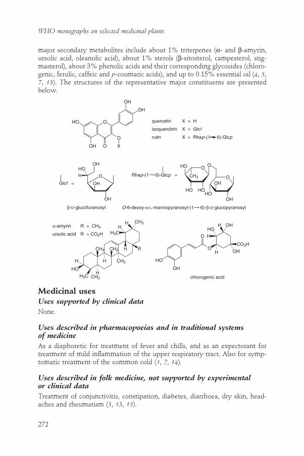
major secondary metabolites include about 1% triterpenes (a- and b-amyrin,ursolic acid, oleanolic acid), about 1% sterols (b-sitosterol, campesterol, stig-masterol), about 3% phenolic acids and their corresponding glycosides (chloro-genic, ferulic, caffeic and p-coumaric acids), and up to 0.15% essential oil (4, 5,7, 13). The structures of the representative major constituents are presentedbelow.
WHO monographs on selected medicinal plants
272
O
O
OH
OH
OOH
HO
X
quercetin
isoquercitrin
rutin
X = H
X = Glcf
X = Rhap-(1 6)-Glcp
Medicinal usesUses supported by clinical dataNone.
Uses described in pharmacopoeias and in traditional systems of medicineAs a diaphoretic for treatment of fever and chills, and as an expectorant fortreatment of mild inflammation of the upper respiratory tract. Also for symp-tomatic treatment of the common cold (1, 7, 14).
Uses described in folk medicine, not supported by experimental or clinical dataTreatment of conjunctivitis, constipation, diabetes, diarrhoea, dry skin, head-aches and rheumatism (5, 13, 15).
OH
OH
Glcf =
β-D-glucofuranosyl
O OOH
OHHO
OO
HOHO
CH3
HO
Glcp =Rhap-(1 6)-
O-6-deoxy-α-L-mannopyranosyl-(1 6)-b-D-glucopyranosyl
OHHOH
HO
H3C
H
CH3H
H CH3
CH3 CH3 H R
H3C
HH
CH3
O
O
OH
HO
CO2H
OH
HO
H
H OH
H
a-amyrin
ursolic acid
R = CH3
R = CO2H
chlorogenic acid
OH
OH
Glcf =
O
OHHO
H

PharmacologyExperimental pharmacologyAnti-inflammatory activityAn 80% ethanol extract of Flos Sambuci had moderate anti-inflammatory activity in rats: it inhibited carrageenan-induced footpad oedema by 27%. Theextract was administered intragastrically (100mg/kg body weight) 1 hour priorto administration of carrageenan. The control drug, indometacin (5mg/kg bodyweight) inhibited carrageenan-induced footpad oedema by 45% (16). Intra-peritoneal administration of an unsaponifiable fraction of the flowers to micemoderately enhanced phagocytosis at a dose of 0.5ml/animal (17). A 100%methanol extract of the flowers inhibited the biosynthesis of the inflammatorycytokines interleukin-1a, interleukin-1b and tumour necrosis factor-a at a con-centration of 30mg/ml in human peripheral mononuclear cells in vitro (18).
Diuretic activityIntragastric administration of an infusion of the flowers (20ml/kg body weight)or of a potassium- and flavonoid-rich extract of the flowers had a diuretic effectin rats which was greater than that observed with theophylline (5mg/kg bodyweight) (19).
Clinical pharmacologyDiaphoretic activityFlos Sambuci is reported to increase the response of the sweat glands to heatstimuli (7, 20, 21), and increase diaphoresis in healthy subjects (7, 21).
ContraindicationsNo information available.
WarningsNo information available.
PrecautionsNo information available on general precautions or precautions concerning druginteractions; drug and laboratory test interactions; carcinogenesis, mutagenesis,impairment of fertility; teratogenic and non-teratogenic effects in pregnancy;nursing mothers; or paediatric use. Therefore, Flos Sambuci should not be admin-istered during pregnancy or lactation or to children without medical supervision.
Adverse reactionsNo information available.
Flos Sambuci
273

Dosage formsCrude drug for decoctions and infusions (crude drug also available in tea bags);as a component of multi-ingredient products (7). Store in a well-closed con-tainer, protected from light (2).
Posology(Unless otherwise indicated)Daily dosage: crude drug 3–5g as an infusion (preferably taken hot) three timesdaily; 25% ethanol extract 3–5ml; tincture (1 :5 in 25% ethanol) 10–25ml (22).
References1. British herbal pharmacopoeia. London, British Herbal Medicine Association, 1996.2. European pharmacopoeia, 3rd ed., Suppl. 2000. Strasbourg, Council of Europe, 1999.3. Pharmacopoeia Hungarica, 7th ed. Budapest, Hungarian Pharmacopoeia Commission,
Medicina Konyvkiado, 1986.4. Blaschek W et al., eds. Hagers Handbuch der pharmazeutischen Praxis. Folgeband 2:
Drogen A–K, 5th ed. Berlin, Springer-Verlag, 1998.5. Farnsworth NR, ed. NAPRALERT database. University of Illinois at Chicago, IL,
February 9, 1998 production (an on-line database available directly through the Uni-versity of Illinois at Chicago or through the Scientific and Technical Network [STN]of Chemical Abstracts Services).
6. Youngken HW. Textbook of pharmacognosy, 6th ed. Philadelphia, PA, Blakiston, 1950.
7. Bisset NG. Herbal drugs and phytopharmaceuticals. Boca Raton, FL, CRC Press, 1994.8. Zargari A. Medicinal plants. Vol. 2, 3rd ed. Teheran, Teheran University Publication,
1982.9. Pharmacopoea helvetica, 8th ed. Berne, Département fédéral de l’intérieur, 1997.
10. Quality control methods for medicinal plant materials. Geneva, World Health Organiza-tion, 1998.
11. European pharmacopoeia, 3rd ed. Strasbourg, Council of Europe, 1996.12. Guidelines for predicting dietary intake of pesticide residues, 2nd rev. ed. Geneva, World
Health Organization, 1997 (document WHO/FSF/FOS/97.7).13. Newall CA, Anderson LA, Phillipson JD. Herbal medicines, a guide for healthcare pro-
fessionals. London, Pharmaceutical Press, 1996.14. Blumenthal M et al., eds. The complete German Commission E monographs. Austin, TX,
American Botanical Council, 1998.15. A nationwide compilation of traditional Chinese medicine herbs, 1st ed. Beijing, People’s
Health Publishing House, 1975.16. Mascolo N et al. Biological screening of Italian medicinal plants for anti-
inflammatory activity. Phytotherapy Research, 1987, 1:28–31.17. Delaveau P, Lallouette P, Tessier AM. Stimulation of the phagocytic activity of the
reticuloendothelial system by plant extracts. Planta Medica, 1980, 40:49–54.18. Yesilada E et al. Inhibitory effects of Turkish folk remedies on inflammatory
cytokines: interleukin-1a, interleukin-1b, and tumor necrosis factor a. Journal ofEthnopharmacology, 1997, 58:59–73.
19. Rebuelta M et al. Étude de l’effet diurétique de différentes préparations des fleursdu Sambucus nigra L. Plantes médicinales et Phytothérapie, 1983, 17:173–181.
20. Schmersahl KJ. Über die Wirkstoffe der diaphoretischen Drogen des DAB 6. Naturwissenschaften, 1964, 51:361.
WHO monographs on selected medicinal plants
274

21. Wiechowski W. Die Bedeutung der schweisstreibenden Tees. Medizinische Klinik,1927, 23:590–592.
22. Bradley PR, ed. British herbal compendium. Vol. I. Bournemouth, British Herbal Medicine Association, 1992.
Flos Sambuci
275

276
DefinitionRadix Senegae consists of the dried roots and root crowns of Polygala senegaL., Polygala senega L. var. latifolia Torrey et Gray, or other closely related Poly-gala species (Polygalaceae) (1–3).
SynonymsPolygala senegum L. (1), P. rosea Steud., Senega officinalis Spach (4).
Selected vernacular namesBambara, bulughâ lon, gizr uththuban, Klapperschlangenwurzel, mountain flax,peuhl, polygala de virginie, racine de polygala, racine de senega, Radix poly-galae, Radix polygalae senegae, rattlesnake root, seneca snakeroot, Senega-kreuzblume, senega root, senega snakeroot, Senegawurzel, snake root, szenegagyökér, tsuknida, vahulill, virginische Schlangenwurzel, yoruba (1–3, 5–7).
Geographical distributionIndigenous to eastern Canada and north-eastern United States of America (6–8).
DescriptionA perennial herbaceous plant with numerous stems sprouting from a singlethick gnarled crown arising from a conical, twisted, branched yellow root.Aerial portion consists of several erect or ascending, smooth stems up to 15–40cm high, bearing alternate, lanceolate or oblong-lanceolate leaves with ser-rulate margins. Inflorescence a spike of small, white flowers, which are almostsessile with rounded-obovate wings, concave with a short crested carina (1, 7).
Plant material of interest: dried roots and root crownsGeneral appearanceRoot crown greyish-brown, wider than the root; diameter of the root crownup to 3cm, gradually tapering to the tip; surface transversely and longitudinallystriated, often shows a more or less distinct decurrent, elongated spiral keel.Forms an irregular head consisting of numerous remains of stems and tightly
Radix Senegae

packed purplish-brown to red buds. Taproot, 0.5–1.5cm in diameter and 3–20cm in length, brown to yellow, occasionally branched, sometimes flexuous,usually without secondary roots, except in the Japanese varieties and species,which contain numerous fibrous branched rootlets. Fracture short and showsa yellowish cortex of varying thickness surrounding a pale central woody areasomewhat circular or irregular in shape, depending on the species (1–3).
Organoleptic propertiesOdour: characteristic, faint, sweet, slightly rancid or reminiscent of methyl salicylate, sternutatory when in powder form; taste: sweet, subsequently acridand irritating to the throat (1–3).
Microscopic characteristicsCork layer consisting of several rows of light-brown cork cells; secondarycortex composed of parenchyma cells and sieve tubes, traversed by medullaryrays, 1–3 cells wide. Phelloderm of slightly collenchymatous cells containingdroplets of oil. Phloem and xylem arrangement usually normal, especially nearthe crown, but where a keel is present, it is formed by increased developmentof phloem; other anomalous secondary development sometimes occurs, result-ing in formation of 1 or 2 large wedge-shaped rays in phloem and xylem, theparenchymatous cells of which contain droplets of oil. Xylem usually central,consists of vessels up to 60mm in diameter associated with numerous thin-walled tracheids and a few small lignified parenchymatous cells. Starch grainsand calcium oxalate crystals absent (1–3).
Powdered plant materialLight brown. Longitudinal fragments of lignified tissue made up of pitted tracheids and somewhat larger vessels with numerous bordered pits or withreticulate thickening; yellowish parenchyma and collenchymatous cells con-taining droplets of oil; occasional fragments of cork and epidermal tissue withstomata and unicellular trichomes from bud scales. Calcium oxalate crystalsand stone cells are absent (1–3).
General identity testsMacroscopic and microscopic examinations (1–3), chemical tests and froth for-mation (1, 2), and thin-layer chromatography for the presence of saponins (3).
Purity testsMicrobiologicalTests for specific microorganisms and microbial contamination limits are asdescribed in the WHO guidelines on quality control methods for medicinalplants (9).
Radix Senegae
277

Foreign organic matterNot more than 2% stems and not more than 1% other foreign matter (2).
Total ashNot more than 6% (3).
Acid-insoluble ashNot more than 2% (1, 2).
Alcohol-soluble extractiveNot less than 30% in 20% alcohol (2).
Loss on dryingNot more than 13% (2).
Pesticide residuesThe recommended maximum limit of aldrin and dieldrin is not more than 0.05mg/kg (3). For other pesticides, see the European pharmacopoeia (3) and theWHO guidelines on quality control methods for medicinal plants (9) and pesticide residues (10).
Heavy metalsFor maximum limits and analysis of heavy metals, consult the WHO guidelineson quality control methods for medicinal plants (9).
Radioactive residuesWhere applicable, consult the WHO guidelines on quality control methods formedicinal plants (9) for the analysis of radioactive isotopes.
Other purity testsChemical, sulfated ash and water-soluble extractive tests to be established inaccordance with national requirements.
Chemical assaysQuantitative analysis of triterpene saponins by high-performance liquid chro-matography (11).
Major chemical constituentsMethyl salicylate (0.1–0.3%), the compound responsible for the characteristicodour of the drug (12). The major reported biologically active constituents are triterpene saponins (6–16%) (6, 8, 13). The saponins are 3-glucosides of
WHO monographs on selected medicinal plants
278

Radix Senegae
279
presenegenin, which also contain at C-28 an oligosaccharide chain that has afucose moiety esterified with 3,4-dimethoxycinnamic or 4-methoxycinnamicacid (14–16). The structures of the representative saponins are presented below.
(E )-senegasaponin A
(Z )-senegasaponin A
desacylsenegasaponin A
(E )-senegasaponin B
(Z )-senegasaponin B
senegin II
(Z )-senegin II
desacylsenegin II
(E )-senegin III
(Z )-senegin III
desacylsenegin III
R1
H
H
H
H
H
H
H
H
Rha
Rha
Rha
R3
Api
Api
Api
H
H
H
H
H
H
H
H
R2
E-MC
Z-MC
H
E-MC
Z-MC
E-DMC
Z-DMC
H
E-MC
Z-MC
H
Medicinal usesUses supported by clinical dataNone.
Uses described in pharmacopoeias and in traditional systems of medicineAs an expectorant for symptomatic treatment of coughs due to bronchitis,emphysema and catarrh of the upper respiratory tract (1, 6, 14, 17–19).
Uses described in folk medicine, not supported by experimental or clinical dataTreatment of amenorrhoea, asthma, constipation, rheumatism and snake bites(5).
OH
Api =
D-apio-b-D-furanosyl
OH
OHO
O
OH
HO
CH3
HO
Rha =
6-deoxy-a-L-mannopyranosyl
H3C CO2H
CH3H3C
CH3 CH3
H
H
O O
O
OH
HO
H
H
H
O
OH
HO
HO
OH
O
O
CH3
O
O
O
OH
OH
HO
HOOO
OH
O
OH
O
OHOR3
CH3
R1
R2
C
H3CO
R
O
H3CO
R
COE-MC
R = OCH3
R = H
E-DMC
Z-MC
R = OCH3
R = H
Z-DMC
Rha =
O
OH
HO
HO
CH3

PharmacologyExperimental pharmacologyExpectorant activityIntragastric administration of a fluidextract of Radix Senegae (0.1–10ml/kgbody weight) enhanced the production of respiratory tract fluid in decerebrateor anaesthetized animals. Three to four hours after administration, the outputof respiratory tract fluid increased by up to 173% in cats and 186% in guinea-pigs, but no effect was observed in rabbits (20). In another study, administra-tion of a syrup of the root to anaesthetized dogs significantly increased the volume of respiratory tract fluid within 5–30 minutes (P < 0.001); after 2 hours,the fluid volume in the treatment group was 0.114ml as compared with 0.01ml in control animals treated with saline (21). Intragastric administrationof a 50% methanol extract of the root (2g/kg body weight) inhibited stress-induced gastric ulcers in rats by 98.5% (22). Intragastric administration of anaqueous suspension of a 50% methanol extract of the root (2g/kg body weight)to rats reduced congestive oedema by 62% and significantly increased the 24-hour urine volume as compared with control animals (P < 0.01) (23).
Effect on blood cholesterol and triglyceride levelsIntraperitoneal administration of an n-butanol extract of the root (5mg/kg bodyweight) reduced blood triglyceride levels in mice fed a normal diet, and reducedblood cholesterol and triglyceride levels in mice fed a high cholesterol diet (24).
Antihyperglycaemic activityIntraperitoneal administration of an n-butanol extract of the roots (10mg/kgbody weight) reduced blood glucose levels in healthy mice and in mice withstreptozocin-induced hyperglycaemia (25). Intragastric administration of asaponin fraction of a root extract reduced glucose-induced hyperglycaemia inrats at a dose of 200mg/kg body weight (16). Intraperitoneal administration of a saponin fraction of a root extract (25mg/kg body weight) significantlyincreased the plasma levels of adrenocorticotropic hormone, cortisone andglucose in rats (P < 0.01) (26). Intragastric administration of a 100% methanolextract of the root decreased the absorption of ethanol in rats (500mg/kg bodyweight) (15).
ToxicityThe LD50 of the root was 17g/kg body weight after intragastric administrationto mice. The LD50 of the root bark was 10g/kg body weight and that of theroot core (which had the lowest saponin concentration of the three rootsamples) was 75g/kg body weight (13).
WHO monographs on selected medicinal plants
280

Clinical pharmacologyExpectorant activityThe expectorant activity of the crude drug is due to the constituent saponinswhich produce local irritation of the mucous membranes of the throat and respiratory tract. This irritation stimulates an increase in bronchial secre-tions, thereby diluting the mucus, reducing its viscosity and facilitating ex-pectoration (19–21, 27, 28). Saponins may also reduce the surface tension ofmucus, thus reducing its viscosity (29). Oral administration of a fluidextract of the root was shown to reduce the viscosity of mucus in patients withbronchiectasis (17).
ContraindicationsPregnancy (See Precautions).
WarningsIf coughing persists for more than 7 days, seek medical advice. Radix Senegaemay exacerbate existing gastrointestinal inflammations such as gastritis orgastric ulcers, and excessive doses may cause vomiting (30).
PrecautionsCarcinogenesis, mutagenesis, impairment of fertilityNo mutagenic effects of an aqueous or 50% methanol extract of the root wereobserved in the Bacillus subtilis recombination assay or in the microsome rever-sion assay in Salmonella typhimurium strains TA98 and TA100 (31).
Pregnancy: teratogenic effectsSee Contraindications.
Pregnancy: non-teratogenic effectsTraditional uses for Radix Senegae include its use as an emmenagogue (5). Asextracts of the root have been shown to stimulate uterine contractions in animalmodels (32), Radix Senegae should not be taken during pregnancy.
Other precautionsNo information available on general precautions or precautions concerning drug interactions; drug and laboratory test interactions; nursing mothers; or
Radix Senegae
281

paediatric use. Therefore, Radix Senegae should not be administered during lactation or to children without medical supervision.
Adverse reactionsOverdose with Radix Senegae preparations may cause nausea, diarrhoea andvomiting due to gastrointestinal upset (13). In sensitive individuals, gastro-intestinal upset may occur even at the therapeutic dosage (33, 34).
Dosage formsChopped crude drug for decoctions and extracts (6, 18). Store in a tightly closedcontainer, protected from light and humidity (3).
Posology(Unless otherwise indicated)Daily dosage: 1.5–3.0g crude drug as an infusion or decoction in divided doses(18, 35). A 60% ethanol extract (made slightly alkaline with dilute ammonia):0.9–3ml; tincture: 2.5–7.5g. Equivalent preparations (18).
References1. African pharmacopoeia. Vol. 1, 1st ed. Lagos, Organization of African Unity, Scientific
Technical & Research Commission, 1985.2. The Japanese pharmacopoeia, 13th ed. (English version). Tokyo, Ministry of Health
and Welfare, 1996.3. European pharmacopoeia, 3rd ed. Strasbourg, Council of Europe, 1996.4. Hooker JD, Jackson BD. Index Kewensis. Vol. 1. Oxford, Clarendon Press, 1895.5. Farnsworth NR, ed. NAPRALERT database. Chicago, University of Illinois at
Chicago, IL, February 9, 1998 production (an online database available directlythrough the University of Illinois at Chicago or through the Scientific and Techni-cal Network [STN] of Chemical Abstracts Services).
6. Bisset NG. Herbal drugs and phytopharmaceuticals. Boca Raton, FL, CRC Press, 1994.
7. Youngken HW. Textbook of pharmacognosy, 6th ed. Philadelphia, PA, Blakiston, 1950.
8. Bruneton J. Pharmacognosy, phytochemistry, medicinal plants. Paris, Lavoisier, 1995.9. Quality control methods for medicinal plant materials. Geneva, World Health Organiza-
tion, 1998.10. Guidelines for predicting dietary intake of pesticide residues, 2nd rev. ed. Geneva,
World Health Organization, 1997 (document WHO/FSF/FOS/97.7).11. Kanazawa H et al. Determination of acidic saponins in crude drugs by high-
performance liquid chromatography on octadecylsilyl porous glass. Journal of Chro-matography, 1993, 630:408–414.
12. Hayashi S, Kameoka H. Volatile compounds of Polygala senega L. var. latifolia Torreyet Gray roots. Flavour and Fragrance Journal, 1995, 10:273–280.
WHO monographs on selected medicinal plants
282

Radix Senegae
283
13. De Smet PAGM. Polygala species. In: De Smet PAGM et al., eds. Adverse effects ofherbal drugs. Vol. 2. Berlin, Springer-Verlag, 1993.
14. Samuelsson G. Drugs of natural origin, a textbook of pharmacognosy. Stockholm,Swedish Pharmaceutical Press, 1992.
15. Yoshikawa M et al. E-Senegasaponins A and B, Z-senegasaponins A and B, Z-senegins II and III, new type inhibitors of ethanol absorption in rats from Senegaeradix, the roots of Polygala senega L. var. latifolia Torrey et Gray. Chemical and Phar-maceutical Bulletin, 1995, 43:350–352.
16. Yoshikawa M et al. Bioactive saponins and glycosides. II. Senegae radix. (2): Chem-ical structures, hypoglycemic activity, and ethanol absorption-inhibitory effect of E-senegasaponin C, Z-senegasaponin C, and Z-senegins II, III and IV. Chemical andPharmaceutical Bulletin, 1996, 44:1305–1313.
17. Basch FP, Holinger P, Poncher HG. Physical and chemical properties of sputum. II.Influence of drugs, steam, carbon dioxide and oxygen. American Journal of Diseasesof Childhood, 1941, 62:1149–1171.
18. Blumenthal M et al., eds. The complete German Commission E monographs. Austin, TX,American Botanical Council, 1998.
19. Reynolds JEF, ed. Martindale, the extra pharmacopoeia, 30th ed. London, Pharmaceu-tical Press, 1996.
20. Boyd EM, Palmer ME. Effect of Quillaja, Senega, Grindelia, Sanguinaria, Chionanthusand Dioscorea upon the output of respiratory tract fluid. Acta Pharmacologia Toxicolo-gia, 1946, 2:235–239.
21. Misawa M, Yanaura S. Continuous determination of tracheobronchial secretory activity in dogs. Japanese Journal of Pharmacology, 1980, 30:221–229.
22. Yamahara J et al. Biological active principles of the crude drugs. II. Antiulcerogenicand anti-inflammatory actions of the crude drugs containing saponin. YakugakuZasshi, 1975, 95:1179–1182.
23. Yamahara J et al. Effects of crude drugs on congestive edema. Chemical and Phar-maceutical Bulletin, 1979, 27:1464–1468.
24. Masuda H et al. Intraperitoneal administration of Senegae Radix extract and its maincomponent, senegin-II, affects lipid metabolism in normal and hyperlipidemic mice.Biological and Pharmaceutical Bulletin, 1996, 19:315–317.
25. Kato M et al. Hypoglycemic effect of the rhizomes of Polygala senega in normal anddiabetic mice and its main component, the triterpenoid glycoside senegin-II. PlantaMedica, 1996, 62:440–443.
26. Yokoyama H et al. Effects of total saponins extracted from several crude drugs on rat adrenocortical hormone secretion. Yakugaku Zasshi, 1982, 102:555–559.
27. Boyd EM. Expectorants and respiratory tract fluid. Journal of Pharmacy and Pharma-cology, 1954, 6:521–542.
28. ESCOP monographs on the medicinal uses of plant drugs. Fascicule 3. Devon, EuropeanScientific Cooperative on Phytotherapy, 1997.
29. Hostettmann K, Marston A. Saponins. Cambridge, Cambridge University Press,1995.
30. Newall CA, Anderson LA, Phillipson JD. Herbal medicines, a guide for healthcare pro-fessionals. London, Pharmaceutical Press, 1996.
31. Morimoto I et al. Mutagenicity screening of crude drugs with Bacillus subtilisrec-assay and Salmonella/microsome reversion assay. Mutation Research, 1982, 97:81–102.
32. Goto M et al. Uterus-contracting ingredients in plants. Takeda Kenkyusho Nempo,1957, 16:21.
33. Briggs CJ. Senega snakeroot—a traditional Canadian herbal medicine. CanadianPharmaceutical Journal, 1988, 121:199–201.

34. Wichtl M. Senegawurzel. In: Wichtl M, ed. Teedrogen. Ein Handbuch für die Praxis auf wissenschaftlicher Grundlage. 2. Auflage. Stuttgart, Wissenschaftliche Verlagsge-sellschaft, 1989.
35. Bradley PR, ed. British herbal compendium. Vol. 1. Bournemouth, British Herbal Medicine Association, 1992.
WHO monographs on selected medicinal plants
284

DefinitionFructus Serenoae Repentis consists of the dried ripe fruits of Serenoa repens(Bartr.) Small. (Arecaceae) (1–3).
SynonymsBrahea serrulata (Michx.) H. Wendl., Chamaerops serrulata Michx., Corypha repensBartr., Sabal serrulata (Michx.) Nichols, Sabal serrulata (Michx.) Nuttall. exSchult., Serenoa serrulata Hook., Serenoa serrulata Roem. et Schult., Serenoa serrulatum (Michx.) Benth et Hook, Serenoa serrulatum Schult. (1, 3–5).
Selected vernacular namesAmerican dwarf palm tree, dwarf palm tree, dwarf palmetto, fan palm, sabal,sabal fructus, Sägepalmenfrüchte, saw palmetto, saw palmetto berries, serenoa(1, 2, 5, 6).
Geographical distributionIndigenous to the south-east of the United States of America, from South Carolina to Florida (2, 6).
DescriptionLow scrubby palm growing in sandy soil, with characteristic creeping rhizome,one end of which rises a short distance above ground, surrounded by a densecrown of leaves with saw-like margins. Petioles slender and spinose on edges;blade fan-shaped, with palmate divisions that are slightly cleft at the summit.Inflorescence densely tomentose and shorter than the leaves. Fruit a 1-seededdrupe (6).
Plant material of interest: dried ripe fruitsGeneral appearanceDrupe superior, ellipsoidal, ovoid or somewhat globular, 1.5–3.0cm long, 1.0–1.5cm in diameter; dark brown to black with a smooth, dull surface, somewhatoily, with a few large, angular depressions and ridges due to contraction of the
285
Fructus Serenoae Repentis

inner layer on drying; summit marked by remains of style; base marked bystem-scars or has remains of stem. Epicarp and sarcocarp together form a thincoriaceous shell enclosing a hard but thin endocarp; endocarp externallyreddish-brown and somewhat fibrous, as is inner layer of the sarcocarp; innerlayer of endocarp smooth, enclosing an ellipsoidal or ovoid, hard somewhatflattened, anatropous, reddish-brown seed marked on the raphe side by anarillus-like appendage and marked on the opposite side near the end by themicropyle, which forms a slight projection; has a large endosperm of thick-walled parenchyma and a very small embryo at the micropyle (2, 3, 6).
Organoleptic propertiesOdour: pronounced, aromatic, fruity; taste: sweetish, aromatic, slightly acrid (6).
Microscopic characteristicsSarcocarp covered by a small-celled, thin-walled epidermis. Outermost layersof pulp wall contain yellowish-brown or brownish-red substances; inner layershave scattered single cells containing brown substances; occasional large, thick-walled, punctate stone cells with wide lumens. Innermost layer of sarcocarpwall consists almost completely of thick-walled, punctate, irregularly shapedstone cells. Outer layer of the seed coat consists of thick-walled large cells; cellsin middle layer smaller and thin-walled; cells of innermost layer small and flat-tened; contents of all seed-coat cells non-punctate brown. Outer endospermcells radially elongated, coarse-walled and inner cells larger, thick-walled andcoarsely punctate. Vascular bundles accompanied by fibres with stigmata whichhave siliceous solids attached (3, 6).
Powdered plant materialYellowish-brown. Fragments of sarcocarp, the cells of which contain yellowish-brown or brownish-red amorphous substances; whitish fragments ofendosperm, the cell walls considerably thickened and with large pores; occa-sional stone cells, nearly colourless, more or less tabular or irregular in shape,up to 140mm in length, with walls about 15mm thick, showing numeroussimple or branching pores (2, 6).
General identity testsMacroscopic and microscopic examinations (2, 3, 6), and thin-layer chro-matography (7).
Purity testsMicrobiologicalTests for specific microorganisms and microbial contamination limits are asdescribed in the WHO guidelines on quality control methods for medicinalplants (8).
WHO monographs on selected medicinal plants
286

Foreign organic matterNot more than 2% (2, 3).
Total ashNot more than 5% (3).
Acid-insoluble ashNot more than 1% (2, 3).
Water-soluble extractiveNot less than 8% (2, 3).
Loss on dryingNot more than 12% (3).
Pesticide residuesThe recommended maximum limit of aldrin and dieldrin is not more than 0.05mg/kg (9). For other pesticides, see the European pharmacopoeia (9), and the WHO guidelines on quality control methods for medicinal plants (8) andpesticide residues (10).
Heavy metalsFor maximum limits and analysis of heavy metals, consult the WHO guidelineson quality control methods for medicinal plants (8).
Radioactive residuesWhere applicable, consult the WHO guidelines on quality control methods formedicinal plants (8) for the analysis of radioactive isotopes.
Other purity testsChemical, sulfated ash and alcohol-soluble extractive tests to be established inaccordance with national requirements.
Chemical assaysQuantitation of fatty acids, both free and as their corresponding ethyl esters, by gas chromatography. The total fatty acid content is not less than9.0%, and the amounts of individual fatty acids are not less than 3.0% oleic,2.0% lauric, 1.2% myristic, 1.1% palmitic, 0.4% linoleic, 0.2% caphoic, 0.2%caprylic, 0.2% capric, 0.1% palmitoleic, 0.1% stearic and 0.1% linolenic acids (3).
Fructus Serenoae Repentis
287

Major chemical constituentsThe major constituents are free fatty acids and their corresponding ethyl esters;sterols and lipids. The primary fatty acid constituents include oleic, lauric,myristic, palmitic, linoleic, caphoic, caprylic, capric, palmitoleic, stearic andlinolenic acids (5, 11, 12). The major sterols include b-sitosterol, stigmasteroland daucosterol (13). The lipids consist of triglycerides of fatty acids. The struc-tures of some of the free fatty acids are presented below.
WHO monographs on selected medicinal plants
288
lauric acid
linoleic acid
linolenic acid
H3CCO2H
H3CCO2H
CO2HH3C
Medicinal usesUses supported by clinical dataTreatment of lower urinary tract symptoms (nocturia, polyuria, urinary reten-tion) secondary to BPH stages I and II, as defined by Alken (1, 4, 14–33), incases where diagnosis of prostate cancer is negative.
Uses described in pharmacopoeias and in traditional systems of medicineAs a diuretic and to treat an enlarged prostate (2).
Uses described in folk medicine, not supported by experimental or clinical dataAs an aphrodisiac, a sedative and a nutritional tonic, as well as for the treatmentof bronchitis, cystitis, dysmenorrhoea, sore throat and the common cold (5).
PharmacologyExperimental pharmacologyAntispasmodic activityBoth lipid and saponifiable fractions of Fructus Serenoae Repentis reduced norepinephrine-induced contractions in vitro of rat aorta (IC50 0.53 and 0.50mg/ml, respectively), as well as potassium chloride-induced contractionsof rat uterus (EC50 0.35 and 0.43mg/ml, respectively) (34). A 90% ethanolextract of the fruit reduced vanadate-induced contractions of the rat uterus(EC50 11.41mg/ml). Norepinephrine-induced contractions of rat deferential duct,and potassium chloride-induced contractions of guinea-pig ileum and bladdersmooth muscle tissue were reduced by the addition of a 90% ethanol extractof the fruit (0.33 and 0.15mg/ml, respectively) (35).

Anti-inflammatory activityIntragastric administration of an ethanol extract of the fruit to rats (5.0g/kgbody weight) inhibited carrageenan-induced footpad oedema (36). Externalapplication of a 90% ethanol extract of the fruit (500 mg) to mice inhibitedcroton oil-induced ear oedema by 42% (37).
Intragastric administration of an n-hexane extract of the fruit to rats (10ml/kgbody weight) decreased capillary permeability induced by histamine, com-pound 48/80 and dextran, and generalized oedema induced by dextran (38). Acarbon dioxide (supercritical) extract of the fruit inhibited cyclooxygenase and5-lipoxygenase in vitro (IC50 28.1 and 18.0mg/ml, respectively) (39). A lipido-sterolic extract of the fruit inhibited the in vitro production of leukotriene B4 inhuman polymorphonuclear neutrophils stimulated with the calcium ionophoreA23187 (40). An ethanol extract of the fruit also suppressed A23187-stimulatedsynthesis of leukotriene B4 (IC50 8.3mg/ml) and thromboxane B2 (IC50 15.4mg/ml)in rat peritoneal leukocytes in vitro (37).
Immunostimulatory activityIntraperitoneal administration of a polysaccharide fraction, isolated from anaqueous extract of the fruit, to mice (10mg/kg body weight) had immuno-stimulant activity, as measured by the colloidal carbon clearance test (41). Anincreased rate of phagocytosis by human polymorphonuclear leukocytes was observed in cells treated with a polysaccharide fraction of the extract (10mg/ml) (41).
Anti-gonadotropic effectsn-Hexane extracts of the fruit had anti-androgenic and anti-estrogenic activityin vitro (42–47). Dihydrotestosterone and testosterone uptake by cytosolicandrogen receptors of human foreskin and other tissues was inhibited by40.9% and 41.9%, respectively, after treatment of the tissues with the extract(42). In another study, the binding of [3H]dihydrotestosterone to both cytoso-lic and nuclear androgen receptors in cultured human foreskin fibroblasts wasinhibited by 90% and 70%, respectively, after treatment of the cells with asterol fraction of the n-hexane extract (IC50 7.1 units/ml) (43). An n-hexane fruitextract inhibited androgen binding to cytosolic androgen receptors of rat pro-static tissue in a specific and competitive manner (IC50 330.0–367.5mg/ml) (44,45). However, in contrast to these findings, the same extract did not inhibit the binding of [3H]dihydrotestosterone to androgen receptors in cultured hu-man foreskin fibroblasts (46). Oral administration of an n-hexane extract (160mg/day) inhibited the binding of 3H-labelled 17b-estradiol to the nuclearestrogen receptors in samples of prostatic tissue from patients with BPH.Binding to the cytosolic and nuclear estrogen and androgen receptors was mea-sured by saturation analysis and an enzyme-linked immunosorbent assay (47).
The effect of an n-hexane extract of the fruit was evaluated in two humanprostatic cell lines, LNCaP and PC3, which are respectively responsive and unre-
Fructus Serenoae Repentis
289

sponsive to androgen stimulation. The extract (100mg/ml) induced proliferationand differentiation in LNCaP cells, but not in PC3 cells, suggesting that the andro-gen receptor plays a role in mediating the effects of the fruit in LNCaP cells (48).In PC3 cells cotransfected with genes for wild-type androgen receptor and a chloramphenicol acetyltransferase reporter under the control of an androgen-responsive element, the extract (25mg/ml) inhibited androgen-induced chloram-phenicol acetyltransferase transcription by 70% (48).
n-Hexane, 90% ethanol and supercritical carbon dioxide extracts of the fruitinhibited 5a-reductase activity in vitro (37, 43, 46, 49–53). A lipidosterolicextract of the fruit (100mg/ml) inhibited 5a-reductase activity in the rat ventral prostate by 50%, and reduced conversion of testosterone into di-hydrotestosterone in human foreskin fibroblasts by 90%. The conversion of dihydrotestosterone to 5a-androstane-3a-17b-diol by 3a-ketosteroid oxido-reductase was also partially inhibited in cultured human foreskin fibroblasts(43). An n-hexane extract of the fruit inhibited the activity of both 5a-reductase and 17b-hydroxysteroid dehydrogenase in cultures of epithelial cells(IC50 60 and 40mg/ml, respectively) and fibroblast cells (IC50 30 and 200mg/ml,respectively) obtained from the prostates of patients with BPH (50). One studyreported no effect of several lipidosterolic extracts of the fruit on the activityof 5a-reductase from human prostate or on dihydrotestosterone binding to therat prostatic androgen receptors at concentrations up to 100mg/ml (51). Thereasons for these conflicting results are unclear, and may be due to the differ-ent methodologies used. Recently, it has been demonstrated that human 5a-reductase has two isoforms, type 1 and type 2; finasteride, a testosterone5a-reductase inhibitor has been shown to be a selective inhibitor of the type 2isoform (inhibitory concentration [Ki] 7.3nmol/l). Furthermore, an n-hexaneextract of the fruit was a non-competitive inhibitor of the type 1 isoform (IC50
7.2mg/ml) and an uncompetitive inhibitor of type 2 (IC50 4.9mg/ml) (52). A 90% ethanol extract of the fruit showed a dose-dependent inhibition of 5a-reductase activity in the epithelium (29% inhibition) and stroma (45% inhibi-tion) of prostate tissue from patients with BPH (52). When the extract wasfractionated into saponifiable, non-saponifiable and hydrophilic subfractions,only the saponifiable subfraction (consisting mainly of lauric, oleic, myristic and palmitic acids) was active. Of these fatty acids, lauric acid was the mostactive: it inhibited epithelial and stromal 5a-reductase activity by 51% and42%, respectively. The inhibition by lauric acid was noncompetitive and dose-dependent up to a concentration of 0.2mmol/l. The nonsaponifiable fraction,consisting mainly of phytosterols, was weakly active, while the hydrophilicsubfractions, containing carbohydrates, amino acids and polysaccharides, wereinactive (53). A supercritical extract of the fruit inhibited 5a-reductase activityin homogenates of cultured human foreskin fibroblasts (IC50 0.025mg/ml) (46).
One study compared testosterone metabolism in primary cultures of epithe-lial cells and fibroblasts obtained from the prostates of patients with BPH andprostate cancer. In all cultures, androst-4-ene-3,12-dione, formed by the oxida-tion of testosterone by 17b-hydroxysteroid dehydrogenase, accounted for 80%
WHO monographs on selected medicinal plants
290

of all metabolites recovered. An n-hexane extract of the fruit inhibited the for-mation of androst-4-ene-3,12-dione in both cell types, indicating that it inhib-ited the activity of 17b-hydroxysteroid dehydrogenase, unlike finasteride,which was inactive (50).
An increase in the activity of 3a-hydroxysteroid-oxidoreductase (the enzymethat metabolizes dihydrotestosterone into the inactive androstenediol form) inprostate tissue from patients with BPH was reported following treatment ofpatients with an n-hexane extract of the fruit (320mg daily for 3 months). Analy-sis of enzyme kinetics showed that the Vmax of 3-hydroxysteroid-oxidoreductasewas significantly enhanced in the prostate stroma of treated patients. Since 3-hydroxysteroid-oxidoreductase also has a strong substrate affinity forprostaglandins, increased activity of the enzyme may also increase the metabo-lism of prostaglandins, thereby accounting for the reduction of prostaglandin-mediated congestion or intraprostatic oedema formation (54).
Intragastric administration of an n-hexane extract of the fruit to castratedrats for 60–90 days inhibited the increase in total weight of the prostate inducedby estradiol and testosterone (55). Intragastric administration of a 90% ethanolextract to castrated rats (6ml/kg body weight, weekly for 8 weeks) inhibitedthe increase in weight of the ventral prostate, seminal vesicles and coagulationglands induced by testosterone (37). Intragastric administration of a 90%ethanol extract of the fruit inhibited prostate growth stimulated by both estra-diol and dihydrotestosterone in nude mice into which prostate tissue fromhumans with BPH had been transplanted (56). An n-hexane extract of the fruit(30mg/ml) inhibited the proliferation of human prostate cells induced by basicfibroblast growth factor. Lupenone, hexacosanol and an unsaponified fractionof the extract markedly inhibited the proliferation of human prostate cellsinduced by basic fibroblast growth factor, but had only a minimal effect onbasal cell proliferation (57).
Effects on signal transductionAddition of an n-hexane extract of the fruit (1–10mg/ml) to Chinese hamsterovary cells completely inhibited the effects of prolactin on potassium conduc-tance, protein kinase C activity and intracellular concentrations of calcium.These results suggest that the extract may inhibit prolactin-induced prostaticgrowth by interfering with the transduction signals involving the prolactinreceptor (58). Lipidosterolic extracts of the fruit noncompetitively inhibitedradioligand binding to human prostatic a1-adrenoceptors and agonist-induced[3H]inositol phosphate formation (59).
Clinical pharmacologyPlacebo-controlled clinical trialsEleven double-blind, placebo-controlled studies have assessed the effects of lipi-dosterolic extracts of Fructus Serenoae Repentis in the symptomatic treatmentof mild to moderate BPH (26–33, 60–62). The number of patients in each study
Fructus Serenoae Repentis
291

ranged from 22 to 205, and the dosage of the extract was generally 160mgtwice daily for 1–3 months. All but one study (61) reported that the extract wassignificantly more effective than placebo in reducing the symptoms of mild tomoderate BPH. In this study of 70 patients, which was also randomized,although a significant improvement in flow rate was seen in patients treatedwith either a hexane extract of the fruit (320mg) or placebo daily for 3 months,no significant difference between the treatment groups was observed (61).However, most studies demonstrated an increase in urinary flow rate and adecrease in postvoid residual urine volume (26). In another study which wasalso randomized, 205 patients were treated with 320mg extract or placebodaily for 3 months. The study concluded that the extract was superior toplacebo in reducing the total symptom score (polyuria, nocturia, dysuria, andurgency and hesitancy of micturition), improving the quality of life score, andincreasing urinary volume (62).
A study was performed on 176 patients with BPH who had been unre-sponsive to placebo treatment in previous clinical studies. After 30 days of treat-ment with an extract of the fruit (160mg, twice daily), there was a significantreduction in dysuria, polyuria and nocturia in the treated group as comparedwith the placebo group. Patients treated with the extract had a significantlygreater increase in mean peak urinary flow rate (28.9%), as compared with those that received the placebo (8.5%), and the overall efficacy of theextract was rated higher than that of the placebo by both patients and physicians (33).
Another double-blind, placebo-controlled study assessed the effect of a lipi-dosterolic extract in the reduction of prostate oedema and congestion in 18patients with BPH. Histopathological analysis of enucleated prostate tissuefrom patients treated preoperatively with the extract (320mg daily for 12weeks) showed a significant decrease in prostate stromal oedema and conges-tion in treated patients, as compared with those in the placebo group (P � 0.05)(63).
Controlled clinical trialsIn a controlled clinical trial, 25 men with symptoms of urinary obstruction wererandomized into two groups: 15 patients received no treatment, while 10 weretreated with an n-hexane extract of Fructus Serenoae Repentis (320mg extractdaily). After 3 months, prostatic specimens were removed by suprapubicprostatectomy and were sectioned into three regions (i.e. periurethral, subcap-sular and intermediate). In each region, the concentration of testosterone, dihydrotestosterone and epidermal growth factor was measured by radio-immunoassay. In the patients treated with the extract, a significant reduction (P < 0.001) in the concentration of dihydrotestosterone (50%) and epidermalgrowth factor (50%), and a significant increase (P < 0.001) in testosterone levels(125%), were observed in the periurethral region (64).
WHO monographs on selected medicinal plants
292

Clinical trials without controlsNumerous clinical studies without controls of men with BPH have reportedimprovements in both objective and subjective variables after treatment withlipidosterolic extracts of the fruit (15–28). The largest trial of 1334 patientstreated with 320mg of a lipidosterolic fruit extract daily for 6 months showedan improvement in postvoid residual urine volume (50% decrease), nocturia(54% decrease) and polyuria (37% decrease) (16). The results of a prospectivemulticentre study in which 435 patients with BPH were treated with a lipido-sterolic extract of the fruit (320mg daily for 3 years) showed a steady improvement in micturition. The improvement was due to a marked decreasein symptoms and postvoid residual urine volume (50% decrease), and anincrease in peak urinary flow rate (about 25%) (17). Another multicentre studyanalysed the effect of a lipidosterolic extract of the fruit (160mg twice daily for3 months) in 305 patients with mild to moderate BPH. After treatment,increases in maximal and mean urinary flow rates (of 25% and 27%, respec-tively) and a 35% improvement in the mean International Prostate SymptomScore were seen (18). Other studies have also reported improvements in symp-toms and objective measurements of disease severity after 1–6 months of treat-ment with a lipidosterolic extract of the fruit (320mg daily) (19–28). Generally,studies involving periodic evaluation over the course of treatment have demon-strated that improvements in both objective and subjective variables were pro-gressive over time (17, 24–27).
Comparative trialsAn n-hexane extract of Fructus Serenoae Repentis, finasteride and a1-receptorantagonists have been shown to be clinically effective in the treatment of BPHin comparative trials (16, 65–69). One large international randomized, double-blind clinical trial compared the efficacy of the extract (320mg daily) with thatof finasteride (5mg daily) in the treatment of 1098 patients with mild to mod-erate BPH. After 6 months of therapy, the International Prostate Symptom Scoredecreased from baseline by 37% in patients treated with the extract as com-pared with a decrease of 39% in patients who received finasteride. No signifi-cant difference was observed between the treatment groups in improvementof patient-rated quality of life scores and the primary end-point of objectivesymptom score. Both treatments resulted in improved peak urinary flow ratesand a reduction in the size of the prostate. Peak urinary flow rate increasedfrom 10.6ml/s to 13.3ml/s in patients treated with the extract, and from 10.8ml/s to 14.0ml/s in those who received finasteride. The size of the prostatewas reduced by 6% in patients treated with the extract, and by 18% in thosetreated with finasteride. Serum prostate-specific antigen levels were reduced by41% following finasteride treatment, but remained unchanged in patientstreated with the extract (16).
Fructus Serenoae Repentis
293

Other smaller, shorter, randomized double-blind trials involving groups of41–63 patients compared the efficacy of the fruit extract (320mg daily) withthe a1-receptor antagonists alfuzosin and prazosin (68, 69). In a 3-week com-parative trial with alfuzosin, the total mean symptom score using Boyarsky’srating scale improved by 27% and 39% in patients treated with the extract and alfuzosin, respectively. Although improvements in the peak urinary flowrates were greater in the alfuzosin-treated group, there was no significant dif-ference between the treatments (68). In a 12-week randomized trial comparingthe efficacy of a fruit extract (in 20 patients) and prazocin (in 21 patients),improvements in polyuria, mean urinary flow rate and postvoid residual urinevolume were similar in both groups, but no statistical analysis of the data wasprovided by the investigators (67). Further large, well-designed, randomizedtrials of long duration are necessary to compare adequately the clinical efficacyof Fructus Serenoae Repentis and a1-receptor antagonists.
Four reviews of the randomized controlled clinical trials have indicated thatlipidosterolic extracts of the fruit improve the symptoms of urinary tract disorders and urinary flow rates in men with mild to moderate BPH (14, 68–70).
PharmacokineticsThe pharmacokinetics of Fructus Serenoae Repentis were investigated in a bio-equivalence study that compared a new capsule formulation (320mg/capsule)to a reference preparation (160mg/capsule). Concentrations of the componentsof the extract were measured in plasma samples from 12 healthy fasting males (mean age 24 years) after oral administration of 320mg extract (eitherone capsule of 320mg or two capsules of 160mg) (71). However, the method-ology used in this study was questionable.
Tissue distribution was measured in rats after intragastric administration of a lipidosterolic extract supplemented with radiolabelled oleic acid, lauric acidor b-sitosterol. This investigation demonstrated that the uptake of the extract was much higher in the prostate than either the liver or genitourinary tissues(72).
ToxicityClinical studies have shown that extracts of Fructus Serenoae Repentis are verywell tolerated in humans (16, 69). Minor gastrointestinal side-effects have been reported in most of the clinical trials, but results from standard bloodchemistry tests were normal (69).
ContraindicationsOwing to its effects on androgen and estrogen metabolism, the use of FructusSerenoae Repentis during pregnancy or lactation and in children under the ageof 12 years is contraindicated.
WHO monographs on selected medicinal plants
294

WarningsFructus Serenoae Repentis relieves the symptoms associated with BPH, butdoes not have an effect on the size of the prostate. If symptoms worsen or donot improve, or in cases of blood in the urine or acute urinary retention, contacta physician (1).
PrecautionsPregnancy: teratogenic effectsSee Contraindications. There is no therapeutic rationale for the use of FructusSerenoae Repentis during pregnancy.
Pregnancy: non-teratogenic effectsSee Contraindications. There is no therapeutic rationale for the use of FructusSerenoae Repentis during pregnancy.
Nursing mothersSee Contraindications. There is no therapeutic rationale for the use of FructusSerenoae Repentis during lactation.
Paediatric useSee Contraindications.
Other precautionsNo information available on general precautions or precautions concerning druginteractions; drug and laboratory test interactions; or carcinogenesis, mutage-nesis and impairment of fertility.
Adverse reactionsBoth short- and long-term clinical studies have found that extracts of FructusSerenoae Repentis are very well tolerated. Occasional nausea, diarrhoea andother minor gastrointestinal complaints have been reported (18).
Dosage formsCrude drug, lipidosterolic extracts (n-hexane, 90% ethanol or fluid [carbondioxide] supercritical extracts standardized to contain 70–95% free fatty acidsand corresponding ethyl esters), and preparations thereof. Store in a tightlyclosed container in a cool, dry place.
Posology(Unless otherwise indicated)Daily dosage: 1–2g crude drug or 320mg (as a single dose or 160mg twice daily) of a lipidosterolic extract (n-hexane, 90% ethanol or supercritical
Fructus Serenoae Repentis
295

fluid [carbon dioxide] extract standardized to contain between 70 and 95% free fatty acids and corresponding ethyl esters) or equivalent preparations(16–33, 60–62).
References1. Blumenthal M et al., eds. The complete German Commission E monographs. Austin, TX,
American Botanical Council, 1998.2. British herbal pharmacopoeia. London, British Herbal Medicine Association, 1996.3. The United States pharmacopeia 24: national formulary 19. Rockville, MD, United States
Pharmacopeial Convention, 1999.4. Blaschek W et al., eds. Hagers Handbuch der pharmazeutischen Praxis. Folgeband 2:
Drogen A–K, 5th ed. Berlin, Springer-Verlag, 1998.5. Farnsworth NR ed. NAPRALERT database. Chicago, University of Illinois at Chicago,
IL, January 20, 1998 production (an online database available directly through theUniversity of Illinois at Chicago or through the Scientific and Technical Network[STN] of Chemical Abstracts Services).
6. Gathercoal EN, Wirth EH. Pharmacognosy. Philadelphia, Lea & Febiger, 1936.7. Hänsel R, Rimpler H, Schoepfliin G. Thin-layer chromatography of Sabal (saw pal-
metto) fruits. Planta Medica, 1964, 12:169–172.8. Quality control methods for medicinal plant materials. Geneva, World Health Organiza-
tion, 1998.9. European pharmacopoeia, 3rd ed. Strasbourg, Council of Europe, 1996.
10. Guidelines for predicting dietary intake of pesticide residues. Geneva, World Health Orga-nization, 1989.
11. De Swaef SI, Vlietinck AJ. Simultaneous quantitation of lauric acid and ethyl laureate in Sabal serrulata by capillary gas chromatography and derivatisation withtrimethyl sulphonium hydroxide. Journal of Chromatography, 1996, 719:479–482.
12. Wajda-Dubois JP et al. Comparative study on the lipid fraction of pulp and seedsof Serenoa repens (Palmaceae). Oleagineux Corps Gras Lipides, 1996, 3:136–139.
13. Hänsel R et al. Eine Dünnschichtchromatographische Untersuchung der Sabal-früchte. Planta Medica, 1964, 12:136–139.
14. Wilt TJ et al. Saw palmetto extracts for treatment of benign prostatic hyperplasia.A systematic review. Journal of the American Medical Association, 1998, 280:1604–1609.
15. Vahlensieck W et al. Benigne Prostatahyperplasie-Behandlung mit Sabalfrüchte-extrakt. Fortschritte der Medizin, 1993, 111:323–326.
16. Carraro JC et al. Comparison of phytotherapy (Permixon®) with finasteride in thetreatment of benign prostatic hyperplasia: a randomized international study of 1098patients. Prostate, 1996, 29:231–240.
17. Bach D, Ebeling L. Long-term treatment of benign prostatic hyperplasia—results ofa prospective 3-year multicenter study using Sabal extract IDS 89. Phytomedicine,1996, 3:105–111.
18. Braeckman J. The extract of Serenoa repens in the treatment of benign prostatichyperplasia: a multicenter open study. Current Therapeutic Research, 1994, 55:776–786.
19. Schneider HJ, Uysal A. Internationaler Prostata-Symptomenscore (I-PSS) im klinis-chen Alltag. Urologie [B], 1994, 34:443–447.
20. Braekman J et al. Efficacy and safety of the extract of Serenoa repens in the treatmentof benign prostatic hyperplasia: therapeutic equivalence between twice and oncedaily dosage forms. Phytotherapy Research, 1997, 11:558–563.
21. Derakhshani P et al. Beeinflussung des Internationalen Prostata-Symptomensoreunter der Therapie mit Sägepalmenfrüchteextrakt bei täglicher Einmalgabe. Urologie[B], 1997, 37:384–391.
WHO monographs on selected medicinal plants
296

22. Ziegler H, Hölscher U. Wirksamkeit des Spezialextraktes WS 1473 aus Sägepal-menfrüchteextrakt bei Patienten mit benigner Prostatahyperplasie im Stadium I–IInach Alken—offene Multicenter-Studie. Jatros Uro, 1998, 14:34–43.
23. Redecker KD, Funk P. Sabal-Extrakt WS 1473 bei benigner Prostatahyperplasie.Extracta Urologica, 1998, 21:23–25.
24. Hanus M, Matousková M. Alternativní lécba BPH — Permixon (Capistan). RozhledyChirurgia, 1993, 72:75–79.
25. Romics I et al. Experience in treating benign prostatic hypertrophy with Sabal ser-rulata for one year. Journal of International Urology and Nephrology, 1993, 25:565–569.
26. Boccafoschi C, Annoacia S. Confronto fra estratto di Serenoa repens e placebo medi-ante prova clinica controllata in pazienti co adenmatosi prostatica. Urologia, 1983,50:1–14.
27. Champault G et al. A double-blind trial of an extract of the plant Serenoa repensin benign prostatic hyperplasia. British Journal of Clinical Pharmacology, 1984,18:461–462.
28. Cukier C et al. Serenoa repens extract vs placebo. Comptes rendus de therapeutiques etde Pharmacologie clinique, 1985, 4:15–21.
29. Descotes JL et al. Placebo-controlled evaluation of the efficacy and tolerability ofPermixon® in benign prostatic hyperplasia after exclusion of placebo responders.Clinical Drug Investigations, 1995, 9:291–297.
30. Emili E et al. Clinical results on a new drug in the treatment of benign prostatichyperplasia (Permixon). Urologia, 1983, 50:1042–1049.
31. Tasca A et al. Treatment of obstruction in prostatic adenoma using an extract ofSerenoa repens. Double-blind clinical test vs placebo. Minerva Urologica e Nefrologica,1985, 37:87–91.
32. Mandressi A et al. Treatment of uncomplicated benign prostatic hypertrophy (BPH)by an extract of Serenoa repens: clinical results. Journal of Endocrinology Investigations,1987, 10 (Suppl. 2):49.
33. Descotes JL et al. Placebo-controlled evaluation of the efficacy and tolerability ofPermixon in benign prostatic hyperplasia after exclusion of placebo responders. Clinical Drug Investigations, 1995, 9:291–297.
34. Gutierrez M et al. Mechanisms involved in the spasmolytic effect of extracts fromSabal serrulata fruit on smooth muscle. General Pharmacology, 1996, 27:171–176.
35. Odenthal KP, Rauwald HW. Lipophilic extract from Sabal serrulata inhibits contrac-tions in smooth muscle tissue. Aktuelle Urologie, 1996, 27:152–157.
36. Hiermann A. About the contents of Sabal fruits and their anti-inflammatory effect.Archiv der Pharmazie (Weinheim), 1989, 322:111–114.
37. Koch E. Pharmakologie und Wirkmechanismen von Extrakten aus Sabalfrüchten(Sabal fructus), Brennesselwurzeln (Urticae radix) und Kürbissamen (Curcurbitaepeponis semen) bei der Behandlung der benignen Prostatahyperplasie. In: Loew D,Rietbrock N, eds. Phytopharmaka in Forschung und klinischer Anwendung. Darmstadt,Steinkopff, 1995:57–79.
38. Tarayre JP et al. Anti-edematous action of a hexane extract from Serenoa repens Bartr.drupes. Annales pharmaceutiques françaises, 1983, 41:559–570.
39. Breu W et al. Antiphlogistische Wirkung eines mit hyperkritischem Kohlendioxidgewonnenen Sabalfrucht-Extraktes. Arzneimittel-Forschung, 1992, 42:547–551.
40. Paubert-Braquet M et al. Effect of the lipidic lipidosterolic extract of Serenoa repens(Permixon®) on the ionophore A23187-stimulated production of leukotriene B4(LTB4) from human polymorphonuclear neutrophils. Prostaglandins, Leukotrienes andEssential Fatty Acids, 1997, 57:299–304.
41. Wagner H. Immunstimulierend wirkende Polysaccharide (Heteroglykane) aushöheren Pflanzen. Arzneimittel-Forschung, 1985, 35:1069–1075.
42. El-Sheikh MM, Dakkak MR, Saddique A. The effect of Permixon® on androgenreceptors. Acta Obstetrics and Gynecology of Scandinavia, 1988, 67:397–399.
Fructus Serenoae Repentis
297

43. Sultan C et al. Inhibition of androgen metabolism and binding by a liposterolicextract of “Serenoa repens B” in human foreskin fibroblasts. Journal of Steroid Bio-chemistry, 1984, 20:515–519.
44. Briley M, Carilla E, Fauran F. Permixon, a new treatment for benign prostatic hyper-plasia, acts directly at the cytosolic androgen receptor in rat prostate. British Journalof Pharmacology, 1983, 79:327.
45. Carilla E et al. Binding of Permixon, a new treatment for prostatic benign hyperpla-sia, to the cytosolic androgen receptor in rat prostate. Journal of Steroid Biochemistry,1984, 20:521–523.
46. Hagenlocher M et al. Specific inhibition of 5a-reductase by a new extract of Sabalserrulata. Aktuelle Urologie, 1993, 24:146–149.
47. Di Silverio F et al. Evidence that Serenoa repens extract displays an antiestrogenicactivity in prostatic tissue of benign prostatic hypertrophy patients. European Urology1992, 21:309–314.
48. Ravenna L et al. Effects of the lipidosterolic extract of Serenoa repens (Permixon®)on human prostatic cell lines. Prostate, 1996, 29:219–230.
49. Niederprüm HJ et al. Testosterone 5a-reductase inhibition by free fatty acids fromSabal serrulata fruits. Phytomedicine, 1994, 1:127–133.
50. Delos S et al. Testosterone metabolism in primary cultures of human prostateepithelial cells and fibroblasts. Journal of Steroid Biochemistry and Molecular Biology,1995, 55:375–383.
51. Rhodes L et al. Comparison of finasteride (Proscar®), a 5a-reductase inhibitor, andvarious commercial plant extracts in in vitro and in vivo 5a-reductase inhibition.Prostate, 1993, 22:43–51.
52. Iehle C et al. Human prostatic steroid 5a-reductase isoforms, a comparative studyof selective inhibitors. Journal of Steroid Biochemistry and Molecular Biology, 1995, 54:273–279.
53. Weisser H et al. Effects of Sabal serrulata extract IDS 89 and its subfractions on 5a-reductase activity in human benign prostatic hyperplasia. Prostate, 1996, 28:300–306.
54. Weisser H et al. Enzyme activities in tissue of human benign prostatic hyperplasia(BPH) after three months of treatment with Sabal serrulata extract IDS 89 (Strogen)or placebo. European Urology, 1997, 31:97–101.
55. Paubert-Braquet M et al. Effect of Serenoa repens extract (Permixon®) on estra-diol/testosterone-induced experimental prostate enlargement in the rat. Pharmaco-logical Research, 1996, 34:171–179.
56. Otto U et al. Transplantation of human benign hyperplastic prostate tissue into nudemice: first results of systemic therapy. Urologie Internationale, 1992, 48:167–170.
57. Paubert-Braquet M et al. Effect of the lipidosterolic extract of Serenoa repens(Permixon) and its major components on basic fibroblast growth factor-induced proliferation of cultures of human prostate biopsies. European Urology, 1998, 33:340–347.
58. Vacher P et al. The lipidosterolic extract from Serenoa repens interferes with prolactinreceptor signal transduction. Journal of Biomedical Sciences, 1995, 2:357–365.
59. Goepel M et al. Saw palmetto extracts potently and noncompetitively inhibit humana1-adrenoceptors in vitro. Prostate, 1999, 38:208–215.
60. Gabric V, Miskic H. Behandlung des benignen Prostataadenoms und der chroni-schen Prostatitis. Therapiewoche, 1987, 37:1775–1788.
61. Reese-Smith H et al. The value of Permixon in benign prostatic hypertrophy. BritishJournal of Urology, 1986, 58:36–40.
62. Braeckman J et al. A double-blind, placebo-controlled study of the plant extractSerenoa repens in the treatment of benign hyperplasia of the prostate. European Journalof Clinical Research, 1997, 9:247–259.
63. Helpap B et al. Morphology of benign prostatic hyperplasia after treatment withsabal extract IDS 89 or placebo. Journal of Urology and Pathology, 1995, 3:175–182.
WHO monographs on selected medicinal plants
298

64. Di Silverio F et al. Effects of long-term treatment with Serenoa repens (Permixon®)on the concentrations and regional distribution of androgens and epidermal growthfactor in benign prostatic hyperplasia. Prostate, 1998, 37:77–83.
65. Anderson JT. a1-Blockers vs 5a-reductase inhibitors in benign prostatic hyperplasia:a comparative review. Drugs and Aging, 1995, 6:388–396.
66. Grasso M et al. Comparative effects of alfuzosin versus Serenoa repens in the treat-ment of symptomatic benign prostatic hyperplasia. Archivos Espanoles de Urologia,1995, 48:97–103.
67. Adriazola Semino M et al. Tratamiento sintomatico de la hipertrofia benigna deprostata. Estudio comparativo entre Prazosin y Serenoa repens. Archivos Espanoles deUrologia, 1992, 45:211–213.
68. Bombardelli E, Morazzoni P. Serenoa repens (Bartram) J.K. Small. Fitoterapia, 1997,68:99–113.
69. Lowe FC et al. Review of recent placebo-controlled trials utilizing phytotherapeu-tic agents for treatment of BPH. Prostate, 1998, 37:187–193.
70. Plosker GL, Brogden RN. Serenoa repens (Permixon®). A review of its pharmacologyand therapeutic efficacy in benign prostatic hyperplasia. Drugs and Aging, 1996, 9:379–395.
71. De Bernardi di Valserra M, Tripodi AS, Contos S. Serenoa repens capsules: a bio-equivalence study. Acta Toxicologia Therapeutica, 1994, 15:21–39.
72. Bernard P, Cousse H, Chevalier G. Distribution of radioactivity in rats after oraladministration of lipidosterolic extract of Serenoa repens (Permixon®) supplementedwith [1-14C]-lauric acid, [1-14C] oleic acid or [4-14C] beta-sitosterol. European Journalof Drug Metabolism and Pharmacokinetics, 1997, 22:73–83.
Fructus Serenoae Repentis
299

300
DefinitionFructus Silybi Mariae consists of the dried ripe fruits, freed from the pappus,of Silybum marianum (L.) Gaertn., Asteraceae (1, 2).
SynonymsCarduus marianus L., Carthamus maculatum Lam., Cirsium maculatum Scop.,Mariana mariana (L) Hill., Silybum maculatum Moench. (3, 4). Asteraceae are alsoknown as Compositae.
Selected vernacular namesAkùb, Artichnuat sauvage, blessed thistle, bull thistle, cardo blanco, cardo deburro, cardo mariano, carduo mariano, chardon argente, chardon-marie, épineblanche, Frauendistelfrüchte, fructus cardui mariae, fruit de chardon marie, holythistle, kharshat barri, khorfeish, kocakavkas, kuub, Lady’s milk, Lady’s thistle,lait de Notre Dame, marian thistle, máriatövis-termés, mariazami, Marien-distel, Mariendistelfrüchte, Marienkörner, maritighal, mild marian thistle, milkthistle, pternix, shawkeddiman, Silberdistil, silybe, silybon, silybum, St Mary’sthistle, thistle, thistle of the Blessed Virgin, true thistle, variegated marian thistle(3–7).
Geographical distributionIndigenous to North Africa, Asia Minor, southern Europe and southern RussianFederation; naturalized in North and South America, Australia, China andCentral Europe (3, 4).
DescriptionAn annual or biennial herb, stem 20–150cm high, green, glabrous or slightlyarachnoid-pubescent. Leaves alternate, large, glossy green, white-veined or variegated, glabrous with strongly spiny margins, basal leaves (25–50cm long,12–25cm wide) cauline, pinnatifid. Inflorescence large, composed of red-purple,hermaphrodite, tubular florets gathered into a capitulum (2.5–4.0cm in diam-eter), tucked in an involucre with thorny external bracts. Fruits 6–7mm long,composed of 6–8 hard-skinned achenes with a white, silky pappus (15–20mmin diameter) at apex. 2n = 34 (3, 7–12).
Fructus Silybi Mariae

Plant material of interest: dried ripe fruits, freed from thepappusGeneral appearanceObliquely obovoid with remainder of a flower crown on its top; 6–7mm long,up to 3mm wide, 1.5mm thick. Testa shiny brownish-black or matt greyish-brown, with dark or greyish-white dots. At the tip, there is a projecting yel-lowish cartilaginous, swollen ring, and at the bottom at the side, a canaliculatehilum. Silvery pappus absent from the drug. Varieties are white, grey and black(2, 4).
Organoleptic propertiesOdour: scarcely perceptible; taste: oily, bitter (2–4).
Microscopic characteristicsPericarp epidermis a colourless palisade layer of cells (about 75mm long and 8mm wide) with a strongly thickened outside wall, which reduces the lumenin that part of the cell to a slit; subepidermal layer composed of colourless, thin-walled, parenchyma cells or groups of parenchyma cells alternating with a variable number of pigmented cells; innermost layer mostly collapsed and con-taining cigar-shaped or monoclinic prismatic crystals of calcium oxalate. Testa epidermis consists of large, lemon-yellow, palisade-like, elongated cells (about150mm long) with striated walls and narrow lumen widening slightly at theends; subepidermal layers have lignified and pitted cells (2, 4).
Powdered plant materialBrownish-yellow. Fragments of colourless palisade-like epidermal cells from thefruit wall with attached pigment layer; epidermal cells about 75mm long and 8mm wide; cigar-shaped or monoclinic prismatic crystals of calcium oxalate;fragments of lemon-yellow, palisade-like testa cells about 150mm long; frag-ments of embryo with thin-walled cells, small druses and lipophilic substances(2).
General identity testsMacroscopic and microscopic examinations (2, 4), and thin-layer chromatogra-phy for the presence of marker compounds (taxifolin, silybin, silydianin andsilychristin) (2, 13).
Purity testsMicrobiologicalTests for specific microorganisms and microbial contamination limits are asdescribed in the WHO guidelines on quality control methods for medicinalplants (14).
Fructus Silybi Mariae
301

Foreign organic matterNot more than 2% (1, 2).
Total ashNot more than 8% (1, 2).
Acid-insoluble ashNot more than 1% (1).
Water-soluble extractiveNot less than 10% (1).
Loss on dryingNot more than 8% (2).
Pesticide residuesThe recommended maximum limit of aldrin and dieldrin is not more than 0.05mg/kg (15). For other pesticides, see the European pharmacopoeia (15), andthe WHO guidelines on quality control methods for medicinal plants (14) andpesticide residues (16).
Heavy metalsFor maximum limits and analysis of heavy metals, consult the WHO guidelineson quality control methods for medicinal plants (14).
Radioactive residuesWhere applicable, consult the WHO guidelines on quality control methods formedicinal plants (14) for the analysis of radioactive isotopes.
Other purity testsChemical, sulfated ash and alcohol-soluble extractive tests to be established inaccordance with national requirements.
Chemical assaysContains not less than 1.5% silymarin, calculated as silybin, as analysed byhigh-performance liquid chromatography (2). Other high-performance liquidchromatography methods are also available (3, 17, 18).
Major chemical constituentsThe major active constituents are flavonolignans (1.5–3.0%), collectively knownas silymarin. The major components of the silymarin complex are the four
WHO monographs on selected medicinal plants
302

Fructus Silybi Mariae
303
isomers silybin and isosilybin (a 1 :1 mixture of diastereoisomers), silychristinand silydianin. Other flavonolignans identified include 2,3-dehydrosilybin and2,3-dehydrosilychristin. Taxifolin, a 2,3-dihydroflavonol, which may be regard-ed as the parent flavonol of the silymarin compounds, is another major markerfor Fructus Silybi Mariae (3, 4, 6–8, 19, 20). The structures of the major silyma-rin components and taxifolin are presented below.
O
OH
H
H
HO
OOH
OH
OCH3HO
OH O
23
H
silybin
Medicinal usesUses supported by clinical dataSupportive treatment of acute or chronic hepatitis and cirrhosis induced byalcohol, drugs or toxins (21–34).
Uses described in pharmacopoeias and in traditional systems of medicineTreatment of dyspeptic complaints and gallstones (7, 35).
Uses described in folk medicine, not supported by experimental or clinical dataTreatment of amenorrhoea, constipation, diabetes, hay fever, uterine haemor-rhages and varicose veins (6).
PharmacologyMost of the biochemical and pharmacological studies have been performedusing a standardized silymarin preparation, or its major constituent, silybin.
O
OH
H
H
HO
OH O
O
O
OH
H
H
H
H
OH
OCH3
silydianin
O
OH
H
H
HO
OH O
23
silychristin
OH
OOH
OCH3
H
H OH
O
OH
H
H
HO
OH O
23
taxifolin
OH
OH

Experimental pharmacologyAntioxidant activitySilymarin and silybin have antioxidant activity in vitro: both react with oxygen-free radicals such as hydroxyl anions, phenoxy radicals and hypochlorous acidin various model systems such as human platelets, human fibroblasts, rat livermicrosomes and mitochondria, and using enzymatically and non-enzymaticallygenerated free inorganic radicals (36–42). The production of superoxide anionradicals and nitric oxide was inhibited after treatment of isolated rat Kupffercells with silybin (IC50 80mmol/l) (43). Both silymarin and silybin inhibited freeradical-induced lipid peroxidation in microsomal and mitochondrial prepara-tions of human red blood cells, thereby stabilizing the structure of the cell membrane (36, 44–52). Inhibition of cyclic AMP-dependent phosphodiesteraseby silybin, silydianin and silychristin has been demonstrated in vitro. Sincecyclic AMP is known to stabilize lysosomal membranes, an increase in the concentration of this nucleoside has been proposed to be the mechanism ofmembrane stabilization and thus the anti-inflammatory activity of silymarin(53). Silybin also inhibits phospholipid synthesis and breakdown in rat liver membranes in vitro, and corrects the alteration in phospholipid metabolism inethanol-treated rats (54). Both silymarin and silybin are incorporated into thehydrophobic–hydrophilic interface of the rat microsomal membrane bilayerand alter the structure by influencing the packing of the acyl chains (47).
Antihepatotoxic activitySilymarin and silybin inhibited hepatotoxicity induced by paracetamol (aceta-minophen), amitriptyline, carbon tetrachloride, ethanol, erythromycin estolate,galactosamine, nortriptyline and tert-butyl hydroperoxide in rat hepatocytes invitro (55–58). Silybin reduced ischaemic damage to nonparenchymal hepaticcells and improved post-ischaemic function in pig livers (59). Allyl alcohol-induced toxicity, and associated lipid peroxidation and glutathione depletionwere suppressed after treatment of isolated rat hepatocytes with silymarin andsilybin at concentrations of 0.1 and 1.0mmol/l, respectively (60).
Silybin stimulated macromolecular biosynthesis in vitro and in vivo (61–64).Silybin increased the rate of ribosomal RNA synthesis by 20% in rat liver, cul-tured hepatocytes and isolated liver nuclei, via activation of DNA-dependentRNA polymerase I (63). Silybin binds to the regulatory subunit of DNA-dependent RNA polymerase I at the estrogen binding site, thereby acting as a natural steroid effector, and thus activating the enzyme and increasing the rate of ribosomal RNA synthesis (64). Silybin had no effect on the transcrip-tion of RNA polymerase II or III. The increase of ribosomal RNA synthesis in the liver stimulates the formation of mature ribosomes, and hence proteinbiosynthesis (63). Furthermore, an increase in DNA synthesis was observed inlivers from hepatectomized rats treated with silybin (27mg/kg body weight)(65).
WHO monographs on selected medicinal plants
304
Intraperitoneal or intragastric administration of silymarin (15–800mg/kgbody weight) to dogs, mice and rats prevented carbon tetrachloride-inducedliver damage (46, 66–68). This effect of silymarin was attributed to its antiox-idant activity, a decrease in the metabolic activation of carbon tetrachloride,and stabilization of hepatocyte membranes (46, 66, 67, 69). Intragastric admin-istration of silymarin (50mg/kg body weight) improved the metabolism andtissue distribution of aspirin in rats with carbon tetrachloride-induced liver tox-icity (70). Intraperitoneal administration of either silymarin or silybin markedlyinhibited liver damage induced by paracetamol (acetaminophen), Amanita phalloides toxins (e.g. phalloidin and a-amanitin), ethanol, galactosamine,halothane, polycyclic aromatic hydrocarbons, rare earth metals (e.g. cerium,praseodymium and lanthanum) and thallium in various rodent models (50,71–81). Furthermore, intravenous administration of silybin hemisuccinatesodium salt (50mg/kg body weight) to dogs given sublethal doses of Amanitaphalloides (85mg/kg body weight) prevented the increase in concentration ofliver enzymes in the blood and the decrease in clotting factors (82). The uptakeof [3H]dimethyl phalloidin in isolated rat hepatocytes was inhibited by 79% incells treated with silybin ester (100mg/ml) (73). However, intravenous admin-istration of silybin (50mg/kg body weight) to rats inhibited the protective effectof ethanol on paracetamol-induced hepatotoxicity. The combination of ethanoland silybin appeared to lead to inhibition of paracetamol metabolism by micro-somes (83). Intravenous administration of silybin hemisuccinate sodium salt (50mg/kg body weight) to mice preinfected with sublethal doses of frog virus3 attenuated histological changes in hepatocyte nuclei; animals treated with alethal dose of frog virus 3 showed increased survival times (84–86).
Intragastric administration of silymarin (50mg/kg body weight) to ratsinhibited collagen accumulation in early and advanced biliary fibrosis secondaryto complete bile duct occlusion induced by sodium amidotrizoate (87). Sily-marin increased the redox state and the total glutathione content in the liver,intestine and stomach of rats after intraperitoneal administration (200mg/kgbody weight) (42, 88).
In a transplantation experiment, explanted pig liver was subjected to cold-induced ischaemia by storage of the liver at 4°C for 24 hours, followed by extra-corporeal reperfusion for 4 hours. Intravenous administration of 500mg silybinester prior to removal of the liver, followed by 400mg/l during cold storage and100mg/h during reperfusion, reduced histological damage to the liver cells(measured by bile production) and improved liver function during reperfusionby 24–66% (measured by bile acid excretion) (59).
Anti-inflammatory and anti-allergic activitySilybin inhibited neutrophil-mediated histamine release induced by f-metpeptide and anti-IgE from human basophil leukocytes. The inhibitory effectwas significantly attenuated (P < 0.05) by elevating the extracellular calciumconcentration. However, no effect was observed on histamine release inducedby the calcium ionophore A23187 (89). Silymarin inhibited neutrophil-
Fructus Silybi Mariae
305

mediated histamine release activated by N-formylmethionyl-leucyl-phenylala-nine from rat peritoneal mast cells at a concentration of 25mg/ml (90). Silybininhibited the synthesis of leukotriene B4 (IC50 15mmol/l) in isolated rat Kupffercells, but had no effect on prostaglandin E2 formation at concentrations up to100mmol/l (43). Silymarin, silybin, silydianin and silychristin inhibited theactivity of lipoxygenase and prostaglandin synthetase in vitro (91–93). The anti-inflammatory activity of silybin was assessed in human polymorphonuclearleukocytes in vitro. The chemotactic and phagocytic activities of the polymor-phonuclear leukocytes were not modified by silybin at concentrations of0.5–25.0mg/ml. However, the compound did inhibit luminol-enhanced chemi-luminescence, suggesting that the mechanism of anti-inflammatory activityinvolved the inhibition of hydrogen peroxide formation (94). Intragastricadministration of silymarin reduced carrageenan-induced footpad oedema inrats (ED50 62.42mg/kg body weight). Topical application of silymarin inhibitedxylene-induced ear inflammation in mice, and its activity was similar to that ofindometacin (25mg/kg body weight). In addition, silymarin inhibited leukocyteaccumulation in inflammatory exudates following intraperitoneal administra-tion of carrageenan to mice (95).
Intragastric administration (25–1000mg/kg body weight) of an acetoneextract of the fruit containing silybin increased the volume and dry mass ofexcreted bile in rats (96). Intragastric administration of silymarin (100mg/kgbody weight) prevented gastric ulceration in rats induced by cold-restraint andpyloric ligation, but was not effective against ethanol-induced ulcers (97). Intra-gastric administration of silymarin (100mg/kg body weight) to rats preventedgastric injury induced by ischaemia-reperfusion (98).
Clinical pharmacologyAlcohol-induced hepatitisThe efficacy of a standardized silymarin preparation for the treatment ofalcohol-induced cirrhosis was assessed in six placebo-controlled clinical trials(24–27, 31, 33, 99). The majority of these studies involved between 50 and 100patients, with one study including 170 patients (26). Patients generally receivedan oral dose of 280–420mg (140mg two or three times daily) of a standard-ized silymarin preparation or placebo. One of the studies had a treatmentperiod of up to 4 years, and used survival rates as their outcome parameter.The results of this study showed a significant decrease in the mortality ofpatients treated with silymarin as compared with placebo (P < 0.05) (26). Aftertreatment with the silymarin preparation (140mg twice daily), a decrease intotal bilirubin, liver enzymes and serum N-terminal propeptide of collagen type III levels was observed (25). A 6-month trial that was also double-blindassessed the efficacy of silymarin in patients who had histological documen-tation of chronic alcoholic hepatitis. Silymarin treatment improved histology,and lymphocyte proliferation and lipid peroxidation (24). In two studies thatwere also randomized and double-blind, treatment of 163 patients with the
WHO monographs on selected medicinal plants
306

silymarin preparation decreased serum levels of liver enzymes, improved liver function, and returned sulfobromophthalein levels to normal, as com-pared with placebo (27, 31). Another trial that was also randomized and double-blind analysed the effects of silymarin in 116 patients with alcohol-inducedhepatitis, 58 of whom had liver cirrhosis. Patients received 420mg silymarin orplacebo daily for 3 months. A significant improvement was noted in both groups (P < 0.05); however, silymarin was not more effective than placebo (99).
Five double-blind clinical trials assessed the efficacy of silymarin in the treat-ment of various chronic liver diseases induced by alcohol (22, 23, 25, 29, 30).In four of these trials, treatment of patients with 420mg of the silymarin prepa-ration daily for 6 months decreased the serum levels of bilirubin, procollagenIII peptide and liver enzymes, and increased serum glutathione peroxidaseactivity and lectin-induced lymphoblast transformation (23, 25, 29, 30). In thefifth study, which was also placebo-controlled, the efficacy of silymarin wasassessed in 20 patients with various chronic liver diseases. After 13 months oftreatment (420mg daily), histopathological findings showed improvements inthe treated group as compared with the group that received placebo (22).
In a randomized trial of 60 patients with diabetes caused by alcohol-inducedcirrhosis, patients received either 600mg silymarin daily or no treatment for 6months (100). The blood glucose and malondialdehyde levels, daily insulinneed and fasting insulinaemia levels were all significantly lower in treatedpatients than in those that were untreated (P < 0.05), and lower than initialbaseline values (100, 101). A study without controls assessed the efficacy of astandardized silymarin preparation (420mg daily) in inhibiting fibrotic activityin 277 patients with various chronic liver diseases. In liver fibrosis, the serumlevel of the procollagen III peptide increases. The elevated levels of this peptidedecreased over the 4-week treatment period (102). In a drug monitoring studywithout controls, 108 patients with alcohol-induced hepatotoxicity and liverinflammation were treated with silymarin (200–400mg/kg body weight, in asingle dose) daily for 5 weeks. After treatment, the serum procollagen IIIpeptide and liver enzyme levels were lower in comparison to the initial base-line values. The preparation was generally well tolerated in 98% of patients(103). The safety and efficacy of silymarin were evaluated in over 3500 patientsin two drug-monitoring studies. In one study, 2637 patients with various liverdisorders were treated with a standardized silymarin preparation (560mg,given in four divided doses) daily for 8 weeks. Subjective symptoms decreasedby 63%, clinical findings improved and elevated serum levels of liver enzymeswere reduced in the treated group. Treatment was rated as very good, good orsatisfactory by 88% of the physicians (21). Minor gastrointestinal side-effectswere reported in 1% of patients (21, 28).
Acute and chronic viral hepatitisThree controlled trials assessed the efficacy of silymarin in the treatment of acute viral hepatitis (104–106). In a randomized, double-blind study of 57
Fructus Silybi Mariae
307

patients with acute viral hepatitis A or B, patients received 420mg of a stan-dardized silymarin preparation or placebo daily for 3 weeks. In the treatmentgroup, 40% of patients had a normalized blood bilirubin level, as comparedwith 11% of the placebo group; 82% of the treated patients had a normalizedblood level of aspartamine transaminase, as compared with 52% of the placebogroup. There was no difference between the two groups in the number ofpatients who developed immunity (105). In another trial, the duration of in-patient care was shown to be shorter for patients treated with silymarin, compared to those who received supportive care (23.3 and 30.4 days, respec-tively). In patients with viral hepatitis B, treatment with silymarin led to ashorter interval to the development of immunity (30.4 days), compared to sup-portive therapy only (41.2 days) (104). A double-blind study in patients withacute viral hepatitis indicated that daily treatment with 420mg silymarin (threedoses of 140mg) decreased the complications associated with the infection(106).
A 12-month study combining two double-blind, placebo-controlled trialsassessed the efficacy of silymarin in the treatment of chronic hepatitis, with orwithout cirrhosis, in 36 patients. Patients were treated with 420mg of a stan-dardized silymarin preparation or placebo daily for 3–12 months. Assessmentof serum levels of bilirubin and liver enzymes did not reveal any significant dif-ferences in liver function between the treatment and placebo groups. However,histological improvements were noted in patients who received silymarin (107).
Organic compound-induced hepatitisA controlled clinical study of patients with a 5–20-year history of occupationalexposure to toluene and/or xylene vapours was performed to assess the effi-cacy of a standardized silymarin preparation on liver function. Thirty patientswere treated orally with 140mg of the preparation three times daily for 30 days,and the results were compared with those from 19 untreated matched controls.Both liver function and platelet counts markedly improved in the treatedpatients (the elevated serum levels of glutamic oxaloacetic transaminase andglutamic pyruvic transaminase were reduced, and the low platelet numbersincreased) as compared with the controls (32). In another study, the effects ofa silymarin preparation (420mg/day) on liver function in 14 patients chroni-cally exposed to the organophosphate malathion were assessed. After treat-ment, patients showed no improvement in liver function tests when comparedwith the controls (10 healthy volunteers) (108).
Drug-induced hepatitisA double-blind, placebo-controlled study assessed the efficacy of silymarin inthe prevention of hepatic damage induced by psychotropic drugs. Sixty patientsreceiving chronic therapy with psychotropic drugs (butyrophenones or phe-nothiazines) were treated orally with 800mg silymarin or placebo daily for 90days. Silymarin treatment improved liver function and reduced lipoperoxida-
WHO monographs on selected medicinal plants
308

tive hepatic damage as determined by serum malondialdehyde levels (the end-product of the oxidation of polyunsaturated fatty acids) (109). A small clinicalstudy found improvements in biochemical parameters in 19 patients using psy-chotropic drugs after 6 months of treatment with silymarin (110).
Toxin-induced hepatitisNumerous case reports have indicated that silymarin and silybin are effectivein the treatment of poisoning due to ingestion of the deathcap mushroomAmanita phalloides (34, 111–114). Amanita toxins inhibit the activity of RNApolymerase in hepatocytes, causing cell death after 12–24 hours. In a clinicaltrial without controls, 60 patients were treated intravenously with silybin (20mg/kg body weight, daily for 1–2 days), 24–36 hours after ingestion ofAmanita phalloides. The survival rate was 100% (34). Results of a multicentrestudy of 252 cases of poisoning due to ingestion of Amanita phalloides indicatedthat intravenous infusion of silybin (20mg/kg body weight, daily for 1–2 days),in combination with the standard management techniques, dramaticallyreduced mortality, without producing side-effects (111–113).
Assessment of the clinical trials of silymarin for the treatment of hepatitisinduced by alcohol, drugs or toxins, and acute and chronic viral hepatitis shouldbe interpreted with caution because of the small number of patients involved,the heterogeneity of diagnoses and outcome parameters, and the inconsistentreporting of alcohol intake by patients during the studies (115).
PharmacokineticsIn a randomized, four-way crossover study without controls, a single dose of102, 153, 203 or 254mg silybin was administered orally to six healthy males.Silybin and isosilybin concentrations in plasma were measured as unconjugatedcompounds as well as total isomers after hydrolysis using high-performanceliquid chromatography. Areas under the curve were linear with the dose, andonly 10% of total silybin in the plasma was in the conjugated form. The elim-ination half-life of unconjugated silybin was less than 1 hour; that of totalsilybin was estimated to be 6 hours. Approximately 5% of the dose wasexcreted into the urine as total silybin, corresponding to a renal clearance rateof 30ml/min (116).
After oral administration of a single dose of 560mg silymarin (equivalent to 240mg silybin) to six healthy volunteers, maximum serum concentrationsof silybin were low, ranging from 0.18 to 0.62mg/ml. Only 1–2% of the dosewas excreted in the urine during the 24 hours following administration. Afteroral administration of a single dose of 140mg silymarin (equivalent to 60mgsilybin) to 14 patients who had undergone cholecystectomy, bile collected fromthe T-tube drains contained 11–47mg/ml silybin, equivalent to 7–15% of thedose, after 24 hours (117).
Following oral administration of a single dose of a standardized silymarinpreparation (140mg) to nine patients who had undergone cholecystectomy, theurinary and biliary excretion of silybin, silydianin and silychristin were mea-
Fructus Silybi Mariae
309
sured. The urinary excretion of silybin and silychristin was insignificant. Bothsilybin and silychristin were excreted in the bile in the form of sulfate and glucuronide conjugates. The total elimination of silybin was estimated to be20–40% and that of silychristin was 4–10%. Urinary excretion of silymarinoccurred over a 24-hour period, with maximum excretion occurring between2 and 9 hours after administration (118).
The bioavailability of silymarin varies considerably and is dependent on theformulation of the product (119).
ContraindicationsFructus Silybi Mariae is contraindicated in cases of known allergy to plants ofthe Asteraceae family (120).
WarningsNo information available.
PrecautionsNo information available on general precautions or precautions concerning druginteractions; drug and laboratory test interactions; carcinogenesis, mutagenesis,impairment of fertility; teratogenic and non-teratogenic effects in pregnancy;nursing mothers; or paediatric use. Therefore, Fructus Silybi Mariae should not be administered during pregnancy or lactation or to children withoutmedical supervision.
Adverse reactionsCrude drug: one case of anaphylactic shock was reported in a patient ingest-ing a tea prepared from Fructus Silybi Mariae (120). Standardized preparation:a mild laxative effect has been reported (35).
Dosage formsUsually standardized extracts for phytomedicine; crude drug for decoction (4).Store in a well-closed container, protected from light and humidity (2).
Posology(Unless otherwise indicated)Daily dosage: 12–15g crude drug (35); 200–400mg silymarin, calculated assilybin, in standardized preparations (35).
A parenteral preparation, silybin hemisuccinate sodium salt, is available in Germany for treatment of poisoning due to ingestion of Amanita phalloides(111–114, 121). The total dosage is 20mg/kg body weight, given as four infu-sions over a 24-hour period, with each dose administered over a 2-hour period(121).
WHO monographs on selected medicinal plants
310

Fructus Silybi Mariae
311
References1. British herbal pharmacopoeia. London, British Herbal Medicine Association, 1996.2. Deutsches Arzneibuch. Stuttgart, Deutscher Apotheker Verlag, 1998.3. Blaschek W et al., eds. Hägers Handbuch der pharmazeutischen Praxis. Folgeband 2:
Drogen A–K, 5th ed. Berlin, Springer-Verlag, 1998.4. Bisset NG. Herbal drugs and phytopharmaceuticals. Boca Raton, FL, CRC Press, 1994.5. Bedevian AK. Illustrated polyglottic dictionary of plant names in Latin, Arabic, Armenian,
English, French, German, Italian and Turkish languages. Cairo, Argus & Papazian Press,1936.
6. Farnsworth NR, ed. NAPRALERT database. Chicago, University of Illinois atChicago, IL, February 9, 1998 production (an online database available directlythrough the University of Illinois at Chicago or through the Scientific and Techni-cal Network [STN] of Chemical Abstracts Services).
7. Morazzoni P, Bombardelli E. Silybum marianum (Carduus marianus). Fitoterapia, 1995,66:3–42.
8. Bruneton J. Pharmacognosy, phytochemistry, medicinal plants. Paris, Lavoisier, 1995.9. Leng-Peschlow E, Strenge-Hesse A. The milk thistle (Silybum marianum) and sily-
marin as hepatic therapeutic agents. Zeitschrift für Phytotherapie, 1991, 12:162–174.10. Leng-Peschlow E. Properties and medical use of flavonolignans (silymarin) from
Silybum marianum. Phytotherapy Research, 1996, 10 (Suppl. 1):S25–S26.11. Tutin TG, eds. Flora Europea. Vol. 4. Cambridge, Cambridge University Press, 1976.12. Hegi G, ed. Illustrierte Flora von Mittel-Europa. Vol. 6 (2. Hälfte). Munich, JF Lehmanns
Verlag, 1922.13. Wagner H, Bladt S. Plant drug analysis, 2nd ed. Berlin, Springer-Verlag, 1995.14. Quality control methods for medicinal plant materials. Geneva, World Health Organi-
zation, 1998.15. European pharmacopoeia, 3rd ed. Strasbourg, Council of Europe, 1996.16. Guidelines for predicting dietary intake of pesticide residues, 2nd rev. ed. Geneva, World
Health Organization, 1997 (document WHO/FSF/FOS/97.7).17. Tittel G, Wagner H. High-performance liquid chromatographic separation of sily-
marins and their determination in raw extracts of Silybum marianum Gaertn. Journalof Chromatography, 1977, 135:499–501.
18. Tittel G, Wagner H. High-performance liquid chromatography of silymarin. II. Quantitative determination of silymarin from Silybum marianum by high-performance liquid chromatography. Journal of Chromatography, 1978, 153:227–228.
19. Wagner H, Diesel P, Seitz M. The chemistry and analysis of silymarin from Silybummarianum Gaertn. Arzneimittel-Forschung, 1974, 24:466–471.
20. Wagner H et al. Silydianin and silychristin, two isomeric silymarins from Silybum marianum (milk thistle). Zeitschrift für Naturforschung, Series B, 1976, 31:876–880.
21. Albrecht M et al. Die Therapie toxischer Leberschäden mit Legalon®. Zeitschrift fürKlinische Medizin, 1992, 47:87–92.
22. Berenguer J, Carrasco D. Ensayo doble ciego de Silimarina frente a placebo en eltratamiento de hepatopatías crónicas de diversa génesis. Münchener MedizinischeWochenschrift, 1977, 119:240–260.
23. Deák G et al. Silymarin kezelés immunmoduláns hatása krónikus alkoholos májbetegségben. Orvosi Hetilap, 1990, 131:1291–1296.
24. Feher J, Lang I. Wirkmechanismen der sogenannten Leberschutzmittel. BayerInternist, 1988, 4:3–7.
25. Feher J et al. Hepatoprotective activity of silymarin Legalon therapy in patientswith chronic alcoholic liver disease. Orvosi Hetilap, 1989, 130:2723–2727.
26. Ferenci P et al. Randomized controlled trial of silymarin treatment in patients withcirrhosis of the liver. Journal of Hepatology, 1989, 9:105–113.

27. Fintelmann V, Albert A. Nachweis der therapeutischen Wirksamkeit von Legalon®bei toxischen Lebererkrankungen im Doppelblindversuch. Therapiewoche, 1980, 30:5589–5594.
28. Grüngreiff K et al. Nutzen der medikamentösen Lebertherapie in derhausärztlichen Praxis. Die Medizinische Welt, 1995, 46:222–227.
29. Müzes M et al. Silymarin (Legalon®) kezelés hatása idült alkoholos májbetegekantioxidáns védorendszerée és a lipid peroxidációra (kettos vak protokoll). OrvosiHetilap, 1990, 131:863–866.
30. Láng I et al. Hepatoprotective and immunomodulatory effects of antioxidanttherapy. Acta Medica Hungarica, 1988, 45:287–295.
31. Salmi HA, Sarna S. Effect of silymarin on chemical, functional, and morpholog-ical alterations of the liver. Scandinavian Journal of Gastroenterology, 1982, 17:517–521.
32. Szilárd S et al. Protective effect of Legalon® in workers exposed to organic solvents.Acta Medica Hungarica, 1988, 45:249–256.
33. Varis K et al. Die Therapie der Lebererkrankung mit Legalon: eine kontrollierteDoppelblindstudie. In: Aktuelle Hepathologie, Third International Symposium, Cologne.Lübeck, Hanseatisches Verlagskontor, 1978:42–43.
34. Vogel G. Natural substances with effects on the liver. In: Wagner H, Wolff P. Newnatural products and plant drugs with pharmacological, biological or therapeutic activity.New York, NY, Springer-Verlag, 1977:2651–2665.
35. Blumenthal M et al., eds. The complete German Commission E monographs. Austin,TX, American Botanical Council, 1998.
36. Cavallini L, Bindoli A, Siliprandi N. Comparative evaluation of antiperoxidativeaction of silymarin and other flavonoids. Pharmacological Research Communications,1978, 10:133–136.
37. Dehmlow C, Murawski N, de Groot H. Scavenging of reactive oxygen species andinhibition of arachidonic acid metabolism by silibinin in human cells. Life Sciences,1996, 58:1591–1600.
38. György I, Azvedo MS, Manso C. Reactions of inorganic free radicals with liver-protecting drugs. Radiation Physical Chemistry, 1990, 36:165–167.
39. Mira ML, Azvedo MS, Manso C. The neutralization of hydroxyl radical by silibin,sorbinil and bendazac. Free Radical Research Communications, 1987, 4:125–129.
40. Noel-Hudson MS et al. In vitro cytotoxic effects of enzymatically induced oxygenradicals in human fibroblasts: experimental procedures and protection by radicalscavengers. Toxicology in Vitro, 1989, 3:103–109.
41. Pascual C et al. Effect of silymarin and silybin on oxygen radicals. Drug Develop-ment Research, 1993, 29:73–77.
42. Valenzuela A, Garrido A. Biochemical bases of the pharmacological action of theflavonoid silymarin and of its structural isomer silibinin. Biological Research, 1994,27:105–112.
43. Dehmlow C, Erhard J, De Groot H. Inhibition of Kupffer cell functions as an explanation for the hepatoprotective properties of silibinin. Hepatology, 1996,23:749–754.
44. Bindoli A, Cavallini L, Siliprandi N. Inhibitory action of silymarin of lipid perox-ide formation in rat liver mitochondria and microsomes. Biochemical Pharmacology,1977, 26:2405–2409.
45. Koch HP, Löffler E. Influence of silymarin and some flavonoids on lipid peroxida-tion in human platelets. Methods and Experimental Findings in Clinical Pharmacology,1985, 7:13–18.
46. Lettéron P et al. Mechanism for the protective effects of silymarin against carbontetrachloride-induced lipid peroxidation and hepatotoxicity in mice. BiochemicalPharmacology, 1990, 39:2027–2034.
WHO monographs on selected medicinal plants
312

Fructus Silybi Mariae
313
47. Parasassi T et al. Drug-membrane interactions: silymarin, silibyn and microsomalmembranes. Cell Biochemistry and Function, 1984, 2:85–88.
48. Ramellini G, Meldolesi J. Stabilization of isolated rat liver plasma membranes bytreatment in vitro with silymarin. Arzneimittel-Forschung, 1974, 24:806–808.
49. Valenzuela A et al. Inhibitory effect of the flavonoid silymarin on the erythrocytehemolysis induced by phenylhydrazine. Biochemical and Biophysical Research Com-munications, 1985, 126:712–718.
50. Valenzuela A et al. Silymarin protection against hepatic lipid peroxidation inducedby acute ethanol intoxication in the rat. Biochemical Pharmacology, 1985, 34:2209–2212.
51. Valenzuela A, Guerra R. Differential effect of silybin on the Fe2+-ADP and t-butylhydroperoxide-induced microsomal lipid peroxidation. Experientia, 1986, 42:139–141.
52. Valenzuela A, Guerra R, Garrido A. Silybin dihemisuccinate protects rat erythro-cytes against phenylhydrazine-induced lipid peroxidation and hemolysis. PlantaMedica, 1987, 53:402–405.
53. Koch HP et al. Silymarin: potent inhibitor of cyclic AMP phosphodiesterase.Methods and Experimental Findings in Clinical Pharmacology, 1985, 7:409–413.
54. Castigli E et al. The activity of silybin on phospholipid metabolism of normal andfatty liver in vivo. Pharmacological Research Communications, 1977, 9:59–69.
55. Davila JC, Lenherr A, Acosta D. Protective effect of flavonoids on drug-inducedhepatotoxicity in vitro. Toxicology, 1989, 57:267–286.
56. Hikino H et al. Antihepatotoxic actions of flavonolignans from Silybum marianumfruits. Planta Medica, 1984, 50:248–250.
57. Joyeux M et al. Tert-butyl hydroperoxide-induced injury in isolated rat hepatocytes:a model for studying anti-hepatotoxic crude drugs. Planta Medica, 1990, 56:171–174.
58. Ramellini G, Meldolesi J. Liver protection by silymarin: in vitro effect on dissoci-ated rat hepatocytes. Arzneimittel-Forschung, 1976, 26:69–73.
59. Blumhardt G et al. Silibinin reduces ischemic damage to nonparenchymal cells andimproves post-ischemic liver function of UW-preserved porcine livers. Zeitschrift fürGastroenterologie, 1994, 32:59 (abstract).
60. Miguez MP et al. Hepatoprotective mechanism of silymarin: no evidence forinvolvement of cytochrome P450 2E1. Chemico–biological Interactions, 1994, 91:51–63.
61. Machicao F, Sonnenbichler J. Mechanism of the stimulation of RNA synthesis inrat liver nuclei by silybin. Hoppe-Seyler’s Zeitschrift für Physiologische Chemie, 1977,358:141–147.
62. Sonnenbichler J, Mattersberger J, Rosen H. Stimulierung der RNA-Synthese in Rat-tenleber und in isolierten Hepatozyten durch Silybin, einen antihepatotoxischenWirkstoff aus Silybum marianum L. Gaertn. Hoppe-Seyler’s Zeitschrift für Physiolo-gische Chemie, 1976, 357:1171–1180.
63. Sonnenbichler J, Zetl I. Stimulating influence of a flavonolignane derivative on pro-liferation, RNA synthesis and protein synthesis in liver cells. In: Okolicsanyi L etal., eds. Assessment and management of hepatobiliary disease. Berlin, Springer-Verlag,1987:265–272.
64. Sonnenbichler J, Zetl I. Biochemistry of a liver drug from the thistle Silybum marianum. Planta Medica, 1992, 58 (Suppl.): A580–A581.
65. Sonnenbichler J et al. Stimulatory effect of silibinin on the DNA synthesis in par-tially hepatectomized rat livers: non-response in hepatoma and other malign celllines. Biochemical Pharmacology, 1986, 35:538–541.
66. Martin R et al. Hepatic regeneration drugs in dogs: effect of choline and silibin indogs with liver damage. Veterinary Medicine, 1984, April: 504–510.
WHO monographs on selected medicinal plants
314
67. Mourelle M et al. Prevention of CCl4-induced liver cirrhosis by silymarin. Funda-mentals of Clinical Pharmacology, 1989, 3:183–191.
68. Muriel P, Mourelle M. Prevention by silymarin of membrane alterations in acuteCCl4-induced liver damage. Journal of Applied Toxicology, 1990, 10:275–279.
69. Muriel P, Mourelle M. The role of membrane composition in ATPase activities ofcirrhotic rat liver: effect of silymarin. Journal of Applied Toxicology, 1990, 10:281–284.
70. Mourelle M, Favari L. Silymarin improves metabolism and disposition of aspirinin cirrhotic rats. Life Sciences, 1989, 43:201–207.
71. Barbarino F et al. Effect of silymarin on experimental liver lesions. Revue roumainede Médecine, 1981, 19:347–357.
72. Campos R et al. Silybin dihemisuccinate protects against glutathione depletion andlipid peroxidation induced by acetaminophen on rat liver. Planta Medica, 1989,55:417–419.
73. Faulstich H, Jahn W, Wieland T. Silybin inhibition of amatoxin uptake in the per-fused rat liver. Arzneimittel-Forschung, 1980, 30:452–454.
74. Janiak B. Die Hemmung der Lebermikrosomenaktivität bei der Maus nach einma-liger Halothannarkose und seine Beeinflussbarkeit durch Silybin (Silymarin).Anaesthesist, 1974, 23:389–393.
75. Meiss R et al. Effect of silybin on hepatic cell membranes after damage by poly-cyclic aromatic hydrocarbons (PAH). Agents and Actions, 1982, 12:254–257.
76. Mourelle M, Favari L, Amezcua JL. Protection against thallium hepatotoxicity bysilymarin. Journal of Applied Toxicology, 1988, 8:351–354.
77. Strubelt O, Siegers C-P, Younes M. The influence of silybin on the hepatotoxic and hypoglycemic effects of praseodymium and other lanthanides. Arzneimittel-Forschung, 1980, 30:1690–1694.
78. Trost W, Lang W. Effect of thioctic acid and silibinin on the survival rate in amanitin- and phalloidin-poisoned mice. IRCS Medical Science, 1984, 12:1079–1080.
79. Tuchweber B et al. Prevention of praseodymium-induced hepatotoxicity by silybin.Toxicology and Applied Pharmacology, 1976, 38:559–570.
80. Tyutyulkova N et al. Hepatoprotective effect of silymarin (Carsil) on liver of D-galactosamine-treated rats. Biochemical and morphological investigations. Methodsand Findings in Experimental Clinical Pharmacology, 1981, 3:71–77.
81. Wang M et al. Hepatoprotective properties of Silybum marianum herbal preparationon ethanol-induced liver damage. Fitoterapia, 1996, 67:166–171.
82. Floersheim GL et al. Effects of penicillin and silymarin on liver enzymes and bloodclotting factors in dogs given a boiled preparation of Amanita phalloides. Toxicologyand Applied Pharmacology, 1978, 46:455–462.
83. Garrido A et al. The flavonoid silybin ameliorates the protective effect of ethanolon acetaminophen hepatotoxicity. Research Communications in Substances of Abuse,1989, 10:193–196.
84. Elharrar M et al. Ein neues Modell der experimentellen toxischen Hepatitis.Arzneimittel-Forschung, 1975, 25:1586–1591.
85. Gendrault JL et al. Wirkung eines wasserlöslichen Derivates von Silymarin auf diedurch Frog-Virus 3 an Mäusehepatozyten hervorgerufenen morphologischen undfunktionellen Veränderungen. Arzneimittel-Forschung, 1979, 29:786–791.
86. Steffan AM, Kirn A. Multiplication of vaccinia virus in the livers of mice after frogvirus 3-induced damage to sinusoidal cells. Journal of the Reticuloendothelial Society,1979, 26:531–538.
87. Boigk G et al. Silymarin retards collagen accumulation in early and advanced biliaryfibrosis secondary to complete bile duct obliteration in rats. Hepatology, 1997, 26:643–649.
88. Valenzuela A et al. Selectivity of silymarin on the increase of the glutathionecontent in different tissues of the rat. Planta Medica, 1989, 55:420–422.

89. Miadonna A et al. Effects of silybin on histamine release from human basophil leucocytes. British Journal of Clinical Pharmacology, 1987, 24:747–752.
90. Fantozzi R et al. FMLP-activated neutrophils evoke histamine release from mastcells. Agents and Actions, 1986, 18:155–158.
91. Baumann J, Wurm G, von Bruchhausen F. Hemmung der Prostaglandin-synthetase durch Flavonoide und Phenolderivate im Vergleich mit deren O2-Radikalfängereigenschaften. Archiv der Pharmazie (Weinheim), 1980, 313:330–337.
92. Fiebrich F, Koch H. Silymarin, an inhibitor of lipoxygenase. Experientia, 1979,35:1548–1550.
93. Fiebrich F, Koch H. Silymarin, an inhibitor of prostaglandin synthetase. Experien-tia, 1979, 35:1550–1552.
94. Minonzio F et al. Modulation of human polymorphonuclear leukocyte function bythe flavonoid silybin. International Journal of Tissue Reactions, 1988, 10:223–231.
95. De La Puerta R et al. Effect of silymarin of different acute inflammation modelsand on leukocyte migration. Journal of Pharmacy and Pharmacology, 1996, 48:969–970.
96. Danielak R, Popowska E, Borkowski B. The preparation of vegetable products con-taining isofraxidin, silibin, and Glaucium alkaloids and evaluation of their cholereticaction. Polish Pharmacology and Pharmacy, 1973, 25:271–283.
97. Alarcón de la Lastra C et al. Gastric anti-ulcer activity of silymarin, a lipoxygenaseinhibitor, in rats. Journal of Pharmacy and Pharmacology, 1992, 44:929–931.
98. Alarcón de la Lastra C et al. Gastroprotection induced by silymarin, the hepato-protective principle of Silybum marianum in ischemia-reperfusion mucosal injury:role of neutrophils. Planta Medica, 1995, 61:116–119.
99. Trinchet JC et al. Traitement de l’hépatite alcoolique par la silymarine. Une étudecomparative en double insu chez 116 malades. Gastroenterologie clinique et biologie,1989, 13:120–124.
100. Velussi M et al. Silymarin reduces hyperinsulinemia, malondialdehyde levels, anddaily insulin need in cirrhotic diabetic patients. Current Therapeutic Research, 1993,53:533–544.
101. Velussi M et al. Long-term (12 months) treatment with an antioxidant drug (silymarin) is effective on hyperinsulinemia, exogenous insulin need and malondi-aldehyde levels in cirrhotic diabetic patients. Journal of Hepatology, 1997, 26:871–879.
102. Held C. Fibrose-Hemmung unter Praxisbedingen. Therapiewoche, 1992, 42:1696–1701.
103. Held C. Therapie der toxischen Hepatopathien. Mariendistel verringert Fibroseak-tivität. Therapiewoche, 1993, 43:2002–2009.
104. Cavalieri S. Kontrollierte klinische Pruefung von Legalon. Gazzette Medica Italiana,1974, 133:628.
105. Magliulo E et al. Zur Wirkung von Silymarin bei der Behandlung der akutenVirushepatitis. Medizinische Klinik, 1978, 73:1060–1065.
106. Plomteux G et al. Hepatoprotector action of silymarin in human acute viral hepati-tis. International Research Communications Systems, 1977, 5:259–261.
107. Kiesewetter E et al. Ergebnisse zweier Doppelblindstudien zur Wirksamkeit vonSilymarin bei chronischer Hepatitis. Leber, Magen, Darm, 1977, 7:318–323.
108. Boari C et al. Silymarin in the protection against exogenous noxae. Drugs in Exper-imental Clinical Research, 1981, 7:115–120.
109. Palasciano G et al. The effect of silymarin on plasma levels of malondialdehyde inpatients receiving long-term treatment with psychotrophic drugs. Current Thera-peutic Research, 1994, 55:537–545.
110. Saba P et al. Effetti terapeutici della silimarina nelle epatopatie croniche indotte dapsicofarmaci. Gazzetta Medica Italiana, 1976, 135:236–251.
111. Floersheim GL et al. Clinical deathcap (Amanita phalloides) poisoning: prognostic
Fructus Silybi Mariae
315

factors and therapeutic measures. Analysis of 205 cases. Schweizerische Medizin-ische Wochenschrift, 1982, 112:1164–1177.
112. Hruby C. Silibinin in the treatment of deathcap fungus poisoning. Forum, 1984, 6:23–26.
113. Hruby C et al. Pharmakotherapie der Knollenblätterpilzvergiftung mit Silibinin.Wiener Klinische Wochenschrift, 1983, 95:225–231.
114. Vogel G. The anti-Amanita effect of silymarin. In: Faulstich H et al., eds. Amanitatoxins and poisonings. International Amanita symposium. Baden-Baden, Gerhard &Witzstrock, 1980:180–189.
115. Flora K et al. Milk thistle (Silybum marianum) for the therapy of liver disease. American Journal of Gastroenterology, 1998, 93:139–143.
116. Weyhenmeyer R, Mascher H, Birkmayer J. Study on dose-linearity of the phar-macokinetics of silibinin diastereomers using a new stereospecific assay. Interna-tional Journal of Clinical Pharmacology, 1992, 30:134–138.
117. Lorenz D et al. Pharmacokinetic studies with silymarin in human serum and bile.Methods and Experimental Findings in Clinical Pharmacology, 1984, 6:655–661.
118. Flory PJ et al. Studies on elimination of silymarin in cholecystectomized patients.I. Biliary and renal elimination after a single oral dose. Planta Medica, 1980, 38:227–237.
119. Schultz HU et al. Untersuchungen zum Freisetzungsverhalten und zur Bioäquivalenz von Silymarin-Präparaten. Arzneimittel-Forschung, 1995, 45:61–64.
120. Geier J, Fuchs T, Wahl R. Anaphylaktischer Schock durch einen Mariendistel-Extrakt bei Soforttyp-Allergie auf Kiwi. Allergologie, 1990, 13:387–388.
121. Schultz V et al. Rational phytotherapy. A physician’s guide to herbal medicine. Berlin,Springer-Verlag, 1997.
WHO monographs on selected medicinal plants
316

DefinitionHerba Tanaceti Parthenii consists of the dried leaves (1), or dried aerial parts ofTanacetum parthenium (L.) Schultz Bip. (Asteraceae) (2, 3).
SynonymsChrysanthemum parthenium (L.) Bernh., Leucanthemum parthenium (L.) Gren &Gordon, Matricaria eximia Hort., M. parthenium L., Pyrethrum parthenium (L.) Sm.(4–6). Asteraceae are also known as Compositae.
Selected vernacular namesAcetilla, âghovân, alfinetes de senhora, altamisa, altamisa mexicana, altamza,amargosa, artemijio, artemijo, artmija, bachelor’s buttons, boulet, bouton d’ar-gent, camamieri, camomilla, camoumida, camsumilha, canamelha, featherfew,featherfoil, feather-fully, febrifuge plant, feverfew, feverfew tansy, flirtwort,grande camomille, hierba Santa Maria, manzanilla, matricaria, matricariacomum, midsummer daisy, Moederkruid, Mutterkraut, natsushirogiku, SantaMaria, varadika, vettervoo (3–5, 7).
Geographical distributionIndigenous to south-east Europe, as far east as the Caucasus, but commonlyfound throughout Europe and the United States of America (8, 9).
DescriptionA perennial plant up to 30–90cm high. Stem up to 5mm in diameter, more orless branched. Leaves greenish-yellow, 2–5cm, sometimes up to 10cm, long;pinnatisect to bipinnate, petiolate, alternate, more or less pubescent on bothsides. Capitula grouped in wide corymbs of 5–30 florets, each floret with longpedicels, and 1.2–2.2cm in diameter. Involucre in the shape of a hemisphere,6–8mm wide and composed of numerous partly overlapping sheathing bracts;interior bracts narrow, obtuse, scarious and fragmented at apex; exterior bractsoval and membranous on edges. Central hermaphrodite flowers yellow, tubi-form, 5-toothed, and have 5 stamens inserted on the corolla; filaments entirelyfree, but the anthers welded together in a tube, through which passes the style
317
Herba Tanaceti Parthenii

with its 2 stigmatic branches. Peripheral female flowers have a white 3-toothedligule 2–7mm long. Fruit an achene, 1.2–1.5mm long, brown when mature,with 5–10 white longitudinal ribs; glandular with a short membranous, crenu-late crown (3, 5, 7, 10).
Plant material of interest: dried leaves or aerial partsGeneral appearanceStem bright green, longitudinally furrowed, almost quadrangular, slightlypubescent. Leaves pinnatisect or bipinnate, divided into 5–9 segments of whichthe lamina is coarsely crenate at edge, apex obtuse, a prominent central vein tothe underside, both surfaces pubescent (1–3).
Organoleptic propertiesOdour: camphorous; taste: bitter (3).
Microscopic characteristicsEpidermal cells have sinuate walls, striated cuticle and anomocytic stomata,more frequent on the lower epidermis. Trichomes, more abundant on the lowerepidermis, of 2 types: covering trichomes uniseriate, consisting of a trapezoidalbasal cell with a striated cuticle composed of 3–5 small, rectangular, thick-walled cells, and elongated, tapering apical cells, often curved at 90° to the axisof the basal cell; glandular trichomes slightly sunken, composed of a short, bi-seriate, 2- or 4-celled stalk and a biseriate head of 4 cells, around which thecuticle forms a bladder-like covering; spherical, echinulate pollen grains about25mm in diameter with 3 germinal pores (1, 3).
Powdered plant materialEpidermal cells have sinuate walls and a striated cuticle. Numerous large multicellular, uniseriate trichomes, consisting of a trapezoidal basal cell with astriated cuticle composed of 3–5 small, rectangular, thick-walled cells, and ter-minated by elongated, tapering apical cells, often curved at 90° to the axis ofthe basal cell. Secretory hairs sparse, but typical of Asteraceae family. Numer-ous punctate spiral vessels; stratified parenchyma or collenchyma cells; isolatedcalcium oxalate crystals in the interior of the mesophyll (3).
General identity testsMacroscopic and microscopic examinations, thin-layer chromatography, andhigh-performance liquid chromatography for the presence of parthenolide (1, 3).
WHO monographs on selected medicinal plants
318

Purity testsMicrobiologicalTests for specific microorganisms and microbial contamination limits are asdescribed in the WHO guidelines on quality control methods for medicinalplants (11).
Foreign organic matterNot more than 10%, including stems greater than 5mm in diameter (1, 3).
Total ashNot more than 12% (1, 3).
Acid-insoluble ashNot more than 3% (1, 2).
Water-soluble extractiveNot less than 15% (2).
Loss on dryingNot more than 10% (1, 3).
Pesticide residuesThe recommended maximum limit of aldrin and dieldrin is not more than 0.05mg/kg (12). For other pesticides, see the European pharmacopoeia (12), andthe WHO guidelines on quality control methods for medicinal plants (11) andpesticide residues (13).
Heavy metalsFor maximum limits and analysis of heavy metals, consult the WHO guidelineson quality control methods for medicinal plants (11).
Radioactive residuesWhere applicable, consult the WHO guidelines on quality control methods formedicinal plants (11) for the analysis of radioactive isotopes.
Other purity testsChemical, sulfated ash and alcohol-soluble extractive tests to be established inaccordance with national requirements.
Chemical assaysContains not less than 0.2% parthenolide (dry weight), as determined by high-performance liquid chromatography (3).
Herba Tanaceti Parthenii
319

Major chemical constituentsThe major constituent is parthenolide (up to 0.9%), a germacranolide sesquiter-pene lactone (14–16). Parthenolide and other characteristic sesquiterpene lac-tones, including members of the guaianolides (e.g. canin and artecanin), containan a-methylenebutyrolactone structure. To date, more than 45 sesquiterpeneshave been identified in Herba Tanaceti Parthenii. Monoterpenes, flavonoids andpolyacetylenes have also been detected (1, 4, 10, 12, 17–19). The structures ofthe representative sesquiterpene lactones, parthenolide, 3b-hydroxypartheno-lide, canin and artecanin, are presented below.
WHO monographs on selected medicinal plants
320
OO
CH2H
R
H
HH3CO H
CH3
parthenolide
3b-hydroxyparthenolide
R = H
R = OH
Medicinal usesUses supported by clinical dataPrevention of migraine (20–24). Although Herba Tanaceti Parthenii has beenused for treatment of rheumatoid arthritis, a clinical study failed to prove anybeneficial effects (25).
Uses described in pharmacopoeias and traditional systems of medicineNone.
Uses described in folk medicine, not supported by experimental or clinical dataTreatment of anaemia, arthritis, asthma, common cold, constipation, diarrhoea,dysmenorrhoea, dyspepsia, oedema, fever, indigestion, insect bites, rheuma-tism, sciatica, tinnitus, toothache and vertigo (4, 26–30).
PharmacologyExperimental pharmacologyPrevention and treatment of migraineThe mechanism of action of Herba Tanaceti Parthenii in the prevention ofmigraine is currently a matter of debate (27, 31, 32). However, based on
O
O
CH2
HHH3C
O
H
H OOH
CH3
artecanin
H
O
O
CH2
HHH3C
O
H
H OOH
CH3
canin
H

pharmacological studies of the herb and parthenolide, the mechanism appearsto be threefold: antiinflammatory activity, an effect on platelets and inhibi-tion of serotonin binding.
Anti-inflammatory activityExtracts of the herb and parthenolide both inhibit the biosynthesis of pro-staglandins, leukotrienes and thromboxanes, collectively known as eico-sanoids, which are potent mediators of inflammation. An aqueous extract ofthe herb (50mg/ml) inhibited the activity of lipoxygenase in rat leukocytes invitro, thereby reducing the biosynthesis of prostaglandins and thromboxane B2
(33). A chloroform extract of the leaves (IC50 < 50mg/ml) inhibited the biosyn-thesis in vitro of leukotriene B4 and thromboxane B2 in human and rat leuko-cytes which had been stimulated by N-formyl-methionyl-leucyl-phenylalanineor the calcium ionophore A23187 (34). The powdered leaf inhibited arachidonicacid metabolism in Pseudomonas fluorescens in vitro (35). A buffered aqueousextract of the leaves (pH 7.4) inhibited the activity of phospholipase A2 inhuman platelets in vitro (30ml). Phospholipase A2 facilitates the release ofarachidonic acid (the precursor of the eicosanoids) from the cell membrane (36).The extract was also shown to prevent both arachidonic acid release andmetabolism in human platelets in vitro (36, 37). A chloroform–methanol extractof the leaves (100ml) inhibited the release of vitamin B12-binding protein in vitrofrom human polymorphonuclear leukocytes induced by N-formyl-methionyl-leucyl-phenylalanine or sodium arachidonate (38). An acetone, chloroform orsaline extract of the leaves (IC50 0.79mg/ml) inhibited oxidative burst in vitroin human polymorphonuclear leukocytes induced by phorbol 12-myristate 13-acetate (39, 40). A chloroform extract of the leaves inhibited histamine releasein vitro in rat peritoneal mast cells stimulated by anti-IgE antibodies or thecalcium ionophore A23187 (41). Parthenolide inhibited gene expression in vitroof cyclooxygenase and the proinflammatory cytokines, tumour necrosis factor-a and interleukin-1, in murine macrophages stimulated by lipopolysaccharide.Parthenolide also suppressed protein tyrosine phosphorylation in these cells,which correlated with its inhibitory effect on the expression of cyclooxygenaseand the cytokines (42).
Effect on plateletsAnother possible mechanism of action of the herb and its constituent sesquiter-pene lactones involves the inhibition of platelet aggregation and serotoninrelease from platelets in response to various chemical stimuli (27, 43–45).Aqueous, chloroform or chloroform–methanol extracts of the leaves (up to 100ml) inhibited human platelet aggregation in vitro induced by arachidonicacid, collagen or adrenalin (35, 36, 38, 44, 46, 47). A chloroform extract of thefresh leaves of Tanaceti parthenium completely inhibited human platelet aggre-gation in vitro. After fractionation of the extract, only fractions containing constituents with an a-methylenebutyrolactone functional group were active.
Herba Tanaceti Parthenii
321

Parthenolide was the most active; canin, tanaparthin-a-peroxide and cis-cycloheptane lactone ester were partially active (48). Although the exact mech-anism by which these compounds affect platelet function is unknown, it hasbeen suggested that their ability to undergo Michael addition with thiol groupsmay influence their biological activity (48). The following evidence supportsthis hypothesis: addition of cysteine or 2-mercaptoethanol to the crude extractor parthenolide completely suppressed their ability to inhibit platelet aggre-gation. Furthermore, the inhibitory effects of the extract and parthenolide were both dose- and time-dependent, and treatment of platelets with theextract or parthenolide caused a dramatic reduction in the number of thiolgroups (44, 45, 48). Acetone, chloroform or chloroform–methanol extracts ofthe leaves (100ml) inhibited serotonin release in vitro from human platelets and polymorphonuclear leukocytes stimulated by arachidonic acid, adenosinediphosphate, collagen and adrenalin (38, 46, 47). A chloroform–methanolextract did not, however, inhibit serotonin release from human platelets orpolymorphonuclear leukocytes stimulated by the calcium ionophore A23187(38). A 95% ethanol extract of the leaves inhibited serotonin release frombovine platelets in vitro (IC50 1.3–2.9mg/ml) (49). The ability of freeze-dried or air-dried aqueous leaf extracts to inhibit serotonin release from humanplatelets correlated with the concentration of parthenolide in the extracts (16, 32).
Inhibition of serotonin bindingCurrent evidence also indicates that serotonin receptor-based mechanisms areinvolved in the pathophysiology of migraine. In vitro studies have demon-strated that parthenolide displaces radioligand binding from cloned serotoninreceptors and from serotonin receptors isolated from rat and rabbit brains, indi-cating that parthenolide may be a low-affinity antagonist (50).
A chloroform extract of the fresh leaves of Tanacetum parthenium inhibitedthe contractile response of isolated rings of rabbit aorta to exogenously appliedserotonin-receptor agonists (serotonin, angiotensin, phenylephrine, thrombox-ane mimetic U48819 or thromboxane A2) (51, 52). However, a chloroformextract of the dried leaves which did not contain parthenolide or othersesquiterpene lactones was not active (52).
ToxicologyAn in vitro study demonstrated that an extract of the herb or parthenolide was cytotoxic to human peripheral blood mononuclear cells induced by mito-gens and synovial cells stimulated by interleukin-1 (53). Parthenolide-induced cytotoxicity was due to the inhibition of thymidine incorporation into DNA(54, 55). Intragastric administration of 100 times the normal daily dose forhumans of powdered leaf to rats did not result in loss of appetite or weight(19).
WHO monographs on selected medicinal plants
322

Clinical pharmacology
MigraineFive randomized, double-blind, placebo-controlled studies have assessed theefficacy of various Herba Tanaceti Parthenii products for the prevention ofmigraine (21–23, 56, 57). Three of the trials used an encapsulated dried orfreeze-dried leaf product (21–23), while one study used a 90% ethanol extractof the herb bound to microcrystalline cellulose (56). The remaining study wasreported only as an abstract and the herb preparation used was not defined (57).These five trials were analysed recently by two independent reviewers (24).The data were analysed in a predefined, standardized fashion, and each trialwas assessed using the Jadad scoring system. Although the data would suggestthat the herb was more effective than a placebo in preventing migraine, a firmconclusion could not be reached given the shortcomings of the trials (such assmall sample size, poor definition of inclusion criteria and lack of washoutperiod) (24).
In the first study, 17 patients who had been treating themselves with thefresh leaves of Tanacetum parthenium for 3–4 years for migraine were recruited.Patients were administered an oral dose of 50mg (concentration of parthenolidewas not stated) of a freeze-dried leaf preparation or a placebo daily for 6 months.The average number of migraines in the treatment group was 1.69 over thewhole treatment period and 1.50 during the final 3 months of the study, com-pared with 3.13 and 3.43, respectively, in the placebo group (21). Bouts of nauseaand vomiting were reported 39 times in the treatment group, compared with 116occasions in the placebo group. The reduction in frequency of nausea and vom-iting was significant (P < 0.05) (20, 21).
In the second study, 59 patients with a history of migraine attacks weretreated daily with either an encapsulated product containing 70–114mg leaves(equivalent to 0.545mg parthenolide) or a placebo, after a 1-month placebo run-in. During this crossover trial, patients received the leaf product for 4 monthsand then a placebo for 4 months. During the treatment phase, a 24% decreasewas reported in the number of migraines in the treatment group as comparedwith the number in the placebo group. No change in the duration of the migraineor in the proportion of attacks associated with an aura was observed in the treat-ment group. However, a significant reduction in the number of bouts of nauseaand vomiting associated with the migraine was reported (P < 0.02). Globalassessments of efficacy also demonstrated that the leaf product was significantlysuperior to the placebo in preventing migraines (P < 0.0001) (22).
In the third study, 57 patients were divided into two groups for the initial open-label phase. Patients were treated with either a placebo or 100mg encapsulatedleaf preparation (standardized to contain 0.2mg parthenolide) daily for 60 days.After the open-label phase, a randomized, double-blind, placebo-controlled,crossover study was carried out. Patients were again divided into two groups: 30patients continued to receive 100mg leaf preparation and 27 patients received aplacebo. After 30 days, the treatments were crossed over (i.e. patients who had
Herba Tanaceti Parthenii
323

received the placebo were then given the leaf preparation, or vice versa) for afurther 30 days. No washout period was allowed between each phase. Results ofthe open-label phase showed a significant reduction in pain intensity of migrainesand symptoms such as vomiting, or sensitivity to light or noise in patients in thetreatment group (P < 0.001). In the double-blind phase, patients in the treatmentgroup reported a significant decrease in the pain intensity of migraines (P < 0.01),while patients in the placebo group noted an increase. Similar results werereported after the crossover. In the double-blind phase, a significant decrease invomiting (P < 0.001) and light- and noise-sensitivity (P < 0.017) was observed inthe treatment group compared with the placebo group (23).
A randomized, double-blind, placebo-controlled, crossover study assessedthe efficacy of a 90% ethanol extract of the herb bound to microcrystalline cel-lulose in the prevention of migraine headache in 44 patients. Diagnosis wascarried out using the International Headache Society diagnostic criteria. Afteran initial 1-month placebo run-in, patients were treated with either 143mgextract standardized to contain 0.5mg parthenolide or placebo daily for 4months, then the treatments were crossed over for a further 4 months (56). Theaverage response to the two treatments was the same and the extract did notprevent migraines. Statistical significance was not reported.
A study without controls demonstrated that platelet aggregation in 10patients who had taken preparations of the herb for 3.5–8.0 years was the sameas in a control group of four patients who had stopped taking the herb for atleast 6 months prior to being tested (58).
Rheumatoid arthritisA double-blind, placebo-controlled trial assessed the efficacy of the herb for thetreatment of rheumatoid arthritis. Forty women with rheumatoid arthritis weretreated with either 70–86mg of an encapsulated leaf product or a placebo dailyfor 6 weeks. No beneficial effects were observed (25).
ContraindicationsHerba Tanaceti Parthenii is contraindicated in cases of known allergy to plantsof the Asteraceae family, and during pregnancy (1, 19, 28).
WarningsNo information available.
PrecautionsCarcinogenesis, mutagenesis, impairment of fertilityNo significant differences were observed in the mean frequency of chromoso-mal aberrations or sister-chromatid exchange in the circulating peripheral lym-
WHO monographs on selected medicinal plants
324

phocytes of 30 patients who had taken Herba Tanaceti Parthenii for 11 monthsor longer. Urine samples from these patients did not induce a significantincrease in the number of revertants in the Salmonella/microsome assay withor without metabolic activation (59).
Pregnancy: teratogenic effectsSee Contraindications. The use of Herba Tanaceti Parthenii during pregnancyis contraindicated due to its uterotonic activity in vivo (19, 28).
Pregnancy: non-teratogenic effectsSee Contraindications.
Other precautionsNo information available on general precautions or precautions concerning druginteractions; drug and laboratory test interactions; nursing mothers; or paedi-atric use. Therefore, Herba Tanaceti Parthenii should not be administeredduring lactation or to children without medical supervision.
Adverse reactionsDizziness, heartburn, indigestion, inflammation of the mouth and tongue withswelling of the lips, loss of taste, mouth ulceration, and weight gain have beenreported (19, 21, 22). Mouth ulceration is a systemic reaction to Herba TanacetiParthenii and requires discontinuation of the product. Inflammation of themouth and tongue with swelling of the lips appears to be a local reaction thatmay be overcome by using encapsulated herb products. Abdominal bloating,heart palpitations, constipation, diarrhoea, flatulence, increased menstrual flow,nausea and skin rashes have also been reported to a lesser degree (21, 22, 56).Allergic reactions, such as contact dermatitis, have also been reported (19).Cross-sensitivity between pollen allergens of other members of the Composi-tae family, Parthenium hysterophorus (American feverfew) and Ambrosia species(ragweed), has been reported (60).
Dosage formsCrude drug for decoction; powdered drug or extracts in capsules, tablets, tinc-tures and drops (2). Store in a well-closed container, protected from light andhumidity (3).
Posology(Unless otherwise indicated)Daily dosage: encapsulated crude drug equivalent to 0.2–0.6mg parthenolide(as a chemical marker) for prevention of migraine (21–23, 27, 32).
Herba Tanaceti Parthenii
325

References1. The United States pharmacopeia 24: national formulary 19. Rockville, MD, The United
States Pharmacopeial Convention, 1998.2. British herbal pharmacopoeia. London, British Herbal Medical Association, 1996.3. Pharmacopée française. Paris, Adrapharm, 1996.4. Farnsworth NR, ed. NAPRALERT database. Chicago, University of Illinois at
Chicago, IL, February 9, 1998 production (an online database available directlythrough the University of Illinois at Chicago or through the Scientific and Techni-cal Network [STN] of Chemical Abstracts Services).
5. Hobbs C. Feverfew. Tanacetum parthenium. HerbalGram, 1989, 20:26–35.6. Tutin TG et al., eds. Flora Europae. Vol. 4. Cambridge, Cambridge University Press,
1976.7. Lette C. Tanacetum parthenium. Australian Journal of Medicinal Herbalism, 1992, 4:80–85.8. Murray MT. The healing power of herbs. Rocklin, CA, Prima, 1991.9. Mabberley DJ. The plant book. Cambridge, Cambridge University Press, 1997.
10. Bruneton J. Pharmacognosy, phytochemistry, medicinal plants. Paris, Lavoisier, 1995.11. Quality control methods for medicinal plant materials. Geneva, World Health Organiza-
tion, 1998.12. European pharmacopoeia, 3rd ed. Strasbourg, Council of Europe, 1996.13. Guidelines for predicting dietary intake of pesticide residues, 2nd rev. ed. Geneva, World
Health Organization, 1997 (document WHO/FSF/FOS/97.7).14. Awang DVC et al. Parthenolide content of feverfew (Tanacetum parthenium) assessed
by HPLC and 1H-NMR spectroscopy. Journal of Natural Products, 1991, 54:1516–1521.
15. Banthorpe DV et al. Parthenolide and other volatiles in the flowerheads of Tanace-tum parthenium (L.) Schultz Bip. Flavour and Fragrance Journal, 1990, 5:183–185.
16. Heptinstall S et al. Parthenolide content and bioactivity of feverfew (Tanacetumparthenium (L.) Schultz Bip.). Estimation of commercial and authenticated feverfewproducts. Journal of Pharmacy and Pharmacology, 1992, 44:391–395.
17. ESCOP monographs on the medicinal uses of plant drugs. Fascicule 2. Elberg, EuropeanScientific Cooperative on Phytotherapy, 1996.
18. Blaschek W et al., eds. Hagers Handbuch der pharmazeutischen Praxis. Folgeband 2:Drogen A–K, 5th ed. Berlin, Springer-Verlag, 1998.
19. Hausen BM. Sesquiterpene lactones—Tanacetum parthenium. In: De Smet PAGM etal., eds. Adverse effects of herbal drugs 1. Berlin, Springer-Verlag, 1994.
20. Hylands DM et al. Efficacy of feverfew as prophylactic treatment of migraine (reply).British Medical Journal, 1985, 291:1128.
21. Johnson ES et al. Efficacy of feverfew as prophylactic treatment of migraine. BritishMedical Journal, 1985, 291:569–573.
22. Murphy JJ, Heptinstall S, Mitchell JRA. Randomized double-blind placebo-controlled trial of feverfew in migraine prevention. Lancet, 1988, 8604:189–192.
23. Palevitch D, Earon G, Carasso R. Feverfew (Tanacetum parthenium) as a prophylactictreatment for migraine: a double-blind placebo-controlled study. PhytotherapyResearch, 1997, 11:508–511.
24. Vogler BK et al. Feverfew as a preventative treatment for migraine: a systematicreview. Cephalagia, 1998, 18:704–708.
25. Pattrick M et al. Feverfew in rheumatoid arthritis: a double-blind, placebo-controlledstudy. Annals of Rheumatic Diseases, 1989, 48:547–549.
26. Berry MI. Feverfew faces the future. Pharmacy Journal, 1984, 232:611–614.27. Heptinstall S, Awang DVC. Feverfew: a review of its history, its biology and med-
icinal properties, and the status of commercial preparations of the herb. In: LawsonL, Bauer R, eds. Phytomedicines of Europe, chemistry and biological activity. Washington,DC, American Chemical Society, 1998:158–175 (ACS Symposium Series).
WHO monographs on selected medicinal plants
326

28. Newall CA, Anderson LA, Phillipson JD. Herbal medicines, a guide for healthcare pro-fessionals. London, Pharmaceutical Press, 1996.
29. Tyler VE. The honest herbal, 3rd ed. New York, NY, Pharmaceutical Press, 1993.30. Pugh WJ et al. Prostaglandin synthetase inhibitors in feverfew. Journal of Pharmacy
and Pharmacology, 1988, 40:743–745.31. Awang DVC. Parthenocide: the demise of a facile theory of feverfew activity. Journal
of Herbs, Spices and Medicinal Plants, 1998, 5:95–98.32. Awang DVC. Prescribing therapeutic feverfew (Tanacetum parthenium (L.) Schultz
Bip. syn. Chrysanthemum parthenium (L.) Bernh.). Integrative Medicine, 1998, 1:11–13.33. Capasso F. The effect of an aqueous extract of Tanacetum parthenium L. on arachi-
donic acid metabolism by rat peritoneal leukocytes. Journal of Pharmacy and Phar-macology, 1986, 38:71–72.
34. Summer H et al. Inhibition of 5-lipoxygenase and cyclooxygenase in leukocytes byfeverfew. Involvement of sesquiterpene lactones and other components. Journal ofPharmacy and Pharmacology, 1992, 44:737–740.
35. Loesche W et al. Effects of an extract of feverfew (Tanacetum parthenium) on arachi-donic acid metabolism in human blood platelets. Biomedica et Biochimica Acta, 1988,47:5241–5243.
36. Makheja AN, Bailey JM. A platelet phospholipase inhibitor from the medicinal herbfeverfew (Tanacetum parthenium). Prostaglandins, Leukotrienes and Medicine, 1982, 8:653–660.
37. Jain MK, Jahagirdar DV. Action of phospholipase A-2 on bilayers. Effects ofinhibitors. Biochimica et Biophysica Acta, 1985, 814:319–326.
38. Heptinstall S et al. Extracts of feverfew inhibit granule secretion in blood plateletsand polymorphonuclear leucocytes. Lancet, 1985, i:1071–1074.
39. Brown AMG et al. Pharmacological activity of feverfew (Tanacetum parthenium (L.)Schultz Bip.): assessment by inhibition of human polymorphonuclear leukocytechemiluminescence in vitro. Journal of Pharmacy and Pharmacology, 1997, 49:558–561.
40. Brown AMG et al. Effects of extracts of Tanacetum species on human polymor-phonuclear leukocyte activity in vitro. Phytotherapy Research, 1997, 11:479–484.
41. Hayes NA, Foreman JC. The activity of compounds extracted from feverfew on histamine release from rat mast cells. Journal of Pharmacy and Pharmacology, 1987,39:466–470.
42. Hwang D et al. Inhibition of the expression of inducible cyclooxygenase and pro-inflammatory cytokines by sesquiterpene lactones in macrophages correlates withthe inhibition of MAP kinases. Biochemical and Biophysical Research Communications,1996, 226:810–818.
43. Groenewegen WA, Knight DW, Heptinstall S. Compounds extracted from feverfewthat have anti-secretory activity contain an a-methylene butyrolactone unit. Journalof Pharmacy and Pharmacology, 1986, 38:709–712.
44. Heptinstall S et al. Extracts of feverfew may inhibit platelet behaviour via neutral-ization of sulphydryl groups. Journal of Pharmacy and Pharmacology, 1987, 39:459–465.
45. Heptinstall S et al. Studies on feverfew and its mode of action. In: Rose FC, ed.Advances in headache research. London, John Libbey, 1987:129–134.
46. Groenewegen WA, Heptinstall S. A comparison of the effects of an extract of fever-few and parthenolide, a component of feverfew, on human platelet activity in vitro.Journal of Pharmacy and Pharmacology, 1990, 42:553–557.
47. Heptinstall S et al. Inhibition of platelet behaviour by feverfew: a mechanism ofaction involving sulphydryl groups. Folia Haematologica, 1988, 115:447–449.
48. Hewlett MJ et al. Sesquiterpene lactones from feverfew, Tanacetum parthenium: iso-lation, structural revision, activity against human blood platelet function and impli-cation for migraine therapy. Journal of the Chemical Society, Perkin Transactions I, 1996,16:1979–1986.
Herba Tanaceti Parthenii
327

49. Marles RJ et al. A bioassay for inhibition of serotonin release from bovine platelets.Journal of Natural Products, 1992, 55:1044–1056.
50. Weber JT et al. Activity of parthenolide at 5HT2A receptors. Journal of Natural Prod-ucts, 1997, 60:651–653.
51. Barsby RWJ et al. Feverfew extracts and parthenolide irreversibly inhibit vascularresponses of the rabbit aorta. Journal of Pharmacy and Pharmacology, 1992, 44:737–740.
52. Barsby RWJ et al. Feverfew and vascular smooth muscle: extracts from fresh anddried plants show opposing pharmacological profiles, dependent upon sesquiter-pene lactone content. Planta Medica, 1993, 59:20–25.
53. O’Neill LAJ et al. Extracts of feverfew inhibit mitogen-induced human peripheralblood mononuclear cell proliferation and cytokine-mediated responses: a cytotoxiceffect. British Journal of Clinical Pharmacology, 1987, 23:81–83.
54. Woynarowski JW et al. Induction of deoxyribonucleic acid damage in HeLa S-3 cellsby cytotoxic and antitumor sesquiterpene lactones. Biochemical Pharmacology, 1981,30:3305–3307.
55. Woynarowski JW et al. Inhibition of DNA biosynthesis in HeLa cells by cytotoxicand antitumor sesquiterpene lactones. Molecular Pharmacology, 1981, 19:97–102.
56. De Weerdt, Bootsma HPR, Hendriks H. Herbal medicines in migraine prevention:randomized double-blind placebo-controlled crossover trial of a feverfew prepara-tion. Phytomedicine, 1996, 3:225–230.
57. Kuritzky A et al. Feverfew in the treatment of migraine: its effect on serotoninuptake and platelet activity. Neurology, 1994, 44 (Suppl. 2):293P.
58. Biggs MJ et al. Platelet aggregation in patients using feverfew for migraine. Lancet,1982, ii:776.
59. Anderson D et al. Chromosomal aberrations and sister chromatid exchanges in lym-phocytes and urine mutagenicity of migraine patients: a comparison of chronicfeverfew users and matched non-users. Human Toxicology, 1988, 7:145–152.
60. Sriramarao P, Rao PV. Allergenic cross-reactivity between Parthenium and ragweedpollen allergens. International Archives of Allergy and Immunology, 1993, 100:79–85.
WHO monographs on selected medicinal plants
328

DefinitionRadix Urticae consists of the dried roots and rhizomes of Urtica dioica L., U.urens L. (Urticaceae), their hybrids or mixtures thereof (1, 2).
SynonymsUrtica dioica L.Urtica gracilis Ait., U. major Kanitz., U. urens maxima Blackw. (3, 4).
Urtica urens L.Urtica minor Fuchs, U. minor Moench., U. urens minima Dod. (3, 4).
Selected vernacular namesUrtica dioica L.Brennesselwurzel, common nettle, csalángyökér, gazaneh, grande ortie, greaternettle, grosse Brennessel, Haarnesselwurzel, Hanfnesselwurzel, hhurrayq, Nesselwurzel, nettle root, ortica, ortie, ortiga, pokrzywa, qurrays, racined’ortie, raiz de ortiga, stinging nettle, tsuknida, zwyczajna (4–6).
Urtica urens L.Dwarf nettle, Eiternessel, kleine Brennessel, lesser nettle, ortica minore, orticapiccola, ortie brulante, petite ortie, sha’reláguz, small nettle (4, 6–9).
Geographical distributionUrtica dioica is indigenous to Africa and western Asia, but is now found in all temperate regions of the world in Africa, North and South America, Asia,Australia and Europe (3, 4, 6, 7, 10).
Owing to the difficulty in botanical differentiation between Urtica dioica andU. urens in the wild, they are often harvested together. Although both specieshave a similar distribution, U. urens has become less widely distributed due tothe reduction of its habitat (3).
329
Radix Urticae

DescriptionUrtica dioica L.A herbaceous perennial with erect, green to purplish square stems, 30–150cmhigh, with creeping roots; whole plant covered with stinging hairs. Leavesopposite, cordate at the base, oblong or ovate, finely toothed; upper surfacedark green and underside paler. Flowers incomplete, small, green, dioecious(plant has either male or female flowers in separate inflorescences) and occuras racemes in axils of upper leaves; male or barren flowers have a perianth of4 segments and 4 stamens, which are bent inwards at bud stage; female orfertile flowers have similar perianth surrounding a single 1-seeded carpel,bearing 1 style with a brush-like stigma. Fruit an achene (3, 8).
Urtica urens L.A herbaceous annual resembling Urtica dioica, but is smaller (usually up to 30cm high), has smaller leaves and flowers are in short, mostly unbranchedclusters; male and female flowers appear together in the same raceme. Glabrousexcept for the stinging hairs (8, 11).
Plant material of interest: dried roots and rhizomesGeneral appearanceRhizome cylindrical and tapering, occasionally branched, up to about 6mmthick at upper end; outer surface yellowish-brown; internodes with deep lon-gitudinal furrows, numerous smooth, very thin and wiry roots arising from the nodes; in the outer part, inner surface creamy-white with a central hollow;fracture fibrous and tough.
Root greyish-brown, irregularly twisted, about 5mm thick, distinct longi-tudinal furrows; hollow in cross-section, cut surface white; fracture fibrous andtough (1, 7).
Organoleptic propertiesOdourless; taste: faintly aromatic, characteristically bitter (1).
Microscopic characteristicsRhizome: thin cork composed of brown, thin-walled cells, a few rows of tan-gentially elongated cortical parenchyma and a pericyclic region with fairlynumerous fibres; fibres usually in small groups, sometimes single; individualfibres greatly elongated with very thick, lignified walls; some cells of pericycleand outer part of the secondary phloem contain fairly large cluster crystals ofcalcium oxalate. Cambial region distinct and continuous, with narrow radialgroups of vascular tissues separated by wide medullary rays; secondary phloem
WHO monographs on selected medicinal plants
330

mainly parenchymatous, whereas secondary xylem dense and completely lig-nified; medullary rays in secondary xylem show alternating areas of lignifiedand unlignified cells; lignified cells have moderately thickened walls and numer-ous simple pits. Pith composed of rounded, unlignified parenchyma.
Root: very thin cork, narrow phelloderm and secondary phloem and xylemwith alternating areas of lignified and unlignified parenchyma in the widemedullary rays, as in the rhizome; a strand of primary xylem in the centre witha few small vessels (1).
Powdered plant materialFibrous and pale beige. Fragments of greatly elongated pericyclic fibres, occur-ring singly or in groups, with thick and lignified walls, xylem vessels with bordered pits, associated with thick-walled fibres with slit-shaped pits; lignified, moderately thick-walled and pitted parenchyma from the medullaryrays of xylem; abundant thin-walled parenchymatous cells, some containinglarge cluster crystals or scattered crystals of calcium oxalate; fragments ofbrownish cork (1).
General identity testsMacroscopic and microscopic examinations (1, 2), and thin-layer chromatogra-phy for scopoletin and phytosterols (2).
Purity testsMicrobiologicalTests for specific microorganisms and microbial contamination limits are asdescribed in the WHO guidelines on quality control methods for medicinalplants (12).
Foreign matterNot more than 2% (1).
Total ashNot more than 8% (2).
Acid-insoluble ashNot more than 3.5% (1).
Water-soluble extractiveNot less than 15% (1).
Radix Urticae
331

Loss on dryingNot more than 12% (2).
Pesticide residuesThe recommended maximum limit of aldrin and dieldrin is not more than 0.05mg/kg (13). For other pesticides, see the European pharmacopoeia (13), andthe WHO guidelines on quality control methods for medicinal plants (12), andpesticide residues (14).
Heavy metalsFor maximum limits and analysis for heavy metals, consult the WHO guide-lines on quality control methods for medicinal plants (12).
Radioactive residuesWhere applicable, consult the WHO guidelines on quality control methods formedicinal plants (12) for the analysis of radioactive isotopes.
Other purity testsChemical, sulfated ash and alcohol-soluble extractive tests to be established inaccordance with national requirements.
Chemical assaysIn addition to thin-layer chromatography for qualitative analysis (2), enzyme-linked immunosorbent assay and high-performance liquid chromatographymethods have also been developed to determine the concentration of Urticadioica agglutinin in Radix Urticae (15, 16). However, concentration limits needto be established.
Major chemical constituentsA large number of compounds of different polarity and belonging to variouschemical classes, including fatty acids, terpenes, phenylpropanes, lignans,coumarins, triterpenes, ceramides, sterols and lectins, have been isolated fromRadix Urticae. Among these are oxalic acid, linoleic acid, 14-octacosanol, 13-hydroxy-9-cis,11-trans-octadecadienoic acid, a-dimorphecolic acid (9-hydroxy-10-trans,12-cis-octadecadienoic acid), scopoletin, p-hydroxybenzaldehyde,homovanillyl alcohol, b-sitosterol, stigmasterol, 24-R-ethyl-5a-cholestan-3b,6a-diol, campesterol, daucosterol (and related glycosides), secoisolari-ciresinol-9-O-b-D-glucoside, neoolivil, oleanolic acid, ursolic acid, Urtica dioicaagglutinin and polysaccharides RP1–RP5 (3–5, 10, 17–21). The structures of therepresentative constituents are presented below.
WHO monographs on selected medicinal plants
332

Medicinal usesUses supported by clinical dataSymptomatic treatment of lower urinary tract disorders (nocturia, polyuria,urinary retention) resulting from BPH stages I and II, as defined by Alken, incases where diagnosis of prostate cancer is negative (22–35).
Uses described in pharmacopoeias and traditional systems of medicineAs a diuretic and for the treatment of rheumatism and sciatica (6).
Uses described in folk medicine, not supported by experimental or clinical dataTreatment of asthma, coughs, dandruff, diabetes, diarrhoea, eczema, fever,gout, haemorrhoids, nose bleeds, scurvy, snakebites and tuberculosis (5, 6). The
Radix Urticae
333
H3CR
NHH3C
O
OH
OHH
CHO
HO
H3C CO2H
H OH O OHO
H3CO
n
p-hydroxybenzaldehyde
scopoletin
ceramides
a-dimorphecolic acid
OH
HO
OCH3
homovanillyl alcohol
HO
H3COO
OCH3
OH
HOH
H H
HOH
neoolivil
and enantiomer
CH3
CH3
CH3 H
H
O
H H
HCH3
R
H3C HH
H3C
b-sitosterol
daucosterol
R = H
R = Glc
Glc = b-D-glucopyranosyl
HO
H3C
H
CH3H
H CH3
CH3 CH3 H CO2H
H3C CH3
oleanolic acid

plant has also been used to stop uterine bleeding after childbirth, increase lac-tation and promote hair growth, and as a vermifuge (5, 6).
PharmacologyExperimental pharmacologyAnti-inflammatory activityAn ethanol extract of Radix Urticae inhibited the activity of human leukocyteelastase and reduced the amount of the enzyme released by activated poly-morphonuclear granulocytes during the inflammatory response. The extractalso inhibited degradation of a peptide substrate in vitro by human leukocyteelastase (IC50 3.6mg/ml) and bovine elastin (IC50 68mg/ml) (36). Intragastricadministration of a polysaccharide fraction isolated from Radix Urticae to rats(40mg/kg body weight) suppressed carrageenan-induced footpad oedema forup to 20h (21, 37). The activity of the polysaccharides was comparable to thatof indometacin (10mg/kg body weight) (21, 37).
Lymphocyte proliferationA lyophilized aqueous extract (10mg/ml) and a 40% alcohol extract of the roots(100mg/ml) stimulated human lymphocyte proliferation in vitro by 63% and100%, respectively (21, 37). Polysaccharides isolated from an aqueous rootextract induced human lymphocyte proliferation in vitro (10–100mg/ml) (21,37). An ethyl acetate extract of the roots induced cell differentiation in humanpromyelocytic leukaemia HL-60 cells in vitro (ED50 4mg/ml) (38). Urtica dioicaagglutinin (500ng/ml), however, inhibited lymphocyte proliferation and thebinding of epidermal growth factor to its receptor on A431 epidermoid cancercells in vitro (39). The lectin also exhibited immunomodulatory effects on T-lymphocytes in a dose-dependent manner (21, 37). Urtica dioica agglutininbound to the cell membrane of prostatic hyperplastic cells (40) and inhibitedtheir proliferation (21).
Effect on benign prostatic hyperplasiaEffect on sex hormone-binding globulinSex hormone-binding globulin (SHBG) is a blood plasma protein that binds tocirculating androgens and estrogens, thereby regulating their free concentrationin plasma. The plasma membrane of the human prostate contains specificSHBG receptors, and SHBG appears to play a role in the development of BPH.A 10% hydroalcoholic extract of the root reduced the binding capacity of SHBG(isolated from human plasma) for 5a-dihydrotestosterone by 67% in vitro (41).An aqueous extract of the root (0.6–10.0mg/ml) inhibited the binding of 125I-labelled SHBG to human prostate membranes in vitro (42). The lignan, secoisolariciresinol, and a mixture of the isomeric compounds 13-hydroxy-9-cis,11-trans-octadecadienoic acid and 9-hydroxy-10-trans,12-cis-octadecadienoicacid isolated from a methanol root extract, reduced the binding of SHBG to 5a-
WHO monographs on selected medicinal plants
334

dihydrotestosterone (18). Secoisolariciresinol and its main intestinal transfor-mation products, (-)-3,4-divanillyltetrahydrofuran and enterofuran, displacedthe binding of 5a-dihydrotestosterone to SHBG in vitro by 60%, 95% and 73%,respectively (43).
Enzymatic activityIntragastric administration of a 30% ethanol extract of the root to male mice inhibited the activities of 5a-reductase and aromatase (ED50 14.7 and 3.58mg/ml, respectively) (44). However, a hydroalcoholic extract of the root dissolved in dimethyl sulfoxide did not inhibit the activity of 5a-reductase fromhuman prostate cells in vitro (up to 500mg/ml) (45). A standardized hydroalco-holic extract of the roots (IC50 338mg/ml) inhibited aromatase activity in vitro.A heptane-soluble fraction of the extract was the most effective inhibitor (IC50
9mg/ml) (36). Both ursolic acid and 14-octacosanol isolated from a methanolextract of the roots inhibited the activity of aromatase in vitro (46). 9-Hydroxy-10-trans,12-cis-octadecadienoic acid isolated from the roots inhibited the activ-ity of aromatase in vitro (19). Butanol, ether, ethyl acetate and hexane extractsof the roots inhibited the activity of sodium- and potassium-adenosine triphos-phatase isolated from prostatic hyperplastic cells by 27.6–81.5% (47). In addi-tion, steroidal components of the roots, stigmast-4-en-3-one, stigmasterol andcampesterol (1mmol/l to 1mmol/l), inhibited sodium- and potassium-adenosinetriphosphatase activity by 23–67% (47).
Effect on prostate growthIntragastric administration of a hexane extract of the roots (1.28g daily) to castrated rats did not inhibit prostate growth stimulated by testosterone ordihydrotestosterone (45). Intraperitoneal administration of a hydroalcoholicextract of the roots (20mg/kg body weight) suppressed testosterone-stimulatedincreases in prostate weight and prostatic ornithine decarboxylase activity incastrated rats (48). Daily oral administration of a hydroalcoholic extract of theroot to dogs with BPH (30mg/kg body weight) decreased prostate volume by30% after 100 days of treatment (49).
The effect of various root extracts was assessed after implantation of thefetal urogenital sinus into the prostate gland of adult mice. Intragastric admin-istration of a butanol, cyclohexane or ethyl acetate extract of the root (0.25ml/daily for 3 weeks) had no effect on the development of BPH in mice.However, intragastric administration of the same dose of a 20% methanolextract of the root reduced the development of BPH by 51.4% (50).
ToxicologyThe LD50 of an aqueous extract or infusion of the roots after intravenous admin-istration to rats was 1721mg/kg body weight and 1929mg/kg body weight,
Radix Urticae
335

respectively. Oral administration of an infusion of the roots to rats was welltolerated at doses up to 1310mg/kg body weight (3).
Clinical pharmacologyBenign prostatic hyperplasiaPlacebo-controlled clinical trialsThree double-blind, placebo-controlled clinical trials have assessed the efficacyof oral administration of Radix Urticae for the symptomatic treatment of lowerurinary tract disorders resulting from BPH (24, 27, 35). One study assessed theefficacy of a 20% methanol extract of the roots in 50 men with BPH stages Iand II (35). A significant increase in urine volume (by 43.7%; P = 0.027) and asignificant decrease in serum levels of SHBG (P = 0.0005) was observed inpatients treated with 600mg extract daily for 9 weeks. A modest increase in maximum urinary flow of 8% was also observed in the treated group;however, it was not significant (35). Another study assessed the efficacy of a20% methanol extract in 40 men with BPH. Treatment with 1200mg extractdaily for 6 weeks decreased the frequency of micturition and serum levels ofSHBG (27). The third study assessed the efficacy of a methanol extract in thetreatment of 32 men with BPH stage I (24). A 4–14% increase in average urinaryflow and a 40–53% decrease in postvoid residual volume were observed inpatients treated with 600mg extract daily for 4–6 weeks (24).
Clinical trials without controlsNumerous clinical trials without controls have assessed the efficacy of oraladministration of various Radix Urticae extracts (20% methanol or 30–45%ethanol) for the symptomatic treatment of lower urinary tract disorders (noc-turia, polyuria, dysuria, urine retention) resulting from BPH (22, 23, 25, 26,28–32, 34, 51, 52). One trial assessed the efficacy of a 40% ethanol extract ofthe roots in 67 men with BPH. Treatment with 5ml daily for 6 monthsdecreased nocturia and postvoid residual volume, but did not reduce prostateenlargement (23). In another trial, a 20% methanol extract of the roots wasassessed in 89 men with BPH. Treatment with 600mg daily decreased the post-void residual volume in 75% of patients after 3–24 months (25). In a study of26 men with BPH stage I or II, a decrease in prostate volume was observed in54% of patients, and a decrease in postvoid residual volume was observed in75% of patients, after treatment with 1200mg methanol extract daily for 3–24weeks (26). Ten men with BPH were treated with 30–150 drops of a 45%ethanol extract of the root daily for 30 days. After treatment, the postvoid resid-ual volume decreased by 66% (29). In a study of 39 men with BPH stages I–III,an improvement in urinary flow, and a reduction in postvoid residual volume,nocturia and polyuria were seen in 95% of patients after 6 months of treat-ment with a 20% methanol extract (600–1200mg daily) (51). Twenty-sevenmen with BPH stages I and II were treated with a 20% methanol extract of theroots for 3.5 months. Postvoid residual volume decreased significantly in 75%
WHO monographs on selected medicinal plants
336

of patients (P < 0.001), and maximum urinary flow increased significantly in50% of patients (P < 0.002) (52).
Three large-scale multicentre studies involving 14033 men with BPHassessed the efficacy of a 20% methanol extract (28, 31, 32). In one study, adecrease in nocturia and polyuria was seen in 91% of patients after 6 monthsof treatment (28). In another study, a 50% decrease in nocturia was observedin patients treated with 1200mg extract daily for 10 weeks (31). In the thirdstudy, significant improvements in both urinary flow and postvoid residualvolume were observed in 4480 patients treated with 600–1200mg extract dailyfor 20 weeks (P < 0.01) (32).
Effects on prostate morphologyThree studies without controls examined the effect of various methanolextracts of Radix Urticae on prostate morphology. Prostate cells were obtainedfrom patients with BPH by needle biopsy, and were analysed for morphologi-cal changes before and after treatment. In two of the studies, cells were takenfrom the patients at various intervals during treatment (53, 54). In the thirdstudy, cells were obtained once from the patients, and treatment with theextract was carried out in vitro (55). In the first study, 31 men with BPH stagesI and II were treated orally with 1200mg of a 20% methanol root extract dailyfor 20 weeks. Prostate cells were analysed every 4 weeks by fluorescentmicroscopy. After 4–16 weeks of treatment, an increase in nuclear volume, aswell as hydropic swelling and vacuolization of the cytoplasm, were observed(53). In the second study, prostate cells from four men with BPH stage I wereexamined by electron microscopy. After 6 months of oral treatment with a 20% methanol extract (1200mg daily), a reduction in the activity of smoothmuscle cells and an increase in the secretory activity of glandular epithelial cellswere observed (54). In the third study, prostate glandular epithelial cells from33 patients with BPH were analysed by fluorescent microscopy following in-cubation of the cells in vitro with a 20% methanol extract of the root. Treat-ment with the extract caused an increase in nuclear volume, loosening ofchromatin and hydropic swelling of the cytoplasm. In addition, the number ofhomogeneous secretory granules was reduced, indicating a reduction in the bio-logical activity of these cells (55).
ContraindicationsRadix Urticae is contraindicated in cases of known allergy to plants of theUrticaceae family. Owing to its effects on androgen and estrogen metabolism,the use of Radix Urticae during pregnancy and lactation and in children underthe age of 12 years is contraindicated.
WarningsRadix Urticae relieves the symptoms associated with BPH but does not have an effect on the size of the prostate. If symptoms worsen or do not
Radix Urticae
337

improve, or in cases of blood in the urine or acute urinary retention, contact aphysician.
PrecautionsPregnancy: teratogenic effectsSee Contraindications.
Pregnancy: non-teratogenic effectsSee Contraindications.
Nursing mothersSee Contraindications.
Paediatric useSee Contraindications.
Other precautionsNo information available on general precautions or precautions concerning druginteractions; drug and laboratory test interactions; or carcinogenesis, mutagen-esis and impairment of fertility.
Adverse reactionsClinical studies have shown that extracts of Radix Urticae are well tolerated inhumans. A few cases of minor transient gastrointestinal side-effects, such asdiarrhoea, gastric pain and nausea (32, 35), and allergic skin reactions (32), havebeen reported.
Dosage formsCrude drug for infusion; hydroalcoholic extracts (4, 56). Store in a well-closedcontainer, protected from light and humidity (2, 13).
Posology(Unless otherwise indicated)Daily dosage: 4–6g crude drug or equivalent preparations as an infusion (4, 56);600–1200mg dried 20% methanol extract (5 :1) (22, 25, 27, 31, 32); 1.5–7.5ml45% ethanol extract (1 :1) (29); 5ml 40% ethanol extract (1 :5) (17, 23).
References1. British herbal pharmacopoeia. London, British Herbal Medicine Association, 1996.2. Deutsches Arzneibuch. Stuttgart, Deutscher Apotheker Verlag, 1998.
WHO monographs on selected medicinal plants
338

3. Bombardelli E, Morazzoni P. Urtica dioica L. Fitoterapia, 1997, 68:387–402.4. Blaschek W et al., eds. Hagers Handbuch der pharmazeutischen Praxis. Folgeband 2:
Drogen A–K, 5th ed. Berlin, Springer-Verlag, 1998.5. Farnsworth NR, ed. NAPRALERT database. Chicago, University of Illinois at
Chicago, IL, February 9, 1998 production (an online database available directlythrough the University of Illinois at Chicago or through the Scientific and Techni-cal Network [STN] of Chemical Abstracts Services).
6. Patten G. Urtica. Australian Journal of Medical Herbalism, 1993, 5:5–13.7. Bisset NG. Herbal drugs and phytopharmaceuticals. Boca Raton, FL, CRC Press, 1994.8. Grieve M. A modern herbal. Vol. II. New York, NY, Dover Publications, 1981.9. Bedevian AK. Illustrated polyglottic dictionary of plant names in Latin, Arabic, Armenian,
English, French, German, Italian and Turkish languages. Cairo, Argus & Papazian Press,1936.
10. Bruneton J. Pharmacognosy, phytochemistry, medicinal plants. Paris, Lavoisier, 1995.11. Tutin TG et al., eds. Flora Europae. Vol. 4. Cambridge, Cambridge University Press,
1976.12. Quality control methods for medicinal plant materials. Geneva, World Health Organiza-
tion, 1998.13. European pharmacopoeia, 3rd ed. Strasbourg, Council of Europe, 1996.14. Guidelines for predicting dietary intake of pesticide residues, 2nd rev. ed. Geneva, World
Health Organization, 1997 (document WHO/FSF/FOS/97.7).15. Willer F, Wagner H, Schecklies E. Urtica root extract. Deutsche Apotheker Zeitung, 1991,
131:1211–1217.16. Samtleben R, Boos G, Wagner H. Novel enzyme-linked immunoassay for the quan-
titation of Urtica dioica agglutinin (UDA) in stinging nettle extracts and human excre-tions. Phytomedicine, 1996, 2(Suppl. 1):134.
17. ESCOP monographs on the medicinal uses of plant drugs. Fascicule 2. Elburg, EuropeanScientific Cooperative on Phytotherapy, 1996.
18. Gansser D, Spiteller G. Plant constituents interfering with human sex hormone-binding globulin. Evaluation of a test method and its application to Urtica dioica rootextracts. Zeitschrift für Naturforschung, 1995, 50c:98–104.
19. Kraus R, Spiteller G, Bartsch W. (10E,12Z)-9-Hydroxy-10,12-octadecadiensäure, einAromatase-Hemmstoff aus dem Wurzelextrakt von Urtica dioica. Liebigs Annelen derChemie, 1990, 12:1205–1214.
20. Schilcher H, Effenberger S. Scopoletin und b-Sitosterol—zwei geeignete Leitsub-stanzen für Urticae radix. Deutsche Apotheker Zeitung, 1986, 126:79–81.
21. Wagner H et al. Lektine und Polysaccharide—die Wirkprinzipien der Urtica dioicaWurzel. In: Boos G, ed. Benigne Prostatahyperplasie. Frankfurt, PMI, 1994.
22. Bauer HW et al. Endokrine Parameter während der Behandlung der benignenProstatahyperplasie mit ERU. In: Bauer HW, ed. Benigne Prostatahyperplasie II, klini-sche und experimentelle Urologie 19. Munich, Zuckschwerdt, 1988.
23. Belaiche P, Lievoux O. Clinical studies on the palliative treatment of prostaticadenoma with extract of Urtica root. Phytotherapy Research, 1991, 5:267–269.
24. Dathe G, Schmid H. Phytotherapie der benignen Prostatahyperplasie (BPH). Dop-pelblindstudie mit Extraktum Radicis Urticae (ERU). Urologie [B], 1987, 27:223–226.
25. Djulepa J. Zweijährige Erfahrung in der Therapie des Prosta-Syndroms. ÄrztlichePraxis, 1982, 34:2199–2202.
26. Feiber H. Sonographische Verlaufsbeobachtungen zum Einfluss der medikamen-tösen Therapie der benignen Prostatahyperplasie (BPH). In: Bauer HW, ed. BenigneProstatahyperplasie II, klinische und experimentelle Urologie 19. Munich, Zuckschwerdt,1988.
27. Fisher M, Wilbert D. Wirkprüfung eines Phytopharmakons zur Behandlung derbenignen Prostatahyperplasie (BPH). In: Rutishauser G, ed. Benigne Prostatahyper-plasie III, klinische und experimentelle Urologie 22. Munich, Zuckschwerdt, 1992:79–83.
Radix Urticae
339

28. Friesen A. Statistische Analyse einer Multizenter-Langzeitstudie mit ERU. In: BauerHW, ed. Benigne Prostatahyperplasie II, klinische und experimentelle Urologie 19. Munich,Zuckschwerdt, 1988:121–130.
29. Goetz P. Die Behandlung der benignen Prostatahyperplasie mit Brennesselwurzeln.Zeitschrift für Phytotherapie, 1989, 10:175–178.
30. Sonnenschein R. Untersuchung der Wirksamkeit eines prostatotropen Phytothera-peutikums (Urtica plus) bei benigner Prostatahyperplasie und Prostatitis—eineprospektive multizentrische Studie. Urologie [B], 1987, 27:232–237.
31. Stahl HP. Die Therapie prostatischer Nykturie. Zeitschrift für Allgemeine Medizin, 1984,60:128–132.
32. Tosch U et al. Medikamentöse Behandlung der benignen Prostatahyperplasie.Euromed, 1983, 6:1–3.
33. Vahlensieck W. Konservative Behandlung der benignen Prostatahyperplasie. Thera-piewoche Schweiz, 1986, 2:619–624.
34. Vandierendounck EJ, Burkhardt P. Extractum radicis urticae bei Fibromyoadenomder Prostata mit nächtlicher Pollakisurie. Therapiewoche Schweiz, 1986, 2:892–895.
35. Vontobel HP et al. Ergebnisse einer Doppelblindstudie über die Wirksamkeit vonERU-Kapseln in der konservativen Behandlung der benignen Prostatahyperplasie.Urologie [A], 1985, 24:49–51.
36. Koch E. Pharmacology and modes of action of extracts of Palmetto fruits (SabalFructus), stinging nettle roots (Urticae Radix) and pumpkin seed (Cucurbitae PeponisSemen) in the treatment of benign prostatic hyperplasia. In: Loew D, Rietbrock N,eds. Phytopharmaka in Forschung und klinischer Anwendung. Darmstadt, Verlag Diet-rich Steinkopf, 1995:57–79.
37. Wagner H et al. Search for the antiprostatic principle of stinging nettle (Urtica dioica)roots. Phytomedicine, 1994, 1:213–224.
38. Suh N et al. Discovery of natural product chemopreventive agents utilizing HL-60cell differentiation as a model. Anticancer Research, 1995, 15:233–239.
39. Wagner H et al. Studies on the binding of Urtica dioica agglutinin (UDA) and otherlectins in an in vitro epidermal growth factor receptor test. Phytomedicine, 1995,4:287–290.
40. Sinowatz F et al. Zur parakrinen Regulation des Prostatawachstums: Besteht eineWechselwirkung zwischen dem basalen Fibroblasten-Wachstumsfaktor und demLektin UDA? In: Boos G, ed. Benigne Prostatathyperplasie. Frankfurt, PMI, 1994.
41. Schmidt K. The effect of an extract of Radix Urticae and various secondary extractson the SHBG of blood plasma in benign prostatic hyperplasia. Fortschritte der Medizin,1983, 101:713–716.
42. Hryb DJ et al. The effects of extracts of the roots of the stinging nettle (Urtica dioica)on the interaction of SHBG with its receptor on human prostatic membranes. PlantaMedica, 1995, 61:31–32.
43. Schöttner M, Gansser D, Spiteller G. Lignans from the roots of Urtica dioica and theirmetabolites bind to human sex hormone-binding globulin (SHBG). Planta Medica,1997, 63:529–532.
44. Hartmann RW, Mark M, Soldati F. Inhibition of 5a-reductase and aromatase by PHL-00801 (Prostatonin®), a combination of PY 102 (Pygeum africanum) and UR 102 (Urticadioica) extracts. Phytomedicine, 1996, 3:121–128.
45. Rhodes L et al. Comparison of finasteride (Proscar®), a 5a-reductase inhibitor, andvarious commercial plant extracts in in vitro and in vivo 5a-reductase inhibition.Prostate, 1993, 22:43–51.
46. Gansser D, Spiteller G. Aromatase inhibitors from Urtica dioica roots. Planta Medica,1995, 61:138–140.
47. Hirano T, Homma M, Oka K. Effects of stinging nettle root extracts and theirsteroidal components on the Na+, K+-ATPase of the benign prostatic hyperplasia.Planta Medica, 1994, 60:30–33.
WHO monographs on selected medicinal plants
340

48. Scapagnini U, Friesen A. Urtica dioica-Extrakt und Folgesubstanzen im Tierversuch.Klinische und Experimentelle Urologie, 1992, 22:138–144.
49. Daube G. Pilotstudie zur Behandlung der benignen Prostatahyperplasie bei Hundenmit Extractum Radicis Urticae (ERU). In: Bauer HW, ed. Benigne ProstatahyperplasieII, klinische und experimentelle Urologie 19. Munich, Zuckschwerdt, 1988:63–66.
50. Lichius JJ, Muth C. The inhibiting effects of Urtica dioica root extracts on experi-mentally induced prostatic hyperplasia in the mouse. Planta Medica, 1997, 63:307–310.
51. Maar K. Rückbildung der Symptomatik von Prostataadenomen. Fortschritte derMedizin, 1987, 105:50–52.
52. Romics I. Observations with Bazotona® in the management of prostatic hyperpla-sia. International Urology and Nephrology, 1987, 19:293–297.
53. Ziegler H. Cytomorphological study of benign prostatic hyperplasia under treat-ment with extract. Radicis Urticae (ERU)—preliminary results. Fortschritte derMedizin, 1982, 39:1823–1824.
54. Oberholzer M et al. Elektronenmikroskopische Ergebnisse bei medikamentösbehandelter benigner Prostatahyperplasie. In: Bauer HW, ed. Benigne Prostatahyper-plasie. Munich, Zuckschwerdt, 1986.
55. Ziegler H. Investigations of prostate cells under the effect of extract Radix Urticae(ERU) by fluorescent microscopy. Fortschritte der Medizin, 1983, 101:2112–2114.
56. Blumenthal M et al., eds. The complete German Commission E monographs. Austin, TX,American Botanical Council, 1998.
Radix Urticae
341

342
DefinitionFolium Uvae Ursi consists of the dried leaves of Arctostaphylos uva-ursi (L.)Spreng. (Ericaceae) (1–3).
SynonymsArbutus uva-ursi L., Arctostaphylos media Greene, Arctostaphylos officinalis Wimm.,Arctostaphylos procumbens Patzke, Mairania uva-ursi Desv., Uva-ursi buxifolia S.F.Gray, Uva-ursi procumbens Moench. (4).
Selected vernacular namesAchelblätter, Achelkraut, arberry, arctostaphylos, Bärenkraut, Bärentraube,Bärentraubenblätter, bearberry, bear’s grape, Beredruif, berry leaves, brock-berry, busserole, coralillo, crowberry, dogberry, enab edhdhib, feuille de busse-role, feuille de raisin d’ours, folia artostaphyli, folia garjubae, folia uvae-ursi,folia vaccinii ursi, foxberry, gayuba, herba garjubae, hog cranberry, hojas degayuba, kinnikinnick, leesikas, lisc maçznicy, mealyberry, medveszölölevel,Moosbeerenblätter, mountain box, ptarmigan berry, raisin d’ours, red bear-berry, sagochomi, Sandblätter, Steinbeerenblätter, upland cranberry, uva ursi,uvaursina, uwaurushi, Wolfsbeerenblätter (4–6).
Geographical distributionFound in North America, Asia and northern Europe (6).
DescriptionProcumbent evergreen shrub with trailing stems bearing short ascendingbranches; branches bear leaves that are ovate, ovate-spatulate to spatulate.Flowers bell-shaped, pinkish-white, hypogynous and borne in small clusters at ends of branches; each flower consists of a calyx of 5 reddish sepals, areddish-white urceolate corolla, gamopetalous but divided at the margin into5 short reflexed segments, 10 short stamens with 2-lobed anthers, and syncar-pous pistil of 5 carpels. Style portion of the pistil simple, longer than thestamens and ends in a knob-like stigma (6).
Folium Uvae Ursi

Folium Uvae Ursi
343
Plant material of interest: dried leavesGeneral appearanceLeaf entire or nearly entire; lamina obovate, oblong or spatulate, 7–30mm longand 5–13mm wide, apex obtuse or rounded, margin entire or slightly revolute,base cuneate, tapering to a short (about 5mm long), slightly pubescent petiole. Upper surface green to brownish-green, waxy, shiny and coriaceous,finely wrinkled due to depression of midrib and veins. Lower surface greyish-green, reticulate. Young leaves ciliate on the margins, old leaves glabrous (1, 3, 6).
Organoleptic propertiesOdour: slightly aromatic, tea-like; taste: astringent, bitter (1, 6).
Microscopic characteristicsBoth epidermises covered with a thick cuticle; cells of the upper epidermis rec-tangular with straight, slightly thickened and distinctly pitted and beaded walls;cells of the lower epidermis similar but smaller; numerous large, anomocyticstomata in lower epidermis only. Occasional unicellular, thick-walled, conicaltrichomes on petiole and margin of young leaves; palisade usually of 3 layers,occasionally more; some spongy mesophyll cells filled with orange-brownpigment; prismatic crystals of calcium oxalate in parenchymatous cells sur-rounding the narrow, lignified sclerenchymatous fibres associated with theveins (1).
Powdered plant materialGreenish-grey or yellowish-green. Numerous cells of the mesophyll withchloroplasts and frequently irregular masses of carbohydrate; fragments offibrovascular bundles showing spiral tracheae and narrow lignified scle-renchyma fibres associated with crystal fibres containing monoclinic prismaticcrystals of calcium oxalate up to 30mm in length; epidermis with polygonal cellsand broadly elliptical anomocytic stomata up to 40mm in length, surroundedby 5–11 subsidiary cells; pericyclic fibres lignified, of irregular shape with thick,porous, tuberculated walls and curved ends; trichomes unicellular, non-glandular, short, serpentine or straight; numerous fragments of cells containingyellowish-brown resin which turns blueish-black with iron (III) chloride testsolution (3, 6).
General identity testsMacroscopic and microscopic examinations, and thin-layer chromatography forthe presence of arbutin, hydroquinone and gallic acid (2, 3).
Purity testsMicrobiologicalTests for specific microorganisms and microbial contamination limits are asdescribed in the WHO guidelines on quality control methods for medicinalplants (7).
Foreign organic matterNot more than 5% twigs and not more than 3% other foreign matter (2, 3).
Total ashNot more than 5% (2, 3).
Acid-insoluble ashNot more than 1.5% (2).
Water-soluble extractiveNot less than 25% (1).
Loss on dryingNot more than 10% (3).
Pesticide residuesThe recommended maximum limit of aldrin and dieldrin is not more than 0.05mg/kg (3). For other pesticides, see the European pharmacopoeia (3), and the WHO guidelines on quality control methods for medicinal plants (7) andpesticide residues (8).
Heavy metalsFor maximum limits and analysis of heavy metals, consult the WHO guidelineson quality control methods for medicinal plants (7).
Radioactive residuesWhere applicable, consult the WHO guidelines on quality control methods formedicinal plants (7) for the analysis of radioactive isotopes.
Other purity testsChemical, sulfated ash and alcohol-soluble extractive tests to be established inaccordance with national requirements.
Chemical assaysContains not less than 7% hydroquinone derivatives calculated as anhydrousarbutin, according to The Japanese pharmacopoeia (2). Contains not less than 8%
WHO monographs on selected medicinal plants
344

Folium Uvae Ursi
345
hydroquinone derivatives calculated as anhydrous arbutin, according to theEuropean pharmacopoeia (3). Quantitative analysis is performed by spectro-photometry at 455nm (3) or by high-performance liquid chromatography for the quantitative analysis of arbutin (2), hydroquinone and related derivatives (9).
Major chemical constituentsThe major constituent is arbutin (5–15%). Related hydroquinone derivativespresent include hydroquinone and methylarbutin (up to 4%). Gallic acid is themajor phenolic carboxylic acid present, together with galloyl arbutin and up to20% of gallotannins, flavonoids and triterpenes, mainly ursolic acid and uvaol(4, 10–12). The structures of the major constituents are presented below.
OOH
O
OHHO
HOOH
arbutin
HO
OH
hydroquinone
HO
CO2H
gallic acid
HO
OH
Medicinal usesUses supported by clinical dataNone.
Uses described in pharmacopoeias and in traditional systems of medicineInternally, as a mild urinary antiseptic for moderate inflammatory conditionsof the urinary tract and bladder, such as cystitis, urethritis and dysuria (11, 13,14).
Uses described in folk medicine, not supported by experimental or clinical dataAs a diuretic, to stimulate uterine contractions, and to treat diabetes, poor eye-sight, renal or urinary calculi, rheumatism and venereal disease (4, 5, 15).Topical applications have been used for skin depigmentation (15).
HO
H3C
H
CH3H
H CH3
CH3 CH3 H R
H3C
HH
CH3
ursolic acid
uvaol
R = CO2H
R = CH2OH

PharmacologyExperimental pharmacologyAntimicrobial activityA 30% ethanol extract of Folium Uvae Ursi inhibited the growth in vitro ofBacillus subtilis, Escherichia coli, Pseudomonas aeruginosa, Salmonella typhimurium,Serratia marcescens and Staphylococcus aureus (16). However, 95% ethanol or chloroform extracts had no antibacterial activity (17, 18). An aqueous extractof the leaves inhibited the growth of Streptococcus mutans OMZ176 in vitro (19). Ethanol and ethyl acetate extracts of the leaves were active in vitro against Escherichia coli, Proteus vulgaris, Streptococcus faecalis and Enterobacter aerogenes (20). Arbutin is responsible for most of the antibacterial activity (21). Arbutin and hydroquinone inhibited the growth in vitro of Ureaplasmaurealyticum and Mycoplasma hominis (22). After ingestion of the leaves, arbutinis absorbed from the gastrointestinal tract, and is hydrolysed by intestinal flora to form the aglycone, hydroquinone (23). Hydroquinone is metabo-lized to glucuronate and sulfate esters that are excreted in the urine (24, 25).These active hydroquinone derivatives exert an antiseptic and astringent effect on the urinary mucous membranes when the urine is alkaline (pH 8.0).Their antibacterial action reaches a maximum approximately 3–4 hours after ingestion (13).
An aqueous extract of the leaves had antiviral activity in vitro against herpessimplex virus type 2, influenza virus A2 (Mannheim 57) and vaccinia virus ata concentration of 10% (26).
Anti-inflammatory activityIntragastric administration of a 50% methanol extract of the leaves (100mg/kgbody weight) to mice inhibited picryl chloride-induced ear inflammation (27).The extract also potentiated the efficacy of prednisolone and dexamethasonein mice (27, 28). Arbutin, however, had no effect on the activity of the twosteroids (28).
Effect on glucose levelsAdministration of the leaves (6.35% of diet) to streptozocin-treated mice for18 days did not reduce plasma glucose levels (29).
Effect on calcium excretionAddition of an infusion of the leaves to the drinking-water (3g/l) of rats fed a standard diet fortified with calcium (8g/kg body weight) had no effect onurinary calcium excretion and diuresis (30).
Antitussive activityArbutin (50–100mg/kg body weight, administered intraperitoneally or intra-gastrically) was as active as codeine (10mg/kg body weight, administered
WHO monographs on selected medicinal plants
346

intraperitoneally) as an antitussive in unanaesthetized cats with coughs inducedby nylon fibres (31).
Effect on skin depigmentationExtracts of the leaves have been widely used in cosmetic preparations to lightenthe skin, with the active principles being hydroquinone and its derivatives (15).
Toxicity and overdoseThe oral LD50 of hydroquinone ranged from 300 to 1300mg/kg body weight inrodents and dogs, but was only 42–86mg/kg body weight in cats. Acute expo-sure of rats to high doses of hydroquinone (over 1300mg/kg body weight)caused severe effects on the central nervous system, including hyperexcitabil-ity, tremor, convulsions, coma and death (32).
Clinical pharmacologyAntibacterial activityIn a study without controls, urine samples from healthy volunteers were col-lected 3 hours after oral administration of 0.1 or 1.0g arbutin. The urine samples(adjusted to pH 8.0) and 20 antibacterial compounds (at their usual urine con-centration) were tested in vitro using 74 strains of bacteria, including Escherichiacoli, Proteus mirabilis, Pseudomonas aeruginosa and Staphylococcus aureus. Onlyarbutin (present in urine samples collected after administration of 1.0g arbutin),gentamicin and nalidixic acid were active against all the strains tested (24). Oraladministration of 800mg arbutin or an infusion of the leaves containing anequivalent amount of arbutin to healthy volunteers had strong antibacterialactivity, as measured in urine samples after adjustment of the urine pH to 8.0(25).
ContraindicationsDuring pregnancy (33) or lactation, or in children under the age of 12 years (13).Folium Uvae Ursi is also contraindicated in patients with kidney disorders (12).
WarningsFolium Uvae Ursi should not be used for prolonged periods. Patients with per-sistent symptoms of a urinary tract infection should consult a physician. Useof Folium Uvae Ursi may cause a greenish-brown coloration of the urine thatdarkens on exposure to air due to the oxidation of hydroquinone.
PrecautionsDrug interactionsFolium Uvae Ursi should not be administered with foods or medicines thatacidify the urine.
Folium Uvae Ursi
347

Carcinogenesis, mutagenesis, impairment of fertilityFolium Uvae Ursi was not mutagenic in the Salmonella/microsome assay withS. typhimurium strains TA98 or TA100 (34, 35). Hydroquinone was also notmutagenic in the Salmonella/microsome assay with S. typhimurium strains TA98,TA100, TA1535 or TA1537, with or without metabolic activation (36).
Although extracts of the leaves do not appear to be carcinogenic, there issome evidence that hydroquinone is carcinogenic. Treatment of F344/N ratswith hydroquinone resulted in a marked increase in tubular cell adenomas of the kidney in males, and an increase in mononuclear cell leukaemia infemales. There was also some evidence of carcinogenic activity of hydro-quinone in female B6C3F1 mice, as shown by an increase in hepatocellular neo-plasms, mainly adenomas. There was no evidence, however, of carcinogenicactivity of an aqueous extract of the leaves in male B6C3F1 mice (treated bygavage with 50–100mg extract/kg body weight) (36). The sources of humanexposure to hydroquinone (including environmental sources) have beenreviewed, as have data on its kinetics and metabolism, and its effects in animalsand humans (32).
Arbutin was administered subcutaneously at 25, 100 or 400mg/kg bodyweight daily to male rats before mating, and to female rats during pregnancyand lactation. No effect on reproduction of male and female rats, or the devel-opment of the offspring was observed at doses of up to 100mg/kg body weight.Fetal toxicity was observed at doses of 400mg/kg body weight (37).
Pregnancy: teratogenic effectsSee Contraindications.
Pregnancy: non-teratogenic effectsSee Contraindications.
Nursing mothersSee Contraindications.
Paediatric useSee Contraindications.
Other precautionsNo information available on general precautions or precautions concerning drug interactions; or drug and laboratory test interactions.
Adverse reactionsInternal use of Folium Uvae Ursi may cause nausea and vomiting due tostomach irritation from the high tannin content (13, 38). The hydroquinoneconcentration in topical preparations is limited to 2% in Nigeria, the United
WHO monographs on selected medicinal plants
348

Kingdom and the United States of America, following reports that preparationscontaining more than 2% hydroquinone caused exogenous ochronosis in blackwomen in South Africa (39). Topical application of preparations containing lessthan 3% hydroquinone in different bases caused negligible effects in male vol-unteers from different racial groups. However, there are case reports suggest-ing that skin-lightening creams containing 2% hydroquinone have producedleukoderma as well as ochronosis. Hydroquinone (at a concentration of 1% inaqueous solution or 5% in a cream) has caused erythema and allergic contactdermatitis (32).
Dosage formsCrude drug for infusions or cold macerates, extracts and solid forms for oraladministration (13). Store in a well-closed container, protected from light (3).
Posology(Unless otherwise indicated)Daily dose: 3g crude drug in 150ml water as an infusion or cold macerate, upto three or four times daily; 400–850mg hydroquinone derivatives. Otherpreparations accordingly calculated as arbutin (12, 13).
Patients should avoid highly acidic foods, such as acidic fruits or fruit juice,during treatment (25, 40), and be advised to drink plenty of fluids.
References1. British herbal pharmacopoeia. London, British Herbal Medicine Association, 1996.2. The Japanese pharmacopoeia, 13th ed. (English ed.). Tokyo, Ministry of Health and
Welfare, 1997.3. European pharmacopoeia, 3rd ed. Strasbourg, Council of Europe, 1996.4. Hänsel R et al., eds. Hagers Handbuch der pharmazeutischen Praxis. Bd. 6: Drogen P–Z,
5th ed. Berlin, Springer-Verlag, 1994.5. Farnsworth NR, ed. NAPRALERT database. Chicago, University of Illinois at
Chicago, IL, February 9, 1998 production (an online database available directlythrough the University of Illinois at Chicago or through the Scientific and Techni-cal Network [STN] of Chemical Abstracts Services).
6. Youngken HW. Textbook of pharmacognosy, 6th ed. Philadelphia, PA, Blakiston, 1950.7. Quality control methods for medicinal plant materials. Geneva, World Health Organiza-
tion, 1998.8. Guidelines for predicting dietary intake of pesticide residues, 2nd rev. ed. Geneva,
World Health Organization, 1997 (document WHO/FSF/FOS/97.7).9. Sticher O, Soldati F, Lehmann D. High-performance liquid chromatographic sepa-
ration and quantitative determination of arbutin, methylarbutin, hydroquinone andhydroquinone monomethylether in Arctostaphylos, Bergenia, Calluna and Vacciniumspecies. Planta Medica, 1979, 35:253–261.
10. Bruneton J. Pharmacognosy, phytochemistry, medicinal plants. Paris, Lavoisier, 1995.11. Bisset NG. Herbal drugs and phytopharmaceuticals. Boca Raton, FL, CRC Press, 1994.12. ESCOP monographs on the medicinal uses of plant drugs. Fascicule 5. Devon, European
Scientific Cooperative on Phytotherapy, 1997.
Folium Uvae Ursi
349

13. Blumenthal M et al., eds. The complete German Commission E monographs. Austin, TX,American Botanical Council, 1998.
14. Stammwitz U. Pflanzliche Harnwegs-Desinfizienzien—heute noch aktuell. Zeit-schrift für Phytotherapie, 1998, 19:90–95.
15. Scarpa A, Guerci A. Depigmenting procedures and drugs employed by melanodermpopulations. Journal of Ethnopharmacology, 1987, 19:17–36.
16. Leslie GB. A pharmacometric evaluation of nine bio-strath herbal remedies. Medita,1978, 8:3–19.
17. Rios JL et al. Antimicrobial activity of selected plants employed in the SpanishMediterranean area. Journal of Ethnopharmacology, 1987, 21:139–152.
18. Gottshall RY et al. The occurrence of antibacterial substances active againstMycobacterium tuberculosis in seed plants. Journal of Clinical Investigation, 1949, 28:920–923.
19. Namba T et al. Studies on dental caries prevention by traditional Chinese medi-cines. Part I. Screening of crude drugs for antibacterial action against Streptococcusmutans. Shoyakugaku Zasshi, 1981, 35:295–302.
20. Holopainen M et al. Antimicrobial activity of some Finnish ericaceous plants. ActaPharmaceutica Fennica, 1988, 97:197–202.
21. Jahodár L et al. Antimikrobiálni pusobeni arbutinu a extraktu z listu medvèdicelécive in vitro. Ceskoslovenska Farmacie, 1985, 34:174–178.
22. Robertson JA, Howard LA. Effect of carbohydrates on growth of Ureaplasma urealyticum and Mycoplasma hominis. Journal of Clinical Microbiology, 1987, 25:160–161.
23. Paper DH et al. Bioavailability of drug preparations containing a leaf extract of Arctostaphylos uva-ursi (L.) Sprengl. (Uvae ursi folium). Pharmacy and PharmacologyLetters, 1993, 3:66.
24. Kedzia B et al. Antibacterial action of urine containing arbutin metabolic products.Medycyna Doswiadczalna I Mikrobiologia, 1975, 27:305–314.
25. Frohne D. Untersuchungen zur Frage der harndesinfizierenden Wirkungen vonBärentraubenblatt-Extrakten. Planta Medica, 1970, 18:23–25.
26. May G, Willuhn G. Antiviral activity of aqueous extracts from medicinal plants intissue cultures. Arzneimittel-Forschung, 1978, 28:1–7.
27. Kubo M et al. Pharmacological studies on leaf of Arctostaphylos uva-ursi (L.) Spreng.I. Combined effect of 50% methanolic extract of Arctostaphylos uva-ursi (L.) Spreng(bearberry leaf) and prednisolone on immuno-inflammation. Yakugaku Zasshi, 1990,110:59–67.
28. Matsuda H et al. Pharmacological study on Arctostaphylos uva-ursi (L.) Spreng. II.Combined effects of arbutin and prednisolone or dexamethasone on immuno-inflammation. Yakugaku Zasshi, 1990, 110:68–76.
29. Swanson-Flatt SK et al. Evaluation of traditional plant treatments for diabetes:studies in streptozotocin-diabetic mice. Acta Diabetologia, 1989, 26:51–55.
30. Grases F et al. Urolithiasis and phytotherapy. International Urology and Nephrology,1994, 26:507–511.
31. Strapkova A et al. Antitussive effect of arbutin. Pharmazie, 1991, 46:611–612.32. Hydroquinone. Geneva, World Health Organization, 1994 (WHO Environmental
Health Criteria, No. 157).33. Expert Advisory Committee in Herbs and Botanical Preparations Report. Ottawa, Cana-
dian Health Protection Branch, 1986.34. Morimoto I et al. Mutagenicity screening of crude drugs with Bacillus subtilis
rec-assay and Salmonella/microsome reversion assay. Mutation Research, 1982, 97:81–102.
35. Yamamoto H et al. Studies on the mutagenicity of crude drug extracts. I. YakugakuZasshi, 1982, 102:596–601.
WHO monographs on selected medicinal plants
350

36. Toxicology and carcinogenesis studies of hydroquinone (CAS No. 123-31-9) in F344/N rats and B6C3F1 mice (gavage studies). Washington, DC, Department of Health andHuman Services, Public Health Service, National Institutes of Health, 1989 (National Toxicology Program Technical Report Series, No. 366).
37. Itabashi M et al. Reproduction study in rats by subcutaneous administration.Iyakuhin Kenkyu, 1988, 19:282–297.
38. Frohne D. Bärentraube. In Wichtl M, ed. Teedrogen, 2nd ed. Stuttgart, Wis-senschaftliche Verlagsgesellschaft, 1989:72–74.
39. Williams H. Skin-lightening creams containing hydroquinone. The case for a tem-porary ban. British Medical Journal, 1992, 305:903–904.
40. Frohne D. Arctostaphylos uva-ursi: die Bärentraube. Zeitschrift der Phytotherapie, 1986,7:45–47.
Folium Uvae Ursi
351


![[Product Monograph Template - Standard] - Takeda](https://static.fdokumen.com/doc/165x107/631b5b22209fbf85b505c2eb/product-monograph-template-standard-takeda.jpg)