Influenza dei trattamenti termici sulla propagazione della cricca di fatica in una lega Ti-6Al-4V
In vitro biological response of plasma electrolytically oxidized andplasma-sprayed hydroxyapatite coatings on Ti–6Al–4V alloy
Wing Kiu Yeung,1 Gwendolen C. Reilly,1,2 Allan Matthews,1 Aleksey Yerokhin1
1Department of Materials Science and Engineering, The University of Sheffield, Sheffield S1 3JD, UK2The Kroto Research Institute, North Campus, The University of Sheffield, Sheffield S3 7HQ, UK
Received 6 August 2012; revised 20 December 2012; accepted 7 January 2013
Published online in Wiley Online Library (wileyonlinelibrary.com). DOI: 10.1002/jbm.b.32899
Abstract: Plasma electrolytic oxidation (PEO) is a relatively
new surface modification process that may be used as an alter-
native to plasma spraying methods to confer bioactivity to Ti
alloy implants. The aim of this study was to compare physical,
chemical and biological surface characteristics of two coatings
applied by PEO processes, containing different calcium phos-
phate (CaP) and titanium dioxide phases, with a plasma-
sprayed hydroxyapatite (HA) coating. Coating characteristics
were examined by X-ray diffraction, energy dispersive X-ray
spectroscopy, scanning electron microscopy, surface profilom-
etry, and wettability tests. The biological properties were
determined using the human osteoblastic cell line MG-63 to
assess cell viability, calcium and collagen synthesis. The tests
showed that PEO coatings are significantly more hydrophilic
(6%) and have 78% lower surface roughness (Ra) than the
plasma-sprayed coatings. Cell behavior was demonstrated to
be strongly dependent on the phase composition and surface
distribution of elements in the PEO coating. MG-63 viability for
the TiO2-based PEO coating containing amorphous CaPs was
significantly lower than that for the PEO coating containing
crystalline HA and the plasma-sprayed coating. However, col-
lagen synthesis on both the CaP and the TiO2 PEO coatings
was significantly higher (92% and 71%, respectively) than on
the plasma-sprayed coating after 14 days. PEO has been dem-
onstrated to be a promising method for coating of orthopedic
implant surfaces. VC 2013 Wiley Periodicals, Inc. J Biomed Mater Res
Part B: Appl Biomater 00B:000–000, 2013.
Key Words: plasma electrolytic oxidation, plasma spraying,
osteoblast, hydroxyapatite, collagen, Ti–6Al–4V
How to cite this article: YeungWK, Reilly GC, Matthews A, Yerokhin A. 2013. In vitro biological response of plasma electrolyticallyoxidized and plasma-sprayed hydroxyapatite coatings on Ti–6Al–4V alloy. J Biomed Mater Res Part B 2013:00B:000–000.
INTRODUCTION
Ti alloys are widely used for orthopedic implants as they pos-sess good biocompatibility, chemical stability and excellentmechanical strength.1–3 Plasma spraying of calcium phos-phate (CaP) based ceramics onto Ti substrates, with the aimof improving bone bonding, is a common procedure.4,5
Although plasma-sprayed CaP can increase osseointegration,the coating adhesion to the metal substrate is relativelyweak, which may lead to delamination.6 Moreover, the hightemperature melting and rapid solidification processes oftenlead to a change in the chemical structure of the coating.7
A relatively new electrochemical surface treatment pro-cess known as plasma electrolytic oxidation (PEO) presentsa novel approach to modify Ti surfaces. PEO provides aplasma assisted electrochemical conversion of metal surfa-ces into oxide ceramic layers.8 It operates at anodic poten-tials above the dielectric breakdown voltage of the growingoxide film, producing a ceramic coating containing elementsof the metal substrate and the electrolyte.9–12 A porous sur-face morphology is formed due to the plasma discharge andgas liberation during the PEO process,13–15 which providesa potentially favorable structure for cell adhesion and
growth. Further enhancement of bioactivity can be achievedby incorporation of Ca and P into PEO coatings, which canbe implemented through either sequential or hybrid proc-essing routes. In the former, a PEO treatment of Ti is fol-lowed by either direct deposition (e.g., electrophoretic16 orcathodic17) of hydroxyapatite (HA) or by chemical and/orthermal post-treatments18–23 to increase the Ca content andpromote HA crystallization of the CaP-containing amorphouscomponent of the PEO coating. In the latter, crystalline CaPsare codeposited and incorporated into growing PEO coatingin situ from electrolytes containing suspended HA nanopar-ticles24,25 or soluble calcium and phosphate salts26–29 usingdirect current (DC),26–28 alternative current, or pulsed bipo-lar current (PBC)29 PEO modes. The reverse current modespromote refinement of the coating morphology and facilitateprecipitation of crystalline CaPs without the need to chelateCa2þ cations30 or use electrolytes with a high Ca to P ratio(>2:1). This offers substantial benefits as the phosphateforming capacity of the electrolyte is known to increasewith an increasing Me to P content ratio whereas the solu-tion stability decreases31; however, the coatings producedby these methods are somewhat less studied.
Correspondence to: A. Yerokhin; email: [email protected]
Contract grant sponsors: UK EPSRC (WKY and AY)
VC 2013 WILEY PERIODICALS, INC. 1
The other rather neglected research aspect is associatedwith comparative evaluation of the bioactivity of PEO coat-ings. Most studies relate it to the biological response ofbare Ti which is known to be a bioinert material; fewarticles compared PEO coatings with mechanical (e.g., sandblasting), chemical activation (e.g., acid etching), or anodiz-ing treatments32,33 and none provides a direct comparisonwith an established benchmark CaP coating material, suchas plasma-sprayed HA. The purpose of this work is there-fore to compare the bioactivity and structure of PEO coat-ings with conventional plasma-sprayed coatings via coatinganalyses and in vitro tests in order to gain a better under-standing of the influences of processing parameters onbioactivity.
EXPERIMENTAL
Materials and chemicalsDisk samples made of Ti–6Al–4V alloy with a diameter of1 cm and thickness of 1 mm were used as substrates.Before coating, all substrates were cleaned with acetone,rinsed in deionized water and dried. A set of samplesintended for the plasma spraying of HA powder was alsoroughened by grit blasting. The two other sets of sampleswere submitted to the PEO coating without additionalpretreatments.
All chemicals, cell culture, and assay reagents used inthis study were from Sigma-Aldrich (UK).
Coating depositionThe plasma-sprayed HA coatings were deposited using acommercially available equipment and HA powder manufac-tured to existing ISO standards. The PEO treatments werecarried out for 10 min in the electrolyte (pH ¼ 7.5–8.3, j ¼14–17 mS/cm) containing 0.15 M of calcium acetate hydrate(CH3COO)2Ca�H2O) and 0.075 M of trisodium orthophos-phate dodecahydrate (Na3PO4�10H2O) at a temperature of30 6 6�C.29 A PBC mode was used with potentiostatic con-trol for both voltages and the pulse frequency in the rangesof 430–470 V, �40 to �80 V, and 250–350 Hz, respectively.Further details of the PEO process can be found else-where.29 Coatings containing mixtures of TiO2 with amor-phous and crystalline CaP compounds were produced,denoted TiO2-PEO and CaP-PEO coatings, respectively. Afterthe PEO treatment, the samples were rinsed in distilledwater and dried with hot air.
Surface characterizationThe phase composition of the sample surfaces were ana-lyzed by X-ray diffraction (XRD, Siemens D500 X-ray Diffrac-tometer) using Cu Ka radiation (k ¼ 1.54Å) at 30 mA tubecurrent and 40 kV acceleration voltage. Samples werescanned in a range of 2y for the coating and substrate iden-tification using a y/2y geometry scan, within range 2y ¼20–45� with an increment of 0.02�, 9-s time per step.
Scanning electron microscopy (SEM) was used to exam-ine the coating thickness, surface and cross-sectional mor-phology, whereas energy dispersive X-ray (EDX) spectros-copy was used to study the elemental composition and
distributions of the coatings. Before SEM and EDX analyses,the samples were carbon coated to avoid surface chargingduring observation. An FEI Quanta 200 3D SEM and a JOELJSM-6400 SEM were employed, both operating at an acceler-ating voltage of 20 kV. Surface roughness (Ra) was meas-ured using a Veeco Dektak 1500 Surface profilometer. Themeasurements were conducted on the peripheral and cen-tral regions of each sample and the results are expressed asthe average roughness value Ra. The wettability of the coat-ing surfaces was measured with distilled water at the pe-ripheral and the central regions independently on a Rame-Hart contact angle goniometer.
Cell culturingHuman osteosarcoma cells (MG-63s) were used for theseexperiments. The culture medium for MG63 was DMEM,10% fetal calf serum, 0.25% fungizone, 1% penicillin andstreptomycin, and 1% L-glutamine. Cells were incubated at37�C in 5% CO2. The medium was replenished every 3days.
Cell seedingSamples were sterilized in 70% ethanol and washed inphosphate buffered saline (PBS) before cell seeding. Eachsample was placed in an individual well in a 24-well plate.Cells were suspended in culture medium at a concentrationof 12,000 cells/mL and 1 mL of cell suspension was addedto each sample. The medium was changed after 24 h,thereby removing any cells that had not attached in thistime, and then every 3 days.
Cell viabilityThe viability of cells on each coating was determined by anMTT (3-(4,5-dimethythiazol-2-yl)-2,5-diphenyl tetrazoliumbromide) assay34 after 14 days of culture, MTT is a forma-zan salt method that indicates metabolically active cells bycolorimetric analysis. Cells were incubated with 0.5 mg/mLMTT in PBS for 40 min at 37�C. The formazan salt was dis-solved in 300 lL of acidified isopropanol and optical densitymeasurement was done at 540 nm.
Collagen assaysAfter the MTT assay, samples were washed three times in PBSthen fixed with 3.7% formalin.35 Collagen production is vitalfor cell maintenance; collagen staining was conducted withSirius Red (1 mg/mL in saturated picric acid) for 90 min on arocker. The stain was then removed and washed with tapwater to remove excess dye. For quantitative analysis, thesamples were destained with 0.2 M sodium hydroxide: meth-anol in a ratio of 1:1 and left on the rocker and 200 lL ofdestain was transferred to a 96-well plate; optical densitywas measured at 490 nm. The absorbance of Sirius Red wasnormalized to that of MTT to estimate the influence of thecoatings on calcium or collagen production per viable cell.
Statistical analysisThe results are presented as mean values with standarddeviations. Statistical analysis was performed using Minitab
2 YEUNG ET AL. IN VITRO BIOLOGICAL RESPONSE OF PEO
software, with one-way analysis of variance, and Tukey’stest for pairwise post hoc comparison. Significance wasdefined as p < 0.05 (n ¼ 4).
RESULTS
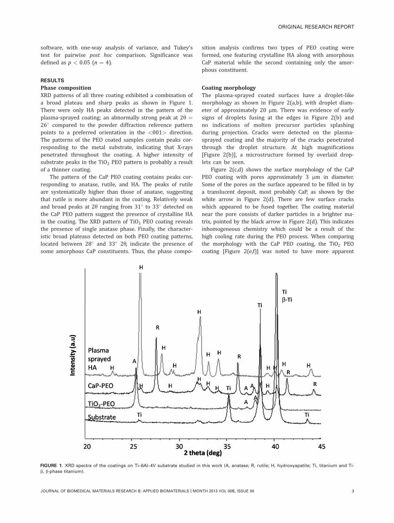
Phase compositionXRD patterns of all three coating exhibited a combination ofa broad plateau and sharp peaks as shown in Figure 1.There were only HA peaks detected in the pattern of theplasma-sprayed coating; an abnormally strong peak at 2y ¼26� compared to the powder diffraction reference patternpoints to a preferred orientation in the <001> direction.The patterns of the PEO coated samples contain peaks cor-responding to the metal substrate, indicating that X-rayspenetrated throughout the coating. A higher intensity ofsubstrate peaks in the TiO2 PEO pattern is probably a resultof a thinner coating.
The pattern of the CaP PEO coating contains peaks cor-responding to anatase, rutile, and HA. The peaks of rutileare systematically higher than those of anatase, suggestingthat rutile is more abundant in the coating. Relatively weakand broad peaks at 2y ranging from 31� to 33� detected onthe CaP PEO pattern suggest the presence of crystalline HAin the coating. The XRD pattern of TiO2 PEO coating revealsthe presence of single anatase phase. Finally, the character-istic broad plateaus detected on both PEO coating patterns,located between 28� and 33� 2y, indicate the presence ofsome amorphous CaP constituents. Thus, the phase compo-
sition analysis confirms two types of PEO coating wereformed, one featuring crystalline HA along with amorphousCaP material while the second containing only the amor-phous constituent.
Coating morphologyThe plasma-sprayed coated surfaces have a droplet-likemorphology as shown in Figure 2(a,b), with droplet diam-eter of approximately 20 lm. There was evidence of earlysigns of droplets fusing at the edges in Figure 2(b) andno indications of molten precursor particles splashingduring projection. Cracks were detected on the plasma-sprayed coating and the majority of the cracks penetratedthrough the droplet structure. At high magnifications[Figure 2(b)], a microstructure formed by overlaid drop-lets can be seen.
Figure 2(c,d) shows the surface morphology of the CaPPEO coating with pores approximately 3 lm in diameter.Some of the pores on the surface appeared to be filled in bya translucent deposit, most probably CaP, as shown by thewhite arrow in Figure 2(d). There are few surface crackswhich appeared to be fused together. The coating materialnear the pore consists of darker particles in a brighter ma-trix, pointed by the black arrow in Figure 2(d). This indicatesinhomogeneous chemistry which could be a result of thehigh cooling rate during the PEO process. When comparingthe morphology with the CaP PEO coating, the TiO2 PEOcoating [Figure 2(e,f)] was noted to have more apparent
FIGURE 1. XRD spectra of the coatings on Ti–6Al–4V substrate studied in this work (A, anatase; R, rutile; H, hydroxyapatite; Ti, titanium and Ti-
b, b-phase titanium).
ORIGINAL RESEARCH REPORT
JOURNAL OF BIOMEDICAL MATERIALS RESEARCH B: APPLIED BIOMATERIALS | MONTH 2013 VOL 00B, ISSUE 00 3
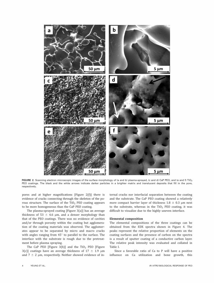
pores and at higher magnifications [Figure 2(f)] there isevidence of cracks connecting through the skeleton of the po-rous structure. The surface of the TiO2 PEO coating appearsto be more homogeneous than the CaP PEO coating.
The plasma-sprayed coating [Figure 3(a)] has an averagethickness of 53 6 4.6 lm, and a denser morphology thanthat of the PEO coatings. There was no evidence of cavitiesand/or through porosity within the coating but agglomera-tion of the coating materials was observed. The agglomer-ates appear to be separated by micro and macro crackswith angles ranging from 45� to parallel to the surface. Theinterface with the substrate is rough due to the pretreat-ment before plasma spraying.
The CaP PEO [Figure 3(b)] and the TiO2 PEO [Figure3(c)] coatings have an average thickness of 17 6 1.9 lmand 7 6 2 lm, respectively. Neither showed evidence of in-
ternal cracks nor interfacial separation between the coatingand the substrate. The CaP PEO coating showed a relativelymore compact barrier layer of thickness 1.8 6 0.3 lm nextto the substrate, whereas in the TiO2 PEO coating, it wasdifficult to visualize due to the highly uneven interface.
Elemental compositionThe elemental compositions of the three coatings can beobtained from the EDX spectra shown in Figure 4. Thepeaks represent the relative proportion of elements on thecoating surfaces and the presence of carbon on the spectrais a result of sputter coating of a conductive carbon layer.The relative peak intensity was evaluated and collated inTable I.
Since a favorable ratio of Ca to P will have a positiveinfluence on Ca utilization and bone growth, this
FIGURE 2. Scanning electron microscopic images of the surface morphology of (a and b) plasma-sprayed, (c and d) CaP PEO, and (e and f) TiO2
PEO coatings. The black and the white arrows indicate darker particles in a brighter matrix and translucent deposits that fill in the pore,
respectively.
4 YEUNG ET AL. IN VITRO BIOLOGICAL RESPONSE OF PEO
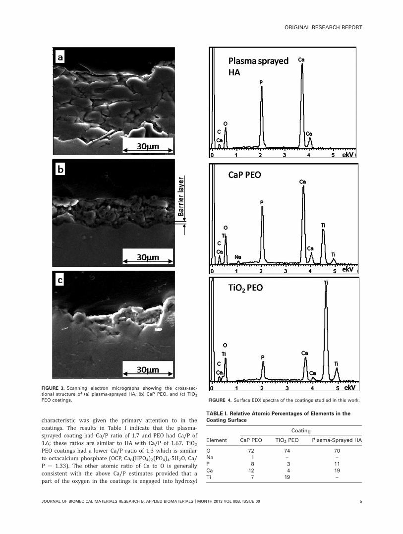
characteristic was given the primary attention to in thecoatings. The results in Table I indicate that the plasma-sprayed coating had Ca/P ratio of 1.7 and PEO had Ca/P of1.6; these ratios are similar to HA with Ca/P of 1.67. TiO2
PEO coatings had a lower Ca/P ratio of 1.3 which is similarto octacalcium phosphate (OCP, Ca8(HPO4)2(PO4)4�5H2O, Ca/P ¼ 1.33). The other atomic ratio of Ca to O is generallyconsistent with the above Ca/P estimates provided that apart of the oxygen in the coatings is engaged into hydroxyl
FIGURE 3. Scanning electron micrographs showing the cross-sec-
tional structure of (a) plasma-sprayed HA, (b) CaP PEO, and (c) TiO2
PEO coatings. FIGURE 4. Surface EDX spectra of the coatings studied in this work.
TABLE I. Relative Atomic Percentages of Elements in the
Coating Surface
Element
Coating
CaP PEO TiO2 PEO Plasma-Sprayed HA
O 72 74 70Na 1 – –P 8 3 11Ca 12 4 19Ti 7 19 –
ORIGINAL RESEARCH REPORT
JOURNAL OF BIOMEDICAL MATERIALS RESEARCH B: APPLIED BIOMATERIALS | MONTH 2013 VOL 00B, ISSUE 00 5
groups in the amorphous constituent as well as into tita-nium dioxide, in the case of PEO coatings. The presence oftrace amounts of Na in the CaP PEO coating is due to theirpresence in the electrolyte.
Hydroxyl groups have been considered to favor osteo-blast adhesion therefore are an important factor in a suc-cessful implant. Because hydrogen cannot be detected byEDX, we estimated the level of hydroxyl groups on the coat-ing surface based on the difference between amounts ofoxygen detected and estimated based stoichiometry of HA(O/Ca ¼ 2.6), OCP (O/Ca ¼ 3.625), and TiO2 present in thecoatings. The data in Table I suggest that there was 27 at%, 22 at %, and 18 at % of oxygen contributing to thehydroxyl group formation on the surfaces of CaP PEO, TiO2
PEO, and plasma-sprayed HA coatings, respectively. There-fore, the CaP PEO coating should favor cell attachment bet-ter than the other two coatings.
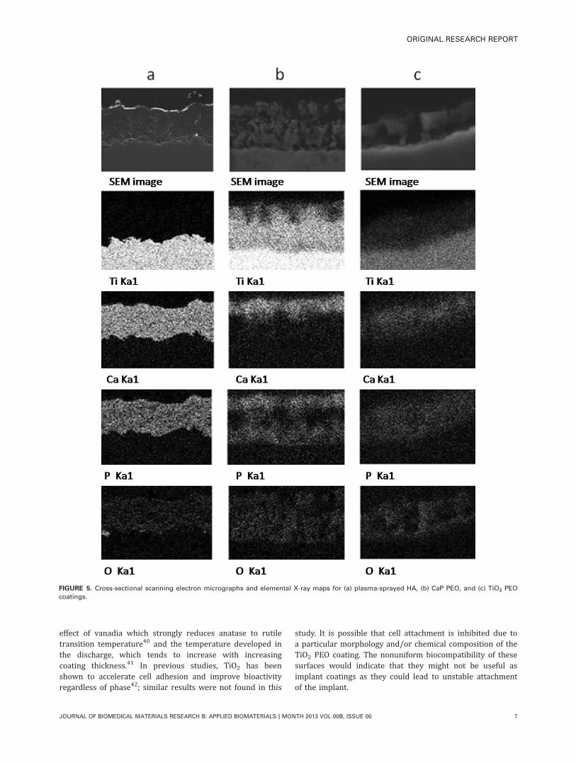
Distribution of elementsElemental mapping outlines differences in chemicaldistributions between the coatings studied, as presented inFigure 5. The plasma-sprayed coating has even distributionof all coating elements, with no penetration of Ti in the HAlayer, and evidence of interfacial porosity noted by the highlevel of oxygen on the left side of the image. The distribu-tion of Ti in the PEO coatings reveals the specifics of thesurface layer formation by the PEO process, wherein Tienters the coating due to the conversion of the metal sub-strate into oxide, producing a graded concentration profile.Oxygen appears to be evenly distributed throughout thePEO coatings whereas Ca is enriched at the outer region.This is particularly obvious in the CaP PEO coating whereCa is mostly located at the top 2.5–3 lm of the coatingwhere Ti is depleted. There is an enrichment of P at theouter and inner regions and it is depleted where Ti distribu-tion is high in the midregion. Similar trends can be seen inthe TiO2 PEO coating, but the distributions of Ca and Pappear to be more diffuse.
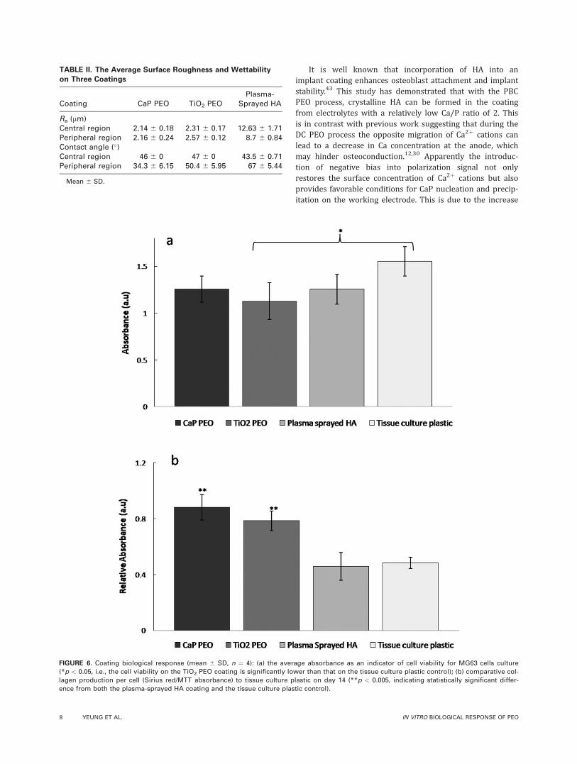
Average surface roughness and wettability of thecoatingsThe surface roughness Ra and wettability been accepted tobe influential factors for cell adhesion. Therefore, thesecharacteristics were measured both in the center and on theperiphery of the samples to evaluate influence of the coat-ing formation mechanisms on possible edge effects that mayoccur during the coating growth (Table II).
The Ra values of the two PEO coatings are significantlylower than the plasma-sprayed coating (p < 0.005). Withthe plasma-sprayed coating the Ra values decrease from thecentral to the peripheral region, while the two PEO coatingsshow an increase in surface roughness progressing towardthe peripheral region.
Theory suggests that for cell attachment the thresholdvalue of wettability should be 60�, below which is classedas hydrophilic and cell attachment is favored.36 Similar val-ues of wettability were obtained from the central regions ofall three coatings as shown in Table II. The CaP PEO coating
exhibited a lower contact angle at the peripheral regionwhen compared to the central region, but both are classedas hydrophilic. The TiO2 PEO and the plasma-sprayed coat-ing have a higher contact angle at the peripheral regionthan the central region, where the peripheral region of theplasma-sprayed coating is classed as hydrophobic. The pe-ripheral region of the CaP PEO coating has a statisticallylower wettability value than the TiO2 PEO and the plasma-sprayed coating (p < 0.05).
During the PEO process in this study, edge effects wereshown to have a small influence on wettability, however,both PEO coatings were classed as hydrophilic overall.
Cellular response to coatingsVisual examination following the MTT staining revealed thatthe viable cells were evenly distributed over the surfaces ofthe CaP PEO and the plasma-sprayed coatings. The TiO2
PEO coating supported a higher concentration of cells onthe peripheral region than the central region of the sample,while at some parts in the central region no cells appearedto be present. Measurement of the optical density of theMTT staining [Figure 6(a)] revealed that the TiO2 PEO coat-ing had significantly fewer viable cells on the surface whencompared to the tissue culture plastic (p < 0.005).
The two PEO coatings supported significantly higher col-lagen deposition per cell than the plasma-sprayed and tis-sue culture plastic (p < 0.005) [Figure 6(b)], indicating thatthe PEO coating favors collagen production. Calcium produc-tion per cell showed that the amount of calcium depositedby the cells at this time point was below the linear regionof the colorimetric assay and was not significantly differentbetween the surfaces (data not shown).
DISCUSSION
In this study, we compared Ca- and P-containing PEO coat-ings with plasma-sprayed HA coatings to investigate thepotential for the former to improve the surfaces of orthope-dic Ti implants. PEO is easily tailored to specific chemis-tries and has the advantage that there is no need for pre-treatments such as glass bead or grit blasting to increasethe roughness of the surface.37 Interestingly, the two PEOcoatings formed under different processing parametersresulted in very different coating characteristics but bothled to a high level of collagen production by bone cellswhen compared to the plasma-sprayed coating, suggestingthat the PEO morphology is beneficial for bone matrixproduction.
The CaP PEO coating showed a more convoluted surfacemorphology than the TiO2 PEO coating, this is probably dueto CaP incorporating into the porous structure then crystal-lizing into HA during the PEO process.
The relatively thick CaP PEO coating was shown to con-tain both anatase and rutile, while the thin TiO2 PEO coat-ing contains only anatase. It was previously observed thatanatase is generally preferred in PEO coatings formed inphosphate electrolytes on cp-Ti38 but on Ti–6Al–4V alloyrutile is promoted and may become the main phase in thickenough coatings.39 This is likely to be due to the combined
6 YEUNG ET AL. IN VITRO BIOLOGICAL RESPONSE OF PEO
effect of vanadia which strongly reduces anatase to rutiletransition temperature40 and the temperature developed inthe discharge, which tends to increase with increasingcoating thickness.41 In previous studies, TiO2 has beenshown to accelerate cell adhesion and improve bioactivityregardless of phase42; similar results were not found in this
study. It is possible that cell attachment is inhibited due toa particular morphology and/or chemical composition of theTiO2 PEO coating. The nonuniform biocompatibility of thesesurfaces would indicate that they might not be useful asimplant coatings as they could lead to unstable attachmentof the implant.
FIGURE 5. Cross-sectional scanning electron micrographs and elemental X-ray maps for (a) plasma-sprayed HA, (b) CaP PEO, and (c) TiO2 PEO
coatings.
ORIGINAL RESEARCH REPORT
JOURNAL OF BIOMEDICAL MATERIALS RESEARCH B: APPLIED BIOMATERIALS | MONTH 2013 VOL 00B, ISSUE 00 7
It is well known that incorporation of HA into animplant coating enhances osteoblast attachment and implantstability.43 This study has demonstrated that with the PBCPEO process, crystalline HA can be formed in the coatingfrom electrolytes with a relatively low Ca/P ratio of 2. Thisis in contrast with previous work suggesting that during theDC PEO process the opposite migration of Ca2þ cations canlead to a decrease in Ca concentration at the anode, whichmay hinder osteoconduction.12,30 Apparently the introduc-tion of negative bias into polarization signal not onlyrestores the surface concentration of Ca2þ cations but alsoprovides favorable conditions for CaP nucleation and precip-itation on the working electrode. This is due to the increase
TABLE II. The Average Surface Roughness and Wettability
on Three Coatings
Coating CaP PEO TiO2 PEOPlasma-
Sprayed HA
Ra (lm)Central region 2.14 6 0.18 2.31 6 0.17 12.63 6 1.71Peripheral region 2.16 6 0.24 2.57 6 0.12 8.7 6 0.84Contact angle (�)Central region 46 6 0 47 6 0 43.5 6 0.71Peripheral region 34.3 6 6.15 50.4 6 5.95 67 6 5.44
Mean 6 SD.
FIGURE 6. Coating biological response (mean 6 SD, n ¼ 4): (a) the average absorbance as an indicator of cell viability for MG63 cells culture
(*p < 0.05, i.e., the cell viability on the TiO2 PEO coating is significantly lower than that on the tissue culture plastic control); (b) comparative col-
lagen production per cell (Sirius red/MTT absorbance) to tissue culture plastic on day 14 (**p < 0.005, indicating statistically significant differ-
ence from both the plasma-sprayed HA coating and the tissue culture plastic control).
8 YEUNG ET AL. IN VITRO BIOLOGICAL RESPONSE OF PEO
in the local pH of the electrolyte region adjacent to the elec-trode caused by the main electrochemical process duringthe negative pulse:
2H2Oþ 2e ! H2 þ 2OH�
Since the positive bias leads to acidification of alkaline sol-utions44 the PBC waveform needs to be carefully adjustedto achieve a suitable balance between local Ca2þ/PO4
3�
ionic ratio and electrolyte pH in the vicinity of the work-ing electrode. Stability regions of HA and OCP correspondto pH ¼ 9.5–12 and 5.5–7.0, respectively,45 and it is likelythat such local pH values have been achieved during depo-sition of CaP and TiO2 PEO coatings. It can therefore bespeculated that in the case of TiO2 PEO coating, HAdeposition was affected by insufficient Ca2þ concentrationand/or local electrolyte pH, hence the thinner coating withlower Ca/P ratio. Nevertheless, OCP is often considered asa precursor to HA in the physiological environment and itcan transform into HA without a change inmorphology and has been shown to enhance osteoblasticdifferentiation.46
Although the two PEO coatings showed different Ca/Pratios, elemental mapping indicated similar trends in thedistribution of these elements; Ca is only present in theouter region of the coating, whereas P can also be foundin the inner region, closer to the barrier layer. This couldbe a result of phosphate ions being involved into both CaPprecipitation and specific adsorption at the bottom of thepores thus inhibiting anodic dissolution of Ti. EDX resultssuggest there are surface hydroxyl groups (OH�) presenton all the coatings and these are likely to be associatedwith amorphous constituent of the coating material. Fur-ther verification and quantification of the hydroxyl groupsby other techniques, for example, micro-Raman spectros-copy, time-of-flight secondary ion mass spectrometry, orX-ray photoelectron spectroscopy,47 should be consideredin the future work. Hydroxyl groups are known to be effi-cient inducers of apatite nucleation.48 In a preliminarystudy, calcium deposition by the cells was assessed at day14 however little calcium had been deposited by MG63s atthis time point on any surfaces, so it was not possible todiscern any differences between the coatings; it is there-fore possible that there is an influence of hydroxyl groupson apatite nucleation which would be seen in longer termcultures.
Surface roughness has previously been thought to berelated to the thickness of the coating.36,49 The plasma-sprayed coating had a significantly higher Ra value than thetwo PEO coatings regardless of region, and it had a higherRa value at the central than the peripheral region, this couldbe a result of slower solidification rate at the central regionand/or due to the formation process. Anatase and rutilephases have been thought to be an influencing factor onsurface roughness and contact angle.50 Our results showedthat the presence of rutile in the CaP PEO coating did nothave a statistically significant effect on its wettability, as ithad a statistically similar wettability to the TiO2 PEO coat-
ing in the central region. However, there were differences inwettability between the two PEO coatings in the peripheralregion, but it is probable that this was a result of edgeeffects and/or current density rather than crystallinity. Thewettability of the central region in all three coatings wassimilar and hydrophilic, which should favor cell adhesion;however, MTT results showed a nonuniform cell adhesion atthe central region of the TiO2 PEO coating. Meanwhile thehydrophobic peripheral region of the plasma-sprayed coat-ing showed good cell attachment. These results indicate thatfactors other than wettability are governing cell adhesion,such as surface energy.51
CONCLUSIONS
Ceramic coatings containing crystalline titania, HA, andamorphous CaPs were prepared on Ti–6Al–4V alloy by aone-step PBC PEO process. The coating morphology, topol-ogy, and chemical composition can be varied to influencethe osteoblastic behavior. The CaP PEO coatings can supportas good or better biological response of MG-63 osteoblastsin vitro, in terms of maintaining cell viability and enablingcollagenous matrix deposition, compared with commerciallyavailable plasma-sprayed HA coatings. These coatingspromote cell proliferation and significantly more collagen isdeposited compared to the plasma-sprayed coatings and thetissue culture plastic. Thin TiO2 PEO coatings with low Ca/Pratio showed a nonuniform bioactivity across the surface,which may be due to hindered precipitation of crystallineCaPs and OH groups during coating formation. At the sametime, the differences in surface roughness and wettabilityobserved between the PEO and the plasma-sprayed coatingsdid not appear to influence the cell proliferation whichsuggests that osteoblasts are more sensitive to elementalcomposition and distribution than the physical properties ofthe surface.
ACKNOWLEDGMENTS
The samples for this work were provided by Plasma Coatings(Medical Division) and Plasma Biotal. Assistance from Mr. Chu,P-J and Mr. Delaine-Smith, RM of the Department of MaterialsScience and Engineering, The University of Sheffield are alsogratefully acknowledged.
REFERENCES1. Rack HJ, Qazi JI. Titanium alloys for biomedical applications.
Mater Sci Eng C 2006;26(8):1269–1277.
2. Geetha M, Singh AK, Asokamani R, Gogia AK. Ti based biomateri-
als, the ultimate choice for orthopaedic implants—A review. Prog
Mater Sci 2009;54(3):397–425.
3. Biesiekierski A, Wang J, Abdel-Hady Gepreel M, Wen C. A new
look at biomedical Ti-based shape memory alloys. Acta Biomater
2012;8(5):1661–1669.
4. Sun L, Berndt CC, Gross KA, Kucuk A. Material fundamentals and
clinical performance of plasma-sprayed hydroxyapatite coatings:
A review. J Biomed Mater Res 2001;58(5):570–592.
5. Liu X, Chu PK, Ding C. Surface modification of titanium, titanium
alloys, and related materials for biomedical applications. Mater
Sci Eng R: Rep 2004;47(3–4):49–121.
6. Lynn AK, DuQuesnay DL. Hydroxyapatite-coated Ti–6Al–4V: Part
1: The effect of coating thickness on mechanical fatigue behav-
iour. Biomaterials 2002;23(9):1937–1946.
ORIGINAL RESEARCH REPORT
JOURNAL OF BIOMEDICAL MATERIALS RESEARCH B: APPLIED BIOMATERIALS | MONTH 2013 VOL 00B, ISSUE 00 9
7. Lynn AK, DuQuesnay DL. Hydroxyapatite-coated Ti–6Al–4V: Part
2: The effects of post-deposition heat treatment at low tempera-
tures. Biomaterials 2002;23(9):1947–1953.
8. Yerokhin AL, Nie X, Leyland A, Matthews A, Dowey SJ. Plasma
electrolysis for surface engineering. Surf Coat Technol 1999;
122(2–3):73–93.
9. Yerokhin AL, Nie X, Leyland A, Matthews A. Characterisation of
oxide films produced by plasma electrolytic oxidation of a Ti–6Al–
4V alloy. Surf Coat Technol 2000;130(2–3):195–206.
10. Matykina E, Berkani A, Skeldon P, Thompson GE. Real-time imag-
ing of coating growth during plasma electrolytic oxidation of tita-
nium. Electrochim Acta 2007;53(4):1987–1994.
11. Li LH, Kong YM, Kim HW, Kim YW, Kim HE, Heo SJ, Koak JY.
Improved biological performance of Ti implants due to surface
modification by micro-arc oxidation. Biomaterials 2004;25(14):
2867–2875.
12. Whiteside P, Matykina E, Gough JE, Skeldon P, Thompson GE. In
vitro evaluation of cell proliferation and collagen synthesis on ti-
tanium following plasma electrolytic oxidation. J Biomed Mater
Res Part A 2010;94A(1):38–46.
13. Snizhko LO, Yerokhin AL, Gurevina NL, Misnyankin DO, Pilkington
A, Leyland A, Matthews A. A model for galvanostatic anodising
of Al in alkaline solutions. Electrochim Acta 2005;50(27):
5458–5464.
14. Snizhko LO, Yerokhin AL, Gurevina NL, Patalakha VA, Matthews
A. Excessive oxygen evolution during plasma electrolytic oxida-
tion of aluminium. Thin Solid Films 2007;516(2–4):460–464.
15. Matykina E, Arrabal R, Skeldon P, Thompson GE, Habazaki H.
Influence of grain orientation on oxygen generation in anodic tita-
nia. Thin Solid Films 2008;516(8):2296–2305.
16. Nie X, Leyland A, Matthews A. Deposition of layered bioceramic
hydroxyapatite/TiO2 coatings on titanium alloys using a hybrid
technique of micro-arc oxidation and electrophoresis. Surf Coat
Technol 2000;125(1–3):407–414.
17. Matykina E, Montuori M, Gough J, Monfort F, Berkani A, Skel-
don P, Thompson GE, Habazaki H. Spark anodising of titanium
for biomedical applications. Trans Inst Met Finish 2006;84(3):
125–133.
18. Liu F, Song Y, Wang F, Shimizu T, Igarashi K, Zhao L. Formation
characterization of hydroxyapatite on titanium by microarc oxida-
tion and hydrothermal treatment. J Biosci Bioeng 2005;100(1):
100–104.
19. Ryu HS, Song W-H, Hong S-H. Biomimetic apatite induction of P-
containing titania formed by micro-arc oxidation before and after
hydrothermal treatment. Surf Coat Technol 2008;202(9):
1853–1858.
20. Wei D, Zhou Y, Yang C. Characteristic and microstructure of the
microarc oxidized TiO2-based film containing P before and after
chemical- and heat treatment. Appl Surf Sci 2009;255(18):
7851–7857.
21. Zhao X, Cai Q, Li D, He J, Luo Q, Li X, Sun W. Preparation and
characterization of bio-ceramic coatings on the surface of pure ti-
tanium by plasma electrolytic oxidation-alkali and heat treatment.
J Ceram Process Res 2010;11(5):575–580.
22. Song H-J, Shin K-H, Kook M-S, Oh H-K, Park Y-J. Effects of
the electric conditions of AC-type microarc oxidation and
hydrothermal treatment solution on the characteristics of hy-
droxyapatite formed on titanium. Surf Coat Technol 2010;
204(14):2273–2278.
23. Kung K-C, Yuan K, Lee T-M, Lui T-S. Effect of heat treatment on
microstructures and mechanical behavior of porous Sr–Ca–P coat-
ings on titanium. J Alloys Compd 2012;515(0):68–73.
24. Bai Y, Kim K-A, Park IS, Lee SJ, Bae TS, Lee MH. In situ compos-
ite coating of titania-hydroxyapatite on titanium substrate by
micro-arc oxidation coupled with electrophoretic deposition proc-
essing. Mater Sci Eng B 2011;176(15):1213–1221.
25. Kim DY, Kim M, Kim HE, Koh YH, Kim HW, Jang JH. Formation
of hydroxyapatite within porous TiO2 layer by micro-arc oxidation
coupled with electrophoretic deposition. Acta Biomater 2009;5(6):
2196–2205.
26. Kim M-S, Ryu J-J, Sung Y-M. One-step approach for nano-crystal-
line hydroxyapatite coating on titanium via micro-arc oxidation.
Electrochem Commun 2007;9(8):1886–1891.
27. Han Y, Sun J, Huang X. Formationmechanism of HA-based coatings
bymicro-arc oxidation. ElectrochemCommun 2008;10(4):510–513.
28. Sun J, Han Y, Huang X. Hydroxyapatite coatings prepared by
micro-arc oxidation in Ca- and P-containing electrolyte. Surf Coat
Technol 2007;201(9–11):5655–5658.
29. Yerokhin A. Method of forming a bioactive coating. United King-
dom Patent WO 2009/053670; 2009.
30. Frauchiger VM, Schlottig F, Gasser B, Textor M. Anodic plasma-
chemical treatment of CP titanium surfaces for biomedical appli-
cations. Biomaterials 2004;25(4):593–606.
31. Rudnev VS, Yarovaya TP, Boguta DL, Tyrina LM, Nedozorov PM,
Gordienko PS. Anodic spark deposition of P, Me(II) or Me(III) con-
taining coatings on aluminium and titanium alloys in electrolytes
with polyphosphate complexes. J Electroanal Chem 2001;
497(1–2):150–158.
32. Sul Y-T, Byon E, Wennerberg A. Surface characteristics of electro-
chemically oxidized implants and acid-etched implants: Surface
chemistry, morphology, pore configurations, oxide thickness,
crystal structure, and roughness. Int J Oral Maxillofac Implants
2008;23(4):631–640.
33. Verrier S, Peroglio M, Voisard C, Lechmann B, Alini M. The osteo-
genic differentiation of human osteoprogenitor cells on anodic-
plasma-chemical treated Ti6Al7Nb. Biomaterials 2011;32(3):672–680.
34. Reilly GC, Radin S, Chen AT, Ducheyne P. Differential alkaline
phosphatase responses of rat and human bone marrow derived
mesenchymal stem cells to 45S5 bioactive glass. Biomaterials
2007;28(28):4091–4097.
35. Sittichockechaiwut A, Scutt AM, Ryan AJ, Bonewald LF, Reilly
GC. Use of rapidly mineralising osteoblasts and short periods of
mechanical loading to accelerate matrix maturation in 3D scaf-
folds. Bone 2009;44(5):822–829.
36. Anselme K. Osteoblast adhesion on biomaterials. Biomaterials
2000;21(7):667–681.
37. Tsui YC, Doyle C, Clyne TW. Plasma sprayed hydroxyapatite coat-
ings on titanium substrates. Part 2: Optimisation of coating prop-
erties. Biomaterials 1998;19(22):2031–2043.
38. Khan RHU, Yerokhin AL, Matthews A. Structural characteristics and
residual stresses in oxide films produced on Ti by pulsed unipolar
plasma electrolytic oxidation. PhilosMag 2008;88(6):795–807.
39. Apachitei I, Lonyuk B, Fratila-Apachitei LE, Zhou J, Duszczyk J. Fa-
tigue response of porous coated titanium biomedical alloys.
Scripta Mater 2009;61(2):113–116.
40. Bucharsky EC, Schell G, Oberacker R, Hoffmann MJ. Anatase–ru-
tile transformation in TiO2–V2O5 catalyst coatings for ceramic
foams. J Eur Ceram Soc 2009;29(10):1955–1961.
41. Hussein RO, Nie X, Northwood DO, Yerokhin A, Matthews A.
Spectroscopic study of electrolytic plasma and discharging
behaviour during the plasma electrolytic oxidation (PEO) process.
J Phys D: Appl Phys 2010;43(10):105203.
42. Sul Y-T. The significance of the surface properties of oxidized tita-
nium to the bone response: Special emphasis on potential bio-
chemical bonding of oxidized titanium implant. Biomaterials
2003;24(22):3893–3907.
43. Paital SR, Dahotre NB. Calcium phosphate coatings for bio-
implant applications: Materials, performance factors, and method-
ologies. Mater Sci Eng R: Rep 2009;66(1–3):1–70.
44. Snizhko LO, Yerokhin AL, Pilkington A, Gurevina NL, Misnyankin
DO, Leyland A, Matthews A. Anodic processes in plasma electro-
lytic oxidation of aluminium in alkaline solutions. Electrochim
Acta 2004;49(13):2085–2095.
45. Dorozhkin SV, Epple M. Biological and medical significance of
calcium phosphates. Angew Chem Int Ed 2002;41(17):
3130–3146.
46. Dorozhkin SV, Schmitt M, Bouler JM, Daculsi G. Chemical trans-
formation of some biologically relevant calcium phosphates in
aqueous media during a steam sterilization. J Mater Sci: Mater
Med 2000;11(12):779–786.
47. Yan L, Leng Y, Weng L-T. Characterization of chemical inhomoge-
neity in plasma-sprayed hydroxyapatite coatings. Biomaterials
2003;24(15):2585–2592.
48. Song W-H, Jun Y-K, Han Y, Hong S-H. Biomimetic apatite coat-
ings on micro-arc oxidized titania. Biomaterials 2004;25(17):
3341–3349.
10 YEUNG ET AL. IN VITRO BIOLOGICAL RESPONSE OF PEO

49. Chen H-T, Hsiao C-H, Long H-Y, Chung C-J, Tang C-H, Chen K-C,
He J-L. Micro-arc oxidation of b-titanium alloy: Structural charac-
terization and osteoblast compatibility. Surf Coat Technol 2009;
204(6–7):1126–1131.
50. He J, Zhou W, Zhou X, Zhong X, Zhang X, Wan P, Zhu B, Chen
W. The anatase phase of nanotopography titania plays an impor-
tant role on osteoblast cell morphology and proliferation. J Mater
Sci: Mater Med 2008;19(11):3465–3472.
51. Zhao G, Schwartz Z, Wieland M, Rupp F, Geis-Gerstorfer J, Coch-
ran DL, Boyan BD. High surface energy enhances cell response to
titanium substrate microstructure. J Biomed Mater Res Part A
2005;74A(1):49–58.
ORIGINAL RESEARCH REPORT
JOURNAL OF BIOMEDICAL MATERIALS RESEARCH B: APPLIED BIOMATERIALS | MONTH 2013 VOL 00B, ISSUE 00 11
Copyright © 2022 FDOKUMEN