Prognosis of angina and myocardial infarction in South Asian ...
Upload
independentCategory
view
0download
0
ORIGINAL PAPER
In situ delivery of bone marrow cells and mesenchymal stem cellsimproves cardiovascular function in hypertensive rats submittedto myocardial infarction
Luisa Maria Gomes de Macedo Braga Æ Silvia Lacchini Æ Beatriz D’Agord Schaan ÆBruno Rodrigues Æ Kaleizu Rosa Æ Katia De Angelis Æ Luciano Figueiredo Borges ÆMaria Claudia Irigoyen Æ Nance Beyer Nardi
Received: 24 August 2007 / Accepted: 14 January 2008 / Published online: 7 February 2008
� National Science Council Taipei 2008
Abstract This work aimed to evaluate cardiac morphol-
ogy/function and histological changes induced by bone
marrow cells (BMCs) and cultured mesenchymal stem
cells (MSCs) injected at the myocardium of spontaneously
hypertensive rats (SHR) submitted to surgical coronary
occlusion. Female syngeneic adult SHR, submitted (MI) or
not (C) to coronary occlusion, were treated 24 h later with
in situ injections of normal medium (NM), or with MSCs
(MSC) or BMCs (BM) from male rats. The animals were
evaluated after 1 and 30 days by echocardiography, his-
tology of heart sections and PCR for the Y chromosome.
Improved ejection fraction and reduced left ventricle
infarcted area were observed in MSC rats as compared to
the other experimental groups. Treated groups had signif-
icantly reduced lesion tissue score, increased capillary
density and normal (not-atrophied) myocytes, as compared
to NM and C groups. The survival rate was higher in C,
NM and MSC groups as compared to MI and BM groups.
In situ injection of both MSCs and BMCs resulted in
improved cardiac morphology, in a more physiological
model of myocardial infarction represented by surgical
coronary occlusion of spontaneously hypertensive rats.
Only treatment with MSCs, however, ameliorated left
ventricle dysfunction, suggesting a positive role of these
cells in heart remodeling in infarcted hypertensive subjects.
Keywords Bone marrow cells � Mesenchymal stem
cells � Spontaneously hypertensive rats � Hypertension �In situ delivery
Ischemic heart disease is the leading cause of death
worldwide and also an important cause of heart failure.
Atherosclerosis, the major determinant of ischemic car-
diomyopathy, involves several complex processes, which
can be initiated and accelerated by risk factors including
systemic arterial hypertension, dyslipidemia, cigarette
smoking, diabetes mellitus, age, male sex and family his-
tory of premature coronary heart disease [1].
Animal models have allowed the study of cardiovascular
diseases in the early stages, as well as the pathogenic
mechanisms and the effects of therapeutic interventions.
Although there is no model that can entirely mimic human
ischemic cardiomyopathy, the most frequently used animal
model involves the induction of complete coronary occlu-
sion [2]. Its performance in spontaneous hypertensive rats
(SHR) accelerates left ventricular dilatation and haemody-
namic alterations caused by the myocardial infarction [3].
The many available treatments for heart failure do not
target the progressive loss of cardiomyocytes. Experi-
mental studies have suggested that stem cells can exert
beneficial effects on the failing heart by transdifferentiating
into cardiac cell types and/or by providing a source of
cardioprotective paracrine factors [4]. Several adult stem
L. M. G. de Macedo Braga � N. B. Nardi (&)
Department of Genetics, Universidade Federal do Rio Grande do
Sul, Av Bento Goncalves 9500, 91501-970 Porto Alegre, RS,
Brazil
e-mail: [email protected]
L. M. G. de Macedo Braga
State Foundation of Production and Research in Health of Rio
Grande do Sul, Porto Alegre, RS, Brazil
S. Lacchini � B. Rodrigues � K. Rosa � K. De Angelis �L. F. Borges � M. C. Irigoyen
Hypertension Unit, Heart Institute, University of Sao Paulo, Sao
Paulo, SP, Brazil
B. D. Schaan
Cardiology Institute, Rio Grande do Sul, Cardiology Foundation,
Porto Alegre, RS, Brazil
123
J Biomed Sci (2008) 15:365–374
DOI 10.1007/s11373-008-9237-z

cell types [5–9], animal models [10, 11] and delivery
pathways [12–14] have been tested in the last years, in the
search for ideal conditions which were not yet found. Bone
marrow cells (BMCs) [15] and cultured mesenchymal stem
cells (MSCs) [3, 16], administered mainly by in situ
injection [10], are the most frequently used types of cells.
The aim of the present study was to evaluate the effects
of two different kinds of adult stem cell populations,
administered in situ, on cardiac morphology, function and
histology of SHR submitted to surgical coronary occlusion,
in an attempt to investigate this therapeutic approach in a
more physiological animal model of myocardial infarction.
Materials and methods
Cell culture
Bone marrow cells (BMCs) were obtained from 3-week-
old male SHR by flushing the cavity of femurs with normal
culture medium (NM), consisting of low-glucose Dul-
becco’s modified Eagle’s medium (DMEM, Sigma
Chemical Co., St Louis, MO) with 10% fetal bovine serum
(Cultilab, Sao Paulo, Brazil) and supplemented with 3.7 g/l
sodium bicarbonate and 2.5 g/l HEPES (Sigma). No anti-
biotics were added. BMCs were centrifuged at 800 g for
10 min, and viability was assessed by Trypan blue staining.
Isolation, primary culture and expansion of MSCs were
performed as we previously described for murine cells [17].
Briefly, bone marrow cells were collected, washed and
cultured in concentration of 2.15 9 106 cells/ml in 25 cm3
bottles (7 ml/bottle). The cells were kept at 37�C in NM,
and non-adherent cells were removed after 72 h. MSCs
were maintained by passage every 3 or 4 days, and were
used for cell therapy between the 8th and 12th passages. For
transplantation, MSC cultures were washed twice with
Ca2+Mg2+-free Hank’s balanced salt solution (Sigma),
trypsinized, resuspended in 5 ml NM and washed. The cells
were counted and resuspended in NM at 107 cells/ml.
Osteogenic and adipogenic differentiation of MSCs were
induced as previously described [18]. Briefly, osteogenic
differentiation was induced by culturing MSCs for up to
4 weeks in NM supplemented with 10-8 mol/l dexameth-
asone (Sigma), 5 lg/ml ascorbic acid 2-phosphate (Sigma)
and 10 mmol/l b-glycerophosphate (Sigma). To observe
calcium deposition, cultures were washed once with phos-
phate-buffered saline (PBS), and stained for 5 min at room
temperature with Alizarin Red S stain (Sigma), pH 4.2. For
adipogenic differentiation, MSCs were cultured for up to
4 weeks in NM supplemented with 10.8 mol/l dexametha-
sone and 5 lg/ml insulin (Sigma). Adipocytes were easily
distinguished from undifferentiated cells by phase-contrast
microscopy. To further confirm their identity, cells were
fixed with 4% paraformaldehyde in PBS for 1 h at room
temperature, and stained with Oil Red O (Sigma).
Immunophenotyping
Cells were trypsinized and incubated with fluorescein iso-
thiocyanate (FITC)-conjugated antibodies against rat
CD29, CD44H and CD31 (Becton Dickinson, San Jose,
CA), CD11 and CD45 (Caltag, Burlingame, CA). The cells
were analyzed in a cytometer equipped with a 488 nm
argon laser (FACScalibur, Becton Dickinson) using the
CellQuest software. At least 10,000 events were collected.
Animal studies
Syngeneic spontaneously hypertensive rats (SHR) used in
the experiments were housed and maintained under stan-
dard conditions, and were treated in accordance with the
Guidelines for the Care and Use of Laboratory Animals
prepared by the National Academy of Sciences and pub-
lished by the National Institutes of Health. All procedures
were approved by a local Research Ethics Committee.
Myocardial infarction was induced in 45 females anesthe-
tized by intraperitoneal (i.p.) injection of 50 mg/kg body
wt ketamine (Parke-Davis, Ann Arbor, MI) and 10 mg/kg
xylazine (Bayer, Newhaven, CT). After intubation, animals
were positive-pressure ventilated with room air at 2.5 ml,
65 strokes/min with a pressure cycled rodent ventilator
(HARVARD Apparatus, Model 683, Holliston, MA). For
induction of myocardial infarction, a 2-cm left lateral
thoracotomy was performed in the third intercostal space
and the left anterior descending coronary artery was
occluded with a single nylon (6.0) suture at approximately
1 mm from its origin below the tip of the left atrium as
previously described [19, 20]. The chest was closed with
silk suture. The animals were maintained in the ventilator
until recovery. All rats received antibiotics (penicillin,
20,000 U) and Tramadol (20 mg/kg, every 6 h). The ani-
mals were matched for body weight (160–180 g) and
treated 24 h after surgery with BMCs (BM group, n = 5),
MSCs (MSC group, n = 10) or NM (NM group, n = 7).
Two other groups served as control: one group was sub-
mitted to myocardial infarction and did not receive any
treatment (MI group, n = 6), and another one was not
operated or cell-treated (C group, n = 8).
Cell treatment
Twenty-four hours after coronary occlusion, the animals
were re-anesthetized and an echocardiography was
366 J Biomed Sci (2008) 15:365–374
123
performed to measure the infarction area (IA) and the
ejection fraction (EF). Only animals with EF B 50% and
IA C 30% were maintained in the experiment (37% were
excluded). A total of 1 9 106 MSCs or BMCs were sus-
pended in 100 ll NM and drawn into a 300 ll syringe with
a 30-gauge needle. The animals were intubated, a re-tho-
racotomy was performed in the fourth intercostal space,
and the cells or normal medium were injected in five points
of the left ventricle anterior wall, in the viable myocardial
bordering the infarcted area. Proper administration of the
injected material was evaluated by the immediate appear-
ance of a thin film of white liquid inflated underneath the
visceral pericardium.
Systolic arterial pressure
Systolic arterial pressure (SAP) was measured using tail-
cuff plethysmography before the MI surgery, to ensure
similar levels of hypertension among the groups.
Echocardiography
Echocardiographic indices were obtained before treatment
and one and 30 days after induction of myocardial infarction
under the guidelines of the American Society of Echocar-
diography, by one observer blind to the treatments and with
the use of a 10–13 MHz multi-frequency linear transducer in
a SEQUOIA 512 (ACUSON Corporation, Mountain View,
CA). Images were obtained with the transducer on the shaved
chest (lateral recumbence). Rats were anesthetized as
described above and scanned from below, at 2-cm depth with
focus optimized at 1 cm. All measurements were based on
the average of three consecutive cardiac cycles. Wall
thickness and left ventricle (LV) dimensions were obtained
from a short-axis view at the level of the papillary muscles.
The LV mass was calculated by the use of the following
formula, assuming a spherical LV geometry and validated
in rats: LV mass = 1.047 9 [(LVd + PWd + IVSd)3-
LVd3], where 1.047 is the specific gravity of muscle, LVd is
LV end-diastolic diameter, PWd is end-diastolic posterior
wall thickness and IVSd is end-diastolic interventricular
septum thickness. Two-dimensionally guided pulsed-wave
Doppler recordings of LV inflow were obtained from the
apical four-chamber view. Maximal early diastolic peak
velocity (E) and late peak velocity (A) were derived from
mitral inflow. Peak E desacceleration time was also mea-
sured as an index of diastolic function. Isovolumic relaxation
time (IVRT) was taken from aortic valve closure to the onset
of mitral flow. Ejection fraction (EF%) was obtained by a
modification of Simpson’s method, which is more trust-
worthy in infarcted hearts. Its formula is
V ¼ p4�X20
i¼1
ai � bi�L
20;
where L = LV length divided into 20 discs (i = 1 to 1 = 20)
from basis to apex, with the diameter of each disc determined
in 2 apical views (A and B). After these measurements in
diastole and systole, the respective volumes were obtained,
allowing EF evaluation (final diastolic volume—final sys-
tolic volume/final diastolic volume) 9 100%.
The infarction area was delimited taking into account
the movement of LV walls, by the observation of longi-
tudinal, apical and transversal views of the LV. Regions
with systolic thickness under normal, as well as portions
with paradoxal movement, were considered as infarcted.
The infarcted area (%) was thus determined by the ratio of
these regions by total area of LV walls [21].
Tissue histology and morphometry
Thirty days after the treatments, the rats were anesthetized
with sodium pentobarbital (80 mg/kg body weight), a
transverse incision below the diaphragm and bilateral tho-
racotomy incisions were made and the LV was cannulated
retrogradely with a needle to perfuse. The hearts were
arrested in diastole by perfusion with a NaCl 0.9% plus
14 mM KCl solution, followed by buffered formalin (4%)
for tissue fixation.
Excised hearts were immersed in 4% formalin for 24 h
and then transected 5 mm below coronary ligation.
Transversal slices were processed and embedded in
paraplast. Sections of 5 lm were stained with hematoxylin-
eosin (HE) for qualitative assessment, with picrosirius-
hematoxylin (red stain) for fibrosis evaluation, and with
periodic acid Schiff (PAS) for quantification of capillary
density. Histo-morphometric analyses were performed
blinded regarding the identity of experimental groups.
Fibrosis evaluation
Fibrosis in the LV was evaluated by dividing it in 8
quadrants, and determining a fibrosis score (0–4) for each
quadrant. The aspect of collagen fibers was evaluated under
polarized light, allowing evaluation of the molecular dis-
position of collagen and aspects related to myocardial
function. Picrosirius-polarization staining was performed
as previously described [12].
Evaluation of capillary density
Capillary density was quantified by microscopic examina-
tion of the sections using a 10 9 10 grid optically
J Biomed Sci (2008) 15:365–374 367
123
superimposed on each of 20 non-overlapping fields at
4009 magnification, randomly distributed on the antero-
septal side of the left ventricle. Capillary density was
evaluated adjacent to the cardiac scars as the total number
of capillaries in the fields, expressed as number of capil-
laries per field (units/mm2) [5].
Molecular evaluation
To analyze the presence of the injected cells in the heart
tissue, the infarcted heart was divided in four pieces.
Genomic DNA was extracted from each piece using the
proteinase K method [22] and submitted to PCR. The
quality of the DNA samples was analyzed with GAPDH
primers (Table 1). The PCR reaction mixture contained
approximately 100 ng genomic DNA, 1U Taq DNA poly-
merase (Invitrogen, Sao Paulo, Brazil), 10 pmol of each
oligonucleotide primers, 1 ll of 5 mM dNTP, 2.5 ll of 109
PCR buffer, and 1 ll of 50 mM MgCl2, in a final volume of
25 ll. The PCR was carried out in a Techne thermocycler
(Barloworld Scientific, Staffordshire, UK) for 40 cycles of
denaturation (94�C, 1 min), annealing (58�C, 1 min), and
extension (72�C, 1 min) with final extension of 7 min
(72�C). Presence of the Y chromosome was analyzed by
nested PCR. The reaction mixture and amplification cycles
were the same as for GAPDH PCR, except that it contained
10 pmol of rat Sry-specific oligonucleotide primers
(Table 1). In the first round, SRY external primers were
used to amplify a fragment of 272 bp. The second round
(nested PCR) included two SRY internal primers added to
the 2 ll of the first round amplification product. Both
reactions were conducted in the same conditions. The PCR
products were analyzed by electrophoresis in 1.5% agarose
gels stained with ethidium bromide.
Statistical analyses
Data are reported as mean ± SEM. According to the
parameter, one or two way ANOVA was used to compare
groups, followed by the Student-Newman-Keuls test. The
Kaplan Meyer method was used to determine the survival
curve. The significance level was established at P \ 0.05.
Results
Establishment and characterization of rat MSCs
The cell population isolated consisted of adherent cells
with fibroblastoid morphology which, when properly
stimulated, gave origin to adipocytes and osteocytes in
culture (Fig. 1a). The cells were positive for CD29 and
CD44, and negative for CD45, CD11b and CD31 (Fig. 1b).
Body weight
Body weight was similar in all groups before coronary
occlusion (173.8 ± 4, 166.6 ± 4; 169.7 ± 1; 168 ± 5 and
172 ± 5 g) but lower 30 days after the procedure
(166.7 ± 3; 157.2 ± 1; 160.7 ± 7; 158.2 ± 5 and
162.8 ± 7 g) in C, MI, NM, BM and MSC groups,
respectively (P [ 0.05).
Cardiovascular evaluation
As expected, all animals were hypertensive at the begin-
ning of the protocol (mean systolic arterial pressure:
167 ± 3 mmHg), with no differences among the groups.
Figure 2a shows the LV infarcted area 24 h and 30 days
after coronary occlusion. The result was similar between
groups one day after coronary occlusion (*40% of the left
ventricle wall). Treatment with mesenchymal stem cells
determined a significant reduction in the LV infarcted area
(MSC: 26 ± 4%) 30 days after coronary occlusion as
compared to groups that did not receive cells (MI:
52 ± 2% and NM: 41 ± 5%), and also to the group that
received BMCs (BM: 45 ± 4%).
Left ventricle ejection fraction, evaluated at 24 h and
30 days after coronary occlusion, is shown in Fig. 2b. It
was reduced by *40% one day after coronary occlusion in
Table 1 Sequences of forward and reverse oligonucleotide primers
Primer Sequence
GAPDH GAPDH F: 50ACCACAGTCCATGCCATCAC 30
GAPDH R: 50TCCACCACCCTGTTGCTGTA 30
SRY External SRY R ext: 50GTAGGTTGTTGTCCCATTGC 30
SRY F ext: 50GAGAGAGGCACAAGTTGGC 30
Internal SRY F int: 50AAGCAGCTGGGATATCAGTGG 30
SRY R int: 50TTTTGTTGAGGCAACTTCACG 30
368 J Biomed Sci (2008) 15:365–374
123

the animals submitted to myocardial infarction (36.6 ±
4%; 41.5 ± 2%; 40.2 ± 3%; 39.9 ± 4% in MI, NM, BM
and MSC, respectively), as compared to control animals
(65 ± 4%). The left ventricle ejection fraction was lower
at the end of the protocol in the MI (34 ± 3%), NM
(48 ± 5%) and BM (41 ± 4%) groups, as compared to the
C group (69 ± 3%). Thirty days after coronary occlusion,
however, the rats injected with cultivated mesenchymal
stem cells (MSC group) presented an improvement in the
left ventricle ejection fraction (59 ± 4%) in comparison to
MI, NM and BM rats, whereas no difference was observed
between MSC and C animals.
Other variables analyzed by echocardiography (LV
mass, LVd, E wave, A wave, E/A ratio, EDT and IVRT) are
shown in Table 2. Left ventricle mass and LVd were
unchanged 1 day after coronary occlusion in all groups, as
compared to the C group. The LV mass was increased in
the BM group in relation to the other groups at the end of
the protocol (30 days after coronary occlusion). At the
same time, the LV area was increased in all infarcted
groups in comparison to the C group and to the initial
evaluation in infarcted groups (1 day after coronary
occlusion). Left ventricle diameter during diastole (LVd)
was also higher in MI, MSC and BM rats in relation to C
rats. The BM group presented increased LVd as compared
to MI, NM and MSC groups. However, LVd was similar in
the infarcted rats injected with cultivated mesenchymal
stem cells (MSC group) as compared to C animals.
Diastolic function, evaluated by A wave, E wave, E/A
ratio, EDT and IVRT, was similar in the infarcted groups
Fig. 1 (a) Cultured rat
mesenchymal stem cell (MSCs)
assumes a flat morphology
(4009). The cells differentiate
in osteoblasts (ost, 1,0009) and
adipocytes (adip, 4009) when
cultured with specific induction
media. Bar, 20 lm. (b)
Immunophenotyping of
cultivated MSCs. The cells are
negative for CD31 (not shown),
CD45 and CD11b, and positive
for CD29 and CD44. ...., control
reactions; ___ , incubation with
antibodies
Fig. 2 Left ventricle (LV) wall myocardium infarction (MI) area (a)
and ejection fraction (EF) (b) in C, MI, NM, BM and MSC groups in
the initial and final echocardiographic evaluations. *P \ 0.05 vs.
control group; �P \ 0.05 vs. MI group; �P \ 0.05 vs. BM group;
•P \ 0.05 vs. NM; #P \ 0.05 vs. initial evaluation in the same group
J Biomed Sci (2008) 15:365–374 369
123

and the C group 1 day after coronary occlusion. The E
wave was also similar among groups at the end of the
protocol. The A wave was reduced and the E/A ratio was
increased in MI and BM groups as compared with C, NM
and MSC groups 30 days after coronary occlusion. The E/
A ratio was also higher in MI and BM rats comparing the
final (30 days) vs. the initial (1 day) evaluation. Thirty
days after coronary occlusion, EDT was increased in all
infarcted groups in comparison to the C group and to their
respective initial evaluation (1 day). IVRT remained
unchanged during the whole protocol.
Survival rates were similar among the C (100.0%), NM
(90.0%) and MSC (87.5%) groups, but these animals pre-
sented higher survival rates as compared to the MI (56.2%)
and BM (66.7%) groups (P \ 0.05).
Molecular analyses
PCR and nested PCR showed the presence of the Y chro-
mosome in normal male heart tissue (not shown), but no
amplification was obtained in heart samples collected from
the female rats in any of the experimental groups. Positive
results with GAPDH control primers (not shown) proved
the adequate quality of the DNA samples prepared.
Ventricle fibrosis evaluation
Myocardial scars following ischemia-induced lesion were
always extended to the anteroseptal LV wall. Fibrosis
deposition was not observed in control rats, while MI
groups presented different scar extension and fibrosis.
Figure 3 depicts the fibrosis scores, which were higher in
the MI group, but reduced by treatment with cells or cul-
ture medium.
The study under polarized light showed, in all groups
submitted to coronary occlusion, a meshwork of thick,
intensely bright fibers exhibiting irregular and anomalous
dispersion of birefringence in both yellow and/or red pat-
terns (Figs. 4 and 5). Control rats showed thin, weakly
birefringent, greenish fibers surrounding each myocyte.
Interestingly, hearts injected with MSCs and BMCs had
normal (not-atrophied) myocytes, presenting visible inter-
calated discs, between fibrosis and scar.
Table 2 Echodoppler cardiovascular evaluation 1 and 30 days after coronary occlusion
Parameter Day C MI NM BM MSC
LV mass (g) 1 0.92 ± 0.03 0.95 ± 0.03 0.95 ± 0.005 1.06 ± 0.02 0.95 ± 0.02
30 0.93 ± 0.03 1.00 ± 0.02 1.03 ± 0.04 1.24 ± 0.01*�•# 0.99 ± 0.07�
LVDIA (cm) 1 0.599 ± 0.02 0.60 ± 0.02 0.62 ± 0.02 0.66 ± 0.01 0.62 ± 0.02
30 0.60 ± 0.02 0.76 ± 0.02*# 0.73 ± 0.04* 0.85 ± 0.02*�•# 0.69 ± 0.03�
E-wave (m/s) 1 0.60 ± 0.03 0.64 ± 0.05 0.66 ± 0.036 0.66 ± 0.01 0.59 ± 0.02
30 0.59 ± 0.01 0.75 ± 0.03 0.71 ± 0.05 0.75 ± 0.04 0.64 ± 0.04
A-wave (m/s) 1 0.34 ± 0.02 0.24 ± 0.03 0.291 ± 0.035 0.276 ± 0.02 0.241 ± 0.02
30 0.37 ± 0.02 0.16 ± 0.04* 0.31 ± 0.04� 0.12 ± 0.04*• 0.31 ± 0.04��
E/A ratio 1 1.75 ± 0.09 3.02 ± 0.63 2.36 ± 0.183 2.77 ± 0.28 2.53 ± 0.20
30 1.63 ± 0.08 6.47 ± 1.34*# 3.03 ± 0.77� 5.67 ± 0.62*•# 2.11 ± 0.48��
EDT (ms) 1 31.13 ± 1.89 28.00 ± 2.51 25.67 ± 2.25 32.00 ± 1.15 26.33 ± 1.93
30 30.38 ± 2.33 43.14 ± 3.37*# 41.22 ± 3.43*# 45.33 ± 1.86*# 43.43 ± 1.81*#
IVRT (ms) 1 34.13 ± 1.54 33.67 ± 3.20 29.80 ± 1.02 35.80 ± 1.71 36.60 ± 1.58
30 37.75 ± 2.76 31.00 ± 3.52 36.11 ± 1.89 30.33 ± 1.36 35.18 ± 2
Values are expressed as mean ± SEM. MI, myocardial infarction; NM, MI + normal medium; BM, MI + treatment with bone marrow cells;
MSC, MI + treatment with mesenchymal cells; LV, left ventricular; LVd, LV diameter during diastole; E-wave, maximal early diastolic peak
velocity; A-wave, late peak velocity; E/A ratio, ratio between the velocities of E and A waves; EDT, peak E desacceleration time; IVRT, LV
isovolumetric relaxation time. * P \ 0.05 vs. control group; � P \ 0.05 vs. MI; � P \ 0.05 vs. BM; • P \ 0.05 vs. NM; # P \ 0.05 vs. initial
evaluation in the same group
Fig. 3 Fibrosis score in the studied groups. It was higher in MI rats as
compared to C rats, but similarly lower in the MSC, BMC and NM
groups. *P \ 0.05 vs. MI group
370 J Biomed Sci (2008) 15:365–374
123
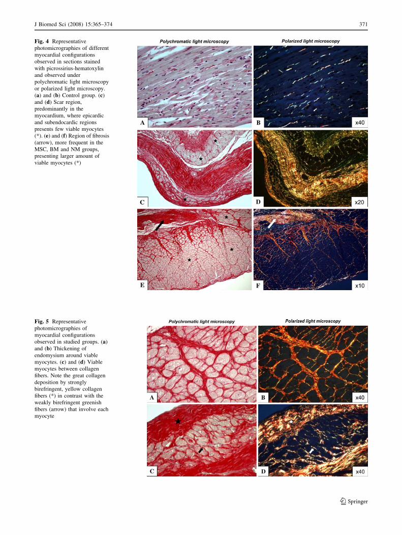
Fig. 4 Representative
photomicrographies of different
myocardial configurations
observed in sections stained
with picrossirius-hematoxylin
and observed under
polychromatic light microscopy
or polarized light microscopy.
(a) and (b) Control group. (c)
and (d) Scar region,
predominantly in the
myocardium, where epicardic
and subendocardic regions
presents few viable myocytes
(*). (e) and (f) Region of fibrosis
(arrow), more frequent in the
MSC, BM and NM groups,
presenting larger amount of
viable myocytes (*)
Fig. 5 Representative
photomicrographies of
myocardial configurations
observed in studied groups. (a)
and (b) Thickening of
endomysium around viable
myocytes. (c) and (d) Viable
myocytes between collagen
fibers. Note the great collagen
deposition by strongly
birefringent, yellow collagen
fibers (*) in contrast with the
weakly birefringent greenish
fibers (arrow) that involve each
myocyte
J Biomed Sci (2008) 15:365–374 371
123

Capillary proliferation
The injection of MSCs and BMCs around the infarcted area
in the LV resulted in important increase of capillary den-
sity. Capillary quantification was performed in viable
myocardium (not in the scar), and MI rats did not show
differences in capillary density as compared to control rats
(273 ± 49 vs. 262 ± 28 capillaries/mm2, respectively).
Cell injections increased capillary density, compared to MI
and NM groups (MSC: 487 ± 29, and BM: 453 ± 38 vs.
NM: 366 ± 73 capillaries/mm2). Treatment with normal
medium increased capillary density in the NM group as
compared to the MI group (366 ± 73 capillaries/mm2 vs.
262 ± 28 capillaries/mm2).
Discussion
Adequate definition of the type and route of delivery of
cells is of fundamental importance for stem cell therapy of
cardiovascular diseases to be fully successful. Stem cells
can be delivered through coronary arteries, coronary veins,
peripheral veins or intra myocardial injection. The main
results of the present study showed that in situ injection of
MSCs can successfully reduce the myocardial infarction
area, increase the ejection fraction and reduce mortality in
an animal model of myocardial infarction associated to
systemic hypertension. The effects were not so clearly
observed with the same delivery approach using BMCs,
although in both cases the fibrosis area was decreased and
capillary density increased.
The specific cell type used for treating cardiovascular
diseases can result in different outcomes [10, 15, 23, 24].
We used mesenchymal stem cells obtained from 8th to 12th
passage of cultured bone marrow cells, which showed
plasticity and immunophenotype similar to those previ-
ously described [18, 23]. Bone marrow preparations, more
frequently used in clinical trials of cell-based therapies, are
a heterogeneous population of mature mononuclear cells,
blasts and stem/progenitor cells. MSCs, on the other hand,
represent the expansion of a specific subpopulation, whose
therapeutic potential has been increasingly recognized [23].
The number of cells used for treatment (106/animal) has
already been described as therapeutically appropriate in rat
models of cardiovascular disease [25].
Since hypertension accelerates left ventricular dilatation
and haemodynamic alterations following myocardial
infarction in rats [3], we decided to use SHRs submitted to
coronary occlusion as a more physiological model of the
association of these pathologies. There is no report in the
literature concerning the use of stem cell therapy in this
animal model. We observed that in these animals, ven-
tricular dilatation was not accelerated as in normotensive
rats following myocardial infarction, probably due to pre-
existing LV hypertrophy and increased hypertrophic
response to myocardial infarction [26]. Absolute left ven-
tricular dimensions in SHR have been shown to increase
out of proportion to body growth, consistent with concen-
tric hypertrophy [27], a finding we did not observe—LV
mass was not different comparing the final vs. the initial
evaluation. This difference is probably due to the younger
age of animals and the short time of follow up (30 days) in
the present study. The only group that presented higher LV
mass at the final evaluation was the BM group. We ascri-
bed this difference, even though it was not statistically
different from the other groups, to their higher baseline LV
mass.
Intracoronary infusions do not appear to infiltrate the
myocardial infarction zone or reduce the myocardial
infarction size [28], so the direct delivery of cells into the
area of tissue necrosis seems to be the best delivery
approach. This could be shown in the present study with
the use of MSCs, but not BMCs, in hypertensive rats, since
the former effectively reduced the LV infarction area. This
structural benefit was also translated into functional
advantage, as observed by the higher LV ejection fraction
and similar LVd of the MSC group as compared to the C
group. Using normotensive pigs, similar structural and
functional benefits have been observed with percutaneous
injection catheter delivery of MSCs, persisting until
8 weeks after the cell therapy [10].
Baseline diastolic parameters were similar among
groups, as expected. However, diastolic dysfunction was
observed 30 days after coronary occlusion in MI and BM
groups, but not in C, NM and MSC groups, as indicated by
decreased A-wave and increased E/A ratio. These data
showed that LV remodeling, common in infarcted hearts,
was modified by the in situ injection of NM and MSCs. The
unexpected effect of NM may be due to growth factors
present in the serum. The evaluation of diastolic function
using echodopplercardiography has been reported by few
investigators. The present work showed improved results
as compared to similar experiments with in situ adminis-
tration of MSCs in pigs [10].
Reduction of fibrosis score may suggest reestablishment
of the correct arrange of cytoskeleton, supporting the better
ventricular function. Interestingly, treatment with culture
medium increased capillary density, and this may explain
the reduction of fibrosis score in this group when compared
to MI. The increase in capillary density is in accordance
with a putative angiogenic effect of both MSCs and BMCs
[10, 24].
Cultured mesenchymal stem cells and BMCs treatments
did not induce arrhythmias, differently from results
recently reported [29]. One possible explanation for this
difference could be related to the fact that we injected
372 J Biomed Sci (2008) 15:365–374
123

smaller volumes distributed in more myocardial points.
Also, in that study arrhythmia was assessed by telemetry, a
more sensitive method than ours, since we evaluated the
cardiac rhythm by echocardiography twice during the
whole protocol. Since mortality was not different in ani-
mals that presented arrhythmia vs. those that did not have
this complication in the study above, and no higher mor-
tality was seen in our groups injected with stem cells, the
significance of those findings is uncertain.
The therapeutic role of MSCs in myocardial repair is
under intense investigation, and may involve multiple
factors such as direct differentiation into cardiac cells, cell
fusion or the secretion of cytokines and growth factors with
paracrine activities [30]. Our results favor this latter model,
since transplanted male cells were not found in the heart of
treated females when they were analyzed by PCR and
nested PCR 30 days later. Although surprising, similar
results have recently been reported [31, 32]. Prockopp and
collaborators, for instance, using a mouse model of acute
MI, observed that cardiac function and fibrosis were sig-
nificantly improved, but there was no evident MSC
engraftment in the heart despite of the use of three different
and highly sensitive assays [31]. These results give support
to the hypotheses that theses cells can exert beneficial
effects after coronary artery occlusion through the secre-
tion of cardioprotective and reparative factors.
In an attempt to explain these results, the gene expres-
sion profiles of cultured MSCs were analyzed with
microarrays. The expression of genes coding for anti-
apoptotic, angiogenic/arteriogenic, and matrix-mediating
factors, particularly IL-6, leukemia inhibitory factor (LIF)
and vascular endothelial growth factor (VEGF) family
members in the MSCs was observed [31]. More recently,
Sze et al. [33] determined the secretion proteome of human
embryo-derived MSCs. The study revealed the presence of
201 unique gene products, representing important signaling
pathways in cardiovascular biology, bone development and
hematopoiesis. The MSC secretory products identified may
act as paracrine modulators of tissue repair and replace-
ment in cardiovascular diseases. Since our MSC cultures
are in all aspects (morphology, kinetics, immunopheno-
typing and plasticity) identical to the cell populations used
in those studies, we may assume that they secret similar
profiles of cytokines and growth/differentiation factors.
The fact the bone marrow cells did not result in amelio-
ration of left ventricle dysfunction is also an evidence of a
mechanism exclusive of MSCs in heart remodeling.
Systemically administered MSCs determine different
responses in comparison to their in situ administration. We,
in SHR [34], and others, in normotensive rats [23],
observed increased LVd after myocardial infarction and
treatment with systemically injected MSCs. Immunodefi-
cient mice submitted to the same procedure showed
improved cardiac function and reduced myocardial fibrosis
[31]. These results raise the possibility of different actions
of the cells delivered by different routes, and show that in
situ delivery of MSCs is more effective for regeneration of
the damaged myocardium.
Acknowledgements This work was supported by Conselho Nac-
ional de Desenvolvimento Cientıfico e Tecnologia (CNPq, 474828/
2004-2), Secretaria de Ciencia e Tecnologia do Estado RS, Fundacao
de Amparo a Pesquisa do Estado do Rio Grande do Sul and Fundacao
Estadual de Pesquisa em Saude RS.
References
1. Ridker PM, Stampfer MJ, Rifai N (2001) Novel risk factors for
systemic atherosclerosis: a comparison of c-reactive protein,
fibrinogen, homocysteine, lipoprotein(a), and standard cholesterol
screening as predictors of peripheral arterial disease. JAMA
285:2481–2485
2. Doggrell SA, Brown L (1998) Rat models of hypertension, car-
diac hypertrophy and failure. Cardiovasc Res 39:89–105
3. Nishikimi T, Yamagishi H, Takeuchi K, Takeda T (1995) An
angiotensin II receptor antagonist attenuates left ventricular
dilatation after myocardial infarction in the hypertensive rat.
Cardiovasc Res 29:856–861
4. Wollert KC, Drexler H (2006) Cell-based therapy for heart fail-
ure. Curr Opin Cardiol 21:234–239
5. Becker C, Lacchini S, Muotri AR, da Silva GJ, Castelli JB,
Vassallo PF, Menck CF, Krieger JE (2006) Skeletal muscle cells
expressing VEGF induce capillary formation and reduce cardiac
injury in rats. Int J Cardiol 113:348–354
6. Kucia M, Ratajczak J, Reca R, Janowska-Wieczorek A, Ratajc-
zak MZ (2004) Tissue-specific muscle, neural and liver stem/
progenitor cells reside in the bone marrow, respond to an SDF-1
gradient and are mobilized into peripheral blood during stress and
tissue injury. Blood Cells Mol Dis 32:52–57
7. Lin F, Cordes K, Li L, Hood L, Couser WG, Shankland SJ,
Igarashi P (2003) Hematopoietic stem cells contribute to the
regeneration of renal tubules after renal ischemia-reperfusion
injury in mice. J Am Soc Nephrol 14:1188–1199
8. Rehman J, Li J, Orschell CM, March KL (2003) Peripheral blood
‘‘endothelial progenitor cells’’ are derived from monocyte/mac-
rophages and secrete angiogenic growth factors. Circulation
107:1164–1169
9. Zhang FB, Li L, Fang B, Zhu DL, Yang HT, Gao PJ (2005)
Passage-restricted differentiation potential of mesenchymal stem
cells into cardiomyocyte-like cells. Biochem Biophys Res Com-
mun 336:784–792
10. Amado LC, Saliaris AP, Schuleri KH, St John M, Xie JS, Cat-
taneo S, Durand DJ, Fitton T, Kuang JQ, Stewart G, Lehrke S,
Baumgartner WW, Martin BJ, Heldman AW, Hare JM (2005)
Cardiac repair with intramyocardial injection of allogeneic mes-
enchymal stem cells after myocardial infarction. Proc Natl Acad
Sci USA 102:11474–11479
11. Orlic D, Kajstura J, Chimenti S, Limana F, Jakoniuk I, Quaini F,
Nadal-Ginard B, Bodine DM, Leri A, Anversa P (2001) Mobi-
lized bone marrow cells repair the infarcted heart, improving
function and survival. Proc Natl Acad Sci USA 98:10344–10349
12. Boomsma RA, Swaminathan PD, Geenen DL (2006) Intrave-
nously injected mesenchymal stem cells home to viable
myocardium after coronary occlusion and preserve systolic
function without altering infarct size. Int J Cardiol [Epub ahead of
print]
J Biomed Sci (2008) 15:365–374 373
123

13. Kinnaird T, Stabile E, Burnett MS, Shou M, Lee CW, Barr S,
Fuchs S, Epstein SE (2004) Local delivery of marrow-derived
stromal cells augments collateral perfusion through paracrine
mechanisms. Circulation 109:1543–1549
14. Nagaya N, Fujii T, Iwase T, Ohgushi H, Itoh T, Uematsu M,
Yamagishi M, Mori H, Kangawa K, Kitamura K (2004) Intra-
venous administration of mesenchymal stem cells improves
cardiac function in rats with acute myocardial infarction through
angiogenesis and myogenesis. Am J Physiol Heart Circ Physiol
287:H2670–H2676
15. Davani S, Marandin A, Mersin N, Royer B, Kantelip B, Herve P,
Etievent JP, Kantelip JP (2003) Mesenchymal progenitor cells
differentiate into an endothelial phenotype, enhance vascular
density, and improve heart function in a rat cellular cardiomyo-
plasty model. Circulation 108(Suppl 1):II253–II258
16. Shyu KG, Wang BW, Hung HF, Chang CC, Shih DT (2006)
Mesenchymal stem cells are superior to angiogenic growth factor
genes for improving myocardial performance in the mouse model
of acute myocardial infarction. J Biomed Sci 13:47–58
17. Meirelles Lda S, Nardi NB (2003) Murine marrow-derived
mesenchymal stem cell: isolation, in vitro expansion, and char-
acterization. Br J Haematol 123:702–711
18. Da Silva Meirelles L, Chagastelles PC, Nardi NB (2006) Mes-
enchymal stem cells reside in virtually all post-natal organs and
tissues. J Cell Sci 119:2204–2213
19. De Angelis K, Leirner AA, Irigoyen MC, Cestari IA (2001)
Nonstimulated cardiomyoplasty improves hemodynamics in
myocardial-infarcted rats. Artif Organs 25:939–943
20. Johns TN, Olson BJ (1954) Experimental myocardial infarction.
I. A method of coronary occlusion in small animals. Ann Surg
140:675–682
21. Nozawa E, Kanashiro RM, Murad N, Carvalho AC, Cravo SL,
Campos O, Tucci PJ, Moises VA (2006) Performance of two-
dimensional Doppler echocardiography for the assessment of
infarct size and left ventricular function in rats. Braz J Med Biol
Res 39:687–695
22. Sambrook J, Fritsch EF, Maniatis T (1989) Molecular cloning: a
laboratory manual, 2nd edn. Cold Spring Harbor, Cold Spring
Harbor Laboratory Press, New York
23. Beyer Nardi N, da Silva Meirelles L (2006) Mesenchymal stem
cells: isolation, in vitro expansion and characterization. Hand-
book Exp Pharmacol 174:249–282
24. Kudo M, Wang Y, Wani MA, Xu M, Ayub A, Ashraf M (2003)
Implantation of bone marrow stem cells reduces the infarction
and fibrosis in ischemic mouse heart. J Mol Cell Cardiol
35:1113–1119
25. Lovell MJ, Mathur A (2004) The role of stem cells for treatment
of cardiovascular disease. Cell Prolif 37:67–87
26. Zdrojewski T, Gaudron P, Whittaker P, Poelzl S, Schiemann J,
Hu K, Ertl G (2002) Ventricular remodeling after myocardial
infarction and effects of ACE inhibition on hemodynamics and
scar formation in SHR. Cardiovasc Pathol 11:88–93
27. Pfeffer J, Pfeffer M, Fletcher P, Braunwald E (1979) Alterations
of cardiac performance in rats with established spontaneous
hypertension. Am J Cardiol 44:994–998
28. Wollert KC, Drexler H (2004) Cell therapy for acute myocardial
infarction: where are we heading? Nat Clin Pract Cardiovasc Med
1:61
29. Fukushima S, Varela-Carver A, Coppen SR, Yamahara K, Felkin
LE, Lee J, Barton PJ, Terracciano CM, Yacoub MH, Suzuki K
(2007) Direct intramyocardial but not intracoronary injection of
bone marrow cells induces ventricular arrhythmias in a rat
chronic ischemic heart failure model. Circulation 115:2254–2261
30. Wang XJ, Li QP (2007) The roles of mesenchymal stem cells
(MSCs) therapy in ischemic heart diseases. Biochem Biophys Res
Commun 359:189–193
31. Iso Y, Spees JL, Serrano C, Bakondi B, Pochampally R, Song
YH, Sobel BE, Delafontaine P, Prockop DJ (2007) Multipotent
human stromal cells improve cardiac function after myocardial
infarction in mice without long-term engraftment. Biochem
Biophys Res Commun 354:700–706
32. Noiseux N, Gnecchi M, Lopez-Ilasaca M, Zhang L, Solomon SD,
Deb A, Dzau VJ, Pratt RE (2006) Mesenchymal stem cells
overexpressing Akt dramatically repair infarcted myocardium
and improve cardiac function despite infrequent cellular fusion or
differentiation. Mol Ther 14:840–850
33. Sze SK, de Kleijn DP, Lai RC, Tan EK, Zhao H, Yeo KS, Low
TY, Lian Q, Lee CN, Mitchell W, El Oakley RM, Lim SK (2007)
Elucidating the secretion proteome of human ESC-derived mes-
enchymal stem cells. Mol Cell Proteomics [Epub ahead of print]
34. Braga LMGM, Rosa K, Rodrigues B, Malfitano C, Camassola M,
Chagastelles P, Lacchini S, Fiorino P, De Angelis K, Schaan B,
Irigoyen MC, Nardi NB (2007) Systemic delivery of adult stem
cells improves cardiac function in spontaneously hypertensive
rats. Clin Exp Pharmacol Physiol 35; [Epub ahead of print]
374 J Biomed Sci (2008) 15:365–374
123
Copyright © 2022 FDOKUMEN