Growth Factor-Induced Mobilization of Cardiac Progenitor Cells Reduces the Risk of Arrhythmias, in a...
14
Growth Factor-Induced Mobilization of Cardiac Progenitor Cells Reduces the Risk of Arrhythmias, in a Rat Model of Chronic Myocardial Infarction Leonardo Bocchi 1. , Monia Savi 1. , Gallia Graiani 2 , Stefano Rossi 1 , Aldo Agnetti 3,7 , Francesca Stillitano 4 , Costanza Lagrasta 2,7 , Silvana Baruffi 1 , Roberta Berni 1 , Caterina Frati 2 , Mario Vassalle 5 , Umberto Squarcia 3,7 , Elisabetta Cerbai 4 , Emilio Macchi 1,7 , Donatella Stilli 1,7 , Federico Quaini 6,7 , Ezio Musso 1,7 * 1 Dipartimento di Biologia Evolutiva e Funzionale, Universita ` di Parma, Parma, Italy, 2 Dipartimento di Patologia e Medicina di Laboratorio, Universita ` di Parma, Parma, Italy, 3 Dipartimento dell’Eta ` Evolutiva, Universita ` di Parma, Parma, Italy, 4 Dipartimento di Farmacologia Preclinica e Clinica, Universita ` di Firenze, Firenze, Italy, 5 Department of Physiology and Pharmacology, SUNY, Downstate Medical Center, Brooklyn, New York, United States of America, 6 Dipartimento di Medicina Interna e Scienze Biomediche, Universita ` di Parma, Parma, Italy, 7 Centro Interdipartimentale Cellule Staminali Cardiache (CISTAC), Universita ` di Parma, Parma, Italy Abstract Heart repair by stem cell treatment may involve life-threatening arrhythmias. Cardiac progenitor cells (CPCs) appear best suited for reconstituting lost myocardium without posing arrhythmic risks, being commissioned towards cardiac phenotype. In this study we tested the hypothesis that mobilization of CPCs through locally delivered Hepatocyte Growth Factor and Insulin-Like Growth Factor-1 to heal chronic myocardial infarction (MI), lowers the proneness to arrhythmias. We used 133 adult male Wistar rats either with one-month old MI and treated with growth factors (GFs, n = 60) or vehicle (V, n = 55), or sham operated (n = 18). In selected groups of animals, prior to and two weeks after GF/V delivery, we evaluated stress-induced ventricular arrhythmias by telemetry-ECG, cardiac mechanics by echocardiography, and ventricular excitability, conduction velocity and refractoriness by epicardial multiple-lead recording. Invasive hemodynamic measurements were performed before sacrifice and eventually the hearts were subjected to anatomical, morphometric, immunohistochemical, and molecular biology analyses. When compared with untreated MI, GFs decreased stress-induced arrhythmias and concurrently prolonged the effective refractory period (ERP) without affecting neither the duration of ventricular repolarization, as suggested by measurements of QTc interval and mRNA levels for K-channel a-subunits Kv4.2 and Kv4.3, nor the dispersion of refractoriness. Further, markers of cardiomyocyte reactive hypertrophy, including mRNA levels for K-channel a-subunit Kv1.4 and b-subunit KChIP2, interstitial fibrosis and negative structural remodeling were significantly reduced in peri-infarcted/remote ventricular myocardium. Finally, analyses of BrdU incorporation and distribution of connexin43 and N-cadherin indicated that cytokines generated new vessels and electromechanically- connected myocytes and abolished the correlation of infarct size with deterioration of mechanical function. In conclusion, local injection of GFs ameliorates electromechanical competence in chronic MI. Reduced arrhythmogenesis is attributable to prolongation of ERP resulting from improved intercellular coupling via increased expression of connexin43, and attenuation of unfavorable remodeling. Citation: Bocchi L, Savi M, Graiani G, Rossi S, Agnetti A, et al. (2011) Growth Factor-Induced Mobilization of Cardiac Progenitor Cells Reduces the Risk of Arrhythmias, in a Rat Model of Chronic Myocardial Infarction. PLoS ONE 6(3): e17750. doi:10.1371/journal.pone.0017750 Editor: Piero Anversa, Brigham and Women’s Hospital, United States of America Received January 25, 2011; Accepted February 13, 2011; Published March , 2011 Copyright: ß 2011 Bocchi et al. This is an open-access article distributed under the terms of the Creative Commons Attribution License, which permits unrestricted use, distribution, and reproduction in any medium, provided the original author and source are credited. Funding: This study was supported by the following grants: Italian Ministry of Education, University and Research PRIN 062944 2005, European Commission CORDIS FP7-BIOSCENT, NMP-214539 2007, Italian Ministry of Health THEAPPL 2008, and funding by ‘‘Fondazione Cassa di Risparmio di Parma’’, ‘‘Ente Cassa di Risparmio di Firenze’’, ‘‘Compagnia di San Paolo’’, and ‘‘Istituto Nazionale di Ricerche Cardiovascolari.’’ The funders had no role in study design, data collection and analysis, decision to publish, or preparation of the manuscript. Competing Interests: The authors have declared that no competing interests exist. * E-mail: [email protected] . These authors contributed equally to this work. Introduction During the last decade, a growing number of experimental and clinical observations have raised the possibility of treating cardiac damage through the direct repairing ability or the developmental plasticity of stem cells [1]. Although not generally agreed, therapies based on the mobilization/injection of several types of stem cells have been shown to ameliorate the mechanical function and perfusion in ischemic cardiomyopathy [2–4]. A few clinical and experimental studies have addressed the issue of the electrophysiological effects of implantation, homing, engraftment and differentiation of regenerating cells within the damaged myocardium [5–9]. The improvement of cardiac mechanical function by stem cell based treatments can be associated with increase, reduction or no changes in the risk of arrhythmias [5–9]. Often, the occurrence of arrhythmias is negatively correlated with the ability of the newly formed tissue to adopt a cardiac fate or to couple electrically with the spared myocardium [10–15]. The heart contains several cohorts of resident cardiac progenitor cells (CPCs) which have been characterized by different groups [16–22] and are considered to be responsible for tissue PLoS ONE | www.plosone.org 1 March 2011 | Volume 6 | Issue 3 | e17750 18
Transcript of Growth Factor-Induced Mobilization of Cardiac Progenitor Cells Reduces the Risk of Arrhythmias, in a...

Growth Factor-Induced Mobilization of CardiacProgenitor Cells Reduces the Risk of Arrhythmias, in aRat Model of Chronic Myocardial InfarctionLeonardo Bocchi1., Monia Savi1., Gallia Graiani2, Stefano Rossi1, Aldo Agnetti3,7, Francesca Stillitano4,
Costanza Lagrasta2,7, Silvana Baruffi1, Roberta Berni1, Caterina Frati2, Mario Vassalle5, Umberto
Squarcia3,7, Elisabetta Cerbai4, Emilio Macchi1,7, Donatella Stilli1,7, Federico Quaini6,7, Ezio Musso1,7*
1 Dipartimento di Biologia Evolutiva e Funzionale, Universita di Parma, Parma, Italy, 2 Dipartimento di Patologia e Medicina di Laboratorio, Universita di Parma, Parma,
Italy, 3 Dipartimento dell’Eta Evolutiva, Universita di Parma, Parma, Italy, 4 Dipartimento di Farmacologia Preclinica e Clinica, Universita di Firenze, Firenze, Italy,
5 Department of Physiology and Pharmacology, SUNY, Downstate Medical Center, Brooklyn, New York, United States of America, 6 Dipartimento di Medicina Interna e
Scienze Biomediche, Universita di Parma, Parma, Italy, 7 Centro Interdipartimentale Cellule Staminali Cardiache (CISTAC), Universita di Parma, Parma, Italy
Abstract
Heart repair by stem cell treatment may involve life-threatening arrhythmias. Cardiac progenitor cells (CPCs) appear bestsuited for reconstituting lost myocardium without posing arrhythmic risks, being commissioned towards cardiacphenotype. In this study we tested the hypothesis that mobilization of CPCs through locally delivered Hepatocyte GrowthFactor and Insulin-Like Growth Factor-1 to heal chronic myocardial infarction (MI), lowers the proneness to arrhythmias. Weused 133 adult male Wistar rats either with one-month old MI and treated with growth factors (GFs, n = 60) or vehicle(V, n = 55), or sham operated (n = 18). In selected groups of animals, prior to and two weeks after GF/V delivery, weevaluated stress-induced ventricular arrhythmias by telemetry-ECG, cardiac mechanics by echocardiography, and ventricularexcitability, conduction velocity and refractoriness by epicardial multiple-lead recording. Invasive hemodynamicmeasurements were performed before sacrifice and eventually the hearts were subjected to anatomical, morphometric,immunohistochemical, and molecular biology analyses. When compared with untreated MI, GFs decreased stress-inducedarrhythmias and concurrently prolonged the effective refractory period (ERP) without affecting neither the duration ofventricular repolarization, as suggested by measurements of QTc interval and mRNA levels for K-channel a-subunits Kv4.2and Kv4.3, nor the dispersion of refractoriness. Further, markers of cardiomyocyte reactive hypertrophy, including mRNAlevels for K-channel a-subunit Kv1.4 and b-subunit KChIP2, interstitial fibrosis and negative structural remodeling weresignificantly reduced in peri-infarcted/remote ventricular myocardium. Finally, analyses of BrdU incorporation anddistribution of connexin43 and N-cadherin indicated that cytokines generated new vessels and electromechanically-connected myocytes and abolished the correlation of infarct size with deterioration of mechanical function. In conclusion,local injection of GFs ameliorates electromechanical competence in chronic MI. Reduced arrhythmogenesis is attributable toprolongation of ERP resulting from improved intercellular coupling via increased expression of connexin43, and attenuationof unfavorable remodeling.
Citation: Bocchi L, Savi M, Graiani G, Rossi S, Agnetti A, et al. (2011) Growth Factor-Induced Mobilization of Cardiac Progenitor Cells Reduces the Risk ofArrhythmias, in a Rat Model of Chronic Myocardial Infarction. PLoS ONE 6(3): e17750. doi:10.1371/journal.pone.0017750
Editor: Piero Anversa, Brigham and Women’s Hospital, United States of America
Received January 25, 2011; Accepted February 13, 2011; Published March , 2011
Copyright: � 2011 Bocchi et al. This is an open-access article distributed under the terms of the Creative Commons Attribution License, which permitsunrestricted use, distribution, and reproduction in any medium, provided the original author and source are credited.
Funding: This study was supported by the following grants: Italian Ministry of Education, University and Research PRIN 062944 2005, European CommissionCORDIS FP7-BIOSCENT, NMP-214539 2007, Italian Ministry of Health THEAPPL 2008, and funding by ‘‘Fondazione Cassa di Risparmio di Parma’’, ‘‘Ente Cassa diRisparmio di Firenze’’, ‘‘Compagnia di San Paolo’’, and ‘‘Istituto Nazionale di Ricerche Cardiovascolari.’’ The funders had no role in study design, data collection andanalysis, decision to publish, or preparation of the manuscript.
Competing Interests: The authors have declared that no competing interests exist.
* E-mail: [email protected]
. These authors contributed equally to this work.
Introduction
During the last decade, a growing number of experimental and
clinical observations have raised the possibility of treating cardiac
damage through the direct repairing ability or the developmental
plasticity of stem cells [1]. Although not generally agreed,
therapies based on the mobilization/injection of several types of
stem cells have been shown to ameliorate the mechanical function
and perfusion in ischemic cardiomyopathy [2–4].
A few clinical and experimental studies have addressed the issue
of the electrophysiological effects of implantation, homing,
engraftment and differentiation of regenerating cells within the
damaged myocardium [5–9]. The improvement of cardiac
mechanical function by stem cell based treatments can be
associated with increase, reduction or no changes in the risk of
arrhythmias [5–9]. Often, the occurrence of arrhythmias is
negatively correlated with the ability of the newly formed tissue
to adopt a cardiac fate or to couple electrically with the spared
myocardium [10–15].
The heart contains several cohorts of resident cardiac
progenitor cells (CPCs) which have been characterized by different
groups [16–22] and are considered to be responsible for tissue
PLoS ONE | www.plosone.org 1 March 2011 | Volume 6 | Issue 3 | e17750
18
homeostasis [23]. Because CPCs are intrinsically programmed to
generate myocardium, they appear best suited for the complex
task of reconstituting tissue that is lost with a myocardial infarction
[24] and restoring the blood supply in the damaged area [25].
These properties of CPCs would be expected to improve the
organization and functional integration of newly formed tissues
with the spared tissue, minimizing heterogeneous electrical
remodeling and poor intercellular coupling occurring with
numerous cardiac regenerative treatments [10–15]. Thus, mend-
ing of the heart promoted by CPCs can be an appropriate
substrate to explore the proarrhythmic or antiarrhythmic
consequences of stem cell based myocardial regeneration.
CPCs have been reported to express c-Met and insulin-like
growth factor-1 (IGF-1) receptors and synthesize and secrete the
corresponding ligands, hepatocyte growth factor (HGF) (which
mobilizes CPCs) and IGF-1 (which promotes their survival and
proliferation) [26]. In infarcted hearts of dogs [27], mice [26], and
rats [28], the intramyocardial injection of HGF and IGF-1
enhanced the translocation of CPCs from the surrounding
myocardium to the dead tissue and their viability and growth
within the damaged area, fostering cardiac regeneration and
improving mechanical function. In the present study, we tested the
hypothesis that activating the lineage commitment and progeny
formation of resident CPCs, via the HGF/IGF-1-receptor systems,
can also ameliorate the electrical competence of the infarcted
heart, in a rat model of chronic myocardial infarction (MI).
Importantly, post-MI ventricular remodeling contributes to end
stage heart failure [29], and lethal arrhythmias are responsible for
up to half the deaths in heart failure [30].
By following an approach from the intact animal to tissue,
cellular, and molecular levels, we found that repair of chronic MI
by cytokine treatment significantly reduced the occurrence of
arrhythmias. GFs also promoted the development of new
myocardial tissue, attenuated negative structural remodeling, and
partially restored mechanical function.
The amelioration in cardiac electrical stability appeared to
include a prolongation of the effective refractory period without
changes in the duration of the recovery process attributed to a
better intercellular coupling, and positive ventricular remodeling.
Methods
A detailed description of the methods is provided in the
supporting information (File S1). This study was carried out in
strict accordance with the recommendations in the Guide for the
Care and Use of Laboratory Animals of the National Institute of
Health. The protocol was approved by the Veterinary Animal
Care and Use Committee of the University of Parma and
conforms to the National Ethical Guidelines of the Italian Ministry
of Health (Permit number: 41/2009-B).
All surgery was performed under anesthesia (ketamine+mede-
tomidine or droperidol+fentanyl citrate), and all efforts were made
to minimize suffering.
Animal populationTwo hundred and seven male Wistar rats, aged 12–14 weeks
and weighing 300–350 g, were subjected either to myocardial
infarction (MI group) or to sham operation (SO group). Four
weeks later, all animals were treated with HGF+IGF-1 (GF) or
vehicle (V) and assigned to the following subgroups: MI+GF,
MI+V and SO+V. Fifty four rats died in the peri-operative periods
and 20 additional rats, assigned to the MI+GF or MI+V groups,
were excluded from the study because infarction could not be
clearly detected at autopsy, leaving a total of 133 rats.
Outline of the experimental protocolsThe experimental design and measurements performed in the
study are summarized in Table S1.
In vivo studies. The occurrence of spontaneous and stress-
induced arrhythmias in conscious, freely moving rats was
evaluated by Telemetry-ECG recording (TE rats, including 22
MI+GF, 23 MI+V, and 15 SO+V animals), prior to and 15 days
after GF or V (GF/V) administration. At those times, cardiac
mechanical function was also evaluated by echocardiography.
Invasive hemodynamic measurements were performed before
sacrifice. Simultaneously with GF/V injection, selected subgroups
of TE rats (MI+GF: n = 14 and MI+V: n = 11), were treated with
5-Bromo-29-deoxycytidine (BrdC) which in vivo is metabolically
converted to BrdU (5-Bromo-29-deoxyuridine), to evaluate the
cumulative amount of cell proliferation. Conduction velocity,
excitability, refractoriness, dispersion of refractoriness and QT
interval duration were measured by Epicardial Multiple-lead
recording (EM rats) in an additional 24 MI+GF and 19 MI+V
animals.
Cardiac anatomy and morphometric analyses. Hearts
obtained from TE and EM rats were perfusion fixed: (i) to
ascertain myocardial infarction (all animals), and (ii) to evaluate
Left Ventricular (LV) anatomical changes and infarct size, and to
carry out morphometric and immunohistochemical analyses
(selected subgroups of TE rats).
Molecular studies. In 30 additional animals, two weeks after
GF/V injection the heart was excised and immediately frozen for
Molecular Biology evaluation (MB rats) which included
electrophoretic and immunoblot assays (5 MI+GF and 5 MI+V
animals) and quantitative RT-PCR measurements (3 SO+V, 9
MI+GF and 8 MI+V animals).
Telemetry-ECG data acquisition and processing andstress-induced sympathetic stimulation
A miniaturized telemetry-ECG transmitter was chronically
implanted in TE rats under anesthesia, as previously described
[31]. A 15-minute telemetry-ECG recording was performed in
experimental animals when alone and undisturbed in their home
cage (baseline period) and again when subjected to stress-induced
sympathetic stimulation (stress period) [32]. Telemetry-ECGs were
processed off-line for evaluating the incidence of ventricular
arrhythmias, heart rate (1/R-R interval), and time domain indexes
of heart rate variability (SDRR: standard deviation of average R-R
interval; r-MSSD: root mean square of successive R-R interval
square differences), as indirect measurements of the autonomic
input to the heart [33].
Myocardial infarctionIn MI animals under anesthesia and artificial ventilation, a
thoracotomy via the third left-intercostal space was performed and
the left coronary artery was ligated. The chest was then closed, the
pneumothorax was reduced by suction of air and fluid and the rats
were allowed to recover. In SO rats the ligature around the
coronary artery was not tied.
Growth factor and BrdC administrationFour weeks after coronary artery ligature, a second left lateral
thoracotomy was performed in all animals. In MI+GF rats, GFs
were injected in six intramural sites: near the left atrium, between
the atrium and the infarct and at four opposite sites of the border
zone [26]. The concentration of IGF-1 was constant (200 ng/mL)
whereas HGF was administered at increasing concentrations from
the atrium to the border zone (50, 100, 200 ng/mL). In MI+V and
Electrical Viability in Cell-Based Cardiac Repair
PLoS ONE | www.plosone.org 2 March 2011 | Volume 6 | Issue 3 | e17750
SO+V rats, corresponding regions of the heart were injected with
vehicle. In MI+GF animals, Rhodamine spheres were added to
the solutions (v/v 5%) to mark the injection sites.
In BrdC treated rats, a continuous infusion of BrdC (0.6 mol/L)
was performed until sacrifice (two weeks) by an osmotic pump
implanted subcutaneously in the inter-scapular region. The long
infusion time prompted us to use BrdC rather than the most
commonly employed BrdU because of the nearly six-fold higher
solubility of BrdC.
EchocardiographySerial echocardiograms were obtained from TE rats under
anesthesia. Two-dimensional (2-D) and M-mode images were
recorded from modified parasternal long axis and parasternal
short axis views. Systolic and diastolic morpho-functional
parameters were evaluated using standard methods [34].
Invasive hemodynamicsIn anesthetized TE rats, systolic and diastolic arterial blood
pressures were recorded by means of a microtip pressure
transducer inserted into the right carotid artery. The catheter
was then advanced into the left ventricle to measure left
ventricular systolic and end-diastolic pressures, and +/2 dP/dt,
in the closed-chest preparation.
Epicardial multiple-lead recordingEM rats under anesthesia and artificial respiration were
subjected to left thoracotomy. A 565 or 868 row and column
electrode array [35] (see Fig. S1A–B), was positioned over the
infarcted region and the surrounding areas. Epicardial unipolar
electrograms (EGs) were recorded during sinus rhythm or specific
pacing protocols in order to determine: (i) conduction velocity at
the ventricular surface along and across fibers, (ii) cardiac
excitability and refractoriness by strength duration curve (S-D
curve) and effective refractory period (ERP) measurements
respectively, (iii) dispersion of refractoriness [36], and (iv) duration
of QT and corrected QT interval (QTc) [37–38].
Cardiac anatomyIn each heart, after perfusion with 10% buffered formalin, the
left and right ventricular weights and LV chamber volume were
determined. The LV diameter and wall thickness were computed
on the equatorial transverse section of the ventricle, cut
perpendicularly to the major axis. The LV chamber volume was
calculated according to the Dodge equation [39]. Subsequently,
from the equatorial slice embedded in paraffin, five-micrometer
thick sections were cut and used for morphometric and
immunohistochemical analyses.
Morphometric analysisInfarct size was measured by calculating the fraction of
myocytes lost as a result of coronary occlusion [40–41].
Myocardial sections stained with Masson’s trichrome were
analyzed by optical microscopy to assess the volume fraction of
myocytes and perivascular and interstitial fibrosis, according to a
procedure previously described [42].
Immunohistochemical analysisSections from BrdC treated rat hearts with comparable infarct
size were analyzed to determine: (i) the expression and spatial
distribution of connexin43 (Cx43) and N-cadherin, (ii) the
incidence of c-kit+ CPCs, and (iii) the fraction of nuclei labeled
by BrdU, in the infarcted, peri-infarcted and remote LV
myocardium. The amount of newly formed myocardium was
computed by calculating: (i) the number and size of small a-
sarcomeric actin positive (a-SARC+) myocytes and (ii) the number
of resistance arterioles and capillaries labeled by a-smooth muscle
actin (a-SMA) and von Willebrand Factor (vW) respectively,
according to a methodology previously employed [41].
Biochemical and Molecular Biology analysesImmediately after death, left and right ventricles were excised
from the heart of 30 MB rats (see Table S1), weighed and
immediately frozen at 280uC.
Electrophoretic and immunoblot assay. The infarcted
and non-infarcted portions of the left ventricles obtained from 5
MI+GF and 5 MI+V hearts were used for immunoblot assay of
Cx43.
Quantification of Kv1.4, Kv4.2, Kv4.3 and KChIP2
transcripts. In 3 SO+V, 9 MI+GF and 8 MI+V animals,
total RNA was harvested from frozen tissue samples using a
column-based extraction method. Gene expression was evaluated
by reverse transcription quantitative polymerase chain reaction
(RT-qPCR). Real-Time PCR for Kv1.4, Kv4.2, Kv4.3 and
KChIP2 was performed using an ABI Prism 7500 Sequence
Detection System with TaqMan gene expression assays.
Statistical analysis.The SPSS statistical package was used (SPSS, Chicago, IL,
USA, 17th version). Normal distribution of variables was checked
by means of the Kolmogorov-Smirnov test. Statistics of variables
normally distributed (all variables except baseline arrhythmias)
included mean 6 standard error (SE), paired and unpaired
Student’s t test, and one-way analysis of variance (post-hoc
analysis: Games-Howell test and Tukey test when appropriate).
Non-parametric statistical tests were used to evaluate differences in
the incidence of baseline ventricular arrhythmias among groups
(Kruskall-Wallis test and Mann-Whitney U-test), and differences
between baseline and stress-induced arrhythmias within each
group (Wilcoxon test). Statistical significance was set at p,0.05.
Results
1) Electrical functionTelemetry-ECG recording. R-R interval, SDRR and r-
MSSD during baseline ECG recordings had similar values in all
groups (R-R: approximately 175 ms, on average; SDRR: 11 ms; r-
MSSD: 4 ms), before and after GF/V injection. Stress procedure
generally increased heart rate (1/R-R interval) by about 30% and
reduced SDRR and r-MSSD by about 40% and 30% respectively
(p,0.01), as a result of the enhanced sympathetic activity brought
about by the social challenge [32]. The effects of stress were similar
before and after GF/V injection in each animal group, suggesting
that GFs did not affect the autonomic input to the regenerated
heart.
In all animals, ventricular arrhythmias mostly consisted of
isolated premature beats and a few salvos. Arrhythmia vulnera-
bility was evaluated as the number of ventricular arrhythmic
events (VAEs) during the 15-minute baseline and stress periods.
Before GF/V injection, VAEs were negligible during baseline in
all groups (range 0–6 events). Stress increased VAEs in both SO
and MI rats (p,0.01) although the increment was about two-fold
higher in MI animals (p,0.05 vs. SO, Fig. 1A–B). Two weeks after
injection, baseline-VAEs remained unchanged in all groups while
stress-VAEs were markedly reduced in MI+GF rats (p,0.05) but
not in SO+V and MI+V animals (Fig. 2). Thus, cytokine treatment
lowered the proneness to arrhythmias triggered by stress-induced
Electrical Viability in Cell-Based Cardiac Repair
PLoS ONE | www.plosone.org 3 March 2011 | Volume 6 | Issue 3 | e17750

sympathetic stimulation in conscious animals with chronic
myocardial infarction. Importantly, this procedure mimics stressful
conditions encountered by social animals in everyday life.
Epicardial multiple lead recording. To measure
excitability, 121, 120 and 114 Strength-Duration (S-D) curves
and as many Rheobase (Rh) and Chronaxie (Chr) values (see Fig.
S1F) were determined in the MI, MI+V and MI+GF groups
respectively. Before injection, Rh and Chr in MI animals were
114610 mA and 1.260.04 ms respectively. Cytokines shifted
upward and to the right S-D curves and concurrently Rh and
Chr values in MI+GF rats were slightly higher than those
measured in MI+V group, suggesting a tendential reduction in
excitability (Fig. 3A–B).
Given the limited number of electrodes of the 565 array, no
reliable conduction velocity (CV) measurements could be
performed in the MI rats and only post-injection data were
collected in the MI+V and MI+GF groups. CV was computed
longitudinally (CV-l) and transversally (CV-t) to fiber orientation
(see Fig. S1E) at 114 and 112 epicardial sites in MI+V animals and
92 and 100 sites in MI+GF rats. The values of both CV-l and CV-t
were similar in the two groups (Fig. 3C), indicating that growth
factor administration did not have any sizable effect on the spread
of excitation at the epicardial surface.
The effective refractory period (ERP) was measured at 87, 113
and 92 epicardial sites in the MI, MI+V and MI+GF animals
respectively. In the MI rats the average ERP value was
88.862.4 ms. After GF/V injection, ERP was about 30% longer
in the MI+GF animals when compared with the MI+V group
(Fig. 3D, p,0.001). Interestingly, a longer ERP would reduce the
probability of reentry circuits provided that CV increases or
remains constant [43]. Thus, by the combined effect on
conduction velocity and refractoriness, cytokine treatment is
expected to reduce cardiac electrical instability. Indeed, stress
induced VAEs were significantly reduced in GF treated animals
(Fig. 2).
The degree of dispersion of ERP values and the duration of
QTc interval were comparable in all animals (ERP dispersion:
1161.5 ms in MI+V and 1662.9 ms in MI+GF; QTc:
38.361 ms in MI+V and 39.4612 ms in MI+GF) suggesting that
(i) the influence of GFs on propensity to reentrant arrhythmias was
not mediated by changes in the spatially non-uniform distribution
of refractoriness and (ii) the longer ERP in MI+GF animals does
not seem attributable to the longer duration of the recovery
process.
Quantitative RT-PCR measurements of Ito current
subunits. To determine whether an altered modulation of
transient outward K current (Ito) could affect ERP, the major
isoforms of the rat alpha (Kv4.2, Kv4.3, Kv1.4) and beta
(KChIP2) subunits were measured at mRNA level in the peri-
infarcted and remote (left and right) ventricular myocardium of the
MI+V and MI+GF rats, in comparison with SO+V. By RT-PCR
analysis, the mRNA expression for Kv4.2 and Kv4.3 alpha
subunits in each myocardial region was alike in all animals (data
not shown). Conversely, Kv1.4 and the accessory subunit of the K
channel KChIP2 were significantly increased by MI (MI+V
group), in the remote left and right ventricular myocardium
(Kv1.4: about +50%, p,0.05) (Fig. 4A, C), and peri-infarcted and
remote LV myocardium (KChIP2: about +100% and +20%,
p,0.05) (Fig. 4B). In both cases, the rise was prevented by
cytokines (MI+GF group).
Figure 1. Ventricular arrhythmic events. A) Telemetry-ECG recordings during social stress from representative SO (upper tracings) and MIanimals (lower tracings) showing the different types and severity of ventricular arrhythmic events (VAEs). B) Average values6SE of the number ofVAEs occurring in SO and MI groups, during stress exposure. * p,0.01 vs. SO.doi:10.1371/journal.pone.0017750.g001
Electrical Viability in Cell-Based Cardiac Repair
PLoS ONE | www.plosone.org 4 March 2011 | Volume 6 | Issue 3 | e17750

Connexin43 expression. To confirm the positive effects on
electrical function evoked by the intramyocardial injection of GFs,
the Cx43 expression was evaluated in the infarcted and remote
portions of the rat ventricles. The levels of Cx43 within the
infarcted region were negligible in the MI+V group whereas local
GF administration increased by about 10-fold (p,0.05) the
expression of the gap-junctional protein (Fig. 5), suggesting a
better electrical coupling within the scarred partially regenerated
myocardium. Similar findings, although of lower magnitude
(about 30% increase, p,0.05), were observed in the remote
myocardium (Fig. 5).
2) Mechanical functionEchocardiographic and Hemodynamic Study. Before
GF/V injection, echocardiographic measurements revealed the
expected global deterioration in cardiac function in the MI group
when compared with the SO group (Table S2). Two weeks after
injection, in the MI+V animals, the morpho-functional properties
of the left ventricle either remained unchanged or underwent a
slight further deterioration (Table S2). In contrast, in the MI+GF
rats, the treatment was followed by an improvement in the values
of left ventricular end-diastolic diameter (LVEDD) and left
ventricular end-diastolic volume (LVEDV), which approached
those measured in the SO+V group (Table S2). Moreover, in the
MI+V group, infarct size (morphometrically determined, see
below) was negatively correlated with fractional shortening and
ejection fraction and positively correlated with left ventricular
systolic diameter (LVSD) and left ventricular end-systolic volume
(LVESV) (Fig. 6A–B). These correlations disappeared in MI+GF
group (Fig. 6A’–B’) further supporting the hypothesis that the
administration of HGF+IGF-1 induced a partial recovery of
ventricular mechanical performance.
In line with echocardiographic data, invasive hemodynamic
measurements indicated that the impairment of dP/dt and left
ventricular end-diastolic pressure (LVEDP) was correlated with
post mortem determination of infarct size in the MI+V group
(Fig. 7A–B) but not in the MI+GF group (Fig. 7A’–B’). These
findings confirm that GF treatment abolishes the proportional
deleterious effect of infarct size on the magnitude of LV
mechanical performance.
3) Cardiac remodeling and regenerationCardiac Anatomy and Structure. We tested whether the
beneficial effects of cytokine treatment on the electromechanical
properties of the infarcted heart had a structural/anatomical
counterpart.
Compared with the SO+V animals, the MI+V group exhibited
a significant increase in chamber volume associated with a
moderate thinning of the left ventricular wall, resulting in a
decreased mass-to-chamber volume ratio (Table 1). These markers
of unfavorable remodeling, representing the major anatomical
determinants of heart failure, were attenuated in the MI+GF rats
(Table 1).
Myocardial fibrosis and small foci of collagen accumulation,
uniformly distributed throughout the LV wall, were detected in the
spared non-infarcted ventricular myocardium of both the MI+V
and MI+GF groups. However, the volume fraction of interstitial
fibrosis and the number of foci of replacement fibrosis were more
than 2-fold lower in the treated MI+GF hearts (p,0.05; Fig. 8A–B).
The measurement in tissue sections of cross sectional area of
transversally oriented spared myocytes showed that the average
cell size was significantly increased only in the MI+V rats (p,0.05
vs. SO+V; Fig. 8C), indicating that reactive cellular hypertrophy
was reduced by intramyocardial injection of GFs.
Figure 2. Effects of GF/V injection on the proneness to stress-induced arrhythmias. Changes in the number of stress-induced VAEs afterGF/V injection, in each animal of SO+V, MI+V and MI+GF groups. * p,0.02 vs. pre-injection values, within MI+GF group. The insert shows the averagevalues6SE of the difference (D) between the number of VAEs measured before and after GF/V injection, in each experimental group. # p,0.01 vs.SO+V and MI+V.doi:10.1371/journal.pone.0017750.g002
Electrical Viability in Cell-Based Cardiac Repair
PLoS ONE | www.plosone.org 5 March 2011 | Volume 6 | Issue 3 | e17750

Immunohistochemical analysis of myocardial
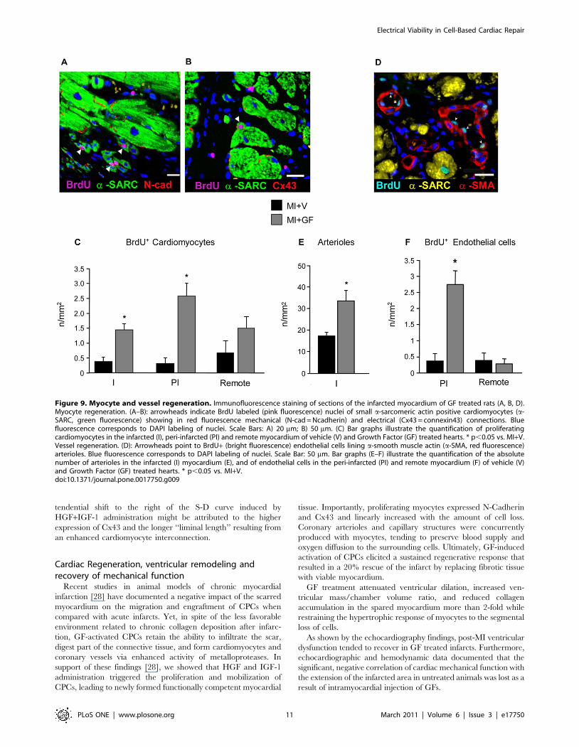
regeneration. Areas of myocardial regeneration were detected
in the infarcted portion of GF treated rat hearts. Small BrdU+myocytes, expressing N-Cadherin (Fig. 9A) and Cx43 (Fig. 9B)
were observed, indicating that newly formed, electromechanically
coupled cardiac myocytes resulted from local injection of GFs.
Within the two weeks from cytokine injection a nearly 3-fold
increase in cycling myocytes accumulated in MI+GF infarcts when
compared with the MI+V ones (Fig. 9C). New myocytes properly
integrated with the surrounding tissue were also present in the
peri-infarcted and remote myocardium of treated animals,
suggesting that local delivery of GFs exerted a global beneficial
effect on the heart (Fig. 9C). Although similar findings could
occasionally be detected in untreated chronic infarcts, the
quantitative estimation clearly indicated that the regenerative
processes were markedly enhanced in the MI+GF group in both
the peri-infarcted and infarcted areas (Fig. 9C). Importantly, the
newly formed myocardium was properly perfused, as indicated by
the significant increase in the density of arteriolar profiles (two-
fold) and BrdU+ endothelial cells (five-fold) (Fig. 9D–F).
Infarct size, as measured by the amount of myocytes lost 6 weeks
after coronary artery ligation (4 weeks prior + 2 weeks after GF/V
injection), was similar in the two experimental groups. As shown in
Fig. 10A, nearly 4.56106 myocytes were lost in the MI+V and
MI+GF hearts. Within the infarcted area, cytokine treatment
promoted the formation of 15.662.5 mm3 of new myocardium in
which an average of 5.960.16106 developing myocytes ranging in
size from 200 to a maximum of 4000 mm3 were present (Fig. 10B).
Most newly formed cardiomyocytes had a volume of less than
2000 mm3, although cytokine treatment was able to generate a
consistent number of cells approaching a volume of 4000 mm3. This
magnitude of myocardial regeneration resulted in a 21.261.7%
replacement of the infarcted volume by newly formed myocardium.
Conversely, myocardial repair in untreated infarcts was unremark-
able. It is noteworthy that the generation of new myocytes increased
linearly with the amount of cell loss in GF treated animals (Fig. 10C).
A similar reparative capacity was not observed in untreated rats. On
the other hand, myocardial regeneration was associated with intense
activation and translocation of resident progenitor cells (Fig. 10D).
In comparison with MI+V, a significant (p,0.05) increase in the
incidence of c-kit+ cells was produced by GFs in the infarcted, peri-
infarcted and remote myocardium. Altogether these findings
strongly suggest that GF-induced cardiac repair was mediated by
a multipotent stem cell population.
Figure 3. Epicardial multiple-lead recording. Strength-duration (S-D) curves (A) computed in representative rat hearts belonging to MI+V andMI+GF groups, before (solid line) and after (dotted line) GF/V injection. The bar graphs illustrate the mean values6SE of: (B) rheobase and chronaxie,(C) conduction velocity along (CV-l) and across (CV-t) epicardial fiber direction, and (D) effective refractory period (ERP), in MI+V and MI+GF groups.* p,0.001 vs. MI+V.doi:10.1371/journal.pone.0017750.g003
Electrical Viability in Cell-Based Cardiac Repair
PLoS ONE | www.plosone.org 6 March 2011 | Volume 6 | Issue 3 | e17750

Discussion
The induction or exacerbation of arrhythmias is a major
concern in stem cell based therapies for cardiac repair, and the
effect of regenerative treatments on cardiac electrogenesis is still
under debate [5–15,46–47]. The present results demonstrate that
intra-myocardial injection of HGF and IGF-1 in a rat model of
healed myocardial infarction caused a significant decline in
arrhythmias of conscious freely moving animals subjected to
stressful conditions. Concurrently, the effective refractory period
(ERP) at the ventricular surface of GF treated hearts was
prolonged without changes in dispersion of refractoriness, QTc
interval duration and mRNA levels of potassium channel a-
subunits Kv4.2 and Kv4.3. In contrast, lower mRNA levels of a-
subunit Kv1.4 and b-subunit KChIP2 were found in the remote
and remote/peri-infarcted myocardium respectively.
In support of previous data [26–28], we also found that resident
CPCs, stimulated locally by GF treatment, invaded the scarred
myocardium and generated new electromechanically-connected
myocytes and vessels. Additionally, experimental evidence was
provided for a partial recovery of mechanical competence in the
regenerated heart, associated with attenuation of unfavorable
remodeling. Finally, newly formed myocardial structures together
with a reduction in reactive cellular hypertrophy and interstitial
fibrosis were detected in peri-infarcted and remote myocardium,
suggesting that local delivery of GFs also exerted beneficial effects
in the spared tissue.
Recovery of Electrical FunctionExperimental electrophysiological studies aimed at assessing the
proarrhythmic or antiarrhythmic potential of stem cell based
Figure 4. Expression of K channel isoforms. Quantification ofKv1.4 (A, C) and KChIP2 (B) m-RNA levels, in ventricular myocardium. PI:peri-infarcted; Remote: spared left ventricular myocardium. Each barrepresents the mean of 4 different samples in triplicate. * p,0.05 vs.SO+V and MI+GF.doi:10.1371/journal.pone.0017750.g004
Figure 5. Quantification of connexin43 protein expression.Western blot analysis of connexin43 protein expression in infarcted (I)and remote (R) left ventricular myocardium of MI+V and MI+GF hearts.MW = molecular weight marker; OD = optical density. * p,0.05 vs. MI+V.doi:10.1371/journal.pone.0017750.g005
Electrical Viability in Cell-Based Cardiac Repair
PLoS ONE | www.plosone.org 7 March 2011 | Volume 6 | Issue 3 | e17750

therapies in vivo have been carried out mostly in anesthetized
animals and in a few cases in conscious animals in baseline
conditions [44]. While anesthesia can have confusing effects, the
incidence of arrhythmias in baseline conditions is generally low.
To circumvent these limitations, we measured the proneness to
arrhythmias in conscious freely moving rats subjected to the
resident-intruder test [32] which, by mimicking a natural situation,
ensured that the mechanisms of arrhythmias acting during stress
were in all probability the same which may trigger arrhythmias in
everyday life of social animals. The procedure is known to
consistently induce an intense activation of the autonomic nervous
system, with a shift of the sympatho-vagal balance towards a
sympathetic predominance, resulting in arrhythmias even in
normal animals [45]. Stress-induced ventricular arrhythmias were
reduced by about a half following GF injection, while no changes
were observed in untreated rats, indicating a protective effect of
local delivery of cytokines.
It has been recently reported [6] that important issues to
consider when assessing the effect of stem cell based myocardial
regeneration on cardiac electrical competence include: (i) intrinsic
electrophysiological properties of stem cells, (ii) modulated graft-
host and/or graft-graft electromechanical coupling, (iii) changes in
ion channel function, (iv) induced heterogeneity, and (v) altered
myocardial tissue architecture comprising heterogeneous sympa-
thetic nerve sprouting. All these factors may affect cardiac
electrogenesis either by acting alone or in concert with the
arrhythmic substrate of the injured myocardium.
We found that dispersion of refractoriness was similar in treated
and untreated animals, as were the indirect indexes of autonomic
input to the heart (SDRR and r-MSSD), signifying that GFs did not
alleviate arrhythmia susceptibility by affecting repolarization
heterogeneity [46] or cardiac sympathetic activity [47].
Quantitative RT-PCR analysis of the genes encoding the Ca2+-
independent transient outward potassium current (Ito) revealed
that the transcript expression of Kv1.4 was up-regulated in the
remote left ventricle only in untreated rats. Kv1.4 is the main
channel subunit contributing to Ito during the embryonic
development of mice and rats. After birth, Kv1.4 expression levels
decrease, while Kv4.2 and Kv4.3 are up-regulated [48].
Importantly, cardiac hypertrophy induces a re-expression of
Kv1.4 mRNA [49]. Furthermore, the rise in mRNA levels of
potassium channel b-subunit KChIP2 produced by myocardial
infarction was cancelled out by GF administration. Recent studies
documented higher mRNA levels of KChIP2 in cardiac
Figure 6. Blunting effect of GF treatment on the correlation between infarct size and echocardiographic parameters. Linearcorrelations between infarct size and ejection fraction (EF) and left ventricular end-systolic volume (LVESV), in MI+V (A–B) and MI+GF (A’–B’) animals.The statistical significance of correlations was limited to the MI+V group.doi:10.1371/journal.pone.0017750.g006
Electrical Viability in Cell-Based Cardiac Repair
PLoS ONE | www.plosone.org 8 March 2011 | Volume 6 | Issue 3 | e17750

hypertrophy [50] although a full appreciation of this result is
impossible, since to date the role of KChip(s) subunit(s) has not
been clearly elucidated. In conclusion, the lower mRNA levels of
Kv1.4 and KChIP2 subunits produced by GF injection might be
associated with the reduced reactive cellular hypertrophy following
cardiac repair. These findings, together with cardiac anatomical
and morphometric data, indicate that GF treatment may succeed
in partially controlling endogenous arrhythmic substrates by
limiting myocardial damage, and reducing myocardial fibrosis
and reactive cellular hypertrophy in the remote myocardium,
thereby helping to prevent the occurrence of reentry circuits.
By using epicardial multiple-lead recording, we showed that
cytokine injection resulted in a marked prolongation of ERP, while
conduction velocity (CV) was unchanged. The combined effect of
CV and ERP on impulse propagation is defined by the wavelength
L = CV x ERP, implying that a longer L (by increasing CV and/or
ERP) will reduce the likelihood that a single or multiple reentrant
circuits can be accommodated by the heart [43]. Hence, ERP
changes observed in GF treated rats could reduce the ability of
cardiac tissue to elicit an abnormal propagated response by
increasing L, thus hampering reentry. The finding that epicardial
QTc values were similar in all animals indicates that mechanisms
other than lengthening of action potential duration (APD) were
responsible for the longer ERP in GF treated rats. Accordingly,
either membrane currents affecting APD were not influenced by
GFs or, if they were, the net modification might not be large
enough to manifest itself at the tissue level. Indeed, our data show
that mRNA levels of the potassium channel a-subunits Kv4.2 and
Figure 7. Blunting effect of GF treatment on the correlation between infarct size and hemodynamic parameters. Linear correlationsbetween infarct size and maximum rate of ventricular pressure rise (+dP/dt) and left ventricular end-diastolic pressure (LVEDP), in MI+V (A–B) andMI+GF animals (A’–B’). The statistical significance of correlations was limited to the MI+V group.doi:10.1371/journal.pone.0017750.g007
Electrical Viability in Cell-Based Cardiac Repair
PLoS ONE | www.plosone.org 9 March 2011 | Volume 6 | Issue 3 | e17750

Kv4.3 did not exhibit any significant difference between untreated
and HGF+IGF-1 treated hearts, suggesting that the transient
outward potassium current (Ito), which contributes conspicuously
to determine APD, was unaltered.
On the other hand, recent studies have demonstrated that IGF-
1, alone or in combination with other growth factors, increases
Cx43 expression in cardiac myocytes [10]. In support of these
findings, we showed that GF administration significantly increased
the expression of the gap-junctional protein Cx43, providing
evidence for an increased electrical coupling within the scarred,
partially regenerated myocardium as well as in the remote spared
tissue. A better intercellular electrical connection is considered as
being an important antiarrhythmic factor [10–15,51] and could
also result in ERP prolongation. It has recently been remarked
[52] that the amount of current needed for cardiac excitation by
point stimulation is modulated by the properties of electrical
coupling of the interconnected myocytes via gap-junctions. The
smallest myocardial region capable of initiating a propagated
action potential (‘‘liminal length’’) is inversely related to the extra-
and intracellular resistivities, which are largely dependent on
interstitial fibrosis and electrical coupling between myocytes
respectively [52–53].
The role of myocardial geometry in the charge threshold for
excitation was documented by computing the S-D curve, which
shifted to the right when the ‘‘liminal length’’ increased (i.e.
reduced excitability) and to the left when it decreased (i.e.
increased excitability) [53]. Changes in ‘‘liminal length’’ resulting
from different degrees of electrical coupling between myocytes are
also expected to affect cardiac refractoriness, which will be
increased by an increase in ‘‘liminal length’’ and reduced by its
reduction. Concurrently, the longer ERP and the associated
Table 1. Left ventricular geometry and infarct size.
SO+V(n = 15)
MI+V(n = 23)
MI+GF(n = 22)
LV mass (mm3) 769643 1046635 * 1031617 *
LV wall thickness (mm) 2.260.1 2.060.1 2.260.1
LV chamber diameter (mm) 5.560.2 7.260.2 * 6.760.2 *
LV chamber volume (mm3) 199611 413625 * 336618 * #
mass/chamber volume 4.160.1 2.460.1 * 2.960.2 *
Infarct size (%) 19.463.2 19.962.9
Mean values6SE of left ventricular (LV) geometrical properties measured inSO+V, MI+V and MI+GF groups.*p,0.01 significant differences vs. SO+V group;# p,0.05 significant differences vs. MI+V.doi:10.1371/journal.pone.0017750.t001
Figure 8. Myocardial fibrosis and left ventricular myocyte size. Mean values6SE of volume fraction of fibrosis (A) and numerical density offibrotic foci (B), morphometrically evaluated in the remote left ventricular (LV) myocardium of MI+V and MI+GF hearts. In (C), mean values6SE of theLV myocyte cross-sectional area. * p,0.05 vs. MI+V; # p,0.05 vs. SO+V.doi:10.1371/journal.pone.0017750.g008
Electrical Viability in Cell-Based Cardiac Repair
PLoS ONE | www.plosone.org 10 March 2011 | Volume 6 | Issue 3 | e17750
tendential shift to the right of the S-D curve induced by
HGF+IGF-1 administration might be attributed to the higher
expression of Cx43 and the longer ‘‘liminal length’’ resulting from
an enhanced cardiomyocyte interconnection.
Cardiac Regeneration, ventricular remodeling andrecovery of mechanical function
Recent studies in animal models of chronic myocardial
infarction [28] have documented a negative impact of the scarred
myocardium on the migration and engraftment of CPCs when
compared with acute infarcts. Yet, in spite of the less favorable
environment related to chronic collagen deposition after infarc-
tion, GF-activated CPCs retain the ability to infiltrate the scar,
digest part of the connective tissue, and form cardiomyocytes and
coronary vessels via enhanced activity of metalloproteases. In
support of these findings [28], we showed that HGF and IGF-1
administration triggered the proliferation and mobilization of
CPCs, leading to newly formed functionally competent myocardial
tissue. Importantly, proliferating myocytes expressed N-Cadherin
and Cx43 and linearly increased with the amount of cell loss.
Coronary arterioles and capillary structures were concurrently
produced with myocytes, tending to preserve blood supply and
oxygen diffusion to the surrounding cells. Ultimately, GF-induced
activation of CPCs elicited a sustained regenerative response that
resulted in a 20% rescue of the infarct by replacing fibrotic tissue
with viable myocardium.
GF treatment attenuated ventricular dilation, increased ven-
tricular mass/chamber volume ratio, and reduced collagen
accumulation in the spared myocardium more than 2-fold while
restraining the hypertrophic response of myocytes to the segmental
loss of cells.
As shown by the echocardiography findings, post-MI ventricular
dysfunction tended to recover in GF treated infarcts. Furthermore,
echocardiographic and hemodynamic data documented that the
significant, negative correlation of cardiac mechanical function with
the extension of the infarcted area in untreated animals was lost as a
result of intramyocardial injection of GFs.
Figure 9. Myocyte and vessel regeneration. Immunofluorescence staining of sections of the infarcted myocardium of GF treated rats (A, B, D).Myocyte regeneration. (A–B): arrowheads indicate BrdU labeled (pink fluorescence) nuclei of small a-sarcomeric actin positive cardiomyocytes (a-SARC, green fluorescence) showing in red fluorescence mechanical (N-cad = Ncadherin) and electrical (Cx43 = connexin43) connections. Bluefluorescence corresponds to DAPI labeling of nuclei. Scale Bars: A) 20 mm; B) 50 mm. (C) Bar graphs illustrate the quantification of proliferatingcardiomyocytes in the infarcted (I), peri-infarcted (PI) and remote myocardium of vehicle (V) and Growth Factor (GF) treated hearts. * p,0.05 vs. MI+V.Vessel regeneration. (D): Arrowheads point to BrdU+ (bright fluorescence) endothelial cells lining a-smooth muscle actin (a-SMA, red fluorescence)arterioles. Blue fluorescence corresponds to DAPI labeling of nuclei. Scale Bar: 50 mm. Bar graphs (E–F) illustrate the quantification of the absolutenumber of arterioles in the infarcted (I) myocardium (E), and of endothelial cells in the peri-infarcted (PI) and remote myocardium (F) of vehicle (V)and Growth Factor (GF) treated hearts. * p,0.05 vs. MI+V.doi:10.1371/journal.pone.0017750.g009
Electrical Viability in Cell-Based Cardiac Repair
PLoS ONE | www.plosone.org 11 March 2011 | Volume 6 | Issue 3 | e17750

It could be argued that a single injection of cells or GFs hardly
promotes a sustained and prolonged cardiac repair in the absence
of a robust activation of autocrine/paracrine processes. Moreover,
the question remains as to whether a 20% recovery of myocardial
mass within the infarct, mostly consisting of small contractile cells,
is sufficient to prevent the evolution of myocardial infarction
toward cardiac decompensation. However, infarct size is propor-
tional to the number of lost cardiomyocytes, representing the
major determinant of unfavorable LV remodeling and its chronic
evolution toward heart failure [40]. Hence it is of relevance that
the GF-mediated generation of young functionally competent
cardiomyocytes succeeds in making cardiac function and anatomy
independent of the amount of tissue lost by the occlusion of the
supplying coronary artery. These results suggest that locally
delivered GFs, by reversing the hostile microenvironment of
scarred myocardium, promote a cardiac repair which is propor-
tional to tissue demand. Intriguingly, this phenomenon has been
observed in clinical experience of cell therapy for ischemic
cardiomyopathy [54–56], documenting greater improvement in
global cardiac function in patients who had a lower ejection
fraction at baseline. Thus, regenerative approaches seem to exert a
more beneficial effect on larger myocardial infarcts.
ConclusionAltogether, this study indicates that local injection in scarred
infarcts of active peptides able to recapitulate an endogenous cell
program aimed at generating functionally competent myocardi-
um, besides having a positive impact on the mechanical properties
of the injured heart, results in a significant decline in proneness to
arrhythmias. Both ERP prolongation (likely mediated by improved
intercellular electrical coupling leading to increased liminal length
for impulse formation) and positive ventricular structural remod-
eling contribute to the reduced ventricular arrhythmogenesis.
Supporting Information
File S1 Detailed description of the experimental design, surgical
procedures, electrophysiological measurements (telemetry ECGs
and multiple-lead epicardial recording), echocardiographic and
hemodynamic measurements, morphometrical and immunohisto-
Figure 10. Quantitative analysis of cardiac progenitor cells and cardiomyocyte loss and regeneration. In the presence of similaramounts of myocyte loss (A), compared to vehicle treated infarcts (MI+V), intramyocardial injection of Growth Factors (GF) produced a significantnumber of small, immature and larger, more mature cardiomyocytes (B) whose generation was directly related to the number of lost cells (C).Treatment with GFs was also able to increase the incidence of c-kit+ cardiac progenitors in the infarcted, peri-infarcted (PI) and remote myocardium(D). * p,0.05 vs. MI+V.doi:10.1371/journal.pone.0017750.g010
Electrical Viability in Cell-Based Cardiac Repair
PLoS ONE | www.plosone.org 12 March 2011 | Volume 6 | Issue 3 | e17750
chemical analyses, and biochemical and molecular biology
analyses (electrophoresis and immunoblot assay, and RT-PCR).
(DOC)
Table S1 Outline of the experimental protocols.
(DOC)
Table S2 Echocardiographic data.
(DOC)
Figure S1 Epicardial multiple-lead recording: electrodearrays and procedures. (A) Electrode array positioned on the
anterior aspect of the ventricular surface. (B) Schematic represen-
tation of the 565 (in red) and 868 electrode arrays showing the
positions (larger circles) of the 5 and 8 selected electrodes used for
specific pacing protocols. (C–D): Unipolar electrograms collected
by means of the 868 electrode array during normal sinus rhythm
(C) and ventricular pacing at the electrode indicated by the pulse
symbol (D). In (E), example of paced activation isochrone map
used for computing conduction velocity longitudinally (blue-arrow)
and transversally (red-arrow) to fiber orientation; numbers on each
isochrone line indicate the activation time in ms. In (F), strength-
duration curve obtained in a control rat by plotting pulse threshold
current I as a function of pulse duration T (Rh: Rheobase, Chr:
Chronaxie).
(TIF)
Author Contributions
Conceived and designed the experiments: E. Musso FQ DS. Performed the
experiments: LB MS GG AA FS CL SB RB CF. Analyzed the data: LB
MS SR E. Macchi EC US GG. Wrote the paper: E. Musso FQ DS MV
EC E. Macchi.
References
1. Marban E, Cheng K (2010) Heart to heart: The elusive mechanism of cell
therapy. Circulation 121: 1981–1984.
2. Dimmeler S, Zeiher AM, Schneider MD (2005) Unchain my heart: the scientific
foundations of cardiac repair. J Clin Invest 115: 572–583.
3. Joggerst SJ, Hatzopoulos AK (2009) Stem cell therapy for cardiac repair: benefits
and barriers. Expert Rev Mol Med 11: e20.
4. Leri A, Kajstura J, Anversa P (2005) Cardiac stem cells and mechanisms of
myocardial regeneration. Physiol Rev 85: 1373–1416.
5. Chen HS, Kim C, Mercola M (2009) Electrophysiological challenges of cell-
based myocardial repair. Circulation 120: 2496–2508.
6. Ly HQ, Nattel S (2009) Stem cells are not proarrhythmic: letting the genie out of
the bottle. Circulation 119: 1824–1831.
7. Macia E, Boyden PA (2009) Stem cell therapy is proarrhythmic. Circulation
119: 1814–1823.
8. Menasche P (2009) Stem cell therapy for heart failure: are arrhythmias a realsafety concern? Circulation 119: 2735–2740.
9. Smith RR, Barile L, Messina E, Marban E (2008) Stem cells in the heart: what’sthe buzz all about? Part 2: Arrhythmic risks and clinical studies. Heart Rhythm
5: 880–887.
10. Hahn JY, Cho HJ, Kang HJ, Kim TS, Kim MH, et al. (2008) Pre-treatment of
mesenchymal stem cells with a combination of growth factors enhances gapjunction formation, cytoprotective effect on cardiomyocytes, and therapeutic
efficacy for myocardial infarction. J Am Coll Cardiol 51: 933–943.
11. Kuhlmann MT, Kirchhof P, Klocke R, Hasib L, Stypmann J, et al. (2006) G-CSF/SCF reduces inducible arrhythmias in the infarcted heart potentially via
increased connexin43 expression and arteriogenesis. J Exp Med 203: 87–97.
12. Mills WR, Mal N, Kiedrowski MJ, Unger R, Forudi F, et al. (2007) Stem cell
therapy enhances electrical viability in myocardial infarction. J Mol Cell Cardiol42: 304–314.
13. Roell W, Lewalter T, Sasse P, Tallini YN, Choi BR, et al. (2007) Engraftment ofconnexin 43-expressing cells prevents post-infarct arrhythmia. Nature 450:
819–824.
14. Gepstein L, Ding C, Rehemedula D, Wilson EE, Yankelson L, et al. (2010) Invivo assessment of the electrophysiological integration and arrhythmogenic risk
of myocardial cell transplantation strategies. Stem Cells 28: 2151–2161.
15. Wang D, Zhang F, Shen W, Chen M, Yang B, et al. (2010) Mesenchymal stem
cell injection ameliorates the inducibility of ventricular arrhythmias aftermyocardial infarction in rats. Int J Cardiol Epub of print;(DOI: 10.1016/
j.ijcard.2010.07.025).
16. Beltrami AP, Barlucchi L, Torella D, Baker M, Limana F, et al. (2003) Adult
cardiac stem cells are multipotent and support myocardial regeneration. Cell
114: 763–776.
17. Oh H, Bradfute SB, Gallardo TD, Nakamura T, Gaussin V, et al. (2003)
Cardiac progenitor cells from adult myocardium: homing, differentiation, andfusion after infarction. Proc Natl Acad Sci U S A 100: 12313–12318.
18. Martin CM, Meeson AP, Robertson SM, Hawke TJ, Richardson JA, et al.(2004) Persistent expression of the ATP-binding cassette transporter, Abcg2,
identifies cardiac SP cells in the developing and adult heart. Dev Biol 265:
262–275.
19. Messina E, De Angelis L, Frati G, Morrone S, Chimenti S, et al. (2004) Isolation
and expansion of adult cardiac stem cells from human and murine heart. CircRes 95: 911–921.
20. Laugwitz KL, Moretti A, Lam J, Gruber P, Chen Y, et al. (2005) Postnatal isl1+cardioblasts enter fully differentiated cardiomyocyte lineages. Nature 433:
647–653.
21. Limana F, Zacheo A, Mocini D, Mangoni A, Borsellino G, et al. (2007)
Identification of myocardial and vascular precursor cells in human and mouse
epicardium. Circ Res 101: 1255–1265.
22. Davis DR, Kizana E, Terrovitis J, Barth AS, Zhang Y, et al. (2010) Isolation and
expansion of functionally-competent cardiac progenitor cells directly from heart.
J Mol Cell Cardiol 49: . pp 312–321.
23. Quaini F, Urbanek K, Beltrami AP, Finato N, Beltrami CA, et al. (2002)
Chimerism of the transplanted heart. N Engl J Med 346: 5–15.
24. Chamuleau SA, Vrijsen KR, Rokosh DG, Tang XL, Piek JJ, et al. (2009) Cell
therapy for ischaemic heart disease: focus on the role of resident cardiac stem
cells. Neth Heart J 17: 199–207.
25. Dawn B, Stein AB, Urbanek K, Rota M, Whang B, et al. (2005) Cardiac stem
cells delivered intravascularly traverse the vessel barrier, regenerate infarcted
myocardium, and improve cardiac function. Proc Natl Acad Sci U S A 102:
3766–3771.
26. Urbanek K, Rota M, Cascapera S, Bearzi C, Nascimbene A, et al. (2005)
Cardiac stem cells possess growth factor-receptor systems that after activation
regenerate the infarcted myocardium, improving ventricular function and long-
term survival. Circ Res 97: 663–673.
27. Linke A, Muller P, Nurzynska D, Casarsa C, Torella D, et al. (2005) Stem cells
in the dog heart are self-renewing, clonogenic, and multipotent and regenerate
infarcted myocardium, improving cardiac function. Proc Natl Acad Sci U S A
102: 8966–8971.
28. Rota M, Padin-Iruegas ME, Misao Y, De Angelis A, Maestroni S, et al. (2008)
Local activation or implantation of cardiac progenitor cells rescues scarred
infarcted myocardium improving cardiac function. Circ Res 103: 107–116.
29. Gheorghiade M, Bonow RO (1998) Chronic heart failure in the United States: a
manifestation of coronary artery disease. Circulation 97: 282–289.
30. Kjekshus J (1990) Arrhythmias and mortality in congestive heart failure.
Am J Cardiol 65: 421–481.
31. Sgoifo A, Stilli D, Medici D, Gallo P, Aimi B, et al. (1996) Electrode positioning
for reliable telemetry ECG recordings during social stress in unrestrained rats.
Physiol Behav 60: 1397–1401.
32. Martinez M, Calvo Torrent A, Pico Alfonso MA (1998) Social defeat and
subordination as models of social stress in laboratory rodents: a review. Aggress
Behav 24: 241–256.
33. Task Force of the European Society of Cardiology and the North American
Society of Pacing and Electrophysiology (1996) Heart rate variability. Standards
of measurement, physiological interpretation, and clinical use. Circulation 93:
1043–1075.
34. Pollick C, Hale SL, Kloner RA (1995) Echocardiographic and cardiac Doppler
assessment of mice. J Am Soc Echocardiogr 8: 602–610.
35. Macchi E, Cavalieri M, Stilli D, Musso E, Baruffi S, et al. (1998) High-density
epicardial mapping during current injection and ventricular activation in rat
hearts. Am J Physiol Heart Circ Physiol 275: H1886–1897.
36. Ogawa S, Furuno I, Satoh Y, Yoh S, Saeki K, et al. (1991) Quantitative indices
of dispersion of refractoriness for identification of propensity to re-entrant
ventricular tachycardia in a canine model of myocardial infarction. Cardiovasc
Res 25: 378–383.
37. Mitchell GF, Jeron A, Koren G (1998) Measurement of heart rate and Q-T
interval in the conscious mouse. Am J Physiol Heart Circ Physiol 274:
H747–H751.
38. Fuller MS, Sandor G, Punske B, Taccardi B, MacLeod RS, et al. (2000)
Estimates of repolarization dispersion from electrocardiographic measurements.
Circulation 102: 685–691.
39. Dodge HT, Baxley WA (1969) Left ventricular volume and mass and their
significance in heart disease. Am J Cardiol 23: 528–537.
40. Olivetti G, Capasso JM, Meggs LG, Sonnenblick EH, Anversa P (1991) Cellular
basis of chronic ventricular remodeling after myocardial infarction in rats. Circ
Res 68: 856–869.
Electrical Viability in Cell-Based Cardiac Repair
PLoS ONE | www.plosone.org 13 March 2011 | Volume 6 | Issue 3 | e17750

41. Orlic D, Kajstura J, Chimenti S, Limana F, Jakoniuk I, et al. (2001) Mobilized
bone marrow cells repair the infarcted heart, improving function and survival.Proc Natl Acad Sci U S A 98: 10344–10349.
42. Stilli D, Lagrasta C, Berni R, Bocchi L, Savi M, et al. (2007) Preservation of
ventricular performance at early stages of diabetic cardiomyopathy involveschanges in myocyte size, number and intercellular coupling. Basic Res Cardiol
102: 488–499.43. van Rijen HV, van Veen TA, Gros D, Wilders R, de Bakker JM (2006)
Connexins and cardiac arrhythmias. Adv Cardiol 42: 150–160.
44. Kolettis TM (2006) Arrhythmogenesis after cell transplantation post-myocardialinfarction. Four burning questions-and some answers. Cardiovasc Res 69:
299–301.45. Stilli D, Berni R, Bocchi L, Zaniboni M, Cacciani F, et al. (2004) Vulnerability
to ventricular arrhythmias and heterogeneity of action potential duration innormal rats. Exp Physiol 89: 387–396.
46. Fukushima S, Varela-Carver A, Coppen SR, Yamahara K, Felkin LE, et al.
(2007) Direct intramyocardial but not intracoronary injection of bone marrowcells induces ventricular arrhythmias in a rat chronic ischemic heart failure
model. Circulation 115: 2254–2261.47. Pak HN, Qayyum M, Kim DT, Hamabe A, Miyauchi Y, et al. (2003)
Mesenchymal stem cell injection induces cardiac nerve sprouting and increased
tenascin expression in a swine model of myocardial infarction. J CardiacElectrophysiol 14: 841–848.
48. van der Heyden MA, Wijnhoven TJ, Opthof T (2006) Molecular aspects ofadrenergic modulation of the transient outward current. Cardiovasc Res 71:
430–442.
49. Marionneau C, Brunet S, Flagg TP, Pilgram TK, Demolombe S, et al. (2008)
Distinct cellular and molecular mechanisms underlie functional remodeling of
repolarizing K+ currents with left ventricular hypertrophy. Circ Res 102:
1406–1415.
50. Tozakidou M, Goltz D, Hagenstrom T, Budack MK, Vitzthum H, et al. (2010)
Molecular and functional remodeling of I(to) by angiotensin II in the mouse left
ventricle. J Mol Cell Cardiol 48: 140–151.
51. Duffy HS (2008) Cardiac Connections-The Antiarrhythmic Solution?
N Engl J Med 358: 1397–1398.
52. Fozzard HA (2001) Gap junctions and liminal length in hypertrophy: something
old and something new. J Cardiovasc Electrophysiol 12: 836–837.
53. Fozzard HA, Schoenberg M (1972) Strength-duration curves in cardiac Purkinje
fibres: effects of liminal length and charge distribution. J Physiol 226: 593–618.
54. Schachinger V, Assmus B, Britten MB, Honold J, Lehmann R, et al. (2004)
Transplantation of progenitor cells and regeneration enhancement in acute
myocardial infarction: final one-year results of the TOPCARE-AMI Trial. J Am
Coll Cardiol 44: 1690–1699.
55. Schachinger V, Erbs S, Elsasser A, Haberbosch W, Hambrecht R, et al. (2006)
Intracoronary bone marrow-derived progenitor cells in acute myocardial
infarction. N Engl J Med 355: 1210–1221.
56. Schachinger V, Erbs S, Elsasser A, Haberbosch W, Hambrecht R, et al. (2006)
Improved clinical outcome after intracoronary administration of bone-marrow-
derived progenitor cells in acute myocardial infarction: final 1-year results of the
REPAIR-AMI trial. Eur Heart J 27: 2775–2783.
Electrical Viability in Cell-Based Cardiac Repair
PLoS ONE | www.plosone.org 14 March 2011 | Volume 6 | Issue 3 | e17750