Involvement of Foxo transcription factors in angiogenesis and postnatal neovascularization
Upload
newcastle-auCategory
view
4download
0
Haruchika Masuda, Takayuki Asahara and Douglas W. LosordoKawamoto, Raj Kishore, Yan Zhu, Gangjian Qin, Marcy Silver, Tina Thorne, Liz Eaton,
Atsushi Iwakura, Shubha Shastry, Corinne Luedemann, Hiromichi Hamada, AtsuhikoMetalloproteinase-9
Mediated Activation of Matrix−Neovascularization via Endothelial Nitric Oxide Synthase Derived Endothelial Progenitor Cells Into Sites of Ischemia-Induced−of Bone Marrow
Estradiol Enhances Recovery After Myocardial Infarction by Augmenting Incorporation
Print ISSN: 0009-7322. Online ISSN: 1524-4539 Copyright © 2006 American Heart Association, Inc. All rights reserved.
is published by the American Heart Association, 7272 Greenville Avenue, Dallas, TX 75231Circulation doi: 10.1161/CIRCULATIONAHA.105.553925
2006;113:1605-1614; originally published online March 13, 2006;Circulation.
http://circ.ahajournals.org/content/113/12/1605World Wide Web at:
The online version of this article, along with updated information and services, is located on the
http://circ.ahajournals.org/content/suppl/2006/03/13/CIRCULATIONAHA.105.553925.DC1.htmlData Supplement (unedited) at:
http://circ.ahajournals.org//subscriptions/
is online at: Circulation Information about subscribing to Subscriptions:
http://www.lww.com/reprints Information about reprints can be found online at: Reprints:
document. Permissions and Rights Question and Answer this process is available in the
click Request Permissions in the middle column of the Web page under Services. Further information aboutOffice. Once the online version of the published article for which permission is being requested is located,
can be obtained via RightsLink, a service of the Copyright Clearance Center, not the EditorialCirculationin Requests for permissions to reproduce figures, tables, or portions of articles originally publishedPermissions:
by guest on June 24, 2014http://circ.ahajournals.org/Downloaded from by guest on June 24, 2014http://circ.ahajournals.org/Downloaded from
Estradiol Enhances Recovery After Myocardial Infarctionby Augmenting Incorporation of Bone Marrow–Derived
Endothelial Progenitor Cells Into Sites of Ischemia-InducedNeovascularization via Endothelial Nitric Oxide
Synthase–Mediated Activation of Matrix Metalloproteinase-9Atsushi Iwakura, MD, PhD; Shubha Shastry, PhD; Corinne Luedemann, BS;
Hiromichi Hamada, MD, PhD; Atsuhiko Kawamoto, MD, PhD; Raj Kishore, PhD;Yan Zhu, MD, PhD; Gangjian Qin, MD, PhD; Marcy Silver, MS; Tina Thorne, MS; Liz Eaton, BS;
Haruchika Masuda, MD, PhD; Takayuki Asahara, MD, PhD; Douglas W. Losordo, MD
Background—Recent data have indicated that estradiol can modulate the kinetics of endothelial progenitor cells (EPCs)via endothelial nitric oxide synthase (eNOS)–dependent mechanisms. We hypothesized that estradiol could augment theincorporation of bone marrow (BM)–derived EPCs into sites of ischemia-induced neovascularization, resulting inprotection from ischemic injury.
Methods and Results—Myocardial infarction (MI) was induced by ligation of the left coronary artery in ovariectomizedmice receiving either 17�-estradiol or placebo. Estradiol induced significant increases in circulating EPCs 2 and 3 weeksafter MI in estradiol-treated animals, and capillary density was significantly greater in estradiol-treated animals. Greaternumbers of BM-derived EPCs were observed at ischemic sites in estradiol-treated animals than in placebo-treatedanimals 1 and 4 weeks after MI. In eNOS-null mice, the effect of estradiol on mobilization of EPCs was lost, as wasthe functional improvement in recovery from acute myocardial ischemia. A decrease was found in matrixmetalloproteinase-9 (MMP-9) expression in eNOS-null mice under basal and estradiol-stimulated conditions after MI,the mobilization of EPCs by estradiol was lost in MMP-9–null mice, and the functional benefit conferred by estradioltreatment after MI in wild-type mice was significantly attenuated.
Conclusions—Estradiol preserves the integrity of ischemic tissue by augmenting the mobilization and incorporation ofBM-derived EPCs into sites of neovascularization by eNOS-mediated augmentation of MMP-9 expression in the BM.Moreover, these data have broader implications with regard to our understanding of the role of EPCs in post-MIrecovery and on the sex discrepancy in cardiac events. (Circulation. 2006;113:1605-1614.)
Key Words: angiogenesis � endothelium � myocardial infarction � nitric oxide synthase � stem cells
Adult organs exhibit virtually no angiogenesis undernormal conditions, except in the female reproductive
tract. Several lines of experimental evidence have demon-strated that ovarian sex steroid hormones, such as estrogenand progesterone, modulate angiogenesis via effects on en-dothelial cells. Estradiol induces endothelial proliferation andmigration1 mediated by the classic estrogen receptor, which isexpressed by endothelial cells.2–4 Previously, endometrialneovascularization throughout the menstrual cycle has beenconsidered to be the result of angiogenesis, ie, proliferationand migration of fully differentiated endothelial cells from
preexisting “parent” vessels.5,6 However, normal monthlyphysiological endometrial proliferation would require thatendothelial cells in the uterus replicate �1000 times duringthe reproductive life span of the average human female.Accordingly, it is unlikely that differentiated endothelial cellsin situ could accomplish this mission without the occurrenceof replicative senescence.7
Clinical Perspective p 1614
Recently, endothelial progenitor cells (EPCs) isolated fromperipheral blood have been shown to incorporate into foci of
Received August 17, 2004; de novo received April 5, 2005; revision received January 16, 2006; accepted January 20, 2006.From the Division of Cardiovascular Research, St. Elizabeth’s Medical Center, Tufts University School of Medicine, Boston, Mass (A.I., S.S., C.L.,
H.H., R.K., Y.Z., G.Q., M.S., T.T., L.E., D.W.L.); and the Division of Regenerative Medicine, Institute of Biomedical Research and Innovation, Kobe,Japan (A.K., H.M., T.A.).
The online-only Data Supplement can be found at http://circ.ahajournals.org/cgi/content/full/CIRCULATIONAHA.105.553925/DC1.Correspondence to Douglas W. Losordo, MD, St. Elizabeth’s Medical Center of Boston, 736 Cambridge St, Boston, MA 02135. E-mail
[email protected]© 2006 American Heart Association, Inc.
Circulation is available at http://www.circulationaha.org DOI: 10.1161/CIRCULATIONAHA.105.553925
1605
Molecular Cardiology
by guest on June 24, 2014http://circ.ahajournals.org/Downloaded from

neovascularization in the adult; this is consistent with thenotion of postnatal vasculogenesis.8,9 These circulating EPCsare derived from bone marrow (BM) and are mobilizedendogenously in response to tissue ischemia or exogenouslyby cytokine stimulation.10 Previous findings from our labo-ratory11 have suggested that cyclic neovascularization of theendometrium involves estradiol-regulated in situ incorpora-tion and differentiation of BM-derived EPCs and that estra-diol could also augment the recruitment of EPCs for vascularrepair.12,13 These prior studies have also suggested thatestradiol exerts its effects on EPCs via an endothelial nitricoxide synthase (eNOS)–dependent mechanism. The effect ofestradiol on vascular repair of the heart, and in particular thepotential role of EPCs in this process, has not been previouslyinvestigated. Accordingly, in the present study, we investi-gated the hypothesis that estradiol may augment EPC incor-poration into sites of myocardial neovascularization aftermyocardial infarction (MI).
MethodsA detailed Methods section is provided in the online Data Supple-ment available at http://circ.ahajournals.org/cgi/content/full/CIRCULATIONAHA.105.553925/DC1.
Statistical AnalysisAll values are expressed as mean�SEM. Statistical significance wasevaluated using unpaired Student t tests for comparisons betweenestradiol- and placebo-treated mice. When multiple time-point mea-surements were taken over time, repeated-measures analysis wasperformed, followed by an unpaired t test. Differences in mortalityrates were compared by using Fisher exact tests. A value of P�0.05was considered statistically significant.
The authors had full access to the data and take responsibility forits integrity. All authors have read and agreed to the article aswritten.
ResultsEstradiol Preserves Left Ventricular FunctionAfter Acute MIMI was induced in ovariectomized mice receiving eitherestradiol or placebo via a subcutaneously implanted pellet.13
Left ventricular (LV) function and dimensions (LV diastolicdimension [LVDd], LV systolic dimension [LVDs], frac-tional shortening, and heart rate) were similar in estradiol-and placebo-treated mice before and for the first 2 weeks afterMI (Table). Beginning 3 weeks after MI, however, echocar-diography revealed less ventricular dilation in the estradiol-treated versus placebo-treated mice (LVDd, 3.7�0.06 versus4.0�0.05 mm, respectively, at 3 weeks, and 3.8�0.04 versus
Hemodynamic Parameters in Mice Receiving Estrogen or Placebo
After MI
n Before MI 1 wk 2 wk 3 wk 4 wk
LVDd, mm
Estrogen 15 2.8�0.03 3.1�0.04 3.9�0.07 3.7�0.06* 3.8�0.04*
Placebo 10 2.8�0.03 3.0�0.04 4.0�0.12 4.0�0.05 4.2�0.06
LVDs, mm
Estrogen 15 1.4�0.03 1.9�0.04 2.8�0.09 2.5�0.04* 2.6�0.05*
Placebo 10 1.4�0.02 1.8�0.03 3.0�0.11 3.0�0.08 3.2�0.06
FS, %
Estrogen 15 52�0.4 38�1.0 30�1.7 31�0.5* 32�0.7*
Placebo 10 52�0.6 39�0.7 25�1.2 24�0.9 23�1.2
HR, bpm
Estrogen 15 478�13 511�12 532�6 502�10 492�8*
Placebo 10 487�13 517�16 565�11 542�12 562�10
LVSP, mm Hg
Estrogen 6 � � � 62�3.1 � � � � � � 74�2.7
Placebo 6 � � � 62�1.5 � � � � � � 67�2.4
LVEDP, mm Hg
Estrogen 6 � � � 1.4�0.2 � � � � � � 1.6�0.4
Placebo 6 � � � 2.8�0.6 � � � � � � 5.4�1.9
LV �dP/dt, mm Hg/s
Estrogen 6 � � � 3094�117 � � � � � � 2984�155†
Placebo 6 � � � 2856�79 � � � � � � 2252�180
LV �dP/dt, mm Hg/s
Estrogen 6 � � � 2550�85 � � � � � � 2122�107*
Placebo 6 � � � 2081�183 � � � � � � 1650�83
Values are mean�SEM. FS indicates fractional shortening; HR, heart rate; LVSP, left ventricularsystolic pressure; and LVEDP, left ventricular end-diastolic pressure.
*P�0.01 and †P�0.05 vs placebo group.
1606 Circulation March 28, 2006
by guest on June 24, 2014http://circ.ahajournals.org/Downloaded from

4.2�0.06 mm, respectively, at 4 weeks [P�0.01]; LVDs,2.5�0.04 versus 3.0�0.08 mm, respectively, at 3 weeks and2.6�0.05 versus 3.2�0.06 mm, respectively, at 4 weeks[P�0.01]). LV function was also significantly better in theestradiol group than in the placebo group (fractional shorten-ing, 31�0.5 versus 24�0.9 mm, respectively, at 3 weeks and32�0.7 versus 23�1.2 mm, respectively, at 4 weeks[P�0.01]). Finally, heart rate, an excellent prognostic indi-cator after MI, was lower (better) in the estradiol group thanin the placebo group (492�8 versus 562�10 bpm, respec-tively [P�0.01]).
Hemodynamic measurements were performed 1 and 4weeks after MI (Table). There was no significant differencein LV systolic or end-diastolic pressure between the groups ateither time point after MI. However, both LV �dP/dt and LV�dP/dt, sensitive indicators of LV function, were signifi-cantly better preserved in the estradiol vs the placebo group 4weeks after MI (LV �dP/dt, 2984�155 versus2252�180 mm Hg/s, respectively [P�0.01]; LV �dP/dt,2122�107 versus 1650�83 mm Hg/s, respectively[P�0.01]).
Serum levels of 17�-estradiol in the placebo group 1 and 4weeks after MI were �2 pg/mL, whereas levels in theestradiol group after 1 and 4 weeks after MI were 580�55and 559�62 pg/mL, respectively; these values are consistentwith the upper range of levels in premenopausal females.13
Acute mortality within 1 week after MI in the estradiolgroups (mortality rate, 19%; total number of operatedmice�48, including 36 wild-type [WT] and 12 Tie-2/LacZ[LZ]/bone marrow transplant [BMT] mice) was similar tothat in the placebo group (mortality rate, 15%; total numberof operated mice�40, including 31 WT and 9 Tie-2/LZ/BMTmice).
Histological Assessment of WT AnimalsCapillary density 4 weeks after MI was significantly greaterin the estradiol group than in the placebo group (2432�110/mm2 versus 1134�39/mm2, respectively [P�0.01]; Figure1A and 1B). The finding of preserved/improved capillary
density has been associated with improved physiologicaloutcome in multiple animal models and has been correlatedwith a decreased incidence of heart failure.14
Elastic trichrome–stained tissue in the placebo group alsoindicated marked dilatation of the LV cavity, which isconsistent with the echocardiographic measurements. In com-parison, the estradiol group showed significantly less LVremodeling (Figure 1C). Moreover, the area of LV fibrosis,indicating the zone of permanent myocardial injury, wassignificantly less in mice receiving estradiol than in theplacebo group (36.0�2.6% versus 47.5�2.4%, respectively[P�0.05]; Figure 1D). These findings of preserved chamberdimensions (less dilation) and decreased LV fibrosis repre-sent positive long-term prognostic findings.15,16
Effect of Estradiol on Circulating EPC KineticsAfter Acute MIAs shown in Figure 1A, a greater number of BM-derivedEPCs, identified by double staining for 1,1�-dioctadecyl-3,3,3�,3�-tetramethylindocarbocyanine perchlorate (DiI)–acetylated LDL and Bandeiraea simplicifolia-1 lectin, wereobserved 2 and 3 weeks after MI in the estradiol group thanin the placebo group. It is notable that the stimulus of MIalone resulted in a significant increase in circulating EPCs inboth groups in the first week after MI, indicating thatmobilization of these cells is a natural response to myocardialinjury. Indeed, this has been documented in humans as well,17
with enhanced mobilization of EPCs correlated with betterlong-term outcome.18 Within 1 week after MI, however, thenumber of EPCs decreased in a time-dependent manner in theplacebo group (Figure 2B), whereas the estradiol groupmaintained a significantly higher number of circulating EPCsfor 2 and 3 weeks after MI (2 weeks, 430�56/mm2 versus212�19/mm2 for estradiol versus placebo groups, respec-tively; 3 weeks, 265�25/mm2 versus 161�17/mm2 for estra-diol versus placebo groups, respectively [P�0.01]). Thesefindings were corroborated by fluorescence-activated cellsorter (FACS) analysis of peripheral blood samples collected1, 2, and 4 weeks after MI, indicating that the number of
Figure 1. Estradiol increases capillarydensity and reduces fibrosis after MI. A,Shown are immunohistochemical find-ings in the peri-infarct myocardium afteruse of an antibody against isolectin B4at 4 weeks after MI (a indicates estradiolgroup; b, placebo group; original magni-fication �200). B, Capillary density isshown in peri-infarct myocardium 4weeks after MI in mice receiving estra-diol (n�15) or placebo (n�10). C, Elastictissue/trichrome-stained myocardium 4weeks after MI from mice receivingestradiol (a) or placebo (b) showsdecreased fibrosis with estradiol treat-ment. D, Ratio of fibrosis area to LV areain estradiol (n�15) and placebo (n�10)groups reveals decreased fibrosis (scar-ring) after MI with estradiol treatment.
Iwakura et al Estrogen Modulates Endothelial Progenitor Cells 1607
by guest on June 24, 2014http://circ.ahajournals.org/Downloaded from

Sca-1�/Flk-1� cells was consistently greater in the estradiol-treated mice than in placebo-treated animals (Figure 2C).
Enhanced Contribution of BM-Derived EPCs toMyocardial NeovascularizationNext, Tie-2/LZ/BMT mice were euthanized 1 and 4 weeksafter MI. As shown in Figure 3, macroscopic examination of5-bromo-4-chloro-3-indolyl �-D-galactopyranoside (X-gal)–stained hearts 1 week after MI revealed evidence of abundantrecruitment of BM-derived progenitor cells, indicated by theblue areas in the cross sections of the myocardium. Fourweeks after MI, macroscopic examination the LVs suggestedthat a significantly greater number of X-gal–positive cellswere present in the estradiol-treated mice than in the placebo-treated mice (Figure 3). Microscopic examination revealed
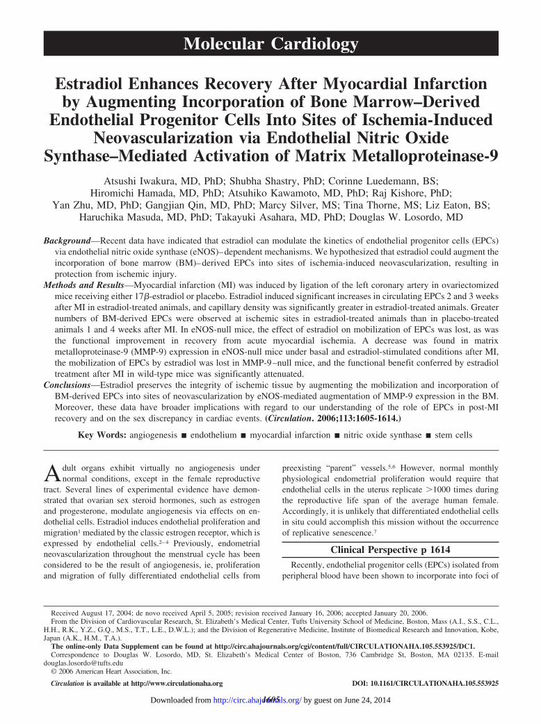
that at 1 and 4 weeks after MI, the number of X-gal–positivecells was significantly greater in the estradiol group than inthe placebo group (1 week, 298�24/mm2 versus 220�9/mm2,respectively [P�0.05]; 4 weeks, 82�8/mm2 versus 24�7/mm2, respectively [P�0.01]; Figure 4A and 4B). Further-more, fluorescence immunohistochemistry performed on fro-zen sections 1 week after MI documented an increase in cellsthat were double positive for �-galactosidase (indicating BMorigin and Tie-2 expression) and the endothelial cell–specificmarker isolectin B4 in the estradiol group (Figure 4C). Thesedata indicate that use of estradiol results in an increase in thekinetics of BM-derived EPCs, not only in the circulation butin the myocardium as well, resulting in enhanced incorpora-tion of BM-derived cells into the neovasculature after MI.
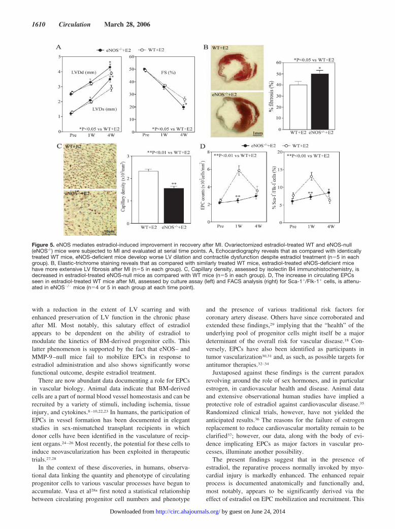
eNOS Is Required for Estradiol-InducedNeovascularizationPrior studies have suggested that eNOS is required forestradiol-mediated mobilization of EPCs after arterial inju-ry.13,19 Figure 5 shows the results of echocardiography beforeand 1 and 4 weeks after MI in eNOS�/� and WT micereceiving 17�-estradiol pellets. Despite estradiol administra-tion, there was more LV dilation in eNOS�/� mice than inidentically treated WT mice, indicating that the beneficialeffect of estradiol on LV remodeling after MI was lost in theabsence of eNOS (LVDd, 3.9�0.1 versus 4.3�0.1 mm,respectively [P�0.05]; LVDs, 2.9�0.2 versus 3.5�0.2 mm,respectively [P�0.05]). LV function, assessed by fractionalshortening, was also significantly worse despite estradioltreatment in eNOS�/� mice as compared with WT mice.Histological assessment also revealed that eNOS�/� miceshowed more LV fibrosis than did WT mice (50.0�2.7%versus 40.1�2.6%, respectively [P�0.05]; Figure 5B). More-over, capillary density 4 weeks after MI was significantlylower in estradiol-treated eNOS�/� mice than in identically
Figure 2. Estradiol augments EPC kinet-ics after MI. A, Representative immuno-fluorescent staining of EPCs culturedfrom mouse peripheral blood at serialtime points after MI. Circulating mononu-clear cells (PB-MNCs) were collected,and immunofluorescent staining forisolectin B4 was performed, followed bylabeling with DiI-acetylated LDL, both ofwhich identify endothelial cells. Double-positive cells are counted as EPCs (E1through E4 indicates estradiol group at1, 2, 3, and 4 weeks, respectively, afterMI; P1 through P4, placebo group at 1,2, 3, and 4 weeks, respectively, after MI;magnification �200). B and C, Circulat-ing EPC counts at serial time pointsbefore and after MI in estradiol (�E2)and placebo (�E2) groups assessed byculture assay (B) and FACS for Sca-1and Flk-1 expression (C) (n�6 in eachgroup at each time point).
Figure 3. Estradiol increases recruitment of BM-derived EPCsto the myocardium after MI. Representative photographs ofX-Gal–stained hearts from WT mice that were recipients of BMfrom Tie-2/LacZ donor mice. One week after MI, there is abun-dant blue staining in hearts from estradiol- and placebo-treatedmice, indicating acute recruitment of BM-derived Tie-2–ex-pressing cells. After 4 weeks, the blue color is visibly greater inthe myocardium of the estradiol-treated mouse (a and c indicateestradiol at 1 and 4 weeks after MI; b and d, placebo at 1 and 4weeks after MI).
1608 Circulation March 28, 2006
by guest on June 24, 2014http://circ.ahajournals.org/Downloaded from
treated WT mice (1560�83/mm2 versus 2335�84/mm2, re-spectively [P�0.01]; Figure 5C).
EPC counts, documented by FACS analysis and cultureassay significantly increased 1 week after MI only inestradiol-treated WT mice as compared with WT mice(culture assay, 588�55/mm2 versus 284�24 cells/mm2, re-spectively [P�0.01]; FACS analysis, 13.2�1.1% versus7.3�0.7%, respectively [P�0.01]; Figure 5D). Thus, estra-diol failed to increase EPC numbers after MI and failed toimprove functional and anatomic preservation after MI in theabsence of intact eNOS signaling.
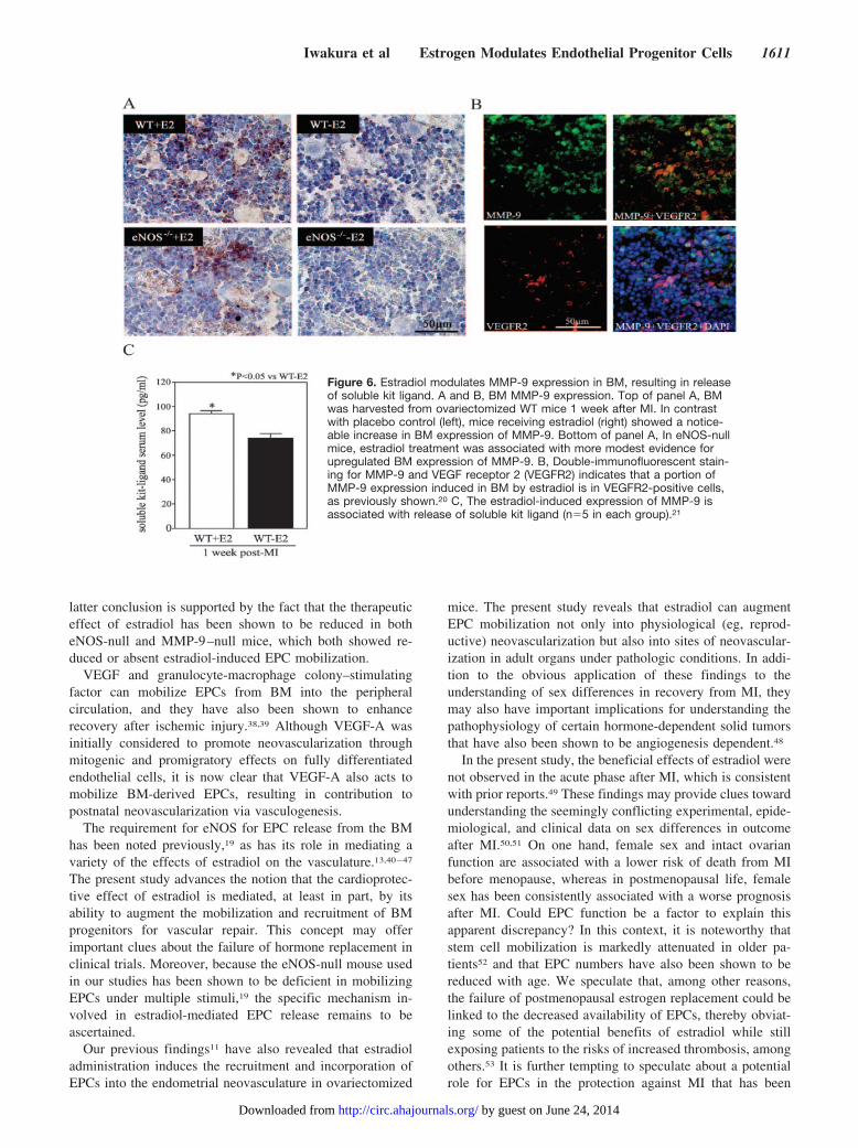
Matrix Metalloproteinase-9 Is Essential for EPCMobilization Induced by Estradiol After MIIn BM cells of untreated WT mice, mild matrixmetalloproteinase-9 (MMP-9) immunoreactivity was noted,whereas it was undetectable in the BM of untreated eNOS�/�
mice. In estradiol-treated mice, MMP-9 was strongly induced1 day after MI in the WT mice, whereas eNOS-null micerevealed a more modest upregulation of MMP-9 expression(Figure 6A). At least a portion of estradiol-induced MMP-9expression in the BM is from vascular endothelial growthfactor (VEGF) receptor 2–expressing cells (Figure 6B) andresults in release of soluble kit ligand (Figure 6C), consistentwith the documented role of this pathway invasculogenesis.20,21
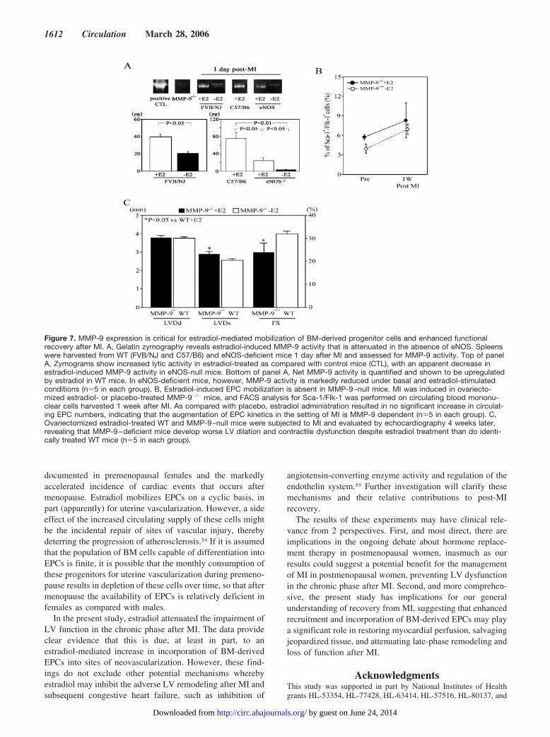
Next, we assessed MMP-9 activity with the use of gelatinzymography. MMP-9 activity was quantified as a band ofgelatinolytic activity, which was based on its specific molec-ular weight (92 kDa), and was confirmed by comparison withcommercially available pure MMP-9 (Figure 7A). As shownin Figure 6, estradiol administration resulted in robust induc-
tion of MMP-9 activity in WT (FVB/NJ) mice (for WT mice,40.0�3.0 pg for estradiol versus 21.0�2.0 pg for placebo).Examination of eNOS-null mice and comparison with WTmice revealed that basal MMP-9 activity was significantlyreduced in the eNOS-null mice as compared with WT miceand that estradiol-induced MMP-9 activity was also signifi-cantly attenuated in the eNOS-null mice as compared withWT control mice (for eNOS�/� mice, 24.0�6.7 pg forestradiol versus 3.0�0.6 pg for placebo). In direct compari-son, there was a significant difference in the induced MMP-9activity between WT mice (C57BL/6J) and eNOS�/� micereceiving 17�-estradiol pellets (76.3�11.7 versus 24.0�6.7pg, respectively [P�0.05]). These findings suggest thatestradiol-induced EPC mobilization after MI might occur byincreasing the activity of MMP-9 and that this is, at least inpart, dependent on intact eNOS function. This hypothesis wasstrongly supported by FACS analysis of circulating mononu-clear cells from MMP-9�/� mice before and 1 week after MI.Quantification of Sca-1�/Flk-1� cells revealed that estradiolhad no effect on circulating EPC counts after MI in MMP-9–null mice; ie, the MI-induced increase in circulating EPCswas not augmented by estradiol in the absence of MMP-9(Figure 7B). Most notably, MMP-9–null mice failed toachieve the full benefit of estradiol treatment after MI,showing worse LV dilation and more severely reducedcontractile function than was found with WT animals.
DiscussionThe present study provides evidence that estradiol mobilizescirculating EPCs from BM, resulting in incorporation intosites of neovascularization in the adult heart after MI.Moreover, this enhanced neovascularization is associated
Figure 4. Estradiol enhances the contri-bution of BM-derived EPCs to myocar-dial neovascularization. A, Representa-tive photomicrographs after X-galstaining of myocardium in Tie-2/LZ/BMTmice obtained 1 and 4 weeks after MI (aand b are from estradiol group; c and d,from placebo group). X-gal–positive cells(blue) indicate Tie-2–expressingBM-derived EPCs. B, Quantitative analy-ses of X-gal–positive cells in estradioland placebo groups 1 and 4 weeks afterMI indicate that at both time points afterMI, estradiol treatment resulted in greaternumbers of Tie-2–expressing cells in themyocardium (n�4 in each group). C,Representative photomicrographs afterdouble-immunofluorescent histochemis-try of myocardial tissue in Tie-2/LZ/BMTmice 1 week after MI. Green fluores-cence indicates endothelium-specificisolectin B4 binding in estradiol (E1) orplacebo (P1) groups; red fluorescenceindicates �-gal immunoreactivity in estra-diol (E2) or placebo (P2) groups. Thedouble-positive cells (arrows) indicateBM-derived EPCs that have incorporatedinto the myocardial neovasculature (E3indicates estradiol; P3, placebo).
Iwakura et al Estrogen Modulates Endothelial Progenitor Cells 1609
by guest on June 24, 2014http://circ.ahajournals.org/Downloaded from
with a reduction in the extent of LV scarring and withenhanced preservation of LV function in the chronic phaseafter MI. Most notably, this salutary effect of estradiolappears to be dependent on the ability of estradiol tomodulate the kinetics of BM-derived progenitor cells. Thislatter phenomenon is supported by the fact that eNOS– andMMP-9–null mice fail to mobilize EPCs in response toestradiol administration and also shows significantly worsefunctional outcome, despite estradiol treatment.
There are now abundant data documenting a role for EPCsin vascular biology. Animal data indicate that BM-derivedcells are a part of normal blood vessel homeostasis and can berecruited by a variety of stimuli, including ischemia, tissueinjury, and cytokines.8–10,22,23 In humans, the participation ofEPCs in vessel formation has been documented in elegantstudies in sex-mismatched transplant recipients in whichdonor cells have been identified in the vasculature of recip-ient organs.24–26 Most recently, the potential for these cells toinduce neovascularization has been exploited in therapeutictrials.27,28
In the context of these discoveries, in humans, observa-tional data linking the quantity and phenotype of circulatingprogenitor cells to various vascular processes have begun toaccumulate. Vasa et al28a first noted a statistical relationshipbetween circulating progenitor cell numbers and phenotype
and the presence of various traditional risk factors forcoronary artery disease. Others have since corroborated andextended these findings,29 implying that the “health” of theunderlying pool of progenitor cells might itself be a majordeterminant of the overall risk for vascular disease.18 Con-versely, EPCs have also been identified as participants intumor vascularization30,31 and, as such, as possible targets forantitumor therapies.32–34
Juxtaposed against these findings is the current paradoxrevolving around the role of sex hormones, and in particularestrogen, in cardiovascular health and disease. Animal dataand extensive observational human studies have implied aprotective role of estradiol against cardiovascular disease.35
Randomized clinical trials, however, have not yielded theanticipated results.36 The reasons for the failure of estrogenreplacement to reduce cardiovascular mortality remain to beclarified37; however, our data, along with the body of evi-dence implicating EPCs as major factors in vascular pro-cesses, illuminate another possibility.
The present findings suggest that in the presence ofestradiol, the reparative process normally invoked by myo-cardial injury is markedly enhanced. The enhanced repairprocess is documented anatomically and functionally and,most notably, appears to be significantly derived via theeffect of estradiol on EPC mobilization and recruitment. This
Figure 5. eNOS mediates estradiol-induced improvement in recovery after MI. Ovariectomized estradiol-treated WT and eNOS-null(eNOS-/-) mice were subjected to MI and evaluated at serial time points. A, Echocardiography reveals that as compared with identicallytreated WT mice, eNOS-deficient mice develop worse LV dilation and contractile dysfunction despite estradiol treatment (n�5 in eachgroup). B, Elastic-trichrome staining reveals that as compared with similarly treated WT mice, estradiol-treated eNOS-deficient micehave more extensive LV fibrosis after MI (n�5 in each group). C, Capillary density, assessed by isolectin B4 immunohistochemistry, isdecreased in estradiol-treated eNOS-null mice as compared with WT mice (n�5 in each group). D, The increase in circulating EPCsseen in estradiol-treated WT mice after MI, assessed by culture assay (left) and FACS analysis (right) for Sca-1�/Flk-1� cells, is attenu-ated in eNOS�/� mice (n�4 or 5 in each group at each time point).
1610 Circulation March 28, 2006
by guest on June 24, 2014http://circ.ahajournals.org/Downloaded from
latter conclusion is supported by the fact that the therapeuticeffect of estradiol has been shown to be reduced in botheNOS-null and MMP-9–null mice, which both showed re-duced or absent estradiol-induced EPC mobilization.
VEGF and granulocyte-macrophage colony–stimulatingfactor can mobilize EPCs from BM into the peripheralcirculation, and they have also been shown to enhancerecovery after ischemic injury.38,39 Although VEGF-A wasinitially considered to promote neovascularization throughmitogenic and promigratory effects on fully differentiatedendothelial cells, it is now clear that VEGF-A also acts tomobilize BM-derived EPCs, resulting in contribution topostnatal neovascularization via vasculogenesis.
The requirement for eNOS for EPC release from the BMhas been noted previously,19 as has its role in mediating avariety of the effects of estradiol on the vasculature.13,40–47
The present study advances the notion that the cardioprotec-tive effect of estradiol is mediated, at least in part, by itsability to augment the mobilization and recruitment of BMprogenitors for vascular repair. This concept may offerimportant clues about the failure of hormone replacement inclinical trials. Moreover, because the eNOS-null mouse usedin our studies has been shown to be deficient in mobilizingEPCs under multiple stimuli,19 the specific mechanism in-volved in estradiol-mediated EPC release remains to beascertained.
Our previous findings11 have also revealed that estradioladministration induces the recruitment and incorporation ofEPCs into the endometrial neovasculature in ovariectomized
mice. The present study reveals that estradiol can augmentEPC mobilization not only into physiological (eg, reprod-uctive) neovascularization but also into sites of neovascular-ization in adult organs under pathologic conditions. In addi-tion to the obvious application of these findings to theunderstanding of sex differences in recovery from MI, theymay also have important implications for understanding thepathophysiology of certain hormone-dependent solid tumorsthat have also been shown to be angiogenesis dependent.48
In the present study, the beneficial effects of estradiol werenot observed in the acute phase after MI, which is consistentwith prior reports.49 These findings may provide clues towardunderstanding the seemingly conflicting experimental, epide-miological, and clinical data on sex differences in outcomeafter MI.50,51 On one hand, female sex and intact ovarianfunction are associated with a lower risk of death from MIbefore menopause, whereas in postmenopausal life, femalesex has been consistently associated with a worse prognosisafter MI. Could EPC function be a factor to explain thisapparent discrepancy? In this context, it is noteworthy thatstem cell mobilization is markedly attenuated in older pa-tients52 and that EPC numbers have also been shown to bereduced with age. We speculate that, among other reasons,the failure of postmenopausal estrogen replacement could belinked to the decreased availability of EPCs, thereby obviat-ing some of the potential benefits of estradiol while stillexposing patients to the risks of increased thrombosis, amongothers.53 It is further tempting to speculate about a potentialrole for EPCs in the protection against MI that has been
Figure 6. Estradiol modulates MMP-9 expression in BM, resulting in releaseof soluble kit ligand. A and B, BM MMP-9 expression. Top of panel A, BMwas harvested from ovariectomized WT mice 1 week after MI. In contrastwith placebo control (left), mice receiving estradiol (right) showed a notice-able increase in BM expression of MMP-9. Bottom of panel A, In eNOS-nullmice, estradiol treatment was associated with more modest evidence forupregulated BM expression of MMP-9. B, Double-immunofluorescent stain-ing for MMP-9 and VEGF receptor 2 (VEGFR2) indicates that a portion ofMMP-9 expression induced in BM by estradiol is in VEGFR2-positive cells,as previously shown.20 C, The estradiol-induced expression of MMP-9 isassociated with release of soluble kit ligand (n�5 in each group).21
Iwakura et al Estrogen Modulates Endothelial Progenitor Cells 1611
by guest on June 24, 2014http://circ.ahajournals.org/Downloaded from
documented in premenopausal females and the markedlyaccelerated incidence of cardiac events that occurs aftermenopause. Estradiol mobilizes EPCs on a cyclic basis, inpart (apparently) for uterine vascularization. However, a sideeffect of the increased circulating supply of these cells mightbe the incidental repair of sites of vascular injury, therebydeterring the progression of atherosclerosis.54 If it is assumedthat the population of BM cells capable of differentiation intoEPCs is finite, it is possible that the monthly consumption ofthese progenitors for uterine vascularization during premeno-pause results in depletion of these cells over time, so that aftermenopause the availability of EPCs is relatively deficient infemales as compared with males.
In the present study, estradiol attenuated the impairment ofLV function in the chronic phase after MI. The data provideclear evidence that this is due, at least in part, to anestradiol-mediated increase in incorporation of BM-derivedEPCs into sites of neovascularization. However, these find-ings do not exclude other potential mechanisms wherebyestradiol may inhibit the adverse LV remodeling after MI andsubsequent congestive heart failure, such as inhibition of
angiotensin-converting enzyme activity and regulation of theendothelin system.55 Further investigation will clarify thesemechanisms and their relative contributions to post-MIrecovery.
The results of these experiments may have clinical rele-vance from 2 perspectives. First, and most direct, there areimplications in the ongoing debate about hormone replace-ment therapy in postmenopausal women, inasmuch as ourresults could suggest a potential benefit for the managementof MI in postmenopausal women, preventing LV dysfunctionin the chronic phase after MI. Second, and more comprehen-sive, the present study has implications for our generalunderstanding of recovery from MI, suggesting that enhancedrecruitment and incorporation of BM-derived EPCs may playa significant role in restoring myocardial perfusion, salvagingjeopardized tissue, and attenuating late-phase remodeling andloss of function after MI.
AcknowledgmentsThis study was supported in part by National Institutes of Healthgrants HL-53354, HL-77428, HL-63414, HL-57516, HL-80137, and
Figure 7. MMP-9 expression is critical for estradiol-mediated mobilization of BM-derived progenitor cells and enhanced functionalrecovery after MI. A, Gelatin zymography reveals estradiol-induced MMP-9 activity that is attenuated in the absence of eNOS. Spleenswere harvested from WT (FVB/NJ and C57/B6) and eNOS-deficient mice 1 day after MI and assessed for MMP-9 activity. Top of panelA, Zymograms show increased lytic activity in estradiol-treated as compared with control mice (CTL), with an apparent decrease inestradiol-induced MMP-9 activity in eNOS-null mice. Bottom of panel A, Net MMP-9 activity is quantified and shown to be upregulatedby estradiol in WT mice. In eNOS-deficient mice, however, MMP-9 activity is markedly reduced under basal and estradiol-stimulatedconditions (n�5 in each group). B, Estradiol-induced EPC mobilization is absent in MMP-9–null mice. MI was induced in ovariecto-mized estradiol- or placebo-treated MMP-9�/� mice, and FACS analysis for Sca-1/Flk-1 was performed on circulating blood mononu-clear cells harvested 1 week after MI. As compared with placebo, estradiol administration resulted in no significant increase in circulat-ing EPC numbers, indicating that the augmentation of EPC kinetics in the setting of MI is MMP-9 dependent (n�5 in each group). C,Ovariectomized estradiol-treated WT and MMP-9–null mice were subjected to MI and evaluated by echocardiography 4 weeks later,revealing that MMP-9–deficient mice develop worse LV dilation and contractile dysfunction despite estradiol treatment than do identi-cally treated WT mice (n�5 in each group).
1612 Circulation March 28, 2006
by guest on June 24, 2014http://circ.ahajournals.org/Downloaded from
P01 HL-66957. The authors gratefully acknowledge the secretarialassistance of Mickey Neely and Dierdre Couchon in the preparationof this manuscript.
DisclosuresNone.
References1. Morales DE, McGowan KA, Grant DS, et al. Estrogen promotes
angiogenic activity in human umbilical vein endothelial cells in vitro andin a murine model. Circulation. 1995;91:755–763.
2. Venkov CD, Rankin AB, Vaughan DE. Identification of authenticestrogen receptor in cultured endothelial cells. Circulation. 1996;94:727–733.
3. Kim-Schulze S, McGowan KA, Hubchak SC, et al. Expression of anestrogen receptor by human coronary artery and umbilical vein endothe-lial cells. Circulation. 1996;94:1402–1407.
4. Johns A, Freay AD, Fraser W, et al. Disruption of estrogen receptor geneprevents 17 beta estradiol–induced angiogenesis in transgenic mice.Endocrinology. 1996;137:4511–4513.
5. Folkman J, Klagsbrun M. Angiogenic factors. Science. 1987;235:442–447.
6. Ferrara N, Chen H, David-Smith T, et al. Vascular endothelial growthfactor is essential for corpus luteum angiogenesis. Nat Med. 1998;4:336–340.
7. Chang E, Harley CB. Telomere length and replicative aging in humanvascular tissues. Proc Natl Acad Science U S A. 1995;92:11190–11194.
8. Asahara T, Murohara T, Sullivan A, et al. Isolation of putative progenitorendothelial cells for angiogenesis. Science. 1997;275:964–967.
9. Shi Q, Rafii S, Wu MH-D, et al. Evidence for circulating bone marrow-–derived endothelial cells. Blood. 1998;92:362–367.
10. Takahashi T, Kalka C, Masuda H, et al. Ischemia- and cytokine-inducedmobilization of bone marrow–derived endothelial progenitor cells forneovascularization. Nat Med. 1999;5:434–438.
11. Masuda H, Kalka C, Takahashi T, Iwaguro H, Hayashi S, Silver M, ChenD, Ma H, Kearney M, Losordo DW, Isner JM, Asahara T. Regulatory roleof estrogen on endothelial progenitor cell kinetics for cyclic endometrialneovascularization. Circulation. 1999;100(suppl I):I-2503. Abstract.
12. Strehlow K, Werner N, Berweiler J, et al. Estrogen increases bonemarrow–derived endothelial progenitor cell production and diminishesneointima formation. Circulation. 2003;107:3059–3065.
13. Iwakura A, Luedemann C, Shastry S, et al. Estrogen-mediated, endothe-lial nitric oxide synthase– dependent mobilization of bone marrow-–derived endothelial progenitor cells contributes to reendothelializationafter arterial injury. Circulation. 2003;108:3115–3121.
14. Carmeliet P, Ng Y-S, Nuyens D, et al. Impaired myocardial angiogenesisand ischemic cardiomyopathy in mice lacking the vascular endothelialgrowth factor isoforms VEGF188. Nat Med. 1999;5:495–502.
15. Lamas GA, Pfeffer MA. Increased left ventricular volume followingmyocardial infarction in man. Am Heart J. 1986;111:30–35.
16. Pfeffer MA. Left ventricular remodeling after acute myocardialinfarction. Annu Rev Med. 1995;46:455–466.
17. Shintani S, Murohara T, Ikeda H, et al. Mobilization of endothelialprogenitor cells in patients with acute myocardial infarction. Circulation.2001;103:2776–2779.
18. Werner N, Kosiol S, Schiegl T, et al. Circulating endothelial progenitorcells and cardiovascular outcomes. N Engl J Med. 2005;353:999–1007.
19. Aicher A, Heeschen C, Mildner-Rihm C, et al. Essential role of endo-thelial nitric oxide synthase for mobilization of stem and progenitor cells.Nat Med. 2003;9:1370–1376.
20. Heissig B, Hattori K, Dias S, et al. Recruitment of stem and progenitorcells from the bone marrow niche requires MMP-9 mediated release ofkit-ligand. Cell. 2002;109:625–637.
21. Heissig BWZ, Raffi S, Hattori K. Role of c-kit/Kit ligand signaling inregulating vasculogenesis. Thromb Haemost. 2003;90:570–576.
22. Hatzopoulos AK, Folkman J, Vasile E, et al. Isolation and character-ization of endothelial progenitor cells from mouse embryos. Devel-opment. 1998;125:1457–1468.
23. Peichev M, Naiyer AJ, Pereira D, et al. Expression of VEGFR-2 andAC133 by circulating human CD34� cells identifies a population offunctional endothelial precursors. Blood. 2000;95:952–958.
24. Unger ER, Sung JH, Manivel JC, et al. Male donor-derived cells in thebrains of female sex-mismatched bone marrow transplant recipients: a
Y-chromosome specific in situ hybridization study. J Neuropathol ExpNeurol. 1993;52:460–470.
25. Quaini F, Urbanek K, Beltrami AP, et al. Chimerism of the transplantedheart. N Engl J Med. 2002;346:5–15.
26. Caplice NM, Bunch TJ, Stalboerger PG, et al. Smooth muscle cells inhuman coronary atherosclerosis can originate from cells administered atmarrow transplantation. Proc Natl Acad Sci U S A. 2003;100:4754–4759.
27. Tateishi-Yuyama E, Matsubara H, Murohara T, et al. Therapeutic angio-genesis for patients with limb ischaemia by autologous transplantation ofbone-marrow cells: a pilot study and a randomised controlled trial.Lancet. 2002;360:427–435.
28. Assmus B, Schachinger V, Teupe C, et al. Transplantation of progenitorcells and regeneration enhancement in acute myocardial infarction(TOPCARE-AMI). Circulation. 2002;106:3009–3017.
28a.Vasa M, Fichtlscherer S, Aicher A, Adler K, Urbich C, Martin H, ZeiherAM, Dimmeler S. Number and migratory activity of circulating endo-thelial progenitor cells inversely correlate with risk factors for coronaryartery disease. Circ Res. 2001;89:E1–E7.
29. Hill JM, Zalos G, Halcox JP, et al. Circulating endothelial progenitorcells, vascular function, and cardiovascular risk. N Engl J Med. 2003;348:593–600.
30. Lyden D, Hattori K, Dias S, et al. Impaired recruitment of bone-marrow–derived endothelial and hematopoietic precursor cells blockstumor angiogenesis and growth. Nat Med. 2001;7:1194–1201.
31. Schuch G, Heymach JV, Nomi M, et al. Endostatin inhibits the vascularendothelial growth factor–induced mobilization of endothelial progenitorcells. Cancer Res. 2003;63:8345–8350.
32. Rafii S, Lyden D, Benezra R, et al. Vascular and haematopoietic stemcells: novel targets for anti-angiogenesis therapy? Nat Rev Cancer. 2002;2:826–835.
33. Stoll BR, Migliorini C, Kadambi A, et al. A mathematical model of thecontribution of endothelial progenitor cells to angiogenesis in tumors:implications for antiangiogenic therapy. Blood. 2003;102:2555–2561.
34. Willett CG, Boucher Y, di Tomaso E, et al. Direct evidence that theVEGF-specific antibody bevacizumab has antivascular effects in humanrectal cancer. Nat Med. 2004;10:145–147.
35. Mendelsohn ME, Karas RH. The protective effects of estrogen on thecardiovascular system. N Engl J Med. 1999;340:1801–1811.
36. Herrington DM, Howard TD. From presumed benefit to potential harm:hormone therapy and heart disease. N Engl J Med. 2003;349:519–521.
37. Mendelsohn ME, Karas RH. The time has come to stop letting the HERStale wag the dogma. Circulation. 2001;104:2256–2259.
38. Takeshita S, Zheng LP, Brogi E, et al. Therapeutic angiogenesis: a singleintraarterial bolus of vascular endothelial growth factor augments revas-cularization in a rabbit ischemic hind limb model. J Clin Invest. 1994;93:662–670.
39. Orlic D, Kajstura J, Chimenti S, et al. Mobilized bone marrow cells repairthe infarcted heart, improving function and survival. Proc Natl Acad SciU S A. 2001;98:10344–10349.
40. Rosenfeld CR, Morriss FH Jr, Battaglia FC, et al. Effect of estradiol-17beta on blood flow to reproductive and nonreproductive tissues inpregnant ewes. Am J Obstet Gynecol. 1976;124:618–629.
41. Williams JK, Adams MR, Klopfenstein HS. Estrogen modulatesresponses of atherosclerotic coronary arteries. Circulation. 1990;81:1680–1687.
42. Rosano GM, Sarrel PM, Poole-Wilson PA, et al. Beneficial effect ofoestrogen on exercise-induced myocardial ischaemia in women withcoronary artery disease. Lancet. 1993;342:133–136.
43. Herrington DM, Braden GA, Williams JK, et al. Endothelial-dependentcoronary vasomotor responsiveness in postmenopausal women with andwithout estrogen replacement therapy. Am J Cardiol. 1994;73:951–952.
44. Weiner CP, Lizasoain I, Baylis SA, et al. Induction of calcium-dependentnitric oxide synthases by sex hormones. Proc Natl Acad Sci U S A.1994;91:5212–5216.
45. Caulin-Glaser T, Garcia-Cardena G, Sarrel P, et al. 17 Beta-estradiolregulation of human endothelial cell basal nitric oxide release, inde-pendent of cytosolic Ca2� mobilization. Circ Res. 1997;81:885–892.
46. Chen Z, Yuhanna IS, Galcheva-Gargova Z, et al. Estrogen receptor alphamediates the nongenomic activation of endothelial nitric oxide synthaseby estrogen. J Clin Invest. 1999;103:401–406.
47. Goetz RM, Thatte HS, Prabhakar P, et al. Estradiol induces the calcium-dependent translocation of endothelial nitric oxide synthase. Proc NatlAcad Sci U S A. 1999;96:2788–2793.
Iwakura et al Estrogen Modulates Endothelial Progenitor Cells 1613
by guest on June 24, 2014http://circ.ahajournals.org/Downloaded from
48. Weidner N, Semple JP, Welch WR, et al. Tumor angiogenesis andmetastasis-correlation in invasive breast carcinoma. N Engl J Med. 1991;324:1–8.
49. Smith PJ, Ornatsky O, Stewart DJ, et al. Effects of estrogen replacementon infarct size, cardiac remodeling, and the endothelin system aftermyocardial infarction in ovariectomized rats. Circulation. 2000;102:2983–2989.
50. Gertler MM, Driskell MM, Bland EF, et al. Clinical aspects of coronaryheart disease: an analysis of 100 cases in patients 23 to 40 years of agewith myocardial infarction. JAMA. 1951;146:1291–1295.
51. Maynard C, Litwin PE, Martin JS, et al. Gender differences in thetreatment and outcome of acute myocardial infarction: results from the
Myocardial Infarction Triage and Intervention Registry. Arch Intern Med.1992;152:972–976.
52. Wiesneth M, Schreiner T, Friedrich W, et al. Mobilization and collectionof allogeneic peripheral blood progenitor cells for transplantation. BoneMarrow Transplant. 1998;21(suppl 3):S21–S24.
53. Scarabin PY, Oger E, Plu-Bureau G. Differential association of oral andtransdermal oestrogen-replacement therapy with venous thromboem-bolism risk. Lancet. 2003;362:428–432.
54. Ross R. The pathogenesis of atherosclerosis: a perspective for the 1990s.Nature. 1993;362:801–805.
55. Proudler AJ, Ahmed AI, Crook D, et al. Hormone replacement therapyand serum angiotensin-converting-enzyme activity in postmenopausalwomen. Lancet. 1995;346:89–90.
CLINICAL PERSPECTIVEFrom a clinical perspective, the findings in the present study may have several implications. Most general, but perhaps mostimportant, these findings provide another piece of evidence indicating that preservation of the myocardial microvasculaturein the peri-infarct zone is associated with preservation of overall left ventricular function. These data also indicate thatmobilizing endothelial progenitor cells and enhancing the contribution of these bone marrow–derived stem cells into themyocardial microvasculature can achieve improved outcome after myocardial infarction. Finally, the mechanismsinvolving endothelial nitric oxide synthase signaling and matrix metalloproteinase-9 that are revealed may provide suitabletherapeutic targets for human study.
1614 Circulation March 28, 2006
by guest on June 24, 2014http://circ.ahajournals.org/Downloaded from
Copyright © 2022 FDOKUMEN